Ionophore-resistant mutant of Toxoplasma gondii reveals involvement of a sodium/hydrogen exchanger...
10
The Journal of Cell Biology © The Rockefeller University Press, 0021-9525/2004/06/653/10 $8.00 The Journal of Cell Biology, Volume 165, Number 5, June 7, 2004 653–662 http://www.jcb.org/cgi/doi/10.1083/jcb.200309097 JCB Article 653 Ionophore-resistant mutant of Toxoplasma gondii reveals involvement of a sodium/hydrogen exchanger in calcium regulation Gustavo Arrizabalaga, 1 Felix Ruiz, 2 Silvia Moreno, 2 and John C. Boothroyd 1 1 Department of Microbiology and Immunology, Stanford University School of Medicine, Stanford, CA 94305 2 Laboratory of Molecular Parasitology and Center for Zoonoses Research, Department of Pathobiology, University of Illinois at Urbana-Champaign, Urbana, IL 61802 alcium is a critical mediator of many intracellular processes in eukaryotic cells. In the obligate intra- cellular parasite Toxoplasma gondii, for example, a rise in [Ca 2 ] is associated with significant morphological changes and rapid egress from host cells. To understand the mechanisms behind such dramatic effects, we isolated a mutant that is altered in its responses to the Ca 2 ionophore A23187 and found the affected gene encodes a homologue of Na /H exchangers (NHEs) located on the parasite’s C plasma membrane. We show that in the absence of TgNHE1, Toxoplasma is resistant to ionophore-induced egress and extracellular death and amiloride-induced proton efflux inhibition. In addition, the mutant has increased levels of intracellular Ca 2 , which explains its decreased sensitivity to A23187. These results provide direct genetic evidence of a role for NHE1 in Ca 2 homeostasis and important insight into how this ubiquitous pathogen senses and responds to changes in its environment. Introduction Toxoplasma gondii is an obligate intracellular parasite with the ability to infect virtually any nucleated cell from a wide range of mammalian and avian species (Joiner and Dubremetz, 1993). In humans, Toxoplasma infections are widespread and can lead to severe disease in individuals with an immature or suppressed immune system. Consequently, Toxoplasmosis has become one of the main opportunistic infections in AIDS patients (Luft and Remington, 1992). A devastating consequence of the uncontrolled growth of Toxoplasma, and a cause of much of its associated disease, is the lethal lysis of the host cell as the parasite exits the parasitophorous vacuole in which it replicates. This process of egress is thought to share many of the molecular mechanisms used during the active invasion of host cells, allowing the parasite to quickly move from the lysed cell to a new one with little exposure to the extracellular environment (Hoff and Carruthers, 2002). One of the aspects functionally connecting invasion and egress is the dependence of both events on Ca 2 signaling (Pezzella et al., 1997; Black and Boothroyd, 2000; Moudy et al., 2001). The relation between Ca 2 fluxes and egress is particularly evident in experiments with the calcium ionophore A23187, which induces the parasite to quickly exit its host cell (Endo et al., 1982). This ionophore-induced egress (IIE) will stimulate a population of intracellular parasites to rapidly exit their parasitophorous vacuole and the host cell in a manner similar to natural egress except that IIE can be induced at any stage during the lytic cycle, whereas in vitro, at least, natural egress generally occurs only when there are 64 or more parasites inside the host cell. As in natural egress, parasites that undergo IIE become motile and change their morphology before they exit, suggesting that this is an active process requiring the cytoskeleton and motility machinery (Black et al., 2000; Black and Boothroyd, 2000; Hoff and Carruthers, 2002). Similarly, when exposed to the ionophore A23187, extracellular parasites activate the secretory and cytoskeletal events required for invasion (Mondragon and Frixione, 1996; Carruthers et al., 1999). Prolonged exposure to the ionophore while extracellular causes Toxoplasma to irreversibly lose its ability to invade host cells, presumably due to the exhaustion of required invasion factors. The result Address correspondence to John C. Boothroyd, Dept. of Microbiology and Immunology, Fairchild Building D305, 300 Pasteur Dr., Stanford University School of Medicine, Stanford, CA 94305-5124. Tel.: (650) 723-7984. Fax: (650) 723-6853. email: [email protected] Key words: Toxoplasma; ionophore; egress; calcium; NHE Abbreviations used in this paper: DMA, dimethylamiloride; HFF, human foreskin fibroblast; IID, ionophore-induced death; IIE, ionophore-induced egress; MPA, mycophenolic acid; NHE, Na /H exchanger; pH e , extra- cellular pH; pH i , intraparasitic pH; TM, transmembrane. on June 8, 2016 jcb.rupress.org Downloaded from Published June 1, 2004
Transcript of Ionophore-resistant mutant of Toxoplasma gondii reveals involvement of a sodium/hydrogen exchanger...
The
Jour
nal o
f Cel
l Bio
logy
©
The Rockefeller University Press, 0021-9525/2004/06/653/10 $8.00The Journal of Cell Biology, Volume 165, Number 5, June 7, 2004 653–662http://www.jcb.org/cgi/doi/10.1083/jcb.200309097
JCB
Article
653
Ionophore-resistant mutant of
Toxoplasma gondii
reveals involvement of a sodium/hydrogen exchanger in calcium regulation
Gustavo Arrizabalaga,
1
Felix Ruiz,
2
Silvia Moreno,
2
and John C. Boothroyd
1
1
Department of Microbiology and Immunology, Stanford University School of Medicine, Stanford, CA 94305
2
Laboratory of Molecular Parasitology and Center for Zoonoses Research, Department of Pathobiology, University of Illinois at Urbana-Champaign, Urbana, IL 61802
alcium is a critical mediator of many intracellularprocesses in eukaryotic cells. In the obligate intra-cellular parasite
Toxoplasma gondii
, for example, arise in [Ca
2
�
] is associated with significant morphologicalchanges and rapid egress from host cells. To understand themechanisms behind such dramatic effects, we isolated amutant that is altered in its responses to the Ca
2
�
ionophoreA23187 and found the affected gene encodes a homologueof Na
�
/H
�
exchangers (NHEs) located on the parasite’s
C
plasma membrane. We show that in the absence ofTgNHE1,
Toxoplasma
is resistant to ionophore-inducedegress and extracellular death and amiloride-induced protonefflux inhibition. In addition, the mutant has increasedlevels of intracellular Ca
2
�
, which explains its decreasedsensitivity to A23187. These results provide direct geneticevidence of a role for NHE1 in Ca
2
�
homeostasis andimportant insight into how this ubiquitous pathogen sensesand responds to changes in its environment.
Introduction
Toxoplasma gondii
is an obligate intracellular parasite withthe ability to infect virtually any nucleated cell from a widerange of mammalian and avian species (Joiner and Dubremetz,1993). In humans,
Toxoplasma
infections are widespreadand can lead to severe disease in individuals with an immatureor suppressed immune system. Consequently, Toxoplasmosishas become one of the main opportunistic infections inAIDS patients (Luft and Remington, 1992). A devastatingconsequence of the uncontrolled growth of
Toxoplasma
, anda cause of much of its associated disease, is the lethal lysis ofthe host cell as the parasite exits the parasitophorous vacuolein which it replicates. This process of egress is thought toshare many of the molecular mechanisms used during theactive invasion of host cells, allowing the parasite to quicklymove from the lysed cell to a new one with little exposure tothe extracellular environment (Hoff and Carruthers, 2002).
One of the aspects functionally connecting invasion andegress is the dependence of both events on Ca
2
�
signaling(Pezzella et al., 1997; Black and Boothroyd, 2000; Moudy et
al., 2001). The relation between Ca
2
�
fluxes and egress isparticularly evident in experiments with the calcium ionophoreA23187, which induces the parasite to quickly exit its hostcell (Endo et al., 1982). This ionophore-induced egress (IIE)will stimulate a population of intracellular parasites torapidly exit their parasitophorous vacuole and the host cellin a manner similar to natural egress except that IIE can beinduced at any stage during the lytic cycle, whereas in vitro,at least, natural egress generally occurs only when there are64 or more parasites inside the host cell. As in natural egress,parasites that undergo IIE become motile and change theirmorphology before they exit, suggesting that this is an activeprocess requiring the cytoskeleton and motility machinery(Black et al., 2000; Black and Boothroyd, 2000; Hoff andCarruthers, 2002). Similarly, when exposed to the ionophoreA23187, extracellular parasites activate the secretory andcytoskeletal events required for invasion (Mondragon andFrixione, 1996; Carruthers et al., 1999). Prolonged exposureto the ionophore while extracellular causes
Toxoplasma
toirreversibly lose its ability to invade host cells, presumablydue to the exhaustion of required invasion factors. The result
Address correspondence to John C. Boothroyd, Dept. of Microbiologyand Immunology, Fairchild Building D305, 300 Pasteur Dr., StanfordUniversity School of Medicine, Stanford, CA 94305-5124. Tel.: (650)723-7984. Fax: (650) 723-6853. email: [email protected]
Key words:
Toxoplasma
; ionophore; egress; calcium; NHE
Abbreviations used in this paper: DMA, dimethylamiloride; HFF, humanforeskin fibroblast; IID, ionophore-induced death; IIE, ionophore-inducedegress; MPA, mycophenolic acid; NHE, Na
�
/H
�
exchanger; pH
e
, extra-cellular pH; pH
i
, intraparasitic pH; TM, transmembrane.
on June 8, 2016jcb.rupress.org
Dow
nloaded from
Published June 1, 2004
654 The Journal of Cell Biology
|
Volume 165, Number 5, 2004
of such treatment for this obligate intracellular parasite isdeath, and thus this phenomenon is known as ionophore-induced death (IID) (Mondragon and Frixione, 1996).
We have previously reported the isolation and character-ization of chemically induced mutants deficient in IIE (Iie
�
mutants) (Black et al., 2000). From those analyses, a step-wise model for IIE was proposed. First, the ionophore in-duces the release of intracellular Ca
2
�
stores (either withinthe host or the parasite). The parasites respond to thesechanges in Ca
2
�
concentrations by first extending theirconoid, an anterior cytoskeletal structure involved in inva-sion, and then permeabilizing the host plasma membraneand the parasitophorous vacuole by an unknown mecha-nism. This allows a proposed final egress signal to reach theparasite, inducing it to leave. In addition, Iie
�
mutants arealso defective in the earliest stages of the lytic cycle (i.e.,invasion) and some of them exhibit a resistance to IID(Iie
�
Iid
�
mutants), confirming commonality between IIE,IID, and normal processes of the parasite (Black et al.,2000).
The permeabilization of the host cell has also been re-ported to occur before natural egress (Moudy et al., 2001).While it is unclear whether that permeabilization event isparasite driven, Moudy and colleagues report that it is thesubsequent loss of K
�
in the host cell that initiates the Ca
2
�
-dependent egress process. The involvement of changes inboth [K
�
] and [Ca
2
�
] during egress highlights the interplayand importance of ions in mediating crucial functions in
Toxoplasma
.While the study of chemically induced IIE mutants
yielded much information about
Toxoplasma
egress, identi-fying the disrupted genes has proven elusive, rendering theinterpretation of the mutant phenotype and the study ofegress genes difficult (Black et al., 2000). Random inser-tional mutagenesis is an alternative and previously successfulmethod of mutating
Toxoplasma
, which simplifies the iden-tification of the affected gene (Donald and Roos, 1995). Wehave now isolated and characterized an insertional mutantwith a delayed IIE phenotype. This mutant was isolated us-ing a selection based on the observations that most mutantsselected for resistance to IID also show a defect in IIE (Blacket al., 2000). Identification of the disrupted gene in theisolated Iid
�
Iie
�
mutant revealed an insertion within apreviously undescribed
Toxoplasma
Na
�
/H
�
exchanger (Tg-NHE1). In this report, we provide evidence that TgNHE1is in the plasma membrane, where it is active in protontransport, and that its disruption results in elevated intrapar-asitic [Ca
2
�
]. These results are used to formulate a model forthe role of the Na
�
/H
�
exchanger (NHE) in the Ca
2
�
-depen-dent IIE and IID processes.
Results
Isolation of insertional mutants defective in IID
Previously, a genetic selection was employed to isolate chem-ically induced mutants of the RH
�
hpt
strain that were resis-tant to IID (Black et al., 2000). Several of these Iid
�
mu-tants were found to also have a delay in IIE (Iid
�
Iie
�
mutants). Due to the ease of the IID selection and its successat identifying mutants in the IIE process, we repeated the se-
lection scheme using insertional mutants, which should sim-plify the identification of the disrupted genes. For this pur-pose, two independent populations of insertional mutantswere created by transforming extracellular parasites of theRH
�
hpt
strain with a vector carrying
HPT
, as previously de-scribed (Knoll et al., 2001; see Materials and methods). Par-asites that carried a random insertion of the plasmid were se-lected for the resistance to mycophenolic acid and xanthineconferred by the
HPT
marker, and the stably transformedpopulations were used in the subsequent selection for Iid
�
mutants. In brief, extracellular parasites from two mutantpopulations (GAD1 and GAD2) and nonmutagenized RH
�
hpt
parasites were exposed to 1
�
M A23187 for 30–60 min be-fore allowing them to infect fibroblasts to recover the survi-vors (see Materials and methods). After the third round ofselection, no survivors from either the GAD2 population orthe nonmutagenized sample were recovered. After six roundsof selection, the surviving parasites from the GAD1 popula-tion were plated at a limiting dilution, and 30 clones wereisolated. The Iid
�
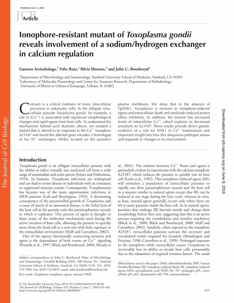
phenotype of two clones (GAD1.7 andGAD1.15) is shown in Fig. 1. Both strains showed a 10%survival rate after 60 min, at which time point no survivorswere ever detected with the parental strain.
GAD1.7, but not GAD1.15, exhibits a delay in IIE
To investigate whether any of the clones obtained in the IIDselection were also defective in IIE, as expected from previ-ous results (Black et al., 2000), intracellular parasites fromall 30 clones were treated with 1
�
M A23187 for 3 min, andthe levels of egress were assessed visually as compared withthe parental strain (see Materials and methods). In this man-ner, five clones (GAD1.3, GAD1.7, GAD1.17, GAD1.24,and GAD1.26) were identified as having 10–30% of vacu-oles intact after the incubation in three replicate experiments(Iie
�
clones), while all other clones and the parental strainexhibited
�
1% intact vacuoles by this time point (Iie
�
).The detailed IIE phenotypes of the parental strain andclones GAD1.7 (Iid
�
Iie
�
) and GAD1.15 (Iid
�
Iie
�
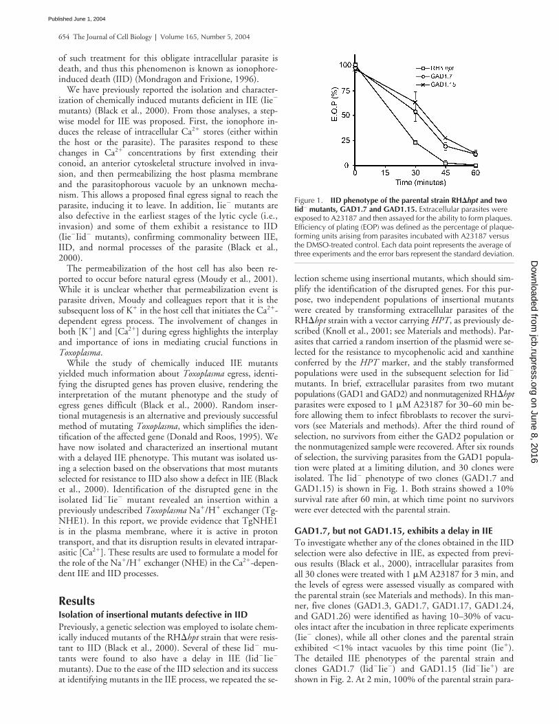
) areshown in Fig. 2. At 2 min, 100% of the parental strain para-
Figure 1. IID phenotype of the parental strain RH�hpt and two Iid� mutants, GAD1.7 and GAD1.15. Extracellular parasites were exposed to A23187 and then assayed for the ability to form plaques. Efficiency of plating (EOP) was defined as the percentage of plaque-forming units arising from parasites incubated with A23187 versus the DMSO-treated control. Each data point represents the average of three experiments and the error bars represent the standard deviation.
on June 8, 2016jcb.rupress.org
Dow
nloaded from
Published June 1, 2004
Toxoplasma
NHE’s involvement in calcium regulation |
Arrizabalaga et al. 655
sites are out of their vacuoles as previously reported (Black etal., 2000). Clone GAD1.15 behaves as wild type, whileclone GAD1.7 parasites exhibit a consistently delayed reac-tion to the ionophore with only 61% of parasites having ex-ited their host cells by 2 min. Nevertheless, with longer in-cubations, the mutants do eventually respond such that by 5min,
�
95% of vacuoles are lysed (Fig. 2). Since clones withand without an Iie
�
phenotype were isolated from the sameinsertional mutant population, the delay in IIE is most likelydue to the disruption of a gene and not a general effect ofthe vector used. Finally, this selection confirms that selectingfor Iid
�
mutants results in parasites that also have an Iie
�
phenotype and provides further evidence that these two pro-cesses are genetically linked (Black et al., 2000).
The mutagenic vector in GAD1.7 is inserted near a region of homology to sodium hydrogen exchangers
The most likely explanation for the Iie
�
defect of GAD1.7 isthat the gene disrupted by the random insertion is involvedin this process and in IID. Hence, the site of insertion wasidentified for this mutant. A molecular cloning approachwas used to recover the inserted plasmid along with one ofthe flanking genomic DNA fragments (see Materials andmethods). This flanking fragment was sequenced and deter-mined to be within a 10-kb genomic region from the
Toxo-plasma
genome project (http://ToxoDB.org). PCR analysiswas used to determine that all five Iie
�
clones (see above)had the same insertion, while none of the 10 Iie
�
clonestested showed this disruption (unpublished data). These fiveclones, therefore, are most likely the result of a single inser-tion event and subsequent expansion of the resulting mutantduring the six rounds of selection. Sequence analysis of thetargeted region revealed a segment
�
2,000 base pairs awayfrom the insertion site with high homology to NHEs fromvarious organisms. Given that this mutant was selected for adelay in responding to Ca
2
�
fluxes, an ion exchange proteinfits the profile of a candidate gene whose disruption mightlead to such a result. While intriguing, the homology to
NHE was not conclusive about the gene disrupted however,because the insertion was not within the NHE homology re-gion and therefore could be in an adjacent gene.
While only GAD1.7 (Iid
�
Iie
�
) is discussed here in detail,the insertion for GAD1.15 (Iid
�
Iie
�
) has also been deter-mined. The disturbed region shows no homology to knowngenes, no EST lie close to the insertion site, and no longopen reading frame is affected. The basis for the resultingphenotype, therefore, is unknown.
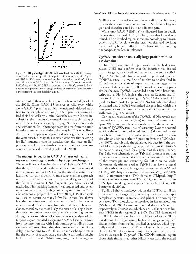
TgNHE1
encodes an unusually large protein with 12 TM domains
To further characterize this previously undescribed
Toxo-plasma
NHE and confirm that the insertion was indeedwithin its gene, we cloned and sequenced the entire cDNA(Fig. 3 A). We call this gene and its predicted productTgNHE1, since it is the first of its class to be described in
Toxoplasma
and analysis of sequence databases suggests thepresence of three additional NHE homologues in this para-site (see below).
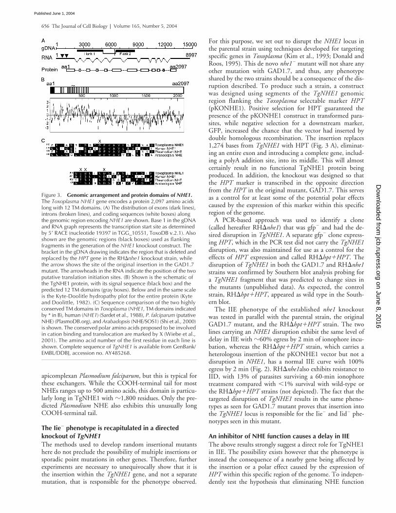
TgNHE1
is encoded by an 8,997-base tran-script and, as Fig. 3 A depicts, the gene has 12 exons and 11introns. The complete cloning of
TgNHE1
along with PCRproducts from GAD1.7 genomic DNA (unpublished data)confirmed that
TgNHE1
was indeed the gene into which themutagenic vector had inserted; specifically it was within thesixth exon (Fig. 3 A, arrow).
Conceptual translation of the
TgNHE1
cDNA reveals twopotential start methionine (Met) residues, 190 amino acidsapart. While no direct data exist to determine the actual startcodon of
TgNHE1
, two lines of reasoning favor the secondAUG as the start point of translation: (1) the second codonhas a better context for a
Toxoplasma
translational initiationsite with an adenine at the
�
3 position from the AUG (See-ber, 1997), and (2) only the translated product from the sec-ond Met has a predicted signal peptide within the first 65amino acids as expected for a protein of this type. Hence,Fig. 3 shows a schematic of the TgNHE1 protein as startingfrom the second potential initiator methionine (base 1161of the transcript) and extending for 2,097 amino acids.Computer algorithms predict TgNHE1 to have a signalpeptide with a putative cleavage site between residues 62 and63 (SignalP, http://www.cbs.dtu.dk/services/SignalP-2.0/),and 12 transmembrane (TM) domains (TMpred, http://www.ch.embnet.org/software/TMPRED_form.html) withinits NH
2
-terminal region as expected for an NHE (Fig. 3 B;Putney et al., 2002).
TgNHE1 shows homology within the 12 TMs to NHEsfrom a variety of organisms, including bacteria, other api-complexan parasites, mammals, and plants. The two highlyconserved TMs thought to be involved in ion translocation(Wiebe et al., 2001) correspond to TM domains V and VIrespectively in
Toxoplasma
, which has
�
72% identity to hu-man NHE1 in this region (Fig. 3 C). The TM domains ofTgNHE1 exhibit homology to a plethora of other NHEsbut do not show significantly higher homology to any par-ticular isoform from the best-studied eukaryotes, which typ-ically encode three to six NHE homologues. Hence, we havechosen TgNHE1 as a name simply to denote that it is thefirst of its class in
T. gondii
. The COOH-terminal regionshows little similarity to other NHEs, even from the related
Figure 2. IIE phenotype of GAD and knockout mutants. Percentage of vacuoles lysed at specific time points after induction with 1 �M A23187 in DMEi was measured for the parental strain RH�hpt, the two Iid� mutants GAD1.7 and GAD1.15, the NHE1 knockout strain RH�nhe1, and the HXGPRT-expressing strain RH�hpt�HPT. Each data point represents the average of three experiments, and the error bars represent the standard deviation.
on June 8, 2016jcb.rupress.org
Dow
nloaded from
Published June 1, 2004
656 The Journal of Cell Biology
| Volume 165, Number 5, 2004
apicomplexan Plasmodium falciparum, but this is typical forthese exchangers. While the COOH-terminal tail for mostNHEs ranges up to 500 amino acids, this domain is particu-larly long in TgNHE1 with �1,800 residues. Only the pre-dicted Plasmodium NHE also exhibits this unusually longCOOH-terminal tail.
The Iie� phenotype is recapitulated in a directed knockout of TgNHE1The methods used to develop random insertional mutantshere do not preclude the possibility of multiple insertions orsporadic point mutations in other genes. Therefore, furtherexperiments are necessary to unequivocally show that it isthe insertion within the TgNHE1 gene, and not a separatemutation, that is responsible for the phenotype observed.
For this purpose, we set out to disrupt the NHE1 locus inthe parental strain using techniques developed for targetingspecific genes in Toxoplasma (Kim et al., 1993; Donald andRoos, 1995). This de novo nhe1� mutant will not share anyother mutation with GAD1.7, and thus, any phenotypeshared by the two strains should be a consequence of the dis-ruption described. To produce such a strain, a constructwas designed using segments of the TgNHE1 genomicregion flanking the Toxoplasma selectable marker HPT(pKONHE1). Positive selection for HPT guaranteed thepresence of the pKONHE1 construct in transformed para-sites, while negative selection for a downstream marker,GFP, increased the chance that the vector had inserted bydouble homologous recombination. The insertion replaces1,274 bases from TgNHE1 with HPT (Fig. 3 A), eliminat-ing an entire exon and introducing a complete gene, includ-ing a polyA addition site, into its middle. This will almostcertainly result in no functional TgNHE1 protein beingproduced. In addition, the knockout was designed so thatthe HPT marker is transcribed in the opposite directionfrom the HPT in the original mutant, GAD1.7. This servesas a control for at least some of the potential polar effectscaused by the expression of this marker within this specificregion of the genome.
A PCR-based approach was used to identify a clone(called hereafter RH�nhe1) that was gfp� and had the de-sired disruption in TgNHE1. A separate gfp� clone express-ing HPT, which in the PCR test did not carry the TgNHE1disruption, was also maintained for use as a control for theeffects of HPT expression and called RH�hpt�HPT. Thedisruption of TgNHE1 in both the GAD1.7 and RH�nhe1strains was confirmed by Southern blot analysis probing fora TgNHE1 fragment that was predicted to change sizes inthe mutants (unpublished data). As expected, the controlstrain, RH�hpt�HPT, appeared as wild type in the South-ern blot.
The IIE phenotype of the established nhe1 knockoutwas tested in parallel with the parental strain, the originalGAD1.7 mutant, and the RH�hpt�HPT strain. The twolines carrying an NHE1 disruption exhibit the same level ofdelay in IIE with �60% egress by 2 min of ionophore incu-bation, whereas the RH�hpt�HPT strain, which carries aheterologous insertion of the pKONHE1 vector but not adisruption in NHE1, has a normal IIE curve with 100%egress by 2 min (Fig. 2). RH�nhe1also exhibits resistance toIID, with 13% of parasites surviving a 60-min ionophoretreatment compared with �1% survival with wild-type orthe RH�hpt�HPT strains (not depicted). The fact that thetargeted disruption of TgNHE1 results in the same pheno-types as seen for GAD1.7 mutant proves that insertion intothe TgNHE1 locus is responsible for the Iie� and Iid� phe-notypes seen in this mutant.
An inhibitor of NHE function causes a delay in IIEThe above results strongly suggest a direct role for TgNHE1in IIE. The possibility exists however that the phenotype isinstead the consequence of a nearby gene being affected bythe insertion or a polar effect caused by the expression ofHPT within this specific region of the genome. To indepen-dently test the hypothesis that eliminating NHE function
Figure 3. Genomic arrangement and protein domains of NHE1. The Toxoplasma NHE1 gene encodes a protein 2,097 amino acids long with 12 TM domains. (A) The distribution of exons (dark lines), introns (broken lines), and coding sequences (white boxes) along the genomic region encoding NHE1 are shown. Base 1 in the gDNA and RNA graph represents the transcription start site as determined by 5� RACE (nucleotide 19397 in TGG_10551, ToxoDB v.2.1). Also shown are the genomic regions (black boxes) used as flanking fragments in the generation of the NHE1 knockout construct. The bracket in the gDNA drawing indicates the region that is deleted and replaced by the HPT gene in the RH�nhe1 knockout strain, while the arrow shows the site of the original insertion in the GAD1.7 mutant. The arrowheads in the RNA indicate the position of the two putative translation initiation sites. (B) Shown is the schematic of the TgNHE1 protein, with its signal sequence (black box) and the predicted 12 TM domains (gray boxes). Below and in the same scale is the Kyte-Doolitle hydropathy plot for the entire protein (Kyte and Doolittle, 1982). (C) Sequence comparison of the two highly conserved TM domains in Toxoplasma (NHE1, TM domains indicated by * in B), human (NHE1) (Sardet et al., 1988), P. falciparum (putative NHE) (PlasmoDB.org), and Arabadopsis (NHE/SOS1) (Shi et al., 2000) is shown. The conserved polar amino acids proposed to be involved in cation binding and translocation are marked by X (Wiebe et al., 2001). The amino acid number of the first residue in each line is shown. Complete sequence of TgNHE1 is available from GenBank/EMBL/DDBJ, accession no. AY485268.
on June 8, 2016jcb.rupress.org
Dow
nloaded from
Published June 1, 2004

Toxoplasma NHE’s involvement in calcium regulation | Arrizabalaga et al. 657
causes an Iie� phenotype, therefore, we investigated the ef-fect on IIE of pharmacologically inhibiting NHE function.Amiloride and its derivatives are known to be powerful in-hibitors of NHEs, acting by competitively inhibiting Na�
binding (Paris and Pouyssegur, 1983) and by noncompeti-tively inhibiting ion translocation (Ives et al., 1983). Hence,intracellular parasites were preincubated with dimethyl-amiloride (DMA) before inducing egress with the iono-phore (see Materials and methods). As can be seen in Fig. 4,preincubation with DMA does indeed retard IIE: only 67%of DMA-treated parasites are out of their host cells by 2min, whereas 100% of nontreated parasites have emerged bythe same time point. This level of reduction is essentially thesame as that seen with the genetic disruption of NHE1 in ei-ther the knockout or the original mutant (Fig. 4). The spec-ificity of the drug is confirmed by the fact that DMA treat-ment of nhe1� parasites gives no further delay in IIE (Fig.4). These results confirm that the effect seen in the geneticmutant is caused by the loss of TgNHE1 function and is notan indirect consequence of the insertion.
TgNHE1 is localized in the plasma membraneTo understand how TgNHE1 might affect IIE and otherevents, it is critical to determine its localization within theparasite. For this purpose, we created a polyclonal antibodyagainst a bacterially expressed fragment of TgNHE1 (see Ma-terials and methods). The fragment expressed was chosenfrom the COOH-terminal domain of the protein to avoidcross-reactivity with other NHEs, which usually show homol-ogy only in the TM domains. Specificity of the antibodies tothis segment was also predicted by the fact that sequenceanalysis did not identify any other regions in the Toxoplasmagenome with significant similarity to this fragment.
The mouse antibody produced was used to stain intra-cellular parasites by immunofluorescence. This approachyielded a clear staining at the extreme periphery of wild-typeparasites (Fig. 5 A), which was absent when preimmune se-rum was used (not depicted). The specificity of this anti-
body for TgNHE1 was confirmed by the absence of stainingin the GAD1.7 mutant and the nhe1 knockout strain (Fig.5, B and C). Some faint, nondistinct intraparasitic stainingcan be seen in the nhe1� mutants. This is presumably the re-sult of weak cross-reactivity to another NHE or an unrelatedprotein, despite the absence of detectable similarity to the re-gion used for immunization in any hypothetical Toxoplasmaprotein. The fluorescence pattern seen with wild-type para-sites does not change during different stages of intracellulargrowth, indicating that the signal is from protein within theparasite plasma membrane rather than the inner membranecomplex just below, which forms within dividing parasitesand exhibits an immunofluorescence pattern distinct fromplasma membrane (Hu et al., 2002). The fact that thisantibody does not stain unpermeabilized parasites (unpub-lished data) indicates that the COOH-terminal region ofTgNHE1 is not exposed to the outside, as predicted fromresults with other NHE proteins (Putney et al., 2002).
Figure 4. Effect of pretreating parasites with an NHE inhibitor on IIE. Percentage of vacuoles lysed at specific time points after induction with 1 �M A23187 in DMEi was measured for RH�hpt, GAD1.7, and RH�nhe1 parasites that were pretreated with either 100 �M DMA dissolved in DMSO or an equivalent amount of DMSO alone for 10 min. The data are pooled from three independent trials, and the error bars represent the standard deviation.
Figure 5. Intracellular localization of TgNHE1. Intracellular parasites of either (A) the wild-type RH�hpt, (B) the mutant GAD1.7, or (C) the NHE knockout RH�nhe1 were stained with antibodies raised in mouse against amino acids 716–1073 of the TgNHE1 protein. An Alexa fluor 488–conjugated goat anti–mouse antibody was used to visualize the TgNHE1 signal. The phase contrast and fluorescence images of the same vacuole are shown for each strain.
on June 8, 2016jcb.rupress.org
Dow
nloaded from
Published June 1, 2004
658 The Journal of Cell Biology | Volume 165, Number 5, 2004
Proton efflux and calcium homeostasis in theTgNHE1 mutantsPlasma membrane NHEs in other systems exchange Na� forH�, linking the electrochemical potential of sodium and hy-drogen ions maintained across the membrane. In animalcells, NHEs have been implicated in a variety of functions,including the regulation of cytoplasmic pH and Na� con-centration and the control of cell volume (Putney et al.,2002). Assuming TgNHE1 has a similar function, its dis-ruption should have an effect on the proper balance of intra-cellular ions. To investigate this possibility, and to furtherexplore its role in IIE and IID, several physiological assayswere performed with RH�hpt, GAD1.7, and RH�nhe1parasites that are purified away from host cells. First, the in-traparasitic pH (pHi) was measured for the three strains un-der conditions where the extracellular pH (pHe) ranged pHe
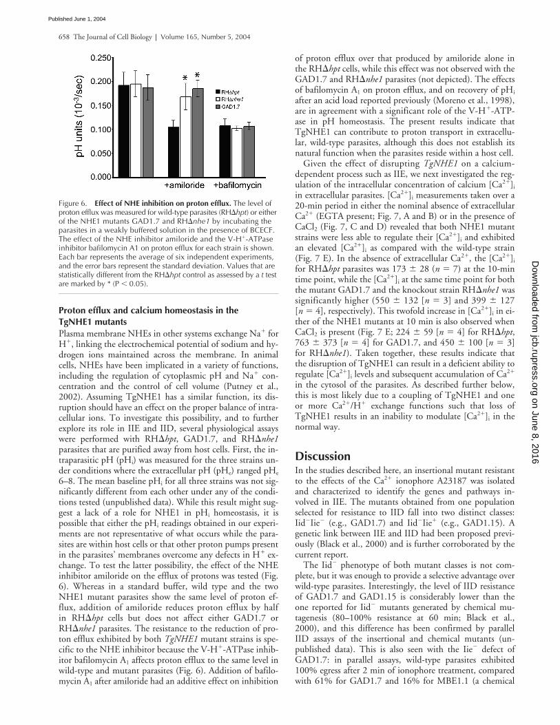
6–8. The mean baseline pHi for all three strains was not sig-nificantly different from each other under any of the condi-tions tested (unpublished data). While this result might sug-gest a lack of a role for NHE1 in pHi homeostasis, it ispossible that either the pHi readings obtained in our experi-ments are not representative of what occurs while the para-sites are within host cells or that other proton pumps presentin the parasites’ membranes overcome any defects in H� ex-change. To test the latter possibility, the effect of the NHEinhibitor amiloride on the efflux of protons was tested (Fig.6). Whereas in a standard buffer, wild type and the twoNHE1 mutant parasites show the same level of proton ef-flux, addition of amiloride reduces proton efflux by halfin RH�hpt cells but does not affect either GAD1.7 orRH�nhe1 parasites. The resistance to the reduction of pro-ton efflux exhibited by both TgNHE1 mutant strains is spe-cific to the NHE inhibitor because the V-H�-ATPase inhib-itor bafilomycin A1 affects proton efflux to the same level inwild-type and mutant parasites (Fig. 6). Addition of bafilo-mycin A1 after amiloride had an additive effect on inhibition
of proton efflux over that produced by amiloride alone inthe RH�hpt cells, while this effect was not observed with theGAD1.7 and RH�nhe1 parasites (not depicted). The effectsof bafilomycin A1 on proton efflux, and on recovery of pHi
after an acid load reported previously (Moreno et al., 1998),are in agreement with a significant role of the V-H�-ATP-ase in pH homeostasis. The present results indicate thatTgNHE1 can contribute to proton transport in extracellu-lar, wild-type parasites, although this does not establish itsnatural function when the parasites reside within a host cell.
Given the effect of disrupting TgNHE1 on a calcium-dependent process such as IIE, we next investigated the reg-ulation of the intracellular concentration of calcium [Ca2�]i
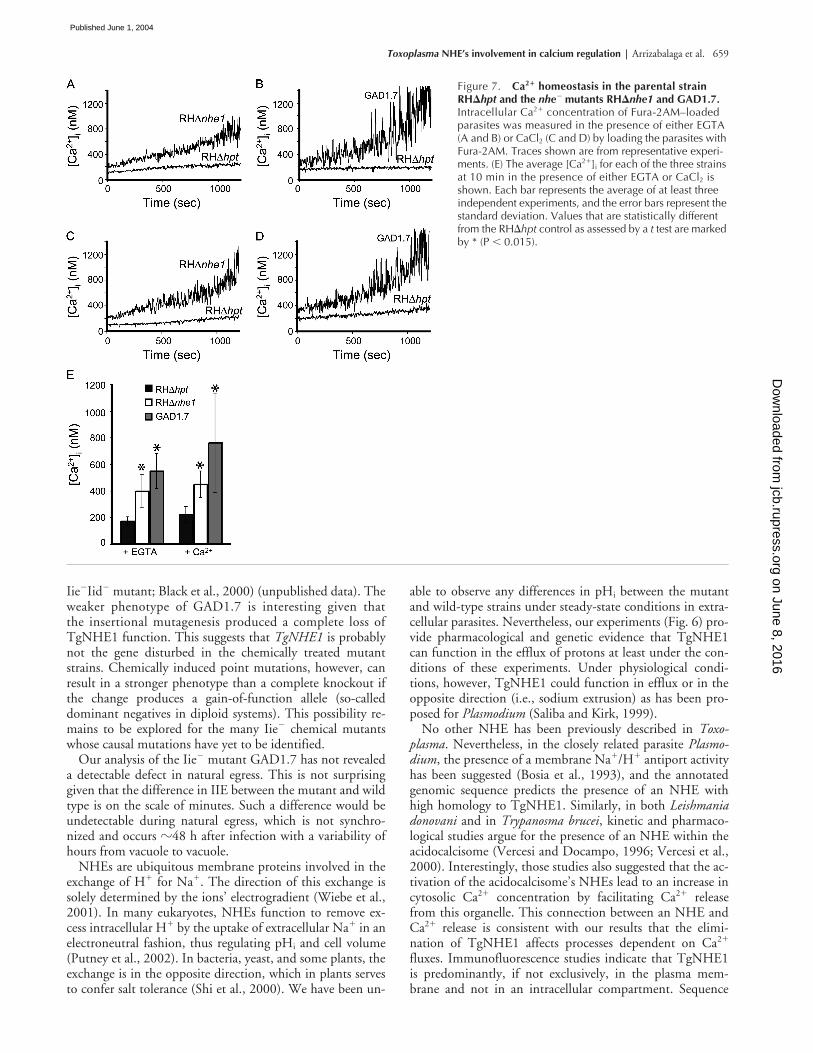
in extracellular parasites. [Ca2�]i measurements taken over a20-min period in either the nominal absence of extracellularCa2� (EGTA present; Fig. 7, A and B) or in the presence ofCaCl2 (Fig. 7, C and D) revealed that both NHE1 mutantstrains were less able to regulate their [Ca2�]i and exhibitedan elevated [Ca2�]i as compared with the wild-type strain(Fig. 7 E). In the absence of extracellular Ca2�, the [Ca2�]i
for RH�hpt parasites was 173 � 28 (n � 7) at the 10-mintime point, while the [Ca2�]i at the same time point for boththe mutant GAD1.7 and the knockout strain RH�nhe1 wassignificantly higher (550 � 132 [n � 3] and 399 � 127[n � 4], respectively). This twofold increase in [Ca2�]i in ei-ther of the NHE1 mutants at 10 min is also observed whenCaCl2 is present (Fig. 7 E; 224 � 59 [n � 4] for RH�hpt,763 � 373 [n � 4] for GAD1.7, and 450 � 100 [n � 3]for RH�nhe1). Taken together, these results indicate thatthe disruption of TgNHE1 can result in a deficient ability toregulate [Ca2�]i levels and subsequent accumulation of Ca2�
in the cytosol of the parasites. As described further below,this is most likely due to a coupling of TgNHE1 and oneor more Ca2�/H� exchange functions such that loss ofTgNHE1 results in an inability to modulate [Ca2�]i in thenormal way.
DiscussionIn the studies described here, an insertional mutant resistantto the effects of the Ca2� ionophore A23187 was isolatedand characterized to identify the genes and pathways in-volved in IIE. The mutants obtained from one populationselected for resistance to IID fall into two distinct classes:Iid�Iie� (e.g., GAD1.7) and Iid�Iie� (e.g., GAD1.15). Agenetic link between IIE and IID had been proposed previ-ously (Black et al., 2000) and is further corroborated by thecurrent report.
The Iid� phenotype of both mutant classes is not com-plete, but it was enough to provide a selective advantage overwild-type parasites. Interestingly, the level of IID resistanceof GAD1.7 and GAD1.15 is considerably lower than theone reported for Iid� mutants generated by chemical mu-tagenesis (80–100% resistance at 60 min; Black et al.,2000), and this difference has been confirmed by parallelIID assays of the insertional and chemical mutants (un-published data). This is also seen with the Iie� defect ofGAD1.7: in parallel assays, wild-type parasites exhibited100% egress after 2 min of ionophore treatment, comparedwith 61% for GAD1.7 and 16% for MBE1.1 (a chemical
Figure 6. Effect of NHE inhibition on proton efflux. The level of proton efflux was measured for wild-type parasites (RH�hpt) or either of the NHE1 mutants GAD1.7 and RH�nhe1 by incubating the parasites in a weakly buffered solution in the presence of BCECF. The effect of the NHE inhibitor amiloride and the V-H�-ATPase inhibitor bafilomycin A1 on proton efflux for each strain is shown. Each bar represents the average of six independent experiments, and the error bars represent the standard deviation. Values that are statistically different from the RH�hpt control as assessed by a t test are marked by * (P � 0.05).
on June 8, 2016jcb.rupress.org
Dow
nloaded from
Published June 1, 2004
Toxoplasma NHE’s involvement in calcium regulation | Arrizabalaga et al. 659
Iie�Iid� mutant; Black et al., 2000) (unpublished data). Theweaker phenotype of GAD1.7 is interesting given thatthe insertional mutagenesis produced a complete loss ofTgNHE1 function. This suggests that TgNHE1 is probablynot the gene disturbed in the chemically treated mutantstrains. Chemically induced point mutations, however, canresult in a stronger phenotype than a complete knockout ifthe change produces a gain-of-function allele (so-calleddominant negatives in diploid systems). This possibility re-mains to be explored for the many Iie� chemical mutantswhose causal mutations have yet to be identified.
Our analysis of the Iie� mutant GAD1.7 has not revealeda detectable defect in natural egress. This is not surprisinggiven that the difference in IIE between the mutant and wildtype is on the scale of minutes. Such a difference would beundetectable during natural egress, which is not synchro-nized and occurs �48 h after infection with a variability ofhours from vacuole to vacuole.
NHEs are ubiquitous membrane proteins involved in theexchange of H� for Na�. The direction of this exchange issolely determined by the ions’ electrogradient (Wiebe et al.,2001). In many eukaryotes, NHEs function to remove ex-cess intracellular H� by the uptake of extracellular Na� in anelectroneutral fashion, thus regulating pHi and cell volume(Putney et al., 2002). In bacteria, yeast, and some plants, theexchange is in the opposite direction, which in plants servesto confer salt tolerance (Shi et al., 2000). We have been un-
able to observe any differences in pHi between the mutantand wild-type strains under steady-state conditions in extra-cellular parasites. Nevertheless, our experiments (Fig. 6) pro-vide pharmacological and genetic evidence that TgNHE1can function in the efflux of protons at least under the con-ditions of these experiments. Under physiological condi-tions, however, TgNHE1 could function in efflux or in theopposite direction (i.e., sodium extrusion) as has been pro-posed for Plasmodium (Saliba and Kirk, 1999).
No other NHE has been previously described in Toxo-plasma. Nevertheless, in the closely related parasite Plasmo-dium, the presence of a membrane Na�/H� antiport activityhas been suggested (Bosia et al., 1993), and the annotatedgenomic sequence predicts the presence of an NHE withhigh homology to TgNHE1. Similarly, in both Leishmaniadonovani and in Trypanosma brucei, kinetic and pharmaco-logical studies argue for the presence of an NHE within theacidocalcisome (Vercesi and Docampo, 1996; Vercesi et al.,2000). Interestingly, those studies also suggested that the ac-tivation of the acidocalcisome’s NHEs lead to an increase incytosolic Ca2� concentration by facilitating Ca2� releasefrom this organelle. This connection between an NHE andCa2� release is consistent with our results that the elimi-nation of TgNHE1 affects processes dependent on Ca2�
fluxes. Immunofluorescence studies indicate that TgNHE1is predominantly, if not exclusively, in the plasma mem-brane and not in an intracellular compartment. Sequence
Figure 7. Ca2� homeostasis in the parental strain RH�hpt and the nhe� mutants RH�nhe1 and GAD1.7. Intracellular Ca2� concentration of Fura-2AM–loaded parasites was measured in the presence of either EGTA (A and B) or CaCl2 (C and D) by loading the parasites with Fura-2AM. Traces shown are from representative experi-ments. (E) The average [Ca2�]i for each of the three strains at 10 min in the presence of either EGTA or CaCl2 is shown. Each bar represents the average of at least three independent experiments, and the error bars represent the standard deviation. Values that are statistically different from the RH�hpt control as assessed by a t test are marked by * (P � 0.015).
on June 8, 2016jcb.rupress.org
Dow
nloaded from
Published June 1, 2004

660 The Journal of Cell Biology | Volume 165, Number 5, 2004
analysis of the Toxoplasma genome and a collection of ESTsreveal the existence of three other NHE homologues in thisparasite. It is likely that one of these is present in the Toxo-plasma acidocalcisome, and further studies will be needed tounderstand the role of these NHEs in ion homeostasis.
We had previously hypothesized that the Ca2� detected bythe parasites before egress was of extra-parasitic origin (Blacket al., 2000). This was mostly based on the observation thatBAPTA-AM, which should not enter intracellular parasites,blocked the effects of the ionophore in IIE. Nevertheless, wecould not exclude the possibility that BAPTA was in fact en-tering the parasite and that therefore the Ca2� signal wascoming from intracellular compartments within the para-sites. Additional studies on natural egress clearly argue forthis latter possibility (Moudy et al., 2001). Regardless, egressin wild-type parasites appears to be the result of parasitessensing an increase in [Ca2�]i to �400 nM (Moudy etal., 2001). The delay in IIE caused by the disruption ofTgNHE1 therefore could be related to the fact that in thenhe1� mutant, the resting [Ca2�]i may already be elevatedbecause of an inability to appropriately regulate [Ca2�]i. Ac-cordingly, addition of an ionophore and induction of Ca2�
fluxes might produce less of an initial percentage change in[Ca2�]i and hence a delay in IIE and IID.
While a role for an NHE in Ca2� homeostasis has beensuggested in human platelets (Siffert and Akkerman, 1987),this interpretation has been disputed based on the lack ofspecificity of the pharmacological inhibitors used (Hunyadyet al., 1987). Our studies using an organism with a geneticelimination of a specific NHE unequivocally show a role forthis exchanger in the regulation of Ca2� levels. While themutant parasites are clearly able to adapt to defects in Ca2�
regulation, they exhibit a defect in responding to changes inlocal ion concentrations as seen by the delay in IIE. The ex-act mechanism responsible for the altered ability to main-tain calcium levels under our experimental conditions inthe nhe1� mutants is not known. While we cannot excludethe possibility that TgNHE1 serves to directly transportCa2�, Ca2� extrusion from the cytosol is thought to be cat-alyzed by Ca2�/H� countertransporting ATPases located inthe plasma membrane, endoplasmic reticulum, and/or aci-docalcisomes (Moreno and Docampo, 2003). Coupling ofthe plasma membrane Ca2�-ATPase with TgNHE1, there-fore, could be involved in Ca2� efflux and Na� entry so thatdisruption of the TgNHE1 would result in higher [Ca2�]i.Such coupling between a Ca2�/H� ATPase and an NHEhas been suggested in intracellular Ca2� regulation in stim-ulated Jurkat T cells (Berthe et al., 1991). It is also possiblethat TgNHE1 is not involved in pH homeostasis underphysiological conditions, and that the effect on Ca2� regu-lation is a consequence of some unidentified function of Tg-NHE1. Since our experiments suggest that TgNHE1 caninfluence proton fluxes, an alternative explanation for ourresults is that, under physiological conditions, TgNHE1 islinked to inward proton movement and functions in the ex-trusion of Na�. The absence of the TgNHE could lead tocytosolic Na� increase, which has been shown to stimulateCa2� release from the acidocalcisomes of L. donovani (Ver-cesi et al., 2000) and T. brucei (Vercesi and Docampo,1996).
The results reported here show that efficient IIE is depen-dent on the function of an NHE homologue. Furthermore,our work has revealed that the maintenance of Ca2� homeo-stasis in Toxoplasma is dependent on this same protein. Thisprovides strong evidence for a connection between Na� andCa2� exchange and for a specific role in these processes forTgNHE1. This discovery also validates the use of forwardgenetics in the study of IIE since, a priori, this specificplasma membrane NHE would not have been predicted tobe involved in Ca2� homeostasis and the parasite functionsregulated by Ca2�.
Materials and methodsParasite cultivation and reagentsThe tachyzoite stage of the RH�HXGPRT strain (RH�hpt; Donald et al.,1996) was the parent strain used for the mutagenesis and phenotypic anal-ysis. Parasites of the Prugniaud strain (Zenner et al., 1993) were used to ex-tract RNA for cloning and sequencing of the NHE1 gene. Strains weremaintained in vitro by serial passage on monolayers of human foreskin fi-broblasts (HFFs) at 37C in 0.5% CO2 as previously described (Roos et al.,1994). DME (GIBCO BRL) was modified for use in three different forms: (1)DMEc for normal propagation was supplemented with 10% NuSerum (Col-laborative Biomedical Products), 2 mM glutamine, and 20 �g/ml gentamy-cin; (2) DMEm/x consisted of DMEc with 50 �g/ml mycophenolic acid(MPA) and 50 �g/ml xanthine; (3) DMEi was same as DMEc without serum.
The calcium ionophore A23187 (Sigma-Aldrich) was dissolved inDMSO at 1 mM. DMA (Sigma-Aldrich) was dissolved in DMSO at 10 mM.MPA and xanthine (Sigma-Aldrich) were both dissolved in 0.3 N NaOH at25 mg/ml.
Insertional mutagenesis and selection of Iid� mutantsThe previously described plasmid, pHANA, containing the hypoxanthine-xanthine-guanine phosphoribosyl transferase (HPT) gene under the dihyro-folate reductase-thymidylate synthase (DHFR) promoter, was used to gen-erate random insertional mutants (Black and Boothroyd, 1998). Insertionalmutagenesis was performed by transfecting extracellular RH�hpt parasitesby electroporation (Soldati and Boothroyd, 1993) with 50 �g of pHANAlinearized with NotI. In addition, a mock transfection without DNA wasperformed as a control. Parasites were then allowed to expand in a mono-layer of HFFs in a T75 flask. After 24 h, the medium of the mutagenizedparasites was changed to DMEm/x. Parasites were allowed to lyse and athird of the parasites were passed to a new T75 continuing the selectionwith DMEm/x. After 48 h, the mutagenized and nonmutagenized popula-tions were syringed with a 27-gauge needle and pelleted at 1,000 g for 10min. The parasites were resuspended in warm DMEi that contained 1 �MA23187 and incubated for 30 min at 37C followed by pelleting for 10 minat 1,000 g. The entire pellet of parasites was resuspended in DMEc platedonto a fresh HFF monolayer in a T75 flask to recover the survivors. After48 h, a second round of selection was performed by incubating syringe-lysed extracellular parasites in 1 �M A23187 for 45 min at 37C. Survivingparasites were recovered by adding them to HFFs in T25 flasks in DMEc
followed by four additional rounds of selection using 60 min of incubationin 1 �M A23187 for each round. Only parasites from the mutagenizedGAD1 population survived this series of selections. These were plated outon monolayers in a 96-well plate at a limiting dilution for isolating clones.30 clones were isolated from the GAD1 population and maintained in 24-well plates with DMEm/x for further analysis. Consistent with nomenclaturepreviously used (Black et al., 2000), clones were labeled GADx.y, whereGA indicate the initials of the researcher performing the selection, “D” in-dicates that the mutant came from a death selection, x represents the ex-periment number, and y is the clone number in that experiment.
Screening for Iie� mutants30 �l of lysed parasites from each of the 30 clones and the parental strainwere passed to HFFs in a 24-well plate with DMEc. Parasites were allowedto replicate for 30 h at 37C. The growth medium was washed off themonolayer with PBS and replaced with warm DMEi containing 1 �MA23187 for 3 min at 37C, at which point wild-type parasites show 100%egress (Black et al., 2000). The ionophore was removed and the monolay-ers were fixed with 100% methanol and stained with Difquick (Dade-Behring). Vacuoles were examined by light microscopy to determine if
on June 8, 2016jcb.rupress.org
Dow
nloaded from
Published June 1, 2004
Toxoplasma NHE’s involvement in calcium regulation | Arrizabalaga et al. 661
egress had occurred as previously described (Black et al., 2000). Thoseclones with 10% intact vacuoles were selected for confirmation of anIie� phenotype.
Quantitation of IID and IIEThe exact quantification of the IID and IIE phenotype was performed aspreviously described with the following changes (Black et al., 2000). Theionophore incubations were performed in warm DMEi containing 1 �MA23187 in DMSO. For the IIE assays, the monolayers were fixed with100% methanol and stained with Difquick (Dade-Behring). Vacuoles werethen examined by light microscopy counting no fewer than five fields fromeach well (�100 vacuoles) at a magnification of 400�.
The same methods were used to analyze the effect of DMA on IIE, incu-bating the parasites with 100 �M DMA in DMSO or a comparable amountof DMSO alone in DMEi at 37C for 10 min before the addition of an equalvolume of DMEi � 2 �M A23187. The statistical significance of the differ-ences between the mutants and the wild-type strains was assessed by t test.
Plasmid rescue of GAD1.7 mutantGenomic Toxoplasma DNA was purified from freshly lysed tachyzoites us-ing standard methods (Black and Boothroyd, 1998). To rescue fragmentsflanking the inserted pHANA vector, 1 �g of genomic DNA was cut withAgeI for 12 h, followed by heat inactivation. 20 ng of digested DNA wasincubated with T4 DNA ligase at a concentration of 2 ng/�l to favor in-tramolecular ligation and subsequently transformed into DH5 chemicallycompetent cells. Plasmids were isolated from ampicillin-resistant coloniesand analyzed by restriction digest and sequencing to confirm the presenceand identity of flanking genomic sequences.
Cloning of NHE1A genomic contig (TGG_10551) from version 2.1 of the Toxoplasma ge-nome project (http://ToxoDB.org) was identified as the region of insertionin mutant GAD1.7 as described above. The internal sequence of the NHE1cDNA was identified from a directional cDNA library (from day 3 in vitrobradyzoites from pH stressed Prugniaud parasites; a gift from M. Cleary[Stanford University] and Compligen) using primers designed against re-gions of TgNHE1 homology within the Toxoplasma genomic sequence andlibrary vector primers. The primers used were 5�-aaaggcgagaagagagcaatc-3� in combination with M13forward in a primary reaction and the nestedprimer 5�-gtaagtcacaatgccgctgat-3� used with T7 in a secondary reaction.In addition, 5�-ccgtcatcacagtctgctaca-3� was used with M13reverse andthe nested primer 5�-gctacatcgcctactttgtcg-3� was used with T3 in a sec-ondary reaction. The ends of the cDNA were identified using 5�RACE and3�RACE systems (Invitrogen) with RNA isolated from Prugniaud parasitesas described before (Singh et al., 2002) and using primers 5�-gccgctgatgat-gaaaatagc-3�, 5�-gcattgcgtcatgggatattt-3�, 5�-cgtgcacagtttgtctgaagc-3�, 5�-acacgatctccggttttgtc-3�, and 5�-aaagatgtcgggagacgatgg-3�.
Generation of NHE1-specific knockoutThe parental vector pMini-GFP.ht was generated by inserting a Klenow-blunted HindIII to BamHI fragment from pgraGFP (Kim et al., 2001) withGFP under the GRA2 promoter into the HPT-containing vector pMiniHXG-PRT-I (Donald et al., 1996) digested with SacI and blunted with T4 poly-merase. This approach generated pMini-GFP.ht with GFP in the 5� to 3�end direction in respect to HPT. To design the knockout construct, first aPCR fragment from bases 6463–8676 (flank 2 in Fig. 3 A) from NHE1gDNA was digested with HindIII and KpnI and cloned into pMini-GFP.htdigested with the same restriction enzymes. The resulting plasmid was di-gested with NotI and XbaI and ligated to a PCR fragment from genomicbases 2292–5189 (flank 1 in Fig. 3 A) also cut with NotI and XbaI. Theprimers used to expand the flanking regions were 5�-ggggcgaagctta-gaaacgcgaagacgaaaaa-3� and 5�-ggggggtaccccatcgtctccgacatcttt-3� forflank 1 and 5�-gggaaagggaaagcggccgcgattggtggatgcgtgcgtc-3� and 5�-gctaatcacgaagttctagagacgg-3� for flank 2.
The resulting vector, pKONHE1, was linearized with NotI, and 50 �g ofDNA was introduced into RH�hpt strain parasites by standard methods(Soldati and Boothroyd, 1993). Six independent transformations were per-formed, and parasites were then grown in HFFs in T25 flasks for 24 h, atwhich point the media was changed to DMEm/x. A fifth of the surviving par-asites from each population were passed to new cells when lysed and keptunder the MPA/xanthine selection. 10 d after the transformation, genomicDNA was obtained from the selected population and used in PCR to con-firm the knockout event using primers that yielded a product only if theknockout had occurred (5�-gtggcgattctcatcgactt-3� and 5�-aacgtaatctcgtg-gcttcg-3�). Parasites from the one population with the knockout weregrown in 24-well plates in pools of 50 parasites per well. Parasites from
each of 24 wells were tested again for the knockout parasite by PCR, andparasites from one of the positive wells were cloned by limiting dilution.40 clones that did not express GFP were tested by PCR, and one clone wasdetermined to have the desired knockout. Besides the knockout cloneRH�nhe1, a clone with a heterologous insertion of the knockout constructwas maintained and dubbed RH�hpt�HPT.
Generation of TgNHE1 antibodies and immunofluorescence analysisCoding sequences corresponding to amino acids 716–1073 of TgNHE1were amplified from cDNA and subcloned into the T7 expression vectorpET28a(�) (Novagen) that had been digested with NdeI and EcoRI. Prim-ers used in this step were 5�-gggcagcatatgaccgtcgtgaagcgcatggcgacg-3�and 5�-ctgtgtgcgcatgctggtcaggcgat-3�. The resulting vector was trans-formed into Escherichia coli strain BL21(DE3) (Novagen), and expression ofthe recombinant protein, which encoded 6X-His tags at both ends, was in-duced with IPTG. TgNHE1(716–1073) was purified using affinity chroma-tography with Ni-NTA agarose from QIAGEN and following the manufac-turer’s instructions. 100 �g of purified recombinant protein was injectedinto Balb/C mice (Teconic) along with 100 �l of RIBI adjuvant (Corixa).Three boost injections were performed, at 3-wk intervals. Mouse serumwas tested for specific immunoreactivity 4 d after the second and thirdboost by immunofluorescence staining of wild-type and NHE1 knockoutparasites.
For immunofluorescence analysis, parasites were grown for 24 h inHFFs on glass coverslips. Staining of intracellular parasites was performedas described before (Singh et al., 2002). For primary staining, serum fromthe TgNHE1(716–1073) immunized mouse was used at 1:1,000 in PBS/0.2% Triton X-100/3% BSA for 1 h at room temperature. The secondary an-tibody, Alexa fluor 488–conjugated goat anti–mouse (Molecular Probes),was used at 1:2,000 antibody in PBS with 3% BSA for 1 h. Coverslips weremounted on slides with vectashield (Vector Laboratories) and examinedusing a 100� lens of an Olympus BX60 fluorescence microscope, and im-ages were captured using a Hamamatsu digital camera (model C4742-95)and the Image Pro Plus software.
Spectrofluorometric determinationsSpectrofluorometric determinations were performed as described previ-ously (Moreno and Zhong, 1996). In summary, for the proton extrusion ex-periments, freshly lysed and filtered tachyzoites were washed twice andresuspended in buffer A (116 mM NaCl, 5.4 mM KCl, 0.8 mM MgSO4, 5.5mM D-glucose, and 50 mM Hepes, pH 7.4) at 109 parasites/ml and kept inice until use. 108 parasites were centrifuged and resuspended in 100 �l ofa weakly buffered (100 �M Hepes-Tris, pH 7.4) standard solution and thenmixed in with 2.4 ml of standard solution containing 0.38 �M of free acidBCECF (Molecular Probes) and either 100 �M amiloride (Sigma-Aldrich) or1 �M bafilomycin (Kamiya Biomedical). Fluorescence measurementsstarted after 1 min of stirring in a cuvette. For Ca2� measurements, freshlylysed and filtered parasites were washed three times with buffer A and pre-incubated at 25C for 30 min in buffer A with 1% sucrose, and 4.5 �MFura 2-AM (Molecular Probes) at 109 parasites/ml. Parasites were thenwashed twice and resuspended in buffer A at the same concentration. Foreach measurement, 50 �l of parasite suspension was diluted in a cuvettewith 2.5 ml of a standard buffer, with either 1 mM EGTA or 1mM CaCl2,and allowed to stir for 2 min before recording [Ca2�].
The host strain RH�hpt was obtained through the AIDS Research and Ref-erence Program, Division of AIDS, NIAID, NIH from Dr. David Roos. Wewould also like to thank Mike Black and members of the Boothroyd andMoreno labs for critical input into this work, and Linda Brown for techni-cal assistance.
This work was supported in part by the National Institutes of Health(AI45051, AI07328-12, GM20872-02, and AI43614).
Submitted: 16 September 2003Accepted: 21 April 2004
ReferencesBerthe, P., J.L. Cousin, and J.P. Breittmayer. 1991. Intracellular Ca2� regulation in
CD3 stimulated Jurkat T cells involves H� fluxes. Cell. Signal. 3:453–459.Black, M.W., and J.C. Boothroyd. 1998. Development of a stable episomal shuttle
vector for Toxoplasma gondii. J. Biol. Chem. 273:3972–3979.Black, M.W., and J.C. Boothroyd. 2000. Lytic cycle of Toxoplasma gondii. Micro-
biol. Mol. Biol. Rev. 64:607–623.
on June 8, 2016jcb.rupress.org
Dow
nloaded from
Published June 1, 2004
662 The Journal of Cell Biology | Volume 165, Number 5, 2004
Black, M.W., G. Arrizabalaga, and J.C. Boothroyd. 2000. Ionophore-resistant mu-tants of Toxoplasma gondii reveal host cell permeabilization as an early eventin egress. Mol. Cell. Biol. 20:9399–9408.
Bosia, A., D. Ghigo, F. Turrini, E. Nissani, G.P. Pescarmona, and H. Ginsburg.1993. Kinetic characterization of Na�/H� antiport of Plasmodium falcip-arum membrane. J. Cell. Physiol. 154:527–534.
Carruthers, V.B., O.K. Giddings, and L.D. Sibley. 1999. Secretion of micronemalproteins is associated with Toxoplasma invasion of host cells. Cell. Microbiol.1:225–235.
Donald, R.G., and D.S. Roos. 1995. Insertional mutagenesis and marker rescue ina protozoan parasite: cloning of the uracil phosphoribosyltransferase locusfrom Toxoplasma gondii. Proc. Natl. Acad. Sci. USA. 92:5749–5753.
Donald, R.G., D. Carter, B. Ullman, and D.S. Roos. 1996. Insertional tagging,cloning, and expression of the Toxoplasma gondii hypoxanthine-xanthine-guanine phosphoribosyltransferase gene. Use as a selectable marker for stabletransformation. J. Biol. Chem. 271:14010–14019.
Endo, T., K.K. Sethi, and G. Piekarski. 1982. Toxoplasma gondii: calcium iono-phore A23187-mediated exit of trophozoites from infected murine macro-phages. Exp. Parasitol. 53:179–188.
Hoff, E.F., and V.B. Carruthers. 2002. Is Toxoplasma egress the first step in inva-sion? Trends Parasitol. 18:251–255.
Hu, K., T. Mann, B. Striepen, C.J. Beckers, D.S. Roos, and J.M. Murray. 2002.Daughter cell assembly in the protozoan parasite Toxoplasma gondii. Mol.Biol. Cell. 13:593–606.
Hunyady, L., B. Sarkadi, E.J. Cragoe Jr., A. Spat, and G. Gardos. 1987. Activationof sodium-proton exchange is not a prerequisite for Ca2� mobilization andaggregation in human platelets. FEBS Lett. 225:72–76.
Ives, H.E., V.J. Yee, and D.G. Warnock. 1983. Mixed type inhibition of the renalNa�/H� antiporter by Li� and amiloride. Evidence for a modifier site. J.Biol. Chem. 258:9710–9716.
Joiner, K.A., and J.F. Dubremetz. 1993. Toxoplasma gondii: a protozoan for thenineties. Infect. Immun. 61:1169–1172.
Kim, K., D. Soldati, and J.C. Boothroyd. 1993. Gene replacement in Toxoplasmagondii with chloramphenicol acetyltransferase as selectable marker. Science.262:911–914.
Kim, K., M.S. Eaton, W. Schubert, S. Wu, and J. Tang. 2001. Optimized expres-sion of green fluorescent protein in Toxoplasma gondii using thermostablegreen fluorescent protein mutants. Mol. Biochem. Parasitol. 113:309–313.
Knoll, L.J., G.L. Furie, and J.C. Boothroyd. 2001. Adaptation of signature-taggedmutagenesis for Toxoplasma gondii: a negative screening strategy to isolategenes that are essential in restrictive growth conditions. Mol. Biochem. Para-sitol. 116:11–16.
Kyte, J., and R.F. Doolittle. 1982. A simple method for displaying the hydropathiccharacter of a protein. J. Mol. Biol. 157:105–132.
Luft, B.J., and J.S. Remington. 1992. Toxoplasmic encephalitis in AIDS. Clin. In-fect. Dis. 15:211–222.
Mondragon, R., and E. Frixione. 1996. Ca2�-dependence of conoid extrusion inToxoplasma gondii tachyzoites. J. Eukaryot. Microbiol. 43:120–127.
Moreno, S.N., and R. Docampo. 2003. Calcium regulation in protozoan parasites.Curr. Opin. Microbiol. 6:359–364.
Moreno, S.N., and L. Zhong. 1996. Acidocalcisomes in Toxoplasma gondiitachyzoites. Biochem. J. 313:655–659.
Moreno, S.N.J., L. Zhong, H.G. Lu, W. de Souza, and M. Benchimol. 1998. Avacuolar type H�-ATPase regulates cytoplasmic pH in Toxoplasma gondiitachyzoites. Biochem. J. 330:853–860.
Moudy, R., T.J. Manning, and C.J. Beckers. 2001. The loss of cytoplasmic potas-sium upon host cell breakdown triggers egress of Toxoplasma gondii. J. Biol.Chem. 276:41492–41501.
Paris, S., and J. Pouyssegur. 1983. Biochemical characterization of the amiloride-sensitive Na�/H� antiport in Chinese hamster lung fibroblasts. J. Biol.Chem. 258:3503–3508.
Pezzella, N., A. Bouchot, A. Bonhomme, L. Pingret, C. Klein, H. Burlet, G.Balossier, P. Bonhomme, and J.M. Pinon. 1997. Involvement of calciumand calmodulin in Toxoplasma gondii tachyzoite invasion. Eur. J. Cell Biol.74:92–101.
Putney, L.K., S.P. Denker, and D.L. Barber. 2002. The changing face of the Na�/H� exchanger, NHE1: structure, regulation, and cellular actions. Annu. Rev.Pharmacol. Toxicol. 42:527–552.
Roos, D.S., R.G. Donald, N.S. Morrissette, and A.L. Moulton. 1994. Moleculartools for genetic dissection of the protozoan parasite Toxoplasma gondii.Methods Cell Biol. 45:27–63.
Saliba, K.J., and K. Kirk. 1999. pH regulation in the intracellular malaria parasite,Plasmodium falciparum. H(�) extrusion via a v-type h(�)-atpase. J. Biol.Chem. 274:33213–33219.
Sardet, C., A. Franchi, and J. Pouyssegur. 1988. Molecular cloning of the growth-factor-activatable human Na�/H� antiporter. Cold Spring Harb. Symp.Quant. Biol. 53:1011–1018.
Seeber, F. 1997. Consensus sequence of translational initiation sites from Toxo-plasma gondii genes. Parasitol. Res. 83:309–311.
Shi, H., M. Ishitani, C. Kim, and J.K. Zhu. 2000. The Arabidopsis thaliana salt tol-erance gene SOS1 encodes a putative Na�/H� antiporter. Proc. Natl. Acad.Sci. USA. 97:6896–6901.
Siffert, W., and J.W. Akkerman. 1987. Activation of sodium-proton exchange is aprerequisite for Ca2� mobilization in human platelets. Nature. 325:456–458.
Singh, U., J.L. Brewer, and J.C. Boothroyd. 2002. Genetic analysis of tachyzoite tobradyzoite differentiation mutants in Toxoplasma gondii reveals a hierarchyof gene induction. Mol. Microbiol. 44:721–733.
Soldati, D., and J.C. Boothroyd. 1993. Transient transfection and expression inthe obligate intracellular parasite Toxoplasma gondii. Science. 260:349–352.
Vercesi, A.E., and R. Docampo. 1996. Sodium-proton exchange stimulates Ca2� re-lease from acidocalcisomes of Trypanosoma brucei. Biochem. J. 315:265–270.
Vercesi, A.E., C.O. Rodrigues, R. Catisti, and R. Docampo. 2000. Presence of aNa�/H� exchanger in acidocalcisomes of Leishmania donovani and their al-kalization by anti-leishmanial drugs. FEBS Lett. 473:203–206.
Wiebe, C.A., E.R. Dibattista, and L. Fliegel. 2001. Functional role of polar aminoacid residues in Na�/H� exchangers. Biochem. J. 357:1–10.
Zenner, L., F. Darcy, M.F. Cesbron-Delauw, and A. Capron. 1993. Rat model ofcongenital toxoplasmosis: rate of transmission of three Toxoplasma gondiistrains to fetuses and protective effect of a chronic infection. Infect. Immun.61:360–363.
on June 8, 2016jcb.rupress.org
Dow
nloaded from
Published June 1, 2004