GABAergic Signaling Is Linked to a Hypermigratory Phenotype in Dendritic Cells Infected by...
16
GABAergic Signaling Is Linked to a Hypermigratory Phenotype in Dendritic Cells Infected by Toxoplasma gondii Jonas M. Fuks 1,2. , Romanico B. G. Arrighi 1,2. , Jessica M. Weidner 1,2. , Suresh Kumar Mendu 3 , Zhe Jin 3 , Robert P. A. Wallin 1,4 , Bence Rethi 4 , Bryndis Birnir 3 , Antonio Barragan 1,2 * 1 Center for Infectious Medicine, Department of Medicine, Karolinska Institutet, Karolinska University Hospital Huddinge, Stockholm, Sweden, 2 Swedish Institute for Communicable Disease Control, Stockholm, Sweden, 3 Department of Neuroscience, Uppsala University, Uppsala, Sweden, 4 Department of Microbiology, Tumor and Cell Biology, Karolinska Institutet, Karolinska University Hospital Huddinge, Stockholm, Sweden Abstract During acute infection in human and animal hosts, the obligate intracellular protozoan Toxoplasma gondii infects a variety of cell types, including leukocytes. Poised to respond to invading pathogens, dendritic cells (DC) may also be exploited by T. gondii for spread in the infected host. Here, we report that human and mouse myeloid DC possess functional c- aminobutyric acid (GABA) receptors and the machinery for GABA biosynthesis and secretion. Shortly after T. gondii infection (genotypes I, II and III), DC responded with enhanced GABA secretion in vitro. We demonstrate that GABA activates GABA A receptor-mediated currents in T. gondii-infected DC, which exhibit a hypermigratory phenotype. Inhibition of GABA synthesis, transportation or GABA A receptor blockade in T. gondii-infected DC resulted in impaired transmigration capacity, motility and chemotactic response to CCL19 in vitro. Moreover, exogenous GABA or supernatant from infected DC restored the migration of infected DC in vitro. In a mouse model of toxoplasmosis, adoptive transfer of infected DC pre-treated with GABAergic inhibitors reduced parasite dissemination and parasite loads in target organs, e.g. the central nervous system. Altogether, we provide evidence that GABAergic signaling modulates the migratory properties of DC and that T. gondii likely makes use of this pathway for dissemination. The findings unveil that GABA, the principal inhibitory neurotransmitter in the brain, has activation functions in the immune system that may be hijacked by intracellular pathogens. Citation: Fuks JM, Arrighi RBG, Weidner JM, Kumar Mendu S, Jin Z, et al. (2012) GABAergic Signaling Is Linked to a Hypermigratory Phenotype in Dendritic Cells Infected by Toxoplasma gondii. PLoS Pathog 8(12): e1003051. doi:10.1371/journal.ppat.1003051 Editor: Christopher A. Hunter, University of Pennsylvania, United States of America Received January 13, 2012; Accepted October 10, 2012; Published December 6, 2012 Copyright: ß 2012 Fuks et al. This is an open-access article distributed under the terms of the Creative Commons Attribution License, which permits unrestricted use, distribution, and reproduction in any medium, provided the original author and source are credited. Funding: This study was supported by grants from the Swedish Medical Research Council (to Antonio Barragan and Brindys Birnir; URL http://vrproj.vr.se/default. asp?funk = s), the Swedish Foundation for Strategic Research and a grant from Uppsala University. Jessica M Weidner is the recipent of a postdoctoral fellowship from the Wenner Gren Foundation. Zhe Jin holds a postdoctoral stipend from the Swedish Society for Medical Research. The funders had no role in study design, data collection and analysis, decision to publish, or preparation of the manuscript. Competing Interests: The authors have declared that no competing interests exist. * E-mail: [email protected] . These authors contributed equally to this work. Introduction Toxoplasma gondii is an obligate intracellular parasite that infects warm-blooded vertebrates. It infects approximately 25% of the global human population [1]. Initial infection occurs orally or congenitally, whereby the formed tachyzoite stages dissemi- nate widely in the organism. Although principally asymptomatic in humans, infection can cause severe neurological complications in immune-compromised individuals, disseminated congenital infections in the developing fetus, and ocular manifestations in otherwise healthy individuals [1]. T. gondii enters host cells by active penetration, a rapid process that is dependent on the actin-myosin cytoskeleton of the parasite, and does not rely on the host cell machinery for uptake [2]. T. gondii can invade and multiply inside any nucleated cell type, including blood leukocytes, and a preference to infect myeloid leukocytes in vitro has been reported [3]. Following primary infection, T. gondii strikes a fine balance between eliciting an effective immune response and establishing a silent, life-long infection [4–6]. Acute infection triggers a robust Th1 polarized immune response with efficient activation of antigen presenting cells, including dendritic cells (DC) [7,8]. DC are a fundamental component of the immune response but also a putative gate to immune evasion and persistence for pathogens [9]. DC serve as sensors in peripheral tissues that allow processing and presentation of antigens for initiation of adaptive immune responses and pathogen clearance. The mechanisms underlying DC migration are complex and the molecular traffic signals that govern DC migration are not fully understood [10]. One of the hallmarks of mature DC is the expression of the C-C chemokine receptor 7 (CCR7). Binding to its ligands (CCL19 and CCL21) guides the migrating cells to the lymph nodes where adaptive immune response is initiated [11]. In order to avoid clearance by the immune system, intracellular parasites, bacteria, fungi and virus have evolved diverse strategies to subvert this central function of DC [9,12]. Mounting evidence indicates that DC play a pivotal role during T. gondii infection as mediators of essential immune responses [8,13] and as parasite carriers that facilitate the dissemination of the infection [14–17]. In this context, T. gondii induces a hypermotility state in infected DC that contributes to parasite dissemination in vivo PLOS Pathogens | www.plospathogens.org 1 December 2012 | Volume 8 | Issue 12 | e1003051
-
Upload
independent -
Category
Documents
-
view
0 -
download
0
Transcript of GABAergic Signaling Is Linked to a Hypermigratory Phenotype in Dendritic Cells Infected by...

GABAergic Signaling Is Linked to a HypermigratoryPhenotype in Dendritic Cells Infected by ToxoplasmagondiiJonas M. Fuks1,2., Romanico B. G. Arrighi1,2., Jessica M. Weidner1,2., Suresh Kumar Mendu3, Zhe Jin3,
Robert P. A. Wallin1,4, Bence Rethi4, Bryndis Birnir3, Antonio Barragan1,2*
1 Center for Infectious Medicine, Department of Medicine, Karolinska Institutet, Karolinska University Hospital Huddinge, Stockholm, Sweden, 2 Swedish Institute for
Communicable Disease Control, Stockholm, Sweden, 3 Department of Neuroscience, Uppsala University, Uppsala, Sweden, 4 Department of Microbiology, Tumor and Cell
Biology, Karolinska Institutet, Karolinska University Hospital Huddinge, Stockholm, Sweden
Abstract
During acute infection in human and animal hosts, the obligate intracellular protozoan Toxoplasma gondii infects a varietyof cell types, including leukocytes. Poised to respond to invading pathogens, dendritic cells (DC) may also be exploited by T.gondii for spread in the infected host. Here, we report that human and mouse myeloid DC possess functional c-aminobutyric acid (GABA) receptors and the machinery for GABA biosynthesis and secretion. Shortly after T. gondii infection(genotypes I, II and III), DC responded with enhanced GABA secretion in vitro. We demonstrate that GABA activates GABAA
receptor-mediated currents in T. gondii-infected DC, which exhibit a hypermigratory phenotype. Inhibition of GABAsynthesis, transportation or GABAA receptor blockade in T. gondii-infected DC resulted in impaired transmigration capacity,motility and chemotactic response to CCL19 in vitro. Moreover, exogenous GABA or supernatant from infected DC restoredthe migration of infected DC in vitro. In a mouse model of toxoplasmosis, adoptive transfer of infected DC pre-treated withGABAergic inhibitors reduced parasite dissemination and parasite loads in target organs, e.g. the central nervous system.Altogether, we provide evidence that GABAergic signaling modulates the migratory properties of DC and that T. gondiilikely makes use of this pathway for dissemination. The findings unveil that GABA, the principal inhibitory neurotransmitterin the brain, has activation functions in the immune system that may be hijacked by intracellular pathogens.
Citation: Fuks JM, Arrighi RBG, Weidner JM, Kumar Mendu S, Jin Z, et al. (2012) GABAergic Signaling Is Linked to a Hypermigratory Phenotype in Dendritic CellsInfected by Toxoplasma gondii. PLoS Pathog 8(12): e1003051. doi:10.1371/journal.ppat.1003051
Editor: Christopher A. Hunter, University of Pennsylvania, United States of America
Received January 13, 2012; Accepted October 10, 2012; Published December 6, 2012
Copyright: � 2012 Fuks et al. This is an open-access article distributed under the terms of the Creative Commons Attribution License, which permits unrestricteduse, distribution, and reproduction in any medium, provided the original author and source are credited.
Funding: This study was supported by grants from the Swedish Medical Research Council (to Antonio Barragan and Brindys Birnir; URL http://vrproj.vr.se/default.asp?funk = s), the Swedish Foundation for Strategic Research and a grant from Uppsala University. Jessica M Weidner is the recipent of a postdoctoral fellowshipfrom the Wenner Gren Foundation. Zhe Jin holds a postdoctoral stipend from the Swedish Society for Medical Research. The funders had no role in study design,data collection and analysis, decision to publish, or preparation of the manuscript.
Competing Interests: The authors have declared that no competing interests exist.
* E-mail: [email protected]
. These authors contributed equally to this work.
Introduction
Toxoplasma gondii is an obligate intracellular parasite that
infects warm-blooded vertebrates. It infects approximately 25%
of the global human population [1]. Initial infection occurs orally
or congenitally, whereby the formed tachyzoite stages dissemi-
nate widely in the organism. Although principally asymptomatic
in humans, infection can cause severe neurological complications
in immune-compromised individuals, disseminated congenital
infections in the developing fetus, and ocular manifestations in
otherwise healthy individuals [1]. T. gondii enters host cells by
active penetration, a rapid process that is dependent on the
actin-myosin cytoskeleton of the parasite, and does not rely on
the host cell machinery for uptake [2]. T. gondii can invade and
multiply inside any nucleated cell type, including blood
leukocytes, and a preference to infect myeloid leukocytes in vitro
has been reported [3]. Following primary infection, T. gondii
strikes a fine balance between eliciting an effective immune
response and establishing a silent, life-long infection [4–6]. Acute
infection triggers a robust Th1 polarized immune response with
efficient activation of antigen presenting cells, including dendritic
cells (DC) [7,8].
DC are a fundamental component of the immune response butalso a putative gate to immune evasion and persistence for pathogens[9]. DC serve as sensors in peripheral tissues that allow processingand presentation of antigens for initiation of adaptive immuneresponses and pathogen clearance. The mechanisms underlying DCmigration are complex and the molecular traffic signals that governDC migration are not fully understood [10]. One of the hallmarks ofmature DC is the expression of the C-C chemokine receptor 7(CCR7). Binding to its ligands (CCL19 and CCL21) guides themigrating cells to the lymph nodes where adaptive immune responseis initiated [11]. In order to avoid clearance by the immune system,intracellular parasites, bacteria, fungi and virus have evolved diversestrategies to subvert this central function of DC [9,12].
Mounting evidence indicates that DC play a pivotal role during
T. gondii infection as mediators of essential immune responses [8,13]
and as parasite carriers that facilitate the dissemination of the
infection [14–17]. In this context, T. gondii induces a hypermotility
state in infected DC that contributes to parasite dissemination in vivo
PLOS Pathogens | www.plospathogens.org 1 December 2012 | Volume 8 | Issue 12 | e1003051

[14,15]. Interestingly, this strategy for dissemination appears to be
conserved among other members of the Apicomplexan parasite
family, e.g. Neospora caninum [18]. Yet, the molecular mechanism
controlling the parasite-induced hypermigratory phenotype in DC
remains unknown. Given its characteristics, i.e. random directional
hypermotility in absence of chemotactic cues, alternative/non-
classical pathways are likely to be involved [4].
c-aminobutyric acid (GABA) is one of the major neurotrans-
mitters in the CNS [19], acting via activation of GABAA receptors
[20] and to a lesser extent GABAB receptors [21]. GABA is
shuttled in and out of cells via GABA transporters (GAT) of the
solute carrier family 6 [22]. GABAergic cells synthesize GABA via
glutamate decarboxylases (GAD) [23]. In contrast to its role as an
inhibitory neurotransmitter, GABA plays an excitatory role during
neuronal development [24,25]. In fact, mounting evidence
indicates that neurotransmitters, including GABA, have a
motogenic function and participate outside the CNS in diverse
functions including cell migration, immunomodulation, and
metastasis [26,27]. GABA, its synthesis enzymes GAD, GABA
receptors and transporters have been found in a variety of tissues
outside the CNS, such as the pancreatic islets and testes [28,29].
Using in vitro models and in vivo bioluminescence imaging (BLI)
in a mouse model of toxoplasmosis, we demonstrate that DC are
GABAergic cells and that GABA modulates the hypermigratory
phenotype observed in Toxoplasma-infected DC. During in vivo
infections, the GABAergic system of infected DC is likely used to
facilitate parasite dissemination.
Results
Mouse and human DC secrete GABA upon infection withT. gondii
To address the GABAergic response of mouse DC upon
infection, GABA was quantified in the cell supernatant. Challenge
of DC with freshly egressed T. gondii tachyzoites led to a significant
increase of GABA in the supernatant, while heat inactivated
parasites, parasite lysate or LPS did not increase GABA secretion
relative to non-infected DC (Figure 1A). Moreover, secretion of
GABA from DC challenged with freshly egressed tachyzoites
rapidly increased over time, even prior to parasite replication, and
augmented over 24 h (Figure 1B). In contrast, the GABA-
precursor glutamate exhibited a modest transient increase in the
supernatant following infection, which was redundant by 24 h
(Figure S1). We next assessed if GABA secretion was induced in
infected DC or uninfected bystander DC. GABA secretion rapidly
augmented with MOI over time (Figure 1C) and supernatants
from infected DC did not induce significant GABA secretion in
DC (Figure 1D). Moreover, fluorescence-activated cell sorting of
DC populations challenged with GFP-expressing T. gondii showed
that GABA secretion occurred essentially in GFP+ cells (Figure 1E).
Altogether, this shows that the observed elevation of GABA
secretion emanates from infected DC and that GABA secretion of
by-stander DC and DC in complete medium (CM) are similar.
Next, 9 human donors were assessed. Monocyte-derived DC from
all donors responded with increased amounts of GABA upon T.
gondii infection and variability in the secreted levels of GABA was
observed among the donors (Figure 1F). Monocytes challenged
with T. gondii also exhibited an increase in GABA secretion (Figure
S2A). Representative strains from the three predominant T. gondii
genotypes (I, II and III) induced GABA secretion in infected DC
(Figure S3). We conclude that upon Toxoplasma-infection, mouse
and human myeloid DC exhibit elevated levels of GABA
secretion.
Mouse and human DC express functional GABAA
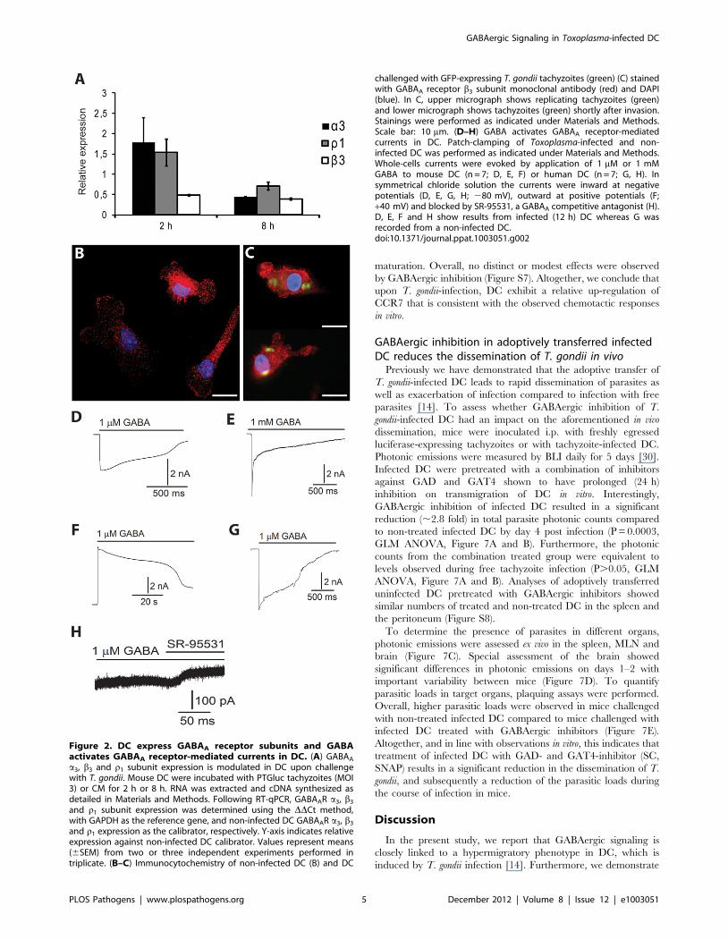
receptorsIn an effort to ascertain which GABAA receptor subunits are
expressed in mouse DC, we screened the 19 subunits expression
profiles in mouse DC and astrocytes. We detected GABAAR a3,
a5, b1, b3, and r1 subunit transcripts in DC, whilst 12 different
subunits were detected in primary astrocytes (Table 1, Table S1
for primer sequences). We decided to quantify differential gene
transcription in mouse DC following T. gondii infection using a3, b3
and r1, the most strongly expressed subunits in non-infected
mouse DC (Table 1). The transcript level analysis, using template
from infected DC and non-infected DC, showed an up-regulation
for the a3 and r1 transcripts after 2 h infection and down-
regulation by 8 h. A down-regulation was observed for the b3
subunit at both time-points (Figure 2A). In addition, immunocy-
tochemical stainings indicated expression of the b3 subunit in DC
in CM (Figure 2B) and in Toxoplasma-infected DC (Figure 2C). In
DC suspensions challenged with T. gondii, similar staining patterns
were observed in infected and non-infected DC (Figure S4).
We next examined functional expression of the GABAA
channels in DC using the whole-cell patch-clamp technique. We
recorded currents from human and mouse DC infected for 12 h
with T. gondii (Figure 2D, E, F, H) and non-infected DC
(Figure 2G). At a negative holding potential (- 80 mV) in
symmetrical chloride solutions, 1 mM GABA application to the
cells resulted in an inward current that ranged widely in
magnitude. In mouse and human DC the peak-current value
ranged from 29 pA to 29.9 nA (n = 7) and 238 pA to 27.7 nA
(n = 7), respectively. The currents reversed at positive holding
potential (Figure 2F, +40 mV) and were inhibited by the GABAA
competitive antagonist SR-95531 (Figure 2H). We conclude that
human and mouse myeloid DC express functional GABAA
receptors and that GABA can induce membrane currents in
Toxoplasma-infected DC.
Author Summary
Toxoplasma gondii is an obligate intracellular protozoanparasite and an important food- and water-borne humanand veterinary pathogen. Toxoplasmosis is normally self-limiting but severe manifestations occur upon congenitaltransmission to the developing fetus or during infection inimmune-compromised individuals. Toxoplasma invades avariety of cell types and mounting evidence shows thatcertain white blood cells, e.g. dendritic cells, can shuttleparasites in the infected host by a Trojan horse type ofmechanism. Dendritic cells are considered the gatekeepersof the immune system but can, paradoxically, also mediatedissemination of the parasite. Previous work has shownthat Toxoplasma induces a hypermigratory state indendritic cells when they become infected. Here, we showthat, shortly after infection by the parasite, dendritic cellsstart secreting c-aminobutyric acid (GABA), also known asthe major inhibitory neurotransmitter in the brain. Weshow that dendritic cells express GABA receptors, as wellas the machinery to synthesize and transport GABA. WhenGABA synthesis, transport or receptor function wasinhibited, the migration of infected dendritic cells wasimpaired. In a mouse model of toxoplasmosis, treatment ofinfected dendritic cells with GABA inhibitors resulted inreduced propagation of the parasite. This study establishesthat GABAergic signaling modulates the migratory prop-erties of dendritic cells and that the intracellular pathogenToxoplasma gondii sequesters the GABAergic signaling ofdendritic cells to assure propagation.
GABAergic Signaling in Toxoplasma-infected DC
PLOS Pathogens | www.plospathogens.org 2 December 2012 | Volume 8 | Issue 12 | e1003051
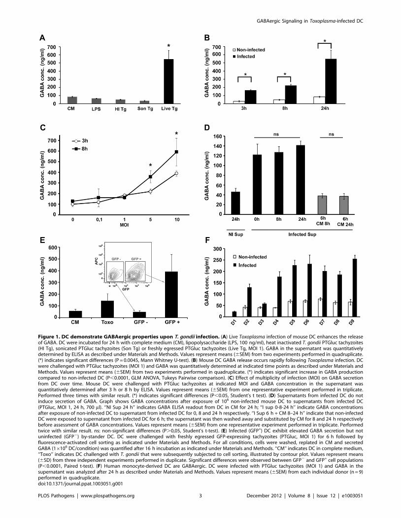
Figure 1. DC demonstrate GABAergic properties upon T. gondii infection. (A) Live Toxoplasma infection of mouse DC enhances the releaseof GABA. DC were incubated for 24 h with complete medium (CM), lipopolysaccharide (LPS, 100 ng/ml), heat inactivated T. gondii PTGluc tachyzoites(HI Tg), sonicated PTGluc tachyzoites (Son Tg) or freshly egressed PTGluc tachyzoites (Live Tg, MOI 1). GABA in the supernatant was quantitativelydetermined by ELISA as described under Materials and Methods. Values represent means (6SEM) from two experiments performed in quadruplicate.(*) indicates significant differences (P = 0.0045, Mann Whitney U-test). (B) Mouse DC GABA release occurs rapidly following Toxoplasma infection. DCwere challenged with PTGluc tachyzoites (MOI 1) and GABA was quantitatively determined at indicated time points as described under Materials andMethods. Values represent means (6SEM) from two experiments performed in quadruplicate. (*) indicates significant increase in GABA productioncompared to non-infected DC (P,0.0001, GLM ANOVA, Tukeys Pairwise comparison). (C) Effect of multiplicity of infection (MOI) on GABA secretionfrom DC over time. Mouse DC were challenged with PTGluc tachyzoites at indicated MOI and GABA concentration in the supernatant wasquantitatively determined after 3 h or 8 h by ELISA. Values represent means (6SEM) from one representative experiment performed in triplicate.Performed three times with similar result. (*) indicates significant differences (P,0.05, Student’s t test). (D) Supernatants from infected DC do notinduce secretion of GABA. Graph shows GABA concentrations after exposure of 106 non-infected mouse DC to supernatants from infected DC(PTGluc, MOI 1, 24 h, 700 ml). ‘‘NI Sup 24 h’’ indicates GABA ELISA readout from DC in CM for 24 h; ‘‘I sup 0-8-24 h’’ indicate GABA concentrationsafter exposure of non-infected DC to supernatant from infected DC for 0, 8 and 24 h respectively. ‘‘I Sup 6 h + CM 8–24 h’’ indicate that non-infectedDC were exposed to supernatant from infected DC for 6 h; the supernatant was then washed away and substituted by CM for 8 and 24 h respectivelybefore assessment of GABA concentrations. Values represent means (6SEM) from one representative experiment performed in triplicate. Performedtwice with similar result. ns: non-significant differences (P.0,05, Student’s t-test). (E) Infected (GFP+) DC exhibit elevated GABA secretion but notuninfected (GFP2) by-stander DC. DC were challenged with freshly egressed GFP-expressing tachyzoites (PTGluc, MOI 1) for 6 h followed byfluorescence-activated cell sorting as indicated under Materials and Methods. For all conditions, cells were washed, replated in CM and secretedGABA (16106 DC/condition) was quantified after 16 h incubation as indicated under Materials and Methods. ‘‘CM’’ indicates DC in complete medium,‘‘Toxo’’ indicates DC challenged with T. gondii that were subsequently subjected to cell sorting, illustrated by contour plot. Values represent means(6SD) from three independent experiments performed in duplicate. Significant differences were observed between GFP2 and GFP+ cell populations(P,0.0001, Paired t-test). (F) Human monocyte-derived DC are GABAergic. DC were infected with PTGluc tachyzoites (MOI 1) and GABA in thesupernatant was analyzed after 24 h as described under Materials and Methods. Values represent means (6SEM) from each individual donor (n = 9)performed in quadruplicate.doi:10.1371/journal.ppat.1003051.g001
GABAergic Signaling in Toxoplasma-infected DC
PLOS Pathogens | www.plospathogens.org 3 December 2012 | Volume 8 | Issue 12 | e1003051

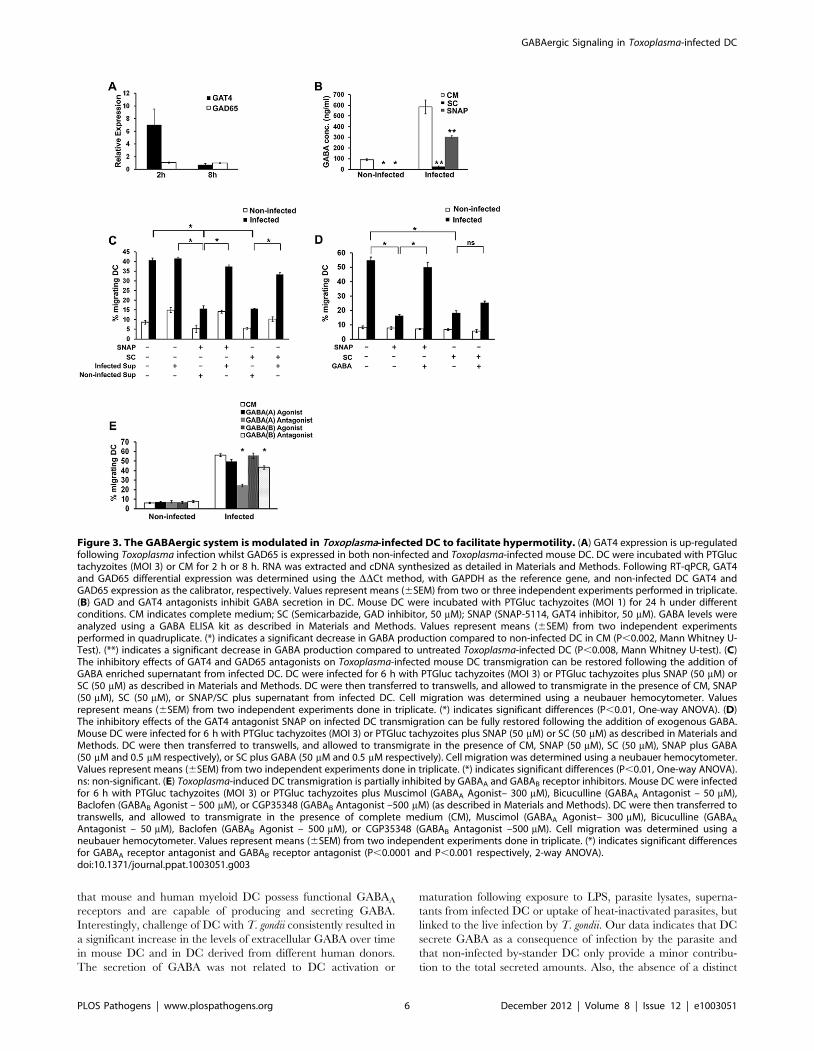
Targeting GABA synthesis and transport reducestransmigration of infected DC in vitro
To investigate the effects of Toxoplasma infection on the
GABAergic system, expression levels of the GABA transporter
GAT4 and the GABA synthesizing enzymes, GAD65 and
GAD67, were assessed in DC. A rapid induction of GAT4
transcription was observed shortly after infection (Figure 3A). In
contrast, expression of GAD65 was detected in both non-infected
and infected DC at similar levels (Figure 3A), whereas GAD67
expression was not detectable in either group (data not shown).
Moreover, addition of GAD inhibitor (SC) and GAT4 inhibitor
(SNAP) to infected DC nearly abolished or significantly reduced,
respectively, the secreted levels of GABA in the supernatant
(Figure 3B). Inhibitor treatments did not significantly affect
intracellular parasite replication in vitro (Figure S5) or the GABA
signal detected in complete medium containing extracellular
parasites (Figure S6). We next assessed the impact of the
GABAergic inhibitors on the transmigration of infected DC. Both
GAT4 (SNAP) and GAD inhibition (SC) had a significant
inhibitory effect on the transmigration of infected DC, and
transmigration was significantly restored following incubation of
the inhibitor-treated cells in supernatant from infected DC
cultures (Figure 3C). In contrast, monocytes did not exhibit this
migratory phenotype observed in human and murine DC [14] but
GABAergic inhibition significantly reduced the transmigration of
non-infected monocytes (Figure S2B). Furthermore, the transmi-
gration phenotype of DC was either fully (SNAP, GAT4 inhibitor)
or partially (SC, GAD inhibitor) restored after addition of
exogenous GABA (Figure 3D). In line with this, GABAA receptor
antagonist, and to a lesser extent GABAB receptor antagonist,
significantly reduced transmigration of infected DC (Figure 3E).
Notably, GABAA and GABAB receptor agonists did not enhance
transmigration of non-infected or infected DC. Thus, GABA per se
was not sufficient to induce transmigration of non-infected DC but
could restore transmigration in infected DC impaired in GABA
production or transportation. Altogether, these data implicate
GABA synthesis, transportation and receptor activity in parasite-
induced transmigration of DC in vitro.
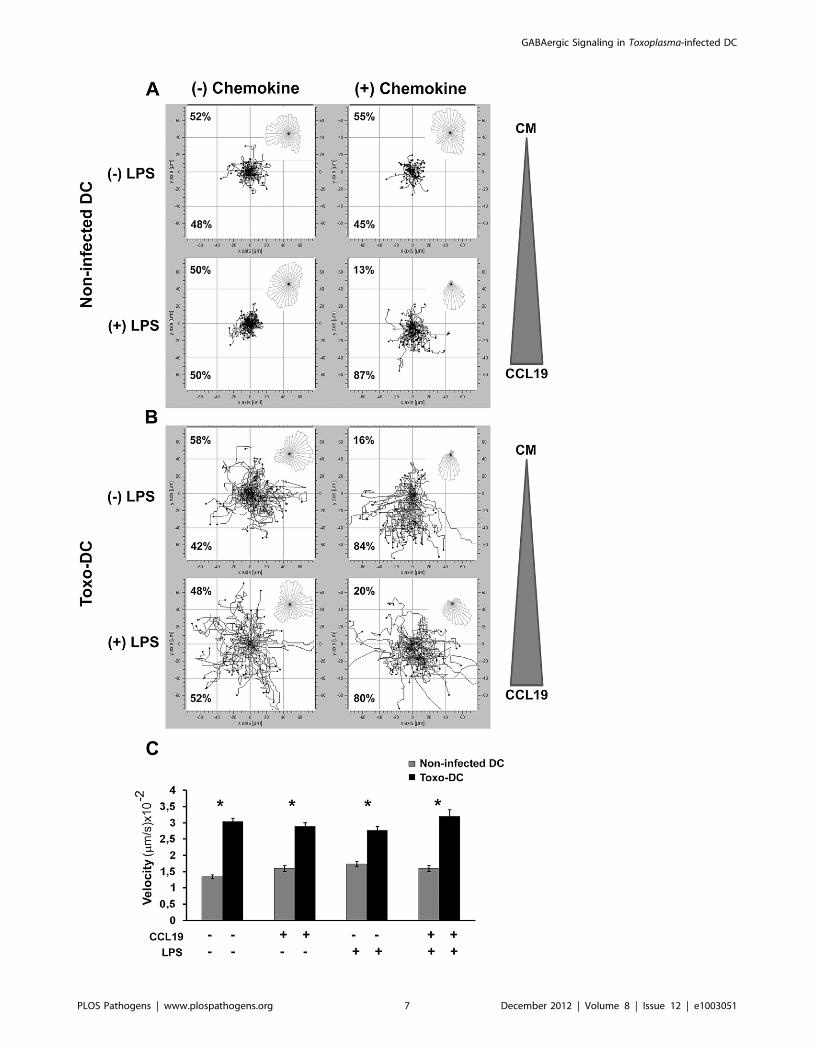
GABAergic signaling modulates motility and chemotaxisof infected DC in vitro
To determine whether the GABAergic system also affected DC
motility and chemotaxis, infected and non-infected DC were
allowed to migrate along a concentration gradient of CCL19 in a
chemotaxis chamber system. Non-infected DC exhibited a low
level of random directional motility in absence or presence of
chemokine and LPS-stimulation of non-infected DC resulted in a
distinct directional migration towards CCL19 (Figure 4A). In
contrast, Toxoplasma-infected DC exhibited a dramatically en-
hanced random directional motility in absence of chemokine
(Figure 4B), with a significant increase in velocity compared to
non-infected DC (Figure 4C). Interestingly, directionality towards
CCL19 was observed for infected DC, similar to that observed
upon LPS maturation (Figure 4B). It is also notable that
Toxoplasma-infected DC ((2) chemokine, Fig. 4 B) outranged
LPS-matured non-infected DC ((+) chemokine, Figure 4A) in
migrated distances and velocity (Figure 4A, B, C). We conclude
that Toxoplasma-infected DC exhibit a hypermigratory phenotype
in vitro and that hypermotile Toxoplasma-infected DC maintain the
ability to chemotax in vitro.
Next, we determined whether targeting the GABAergic system
affected the migratory and chemotactic responsiveness of non-
infected DC (Figure 5A, B) and Toxoplasma-infected DC
(Figure 5D, E) in vitro. Overall, inhibition of GABA synthesis
(SC, GAD inhibitor) or GABA transport (SNAP, GAT4 inhibitor)
led to a significant decrease in the velocity and the accumulated
distance covered by DC (Figure 5C, F). Interestingly, the ability to
respond with directionality towards CCL19 was not abolished by
inhibiting GABA transport or synthesis but, as a consequence of
the reduction in velocity, the overall chemotactic response was
diminished (Figure 5). No significant influence of a GABA gradient
on the directionality of DC motility was observed for non-infected
and infected DC (data not shown). In summary, present data show
that inhibition of the GABAergic signaling system significantly
reduces the velocity of infected DC in vitro and thereby the
magnitude of the chemotactic response in vitro.
Human and mouse DC exhibit upregulation of CCR7upon T. gondii infection in vitro
We next assessed the relative expression of the CCL19 ligand
CCR7 on human and mouse DC by flow cytometry. First, the
chemotactic responses observed with mouse DC were confirmed
using human monocyte-derived DC (Figure 6A). Additionally,
monitoring of infected and uninfected DC in suspensions
challenged with T. gondii showed that the chemotactic response
occurred preferentially in the infected (RFP+) DC population
(Figure 6A, central panel). In line with this result, DC challenged
with T. gondii or treated with LPS exhibited a relatively higher
expression of CCR7 compared to DC in complete medium
(Figure 6B). The analyses of DC populations challenged with T.
gondii showed that upregulation of CCR7 occurred essentially in
infected (RFP+) DC (Figure 6B, central panel). An upregulation of
CCR7 was consistently observed in infected DC from 7 different
human donors (Figure 6C). For mouse DC, a small but significant
upregulation of CCR7 was observed in infected DC (Figure 6E).
In the presence of GABAergic inhibitors (SC, SNAP), overall non-
significant effects on CCR7 expression were observed (Figure 6D
and E). We also assessed the effects of GABAergic inhibitors (SC,
SNAP) on the expression of co-stimulatory molecules and
Table 1. Characterization of GABAA receptor subunit gene expression in murine bone marrow-derived DC related to primarymurine astrocytes.
Cell type a1 a2 a3 a4 a5 a6 b1 b2 b3 c1 c2 c3 d h e p r1 r2 r3
DC xa x x xa xa
Astrocytes x x x x x x x x x x x x
A PCR screen of the 19 GABAA subunits was performed using mouse DC cDNA and mouse astrocyte cDNA as a positive control as indicated under Materials andMethods.aThe transcripts of GABAA receptor subunits a3, b3, and r1 were consistently quantified, and mean Ct values from non-infected DC cDNA were 37,0; 30,8; and 34,9respectively. The transcripts of subunits a5 and b1 could not be consistently quantified.doi:10.1371/journal.ppat.1003051.t001
GABAergic Signaling in Toxoplasma-infected DC
PLOS Pathogens | www.plospathogens.org 4 December 2012 | Volume 8 | Issue 12 | e1003051
maturation. Overall, no distinct or modest effects were observed
by GABAergic inhibition (Figure S7). Altogether, we conclude that
upon T. gondii-infection, DC exhibit a relative up-regulation of
CCR7 that is consistent with the observed chemotactic responses
in vitro.
GABAergic inhibition in adoptively transferred infectedDC reduces the dissemination of T. gondii in vivo
Previously we have demonstrated that the adoptive transfer of
T. gondii-infected DC leads to rapid dissemination of parasites as
well as exacerbation of infection compared to infection with free
parasites [14]. To assess whether GABAergic inhibition of T.
gondii-infected DC had an impact on the aforementioned in vivo
dissemination, mice were inoculated i.p. with freshly egressed
luciferase-expressing tachyzoites or with tachyzoite-infected DC.
Photonic emissions were measured by BLI daily for 5 days [30].
Infected DC were pretreated with a combination of inhibitors
against GAD and GAT4 shown to have prolonged (24 h)
inhibition on transmigration of DC in vitro. Interestingly,
GABAergic inhibition of infected DC resulted in a significant
reduction (,2.8 fold) in total parasite photonic counts compared
to non-treated infected DC by day 4 post infection (P = 0.0003,
GLM ANOVA, Figure 7A and B). Furthermore, the photonic
counts from the combination treated group were equivalent to
levels observed during free tachyzoite infection (P.0.05, GLM
ANOVA, Figure 7A and B). Analyses of adoptively transferred
uninfected DC pretreated with GABAergic inhibitors showed
similar numbers of treated and non-treated DC in the spleen and
the peritoneum (Figure S8).
To determine the presence of parasites in different organs,
photonic emissions were assessed ex vivo in the spleen, MLN and
brain (Figure 7C). Special assessment of the brain showed
significant differences in photonic emissions on days 1–2 with
important variability between mice (Figure 7D). To quantify
parasitic loads in target organs, plaquing assays were performed.
Overall, higher parasitic loads were observed in mice challenged
with non-treated infected DC compared to mice challenged with
infected DC treated with GABAergic inhibitors (Figure 7E).
Altogether, and in line with observations in vitro, this indicates that
treatment of infected DC with GAD- and GAT4-inhibitor (SC,
SNAP) results in a significant reduction in the dissemination of T.
gondii, and subsequently a reduction of the parasitic loads during
the course of infection in mice.
Discussion
In the present study, we report that GABAergic signaling is
closely linked to a hypermigratory phenotype in DC, which is
induced by T. gondii infection [14]. Furthermore, we demonstrate
Figure 2. DC express GABAA receptor subunits and GABAactivates GABAA receptor-mediated currents in DC. (A) GABAA
a3, b3 and r1 subunit expression is modulated in DC upon challengewith T. gondii. Mouse DC were incubated with PTGluc tachyzoites (MOI3) or CM for 2 h or 8 h. RNA was extracted and cDNA synthesized asdetailed in Materials and Methods. Following RT-qPCR, GABAAR a3, b3
and r1 subunit expression was determined using the DDCt method,with GAPDH as the reference gene, and non-infected DC GABAAR a3, b3
and r1 expression as the calibrator, respectively. Y-axis indicates relativeexpression against non-infected DC calibrator. Values represent means(6SEM) from two or three independent experiments performed intriplicate. (B–C) Immunocytochemistry of non-infected DC (B) and DC
challenged with GFP-expressing T. gondii tachyzoites (green) (C) stainedwith GABAA receptor b3 subunit monoclonal antibody (red) and DAPI(blue). In C, upper micrograph shows replicating tachyzoites (green)and lower micrograph shows tachyzoites (green) shortly after invasion.Stainings were performed as indicated under Materials and Methods.Scale bar: 10 mm. (D–H) GABA activates GABAA receptor-mediatedcurrents in DC. Patch-clamping of Toxoplasma-infected and non-infected DC was performed as indicated under Materials and Methods.Whole-cells currents were evoked by application of 1 mM or 1 mMGABA to mouse DC (n = 7; D, E, F) or human DC (n = 7; G, H). Insymmetrical chloride solution the currents were inward at negativepotentials (D, E, G, H; 280 mV), outward at positive potentials (F;+40 mV) and blocked by SR-95531, a GABAA competitive antagonist (H).D, E, F and H show results from infected (12 h) DC whereas G wasrecorded from a non-infected DC.doi:10.1371/journal.ppat.1003051.g002
GABAergic Signaling in Toxoplasma-infected DC
PLOS Pathogens | www.plospathogens.org 5 December 2012 | Volume 8 | Issue 12 | e1003051
that mouse and human myeloid DC possess functional GABAA
receptors and are capable of producing and secreting GABA.
Interestingly, challenge of DC with T. gondii consistently resulted in
a significant increase in the levels of extracellular GABA over time
in mouse DC and in DC derived from different human donors.
The secretion of GABA was not related to DC activation or
maturation following exposure to LPS, parasite lysates, superna-
tants from infected DC or uptake of heat-inactivated parasites, but
linked to the live infection by T. gondii. Our data indicates that DC
secrete GABA as a consequence of infection by the parasite and
that non-infected by-stander DC only provide a minor contribu-
tion to the total secreted amounts. Also, the absence of a distinct
Figure 3. The GABAergic system is modulated in Toxoplasma-infected DC to facilitate hypermotility. (A) GAT4 expression is up-regulatedfollowing Toxoplasma infection whilst GAD65 is expressed in both non-infected and Toxoplasma-infected mouse DC. DC were incubated with PTGluctachyzoites (MOI 3) or CM for 2 h or 8 h. RNA was extracted and cDNA synthesized as detailed in Materials and Methods. Following RT-qPCR, GAT4and GAD65 differential expression was determined using the DDCt method, with GAPDH as the reference gene, and non-infected DC GAT4 andGAD65 expression as the calibrator, respectively. Values represent means (6SEM) from two or three independent experiments performed in triplicate.(B) GAD and GAT4 antagonists inhibit GABA secretion in DC. Mouse DC were incubated with PTGluc tachyzoites (MOI 1) for 24 h under differentconditions. CM indicates complete medium; SC (Semicarbazide, GAD inhibitor, 50 mM); SNAP (SNAP-5114, GAT4 inhibitor, 50 mM). GABA levels wereanalyzed using a GABA ELISA kit as described in Materials and Methods. Values represent means (6SEM) from two independent experimentsperformed in quadruplicate. (*) indicates a significant decrease in GABA production compared to non-infected DC in CM (P,0.002, Mann Whitney U-Test). (**) indicates a significant decrease in GABA production compared to untreated Toxoplasma-infected DC (P,0.008, Mann Whitney U-test). (C)The inhibitory effects of GAT4 and GAD65 antagonists on Toxoplasma-infected mouse DC transmigration can be restored following the addition ofGABA enriched supernatant from infected DC. DC were infected for 6 h with PTGluc tachyzoites (MOI 3) or PTGluc tachyzoites plus SNAP (50 mM) orSC (50 mM) as described in Materials and Methods. DC were then transferred to transwells, and allowed to transmigrate in the presence of CM, SNAP(50 mM), SC (50 mM), or SNAP/SC plus supernatant from infected DC. Cell migration was determined using a neubauer hemocytometer. Valuesrepresent means (6SEM) from two independent experiments done in triplicate. (*) indicates significant differences (P,0.01, One-way ANOVA). (D)The inhibitory effects of the GAT4 antagonist SNAP on infected DC transmigration can be fully restored following the addition of exogenous GABA.Mouse DC were infected for 6 h with PTGluc tachyzoites (MOI 3) or PTGluc tachyzoites plus SNAP (50 mM) or SC (50 mM) as described in Materials andMethods. DC were then transferred to transwells, and allowed to transmigrate in the presence of CM, SNAP (50 mM), SC (50 mM), SNAP plus GABA(50 mM and 0.5 mM respectively), or SC plus GABA (50 mM and 0.5 mM respectively). Cell migration was determined using a neubauer hemocytometer.Values represent means (6SEM) from two independent experiments done in triplicate. (*) indicates significant differences (P,0.01, One-way ANOVA).ns: non-significant. (E) Toxoplasma-induced DC transmigration is partially inhibited by GABAA and GABAB receptor inhibitors. Mouse DC were infectedfor 6 h with PTGluc tachyzoites (MOI 3) or PTGluc tachyzoites plus Muscimol (GABAA Agonist– 300 mM), Bicuculline (GABAA Antagonist – 50 mM),Baclofen (GABAB Agonist – 500 mM), or CGP35348 (GABAB Antagonist –500 mM) (as described in Materials and Methods). DC were then transferred totranswells, and allowed to transmigrate in the presence of complete medium (CM), Muscimol (GABAA Agonist– 300 mM), Bicuculline (GABAA
Antagonist – 50 mM), Baclofen (GABAB Agonist – 500 mM), or CGP35348 (GABAB Antagonist –500 mM). Cell migration was determined using aneubauer hemocytometer. Values represent means (6SEM) from two independent experiments done in triplicate. (*) indicates significant differencesfor GABAA receptor antagonist and GABAB receptor antagonist (P,0.0001 and P,0.001 respectively, 2-way ANOVA).doi:10.1371/journal.ppat.1003051.g003
GABAergic Signaling in Toxoplasma-infected DC
PLOS Pathogens | www.plospathogens.org 6 December 2012 | Volume 8 | Issue 12 | e1003051
GABAergic Signaling in Toxoplasma-infected DC
PLOS Pathogens | www.plospathogens.org 7 December 2012 | Volume 8 | Issue 12 | e1003051

Figure 4. Toxoplasma-infected mouse DC exhibit hypermotility and chemotaxis. (A) Non-infected mouse DC or (B) Toxoplasma-infectedmouse DC (PTGluc, MOI 1/Toxo-DC) were incubated 6 LPS (200 ng/ml). After 24 h, cells were collected and placed in a chemotaxis chamber 6 CCL19gradient and tracked for 60 min as described under Materials and Methods. The tracking data presented is a composite of two independentexperiments (n = 60 cells). The overall directionality of migration is depicted in the rose-diagram in the upper right corner of each single tracksummary. Percentages represent the proportion of cells migrating towards or away from chemoattractant or complete medium (CM). For thecondition (+) chemokine, the triangle indicates the placement of the CCL19 chemokine gradient in each chemotaxis chamber. For (2) chemokine, CMwas added to both chambers. (C) Velocity analysis of non-infected mouse DC or Toxoplasma-infected mouse DC in presence or absence of thechemokine CCL19. Bars indicate velocity (+SEM) from two independent experiments. (*) indicate significant differences (P,0.001, Mann Whitney U-test).doi:10.1371/journal.ppat.1003051.g004
Figure 5. DC motility and chemotaxis are affected by the GABAergic system. (A, B) Non-infected mouse DC or (D, E) Toxoplasma-infectedmouse DC (PTGluc, MOI 1/Toxo-DC) were treated with GAD inhibitor (SC, Semicarbazide, 50 mM) or GAT4 inhibitor (SNAP, SNAP5114, 50 mM) 6 LPS(200 ng/ml). After 24 h, cells were collected and placed in a chemotaxis chamber 6 CCL19 gradient and tracked for 60 min as described underMaterials and Methods. The tracking data presented is a composite of one to two experiments performed in duplicate (n = 60 cells). The overalldirectionality of migration is depicted in the rose-diagram in the upper right corner of each single track summary. Percentages represent theproportion of cells migrating towards or away from chemoattractant or complete medium (CM). For the condition (+) chemokine, the triangleindicates the placement of the CCL19 chemokine gradient in each chemotaxis chamber. For (2) chemokine, CM was added to both chambers. (C, F)Velocity analysis of non-infected DC (A, B) and Toxoplasma-infected DC (D, E) in presence or absence of GABAergic inhibition (CM, complete medium;SC, GAD inhibitor; SNAP, GAT4 inhibitor) 6 gradient with the chemokine CCL19. Bars indicate velocity (+SEM) from two independent experiments. (*)indicate significant differences (P,0.001, Mann Whitney U-test).doi:10.1371/journal.ppat.1003051.g005
GABAergic Signaling in Toxoplasma-infected DC
PLOS Pathogens | www.plospathogens.org 8 December 2012 | Volume 8 | Issue 12 | e1003051
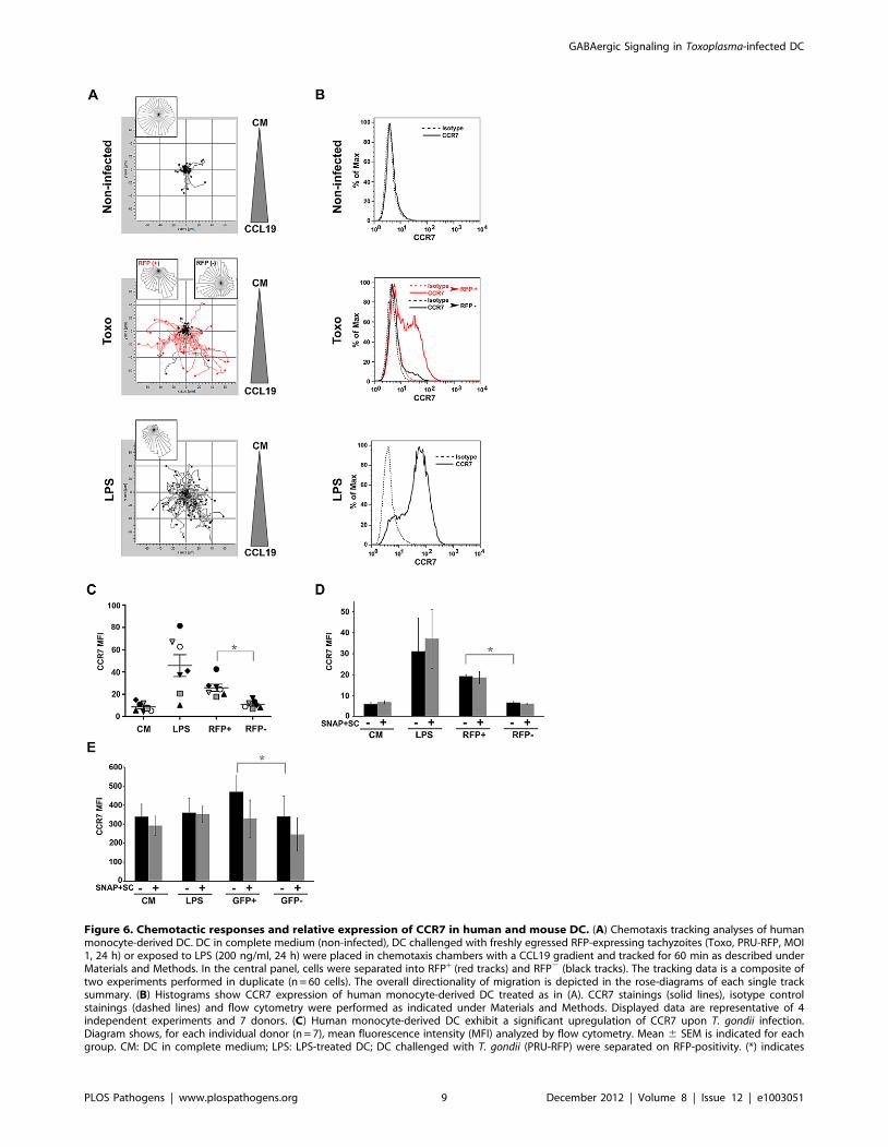
Figure 6. Chemotactic responses and relative expression of CCR7 in human and mouse DC. (A) Chemotaxis tracking analyses of humanmonocyte-derived DC. DC in complete medium (non-infected), DC challenged with freshly egressed RFP-expressing tachyzoites (Toxo, PRU-RFP, MOI1, 24 h) or exposed to LPS (200 ng/ml, 24 h) were placed in chemotaxis chambers with a CCL19 gradient and tracked for 60 min as described underMaterials and Methods. In the central panel, cells were separated into RFP+ (red tracks) and RFP2 (black tracks). The tracking data is a composite oftwo experiments performed in duplicate (n = 60 cells). The overall directionality of migration is depicted in the rose-diagrams of each single tracksummary. (B) Histograms show CCR7 expression of human monocyte-derived DC treated as in (A). CCR7 stainings (solid lines), isotype controlstainings (dashed lines) and flow cytometry were performed as indicated under Materials and Methods. Displayed data are representative of 4independent experiments and 7 donors. (C) Human monocyte-derived DC exhibit a significant upregulation of CCR7 upon T. gondii infection.Diagram shows, for each individual donor (n = 7), mean fluorescence intensity (MFI) analyzed by flow cytometry. Mean 6 SEM is indicated for eachgroup. CM: DC in complete medium; LPS: LPS-treated DC; DC challenged with T. gondii (PRU-RFP) were separated on RFP-positivity. (*) indicates
GABAergic Signaling in Toxoplasma-infected DC
PLOS Pathogens | www.plospathogens.org 9 December 2012 | Volume 8 | Issue 12 | e1003051
modulation on the secreted levels of the GABA-precursor
glutamate is indicative of a selective effect on GABA synthesis
and secretion.
We found that a determined subset of GABAA receptors subunit
genes was transcribed in mouse DC (a3, a5, b1, b3 and r1) in
contrast to the broader expression in astrocytes. This finding is
consistent with the concept that most GABAA receptor pentamers
are composed of at least two a- and two b-type subunits while the
final subunit type may vary [31]. This also strongly suggests that
genetic control plays a major role in the choice of transcribed
subunit variants. In fact, such differential expression has been
implicated in the changes in responsiveness and function of
GABAA receptors [32,33]. Moreover, factors that affect GABAer-
gic signaling, e.g. infection, may simultaneously induce up- and
down-regulation of specific GABAA subunits through epigenetic
mechanisms [34].
Here, we demonstrate for the first time that GABA evokes
GABAA receptor-mediated currents in T. gondii-infected DC and
in non-infected DC. While transcript levels of the a3, b3 and r1
subunits were modulated upon infection, functional patch-
clamping data indicates that GABAA receptors are constitutively
expressed in DC. In line with this, immunocytochemical analyses
indicated expression of the GABAA receptor b3 subunit in infected
and non-infected DC. As individual receptor subunits do not
necessarily reflect the number of functional receptor pentamers or
combinations of pentamers that are expressed in a particular cell,
it remains unknown how individual subunits relate to the receptor
function in DC. Thus, further studies are needed to characterize
and quantify the precise receptor subset composition of immune
cells, and whether sensitivity to GABA is modulated upon
infection.
Recently, human monocytes were shown to express the GABAA
b2 subunit and functional GABAA receptors were described in a
human myelomonocytic cell line (a4, b2, c1 and d subunits) [35].
In contrast to DC, monocytes do not exhibit enhanced transmi-
gration upon T. gondii-infection in vitro [36]. Here, we report that
monocytes respond with GABA secretion upon T. gondii infection.
Altogether, this raises the questions if receptor activation through
secreted GABA, e.g. an autocrine loop, is needed for migratory
activation or if different subsets of receptors are expressed in DC
compared to monocytes. Whether these intriguing phenotypic
differences between monocytes and DC depend on GABAA
receptor expression levels, functional receptor subunit composition
or capacity to rapidly secrete GABA awaits further investigation.
Induction of GABA secretion in infected DC was confirmed in
strains from the three predominant genotypes of T. gondii, but it is
unlikely that the maintenance and expression of GABA receptors
and the GABAergic system in DC are exclusively a result of
evolutionary pressure from T. gondii. Thus, additional functions for
the GABAergic system in DC are likely to be discovered.
Mounting evidence indicates that T. gondii modulates the host’s
pathways for cell migration to facilitate its dissemination and
establishment of a chronic infection. Our studies demonstrate that
targeting the GABAergic machinery of host DC, i.e. GABA
biosynthesis, transport, or ligand channel activation in vitro resulted
in impaired ability to transmigrate, most prominently using
inhibitors targeting host cell GAD and GAT4. Furthermore, the
motility of treated infected DC was reduced to levels comparable
to non-infected DC. In contrast, targeting the GABAergic system
did not abrogate the ability of DC to respond with directionality in
a CCL19 chemokine gradient but significantly reduced the speed
and migrated distances of infected DC, thus reducing the
chemotactic response in vitro. Further, in absence of a chemokine
gradient, the motility and velocity of DC was reduced. This
suggests that GABA and the GABAergic system may primarily
interfere with the mechanisms of cell motility. In fact, GABA has
been shown to promote the metastasis of cancer cells [37] and
increase the velocity of human sperm motility via activation of
GABAA and GABAB receptors [38]. Also, our in vitro chemotaxis
data using GABA as a chemoattractant suggests that GABA alone
is not sufficient to mitigate directional DC migration, irrespective
of infection status or LPS maturation.
Notably, during T. gondii infections CCR7 and CCL19 are up-
regulated [39,40] and soluble factors can trigger upregulation of
CCR7 in DC [41]. We have previously shown that Toxoplasma-
induced hypermotility of DC in vitro occurs in absence of
chemotactic cues and does not depend on CCR7-, CCR5- or
MyD88-activation [14]. Here, we report that hypermotile T.
gondii-infected human and mouse DC respond to chemotactic cues
(CCL19) in vitro and significantly up-regulate CCR7. In neurons,
reports indicate that GABAergic chemokinetic signaling cooper-
ates with other chemotactic cues for embryonic cell migration
[42]. In line with this, GABAergic inhibition has been shown to
down-modulate the chemotactic responses of monocytes and
neutrophils [35,43]. Hypothetically, we propose that the two
mechanisms tested here, GABA/GABAA receptor-mediated
hypermotility and CCR7-mediated chemotaxis, could be acting
simultaneously or even synergistically in Toxoplasma-infected DC,
thereby enhancing DC motility and potentiating the dissemination
of parasites infecting DC. Also, possible synergistic effects with
activation of multiple chemokine receptors or downstream
signaling [14,40,44] need to be tested in vivo in future research,
especially in the context of the remarkably efficient passage of the
parasite across restrictive biological barriers.
Present data are consistent with the notion that T. gondii is able
to subvert the regulation of host cell motility and exploits the host’s
natural pathways of cellular migration for parasite dissemination
[4]. In line with the above, GABAergic inhibition in T. gondii-
infected DC adoptively transferred to mice abolished the
disseminatory advantage provided by ‘shuttling’ DC [14] and
resulted in a reduced parasite burden over the course of infection
in mice. Although the overall trend was that GABAergic inhibition
yielded generally lower parasitic loads in organs, it is particularly
interesting that significantly lower parasitic loads were observed in
the brain. Recent reports have elucidated that the processes
leading to DC migration into the brain parenchyma in the context
of toxoplasmic encephalitis are complex and include signaling
through multiple chemokine receptors [40]. The present study
does not address the passage of the parasite across the blood brain
barrier and whether the infected DC directly transport parasites
into the CNS. Alternative but not mutually exclusive hypotheses
are possible for the observed differences in parasitic loads in the
brain. First, GABAergic inhibition of DC, similar to pertussis toxin
treatment [14,40], may reduce the number of DC that reach the
significant difference (RFP+ vs. RFP2: P#0.001, Paired t-test). (D and E) Effect of GABAergic inhibition on CCR7 expression in human DC (D) andmouse DC (E). Bar graphs show mean fluorescence intensity (MFI; 6SEM) analyzed by flow cytometry, from three independent experiments. Non-significant differences were observed in presence of GABAergic inhibitors (SC, SNAP) for the different conditions (P.0.05, Paired t-test). DC exhibiteda significant upregulation of CCR7 upon T. gondii infection, indicated by asterisk (RFP+ vs. RFP2: P = 0.0003 for human DC; GFP+ vs. GFP2: P = 0.029 formouse DC, Paired t-test).doi:10.1371/journal.ppat.1003051.g006
GABAergic Signaling in Toxoplasma-infected DC
PLOS Pathogens | www.plospathogens.org 10 December 2012 | Volume 8 | Issue 12 | e1003051

CNS microvasculature. Second, intracellular localization, e.g. in
migratory DC, may offer a safe intracellular niche per se for
targeted delivery to organs [4,18]. Third, differential transfer of
the parasite to other immune cells, e.g. NK, T cells, during
infection may modulate the dissemination and passage of T. gondii
and of infected leukocytes to the CNS [45–47]. The relative
contribution of these processes to the passage of T. gondii across the
blood brain barrier remains unknown. Nonetheless, adoptive
transfers with infected DC led to higher parasitic loads in the CNS
in models of toxoplasmosis and neosporosis [14,16,18], while
adoptive transfer of infected macrophages and lymphocytes did
not [36]. Different results have also been reported on the
contribution of CD11b+/monocytic cells [16,48]. Additionally,
these processes need to be tested in the context of oral natural
infections in future research.
On the technical note, bioluminescence imaging of the brain ex
vivo failed to detect significant differences between groups treated
with GABAergic inhibitors and untreated groups beyond day 2
post-infection. This could be due to the overall low parasitic loads
in the brain and to the relatively high variations between
individual mice, as demonstrated by plaquing assays. It also
indicates that plaquing assays remain the superior method in
detecting and quantifying viable parasitic loads in the CNS [30].
The effects of GABA on the immune system have not been
extensively studied. In human blood, GABAA receptor subunits
haven been detected in CD4+ and CD8+ T cells, B cells and
certain monocytes [35,49]. In rodents, functional GABAA
receptors have been described in T cells [50] and peritoneal
macrophages [51]. Activation of GABAA receptors has been
shown to inhibit T cell proliferation [50,52,53] and autoimmune
inflammation [51,54]. Thus, it is conceivable that GABA release
by T. gondii-infected DC may modulate DC activation to prevent
T-cell proliferation during the early phase of infection.
The ‘inflammatory reflex’ is a concept gaining ground in the
field of immunology in demonstrating that neurotransmitters can
interact and influence immune cell function [55]. This is an
interesting perspective given the psychobehavioural impact of
chronic Toxoplasma infection in humans and rodents [56]. Also
similar to DC, microglia respond with a migratory phenotype
upon T. gondii infection [47] and their migratory behavior in the
CNS could be modulated by ambient GABA. In fact GABA has
been shown to suppress the reactivity of microglia [57], leading to
attenuation of IL-6 and IL-12 responses [58]. Furthermore,
GABA levels in the CNS range from submicromolar outside of
synapses to millimolar concentrations in active synapses [59,60]
whilst GABA is present in peripheral tissues at around 100 nM
[61]. Here, the current responses to GABA by DC exhibited
characteristics of neuronal synaptic and extrasynaptic GABA-
activated currents [62]. The synaptic-like currents responded
rapidly and then decayed whereas the extrasynaptic-like currents
activated with a delay and maintained low current amplitudes for
an extended period. The finding that 1 mM GABA concentra-
tions gave rise to robust transient phasic and tonic currents
confirms that physiological GABA concentrations may be
sufficient to activate both types of currents in DC. This also
raises the prospect of GABA acting as a chemoattractant to
mitigate ‘homing’ of the infected leukocytes to the CNS and as a
possible modulator of microglia-mediated parasite dissemination
in the CNS [47]. Human cord blood-derived hematopoietic and
progenitor cells have been reported to migrate towards a GABA
gradient [63]. In contrast, our in vitro chemotaxis data using
GABA as a chemoattractant suggests that GABA alone is not
sufficient to mitigate directional migration of T. gondii-infected
DC in vitro.
A recently proposed model envisages a complex interplay
between ambient GABA, GABAA receptor activation, and
chloride transport as regulators of interneuron migration [64]. It
is conceivable that the DC GABAergic system may be working in
a similar manner in relation to the hypermigratory phenotype
exhibited by T. gondii-infected DC. Our model comprises: 1) DC
invasion by Toxoplasma, resulting in increased GABA production;
2) GABA is shuttled out of the cell by GABA transporters, leading
to an autocrine effect in activating GABAA receptors; 3) chloride
ion efflux by GABAA receptor channels and subsequent calcium
influx maintain DC in a depolarizing migratory state. The effector
mechanisms leading to increased GABA production in DC as a
consequence of T. gondii-infection await further investigation. It
has been shown that T. gondii-infection can lead to extensive
transcriptional regulation of host genes in DC [65] and a
modulated transcription of GAT4 and of GABAA a3, b3 and r1
subunits was observed in infected DC. We found no evidence of
significant production of GABA by extracellular parasites but a
modulation of the DC biosynthesis of GABA by intracellular
parasites cannot be excluded.
In summary, we provide substantial evidence that the DC
GABAergic system plays a significant role in the maintenance of
the T. gondii-induced hypermigratory phenotype observed in
infected DC. To the best of our knowledge, this constitutes the
first report showing that the GABAergic system can be utilized by
an intracellular pathogen to modulate host cell motility and
potentiate systemic dissemination. It remains to be seen whether
other pathogens also utilize the GABAergic system to facilitate the
establishment of an infection. Further investigation of the specific
molecules and pathways involved will enable a greater under-
standing of the diverse roles that the GABAergic system may play
outside the CNS.
Materials and Methods
Ethics statementAll protocols involving animals were approved by the Regional
Animal Research Ethical Board, Stockholm, Sweden, following
proceedings described in EU legislation (Council Directive 86/
609/EEC). The Regional Ethics Committee, Stockholm, Sweden,
approved protocols involving human cells. All donors received
written and oral information upon donation of blood at the
Karolinska University Hospital. Written consent was obtained for
utilization of white blood cells for research purposes. The ethics
committees approved this consent procedure.
Parasites and cell linesTachyzoites from the green fluorescence protein (GFP) and
luciferase-expressing T. gondii line PTGluc (type II, cloned from
ME49/PTG-GFPS65T) [30], RH-LDMluc (Type I, cloned from
RH-GFPS65T) [30], CTGluc (type III) [66] and PRU-RFP [67]
were maintained by serial 2-days passage in human foreskin
fibroblast (HFF) monolayers. HFFs were propagated in Dulbecco’s
modified Eagle’s medium (DMEM; Invitrogen) with 10% fetal
bovine serum (FBS), gentamicin (20 mg/ml, Gibco), glutamine
(2 mM, Gibco) and HEPES (0.01 M, Gibco) referred to as
complete medium (CM).
MiceC57BL/6 mice (6–10 weeks old) were maintained and bred at
the animal facility of the department of Microbiology, Tumor and
Cell Biology, Karolinska Institutet (Stockholm, Sweden). For
bioluminescence assays, male BALB/c mice (6–8 weeks old) were
GABAergic Signaling in Toxoplasma-infected DC
PLOS Pathogens | www.plospathogens.org 11 December 2012 | Volume 8 | Issue 12 | e1003051

purchased from Charles River (Sulzfeld, Germany) and main-
tained under pathogen-free conditions.
In vitro generated DC, monocytes and astrocytesMouse bone marrow-derived DC were generated as described
[68]. Briefly, cells from bone marrow of C57BL/6 mice were grown
in CM containing 20% supernatant from the GM-CSF-secreting cell
line X63 or 10 ng/ml recombinant mouse GM-CSF (Peprotech).
Loosely adherent cells were harvested on day 6. To generate human
monocyte-derived DC, buffy coats obtained from healthy blood
donors at the Karolinska University Hospital Blood Center were
treated with 1 ml RosetteSep (StemCell Technologies) per 15 ml of
buffy coat, followed by centrifugation on Lymphoprep (Axis.Shield
PoC AS) gradients. The population, defined as monocytes, exhibited
CD14+ (DakoCytomation) and ,1% CD3+/19+ (BD Biosciences) as
evaluated by flow cytometry (FACSCalibur, BD Biosciences). DC
were generated as described previously [69]. Briefly, purified cells
were cultured 7 days in CM supplemented with 100 ng/ml GM-
CSF (Peprotech) and 12.5 ng/ml IL-4 (R&D Systems). DC were
typified by expression of CD1a, CD11b, CD14 (DakoCytomation),
CD80, CD83, CD86, HLA-DR, CD11a, CD18 (BD Biosciences).
Primary astrocytes were generated from cortices from 1–3 day-old
C57BL/6 mice as previously described [47].
ImmunocytochemistryHuman monocyte-derived DC were cultured on poly-L-lysine-
coated glass coverslips (Sigma) and challenged with freshly egressed
tachyzoites (PTGluc, MOI 1) for 16 h. After fixation (3%
paraformaldehyde; Sigma) and blockade (5% FBS; Gibco), the cells
were stained with mouse-anti human b3 (NeuroMab; clone N87/25;
UC Davis/NIH NeuroMab Facility; 1:250). Anti-mouse Alexa
Fluor-conjugate (Invitrogen) was used as secondary antibody. Slides
were mounted using VectaShield with DAPI (Vector Laboratories)
and assessed by epifluorescence microscopy (Leica DMRB).
Methods for electrophysiologyDC (36106 cells) were collected and centrifuged for 2 min at
100 g. The supernatant was removed, the pellet washed with the
extracellular solution, and centrifuged for 2 min at 100 g. The
pellet was then resuspended using 100 ml of the extracellular
solution. Nanion’s Port-a-Patch chip technology (Nanion, Ger-
many) was used to voltage-clamp the cells. A cell suspension of 5 ml
was dispensed into the extracellular chamber containing the
recording chip of 2–3.5 MV resistance. The whole-cell configu-
ration was established and currents were recorded at holding
potential of 280 mV. GABA (1 mM and 1 mM) or GABA plus
100 mM SR95531 (GABAA antagonist, Sigma-Aldrich) were
prepared with the extracellular recording solution and perfused
into the extracellular chamber at a rate of 1 ml/min. Extracellular
solution in mM: 145 NaCl , 5 KCl, 1 MgCl2, 1.8 CaCl2 , 10 TES
(pH 7.3) and 297 mOsm/kg. Internal recording solution in mM:
50 CsCl , 10 NaCl, 60 Cs-Fluoride, 20 EGTA, 10 TES (pH 7.3)
and 284 mOsm/kg. Extracellular recording solution in mM: 80
NaCl, 3 KCl, 10 MgCl2, 35 CaCl2, 10 HEPES (pH 7.3) and 296
mOsm/kg. All patch-clamp recordings were performed at room
temperature (20–22uC). Patch-clamp recordings were done using
an Axopatch 200B amplifier, filtered at 2 kHz, digitized on-line at
10 kHz using an analogue-to-digital converter and analyzed with
pClamp software (Molecular Devices, USA).
RNA analysis with real time qPCRTotal RNA was extracted using TRIzol reagent (Invitrogen).
First-strand cDNA was synthesized using Superscript II Reverse
Transcriptase (Invitrogen). All primers were initially screened for
signal detection using cDNA and conventional PCR. Following
sequence confirmation, gene transcription levels were monitored.
Using the SYBR Green Master Mix (Applied Biosystems) PCR
reactions were carried out on the Applied Biosystems 7500 Fast
Real-Time PCR System using the following program: 10 min
holding at 95uC, 40 cycles of 95uC for 15 s, 60uC for 1 min. A
dissociation stage (95uC for 15 s, 60uC for 1 min, 95uC for 15 s) was
added to generate a melting curve for data analysis. Glyceraldehyde
3-phosphate dehydrogenase (GAPDH) was used as the reference
gene. The data was analyzed using Sequence Detection Software
v.1.3.1 (Applied Biosystems) and fold changes in expression were
calculated using the comparative DCT method against the non-
infected DC control. Primers against the 19 GABAAR subunits
(Table S1) were designed using OligoPerfect Designer software
(Invitrogen) and Primer-BLAST (http://www.ncbi.nlm.nih.gov/
tools/primer-blast/), and validated using OligoAnalyzer (http://eu.
idtdna.com/analyzer/Applications/OligoAnalyzer). The primers
were purchased from Invitrogen or Qiagen.
GABA and glutamate ELISADC were plated at a density of 16106 cells per well and
incubated with freshly egressed T. gondii tachyzoites (MOI 1) or
with soluble extracts and reagents in a total volume of 700 ml CM
for 24 h. Freshly egressed tachyzoites were heat-inactivated at
56uC for 30 min. Parasite lysates were obtained by harvesting
supernatants after sonicating 16106 tachyzoites for three 30 s
pulses (Soniprep 150). Reagents were purchased from Sigma-
Aldrich unless stated otherwise, and added at the following final
concentrations: Semicarbazide (SC, 50 mM); (S)-SNAP-5114
(SNAP; 50 mM); LPS (100 ng/ml). The cells were centrifuged at
4000 g for 2 min, and the medium supernatant collected and
stored at -70uC until analysis. Samples were extracted, derivatized,
and incubated with antiserum according to manufactures protocol
(GABA or Glutamate Research ELISA kits, Labor Diagnostica
Nord, Nordhorn, Germany). GABA and glutamate concentrations
were quantitatively determined by ELISA, monitored at 450 nm
(Multiskan EX, Labsystem Oy, Finland).
In vitro migration assays and replication assaysDC and monocytes were plated at a density of 16106 cells per
well and incubated with freshly egressed T. gondii tachyzoites (at
indicated MOI) or with soluble extracts and reagents in CM for
6 h. Cells were then gently transferred into triplicate transwell
filters (8 mm pore size; BD) at a density of 26105 per well, and
incubated as indicated. Reagents were added at the following final
concentrations: Muscimol (300 mM); Bicuculline (50 mM); Baclo-
fen (500 mM); CGP35348 (500 mM); Semicarbazide (50 mM);
SNAP 5114 (50 mM); LPS (100 ng/ml); GABA (0.5 mM). When
indicated, parasite viability and replication was assessed by
plaquing assays on HFF monolayers and vacuole counts in DC
as previously described [47]. DC integrity was monitored by
propidium iodide staining. In the GAD/GAT4 migration
restoration experiment, GABA supernatant was obtained from
infected DC which had been incubated for 24 h in CM to enrich
for DC derived GABA or commercially available exogenous
GABA was added to the transwell medium following DC transfer.
Migrated DC were quantified using a neubauer hemocytometer.
Chemotaxis assays, imaging and analysisNon-infected DC or Toxoplasma-infected DC (PTGluc/MOI 1)
were incubated 6 LPS (200 ng/ml) as well as 6 SNAP5114
(50 mM) or Semicarbazide (50 mM) for 24 h. Following incuba-
tion, DC were mixed with Collagen I solution (3 mg/ml, Gibco),
GABAergic Signaling in Toxoplasma-infected DC
PLOS Pathogens | www.plospathogens.org 12 December 2012 | Volume 8 | Issue 12 | e1003051
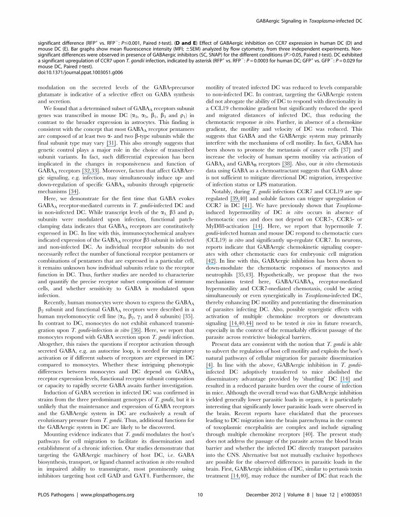
Figure 7. Decreased dissemination and parasite load after adoptive transfer of Toxoplasma-infected DC treated with GABAergicinhibitors. (A) BALB/c mice were challenged with 56104 cfu of freshly egressed PTGluc tachyzoites (Toxo), 56104 cfu of tachyzoite-infected DC(Toxo-DC) or 56104 cfu tachyzoite-infected DC treated with SNAP and SC (Toxo-DC + SNAP&SC). Photonic emissions were assessed by BLI on fiveconsecutive days after inoculation of mice. Images show progression of infection and parasite biomass expansion in mice. Control shows non-infected mouse. Color scales indicate photo emission (photons/s/cm2/sr) during a 180 s exposure. Data are from one set of mice and representativeof two independent experiments with three to five mice per group. (B) Total photon emission analysis from individual mice on days 1–5 postinoculation showed a decrease in parasite tissue burden in mice inoculated with GAD and GAT4 inhibitor-treated DC infected with T. gondii whencompared with burdens from mice inoculated with untreated DC infected with T. gondii. Photonic emissions were assessed (photons/s/ROI) during a180 s exposure. Data are from two independent experiments with 3–5 mice per group. Asterisks indicates a significant increase in photon emissionson day 4 and 5 post infection from mice in the ‘‘Toxo-DC’’ group compared to mice in the ‘‘Toxo’’ or ‘‘Toxo-DC (+SNAP&SC)’’ groups (P#0.0004, GLMANOVA, Tukeys Pairwise comparison). ROI – region of interest. (C) Ex vivo photonic emissions from spleen, MLN and brains of mice BALB/c micechallenged with 105 cfu of tachyzoite-infected DC (Toxo-DC) or 105 cfu tachyzoite-infected DC treated with SNAP and SC (Toxo-DC + SNAP&SC).Color scales indicate photonic emissions (photons/s/cm2/sr) during a 180 s exposure as indicated under Materials and Methods. Representativeimages from day 2 and day 5 post-inoculation are shown. White arrows (lower panel) indicate signal from the brain tissue. (D) Box-plot and whiskersgraph represents the lower, upper quartiles, median and minimum–maximum of photonic emissions ex vivo from the brains of infected mice. Thewhite bars indicate brains from mice infected with tachyzoite-infected DC (Toxo-DC) and the dark grey bars indicate brains from mice infected withtachyzoite-infected DC treated with SNAP and SC (Toxo-DC + SNAP&SC) (n = 4/group/day). Non-significant differences were observed between thetreated and untreated groups (P$0.05; Kruskal Wallis test) with significant differences on days 1 and 2 (P#0.05, Student’s t test, indicated byasterisks). (E) Parasite load in spleen, MLN and brain on days 1–6 post inoculation quantified by plaquing assays as indicated under Materials andMethods. BALB/c mice were challenged with 105 cfu of tachyzoite-infected DC (Toxo-DC, open circles) or 105 cfu tachyzoite-infected DC treated withSNAP and SC (Toxo-DC + SNAP&SC, filled triangles). Mean parasite tissue load for each group is indicated (open and filled lines respectively) anddashed lines represent trendlines for the two groups (n = 4/group/day). Significant differences in parasite load between groups were observed forspleen and brain (P#0.022 and P#0.02 respectively, Kruskal-Wallis test). Non-significant differences were observed in MLN (P$0.05, Kruskal-Wallistest) with significant differences between groups on days 2, 4, 5, 6 (P#0.05, Student’s t-test).doi:10.1371/journal.ppat.1003051.g007
GABAergic Signaling in Toxoplasma-infected DC
PLOS Pathogens | www.plospathogens.org 13 December 2012 | Volume 8 | Issue 12 | e1003051

7.5% NaHCO3 (Invitrogen) and 106 Minimum Essential
Medium (MEM; Invitrogen). Approximately 7.56104 cells were
loaded into m-slide 3D chemotaxis chamber slides (Ibidi) and
placed at 37uC, 5% CO2 to allow gel formation. Then, to establish
a gradient, 1.25 mg/ml CCL19 (R&D systems) or GABA (0.5 mM
or 5 mM) were added to one chamber reservoir whilst the other
reservoir was filled with CM. Control experiments used CM in
both reservoirs. Cell migration was monitored using a Zeiss
AxioImager Z1 microscope and AxioVision software (version
4.7.2). Images were taken every 60 s for 60 min. Cell tracking and
chemotaxis analysis were performed using ImageJ (http://imagej.
nih.gov/ij/) with Manual Tracking (Cordelieres, Institute Curie)
and Chemotaxis Tool (Ibidi) plugins.
Flow cytometryCCR7 expression of human monocyte-derived DC was studied
using FITC-labeled anti-CCR7 and mouse IgG2a isotype control
antibodies (R&D Systems). Murine bone marrow-derived DC
were stained with CCL19-Fc (eBiosciences), CD 40, CD80, CD86,
MHC II, CD18 antibodies (BD Biosciences) as indicated by the
manufacturer. Data were generated using a CyAn ADP (Beckman
Coulter) or a FACSCalibur (BD Bioscience) flow cytometer.
Fluorescence-activated sorting of Toxoplasma-infected cells was
performed at the Center for Cell Analysis, Karolinska institutet on
a FACSAria (BD Bioscience) system. Dead cells were gated out by
SYTOX Blue stain (Invitrogen) and pre-sorting infection rates
were 43–63%. Data analysis was done with FACSDiva software,
version 6.1.3 (BD Bioscience).
Adoptive transfers of DCDC were challenged with freshly egressed PTGluc tachyzoites
for 6 h at MOI 1. Extracellular parasites were removed following
three washes at 80 g. Following infection and resuspension in PBS,
56104 colony forming units (cfu) were adoptively transferred into
male BALB/c mice. Total numbers of cfu injected into animals
was confirmed by plaquing assays [70]. SNAP 5114 (50 mM) and
Semicarbazide (50 mM) was added upon DC infection for 5 h.
When indicated, such groups were treated with an additional
50 mM combination therapy of SNAP 5114 and Semicarbazide
for 1 h, and added to PBS DC suspension prior to injection.
DC were stained with 5 mM 5-,6-(4-chloromethyl)benzoyl-
amino-tetramethylrhodamine (CMTMR; Molecular Probes) or
5 mM carboxyfluorescein diacetate, succinimidyl ester (CFSE;
Invitrogen) as indicated by the manufacturer. Stained cells
(650 mM SC+50 mM SNAP for 6 h) were then injected i.p. into
BALB/c mice. After 12 h, the spleens were harvested, homoge-
nized and cells were analyzed by flow cytometry (FACScalibur).
An intraperitoneal lavage was performed and cells were similarly
analyzed by flow cytometry.
In vivo bioluminescence imaging (BLI)BLI was performed as described [30]. Briefly, BALB/c mice
inoculated i.p. with freshly egressed PTGluc tachyzoites, or with
PTGluc-infected DC 6 Semicarbazide and SNAP 5114 were
injected i.p. with 1.5 mg D-luciferin potassium salt (Caliper Life
Sciences, Hopkinton, MA, USA) and anaesthetized with 2.3%
isoflurane prior to BLI. Ten min after injection of D-luciferin,
biophotonic images were acquired at a binning of 8 (medium) for
180 s with an In Vivo Imaging System Spectrum CT (Caliper Life
Sciences). For ex vivo imaging of organs, mice were injected i.p.
with 1.5 mg D-luciferin and euthanized after 10–15 min. Organs
were extracted as assessed as above. Analysis of images and
assessment of photons emitted from a region of interest (ROI) was
performed with Living Image software (version 4.2; Caliper Life
Sciences).
Plaquing assaysPlaquing assays were performed as described [14]. Briefly,
organs were extracted and homogenized under conditions that did
not affect parasite viability. The number of parasites was
determined by plaque formation on HFF monolayers.
Statistical analysisAll statistics were performed using Minitab version 15 (Minitab
Inc, PA, USA).
Online supplemental materialTable S1 shows the 19 subunit primer pair sequences used to
screen DC and astrocyte cDNA for GABAAR subunit transcripts
and primer sequences for GAD 65, GAD 67, GAT4 and GAPDH
cDNA. Figure S1 shows the ELISA-determined glutamate levels
from non-infected and Toxoplasma-infected DC supernatant.
Figure S2 shows GABA secretion and transmigration by
monocytes upon challenge with T. gondii. Figure S3 shows GABA
secretion of DC challenged with T. gondii type I, II and III strains.
Figure S4 shows immunocytochemistry of infected and non-
infected DC. Figure S5 shows parasite replication in DC in the
presence of GABAergic inhibitors. Figure S6 shows the effects of
GABAergic inhibitors on tachyzoite-infected DC and extracellular
tachyzoites. Figure S7 shows effects of GABAergic inhibition on
activation and maturation markers of DC. Figure S8 shows flow
cytometric analyses of DC in vivo after GABAergic inhibition.
Supporting Information
Figure S1 DC glutamate secretion is transiently affect-ed by Toxoplasma infection. Mouse DC were challenged with
PTGluc tachyzoites (MOI 1) and glutamate levels were analyzed
at indicated time points using a glutamate ELISA kit as described
in Materials and Methods. Values represent means (6SEM) from
two experiments performed in quadruplicate. (*) indicates a
statistically significant increase in glutamate levels following
infection at 3 h (P = 0.034) and 8 h (P,0.001). However, there
were no significant differences in glutamate levels by the end of the
time course (GLM ANOVA, Tukeys pairwise comparison,
P.0.05).
(TIF)
Figure S2 GABA secretion and transmigration bymonocytes upon challenge with T. gondii. (A) Monocytes
respond with GABA secretion upon challenge with T. gondii.
Freshly isolated human monocytes from four donors (D1–D4)
were incubated with PTGluc tachyzoites (MOI 1) for 24 h. GABA
in the supernatant was quantitatively determined by ELISA as
described in Materials and Methods. For each donor, values
represent mean (6SD) performed in triplicate. (B) Transmigration
of freshly isolated monocytes. Monocytes were challenged with
PTGluc tachyzoites (MOI 3) or PTGluc tachyzoites plus SNAP
(GAT4 inhibitor, 50 mM) and SC (GAD inhibitor, 50 mM) as
described in Materials and Methods. Monocytes were then
transferred to transwells, and allowed to transmigrate in the
presence of CM (complete medium) or inhibitors (SNAP, 50 mM +SC, 50 mM). Cell migration was determined using a neubauer
hemocytometer. Values represent means (6SD) from two different
human donors done in triplicate. (*) indicates significant difference
(P,0.05, Student’s t-test). ns: non-significant.
(TIF)
GABAergic Signaling in Toxoplasma-infected DC
PLOS Pathogens | www.plospathogens.org 14 December 2012 | Volume 8 | Issue 12 | e1003051
Figure S3 GABA secretion of DC challenged with T.gondii type I, II and III strains. Mouse DC were challenged
with RH-LDMluc (type I), PTGluc (type II) or CTGluc (type III)
at indicated MOIs. GABA in the supernatant was quantitatively
determined after 24 h by ELISA as described under Materials and
Methods. One representative experiment performed in duplicate is
shown. Performed twice with similar result. Non-significant
differences were observed between strains (P.0.05, Student’s t-
test).
(TIF)
Figure S4 Infected and non-infected DC express theGABAA receptor b3 subunit. Micrographs show immunocyto-
chemistry of DC challenged with GFP-expressing T. gondii
tachyzoites (PTGluc, green) stained with GABAA receptor b3
subunit monoclonal antibody (red) and DAPI (blue). Stainings
were performed as indicated under Materials and Methods. Scale
bar: 10 mm.
(TIF)
Figure S5 Parasite replication in the presence ofGABAergic inhibition of DC. Replication of tachyzoites in
DC assessed by vacuole size counts at the indicated time points as
described in Materials and Methods. Non-significant differences
were observed between samples in complete medium (CM) and
samples treated with a combination of SC (GAD inhibitor, 50 mM)
and SNAP (GAT4 inhibitor, 50 mM) (P.0.05; Mann-Whitney U
test). Geneticin, a blocker of polypeptide synthesis in eukaryotic
cells, was used as a reference control. Significant differences were
observed in samples treated with geneticin (P,0.05; Mann-
Whitney U test).
(TIF)
Figure S6 GABAergic inhibitors target host cell-derivedGABA production and transport. Extracellular PTGluc
tachyzoites (Toxo) or tachyzoite-infected mouse DC (MOI 1;
Toxo-DC) were incubated for 24 h in CM under different
conditions. SC (GAD inhibitor, 50 mM); SNAP (GAT4 inhibitor,
50 mM). GABA levels were analyzed using a GABA ELISA kit as
described in Materials and Methods. Values represent means
(6SE) from two independent experiments performed in quadru-
plicate. (*) indicates a significant decrease in GABA production
compared to untreated Toxoplasma-infected DC (P,0.008, Mann
Whitney U test).
(TIF)
Figure S7 Co-stimulatory molecule stainings and mat-uration markers of DC in presence of GABAergic
inhibitors. Mouse bone marrow-derived DC were treated as
indicated for 12–16 h and stained as described under Materials
and Methods. ‘‘Untreated’’ and ‘‘LPS’’ indicate DC in complete
medium or exposed to LPS (200 ng/ml), respectively; ‘‘Toxo’’
indicates DC challenged with freshly egressed GFP-expressing
PTGluc tachyzoites (MOI 1). ‘‘SC + SNAP’’ indicates treatment
with SC (GAD inhibitor, 50 mM) and SNAP (GAT4 inhibitor,
50 mM). Results are representative of two or three independents
experiments with similar results.
(TIF)
Figure S8 Prevalence and dissemination of adoptivelytransferred DC upon GABAergic inhibition. BALB/c mice
were inoculated i.p with 56106 untreated DC labeled with
CMTMR and 56106 untreated DC labeled with CFSE (top panel)
or with 56106 untreated CMTMR-labeled DC and 56106
SC+SNAP treated DC, labeled with CFSE (bottom panel). After
12 h, an intraperitoneal lavage was performed and spleens were
extracted and processed for flow cytometry as indicated in
Materials and Methods. Dot plots of CMTMR (y-axis) and CFSE
(x-axis) show numbers of SC+SNAP treated DC as compared to
the untreated population, in both the spleen and peritoneal lavage.
Numbers indicate the relative portion of each quadrant related to
the total population. Double-positive (CMTMR/CFSE) cells may
indicate uptake in phagocytic cells. Data shown are from one
experiment, and are representative of the results from 2
independent experiments.
(TIF)
Table S1 Primer pair sequences used to screen DC andastrocyte cDNA for 19 GABAAR subunit transcripts andprimer sequences for GAD 65, GAD 67, GAT4 andGAPDH cDNA. Primers were designed as indicated under
Materials and Methods.
(DOCX)
Acknowledgments
We thank H-G Ljunggren, K. Kristensson and E. Collantes-Fernandez for
fruitful discussions.
Author Contributions
Conceived and designed the experiments: AB JMF JMW RBGA BB.
Performed the experiments: JMF RBGA JMW SKM ZJ RPAW BR AB.
Analyzed the data: JMF JMW RBGA SKM ZJ BB RPAW BR AB. Wrote
the paper: AB JMF JMW.
References
1. Joynson DH, Wreghitt TJ (2001) Toxoplasmosis: a comprehensive clinical guide.Cambridge, UK: Cambridge University Press.
2. Dobrowolski JM, Sibley LD (1996) Toxoplasma invasion of mammalian cells is
powered by the actin cytoskeleton of the parasite. Cell 84: 933–939.3. Channon JY, Seguin RM, Kasper LH (2000) Differential infectivity and division
of Toxoplasma gondii in human peripheral blood leukocytes. Infect Immun 68:4822–4826.
4. Lambert H, Barragan A (2010) Modelling parasite dissemination: host cell subversion
and immune evasion by Toxoplasma gondii. Cell Microbiol 12: 292–300.5. Lang C, Gross U, Luder CG (2007) Subversion of innate and adaptive immune
responses by Toxoplasma gondii. Parasitol Res 100: 191–203.6. Aliberti J, Valenzuela JG, Carruthers VB, Hieny S, Andersen J, et al. (2003)
Molecular mimicry of a CCR5 binding-domain in the microbial activation ofdendritic cells. Nat Immunol 4(5): 485–490.
7. Reis e Sousa C, Hieny S, Scharton-Kersten T, Jankovic D, Charest H, et al.
(1997) In vivo microbial stimulation induces rapid CD40 ligand-independentproduction of interleukin 12 by dendritic cells and their redistribution to T cell
areas. J Exp Med 186: 1819–1829.8. Mashayekhi M, Sandau MM, Dunay IR, Frickel EM, Khan A, et al. (2011)
CD8alpha(+) Dendritic Cells Are the Critical Source of Interleukin-12 that Controls
Acute Infection by Toxoplasma gondii Tachyzoites. Immunity 35: 249–259.
9. Steinman RM, Banchereau J (2007) Taking dendritic cells into medicine. Nature449: 419–426.
10. Alvarez D, Vollmann EH, von Andrian UH (2008) Mechanisms and
consequences of dendritic cell migration. Immunity 29: 325–342.11. Forster R, Davalos-Misslitz AC, Rot A (2008) CCR7 and its ligands: balancing
immunity and tolerance. Nat Rev Immunol 8: 362–371.12. Sacks D, Sher A (2002) Evasion of innate immunity by parasitic protozoa. Nat
Immunol 3: 1041–1047.
13. Liu CH, Fan YT, Dias A, Esper L, Corn RA, et al. (2006) Cutting edge:dendritic cells are essential for in vivo IL-12 production and development of
resistance against Toxoplasma gondii infection in mice. J Immunol 177: 31–35.
14. Lambert H, Hitziger N, Dellacasa I, Svensson M, Barragan A (2006) Inductionof dendritic cell migration upon Toxoplasma gondii infection potentiates
parasite dissemination. Cell Microbiol 8: 1611–1623.
15. Lambert H, Vutova PP, Adams WC, Lore K, Barragan A (2009) TheToxoplasma gondii-shuttling function of dendritic cells is linked to the parasite
genotype. Infect Immun 77: 1679–1688.16. Courret N, Darche S, Sonigo P, Milon G, Buzoni-Gatel D, et al. (2006) CD11c-
and CD11b-expressing mouse leukocytes transport single Toxoplasma gondii
tachyzoites to the brain. Blood 107: 309–316.
GABAergic Signaling in Toxoplasma-infected DC
PLOS Pathogens | www.plospathogens.org 15 December 2012 | Volume 8 | Issue 12 | e1003051
17. Bierly AL, Shufesky WJ, Sukhumavasi W, Morelli AE, Denkers EY (2008)
Dendritic cells expressing plasmacytoid marker PDCA-1 are Trojan horsesduring Toxoplasma gondii infection. J Immunol 181: 8485–8491.
18. Collantes-Fernandez E, Arrighi RB, Alvarez-Garcia G, Weidner JM, Regidor-
Cerrillo J, et al. (2012) Infected dendritic cells facilitate systemic dissemination
and transplacental passage of the obligate intracellular parasite Neosporacaninum in mice. PLoS One 7: e32123.
19. Farrant M, Kaila K (2007) The cellular, molecular and ionic basis of GABA(A)
receptor signalling. Prog Brain Res 160: 59–87.
20. Olsen RW, Sieghart W (2008) International Union of Pharmacology. LXX.Subtypes of gamma-aminobutyric acid(A) receptors: classification on the basis of
subunit composition, pharmacology, and function. Update. Pharmacol Rev 60:243–260.
21. Bettler B, Kaupmann K, Mosbacher J, Gassmann M (2004) Molecular structure
and physiological functions of GABA(B) receptors. Physiol Rev 84: 835–867.
22. Hoglund PJ, Adzic D, Scicluna SJ, Lindblom J, Fredriksson R (2005) The
repertoire of solute carriers of family 6: identification of new human and rodentgenes. Biochem Biophys Res Commun 336: 175–189.
23. Soghomonian JJ, Martin DL (1998) Two isoforms of glutamate decarboxylase:
why? Trends Pharmacol Sci 19: 500–505.
24. Owens DF, Kriegstein AR (2002) Is there more to GABA than synapticinhibition? Nat Rev Neurosci 3: 715–727.
25. Khakhalin AS (2011) Questioning the depolarizing effects of GABA during early
brain development. J Neurophysiol 106: 1065–1067.
26. Entschladen F, Bastian P, Niggemann B, Zaenker KS, Lang K (2004) Inhibitionof cell migration via G protein-coupled receptors to opioid peptides and
angiotensin. Ann N Y Acad Sci 1028: 320–328.
27. Entschladen F, Lang K, Drell TL, Joseph J, Zaenker KS (2002) Neurotrans-
mitters are regulators for the migration of tumor cells and leukocytes. CancerImmunol Immunother 51: 467–482.
28. Watanabe M, Maemura K, Kanbara K, Tamayama T, Hayasaki H (2002)
GABA and GABA receptors in the central nervous system and other organs. IntRev Cytol 213: 1–47.
29. Geigerseder C, Doepner R, Thalhammer A, Frungieri MB, Gamel-Didelon K,
et al. (2003) Evidence for a GABAergic system in rodent and human testis: local
GABA production and GABA receptors. Neuroendocrinology 77: 314–323.
30. Hitziger N, Dellacasa I, Albiger B, Barragan A (2005) Dissemination ofToxoplasma gondii to immunoprivileged organs and role of Toll/interleukin-1
receptor signalling for host resistance assessed by in vivo bioluminescenceimaging. Cell Microbiol 7: 837–848.
31. Birnir B, Korpi ER (2007) The impact of sub-cellular location and intracellular
neuronal proteins on properties of GABA(A) receptors. Curr Pharm Des 13:3169–3177.
32. Steiger JL, Russek SJ (2004) GABAA receptors: building the bridge between
subunit mRNAs, their promoters, and cognate transcription factors. Pharmacol
Ther 101: 259–281.
33. Sieghart W, Sperk G (2002) Subunit composition, distribution and function ofGABA(A) receptor subtypes. Curr Top Med Chem 2: 795–816.
34. Fukuchi M, Nii T, Ishimaru N, Minamino A, Hara D, et al. (2009) Valproic acid
induces up- or down-regulation of gene expression responsible for the neuronalexcitation and inhibition in rat cortical neurons through its epigenetic actions.
Neurosci Res 65: 35–43.
35. Wheeler DW, Thompson AJ, Corletto F, Reckless J, Loke JC, et al. (2011)Anaesthetic impairment of immune function is mediated via GABA(A) receptors.
PLoS One 6: e17152.
36. Lambert H, Dellacasa-Lindberg I, Barragan A (2011) Migratory responses of
leukocytes infected with Toxoplasma gondii. Microbes Infect 13: 96–102.
37. Azuma H, Inamoto T, Sakamoto T, Kiyama S, Ubai T, et al. (2003) Gamma-aminobutyric acid as a promoting factor of cancer metastasis; induction of
matrix metalloproteinase production is potentially its underlying mechanism.Cancer Res 63: 8090–8096.
38. Calogero AE, Hall J, Fishel S, Green S, Hunter A, et al. (1996) Effects of
gamma-aminobutyric acid on human sperm motility and hyperactivation. Mol
Hum Reprod 2: 733–738.
39. Noor S, Habashy AS, Nance JP, Clark RT, Nemati K, et al. (2010) CCR7-
dependent immunity during acute Toxoplasma gondii infection. Infect Immun
78: 2257–2263.
40. John B, Ricart B, Tait Wojno ED, Harris TH, Randall LM, et al. (2011)Analysis of behavior and trafficking of dendritic cells within the brain during
toxoplasmic encephalitis. PLoS Pathog 7: e1002246.
41. Diana J, Vincent C, Peyron F, Picot S, Schmitt D, et al. (2005) Toxoplasmagondii regulates recruitment and migration of human dendritic cells via different
soluble secreted factors. Clin Exp Immunol 141: 475–484.
42. Behar TN, Schaffner AE, Colton CA, Somogyi R, Olah Z, et al. (1994) GABA-
induced chemokinesis and NGF-induced chemotaxis of embryonic spinal cordneurons. J Neurosci 14: 29–38.
43. Rane MJ, Gozal D, Butt W, Gozal E, Pierce WM, Jr., et al. (2005) Gamma-
amino butyric acid type B receptors stimulate neutrophil chemotaxis duringischemia-reperfusion. J Immunol 174: 7242–7249.
44. Gouwy M, Struyf S, Noppen S, Schutyser E, Springael JY, et al. (2008) Synergy
between coproduced CC and CXC chemokines in monocyte chemotaxisthrough receptor-mediated events. Mol Pharmacol 74: 485–495.
45. Persson EK, Agnarson AM, Lambert H, Hitziger N, Yagita H, et al. (2007)
Death receptor ligation or exposure to perforin trigger rapid egress of theintracellular parasite Toxoplasma gondii. J Immunol 179: 8357–8365.
46. Persson CM, Lambert H, Vutova PP, Dellacasa-Lindberg I, Nederby J, et al.(2009) Transmission of Toxoplasma gondii from infected dendritic cells to
natural killer cells. Infect Immun 77: 970–976.
47. Dellacasa-Lindberg I, Fuks JM, Arrighi RB, Lambert H, Wallin RP, et al. (2011)Migratory Activation of Primary Cortical Microglia upon Infection with
Toxoplasma gondii. Infect Immun 79: 3046–3052.48. Lachenmaier SM, Deli MA, Meissner M, Liesenfeld O (2011) Intracellular
transport of Toxoplasma gondii through the blood-brain barrier.J Neuroimmunol 232: 119–130.
49. Alam S, Laughton DL, Walding A, Wolstenholme AJ (2006) Human peripheral
blood mononuclear cells express GABAA receptor subunits. Mol Immunol 43:1432–1442.
50. Tian J, Chau C, Hales TG, Kaufman DL (1999) GABA(A) receptors mediateinhibition of T cell responses. J Neuroimmunol 96: 21–28.
51. Bhat R, Axtell R, Mitra A, Miranda M, Lock C, et al. (2010) Inhibitory role for
GABA in autoimmune inflammation. Proc Natl Acad Sci U S A 107: 2580–2585.
52. Bjurstom H, Wang J, Ericsson I, Bengtsson M, Liu Y, et al. (2008) GABA, anatural immunomodulator of T lymphocytes. J Neuroimmunol 205: 44–50.
53. Mendu SK, Akesson L, Jin Z, Edlund A, Cilio C, et al. (2011) IncreasedGABA(A) channel subunits expression in CD8(+) but not in CD4(+) T cells in BB
rats developing diabetes compared to their congenic littermates. Mol Immunol
48: 399–407.54. Soltani N, Qiu H, Aleksic M, Glinka Y, Zhao F, et al. (2011) GABA exerts
protective and regenerative effects on islet beta cells and reverses diabetes. ProcNatl Acad Sci U S A 108: 11692–11697.
55. Rosas-Ballina M, Tracey KJ (2009) The neurology of the immune system: neural
reflexes regulate immunity. Neuron 64: 28–32.56. Henriquez SA, Brett R, Alexander J, Pratt J, Roberts CW (2009) Neuropsy-
chiatric disease and Toxoplasma gondii infection. Neuroimmunomodulation 16:122–133.
57. Lee M, Schwab C, McGeer PL (2011) Astrocytes are GABAergic cells thatmodulate microglial activity. Glia 59: 152–165.
58. Kuhn SA, van Landeghem FK, Zacharias R, Farber K, Rappert A, et al. (2004)
Microglia express GABA(B) receptors to modulate interleukin release. Mol CellNeurosci 25: 312–322.
59. Glykys J, Mody I (2007) Activation of GABAA receptors: views from outside thesynaptic cleft. Neuron 56: 763–770.
60. Floyer-Lea A, Wylezinska M, Kincses T, Matthews PM (2006) Rapid
modulation of GABA concentration in human sensorimotor cortex duringmotor learning. J Neurophysiol 95: 1639–1644.
61. Bjork JM, Moeller FG, Kramer GL, Kram M, Suris A, et al. (2001) PlasmaGABA levels correlate with aggressiveness in relatives of patients with unipolar
depressive disorder. Psychiatry Res 101: 131–136.62. Jin Z, Jin Y, Kumar-Mendu S, Degerman E, Groop L, et al. (2011) Insulin
reduces neuronal excitability by turning on GABA(A) channels that generate
tonic current. PLoS One 6: e16188.63. Zangiacomi V, Balon N, Maddens S, Tiberghien P, Versaux-Botteri C, et al.
(2009) Human cord blood-derived hematopoietic and neural-like stem/progenitor cells are attracted by the neurotransmitter GABA. Stem Cells Dev
18: 1369–1378.
64. Bortone D, Polleux F (2009) KCC2 expression promotes the termination ofcortical interneuron migration in a voltage-sensitive calcium-dependent manner.
Neuron 62: 53–71.65. Chaussabel D, Semnani RT, McDowell MA, Sacks D, Sher A, et al. (2003)
Unique gene expression profiles of human macrophages and dendritic cells to
phylogenetically distinct parasites. Blood 102: 672–681.66. Dellacasa-Lindberg I, Hitziger N, Barragan A (2007) Localized recrudescence of
Toxoplasma infections in the central nervous system of immunocompromisedmice assessed by in vivo bioluminescence imaging. Microbes Infect 9: 1291–
1298.67. Pepper M, Dzierszinski F, Wilson E, Tait E, Fang Q, et al. (2008) Plasmacytoid
dendritic cells are activated by Toxoplasma gondii to present antigen and
produce cytokines. J Immunol 180: 6229–6236.68. Norbury CC, Chambers BJ, Prescott AR, Ljunggren HG, Watts C (1997)
Constitutive macropinocytosis allows TAP-dependent major histocompatibilitycomplex class I presentation of exogenous soluble antigen by bone marrow-
derived dendritic cells. Eur J Immunol 27: 280–288.
69. Sallusto F, Lanzavecchia A (1994) Efficient presentation of soluble antigen bycultured human dendritic cells is maintained by granulocyte/macrophage
colony-stimulating factor plus interleukin 4 and downregulated by tumornecrosis factor alpha. J Exp Med 179: 1109–1118.
70. Barragan A, Sibley LD (2002) Transepithelial migration of Toxoplasma gondii islinked to parasite motility and virulence. J Exp Med 195: 1625–1633.
GABAergic Signaling in Toxoplasma-infected DC
PLOS Pathogens | www.plospathogens.org 16 December 2012 | Volume 8 | Issue 12 | e1003051


![Synthesis, anti- Toxoplasma gondii and antimicrobial activities of benzaldehyde 4-phenyl-3-thiosemicarbazones and 2-[(phenylmethylene)hydrazono]-4-oxo-3-phenyl-5-thiazolidineacetic](https://static.fdokumen.com/doc/165x107/63133f6fb22baff5c40f0921/synthesis-anti-toxoplasma-gondii-and-antimicrobial-activities-of-benzaldehyde.jpg)