Forward Genetics in Toxoplasma gondii Reveals a Family of Rhoptry Kinases That Mediates Pathogenesis
10
Published Ahead of Print 22 May 2009. 2009, 8(8):1085. DOI: 10.1128/EC.00107-09. Eukaryotic Cell Asis Khan and Raymond Hui L. David Sibley, Wei Qiu, Sarah Fentress, Sonya J. Taylor, Mediates Pathogenesis Reveals a Family of Rhoptry Kinases That Toxoplasma gondii Forward Genetics in http://ec.asm.org/content/8/8/1085 Updated information and services can be found at: These include: REFERENCES http://ec.asm.org/content/8/8/1085#ref-list-1 at: This article cites 43 articles, 26 of which can be accessed free CONTENT ALERTS more» articles cite this article), Receive: RSS Feeds, eTOCs, free email alerts (when new http://journals.asm.org/site/misc/reprints.xhtml Information about commercial reprint orders: http://journals.asm.org/site/subscriptions/ To subscribe to to another ASM Journal go to: on September 26, 2013 by guest http://ec.asm.org/ Downloaded from on September 26, 2013 by guest http://ec.asm.org/ Downloaded from on September 26, 2013 by guest http://ec.asm.org/ Downloaded from on September 26, 2013 by guest http://ec.asm.org/ Downloaded from on September 26, 2013 by guest http://ec.asm.org/ Downloaded from on September 26, 2013 by guest http://ec.asm.org/ Downloaded from on September 26, 2013 by guest http://ec.asm.org/ Downloaded from on September 26, 2013 by guest http://ec.asm.org/ Downloaded from on September 26, 2013 by guest http://ec.asm.org/ Downloaded from on September 26, 2013 by guest http://ec.asm.org/ Downloaded from
Transcript of Forward Genetics in Toxoplasma gondii Reveals a Family of Rhoptry Kinases That Mediates Pathogenesis
Published Ahead of Print 22 May 2009. 2009, 8(8):1085. DOI: 10.1128/EC.00107-09. Eukaryotic Cell
Asis Khan and Raymond HuiL. David Sibley, Wei Qiu, Sarah Fentress, Sonya J. Taylor, Mediates Pathogenesis Reveals a Family of Rhoptry Kinases That
Toxoplasma gondiiForward Genetics in
http://ec.asm.org/content/8/8/1085Updated information and services can be found at:
These include:
REFERENCEShttp://ec.asm.org/content/8/8/1085#ref-list-1at:
This article cites 43 articles, 26 of which can be accessed free
CONTENT ALERTS more»articles cite this article),
Receive: RSS Feeds, eTOCs, free email alerts (when new
http://journals.asm.org/site/misc/reprints.xhtmlInformation about commercial reprint orders: http://journals.asm.org/site/subscriptions/To subscribe to to another ASM Journal go to:
on Septem
ber 26, 2013 by guesthttp://ec.asm
.org/D
ownloaded from
on S
eptember 26, 2013 by guest
http://ec.asm.org/
Dow
nloaded from
on Septem
ber 26, 2013 by guesthttp://ec.asm
.org/D
ownloaded from
on S
eptember 26, 2013 by guest
http://ec.asm.org/
Dow
nloaded from
on Septem
ber 26, 2013 by guesthttp://ec.asm
.org/D
ownloaded from
on S
eptember 26, 2013 by guest
http://ec.asm.org/
Dow
nloaded from
on Septem
ber 26, 2013 by guesthttp://ec.asm
.org/D
ownloaded from
on S
eptember 26, 2013 by guest
http://ec.asm.org/
Dow
nloaded from
on Septem
ber 26, 2013 by guesthttp://ec.asm
.org/D
ownloaded from
on S
eptember 26, 2013 by guest
http://ec.asm.org/
Dow
nloaded from
EUKARYOTIC CELL, Aug. 2009, p. 1085–1093 Vol. 8, No. 81535-9778/09/$08.00�0 doi:10.1128/EC.00107-09Copyright © 2009, American Society for Microbiology. All Rights Reserved.
MINIREVIEW
Forward Genetics in Toxoplasma gondii Reveals a Family of RhoptryKinases That Mediates Pathogenesis�
L. David Sibley,1* Wei Qiu,2 Sarah Fentress,1 Sonya J. Taylor,1 Asis Khan,1 and Raymond Hui2
Department of Molecular Microbiology, Washington University School of Medicine, St. Louis, Missouri 63130,1 andStructural Genomics Consortium, University of Toronto, Toronto, Ontario M5G 1L7, Canada2
Toxoplasma gondii is a widespread protozoan parasite thatcommonly infects domestic, wild, and companion animals (8).Like related tissue coccidians, T. gondii has a complex life cyclealternating between a sexual cycle, which occurs only withinenterocytes of the small intestines of cats (all members of theFelidae appear to be susceptible), and asexual propagation ina variety of warm-blooded vertebrate hosts (8). The sexualphase culminates with fecal shedding of a spore-like stagecalled the oocyst, which is diploid and undergoes meiosis in theenvironment. During the asexual cycle, the parasite intercon-verts between a fast-growing lytic form known as the tacyhzoiteand a slow-growing, semidormant form called the bradyzoite.Tachyzoites propagate rapidly in virtually all types of nucleatedcells, including macrophages, while differentiation to brady-zoites is favored in long-lived, terminally differentiated hostcells (49). Tachyzoites are adept at direct migration acrosscellular barriers and also disseminate rapidly within leukocytes,thereby reaching sites of immune privilege such as the centralnervous system and the developing fetus, where they are morelikely to cause disease (2). The life cycle shows remarkableflexibility between lytic and dormant states, thus facilitatingasexual transmission between intermediate hosts. This adapta-tion may account for the recent spread and emergence of a fewdominant clonal groups within North America and Europe(46).
Toxoplasma is well adapted to mammalian hosts, beingtransmitted by ingestion of undercooked meat harboring tissuecysts and through food and water supplies contaminated withoocysts shed from cats (8). Humans are accidental hosts of T.gondii, yet seroprevalence rates indicate high rates of chronicinfection in many countries of Europe and Central and SouthAmerica (16). Infections are often mild or subclinical inhealthy adults; however, toxoplasmosis can present as a clini-cally important infection in immunocompromised patients andthe developing fetus (32). In many regions of the world, toxo-plasmosis remains a frequent problem in patients infected withhuman immunodeficiency virus, due to lack of access to effec-tive antiviral therapy (29). Additionally, in regions such as
southern Brazil, ocular toxoplasmosis often presents as a clin-ically severe infection in otherwise healthy adults (18).
Although T. gondii is primarily an opportunistic pathogen, ithas emerged as a model for study of the biology of apicompl-exan parasites, a group that contains Plasmodium spp. (malariaparasites), Cryptosporidium spp., and a variety of animal patho-gens. Although the life cycles of these parasites differ substan-tially, they share common pathways for actin-myosin-basedmotility, calcium-dependent secretion, and active cell invasion(41). Toxoplasma offers excellent tools for studying molecularand cell biology, forward and reverse genetics, and animalmodels. Hence, exploration of the molecular basis of complextraits such as pathogenesis has been feasible in this system.This review summarizes recent advances using forward genet-ics to identify genes involved in pathogenesis, as well as the useof reverse genetics to validate the roles of prospective candi-date genes.
STRAIN DIVERSITY AND GENETICANALYSIS IN T. GONDII
Toxoplasma gondii has a highly unusual population structurecomprising three predominant clonal populations in NorthAmerica and Europe (42). These lineages are found in over-lapping geographic regions, where they infect a variety ofwarm-blooded animals, including humans. Despite beingmaintained as clonal lineages, they are highly similar geneti-cally and originated from several closely related parental typesin the wild (6). Estimates of their last common ancestry indi-cate that this event occurred within the past 10,000 years,followed by rapid expansion of these three lineages (46). Thecause of this unusual pattern is uncertain, but it may indicate alocalized bottleneck followed by the expansion of a few lin-eages, or a strong genetic sweep that replaced abundant andmore-diverse isolates. Intriguingly, this recent origin is alsomarked by the common inheritance of a single monomorphicchromosome Ia that is shared by the three clonal lineages (19).The population structure differs substantially in South Amer-ica, where lineages are genetically more diverse and undergoboth sexual recombination and asexual transmission (20, 28).
While the origins of the unique population structure areuncertain, it has profound consequences for population stud-ies. For example, association studies generally are not infor-mative for defining the contributions of genes to particularphenotypes, since linkage disequilibrium is so high. However, a
* Corresponding author. Mailing address: Department of MolecularMicrobiology, Washington University School of Medicine, St. Louis,MO 63130. Phone: (314) 362-8873. Fax: (314) 286-0060. E-mail: [email protected].
� Published ahead of print on 22 May 2009.
1085
number of strong phenotypic differences have been noted; forexample, type I strains are acutely virulent in outbred mice (seebelow). Additionally, type I strains have been shown to exhibitenhanced migration across biological barriers in vitro and todisseminate more rapidly in vivo in the mouse model thanother strains (3). It is not clear how these traits translate tohuman infections, which are caused primarily by type II strains,although several reports have indicated an increased frequencyof type I strains (or strains bearing type I alleles) among clin-ical isolates (11, 14, 21). Although the laboratory mouse ap-pears to be highly sensitive to toxoplasmosis compared to hu-mans, it provides a useful starting point for investigating genesthat contribute to pathogenesis.
Classical genetic crosses in T. gondii were pioneered by thestudies of Elmer Pfefferkorn and his colleagues, who estab-lished procedures for crossing strains of the parasite by coin-fecting cats and collecting the resulting recombinant oocysts.These early studies established several key properties, includ-ing the following. (i) A single cloned tachyzoite can give rise tothe entire life cycle, indicating that there is not a predeter-mined mating type (34). (ii) The replicating stages are allhaploid, and mating leads to a diploid stage (35). (iii) Whencoinfecting a single cat, strains undergo self-mating or out-crossing at approximately equal frequencies (35). (iv) Un-linked genes segregate randomly and yet can demonstrate ep-istatic interactions (33). These studies provided the beginningsof a working genetic system, which was later expanded by thedevelopment of molecular markers, thus allowing the construc-tion of rudimentary linkage maps (44). Further expansion ofthe number of markers was used to support preliminary map-ping of acute virulence in the mouse model; remarkably, viru-lence was linked to a single major locus on chromosome VII(since renamed VIIa) (47). This study was also notable becauseit demonstrated that acute virulence was a genetically heritabletrait and suggested that fine mapping could likely be used toidentify the specific gene(s) involved. For the current versionof the genetic map, the segregation of more than 200 markerswas compared among crosses between either types II and III ortypes I and III, to generate linkage maps for the 14 chromo-somes (23). This composite map defined the order of markerson the chromosomes, established the basic parameters for re-combination, and provided a framework for assembly of theT. gondii genome, which is approximately 65 Mb (http://ToxoDB.org). The composite genetic map consists of �600cM (1 cM � 1% recombination), with an average map unit ofaround 100 kb. Although the markers are fairly widely dis-persed, the most limiting attribute of genetic mapping in T.gondii is the relatively modest recombination rate; hence, map-ping precision is limited by the number of available progenyrather than by the frequency of markers. The various advan-tages and limitations of forward genetics in T. gondii have beendiscussed previously in more detail (1).
MAPPING ACUTE VIRULENCE IN THE MOUSE MODEL
Toxoplasma gondii naturally infects a variety of rodents, andlaboratory mice provide a reasonable model for both acute andchronic infections (38). Type I strains are acutely virulent inmice: limiting dilution indicates that a single tachyzoite admin-istered by needle inoculation is lethal in many laboratory
mouse strains, including outbred mice (43). In contrast, type IIand III strains are nonvirulent in outbred mice, although dif-ferences can be discerned in the susceptibilities of inbred mice(38). Death of the animals results from overwhelming cytokineproduction, an outcome that occurs following inoculation withlow doses of type I strains but only after high doses of type IIstrains (12, 30). While needle inoculation bypasses the naturaloral route of infection, it has the advantage of being easilyquantifiable and reproducible and giving very discrete end-points. For these reasons, genetic mapping studies of complexphenotypes have relied largely on this model. Additional phe-notypic differences include the variable induction of cytokinesfollowing in vitro infection. For example, type II strains arepotent inducers of interleukin 12 (IL-12) from naïve macro-phages, while type I and III strains are relatively quiescent(39). Differences in the activation of pathways for IL-12 induc-tion have also been noted: type I strains rely primarily on anon-MyD88-dependent pathway, while type II strains triggerboth dependent and independent pathways, leading to greateroverall levels of IL-12 (24). While this seems counter to theresults described above for in vivo infection (type I leads togreatly elevated cytokine levels later in infection), differencesincluding growth rate (37), escape from cellular destruction(50), and ability to disseminate rapidly (3) also contribute todifferent outcomes in vivo.
Forward genetic analysis of acute virulence in the murinemodel was based on a genetic cross between the acutely viru-lent type I strain called GT-1 (marked by resistance to fluoro-deoxyuridine) and the type III strain CTG (marked by resis-tance to adenine arabinoside and sinefungin) (47). Progenyclones from this cross were tested in outbred mice to defineacute virulence based on cumulative mortality during the initialphase of infection following different doses of trachyzoites.The progeny were also genotyped in order to analyze thesegregation of acute mortality with specific markers. Geneticassociations of other traits were also analyzed based on theability to migrate across polarized epithelia (transmigration)and the ability to migrate away from a focus of infection, traitsthat had previously been associated with type I strains (3).Differences in growth rate were also analyzed based on previ-ous findings that type I strains have a shorter cell cycle (37).Finally, we examined the serological responses in survivinganimals, since less-virulent strains readily give rise to chronicinfections, while acutely virulent type I strains do not (43).Because these traits were likely to arise as the result of acombination of genetic effects, the phenotypes were analyzedusing quantitative trait locus (QTL) mapping (26, 27), a sta-tistical method for defining the association of complex pheno-types with multiple underlying genetic regions. Analyses of 34genetically independent recombinant progeny were based on175 of the genetic markers from the composite genetic mapthat were informative in the type I-versus-type III cross (theremaining markers being identical between these two lineagesbut different in type II) (23).
Genomewide association studies revealed the remarkablefinding that nearly all of the phenotypes described above werestrongly associated with the same major peak on chromosomeVIIa (48). This was highly unexpected, since the assumptionwas that such complex biological phenotypes would be multi-genic and controlled by a number of separate loci. Instead, a
1086 MINIREVIEW EUKARYOT. CELL
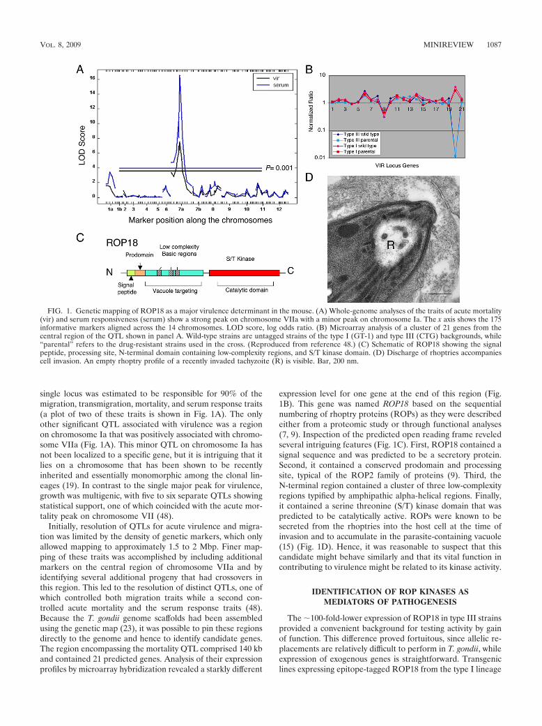
single locus was estimated to be responsible for 90% of themigration, transmigration, mortality, and serum response traits(a plot of two of these traits is shown in Fig. 1A). The onlyother significant QTL associated with virulence was a regionon chromosome Ia that was positively associated with chromo-some VIIa (Fig. 1A). This minor QTL on chromosome Ia hasnot been localized to a specific gene, but it is intriguing that itlies on a chromosome that has been shown to be recentlyinherited and essentially monomorphic among the clonal lin-eages (19). In contrast to the single major peak for virulence,growth was multigenic, with five to six separate QTLs showingstatistical support, one of which coincided with the acute mor-tality peak on chromosome VII (48).
Initially, resolution of QTLs for acute virulence and migra-tion was limited by the density of genetic markers, which onlyallowed mapping to approximately 1.5 to 2 Mbp. Finer map-ping of these traits was accomplished by including additionalmarkers on the central region of chromosome VIIa and byidentifying several additional progeny that had crossovers inthis region. This led to the resolution of distinct QTLs, one ofwhich controlled both migration traits while a second con-trolled acute mortality and the serum response traits (48).Because the T. gondii genome scaffolds had been assembledusing the genetic map (23), it was possible to pin these regionsdirectly to the genome and hence to identify candidate genes.The region encompassing the mortality QTL comprised 140 kband contained 21 predicted genes. Analysis of their expressionprofiles by microarray hybridization revealed a starkly different
expression level for one gene at the end of this region (Fig.1B). This gene was named ROP18 based on the sequentialnumbering of rhoptry proteins (ROPs) as they were describedeither from a proteomic study or through functional analyses(7, 9). Inspection of the predicted open reading frame reveledseveral intriguing features (Fig. 1C). First, ROP18 contained asignal sequence and was predicted to be a secretory protein.Second, it contained a conserved prodomain and processingsite, typical of the ROP2 family of proteins (9). Third, theN-terminal region contained a cluster of three low-complexityregions typified by amphipathic alpha-helical regions. Finally,it contained a serine threonine (S/T) kinase domain that waspredicted to be catalytically active. ROPs were known to besecreted from the rhoptries into the host cell at the time ofinvasion and to accumulate in the parasite-containing vacuole(15) (Fig. 1D). Hence, it was reasonable to suspect that thiscandidate might behave similarly and that its vital function incontributing to virulence might be related to its kinase activity.
IDENTIFICATION OF ROP KINASES ASMEDIATORS OF PATHOGENESIS
The �100-fold-lower expression of ROP18 in type III strainsprovided a convenient background for testing activity by gainof function. This difference proved fortuitous, since allelic re-placements are relatively difficult to perform in T. gondii, whileexpression of exogenous genes is straightforward. Transgeniclines expressing epitope-tagged ROP18 from the type I lineage
FIG. 1. Genetic mapping of ROP18 as a major virulence determinant in the mouse. (A) Whole-genome analyses of the traits of acute mortality(vir) and serum responsiveness (serum) show a strong peak on chromosome VIIa with a minor peak on chromosome Ia. The x axis shows the 175informative markers aligned across the 14 chromosomes. LOD score, log odds ratio. (B) Microarray analysis of a cluster of 21 genes from thecentral region of the QTL shown in panel A. Wild-type strains are untagged strains of the type I (GT-1) and type III (CTG) backgrounds, while“parental” refers to the drug-resistant strains used in the cross. (Reproduced from reference 48.) (C) Schematic of ROP18 showing the signalpeptide, processing site, N-terminal domain containing low-complexity regions, and S/T kinase domain. (D) Discharge of rhoptries accompaniescell invasion. An empty rhoptry profile of a recently invaded tachyzoite (R) is visible. Bar, 200 nm.
VOL. 8, 2009 MINIREVIEW 1087
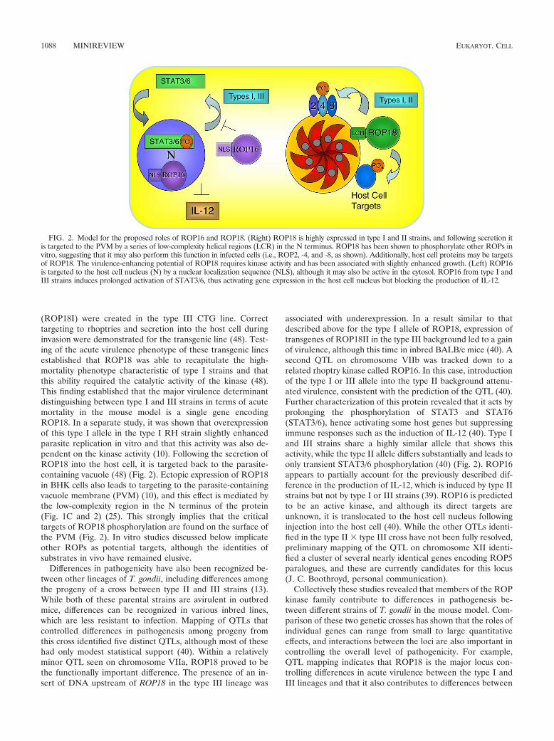
(ROP18I) were created in the type III CTG line. Correcttargeting to rhoptries and secretion into the host cell duringinvasion were demonstrated for the transgenic line (48). Test-ing of the acute virulence phenotype of these transgenic linesestablished that ROP18 was able to recapitulate the high-mortality phenotype characteristic of type I strains and thatthis ability required the catalytic activity of the kinase (48).This finding established that the major virulence determinantdistinguishing between type I and III strains in terms of acutemortality in the mouse model is a single gene encodingROP18. In a separate study, it was shown that overexpressionof this type I allele in the type I RH strain slightly enhancedparasite replication in vitro and that this activity was also de-pendent on the kinase activity (10). Following the secretion ofROP18 into the host cell, it is targeted back to the parasite-containing vacuole (48) (Fig. 2). Ectopic expression of ROP18in BHK cells also leads to targeting to the parasite-containingvacuole membrane (PVM) (10), and this effect is mediated bythe low-complexity region in the N terminus of the protein(Fig. 1C and 2) (25). This strongly implies that the criticaltargets of ROP18 phosphorylation are found on the surface ofthe PVM (Fig. 2). In vitro studies discussed below implicateother ROPs as potential targets, although the identities ofsubstrates in vivo have remained elusive.
Differences in pathogenicity have also been recognized be-tween other lineages of T. gondii, including differences amongthe progeny of a cross between type II and III strains (13).While both of these parental strains are avirulent in outbredmice, differences can be recognized in various inbred lines,which are less resistant to infection. Mapping of QTLs thatcontrolled differences in pathogenesis among progeny fromthis cross identified five distinct QTLs, although most of thesehad only modest statistical support (40). Within a relativelyminor QTL seen on chromosome VIIa, ROP18 proved to bethe functionally important difference. The presence of an in-sert of DNA upstream of ROP18 in the type III lineage was
associated with underexpression. In a result similar to thatdescribed above for the type I allele of ROP18, expression oftransgenes of ROP18II in the type III background led to a gainof virulence, although this time in inbred BALB/c mice (40). Asecond QTL on chromosome VIIb was tracked down to arelated rhoptry kinase called ROP16. In this case, introductionof the type I or III allele into the type II background attenu-ated virulence, consistent with the prediction of the QTL (40).Further characterization of this protein revealed that it acts byprolonging the phosphorylation of STAT3 and STAT6(STAT3/6), hence activating some host genes but suppressingimmune responses such as the induction of IL-12 (40). Type Iand III strains share a highly similar allele that shows thisactivity, while the type II allele differs substantially and leads toonly transient STAT3/6 phosphorylation (40) (Fig. 2). ROP16appears to partially account for the previously described dif-ference in the production of IL-12, which is induced by type IIstrains but not by type I or III strains (39). ROP16 is predictedto be an active kinase, and although its direct targets areunknown, it is translocated to the host cell nucleus followinginjection into the host cell (40). While the other QTLs identi-fied in the type II � type III cross have not been fully resolved,preliminary mapping of the QTL on chromosome XII identi-fied a cluster of several nearly identical genes encoding ROP5paralogues, and these are currently candidates for this locus(J. C. Boothroyd, personal communication).
Collectively these studies revealed that members of the ROPkinase family contribute to differences in pathogenesis be-tween different strains of T. gondii in the mouse model. Com-parison of these two genetic crosses has shown that the roles ofindividual genes can range from small to large quantitativeeffects, and interactions between the loci are also important incontrolling the overall level of pathogenicity. For example,QTL mapping indicates that ROP18 is the major locus con-trolling differences in acute virulence between the type I andIII lineages and that it also contributes to differences between
FIG. 2. Model for the proposed roles of ROP16 and ROP18. (Right) ROP18 is highly expressed in type I and II strains, and following secretion itis targeted to the PVM by a series of low-complexity helical regions (LCR) in the N terminus. ROP18 has been shown to phosphorylate other ROPs invitro, suggesting that it may also perform this function in infected cells (i.e., ROP2, -4, and -8, as shown). Additionally, host cell proteins may be targetsof ROP18. The virulence-enhancing potential of ROP18 requires kinase activity and has been associated with slightly enhanced growth. (Left) ROP16is targeted to the host cell nucleus (N) by a nuclear localization sequence (NLS), although it may also be active in the cytosol. ROP16 from type I andIII strains induces prolonged activation of STAT3/6, thus activating gene expression in the host cell nucleus but blocking the production of IL-12.
1088 MINIREVIEW EUKARYOT. CELL
types II and III. In contrast, ROP18 contributes only partiallyto growth differences between type I and type III lineages, andmultiple other loci also contribute to this trait (48). This resultindicates that enhanced virulence is not simply the result offaster replication, although this is certainly one factor thatinfluences pathogenesis in the mouse model. Additionally, aspredicted from the genetic mapping studies, ROP18 plays norole in the enhanced migration phenotype, which also maps tochromosome VIIa (supplemental data in reference 48). Withinthis migration locus are a number of genes that might contrib-ute to actin-myosin-based motility, and the precise geneticcontrol of this trait is still under investigation. It is conceivablethat traits controlled by this migration QTL may also affect theseverity of disease, for example, by advancing the onset ofinfection.
Following the discovery that ROP18 mediates differences inacute virulence between different laboratory strains, we alsoinvestigated to what extent these properties were conservedamong natural isolates. Not surprisingly, given the extremelyclonal population structure in North America and Europe,other isolates of the canonical type I, II, and III lineages sharethe same alleles and expression profiles as the laboratorystrains used in the genetic crosses (22). Hence, it is highly likelythat ROP18 contributes to the pathogenesis of these isolates ina manner similar to that described for the common laboratoryisolates. However, in other regions, such as South America, thepopulation consists of a number of unique lineages, some ofwhich have undergone greater recombination in the wild (20,28). To analyze the diversity of ROP18 among these popula-tions, we compared genetic diversity to a set of selectivelyneutral introns as well as housekeeping genes and other anti-gens (22). Remarkably, although genetic diversity among theseisolates was high, ROP18 displayed only three distinct alleles,corresponding to those already described for the North Amer-ican lineages (22). Furthermore, these alleles have coexistedover a long period of evolutionary time and show very differentprofiles in terms of diversifying selection. While the type IIIallele is the oldest, it shows little evidence of positive selection.In contrast, the type I and II alleles show high levels of diver-sifying selection. This is correlated with major differences inexpression levels that are associated with the presence of anupstream region in the type III allele. Comparison to theoutgroup Neospora caninum revealed that this upstream regionis ancestral (22). Taken together, these findings suggest thatdeletion or rearrangement of this upstream region led to up-regulation of ROP18 and promoted diversifying selection (22).These findings also suggest that the stable coexistence of threesubtypes results from adaptations to different niches in theenvironment. The long-term maintenance of the type III allele,which is associated with low levels of virulence, suggests adap-tation for hosts where this trait is advantageous (or where highvirulence is disadvantageous). In contrast, the more recentexpansion and widespread nature of the type II and I alleles,which show moderate and high levels of virulence, respectively,suggest that these traits are adaptive in a different niche. Whilethe phenotypes of ROP18 have been studied primarily in thelaboratory mouse, this host is highly susceptible to toxoplas-mosis relative to wild rodents. Thus, it may be that enhancedexpression of ROP18 is an adaptive trait for the infection of
natural hosts, where rather than contributing to pathology, itenhances transmission.
DIVERSITY AMONG THE ROP KINASES
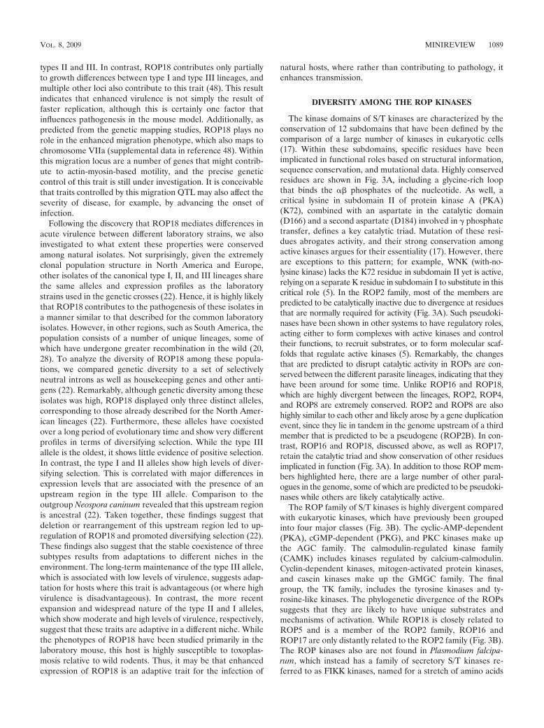
The kinase domains of S/T kinases are characterized by theconservation of 12 subdomains that have been defined by thecomparison of a large number of kinases in eukaryotic cells(17). Within these subdomains, specific residues have beenimplicated in functional roles based on structural information,sequence conservation, and mutational data. Highly conservedresidues are shown in Fig. 3A, including a glycine-rich loopthat binds the �� phosphates of the nucleotide. As well, acritical lysine in subdomain II of protein kinase A (PKA)(K72), combined with an aspartate in the catalytic domain(D166) and a second aspartate (D184) involved in � phosphatetransfer, defines a key catalytic triad. Mutation of these resi-dues abrogates activity, and their strong conservation amongactive kinases argues for their essentiality (17). However, thereare exceptions to this pattern; for example, WNK (with-no-lysine kinase) lacks the K72 residue in subdomain II yet is active,relying on a separate K residue in subdomain I to substitute in thiscritical role (5). In the ROP2 family, most of the members arepredicted to be catalytically inactive due to divergence at residuesthat are normally required for activity (Fig. 3A). Such pseudoki-nases have been shown in other systems to have regulatory roles,acting either to form complexes with active kinases and controltheir functions, to recruit substrates, or to form molecular scaf-folds that regulate active kinases (5). Remarkably, the changesthat are predicted to disrupt catalytic activity in ROPs are con-served between the different parasite lineages, indicating that theyhave been around for some time. Unlike ROP16 and ROP18,which are highly divergent between the lineages, ROP2, ROP4,and ROP8 are extremely conserved. ROP2 and ROP8 are alsohighly similar to each other and likely arose by a gene duplicationevent, since they lie in tandem in the genome upstream of a thirdmember that is predicted to be a pseudogene (ROP2B). In con-trast, ROP16 and ROP18, discussed above, as well as ROP17,retain the catalytic triad and show conservation of other residuesimplicated in function (Fig. 3A). In addition to those ROP mem-bers highlighted here, there are a large number of other paral-ogues in the genome, some of which are predicted to be pseudoki-nases while others are likely catalytically active.
The ROP family of S/T kinases is highly divergent comparedwith eukaryotic kinases, which have previously been groupedinto four major classes (Fig. 3B). The cyclic-AMP-dependent(PKA), cGMP-dependent (PKG), and PKC kinases make upthe AGC family. The calmodulin-regulated kinase family(CAMK) includes kinases regulated by calcium-calmodulin.Cyclin-dependent kinases, mitogen-activated protein kinases,and casein kinases make up the GMGC family. The finalgroup, the TK family, includes the tyrosine kinases and ty-rosine-like kinases. The phylogenetic divergence of the ROPssuggests that they are likely to have unique substrates andmechanisms of activation. While ROP18 is closely related toROP5 and is a member of the ROP2 family, ROP16 andROP17 are only distantly related to the ROP2 family (Fig. 3B).The ROP kinases also are not found in Plasmodium falcipa-rum, which instead has a family of secretory S/T kinases re-ferred to as FIKK kinases, named for a stretch of amino acids
VOL. 8, 2009 MINIREVIEW 1089
that occurs in their N termini (31). FIKK kinases are exportedvia a PEXEL (Plasmodium export element) motif and arefound in the infected red blood cell in association with a mem-branous system, although their role there is not understood(31). ROP kinases are found in related tissue coccidia, notablyNeospora caninum, for which there is a genome project (http://www.sanger.ac.uk/sequencing/Neospora/caninum/).
Early studies of ROP2 revealed that it was targeted to thePVM and suggested, based on differential staining with anti-bodies to the N and C termini, that it adopted a transmem-brane topology (4). The N-terminal region of ROP2 was alsoimplicated in binding to the host endoplasmic reticulum andmitochondria, which are recruited into close proximity with thePVM (45). More-recent analysis of members of the ROP2 familysuggests that they adopt a kinase fold and that the originallyproposed transmembrane domain is in fact buried in the core ofthe C lobe of the kinase domain (9). This suggests an alternativeexplanation for the detergent accessibility experiments describedabove based on protein folding rather than membrane topology.Furthermore, while the N-terminal amphipathic alpha-helical re-gions are involved in recruitment to the PVM (9), they may alsoparticipate in organelle recruitment, especially if ROPs formcomplexes at this interface. In this regard, ROP18 is more similarto the ROP2 family in that it conserves this N-terminal low-complexity region, which is important in binding to the PVM (Fig.1C), while ROP16 does not.
UNIQUE STRUCTURAL AND REGULATORYFEATURES OF ROP KINASES
Initial attempts to model the kinase domain of ROPs pro-vided only low-homology models based on comparison toknown kinases (9, 48), in part due to the sequence divergencewithin this family. This problem was resolved by the recentsolution of X-ray crystal structures for the kinase domains ofROP8 (Protein Data Bank [PDB] accession no. 3BYV) and
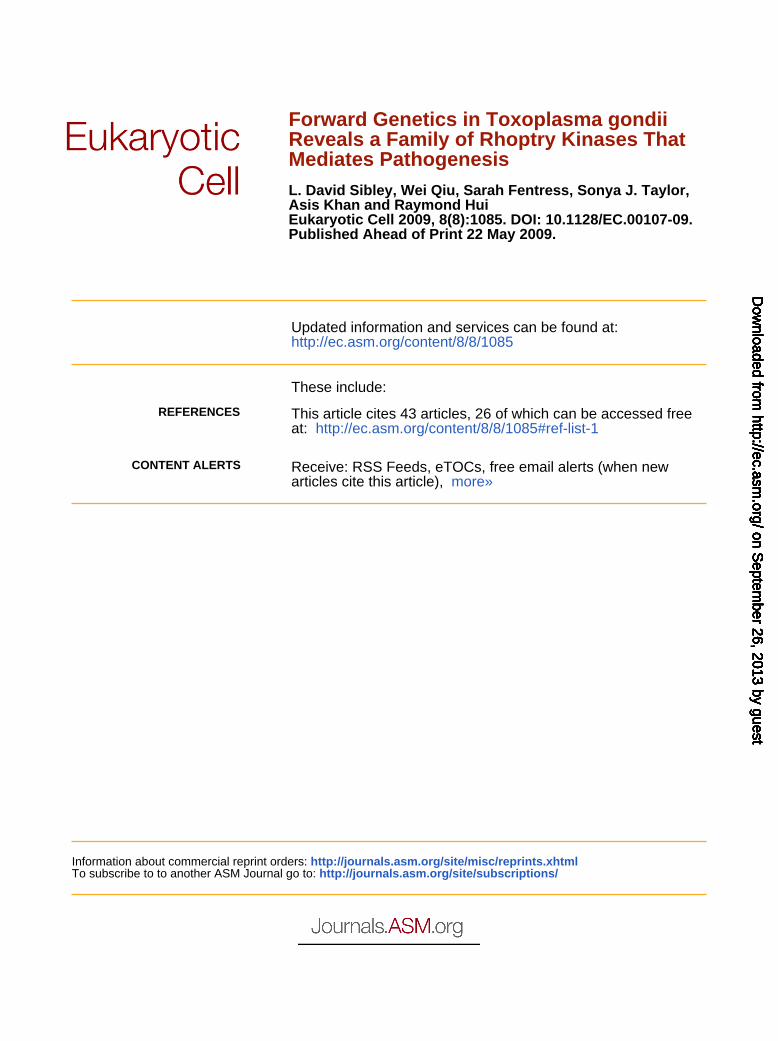
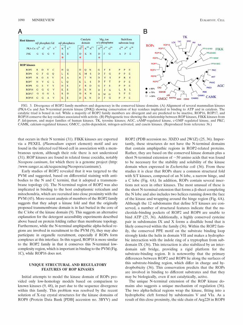
ROP2 (PDB accession no. 3DZO and 2W1Z) (25, 36). Impor-tantly, these structures do not have the N-terminal domainsthat contain amphipathic regions in ROP2-related proteins.Rather, they are based on the conserved kinase domain plus ashort N-terminal extension of �50 amino acids that was foundto be necessary for the stability and solubility of the kinasedomain when expressed in Escherichia coli (36). From thesestudies it is clear that ROPs share a common structural foldwith S/T kinases, composed of an N lobe, a narrow hinge, anda C lobe (Fig. 4A). In addition, ROPs contain several inser-tions not seen in other kinases. The most unusual of these isthe short N-terminal extension that forms a �-sheet completingthe N lobe and also includes two helices running down the faceof the kinase and wrapping around the hinge region (Fig. 4A).Although the 12 subdomains that define S/T kinases are con-served, a number of structural features indicate that the nu-cleotide-binding pockets of ROP2 and ROP8 are unable tobind ATP (25, 36). Additionally, a highly conserved cysteinepair in subdomains IX and X forms a disulfide bond that islikely conserved within the family (36). Within the ROP2 fam-ily, the conserved PPE motif on the substrate binding loopstrongly kinks the helix in domain VII and makes a hydropho-bic interaction with the indole ring of a tryptophan from sub-domain IX (36). This interaction is also stabilized by an inter-domain salt bridge, providing a rigid platform for thesubstrate-binding region. It is noteworthy that the primarydifferences between ROP2 and ROP8 lie along the surfaces ofthis substrate-binding region, which differ in charge and hy-dropbobicity (36). This conservation predicts that the ROPsare involved in binding to different substrates and that theymay be biologically, even if not catalytically, active.
The unique N-terminal extension of the ROP kinase do-mains also suggests a unique mechanism of regulation (36).The two alpha-helical regions wrap the kinase, fitting into ahydrophobic cleft formed by subdomains V and VIa. As aresult of this close proximity, the side chain of Arg228 in ROP8
FIG. 3. Divergence of ROP2 family members and degeneracy in the conserved kinase domains. (A) Alignment of several mammalian kinases(PKA-C� and Jun N-terminal protein kinase [JNK]) showing conservation of key residues implicated in binding to ATP and in catalysis. Thecatalytic triad is boxed in red. While a majority of ROP2 family members are divergent and are predicted to be inactive, ROP16, ROP17, andROP18 conserve the key residues associated with activity. (B) Phylogenetic tree showing the relationship between ROP kinases, FIKK kinases fromP. falciparum, and major families of human kinases. TK, tyrosine kinases; AGC, cAMP-regulated kinase, c-GMP regulated kinase, and PKC;CAMK, calcium-regulated kinases; GMCC, cyclin-dependent, mitogen-activated, and casein kinases. (Reproduced from reference 36.)
1090 MINIREVIEW EUKARYOT. CELL
(Arg220 in ROP2) protrudes into the nucleotide-bindingpocket, precluding ATP binding (36). Although this is incon-sequential for ROP2 and ROP8, because they are inactivepseudokinases, homology studies predict a similar N-terminalsubdomain in ROP18, with Gln199 instead of an arginine oc-cluding the conserved ATP binding pocket (25, 36). The posi-tioning of these N-terminal helices is stabilized by a number ofother interactions. These sequence motifs are conserved inother ROP2 family members, including ROP4, ROP5, ROP7,and ROP18. Collectively, these findings indicate that the heli-cal regions of the N-terminal extension wrap the kinase do-main in order to stabilize it and to regulate activation, asdiscussed below.
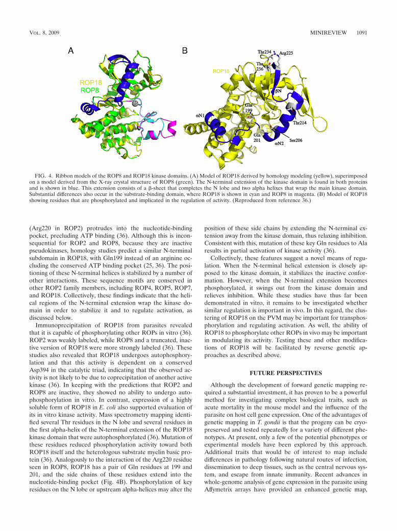
Immunoprecipitation of ROP18 from parasites revealedthat it is capable of phosphorylating other ROPs in vitro (36).ROP2 was weakly labeled, while ROP8 and a truncated, inac-tive version of ROP18 were more strongly labeled (36). Thesestudies also revealed that ROP18 undergoes autophosphory-lation and that this activity is dependent on a conservedAsp394 in the catalytic triad, indicating that the observed ac-tivity is not likely to be due to coprecipitation of another activekinase (36). In keeping with the predictions that ROP2 andROP8 are inactive, they showed no ability to undergo auto-phosphorylation in vitro. In contrast, expression of a highlysoluble form of ROP18 in E. coli also supported evaluation ofits in vitro kinase activity. Mass spectrometry mapping identi-fied several Thr residues in the N lobe and several residues inthe first alpha-helix of the N-terminal extension of the ROP18kinase domain that were autophosphorylated (36). Mutation ofthese residues reduced phosphorylation activity toward bothROP18 itself and the heterologous substrate myelin basic pro-tein (36). Analogously to the interaction of the Arg220 residueseen in ROP8, ROP18 has a pair of Gln residues at 199 and201, and the side chains of these residues extend into thenucleotide-binding pocket (Fig. 4B). Phosphorylation of keyresidues on the N lobe or upstream alpha-helices may alter the
position of these side chains by extending the N-terminal ex-tension away from the kinase domain, thus relaxing inhibition.Consistent with this, mutation of these key Gln residues to Alaresults in partial activation of kinase activity (36).
Collectively, these features suggest a novel means of regu-lation. When the N-terminal helical extension is closely ap-posed to the kinase domain, it stabilizes the inactive confor-mation. However, when the N-terminal extension becomesphosphorylated, it swings out from the kinase domain andrelieves inhibition. While these studies have thus far beendemonstrated in vitro, it remains to be investigated whethersimilar regulation is important in vivo. In this regard, the clus-tering of ROP18 on the PVM may be important for transphos-phorylation and regulating activation. As well, the ability ofROP18 to phosphorylate other ROPs in vivo may be importantin modulating its activity. Testing these and other modifica-tions of ROP18 will be facilitated by reverse genetic ap-proaches as described above.
FUTURE PERSPECTIVES
Although the development of forward genetic mapping re-quired a substantial investment, it has proven to be a powerfulmethod for investigating complex biological traits, such asacute mortality in the mouse model and the influence of theparasite on host cell gene expression. One of the advantages ofgenetic mapping in T. gondii is that the progeny can be cryo-preserved and tested repeatedly for a variety of different phe-notypes. At present, only a few of the potential phenotypes orexperimental models have been explored by this approach.Additional traits that would be of interest to map includedifferences in pathology following natural routes of infection,dissemination to deep tissues, such as the central nervous sys-tem, and escape from innate immunity. Recent advances inwhole-genome analysis of gene expression in the parasite usingAffymetrix arrays have provided an enhanced genetic map,
FIG. 4. Ribbon models of the ROP8 and ROP18 kinase domains. (A) Model of ROP18 derived by homology modeling (yellow), superimposedon a model derived from the X-ray crystal structure of ROP8 (green). The N-terminal extension of the kinase domain is found in both proteinsand is shown in blue. This extension consists of a �-sheet that completes the N lobe and two alpha helixes that wrap the main kinase domain.Substantial differences also occur in the substrate-binding domain, where ROP18 is shown in cyan and ROP8 in magenta. (B) Model of ROP18showing residues that are phosphorylated and implicated in the regulation of activity. (Reproduced from reference 36.)
VOL. 8, 2009 MINIREVIEW 1091
thus facilitating fine positional mapping. As well, additionalgenetic crosses, including a recently completed cross betweentype I and II strains (unpublished data), will make it possible tocompare the roles of specific genes in different genetic back-grounds. To facilitate wider application of genetic analysis, theprogeny from the existing crosses have been made availablethrough the Biodefense and Emerging Infectious Disease Re-search Resources Repository (www.beiresources.org/), and thegenetic maps, coordinates, and markers are freely available athttp://toxomap.wustl.edu/policy.htm.
Thus far, the role of ROPs has been investigated only in themurine system, although some of the in vitro phenotypes arealso expressed in human cells. It will be important to expandthese findings to human infection in order to assess their po-tential to contribute to the variable clinical outcomes seen intoxoplasmosis. The availability of reagents for various ROPswill be useful for establishing whether they are recognized bythe human immune system and whether variants of the variousROPs correlate with the clinical presentation of different iso-lates in human disease. Structural information combined withbiochemical assays will also be useful for screening small-mol-ecule libraries to define chemical inhibitors of the ROPs. Com-pounds that selectively inhibit ROP kinases might be expectedto block the virulence potential of the parasite, thus allowingthe immune system to control the infection and prevent pa-thology.
ACKNOWLEDGMENTS
This work was supported in part by grants from the NIH (AI036629and AI059176, to L.D.S.).
We are grateful to John Boothroyd, David Roos, Michael White,John Wootton, Jim Ajioka, and Jean Francois Dubremetz for helpfulcomments and to members of our laboratories for their contributionsto the work summarized here.
REFERENCES
1. Ajioka, J. W., and L. D. Sibley. 2007. Development and application ofclassical genetics in Toxoplasma gondii, p. 367–389. In L. M. Weiss and K.Kim (ed.), Toxoplasma gondii the model Apicomplexan: perspectives andmethods. Elsevier/Academic Press, New York, NY.
2. Barragan, A., and L. D. Sibley. 2003. Migration of Toxoplasma gondii acrossbiological barriers. Trends Microbiol. 11:426–430.
3. Barragan, A., and L. D. Sibley. 2002. Transepithelial migration of Toxo-plasma gondii is linked to parasite motility and virulence. J. Exp. Med.195:1625–1633.
4. Beckers, C. J. M., J. F. Dubremetz, O. Mercereau-Puijalon, and K. A. Joiner.1994. The Toxoplasma gondii rhoptry protein ROP2 is inserted into theparasitophorous vacuole membrane, surrounding the intracellular parasite,and is exposed to the host cell cytoplasm. J. Cell Biol. 127:947–961.
5. Boudeau, J., D. Miranda-Saavedra, G. J. Barton, and D. R. Alessi. 2006.Emerging roles of pseudokinases. Trends Cell Biol. 16:443–452.
6. Boyle, J. P., B. Rajasekar, J. P. J. Saeij, J. W. Ajioka, M. Berriman, I.Paulsen, L. D. Sibley, M. White, and J. C. Boothroyd. 2006. Just one crossappears capable of dramatically altering the population biology of a eukary-otic pathogen like Toxoplasma gondii. Proc. Natl. Acad. Sci. USA 103:10514–10519.
7. Bradley, P. J., C. Ward, S. J. Cheng, D. L. Alexander, S. Coller, G. H.Coombs, J. D. Dunn, D. J. Ferguson, S. J. Sanderson, J. M. Wastling, andJ. C. Boothroyd. 2005. Proteomic analysis of rhoptry organelles reveals manynovel constituents for host-parasite interactions in T. gondii. J. Biol. Chem.280:34245–34258.
8. Dubey, J. P. 2007. The history and life cycle of Toxoplasma gondii, p. 1–17.In L. M. Weiss and K. Kim (ed.), Toxoplasma gondii the model Apicompl-exan: perspectives and methods. Elsevier/Academic Press, New York, NY.
9. El Hajj, H., E. Demey, J. Poncet, M. Lebrun, B. Wu, N. Galeotti, M. N.Fourmaux, O. Mercereau-Puijalon, H. Vial, and J. F. Dubremetz. 2006. TheROP2 family of Toxoplasma gondii rhoptry proteins: proteomic and genomiccharacterization and molecular modeling. Proteomics 6:5773–5784.
10. El Hajj, H., M. Lebrun, S. T. Arold, H. Vial, G. Labesse, and J. F.Dubremetz. 2007. ROP18 is a rhoptry kinase controlling the intracellularproliferation of Toxoplasma gondii. PLoS Pathog. 3:e14.
11. Fuentes, I., J. M. Rubio, C. Ramírez, and J. Alvar. 2001. Genotypic charac-terization of Toxoplasma gondii strains associated with human toxoplasmosisin Spain: direct analysis from clinical samples. J. Clin. Microbiol. 39:1566–1570.
12. Gavrilescu, L. C., and E. Y. Denkers. 2001. IFN-� overproduction and highlevel apoptosis are associated with high but not low virulence Toxoplasmagondii infection. J. Immunol. 167:902–909.
13. Grigg, M. E., S. Bonnefoy, A. B. Hehl, Y. Suzuki, and J. C. Boothroyd. 2001.Success and virulence in Toxoplasma as the result of sexual recombinationbetween two distinct ancestries. Science 294:161–165.
14. Grigg, M. E., J. Ganatra, J. C. Boothroyd, and T. P. Margolis. 2001. Unusualabundance of atypical strains associated with human ocular toxoplasmosis.J. Infect. Dis. 184:633–639.
15. Håkansson, S., A. J. Charron, and L. D. Sibley. 2001. Toxoplasma evacuoles:a two-step process of secretion and fusion forms the parasitophorous vacu-ole. EMBO J. 20:3132–3144.
16. Hall, S., K. A. Ryan, and D. Buxton. 2001. The epidemiology of toxoplasmainfection, p. 58–124. In D. H. Joynson and T. G. Wreghitt (ed.), Toxoplas-mosis: a comprehensive clinical guide. Cambridge University Press, Cam-bridge, England.
17. Hanks, S. K., and T. Hunter. 1995. Protein kinases 6. The eukaryotic proteinkinase superfamily: kinase (catalytic) domain structure and classification.FASEB J. 9:576–596.
18. Jones, J. L., C. Muccioli, R. Belfort, Jr., G. N. Holland, J. M. Roberts, andC. Silveira. 2006. Recently acquired Toxoplasma gondii infection, Brazil.Emerg. Infect. Dis. 12:582–587.
19. Khan, A., U. Bohme, K. A. Kelly, E. Adlem, K. Brooks, M. Simmonds, K.Mungall, M. A. Quail, C. Arrowsmith, T. Chillingworth, C. Churcher, D.Harris, M. Collins, N. Fosker, A. Fraser, Z. Hance, K. Jagels, S. Moule, L.Murphy, S. O’Neil, M. A. Rajandream, D. Saunders, K. Seeger, S. White-head, T. Mayr, X. Xuan, J. Watanabe, Y. Suzuki, H. Wakaguri, S. Sugano,C. Sugimoto, I. Paulsen, A. J. Mackey, D. S. Roos, N. Hall, M. Berriman, B.Barell, L. D. Sibley, and J. W. Ajioka. 2006. Common inheritance of chro-mosome Ia associated with clonal expansion of Toxoplasma gondii. GenomeRes. 16:1119–1125.
20. Khan, A., B. Fux, C. Su, J. P. Dubey, M. L. Darde, J. W. Ajioka, B. M.Rosenthal, and L. D. Sibley. 2007. Recent transcontinental sweep of Toxo-plasma gondii driven by a single monomorphic chromosome. Proc. Natl.Acad. Sci. USA 104:14872–14877.
21. Khan, A., C. Su, M. German, G. A. Storch, D. Clifford, and L. D. Sibley.2005. Genotyping of Toxoplasma gondii strains from immunocompromisedpatients reveals high prevalence of type I strains. J. Clin. Microbiol. 43:5881–5887.
22. Khan, A., S. Taylor, J. W. Ajioka, B. M. Rosenthal, and L. D. Sibley. 2009.Selection at a single locus leads to widespread expansion of Toxoplasmagondii lineages that are virulent in mice. PLoS Genet. 5:e1000404.
23. Khan, A., S. Taylor, C. Su, A. J. Mackey, J. Boyle, R. H. Cole, D. Glover, K.Tang, I. Paulsen, M. Berriman, J. C. Boothroyd, E. R. Pfefferkorn, J. P.Dubey, D. S. Roos, J. W. Ajioka, J. C. Wootton, and L. D. Sibley. 2005.Composite genome map and recombination parameters derived from threearchetypal lineages of Toxoplasma gondii. Nucleic Acids Res. 33:2980–2992.
24. Kim, L., B. A. Butcher, C. W. Lee, S. Uematsu, S. Akira, and E. Y. Denkers.2006. Toxoplasma gondii genotype determines MyD88-dependent signalingin infected macrophages. J. Immunol. 177:2584–2591.
25. Labesse, G., M. Gelin, Y. Bessin, M. Lebrun, J. Papoin, R. Cerdan, S. T.Arold, and J. F. Dubremetz. 2009. ROP2 from Toxoplasma gondii: a viru-lence factor with a protein-kinase fold and no enzymatic activity. Structure17:139–146.
26. Lander, E., and L. Kruglyak. 1995. Genetic dissection of complex traits: guide-lines for interpreting and reporting linkage results. Nat. Genet. 11:241–247.
27. Lander, E. S., and D. Botstein. 1989. Mapping Mendelian factors underlyingquantitative traits using RFLP linkage maps. Genetics 121:185–199.
28. Lehmann, T., P. L. Marcet, D. H. Graham, E. R. Dahl, and J. P. Dubey. 2006.Globalization and the population structure of Toxoplasma gondii. Proc. Natl.Acad. Sci. USA 103:11423–11428.
29. Mariuz, P., and R. T. Steigbigel. 2007. Toxoplasma infection in HIV-infectedpatients, p. 147–177. In D. H. M. Joynson and T. G. Wreghitt (ed.), Toxo-plasmosis: a comprehensive clinical guide. Cambridge University Press,Cambridge, England.
30. Mordue, D. G., F. Monroy, M. La Regina, C. A. Dinarello, and L. D. Sibley.2001. Acute toxoplasmosis leads to lethal overproduction of Th1 cytokines.J. Immunol. 167:4574–4584.
31. Nunes, M., J. P. D. Goldring, C. Doerig, and A. Scherf. 2007. A novel proteinkinase family in Plasmodium falciparum is differentially transcribed andsecreted to various cellular compartments of the host cell. Mol. Microbiol.63:391–403.
32. Petersen, E., and O. Liesenfeld. 2007. Clinical disease and diagnostics, p.81–100. In L. M. Weiss and K. Kim (ed.), Toxoplasma gondii the modelApicomplexan: perspectives and methods. Elsevier/Academic Press, NewYork, NY.
33. Pfefferkorn, E. R., and L. H. Kasper. 1983. Toxoplasma gondii: genetic
1092 MINIREVIEW EUKARYOT. CELL
crosses reveal phenotypic suppression of hydroxyurea resistance by fluoro-deoxyuridine resistance. Exp. Parasitol. 55:207–218.
34. Pfefferkorn, E. R., L. C. Pfefferkorn, and E. D. Colby. 1977. Development ofgametes and oocysts in cats fed cysts derived from cloned trophozoites ofToxoplasma gondii. J. Parasitol. 63:158–159.
35. Pfefferkorn, L. C., and E. R. Pfefferkorn. 1980. Toxoplasma gondii: geneticrecombination between drug resistant mutants. Exp. Parasitol. 50:305–316.
36. Qiu, W., A. Wernimont, K. Tang, S. Taylor, V. Lunin, M. Schapira, S. J.Fentress, R. Hui, and L. D. Sibley. 2009. Novel structural and regulatoryfeatures of rhoptry secretory kinases in Toxoplasma gondii. EMBO J. 28:969–979.
37. Radke, J. R., B. Striepen, M. N. Guerini, M. E. Jerome, D. S. Roos, andM. W. White. 2001. Defining the cell cycle for the tachyzoite stage of Tox-oplasma gondii. Mol. Biochem. Parasitol. 115:165–175.
38. Reichard, U., and U. Gross. 2007. Toxoplasma animal models and therapeu-tics, p. 153–184. In L. M. Weiss and K. Kim (ed.), Toxoplasma gondii themodel Apicomplexan: perspectives and methods. Elsevier/Academic Press,New York, NY.
39. Robben, P. M., D. G. Mordue, S. M. Truscott, K. Takeda, S. Akira, and L. D.Sibley. 2004. Production of IL-12 by macrophages infected with Toxoplasmagondii depends on the parasite genotype. J. Immunol. 172:3686–3694.
40. Saeij, J. P. J., J. P. Boyle, S. Coller, S. Taylor, L. D. Sibley, E. T. Brooke-Powell, J. W. Ajioka, and J. C. Boothroyd. 2006. Polymorphic secretedkinases are key virulence factors in toxoplasmosis. Science 314:1780–1783.
41. Sibley, L. D. 2004. Invasion strategies of intracellular parasites. Science304:248–253.
42. Sibley, L. D., and J. W. Ajioka. 2008. Population structure of Toxoplasma
gondii: clonal expansion driven by infrequent recombination and selectivesweeps. Annu. Rev. Microbiol. 62:329–351.
43. Sibley, L. D., and J. C. Boothroyd. 1992. Virulent strains of Toxoplasmagondii comprise a single clonal lineage. Nature (London) 359:82–85.
44. Sibley, L. D., A. J. LeBlanc, E. R. Pfefferkorn, and J. C. Boothroyd. 1992.Generation of a restriction fragment length polymorphism linkage map forToxoplasma gondii. Genetics 132:1003–1015.
45. Sinai, A. P., and K. A. Joiner. 2001. The Toxoplasma gondii protein ROP2mediates host organelle association with the parasitophorous vacuole mem-brane. J. Cell Biol. 154:95–108.
46. Su, C., D. Evans, R. H. Cole, J. C. Kissinger, J. W. Ajioka, and L. D. Sibley.2003. Recent expansion of Toxoplasma through enhanced oral transmission.Science 299:414–416.
47. Su, C., D. K. Howe, J. P. Dubey, J. W. Ajioka, and L. D. Sibley. 2002.Identification of quantitative trait loci controlling acute virulence in Toxo-plasma gondii. Proc. Natl. Acad. Sci. USA 99:10753–10758.
48. Taylor, S., A. Barragan, C. Su, B. Fux, S. J. Fentress, K. Tang, W. L. Beatty,E. L. Haijj, M. Jerome, M. S. Behnke, M. White, J. C. Wootton, and L. D.Sibley. 2006. A secreted serine-threonine kinase determines virulence in theeukaryotic pathogen Toxoplasma gondii. Science 314:1776–1780.
49. Weiss, L. M., and K. Kim. 2007. Bradyzoite development, p. 341–366. InL. M. Weiss and K. Kim (ed.), Toxoplasma gondii the model Apicomplexan:perspectives and methods. Elsevier/Academic Press, New York, NY.
50. Zhao, Y., D. J. Ferguson, D. C. Wilson, J. C. Howard, L. D. Sibley, and G. S.Yap. 2009. Virulent Toxoplasma gondii evade immunity-related GTPase-mediated parasite vacuole disruption within primed macrophages. J. Immu-nol. 182:3775–3781.
VOL. 8, 2009 MINIREVIEW 1093