Acquired infection with Toxoplasma gondii in adult mice results in sensorimotor deficits but normal...
17
Acquired infection with Toxoplasma gondii in adult mice results in sensorimotor deficits but normal cognitive behavior despite widespread brain pathology Maria Gulinello a,* , Mariana Acquarone b , John H Kim a , David C. Spray c , Helene S. Barbosa d , Rani Sellers e , Herbert B. Tanowitz b , and Louis M. Weiss b a Behavioral Core Facility, Department of Neuroscience,1410 Pelham PkwyS K925 Albert Einstein College of Medicine, Bronx, NY 10461 b Department of Pathology, Albert Einstein College of Medicine, 1300 Morris Park Avenue, Forchheimer Building room 504, Bronx, NY, 10461 c Dept Neuroscience Department of Neuroscience,1410 Pelham PkwyS K840 Albert Einstein College of Medicine, Bronx, NY 10461 d Laboratório de Biologia Estrutural, Instituto Oswaldo Cruz, Fundação Oswaldo Cruz, Av. Brasil 4361, 21041-361, Rio de Janeiro, RJ, Brasil e Histopathology Core Facility, Department of Pathology,1301 Morris Park Avenue, Price Center/ Block Research Pavilion room 158, Albert Einstein College of Medicine, Bronx, NY 10461 Abstract Toxoplasma gondii is a ubiquitous intracellular parasite which chronically infects 30 to 50% of the human population. While acquired infection is primarily asymptomatic several studies have suggested that such infections may contribute to neurological and psychiatric symptoms. Previous studies in rodents have demonstated that T. gondii infection does not just kill its host, but alters the behavioral repertoire of an infected animal making it more likely that predation with occur completing the parasite life cycle. The aim of the present study was to evaluate the behavioral changes in C57BL/ 6 mice chronically infected with the avirulent T. gondii (ME49, a type II strain), in a comprehensive test battery. Infected mice demonstrated profound and widespread brain pathology, motor coordination and sensory deficits. In contrast, cognitive function, anxiety levels, social behavior and the motivation to explore novel objects were normal. The observed changes in behavior did not represent “gross” brain damage or dysfunction and were not due to targeted destruction of specific areas of the brain. Such changes point out the subtle interaction of this parasite with its intermediate hosts and are consistent with ideas about increased predation being an outcome of infection. © 2010 Elsevier Masson SAS. All rights reserved. * Corresponding Author Behavioral Core Facility Albert Einstein College of Medicine of Yeshiva University Dominick P. Purpura Department of Neuroscience Rose F. Kennedy Center RM 925 1410 Pelham Pkwy S., Bronx, NY 10461 phone: 718-430-4042 fax: 718-430-8821 [email protected] . Publisher's Disclaimer: This is a PDF file of an unedited manuscript that has been accepted for publication. As a service to our customers we are providing this early version of the manuscript. The manuscript will undergo copyediting, typesetting, and review of the resulting proof before it is published in its final citable form. Please note that during the production process errors may be discovered which could affect the content, and all legal disclaimers that apply to the journal pertain. NIH Public Access Author Manuscript Microbes Infect. Author manuscript; available in PMC 2011 July 1. Published in final edited form as: Microbes Infect. 2010 July ; 12(7): 528–537. doi:10.1016/j.micinf.2010.03.009. NIH-PA Author Manuscript NIH-PA Author Manuscript NIH-PA Author Manuscript
Transcript of Acquired infection with Toxoplasma gondii in adult mice results in sensorimotor deficits but normal...

Acquired infection with Toxoplasma gondii in adult mice resultsin sensorimotor deficits but normal cognitive behavior despitewidespread brain pathology
Maria Gulinelloa,*, Mariana Acquaroneb, John H Kima, David C. Sprayc, Helene S.Barbosad, Rani Sellerse, Herbert B. Tanowitzb, and Louis M. WeissbaBehavioral Core Facility, Department of Neuroscience,1410 Pelham PkwyS K925 Albert EinsteinCollege of Medicine, Bronx, NY 10461bDepartment of Pathology, Albert Einstein College of Medicine, 1300 Morris Park Avenue,Forchheimer Building room 504, Bronx, NY, 10461cDept Neuroscience Department of Neuroscience,1410 Pelham PkwyS K840 Albert EinsteinCollege of Medicine, Bronx, NY 10461dLaboratório de Biologia Estrutural, Instituto Oswaldo Cruz, Fundação Oswaldo Cruz, Av. Brasil4361, 21041-361, Rio de Janeiro, RJ, BrasileHistopathology Core Facility, Department of Pathology,1301 Morris Park Avenue, Price Center/Block Research Pavilion room 158, Albert Einstein College of Medicine, Bronx, NY 10461
AbstractToxoplasma gondii is a ubiquitous intracellular parasite which chronically infects 30 to 50% of thehuman population. While acquired infection is primarily asymptomatic several studies havesuggested that such infections may contribute to neurological and psychiatric symptoms. Previousstudies in rodents have demonstated that T. gondii infection does not just kill its host, but alters thebehavioral repertoire of an infected animal making it more likely that predation with occur completingthe parasite life cycle. The aim of the present study was to evaluate the behavioral changes in C57BL/6 mice chronically infected with the avirulent T. gondii (ME49, a type II strain), in a comprehensivetest battery. Infected mice demonstrated profound and widespread brain pathology, motorcoordination and sensory deficits. In contrast, cognitive function, anxiety levels, social behavior andthe motivation to explore novel objects were normal. The observed changes in behavior did notrepresent “gross” brain damage or dysfunction and were not due to targeted destruction of specificareas of the brain. Such changes point out the subtle interaction of this parasite with its intermediatehosts and are consistent with ideas about increased predation being an outcome of infection.
© 2010 Elsevier Masson SAS. All rights reserved.*Corresponding Author Behavioral Core Facility Albert Einstein College of Medicine of Yeshiva University Dominick P. PurpuraDepartment of Neuroscience Rose F. Kennedy Center RM 925 1410 Pelham Pkwy S., Bronx, NY 10461 phone: 718-430-4042 fax:718-430-8821 [email protected] .Publisher's Disclaimer: This is a PDF file of an unedited manuscript that has been accepted for publication. As a service to our customerswe are providing this early version of the manuscript. The manuscript will undergo copyediting, typesetting, and review of the resultingproof before it is published in its final citable form. Please note that during the production process errors may be discovered which couldaffect the content, and all legal disclaimers that apply to the journal pertain.
NIH Public AccessAuthor ManuscriptMicrobes Infect. Author manuscript; available in PMC 2011 July 1.
Published in final edited form as:Microbes Infect. 2010 July ; 12(7): 528–537. doi:10.1016/j.micinf.2010.03.009.
NIH
-PA Author Manuscript
NIH
-PA Author Manuscript
NIH
-PA Author Manuscript

KeywordsToxoplasma gondii; latent infection; motor coordination; cognitive tests; social behavior;parasitology
1 INTRODUCTIONThe protozoan Toxoplasma gondii is a definitive parasite of cats which has as intermediatehosts all warm blooded animals, including humans [1]. Worldwide, approximately 2 billionpeople are chronically infected with T. gondii and it has been estimated that T. gondiichronically infects 30 to 50% of the human population [2]. Infection is found throughout theworld in the tissues of food animals [2]. While infection with T. gondii has generally thoughtto be primarily asymptomatic in immune competent humans, recent studies have suggestedthat infection may contribute to the development of various neurological and psychiatricsymptoms [3-10] and that negative outcomes resulting from initial or chronic infection maybe under-diagnosed [10-13]. These studies in humans are, however, associative and cannotprove if infection causes the behavior or the behavior increases the risk of infection. There are,however, several lines of evidence in experimental animals that suggest that specific host-parasite interactions influence the behavior of intermediate hosts in important ways and thatthese behavioral changes increase the likelihood of horizontal transmission [14-16]. Systematicanalysis of the behavioral outcomes of infection in intermediate hosts may have implicationsfor understanding and controlling the transmission of the parasite and also for elucidating thehost-parasite interactions in regulating the specific outcomes of infection.
Toxoplasma gondii has several life cycle stages. The tachyzoite invades cells, replicates rapidlyand results in dissemination of infection. The bradyzoite is found in latent infections as tissuecysts. The sporozoite is found in oocysts shed by cats and is the product of sexual reproduction.Infection can occur in intermediate hosts (including humans) due to the ingestion of tissuecysts, the ingestion of oocysts, or by congenital infection during acute infection in a pregnanthost. There has been a clonal expansion of T. gondii lineages with three main lineages in theUnited States and Europe, Type I, Type II and Type III, which are defined by their ability toform cysts in mice (type II and III) and by their acute virulence (i.e. type I are lethal to miceduring acute infection) [17].
Latent infection persists during the lifespan of the intermediate hosts by the formation of cystsin muscle, neurons and glia. In congenitally infected children, T gondii infection causes mentalretardation, seizures and loss of vision. Reactivation of latent infection with the transformationof bradyzoites back into tachyzoites can occur in immune suppressed patients, such asindividuals with AIDS, and can be fatal [18]. The central nervous system (CNS) is the mostcommon site of such reactivation infections, resulting in Toxoplasmic encephalitis.
Any behavior in an intermediate hosts (i.e. rodents) that influences the likelihood of predationwill increase the frequency of transmission to the definitive host (i.e. cats) facilitatingcompletion of the sexual stage of the parasitic life cycle and the potential for recombinationand reassortment of genetic loci [9,14,15,19,20]. Carnivorism of infected intermediate hosts,such as rodents, with subsequent oral-fecal transmission is thought to be the most prevalentroute of transmission due to cats [21-23]. Several lines of evidence suggest that specific host-parasite interactions may alter the behavior of infected rodents and increase the risk of predation[14,15]. Rodents infected with T. gondii [10-12] show reduced fear specifically toward felinepredators that does not generalize to non-feline predators [16,19,24], which may lead to anaugmented rate of predation and multiplication of the parasite through an increased number oflife cycles. Chronically infected adult rodents show also show reduced anxiety and risk
Gulinello et al. Page 2
Microbes Infect. Author manuscript; available in PMC 2011 July 1.
NIH
-PA Author Manuscript
NIH
-PA Author Manuscript
NIH
-PA Author Manuscript

assessment [25]. Notably, the social withdrawal associated with sickness behavior in mostrodents [26] is also not evident in rodents chronically infected with T. gondii [25,27].
Though several studies demonstrate behavioral changes in rodents infected with T. gondii theprecise effects of adult-acquired, chronic latent infection are still unclear. Many studies haveprimarily examined the behavioral and pathological effects of congenital infection [27,28], andfew studies have included behavioral assays in multiple behavioral domains. Furthermore, theparasite load, anatomical location of parasite stages and behavioral outcomes change duringthe course of the infection [29,30] and interpretations of behavioral outcomes in the acute phaseof infection are complicated by the high levels of tachyzoites in critical peripheral tissues, suchas muscle, heart and lungs. The aim of the present study was to evaluate the behavioral changesin C57BL/6 mice chronically infected with the avirulent T. gondii strain ME49 (a type II strain),in a comprehensive test battery. The mice were tested 7 to 9 weeks after initial infection at atime when chronic infection has developed, as demonstrated by the presence of tissue cystscontaining bradzyoties, and acute infection has resolved, as demonstrated by the absence oftachyzoites
2 MATERIALS AND METHODS2.1 Subjects and infection protocol
Experiments were performed in accordance with the Institutional Animal Care and UseCommittee of the Albert Einstein College of Medicine. Eight-week-old C57BL/6 male mice(Jackson Laboratories) were infected with 103 Toxoplasma gondii bradyzoites of the type IIME49 strain diluted in 200 μL of PBS (20 infected mice) and injected intraperitoneally or wereinjected only with PBS (10 control mice). A total of 10 of the infected mice died within 3 weeksafter initial infection during the development of chronic infection. Mice were then housed at5 animals per cage and behavioral testing began 7 weeks after infection. This time point waschosen for a variety of reasons. First, active parasite levels in critical peripheral organs (i.e.lungs) are absent by this time, making further deaths unlikely in surviving subjects. Second,parasite load in the brain is reasonably stable from this point in chronically infected mice[30]. The colony was documented to be free of a number of standard viral, bacterial, andparasitic agents including Mouse Hepatitis Virus (MHV), Epidemic Diarrhea of Infant Mice(rotavirus, EDIM), Theiler’s Murine Encephalomyelitis Virus (TMEV, GDVII), MouseParvovirus (MPV1)(MPV2) (VP2)general (NS1), Minute Virus of Mice (MMV), MouseAdenovirus I and II (MAD), Ectromelia (Ectro), Lymphocytic Choriomeningitis Virus (LCM),Mycoplasma pulmonis (M. pulmonis), Pneumonia Virus of Mice (PVM), Reovirus (Reo),Sendai Virus (Sendai), Mouse Cytomegalovirus (MCMV), Mouse Norovirus (MNV),Ectoparasites (mites) by microscopic evaluation of fur plucks and Endoparasites (pinworms)by cecal and colon float and anal tape test.
2.2 Behavioral assaysMice were tested behaviorally starting at 7 weeks after infection in the open field, objectplacement, object recognition, balance beam, grip strength, gait analysis and functionalobservation battery, in that order.
2.2.1 Functional observation battery—This primary screen was based on the SHIRPAprotocol [31]. This is a standard method that provides a quantitative behavioral and functionalprofile by observational assessment of mice. Assessments were divided into five behavioraldomains: general activity/arousal, motor and autonomic, reflexes, sensory and generalcondition. The details of each observation and list of tests are summarized in table 1. Mostobservations used an ordinal scale such that 0-1 represented normal or average behavior,negative numbers indicated deficits, hypoactivity or absence of normal behaviors and positive
Gulinello et al. Page 3
Microbes Infect. Author manuscript; available in PMC 2011 July 1.
NIH
-PA Author Manuscript
NIH
-PA Author Manuscript
NIH
-PA Author Manuscript
numbers indicate hyper-reactivity or higher incidence of abnormal behavior. Some tests couldproduce absolute, rather than ordinal values. Activity, for instance was assessed and numberof grid crosses in 1 min. Table 1 (results) details these numerous assays and the units ofmeasurement for each assay not employing an the ordinal scale.
2.2.2 Open field—The open field has been extensively validated, ethologically andpharmacologically in both mice and rats [32]. Behavior was recorded manually for 6 min 16inch square acrylic white box. Exploration was assessed as the number of rears (defined asraising both forepaws off the ground, except when grooming) and general locomotor activitywas scored as the number of grid crosses (defined when the animal crossed a grid past themidline of the body).
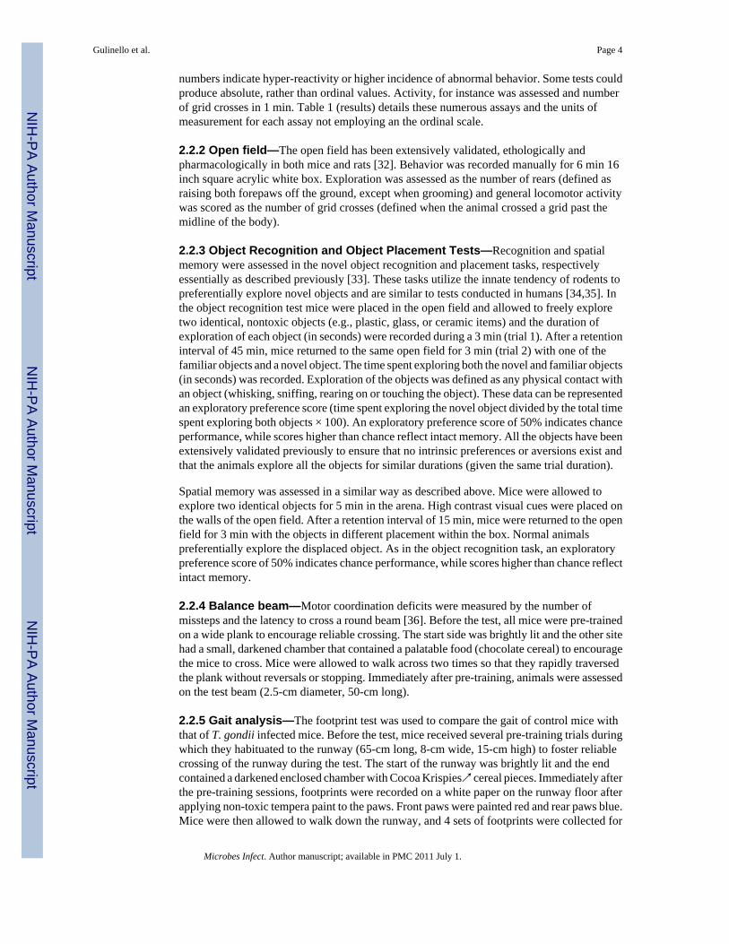
2.2.3 Object Recognition and Object Placement Tests—Recognition and spatialmemory were assessed in the novel object recognition and placement tasks, respectivelyessentially as described previously [33]. These tasks utilize the innate tendency of rodents topreferentially explore novel objects and are similar to tests conducted in humans [34,35]. Inthe object recognition test mice were placed in the open field and allowed to freely exploretwo identical, nontoxic objects (e.g., plastic, glass, or ceramic items) and the duration ofexploration of each object (in seconds) were recorded during a 3 min (trial 1). After a retentioninterval of 45 min, mice returned to the same open field for 3 min (trial 2) with one of thefamiliar objects and a novel object. The time spent exploring both the novel and familiar objects(in seconds) was recorded. Exploration of the objects was defined as any physical contact withan object (whisking, sniffing, rearing on or touching the object). These data can be representedan exploratory preference score (time spent exploring the novel object divided by the total timespent exploring both objects × 100). An exploratory preference score of 50% indicates chanceperformance, while scores higher than chance reflect intact memory. All the objects have beenextensively validated previously to ensure that no intrinsic preferences or aversions exist andthat the animals explore all the objects for similar durations (given the same trial duration).
Spatial memory was assessed in a similar way as described above. Mice were allowed toexplore two identical objects for 5 min in the arena. High contrast visual cues were placed onthe walls of the open field. After a retention interval of 15 min, mice were returned to the openfield for 3 min with the objects in different placement within the box. Normal animalspreferentially explore the displaced object. As in the object recognition task, an exploratorypreference score of 50% indicates chance performance, while scores higher than chance reflectintact memory.
2.2.4 Balance beam—Motor coordination deficits were measured by the number ofmissteps and the latency to cross a round beam [36]. Before the test, all mice were pre-trainedon a wide plank to encourage reliable crossing. The start side was brightly lit and the other sitehad a small, darkened chamber that contained a palatable food (chocolate cereal) to encouragethe mice to cross. Mice were allowed to walk across two times so that they rapidly traversedthe plank without reversals or stopping. Immediately after pre-training, animals were assessedon the test beam (2.5-cm diameter, 50-cm long).
2.2.5 Gait analysis—The footprint test was used to compare the gait of control mice withthat of T. gondii infected mice. Before the test, mice received several pre-training trials duringwhich they habituated to the runway (65-cm long, 8-cm wide, 15-cm high) to foster reliablecrossing of the runway during the test. The start of the runway was brightly lit and the endcontained a darkened enclosed chamber with Cocoa Krispies↗ cereal pieces. Immediately afterthe pre-training sessions, footprints were recorded on a white paper on the runway floor afterapplying non-toxic tempera paint to the paws. Front paws were painted red and rear paws blue.Mice were then allowed to walk down the runway, and 4 sets of footprints were collected for
Gulinello et al. Page 4
Microbes Infect. Author manuscript; available in PMC 2011 July 1.
NIH
-PA Author Manuscript
NIH
-PA Author Manuscript
NIH
-PA Author Manuscript
each animal. The footprint patterns were analyzed using the program Image J (NIH). Stridelength was measured from the middle of one rear paw to the middle of the next footprint of thesame paw (see fig 3D). Foot drags were counted as a total incidence in the set of 4 tracks, andwere identified as smearing either before or after foot placement. The number of missteps wasalso analyzed as a total incidence in 4 sets of tracks and stringently defined as pawmisplacement within an identifiable normal stride. The number of misplacements was likelyunder-estimated due to these strict criteria.
2.3 HistopathologyFive controls and ten infected mice were humanely euthanized using CO2 gas after theconclusion of behavioral testing. The brains were carefully removed and fixed in 10% neutralbuffered formalin (Fisher Scientific). All brains were sectioned coronally and routinelyprocessed to paraffin blocks. A 5 μm section of brain from each sample was taken at or closeto 6 Bregma levels: 1.3 mm; 0 mm; −2.1 mm; −6.2 mm; −7 mm; 8.0 mm. Slides were routinelystained by hematoxylin and eosin and evaluated microscopically by a board certified veterinarypathologist (RSS). Brain samples were evaluated for the severity and distribution ofinflammation, the numbers of cysts, tissue destruction/loss, edema, and cell death. Grossexamination of muscle tissues demonstrated them to be normal. In our previous studies withT. gondii infection in C57BL/6 mice no pathology was seen in leg muscle tissue.
2.4 Statistical analysisData are displayed as means ± SEM. Statistical analysis was performed using JMP (SAS: Cary,NC) using ANOVA for all parametric data (open field, balance, beam, preference scores, gaitetc). Welch’s correction was applied to means testing of the gait analysis data, as the varianceswere not equal. For all ordinal data from the functional observation battery, statistical analysiswas performed using an ordinal logistic model.
Two infected animals had severe enough ataxia that it was not possible to accurately or reliablydetermine strides, and these were excluded from the gait analysis data. These two animals werealso unable to rear and explore the objects in the cognitive tests without falling, stumbling andor circling, and were also excluded from analysis in these data.
3 RESULTS3.1 Pathology
All levels of the brain and brainstem had some minimal to moderate lymphohistiocytic,plasmacytic, and rarely neutrophilic meningoencephalitis with gliosis. The cerebrum was moresignificantly affected than the brainstem and the lesions, which included glial nodules andrarefaction of brain parenchyma. Lesions in the brain and brainstem were multifocal andrandom. Intact protozoal cysts (T. gondii.) were present multifocally throughout many sectionsand were generally intact and unassociated with an inflammatory or cellular reaction. The leastaffected region tended to be the cerebellar folia, which occasionally had areas of gliosis andwhite matter loss, but which were generally normal in appearance by hematoxylin and eosinstaining.
3.2 Functional Observation BatteryWhen assessed in a primary function screen [31], mice infected with T. gondii exhibited themost robust differences compared to control mice in sensory, motor and general conditiondomains (see table 1) . Measures of activity and arousal were generally normal except forsignificant differences in positional passivity (the struggling response to a tail hold). Mostreflexes were grossly normal. In the sensorimotor domain, T. gondii-infected mice exhibited
Gulinello et al. Page 5
Microbes Infect. Author manuscript; available in PMC 2011 July 1.
NIH
-PA Author Manuscript
NIH
-PA Author Manuscript
NIH
-PA Author Manuscript

abnormal pelvic elevation, ataxia and poorer grip strength compared to controls. In addition,infected mice had deficits in visual placing, indicating poor visual acuity. Whisker mobilitywas abnormally low or absent in almost all T. gondii mice and infected mice exhibited a strikinglack of orientation response to whisker stimulation. Body weight was lower in T. gondii mice,who also exhibited stereotyped behaviors, including retropulsion, tail dorsoflexion (Straub tail)and circling. When returned to the home cage, social behavior was monitored as normal(immediate exploration of, contact with or return to “huddle” with cage mates). All subjectsimmediately (under 20 sec) returned to cage mates and thus exhibited normal social behavioras measured by this assay (data not shown) consistent with previous reports of normal socialinteraction in chronically infected subject [25,27].
3.3 Motor CoordinationMotor coordination deficits were evident in both the balance beam assay and gait analysis. T.gondii mice had significantly more slips in the balance beam (fig 2A) and a significantly longerlatency to cross the beam (fig 2B). It is notable that despite the motor difficulties, all infectedanimals continued attempting to cross the beam until they reached the end.
Analysis of gait revealed multiple abnormal measures, including higher incidence of mis-steps(putting a paw down in the middle of a stride, fig 3A), shorter stride length (fig 3B), and higherincidence of foot dragging (fig 3C) in the rear paws. The incidence of foot dragging in the frontpaws was not significantly higher in infected mice compared to controls (data not shown). Inaddition to differences in absolute measures of stride length, there was also significantly morevariability in stride length in T. gondii mice compared to controls.
3.4 Open FieldDespite the normal activity/arousal scores in the comparatively short (1 min) test period in thefunctional observation battery, further open field testing did reveal significant differences inactivity (Fig 4A) and exploration (Fig 4B) between T. gondii and control mice. Several infectedmice exhibited bouts of circling, which also effectively decreases mean number of grid crosses,whereas none of the control mice exhibited this stereotyped behavior. However, analysis ofdata without the circling mice included still yielded significant differences between T. gondiiand control mice (data not shown). Although infected mice had fewer grid crosses, this wasnot because of long periods of immobility that reduced the apparent mean activity, but ratherinfected mice continued exploration for the total test period. Thus the activity difference waslikely to be due to speed and coordination of movement and not to longer durations of inactivity.Even the severely ataxic animals continued to ambulate for the entire test period.
The decreased rearing in infected mice is consistent with the almost total lack of whisking andlack of whisker orientation response demonstrated in the functional observation battery. Thus,though the animals are ambulating, they are doing so without the exploration (rearing, sniffing,whisking) that normally occurs in the open field.
3.5 Cognitive FunctionNotably, despite the profound deficits in sensorimotor function, infected mice hadcomparatively normal cognitive function. Infected mice had normal spatial memory (fig 5A)and normal levels of novel object exploration (fig 5C) in the object placement test after a 15min retention interval and normal recognition memory (fig 5B) and object exploration (5D) inthe object recognition assay after a 45 min retention interval.
Gulinello et al. Page 6
Microbes Infect. Author manuscript; available in PMC 2011 July 1.
NIH
-PA Author Manuscript
NIH
-PA Author Manuscript
NIH
-PA Author Manuscript

4 DISCUSSIONThe protozoan parasite T. gondii remains as a chronic, latent brain infection throughout the lifeof the host and may affect host behavior after the acute phase of infection [37]. The decreasedactivity and exploration during first exposure to the open field reported here (fig 4) is consistentwith previous studies in mice with chronic latent infection [29,38], but in contrast to studiesreporting higher activity levels in congenitally infected mice or mice in acute phases ofinfection [39,40]. The lack of rearing and whisking would be consistent with host-parasiteinteractions that increase the risk of predation, as these (and olfactory) behaviors are a primarymeans of gathering information for a nocturnal species with poor visual acuity.
The motor coordination deficits reported here are also largely consistent with previous reports[37,41,42] and with the decreased levels of dopamine and other catecholamines reported in thebrains of infected mice [43]. Cysts were found throughout the brain consistent with previousstudies [24], however, we did not appreciate an increase in cyst density in the amygdalarstructures [24]. There were pathologic changes in the cerebellum and in all cortical regionsthat regulate sensorimotor function. It is possible that skeletal muscle pathology [44] is acontributing factor to the deficits we demonstrate in the balance beam (fig 2) and the gaitanalysis (fig 3). However, myopathy alone would not explain the lack of whisking and the factthat foot dragging was specific to the hind limbs, nor the high incidence of stereotypies, suchas circling, which are more likely to be of central nervous system origin. In previous studieswe had not seen significant muscle pathology, nor has this been seen by other investigators[24,37]. In either case, the lack of motor coordination, including decreased stride length,indicative a slower speed of locomotion, would obviously be consistent with the risk ofpredation and with motor coordination deficits purported to occur in humans [10-12].
Although we found normal cognition (fig 5), some previous studies have demonstratedcognitive deficits in mice after infection with T gondii [45-47]. However, in these studies spatiallearning was altered in a maze assay while memory was found to be intact [45,46]. Rodentshave also been demonstrated to lose their fear of cat urine (cat kariomones), which is a changein an innate hard-wired behavior unrelated to learning or memory [24,48] and to have a decreasein learned fear response [48]. The object recognition and placement tasks essentially only assessmemory, so these data are not necessarily inconsistent. In addition, lower locomotor activityand poor motor coordination in infected mice may confound interpretations in maze assayswhere latency to make a correct choice is one of the primary measures and where lower levelsof maze exploration prevents learning. It is thus difficult to distinguish between performancedeficits and slower rates of learning. Furthermore, these studies require food deprivation orrestriction. Given the significant differences in body weight between infected and control mice,there may confounding effects due to food deprivation effects on stamina or metabolism ininfected mice evaluated using a maze assay. There is also a possibility that the cognitive testsused here were simply not sensitive enough or appropriate to detect cognitive deficits shouldthese exist in infected mice. However, we have previously detected cognitive deficits at evenshorter retention intervals using these tasks in subjects with no visible pathology [49] or withfocal pathology [50] and in various other rodents models . It is also remarkable, given the grosssensorimotor deficits, the lack of normal exploratory activity and the profound and widespreadcentral nervous system pathology evident in infected mice that they should have normal spatialand recognition memory at any retention interval or would in fact perform the task at all. Thisreinforces our observations of the lack of normal sickness behaviors in these mice, includinglethargy, reduced response to novel objects and social withdrawal typical of sick or distressedrodents.
Overall, these studies demonstrate the complex and subtle interactions that have occurred inthe evolution of the relationship of T. gondii with its intermediate hosts. The parasite does not
Gulinello et al. Page 7
Microbes Infect. Author manuscript; available in PMC 2011 July 1.
NIH
-PA Author Manuscript
NIH
-PA Author Manuscript
NIH
-PA Author Manuscript

just kill its host, but alters the behavioral repertoire of an infected animal making it more likelythat predation with occur completing the parasite life cycle facilitating the transmission ofgenetic information to subsequent generations of parasites. The changes in behavior arespecific and do not represent “gross” brain damage or dysfunction, but display specificity. Thisspecificity, however, is not due to targeted destruction of specific areas of the brain, but a subtledysregulation of brain function. The mechanisms by which T. gondii alters behavior deservefurther investigation and may involve mimicking of neurotransmitters, or alternations or nitricoxide levels in the brain resulting in the modulation of signaling pathways.
AcknowledgmentsThis work was supported by NIH Grants AI39454 (LMW) and NIH D43TW007129 (MA) and NCI P30CA013330to RS
5 REFERENCES[1]. Dubey JP. History of the discovery of the life cycle of Toxoplasma gondii. International Journal for
Parasitology 2009;39:877–882. [PubMed: 19630138][2]. Tenter AM, Heckeroth AR, Weiss LM. Toxoplasma gondii: from animals to humans. Int J Parasitol
2000;30:1217–1258. [PubMed: 11113252][3]. Henriquez SA, Brett R, Alexander J, Pratt J, Roberts CW. Neuropsychiatric disease and Toxoplasma
gondii infection. Neuroimmunomodulation 2009;16:122–133. [PubMed: 19212132][4]. Yolken RH, Dickerson FB, Torrey E. Fuller. Toxoplasma and schizophrenia. Parasite Immunol
2009;31:706–715. [PubMed: 19825110][5]. Carruthers VB, Suzuki Y. Effects of Toxoplasma gondii infection on the brain. Schizophr Bull
2007;33:745–751. [PubMed: 17322557][6]. Flegr J. Effects of toxoplasma on human behavior. Schizophr Bull 2007;33:757–760. [PubMed:
17218612][7]. Webster JP, Lamberton PH, Donnelly CA, Torrey EF. Parasites as causative agents of human affective
disorders? The impact of anti-psychotic, mood-stabilizer and anti-parasite medication onToxoplasma gondii’s ability to alter host behaviour. Proc Biol Sci 2006;273:1023–1030. [PubMed:16627289]
[8]. Prandota, J. Autism spectrum disorders may be due to cerebral toxoplasmosis associated with chronicneuroinflammation causing persistent hypercytokinemia that resulted in an increased lipidperoxidation, oxidative stress, and depressed metabolism of endogenous and exogenous substances.Research in Autism Spectrum Disorders In Press. 2009. Corrected Proof
[9]. da Silva RC, Langoni H. Toxoplasma gondii: host-parasite interaction and behavior manipulation.Parasitol Res 2009;105:893–898. [PubMed: 19548003]
[10]. Havlicek J, Gasova ZG, Smith AP, Zvara K, Flegr J. Decrease of psychomotor performance insubjects with latent ‘asymptomatic’ toxoplasmosis. Parasitology 2001;122:515–520. [PubMed:11393824]
[11]. Flegr J, Klose J, Novotna M, Berenreitterova M, Havlicek J. Increased incidence of traffic accidentsin Toxoplasma-infected military drivers and protective effect RhD molecule revealed by a large-scale prospective cohort study. BMC Infect Dis 2009;9:72. [PubMed: 19470165]
[12]. Yereli K, Balcioglu IC, Ozbilgin A. Is Toxoplasma gondii a potential risk for traffic accidents inTurkey? Forensic Sci Int 2006;163:34–37. [PubMed: 16332418]
[13]. Arendt G, Hefter H, Figge C, Neuen-Jakob E, Nelles HW, Elsing C, Freund HJ. Two cases ofcerebral toxoplasmosis in AIDS patients mimicking HIV-related dementia. J Neurol 1991;238
[14]. Lyte, M.; Gaykema, RPA.; Goehler, LE.; Moselio, S. Encyclopedia of Microbiology. AcademicPress; Oxford: 2009. Behavior Modification of Host by Microbes; p. 121-127.
[15]. Webster JP. Rats, cats, people and parasites: the impact of latent toxoplasmosis on behaviour.Microbes and Infection 2001;3:1037–1045. [PubMed: 11580990]
Gulinello et al. Page 8
Microbes Infect. Author manuscript; available in PMC 2011 July 1.
NIH
-PA Author Manuscript
NIH
-PA Author Manuscript
NIH
-PA Author Manuscript

[16]. Lamberton PH, Donnelly CA, Webster JP. Specificity of the Toxoplasma gondii-altered behaviourto definitive versus non-definitive host predation risk. Parasitology 2008;135:1143–1150.[PubMed: 18620624]
[17]. Howe DK, Honore S, Derouin F, Sibley LD. Determination of genotypes of Toxoplasma gondiistrains isolated from patients with toxoplasmosis. J Clin Microbiol 1997;35:1411–1414. [PubMed:9163454]
[18]. Dubey JP. Strategies to reduce transmission of Toxoplasma gondii to animals and humans.Veterinary Parasitology 1996;64:65–70. [PubMed: 8893464]
[19]. Berdoy M, Webster JP, Macdonald DW. Fatal attraction in rats infected with Toxoplasma gondii.Proc Biol Sci 2000;267:1591–1594. [PubMed: 11007336]
[20]. Webster JP. The effect of Toxoplasma gondii on animal behavior: playing cat and mouse. SchizophrBull 2007;33:752–756. [PubMed: 17218613]
[21]. Afonso E, Lemoine M, Poulle M-L, Ravat M-C, Romand S, Thulliez P, Villena I, Aubert D,Rabilloud M, Riche B, Gilot-Fromont E. Spatial distribution of soil contamination by Toxoplasmagondii in relation to cat defecation behaviour in an urban area. International Journal for Parasitology2008;38:1017–1023. [PubMed: 18325523]
[22]. Afonso E, Thulliez P, Gilot-Fromont E. Transmission of Toxoplasma gondii in an urban populationof domestic cats (Felis catus). International Journal for Parasitology 2006;36:1373–1382. [PubMed:16989836]
[23]. Dubey JP. Comparative infectivity of oocysts and bradyzoites of Toxoplasma gondii forintermediate (mice) and definitive (cats) hosts. Veterinary Parasitology 2006;140:69–75. [PubMed:16647212]
[24]. Vyas A, Kim SK, Giacomini N, Boothroyd JC, Sapolsky RM. Behavioral changes induced byToxoplasma infection of rodents are highly specific to aversion of cat odors. Proc Natl Acad Sci US A 2007;104:6442–6447. [PubMed: 17404235]
[25]. Gonzalez LE, Rojnik B, Urrea F, Urdaneta H, Petrosino P, Colasante C, Pino S, Hernandez L.Toxoplasma gondii infection lower anxiety as measured in the plus-maze and social interactiontests in rats: A behavioral analysis. Behavioural Brain Research 2007;177:70–79. [PubMed:17169442]
[26]. Dantzer R. Cytokine, sickness behavior, and depression. Immunol Allergy Clin North Am2009;29:247–264. [PubMed: 19389580]
[27]. Arnott MA, Cassella JP, Aitken PP, Hay J. Social interactions of mice with congenital Toxoplasmainfection. Ann Trop Med Parasitol 1990;84:149–156. [PubMed: 2383095]
[28]. Hay J, Hair DM, Graham DI. Localization of brain damage in mice following Toxoplasma infection.Ann Trop Med Parasitol 1984;78:657–659. [PubMed: 6532335]
[29]. Hrda S, Votypka J, Kodym P, Flegr J. Transient nature of Toxoplasma gondii-induced behavioralchanges in mice. J Parasitol 2000;86:657–663. [PubMed: 10958436]
[30]. Derouin F, Garin YJF. Toxoplasma gondii: Blood and tissue kinetics during acute and chronicinfections in mice. Experimental Parasitology 1991;73:460–468. [PubMed: 1959573]
[31]. Rogers DC, Fisher EM, Brown SD, Peters J, Hunter AJ, Martin JE. Behavioral and functionalanalysis of mouse phenotype: SHIRPA, a proposed protocol for comprehensive phenotypeassessment. Mamm Genome 1997;8:711–713. [PubMed: 9321461]
[32]. Wilson RC, Vacek T, Lanier DL, Dewsbury DA. Open-field behavior in muroid rodents. BehavBiol 1976;17:495–506. [PubMed: 788698]
[33]. Ennaceur A, Neave N, Aggleton JP. Spontaneous object recognition and object location memoryin rats: the effects of lesions in the cingulate cortices, the medial prefrontal cortex, the cingulumbundle and the fornix. Exp Brain Res 1997;113:509–519. [PubMed: 9108217]
[34]. Kessels RP, Haan E.H. de, Kappelle LJ, Postma A. Selective impairments in spatial memory afterischaemic stroke. J Clin Exp Neuropsychol 2002;24:115–129. [PubMed: 11935430]
[35]. Salame P, Burglen F, Danion JM. Differential disruptions of working memory components inschizophrenia in an object-location binding task using the suppression paradigm. J Int NeuropsycholSoc 2006;12:510–518. [PubMed: 16981603]
Gulinello et al. Page 9
Microbes Infect. Author manuscript; available in PMC 2011 July 1.
NIH
-PA Author Manuscript
NIH
-PA Author Manuscript
NIH
-PA Author Manuscript

[36]. Stanley JL, Lincoln RJ, Brown TA, McDonald LM, Dawson GR, Reynolds DS. The mouse beamwalking assay offers improved sensitivity over the mouse rotarod in determining motor coordinationdeficits induced by benzodiazepines. J Psychopharmacol 2005;19:221–227. [PubMed: 15888506]
[37]. Hermes G, Ajioka JW, Kelly KA, Mui E, Roberts F, Kasza K, Mayr T, Kirisits MJ, Wollmann R,Ferguson DJ, Roberts CW, Hwang JH, Trendler T, Kennan RP, Suzuki Y, Reardon C, Hickey WF,Chen L, McLeod R. Neurological and behavioral abnormalities,ventricular dilatation, alteredcellular functions, inflammation, and neuronal injury in brains of mice due to common, persistent,parasitic infection. J Neuroinflammation 2008;5:48. [PubMed: 18947414]
[38]. Skallova A, Kodym P, Frynta D, Flegr J. The role of dopamine in Toxoplasma-induced behaviouralalterations in mice: an ethological and ethopharmacological study. Parasitology 2006;133:525–535.[PubMed: 16882355]
[39]. Hay J, Hutchison WM, Aitken PP. Congenital Toxoplasma infection and response to novelty inmice. Ann Trop Med Parasitol 1983;77:437–439. [PubMed: 6639188]
[40]. Hay J, Hutchison WM, Aitken PP, Graham DI. The effect of congenital and adult-acquiredToxoplasma infections on activity and responsiveness to novel stimulation in mice. Ann Trop MedParasitol 1983;77:483–495. [PubMed: 6660954]
[41]. Hay J, Aitken PP, Hutchison WM, Graham DI. The effect of congenital and adult-acquiredToxoplasma infections on the motor performance of mice. Ann Trop Med Parasitol 1983;77:261–277. [PubMed: 6625726]
[42]. Hutchison WM, Aitken PP, Wells BW. Chronic Toxoplasma infections and motor performance inthe mouse. Ann Trop Med Parasitol 1980;74:507–510. [PubMed: 7469564]
[43]. Stibbs HH. Changes in brain concentrations of catecholamines and indoleamines in Toxoplasmagondii infected mice. Ann Trop Med Parasitol 1985;79:153–157. [PubMed: 2420295]
[44]. Tonino P, Finol HJ, Marquez A. Skeletal muscle pathology in mice experimentally infected withToxoplasma gondii. J Submicrosc Cytol Pathol 1996;28:521–526. [PubMed: 8933735]
[45]. Witting PA. Learning capacity and memory of normal andToxoplasma-infected laboratory rats andmice. Parasitology Research 1979;61:29–51. [PubMed: 543216]
[46]. Piekarski G. Behavioral alterations caused by parasitic infection in case of latent toxoplasmainfection. Zentralbl Bakteriol Mikrobiol Hyg A 1981;250:403–406. [PubMed: 7197863]
[47]. Hodkova H, Kodym P, Flegr J. Poorer results of mice with latent toxoplasmosis in learning tests:impaired learning processes or the novelty discrimination mechanism? Parasitology2007;134:1329–1337. [PubMed: 17445326]
[48]. Vyas A, Kim SK, Sapolsky RM. The effects of Toxoplasma infeciton on rodent behavior aredependent on dose of the stimulus. Neuroscience 2007;148:342–348. [PubMed: 17683872]
[49]. Li Y, Vijayanathan V, Gulinello ME, Cole PD. Systemic methotrexate induces spatial memorydeficits and depletes cerebrospinal fluid folate in rats. Pharmacol Biochem Behav 2009;94:454–463. [PubMed: 19887080]
[50]. Gulinello M, Lebesgue D, Jover-Mengual T, Zukin RS, Etgen AM. Acute and chronic estradioltreatments reduce memory deficits induced by transient global ischemia in female rats. Horm Behav2006;49:246–260. [PubMed: 16125703]
Gulinello et al. Page 10
Microbes Infect. Author manuscript; available in PMC 2011 July 1.
NIH
-PA Author Manuscript
NIH
-PA Author Manuscript
NIH
-PA Author Manuscript
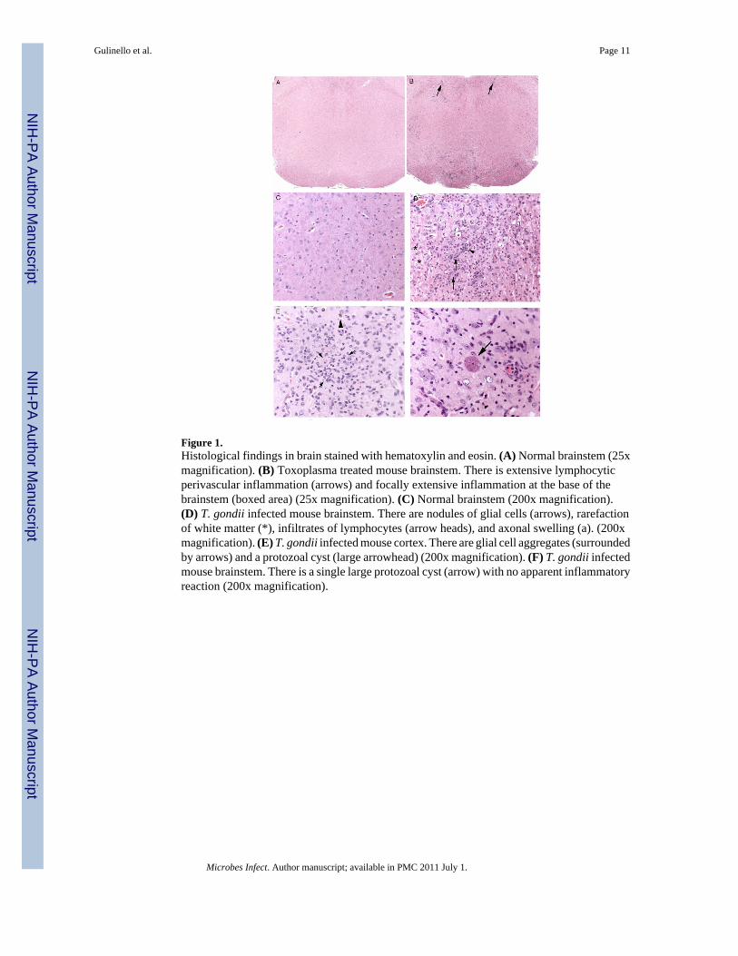
Figure 1.Histological findings in brain stained with hematoxylin and eosin. (A) Normal brainstem (25xmagnification). (B) Toxoplasma treated mouse brainstem. There is extensive lymphocyticperivascular inflammation (arrows) and focally extensive inflammation at the base of thebrainstem (boxed area) (25x magnification). (C) Normal brainstem (200x magnification).(D) T. gondii infected mouse brainstem. There are nodules of glial cells (arrows), rarefactionof white matter (*), infiltrates of lymphocytes (arrow heads), and axonal swelling (a). (200xmagnification). (E) T. gondii infected mouse cortex. There are glial cell aggregates (surroundedby arrows) and a protozoal cyst (large arrowhead) (200x magnification). (F) T. gondii infectedmouse brainstem. There is a single large protozoal cyst (arrow) with no apparent inflammatoryreaction (200x magnification).
Gulinello et al. Page 11
Microbes Infect. Author manuscript; available in PMC 2011 July 1.
NIH
-PA Author Manuscript
NIH
-PA Author Manuscript
NIH
-PA Author Manuscript

Figure 2.Motor coordination deficits assessed as the number of slips (A) and the time to cross (B) a 120cm long and 2.5 cm diameter beam. * indicates significant differences between T. gondiiinfected and control mice, p< 0.001.
Gulinello et al. Page 12
Microbes Infect. Author manuscript; available in PMC 2011 July 1.
NIH
-PA Author Manuscript
NIH
-PA Author Manuscript
NIH
-PA Author Manuscript

Figure 3.Gait deficits assessed as the number of mis-steps (A) and the mean stride length (B) and thenumber of rear paw drags (C). * indicates significant differences between T. gondii infectedand control mice, p< 0.05. Representative footprints of a control mouse (D) compared to T.gondii infected mice (E and F) clearly show examples of the shorter and more variable stridepattern, paw misplacements, weaving gait and other abnormalities. Examples of foot drag(G), paw misplacement (H), and gross ataxia (I) from T. gondii infected mice are alsoillustrated.
Gulinello et al. Page 13
Microbes Infect. Author manuscript; available in PMC 2011 July 1.
NIH
-PA Author Manuscript
NIH
-PA Author Manuscript
NIH
-PA Author Manuscript

Figure 4.Open field behavior in a 6 min test period indicates that T. gondii infected mice have lowerlevels of general locomotor activity (A: grid crosses) and exploratory activity (B: number ofrears) and the number of rear paw drags. * indicates significant differences between T.gondii infected and control mice, p< 0.005.
Gulinello et al. Page 14
Microbes Infect. Author manuscript; available in PMC 2011 July 1.
NIH
-PA Author Manuscript
NIH
-PA Author Manuscript
NIH
-PA Author Manuscript

Figure 5.Cognitive deficits were not evident in T. gondii infected mice (Toxo) in either spatial memory(A) or in recognition memory (B) in the novel object recognition and object placement tasks.Extent of object exploration (sec) was also normal during the trial 1 of each task (D and E) inboth T. gondii infected and control mice
Gulinello et al. Page 15
Microbes Infect. Author manuscript; available in PMC 2011 July 1.
NIH
-PA Author Manuscript
NIH
-PA Author Manuscript
NIH
-PA Author Manuscript

NIH
-PA Author Manuscript
NIH
-PA Author Manuscript
NIH
-PA Author Manuscript
Gulinello et al. Page 16
Tabl
e 1
Func
tiona
l Obs
erva
tion
Bat
tery
Rep
rese
ntat
ive
resu
lts fr
om a
prim
ary
scre
en in
dica
te d
efic
its in
seve
ral b
ehav
iora
l dom
ains
in m
ice
infe
cted
with
T. g
ondi
i. A
n or
dina
l sca
le is
use
d to
cate
goriz
e be
havi
ors w
ith n
orm
al d
efin
ed a
s 0, h
yopo
-rea
ctiv
e be
havi
ors o
r abs
ence
of n
orm
al b
ehav
iors
as p
rogr
essi
vely
neg
ativ
e nu
mbe
rs a
nd h
yper
-re
activ
ity o
r inc
iden
ce o
f abn
orm
al b
ehav
iors
as p
ositi
ve n
umbe
rs. W
here
abs
olut
e va
lues
cou
ld b
e m
easu
red
(pre
cise
num
ber o
f rea
rs e
tc),
thos
e va
lues
and
units
are i
ndic
ated
in th
e tab
le an
d ar
e use
d in
lieu
of t
he o
rdin
al sc
ale.
Pro
babi
lity
(p) i
s bas
ed o
n st
atis
tical
test
ing
as d
escr
ibed
in th
e mat
eria
ls an
d m
etho
dsse
ctio
n. N
S in
dica
tes n
ot si
gnifi
cant
.
cont
rol
SET
. gon
dii
SEp
Act
ivity
/ A
rous
al
Tran
sfer
Aro
usal
(late
ncy
to m
ove
afte
rtra
nsfe
r, se
c)4.
1±
0.7
3.1
±1.
0N
S
Rea
rs (#
in 1
min
)6.
2±
1.4
2.7
±1.
4N
S
Act
ivity
(grid
cro
sses
in 1
min
)33
.2±
5.5
19.1
±4.
0N
S
Touc
h es
cape
1.2
±0.
20.
7±
0.2
NS
Posi
tiona
l Pas
sivi
ty2.
0±
0.0
1.0
±0.
40.
02
Sens
orim
otor
Pelv
ic E
leva
tion
0.0
±0.
0−0
.7±
0.3
0.02
Gai
t−0
.6±
0.2
−0.8
±0.
3N
S
Tail
elev
atio
n0.
0±
0.0
−2.0
±1.
60.
04
Eye
0.0
±0.
0−0
.7±
0.7
0.00
9
Ata
xia
0.0
±0.
0−1
.5±
0.6
0.02
Hin
d lim
b us
e0.
0±
0.0
−0.9
±0.
40.
05
Whi
sker
mob
ility
0.0
±0.
0−0
.4±
0.2
0.05
Grip
(lat
ency
to fa
llfr
om a
wire
in se
c)24
.4±
4.6
2.9
±3.
90.
005
Ref
lexe
s
Toe
pinc
h1.
0±
0.0
1.0
±0.
0N
S
Neg
ativ
e G
eota
xis
(late
ncy
to tu
rn u
prig
htaf
ter i
nver
sion
on
agr
id in
sec)
1.55
±2.
37.
5±
2.1
NS
Star
tle1.
5±
0.3
0.7
±0.
3N
S
Microbes Infect. Author manuscript; available in PMC 2011 July 1.

NIH
-PA Author Manuscript
NIH
-PA Author Manuscript
NIH
-PA Author Manuscript
Gulinello et al. Page 17
cont
rol
SET
. gon
dii
SEp
Vis
ual P
laci
ng1.
4±
0.4
0.0
±0.
5N
S
Whi
sker
stim
ulat
ion
0.0
±0.
0−1
.0±
0.3
0.00
5
Gen
eral
con
ditio
n
Fur
0.0
±0.
0−1
.1±
0.1
0.00
1
Ster
eoty
ped
Beh
avio
r−0
.2±
0.2
−1.4
±0.
30.
008
Wei
ght (
g)26
.6±
1.0
17.5
±0.
90.
001
Microbes Infect. Author manuscript; available in PMC 2011 July 1.