Seroprevalence of Toxoplasma gondii Antibodies in Cats From Durango City, Mexico
33
1214 J. Parasitol., 93(5), 2007, pp. 1214–1216 American Society of Parasitologists 2007 Seroprevalence of Toxoplasma gondii Antibodies in Cats From Durango City, Mexico C. Alvarado-Esquivel, O. Liesenfeld*, R. G. Herrera-Flores†, B. E. Ramı ´rez-Sa ´nchez†, A. Gonza ´ lez-Herrera‡, S. A. Martı ´nez-Garcı ´a, and J. P. Dubey§, Facultad de Medicina, Universidad Jua ´rez del Estado de Durango, Dgo Mexico, Avenida Universidad y Fanny Anitua, 34000 Durango, Dgo. Mexico; *Institute for Microbiology and Hygiene, Campus Benjamin Franklin, Charite ´ Medical School Berlin, Hindenburgdamm 27, D-12203 Berlin, Germany; †Animal Shelter Municipal Office of Public Health and Environment, Durango, Mexico, Carretera a Mexico S/N, Durango, Dgo. Mexico; ‡Secretary of Health, Durango, Mexico. Cuauhtemoc 225 norte, 34000 Durango, Dgo. Mexico; §To whom correspondence should be addressed. United States Department of Agriculture, Agricultural Research Service, Animal and Natural Resources Institute, Animal Parasitic Diseases Laboratory, Building 1001, Beltsville, Maryland 20705-2350. e-mail: [email protected] TABLE I. General characteristics of the cats and prevalence of T. gondii antibodies. Characteristic Cats studied No. % Cats positive No. % Gender Male 34 32.4 6 17.6 Female 71 67.6 16 22.5 Age groups (yr) 0.5 53 50.4 7 13.2 0.5–1 30 28.6 6 20 1 22 21 9 41 Residence area Urban 105 100 22 21 Health status Healthy 95 90.5 21 22.1 Ill 10 9.5 1 10 Origin Stray 28 25.7 9 32.1 Household 77 73.3 13 16.9 Breed Crossbreed 105 100 22 21 Food Commercial 91 86.7 19 20.9 Homemade 66 62.9 13 19.7 Hunting and garbage 30 28.6 9 30 ABSTRACT: The prevalence of antibodies to Toxoplasma gondii was determined in sera from 105 domestic cats from Durango City, Mexico. Using a modified agglutination test, antibodies to this parasite were found in 21% of the 105 cats, with titers of 1:25 in 3 cats, 1:50 in 4 cats, 1:200 in 5 cats, 1:400 in 2 cats, 1:800 in 2 cats, 1:1,600 in 4 cats, and 1:3,200 or higher in 2 cats. Cats older than 1 yr had a significantly higher frequency of infection than that found in cats younger than 0.5 yr (41 vs. 13.2%, respectively; odds ratio 4.55; 95% CI 1.24– 17.18; P 0.01). Overall, the seroprevalence of T. gondii antibodies in cats in Durango, Mexico, is much lower compared with those re- ported in other countries. Cats are essential in the life cycle of Toxoplasma gondii because they are the only hosts that can excrete the environmentally resistant oocysts in nature (Dubey and Beattie, 1988). Little is known of the prevalence of T. gondii in cats in Mexico. In the present report, we determined the prevalence of T. gondii infection in cats from Durango City, Mexico, and we attempted to identify general characteristics of cats associated with infection. All 105 unwanted cats enrolled from August to November 2006 in the animal shelter in Durango City, Mexico, were studied. The animal shelter receives stray cats captured by the municipality from the streets of Durango City and unwanted pet cats given by the owners for adop- tion. General data, including age, breed, gender, health status, origin (stray or household), type of food eaten, and residence for cats were obtained (Table I). The sera were transported by courier from Mexico to Beltsville, Maryland, where serology was performed. Two-fold serial dilutions were made (1:25–1:3,200) and tested with a modified agglutination test (MAT), as described previously (Dubey and Desmonts, 1987). Whole formalin-fixed tachyzoites and mercaptoethanol were used as antigen, and a titer of 1:20 or higher was considered indicative of T. gondii exposure based on experimental studies in cats (Dubey and Thulliez, 1989; Dubey et al., 1995a, 1995b). Results were analyzed with the aid of the software Epi Info 6. For comparison of the frequencies among the groups, the Mantel-Haenszel test, and when indicated the Fisher exact test, were used. The associa- tion of the animal characteristics and the T. gondii infection was as- sessed by calculating the odds ratio (OR) with a 95% confidence inter- val (CI). For age comparison among groups of cats the Student’s t-test was used. A P value of less than 0.05 was considered significant. Antibodies to T. gondii were found in 21% of the 105 cats, with titers of 1:25 in 3 cats, 1:50 in 4 cats, 1:200 in 5 cats, 1:400 in 2 cats, 1:800 in 2 cats, 1:1,600 in 4 cats, and 1:3,200 or higher in 2 cats. General characteristics of 105 cats are shown in Table I. All cats were of crossbreed and resided in urban areas. Most cats studied were females, and the frequency of T. gondii infection observed in these females was similar to that observed in male cats (P 0.56). Cats were 1 mo to 7 yr old (mean 9 mo), and the prevalence of infection increased with age. Most cats were healthy, and the prevalence of infection in this group did not differ from that in ill cats (P 0.33). In addition, most cats studied were pets. The prevalence of infection in male and female cats in the stray group was comparable (33.3 and 31.3%, re- spectively; P 0.61). Similarly, although the prevalence of infection in pet female cats (20%) was 2-fold higher than that observed in pet male cats (9.1%), the overall prevalence of infection in male and female cats did not differ significantly (P 0.21). Cats older than 1 yr had a significantly higher frequency of infection than cats younger than 0.5 yr (41 vs. 13.2%, respectively; OR 4.55; 95% CI 1.24–17.18; P 0.01) and slightly higher than that observed in cats 0.5 to 1 yr old (41 vs. 20%; P 0.18). Toxoplasma gondii seroprevalence in stray cats is much higher in stray versus pet cats (Dubey, 1973; DeFeo et al., 2002; Nutter et al., 2004) as was the case in the present study. Higher seroprevalence in adult cats versus kittens, observed in the present study, supports earlier findings (Dubey, 1973; Ruiz and Frenkel, 1980b; Pena et al., 2006) and relates to the life cycle of T. gondii in cats; most cats are thought to become infected with T. gondii after weaning when they begin to hunt for food. The 21% prevalence of T. gondii antibodies in cats of Durango City, Mexico, in the present study is the lowest among all other surveys from North and South America, West Indies, and 1 study from Europe using a cut-off MAT titer of 1:20 (Table II). The prevalence of T. gondii in cats is a reflection of prevalence of T. gondii in animals that cats access for food. For example, Ruiz and Frenkel (1980a, 1980b) found a very high prevalence of T. gondii in cats and rodents and free-range chickens from Costa Rica. Although there are several reports of T. gondii infec- tion in humans and animals in Mexico (Varela et al., 1961; Fernandez- Torrano et al., 1986; Velasco-Castrejon et al., 1992; Galvan Ramirez et al., 1995, 1997; Del Rio-Chiriboga et al., 1997; Dubey, Morales, and
-
Upload
independent -
Category
Documents
-
view
0 -
download
0
Transcript of Seroprevalence of Toxoplasma gondii Antibodies in Cats From Durango City, Mexico
1214
J. Parasitol., 93(5), 2007, pp. 1214–1216� American Society of Parasitologists 2007
Seroprevalence of Toxoplasma gondii Antibodies in Cats From Durango City, Mexico
C. Alvarado-Esquivel, O. Liesenfeld*, R. G. Herrera-Flores†, B. E. Ramırez-Sanchez†, A. Gonzalez-Herrera‡, S. A. Martınez-Garcıa, andJ. P. Dubey§, Facultad de Medicina, Universidad Juarez del Estado de Durango, Dgo Mexico, Avenida Universidad y Fanny Anitua, 34000Durango, Dgo. Mexico; *Institute for Microbiology and Hygiene, Campus Benjamin Franklin, Charite Medical School Berlin, Hindenburgdamm27, D-12203 Berlin, Germany; †Animal Shelter Municipal Office of Public Health and Environment, Durango, Mexico, Carretera a Mexico S/N,Durango, Dgo. Mexico; ‡Secretary of Health, Durango, Mexico. Cuauhtemoc 225 norte, 34000 Durango, Dgo. Mexico; §To whomcorrespondence should be addressed. United States Department of Agriculture, Agricultural Research Service, Animal and Natural ResourcesInstitute, Animal Parasitic Diseases Laboratory, Building 1001, Beltsville, Maryland 20705-2350. e-mail: [email protected]
TABLE I. General characteristics of the cats and prevalence of T. gondiiantibodies.
Characteristic
Cats studied
No. %
Cats positive
No. %
Gender
Male 34 32.4 6 17.6Female 71 67.6 16 22.5
Age groups (yr)
�0.5 53 50.4 7 13.20.5–1 30 28.6 6 20
�1 22 21 9 41
Residence area
Urban 105 100 22 21
Health status
Healthy 95 90.5 21 22.1Ill 10 9.5 1 10
Origin
Stray 28 25.7 9 32.1Household 77 73.3 13 16.9
Breed
Crossbreed 105 100 22 21
Food
Commercial 91 86.7 19 20.9Homemade 66 62.9 13 19.7Hunting and garbage 30 28.6 9 30
ABSTRACT: The prevalence of antibodies to Toxoplasma gondii wasdetermined in sera from 105 domestic cats from Durango City, Mexico.Using a modified agglutination test, antibodies to this parasite werefound in 21% of the 105 cats, with titers of 1:25 in 3 cats, 1:50 in 4cats, 1:200 in 5 cats, 1:400 in 2 cats, 1:800 in 2 cats, 1:1,600 in 4 cats,and 1:3,200 or higher in 2 cats. Cats older than 1 yr had a significantlyhigher frequency of infection than that found in cats younger than 0.5yr (41 vs. 13.2%, respectively; odds ratio � 4.55; 95% CI � 1.24–17.18; P � 0.01). Overall, the seroprevalence of T. gondii antibodiesin cats in Durango, Mexico, is much lower compared with those re-ported in other countries.
Cats are essential in the life cycle of Toxoplasma gondii because theyare the only hosts that can excrete the environmentally resistant oocystsin nature (Dubey and Beattie, 1988). Little is known of the prevalenceof T. gondii in cats in Mexico. In the present report, we determined theprevalence of T. gondii infection in cats from Durango City, Mexico,and we attempted to identify general characteristics of cats associatedwith infection.
All 105 unwanted cats enrolled from August to November 2006 inthe animal shelter in Durango City, Mexico, were studied. The animalshelter receives stray cats captured by the municipality from the streetsof Durango City and unwanted pet cats given by the owners for adop-tion. General data, including age, breed, gender, health status, origin(stray or household), type of food eaten, and residence for cats wereobtained (Table I).
The sera were transported by courier from Mexico to Beltsville,Maryland, where serology was performed. Two-fold serial dilutionswere made (1:25–1:3,200) and tested with a modified agglutination test(MAT), as described previously (Dubey and Desmonts, 1987). Wholeformalin-fixed tachyzoites and mercaptoethanol were used as antigen,and a titer of 1:20 or higher was considered indicative of T. gondiiexposure based on experimental studies in cats (Dubey and Thulliez,1989; Dubey et al., 1995a, 1995b).
Results were analyzed with the aid of the software Epi Info 6. Forcomparison of the frequencies among the groups, the Mantel-Haenszeltest, and when indicated the Fisher exact test, were used. The associa-tion of the animal characteristics and the T. gondii infection was as-sessed by calculating the odds ratio (OR) with a 95% confidence inter-val (CI). For age comparison among groups of cats the Student’s t-testwas used. A P value of less than 0.05 was considered significant.
Antibodies to T. gondii were found in 21% of the 105 cats, with titersof 1:25 in 3 cats, 1:50 in 4 cats, 1:200 in 5 cats, 1:400 in 2 cats,1:800 in 2 cats, 1:1,600 in 4 cats, and 1:3,200 or higher in 2 cats.
General characteristics of 105 cats are shown in Table I. All catswere of crossbreed and resided in urban areas. Most cats studied werefemales, and the frequency of T. gondii infection observed in thesefemales was similar to that observed in male cats (P � 0.56). Cats were1 mo to 7 yr old (mean 9 mo), and the prevalence of infection increasedwith age. Most cats were healthy, and the prevalence of infection inthis group did not differ from that in ill cats (P � 0.33). In addition,most cats studied were pets. The prevalence of infection in male andfemale cats in the stray group was comparable (33.3 and 31.3%, re-spectively; P � 0.61). Similarly, although the prevalence of infectionin pet female cats (20%) was 2-fold higher than that observed in petmale cats (9.1%), the overall prevalence of infection in male and femalecats did not differ significantly (P � 0.21).
Cats older than 1 yr had a significantly higher frequency of infection
than cats younger than 0.5 yr (41 vs. 13.2%, respectively; OR � 4.55;95% CI � 1.24–17.18; P � 0.01) and slightly higher than that observedin cats 0.5 to 1 yr old (41 vs. 20%; P � 0.18). Toxoplasma gondiiseroprevalence in stray cats is much higher in stray versus pet cats(Dubey, 1973; DeFeo et al., 2002; Nutter et al., 2004) as was the casein the present study. Higher seroprevalence in adult cats versus kittens,observed in the present study, supports earlier findings (Dubey, 1973;Ruiz and Frenkel, 1980b; Pena et al., 2006) and relates to the life cycleof T. gondii in cats; most cats are thought to become infected with T.gondii after weaning when they begin to hunt for food.
The 21% prevalence of T. gondii antibodies in cats of Durango City,Mexico, in the present study is the lowest among all other surveys fromNorth and South America, West Indies, and 1 study from Europe usinga cut-off MAT titer of 1:20 (Table II). The prevalence of T. gondii incats is a reflection of prevalence of T. gondii in animals that cats accessfor food. For example, Ruiz and Frenkel (1980a, 1980b) found a veryhigh prevalence of T. gondii in cats and rodents and free-range chickensfrom Costa Rica. Although there are several reports of T. gondii infec-tion in humans and animals in Mexico (Varela et al., 1961; Fernandez-Torrano et al., 1986; Velasco-Castrejon et al., 1992; Galvan Ramirez etal., 1995, 1997; Del Rio-Chiriboga et al., 1997; Dubey, Morales, and

RESEARCH NOTES 1215
TABLE II. Seroprevalence* of T. gondii antibodies in cats from different countries.
Country City or region Source No. tested%
Prevalence Reference
Brazil Sao Paulo state Stray, unwanted pets 502 26.3 Silva et al. (2002)Sao Paulo state Stray, unwanted pets 237 35.4 Pena et al. (2006)Parana state Stray 58 84.4 Dubey, Navarro et al. (2004)
Colombia Armenia and Bogota Stray, unwanted pets 170 30.5 Dubey, Su et al. (2006)People’s Republic of China Guangzhou Market 34 79.4 Dubey et al. (2007)Spain Barcelona Pets 220 45 Gauss et al. (2003)United States Ohio Pets and stray 275 48 Dubey et al. (2002)
Rhode Island Pets and stray 200 42 DeFeo et al. (2002)Illinois Farms 295 75.6 Dubey, Weigel et al. (1995)North Carolina Stray and pets 176 50.5 Nutter et al. (2004)
West Indies Grenada Pets 40 35 Asthana et al. (2006)St. Kitts Pets 106 84.9 Moura et al. (2007)
* Using modified agglutination test titer of 1:20 or higher.
Lehmann, 2004; Figueroa-Castillo et al., 2006), little is known of theepidemiology of this parasite in Mexico. Varela et al. (1961) found dyetest antibodies in 52.2% of cats in Mexico, but they provided no otherdetails about the cats. Galvan Ramirez et al. (1999) reported that 64%of 59 cat owners and 70.8% of their cats in Guadalajara had T. gondiiantibodies. Toxoplasma gondii oocysts were found in the feces of 13of 200 cats from Mexico (de Aluja and Aguilar, 1977). For epidemio-logic studies, serologic surveys in cats are more useful than the detec-tion of oocysts, because at any given time only 1% of cats shed oocystsin their feces (Dubey, 2004). In the present study, 21% of cats wereseropositive for T. gondii, and they likely were shedding oocysts, there-by contaminating the environment.
We thank O. C. H. Kwok and L. A. Bandini for technical assistance.
LITERATURE CITED
ASTHANA, S. P., C. N. L. MACPHERSON, S. H. WEISS, R. STEPHENS, R.N. SHARMA, AND J. P. DUBEY. 2006. Seroprevalence of Toxoplasmagondii in pregnant women and cats in Grenada, West Indies. Jour-nal of Parasitology 92: 644–645.
DE ALUJA, A. S., AND P. AGUILAR. 1977. Estudio sobre la frecuencia delooquiste de Toxoplasma gondii en el gato domestico del distritofederal. Gaceta Medica de Mexico 113: 455–459.
DEFEO, M. L., J. P. DUBEY, T. N. MATHER, AND R. C. RHODES. 2002.Epidemiologic investigation of seroprevalence of antibodies toToxoplasma gondii in cats and rodents. American Journal of Vet-erinary Research 63: 1714–1717.
DEL RIO-CHIRIBOGA, C., A. ORZECHOWSKI-RALLO, AND G. SANCHEZ-ME-JORADA. 1997. Toxoplasmosis of the central nervous system in pa-tients with AIDS in Mexico. Archives of Medical Research 28:527–530.
DUBEY, J. P. 1973. Feline toxoplasmosis and coccidiosis: A survey ofdomiciled and stray cats. Journal of the American Veterinary Med-ical Association 162: 873–877.
———. 2004. Toxoplasmosis—A waterborne zoonosis. Veterinary Par-asitology 126: 57–72.
———, AND C. P. BEATTIE. 1988. Toxoplasmosis of animals and man.CRC Press, Boca Raton, Florida, 220 p.
———, AND G. DESMONTS. 1987. Serological responses of equids fedToxoplasma gondii oocysts. Equine Veterinary Journal 19: 337–339.
———, M. R. LAPPIN, AND P. THULLIEZ. 1995a. Diagnosis of inducedtoxoplasmosis in neonatal cats. Journal of the American VeterinaryMedical Association 207: 179–185.
———, ———, AND ———. 1995b. Long-term antibody responses ofcats fed Toxoplasma gondii tissue cysts. Journal of Parasitology81: 887–893.
———, E. S. MORALES, AND T. LEHMANN. 2004. Isolation and genotyp-ing of Toxoplasma gondii from free-ranging chickens from Mexico.Journal of Parasitology 90: 411–413.
———, I. T. NAVARRO, C. SREEKUMAR, E. DAHL, R. L. FREIRE, H. H.
KAWABATA, M. C. B. VIANNA, O. C. H. KWOK, S. K. SHEN, P.THULLIEZ, ET AL. 2004. Toxoplasma gondii infections in cats fromParana, Brazil: Seroprevalence, tissue distribution, and biologic andgenetic characterization of isolates. Journal of Parasitology 90:721–726.
———, W. J. A. SAVILLE, J. F. STANEK, AND S. M. REED. 2002. Prev-alence of Toxoplasma gondii antibodies in domestic cats from ruralOhio. Journal of Parasitology 88: 802–803.
———, C. SU, J. A. CORTES, N. SUNDAR, J. E. GOMEZ-MARIN, L. J.POLO, L. ZAMBRANO, L. E. MORA, F. LORA, J. JIMENEZ, ET AL. 2006.Prevalence of Toxoplasma gondii in cats from Colombia, SouthAmerica and genetic characterization of T. gondii isolates. Veteri-nary Parasitology 141: 42–47.
———, AND P. THULLIEZ. 1989. Serologic diagnosis of toxoplasmosisin cats fed Toxoplasma gondii tissue cysts. Journal of the AmericanVeterinary Medical Association 194: 1297–1299.
———, R. M. WEIGEL, A. M. SIEGEL, P. THULLIEZ, U. D. KITRON, M.A. MITCHELL, A. MANNELLI, N. E. MATEUS-PINILLA, S. K. SHEN, O.C. H. KWOK, ET AL. 1995. Sources and reservoirs of Toxoplasmagondii infection on 47 swine farms in Illinois. Journal of Parasi-tology 81: 723–729.
———, X. Q. ZHU, N. SUNDAR, H. ZHANG, O. C. H. KWOK, AND C. SU.2007. Genetic and biologic characterization of Toxoplasma gondiiisolates of cats from China. Veterinary Parasitology 145: 352–356.
FERNANDEZ-TORRANO, M., M. T. SIBAJA-CONTRERAS, AND A. R. GRANIER-MELO. 1986. Sero-epidemiologic survey of anti-Toxoplasma gondiiantibodies in 125 pregnant women from eastern Tabasco State. Bol-etın Medico Hospital Infantil Mexico 43: 274–278.
FIGUEROA-CASTILLO, J. A., V. DUARTE-ROSAS, M. JUAREZ-ACEVEDO, H.LUNA-PASTEN, AND D. CORREA. 2006. Prevalence of Toxoplasmagondii antibodies in rabbits (Oryctolagus cuniculus). Journal ofParasitology 92: 394–395.
GALVAN RAMIREZ, M. L., J. L. SOTO MANCILLA, O. VELASCO CASTREJON,AND R. PEREZ MEDINA. 1995. Incidence of anti-Toxoplasma anti-bodies in women with high-risk pregnancy and habitual abortions.Revista da Sociedade Brasileira de Medicina Tropical 28: 333–337.
———, V. VALDEZ ALVARADO, G. VARGAS GUTIERREZ, O. JIMENEZ GON-ZALEZ, C. GARCıA COSIO, AND M. VIELMA SANDOVAL. 1997. Prev-alence of IgG and IgM anti-Toxoplasma antibodies in patients withHIV and acquired immunodeficiency syndrome (AIDS). Revista daSociedade Brasileira de Medicina Tropical 30: 465–467.
———, G. SANCHEZ VARGAS, M. VIELMA SANDOVAL, AND J. L. SOTO
MANCILLA. 1999. Presence of anti-Toxoplasma antibodies in hu-mans and their cats in the urban zone of Guadalajara. Revista daSociedade Brasileira de Medicina Tropical 32: 483–488.
GAUSS, C. B. L., S. ALMERıA, A. ORTUNO, F. GARCIA, AND J. P. DUBEY.2003. Seroprevalence of Toxoplasma gondii antibodies in domesticcats from Barcelona, Spain. Journal of Parasitology 89: 1067–1068.
MOURA, L., P. KELLY, R. C. KRECEK, AND J. P. DUBEY. 2007. Seroprev-
1216 THE JOURNAL OF PARASITOLOGY, VOL. 93, NO. 5, OCTOBER 2007
alence of Toxoplasma gondii in cats from St. Kitts, West Indies.Journal of Parasitology 93: 952–953.
NUTTER, F. B., J. P. DUBEY, J. F. LEVINE, E. B. BREITSCHWERDT, R. B.FORD, AND M. K. STOSKOPF. 2004. Seroprevalence of antibodiesagainst Bartonellla henselae and Toxoplasma gondii and fecalshedding of Cryptosporidium spp., and Toxocara cati in feral andpet domestic cats. Journal of the American Veterinary Medical As-sociation 225: 1394–1398.
PENA, H. F. J., R. M. SOARES, M. AMAKU, J. P. DUBEY, AND S. M.GENNARI. 2006. Toxoplasma gondii infection in cats from Sao Pau-lo state, Brazil: Seroprevalence, oocyst shedding, isolation in mice,and biologic and molecular characterization. Research in VeterinaryScience 81: 58–67.
RUIZ, A., AND J. K. FRENKEL. 1980a. Intermediate and transport hostsof Toxoplasma gondii in Costa Rica. American Journal of TropicalMedicine and Hygiene 29: 1161–1166.
———, AND ———. 1980b. Toxoplasma gondii in Costa Rican cats.American Journal of Tropical Medicine and Hygiene 29: 1150–1160.
SILVA, J. C. R., S. M. GENNARI, A. M. A. RAGOZO, V. R. AMAJONES, C.MAGNABOSCO, L. E. O. YAI, J. S. FERREIRA-NETO, AND J. P. DUBEY.2002. Prevalence of Toxoplasma gondii antibodies in sera of do-mestic cats from Guarulhos and Sao Paulo, Brazil. Journal of Par-asitology 88: 419–420.
VARELA, G., E. ROCH, AND J. ZAVALA. 1961. Estudios de toxoplasmosis.Gaceta Medica de Mexico 91: 669–673.
VELASCO-CASTREJON, O., B. SALVATIERRA-IZABA, J. L. VALDESPINO, A.M. SEDANO-LARA, S. GALINDO-VIRGEN, C. MAGOS, A. LLAUSAS, R.TAPIA-CONYER, G. GUTIERREZ, AND J. SEPULVEDA. 1992. Seroepi-demiologica de la toxoplasmosis en Mexico Salud. Publica Mexico34: 222–229.
J. Parasitol., 93(5), 2007, pp. 1216–1218� American Society of Parasitologists 2007
Prevalence of Neospora caninum and Toxoplasma gondii Antibodies in Wild RuminantsFrom the Countryside or Captivity in the Czech Republic
E. Bartova, K. Sedlak*, I. Pavlik†, and I. Literak, Department of Biology and Wildlife Diseases, University of Veterinary and PharmaceuticalSciences, Palackeho 1/3, Brno 612 42 Brno, Czech Republic. *Department of Virology and Serology, State Veterinary Institute Prague, Sidlistni136/24, 165 03 Prague 6, Czech Republic. †Veterinary Research Institute, Hudcova 70, 621 32 Brno, Czech Republic. e-mail: [email protected]
ABSTRACT: In the Czech Republic, sera from 720 wild ruminants wereexamined for antibodies to Neospora caninum by screening competi-tive-inhibition enzyme-linked immunosorbent assay and confirmed byindirect fluorescence antibody test (IFAT); the same sera were also ex-amined for antibodies to Toxoplasma gondii by IFAT. Neospora can-inum antibodies were found in 14% (11 positive/79 tested) roe deer(Capreolus capreolus), 14% (2/14) sika deer (Cervus nippon), 6% (24/377) red deer (Cervus elaphus), 1% (2/143) fallow deer (Dama dama),3% (3/105) mouflon (Ovis musimon), and none of 2 reindeer (Rangifertarandus). Toxoplasma gondii antibodies were found in 50% (7/14) sikadeer, 45% (169/377) red deer, 24% (19/79) roe deer, 17% (24/143) fal-low deer, 9% (9/105) mouflon, and 1 of 2 reindeer. In 42 samples ofwild ruminants that tested positive for N. caninum antibodies, 28 (67%of the positive N. caninum samples) reacted solely to N. caninum. Thisis the first evidence of N. caninum infection in mouflon, the first N.caninum seroprevalence study in farmed red deer, and the first surveyof N. caninum in wild ruminants from the Czech Republic.
Toxoplasma gondii and Neospora caninum are 2 closely related pro-tozoan parasites that are distributed worldwide. Both species have anindirect life cycle, with carnivores as the definitive hosts (Dubey andBeattie, 1988; Dubey, 2003). Definitive hosts of N. caninum are dogsand coyotes (Canis latrans) that excrete oocysts in feces (Dubey, 2003;Gondim, 2004). Wild herbivores are suggested to play a role of inter-mediate hosts in the sylvatic cycle of N. caninum infection (Gondim,2006). In North America, antibodies against N. caninum have beenfound in white-tailed deer (Odocoileus virginianus), moose (Alces al-ces), caribou (Rangifer tarandus), bison (Bison bison), and muskox(Ovibos moschatus) (Dubey et al., 1999; Lindsay et al., 2002; Gondimet al., 2004; Dubey and Thulliez, 2005). In Europe, antibodies againstN. caninum were detected in red deer (Cervus elaphus), roe deer (Ca-preolus capreolus), chamois (Rupicapra rupicapra), Alpine ibex (Capraibex), Barbary sheep (Ammotragus lervia), and in European bison (Bi-son bonasus; Ferroglio and Rossi, 2001; Ferroglio et al., 2001; Cabajet al., 2005; Almeria et al., 2006).
Definitive hosts of T. gondii are cats and other felids (Dubey andBeattie, 1988). Many species of warm-blooded animals, including hu-mans, serve as intermediate hosts; hosts can be infected by ingestionof food or water contaminated with sporulated T. gondii oocysts ex-creted by the definitive host, by consumption of cysts in infected ani-
mals tissues, or congenitally (Dubey and Beattie, 1988). As herbivorousanimals, the wild ruminants are a good indicator for the monitoring ofenvironmental contamination with T. gondii oocysts. Meat or venisonof wild ruminants infected with T. gondii could serve as a potentialsource of toxoplasmosis for other animals, especially carnivores, in-cluding humans. Toxoplasmosis infection was documented in deer hunt-ers from the United States after ingesting undercooked or raw deervenison (Sacks et al., 1983; Ross et al., 2001). In North America, an-tibodies against T. gondii have been found in white-tailed deer, black-tailed deer (Odocoileus hemionus), marsh deer (Blastocerus dichoto-mus), pampas deer (Ozotocerus bezoarticus), moose, caribou, bison,Dall sheep (Ovis dalli), and muskox (Lindsay et al., 1991; Chomel etal., 1994; Ferreira et al., 1997; Kutz et al., 2000, 2001; Zarnke et al.,2000). In Europe, antibodies to T. gondii have been reported in red deer,roe deer, fallow deer (Dama dama), reindeer (Rangifer tarandus),moose, chamois, mouflon (Ovis musimon), Spanish ibex (Capra pyr-enaica), and Barbary sheep (Catar, 1972; Kapperud, 1978; Williamsonand Williams, 1980; Hejlicek et al., 1997; Oksanen et al., 1997; Sroka,2001; Vikoren et al., 2004; Gauss et al., 2006).
The aim of this study was to survey the seroprevalence N. caninumand T. gondii antibodies in wild ruminants from the countryside andcaptivity in the Czech Republic. During 1998–2006, sera from 720 wildruminants from 11 of 14 existing Czech regions (Prague, Stredocesky,Ustecky, Karlovarsky, Plzensky, Jihocesky, Liberecky, Vysocina, Mo-ravskoslezsky, Olomoucky, and Jihomoravsk) were collected.
Wild ruminants came from game preserves (a fenced hunting districtwith more than 50 ha, where animals are bred mostly for hunting pur-poses but can, in some cases, be released into the wild), farms (fencedarea, where the size is not limited, but usually is smaller than gamepreserves; animals are bred as farm animals), or free range.
Blood samples were taken from 377 red deer, primarily from 4 farmsand 4 game preserves (but in 1 case, a free-ranging animal), 79 free-ranging roe deer, and in 10 cases, from 2 game preserves; 14 sika deer(Cervus nippon) from 2 game preserves; 143 fallow deer from 6 gamepreserves and 79 free-ranging cases; 76 free-ranging mouflons and 29from 2 game preserves; and 2 reindeer from 1 farm. The blood sampleswere obtained from blood vessels of animals before transportation orfrom hearts of animals that were shot during hunting seasons.
Blood was centrifuged and sera were stored at �20 C until assayedfor antibodies to N. caninum and T. gondii. A commercial competitive-
RESEARCH NOTE 1217
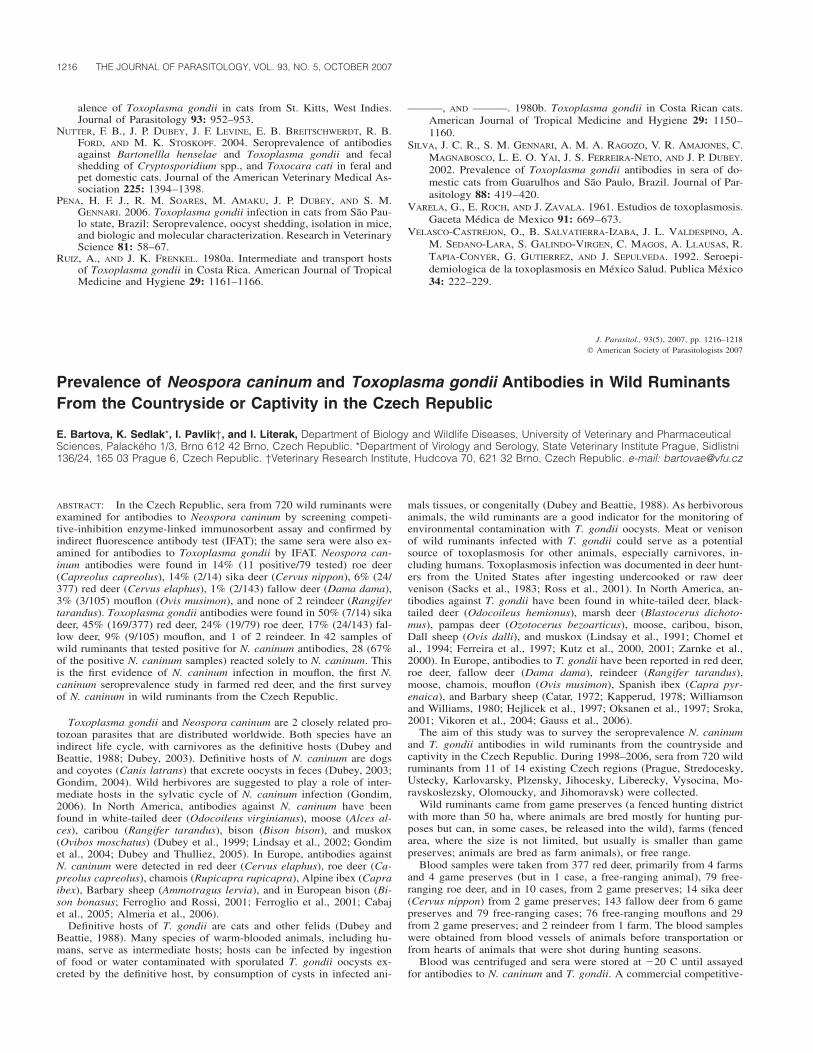
TABLE I. Prevalence of N. caninum (cELISA, IFAT) and T. gondii (IFAT) in wild ruminants from The Czech Republic.
Species
Neospora caninum
ExaminedPositive
(cELISA)Positive(IFAT)*
Prevalence(%)†
Inhibitionrange
(cELISA)
Toxoplasma gondii
ExaminedPositive(IFAT)
Prevalence(%)
Antibodytiter
range
IFAT-positivefor both
N. caninum andT. gondii
Red deer (Cervus elaphus) 377 24 24 6 30.4–85 377 169 45 40–640 18/377 (5%)Sika (Cervus nippon) 14 2 2 14 34–56 14 7 50 80–320 0/14 (0%)Fallow deer (Dama dama) 143 6 2 1 30.9–39 143 24 17 40–160 2/143 (1%)Roe deer (Capreolus ca-
preolus) 79 12 11 14 59–91 79 19 24 40–160 7/79 (9%)Mouflon (Ovis musimon) 105 4 3 3 45–56 105 9 9 40–320 1/105 (1%)Reindeer (Rangifer taran-
dus) 2 0 0 0 0 2 1 50 80 0/2 (0%)Total 720 48 42 6 30.4–91 720 229 32 40–640 14/720 (4%)
* For confirming, the samples positive in screening cELISA were examined by IFAT.† Prevalence was counted from samples that were positive in both of the tests.
inhibition enzyme-linked immunosorbent assay (cELISA; VMRD, Pull-man, Washington) was used for detection of N. caninum antibodies inwild ruminants according to the manufacturers’ instructions. The serawere positive if more than 30% inhibition was found. To confirm resultsin cELISA with a more sensitive method, the positive sera were retestedby direct fluorescence antibody test (IFAT) with a commercially avail-able Neospora NIFR antigen (VMRD), anti-deer IgG, and anti-goat IgGconjugate (VMRD). The sera were diluted in a 2-fold series starting at1:50 as a basic dilution; a titer �1:50 was considered positive. Onlyanimals that were positive to both tests were considered positive.
Toxoplasma gondii antibodies were detected in sera by IFAT with acommercially available antigen Sevatest toxoplasma NIFR (Sevaphar-ma, Prague, The Czech Republic) and anti-deer IgG conjugate (KPLInc., Gaithersburg, Maryland) and anti-goat IgG conjugate (VMRD).The sera were diluted in a 2-fold series starting at 1:40 as a basicdilution; a titer �1:40 was considered positive.
The results of a serologic survey of N. caninum and T. gondii anti-bodies (prevalence, inhibition, and titer ranges) in wild ruminants in theCzech Republic are presented in Table I. This report presents the firstevidence of N. caninum infection in mouflon; Almeria et al. (2006)examined 27 mouflons from Spain, but with negative results. The high-est prevalence of N. caninum was observed in roe deer and sika deer,followed by red deer, and low prevalence in fallow deer and mouflons.Similar results were reported by Almeria et al. (2006) from Spain. Neo-spora caninum antibodies were found in 12% (28/237) of the red deerand in 6% (2/33) of the roe deer; mouflon and fallow deer were nega-tive. A possible explanation for higher prevalence in roe deer and reddeer might be related to their grazing in the lowlands close to humansettlements, where conditions are more conductive to N. caninum trans-mission. Wild ruminants probably become infected by ingesting foodor water contaminated by N. caninum oocysts excreted by canids in thearea (Almeria et al., 2006). On the other hand, wild or domestic dogsin rural areas could be infected by eating offal from wild ruminants leftin the forest by hunters.
Positive reaction in IFAT for both N. caninum and T. gondii wereobserved in 4% of examined animals (Table I). In 42 samples of wildruminants that tested positive for N. caninum antibodies, 28 (67% ofthe positive N. caninum samples) reacted solely to N. caninum. Simi-larly, Almeria et al. (2006) found that 70% of their positive samplesreacted solely to N. caninum, indicating less cross-reaction betweenthese 2 closely related parasites.
Here, we also present the first evidence of T. gondii antibodies insika deer and reindeer in the Czech Republic. We can compare ourresults with a previous T. gondii seroprevalence study in a group ofwild ruminants in the Czech Republic that was done in South Bohemiaduring 1981–1990 (Hejlicek et al., 1997). Sera of fallow deer, red deer,roe deer, and mouflons were examined by the Sabin–Feldman dye test,with 100% (3/3), 15% (46/303), 14% (13/95), and 10% (2/20) preva-lence, respectively. We found a higher prevalence in red deer (45%)and roe deer (24%), but lower in fallow deer (16.8%) and mouflons
(8.6%). Red deer from our study came mostly from farms and gamepreserves, whereas in the previous study (Hejlicek et al., 1997), mostof the animals were free ranging. Fenced areas, especially farms, areusually smaller, and cats had greater access to these areas. The mainsource of T. gondii infection for wild ruminants is feedstuff (grass, hay,or commercial feed) or water contaminated with T. gondii oocystspassed by felids in their feces (Dubey and Beattie, 1988). Other carni-vores are also at risk for T. gondii infection, as well as humans afterconsumption of meat or offal from wild ruminants that are infected byT. gondii (Sacks et al., 1983; Ross et al., 2001).
Similar to Almeria et al. (2006), we found that animals from certainfarms or game preserves were more infected than animals from otherlocalities. We can conclude that T. gondii and N. caninum infection areenzootic in character. In our study, the majority of red deer came from2 large farms (i.e., 1 farm from the Stredocesky region, where 9 and51% of 141 examined animals had antibodies against N. caninum andT. gondii, respectively, and another farm in the Plzensky region, where188 animals were examined, with 4 and 41% N. caninum and T. gondiiprevalence, respectively. All positive samples in sika deer were foundin 1 game preserve (animals were previously imported from Germany)in the Vysocina region, with 20 and 70% (of 10 examined animals) N.caninum and T. gondii prevalence, respectively. All N. caninum– andT. gondii–positive samples in roe deer were found in animals from 1game preserve in the Plzensky region, with 33% (1/3) of both N. can-inum and T. gondii prevalence, and from free-ranging animals in theVysocina region, where 16 and 26% of 69 ruminants had antibodiesagainst N. caninum and T. gondii, respectively. All N. caninum–positivesamples and the majority of all T. gondii–positive samples from mou-flons were found in free-ranging animals from the Ustecky region,where 25% (3/12) and 42% (5/12) had antibodies against N. caninumand T. gondii, respectively, and from the Stredocesky region, whereprevalences were 6% (1/17) for N. caninum and 12% (2/17) for T.gondii. Finally, half of all T. gondii–positive fallow deer came from asingle game preserve in the Stredocesky region, where 29% (of 41examined animals) were positive.
This work was supported by the Ministry of Education, Youth andSports of the Czech Republic (MSM6215712402) and Ministry of Ag-riculture of the Czech Republic (MZE0002716201).
LITERATURE CITED
ALMERIA, S., D. VIDAL, D. FERRER, M. PABON, M. I. G. FERNANDEZ-DE-MERA, F. RUIZ-FONS, V. ALZAGA, I. MARCO, C. CALVETE, S. LAVIN
ET AL. 2006. Seroprevalence of Neospora caninum in non-carniv-orous wildlife from Spain. Veterinary Parasitology 143: 21–28.
CABAJ, W., B. MOSKWA, K. PASTUSIAK, AND J. GILL. 2005. Antibodiesto Neospora caninum in the blood of European bison (Bison bon-asus bonasus L.) living in Poland. Veterinary Parasitology 128:163–168.

1218 THE JOURNAL OF PARASITOLOGY, VOL. 93, NO. 5, OCTOBER 2007
CATAR, G. 1972. Studies on toxoplasmosis as regards its natural focalityin Slovakia. Folia Parasitologica (Praha) 19: 253–256.
CHOMEL, B. B., M. L. CARNICIU, R. W. KASTEN, P. M. CASTELLI, T. M.WORK, AND D. A. JESSUP. 1994. Antibody prevalence of eight ru-minant infectious diseases in California mule and black-tailed deer(Odocoileus hemionus). Journal of Wildlife Diseases 30: 51–59.
DUBEY, J. P., 2003. Reveiw of Neospora caninum and neosporosis inanimals. Korean Journal of Parasitology 41: 1–16.
———, AND C. P. BEATTIE. 1988. Toxoplasmosis of animals and man.CRC Press, Boca Raton, Florida, 220 p.
———, K. HOLLIS, S. ROMAND, P. THULLIEZ, O. C. H. KWOK, L. HUN-GERFORD, C. ANCHOR, AND D. ETTER. 1999. High prevalence ofantibodies to Neospora caninum in white-tailed deer (Odocoileusvirginianus). Journal of Parasitology 29: 1709–1711.
———, AND P. THULLIEZ. 2005. Prevalence of antibodies to Neosporacaninum in wild animals. Journal of Parasitology 91: 1217–1218.
FERREIRA, R. A., J. R. MINEO, J. M. DUARTE, D. A. O. SILVA, AND J. H.PATARROYO. 1997. Toxoplasmosis in naturally infected deer fromBrazil. Journal of Wildlife Diseases 33: 896–899.
FERROGLIO, E., B. BASSANO, A. TRISCIUOGLIO, AND L. ROSSI. 2001. An-tibodies to Neospora caninum in Alpine ibex from the Italian Alps.Zeitschrift fur Jagdwissenschaft 47: 226–228.
———, AND L. ROSSI. 2001. Prevalence of Neospora caninum antibod-ies in wild ruminants from the Italian Alps. Veterinary Record 148:754–755.
GAUSS, C. B. L., J. P. DUBEY, D. VIDAL, O. CABEZON, F. RUIZ-FONS, J.VICENTE, I. MARCO, S. LAVIN, C. GORTAZAR, AND S. ALMERIA. 2006.Prevalence of Toxoplasma gondii antibodies in red deer (Cervuselaphus) and other wild ruminants from Spain. Veterinary Parasi-tology 136: 193–200.
GONDIM, L. F. P. 2006. Neospora caninum in wildlife. Trends in Para-sitology 22: 247–252.
———, M. M. MCALLISTER, N. E. MATEUS-PINILLA, W. C. PITT, L. D.MECH, AND M. E. NELSON. 2004. Transmission of Neospora can-inum between wild and domestic animals. Journal of Parasitology90: 1361–1365.
———, ———, W. C. PITT, AND D. E. ZEMLICKA. 2004. Coyotes (Canislatrans) are definitive hosts of Neospora caninum. InternationalJournal for Parasitology 34: 159–161.
HEJLıCEK, K., I. LITERAK, AND J. NEZVAL. 1997. Toxoplasmosis in wildmammals from the Czech Republic. Journal of Wildlife Diseases33: 480–485.
KAPPERUD, G. 1978. Survey for toxoplasmosis in wild and domesticanimals from Norway and Sweden. Journal of Wildlife Diseases14: 157–162.
KUTZ, S. J., B. ELKIN, A. GUNN, AND J. P. DUBEY. 2000. Prevalence ofToxoplasma gondii antibodies in muskox (Ovibos moschatus) serafrom Northern Canada. Journal of Parasitology 86: 879–882.
———, ———, D. PANAYI, AND J. P. DUBEY. 2001. Prevalence of Toxo-plasma gondii antibodies in barren-ground caribou (Rangifer tar-andus groenlandicus) from the canadian arctic. Journal of Parasi-tology 87: 439–442.
LINDSAY, D. S., B. L. BLAGBURN, J. P. DUBEY, AND W. H. MASON. 1991.Prevalence and isolation of Toxoplasma gondii from white-taileddeer in Alabama. Journal of Parasitology 77: 62–64.
———, S. E. LITTLE, AND W. R. DAVIDSON. 2002. Prevalence of anti-bodies to Neospora caninum in white-tailed deer, Odocoileus vir-ginianus, from the southeastern United States. Journal of Parasi-tology 88: 415–417.
OKSANEN, A., K. ASBAKK, M. MIEMINEN, H. NORBERG, AND A. NAREAHO.1997. Antibodies against Toxoplasma gondii in Fennoscandianreindeer —Association with the degree of domestication. Parasi-tology International 46: 255–261.
ROSS, R. D., L. A. STEC, J. C. WERNER, M. S. BLUMENKRANZ, L. GLAZER,AND G. A. WILLIAMS. 2001. Presumed acquired ocular toxoplas-mosis in deer hunters. Retina 21: 226–229.
SACKS, J. J., D. G. DELGADO, H. O. LOBEL, AND R. L. PARKER. 1983.Toxoplasmosis infection associated with eating undercooked veni-son. American Journal of Epidemiology 118: 832–838.
SROKA, J. 2001. Seroepidemiology of toxoplasmosis in the Lublin re-gion. Annals of Agricultural and Environmental Medicine 8: 25–31.
VIKOREN, T., J. THARALDSEN, B. FREDRIKSEN, AND K. HANDELAND. 2004.Prevalence of Toxoplasma gondii antibodies in wild red deer, roedeer, moose, and reindeer from Norway. Veterinary Parasitology120: 159–169.
WILLIAMSON, J. M. W., AND H. WILLIAMS. 1980. Toxoplasmosis infarmed red deer (Cervus elaphus) in Scotland. Research in Veter-inary Science 29: 36–40.
ZARNKE, R. L., J. P. DUBEY, O. C. H. KWOK, AND J. M. HOEF. 2000.Serologic survey for Toxoplasma gondii in selected wildlife speciesfrom Alaska. Journal of Wildlife Diseases 36: 219–224.
J. Parasitol., 93(5), 2007, pp. 1218–1222� American Society of Parasitologists 2007
Survey of the Metazoan Ectoparasites of the European Flounder Platichthys flesus(Linnaeus, 1758) along the North-Central Portuguese Coast
Francisca I. Cavaleiro and Maria J. Santos*, Universidade do Porto, Faculdade de Ciencias, Departamento de Zoologia-Antropologia, PracaGomes Teixeira, 4099-002 Porto, Portugal, and CIMAR Laboratorio Associado/CIIMAR, Centro Interdisciplinar de Investigacao Marinha eAmbiental, Rua dos Bragas, 289, 4050-123 Porto, Portugal; *To whom correspondence should be addressed. e-mail: [email protected]
ABSTRACT: A survey was undertaken to identify metazoan ectoparasitespecies on the European flounder, Platichthys flesus (Linnaeus, 1758),in 4 different locations off the north-central Portuguese coast. Parasitesof 7 different taxa were found: Caligus diaphanus, Caligus sp., andLepeophtheirus pectoralis (Copepoda: Caligidae); Acanthochondriacornuta (Copepoda: Chondracanthidae); Holobomolochus confusus(Copepoda: Bomolochidae); Nerocila orbignyi (Isopoda: Cymothoidae);and praniza larvae (Isopoda: Gnathiidae). Lernaeocera branchialis, acommon European flounder parasite in the North and Baltic Seas, wasnot observed among the surveyed fish. Caligus diaphanus, Caligus sp.,and Nerocila orbignyi are new host records. The high prevalence andintensity values recorded for L. pectoralis and A. cornuta suggest thatboth parasite species are common to the European flounder along thenorth-central Portuguese coast. In contrast, infection levels with respect
to the other parasite taxa were, in most cases, comparatively lower,thereby indicating that they only occur occasionally among floundersin the surveyed area.
The European flounder Platichthys flesus (Linnaeus, 1758) (Teleostei:Pleuronectidae) is a catadromous flatfish species that spends much ofits life cycle in estuarine and brackish aquatic environments, going tothe open sea to spawn in early spring. Its geographic distribution ex-tends along the Atlantic coast, from the White Sea in the north, tonorthern Africa in the south, including also the Mediterranean and theBlack seas (Lucas and Baras, 2001). It is an important species to thePortuguese fisheries, occurring along the entire coast of Portugal (Sobraland Gomes, 1997).
Several metazoan ectoparasite species have already been recorded on

RESEARCH NOTES 1219
TABLE I. Metazoan ectoparasitic species recorded for the European flounder Platichthys flesus (Linnaeus, 1758) in different studies of the literatureand respective prevalence values (range).
Group: Family
Species Geographic location Prevalence (%) Reference
Monogenea: Gyrodactylidae
Gyrodactylus unicopula Glukhova, 1955 Baltic Sea 0.4–2.0 Chibani and Rokicki, 2004; Chibani et al., 2005North Sea * MacKenzie and Gibson, 1970
Gyrodactylus flesi Malmberg, 1957 Baltic Sea 0.1–0.5 Chibani and Rokicki, 2004; Chibani et al., 2005Gyrodactylus sp. North Sea 1.1 Schmidt, 2003
Copepoda: Caligidae
Caligus curtus Muller, 1785 Norwegian Sea * Lile et al., 1994Caligus elongatus von Nordmann, 1832 North Sea 3.3–28 Boxshall, 1974; Schmidt, 2003Lepeophtheirus pectoralis (Muller, 1777) North Sea 78.4–96 Boxshall, 1974; Schmidt, 2003
Ythan Estuary * MacKenzie and Gibson, 1970Thames River 0.5–13.3 El-Darsh and Whitfield, 1999Norwegian Sea * Lile et al., 1994Atlantic Ocean 52.5–79.4 Marques et al., 2006
Lepeophtheirus europaensis (Zeddam, Berre-bi, Renaud, Raibaut, and Gabrion, 1988)
Mediterranean Sea * Zeddam et al., 1988
Copepoda: Pennellidae
Lernaeocera branchialis (L.) Baltic Sea 4–88 Køie, 1999North Sea 67–92.6 Boxshall, 1974; Schmidt, 2003Ythan Estuary * MacKenzie and Gibson, 1970Thames River 8.9 El-Darsh and Whitfield, 1999Norwegian Sea * Lile et al., 1994
Copepoda: Chondracanthidae
Acanthochondria cornuta (Muller, 1776) North Sea 50–63.7 Boxshall, 1974; Schmidt, 2003Ythan Estuary * MacKenzie and Gibson, 1970Atlantic Ocean 10.5–76.3 Kabata, 1959; Marques et al., 2006Norwegian Sea * Lile et al., 1994
Acanthochondria soleae (Krøyer, 1838) Atlantic Ocean * Kabata, 1959Acanthochondria limandae (Krøyer, 1863) Atlantic Ocean * Kabata, 1959
Copepoda: Bomolochidae
Holobomolochus confusus (Stock, 1959) Baltic Sea 32 Køie, 1999North Sea 4.7 Schmidt, 2003
Isopoda: Gnathiidae
Gnathia sp. Atlantic Ocean 1.3 Marques et al., 2006
* Present.
the European flounder, P. flesus (L.), and reported in different studiesof the literature (see Table I). However, for south European waters, onlya single record indicating a flounder’s infection by a new species, Le-peophtheirus europaensis, in the Mediterranean Sea (Zeddam et al.,1988), and a survey reporting flounder’s infection by 3 different ecto-parasite species in the south-central Portuguese coast (Marques et al.,2006), are known. Indeed, as far as we are aware, no parasitologicalsurvey has yet been conducted for flounders off the northern Portuguesecoast, the geographic area where the economic income from flounderfishing is most important. Moreover, according to Lile et al. (1994), fishparasite communities often vary considerably in composition over shortto moderate distances. Therefore, the main aim of the present study wasto characterize the flounder’s metazoan ectoparasite assemblage alongthe north-central Portuguese coast from different sampling locations.
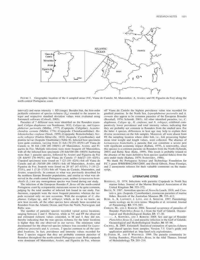
On 2 and 8 September 2005, 120 flounders from 4 locations off thenorth-central Portuguese coast, i.e., Viana do Castelo (VC) (41�40�N,8�50�W), Matosinhos (M) (41�10�N, 8�42�W), Aveiro (A) (40�38�N,8�45�W), and Figueira da Foz (FF) (40�8�N, 8�52�W) (Fig. 1), werecollected for examination of metazoan ectoparasites. In each location,30 fish were collected by random sampling from the nets of local fishingboats. All the fish were kept frozen at �20 C until they could be ex-amined. Each specimen was weighed (mean � SD [minimum–maxi-
mum] � 279.2 � 172.8 [160.7–1,090.4] g [VC]; 314.5 � 217.6 [139.4–1,124.0] g [M]; 267.4 � 122.0 [113.6–613.8] g [A]; 409.9 � 207.3[158.4–836.2] g [FF]), measured (27.6 � 3.7 [24.2–42.8] cm [VC]; 28.7� 4.9 [23.5–42.7] cm [M]; 27.5 � 4.3 [19.8–38.6] cm [A]; 30.9 � 5.0[23.6–41.6] cm [FF]), and sexed (20 males and 10 females [VC]; 10males and 20 females [M]; 9 males and 21 females [A]; 11 males and19 females [FF]). The body skin, eyes, fins, branchial chambers (sub-opercular surfaces, walls, gill arches, and pseudobranchiae), and nasaland buccal cavities were examined for metazoan ectoparasites using astereomicroscope. Collected specimens were cleaned and then fixed in70% alcohol. Later, copepods were cleared in 90% lactic acid (Humesand Gooding, 1964). Parasites were identified according to Naylor(1972) and Bruce (1987) for Isopoda, and to Kabata (1979, 1992) forCopepoda. It was not possible to identify the gnathiid pranizae at thespecies level because the identification keys require adult male speci-mens that were not found in our survey. Nevertheless, all the femalelarvae presented the same morphological type, which is, presumably,an indication of a single species.
After evaluating the sites of parasite infection on the host’s bodysurface, the following ecological parameters were determined accordingto Bush et al. (1997) for each of the 4 sampled locations: prevalence(number of infected fish/percentage of infected fish [95% confidence

1220 THE JOURNAL OF PARASITOLOGY, VOL. 93, NO. 5, OCTOBER 2007
TA
BL
EII
.M
etaz
oan
ecto
para
siti
cta
xare
cord
edon
flou
nder
sfr
omth
e4
sam
pled
loca
tion
sof
fth
eno
rth-
cent
ral
Por
tugu
ese
coas
t,th
eir
site
sof
infe
ctio
n,in
fect
ion
para
met
ers
(num
ber
ofin
fect
edfi
sh/p
reva
lenc
e[9
5%co
nfide
nce
inte
rval
]%,
mea
nin
tens
ity
�S
D[r
ange
]),
and
firs
t-or
der
jack
knif
ees
tim
ator
ofsp
ecie
sri
chne
ss(e
stim
ated
rich
ness
�S
D[N
�30
fish
for
all
sam
pled
loca
tion
s]).
Par
asit
egr
oup
Fam
ily
Tax
aH
ost
site
*
Sam
pled
loca
tion
Via
nado
Cas
telo
Mat
osin
hos
Ave
iro
Fig
ueir
ada
Foz
Cop
epod
a
Cal
igid
aeC
alig
usdi
apha
nus
B—
—1/
3(0
–17)
—(1
)C
alig
ussp
.B
;F
—5/
17(6
–35)
——
(1)
Lep
eoph
thei
rus
pect
oral
isB
;F
6/20
(8–3
9)30
/100
(88–
100)
29/9
7(8
3–10
0)28
/93
(78–
99)
7.2
�7.
2(1
–19)
14.1
�9.
9(3
–53)
7.6
�6.
9(1
–34)
9.5
�10
.0(1
–50)
Cho
ndra
cant
hida
eA
cant
hoch
ondr
iaco
rnut
aB
;F
;5/
17(6
–35)
30/1
00(8
8–10
0)29
/97
(83–
100)
30/1
00(8
8–10
0)S
OS
;G
A;
P22
.0�
12.8
(5–
41)
47.6
�22
.6(8
–96)
34.4
�24
.2(2
–110
)38
.1�
25.9
(4–1
04)
Bom
oloc
hida
eH
olob
omol
ochu
sco
nfus
usN
C—
1/3
(0–1
7)—
—(1
)
Isop
oda
Cym
otho
idae
Ner
ocil
aor
bign
yiF
;G
A—
3/10
(2–2
7)—
—(1
)G
nath
iida
eP
rani
zala
rvae
B;
F;
BC
;G
A20
/67
(47–
83)
—1/
3(0
–17)
—1.
7�
0.9
(1–
4)(3
)
Est
imat
edri
chne
ss3
�0.
06
�1.
06
�1.
32
�0.
0S J
K
*B
,bo
dy;
BC
,bu
ccal
cavi
ty;
F,fi
ns;
GA
,gi
llar
ches
;N
C,
nasa
lca
viti
es;
P,ps
eudo
bran
chia
e;S
OS
,su
bope
rcul
arsu
rfac
es.
RESEARCH NOTES 1221
FIGURE 1. Geographic location of the 4 sampled areas (VC, Viana do Castelo; M, Matosinhos; A, Aveiro; and FF, Figueira da Foz) along thenorth-central Portuguese coast.
interval]) and mean intensity � SD (range). Besides that, the first-orderjackknife estimator of species richness (SJK) rounded to the nearest in-teger and respective standard deviation values were evaluated usingEstimateS software (Colwell, 2005).
Parasites of 7 different taxa were identified on the flounders exam-ined: Caligus diaphanus von Nordmann, 1832, Caligus sp., and Lepeo-phtheirus pectoralis (Muller, 1777) (Copepoda: Caligidae); Acantho-chondria cornuta (Muller, 1776) (Copepoda: Chondracanthidae); Ho-lobomolochus confusus (Stock, 1959) (Copepoda: Bomolochidae); Ner-ocila orbignyi (Guerin-Meneville, 1832) (Isopoda: Cymothoidae); andpraniza larvae (Isopoda: Gnathiidae) (Table II). Infected host specimenswere quite common, varying from 21 fish (70 [51–85]%) off Viana doCastelo to 30 fish (100 [88–100]%) off Matosinhos, Aveiro, and Fi-gueira da Foz. Multiple infections were more frequent off Matosinhos,with all the infected host specimens (30 fish/100 [88–100]%) harboringmore than 1 parasite species, followed by Aveiro and Figueira da Foz(28 fish/93 [78–99]%), and Viana do Castelo (7 fish/23 [10–42]%).Copepod specimens were found on 7 (23 [10–42]%) fish off Viana doCastelo and all (30/100 [88–100]%) fish off Matosinhos, Aveiro, andFigueira da Foz. Isopods were found on 20 (67 [47–83]%), 3 (10 [2–27]%), and 1 (3 [0–17]%) fish off Viana do Castelo, Matosinhos, andAveiro, respectively. In contrast to what was previously described forthe northern Europe flounder populations, and similar to what was ob-served in the south-central Portuguese coast, neither Lernaeocera bran-chialis (L.) nor any monogenean species was found during our study.
The infection of the European flounder P. flesus off the north-centralPortuguese coast by ectoparasitic metazoans seems to be quite common,judging by the total number of infected fish found in our study. Fur-thermore, copepods were the most frequent parasites, whereas the iso-pods occurred only on rare occasions. With the exception of C. dia-phanus, Caligus sp., and N. orbignyi, which, as far as we know, arenew host records, all the other species have already been recorded onflounders from the Atlantic Ocean, and from the North, Norwegian, andBaltic seas.
The number of parasitic species recorded varied across locations,ranging between 2 and 5. However, while in VC and FF the observedand estimated richness values coincided, in M and A they did not,thereby indicating that the true species richness for the latter locationsis higher than the one observed in our survey. The minimum valuedocumented for the observed species richness was recorded for Lepeo-phtheirus pectoralis and A. cornuta, 2 species common to all the sam-pled locations. In fact, prevalence and intensity values recorded forthese 2 species suggest that they are probably common parasites offlounders throughout the north-central Portuguese coast. Both copepodswere dominant off Matosinhos, Aveiro, and Figueira da Foz, whereas
off Viana do Castelo the highest prevalence value was recorded forgnathiid pranizae. In the North Sea, Lepeophtheirus pectoralis and A.cornuta also appear to be common parasites of the European flounder(Boxshall, 1974; Schmidt, 2003). All other identified parasites, i.e., C.diaphanus, Caligus sp., H. confusus, and N. orbignyi, exhibited com-paratively lower prevalence and total intensity values, indicating thatthey are probably not common in flounders from the studied area. Forthe latter 4 species, differences in host age may help to explain theirdiverse occurrence on the fish samples. Moreover, all were absent fromFF, the sampling location where older fish, i.e., fish possessing highermean total weight and length values, were collected. The absence ofLernaeocera branchialis, a parasite that can constitute a severe pestwith significant economic impact (Kabata, 1979), is noteworthy, sincethis species is a common parasite on flounders from the North (Schmidt,2003) and Baltic Seas (Køie, 1999). This result is probably related tothe absence of the main definitive host species (gadoid fishes) from thearea under study (Kabata, 1979; Svetovidov, 1986).
We thank the Portuguese Science and Technology Foundation forF.C.’s grant SFRH/BM/23063/2005, and David Gibson, Nuno Formigo,and 2 anonymous referees for their valuable comments on the manu-script.
LITERATURE CITED
BOXSHALL, G. 1974. Infections with parasitic Copepods in North Seamarine fishes. Journal of the Marine Biological Association of theUnited Kingdom 54: 355–372.
BRUCE, N. 1987. Australian species of Nerocila Leach, 1818, and Cren-iola n. gen. (Isopoda: Cymothoidae), crustacean parasites of marinefishes. Records of the Australian Museum 39: 355–412.
BUSH, A., K. LAFFERTY, J. LOTZ, AND A. SHOSTAK. 1997. Parasitologymeets ecology on its own terms: Margolis et al. revisited. Journalof Parasitology 83: 575–583.
CHIBANI, M., AND J. ROKICKI. 2004. Seasonal occurrence of parasites offlounder Platichthys flesus (L.) from the Gulf of Gdansk. Oceano-logical and Hydrobiological Studies 33: 17–30.
———, A. KIJEWSKA, AND J. ROKICKI. 2005. Sex and age of flounderPlatichthys flesus (L.) and parasitic infection in the Gulf of Gdansk.Oceanological and Hydrobiological Studies 34: 85–96.
COLWELL, R. 2005. EstimateS: Statistical estimation of species richnessand shared species from samples. Version 7.5. User’s guide andapplication published at: http://purl.oclc.org/estimates.
EL-DARSH, H., AND P. WHITFIELD. 1999. The parasite community in-fecting flounders, Platichthys flesus, in the tidal Thames. Journalof Helminthology 73: 203–214.

1222 THE JOURNAL OF PARASITOLOGY, VOL. 93, NO. 5, OCTOBER 2007
HUMES, A., AND R. GOODING. 1964. A method for studying the externalanatomy of copepods. Crustaceana 6: 238–240.
KABATA, Z. 1959. Ecology of the genus Acanthochondria Oakley (Co-pepoda Parasitica). Journal of the Marine Biological Association ofthe United Kingdom 38: 249–261.
———. 1979. Parasitic Copepoda of British fishes. The Ray Society,London, U.K., 468 p.
———. 1992. Copepods parasitic on fishes. Synopses of the Britishfauna (new series), No. 47. Universal Book Services/Dr. W. Back-huys, Oegstgeest, The Netherlands, 264 p.
KøIE, M. 1999. Metazoan parasites of flounder Platichthys flesus (L.)along a transect from the southwestern to the northeastern BalticSea. ICES Journal of Marine Science 56: 157–163.
LILE, N., O. HALVORSEN, AND W. HEMMINGSEN. 1994. Zoogeographicalclassification of the macroparasite faunas of four flatfish speciesfrom the northeastern Atlantic. Polar Biology 14: 137–141.
LUCAS, M., AND E. BARAS. 2001. Migration of freshwater fishes. Black-well Science Ltd., Oxford, U.K., 420 p.
MACKENZIE, K., AND D. GIBSON. 1970. Ecological studies of some par-asites of plaice Pleuronectes platessa L. and flounder Platichthysflesus (L.). In Aspects of fish parasitology, Symposia of the BritishSociety for Parasitology, A. Taylor and R. Muller (eds.). BlackwellScientific Publications, Oxford, U.K., p. 1–42.
MARQUES, J., C. TEIXEIRA, AND H. CABRAL. 2006. Differentiation ofcommercially important flatfish populations along the Portuguesecoast: Evidence from morphology and parasitology. Fisheries Re-search 81: 293–305.
NAYLOR, E. 1972. British marine isopods. The Linnean Society of Lon-don. Synopses of the British Fauna (New Series), No. 3. AcademicPress, London, U.K., 85 p.
SCHMIDT, V. 2003. Parasites of European flounder (Platichthys flesus L.)from the German Bight, North Sea, and their potential use in eco-system monitoring. Ph.D. Thesis. Universitat Hannover, Kiel, Rus-sia, 154 p.
SOBRAL, D., AND J. GOMES. 1997. Peixes litorais - Estuario do Sado.Instituto da Conservacao da Natureza, Lisboa, Portugal, 54 p.
SVETOVIDOV, A. 1986. Gadidae. In Fishes of the northeastern Atlanticand the Mediterranean, Vol. II, P. Whitehead, M.-L. Bauchot, J.-C.Hureau, J. Nielsen, and E. Tortonese (eds.). Unesco ed., Paris,France, p. 680-710.
ZEDDAM, J., P. BERREBI, F. RENAUD, A. RAIBAUT, AND C. GABRION. 1988.Characterization of two species of Lepeophtheirus (Copepoda, Cal-igidae) from flatfishes. Description of Lepeophtheirus europaensissp. nov. Parasitology 96: 129–144.
J. Parasitol., 93(5), 2007, pp. 1222–1225� American Society of Parasitologists 2007
Early Migration of Sarcocystis neurona in Ponies Fed Sporocysts
E. Elitsur, A. E. Marsh, S. M. Reed*, J. P. Dubey†, M. J. Oglesbee‡, J. E. Murphy*, and W. J. A. Saville§, Department of VeterinaryPreventive Medicine, College of Veterinary Medicine, The Ohio State University, Columbus, Ohio 43210-1092; *Department of VeterinaryClinical Sciences, College of Veterinary Medicine, The Ohio State University, Columbus, Ohio 43210-1092; †United States Department ofAgriculture, Agricultural Research Service, Animal and Natural Resources Institute, Animal Parasitic Diseases Laboratory, Beltsville, Maryland20705-2350; ‡Department of Veterinary Biosciences, College of Veterinary Medicine, The Ohio State University, Columbus, Ohio 43210-1092;§To whom correspondence should be addressed. e-mail: [email protected]
ABSTRACT: Sarcocystis neurona is the most important cause of equineprotozoal myeloencephalitis (EPM), a neurologic disease of the horse.In the present work, the kinetics of S. neurona invasion is determinedin the equine model. Six ponies were orally inoculated with 250 106
S. neurona sporocysts via nasogastric intubation and killed on days 1,2, 3, 5, 7, and 9 postinoculation (PI). At necropsy, tissue samples wereexamined for S. neurona infection. The parasite was isolated from themesenteric lymph nodes at 1, 2, and 7 days PI; the liver at 2, 5, and 7days PI; and the lungs at 5, 7, and 9 days PI by bioassays in interferongamma gene knock out mice (KO) and from cell culture. Microscopiclesions consistent with an EPM infection were observed in brain andspinal cord of ponies killed 7 and 9 days PI. Results suggest that S.neurona disseminates quickly in tissue of naive ponies.
Equine protozoal myeloencephalitis (EPM) is a serious neurologicdisease and Sarcocystis neurona is the most important cause (Dubey etal., 1991). Sarcocystis neurona has a 2-host life cycle, including a meat-eating definitive host, the opossums Didelphis virginiana and Didelphisalbiventris (Dubey, Lindsay, Kerber et al., 2001; Dubey, Lindsay, Sa-ville et al., 2001). There is a wide range of intermediate hosts, includingthe raccoon (Dubey, Saville et al., 2001), armadillo (Cheadle, Tanhauseret al., 2001), skunk (Cheadle, Yowell et al., 2001), sea otter (Dubey etal., 2002), and the domestic cat (Dubey and Hamir, 2000; Dubey et al.,2000; Turay et al., 2002). The horse is considered an aberrant inter-mediate host (Dubey, Lindsay, Saville et al., 2001). Schizonts and mer-ozoites are the only stages known in the horse, and they are found onlyin the central nervous system (CNS) following an uncharacterized mi-gratory route. Attempts to demonstrate S. neurona in tissues of horsesfed sporocysts have been unsuccessful despite the fact that horses de-veloped neurological signs (Fenger et al., 1997; Lindsay et al., 2000;Cutler et al., 2001; Saville et al., 2001; Sofaly et al., 2002). In thepresent article, we have attempted to follow the migration of S. neurona
in tissues of ponies by orally inoculating them with large numbers ofsporocysts and examining at shorter postchallenge intervals.
Eight seronegative ponies (Table I) were randomly assigned to treat-ment (n � 6) or control (n � 2) groups and housed in separate stalls.Neurologic examinations were conducted before the initiation of theproject and daily there after, including the date of termination. Theexaminations were performed by a coauthor (S.M.R.). Physical exam-inations were also performed daily. On day 0, cerebral spinal fluid(CSF) and blood samples were collected from each horse, and treatmentponies were inoculated with sporocysts via nasogastric intubation with250 106 sporocysts (25 ml) and 120 ml doses of phosphate bufferedsaline (PBS) to ensure complete dosing. The sporocysts were of theraccoon isolate SN 37-R and had been obtained from the intestines ofthe laboratory-raised opossums fed tissues of experimentally infectedraccoons as described (Sofaly et al., 2002; Stanek et al., 2002).
Control ponies were given saline solution (25 ml) and 120 ml dosesof PBS via the nasogastric tube. Disposable gloves and plastic bootswere used upon entrance into the control ponies’ stalls to avoid cross-contamination and were immediately discarded afterwards. An emptystall was maintained between the control ponies and treatment poniesas well. Blood for serology was collected daily (days 1–9) and for buffycoat culture on terminal dates. Treatment ponies were randomly as-signed to serial killing on days 1, 2, 3, 5, 7, and 9 PI, and the controlponies were killed on days 3 and 9 PI. Ponies were humanely killedwith an overdose of Euthasol euthanasia solution (Delmarva Labora-tories, Midlothian, Virginia), and CSF was collected via the atlanto-occipital space at postmortem.
Necropsy was performed on all ponies. At necropsy, samples of lung,liver, mesenteric lymph nodes, and mesenteric artery were removedaseptically for S. neurona isolation. Additional tissue samples werefixed in 10% buffered formalin for routine microscopic examination,including the heart, lung, diaphragm, liver, spleen, adrenal gland, kid-ney, tongue, mesenteric lymph node, mesenteric artery, cecum, sciatic

RESEARCH NOTES 1223
TABLE I. Sarcocystis neurona in tissues of ponies detected by parasiteisolation.
Pony Day PI
Bioassay in KO mice
MLN* Liver Lung CNS† Cell culture
6459 1 3/5‡ 0/5 ND§ 0/5 Negative6460 2 5/5 1/5 ND 0/5 Positive¶6617� 3 0/5 1/5# ND 0/5 Negative6453 3 0/5 0/5 ND 0/5 Negative6570 5 0/5 4/5 2/2 0/5 Negative6451 7 0/5 2/5 2/2 0/5 Positive¶744 9 0/5 0/5 0/5 0/5 Positive**742� 9 ND ND ND ND Negative
* MLN, mesenteric lymph node.† CNS, central nervous system.‡ No. of mice with demonstrable S. neurona merozoites/no. of mice inoculated
with pony tissues.§ ND, not done.� Negative control ponies.# Mouse died from bacterial meningitis; no parasites detected in its tissues.¶ Positive in mesenteric lymph nodes.** Positive in lung.
FIGURE 1. Reactivity of sera from ponies inoculated with S. neuronasporocysts. Immunoblots from SDS-PAGE separated non-reduced S.neurona proteins. The antigens were probed with sera serially collectedbeginning with preinoculation (day 0) and 3, 5, 7, and 9 DPI*. Negativecontrol is pony no. 742 (lane 3), which did not receive sporocysts duringthe study. In all categories, lane 1 corresponds to pony no. 6451, lane2 corresponds to pony no. 744, and lane 3 (control terminal) corre-sponds to pony no. 742. All samples were diluted at 1:10. Approximatemolecular sizes are given at the margin in kDa. *DPI, days postinfec-tion. †Positive control (lane 4).
nerve, and quadriceps. The entire small intestine was removed andcross-sectioned at the ileum, jejunum, and duodenum regions, and mul-tiple sections from each region were placed in 10% buffered formalin.Finally, brain and spinal cord (with dura intact) were removed and sam-pled for protozoan isolation, followed by fixation in 10% buffered for-malin for microscopic examination (Saville et al., 2004). All tissueswere paraffin-embedded and sections were stained with hematoxylinand eosin (H & E) for microscopic evaluation. Histologic evaluationsampled the brain stem at 4 levels (thalamus, mesencephalon, meten-cephalon, and myelencephalon) and the cervical, thoracic, lumbar, andsacral spinal cord at 7, 7, 5, and 2 levels, respectively.
Sarcocystis neurona immunoblot analysis was used to detect IgM-specific antibodies in serum for ponies nos. 6451, 744, and 742 at prein-oculation as well as at 3, 5, 7, and 9 days PI, and in CSF for alltreatment ponies and 1 control pony (no. 742) preinoculation and atpostmortem. The immunoblot was performed as previously describedwith slight modifications to incorporate the S. neurona antigen (isolateSN-UCD1) and facilitate IgM antibody detection (Granstrom et al.,1993; Marsh et al., 2001, 2004; Murphy et al., 2006). A positive controlhorse serum and CSF was used (Sofaly et al., 2002).
For isolation of S. neurona in cell culture, sections of brain, lung,liver, mesenteric lymph nodes, mesenteric artery, and spinal cord wereimmediately processed separately following necropsy as previously de-scribed (Dubey et al., 1991). The dura mater of spinal cord was opened,and 0.5 cm cross section segments of spinal cord representing spinalcord segments at C1–2, C3–4, C5–6, C7–T1, T4–5, T8–9, T12–13, L1–2, L3–4, andL7–S2 were removed. Tissue samples were washed with antibacterial/antifungal wash, placed in 4 C sterile phosphate solution, and stored at4 C until ready for processing onto cell culture. The buffy coat from10 ml whole blood was removed with a sterile pipette, mixed with 4ml of sterile RPMI protozoal media 1640 (Gibco, Grand Island, NewYork), and then inoculated onto a monolayer of equine dermal cells forprotozoal isolation (Dubey et al., 1991). The cells were allowed to cul-ture for 88–97 days (Dubey et al., 1999). Positive cell cultures weredetermined by inverted microscope followed by confirmation of mero-zoite presence in media using cytocentrifugation, followed by Dif-Quickstaining (IMEB, Inc., San Marcos, California) to observe merozoites.
Tissues (Table I) were shipped on ice to the USDA laboratory (Belts-ville, Maryland) for bioassay in interferon gamma gene knock out (KO)mice (BALB/c-IfngtmITs, females, 6 to 12 wk old, Jackson Laboratories,Bar Harbor, Maine). Tissues were homogenized in 0.85% sodium chlo-ride aqueous solution and inoculated s.c. into KO mice (Sofaly et al.,2002). Mice were observed for 60 days unless they exhibited neurologicsymptoms, at which point they were killed, and their cerebellum pro-cessed for S. neurona immunohistochemistry (IHC). Selected pony tis-
sue samples were also stained by IHC for S. neurona antigens as pre-viously described by Dubey and Hamir (2000).
All ponies appeared to be clinically normal at all postinoculationintervals, aside from some nonspecific physical signs of illness, such asfever and nasal and ocular discharge. Neurologic deficits were not notedin any of the ponies throughout the study.
Sarcocystis neurona was isolated from tissue of the ponies by bio-assay in KO mice and was isolated in cell culture from 2 ponies (TableI). Differences in results most likely are due to differences in the sen-sitivities of each test and parasite distribution within each tissue. Sar-cocystis neurona was isolated from mesenteric lymph node at 1, 2, and7 days PI; liver at 2, 3, 5, and 7 days PI; and lung at 5, 7, and 9 daysPI. Sarcocystis neurona was not found in any pony tissues sectionsstained with H & E or with S. neurona antibody. Additionally, onlyserum from pony no. 744 (killed 9 days PI) had IgM antibodies specificto S. neurona at 2 markers, 28 kDa and 15 kDa (Fig. 1).
Histologic tissue sections were examined by 2 coauthors (J.P.D. andM.J.O.). Lesions associated with protozoan infections were not foundin ponies killed 1–5 day PI. Ponies killed at 7 and 9 days PI had fociof inflammation that are consistent with EPM infection. Pony no. 6451had a single focus of mild severity in the C3–4 region of the spinal cord.The focus had moderate gliosis, but perivascular leukocytes were notobserved. Pony no. 744 had multiple lesions of mild to moderate se-verity observed in the cervical intumescence (C7–T1) and T12–13 regionsof the spinal cord, with foci of lesser severity in the thalamus, mesen-cephalon, and metencephalon (pons region). The lesions within the spi-nal cord consisted of moderate focal perivascular astro- and microgliosiswith mononuclear cell infiltrate in the grey matter. The lesions withinthe brain were restricted to the grey matter and included focal glialnodule formation and perivascular increases in glial density. However,no parasites were detected by S. neurona–specific IHC staining on tis-sues with lesions compatible with protozoal infection.
Once it is ingested, the 2 possible routes that a parasite can take toreach distant organs are via the lymph, the portal blood, or both (Dubeyet al., 1989). In KO mice, the first generation of S. neurona schizogonyoccurs intravascularly in visceral tissues before invasion of the CNS.Mesenteric lymph node invasion is evidence of lymphatic dissemina-tion, and liver and lung infections suggests hematogenous spread afteroral inoculation. Although parasitemia was not directly evident within

1224 THE JOURNAL OF PARASITOLOGY, VOL. 93, NO. 5, OCTOBER 2007
this study, parasite isolation from the liver and lung suggest a hema-togenous dissemination of the parasite. In the present study, serocon-version for S. neurona–specific IgM was detected at 9 days PI, and thisfinding is confirmed in previous equine studies that have reported IgGimmunoconversion at 8 days PI (Lindsay et al., 2000; Sofaly et al.,2002). Finally, although none of the ponies in this study had detectableanti-S. neurona IgM antibodies in the CSF by 9 days PI, that findingis supported by similar findings in previous equine models, where theearliest reported CSF conversion occurred at 28 days PI (Fenger et al.,1997).
The results of the present study, and those in KO mice and in rac-coons (Dubey, 2001a, 2001b; Stanek et al., 2002) fed sporocysts, in-dicate that S. neurona quickly travels from the gastrointestinal tract tothe lymph nodes, within 24 to 48 hr. Mesenteric lymph node invasionis evident as early as 24 hr PI, and liver infection occurs as early as 2days PI. It is interesting to note that the only other time that S. neuronawas isolated from the blood after oral inoculation of sporocysts at 21days PI from a severe immunocompromised disease (SCID) horse(Long et al., 2002). Our results suggest that parasitemia occurs at orbefore 2 days PI (as is evident in liver bioassays). Although S. neuronawas not isolated from brain and spinal cord of the ponies, histologically,lesions in the CNS of ponies at day 7 and 9 PI were suggestive of anS. neurona invasion based upon characteristic tissue changes, correla-tion to isolation from other tissues in KO mice or tissue culture, andcomparable kinetics in previously reported KO mice and raccoon stud-ies. This is the first time that S. neurona has been isolated from themesenteric lymph nodes, liver, and lungs from either experimentally ornaturally infected immunocompetent equids.
This study was funded by a gift from American Live Stock InsuranceCompany, Geneva, Illinois, to S.M.R. The authors thank Bradd Barr,California Animal Health and Food Safety Laboratory, Davis, Califor-nia, for confirmation of the IHC results.
LITERATURE CITED
CHEADLE, M. A., S. M. TANHAUSER, J. B. DAME, D. C. SELLON, M.HINES, P. E. GINN, R. J. MACKAY, AND E. C. GREINER. 2001. Thenine-banded armadillo (Dasypus novemcinctus) is an intermediatehost for Sarcocystis neurona. International Journal for Parasitology31: 330–335.
———, C. A. YOWELL, D. C. SELLON, M. HINES, P. E. GINN, A. E.MARSH, J. B. DAME, AND E. C. GREINER. 2001. The striped skunk(Mephitis mephitis) is an intermediate host for Sarcocystis neurona.International Journal of Parasitology 31: 843–849.
CUTLER, T. J., R. J. MACKAY, P. E. GINN, K. GILLIS, S. M. TANHAUSER,E. V. LERAY, J. B. DAME, AND E. C. GREINER. 2001. Immunocon-version against Sarcocystis neurona in normal and dexamethasone-treated horses challenged with S. neurona sporocysts. VeterinaryParasitology 95: 197–210.
DUBEY, J. P. 2001a. Migration and development of Sarcocystis neuronain tissues of interferon gamma knockout mice fed sporocysts froma naturally infected opossum. Veterinary Parasitology 95: 341–351.
———. 2001b. Parasitemia and early tissue localization of Sarcocystisneurona in interferon gamma gene knockout mice fed sporocysts.Journal of Parasitology 87: 1476–1479.
———, S. W. DAVIS, C. A. SPEER, D. D. BOWMAN, A. DE LAHUNTA,D. E. GRANSTROM, M. J. TOPPER, A. HAMIR, J. CUMMINGS, AND M.M. SUTTER. 1991. Sarcocystis neurona n. sp. (Protozoa: Apicom-plexa), the etiologic agent of equine protozoal myeloencephalitis.Journal of Parasitology 77: 212–218.
———, AND A. N. HAMIR. 2000. Immunohistochemical confirmation ofSarcocystis neurona infections in raccoons, mink, cat, skunk, andpony. Journal of Parasitology 86: 1150–1152.
———, D. S. LINDSAY, C. E. KERBER, N. KASAI, H. F. PENA, S. M.GENNARI, O. C. KWOK, S. K. SHEN, AND B. M. ROSENTHAL. 2001.First isolation of Sarcocystis neurona from the South Americanopossum, Didelphis albiventris, from Brazil. Veterinary Parasitol-ogy 95: 295–304.
———, ———, W. J. A. SAVILLE, S. M. REED, D. E. GRANSTROM, AND
C. A. SPEER. 2001. A review of Sarcocystis neurona and equineprotozoal myeloencephalitis (EPM). Veterinary Parasitology 95:89–131.
———, D. E. MATTSON, C. A. SPEER, R. J. BAKER, D. M. MULROONEY,S. J. TORNQUIST, A. N. HAMIR, AND T. C. GERROS. 1999. Charac-terization of a Sarcocystis neurona isolate (SN6) from a naturallyinfected horse from Oregon. Journal of Eukaryotic Microbiology46: 500–506.
———, A. C. ROSYPAL, B. M. ROSENTHAL, N. J. THOMAS, D. S. LIND-SAY, J. F. STANEK, S. M. REED, AND W. J. A. SAVILLE. 2002. Sar-cocystis neurona infections in sea otters (Enhydra lutris): Evidenceof natural infections with sarcocysts and transmission of infectionto opossums (Didelphis virginiana). Journal of Parasitology 87:1387–1393.
———, W. J. A. SAVILLE, D. S. LINDSAY, R. W. STICH, J. F. STANEK,C. A. SPEER, B. M. ROSENTHAL, C. J. NJOKU, O. C. KWOK, S. K.SHEN, AND S. M. REED. 2000. Completion of the life cycle of Sar-cocystis neurona. Journal of Parasitology 86: 1276–1280.
———, ———, J. F. STANEK, D. S. LINDSAY, B. M. ROSENTHAL, M. J.OGLESBEE, A. C. ROSYPAL, C. J. NJOKU, R. W. STICH, O. C. H.KWOK, S. K. SHEN, A. N. HAMIR, AND S. M. REED. 2001. Sarco-cystis neurona infection in raccoons (Procyon lotor): Evidence fornatural infection with sarcocysts, transmission of infection to opos-sums (Didelphis virginiana), and experimental induction of neu-rologic disease in raccoons. Veterinary Parasitology 100: 117–129.
———, C. A. SPEER, AND R. FAYER. 1989. Sarcocystosis of animalsand man. CRC Press, Boca Raton, Florida, 215 p.
FENGER, C. K., D. E. GRANSTROM, A. A. GAJADHAR, N. M. WILLIAMS,S. A. MCCRILLIS, S. STAMPER, J. L. LANGEMEIER, AND J. P. DUBEY.1997. Experimental induction of equine protozoal myeloencephal-itis in horses using Sarcocystis sp. sporocysts from the opossum(Didelphis virginiana). Veterinary Parasitology 68: 199–213.
GRANSTROM, D. E., J. P. DUBEY, S. D. DAVIS, R. FAYER, J. C. FOX, K.B. POONACHA, R. C. GILES, AND P. F. COMER. 1993. Equine proto-zoal myeloencephalitis: Antigen analysis of cultured Sarcocystisneurona merozoites. Journal of Veterinary Diagnostic Investigation5: 88–90.
LINDSAY, D. S., C. C. DYKSTRA, A. WILLIAMS, J. A. SPENCER, S. D.LENZ, K. PALMA, J. P. DUBEY, AND B. L. BLAGBURN. 2000. Inocu-lation of Sarcocystis neurona merozoites into the central nervoussystem of horses. Veterinary Parasitology 92: 157–163.
LONG, M. T., M. T. MINES, D. P. KNOWLES, S. M. TANHAUSER, J. B.DAME, T. J. CUTLER, R. J. MACKAY, AND D. C. SELLON. 2002. Sar-cocystis neurona: Parasitemia in a severe combined immunodefi-cient (SCID) horse fed sporocysts. Experimental Parasitology 100:150–154.
MARSH, A. E., P. J. JOHNSON, J. RAMOS-VARA, AND G. C. JOHNSON. 2001.Characterization of a Sarcocystis neurona isolate from a Missourihorse with equine protozoal myeloencephalitis. Veterinary Parasi-tology 95: 143–154.
———, J. LAKRITZ, P. J. JOHNSON, M. A. MILLER, Y. W. CHIANG, AND
H. J. CHU. 2004. Evaluation of immune responses in horses im-munized using a killed Sarcocystis neurona vaccine. VeterinaryTherapeutics 5: 34–42.
MURPHY, J. E., A. E. MARSH, S. M. REED, C. MEADOWS, K. BOLTEN,AND W. J. A. SAVILLE. 2006. Development and evaluation of aSarcocystis neurona-specific IgM capture enzyme-liked immuno-sorbent assay. Journal of Veterinary Internal Medicine 20: 322–328.
SAVILLE, W. J. A., C. D. SOFALY, S. M. REED, J. P. DUBEY, M. J. OG-LESBEE, V. A. LACOMBE, R. O. KEENE, K. M. GUGISBERG, S. W.SWENSEN, R. D. SHIPLEY, Y.-W. CHIANG, H. J. CHU, AND T. NG.2004. An equine protozoal myeloencephalitis challenge model test-ing a second transport after inoculation with Sarcocystis neuronasporocysts. Journal of Parasitology 90: 1406–1410.
———, R. W. STICH, S. M. REED, C. J. NJOKU, M. J. OGLESBEE, A.WUNSCHMANN, D. L. GROVER, A. L. LAREW-NAUGLE, J. F. STANEK,D. E. GRANSTROM, AND J. P. DUBEY. 2001. Utilization of stress inthe development of an equine model for equine protozoal mye-loencephalitis. Veterinary Parasitology 95: 211–222.
SOFALY, C. D., S. M. REED, J. C. GORDON, J. P. DUBEY, M. J. OGLESBEE,C. J. NJOKU, D. L. GROVER, AND W. J. A. SAVILLE. 2002. Experi-mental induction of equine protozoal myeloencephalitis (EPM) inthe horse: effect of Sarcocystis neurona sporocyst inoculation doseon the development of clinical neurologic disease. Journal of Par-asitology 88: 1164–1170.

RESEARCH NOTES 1225
STANEK, J. F., J. P. DUBEY, M. J. OGLESBEE, S. M. REED, D. S. LINDSAY,L. A. CAPITINI, C. J. NJOKU, K. L. VITTITOW, AND W. J. A. SAVILLE.2002. Life cycle of Sarcocystis neurona in its natural intermediatehost, the raccoon Procyon lotor. Journal of Parasitology 88: 1151–1158.
TURAY, H. O., B. C. BARR, A. CALDWELL, K. R. BRANSON, M. K. R.COCKRELL, AND A. E. MARSH. 2002. Sarcocystis neurona reactingantibodies in Missouri feral domestic cats (Felis domesticus) andtheir role as an intermediate host. Parasitology Research 88: 38–43.
J. Parasitol., 93(5), 2007, pp. 1225–1227� American Society of Parasitologists 2007
A Human Case of Plagiorchis vespertilionis (Digenea: Plagiorchiidae) Infection in theRepublic of Korea
S.-M. Guk, J.-L. Kim, J.-H. Park, and J.-Y. Chai*, Department of Parasitology and Tropical Medicine, Seoul National University College ofMedicine, and Institute of Endemic Diseases, Seoul National University Medical Research Center, Seoul 110-799, Korea; *To whomcorrespondence should be addressed. e-mail: [email protected]
TABLE I. Morphological characteristics of Plagiorchis vespertilionis in the present specimen and those reported by previous workers.
Organs The present specimen Sogandares-Bernal (1956) Tkach et al. (2000)
Body length width 2.6 0.7 mm 2.7–4.8 0.8–1.6 mm 2.3–4.1 0.2–0.5 mmSucker ratio 1.0:1.0 1:0.67–1:1.39 1.0:1.2Pharynx 0.14 0.15 mm 0.11–0.19 0.11–0.15 mm Diameter: 0.1 mmEsophagus Nearly absent Variable in length or absent Very short or noneCeca Extend to near posterior extremity Extend into posterior end of body End 0.18 mm from body extremityCirrus sac Extend from VS* to anterior part
of ovaryExtend from VS to anterior part of
ovaryElongated and situated along the
median body axisCirrus 0.195 0.057 mm straight form Not mentioned 0.38–0.55 0.013–0.015 mm, coiled or
straight formTestis Postovarian, oval Rounded, postequatorial, oblique Postovarian, oval, and oblique
Anterior testis: 0.184 0.2 mm Anterior testis: 0.21–0.53 mm Anterior testis: 0.26–0.33 0.15–0.23 mmPosterior testis: 0.23 0.23 mm Posterior testis: 0.23–0.50 mm Posterior testis: 0.25–0.35 0.16–0.24 mm
Ovary Postacetabulum0.163 0.213 mm
Rounded, postacetabulum0.17–0.27 mm
Oval or rounded, median of cirrus sac0.14–0.26 0.10–0.21 mm
Vitellaria Extend to posterior end of worm Distributed lateral to ceca andextend to posterior end of worm
Well developed and distributed oneither side of body
Uterus With descending and ascendingloops
With descending and ascendingloops, intertesticular
With descending and ascending loops
Eggs 32.5 � 1.3 17.5 � 0.5 m 37 18 m 29–34 16–21 m
* Ventral sucker.
ABSTRACT: Plagiorchis vespertilionis (Digenea: Plagiorchiidae) is gen-erally considered a bat parasite, but here it is reported for the first timein a human. The patient was a 34-yr-old male who lived in a coastalvillage of Haenam-gun (county), Jeollanam-do (province), Republic ofKorea. Only 1 worm, 2.6 mm long and 0.7 mm wide, was recoveredafter praziquantel treatment and purging with magnesium salts. Thefluke was characterized by a large body size, a sucker ratio of 1:1, astraight cirrus organ, a short distance between the ventral sucker andovary, well-developed vitellaria, a uterus with descending and ascend-ing loops, and fully developed eggs with an average size of 32.5 17.5 m. The patient had habitually eaten the raw flesh of snakeheadmullet and gobies that had been caught near his village. The presentcase represents the first record of a human P. vespertilionis infection.
Species of Plagiorchis Luhe, 1899 (Digenea: Plagiorchiidae) are in-testinal parasites of reptiles, birds, and mammals (Yamaguti, 1958), andtheir metacercariae are found in mosquito larvae, insect naiads, fresh-water snails, and freshwater fish (Tanabe, 1922; Asada et al., 1962;Komiya, 1965; Hong et al., 1996; Chai and Lee, 2002). Human infec-tions of Plagiorchis spp. are rare. Worldwide, only 11 natural humancases have been reported in the literature (Radomyos et al., 1989; Honget al., 1996). The species responsible for human infections include Pla-
giorchis philippinensis in the Philippines (Sandground, 1940), Plagi-orchis muris Tanabe, 1922 in Japan and the Republic of Korea (Asadaet al., 1962; Hong et al., 1996), Plagiorchis javensis in Indonesia(Sandground, 1940), and Plagiorchis harinasutai in Thailand (Radom-yos et al., 1989). In addition to P. muris in Korean house rats (Seo etal., 1964, 1981), 7 other species have been reported in Korean bats, i.e.,Plagiorchis vespertilionis (Muller, 1780) Braun, 1900, Plagiorchis mag-nacotylus Park, 1939, Plagiorchis orientalis Park, 1939, Plagiorchisrhinolophi (Park, 1939), Plagiorchis koreanus Ogata, 1938, Plagiorchiskyushuensis Kifune and Sawada, 1979, and Plagiorchis corpulentus Ki-fune and Sawada, 1979 (Park, 1939a, 1939b; Sogandares-Bernal, 1956;Kifune et al., 1983).
Plagiorchis vespertilionis was first described in the brown long-earedbat Plecotus auritus in Europe, and then in many countries, includingthe Republic of Korea (Sogandares-Bernal, 1956). A subspecies, namedPlagiorchis vespertilionis parorchis, was described in the United States(Macy, 1960). The xiphidiocercariae of P. vespertilionis parorchis de-velop in the snail intermediate host, Lymnaea stagnalis and encyst inmosquito larvae, caddis-fly larvae, mayfly larvae, and dragonflynymphs (Macy, 1960). In addition, an attempted experimental infectionof mice and hamsters with this species was successful, and adult flukeswere harvested from the small intestine (Macy, 1960). During studies
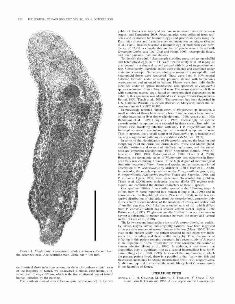
1226 THE JOURNAL OF PARASITOLOGY, VOL. 93, NO. 5, OCTOBER 2007
FIGURE 1. Plagiorchis vespertilionis adult specimen collected fromthe described case. Acetocarmine stain. Scale bar � 0.6 mm.
on intestinal fluke infections among residents of southern coastal areasof the Republic of Korea, we discovered a human case naturally in-fected with P. vespertilionis, which is the first confirmed case of naturalhuman infection by the parasite.
The southern coastal area (Haenam-gun, Jeollanam-do) of the Re-
public of Korea was surveyed for human intestinal parasites betweenAugust and September 2003. Fecal samples were collected from resi-dents and examined for helminth eggs and protozoan cysts using theKato thick smear and formalin-ether sedimentation techniques (Beaveret al., 1984). Results revealed a helminth egg or protozoan cyst prev-alence of 37.9%; a considerable number of people were infected withGymnophalloides seoi Lee, Chai and Hong, 1993, heterophyid flukes,and other parasites (data not shown).
To identify the adult flukes, people shedding presumed gymnophallidand heterophyid eggs (n � 11) were treated orally with 10 mg/kg ofpraziquantel in a single dose and purged with 30 g of magnesium sul-fate. Subsequently, diarrheic stools were collected and examined undera stereomicroscope. Numerous adult specimens of gymnophallid andheterophyid flukes were recovered. These were fixed in 10% neutralbuffered formalin under coverslip pressure, stained with Semichon’sacetocarmine, and mounted in balsam. Flukes were then individuallyidentified under an optical microscope. One specimen of Plagiorchissp. was recovered from a 34-yr-old man. The worm was an adult flukewith numerous uterine eggs. Based on morphological characteristics inTable 1, this specimen was identified as P. vespertilionis (Sogandares-Bernal, 1956; Tkach et al., 2000). The specimen has been deposited inU.S. National Parasite Collection (Beltsville, Maryland) under the ac-cession number USNPC 99592.
In previously reported human cases of Plagiorchis sp. infection, asmall number of flukes have usually been found among a large numberof other intestinal or liver flukes (Sandground, 1940; Asada et al., 1962;Radomyos et al., 1989; Hong et al., 1996). Interestingly, no specificgastrointestinal symptoms were recorded in these cases. Similarly, thepresent case, involving infection with only 1 P. vespertilionis and 9Heterophyes nocens specimens, had no intestinal symptoms of note.Thus, it appears that a small number of Plagiorchis sp. is incapable ofcausing a significant pathological condition (McMullen, 1937).
In terms of the identification of Plagiorchis species, the location andmorphologies of the cirrus sac, cirrus, testes, ovary, and Mehlis gland,and the positions and extents of vitellaria and uterus, and the suckerratio are important (Sandground, 1940; Sogandares-Bernal, 1956; Ki-fune et al., 1983, 1997; Radomyos et al., 1989; Tkach et al., 2000).However, the taxonomic status of Plagiorchis spp. occurring in Euro-pean bats was confusing because of the high degree of morphologicalsimilarity between different forms and species and an inadequate initialdescription of P. vespertilionis by Muller in 1780 (Tkach et al., 2000).In particular, the morphological data on the P. vespertilionis group, i.e.,P. vespertilionis, Plagiorchis muelleri Tkach and Sharpilo, 1990, andP. koreanus Ogata, 1938, were inadequate. To resolve this problem,Tkach et al. (2000) used molecular (nuclear rDNA ITS region) tech-niques, and confirmed the distinct characters of these 3 species.
Our specimen differs from similar species in the following ways. Itdiffers from P. muris reported in a human (Hong et al., 1996) and inhouse rats in the Republic of Korea (Seo et al., 1964), in the less ex-tensive distribution of vitellaria, from the posterior body extremity onlyto the ventral sucker median; of the locations of ovary and testes; andof smaller egg size. Our fluke has a sucker ratio of 1:1, which differsfrom P. koreanus, which has a smaller ventral sucker (Ogata, 1938;Kifune et al., 1997). Plagiorchis muelleri differs from our specimen inhaving a substantially greater distance between the ovary and ventralsucker (Tkach et al., 2000).
The known second intermediate hosts of P. vespertilionis, i.e., caddis-fly larvae, mayfly larvae, and dragonfly nymphs, have been suggestedto be possible sources of natural human infection (Macy, 1960). How-ever, in the present study, the patient recalled he had eaten raw fresh-water fish, including snakehead mullet and goby. Thus, the source ofinfection in this patient remains uncertain. In a recent study of P. murisin the Republic of Korea, freshwater fish were considered the source ofhuman infection (Hong et al., 1996). In addition, it was shown thatdragonflies play a significant role as a second intermediate host for P.muris (Hong et al., 1998, 1999). In view of the environment in whichthe present patient lived, there is a possibility that freshwater fish andfreshwater snails may be second intermediate hosts for P. vespertilionis.Studies are required to elucidate the whole life cycle of P. vespertilionisin the Republic of Korea.
LITERATURE CITEDASADA, J. I., H. OTAGAKI, M. MORITA, T. TAKEUCHI, Y. SAKAI, T. KO-
NOSHI, AND K. OKAHASHI. 1962. A case report on the human infec-

RESEARCH NOTES 1227
tion with Plagiorchis muris Tanabe, 1922 in Japan. Japanese Jour-nal of Parasitology 11: 512–516.
BEAVER, P. C., R. C. JUNG, AND E. W. CUPP. 1984. Clinical parasitology,9th ed. Lea and Febiger, Philadelphia, Pennsylvania, 825 p.
CHAI, J. Y., AND S. H. LEE. 2002. Food-borne intestinal trematode in-fections in the Republic of Korea. Parasitology International 51:129–154.
HONG, S. J., J. H. AHN, AND H. C. WOO. 1998. Plagiorchis muris:Recovery, growth and development in albino rats. Journal of Hel-minthology 72: 251–256.
———, H. C. WOO, AND J. Y. CHAI. 1996. A human case of Plagiorchismuris (Tanabe, 1922: Digenea) infection in the Republic of Korea:Freshwater fish as a possible source of infection. Journal of Para-sitology 82: 647–649.
———, ———, S. U. LEE, AND S. HUH. 1999. Infection status of drag-onflies with Plagiorchis muris metacercariae in Korea. KoreanJournal of Parasitology 37: 65–70.
KIFUNE, T., M. HARADA, I. SAWADA, AND M. H. YOON. 1997. Trematodeparasites of five Korean bats. Medical Bulletin of Fukuoka Uni-versity 24: 225–232.
———, I. SAWADA, AND W. C. LEE. 1983. Trematode parasites of twoKorean bats. Medical Bulletin of Fukuoka University 10: 3–8.
KOMIYA, Y. 1965. Metacercariae in Japan and adjacent territories. InProgress of medical parasitology in Japan, Vol. II, K. Morishita, Y.Komiya, and H. Matsubayashi (eds.). Meguro Parasitological Mu-seum, Tokyo, Japan, p. 225–233.
MACY, R. W. 1960. The life cycle of Plagiorchis vespertilionis paror-chis n. ssp. (Trematoda: Plagiorchiidae) and observations on theeffects of light on the emergence of the cercaria. Journal of Para-sitology 46: 337–345.
MCMULLEN, D. B. 1937. An intestinal infection of Plagiorchis muris inman. Journal of Parasitology 23: 113–115.
OGATA, T. 1938. Contribution a la connaissance de la faune helmin-thologique coreenne I. Une nouvelle espece de trematodes proenant
de chauves-souris. Annotationes Zoologicae Japanenses 17: 581–586.
PARK, J. T. 1939a. Trematodes from Mammalia and Aves II. Two newtrematodes of Plagiorchidae: Plagiorchoides rhinolophi n. sp. andPlagiorchis orientallis n. sp. rom Tyosen (Korea). Keijo Journal ofMedicine 10: 1–6.
———. 1939b. Trematodes of Mammals and Aves from Tyosen III. Anew trematode of the family Plagiorchidae Ward, 1917, Plagiorchismagnacotylus sp. nov. Keijo Journal of Medicine 10: 43–45.
RADOMYOS, P., D. BUNNAG, AND T. HARINASUTA. 1989. A new intestinalfluke, Plagiorchis harinasutai n. sp. Southeast Asian Journal ofTropical Medicine and Public Health 20: 101–107.
SANDGROUND, J. H. 1940. Plagiorchis javensis n. sp. a new trematodeparasitic in man. Review of Medicine and Tropical Parasitology(Habana) 6: 207–211.
SEO, B. S., S. Y. CHO, S. T. HONG, S. J. HONG, AND S. H. LEE. 1981.Studies on the parasitic helminthes of Korea V. Survey on intestinaltrematodes of house rats. Korean Journal of Parasitology 19: 131–136.
———, H. J. RIM, AND C. W. LEE. 1964. Studies on the parasitic hel-minthes of Korea I. Trematodes of rodents. Korean Journal of Par-asitology 2: 20–26.
SOGANDARES-BERNAL, F. 1956. Four trematodes from Korean bats withdescription of three new species. Journal of Parasitology 42: 200–206.
TANABE, H. 1922. A contribution to the study of the life cycle of di-genetic trematodes. A study of a new species, Lepoderma muris n.sp. Okayama Igakkai Zasshi 385: 47–58.
TKACH, V. S., J. PAWLOWSKI, AND V. P. SHARPILO. 2000. Molecular andmorphological differentiation between species of the Plagiorchisvespertilionis group (Digenea, Plagiorchiidae) occurring in Euro-pean bats, with a re-description of P. vespertilionis (Muller, 1780).Systematic Parasitology 47: 9–22.
YAMAGUTI, S. 1958. Part V. Digenea of mammals. Systema helminthum,Vol. I. Interscience Publishers Inc., New York, New York, 800 p.
J. Parasitol., 93(5), 2007, pp. 1227–1230� American Society of Parasitologists 2007
Influence of a Thermal Discharge on Parasites of a Cold-Water Flatfish,Pleuronectes americanus, as a Bioindicator of Subtle Environmental Change
R. A. Khan and R. G. Hooper, Department of Biology, Memorial University of Newfoundland, St. John’s, Newfoundland, Canada, A1B 3X9.e-mail: [email protected]
ABSTRACT: A study was conducted to ascertain the influence of a ther-mal discharge on the health and parasites of a coastal cold-water flatfish,the winter flounder (Pleuronectes americanus), a species sensitive toenvironmental change. Flounder were sampled in spring 1998 and 1999beneath the plume and at reference sites north and south up to 1 kmfrom the discharge. Species diversity and estimates of abundance ofmacroscopic algae, invertebrates, and fish were also recorded. Aftercapture by scuba divers, a comparison of condition factor, organ indices,blood values, histology, and parasites was made between groups of fishfrom the discharge and reference sites. Diversity and abundance of al-gae, invertebrates, and fish were considerably greater beneath the plumethan at the reference sites. The thermal water had no apparent effect onflounder taken beneath the plume, but it affected both its ecto- andendoparasites. Prevalence and mean abundance of Cryptocotyle linguametacercariae were significantly greater, whereas Trichodina jadranicaand Gyrodactylus pleuronecti were less on the gills of fish sampledbeneath the plume than at the reference sites. Four endoparasites, i.e.,Ceratomyxa drepanopsettae, Steringophorus furciger, Macvicarius so-leae, and Lecithaster gibbosus were significantly more abundant in thereference samples. These results suggest that environmental change af-fected transmission of the parasites of winter flounder exposed to thethermal effluent.
Coastal fossil fuel-generating plants discharging heated water into thesea tend to have some influence on the environment and also on benthiccommunities, which invariably are sessile or possess limited scope formovement. Some studies have reported changes in abundance and spa-tial variability of benthic assemblages by thermal effluent affecting themost abundant species, whereas others have observed minimal changesin the vicinity of the discharge (Landicci et al., 1999). It seems thatthermal tolerance of some species is related to the temperature; from 7to 10 C above the ambient impacted on benthic organisms more than1–2 C (Suresh et al., 1993; Landicci et al., 1999). Additionally, thevolume of water, season, temperature, and rate of discharge could alsohave a profound influence on benthic organisms (Landicci et al., 1999).
Several methods have been proposed to assess the impact of envi-ronmental change in fish (Adams, 1990). These include external abnor-malities, condition factor, organ indices, and histology in species thatare sessile and sensitive to ecosystem alterations (Adams, 1990). Morerecently, there is evidence that some fish parasites can be useful asbioindicators of stress (Khan and Thulin, 1991). A thermal power plantin Holyrood, Newfoundland, Canada, using Bunker oil as fuel has beendischarging heated seawater into the ocean for a number of years wherea population of winter flounder (Pleuronectes americanus), a speciessensitive to environmental change, occurs. The influence of the dis-charge was reported to affect the horse mussel (Modiolus sp.), but noth-

1228 THE JOURNAL OF PARASITOLOGY, VOL. 93, NO. 5, OCTOBER 2007
ing was known of its influence on fish health (LGL Ltd., 1999). Con-sequently, a study was designed to investigate the effect of the thermaldischarge at Holyrood on winter flounder living beneath the plume andto compare the results with samples taken from fish at varying distancesnorth and south from the discharge. This included some of the biolog-ical indicators mentioned above, including blood values of the floundersand also its parasites.
The thermal generating plant, using Bunker AC as fuel oil, is locatedat Holyrood (47�27�N, 53�08�W). Approximately 6 105 L/min of ther-mal water, 5–13 C, was discharged into Conception Bay, but tempera-ture change extended up to only 1 m below the surface (LGL Ltd.,1999). The benthic water temperature during both years was 0 C, where-as the surface temperatures were 3–4 C in May and 7–8 C in June.Benthic sampling was conducted at all sites for macroscopic algae, in-vertebrates, and fish, and estimates of their abundance were recorded.Winter flounder were captured beneath the plume by scuba divers usinga fish-landing net at about 5 m in late May and June 1998 and in May1999. In addition, flounder were taken at 0.5 m north (upcurrent) anddownflow at 0.4–0.5 and 1.0 km southwest from the discharge in 1998and downflow at 100–200 and 400 m in 1999. The fish were examinedafter capture for external lesions and macroscopic parasites, which wereenumerated. Two skin smears were prepared for examination subse-quently for microscopic parasites. Blood was taken from the caudal veinby a needle and heparinized syringe to determine hermatocrit, hemo-globin, and total plasma protein in the serum as reported in anotherstudy (George-Nascimento et al., 2000). A blood smear was preparedfor ascertaining lymphocytic levels after staining with Giemsa. At nec-ropsy, the total body length, eviscerated body, liver, and gonad masseswere examined. Samples of tissues, including gill, liver, spleen, andgonad were fixed in 10% buffered formalin and processed by conven-tional histological methods. Cross sections, 10 m, were stained withhematoxylin and eosin. Sections of spleen were also stained with Perl’sPrussian blue method for hemosiderin that was estimated by digitalimage analysis (Khan and Nag, 1993). Bile was taken from the gall-bladder by a needle and syringe, preserved with an equal volume of70% ethanol, and examined subsequently for myxozoans microscopi-cally (400). The digestive tract was examined for metazoan parasitesthat were removed and fixed in 70% ethanol. Digenes were stained withborax carmine. Metacercariae infecting the gills were estimated in crosssections after microscopic examination of 10 secondary lamellae.
Samples of flounder were compared statistically according to sex,site, and year of capture. Variables compared included condition factor(eviscerated body mass/length3), hepatosomatic index (liver mass/evis-cerated body mass), gonadosomatic index (gonadal mass/evisceratedbody mass), hemoglobin (g%), hematocrit (%), total plasma protein(g%), and lymphocyte levels (no. of lymphocytes/1,000 erythrocytes inGiemsa-stained blood smears; see George-Nascimento et al., 2000).However, the samples examined in 1999 at 100 and 200 m from thedischarge were pooled, with the exception of the parasites in the diges-tive tract, because of the paucity in numbers of fish that were captured.The data were analyzed using an analysis of covariance for site signif-icance. These included condition (k) factor (eviscerated body mass/length3), organ indices (organ mass/eviscerated body mass), and bloodvalues. Prevalence (%) and mean abundance were calculated for eachfish group. Chi-square and Fisher’s exact probability tests comparedprevalence, whereas the nonparametric test of Kruskal–Wallis, after logtransformation of parasitic numbers, was used for comparing fish groupsaccording to site.
Profound differences in the diversity and abundance of the speciesand genera of algae, invertebrates, and fish occurred between the siteat the thermal outfall and locations north and south of it. Three speciesof seaweed (Callithamnion, Bryopsis, and Gloiosiphonia) were confinedto the plume, 7 species within 5 genera (Chondrus, Fucus, Saccharina,Cladophora, and Scytsiphon) were observed near the plume, whereasonly 5 species (Alaria, Laminaria, Spongomorpha, Urospora, and Mon-ostroma) occurred north and south of it. Thirteen species of inverte-brates, including 7 species of grazing crustaceans (Semibalanus, Gam-marus, Calliopius, Ischyroceros Jassa, Jaera, and Idothea), a crab(Cancer sp.), 4 species of mollusks (Littorina, Nucella, Lacuna, andMytilus), and an echinoderm (Asterias sp.) were present near the plume,whereas only a crab (Hyas sp.), 2 mollusc species (Modiolus and Buc-cinum), and an echinoderm (Strongylocentrotus sp.) occurred at the oth-er sites. Large numbers of Littorina littorea, a shallow-water mussel,
covered the bottom of the effluent channel. Additionally, 9 species offish (Salmo trutta, Tautogolabrus adspersus, Myoxocephalus octode-cemspinosus, M. scorpius, Macrozoarces americanus, Pholis gunnellus,Gasterosteus aculeatus, Microgadus tomcod, and Pleuronectes ameri-canus) were observed near the plume, in contrast to 4 species (Stichaeuspunctatus, Lycodes lavalei, Hippoglossoides platessoides, and P. amer-icanus) at the other locations. Rating the abundance of seaweeds, in-vertebrates, and fish species between the reference sites and the plume,the latter exceeded the others by a magnitude of 15 to 20. The prolificgrowth of the seaweeds and abundance of animals declined beneath theplume after the plant ceased operations during summer and as the areacame under the influence of colder, ambient seawater. However, growthand abundance returned to normal once the plant resumed operationsin autumn. The flora and fauna near the plume have remained un-changed since an initial survey was conducted in 1976 (R. G. Hooper,unpubl. obs.). In summary, a greater diversity and abundance of theseorganisms were evident near the plume than at locations north or southof it.
The stomachs of foraging winter flounder sampled during 1998 and1999 below the plume contained crustacean remnants, especially gam-marid amphipods, and, less often, small molluscs, mainly gastropods.Flounder sampled during May in both 1998 and 1999 at the referencesites exhibited no evidence of feeding, and, at the time of capture, theylay covered by sediment at the bottom. Samples of flounder taken inJune 1998 fed on polychaetes, crustaceans, and molluscs at the down-stream sites, and the fish were located lying above the sediment.
Comparison of biological variables, i.e., condition factor and organindices of male and female winter flounder sampled below the plumeand reference sites, 0.4–0.5 north and south to 1.0 km away, revealedno significant differences. However, there was an exception: the gonadalsomatic index of males sampled �0.5 m from the discharge point wassignificantly greater than that of the other 2 groups. Gonad developmentwas delayed because males of the other groups had produced sperm,whereas examination of the testes of the aforementioned group revealedspermatids in the seminiferous tubules. Blood values, including hemat-ocrit, hemoglobin, total plasma protein, and lymphocytic levels, weresimilar in all groups of fish. Additionally, comparison of the variablesof flounder sampled in 1999 near the plume and at 100–200 and 400m exhibited no differences. Examination of histological sections of thegills, liver, spleen, and gonads revealed an absence of lesions in allgroups of fish in 1998 and 1999, excepting for an infection with meta-cercariae of the digenean Cryptocotyle lingua, which caused distortionof the secondary gill lamellae.
There were significant differences in the prevalence of some parasitesinfecting winter flounder from the 3 sites in 1998 and 1999. Trichodinajadranica (�murmanica) (Ciliophora; see Arthur et al., 2004), Cera-tomyxa drepanopsettae (Myxozoa), Gyrodactyus pleuronecti (Mono-genea), Steringophorus furciger, Macvicarius soleae, and Lecithastergibbosus (Digenea) were significantly greater in flounder sampled at thereference sites than beneath the plume (Table I). Although the preva-lence of C. lingua was similar at the 3 sites, the mean abundance onthe skin and in the gill lamellae of fish sampled at the plume wassignificantly greater than at various distances away from it (Table II).This is not unusual, because Sekar and Threlfall (1970) observed thatan infection with C. lingua was associated with the abundance of itsmolluscan intermediate host, the periwinkle (Littorina littorea), whichoccurred in greater numbers in the intertidal warm water than in otherareas. Additionally, the mean abundance of S. furciger was significantlyless in samples taken below the plume in both years of sampling thanfurther away (Fig. 1). The abundance of M. soleae and L. gibbosus wasconsistently low (�1.0), except in 1998 at 400 m, when that of M.soleae was 1.2 � 0.2. No nematodes or acanthocephalans were ob-served in the digestive tract.
The present study focused on winter flounder, a sedentary, cold-waterspecies that has been shown to be sensitive to environmental change(Khan, 2003, 2004). It was anticipated that this fish would respond toan environmental disturbance associated with the discharge of thermaleffluent. The results from the present study, based on several conven-tional bioindicators, indicate that the heated effluent caused no effectson fish health compared with samples from 2 reference sites in 1998and 1999. However, differences in the prevalence and abundance ofsome parasites suggest that subtle changes in environmental conditionsprobably occurred that affected its parasites. Two ectoparasites attached
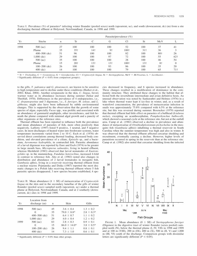
RESEARCH NOTES 1229
TABLE I. Prevalence (%) of parasites* infecting winter flounder (pooled sexes) north (upcurrent, uc), and south (downcurrent, dc) (m) from a sitedischarging thermal effluent at Holyrood, Newfoundland, Canada, in 1998 and 1999.
Yr Site/m
Parasite/prevalence (%)
n Tr C G Cl St McV L
1998 500 (uc) 27 100 100 100 52 100 37 41Plume 35 35† 14† 9† 100† 31† 36 3
400–500 (dc) 51 96 100 100 82 100 80† 261,000 (dc) 29 100 100 97 72 100 55 62†
1999 500 (uc) 35 100 100 100 28 100 46 54Plume 15 20† 13† 13† 100† 13† 8† 0
100–200 (dc) 26 100 100 93 86 100 35 20400 (dc) 14 100 100 100 79 100 65 71†
* Tr � Trichodina, C � Ceratomyxa, G � Gyrodactylus, Cl � Cryptocotyle lingua, St � Steringophorus, McV � McVicarius, L � Lecithaster.† Significantly different (P � 0.05) from comparison group(s).
TABLE II. Mean abundance (x � SE) of metacercariae of Cryptocotylelingua on the skin and in the secondary lamellae of the gills of winterflounder (pooled sexes) sampled north (upcurrent, uc) under a thermalplume at Holyrood, Newfoundland, Canada, and at 2 southerly (down-current, dc) sites in 1998 and 1999.
YrLocation fromdischarge (m) n Skin Gills
1998 500 (uc) 45 3.6 � 0.4 1.3 � 0.20 35 79.0 � 8.8* 2.8 � 0.3*
400–500 (dc) 51 6.4 � 0.7 1.1 � 0.21,000 (dc) 29 4.0 � 0.4 1.2 � 0.2
1999 500 (uc) 63 2.8 � 0.3 0.9 � 0.10 15 86.0 � 14.0* 3.8 � 0.5*
100–200 (dc) 26 9.4 � 1.1 0.8 � 0.1400 (dc) 14 7.3 � 1.0 0.6 � 0.1
* Significantly different (P � 0.05) than other groups.
FIGURE 1. Mean abundance (0 � SE) of Steringophorus furciger(Digenea) in the digestive tract of winter flounder (sexes pooled) sam-pled north (N), below the thermal plume (I, IV, VII) in 1998 and 1999and at 100 m (VIII), 200 m (IX), 400 m (X), 500 m (II, V) and 1,000m (III, VI) south of the discharge. Comparison groups with dissimilarletters are significantly different (P � 0.05).
to the gills, T. jadranica and G. pleuronecti, are known to be sensitiveto high temperature and to decline under these conditions (Barker et al.,2002; Khan, 2004). Another ectoparasite in the skin, C. lingua, favorswarmer water as its abundance increases during summer (Sekar andThrelfall, 1970). Differences in the prevalence of 4 endoparasites, i.e.,C. drepanopsettae and 3 digeneans, i.e., S. furciger, M. soleae, and L.gibbosus, might also have been influenced by subtle environmentalchanges. This is supported by the observation that the growth of somespecies of algae, especially Fucus spp., was prolific and associated withan abundance of gammarid amphipids, other invertebrates, and fish be-neath the plume compared with minimal algal growth and a paucity ofother organisms at the reference sites.
Thermal effluent has been reported to influence both the prevalenceand mean abundance of parasites of fish, more often positively thannegatively. Lafferty (1997) listed 9 positive, 1 neutral, and 5 negativecases. In most discharges of heated water into freshwater systems, watertemperature increments varied from 2 to 10 C. Esch et al. (1976) ob-served direct correlations among thermal loading, decreased body con-dition, and elevated prevalence of a ciliate, Epistylis sp., and a bacte-rium, Aeromonas hydrophila, in centrarchid fish. Parasitic abundanceof a larval digenean was reported by Eure and Esch (1974) to be greaterin large mouth bass, Micropterus salmoides, living in heated effluent,whereas Hirshfield (1983) observed that larval nematodes of Eustron-gylides sp. in the mummichog, Fundulus heteroclitus, increased 4-foldin contrast to reference fish. Aho et al. (1982) noted also changes indistribution and abundance of 2 larval trematodes in mosquito fish,Gambusia affinis, living in a reservoir receiving thermal effluent froma nuclear reactor. Pojmanska and Dzika (1987) reported the most dra-matic changes in a Polish lake receiving thermal effluent where 5 fishparasitic species disappeared, 3 new species became established, 4 spe-
cies decreased in frequency, and 6 species increased in abundance.These changes resulted in a modification of dominance in the com-munity structure. They attributed these changes to the habitat that af-fected both the invertebrate intermediate and avian definitive hosts. Anunusual observation was noted by Sankurathri and Holmes (1976) in alake where thermal water kept it ice-free in winter, and, as a result ofwaterfowl concentration, the prevalence of metacercariae infection insnails was approximately 75.8% compared with 6.5% at the referencesite, but this was reversed during summer. Boxrucker (1979) reportedthat thermal effluent had little effect on parasites of a bullhead (Ictalurusmelas), excepting an acanthocephalan, Pomphorhynchus bulbocolli,which showed a seasonal cycle at the reference site, but not at the outfallarea. Camp et al. (1982) observed a decline in prevalence and abun-dance of metacercariae of Ornithodiplostomum ptchocheilus in the mos-quito fish (Gambusia affinis) inhabiting a thermal reservoir in SouthCarolina when the summer temperature was high and also in winter. Itwas observed that the thermal effluent affected cercariae shedding andrecruitment, eventually causing a decline. However, higher levels ofparasitism occurred in spring in the reservoir than at the reference site.Camp et al. (1982) also noted that cercariae shedding from the infected

1230 THE JOURNAL OF PARASITOLOGY, VOL. 93, NO. 5, OCTOBER 2007
snails (Physa sp.) continued throughout the winter. MacKenzie et al.(1995) cited an abstract of Hoglund and Thulin who reported no changein parasitism.
In summary, exposure of a cold-water, benthic, sedentary flatfish tothermal effluent discharged 5 m above the plume apparently caused noeffect on body condition, organ indices, or hematological or patholog-ical alterations. However, prevalence, or mean abundance, or both ofsome parasites of the fish were affected below the plume, 1 speciesincreasing and 6 species declining. This observation suggests eithersensitivity to the effluent or interruption of transmission via their inter-mediate hosts near the thermal plume. These results provide additionalevidence to support the view that some parasites of fish can be usefulas bioindicators of subtle environmental changes.
We are grateful to M. O’Keefe for technical assistance and to S. Wallfor typing the manuscript. The study was supported by a contract andthe Natural Sciences and Engineering Council of Canada.
LITERATURE CITED
ADAMS, S. M. 1990. Status and use of biological indicators for evalu-ating the effects of stress on fish. In Biological indicators of stressin fish, S. M. Adams (ed.). American Fisheries Society Symposium8: 1–8.
AHO, J. M., J. W. CAMP, AND G. W. ESCH. 1982. Long-term studies onthe population biology of Diplostomulun scheuringi in a thermallyaltered reservoir. Journal of Parasitology 68: 695–708.
ARTHUR, J. R., D. K. CONE, R. L. CUSACK, D. E. BARKER, AND M. D.B. BURT. 2004. Two species of Trichodina (Ciliophora: Peritrichi-da) from cultured flatfishes (Pleuronectiformes) in Atlantic Canada.Comparative Parasitology 71: 247–250.
BARKER, D. E., D. K. CONE, AND M. D. B. BURT. 2002. Trichodinamurmanica (Ciliophora) and Gyrodactylus pleuronecti parasitisinghatchery-reared winter flounder, Pseudopleuronectes americanus(Walbaum): Effects on host growth and assessment of parasite in-teraction. Journal of Fish Diseases 25: 81–89.
BOXRUCKER, J. C. 1979. Effects of a thermal effluent on the incidenceand abundance of the gill and intestinal parasites of the black bull-head. Parasitology 78: 195–206.
CAMP, J. W., J. M. AHO, AND G. W. ESCH. 1982. A long-term study onvarious aspects of the population biology of Ornithodiplostomumptychocheilus in a South Carolina cooling reservoir. Journal of Par-asitology 68: 709–718.
ESCH, G. W., T. C. HAZEN, R. V. DIMOCK, AND J. W. GIBBONS. 1976.Thermal effluent and the epizootiology of the ciliate Epistylis andthe bacterium Aeromonas in association with centrarchid fish.Transactions of the American Microscopical Society 95: 687–693.
EURE, H. E., AND G. W. ESCH. 1974. Effects of thermal effluent on thepopulation dynamics of helminth parasites in large mouth bass. InThermal ecology, J. W. Gibbons and R. R. Sharitz (eds.). AECSymposium Series (CONF-730505), Oak Ridge Tennessee Tech-nical Information Center, U.S. Atomic Energy Commission, OakRidge, Tennessee, p. 237–243.
GEORGE-NASCIMENTO, M., R. A. KHAN, F. GARCIAS, V. LOBOS, G.MUNOZ, AND V. VALDEBENITO. 2000. Impaired health in flounder,Paralichthus spp. inhabiting coastal Chile. Bulletin of Environ-mental Contamination and Toxicology 64: 84–190.
HIRSHFIELD, M. F., R. P. MORIN, AND D. J. HEPNER. 1983. Increasedprevalence of larvae Eustrongylides (Nematoda) in the mummi-cheg, Fundulus heteroclitus (L.) from a discharge canal of a pow-erplant in the Chesapeake Bay. Journal of Fish Biology 23: 135–142.
KHAN, R. A. 2003. Stress-related bioindicator anomalies in feral malewinter flounder (Pleuronectes americanus) exposed to effluent fromtwo pulp and paper mills in Newfoundland. Bulletin of Environ-mental Contamination and Toxicology 70: 401–407.
———. 2004. Parasites of fish as biomarkers of environmental degra-dation: A field study. Bulletin of Environmental Contamination andToxicology 72: 394–400.
———, AND K. NAG. 1993. Estimation of hemosiderosis in seabirdsand fish exposed to petroleum. Bulletin of Environmental Contam-ination and Toxicology 50: 125–131.
———, AND J. THULIN. 1991. Influence of pollution on parasites ofaquatic animals. Advances in Parasitology 30: 201–238.
LAFFERTY, K. D. 1997. Environmental parasitology: What can parasitestell us about human impacts on the environment. Parasitology To-day 13: 251–255.
LANDICCI, C., F. ROSSI, AND F. MALTAGLIATI. 1999. Detection of thermalpollution: Variability of benthic communities at two different spa-tial scales in an area influenced by a coastal power station. MarinePollution Bulletin 38: 296–303.
LGL LTD. 1999. Characterisation of the thermal plume at the HolyroodThermal Generating Station, Seal Cove, Newfoundland, duringFebruary 1998 to March 1999, SA575, LGL publication, St. John’s,NF, Canada, 29 p.
MACKENZIE, K., H. H. WILLIAMS, B. WILLIAMS, A. H. MCVICAR, AND
R. SIDDALL. 1995. Parasites as indicators of water quality and thepotential use of helminth transmission in marine pollution studies.Advances in Parasitology 35: 86–144.
POJMANSKA, T., AND E. DZIKA. 1987. Parasites of bream (Abramis bramaL.) from the lake Goslawskie (Poland) affected by long-term ther-mal pollution. Parasitologica Polonca 32: 139–161.
SANKURATHRI, C. S., AND J. C. HOLMES. 1976. Effects of thermal efflu-ents on parasites and commensals of Physa gyrini Say (Mollusca;Gastropoda) and their interactions at Lake Wabamun, Alberta. Ca-nadian Journal of Zoology 54: 1742–1753.
SEKHAR, S. E., AND W. THRELFALL. 1970. Helminth parasites of cunner,Tautogolabrus adspersus (Walbaum) in Newfoundland. Journal ofHelminthology 44: 169–188.
SURESH, K., M. S. AHAMED, G. DURAIRAJ, AND K. V. K. NAIR. 1993.Impact of a power plant heated effluent on the abundance of sed-imentary organism, off Kalpakkan, east coast of India. Hydrobiol-ogia 268: 109–114.
J. Parasitol., 93(5), 2007, pp. 1230–1234� American Society of Parasitologists 2007
Species of Coccidia (Apicomplexa: Eimeriidae) Infecting Pikas From Alaska, U.S.A. andNortheastern Siberia, Russia
A. J. Lynch, D. W. Duszynski, and J. A. Cook, Department of Biology, The University of New Mexico, Albuquerque, New Mexico 87131.e-mail: [email protected]
ABSTRACT: Eighty-eight fecal samples from 2 species of pika, Ocho-tona collaris and Ochotona hyperborea, collected in Alaska (N � 53)and Russia (N � 35), respectively, were examined for the presence ofcoccidia (Apicomplexa: Eimeriidae). Five oocyst morphotypes were ob-served. In O. collaris, we found Eimeria calentinei, Eimeria crypto-
barretti, and Eimeria klondikensis, whereas in O. hyperborea, we foundEimeria banffensis, E. calentinei, E. cryptobarretti, E. klondikensis, andIsospora marquardti. This study represents new geographic records forall 5 coccidia and new host records for E. cryptobarretti and I. mar-quardti. Only minor quantitative differences were seen between the

RESEARCH NOTES 1231
sporulated oocysts we studied and those reported in their original de-scriptions.
Pikas are holarctic lagomorphs composed of the single genus, Och-otona, with 30 species (Wilson and Reeder, 2005). The majority ofspecies are found in Asia, mainly in the Tibet (Qinghai-Xizang) Plateauregion, but also in Afghanistan, Burma, China, India, Iran, Japan, Ka-zakhstan, Korea, Nepal, Pakistan, and Russia, whereas only 2 speciesare found in North America (Chapman and Flux, 1990; Yu et al., 2000;Wilson and Reeder, 2005). Currently, 18 coccidia species (16 Eimeria,2 Isospora) are described from all Ochotona species. Over 3 summerfield seasons (2000–2002), the collared pika, Ochotona collaris (Nel-son, 1893), and the northern pika, Ochotona hyperborea (Pallas, 1811),were collected in Alaska and northeastern Siberia, Russia, respectively,as part of the Beringia Coevolution Project (Hoberg et al., 2003; Cooket al., 2005). The present study was conducted to assess the similarityof coccidia fauna in 2 closely related hosts geographically separated bythe Bering Strait.
Pikas were caught with museum snap traps or shot with firearms.Fecal specimens were taken from 88 animals from 6 regional field sites:O. collaris were collected from 2 sites in Alaska (N � 53), whereas O.hyperborea were collected from 4 sites in northeastern Siberia, Russia(N � 35). The Alaskan sites were Wrangell-St. Elias National Park andYukon-Charley Rivers National Preserve; 4 regions in northeastern Si-beria were sampled, the Omolon, Anadyr, and Kolyma river basins andthe Providenya Oblast. Symbiotype host specimens (Frey et al., 1992;Brooks, 1993), in which all oocyst species/forms were seen and iden-tified here, are maintained in the University of Alaska Museum of theNorth (UAM). Feces were preserved in 2.5% (w/v) aqueous K2Cr2O7
solution. Oocysts were isolated, measured, and photographed as de-scribed by Duszynski and Wilber (1997).
In all, 25% (22/88) of the samples were positive: 12/35 (34%) O.hyperborea, and 10/53 (19%) O. collaris. Only 4 pikas were host tomultispecies infections of coccidia. Five distinct oocyst morphotypeswere observed and these were consistent with previously recognizedcoccidia species from other pikas. Three coccidia species were recov-ered from O. collaris: Eimeria calentinei, Eimeria cryptobarretti, andEimeria klondikensis; 5 were recovered from O. hyperborea: Eimeriabanffensis, E. calentinei, E. cryptobarretti, E. klondikensis, and Isosporamarquardti. The recovery of E. cryptobarretti from O. collaris and O.hyperborea represents 2 new host records. Previously, E. cryptobarrettionly had been found in Ochotona princeps, the American pika, in Col-orado (Duszynski and Brunson, 1973). The recovery of I. marquardtifrom O. hyperborea also represents a new host record. The recovery of3 species of coccidia in Alaskan O. collaris represents geographic rangeextensions, as does the recovery of 5 species from O. hyperborea inSiberia. Both hosts were studied by Hobbs and Samuel (1974) frompikas collected in the Yukon Territory, Canada, (O. collaris) and Japan(O. hyperborea); in 92 O. collaris they reported E. banffensis, Eimeriabarretti, Eimeria circumborealis, Eimeria princeps, I. marquardti, andIsospora yukonensis, and in 14 O. hyperborea they recovered E. cir-cumborealis, E. princepsis, and Eimeria worleyi.
Because there have been so few published reports of coccidia fromthese hosts, we include brief mention of qualitative or quantitative (orboth) structures as they differ from the original descriptions, along withtaxonomic summaries of the species recovered.
Eimeria banffensis Lepp, Todd, and Samuel, 1973
Type host: O. princeps (Richardson, 1828), American pika.Other hosts (this study): O. hyperborea.Type locality: North America: Canada: Alberta, Banff, Jumping-
pound, and Sibbald creeks.Geographic distribution: North America: Canada: Alberta, Banff,
Jumpingpound, and Sibbald creeks, 51�N, 115�W; Yukon Territory,Ogilvie Mountains, 64�N, 138�W; U.S.A.: Colorado, Larimer and ClearCreek counties; Asia: Japan: Hokkaido, Daisetzusan National Park;Russia: Siberia, Chukotka, 3 km SSE of confluence of Volchya Riverand Liman Sea, 64�48�N, 177�33�E (this study).
Prevalence: 5/92 (5%) O. collaris (type host) in Yukon Territory;3/14 (21%) O. hyperborea in Japan; 5/35 (14%) O. hyperborea in Rus-sia (this study); 40/167 (24%) O. princeps in Colorado; 11/145 (8%)O. princeps in Alberta.
Material deposited: Skull, skeleton, and tissues of a symbiotype host(this study) are preserved in UAM, as UAM no. 84368 (IF 5252), male,11 August 2002 (collected by N. E. Dokuchaev, A. A. Tsvetkova).Photosyntype of sporulated oocysts are in the U.S. National ParasiteCollection (USNPC) as USNPC no. 87390.
Remarks: The morphology of E. banffensis from O. hyperborea inRussia is similar to the original description provided by Lepp et al.(1973) for this species collected and described from O. princeps inAlberta, Canada. Whereas Duszynski and Brunson (1973) described oo-cysts that were nearly 2 m smaller in both length and width, the oocystsizes of our Russian oocysts did not differ when compared with theoriginal specimens (30 25 vs. 30 25). Duszynski and Brunson(1973) and Hobbs and Samuel (1974) failed to detect the �2-m polargranule that was observed in both this study and the original study byLepp et al. (1973). The recovery of E. banffensis is a new geographicrecord for this parasite in Russia.
Eimeria calentinei Duszynski and Brunson, 1973
Type host: O. princeps (Richardson, 1828), American pika.Other hosts (this study): O. collaris, O. hyperborea.Type locality: North America: Colorado, Larimer County.Geographic distribution: North America: Canada: Yukon Territory,
Ogilvie Mountains, 64�N, 138�W, Alberta, 51�N, 115�W; U.S.A.: Col-orado: Clear Creek and Larimer counties; Alaska: Yukon-Charley Riv-ers National Preserve, NW of Rocky Slope of Mt. Kathryn, S of Wood-chopper Creek, 65�12�N, 143�33�W (this study); Asia: Japan: Hokkaido,Daisetzusan National Park; Russia: Siberia, Magadanskaya Oblast, 40km W Magadan, 59�41�N, 150�20�E (this study).
Prevalence: 5/53 (9%) O. collaris in Alaska (this study); 8/92 (9%)O. collaris in Yukon Territory; 2/35 (6%) O. hyperborea in Siberia (thisstudy); 1/14 (7%) O. hyperborea in Japan; 2/111 (2%) O. princeps inAlberta; 39/167 (23%) O. princeps (type host) in Colorado.
Material deposited: Skin, skull, skeleton, and tissues of 2 symbiotypehosts, one for each host species from this study, are preserved in theUAM: O. collaris, UAM no. 58399 (AF 49330), male, 1 August 2001(collected by H. Henttonen, J. Niemimaa, K. Gamblin, L. B. Barrelli)and O. hyperborea, UAM no. 80824 (AF 38535), 4 September 2000(collected by S. O. MacDonald, N. E. Dokuchaev, K. E. Galbreath).Photosyntype of a sporulated oocyst in the USNPC as no. 87393.
Remarks: The morphology of sporulated oocysts of E. calentineifrom O. hyperborea in Russia and O. collaris in Alaska is nearly iden-tical to those described by Duszynski and Brunson (1973) for the samespecies collected from O. princeps in Colorado. The recovery of E.calentinei establishes new geographic records for this parasite in Russiaand Alaska.
Eimeria cryptobarretti Duszynski and Brunson, 1973
Type host: O. princeps (Richardson, 1828), American pika.Other hosts (this study): O. collaris, O. hyperborea.Type locality: North America: U.S.A.: Colorado, Larimer and Clear
Creek counties.Geographic distribution: North America: Colorado: Larimer and
Clear Creek counties; Alaska: Wrangell-St. Elias National Park (thisstudy), Yukon-Charley Rivers National Preserve, mountainside NW ofHeadwater Lake of Crescent Creek, 64�82�N, 143�75�W (this study);Asia: Russia: Siberia, Magadanskaya Oblast, mouth of Kegali River,64�26�N, 161�47�E (this study).
Prevalence: 6/53 (11%) O. collaris in Alaska (this study); 5/35 (14%)O. hyperborea in Siberia (this study); 107/167 (64%) O. princeps (typehost) in Colorado.
Material deposited: Skin, skull, skeleton, and tissues of 2 symbiotypehosts, one for each host species from this study, are preserved in theUAM: O. collaris, UAM no. 58213 (AF 49535), 18 July 2001 (collectedby H. Henttonen, J. Niemimaa, K. Gamblin, L. B. Barrelli) and O.hyperborea, UAM no. 80603 (AF 38233), male, 19 August 2000 (col-lected by S. O. MacDonald, N. E. Dokuchaev, K. E. Galbreath). Pho-tosyntype and photoparatype of sporulated oocysts are in the USNPC,nos. 87480 and 88170, respectively.
Remarks: The morphology of E. cryptobarretti from O. hyperboreain Russia and O. collaris in Alaska is similar to the description byDuszynski and Brunson (1973) for the same species collected from O.
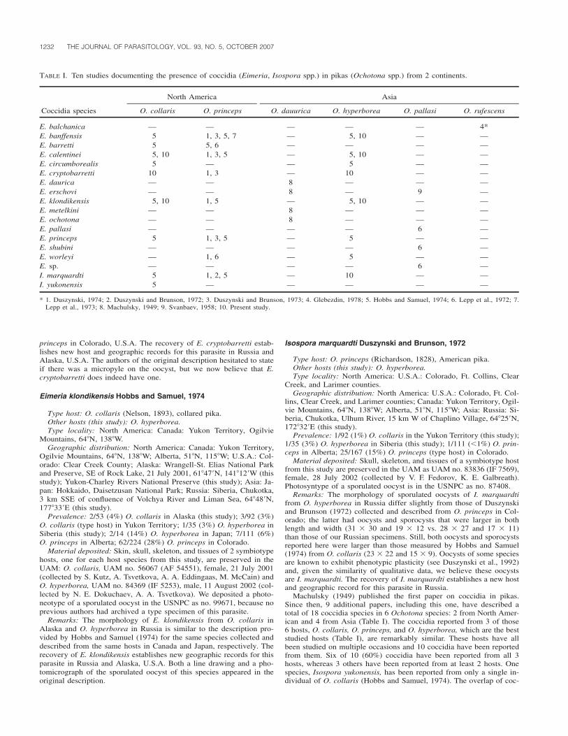
1232 THE JOURNAL OF PARASITOLOGY, VOL. 93, NO. 5, OCTOBER 2007
TABLE I. Ten studies documenting the presence of coccidia (Eimeria, Isospora spp.) in pikas (Ochotona spp.) from 2 continents.
Coccidia species
North America
O. collaris O. princeps
Asia
O. dauurica O. hyperborea O. pallasi O. rufescens
E. balchanica — — — — — 4*E. banffensis 5 1, 3, 5, 7 — 5, 10 — —E. barretti 5 5, 6 — — — —E. calentinei 5, 10 1, 3, 5 — 5, 10 — —E. circumborealis 5 — — 5 — —E. cryptobarretti 10 1, 3 — 10 — —E. daurica — — 8 — — —E. erschovi — — 8 — 9 —E. klondikensis 5, 10 1, 5 — 5, 10 — —E. metelkini — — 8 — — —E. ochotona — — 8 — — —E. pallasi — — — — 6 —E. princeps 5 1, 3, 5 — 5 — —E. shubini — — — — 6 —E. worleyi — 1, 6 — 5 — —E. sp. — — — — 6 —I. marquardti 5 1, 2, 5 — 10 — —I. yukonensis 5 — — — — —
* 1. Duszynski, 1974; 2. Duszynski and Brunson, 1972; 3. Duszynski and Brunson, 1973; 4. Glebezdin, 1978; 5. Hobbs and Samuel, 1974; 6. Lepp et al., 1972; 7.Lepp et al., 1973; 8. Machulsky, 1949; 9. Svanbaev, 1958; 10. Present study.
princeps in Colorado, U.S.A. The recovery of E. cryptobarretti estab-lishes new host and geographic records for this parasite in Russia andAlaska, U.S.A. The authors of the original description hesitated to stateif there was a micropyle on the oocyst, but we now believe that E.cryptobarretti does indeed have one.
Eimeria klondikensis Hobbs and Samuel, 1974
Type host: O. collaris (Nelson, 1893), collared pika.Other hosts (this study): O. hyperborea.Type locality: North America: Canada: Yukon Territory, Ogilvie
Mountains, 64�N, 138�W.Geographic distribution: North America: Canada: Yukon Territory,
Ogilvie Mountains, 64�N, 138�W; Alberta, 51�N, 115�W; U.S.A.: Col-orado: Clear Creek County; Alaska: Wrangell-St. Elias National Parkand Preserve, SE of Rock Lake, 21 July 2001, 61�47�N, 141�12�W (thisstudy); Yukon-Charley Rivers National Preserve (this study); Asia: Ja-pan: Hokkaido, Daisetzusan National Park; Russia: Siberia, Chukotka,3 km SSE of confluence of Volchya River and Liman Sea, 64�48�N,177�33�E (this study).
Prevalence: 2/53 (4%) O. collaris in Alaska (this study); 3/92 (3%)O. collaris (type host) in Yukon Territory; 1/35 (3%) O. hyperborea inSiberia (this study); 2/14 (14%) O. hyperborea in Japan; 7/111 (6%)O. princeps in Alberta; 62/224 (28%) O. princeps in Colorado.
Material deposited: Skin, skull, skeleton, and tissues of 2 symbiotypehosts, one for each host species from this study, are preserved in theUAM: O. collaris, UAM no. 56067 (AF 54551), female, 21 July 2001(collected by S. Kutz, A. Tsvetkova, A. A. Eddingaas, M. McCain) andO. hyperborea, UAM no. 84369 (IF 5253), male, 11 August 2002 (col-lected by N. E. Dokuchaev, A. A. Tsvetkova). We deposited a photo-neotype of a sporulated oocyst in the USNPC as no. 99671, because noprevious authors had archived a type specimen of this parasite.
Remarks: The morphology of E. klondikensis from O. collaris inAlaska and O. hyperborea in Russia is similar to the description pro-vided by Hobbs and Samuel (1974) for the same species collected anddescribed from the same hosts in Canada and Japan, respectively. Therecovery of E. klondikensis establishes new geographic records for thisparasite in Russia and Alaska, U.S.A. Both a line drawing and a pho-tomicrograph of the sporulated oocyst of this species appeared in theoriginal description.
Isospora marquardti Duszynski and Brunson, 1972
Type host: O. princeps (Richardson, 1828), American pika.Other hosts (this study): O. hyperborea.Type locality: North America: U.S.A.: Colorado, Ft. Collins, Clear
Creek, and Larimer counties.Geographic distribution: North America: U.S.A.: Colorado, Ft. Col-
lins, Clear Creek, and Larimer counties; Canada: Yukon Territory, Ogil-vie Mountains, 64�N, 138�W; Alberta, 51�N, 115�W; Asia: Russia: Si-beria, Chukotka, Ulhum River, 15 km W of Chaplino Village, 64�25�N,172�32�E (this study).
Prevalence: 1/92 (1%) O. collaris in the Yukon Territory (this study);1/35 (3%) O. hyperborea in Siberia (this study); 1/111 (�1%) O. prin-ceps in Alberta; 25/167 (15%) O. princeps (type host) in Colorado.
Material deposited: Skull, skeleton, and tissues of a symbiotype hostfrom this study are preserved in the UAM as UAM no. 83836 (IF 7569),female, 28 July 2002 (collected by V. F. Fedorov, K. E. Galbreath).Photosyntype of a sporulated oocyst is in the USNPC as no. 87408.
Remarks: The morphology of sporulated oocysts of I. marquardtifrom O. hyperborea in Russia differ slightly from those of Duszynskiand Brunson (1972) collected and described from O. princeps in Col-orado; the latter had oocysts and sporocysts that were larger in bothlength and width (31 30 and 19 12 vs. 28 27 and 17 11)than those of our Russian specimens. Still, both oocysts and sporocystsreported here were larger than those measured by Hobbs and Samuel(1974) from O. collaris (23 22 and 15 9). Oocysts of some speciesare known to exhibit phenotypic plasticity (see Duszynski et al., 1992)and, given the similarity of qualitative data, we believe these oocystsare I. marquardti. The recovery of I. marquardti establishes a new hostand geographic record for this parasite in Russia.
Machulsky (1949) published the first paper on coccidia in pikas.Since then, 9 additional papers, including this one, have described atotal of 18 coccidia species in 6 Ochotona species: 2 from North Amer-ican and 4 from Asia (Table I). The coccidia reported from 3 of those6 hosts, O. collaris, O. princeps, and O. hyperborea, which are the beststudied hosts (Table I), are remarkably similar. These hosts have allbeen studied on multiple occasions and 10 coccidia have been reportedfrom them. Six of 10 (60%) coccidia have been reported from all 3hosts, whereas 3 others have been reported from at least 2 hosts. Onespecies, Isospora yukonensis, has been reported from only a single in-dividual of O. collaris (Hobbs and Samuel, 1974). The overlap of coc-
RESEARCH NOTES 1233
cidia species among O. collaris, O. hyperborea, and O. princeps sug-gests the possibility that these coccidia may have evolved from a com-mon ancestor, i.e., that shared coccidia faunas in 3 closely related pikaspecies may reflect a single origin for the parasites in their commonancestor. On the other hand, this pattern may indicate that each coccid-ium had a common ancestor in the ancestor of the pikas. Thus, theparasite community may have a recent origin, but this doesn’t say any-thing about relationships among these coccidia.
Except for Eimeria erschovi Machulsky, 1949, the 7 coccidia iden-tified from the remaining Asian pikas, Ochotona dauurica (Eimeriadaurica, Eimeria metelkini, Eimeria ochotona, in 1949), Ochotona pal-lasi (Eimeria pallasi, Eimeria shubini, Eimeria sp., in 1958), and Och-otona rufescens (Eimeria balchanica, in 1978), only have been identi-fied once, each from 2–3 specimens of their single host species. Initially,the lack of overlap indicates that these coccidia may be more host-species specific, but nothing substantive is known about host specificityin pika coccidia. On the other hand, given the known distributions ofthe 3 host species, it is unlikely that these coccidia would ever comeinto contact with an Ochotona species different from the one in whichit was first described; O. rufescens, the Afghan pika, is geographicallyseparated from the other 2 species and, although the ranges of O. dauur-ica and O. pallasi share some overlap, e.g., Mongolia, these species areseparated both by altitude and biome (high mountain vs. desert, respec-tively). In addition, the obvious sampling bias doesn’t allow meaningfulcomparisons. Finally, the question must be asked whether any of these7 coccidia even still exist since 2 of the 3 host species are either en-dangered (O. pallasi) or threatened (O. rufescens).
Our a posteriori hypothesis was that the similarity, or disparity, ofcoccidia infecting pikas would reflect the systematics and phylogeneticsof the hosts. Work by Yu et al. (2000) on the phylogeny of 19 pikaspecies included O. hyperborea, O. princeps, O. pallasi, and O. dauur-ica; sequences from O. collaris and O. rufescens were not incorporated.The data of Yu et al. (2000) indicated that there are 3 pika clades: ashrub-steppe group of 7 species (including O. dauurica), a northerngroup of 5 species (including O. hyperborea, O. pallasi, and O. prin-ceps), and a mountain group of 7 species. Host–parasite data to date(Table I) support the notion that O. hyperborea and O. princeps maybe infected by the same coccidia because they have descended from arecent common ancestor. If true, this would predict that the same orsimilar coccidia species will be found in other species from the northerngroup of Yu et al. (2000). In other words, the morphological similarityof the coccidia in this study might reflect close phylogenetic relation-ships that are a consequence of the close relationship between the hosts.
The data of Yu et al. (2000) posit that O. princeps is the most basalmember of the northern group. Interestingly, O. pallasi is infected byan entirely different set of coccidia from other hosts in the northernclade. In fact, O. pallasi is infected by E. erschovi, a coccidium firstidentified from O. dauurica, a member of the shrub-steppe group ofpikas. Despite an older association between O. dauurica and O. pallasi,it is possible that E. erschovi is a generalist parasite capable of a broadco-accommodation of hosts (Brooks, 1979). In other words, the asso-ciation between E. erschovi and 2 deeply divergent pika lineages maysuggest the generalist nature of this coccidium. These hosts are in rel-atively close contact as the range of O. pallasi overlaps that of O.dauurica, although they occupy different habitats (Chapman and Flux,1990); unfortunately, it is not known if any burrowing or talus-dwellingpikas live in enough proximity to connect these pika lineages. Perhapsthe other Eimeria spp. identified from O. pallasi (Eimeria pallasi, Ei-meria shubini, and Eimeria sp.) are more derived (than those infectingO. princeps) and are results of recent speciation. This would keep thecospeciation hypothesis alive, but it is also possible that a host switchcould have led to this association. Only phylogenetic (sequence) datafor these coccidia will resolve the relationships among them.
It also is recognized that the dichotomy seen in Table I, where 3hosts overlap in eimeriid fauna and the other 3 hosts have divergentfauna, could be the result of poor species descriptions. Both Hobbs andSamuel (1974) and Lepp et al. (1972) addressed this possibility. Hobbsand Samuel (1974) noted the extreme similarity between many of the‘‘continental Asian’’ and the ‘‘North American’’ coccidia. In all cases,there were enough differences among those coccidia to prevent the syn-onymy of species. Before conclusions can be made regarding the valid-ity of species descriptions, more Asian pikas must be surveyed. Findingevidence for cryptic species of coccidia in pikas also could be an im-
portant issue for sorting out the origins of their host–parasite associa-tions.
In conclusion, we emphasize 3 major points: (1) the similarity incoccidia fauna among O. princeps, O. collaris, and O. hyperborea; (2)the different and more diverse coccidia parasites in Asian hosts; and(3) the apparent widespread species of coccidia found in pikas repre-senting 2 different host clades.
Thanks are due the multinational field teams in Alaska and Russiawho made this work possible; in particular we thank L. B. Barrelli, N.E. Dokuchaev, A. A. Eddingaas, V. F. Fedorov, K. E. Galbreath, K.Gamblin, H. Henttonen, S. Kutz, S. O. MacDonald, M. McCain, J.Niemimaa, A. A. Tsvetkova, and other members of our field teams. Wealso thank K. Galbreath for his very helpful suggestions when readingan earlier version of this manuscript. This work was supported by NSF-DEB grant 0196095, the Beringian Coevolution Project (to J.A.C.) anda subcontract on DEB 0196095 to UNM, coevolution of insectivoresand their coccidia parasites in Beringia (to D.W.D.).
LITERATURE CITED
BROOKS, D. R. 1979. Testing the context and extent of host–parasitecoevolution. Systematic Zoology 28: 299–307.
———. 1993. Critical comment: Extending the symbiotype concept tohost voucher specimens. Journal of Parasitology 79: 631–633.
CHAPMAN, J. A., AND J. E. C. FLUX. 1990. Rabbits, hares and pikas:Status survey and conservation action plan. International Union forConservation of Nature and Natural Resources. Gland, Switzerland.
COOK, J. A., E. P. HOBERG, A. KOEHLER, S. O. MACDONALD, H. HENT-TONEN, L. WICKSTROM, V. HAUKISALMI, K. GALBREATH, F. CHER-NYAVSKI, N. DOKUCHAEV, ET AL. 2005. Beringia: Intercontinentalexchange and diversification of high latitude mammals and theirparasites during the Pliocene and Quaternary. Mammal Science 30:S33–S44.
DUSZYNSKI, D. W. 1974. More information on the coccidian parasites(Protozoa: Eimeriidae) of the Colorado pika, Ochotona princeps,with a key to the species. Journal of Wildlife Diseases 10: 94–100.
———, AND J. T. BRUNSON. 1972. The structure of the oocyst and theexcystation process of Isospora marquardti sp. n. from the Colo-rado pika, Ochotona princeps. Journal of Protozoology 19: 257–259.
———, AND ———. 1973. Structure of the oocysts and excystationprocesses of four Eimeria spp. (Protozoa: Eimeriidae) from the Col-orado pika, Ochotona princeps. Journal of Parasitology 59: 28–34.
———, M. J. PATRICK, L. COUCH, AND S. J. UPTON. 1992. Eimeriansin harvest mice, Reithrodontomys spp., from Mexico, Californiaand New Mexico, and phenotypic plasticity in oocysts of Eimeriaarizonensis. Journal of Protozoology 39: 644–648.
———, AND P. G. WILBER. 1997. A guideline for the preparation ofspecies descriptions in the Eimeriidae. Journal of Parasitology 83:333–336.
FREY, J. K., T. L. YATES, D. W. DUSZYNSKI, W. L. GANNON, AND S. L.GARDNER. 1992. Designation and curatorial management of typehost specimens (symbiotypes) for new parasite species. Journal ofParasitology 78: 930–932.
GLEBEZDIN, V. S. 1978. About the coccidia fauna of wild mammals ofsouth-western Turkmenistan. Izvestiya Akademii Nauk Turkmen-skoi SSR 3: 71–78.
HOBBS, R. P., AND W. M. SAMUEL. 1974. Coccidia of pikas with specificreference to Ochotona collaris, O. princeps, and O. hyperboreayesoensis. Canadian Journal of Zoology 52: 1079–1085.
HOBERG, E. P., S. J. KUTZ, K. E. GALBREATH, AND J. A. COOK. 2003.Arctic biodiversity: From discovery to faunal baselines—Revealingthe history of a dynamic system. Journal of Parasitology 89: S84–S95.
LEPP, D. L., K. S. TODD, JR., AND W. M. SAMUEL. 1972. Four newspecies of Eimeria (Protozoa: Eimeriidae) from the pika Ochotonaprinceps from Alberta and Ochotona pallasi from Kazakhstan.Journal of Protozoology 19: 192–195.
———, ———, AND ———. 1973. Eimeria banffensis n. sp. (Proto-zoa: Eimeridae) from the pika Ochotona princeps from Alberta.Transactions of the American Microscopical Society 92: 305–307.
MACHULSKY, S. N. 1949. About coccidia in rodents of southern areas

1234 THE JOURNAL OF PARASITOLOGY, VOL. 93, NO. 5, OCTOBER 2007
of Buryat-Mongol, USSR. Trudy Buryat-Mongol’skoi Zooveteri-narnogo Instituta 5: 40–56.
SVANBAEV, S. K. 1958. Coccidia of rodents of Central Kazakhstan. Tru-dy Instituta Zoologii, Akademii Nauk Kazahkskoi SSR 9: 183–186.
WILSON, D. E., AND D. M. REEDER. 2005. Mammal species of the world:
A taxonomic and geographic reference, 3rd ed. Johns Hopkins Uni-versity Press, Baltimore, Maryland, 2142 p.
YU, N., C. ZHENG, Y. ZHANG, AND W. LI. 2000. Molecular systematicsof pikas (Genus Ochotona) inferred from mitochondrial DNA se-quences. Molecular Phylogenetics and Evolution 16: 85–95.
J. Parasitol., 93(5), 2007, pp. 1234–1238� American Society of Parasitologists 2007
A Ribeiroia Spp. (Class: Trematoda)–-Specific PCR-Based Diagnostic
David M. Reinitz, Timothy P. Yoshino, and Rebecca A. Cole*†, University of Wisconsin–Madison, Department of Pathobiological Sciences,2115 Observatory Drive, Madison, Wisconsin 53726; *United States Geological Survey National Wildlife Health Center, 6006 Schroeder Road,Madison, Wisconsin 53711; and †To whom correspondence should be addressed. e-mail: rebecca�[email protected]
TABLE I. Oligonucleotides and PCR profiles used to detect Ribeiroia sp.
Primer Sequence Tm % GC Use/product size
21-up AGTCATGGTGAGGTGCAGTGA 59.7 52.4 with 18-dn, 290 bp, profile 118-dn AGACCGCTTAGATAGCAG 51.4 50.0 with 21-up, 290 bp, profile 118-up CGTGTTTGGCGATTTAGT 51.4 44.4 Nested reaction with 19-dn, 164 bp, profile 219-dn TCAAAAATGAAGCAACAGT 49.1 31.6 Nested reaction with 18-up, 164 bp, profile 2
Profile 1
1X 94 C 4 min94 C 15 sec 94 C 15 sec
10X 59 C 30 sec 26X 59 C 30 sec 1X 72 C 7 min72 C 45 sec 72 C 90 sec 4 C Hold
Profile 2. As above except anneal at 53 C versus 59 C.
ABSTRACT: Increased reporting of amphibian malformations in NorthAmerica has been noted with concern in light of reports that amphibiannumbers and species are declining worldwide. Ribeiroia ondatrae hasbeen shown to cause a variety of types of malformations in amphibians.However, little is known about the prevalence of R. ondatrae in NorthAmerica. To aid in conducting field studies of Ribeiroia spp., we havedeveloped a polymerase chain reaction (PCR)-based diagnostic. Herein,we describe the development of an accurate, rapid, simple, and cost-effective diagnostic for detection of Ribeiroia spp. infection in snails(Planorbella trivolvis). Candidate oligonucleotide primers for PCR weredesigned via DNA sequence analyses of multiple ribosomal internaltranscribed spacer-2 regions from Ribeiroia spp. and Echinostoma spp.Comparison of consensus sequences determined from both genera iden-tified areas of sequence potentially unique to Ribeiroia spp. The PCRreliably produced a diagnostic 290-base pair (bp) product in the pres-ence of a wide concentration range of snail or frog DNA. Sensitivitywas examined with DNA extracted from single R. ondatrae cercaria.The single-tube PCR could routinely detect less than 1 cercariae equiv-alent, because DNA isolated from a single cercaria could be diluted atleast 1:50 and still yield a positive result via gel electrophoresis. Aneven more sensitive nested PCR also was developed that routinely de-tected 100 fg of the 290-bp fragment. The assay did not detect furco-cercous cercariae of certain Schistosomatidae, Echinostoma sp., orSphaeridiotrema globulus nor adults of Clinostomum sp. or Cyathoco-tyle bushiensis. Field testing of 137 P. trivolvis identified 3 positiveswith no overt environmental cross-reactivity, and results concurred withmicroscopic examinations in all cases.
Concern over declining numbers and species of amphibians has cometo the forefront over the past 20 yr (Barinaga, 1990; Blaustein andWake, 1990; Phillips, 1990; Pechmann et al., 1991; Wake, 1998). Sug-gested factors, singly or in synergism, that have been hypothesized asreasons for the decline of this class of animals include habitat destruc-tion (Kolozsvary and Swihart, 1999; Houlahan and Findlay, 2003), UV
irradiation (Blaustein et al., 1998, 2003), introduced species (Knapp andMathews, 2000), climate change (Beebee, 1995; Corn, 2005), and var-ious pathogens (Daszak et al., 2003). A current review of the factors isfound in Beebee and Griffiths (2005). Amphibian malformations are ofgrowing concern, because they have been observed with increased prev-alence in North America (Ouellet, 2000). Although malformations havethe potential to deleteriously affect populations or species at particularsites, they have not been empirically linked to global or regional de-clines. Recent reports (Johnson et al., 1999, 2002; Lannoo et al., 2003;Schoff et al., 2003; Schotthoefer et al., 2003) have implicated the trem-atode Ribeiroia ondatrae as a causative agent of some types of mal-formations. Little is known about the distribution of this parasite in itshosts within North America. Wilson et al. (2005) identified 3 speciesof Ribeiroia: R. ondatrae within the Americas; R. marini in the Carrib-bean, and Cercaria lileta in Africa. Ribeiroia ondatrae has a 3-host lifecycle with 2 aquatic intermediate hosts and a predator definitive host,usually a bird or mammal. Planorbella spp. serves as first intermediatehost, with fish and various amphibians as second intermediate hosts.Exogenous factors, which include pesticides (Kiesecker, 2002), and eu-trophication, which leads to a dominance of Planorbella spp. (Johnsonand Chase, 2004), have been shown to increase malformation rates.Currently, Ribeiroia spp. infections in the first intermediate host arediagnosed by dissection of live or freshly dead snail hosts for variouslarval stages, which requires training and substantial time to locate in-fected tissues to identify the parasite correctly. Identifying larvae earlyin development after miracidial penetration but before the developmentof the cercariae is difficult, if not impossible, using morphological char-acters. To increase the speed and accuracy in the examination of largenumbers of snails for the presence of Ribeiroia spp., to reduce laborcosts, and to simplify training required, we have developed a genus-specific polymerase chain reaction (PCR)-based diagnostic that targetsthe second internal transcribed spacer (ITS-2) region of the ribosomalRNA gene cluster (Morgan and Blair, 1998; Kostadinova et al., 2003;Wilson et al., 2005). By using various combinations of 4 oligonucleo-
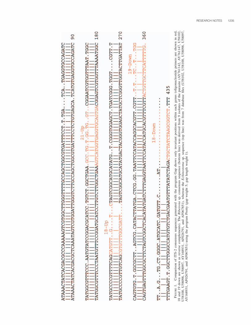
RESEARCH NOTES 1235
FIG
UR
E1.
Com
pari
son
ofIT
S-2
cons
ensu
sse
quen
ces
gene
rate
dw
ith
the
prog
ram
Gap
show
son
lyin
vari
ant
posi
tion
sw
ithi
nea
chge
nus
(oli
gonu
cleo
tide
prim
ers
are
show
nin
red,
18an
d19
dow
nar
esh
own
asre
vers
eco
mpl
emen
ts).
The
Rib
eiro
iasp
.co
nsen
sus
sequ
ence
(bot
tom
line
)w
asde
rive
dfr
om9
isol
ates
ofth
epa
rasi
te(A
Y76
1142
,A
Y76
1147
,U
5810
2,U
5810
0,U
5809
8,U
5809
7,A
Y16
8931
,A
F02
6791
,an
dA
F06
7851
),w
here
asth
eE
chin
osto
ma
sp.
sequ
ence
(top
line
)w
asfr
om7
data
base
file
s(U
5810
2,U
5810
0,U
5809
8,U
5809
7,A
Y16
8931
,A
F02
6791
,an
dA
F06
7851
)us
ing
the
prog
ram
Pre
tty
(gap
wei
ght
5;ga
ple
ngth
wei
ght
1).
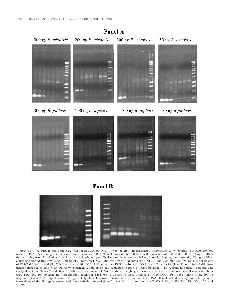
1236 THE JOURNAL OF PARASITOLOGY, VOL. 93, NO. 5, OCTOBER 2007
FIGURE 2. (A) Production of the Ribeiroia-specific 290-bp DNA (lowest band) in the presence of Planorbella trivolvis (row 1) or Rana pipiens(row 2) DNA. Two nanograms of Ribeiroia sp. cercarial DNA (lane 1) was diluted 10-fold in the presence of 300, 200, 100, or 50 ng of DNA(left to right) from P. trivolvis (row 1) or from R. pipiens (row 2). Routine detection was 0.2 ng (lane 2, all gels); and optimally, 20 pg of DNAcould be detected (top row, lane 3, 50 ng of P. trivolvis DNA). The five lowest standards are 1,500, 1,000, 750, 500, and 250 bp. (B) Sensitivityof ITS-2 (L) and nested (R) Ribeiroia sp.-specific PCR. Left gel shows PCR results with DNA from 20 cercariae (lane 1) and 10-fold dilutionsthereof (lanes 2–6; lane 7, no DNA) with primers 21-up/18-dn and subjected to profile 1 (290-bp target). DNA from less than 1 cercaria waseasily detectable (lanes 2 and 3) with little or no extraneous DNAs produced. Right gel shows results from the second nested reaction, whichused a purified 290-bp template from the first reaction and primers 18-up and 19-dn to produce a 164 bp DNA. Ten-fold dilutions of the 290-bpfragment (lanes 2–7) ranged from 100 pg to 1 fg; lane 8 shows a reaction with no template DNA. One hundred femtograms (�1 genomeequivalent) of the 290-bp fragment could be routinely detected (lane 5). Standards in both gels are 2,000, 1,500, 1,000, 750, 500, 300, 150, and50 bp.

RESEARCH NOTES 1237
tide primers, the assay can be performed as a single-tube PCR or as anested reaction for greater sensitivity. Table I summarizes primer char-acteristics, uses, and profiles. Desirable assay characteristics include suf-ficient sensitivity to detect early infection of snails, accuracy, cost-ef-fectiveness, simplicity, and the ability to detect parasite DNA in thepresence of excess frog or snail host DNA. In this article, we examinedhow the primary pair of primers worked to amplify target sequences inthe presence of variable amounts of frog or snail DNA with regard tosensitivity and specificity.
ITS-2 sequences unique to Ribeiroia spp. were tentatively identifiedby a 2-step process (Fig. 1): first, by producing an ITS-2 consensussequence from both Ribeiroia and Echinostoma spp. with the programPretty (Accelrys, San Diego, California); and second, by an alignmentof the 2 consensus sequences with the program Gap (Accelrys) to iden-tify areas of divergence. Echinostoma spp. were selected for comparisonbecause sequence data for more closely related organisms were notavailable. From sequences potentially unique to R. ondatrae, we de-signed 4 oligonucleotide primers, denoted 21-up, 18-dn, 18-up, and 19-dn (Table I) for use as PCR primers. Primer dimer formation was notpredicted to be problematic by the programs Gap and Bestfit (Accelrys),and the theoretical melting temperatures (Tm) provided by the manufac-turer (Integrated DNA Technologies, Inc., Coralville, Iowa). Meltingpoint considerations and degree of mismatch with Echinostoma spp.sequences lead us to first test primers 21-up with 18-dn (single reaction)and 18-up with 19-down (nested reaction), which produced DNA frag-ments of 290 base pairs (bp) and 164 bp, respectively.
PCR mixtures contained 200 M each deoxynucleoside-5[prime]-tri-phosphate, 300 nM 18-up, 300 nM 18-down, 2.6 U of Expand HighFidelity Enzyme (Roche Diagnostics, Indianapolis, Indiana), and 1.5mM MgCl2 in 50 l. Alternatively, 25-l reactions with PCR Beads (G.E. Healthcare, Buckinhamshire, U.K.) with primer and DNA concen-trations, as described above, produced identical results. After amplifi-cation, samples were analyzed by agarose gel electrophoresis for a DNAfragment of specific size (290 bp or 164 bp). The identity of the am-plified DNA was confirmed to be Ribeiroia spp. by sequence analysis.Amplified DNA was purified directly from a 50-l PCR reaction usingWizard PCR Preps (Promega, Madison, Wisconsin), mixed with thePCR primer (10 ng of DNA and 10 of pmol primer) in water and sentto Northwoods DNA Inc. (Solway, Minnesota). The sequence producedwas compared with the R. ondatrae sequence using the program BestFit(Accelrys).
Ribeiroia ondatrae genomic DNA was purified from cercariae shedby Planorbella trivolvis, using a DNeasy kit (cultured cells protocol;QIAGEN, Valencia, California). Specificity of the assay with primers21-up and 18-dn was examined in the presence of excess DNA isolatedfrom hosts P. trivolvis and Rana pipiens (DNeasy, animal tissue pro-tocol; QIAGEN). Figure 2A shows the detection of a serial 10-folddilution of 2 ng of R. ondatrae cercarial DNA (lane 1, with 21-up/18-dn) in the presence of 300, 200, 100, and 50 ng of P. trivolvis or R.pipiens DNA. Neither of these excess DNAs was problematic, althoughP. trivolvis DNA did produce slightly more nonspecific amplificationproducts than did R. pipiens DNA.
In the presence of excess P. trivolvis or R. pipiens DNA, the Ribei-roia-specific 290-bp fragment was consistently produced (verified bysequencing bands excised from such gels). Row 1 of Figure 2A showsproduction of the diagnostic 290-bp fragment in the presence of 4 levelsof P. trivolvis DNA (300, 200, 100, and 50 ng/50 l). No intense DNAfragments of similar size that might obscure detection of the diagnostic290-bp fragment were produced at any of the levels of host DNA tested.Some low-level nonspecific DNAs were produced (e.g., 300 ng, lanes3–6) that were distinctly different sizes (�1,200 bp) from the 290-bpDNA from R. ondatrae and are not seen as an impediment to correctassay interpretation.
Clearly, faint 290-bp bands, such as those observed in lanes 4–7 inmost of Figure 2A, should be considered negative results. In the absenceof P. trivolvis or R. pipiens DNA, these nonspecific bands were notpresent, so they probably represent weakly amplified host DNA se-quences that in essence define the negative background signal that mustbe significantly exceeded for a positive determination. In addition, whenfield testing either P. trivolvis or R. pipiens tissue, total DNA added didnot exceed 100 ng, a level where interpretation of the agarose gel resultsis straightforward. Overall, many of the gel lanes in Figure 2A con-tained only the specific fragment.
The single-tube PCR with primers 21-up and 18-dn can routinelydetect less than a single cercariae equivalent (CE) of R. ondatrae DNA.Figure 2B shows the results of serial 10-fold dilutions of this targetDNA starting with 20 CE. A positive reaction was clearly and consis-tently seen with 0.2 CE (lane 3). The amount of input target was de-termined by extracting DNA from 20 cercariae (with carrier RNA, 10g/sample) collected from R. ondatrae-infected P. trivolvis. Becausethis procedure assumed a 100% recovery, the assay can probably detectless than 0.2 CE. This also was convincingly demonstrated using DNAisolated from a single cercaria (with carrier RNA, 10 g/sample) withidentical results (data not shown). Furthermore, if an aliquot of theprimary PCR reaction was subjected to a second round of PCR withnested primers 18-up and 19-dn, as little as 100 fg (�1 genome equiv-alent) of the 290-bp template could be detected (Fig. 2B). Although theconventional PCR assay (21-up/18-dn) has not been extensively fieldtested, many co-occurring and potentially cross-reactive organisms, in-cluding Echinostoma sp., have been examined.
Two field samples of P. trivolvis infected with Echinostoma sp. rediaeand cercariae were tested by excising parasite tissue for DNA extrac-tion. The 2 samples, at 3 different levels of target DNA (10-fold dilu-tions), produced no DNAs in the 290-bp size range and little nonspecificDNA of any size. Such samples, when spiked with R. ondatrae targetDNA (100 ng), produced a 290-bp band, which indicated that lack ofEchinostoma sp. reactivity was not due to inhibitory substances presentin these samples. And, an independent examination of trematodes thatincluded Clinostomum sp., Cyathocotyle bushiensis, or Sphaeridiotremaglobulus, showed no cross-reactivity in amplifications with 21-up/18-dn. Epistylis sp. (class Ciliata), an epibiont from snail shells, did givea positive reaction that sequencing revealed to be due to filter feedingupon R. ondatrae sp. cercariae. The assay does not detect xiphidiocer-cariae from P. trivolvis; and as expected, the assay does strongly detectR. marini cercariae.
Snails from an ongoing field study in Illinois were first microscopi-cally examined for R. ondatrae cercariae. The hepatopancreas was thenexcised and pooled (1–3 individuals) into 137 tissue samples fromwhich DNA was purified (DNeasy kit; QIAGEN), and samples wereassayed via PCR with 21-up/18-dn. No cross-reactivity or other inter-ference was observed, and the assay identified 3 positive pools of tissue,which corroborated microscopic examinations. Although several groupsof tissue were from snails where infections were very immature (labo-ratory reared snails were placed in field cages for 3–4 wk), and iden-tification based on morphological characters was inconclusive, the PCRassay enabled positive identification of R. ondatrae. Other more limitedfield tests also gave results that correlated well with microscopic ex-aminations as 9 P. trivolvis from Duck Lake, Minnesota, and 3 P. tri-volvis from Merriville, Minnesota, yielded 6 positives, also visible mi-croscopically in all cases. Overall, the assay is very robust, with noknown cross-reactive DNA from environmental sources that interfereswith its interpretation. Although the assay does require some subjectiv-ity to discern weak positive signals from background negatives, it ismore rapid, more sensitive, and far simpler to train new personnel toconduct these procedures than to develop the ability to microscopicallyidentify the different life cycle stages of Ribeiroia spp. and other or-ganisms that may be present within snails. This assay will be useful instudies where a large number snails must be examined for Ribeiroiaspp. infections, and it will help determine where and when Ribeiroiaspp. infections can have a deleterious impact on frog populations.
We thank Sharon File-Emperador, University of Puerto Rico, RioPiedras Campus, San Juan, for supplying R. marini cercariae; and thelate Dan Sutherland, University Wisconsin, LaCrosse, Wisconsin, andAnna Schotthoefer, University Illinois, Urbana, Illinois, for R. ondatrae-infected snails.
LITERATURE CITED
BARINAGA, M. 1990. Where have all the froggies gone? Science 247:1033–1034.
BEEBEE, T. J. C., AND R. A. GRIFFITHS. 2005. The amphibian declinecrisis: A watershed for conservation biology? Biological Conser-vation 125: 271–285.
BLAUSTEIN, A. R., AND D. B. WAKE. 1990. Declining amphibian popu-lations: A global phenomenon? Trends in Ecology and Evolution5: 203–204.
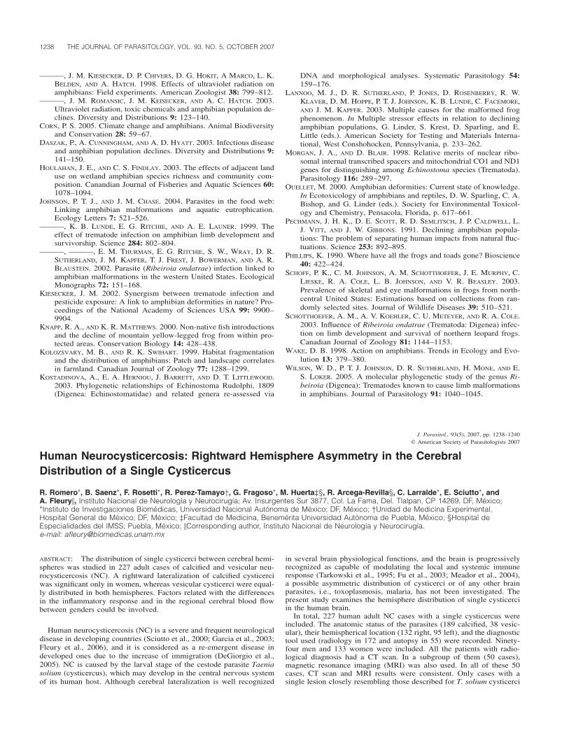
1238 THE JOURNAL OF PARASITOLOGY, VOL. 93, NO. 5, OCTOBER 2007
———, J. M. KIESECKER, D. P. CHIVERS, D. G. HOKIT, A MARCO, L. K.BELDEN, AND A. HATCH. 1998. Effects of ultraviolet radiation onamphibians: Field experiments. American Zoologist 38: 799–812.
———, J. M. ROMANSIC, J. M. KEISECKER, AND A. C. HATCH. 2003.Ultraviolet radiation, toxic chemicals and amphibian population de-clines. Diversity and Distributions 9: 123–140.
CORN, P. S. 2005. Climate change and amphibians. Animal Biodiversityand Conservation 28: 59–67.
DASZAK, P., A. CUNNINGHAM, AND A. D. HYATT. 2003. Infectious diseaseand amphibian population declines. Diversity and Distributions 9:141–150.
HOULAHAN, J. E., AND C. S. FINDLAY. 2003. The effects of adjacent landuse on wetland amphibian species richness and community com-position. Canandian Journal of Fisheries and Aquatic Sciences 60:1078–1094.
JOHNSON, P. T. J., AND J. M. CHASE. 2004. Parasites in the food web:Linking amphibian malformations and aquatic eutrophication.Ecology Letters 7: 521–526.
———, K. B. LUNDE, E. G. RITCHIE, AND A. E. LAUNER. 1999. Theeffect of trematode infection on amphibian limb development andsurvivorship. Science 284: 802–804.
———, ———, E. M. THURMAN, E. G. RITCHIE, S. W., WRAY, D. R.SUTHERLAND, J. M. KAPFER, T. J. FREST, J. BOWERMAN, AND A. R.BLAUSTEIN. 2002. Parasite (Ribeiroia ondatrae) infection linked toamphibian malformations in the western United States. EcologicalMonographs 72: 151–168.
KIESECKER, J. M. 2002. Synergism between trematode infection andpesticide exposure: A link to amphibian deformities in nature? Pro-ceedings of the National Academy of Sciences USA 99: 9900–9904.
KNAPP, R. A., AND K. R. MATTHEWS. 2000. Non-native fish introductionsand the decline of mountain yellow-legged frog from within pro-tected areas. Conservation Biology 14: 428–438.
KOLOZSVARY, M. B., AND R. K. SWIHART. 1999. Habitat fragmentationand the distribution of amphibians: Patch and landscape correlatesin farmland. Canadian Journal of Zoology 77: 1288–1299.
KOSTADINOVA, A., E. A. HERNIOU, J. BARRETT, AND D. T. LITTLEWOOD.2003. Phylogenetic relationships of Echinostoma Rudolphi, 1809(Digenea: Echinostomatidae) and related genera re-assessed via
DNA and morphological analyses. Systematic Parasitology 54:159–176.
LANNOO, M. J., D. R. SUTHERLAND, P. JONES, D. ROSENBERRY, R. W.KLAVER, D. M. HOPPE, P. T. J. JOHNSON, K. B. LUNDE, C. FACEMORE,AND J. M. KAPFER. 2003. Multiple causes for the malformed frogphenomenon. In Multiple stressor effects in relation to decliningamphibian populations, G. Linder, S. Krest, D. Sparling, and E.Little (eds.). American Society for Testing and Materials Interna-tional, West Conshohocken, Pennsylvania, p. 233–262.
MORGAN, J. A., AND D. BLAIR. 1998. Relative merits of nuclear ribo-somal internal transcribed spacers and mitochondrial CO1 and ND1genes for distinguishing among Echinostoma species (Trematoda).Parasitology 116: 289–297.
OUELLET, M. 2000. Amphibian deformities: Current state of knowledge.In Ecotoxicology of amphibians and reptiles, D. W. Sparling, C. A.Bishop, and G. Linder (eds.). Society for Environmental Toxicol-ogy and Chemistry, Pensacola, Florida, p. 617–661.
PECHMANN, J. H. K., D. E. SCOTT, R. D. SEMLITSCH, J. P. CALDWELL, L.J. VITT, AND J. W. GIBBONS. 1991. Declining amphibian popula-tions: The problem of separating human impacts from natural fluc-tuations. Science 253: 892–895.
PHILLIPS, K. 1990. Where have all the frogs and toads gone? Bioscience40: 422–424.
SCHOFF, P. K., C. M. JOHNSON, A. M. SCHOTTHOEFER, J. E. MURPHY, C.LIESKE, R. A. COLE, L. B. JOHNSON, AND V. R. BEASLEY. 2003.Prevalence of skeletal and eye malformations in frogs from north-central United States: Estimations based on collections from ran-domly selected sites. Journal of Wildlife Diseases 39: 510–521.
SCHOTTHOEFER, A. M., A. V. KOEHLER, C. U. METEYER, AND R. A. COLE.2003. Influence of Ribeiroia ondatrae (Trematoda: Digenea) infec-tion on limb development and survival of northern leopard frogs.Canadian Journal of Zoology 81: 1144–1153.
WAKE, D. B. 1998. Action on amphibians. Trends in Ecology and Evo-lution 13: 379–380.
WILSON, W. D., P. T. J. JOHNSON, D. R. SUTHERLAND, H. MONE, AND E.S. LOKER. 2005. A molecular phylogenetic study of the genus Ri-beiroia (Digenea): Trematodes known to cause limb malformationsin amphibians. Journal of Parasitology 91: 1040–1045.
J. Parasitol., 93(5), 2007, pp. 1238–1240� American Society of Parasitologists 2007
Human Neurocysticercosis: Rightward Hemisphere Asymmetry in the CerebralDistribution of a Single Cysticercus
R. Romero*, B. Saenz*, F. Rosetti*, R. Perez-Tamayo†, G. Fragoso*, M. Huerta‡§, R. Arcega-Revilla§, C. Larralde*, E. Sciutto*, andA. Fleury�, Instituto Nacional de Neurologıa y Neurocirugıa; Av. Insurgentes Sur 3877, Col. La Fama, Del. Tlalpan, CP 14269, DF, Mexico;*Instituto de Investigaciones Biomedicas, Universidad Nacional Autonoma de Mexico; DF, Mexico; †Unidad de Medicina Experimental,Hospital General de Mexico; DF, Mexico; ‡Facultad de Medicina, Benemerita Universidad Autonoma de Puebla, Mexico; §Hospital deEspecialidades del IMSS; Puebla, Mexico; �Corresponding author, Instituto Nacional de Neurologıa y Neurocirugıa.e-mail: [email protected]
ABSTRACT: The distribution of single cysticerci between cerebral hemi-spheres was studied in 227 adult cases of calcified and vesicular neu-rocysticercosis (NC). A rightward lateralization of calcified cysticerciwas significant only in women, whereas vesicular cysticerci were equal-ly distributed in both hemispheres. Factors related with the differencesin the inflammatory response and in the regional cerebral blood flowbetween genders could be involved.
Human neurocysticercosis (NC) is a severe and frequent neurologicaldisease in developing countries (Sciutto et al., 2000; Garcia et al., 2003;Fleury et al., 2006), and it is considered as a re-emergent disease indeveloped ones due to the increase of immigration (DeGiorgio et al.,2005). NC is caused by the larval stage of the cestode parasite Taeniasolium (cysticercus), which may develop in the central nervous systemof its human host. Although cerebral lateralization is well recognized
in several brain physiological functions, and the brain is progressivelyrecognized as capable of modulating the local and systemic immuneresponse (Tarkowski et al., 1995; Fu et al., 2003; Meador et al., 2004),a possible asymmetric distribution of cysticerci or of any other brainparasites, i.e., toxoplasmosis, malaria, has not been investigated. Thepresent study examines the hemisphere distribution of single cysticerciin the human brain.
In total, 227 human adult NC cases with a single cysticercus wereincluded. The anatomic status of the parasites (189 calcified, 38 vesic-ular), their hemispherical location (132 right, 95 left), and the diagnostictool used (radiology in 172 and autopsy in 55) were recorded. Ninety-four men and 133 women were included. All the patients with radio-logical diagnosis had a CT scan. In a subgroup of them (50 cases),magnetic resonance imaging (MRI) was also used. In all of these 50cases, CT scan and MRI results were consistent. Only cases with asingle lesion closely resembling those described for T. solium cysticerci
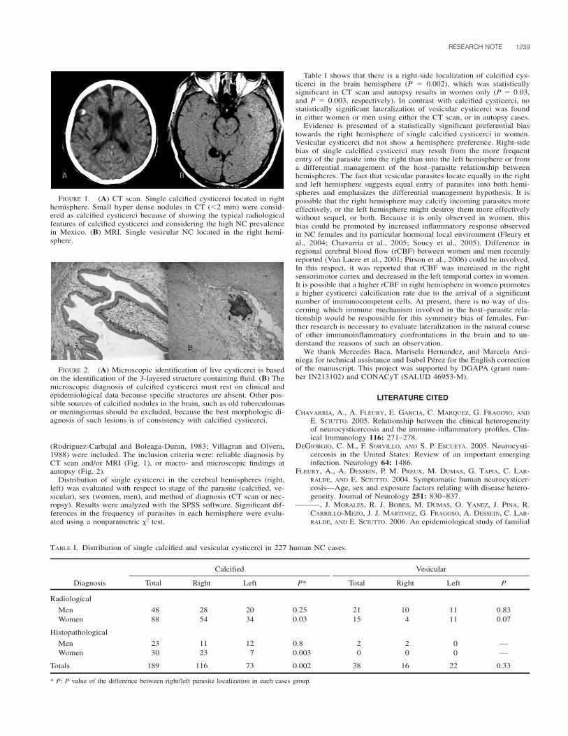
RESEARCH NOTE 1239
FIGURE 1. (A) CT scan. Single calcified cysticerci located in righthemisphere. Small hyper dense nodules in CT (�2 mm) were consid-ered as calcified cysticerci because of showing the typical radiologicalfeatures of calcified cysticerci and considering the high NC prevalencein Mexico. (B) MRI. Single vesicular NC located in the right hemi-sphere.
FIGURE 2. (A) Microscopic identification of live cysticerci is basedon the identification of the 3-layered structure containing fluid. (B) Themicroscopic diagnosis of calcified cysticerci must rest on clinical andepidemiological data because specific structures are absent. Other pos-sible sources of calcified nodules in the brain, such as old tuberculomasor meningiomas should be excluded, because the best morphologic di-agnosis of such lesions is of consistency with calcified cysticerci.
TABLE I. Distribution of single calcified and vesicular cysticerci in 227 human NC cases.
Diagnosis
Calcified
Total Right Left P*
Vesicular
Total Right Left P
Radiological
Men 48 28 20 0.25 21 10 11 0.83Women 88 54 34 0.03 15 4 11 0.07
Histopathological
Men 23 11 12 0.8 2 2 0 —Women 30 23 7 0.003 0 0 0 —
Totals 189 116 73 0.002 38 16 22 0.33
* P: P value of the difference between right/left parasite localization in each cases group.
(Rodriguez-Carbajal and Boleaga-Duran, 1983; Villagran and Olvera,1988) were included. The inclusion criteria were: reliable diagnosis byCT scan and/or MRI (Fig. 1), or macro- and microscopic findings atautopsy (Fig. 2).
Distribution of single cysticerci in the cerebral hemispheres (right,left) was evaluated with respect to stage of the parasite (calcified, ve-sicular), sex (women, men), and method of diagnosis (CT scan or nec-ropsy). Results were analyzed with the SPSS software. Significant dif-ferences in the frequency of parasites in each hemisphere were evalu-ated using a nonparametric �2 test.
Table I shows that there is a right-side localization of calcified cys-ticerci in the brain hemisphere (P � 0.002), which was statisticallysignificant in CT scan and autopsy results in women only (P � 0.03,and P � 0.003, respectively). In contrast with calcified cysticerci, nostatistically significant lateralization of vesicular cysticerci was foundin either women or men using either the CT scan, or in autopsy cases.
Evidence is presented of a statistically significant preferential biastowards the right hemisphere of single calcified cysticerci in women.Vesicular cysticerci did not show a hemisphere preference. Right-sidebias of single calcified cysticerci may result from the more frequententry of the parasite into the right than into the left hemisphere or froma differential management of the host–parasite relationship betweenhemispheres. The fact that vesicular parasites locate equally in the rightand left hemisphere suggests equal entry of parasites into both hemi-spheres and emphasizes the differential management hypothesis. It ispossible that the right hemisphere may calcify incoming parasites moreeffectively, or the left hemisphere might destroy them more effectivelywithout sequel, or both. Because it is only observed in women, thisbias could be promoted by increased inflammatory response observedin NC females and its particular hormonal local environment (Fleury etal., 2004; Chavarria et al., 2005; Soucy et al., 2005). Difference inregional cerebral blood flow (rCBF) between women and men recentlyreported (Van Laere et al., 2001; Pirson et al., 2006) could be involved.In this respect, it was reported that rCBF was increased in the rightsensorimotor cortex and decreased in the left temporal cortex in women.It is possible that a higher rCBF in right hemisphere in women promotesa higher cysticerci calcification rate due to the arrival of a significantnumber of immunocompetent cells. At present, there is no way of dis-cerning which immune mechanism involved in the host–parasite rela-tionship would be responsible for this symmetry bias of females. Fur-ther research is necessary to evaluate lateralization in the natural courseof other immunoinflammatory confrontations in the brain and to un-derstand the reasons of such an observation.
We thank Mercedes Baca, Marisela Hernandez, and Marcela Arci-niega for technical assistance and Isabel Perez for the English correctionof the manuscript. This project was supported by DGAPA (grant num-ber IN213102) and CONACyT (SALUD 46953-M).
LITERATURE CITED
CHAVARRIA, A., A. FLEURY, E. GARCIA, C. MARQUEZ, G. FRAGOSO, AND
E. SCIUTTO. 2005. Relationship between the clinical heterogeneityof neurocysticercosis and the immune-inflammatory profiles. Clin-ical Immunology 116: 271–278.
DEGIORGIO, C. M., F. SORVILLO, AND S. P. ESCUETA. 2005. Neurocysti-cercosis in the United States: Review of an important emerginginfection. Neurology 64: 1486.
FLEURY, A., A. DESSEIN, P. M. PREUX, M. DUMAS, G. TAPIA, C. LAR-RALDE, AND E. SCIUTTO. 2004. Symptomatic human neurocysticer-cosis—Age, sex and exposure factors relating with disease hetero-geneity. Journal of Neurology 251: 830–837.
———, J. MORALES, R. J. BOBES, M. DUMAS, O. YANEZ, J. PINA, R.CARRILLO-MEZO, J. J. MARTINEZ, G. FRAGOSO, A. DESSEIN, C. LAR-RALDE, AND E. SCIUTTO. 2006. An epidemiological study of familial

1240 THE JOURNAL OF PARASITOLOGY, VOL. 93, NO. 5, OCTOBER 2007
neurocysticercosis in an endemic Mexican community. Transac-tions of the Royal Society of Tropical Medicine and Hygiene 100:551–558.
FU, Q. L., Y. Q. SHEN, M. X. GAO, J. DONG, P. J. NEVEU, AND K. S. LI.2003. Brain interleukin asymmetries and paw preference in mice.Neuroscience 116: 639–647.
GARCIA, H. H., A. E. GONZALEZ, C. A. EVANS, AND R. H. GILMAN. 2003.Taenia solium cysticercosis. Lancet 362: 547–556.
MEADOR, K. J., D. W. LORING, P. G. RAY, S. W. HELMAN, B. R. VAZQUEZ,AND P. J. NEVEU. 2004. Role of cerebral lateralization in control ofimmune processes in humans. Annals of Neurology 55: 840–844.
PIRSON, A. S., T. VANDER BORGHT, AND K. VAN LAERE. 2006. Age andgender effects on normal regional cerebral blood flow. AmericanJournal of Neuroradiology 27: 1161–1162.
RODRIGUEZ-CARBAJAL, J., AND B. BOLEAGA-DURAN. 1983. Neuroradiol-ogy of human cysticercosis. In Cysticercosis: Present state ofknowledge and perspectives, A. Flisser, K. Willms, J. P. Laclette,C. Larralde, C. Ridaura, and F. Beltran (eds.). Academic Press, NewYork, New York, p. 139–161.
SCIUTTO, E., G. FRAGOSO, A. FLEURY, J. P. LACLETTE, J. SOTELO, A.ALUJA, L. VARGAS, AND C. LARRALDE. 2000. Taenia solium diseasein humans and pigs: An ancient parasitosis disease rooted in de-veloping countries and emerging as a major health problem of glob-al dimensions. Microbes and Infection 2: 1875–1890.
SOUCY, G., G. BOIVIN, F. LABRIE AND S. RIVEST. 2005. Estradiol is re-quired for a proper immune response to bacterial and viral patho-gens in the female brain. Journal of Immunology 174: 6391–6398.
TARKOWSKI, E., C. BLOMSTRAND, AND A. TARKOWSKI. 1995. Stroke in-duced lateralization of delayed type hypersensivity in the early andchronic phase of the disease: A prospective study. Journal of Clin-ical & Laboratory Immunology 46: 73–83.
VAN LAERE, K., J. VERSIJPT, K. AUDENAERT, M. KOOLE, I. GOETHALS,E. ACHTEN, AND R. DIERCKX. 2001. 99mTc-ECD brain perfusionSPET: Variability, asymmetry and effects of age and gender inhealthy adults. European Journal of Nuclear Medicine 28: 873–887.
VILLAGRAN, J., AND J. E. OLVERA. 1988. Cisticercosis humana: Estudioclınico y patologico de 481 casos de autopsia. Patologıa 26: 149–156.
J. Parasitol., 93(5), 2007, pp. 1240–1243� American Society of Parasitologists 2007
Ribeiroia ondatrae Cercariae Are Consumed by Aquatic Invertebrate Predators
Anna M. Schotthoefer*†, K. Marie Labak, and Val R. Beasley‡, College of Veterinary Medicine, University of Illinois at Urbana-Champaign,Urbana, Illinois 61802; *Current address for corresponding author: The Metropolitan State College of Denver, Department of Biology, CampusBox 53, P.O. Box 173362, Denver, Colorado 80217; †Department of Pathobiology, University of Illinois at Urbana-Champaign, Urbana, Illinois61802; ‡Department of Veterinary Biosciences, University of Illinois at Urbana-Champaign, Urbana, Illinois 61802.e-mail: [email protected]
ABSTRACT: Trematodes amplify asexually in their snail intermediatehosts, resulting in the potential release of hundreds to thousands of free-living cercariae per day for the life of the snail. The high number ofcercariae released into the environment undoubtedly increases the prob-ability of transmission. Although many individual cercariae successfullyinfect another host in their life cycle, most fail. Factors that preventsuccessful transmission of cercariae are poorly understood. Microcrus-taceans and fish have been observed to eat cercariae of some species,although the possibility that predation represents a significant source ofmortality for cercariae has been largely unexplored. We tested the cer-cariophagic activity of several freshwater invertebrates on Ribeiroia on-datrae, a trematode that causes limb deformities in amphibians. Indi-viduals of potential predators were placed into wells of multiwell plateswith 10–15 cercariae, and numbers of cercariae remaining over timewere recorded and compared with numbers in control wells that con-tained no predators. Of the species tested, Hydra sp., damselfly (Odon-ata, Coenagrionidae) larvae, dragonfly (Odonata, Libellulidae), larvae,and copepods (Cyclopoida) consumed cercariae. In some cases, 80–90% of the cercariae offered to damselfly and dragonfly larvae wereconsumed within 10 min. In most cases, predators continued to consumecercariae at the same average rates when offered cercariae together withindividuals of an alternate prey item. Hydra sp. ate fewer cercariae inthese trials. Our findings suggest the need for field and laboratory stud-ies to further explore the effects of predators on transmission of R.ondatrae to amphibian larvae. In addition, the results suggest that con-servation of the biodiversity and numbers of aquatic predators may limitadverse impacts of trematode infections in vertebrate hosts.
Coinciding with declines in North American amphibian populationsover the past decade is an increase in the frequency of limb deformities,with 50% or more deformities occurring in many populations (Johnsonet al., 2002, 2003). Although it has yet to be determined if some of thedeformities observed in wild populations can be attributed to other ter-atogenic factors, such as UV radiation and pesticide contamination, lab-oratory and field-based investigations have pointed to infection by trem-atode cercariae of the species R. ondatrae as the most likely cause, at
least of supernumerary and branched limbs (Johnson et al., 1999, 2002;Blaustein and Johnson, 2003; Taylor et al., 2006).
Species of Ribeiroia have complex life cycles in which snails becomeinfected by miracidia and release free-swimming cercariae in large num-bers (A. Schotthoefer, pers. obs.). The cercariae go on to infect tadpolesor fish, encysting as metacercariae. When these latter infected inter-mediate hosts are ingested by avian or mammalian definitive hosts,Ribeiroia spp. develop to their adult stage and may engage in sexualreproduction (Johnson et al., 2004). Whether tadpoles develop defor-mities is a function of the timing and intensity of infections acquiredduring the early period of limb development (Johnson et al., 1999;Schotthoefer, Koehler et al., 2003). Identifying the conditions in aquatichabitats that influence the likelihood of transmission of R. ondatraecercariae and tadpoles during this sensitive period is critical for under-standing the emergence of this infectious disease.
The density of infective R. ondatrae cercariae in the environment islikely an important determinant of tadpole infection. Though the num-ber of infected snails and the number of cercariae released by eachinfected snail will be central to determining the density of cercariae inthe environment, factors that facilitate or hinder the survival or abilityof cercariae to locate and infect tadpoles once released by snails willalso be of critical significance. It has been proposed that cercariae arelost from the infective stage pool through predation by small aquaticpredators (Anderson et al., 1978; Lafferty et al., 2006). Microcrusta-ceans and fish, for example, have been reported to eat cercariae ofSchistosoma mansoni (Rowan, 1958; Knight et al., 1970; Christensen,1979). However, few studies have demonstrated cercariophagic activityof predators. Therefore, it is important to begin to examine the possi-bility that predation represents a significant source of mortality thatnaturally limits cercaria transmission. In this study, we explored thecercariophagic potential of several invertebrate aquatic organisms, in-cluding members of the phyla Arthropoda (Odonata, Coleoptera, An-omopoda, Notostraca, Cyclopoida) and Cnidaria (Hydroida).
Preliminary observations of the potential cercariophagic activity weremade with Hydra sp. (Hydroida), Daphnia pulex (Anomopoda), Triopssp. (Notostraca), copepods (Cyclopoida), damselfly larvae (Odonata,
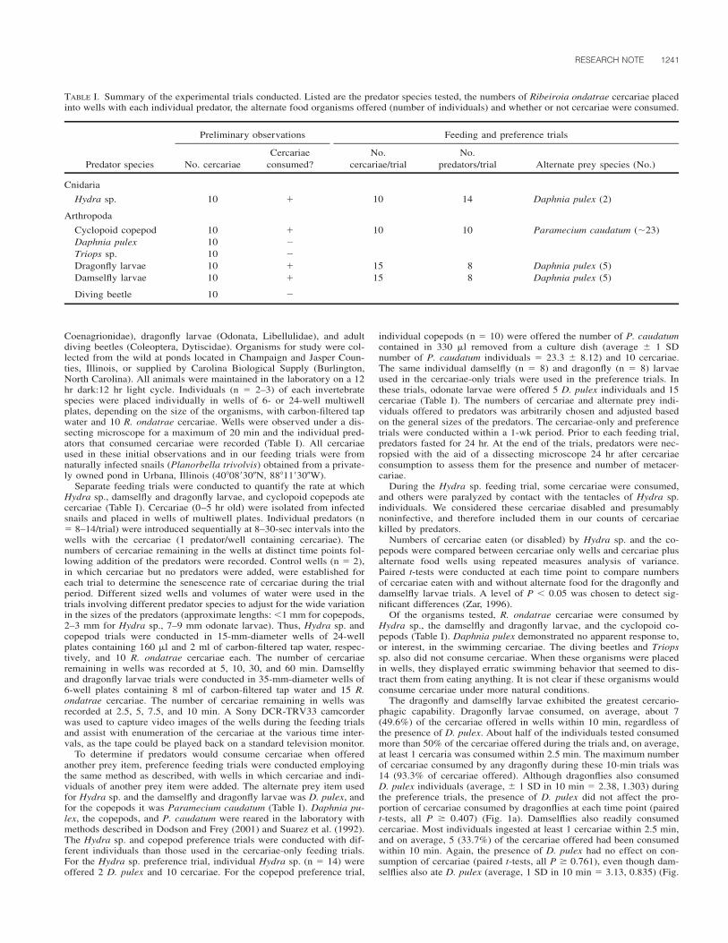
RESEARCH NOTE 1241
TABLE I. Summary of the experimental trials conducted. Listed are the predator species tested, the numbers of Ribeiroia ondatrae cercariae placedinto wells with each individual predator, the alternate food organisms offered (number of individuals) and whether or not cercariae were consumed.
Predator species
Preliminary observations
No. cercariaeCercariae
consumed?
Feeding and preference trials
No.cercariae/trial
No.predators/trial Alternate prey species (No.)
Cnidaria
Hydra sp. 10 10 14 Daphnia pulex (2)
Arthropoda
Cyclopoid copepod 10 10 10 Paramecium caudatum (�23)Daphnia pulex 10 �Triops sp. 10 �Dragonfly larvae 10 15 8 Daphnia pulex (5)Damselfly larvae 10 15 8 Daphnia pulex (5)
Diving beetle 10 �
Coenagrionidae), dragonfly larvae (Odonata, Libellulidae), and adultdiving beetles (Coleoptera, Dytiscidae). Organisms for study were col-lected from the wild at ponds located in Champaign and Jasper Coun-ties, Illinois, or supplied by Carolina Biological Supply (Burlington,North Carolina). All animals were maintained in the laboratory on a 12hr dark:12 hr light cycle. Individuals (n � 2–3) of each invertebratespecies were placed individually in wells of 6- or 24-well multiwellplates, depending on the size of the organisms, with carbon-filtered tapwater and 10 R. ondatrae cercariae. Wells were observed under a dis-secting microscope for a maximum of 20 min and the individual pred-ators that consumed cercariae were recorded (Table I). All cercariaeused in these initial observations and in our feeding trials were fromnaturally infected snails (Planorbella trivolvis) obtained from a private-ly owned pond in Urbana, Illinois (40�08�30�N, 88�11�30�W).
Separate feeding trials were conducted to quantify the rate at whichHydra sp., damselfly and dragonfly larvae, and cyclopoid copepods atecercariae (Table I). Cercariae (0–5 hr old) were isolated from infectedsnails and placed in wells of multiwell plates. Individual predators (n� 8–14/trial) were introduced sequentially at 8–30-sec intervals into thewells with the cercariae (1 predator/well containing cercariae). Thenumbers of cercariae remaining in the wells at distinct time points fol-lowing addition of the predators were recorded. Control wells (n � 2),in which cercariae but no predators were added, were established foreach trial to determine the senescence rate of cercariae during the trialperiod. Different sized wells and volumes of water were used in thetrials involving different predator species to adjust for the wide variationin the sizes of the predators (approximate lengths: �1 mm for copepods,2–3 mm for Hydra sp., 7–9 mm odonate larvae). Thus, Hydra sp. andcopepod trials were conducted in 15-mm-diameter wells of 24-wellplates containing 160 l and 2 ml of carbon-filtered tap water, respec-tively, and 10 R. ondatrae cercariae each. The number of cercariaeremaining in wells was recorded at 5, 10, 30, and 60 min. Damselflyand dragonfly larvae trials were conducted in 35-mm-diameter wells of6-well plates containing 8 ml of carbon-filtered tap water and 15 R.ondatrae cercariae. The number of cercariae remaining in wells wasrecorded at 2.5, 5, 7.5, and 10 min. A Sony DCR-TRV33 camcorderwas used to capture video images of the wells during the feeding trialsand assist with enumeration of the cercariae at the various time inter-vals, as the tape could be played back on a standard television monitor.
To determine if predators would consume cercariae when offeredanother prey item, preference feeding trials were conducted employingthe same method as described, with wells in which cercariae and indi-viduals of another prey item were added. The alternate prey item usedfor Hydra sp. and the damselfly and dragonfly larvae was D. pulex, andfor the copepods it was Paramecium caudatum (Table I). Daphnia pu-lex, the copepods, and P. caudatum were reared in the laboratory withmethods described in Dodson and Frey (2001) and Suarez et al. (1992).The Hydra sp. and copepod preference trials were conducted with dif-ferent individuals than those used in the cercariae-only feeding trials.For the Hydra sp. preference trial, individual Hydra sp. (n � 14) wereoffered 2 D. pulex and 10 cercariae. For the copepod preference trial,
individual copepods (n � 10) were offered the number of P. caudatumcontained in 330 l removed from a culture dish (average � 1 SDnumber of P. caudatum individuals � 23.3 � 8.12) and 10 cercariae.The same individual damselfly (n � 8) and dragonfly (n � 8) larvaeused in the cercariae-only trials were used in the preference trials. Inthese trials, odonate larvae were offered 5 D. pulex individuals and 15cercariae (Table I). The numbers of cercariae and alternate prey indi-viduals offered to predators was arbitrarily chosen and adjusted basedon the general sizes of the predators. The cercariae-only and preferencetrials were conducted within a 1-wk period. Prior to each feeding trial,predators fasted for 24 hr. At the end of the trials, predators were nec-ropsied with the aid of a dissecting microscope 24 hr after cercariaeconsumption to assess them for the presence and number of metacer-cariae.
During the Hydra sp. feeding trial, some cercariae were consumed,and others were paralyzed by contact with the tentacles of Hydra sp.individuals. We considered these cercariae disabled and presumablynoninfective, and therefore included them in our counts of cercariaekilled by predators.
Numbers of cercariae eaten (or disabled) by Hydra sp. and the co-pepods were compared between cercariae only wells and cercariae plusalternate food wells using repeated measures analysis of variance.Paired t-tests were conducted at each time point to compare numbersof cercariae eaten with and without alternate food for the dragonfly anddamselfly larvae trials. A level of P � 0.05 was chosen to detect sig-nificant differences (Zar, 1996).
Of the organisms tested, R. ondatrae cercariae were consumed byHydra sp., the damselfly and dragonfly larvae, and the cyclopoid co-pepods (Table I). Daphnia pulex demonstrated no apparent response to,or interest, in the swimming cercariae. The diving beetles and Triopssp. also did not consume cercariae. When these organisms were placedin wells, they displayed erratic swimming behavior that seemed to dis-tract them from eating anything. It is not clear if these organisms wouldconsume cercariae under more natural conditions.
The dragonfly and damselfly larvae exhibited the greatest cercario-phagic capability. Dragonfly larvae consumed, on average, about 7(49.6%) of the cercariae offered in wells within 10 min, regardless ofthe presence of D. pulex. About half of the individuals tested consumedmore than 50% of the cercariae offered during the trials and, on average,at least 1 cercaria was consumed within 2.5 min. The maximum numberof cercariae consumed by any dragonfly during these 10-min trials was14 (93.3% of cercariae offered). Although dragonflies also consumedD. pulex individuals (average, � 1 SD in 10 min � 2.38, 1.303) duringthe preference trials, the presence of D. pulex did not affect the pro-portion of cercariae consumed by dragonflies at each time point (pairedt-tests, all P � 0.407) (Fig. 1a). Damselflies also readily consumedcercariae. Most individuals ingested at least 1 cercariae within 2.5 min,and on average, 5 (33.7%) of the cercariae offered had been consumedwithin 10 min. Again, the presence of D. pulex had no effect on con-sumption of cercariae (paired t-tests, all P � 0.761), even though dam-selflies also ate D. pulex (average, 1 SD in 10 min � 3.13, 0.835) (Fig.
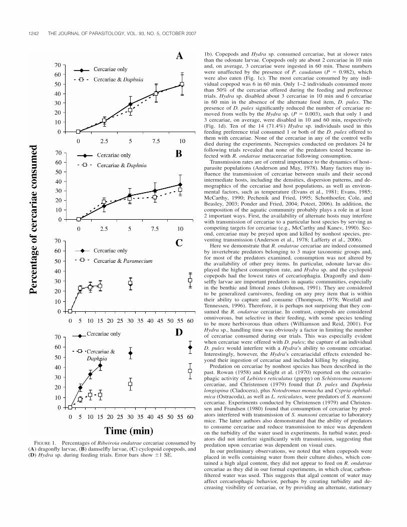
1242 THE JOURNAL OF PARASITOLOGY, VOL. 93, NO. 5, OCTOBER 2007
FIGURE 1. Percentages of Ribeiroia ondatrae cercariae consumed by(A) dragonfly larvae, (B) damselfly larvae, (C) cyclopoid copepods, and(D) Hydra sp. during feeding trials. Error bars show �1 SE.
1b). Copepods and Hydra sp. consumed cercariae, but at slower ratesthan the odonate larvae. Copepods only ate about 2 cercariae in 10 minand, on average, 3 cercariae were ingested in 60 min. These numberswere unaffected by the presence of P. caudatum (P � 0.982), whichwere also eaten (Fig. 1c). The most cercariae consumed by any indi-vidual copepod was 6 in 60 min. Only 1–2 individuals consumed morethan 50% of the cercariae offered during the feeding and preferencetrials. Hydra sp. disabled about 3 cercariae in 10 min and 6 cercariaein 60 min in the absence of the alternate food item, D. pulex. Thepresence of D. pulex significantly reduced the number of cercariae re-moved from wells by the Hydra sp. (P � 0.003), such that only 1 and3 cercariae, on average, were disabled in 10 and 60 min, respectively(Fig. 1d). Ten of the 14 (71.4%) Hydra sp. individuals used in thisfeeding preference trial consumed 1 or both of the D. pulex offered tothem with cercariae. None of the cercariae in any of the control wellsdied during the experiments. Necropsies conducted on predators 24 hrfollowing trials revealed that none of the predators tested became in-fected with R. ondatrae metacercariae following consumption.
Transmission rates are of central importance to the dynamics of host–parasite populations (Anderson and May, 1978). Many factors may in-fluence the transmission of cercariae between snails and their secondintermediate hosts, including the densities, dispersion patterns, and de-mographics of the cercariae and host populations, as well as environ-mental factors, such as temperature (Evans et al., 1981; Evans, 1985;McCarthy, 1990; Pechenik and Fried, 1995; Schotthoefer, Cole, andBeasley, 2003; Ponder and Fried, 2004; Poteet, 2006). In addition, thecomposition of the aquatic community probably plays a role in at least2 important ways. First, the availability of alternate hosts may interferewith transmission of cercariae to a particular host species by serving ascompeting targets for cercariae (e.g., McCarthy and Kanev, 1990). Sec-ond, cercariae may be preyed upon and killed by nonhost species, pre-venting transmission (Anderson et al., 1978; Lafferty et al., 2006).
Here we demonstrate that R. ondatrae cercariae are indeed consumedby invertebrate predators belonging to 3 major taxonomic groups and,for most of the predators examined, consumption was not altered bythe availability of other prey items. In particular, odonate larvae dis-played the highest consumption rate, and Hydra sp. and the cyclopoidcopepods had the lowest rates of cercariophagia. Dragonfly and dam-selfly larvae are important predators in aquatic communities, especiallyin the benthic and littoral zones (Johnson, 1991). They are consideredto be generalized carnivores, feeding on any prey item that is withintheir ability to capture and consume (Thompson, 1978; Westfall andTennessen, 1996). Therefore, it is perhaps not surprising that they con-sumed the R. ondatrae cercariae. In contrast, copepods are consideredomnivorous, but selective in their feeding, with some species tendingto be more herbivorous than others (Williamson and Reid, 2001). ForHydra sp., handling time was obviously a factor in limiting the numberof cercariae consumed during our trials. This was especially evidentwhen cercariae were offered with D. pulex; the capture of an individualD. pulex would interfere with a Hydra’s ability to consume cercariae.Interestingly, however, the Hydra’s cercariacidal effects extended be-yond their ingestion of cercariae and included killing by stinging.
Predation on cercariae by nonhost species has been described in thepast. Rowan (1958) and Knight et al. (1970) reported on the cercario-phagic activity of Lebistes reticulatus (guppy) on Schistosoma mansonicercariae, and Christensen (1979) found that D. pulex and Daphnialongispina (Cladocera), plus Notodromas monacha and Cypria ophthal-mica (Ostracoda), as well as L. reticulates, were predators of S. mansonicercariae. Experiments conducted by Christensen (1979) and Christen-sen and Frandsen (1980) found that consumption of cercariae by pred-ators interfered with transmission of S. mansoni cercariae to laboratorymice. The latter authors also demonstrated that the ability of predatorsto consume cercariae and reduce transmission to mice was dependenton the turbidity of the water used in experiments. In turbid water, pred-ators did not interfere significantly with transmission, suggesting thatpredation upon cercariae was dependent on visual cues.
In our preliminary observations, we noted that when copepods wereplaced in wells containing water from their culture dishes, which con-tained a high algal content, they did not appear to feed on R. ondatraecercariae as they did in our formal experiments, in which clear, carbon-filtered water was used. This suggests that algal content of water mayaffect cercariophagic behavior, perhaps by creating turbidity and de-creasing visibility of cercariae, or by providing an alternate, stationary

RESEARCH NOTE 1243
food source that requires less effort to acquire. The effects of environ-mental factors, including water algal content, plant content, turbidity,and shade on cercariophagic activity, are worthy of further investigation.In addition to environmental factors, there are likely species-specifictraits that may influence a predator’s cercariophagic ability, i.e., foragingbehavior, and microhabitat preferences, which we did not examine here,but which would provide important information on the predators mostlikely to influence cercariae transmission in nature. Moreover, the rel-ative densities of potential predators versus the densities of cercariaeand other prey items will probably also play a role in determining whichpredators are most important in different wetland settings. We arbitrarilychose the densities of 10 or 15 cercariae to feed to the predators in ourtrials, and conducted all trials in the evenings when cercariae wereavailable from shedding snails. Obviously, the cercariophagic ability ofdifferent predators in nature may vary depending on the densities ofcercariae available to them in their microhabitats during their activeforaging times of the day. Further investigation of the cercariophagicactivity of potential cercariae predators in the context of environmentaland predatory–prey community dynamics will provide a framework bywhich to group likely predators and unlikely predators.
The observations reported here suggest the loss of R. ondatrae cer-cariae through predation may be an important regulatory function intransmission of cercariae to tadpoles in nature, and therefore may affectthe occurrence of limb deformities in amphibian populations. We pro-pose that there is need to examine determinants of cercariophagic ac-tivity by aquatic predators to assess the significance of such processesin protecting intermediate and definitive vertebrate hosts from trematodeinfections. The relevance of cercariophagic activity to the transmissiondynamics of cercariae under natural versus anthropogenically alteredconditions also needs to be explored.
We would like to thank K. Tadin, M. Wilken, and J. Clennon forlaboratory assistance, U. Kitron for use of laboratory space, and C.Caceres for Daphnia pulex donations. Financial support was providedto K.M.L. by the Center for Zoonoses Summer Research Training Pro-gram at the University of Illinois at Urbana-Champaign.
LITERATURE CITED
ANDERSON, R. M., AND R. M. MAY. 1978. Regulation and stability ofhost–parasite population interactions. I. Regulatory processes. Jour-nal of Animal Ecology 47: 219–249.
———, P. J. WHITFIELD, A. P. DOBSON, AND A. E. KEYMER. 1978. Con-comitant predation and infection processes: An experimental study.Journal of Animal Ecology 47: 891–911.
BLAUSTEIN, A. R., AND P. T. J. JOHNSON. 2003. The complexity of de-formed amphibians. Frontiers in Ecology and the Environment 1:87–94.
CHRISTENSEN, N. Ø. 1979. Schistosoma mansoni: Interference with cer-carial host-finding by various aquatic organisms. Journal of Hel-minthology 53: 7–14.
———, AND F. FRANDSEN. 1980. The interaction of some environmentalfactors influencing Schistosoma mansoni cercarial host-finding.Journal of Helminthology 54: 203–205.
DODSON, S. I., AND D. G. FREY. 2001. Cladocera and other Branchio-poda. In Ecology and classification of North American freshwaterinvertebrates, J. H. Thorp and A. P. Covich (eds.). American Press,San Diego, California, p. 850–913.
EVANS, N. A. 1985. Experimental studies on transmission dynamics ofthe cercariae of Echinoparyphium recurvatum (Digenea: Echinos-tomatidae). Parasitology 87: 167–174.
———, P. J. WHITFIELD, AND A. P. DOBSON. 1981. Parasite utilizationof a host community: The distribution and occurrence of metacer-carial cysts of Echinoparyphium recurvatum (Digenea; Echinosto-matidae) in seven species of mollusc at Harting Pond, Sussex. Par-asitology 83: 1–12.
JOHNSON, D. M. 1991. Behavioral ecology of larval dragonflies anddamselflies. Trends in Ecology and Evolution 6: 8–13.
JOHNSON, P. T. J., K. B. LUNDE, E. G. RITCHIE, AND A. E. LAUNER. 1999.The effect of trematode infection on amphibian limb developmentand survivorship. Science 284: 802–804.
———, K. B. LUNDE, E. M. THURMAN, E. G. RITCHIE, S. W. WRAY, D.R. SUTHERLAND, J. M. KAPFER, T. J. FREST, J. BOWERMAN, AND A.
R. BLAUSTEIN. 2002. Parasite (Ribeiroia ondatrae) infection linkedto amphibian malformations in the western United States. Ecolog-ical Monographs 72: 151–168.
———, ———, D. A. ZELMER, AND J. K. WERNER. 2003. Limb defor-mities as an emerging parasitic disease in amphibians: Evidencefrom museum specimens and resurvey data. Conservation Biology17: 1724–1737.
———, D. R. SUTHERLAND, J. M. KINSELLA, AND K. B. LUNDE. 2004.Review of the trematode genus Ribeiroia (Psilostomidae): Ecology,life history and pathogenesis with special emphasis on the amphib-ian malformation problem. Advances in Parasitology 57: 191–253.
KNIGHT, W. B., L. S. RITCHIE, F. LIARD, AND J. CHIRIBOGA. 1970. Cer-cariophagic activity of guppy fish (Lebistes reticulatus) detected bycercariae labeled with radioselenium (75 SE). American Journal ofTropical Medicine and Hygiene 19: 620–625.
LAFFERTY, K. D., R. F. HECHINGER, J. C. SHAW, K. WHITNEY, AND A.M. KURIS. 2006. Food webs and parasites in a salt marsh ecosys-tem. In Disease ecology: Community structure and pathogen dy-namics, S. K. Collinge and C. Ray (eds.). Oxford University Press,New York, New York, p. 119–134.
MCCARTHY, A. M. 1990. The influence of second intermediate hostdispersion pattern upon the transmission of cercariae of Echino-paryphium recurvatum (Digenea: Echinostomatidae). Parasitology101: 43–47.
———, AND I. KANEV. 1990. Pseudechinoparyphium echinatum (Di-genea: Echinostomatidae): Experimental observations on cercarialspecificity toward second intermediate hosts. Parasitology 100:423–428.
PECHENIK, J. A., AND B. FRIED. 1995. Effect of temperature on survivaland infectivity of Echinostoma trivolvis cercariae: A test of theenergy limitation hypothesis. Parasitology 111: 373–378.
PONDER, E. L., AND B. FRIED. 2004. Effects of snail size and diet onencystment of Echinostoma caproni in juvenile Helisoma trivolvis(Colorado strain) and observations on survival of infected snails.Journal of Parasitology 90: 422–424.
POTEET, M. F. 2006. Shifting roles of abiotic and biotic regulation of amulti-host parasite following disturbance. In Disease ecology:Community structure and pathogen dynamics, S. K. Collinge andC. Ray (eds.). Oxford University Press, New York, New York, p.135–152.
ROWAN, W. B. 1958. Daily periodicity of Schistosoma mansoni cercariaein Puerto Rican waters. American Journal of Tropical Medicine andHygiene 7: 374–381.
SCHOTTHOEFER, A. M., A. V. KOEHLER, C. U. METEYER, AND R. A. COLE.2003. Influence of Ribeiroia ondatrae (Trematoda : Digenea) in-fection on limb development and survival of northern leopard frogs(Rana pipiens): Effects of host stage and parasite-exposure level.Canadian Journal of Zoology 81: 1144–1153.
———, R. A. COLE, AND V. R. BEASLEY. 2003. Relationship of tadpolestage to location of echinostome cercariae encystment and the con-sequences for tadpole survival. Journal of Parasitology 89: 475–482.
SUAREZ, M. F., G. G. MARTIN, AND G. G. CLARK. 1992. A simple methodfor cultivating freshwater copepods used in biological control ofAedes aegypti. Journal of the American Mosquito Control Asso-ciation 8: 409–412.
TAYLOR, B., D. SKELLY, L. K. DEMARCHIS, M. D. SLADE, D. GALUSHA,AND P. M. RABINOWITZ. 2006. Proximity to pollution sources andrisk of amphibian limb malformation. Environmental Health Per-spectives 113: 1497–1500.
THOMPSON, D. J. 1978. Prey size selection by larvae of the damselfly,Ischnura elegans (Odonata). Ecology 47: 769–785.
WESTFALL, M. J., JR., AND K. J. TENNESSEN. 1996. Odonata. In An in-troduction to the aquatic insects of North America, R. W. Merrittand K. W. Cummins (eds.). Kendall/Hunt Publishing, Dubuque,Iowa, p. 164–211.
WILLIAMSON, C. E., AND J. W. REID. 2001. Copepoda. In Ecology andclassification of North American freshwater invertebrates, J. H.Thorp and A. P. Covich (eds.). American Press, San Diego, Cali-fornia, p. 915–954.
ZAR, J. H. 1996. Biostatistical analysis. Prentice Hall, Upper SaddleRiver, New Jersey, 662 p.
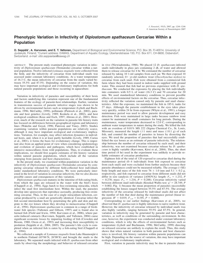
1244 THE JOURNAL OF PARASITOLOGY, VOL. 93, NO. 5, OCTOBER 2007
J. Parasitol., 93(5), 2007, pp. 1244–1246� American Society of Parasitologists 2007
Phenotypic Variation in Infectivity of Diplostomum spathaceum Cercariae Within aPopulation
O. Seppala*, A. Karvonen, and E. T. Valtonen, Department of Biological and Environmental Science, P.O. Box 35, FI-40014, University ofJyvaskyla, Finland; *Current address: EAWAG, Department of Aquatic Ecology, Uberlandstrasse 133, P.O. Box 611, CH-8600, Dubendorf,Switzerland. e-mail: [email protected]
ABSTRACT: The present study examined phenotypic variation in infec-tivity of Diplostomum spathaceum (Trematoda) cercariae within a nat-ural population. Twelve infected Lymnaea stagnalis were collected fromthe field, and the infectivity of cercariae from individual snails wasassessed under constant laboratory conditions. At a water temperatureof 16.3 C, the mean infectivity of cercariae from the snails varied be-tween 55.5% and 87.5%. Depending on the source of variation, thismay have important ecological and evolutionary implications for bothnatural parasite populations and those occurring in aquaculture.
Variation in infectivity of parasites and susceptibility of their hostsand factors underlying this variation represent one of the fundamentalfeatures of the ecology of parasite-host relationships. Earlier, variationin transmission success of parasite infective stages was shown to bedriven by environmental factors such as temperature (Lyholt and Buch-mann, 1996; McCarthy, 1999), pollutants (Pietrock et al., 2002), para-site and host genetics (Lively, 1989; Koskela et al., 2002), and hostecological condition (Keas and Esch, 1997; Abrous et al., 2001). How-ever, much of the research on the variation in parasite life history traitshas focused on differences between geographical regions and laboratorylines (Wakelin and Goyal, 1996; Paterson and Viney, 2003). Studiesexamining variation within parasite populations are relatively scarce,although it may have important ecological and evolutionary implica-tions. Variation, for example, may affect infection dynamics in individ-ual hosts, and, when it is at least partly heritable (Lively, 1989; Cariuset al., 2001), it forms the basis for evolutionary change. This is impor-tant also from an applied point of view when considering epidemiologyand evolution of parasites and pathogens, which have established inintensive monocultures from wild populations. Thus, to evaluate theseimplications, we should first acquire estimates of life history variationwithin natural parasite populations, which include all the variationemerging from parasite and host characteristics.
In the present study, we examined within-population variation in theinfectivity of Diplostomum spathaceum (Trematoda) cercariae by com-paring cercariae released by different field-collected host individualsunder standardized laboratory conditions. We were particularly inter-ested in the level of variation in cercariae infectivity, but we also discusspossible causes and consequences of this variation.
Diplostomum spathaceum matures in the intestine of fish-eating birds,from which the eggs are released in the water with the bird’s feces(Chappell et al., 1994). Eggs hatch to free-swimming miracidia, whichinfect the snail first intermediate host. Within the snail, the parasitesdevelop into sporocysts that reproduce asexually, producing free-swim-ming cercariae. Thus, in the case of a single miracidium infection, cer-cariae produced by a snail are genetically identical. Cercariae infect thefish second intermediate host by penetrating the gills and skin and mi-grate to the eye lenses where they develop to metacercariae (Chappellet al., 1994). Diplostomum spathaceum is common in a range of fresh-water fish species (Valtonen and Gibson, 1997) and is found also infarmed fish (Field and Irwin, 1994; Karvonen et al., 2006), where par-asite-induced cataracts (Karvonen, Seppala, and Valtonen, 2004) causesignificant economic losses by impairing feeding of fish (Crowden andBroom, 1980; Owen et al., 1993) and increasing their susceptibility topredation (Seppala et al., 2004, 2005). The parasite’s life cycle is com-pleted when an infected fish is eaten by a fish-eating bird (Chappell etal., 1994).
We collected a sample of Lymnaea stagnalis from Lake Huumonjarviin northern Finland (65�06�N, 26�08�E) and brought the snails to thelaboratory. We separated snails infected with D. spathaceum from othersnails by observing the morphology and behavior of released cercariae
in vivo (Niewiadomska, 1986). We placed 12 D. spathaceum–infectedsnails individually in glass jars containing 2 dL of water and allowedthem to release cercariae for 4 hr. We estimated the number of cercariaereleased by taking 10 1-ml samples from each jar. We then exposed 10randomly selected, 0 yr-old rainbow trout (Oncorhynchus mykiss) tocercariae from each snail. Fish were obtained from a commercial fishfarm where they had been reared in indoor tanks supplied with groundwater. This ensured that fish had no previous experience with D. spa-thaceum. We conducted the exposures by placing the fish individuallyinto containers with 0.5 L of water (16.3 C) and 70 cercariae for 20min. We used standardized laboratory conditions to prevent possibleeffects of environmental factors on the cercariae. Thus, cercariae infec-tivity reflected the variation caused only by parasite and snail charac-teristics. After the exposure, we maintained the fish in 185-L tanks for27 days. Although the parasite establishment in the eyes takes placevery quickly, within 24 hr from exposure (Whyte et al., 1991), a longermaintenance period was used to allow parasite development and easydetection. Fish were maintained in large tanks because rainbow troutcannot be maintained in small containers for long periods. During themaintenance, water temperature decreased to 12.0 C, corresponding toambient water temperature in nature during the study. We killed the fishwith an overdose of 0.01% MS 222 (Sigma Chemical Co., St. Louis,Missouri), measured the length (�1 mm) and mass (�0.1 g) of eachfish, and counted the number of parasites in lenses by dissecting theeyes. We used the proportion of parasites that had successfully estab-lished the eye lenses as an estimate of parasite infectivity. The relation-ship between the number of cercariae released by each snail, and theirinfectivity, was not examined because cercariae release by D. spatha-ceum is highly variable (Karvonen, Kirsi et al., 2004). Thus, the shorttime period (4 hr) used in the experiment was unlikely to provide re-liable estimates of cercariae production.
Eighteen fish of the total of 120 exposed to cercariae died during themaintenance period (0–4 individuals from fish exposed to cercariaefrom each snail) and were excluded from further analysis because theirparasite abundances could not be measured reliably. The average (�SE)body length and mass of the fish were 76 � 1.0 mm and 5.1 � 0.2 g,respectively, and fish exposed to cercariae from different snails did notdiffer in size (analysis of variance [ANOVA]: length: F11 � 1.232, P� 0.278; mass: F11 � 1.216, P � 0.288). Cercariae infectivity variedbetween different snail individuals (Kruskal-Wallis test: �2 � 28.749, P� 0.002; Fig. 1) because the mean proportion of parasites successfullyestablishing the lenses ranged between 55.5% and 87.5%. The averageinfectivity of the cercariae released by individual snails did not affectthe number of fish that died during the maintenance period (linear re-gression: R2 � 0.019, F1,10 � 0.195, P � 0.668).
Corresponding to our earlier findings (Karvonen et al., 2005), weobserved that D. spathaceum is highly infectious to naıve rainbow trout.However, the infectivity of cercariae released by individual snails wasalso highly variable, ranging between 55.5% and 87.5%. In general,variation in infectivity may be generated by parasite and host charac-teristics, as well as conditions of the surrounding environment. In thiscase, however, the experiment was conducted under constant laboratoryconditions, which is why the effects of environmental factors such astemperature (Lyholt and Buchmann, 1996; McCarthy, 1999) directingon released cercariae are unlikely to explain the result. Thus, this studyshows that when natural variation in both parasite and host character-istics is involved, life history variation within parasite populations canbe high. Depending on the source of variation, this may have importantecological and evolutionary implications.
First, variation in parasite infectivity may be due to parasite charac-
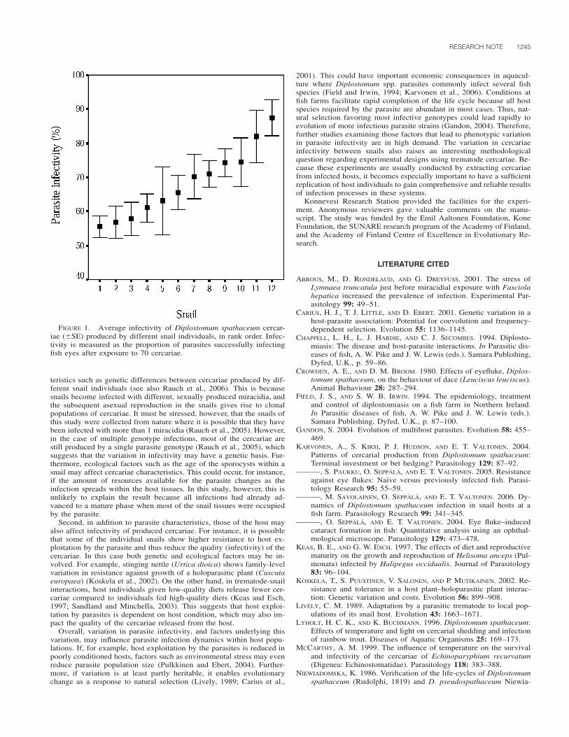
RESEARCH NOTE 1245
FIGURE 1. Average infectivity of Diplostomum spathaceum cercar-iae (�SE) produced by different snail individuals, in rank order. Infec-tivity is measured as the proportion of parasites successfully infectingfish eyes after exposure to 70 cercariae.
teristics such as genetic differences between cercariae produced by dif-ferent snail individuals (see also Rauch et al., 2006). This is becausesnails become infected with different, sexually produced miracidia, andthe subsequent asexual reproduction in the snails gives rise to clonalpopulations of cercariae. It must be stressed, however, that the snails ofthis study were collected from nature where it is possible that they havebeen infected with more than 1 miracidia (Rauch et al., 2005). However,in the case of multiple genotype infections, most of the cercariae arestill produced by a single parasite genotype (Rauch et al., 2005), whichsuggests that the variation in infectivity may have a genetic basis. Fur-thermore, ecological factors such as the age of the sporocysts within asnail may affect cercariae characteristics. This could occur, for instance,if the amount of resources available for the parasite changes as theinfection spreads within the host tissues. In this study, however, this isunlikely to explain the result because all infections had already ad-vanced to a mature phase when most of the snail tissues were occupiedby the parasite.
Second, in addition to parasite characteristics, those of the host mayalso affect infectivity of produced cercariae. For instance, it is possiblethat some of the individual snails show higher resistance to host ex-ploitation by the parasite and thus reduce the quality (infectivity) of thecercariae. In this case both genetic and ecological factors may be in-volved. For example, stinging nettle (Urtica dioica) shows family-levelvariation in resistance against growth of a holoparasitic plant (Cuscutaeuropaea) (Koskela et al., 2002). On the other hand, in trematode-snailinteractions, host individuals given low-quality diets release fewer cer-cariae compared to individuals fed high-quality diets (Keas and Esch,1997; Sandland and Minchella, 2003). This suggests that host exploi-tation by parasites is dependent on host condition, which may also im-pact the quality of the cercariae released from the host.
Overall, variation in parasite infectivity, and factors underlying thisvariation, may influence parasite infection dynamics within host popu-lations. If, for example, host exploitation by the parasites is reduced inpoorly conditioned hosts, factors such as environmental stress may evenreduce parasite population size (Pulkkinen and Ebert, 2004). Further-more, if variation is at least partly heritable, it enables evolutionarychange as a response to natural selection (Lively, 1989; Carius et al.,
2001). This could have important economic consequences in aquacul-ture where Diplostomum spp. parasites commonly infect several fishspecies (Field and Irwin, 1994; Karvonen et al., 2006). Conditions atfish farms facilitate rapid completion of the life cycle because all hostspecies required by the parasite are abundant in most cases. Thus, nat-ural selection favoring most infective genotypes could lead rapidly toevolution of more infectious parasite strains (Gandon, 2004). Therefore,further studies examining those factors that lead to phenotypic variationin parasite infectivity are in high demand. The variation in cercariaeinfectivity between snails also raises an interesting methodologicalquestion regarding experimental designs using trematode cercariae. Be-cause these experiments are usually conducted by extracting cercariaefrom infected hosts, it becomes especially important to have a sufficientreplication of host individuals to gain comprehensive and reliable resultsof infection processes in these systems.
Konnevesi Research Station provided the facilities for the experi-ment. Anonymous reviewers gave valuable comments on the manu-script. The study was funded by the Emil Aaltonen Foundation, KoneFoundation, the SUNARE research program of the Academy of Finland,and the Academy of Finland Centre of Excellence in Evolutionary Re-search.
LITERATURE CITED
ABROUS, M., D. RONDELAUD, AND G. DREYFUSS. 2001. The stress ofLymnaea truncatula just before miracidial exposure with Fasciolahepatica increased the prevalence of infection. Experimental Par-asitology 99: 49–51.
CARIUS, H. J., T. J. LITTLE, AND D. EBERT. 2001. Genetic variation in ahost-parasite association: Potential for coevolution and frequency-dependent selection. Evolution 55: 1136–1145.
CHAPPELL, L. H., L. J. HARDIE, AND C. J. SECOMBES. 1994. Diplosto-miasis: The disease and host-parasite interactions. In Parasitic dis-eases of fish, A. W. Pike and J. W. Lewis (eds.). Samara Publishing,Dyfed, U.K., p. 59–86.
CROWDEN, A. E., AND D. M. BROOM. 1980. Effects of eyefluke, Diplos-tomum spathaceum, on the behaviour of dace (Leuciscus leuciscus).Animal Behaviour 28: 287–294.
FIELD, J. S., AND S. W. B. IRWIN. 1994. The epidemiology, treatmentand control of diplostomiasis on a fish farm in Northern Ireland.In Parasitic diseases of fish, A. W. Pike and J. W. Lewis (eds.).Samara Publishing, Dyfed, U.K., p. 87–100.
GANDON, S. 2004. Evolution of multihost parasites. Evolution 58: 455–469.
KARVONEN, A., S. KIRSI, P. J. HUDSON, AND E. T. VALTONEN. 2004.Patterns of cercarial production from Diplostomum spathaceum:Terminal investment or bet hedging? Parasitology 129: 87–92.
———, S. PAUKKU, O. SEPPALA, AND E. T. VALTONEN. 2005. Resistanceagainst eye flukes: Naıve versus previously infected fish. Parasi-tology Research 95: 55–59.
———, M. SAVOLAINEN, O. SEPPALA, AND E. T. VALTONEN. 2006. Dy-namics of Diplostomum spathaceum infection in snail hosts at afish farm. Parasitology Research 99: 341–345.
———, O. SEPPALA, AND E. T. VALTONEN. 2004. Eye fluke–inducedcataract formation in fish: Quantitative analysis using an ophthal-mological microscope. Parasitology 129: 473–478.
KEAS, B. E., AND G. W. ESCH. 1997. The effects of diet and reproductivematurity on the growth and reproduction of Helisoma anceps (Pul-monata) infected by Halipegus occidualis. Journal of Parasitology83: 96–104.
KOSKELA, T., S. PUUSTINEN, V. SALONEN, AND P. MUTIKAINEN. 2002. Re-sistance and tolerance in a host plant–holoparasitic plant interac-tion: Genetic variation and costs. Evolution 56: 899–908.
LIVELY, C. M. 1989. Adaptation by a parasitic trematode to local pop-ulations of its snail host. Evolution 43: 1663–1671.
LYHOLT, H. C. K., AND K. BUCHMANN. 1996. Diplostomum spathaceum:Effects of temperature and light on cercarial shedding and infectionof rainbow trout. Diseases of Aquatic Organisms 25: 169–173.
MCCARTHY, A. M. 1999. The influence of temperature on the survivaland infectivity of the cercariae of Echinoparyphium recurvatum(Digenea: Echinostomatidae). Parasitology 118: 383–388.
NIEWIADOMSKA, K. 1986. Verification of the life-cycles of Diplostomumspathaceum (Rudolphi, 1819) and D. pseudospathaceum Niewia-

1246 THE JOURNAL OF PARASITOLOGY, VOL. 93, NO. 5, OCTOBER 2007
domska, 1984 (Trematoda, Diplostomidae). Systematic Parasitolo-gy 8: 23–31.
OWEN, S. F., I. BARBER, AND P. J. B. HART. 1993. Low level infectionby eye fluke, Diplostomum spp., affects the vision of three-spinedsticklebacks, Gasterosteus aculeatus. Journal of Fish Biology 42:803–806.
PATERSON, S., AND M. E. VINEY. 2003. Functional consequences of ge-netic diversity in Strongyloides ratti infections. Proceedings of theRoyal Society of London B 270: 1023–1032.
PIETROCK, M., D. J. MARCOGLIESE, T. MEINELT, AND J. D. MCLAUGHLIN.2002. Effects of mercury and chromium upon longevity of Diplos-tomum sp. (Trematoda: Diplostomidae) cercariae. Parasitology Re-search 88: 225–229.
PULKKINEN, K., AND D. EBERT. 2004. Host starvation decreases parasiteload and mean host size in experimental populations. Ecology 85:823–833.
RAUCH, G., M. KALBE, AND T. B. H. REUSCH. 2005. How a complex lifecycle can improve a parasite’s sex life. Journal of EvolutionaryBiology 18: 1069–1075.
———, ———, AND ———. 2006. One day is enough: Rapid and
specific host-parasite interactions in a stickleback-trematode sys-tem. Biology Letters 2: 382–384.
SANDLAND, G. J., AND D. J. MINCHELLA. 2003. Effects of diet and Echi-nostoma revolutum infection on energy allocation patterns in ju-venile Lymnaea elodes snails. Oecologia 134: 479–486.
SEPPALA, O., A. KARVONEN, AND E. T. VALTONEN. 2004. Parasite-in-duced change in host behaviour and susceptibility to predation inan eye fluke–fish interaction. Animal Behaviour 68: 257–263.
———, ———, AND ———. 2005. Manipulation of fish host by eyeflukes in relation to cataract formation and parasite infectivity. An-imal Behaviour 70: 889–894.
VALTONEN, E. T., AND D. I. GIBSON. 1997. Aspects of the biology ofdiplostomid metacercarial (Digenea) populations occurring in fishesin different localities of northern Finland. Annals Zoologici Fennici34: 47–59.
WAKELIN, D., AND P. K. GOYOL. 1996. Trichinella isolates: Parasite var-iability and host responses. International Journal for Parasitology26: 471–481.
WHYTE, S. K., C. J. SECOMBES, AND L. H. CHAPPELL. 1991. Studies onthe infectivity of Diplostomum spathaceum in rainbow trout (On-corhynchus mykiss). Journal of Helminthology 65: 169–178.