Characterization of a novel organelle in Toxoplasma gondii with similar composition and function to...
34
Characterization of a novel organelle in Toxoplasma gondii with similar composition and function to the plant vacuole Kildare Miranda 1,2,* , Douglas A. Pace 1,* , Roxana Cintron 1 , Juliany C.F. Rodrigues 1,2 , Jianmin Fang 1 , Alyssa Smith 1 , Peter Rohloff 1 , Elvis Coelho 2 , Felix de Haas 3 , Wanderley de Souza 2 , Isabelle Coppens 4 , L. David Sibley 5 , and Silvia N. J. Moreno 1,# 1 Center for Tropical and Emerging Global Diseases and Department of Cellular Biology, University of Georgia, Athens, GA 30602 2 Instituto de Biofisica Carlos Chagas Filho, Universidade Federal do Rio de Janeiro, Rio de Janeiro 21941, Brazil 3 FEI Nanoport, Eindhoven, The Netherlands 4 Department of Molecular Microbiology and Immunology, Johns Hopkins University Bloomberg School of Public Health, Baltimore, MD 21205 5 Department of Molecular Microbiology, Washington University School of Medicine, St. Louis, MO 63110 Abstract Toxoplasma gondii belongs to the phylum Apicomplexa and is an important cause of congenital disease and infection in immunocompromised patients. Like most apicomplexans, Toxoplasma gondii possesses several plant-like features, such as the chloroplast-like organelle, the apicoplast. We describe and characterize a novel organelle in T. gondii tachyzoites, which is visible by light microscopy, and possesses a broad similarity to the plant vacuole. Electron tomography shows the interaction of this vacuole with other organelles. The presence of a plant-like vacuolar proton pyrophosphatase (TgVP1), a vacuolar proton ATPase, a cathepsin L-like protease (TgCPL), an aquaporin (TgAQP1), as well as Ca 2+ /H + and Na + /H + exchange activities, supports similarity to the plant vacuole. Biochemical characterization of TgVP1 in enriched fractions shows a functional similarity to the respective plant enzyme. The organelle is a Ca 2+ store and appears to have protective effects against salt stress potentially linked to its sodium transport activity. In intracellular parasites, the organelle fragments, with some markers co-localizing with the late endosomal marker, Rab7, suggesting its involvement with the endocytic pathway. Studies on the characterization of this novel organelle will be relevant to the identification of novel targets for chemotherapy against T. gondii and other apicomplexan parasites as well. Introduction Toxoplasma gondii is a protist parasite that causes widespread infection in humans and has been recognized as a major opportunistic pathogen of immunocompromised patients. Additionally, first time infection with T. gondii of pregnant women poses a significant risk to the developing fetus. As a member of the phylum Apicomplexa, T. gondii possesses a distinct apical complex consisting of different types of secretory organelles, such as micronemes, dense granules and rhoptries, these latter organelles being acidic (Shaw et al., # To whom correspondence should be addressed: Center for Tropical and Emerging Global Disease and Department of Cellular Biology, 350A Paul D. Coverdell Center, University of Georgia, Athens, GA 30602. Tel.: 706-542-4736; Fax: 706-542-9493; [email protected]. * Both authors contributed equally The name “Plant-Like Vacuole” or PLV was a recommendation of the participants of the 10 th International Congress on Toxoplasmosis, in Kerkrade, the Netherlands, June 19–23, 2009. NIH Public Access Author Manuscript Mol Microbiol. Author manuscript; available in PMC 2011 June 1. Published in final edited form as: Mol Microbiol. 2010 June 1; 76(6): 1358–1375. doi:10.1111/j.1365-2958.2010.07165.x. NIH-PA Author Manuscript NIH-PA Author Manuscript NIH-PA Author Manuscript
-
Upload
hms-harvard -
Category
Documents
-
view
2 -
download
0
Transcript of Characterization of a novel organelle in Toxoplasma gondii with similar composition and function to...

Characterization of a novel organelle in Toxoplasma gondii withsimilar composition and function to the plant vacuole
Kildare Miranda1,2,*, Douglas A. Pace1,*, Roxana Cintron1, Juliany C.F. Rodrigues1,2,Jianmin Fang1, Alyssa Smith1, Peter Rohloff1, Elvis Coelho2, Felix de Haas3, Wanderleyde Souza2, Isabelle Coppens4, L. David Sibley5, and Silvia N. J. Moreno1,#
1Center for Tropical and Emerging Global Diseases and Department of Cellular Biology,University of Georgia, Athens, GA 30602 2Instituto de Biofisica Carlos Chagas Filho,Universidade Federal do Rio de Janeiro, Rio de Janeiro 21941, Brazil 3FEI Nanoport, Eindhoven,The Netherlands 4Department of Molecular Microbiology and Immunology, Johns HopkinsUniversity Bloomberg School of Public Health, Baltimore, MD 21205 5Department of MolecularMicrobiology, Washington University School of Medicine, St. Louis, MO 63110
AbstractToxoplasma gondii belongs to the phylum Apicomplexa and is an important cause of congenitaldisease and infection in immunocompromised patients. Like most apicomplexans, Toxoplasmagondii possesses several plant-like features, such as the chloroplast-like organelle, the apicoplast.We describe and characterize a novel organelle in T. gondii tachyzoites, which is visible by lightmicroscopy, and possesses a broad similarity to the plant vacuole. Electron tomography shows theinteraction of this vacuole with other organelles. The presence of a plant-like vacuolar protonpyrophosphatase (TgVP1), a vacuolar proton ATPase, a cathepsin L-like protease (TgCPL), anaquaporin (TgAQP1), as well as Ca2+/H+ and Na+/H+ exchange activities, supports similarity tothe plant vacuole. Biochemical characterization of TgVP1 in enriched fractions shows a functionalsimilarity to the respective plant enzyme. The organelle is a Ca2+ store and appears to haveprotective effects against salt stress potentially linked to its sodium transport activity. Inintracellular parasites, the organelle fragments, with some markers co-localizing with the lateendosomal marker, Rab7, suggesting its involvement with the endocytic pathway. Studies on thecharacterization of this novel organelle will be relevant to the identification of novel targets forchemotherapy against T. gondii and other apicomplexan parasites as well.
IntroductionToxoplasma gondii is a protist parasite that causes widespread infection in humans and hasbeen recognized as a major opportunistic pathogen of immunocompromised patients.Additionally, first time infection with T. gondii of pregnant women poses a significant riskto the developing fetus. As a member of the phylum Apicomplexa, T. gondii possesses adistinct apical complex consisting of different types of secretory organelles, such asmicronemes, dense granules and rhoptries, these latter organelles being acidic (Shaw et al.,
#To whom correspondence should be addressed: Center for Tropical and Emerging Global Disease and Department of CellularBiology, 350A Paul D. Coverdell Center, University of Georgia, Athens, GA 30602. Tel.: 706-542-4736; Fax: 706-542-9493;[email protected].*Both authors contributed equallyThe name “Plant-Like Vacuole” or PLV was a recommendation of the participants of the 10th International Congress onToxoplasmosis, in Kerkrade, the Netherlands, June 19–23, 2009.
NIH Public AccessAuthor ManuscriptMol Microbiol. Author manuscript; available in PMC 2011 June 1.
Published in final edited form as:Mol Microbiol. 2010 June 1; 76(6): 1358–1375. doi:10.1111/j.1365-2958.2010.07165.x.
NIH
-PA Author Manuscript
NIH
-PA Author Manuscript
NIH
-PA Author Manuscript

1998, Dubremetz, 2007). T. gondii also contains acidocalcisomes, which are rich in calcium,pyrophosphate, and polyphosphate and are acidified by a membrane-bound vacuolar protonpyrophosphatase (V-H+-PPase, or TgVP1) (Drozdowicz et al., 2003, Luo et al., 2001). In T.gondii, V-H+-PPase was found also in a large vacuolar compartment of tachyzoites thatundergoes dynamic changes during invasion of host cells (Drozdowicz et al., 2003). Anassociation of the V-H+-PPase with the endosomal/lysosomal pathway was proposedbecause the enzyme was found to label an endosome-associated compartment (`VP1compartment') of the secretory pathway. This VP1 positive compartment accumulatesmicroneme protein 2 (MIC2) when the propeptide of the non-anchored microneme protein 2associated protein (M2AP) is deleted (Harper et al., 2006).
Large vacuolar compartments have been frequently observed in T. gondii. Interestingly, theprotease cathepsin B localizes to a “large endosomal vacuole” in tachyzoites, distinct fromthe rhoptries, the primary location of this protease (Que et al., 2002). TgRab51, anhomologue of human Rab5, was observed to partially co-localize with TgVP1 (Harper et al.,2006) in similarly described “large lucent vacuoles” that appear “empty” (Robibaro et al.,2002). In addition, FITC-labeled heparin was observed in large “round vesicles” spanningseveral optical sections of the tachyzoites “sometimes collectively occupying more than50% of the parasite” (Gross et al., 1993, Botero-Kleiven et al., 2001). These `large vacuoles'have been referred to as pre-rhoptries or forming rhoptries (Robibaro et al., 2002) but a truelink with the rhoptry compartment has never been established. In fact, the absence ofspecific markers has precluded the identification of these large vacuoles as lysosomes, lateendosomes, or lysosome-related organelles.
Although disputed (Hunter et al., 2007, Olbrich et al., 2007), there is experimental evidenceindicating the presence of two functionally distinct vacuoles in plant cells: a lytic vacuoleand a protein-storage vacuole (PSV) (Paris et al., 1996, Jiang et al., 2002). These vacuolesare served by separate vesicular pathways from the Golgi: clathrin-coated vesicles for thelytic vacuole and dense vesicles for the PSV (Paris et al., 1996). Plant lytic vacuoles containhydrolytic enzymes and function as digestive organelles similar to lysosomes in animal cells(Boller & Kende, 1979). All plant vacuoles contain the vacuolar H+-ATPase, the V-H+-PPase and Tonoplast Intrinsic Proteins (TIP)-like aquaporins (Martinoia et al., 2007).
We describe a novel organelle in T. gondii, with broad similarities to the plant vacuole thatcontains transporters usually found in the plant vacuole membrane (tonoplast). In addition,this novel organelle appears to have homeostatic and calcium storage functions in theextracellular stage of the parasite. The relevance of this organelle in T. gondii extracellularparasites is discussed in relation to the known functions of the plant vacuole.
ResultsA Large Vacuole in Extracellular Tachyzoites Labels with Antibodies Against a Vacuolar-H+-pyrophosphatase (TgVP1), a cathepsin L (TgCPL) and an aquaporin (TgAQP1)
All plant vacuoles contain the V-H+-pyrophosphatase (Rea & Poole, 1986), an enzyme thatwas originally described in Rhodospirillum rubrum (Baltscheffsky & von Stedingk, 1966,Moyle et al., 1972), and more recently in acidocalcisomes of several microorganisms (Scottet al., 1998, Docampo et al., 2005), including T. gondii (Rodrigues et al., 2000). In order tocharacterize and study the localization of this enzyme in T. gondii we produced antibodiesagainst two distinct peptides in the sequence of the T. gondii V-H+-PPase (TgVP1).Immunofluorescence analysis (IFA) of extracellular tachyzoites with one of these antibodiesshows labeling of a large vacuolar structure also clearly observable by differentialinterference contrast (DIC) microscopy (Fig. 1A and 1B). This enzyme was previouslylocalized to the acidocalcisomes (Rodrigues et al., 2000) and vesicles labeled with the
Miranda et al. Page 2
Mol Microbiol. Author manuscript; available in PMC 2011 June 1.
NIH
-PA Author Manuscript
NIH
-PA Author Manuscript
NIH
-PA Author Manuscript

antibody are observed in all preparations with both antibodies (see Figs. 1B and S1A,arrows). Partial co-localization of both antibodies is shown in supplementary Fig. S1A. Thespecificity of these antibodies was further confirmed by comparing their localization in cellsexpressing an epitope-tagged version of TgVP1 (Fig. S1B). Western blot analysis oftachyzoite total lysates from RH (wild type strain) and TgVP1-tagged overexpressingparasites also confirmed antibody specificity (Fig. S1C). A band of 80 kDa was detected incell overexpressing the myc-tagged protein using the anti-myc antibody (Fig. S1C, lane 1)and a second band of 72 kDa, corresponding to the endogenous protein, was detected in totallysates using the anti-TgVP1 antibody (Fig. S1C, lane 3). A size discrepancy between theexpected (~89 kDa) and the observed molecular mass has been reported for the V-H+-PPases of plants (Sarafian et al., 1992, Kim et al., 1994) and trypanosomatids (Hill et al.,2000, Lemercier et al., 2002). It could be attributed either to the anomalous migration ofhydrophobic proteins on SDS gels (Maddy, 1976) or to partial degradation. We also showthat this large vacuolar compartment labels with anti-TgVP1 by immunoelectronmicroscopy (IEM) (Fig. 1C, right panel). Fig. 1C, left panel, shows labeling of a smallervacuole of the size of an acidocalcisome (about 200 nm) (Luo et al., 2001).
A large vacuolar compartment was also observed in sectioned tachyzoites by transmissionelectron microscopy (Figs. 2A–C), or by electron tomography (Fig. 2D). This largecompartment contained single-membrane-bounded vesicles of diverse size and appearanceand was occupied by a less electron dense material than that present in the cytosol (Fig. 2A–C). Some of these vesicles were more electron-lucent and contained very electron-densematerial (Fig. 2B, inset), as is typical of acidocalcisomes (Docampo et al., 2005). In somecases it was possible to observe typical acidocalcisomes (empty vacuoles containingelectron-dense material) in physical contact with this compartment appearing to fuse andeven internalize (Fig. 2B–D, arrow in D).
Plant lytic vacuoles are characterized by a high content of hydrolytic enzymes, such asproteases, and it has been proposed that amino acid recycling by protein degradation is amajor function of the plant vacuole (Muntz, 2007). A cathepsin L-like enzyme (TgCPL)with homology to the previously described vacuolar aleurain from barley (Rogers et al.,1985) has been shown to localize to ultrastructural empty vesicles in T. gondii (Huang et al.,2009) (see also (Larson et al., 2009)). We investigated the potential co-localization ofTgCPL with TgVP1. As shown in Fig. 3A, TgVP1 and TgCPL show co-localization to thesame organelle with TgCPL occupying the interior of a large vacuole while antibodiesagainst TgVP1 labeled its membrane (Movie S1 shows that both markers are in the samecompartment). In other cells TgCPL gave a strong reaction in the periphery of the vacuole(see for example Fig. 3B, TgCPL and merge). This is possible because the propeptide ofTgCPL has a transmembrane domain and could be inserted in the membrane. It is possiblethat the two localizations could represent different stages of maturation of the enzyme, atfirst membrane-associated and then cleaved to be in the lumen. The accompanying article byParussini et al. shows in detail the biochemical characterization and subcellular distributionof TgCPL.
The co-localization of TgVP1 and TgCPL in the same compartment was also confirmed byIEM (Figs. 3C, and 3D), showing labeling of both the limiting membrane and internalvesicles (Figs. 3C, and 3D, arrow in a vesicle in Fig. 3C).
The T. gondii aquaporin water-channel, TgAQP1, has a high similarity to the plantaquaporins known as TIPs, which are found in lytic plant vacuoles (Pavlovic-Djuranovic etal., 2003, Zeuthen et al., 2006). In addition, a motif analysis of representative proteins fromthe Major Intrinsic Protein (MIP) family of aquaporins supports the close relationshipbetween TgAQP1 and plant gamma-TIPs (see Supplementary Fig. 2). Because the
Miranda et al. Page 3
Mol Microbiol. Author manuscript; available in PMC 2011 June 1.
NIH
-PA Author Manuscript
NIH
-PA Author Manuscript
NIH
-PA Author Manuscript

localization of TgAQP1 has not been reported, we prepared a polyclonal antibody against aC-terminal peptide (LGYVGTHAYHNPVPLRFLNFRGL) of the TgAQP1 gene. Antibodyspecificity of anti-TgAQP1 was compared by immunofluorescence and western blotanalyses in cells overexpressing an epitope-tagged version of TgAQP1 (Fig. S3). The anti-TgAQP1 antibody did not show a clear and defined reaction when used with wild typeparasites. This could be due to the low level of expression of the channel. In this regard, itshould be mentioned that some pumps and transporters are found in very low numbers insmall vesicles although they are functional. For example, it has been calculated that only onevacuolar H+-ATPase pump is present per synaptic vesicle (Takamori et al., 2006). However,there is transcript and proteomic information in the Toxoplasma database (ToxoDB) thatindicates the expression of this gene in tachyzoites. TgAQP1 detection by antibodies againstTgAQP1 and the epitope marker (c-myc) showed a similar discrete localization to a largevacuolar structure in extracellular tachyzoites (data not shown). This localization wasconsistently observed in a large number of cells (Figs. 4 and S3B and S3C) from multipleIFA preparations. Further analysis using TgAQP1 overexpressing cells and the affinitypurified antibody for TgAQP1 showed that the T. gondii aquaporin co-localized with bothTgCPL (Fig. 4B) and TgVP1 (Fig. 4C) at the site of a large vacuolar structure as visualizedby DIC microscopy. Interestingly, the interaction between TgVP1 and TgAQP1 wassometimes complex and involved varying levels of localization to different vacuolecompartments that were in physical contact (i.e., budding or fusing) (Fig. S3E–H, andMovie S2). These instances suggest a dynamic relationship between the vacuoles wherethese markers are present (Fig. 4C and Movie S2, see smaller vacuoles labeled with bothTgAQP1 and TgVP1). The affinity-purified antibody against TgAQP1 showed a reactionagainst a protein of the expected size of 27 kDa by western blot analysis of T. gondii lysatesof two clones overexpressing TgAQP1 (Fig. S3D, lanes 1 and 2). The clones overexpressingthe TgAQP1 gene with a triple myc tag showed a reaction with the antibody at 32 kDa (Fig.S3D, lanes 3 and 4). No reaction was detected with preimmune serum (not shown).
Our data demonstrates the consistent presence of a large vacuolar compartment in T. gondiiextracellular tachyzoites. This structure contains a hydrolytic cathepsin L-like protease(TgCPL) as well as a proton pump commonly found in plant vacuoles (TgVP1), therebysuggesting a lytic function to this organelle (see also Parussini et al., accompanying article).Furthermore, the limiting membrane of this structure contains an aquaporin water channelwith specific similarities to the plant-γ-TIP aquaporins (TgAQP1). We will refer to thisorganelle as the Plant-like Vacuole or PLV.
Interaction of the PLV with other VacuolesBased on the electron microscopy in Fig. 2 it is evident that the PLV interacts with othervesicles, some of them appearing to be acidocalcisomes (see inset in Fig. 2B). The structuraldetails of the two-dimensional organization of the PLV in these thin sections reveal itsapproximate size, preferential location in the cell and multi-vesicular nature. However, ageneral perspective of the organelle as a whole, its interaction with other cell compartmentsand its substructure at high resolution is better attained with the use of three-dimensionalelectron microscopy using electron tomography. This analysis revealed in more detail themultivesicular nature of the organelle, presenting vesicles of different sizes and varyinglevels of interaction with the PLV (i.e., vesicles inside, outside, or in contact with the PLVmembrane) (Fig. 2D–G and supplementary movie 3). These results suggest that the PLV is ahighly dynamic feature of the cellular architecture of extracellular tachyzoites.
Functional analysis of the PLVCharacterization of TgVP1 Activity in PLV-enriched Fractions—The presence of avacuolar proton pyrophosphatase (TgVP1) indicates a significant role of the enzyme in the
Miranda et al. Page 4
Mol Microbiol. Author manuscript; available in PMC 2011 June 1.
NIH
-PA Author Manuscript
NIH
-PA Author Manuscript
NIH
-PA Author Manuscript

overall function of the organelle. In order to functionally characterize the biochemical roleof this transporter, we generated a tachyzoite RH clone overexpressing the enzyme (seeExperimental Procedures) (TgVP1-OE). These cells were used, together with the wild typecells (RH strain), to characterize the V-H+-PPase activity in PLV-enriched fractions (seebelow). TgVP1-OE had a higher content of TgVP1 as detected by enzymatic activity(225.08 ± 3.85 and 45.77 ± 4.51 nmol PPi hydrolyzed min−1 × mg of protein in the fractionenriched in PLVs from overexpressing and control tachyzoites, respectively) (Table 1), andimmunofluorescence analysis (IFA) (Fig. 1B). V-H+-PPase activity was assessed measuringAMDP-sensitive production of Pi from pyrophosphate (PPi) under appropriate conditions(Rodrigues et al., 2000). AMDP is a specific inhibitor of the V-H+-PPase (Zhen et al.,1994,Rodrigues et al., 2000,Drozdowicz et al., 2003) which we used to discriminate theTgVP1 activity from the activity of other pyrophosphatases. Since the hydrolysis of PPireleases energy that can be used to pump protons, the activity of TgVP1 could also bemeasured directly by measuring proton transport using acridine orange (Rodrigues et al.,2000). This proton transport activity was also increased in TgVP1-OE cells (compare slopesof graphs in Fig. 5A and B).
An iodixanol gradient (Rohloff et al., 2004) was used to isolate a fraction enriched in PLVs(Fig. S4A). PLV enrichment was estimated by measuring the AMDP-sensitive V-H+-PPaseactivity, and by detection of TgCPL by Western blot analysis. As TgVP1 is not expressed athigh levels, isolation of PLVs from wild type RH parasites did not result in fractions withhigh proton transporting activity (Fig. 5B), prompting the use of TgVP1-OE tachyzoites tocharacterize the TgVP1 proton translocating activity. Fractionation of TgVP1-OE cellsresulted in fraction 1 having the highest specific activity (Table 1) and also the highestpercent of activity of TgVP1 (Fig. S4B). In addition, TgCPL was also enriched in fractions1–3 (Fig. S4B, bottom panel). The PLV-enriched fraction did not show enrichment inmitochondrial (cytochrome c reductase activity) or cytosolic (soluble pyrophosphataseactivity) markers (Fig. S4C and Table 1), but did show enrichment in acid phosphataseactivity, a marker for lysosomes in other cells (de Duve, 2005) (Fig. S4B). A gene(TGME49_004080) encoding a putative acid phosphatase is present in the T. gondiidatabase, and peptides of the encoded protein were enriched in Fraction 1 in our proteomicstudies (Fig. S4, and S.N.J. Moreno and V. Carruthers, unpublished results).
Proton transport was detected in the enriched PLV fractions from both TgVP1-OE and wild-type tachyzoites (Fig. 5) using acridine orange (Rohloff et al., 2004) indicating the presenceof sealed vesicles. Acridine orange accumulates in acidic compartments and the quenchingof its fluorescence correlates with the ΔpH (inside acidic). This accumulation is strictlydependent on the presence of PPi because there was no acridine orange uptake detected inthe absence of PPi (Fig. 5A, trace -PPi) indicating a pyrophosphatase activity. The slope ofthe tracings correlates with the activity of the enzyme as more protons are actively pumpedinside of sealed vesicles. Upon addition of nigericin, a K+/H+ ionophore, protons arereleased from the vesicular compartment in exchange for potassium (see scheme in inset ofFig. 5). Further characterization of PPi-dependent proton uptake was conducted usingdifferent buffers to simulate varying environmental conditions relevant to tachyzoites aswell as potential inhibitors of the PPase activity (Figs. 5C,D and S5). Potassium had astimulatory effect with the maximum proton transport observed at 130 mM KCl (Fig. 5C,trace a). Decreasing the amount of KCl from 130 to 65 mM reduced proton pumpingactivity relative to controls (Fig. 5C, trace b), as did replacing KCl with NaCl (Fig. 5C,trace c), or replacing KCl and NaCl, with sucrose (Fig. 5C, trace d). These results indicatethat the TgVP1 is stimulated by K+, similar to V-H+-PPases from plant and other protists(Rea & Poole, 1986,Scott et al., 1998,Rodrigues et al., 1999).
Miranda et al. Page 5
Mol Microbiol. Author manuscript; available in PMC 2011 June 1.
NIH
-PA Author Manuscript
NIH
-PA Author Manuscript
NIH
-PA Author Manuscript

Pyrophosphate-induced acidification rate was inhibited in a concentration-dependent mannerby the pyrophosphate analogs AMDP and IDP (Fig. 5D, and S5A respectively) and by highconcentrations of either NaF or KF (Fig. S5C,D, respectively). Chloroquine, a drug thatalkalinizes acidic compartments, was also able to inhibit uptake or stimulate acridine orangeefflux depending on the concentration used (Fig. S5B).
It was observed that PLV fractions from both the TgVP1-OE (Fig. 6C) cells and the wild-type tachyzoites (Fig. 6B) had similar ATP-driven acridine orange transport activity(compare Fig. 6B and trace b from 6C). However, PLV fractions obtained from TgVP1-OEtachyzoites had much higher PPi-driven acridine orange uptake (compare the slopes of thetracings of TgVP1-OE and wild-type tachyzoite fractions in Figs 5A and 5B, respectively).The stimulation of proton uptake by ATP indicates the presence of a H+-ATPase activitywhich was completely inhibited by 1 μM bafilomycin A1, (Fig. 6C, trace a), a specificinhibitor of V-H+-ATPases when used at low concentrations (Bowman et al., 1988). ThisATP-driven acridine orange uptake was further stimulated by addition of PPi, at a rate fasterthan that obtained with ATP alone (Fig. 6C, trace b). When the order of additions wasreversed, PPi caused fast acridine orange uptake but addition of ATP did not lead to furtheraccumulation of the dye (Fig. 6C, trace c). In both cases, acridine orange was immediatelyreleased by addition of nigericin but no additive effects were observed suggesting that theATP-driven and PPi-driven proton pumps are located in the same compartment (i.e., thePLV) in this fraction.
Based on the biochemical evidence of ATP-driven proton transport in T. gondii PLVfractions (Fig. 6B), we investigated the localization of the V-H+-ATPase using antibodiesagainst the purified V-H+-ATPase from Dictyostelium discoideum that we previously usedto detect localization of this enzyme in T. gondii (Moreno et al., 1998). These antibodiesreact with several subunits of T. gondii V-H+-ATPase, as detected by western blot analysis(Moreno et al., 1998). Interestingly, by IFA these antibodies labeled weakly the plasmamembrane and very strongly an intracellular vacuole that at that time we were unable toidentify (Moreno et al., 1998). Immunoelectron microscopy revealed labeling of themembrane of the PLV and of internal vesicles (Fig. 6A), a labeling very similar to thatobtained with antibodies against TgVP1 (Fig. 3C) and TgCPL (Fig. 3D).
Ion homeostasis and salt stress—Further experiments on enriched PLV fractionsshowed the presence of additional ion exchange mechanisms. Addition of 80 mM NaCl (Fig.7A, trace b) but not of 80 mM KCl (Fig. 7A, trace c) after PPi-driven acridine orangeuptake reached a steady-state level, resulted in acridine orange efflux, suggesting the activityof a Na+/H+ exchanger in the PLV fraction (see scheme in inset of Fig. 7A).
We exposed extracellular tachyzoites to high concentrations of NaCl (287 mM finalconcentration) during short periods of time (30 min), allowed them to infect host cells andevaluated their capacity to form plaques in a fibroblast culture at 9 days after infection.Relative to the number of plaques formed using invasion media, RH cells showed 90%inhibition in the number of plaques formed in the high salt condition (Fig. 7B,C). However,tachyzoites overexpressing the V-H+-PPase (TgVP1) were significantly more resistant tothis treatment, and the number of plaques was only inhibited by 56% under identicaltreatment (Fig. 7B,C). The number of plaques indicates the number of cells that survive thestress treatment and the results show that TgVP1-OE cells are more resistant to the saltstress. TgVP1 may be facilitating the sequestration of sodium into the PLV and helping theparasite to survive under these extreme conditions. As shown in Fig. 7A sodium transportinto the PLV could be coupled to proton transport by the TgVP1.
Miranda et al. Page 6
Mol Microbiol. Author manuscript; available in PMC 2011 June 1.
NIH
-PA Author Manuscript
NIH
-PA Author Manuscript
NIH
-PA Author Manuscript

Calcium storage—Addition of 100 μM CaCl2 to PLV enriched fractions resulted inacridine orange efflux (Fig. 8A, trace a), due to calcium being transported into acompartment in exchange for protons (see scheme in inset of A). This result suggests thepresence of a Ca2+/H+ exchanger and indicates that the organelle could be, as is the case inthe plant vacuole or the mammalian lysosome, an important acidic calcium store (Patel &Docampo). To test this, we used glycyl-L-phenylalanine-naphthylamide (GPN), which isspecifically hydrolyzed in the lysosome of a variety of different cell types by a cathepsin Cprotease. This results in an increase in osmolarity within the lysosome leading to its swellingand release of stored calcium into the cytosol (Haller et al., 1996,Christensen et al.,2002,Lloyd-Evans et al., 2008). In this regard, our proteomic analysis of the PLV enrichedfractions (S.N.J. Moreno and V. Carruthers, unpublished results) revealed the presence oftwo cathepsin C proteases. In agreement with our hypothesis, addition of GPN to fura-2AM-loaded tachyzoites in the nominal absence of extracellular Ca2+ (1 mM extracellular EGTAadded) resulted in Ca2+ release to the cytosol, which was independent of Ca2+ releaseinduced by thapsigargin, a drug that releases Ca2+ from the endoplasmic reticulum (Fig. 8C,trace a, two independent peaks are observed, evidence of two separate pools) (Moreno &Zhong, 1996). Similar results were observed when the order of additions was reversed (Fig.8C, trace b). GPN also released calcium after nigericin (Fig. 8B, trace b) indicating thepresence of a distinct acidic calcium pool (acidocalcisomes), which does not contain acysteine protease. Similar results were obtained when GPN was added first and nigericinwas added second (Fig 8B, trace a).
Response to environmental stress—HgCl2 is a specific inhibitor of aquaporin waterchannels (Niemietz & Tyerman, 2002). With the aim of testing the physiological function ofthe aquaporins, the ability to tolerate toxic levels of HgCl2 was investigated in wild type(RH) and TgAQP1 overexpressing tachyzoites. When RH tachyzoites were incubated in 1μM HgCl2 for 5 min, the cells became rounded and the PLV occupied a larger proportion ofthe cellular volume relative to untreated control cells (Fig. 9A, RH). In contrast, tachyzoitesoverexpressing TgAQP1 showed a much greater tolerance to this stress. Overexpressingmutants of TgAQP1 were less rounded, maintained their typical size and volume, and thePLV retained its usual shape and size when exposed to 1 μM HgCl2 (Fig. 9A, Tg-AQP-OE,compare no treatment with mercury treatment). We performed an analysis of overall shapeby quantifying the circularity of extracellular tachyzoites when in the presence or absence of1 μM HgCl2 (Fig. 9B). The circularity of RH cells increased significantly (i.e., the cellsbecame more rounded), the circularity index changing from 0.63 ± 0.07 (SD) to 0.81 ± 0.09(SD) when incubated in 1 μM HgCl2 (Fig. 9B). The TgAQP1 overexpressing cells had anaverage circularity similar to the control values of RH at 0.62 ± 0.09 (SD). However, whenexposed to 1 μM HgCl2 the general shape of the parasites remained unchanged with anaverage circularity of 0.63 ± 0.09 (SD).
The PLV in Intracellular ParasitesDuring intracellular replication of T. gondii, the localization of TgCPL and TgVP1 changedas both took on a punctate distribution and their expression no longer localized to a highlyvisible (i.e., by DIC microscopy) vacuolar structure (Fig. 10A). Proteomic studies of thePLV-enriched fractions (S.N.J. Moreno, and V. Carruthers, unpublished) resulted in theidentification of several Rab (Ras-related proteins in the brain) proteins, among them Rab7.Rab proteins are members of the wider Ras superfamily of GTPases and are essentialregulators of membrane trafficking. Rab7 is usually associated with late endosomes andlysosomes where it regulates membrane fusion (Stenmark & Olkkonen, 2001). We over-expressed an hemaglutinin (HA) epitope-tagged TgRab7 in T. gondii tachyzoites and foundthat it localizes to a compartment that contains vesicles that label with anti-TgVP1 inintracellular parasites (Fig. 10B). TgRab7 was specifically observed in these vacuoles while
Miranda et al. Page 7
Mol Microbiol. Author manuscript; available in PMC 2011 June 1.
NIH
-PA Author Manuscript
NIH
-PA Author Manuscript
NIH
-PA Author Manuscript

TgVP1 was also observed in other parts of the cells (acidocalcisomes and/or other vacuolarcompartments). During intracellular replication the circular profile of the PLV becamediminished and the majority of the TgVP1 signal was localized in a post-Golgi compartmentas shown by the relative location of the Golgi marker TgGRASP55 (Fig. 10C). Under ourexperimental conditions, we did not observe co-localization between anti-TgCPL andTgRab7 labeled with anti-HA (not shown). These results suggest that intracellular parasitesmay not have PLVs and the compartment labeled with Rab7 and TgVP1 corresponds to thepreviously described “VP1 compartment” (Harper et al., 2006) which would be a pre-PLVcompartment with characteristics of a late endosome. This compartment may then form partof the PLV during the transition from intracellular to extracellular parasites. For instance,parasites in 1-cell parasitophorous vacuoles retain a visible PLV showing clear localizationof TgVP1, TgCPL and TgAQP1 (Figs. 10D, and S6, one parasite panels). However, fromthe 2-cell stage onward, the structure of the PLV is no longer noticeable by DIC or antibodylabeling of these markers. This fragmentation (Larson et al., 2009) and apparent loss of thePLV results in anti-TgVP1 and anti-TgAQP1 only partially co-localizing with anti-TgCPL(Fig. 10A). Parussini et al. describe in detail in the accompanying article the PLVfragmentation during daughter cell formation.
The PLV Appears in Recently Egressed Extracellular ParasitesDue to the observed fragmentation of the PLV during intracellular replication, furtheranalysis was conducted on extracellular tachyzoites to better understand its occurrenceduring the entire extracellular stage. Intracellular tachyzoites from semisynchronizedcultures were released by scrapping off the host monolayer and passage through a syringeneedle. These parasites were immediately collected and incubated under two differentconditions (BAG vs. IM, see description under Experimental Procedures and legend to Fig.11) for 0, 2 and 4 h, during which time the shape of the PLV (closed vs. open) and numberof PLV-containing parasites were evaluated (Table 2). Fig. 11 shows the results obtainedfrom one representative experiment. It is noticeable that under the conditions of theextracellular buffer BAG, the parasites show a more open PLV when compared to the sameparasites in Invasion Media (IM) (Table 2). Analysis of invading tachyzoites showed thatthe PLV was localized to the apical end of parasites at the onset of invasion (Fig. 11Binvading tachyzoite). In this regard, a similar clear vacuole appears in invading tachyzoitesin Fig. 5C and accompanying movie of ((Lovett & Sibley, 2003). As described above, onceinside the host cell the PLV diminished as replication progressed. The presence of an open(i.e., large and circular) PLV in recently released as well as actively invading parasitesindicate that the PLV is a normal aspect of tachyzoite morphology and not an artifactobserved in degenerating or aging cells.
DiscussionWe report the identification of a previously uncharacterized organelle in tachyzoites of T.gondii. This organelle is a large multivesicular structure that possesses similarities to thecentral vacuoles found in plant cells. Similarities in structure, composition and potentialfunctions prompted us to name this organelle the plant-like vacuole, or PLV. This organellelabels with antibodies against proteins with great similarity to vacuolar plant pumps andchannels, such as a K+-sensitive V-H+-PPase (TgVP1), and an aquaporin water channel(TgAQP1). It is interesting to note that Drozdowicz et al. (2003) detected the localization ofTgVP1 to a large vacuole in invading tachyzotes that we can now identify as the PLV.Sequence analysis has shown that TgAQP1 groups with other coccidian apicomplexanaquaporins such as Emeria sp. (Pavlovic-Djuranovic et al., 2003). Interestingly theseaquaporins share a plant-like divergence from other aquaporins (e.g., Plasmodium sp.) inthat the highly conserved arginine residue at the aromatic arginine region of the channel
Miranda et al. Page 8
Mol Microbiol. Author manuscript; available in PMC 2011 June 1.
NIH
-PA Author Manuscript
NIH
-PA Author Manuscript
NIH
-PA Author Manuscript

pore (Ar/R region) has been replaced with a valine residue (Pavlovic-Djuranovic et al.,2003). This substitution is characteristic of Tonoplast Intrinsic Proteins (TIPs) from plantsand has a significant function in determining transport capabilities of these channels. Suchanalyses have been used to infer that apicomplexan aquaporins may have been adopted fromdifferent taxonomic sources, with the Plasmodium sp. aquaporins deriving from bacterialglycerol facilitators and the T. gondii aquaporin originating from a plant TIPs (Pavlovic-Djuranovic et al., 2003). This analysis further supports the plant-like nature of TgAQP1 andof the PLV. In addition, all plant vacuoles contain the V-H+-PPase supporting the similarityof the PLV to plant vacuoles. Physiological evidence revealed further similarities to plantvacuoles such as the presence of a V-H+-ATPase, a Na+/H+ exchanger, and a Ca2+/H+
exchanger, and the storage of calcium (Maeshima, 2001, Becker, 2007).
Many proteases reside in the lumen of plant vacuoles and are used for protein degradation toproduce amino acids that are recycled for metabolic processes that take place outside thevacuole (Muntz, 2007). Cathepsin L-like proteases (Bethke et al., 1996, Vincent & Brewin,2000) have been described in a number of plant vacuoles (Boller & Kende, 1979) and ourresults show the localization of a cathepsin L-like protease (TgCPL) in the PLV. Theobservation that TgCPL is inside vesicles as well as in the membrane of the PLV isinteresting. It is possible that this lytic enzyme has two distinct locations based on thematuration of its propeptide or that it is temporarily compartmentalized to avoid contact withits substrates as is the case in the plant vacuole (Jiang et al., 2001).
Our results show that the PLV is a prominent feature of extracellular tachyzoites and withmultiple potential functions as is the case for the plant vacuole in the plant cell. We showevidence for its potential role in the transport of sodium and calcium, calcium storage, and inresistance to environmental stresses, although we cannot rule out that some of the functionsattributed to the presence of TgAQP1 and TgVP1 in the PLV could be shared with otherorganelles in which these proteins are located. Transgenic plants overexpressing the V-H+-PPase are much more resistant to high concentrations of NaCl than the wild-type strains(Gaxiola et al., 2001). When T. gondii tachyzoites egress from the host cell they are exposedto an abrupt change in sodium from the intracellular concentration of 2–5 mM to theextracellular concentration of 100–150 mM. Parasites have to survive under these conditionslong enough to find other host cells to invade. Our salt stress experiments combined withNa+/H+ exchange activity observed in PLV fractions, strongly suggest that TgVP1 mighthave a role in the homeostasis of intracellular sodium by helping to sequester it into thePLV.
Our analysis of semisynchronized cells shows that the PLV is already present in freshlyegressed tachyzoites as well as invading tachyzoites, indicating that the PLV fulfillsimportant physiological functions and is not a consequence of aging parasites. We find asignificant difference in the appearance and size of the PLV that is related to the conditionsof the extracellular media. Extracellular tachyzoites incubated in basic extracellular media(BAG) show a more prominently open PLV. Parasites collected in Ringer buffer also gave asimilar result (not shown). When parasites were prepared and incubated in a mediacontaining significantly less NaCl (IM, 50% less NaCl) the appearance of the PLV wasdiminished and collapsed. It is possible that ionic composition plays a large role in themorphology of the PLV with NaCl being at least one important determinant of its state.Future work analyzing in more detail the specific component(s) that produces these changesin the morphology of the PLV will help clarify this phenomenon.
During intracellular replication, the co-localization of TgVP1 and TgCPL is not clearlyevident or consistent. Instead, TgVP1 co-localizes with TgRab7 to a vacuolar compartment,potentially a late endosome. This is interesting because it might shed light into the
Miranda et al. Page 9
Mol Microbiol. Author manuscript; available in PMC 2011 June 1.
NIH
-PA Author Manuscript
NIH
-PA Author Manuscript
NIH
-PA Author Manuscript

generation of this organelle as the parasites leave the host cell and prepare to confront theexternal environment. It is possible that the PLV is formed through the fusion of a lateendosome (because of its labeling with Rab7) containing TgVP1 (the “VP1 compartment”described by (Harper et al., 2006)) and TgAQP1 with lysosome-like vacuoles containingTgCPL. This structure is then maintained in extracellular tachyzoites until after invasion of anew host cell (See our proposed model in Fig. S7). We propose that this VP1 compartmentfunctions as a pre-PLV compartment in intracellular parasites, which fuses with othervesicles to form the PLV in extracellular parasites.
In this work we have also characterized for the first time the proton pumping activity of T.gondii V-H+-PPase (TgVP1). Pyrophosphate-driven proton transport was inhibited by thePPi analogs IDP and AMDP and was stimulated by K+ ions. In this regard, AMDP and otherPPi analogs have been shown to inhibit T. gondii growth (Rodrigues et al., 2000,Drozdowicz et al., 2003). In plants, the V-H+-PPase and the V-H+-ATPase are located in thesame membrane (Rea et al., 1992) and in our enriched PLV fractions, we observed that thereis no additive acidification induced by ATP or PPi suggesting that both pumps are present inthe same vacuole.
Plant lytic vacuoles, which are acidic and rich in hydrolases are considered as equivalent tothe animal lysosome and are recognized by the presence of γ-TIP (Becker, 2007). Sequenceanalysis of TgAQP1 demonstrated its similarity with plant γ-TIPs (Fig. S2), which arespecific of the plant lytic vacuole (Martinoia et al., 2007). We used the major intrinsicprotein database (MIPDB) as a reference to retrieve representative protein sequences fromeach MIP subfamily (there are ~8 MIP subfamilies, plants contain three subfamilies: PIPs,TIPs and NIPs). We identified a total of five conserved motif regions in the TgAQP1sequence (Figs. S2A and B). Of these five motifs, four were exclusively associated with theplant γ-TIP family (Fig. S2A, motifs 1–4, green). The fifth motif belonged to theprokaryotic glycerol facilitator subfamily and is present in several taxonomically divergenttaxa (e.g., Plasmodium sp., Neospora sp., and plants). These results confirm previoussequence analysis showing that TgAQP1 has a high similarity to plant γ-TIPs (Pavlovic-Djuranovic et al., 2003, Zeuthen et al., 2006) and provide further support to the similaritybetween the T. gondii vacuole and the plant lytic vacuole.
Plant cell vacuoles are part of the eukaryotic endomembrane system and exhibit a widediversity in form and function (Becker, 2007, Marty, 1999). Plants have evolved a largecentral vacuole to allow them to increase their cell volume without the need to invest incytoplasm and other organelles (Becker, 2007). Many protists, especially those that live inwater, posses contractile vacuoles (CV) involved in osmoregulation as well as other acidicvacuoles like acidocalcisomes for storage of phosphorus and cations. T. gondii containsacidocalcisomes and it is possible that it acquired an organelle similar to the one found inplants through convergent evolution. Plant vacuoles are thought to have a division of laborbetween storage and lytic vacuoles, and a similar situation might be occurring in T. gondiitachyzoites. Our characterization of the PLV has also demonstrated that it not only has ahigh degree of physical interaction with acidocalcisomes but also possesses markers that arecommon to acidocalcisomes (e.g., TgVP1 and H+-ATPase). It may be that acidocalcisomesin T. gondii have an analogous role as plant storage vacuoles and that the PLV is fulfillingfunctions in T. gondii tachyzoites that are done by lytic vacuoles in plants.
The PLV is a proton sequestering organelle driven by PPi and ATP and appears to possessseveral other ion exchange mechanisms. A model showing how these pumps would work incombination with other transporters (e.g., sodium, calcium and other cations) is shown inFig. 12. Considering the role of the plant vacuole, and based on our findings, the PLV couldhave an homeostatic function providing protective mechanisms needed by extracellular
Miranda et al. Page 10
Mol Microbiol. Author manuscript; available in PMC 2011 June 1.
NIH
-PA Author Manuscript
NIH
-PA Author Manuscript
NIH
-PA Author Manuscript

tachyzoites to survive in diverse biological environments. Although T. gondii is an obligateintracellular parasite, it is still not completely understood how tachyzoites dispersethroughout the host organism as they can cross the intestinal epithelium, disseminate into thedeep tissues, and actively traverse biological barriers such as the placenta and the blood-brain barriers (Barragan & Sibley, 2003,Tardieux & Menard, 2008). We propose that thePLV plays a critical role in this biological context. The PLV could also provide “turgorpressure” to the parasite to facilitate active egress from the host cell or active penetrationinto a new host cell in the same way that the plant vacuole provides turgidity. Duringintracellular replication the pre-PLV compartment may play additional roles involved in theendocytic/exocytic pathways where the sorting of proteins targeted to various organellesnecessary for invasion and egress occurs. The accompanying manuscript by Parussini et al.provides evidence for a role of this compartment in the proteolytic maturation of pro-proteins targeted to micronemes. This plant-like organelle has not been described before inany non-plant organism. Its detailed characterization will help in better understanding theadaptive responses of T. gondii tachyzoites during the extracellular stage when it has thecapability of invading numerous types of host cells. It is quite likely that the PLV is alsopresent in other apicomplexan parasites and further studies will be relevant to theidentification of novel targets for chemotherapy against T. gondii and other parasites as well.
Experimental ProceduresParasites
T. gondii tachyzoites (RH strain) were grown in h-Tert human fibroblasts (Farwell et al.,2000) as described before (Moreno & Zhong, 1996). These cells grow in DMEM mediacontaining 1% FBS.
For semisynchronization of cultures, h-Tert cells cultured in 75 cm2 flasks, were infectedwith 3.7 × 10⁷ tachyzoites/flask for two hours, extracellular parasites thoroughly washed andthe cultures allowed to grow for 35–40 hours. At this time, extracellular parasites wereremoved by washing with fresh invasion medium (IM) (DMEM containing 20 mM HepespH 7.4 with 1% FBS) 3 times and the cultures allowed growth for two more hours in IM.Subsequently, the extracellular tachyzoites were washed off and the intracellular tachyzoitescollected in fresh IM by scrapping off the host monolayer and purifying the parasites byfiltration through a nucleopore membrane. The isolated tachyzoites were centrifuged andresuspended in IM without serum or Buffer A plus glucose (BAG) (116 mM NaCl, 5.4 mMKCl, 0.8 mM MgSO4, 50 mM Hepes, pH 7.2, 5.5 mM glucose) at a concentration of 5 × 10⁷
tachyzoites/ml, and incubated for two or four hours at 37°C.
For stress experiments involving mercury tolerance, tachyzoites were incubated in BAGwith 1 μM HgCl2 for 5 min, fixed with 4% paraformaldehyde and mounted on coverslips.Using Image J software (NIH), circularity measurements (a metric of roundness where 1equals a perfect circle) were made on 50 randomly chosen cells from each treatment todetermine changes in overall cell shape when in the presence or absence of 1 μM HgCl2.
For salt stress experiments freshly egressed tachyzoites were purified and washed in IM,incubated for 15 and 30 min under stress conditions (described in the legend for Fig. 7) andsubsequently added to the regular culture medium. For plaque assays confluent monolayersof fibroblasts grown in 6 well plates were infected, in triplicate, with 225 tachyzoites perwell. The parasites were allowed to plaque for 9 days, fixed, and stained as described (Rooset al., 1994).
Miranda et al. Page 11
Mol Microbiol. Author manuscript; available in PMC 2011 June 1.
NIH
-PA Author Manuscript
NIH
-PA Author Manuscript
NIH
-PA Author Manuscript

Isolation of PLV-Enriched FractionsIsolation of PLV-enriched fractions was according to a modification of a method describedpreviously for the isolation of contractile vacuoles from Trypanosoma cruzi (Rohloff et al.,2004) (Fig. S4). Tachyzoites (~1–2 × 10¹⁰ cells) were purified and washed with lysis buffer(125 mM sucrose, 50 mM KCl, 4 mM MgCl2, 0.5 mM EDTA, 20 mM K-Hepes pH 7.2, 5mM dithiothreitol, protease inhibitors (0.2% v/v), 12 mg/ml DNAse, 12 mg/ml RNAse, and8 mg/ml nocodazole). The pellet was mixed with 2 × wet weight silicon carbide and grindedfor 60 s. This mixture was re-suspended in approximately 40 ml of lysis buffer and thesuspension decanted and clarified by three low speed centrifugations. The supernatant wascentrifuged at 15,000 g for 10 min, and the new supernatant centrifuged at 100,000 g for 60min. The resulting pellet (P3) was homogenized and loaded into the 20% layer of aniodixanol gradient containing 4-ml steps of 15, 20, 25, 30, 34, and 38%. The gradient wascentrifuged at 50,000 g for 60 min. 15 fractions of 1.8 ml each were collected from the topof the gradient. Fractions 1–3 were the PLV-enriched fractions (Fig. S4).
Antibody Generation and PurificationGuinea pig and rabbit polyclonal antisera were raised against synthetic peptidescorresponding to amino acids GLGPEVRSRTDALDA (between transmembrane region 11and 12) and SGKNEYGMSEDDPRN (between transmembrane region 6 and 7) of theTgVP1 sequence respectively, and affinity purified by Covance Research Products(Berkeley, CA). The antibodies were shown to react with a protein of 72 kDa in subcellularfractions of T. gondii (Fig. S1C). No reaction was detected with preimmune serum for eitherantibody. A peptide (LGYVGTHAYHNPVPLRFLNFRGL) from the C-terminal domain ofthe TgAQP1 sequence was synthesized by Covance Inc. and used to generate antibodies inrabbit. The affinity-purified antibody showed a reaction against a protein of approx. 27 kDa(Fig. S3D).
Enzyme Assays and Calcium MeasurementsPyrophosphatase activities were assayed by measuring phosphate release using themalachite green assay (Lanzetta et al., 1979). Aminomethylenediphosphonate (AMDP) wasused to distinguish between the vacuolar and the soluble activity. Acid phosphatase wasassayed by measuring phosphate release from p-nitrophenylphosphate (Rodrigues et al.,2002) in acetate buffer pH 5.0.
PPi- and ATP-driven proton transport in PLV-enriched fractions was measured by changesin the fluorescence of Acridine Orange at excitation and emission wavelengths of 470 and526 nm, respectively, using a Molecular Devices Microplate Reader. Fractions wereincubated in a 200 μl final volume of a solution which is described in the Fig. legends, plus3 μM Acridine Orange for 3 min prior to the addition of 100 μM PPi or 0.5 mM ATP.Nigericin was used to collapse the membrane potential generated. Each experiment wasrepeated at least three times with different fractionations, and the figures showrepresentative experiments.
Intracellular calcium measurements were performed using fura-2AM (Invitrogen) aspreviously described (Moreno & Zhong, 1996).
Overexpression of the TgVP1, TgRab7 and TgAQP1We isolated tachyzoites that overexpress the vacuolar proton pyrophosphatase bytransfecting cells of the RH strain with the plasmid ptubTgVP1-FLAG/sag-CAT (Striepen etal., 1998), which contains the entire coding sequence of the TgVP1 gene (Drozdowicz et al.,2003) (GenBank: AAK38077.1; Toxodb: TGME49_048670). This plasmid was made byreplacing the P30 gene with the coding sequence of the TgVP1 in the vector ptubP30-
Miranda et al. Page 12
Mol Microbiol. Author manuscript; available in PMC 2011 June 1.
NIH
-PA Author Manuscript
NIH
-PA Author Manuscript
NIH
-PA Author Manuscript

FLAG/sag-CAT (Luo et al., 2005). Selection was done in the presence of chloramphenicoland one clone was selected for further analysis. TgVP1-OE had a higher content of TgVP1as detected by western blot (Fig. S1C), proton transport activity (Table 1) andimmunofluorescence analyses (Figs. 1A–B, 3A–B).
The entire open reading frame of TgAQP1 (GenBank: AAP33053.1; Toxodb:TGME49_015450) was amplified by RT-PCR from T. gondii cDNA. The forward primer(5'-AGATCT ATGGACCAATTTGTTTTTTCAGGAGGTTC -3') included the BglIIrestriction site (underlined), which is compatible with the BamHI site in the vector. Thereverse primer (5'-CCTAGG GAGCCCCCTGAAGTTCAAGA -3') omitted the stop codonand introduced an AvrII restriction site (underlined). The PCR product was cloned into theexpression vector pDTM3 (provided by Dr. Boris Striepen) between the BglII/AvrII sites,creating a triple-c myc epitope-tagged version of TgAQP1. The created pDTM3AQP-1recombinant plasmid was confirmed by DNA sequencing. T. gondii RH tachyzoites weretransfected with 50 μg of plasmid DNA and selected with 1 μM of pyrimethamine. Analternative construct was also made in the same vector but the stop codon of TgAQP1 wasnot omitted, therefore removing the triple-c myc epitope. For these constructs, AQP1 wasdetected with the peptide antibody to the C-terminal domain of TgAQP1 (see above).
The entire open reading frame of TgRab7 (GenBank: XP_002367234.1; ToxoDB:TGME49_048880) was amplified by RT-PCR from T. gondii cDNA. The forward primer(5'-TCCCCCGGGATGCCGC CCAAGAAGAAGG-3') included the XmaI restriction site(underlined). The reverse primer (5'-CGCG GACGTCTCAGCAGCAGCCGCCG-3')included the stop codon and introduced an AatII restriction site (underlined). Therecombinant TgRab7 was cloned into the pCTH expression vector (provided by Dr. BorisStriepen) between the XmaI/AatII sites. The pCTH-TgRab7 recombinant plasmid wasconfirmed by DNA sequencing. Twenty-five micrograms of recombinant plasmid (pCTH-TgRab7) were used to transfect T. gondii tachyzoites, which were inoculated in 12-wellplates containing hTERT fibroblasts grown in coverslips. Parasites were cultured for 16–20hours and used for IFA analysis.
Fluorescence MicroscopyT. gondii tachyzoites were harvested and washed with BAG or Ringer buffer (155 mMNaCl, 3 mM KCl, 1 mM MgCl2, 3 mM NaH2PO4-H2O, 10 mM Hepes, pH 7.2, 10 mMglucose) and fixed with 4% formaldehyde for 1 h. Immunofluorescence assays wereperformed as described (Luo et al., 2001) by using primary antibodies at the concentrationsindicated in the legends. Secondary antibodies were Alexa 488 and Alexa 546 conjugatedanti-rabbit IgG or Alexa 488 and Alexa 568 (1:500 or 1:1000) conjugated anti-mouse(Molecular Probes). Fluorescence images were collected with an Olympus IX-71 invertedfluorescence microscope with Photometrix CoolSnapHQ CCD camera driven by DeltaVisionsoftware (Applied Precision, Seattle, WA). Collected images were deconvolved usingSoftworx deconvolution software (Applied Precision, Seattle, WA). For all images, 15cycles of enhanced ratio deconvolution were used.
Electron MicroscopyFor conventional transmission electron microscopy cells were washed in Buffer A or Ringerbuffer, pH 7.2, fixed in 2.5% glutaraldehyde, 4% paraformaldehyde in 0.1 M cacodylic acidbuffer, postfixed in 1% OsO4 plus 0.8% ferrocyanide and 5 mM CaCl2 in 0.1 M cacodylicacid buffer for 30 min, dehydrated in acetone series, and embedded in Polibed 812 epoxideresin. Sections of 70 nm were obtained and stained for 40 min in 5% aqueous uranyl acetateand for 5 min in lead citrate pH 12.0. Observation was made in a Zeiss 900 transmissionelectron microscope operating at 80 kV.
Miranda et al. Page 13
Mol Microbiol. Author manuscript; available in PMC 2011 June 1.
NIH
-PA Author Manuscript
NIH
-PA Author Manuscript
NIH
-PA Author Manuscript

Electron TomographyFour hundred nanometer sections of epoxide embedded tachyzoites were obtained, collectedin 200 mesh copper grids and stained as above. Tomographic tilt series over a range of 120°(± 60°) by 1° angular increments were collected an in a Tecnai 20 transmission electronmicroscope (FEI company, Eindhoven, The Netherlands) operating at 200kV. Images wererecorded on a bottom mounted Eagle 4K CCD camera. Alignment of the tilt series wasperformed by cross correlation using the Inspect 3D software package (FEI company). 3Dreconstruction was calculated by weighted back projection. Segmentation and generation ofa 3D model was calculated using IMOD software (Kremer et al., 1996).
Immunoelectron MicroscopyFor single localization studies by cryoimmunoEM, infected cells were fixed in 4%paraformaldehyde/0.05% glutaraldehyde (Polysciences Inc., Warrington, PA) in 100 mMPIPES buffer. Samples were then embedded in 10% gelatin and infiltrated overnight with2.3 M sucrose/20% polyvinyl pyrrolidone in PIPES at 4°C. Samples were frozen in liquidnitrogen and sectioned with a cryo-ultramicrotome. Sections were probed with purifiedrabbit anti-TgVP1 antibody (1:100) followed by secondary antibody conjugated to 18 nmcolloidal gold, stained with uranyl acetate/methylcellulose, and analyzed by transmissionEM.
For double localization studies, extracellular T. gondii were washed twice with PBS beforefixation in 4% paraformaldehyde (Electron Microscopy Sciences, PA) in 0.25 M HEPES(pH 7.4) for 1 h at room temperature, then in 8% paraformaldehyde in the same bufferovernight at 4°C. Parasites were pelleted in 10% fish skin gelatin and the gelatin-embeddedpellets were infiltrated overnight with 2.3 M sucrose at 4°C and frozen in liquid nitrogen.Ultrathin cryosections were incubated in PBS and 1% fish skin gelatin containing anti-TgCPL or TgVP1 antibodies at 1:600 and 1:200 dilutions, respectively, and then exposed tothe secondary antibodies that were revealed with protein A-gold conjugates.
Supplementary MaterialRefer to Web version on PubMed Central for supplementary material.
AcknowledgmentsWe thank Melina Galizzi and Cuiying Jiang for excellent technical assistance. We would like to specially thankDrs. Vern Carruthers (University of Michigan) and Fabiola Parussini for sharing unpublished information andreagents, for extensive discussions and critically reading the manuscript. We also would like to thank WilliamSullivan (University of Indiana) and Boris Striepen (University of Georgia) for Toxoplasma expression plasmids.Technical assistance with cryoimmunoEM (Fig. 1C) was provided by Wandy Beatty, Microbiology ImagingFacility (Washington University). We also thank Lia Carolina Medeiros for helping with movie renderization fromthe tomography data. This work was supported by U.S. National Institutes of Health Grant AI-079625 to S.N.J.M,and AI-034036 to L.D.S. K.M. was supported by a training grant from the Ellison Medical Foundation to the Centerfor Tropical and Emerging Global Diseases, Fundação Carlos Chagas Filho de Amparo a Pesquisa do Estado doRio de Janeiro (FAPERJ) and Programa Jovens Pesquisadores CNPq-Brazil. D.A.P. was partially supported by anNIH T32 training grant AI-60546 to the Center for Tropical and Emerging Global Diseases. R.C.was supported inpart by NIH research supplement 3R01AI068647-04S1.
ReferencesBaltscheffsky H, von Stedingk LV. Bacterial photophosphorylation in the absence of added nucleotide.
A second intermediate stage of energy transfer in light-induced formation of ATP. BiochemBiophys Res Commun 1966;22:722–728. [PubMed: 5944772]
Barragan A, Sibley LD. Migration of Toxoplasma gondii across biological barriers. Trends Microbiol2003;11:426–430. [PubMed: 13678858]
Miranda et al. Page 14
Mol Microbiol. Author manuscript; available in PMC 2011 June 1.
NIH
-PA Author Manuscript
NIH
-PA Author Manuscript
NIH
-PA Author Manuscript
Becker B. Function and evolution of the vacuolar compartment in green algae and land plants(Viridiplantae). Int Rev Cytol 2007;264:1–24. [PubMed: 17964920]
Bethke PC, Hillmer S, Jones RL. Isolation of Intact Protein Storage Vacuoles from Barley Aleurone(Identification of Aspartic and Cysteine Proteases). Plant Physiol 1996;110:521–529. [PubMed:12226201]
Boller T, Kende H. Hydrolytic Enzymes in the Central Vacuole of Plant Cells. Plant Physiol1979;63:1123–1132. [PubMed: 16660869]
Botero-Kleiven S, Fernandez V, Lindh J, Richter-Dahlfors A, von Euler A, Wahlgren M. Receptor-mediated endocytosis in an apicomplexan parasite (Toxoplasma gondii). Exp Parasitol2001;98:134–144. [PubMed: 11527436]
Bowman EJ, Siebers A, Altendorf K. Bafilomycins: a class of inhibitors of membrane ATPases frommicroorganisms, animal cells, and plant cells. Proc Natl Acad Sci U S A 1988;85:7972–7976.[PubMed: 2973058]
Christensen KA, Myers JT, Swanson JA. pH-dependent regulation of lysosomal calcium inmacrophages. J Cell Sci 2002;115:599–607. [PubMed: 11861766]
de Duve C. The lysosome turns fifty. Nat Cell Biol 2005;7:847–849. [PubMed: 16136179]Docampo R, de Souza W, Miranda K, Rohloff P, Moreno SN. Acidocalcisomes - conserved from
bacteria to man. Nat Rev Microbiol 2005;3:251–261. [PubMed: 15738951]Drozdowicz YM, Shaw M, Nishi M, Striepen B, Liwinski HA, Roos DS, Rea PA. Isolation and
characterization of TgVP1, a type I vacuolar H+-translocating pyrophosphatase from Toxoplasmagondii. The dynamics of its subcellular localization and the cellular effects of a diphosphonateinhibitor. J Biol Chem 2003;278:1075–1085. [PubMed: 12411435]
Dubremetz JF. Rhoptries are major players in Toxoplasma gondii invasion and host cell interaction.Cell Microbiol 2007;9:841–848. [PubMed: 17346309]
Farwell DG, Shera KA, Koop JI, Bonnet GA, Matthews CP, Reuther GW, Coltrera MD, McDougallJK, Klingelhutz AJ. Genetic and epigenetic changes in human epithelial cells immortalized bytelomerase. Am J Pathol 2000;156:1537–1547. [PubMed: 10793065]
Gaxiola RA, Li J, Undurraga S, Dang LM, Allen GJ, Alper SL, Fink GR. Drought- and salt-tolerantplants result from overexpression of the AVP1 H+-pump. Proc Natl Acad Sci U S A2001;98:11444–11449. [PubMed: 11572991]
Gross U, Hambach C, Windeck T, Heesemann J. Toxoplasma gondii: uptake of fetuin andidentification of a 15-kDa fetuin-binding protein. Parasitol Res 1993;79:191–194. [PubMed:7684138]
Haller T, Dietl P, Deetjen P, Volkl H. The lysosomal compartment as intracellular calcium store inMDCK cells: a possible involvement in InsP3-mediated Ca2+ release. Cell Calcium 1996;19:157–165. [PubMed: 8689673]
Harper JM, Huynh MH, Coppens I, Parussini F, Moreno S, Carruthers VB. A cleavable propeptideinfluences Toxoplasma infection by facilitating the trafficking and secretion of the TgMIC2-M2AP invasion complex. Mol Biol Cell 2006;17:4551–4563. [PubMed: 16914527]
Hill JE, Scott DA, Luo S, Docampo R. Cloning and functional expression of a gene encoding avacuolar-type proton-translocating pyrophosphatase from Trypanosoma cruzi. Biochem J2000;351:281–288. [PubMed: 10998372]
Huang R, Que X, Hirata K, Brinen LS, Lee JH, Hansell E, Engel J, Sajid M, Reed S. The cathepsin Lof Toxoplasma gondii (TgCPL) and its endogenous macromolecular inhibitor, toxostatin. MolBiochem Parasitol 2009;164:86–94. [PubMed: 19111576]
Hunter PR, Craddock CP, Di Benedetto S, Roberts LM, Frigerio L. Fluorescent reporter proteins forthe tonoplast and the vacuolar lumen identify a single vacuolar compartment in Arabidopsis cells.Plant Physiol 2007;145:1371–1382. [PubMed: 17905861]
Jiang L, Erickson A, Rogers J. Multivesicular bodies: a mechanism to package lytic and storagefunctions in one organelle? Trends Cell Biol 2002;12:362–367. [PubMed: 12191912]
Jiang L, Phillips TE, Hamm CA, Drozdowicz YM, Rea PA, Maeshima M, Rogers SW, Rogers JC. Theprotein storage vacuole: a unique compound organelle. J Cell Biol 2001;155:991–1002. [PubMed:11739409]
Miranda et al. Page 15
Mol Microbiol. Author manuscript; available in PMC 2011 June 1.
NIH
-PA Author Manuscript
NIH
-PA Author Manuscript
NIH
-PA Author Manuscript

Kim Y, Kim EJ, Rea PA. Isolation and characterization of cDNAs encoding the vacuolar H(+)-pyrophosphatase of Beta vulgaris. Plant Physiol 1994;106:375–382. [PubMed: 7972521]
Kremer JR, Mastronarde DN, McIntosh JR. Computer visualization of three-dimensional image datausing IMOD. J Struct Biol 1996;116:71–76. [PubMed: 8742726]
Lanzetta PA, Alvarez LJ, Reinach PS, Candia OA. An improved assay for nanomole amounts ofinorganic phosphate. Anal Biochem 1979;100:95–97. [PubMed: 161695]
Larson ET, Parussini F, Huynh MH, Giebel JD, Kelley AM, Zhang L, Bogyo M, Merritt EA,Carruthers VB. Toxoplasma gondii cathepsin l is the primary target of the invasion inhibitorycompound LHVS. J Biol Chem. 2009
Lemercier G, Dutoya S, Luo S, Ruiz FA, Rodrigues CO, Baltz T, Docampo R, Bakalara N. Avacuolar-type H+-pyrophosphatase governs maintenance of functional acidocalcisomes andgrowth of the insect and mammalian forms of Trypanosoma brucei. J Biol Chem 2002;277:37369–37376. [PubMed: 12121996]
Lloyd-Evans E, Morgan AJ, He X, Smith DA, Elliot-Smith E, Sillence DJ, Churchill GC, SchuchmanEH, Galione A, Platt FM. Niemann-Pick disease type C1 is a sphingosine storage disease thatcauses deregulation of lysosomal calcium. Nat Med 2008;14:1247–1255. [PubMed: 18953351]
Lovett JL, Sibley LD. Intracellular calcium stores in Toxoplasma gondii govern invasion of host cells.J Cell Sci 2003;116:3009–3016. [PubMed: 12783987]
Luo S, Ruiz FA, Moreno SN. The acidocalcisome Ca2+-ATPase (TgA1) of Toxoplasma gondii isrequired for polyphosphate storage, intracellular calcium homeostasis and virulence. MolMicrobiol 2005;55:1034–1045. [PubMed: 15686552]
Luo S, Vieira M, Graves J, Zhong L, Moreno SN. A plasma membrane-type Ca(2+)-ATPase co-localizes with a vacuolar H(+)-pyrophosphatase to acidocalcisomes of Toxoplasma gondii. Embo J2001;20:55–64. [PubMed: 11226155]
Maddy AH. A critical evaluation of the analysis of membrane proteins by polyacrylamide gelelectrophoresis in the presence of dodecyl sulphate. J Theor Biol 1976;62:315–326. [PubMed:994526]
Maeshima M. TONOPLAST TRANSPORTERS: Organization and Function. Annu Rev Plant PhysiolPlant Mol Biol 2001;52:469–497. [PubMed: 11337406]
Martinoia E, Maeshima M, Neuhaus HE. Vacuolar transporters and their essential role in plantmetabolism. J Exp Bot 2007;58:83–102. [PubMed: 17110589]
Marty F. Plant vacuoles. Plant Cell 1999;11:587–600. [PubMed: 10213780]Moreno SN, Zhong L. Acidocalcisomes in Toxoplasma gondii tachyzoites. Biochem J 1996;313(Pt 2):
655–659. [PubMed: 8573106]Moreno SN, Zhong L, Lu HG, Souza WD, Benchimol M. Vacuolar-type H+-ATPase regulates
cytoplasmic pH in Toxoplasma gondii tachyzoites. Biochem J 1998;330(Pt 2):853–860. [PubMed:9480901]
Moyle J, Mitchell R, Mitchell P. Proton-translocating pyrophosphatase of Rhodospirillum rubrum.FEBS Lett 1972;23:233–236. [PubMed: 4343931]
Muntz K. Protein dynamics and proteolysis in plant vacuoles. J Exp Bot 2007;58:2391–2407.[PubMed: 17545219]
Niemietz CM, Tyerman SD. New potent inhibitors of aquaporins: silver and gold compounds inhibitaquaporins of plant and human origin. FEBS Lett 2002;531:443–447. [PubMed: 12435590]
Olbrich A, Hillmer S, Hinz G, Oliviusson P, Robinson DG. Newly formed vacuoles in root meristemsof barley and pea seedlings have characteristics of both protein storage and lytic vacuoles. PlantPhysiol 2007;145:1383–1394. [PubMed: 17965174]
Paris N, Stanley CM, Jones RL, Rogers JC. Plant cells contain two functionally distinct vacuolarcompartments. Cell 1996;85:563–572. [PubMed: 8653791]
Parussini F, Coppens I, Shah PP, Diamond SL, Carruthers VB. Cathepsin L occupies a vacuolarcompartment and is a protein maturase within the endo/exocytic system of Toxoplasma gondii.Mol Microbiol. 2010 inpress.
Patel S, Docampo R. Acidic calcium stores open for business: expanding the potential for intracellularCa(2+) signaling. Trends Cell Biol 20:277–286. [PubMed: 20303271]
Miranda et al. Page 16
Mol Microbiol. Author manuscript; available in PMC 2011 June 1.
NIH
-PA Author Manuscript
NIH
-PA Author Manuscript
NIH
-PA Author Manuscript

Pavlovic-Djuranovic S, Schultz JE, Beitz E. A single aquaporin gene encodes a water/glycerol/ureafacilitator in Toxoplasma gondii with similarity to plant tonoplast intrinsic proteins. FEBS Lett2003;555:500–504. [PubMed: 14675763]
Que X, Ngo H, Lawton J, Gray M, Liu Q, Engel J, Brinen L, Ghosh P, Joiner KA, Reed SL. Thecathepsin B of Toxoplasma gondii, toxopain-1, is critical for parasite invasion and rhoptry proteinprocessing. J Biol Chem 2002;277:25791–25797. [PubMed: 12000756]
Rea PA, Kim Y, Sarafian V, Poole RJ, Davies JM, Sanders D. Vacuolar H(+)-translocatingpyrophosphatases: a new category of ion translocase. Trends Biochem Sci 1992;17:348–353.[PubMed: 1329278]
Rea PA, Poole RJ. Chromatographic Resolution of H-Translocating Pyrophosphatase from H-Translocating ATPase of Higher Plant Tonoplast. Plant Physiol 1986;81:126–129. [PubMed:16664761]
Robibaro B, Stedman TT, Coppens I, Ngo HM, Pypaert M, Bivona T, Nam HW, Joiner KA.Toxoplasma gondii Rab5 enhances cholesterol acquisition from host cells. Cell Microbiol2002;4:139–152. [PubMed: 11906451]
Rodrigues CO, Ruiz FA, Rohloff P, Scott DA, Moreno SN. Characterization of isolatedacidocalcisomes from Toxoplasma gondii tachyzoites reveals a novel pool of hydrolyzablepolyphosphate. J Biol Chem 2002;277:48650–48656. [PubMed: 12379647]
Rodrigues CO, Scott DA, Bailey BN, De Souza W, Benchimol M, Moreno B, Urbina JA, Oldfield E,Moreno SN. Vacuolar proton pyrophosphatase activity and pyrophosphate (PPi) in Toxoplasmagondii as possible chemotherapeutic targets. Biochem J 2000;349(Pt 3):737–745. [PubMed:10903134]
Rodrigues CO, Scott DA, Docampo R. Presence of a vacuolar H+-pyrophosphatase in promastigotesof Leishmania donovani and its localization to a different compartment from the vacuolar H+-ATPase. Biochem J 1999;340(Pt 3):759–766. [PubMed: 10359662]
Rogers JC, Dean D, Heck GR. Aleurain: a barley thiol protease closely related to mammaliancathepsin H. Proc Natl Acad Sci U S A 1985;82:6512–6516. [PubMed: 3901004]
Rohloff P, Montalvetti A, Docampo R. Acidocalcisomes and the contractile vacuole complex areinvolved in osmoregulation in Trypanosoma cruzi. J Biol Chem 2004;279:52270–52281.[PubMed: 15466463]
Roos DS, Donald RG, Morrissette NS, Moulton AL. Molecular tools for genetic dissection of theprotozoan parasite Toxoplasma gondii. Methods Cell Biol 1994;45:27–63. [PubMed: 7707991]
Sarafian V, Kim Y, Poole RJ, Rea PA. Molecular cloning and sequence of cDNA encoding thepyrophosphate-energized vacuolar membrane proton pump of Arabidopsis thaliana. Proc NatlAcad Sci U S A 1992;89:1775–1779. [PubMed: 1311852]
Scott DA, de Souza W, Benchimol M, Zhong L, Lu HG, Moreno SN, Docampo R. Presence of a plant-like proton-pumping pyrophosphatase in acidocalcisomes of Trypanosoma cruzi. J Biol Chem1998;273:22151–22158. [PubMed: 9705361]
Shaw MK, Roos DS, Tilney LG. Acidic compartments and rhoptry formation in Toxoplasma gondii.Parasitology 1998;117(Pt 5):435–443. [PubMed: 9836308]
Stenmark H, Olkkonen VM. The Rab GTPase family. Genome Biol 2001;2 REVIEWS3007.Striepen B, He CY, Matrajt M, Soldati D, Roos DS. Expression, selection, and organellar targeting of
the green fluorescent protein in Toxoplasma gondii. Mol Biochem Parasitol 1998;92:325–338.[PubMed: 9657336]
Takamori S, Holt M, Stenius K, Lemke EA, Gronborg M, Riedel D, Urlaub H, Schenck S, Brugger B,Ringler P, Muller SA, Rammner B, Grater F, Hub JS, De Groot BL, Mieskes G, Moriyama Y,Klingauf J, Grubmuller H, Heuser J, Wieland F, Jahn R. Molecular anatomy of a traffickingorganelle. Cell 2006;127:831–846. [PubMed: 17110340]
Tardieux I, Menard R. Migration of Apicomplexa across biological barriers: the Toxoplasma andPlasmodium rides. Traffic 2008;9:627–635. [PubMed: 18194412]
Vincent JL, Brewin NJ. Immunolocalization of a cysteine protease in vacuoles, vesicles, andsymbiosomes of pea nodule cells. Plant Physiol 2000;123:521–530. [PubMed: 10859182]
Zeuthen T, Wu B, Pavlovic-Djuranovic S, Holm LM, Uzcategui NL, Duszenko M, Kun JF, SchultzJE, Beitz E. Ammonia permeability of the aquaglyceroporins from Plasmodium falciparum,
Miranda et al. Page 17
Mol Microbiol. Author manuscript; available in PMC 2011 June 1.
NIH
-PA Author Manuscript
NIH
-PA Author Manuscript
NIH
-PA Author Manuscript

Toxoplasma gondii and Trypansoma brucei. Mol Microbiol 2006;61:1598–1608. [PubMed:16889642]
Zhen RG, Baykov AA, Bakuleva NP, Rea PA. Aminomethylenediphosphonate: A Potent Type-Specific Inhibitor of Both Plant and Phototrophic Bacterial H+-Pyrophosphatases. Plant Physiol1994;104:153–159. [PubMed: 12232069]
Miranda et al. Page 18
Mol Microbiol. Author manuscript; available in PMC 2011 June 1.
NIH
-PA Author Manuscript
NIH
-PA Author Manuscript
NIH
-PA Author Manuscript

Fig. 1.Antibody specific to the vacuolar proton pyrophosphatase (TgVP1) consistently labels avacuolar structure in extracellular tachyzoites. (A) Extracellular tachyzoites were incubatedin Ringer's buffer, fixed and stained with anti-TgVP1 antibody (1:4,000) and DAPI (blue).Left panel shows the DIC images of three parasites with arrows pointing at a large vacuolarcompartment clearly visible. The right panel shows strong labeling with anti-TgVP1 in alarge vacuole in the three parasites. (B) Reaction with anti-TgVP1 in tachyzoitesoverexpressing TgVP1 (TgVP1-OE) (arrow point to an acidocalcisome). (C) Immunogoldelectron microscopy labeling with anti-TgVP1 antibody of a small empty vacuole with thesize of an acidocalcisome (left) and a large vacuolar compartment (right). The antibody used
Miranda et al. Page 19
Mol Microbiol. Author manuscript; available in PMC 2011 June 1.
NIH
-PA Author Manuscript
NIH
-PA Author Manuscript
NIH
-PA Author Manuscript

was the purified rabbit anti-serum generated against the peptide: SGKNEYGMSEDDPRN.Bars A, B = 5 μm, D: 200 nm
Miranda et al. Page 20
Mol Microbiol. Author manuscript; available in PMC 2011 June 1.
NIH
-PA Author Manuscript
NIH
-PA Author Manuscript
NIH
-PA Author Manuscript

Fig. 2.Transmission electron microscopy of recently released tachyzoites. (A–C) Thin sections ofwhole tachyzoites showing a large vacuole. Arrow in (B) points to an acidocalcisome-likeorganelle. The inset shows the enlargement of the vacuole containing this structure. Thepresence of internal vesicles is also evident inside the large vacuole in (C). R: rhoptry; DG:dense granule; N: nucleus; A: apicoplast. Scale bars: A, B = 1 μm; C = 0.5 μm. (D–G) 3Dreconstruction of a T. gondii tachyzoite by electron tomography. The image in D shows aprofile of a tachyzoite within the reconstructed volume showing intracellular structures, anacidocalcisome, and a large vacuole containing internal vesicles. The 3D models (E–G)show a segmented plasma membrane (golden yellow), the mitochondrion (light blue),acidocalcisomes (orange) and a large vacuole (green). Different tilted views of the modelsare presented where it is possible to observe internal vesicles (dark gold). Arrow in D showspoint of contact of an acidocalcisome with the large vacuole.
Miranda et al. Page 21
Mol Microbiol. Author manuscript; available in PMC 2011 June 1.
NIH
-PA Author Manuscript
NIH
-PA Author Manuscript
NIH
-PA Author Manuscript

Fig. 3.Co-localization of TgVP1 and TgCPL as observed by IFA and immunogold electronmicroscopy. Tachyzoites were fixed and stained with rabbit anti-TgVP1 antibody (1:4,000)(green), and mouse anti-CPL antibody (1:400) (red), and DAPI (blue). Both antibodieslocalize to the same compartment (merge and overlay). Anti-TgVP1 labels the membrane ofa large vacuole (green and merge in (A) and (B)). Anti-TgCPL is also observed at themembrane of the large vacuole (red and merge in (B)). Scale bars: A = 10 μm; B = 3 μm.(C) Immunogold electron microscopy showing co-localization of TgVP1 (rabbit serum)with TgCPL. Co-localization of TgVP1 (5 nm gold particles, arrows) with TgCPL (10 nmgold particles) to the membrane of a large vacuole (C, D) or to internal vesicles (C). Scalebars = 0.1 μm.
Miranda et al. Page 22
Mol Microbiol. Author manuscript; available in PMC 2011 June 1.
NIH
-PA Author Manuscript
NIH
-PA Author Manuscript
NIH
-PA Author Manuscript

Fig. 4.Localization of T. gondii aquaporin-1 (TgAQP1) in extracellular tachyzoites to a largevacuolar structure. To help ascertain the correct location of TgAQP1, tachyzoites over-expressing the native version of TgAQP1 were used. Detection of TgAQP1 was performedusing an affinity purified antibody against the C-terminal region of TgAQP1 (1:200). (A)Localization of TgAQP1 to a large vacuole, evident by DIC overlay and IFA (arrow). (B)Co-localization of TgAQP1 (green) with endogenous TgCPL (red). Arrow in merge showsco-localization (yellow) of TgAQP1 (green) with TgCPL (red). (C) Co-localization ofTgAQP1 (green) with endogenous TgVP1 (red). Bottom arrow in merge shows a vacuoleand localization of TgVP1 (guinea pig anti-serum at 1:100) where TgAQP1 is located. Toparrow shows co-localization of TgAQP1 and TgVP1 in a second vacuole or putativeacidocalcisome. Scale bars: A = 5 μm, B: 3 μm and C = 5 μm.
Miranda et al. Page 23
Mol Microbiol. Author manuscript; available in PMC 2011 June 1.
NIH
-PA Author Manuscript
NIH
-PA Author Manuscript
NIH
-PA Author Manuscript

Fig. 5.PPi-driven proton transport in PLV-enriched fractions. Acridine orange accumulates in anacidic compartment and the quenching of the fluorescence correlates with the ΔpH (insideacidic). This accumulation is strictly dependent on the presence of PPi indicating apyrophosphatase activity. The slope of the tracings correlates with the activity of theenzyme. This uptake is reverted upon addition of nigericin, a K+/H+ ionophore due toprotons being released from the vesicular compartment. PLV-enriched fraction (0.15 μgprotein/ml) was added to a buffer containing 130 mM KCl, 2 mM potassium phosphate, 2mM MgCl2, 10 mM Hepes, pH 7.2, and 3 μM acridine orange. PPi (0.1 mM), ATP (0.5mM), and nigericin (0.5 μM) were added where indicated. (A, C, and D) (A) Activity offractions obtained from TgVP1-OE cells. (B) Activity of fractions obtained from RH cells.(C) Effect of buffer composition on the PPi-stimulated proton transport. The bufferdescribed above was supplemented with either 130 mM KCl (trace a), 65 mM KCl and 130mM sucrose (trace b); 130 mM NaCl (trace c) or 250 mM sucrose (trace d). (D) Inhibitoryeffect by AMDP (trace a: 0; trace b: 40 μM, trace c: 100 μM; trace d: 500 μM). F.U.indicates arbitrary fluorescence units.
Miranda et al. Page 24
Mol Microbiol. Author manuscript; available in PMC 2011 June 1.
NIH
-PA Author Manuscript
NIH
-PA Author Manuscript
NIH
-PA Author Manuscript

Fig. 6.Localization of V-H+-ATPase in the PLV. (A) Immunogold electron microscopy showinglabeling of the large vacuole and internal vesicles with a rabbit polyclonal antibody againstD. discoideum V-H+-ATPase (Moreno et al., 1998) (10 nm gold particles). Bar = 0.1 μm.(B) ATP-driven H+ transport in PLV-enriched fractions from wild type parasites. (C) Protontransport in PLV-enriched fractions from TgVP1-OE parasites stimulated by ATP (traces aand b) in the presence of bafilomycin (trace a). Addition of PPi after ATP shows asignificant increase in proton transport. Trace c shows first the transport stimulated by PPiand addition of ATP does not affect the rate when added subsequently. All incubations weredone with PLV fraction (0.15 μg protein/ml) in the same buffer described in the legend forFig. 5A and ATP (1 mM), PPi (0.1 mM) and nigericin (0.5 μM) were added where indicated.F.U. indicates arbitrary fluorescence units.
Miranda et al. Page 25
Mol Microbiol. Author manuscript; available in PMC 2011 June 1.
NIH
-PA Author Manuscript
NIH
-PA Author Manuscript
NIH
-PA Author Manuscript

Fig. 7.TgVP1-overexpressing cells are more resistant to salt stress and evidence for a Na+/H+
exchanger. (A) Upon addition of sodium, acridine orange is released from the vacuolarcompartment due to alkalynization. This is because there is an exchange of sodium forprotons strongly suggesting the operation of a sodium proton exchanger (see scheme). Effectof 80 mM NaCl (trace b) or 80 mM KCl (trace c) addition. F.U. arbitrary fluorescence unitsat 470–526 nm. Other conditions as in Fig. 5A. (B) Plaque assays comparing growth of RHvs. TgVP1-OE cells pre-incubated for 30 min in invasion media (IM) or PBS containingadditional 150 mM NaCl (NaCl) (287 mM in total). (C) Plaque number was quantified infour independent wells for each condition. Bars represent standard errors.
Miranda et al. Page 26
Mol Microbiol. Author manuscript; available in PMC 2011 June 1.
NIH
-PA Author Manuscript
NIH
-PA Author Manuscript
NIH
-PA Author Manuscript

Fig. 8.The PLV contains calcium, which can be released by GPN. (A) Biochemical evidence of acalcium-proton exchange mechanism in tachyzoites. Upon addition of calcium (100 mM,trace a) to a previously PPi-energized PLV fraction, a discernable release of protons isevident that was not observed in the control experiment (see scheme for proposed scenario).Other conditions as in Fig. 5A. (B and C) Effect to GPN on intracellular calcium levels. T.gondii tachyzoites were loaded with fura 2-AM and cytosolic calcium concentration wasmonitored in BAG containing 1 mM EGTA at a 2 ×10⁷ cells/ml final concentration in acuvette and scanned in a Hitachi F-4500 spectrofluorometer. (B) Cytosolic calciumconcentration upon sequential addition of GPN followed by nigericin (NIG) (trace a). Trace
Miranda et al. Page 27
Mol Microbiol. Author manuscript; available in PMC 2011 June 1.
NIH
-PA Author Manuscript
NIH
-PA Author Manuscript
NIH
-PA Author Manuscript
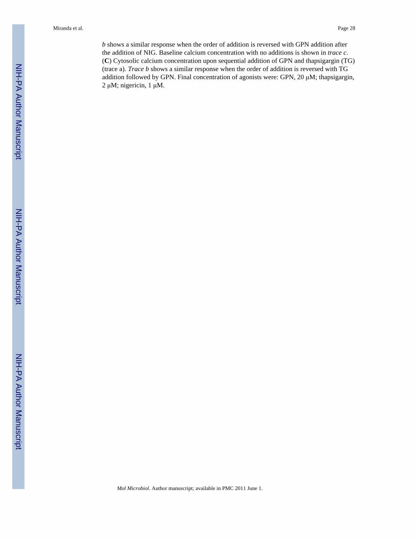
b shows a similar response when the order of addition is reversed with GPN addition afterthe addition of NIG. Baseline calcium concentration with no additions is shown in trace c.(C) Cytosolic calcium concentration upon sequential addition of GPN and thapsigargin (TG)(trace a). Trace b shows a similar response when the order of addition is reversed with TGaddition followed by GPN. Final concentration of agonists were: GPN, 20 μM; thapsigargin,2 μM; nigericin, 1 μM.
Miranda et al. Page 28
Mol Microbiol. Author manuscript; available in PMC 2011 June 1.
NIH
-PA Author Manuscript
NIH
-PA Author Manuscript
NIH
-PA Author Manuscript

Fig. 9.Response to HgCl2 in TgAQP1 overexpressing extracellular tachyzoites. Extracellulartachyzoites were collected, removed from culture media, and resuspended in buffer A withglucose (BAG). (A) RH and TgAQP1 overexpressing tachyzoites were either kept in BAGfor 5 min (`no treatment') or incubated in BAG with HgCl2 at a final concentration of 1 μMfor 5 min (`mercury treatment'). Cells were then fixed with paraformaldehyde for 1 hour andmounted on coverslips for visualization using DIC microscopy. Scale bars for A = 10 μm.(B) Morphometric analysis of RH and TgAQP1 overexpressing cells in the absence (blackbars) or presence (gray bars) of 1 μM HgCl2. Circularity measurements were made on 50randomly chosen parasites for each treatment. Error bars are standard deviation, ***P<0.001.
Miranda et al. Page 29
Mol Microbiol. Author manuscript; available in PMC 2011 June 1.
NIH
-PA Author Manuscript
NIH
-PA Author Manuscript
NIH
-PA Author Manuscript

Fig. 10.The PLV fragments after host cell invasion. (A) Lack of co-localization of anti-TgCPL usedat a dilution of 1:400 (green) and anti-TgVP1 used at a dilution of 1:4,000 (red). The arrowsindicate a typical appearance of vesicles labeled with anti-TgVP1 apical to the nucleus.hTert monolayers were infected with 1.5 × 10⁶ tachyzoites and fixed 20 hours later for IFAassay. (B) Co-localization of anti-TgVP1 (green) and antibodies against the HA tag ofTgRab7 used at a dilution of 1:800 (red). The arrows points at vesicles labeled with the anti-TgVP1 antibody. (C) Labeling by anti-TgVP1 is in a post-Golgi compartment. Parasiteswere transfected with the plasmid pTubGRASP55-RFP/sagCAT. The red labeling comesfrom the direct fluorescence of the red fluorescent protein (arrow points at the red-labeledcompartment in left panel). The labeling of vesicles with anti-TgVP1 is indicated by arrowsin the TgVP1 and Merge panels. (D) Replicating, intracellular parasites were labeled atdifferent stages of division with anti-TgVP1 antibodies. The arrows show the appearance ofthe PLV as the cells divide. Anti-TgVP1 primary antibody was used at 1:4,000 and goatanti-rabbit Alexa488 as secondary antibody was at 1:1000. Blue fluorescence = DAPI. Scalebars =5 μm.
Miranda et al. Page 30
Mol Microbiol. Author manuscript; available in PMC 2011 June 1.
NIH
-PA Author Manuscript
NIH
-PA Author Manuscript
NIH
-PA Author Manuscript

Fig. 11.The PLV is evident in extracellular parasites soon after release from host cells. (A)Intracellular tachyzoites were obtained after scrapping off infected host monolayers andfixed immediately (time 0), or incubated for 2 and 4 hours in BAG or IM. Anti-TgCPL andanti-TgVP1 antibodies were used to visualize the PLVs at those times. The most commonphenotype is shown. Table 2 shows the quantification of these cells. (B) Tachyzoites wereallowed to invade hTert monolayers for 5 min at 37 °C after a pre-incubation of 15 min onice. Cells were immediately fixed and anti-TgCPL and anti-TgVP1 antibodies were used tovisualize the PLV. Antibody concentrations are indicated in the legend for Fig. 3. Scale Bars= 5 μm.
Miranda et al. Page 31
Mol Microbiol. Author manuscript; available in PMC 2011 June 1.
NIH
-PA Author Manuscript
NIH
-PA Author Manuscript
NIH
-PA Author Manuscript
Fig. 12.Schematic representation of the plant-like vacuole. The H+ gradient is established by avacuolar H+-ATPase (V-H+-ATPase) and a vacuolar proton pyrophosphatase (V-H+-PPase).An aquaporin channel would transport water or other osmolytes which could help theparasite deal with environmental stress. Other potential transporters include Na+/H+, andCa2+/H+ exchangers. As in the plant vacuole, other transporters may be present which couldtransport other cations using the proton gradient generated by the proton pumps. Someinternal vesicles and acidocalcisomes are also shown.
Miranda et al. Page 32
Mol Microbiol. Author manuscript; available in PMC 2011 June 1.
NIH
-PA Author Manuscript
NIH
-PA Author Manuscript
NIH
-PA Author Manuscript

NIH
-PA Author Manuscript
NIH
-PA Author Manuscript
NIH
-PA Author Manuscript
Miranda et al. Page 33
Table 1
Enrichment of Fraction 1 in AMDP-sensitive PPase activity.
Enzyme Parasites P3 Fraction 1a
V-H+-Ppaseb (nmol/min/mg)RH 2.27 ± 0.24 45.77 ± 4.51
OE 19.32 ± 0.17 225.08 ± 3.85
Acid phosphatasec (nmol/min/mg)RH 32.60 ± 0.55 55.25 ± 1.27
OE 12.64 ± 0.12 105.05 ± 2.60
Soluble PPasec (nmol/min/mg)RH 14.00 ± 0.77 11.38 ± 0.10
OE 14.27 ± 0.50 37.83 ± 1.72
Comparison of the values for columns P3 (Pellet 3 from fractionation schematic in Fig. S4) and Fraction 1 show a significant increase in the
activities for the PLV markers (V-H+-PPase and acid phosphatase) and not in the activity of the soluble PPase (a cytosolic enzyme) in RH
tachyzoites. The activity of the V-H+-PPase is higher in the fraction obtained from the TgPPase-OE (OE) cells than in the fraction from RHtachyzoites.
These results are representative of more than 10 fractionation experiments.
aFraction 1 was obtained through subcellular fractionation of T. gondii tachyzoites following the steps described in the supplementary Fig. S4.
bThis activity is the AMDP-sensitive pyrophosphate hydrolysis activity measured as explained under Experimental Procedures.
cProtocols for the determination of these enzyme activities are explained under Experimental Procedures.
Mol Microbiol. Author manuscript; available in PMC 2011 June 1.

NIH
-PA Author Manuscript
NIH
-PA Author Manuscript
NIH
-PA Author Manuscript
Miranda et al. Page 34
TABLE 2
Number of open vs. closed PLVs in recently released tachyzoites.
Conditions Total Number of Parasitesa Number of Open Vacuolesb Number of Closed Vacuoles
BAG 38 26 (68%) 3 (7%)
IM 35 7 (24%) 23 (67%)
The results presented are from one experiment out of 3. The other two experiments gave similar results.
Tachyzoites, were collected by scrapping off the host monolayer and releasing parasites by passing them through a 25 G syringe needle. Cells werecollected in either BAG or IM and the numbers in the table are the ones obtained immediately after isolating the parasites (Time 0 in Fig. 11).Vacuoles were considered open when their diameter was larger than 0.5 μm. Vacuoles were considered closed when their diameter was equal orsmaller than 0.5 μm.
aThese numbers are the average number of parasites counted in 5 different fields. Parasites with visible vacuoles by DIC and labeled with anti-
TgVP1 were counted. Total cells evaluated were 189 in BAG and 177 in IM.
bNumbers given in parentheses represent the percentage of cells in open or closed configuration. Cells with no clear PLV were not counted as
either open or closed, and represented less than 25% of the total population.
Mol Microbiol. Author manuscript; available in PMC 2011 June 1.