Contractile effect of TRPA1 receptor agonists in the isolated mouse intestine

Identification of Contractile Vacuole Proteins inTrypanosoma cruziPaul N. Ulrich1., Veronica Jimenez1., Miyoung Park1, Vicente P. Martins1, James Atwood III2, Kristen
Moles1, Dalis Collins1, Peter Rohloff1, Rick Tarleton1, Silvia N. J. Moreno1, Ron Orlando2, Roberto
Docampo1*
1 Center for Tropical and Emerging Global Diseases and Department of Cellular Biology, University of Georgia, Athens, Georgia, United States of America, 2 Complex
Carbohydrate Research Center, University of Georgia, Athens, Georgia, United States of America
Abstract
Contractile vacuole complexes are critical components of cell volume regulation and have been shown to have otherfunctional roles in several free-living protists. However, very little is known about the functions of the contractile vacuolecomplex of the parasite Trypanosoma cruzi, the etiologic agent of Chagas disease, other than a role in osmoregulation.Identification of the protein composition of these organelles is important for understanding their physiological roles. Weapplied a combined proteomic and bioinfomatic approach to identify proteins localized to the contractile vacuole.Proteomic analysis of a T. cruzi fraction enriched for contractile vacuoles and analyzed by one-dimensional gelelectrophoresis and LC-MS/MS resulted in the addition of 109 newly detected proteins to the group of expressed proteins ofepimastigotes. We also identified different peptides that map to at least 39 members of the dispersed gene family 1 (DGF-1)providing evidence that many members of this family are simultaneously expressed in epimastigotes. Of the proteinspresent in the fraction we selected several homologues with known localizations in contractile vacuoles of other organismsand others that we expected to be present in these vacuoles on the basis of their potential roles. We determined thelocalization of each by expression as GFP-fusion proteins or with specific antibodies. Six of these putative proteins (Rab11,Rab32, AP180, ATPase subunit B, VAMP1, and phosphate transporter) predominantly localized to the vacuole bladder.TcSNARE2.1, TcSNARE2.2, and calmodulin localized to the spongiome. Calmodulin was also cytosolic. Our resultsdemonstrate the utility of combining subcellular fractionation, proteomic analysis, and bioinformatic approaches forlocalization of organellar proteins that are difficult to detect with whole cell methodologies. The CV localization of theproteins investigated revealed potential novel roles of these organelles in phosphate metabolism and provided informationon the potential participation of adaptor protein complexes in their biogenesis.
Citation: Ulrich PN, Jimenez V, Park M, Martins VP, Atwood J III, et al. (2011) Identification of Contractile Vacuole Proteins in Trypanosoma cruzi. PLoS ONE 6(3):e18013. doi:10.1371/journal.pone.0018013
Editor: Gordon Langsley, Institut national de la sante et de la recherche medicale - Institut Cochin, France
Received October 28, 2010; Accepted February 22, 2011; Published March 18, 2011
Copyright: � 2011 Ulrich et al. This is an open-access article distributed under the terms of the Creative Commons Attribution License, which permitsunrestricted use, distribution, and reproduction in any medium, provided the original author and source are credited.
Funding: This work was supported by the U.S. National Institutes of Health (grant AI-068647 to RD), and postdoctoral fellowships from the American HeartAssociation (to PNU and VJ). KM and DC were supported in part by a summer training grant from the National Institutes of Health (RR-022685). The funders hadno role in study design, data collection and analysis, decision to publish, or preparation of the manuscript.
Competing Interests: The authors have declared that no competing interests exist.
* E-mail: [email protected]
. These authors contributed equally to this work.
Introduction
Trypanosoma cruzi is the etiologic agent of Chagas disease, the
leading cause of heart disease in endemic areas of Latin America
[1]. Living in a wide range of environments, T. cruzi developed
ways of coping with sudden or prolonged changes in its
surroundings. T. cruzi experiences osmotic challenges as it passes
between the blood of mammalian hosts (300 mOsm) and the
rectum of insect hosts (.750 mOsm) [2]. In addition, bloodstream
trypomastigotes must be able to resist up to 1,400 mOsm when
passing through the renal medulla and return to the isosmotic
environment at the general circulation [3]. Thus, similar to
erythrocytes, these parasites must have fast and efficient
mechanisms to face such extreme challenges.
Previous studies of osmotic stress in T. cruzi demonstrated that a
contractile vacuole (CV) complex contributes to regulatory volume
decrease under hyposmotic stress [4–6]. The roles of the
contractile vacuoles in protists, though, extend beyond regulation
of cell volume to regulation of Ca2+ homeostasis [7–9] and
transport of proteins to the plasma membrane [10]. Recently,
Hasne et al. [11] demonstrated that the contractile vacuole of T.
cruzi houses a polyamine transporter that can be transferred to the
plasma membrane when the incubation media is deficient in
polyamines.
Knowledge of the protein composition of the CV will facilitate
understanding of the physiological roles of these organelles in T.
cruzi. Only a few proteins have been localized to the CV of T. cruzi
so far. Among these are vacuolar proton pyrophosphatase
(TcPPase or TcVP1) [5], aquaporin 1 (TcAQP1) [5], calmodulin
[5], cyclicAMP phosphodiesterease C (TcPDEC) [12], alkaline
phosphatase [4], and a polyamine transporter (TcPOT1) [11].
The contribution of each to T. cruzi physiology is limited largely to
roles in cell volume regulation. Hyposmotic stress increases cAMP
concentration in T. cruzi, and stimulates the translocation of
PLoS ONE | www.plosone.org 1 March 2011 | Volume 6 | Issue 3 | e18013

TcAQP1 to the CV from acidocalcisomes [4], acidic organelles
containing high amounts of polyphosphate and cations [13]. A
current model suggests that hydrolysis of polyphosphate osmoti-
cally drives water from the cytosol into the CV and that
termination of this process occurs after hydrolysis of cAMP by
the CV-localized TcPDEC [6]. However, validation of this model
awaits further elucidation of the regulatory and effector proteins
that populate the CV.
In this study, we used a combined proteomic and bioinformatic
strategy to identify proteins of the CV. We validated their CV
localization by their expression as GFP-fusion proteins and by
immunofluorescence with specific antibodies. The results support
the role of these organelles in osmoregulation.
Results
Protein identificationWe identified 220 (1% false discovery rate, total protein group
probability .0.95) proteins from fractions enriched in CVs from
T. cruzi epimastigotes (see Tables S1 and S2). Seventy-four are
annotated as ‘‘hypothetical’’ in the T. cruzi genome. Seventy five
(38 ‘‘hypothetical’’) were not represented in proteomic data
available on TriTrypdb.org (downloaded 4/10/2009) or the
ribosomal proteome [14]. One hundred nine were not previously
identified in epimastigote data from these sources.
Of the newly identified proteins the most interesting are several
members of the dispersed gene family 1 (DGF-1). The DGF-1 is a
large gene family predicted in the T. cruzi genome with over 500
members [15]. We identified peptides that map to at least 39
members of this family (see Table S3) providing evidence, for the
first time, that many of these proteins are simultaneously expressed
in epimastigotes. A second interesting group is that of the calpain-
like cysteine peptidase with peptides that unambiguously map to 4
different pseudogenes (see Table S2). Calpain-like proteins are
related to Ca2+ dependent cytosolic cysteine peptidases (calpains)
but lack the Ca2+-binding EF-hand domain motif of the domain
IV of conventional calpains [16]. Another important finding was
the identification of 2 amastins in the subcellular proteome of
epimastigotes. Amastins are transmembrane glycoproteins encod-
ed by a large gene family found predominantly on the cell surface
of T. cruzi and Leishmania spp. amastigotes [17].
Approximately 29% (70) of the 220 proteins we identified in our
subcellular fraction have predicted transmembrane domains (see
Table S4), consistent with estimates of representation in other
organisms [18]. Fifty-five proteins (22.9%) possessed putative
transmembrane domains but no signal peptide. Annotated
proteins in the subcellular proteome analyzed span a broad range
of metabolic groups (Fig. 1, and Table S5). Transport-related and
intracellular proteins accounted for ,21%. Among these were
small G proteins (Rabs), transporters, and channels. T. cruzi
vacuolar-H+-pyrophosphatase, an acidocalcisomal marker also
found in the CV [5], was clearly identified in our dataset (total
protein probability = 1). Other well-represented metabolic groups
in our dataset were energy metabolism (19%), protein and amino
acid metabolism (29%), and cell structure and organization (15%).
Subcellular localizations of each protein were predicted (Table
S6). Targeting predictions based on consensus of at least two
algorithms indicated that ,24% of the proteins are mitochondrial,
,19% are cytosolic, and ,5.4% are nuclear. Plasma membrane,
Golgi, and secretory localizations represented a small minority.
Sixty proteins (29.5%) had one or more predicted transmembrane
domains, and 26 (11.8%) proteins had predicted signal peptides.
Of the proteins identified by proteomic analysis of the
subcellular fraction we selected a number of proteins with
homologues that localize to the CV in other organisms, or
properties that could justify CV localization for further validation
(Table 1). T. cruzi Rab11, while unidentified in our dataset, is
included in Table 1 because it localizes in CV bladders of
Dictyostelium discoideum [19]. The Rab11 gene we cloned and
sequenced is identical to a gene (Tc00.1047053511407.60)
annotated in the TriTrypDB database but differs from a gene
previously named Rab11 [20]. The Rab11 sequence previously
described contains insertions of two bases that shift the reading
frame. VAMP1 is included because of its similarity (2.8e212) with
PtSyb2-2, a Paramecium tetraurelia synaptobrevin that localizes to the
contractile vacuole [21]. Calmodulin is included in Table 1
because it was shown to be localized in the CV of T. cruzi by
immunofluorescence analysis using human antibodies [5] and is
present in the CV of D. discoideum [22] and Paramecium multi-
micronucleatum [23]. A homologue to a vacuolar phosphate
transporter from yeast (Pho91) [24] annotated as a sodium/
sulphate symporter is also included because orthophosphate (Pi)
was shown to be abundant in the CV of T. cruzi [4].
Table 1 also shows other proteins that were present in our
proteomic analysis and that have homologues in other organisms
that localize to the CV, such as a golvesins [25], myosins [26,27],
clathrin heavy chain [28,29], neurobeachin [30], IP3/ryanodine
receptor [31], and disgorgin [32]. A putative calcium channel was
also detected although there are no reports of this type of channels
in the CV.
V-H+-ATPase subunit B localizes mainly to the bladder ofthe CV
Vacuolar-H+-ATPase is a multisubunit complex that is a marker
for the CV in D. discoideum [33], Amoeba proteus [34], and
Chlamydomonas reinhardtii [35]. Several subunits were detected in
Figure 1. Annotated proteins in the contractile vacuoleproteome belong to a variety of metabolic groups.doi:10.1371/journal.pone.0018013.g001
Proteins of the Contractile Vacuole of T. cruzi
PLoS ONE | www.plosone.org 2 March 2011 | Volume 6 | Issue 3 | e18013

our proteomic analysis (a, B, D, and G, Table 1). In early work we
detected co-localization of this V-H+-ATPase with a plasma
membrane-type Ca2+-ATPase (PMCA) to the acidocalcisomes of
T. cruzi [36]. To investigate the localization of this proton pump,
we generated a V-H+-ATPase subunit B-GFP fusion expression
construct and transfected epimastigotes with it. Subunit B of T.
Table 1. Proteins identified as potentially present in the contractile vacuole complex, showing localizations confirmed in thisstudy or in other organisms.
Protein nameTriTrypDB Gene ID(GenBank ID) Peptides
Mascotscore
CVC localization(this work)
Protists withhomologousCV protein References
V-H+-ATPase subunit B(vacuolar synthasesubunit B)
Tc00.1047053506025.50(EAN86474.1)
AIQSGYSVKPHLEYTTIR 25 bladder D. discoideumA. proteusC. reinhardtii
[33–35]
V-H+-ATPase subunit a Tc00.1047053509601.70(EAN92926.1)
ERVPILER 18 D. discoideumA. proteusC. reinhardtii
[33–35]
V-H+-ATPase subunit D(vacuolar ATP synthasesubunit D)
Tc00.1047053509017.30(EAN86631.1)
EALAR 17 D. discoideumA. proteusC. reinhardtii
[33–35]
V-H+-ATPase subunitG (H+-ATPase G subunit)
Tc00.1047053510993.10(EAN81810.1)
AQQLSGADENLELAR 23 D. discoideumA. proteusC. reinhardtii
[33–35]
SNARE 2.1 (hypotheticalprotein)
Tc00.1047053507625.183(EAN94402.1)
TAPVR 16 spongiome P. tetraurelia [21]
SNARE 2.2 (hypotheticalprotein)
Tc00.1047053506715.50(EAN84023.1)
EVEMFNDK;KILANIKRPLVER
37 spongiome P. tetraurelia, [21]
Rab32 (small GTP-binding protein)
Tc00.1047053506289.80(EAN96691.1)
NTSGK 17 bladder N/A
Rab11* Tc00.1047053511407.60(EAN88612.1)
N/A N/A bladder D. discoideum [19]
Calmodulin* Tc00.1047053507483.39(EAN86242.1)
N/A N/A spongiome D. discoideum,P. multimicronleatum
[22,23]
Phosphate transporter (Pho1)(sodium/sulphatesymporter)*
Tc00.1047053508831.60(EAN97069.1)
N/A N/A bladder
AP180 (clathrin coatassembly protein)
Tc00.1047053503449.30(EAN83025.1)
LSSIPR 18 bladder D. discoideum [28,38]
Golvesin-1(methyltransferase)
Tc00.1047053509805.40(EAN98089.1)
VPIAK 16 D. discoideum [25]
Golvesin-2 (hypotheticalprotein)
Tc00.1047053503455.30(EAN84949.1)
TCESIGVVSDPVR 15 D. discoideum [25]
myosin heavy chain(myosin V-1)
Tc00.1047053511527.70(EAN87803.1)
MGFPR 20 D. discoideumA. castellani
[26,27]
myosin IB heavychain (myosin V-2)
Tc00.1047053507739.110(EAN89650.1)
ACVDFK 17 D. discoideumA. castellani
[26,27]
clathrin heavy chain Tc00.1047053506167.50(EAN87927.1)
TWTAVNIACIEANEIK 17 D. discoideum [28,29]
V-H+-PPase Tc00.1047053510773.20(EAN91609.1)
AADVGADLVGK;EITDALDAAGNTTAAIGK;NVYVISR
205 D. discoideum [5]
neurobeachin/beige protein
Tc00.1047053511159.7(EAN84625.1)
EVVENK 16 D. discoideum [30]
IP3/ryanodine receptor(hypothetical protein)
Tc00.1047053509461.90(EAN89926.1)
SSRQEIVQDVMFLR;LLGSIDLFMR
15 P. tetraurelia [31]
Calcium channel protein Tc00.1047053504105.130(EAN97848.1)
ALTGGRTPQELEDKNR;DDNAMYEEALLFDR;LPGLYQPAIDEK
18
Disgorgin (rab-likeGTPase activatingprotein, RabGAP)
Tc00.1047053508723.80(EAN91303.1)
EKHDLPAK 20 D. discoideum [32]
VAMP (vesicle-associatedmembrane protein)
Tc00.1047053511627.60(EAN87604.1)
N/A N/A bladder D. discoideum
References of contractile vacuole homologue proteins present in other protists are indicated. The proteins denoted with an asterisk were not present in the MS data.Protein names from the annotated genome, if different from usages in the text, are provided in parenthesis.doi:10.1371/journal.pone.0018013.t001
Proteins of the Contractile Vacuole of T. cruzi
PLoS ONE | www.plosone.org 3 March 2011 | Volume 6 | Issue 3 | e18013

cruzi V-H+-ATPase is 71% identical to its homologous in D.
discoideum. After several weeks of selection, expression of the fusion
protein in the transfectants was analyzed by direct fluorescence
analysis. Although detectable under isosmotic conditions (when
the CV was collapsed in most cells), V-H+-ATPase subunit B
strongly delineated the outer margin of the enlarged CV bladder
under hyposmotic conditions (150 mOsm) (Fig. 2A). Additional
labeling was detected in smaller vacuoles, which are visible in the
differential interference contrast (DIC) images (Fig. 2A, arrows)
and could correspond to acidocalcisomes, which are known to
increase in volume under hyposmotic stress [4]. We confirmed C-
terminal tagging of T. cruzi V-H+-ATPase subunit B with GFP by
western blot analysis (Fig. 2A). Although this subunit was present
in the total cell homogenate and in the 100,000 g pellet, it was also
detected in the 100,000 g supernatant. The presence of subunit B
in the soluble fraction is due to the well-known dissociation and
loss of peripheral subunits of the V-H+-ATPase that occurs during
cell fractionation of T. cruzi [37]. This subunit associated with but
did not co-localize with calmodulin (CaM), a protein that localizes
to a compartment proximal to the bladder [4,22] (data not shown).
AP180 localizes to the bladderAP180 is a protein that promotes assembly of clathrin triskelia
and is localized in the plasma membrane and contractile vacuole
of D. discoideum [38]. The T. cruzi orthologue (annotated ‘‘clathrin
coat assembly protein’’) contains a conserved ANTH (AP180 N-
terminal homology) domain. Within the ANTH domain of T. cruzi
AP180 is a region similar to the domain consensus sequence
[KG]A[TI]xxxxxx[PLV]KxK[HY] [39]. Additionally, T. cruzi
AP180 contains a clathrin box motif (LVAVE) and a tyrosine-
based sorting motif (YAAL, detected with the ELM server [40]),
which may mediate clathrin and adaptor protein 2 (AP-2) complex
interactions, respectively [41,42].
AP180 fused to GFP (N-terminal tag) was present in the CV
bladder (Fig. 2B) and detected by western blot analysis (Fig. 2B).
To investigate whether AP180 resides in the bladder, we observed
live cells under hyposmotic conditions (Fig. 2B) and localized
AP180 with antibodies against GFP and CaM in fixed cells (see
Fig. S1). AP180 was also present in a structure adjacent to the
bladder stained by antibodies against CaM (see Fig. S1). The
vesicular structure of the bladder was not evident because fixation
collapses the CV. Immunogold electron microscopy confirmed the
predominant localization of AP180 in the bladder of the CV and
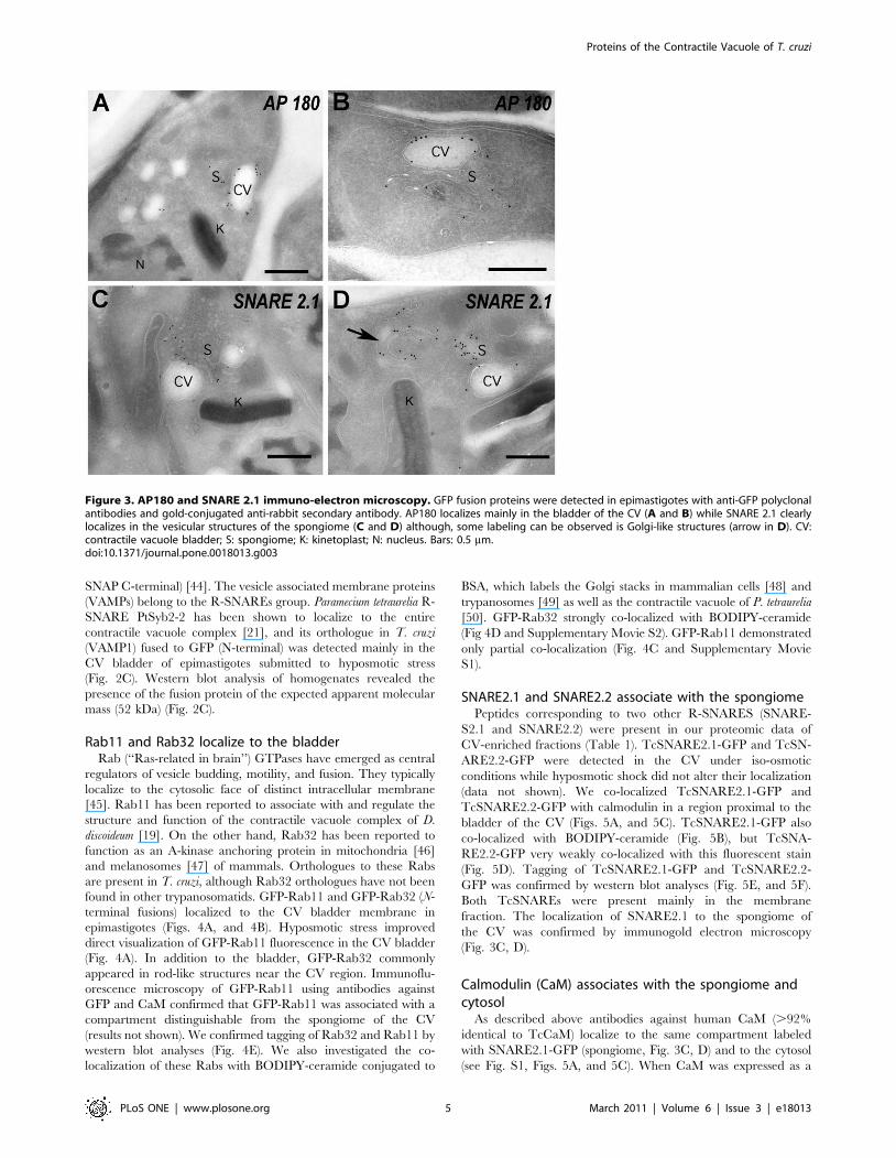
to some tubules and vesicles that form the spongiome (Fig. 3A,B), a
network of collecting ducts connected to the bladder.
VAMP1 localizes to the bladderSoluble N-ethylmaleimide-sensitive factor (NSF) adaptor pro-
teins (SNAPs) receptors (SNAREs) are key components of the
intracellular vesicle-mediated transports that take place in
eukaryotic cells and are distinguished by the presence of a
common motif (SNARE motif). These proteins can be classified as
Q- and R-SNAREs according to the residue present in the center
of the motif [43]. The Q group can be further divided into three
subgroups according to their overall homology in the SNARE
domain: Qa (or syntaxins), Qb (or SNAP N-terminal) and Qc (or
Figure 2. Fluorescence microscopy and western blot analysis of V-H+-ATPase subunit B-, AP180-, and VAMP1-GFP fusion proteinsin live T. cruzi epimastigotes. V-H+-ATPase subunit B (A), AP180 (B), and VAMP1 (C) localize to the bladder under hyposmotic conditions.Brightness and contrast of panels was adjusted, and fluorescence images in C were deconvolved. Scale bars: 10 mm. Confirmation of tagging bywestern blot analyses with polyclonal anti-GFP (dilution 1:5,000-1:10,000, Invitrogen) in epimastigotes. HRP-conjugated goat anti-rabbit was used as asecondary antibody. Magic Mark XP (Invitrogen) was used as a molecular weight marker. Arrows indicate bands of interest. A, V-H+-ATPase subunit B,expected size of fusion protein = 82 kDa. B, AP-180, expected size of fusion protein = 81 kDa. A 100 kDa cross-reacting band is only detected in thesupernatant. C, VAMP1 expected size = 52 kDa. P, membrane pellet, S, soluble fraction, H, homogenate of whole parasites, WT, wild-typeepimastigotes (negative control).doi:10.1371/journal.pone.0018013.g002
Proteins of the Contractile Vacuole of T. cruzi
PLoS ONE | www.plosone.org 4 March 2011 | Volume 6 | Issue 3 | e18013
SNAP C-terminal) [44]. The vesicle associated membrane proteins
(VAMPs) belong to the R-SNAREs group. Paramecium tetraurelia R-
SNARE PtSyb2-2 has been shown to localize to the entire
contractile vacuole complex [21], and its orthologue in T. cruzi
(VAMP1) fused to GFP (N-terminal) was detected mainly in the
CV bladder of epimastigotes submitted to hyposmotic stress
(Fig. 2C). Western blot analysis of homogenates revealed the
presence of the fusion protein of the expected apparent molecular
mass (52 kDa) (Fig. 2C).
Rab11 and Rab32 localize to the bladderRab (‘‘Ras-related in brain’’) GTPases have emerged as central
regulators of vesicle budding, motility, and fusion. They typically
localize to the cytosolic face of distinct intracellular membrane
[45]. Rab11 has been reported to associate with and regulate the
structure and function of the contractile vacuole complex of D.
discoideum [19]. On the other hand, Rab32 has been reported to
function as an A-kinase anchoring protein in mitochondria [46]
and melanosomes [47] of mammals. Orthologues to these Rabs
are present in T. cruzi, although Rab32 orthologues have not been
found in other trypanosomatids. GFP-Rab11 and GFP-Rab32 (N-
terminal fusions) localized to the CV bladder membrane in
epimastigotes (Figs. 4A, and 4B). Hyposmotic stress improved
direct visualization of GFP-Rab11 fluorescence in the CV bladder
(Fig. 4A). In addition to the bladder, GFP-Rab32 commonly
appeared in rod-like structures near the CV region. Immunoflu-
orescence microscopy of GFP-Rab11 using antibodies against
GFP and CaM confirmed that GFP-Rab11 was associated with a
compartment distinguishable from the spongiome of the CV
(results not shown). We confirmed tagging of Rab32 and Rab11 by
western blot analyses (Fig. 4E). We also investigated the co-
localization of these Rabs with BODIPY-ceramide conjugated to
BSA, which labels the Golgi stacks in mammalian cells [48] and
trypanosomes [49] as well as the contractile vacuole of P. tetraurelia
[50]. GFP-Rab32 strongly co-localized with BODIPY-ceramide
(Fig 4D and Supplementary Movie S2). GFP-Rab11 demonstrated
only partial co-localization (Fig. 4C and Supplementary Movie
S1).
SNARE2.1 and SNARE2.2 associate with the spongiomePeptides corresponding to two other R-SNARES (SNARE-
S2.1 and SNARE2.2) were present in our proteomic data of
CV-enriched fractions (Table 1). TcSNARE2.1-GFP and TcSN-
ARE2.2-GFP were detected in the CV under iso-osmotic
conditions while hyposmotic shock did not alter their localization
(data not shown). We co-localized TcSNARE2.1-GFP and
TcSNARE2.2-GFP with calmodulin in a region proximal to the
bladder of the CV (Figs. 5A, and 5C). TcSNARE2.1-GFP also
co-localized with BODIPY-ceramide (Fig. 5B), but TcSNA-
RE2.2-GFP very weakly co-localized with this fluorescent stain
(Fig. 5D). Tagging of TcSNARE2.1-GFP and TcSNARE2.2-
GFP was confirmed by western blot analyses (Fig. 5E, and 5F).
Both TcSNAREs were present mainly in the membrane
fraction. The localization of SNARE2.1 to the spongiome of
the CV was confirmed by immunogold electron microscopy
(Fig. 3C, D).
Calmodulin (CaM) associates with the spongiome andcytosol
As described above antibodies against human CaM (.92%
identical to TcCaM) localize to the same compartment labeled
with SNARE2.1-GFP (spongiome, Fig. 3C, D) and to the cytosol
(see Fig. S1, Figs. 5A, and 5C). When CaM was expressed as a
Figure 3. AP180 and SNARE 2.1 immuno-electron microscopy. GFP fusion proteins were detected in epimastigotes with anti-GFP polyclonalantibodies and gold-conjugated anti-rabbit secondary antibody. AP180 localizes mainly in the bladder of the CV (A and B) while SNARE 2.1 clearlylocalizes in the vesicular structures of the spongiome (C and D) although, some labeling can be observed is Golgi-like structures (arrow in D). CV:contractile vacuole bladder; S: spongiome; K: kinetoplast; N: nucleus. Bars: 0.5 mm.doi:10.1371/journal.pone.0018013.g003
Proteins of the Contractile Vacuole of T. cruzi
PLoS ONE | www.plosone.org 5 March 2011 | Volume 6 | Issue 3 | e18013

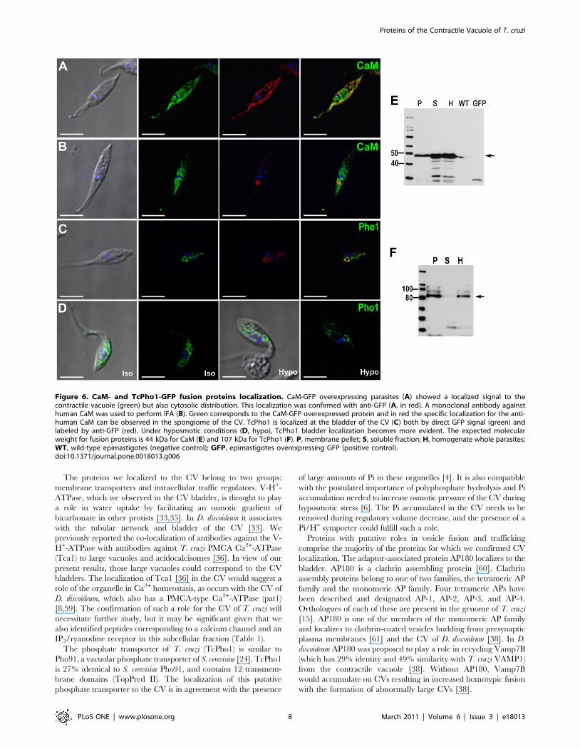
fusion construct with GFP, the protein also showed cytosolic
staining in addition to CV localization (Fig. 6A). Tagging of CaM-
GFP was confirmed by western blot analysis (Fig. 6E). In
epimastigotes overexpressing CaM-GFP fusion proteins, immu-
nolocalization with anti-human CaM antibody only showed
partial co-localization in the CV spongiome (Fig. 6B).
Phosphate transporter localizes to the bladderPho91 is a vacuolar phosphate transporter that regulates
phosphate and polyphosphate metabolism in Saccharomyces cerevisiae
[24]. The T. cruzi orthologue, when expressed as a fusion with
GFP, strongly localized to the bladder of the CV and also showed
some granular localization in the cytosol (Fig. 6C). Under
hyposmotic stress, the localization of the phosphate transporter
becomes more evidently associated with the CV bladder, which is
clearly delineated by the GFP-labeled protein (Fig. 6D). Western
blot analysis confirmed the expression of this construct (Fig. 6F).
The recognized polypeptide had an apparent molecular mass of
87 kDa. Since T. cruzi Pho1 has 12 transmembrane domains, a
size discrepancy between the expected (107 kDa) and the observed
molecular mass could be attributed to the usual anomalous
migration of hydrophobic protein on SDS gels [51] or to partial
degradation. In agreement with these results, the protein could not
be detected if the samples were boiled before SDS gel
electrophoreses.
Discussion
We report here the proteomic analysis of a subcellular fraction
enriched in CV from T. cruzi. Previous work [4] indicated that this
fraction is enriched in the contractile vacuole markers alkaline
phosphatase [52] and bafilomycin A1-sensitive vacuolar H+-
ATPase [33–35], basic amino acids, and phosphate. Additionally,
this protocol yields fractions well resolved from organelle markers
for mitochondria (alanine aminotransferase), glycosomes (hexoki-
nase) and lysosomes (a-mannosidase) [4].
We identified 220 proteins (1% false discovery rate, protein
group probability .0.95, (see Table S2) in this dataset. Among the
annotated proteins, we identified vacuolar-H+-pyrophosphatase
(VP1), a proton pump that co-localizes with aquaporin found in
the T. cruzi CV [5]. VP1 is a major component of acidocalcisomes,
acidic organelles that fuse with the CV in response to osmotic
challenge [4].
Many proteins we identified likely belong to different cellular
compartments, but the relatively high representation of membrane
proteins (65 proteins, 29.5%) is notable, given that membrane
proteins are challenging for proteomic analysis. In comparison, the
recently published plasma membrane proteome of Trypanosoma brucei
contains a lower proportion of membrane proteins (16.1% of 1536
proteins, [53]). Thus we feel that our fractionation successfully
enriched proteins with potential membrane-related functions.
Figure 4. Rab-GFP fusion proteins localize in T. cruzi contractile vacuole. Rab11 (green) (A) land Rab32 (green) (B) localize to the bladderunder hyposmotic conditions. Rab11 (C) and Rab32 (D) partially co-localize with BODIPY-ceramide (red). DNA is stained with DAPI (blue). Brightnessand contrast of panels was adjusted, and fluorescence images in C-D were deconvolved. Inset in C shows one cell (dotted rectangle) at highermagnification. Scale bars: A, B and C = 10 mm; D = 5 mm. E, confirmation of tagging by western blot analyses with anti GFP shows the expected sizefor both fusion proteins (50 kDa). Wild-type epimastigotes were used as negative control (WT).doi:10.1371/journal.pone.0018013.g004
Proteins of the Contractile Vacuole of T. cruzi
PLoS ONE | www.plosone.org 6 March 2011 | Volume 6 | Issue 3 | e18013

A comparison with previous whole cell protein expression
studies carried out in T. cruzi revealed that our proteomic analysis
resulted in the identification of 75 previously undetected proteins.
This confirms the validity of subcellular proteomics as a method of
choice for the identification of larger number of proteins than with
whole cell proteomics [54]. Our identification of DGF-1 proteins
illustrates this clearly. Only one glycopeptide that maps to several
members of the DGF-1 was previously identified in a glycopro-
teomic study of T. cruzi trypomastigotes [55]. We recently reported
that these proteins localize to a population of organelles which do
not co-localize with markers of acidocalcisomes, glycosomes,
reservosomes, lipid droplets, or endocytic vesicles in different
stages of T. cruzi [56]. We also demonstrated that these proteins
are released from trypomastigotes during their differentiation into
amastigotes. Proteomic analysis of supernatants from these
incubations contained peptides mapping to at least 22 DGF-1
members [56]. Here we report the detection of peptides that map
to at least 39 DGF-1 members in T. cruzi, providing definitive
evidence of simultaneous expression of many of these proteins in
epimastigotes.
The detection of peptides mapping to several calpain-like
cysteine peptidases pseudogenes suggests that these were inaccu-
rately annotated in the genome. Finally, the presence of peptides
that map to amastin proteins indicates that these proteins are not
exclusively expressed in amastigotes but are also present in the
epimastigote stage of T. cruzi.
While mass spectrometry is exquisitely sensitive, subcellular
fractionations only provide partial enrichment of cellular compo-
nents from contaminants. Thus, complementation of MS analysis
with in vivo expression of tagged proteins validates proteomic data
within a cellular context. Few studies adopt this approach despite
the fact that contaminant proteins may represent 20% of proteins
or more in subcellular MS datasets [57]. Medium-throughput
tagging of proteins to validate data is growing more popular [58],
but few studies to date have implemented such to verify proteomes
of trypanosomatid parasites [54].
We validated our dataset by expressing a number of proteins
identified in the CV proteome as GFP-fusion proteins in T. cruzi.
We complemented this set of proteins with selected proteins with
known localizations to the CV in other protists and with proteins
that could potentially be present in the CV on the basis of our
knowledge of the organelle. Four proteins not present in any of our
mass spectrometry data (Rab11, calmodulin, VAMP1, and a
phosphate transporter), were added due to roles in CV physiology
in D. discoideum (Rab11) [19], previously suggested presence in the
CV of epimastigotes (CaM) [4], or P. tetraurelia (VAMP1) [21], and
the abundance of phosphate in the CV of epimastigotes (Pho1) [4],
respectively.
Figure 5. SNARE-GFP fusion proteins localize in the contractile vacuole spongiome. SNARE2.1-GFP co-localizes with calmodulin (CaM) (A)and BODIPY-ceramide (B). SNARE2.2-GFP co-localizes with CaM (C) but localizes to a compartment that does not stain with BODIPY-ceramide (D).DNA is stained with DAPI (blue). Brightness and contrast of panels was adjusted, and fluorescence images in B and D were deconvolved. Scale bars =10 mm. E, F, western blot analyses reveal the expected size for GFP tagged SNARE proteins (50 kDa). P, membrane pellet; S, soluble fraction, H,homogenate of whole parasites; WT, wild-type epimastigotes (negative control); GFP, epimastigotes overexpressing GFP (positive control).doi:10.1371/journal.pone.0018013.g005
Proteins of the Contractile Vacuole of T. cruzi
PLoS ONE | www.plosone.org 7 March 2011 | Volume 6 | Issue 3 | e18013
The proteins we localized to the CV belong to two groups:
membrane transporters and intracellular traffic regulators. V-H+-
ATPase, which we observed in the CV bladder, is thought to play
a role in water uptake by facilitating an osmotic gradient of
bicarbonate in other protists [33,35]. In D. discoideum it associates
with the tubular network and bladder of the CV [33]. We
previously reported the co-localization of antibodies against the V-
H+-ATPase with antibodies against T. cruzi PMCA Ca2+-ATPase
(Tca1) to large vacuoles and acidocalcisomes [36]. In view of our
present results, those large vacuoles could correspond to the CV
bladders. The localization of Tca1 [36] in the CV would suggest a
role of the organelle in Ca2+ homeostasis, as occurs with the CV of
D. discoideum, which also has a PMCA-type Ca2+-ATPase (pat1)
[8,59]. The confirmation of such a role for the CV of T. cruzi will
necessitate further study, but it may be significant given that we
also identified peptides corresponding to a calcium channel and an
IP3/ryanodine receptor in this subcellular fraction (Table 1).
The phosphate transporter of T. cruzi (TcPho1) is similar to
Pho91, a vacuolar phosphate transporter of S. cerevisiae [24]. TcPho1
is 27% identical to S. cerevisiae Pho91, and contains 12 transmem-
brane domains (TopPred II). The localization of this putative
phosphate transporter to the CV is in agreement with the presence
of large amounts of Pi in these organelles [4]. It is also compatible
with the postulated importance of polyphosphate hydrolysis and Pi
accumulation needed to increase osmotic pressure of the CV during
hyposmotic stress [6]. The Pi accumulated in the CV needs to be
removed during regulatory volume decrease, and the presence of a
Pi/H+ symporter could fulfill such a role.
Proteins with putative roles in vesicle fusion and trafficking
comprise the majority of the proteins for which we confirmed CV
localization. The adaptor-associated protein AP180 localizes to the
bladder. AP180 is a clathrin assembling protein [60]. Clathrin
assembly proteins belong to one of two families, the tetrameric AP
family and the monomeric AP family. Four tetrameric APs have
been described and designated AP-1, AP-2, AP-3, and AP-4.
Orthologues of each of these are present in the genome of T. cruzi
[15]. AP180 is one of the members of the monomeric AP family
and localizes to clathrin-coated vesicles budding from presynaptic
plasma membranes [61] and the CV of D. discoideum [38]. In D.
discoideum AP180 was proposed to play a role in recycling Vamp7B
(which has 29% identity and 49% similarity with T. cruzi VAMP1)
from the contractile vacuole [38]. Without AP180, Vamp7B
would accumulate on CVs resulting in increased homotypic fusion
with the formation of abnormally large CVs [38].
Figure 6. CaM- and TcPho1-GFP fusion proteins localization. CaM-GFP overexpressing parasites (A) showed a localized signal to thecontractile vacuole (green) but also cytosolic distribution. This localization was confirmed with anti-GFP (A, in red). A monoclonal antibody againsthuman CaM was used to perform IFA (B). Green corresponds to the CaM-GFP overexpressed protein and in red the specific localization for the anti-human CaM can be observed in the spongiome of the CV. TcPho1 is localized at the bladder of the CV (C) both by direct GFP signal (green) andlabeled by anti-GFP (red). Under hyposmotic conditions (D, hypo), TcPho1 bladder localization becomes more evident. The expected molecularweight for fusion proteins is 44 kDa for CaM (E) and 107 kDa for TcPho1 (F). P, membrane pellet; S, soluble fraction; H, homogenate whole parasites;WT, wild-type epimastigotes (negative control); GFP, epimastigotes overexpressing GFP (positive control).doi:10.1371/journal.pone.0018013.g006
Proteins of the Contractile Vacuole of T. cruzi
PLoS ONE | www.plosone.org 8 March 2011 | Volume 6 | Issue 3 | e18013

SNARE proteins are found throughout the eukaryotes
and are important for vesicular fusion [62]. Two T. cruzi R-
SNARE proteins co-localize with calmodulin to a compartment
proximal to the bladder (spongiome, see below) while another
R-SNARE (VAMP1) localizes to the CV bladder. These
SNAREs could direct fusion of the spongiome with bladder
membranes or acidocalcisomes during hyposmotic stress [4,6].
While ceramide conjugates are typically used as markers for
the Golgi complex in other systems [48,49], we observed that
SNARE2.1 co-localized with BSA-conjugated BODIPY-cer-
amide in T. cruzi. In P. tetraurelia, ceramide labels CV com-
plexes and acidosomes [50]. Acidosomes have been postulated
to be part of the spongiome of the contractile vacuole
complex [52] or fragmented contractile vacuole membranes
in D. discoideum [33]. Fig. 3D (arrow) also shows labeling
of Golgi-like structures with antibodies against SNARE2.
1-GFP, which could suggest some link between these two
structures.
Rab proteins regulate CV function in D. discoideum [19,32]. Of
these, Rab11 is particularly important. We report that T. cruzi
Rab11 and Rab32 are present in the CV bladder. Rab11 may
mediate CV discharge in T. cruzi via interaction with drainin, a
Rab11A effector that regulates CV discharge in D. discoideum
[32,63]. Both Rab32 and Rab11 partially co-localized with
BODIPY-ceramide in CV bladders. Rab32 has been reported to
function as an A-kinase anchoring protein in mitochondria [46]
and melanosomes [47]. Rab32 may be involved in the signaling
pathway leading to regulatory volume decrease in T. cruzi [6]. The
localization of the putative phosphate transporter together with
Rab32 adds two novel proteins to the protein complement of CV
of all organisms.
Calmodulin (CaM) has been defined as a cytosolic Ca2+
receptor. TcCaM was purified from epimastigotes [64,65] and
can stimulate the PMCA Ca2+-ATPase [65] and cyclic AMP
phosphodiesterase [64]. It has four calcium-binding sites (EF-hand
domains), is 92% identical to human CaM, and is encoded by
several copies in the genome [66]. Antibodies against human CaM
localize to the CV [4] and to the cytosol, and this was confirmed in
this work using GFP-tagged CaM.
The identification of these novel CV proteins provides useful
insights into the biogenesis of these organelles. A common feature
of all the validated CV proteins identified in this study is the
presence of one or more tyrosine-based sorting signals with the
YXXØ consensus motif (see Table S7). This sequence binds to
the m subunits of the four AP complexes [67]. In this regard, AP-1
is required for the biogenesis of the CV in D. discoideum [68], and
AP-2 is known to interact with AP180 in bovine brain [60].
These motifs are also present in the proteins previously identified
in the CV of T. cruzi (see Table S7). Except for AQP1, all of these
proteins also have casein kinase 2 (CK2) and glycogen synthase
kinase b (GSK3b) phosphorylation sites. Excepting CaM, all the
proteins possess generic N-glycosylation motifs (see Table S7). It
is known that a variety of kinases localize to the Golgi and
regulate post-Golgi membrane trafficking [69]. These findings
will help guiding future studies on the biogenesis of these
organelles.
In summary, in addition to validate the expression at the protein
level of a number of important genes (DGF-1, calpain-like cysteine
peptidases, amastins) in epimastigotes, we identified nine CV proteins
using a strategy complementing subcellular proteomics and
bioinformatics with in vivo localization. Two of these proteins
(Rab32, Pho1) are newly identified CV proteins, and their
identification will facilitate further studies to elucidate the roles
of this organelle in T. cruzi physiology.
Materials and Methods
Cell cultureT. cruzi epimastigotes (CL strain) were grown at 28uC in liver
infusion tryptose (LIT) medium [70] supplemented with 10% heat-
inactivated newborn calf serum. GFP-expressing cell lines were
maintained in LIT medium supplemented with 10% heat-
inactivated fetal bovine serum and G418 (Calbiochem).
Subcellular fractionation of contractile vacuoles and 1-Dgel electrophoresis
Fractions enriched in CVs were isolated as described [4] using
differential and gradient centrifugation. Briefly, epimastigotes
(1.4 g wet weight) were washed twice with Buffer A (116 mM
NaCl, 5.4 mM KCl, 0.8 mM MgSO4, 50 mM HEPES, pH 7.2)
with 5.5 mM glucose. The parasites were washed once in cold lysis
buffer (120 mM sucrose, 50 mM KCl, 4 mM MgCl, 0.5 mM
EDTA, 20 mM HEPES, 5 mM DTT, 0.2% Sigma mammalian
protease inhibitor cocktail, pH 7.2) prior to lysis with silicon
carbide in lysis buffer. Silicon carbide and cell debris was
eliminated by a series of low speed centrifugations (38 g, 144 g,
and 1,200 g). The supernatant was centrifuged at 100,000 g for
60 min, and the pellet was resuspended in 2 ml lysis buffer and
applied to the 25% step of a discontinuous gradient of iodixanol,
with 4 ml steps of 15, 20, 25, 30, 34, 37 and 40% iodixanol,
diluted in lysis buffer. The gradient was centrifuged at 50,000 g in
a Beckman JS-24.38 rotor for 65 min and fractions were collected
from the top. Two hundred microliters were reserved for
quantification of protein by Bradford assay and marker enzyme
assays, as described before [4]. The first 4.5 ml from the top of the
gradient were centrifuged at 100,000 g for 30 min and the pellet
used for proteomic analysis. The final pellet was resuspended in
Laemmli buffer (Sigma-Aldrich) and heated at 80uC for 15 min.
Solubilized proteins were separated on a Nu-PAGE 4-12% Bis-
Tris (Invitrogen) gradient gel at 150 V for 2 h.
In-gel digestionThe gel lane was washed twice in ddH2O for 15 min and cut
into 7 equal slices. Proteins were reduced with 10 mM DTT/
100 mM Ambic (ammonium bicarbonate) solution at 57uC for 1 h
and carboxyamidomethylated with 55 mM iodoacetamide and
100 mM Ambic for 1 h at room temperature in the dark.
Enzymatic digestion was performed with porcine trypsin (1:50,
Promega, Madison, WI) at 37uC overnight. Tryptic peptides were
extracted three times with 200 ml of 50% ACN (1:1 in water).
Combined extracts were dried in a speed vacuum, resuspended in
50 ml 0.1% formic acid, and stored at 220uC.
Mass spectrometryPrior to LC-MS/MS, trypsin was removed by centrifugal
ultrafiltration (MWCO, 30 kDa; Millipore), and peptides were
analyzed on an Agilent 1100 capillary LC (Palo Alto, CA) coupled
to a LTQ linear ion trap mass spectrometer (Thermo Electron).
Mobile phases A and B were H2O/0.1% formic acid and ACN/
0.1% formic acid, respectively. Fractions were loaded for 1 h onto
a PicoFrit 8 cm 650 mm column (New Objective) packed with
5 mm C18 beads under positive N2 pressure. Peptides were
desalted for 10 min with 0.1% formic acid using positive N2
pressure and eluted into the mass spectrometer during a 70 min
linear gradient from 5–45% B at a flow rate of 200 nL min21. The
spectrometer acquired MS/MS spectra on the nine most
abundant precursor ions from each scan with a repeat count of
three and repeat duration of 15 sec. Dynamic exclusion was
enabled for 160 sec. Raw tandem mass spectra were converted
Proteins of the Contractile Vacuole of T. cruzi
PLoS ONE | www.plosone.org 9 March 2011 | Volume 6 | Issue 3 | e18013
into mzXML format and peak lists using ReAdW and
mzMXL2Other [71]. Peak lists were searched using Mascot v1.9
software (Matrix Science, Boston, MA) and two databases were
constructed. The first database (normal) was composed of T. cruzi
gene annotations provided by GeneDB (GeneDB.org). A second
decoy (random) database was constructed by reversing sequences
from the normal database. Database searches were performed
against normal and random databases using the following
parameters: full tryptic enzymatic cleavage with three possible
missed cleavages, peptide tolerance of 1,000 ppm, fragment ion
tolerance of 0.6 Da, and variable carboxyamidomethylation
(+57 Da) modification. Peptide matches were extracted from the
normal and reverse databases. Protein false-discovery rates (PRO-
FDR) were calculated using the ProValt algorithm in ProteoIQ
(BioInquire, Athens, GA) [72]. ProValt parsimoniously clusters
nonredundant peptides falling within user-defined criteria to
protein homology groups based upon sequence homology. ProValt
returns the protein with highest sequence coverage as the top
scoring protein. Proteins identified below a 1% false protein
discovery rate were considered significant. Prior to additional
refinement, we manually screened all protein hits to ensure we
would not overlook potentially valuable data (i.e. hits with
homology to known CV proteins in other protists) that could be
filtered out by more conservative analysis. To increase confidence
in protein identification, the dataset (1% false protein discovery
rate) was filtered by a protein group probability of 0.95 using the
ProteinProphet algorithm [73].
Bioinformatic analysis of mass spectrometry resultsThough subcellular enrichment increases representation of
organellar proteins, organellar proteomic datasets invariably
include contaminants from other compartments due to high
sensitivity of mass spectrometry and limitations of fractionation
protocols. While no known motifs or structures target proteins to
the CV of T. cruzi, we used the following localization prediction
algorithms to identify contaminant proteins from other cellular
compartments: targetP 1.1 [74], pTARGET [75], WoLfPsort
[76], SLP-LOCAL [77], PA-SUB [78]. Perl scripts were used to
filter data with prediction confidence thresholds of 80%. Final
predictions of localization were made where two or more
algorithms agreed. Signal peptides and membrane topology were
predicted with SignalP3 [79], TMHMM2.0c [80], HMMTOP2.1
[81] and PolyPhobius [82,83] (accessed April 10, 2009). Empirical
localization data from literature were collated for annotated
proteins.
Confirmation of localization with GFP-fusion proteinsOver-expression constructs for genes encoding putative CV
proteins were subcloned from T. cruzi CL Brener. Primers (see
Table S8) were designed to insert sequences into C-terminal or N-
terminal GFP-fusion plasmids derived from the T. cruzi shuttle
vectors pTEX [84] or pTREX [85]. We designed a new vector
(pTEX-GFPN) for expressing N-terminal GFP fusion proteins in
T. cruzi by insertion of GFP into pTEX between the SpeI and
BamHI restriction sites (see Fig. S2). All T. cruzi sequences were
verified by sequencing (Yale DNA Analysis Facility, Yale
University, New Haven, Connecticut). T. cruzi CL or Y strain
epimastigotes (108) were transfected in cytomix (120 mM KCl,
0.15 mM CaCl2, 10 mM K2HPO4, 2 mM EDTA, 5 mM MgCl2,
pH 7.6) containing 80 mg of each plasmid construct in 2 mm
electroporation cuvettes with 3 pulses (300 V, 500 mF) delivered
by a Gene Pulser II (Bio-Rad), and expression of GFP-fusion
proteins was verified by western blot analyses. Stable cell lines
were established under drug selection with G418 at 250 mg ml21.
GFP-Rab32, GFP-Pho1, GFP-CaM and GFP-VAMP1 cell lines
were established as above with some modifications. Electropora-
tion was performed in cytomix supplemented with 25 mM
HEPES using 50–100 mg plasmid DNA in a 4 mm cuvette. The
cuvette was cooled on ice for 10 min and pulsed 3 times (1.5 kV,
25 mF) with a Gene Pulser XcellTM (Bio-Rad). Cuvettes were kept
at room temperature for 15 min, and the parasites transferred to
5 ml of LIT medium containing 20% newborn calf serum. Stable
cell lines were established under drug selection with G418 at
200 mg ml21. Enrichment of GFP-fluorescent parasites was
performed with a high speed cell sorter when needed (MoFlo
Legacy; Beckman-Coulter, Hialeah, FL).
Western blot analysesFor western blot analyses of T. cruzi soluble and membrane
fractions, T. cruzi epimastigotes (,108) were washed twice with
PBS (pH 7.4) and resuspended in 50 mM Tris-HCl (pH 7.4)
containing protease inhibitors (Sigma P8340, diluted 1:250),
2 mM EDTA, 2 mM PMSF, 2 mM TPCK and 0.1 mM E64.
The cells were lysed with three cycles of freezing (5 min, liquid N2)
and thawing (1 min, 37u). Lysed cells were centrifuged for 1 h at
100,000 g at 4uC to separate soluble (supernatant) and membrane-
associated (pellet) fractions. The membrane-associated protein was
resuspended in modified RIPA buffer (150 mM NaCl, 20 mM
Tris-Cl pH 7.5, 1 mM EDTA, 1% SDS and 0.1% Triton x-100).
For the detection of T. cruzi Pho1, the samples were loaded directly
without incubation at 95uC. Proteins were separated by SDS-
PAGE and transferred to nitrocellulose. Western blot analysis was
performed in PBS-T (PBS plus 0.1% Tween 20). Membranes were
blocked overnight in 5% nonfat dry milk prior to blotting with
polyclonal anti-GFP antibody (diluted 1:10,000, Invitrogen) and
horseradish peroxidase-labeled anti-rabbit IgG. The blots were
developed with ECL reagent (Pierce). Epimastigote whole
homogenates were prepared in modified RIPA buffer, incubated
on ice for 1 h and electrophoresed as described before.
GFP-Rab32 and GFP-Rab11 cell lines were prepared similarly
but with the following modifications. Epimastigotes were harvest-
ed, washed 3 times in cold PBS, and lysed for 4 hours at 4uC in
500 ml radioimmunoprecipitation analysis (RIPA) buffer (50 mM
Tris-HCl, pH 7.4, 150 mM NaCl, 0.5% Nonidet P-40, 0.5%
sodium deoxycholate, 0.1% SDS 1 mM EGTA, and 1 mM
MgCl2) containing protease inhibitors (Sigma P8340, diluted
1:250). Protein (50 mg) was separated using 4%–20% gradient
Ready Gels (Bio-Rad) and blotted onto nitrocellulose. Subsequent
processing steps were done in PBS containing 0.1% Tween 20.
Blots were blocked overnight at 4uC with 2% BSA prior to labeling
with polyclonal anti-GFP antibody (diluted 1:7500) and horserad-
ish peroxidase-labeled anti-rabbit IgG.
Fluorescence MicroscopyWe directly observed subcellular localization of GFP-fusion
proteins in epimastigotes under isosmotic (300 mOsm) and
hyposmotic conditions (150 mOsm). We prepared cells for
observation under hyposmotic stress by washing twice in PBS
(pH 7.4) and resuspending them in PBS or isotonic chloride buffer
(iso-Cl[86]). The osmolality of iso-Cl buffer was adjusted to 300
mOsm using a 3D3 osmometer (Advanced Instruments, Nor-
wood). Hyposmotic stress (150 mOsm) was induced by addition of
an equal volume of deionized water to cell suspensions.
For immunofluorescence microscopy, cells were fixed in PBS
(pH 7.4) with 4% paraformaldehyde, adhered to poly-lysine
coverslips, and permeabilized for 5 min with PBS (pH 7.4)
containing 0.3% Triton X-100. Permeabilized cells were blocked
1 hr in PBS (pH 7.4) containing 3% bovine serum albumin, 1%
Proteins of the Contractile Vacuole of T. cruzi
PLoS ONE | www.plosone.org 10 March 2011 | Volume 6 | Issue 3 | e18013

fish gelatin (Sigma), 5% goat serum, and 50 mM NH4Cl. GFP was
labeled with a monoclonal anti-GFP antibody (3E6, 1:300
dilution, Invitrogen) and goat anti-mouse Alexa conjugated
secondary antibody (1:2,000 dilution, Invitrogen). Calmodulin
(CaM) was labeled with goat anti-CaM antibody (1:500 dilution,
Santa Cruz Biotechnology) and rabbit a-goat Alexa conjugated
secondary antibody (1:2,000 dilution, Invitrogen). For Pho1, CaM
and VAMP1 cell lines, the parasites were fixed and permeabilized
as described. After blocking overnight at 4uC in 3% bovine serum
albumin (PBS pH 8), the cells were incubated with rabbit
polyclonal anti-GFP antibody (1:2000 dilution, Invitrogen) and
goat anti-rabbit Alexa conjugated secondary antibody (1:2000
dilution, Invitrogen).Specimens were imaged using a Delta Vision
deconvolution microscope (Applied Precision).
To label cells with BODIPYH-ceramide complexed to BSA
(Invitrogen), ,107 mid-log phase epimastigotes were washed 3
times in 1 ml of cold PBS and resuspended in 150 ml of cold LIT
medium without serum. The cells were incubated on ice for
1 hour with 5 mM BODIPYH TR C5-ceramide complexed to BSA
(Invitrogen). After incubation with the dye, cells were washed 3
times with 1 ml of cold LIT medium without serum and
resuspended in 150 ml of the same medium pre-warmed to
37uC. Cells were incubated 1 hour at 37uC to allow for dye uptake
prior to fixation in PBS containing 4% paraformaldehyde.
Following fixation, cells were washed in PBS, adhered to poly-
lysine coverslips, and imaged using an Olympus IX-71 fluores-
cence microscope coupled with a Photometrix CoolSnapHQ CCD
(charge-coupled device) camera driven by Delta Vision software
(Applied Precision), and images were deconvolved when indicated
in the figure legends.
Electron MicroscopyT.cruzi epimastigotes over-expressing AP180-GFP or SNARE
2.1-GFP were washed twice in 0.1 M sodium cacodylate buffer,
pH 7.4, and fixed for 1 h on ice with 0.1% glutaraldehyde, 4%
paraformaldehyde and 0.1 M sodium cacodylate buffer, pH 7.4.
Samples were processed for cryo-immunoelectron microscopy at
the Molecular Microbiology Imaging Facility, Washington
University School of Medicine. GFP-fusion protein localization
was detected with a polyclonal antibody against GFP (Invitrogen)
and anti-rabbit gold conjugated as a secondary antibody.
Supporting Information
Table S1 Trypanosoma cruzi Proteins identified from afraction enriched in contractile vacuoles. This Table lists all
proteins identified with a 1% false discovery rate and a total protein
probabilities .0.95. The gel slice in which each protein was
identified is indicated by A–G. The approximate MW ranges of
each slice correspond to; (A) 35–145, (B) 45–132, (C) 56–100, (D)
71–112, (E) 24.5–49, (F) 19–50, and (G) 12–36, as calculated by the
MW of the proteins identified at the 25th and 75th percentile (ranked
by calculated MW) in each slice. The appearance of the same
protein in multiple bands presumably results from these proteins
being partially degraded and thus appearing at a lower MW than
expected, or being modified in some manner to give them a higher
MW than that calculated based solely on amino acid composition.
(PDF)
Table S2 Peptide list for all proteins matched to massspectra from the contractile vacuole data set. This Table
includes all proteins with above 1% false discovery rate and a total
protein probability . 0.95.
(PDF)
Table S3 Peptides identified in the proteomic analysisof the subcellular fraction that map to DGF-1 proteins inepimastigotes. This Table lists the peptides identified in the
proteomic analysis that map to DGF-1 proteins in epimastigotes.
(PDF)
Table S4 Signal peptide (SP) and transmembranedomain (TM) predictions for proteins in the T. cruziCV dataset. This Table lists SP and TM predictions for proteins
in the CV dataset.
(PDF)
Table S5 Annotated proteins identified by mass spec-trometry in enriched CV fractions. This Table lists the
known proteins identified in CV fractions according to their
potential function.
(PDF)
Table S6 Predicted subcellular locations for proteins inthe T. cruzi CV fraction from five targeting predictionservers. This Table includes a list of high confidence (1% false
discovery rate, protein group probability .0.95) and low
confidence spectral matches curated from 1% false discovery rate
dataset guided by CV literature.
(PDF)
Table S7 Common features of CV proteins identified bythe ELM server. This Table list common features of proteins
identified in the CV fraction.
(PDF)
Table S8 Primers used to generate expression constructs of
contractile vacuole proteins. This Table includes all primers used
in this work.
(PDF)
Figure S1 Immunofluorescence microscopy of AP180.A. DIC. B. AP180-GFP. C. Calmodulin. D. Merge. AP180-GFP
is shown in green, calmodulin in red, and DAPI in blue. Scale
bars = 5 mm.
(PDF)
Figure S2 Map of vector GFP-pTEX, an N-terminal GFPfusion vector for T. cruzi. This figure shows that the GFP gene
was inserted between SpeI and BamHI. GFP is in frame with both
SpeI and BamHI.
(PDF)
Movie S1 Rab11 and BODIPY-ceramide co-localization.Images of labeled cells (Fig. 4C) were captured in the channels
blue, red and green by Z-sectioning in order to obtain 25 optical
sectioned images with 0.2 mm of optical section space between
each one, covering 5 mm of sample thickness. Images were then
deconvolved using the softWorx toolbar. Deconvolved sections
were grouped to give a volume perspective of the labeling using
the volume viewer tool (softWorx tool bar) and finally saved as a
movie showing a rotation of 180 degrees around the Y axis.
Imunofluorescence techniques are described in material &
methods section. Scale bar = 10 mm.
(MOV)
Movie S2 Rab32 and BODIPY-ceramide co-localization.
Images of labeled cells (Fig. 4D) were captured and processed as in
Movie S1.
(MOV)
Proteins of the Contractile Vacuole of T. cruzi
PLoS ONE | www.plosone.org 11 March 2011 | Volume 6 | Issue 3 | e18013

Acknowledgments
We thank Melina Galizzi for technical help and Wandy L. Beatty for help
with the immunogold electron microscopy.
Author Contributions
Conceived and designed the experiments: PNU VJ MP VPM JA PR RT
SNJM RO RD. Performed the experiments: PNU VJ MP VPM JA KM
DC PR. Analyzed the data: PNU VJ MP VPM JA SNJM RO RD. Wrote
the paper: PNU VJ MP VPM JA RO RD.
References
1. Urbina JA, Docampo R (2003) Specific chemotherapy of Chagas disease:
controversies and advances. Trends Parasitol 19: 495–501.
2. Kollien AH, Grospietsch T, Kleffmann T, Zerbst-Boroffka I, Schaub GA (2001)
Ionic composition of the rectal contents and excreta of the reduviid bug Triatoma
infestans. J Insect Physiol 47: 739–747.
3. Lang F (2007) Mechanisms and significance of cell volume regulation. J Am Coll
Nutr 26: 613S–623S.
4. Rohloff P, Montalvetti A, Docampo R (2004) Acidocalcisomes and the
contractile vacuole complex are involved in osmoregulation in Trypanosoma
cruzi. J Biol Chem 279: 52270–52281.
5. Montalvetti A, Rohloff P, Docampo R (2004) A functional aquaporin co-
localizes with the vacuolar proton pyrophosphatase to acidocalcisomes and the
contractile vacuole complex of Trypanosoma cruzi. J Biol Chem 279:
38673–38682.
6. Rohloff P, Docampo R (2008) A contractile vacuole complex is involved in
osmoregulation in Trypanosoma cruzi. Exp Parasitol 118: 17–24.
7. Xie Y, Coukell MB, Gombos Z (1996) Antisense RNA inhibition of the putative
vacuolar H+-ATPase proteolipid of Dictyostelium reduces intracellular Ca2+
transport and cell viability. J Cell Sci 109: 489–497.
8. Moniakis J, Coukell MB, Janiec A (1999) Involvement of the Ca2+-ATPase
PAT1 and the contractile vacuole in calcium regulation in Dictyostelium discoideum.
J Cell Sci 112: 405–414.
9. Malchow D, Lusche DF, Schlatterer C, De Lozanne A, Muller-Taubenberger A
(2006) The contractile vacuole in Ca2+-regulation in Dictyostelium: its essential
function for cAMP-induced Ca2+-influx. BMC Dev Biol 6: 31.
10. Sesaki H, Wong EF, Siu CH (1997) The cell adhesion molecule DdCAD-1 in
Dictyostelium is targeted to the cell surface by a nonclassical transport pathway
involving contractile vacuoles. J Cell Biol 138: 939–951.
11. Hasne MP, Coppens I, Soysa R, Ullman B (2010) A high-affinity putrescine-
cadaverine transporter from Trypanosoma cruzi. Mol Microbiol 76: 78–91.
12. Schoijet AC, Miranda K, Medeiros LCS, de Souza W, Flawia MM, et al. (2011)
Defining the role of a FYVE domain in the localization and activity of a cAMP
phosphodiesterase implicated in osmoregulation in Trypanosoma cruzi. Mol
Microbiol 79: 50–62.
13. Docampo R, de Souza W, Miranda K, Rohloff P, Moreno SN (2005)
Acidocalcisomes - conserved from bacteria to man. Nat Rev Microbiol 3:
251–261.
14. Ayub MJ, Atwood J, Nuccio A, Tarleton R, Levin MJ (2009) Proteomic analysis
of the Trypanosoma cruzi ribosomal proteins. Biochem Biophys Res Commun 382:
30–34.
15. El-Sayed NM, Myler PJ, Bartholomeu DC, Nilsson D, Aggarwal G, et al. (2005)
The genome sequence of Trypanosoma cruzi, etiologic agent of Chagas disease.
Science 309: 409–415.
16. Ersfeld K, Barraclough H, Gull K (2005) Evolutionary relationships and protein
domain architecture in an expanded calpain superfamily in kinetoplastid
parasites. J Mol Evol 61: 742–757.
17. Jackson AP (2010) The evolution of amastin surface glycoproteins in
trypanosomatid parasites. Mol Biol Evol 27: 33–45.
18. Wallin E, von Heijne G (1998) Genome-wide analysis of integral membrane
proteins from eubacterial, archaean, and eukaryotic organisms. Protein Sci 7:
1029–1038.
19. Harris E, Yoshida K, Cardelli J, Bush J (2001) Rab11-like GTPase associates
with and regulates the structure and function of the contractile vacuole system in
Dictyostelium. J Cell Sci 114: 3035–3045.
20. Mauricio de Mendonca SM, Nepomuceno da Silva JL, Cunha e-Silva N, de
Souza W, Gazos Lopes U (2000) Characterization of a Rab11 homologue in
Trypanosoma cruzi. Gene 243: 179–185.
21. Schilde C, Wassmer T, Mansfeld J, Plattner H, Kissmehl R (2006) A multigene
family encoding R-SNAREs in the ciliate Paramecium tetraurelia. Traffic 7:
440–455.
22. Zhu Q, Clarke M (1992) Association of calmodulin and an unconventional
myosin with the contractile vacuole complex of Dictyostelium discoideum. J Cell Biol
118: 347–358.
23. Fok AK, Aihara MS, Ishida M, Allen RD (2008) Calmodulin localization and its
effects on endocytic and phagocytic membrane trafficking in Paramecium
multimicronucleatum. J Eukaryot Microbiol 55: 481–491.
24. Hurlimann HC, Stadler-Waibel M, Werner TP, Freimoser FM (2007) Pho91 is
a vacuolar phosphate transporter that regulates phosphate and polyphosphate
metabolism in Saccharomyces cerevisiae. Mol Biol Cell 18: 4438–4445.
25. Schneider N, Schwartz JM, Kohler J, Becker M, Schwarz H, et al. (2000)
Golvesin-GFP fusions as distinct markers for Golgi and post-Golgi vesicles in
Dictyostelium cells. Biol Cell 92: 495–511.
26. Jung G, Titus MA, Hammer JA, 3rd (2009) The Dictyostelium type V myosinMyoJ is responsible for the cortical association and motility of contractile vacuole
membranes. J Cell Biol 186: 555–570.
27. Baines IC, Brzeska H, Korn ED (1992) Differential localization of Acanthamoeba
myosin I isoforms. J Cell Biol 119: 1193–1203.
28. Stavrou I, O’Halloran TJ (2006) The monomeric clathrin assembly protein,
AP180, regulates contractile vacuole size in Dictyostelium discoideum. Mol Biol Cell17: 5381–5389.
29. O’Halloran TJ, Anderson RG (1992) Clathrin heavy chain is required for
pinocytosis, the presence of large vacuoles, and development in Dictyostelium.J Cell Biol 118: 1371–1377.
30. Gerald NJ, Siano M, De Lozanne A (2002) The Dictyostelium LvsA protein is
localized on the contractile vacuole and is required for osmoregulation. Traffic 3:50–60.
31. Ladenburger EM, Korn I, Kasielke N, Wassmer T, Plattner H (2006) An
Ins(1,4,5)P3 receptor in Paramecium is associated with the osmoregulatory system.
J Cell Sci 119: 3705–3717.
32. Du F, Edwards K, Shen Z, Sun B, De Lozanne A, et al. (2008) Regulation of
contractile vacuole formation and activity in Dictyostelium. EMBO J 27:
2064–2076.
33. Heuser J, Zhu Q, Clarke M (1993) Proton pumps populate the contractile
vacuoles of Dictyostelium amoebae. J Cell Biol 121: 1311–1327.
34. Nishihara E, Yokota E, Tazaki A, Orii H, Katsuhara M, et al. (2008) Presence of
aquaporin and V-ATPase on the contractile vacuole of Amoeba proteus. Biol Cell100: 179–188.
35. Ruiz FA, Marchesini N, Seufferheld M, Govindjee, Docampo R (2001) The
polyphosphate bodies of Chlamydomonas reinhardtii possess a proton-pumpingpyrophosphatase and are similar to acidocalcisomes. J Biol Chem 276:
46196–46203.
36. Lu HG, Zhong L, de Souza W, Benchimol M, Moreno S, et al. (1998) Ca2+
content and expression of an acidocalcisomal calcium pump are elevated in
intracellular forms of Trypanosoma cruzi. Mol Cell Biol 18: 2309–2323.
37. Scott DA, Docampo R (2000) Characterization of isolated acidocalcisomes ofTrypanosoma cruzi. J Biol Chem 275: 24215–24221.
38. Wen Y, Stavrou I, Bersuker K, Brady RJ, De Lozanne A, et al. (2009) AP180-
mediated trafficking of Vamp7B limits homotypic fusion of Dictyostelium
contractile vacuoles. Mol Biol Cell 20: 4278–4288.
39. Ford MG, Mills IG, Peter BJ, Vallis Y, Praefcke GJ, et al. (2002) Curvature of
clathrin-coated pits driven by epsin. Nature 419: 361–366.
40. Puntervoll P, Linding R, Gemund C, Chabanis-Davidson S, Mattingsdal M,et al. (2003) ELM server: A new resource for investigating short functional sites
in modular eukaryotic proteins. Nucleic Acids Res 31: 3625–3630.
41. Owen DJ, Evans PR (1998) A structural explanation for the recognition oftyrosine-based endocytotic signals. Science 282: 1327–1332.
42. Dell’Angelica EC (2001) Clathrin-binding proteins: got a motif? Join the
network! Trends Cell Biol 11: 315–318.
43. Fasshauer D, Sutton RB, Brunger AT, Jahn R (1998) Conserved structuralfeatures of the synaptic fusion complex: SNARE proteins reclassified as Q- and
R-SNAREs. Proc Natl Acad Sci U S A 95: 15781–15786.
44. Bock JB, Matern HT, Peden AA, Scheller RH (2001) A genomic perspective onmembrane compartment organization. Nature 409: 839–841.
45. Stenmark H, Olkkonen VM (2001) The Rab GTPase family. Genome Biol 2:
3007.
46. Alto NM, Soderling J, Scott JD (2002) Rab32 is an A-kinase anchoring proteinand participates in mitochondrial dynamics. J Cell Biol 158: 659–668.
47. Park M, Serpinskaya AS, Papalopulu N, Gelfand VI (2007) Rab32 regulates
melanosome transport in Xenopus melanophores by protein kinase a recruitment.Curr Biol 17: 2030–2034.
48. Pagano RE, Martin OC, Kang HC, Haugland RP (1991) A novel fluorescent
ceramide analogue for studying membrane traffic in animal cells: accumulation
at the Golgi apparatus results in altered spectral properties of the sphingolipidprecursor. J Cell Biol 113: 1267–1279.
49. Field H, Sherwin T, Smith AC, Gull K, Field MC (2000) Cell-cycle and
developmental regulation of TbRAB31 localisation, a GTP-locked Rab proteinfrom Trypanosoma brucei. Mol Biochem Parasitol 106: 21–35.
50. Iwamoto M, Allen RD (2004) Uptake and rapid transfer of fluorescent ceramide
analogues to acidosomes (late endosomes) in Paramecium. J Histochem Cytochem52: 557–565.
51. Maddy AH (1976) A critical evaluation of the analysis of membrane proteins by
polyacrylamide gel electrophoresis in the presence of dodecyl sulphate. J TheorBiol 62: 315–326.
52. Nolta KV, Steck TL (1994) Isolation and initial characterization of the bipartite
contractile vacuole complex from Dictyostelium discoideum. J Biol Chem 269:2225–2233.
Proteins of the Contractile Vacuole of T. cruzi
PLoS ONE | www.plosone.org 12 March 2011 | Volume 6 | Issue 3 | e18013

53. Bridges DJ, Pitt AR, Hanrahan O, Brennan K, Voorheis HP, et al. (2008)
Characterisation of the plasma membrane subproteome of bloodstream formTrypanosoma brucei. Proteomics 8: 83–99.
54. Ferella M, Nilsson D, Darban H, Rodrigues C, Bontempi EJ, et al. (2008)
Proteomics in Trypanosoma cruzi-localization of novel proteins to variousorganelles. Proteomics 8: 2735–2749.
55. Atwood JA, 3rd, Minning T, Ludolf F, Nuccio A, Weatherly DB, et al. (2006)Glycoproteomics of Trypanosoma cruzi trypomastigotes using subcellular fraction-
ation, lectin affinity, and stable isotope labeling. J Proteome Res 5: 3376–3384.
56. Lander N, Bernal C, Diez N, Anez N, Docampo R, et al. (2010) Localizationand developmental regulation of a dispersed gene family 1 protein in Trypanosoma
cruzi. Infect Immun 78: 231–240.57. Heazlewood JL, Tonti-Filippini J, Verboom RE, Millar AH (2005) Combining
experimental and predicted datasets for determination of the subcellular locationof proteins in Arabidopsis. Plant Physiol 139: 598–609.
58. Pendle AF, Clark GP, Boon R, Lewandowska D, Lam YW, et al. (2005)
Proteomic analysis of the Arabidopsis nucleolus suggests novel nucleolar functions.Mol Biol Cell 16: 260–269.
59. Marchesini N, Ruiz FA, Vieira M, Docampo R (2002) Acidocalcisomes arefunctionally linked to the contractile vacuole of Dictyostelium discoideum. J Biol
Chem 277: 8146–8153.
60. Hao W, Luo Z, Zheng L, Prasad K, Lafer EM (1999) AP180 and AP-2 interactdirectly in a complex that cooperatively assembles clathrin. J Biol Chem 274:
22785–22794.61. Takei K, Mundigl O, Daniell L, De Camilli P (1996) The synaptic vesicle cycle:
a single vesicle budding step involving clathrin and dynamin. J Cell Biol 133:1237–1250.
62. Hong W (2005) SNAREs and traffic. Biochim Biophys Acta 1744: 120–144.
63. Becker M, Matzner M, Gerisch G (1999) Drainin required for membrane fusionof the contractile vacuole in Dictyostelium is the prototype of a protein family also
represented in man. EMBO J 18: 3305–3316.64. Tellez-Inon MT, Ulloa RM, Torruella M, Torres HN (1985) Calmodulin and
Ca2+-dependent cyclic AMP phosphodiesterase activity in Trypanosoma cruzi. Mol
Biochem Parasitol 17: 143–153.65. Benaim G, Losada S, Gadelha FR, Docampo R (1991) A calmodulin-activated
Ca2+-Mg2+-ATPase is involved in Ca2+ transport by plasma membrane vesiclesfrom Trypanosoma cruzi. Biochem J 280: 715–720.
66. Chung SH, Swindle J (1990) Linkage of the calmodulin and ubiquitin loci inTrypanosoma cruzi. Nucleic Acids Res 18: 4561–4569.
67. Bonifacino JS, Traub LM (2003) Signals for sorting of transmembrane proteins
to endosomes and lysosomes. Annu Rev Biochem 72: 395–447.68. Lefkir Y, de Chassey B, Dubois A, Bogdanovic A, Brady RJ, et al. (2003) The
AP-1 clathrin-adaptor is required for lysosomal enzymes sorting and biogenesisof the contractile vacuole complex in Dictyostelium cells. Mol Biol Cell 2003 14:
1835–1851.
69. Adachi A, Kano F, Tsuboi T, Fujita M, Maeda Y, et al. (2010) Golgi-associatedGSK3b regulates the sorting process of post-Golgi membrane trafficking. J Cell
Sci 123: 3215–3225.
70. Bone GJ, Steinert M (1956) Isotopes incorporated in the nucleic acids of
Trypanosoma mega. Nature 178: 308–309.
71. Pedrioli PG, Eng JK, Hubley R, Vogelzang M, Deutsch EW, et al. (2004) A
common open representation of mass spectrometry data and its application to
proteomics research. Nat Biotechnol 22: 1459–1466.
72. Weatherly DB, Atwood3rd JA, Minning TA, Cavola C, Tarleton RL, et al.
(2005) A Heuristic method for assigning a false-discovery rate for protein
identifications from Mascot database search results. Mol Cell Proteomics 4:
762–772.
73. Nesvizhskii AI, Keller A, Kolker E, Aebersold R (2003) A statistical model for
identifying proteins by tandem mass spectrometry. Anal Chem 75: 4646–4658.
74. Emanuelsson O, Nielsen H, Brunak S, von Heijne G (2000) Predicting
subcellular localization of proteins based on their N-terminal amino acid
sequence. J Mol Biol 300: 1005–1016.
75. Guda C, Subramaniam S (2005) pTARGET [corrected] a new method for
predicting protein subcellular localization in eukaryotes. Bioinformatics 21:
3963–3969.
76. Horton P, Park KJ, Obayashi T, Nakai K. TaipeiTaiwan: In: Proceedings of the
4th Annual Asia Pacific Bioinformatcs Conference APBC06. pp 39–48.
77. Matsuda S, Vert JP, Saigo H, Ueda N, Toh H, et al. (2005) A novel
representation of protein sequences for prediction of subcellular location using
support vector machines. Protein Sci 14: 2804–2813.
78. Lu Z, Szafron D, Greiner R, Lu P, Wishart DS, et al. (2004) Predicting
subcellular localization of proteins using machine-learned classifiers. Bioinfor-
matics 20: 547–556.
79. Bendtsen JD, Nielsen H, von Heijne G, Brunak S (2004) Improved prediction of
signal peptides: SignalP 3.0. J Mol Biol 340: 783–795.
80. Krogh A, Larsson B, von Heijne G, Sonnhammer EL (2001) Predicting
transmembrane protein topology with a hidden Markov model: application to
complete genomes. J Mol Biol 305: 567–580.
81. Tusnady GE, Simon I (2001) The HMMTOP transmembrane topology
prediction server. Bioinformatics 17: 849–850.
82. Kall L, Krogh A, Sonnhammer EL (2005) An HMM posterior decoder for
sequence feature prediction that includes homology information. Bioinformatics
21 Suppl 1: i251–257.
83. Kall L, Krogh A, Sonnhammer EL (2007) Advantages of combined
transmembrane topology and signal peptide prediction–the Phobius web server.
Nucleic Acids Res 35(Web Server issue). pp W429–432.
84. Kelly JM, Ward HM, Miles MA, Kendall G (1992) A shuttle vector which
facilitates the expression of transfected genes in Trypanosoma cruzi and Leishmania.
Nucleic Acids Res 20: 3963–3969.
85. Vazquez MP, Levin MJ (1999) Functional analysis of the intergenic regions of
TcP2beta gene loci allowed the construction of an improved Trypanosoma cruzi
expression vector. Gene 239: 217–225.
86. Rohloff P, Rodrigues CO, Docampo R (2003) Regulatory volume decrease in
Trypanosoma cruzi involves amino acid efflux and changes in intracellular calcium.
Mol Biochem Parasitol 126: 219–230.
Proteins of the Contractile Vacuole of T. cruzi
PLoS ONE | www.plosone.org 13 March 2011 | Volume 6 | Issue 3 | e18013
Copyright © 2022 FDOKUMEN