Thiosemicarbazones derived from 1-indanones as new anti- Trypanosoma cruzi agents
Upload
independentCategory
view
1download
0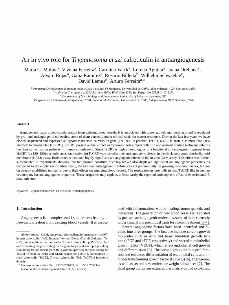
An in vivo role forTrypanosoma cruzicalreticulin in antiangiogenesis
Marıa C. Molinaa, Viviana Ferreiraa, Carolina Valcka, Lorena Aguilara, Juana Orellanaa,Alvaro Rojasa, Galia Ramireza, Rosario Billettab, Wilhelm Schwaeblec,
David Lemusd, Arturo Ferreiraa,∗a Programa Disciplinario de Inmunolog´ıa, ICBM, Facultad de Medicina, Universidad de Chile, Independencia, 1027 Santiago, Chile
b Androclus Therapeutics, 4202 Sorrento Valley Blvd, Suite E-D, San Diego, CA 92121-1412, USAc Department of Microbiology and Immunology, University of Leicester, Leicester, UK
d Programa Disciplinario de Morfolog´ıa, ICBM, Facultad de Medicina, Universidad de Chile, Independencia 1027, Santiago, Chile
Abstract
Angiogenesis leads to neovascularization from existing blood vessels. It is associated with tumor growth and metastasis and is regulatedb s we havec 0%i nd inhibitst ent fromH ioallantoidm as furthers , asc es, but noto e its humanc erimentalc
K
1
n
hCmsTcd
andlated
ently
d di-wthfac-
helialth
a-d in-,
nes,
y pro- and antiangiogenic molecules, some of them currently under clinical trials for cancer treatment. During the last few yearloned, sequenced and expressed aTrypanosoma cruzicalreticulin gene (TcCRT). Its product, TcCRT, a 45 kDa protein, is more than 5dentical to human CRT (HuCRT). TcCRT, present on the surface of trypomastigotes, binds both C1q and mannan binding lectin ahe classical activation pathway of human complement. Since TcCRT is highly homologous to a functional antiangiogenic fragmuCRT (aa 120–180), recombinant (r) and native (n) TcCRT were tested in their antiangiogenic effects, in the chick embryonic chorembrane (CAM) assay. Both proteins mediated highly significant antiangiogenic effects in the in vivo CAM assay. This effect w
ubstantiated in experiments showing that the plasmid construct pSecTag/TcCRTalso displayed significant antiangiogenic propertiesompared to the empty vector. Most likely, the fact that antiangiogenic substances act preferentially on growing neoplasic tissun already established tumors, is due to their effects on emerging blood vessels. The results shown here indicate that TcCRT, likounterpart, has antiangiogenic properties. These properties may explain, at least partly, the reported antineoplasic effect of expT.ruzi infection.
eywords: Trypanosoma cruzi; Calreticulin; Antiangiogenesis
. Introduction
Angiogenesis is a complex multi-step process leading toeovascularization from existing blood vessels. It is associ-
Abbreviations: CAM, embryonic chorioallantoid membrane; HuCRT,uman calreticulin; IWB, Immuno Western Blots; kDa, kiloDaltons; nTc-RT, immunoaffinity purified nativeT. cruzicalreticulin; pGM-CSF, plas-id expressing the gene coding for the granulocyte and macrophage colony
timulating factor; pSecTag/TcCRT, plasmid expressing the gene coding forcCRT without its leader and KEDL sequences; rTcCRT, recombinantT.ruzi calreticulin; TcCRT,T. cruzi calreticulin; TcS, TcCRT S functionalomain∗ Corresponding author. Tel.: +56 2 6786724; fax: +56 2 7353346.E-mail address:[email protected] (A. Ferreira).
ated with inflammation, wound healing, tumor growth,metastasis. The generation of new blood vessels is reguby pro- and antiangiogenic molecules, some of them currunder clinical and preclinical trials for cancer treatment[1–4].
Several angiogenic factors have been identified anvided into three groups. The first one includes soluble gromolecules such as acid and basic fibroblast growthtors (aFGF and bFGF, respectively) and vascular endotgrowth factor (VEGF), which affect endothelial cell growand differentiation[5]. The second group inhibits prolifertion and enhances differentiation of endothelial cells ancludes transforming growth factor� (TGF�) [6], angiogeninas well as several low-molecular weight substances[7]. Thethird group comprises extracellular matrix-bound cytoki
M.C. Molina et al.
which may contribute to angiogenic regulation[7]. The pre-vailing notion is that angiogenesis is governed by a balanceamong positive and negative regulators and the microenvi-ronment[1].
During the last few years we have cloned, sequenced andexpressed aTrypanosoma cruzicalreticulin gene (TcCRT)that was localized in several chromosomes. We have namedits product TcCRT, a 45 kDa, electrophoretically dimorphicand immunogenic protein[8–12], more than 50% identicalto the human counterpart (HuCRT)[10,13,14]. TcCRT ispresent on the surface of trypomastigotes and binds both C1qand mannan binding lectin (MBL), resulting in the inhibitionof the classical activation pathway of the human complementsystem[15].
Over 40 functions have been described for HuCRT[13,14]. Three fragments derived from HuCRT (aa 120–180,aa 1–180 or vasostatin, and aa 120–400 or D120CRT), as wellas the whole molecule, display antiangiogenic effects. Theyinhibit the proliferation of endothelial cells, the generationof new blood vessels and in vitro and in vivo tumoral growth[16–19]. Specifically, vasostatin inhibits the endothelial cellbinding to laminin, thus reducing the basic fibroblast growthfactor (bFGF)—dependent proliferation of these cells[16].In TcCRT we find segments with 46% identity and 60% posi-tivity with a functional fragment from HuCRT (aa 120–180).
edi , in-s les,a ven-t els isc atorsh s as-s neo-v say[ soneh hisa T, asw siblye fectso
2
2
li-b dK am-p o-vC actt mers.RBp on-
taining 34 mg× ml−1 chloramphenicol and 50 mg× ml−1
kanamycin) at 37◦C, until reaching an O.D. of 0.5 at 600 nm.rTcCRT expression was induced with 1 mM IPTG, during 3 h,at 37◦C and the protein was purified by affinity chromatogra-phy in a nickel column (HisBind Resin, Novagen, Darmstadt,Germany). Purity of rTcCRT was determined both by sodiumdodecyl sulphate-polyacrylamide gel electrophoresis (SDS-PAGE) and by immunowestern blotting (IWB), developedwith an anti-TcCRT monoclonal antibody (E2G7)[10] andalso with rabbit polyclonal antibodies anti-rTcCRT and antiits S functional domain (TcS, aa 159–281)[15], all generatedin our laboratory. A whole epimastigote extract was used as apositive control in the IWB, as described in[10]. The recom-binant protein preparations were tested for the presence ofcontaminating endotoxin (Limulus Amebocyte Lysate, Pyro-gent Plus® kit, BioWhittaker Inc., Cambrex Company, MD,USA).
2.2. Native TcCRT
Native TcCRT (nTcCRT) was purified by immunoaffin-ity chromatography, from a whole epimastigote (Tulahuenstrain) extract, in a cyanogen bromide-activated Sepharose(Pharmacia, Stockholm, Sweden) column, prepared withpolyclonal rabbit antibodies anti-rTcCRT. Purity of nTcCRTw
2
tsl ag2Bvse trol.
enec tingf onalC B),T ereo c-T on-v
2
saywa nh hile)w incu-b t-i thee helld rar-i batedf iscs
In the chorioallantoic membrane (CAM) assay, performn fertilized chicken eggs, sterile methyl cellulose discstilled with solutions of pro- or anti-angiogenic molecure placed on the embryonic membrane. Using con
ional histological procedures, the number of blood vessounted in the areas adjacent to the filters. Many investigave studied the histological and morphological changeociated with the proliferation of new vessels and tumorascularization by direct observation using the CAM as20–25]. For example, the angiostatic effect of betamethaas been reported[26,27]by using this assay. Here, using tssay, we show that both native and recombinant TcCRell as its coding gene, have antiangiogenic effects, posxplaining, at least in part, the reported antineoplasic eff experimentalT. cruziinfection[28–31].
. Materials and methods
.1. Recombinant TcCRT
DNA coding for TcCRT was isolated from a genomicrary in the�gt11 phage[10]. TcCRT, without its leader anEDL endoplasmic reticulum retention sequence, waslified by PCR and ligated in the pET-28b(+) vector (Nagen, Darmstadt, Germany) inEcoRI restriction sites.Tc-RTwas amplified by using forward (gga att cca cgg tgt
cc acg ag) and reverse (tgg aat tcg ggt gca gca att tt) priecombinant TcCRT (rTcCRT) was expressed fromE.coliL21(DE3)pLysS, transformed with the pET-28b(+)/TcCRTlasmid and cultivated in Luria Bertani medium (c
as determined as described in Section2.1.
.3. Plasmid constructs
The PCR-amplifiedTcCRT gene product, without ieader and KEDL sequences, was ligated to the pSecTector (Invitrogen, La Jolla, CA, USA) inEcoRI restrictionites. This construct was designated as pSecTag/TcCRT. Thempty vector (pSecTag2B) was used as a negative con
A pGM-CSF plasmid, containing and expressing the goding for the granulocyte–macrophage colony stimulaactor was donated by Dr. Oscar R. Burrone (Internatienter for Genetic Engineering and Biotechnology (ICGErieste, Italy). Endotoxin free plasmid preparations wbtained fromE.coli DH5�, transformed with the pSeag/TcCRT, pSecTag2B or pGM-CSF plasmids, using centional technology[32,33].
.4. Chick embryonic chorioallantoid membrane assay
The chick embryonic chorioallantoid membrane asas performed as previously described by us[26,27]and bynother laboratory[34]. Briefly, 10 fertilized White Leghoren eggs (National Public Health Institute, Santiago, Cere used in protein and DNA assays. The eggs wereated for 48 h in a humid 38.5◦C atmosphere. After extrac
ng 2–3 ml of albumin, a small window was opened ingg, in order to allow separation of the CAM from the suring the embryo development. The window was tempo
ly sealed with adherent tape and the eggs were incuor additional 5 days. Then, sterile methyl cellulose d

M.C. Molina et al.
(5 mm diameter, 0.25�m pore size, 125�m thickness) (Ad-vantec MFS Inc., CA, USA), were deposited on the CAMs.Immediately, 10�l of the corresponding samples were di-rectly added onto the filters. The windows were then sealedand the eggs were incubated for additional 72 h, as indicatedabove. Then, the CAMs were sliced, following the filters con-tours, and fixed in 10% (v/v) formaldehyde. Tissue sectionswere prepared, by standard procedures, for conventional lightmicroscopy aimed at detecting mainly acid polysaccharides,nuclei and cytoskeleton. Blood vessels were counted in a lightmicroscope with a 1 cm2 micrometric grid, divided in 1 mm2
sections. Ten of these sections, corresponding to a tissue areaof 9000�m2 were counted. Blood vessels were identifiedby their histological characteristics, mainly by their endothe-lial cells, and by the presence of red blood cells in their lu-mens. Counting, carried out in a double-blind fashion, wasperformed in 35 microscopic fields of CAM tissue segments,adjacent to the filter edge.
The above procedures were used in three different exper-imental approaches:
(a) In one experiment, in order to build a dose–responsecurve, rTcCRT amounts, ranging between 1–1000 pg,were tested. Separate filters contained 10 pg of nTcCRT.
(b) A second experiment was designed in order to test thepossible role of contaminating lipopolysaccharide (LPS)
con-tam-tersride
( herFil-es-di-
ofgagell
freed etha-s ti-i ties[
2p ofm
s ce ah icei
construct. As a negative control, mice were immunized withthe empty vector, pSecTag2B, under the same conditions.
The presence of specific anti TcCRT antibodies in thesemurine sera (a demonstration that the pSecTag2B/TcCRTconstruct was indeed productive in vivo) was detected in twoindependent experimental approaches: The first one involvedflow cytometric indirect immunofluorescence assays, against4% paraformaldehyde-fixed and live MF strainT. cruzi try-pomastigotes (donated by Dr. Gittith Sanchez, Cellular andMolecular Biology Program, ICBM, Faculty of Medicine,University of Chile), as previously described in our labora-tory [15]. In the second approach, the murine anti TcCRT an-tibodies were detected in a conventional ELISA[37], againstthe recombinant molecule.
2.6. Statistical analysis
Conventional one-tailed Student’st tests were used to as-sess the statistical significance of the antiangiogenic effects.Data are expressed as their means and standard errors. Exactp-values are provided.
3. Results
3 dI
na-l1 and,w
nala ain6 tract,w
mw im-m Dac wasd
nti-T RTa re notd 2).
rec-o pa-r bleiW nti-b cCRT( tion,a y-c cko nala cor-
in the recombinant protein preparations. Some filterstained 4.9 and 49 ng of rTcCRT, endogenously coninated with 0.02 and 0.2 endotoxin EUs. Other filcontained only 0.02 and 0.2 EU of lipopolysaccha(Sigma, MO, USA).
c) A third experiment was designed in order to furtsubstantiate the antiangiogenic effect of TcCRT.ters containing alternatively 10�g of the endotoxin-freDNA pSegTag/TcCRTor 10�g pSecTag2B empty plamid, used as negative control. DNA plasmids wereluted in endotoxin-free water (Laboratorio Chile®, San-tiago, Chile). In order to maximize the stringencythis experiment, 10�g of a pGM-CSF plasmid, codinfor the proangiogenic cytokine granulocyte/macrophcolony stimulating factor[35,36], were added to aDNA-containing filters.
In all experiments, PBS prepared with endotoxin-istilled water was used as a negative control. Betamone (Laboratorio Chile®, Santiago, Chile), a steroidal an
nflamatory agent, with known anti-angiogenic proper26,27], was used as a positive control.
.5. In vivo validation of the productive properties of theSecTag/TcCRT construct using genetic immunizationice
The productive properties of the pSecTag/TcCRTcon-truct were validated by assessing its capacity to induumoral polyclonal anti-TcCRT response in BALB/c m
mmunized three times i.m., every 15 days, with 50�g of the
.1. Analysis of rTcCRT and nTcCRT by SDS-PAGE anWB
rTcCRT and immunoaffinity purified nTcCRT were ayzed in Coomasie Blue stained SDS-PAGE (Fig. 1A, lanes
and 2, respectively). rTcCRT showed a main 60 kDa bhile nTcCRT presented a single 46 kDa component.In Fig. 1B, an IWB developed with the E2G7 monoclo
ntibody, the antigens were rTcCRT, visualized as a m0 kDa component (lane 1), and whole epimastigote exhere a 46 kDa band was seen (lane 2).In Fig. 1C, an anti-rTcCRT rabbit polyclonal antiseru
as used to develop the IWB, and the antigens wereunoaffinity purified nTcCRT (visualized as a main 46 k
omponent) and rTcCRT, where a main 60 kDa bandetected (lanes 1 and 2, respectively).
In Fig. 1D (lane 1), the IWB was developed with an acS rabbit polyclonal antiserum, the antigen was rTcCnd a main 60 kDa band was observed. These bands weetected by the corresponding preimmune serum (lane
A 71 kDa band was also prominent and consistentlygnized by both polyclonal antisera in the rTcCRT preations (Fig. 1C (lane 2) and D (lane 1)) and was visin a Coomasie Blue stained SDS-PAGE (Fig. 1A (lane 1)).
ith one exception, both monoclonal and polyclonal aodies, recognized the same degradation products in rTFig. 1B–D, lanes 1, 2, and 1, respectively). The excep
29 kDa band (Fig. 1A (lane 1)), reacted with both pollonal antibodies (Fig. 1C (lane 2) and D (lane 1)). The laf recognition of this 29 kDa band by the E2G7 monoclontibody was most likely due to enzymatic loss of the
M.C. Molina et al.
Fig. 1. Analysis of purity of recombinant (r) and native (n) TcCRT by SDS-PAGE and IWB. (A) Coomasie Blue stained SDS-PAGE showing 10�grTcCRT (lane 1) and 3�g nTcCRT; (B) IWB developed with the mono-clonal antibody E2G7 for the detection of rTcCRT (lane 1) and native Tc-CRT, present in an epimastigote extract (lane 2); (C) IWB of immunoaffinitypurified nTcCRT (lane 1) and rTcCRT (lane 2), developed with rabbit poly-clonal antibodies anti-rTcCRT; (D) IWB of rTcCRT developed with rabbitpolyclonal antibodies anti the S domain of TcCRT (lane 1) and with the cor-responding preimmune serum (lane 2). Molecular weight standards (MWstd) are indicated at the left side of each panel.
responding epitope. None of the bands, recognized only bythe rabbit polyclonal antibodies (71 and 29 kDa), decreasedtheir intensity after exhaustive absorption (three times, 2 heach, at 37◦C) with saturated, solid phase-bound, wholeE.coli extract (results not shown).
3.2. Antiangiogenic effect of TcCRT, as determined inthe CAM assay
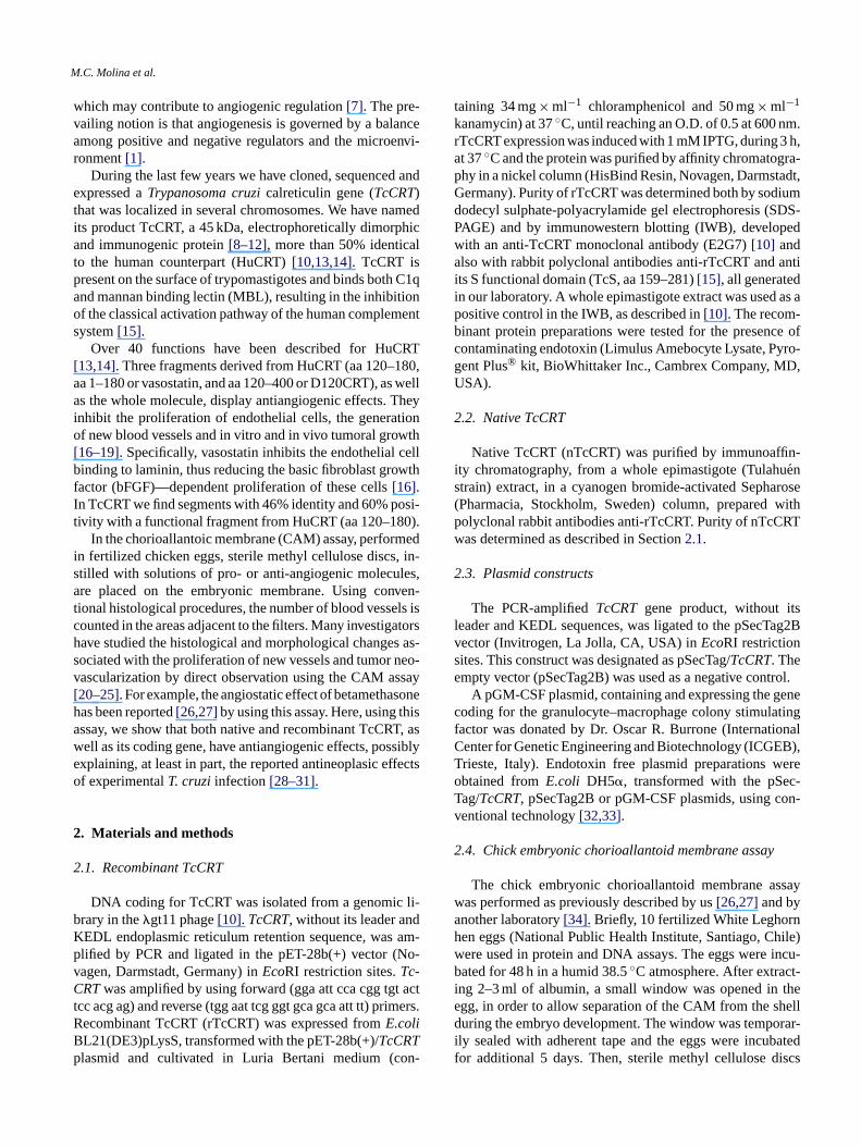
Fig. 2 summarizes the antiangiogenic properties of rTc-CRT and nTcCRT, as determined in the CAM assay. 10 pgof nTcCRT and 1–1000 pg of rTcCRT were assayed. PBSand 10 ng betamethasone were used as negative and positivcontrols, respectively. A dose/response relationship betweenseveral rTcCRT concentrations and angiogenesis inhibitionwas obtained. At the extreme concentrations (1 pg–1 ng) ofthe recombinant parasite molecule, angiogenesis inhibitionsof 35% (p= 6× 10⁻⁹) and 55% (p= 10−16) were respectivelyobserved, while nTcCRT, at 10 pg per filter, induced about
Table 1Antiangiogenic effect of rTcCRT vs. LPS endotoxin, in a CAM assay
Group Quantity per filter % Inhibition
LPS 0.02 EU 39.720.2 EU 48.50
rTcCRT + (LPS) 4.9 ng (0.02 EU) 65.9949 ng (0.2 EU) 68.56
p(rTcCRT (4.9 ng) vs. LPS (0.02 EU))= 4.5× 10⁻⁷;p(rTcCRT (49 ng) vs. LPS(0.2 EU))= 1.4× 10⁻³.
30% inhibition (p= 6× 10⁻⁷). Intermediate concentrationsof rTcCRT also induced different inhibition degrees, thus sta-tistically validating a dose-response relationship, as indicatedin Fig. 2.
Since endotoxin (LPS), with known antiangiogenic effects[38] often contaminatesE. coli recombinant protein prepa-rations, we determined the levels of endogenous LPS con-tamination present in our samples, as described in Section2.1. We then carried out the CAM assays including eggs towhich only pure LPS was added to the filters, in the same con-centrations present in our rTcCRT preparations. As shown inTable 1, 4.9 ng of rTcCRT containing 0.02 EU of endogenousendotoxin, showed a significantly higher (p= 4.5× 10⁻⁷) an-tiangiogenic effect than 0.02 EU pure LPS. Moreover, 49 ngof rTcCRT containing 0.2 EU of endogenous endotoxin alsomediated a significantly higher (p= 1.4× 10⁻³) antiangio-genic effect as compared to 0.2 EU pure LPS.
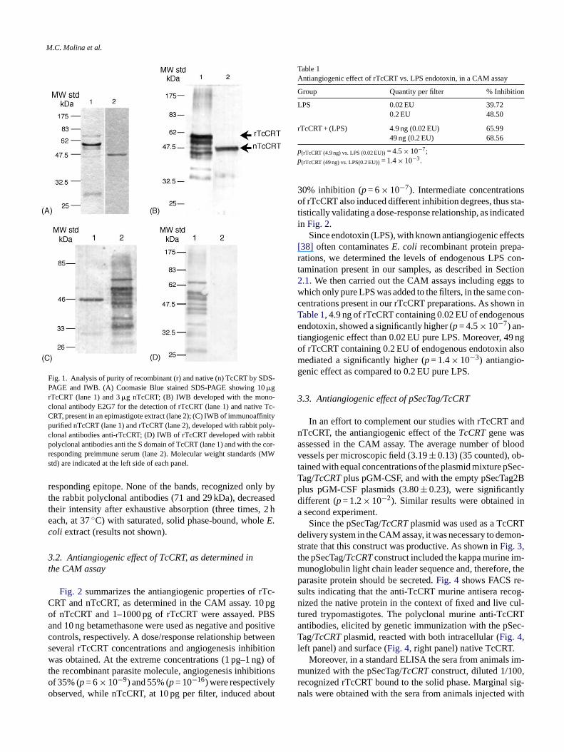
3.3. Antiangiogenic effect of pSecTag/TcCRT
In an effort to complement our studies with rTcCRT andnTcCRT, the antiangiogenic effect of theTcCRTgene wasassessed in the CAM assay. The average number of bloodvessels per microscopic field (3.19± 0.13) (35 counted), ob-tained with equal concentrations of the plasmid mixture pSec-T 2Bp yd ina
RTd mon-st m-m , thep -s cog-n cul-t RTa ec-Tl
im-m ,r sig-n with
e
ag/TcCRTplus pGM-CSF, and with the empty pSecTaglus pGM-CSF plasmids (3.80± 0.23), were significantlifferent (p= 1.2× 10⁻²). Similar results were obtainedsecond experiment.Since the pSecTag/TcCRTplasmid was used as a TcC
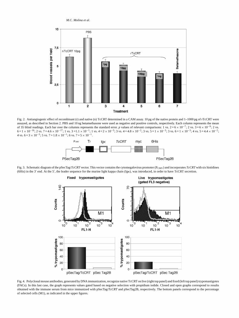
elivery system in the CAM assay, it was necessary to detrate that this construct was productive. As shown inFig. 3,he pSecTag/TcCRTconstruct included the kappa murine iunoglobulin light chain leader sequence and, thereforearasite protein should be secreted.Fig. 4 shows FACS reults indicating that the anti-TcCRT murine antisera reized the native protein in the context of fixed and live
ured trypomastigotes. The polyclonal murine anti-TcCntibodies, elicited by genetic immunization with the pSag/TcCRTplasmid, reacted with both intracellular (Fig. 4,
eft panel) and surface (Fig. 4, right panel) native TcCRT.Moreover, in a standard ELISA the sera from animals
unized with the pSecTag/TcCRTconstruct, diluted 1/100ecognized rTcCRT bound to the solid phase. Marginalals were obtained with the sera from animals injected
M.C. Molina et al.
Fig. 2. Antiangiogenic effect of recombinant (r) and native (n) TcCRT determined in a CAM assay. 10 pg of the native protein and 1–1000 pg of rTcCRT wereassayed, as described in Section2. PBS and 10 ng betamethasone were used as negative and positive controls, respectively. Each column represents the meanof 35 blind readings. Each bar over the columns represents the standard error.p values of relevant comparisons: 1 vs. 2 = 6× 10⁻⁷; 2 vs. 3 = 6× 10⁻⁹; 2 vs.6 = 1× 10⁻¹⁶; 2 vs. 7 = 4.6× 10⁻¹⁷; 1 vs. 3 =1.1× 10⁻¹; 1 vs. 4 = 2× 10⁻³; 3 vs. 4 = 4.8× 10⁻²; 3 vs. 5 = 1× 10⁻³; 3 vs. 6 = 1× 10⁻³; 4 vs. 5 = 4.4× 10⁻²;4 vs. 6 = 3× 10⁻³; 5 vs. 7 = 1.8× 10⁻¹; 6 vs. 7 = 5× 10⁻¹.
Fig. 3. Schematic diagram of the pSecTag/TcCRTvector. This vector contains the cytomegalovirus promoter (PCMV) and incorporatesTcCRTwith six histidines(6His) in the 3′ end. At the 5′, the leader sequence for the murine light kappa chain (Ig�), was introduced, in order to have TcCRT secretion.
Fig. 4. Polyclonal mouse antibodies, generated by DNA immunization, recognize native TcCRT on live (right top panel) and fixed (left top panel) trypomastigotes(FACs). In this last case, the graph represents values gated based on negative selection with propidium iodide. Closed and open graphs correspond to resultsobtained with the immune serum from mice immunized with pSecTag/TcCRTand pSecTag2B, respectively. The bottom panels correspond to the percentageof selected cells (M1), as indicated in the upper figures.
M.C. Molina et al.
the empty vector (p= 1.6× 10⁻⁶, for the comparison of bothgroups).
4. Discussion
Several reports[28–31] indicate thatT. cruzi infectionmay have antineoplasic effects. On the other hand, neopla-sic growth and metastasis are intimately related with neoan-giogenesis[1,3,4], and human calreticulin (HuCRT) has im-portant antiangiogenic properties[16–19]. Since inT. cruzicalreticulin (TcCRT) there is a segment with 46% identityand 60% positivity with a functional antiangiogenic fragmentfrom HuCRT (aa 120–180), we asked whether the parasitemolecule shares this property.
Fig. 2 andTable 1show that, in the CAM assay, the dif-ference in the number of blood vessels between the groupstreated with rTcCRT and their controls is highly significant, afact further substantiated by the use of the native purified par-asite protein (nTcCRT). The dose-response curve includedin Fig. 2 (columns 2–6) shows significant differences notonly between the extreme TcCRT amounts, but also amongmost of the intermediate doses. Most important, nTcCRT hasa highly significant antiangiogenic effect (p= 6× 10⁻⁷), al-though lower than the recombinant counterpart. Since nTc-C nt tivitym (ex-p
ene-s antss ld bea mo-g2 anta
ntedi6 rTc-C on-o eo l epi-m ationd se int oid-a omoo ivedIp botho tedb RTa tiont inantd tioni oper
Since all our polyclonal antisera recognize the 29 kDaband, the lack of detection by the AcMo could be due toenzymatic loss of the relevant epitope. This possibility isconsistent with the fact that the recombinant protein is moresusceptible to degradation than the native counterpart, an is-sue evident when tracks 1A versus 2A, 1B versus 2B, and 2Cversus 1C, are compared inFig. 1.
Moreover, the possibility that the 71 and 29 kDamolecules, detected in rTcCRT, arise fromE. coli, is highlyunlikely. We prepared a solid-phase immunoabsorbent withexcess total bacterial antigens. With this preparation we ex-haustively absorbed our polyclonal antiserum, at the working1/500 dilution (three rounds, 2 h each, at 37◦C). When usedin Westerns, both the absorbed and non-absorbed reagentsdetected the 71 and 29 kDa bands with equal intensity. Like-wise, the recognition of rTcCRT was not altered (results notshown).
The observed molecular weight of the recombinant proteinis higher than that of the native protein (Fig. 1) since theprocedure to obtain the recombinant protein involves, amongother, the addition of extra aa from the plasmid vector and ahistidine tail, for purification purposes. These extra aa, couldexplain at least an important part of the observed difference.
Given the known antiangiogenic effects of endotoxin(LPS)[38], a frequent contaminant inE. coli-derived recom-b f en-d cribedi ud-i s, int tions.A itho antp atingL
re-s ara-sc ct int whent ,t ase,a r na-t mi-t SF)i pertyo nem ruma ionalE bod-i ativem inte-r ssi-b tructm ain-i arei T, in
RT was purified fromT. cruzi epimastigotes, not knowo contain endogenous LPS, a residual endotoxin acay explain the higher antiangiogenic effect of rTcCRTressed inE. coli).
When dealing with molecules that modulate angiogis, their purity is of utmost relevance. Possible contaminhould be identified and their effect on angiogenesis shoussessed. Immunoaffinity purified nTcCRT, showing hoeneous behavior in SDS-PAGE and in IWB (Fig. 1A (lane) and C (lane 1)), had an important and highly significntiangiogenic effect.
The recombinant protein used in the studies preses over 90% pure, as shown by SDS-PAGE (Fig. 1A). The0 kDa protein band, along with bands corresponding toRT degradation, are recognized by the anti-TcCRT mclonal antibody (E2G7) in IWB (Fig. 1B(lane 1)). On thther hand, E2G7 recognizes a single band in the totaastigote extract. Since the recombinant protein preparoes not contain protease inhibitors, due to its intended u
he in vivo CAM assays, degradation products are unavble. Likewise, the generation of variable amounts of hligomers is a frequent finding in SDS-PAGE and der
WB technologies. Our results, shown inFig. 1, detect theresence of two bands (apparent Mws 71 and 29 kDa),f them not recognized by the MoAb, but clearly detecy the two polyclonal rabbit antibodies used (anti-rTcCnd anti-TcS). We have also observed this oligomeriza
endency with the TcS and TcR (aa 136–281) recombomains of TcCRT (results not shown). Thus, aggrega
n the 71 kDa band could result in masking of the epitecognized by the AcMo.
inant protein preparations, we determined the levels oogenous contamination present in our samples, as des
n Section2.1. We then carried out the CAM assays inclng eggs to which only pure LPS was added to the filterhe same concentrations present in our rTcCRT preparas shown inTable 1, the antiangiogenic effect observed wur rTcCRT preparation is mainly due to the recombinarasite molecule and, to a lesser extent, to contaminPS.
In an effort to further support the antiangiogenicults obtained with both the recombinant and native pite molecules, we asked whether theTcCRTgene, in theontext of a productive plasmid, mediates a similar effehe CAM assay. The degree of significance decreasesreatment is performed with theTcCRTgene. Most likelyhe actual amount of available protein is lower in this cs compared with the situation when the recombinant o
ive proteins are directly provided. Alternatively or concoantly, the presence of a proangiogenic cytokine (GM-Cncreased the stringency of the assay. The productive prof the pSecTag/TcCRTconstruct, in an eukaryotic muriodel, is demonstrated by the detection of specific sentibodies against the parasite molecule in a conventLISA against the recombinant molecule. These anti
es were also detected by FACS analysis, where the nolecule is recognized both at the parasite surface and
ior (Fig. 4). The results of this experiment support the poility that, in the CAM assay used here, this genetic consediates TcCRT production by the CAM cells, thus expl
ng the observed inhibition of angiogenesis. Our resultsn accordance with those previously described for HuCR

M.C. Molina et al.
which antiangiogenic effects were detected under the sameexperimental conditions[34]. As mentioned above and in pre-vious results from our laboratory[15], TcCRT is expressedon the trypomastigote surface, in spite of having the KEDLendoplasmic reticulum retention signal and lacking a trans-membrane region, a fact also observed with mammal CRT[39,40].
Most likely, the fact that antiangiogenic substances actpreferentially on growing neoplasic tissues and not on al-ready established tumors, is due to their effects on emergingblood vessels[41]. This may explain why in the CAM as-say used, rTcCRT mediated an antiangiogenic effect that,although highly significant, reached a maximum of approx-imately 50%. In other words, it is possible that most of theremaining vessels were already established and thus resistantto the parasite molecule.
For decades, it has been speculated about possible mecha-nisms involved in the in vivo experimental growth inhibitoryeffect that severalT. cruzistrains have on a variety of trans-planted and spontaneous tumors, in animals and humans[28,29,31]. In order to explain the antineoplasic effect ofT.cruzi infection, induction of a specific anti-tumoral immuneresponse[30] and secretion of a “toxic substance”[29] bythe parasite have been proposed, but not experimentally sub-stantiated. Since theT. cruzilife cycle involves an importanti pref-e arec eo-p fort
RT[ thei cen-t rties,t po-m orted[t ultsw evi-d inanta
A
069,f B,T rp maO
R
other
[2] Risau W. Mechanisms of angiogenesis. Nature 1997;386:671–4.[3] Carmeliet P, Jain RK. Angiogenesis in cancer and other diseases.
Nature 2000;407:249–57.[4] Jain RK. Normalizing tumor vasculature with anti-angiogenic
therapy: a new paradigm for combination therapy. Nat Med2001;7:987–9.
[5] Burgess WH, Maciag T. The heparin-binding (fibroblast) growthfactor family of proteins. Annu Rev Biochem 1989;58:575–606.
[6] Pepper MS, Belin D, Montesano R, Orci L, Vassalli JD. Transform-ing growth factor-beta 1 modulates basic fibroblast growth factor-induced proteolytic and angiogenic properties of endothelial cells invitro. J Cell Biol 1990;111:743–55.
[7] Meininger GA, Davis MJ. Cellular mechanisms involved in the vas-cular myogenic response. Am J Physiol 1992;263:H647–59.
[8] Aguillon JC, Bustos C, Vallejos P, et al. Purification and prelimi-nary sequencing of Tc-45, an immunodominantTrypanosoma cruziantigen: absence of homology with cruzipain, cruzain, and a 46-kiloDalton protein. Am J Trop Med Hyg 1995;53:211–5.
[9] Aguillon JC, Harris R, Molina MC, et al. Recognition of an im-munogenetically selectedTrypanosoma cruziantigen by seropositivechagasic human sera. Acta Trop 1997;63:159–66.
[10] Aguillon JC, Ferreira L, Perez C, et al. Tc45, a dimorphicTry-panosoma cruziimmunogen with variable chromosomal localization,is calreticulin. Am J Trop Med Hyg 2000;63:306–12.
[11] Marcelain K, Colombo A, Molina MC, et al. Development of an im-munoenzymatic assay for the detection of human antibodies againstTrypanosoma cruzicalreticulin, an immunodominant antigen. ActaTrop 2000;75:291–300.
[12] Aguillon JC, Hermosilla T, Molina MC, et al.Trypanosoma cruzi: H2une
.[ cal-
log
[ munol
[ tionhib-:
[ ag-Biol
[ rag-p Med
[ frag-wth.
[ andiculin
[ very
[ ions onthol
[ nesisfts ininol
[ od-ases
ntracellular phase, this putative molecule should have arential toxic effect for neoplasic cells. Both explanationsompatible with an evolutionary speculation that this antinlasic effect would protect the host, with evident benefits
he parasite.In synthesis, our results indicate that, similar to HuC
16–19,42,43], TcCRT has an antiangiogenic effect inn vivo CAM assay proposed here, even at the low conrations tested. It could be speculated that these propeogether with the accessibility of this molecule on the tryastigote surface, may explain, at least partly, the rep
30,31] antineoplasic effect of experimentalT. cruzi infec-ion, an intriguing and maybe important issue. The resith DNA, although less telling, are presented here asence that supports the results obtained with the recombnd native parasite molecules.
cknowledgements
This work was supported by grants 1010930 and 2010rom FONDECYT, Chile, and CRP/CHI02-01, from ICGErieste, Italy. We sincerely thank Dr. Gittith Sanchez, foroviding us with cultured trypomastigotes and Mrs. Irrellana, for excellent technical assistance.
eferences
[1] Folkman J. Angiogenesis in cancer, vascular, rheumatoid anddisease. Nat Med 1995;1:27–31.
complex and genetic background influence on the humoral immresponse against epimastigotes. Int J Parasitol 2000;30:981–4
13] Ferreira V, Molina MC, Valck C, Rojas A, Ferreira A. Parasitereticulin: possible roles in the parasite/host interface. Inmunoıa2002;21:156–68.
14] Ferreira V, Molina MC, Valck C, et al. Role of calreticulin froparasites in its interaction with vertebrate hosts. Mol Imm2004;40:1279–91.
15] Ferreira V, Valck C, Sanchez G, et al. The classical activapathway of the human complement system is specifically inited by calreticulin fromTrypanosoma cruzi. J Immunol 2004;1723042–50.
16] Yao L, Pike SE, Tosato G. Laminin binding to the calreticulin frment vasostatin regulates endothelial cell function. J Leukoc2002;71:47–53.
17] Pike SE, Yao L, Jones KD, et al. Vasostatin, a calreticulin fment, inhibits angiogenesis and suppresses tumor growth. J Ex1998;188:2349–56.
18] Pike SE, Yao L, Setsuda J, et al. Calreticulin and calreticulinments are endothelial cell inhibitors that suppress tumor groBlood 1999;94:2461–8.
19] Cheng WF, Hung CF, Chai CY, et al. Tumor-specific immunityantiangiogenesis generated by a DNA vaccine encoding calretlinked to a tumor antigen. J Clin Invest 2001;108:669–78.
20] Folkman J, Ingber DE. Angiostatic steroids. Method of discoand mechanism of action. Ann Surg 1987;206:374–83.
21] Mostafa LK, Jones DB, Wright DH. Mechanism of the inductof angiogenesis by human neoplastic lymphoid tissue: studiethe chorioallantoic membrane (CAM) of the chick embryo. J Pa1980;132:191–205.
22] Petruzzelli GJ, Snyderman CH, Johnson JT, Myers EN. Angiogeinduced by head and neck squamous cell carcinoma xenograthe chick embryo chorioallantoic membrane model. Ann Otol RhLaryngol 1993;102:215–21.
23] Buttgereit F, Wehling M, Burmester GR. A new hypothesis of mular glucocorticoid actions: steroid treatment of rheumatic diserevisited. Arthritis Rheum 1998;41:761–7.
M.C. Molina et al.
[24] Quigley JP, Armstrong PB. Tumor cell intravasation alu-cidated: thechick embryo opens the window. Cell 1998;94:281–4.
[25] Ribatti D, Conconi MT, Nico B, et al. Angiogenic response inducedby acellular brain scaffolds grafted onto the chick embryo chorioal-lantoic membrane. Brain Res 2003;989:9–15.
[26] Lemus D, Dabancens A, Illanes J, et al. Antiangiogenic effect of be-tamethasone on the chick cam stimulated by TA3 tumor supernatant.Biol Res 2001;34:227–36.
[27] Zuniga J, Fuenzalida M, Guerrero A, et al. Effects of steroidal andnon-steroidal drugs on the neovascularization response induced bytumoral TA3 supernatant on CAM from chick embryo. Biol Res2003;36:233–40.
[28] Roskin GI. Toxin therapy of experimental cancer. Cancer Res1946;6:363–5.
[29] Hauschka TS, Goodwin MB.Trypanosoma cruziendotoxin (KR) inthe treatment of malignant mouse tumors. Science 1948;107:600–2.
[30] Cabral HR. The tumoricidal effect ofTrypanosoma cruzi: its intra-cellular cycle and the immune response of the host. Med Hypotheses2000;54:1–6.
[31] Oliveira EC, Leite MS, Miranda JA, et al. ChronicTry-panosoma cruziinfection associated with low incidence of 1,2-dimethylhydrazine-induced colon cancer in rats. Carcinogenesis2001;22:737–40.
[32] Sambrook J, Fritsch EF, Maniatis T. Molecular Cloning: a LaboratoryManual. 2 ed. Cold Spring Harbor: Cold Spring Harbor LaboratoryPress; 1989.
[33] Aida Y, Pabst MJ. Removal of endotoxin from protein solutionsby phase separation using Triton X-114. J Immunol Methods1990;132:191–5.
[34] Xiao F, Wei Y, Yang L, et al. A gene therapy for cancer based onthe angiogenesis inhibitor, vasostatin. Gene Ther 2002;9:1207–13.
[35] Bikfalvi A, Han ZC. Angiogenic factors are hematopoietic growthfactors and vice versa. Leukemia 1994;8:523–9.
[36] Valdembri D, Serini G, Vacca A, Ribatti D, Bussolino F. In vivoactivation of JAK2/STAT-3 pathway during angiogenesis induced byGM-CSF. Faseb J 2002;16:225–7.
[37] Engvall E, Perlman P. Enzyme-linked immunosorbent assay(ELISA). Quantitative assay of immunoglobulin G. Immunochem-istry 1971;8:871–4.
[38] Pipili-Synetos E, Kritikou S, Papadimitriou E, et al. Nitric oxidesynthase expression, enzyme activity and NO production during an-giogenesis in the chick chorioallantoic membrane. Br J Pharmacol2000;129:207–13.
[39] Arosa FA, de Jesus O, Porto G, Carmo AM, de Sousa M. Calreticulinis expressed on the cell surface of activated human peripheral bloodT lymphocytes in association with major histocompatibility complexclass I molecules. J Biol Chem 1999;274:16917–22.
[40] Ghiran I, Klickstein LB, Nicholson-Weller A. Calreticulin is at thesurface of circulating neutrophils and uses CD59 as an adaptormolecule. J Biol Chem 2003;278:21024–31.
[41] Zetter BR. Angiogenesis and tumor metastasis. Annu Rev Med1998;49:407–24.
[42] Yao L, Pike SE, Pittaluga S, et al. Anti-tumor activities of the angio-genesis inhibitors interferon-inducible protein-10 and the calreticulinfragment vasostatin. Cancer Immunol Immunother 2002;51:358–66.
[43] Yao L, Pike SE, Setsuda J, et al. Effective targeting of tumor vascu-lature by the angiogenesis inhibitors vasostatin and interleukin-12.Blood 2000;96:1900–5.
Copyright © 2022 FDOKUMEN