Myxoma and Vaccinia Viruses Bind Differentially to Human Leukocytes

Two casein kinase 1 isoforms are differentially expressed inTrypanosoma cruzi�
Carmenza Spadafora a, Yolanda Repetto b, Cristina Torres a, Laura Pino b,Carlos Robello a,1, Antonio Morello b, Francisco Gamarro a, Santiago Castanys a,*
a Instituto de Parasitologıa y Biomedicina ‘Lopez-Neyra’, Consejo Superior de Investigaciones Cientıficas (C.S.I.C.), Calle Ventanilla 11, 18001
Granada, Spainb Programa de Farmacologıa Molecular y Clınica, Instituto de Ciencias Biomedicas, Facultad de Medicina, Universidad de Chile, Santiago de Chile,
Chile
Received 13 May 2002; received in revised form 23 July 2002; accepted 23 July 2002
Abstract
The cDNAs for two casein kinase 1 (CK1) homologues, TcCK1.1 and TcCK1.2 , have been isolated from Trypanosoma cruzi .
Both isoforms showed strong identity with other known CK1s. Their corresponding genes encode proteins of 312- and 330-amino
acid residues with apparent molecular weights of 16 and 37 kDa, respectively. TcCK1.1 is a two-copy gene while TcCK1.2 is
tandemly repeated, an arrangement not yet found in any other CK1. TcCK1.1 has been overexpressed in Escherichia coli and the
recombinant protein exhibited properties characteristic of the CK1 family. Northern blot indicated that both TcCK1s are expressed
differentially during the life stages of the parasite: the isoform TcCK1.1 shows low levels of mRNA expression in epimastigotes and
increased expression in trypomastigotes while TcCK1.2 presents an augmented expression in amastigotes as compared with the
other two life stages of the parasite. The CK1-like activity of amastigotes and trypomastigotes is significantly higher than that of
epimastigotes and, independent of the life stage of the parasite, a constitutive activity is observed which, in the epimastigote forms, is
found predominantly in the microsomal fraction. Also in the epimastigote forms, the CK1-like activity increases in the log phase of
growth of the parasites, and, through synchronization studies, this activity has been most conspicuously circumscribed to the S and
M phases of the cell cycle. # 2002 Elsevier Science B.V. All rights reserved.
Keywords: Casein kinase 1; Trypanosoma cruzi ; Cell cycle; Hymenialdisine; Differential expression
1. Introduction
The protozoan parasite Trypanosoma cruzi has a
complex life cycle, existing as extracellular flagellated
epimastigotes and metacyclic trypomastigotes in the
invertebrate host, and as trypomastigotes and intracel-
lular non-flagellated amastigotes in both phagocytic
(macrophages) and non-phagocytic cells such as fibro-
blasts, epithelial, endothelial muscle and nerve cells of
the vertebrate host. The intricate life cycle of T. cruzi
involves alternation between proliferative (epimastigotes
and amastigotes) and non-replicative (trypomastigotes)
stages. During its life cycle, the parasite encounters
changing environments that require rapid responses to
insure its survival. Little information is available about
the regulation of these complex cellular changes; how-
ever, as in higher eukaryotes, it is likely that these
protozoans control processes such as differentiation,
metabolism, growth, gene expression and other cellular
events through the reversible phosphorylation of pro-
teins.
Protein kinase CK1, also known as casein kinase 1
(CK1), is a family of multipotential Ser/Thr protein
kinases common to all eukaryotic cells. The ability to
Abbreviations: CK1, casein kinase 1; CKI-7, N -(-2-aminoethyl)-5-
chloroisoquinoline-8-sulphonamide; HD, hymenialdisine.�
Note: Nucleotide sequences data reported in this paper are
available in the GenBankTM database under the accession numbers
AF274060 and AF274059.
* Corresponding author. Tel.: �/34-958-805185; fax: �/34-958-
203911; http://www.ipb.csic.es
E-mail address: [email protected] (S. Castanys).1 Present address: Departamento de Bioquımica, Facultad de
Medicina, Universidad de la Republica, Montevideo, Uruguay.
Molecular & Biochemical Parasitology 124 (2002) 23�/36
www.parasitology-online.com
0166-6851/02/$ - see front matter # 2002 Elsevier Science B.V. All rights reserved.
PII: S 0 1 6 6 - 6 8 5 1 ( 0 2 ) 0 0 1 5 6 - 1

phosphorylate a wide variety of cellular proteins such as
cytoskeleton components, signaling molecules, meta-
bolic enzymes, proteins involved in mRNA translation,
enzymes involved in nucleic acid processing, and pro-teins involved in vesicular trafficking attests to the
importance of this multipotential protein kinase in cell
function [reviewed in 1 and 2]. CK1s are ubiquitous
proteins present in membrane, nucleus, cytoplasm,
vesicles, and cytoskeleton of eukaryotic cells. CK1s
have been generally isolated as monomer proteins with
variable molecular size (from 25 to 60 kDa) due to the
presence of many isoforms of the CK1 family encodedby different genes that differ in size. Seven members of
the CK1 family, and some of their splice variants, have
been cloned from mammalian cells (isoforms a, b, g1,
g2, g3, d, and o) [3�/6]. In Plasmodium falciparum a CK1
activity which is stage-specific regulated has been
described [7], and an ectokinase activity has been
associated with CK1 in Leishmania major [8] capable
of phosphorylating and inactivating molecules of thehuman complement system [9]. In Saccharomyces cer-
evisiae and Schizosaccharomyces pombe , eight different
CK1s have been characterized [10�/14]. One of these
genes, HRR25 of S. cerevisiae , has been described as a
CK1 involved in DNA repair and cell cycle control. In
S. pombe , the CK1 homologues Hhp1 and Hhp2 play
similar roles since single and double mutations in the
Hhp genes reveal DNA repair defects in these cells.Interestingly, RAG8, the CK1 isoform of Kluveromyces
lactis , seems to be an essential gene based on the
lethality of the rag8 null mutation [15]. Most recently,
Calabokis et al. [16] reported the biochemical character-
ization of a partially purified CK1-like activity in T.
cruzi , but the CK1 genes of this trypanosomatid had not
yet been isolated and a study of its function had not
been initiated until now. Due to the high degree ofconservation of this enzyme between lower and higher
eukaryotic isoforms, it is likely that findings on CK1
activity and function will hold for all members of this
subfamily of kinases and that the study of lower
eukaryotes will help to shed some light on the increas-
ingly interesting role of CK1 in mammals, particularly
with respect to its participation in neuropathologies [17]
circadian rhythm [18], and developmental processesthrough a positive regulation of the Wnt pathway [19].
The differences found, nonetheless, represent the key
point in which an important therapeutic window may
potentially open for the treatment of this parasitic
disease.
Here we report the molecular characterization, and
stage specific expression of T. cruzi casein kinase
homolog 1 (TcCK1.1 ) and homolog 2 (TcCK1.2 ) anda partial biochemical analysis of TcCK1.1. We under-
took the study of the relationship of CK1 to the
proliferative activities of T. cruzi . Also, the inhibitory
effectiveness of the marine sponge-isolated inhibitor of
CK1, hymenialdisine [20], was tested on T. cruzi
parasite extracts as well as on recombinant TcCK1.1.
2. Materials and methods
2.1. Chemicals
[g-32P]ATP (5000 Ci mmol�1) was obtained fromAmersham International; Ni2�-nitrilotriacetic acid
agarose (Ni-NTA) column was purchased from Qiagen;
mouse 6X-His monoclonal antibody was from Clontech;
pET22b�/ His-tagged expression vector was from No-
vagen; N -(2-aminoethyl)-5-chloroisoquinoline-8-sul-
phonamide (CKI-7) was from Seikagaku America;
phosphocellulose P81 was from Whatman; Propidium
iodide (PI) and the Casein Kinase-1 peptide substratewere from Sigma. All other chemicals were of analytical
reagent grade.
2.2. Parasite culture, subcellullar fractionation, and
synchronization
The Y strain of T. cruzi was used throughout most of
our work, but in some cases CL-Brener was also used.
Epimastigotes were grown in liver infusion tryptose
(LIT) medium supplemented with 10% heat-inactivated
fetal calf serum (FCS) at 28 8C. Trypomastigotes wereobtained according to a method described by Zulantay
and co-workers [21]. Amastigotes were obtained by
incubating recently released trypomastigotes in LIT
medium under 5% CO2 for 24 h at 37 8C [22]. For
subcellular fractionation, epimastigotes were grown to
logarithmic phase and harvested by centrifugation
(110�/g for 5 min) and washed twice in PBS. The
parasites were then resuspended in Buffer L. (50 mMHepes, pH 7.8, 1 mM PMSF, 0.7 mg ml�1 pepstatin A,
and 5 mg ml�1 each of leupeptin, aprotinin and
antipain) and disrupted by two sonications of 30 s on
a Vibra Cell Sonifier, with a 30 s interval between each
sonication, using the micro (20s) probe on the 50% duty
pulse setting; the sonicated extract was centrifuged at
16 300�/g for 15 min at 4 8C. The pellet, called P-10
was stored at �/20 8C until use and the supernatant wascentrifuged further at 105 000�/g for 1 h. This step left
us with a microsomal pellet fraction and a cytosolic
supernatant. Synchronization of the cell cycle in epi-
mastigotes, using hydroxyurea, was performed as de-
scribed [23]. Briefly, cells from the 6th day of culture
were incubated for 24 h in fresh LIT medium containing
10% FCS plus 0.02 M hydroxyurea. Following this
treatment, cells were collected, washed twice with PBS,and resuspended in fresh medium containing 10% FCS.
Aliquots of about 6�/107 parasites were then collected
at different times and their cell cycles analyzed by flow
C. Spadafora et al. / Molecular & Biochemical Parasitology 124 (2002) 23�/3624

cytometry to confirm synchronization of the parasites as
described [24] using PI to analyze the DNA content.
2.3. Library screening, subcloning, and sequence analysis
A T. cruzi (Y strain) lZAP† (Stratagene, La Jolla,
CA) cDNA library was used. The CK1 conserved probe
was obtained by PCR using degenerate primers from the
CK1 conserved sequences in motif 1 (See Fig. 1)
[sense: CT(G,C)CT(G,C)GG(C,G)CC(G,C)TC(G,C)
CT(G,C)GA] and motif 3 [anti sense: TT(G,C)
AG(C,G)CCCTGCCA(G,C)GG(G,C) AG] on the basis
of sequence alignment of 11 CK1 isoforms from eight
species [25]. The PCR was run by cycling 10 s at 94 8C,
30 s at 58 8C, and 60 s at 72 8C, over 30 cycles, using
genomic DNA from the Y strain as the template. This
PCR yielded a product of 603 bp, which was sequenced
on both strands by the dideoxy chain-termination
Fig. 1. Sequence alignment of CK1 isoforms from different species. TcCK1.1 (T. cruzi ), TcCK1.2 (T. cruzi ), HuCK1d (Human d-isoform), HuCK1e
(Human o-isoform), Hhp1 (S. pombe ), PfCK1 (P. falciparum ), DmCK1 (D. melanogaster ), AtCK1 (A. thaliana ). Protein sequences were aligned for
maximum identity by introducing gaps (represented by dots) using the University of Wisconsin GCG PILEUP program. Identical residues in all
CK1s described to date are shaded in gray, and identical residues in the represented species which are conserved in the rest of the CK1 family
members are shaded in black. The signature sequences (motifs) specific to the CK1 family [1] are shown with a double line on top and numbered (1, 2,
3). The primers designed to obtain the CK1 probe were taken from motifs 1 and 3. The three residues exclusive to T. cruzi CK1s are boxed. The
single and dotted lines show the kinesin homology domain and the nuclear localization signal, respectively.
C. Spadafora et al. / Molecular & Biochemical Parasitology 124 (2002) 23�/36 25

method in a 373 Automated DNA sequencer (Applied
Biosystems, USA) and used as a probe to screen the T.
cruzi cDNA library.
Approximately 100 000 phage plaques were screened
with this probe using 50% formamide at 42 8C accord-
ing to standard procedure [26]. Filters were washed
twice in 2�/SSC (1�/SSC is 0.15 M NaCl, 0.015 M
sodium citrate), 0.1% SDS at 42 8C; the positive
plaques were purified and their DNA isolated by in
vivo excision with the ExAssistTM Helper Phage (Stra-
tagene), which generated subclones in the pBluescript†
SK-phagemid (Stratagene) in the Escherichia coli strain
XOLR. The subclones were sequenced by using uni-
versal and overlapping oligonucleotide primers and
analyzed by using the BLAST algorithm [27] and the
University of Wisconsin GCG software package [28].
2.4. Parasite genomic organization and mRNA
expression
T. cruzi DNA was obtained by phenol extraction [29].
It was subjected to restriction analysis, Southern blot,
and hybridization through classical methods [26]. The
blotted DNA was hybridized to the 603 bp CK1 probe
and to the specific TcCK1.1 and TcCK1.2 probes,
labeled by random priming. Total RNA from T. cruzi
amastigote, trypomastigote, and epimastigote forms was
obtained by Trizol (Invitrogen�/Life Technologies) ex-
traction. It was size-separated on formaldehyde-agarose
gels, blotted, and hybridized to the same specific probes
described above, and to the TcCK1 probe that com-
prises the complete TcCK1.2 gene, 88% identical to
TcCK1.1 . Hybridization and washings were conducted
at 42 8C as described [26]. Hybridizations with T. cruzi
b-tubulin and 18S rDNA probes, and ethidium bromide
images were employed for normalization of the North-
ern blots. The radioactive signals were quantified using
InstantImager electronic autoradiographies (Packard,
Meriden, CT), and the ethidium bromide stainings
were captured with a Gel Printer Plus Station (Scion
Image, Scion Corp. Maryland, USA) and analyzed with
the 1-D Manager (T.D.I., S.A., Madrid, Spain) soft-
ware. Low-melting point agarose blocks of T. cruzi
epimastigote forms were prepared as described [30].
Total chromosomal DNA was separated by a contour-
clamped homogeneous electric field (CHEF) in a LKB
2015 Pulsaphor System apparatus (Pharmacia LKB,
Sweden), using 1% agarose gels and 0.5�/TBE buffer
(1�/TBE is 45 mM Tris, 45 mM boric acid, 1 mM
EDTA, pH 8.3) at 13 8C. The running conditions were
250-, 500-, 750-, and 1000-s pulse times for 24 h at 90 V.
DNA standards were from the S. cerevisiae strain S13
(Roche, Germany). Transferred DNA was hybridized to
the TcCK.1.1 and TcCK1.2 probes.
2.5. Expression and purification of recombinant TcCK1.1
The TcCK1.1 open-reading frame from the cDNA
clone was amplified by PCR, employing the ExpandTM
Long Template PCR System kit (Roche) by cycling 10 s
at 94 8C, 30 s at 60 8C, and 60 s at 68 8C over 30
cycles, using the specific primers 5?-ctggatccgAT-
GAACCTAATGATTGCAAACAG-3? and 5?-acaagct-
tATTGTTAGACAATTCTTTCTCTTC-3? (lowercase
letters represent non-coding sequences containing the
restriction sites BamHI and HindIII, respectively, in-
troduced to facilitate cloning of PCR products). ThePCR product was digested with BamHI and HindIII
and ligated to the BamHI/HindIII sites of E. coli vector
pET22b�/ for protein expression. Double-stranded
DNA sequencing was performed to confirm the correct
sequence after PCR amplification and ligation. The
construction was called TcCK1.1/pET22b�/. E. coli
strain C41(DE3), derived from E. coli BL21(DE3),
(generously provided by Dr J.E. Walker, Laboratoryof Molecular Biology, Hills Road, Cambridge, UK) was
used to express the TcCK1.1 protein. Transformed cells
were incubated for nearly 4 h at 37 8C in LB medium
and induced for periods of either 4 or 18 h with 0.5 mM
isopropyl b-thiogalactoside at the respective tempera-
tures of 37 or 19 8C, then harvested by centrifugation,
washed twice with PBS, and stored at �/80 8C until use.
Aliquots of the E. coli extracts were loaded into a 12%SDS-PAGE according to Laemmli [31] for analysis.
Frozen pellets of induced bacterial cells were resus-
pended in 5 ml buffer L and 10 mM MgCl2 per gram of
wet weight. Lysozyme was added to a final concentra-
tion of 1 mg ml�1 and incubated for 30 min at room
temperature. The cell suspension was sonicated five
times for 30 s with 30 s intervals and incubated with
DNase I (200 mg ml�1) for 1 h at 4 8C, then centrifugedat 30 000�/g for 20 min at 4 8C to yield a clear
supernatant that was loaded directly onto a 2 ml Ni-
NTA agarose column. The column was previously
equilibrated in ten volumes of 50 nM Hepes, pH 7.8,
150 mM NaCl, 1% Triton X-100, and 10% glycerol.
After protein binding, the column was loaded with 50
bed volumes of wash buffer (50 mM Hepes, pH 7.8, 0.7
M NaCl, 10% glycerol, 40 mM imidazole, 2 mM b-mercaptoethanol and 0.05% Triton X-100) The protein
was eluted with 50 mM Hepes, pH 7.8, 150 mM NaCl,
and 10% glycerol with an increasing linear gradient of
imidazole (from 40 to 200 mM). 500 ml protein fractions
were collected and their aliquots analyzed on 12% SDS-
polyacrylamide gels. The fractions were stored at �/
2 8C; under these conditions, the enzyme retained
activity for at least 1 year. The SDS-polyacrylamidegels containing the fractions aliquots were electro-
transferred onto Millipore PVDF membranes. Blots
were incubated with the mouse anti-hexahistidine mono-
clonal antibody at a 1:2500 dilution in Buffer A [TBS
C. Spadafora et al. / Molecular & Biochemical Parasitology 124 (2002) 23�/3626

(25 mM Tris, 2.7 mM KC1, and 137 mM NaCl at pH
7.4) 1% BSA, 0.1% Tween 20], for 1 h at room
temperature. After washing with TBS, bound antibodies
were detected by incubating the blot for 30 min with theHRP-conjugated anti-mouse IgG (Sigma) at a 1:3750
dilution in Buffer A, and developed with the enhanced
chemoluminiscence (ECL) kit from Amersham pharma-
cia biotech. Similar experiments to clone and express
TcCK1.2 were unsuccessful, yielding no product of the
expected size.
2.6. Protein kinase assays
The assay mixture (30 ml) contained: 50 mM Hepes,
pH 7.8, 150 mM NaCl, 7 mM MgCl2, 0.5 mM DTT,
[g-32P]ATP (2�/100 mM, specific activity 500�/1000 cpm
pmol�1), 1�/16 mg ml�1 dephosphorylated b-casein
100�/800 mM Casein Kinase-1 peptide substrate
(RRKDLHDDEEDEAMSITA), and, in most cases,
two units of recombinant TcCK1.1 from the purified
fractions eluted from the Ni-NTA agarose column. Aunit of enzyme activity (U) is defined as the amount of
enzyme that catalyzes the incorporation of 1 pmol of
phosphate into the substrate per minute. When testing
the activity of the recombinant protein, a bacterial
control was included to counteract the effect of en-
dogenous kinases. For determining inhibition of CK1
activity, heparin or CKI-7 [32] (a specific CK1 inhibi-
tor), were also used in the mixture as follows: heparinwas added in concentrations ranging from 0.15 to 30 mg
ml�1, and to assay the effect of CKI-7 we used it at 100
mM. We have also tested a different CK1 inhibitor,
hymenialdisine (HD), a marine sponge-derived natural
product (generously provided by Dr L. Meijer and Dr
G.R. Pettit from the Station Biologique, CNRS, Rosc-
off, France, and The Cancer Research Institute, Arizona
State University, AZ, USA, respectively) [20] in con-centrations ranging from 5 to 100 nM. In the experi-
ments where total parasite extracts were employed, the
parasites were collected and washed three times with
PBS and then resuspended in buffer L containing 0.1%
Triton X-100. The amount of protein used per sample in
these experiments was approximately 10 mg. In all cases,
reactions were initiated by the addition of the
[g-32P]ATP, except for the Km assays for ATP, wherethey were initiated by the addition of the kinase.
Incubations were carried out for 10 min at 30 8C and
the reaction was terminated by adsorbing the solution
onto 2.0 cm2 of Watman P81 phosphocellulose filters as
described [6]. The filters were washed extensively in 75
mM phosphoric acid and dried. The radioactivity
retained in the filters was quantified by duplicates in a
b-scintillation counter. Control values for correctingbackground and endogenous phosphorylation were
obtained by reactions run in the absence of either the
enzyme or substrate in the assay mixture. These values
were subtracted from the sample activity. In addition to
the use of double reciprocal plots (Lineweaver�/Burk
plots), the kinetic constants of all substrates were
determined by non-linear regression data fit analysis ofthe Michaelis�/Menten curve, as calculated by the
GraphIt software with a robust weighting of the data.
3. Results
3.1. Cloning of TcCK1.1 and TcCK1.2 gene
Since proteins belonging to the CK1 family containseveral hallmark amino acid sequence identifiers that
distinguish these proteins from other kinases [33], we
used these conserved sequences to design primers to run
a PCR amplification of the conserved part of the T.
cruzi CK1 gene(s). This PCR product of 603 bp was
sequence-analyzed and confirmed to correspond to a
CK1 showing a 79% homology with human CK1d. The
radiolabeled fragment was subsequently employed as aprobe to screen a cDNA library of the Y strain of T.
cruzi. Eight positive clones were isolated and purified.
The cDNA clones were sequenced and confirmed to
contain two isoforms of the T. cruzi CK1 genes. One of
them contained the TcCK1 homolog 1 (TcCK1.1 ), 936
nucleotides long and coding for a 312 amino acid
polypeptide of approximately 36 kDa, and the others
contained the TcCK1 homolog 2 (TcCK1.2 ), 990nucleotides long and coding for a 330 amino acid
polypeptide of approximately 37 kDa. The isoforms
are 88% identical to each other (94% in their catalytic
domains) and differ mainly in their carboxyl-terminal.
Homolog 2 has a 35-amino acid extension beyond the
kinase domain, whereas homolog 1 has a shorter 19-
amino-acid tail (Fig. 1). TcCK1.1 and TcCK1.2 are
about 65% identical to human CK1d and CK1o, and toS. pombe Hhpl. They are also highly homologous to
CK1s of other species, like Arabidopsis thaliana (63%),
Drosophila melanogaster (59%), and P. falciparum
(58%). With respect to the amino acids of interest for
the tertiary structure, the three signature sequences
specific to the CK1 family are present in TcCK1.1 and
TcCK1.2 (See also Fig. 1). The signature sequence of
loop L-EF (LPWQGLKA) (residues 214�/221, takingTcCK1.1 as its reference) is conserved in all organisms
described up to this point, with the exception of
TcCK1.1, described here, in which the lysine is replaced
by proline. (For a description of the nomenclature used
for CK1 proteins see Xu et al. [34]). The nuclear
localization sequence described by Gross and Anderson
[1], TKRQKY (on human CK1d), corresponds to the
sequence TKQEKY (residues 223�/228) in both TcCK1homologues. There is also a kinesin homology domain
[1], HIPYR (again, in human CK1d), which is conserved
in both T. cruzi homologues (residues 167�/171). On the
C. Spadafora et al. / Molecular & Biochemical Parasitology 124 (2002) 23�/36 27

other hand, there is an insertion of three residues, GGV,
in the nucleotide binding domain of T. cruzi (residues
63�/65), absent in other CK1s published (Fig. 1).
3.2. Genomic organization and chromosomal localization
of TcCK1 genes
Preliminary digestion with restriction enzymes of
genomic DNA from the Y strain of T. cruzi and
subsequent hybridization with the 603 bp CK1 con-served probe suggested the existence of different CK1
genes, one of which seemed to be a multicopy gene (Fig.
2A). When the two isolated genes had been cloned and
sequenced, we designed specific probes for each one of
them. For TcCK1.1 , we designed a probe based on
nucleotides �/166 to 37; and for TcCK1.2 we used a
specific probe based on nucleotides 909 to 1260. These
labelled fragments were used to hybridize a Southern
blot of T. cruzi genomic DNA digested with different
endonucleases. Each hybridized Southern blot revealed
different restriction patterns as shown in Fig. 2 (B, C).
In the case of TcCK1.2 , except for one lane, the entire
DNA hybridized to bands of the same size when it was
digested with different enzymes that cut the gene once,
except for KspI (Fig. 2C). In the case of digestion with
KspI a fragment of approximately 600 bp was obtained
due to an extra restriction site in the gene (Fig. 2C, lane
1). The hybridization pattern suggests that TcCK1.2 is
indeed genomically repeated in tandem, which is not the
case of TcCK1.1 , a seemingly two-copy gene per haploid
genome (Fig. 2B). To further confirm that TcCK1.2 is a
multicopy gene, we made a partial digestion of genomic
Fig. 2. Genomic organization of TcCK1 genes from T. cruzi . 5 mg per lane of genomic DNA from the Y strain were digested with different
restriction enzymes, electrophoresed, blotted and hybridized with different probes. (A) a probe corresponding to a 603-bp CK1 coding region (see
Experimental Procedures), lane 1, Ksp I; lane 2, Sac I; lane 3, Kpn I; lane 4, Sca I. (B) TcCK1.1 specific probe. Lane 1, Xho I; lane 2, Sca I; lane 3, Ksp I;
lane 4, Pvu II. (C) TcCK1.2 specific probe. Lane 1, Ksp I; lane 2, Sac I: lane 3, Kpn I; lane 4, Sca I. (D) 5 mg/lane of genomic DNA were digested with
Kpn I at 2, 4, 8, 15, 30, 60, and 90 min (lanes 1 to 7), and hybridized with the TcCK1.2 specific probe, showing the typical ladder pattern of a
tandemly-repeated gene. None of the enzymes used cut the probes. Size markers (Kb) are derived from l phage DNA digested with the restriction
endonucleases Hind III and fX174 digested with Hae III. (E,F) Chromosomal localization of TcCK1.1 and TcCK1.2 genes: Chromosomes of
epimastigote forms were separated by Contour-clamped homogeneous electric field (CHEF) under conditions described in Experimental Procedures.
The gel was blotted and hybridized with the specific probes TcCK1.1 (E) and TcCK1.2 (F). Size markers (Mb) were Yeast Chromosomes PFG
Marker from Biolabs. (O) represents the well location.
C. Spadafora et al. / Molecular & Biochemical Parasitology 124 (2002) 23�/3628
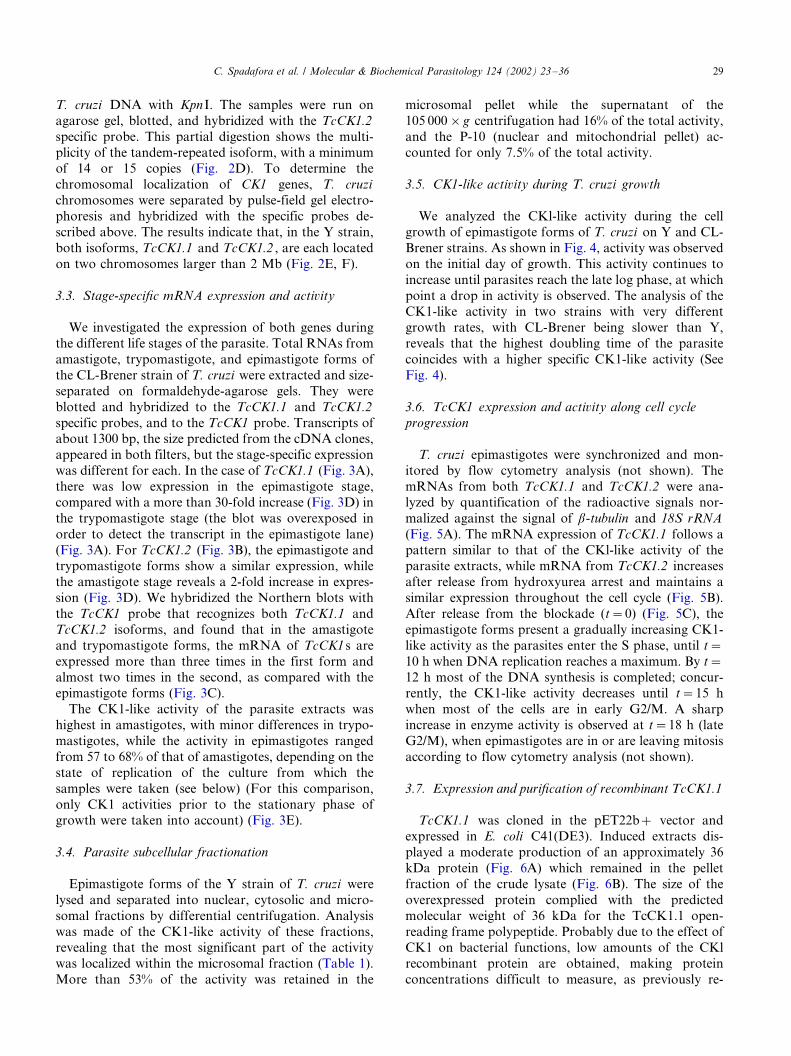
T. cruzi DNA with KpnI. The samples were run on
agarose gel, blotted, and hybridized with the TcCK1.2
specific probe. This partial digestion shows the multi-
plicity of the tandem-repeated isoform, with a minimumof 14 or 15 copies (Fig. 2D). To determine the
chromosomal localization of CK1 genes, T. cruzi
chromosomes were separated by pulse-field gel electro-
phoresis and hybridized with the specific probes de-
scribed above. The results indicate that, in the Y strain,
both isoforms, TcCK1.1 and TcCK1.2 , are each located
on two chromosomes larger than 2 Mb (Fig. 2E, F).
3.3. Stage-specific mRNA expression and activity
We investigated the expression of both genes during
the different life stages of the parasite. Total RNAs from
amastigote, trypomastigote, and epimastigote forms of
the CL-Brener strain of T. cruzi were extracted and size-
separated on formaldehyde-agarose gels. They were
blotted and hybridized to the TcCK1.1 and TcCK1.2
specific probes, and to the TcCK1 probe. Transcripts ofabout 1300 bp, the size predicted from the cDNA clones,
appeared in both filters, but the stage-specific expression
was different for each. In the case of TcCK1.1 (Fig. 3A),
there was low expression in the epimastigote stage,
compared with a more than 30-fold increase (Fig. 3D) in
the trypomastigote stage (the blot was overexposed in
order to detect the transcript in the epimastigote lane)
(Fig. 3A). For TcCK1.2 (Fig. 3B), the epimastigote andtrypomastigote forms show a similar expression, while
the amastigote stage reveals a 2-fold increase in expres-
sion (Fig. 3D). We hybridized the Northern blots with
the TcCK1 probe that recognizes both TcCK1.1 and
TcCK1.2 isoforms, and found that in the amastigote
and trypomastigote forms, the mRNA of TcCK1s are
expressed more than three times in the first form and
almost two times in the second, as compared with theepimastigote forms (Fig. 3C).
The CK1-like activity of the parasite extracts was
highest in amastigotes, with minor differences in trypo-
mastigotes, while the activity in epimastigotes ranged
from 57 to 68% of that of amastigotes, depending on the
state of replication of the culture from which the
samples were taken (see below) (For this comparison,
only CK1 activities prior to the stationary phase ofgrowth were taken into account) (Fig. 3E).
3.4. Parasite subcellular fractionation
Epimastigote forms of the Y strain of T. cruzi were
lysed and separated into nuclear, cytosolic and micro-
somal fractions by differential centrifugation. Analysis
was made of the CK1-like activity of these fractions,revealing that the most significant part of the activity
was localized within the microsomal fraction (Table 1).
More than 53% of the activity was retained in the
microsomal pellet while the supernatant of the
105 000�/g centrifugation had 16% of the total activity,
and the P-10 (nuclear and mitochondrial pellet) ac-
counted for only 7.5% of the total activity.
3.5. CK1-like activity during T. cruzi growth
We analyzed the CKl-like activity during the cell
growth of epimastigote forms of T. cruzi on Y and CL-
Brener strains. As shown in Fig. 4, activity was observed
on the initial day of growth. This activity continues to
increase until parasites reach the late log phase, at which
point a drop in activity is observed. The analysis of theCK1-like activity in two strains with very different
growth rates, with CL-Brener being slower than Y,
reveals that the highest doubling time of the parasite
coincides with a higher specific CK1-like activity (See
Fig. 4).
3.6. TcCK1 expression and activity along cell cycle
progression
T. cruzi epimastigotes were synchronized and mon-
itored by flow cytometry analysis (not shown). The
mRNAs from both TcCK1.1 and TcCK1.2 were ana-
lyzed by quantification of the radioactive signals nor-
malized against the signal of b-tubulin and 18S rRNA
(Fig. 5A). The mRNA expression of TcCK1.1 follows a
pattern similar to that of the CKl-like activity of theparasite extracts, while mRNA from TcCK1.2 increases
after release from hydroxyurea arrest and maintains a
similar expression throughout the cell cycle (Fig. 5B).
After release from the blockade (t�/0) (Fig. 5C), the
epimastigote forms present a gradually increasing CK1-
like activity as the parasites enter the S phase, until t�/
10 h when DNA replication reaches a maximum. By t�/
12 h most of the DNA synthesis is completed; concur-rently, the CK1-like activity decreases until t�/15 h
when most of the cells are in early G2/M. A sharp
increase in enzyme activity is observed at t�/18 h (late
G2/M), when epimastigotes are in or are leaving mitosis
according to flow cytometry analysis (not shown).
3.7. Expression and purification of recombinant TcCK1.1
TcCK1.1 was cloned in the pET22b�/ vector and
expressed in E. coli C41(DE3). Induced extracts dis-
played a moderate production of an approximately 36
kDa protein (Fig. 6A) which remained in the pellet
fraction of the crude lysate (Fig. 6B). The size of the
overexpressed protein complied with the predicted
molecular weight of 36 kDa for the TcCK1.1 open-
reading frame polypeptide. Probably due to the effect ofCK1 on bacterial functions, low amounts of the CKl
recombinant protein are obtained, making protein
concentrations difficult to measure, as previously re-
C. Spadafora et al. / Molecular & Biochemical Parasitology 124 (2002) 23�/36 29

ported; nonetheless, it has a very high specific activity,
as reported by Klimczak and co-workers [35]. To
optimize the yield of the TcCK1.1 recombinant protein,
we reduced the temperature for induction from 37 to
19 8C and increased incubation time to 18 h. Immuno-
detection of the soluble fraction failed to produce a
signal. We proceeded with the purification of this
soluble fraction by incubating it in a Ni-NTA agarose
column and eluting with a linear gradient of imidazole.
The activity of the fractions was determined by their
kinetic functionality, as described below; those with the
highest activity were pooled, concentrated, and analyzed
by SDS-PAGE and subsequent Coomasie staining (Fig.
6B), Western blotting, and immunodetection. Using this
Fig. 3. Stage-specific expression of TcCK1 mRNA. Total RNA (20 mg) front mid-to-late log epimastigote (E), amastigote (A), and trypomastigote
(T) forms of the life cycle of the CL-Brener strain of T. cruzi were hybridized with the TcCK1.1 (A), TcCK1.2 (B), and the TcCK1 specific probes
(C). The ethidium bromide stainings of the rRNA s are shown in the lower panels. Standard RNA markers (kb) were from Promega. (D) Fold CK1
mRNA expression with respect to the epimastigote forms of the parasite, according to normalization with densitometry analysis of rRNAs (See
Experimental Procedures). (E) Different forms of the parasite (CL-Brener strain) were obtained as described in Section 2. Supernatant samples of the
13 000�/g centrifugation were analyzed in duplicate with respect to their specific CK1 activity. The data is the result of a representative experiment.
For the epimastigotes, the number calculated was the media of the activity during 6 days of the logarithmic phase of growth.
Table 1
CKl-like activity of different cellular fractions of T. cruzi
Fraction Specific Activity
(pmol min�1 per mg)
Total Activity
(pmol min�1)
Sonicated total extract 3180 24 887
P-10 2460 1877
Cytosolic supernatant 1406 4007
Microsomal pellet 3755 13 261
Epimastigotes from the T. cruzi Y strain were fractionated as
described in Experimental Procedures. CK1-like activity was measured
in the different fractions. The kinetic assays were performed by
duplicate with standard deviation (S.D.) below 10%.
C. Spadafora et al. / Molecular & Biochemical Parasitology 124 (2002) 23�/3630

strategy, we detected the presence of the recombinant
protein (Fig. 6C).
3.8. Enzymatic activity of recombinant TcCK1.1
Recombinant TcCK1.1 was used for enzymatic assays
according to the method previously described. The
fractions eluted from the Ni-NTA agarose column
were analyzed on the basis of their CK1 activities.
Those fractions within the peak of activity were pooled,concentrated, and used for the kinetic assays. We found
that the apparent Km for b-casein and the specific
synthetic peptide Casein Kinase-1 substrate were 5.7 mg
ml�1 and 128 mM, respectively. For ATP, the TcCK1.1
protein exhibited an apparent Km of 56 mM. When the
specific synthetic peptide Casein Kinase-1 was used as
substrate, an inhibition of nearly 40% was obtained at
100 mM CKI-7 with 100 mM ATP as well. Heparin,being a specific inhibitor of CK2 proteins, has been used
as a means of distinguishing between CK1 and CK2
protein kinases [25]. We observed that heparin slightly
diminished TcCK1.1 activity, by about 10% at the
highest concentration used (30 mg ml�1). The other
compound tested, HD, is a natural and potent inhibitor
of CK1 [20]. When the effect of HD on CK1 was
studied, we observed that CK1 activity was significantly
inhibited when using parasite extracts (IC50, 23 nM), byas much as when recombinant TcCKl.l (IC50, 13 nM) is
used (data not shown). Thus, in terms of general
biochemical parameters, recombinant TcCK1.1 exhib-
ited properties characteristic of the CK1 family of
enzymes.
4. Discussion
We have isolated two genes in T. cruzi that encode
proteins homologous to the CK1 family. Both TcCK1
genes seem to be localized in two chromosomes,however, bearing in mind the diploidy of this organism,
and since allele polymorphisms have been described in
this trypanosomatid [36], they are probably in two
Fig. 4. CK1-like activity throughout cell growth of epimastigote forms of T. cruzi . Using epimastigote forms of (A) Y and (B) CL-Brener strains of
T. cruzi , we analyzed CK1 activities throughout the cell growth of parasites using the Casein kinase-1 specific substrate. Circles represent the number
of parasites per ml and squares represent CK1-like specific activity.
C. Spadafora et al. / Molecular & Biochemical Parasitology 124 (2002) 23�/36 31

Fig. 5. Expression and CK1-like activity along the cell cycle of T. cruzi . (A) Expression of TcCK1.1 and TcCK1.2 mRNAs. Aliquots of
synchronized T. cruzi Y epimastigotes were taken at times 0, 6, 8, 12, 16, 18, and 24 h. Time zero represents the moment in which the parasites ant
taken out of the hydroxyurea medium (see Experimental Procedures). Their RNAs were extracted, approximately 25 mg of each were transferred to a
nylon filter and hybridized with the specific isoform probes TcCK1.1 and TcCK1.2 to determine the variations of expression throughout the cell
cycle. The blot was normalized using the b-tubulin and the 18S rRNA probes. (B) Relationship of the mRNA expression and activity of the TcCK1
isoforms and the cell cycle. From the two upper panels, each time point represents the fold change in mRNAs levels (relative to the 0 h time point)
with respect to their normalization with either b-tubulin (solid lines) and 18S rRNA (dotted lines) specific probes. The lower panel shows CK1-like
activity in synchronized parasites. For enzymatic activity, samples of the parasites were taken out at times. 0, 2, 4, 6, 8. 10, 12, 15, 18, 21, and 23 h,
and their CK1-like activity determined using the Casein kinase-1 peptide substrate (See Experimental Procedures). Experiments were repeated three
times and gave essentially the same profiles as the experiment shown here.
Fig. 6. Overexpression and purification of recombinant TcCK1.1, (A) Stained Coomasie Blue SDS-PAGE gel of total bacterial proteins from E. coli
cells transformed with pET22b�/ (lane 1) and TcCK1.1/pET22b-(lane 2) after IPTG induction. (B) Stained gel containing supernatant (lane 3) and
pellet (lane 4) fractions from the total crude extracts of TcCK1.1/pET22b�/; purified 6XHis-tagged proteins from Ni-NTA agarose column: a
fraction with the maximal CK1 activity (lane 5) and a fraction without CK1 activity (lane 6). (C) Western blot analysis of total induced extracts (lane
7), a fraction with the maximal CK1 activity (lane 8) and a fraction without CK1 activity (lane 9) using a monoclonal anti 6X-His antibody (at 1:1000
dilution) as described in ‘Experimental Procedures’. Arrows indicate the position of TcCK1.1. Molecular mass standards (kDa) were from Bio-Rad.
C. Spadafora et al. / Molecular & Biochemical Parasitology 124 (2002) 23�/3632

alleles of the same chromosome, with different sizes. An
important characteristic of the TcCK1 isoforms is that
even though one of the genes, TcCK1.1 , is a two-copy
gene (per haploid genome), the other, TcCK1.2 , is amulticopy gene, the only one described to date.
CK1 is a family of Ser/Thr kinases which are highly
conserved in their catalytic domain; they are identical by
50% or more, presenting the most differences in their
NH2 and COOH termini. It is these sequence differences
upon which most isoform classification is based. The
CK1 isoforms have been clustered in three main
branches [1]: enzymes found exclusively in the cyto-plasm, where they interact with the plasma membrane,
and which are represented by YCKs and Cki1; enzymes
found largely in the nucleus, regulating DNA repair and
best exemplified by HRR25; and finally, enzymes which
appear to be widely distributed throughout the cell,
performing such diverse functions as regulation of
mitosis or signal transduction, and represented by
CK1b and CK1a. The TcCK1 homologues are similarto many of the known CK1 isoforms from mammals
and yeast, and cannot be ascribed a function based on
sequence similarity alone. Both TcCK1 genes contain
almost all of the residues that are characteristic of these
protein kinases, but there are some differences. The
most remarkable aspect of the sequence of the TcCK1
isoforms is a unique three-residue insertion within the
catalytic domain, not present in the rest of the familydescribed. This insertion would occupy a place in the
loop joining the aA helix with the b4 strand (L-A4) [34].
The loop is localized behind the ATP binding site, where
the three residues could influence nucleotide binding.
This sequence insertion could explain some relative
biochemical differences found in TcCK1.1. Since the
triphosphate subsite is highly conserved throughout the
kinase superfamily, the differences found in the residuesthat line the purine pocket are key to the design of
specific agents that could interfere with the enzyme
activity. In the search for successful drugs against
parasitic diseases, the enzyme targeted should be suffi-
ciently different from the host protein so that the drug
used only affects the parasite. Significantly, Knockaert
and colleagues [37] described how a matrix of purvala-
nol, a purine derivative, bound some protozoan CK1s,T. cruzi among them, and not those CK1s from other
species. Could these three residues lie behind such an
observation in the case of this trypanosomatid?
While looking for specific inhibitors of cyclin-depen-
dent kinases (CDKs), Meijer and other colleagues
reported the finding of hymenyaldisine (HD) [20], an
inhibitor of human CDK1, CDK5, GSK3-b and CK1,
with IC50 values of 22, 28, 10, and 35 nM, respectively.We have tried HD on T. cruzi CK1, obtaining an IC50
of 23 nM for the parasite CK1-like activity and 13 nM
for recombinant TcCK1.1. In all experiments we used
the Casein Kinase-1 peptide as the substrate. The
specificity of this peptide greatly diminishes the chance
of phosphorylation by other kinases, a notion which
appears to be confirmed by the fact that the inhibition of
the recombinant TcCK1.1 exhibits a pattern almostidentical to that of the total T. cruzi extracts, indicating
that most of the activity measured in the latter was due
exclusively to CK1 and not to other kinases present in
the extracts. Another inhibitor of some CK1s, heparin, a
polyanion which serves to distinguish between the two
casein kinases, CK1 and CK2, was tested on recombi-
nant TcCK1.1. A low inhibition (30%) was obtained at
30 mg ml�1. This is comparable with data published formammalian CK1 with an IC50 of 24 mg ml�1 when
assayed at 2 mg ml�1 casein [38] while the IC50 of CK2
is 5/0.15 mg ml�1 [39]. Apart from this data, the
biochemical parameters of recombinant TcCK1.1 point
to a structural difference between T. cruzi CK1 and
most other CK1s reported. TcCK1.1 has one of the
highest reported Km values for ATP, with the exception
of those of rat liver nuclei and yeast [1]. As for b-casein,we calculated a Km higher than that of most other
organisms [7,25,40�/42]; however, when using the spe-
cific peptide Casein Kinase-1 substrate, our TcCK1.1
gave us a Km value somewhat lower than what was
calculated for other CK1s [25]. It should be mentioned
that the concentration of NaCl used in the assays for
CK1 vary from group to group. However, based on
studies carried out by Calabokis et al. [16] on theoptimal salt concentration for a partially purified CK1
of T. cruzi at a concentration of 150 mM, which is what
we used, the activity varies only 30% from the maximum
and 60% from the minimum possible without the
addition of any salts. Therefore, most of our compar-
isons are made with groups that used similar assay
conditions. Finally, the low activity of the inhibitor
CKI-7 denotes that the affinity for the nucleotidebinding site is lower than in other species [4,7]. Our
observations are confirmed by the work of Calabokis et
al. [16], with their reported values on the biochemical
parameters of a partially purified CK1 of T. cruzi
agreeing, for the most part, with those we have
obtained. Furthermore, Vancura et al. [40] worked
with a yeast homolog of CK1, YCK2 and found only
a 30% inhibition with concentrations up to 500 mM. Soit is not unusual to find some members of the family for
which the inhibitor does not respond efficiently. This is
another clue pointing to a structural difference of T.
cruzi CK1, making it all the more relevant when looking
for potential drug targets against the parasite.
As we mentioned before, a peculiarity of the CK1
genes characterized here is the fact that one of them is
arranged in tandem. In many organisms there have beenreports of CK1 present in various isoforms, and as
different splicing products, but in our case we have the
same isoform, TcCK1.2 , repeated several times. In
trypanosomatids, many housekeeping genes are present
C. Spadafora et al. / Molecular & Biochemical Parasitology 124 (2002) 23�/36 33

in large tendem repeated clusters. These duplications
may be involved in the regulation of expression for these
genes. In fact, TcCK1.2 was more highly expressed than
TcCK1.1 . Comparative analysis of the TcCK1s mRNA
expression reflects a significant contribution from
TcCK1.1 in the infective stage. Otherwise, TcCK1.1
seems to contribute little to the general TcCK1s expres-
sion. We cannot rule out the possibility of another CK1
isoform contributing to the overall expression and
activity, particularly with respect to the mRNA expres-
sion of amastigotes where the increased signal seems to
be superior to that of the two isoforms we have isolated
together. The results of the CK1 activity assays in the
three life forms reveal that the two mammalian stages,
amastigotes and trypomastigotes, have a significantly
higher enzyme activity than the insect stage. Common to
these stages of the parasite is the need to adjust to
diverse physiological environments and cell require-
ments, such as temperature, host�/parasite signaling,
trafficking, etc., which might require the presence of
numerous metabolism regulators, such as CK1. The fact
that the CK1-like activity presents differences with
respect to the mRNA expression can be attributed to
post-translational modifications as proteolysis, phoryla-
tion, and localization of the protein that have been
reported for CK1 [43,44].
TcCK1.1 mRNA expression varies along the cell
cycle, while TcCK1.2 mRNA, after being released
from arrest, achieves a level which tends to remain
constitutively high. In spite of this constant mRNA
expression, a drop in the CK1-like activity is found in
the middle of the cell cycle, probably due to post-
translational regulations as mentioned above. The
results of CK1-like activity measurements in the syn-
chronized cultures show an increase in the enzyme
activity corresponding to the cell entry into S, confirmed
by [3H]Thymidine incorporation assays (not shown),
and probably M phases. The presence of a peak for a
CK1-like activity in mitosis confirms the earlier findings
of other groups [45,46], but still needs to be character-
ized further in T. cruzi . What was certainly unexpected
and new to us was the peak of activity found during the
phase of DNA synthesis, appearing consistently
throughout our assays. Substrates found for CK1 in
other organisms, and its action on them, also attest to
the probable function of CK1 in regulating DNA
replication, as is the case with the blockage of the origin
of replication of the SV40 large T antigen by CK1 [47].
Thus a probable role of TcCK1s in growth and cell cycle
control cannot be ruled out. This concept is further
supported by our studies of the CK1-like activity during
epimastigote growth, the results of which reveal some
ongoing activity even in cells with null or little replica-
tive activity, but with an evident increase in enzyme
activity during the log cell phase.
A possible tie between all our observations related to
TcCK1 function is the phosphorylation of structural
substrates. Tubulin, troponin, myosin, band 4.1, kine-
sin, tau, and flagellar dynein, all of them structure-
related proteins, have been described as possible CK1
substrates [2,48,49]. Some of them would need the
action of one isoform or the other, depending on the
life stage of the parasite. Dynein, apart from being
present in the flagella, is a microtubule motor protein
present in the mitotic spindles and centrosomes, and has
an important role in mitosis when microtubules lay
down the structure for chromosome segregation. CK1
has been linked to centrosomes and mitotic spindle [50],
most conspicuously following induced DNA damage,
through phosphorylation of the tumor suppressor
protein p53 [51], just as HRR25 is linked to DNA
repair in S. cerevisiae [12] and D. melanogaster CK1 is
induced by DNA perturbation [52]. Our TcCK1s could
play similar roles in the parasite. Not only do they have
a nuclear localization signal that would situate them
spatially suited to act during mitosis: they also have a
kinesin homology sequence, another reason to make one
ponder the possible relationship CK1 has with structural
proteins, especially those associated with microtubules.
Our results would not seem to rule out this possibility.
First, the subcellular fractions containing the most CK1-
like activity were those enriched with membranes, where
the cytoskeletal proteins remain. Second, the activity
also peaks during the S and M phases of the cell cycle,
especially in M, when the presence of microtubules is
vital. Moreover, apart from the synchronized cultures,
when the parasites are more actively replicating, that is,
in the middle of the logarithmic phase, the CK1-like
activity of T. cruzi reaches its peak. Third, the fact that
one isoform is repeated in tandem points to a high
requirement of CK1 activity, which is usually the case
when the substrates are structural proteins with large
needs for the enzyme. All of the above could lead us to
envision a future link between TcCK1s and cytoskeleton
proteins such as microtubules, microtubule associated
proteins (MAPs), or microtubule motor proteins like
kinesin and dynein.
CK1 has been described as a pleitropic enzyme,
meaning that it could be involved in metabolism,
motility, trafficking and/or signalling. The presence of
expression and activity of TcCK1 in the parasite,
regardless of the life stage in which it is found, and
with all the different physiological conditions that each
stage implies, seems to support this idea.The possibility of multifunctionality, together with
the finding that the sequences have a unique insertion
which most probably confers structural differences to
the TcCK1 isoforms with respect to other CK1s make
TcCK1s very relevant when possible molecular targets
are considered in the fight against Chagas disease. At
C. Spadafora et al. / Molecular & Biochemical Parasitology 124 (2002) 23�/3634

the same time, our results with T. cruzi may help to shed
new light on possible roles of CK1 in higher eukaryotes.
Acknowledgements
The authors would like to thank L. Meijer and G.R.
Pettit for kindly providing hymenialdisine. We thank
Pilar Navarro for help in parasite cultures. We thank N.
Galanti, I. Espinoza, J. Allende and V. Pulgar for
suggestions, help, and guidance in some experiments,
as well as G. Robledo for very useful collaboration. We
thank all our laboratory colleagues for helpful discus-
sions as this work progressed. C. Spadafora is recipientof a Becas 2003 fellowship from Secretarıa Nacional de
Ciencia y Tecnologıa (Panama). C. Torres is recipient of
a fellowship from the Fundacion Ramon Areces (Ma-
drid, Spain). This work was supported by The Spanish
Grants PM97-0139 (F. G.), PM98-0115 (S. Castanys),
CICYT-FEDER IFD97-0747-C04-03 (S. C.), the FON-
DECYT-Chile 1020095 (A. Morello), and the Convenio
CSIC-Universidad de Chile between F. Gamarro and A.Morello (1997�/2000).
References
[1] Gross SD, Anderson RA. Casein kinase I: spatial organization
and positioning of a multifunctional protein kinase family. Cell
Signal 1998;10:699�/711.
[2] Tuazon PT, Traugh JA. Casein kinase I and II/multipotential
serine protein kinases: structure, function, and regulation. Adv
Second Messenger Phosphoprotein Res 1991;23:123�/64.
[3] Graves PR, Haas DW, Hagedom CH, DePaoli-Roach AA,
Roach PJ. Molecular cloning, expression, and characterization
of a 49-kilodalton casein kinase I isoform from rat testis. J Biol
Chem 1993;268:6394�/401.
[4] Fish KJ, Cegielska A, Getman ME, Landes GM, Virshup DM.
Isolation and characterization of human Casein Kinase I o (CK1oa novel member of the CKI gene family). J Biol Chem
1995;270:14875�/83.
[5] Rowles J, Slaughter C, Moomaw C, Hsu J, Cobb MH. Purifica-
tion of casein kinase I and isolation of cDNAs encoding multiple
casein kinase I-like enzymes. Proc Natl Acad Sci USA
1991;88:9548�/52.
[6] Zhai L, Graves PR, Robinson LC, Italiano M, Culbertson MR,
Rowles J, Cobb MH, DePaoli-Roach AA, Roach PJ. Casein
kinase g subfamily. Molecular cloning, expression, and character-
ization of three mammalian isoformas and complementation of
defects in the Saccharomices cerevisiae YCK genes. J Biol Chem
1995;270:12717�/24.
[7] Barik S, Taylor RE, Chakrabarti D. Identification, cloning, and
mutational analysis of the casein kinase I cDNA of the malarial
parasite Plasmodium falciparum . J Biol Chem 1997;272:26132�/8.
[8] Hermoso T, Fishelson Z, Becker SI, Hirschberg K, Jaffe CL.
Leishmanial protein kinases phosphorylate components of the
complement system. EMBO J 1991;10:4061�/7.
[9] Paas Y, Fishelson Z. Shedding of tyrosine and serine-threonine
ecto-protein kinases from human leukemic cells. Arch Biochem
Biophys 1995;316:780�/8.
[10] Kearney PH, Ebert M, Kuret J. Molecular cloning and sequence
analysis of two novel fission yeast casein kinase-1 isoforms.
Biochem Biophys Res Commun 1994;203:231�/6.
[11] Wang PC, Vancura A, Desai A, Carmel G, Kuret J. Cytoplasmic
forms of fission yeast casein kinase-1 associate primarily with the
particulate fraction of the cell. J Biol Chem 1994;269:12014�/23.
[12] Hoekstra MF, Liskay RM, Ou AC, DeMaggio AJ, Burbee DG,
Heffron F. HRR25, a putative protein kinase from budding yeast:
association with repair of damaged DNA. Science
1991;253:1031�/4.
[13] Wang P, Vancura A, Mitcheson TGM, Kuret J. Two genes in
Saccharomyces cerevisiae encode a membrane bound form of
casein kinase 1. Mol Biol Cell 1992;3:275�/86.
[14] Wang X, Hoekstra MF, DeMaggio AJ, Dhillon N, Vancura A,
Kuret J, Johnston GC, Singer RA. Prenylated isoforms of yeast
casein kinase I, including the novel Yck3p, suppress the gcs
blockage of cell proliferation from stationary phase. Mol Cell Biol
1996;16:5375�/85.
[15] Blaisonneau J, Fukuhara H, Wesolowski-Louvel M. The
Kluveromyces lactis equivalent of casein kinase 1 is required for
the transcription of the gene encoding the low-affinity glucose
permcase. Mol Gen Genet 1997;253:469�/77.
[16] Calabokis M, Kurz L, Wilkesman J, Galan-Caridad JM, Moller
C, Gonzatti MI, Bubis J. Biochemical characterization of a
partially purified casein kinase-1 like activity from Trypanosoma
cruzi . Parasitol Int 2002;51:25�/39.
[17] Schwab C, DeMaggio A, Ghoshal N, Binder L, Kuret J, McGeer
P. Casein kinase 1 delta is associated with pathological accumula-
tion of tau in several neurodegenerative diseases. Neurobiol Aging
2000;21:503�/10.
[18] Camacho F, Cilio M, Guo Y, Virshup D, Patel K, Khorkova O,
Styren S, Morse B, Yao Z, Keesler G. Human casein kinase Idphosphorylation of human circadian clock proteins period 1 and
2. FEBS Lett 2001;489:159�/65.
[19] Kishida M, Hino S, Michiwe T, Yamamoto H, Kishida S, Fukui
A, Asashima M, Kikuchi A. Synergistic activation of the Wnt
signaling pathway by Dvl and casein kinase Io. J Biol Chem
2001;276:32147�/55.
[20] Meijer L, Thunnissen AWH, White AW, Garnier M, Nikolic M,
Tsai L, Walter J, Cleverley KE, Salinas PC, Wu Y, Biernat J,
Mandelkow E, Kim S, Pettit GR. Inhibition of cyclin-dependent
kinases, GSK-3b, and CK1 by bymenialdisine, a marine sponge
constituent. Chem Biol 2000;7:51�/63.
[21] Zulantay I, Venegas J, Apt W, Solari A, Sanchez G. Lytic
antibodies in Trypanosoma cruzi infected persons with low
parasitemia. Am J Trop Med Hyg 1998;58:775�/9.
[22] Robello C, Dallagiovanna B, Engel JC, Gamarro F, Castanys S.
A new member of YER057C family in Trypanosoma cruzi is
adjacent to an ABC-transporter. Gene 1998;220:1�/12.
[23] Galanti N, Dvorak JA, Grenet J, McDaniel JP. Hydroxyurea-
induced synchrony of DNA replication in the Kinetoplastida. Exp
Cell Res 1994;214:225�/30.
[24] Dvorak JA. Analysis of the DNA of parasitic protozoa by flow
cytometry. In: Hyde JF, editor. Methods in Molecular Biology:
Protocols in Molecular Parasitology, vol. 21. Totowa: Humana
Press Inc, 1993:191�/204.
[25] Pulgar V, Tapia C, Vignolo P, Santos J, Sunkerl CE, Allende CC,
Allende E. The recombinant a isoforms of protein kinase CK1
from Xenopus laevis can phosphorylate tyrosine in synthetic
substrates. Eur J Biochem 1996;242:519�/25.
[26] Sambrook J, Fritsch EF, Maniatis T. Molecular Cloning: a
Laboratory Manual, 2nd edn.. Cold Spring Harbor: Cold Spring
Harbor Laboratory Press, 1989.
[27] Altshul SF, Gish W, Myers EW. Basic local alignment search
tool. J Mol Biol 1990;215:403�/10.
C. Spadafora et al. / Molecular & Biochemical Parasitology 124 (2002) 23�/36 35

[28] Devereux J, Haeberth P, Smithies O. A comprehensive set of
sequence analysis programs for the VAX. Nucleic Acids Res
1984;8:5725�/37.
[29] Coderre JA, Beverley SM, Schimke RT, Santi DV. Overproduc-
tion of a bifunctional thymidylate synthetase-dihydrofolate re-
ductase and DNA amplification in methotrexate resistant
Leishmania tropica . Proc Natl Acad Sci USA 1983;80:2132�/6.
[30] Garvey EP, Santi DV. Stable amplified DNA in drug-resistant
Leishmania exists as extrachromosomal circles. Science
1986;233:535�/40.
[31] Laemmli UK. Cleavage of structural proteins during the assembly
of bacteriophage T4. Nature 1970;227:680�/5.
[32] Chijiwa T, Hagiwara M, Hidaka H. A newly synthesized selective
casein kinase I inhibitor, N-(2-aminoethyl)-5-chloroisoquinoline-
8-sulfonamide, and affinity purification of casein kinase I from
bovine testis. J Biol Chem 1989;264:4924�/7.
[33] Hanks SK, Quinn AM. Protein kinase catalytic domain sequence
database: identification of conserved features of primary struc-
tures and classification of family members. Methods Enzymol
1991;200:38�/62.
[34] Xu R, Carmel G, Sweet RM, Kuret J, Cheng X. Crystal structure
of casein kinase-1, a phosphate-directed protein kinase. EMBO J
1995;14:1015�/23.
[35] Klimczak LJ, Farini D, Lin C, Ponti D, Cashmore AR, Giuliano
G. Multiple isoforms of Arabidopsis casein kinase 1 combine
conserved catalytic domains with variable carboxyl-terminal
extensions. Plant Physiol 1995;109:687�/96.
[36] Dujardin JC, Henriksson J, Victoir K, Brisse S, Gamboa D,
Arevalo J, Le Ray D. Genomic rearrangements in trypanosoma-
tids: an alternative to the ‘one gene’ evolutionary hypotheses.
Mem Inst Oswaldo Cruz 2000;95:527�/34.
[37] Knockaert M, Gray N, Damiens E, Chang Y-T, Grellier P, Grant
K, Fergusson D, Mottram J, Soete M, Dubremetz J-F, LeRoch
K, Doerig C, Suhultz PG, Meijer L. Intracellular targets of
cycline-dependent kinase inhibitors: identification by affinity
chromatography using immobilized inhibitors. Chem Biol
2000;7:411�/22.
[38] Kuret J, Woodget JR, Cohen P. Multisite phosphorylation of
glycogen synthase from rabbit skeletal muscle. Identification of
the sites phosphorylated by casein kinase-1. Eur J Biochem
1985;151:39�/48.
[39] Padmanabha R, Glover CVC. Casein kinase II of yeast contains
two distinct alpha polypeptides and an unusually large beta
subunit. J Biol Chem 1987;262:1829�/35.
[40] Vancura A, O’Connor A, Patterson SD, Mirza U, Chait BT,
Kuret J. Isolation and properties of YCK2, a Saccharomyces
cerevisiae homolog of casein kinase-1. Arch Biochem Biophys
1993;305:47�/53.
[41] Itarte E, Mor MA, Salavert A, Pena JM, Bertomeu JF, Guinovart
JJ. Purification and characterization of two cyclic AMP-indepen-
dent casein/glycogen synthase kinases from rat liver cytosol.
Biochim Biophys Acta 1981;658:334�/47.
[42] Klimczak LJ, Cashmore AR. Purification and characterization of
casein kinase I from broccoli. Biochem J 1993;293:283�/8.
[43] Graves PR, Roach PJ. Role of COOH-terminal phosphorylation
in the regulation of casein kinase I delta. J Biol Chem
1995;270:21689�/94.
[44] Zhai L, Graves PR, Longenecker KL, DePaoli-Roach AA, Roach
PJ. Recombinant rabbit muscle casein kinase I alpha is inhibited
by heparin and activated by polylysine. Biochem Biophys Res
Commun 1992;189:944�/9.
[45] Behrend L, Stoter M, Kurth M, Rutter G, Heukeshoven J,
Deppert W, Knippschild U. Interaction of casein kinase I delta
(CK1 delta) with post-Golgi-structures, microtubules and the
spindle apparatus. Eur J Cell Biol 2000;79:240�/51.
[46] Robinson LC, Bradley C, Bryan JD, Jerome A, Kweon Y, Panek
HR. The Yck2 yeast casein kinase I isoform shows cell cycle-
specific localization to sites of polarized growth and is required
for proper septin organization. Mol Biol Cell 1999;10:1077�/92.
[47] Cegielska A, Virshup DM. Control of Simian Virus 40 DNA
replication by the HeLa cell nuclear kinase casein kinase I. Mol
Cell Biol 1993;13:1202�/11.
[48] Singh TJ, Grundke-Iqbal I, Iqbal K. Phosphorylation of tau
protein by casein kinase-1 converts it to an abnormal Alzheimer-
like state. J Neurochem 1995;64:1420�/3.
[49] Yang P, Sale W. Casein kinase I is anchored on axonemal doublet
microtubules and regulates flagellar dynein phosphorylation and
activity. J Biol Chem 2000;275:18905�/12.
[50] Brockman JL, Gross SD, Sussman MR, Anderson RA. Cell cycle-
dependent localization of casein kinase 1 to mitotic spindles. Proc
Natl Acad Sci USA 1992;89:9454�/8.
[51] Knippschild U, Milne DM, Campbell LE, DeMaggio AJ,
Christenson E, Hoekstra MF, Meek DW. p53 is phosphorylated
in vitro and in vivo by the delta and epsilon isoforms of casein
kinase 1 and enhances the level of casein kinase 1 delta in response
to topoisomerase-directed drugs. Oncogene 1997;15:1727�/36.
[52] Santos JA, Logarinho E, Tapia C, Allende CC, Allende JE. The
casein kinase la gene of Drosophila melanogaster is developmen-
tally regulated and the kinase activity of the protein induced by
DNA damage. J Cell Sci 1996;109:1847�/56.
C. Spadafora et al. / Molecular & Biochemical Parasitology 124 (2002) 23�/3636
Copyright © 2022 FDOKUMEN