CD44 isoforms in normal and leukemic hematopoiesis
16
Experimental Hematology 27 (1999) 978–993 0301-472X/99 $–see front matter. Copyright © 1999 International Society for Experimental Hematology. Published by Elsevier Science Inc. PII S0301-472X(99)00023-5 CD44 isoforms in normal and leukemic hematopoiesis Saghi Ghaffari a , Florence Smadja-Joffe b , Robert Oostendorp a,c , Jean-Pierre Lévesque d , Graeme Dougherty e , Allen Eaves a , and Connie Eaves a a Terry Fox Laboratory, British Columbia Cancer Agency and the University of British Columbia, Vancouver, BC, Canada; b Institut National de la Santé et de la Recherche Médicale U268, Hôpital Paul Brousse, Villejuif, France; c GSF, Institute for Experimental Hematology, Munich, Germany; d Leukaemia Research Unit, Department of Haematology, Hanson Center for Cancer Research, Adelaide, Australia; e UCLA Medical Center, Department of Radiation Oncology, Experimental Division, Los Angeles, CA (Received 10 December 1998; revised 27 January 1999; accepted 3 February 1999) Introduction The CD44 family of adhesion molecules is a fascinating group of highly glycosylated proteins encoded by a single gene. The various isoforms produced are differentially ex- pressed in many cell types, where they are involved in a wide range of biological processes [1]. Initially, CD44 cell surface molecules were identified and named according to their reactivity with particular monoclonal antibodies (mAb). This resulted in the rapid accumulation of a highly confus- ing terminology [e.g., Pgp-1, Ln (Lu)-related p80, ECM III, Hermes, HUTCH-1, and gp-85]. However, this confusion has now been largely dissipated by a detailed knowledge of the structure of the CD44 gene and its multiple transcripts. Although much has been written about CD44, an apprecia- tion of its potential role in normal and leukemic hemato- poiesis has only recently emerged. Overview of the CD44 gene and its products The complexity of CD44 functions is matched by, and pos- sibly related, to the complex structure of the CD44 gene. The highly conserved single copy CD44 gene comprises ap- proximately 50 Kb of genomic DNA and contains 20 exons [2,3]. The CD44 gene is located on the short arm of chro- mosome 11 in humans and on chromosome 2 in mice [4,5]. In the human CD44 gene (shown as a diagram, Fig. 1), there are 10 “standard” exons found in all CD44 isoforms. The other 10 are variably spliced. These latter exons are referred to as v1 to v10 and correspond to exons 6 to 15 (v is short for variant). The human v1 exon (exon 6) contains an in- frame stop codon. In fact, 12 of the 20 exons are known to undergo alternative splicing (Fig. 1), to produce at least 18 distinct transcripts [2,3]. All of these contain additional se- quences within a single site in the extracellular domain of CD44, thus explaining the large number of higher molecular weight isoforms of CD44 described [2]. The CD44 protein has several interesting domains (also shown in the diagram, Fig. 1). The standard molecule CD44 (CD44H), which does not contain any variable regions, en- codes a 341 amino acid transmembrane protein with a pre- dicted molecular weight of 37–38 KD. It comprises an ex- tracellular domain of 248 amino acids, a transmembrane spanning domain of 21 amino acids, and a 72 amino acid cytoplasmic domain [6–9]. A CD44 molecule with a short 3 amino acid long cytoplasmic domain has also been de- scribed [10]. As noted above, higher molecular weight iso- forms have a larger extracellular domain due to the insertion of additional amino acids within a unique site in the mem- brane proximal region [11–14]. A stretch of 90 relatively hydrophobic residues com- prises the amino-terminus of the CD44 polypeptide. This amino-terminal domain has homology to cartilage link pro- teins and other proteoglycans, and mediates interactions of CD44 with glycosaminoglycans [6–9]. In particular, this re- gion binds to hyaluronan (HA). The cartilage link homolo- gous region of CD44 contains six cysteine residues that are thought to be linked by disulfide bounds to form a single distal globular domain. The entire extracellular portion of human CD44 contains six potential sites for N-linked glyco- sylation, five of which lie in the cartilage link homologous region [8,9]. The membrane proximal region of CD44 con- tains several serine and threonine residues that may serve as sites for O-linked glycosylation as well as four serine-gly- cine motifs that may function as sites for chondroitin sulfate or heparan sulfate attachments [8,9,15]. The high glycosyla- tion and attachment of chondroitin and heparan sulfate ac- count for the additional molecular mass and production of a 85–90 KD molecule, found on the majority of hematopoie- Offprint requests to: Saghi Ghaffari, M.D., Ph.D., Whitehead Institute for Biomedical Research, 9 Cambridge Center, Cambridge, MA 02142; E-mail: [email protected]
-
Upload
independent -
Category
Documents
-
view
0 -
download
0
Transcript of CD44 isoforms in normal and leukemic hematopoiesis

Experimental Hematology 27 (1999) 978–993
0301-472X/99 $–see front matter. Copyright © 1999 International Society for Experimental Hematology. Published by Elsevier Science Inc.PII S0301-472X(99)00023-5
CD44 isoforms in normal and leukemic hematopoiesis
Saghi Ghaffari
a
, Florence Smadja-Joffe
b
, Robert Oostendorp
a,c
, Jean-Pierre Lévesque
d
, Graeme Dougherty
e
, Allen Eaves
a
, and Connie Eaves
a
a
Terry Fox Laboratory, British Columbia Cancer Agency and the University of British Columbia, Vancouver, BC, Canada;
b
Institut National de la Santé et de la Recherche Médicale U268, Hôpital Paul Brousse, Villejuif, France;
c
GSF, Institute for Experimental Hematology, Munich, Germany;
d
Leukaemia Research Unit, Department of Haematology, Hanson Centerfor Cancer Research, Adelaide, Australia;
e
UCLA Medical Center, Department of Radiation Oncology, Experimental Division, Los Angeles, CA
(Received 10 December 1998; revised 27 January 1999; accepted 3 February 1999)
Introduction
The CD44 family of adhesion molecules is a fascinatinggroup of highly glycosylated proteins encoded by a singlegene. The various isoforms produced are differentially ex-pressed in many cell types, where they are involved in awide range of biological processes [1]. Initially, CD44 cellsurface molecules were identified and named according totheir reactivity with particular monoclonal antibodies (mAb).This resulted in the rapid accumulation of a highly confus-ing terminology [e.g., Pgp-1, Ln (Lu)-related p80, ECM III,Hermes, HUTCH-1, and gp-85]. However, this confusionhas now been largely dissipated by a detailed knowledge ofthe structure of the CD44 gene and its multiple transcripts.Although much has been written about CD44, an apprecia-tion of its potential role in normal and leukemic hemato-poiesis has only recently emerged.
Overview of the CD44 gene and its products
The complexity of CD44 functions is matched by, and pos-sibly related, to the complex structure of the CD44 gene.The highly conserved single copy CD44 gene comprises ap-proximately 50 Kb of genomic DNA and contains 20 exons[2,3]. The CD44 gene is located on the short arm of chro-mosome 11 in humans and on chromosome 2 in mice [4,5].In the human CD44 gene (shown as a diagram, Fig. 1), thereare 10 “standard” exons found in all CD44 isoforms. Theother 10 are variably spliced. These latter exons are referredto as v1 to v10 and correspond to exons 6 to 15 (v is shortfor variant). The human v1 exon (exon 6) contains an in-frame stop codon. In fact, 12 of the 20 exons are known to
undergo alternative splicing (Fig. 1), to produce at least 18distinct transcripts [2,3]. All of these contain additional se-quences within a single site in the extracellular domain ofCD44, thus explaining the large number of higher molecularweight isoforms of CD44 described [2].
The CD44 protein has several interesting domains (alsoshown in the diagram, Fig. 1). The standard molecule CD44(CD44H), which does not contain any variable regions, en-codes a 341 amino acid transmembrane protein with a pre-dicted molecular weight of 37–38 KD. It comprises an ex-tracellular domain of 248 amino acids, a transmembranespanning domain of 21 amino acids, and a 72 amino acidcytoplasmic domain [6–9]. A CD44 molecule with a short 3amino acid long cytoplasmic domain has also been de-scribed [10]. As noted above, higher molecular weight iso-forms have a larger extracellular domain due to the insertionof additional amino acids within a unique site in the mem-brane proximal region [11–14].
A stretch of 90 relatively hydrophobic residues com-prises the amino-terminus of the CD44 polypeptide. Thisamino-terminal domain has homology to cartilage link pro-teins and other proteoglycans, and mediates interactions ofCD44 with glycosaminoglycans [6–9]. In particular, this re-gion binds to hyaluronan (HA). The cartilage link homolo-gous region of CD44 contains six cysteine residues that arethought to be linked by disulfide bounds to form a singledistal globular domain. The entire extracellular portion ofhuman CD44 contains six potential sites for N-linked glyco-sylation, five of which lie in the cartilage link homologousregion [8,9]. The membrane proximal region of CD44 con-tains several serine and threonine residues that may serve assites for O-linked glycosylation as well as four serine-gly-cine motifs that may function as sites for chondroitin sulfateor heparan sulfate attachments [8,9,15]. The high glycosyla-tion and attachment of chondroitin and heparan sulfate ac-count for the additional molecular mass and production of a85–90 KD molecule, found on the majority of hematopoie-
Offprint requests to: Saghi Ghaffari, M.D., Ph.D., Whitehead Institutefor Biomedical Research, 9 Cambridge Center, Cambridge, MA 02142;E-mail: [email protected]

S. Ghaffari et al./Experimental Hematology 27 (1999) 978–993
979
tic cells [16–18]. However, variation in the post-transla-tional modification of CD44 may also contribute to the pro-duction of cell type-specific forms of the mature moleculeand CD44 proteins of higher molecular weight in the rangeof 180–220 KD are found on other cell types [19–22]. Thecytoplasmic region of CD44 contains six serine residuesthat are subject to phosphorylation [1]. Soluble forms ofCD44 (up to 5
m
g/mL) in the plasma of normal individualshave also been identified [23–27].
CD44 ligands
The amino terminal region of CD44 protein is homologousto cartilage link proteins and proteoglycans. Like these re-lated molecules, CD44 has HA-binding activity [8,9]. Mo-lecular modeling and NMR analysis have been used re-cently to predict the structural domain of HA-bindingproteins. A model based on the ligand-binding of TSG-6(tumor necrosis factor stimulated gene-6) has given someinsight into the HA-binding capacity of CD44, including re-markable similarities with C-type lectins in the absence ofsequence homology [28]. An ability of CD44 to bind HAwas first suggested by the studies of Underhill et al. [17] whodescribed a HA receptor similar to CD44 in size, cellular
distribution and cytoskeletal interactions. Soon after, Mi-yake et al. [33] showed that the adhesive interactions ofCD44
1
B cells with stromal cell lines were mediated by HAand inhibitable with certain anti-CD44 antibodies that rec-ognize the HA-binding site of CD44. Formal proof of thespecificity of CD44 for HA was provided by the demonstra-tion that a soluble CD44-immunoglobulin-fusion proteinbound to rat lymph node high endothelial venules (HEV) ina HA-dependent manner [30]. Interestingly, an anti-CD44mAb (Hermes-3) that specifically blocks the binding oflymphocytes to mucosal HEV was not found to interferewith the CD44-mediated binding of stromal cells to purifiedHA [9,31]. Subsequently, it was shown that this mAb recog-nizes a CD44 domain that is not involved in HA-binding[32]. Nevertheless, treatment of HEV or stromal cells withhyaluronidase, or the addition of soluble HA or anti-CD44antibodies (mAb, KM201, and K-3) have been shown tocompletely and specifically inhibit the interaction of stro-mal cells with lymphocytes [29,30,33,34].
Site-directed mutagenesis and truncation studies haveidentified two clusters of basic amino acid residues withinthe CD44 molecule that are essential for HA-binding[35,36]. One of these amino acids is located inside and oneoutside of the cartilage link homologous region. CD44 hasbeen found to share the same HA-binding motif B(X7)Bwith RHAMM and Link protein, two related HA-bindingmolecules. In CD44, this B(X7)B motif is located withineach of the two clusters of basic amino acids. The B ends ofthe motif represent one of the two basic amino acid resi-dues, arginine or lysine, and the seven intervening aminoacids are also non-acidic [36]. Interestingly, the amount ofCD44 expressed on a given cell does not necessarily corre-late with its HA-binding activity. This function appears tobe regulated by other mechanisms including those that de-termine the nature and extent of CD44 glycosylation.
The HA-binding activity of higher molecular weight iso-forms of CD44 has been a subject of some controversy. Thisemanated initially from the contradictory reports of an in-ability [13] and an ability [37,38] of exon v8–v10 contain-ing CD44 isoforms to bind to HA. More recently, this issuewas re-examined by several investigators [39–41]. Bennettet al. [39], found that although several CD44 variants areable to bind HA, the force of these interactions is muchweaker than that obtained between HA and CD44H. The re-duced HA-binding of higher molecular weight isoforms wasshown to be due, at least in part, to O-linked carbohydratemoieties that are added to the region encoded by variablyspliced exons, rather than to the additional peptide compo-nent of these glycoproteins. Glycosylation of CD44, there-fore, appears to be an important factor in the regulation ofits HA-binding activity [39–41]. In general, the HA-bindingfunction of CD44 can be either constitutive, inducible, ornonexistent on a given CD44-expressing cell. Studies ofvariant clones derived from a single parental cell line haveconfirmed this principle [42].
Figure 1. Structure of the CD44 protein (A) and the corresponding gene(B). Potential sites of carbohydrate attachment are indicated. —— 5 chon-droitin sulfate; the fork symbol 5 asparagine-linked glycans; TM 5 theexon encoding the transmembrane region.

980
S. Ghaffari et al./Experimental Hematology 27 (1999) 978–993
In addition to HA, CD44 has the ability to bind severalother ligands. These include type I and type VI collagen[18,43], fibronectin and laminin [44], mucosal vascular ad-dressins [9,45], and CD44 itself [46]. Chondroitin sulfate-bound CD44 can also mediate interactions of CD44 with theheparin-binding site of fibronectin [44]. Overexpression ofCD44 has been found to promote homotypic aggregationbetween CD44
1
cells [47] and treatment of hematopoieticcells with certain anti-CD44 antibodies can have the sameeffect [48,49]. Homotypic aggregation of hematopoieticcells and fibroblasts may occur through CD44-HA interac-tions if the cells express CD44 and are coated with HA [50].Recently, it was shown that homotypic aggregation of cellscould also be mediated by CD44 binding to itself [46].
In addition to these ligands of CD44, two others have re-cently been described. The first is the heavily glycosylated,secreted glycoprotein, serglycin [51]. Serglycin is a familyof small proteoglycans with Ser-Gly dipeptide repeats andis modified with various types of glycosaminoglycans. Ex-pression of serglycin seems to be restricted to cells of theyolk sac, hematopoietic tissues, and some tumor cell lines[52]. The serglycins are secreted by normal splenocytes,lymph node lymphocytes, and bone marrow cells. Serglycinhas been implicated in stabilizing and packaging basicallycharged proteases, cytolytic proteins, and cytokines withinsecretory granules. Binding of CD44 that is expressed onhematopoietic cells or stromal cells binding to serglycinmay contribute to functions attributed to serglycin. It mayalso have a role in the transport of these molecules to the ex-tracellular environment. The CD44 binding elements on ser-glycin are chondroitin-4-sulfate-containing glycosaminogly-cans. Interestingly, the binding site of serglycin on CD44appears to be localized to the amino terminal region, closeto and/or overlapping with the HA-binding site [53]. Thus,an anti-CD44 antibody recognizing the HA-binding site ofCD44 can inhibit its ability to bind serglycin and, con-versely, serglycin strongly inhibits the binding of CD44 toHA [53]. In addition, serglycin appears to bind an activatedform of CD44 on cytotoxic T-cell (CTL) clones, as has beenpreviously described for HA, and this interaction results inan increased cytolytic activity [53]. The second newly docu-mented ligand of CD44 is the cytokine osteopontin or ETA-1(early T-lymphocyte activation 1) [54,55]. Osteopontin isan adhesive extracellular phosphoprotein secreted by acti-vated T cells, osteoblasts, macrophages, and other cell types[56]. Osteopontin is also expressed by transformed cells.Osteopontin was found to bind preferentially to CD44 andthis interaction was blocked by antibodies recognizing theHA-binding site of CD44 (mAbs, KM81, and IM7). Interac-tions between CD44 and ETA-1, but not CD44 and HA,have been found to be involved in the chemotaxis of CD44-transfected cells [54,55]. Osteopontin contains also severalsequences shown to be critical for cell adhesion through in-tegrins [57]. Osteopontin may, therefore, play the role of abridge between CD44 and integrins.
Regulation of CD44 ligand-binding
As noted earlier, expression of CD44 is not sufficient forligand-binding to occur. Regulation of the ligand-bindingfunction of adhesion receptors on cells controls the timing,specificity, and durability of their interactions with othercells or substrates. CD44 shares this property of regulatedbinding function with many other cell adhesion molecules.Studies on the regulation of CD44 binding activity haveraised the possibility that the cytoplasmic domain of CD44may contribute to its functional state via interactions withother molecules in the cytosol. The CD44 cytoplasmic do-main is known to associate with actin and several cytoskele-tal proteins including ezrin, radixin, and moesin of the(ERM) family, and ankyrin [19,58–60] as well as certainsignal transducing proteins such as protein kinase C (PKC)[69], and activated members of the src family of tyrosine ki-nases, including lck and fyn [61,62]. In addition, the cyto-plasmic domain of purified CD44 has been found to exhibita GTP-binding and GTP-ase activity that enhances its inter-action with ankyrin [63].
Deletion and truncation studies have shown that removalof the cytoplasmic domain of CD44 not only abrogates itsintracellular binding to ankyrin, but also impairs its extra-cellular HA-binding ability [42,64,65]. This suggests that aphysical interaction of the cytoplasmic domain of CD44with the cytoskeleton may be required for full retention ofits extracellular adhesion function. However, the mecha-nism by which this might occur is still uncertain. It is inter-esting to note that, in the absence of the cytoplasmic do-main, CD44 activating antibodies can re-establish CD44–HA binding [42,64,66–68]. This does not appear to involvea change in the level of CD44 expression on the cell surface,but rather the induction of a conformational change in theCD44 molecule or a change in the pattern of distribution ofCD44 molecules on the cell surface. The latter could in-volve the formation of clusters of CD44 molecules and/ortheir association with other cell surface or intracellular com-ponents. These findings suggest that, for high affinity bind-ing of HA to CD44, a multivalent interaction (and/or cross-linking of CD44 to form multimeric aggregates on the cellsurface) may be required [68]. Such a model is consistentwith the finding that cells transfected with a truncated formof CD44 can bind immobilized HA [64], because it may beenvisaged that immobilized HA might stabilize low affinityinteractions by promoting the clustering of cell surfaceCD44 molecules and hence their ability to act as multivalentreceptors.
Phosphorylation of the cytoplasmic domain of CD44also appears to be important for CD44–HA binding. Pointmutations of either of the serine residues (ser 325 or ser327) that are found within the cytoplasmic domain of CD44expressed on T cells abolished both the CD44-mediatedHA-binding activity of these cells as well as their ability toundergo ligand-induced receptor modulation. Thus, in trans-fected cells, wild-type CD44 accumulated in molecular

S. Ghaffari et al./Experimental Hematology 27 (1999) 978–993
981
clusters as a result of ligand binding, whereas the mutatedCD44 redistributed to a single polar cap [70]. It has beenproposed that phoshorylation of the serine residues in thecytoplasmic domain of CD44 can regulate its associationwith the cytoskeleton [69,70]. However, it is not clearwhether phosphorylation of the cytoplasmic domain ofCD44 regulates its ability to interact with both HA on theoutside of the cell and the cytoskeleton in the cytosol, orwhether (at least in some cells) phosphorylation of cytoskel-etal proteins associated with the CD44 cytoplasmic domainmay simply be required to maximize extracellular CD44-ligand interactions. For example, in migrating macrophages,the majority of CD44 has been found to be phosphorylatedbut is not bound to the cytoskeleton [70]. In addition, muta-tion of the serine residues responsible for the majority ofCD44 phosphorylations seen in epithelial cells (ser 323 andser 325) do not affect the localization of CD44 in thesecells, nor the association of CD44 with the cytoskeleton[71]. In fact, the latter observations have led to the hypothe-sis that phosphorylation of a linker protein, for examplesome member of the ERM family [60], may regulate theability of CD44 to associate with specific components of thecytoskeleton [71,72]. Additional phosphorylation of cyto-skeletal proteins associated with the CD44 cytoplasmic do-main might then also play a role in controlling the clusteringof CD44 on the cell surface and thereby influence the abilityof CD44 to bind extracellular HA.
PKC-induced phosphorylation of the intracytoplasmictail of CD44 has been associated with an enhancement of itsankyrin-binding function in vitro [69]. It has also beenfound that phorbol ester treatment of some cells can in-crease their CD44-mediated HA-binding ability. However,it is not yet clear whether this latter effect is simply a conse-quence of an increased level of CD44 expression on the cellsurface or whether phorbol ester-induced activation of PKCalso has a direct effect on the phosphorylation of the cyto-plasmic domain of CD44 and its subsequent associationwith the cytoskeleton [50,69,73–75].
Taken together, these data support the hypothesis thatphosphorylation of residues within the cytoplasmic domainof CD44 can be an important step in the activation of itsability to bind HA. Thus, the affinity of CD44 binding to itsligands is determined by cytoskeletal interactions of CD44and anything affecting cytoskeletal rearrangement, such asactivation of cytoskeleton by cytokines, may affect the af-finity of CD44 binding to its ligands. Whether PKC activa-tion may enhance HA-binding of CD44 by upregulating theexpression of CD44, or by inducing the phosphorylation ofcytoskeletal proteins that bind the cytoplasmic domain ofCD44, awaits further investigation. An intact cytoskeletonmay be required, at least in some cells, to allow the CD44molecules on the cell surface to rearrange their distributionfrom a conformation that only allows HA to bind with lowaffinity to one that confers high affinity binding activity.Rearrangement of and/or phosphorylation changes in asso-
ciated cytoskeletal proteins may also play a role in facilitat-ing the phosphorylation of the cytoplasmic domain of CD44and its acquisition of an increased HA-binding affinity. In-terestingly, a cysteine residue within the transmembrane re-gion of CD44 appears to be required for inducing intermole-cule interactions and homodimerization of CD44 enhancingthe binding to HA [76,77].
Thus, many factors contribute to the HA-binding ofCD44. N-linked glycosylation of the amino-terminal regionof CD44 can reduce its recognition of HA [39–41]. In addi-tion, the level of CD44 expression, as well as the types andisoforms of CD44 that are expressed can vary among celltypes, or with the state of activation and/or proliferationand/or differentiation of a given cell. Alternative splicing ofCD44 has also been described in different cells [1]. How-ever, the exact mechanisms that determine the structure andlevel of particular CD44 transcripts found in different cellsare unknown. Nevertheless, it is clear that all of these maycontribute to the variations seen in their CD44-mediatedligand-binding ability.
Expression and role of CD44 on hematopoietic cells
CD44 is found on all types of mature blood cells [1] (Fig.2). In particular, CD44 has been described as a differentia-tion, adhesion, and costimulatory molecule on T cells and asa differentiation and activation marker on B cells [67,78–83] (and reviewed in Lesley et al.) [1]. CD44 has also beenshown involved in mediating the adhesion of murine plate-lets to HA [84]. CD44 may also have a role in NK cell func-tion. This was first suggested by studies in which dogs weretreated with a monoclonal anti-CD44 antibody (mAb S5)that facilitated their engraftment with marrow from an unre-lated donor [85]. Subsequent studies suggested that this wasdue to the ability of the anti-CD44 antibody treatment inmaking the NK cells in the recipient dogs radiation-sensi-tive [86], therefore decreasing the potential of NK cells inrejecting the graft. Anti-CD44 antibody-mediated activationof NK cells has also been found to result in increased effec-tor-target conjugate formation and TNF production [87].This mechanism may be involved in the cytolysis that is ini-tiated by polymorphonuclear cells when these come in con-tact with HA-coated target cells [88]. Similarly, CD44-mediated activation of monocytes, as a result of treatmentwith anti-CD44 antibody or HA, has been found to stimulatemonocyte production and the release of a number of cyto-kines, including TNF-
a
, IL-1, M-CSF, TGF-
b
, and severalmembers of the CC family of chemokines [88–94].
The presence of CD44 on more primitive hematopoieticcells, including murine progenitors detectable in vivo asday-10 CFU-S or capable of forming colonies of granulo-cytes and macrophages in vitro is also well established [95–97]. More recently, through the use of antibody-staining andmultiparameter cell sorting, CD44 expression has beendemonstrated on the majority of light density mononuclear,

982
S. Ghaffari et al./Experimental Hematology 27 (1999) 978–993
human CD34
1
cells and on all types of lineage-restrictedhematopoietic progenitors present in normal human bonemarrow [98–100]. In addition, it has been shown that, al-though CD44 is highly expressed on very primitive humanhematopoietic cells [100], the level of CD44 expressionmay change with differentiation [99,100]. For example,CD44 appears to be expressed normally at high (CD44
11
)to very high (CD44
111
) levels on both granulopoietic col-ony-forming cells [granulocyte-macrophage colony-form-ing unit (CFU-GM)] in human marrow and their more prim-itive precursors, detectable as long-term culture-initiatingcells (LTC-IC) (Fig. 3). However, primitive erythropoieticcolony-forming cells [CFC; also called burst-forming units-erythroid (BFU-E)] in normal human marrow are more ho-mogeneous in their CD44 expression and very few (
,
5%)show the very high levels of CD44 seen on 20%–25% of theLTC-IC and CFU-GM. Kansas et al. [99]
have found CD44to be further downregulated during the later stages of eryth-roid cell differentiation. However, it is not known whetherany of these differences in levels of CD44 expression haveassociated functional consequences.
A role of CD44 in primitive hematopoiesis was first sug-gested by the observation that addition of an anti-CD44 an-tibody (mAb KM201) to LTC of murine B lymphoid (Whit-lock-Witte) or myeloid (Dexter) cells inhibited the subsequent
production of nonadherent cells [34]. The observation that aprofound inhibition occurred only if the anti-CD44 antibodywas added within the first week of initiating the culture wasinterpreted as indicative of an important role of CD44 in es-tablishing crucial initial interactions of primitive hemato-poietic cells with stromal cells. The effect of this antibodyis, however, less profound, even if added initially, when anirradiated preestablished stromal feeder layer is provided[101]. This suggests that the inhibitory effect of the CD44antibody may be on both the stromal cells and on the he-matopoietic cells (Fig. 2). The antibody responsible for thiseffect, KM201, has subsequently been found to recognizethe HA-binding site of CD44 [33]. It is tempting to specu-late that HA-binding may play a role in this phenomenonbecause CD44 is highly expressed on hematopoietic pro-genitors and HA is present at high concentrations in the ex-tracellular matrix of the bone marrow [102–105]. However,as discussed above, CD44–HA binding is highly regulatedand CD44 expression does not always correlate with itsHA-binding ability.
Nevertheless, recent evidence has shown that at leastsome human hematopoietic progenitor cells can bind HAvia the CD44 they express on their surface [75,105–107]About 15% of the CD34
1
cells in normal human marrow,including
z
20% of the total CFU-GM population, but only
Figure 2. Effects of anti-CD44 monoclonal antibodies on hematopoiesis in vitro. Three putative mechanistic explanations of the positive (8d8 or L178) andnegative (3c12) effects of these antibodies on hematopoiesis in vitro.

S. Ghaffari et al./Experimental Hematology 27 (1999) 978–993
983
z
7% of the BFU-E, adhere to HA-coated dishes in a CD44-dependent manner [107]. Interestingly, this difference be-tween the HA-binding capacity of CFU-GM and BFU-Ematches the difference observed in the proportions of thesecells that display very high levels of CD44H expression[100]. Similarly, anti-CD44 antibody treatment of bonemarrow cells can increase the number of BFU-E, but not thenumber of CFU-GM, that bind to HA (H90 antibody) [107].More importantly, pretreatment of CD34
1
cells with a com-bination of growth factors (i.e., Steel Factor, IL-3, and GM-CSF) increased the number of HA-adherent CFU-GM butnot BFU-E [107] indicating that stimulation by these factorscan modulate HA-binding properties of some hematopoieticprogenitors. However not all antibodies that block bindingof HA to CD44 inhibit binding of hematopoietic progenitorsto stromal cells [108,109].
Many anti-CD44 antibodies also enhance the binding ofhematopoietic CFC to bone marrow stromal cells [108–110](Fig. 2). Interestingly, these antibodies seem to recognizethe same domain of CD44 and one that is distinct from thatinvolved in HA-binding [108,109,111,112]. The CD44 en-hanced adhesion of BFU-E and CFU-GM to stromal cellsinvolves CD44 on both hematopoietic cells and stromalcells, requires tyrosine kinase activity and is independent ofVLA-4, VLA-5 or LFA-1 integrins, or the presence of diva-lent cations Ca or Mg [109].
Several anti-CD44 antibodies can also increase thestroma-dependent production in LTC of hematopoietic pro-genitors [101,113]. Because only a minor fraction of thesebind HA in a CD44-specific manner and it is not yet clearwhether any of their precursors bind HA, it may be impor-tant to determine the role of CD44 in mediating the bindingof hematopoietic progenitors to other ligands. The heparin-binding site of fibronectin is one such candidate since it isknown to mediate the adherence of the CS-1 fragment of fi-bronectin to hematopoietic progenitors via CD44 in cooper-ation with VLA-4 [114].
The addition of anti-CD44 antibodies to human LTC canalso inhibit human hematopoiesis. Interestingly, the firstanti-CD44 antibody [115] used to demonstrate this effectwas one that binds to an epitope located within the pro-teoglycan homologous domain of CD44 [35], inhibits HA-binding of CD44 when expressed on human progenitors(Smadja-Joffe, unpublished results) but does not inhibitCD44-HA binding of a bladder carcinoma cell line [29].These findings may indicate that specific CD44 epitopesmay be presented differently on the surface depending onthe cell examined. We have recently confirmed these obser-vations using an anti-CD44 antibody, 3c12 [101]. We haveshown that more primitive hematopoietic cells (LTC-IC)are also affected by this treatment, resulting in a decrease intheir maintenance and in their ability to generate progenyCFC; 3c12 also inhibits the HA-binding of CD44. The in-hibitory effect could be seen within 1 week after the initia-tion of the LTC [101] and occurred when either total bone
marrow or highly purified CD34
1
CD38
2
cells were used toinitiate hematopoiesis suggesting that other accessory cells notalready present in the adherent layer are not required [101].
In order to determine whether the inhibitory effect of theantibody is due to the presence of stroma, or whether therewas a direct effect of the antibody on hematopoietic cells,the anti-CD44 antibody (3c12) was added to hematopoieticcells in serum-free media containing defined growth factors.This media supports both proliferation and differentiation ofhematopoietic cells to the same extent as the marrow mi-croenvironment. The anti-CD44 antibody did not affect theproduction of LTC-IC or CFC after 1 week in growth factorcontaining serum-free media [101]. These results suggestthat the inhibitory effects of the added anti-CD44 antibodiesare stroma-dependent, although the nature of this depen-dence is still not clear. It is known that certain extracellularmatrix elements such as fibronectin can regulate hematopoi-etic cell proliferation/differentiation and/or survival [116,117].The interaction of CD44 on primitive hematopoietic cellswith HA in response to mitogenic signals may be a weakand transient interaction that is tightly regulated. Alterna-tively, 3c12 may induce a stromal cell to release a hemato-poietic inhibitory factor in LTC or alternatively present aninhibitory factor to progenitors as has been reported withother anti-CD44 antibodies [88–94,118,119]. Our prelimi-nary finding that MIP-1
b
partially restores CFC maintenancein the presence of 3c12, would support this hypothesis(Oostendorp and Eaves, unpublished results). Although themechanisms of these effects are not clear, these data indi-cate that CD44 may play various roles in hematopoiesis asshown in other systems. CD44 has also been shown to me-diate homing of primitive hematopoietic CFU-S cells toboth bone marrow and spleen [120]. In particular, homingto the spleen seems to be mediated by CD44 and not by in-tegrins. In addition, anti-CD44 treatment of mice, mobilizedCFU-S and CFU-GM but not BFU-E to the blood [120].
Work by Verfaillie et al. [114]
has demonstrated thatCD44 is present on hematopoietic progenitors in its pro-teoglycan form due to the attachment of chondroitin sulfate.CD44 can bind growth factors, a property that it shares withother proteoglycans [118,121,122]. The core protein of pro-teoglycans is covalently attached to glycosaminoglycans(GAGs), which are produced at high concentrations in boththe bone marrow extracellular matrix and LTC. An increasein the synthesis of chondroitin sulfate glycosaminoglycan,is correlated with an increase in the production of primitivehematopoietic cells, including CFU-S, in LTC [102,103].The role of proteoglycans in the binding, stabilization, pre-sentation of growth factors, and the regulation of growthfactor receptor-binding has been established [123,124].Therefore, it is possible that 3c12 binding to CD44 also pre-vents the presentation of a stimulatory factor by a CD44proteoglycan to primitive hematopoietic cells, as has beenobserved with anti-CD44 mAbs in other systems [90,92,125].Although it has been shown that the proteoglycan moiety of

984
S. Ghaffari et al./Experimental Hematology 27 (1999) 978–993
CD44 binds MIP-1
b
[118], it is still not known whether theCD44 present on hematopoietic progenitors actually func-tions as a proteoglycan and binds and/or presents growth fac-tors to the hematopoietic or stromal cells on which it is found.In summary, anti-CD44 antibodies may affect hematopoiesisin vitro in several opposite ways (Fig. 2).
CD44 isoforms
The expression of some CD44 isoforms, such as those con-taining v6 or v9, first gained attention because of their re-ported association with malignant cells. An important earlystudy in this regard showed that these isoforms could spe-cifically confer in vivo metastatic ability to certain nonmet-astatic parental tumor cells [126,127]. Subsequently, manyexamples have been found of an association and even corre-lation between the expression of certain isoforms of CD44and tumor burden including hematopoietic malignanciessuch as AML, lymphoma, and multiple myeloma [106,128–131]. However, over the years, it has become clear that thesame CD44 isoforms are also expressed on normal cells [1],particularly when these are activated or induced to prolifer-ate. Some of these isoforms also appear during the normalmaturation of immune cells [128,132–136]. Inflammatoryagents such as TNF-
a
or IFN-
g
can specifically upregulatethe expression of v6 or v9 carrying CD44 variants in a num-ber of different cell types [134,135].
CD44 is also widely expressed during embryonic devel-opment [137–140], but the expression of certain CD44splice variants may be more restricted [137,139,141]. Forexample, in instructive epithelia they may acquire or modu-late ligand-binding activities [137]. CD44R1, which incor-porates exons v8–v10, has a higher affinity than CD44H forCD44R1 and CD44R1 molecules can thus homoaggregatein a HA-independent fashion [46]. It has also recently beenshown that a specific CD44 splice variant is crucial for theproliferation of mesenchymal cells in the developing verte-brate limb, possibly by presenting fibroblast growth factorto these cells [125]. Injection of anti-CD44H or anti-CD44v6 antibodies into pregnant rats caused a general de-crease of CD44 expression throughout the embryos due tointernalization of the antibody-CD44 complex and severelyretarded embryonic development. In addition, degradationof HA was noted in the developing kidneys [138]. These ob-servations suggest an important although still poorly de-fined functional role of CD44 and its isoforms during em-bryonic development.
During tumorigenesis and malignancy, transformed cellsmay reactivate the expression of gene segments that servehighly specialized functions both during development andin adult life. The abnormal expression of CD44 isoforms onmalignant cells may thus simply be a secondary conse-quence of other changes associated with malignant transfor-mation. Alternatively, an increased expression of certainCD44v isoforms may confer or predispose cells to the ac-
quisition of certain malignant properties. In addition, it hasbeen shown that CD44-binding to hyaluronan in tumor cellsprotects cells from apoptosis, which is induced once thisbinding is disrupted [142,143].
Variably spliced exons modify the CD44 molecule by of-fering new sites for O-linked glycosylation and glycosami-noglycan attachment, and the presence of highly glycosy-lated sequences in the extracellular domain of CD44 isknown to reduce the HA-binding function of the molecule[39–41]. In its glycosaminoglycan-bound form, CD44 mayfunction as a proteoglycan, able to immobilize and presentcertain growth factors, like MIP-1
b
and PDGF [118,121,122]. Interestingly, v3-containing CD44 variants have beenfound attached to chondroitin sulfate and heparan sulfate(CD44v3–v10 and CD44v3, v8–v10), and thus be able topresent heparin-binding growth factors [121,122].
Splice variants of CD44 may also be found occasionallyon some normal hematopoietic cells. For example, weshowed that exon v10 containing CD44 isoforms are ex-pressed by a subpopulation of maturing myeloid cells thatconstitute
,
10% of the CD34
1
light density cells present innormal human bone marrow [100]. This CD44 isoform wasalso found highly expressed in malignant human skin lym-phocytes [144]. The proportion of more primitive CD34
1
marrow cells that express this isoform of CD44 is muchlower (
,
1%, [100]). Although 10%–12% of the CD34
1
population, which includes many CFU-GM, express exonv6 [131], expression of CD44v6 appears to be upregulatedduring the subsequent differentiation of these cells along themonocytic lineage and downregulated during their differen-tiation along the granulocytic lineage. Thus, the splicing ofCD44 transcripts as well as their translation appear to alterduring hematopoietic cell differentiation.
CD44 in human leukemias
CD44 expression appears to be altered in both chronic andacute myeloid leukemia (CML and AML) [101,105,106,131]. This includes the expression of isoforms of CD44 onhuman leukemic cells that are not seen on their normalcounterparts, as well as the appearance on leukemic cells ofnew CD44 epitopes. For example, the 7f4 antibody, whichbinds a CD44 epitope present on activated, but not resting,T cells is found on a higher proportion of both total andCD34
1
AML and CML mononuclear cells. Expression ofCD44v10 is similarly increased on these cells, although inneither case has evidence of a correlation with the whiteblood cell count (WBC) or a particular type of leukemiabeen seen. In addition, expression of new forms of CD44 inleukemic cells appears to be heterogeneous. An increasedexpression specifically of exon v6 containing isoforms inAML cells has also been recently been detected [131]. Inter-estingly, in this report, the frequency of cells expressing theCD44v6 isoform was found to correlate with a decreasedsurvival. An increased frequency of expression of other

S. Ghaffari et al./Experimental Hematology 27 (1999) 978–993
985
CD44 isoforms, such as v3 and v9, in AML has also beennoted [131]. We also found that more CML CFU-GM andLTC-IC express very high level of CD44 as compared totheir normal counterparts and are found within the CD34
1
CD44
111
compartment of progenitors [100] (Fig. 3). It isknown that Ras pathways are activated in BCR-ABL trans-formed cells [145,146]. Several studies have shown that ac-tivation of Ras may alter CD44 expression both by modulat-ing CD44 promotor activity as well as via mechanisms thatcontrol CD44 transcript splicing [147–149]. It is interestingto note that modulations of CD44 expression were not ob-served in cells obtained from patients whose progenitorshave been mobilized into blood through IL-3 treatment inspite of the described ability of IL-3 to transiently activatethe Ras pathway (Fig. 3). The expression of v10 or v6 con-taining CD44 could conceivably confer new ligand bindingabilities to the leukemic cells on which such isoforms ofCD44 are expressed and thereby facilitate their exodus from
normal sites of hematopoiesis. However, there is, as yet, noevidence to support (or refute) such a possibility, nor is iteven clear whether any of the alterations in CD44 expres-sion seen in human leukemic cells actually contribute to thepathogenesis of the leukemia, or whether they simply repre-sent secondary sequelae.
CD44 function is altered in CML
Primitive CML progenitors have a decreased ability to ad-here to normal stroma or fibronectin under certain definedconditions in vitro [150–152] and it has been suggested thatthis altered behavior may be related to their abnormal re-lease into the peripheral blood in vivo. Several lines of evi-dence indicate that CML progenitors do not respond to thesignaling pathways activated by adhesion molecules in thebone marrow, and this may contribute to their escape fromcertain negative regulators whose effects are mediated
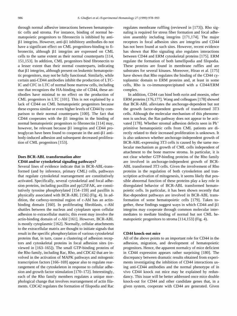
Figure 3. Distribution of CML progenitors according to CD44 expression. Comparison of the distribution of BFU-E, CFU-GM, and LTC-IC in theCD341CD4411 (h) and CD341CD44111 (j) fractions of CML peripheral blood (CML, n $ 3), mobilized blood (MOB, n 5 3), and normal bone marrow(NBM, n 5 6). ND 5 not done.
986
S. Ghaffari et al./Experimental Hematology 27 (1999) 978–993
through normal adhesive interactions between hematopoie-tic cells and stroma. For instance, binding of normal he-matopoietic progenitors to fibronectin is inhibited by anti-
b
1 integrins. However, anti-
b
1 inhibitory antibodies do nothave a significant effect on CML progenitors-binding to fi-bronectin, although
b
1 integrins are expressed on CMLcells to the same extent as their normal counterparts [114,151,153]. In addition, CML progenitors bind fibronectin toa lesser extent than their normal counterparts, indicatingthat
b
1 integrins, although present on leukemic hematopoie-tic progenitors, may not be fully functional. Similarly, whilecertain anti-CD44 antibodies inhibit the production of LTC-IC and CFC in LTC of normal bone marrow cells, includingone that recognizes the HA-binding site of CD44, these an-tibodies have minimal to no effect on the production ofCML progenitors in LTC [101]. This is not explained by alack of CD44 on CML hematopoietic progenitors becausethese express similar or even higher levels of CD44 by com-parison to their normal counterparts [100]. The fact thatCD44 cooperates with the
b
1 integrins in the binding ofnormal hematopoietic progenitors to fibronectin [114] may,however, be relevant because
b
1 integrins and CD44 pro-teoglycan have been found to cooperate in the anti-
b
1 anti-body-induced adhesion and subsequent decreased prolifera-tion of CML progenitors [153].
Does BCR-ABL transformation alter CD44 and/or cytoskeletal signaling pathways?
Several lines of evidence indicate that in BCR-ABL-trans-formed (and by inference, primary CML) cells, pathwaysthat regulate cytoskeletal rearrangement are constitutivelyactivated. Specifically, several cytoskeletal and focal adhe-sion proteins, including paxillin and pp125FAK, are consti-tutively tyrosine phosphorylated [154–159] and paxillin isphysically associated with BCR-ABL [156] (Fig. 4). In ad-dition, the carboxy-terminal region of c-Abl has an actin-binding domain [160]. In proliferating fibroblasts, c-Ablshuttles between the nucleus and cytoplasm upon cellularadhesion to extracellular matrix; this event may involve theactin-binding domain of c-Abl [161]. However, BCR-ABLis mostly cytoplasmic [162]. Normally, cellular attachmentsto the extracellular matrix are thought to initiate signals thatresult in the specific phosphorylation of various cytoskeletalproteins that, in turn, cause a clustering of adhesion recep-tors and cytoskeletal proteins in focal adhesion sites (re-viewed in [163–165]). The small GTP-binding proteins ofthe Rho family, including Rac, Rho, and CDC42 that are in-volved in the activation of MAPK pathways and mitogenictranscription factors [166–169] appear also to regulate rear-rangement of the cytoskeleton in response to cellular adhe-sion and growth factor stimulation [170–172]. Interestingly,each of the Rho family members regulates a unique mor-phological change that involves rearrangement of actin fila-ments. CDC42 regulates the formation of filopodia and Rac
regulates membrane ruffling (reviewed in [173]). Rho sig-naling is required for stress fiber formation and focal adhe-sion assembly including integrins [171,174]. The majorreceptors in focal adhesion sites are integrins and CD44has not been found at such sites. However, recent evidencehas shown that Rho signaling also regulates interactionsbetween CD44 and ERM cytoskeletal proteins [175]. ERMregulate the formation of both lamellipodia and filopodia.These proteins are found in membrane ruffles and aresubstrates for several kinases. Moreover, Hirao et al. [175]have shown that Rho regulates the binding of the CD44 cy-toplasmic domain to ERM proteins and, at least in somecells, Rho is co-immunoprecipitated with a CD44/ERMcomplex.
In addition, CD44 can bind both ezrin and moesin, otherERM proteins [176,177]. Wang and colleagues [178] showedthat BCR-ABL alleviates the anchorage-dependent but notthe growth factor-dependent growth of transformed 3T3cells. Although the molecular mechanism of this phenome-non is unclear, the Ras pathway does not appear to be acti-vated [178]. Whether stromal adhesion defects seen in Ph
1
primitive hematopoietic cells from CML patients are di-rectly related to their increased proliferation is unknown. Itis also unknown whether anchorage-independent growth ofBCR-ABL-expressing 3T3 cells is caused by the same mo-lecular mechanism as growth of CML cells independent ofattachment to the bone marrow stroma. In particular, it isnot clear whether GTP-binding proteins of the Rho familyare involved in anchorage-independent growth of BCR-ABL transformed 3T3 cells. Given the involvement of Rhoproteins in the regulation of both cytoskeleton and tran-scription activation of mitogenesis, it seems likely that pos-sible abnormal function of these proteins play a key role indisregulated behavior of BCR-ABL transformed hemato-poietic cells. In particular, it has been shown recently thatRac-dependent pathways are involved in BCR-ABL trans-formation of some hematopoietic cells [179]. Taken to-gether, these findings suggest ways in which CD44 and
b
1integrins may cooperate through common molecular inter-mediates to mediate binding of normal but not CML he-matopoietic progenitors to stroma [114,153] (Fig. 4).
CD44 knock-out mice
All of the above points to an important role for CD44 in theadhesion, migration, and development of hematopoieticprogenitors. Hence, the apparent normalcy of mice deficientin CD44 expression appears rather surprising [180]. Thediscrepancy between dramatic results obtained from experi-ments investigating the inhibition of CD44 interactions us-ing anti-CD44 antibodies and the normal phenotype of invivo CD44 knock out mice may be explained by redun-dancy. This issue will be better addressed once mice doubleknock-out for CD44 and other candidate genes that, in agiven system, cooperate with CD44 are generated. Given

S. Ghaffari et al./Experimental Hematology 27 (1999) 978–993
987
that CD44 has multiple functions and the specificity of itsfunctions are determined by the cellular context, it is highlysuspected that CD44/other adhesion molecules doubleknock-out mice will generate valuable information onCD44 function. We have also analyzed CD44
2
/
2
mice andextended the observations of unperturbed hematopoiesis inthem to include an even broader range of very primitive andcommitted hematopoietic cell types (Oostendorp and Eaves,unpublished results). Total number of CFC and specificallyCFU-GM are slightly, but significantly, reduced in thespleen of CD44
2
/
2
mice as compared to heterozygote mice[180]. Conversely, the total number of CFC and specificallyCFU-GM were slightly, but significantly, increased in themarrow of CD44
2
/
2
mice as compared to normal mice[180]. CD44 does not appear to protect against or attenuate
radiation-induced lethality or cell loss in different hemato-poietic tissues. It has been suggested that CD44 is involvedin the homing of hematopoietic cells, because anti-CD44antibodies were found to decrease hematopoietic cell en-graftment to rat bone marrow and spleen [181]. However,CD44
2
/
2
mice have proven indistinguishable from
1
/
1
controls as either donors or recipients of marrow cells usedin homing experiments [182]. Thus, CD44 is clearly not es-sential to the engraftment process, per se. On the other hand,the greater accumulation of hematopoietic progenitors seenin the spleens of G-CSF-treated CD44
2
/
2
mice (comparedto
1
/
1
controls) suggests that CD44 interactions in vivocan have consequences on the redistribution of hematopoie-tic progenitors under certain circumstances [180]. It wouldbe interesting to take advantage of the newly available CML
Figure 4. A hypothetical model: cytoskeletal alterations in BCR-ABL transformed cells. Activation of the Ras pathway in BCR-ABL transformed cells mayresult in the upregulation of CD44 isoform expression observed in primitive hematopoietic progenitor cells. CD44-ligand binding and CD44 signaling haveboth been found to require an intact cytoskeleton in some cells. In BCR-ABL transformed cells several cytoskeletal proteins have been found constitutivelyphosphorylated and at least one of them (paxillin) has been found physically associated with BCR-ABL. In BCR-ABL transformed cells, pathways control-ling the reorganization of cytoskeleton may be altered (constitutively activated). This would result in an abnormal function of adhesion molecules whoseligand binding and/or signaling require an intact cytoskeleton.

988
S. Ghaffari et al./Experimental Hematology 27 (1999) 978–993
mouse models [183,184] and use CD44
2
/
2
hematopoieticcells transduced by BCR-ABL in transplantation experi-ments in order to determine the in vivo relevance of CD44for the pathogenesis of CML.
Conclusion
Some provocative findings suggesting that the CD44 familyof adhesion molecules may play a role in normal and leuke-mic hematopoiesis have been obtained in the past few years.Nevertheless, CD44
2
/
2
mice are hematologically normaland very little effect on their hematopoiesis has emergedfrom detailed investigations of this system. Thus, a challengefor the future is to determine how various CD44-mediatedinteractions lead to the perturbations of hematopoiesis thathave been described and whether the expression of abnor-mal CD44 spliced variants on leukemic cells may contributeto their pathologic behavior.
Acknowledgments
We apologize to those whose work could not be cited due to spaceconsiderations. Much of the work reviewed here was performedwith support from Novartis Canada and a grant from the NationalCancer Institute of Canada (NCIC) with funds from the CanadianCancer Society and the Terry Fox Run. S. Ghaffari was a recipientof a Terry Fox Physician-Scientist Postdoctoral Fellowship awardedby NCIC with funds from the Terry Fox Run. C. Eaves is a TerryFox Cancer Research Scientist of the NCIC.
References
1. Lesley J, Hyman R, Kincade PW (1993) CD44 and its interactionwith extracellular matrix. Adv Immunol 54:271–335
2. Screaton GR, Bell MV, Jackson DG, Cornelis FB, Gerth U, Bell JI(1992) Genomic structure of DNA encoding the lymphocyte homingreceptor CD44 reveals at least 12 alternatively spliced exons. ProcNatl Acad Sci U S A 89:12160–12164
3. Tolg C, Hofmann M, Herrlich PPonta H (1993) Splicing choice fromten variant exons establishes CD44 variability. Nucleic Acids Res21:1225–1229
4. Goodfellow PN, Banting G, Wiles MV, Tunnacliffe A, Parkar M, Sol-omon E, Dalchau R, Fabre JW (1982) The gene, MIC4, which con-trols expression of the antigen defined by monoclonal antibodyF10.44.2, is on human chromosome 11. Eur J Immunol 12:659–663
5. Francke U, Foellmer BEHaynes BF (1983) Chromosome mapping ofhuman cell surface molecules: monoclonal anti-human lymphocyteantibodies 4F2, A3D8, and A1G3 define antigens controlled by dif-ferent regions of chromosome 11. Somatic Cell Genet 9:333–344
6. Zhou DF, Ding JF, Picker LJ, Bargatze RF, Butcher EC, Goeddel DV(1989) Molecular cloning and expression of Pgp-1. The mouse ho-molog of the human H-CAM (Hermes) lymphocyte homing receptor.J Immunol 143:3390–3395
7. Nottenburg C, Rees G, St. John T (1989) Isolation of mouse CD44cDNA: structural features are distinct from the primate cDNA. ProcNatl Acad Sci U S A 86:8521–8525
8. Stamenkovic I, Amiot M, Pesando JM, Seed B (1989) A lymphocytemolecule implicated in lymph node homing is a member of the carti-lage link protein family. Cell 56:1057–1062
9. Goldstein LA, Zhou DF, Picker LJ, Minty CN, Bargatze RF, Ding JF,Butcher EC (1989) A human lymphocyte homing receptor, the her-mes antigen, is related to cartilage proteoglycan core and link pro-teins. Cell 56:1063–1072
10. Goldstein LA, Butcher EC (1990) Identification of mRNA that en-codes an alternative form of H-CAM(CD44) in lymphoid and non-lymphoid tissues. Immunogenetics 32:389–397
11. Dougherty GJ, Landorp PM, Cooper DL, Humphries RK (1991) Mo-lecular cloning of CD44R1 and CD44R2, two novel isoforms of thehuman CD44 lymphocyte “homing” receptor expressed by hemopoi-etic cells. J Experimental Med 174:1–5
12. Hofmann M, Rudy W, Zoller M, Tolg C, Ponta H, Herrlich P,Gunthert U (1991) CD44 splice variants confer metastatic behaviorin rats: homologous sequences are expressed in human tumor celllines. Cancer Res 51:5292–5297
13. Stamenkovic I, Aruffo A, Amiot M, Seed B (1991) The hematopoie-tic and epithelial forms of CD44 are distinct polypeptides with differ-ent adhesion potentials for hyaluronate-bearing cells. EMBO J10:343–348
14. Jackson DG, Buckley J, Bell JI (1992) Multiple variants of the hu-man lymphocyte homing receptor CD44 generated by insertions at asingle site in the extracellular domain. J Biol Chem 267:4732–4739
15. Brown TA, Bouchard T, St. John T, Wayner E, Carter WG (1991)Human keratinocytes express a new CD44 core protein (CD44E) as aheparan-sulfate intrinsic membrane proteoglycan with additional ex-ons. J Cell Biol 113:207–221
16. Jalkanen S, Bargatze RF, de los Toyos J, Butcher EC (1987) Lym-phocyte recognition of high endothelium: antibodies to distinctepitopes of an 85-95-kD glycoprotein antigen differentially inhibitlymphocyte binding to lymph node, mucosal, or synovial endothelialcells. J Cell Biol 105:983–990
17. Underhill CB, Green SJ, Comoglio PM, Tarone G (1987) The hyal-uronate receptor is identical to a glycoprotein of Mr 85,000 (gp85) asshown by a monoclonal antibody that interferes with binding activity.J Biol Chem 262:13142–13146
18. Wayner EA, Carter WG (1987) Identification of multiple cell adhe-sion receptors for collagen and fibronectin in human fibrosarcomacells possessing unique alpha and common beta subunits. J Cell Biol105:1873–1884
19. Carter WG, Wayner EA (1988) Characterization of the class III col-lagen receptor, a phosphorylated, transmembrane glycoprotein ex-pressed in nucleated human cells. J Biol Chem 263:4193–4201
20. Jalkanen S, Jalkanen M, Bargatze R, Tammi M, Butcher EC (1988)Biochemical properties of glycoproteins involved in lymphocyte rec-ognition of high endothelial venules in man. J Immunol 141:1615–1623
21. Kansas GS, Wood GS, Dailey MO (1989) A family of cell-surfaceglycoproteins defined by a putative anti-endothelial cell receptor an-tibody in man. J Immunol 142:3050–3057
22. Naujokas MF, Morin M, Anderson MS, Peterson M, Miller J (1993)The chondroitin sulfate form of invariant chain can enhance stimula-tion of T cell responses through interaction with CD44. Cell 74:257–268
23. Picker LJ, De los Toyos J, Telen MJ, Haynes BF, Butcher EC (1989)Monoclonal antibodies against the CD44 [In(Lu)-related p80], andPgp-1 antigens in man recognize the Hermes class of lymphocytehoming receptors. J Immunol 142:2046–2051
24. Bazil V, Horejsi V (1992) Shedding of the CD44 adhesion moleculefrom leukocytes induced by anti-CD44 monoclonal antibody simulat-ing the effect of a natural receptor ligand. J Immunol 149:747–753
25. Yang H, Binns RM (1993) Isolation and characterization of the solu-ble and membrane-bound porcine CD44 molecules. Immunology78:547–554
26. Katoh S, McCarthy JBKincade PW (1994) Characterization of solu-ble CD44 in the circulation of mice. Levels are affected by immuneactivity and tumor growth. J Immunol 153:3440–3449

S. Ghaffari et al./Experimental Hematology 27 (1999) 978–993
989
27. Ristamaki R, Joensuu H, Salmi M, Jalkanen S (1994) Serum CD44 inmalignant lymphoma: an association with treatment response. Blood84:238–243
28. Kohda D, Morton CJ, Parkar AA, Hatanaka H, Inagaki FM, Camp-bell ID, Day AJ (1996) Solution structure of the link module: a hyal-uronan-binding domain involved in extracellular matrix stability andcell migration. Cell 86:767–775
29. Culty M, Miyake K, Kincade PW, Sikorski E, Butcher EC, UnderhillC, Sikorski E (1990) The hyaluronate receptor is a member of theCD44 (H-CAM) family of cell surface glycoproteins [published erra-tum appears in J Cell Biol (1991) 112(3):following 513]. J Cell Biol111:2765–2774
30. Aruffo A, Stamenkovic I, Melnick M, Underhill CB, Seed B (1990)CD44 is the principal cell surface receptor for hyaluronate. Cell61:1303–1313
31. Jalkanen S, Aho R, Kallajoki M, Ekfors T, Nortamo P, Gahmberg C,Duijvestijn A, Kalimo H (1989) Lymphocyte homing receptors andadhesion molecules in intravascular malignant lymphomatosis. Inter-national J Cancer 44:777–782
32. Spring FA, Dalchau R, Daniels GL, Mallinson G, Judson PA, Par-sons SF, Fabre JW, Anstee DJ (1988) The Ina and Inb blood groupantigens are located on a glycoprotein of 80,000 MW (the CDw44glycoprotein) whose expression is influenced by the In(Lu) gene. Im-munology 64:37–43
33. Miyake K, Underhill CB, Lesley J, Kincade PW (1990) Hyaluronatecan function as a cell adhesion molecule and CD44 participates in hy-aluronate recognition. J Exp Med 172:69–75
34. Miyake K, Medina KL, Hayashi S, Ono S, Hamaoka T, Kincade PW(1990) Monoclonal antibodies to Pgp-1/CD44 block lympho-hemo-poiesis in long-term bone marrow cultures. J Exp Med 171:477–488
35. Peach RJ, Hollenbaugh D, Stamenkovic I, Aruffo A (1993) Identifi-cation of hyaluronic acid binding sites in the extracellular domain ofCD44. J Cell Biol 122:257–264
36. Yang B, Yang BL, Savani RC, Turley EA (1994) Identification of acommon hyaluronan binding motif in the hyaluronan binding pro-teins RHAMM, CD44 and link protein. EMBO J 13:286–296
37. He Q, Lesley J, Hyman R, Ishihara K, Kincade PW (1992) Molecularisoforms of murine CD44 and evidence that the membrane proximaldomain is not critical for hyaluronate recognition. J Cell Biol119:1711–1719
38. Dougherty GJ, Cooper DL, Memory JF, Chiu RK (1994) Ligandbinding specificity of alternatively spliced CD44 isoforms. Recognitionand binding of hyaluronan by CD44R1. J Biol Chem 269:9074–9078
39. Bennett KL, Modrell B, Greenfield B, Bartolazzi A, Stamenkovic I,Peach R, Jackson DG, Spring F, Aruffo A (1995) Regulation ofCD44 binding to hyaluronan by glycosylation of variably spliced ex-ons. J Cell Biol 131:1623–1633
40. Lesley J, English N, Perschl A, Gregoroff J, Hyman R (1995) Variantcell lines selected for alterations in the function of the hyaluronan recep-tor CD44 show differences in glycosylation. J Exp Med 182:431–437
41. Katoh S, Zheng Z, Oritani K, Shimozato T, Kincade PW (1995) Gly-cosylation of CD44 negatively regulates its recognition of hyaluronan.J Exp Med 182:419–429
42. Lesley J, Kincade PW, Hyman R (1993) Antibody-induced activationof the hyaluronan receptor function of CD44 requires multivalentbinding by antibody. Eur J Immunol 23:1902–1909
43. Faassen AE, Schrager JA, Klein DJ, Oegema TR, Couchman JR, Mc-Carthy JB (1992) A cell surface chondroitin sulfate proteoglycan, im-munologically related to CD44, is involved in type I collagen-medi-ated melanoma cell motility and invasion. J Cell Biol 116:521–531
44. Jalkanen S, Jalkanen M (1992) Lymphocyte CD44 binds the COOH-terminal heparin-binding domain of fibronectin. J Cell Biol 116:817–825
45. Streeter PR, Berg EL, Rouse BT, Bargatze RF, Butcher EC (1988) Atissue-specific endothelial cell molecule involved in lymphocytehoming. Nature 331:41–46
46. Droll A, Dougherty ST, Chiu RK, Dirks JF, McBride WH, CooperDL, Dougherty GJ (1995) Adhesive interactions between alterna-tively spliced CD44 isoforms. J Biol Chem 270:11567–11573
47. St. John T, Meyer J, Idzerda R, Gallatin WM (1990) Expression ofCD44 confers a new adhesive phenotype on transfected cells. Cell60:45–52
48. Koopman G, van Kooyk Y, de Graaff M, Meyer CJ, Figdor CG, PalsST (1990) Triggering of the CD44 antigen on T lymphocytes pro-motes T cell adhesion through the LFA-1 pathway. J Immunol145:3589–593
49. Belitsos PC, Hildreth JE, August JT (1990) Homotypic cell aggrega-tion induced by anti-CD44(Pgp-1) monoclonal antibodies and relatedto CD44(Pgp-1) expression. J Immunol 144:1661–1670
50. Lesley J, Schulte RHyman R (1990) Binding of hyaluronic acid tolymphoid cell lines is inhibited by monoclonal antibodies againstPgp-1. Exp Cell Res 187:224–233
51. Toyama-Sorimachi N, Miyasaka M (1994) A sulfated proteoglycanas a novel ligand for CD44. J Dermatol 21:795–801
52. Stevens RL, Avraham S, Gartner MC, Bruns GA, Austen KF, WeisJH (1988) Isolation and characterization of a cDNA that encodes thepeptide core of the secretory granule proteoglycan of human promye-locytic leukemia HL-60 cells. J Biol Chem 263:7287–7291
53. Toyama-Sorimachi N, Sorimachi H, Tobita Y, Kitamura F, Yagita H,Suzuki K, Miyasaka M (1995) A novel ligand for CD44 is serglycin,a hematopoietic cell lineage-specific proteoglycan. Possible involve-ment in lymphoid cell adherence and activation. J Biol Chem270:7437–7444
54. Weber GF, Ashkar S, Glimcher MJ, Cantor H (1996) Receptor-ligand interaction between CD44 and osteopontin (Eta-1). Science271:509–512
55. Weber GF, Ashkar S, Cantor H (1997) Interaction between CD44and osteopontin as a potential basis for metastasis formation. ProcAssoc Am Physicians 109:1–9
56. Patarca R, Wei FY, Singh P, Morasso MI, Cantor H (1990) Dysregu-lated expression of the T cell cytokine Eta-1 in CD4-8- lymphocytesduring the development of murine autoimmune disease. J Exp Med172:1177–1183
57. Smith LL, Cheung HK, Ling LE, Chen J, Sheppard D, Pytela R, Gi-achelli CM (1996) Osteopontin N-terminal domain contains a crypticadhesive sequence recognized by alpha9beta1 integrin. J Biol Chem271:28485–28491
58. Lacy BE, Underhill CB (1987) The hyaluronate receptor is associatedwith actin filaments. J Cell Biol 105:1395–1404
59. Kalomiris EL, Bourguignon LY (1988) Mouse T lymphoma cellscontain a transmembrane glycoprotein (GP85) that binds ankyrin. JCell Biol 106:319–327
60. Tsukita S, Oishi K, Sato N, Sagara J, Kawai A, Tsukita S (1994)ERM family members as molecular linkers between the cell surfaceglycoprotein CD44 and actin-based cytoskeletons. J Cell Biol126:391–401
61. Taher TE, Smit L, Griffioen AW, Schilder-Tol EJ, Borst J, Pals ST(1996) Signaling through CD44 is mediated by tyrosine kinases. As-sociation with p56lck in T lymphocytes. J Biol Chem 271:2863–2867
62. Ilangumaran S, Briol A, Hoessli DC (1998) CD44 selectively associ-ates with active Src family protein tyrosine kinases Lck and Fyn inglycosphingolipid-rich plasma membrane domains of human periph-eral blood lymphocytes. Blood 91:3901–3908
63. Lokeshwar VB, Bourguignon LY (1992) The lymphoma transmem-brane glycoprotein GP85 (CD44) is a novel guanine nucleotide-bind-ing protein which regulates GP85 (CD44)-ankyrin interaction. J BiolChem 267:22073–22078
64. Lesley J, He Q, Miyake K, Hamann A, Hyman R, Kincade PW(1992) Requirements for hyaluronic acid binding by CD44: a role forthe cytoplasmic domain and activation by antibody. J Exp Med175:257–266
65. Lokeshwar VB, Fregien N, Bourguignon LY (1994) Ankyrin-binding

990
S. Ghaffari et al./Experimental Hematology 27 (1999) 978–993
domain of CD44(GP85) is required for the expression of hyaluronicacid-mediated adhesion function. J Cell Biol 126:1099–1109
66. Thomas L, Byers HR, Vink J, Stamenkovic I (1992) CD44H regu-lates tumor cell migration on hyaluronate-coated substrate. J CellBiol 118:971–977
67. Hathcock KS, Hirano H, Murakami SHodes RJ (1993) CD44 expres-sion on activated B cells. Differential capacity for CD44-dependentbinding to hyaluronic acid. J Immunol 151:6712–6722
68. Perschl A, Lesley J, English N, Trowbridge I, Hyman R (1995) Roleof CD44 cytoplasmic domain in hyaluronan binding. Eur J Immunol25:495–501
69. Kalomiris EL, Bourguignon LY (1989) Lymphoma protein kinase Cis associated with the transmembrane glycoprotein, GP85, and mayfunction in GP85-ankyrin binding. J Biol Chem 264:8113–8119
70. Camp RL, Kraus TA, Pure E (1991) Variations in the cytoskeletal in-teraction and posttranslational modification of the CD44 homing re-ceptor in macrophages. J Cell Biol 115:1283–1292
71. Neame SJ, Isacke CM (1993) The cytoplasmic tail of CD44 is re-quired for basolateral localization in epithelial MDCK cells but doesnot mediate association with the detergent-insoluble cytoskeleton offibroblasts. J Cell Biol 121:1299–1310
72. Neame SJ, Isacke CM (1992) Phosphorylation of CD44 in vivo re-quires both Ser323 and Ser325, but does not regulate membrane lo-calization or cytoskeletal interaction in epithelial cells. EMBO J11:4733–4738
73. Hyman R, Lesley J, Schulte R (1991) Somatic cell mutants distin-guish CD44 expression and hyaluronic acid binding. Immunogenet-ics 33:392–395
74. Murakami S, Shimabukuro Y, Miki Y, Saho T, Hino E, Kasai D,Nozaki T, Kusumoto Y, Okada H (1994) Inducible binding of humanlymphocytes to hyaluronate via CD44 does not require cytoskeletonassociation but does require new protein synthesis. J Immunol152:467–477
75. Morimoto K, Robin E, Le Bousse-Kerdiles MC, Li Y, Clay D, Jas-min C, Smadja-Joffe F (1994) CD44 mediates hyaluronan binding byhuman myeloid KG1A and KG1 cells. Blood 83:657–662
76. Liu D, Sy MS (1996) A cysteine residue located in the transmem-brane domain of CD44 is important in binding of CD44 to hyaluronicacid. J Exp Med 183:1987–1994
77. Li R, Walker JR, Johnson P (1998) Chimeric CD4/CD44 moleculesassociate with CD44 via the transmembrane region and reduce hyal-uronan binding in T cell lines. Eur J Immunol 28:1745–1754
78. Haynes BF, Telen MJ, Hale LP, Denning SM (1989) CD44–a mole-cule involved in leukocyte adherence and T-cell activation [publishederratum appears in Immunol Today (1990) 11(3):80]. Immunol To-day 10:423–428
79. Kansas GS, Dailey MO (1989) Expression of adhesion structuresduring B cell development in man. J Immunol 142:3058–3062
80. Murakami S, Miyake K, Kincade PW, Hodes RJ (1991) Functionalrole of CD44 (Pgp-1) on activated B cells. Immunol Res 10:15–27
81. Murakami S, Miyake K, Abe R, Kincade PW, Hodes RJ (1991) Char-acterization of autoantibody-secreting B cells in mice undergoingstimulatory (chronic) graft-versus-host reactions. Identification of aCD44hi population that binds specifically to hyaluronate. J Immunol146:1422–1427
82. Kincade PW (1992) Cell interaction molecules and cytokines whichparticipate in B lymphopoiesis. Baillieres Clin Haematol 5:575–598
83. Camp RL, Kraus TA, Birkeland ML, Pure E (1991) High levels ofCD44 expression distinguish virgin from antigen-primed B cells. JExp Med 173:763–766
84. Koshiishi I, Shizari M, Underhill CB (1994) CD44 can mediate theadhesion of platelets to hyaluronan. Blood 84:390–396
85. Sandmaier BM, Storb R, Appelbaum FR, Gallatin WM (1990) Anantibody that facilitates hematopoietic engraftment recognizes CD44.Blood 76:630–635
86. Tan PH, Santos EB, Rossbach HC, Sandmaier BM (1993) Enhance-
ment of natural killer activity by an antibody to CD44. J Immunol150:812–820
87. Campanero MR, Pulido R, Alonso JL, Pivel JP, Pimentel-MuinosFX, Fresno M, Sanchez-Madrid F (1991) Down-regulation by tumornecrosis factor-alpha of neutrophil cell surface expression of thesialophorin CD43 and the hyaluronate receptor CD44 through a pro-teolytic mechanism. Eur J Immunol 21:3045–3048
88. Pericle F, Sconocchia G, Titus JA, Segal DM (1996) CD44 is a cyto-toxic triggering molecule on human polymorphonuclear cells. J Im-munol 157:4657–4663
89. Denning SM, Le PT, Singer KH, Haynes BF (1990) Antibodiesagainst the CD44 p80, lymphocyte homing receptor molecule aug-ment human peripheral blood T cell activation. J Immunol 144:7–15
90. Webb DS, Shimizu Y, Van Seventer GA, Shaw S, Gerrard TL (1990)LFA-3, CD44, and CD45: physiologic triggers of human monocyteTNF and IL-1 release. Science 249:1295–1297
91. Gruber MF, Webb DS, Gerrard TL (1992) Stimulation of humanmonocytes via CD45, CD44, and LFA-3 triggers macrophage-col-ony-stimulating factor production. Synergism with lipopolysaccha-ride and IL-1 beta. J Immunol 148:1113–1118
92. Noble PW, Lake FR, Henson PM, Riches DW (1993) Hyaluronateactivation of CD44 induces insulin-like growth factor-1 expressionby a tumor necrosis factor-alpha-dependent mechanism in murinemacrophages. J Clin Invest 91:2368–2377
93. McKee CM, Penno MB, Cowman M, Burdick MD, Strieter RM, BaoC, Noble PW (1996) Hyaluronan (HA) fragments induce chemokinegene expression in alveolar macrophages. The role of HA size andCD44. J Clin Invest 98:2403–2413
94. Rameshwar P, Chang VT, Gascon P (1996) Implication of CD44 in ad-hesion-mediated overproduction of TGF-beta and IL-1 in monocytesfrom patients with bone marrow fibrosis. Br J Haematol 93: 22–29
95. Trowbridge IS, Lesley J, Schulte R, Hyman R, Trotter J (1982) Bio-chemical characterization and cellular distribution of a polymorphic,murine cell-surface glycoprotein expressed on lymphoid tissues. Im-munogenetics 15:299–312
96. Hughes EN, Colombatti A, August JT (1983) Murine cell surfaceglycoproteins. Purification of the polymorphic Pgp-1 antigen andanalysis of its expression on macrophages and other myeloid cells. JBiol Chem 258:1014–1021
97. Spangrude GJ, Klein J, Heimfeld S, Aihara Y, Weissman IL (1989)Two monoclonal antibodies identify thymic-repopulating cells inmouse bone marrow. J Immunol 142:425–430
98. Lewinsohn DM, Nagler A, Ginzton N, Greenberg P, Butcher EC(1990) Hematopoietic progenitor cell expression of the H-CAM(CD44) homing-associated adhesion molecule. Blood 75:589–595
99. Kansas GS, Muirhead MJ, Dailey MO (1990) Expression of theCD11/CD18, leukocyte adhesion molecule 1, and CD44 adhesionmolecules during normal myeloid and erythroid differentiation in hu-mans. Blood 76:2483–2492
100. Ghaffari S, Dougherty GJ, Lansdorp PM, Eaves AC, Eaves CJ (1995)Differentiation-associated changes in CD44 isoform expression dur-ing normal hematopoiesis and their alteration in chronic myeloid leu-kemia. Blood 86:2976–2985
101. Ghaffari S, Dougherty GJ, Eaves AC, Eaves CJ (1997) Diverse ef-fects of anti-CD44 antibodies on the stromal cell-mediated support ofnormal but not leukaemic (CML) haemopoiesis in vitro. Br J Haema-tol 97:22–28
102. Gallagher JT, Spooncer E, Dexter TM (1983) Role of the cellular ma-trix in haemopoiesis. I. Synthesis of glycosaminoglycans by mousebone marrow cell cultures. J Cell Sci 63:155–171
103. Spooncer E, Gallagher JT, Krizsa F, Dexter TM (1983) Regulation ofhaemopoiesis in long-term bone marrow cultures. IV. Glycosami-noglycan synthesis and the stimulation of haemopoiesis by beta-D-xylosides. J Cell Biol 96:510–514
104. Underhill C (1992) CD44: the hyaluronan receptor. J Cell Sci103:293–298

S. Ghaffari et al./Experimental Hematology 27 (1999) 978–993
991
105. Smadja-Joffe F, Legras S, Girard N, Li Y, Delpech B, Bloget F,Morimoto K, Le Bousse-Kerdiles C, Clay D, Jasmin C, Levesque JP(1996) CD44 and hyaluronan binding by human myeloid cells. LeukLymphoma 21:407–520 (color plates following p. 528)
106. Ghaffari S, Dougherty GJ, Eaves AC, Eaves CJ (1996) Altered pat-terns of CD44 epitope expression in human chronic and acute mye-loid leukemia. Leukemia 10:1773–1781
107. Legras S, Levesque JP, Charrad R, Morimoto K, Le Bousse C, ClayD, Jasmin C, Smadja-Joffe F (1997) CD44-mediated adhesiveness ofhuman hematopoietic progenitors to hyaluronan is modulated by cy-tokines. Blood 89:1905–1914
108. Oostendorp RA, Spitzer E, Dormer P (1996) Adhesion of human he-matopoietic progenitor cells to bone-marrow-derived stromal cells isenhanced by antibodies to CD44. Acta Haematologica 95:243–247
109. Oostendorp RA, Spitzer E, Brandl M, Eaves CJ, Dormer P (1998)Evidence for differences in the mechanisms by which antibodiesagainst CD44 promote adhesion of erythroid and granulopoietic pro-genitors to marrow stromal cells. Br J Haematol 101:436–445
110. Oostendorp RA, Spitzer E, Reisbach G, Dormer P (1997) Antibodiesto the beta 1-integrin chain, CD44, or ICAM-3 stimulate adhesion ofblast colony-forming cells and may inhibit their growth. Exp Hema-tol 25:345–349
111. Dittel BN, Le Bien TW (1995) Reduced expression of vascular celladhesion molecule-1 on bone marrow stromal cells isolated frommarrow transplant recipients correlates with a reduced capacity tosupport human B lymphopoiesis in vitro. Blood 86:2833–2841
112. Bendall LJ, Kirkness J, Hutchinson A, Bianchi A, Makrynikola V,Bradstock KF, Gottlieb DJ (1997) Antibodies to CD44 enhance adhe-sion of normal CD34
1
cells and acute myeloblastic but not lymphoblas-tic leukaemia cells to bone marrow stroma. Br J Haematol 98:828–837
113. Rossbach HC, Krizanac-Bengez L, Santos EB, Gooley TA, Sand-maier BM (1996) An antibody to CD44 enhances hematopoiesis inlong-term marrow cultures. Exp Hematol 24:221–227
114. Verfaillie CM, Benis A, Iida J, McGlave PB, McCarthy JB (1994)Adhesion of committed human hematopoietic progenitors to syn-thetic peptides from the C-terminal heparin-binding domain of fi-bronectin: cooperation between the integrin alpha 4 beta 1 and theCD44 adhesion receptor. Blood 84:1802–1811
115. Gunji Y, Nakamura M, Hagiwara T, Hayakawa K, Matsushita H, Os-awa H, Nagayoshi K, Nakauchi H, Yanagisawa M, Miura Y, et al.(1992) Expression and function of adhesion molecules on human he-matopoietic stem cells: CD34
1
LFA-1
2
cells are more primitive thanCD34
1
LFA-1
1
cells. Blood 80:429–436116. Patel VP, Lodish HF (1984) Loss of adhesion of murine erythroleu-
kemia cells to fibronectin during erythroid differentiation. Science224:996–998
117. Sugahara H, Kanakura Y, Furitsu T, Ishihara K, Oritani K, Ikeda H,Kitayama H, Ishikawa J, Hashimoto K, Kanayama Y, et al. (1994) In-duction of programmed cell death in human hematopoietic cell linesby fibronectin via its interaction with very late antigen 5. J Exp Med179:1757–1766
118. Tanaka Y, Adams DH, Hubscher S, Hirano H, Siebenlist U, Shaw S(1993) T-cell adhesion induced by proteoglycan-immobilized cyto-kine MIP-1 beta [see comments]. Nature 361:79–82
119. Sandmaier BM, Storb R, Liu Y, Santos EB, Bryant E, Schuening FG,Deeg HJ, Seidel K, Graham T (1996) An anti-CD44 antibody doesnot enhance engraftment of DLA-identical marrow after low-dose to-tal body irradiation. Transplant Immunol 4:271–274
120. Vermeulen M, Le Pesteur F, Gagnerault MC, Mary JY, Sainteny F,Lepault F (1998) Role of adhesion molecules in the homing and mo-bilization of murine hematopoietic stem and progenitor cells. Blood92:894–900
121. Bennett KL, Jackson DG, Simon JC, Tanczos E, Peach R, Modrell B,Stamenkovic I, Plowman G, Aruffo A (1995) CD44 isoforms con-taining exon V3 are responsible for the presentation of heparin-bind-ing growth factor. J Cell Biol 128:687–698
122. Jackson DG, Bell JI, Dickinson R, Timans J, Shields J, Whittle N(1995) Proteoglycan forms of the lymphocyte homing receptor CD44are alternatively spliced variants containing the v3 exon. J Cell Biol128:673–685
123. Ruoslahti E (1989) Proteoglycans in cell regulation. J Biol Chem264:13369–13372
124. Schlessinger J, Lax I, Lemmon M (1995) Regulation of growth factoractivation by proteoglycans: what is the role of the low affinity recep-tors? Cell 83:357–360
125. Sherman L, Wainwright D, Ponta H, Herrlich P (1998) A splice vari-ant of CD44 expressed in the apical ectodermal ridge presents fibro-blast growth factors to limb mesenchyme and is required for limboutgrowth. Genes Develop 12:1058–1071
126. Gunthert U, Hofmann M, Rudy W, Reber S, Zoller M, Haussmann I,Matzku S, Wenzel A, Ponta H, Herrlich P (1991) A new variant ofglycoprotein CD44 confers metastatic potential to rat carcinomacells. Cell 65:13–24
127. Herrlich P, Zoller M, Pals ST, Ponta H (1993) CD44 splice variants:metastases meet lymphocytes. Immunol Today 14:395–399
128. Salles G, Zain M, Jiang WM, Boussiotis VA, Shipp MA (1993) Al-ternatively spliced CD44 transcripts in diffuse large-cell lymphomas:characterization and comparison with normal activated B cells andepithelial malignancies. Blood 82:3539–3547
129. Stauder R, Eisterer W, Thaler J, Gunthert U (1995) CD44 variant iso-forms in non-Hodgkin’s lymphoma: a new independent prognosticfactor. Blood 85:2885–2899
130. Stauder R, Van Driel M, Schwarzler C, Thaler J, Lokhorst HM,Kreuser ED, Bloem AC, Gunthert U, Eisterer W (1996) DifferentCD44 splicing patterns define prognostic subgroups in multiple my-eloma. Blood 88:3101–3108
131. Legras S, Gunthert U, Stauder R, Curt F, Oliferenko S, Kluin-Nele-mans HC, Marie JP, Proctor S, Jasmin C, Smadja-Joffe F (1998) Astrong expression of CD44-6v correlates with shorter survival of pa-tients with acute myeloid leukemia. Blood 91:3401–3413
132. Arch R, Wirth K, Hofmann M, Ponta H, Matzku S, Herrlich P, ZollerM (1992) Participation in normal immune responses of a metastasis-inducing splice variant of CD44 [see comments]. Science 257:682–685
133. Koopman G, Heider KH, Horst E, Adolf GR, van den Berg F, PontaH, Herrlich P, Pals ST (1993) Activated human lymphocytes and ag-gressive non-Hodgkin’s lymphomas express a homologue of the ratmetastasis-associated variant of CD44. J Exp Med 177:897–904
134. Haegel H, Tolg C, Hofmann M, Ceredig R (1993) Activated mouseastrocytes and T cells express similar CD44 variants. Role of CD44in astrocyte/T cell binding. J Cell Biol 122:1067–1077
135. Mackay CR, Terpe HJ, Stauder R, Marston WL, Stark H, Gunthert U(1994) Expression and modulation of CD44 variant isoforms in hu-mans. J Cell Biol 124:71–82
136. Patel DD, Hale LP, Whichard LP, Radcliff G, Mackay CR, HaynesBF (1995) Expression of CD44 molecules and CD44 ligands duringhuman thymic fetal development: expression of CD44 isoforms is de-velopmentally regulated. Intl Immunol 7:277–286
137. Wheatley SC, Isacke CM, Crossley PH (1993) Restricted expressionof the hyaluronan receptor, CD44, during postimplantation mouseembryogenesis suggests key roles in tissue formation and patterning.Development 119:295–306
138. Wirth K, Arch R, Somasundaram C, Hofmann M, Weber B, HerrlichP, Matzku S, Zoller M (1993) Expression of CD44 isoforms carryingmetastasis-associated sequences in newborn and adult rats. Eur JCancer 29:1172–1177
139. Terpe HJ, Stark H, Prehm P, Gunthert U (1994) CD44 variant iso-forms are preferentially expressed in basal epithelial of non-malig-nant human fetal and adult tissues. Histochemistry 101:79–89
140. Campbell S, Swann HR, Aplin JD, Seif MW, Kimber SJ, Elstein M(1995) CD44 is expressed throughout pre-implantation human em-bryo development. Hum Reprod 10:425–430

992
S. Ghaffari et al./Experimental Hematology 27 (1999) 978–993
141. Zoller M, Schmidt A, Denzel A, Moll J (1997) Constitutive expres-sion of a CD44 variant isoform on T cells facilitates regaining of im-munocompetence in allogeneic bone marrow transplantation. Blood90:873–885
142. Yu Q, Toole BP, Stamenkovic I (1997) Induction of apoptosis ofmetastatic mammary carcinoma cells in vivo by disruption of tumorcell surface CD44 function. J Exp Med 186:1985–1996
143. Gunthert AR, Strater J, von Reyher U, Henne C, Joos S, Koretz K,Moldenhauer G, Krammer PH, Moller P (1996) Early detachment ofcolon carcinoma cells during CD95(APO-1/Fas)-mediated apoptosis.I. De-adhesion from hyaluronate by shedding of CD44. J Cell Biol134:1089–1096
144. Wagner SN, Wagner C, Reinhold U, Funk R, Zoller M, Goos M(1998) Predominant expression of CD44 splice variant v10 in malig-nant and reactive human skin lymphocytes. J Invest Dermatol 113:464–471
145. Pendergast AM, Quilliam LA, Cripe LD, Bassing CH, Dai Z, Li N,Batzer A, Rabun KM, Der CJ, Schlessinger J, et al. (1993) BCR-ABL-induced oncogenesis is mediated by direct interaction with theSH2 domain of the GRB-2 adaptor protein. Cell 75:175–185
146. Tauchi T, Boswell HS, Leibowitz D, Broxmeyer HE (1994) Couplingbetween p210bcr-abl and Shc and Grb2 adaptor proteins in hemato-poietic cells permits growth factor receptor-independent link to rasactivation pathway. J Exp Med 179:167–175
147. Hofmann M, Rudy W, Gunthert U, Zimmer SG, Zawadzki V, ZollerM, Lichtner RB, Herrlich P, Ponta H (1993) A link between ras andmetastatic behavior of tumor cells: ras induces CD44 promoter activ-ity and leads to low-level expression of metastasis-specific variantsof CD44 in CREF cells. Cancer Res 53:1516–1521
148. Jamal HH, Cano-Gauci DF, Buick RN, Filmus J (1994) Activated rasand src induce CD44 overexpression in rat intestinal epithelial cells.Oncogene 9:417–423
149. Penno MB, August JT, Baylin SB, Mabry M, Linnoila RI, Lee VS,Croteau D, Yang XL, Rosada C (1994) Expression of CD44 in hu-man lung tumors. Cancer Res 54:1381–1387
150. Gordon MY, Dowding CR, Riley GP, Goldman JM, Greaves MF(1987) Altered adhesive interactions with marrow stroma of hae-matopoietic progenitor cells in chronic myeloid leukaemia. Nature328:342–344
151. Verfaillie CM, McCarthy JBMcGlave PB (1992) Mechanisms under-lying abnormal trafficking of malignant progenitors in chronic my-elogenous leukemia. Decreased adhesion to stroma and fibronectinbut increased adhesion to the basement membrane components lami-nin and collagen type IV. J Clin Invest 90:1232–1241
152. Bhatia R, Wayner EA, McGlave PB, Verfaillie CM (1994) Inter-feron-alpha restores normal adhesion of chronic myelogenous leuke-mia hematopoietic progenitors to bone marrow stroma by correctingimpaired beta 1 integrin receptor function. J Clin Invest 94:384–391
153. Lundell BI, McCarthy JB, Kovach NL, Verfaillie CM (1997) Activa-tion of beta1 integrins on CML progenitors reveals cooperation be-tween beta1 integrins and CD44 in the regulation of adhesion andproliferation. Leukemia 11:822–829
154. Gotoh A, Miyazawa K, Ohyashiki K, Tauchi T, Boswell HS, Brox-meyer HE, Toyama K (1995) Tyrosine phosphorylation and activa-tion of focal adhesion kinase (p125FAK) by BCR-ABL oncoprotein.Exp Hematol 23:1153–1159
155. Salgia R, Brunkhorst B, Pisick E, Li JL, Lo SH, Chen LB, Griffin JD(1995) Increased tyrosine phosphorylation of focal adhesion proteinsin myeloid cell lines expressing p210BCR/ABL. Oncogene 11:1149–1155
156. Salgia R, Li JL, Lo SH, Brunkhorst B, Kansas GS, Sobhany ES, SunY, Pisick E, Hallek M, Ernst T, et al. (1995) Molecular cloning of hu-man paxillin, a focal adhesion protein phosphorylated by P210BCR/ABL. J Biol Chem 270:5039–5047
157. Salgia R, Avraham S, Pisick E, Li JL, Raja S, Greenfield EA, SattlerM, Avraham H, Griffin JD (1996) The related adhesion focal tyrosine
kinase forms a complex with paxillin in hematopoietic cells. J BiolChem 271:31222–31226
158. Salgia R, Sattler M, Pisick E, Li JL, Griffin JD (1996) p210BCR/ABL induces formation of complexes containing focal adhesion pro-teins and the protooncogene product p120c-Cbl. Exp Hematol24:310–313
159. Salgia R, Li JL, Ewaniuk DS, Pear W, Pisick E, Burky SA, Ernst T,Sattler M, Chen LB, Griffin JD (1997) BCR/ABL induces multipleabnormalities of cytoskeletal function. J Clin Invest 100:46–57
160. Wang JY, Ledley F, Goff S, Lee R, Groner Y, Baltimore D (1984)The mouse c-abl locus: molecular cloning and characterization. Cell36:349–356
161. Taagepera S, McDonald D, Loeb JE, Whitaker LL, McElroy AK,Wang JY, Hope TJ (1998) Nuclear-cytoplasmic shuttling of C-ABLtyrosine kinase. Proc Natl Acad Sci U S A 95:7457–7462
162. McWhirter JR, Wang JY (1993) An actin-binding function contrib-utes to transformation by the Bcr-Abl oncoprotein of Philadelphiachromosome-positive human leukemias. EMBO J 12:1533–1546
163. Gumbiner BM (1996) Cell adhesion: the molecular basis of tissue ar-chitecture and morphogenesis. Cell 84:345–357
164. Craig SW, Johnson RP (1996) Assembly of focal adhesions:progress, paradigms, and portents. Curr Opin Cell Biol 8:74–85
165. Lauffenburger DA, Horwitz AF (1996) Cell migration: a physicallyintegrated molecular process. Cell 84:359–369
166. Coso OA, Chiariello M, Yu JC, Teramoto H, Crespo P, Xu N, MikiT, Gutkind JS (1995) The small GTP-binding proteins Rac1 andCdc42 regulate the activity of the JNK/SAPK signaling pathway.Cell 81:1137–1146
167. Minden A, Lin A, Claret FX, Abo A, Karin M (1995) Selective acti-vation of the JNK signaling cascade and c-Jun transcriptional activityby the small GTPases Rac and Cdc42Hs. Cell 81:1147–1157
168. Bagrodia S, Derijard B, Davis RJ, Cerione RA (1995) Cdc42 andPAK-mediated signaling leads to Jun kinase and p38 mitogen-acti-vated protein kinase activation. J Biol Chem 270:27995–27998
169. Gupta S, Campbell D, Derijard B, Davis RJ (1995) Transcription fac-tor ATF2 regulation by the JNK signal transduction pathway. Science267:389–393
170. Ridley AJ, Paterson HF, Johnston CL, Diekmann D, Hall A (1992)The small GTP-binding protein rac regulates growth factor-inducedmembrane ruffling. Cell 70:401–410
171. Ridley AJ, Hall A (1992) The small GTP-binding protein rho regu-lates the assembly of focal adhesions and actin stress fibers in re-sponse to growth factors. Cell 70:389–399
172. Nobes CD, Hall A (1995) Rho, rac, and cdc42 GTPases regulate theassembly of multimolecular focal complexes associated with actinstress fibers, lamellipodia, and filopodia. Cell 81:53–62
173. Zigmond SH (1996) Signal transduction and actin filament organiza-tion. Curr Opin Cell Biol 8:66–73
174. Hotchin NA, Hall A (1995) The assembly of integrin adhesion com-plexes requires both extracellular matrix and intracellular rho/racGTPases. J Cell Biol 131:1857–1865
175. Hirao M, Sato N, Kondo T, Yonemura S, Monden M, Sasaki T, TakaiY, Tsukita S, Tsukita S (1996) Regulation mechanism of ERM(ezrin/radixin/moesin) protein/plasma membrane association: possi-ble involvement of phosphatidylinositol turnover and Rho-dependentsignaling pathway. J Cell Biol 135:37–51
176. Yonemura S, Hirao M, Doi Y, Takahashi N, Kondo T, Tsukita S(1998) Ezrin/radixin/moesin (ERM) proteins bind to a positivelycharged amino acid cluster in the juxta-membrane cytoplasmic do-main of CD44, CD43, and ICAM-2. J Cell Biol 140:885–895
177. Legg JW, Isacke CM (1998) Identification and functional analysis ofthe ezrin-binding site in the hyaluronan receptor, CD44. Curr Biol8:705–708
178. Renshaw MW, McWhirter JR, Wang JY (1995) The human leukemiaoncogene bcr-abl abrogates the anchorage requirement but not thegrowth factor requirement for proliferation. Mol Cell Biol 15:1286–1293

S. Ghaffari et al./Experimental Hematology 27 (1999) 978–993
993
179. Skorski T, Wlodarski P, Daheron L, Salomoni P, Nieborowska-Skorska M, Majewski M, Wasik M, Calabretta B (1998) BCR-ABL-mediated leukemogenesis requires the activity of the small GTP-binding protein Rac. PNAS 95:11858–11862
180. Schmits R, Filmus J, Gerwin N, Senaldi G, Kiefer F, Kundig T,Wakeham A, Shahinian A, Catzavelos C, Rak J, Furlonger C, Zakar-ian A, Simard JJ, Ohashi PS, Paige CJ, Gutierrez-Ramos JC, MakTW (1997) CD44 regulates hematopoietic progenitor distribution,granuloma formation, and tumorigenicity. Blood 90:2217–2233
181. Khaldoyanidi S, Schnabel D, Fohr N, Zoller M (1997) Functional ac-tivity of CD44 isoforms in haemopoiesis of the rat. Br J Haematol96:31–45
182. Oostendorp RA, Eaves CJ (1997) Quantitative analysis of the initialand CD44-independent phases of engraftment in a syngeneic marrowtransplant model. Blood 90:402
183. Pear WS, Miller JP, Xu L, Pui JC, Soffer B, Quackenbush RC, Pen-dergast AM, Bronson R, Aster JC, Scott ML, Baltimore D (1998) Ef-ficient and rapid induction of a chronic myeloid leukemia-like my-eloproliferative disease in mice receiving P210 bcr-abl-transducedbone marrow. Blood 92:3780–3792
184. Zhang X, Ren R (1998) Bcr-Abl efficiently induces a myeloprolifera-tive disease and production of excess interleukin-3 and granulocyte-macrophage colony-stimulating factor in mice: a novel model forchronic myeloid leukemia. Blood 92:3829–3840