Wnt-Related Molecules and Signaling Pathway Equilibrium in Hematopoiesis
10
Cell Stem Cell Review Wnt-Related Molecules and Signaling Pathway Equilibrium in Hematopoiesis Sachin Malhotra 1,2 and Paul W. Kincade 1,2, * 1 Immunobiology and Cancer Program, Oklahoma Medical Research Foundation 2 Department of Microbiology and Immunology University of Oklahoma Health Sciences Center, Oklahoma City, OK 73104, USA *Correspondence: [email protected] DOI 10.1016/j.stem.2008.12.004 There is near consensus that Wnt family molecules establish important gradients within niches where hematopoietic stem cells (HSC) reside. We review recent papers suggesting that a delicate balance is required between competing Wnt ligands and corresponding signaling pathways to maintain HSC integrity. Some steps in the transitions from HSC to lymphoid progenitor seem to be partially reversible and under the influence of Wnts. In addition, it has been recently suggested that HSC can oscillate between dormant versus active or lineage-biased states. We speculate that Wnts control a reflux process that may sustain stem cell self-renewal and differentiation potential. Secreted Wnt glycoproteins are involved in multiple aspects of embryonic development, governing cell fates, polarity, and proliferation (Nusse, 2005). Consequently, Wnt associated abnormalities contribute to embryonic lethality and underlie such disorders as cancer, bone density defects, and polycystic kidney disease. Many Wnt family proteins are expressed in hematopoietic tissues, and a series of reports suggest there must be important roles in blood cell formation. However, few topics have been more controversial, and generalization of Wnt regulation of hematopoietic stem cells (HSC) has been difficult. The daunting complexity and functional redundancy of Wnt- related molecules accounts in part for this failure to reach consensus. Also contributing to the wide range of conclusions drawn with respect to the function of Wnt signaling, investigators have used Wnt-related proteins that vary in structure, potency, and mode of presentation to influence different types of target cells. Moreover, experimental depletion of Wnt signaling may have been incomplete in some cases, and overexpression para- digms that result in very high levels could have given misleading results. A series of recent reports provide new perspectives on these important issues and will provide the basis for discussion throughout this review. Contrary to original expectations, it remains unclear if any Wnt ligands can be regarded as typical growth and differentiation factors for hematopoietic cells. Indeed, some of these ligands impede the expansion and/or differentiation of particular cell types, and such Wnt-dependent responses may be critical to maintaining stem cell integrity under normal circumstances. Other results indicate that early events in hematopoiesis may not be unidirectional and can be experimentally reversed with Wnt signaling. Thus, further study could reveal new ways to exploit Wnt family molecules for protecting, expanding, and even reprogramming HSC. We will focus here on the role of Wnt signaling on the regulation of HSC and their progression toward cells of the humoral immune system. An excellent review concerning the role of Wnts in T lymphocyte development has just appeared (Staal et al., 2008). Complexity of Wnt Signaling Components and Pathways Some 19 Wnt ligands interact with both secreted and membrane-associated proteins to initiate signaling processes (detailed in Staal et al. [2008]; references in Table 1; summarized in Figure 1). The list of Wnt binding proteins include at least ten seven-pass transmembrane Frizzled (Fzd) receptors, two low- density lipoprotein receptor-related proteins (LRP), and a number of extracellular Wnt-modulating proteins such as Kremlin, Dickkopf (Dkk), Wnt-inhibitory factor (WIF), secreted Fzds (SFRP), and Norrin. Depending on the type of ligand- receptor interaction, the availability of intracellular signaling components and the specific target cell, an astonishing ten Wnt signaling pathways have been identified (Semenov et al., 2007; Macdonald et al., 2007). The canonical pathway, arguably the most studied and best understood, results in stabilization and nuclear translocation of b-catenin (Figure 1). This molecule is normally targeted for degradation by a GSK protein complex, but canonical signaling stabilizes b-catenin by destruction of the degradation complex. Stabilized b-catenin then enters the nucleus and acts as a coactivator for transcription factors such as LEF and TCF. A related molecule, g-catenin (plakogloblin), is at least partially redundant to b-catenin and can bind TCF/ LEF. Simultaneous targeting of both catenin genes results in embryonic lethality (Haegel et al., 1995; Ruiz et al., 1996). Further, Wnt signaling continued in the combined absence of b/g catenin after inducible deletions, suggesting involvement of unknown redundant proteins (Jeannet et al., 2007). Adenoma- tous polyposis coli (APC) is a multidomain protein that can bind GSK3b, Axin, and b-catenin to prevent nuclear localization of catenin, thus decreasing canonical Wnt signaling (Aoki and Taketo, 2007). Independently of Wnt signaling, APC is involved in cellular adhesion, microtubule stabilization, chromosomal segregation, and proliferation events. Actions of the Wnt ligands are frequently opposed by other Wnts, soluble receptors, and antagonists (see below). Further- more, biological responses are dictated by which of the alternate signaling mechanisms are utilized. Two noncanonical pathways, Wnt-Ca 2+ and Wnt-JNK, do not (normally) stabilize b-catenin Cell Stem Cell 4, January 9, 2009 ª2009 Elsevier Inc. 27
-
Upload
independent -
Category
Documents
-
view
2 -
download
0
Transcript of Wnt-Related Molecules and Signaling Pathway Equilibrium in Hematopoiesis

Cell Stem Cell
Review
Wnt-Related Molecules and Signaling PathwayEquilibrium in Hematopoiesis
Sachin Malhotra1,2 and Paul W. Kincade1,2,*1Immunobiology and Cancer Program, Oklahoma Medical Research Foundation2Department of Microbiology and ImmunologyUniversity of Oklahoma Health Sciences Center, Oklahoma City, OK 73104, USA*Correspondence: [email protected] 10.1016/j.stem.2008.12.004
There is near consensus that Wnt family molecules establish important gradients within niches wherehematopoietic stem cells (HSC) reside. We review recent papers suggesting that a delicate balance isrequired between competing Wnt ligands and corresponding signaling pathways to maintain HSC integrity.Some steps in the transitions from HSC to lymphoid progenitor seem to be partially reversible and under theinfluence of Wnts. In addition, it has been recently suggested that HSC can oscillate between dormant versusactive or lineage-biased states. We speculate that Wnts control a reflux process that may sustain stem cellself-renewal and differentiation potential.
Secreted Wnt glycoproteins are involved in multiple aspects
of embryonic development, governing cell fates, polarity, and
proliferation (Nusse, 2005). Consequently, Wnt associated
abnormalities contribute to embryonic lethality and underlie
such disorders as cancer, bone density defects, and polycystic
kidney disease. Many Wnt family proteins are expressed in
hematopoietic tissues, and a series of reports suggest there
must be important roles in blood cell formation. However, few
topics have been more controversial, and generalization of Wnt
regulation of hematopoietic stem cells (HSC) has been difficult.
The daunting complexity and functional redundancy of Wnt-
related molecules accounts in part for this failure to reach
consensus. Also contributing to the wide range of conclusions
drawn with respect to the function of Wnt signaling, investigators
have used Wnt-related proteins that vary in structure, potency,
and mode of presentation to influence different types of target
cells. Moreover, experimental depletion of Wnt signaling may
have been incomplete in some cases, and overexpression para-
digms that result in very high levels could have given misleading
results.
A series of recent reports provide new perspectives on these
important issues and will provide the basis for discussion
throughout this review. Contrary to original expectations, it
remains unclear if any Wnt ligands can be regarded as typical
growth and differentiation factors for hematopoietic cells.
Indeed, some of these ligands impede the expansion and/or
differentiation of particular cell types, and such Wnt-dependent
responses may be critical to maintaining stem cell integrity
under normal circumstances. Other results indicate that early
events in hematopoiesis may not be unidirectional and can
be experimentally reversed with Wnt signaling. Thus, further
study could reveal new ways to exploit Wnt family molecules
for protecting, expanding, and even reprogramming HSC. We
will focus here on the role of Wnt signaling on the regulation
of HSC and their progression toward cells of the humoral
immune system. An excellent review concerning the role of
Wnts in T lymphocyte development has just appeared (Staal
et al., 2008).
Complexity of Wnt Signaling Components and PathwaysSome 19 Wnt ligands interact with both secreted and
membrane-associated proteins to initiate signaling processes
(detailed in Staal et al. [2008]; references in Table 1; summarized
in Figure 1). The list of Wnt binding proteins include at least ten
seven-pass transmembrane Frizzled (Fzd) receptors, two low-
density lipoprotein receptor-related proteins (LRP), and a
number of extracellular Wnt-modulating proteins such as
Kremlin, Dickkopf (Dkk), Wnt-inhibitory factor (WIF), secreted
Fzds (SFRP), and Norrin. Depending on the type of ligand-
receptor interaction, the availability of intracellular signaling
components and the specific target cell, an astonishing ten
Wnt signaling pathways have been identified (Semenov et al.,
2007; Macdonald et al., 2007). The canonical pathway, arguably
the most studied and best understood, results in stabilization
and nuclear translocation of b-catenin (Figure 1). This molecule
is normally targeted for degradation by a GSK protein complex,
but canonical signaling stabilizes b-catenin by destruction of the
degradation complex. Stabilized b-catenin then enters the
nucleus and acts as a coactivator for transcription factors such
as LEF and TCF. A related molecule, g-catenin (plakogloblin),
is at least partially redundant to b-catenin and can bind TCF/
LEF. Simultaneous targeting of both catenin genes results in
embryonic lethality (Haegel et al., 1995; Ruiz et al., 1996).
Further, Wnt signaling continued in the combined absence of
b/g catenin after inducible deletions, suggesting involvement of
unknown redundant proteins (Jeannet et al., 2007). Adenoma-
tous polyposis coli (APC) is a multidomain protein that can
bind GSK3b, Axin, and b-catenin to prevent nuclear localization
of catenin, thus decreasing canonical Wnt signaling (Aoki and
Taketo, 2007). Independently of Wnt signaling, APC is involved
in cellular adhesion, microtubule stabilization, chromosomal
segregation, and proliferation events.
Actions of the Wnt ligands are frequently opposed by other
Wnts, soluble receptors, and antagonists (see below). Further-
more, biological responses are dictated by which of the alternate
signaling mechanisms are utilized. Two noncanonical pathways,
Wnt-Ca2+ and Wnt-JNK, do not (normally) stabilize b-catenin
Cell Stem Cell 4, January 9, 2009 ª2009 Elsevier Inc. 27

Cell Stem Cell
Review
Table 1. Selected Wnt Family Molecules Studied in Connection with Hematopoiesis
Ligand Hematopoietic Tissue Expression Significance in Hematopoietic System Pathway
Wnt1 thymus (Mulroy et al., 2002) G.F. (growth factor) for F.L. (fetal liver) progenitors
(Austin et al., 1997); contributes to thymocyte
cellularity (Mulroy et al., 2002)
canonical
Wnt2b F.L. (Van Den Berg et al., 1998);
A.B.M. (Døsen et al., 2006)
G.F. for A.B.M. (adult bone marrow) progenitors
(Van Den Berg et al., 1998; Austin et al., 1997)
canonical
Wnt3 thymus (Balciunaite et al., 2002) possible thymic regulatory signals, G.F, and
hematopoietic commitment of embryoid bodies
(Lako et al., 2001)
canonical
Wnt3a A.B.M.(Willert et al., 2003);
thymus(Mulroy et al., 2002)
G.F. for HSC, and increased their transplantation
potential (Willert et al., 2003; Reya et al., 2003);
detrimental for HSC expansion (Nemeth et al., 2007;
Malhotra et al., 2008); reduced B and myeloid cells with
stimulation via stroma (Yamane et al., 2001; Døsen
et al., 2006); defective stem cells self-renewal in
Wnt3a�/� (Luis et al., 2008); reduced VCAM-1 on
hematopoietic cells after stimulation (Malhotra and
Kincade, 2008); induced reflux in early hematopoietic
progenitor compartment (Malhotra et al., 2008)
canonical and noncanonical
(Tu et al., 2007)
Wnt4 Thymus (Balciunaite et al., 2002;
Mulroy et al., 2002)
contributes to thymocyte cellularity (Mulroy et al.,
2002); low LSK (Lin�,c-KitHi,Sca-1+) frequency in
Wnt4a�/�; overexpression expands Flt3+ LSK
(Louis et al., 2008)
noncanonical and canonical
(Lyons et al., 2004)
Wnt5a CD34+Lin� progenitors
(Van Den Berg et al., 1998);
F.L. and A.B.M.(Austin et al., 1997;
Liang et al., 2003);
thymus(Mulroy et al., 2002)
G.F. for A.B.M. and F.L. progenitors (Van Den Berg
et al., 1998; Austin et al., 1997); increased HSC
repopulation on transplantion (Murdoch et al., 2003);
maintains HSC in G0 (Nemeth et al., 2007); increased
ProB and PreB cells in Wnt5a null F.L.(Liang et al.,
2003); expansion of transplantable stem cells (Nemeth
et al., 2007); increased B lymphopoiesis from HSC on
Wn5a-expressing stroma (Malhotra et al., 2008)
noncanonical and canonical
(Mikels and Nusse, 2006); can
antagonize canonical in
hematopoietic (Nemeth et al., 2007)
Wnt10b thymus (Weerkamp et al., 2006;
Balciunaite et al., 2002);
A.B.M. (Van Den Berg et al., 1998);
F.L. progenitors (Austin et al., 1997)
growth factor for A.B.M. and F.L. progenitors(Van Den
Berg et al., 1998); expression in CD34+progenitors
regulated by Hox (Ferrell et al., 2005); upregulated after
hematopoietic injury in A.B.M. (Congdon et al., 2008).
canonical (Bennett et al., 2005)
sFRP1 suppresses early B lymphopoiesis (Yokota et al., 2008) Wnt antagonistic; induced
canonical in hematopoietic
cells (Yokota et al., 2008)
Dkk1 A.B.M. (Døsen et al., 2006) osteoblast specific transgenic expression increased
stem cell cycling, compromised reconstitution
following transplantation (Fleming et al., 2008);
upregulated expression in Sox17�/� with failed
HSC development (Kim et al., 2007)
Wnt antagonistic; blocks
canonical
b-catenin expanded HSC, increased transplantation efficiency
(Reya et al., 2003); induced reflux in early
hematopoietic progenitor compartment (Baba et al.,
2005); levels of catenin decreased with HSC
differentiation (Baba et al., 2006); marrow failure
and abnormal stem cell cycling in transgenic
mice (Kirstetter et al., 2006; Scheller et al., 2006);
b/g catenin�/� (inducible) had no hematopoietic
phenotype (Koch et al., 2008); continued Wnt signaling
detected in b/g catenin�/� (Jeannet et al., 2007);
stem cell renewal defects in serial transplants of
b-catenin�/� (Vav-Cre) (Zhao et al., 2007)
canonical
The significance of seven Wnt ligands, sFRP1, Dkk1, and b-catenin is summarized relevant to hematopoiesis. Also shown are hematopoietic sites of
expression and known Wnt pathways stimulated by the molecules. G.F., growth factor; F.L., fetal liver; A.B.M., adult bone marrow; H.S.C., hemato-
poietic stem cell.
28 Cell Stem Cell 4, January 9, 2009 ª2009 Elsevier Inc.

Cell Stem Cell
Review
Figure 1. An Overview of the Wnt Family of Proteins and Three Signaling PathwaysThe 19 Wnt ligands are recognized by cell surface receptors (Frizzled, LRP5/6, and ROR2). These receptor-ligand interactions induce many intracellular signaling/nuclear transcription events as indicated. Also shown are extracellular Wnt ligand modulators (sFRPs and WIF) and other Wnt receptor interacting ligands(R-spondin, Norrin, and Dkk).
pools. Rather, Wnt-Fzd receptor interactions activate
membrane-associated G protein complexes and Disheveled
(Dsh) to either increase intracellular Ca2+ levels through
inositol-3-phosphate (IP3) or induce the JNK pathway through
Rho/Rac GTPases. As a result, noncanonical signals can influ-
ence actin-dependent cytoskeletal reorganization. Other studies
suggest crosstalk/competition between canonical and nonca-
nonical Wnt pathways. For example, activation of the canonical
Wnt pathway is observed in mice deficient in the noncanonical
Wnt5a ligand (Topol et al., 2003). Further, activation of nonca-
nonical pathways can sometimes block canonical signaling
(Nemeth et al., 2007).
Wnt Ligands Influence Stem and Progenitor CellsThe discovery that Wnt genes are expressed in hematopoietic
tissues was soon followed by attempts to determine their
contribution to blood cell formation (see Table 1, for a summary,
and references). A variety of hematopoietic cell fractions were
exposed in culture to recombinant proteins in conditioned
medium, commercially obtained materials or purified ligands.
Other experimental paradigms placed stem cells and progeni-
tors in culture with Wnt producing stromal cells. Different forms
of presentation, doses, and posttranslational modifications are
likely to have affected potencies of specific ligands and their
corresponding outcomes. Furthermore, it is becoming clear
that the nature of the target cells is an important parameter in
determining the specific response to a given ligand, either alone
or in combination.
An example of this context-dependent response of blood
progenitors is found in an early report that showed that Wnt1,
Wnt5a, or Wnt10b stimulated the expansion of fetal hematopoi-
etic cells in culture (Austin et al., 1997). Most of the experiments
were conducted with Wnt5a, and 2- to 4-fold increments in cell
production were observed with addition of the factor or retroviral
introduction of a Wnt5a cassette. In another study, human
CD34+ progenitors responded to a similar degree when Wnt5a,
Wnt2b, or Wnt10b were provided by transduced stromal cells,
but it is noteworthy that recombinant human SCF also gave
a similar result (Van Den Berg et al., 1998). Much more impres-
sive results were reported by Reya, Weissman, and colleagues,
where HSC numbers increased 10- to 50-fold on addition of puri-
fied Wnt3a to cultures that contained SCF (Willert et al., 2003).
Although dramatic results were obtained in primary experiments
conducted with stem cells from Bcl-2 transgenic mice, the
authors provided more limited supplemental data generated
using marrow from normal animals. While similar trends were
observed, expansion of long-term repopulating stem cells from
conventional mice was not as robust nor vigorously demon-
strated (Reya et al., 2003). More recent studies concluded that
Wnt3a was detrimental to stem cell survival and expansion in
short-term cultures, even when Bcl-2 transgenic cells were
used (Nemeth et al., 2007; Malhotra et al., 2008). Wnt5a was
shown to function as an antagonist of canonical Wnt signaling
in hematopoietic cells and caused some expansion of trans-
plantable stem cells (Nemeth et al., 2007). Retroviral introduction
of Wnt3a prevented reconstitution, but Wnt5a was without influ-
ence when expressed using the same approach. The negative
Cell Stem Cell 4, January 9, 2009 ª2009 Elsevier Inc. 29

Cell Stem Cell
Review
result could be due to the use of a potentially toxic DsRed fluo-
rochrome (Tao et al., 2007) as an expression marker. Alterna-
tively, Wnt5a might normally be expressed in sufficient amounts
in marrow niches such that elevated exposure to the ligand had
no effect. However, injection of purified Wnt5a into NOD/SCID
mice increased engraftment of human hematopoietic progeni-
tors (Murdoch et al., 2003).
Wnt3a Impact on HSCs and Progenitors
Given the importance of Wnt3a for embryonic development, it
has been challenging to assess its contribution to hematopoiesis
in genetically manipulated animal models. However, Staal and
colleagues recently found that it is possible to study viable
embryos at 12.5 days of gestation (Luis et al., 2008). In these
mice, the percentages of cells in the HSC-containing ‘‘LSK’’ frac-
tion of fetal liver were reduced, as was the frequency of cells
capable of short-term repopulation when transplanted into
sublethally irradiated recipients. The number of early myeloid
progenitors was also below normal, whereas mature myeloid
cells were present in the embryo, and no intrinsic defects were
found in erythroid or B lineage precursors. While primary
transplantation to lethally irradiated recipients did not reveal
stem cell defects, repopulation was severely impaired following
secondary transplantation. The authors conclude that Wnt3a�/�
HSCs recovered from E12.5 fetal livers can generate all types of
blood cells but are permanently abnormal with respect to self-
renewal. The study also demonstrated an important requirement
for thymic stroma-derived Wnt3a for T lymphopoiesis. While
Notch1 may be associated with canonical Wnt signaling in
some contexts (Duncan et al., 2005), the same was not the
case for stem cell defects observed in Wnt3a�/� embryos. While
the Luis et al. study indicates that Wnt3a is required for normal
fetal hematopoiesis and long term HSC function, results from
the Wnt3a-deficient mouse do not reveal which cells or fetal
tissues require Wnt3a expression. That is, Wnt3a may be
required for the normal function of niches that support HSC;
this model is supported by the observation that stromal cells
are responsive to Wnt3a in culture (Yamane et al., 2001). In addi-
tion, canonical Wnt signaling dramatically influences adhesion
molecule expression in several cell types (Malhotra and Kincade,
2008). Alternatively, or in addition, fetal HSC could be direct
Wnt3a targets. Many differences in fetal and adult hematopoi-
esis have been observed, one notable example being that fetal
HSCs are actively cycling (Bowie et al., 2007) versus the predom-
inant quiescent phenotype of adult HSCs isolated at steady
states. However, it is interesting to compare the Luis et al. results
using Wnt3a-targeted fetal liver cells to experiments in which
adult stem cell niches were made Wnt deficient (Fleming et al.
[2008] and see below). In both circumstances, stem cell abnor-
malities were only revealed after repeated rounds of expansion
via serial transplantation.
Wnt3a and Wnt5a Impact on Lymphoid Progenitors
The role of Wnt family proteins in regulating additional aspects of
hematopoiesis has also been investigated. B lymphopoiesis was
abnormal in mice whose Wnt5a gene was targeted, and the
same ligand appeared to interfere with pro-B responses to IL-7
(Liang et al., 2003). The authors concluded from transplant
experiments that lymphopoietic abnormalities were intrinsic to
hematopoietic cells in Wnt5a�/�mice. We recently used stromal
cells to present Wnt3a or Wnt5a to hematopoietic cells (Malhotra
30 Cell Stem Cell 4, January 9, 2009 ª2009 Elsevier Inc.
et al., 2008). While Wnt3a inhibited HSC expansion and progres-
sion in the B lymphocyte lineage, opposite results were obtained
when Wnt5a was provided. Of particular importance, respon-
siveness to these ligands declined as lymphoid differentiation
proceeded. This stage-specific sensitivity is consistent with
a recent report that pre-B cells were unaffected by CD19-
Cre-driven conditional deletion of b-catenin (Yu et al., 2008).
As noted above, several studies report that mild to severe
elevations in canonical signaling at the stem cell level cause
corresponding reductions in the differentiation capacity of the
HSCs (Reya et al., 2003; Baba et al., 2006; Nemeth et al.,
2006; Kirstetter et al., 2006; Qian et al., 2008).
Additional Wnt Ligands
While Wnt3a and Wnt5a are arguably the most studied Wnt
ligands in the hematopoietic context, other Wnts have also
been examined. For example, perturbation of Wnt4 influenced
B and T lymphopoiesis without canonical pathway signaling
(Louis et al., 2008). In this study, Wnt4�/� and Wnt4+/� neonates
had small thymuses, and the LSK fraction of bone marrow, which
contains HSCs, was underrepresented in these animals. It was
not clear from these findings whether the absence of Wnt4 influ-
enced overall body size. However, enforced expression of Wnt4
in transduced fetal liver cells was sufficient to increase thymus
size and also elevated the frequency of marrow LSK cells in
transplant recipients. These changes must have resulted from
secreted Wnt4, rather than due to a cell-intrinsic effect of the
transduced gene, since very few GFP reporter-positive cells
were detected in transplanted recipients (approximately 10%
of cells in marrow and thymus) and this frequency varied widely.
The specific impact of the stimulation on cells that responded to
Wnt4 was not investigated.
Hox, homeodomain proteins, are implicated in hematopoiesis
and have been found in primitive hematopoietic cells. Hox9 and
Hox10 can regulate expression of Wnt10b in human CD34+
progenitors (Ferrell et al., 2005). Wnt10b is upregulated in stem
and stromal cells of bone marrow following hematopoietic injury
where it could theoretically aid in hematopoietic regeneration. In
addition, this ligand can signal via the canonical pathway in at
least some circumstances (Bennett et al., 2005). However, retro-
viral introduction of Wnt10b had a very modest influence on HSC
function (Congdon et al., 2008).
Collectively, these reports still only scratch the surface of Wnt
ligands that stem and progenitor cells may encounter within the
bone marrow and also do not account for all of the receptors
potentially available to the cells for ligand recognition (Table 1).
No less complex are the various signaling pathways that can
be utilized, and the range of modifications that can be applied
to affect the overall outcome of each cascade.
Canonical Wnt Signaling and HematopoiesisAs discussed above, there is a multitude of Wnt signaling
cascades, but the most well-known is the canonical b-catenin
pathway, which has been implicated to play a regulatory role in
hematopoiesis. In one dramatic example, HSC of BCL-2
transgenic mice were transduced with a constitutively active-
b-catenin cassette, promoting their expansion almost 100-fold
(Reya et al., 2003). In fact, even after a 1 week culture period,
the transduced cells retained a robust ability to engraft the
marrow of irradiated recipients and differentiated in all blood

Cell Stem Cell
Review
lineages. The culture conditions that were sufficient to achieve
this result were remarkably simple, but it remains unclear
whether the b-catenin gene provided was itself responsible for
the observed growth promotion of long-term repopulating
HSC. While this hypothesis is certainly an option, it is also
conceivable that appropriate levels of canonical Wnt signaling
may have simply preserved stem and/or multipotent progenitor
integrity and maintained the cultured cells in an undifferentiated
state (see additional comments below). We and others have
since found that some, but not all, stem cell characteristics
can be retained for at least 5 months when primitive hematopoi-
etic cells were transduced with constitutively active b-catenin
(Baba et al., 2006; Templin et al., 2008). That is, the cell lines
were multipotent and self-renewing but unable to efficiently
reconstitute the thymus or rescue lethally irradiated mice. It is
noteworthy that both introduced and endogenous b-catenin
levels declined whenever our multipotent cell lines acquired
lineage-associated markers (Baba et al., 2006). Wnts form
activity gradients, and signaling levels might have to be within
a precise range to dictate stem cell renewal versus differentia-
tion. Consistent with that interpretation, two lines of transgenic
mice that overexpressed large amounts of b-catenin in hemato-
poietic cells experienced marrow failure (Kirstetter et al., 2006;
Scheller et al., 2006). Canonical signaling was also increased in
Hmgb3 knockout mice, where progression in hematopoietic
lineages was again diminished (Nemeth et al., 2006).
Adenomatous polyposis coli (APC) is another essential
component of the canonical Wnt pathway signaling, functioning
in the APC-Axin-GSK3-b complex that causes b-catenin
degradation. A recent study used interferon-triggered deletion
to assess the contribution of APC to hematopoiesis (Qian
et al., 2008). The targeted knockout resulted in marrow failure
within 2 to 3 weeks, marked by greatly reduced numbers of the
stem cell-containing LSK subset as well as more mature progen-
itors in all lineages. Moreover, the APC-deficient HSCs were
intrinsically defective and unable to sustain any type of blood
cell formation when transplanted. The analyses revealed abnor-
mally increased proliferation of stem and progenitor rich subsets
as well as a tendency to undergo apoptosis. As would be ex-
pected, levels of b-catenin were very high in APC-deficient cells,
and most of the abnormalities were consistent with extraordinary
high levels of canonical Wnt signaling (Kirstetter et al., 2006;
Scheller et al., 2006). It is noteworthy that APC is also involved
in Wnt-independent functions, and the authors speculated that
b-catenin driven phenomena might not alone account for the
rapid apoptosis and stem/progenitor depletion they observed.
This hypothesis is similar to the Wnt pathway-independent pro-
apoptotic function proposed for axin (Hsu et al., 2001). In any
case, this report again demonstrates that stem cell function
requires canonical Wnt signaling to be kept within a critical
physiological range.
Glycogen synthase kinase 3 (GSK3) is another key component
of the canonical signaling pathway. GSK3 is a serine-threonine
kinase that phosphorylates cytosolic b-catenin and prevents it
from translocating to the nucleus (Figure 1). Inhibition of GSK3
by treatment with a low MW compound increased the repopula-
tion ability of murine as well as human HSC in immunodeficient
NOD/SCID mice (Trowbridge et al., 2006). The treatment also
expanded stem cells in vitro. In contrast to this result, another
group recently found that GSK3 inhibition delayed ex vivo
expansion of human CD34+ cord blood cells, while preserving
a stem-like phenotype (Holmes et al., 2008). It is important to
note that, in addition to Wnt, GSK3 is involved in other signaling
pathways such as Insulin, Hedgehog and Notch, some of which
have consequences for hematopoiesis (Kockeritz et al., 2006)
(Doble and Woodgett, 2003).
All of these experimental approaches caused canonical Wnt
signaling to be abnormally elevated, and reciprocal results have
not always been obtained in knockout studies. Targeted deletion
of either b- or g- catenin genes results in embryonic lethality, so
several studies made use of inducible gene deletions. Hemato-
poietic cells deficient in one (Cobas et al., 2004) or both (Koch
et al., 2008; Jeannet et al., 2007) catenin genes had no obvious
defects. These impressive studies were hailed by many as defin-
itive demonstrations that the canonical pathway is not essential
for hematopoiesis, but Held and colleagues subsequently
showed that TCF/LEF-dependent transcription continued in the
combined absence of b- and g-catenin (Jeannet et al., 2007). It
has been speculated that partially functional b-catenin protein
might persist in these animals (Staal and Sen, 2008). As another
possibility, the reporters used to assess Wnt-dependent
signaling in the Jeannet study could have been triggered by
non-Wnt ligands such as TGF-b (Barolo, 2006). Furthermore,
each of the conditional deletion experiments discussed above
utilized an interferon-inducible Cre model to induce gene deletion
in adults. In contrast, Zhao et al. subsequently used Vav-cre for
b-catenin gene deletion from fetal life and observed significant
hematopoietic abnormalities (Zhao et al., 2007). Therefore, the
combined set of experiments beg the question of whether the
unique proliferative activity and expansion demands placed
on fetal stem cells reveal a requirement for Wnt signaling that
is otherwise obscured in adults and during homeostasis. As
with other models of Wnt blockage or deficiency (Fleming et al.,
2008; Luis et al., 2008), stem cell renewal defects were only
obvious in secondary transplantation assays. Thus, Wnt-
dependent influences on hematopoietic cells can be subtle and
only appreciated when very rigorous tests are performed.
Competition among Wnt Family MoleculesResponses to Wnts are buffered by the complexity of interacting
ligands and antagonists. Also, there are many examples of
crosstalk and competition between Wnt signaling pathways
(Mikels and Nusse, 2006; Nemeth et al., 2007; Baksh et al.,
2007). As described above, HSCs respond to Wnt5a, and the
same ligand also promotes lineage progression of primitive
lymphoid progenitors. Wnt5a is usually considered to be a non-
canonical Wnt ligand and can antagonize Wnt3a signals via
GSK-3-independent b-catenin degradation. For that reason,
results obtained with Wnt5a�/� mice could reflect the absence
of Wnt5a, an increase in canonical Wnt signaling, or both. As
an example of that complexity, transition of HSC into early
lymphoid cells is inhibited by the canonical Wnt3a ligand and
promoted by Wnt5a (Malhotra et al., 2008). Environmental
context and the nature of target cells are important because
Wnt5a can be an inhibitor of later stages of lymphoid differentia-
tion (Liang et al., 2003).
A number of secreted molecules modulate Wnt functions in
positive and negative ways (Kawano and Kypta, 2003). The
Cell Stem Cell 4, January 9, 2009 ª2009 Elsevier Inc. 31

Cell Stem Cell
Review
Dickkopf (Dkk) proteins may inhibit proximal events in Wnt
responses by binding to LRP coreceptors, and this property
was recently exploited to implicate Wnt signaling in stem cell
maintenance (Fleming et al., 2008). Transgenic expression of
Dkk1 with a collagen promoter directed it to subendosteal
stem cell niches, reducing canonical Wnt pathway signaling in
hematopoietic cells at least 100-fold. While numbers of hemato-
poietic cells were marginally affected, there were dramatic and
durable defects in transplantable stem cells. That is, fewer
HSCs were quiescent and bone marrow function was
compromised with respect to reconstitution. Remarkably, even
temporary residence in Wnt-deficient marrow caused HSC
abnormalities.
Wnt equilibrium in normal hematopoietic tissues could theo-
retically involve one or more of the Dkk proteins. Dkk1 is essen-
tial to normal bone development and bone pathology results
when it is elevated (Mukhopadhyay et al., 2001; Pinzone et al.,
2008). Experimental manipulation of inhibitor levels in the stem
cell niche could indicate how Wnts in that site are normally
countered by Dkks (Fleming et al., 2008).The Sox17 transcription
factor is expressed only during fetal and neonatal life, and this
disparity may account for some fetal/adult stem cell differences
observed in the hematopoietic system (Kim et al., 2007). It is
interesting that Dkk1 was dramatically upregulated in embryos
whose Sox17 gene was knocked out, given that HSC also failed
to develop in these animals. If Dkk1 normally contributes to Wnt-
dependent homeostasis in adult bone marrow, there must be
Sox17-independent mechanisms for regulating its expression.
Again, it must be considered that requirements for particular
Wnt family molecules in fetal and adult life may be different.
In addition to Dkk proteins, several other soluble molecules are
important modulators of Wnt functions. For example, secreted
Frizzled-related proteins (sFRPs) can bind Wnt ligands, prevent
their interaction with transmembrane receptors, and thus, func-
tion as antagonists to Wnt signaling (Uren et al., 2000). However,
the impact of this class of inhibitors is highly dose dependent,
and small amounts of sFRP1 actually enhance Wnt stimulation.
We recently discovered that stromal cells release sFRP1 when
exposed to estrogen, and this production accounts in part for
hormone-mediated suppression of B lymphopoiesis (Yokota
et al., 2008). That is, sFRP1 could function similar to Wnt3a as
a canonical ligand, at least at a given concentration. However,
Wnt3a and sFRP1 were mutually antagonistic when added
together at an appropriate ratio. Wnts are known to be matrix
bound, while the stability and availability of sFRP1 is heparin
dependent (Uren et al., 2000; Zhong et al., 2007). Therefore,
proteoglycan-tethered gradients of these molecules could be
important for determining stem and progenitor cell functions.
The probability of a given stem cell responding via a particular
signaling pathway within bone marrow is, therefore, likely to be
determined by multiple parameters (Figure 2).
Retention and Migration of HSC in Response to Wnts?Wnts were discovered within the context of cellular patterning/
movement, and b-catenin has well-known roles in cell adhesion
junctions (Daugherty and Gottardi, 2007). Thus, it is interesting
that canonical Wnt signaling elicited by Wnt3a or introduction
of a constitutively active b-catenin gene dramatically reduced
expression of cell adhesion molecules (Malhotra and Kincade,
32 Cell Stem Cell 4, January 9, 2009 ª2009 Elsevier Inc.
2008). VCAM-1 was diminished in stromal, endothelial, and
hematopoietic cells. This adhesion molecule is important for
the homing of HSC to bone marrow and is thought to help retain
progenitor cells in appropriate niches (Ulyanova et al., 2007;
Leuker et al., 2001; Koni et al., 2001). Consistent with the require-
ment on stromal rather than hematopoietic cells, HSC that had
been exposed to Wnt3a and as a result no longer expressed
VCAM-1 were still able to engraft transplant recipients. ICAM-1
levels were also reduced on Wnt-stimulated stromal cells
(Malhotra and Kincade, 2008). Proximity of hematopoietic cells
to appropriate Wnt concentrations is likely to be a critical
parameter, and Wnts can control this access by modulating
cell adhesion and migration-related molecules. For example,
one can imagine a stem cell approaching a given niche and
then being encouraged to stay or exit because of the particular
Wnt gradient present in that location (Figure 2).
Wnt Control of Stem Cell Proliferation?Wnts initially appeared to regulate hematopoiesis in a similar
fashion as other established growth and differentiation factors.
For example, exposure to Wnt proteins potentiated colony
formation in cultures that had also been supplemented with
colony-stimulating factors (Austin et al., 1997). One might reach
the same conclusion based on reports that (1) HSC could be
extensively expanded under the influence of recombinant
Wnt3a and (2) that the stem/progenitor compartment was
abnormally cycling in transgenic mice overexpressing b-catenin
(Willert et al., 2003; Scheller et al., 2006; Kirstetter et al., 2006;
Qian et al., 2008). However, fetal hematopoietic cells that were
already rapidly dividing were used in some of the situations where
Wnt family molecules were said to stimulate proliferation (see
Table 1). Also, colony numbers in progenitor assays were usually
not higher than what could be obtained with combinations of
other factors (Van Den Berg et al., 1998). In our hands, exposure
of HSC to Wnt3a-producing stromal cells limited their expansion
(Malhotra et al., 2008). Furthermore, HSC mitotic activity was high
in mice with a Wnt deficiency caused by a Dkk-1 transgene
(Fleming et al., 2008). While it remains possible to attribute these
discrepancies to varying dose and context of Wnt presentation,
we consider it more likely that the primary influence of Wnts is
on the differentiation of hematopoietic cells rather than replica-
tion. Of particular interest is the possibility that Wnts control the
direction of differentiation.
Maintaining and Regaining an Undifferentiated StateDifferentiation from long-term repopulating HSC to short-term
multipotent and restricted progenitor cells is thought to be
unidirectional under normal circumstances. In fact, the best
examples of dedifferentiation of blood cells involve malignancies
and genetic engineering (Cobaleda and Busslinger, 2008).
Similarly, introduction of constitutively active b-catenin had
a dramatic effect on partially differentiated hematopoietic cells
(Baba et al., 2005). That is, lymphoid committed progenitors
could give rise to myeloid cells, and myeloid progenitors could
differentiate into lymphoid cells under the influence of canonical,
b-catenin-dependent signaling. We speculated that excessive
b-catenin caused epigenetic changes that, in turn, diminished
lineage-restricting mechanisms. Cobaleda and Busslinger
recently noted that ‘‘lineage infidelity’’ has been well documented
Cell Stem Cell
Review
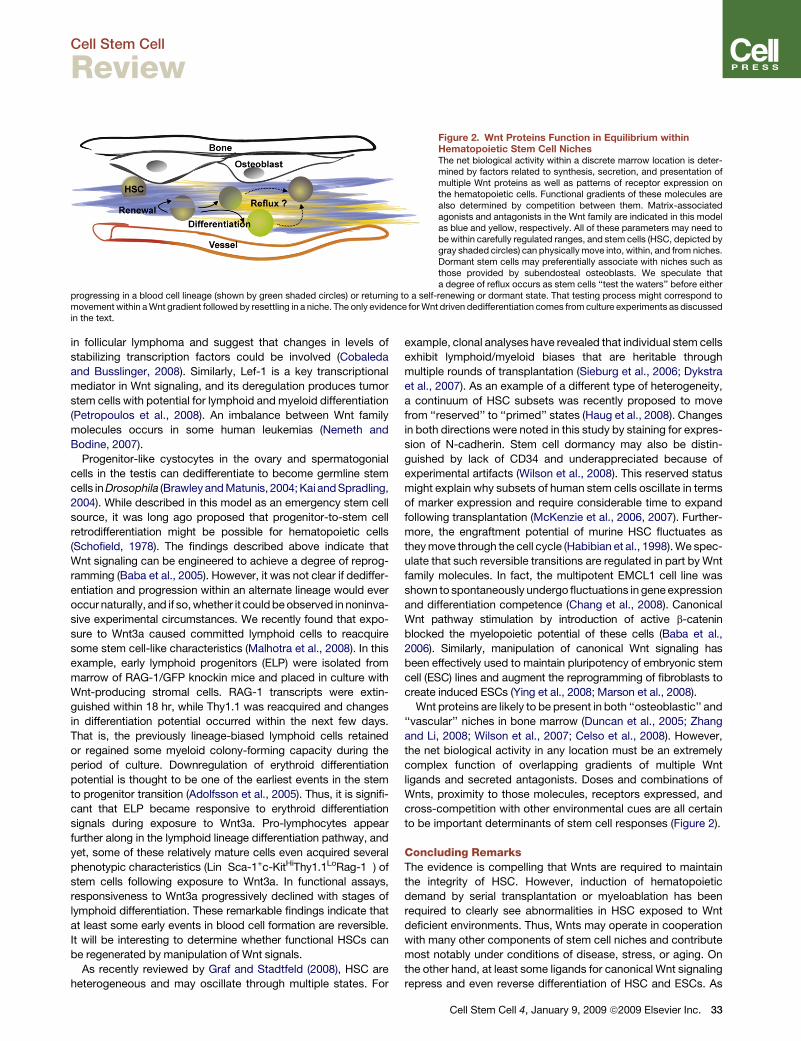
Figure 2. Wnt Proteins Function in Equilibrium withinHematopoietic Stem Cell NichesThe net biological activity within a discrete marrow location is deter-mined by factors related to synthesis, secretion, and presentation ofmultiple Wnt proteins as well as patterns of receptor expression onthe hematopoietic cells. Functional gradients of these molecules arealso determined by competition between them. Matrix-associatedagonists and antagonists in the Wnt family are indicated in this modelas blue and yellow, respectively. All of these parameters may need tobe within carefully regulated ranges, and stem cells (HSC, depicted bygray shaded circles) can physically move into, within, and from niches.Dormant stem cells may preferentially associate with niches such asthose provided by subendosteal osteoblasts. We speculate thata degree of reflux occurs as stem cells ‘‘test the waters’’ before either
progressing in a blood cell lineage (shown by green shaded circles) or returning to a self-renewing or dormant state. That testing process might correspond tomovement within a Wnt gradient followed by resettling in a niche. The only evidence for Wnt driven dedifferentiation comes from culture experiments as discussedin the text.
in follicular lymphoma and suggest that changes in levels of
stabilizing transcription factors could be involved (Cobaleda
and Busslinger, 2008). Similarly, Lef-1 is a key transcriptional
mediator in Wnt signaling, and its deregulation produces tumor
stem cells with potential for lymphoid and myeloid differentiation
(Petropoulos et al., 2008). An imbalance between Wnt family
molecules occurs in some human leukemias (Nemeth and
Bodine, 2007).
Progenitor-like cystocytes in the ovary and spermatogonial
cells in the testis can dedifferentiate to become germline stem
cells in Drosophila (Brawley and Matunis, 2004; Kai and Spradling,
2004). While described in this model as an emergency stem cell
source, it was long ago proposed that progenitor-to-stem cell
retrodifferentiation might be possible for hematopoietic cells
(Schofield, 1978). The findings described above indicate that
Wnt signaling can be engineered to achieve a degree of reprog-
ramming (Baba et al., 2005). However, it was not clear if dediffer-
entiation and progression within an alternate lineage would ever
occur naturally, and if so, whether it could be observed in noninva-
sive experimental circumstances. We recently found that expo-
sure to Wnt3a caused committed lymphoid cells to reacquire
some stem cell-like characteristics (Malhotra et al., 2008). In this
example, early lymphoid progenitors (ELP) were isolated from
marrow of RAG-1/GFP knockin mice and placed in culture with
Wnt-producing stromal cells. RAG-1 transcripts were extin-
guished within 18 hr, while Thy1.1 was reacquired and changes
in differentiation potential occurred within the next few days.
That is, the previously lineage-biased lymphoid cells retained
or regained some myeloid colony-forming capacity during the
period of culture. Downregulation of erythroid differentiation
potential is thought to be one of the earliest events in the stem
to progenitor transition (Adolfsson et al., 2005). Thus, it is signifi-
cant that ELP became responsive to erythroid differentiation
signals during exposure to Wnt3a. Pro-lymphocytes appear
further along in the lymphoid lineage differentiation pathway, and
yet, some of these relatively mature cells even acquired several
phenotypic characteristics (Lin�Sca-1+c-KitHiThy1.1LoRag-1�) of
stem cells following exposure to Wnt3a. In functional assays,
responsiveness to Wnt3a progressively declined with stages of
lymphoid differentiation. These remarkable findings indicate that
at least some early events in blood cell formation are reversible.
It will be interesting to determine whether functional HSCs can
be regenerated by manipulation of Wnt signals.
As recently reviewed by Graf and Stadtfeld (2008), HSC are
heterogeneous and may oscillate through multiple states. For
example, clonal analyses have revealed that individual stem cells
exhibit lymphoid/myeloid biases that are heritable through
multiple rounds of transplantation (Sieburg et al., 2006; Dykstra
et al., 2007). As an example of a different type of heterogeneity,
a continuum of HSC subsets was recently proposed to move
from ‘‘reserved’’ to ‘‘primed’’ states (Haug et al., 2008). Changes
in both directions were noted in this study by staining for expres-
sion of N-cadherin. Stem cell dormancy may also be distin-
guished by lack of CD34 and underappreciated because of
experimental artifacts (Wilson et al., 2008). This reserved status
might explain why subsets of human stem cells oscillate in terms
of marker expression and require considerable time to expand
following transplantation (McKenzie et al., 2006, 2007). Further-
more, the engraftment potential of murine HSC fluctuates as
they move through the cell cycle (Habibian et al., 1998). We spec-
ulate that such reversible transitions are regulated in part by Wnt
family molecules. In fact, the multipotent EMCL1 cell line was
shown to spontaneously undergo fluctuations in gene expression
and differentiation competence (Chang et al., 2008). Canonical
Wnt pathway stimulation by introduction of active b-catenin
blocked the myelopoietic potential of these cells (Baba et al.,
2006). Similarly, manipulation of canonical Wnt signaling has
been effectively used to maintain pluripotency of embryonic stem
cell (ESC) lines and augment the reprogramming of fibroblasts to
create induced ESCs (Ying et al., 2008; Marson et al., 2008).
Wnt proteins are likely to be present in both ‘‘osteoblastic’’ and
‘‘vascular’’ niches in bone marrow (Duncan et al., 2005; Zhang
and Li, 2008; Wilson et al., 2007; Celso et al., 2008). However,
the net biological activity in any location must be an extremely
complex function of overlapping gradients of multiple Wnt
ligands and secreted antagonists. Doses and combinations of
Wnts, proximity to those molecules, receptors expressed, and
cross-competition with other environmental cues are all certain
to be important determinants of stem cell responses (Figure 2).
Concluding RemarksThe evidence is compelling that Wnts are required to maintain
the integrity of HSC. However, induction of hematopoietic
demand by serial transplantation or myeloablation has been
required to clearly see abnormalities in HSC exposed to Wnt
deficient environments. Thus, Wnts may operate in cooperation
with many other components of stem cell niches and contribute
most notably under conditions of disease, stress, or aging. On
the other hand, at least some ligands for canonical Wnt signaling
repress and even reverse differentiation of HSC and ESCs. As
Cell Stem Cell 4, January 9, 2009 ª2009 Elsevier Inc. 33

Cell Stem Cell
Review
suggested above, refluxing between stem and progenitor cell
states could be partially regulated by Wnts. Continued assault
on this complex group of ligands, receptors, and coreceptors
is certain to be useful as well as informative. For example, it
may eventually be possible to expand and reprogram normal
stem and progenitor cells by exploiting the Wnt family of
proteins. Such an approach could improve the ability of HSCs
to rebound following myeloablation or engraft following trans-
plantation, and converse treatments may be found to deprive
leukemia cells of essential Wnt stimulation.
ACKNOWLEDGMENTS
Work in our laboratory is supported by grants AI020069, AI058162, andAI069024 from the National Institutes of Health. P.W.K. holds the William H.and Rita Bell Endowed Chair in Biomedical Research.
REFERENCES
Adolfsson, J., Mansson, R., Buza-Vidas, N., Hultquist, A., Liuba, K., Jensen,C.T., Bryder, D., Yang, L., Borge, O.J., Thoren, L.A., et al. (2005). Identificationof Flt3+ lympho-myeloid stem cells lacking erythro-megakaryocytic potentiala revised road map for adult blood lineage commitment. Cell 121, 295–306.
Aoki, K., and Taketo, M.M. (2007). Adenomatous polyposis coli (APC): a multi-functional tumor suppressor gene. J. Cell Sci. 120, 3327–3335.
Austin, T.W., Solar, G.P., Ziegler, F.C., Liem, L., and Matthews, W. (1997). Arole for the Wnt gene family in hematopoiesis: Expansion of multilineageprogenitor cells. Blood 89, 3624–3635.
Baba, Y., Garrett, K.P., and Kincade, P.W. (2005). Constitutively active b-catenin confers multilineage differentiation potential on lymphoid and myeloidprogenitors. Immunity 23, 599–609.
Baba, Y., Yokota, T., Spits, H., Garrett, K.P., Hayashi, S., and Kincade, P.W.(2006). Constitutively active beta-catenin promotes expansion of multipotenthematopoietic progenitors in culture. J. Immunol. 177, 2294–2303.
Baksh, D., Boland, G.M., and Tuan, R.S. (2007). Cross-talk between Wntsignaling pathways in human mesenchymal stem cells leads to functionalantagonism during osteogenic differentiation. J. Cell. Biochem. 101, 1109–1124.
Balciunaite, G., Keller, M.P., Balciunaite, E., Piali, L., Zuklys, S., Mathieu, Y.D.,Gill, J., Boyd, R., Sussman, D.J., and Hollander, G.A. (2002). Wnt glycoproteinsregulate the expression of FoxN1, the gene defective in nude mice. Nat.Immunol. 3, 1102–1108.
Barolo, S. (2006). Transgenic Wnt/TCF pathway reporters: all you need is Lef?Oncogene 25, 7505–7511.
Bennett, C.N., Longo, K.A., Wright, W.S., Suva, L.J., Lane, T.F., Hankenson,K.D., and MacDougald, O.A. (2005). Regulation of osteoblastogenesis andbone mass by Wnt10b. Proc. Natl. Acad. Sci. USA 102, 3324–3329.
Bowie, M.B., Kent, D.G., Dykstra, B., McKnight, K.D., McCaffrey, L., Hoodless,P.A., and Eaves, C.J. (2007). Identification of a new intrinsically timed develop-mental checkpoint that reprograms key hematopoietic stem cell properties.Proc. Natl. Acad. Sci. USA 104, 5878–5882.
Brawley, C., and Matunis, E. (2004). Regeneration of male germline stem cellsby spermatogonial dedifferentiation in vivo. Science 304, 1331–1334.
Celso, C.L., Fleming, H.E., Wu, J.W., Zhao, C.X., Miake-Lye, S., Fujisaki, J.,Cote, D., Rowe, D.W., Lin, C.P., and Scaddden, D.E. (2008). Live-animaltracking of individual haematopoietic stem/progenitor cells in their niche.Nature, in press. Published online December 3, 2008. 10.1038/nature07434.
Chang, H.H., Hemberg, M., Barahona, M., Ingber, D.E., and Huang, S. (2008).Transcriptome-wide noise controls lineage choice in mammalian progenitorcells. Nature 453, 544–547.
Cobaleda, C., and Busslinger, M. (2008). Developmental plasticity of lympho-cytes. Curr. Opin. Immunol. 20, 139–148.
34 Cell Stem Cell 4, January 9, 2009 ª2009 Elsevier Inc.
Cobas, M., Wilson, A., Ernst, B., Mancini, S.J., MacDonald, H.R., Kemler, R.,and Radtke, F. (2004). Beta-catenin is dispensable for hematopoiesis andlymphopoiesis. J. Exp. Med. 199, 221–229.
Congdon, K.L., Voermans, C., Ferguson, E.C., DiMascio, L.N., Uqoezwa, M.,Zhao, C., and Reya, T. (2008). Activation of Wnt signaling in hematopoieticregeneration. Stem Cells 26, 1202–1210.
Daugherty, R.L., and Gottardi, C.J. (2007). Phospho-regulation of Beta-cateninadhesion and signaling functions. Physiology (Bethesda) 22, 303–309.
Doble, B.W., and Woodgett, J.R. (2003). GSK-3: tricks of the trade for a multi-tasking kinase. J. Cell Sci. 116, 1175–1186.
Døsen, G., Tenstad, E., Nygren, M.K., Stubberud, H., Funderud, S., and Rian,E. (2006). Wnt expression and canonical Wnt signaling in human bone marrowB lymphopoiesis. BMC Immunol. 7, 13.
Duncan, A.W., Rattis, F.M., DiMascio, L.N., Congdon, K.L., Pazianos, G.,Zhao, C., Yoon, K., Cook, J.M., Willert, K., Gaiano, N., and Reya, T. (2005).Integration of Notch and Wnt signaling in hematopoietic stem cell mainte-nance. Nat. Immunol. 6, 314–322.
Dykstra, B., Kent, D., Bowie, M., McCaffrey, L., Hamilton, M., Lyons, K., Lee,S.J., Brinkman, R., and Eaves, C. (2007). Long-term propagation of distincthematopoietic differentiation programs in vivo. Cell Stem Cell 1, 218–229.
Ferrell, C.M., Dorsam, S.T., Ohta, H., Humphries, R.K., Derynck, M.K., Haqq,C., Largman, C., and Lawrence, H.J. (2005). Activation of stem-cell specificgenes by HOXA9 and HOXA10 homeodomain proteins in CD34+ humancord blood cells. Stem Cells 23, 644–655.
Fleming, H.E., Janzen, V., Lo, C.C., Guo, J., Leahy, K.M., Kronenberg, H.M.,and Scadden, D.T. (2008). Wnt signaling in the niche enforces hematopoieticstem cell quiescence and is necessary to preserve self-renewal in vivo. CellStem Cell 2, 274–283.
Graf, T., and Stadtfeld, M. (2008). Heterogeneity of embryonic and adult stemcells. Cell Stem Cell 3, 480–483.
Habibian, H.K., Peters, S.O., Hsieh, C.C., Wuu, J., Vergilis, K., Grimaldi, C.I.,Reilly, J., Carlson, J.E., Frimberger, A.E., Stewart, F.M., and Quesenberry,P.J. (1998). The fluctuating phenotype of the lymphohematopoietic stem cellwith cell cycle transit. J. Exp. Med. 188, 393–398.
Haegel, H., Larue, L., Ohsugi, M., Fedorov, L., Herrenknecht, K., and Kemler,R. (1995). Lack of b-catenin affects mouse development at gastrulation. Devel-opment 121, 3529–3537.
Haug, J.S., He, X.C., Grindley, J.C., Wunderlich, J.P., Gaudenz, K., Ross, J.T.,Paulson, A., Wagner, K.P., Xie, Y., Zhu, R., et al. (2008). N-cadherin expressionlevel distinguishes reserved versus primed states of hematopoietic stem cells.Cell Stem Cell 2, 367–379.
Holmes, T., O’Brien, T.A., Knight, R., Lindeman, R., Shen, S., Song, E.,Symonds, G., and Dolnikov, A. (2008). Glycogen synthase kinase-3betainhibition preserves hematopoietic stem cell activity and inhibits leukemiccell growth. Stem Cells 26, 1288–1297.
Hsu, W., Shakya, R., and Costantini, F. (2001). Impaired mammary gland andlymphoid development caused by inducible expression of Axin in transgenicmice. J. Cell Biol. 155, 1055–1064.
Jeannet, G., Scheller, M., Scarpellino, L., Duboux, S., Gardiol, N., Back, J.,Kuttler, F., Malanchi, I., Birchmeier, W., Leutz, A., et al. (2007). Long-term,multilineage hematopoiesis occurs in the combined absence of beta-cateninand gamma-catenin. Blood 111, 142–149.
Kai, T., and Spradling, A. (2004). Differentiating germ cells can revert into func-tional stem cells in Drosophila melanogaster ovaries. Nature 428, 564–569.
Kawano, Y., and Kypta, R. (2003). Secreted antagonists of the Wnt signallingpathway. J. Cell Sci. 116, 2627–2634.
Kim, I., Saunders, T.L., and Morrison, S.J. (2007). Sox17 dependencedistinguishes the transcriptional regulation of fetal from adult hematopoieticstem cells. Cell 130, 470–483.
Kirstetter, P., Anderson, K., Porse, B.T., Jacobsen, S.E., and Nerlov, C. (2006).Activation of the canonical Wnt pathway leads to loss of hematopoietic stemcell repopulation and multilineage differentiation block. Nat. Immunol. 7,1048–1056.
Cell Stem Cell
Review
Koch, U., Wilson, A., Cobas, M., Kemler, R., MacDonald, H.R., and Radtke, F.(2008). Simultaneous loss of beta- and gamma-catenin does not perturbhematopoiesis or lymphopoiesis. Blood 111, 160–164.
Kockeritz, L., Doble, B., Patel, S., and Woodgett, J.R. (2006). Glycogensynthase kinase-3–an overview of an over-achieving protein kinase. Curr.Drug Targets 7, 1377–1388.
Koni, P.A., Joshi, S.K., Temann, U.A., Olson, D., Burkly, L., and Flavell, R.A.(2001). Conditional vascular cell adhesion molecule 1 deletion in mice:impaired lymphocyte migration to bone marrow. J. Exp. Med. 193, 741–754.
Lako, M., Lindsay, S., Lincoln, J., Cairns, P.M., Armstrong, L., and Hole, N.(2001). Characterisation of Wnt gene expression during the differentiation ofmurine embryonic stem cells in vitro: role of Wnt3 in enhancing haematopoieticdifferentiation. Mech. Dev. 103, 49–59.
Leuker, C.E., Labow, M., Muller, W., and Wagner, N. (2001). Neonatallyinduced inactivation of the vascular cell adhesion molecule 1 gene impairs Bcell localization and T cell-dependent humoral immune response. J. Exp.Med. 193, 755–768.
Liang, H., Chen, Q., Coles, A.H., Anderson, S.J., Pihan, G., Bradley, A.,Gerstein, R., Jurecic, R., and Jones, S.N. (2003). Wnt5a inhibits B cell prolifer-ation and functions as a tumor suppressor in hematopoietic tissue. Cancer Cell4, 349–360.
Louis, I., Heinonen, K.M., Chagraoui, J., Vainio, S., Sauvageau, G., andPerreault, C. (2008). The signaling protein Wnt4 enhances thymopoiesis andexpands multipotent hematopoietic progenitors through beta-catenin-inde-pendent signaling. Immunity 29, 57–67.
Luis, T.C., Weerkamp, F., Naber, B.A., Baert, M.R.M., de Haas, E.F., Nikolic,T., Heuvelmans, S., De Krijger, R.R., Von Dongen, J.M., and Staal, F.J.T.(2008). Wnt3a deficiency irreversibly impairs hematopoietic stem cell self-renewal and leads to defects in progenitor cell differentiation. Blood, in press.Published online October 2, 2008. 10.1182/blood-2008-06-163774.
Lyons, J.P., Mueller, U.W., Ji, H., Everett, C., Fang, X., Hsieh, J.C., Barth, A.M.,and McCrea, P.D. (2004). Wnt-4 activates the canonical beta-catenin-medi-ated Wnt pathway and binds Frizzled-6 CRD: functional implications of Wnt/beta-catenin activity in kidney epithelial cells. Exp. Cell Res. 298, 369–387.
Macdonald, B.T., Semenov, M.V., and He, X. (2007). SnapShot: Wnt/beta-catenin signaling. Cell 131, 1204.
Malhotra, S., and Kincade, P.W. (2008). Canonical Wnt pathway signalingsuppresses VCAM-1 expression by marrow stromal and hematopoietic cells.Exp. Hematol., in press. Published online October 23, 2008. 10.1016/j.ex-phem.2008.08.008.
Malhotra, S., Baba, Y., Garrett, K.P., Staal, F.J., Gerstein, R., and Kincade,P.W. (2008). Contrasting responses of lymphoid progenitors to canonicaland noncanonical Wnt signals. J. Immunol. 181, 3955–3964.
Marson, A., Foreman, R., Chevalier, B., Bilodeau, S., Kahn, M., Young, R.A.,and Jaenisch, R. (2008). Wnt signaling promotes reprogramming of somaticcells to pluripotency. Cell Stem Cell 3, 132–135.
McKenzie, J.L., Gan, O.I., Doedens, M., Wang, J.C., and Dick, J.E. (2006). Indi-vidual stem cells with highly variable proliferation and self-renewal propertiescomprise the human hematopoietic stem cell compartment. Nat. Immunol. 7,1225–1233.
McKenzie, J.L., Gan, O.I., Doedens, M., and Dick, J.E. (2007). Reversible cellsurface expression of CD38 on CD34-positive human hematopoietic repopu-lating cells. Exp. Hematol. 35, 1429–1436.
Mikels, A.J., and Nusse, R. (2006). Purified Wnt5a protein activates or inhibitsbeta-catenin-TCF signaling depending on receptor context. PLoS Biol. 4,e115. 10.1371/journal.pbio.0040115.
Mukhopadhyay, M., Shtrom, S., Rodriguez-Esteban, C., Chen, L., Tsukui, T.,Gomer, L., Dorward, D.W., Glinka, A., Grinberg, A., Huang, S.P., et al.(2001). Dickkopf1 is required for embryonic head induction and limb morpho-genesis in the mouse. Dev. Cell 1, 423–434.
Mulroy, T., McMahon, J.A., Burakoff, S.J., McMahon, A.P., and Sen, J. (2002).Wnt-1 and Wnt-4 regulate thymic cellularity. Eur. J. Immunol. 32, 967–971.
Murdoch, B., Chadwick, K., Martin, M., Shojaei, F., Shah, K.V., Gallacher, L.,Moon, R.T., and Bhatia, M. (2003). Wnt-5A augments repopulating capacity
and primitive hematopoietic development of human blood stem cells in vivo.Proc. Natl. Acad. Sci. USA 100, 3422–3427.
Nemeth, M.J., and Bodine, D.M. (2007). Regulation of hematopoiesis and thehematopoietic stem cell niche by Wnt signaling pathways. Cell Res. 17,746–758.
Nemeth, M.J., Kirby, M.R., and Bodine, D.M. (2006). Hmgb3 regulates thebalance between hematopoietic stem cell self-renewal and differentiation.Proc. Natl. Acad. Sci. USA 103, 13783–13788.
Nemeth, M.J., Topol, L., Anderson, S.M., Yang, Y., and Bodine, D.M. (2007).Wnt5a inhibits canonical Wnt signaling in hematopoietic stem cells andenhances repopulation. Proc. Natl. Acad. Sci. USA 104, 15436–15441.
Nusse, R. (2005). Wnt signaling in disease and in development. Cell Res. 15,28–32.
Petropoulos, K., Arseni, N., Schessl, C., Stadler, C.R., Rawat, V.P., Desh-pande, A.J., Heilmeier, B., Hiddemann, W., Quintanilla-Martinez, L., Boh-lander, S.K., et al. (2008). A novel role for Lef-1, a central transcription mediatorof Wnt signaling, in leukemogenesis. J. Exp. Med. 205, 515–522.
Pinzone, J.J., Hall, B.M., Thudi, N.K., Vonau, M., Qiang, Y.W., Rosol, T.J., andShaughnessy, J.D., Jr. (2008). The role of Dickkopf-1 in bone development,homeostasis and disease. Blood, in press. Published online October 3,2008. 10.1182/blood-2008-03-145169.
Qian, Z., Chen, L., Fernald, A.A., Williams, B.O., and Le Beau, M.M. (2008). Acritical role for Apc in hematopoietic stem and progenitor cell survival. J. Exp.Med. 205, 2163–2175.
Reya, T., Duncan, A.W., Ailles, L., Domen, J., Scherer, D.C., Willert, K., Hintz,L., Nusse, R., and Weissman, I.L. (2003). A role for Wnt signalling in self-renewal of haematopoietic stem cells. Nature 423, 409–414.
Ruiz, P., Brinkmann, V., Ledermann, B., Behrend, M., Grund, C., Thalhammer,C., Vogel, F., Birchmeier, C., Gunthert, U., Franke, W.W., and Birchmeier, W.(1996). Targeted mutation of plakoglobin in mice reveals essential functionsof desmosomes in the embryonic heart. J. Cell Biol. 135, 215–225.
Scheller, M., Huelsken, J., Rosenbauer, F., Taketo, M.M., Birchmeier, W.,Tenen, D.G., and Leutz, A. (2006). Hematopoietic stem cell and multilineagedefects generated by constitutive beta-catenin activation. Nat. Immunol. 7,1037–1047.
Schofield, R. (1978). The relationship between the spleen colony-forming celland the haemopoietic stem cell. Blood Cells 4, 7–25.
Semenov, M.V., Habas, R., Macdonald, B.T., and He, X. (2007). SnapShot:noncanonical Wnt signaling pathways. Cell 131, 1378.
Sieburg, H.B., Cho, R.H., Dykstra, B., Uchida, N., Eaves, C.J., and Muller-Sieburg, C.E. (2006). The hematopoietic stem compartment consists of alimited number of discrete stem cell subsets. Blood 107, 2311–2316.
Staal, F.J., Luis, T.C., and Tiemessen, M.M. (2008). WNT signalling in theimmune system: WNT is spreading its wings. Nat. Rev. Immunol. 8, 581–593.
Staal, F.J., and Sen, J.M. (2008). The canonical Wnt signaling pathway playsan important role in lymphopoiesis and hematopoiesis. Eur. J. Immunol. 38,1788–1794.
Tao, W., Evans, B.G., Yao, J., Cooper, S., Cornetta, K., Ballas, C.B., Hangoc,G., and Broxmeyer, H.E. (2007). Enhanced green fluorescent protein is a nearlyideal long-term expression tracer for hematopoietic stem cells, whereasDsRed-express fluorescent protein is not. Stem Cells 25, 670–678.
Templin, C., Kotlarz, D., Rathinam, C., Rudolph, C., Schatzlein, S., Ramireddy,K., Rudolph, K.L., Schlegelberger, B., Klein, C., and Drexler, H. (2008). Estab-lishment of immortalized multipotent hematopoietic progenitor cell lines byretroviral-mediated gene transfer of beta-catenin. Exp. Hematol. 36, 204–215.
Topol, L., Jiang, X., Choi, H., Garrett-Beal, L., Carolan, P.J., and Yang, Y.(2003). Wnt-5a inhibits the canonical Wnt pathway by promoting GSK-3-independent b-catenin degradation. J. Cell Biol. 162, 899–908.
Trowbridge, J.J., Xenocostas, A., Moon, R.T., and Bhatia, M. (2006). Glycogensynthase kinase-3 is an in vivo regulator of hematopoietic stem cell repopula-tion. Nat. Med. 12, 89–98.
Tu, X., Joeng, K.S., Nakayama, K.I., Nakayama, K., Rajagopal, J., Carroll, T.J.,McMahon, A.P., and Long, F. (2007). Noncanonical Wnt signaling through
Cell Stem Cell 4, January 9, 2009 ª2009 Elsevier Inc. 35

Cell Stem Cell
Review
G protein-linked PKCdelta activation promotes bone formation. Dev. Cell 12,113–127.
Ulyanova, T., Priestley, G.V., Nakamoto, B., Jiang, Y., and Papayannopoulou,T. (2007). VCAM-1 ablation in nonhematopoietic cells in MxCre+ VCAM-1f/fmice is variable and dictates their phenotype. Exp. Hematol. 35, 565–571.
Uren, A., Reichsman, F., Anest, V., Taylor, W.G., Muraiso, K., Bottaro, D.P.,Cumberledge, S., and Rubin, J.S. (2000). Secreted frizzled-related protein-1binds directly to Wingless and is a biphasic modulator of Wnt signaling.J. Biol. Chem. 275, 4374–4382.
Van Den Berg, D.J., Sharma, A.K., Bruno, E., and Hoffman, R. (1998). Roleof members of the Wnt gene family in human hematopoiesis. Blood 92,3189–3202.
Weerkamp, F., Baert, M.R., Naber, B.A., Koster, E.E., de Haas, E.F., Atkuri,K.R., van Dongen, J.J., Herzenberg, L.A., and Staal, F.J. (2006). Wnt signalingin the thymus is regulated by differential expression of intracellular signalingmolecules. Proc. Natl. Acad. Sci. USA 103, 3322–3326.
Willert, K., Brown, J.D., Danenberg, E., Duncan, A.W., Weissman, I.L., Reya,T., Yates, J.R., III, and Nusse, R. (2003). Wnt proteins are lipid-modified andcan act as stem cell growth factors. Nature 423, 448–452.
Wilson, A., Oser, G.M., Jaworski, M., Blanco-Bose, W.E., Laurenti, E., Adol-phe, C., Essers, M.A., MacDonald, H.R., and Trumpp, A. (2007). Dormantand self-renewing hematopoietic stem cells and their niches. Ann. N Y Acad.Sci. 1106, 64–75.
Wilson, A., Laurenti, E., Oser, G., van der Wath, R.C., Blanco-Bose, W.,Jaworski, M., Offner, S., Dunant, C.F., Eshkind, L., Bockamp, E., et al.
36 Cell Stem Cell 4, January 9, 2009 ª2009 Elsevier Inc.
(2008). Hematopoietic stem cells reversibly switch from dormancy to self-renewal during homeostasis and Repair. Cell 135, 1118–1129.
Yamane, T., Kunisada, T., Tsukamoto, H., Yamazaki, H., Niwa, H., Takada, S.,and Hayashi, S.I. (2001). Wnt signaling regulates hemopoiesis through stromalcells. J. Immunol. 167, 765–772.
Ying, Q.L., Wray, J., Nichols, J., Batlle-Morera, L., Doble, B., Woodgett, J.,Cohen, P., and Smith, A. (2008). The ground state of embryonic stem cellself-renewal. Nature 453, 519–523.
Yokota, T., Oritani, K., Garrett, K.P., Kouro, T., Nishida, M., Takahashi, I., Ichii,M., Satoh, Y., Kincade, P.W., and Kanakura, Y. (2008). Soluble frizzled-relatedprotein 1 is estrogen inducible in bone marrow stromal cells and suppressesthe earliest events in lymphopoiesis. J. Immunol., in press.
Yu, Q., Quinn, W.J., III, Salay, T., Crowley, J.E., Cancro, M.P., and Sen, J.M.(2008). Role of beta-catenin in B cell development and function. J. Immunol.181, 3777–3783.
Zhang, J., and Li, L. (2008). Stem cell niche: microenvironment and beyond.J. Biol. Chem. 283, 9499–9503.
Zhao, C., Blum, J., Chen, A., Kwon, H.Y., Jung, S.H., Cook, J.M., Lagoo, A.,and Reya, T. (2007). Loss of beta-catenin impairs the renewal of normal andCML stem cells in vivo. Cancer Cell 12, 528–541.
Zhong, X., Desilva, T., Lin, L., Bodine, P., Bhat, R.A., Presman, E., Pocas, J.,Stahl, M., and Kriz, R. (2007). Regulation of secreted Frizzled-relatedprotein-1 by heparin. J. Biol. Chem. 282, 20523–20533.