molecules - MDPI
15
molecules Article An Investigation into the Potential of Targeting Escherichia coli rne mRNA with Locked Nucleic Acid (LNA) Gapmers as an Antibacterial Strategy Layla R. Goddard 1,2,† , Charlotte E. Mardle 1,† , Hassan Gneid 3,4,‡ , Ciara G. Ball 1,2 , Darren M. Gowers 1 , Helen S. Atkins 5,6 , Louise E. Butt 1 , Jonathan K. Watts 3 , Helen A. Vincent 1,2, * and Anastasia J. Callaghan 1,2, * Citation: Goddard, L.R.; Mardle, C.E.; Gneid, H.; Ball, C.G.; Gowers, D.M.; Atkins, H.S.; Butt, L.E.; Watts, J.K.; Vincent, H.A.; Callaghan, A.J. An Investigation into the Potential of Targeting Escherichia coli rne mRNA with Locked Nucleic Acid (LNA) Gapmers as an Antibacterial Strategy. Molecules 2021, 26, 3414. https:// doi.org/10.3390/molecules26113414 Academic Editor: Mary K. Phillips-Jones Received: 26 March 2021 Accepted: 26 May 2021 Published: 4 June 2021 Publisher’s Note: MDPI stays neutral with regard to jurisdictional claims in published maps and institutional affil- iations. Copyright: © 2021 by the authors. Licensee MDPI, Basel, Switzerland. This article is an open access article distributed under the terms and conditions of the Creative Commons Attribution (CC BY) license (https:// creativecommons.org/licenses/by/ 4.0/). 1 School of Biological Sciences and Institute of Biological & Biomedical Sciences, University of Portsmouth, Portsmouth PO1 2DY, UK; [email protected] (L.R.G.); [email protected] (C.E.M.); [email protected] (C.G.B.); [email protected] (D.M.G.); [email protected] (L.E.B.) 2 Centre for Enzyme Innovation, University of Portsmouth, Portsmouth PO1 2DY, UK 3 RNA Therapeutics Institute, University of Massachusetts Medical School, Worcester, MA 01609, USA; [email protected] (H.G.); [email protected] (J.K.W.) 4 Department of Chemistry, University of Southampton, Southampton SO17 1BJ, UK 5 Defence Science and Technology Laboratory, Porton Down, Salisbury SP4 0JQ, UK; [email protected] 6 College of Life and Environmental Sciences, University of Exeter, Exeter EX4 4QD, UK * Correspondence: [email protected] (H.A.V.); [email protected] (A.J.C.) † These authors contributed equally to this work. ‡ Current address: Department of Chemistry, Tulane University, 6823 St. Charles Avenue, New Orleans, LA 70118, USA. Abstract: The increase in antibacterial resistance is a serious challenge for both the health and defence sectors and there is a need for both novel antibacterial targets and antibacterial strategies. RNA degradation and ribonucleases, such as the essential endoribonuclease RNase E, encoded by the rne gene, are emerging as potential antibacterial targets while antisense oligonucleotides may provide alternative antibacterial strategies. As rne mRNA has not been previously targeted using an antisense approach, we decided to explore using antisense oligonucleotides to target the translation initiation region of the Escherichia coli rne mRNA. Antisense oligonucleotides were rationally designed and were synthesised as locked nucleic acid (LNA) gapmers to enable inhibition of rne mRNA translation through two mechanisms. Either LNA gapmer binding could sterically block translation and/or LNA gapmer binding could facilitate RNase H-mediated cleavage of the rne mRNA. This may prove to be an advantage over the majority of previous antibacterial antisense oligonucleotide approaches which used oligonucleotide chemistries that restrict the mode-of-action of the antisense oligonucleotide to steric blocking of translation. Using an electrophoretic mobility shift assay, we demonstrate that the LNA gapmers bind to the translation initiation region of E. coli rne mRNA. We then use a cell-free transcription translation reporter assay to show that this binding is capable of inhibiting translation. Finally, in an in vitro RNase H cleavage assay, the LNA gapmers facilitate RNase H-mediated mRNA cleavage. Although the challenges of antisense oligonucleotide delivery remain to be addressed, overall, this work lays the foundations for the development of a novel antibacterial strategy targeting rne mRNA with antisense oligonucleotides. Keywords: antibacterial; antisense oligonucleotide; gapmer; locked nucleic acid (LNA); RNase E; RNase H; rne mRNA; translation blocking 1. Introduction The emergence of both natural and engineered antimicrobial resistant strains of bacte- ria poses a significant challenge to the health and defense sectors. Unfortunately, traditional drug development programmes, that successfully provided the antibiotics of the 20th cen- tury, are failing to keep pace with emerging resistance [1,2]. Consequently, there is a Molecules 2021, 26, 3414. https://doi.org/10.3390/molecules26113414 https://www.mdpi.com/journal/molecules
-
Upload
khangminh22 -
Category
Documents
-
view
6 -
download
0
Transcript of molecules - MDPI

molecules
Article
An Investigation into the Potential of Targeting Escherichia colirne mRNA with Locked Nucleic Acid (LNA) Gapmers as anAntibacterial Strategy
Layla R. Goddard 1,2,†, Charlotte E. Mardle 1,†, Hassan Gneid 3,4,‡ , Ciara G. Ball 1,2, Darren M. Gowers 1 ,Helen S. Atkins 5,6, Louise E. Butt 1, Jonathan K. Watts 3, Helen A. Vincent 1,2,* and Anastasia J. Callaghan 1,2,*
�����������������
Citation: Goddard, L.R.; Mardle,
C.E.; Gneid, H.; Ball, C.G.; Gowers,
D.M.; Atkins, H.S.; Butt, L.E.; Watts,
J.K.; Vincent, H.A.; Callaghan, A.J.
An Investigation into the Potential of
Targeting Escherichia coli rne mRNA
with Locked Nucleic Acid (LNA)
Gapmers as an Antibacterial Strategy.
Molecules 2021, 26, 3414. https://
doi.org/10.3390/molecules26113414
Academic Editor: Mary K.
Phillips-Jones
Received: 26 March 2021
Accepted: 26 May 2021
Published: 4 June 2021
Publisher’s Note: MDPI stays neutral
with regard to jurisdictional claims in
published maps and institutional affil-
iations.
Copyright: © 2021 by the authors.
Licensee MDPI, Basel, Switzerland.
This article is an open access article
distributed under the terms and
conditions of the Creative Commons
Attribution (CC BY) license (https://
creativecommons.org/licenses/by/
4.0/).
1 School of Biological Sciences and Institute of Biological & Biomedical Sciences, University of Portsmouth,Portsmouth PO1 2DY, UK; [email protected] (L.R.G.); [email protected] (C.E.M.);[email protected] (C.G.B.); [email protected] (D.M.G.); [email protected] (L.E.B.)
2 Centre for Enzyme Innovation, University of Portsmouth, Portsmouth PO1 2DY, UK3 RNA Therapeutics Institute, University of Massachusetts Medical School, Worcester, MA 01609, USA;
[email protected] (H.G.); [email protected] (J.K.W.)4 Department of Chemistry, University of Southampton, Southampton SO17 1BJ, UK5 Defence Science and Technology Laboratory, Porton Down, Salisbury SP4 0JQ, UK; [email protected] College of Life and Environmental Sciences, University of Exeter, Exeter EX4 4QD, UK* Correspondence: [email protected] (H.A.V.); [email protected] (A.J.C.)† These authors contributed equally to this work.‡ Current address: Department of Chemistry, Tulane University, 6823 St. Charles Avenue,
New Orleans, LA 70118, USA.
Abstract: The increase in antibacterial resistance is a serious challenge for both the health and defencesectors and there is a need for both novel antibacterial targets and antibacterial strategies. RNAdegradation and ribonucleases, such as the essential endoribonuclease RNase E, encoded by the rnegene, are emerging as potential antibacterial targets while antisense oligonucleotides may providealternative antibacterial strategies. As rne mRNA has not been previously targeted using an antisenseapproach, we decided to explore using antisense oligonucleotides to target the translation initiationregion of the Escherichia coli rne mRNA. Antisense oligonucleotides were rationally designed andwere synthesised as locked nucleic acid (LNA) gapmers to enable inhibition of rne mRNA translationthrough two mechanisms. Either LNA gapmer binding could sterically block translation and/or LNAgapmer binding could facilitate RNase H-mediated cleavage of the rne mRNA. This may prove to bean advantage over the majority of previous antibacterial antisense oligonucleotide approaches whichused oligonucleotide chemistries that restrict the mode-of-action of the antisense oligonucleotide tosteric blocking of translation. Using an electrophoretic mobility shift assay, we demonstrate that theLNA gapmers bind to the translation initiation region of E. coli rne mRNA. We then use a cell-freetranscription translation reporter assay to show that this binding is capable of inhibiting translation.Finally, in an in vitro RNase H cleavage assay, the LNA gapmers facilitate RNase H-mediated mRNAcleavage. Although the challenges of antisense oligonucleotide delivery remain to be addressed,overall, this work lays the foundations for the development of a novel antibacterial strategy targetingrne mRNA with antisense oligonucleotides.
Keywords: antibacterial; antisense oligonucleotide; gapmer; locked nucleic acid (LNA); RNase E;RNase H; rne mRNA; translation blocking
1. Introduction
The emergence of both natural and engineered antimicrobial resistant strains of bacte-ria poses a significant challenge to the health and defense sectors. Unfortunately, traditionaldrug development programmes, that successfully provided the antibiotics of the 20th cen-tury, are failing to keep pace with emerging resistance [1,2]. Consequently, there is a
Molecules 2021, 26, 3414. https://doi.org/10.3390/molecules26113414 https://www.mdpi.com/journal/molecules

Molecules 2021, 26, 3414 2 of 15
growing need for the development of novel antibacterial strategies that target alternativepathways and/or have unconventional mechanisms of action.
RNA degradation pathways and ribonucleases (RNases), the enzymes responsiblefor RNA turnover, have recently been identified as targets that could be exploited forantibiotic development [3,4]. Specifically, the endoribonuclease RNase E, and the rne genethat encodes it, are ideal candidates for antibacterial targeting for a number of reasons [3,4].Firstly, RNase E/rne is essential [5–8] and so inhibitors of RNase E, or repressors of rne geneexpression, would be expected to have antibacterial activity. Furthermore, RNase E/rne isimplicated in bacterial virulence of the pathogens Salmonella enterica and Yersinia pestis [9,10].Finally, RNase E/rne is highly conserved amongst Gram-negative bacteria but there is noknown human orthologue [3,11] suggesting that specific inhibitors or repressors wouldtarget RNase E/rne-containing bacteria but not human hosts.
Some progress has been made in validating RNase E/rne as an antibacterial targetthrough the identification of small molecule inhibitors of RNase E, using structure-basedvirtual high-throughput screening, and the characterisation of their inhibitory activityin vitro. Through this approach, a number of small molecules have been identified thatinhibit RNase E from multiple bacterial pathogens in vitro [12,13]. However, the half maxi-mal inhibitory concentration (IC50) for each of these inhibitors was in the low millimolarrange, much higher than would be desired for an effective antibiotic [12,13]. Even enhancedinhibition, obtained using a combination of inhibitory small molecules, required millimolarconcentrations of inhibitors [12]. Therefore, while these small molecules have potential aslead compounds for the development of antibiotics targeting RNase E, there is work still tobe done.
An alternative antibacterial strategy to using small molecule antibiotics is the de-velopment of antisense oligonucleotide antibacterials reviewed in [14–17]. Antisenseoligonucleotides are short, single-stranded nucleic acid sequences that are complementaryto a target mRNA. They can down-regulate gene expression by binding to their targetmRNA and inhibiting its translation through the creation of a steric block to ribosome bind-ing and/or by facilitating RNase H recruitment and RNA cleavage [14–17] (SupplementaryFigure S1). Typically, antisense oligonucleotides are synthesised from nucleotide analogues(Supplementary Figure S2) in order to enhance the affinity for RNA and decrease thesusceptibility to cellular nucleases (reviewed in [14–18]). However, these chemical modifi-cations can also negatively affect RNase H recruitment and limit the mode-of-action of theantisense oligonucleotide to steric blocking of ribosome binding [17,18]. A key advantageof the antisense approach is that it should be possible to rationally design an antisenseoligonucleotide to target any mRNA. If the target mRNA encodes an essential protein,e.g., rne mRNA, then the antisense oligonucleotide may have antibacterial properties. Anumber of antisense oligonucleotides, that target a variety of mRNAs, have been reportedto have antibacterial activity (see Supplementary Table S1 for examples).
In the current study we explored the potential of targeting rne mRNA with antisenseoligonucleotides as a possible alternative antibacterial strategy. To our knowledge, rnemRNA has not been previously targeted using an antisense approach. We rationally de-signed two oligonucleotide sequences to have complementarity to the translation initiationregion of Escherichia coli rne mRNA. Both sequences were synthesised as locked nucleicacid (LNA) gapmers, oligonucleotides consisting of a central region of DNA flanked byregions of chemically modified LNA nucleotides [18], with an LNA3-DNA10-LNA3 and anLNA4-DNA8-LNA4 configuration. The ability of each of the four LNA gapmers to bind tothe translation initiation region of E. coli rne mRNA, inhibit translation, and recruit RNaseH to mediate mRNA cleavage, was evaluated in vitro using an electrophoretic mobilityshift assay (EMSA), a cell-free reporter assay and a gel-based RNase H cleavage assay, re-spectively. All four of the LNA gapmers bound to the translation initiation region of E. colirne mRNA, inhibited translation and facilitated RNase H-mediated cleavage. However,there were preferences with regard to the antisense oligonucleotide sequence/binding siteand gapmer configuration. These studies clearly demonstrate that it is possible to target

Molecules 2021, 26, 3414 3 of 15
rne mRNA with antisense oligonucleotides and they provide key knowledge that could betaken forwards to develop a novel antibacterial strategy.
2. Results2.1. Targeting E. coil rne with LNA Gapmers
The first step in investigating an antisense approach to potentially down-regulate rnegene expression was to identify a region of the E. coli rne mRNA to target and rationallydesign antisense oligonucleotides against it. Most bacterial antisense oligonucleotidestarget the translation initiation region of an mRNA [17]. This is because this region of anmRNA is usually unstructured and accessible to ribosomes meaning that it will likely alsobe accessible to an antisense oligonucleotide [17]. In addition, antisense oligonucleotidebinding to this region of the mRNA is most likely to sterically block translation by prevent-ing ribosome binding which may be more effective than a steric block aimed at haltingribosome progression at a downstream binding site. Indeed, although E. coli rne mRNAcontains a long (361-nucleotide), highly structured 5′ UTR, the translation initiation regionhas been reported to be unstructured [19]. Therefore we decided to target the translationinitiation region of E. coli rne mRNA with antisense oligonucleotides.
Next, we needed to select a suitable antisense oligonucleotide chemistry. Chemicalanalogues that are commonly used in bacterial antisense oligonucleotide chemistry includephosphorothioate, phosphorodiamidate morpholino (PMO), peptide nucleic acid (PNA)and LNA (Supplementary Figure S2) [17,18]. Phosphorothioate has a reduced affinity forRNA, compared to DNA, but phosphorothioate oligonucleotide:mRNA duplexes are recog-nised by RNase H [17,18]. In contrast, PMO, PNA and LNA all have a significantly greateraffinity for RNA but neither PMO oligonucleotide:mRNA, PNA oligonucleotide:mRNAnor LNA oligonucleotide:mRNA duplexes are recognised by RNase H [17,18]. The mode-of-action of entirely PMO, PNA or LNA antisense oligonucleotides is therefore restrictedto steric blocking of translation. To overcome this possible limitation, gapmer antisenseoligonucleotides consisting of a central region of DNA flanked by chemically modifiednucleotides can be used [17,18]. The central DNA region of the gapmer facilitates RNaseH recruitment and mRNA cleavage while the flanking chemically modified nucleotidesprovide the enhanced oligonucleotide stability and RNA binding affinity. In order to allowus to compare both the steric blocking of ribosome binding and the RNase H-mediatedmRNA cleavage mode-of-action of antisense oligonucleotides, we decided to use LNAgapmers to target rne mRNA. This strategy is shown schematically in Figure 1A.
Finally, we needed to rationally design our antisense oligonucleotide sequence anddecide on the LNA gapmer configurations to synthesise. Kurreck et al., found that LNAgapmers with a central region of phosphorothioate DNA of at least seven nucleotides weresufficient to facilitate recruitment of RNase H [20]. In addition, flanking LNA regions ofthree nucleotides were sufficient to increase gapmer binding affinity for RNA and, togetherwith a phosphorothioate backbone, confer protection from nucleases [20]. Consideringthese parameters, two 16-mer sequences, LNA gapmer A and LNA gapmer B, weredesigned to be complementary to the translation initiation region of rne mRNA (Figure1B). A non-complementary scrambled 16-mer sequence, Scrambled LNA gapmer, was alsodesigned to use as a control (Figure 1B). LNA gapmer A would be expected to occludeboth the ribosome binding site (RBS) and the start codon of the rne mRNA and wouldbe expected to prevent ribosome binding. LNA gapmer B would only be expected toocclude the rne mRNA start codon. Binding of LNA gapmer B may, or may not, preventribosome binding but it would be expected to block the progression of bound ribosomes.Therefore comparing the activity of LNA gapmer A and LNA gapmer B may indicatewhether there is a preferred antisense oligonucleotide binding site within the rne mRNAtranslation initiation region. Each LNA gapmer sequence was synthesised as an LNA3-DNA10-LNA3 3-10-3 gapmer (LNA gapmer A1/B1 and Scrambled LNA gapmer) and as anLNA4-DNA8-LNA4 4-8-4 gapmer (LNA gapmer A2/B2) (Figure 1B).

Molecules 2021, 26, 3414 4 of 15
Figure 1. Targeting the translation initiation region of rne mRNA with LNA gapmers. (A) An antisenseLNA gapmer comprising DNA (mauve) flanked by LNA (teal) binds to the translation initiation region(including the ribosome binding site (RBS), gold) of rne mRNA (blue). This prevents the ribosome(grey) from binding and sterically blocks translation. It also recruits RNase H (grey scissors) whichcleaves the rne mRNA to prevent RNase E synthesis. (B) LNA gapmer A and LNA gapmer B weredesigned to be complementary to the translation initiation region of E. coli rne mRNA (blue; RBS,gold; start codon, brown). Scrambled LNA gapmer, which is not complementary to the translationinitiation region of E. coli rne mRNA, was also designed. In each of the LNA gapmers, LNA bases(teal/underlined) flank DNA bases (mauve). Each LNA gapmer was synthesised as an LNA3-DNA10-LNA3 3-10-3 gapmer (LNA gapmer A1/B1 and Scrambled LNA gapmer). LNA gapmers A and Bwere also synthesised as an LNA4-DNA8-LNA4 4-8-4 gapmer (LNA gapmer A2/B2).
2.2. The LNA Gapmers Bind to the Translation Initiation Region of E. coli rne mRNA
Having designed and synthesised LNA gapmers to target the translation initiationregion of E. coli rne mRNA, the next step was to determine if they could bind to E. colirne mRNA in vitro. We decided to use an EMSA to evaluate LNA gapmer binding. Wedesigned an unstructured 45-mer minimal E. coli rne mRNA corresponding to the −30 to+15 translation initiation region of E. coli rne mRNA to use as the target RNA. This region ofE. coli rne mRNA has been reported to be unstructured in the context of the complete rne 5′
UTR [19] and we therefore reasoned that the unstructured minimal E. coli rne mRNA wouldbe a suitable RNA target for preliminary experiments. The minimal E. coli rne mRNAwas synthesised as a 3′ FAM-labelled oligonucleotide (Figure 2A) and incubated with anincreasing concentration of each of the LNA gapmers. The reaction mixtures were analysedby native-PAGE (Figure 2B–D).
Molecules 2021, 26, 3414 5 of 15
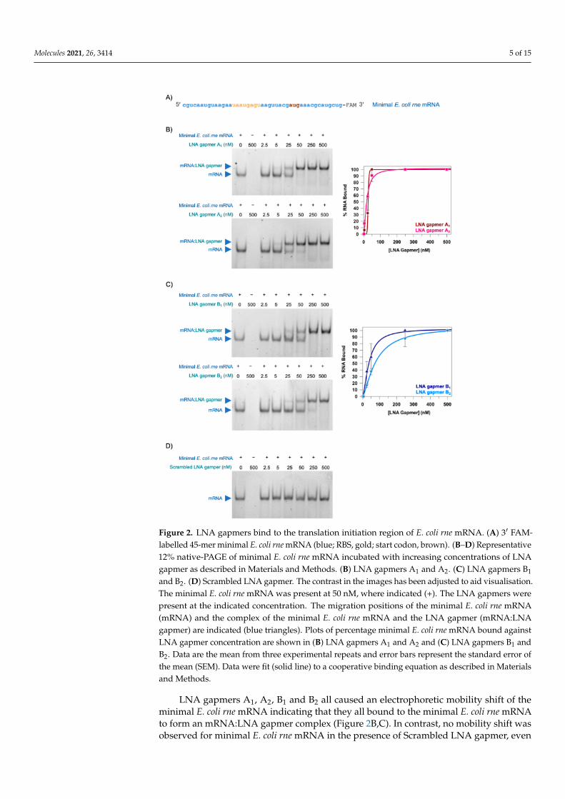
Figure 2. LNA gapmers bind to the translation initiation region of E. coli rne mRNA. (A) 3′ FAM-labelled 45-mer minimal E. coli rne mRNA (blue; RBS, gold; start codon, brown). (B–D) Representative12% native-PAGE of minimal E. coli rne mRNA incubated with increasing concentrations of LNAgapmer as described in Materials and Methods. (B) LNA gapmers A1 and A2. (C) LNA gapmers B1
and B2. (D) Scrambled LNA gapmer. The contrast in the images has been adjusted to aid visualisation.The minimal E. coli rne mRNA was present at 50 nM, where indicated (+). The LNA gapmers werepresent at the indicated concentration. The migration positions of the minimal E. coli rne mRNA(mRNA) and the complex of the minimal E. coli rne mRNA and the LNA gapmer (mRNA:LNAgapmer) are indicated (blue triangles). Plots of percentage minimal E. coli rne mRNA bound againstLNA gapmer concentration are shown in (B) LNA gapmers A1 and A2 and (C) LNA gapmers B1 andB2. Data are the mean from three experimental repeats and error bars represent the standard error ofthe mean (SEM). Data were fit (solid line) to a cooperative binding equation as described in Materialsand Methods.
LNA gapmers A1, A2, B1 and B2 all caused an electrophoretic mobility shift of theminimal E. coli rne mRNA indicating that they all bound to the minimal E. coli rne mRNAto form an mRNA:LNA gapmer complex (Figure 2B,C). In contrast, no mobility shift wasobserved for minimal E. coli rne mRNA in the presence of Scrambled LNA gapmer, even

Molecules 2021, 26, 3414 6 of 15
at the highest concentration of LNA gapmer tested (500 nM) indicating that this LNAgapmer did not bind the minimal E. coli rne mRNA (Figure 2D). Essentially all of theminimal E. coli rne mRNA was present as an mRNA:LNA gapmer complex at LNA gapmerconcentrations of 50 nM and above for LNA gapmer A1 and LNA gapmer A2 (Figure 2B).However, higher LNA gapmer concentrations of 250 nM and above were required for LNAgapmer B1 and LNA gapmer B2 before all of the minimal E. coli rne mRNA was presentas an mRNA:LNA gapmer complex (Figure 2C). This suggests that the LNA gapmer Asequence or binding site is preferred over the LNA gapmer B sequence or binding site. Inan attempt to quantify this observable difference, the data for LNA gapmers A1, A2, B1 andB2 were fit to a cooperative binding equation (Figure 2B,C) and an apparent dissociationconstant (Kd) was calculated for each of these LNA gapmers. The apparent Kds for LNAgapmers A1 and A2 (26.7 ± 4.9 nM and 18.4 ± 2.8 nM, respectively) were lower than theapparent Kds for LNA gapmers B1 and B2 (38.3 ± 3.5 nM and 76.6 ± 7.4 nM, respectively)supporting the qualitative observation that the LNA gapmer A sequence, or binding site,is preferred over the LNA gapmer B sequence, or binding site.
2.3. The LNA Gapmers Inhibit Translation in an In Vitro Cell-Free Assay
In order to determine whether binding of the LNA gapmers to the translation initia-tion region of E. coli rne mRNA can inhibit translation, an in vitro cell-free transcription-translation system coupled with a luciferase assay was devised. This assay is shownschematically in Figure 3A. A translational fusion of the −397 to +30 region of rne and thecoding region of the firefly luciferase (luc) gene (Supplementary Figure S3) was cloned intopET28b to generate pET28[rne-luc]. An in vitro cell-free transcription-translation systemwas then used to transcribe the rne-luc gene and translate it into luciferase. Since luciferaseconverts luciferin into oxy-luciferin, emitting light in the process, the observed lumines-cence can provide a readout of the relative level of rne-luc mRNA translation or the amountof luciferase present. In the presence of an LNA gapmer, if the LNA gapmer binds to thetranslation initiation region of rne mRNA and inhibits rne-luc mRNA translation, it wouldbe expected that less luciferase would be produced and the luminescence would be lowerthan in the absence of LNA gapmer.
The in vitro cell-free assay was performed in the absence of LNA gapmer and inthe presence of 0.5 nM, 5 nM and 50 nM of each of the LNA gapmers (Figure 3B). All ofthe LNA gapmers, including Scrambled LNA gapmer, which should not bind to rne-lucmRNA, negatively affected the total luminescence emitted in a dose-dependent manner.This suggests that the inclusion of any LNA gapmer in the reaction mixture may non-specifically affect rne-luc translation and/or luciferase activity. However, at a concentrationof 50 nM LNA gapmer, the reduction in luminescence was significantly larger for LNAgapmers A1 and A2 and LNA gapmers B1 and B2 than it was for Scrambled LNA gapmer.This is most likely due to the ability of these LNA gapmers to specifically bind to the rne-lucmRNA and sterically block its translation. LNA gapmers A1 and A2 appear to be morepotent inhibitors of translation than LNA gapmers B1 and B2 which is consistent with thehigher binding affinity for the translation initiation region of E. coli rne mRNA that wasobserved in the EMSAs (Figure 2B,C). There is also some indication that the 4-8-4 gapmerconfiguration is more effective than the 3-10-3 gapmer configuration for the LNA gapmerB sequence, which may suggest that the additional LNA nucleotides enhance LNA gapmerstability and/or target binding under these assay conditions.
2.4. The LNA Gapmers Stimulate RNase H-Mediated Cleavage of the Translation Initiation Regionof E. coli rne mRNA In Vitro
Having determined that the LNA gapmers are capable of binding to the translationinitiation region of E. coli rne mRNA and inhibiting translation of rne-luc mRNA in vitro,presumably by sterically blocking translation, we next wanted to investigate whether theycould also recruit RNase H and stimulate RNase H-mediated cleavage of E. coli rne mRNA(see Figure 1A for the expected mode-of-action). An in vitro RNase H cleavage assay wasdeveloped in which the 3′ FAM-labelled 45-mer minimal E. coli rne mRNA (Figure 2A) was

Molecules 2021, 26, 3414 7 of 15
used as the target mRNA. The minimal E. coli rne mRNA was incubated with an increasingconcentration of each of the LNA gapmers in the presence of RNase H and the reactionproducts were analysed by denaturing urea-PAGE (Figure 4).
Figure 3. An in vitro cell-free transcription-translation system, coupled with a luciferase assay. (A) Aschematic of the assay steps. A translational fusion of the −397 to +30 region of E. coli rne and thecoding region of the firefly luciferase (luc) gene (Supplementary Figure S3) was cloned into pET28bto generate pET28[rne-luc]. (Left) An in vitro cell-free transcription-translation system transcribes therne-luc gene into RNA (orange; RBS, gold; start codon, brown) and the RNA is translated to produceluciferase (orange wedge). Luciferase converts luciferin (dark green triangle) into oxy-luciferin(light green triangle) and emits light (yellow lightning bolt). (Right) In the presence of LNA gapmer(teal/mauve), translation is inhibited, less luciferase is produced, less luciferin is converted to oxy-luciferin and less light is emitted. (B) A chart showing the relative luminescence in the presence of0 nM, 0.5 nM, 5 nM or 50 nM LNA gapmer A1, A2, B1, B2 or Scrambled LNA gapmer. Data havebeen normalised to the total luminescence observed in the absence of LNA gapmer. Data are theaverage of three experimental repeats and error bars represent the SEM.

Molecules 2021, 26, 3414 8 of 15
Figure 4. LNA gapmers stimulate RNase H-mediated cleavage of the translation initiation regionof E. coli rne mRNA. Representative denaturing urea-PAGE analysis of RNase H cleavage assaysperformed in the presence of increasing concentrations of LNA gapmer as described in Materialsand Methods. (A) LNA gapmers A1 and A2. (B) LNA gapmers B1 and B2. (C) Scrambled LNAgapmer. The contrast in the images has been adjusted to aid visualisation. The minimal E. coli rnemRNA was present at 50 nM, where indicated (+). The LNA gapmers were present at the indicatedconcentration. RNase H was present at a concentration of 0.008 U/µL, where indicated (+). Note thatthe cleavage products migrate at or near the visible dye front. For this reason, we focused on thedisappearance of the band representing the intact minimal E. coli rne mRNA. Plots of the percentageof intact minimal E. coli rne mRNA remaining at the end of the RNase H cleavage assay against LNAgapmer concentration are shown in (A) LNA gapmers A1 and A2 and (B) LNA gapmers B1 and B2.Data are the mean from three experimental repeats and error bars represent the SEM. Data were fit(solid line) to a four-parameter logistic function as described in Materials and Methods.
As can be seen from the gels in Figure 4, the amount of intact minimal E. coli rne mRNAremaining at the end of the assay decreased with increasing concentration of LNA gapmerfor LNA gapmers A1, A2, B1 and B2 (Figure 4A,B). This suggests that these gapmers allrecruit RNase H to the minimal E. coli rne mRNA and facilitate RNase H-mediated cleavageof the mRNA. Efficient mRNA cleavage only occurred in the presence of both LNA gapmerand RNase H (Figure 4A,B). In contrast, no cleavage of the minimal E. coli rne mRNA wasobserved in the presence of Scrambled LNA gapmer, even at the highest LNA gapmerconcentration (100 nM) tested (Figure 4C).
In order to try to quantitate the effect of the different LNA gapmers on RNase Hrecruitment and RNase H-mediated mRNA cleavage, the data for LNA gapmers A1, A2,B1 and B2 were fit to a four-parameter logistic function (Figure 4A,B) which allowed us to

Molecules 2021, 26, 3414 9 of 15
estimate the half maximal inhibitory concentration (IC50) for each of them. When referringto this as inhibition, we considered cleavage of the minimal E. coli rne mRNA to representinhibition of mRNA function. This analysis suggested that LNA gapmer A1, with an IC50of 0.4 ± 0.1 nM, was the most effective of all of the LNA gapmers to recruit RNase H andstimulate RNase H-mediated cleavage of the minimal E. coli rne mRNA. The IC50 for LNAgapmer A2, at 10.5 ± 2.8 nM, was approximately 25-fold higher than for LNA gapmer A1.Since LNA gapmers A1 and A2 have the same nucleotide sequence, this implies that thedifference is a consequence of the 3-10-3 LNA gapmer configuration of LNA gapmer A1compared to the 4-10-4 LNA gapmer configuration of LNA gapmer A2. However, thistrend between the 3-10-3 and 4-8-4 LNA gapmer configurations was less apparent for theLNA gapmer B sequence with IC50s of 4.2 ± 1.9 nM for the 3-10-3 LNA gapmer B1 and8.0 ± 2.9 nM for the 4-8-4 LNA gapmer B2.
3. Discussion
Increasing antibacterial resistance has led to a need for novel antibacterial targetsand novel antibacterial strategies. RNase E/rne has been identified as a prospectiveantibacterial target [3,4] while antisense oligonucleotides hold potential as an antibacterialstrategy [14–17]. In this study we combined both of these novel factors to target rne mRNAwith antisense oligonucleotides. Specifically, we have successfully designed two antisenseoligonucleotide sequences to target E. coli rne mRNA and have demonstrated that theyhave the requisite translation blocking activity and the ability to recruit RNase H andfacilitate mRNA cleavage in vitro. This work provides the foundation for the developmentof an antibacterial strategy targeting RNase/rne.
The earliest antisense oligonucleotides targeted viral RNAs [21,22]. This pioneeringwork highlighted the potential of antisense approaches to rationally target essentially anyRNA to combat a variety of infections and/or diseases. Although there has been significantprogress in the development of therapeutic antisense oligonucleotides to target disease, suchas neurodegenerative disorders, cardiovascular disorders and cancer (reviewed in [23–25]),antisense oligonucleotides for antibacterial applications are yet to make it to the clinic.One of the main reasons why antibacterial antisense oligonucleotide research lags behindother RNA therapeutics is the challenge of cellular uptake [17]. There has been far lessfocus on antisense oligonucleotide delivery to bacterial cells than to eukaryotic cells, andthe advances that have been made in the eukaryotic field are not broadly applicable toprokaryotic systems [17]. Due to the known challenges with antisense oligonucleotidedelivery to bacterial cells, it is best practice to investigate the plausibility of targeting abacterial mRNA target with antisense oligonucleotides in vitro in the first instance [17],just as we have done here for E. coli rne mRNA.
Indeed, our results demonstrate that it is feasible to target E. coli rne mRNA withantisense oligonucleotides. In addition, our comparison of different antisense oligonu-cleotide sequences, different LNA gapmer configurations and different modes-of-actionof the LNA gapmers has provided some key knowledge to take forwards into in vivostudies. As might have been expected, the LNA gapmer A sequence which targets boththe RBS and start codon of the E. coli rne mRNA translation initiation region was moreefficient at inhibiting translation than the LNA gapmer B sequence which only targets thestart codon (Figure 3). It is possible that preventing ribosome binding may be a betterstrategy than blocking ribosome progression. However, this result may simply reflect thedifferent binding affinities observed for the LNA gapmer A and B sequences (Figure 2).The origin of these different binding affinities is unclear. It is known that the structuralcontext of the antisense oligonucleotide binding site is a critical determinant for its bindingaffinity [26]. However, translation initiation regions of mRNAs are typically unstructuredto allow access to ribosomes [17] and the E. coli rne mRNA translation initiation regionhas been reported to be unstructured [19]. Therefore, there is no obvious difference in thestructural context of the binding site for LNA gapmer A compared to LNA gapmer B.
Molecules 2021, 26, 3414 10 of 15
The comparison of the 3-10-3 and 4-8-4 LNA gapmer configurations generated mixedresults. There were hints that the 4-8-4 LNA gapmer configuration performed betterwith regard to inhibiting translation (Figure 3). This might be explained by the increasedLNA content stabilising the mRNA:LNA gapmer complex. In contrast, the 3-10-3 gapmerconfiguration appeared to perform better with regard to RNase H recruitment and mRNAcleavage (Figure 4). This might be explained if either the higher DNA content of 3-10-3 LNAgapmers enhances RNase H recruitment, or the lower binding affinity of the 3-10-3 LNAgapmers leads to higher LNA gapmer recycling, relative to the 4-8-4 LNA gapmers [27].
Interestingly, although there may be preferred sequences and/or LNA gapmer config-urations, our findings suggest that antisense oligonucleotides can efficiently down-regulaterne mRNA expression by either sterically blocking translation or stimulating RNase H-mediated mRNA cleavage. The majority of antisense oligonucleotides that have beenreported to have antibacterial activity are either entirely PMO or entirely PNA (Supple-mentary Table S1), are not recognised by RNase H, and are therefore restricted to down-regulating gene expression by sterically blocking translation. This is in contrast to widerRNA therapeutic applications where the value of combining oligonucleotide chemistries,e.g., as gapmers, to optimise antisense oligonucleotide properties and mode-of-action isnow recognised [17,18,23]. Incompatible assay conditions meant that, unfortunately, wewere unable to test the effect of combining the two modes of action and it remains to beseen whether there is a preferred mode-of-action in vivo.
Having demonstrated the successful targeting of E. coli rne mRNA with antisenseoligonucleotides in vitro, the next step would be to investigate their activity, particularly inregard to their antibacterial properties, in vivo. As discussed above, delivery of antisenseoligonucleotides to bacterial cells is challenging [17] and a “naked” LNA gapmer wouldnot be expected to enter bacterial cells. Not surprisingly, preliminary Kirby-Bauer diskdiffusion assays [28] with LNA gapmers A1 and A2, and E. coli, showed no inhibition ofbacterial growth. The most common strategy for facilitating antisense oligonucleotidedelivery is the conjugation of a cell-penetrating peptide (CPP), e.g., (KFF)3F [17,29–33],to the antisense oligonucleotide. This strategy has been used to deliver PNA antisenseoligonucleotides into E. coli [29] and LNA antisense oligonucleotides into Staphylococcusaureus [32] and would, therefore, be a good place to start for the E. coli rne mRNA-targetingLNA gapmers. Alternative strategies, such as the use of nanomaterials, are rarely used forantibacterial antisense oligonucleotides [17]. Although, progress is being made in terms ofusing cationic vesicles (bolasomes) to deliver antisense oligonucleotides into Clostridiumdifficile [34]. Cellular uptake could be confirmed by using a fluorescently labelled CPP-LNAgapmer and confocal microscopy [17].
It would also be interesting to investigate whether the antisense oligonucleotidesdesigned here to target E. coli rne mRNA could be effective against rne mRNA fromother bacterial species. Small molecule inhibitors of E. coli RNase E also inhibited RNaseE from other bacteria suggesting potential as lead compounds in the development ofbroad spectrum antibiotics [12,13]. As shown in Figure 5, the region where the LNAgapmers A1 and A2 bind to E. coli rne mRNA, is absolutely conserved in the rne mRNAof the closely related bacterium S. enterica. However, in another closely related bacterium,Y. pestis, there is sequence variation. This sequence variation becomes more pronounced inmore distantly related bacteria such as Francisella tularensis, Acinetobacter baumannii andBurkolderia pseudomallei. In contrast, the rne translation region in Mycobacteriun tuberculosisis similar to that in E. coli. Experimental validation will be needed to ascertain how muchsequence variation can be tolerated before an LNA gapmer fails to have an effect. This willbe important for tailoring antibacterial strategies and also for combatting the emergence ofantibacterial resistant mutants.

Molecules 2021, 26, 3414 11 of 15
Figure 5. Sequence alignment of the −21 to +3 translation initiation region of rne mRNA. A sequencealignment of the −21 to +3 translation initiation region of the rne mRNA from Escherichia coli,Salmonella enterica, Yersinia pestis, Francisella tularensis, Acinetobacter baumannii, Burkolderia pseudomalleiand Mycobacterium tuberculosis. Sequences were aligned using MAFFT [35] and coloured by nucleotidein JalView [36]. The complementary LNA gapmer A sequence is shown above the alignment toindicate the LNA gapmer A1/A2 binding site.
In summary, we have successfully designed two novel antisense oligonucleotidesequences to target E. coli rne mRNA, a novel antibacterial target. We synthesised four LNAgapmers based on these sequences and demonstrated that they bind to E. coli rne mRNA,inhibit translation of E. coli rne mRNA and facilitate RNase H recruitment and mRNAcleavage in vitro. Given these activities, it is anticipated that these LNA gapmers willdisplay antibacterial activity in vivo. Although the challenge of antisense oligonucleotidedelivery remains, there are feasible strategies available. Therefore, this work provides thefoundation for a possible novel antibacterial strategy targeting rne mRNA.
4. Materials and Methods4.1. LNA Gapmer Design and Synthesis
Two 16-mer antisense oligonucleotide sequences (sequence A: 5′ CATCGTAACT-TACTCA 3′; sequence B: 5′ GCGTTTCATCGTAACT 3′) were designed to target the −30 to+15 translation initiation region of the E. coli rne gene (5′ CGUCAAUGUAAGAAUAAU-GAGUAAGUUACGAUGAAACGCAUGCUG 3′). Each sequence was synthesised as anLNA3-DNA10-LNA3 3-10-3 gapmer (LNA gapmer A1/B1) and as an LNA4-DNA8-LNA44-8-4 gapmer (LNA gapmer A2/B2). The oligonucleotides were synthesised under standardconditions at 1 µmol scale using an ABI 394 DNA Synthesizer (Biolytic Lab Performance,Fremont, CA, USA) on 1000 Å UnyLinker-functionalised LCAA CPG support. The oligonu-cleotides were subsequently cleaved from support and the Unylinker moiety and protectinggroups were removed by treatment with concentrated aqueous ammonia at 55 ◦C overnight.The solution was decanted and dried using a centrifugal evaporator, then the pellets werediluted in 1 mL milliQ water and purified using ion exchange HPLC. The oligonucleotideswere characterised using LC-MS (ESI-mode) and the concentration of the final solutionswas determined according to their absorbance at 260 nm.
A scrambled 16-mer sequence (5′ ATCTACCAAATTTCCG 3′) was also generatedbased on sequence A using Shuffle DNA [37]. This sequence was synthesised at 0.05µmole scale as an LNA3-DNA10-LNA3 3-10-3 gapmer (Scrambled LNA gapmer) andHPLC-purified by Merck (Merck Life Science UK Limited, Gillingham, UK).
4.2. Design and Synthesis of a Minimal E. coli rne mRNA
A 3′ 6-fluorescein amidate (FAM)-labelled RNA oligonucleotide corresponding to the−30 to +15 translation initiation region of the E. coli rne gene (RNA; 5′ CGUCAAUGUAA-GAA UAAUGAGUAAGUUACGAUGAAACGCAUGCUG-[FAM] 3′) was synthesised at1 µmole scale and HPLC-purified by Sigma-Aldrich (now Merck Life Science UK Limited).The concentration of FAM-labelled RNA was determined according to the absorbanceat 260 nm using a conversion factor of 0.26 to correct for the 6-FAM absorbance [38].Denaturing urea-PAGE was used to confirm that the RNA was a single species of theexpected size.

Molecules 2021, 26, 3414 12 of 15
4.3. Electrophoretic Mobility Shift Assays (EMSAs)
A 10× (500 nM) stock of minimal E. coli rne mRNA was prepared in EMSA reactionbuffer (10 mM Tris-HCl (pH 8.0), 50 mM NaCl, 50 mM KCl, 0.5 mM ethylenediamineter-traacetic acid (EDTA), 10% glycerol), heated at 80 ◦C for 10 min, cooled at room temperaturefor 10 min and then equilibrated at 37 ◦C for 10 min. 10× stocks of each of the LNA gap-mers were prepared at concentrations of 25 nM, 50 nM, 250 nM, 500 nM, 2.5 µM and 5 µMin EMSA reaction buffer and equilibrated at 37 ◦C for 10 min. 10 µL reaction mixturescontaining 50 nM minimal E. coli rne mRNA and 0 nM, 2.5 nM, 5 nM, 25 nM, 50 nM,250 nM or 500 nM LNA gapmer in EMSA reaction buffer were assembled and incubatedat room temperature for 10 min. Reactions were analysed by 12% native-PAGE run inTris-borate-EDTA (TBE) running buffer at 80 V for 2 h at room temperature. Gels werevisualised using a GBox UV transilluminator (Syngene, a division of Synoptics Ltd., Cam-bridge, UK). Digitised images were quantitated using ImageJ (Rasband, W.S., ImageJ, U.S.National Institutes of Health, Bethesda, MD, USA, https://imagej.nih.gov/ij/, 1997–2018)and the percentage of bound and unbound RNA in each lane was calculated. Data fromtriplicate experiments were fit in Grafit5 (Erithacus Software, Grinstead, West Sussex, UK)to a cooperative binding equation:
y =Ln.Cap
Kn + [L]n+ background (1)
In this equation, y is the percentage of FAM-labelled RNA bound by LNA gapmer, [L]is the concentration of LNA gapmer, n is the slope factor, Cap is the theoretical maximalamount of FAM-labelled RNA than can be bound by LNA gapmer, K is the apparentequilibrium dissociation constant (also termed apparent Kd) and background allows for anyy-axis displacement from the origin.
4.4. Cell-Free Reporter Assay4.4.1. Design and Synthesis of the E. coli rne-Firefly Luciferase (luc) Reporter Plasmid
A translational fusion of the −397 to +30 region of E. coli rne and the coding regionof the firefly luciferase (luc) gene (Supplementary Figure S3) was synthesised by GeneArt(Thermo Fisher Scientific, Waltham, MA, USA) and ligated between the XbaI and XhoIrestriction sites of pET28b (Novagen, a brand of Merck, Darmstadt, Germany) to generatepET28[rne-luc]. The sequence was confirmed by DNA sequencing. E. coli DH5α wastransformed with pET28[rne-luc]. DH5α, pET28[rne-luc] was grown in LB supplementedwith 25 µg/mL kanamycin at 37 ◦C overnight with shaking. Cells were harvested bycentrifugation and pET28[rne-luc] was extracted using the NucleoBond Xtra Midi plasmidpreparation kit (Macherey-Nagel, Düren, Germany).
4.4.2. In Vitro Transcription-Translation Real-Time Reporter Assay
In vitro transcription-translation of pET28 [rne-luc] was performed using the E. coliT7 S30 Extract System for Circular DNA (Promega, Madison, WI, USA). 50 µL reactionswere prepared in a 96-well plate. Each reaction contained 750 ng pET28 [rne-luc], 1 mMluciferin (BD Biosciences, BD Biosciences, San Jose, CA, USA), 5 µL Complete Amino AcidMixture, 20 µL S30 Premix and 15 µL T7 S30 Extract. The reactions were supplemented with0.5 nM, 5 nM or 50 nM LNA gapmer, as indicated. The reactions were not supplementedwith exogenous RNase H. Reactions were incubated at 37 ◦C for 2 h in a Hidex Senseplate-reader (Hidex Ltd., Turku, Finland). The luminescence (or luciferase signal) wasrecorded every 2.5 min for a total of 120 min. The total luciferase signal for each LNAgapmer concentration (0 nM, 0.5 nM, 5 nM and 50 nM) was quantitated by integrating thearea under the curve using a trapezoid method in Excel. Integrated values were normalisedto a percentage (relative to the total luciferase signal in the absence of LNA gapmer) andplotted as a bar chart in Grafit5.

Molecules 2021, 26, 3414 13 of 15
4.5. RNase H Cleavage Assay
Twenty five µL reaction mixtures containing 50 nM minimal E. coli rne mRNA and0 nM, 2.5 nM, 5 nM, 25 nM, 50 nM or 100 nM LNA gapmer in 1× RNase H Reaction Buffer(NEB, Ipswich, MA, USA; 75 mM KCl, 50 mM Tris-HCl pH 8.3, 3 mM MgCl2, 10 mM DTT)were assembled and incubated at 37 ◦C for 5 min to allow LNA gapmer to bind to the targetminimal E. coli rne mRNA and form the RNase H substrate. RNase H, equilibrated at 37 ◦C,was then added to a final concentration of 0.008 U/µL. The complete reaction mixturewas incubated at 37 ◦C for a further 45 min. Reactions were terminated by the additionof 0.5 volumes of quench buffer (95% (v/v) formamide, 18 mM EDTA). Reaction mixtureswere heated at 95 ◦C for 5 min and reaction products were resolved by 8% denaturingurea-PAGE. Gels were visualised using a GBox UV transilluminator (Syngene, a divisionof Synoptics Ltd., Cambridge, UK). Digitised images were quantitated using ImageJ andthe percentage of cleaved and uncleaved FAM-labelled RNA in each lane was calculated.Data from triplicate experiments were fit to a four-parameter logistic function to estimatethe half maximal inhibitory concentration (IC50):
y =Range
1 +(
xIC50
)s + background (2)
In this equation, y is the percentage of intact FAM-labelled RNA at LNA gapmerconcentration x; Range is the theoretical extent of the reaction, IC50 is the concentration ofLNA gapmer at half the Range, s is the slope factor and background allows for any y-axisdisplacement from the origin.
Supplementary Materials: The following are available online, Supplementary References, Supple-mentary Table S1: Examples of antibacterial antisense oligonucleotides, Supplementary Figure S1:Mode-of-action of antisense oligonucleotides, Supplementary Figure S2: Unmodified nucleic acidsand examples of chemical analogues used in antisense oligonucleotides, Supplementary Figure S3:The E. coli rne-firefly luciferase reporter.
Author Contributions: Conceptualization, L.R.G., C.E.M., D.M.G., H.S.A., L.E.B., H.A.V. and A.J.C.;Formal analysis, L.R.G., C.E.M., D.M.G., H.A.V. and A.J.C.; Funding acquisition, J.K.W. and A.J.C.;Investigation, L.R.G., C.E.M. and C.G.B.; Methodology, L.R.G., C.E.M., D.M.G., H.S.A., L.E.B., J.K.W.,H.A.V. and A.J.C.; Resources, H.G. and J.K.W.; Supervision, H.S.A., L.E.B., J.K.W., H.A.V. and A.J.C.;Visualization, L.R.G., C.E.M., C.G.B., D.M.G., H.A.V. and A.J.C.; Writing—original draft, L.R.G.,C.E.M., H.A.V. and A.J.C.; Writing—review & editing, L.R.G., C.E.M., H.G., C.G.B., D.M.G., L.E.B.,J.K.W., H.A.V. and A.J.C. All authors have read and agreed to the published version of the manuscript.
Funding: This research was funded by Defence Science and Technology Laboratory (Dstl), PhDstudentship for C.E.M. (A.J.C.); Research England E3 funding; Engineering and Physical SciencesResearch Council (EPSRC), iCASE award with Dstl for H.G. (J.K.W.); University of MassachusettsMedical School (J.K.W.); and Biotechnology and Biological Sciences Research Council (BBSRC), grantnumber BB/M020576/1 (A.J.C.). The APC was funded by BBSRC.
Institutional Review Board Statement: Not applicable.
Informed Consent Statement: Not applicable.
Data Availability Statement: The data presented in this study are available on request from thecorresponding authors.
Acknowledgments: We thank members of A.J.C.’s research group (University of Portsmouth, UK)between 2012 and 2021 for helpful discussions and technical support. We thank Andy Scott (DefenceScience and Technology Laboratory, UK) for helpful discussions. We thank T.J. Ragan (University ofLeicester, UK) for helpful discussions and critical reading of the manuscript.
Conflicts of Interest: H.S.A. was an employee of Defence Science and Technology Laboratory, one ofthe co-funders of the research, during the course of the study. All other authors declare no conflictof interest.

Molecules 2021, 26, 3414 14 of 15
Sample Availability: Not available.
References1. Lewis, K. Platforms for antibiotic discovery. Nat. Rev. Drug Discov. 2013, 12, 371–387. [CrossRef]2. Ventola, C.L. The antibiotic resistance crisis: Part 1: Causes and threats. Pharm. Ther. 2015, 40, 277–283.3. Eidem, T.M.; Roux, C.M.; Dunman, P.M. RNA decay: A novel therapeutic target in bacteria. Wiley Interdiscip. Rev. RNA 2012, 3,
443–454. [CrossRef] [PubMed]4. Lawal, A.; Jejelowo, O.; Chopra, A.K.; Rosenzweig, J.A. Ribonucleases and bacterial virulence. Microb. Biotechnol. 2010, 4, 558–571.
[CrossRef] [PubMed]5. Apirion, D.; Lassar, A. A conditional lethal mutant of Escherichia coli which affects the processing of ribosomal RNA. J. Biol. Chem.
1978, 253, 1738–1742. [CrossRef]6. Ono, M.; Kuwano, M. A conditional lethal mutation in an Escherichia coli strain with a longer chemical lifetime of messenger
RNA. J. Mol. Biol. 1979, 129, 343–357. [CrossRef]7. McDowall, K.J.; Hernandez, R.G.; Lin-Chao, S.; Cohen, S.N. The ams-1 and rne-3071 temperature-sensitive mutations in the ams
gene are in close proximity to each other and cause substitutions within a domain that resembles a product of the Escherichia colimre locus. J. Bacteriol. 1993, 175, 4245–4249. [CrossRef] [PubMed]
8. Hammarlöf, D.L.; Liljas, L.; Hughes, D. Temperature-sensitive mutants of RNase E in Salmonella enterica. J. Bacteriol. 2011, 193,6639–6650. [CrossRef]
9. Yang, J.; Jain, C.; Schesser, K. RNase E Regulates the Yersinia type 3 secretion system. J. Bacteriol. 2008, 190, 3774–3778. [CrossRef]10. Lee, E.-J.; Groisman, E.A. An antisense RNA that governs the expression kinetics of a multifunctional virulence gene. Mol.
Microbiol. 2010, 76, 1020–1033. [CrossRef]11. Aït-Bara, S.; Carpousis, A. RNA degradosomes in bacteria and chloroplasts: Classification, distribution and evolution of RNase E
homologs. Mol. Microbiol. 2015, 97, 1021–1135. [CrossRef]12. Kime, L.; Vincent, H.A.; Gendoo, D.M.A.; Jourdan, S.S.; Fishwick, C.W.G.; Callaghan, A.J.; McDowall, K.J. The first small-molecule
inhibitors of members of the ribonuclease E family. Sci. Rep. 2015, 5, 08028. [CrossRef] [PubMed]13. Mardle, C.E.; Goddard, L.R.; Spelman, B.C.; Atkins, H.S.; Butt, L.E.; Cox, P.A.; Gowers, D.M.; Vincent, H.A.; Callaghan, A.J.
Identification and analysis of novel small molecule inhibitors of RNase E: Implications for antibacterial targeting and regulationof RNase E. Biochem. Biophys. Rep. 2020, 23, 100773. [CrossRef]
14. Rasmussen, L.C.V.; Sperling-Petersen, H.U.; Mortensen, K.K. Hitting bacteria at the heart of the central dogma: Sequence-specificinhibition. Microb. Cell Factories 2007, 6, 24–26. [CrossRef] [PubMed]
15. Bai, H.; Xue, X.; Hou, Z.; Zhou, Y.; Meng, J.; Luo, X. Antisense antibiotics: A brief review of novel target discovery and delivery.Curr. Drug Discov. Technol. 2010, 7, 76–85. [CrossRef] [PubMed]
16. Sully, E.K.; Geller, B.L. Antisense antimicrobial therapeutics. Curr. Opin. Microbiol. 2016, 33, 47–55. [CrossRef] [PubMed]17. Hegarty, J.P.; Stewart, D.B. Advances in therapeutic bacterial antisense biotechnology. Appl. Microbiol. Biotechnol. 2018, 102,
1055–1065. [CrossRef]18. Khvorova, A.; Watts, J.K. The chemical evolution of oligonucleotide therapies of clinical utility. Nat. Biotechnol. 2017, 35, 238–248.
[CrossRef]19. Diwa, A.; Bricker, A.L.; Jain, C.; Belasco, J.G. An evolutionarily conserved RNA stem-loop functions as a sensor that directs
feedback regulation of RNase E gene expression. Genome Res. 2000, 14, 1249–1260.20. Kurreck, J.; Wyszko, E.; Gillen, C.; Erdmann, V.A. Design of antisense oligonucleotides stabilized by locked nucleic acids. Nucleic
Acids Res. 2002, 30, 1911–1918. [CrossRef]21. Zamecnik, P.C.; Stephenson, M.L. Inhibition of Rous sarcoma virus replication and cell transformation by a specific oligodeoxynu-
cleotide. Proc. Natl. Acad. Sci. USA 1978, 75, 280–284. [CrossRef]22. Stephenson, M.L.; Zamecnik, P.C. Inhibition of Rous sarcoma viral RNA translation by a specific oligodeoxyribonucleotide. Proc.
Natl. Acad. Sci. USA 1978, 75, 285–288. [CrossRef]23. Roberts, T.C.; Langer, R.; Wood, M.J.A. Advances in oligonucleotide drug delivery. Nat. Rev. Drug Discov. 2020, 19, 673–694.
[CrossRef] [PubMed]24. Damase, T.R.; Sukhovershin, R.; Boada, C.; Taraballi, F.; Pettigrew, R.I.; Cooke, J.P. The limitless future of RNA therapeutics. Front.
Bioeng. Biotechnol. 2021, 9, 628137. [CrossRef]25. Hammond, S.M.; Aartsma-Rus, A.; Alves, S.; Borgos, S.E.; Buijsen, R.A.M.; Collin, R.W.J.; Covello, G.; Denti, M.A.; Desviat, L.R.;
Echevarría, L.; et al. Delivery of oligonucleotide-based therapeutics: Challenges and opportunities. EMBO Mol. Med. 2021, 13,e13243. [CrossRef]
26. Lima, W.F.; Monia, B.P.; Ecker, D.J.; Freier, S.M. Implication of RNA structure on antisense oligonucleotide hybridization kinetics.Biochemistry 1992, 31, 12055–12061. [CrossRef] [PubMed]
27. Pedersen, L.; Hagedorn, P.H.; Lindholm, M.W.; Lindow, M. A kinetic model explains why shorter and less affine enzyme-recruitingoligonucleotides can be more potent. Mol. Ther. Nucleic Acids 2014, 3, e149. [CrossRef] [PubMed]
28. Bauer, A.W.; Kirby, W.M.; Sherris, J.C.; Turck, M. Antibiotic susceptibility testing by a standardized single disk method. Am. J.Clin. Pathol. 1966, 45, 493–496. [CrossRef]

Molecules 2021, 26, 3414 15 of 15
29. Good, L.; Awasthi, S.K.; Dryselius, R.; Larsson, O.; Nielsen, P.E. Bactericidal antisense effects of peptide-PNA conjugates. Nat.Biotechnol. 2001, 19, 360–364. [CrossRef]
30. Nekhotiaeva, N.; Awasthi, S.K.; Nielsen, P.E.; Good, L. Inhibition of Staphylococcus aureus gene expression and growth usingantisense peptide nucleic acids. Mol. Ther. 2004, 10, 652–659. [CrossRef] [PubMed]
31. Kulyté, A.; Nekhotiaeva, N.; Awasthi, S.K.; Good, L. Inhibition of Mycobacterium smegmatis gene expression and growth usingantisense peptide nucleic acids. J. Mol. Microbiol. Biotechnol. 2005, 9, 101–109. [CrossRef]
32. Meng, J.; Da, F.; Ma, X.; Wang, N.; Wang, Y.; Zhang, H.; Li, M.; Zhou, Y.; Xue, X.; Hou, Z.; et al. Antisense growth inhibitionof methicillin-resistant Staphylococcus aureus by locked nucleic acid conjugated with cell-penetrating peptide as a novel FtsZinhibitor. Antimicrob. Agents Chemother. 2014, 59, 914–922. [CrossRef] [PubMed]
33. Kurupati, P.; Tan, K.S.W.; Kumarasinghe, G.; Poh, C.-L. Inhibition of gene expression and growth by antisense peptide nucleicacids in a multiresistant β-lactamase-producing Klebsiella pneumoniae strain. Antimicrob. Agents Chemother. 2006, 51, 805–811.[CrossRef]
34. Hegarty, J.P.; Krzeminski, J.; Sharma, A.K.; Guzman-Villanueva, D.; Weissig, V.; Stewart, D.B., Sr. Bolaamphiphile-based nanocomplexdelivery of phosphorothioate gapmer antisense oligonucleotides as a treatment for Clostridium difficile. Int. J. Nanomed. 2016, 11,3607–3619. [CrossRef] [PubMed]
35. Katoh, K.; Standley, D.M. MAFFT multiple sequence alignment software version 7: Improvements in performance and usability.Mol. Biol. Evol. 2013, 30, 772–780. [CrossRef] [PubMed]
36. Waterhouse, A.M.; Procter, J.B.; Martin, D.M.A.; Clamp, M.; Barton, G.J. Jalview Version 2-a multiple sequence alignment editorand analysis workbench. Bioinformatics 2009, 25, 1189–1191. [CrossRef]
37. Stothard, P. The Sequence Manipulation Suite: JavaScript programs for analyzing and formatting protein and DNA sequences.Biotechniques 2000, 28, 1102–1110. [CrossRef]
38. Stephenson, F.H. Calculations for Molecular Biology and Biotechnology; Elsevier Science: Amsterdam, The Netherlands, 2016.[CrossRef]