
Why are Cold Molecules so Hot?
69
Why are cold molecules so hot? Bretislav Friedrich 1 and John M. Doyle 2 1 Fritz-Haber-Institut der Max-Planck-Gesellschaft Faradayweg 4-6, D-14195 Berlin, Germany 2 Department of Physics, Harvard University 17 Oxford Street, Cambridge, MA 02138, U.S.A. 1. Introduction Over the last decades, atomic and molecular physics has come into a spectacular bloom, much of which sprang from the newfashioned techniques to translationally cool (or slow down) gaseous atoms and molecules. The study of slow atoms and molecules, which ensued, has led to uncharted territories – not just of atomic [1] and molecular [2] physics, but of physics at large [3] . Apart from enhancing molecular spectroscopy and amplifying the ability to manipulate molecular trajectories, cold and trapped molecules have begun to play unique roles in a number of diverse areas of fundamental interest, such as quantum collision dynamics, collective effects, and even particle physics. In this review, we describe some of the excitement that the work on cold molecules has brought about. 2. Basic features of cold atoms and molecules At room temperature, gaseous atoms and molecules move at the speed of
-
Upload
independent -
Category
Documents
-
view
2 -
download
0
Transcript of Why are Cold Molecules so Hot?

Why are cold molecules so hot?
Bretislav Friedrich1 and John M. Doyle2
1Fritz-Haber-Institut der Max-Planck-Gesellschaft
Faradayweg 4-6, D-14195 Berlin, Germany 2Department of Physics, Harvard University
17 Oxford Street, Cambridge, MA 02138, U.S.A.
1. Introduction
Over the last decades, atomic and molecular physics has come into a spectacular
bloom, much of which sprang from the newfashioned techniques to
translationally cool (or slow down) gaseous atoms and molecules. The study of
slow atoms and molecules, which ensued, has led to uncharted territories – not
just of atomic[1] and molecular[2] physics, but of physics at large[3].
Apart from enhancing molecular spectroscopy and amplifying the ability to
manipulate molecular trajectories, cold and trapped molecules have begun to
play unique roles in a number of diverse areas of fundamental interest, such as
quantum collision dynamics, collective effects, and even particle physics. In this
review, we describe some of the excitement that the work on cold molecules has
brought about.
2. Basic features of cold atoms and molecules
At room temperature, gaseous atoms and molecules move at the speed of

2
rifle bullets – and their behavior is in many respects reminiscent of that of
bullets. They calm down only at temperatures close to absolute zero (i.e., -
273.15o C); for instance, a nitrogen molecule moves at the speed of a healthy
ant (a few cm/s) only at the temperature of a microkelvin (µK). As the
temperature (speed) is lowered, the behavior of atoms and molecules undergoes
a dramatic change: their wave properties begin to prevail over their corpuscular
ones – the atoms and molecules become waves. Thus, they acquire a character
usually associated with the behavior of light or another electromagnetic
radiation; in contrast to light, these matter waves have a nonzero rest mass.
By slowing (cooling), it is possible to extend the time during which the
atoms or molecules can be observed, and thus enhance the accuracy of the
observation. More than that, one can say that the most accurate measurements
to date can be carried out on cold atoms and molecules.[4] Thus the combination
of the unusual wave behavior of cold atoms and molecules with the ability to
observe it is a most fortunate circumstance which contributes to the boom of
cold matter physics.
The matter waves are characterized by a de Broglie wavelength[5]
€
Λ =hmv
or h(2πmkT)1/2 (1)
with
€
h = 6.63×10⁻³⁴ J s the Planck constant,
€
k =1.38 ×10⁻²³ J
€
K−1 the
Boltzmann constant, m the rest mass, v the velocity and T the absolute
temperature. The nomogram in Figure 1 shows the dependence of the de
Broglie wavelength on temperature and rest mass. One can see that, for
instance, a nitrogen molecule (mass 28 g/mol) has at room temperature (300 K)
a wavelength of 0.2 Å (i.e.,
€
2 ×10⁻⁹ cm). Since the bond length of an N2
molecule is about 1.1 Å, we see that this wavelength is smaller than the size of
the molecule. When cooled to 1 K, the wavelength of N2 increases to 3.3 Å,

3
i.e., a value which is of the same order of magnitude as the molecular size
(which does not change with temperature). At a temperature of 1 mK, the
wavelength of N2 increases to 104 Å, at 1 µK to 3464 Å, i.e. 0.35 µm. That’s a
huge change with respect to the room-temperature value; it is given by the
factor 3464/0.2 = 17320 = (300/10-6)1/2, see eq. (1). Hence we see from this
example that by cooling (slowing), the molecule-projectile at room temperature
is transformed at a microkelvin temperature into a molecule-wave whose
wavelength far exceeds the molecular dimensions.
In order to better appreciate what this means, let us clarify the notion of
molecular size. This is determined by the magnitude of the outer (valence) shell
of a given atom or molecule. But the magnitude of the valence shell is not due
to anything else than the de Broglie wavelength of the valence electrons!
Despite the fact that the mass of the electron is 1836-times less than the mass of
the proton, the de Broglie wavelength of a valence electron is small. This is
because the binding energy of the outer electrons (typically on the order of 10
eV) corresponds to temperatures on the order of 105 K, as follows from the
virial theorem.[6] From the nomogram of Fig. 1, we see immediately that at such
high temperatures the wavelength of an electron is on the order of 1 Å, i.e., the
valence electrons interact with each other at most at this distance. This distance
then defines the “typical dimensions” of an atom or small molecule. But what is
the size of an atom or molecule as a whole, i.e., at which distance can atoms or
molecules (not their valence electrons) feel each other? The maximum distance
is given also by the de Broglie wavelength, but of an entire atom or molecule.
Thus we see that cooling (slowing) of atoms or molecules has a wide-ranging
effect on their ability to interact with each other. That has, in turn, a most
dramatic effect on collisions and the collective behavior of cold atoms and
molecules.[7] From this springs most of the new physics and chemistry that cold
atoms and molecules can offer.

4
3. Why cold molecules?
During the last decade, a number of research groups, spread all over the
world, launched research programs to study cold molecules.[8] Molecules are
namely not only more complicated than atoms, but in some respects also more
interesting: apart from possessing vibrational and rotational degrees of
freedom, molecules may carry dipole (and higher) electric and magnetic
moments. These moments lend properties to molecules which atoms simply
lack. At the same time, it is exactly these features on which new operational
principles can be based (e.g., quantum computing[9]) or that allow to achieve a
qualitatively new behavior of matter to be achieved (e.g., Bose-Einstein
condensates structured by the electric dipole interaction[10]). However, the
trouble is that it is much more difficult to cool molecules than atoms.
A certain class of atoms can be cooled with light. The method of laser
cooling[11] is based on a manifold repetition of the absorption and emission of
photons by the atom. Both absorption and emission of a photon are
accompanied by momentum transfer. Whereas photon absorption can be
arranged to take place preferentially along the atom’s motion, photon emission
occurs in random directions. Then, on average, each absorption-emission cycle
results in a slowing of the atom, i.e., in cooling. Although elegant and efficient,
laser cooling is of limited chemical scope: the requirement of a closed
absorption-emission cycle is fulfilled by only a handful of atoms, best
represented by alkali metals. The complex energy-level structure of most other
atoms and of essentially all molecules renders laser cooling inapplicable. What
disqualifies molecules is mainly their vibrational structure: the Franck-Condon
factors cause a rapid depopulation of the initial vibrational state, and thereby
terminate the absorption-emission cycle.[12] The early era of the cold-matter
research was thus predominantly an alkali age, like in the case of the early

5
crossed-beam studies of reaction dynamics.
4. How to trap a molecule
At low temperatures, gases condense into liquids or solids. Their
condensation has to be precluded by preventing the gas molecules to come in
contact with one another or with the walls of the vessel that contains them. This
can be achieved by holding the molecules at low densities in vacuum by means
of electric or magnetic fields. The interaction of the molecules with such fields
creates an energy barrier that acts as walls. Such a barrier is called a molecular
trap. A given molecular trap is capable of confining only those molecules
whose translational energy is lower than the depth of the trap (i.e., the energy
barrier). Therefore, in order to trap molecules, it is necessary to slow (cool)
them first. This is indeed the reason why cooling and trapping go usually hand
in hand.[13]
5. How to cool molecules
The inability to apply laser cooling to molecules was a challenge taken up a
handful of enthusiasts who, in the mid 1990s, saught to widen the range of
cold-matter research to include molecules. Necessity proved to be a mother of
invention and, over the last decade, a host of techniques had been developed
that made it possible to cool molecules. These techniques are based on new
principles, independent of laser cooling.
In 1998, the first molecule, the radical CaH, was cooled by a cold buffer
gas, and subsequently magnetically trapped.[14,15] This was followed by a
successful synthesis of homonuclear bialkali molecules via photoassociation of
laser-cooled atoms.[16] Recently, ultracold molecules were produced in
collisions controlled by a Feshbach resonance.[17] The last two methods, based
on the synthesis of laser-cooled atoms, can be called indirect. At present, about

6
twenty research groups are exploring and exploiting the indirect methods. Some
of these groups are now turning their attention to the synthesis of heteronuclear
bialkali molecules. The advantages and drawbacks of the indirect methods are
described in section 5.1.
Direct methods are based on cooling of pre-existing molecules. Roughly
fifteen research groups worldwide are concerned with the development and
applications of buffer-gas cooling,[15] Stark deceleration,[18,19] deceleration by
pulsed optical fields,[20,21] deceleration via collisions in crossed molecular
beams,[22,23] supersonic expansion from a counter-rotating nozzle,[24] or
selection of the low-velocity tail of a Maxwell-Boltzmann distribution of
molecules in an effusive beam.[25,26] All direct methods start with relatively hot
molecules (200-1000 K), usually in the form of a supersonic molecular beam*.
Whereas the indirect methods are only suited for the preparation of bialkali
molecules, the direct methods are versatile, applicable to large classes of
molecules (e.g., Stark deceleration to all polar molecules; buffer-gas cooling
coupled with magnetic trapping to all paramagnetic molecules) or to any
molecules (all other direct techniques).
5.1 Synthesis of cold molecules from cold atoms
5.1.1 Photoassociation
The process of photo-associating a pair of atoms into a molecule is shown
schematically in Figure 2a: two free atoms on a collision course with one
* A supersonic expansion of a gas into vacuum – whereby a supersonic molecular beam
is generated – cools all the degrees of freedom of the gas molecules (typically to 1 K, in the case of a pulsed beam). The initial energy of the gas is thereby efficiently transformed into translational energy along the direction of the beam. In contrast, molecules in an effusive beam have a thermal distribution of states in both internal and translational degrees of freedom.

7
another are exposed to laser radiation of a suitable wavelength that excites them
to a common bound (i.e., molecular) state. Since molecules formed by
photoassociation have the translational temperature of the free atoms, it is
possible to make in this way cold molecules from cold atoms.
Figure 2b shows the dependence of the potential energy of the atoms on the
distance of their nuclei (i.e., a potential energy curve) for the case when the
atoms are in their ground or excited electronic states. Also shown are the
vibrational levels within the ground and excited molecular states. A collision of
a pair of ground-state atoms cannot result in a bound molecular state, since a
mechanism is lacking that would relieve the mutually interacting atoms of their
energy and allow them to fall into the potential well of the ground molecular
state. However, it is possible to create a bound excited state: it suffices to excite
the colliding atoms into one of the vibrational states pertaining to the upper
potential energy curve, see Fig. 2b. This state has then automatically a lower
energy than the well depth of the electronically excited state (note that the
translational energy of the cold atoms is negligible compared with the
vibrational energy).
However, the formation of the excited bound state is not the end of the
story but rather an intermediate step: such a state is namely only metastable and
so can spontaneously decay (typically within a microsecond) either back to the
original ground-state atoms or to a molecule in a ground electronic but
vibrationally highly excited state (the lifetime of a vibrational state is typically
10-100 ms).
That the states in which the molecules are formed are vibrationally excited
presents, however, a difficulty: if such molecules collide, their vibrational
energy can get quite efficiently converted into their translational energy,
whereby the molecules become translationally hot (1000 K or more) – thus
defeating the purpose of photoassociating the cold atoms …

8
During the last couple years or so it became possible to stimulate the
transition from an electronically excited state using an additional laser field,
and thus to achieve a population transfer from the excited state to the electronic
and vibrational ground state, Figure 2c. This technique, which functions on the
same principle as ‘coherent population transfer’ (which is the basis of, e.g.,
electromagnetically induced transparency[27]), is especially well applicable to
heteronuclear bialkali molecules; these are coveted since they are polar (i.e.,
they carry a body-fixed electric dipole moment).
Whereas the potential energy curves of both ground and excited electronic
states of a heteronuclear molecule exhibit a R-6 dependence at large internculear
separations R, in the case of homonuclear dimers it is only the ground state that
has an R-6 energy dependence. The electronically excited state of homonuclear
molecules namely shows a R-3 dependence, due to the exchange interaction.
Since the photoassociation step occurs at large R, the R-3 potential (which is
shifted toward larger R with respect to the R-6 potential) enables a good overlap
(i.e., a large Franck-Condon factor) between some of the vibrational states of
the electronically excited state and the ground electronic state. In the second,
stabilization step, such an overlap favors a transition to either an unbound state
of free atoms or to one of the highly vibrationally excited states of the
electronic ground state. In the case of heteronuclear molecules, the overlap
favors a stimulated transition directly to the ground vibrational and electronic
state. As a result, the molecule is cold in all its degrees of freedom, with a
translational temperature on the order of 100 µK. This indirect method has so
far been demonstrated for the RbCs[28] and KRb[29] molecules.
5.1.2 Feshbach resonances
The synthesis of cold molecules via cold-atom collisions controlled by a

9
Feshbach resonance is simple in principle but quite demanding in practice.
Nevertheless, several groups have, independently, succeeded in creating not
just cold molecules but even molecular Bose-Einstein condensates (of K2 and
Li2) using this technique.[30] As if that weren’t enough, Feshbach resonances
found a key role to play in the study of the cross-over between a Bose-Einstein
condensate of molecules and an ensemble of correlated Cooper pairs of the
atoms that constitute these molecules. This is a theme that touches upon few-
and many-body physics, with repercussions for nuclear physics, superfluidity,
and superconductivity, including its high-temperature variant.
An example of a Feshbach resonance is shown in Figure 3. This occurs if
the energy of two colliding atoms (in a given electronic state) equals the energy
of the bound state of a molecule that consists of the same atoms, but is in a
different electronic state. If the bound and unbound states have different dipole
moments, the relative energy of these two states can be varied by tuning an
external field. Because a Feshbach resonance dramatically enhances the
magnitude of the interaction of the two atoms (their collision cross section), it
is possible to sensitively control the molecule formation by fine-tuning the
external field. In the experiments carried out so far, a magnetic field and its
effect on the different spin states of the bound and unbound states was used for
this purpose.
The molecules formed via a Feshbach resonance are in the highest possible
vibrational and rotational states, often just a fraction of a mK below the
dissociation limit. Like any excited state, these states too are apt to relax to
lower states; however, the available lower states are often unbound so that the
relaxation process (induced by mutual collisions) leads to a conversion of the
molecules back into atoms. As a result, the “Feshbach molecules” almost
immediately vanish. Unless they consist of atoms which are fermions!
Fermions differ from bosons in the way they behave when their positions

10
are interchanged. Whether a given indistinguishable particle is a boson or a
fermion is unequivocally determined by its spin: a half-integral spin defines
fermions (e.g., electrons, protons, neutrons) and an integral spin defines bosons
(e.g., photons, mesons). The character of a compound particle, such as an atom
or a molecule, can be easily determined from the number of fermions it
contains: if this number is odd, the compound particle is a fermion; if it is even,
the particle is a boson. It follows that an electroneutral atom whose nucleus has
an odd number of neutrons is a fermion (e.g., 3He), one with an even number of
neutrons a boson (e.g., 4He). Homonuclear diatomic molecules are all bosons.
Bosons and fermions exhibit a dramatically different collective behavior:
whereas bosons like each other’s company, band together, and create high-
density ensembles (e.g., photons in a laser cavity), fermions rather avoid each
other (this behavior is captured by Pauli’s principle which co-determines
electronic configurations of atoms and thereby the chemical properties of the
elements). Pauli’s exclusion principle also wields power over the fate of
Feshbach molecules.
Depending on whether a Feshbach molecule consists of bosons or fermions,
its decay into atoms is either accelerated or inhibited. The inhibition in the case
of molecules consisting of fermions can be so dramatic that there is enough
time for a thorough experimental look at such molecules. The results of these
experiments aptly exemplify what has so far been possible to achieve with cold
atoms and molecules.
A magnetic Feshbach resonance, e.g., for 6Li2 or 40K2 molecules, occurs at
the cross-over between two different regimes of collective behavior of these
systems: on one side of the cross-over there are well-bound molecules that
undergo Bose-Einstein condensation (the BEC regime); on the other side of the
cross-over there is a superfluid state, similar to the state of Cooper-paired
electrons in a superconductor (the Bardeen-Cooper-Schrieffer regime, BCS).
11
The ability to study in detail the “phase transition” between the BEC and BCS
regimes while sensitively controlling all the parameters that determine such a
transition means for condensed-matter physics a dream come true.[31] Thus,
after many decades of arduous labor aided by the tools of condensed-matter
physics thus came unexpectedly a catharsis staged by atomic and molecular
physics. We may remind ourselves that about twenty years ago, atomic and
molecular physics had been considered a closed chapter of twentieth century
physics and few anticipated anything new to come of it.
5.2 Cooling or slowing of pre-existing molecules and their trapping
The swatch of direct methods for cooling or slowing of pre-existing
molecules is varigated and attests to the imagination of their inventors. Buffer-
gas cooling and Stark deceleration turned out to be the most effective among
them. Therefore, in what follows, more space will be dedicated to these
techniques than to the rest, which will be described only telegraphically.
5.2.1 Buffer-gas cooling and magnetic trapping of paramagnetic molecules
The technique of buffer-gas cooling was developed by the group of John
Doyle at Harvard University in 1994-98.[14,15] The buffer gas (helium, He) is
itself cooled by a cryogenic device, such as a thermostat or a dilution
refrigerator. More accurately, the buffer gas mediates a thermal contact
between the cooled atoms or molecules and the cryogenic device. Because this
technique is independent of the energy levels or other particular attributes of
the cooled particles, it is equally well applicable to molecules as it is to atoms.
The thermalization process that leads to the equilibration of the
temperatures of the buffer gas and of the molecules loaded into it (more about
loading is said below) is a result of elastic collisions between the helium atoms
and the cooled molecules. Under typical conditions, about 100 thermalization

12
collisions are needed.
In order to ensure a sufficiently rapid thermalization of the cooled particles
– along a path shorter than the dimensions of the cryogenic cell (i.e., a few cm)
– the buffer gas must have a sufficiently high density (the number of helium
atoms per volume). Since gas density decreases with decreasing temperature, a
certain minimum temperature exists below which the number density would be
too low to enable thermalization (roughly 1016 atoms/cm3). This minimum
temperature is 240 mK in the case of 3He (which has the highest vapor pressure
of all substances at low temperatures). The thermalization process is stable in
the sense that the elastic scattering cross section of the thermalized atoms or
molecules with helium increases with decreasing temperature.
Both buffer-gas cooling and magnetic trapping require cryogenics and so
are well suited to combine with one another. A schematic of the experimental
setup is shown in Figure 4. The setup consists of three parts: a superconducting
magnet, a cryogenic cell, and a dilution refrigerator (which is not shown).
The magnet consists of two coils with electric current running through them
in opposite directions (this is known as the anti-Helmholtz configuration). The
coils strongly repel each other and have to be held together by a titanium cask.
The whole superconducting magnet is immersed in liquid helium.
A copper cryogenic cell, filled with He buffer gas, is placed in the bore of
the magnet. The cryogenic cell is thermally anchored to the mixing chamber of
a dilution refrigerator. The temperature of the cryogenic cell can be varied,
within a range of 100 - 800 mK, using a resistive heater.
The first molecule that was cooled by thermalization with a cold buffer gas
and subsequently magnetically trapped was the calcium monohydride radical
(CaH). The radical was prepared in situ by laser ablation (irradiation with an
intense laser pulse) of solid (and metal-like) calcium dihydride (CaH2), see
Fig. 4. The CaH (2Σ) radical molecules carry a magnetic dipole moment of one

13
Bohr magneton, due to the spin of the single unpaired electron.
The fate of the molecules in the cryogenic cell can be monitored by laser
spectroscopy, for instance by laser-induced fluorescence. The dependence of
the fluorescence intensity on the wavelength of the laser beam yields a
fluorescence spectrum. The fluorescence spectra can be measured in
dependence on time, which begins to run at the moment when the ablation
pulse is fired.
Before turning on the magnetic field, the field-free fluorescence spectrum
shows that the molecules injected into the buffer gas are cooled not just
translationally but also vibrationally and rotationally. The cooling brings them
to the respective ground states.
With the magnetic field on, the ground rotational state is split into a pair of
Zeeman levels: one whose energy increases and one whose energy decreases
with increasing field strength. When subject to an inhomogeneous field (i.e.,
one that varies over space), molecules in the former state seek regions of
minimum field strength (low-field seekers) and molecules in the latter state
regions of maximum field strength (high-field seekers). In either case, the
molecules seek to minimize their potential energy in the field.
The strength of the static anti-Helmholtz magnetic field increases nearly
linearly with distance from the center (where its value is zero) to a certain
maximum value at the edge, before it drops again at the exterior of the coils.
Such a magnetic field deflects low-field seeking molecules towards the field
minimum at the center, and high-field seeking molecules towards the field’s
edge. Figure 5 shows a schematic of the anti-Helmholtz magnetic trap. Instead
of showing the changing magnitude of the magnetic field, we plotted the energy
of the low-field seeking state. It is this energy that represents a “wall” which
keeps the molecules confined.
The molecules injected into the cryogenic cell diffuse through the helium

14
buffer gas and are thus cooled to the buffer-gas temperature. The trapped
ensemble of paramagnetic molecules is left to levitate at the center of the cell
about the minimum of the anti-Helmholtz field.
The fluoresecence spectrum in Figure 6 shows that high-field seeking
molecules are pushed out towards the field maximum where they hit the wall of
the cryogenic cell, are adsorbed on it, and thus lost. On the other hand,
molecules in low-field seeking states are attracted toward the center of the trap.
After about 300 ms, the measured translational energy distribution of the
molecules corresponds to a Maxwell-Boltzmann distribution at a temperature
close to that of the buffer gas. A detailed analysis of the data has shown that
about 108 CaH molecules at a temperature of 400 mK were trapped.
The fluorescence spectra of a trapped molecular ensemble contain a wealth
of information – not just about the dynamics of the trapping process but also
about the trapped molecules themselves, for instance about their electronic
structure[32].
Buffer-gas cooling makes it possible to attain a temperature of about 0.5 K,
which is not ultracold. However, a great advantage of the method (apart from
its versatility) is its ability to cool and trap large numbers of molecules. By
evaporating a fraction of the trapped molecules, it is possible to cool the
molecular ensemble further down toward the ultracold regime (< 1 mK).
Nevertheless, so far the initial numbers of trapped molecules (and other factors)
have not made it possible to effectively apply such an evaporative cooling
scheme.
The number of the magnetically trapped molecules depends mainly on the
technique of loading the molecules into the buffer gas. Laser ablation can
hardly “evaporate” more than 108-1012 (with a single pulse). Therefore, a
considerable effort has recently been put into developing an alternative
technique of loading molecules into the cryogenic environment. The new

15
technique is based on the use of a molecular beam made up of molecules that
are to be cooled. The molecular beam is simultaneously used to “transport” the
molecules into the cryogenic cell. In order to allow for the beam molecules to
enter, the cryogenic cell must be equipped with an orifice. The buffer gas of
course escapes from the cell through the orifice, and its atoms collide with the
beam molecules that are on their way toward the cell. Fortunately, it turned out
to be possible to limit such undesirable collisions by choosing a suitable
pressure of the buffer gas inside the cell and use of clever pumping outside of
the cell. And so it seems that loading molecules into the cryogenic environment
by means of a molecular beam has a bright future ahead of it. Thus far, it was
possible to thermalize 1014 molecules of the NH radical, obtained by
dissociating a pulsed beam of NH3 in an electric discharge.[33]
5.2.2 Buffer-gas cooled beam
Unlike conventional supersonic expansion, it is possible to “pre-cool”
molecules to temperatures well below their boiling point before they are
released through an orifice and into the beam. This technique is directly related
to the buffer-gas cooling described in section 5.2.1. In essence, molecules are
cooled inside a cell held at a temperature of about 1 K by high density helium
and come out of an orifice in the cell (into a beam).[34] Thus, molecules come
out rotationally cold. If the helium density is high-enough, most of the cold
molecules inside the cell will make it into the beam before they get a chance to
diffuse to the wall, where they would freeze. In such a case the molecules exit
into the beam with a forward velocity equal to that of the cold helium gas. This
has been shown to produce very high fluxes of cold molecules, including cold
molecular oxygen injected into a magnetic hexapole guide whose output flux
was in excess of 1010 s-1.[35] This cryogenic method could be combined with the

16
filter described in Section 5.2.8.
5.2.3 Stark deceleration and electric trapping & storage of polar molecules
The technique of Stark deceleration (or acceleration) makes it possible not
only to arbitrarily vary the velocity of polar molecules but also to select the
molecules’ internal state (electronic, vibrational, and rotational) and
orientation[36]. The method thus enables a complete control of molecules …
Although the principle of the method has been known for over half a century, a
Stark accelerator/decelerator was successfully implemented only in 1999, by
the group of Gerard Meijer at Nijmegen (now in Berlin).
The method makes use of a time-dependent inhomogeneous electric field,
by means of which the Stark energy of polar molecules is varied. Stark energy
represents the potential energy of molecules in an electric field. Its change is
compensated for by a change in the kinetic translational energy of the
molecules, as dictated by energy conservation. Depending on whether the Stark
energy provided is negative or positive, the field causes either an acceleration
or a deceleration. In what follows, we’ll limit our description (in keeping with
the subject of this article) to the case of deceleration when the molecules’
translational energy is reduced by a positive Stark energy. Figure 7a shows
schematically what happens when a low-field seeking molecule passes through
an inhomogeneous electric field. The potential energy of the molecule first
increases from zero to a maximum value and then drops to zero again. At the
same time, the translational energy of the molecule drops and then increases
again (the kinetic translational energy is given by the difference between the
plotted Stark energy curve and the constant total energy, shown by the
horizontal line). Let’s consider now a modified scenario: the molecule enters
the field from the left (as before) but, at the moment it reaches the maximum of
the potential energy (and thus a minimum kinetic energy), we switch off the

17
electric field (i.e., we ground the central electrodes). If the field is switched off
sufficiently rapidly, i.e., in a time during which the molecule was not able to
move, the molecule will not regain the subtracted kinetic energy and will thus
be slowed down! Since the lost kinetic energy typically amounts to only about
1 K (kinetic energy divided by Boltzmann’s constant) and the initial
translational energy of the molecules is on the order of 100 K, it is necessary to
repeat the deceleration step many times. In order to capture this, we should
have placed the total energy level in Fig. 7a about a hundred times higher than
we actually did.
Figure 7b shows how the repetition of the deceleration steps is implemented
in a Stark decelerator. Depicted is a decelerator that, for the sake of simplicity,
consists of just four deceleration stages. Depending on which of the
deceleration stages are energized and which are grounded, the inhomogeneous
electric field generated can be in one of two possible configurations. The upper
(red) configuration generates a field whose spatial dependence exhibits maxima
at λ/4, 5λ/4, etc., where λ is the spatial period of the field. The electric field
generated by the lower (blue) configuration has maxima at 3λ/4, 7λ/4, etc. The
electric field can be quickly switched from one configuration to another. The
alternation of the field configurations is timed in such a way as to make the
molecule “climb up the hill” all the time. Figure 7c shows the time dependence
of the electric field: because of the need to synchronize the field with the
slowing motion of the molecules, the frequency of switching between the two
field configurations must be decreasing. The plotted time dependence also
indicates that the transient time during which one field configuration is
switched to the other is much shorter than the time τ/2 during which either of
the two field configurations is on or off. Therefore, the time dependence is well
captured by a temporal square wave.
Figure 8 illustrates the effect of a Stark accelerator/decelerator on a pulse of

18
CO(a 3Π1) molecules: the accelerator/decelerator selects out a fraction of the
original velocity distribution and transfers it to higher/lower velocities. The
experimental setup is shown in Figure 9. It consists of 108 electrode pairs (field
stages); also shown is a view of the electrodes from the perspective of the
accelerated/decelerated molecules. Note the alternating orientations of the
subsequent field stages; this plays a key role in maintaing transversal focusing
of the accelerated/decelerated molecules.[37]
In order to be able to accelerate/decelerate a pulse of molecules with a
distribution of coordinates and velocities (spatial and velocity spread), the
acceleration/deceleration process must be carried out under the conditions of
phase stability. This notion is worthy of a more detailed elucidation, which
we’ll undertake with the help of a recently developed analytic model of a Stark
accelerator/decelerator.[38] The model is exact and thus contains all the rich
physics that makes a Stark accelerator/decelerator tick. The model is based on
the Fourier analysis of the spatial and temporal dependence of the electric field,
which reveals that the field consists of infinitely many waves. These waves
move in both directions along the axis of the decelerator and are characterized
by well defined phase velocities
€
Vn, =nV0 (2)
where n and
€
are positive odd integers. The quantity
€
V0 = λ τ represents a
“fundamental” phase velocity, given by the ratio of the spatial, λ, to the
temporal,
€
τ , period of the decelerator’s electric field.
The question arises as to which molecules that we send into the decelerator
will interact with which waves. Intuition suggests that those molecules whose
velocities come close to the velocity of a given wave will interact strongly with
that wave. But how close need these velocities be in order for the molecules to
interact strongly and thus get a ride from a given wave? The exact answer

19
follows from the analysis of the problem’s dynamics.
If we set up the equation of motion for a molecule interacting with an
arbitrary wave, we realize that this equation is isomorphic with the equation of
motion of a biased pendulum. A realization of this kind makes a physicist
happy: he/she can take a lesson about a new problem (the Stark decelerator)
from an old one, whose solution is anschaulich and sometimes even known
(the authors of this article have been, nevertheless, unable to find any useful
solution of the biased pendulum problem in the literature).
A biased pendulum is shown in Figure 10a. It consists of a normal
pendulum (of mass m) fixed to an axle. Wound around the axle is a string with
a weight attached to it. The weight is called a bias (of mass M). The potential
energy V of a biased pendulum is shown in Figure 10b as a function of position
(or, more accurately, phase)
€
φ; it consists of the potential energy of the
pendulum,
€
Vm, and the potential energy of the bias,
€
VM :
€
V =Vm +VM . The
potential energy V of the biased pendulum has a local minimum at a phase
€
φ = φs and a local maximum at a phase
€
φ = π − φs; these extrema correspond to
the stable and unstable equilibrium points of the biased pendulum. As can be
gleaned from Fig. 10b, the unstable equilibrium point simultaneously
represents the outermost turning point of the biased pendulum: if this point
were exceeded (i.e., for
€
φ > π − φs), the pendulum would be set in a
nonuniform accelerated rotational motion driven by the falling bias. However,
if this point is not exceeded (i.e., for
€
φ < π − φs), the pendulum just periodically
swings between the outer and inner turning points about the equilibrium point
€
φs (the inner turning point cannot be exceeded, for energetic reasons, as also
illustrated by Fig. 10b).
What is the relationship between a biased pendulum and a Stark
decelerator? From the isomorphism of the two problems it follows that the
potential energy of the Stark decelerator has the same form as that of the biased

20
pendulum (they only differ by a constant). Thus the potential energy of the
biased pendulum, Fig. 10b, can be identified with a wave that moves from left
to right through the decelerator with a phase velocity
€
Vn, . At the same time,
the phase
€
φs corresponds to the phase of the so called synchronous molecule,
i.e., a molecule whose velocity
€
vs is equal to the phase velocity of the wave:
€
vs =Vn,. A synchronous molecule maintains a constant position (more
accurately, phase) with respect to a given wave. The synchronous phase takes
values between 0o and 90o, which can be also seen immediately from Fig. 10a
(the stable and unstable equilibrium points are located symmetrically with
respect to a plane perpendicular to the field axis). All other molecules are
nonsynchronous, with velocities
€
v ≠ vs and phases
€
φ ≠ φs. Only certain
nonsynchronous molecules can periodically oscillate about the synchronous
molecule, namely those whose phase is less than that corresponding to the
outermost turning point, i.e.
€
φ < π − φs. Such nonsynchronous molecules will
stay in the moving potential well together with the synchronous molecule,
which is riding at the well’s bottom, and will thus be decelerated along with it.
It is this feature which is captured by the notion of phase stability: without it,
only the synchronous molecule would be decelerated, and not a whole class of
molecules with a range of positions and velocities.
The value of the synchronous phase determines the slope of the biased-
pendulum potential, which, in turn, determines the magnitude of the molecules’
acceleration/deceleration. The greater the value of the synchronous phase, the
greater the acceleration/deceleration (the further up the Stark energy hill the
molecule is allowed to climb, see Fig. 7a). However, at a larger
acceleration/deceleration, the depth of the potential well drops and,
simultaneously, the range of the nonsynchronous phase throughout which
nonsynchronous molecules can periodically oscillate about the synchronous

21
molecule narrows down. These features are summarized in the phase-space
diagram of Figure 11. In practice, a value of the synchronous phase is chosen
which leads to a removal of as much translational energy for as many
molecules as possible (
€
φs ≈ 70). A Stark decelerator can also be used for a
lossless transport of a pulse of molecules: this requires a phase angle
€
φs = 0,
which pertains to the largest phase space area, see Fig. 11. We note that the
decelerator does “something” to the molecules even at the transportation angle
of
€
φs = 0, namely, it rotates the distribution of positions and velocities. This
can be used to either narrow the spatial distribution at the expense of an
increased width of the velocity distribution, or vice versa. Such a narrowing of
the spatial distribution of a molecular pulse, which increases its density, is
called bunching.
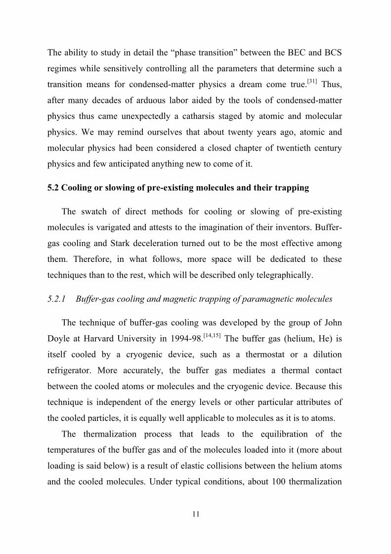
Figure 12 displays a portrait of a larger phase-space area than in Fig. 11.
This phase portrait pertains to the transport of OH molecules and has been
obtained with the help of the analytic model mentioned above, encompassing
80 waves. The portrait agrees in detail with both trajectory simulations of the
Stark acceleration/deceleration dynamics and with experiment. It shows that by
far the largest area of phase stability pertains to the first harmonic wave, with
€
n = =1. Furthermore, we see that there are smaller areas of phase stability
centered around odd-fraction multiples of the fundamental velocity, i.e.,
€
13V0 ,
€
53V0 ,
€
73V0. However, apart from these, there are also small phase-stable areas
centered around even-fraction multiples of
€
V0, i.e.,
€
12V0,
€
32V0,
€
2V0. Where do
such areas come from? The answer to this question follows from the “wave
nature” of the electric field in the Stark decelerator: wherever there are waves,
there must also be interferences among the waves! Indeed, the analytic model
shows that molecules can also ride a well which is formed by the interference

22
between two waves with odd
€
n,
€
a
€
r,
€
s , resulting in a wave whose phase
velocity is an even-fraction multiple of the fundamental velocity
€
V0.
5.2.3.1 Electrostatic trapping of molecules. Sufficiently slow molecules can
be trapped in an electrostatic trap. Figure 13 shows a schematic of a trap
suitable for confining polar molecules in low-field seeking states. The trap
consists of three electrodes, initially energized as indicated in Fig. 13a. The left
field configuration enables the molecules to enter the trap (from the left). The
Stark energy, shown in the lower part of the figure, stops the molecules at a
point somewhere half the way to the top of the potential hill. There the
molecules make an “about-turn” and begin to move to the left. At that instant,
the fields are switched to the configuration shown in Fig. 13b, which produces
an energy barrier for the leftward motion of the molecules. The molecules thus
get stuck between the left and right barriers, i.e., they are trapped, and so
become a coveted subject of further experimentation.
5.2.3.2 A storage ring for molecules and a molecular synchrotron. A
special case of an electrostatic trap is a storage ring, which provides a
transversal but not a longitudinal confinement for low-field seeking molecules.
It is most simply implemented by bending a transversal focuser, such as a
hexapole, onto itself, see Figure 14. The low-field seekers then have a
minimum of potential energy on a circle rather than at a single point. Compared
with a trap, a storage ring is capable of confining molecules without the need of
bringing them to a standstill first. The circling packets of molecules can
repeatedly interact, at well defined times and positions, with electromagnetic
fields and/or other atoms or molecules.
An electrostatic storage ring was first demonstrated in 2004 with a beam
of ammonia molecules decelerated to a velocity of 92 m/s.[39] The density of

23
the ammonia molecules inside the 25 cm diameter hexapole ring was probed
via a laser-ionization-based technique. In the lower part of Fig. 14, the ion
signal is shown as a function of the storage time in the ring. Peaks are observed
whenever the packet of molecules passes through the detection zone. One can
see that each successive lap adds to the width of the packet. This is due to the
finite velocity spread of the molecules. Although this is very small at the
beginning, corresponding to about 300 µK at the moment the molecules are
loaded into the ring from a Stark decelerator, after about 100 laps the packet is
spread out to the extent that it nearly uniformly fills the entire ring.
In order to counteract the spreading of the packet in the ring – and thus to
restore the ability to define the timing and position of the stored molecules – a
storage ring was constructed consisting of two half-rings separated by a gap.[40]
This is shown schematically in the top part of Fig. 15. Depending on the
switching sequence, the gap can act as a time-varying inhomogeneous electric
field (similarly as a stage of a Stark decelerator) that accelerates, decelerates,
or just transports or bunches the molecular packet under the conditions of
phase stability. The split-ring device thus represents a molecular analog of a
synchrotron for charged particles. The lower part of Fig. 15 shows the density
of molecules stored in the synchrotron as a function of storage time. One can
see that the width of the bunched pulse, after an initial decrease during the first
25 laps or so, reaches a fixed value. Bunching ensures a high density of the
stored molecules and, in addition, enables injecting multiple packets – either
co-linear or counter-propagating – into the ring independent of the packet(s)
already stored.
5.2.3.3 AC trapping of molecules in high-field seeking states. Trapping of
molecules in high-field seeking states is of particular interest, chiefly for two
reasons: (i) Ground-state-molecules are always high-field seeking, since the

24
ground state of any system can only be lowered by an external perturbation. As
a ground state cannot decay, molecules trapped via their ground state cannot be
lost from the trap by relaxation processes, which plague trapping via low-field
seeking states in electrostatic or magnetic traps. As a result, ground state
molecules are expected to be amenable to evaporative or sympathetic cooling.
We note that relaxation losses have been predicted to be particularly severe for
polar molecules in excited ro-vibrational states. (ii) Heavier molecules have
small rotational constants (and, often, large dipole moments) and so become
high-field seeking at relatively small field strengths, thus precluding the
application of large-enough forces required to trap or otherwise manipulate
them.
In order to trap high-field seekers, one would wish to create a maximum
of a static field in free space. Unfortunately, this is prohibited by a
consequence of Maxwell’s equations, known as Earnshaw’s theorem.
However, it is possible to generate a static field that has the shape of a saddle
surface, see Figure 16. Such a field focuses molecules in one direction and
defocuses them in the perpendicular one. By reversing the polarity of the
electrodes that generate the field (i.e., AC switching), the focusing and
defocusing directions can be interchanged. The focusing force causes the
molecules to move toward the saddle point at the trap center while the
defocusing force pushes the molecules away from it. Since the focusing and
defocusing forces are equal to one another, one would think that the time-
averaged, net force acting on the molecules would be zero. This, however, is
not the case: the AC switching between the two field configurations causes the
molecules to undergo an oscillatory micromotion whose frequency tends to get
in step with the switching frequency. Since the force acting on the molecules
increases with distance from the saddle point, the amplitude and thus the
kinetic energy of the micromotion increases with distance as well. Because the

25
kinetic energy of the micromotion is positive (as kinetic energy always is), it
creates in effect a potential energy well, with a minimum at the trap center.
The positive kinetic energy of the micromotion is independent of whether the
molecules are in a low- or high-field seeking state, which makes an AC trap
work for either.
Three electrode geometries have been implemented to date, which create
the desired electric trapping field.[41-43] Fig. 17 shows schematically the
electrode geometry of a cylindrical AC trap that has been used to trap both
ND3 molecules and Rb atoms. Panels (b) and (c) show the strength of the
electric field along the symmetry axis (z-axis) and along the radial direction (r)
when the field is focusing either along the z-axis or radially. The ND3
molecules were loaded into the AC trap by first Stark-decelerating low-field
seeking ND3 molecules to a standstill and subsequently applying a microwave
pulse that pumps a fraction (about 20%) of the molecules into a high-field
seeking state. Figure 18 shows the density of the ND3 molecules at the center
of the AC trap as a function of the switching frequency for both the high- and
low-field seeking components of the ground state of para-ammonia.
At low switching frequencies, the trajectories of the molecules in an AC
trap are unstable and no signal is observed. Above a frequency of about 900
Hz, the trap becomes abruptly stable. A maximum signal is observed at about
1100 Hz. When the frequency is increased further, the molecules have less
time to move within a switching period, and the net force acting on the
molecules decreases, as does the trap depth.
It is noteworthy that electrostatic traps (see section 5.2.3.1) are about 1 K
deep (depending on the molecular species and details of the trap design), and
have a volume of typically 1 cm3. An AC electric trap has a depth of about 1-
10 mK and a volume of about 10−2 cm3.

26
5.2.4 The Zeeman and Rydberg decelerator
A magnetic analog of Stark deceleration, based on the Zeeman effect, has
recently been implemented. This has widened the chemical scope of phase-
stable manipulation to include paramagnetic but nonpolar species, such as
atoms or homonuclear molecules.
The Zeeman deceleration technique, pioneered by the group of Frederic
Merkt, at ETH Zurich, has been experimentally demonstrated by slowing down
ground-state H and D atoms, using initially six[44, 45] and later twelve[46] pulsed
magnetic field stages. The deceleration stages consist of copper-wire solenoids
(each about a cm long) which generate magnetic fields of up to 1.5 T. The
cylindrical coils provide a symmetric transverse restoring force that focuses the
decelerated beam onto the longitudinal axis. Achieving the requisite switching
times (of about 5 µs) was a considerable technical challenge. The group of
Mark Raizen, at the University of Texas at Austin, has applied Zeeman
deceleration to metastable Ne atoms in an eighteen-stage decelerator.[47] Using
electromagnetic coils which are encased in magnetic-steel shells with
Permendur discs, even higher magnetic field strengths, of 3.6 T, could be
achieved. Recently, the deceleration of metastable Ne atoms[48] and oxygen
molecules[49] to velocities as low as 50 m/s using sixty four field stages was
reported.
Compared to polar molecules, atoms or molecules in Rydberg states offer a
much larger electric dipole moment. Hence, these particles can be manipulated
using only modest electric field strengths and either a single or just a few field
stages, as demonstrated by Merkt’s group and the group of Tim Softley at the
University of Oxford. Level crossings in the dense Rydberg manifolds limit the
magnitude of the electric field strength that can be applied. The electric field
manipulation of atoms and molecules in high Rydberg states has been

27
pioneered using H2 molecules[50] and Ar atoms[51]. Using a Rydberg
decelerator, H atoms could be stopped and electrostatically trapped in two[52] or
three[53] dimensions. The short lifetimes of Rydberg states (in particular of
molecules) inherently limit the time that is available to manipulate and study
such species. However, fluorescence decay of the Rydberg states may in some
cases generate cold samples of ground-state atoms or molecules.
5.2.5 Slowing by nonresonant light fields
In many respects, this technique resembles Stark or Zeeman deceleration.
One of the differences is that instead of a static electric field, a nonresonant
laser field is used which interacts with the molecules’ polarizability. This
interaction induces an electric dipole moment, which then interacts with the
electric field of the laser radiation. This interaction can be strong enough
(thanks to both the intensity and the gradient of the radiative field) to enable the
deceleration of a pulse of molecules in a single sweep (this is another
difference). So far, two versions of this type of deceleration were considered,
the “scoop”[20] and the “optical lattice.”[21] In the case of a “scoop,” the motion
of a laser focus (and thus of the decelerated molecules) is directly controlled by
a suitable electro-optic device; in the case of an “optical lattice,” implemented
in 2006 by the group of Peter Barker at Edinburgh (now in London), an optical
interference wave is created with a tunable phase velocity which traps a pulse
of molecules in flight and slows it down, see Figure 19.
The deceleration imparted by a Stark decelerator is of the order of 105
m s-2, which suffices to bring thermal molecules to a standstill on a path of ~1
m in about 1–10 ms. The Barker group, however, puts the brakes on for only
about 6 ns (over a path of several micrometres), imparting a deceleration of the
order of 1010 m s-2. Their technique uses not electric field stages, but the

28
gradient of an electromagnetic wave. The resulting deceleration is due to a
force of the order of a femtonewton. Can the molecules withstand such a force
without falling apart in a process that we could dub “breaking by braking”?
They actually can, as a femtonewton force would only dissociate a species
bound by a sub-Kelvin deep well. The large deceleration force is due to the
induced-dipole interaction of the non-resonant radiative field of a laser with the
polarizability of the molecules. This interaction scales with laser intensity and
exceeds the interaction of a permanent dipole with a state-of-the-art
electrostatic field typically at intensities above 1010 W cm-2
(the intensities have
to be held below 1013 W cm-2, however, to avoid ionization). Because the non-
resonant optical interaction is purely attractive, all eigenstates created by it are
high-field seeking. Focused radiation in free space represents a field maximum,
and so produces a potential well (i.e., a trap) for high-field seekers. The optical
force is versatile, robust, and precludes relaxation losses as it puts all states into
a well. In addition, it aligns nonspherical molecules, because it exerts a torque
on the molecular axis. In the state-of-the-art experiment, the non-resonant
radiative field comes in the form of a one-dimensional optical lattice created by
an interference of two nearly counter-propagating laser beams with slightly
different frequencies. The frequency difference of the two beams enables
tuning the velocity of the lattice. The required intensities — of about 1012 W
cm-2 — are attained by employing pulsed lasers. The lattice can be viewed as a
wave whose crests and troughs represent, respectively, the wells and hills of the
potential energy of the molecules (as they are high-field seeking). In the
pioneering experiment, each lattice crest represents a potential well about 22 K
deep. By letting the lattice move at a speed lower than the molecule’s velocity,
the molecule — nitric oxide in this case — is forced to climb a hill. During its
ascent, the molecule loses kinetic energy and is therefore decelerated. About 40
such potential wells, spread over merely 20 µm, see Fig. 14, snatch about 105

29
molecules per pulse, with an estimated density of 1010 molecules per cm3. The
temperature of the molecules remains close to that of the original molecular
beam pulse, around 1 K. The forthcoming stage of this work will involve
trapping of the decelerated molecules, and subsequent cooling to the millikelvin
range. The optical deceleration technique adds more than froth and bubble to
the tidal wave that molecular physics itself is now riding.
5.2.6 Slowing by collisions in molecular beams
Molecules that are slow in the laboratory frame can be also produced by
an inelastic collision or a chemical reaction between fast molecules in
molecular beams, as demonstrated by the groups of David Chandler at Sandia
and Hanjürgen Loesch at Bielefeld. Figure 20 shows the corresponding Newton
diagram, constructed for the Cs+NO2
€
→CsO+NO reaction. If the laboratory
velocity of the center of mass of the colliding system, C, is equal to the recoil
velocity, uCsO, of the the CsO product molecule with respect to the velocity of
the center of mass, i.e., C = -uCsO, the CsO molecule will end up with a zero
laboratory velocity.
So far, the method has been applied to the production of slow NO[22]
molecules in inelastic NO+Ar collisions and to the production of highly polar
KBr[23] molecules in reactive K+Br2 collisions. A time-of-flight spectrum of the
KBr product is shown in Figure 21. The hope is that the slow fraction,
appearing at the time-of-flight times exceeding 4 ms (and corresponding to
velocities lower than 80 m/s), could be loaded into an electrostatic trap and
subsequently evaporatively cooled.
From our example we can see that the technique can also be used for the
preparation of highly reactive slow molecules, such as CsO.

30
5.2.7 Production of slow molecules by a supersonic expansion from a
counter-rotating nozzle
The remaining two techniques that we will mention can be both well
explained using automotive analogies. Figure 22 shows a rotating nozzle which
is supersonically discharging gas in a direction opposite to that of the nozzle’s
tangential motion. If the speed of the nozzle equals that of the molecules, the
supersonic molecular beam ends up having a zero laboratory velocity.[24] This is
similar to what happens with the exhaust gases of a moving car. The gas leaves
the tailpipe with a speed of about 350 km/h. This is the speed of a Formula 1
car. Thus the exhaust gas of such a car speeding at full throttle ends up “at a
standstill” with respect to the racing track! The technique has been successfully
implemented in the group of Dudley Herschbach at Harvard.
5.2.8 Selection of the slow fraction of an effusive molecular beam
An effusive molecular beam has a Maxwell-Boltzmann distribution of
velocities and thus contains a fraction of slow molecules. If the molecules are
endowed with an electric or magnetic dipole moment, it’s possible to select
them out with an inhomogeneous electric[25] or magnetic[26] field, as
demonstrated by the groups of Gerhard Rempe in Munich and of Eric Abraham
at Oklahoma. The field deflects the dipole molecules; for a given field gradient,
the magnitude of the deflection is inversely proportional to the translational
energy of the molecules. The separation of the slow and fast molecules happens
in a curved region of the inhomogeneous field where fast molecules are not
deflected enough, and so “won’t make the curve,” see Figure 23. Only slow-
enough molecules will make it through the curve; such molecules can be then
either trapped or utilized, e.g., in a collision experiment. A drawback of the
technique is that the slow molecules are not internally cooled by the effusive

31
expansion, and so have the temperature of the source — typically 300 K.
6. Ongoing and future experiments with cold molecules
A unique experiment with molecules confined in an elecrostatic trap has
made it possible to measure the radiative lifetime of molecules in a
vibrationally excited state. The measurement was carried out for the OH
radical in its electronic and rotational ground state and showed that the lifetime
of the
€
v'=1→ v"= 0 transition is about 59 ms.[50] This was followed soon after
by another direct measure of radiative lifetime, this time in a magnetic trap.[51]
The measurement was carried out for NH. It showed that the radiative lifetime
for the electronic, rotational ground state for
€
v'=1→ v"= 0 is 37 ms. These two
measurements were the first direct measurements ever of the radiative lifetime
of another than an electronic transition since Albert Einstein introduced the
concept in 1916. The knowledge of the vibrational radiative lifetimes is crucial
for the physics and chemistry of the interstellar space; their measurement thus
far was not possible, however, because in conventional experiments the
molecules either perish or disappear before they have a chance to radiate out a
vibrational quantum.
In another pioneering experiment with slow molecules, an enhancement of
the resolution of a spectroscopic measurement was demonstrated; the
enhancement was due to an extended interaction time between electromagnetic
radiation and the slow molecules. The resolution,
€
ΔE , of a given measurement
is fundamentally limited by the uncertainty principle,
€
ΔEΔt ≈ h , with
€
Δt the
measurement time. By slowing down ND3 molecules (deuterated ammonia)
from the speed of 280 m/s to 52 m/s, it was possible to enhance the resolution
from 10 kHz to 1 kHz, and thus to fully unravel the hyperfine structure of this

32
important model molecule.[52] The frequency of one of ammonia’s vibrational
modes depends sensitively on the mass ratio of the proton to the electron. An
accurate measurement of this ratio can serve as a gauge for determining a
possible change of this ratio (and of related quantities, such as the fine-structure
constant) with the age of the universe. In order to achieve the requisite
accuracy, an extension of the interaction time
€
Δt to about 1 s is needed. This is
expected to become available in the molecular fountain, now under
construction, where slow molecules are thrown vertically upwards, and stopped
and turned back by gravity.
In the introduction we indicated that cold molecules might exhibit a highly
unusual collisional behavior. This behavior was first analyzed by Hans Bethe in
the 1930s and Eugene Wigner in the late 1940s, who had shown that at
temperatures close to absolute zero the collision cross section is proportional to
integer powers of the reciprocal de Broglie wavelength of the colliding
molecules. This implies that the collisional rate constant, which is given by the
product of the collision cross section and collision velocity, is nonzero even in
the limit of
€
T → 0. Detailed calculations on A+A2(v=1)
€
→A+A2(v=0) (A=Na,
Li) systems or on the F+H2
€
→FH+H chemical reaction have corroborated the
validity of Wigner’s law.[53,54] For instance, the rate of the F+H2
€
→FH+H
reaction at a temperature T = 10 mK was found to be 1.25
€
×10-12 cm3/s. This
result may strike a classical chemist as strange indeed, since the activation
barrier of this intensely studied reaction is as much as 758 K (i.e., 6.3 kJ/mol)!
How can the reaction take place at all when the total energy that the reactants
can chip in is just 10 mK? The answer to this question must be sought in the
wave character of cold molecules: if the molecules are cold enough to make
their de Broglie wavelength exceed the thickness of the activation barrier, the
reaction can take place via tunneling through the barrier, without regard for the
barrier’s “height.” Ultracold chemistry is thus a purely quantum phenomenon,

33
and tunneling represents the main reaction mechanism. Reactions at ultracold
temperatures thus resemble mingling waves rather than a crash of billiard balls.
So far, reaction kinetics and chemical dynamics were able to do with a classical
description of the approaching and receding nuclei gliding on a potential energy
hypersurface, given by the quantum energy of the electrons. Ultracold
chemistry calls for a quantum description of not just the electrons but of the
nuclei as well. Quantum chemists will thus have to come up with a qualitatively
new description of chemical change. Collision experiments with cold molecules
– which will require such a new understanding for their interpretation – are
under way in about half a dozen laboratories.
Cold molecules also feature in somewhat futuristic, but nevertheless
seriously taken, schemes that could allow the implementation of a quantum
computer.[55] Already atomic systems such as charged ions and neutral atoms
have been shown to be attractive candidates for physical realization of
quantum computation in view of their exceptionally long coherence times and
well-developed techniques for cooling and trapping. The main conceptual
problems are associated with designing fast and robust multi-atom operations
for quantum entanglement as well as with scaling these systems to large
numbers of qubits. Ultracold polar molecules are a potentially superior
candidate for quantum bits in a scalable quantum computer. Polar molecules
combine the key advantages of neutral atoms and ions while featuring similarly
long coherence times. In particular, large ensembles of cold molecules can be
trapped and cooled similar to neutral atoms, but they can then be manipulated
individually using electric fields in analogy with ions.
The first proposed complete scheme for quantum computing with polar
molecules took advantage of first feature, but not the second. It was based on
an ensemble of ultracold polar molecules trapped in a one-dimensional optical
lattice (similar to the one used to decelerate molecules, Section 5.2.4, but in a

34
stationary version), combined with an inhomogeneous electric field. The
requisite entanglement is achieved via the interaction among the dipoles, each
of which represents a qubit. Such qubits are individually addressable, thanks to
the Stark effect, which is different for each in the inhomogeneous electric field.
A subsequent proposal showed that it should be possible to couple polar
molecules into a quantum circuit using superconducting wires.[56] The
capacitive, electrodynamic coupling to transmission line resonators was
proposed in analogy with coupling to Rydberg atoms and Cooper pair
boxes.[57,58] The key feature of molecules is their RF frequency rotational
transitions, nicely compatible with microwave circuitry. Coupling individual
molecules to microwave strip-lines is advantageous for several reasons. First, it
allows for detection of single molecules by remote sensing of transmission line
potentials, as well as for efficient quantum state readout. Second, the
molecules can be further cooled using a novel method involving microwave
spontaneous emission into on-chip transmission lines. Finally, remotely
separated molecules can be coherently coupled to allow for nonlocal
operations. Local gating with electrostatic fields can be used to achieve
exceptional degree of addressability. Single-bit manipulations can be
accomplished by using local modulated electric fields.
Although quantum computers have been anticipated decades ago (for
instance, by Richard Feynman), their benefits for solving numerical problems
have been discovered only recently, in 1994, by Peter Shore. He showed that
quantum computers are particularly well suited for factorization, a step in
methods of encryption. It is estimated that a quantum computer consisting of
500 qubits would be capable of factorizing a number of the order of 10200
within seconds (for comparison, a classical computer would need about 10150
processors to achieve a similar feat).
Apart from the above, there is a class of experiments in prospect or

35
progress with cold molecules that could answer questions reaching far beyond
the scope of traditional molecular science: these experiments test some of the
fundamental symmetries in physics, such as the time-reversal symmetry (T),
parity (P), or the Pauli principle. These symmetries are a window into the world
of the fundamental forces in nature and thus molecular, table-top experiments
that test them represent an alternative to the high-energy collisional
experiments carried out in giant accelerators.
Particularly promising and interesting is the simultaneous testing of the
time-reversal symmetry and parity in experiments that search for the permanent
electric dipole (EDM) of the electron (and of other elementary particles).[59] A
nonzero value of EDM implies the breaking of both T and P. Since the
Standard Model doe not allow for essentially any violation of the T symmetry,
finding a nonzero EDM would amount to the discovery of physics beyond the
Standard Model. Such a discovery would be followed in all likelihood by a new
revolution in physics.
What does EDM have to do with cold molecules? From the experiments
carried out so far it is known that the EDM of the electron doesn’t exceed the
value of about 5
€
×10-19 Debye. This is an exceedingly small value, one that
would be found if an electron were expanded to the size of the earth and
deformed on the poles by a micron! A dipole moment manifests itself through
its Stark effect, i.e., by a shift of the energy levels of a system carrying the
dipole moment when the system is subject to an electric field. In order for the
Stark shift to be detectable, the EDM has to be exposed to as large a field as
possible. The strongest fields that one can come by are available inside heavy
atoms (where they can reach values of 10 GV/cm, thanks to relativistic effects).
Atoms are spherically symmetric, however, and so before any EDM
measurement they need to be oriented in the laboratory frame (otherwise the
dipole moment would average out). But orienting atoms is difficult, because in

36
doing so one has to rely on their polarizability, which is small. Therefore, it is
of great advantage to add to a given heavy atom another atom, and to carry out
the Stark effect measurement on the polar molecule which thereby ensues!
Polar molecules can namely be easily oriented. In fact, a Stark decelerator or an
electric trap are based on the ability to easily orient polar molecules in the
laboratory frame. The use of cold polar molecules further enhances the
resolution of the measurement (see the beginning of this section). The
experiment of Ed Hinds & Co. at Imperial College, London, with decelerated
YbF molecules already comes close to yielding the most accurate value of the
electron’s EDM.[60] An increase of the experiment’s accuracy by a single order
of magnitude could lead to a rejection or adoption of some of the alternatives of
the Standard Model. We are living in thrilling times!
In conclusion, we must say that although many hands are busy with the
ongoing work outlined in this article, a host of adventures with cold molecules
still await – beyond the horizon we can survey today.
Acknowledgments
JD gratefully acknowledges the Alexander von Humboldt Foundation for
supporting his collaboration with and a stay at the Fritz Haber Institute, made
possible through a Humboldt Research Award. We thank Gerard Meijer for
discussions, comments, and support.

37
References
1. W. Ketterle, Chem. Phys. Chem. 2002, 3, 736-753.
2. Eur. Phys. J. D 2004, 32, pp. 145- 446 – topical issue on cold molecules.
3. D. Herschbach, Rev. Mod. Phys. 1999, 71, S411.
4. N.F. Ramsey, Rev. Mod. Phys. 1990, 62, 541; W. Paul, Rev. Mod. Phys.
1990, 62, 531; T.W. Hänsch, Chem. Phys. Chem. 2006, 7, 1170-1187; J.
Hall, Rev. Mod. Phys. 2006, 78, 1279.
5. R.K. Pathria, Statistical mechanics, Pergamon Press, Oxford, 1988.
6. D.A. McQuarrie, J.D. Simon, Physical Chemistry, University Science
Books, Sausalito, California, 1997.
7. N. Balakrishnan, V. Kharchenko, R.C. Forrey, A. Dalgarno, Chem. Phys.
Lett. 1997, 280, 5.
8. J.M. Doyle, B. Friedrich, R. Krems, F. Masnou-Seeuws, Eur. Phys. J. D
2004, 31, 149.
9. D. DeMille, Phys. Rev. Lett. 2002, 88, 067901.
10. L. Santos, G.V. Shlyapnikov, P. Zoller, M. Lewenstein, Phys. Rev. Lett.
2000, 85, 1791.
11. S. Chu, Rev. Mod. Phys. 1998, 70, 685; W.D. Phillips, Rev. Mod. Phys.
1998, 70, 721.
12. M.D. DiRosa, Eur. Phys. J. D 2004, 31, 395.
13. J.M. Doyle, B. Friedrich, Nature (London) 1999, 401, 749.
14. J.D. Weinstein, R. DeCarvalho, T. Guillet, B. Friedrich, J.M. Doyle,
Nature (London) 1998, 395, 148.
15. R. deCarvalho, J.M. Doyle, B. Friedrich, T. Guillet, J. Kim, D. Patterson,
J.D. Weinstein, Eur. Phys. J. D 1999, 7, 289.
16. F. Masnou-Seeuws, P. Pillet, Adv. At. Mol. Opt. Phys. 2001, 47, 53.

38
17. D. Kleppner, Physics Today 2004, 57, 12.
18. H.L. Bethlem, G. Meijer, Int. Revs. Phys. Chem. 2003, 22, 73.
19. S.Y.T. van de Meerakker, N.Vanhaecke, G. Meijer, Annual Review of
Physical Chemistry 2006, 57, 159-190.
20. B. Friedrich, Phys. Rev. A 2000, 61, 025403.
21. R. Fulton, A.I. Bishop, M.N. Shneider, P.F. Barker, Nature Physics 2006, 2,
465.
22. M.S. Elioff, J.J. Valentini, D.W. Chandler, Science 2003, 302, 1940.
23. N.N. Liu, H. Loesch, Phys. Rev. Lett. 2007, 98, 103002.
24. M. Gupta, D. Herschbach, J. Phys. Chem. A 1999, 103, 10670; 2001, 105,
1626.
25. S.A. Rangwala, T. Junglen, T. Rieger, P.W.H. Pinkse, G. Rempe, Phys.
Rev. A 2003, 67, 043406.
26. E. Nikitin, E. Dashevskaya, J. Alnis, M. Auzinsh, E. R. I. Abraham, B. R.
Furneaux, M. Keil, C. McRaven, N. Shafer-Ray, R. Waskowsky, Phys.
Rev. A 2003, 68, 023403.
27. K. Bergmann, H. Theuer, B. W. Shore, Rev. Mod. Phys. 1998, 70, 1003.
28. A.J. Kerman, J.M. Sage, S. Sainis, T. Bergeman, D. DeMille, Phys. Rev.
Lett. 2004, 92, 153001.
29. D. Wang, J. Qi, M.F. Stone, O. Nikolayeva, H. Wang, B. Hattaway, S.D.
Gensemer, P.L. Gould, E.E. Eyler, W.C. Stwalley, Phys. Rev. Lett. 2004,
93, 243005.
30. S. Jochim, M. Bartenstein, A. Altmeyer, G. Hendl, S. Riedl, C. Chin, J.
Hecker Denschlag, R. Grimm, Science 2003, 302, 2101.
31. R. Grimm, Nature (London) 2005, 435, 1035.
32. B. Friedrich, J.D. Weinstein, R. deCarvalho, J.M. Doyle, J. Chem. Phys.
1999, 110, 2376.
33. D. Egorov, W.C. Campbell, B. Friedrich, S.E. Maxwell, E. Tsikata, L.D.

39
van Buuren, J.M. Doyle, Eur. Phys. J. D 2004, 31, 307–311.
34. S. E. Maxwell, N. Brahms, R. deCarvalho, D. R. Glenn, J. S. Helton, S. V.
Nguyen, D. Patterson, J. Petricka, D. DeMille, J. M. Doyle, Phys. Rev.
Lett. 2005, 95, 173201.
35. D. Patterson, J.M. Doyle, J. Chem. Phys. 2007, 126, 154307.
36. S. Y. T. van de Meerakker, N. Vanhaecke, G. Meijer, Ann. Rev. Phys.
Chem. 2006, 57, 159.
37. S. Y. T. van de Meerakker, N. Vanhaecke, H. L. Bethlem, G. Meijer, Phys.
Rev. A 2006, 73, 023401.
38. K. Gubbels, G. Meijer, B. Friedrich, Phys. Rev. A 2006, 73, 063406.
39. F. M. H. Crompvoets, H. L. Bethlem, G. Meijer, Advances in Atomic,
Molecular, and Optical Physics 2006, 52, 209–287.
40. C.E. Heiner, D. Carty, G. Meijer, H.L. Bethlem, Nature Physics 2007, 3,
115.
41. H. L. Bethlem, J. van Veldhoven, M. Schnell, G. Meijer, Phys. Rev. A
2006, 74, 063403.
42. van Veldhoven, H. L. Bethlem, and G. Meijer, Phys. Rev. Lett. 2005, 94,
083001.
43. M. Schnell, P. Lützow, J. van Veldhoven, H.L. Bethlem, J. Küpper, B.
Friedrich, M. Schleier-Smith, H. Haak, G. Meijer, J. Phys. Chem. A 2007,
111, 7411.
44. N. Vanhaecke, U. Meier, M. Andrist, B. H. Meier, F. Merkt, Phys. Rev. A
2007, 75, 031402.
45. S. D. Hogan, D. Sprecher, M. Andrist, N. Vanhaecke, F. Merkt, Phys. Rev.
A 2007, 76, 023412.
46. S. D. Hogan, A. W. Wiederkehr, M. Andrist, H. Schmutz, F. Merkt, J.
Phys. B: At. Mol. Opt. Phys. 2008, 41, 081005.
47. E. Narevicius, C. G. Parthey, A. Libson, J. Narevicius, I. Chavez, U. Even,

40
M. G. Raizen, New J. of Physics 2007, 9, 358.
48. E. Narevicius, A. Libson, C. G. Parthey, I. Chavez, J. Narevicius, U. Even,
M. G. Raizen, Phys. Rev. Lett. 2008, 100, 093003.
49. E. Narevicius, A. Libson, C. G. Parthey, I. Chavez, J. Narevicius, U. Even,
M. G. Raizen, Phys. Rev. A 2008, 77, 051401.
50. S.Y.T. van de Meerakker, N. Vanhaecke, M. P. J. van de Loo, G.C.
Groenenboom, G. Meijer, Phys. Rev. Lett. 2005, 95, 013003.
51. W.C. Campbell, G.C. Groenenboom, Hsin-I Lu, E. Tsikata, J.M. Doyle,
Phys. Rev. Lett. 2008, 100, 083003.
52. J. van Veldhoven, J. Küpper, H.L. Bethlem, B. Sartakov, A.J.A. van Roij,
G. Meijer, Eur. Phys. J. D 2004, 31, 337.
53. N. Balakrishnan, A. Dalgarno, Chem. Phys. Lett. 2001, 341, 652.
54. P. Soldán, M.T. Cvitas, J.M. Hutson, P. Honvault, J.-M. Launay, Phys. Rev.
Lett. 2002, 89, 153201.
55. N.D. Mermin, Physics Today 2007, 60, 8-9.
56. A. Wallraff, D.I. Schuster, A. Blais, L. Frunzio, J. Majer, R.-S. Huang, S.
Kumar, S.M. Girvin, R.J. Schoelkopf, Nature (London) 2004, 431, 162.
57. A. Sorensen, C. H. van der Wal, L.I. Childress, M. D. Lukin, Phys. Rev.
Lett. 2004, 92, 063601.
58. A. Andre, D. DeMille, J. M. Doyle, M. D. Lukin, S. E. Maxwell, P. Rabl, R.
J. Schoelkopf, P. Zoller, Nature Physics 2006, 2, 636-642.
59. N. Fortson, P. Sandars, S. Barr, Physics Today 2003, 56, 33.
60. J.J. Hudson, B.E. Sauer, M.R. Tarbutt, E.A. Hinds, Phys. Rev. Lett. 2002,
89, 023003; B. E. Sauer, H. T. Ashworth, J. J. Hudson, M. R. Tarbutt, E. A.
Hinds, in Atomic Physics 20, edited by Christian Roos, Hartmut Haeffner,
and Rainer Blatt, AIP Conf. Proc. No. 869, AIP, Melville, New York, p. 44,
2006.

41
Figure Captions
Figure 1: Nomogram for the de Broglie wavelength
€
Λ of a particle with rest
mass m at absolute temperature T. Arrows on the right show the masses of
iodine and nitrogen molecules, of the hydrogen atom, and of the muon and the
electron.
Figure 2: Photoassociation of cold atoms into a translationally cold molecule.
(a) schematic of a photo-assisted collision; (b) potential energy curves for the
ground and excited electronic states; RC marks the internuclear distance at
which the photoassociation laser promotes the colliding system into a bound
state which is electronically, vibrationally and rotationally excited; (c) laser-
stimulated emission into the ground electronic, vibrational and rotational state.
Figure 3: Energy-level structure allowing for a Feshbach resonance between a
bound excited electronic state (shown by the dashed line) and an electronic
ground state (given by the asymptotic energy of the free – and slow – atoms;
shown by the thin horizontal line). See text.
Figure 4: Apparatus for buffer-gas cooling and magnetic trapping of
paramagnetic atoms and molecules. The two superconducting coils, energized
by counter-propagating electric currents (anti-Helmholtz configuration), create
a nearly spherically symmetric magnetic field whose strength increases linearly
with distance from a zero value at the center to a maximum (of about 3 Tesla)
at the edge. The cryogenic cell, fashioned out of copper, is placed in the bore of
the magnet. It is thermally anchored to the mixing chamber of a dilution
refrigerator (not shown), which cools it to about 100-300 mK. The cryogenic
cell is filled with the buffer gas whose density is about 1016 He atoms per cm3.

42
The atoms or molecules to be cooled and trapped are loaded into the cryogenic
environment by laser ablating a suitable precursor, placed directly in the
cryocell. Taken from ref.[14]
Figure 5: Magnetic trapping. The thermalized molecules carry a translational
energy which is less than their magnetic (Zeeman) potential energy (in low-
field seeking states).
Figure 6: Fluorescence spectra of molecules in the anti-Helmholtz field at
different times after firing the ablation laser. Whereas high-field seeking
molecules are expelled from the field (left-hand maximum), low-field seekers
are drawn to the field minimum (right-hand maximum). The analysis of the
spectra showed that about 108 CaH molecules were trapped. The trapped
molecular ensemble was found to exhibit a Maxwell-Boltzmann distribution of
translational energies, pertaining to a temperature of about 400 mK. Taken
from ref.[14]
Figure 7: (a) Stark deceleration. The energy of a molecule consists of its
translational energy and its potential energy. See text. (b) Spatial dependence of
the electric field
€
ε(z) generated by four field stages, each of which consists of a
pair of rod electrodes. In the upper (red) configuration, the electrodes of the
first and third pair are energized by a high voltage of opposite sign (typically
±20 kV), whereas the electrodes of the second and fourth pair are grounded. In
the lower (blue) configuration, the electrodes of the first and third pair are
grounded, whereas the second and fourth field stages are energized. The
resultant electric field in the longitudinal direction, z, (along the direction of a
molecule flying from left to right) reaches values of about 100 kV/cm at the
maxima. This corresponds to the potential (Stark) energy of a typical polar
diatomic of about 1 K. (c) Time dependence of the electric field
€
ε(z,t)

43
determined by the switching between the upper (red) and lower (blue) field
configuration. Also shown are the spatial and temporal periods, λ and τ.
Adapted from refs.[18],[38]
Figure 8: Velocity distribution modified by a Stark accelerator/decelerator.
Lower curve shows the velocity (time-of-flight) distribution of molecules in a
supersonic molecular beam. Upper curves show the effect of a Stark
accelerator/decelerator: this selects out a velocity group and moves it to either
lower velocities (longer times of flight) or higher velocities (shorter times of
flight). The measurement was carried out with CO molecules in a metastable
electronic state (lifetime of about 2.6 ms). The grey (red) curves were obtained
by Monte Carlo simulation of molecular trajectories in the Stark
accelerator/decelerator field. Taken from ref.[18]
Figure 9: Stark accelerator/decelerator, consisting of a 108 pairs of rod
electrodes. The electrodes of a given pair are separated by 4 mm, the pairs by
11 mm from one another. The high voltage used is ±20 kV. The transient time
needed to switch from one configuration to another is about 500 ns. Only a few
ms are required to decelerate a molecule from a thermal velocity to a standstill.
The inset shows the electrodes from the perspective of the
accelerated/decelerated molecules. Taken from ref. [36]
Figure 10: Biased pendulum and its potential energy. (a) A biased pendulum
consists of a normal pendulum of length r and mass m which is fixed to an axle
of radius R; wound around the axle is a string that carries a weight (bias) of
mass M. (b) Potential energy V of a biased pendulum as a function of position
(more accurately, phase)
€
φ , consists of the potential energy of the pendulum,
€
Vm , and the potential energy of the bias,
€
VM :
€
V =Vm +VM (g is the
gravitational acceleration). Potential V has a local minimum at
€
φ = φs and a
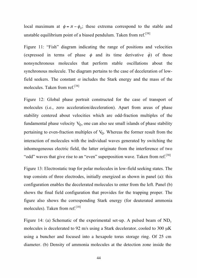
44
local maximum at
€
φ = π − φs; these extrema correspond to the stable and
unstable equilibrium point of a biased pendulum. Taken from ref.[38]
Figure 11: “Fish” diagram indicating the range of positions and velocities
(expressed in terms of phase
€
φ and its time derivative
€
˙ φ ) of those
nonsynchronous molecules that perform stable oscillations about the
synchronous molecule. The diagram pertains to the case of deceleration of low-
field seekers. The constant α includes the Stark energy and the mass of the
molecules. Taken from ref.[38]
Figure 12: Global phase portrait constructed for the case of transport of
molecules (i.e., zero acceleration/deceleration). Apart from areas of phase
stability centered about velocities which are odd-fraction multiples of the
fundamental phase velocity
€
V0, one can also see small islands of phase stability
pertaining to even-fraction multiples of
€
V0. Whereas the former result from the
interaction of molecules with the individual waves generated by switching the
inhomogeneous electric field, the latter originate from the interference of two
“odd” waves that give rise to an “even” superposition wave. Taken from ref.[38]
Figure 13: Electrostatic trap for polar molecules in low-field seeking states. The
trap consists of three electrodes, initially energized as shown in panel (a): this
configuration enables the decelerated molecules to enter from the left. Panel (b)
shows the final field configuration that provides for the trapping proper. The
figure also shows the corresponding Stark energy (for deuterated ammonia
molecules). Taken from ref.[18]
Figure 14: (a) Schematic of the experimental set-up. A pulsed beam of ND3
molecules is decelerated to 92 m/s using a Stark decelerator, cooled to 300 µK
using a buncher and focused into a hexapole torus storage ring. Of 25 cm
diameter. (b) Density of ammonia molecules at the detection zone inside the

45
ring as a function of storage time up to the 33rd lap. Due to the spreading of the
packet, the peak density decreases as 1/t (shown as the dashed line). In the
inset, a measurement of the ammonia density after 49-51 laps is shown,
together with a multi-peak Gaussian fit. Reproduced from ref.[39]
Figure 15: (a) Scheme of the molecular synchrotron. The synchrotron consists
of two hexapole half-rings with a 12.5 cm radius separated by a 2 mm gap. (b)
Density of ammonia molecules at the detection zone inside the synchrotron as
a function of storage time up to the 40th lap. Expanded views of two TOF
profiles are displayed as insets, showing the absolute widths of the molecular
pulses. Reproduced from ref.[40]
Figure 16: Spatial dependence of the electric field strength, |ε|, in an AC trap
for two field configurations generated by energizing suitably shaped electrodes.
Figure 17: (a) Schematic of the cylindrically symmetric AC trap. The electric
field strength along the symmetry axis (z-axis) is shown in (b), and along the
radial direction in (c). When voltages of 5, 7.5, -7.5 and -5 kV are applied to
the electrodes, molecules in high-field seeking states are focused towards the
trap center along the z-axis and defocused in the radial direction. When
voltages of 11, 1.6, -1.6, and -11 kV are applied to the electrodes, molecules in
high-field seeking states are focused radially and defocused along the z-axis.
Reproduced from ref.[41]
Figure 18: Density of 15ND3 molecules in low-field seeking and high-field
seeking levels of the |J,K>=|1,1> state at the center of the trap as a function of
the switching frequency, Ω. The measurements are performed 80 ms after the
molecules are loaded into the trap. The signal of the high-field seekers is
scaled up by a factor of 5 to compensate for the fact that the initial density of
high-field seekers is only about 20% of that of the low-field seekers.

46
Reproduced from ref.[41]
Figure 19: Deceleration of a pulse of supersonically expanded NO molecules
by a pulsed optical lattice. The ultraviolet probe beam that was used to ionize
and detect the NO molecules perturbed by the lattice is also shown. The
velocity of the ions, and thus that of the neutral molecules, was determined
from their time-of-flight in a mass spectrometer. See text. Taken from ref.[21]
Figure 20: Production of slow molecules by a collision of fast molecules in
molecular beams. Shown is a Newton diagram for counter-propagating beams
of Cs and NO2 reactants with laboratory velocities vCs and vNO2. The CsO
product recoils with a velocity UCsO (whose magnitude is shown by the Newton
circle) with respect to the center of mass of the colliding system, which, in turn,
moves with velocity C with respect to the laboratory frame. The reactant
velocity is adjusted such that the recoil velocity of the CsO product equal the
laboratory velocity of the center of mass, UCsO = −C. The dynamics of the
reaction (as shown by the velocity contours) enables a large fraction of the CsO
molecules formed in the Cs + NO2 collisions to emerge with recoil velocities
that fulfill this condition, thus yielding a bounty of molecules that are
predominantly at standstill. See text.
Figure 21: A time-of-flight spectrum of KBr product generated by collisions of
K + Br2 in counter-propagating beams. Taken from ref.[23]
Figure 22: Production of slow molecules by supersonic expansion from a
counter-rotating nozzle. See text. Taken from ref.[24]
Figure 23: Skimming of slow molecules from an effusive molecular beam by a
static inhomogeneous field. See text. Taken from ref.[25]

10-410-310-210-1
101102103104
10-6 10-4 10-2 102 104
m [g
/mol
]
T [K]
λ = 3000
1000 300
100 3010 3
1 Å
H
N2
I2
e-
µ1
1
Figure 1

Pote
ntia
l ene
rgy
Internuclear separation, R
Ground electronic state
Electronically excited state
RC
(b)Photoassociation laser
Figure 2
Colliding cold atoms
Electronically and vibrationallyexcited molecule
Tunable laser (a)
Pote
ntia
l ene
rgy
Internuclear separation, R
Ground electronic state
Electronically excited state
RC
(c)Photoassociation laser
Stim
ulat
ing
lase
r

Figure 3
Pote
ntia
l ene
rgy
Internuclear separation, R
Ground state
Bound state Excited state

preStuff
CaH, VO, CaF, NH, Cr, Eu, Mo, Ti, Sc, Y, Zr, . .CaH,
Ablation target
Fig. 4

Figure 5
Cell wall
Potential(magnetic eigenenergy)
r
µ!
•
•
•
••• •
•••
••
••
•
••
•••
•
•• •
•
•
•
•
•
••
•
Flux of molecules
3He or 4He

Absorption frequency shift [GHz]
Fluo
resc
ence
sig
nal [
arbi
trar
y un
its]
Figure 6

Total energy Kinetic energy
Potential energy
+
− (a)
(c)
Figure 7
(b)

Figure 8

Figure 9

Figure 10
!s"#!s
+z +z
0o
180o
Stable equilibrium Unstable equilibrium
r
R
mM
-4
-2
0
2
4
V(!) /mgr
VM VmV
!ssin!s
!s !out="#!s!in
#360o #180o 0o 360o180o
!
Stable equilibrium
Unstable equilibrium
φ φ
(a) (b)

Figure 11
-1
0
1
-180o -120o -60o 0o 60o 120o 180o
!
= 0o
20o10o 40o 60o 80o
!s
LFS Deceleration
(a)
"
˙ !
2#( )1/2

Figure 12 Initial spatial phase
Initi
al v
eloc
ity [m
/s]

Figure 13
Star
k e
nerg
y
Star
k e
nerg
y

Figure 14

Figure 15

Figure 16
|ε||ε|

Figure 17

Figure 18Ω/(2π) [Hz]

Figure 19

Figure 20
UNO2
UCs
UCsO
C
vNO2
vCs
UNO2 UCsUCsO
vNO2 vCsC
(a) (b)

Figure 21
Slow molecules
Fast molecules
Slow molecules

Figure 22
Rotor
Gas inlet
Nozzle Molecular beam
Velocity of molecules with respect to nozzle Velocity of nozzle
Laboratory velocity of molecules

Figure 23
Effusive beam source
Velocity selector
Fast molecules
Slow molecules