Bioactive Molecules from Marine Microorganisms
172
Bioactive Molecules from Marine Microorganisms Printed Edition of the Special Issue Published in Marine Drugs www.mdpi.com/journal/marinedrugs Hanna Mazur-Marzec and Anna Toruńska-Sitarz Edited by Pobrano z https://repozytorium.bg.ug.edu.pl / Downloaded from Repository of University of Gdańsk 2022-06-01
-
Upload
khangminh22 -
Category
Documents
-
view
2 -
download
0
Transcript of Bioactive Molecules from Marine Microorganisms

Bioactive Molecules from
Marine M
icroorganisms • H
anna Mazur-M
arzec and Anna Toruńska-Sitarz
Bioactive Molecules from Marine Microorganisms
Printed Edition of the Special Issue Published in Marine Drugs
www.mdpi.com/journal/marinedrugs
Hanna Mazur-Marzec and Anna Toruńska-SitarzEdited by
Pobr
ano
z ht
tps:
//rep
ozyt
oriu
m.b
g.ug
.edu
.pl /
Dow
nloa
ded
from
Rep
osito
ry o
f Uni
vers
ity o
f Gda
ńsk
2022
-06-
01

Bioactive Molecules from Marine Microorganisms
Pobr
ano
z ht
tps:
//rep
ozyt
oriu
m.b
g.ug
.edu
.pl /
Dow
nloa
ded
from
Rep
osito
ry o
f Uni
vers
ity o
f Gda
ńsk
2022
-06-
01

Pobr
ano
z ht
tps:
//rep
ozyt
oriu
m.b
g.ug
.edu
.pl /
Dow
nloa
ded
from
Rep
osito
ry o
f Uni
vers
ity o
f Gda
ńsk
2022
-06-
01

Bioactive Molecules from Marine Microorganisms
Editors
Hanna Mazur-Marzec
Anna Torunska-Sitarz
MDPI • Basel • Beijing • Wuhan • Barcelona • Belgrade • Manchester • Tokyo • Cluj • Tianjin
Pobr
ano
z ht
tps:
//rep
ozyt
oriu
m.b
g.ug
.edu
.pl /
Dow
nloa
ded
from
Rep
osito
ry o
f Uni
vers
ity o
f Gda
ńsk
2022
-06-
01
Editors
Hanna Mazur-Marzec
University of Gdansk
Poland
Anna Torunska-Sitarz
University of Gdansk
Poland
Editorial Office
MDPI
St. Alban-Anlage 66
4052 Basel, Switzerland
This is a reprint of articles from the Special Issue published online in the open access journal
Marine Drugs (ISSN 1660-3397) (available at: https://www.mdpi.com/journal/marinedrugs/
special issues/Bioactive Molecules Marine Microorganisms).
For citation purposes, cite each article independently as indicated on the article page online and as
indicated below:
LastName, A.A.; LastName, B.B.; LastName, C.C. Article Title. Journal Name Year, Volume Number,
Page Range.
ISBN 978-3-0365-0620-3 (Hbk)
ISBN 978-3-0365-0621-0 (PDF)
© 2021 by the authors. Articles in this book are Open Access and distributed under the Creative
Commons Attribution (CC BY) license, which allows users to download, copy and build upon
published articles, as long as the author and publisher are properly credited, which ensures maximum
dissemination and a wider impact of our publications.
The book as a whole is distributed by MDPI under the terms and conditions of the Creative Commons
license CC BY-NC-ND.
Pobr
ano
z ht
tps:
//rep
ozyt
oriu
m.b
g.ug
.edu
.pl /
Dow
nloa
ded
from
Rep
osito
ry o
f Uni
vers
ity o
f Gda
ńsk
2022
-06-
01

Contents
About the Editors . . . . . . . . . . . . . . . . . . . . . . . . . . . . . . . . . . . . . . . . . . . . . . vii
Preface to ”Bioactive Molecules from Marine Microorganisms” . . . . . . . . . . . . . . . . . . ix
Samuel Cavalcante do Amaral, Patrick Romano Monteiro, Joaquim da Silva Pinto Neto, Gustavo Marques Serra, Evonnildo Costa Goncalves, Luciana Pereira Xavier and Agenor Valadares Santos
Current Knowledge on Microviridin from CyanobacteriaReprinted from: Mar. Drugs 2021, 19, 17, doi:10.3390/md19010017 . . . . . . . . . . . . . . . . . . 1
Anna Fidor, Michał Grabski, Jan Gawor, Robert Gromadka, Grzegorz Wegrzyn and Hanna
Mazur-Marzec
Nostoc edaphicum CCNP1411 from the Baltic Sea—A New Producer of NostocyclopeptidesReprinted from: Mar. Drugs 2020, 18, 442, doi:10.3390/md18090442 . . . . . . . . . . . . . . . . . 31
Marta Cegłowska, Karolia Szubert, Ewa Wieczerzak, Alicja Kosakowska and Hanna Mazur-Marzec
Eighteen New Aeruginosamide Variants Produced by the Baltic Cyanobacterium Limnoraphis CCNP1324Reprinted from: Mar. Drugs 2020, 18, 446, doi:10.3390/md18090446 . . . . . . . . . . . . . . . . . 49
Jing-Shuai Wu, Xiao-Hui Shi, Guang-Shan Yao, Chang-Lun Shao, Xiu-Mei Fu, Xiu-Li Zhang,
Hua-Shi Guan and Chang-Yun Wang
New Thiodiketopiperazine and 3,4-Dihydroisocoumarin Derivatives from the Marine-DerivedFungus Aspergillus terreusReprinted from: Mar. Drugs 2020, 18, 132, doi:10.3390/md18030132 . . . . . . . . . . . . . . . . . 63
Lu-Ping Chi, Xiao-Ming Li, Li Li, Xin Li and Bin-Gui Wang
Cytotoxic Thiodiketopiperazine Derivatives from the Deep Sea-Derived Fungus Epicoccumnigrum SD-388Reprinted from: Mar. Drugs 2020, 18, 160, doi:10.3390/md18030160 . . . . . . . . . . . . . . . . . 73
Chiara Lauritano, Kirsti Helland, Gennaro Riccio, Jeanette H. Andersen, Adrianna Ianora
and Espen H. Hansen
Lysophosphatidylcholines and Chlorophyll-Derived Molecules from the Diatom Cylindrothecaclosterium with Anti-Inflammatory ActivityReprinted from: Mar. Drugs 2020, 18, 166, doi:10.3390/md18030166 . . . . . . . . . . . . . . . . . 85
Dongbo Xu, Erli Tian, Fandong Kong and Kui Hong
Bioactive Molecules from Mangrove Streptomyces qinglanensis 172205Reprinted from: Mar. Drugs 2020, 18, 255, doi:10.3390/md18050255 . . . . . . . . . . . . . . . . . 97
Yi Ding, Xiaojing Zhu, Liling Hao, Mengyao Zhao, Qiang Hua and Faliang An
Bioactive Indolyl Diketopiperazines from the Marine Derived Endophytic Aspergillus versicolor DY180635Reprinted from: Mar. Drugs 2020, 18, 338, doi:10.3390/md18070338 . . . . . . . . . . . . . . . . . 109
Ji-Yeon Hwang, Sung Chul Park, Woong Sub Byun, Dong-Chan Oh, Sang Kook Lee, Ki-Bong Oh and Jongheon Shin
Bioactive Bianthraquinones and Meroterpenoids from a Marine-Derived Stemphylium sp. FungusReprinted from: Mar. Drugs 2020, 18, 436, doi:10.3390/md18090436 . . . . . . . . . . . . . . . . . 121
vPobr
ano
z ht
tps:
//rep
ozyt
oriu
m.b
g.ug
.edu
.pl /
Dow
nloa
ded
from
Rep
osito
ry o
f Uni
vers
ity o
f Gda
ńsk
2022
-06-
01

Yi-Cheng Chu, Chun-Hao Chang, Hsiang-Ruei Liao, Ming-Jen Cheng, Ming-Der Wu,
Shu-Ling Fu and Jih-Jung Chen
Rare Chromone Derivatives from the Marine-Derived Penicillium citrinum with Anti-Cancer andAnti-Inflammatory ActivitiesReprinted from: Mar. Drugs 2021, 19, 25, doi:10.3390/md19010025 . . . . . . . . . . . . . . . . . . 141
viPobr
ano
z ht
tps:
//rep
ozyt
oriu
m.b
g.ug
.edu
.pl /
Dow
nloa
ded
from
Rep
osito
ry o
f Uni
vers
ity o
f Gda
ńsk
2022
-06-
01

About the Editors
Hanna Mazur-Marzec (Prof. Dr.) is a researcher and lecturer at the Institute of Oceanography,
University of Gdansk where she leads the Division of Marine Biotechnology. She studied organic
chemistry and did her Ph.D. in marine chemistry at the University of Gdansk. In 2017–2019, she was
also employed as a professor at the Institute of Oceanology, Polish Academy of Science. Throughout
her whole research career, she has been involved in studies on bioactive metabolites produced by
microorganisms. Initially, she worked on plant growth regulators in microalgae. Then, she became
interested in cyanobacteria and cyanotoxins. Her current research interests extend to other bioactive
cyanometabolites, especially peptides of pharmaceutical potential. Hanna Mazur-Marzec has
published over 80 research articles, reviews and book chapters and has delivered several invited and
plenary lectures. In 2019, she won the Polish Award of Smart Growth in the category of scientist of
the future. In 2020, she was nominated for the prize of Ambassador of Innovation, awarded by the
European Centre of Economy Development in Poland.
Anna Torunska-Sitarz is currently a researcher, academic teacher and tutor in the Division of
Marine Biotechnology at the Institute of Oceanography, University of Gdansk (Poland). From 2020,
she has also been employed at the Marine Research Institute (Klaipeda University, Lithuania).
She received her M.Sc. in oceanography, and her Ph.D. in environmental sciences. Her research
interests lie in the area of microbial diversity and marine antibacterial compounds. Anna Torunska-
Sitarz has co-authored more than 20 research articles and book chapters. She is also actively involved
in various other activities of the academic community, as a conference organizer, reviewer and
member of scientific societies
viiPobr
ano
z ht
tps:
//rep
ozyt
oriu
m.b
g.ug
.edu
.pl /
Dow
nloa
ded
from
Rep
osito
ry o
f Uni
vers
ity o
f Gda
ńsk
2022
-06-
01

Pobr
ano
z ht
tps:
//rep
ozyt
oriu
m.b
g.ug
.edu
.pl /
Dow
nloa
ded
from
Rep
osito
ry o
f Uni
vers
ity o
f Gda
ńsk
2022
-06-
01

Preface to ”Bioactive Molecules from Marine
Microorganisms”
Microorganisms live in all types of habitats and under variety of environmental conditions,
including extreme ones. They are the most abundant organisms on Earth, highly diverse and
classified into different domains. The diversity of marine microorganisms and their products
represents high, but still unexplored, pharmaceutical potential. The most frequently observed
effects of microbial products include anticancer, anticoagulant, antibacterial, antiviral, neurotoxic
and immune-modulating activities. Metabolites of marine microorganisms have also been used as
templates for the development of new pharmaceuticals with unique mechanisms of action. Despite
the enormous potential offered by marine microorganisms, so far only a few of their metabolites have
been successfully developed into FDA-approved drugs or have entered clinical trials. The fact that
only a small fraction of marine microorganisms and their bioactive molecules has been discovered
gives a great chance to further explore them and develop them into high added value products.
For this Special Issue book, ten papers focusing on novel bioactive molecules from different
marine microorganisms, including fungi, cyanobacteria, actinobacteria and diatoms, were selected.
The isolated biomolecules represent different structures and showed anticancer, antiviral, antifungal,
antibacterial, anti-inflammatory and enzyme-inhibiting activities. One of the papers is a review article
on microviridins, a class of bioactive cyanobacterial peptides.
Hanna Mazur-Marzec, Anna Torunska-Sitarz
Editors
ixPobr
ano
z ht
tps:
//rep
ozyt
oriu
m.b
g.ug
.edu
.pl /
Dow
nloa
ded
from
Rep
osito
ry o
f Uni
vers
ity o
f Gda
ńsk
2022
-06-
01

Pobr
ano
z ht
tps:
//rep
ozyt
oriu
m.b
g.ug
.edu
.pl /
Dow
nloa
ded
from
Rep
osito
ry o
f Uni
vers
ity o
f Gda
ńsk
2022
-06-
01

marine drugs
Review
Current Knowledge on Microviridin from Cyanobacteria
Samuel Cavalcante do Amaral 1, Patrick Romano Monteiro 1,2, Joaquim da Silva Pinto Neto 1,
Gustavo Marques Serra 1, Evonnildo Costa Gonçalves 2, Luciana Pereira Xavier 1 and Agenor Valadares Santos 1,*
���������������
Citation: do Amaral, S.C.; Monteiro,
P.R.; Neto, J.d.S.P.; Serra, G.M.;
Gonçalves, E.C.; Xavier, L.P.; Santos,
A.V. Current Knowledge on
Microviridin from Cyanobacteria.
Mar. Drugs 2021, 19, 17.
https://doi.org/md19010017
Received: 17 November 2020
Accepted: 17 December 2020
Published: 4 January 2021
Publisher’s Note: MDPI stays neu-
tral with regard to jurisdictional clai-
ms in published maps and institutio-
nal affiliations.
Copyright: © 2021 by the authors. Li-
censee MDPI, Basel, Switzerland.
This article is an open access article
distributed under the terms and con-
ditions of the Creative Commons At-
tribution (CC BY) license (https://
creativecommons.org/licenses/by/
4.0/).
1 Laboratory of Biotechnology of Enzymes and Biotransformation, Biological Sciences Institute,Federal University of Pará, Belém 66075-110, Brazil; [email protected] (S.C.d.A.);[email protected] (P.R.M.); [email protected] (J.d.S.P.N.);[email protected] (G.M.S.); [email protected] (L.P.X.)
2 Laboratory of Biomolecular Technology, Biological Sciences Institute, Federal University of Pará,Belém 66075-110, Brazil; [email protected]
* Correspondence: [email protected]; Tel.: +55-91-99177-3164
Abstract: Cyanobacteria are a rich source of secondary metabolites with a vast biotechnologicalpotential. These compounds have intrigued the scientific community due their uniqueness anddiversity, which is guaranteed by a rich enzymatic apparatus. The ribosomally synthesized and post-translationally modified peptides (RiPPs) are among the most promising metabolite groups derivedfrom cyanobacteria. They are interested in numerous biological and ecological processes, manyof which are entirely unknown. Microviridins are among the most recognized class of ribosomalpeptides formed by cyanobacteria. These oligopeptides are potent inhibitors of protease; thus, theycan be used for drug development and the control of mosquitoes. They also play a key ecologicalrole in the defense of cyanobacteria against microcrustaceans. The purpose of this review is tosystematically identify the key characteristics of microviridins, including its chemical structure andbiosynthesis, as well as its biotechnological and ecological significance.
Keywords: cyanobacteria; oligopeptide; microviridin; biotechnology; ecology
1. Introduction
Cyanobacteria are among the first living beings to exist on Earth. The oldest fossilcyanobacteria registries date back 3.8 billion years ago. Their presence was crucial to thecreation of an aerobic atmosphere, resulting in the emergence of an enormous speciesvariety [1]. They are defined as prokaryotic oxygen photosynthetic microorganisms andare mainly known for their ability to synthesize structurally diverse and biologicallyactive natural products [2]. In addition, similar to other bacteria, these microorganismsare nucleus-free and have an immense morphological diversity. The various structuralshapes encountered in these species are the result of their ability to alter their morphologyaccording to the environment allowing for higher energy accumulation and growth [3,4].
These microorganisms are at mercy of various stress situations found in diverse typesof environments, including water-based and land-based. The ability to thrive in these het-erogenous environments can be attributed to an enormous secondary metabolite repertory,which has intrigued numerous scientists for its rarity and richness [5,6]. Peptides generatedby ribosomal synthesis and produced by large multi-domain enzymes called nonribosomalpeptide synthetases (NRPS) are among these metabolites [7,8]. The macrolides present inthese photosynthetic species derive from an enzyme complex called polyketide synthase,which is also modular in nature, similar to animal fatty acid synthase. Some moleculesare synthesized from the combination of these two metabolic pathways, such as toxinnodularin and microcystin. Products from these two pathways constitute the majority ofthe secondary metabolites described in cyanobacteria [9].
The ribosomal peptide pathway forms a group very diverse and complex of products,and it is present in all three domains of life. The building blocks used by this pathway
Mar. Drugs 2021, 19, 17. https://doi.org/10.3390/md19010017 https://www.mdpi.com/journal/marinedrugs
1Pobr
ano
z ht
tps:
//rep
ozyt
oriu
m.b
g.ug
.edu
.pl /
Dow
nloa
ded
from
Rep
osito
ry o
f Uni
vers
ity o
f Gda
ńsk
2022
-06-
01

Mar. Drugs 2021, 19, 17
are usually limited to 20 proteinogenic amino acids. The enormous structural diversityof these proteinaceous substances can be enriched by post-translational modifications,which are also responsible for the functional diversity contained in this category. Suchmodifications occur in the side chains and can lead to different forms of macrocycliza-tion [10,11]. The precursor peptide is mainly formed by a leader peptide (LP) and corepeptides (CP), which act as recognition and modification sites, respectively. This identi-fication assists post-translational enzymes to focus a biosynthetic effort on a particularprecursor peptide. The different types of post-translational modifications (PTMs) are usedto differentiate the subfamilies of this group and can enhance the stability of the peptideand its activities [12,13].
Microviridins are among the most promising peptides found in cyanobacteria. Thesemolecules are potent inhibitors of protease found in an enormous variety of cyanobacteria,mainly those of the genus Microcystis, Planktothrix, Anabaena, Nostoc and Nodularia [14].An in silico analysis revealed that the occurrence of microviridins in bacteria belonged toother phyla [15,16]. Here, we present a review of the microviridins produced by cyanobac-teria and their biotechnological and ecological relevance.
2. Microviridin
Microviridins are one of the most known and largest oligopeptides formed by cyanobac-teria. They are ribosomally produced, classified as depsipeptides. Their size can vary from12 to 20 amino acids, where the N-terminal residue is typically acetylated [17–19]. By post-translational modifications, the side chains of some of these amino acids lead to ω-esterand an ω-amide linkage, which result in distinct ring formations. When completely cyclic,microviridins typically exhibit two ester bonds between the Thr-Asp/Glu and Ser-Asp/Gluside chains and an amide bond formed between the Lys side chain at position 9 and Glu orAsp at position 2. The formation of amide and ester bonds are catalyzed by ATP-grasp en-zymes. Mono- and bicyclical structures may also be formed, possibly due to the lack of oneof the PTM enzymes or further modification of the tricyclic microviridin [14,15,20]. Theseoligopeptides are capable of inhibiting the hydrolytic activity of several serine protease,including elastase, trypsin, thrombin and chymotrypsin, as well as tyrosinase. Hence, theyhave cogitated as promising agent in the treatment of several metabolic disorders [21,22].Their selectivity can be related to their amino acid sequence, especially that occupying thefifth position from the C-terminal. All known microviridins normally share the TxKxPSDmotif and possess Asp, Thr, Ser and Lys residues (Figure 1) [20].
Microviridins have been identified in different cyanobacterial genera, mostly isolatedfrom freshwater. The screening of environmental samples and isolated strains showeda wide distribution and diversity of this oligopeptide [14]. The majority of reports focusedmainly on the strains of Microcystis and Planktothrix, as these genera are bloom-formingand are usually found in the eutrophic ambient. Over the last few years, more microviridinvariants have been discovered in phyla other than cyanobacteria [15,16].
2Pobr
ano
z ht
tps:
//rep
ozyt
oriu
m.b
g.ug
.edu
.pl /
Dow
nloa
ded
from
Rep
osito
ry o
f Uni
vers
ity o
f Gda
ńsk
2022
-06-
01

Mar. Drugs 2021, 19, 17
Figure 1. Diversity of microviridin sequences and the conserved KYPSD motif. Multiple align-ment was obtained by Clustal Omega (https://www.ebi.ac.uk) and visualized using JalViewsoftware (https://www.jalview.org), and the consensus sequence was generated by WebLogo(https://weblogo.berkeley.edu).
3. Microviridin Structure
Microviridin was firstly described in the toxic Microcystis viridis (NIES-102), whichwas isolated from a bloom on Kasumigaura Lake, by Ishitsuka et al. (1990) [21]. Its aminoacid sequence was defined as Ac-Tyr (I)-Gly (I)-Gly (I)-Thr-Phe-Lys-Tyr (II)-Pro-Ser-Asp-Trp-Glu (I)-Glu (II)-Tyr-OH, where Lys is bound to Glu (II) through its ε-NH with γ-COof Glu (II). Thr and Ser amino acids are esterified and form ester bonds with the γ andδ carboxylic moieties of Asp and Glu (I), respectively (Figure 2). After the discovery ofmicroviridin A, Okino et al. (1995) [23] identified a further two novel microviridins in thefreshwater cyanobacterium M. aeruginosa (NIES-298). They were named microviridin B andC, the former exhibiting high similarity to microviridin A. They differ solely by three aminoacid residues: Phe, Thr and Leu, which occupy the same position of Tyr (I), Gly (I) andPhe in microviridin A. The microviridin B amino acid composition was defined as Ac-Phe-Gly-Thr-(I)-Thr (II)-Leu-Lys-Tyr-Pro-Ser-Asp-Trp-Glu-(I)-Glu (II)-Tyr-OH. MicroviridinC is closely related to microviridin B, exhibiting the same amino acid composition butcontaining a methoxy group in the γ carboxylic acid of Glu (I) and one additional hydroxylgroup correlated to Ser. In this oligopeptide, neither Ser nor Glu are esterified. The slightdifference between anti-elastase activity exhibited by both inhibitors was important todemonstrate that the ester bond between Ser and Glu(I) is not included in the reactive site.
3Pobr
ano
z ht
tps:
//rep
ozyt
oriu
m.b
g.ug
.edu
.pl /
Dow
nloa
ded
from
Rep
osito
ry o
f Uni
vers
ity o
f Gda
ńsk
2022
-06-
01

Mar
.Dru
gs2
02
1,1
9,17
Fig
ure
2.
Mic
rovi
ridi
nst
ruct
ures
belo
ngin
gto
grou
pI.
4Pobr
ano
z ht
tps:
//rep
ozyt
oriu
m.b
g.ug
.edu
.pl /
Dow
nloa
ded
from
Rep
osito
ry o
f Uni
vers
ity o
f Gda
ńsk
2022
-06-
01

Mar
.Dru
gs2
02
1,1
9,17
F
igu
re2
.C
ont.
5Pobr
ano
z ht
tps:
//rep
ozyt
oriu
m.b
g.ug
.edu
.pl /
Dow
nloa
ded
from
Rep
osito
ry o
f Uni
vers
ity o
f Gda
ńsk
2022
-06-
01

Mar. Drugs 2021, 19, 17
One year later, Shin et al. (1996) [24] revealed the presence of three novel microviridinsin Planktothrix agardhii (NIES-2014), known as microviridins D, E and F. Microviridin Dis a bicyclic peptide, the N-terminal of which is occupied by an acetylated Tyr. Similarto microviridin A, this metabolite also possesses a ester bond formed between the sidechains of the Thr and Asp residues. Differing from the former, microviridin D has Asnand Met residues instead of Gly and Phe, respectively. Furthermore, the ester bondbetween the γ-carboxyl of the Glu and the Ser hydroxyl group is missing in microviridinD, since γ-carboxyl of the Glu existed as a methyl ester. Microviridin E was the firstmicroviridin composed of 13 amino acids described. In microviridin E, three Phe residuesreplaced two Tyr and one Trp residues of microviridin D. Unlike the other microviridinsmentioned above, which have Glu occupying the second position from the C-terminal, thisoligopeptide presents the residue of Asp in this position. Microviridin F seems to be ahydrolyzed microviridin E product with the same amino acid sequence. The absence of anester bond between Thr and Asp is the main difference compared to other microviridinsmentioned above. Nostoc minutum (NIES-26) was uncovered in 1997 as a source of twonovels microviridins (G and H). Microviridin G is structurally related to microviridins Aand B, while microviridin H has its structure closely related to microviridin C. These newlyidentified peptides have the same amino acid compositions. However, microviridin H doesnot have an ester bond between the Ser and Glu amino acid residues [25].
Microviridin I was firstly identified in the nontoxic P. agardhii strains 2 and 18. Thisoligopeptide exhibits high similarity to microviridins A, B and G. They share the Lys-Tyr (2)-Pro (2)-Ser-Asp (1)-Trp-Glu amino acid sequence, as can be seen in Figure 1 [26].Microviridin J was firstly described in M. aeruginosa strain UWOCC MRC, being composedof 13 amino acids organized in three rings and two linear side chains. Unlike the previousmicroviridins, this peptide has arginine residues between Thr and Lys, which confera special arrangement with the hydrophobic regions formed between the side chain ofthis residue and other amino acid residues. This novel structure conferred by the Argresidue occupying the fifth position provides ring stabilization and may be associated witha strong inhibition of trypsin, which has been identified solely in this microviridin [27]. TheN-acetyl group of microviridin J also contributes to a marginal increase in the inhibition oftrypsin by hydrogen bond formation [28]. The greatest amount of this toxin was obtainedby utilizing MeOH at a concentration between 40–80%. The lowest yield was achieved byutilizing absolute methanol [27].
Reshef and Carmeli (2006) [29] isolated, for the first time, three microviridins withthe nonproteinogenic amino acid β-hydroxyaspartic acid (Has) bound to lysine throughan amide bond. These oligopeptides received the names of microviridin SD1684, SD1634and SD1652 and were isolated from the extract of M. aeruginosa (IL-215). All these mi-croviridins exhibit the same amino acid compositions. However, they differ regarding thenumber of ester bonds. SD1684 has no ester bonds (solely the amide bond), while SD1634possesses the two-ester bonds and SD1552 contains only one ester bond, Ser-Glu.
Vegman and Carmeli (2014) [30] isolated from the extract of a yellow-brown bloommaterial composed of Microcystis spp. (TAU IL-376) the microviridin LH1667, whose aminoacid sequence was defined as Ac-Tyr (I)-Ser(I)-Thr-Leu-Lys-Tyr (II)-Pro-Ser (II)-Asp-Trp-Glu(I)-Glu (II)-Tyr (III), with a Lys side chain amine and Glu (II) side chain carboxylic acidconnected via a lactam, Ser (II) side chain hydroxyl and Glu (I) side chain carboxylic acidconnected via a lactone and a side chain of Thr forming a lactone ring with a side chaincarboxylic acid of Asp [30].
The increased number of genome sequences belonging to cyanobacteria opened thedoors to a deeper knowledge about microviridins, allowing the discovery and engineeringof new variants. The structure of microviridin K was determined by Philmus et al (2008) [15]in P. agardhii CYA126/8. Its amino acid composition is similar to microviridin D. However,the residue of Glu12 is not methylated. This oligopeptide thus contains two rings of lactone.Microviridin L, detected in cyanobacterium M. Aeruginosa (NIES843), was one of the firstcyanobacterial oligopeptides to be characterized with the assistance of genomic data. The
6Pobr
ano
z ht
tps:
//rep
ozyt
oriu
m.b
g.ug
.edu
.pl /
Dow
nloa
ded
from
Rep
osito
ry o
f Uni
vers
ity o
f Gda
ńsk
2022
-06-
01

Mar. Drugs 2021, 19, 17
gene cluster of this metabolite was inserted into a fosmid and subsequently expressed inEscherichia coli [31].
Microviridins N3−N9 were identified in the model strain N. punctiforme PCC73102 viaa genomic approach. These unusual microviridins contain between 15 and 20 amino acidresidues and are not acetylated. The name was given to highlight the difference betweenthe number of N-terminal amino acids, which can range from three to nine [19].
Two new microviridins have recently been discovered in strain M. aeruginosa EAWAG127A: microviridin 1777 and microviridin O [32]. The former is the most potent chy-motrypsin inhibitor of the microviridin class, while the latter was not detected in theextract, although the precursor peptide gene was contained in the genome (EZJ55 03525).An antiSMASH analysis allowed the identification of its gene cluster. This oligopeptideexhibits high similarly with microviridins A, B, G and J. They share the Lys-Tyr (2)-Pro(2)-Ser-Asp (1)-Trp-Glu amino acid sequence. Its peptide sequence is AC-Tyr-Asn-Val-Thr-Leu-Lys-Tyr-Pro-Ser-Asp-Trp-Glu-Glu-Phe.
Based on the number and structure of the ester bonds, microviridins can be classi-fied into four classes. The amide bond is conserved in all of them. Group I consists ofmicroviridins with two ester bonds. The second and third groups have only one ester bondbetween Thr1-Asp7 and Ser6-Glu9, respectively. In the fourth, microviridins are presentwith only the amide bond conserved (Figures 2–5).
Figure 3. Microviridin structures belonging to group II.
7Pobr
ano
z ht
tps:
//rep
ozyt
oriu
m.b
g.ug
.edu
.pl /
Dow
nloa
ded
from
Rep
osito
ry o
f Uni
vers
ity o
f Gda
ńsk
2022
-06-
01

Mar. Drugs 2021, 19, 17
Figure 4. Microviridin structure belonging to group III.
Figure 5. Microviridin structure belonging to group IV.
4. Microviridin Biosynthesis
Owing to their atypical conformation, microviridins have been mistakenly labeledas nonribosomal peptides. This concept has been discarded, because numerous studieshave failed in the quest for biosynthetic gene clusters with mechanisms linked to NRPSgenes and being similar to ribosomally biosynthesized peptides, such as cyanobactins(patellamides, tencyclamides and patellins) and trichamide [15,33]. In addition, NRPSproducts usually have nonproteinogenic amino acids in their structure and can be pairedwith hydroxy acids. Furthermore, their amino acids can also be in a D-configuration. Thesecharacteristics are not usually present in the family of microviridins [15,33]. Microviridinshave recently been identified as ω-ester-containing peptides, along with plesiocins andthuringinins of the ribosomally synthesized and post-translationally modified peptide(RiPP) family [34].
Apart from the fact that microviridins have been isolated and characterized sincethe 1990s, their biosynthesis started to be elucidated by two groups independently usingseparate approaches in 2008 [14,15]. Firstly, Ziemert et al. [14] pursued a NRPS genecluster related to microviridin production in Anabaena; however, they detected a genewith similar sequence to microviridin, known as mdnA. In the immediate proximity ofmdnA, two additional genes were discovered, named mdnB and mdnC [14]. In comparison,Philmus et al. 2008 [15] detected similar genes from Planktothrix agardhii. This filamentouscyanobacterium possesses a homologous mdnA sequence, named mvdE, and homologousgenes of mdnB and C encoding two ATP-grasp ligases (mvdB and mvdC). In addition,an acetyl transferase (mvdB) and an ATP-binding cassette transporter (mvdA) were detected,which their homologous genes were identified in Microcystis named mdnD and mdnE,respectively (Table 1) [15].
8Pobr
ano
z ht
tps:
//rep
ozyt
oriu
m.b
g.ug
.edu
.pl /
Dow
nloa
ded
from
Rep
osito
ry o
f Uni
vers
ity o
f Gda
ńsk
2022
-06-
01

Mar. Drugs 2021, 19, 17
Table 1. Genes involved in microviridins biosynthesis.
Genes Product Role
mdnA/mvdE Microviridin prepeptideContains the leader peptide at
N-terminal and the corepeptide at C-terminal
mdnB/mvdC
Family ofcarboxylate-amine/thiolligases belonging to the
ATP-grasp fold superfamily
Lactam rings formationthrough amide bonds.
mdnC/mvdD
Family ofcarboxylate-amine/thiolligases belonging to the
ATP-grasp fold superfamily
Lactone rings formationthrough ester bonds
mdnD/mvdB N-acetyltransferase of theGNAT family
Acetylation of microviridin atN-terminal
mdnE/mvdA ATP-binding cassette (ABC)transporter
Stabilization of thebiosynthetic enzymes
These genes have been analyzed by various methods, confirming their roles during thesynthesis of microviridins. The heterologous expression of microviridin B mdnA-C genesfrom Microcystis in E. coli produced a tricyclic microviridin-lacking leader peptide [14].Concurrently, the in vitro reconstitution of the MvdB-E enzymes from P. agardhii alsoconfirmed that these genes were linked to the production of microviridins [15]. Thesestudies were important to demonstrate that the microviridin biosynthetic clusters havedifferent organizations, with or without different genes (Figure 6) [14,15,35].
Figure 6. Graphical representation of microviridin biosynthetic clusters. The gene cluster compilationwas accomplished through the Gene Graphics application (https://katlabs.cc/genegraphics/app).
Through an extensive bioinformatics study of microviridin biosynthetic gene clusters,a number of variations between them have been identified. The majority of these clustersconsisted of mdnA-C genes, where mdnB and -C are normally in strict order. However,mdnD is only present in a subset of the clusters found. In comparison, mdnE is also absentin microviridin gene clusters or replaced by the C39 peptidase, which is followed by theHlyD3 homolog protein, normally linked to the transport of proteases across membranes.
9Pobr
ano
z ht
tps:
//rep
ozyt
oriu
m.b
g.ug
.edu
.pl /
Dow
nloa
ded
from
Rep
osito
ry o
f Uni
vers
ity o
f Gda
ńsk
2022
-06-
01

Mar. Drugs 2021, 19, 17
Several other gene clusters carry additional proteins, likely linked to the noncommonpost-translational modification of the core sequence, such as mdnF and G [21,33].
One of the first steps needed to produce a completely tricyclic N-acetylated mi-croviridin is the production of prepeptide. The microviridin precursor gene (mdnA) pro-duces an immature peptide that its leader peptide (LP) has preserved among differentvariants and possessing a highly conserved PFFARFL motif among the microviridin geneclusters, which has a α-helix structure in a solution (Figure 7) [36]. The core sequencefrequently contains Asp, Thr, Ser and Lys residues, as well as the TxKxPSD motif, inwhich both features are related, to form lactone and lactam rings [20]. When evaluatingdifferent cyclized peptides with ω-ester and ω-amide bonds, their core sequences havea high frequency of conserved Thr and Glu residues, which are highly related to lactonering formations. In addition, when contrasting plesiocin, thuringinins and microviridins,the residues involved in both ester and amide bonds are arranged in a similar order: thenucleophilic residues (Lys, Ser and Thr) always precede the acidic residues (Glu or Asp),indicating their relationship to the directionality of the modification enzymes, as describedbelow [34].
Figure 7. Leader peptide sequences from different microviridins. The PFFARFL motif is highly conversed among them.This sequence and some of its flanking amino acids are structured as an α-helix, responsible for recognition by ATP-graspligases. Multiple alignment was obtained by Clustal Omega (https://www.ebi.ac.uk) and visualized using JalView software(https://www.jalview.org).
The PFFARFL motif and its α-helix structure is crucial as a recognition motif for theATP grasp-type ligases (MdnB and -C), as can be visualized in Figure 8A, considering thatboth enzymes do not modify the core microviridin peptide when the leader peptide isabsent, and lactonization and lactamization occur with the PFFARFL motif presence [36–38].The PFFARFL motif is also present in the leader peptide of marinostatin, a double-cyclicpeptide with serine protease inhibitor activity, which, by a phylogenetic analysis, suggeststhat this bicyclic peptide is derived from microviridins [20,37]. Nevertheless, the N-terminalten-residues sequence of MdnA is not relevant for MdnB and C activity, as this modifiedprepeptide still containing a PFFARFL motif can also be cyclized and processed. However,a N-His6-tagged MdnA with an integral LP fused to three consecutives core peptides wasnot able to be processed by MdnC from Anabaena sp. PCC7120 [36,39].
10Pobr
ano
z ht
tps:
//rep
ozyt
oriu
m.b
g.ug
.edu
.pl /
Dow
nloa
ded
from
Rep
osito
ry o
f Uni
vers
ity o
f Gda
ńsk
2022
-06-
01
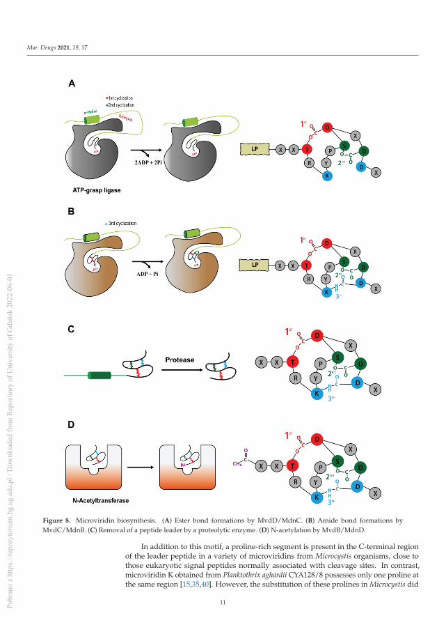
Mar. Drugs 2021, 19, 17
Figure 8. Microviridin biosynthesis. (A) Ester bond formations by MvdD/MdnC. (B) Amide bond formations byMvdC/MdnB. (C) Removal of a peptide leader by a proteolytic enzyme. (D) N-acetylation by MvdB/MdnD.
In addition to this motif, a proline-rich segment is present in the C-terminal regionof the leader peptide in a variety of microviridins from Microcystis organisms, close tothose eukaryotic signal peptides normally associated with cleavage sites. In contrast,microviridin K obtained from Planktothrix aghardii CYA128/8 possesses only one proline atthe same region [15,35,40]. However, the substitution of these prolines in Microcystis did
11Pobr
ano
z ht
tps:
//rep
ozyt
oriu
m.b
g.ug
.edu
.pl /
Dow
nloa
ded
from
Rep
osito
ry o
f Uni
vers
ity o
f Gda
ńsk
2022
-06-
01

Mar. Drugs 2021, 19, 17
not affect the removal of the peptide leader but resulted in the cessation of microviridinproduction [37], suggesting the necessity of the β-turn of peptide leaders MdnB and C.
Ahmed et al [20] analyzed several mdnA sequences among their biosynthetic clustersand divided this gene into three different classes. Class I precursor peptides contain theLP fused to only one core sequence and are often associated with the presence of mdnE,which occurs in a majority of the strains. Class II precursor peptides present a single leaderpeptide for up to five core peptides in tandem, separated or not by double-glycine cleavagesites, and these clusters normally encode a C39 peptidase membrane protein. Finally,class III is identical in length to class II, but the former has its core sequence only at theC-terminal of the prepeptide [20]. This indicates a number of pathways for the geneticorganization of mdnA.
After mdnA has been expressed, prepeptides should be submitted for cycling by thesequential catalysis of the enzymes. Thus, in order to understand the mechanisms relatedto this stage, Philmus et al. [15,37] were the first to define, through biochemical methods,the steps taken by MvdC and D of P. agardhii CYA126/8. Both enzymes are carboxylate-amine/thiol ligases that belong to the ATP-grasp superfamily and act by requiring ATPand Mg2+ [38,40] to form a carboxylate–phosphate intermediate, which is then susceptibleto nucleophilic attacks to form ester, amide or thioester bonds [17,36]. MvdD/MdnCare responsible for the first step in the formation of both ester bonds (Figure 8A) and,subsequently, lactone rings in the linear prepeptide, while MvdC/MdnB are responsiblefor the formation of lactam rings by amide bonds [15,35].
Both enzymes are homodimers with related assemblies, similar to most proteins ofthis family, having three subdomains: N-domain, central domain and C-domain. Besidestheir overall similarities, there are differences comparing their central and C-domains.MdnC/MvdD possess a two-stranded antiparallel β-sheet forming a hairpin structure,followed by a reasonably ordered α-helix that anchors the leader peptide. Meanwhile, thishairpin region is located at the C-domain of MdnB/MvdC, followed by a flexible loop inthe α-helix region. The MdnB has a closed conformation, compared to MdnC, because theantiparallel β-sheet hairpin blocks the pocket site where MdnA interacts. Those differencescan be related to their specificity and mode of action, as can be seen below. Regardingthe ATP-binding pocket, it is structurally conserved, as confirmed by mutagenesis, wheresubstitution of the key amino acids completely abolished the MdnC reaction [36].
Phylogenetic studies and the study of preserved sequences of different classes ofprepeptides forming cyclic structures by the action of ATP-grasp ligases (plesiocins, mi-croviridins and thuringinins) suggest that the enzymes coevolved with their respectiveprecursor peptides due to the specificity of the preserved residues present in the coresequence [36]. Consequently, the association between microviridin production and ATP-grasp enzymes indicates that cyanobacteria recycled primary metabolic enzymes for theproduction of natural products, such as ribosomal peptides, as most ATP-grasp ligasesare engaged to primary metabolism [15,17,35,36]. In addition, MdnC is well-conservedamong Microcystis species, suggesting its derivation from a common ancestor, as well as itsdependence on the core motif KYPSD and threonine and aspartate conservation sites ofmicroviridins, as seen by the mutagenesis and phylogenetic analysis [14,15].
As described by Li et al. (2016) [36], the reaction of the bond formation by MdnC(Protein Data Bank (PDB) code 5IG9) is driven by the interaction with the leader peptide.Thus, the PFFARFL motif structured as a α-helix and its flanking amino acids interactwith the MdnC hairpin, inducing its movement towards the linear prepeptide bound tothe enzyme, then acting as an allosteric region. Considering the ATP-grasp ligase fromMicrocystis aeruginosa, these interactions occur between the amino acids Arg17 (MdnA) andGlu191, Asp192 and Asn195 (MdnC) and Ser20 (MdnA) and Val182 (MdnC). However,Glu191 is mostly replaced by an aspartate residue among all microviridin macrocyclasesbut still bears the negative charge required to recognize the LP [36].
After binding to the peptide leader through the PFFARFL motif, the ester bondformations are strictly required to occur in a specific order in microviridins: MvdD catalyze
12Pobr
ano
z ht
tps:
//rep
ozyt
oriu
m.b
g.ug
.edu
.pl /
Dow
nloa
ded
from
Rep
osito
ry o
f Uni
vers
ity o
f Gda
ńsk
2022
-06-
01

Mar. Drugs 2021, 19, 17
the lactone ring between Asp44 and Thr38, then Glu46 and Ser43 into the prepeptide,by phosphorylating the carboxyl side chain of Asp and Glu with ATP, thus forming thelarge then small lactone rings, respectively [37]. These residues participating in the amidebond and ring formation are highly conserved among the cyclic peptides, suggesting theirrequirement for the correct cyclization and similar catalysis between ATP-grasp ligasesfrom different groups [34].
When a site-directed mutagenesis was applied to produce different variants of MvdE/MdnA, S43A and T38A, it has been noticed that MvdD catalyzes a reaction followinga N-terminal-to-C-terminal direction, as the S43A variant is still lactonized, producinga monocyclic microviridin. In addition, the amino acid bearing the hydroxyl group is crucialfor the reaction, as it seems that MvdD cannot react when it is moved one position in eitherthe N- or C-terminal direction [15,37,41]. It seems that both ATP-grasp ligases are highlytolerant for nonconserved residues, then being able to catalyze different microviridins.However, they are not flexible to conserved residues that are involved in cyclization [34,35].
For a better understanding of the different MdnC/MvdD enzymes, Zhang et al. [39]characterized a homolog of these enzymes from Anabaena sp. PCC7120, AMdnC, whichbelonged to a biosynthetic cluster with a prepeptide of class II, with a LP followed by threeconsecutives core sequences (AMdnA). The mode of action of AMdnC indicates a distribu-tive catalysis, where the ATP-grasp ligase dissociates from the processed peptide after eachmonocyclization, until achieving all lactone ring formations. This feature has been alsodescribed in other modification proteins from RiPP pathways, such as the NisB, LctM,LabKC and HalM2 enzymes from lanthipeptides processing; microcin B17 synthetases;ATP-grasp enzyme PsnB and N-methylation enzyme OphA of omphalotin. Additionally,AMdnC also demonstrates a preferential N-to-C directionality when catalyzing the reactionbut not unstrict. Thus, this homolog of MdnC can process each core peptide independentlyfrom AMdnA. Moreover, the calculated Km from AMdnC when catalyzing AMdnA orMdnA is comparable to MdnC values when processing MdnA; however, the kcat of theATP hydrolysis of AMdnC were up to 60 times faster, suggesting a different mechanismfor processing a prepeptide with multiple core sequences.
MdnB has a similar structure and mechanism of activation as MdnC, where thePFFARFL motif interacts with the hairpin, resulting in the activation of the enzyme [36].Then, the bicyclic prepeptide produced by MvdD/MdnC is catalyzed by MndB/MvdC, andthe lactam ring is formed through the amide bond formation between the ε-amino group ofLys40 and δ-carboxyl group of Glu47 (Figure 8B). The omega-amide bond is similar to thosepresent in microcin J25 and capistruin; however, the enzymatic mechanism is different,because microviridin K synthesis occurs via an acyl-adenylated intermediate [17].
Both preformed lactone rings are required by MvdC/MdnB, as the linear and mono-cyclic peptides are not modified. A single mutation in the PFFARFL pattern in the leaderpeptide prevents the formation of amide bonds, as well as the proper conformation ofthe β-turn by the proline-rich region at the C-terminal of microviridin from Microcystis,suggesting a lower flexibility compared to MndC [39]. In addition, the amino acid sequenceof the core peptide can also influence the correct cyclization, even those not well-conserved,requiring the TxKxPSD motif and Lys and Glu residues [41].
MdnC/MvdD is less rigid than MdnB/MvdC, as it can still catalyze both lactonerings besides single and double mutations in the PFFARFL motif and proline-rich regionof the leader peptide (in Microcystis) but could result in producing different microviridinvariants differing at the N-terminal [37]. This versatility is possibly due to the more openconformation of the hairpin structure and to the binding interaction between MdnC andthe prepeptide relative to MdnB. As seen in vitro, the binding interaction between LPand MdnC is approximately tenfold higher compared to the LP and MdnB, resulting ina rapid processing of the linear prepeptide compared to the bicyclic modification. It isalso believed that this is due to the fact that linear MdnA is less stable, requiring promptmodification [36]. However, this post-translational modification does not seem to be strictlysufficient for further steps, as bicyclic microviridins can still be cleaved and N-acetylated
13Pobr
ano
z ht
tps:
//rep
ozyt
oriu
m.b
g.ug
.edu
.pl /
Dow
nloa
ded
from
Rep
osito
ry o
f Uni
vers
ity o
f Gda
ńsk
2022
-06-
01

Mar. Drugs 2021, 19, 17
and possessing inhibitory activity against proteases [41]. As seen by bioinformatic analyses,the absence of MdnB is normal and is likely to lead to the formation of marinostatin,a peptide that lacks an amide bond and is closely related to microviridins [20]. Firstly,it was suggested that MdnE, an ABC transporter, could be related to the removal of theleader peptide through peptide cleavage due to the presence of a N-terminal C39 peptidasedomain from Anabaena PCC7120 [14,37]. However, not all MdnE carry that domain, andthe heterologous expression of the microviridin cluster lacking this protein still producesthis tricyclic peptide, indicating other roles [14,37]. Comparing the microviridin expressionwith the presence and absence of MdnE, it was noted that these peptides were not correctlyprocessed at the N-terminal and were incompletely cyclized due to a lack of lactam rings, aswas the amount of MdnB observed in the cytoplasmic fraction when the ABC transporterwas absent. This pattern then suggests the hypothesis that MdnE is a scaffolding protein,anchoring and stabilizing the microviridin biosynthesis complex on the cytosolic side ofthe membrane [37]. In addition to its similarity to transporter proteins, its function inexporting microviridin from the cell has not been demonstrated.
Knowledge on the removal of a peptide leader has so far been scarce in the literature.However, the heterologous expression of microviridin suggests that this step can be medi-ated by a nonspecific proteolytic enzyme (Figure 8C), as E. coli expressing only MdnA-Cwas capable of producing microviridin lacking a LP [14]. Moreover, GluC endoprotease iscapable of cleaving peptide bond C-terminals to glutamic acid residues, and during thein vitro production of class II microviridin with three core sequences, this enzyme releasedall three mono-, bi- and tricyclized microviridins [40]. Finally, another hypothesis relatedto MdnE was raised. As described above, this enzyme may have a peptidase domain,typically present in class II clusters. This function may be linked to the presence of inter-spaced regions between the core sequences of MdnA and the release of each individualmicroviridin [14,20,37,39].
Acetylation is one of the last steps for the development of a fully matured tricyclic mi-croviridin. Microviridin synthesis in vitro has shown that MvdB from P. aghardii CYA126/8does not require the presence of the peptide leader to acetylate the microviridin N-terminal.In addition, MdnD/MvdB can react with mono-, bi- and tricyclic, being more flexible thanMdnB and C. Thus, it can be assumed that this step occurs after the peptide leader removal,or this enzyme does not interact with this region (Figure 8D) [38]. In addition, a 12 aminoacid-long tricyclic peptide is not N-acetylated by MdnD from P. aghardii CYA126/8, butthose microviridin with 13/14 amino acids are acetylated, indicating that there a specificsize requirement by this enzyme, and it is flexible regarding the core sequence [20], thussuggesting that N-acetylation occurs only after leader peptide removal [15,20,35,38].
5. Occurrence
Genome mining has shown that cyanobacteria have the potential to generate muchmore microviridin than is typically found under normal growth conditions. A study of thistype has contributed to the expansion of knowledge on the chemical and genetic diversityof microviridins. They have been detected in various cyanobacterial genera and species,and these microorganisms are notorious producers of different groups of peptides andcan be found in many environments, whether in fresh or salt water (Table 2). Due tothis great variety, these bacteria have been evaluated for their significant biotechnologicalpotential. The genus Microcystis and the species M. aeruginosa are the largest producers ofmicroviridins—currently, of the 25 isolated microviridins, 11 belong to the genus Microcystis,and eight of these belong to the species M. aeruginosa [21,23,27,29–31]. Microviridin geneclusters have also been found in genomes of a number of bacteria, such as bacteroidetesand proteobacteria phyla [20].
14Pobr
ano
z ht
tps:
//rep
ozyt
oriu
m.b
g.ug
.edu
.pl /
Dow
nloa
ded
from
Rep
osito
ry o
f Uni
vers
ity o
f Gda
ńsk
2022
-06-
01

Mar. Drugs 2021, 19, 17
Table 2. Occurrence of microviridins.
Species/GeneraIsolationSource
Trophic StateSequenced
GenomeMicroviridins Mass (Da) Ref.
Microcystis.viridis NIES-102 Aquatic Eutrophic + Microviridin A 1729.7 [21]
Microcystis.aeruginosa NIES-
298Aquatic Eutrophic + Microviridin B 1723.8 [23]
Microcystis.aeruginosaNIES-298
Aquatic Eutrophic + Microviridin C 1755.8 [23]
Planktothrixagardhii
NIES-204Aquatic Eutrophic +
Microviridin D 1802.7[24]Microviridin E 1665.7
Microviridin F 1683.7
Nostoc minutumNIES-26
Terrestrial Mesotrophic +Microviridin G 1806
[24]Microviridin H 1838
Planktothrixagardhii
CYA126/8Aquatic N.I. - Microviridin K 1769 [15]
Planktothrix.agardhii strain 2
& 18Aquatic Eutrophic - Microviridin I 1764.7 [26]
Microcystisaeruginosa
UWOCC CBSAquatic N.I. - Microviridin J 1684.4 [27]
MicrocystisaeruginosaNIES-843
Aquatic Eutrophic + Microviridin L 1715 [31]
Planktothrix sp. Aquatic Eutrophic -
Microviridin I 1765.8
[43]Microviridin
1642 1642.8
Microviridin1663 1663.7
Microcystisaeruginosa Aquatic Mesotrophic - N.I. N.I. [44]
Microcystis sp. Aquatic N.I. - MicroviridinLH1667 1666.7 [30]
Microcystisaeruginosa PCC
7820Aquatic N.I. - Microviridin
1706 1707.8 [46]
Planktothrixrubescens
NIVA-CYA 98Aquatic Mesotrophic + Microviridin 1971.8 [33]
Microcystis sp. Aquatic Mesotrophic -
Microviridin1667 1668.6
[42]
Microviridin1684 1695.8
Microviridin1699 1699.8
Microviridin1777 1778.8
Chroococcidiopsissp. CENA 353 Leaf Surface N.I. - N.I. N.I. [45]
15Pobr
ano
z ht
tps:
//rep
ozyt
oriu
m.b
g.ug
.edu
.pl /
Dow
nloa
ded
from
Rep
osito
ry o
f Uni
vers
ity o
f Gda
ńsk
2022
-06-
01

Mar. Drugs 2021, 19, 17
Table 2. Cont.
Species/GeneraIsolationSource
Trophic StateSequenced
GenomeMicroviridins Mass (Da) Ref.
Desmonostoc sp.CENA365 Leaf Surface N.I. - N.I. N.I. [45]
NostocaceaeCENA358 Leaf Surface N.I. - N.I. N.I. [45]
NostocaceaeCENA376 Leaf Surface N.I. - N.I. N.I [45]
N.I.: not informed.
The genus Microcystis was the first to be described in the literature as a cyanobacteriaproducer of microviridins. This peptide was isolated from the bloom-forming M. viridis(NIES-102) on Kasumigaura Lake by Ishitsuka et al. (1990) [21]. This new oligopeptidedemonstrated a noncanonical structure and was named microviridin by the name ofthe viridis species. In addition, other microviridins from the cyanobacteria of the genusMicrocystis have been identified as microviridin B, C, L, SD1684, SD1634, SD1652, LH1667,1777, O and M. Each of these microviridins has a considerable inhibition for at least oneserine protease, such as elastase or trypsin [21,23,27,29–31].
An in-situ diversity investigation of the Microcystis communities present in lakeslocated around and in the city of Berlin, Germany demonstrated that 20% of 165 coloniesanalyzed were capable of producing microviridin. These cyanobacteria were present inalmost all investigated areas. The majority of the microviridins producers also synthe-sized microcystins and cyanopeptolins. The coproduction of microviridin-aeruginosinsand -microginins was rarely reported among the strains, being present in only 4% and2%, respectively. The metabolomic profile of the peptides can be utilized to distinguishMicrocystis strains with elevated morphological similarity whose visualization in the lightmicroscope is not sufficient to differentiate them [42].
Martins and collaborators [43] isolated strains of cyanobacteria M. aeruginosa from alarge range of lakes, rivers and reservoirs in Portugal. These strains were examined for thepresence of secondary metabolites, such as aeruginosins, microviridins and microcystins.In this analysis, 47 strains from different sites were isolated among the identified peptides;microcystin was the most recurrent, appearing in 26 strains, and microviridins werecontained in only three. The results of the analysis of the coproduction showed that thestrains that produced microviridins did not produce microcystins. In another study, Walkeret al [44] isolated the microviridin-producing strains of the Planktothrix genus from Maxseein Germany incapable of producing microcystins.
In a study accomplished by Andreote [45], the purpose of which was to obtain infor-mation on the cyanobacterial community present in the phyllosphere of native plants fromthe Atlantic Forest, identified 40 cyanobacterial strains belonging to the genera Nostocaceae,Desmontosc and Chroococcidiopsis as microviridin producers obtained from Merostachys neesii(bamboo), Euterpe edulis (palmeira jacura), Guapira opposita and Garcinia gardneriana.
Andreote [45] was the pioneer in the identification of these peptides in the Desmontoscand Chroococcidiopsis genera. To identify the presence of this peptide in the strains, PCRamplifications of the mdnA, mdnB and mdnC genes were performed, which were related tothe biosynthetic pathways of the microviridins. The strains Nostocaceae sp. CENA358 andCENA376, Desmonostoc sp. CENA365 and Chroococcidiopsis sp. CENA353 demonstratedthe presence of these genes. Other strains lacked at least one of these genes, which did notrule out the synthesis of this peptide by these microorganisms due to the primers utilizedthat were constructed for strains of Microcystis, causing low-amplification performances,which implies that they might have more strains producing microviridins or possessing abiosynthetic cluster [45].
Eleven cyanopeptides from four different groups were reported from samples ofcyanobacterial bloom in the Salto Grande reservoir, located in the State of São Paulo,
16Pobr
ano
z ht
tps:
//rep
ozyt
oriu
m.b
g.ug
.edu
.pl /
Dow
nloa
ded
from
Rep
osito
ry o
f Uni
vers
ity o
f Gda
ńsk
2022
-06-
01

Mar. Drugs 2021, 19, 17
Brazil, including the microviridin variant 1706. Cyanopeptides such as aeroguniosins,microcystins and cyanopeptolin were also detected. The morphological research showedthat the bulk of the population of cyanobacteria belonged to the genus Microcystis [46].
Variants of microviridins were characterized in two cyanobacteria isolated from Brazil-ian reservoirs. R. fernandoii strain 28 was obtained from the Furnas Reservoir, which issituated in the southeastern region of Brazil and is described as an oligo-to-mesotrophicaquatic environment that receives organic matter contributions from domestic, farmingand agriculture wastewaters. The R. fernandoii 86 strain was identified in an urban eu-trophic reservoir located in the city of Belo Horizonte, Brazil, which suffers a great impactfrom domestic pollution, industrial sewage. A total of twelve peptides were found in thetwo strains. In the R. fernadoii 28 strain, a microviridin MV-1709 was found, and, in thestrain R. fernadoii 86, two microviridins were reported, MV-1707 and MV-1739. Along withmicroviridins, peptides such as microcystins, cyanopeptolin and an unidentified peptidewere also detected [47].
6. Microviridin Ecology
Microviridins play a significant ecological function as antifeeding agents againstcyanobacterial natural predators. This activity is correlated with their ability to inhibitproteolytic enzymes (Figure 9). The first study to explain this mechanism was performed byRohrlack et al. (2014) [48]. A previous work, however, had already indicated microviridinsas an agent capable of causing the interrupting the feeding of Daphnia microcrustaceanvia enzymatic inhibition. This ability can partly explain the dominance of these microor-ganisms in some habitats, including those with a high population density of Daphnia [49].In a similar way, protease inhibitors are produced by terrestrial plants to protect againstherbivores. Metatranscriptomic analyses of the Kranji Eutrophic Reservoir, located inSingapore, revealed important information on the functional dynamics between differentbacterial phyla, including cyanobacteria, which were dominant microorganisms, especiallythose belonging to the Microcystis genus. The microviridin transcripts were found in highquantities, along with those involved in the buoyancy and photosynthetic operation. Thehighest peak of the gene expression related to microviridin biosynthesis was observedwhen the population of Daphnia moved from the mesopelagic zone to the epipelagic zone,corroborating its antipredator activity [49].
Figure 9. Ecological role of microviridins as antifeedant against the microcrustacean Daphnia.
Kaebernick et al. (2001) [50] compared the feeding inhibition of Daphnia galeata andD. pulicaria by a microcystin-producing Microcystis (MRD) and a microcystin-deficient
17Pobr
ano
z ht
tps:
//rep
ozyt
oriu
m.b
g.ug
.edu
.pl /
Dow
nloa
ded
from
Rep
osito
ry o
f Uni
vers
ity o
f Gda
ńsk
2022
-06-
01

Mar. Drugs 2021, 19, 17
Microcystis (MRC), and it has been realized that this hepatotoxin is not associated withthe ingestion rate reduction in both planktonic grazers. However, this metabolite wasresponsible for causing both species to decrease their survival rates. Before the deathprovoked by this hepatotoxin, these microcrustaceans remained immobile in the bottom ofthe vial and shifted only when the surrounding area suffered disturbances. In addition, thefilter legs and antenna were momentarily paused, and the midgut was disrupted.
The same authors [50] described further effects of Microcystis strain UWOCC MRCingestion by D. galeata and D. pulicaria. These microcrustaceans had a dysfunction in theperitrophic membrane. This membrane acts as a barrier formed by the chitin–proteincomplex created by the midgut cells. The consumption of Microcystis made this organmore enervated, as a result of which, food transport was impaired, resulting in particleaggregation in this region and in the digestive diverticula. The ingestion of these cells alsodisturbed the molting process. The old integument was not entirely separate from theDaphnia body, attached to the legs and filter antennas, and strongly hindered the ability ofthese species to swim and feed themselves. Individuals subjected to these conditions weremore likely to die of malnutrition within two days. It was also confirmed that the freshlydeveloped integument remained soft both in the presence of the old integument and afterits mechanical removal. Under field conditions, these affected species would become easyprey to predators, since they would not be able to flee to any shelter.
The typical chitin–protein complex occurs in both structures (peritrophic membraneand integument), indicating that the reported effects were probably caused by the samebioactive compound in which the microviridin variant was cogitated. By its ability toinhibit the serine protease, this oligopeptide could be preventing the tyrosine conversioninto dihydroxyphenylalanine (DOPA) and its subsequent transformation into dopamineby the enzyme DOPA decarboxylase [51]. Dopamine is involved in the cross-linkage oforthoquinones, which results in the cuticle sclerotization [52]. A complementary processwas proposed by Rohrlack et al. (2003) [48]. According to this mechanism, Daphnia’s deathwas associated with incomplete protein digestion, which resulted in an important aminoacid deficiency for tegument development and other structures. Ingestion of the strainof Microcystis UWOCC CBS, a producer of microviridin J, causes the same activity in themolting process of D. pulicaria. However, several additional findings have been made.Particles derived from food suspensions were found on the entire surface of the Daphniabody. This dysfunction was possibly due to the secretion of body fluids. Deformation onthe freshly generated tegument has become more intense as the effort to eradicate it bythese animals has increased. The same phenomenon was visualized when only the purifiedmicroviridin J was added.
Czarnecki et al. (2006) [53] detected eight microviridins distributed in three Microcystisstrains (HUB08B03, HUB11G02 and HUB19B05) with ability to inhibit trypsin-like activityin the planktonic crustacean Daphnia. In addition to microviridins, other classes of proteaseinhibitors, such as some cyanopeptolins, were found in the extract obtained from thesecyanobacteria. This ability of unique cyanobacterium or different cyanobacteria from thesame genus can generate a variety of combinations of different oligopeptides with distinctproteolytic targets and inhibitory activity. This feature acts as an evolutionary barrier,preventing the adaptation process among zooplankton population.
Microviridin toxicity was also accessed in the fairy shrimp Thamnocephalus platyurus,which belonged to order Anostraca. In the course of searching for natural products withcytotoxicity property, Sieber et al. (2019) [32] detected in the extract of M. aeruginosa strainEAWAG 127 deleterious activity against this microcrustacean (LD50 = 0.43 mg.mL−1). Ametabologenomic approach revealed the presence of two novel microviridins: microviridin1777 and microviridin O. The former showed a LD50 value of 95 μM for Thamnocephalusplatyurus. This activity was ascribed to the strong capacity of this peptide in inhibitingelastase and chymotrypsin activity with an IC50 of 160 nM and 100 nM, respectively.
In addition to Cyanobacteria having a low susceptibility to a zooplankton attack, thesemicroorganisms are also the target of various pathogenic bacteria and fungi that play an
18Pobr
ano
z ht
tps:
//rep
ozyt
oriu
m.b
g.ug
.edu
.pl /
Dow
nloa
ded
from
Rep
osito
ry o
f Uni
vers
ity o
f Gda
ńsk
2022
-06-
01

Mar. Drugs 2021, 19, 17
important role in controlling their growth [54,55]. True zoosporic fungi, commonly knownas chytrids, are among the most pathogenic organisms capable of causing a significantnumber of deaths in the cyanobacterial community [56]. The success of this pathogenin infecting these photosynthetic microorganisms can be attributed to the developmentof chemotactic zoospores and the presence of rhizoids, which are capable of locating thetarget and used to extract the nutritional contents, respectively. Oligopeptides produced bycyanobacteria with an inhibitory activity against a predator’s key enzymes is a great defensemechanism. The comparison between the cyanobacterial strain P. agardhii CYA126/8 withits mutants, each one with a type of disability in producing microcystins, anabaenopeptinsor/and microviridins, was conclusive to defining the protective role of these metabolites.The wild strain when incubated with the chytrid strain was unaffected, while all mutantstrains were infected, including those non-microviridin-producing strains [57].
Chytrides are a rich source of protease used as a mechanism to digest their hosts.Microviridins and anabaenopeptins can target these enzymes, reducing the virulence ofthese fungi. The vast variety of microviridins, as well as other oligopeptides, is a majorobstacle in the process of the adaptation of these parasites. On the basis of the literature, theprotection mechanism referred to above appears to be constitutive, since these substancestypically form an oversaturated or saturated solution in the cytoplasm [57]. Microviridinwas also found in bacteria belonging to the microbiome of the plant Populus. Unlikethe lanthipeptides that are widely distributed among the member of this community,microviridins were restricted only to the genus Chryseobacterium, being present in 16 outof the 18 sequenced bacteria. Its role in this microbiome is not clear. A gene clusterfor microviridin in this genus showed from one to four precursor peptides belonging toclass I [20]. Different from cyanobacterial microviridin, the core peptide was composedof 18 amino acid residues. Only half of the microviridin clusters analyzed had a N-acetyltransferase gene. A resistant gene presence in the majority of the microviridinclusters suggested that this oligopeptide could have antibacterial properties, conferring aprotection to plants against pathogenic microorganisms [58].
Other features given to microviridins are related to their allelochemical properties.Cyanobacteria produce a variety of proteases that are essential to different processes,including nutrient absorption, protein activation, unfolded or aggregated protein removal,photoacclimation and stress response [59]. Ghosh et al. (2008) [60] demonstrated thata cyanobacterial oligopeptide with the partial structure of a microviridin affected theproteolysis in M. aeruginosa PCC 7806, strongly inhibiting its capacity to degrade N-alpha-benzoyl-DL-Arg-p-nitroanilide (BApNA). The authors’ hypothesis was that microviridin-producing Microcystis colonies could form an aggregate that could eventually develop as abloom and suppress the growth of competing organisms by targeting critical functions thatrely on protease activity. Another possibility is that microviridins will have a significant roleto play in stress conditions by self-regulating the protease activity among cyanobacterialcells and thus enhancing their survival rates [60].
Some microviridins are not easily detected in environmental samples, since they maybe rapidly degraded by other bacteria. M. viridis has its microviridin A content totally con-sumed when transferred to a nonaxenic medium [21]. The aquatic bacterium Sphingomonassp. B-9, firstly isolated for its microcystin-degradation ability, has hydrolytic enzymescapable of degrading different cyclopeptides, including microviridin I. The degradation ofthis peptide by this bacterium is very slow, lasting around 48 h to reduce 50% of its initialcontent. This process occurs in two steps. Initially, the residue at the C-terminal region isremoved, and, subsequently, the molecule undergoes a linearization step [61].
7. Regulation
Environmental factors play an important role in the regulation of the synthesis ofoligopeptides, as they can increase the growth rate and, consequently, the production ofthese metabolites. In certain cases, however, the best conditions for growth did not lead tothe most desirable conditions for their production [62]. Microcystin was the key subject of
19Pobr
ano
z ht
tps:
//rep
ozyt
oriu
m.b
g.ug
.edu
.pl /
Dow
nloa
ded
from
Rep
osito
ry o
f Uni
vers
ity o
f Gda
ńsk
2022
-06-
01
Mar. Drugs 2021, 19, 17
these studies [63]. Stress situations can alter the cell’s physiological state and act as triggerfor increasing the construction of these molecules. Nitrogen and phosphorus bioavailabilityare among the most nutritional factors investigated in cyanobacterial behaviors [64,65].Both are involved in protein synthesis and in the energy dynamic. Due to anthropic actions,these elements have become more abundant in the aquatic environment [66].
Other parameters, such as temperature, pH and light intensity, have also been inves-tigated and challenge many scientists [67,68]. An assessment of the combined effect ofdifferent environmental elements on the development of cyanopeptides can provide a linkbetween field research and laboratory research. Any of these variables can be associated.As the culture reaches the stationary phase, the quantity of nutrients decreases, as well asthe light availability among cells, thereby reducing their growth rate.
One method used by Rohrlack et al. (2007) [67] to individually determine the lightimpact was to track and maintain a constant nutrient load in the medium. This techniquewas used to analyze the production of microviridin I by P. agardhii strain PT2. The quantityof cell-bound microviridin I expressed in units per biovolume decreased until the eighthday. This behavior reversed when the light availability began to decline. Nitrogen andphosphate reduction also led to a decrease in the production of this microviridin. A similartrend was reported for microcystins and anabaenopeptins. Some authors strongly believethat many of these oligopeptides play the same ecological function. The loss of one canhave, as a consequence, the enhanced production of another, unaffecting the cyanobacterialgrowth [69,70].
The influence of light intensities was also evaluated by Pereira et al. (2012) [47]on the profiles of toxic and nontoxic oligopeptides obtained from two strains of thecyanobacterium: R. fernandoii 28 and 86. In the course of the experiment, they employedthree different irradiances, which were classified as low (25 μMol.m−2.s−1), medium(65 μMol.m−2.s−1) and high (95 μMol.m−2.s−1). Different from other oligopeptides in-vestigated in this study, such as microcystins and cyanopeptolins, microviridins were notencountered in all growth conditions. Microviridin 1709 production reached the maxi-mum amount when the cells of strain 28 were exposed to a medium light intensity, whilemicroviridin 1707, identified in strain 86, was detected solely at low light conditions.
Ferreira et al. (2006) [71] evaluated the combination of different light intensities,nutritional contents, temperatures and growth phases on the oligopeptide production indistinct strains of Microcystis and Aphanizomenon, including microviridins. This proteaseinhibitor was detected solely in the Microcystis strain RST9501. In the absence of nitrateand phosphate, this peptide was produced in higher quantities. In the majority of the cases,the intracellular fraction was responsible for over 80% of the total microviridin pool. At thesame nutritional conditions, an atypical behavior was found when the cells were cultivatedat 20 ◦C. In this condition, the intracellular microviridin concentration diminished to 60%.
The cell-to-cell communication is also a factor to be considered in peptide produc-tion. When this mechanism is dependent on cell density, it is called quorum sensing.Nealson and Hastings (1979) [72] were pioneers in studying this phenomenon in theGammaproteobacterium Vibrio fischeri. These two scientists were capable of demonstrat-ing that the enzyme luciferase, whose role is to transform chemical energy into lightenergy, was expressed only at a high cell density, having its production controlled byautoinducer signaling molecules [72]. The most known autoinducers described are theacylated homoserine lactones [73]. The aquatic environment has a natural tendency todilute the metabolites released by microorganisms. For this reason, some authors believethat oligopeptide production is regulated by quorum sensing [74]. There is little knowledgeabout this mechanism in cyanobacteria. During a bloom episode, the cyanobacterial popu-lation increased significantly, creating a favorable environment for quorum sensing. In thistype of situation, the high cell density augments the concentration of signaling moleculesin the environment [75].
To evaluate the quorum sensing effect on oligopeptide production, Pereira et al. [74]grew the cyanobacteria in a semicontinuous culture system. Hence, the biomass level and
20Pobr
ano
z ht
tps:
//rep
ozyt
oriu
m.b
g.ug
.edu
.pl /
Dow
nloa
ded
from
Rep
osito
ry o
f Uni
vers
ity o
f Gda
ńsk
2022
-06-
01

Mar. Drugs 2021, 19, 17
nutritional content were maintained constant. The growth rates of high and low cell densitycultures were similar. Microviridin production was detected only in a low cell densityculture of R. fernandoii (strain R28). In contrast, the microviridins N3-N9 in N. punctiformePCC73102 had their synthesis optimized under high cell density conditions [19].
The cyanobacterial lifestyle may also have an effect on its oligopeptide content. Somecyanobacteria are typically located on the water and sediment surface. There are also thosewith a biphasic lifestyle, where they migrate to the top during the summer and to thebottom during the winter [76]. A comparative genomics of the genus Planktothrix withdifferent lifestyles performed by Pancrace et al. (2016) [77] demonstrated that all planktonicstrains investigated harbored the microviridin gene cluster. In contrast, in the benthicPlanktothrix, this gene cluster was absent, with the exception of Planktothrix sp. PCC 11201,which is phylogenetically closer to free-living Planktothrix.
8. Application of Microviridins
Proteases play an important role in the regulation of biological processes in all livingorganisms by controlling the maintenance, recovery, development and modification oftissues, which may be beneficial or harmful. They may modulate protein–protein interac-tions that create bioactive molecules involved in DNA replication and transcription [78]. Inplants, these enzymes lead to the maturation and degradation of a series of unique proteinsrelevant to the environmental condition and the stage of growth [79]. Thus, molecules withinhibitory activity against these enzymes have an immense biotechnological potential witha multitude of applications (Figure 10).
Figure 10. Potential applications of microviridins.
Tyrosinase is a multifunctional and metalloenzyme widely distributed among plants,microorganisms and animals, where it plays a key role in the development of melanin [80].The excessive synthesis of this photoprotective pigment may lead to a condition known ashyperpigmentation, which may lead to an esthetic problem where one part of the skin ismore pigmented than the other [81]. In addition, this disorder has been linked to manydiseases, such as skin cancer and Parkinson’s disease [82]. Molecules with the capacity tointerfere with the catalytic activity of tyrosinase have been extensively studied as a skin-whitening agent. They can also be used as food additives, reducing the browning process
21Pobr
ano
z ht
tps:
//rep
ozyt
oriu
m.b
g.ug
.edu
.pl /
Dow
nloa
ded
from
Rep
osito
ry o
f Uni
vers
ity o
f Gda
ńsk
2022
-06-
01

Mar. Drugs 2021, 19, 17
of mushrooms and fruits caused by tyrosinase [83]. Some of these commercially availablecompounds exhibit low stability and safety [84]. Among the microviridins reported in theliterature, microviridin A was demonstrated to be tyrosinase inhibitor. However, its actionmechanism is not clear, and information regarding its toxicity to human cells has neverbeen accessed (Table 3) [21]
Table 3. Inhibitory activities of microviridins.
MicroviridinInhibitory Activity (IC50)
ReferenceTyrosinase Elastase Trypsin Chymotrypsin Thrombin Plasmin Papain
A 0.33 mM * ND ND ND ND ND ND [21]B ND 25.5 nM 33.7 μM 1.45 μM >58.1 μM >58.1 μM ND [23]C ND 47.9 nM 18.2 μM 2.8 μM >57.1 μM >57.1 μM ND [23]D ND 388.5 nM >55.5 μM 665.9 nM >55.5 μM >55.5 μM >55.5 μM [24]E ND 360.1 nM >60 μM 660.3 nM >60 μM >60 μM >60 μM [24]F ND 3.4 μM >59.5 μM >59.5 μM >59.5 μM >59.5 μM >59.5 μM [24]G ND 10.5 nM >55.4 μM 775.6 nM >55.4 μM >55.4 μM >55.4 μM [25]H ND 16.9 nM >54.4 μM 1.6 μM >54.4 μM >54.4 μM >54.4 μM [25]I ND 192.7 nM 14.9 μM 12.3 μM >56.7 μM ND ND [26]J ND >5.9 μM 20–90 nM 1.7 μM ND ND ND [27]L ND >58 μM 42 μM 58 μM ND ND ND [31]
1777 ND 160 nM >10 μM 100 nM ND ND ND [32]SD1684 ND ND Inactive Inactive ND ND ND [29]SD1634 ND ND 8.2 μM 15.7 μM ND ND ND [29]SD1652 ND ND Inactive Inactive ND ND ND [29]LH1667 ND 20 nM >45.5 μM 2.8 μM ND ND ND [30]
* IC50 not defined. ND, not determined.
The mechanism of coagulation is regulated by proteases. Serine protease inhibitorstherefore function as essential regulators in this pathway, such as proteins in the serpinsuperfamily. Malfunctioning of one of these elements can lead to coagulation disorders.Excessive blood clotting can lead to a disorder known as thrombosis, where the bloodflow is blocked by thrombus [85]. Thrombin is one the major target of anticoagulantdrugs, since it acts in the conversion of soluble fibrinogen into insoluble filamentous offibrins, which, together with platelets, are responsible for a hemostatic plug formation,impeding the bleeding. The thrombin inhibition by microviridin B is superior to thepositive control Leupeptin, possessing an EC50 (half maximal effective concentration) valueequal to 4.58 μM. This value was inferior than that encountered for Micropeptin K139,a serine protease also detected in cyanobacteria. In contrast, microviridins D-F do not affectthrombin activity, most likely due to the absence of an indole motif, which is encounteredin microviridin B, suggesting its role as a recognition motif for thrombin [86].
Human neutrophil elastase is a proteolytic enzyme that belongs to the serum proteinfamily of chymotrypsin-like. This highly active enzyme has revealed a wide substratespecificity and is one of the few proteases capable of degrading the extracellular matrixprotein elastin, resulting in the enzyme’s name [87]. The elastase overactivity is involvedin tissue destruction and inflammation characteristic of various diseases, such as chronicobstructive hereditary emphysema, pulmonary disease, cystic fibrosis, adult respiratorydistress syndrome and ischemic-reperfusion injury [88]. Pharmaceuticals already useelastase inhibitors for the treatment of diseases related to this enzyme, such as the drugSivelastat, which has been cogitated in the treatment of COVID-19 [89].
A study by Masahiro Murakami et al. (1997) [25] evaluated the elastase inhibitoryeffect of some microviridins, synthesized by the cyanobacterium Nostoc insulare (NIES-26).The results of the analysis showed two new peptides, microviridin G and microviridinH. In the experiment, both IC50 were compared with the values of other microviridinsalready described in the literature. Microviridin A showed no inhibitory effect on elastase;microviridins D and F had the weakest values for inhibiting this enzyme. Microviridins Gand H had the best results, followed by microviridins B and C, respectively. After the work
22Pobr
ano
z ht
tps:
//rep
ozyt
oriu
m.b
g.ug
.edu
.pl /
Dow
nloa
ded
from
Rep
osito
ry o
f Uni
vers
ity o
f Gda
ńsk
2022
-06-
01

Mar. Drugs 2021, 19, 17
of Murakami et al. [25], three new microviridins were described as elastase inhibitors: I,LH1667 and 1777 (Table 3) [26,30,32].
Proteases are also essential for the growth of insects, such as in the larval and adultstages, where they are present in the intestine and play an important role in digestion [90].For example, silkworms, near the final stage of their metamorphosis, produce cocoonase,a serine protease capable of hydrolyzing silk protein, which enables the adult moth toemerge [91]. During the embryony phase, proteases digest egg-specific proteins, such asvitellin, for the amino acid release, which are utilized as a nitrogen source [92]. Serineprotease can also confer protection against predation. The South American Saturniidcaterpillars belonging to the genus Lonomia harbor in their hemolymph a toxic proteasethat some mammals who come in contact with can have, as a consequence, bleedingdisorders [93]. Nowadays, various studies have focused on the search for a proteaseinhibitor whose target are proteases produced by insects involved in disease transmission,such as the Aedes aegypti, one of the largest vectors of arboviruses, being responsible for thepropagation of the dengue virus, yellow fever, zika virus and chikungunya fever [94].
Microviridins have already shown to be good inhibitors of enzymes present in micro-crustaceans intestines, such as those of the genus Daphnia and Thamnocephalus [32,50]. Forinstance, in the presence of microviridins B, C, I, J, L and SD1652, the enzyme trypsin has itsactivity negatively affected [23,26,27,29,31] (Table 3). They act by inhibiting enzymes that areclosely related to the diet of these crustaceans. In insects, many of these serine proteases arelocated in the intestines as well [95], sharing similar functions. Two of these serine proteasesare trypsin and chymotrypsin, well-known targets of some microviridins (Table 3).
Plants have served as a great heterology expression system for bioactive peptides.Hilder et al. (1987) [96] were the first to use these organisms to express serine proteaseinhibitors with the potential to kill predatory larvae insects. There is a considerable numberof works employing plants as hosts of ribosomally synthesized and post-translationallymodified peptides [97–99]. Plant-based microviridins have promises for future applications,since they can replace the use of pesticides to help control insect pests with low costs andlow environmental impacts. Furthermore, they can be easily purified with a high yield,retaining full activity.
One of the bottlenecks for microviridin production and the evaluation in cyanobacteriais a low yield of this peptide. Several extraction approaches in different genus of thisphylum demonstrated that these organisms are not well-suited for industrial applicationswhen considering both the time and volume of the cultivations. A heterologous expressionin E. coli demonstrated to be an efficient method for microviridin biosynthesis resulted ina yield of 60 ◦C 70 mg of microviridin L per 100 g of dried cells after five hours of cultivation.In comparison, about 0.87 mg.g−1 of microviridin A was obtained from Microcystis viridis(NIES-102) by a cultivation period of 10 ◦C for 14 days. In filamentous cyanobacteria, thisyield was even lower, with a production of 9.1 mg.g−1 of microviridin E after the sameperiod of incubation of Planktothrix in 400 L [21,25,31].
However, the problems related to microorganism cultivation, heterologous expressionand laborious purification can still be tackled, and techniques have been developed toovercome these barriers in order to explore the diversity of the microviridins. As a con-sequence, the development of microviridins obtained from environmental DNA can beaccomplished by the synthesis of the solid-phase peptide of the core peptide coupling withMdnC and -B enzymes fused to the leader peptide in the N-terminal. This chemoenzymaticapproach allows an in vitro production of a fully processed microviridin, demonstratingthe efficiency during production of different variants of this peptide [34]. Another approachfor microviridin production in vitro is to provide the LP in trans for both MdnB and -C,also achieving a tricyclic microviridin J [36].
Another important feature for the biotechnological application of microviridin is itsbinding affinity to serine proteases. Microviridin J demonstrated a KD value of 0.68 μMfrom its interaction with trypsin. This mode of inhibition is similar to a cyclic depsipeptideA90720A produced by a nonribosomal peptide synthetase [34,37]. However, the NRPS
23Pobr
ano
z ht
tps:
//rep
ozyt
oriu
m.b
g.ug
.edu
.pl /
Dow
nloa
ded
from
Rep
osito
ry o
f Uni
vers
ity o
f Gda
ńsk
2022
-06-
01
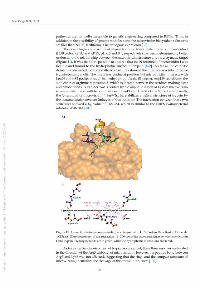
Mar. Drugs 2021, 19, 17
pathways are not well-susceptible to genetic engineering compared to RiPPs. Thus, inaddition to the possibility of genetic modifications, the microviridin biosynthetic cluster issmaller than NRPS, facilitating a heterologous expression [37].
The crystallography structure of trypsin bound to N-acetylated tricyclic microviridin J(PDB codes: 4KTU and 4KTS; pH 6.5 and 8.5, respectively) has been determined to betterunderstand the relationship between the microviridin structure and its enzymatic target(Figure 11). It was therefore possible to observe that the N-terminal of microviridin J wasflexible and bound to the hydrophobic surface of trypsin [100]. As far as the catalyticdomain is concerned, both crystallized structures showed the interface as a substrate-liketrypsin-binding motif. The threonine residue at position 4 of microviridin J interacts withLeu99 at the S2 pocket through its methyl group. At the S1 pocket, Asp189 coordinates theside chain of arginine at position 5, which is located between the residues making esterand amide bonds. A van der Waals contact by the aliphatic region of Lys6 of microviridinis made with the disulfide bond between Cys43 and Cys58 of the S1’ subsite. Finally,the C-terminal of microviridin J, Ser9-Trp14, stabilizes a helical structure of trypsin bythe intramolecular covalent linkages of this inhibitor. The interaction between these twostructures showed a KD value of 0.68 μM, which is similar to the NRPS cyanobacterialinhibitor A90720A [100].
Figure 11. Interaction between microviridin J and trypsin at pH 8.5 (Protein Data Bank (PDB) code:4KTS). (A) 3D representation of the interaction. (B) 2D view of the major interaction between microviridinJ and trypsin. Hydrogen bonds are in green, while the hydrophobic interactions are in red.
As far as the Ser-His-Asp triad of trypsin is concerned, these three residues are locatedin the direction of the Arg5 carbonyl of microviridin. However, the peptide bond betweenArg5 and Lys6 was not affected, suggesting that the rings and the compact structure ofmicroviridin J neutralize the cleavage of this tricyclic structure [100].
24Pobr
ano
z ht
tps:
//rep
ozyt
oriu
m.b
g.ug
.edu
.pl /
Dow
nloa
ded
from
Rep
osito
ry o
f Uni
vers
ity o
f Gda
ńsk
2022
-06-
01

Mar. Drugs 2021, 19, 17
According to the crystallized structures, it could be concluded that position 5 of themicroviridins is essential to its inhibitory activity due to the interaction of the trypsin triad.This hypothesis supports the mutagenesis approach to the modification of residue 5 ofmicroviridin L, which modified both the specificity and the inhibitory activity againstdifferent proteases. The wild-type microviridin L has its most potent inhibition againstsubtilisin (IC50 = 5.8 μMol.L−1); however, the replacement of Phe5 for other amino acidschanged this activity. The F5L mutant improved the elastase inhibition; in comparison,F5R increased its inhibition toward trypsin. The F5Y variant shifted its activity againstchymotrypsin, and F5M had an IC50 = 0.09 μMol.L−1 toward subtilisin. In contrast, theexchange of amino acids at positions 7, 9 and 11 did not boost the inhibitory activity at thesame scale, nor did any inhibition cease [20,100].
The G2A variant coupled with the shift in position 5 of microviridin J not only en-hanced the post-translational modification performances but, also, increased the inhibitoryfunction. The positive charged residues of Arg and Lys at position 5 had superior activitytowards subtilisin and trypsin, while the latter was the only variant with a low micromolaractivity against plasmin [100]. Similar findings were also observed with microviridin Bvariants L5R and L5K [38]. In addition, the hydrophobic residues of Leu and Val alsodemonstrated the inhibition of subtilisin and inhibition of elastase inhibitory activity, withminor variations compared to the single mutants at position 5 [100]. As a result, the aminoacids at position 5 of the microviridins have shown great potential to be the focus fortherapeutic development, with the goal of enhancing and defining the inhibitory action ofmicroviridins by different synthetic chemical techniques.
9. Final Considerations
Microviridins are one of the largest oligopeptides present in cyanobacteria. While theywere firstly identified in this community of microorganisms, the genomic approach hasrevealed gene clusters for these metabolites in bacteria belonging to another phyla. Theirproduction is affected by abiotic and biotic factors such as temperature, pH, nutritionalcontent and quorum sensing. This latter is poorly explored in cyanobacteria and can act asa powerful tool in the control of these microorganisms in the environment. Due to theirprotease inhibitory property, microviridins can be utilized for various purposes, such asan anticoagulant and as whitening agent, as well as in the control of disease vectors.
One of the greatest bottlenecks to the commercial application of microviridins is thelow yield and the absence of information about their use in humans and animals. Theformer issue can be mitigated with the utilization of a heterologous expression system,which has well-described in the literature for this oligopeptide, mainly in the modelorganism E. coli. Others approaches would be the in vitro chemoenzymatic synthesis orthe variations in the culture conditions, which could serve as an elicitor, leading to anupregulation of the metabolite. The strong inducible insertion of the promoter may makethis process less laborious. In relation to health risks, further studies are required to betterdevelop the inhibitory mechanism of microviridins, as well as their toxicity to humans.However, since they are a ribosomally synthesized and post-translationally modifiedpeptide, they possess a certain plasticity for engineering that can reduce their risks andincrease their specificity. The bulk of the cyanobacterial gene cluster remains undiscovered.The ability of these photosynthetic microorganisms to generate biomolecules is greaterthan was assumed before the genome age. Future studies will disclose new microviridins,as well as knowledge on their biological significance.
Author Contributions: Conceptualization, S.C.d.A., P.R.M., L.P.X. and A.V.S. Investigation, S.C.d.A.,P.R.M. and G.M.S. Writing—original draft preparation, S.C.d.A., P.R.M., J.d.S.P.N. and G.M.S.Writing—review and editing, S.C.d.A., P.R.M., A.V.S. and L.P.X. Supervision, E.C.G., A.V.S. andL.P.X. All authors have read and agreed to the published version of the manuscript.
25Pobr
ano
z ht
tps:
//rep
ozyt
oriu
m.b
g.ug
.edu
.pl /
Dow
nloa
ded
from
Rep
osito
ry o
f Uni
vers
ity o
f Gda
ńsk
2022
-06-
01
Mar. Drugs 2021, 19, 17
Funding: This study was financed in part by Coordenação de Aperfeiçoamento de Pessoal deNível Superior—Brasil (CAPES)—Finance Code 001 and Fundação Amazônia Paraense de Amparoa Estudos e Pesquisas (FAPESPA)—03/2019.
Acknowledgments: The authors would like to thank Pró-Reitoria de Pesquisa e Pós-Graduação daUniversidade Federal do Pará (PROPESP/UFPA).
Conflicts of Interest: The authors declare no conflict of interest.
References
1. Kulasooriya, S. Cyanobacteria: Pioneers of Planet Earth. Ceylon J. Sci. 2012, 40, 71.2. Vijay, D.; Akhtar, M.; Hess, W. Genetic and metabolic advances in the engineering of cyanobacteria. Curr. Opin. Biotechnol. 2019,
59, 150–156. [PubMed]3. Singh, P.K.; Rai, S.; Pandey, S.; Agrawal, C.; Shrivastava, A.K.; Kumar, S.; Rai, L.C. Cadmium and UV-B induced changes in
proteome and some biochemical attributes of Anabaena sp. PCC7120. Phykos 2012, 42, 39–50.4. Koch, R.; Kupczok, A.; Stucken, K.; Ilhan, J.; Hammerschmidt, K.; Dagan, T. Plasticity first: Molecular signatures of a complex
morphological trait in filamentous cyanobacteria. BMC Evol. Biol. 2017, 17, 1–11.5. Abed, R.; Dobretsov, S.; Sudesh, K. Applications of cyanobacteria in biotechnology. J. Appl. Microbiol. 2009, 106, 1–12.6. Blin, K.; Pascal Andreu, V.; de los Santos, E.L.C.; Del Carratore, F.; Lee, S.Y.; Medema, M.H.; Weber, T. The antiSMASH database
version 2: A comprehensive resource on secondary metabolite biosynthetic gene clusters. Nucleic Acids Res. 2019, 47, 625–630.7. Wang, H.; Fewer, D.; Sivonen, K. Genome mining demonstrates the widespread occurrence of gene clusters encoding bacteriocins
in cyanobacteria. PLoS ONE 2011, 6, 22384.8. Fidor, A.; Konkel, R.; Mazur-Marzec, H. Bioactive peptides produced by cyanobacteria of the genus Nostoc: A review. Marine
Drugs 2019, 17, 561.9. Kehr, J.; Picchi, D.; Dittmann, E. Natural product biosynthesis in cyanobacteria: A treasure trove of unique enzymes. Beilstein J.
Org. Chem. 2011, 7, 1622–1635.10. Li, B.; Sher, D.; Kelly, L.; Shi, Y.; Huang, K.; Knerr, P.J.; Van Der Donk, W.A. Catalytic promiscuity in the biosynthesis of cyclic
peptide secondary metabolites in planktonic marine cyanobacteria. Proc. Natl. Acad. Sci. USA 2010, 107, 10430–10435.11. Martins, J.; Vasconcelos, V. Cyanobactins from cyanobacteria: Current genetic and chemical state of knowledge. Marine Drugs
2015, 13, 6910–6946. [PubMed]12. Arnison, P.G.; Bibb, M.J.; Bierbaum, G.; Bowers, A.A.; Bugni, T.S.; Bulaj, G.; Camarero, J.A.; Campopiano, D.J.; Challis, G.L.; Clardy,
J.; et al. Ribosomally synthesized and post-translationally modified peptide natural products: Overview and recommendationsfor a universal nomenclature. Nat. Prod. Rep. 2014, 30, 108–160.
13. Paz-Yepes, J.; Brahamsha, B.; Palenik, B. Role of a Microcin-C-like biosynthetic gene cluster in allelopathic interactions in marineSynechococcus. Proc. Natl. Acad. Sci. USA 2013, 110, 12030–12035. [PubMed]
14. Ziemert, N.; Ishida, K.; Liaimer, A.; Hertweck, C.; Dittmann, E. Ribosomal synthesis of tricyclic depsipeptides in bloom-formingcyanobacteria. Angew. Chem. Int. Ed. 2008, 47, 7756–7759.
15. Philmus, B.; Christiansen, G.; Yoshida, W.; Hemscheidt, T. Post-translational modification in microviridin biosynthesis. Chem-BioChem 2008, 9, 3066–3073.
16. Lee, H.; Park, Y.; Kim, S. Enzymatic Cross-Linking of Side Chains Generates a Modified Peptide with Four Hairpin-like BicyclicRepeats. Biochemistry 2017, 56, 4927–4930.
17. Moore, B.S. Extending the biosynthetic repertoire in ribosomal peptide assembly. Angew. Chem. Int. Ed. 2008, 47, 9386–9388.18. Ziemert, N. Identification and Characterization of Ribosomal Biosynthesis Pathways of Two Cyclic Peptides from Cyanobacteria.
Ph.D. Thesis, Faculty of Mathematics and Natural Sciences, Humboldt-Universität zu Berlin, Berlin, Germany, 2009. Availableonline: https://edoc.hu-berlin.de/handle/18452/16681 (accessed on 10 July 2020).
19. Dehm, D.; Krumbholz, J.; Baunach, M.; Wiebach, V.; Hinricht, K.; Guljamow, A.; Tabuchi, T.; Jenke-Kodama, H.; Sussmuth,R.D.; Dittmann, E. Unlocking the spatial control of secondary metabolism uncovers hidden natural product diversity in Nostocpunctiforme. ACS Chem. Biol. 2019, 14, 1271–1279.
20. Ahmed, M.; Reyna-González, E.; Schmid, B.; Wiebach, V.; Süssmuth, R.D.; Dittmann, E.; Fewer, D.P. Phylogenomic analysis of themicroviridin biosynthetic pathway coupled with targeted chemo-enzymatic synthesis yields potent protease inhibitors. ACSChem. Biol. 2017, 12, 1538–1546.
21. Ishitsuka, M.O.; Kusumi, T.; Kakisawa, H.; Kaya, K.; Watanabe, M.M. Microviridin: A Novel Tricyclic Depsipeptide from theToxic Cyanobacterium Microcystis viridis. J. Am. Chem. Soc. 1990, 112, 8180–8182.
22. Zolghadri, S.; Bahrami, A.; Hassan Khan, M.T.; Munoz-Munoz, J.; Garcia-Molina, F.; Garcia-Canovas, F.; Saboury, A.A. Acomprehensive review on tyrosinase inhibitors. J. Enzyme Inhib. Med. Chem. 2019, 34, 279–309. [CrossRef] [PubMed]
23. Okino, T.; Matsuda, H.; Murakami, M.; Yamaguchi, K. New microviridins, elastase inhibitors from the blue-green alga Microcystisaeruginosa. Tetrahedron 1995, 51, 10679–10686. [CrossRef]
24. Shin, H.; Murakami, M.; Matsuda, H.; Yamaguchi, K. Microviridins D-F, serine protease inhibitors from the cyanobacteriumOscillatoria agardhii (NIES-204). Tetrahedron 1996, 52, 8159–8168. [CrossRef]
26Pobr
ano
z ht
tps:
//rep
ozyt
oriu
m.b
g.ug
.edu
.pl /
Dow
nloa
ded
from
Rep
osito
ry o
f Uni
vers
ity o
f Gda
ńsk
2022
-06-
01

Mar. Drugs 2021, 19, 17
25. Murakami, M.; Sun, Q.; Ishida, K.; Matsuda, H.; Okino, T.; Yamaguchi, K. Microviridins, elastase inhibitors from the cyanobac-terium Nostoc minutum (NIES-26). Phytochemistry 1997, 45, 1197–1202. [CrossRef]
26. Fujii, K.; Sivonen, K.; Naganawa, E.; Harada, K. Non-toxic peptides from toxic cyanobacteria, Oscillatoria agardhii. Tetrahedron2000, 56, 725–733. [CrossRef]
27. Rohrlack, T.; Christoffersen, K.; Hansen, P.E.; Zhang, W.; Czarnecki, O.; Henning, M.; Fastner, J.; Erhard, M.; Neilan, B.A.;Kaebernick, M. Isolation, characterization, and quantitative analysis of microviridin J, a new Microcystis metabolite toxic toDaphnia. J. Chem. Ecol. 2003, 29, 1757–1770. [CrossRef]
28. Funk, M.; Van Der Donk, W. Ribosomal Natural Products, Tailored to Fit. Acc. Chem. Res. 2017, 50, 1577–1586. [CrossRef]29. Reshef, V.; Carmeli, S. New microviridins from a water bloom of the cyanobacterium Microcystis aeruginosa. Tetrahedron 2006, 62,
7361–7369. [CrossRef]30. Vegman, M.; Carmeli, S. Three aeruginosins and a microviridin from a bloom assembly of Microcystis spp. collected from a
fishpond near Kibbutz Lehavot HaBashan, Israel. Tetrahedron 2014, 70, 6817–6824. [CrossRef]31. Ziemert, N.; Ishida, K.; Weiz, A.; Hertweck, C.; Dittmann, E. Exploiting the natural diversity of microviridin gene clusters for
discovery of novel tricyclic depsipeptides. Appl. Environ. Microbiol. 2010, 76, 3568–3574. [CrossRef]32. Sieber, S.; Grendelmeier, S.; Harris, L.; Mitchell, D.; Gademann, K. Microviridin 1777: A Toxic Chymotrypsin Inhibitor Discovered
by a Metabologenomic Approach. J. Nat. Prod. 2020, 83, 438–446. [CrossRef] [PubMed]33. Rounge, T.; Rohrlack, T.; Nederbragt, A.; Kristensen, T.; Jakobsen, K. A genome-wide analysis of nonribosomal peptide synthetase
gene clusters and their peptides in a Planktothrix rubescens strain. BMC Genom. 2009, 10, 396. [CrossRef] [PubMed]34. Lee, H.; Choi, M.; Park, J.; Roh, H.; Kim, S. Genome Mining Reveals High Topological Diversity of ω-Ester-Containing Peptides
and Divergent Evolution of ATP-Grasp Macrocyclases. J. Am. Chem. Soc. 2020, 142, 3013–3023. [CrossRef] [PubMed]35. Philmus, B.; Guerrette, J.; Hemscheidt, T. Substrate specificity and scope of MvdD, a GRASP-like ligase from the microviridin
biosynthetic gene cluster. ACS Chem. Biol. 2009, 4, 429–434. [CrossRef]36. Li, K.; Condurso, H.; Li, G.; Ding, Y.; Bruner, S. Structural basis for precursor protein-directed ribosomal peptide macrocyclization.
Nat. Chem. Biol. 2016, 12, 973–979. [CrossRef]37. Weiz, A.R.; Ishida, K.; Makower, K.; Ziemert, N.; Hertweck, C.; Dittmann, E. Leader peptide and a membrane protein scaffold
guide the biosynthesis of the tricyclic peptide microviridin. Chem. Biol. 2011, 18, 1413–1421. [CrossRef]38. Reyna-González, E.; Schmid, B.; Petras, D.; Süssmuth, R.; Dittmann, E. Leader Peptide-Free In Vitro Reconstitution of Microviridin
Biosynthesis Enables Design of Synthetic Protease-Targeted Libraries. Angew. Chem. Int. Ed. 2016, 55, 9398–9401. [CrossRef]39. Zhang, Y.; Li, K.; Yang, G.; McBride, J.L.; Bruner, S.D.; Ding, Y. A distributive peptide cyclase processes multiple microviridin core
peptides within a single polypeptide substrate. Nat. Commun. 2018, 9, 1–10.40. Hemscheidt, T.K. Chapter Two—Microviridin Biosynthesisin. In Methods in Enzymology; Academic Press Inc.: Cambridge, MA,
USA, 2012; Volume 516, pp. 25–35.41. Gatte-Picchi, D.; Weiz, A.; Ishida, K.; Hertweck, C.; Dittmann, E. Functional analysis of environmental DNA-derived microviridins
provides new insights into the diversity of the tricyclic peptide family. Appl. Environ. Microbiol. 2014, 80, 1380–1387. [CrossRef]42. Welker, M.; Brunke, M.; Preussel, K.; Lippert, I.; von Döhren, H. Diversity and distribution of Microcystis (cyanobacteria)
oligopeptide chemotypes from natural communities studies by single-colony mass spectrometry. Microbiology 2004, 150, 1785–1796. [CrossRef]
43. Martins, J.; Saker, M.; Moreira, C.; Welker, M.; Fastner, J.; Vasconcelos, V. Peptide diversity in strains of the cyanobacteriumMicrocystis aeruginosa isolated from Portuguese water supplies. Appl. Environ. Microbiol. 2009, 82, 951–961. [CrossRef] [PubMed]
44. Welker, M.; Christiansen, G.; von Döhren, H. Diversity of coexisting Planktothrix (cyanobacteria) chemotypes deduced by massspectral analysis of microystins and other oligopeptides. Arch. Microbiol. 2005, 182, 288–298. [CrossRef] [PubMed]
45. Andreote, A.P.D. Filosfera da Mata Atlântica: Isolamento e Sistemática de Cianobactérias, Bioprospecçăo e Caracterizaçăo daComunidade Diazotrófica. Ph.D. Thesis, Centro de Energia Nuclear na Agricultura, São Paulo, Brazil, 2014. Available online:www.teses.usp.br (accessed on 10 September 2020).
46. Sandonato, B.B.; Santos, V.G.; Luizete, M.F.; Bronzel, J.L., Jr.; Eberlin, M.N.; Milagre, H.M.S. MALDI Imaging Mass Spectrometryof Fresh Water Cyanobacteria: Spatial Distribution of Toxins and Other Metabolites. J. Braz. Chem. Soc. 2017, 28, 521–528.[CrossRef]
47. Pereira, D.; Pimenta, A.; Giani, A. Profiles of toxic and non-toxic oligopeptides of Radiocystis fernandoii (Cyanobacteria) exposedto three different light intensities. Microbiol. Res. 2012, 167, 413–421. [CrossRef]
48. Rohrlack, T.; Christiansen, G.; Kurmayer, R. Putative antiparasite defensive system involving ribosomal and nonribosomaloligopeptides in cyanobacteria of the genus Planktothrix. Appl. Environ. Microbiol. 2014, 79, 2642–2647. [CrossRef]
49. Penn, K.; Wang, J.; Fernando, S.; Thompson, J. Secondary metabolite gene expression and interplay of bacterial functions in atropical freshwater cyanobacterial bloom. ISME J. 2014, 8, 1866–1878. [CrossRef]
50. Kaebernick, M.; Rohrlack, T.; Christoffersen, K.; Neilan, B. A spontaneous mutant of microcystin biosynthesis: Genetic characteri-zation and effect on Daphnia. Environ. Microbiol. 2001, 3, 669–679. [CrossRef]
51. Arakane, Y.; Noh, M.Y.; Asano, T.; Kramer, K.J. Tyrosine Metabolism for Insect Cuticle Pigmentation and Sclerotization. InExtracellular Composite Matrices in Arthropods; Cohen, E., Moussian, B., Eds.; Springer: Cham, Switzerland, 2016.
52. Weiss, L.C.; Leese, F.; Laforsch, C.; Tollrian, R. Dopamine is a key regulator in the signalling pathway underlying predator-induceddefences in Daphnia. Proc. R. Soc. B Biol. Sci. 2015, 282, 20151440. [CrossRef]
27Pobr
ano
z ht
tps:
//rep
ozyt
oriu
m.b
g.ug
.edu
.pl /
Dow
nloa
ded
from
Rep
osito
ry o
f Uni
vers
ity o
f Gda
ńsk
2022
-06-
01

Mar. Drugs 2021, 19, 17
53. Czarnecki, O.; Henning, M.; Lippert, I.; Welker, M. Identification of peptide metabolites of Microcystis (Cyanobacteria) that inhibittrypsin-like activity in planktonic herbivorous Daphnia (Cladocera). Environ. Microbiol. 2006, 8, 77–87. [CrossRef]
54. Mohamed, Z.; Hashem, M.; Alamri, S. Growth inhibition of the cyanobacterium Microcystis aeruginosa and degradation of itsmicrocystin toxins by the fungus Trichoderma citrinoviride. Toxicon 2014, 86, 51–58. [CrossRef]
55. Meyer, M.; Bigalke, A.; Kaulfuß, A.; Pohnert, G. Strategies and ecological roles of algicidal bacteria. FEMS Microbiol. Rev. 2017, 41,880–899. [CrossRef] [PubMed]
56. Gerphagnon, M.; Colombet, J.; Latour, D.; Sime-Ngando, T. Spatial and temporal changes of parasitic chytrids of cyanobacteria.Sci. Rep. 2017, 7, 1–9. [CrossRef] [PubMed]
57. Srnstebr, J.; Rohrlack, T. Possible implications of Chytrid parasitism for population subdivision in freshwater cyanobacteria of thegenus Planktothrix. Appl. Environ. Microbiol. 2011, 77, 1344–1351.
58. Blair, P.; Land, M.; Piatek, M.; Jacobson, D.; Lu, T.; Doktycz, M.; Pelletier, D. Exploration of the Biosynthetic Potential of thePopulus Microbiome. mSystems 2018, 7, 1–17.
59. Dagnino, D.; de Abreu, D.; de Aquino, J.C. Growth of nutrient-replete Microcystis PCC 7806 cultures is inhibited by an extracellularsignal produced by chlorotic cultures. Environ. Microbiol. 2006, 8, 30–36. [CrossRef]
60. Ghosh, S.; Bagchi, D.; Bagchi, S.N. Proteolytic activity in Microcystis aeruginosa PCC7806 is inhibited by a trypsin-inhibitorycyanobacterial peptide with a partial structure of microviridin. J. Appl. Phycol. 2008, 20, 1045–1052. [CrossRef]
61. Kato, H.; Imanishi, S.; Tsuji, K.; Harada, K. Microbial degradation of cyanobacterial cyclic peptides. Water Res. 2007, 41, 1754–1762.[CrossRef]
62. Raksajit, W.; Satchasataporn, K.; Lehto, K.; Mäenpää, P.; Incharoensakdi, A. Enhancement of hydrogen production by thefilamentous non-heterocystous cyanobacterium Arthrospira sp. PCC 8005. Int. J. Hydrog. Energy 2012, 37, 18791–18797. [CrossRef]
63. Omidi, A.; Esterhuizen-Londt, M.; Pflugmacher, S. Still challenging: The ecological function of the cyanobacterial toxinmicrocystin–What we know so far. Toxin Rev. 2018, 37, 87–105. [CrossRef]
64. Xu, Q.; Yang, L.; Yang, W.; Bai, Y.; Hou, P.; Zhao, J.; Zhou, L.; Zuo, Z. Volatile organic compounds released from Microcystisflos-aquae under nitrogen sources and their toxic effects on Chlorella vulgaris. Ecotoxicol. Environ. Saf. 2017, 135, 191–200. [CrossRef]
65. Zuo, Z.; Yang, L.; Chen, S.; Ye, C.; Han, Y.; Wang, S.; Ma, Y. Effects of nitrogen nutrients on the volatile organic compoundemissions from Microcystis aeruginosa. Ecotoxicol. Environ. Saf. 2018, 161, 214–220. [CrossRef] [PubMed]
66. Huisman, J.; Codd, G.; Paerl, H.; Ibelings, B.; Verspagen, J.; Visser, P. Cyanobacterial blooms. Nat. Rev. Microbiol. 2018, 16, 471–483.[CrossRef] [PubMed]
67. Rohrlack, T.; Utkilen, H. Effects of nutrient and light availability on production of bioactive anabaenopeptins and microviridin bythe cyanobacterium Planktothrix agardhii. Hydrobiologia 2007, 583, 231–240. [CrossRef]
68. Hameed, S.; Lawton, L.; Edwards, C.; Khan, A.; Farooq, U.; Khan, F. Effects of temperature and salinity on the production of cellbiomass, chlorophyll-a and intra- and extracellular nodularins (NOD) and nodulopeptin 901 produced by Nodularia spumigenaKAC 66. J. Appl. Phycol. 2017, 29, 1801–1810. [CrossRef]
69. Repka, S.; Koivula, M.; Harjunpä, V.; Rouhiainen, L.; Sivonen, K. Effects of Phosphate and Light on Growth of and BioactivePeptide Production by the Cyanobacterium Anabaena Strain 90 and Its Anabaenopeptilide Mutant. Appl. Environ. Microbiol.2004, 70, 4551–4560. [CrossRef] [PubMed]
70. Briand, E.; Bormans, M.; Gugger, M.; Dorrestein, P.; Gerwick, W. Changes in secondary metabolic profiles of Microcystis aeruginosastrains in response to intraspecific interactions. Environ. Microbiol. 2016, 18, 384–400. [CrossRef]
71. Ferreira, A. Peptides in Cyanobacteria under Different Environmental Conditions. Ph.D. Thesis, Technische Universität Berlin,Berlin, Germany, 2006; pp. 1–120.
72. Nealson, K.; Hastings, W. Bacterial bioluminescence: Its control and ecological significance. Microbiol. Rev. 1979, 43, 496–518.[CrossRef]
73. Fuqua, C.; Greenberg, E. Listening in on bacteria: Acyl-homoserine lactone signalling. Nat. Rev. Mol. Cell Biol. 2002, 3, 685–695.[CrossRef]
74. Pereira, D. Quorum Sensing em Cianobactérias. Ph.D. Thesis, Universidade Federal de Minas Gerais, Belo Horizonte, Brazil,2014; pp. 1–110.
75. Luc Rolland, J.; Stien, D.; Sanchez-Ferandin, S.; Lami, R. Quorum sensing and quorum quenching in the phycosphere ofphytoplankton: A case of chemical interactions in ecology. J. Chem. Ecol. 2016, 42, 1201–1211. [CrossRef]
76. Sánchez-Baracaldo, P. Origin of marine planktonic cyanobacteria. Sci. Rep. 2015, 5, 14–17. [CrossRef]77. Pancrace, C.; Barny, M.; Ueoka, R.; Calteau, A.; Scalvenzi, T.; Pédron, J.; Barbe, V.; Piel, J.; Humbert, J. Insights into the Planktothrix
genus: Genomic and metabolic comparison of benthic and planktic strains. Sci. Rep. 2017, 7, 1–10. [CrossRef] [PubMed]78. López-Otín, C.; Bond, J. Proteases: Multifunctional enzymes in life and disease. J. Biol. 2008, 283, 30433–30437. [CrossRef]
[PubMed]79. García-Lorenzo, M.; Sjödin, A.; Jansson, S.; Funk, C. Protease gene families in Populus and Arabidopsis. BMC Plant. Biol. 2006, 6,
1–24. [CrossRef] [PubMed]80. Chang, T.-S. An Updated Review of Tyrosinase Inhibitors. Int. J. Mol. Sci. 2009, 10, 2440–2475. [CrossRef] [PubMed]81. Brenner, M.; Hearing, V. The Protective Role of Melanin Against UV Damage in Human Skin. Photochem. Photobiol. 2008, 84,
539–549. [CrossRef]
28Pobr
ano
z ht
tps:
//rep
ozyt
oriu
m.b
g.ug
.edu
.pl /
Dow
nloa
ded
from
Rep
osito
ry o
f Uni
vers
ity o
f Gda
ńsk
2022
-06-
01
Mar. Drugs 2021, 19, 17
82. Carballo-Carbajal, I.; Laguna, A.; Romero-Gímenez, J.; Cuadros, T.; Bové, J.; Martinez-Vicente, M.; Gonzalez-Sepulveda, M.;Parent, A.; Penuelas, N.; Torra, A.; et al. Brain tyrosinase overexpression implicates age-dependent neuromelanin production inParkinson’s disease pathogenesis. Nat. Commun. 2018, 10, 1–19. [CrossRef]
83. Pillaiyar, T.; Manickam, M.; Namasivayam, V. Skin whitening agents: Medicinal chemistry perspective of tyrosinase inhibitors. J.Enzyme Inhib. Med. Chem. 2017, 32, 403–425. [CrossRef]
84. Tan, S.; Sim, C.; Goh, C. Hydroquinone-induced exogenous ochronosis in Chinese—Two case reports and a review. Int. J. Dermatol.2008, 47, 639–640. [CrossRef]
85. Owens, A.; MacKman, N. Microparticles in hemostasis and thrombosis. Circ. Res. 2011, 108, 1284–1297. [CrossRef]86. Anas, A.; Mori, A.; Tone, M.; Naruse, C.; Nakajima, A.; Asukabe, H.; Takaya, Y.; Imanish, S.; Nishizawa, T.; Shirai, M.; et al.
FVIIa-sTF and thrombin inhibitory activities of compounds isolated from Microcystis aeruginosa K-139. Marine Drugs 2017, 15, 275.[CrossRef]
87. Von Nussbaum, F.; Li, V. Neutrophil elastase inhibitors for the treatment of (cardio)pulmonary diseases: Into clinical testing withpre-adaptive pharmacophores. Bioorg. Med. Chem. Lett. 2015, 25, 4370–4381. [CrossRef] [PubMed]
88. Korkmaz, B.; Horwitz, M.S.; Jenne, D.E.; Gauthier, F. Neutrophil elastase, proteinase 3, and cathepsin G as therapeutic targets inhuman diseases. Pharmacol. Rev. 2010, 62, 726–759. [CrossRef] [PubMed]
89. Sahebnasagh, A.; Saghafi, F.; Safdari, M.; Khataminia, M.; Sadremomtaz, A.; Ghaleno, H.R.; Bagheri, M.; Bagheri, M.S.;Habtemariam, S.; Avan, R. Neutrophil Elastase Inhibitor (Sivelestat), may be a Promising Therapeutic Option for Management ofAcute Lung Injury/Acute Respiratory Distress Syndrome or Disseminated Intravascular Coagulation in COVID-19. AuthoreaPrepr. 2020. [CrossRef]
90. Kanost, M.R.; Clem, J.R. Insect Proteases. In Reference Module in Life Sciences; Elsevier: Amsterdam, The Netherlands, 2017; pp.1–16.
91. Kafatos, F. The Cocoonase zymogen cells of silk moths: A model of terminal cell differentiation for specific protein synthesis.Curr. Top. Dev. Biol. 1972, 5, 125–191.
92. Sojka, D.; Francischetti, I.; Calvo, E.; Kotsyfakis, M. Cysteine Proteases from Bloodfeeding Arthropod Ectoparasites. Adv. Exp.Med. Biol. 2011, 712, 171–191.
93. Pinto, A.; Silva, K.; Guimarăes, J. Proteases from Lonomia obliqua venomous secretions: Comparison of procoagulant, fib-rin(ogen)olytic and amidolytic activities. Toxicon 2006, 47, 113–121. [CrossRef] [PubMed]
94. Soares, T. Molecular Studies of Trypsin-like Enzymes Present in Midgut of Aedes aegypti larvae. Master’s Thesis, UniversidadeFederal de Săo Paulo, UNIFESP, Sao Paulo, Brazil, 2009.
95. Terra, W.; Ferreira, C. Insect digestive enzymes: Properties, compartmentalization and function. Comp. Biochem. Physiol. B.Biochem. Mol. Biol. 1994, 109, 1–62. [CrossRef]
96. Hilder, V.; Gatehouse, A.; Sheerman, S.; Barker, R.; Boulter, D. A novel mechanism of insect resistance engineered into tobacco.Nature 1987, 330, 160–163. [CrossRef]
97. Rooney, W.M.; Grinter, R.W.; Correia, A.; Parkhill, J.; Walker, D.C.; Milner, J.J. Engineering bacteriocin-mediated resistance againstthe plant pathogen Pseudomonas syringae. Plant Biotechnol. J. 2020, 18, 1296–1306. [CrossRef]
98. Di, R.; Blechl, A.; Dill-Macky, R.; Tortora, A.; Tumer, N.E. Expression of a truncated form of yeast ribosomal protein L3 intransgenic wheat improves resistance to Fusarium head blight. Plant Sci. 2010, 178, 374–380. [CrossRef]
99. Liang, X.; Liu, Y.; Xie, L.; Liu, X.; Wei, Y.; Zhou, X.; Zhang, S. A ribosomal protein AgRPS3aE from halophilic Aspergillus glaucusconfers salt tolerance in heterologous organisms. Int. J. Mol. Sci. 2015, 16, 3058–3070. [CrossRef] [PubMed]
100. Weiz, A.R.; Ishida, K.; Quitterer, F.; Meyer, S.; Kehr, J.-C.; Muller, K.M.; Groll, M.; Hertweck, C.; Dittmann, E. Harnessing theevolvability of tricyclic microviridins to dissect protease-inhibitor interactions. Angew. Chem. Int. Ed. 2014, 53, 3735–3738.[CrossRef] [PubMed]
29Pobr
ano
z ht
tps:
//rep
ozyt
oriu
m.b
g.ug
.edu
.pl /
Dow
nloa
ded
from
Rep
osito
ry o
f Uni
vers
ity o
f Gda
ńsk
2022
-06-
01

Pobr
ano
z ht
tps:
//rep
ozyt
oriu
m.b
g.ug
.edu
.pl /
Dow
nloa
ded
from
Rep
osito
ry o
f Uni
vers
ity o
f Gda
ńsk
2022
-06-
01

marine drugs
Article
Nostoc edaphicum CCNP1411 from the BalticSea—A New Producer of Nostocyclopeptides
Anna Fidor 1, Michał Grabski 2, Jan Gawor 3, Robert Gromadka 3, Grzegorz Wegrzyn 2
and Hanna Mazur-Marzec 1,*
1 Division of Marine Biotechnology, Faculty of Oceanography and Geography, University of Gdansk,Marszałka J. Piłsudskiego 46, PL-81378 Gdynia, Poland; [email protected]
2 Department of Molecular Biology, University of Gdansk, Wita Stwosza 59, 80-308 Gdansk, Poland;[email protected] (M.G.); [email protected] (G.W.)
3 DNA Sequencing and Oligonucleotide Synthesis Laboratory, Polish Academy of Sciences, Institute ofBiochemistry and Biophysics, 02-106 Warsaw, Poland; [email protected] (J.G.); [email protected] (R.G.)
* Correspondence: [email protected]; Tel.: +48-58-523-66-21 or +48-609-419-132
Received: 31 July 2020; Accepted: 25 August 2020; Published: 26 August 2020
Abstract: Nostocyclopeptides (Ncps) constitute a small class of nonribosomal peptides, exclusivelyproduced by cyanobacteria of the genus Nostoc. The peptides inhibit the organic anion transporters,OATP1B3 and OATP1B1, and prevent the transport of the toxic microcystins and nodularin intohepatocytes. So far, only three structural analogues, Ncp-A1, Ncp-A2 and Ncp-M1, and their linearforms were identified in Nostoc strains as naturally produced cyanometabolites. In the current work,the whole genome sequence of the new Ncps producer, N. edaphicum CCNP1411 from the BalticSea, has been determined. The genome consists of the circular chromosome (7,733,505 bps) andfive circular plasmids (from 44.5 kb to 264.8 kb). The nostocyclopeptide biosynthetic gene cluster(located between positions 7,609,981–7,643,289 bps of the chromosome) has been identified andcharacterized in silico. The LC-MS/MS analyzes of N. edaphicum CCNP1411 cell extracts preparedin aqueous methanol revealed several products of the genes. Besides the known peptides, Ncp-A1and Ncp-A2, six other compounds putatively characterized as new noctocyclopeptide analogueswere detected. This includes Ncp-E1 and E2 and their linear forms (Ncp-E1-L and E2-L), a cyclicNcp-E3 and a linear Ncp-E4-L. Regardless of the extraction conditions, the cell contents of the linearnostocyclopeptides were found to be higher than the cyclic ones, suggesting a slow rate of themacrocyclization process.
Keywords: cyanobacteria; nostocyclopeptides; Nostoc; ncp gene cluster; nonribosomal peptidesynthetase
1. Introduction
Secondary metabolites produced by cyanobacteria of the genus Nostoc (Nostocales) arecharacterized by a high variety of structures and biological activities [1–6]. On the basis of chemicalstructure, these compounds are mainly classified to peptides, polyketides, lipids, polysaccharidesand alkaloids [7]. Abundantly produced cyanopeptides with anticancer, antimicrobial, antiviraland enzyme-inhibiting activity, have attracted attention of many research groups [6,8–12]. Some ofthe metabolites, such as nostocyclopeptides (Ncps) or cryptophycins are exclusively produced bythe cyanobacteria of the genus Nostoc (Figure 1A). Ncps constitute a small class of nonribosomalpeptides. Thus far, only three analogues of the compounds and their linear forms have been discovered.This includes Ncp-A1 and Ncp-A2 detected in Nostoc sp. ATCC53789 isolated from a lichen collectedat Arron Island in Scotland [13]. The same peptides were detected in Nostoc sp. ASN_M, isolatedfrom soil samples of paddy fields in the Golestan province in Iran [14] and in Nostoc strains from
Mar. Drugs 2020, 18, 442; doi:10.3390/md18090442 www.mdpi.com/journal/marinedrugs31Pobr
ano
z ht
tps:
//rep
ozyt
oriu
m.b
g.ug
.edu
.pl /
Dow
nloa
ded
from
Rep
osito
ry o
f Uni
vers
ity o
f Gda
ńsk
2022
-06-
01

Mar. Drugs 2020, 18, 442
liverwort Blasia pusilla L. collected in Northern Norway [15]. A different analogue, Ncp-M1, was foundin Nostoc sp. XSPORK 13A, the cyanobacterium living in symbiosis with gastropod from shallowseawaters at the Cape of Porkkala (Baltic Sea) [16].
Figure 1. Nostoc edaphicum CCNP1411 (A) and the proposed chemical structure of Ncp-E1 (B).
Ncps are composed of seven residues and a unique imino linkage formed between C-terminalaldehyde and an N-terminal amine group of the conserved Tyr1 (Figure 1B) [13,16]. The presenceof modified amino acid residues, e.g., 4-methylproline, homoserine and D-configured glutamine,indicated the nonribosomal biosynthetic pathway of the molecules. Genetic analysis of Nostoc sp.ATCC53789 revealed the presence of the 33-kb Ncp gene cluster composed of two genes, ncpA and ncpB,encoding NcpA1-A3 and NcpB1-B4 modules. These proteins catalyze the activation and incorporationof Tyr, Gly, Gln, Ile and Ser into the Ncp structure [17]. They show high similarity to NosE and NosFwhich take part in the biosynthesis of nostopeptolides in Nostoc sp. GSV224 [18]. The ncpFGCDEfragment of the Ncp gene cluster is involved in the synthesis of MePro (ncpCDE), transport (ncpF) andproteolysis (ncpG) of the peptides. The characteristic features of the Ncp enzymatic complex in Nostoc sp.ATCC53789 is the presence of the epimerase domain (NcpA3) responsible for D-configuration ofglutamine, and the unique reductase domain at C-terminal end of NcpB which catalyze the reductiverelease of a linear peptide aldehyde [19,20].
The activity of Ncps have been explored [13] and their potential as antitoxins, inhibiting thetransport of hepatotoxic microcystin-LR and nodularin into the rat hepatocytes through the organicanion transporter polypeptides OATP1B1/1B3 was revealed [21]. As OATP1B3 is overexpressed insome malignant tumors (e.g., colon carcinomas) [22], Ncps, as inhibitors of this transporter protein,are suggested to be promising lead compounds for new drug development.
In our previous studies, Nostoc edaphicum CCNP1411 (Figure 1A) from the Baltic Sea was found tobe a rich source of cyanopeptolins, the nonribosomal peptides with potent inhibitory activity againstserine proteases [6]. In the current work, the potential of the strain to produce other bioactive metaboliteswas explored. The whole-genome sequence of N. edaphicum CCNP1411 has been determined, and thenostocyclopeptide biosynthetic gene cluster has been identified in the strain and characterized in silicofor the first time. Furthermore, the new products of the Ncp gene cluster have been detected and theirstructures have been characterized by LC-MS/MS.
2. Results and Discussion
2.1. Analysis of N. edaphicum CCNP1411 Genome
Total DNA has been isolated from N. edaphicum CCNP1411, and the whole genome sequencehas been determined. Identified replicons of N. edaphicum CCNP1411 genome consist of the circularchromosome of 7,733,505 bps, and five circular plasmids (Table 1). Within the total size of 8,316,316 bpsgenome (chromosome and plasmids, Figure 2), we have distinguished, according to annotation,the total number of 6957 genes from which 6458 potentially code for proteins (CDSs), 415 are classified
32Pobr
ano
z ht
tps:
//rep
ozyt
oriu
m.b
g.ug
.edu
.pl /
Dow
nloa
ded
from
Rep
osito
ry o
f Uni
vers
ity o
f Gda
ńsk
2022
-06-
01

Mar. Drugs 2020, 18, 442
as pseudo-genes and 84 are coding for non-translatable RNA molecules. Pseudo-genes can be dividedinto subcategories due to the shift in the coding frame (180), internal stop codons (77), incompletesequence (228), or occurrence of multiple problems (63). Genes coding for functional RNAs consist ofthose encoding ribosomal (rRNA) (9), transporting (tRNA) (71) and regulatory noncoding (ncRNA) (4),all embedded on the chromosome. Out of total coding and pseudo-genes sequences (6873), the vastmajority (5846) initiates with the ATG start codon, while GTG and TTG occur less frequently (561 and217 times, respectively). The frequencies of stop codons were set out as follows: TAA (3455), TAG(1750), TGA (1526). Coding and pseudo-genes sequences are distributed almost equally on the leadingand complementary strand, including 3408 and 3465 sequences, respectively.
Table 1. Composition and coverage of N. edaphicum CCNP1411 genome.
RepliconAccessionNumber
Length(bp)
TopologyG+C
Content (%)Coverage (x)
Nanopore DataCoverage (x)
Illumina Data
pNe_1 CP054693.1 44,503 Circular 42.3 115.5 244.8pNe_2 CP054694.1 99,098 Circular 40.2 168.4 135.3pNe_3 CP054695.1 120,515 Circular 41.3 256.4 177.1pNe_4 CP054696.1 53,840 Circular 41.6 102.4 211.5pNe_5 CP054697.1 264,855 Circular 41.0 226.3 160.7
chr CP054698.1 7,733,505 Circular 41.6 160.7 116.9
Figure 2. Map of the N. edaphicum CCNP1411 genome where chromosome (chr) and five plasmids(pNe_1–5) are presented. The ORFs are indicated with grey arrows split into two rings outermostshowing ORFs on direct strand and inner showing complementary strand ORFs. The middle circleshows GC content (black) and the innermost circle shows GC skew (green and purple). Genes forputative NRPS/PKS are marked on the chromosome in their proper position (red within a blackcircle), with closeup on NRPS in position 7,609,981–7,643,289 putatively coding for Ncp biosyntheticgene cluster.
33Pobr
ano
z ht
tps:
//rep
ozyt
oriu
m.b
g.ug
.edu
.pl /
Dow
nloa
ded
from
Rep
osito
ry o
f Uni
vers
ity o
f Gda
ńsk
2022
-06-
01

Mar. Drugs 2020, 18, 442
2.2. Non-Ribosomal Peptide Synthetase (NRPS) Gene Cluster of Nostocyclopeptides
Having the whole genome sequence of N. edaphicum CCNP1411, we have analyzed in detail thenon-ribosomal peptide synthetase (NRPS) cluster, containing potential genes coding for enzymesinvolved in the synthesis of nostocyclopeptides. To establish correct spans for non-ribosomal peptidesynthetases, 35 complete nucleotide sequence clusters derived from Cyanobacteria phylum were alignedresulting in hits scattered around positions 2,287,143–2,323,617 and 7,609,981–7,643,289 within theN. edaphicum CCNP1411 chromosome (7.7 Mbp) (Figure 2). This method of characterization presentedthe overall similarity of selected spans to micropeptin (cyanopeptolin) biosynthetic gene cluster [23] andnostocyclopeptide biosynthetic gene cluster [17], respectively. To confirm these results, the antiSMASHanalysis was employed resulting in confirmation of previously defined NRPS spans and adding twomore regions 1,213,069–1,258,319 and 5,735,625–5,780,238, to small extent (12% and 30%, respectively)similar to anabaenopeptin gene cluster [24]. For the purpose of this study, we focused on putativenostocyclopeptide producing non-ribosomal peptide synthetase. Annotation of the selected regionrevealed nine putative open reading frames (ORFs), transcribed in reverse (7) and forward (2) direction.The identified cluster was arranged in a similar fashion to AY167420.1 (nostocyclopeptide biosyntheticgene cluster from Nostoc sp. ATCC 53789), with the exception of two ORFs (>170 bp), intersectingoperon (ncpFGCDE) putatively encoding proteins involved in MePro assembly, efflux and hydrolysisof products of the second putative operon ncpAB (Figure 3).
Figure 3. Schematic alignment of genes coding for putative non-ribosomal peptide synthetase from N.edaphicum CCNP1411 (red) and two related Ncp-producing synthetases AY167420.1 and CP026681.1(white). The grey bar in the lower right corner shows the identity percentage associated with color ofthe bars connecting homologous regions. The analysis was conducted at the nucleotide level.
Two sequences ORF1 (HUN01_34350) (837 bp) and ORF2 (HUN01_34355) (1107 bp) embeddedon 3′ end of nostocyclopeptide gene cluster resemble nosF and nosE genes, found in the nostopeptolide(nos) gene cluster [18] with 96% nucleotide sequence identities in both instances, putatively encodingfor zinc-dependent long-chain dehydrogenase and a Δ1-pyrroline-5-carboxylic acid reductase. Furtherupstream, there is an ORF3 (HUN01_34360) (798 bp) of 98% homology to unknown gene fromAF204805.2 gene cluster, suggested previously to be involved in 4-methylproline biosynthesis [17,25],due to close proximity of downstream genes encompassing this reaction, but no experimental evidencewas presented. Alignment of the sequence of this putative protein have shown a sequence homology,to some extent, to 4′-phosphopantetheinyl transferase, crucial for PCP aminoacyl substrate binding(Figure 4) [26]. Moreover, partially present adenylate-forming domain within ORF4 (HUN01_34365)(165 bp) belongs to the acyl- and aryl- CoA ligases family, and may putatively engage substrate forpost-translational modification of the PCP domain. Facing the same direction, an ORF5 (HUN01_34370)
34Pobr
ano
z ht
tps:
//rep
ozyt
oriu
m.b
g.ug
.edu
.pl /
Dow
nloa
ded
from
Rep
osito
ry o
f Uni
vers
ity o
f Gda
ńsk
2022
-06-
01

Mar. Drugs 2020, 18, 442
(1605 bp)-bearing putative domain classified as transpeptidase superfamily DD-carboxypeptidase andORF6 (HUN01_34375) (2010 bp) homologous to ABC transporter ATP-binding protein/permease maybe engaged in ncpAB peptide product transport [27]. Neither the ORF7 (HUN01_34780) Shine–Delgarno(SD) sequence upfront translation start codon could be assigned nor the TA-like signal ~12 nucleotidesupstream could be found.
Figure 4. Structure-based sequence alignment of 4′-phosphopantetheinyl transferase and partial ORF3.Amino-acids highlighted in black color indicate conserved residues, whereas those in grey color indicateconservative mutations.
The main part of the Ncp biosynthetic gene cluster is located on the forward strand comprisingtwo large genes which nucleotide sequences are homologous over 80% to ncpA and ncpB subunitsof the ncp cluster in Nostoc sp. ATCC53789 [17]. Both these genes code for proteins consisting ofrepetitive modules incorporating single residue into elongating peptide. ORF 8 (HUN01_34785)(11,334 bp) encompasses three of these modules, whereas ORF 9 (HUN01_34380) (14,157 bp) encodesfour modules. The core of one NRPS module consists of three succeeding domains: condensation (C),adenylation (A) and peptidyl carrier protein (PCP). Moreover, adjacent to coding spans of extrememodules, two tailoring domains were found within ORF8 and ORF9 genes (Figure 5).
Figure 5. Schematic representation of conserved domains within ncpA and ncpB coding nucleotidesequences. They are composed of repetitive modules condensation (C), adenylation (A) and peptidylcarrier protein (PCP) domains adjacent to delineating docking, epimerization and reductase domainsaligned with two related synthetases AY167420.1 and CP026681.1. The analysis was conducted at thenucleotide level.
Alignment of nucleotide sequences to the ncpAB operon revealed major differences in consecutiveNcpB3 and NcpB4 modules. Utilizing the selected spans conjoined with conserved domain searchallowed us to distinguish and compare C, A and PCP amino-acid sequences (Figure 6). Intrinsicmodules of NRPS, with an exception of NcpB3 adenylation domain sequence, were found homologous
35Pobr
ano
z ht
tps:
//rep
ozyt
oriu
m.b
g.ug
.edu
.pl /
Dow
nloa
ded
from
Rep
osito
ry o
f Uni
vers
ity o
f Gda
ńsk
2022
-06-
01
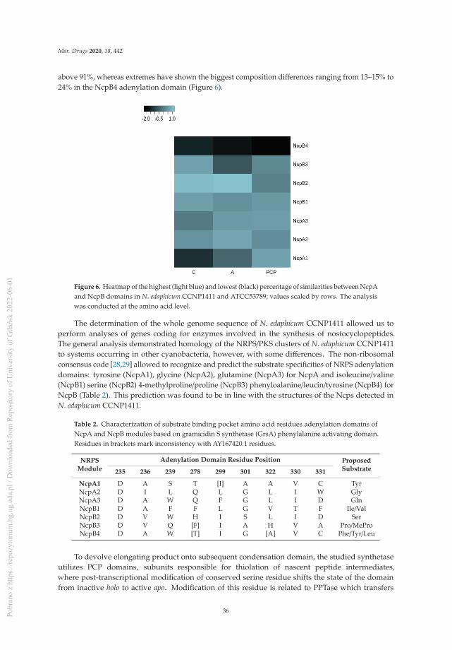
Mar. Drugs 2020, 18, 442
above 91%, whereas extremes have shown the biggest composition differences ranging from 13–15% to24% in the NcpB4 adenylation domain (Figure 6).
Figure 6. Heatmap of the highest (light blue) and lowest (black) percentage of similarities between NcpAand NcpB domains in N. edaphicum CCNP1411 and ATCC53789; values scaled by rows. The analysiswas conducted at the amino acid level.
The determination of the whole genome sequence of N. edaphicum CCNP1411 allowed us toperform analyses of genes coding for enzymes involved in the synthesis of nostocyclopeptides.The general analysis demonstrated homology of the NRPS/PKS clusters of N. edaphicum CCNP1411to systems occurring in other cyanobacteria, however, with some differences. The non-ribosomalconsensus code [28,29] allowed to recognize and predict the substrate specificities of NRPS adenylationdomains: tyrosine (NcpA1), glycine (NcpA2), glutamine (NcpA3) for NcpA and isoleucine/valine(NcpB1) serine (NcpB2) 4-methylproline/proline (NcpB3) phenyloalanine/leucin/tyrosine (NcpB4) forNcpB (Table 2). This prediction was found to be in line with the structures of the Ncps detected inN. edaphicum CCNP1411.
Table 2. Characterization of substrate binding pocket amino acid residues adenylation domains ofNcpA and NcpB modules based on gramicidin S synthetase (GrsA) phenylalanine activating domain.Residues in brackets mark inconsistency with AY167420.1 residues.
NRPSModule
Adenylation Domain Residue Position ProposedSubstrate235 236 239 278 299 301 322 330 331
NcpA1 D A S T [I] A A V C TyrNcpA2 D I L Q L G L I W GlyNcpA3 D A W Q F G L I D GlnNcpB1 D A F F L G V T F Ile/ValNcpB2 D V W H I S L I D SerNcpB3 D V Q [F] I A H V A Pro/MeProNcpB4 D A W [T] I G [A] V C Phe/Tyr/Leu
To devolve elongating product onto subsequent condensation domain, the studied synthetaseutilizes PCP domains, subunits responsible for thiolation of nascent peptide intermediates,where post-transcriptional modification of conserved serine residue shifts the state of the domainfrom inactive holo to active apo. Modification of this residue is related to PPTase which transfers
36Pobr
ano
z ht
tps:
//rep
ozyt
oriu
m.b
g.ug
.edu
.pl /
Dow
nloa
ded
from
Rep
osito
ry o
f Uni
vers
ity o
f Gda
ńsk
2022
-06-
01

Mar. Drugs 2020, 18, 442
a covalently-bound 4′-phosphopantetheine arm of CoA onto the PCP active site, enabling peptideintermediates to bind as reactive thioesters. Case residue which undergoes a nucleophilic attack by thehydroxyl group was conserved in every module within the PCP domain predicted at the front of thesecond helix [30].
The stand-alone docking domain (D) (7,617,812–7,617,964 bp) found on N-terminus of NcpAmay be an essential component mediating interactions, recognition and specific association withinNRPS subunits. The potential acceptor domain, based on sequence homology of conserved residues toC-terminal communication-mediating donor domains (COM), was found at the NcpB4 PCP domainsecond helix, encompassing conserved serine residue within potential binding sequence [31]. Moreover,this communication-mediating domain may putatively bind to C-terminus of NcpB3 and NcpB4condensation (C) domains based on conserved motif LLEGIV, found by sequence homology to lastfive amino-acids of C-terminal docking domains residues, key for their interactions [32]. Within thesame β-hairpin, a group of charged residues (ExxxxxKxR) putatively determines the binding affinityof the N-terminal domain [33].
Two tailoring domains encoded at the 5′ ends of ncpA and ncpB genes were classified asepimerization (E) (7,627,742–7,629,043 bp) domain and reductase (R) (7,642,183–7,643,238 bp) domain,accordingly. Epimerization domain catalyzes the conversion of L-amino acids to D-amino acids,a reaction coherent with D-stereochemistry of the peptide glutamine residue, where His of the conservedHHxxxDG motif and Glu from the upstream EGHGRE motif raceB comprise an epimerisation reactionactive site [34]. Homologous HHxxxDG conserved motif sequence is found in condensation domains(C), where a similar reaction is catalyzed within peptide bond formation, putatively by the secondHis residue [35]. As in ncp cluster [17], module NcpA1 motif includes degenerate sequences in twopositions HQIVGDL with leucine instead of phenylalanine residue at the start of the helix. The secondhistidine site-directed mutagenesis abolished enzymatic activity which might suggest that NcpA1condensation domain is inactive [36].
Reductase domain (R) found at the C-terminus of NRPS was classified as oxidoreductase.Despite 15% discrepancy in domain composition compared to NcpB core catalytic triad Thr-Tyr-Lysand Rossmann-fold, a NAD (P) H nucleotide-binding motif GxxGxxG positions were not affected. Themechanism driving this chain release utilizes NAD (P) H cofactor for redox reaction of the final moietyof the nascent peptide to aldehyde or alcohol [37,38].
2.3. Structure Characterization of Ncps Produced by N. edaphicum CCNP1411
Thus far, only three Ncps, Ncp-A1, A2 and M1, and their linear aldehydes were isolated as purenatural products of Nostoc strains [13,16]. Ncp-A3, with MePhe in the C-terminal position, was obtainedthrough aberrant biosynthesis in the Nostoc sp. ATCC53789 culture supplemented with MePhe [13].The linear aldehydes of Ncp-A1 and Ncp-A2, with Pro instead of MePro, were chemically synthesizedand used to study the Ncps epimerization and macrocyclization equilibria [19,20]. In our work, tenNcps, differing mainly in position 4 and 7, were detected by LC-MS/MS in the N. edaphicum CCNP1411cell extract (Table 3, Figure 1, Figure 7, Figure 8 and Figure S1–S7). These include five cyclic structures,four linear Ncp aldehydes, and one linear hexapeptide Ncp. The putative structures of the six peptides,which were found to be naturally produced by Nostoc for the first time, are marked in Table 3 in bold(Ncp-E1, Ncp-E1-L, Ncp-E2, Ncp-E2-L, Ncp-E3 and Ncp-E4-L).
37Pobr
ano
z ht
tps:
//rep
ozyt
oriu
m.b
g.ug
.edu
.pl /
Dow
nloa
ded
from
Rep
osito
ry o
f Uni
vers
ity o
f Gda
ńsk
2022
-06-
01
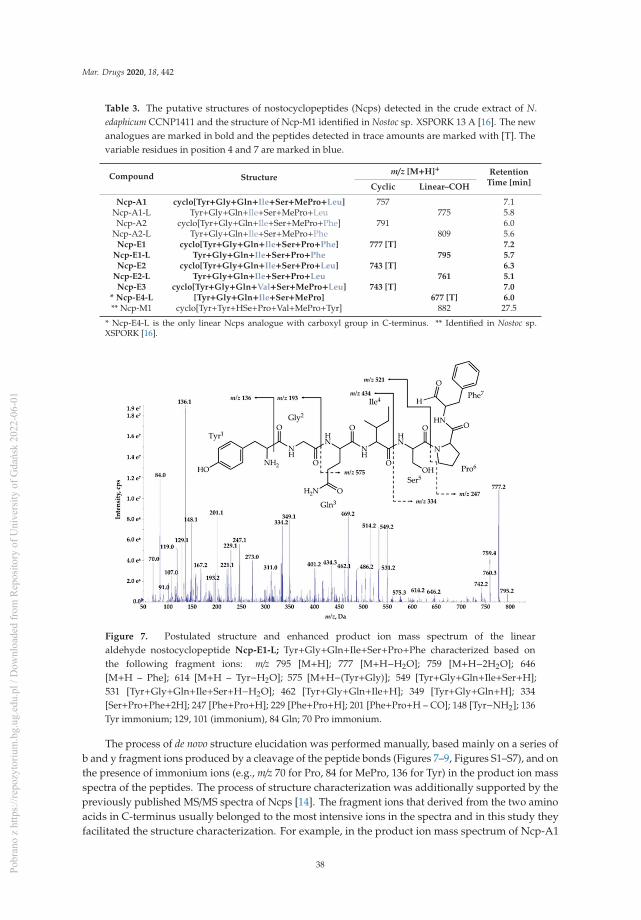
Mar. Drugs 2020, 18, 442
Table 3. The putative structures of nostocyclopeptides (Ncps) detected in the crude extract of N.edaphicum CCNP1411 and the structure of Ncp-M1 identified in Nostoc sp. XSPORK 13 A [16]. The newanalogues are marked in bold and the peptides detected in trace amounts are marked with [T]. Thevariable residues in position 4 and 7 are marked in blue.
Compound Structurem/z [M+H]+ Retention
Time [min]Cyclic Linear–COH
Ncp-A1 cyclo[Tyr+Gly+Gln+Ile+Ser+MePro+Leu] 757 7.1Ncp-A1-L Tyr+Gly+Gln+Ile+Ser+MePro+Leu 775 5.8Ncp-A2 cyclo[Tyr+Gly+Gln+Ile+Ser+MePro+Phe] 791 6.0
Ncp-A2-L Tyr+Gly+Gln+Ile+Ser+MePro+Phe 809 5.6Ncp-E1 cyclo[Tyr+Gly+Gln+Ile+Ser+Pro+Phe] 777 [T] 7.2
Ncp-E1-L Tyr+Gly+Gln+Ile+Ser+Pro+Phe 795 5.7Ncp-E2 cyclo[Tyr+Gly+Gln+Ile+Ser+Pro+Leu] 743 [T] 6.3
Ncp-E2-L Tyr+Gly+Gln+Ile+Ser+Pro+Leu 761 5.1Ncp-E3 cyclo[Tyr+Gly+Gln+Val+Ser+MePro+Leu] 743 [T] 7.0
* Ncp-E4-L [Tyr+Gly+Gln+Ile+Ser+MePro] 677 [T] 6.0** Ncp-M1 cyclo[Tyr+Tyr+HSe+Pro+Val+MePro+Tyr] 882 27.5
* Ncp-E4-L is the only linear Ncps analogue with carboxyl group in C-terminus. ** Identified in Nostoc sp.XSPORK [16].
Figure 7. Postulated structure and enhanced product ion mass spectrum of the linearaldehyde nostocyclopeptide Ncp-E1-L; Tyr+Gly+Gln+Ile+Ser+Pro+Phe characterized based onthe following fragment ions: m/z 795 [M+H]; 777 [M+H−H2O]; 759 [M+H−2H2O]; 646[M+H – Phe]; 614 [M+H – Tyr−H2O]; 575 [M+H−(Tyr+Gly)]; 549 [Tyr+Gly+Gln+Ile+Ser+H];531 [Tyr+Gly+Gln+Ile+Ser+H−H2O]; 462 [Tyr+Gly+Gln+Ile+H]; 349 [Tyr+Gly+Gln+H]; 334[Ser+Pro+Phe+2H]; 247 [Phe+Pro+H]; 229 [Phe+Pro+H]; 201 [Phe+Pro+H – CO]; 148 [Tyr−NH2]; 136Tyr immonium; 129, 101 (immonium), 84 Gln; 70 Pro immonium.
The process of de novo structure elucidation was performed manually, based mainly on a series ofb and y fragment ions produced by a cleavage of the peptide bonds (Figures 7–9, Figures S1–S7), and onthe presence of immonium ions (e.g., m/z 70 for Pro, 84 for MePro, 136 for Tyr) in the product ion massspectra of the peptides. The process of structure characterization was additionally supported by thepreviously published MS/MS spectra of Ncps [14]. The fragment ions that derived from the two aminoacids in C-terminus usually belonged to the most intensive ions in the spectra and in this study theyfacilitated the structure characterization. For example, in the product ion mass spectrum of Ncp-A1
38Pobr
ano
z ht
tps:
//rep
ozyt
oriu
m.b
g.ug
.edu
.pl /
Dow
nloa
ded
from
Rep
osito
ry o
f Uni
vers
ity o
f Gda
ńsk
2022
-06-
01

Mar. Drugs 2020, 18, 442
(Figure S1) and Ncp-E3 (Figure S7), ions at m/z 209 [MePro+Leu+H] and m/z 181 [MePro+Leu+H−CO]were present, while in the spectrum of Ncp-E2 (Figure S5) with Pro (instead MePro), the correspondingions at 14 unit lower m/z values, i.e., 195 and 167 were observed. The spectra of the linear Ncps containedthe intensive Tyr immonium ion at m/z 136. Based on the previously determined structures of Ncp-A1and Ncp-A2 [13], we assumed that in Ncp-E2, the amino acids in position 4 and 7, are Ile and Leu,respectively (Table 3; Figure S5). These two amino acids are difficult to distinguish by MS. Definitely,the NMR analyses are required to confirm the structures of the Ncps. The presence of Val in position 4,instead of Ile, distinguishes the Ncp-E3 from other Ncps produced by N. edaphicum CCNP1411. As itwas previously reported [17], and also confirmed in this study, the predicted substrates of the NcpB1protein encoded by ncpB and involved in the incorporation of the residue in position 4 are Ile/Leu andVal. However, the domain preferentially activates Ile, which explains why only traces of Val-containingNcps were detected in N. edaphicum CCNP1411 (Table 3).
Figure 8. Postulated tructure and enhanced product ion mass spectrum of a linear nostocyclopeptideNcp-E4-L [Tyr+Gly+Gln+Ile+Ser+MePro] characterized based on the following fragment ions:m/z 677 [M+H]; 659 [M+H−H2O]; 642 [M+H−H2O−NH3]; 549 [Tyr+Gly+Gln+Ile+Ser+H]; 531[Tyr+Gly+Gln+Ile+Ser+H−H2O]; 521 [Tyr+Gly+Gln+Ile+Ser+H−CO]; 462 [Tyr+Gly+Gln+Ile+H]; 434[Tyr+Gly+Gln+Ile+H−CO]; 349 [Tyr+Gly+Gln+H]; 329 [Gln+Ile+Ser+H]; 312 [Ile+Ser+MePro+H];221 [Tyr+Gly+H]; 193 [Tyr+Gly+H−CO]; 148 [Tyr−NH2]; 136 Tyr immonium; 86 Ile immonium; 84,101 (immonium), 129 Gln; 84 MePro immonium.
Methylated Pro (MePro) in position 6 is quite conserved. MePro is a rare non-proteinogenic aminoacid biosynthesized from Leu through the activity of the zinc-dependent long chain dehydrogenasesand Δ1-pyrroline-5-carboxylic acid (P5C) reductase homologue encoded by the gene cassettencpCDE [17,18,25]. The genes involved in the biosynthesis of MePro were found in 30 of the 116 testedcyanobacterial strains, majority (80%) of which belonged to the genus Nostoc [39]. The new Ncp-E1and Ncp-E2, detected at trace amounts, are the only Ncps produced by N. edaphicum CCNP1411which contain Pro (Table 3). The presence of m/z 84 ion in the fragmentation spectra of the two Ncpscomplicated the process of de novo structure elucidation. This ion corresponds to the immonium ionof MePro and could indicate the presence of this residue. However, the two ions m/z 101 and 129,which together with ion at m/z 84, are characteristic of Gln, suggested the presence of this amino acidin Ncp-E1 and Ncp-E2. The detailed characterization of Ncp fragmentation pathways is presented inFigures 7–9 and in Supplementary Materials (Figures S1–S7).
39Pobr
ano
z ht
tps:
//rep
ozyt
oriu
m.b
g.ug
.edu
.pl /
Dow
nloa
ded
from
Rep
osito
ry o
f Uni
vers
ity o
f Gda
ńsk
2022
-06-
01

Mar. Drugs 2020, 18, 442
Figure 9. Enhanced product ion mass spectrum of the cyclic nostocyclopeptide Ncp-E1
with putative structure cyclo[Tyr+Gly+Gln+Ile+Ser+Pro+Phe] characterised based on thefollowing fragment ions: m/z 777 [M+H]; 759 [M+H−H2O]; 741 [M+H−2H2O]; 690[M+H−Ser]; 672 [M+H−Ser−H2O]; 662 [M+H−Pro−H2O]; 646 [M+H−Phe]; 628 [M+H−Phe−H2O];575 [M+H−(Ser+Pro)−H2O]; 549 [Tyr+Gly+Glu+Ile+Ser+H]; 480 [Phe+Tyr+Gly+Gln+H]; 462[Tyr+Gly+Gln+Ile+H]; 444 [Tyr+Gly+Gln+Ile+H−H2O]; 434 [Tyr+Gly+Gln+Ile+H−CO]; 392[Pro+Phe+Tyr+H]; 352 [Phe+Tyr+Gly+H]; 335 [Phe+Tyr+Gly+H−H2O]; 316 [Ser+Pro+Phe+H]; 307[Phe+Tyr+Gly+H−H2O -CO]; 298 [Ile+Ser+Pro+H]; 229 [Phe+Pro+H]; 201 [Phe+Pro+H−CO]; 158[Gly+Gln+H−CO]; 132 Phe; 70 Pro immonium. Structure of the peptide is presented in Figure 1.
In addition to the heptapeptide Ncps, N. edaphicum CCNP1411 produces a small amount of thelinear hexapeptide, Ncp-E4-L, whose putative structure is Tyr+Gly+Gln+Ile+Ser+MePro (Table 3,Figure 9). This Ncp was detected only when higher biomass of Nostoc was extracted. As the proposedamino acids sequence in this molecule is the same as the sequence of the first six residues in Ncp-A1and Ncp-A2, the hexapeptide can be a precursor of the two Ncps. The other option is that the cellconcentration of the Ncps is self-regulated and the Ncp-E4-L is released through proteolytic cleavageof the final products. This hypothesis could be verified when the role of the Ncps for the produceris discovered. In the ncp gene cluster, the presence of ncpG encoding the NcpG peptidase, with highhomology to enzymes hydrolyzing D-amino acid-containing peptides was revealed by Becker et al. [17]and also confirmed in this study. Therefore, the in-cell degradation of Ncps by the NcpG peptidase ispossible, but it probably proceeds at D-Gln and gives other products than Ncp-E4-L.
2.4. Production of Ncps by N. edaphicum CCNP1411
Apart from the structural analysis, we also made attempts to determine the relative amountsof the individual Ncps produced by N. edaphicum CCNP1411. To exclude the effect of the extractionprocedure on the amounts of the detected peptides, different solvents and pH were applied. As theprocess of Ncp linearization during long storage of the freeze-dried material was suggested [16], boththe fresh and lyophilized biomasses were analyzed. Regardless of the extraction procedure, Ncp-A2-Lwith Phe in C-terminus was always found to be the main Ncp analogue (Figure 10A–D). In addition,when MePro and Pro-containing peptide were compared separately, the peak intensity of the linearNcps with Phe in C-terminus (i.e., Ncp-A2-L and Ncp-E1-L) was higher than the Ncps with Leu. Theseresults might indicate preferential incorporation of Phe into the synthesized peptide chain.
40Pobr
ano
z ht
tps:
//rep
ozyt
oriu
m.b
g.ug
.edu
.pl /
Dow
nloa
ded
from
Rep
osito
ry o
f Uni
vers
ity o
f Gda
ńsk
2022
-06-
01

Mar. Drugs 2020, 18, 442
Figure 10. Relative cell contents of nostocyclopeptides extracted from 10 mg of lyophilized (A,B,D)or 500 mg fresh (C) biomass of N. edaphicum CCNP1411 with MilliQ water (A), 50% methanol inwater (B), and 20% methanol in water (C,D). The cell content was expressed as peak intensity inLC-MS/MS chromatogram.
The study also showed that the cell contents of the linear Ncps are higher than the cyclic ones.(Figure 10A–D and Figure S8). The release of Ncps from the synthetase as linear aldehydes is catalyzedby a reductase domain, located in the C-terminal part of the NcpB [17]. This reductive release triggersthe spontaneous, and enzyme independent, macrocyclization of the linear peptide [19,20]. The reactionleads to the formation of a stable imino bond between the C-terminal aldehyde and the N-terminalamine group of Tyr [19,20]. In N. edaphicum CCNP1411 cells, depending on the Ncp analogue,the analyzed material (fresh or lyophilized) and extraction solvent, the cyclic Ncps constituted fromeven less than 10% (Ncp-A2, fresh biomass) to over 90% of the linear peptide (Figure 10A–D andFigure S8). In case of Ncp-A1, with MePro-Leu in C-terminus, the contribution of the cyclic form wasalways most significant, and at pH 8 it reached up to 91.7% of the linear peptide aldehyde (Ncp-A1-L)(Figure 10A–D and Figure S8). The cyclic analogues, Ncp-E1 and Ncp-E3 were produced in traceamounts and their spectra were sporadically recorded. It was proven that the macrocyclization processof Ncps is determined by the geometry of the linear peptide aldehyde and the conformation of D-Glnand Gly is crucial for the folding and formation of the imino bond [19]. As these two residues arepresent in all detected Ncps, then, probably other elements of the structure affect the cyclizationprocess, as well. We hypothesize that due to the steric hindrances, the cyclization of Ncp-A1 withLeu in C-terminal position is easier than the cyclization of Ncp-A2 with Phe. As a consequence, theproportion of the cyclic Leu-containing Ncp-A1 to the linear form of the peptide is higher.
Thus far, Ncps synthesis was reported in few Nostoc strains, and the structural diversity of thepeptides was found to be low. Other classes of NRPs were detected in cyanobacteria representingdifferent orders and genera, and within one class of the peptides numerous analogues were detected. Forexample, the number of naturally produced cyclic heptapeptide microcystins (MCs), is over 270 [40,41]and in one cyanobacterial strains even 47 MCs analogues were detected [40]. Cyanopeptolinsare produced by many cyanobacterial taxa and so far more than 190 structural analogues of thepeptides have been discovered [41]. In this work, cyanopeptolin gene cluster was identified in N.edaphicum CCNP1411 and thirteen products of the genes were previously reported [6]. These peptidescontain seven amino acids and a short fatty acid chain, and only one element of the structure,3-amino-6-hydroxy-2-piperidone (Ahp), is conserved [6]. The structural diversity of NRPs is generatedby frequent genetic recombination events and point mutations in the NRP gene cluster. The changes ingene sequences affect the structure and substrate specificity of the encoded enzymes. The tailoring
41Pobr
ano
z ht
tps:
//rep
ozyt
oriu
m.b
g.ug
.edu
.pl /
Dow
nloa
ded
from
Rep
osito
ry o
f Uni
vers
ity o
f Gda
ńsk
2022
-06-
01

Mar. Drugs 2020, 18, 442
enzymes can further modify the product, leading to even higher diversity of the synthetizedpeptides [42]. In case of Ncps, both the number of the producing organisms and the structuraldiversity of the peptides are limited. Ncp-M1 from Nostoc living in symbiosis with gastropod [16] isthe only Ncp with structure distinctly different from Ncp-A1, Ncp-A2 and other Ncps described inthis work.
The diversity within one class of bioactive metabolites offers a good opportunity forstructure-activity relationship studies, without the need to synthesize the variants. The studiesare of paramount importance when the efficacy and safety of a drug candidate are optimized.Therefore, in our future work, when sufficient quantities of pure Ncps are isolated, the activity ofindividual analogues against different cellular targets will be tested and compared, in order to selectthe lead compound for further studies.
3. Materials and Methods
3.1. Isolation, Purification and Culturing of Nostoc CCNP1411
Nostoc strain CCNP1411 was isolated in 2010 from the Gulf of Gdansk, southern Baltic Sea,by Dr. Justyna Kobos. Based on the 16S rRNA sequence (GenBank accession number KJ161445)and morphological features, such as the shape of trichomes, cell size (4.56 ± 0.30 μm wide and4.12 ± 0.72 μm long) and lack of akinetes [43,44], the strain was classified to N. edaphicum species.Purification of the strain was carried out by multiple transfers to a liquid and solid (1% bacterialagar) Z8 medium supplemented with NaCl to obtain the salinity of 7.3 [45]. To establish the strainas a monoculture, free from accompanying heterotrophic bacteria, a third-generation cephalosporin,ceftriaxone (100 μg/mL) (Pol-Aura, Olsztyn, Poland) was used. In addition, the purity of the culturewas regularly tested by inoculation on LA agar (solid LB medium with 1.5% agar) [46] and on agarColumbia +5% sheep blood (BTL Ltd. Łódz, Poland), a highly nutritious medium, recommended forfastidious bacteria. Cyanobacteria cultures were grown in liquid Z8 medium (100 mL) at 22 ± 1 ◦C,continuous light of 5–10 μmol photons m−2 s−1. After three weeks of growth, the cyanobacterialbiomass was harvested by passing the culture through a nylon net (mesh size 25 μm) and thenfreeze-dried before further processing.
3.2. Isolation and Sequencing of Genomic DNA
Genomic DNA of N. edaphicum CCNP1411 was isolated using SDS/Phenol method as describedpreviously [47,48]. DNA quality control was performed by measuring the absorbance at 260/230 nm,template concentration was determined using Qubit fluorimeter (Thermo Fisher Scientific, Waltham,MA, USA), and DNA integrity was analyzed by 0.8% agarose gel electrophoresis and by PFGE usingBiorad CHEF-III instrument (BioRad, Hercules, CA, USA).
Paired-end sequencing library was constructed using the NEB Ultra II FS Preparation Kit(New England Biolabs, Beverly, CA, USA) according to the manufacturer’s instructions. The librarywas sequenced using an Illumina MiSeq platform (Illumina, San Diego, CA, USA) with 2 × 300paired-end reads. Sequence quality metrics were assessed using FASTQC (http://www.bioinformatics.babraham.ac.uk/projects/fastqc/) [49].
The long reads were obtained using the GridION sequencer (Oxford Nanopore Technologies,Oxford, UK). Prior to long-read library preparation, genomic DNA was sheared into 30 kb fragmentsusing 26 G needle followed by size selection on Bluepippin instrument (Sage Science, Beverly, MA,USA). DNA fragments above 20 kb were recovered using PAC30 kb cassette. 5 μg of recovered DNAwas taken for 1D library construction using SQK-LSK109 kit and 0.5 μg of the final library was loadedinto R9.4.1 flowcell and sequenced on MinION sequencer.
42Pobr
ano
z ht
tps:
//rep
ozyt
oriu
m.b
g.ug
.edu
.pl /
Dow
nloa
ded
from
Rep
osito
ry o
f Uni
vers
ity o
f Gda
ńsk
2022
-06-
01

Mar. Drugs 2020, 18, 442
3.3. Genome Assembling
Raw nanopore data was basecalled using Guppy v3.2.2 (Oxford Nanopore Technologies,Oxford, UK). After quality filtering using NanoFilt [50] and residual adapter removal usingPorechop (https://github.com/rrwick/Porechop), the obtained dataset was quality checked usingNanoPlot [50]. Long nanopore reads were then assembled using Flye v2.6 [51]. Flye assembledcontigs were further polished using Illumina sequencing reads and Unicycler_polish pipeline(https://github.com/rrwick/Unicycler).
3.4. Genome and NRPS Alignment
Genome assembly was annotated using the NCBI Prokaryotic Genome Annotation Pipeline [52]with the assistance of prokka [53] refine annotation, with additionally curated database comprised ofsequences selected by Nostocales order from NCBI non-redundant and refseq_genomes (280 positions)databases, enriched by 35 NRPS/PKS clusters selected by cyanobacteria phylum. To create circularmaps of N. edaphicum CCNP1411 genome, the CGView Comparison Tool [54] was engaged withadditional GC skew and GC content analyses.
Selected span for potential NRPS cluster was confirmed with BLASTn, BLASTp [55] andantiSMASH [56]. ORFs start codons within a putative cluster were verified by the presence ofribosome binding sites, 4–12 nucleotides upstream of the start codon. Schematic comparison ofORF BLASTn from relative synthetases, AY167420.1 and CP026681.1, was visualized by EasyFigprogram (http://mjsull.github.io/Easyfig/files.html). Annotated regions of NRPS span were subjectedfor NCBI Conserved Domain Database search [57] with a set e-value threshold (10−3), determiningevolutionarily-conserved protein domains and motifs against CDD v3.18 database. Recognized motifswere selected using samtools v.1.9 and were subjected for protein structure and function predictionby I-TASSER [58], and results were confirmed with literature reports, PKS/NRPS Analysis Web-siteprediction [59] and reevaluated using MEGAX suite [60]. Amino-acid sequence was visualized byBOXSHADE 3.2 program (https://embnet.vital-it.ch/software/BOX_form.html). Determination ofdomain ligand binding and active sites was achieved using COFACTOR and COACH part of I-TASSERanalyses confirmed by MUSCLE amino acid alignment from MEGA X.
3.5. Data Deposition
Genomic sequences generated and analyzed in this study were deposited in the GenBank databaseunder BioProject number: PRJNA638531.
3.6. Extraction and LC-MS/MS Analysis
For LC-MS/MS analyses of Ncps, the lyophilized (10 mg DW) biomass of N. edaphicum CCNP1411was homogenized by grinding with mortar and pestle, and extracted in 1 mL of milliQ water, 20%methanol (pH 3.5, 6.0 and 8.0) and 50% methanol in water. The pH was adjusted with 0.5 M HCl and1.0 M NaOH. In addition, the fresh material (500 mg FW) was extracted in 20% methanol in water. Thesamples were vortexed for 15 min and centrifuged at 14,000 rpm for 10 min, at 4 ◦C. The collectedsupernatants were directly analyzed by LC-MS/MS system.
The LC-MS/MS was carried out on an Agilent 1200 HPLC (Agilent Technologies, Waldbronn,Germany) coupled to a hybrid triple quadrupole/linear ion trap mass spectrometer QTRAP5500(Applied Biosystems MDS Sciex, Concord, ON, Canada). The separation was achieved on a ZorbaxEclipse XDB-C18 column (4.6 mm ID × 150 mm, 5 μm; Agilent Technologies, Santa Clara, CA, USA).The extract components were separated by gradient elution from 10% to 100% B (acetonitrile with 0.1%formic acid) over 25 min, at a flow rate of 0.6 mL/min. As solvent A, 5% acetonitrile in MilliQ waterwith 0.1% formic acid was used. The mass spectrometer was operated in positive mode, with turbo ionsource (5.5 kV; 550 ◦C). An information-dependent acquisition method at the following settings wasused: for ions within the m/z range 500–1100 and signal intensity above the threshold of 500,000 cps
43Pobr
ano
z ht
tps:
//rep
ozyt
oriu
m.b
g.ug
.edu
.pl /
Dow
nloa
ded
from
Rep
osito
ry o
f Uni
vers
ity o
f Gda
ńsk
2022
-06-
01

Mar. Drugs 2020, 18, 442
the MS/MS spectra were acquired within the m/z range 50–1000, at a collision energy of 60 eV anddeclustering potential of 80 eV. Data were acquired with the Analyst ® Sofware (version 1.7 AppliedBiosystems, Concord, ON, Canada).
4. Conclusions
Genes coding for subunits of the non-ribosomal peptide synthetase, in nostocyclopeptide-producing N. edaphicum CCNP1411, revealed differences in nucleotide compositions, compared tothe previously described ncp cluster of Nostoc sp. ATCC53789. Although the analysis of fragmentsof genes coding for active sites and ligand binding sites of the conserved protein domains derivedfrom N. edaphicum CCNP1411 and Nostoc sp. ATCC53789 indicated identical amino-acid compositions,residues within adenylation domains and substrate binding sites were different between comparedsequences. This finding may highlight sites prone to mutations within regions accounted for structureand substrate stability. Analysis of ncp gene products in N. edaphicum CCNP1411 led to the detection ofnew nostocyclopeptide analogues. However, modifications in their structure were minor and limitedto three positions of the heptapeptides. Although the naturally produced nostocyclopeptides werepreviously described as cyclic structures, in N. edaphicum CCNP1411 they are mainly present as linearpeptide aldehydes, indicating a slow cyclization process.
Supplementary Materials: The following are available online at http://www.mdpi.com/1660-3397/18/9/442/s1. Figure S1: Structure and enhanced product ion mass spectrum of the cyclic nostocyclopeptideNcp-A1 cyclo [Tyr+Gly+Gln+Ile+Ser+MePro+Leu] identified based on the following fragment ions:m/z 757 [M+H]; 739 [M+H−H2O]; 729 [M+H−CO]; 721 [M+H−2H2O]; 628 [M+H−MePro−H2O],626 [M+H−Ile−H2O]; 594 [M+H−Tyr]; 549 [Tyr+Gly+Gln+Ile+Ser+H]; 541 [M+H−(Ser+MePro)−H2O],446 [M+H−(Ile+Ser+MePro)]; 428 [M+H−(Ile+Ser+MePro)−H2O]; 386 [Gly+Gln+Ile+Ser+H]; 372[MePro+Leu+Tyr+H]; 300 [Leu+Tyr+Gly+H−H2O]; 209 [MePro+Leu+H]; 181 [MePro+Leu+H−CO]; 86−Ile/Leuimmonium; 84 MePro immonium, Figure S2: Structure and enhanced product ion mass spectrum ofthe linear peptide aldehyde Ncp-A1-L (linear aldehyde of Ncp-A1) Tyr+Gly+Gln+Ile+Ser+MePro+Leuidentified based on the following fragment ions: m/z 775 [M+H]; 757 [M+H – H2O]; 739[M+H−2H2O]; 660 [M+H−Leu]; 549 [Tyr+Gly+Gln+Ile+Ser+H]; 531 [Tyr+Gly+Gln+Ile+Ser+H−H2O];521 [Tyr+Gly+Gln+Ile+Ser+H−CO]; 532 [M+H−(MePro+Leu)−H2O]; 462 [Tyr+Gly+Gln+Ile+H]; 434[Tyr+Gly+Gln+Ile+H−CO]; 386 [Gly+Gln+Ile+Ser+H]; 349 [Tyr+Gly+Gln+H]; 301 [Gln+Ile+Ser+H−CO];227 [MePro+Leu+H]; 221 [Tyr+Gly+H]; 209 [MePro+Leu+H−H2O]; 181 [MePro+Ile+H−H2O−CO]; 148[Tyr−NH2]; 136 Tyr immonium; 86−Ile/Leu immonium; 84, 101 (immonium),129 Gln; 84 MePro immonium,Figure S3: Structure and enhanced product ion mass spectrum of the cyclic nostocyclopeptide Ncp-A2cyclo[Tyr+Gly+Gln+Ile+Ser+MePro+Phe] identified based on the following fragment ions: m/z 791[M+H]; 773 [M+H−H2O]; 763 [M+H−CO]; 755 [M+H−2H2O]; 745 [M+H−CO−H2O]; 678 [M+H−Ile]; 628[M+H−Tyr]; 593 [M+H−(Ser+MePro)]; 549 [Tyr+Gly+Gln+Ile+Ser+H]; 531 [Tyr+Gly+Gln+Ile+Ser+H−H2O]; 480[M+H−(Ile+Ser+MePro)]; 462 [Tyr+Gly+Gln+Ile+H]; 406 [MePro+Phe+Tyr+H]; 379 [MePro+Phe+Tyr+H−CO];349 [Tyr+Gly+Gln+H]; 335 [Phe+Tyr+Gly+H−H2O]; 312 [Ile+Ser+MePro+H]; 307 [Phe+Tyr+Gly+H−H2O−CO];243 [MePro+Phe+H]; 215 [MePro+Phe+H−CO]; 158 [Gly+Gln+H−CO]; 132 Phe; 84 MePro immonium, FigureS4: Structure and enhanced product ion mass spectrum of the linear nostocyclopeptide aldehyde Ncp-A2-L(linear aldehyde of Ncp-A2) Tyr+Gly+Gln+Ile+Ser+MePro+Phe identified based on the following fragmentions: m/z 809 [M+H]; 791 [M+H−H2O]; 773 [M+H−2H2O]; 763 [M+H−CO−H2O]; 660 [M+H−Phe]; 628[M+H−Tyr−H2O]; 549 [Tyr+Gly+Gln+Ile+Ser+H]; 531 [M+H−(MePro+Phe)−H2O]; 462 [Tyr+Gly+Gln+Ile+H];434 [Tyr+Gly+Gln+Ile+H−CO]; 312 [Ile+Ser+MePro+H]; 261 [MePro+Phe+H]; 243 [MePro+Phe+H – H2O];221 [Tyr+Gly+H], 193 [Tyr+Gly+ H−CO]; 148 [Tyr−NH2]; 136 Tyr immonium; 84, 101 (immonium), 129Gln; 84 MePro immonium, Figure S5: Proposed structure and enhanced product ion mass spectrumof cyclic nostocyclopeptide Ncp-E2 cyclo[Tyr+Gly+Gln+Ile+Ser+Pro+Leu] characterized based on thefollowing fragment ions: m/z 743 [M+H]; 725 [M+H−H2O]; 715 [M+H−CO]; 707 [M+H−2H2O]; 697[M+H – H2O−CO]; 656 [M+H−Ser]; 638 [M+H−Ser−H2O]; 628 [M+H−Ser−CO]; 612 [M+H−Ile−H2O];549 [Tyr+Gly+Gln+Ile+Ser+H]; 541 [M+H−(Ser+Pro)−H2O]; 531 [Tyr+Gly+Gln+Ile+Ser+H−H2O]; 428[M+H−(Ile+Ser+Pro)−H2O]; 349 [Tyr+Gly+Gln+H]; 300 [Leu+Tyr+Gly+H−H2O]; 195 [Pro+Leu+H]; 167[Pro+Leu+H−CO]; 84 Gln; 70 Pro immonium, Figure S6: Proposed structure and enhanced product ionmass spectrum of the linear nostocyclopeptide aldehyde Ncp-E2-L (linear aldehyde of Ncp-E2) with generalstructure Tyr+Gly+Gln+Ile+Ser+Pro+Leu characterized based on the following fragment ions: m/z 761 [M+H];743 [M+H−H2O]; 725 [M+H−2H2O]; 549 [Tyr+Gly+Gln+Ile+Ser+H]; 532 [Tyr+Gly+Gln+Ile+Ser+H−H2O];462 [Tyr+Gly+Gln+Ile+H]; 349 [Tyr+Gly+Gln+H]; 434 [Tyr+Gly+Gln+Ile+H−CO]; 300 [Ser+Pro+Leu+H]; 221[Tyr+Gly+H]; 213 [Pro+Leu+H]; 195 [Pro+Leu+H−H2O]; 148 [Tyr−NH2]; 136 Tyr immonium; 84, 101 (immonium),129 Gln; 70 Pro immonium, Figure S7: Proposed structure and enhanced product ion mass spectrum of cyclicnostocyclopeptide Ncp-E3 cyclo[Tyr+Gly+Gln+Val+Ser+MePro+Leu] characterized based on the followingfragment ions: m/z 743 [M+H]; 725 [M+H−H2O]; 715 [M+H−CO]; 707 [M+H – 2H2O]; 697 [M+H−H2O−CO];
44Pobr
ano
z ht
tps:
//rep
ozyt
oriu
m.b
g.ug
.edu
.pl /
Dow
nloa
ded
from
Rep
osito
ry o
f Uni
vers
ity o
f Gda
ńsk
2022
-06-
01
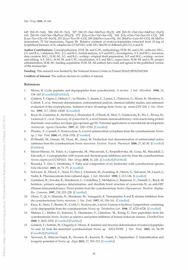
Mar. Drugs 2020, 18, 442
645 [M+H−Val]; 580 [M+H−Tyr]; 527 [M+H−(Ser+MePro)−H2O]; 428 [M+H−(Val+Ser+MePro)−H2O];410 [M+H−(Val+Ser+MePro)−2H2O]; 372 [Gly+Gln+Val+Ser+H]; 344 [Gly+Gln+Val+Ser+H−CO]; 300[Leu+Tyr+Gly+H−H2O]; 233 [Leu+Tyr+H−CO]; 209 [MePro+Leu+H]; 181 [MePro−Leu+H−CO]; 84 MeProimmonium; 72 Val immonium, Figure S8: Relative contents of nostocyclopeptides extracted from 10 mg oflyophilized biomass of N. edaphicum CCNP1411 with 20% MeOH of different pH (3.5, 6 and 8)
Author Contributions: Conceptualization, H.M.-M. and G.W.; methodology, H.M.-M. and G.W.; software, M.G.,J.G. and R.G.; validation, M.G., J.G. and R.G.; formal analysis, A.F. and M.G.; investigation, A.F. and M.G.; resources,data curation, M.G., H.M.-M., J.G. and R.G.; writing—original draft preparation, A.F. and M.G.; writing—reviewand editing, A.F., M.G.; H.M.-M. and G.W.; visualization, A.F. and M.G.; supervision, H.M.-M. and G.W.; projectadministration, H.M.-M.; funding acquisition, H.M.-M. All authors have read and agreed to the published versionof the manuscript.
Funding: This research was funded by the National Science Centre in Poland 2016/21/B/NZ9/02304.
Conflicts of Interest: The authors declare no conflict of interest.
References
1. Moore, R. Cyclic peptides and depsipeptides from cyanobacteria: A review. J. Ind. Microbiol. 1996, 16,134–143. [CrossRef] [PubMed]
2. Golakoti, T.; Ogino, J.; Heltzel, C.; Le Husebo, T.; Jensen, C.; Larsen, L.; Patterson, G.; Moore, R.; Mooberry, S.;Corbett, T.; et al. Structure determination, conformational analysis, chemical stability studies, and antitumorevaluation of the cryptophycins. Isolation of new 18 analogs from Nostoc sp. strain GSV 224. J. Am. Chem.Soc. 1995, 117, 12030–12049. [CrossRef]
3. Boyd, M.; Gustafson, K.; McMahon, J.; Shoemaker, R.; O’Keefe, B.; Mori, T.; Gulakowski, R.; Wu, L.; Rivera, M.;Laurencot, C.; et al. Discovery of cyanovirin-N, a novel human immunodeficiency virus-inactivating proteinthat binds viral surface envelope glycoprotein gp120: Potential applications to microbicide development.Antimicrob. Agents Chemother. 1997, 41, 1521–1530. [CrossRef]
4. Ploutno, A.; Carmeli, S. Nostocyclyne A, a novel antimicrobial cyclophane from the cyanobacterium Nostocsp. J. Nat. Prod. 2000, 63, 1524–1526. [CrossRef]
5. El-Sheekh, M.; Osman, M.; Dyan, M.; Amer, M. Production and characterization of antimicrobial activesubstance from the cyanobacterium Nostoc muscorum. Environ. Toxicol. Pharmacol. 2006, 21, 42–50. [CrossRef][PubMed]
6. Mazur-Marzec, H.; Fidor, A.; Cegłowska, M.; Wieczerzak, E.; Kropidłowska, M.; Goua, M.; Macaskill, J.;Edwards, C. Cyanopeptolins with trypsin and chymotrypsin inhibitory activity from the cyanobacteriumNostoc edaphicum CCNP1411. Mar. Drugs 2018, 16, 220. [CrossRef] [PubMed]
7. Rezanka, T.; Dor, I.; Dembitsky, V. Fatty acid composition of six freshwater wild cyanobacterial species.Folia Microbiol. 2003, 48, 71–75. [CrossRef]
8. Schwartz, R.; Hirsch, C.; Sesin, D.; Flor, J.; Chartrain, M.; Fromtling, R.; Harris, G.; Salvatore, M.; Liesch, J.;Yudin, K. Pharmaceuticals from cultured algae. J. Ind. Microbiol. 1990, 5, 113–124. [CrossRef]
9. Gustafson, K.; Sowder, R.; Henderson, L.; Cardellina, J.; McMahon, J.; Rajamani, U.; Pannell, L.; Boyd, M.Isolation, primary sequence determination, and disulfide bond structure of cyanovirin-N, an anti-HIV(Human Immunodeficiency Virus) protein from the cyanobacterium Nostoc ellipsosporum. Biochem. Biophys.Res. Commun. 1997, 238, 223–228. [CrossRef]
10. Okino, T.; Qi, S.; Matsuda, H.; Murakami, M.; Yamaguchi, K. Nostopeptins A and B, elastase inhibitors fromthe cyanobacterium Nostoc minutum. J. Nat. Prod. 1997, 60, 158–161. [CrossRef]
11. Kaya, K.; Sano, T.; Beattie, K.; Codd, G. Nostocyclin, a novel 3-amino-6-hydroxy-2-piperidone containingcyclic depsipeptide from the cyanobacterium Nostoc sp. Tetrahedron Lett. 1996, 37, 6725–6728. [CrossRef]
12. Mehner, C.; Müller, D.; Kehraus, S.; Hautmann, S.; Gütschow, M.; König, G. New peptolides from thecyanobacterium Nostoc insulare as selective and potent inhibitors of human leukocyte elastase. ChemBioChem2008, 9, 2692–2703. [CrossRef] [PubMed]
13. Golakoti, T.; Yoshida, W.; Chaganty, S.; Moore, R. Isolation and structure determination of nostocyclopeptidesA1 and A2 from the terrestrial cyanobacterium Nostoc sp. ATCC53789. J. Nat. Prod. 2001, 64, 54–59.[CrossRef] [PubMed]
14. Nowruzi, B.; Khavari-Nejad, R.; Sivonen, K.; Kazemi, B.; Najafi, F.; Nejadsattari, T. Indentification andtoxigenic potential of Nostoc sp. Algae 2012, 27, 303–313. [CrossRef]
45Pobr
ano
z ht
tps:
//rep
ozyt
oriu
m.b
g.ug
.edu
.pl /
Dow
nloa
ded
from
Rep
osito
ry o
f Uni
vers
ity o
f Gda
ńsk
2022
-06-
01

Mar. Drugs 2020, 18, 442
15. Liaimer, A.; Jensen, J.; Dittmann, E. A genetic and chemical perspective on symbiotic recruitment ofcyanobacteria of the genus Nostoc into the host plant Blasia pusilla L. Front. Microbiol. 2016, 7, 1963. [CrossRef][PubMed]
16. Jokela, J.; Herfindal, L.; Wahlsten, M.; Permi, P.; Selheim, F.; Vasconçelos, V.; Døskeland, S.; Sivonen, K. Anovel cyanobacterial nostocyklopeptide is a potent antitoxin against Microcystis. ChemBioChem 2010, 11,1594–1599. [CrossRef]
17. Becker, J.; Moore, R.; Moore, B. Cloning, sequencing, and biochemical characterization of thenostocycyclopeptide biosynthetic gene cluster: Molecular basis for imine macrocyclization. Gene 2004, 325,35–42. [CrossRef]
18. Hoffmann, D.; Hevel, J.; Moore, R.; Moore, B. Sequence analysis and biochemical characterization of thenostopeptolide A biosynthetic gene cluster from Nostoc sp. GSV224. Gene 2003, 311, 171–180. [CrossRef]
19. Kopp, F.; Mahlet, C.; Grünewald, J.; Marahiel, M. Peptide macrocyclization: The reductase of thenostocyclopeptide synthetase triggers the self-assembly of a macrocyclic imine. J. Am. Chem. Soc.2006, 128, 16478–16479. [CrossRef]
20. Enck, S.; Kopp, F.; Marahiel, M.; Geyer, A. The entropy balance of nostocyclopeptide macrocyclizationanalysed by NMR spectroscopy. ChemBioChem 2008, 9, 2597–2601. [CrossRef]
21. Herfindal, L.; Myhren, L.; Kleppe, R.; Krakstad, C.; Selheim, F.; Jokela, J.; Sivonen, K.; Døskeland, S.Nostocyclopeptide-M1: A potent, nontoxic inhibitor of the hepatocyte drug trasporters OATP1B3 andOATP1B1. Mol. Pharm. 2011, 8, 360–367. [CrossRef]
22. Lee, W.; Belkhiri, A.; Lockhart, A.; Merchant, N.; Glaeser, H.; Harris, E.; Washington, M.; Brunt, E.; Zaika, A.;Kim, R.; et al. Overexpression of OATP1B3 confers apoptotic resistance in colon cancer. Cancer Res. 2008, 68,10315–10323. [CrossRef] [PubMed]
23. Nishizawa, T.; Ueda, A.; Nakano, T.; Nishizawa, A.; Miura, T.; Asayama, M.; Fuji, K.; Harada, K.; Shirai, M.Characterization of the locus of genes encoding enzymes producing heptadepsipeptide micropeptin in theunicellular cyanobacterium Microcystis. J. Biochem. 2011, 149, 475–485. [CrossRef] [PubMed]
24. Rouhiainen, L.; Jokela, J.; Fewer, D.; Urmann, M.; Sivonen, K. Two alternative starter modules for thenon-ribosomal biosynthesis of specific anabaenopeptin variants in Anabaena (cyanobacteria). Chem. Biol.2010, 17, 265–273. [CrossRef] [PubMed]
25. Luesch, H.; Hoffmann, D.; Hevel, J.; Becker, J.; Golakoti, T.; Moore, R. Biosynthesis of 4-Methylproline inCyanobacteria: Cloning of nosE and nosF and biochemical characterization of the encoded dehydrogenaseand reductase activities. J. Org. Chem. 2003, 68, 83–91. [CrossRef] [PubMed]
26. Lambalot, R.; Walsh, C. Cloning, Overproduction, and Characterization of the Escherichia coli Holo-acylCarrier Protein Synthase. J. Biol. Chem. 1995, 270, 24658–24661. [CrossRef]
27. Marchler-Bauer, A.; Derbyshire, M.; Gonzales, N.; Lu, S.; Chitsaz, F.; Geer, L.; Geer, R.; He, J.; Gwadz, M.;Hurwitz, D.; et al. CDD: NCBI’s conserved domain database. Nucleic Acids Res. 2015, 43, d222–d226.[CrossRef]
28. Stachelhaus, T.; Mootz, H.; Marahiel, M. The specificity-conferring code of adenylation domains innonribosomal peptide synthetases. Chem. Biol. 1999, 6, 493–505. [CrossRef]
29. Challis, G.; Ravel, J.; Townsend, C. Predictive, structure-based model of amino acid recognition bynonribosomal peptide synthetase adenylation domains. Chem. Biol. 2000, 7, 211–224. [CrossRef]
30. Stein, T.; Vater, J.; Kruft, V.; Otto, A.; Wittmann-Liebold, B.; Franke, P.; Panico, M.; McDowell, R.; Morris, H.The multiple carrier model of nonribosomal peptide biosynthesis at modular multienzymatic templates.J. Biol. Chem. 1996, 271, 15428–15435. [CrossRef]
31. Dehling, E.; Volkmann, G.; Matern, J.; Dörner, W.; Alfermann, J.; Diecker, J.; Mootz, H. Mapping of theCommunication-Mediating Interface in Nonribosomal Peptide Synthetases Using a Genetically EncodedPhotocrosslinker Supports an Upside-Down Helix-Hand Motif. J. Mol. Biol. 2016, 428, 4345–4360. [CrossRef]
32. Hacker, C.; Cai, X.; Kegler, C.; Zhao, L.; Weickhmann, K.; Wurm, J.; Bode, H.; Wöhnert, J. Structure-basedredesign of docking domain interactions modulates the product spectrum of a rhabdopeptide-synthesizingNRPS. Nat. Commun. 2018, 9, 4366. [CrossRef]
33. Richter, C.; Nietlispach, D.; Broadhurst, R.; Weissman, K. Multienzyme docking in hybrid megasynthetases.Nat. Chem. Biol. 2008, 4, 75–81. [CrossRef] [PubMed]
46Pobr
ano
z ht
tps:
//rep
ozyt
oriu
m.b
g.ug
.edu
.pl /
Dow
nloa
ded
from
Rep
osito
ry o
f Uni
vers
ity o
f Gda
ńsk
2022
-06-
01
Mar. Drugs 2020, 18, 442
34. Haese, A.; Schubert, M.; Herrmann, M.; Zocher, R. Molecular characterization of the enniatin synthetasegene encoding a multifunctional enzyme catalysing N-methyldepsipeptide formation in Fusarium scirpi.Mol. Biol. 1993, 7, 905–914. [CrossRef]
35. Marahiel, M.; Stachelhaus, T.; Mootz, H. Modular peptide synthetases involved in nonribosomal peptidesynthesis. Chem. Rev. 1997, 97, 2651–2674. [CrossRef]
36. Chang, C.; Lohman, J.; Huang, T.; Michalska, K.; Bigelow, L.; Rudolf, J.; Jedrzejczak, R.; Yan, X.; Ma, M.;Babnigg, G.; et al. Structural Insights into the Free-Standing Condensation Enzyme SgcC5 CatalyzingEster-Bond Formation in the Biosynthesis of the Enediyne Antitumor Antibiotic C-1027. Biochemistry 2018,57, 3278–3288. [CrossRef]
37. Du, L.; Lou, L. PKS and NRPS release mechanisms. Nat. Prod. Rep. 2010, 27, 255–278. [CrossRef] [PubMed]38. Koketsu, K.; Minami, A.; Watanabe, K.; Oguri, H.; Oikawa, H. Pictet-Spenglerase involved in
tetrahydroisoquinoline antibiotic biosynthesis. Curr. Opin. Chem. Biol. 2012, 16, 142–149. [CrossRef][PubMed]
39. Liu, L.; Jokela, J.; Herfindal, L.; Wahlsten, M.; Sinkkonen, J.; Permi, P.; Fewer, D.; Døskeland, S.; Sivonen, K.4-Methylproline guided natural product discovery: Co-occurrence of a 4-hydroxy- and 4-methtylprolines innostoweipeptins and nostopeptolides. ACS Chem. Biol. 2014, 9, 2646–2655. [CrossRef]
40. Bouaïcha, N.; Miles, C.; Beach, D.; Labidi, Z.; Djabri, A.; Benayache, N.; Nguyen-Quang, T. Structuraldiversity, characterization and toxicology of microcystins. Toxins 2019, 11, 714. [CrossRef]
41. Jones, M.; Pinto, E.; Torres, M.; Dörr, F.; Mazur-Marzec, H.; Szubert, K.; Tartaglione, L.; Dell’Aversano, C.;Miles, C.; Beach, D.; et al. Comprehensive database of secondary metabolites from cyanobacteria. BioRxiv2020. [CrossRef]
42. Meyer, S.; Kehr, J.; Mainz, A.; Dehm, D.; Petras, D.; Süssmuth, R.; Dittmann, E. Biochemical dissection of thenatural diversification of microcystin provides lessons for synthetic biology of NRPS. Cell. Chem. Biol. 2016,23, 462–471. [CrossRef] [PubMed]
43. Kondratyeva, N.V. Novyi vyd synio-zelenykh vodorostey—Nostoc edaphicum sp. n. [A new species ofblue-green algae—Nostoc edapicum sp. n. Ukr. Bot. J. 1962, 19, 58–65.
44. Komárek, J. Süsswasserflora von Mitteleuropa. Cyanoprokaryota: 3rd Part: Heterocystous Genera; SpringerSpectrum: Berlin/Heidelberg, Germany, 2013; Volume 19, pp. 1–1130.
45. Kotai, J. Instructions for Preparation of Modified Nutrient Solution Z8 for Algae; Publication: B-11/69; NorwegianInstitute for Water Research: Oslo, Norway, 1972; p. 5.
46. Bertani, G. Studies on lysogenesis. I. The mode of phage liberation by lysogenic Escherichia Coli. J. Bacteriol.1951, 62, 293–300. [CrossRef] [PubMed]
47. Nowak, R.; Jastrzebski, J.; Kusmirek, W.; Sałamatin, R.; Rydzanicz, M.; Sobczyk-Kopcioł, A.;Sulima-Celinska, A.; Paukszto, Ł.; Makowaczenko, K.; Płoski, R.; et al. Hybrid de novo whole genomeassembly and annotation of the model tapeworm Hymenolepis diminuta. Sci. Data 2019, 6, 302. [CrossRef]
48. Wilson, K. Preparation of genomic DNA from bacteria. In Current Protocols in Molecular Biology; Ausubel, R.,Bent, R.E., Eds.; Kingston: Fountain Valley, CA, USA; Wiley & Sons: New York, NY, USA, 1987; pp. 2.10–2.12.
49. Andrews, S. Babraham Bioinformatics, FastQC—A Quality Control Application for High ThroughputSequence Data. 2010. Available online: http://www.bioinformatics.babraham.ac.uk/projects/fastqc/ (accessedon 11 February 2020).
50. De Coster, W.; D’Hert, S.; Schultz, D.; Cruts, M.; Van Broeckhoven, C. NanoPack: Visualizing and processinglong–read sequencing data. Bioinformatics 2018, 34, 2666–2669. [CrossRef]
51. Kolmogorov, M.; Yuan, J.; Lin, Y.; Pevzner, P. Assembly of long, error–prone reads using repeat graphs.Nat. Biotechnol. 2019, 37, 540–546. [CrossRef]
52. Tatusova, T.; DiCuccio, M.; Badretdin, A.; Chetvernin, V.; Nawrocki, E.; Zaslavsky, L.; Lomsadze, A.;Pruitt, K.; Borodovsky, M.; Ostell, J. NCBI prokaryotic genome annotation pipeline. Nucleic Acids Res. 2016,44, 6614–6624. [CrossRef]
53. Seemann, T. Prokka: Rapid prokaryotic genome annotation. Bioinformatics 2014, 30, 2068–2069. [CrossRef]54. Grant, J.; Arantes, A.; Stothard, P. Comparing thousands of circular genomes using the CGView comparison
tool. BMC Genom. 2012, 13, 202. [CrossRef]55. Altschul, S.; Madden, T.; Schäffer, A.; Zhang, J.; Zhang, Z.; Miller, W.; Lipman, D. Gapped BLAST and
PSI–BLAST: A new generation of protein database search programs. Nucleic Acids Res. 1997, 25, 3389–3402.[CrossRef] [PubMed]
47Pobr
ano
z ht
tps:
//rep
ozyt
oriu
m.b
g.ug
.edu
.pl /
Dow
nloa
ded
from
Rep
osito
ry o
f Uni
vers
ity o
f Gda
ńsk
2022
-06-
01

Mar. Drugs 2020, 18, 442
56. Medema, M.; Blin, K.; Cimermanic, P.; de Jager, V.; Zakrzewski, P.; Fischbach, M.; Weber, T.; Takano, E.;Breitling, R. antiSMASH: Rapid identification, annotation and analysis of secondary metabolites biosynthesisgene clusters in bacterial and fungal genome sequences. Nucleic Acids Res. 2011, 39, w339–w346. [CrossRef]
57. Marchler-Bauer, A.; Lu, S.; Anderson, J.; Chitsaz, F.; Derbyshire, M.; DeWesse-Scott, C.; Fong, J.; Geer, L.;Geer, R.; Gonzales, N.; et al. CDD: A conserved database for the functional annotation of proteins. NucleicAcids Res. 2011, 39, d225–d229. [CrossRef] [PubMed]
58. Yang, J.; Yan, R.; Roy, A.; Xu, D.; Poisson, J.; Zhang, Y. The I–TASSER Suite: Protein structure and functionprediction. Nat. Methods 2015, 12, 7–8. [CrossRef] [PubMed]
59. Bachmann, B.; Ravel, J. Methods for in silico prediction of microbial polyketide and nonribosomal peptidebiosynthetic pathways from DNA sequence data. Methods Enzymol. 2009, 458, 181–217.
60. Kumar, S.; Stecher, G.; Li, M.; Knyaz, C.; Tamura, K. MEGA X: Molecular Evolutionary Genetics Analysisacross computing platforms. Mol. Biol. Evol. 2018, 35, 1547–1549. [CrossRef]
© 2020 by the authors. Licensee MDPI, Basel, Switzerland. This article is an open accessarticle distributed under the terms and conditions of the Creative Commons Attribution(CC BY) license (http://creativecommons.org/licenses/by/4.0/).
48Pobr
ano
z ht
tps:
//rep
ozyt
oriu
m.b
g.ug
.edu
.pl /
Dow
nloa
ded
from
Rep
osito
ry o
f Uni
vers
ity o
f Gda
ńsk
2022
-06-
01

marine drugs
Article
Eighteen New Aeruginosamide Variants Produced bythe Baltic Cyanobacterium Limnoraphis CCNP1324
Marta Cegłowska 1, Karolia Szubert 2, Ewa Wieczerzak 3, Alicja Kosakowska 1 and
Hanna Mazur-Marzec 2,*
1 Institute of Oceanology, Polish Academy of Sciences, Powstanców Warszawy 55, PL-81712 Sopot, Poland;[email protected] (M.C.); [email protected] (A.K.)
2 Division of Marine Biotechnology, Faculty of Oceanography and Geography, University of Gdansk,Marszałka J. Piłsudskiego 46, PL-81378 Gdynia, Poland; [email protected]
3 Department of Biomedical Chemistry, Faculty of Chemistry, University of Gdansk, Wita Stwosza 63,PL-80308 Gdansk, Poland; [email protected]
* Correspondence: [email protected]
Received: 31 July 2020; Accepted: 25 August 2020; Published: 27 August 2020
Abstract: Cyanobactins are a large family of ribosomally synthesized and post-translationally modifiedcyanopeptides (RiPPs). Thus far, over a hundred cyanobactins have been detected in different free-livingand symbiotic cyanobacteria. The majority of these peptides have a cyclic structure. The occurrenceof linear cyanobactins, aeruginosamides and virenamide, has been reported sporadically and in fewcyanobacterial taxa. In the current work, the production of cyanobactins by Limnoraphis sp. CCNP1324,isolated from the brackish water Baltic Sea, has been studied for the first time. In the strain, eighteennew aeruginosamide (AEG) variants have been detected. These compounds are characterized by thepresence of prenyl and thiazole groups. A common element of AEGs produced by Limnoraphis sp.CCNP1324 is the sequence of the three C-terminal residues containing proline, pyrrolidine andmethyl ester of thiazolidyne-4-carboxylic acid (Pro-Pyr-TzlCOOMe) or thiazolidyne-4-carboxylicacid (Pro-Pyr-TzlCOOH). The aeruginosamides with methylhomotyrosine (MeHTyr1) and with theunidentified N-terminal amino acids showed strong cytotoxic activity against human breast cancercells (T47D).
Keywords: cyanobacteria; aeruginosamides; Limnoraphis; cytotoxicity
1. Introduction
Nonribosomal and ribosomal cyanobacterial peptides, with their structural diversity and modifiedamino acid moieties, constitute one of the most interesting and biotechnologically promising groups ofmarine natural products [1–5]. Ribosomally synthesized and post-translationally modified (RiPPs)cyanobactins constitute a large family of compounds containing from three to twenty amino acids [6–9].The biosynthesis of these metabolites starts with the encoding of a precursor peptide that undergoesmultiple cleavages leading to a release of a core peptide that is subjected to further enzymaticmodifications. The structure of cyanobactins is characterized by the presence of heterocyclized aminoacids, mainly cysteine (cyclized to thiazole or oxidized thiazoline), threonine and serine (cyclizedto oxazole or oxazoline) [6,7,10]. Cyanobactins can also contain prenyl or, more rarely, geranylgroups. Other modifications include carboxylation of glutamine, hydroxylation of proline, valine orlysine, bromination of tryptophan, acetylation of tyrosine, epimerization or formation of disulfatebridge [7,10,11].
Mar. Drugs 2020, 18, 446; doi:10.3390/md18090446 www.mdpi.com/journal/marinedrugs49Pobr
ano
z ht
tps:
//rep
ozyt
oriu
m.b
g.ug
.edu
.pl /
Dow
nloa
ded
from
Rep
osito
ry o
f Uni
vers
ity o
f Gda
ńsk
2022
-06-
01
Mar. Drugs 2020, 18, 446
Some cyanobactins, such as comoramides, keenamide A, patellamides and vineramides, exhibitcytotoxic activity against several cancer cell lines [12–17]. Venturamides, another class of the peptides,had strong in vitro antimalarial activity against Plasmodium falciparum [18]. Cyanobactins have been alsodescribed as allelopathic agents. Nostocyclamide from Nostoc 31 inhibited the growth of cyanobacterialstrains representing other genera Anabaena, Synechococcus and Synechocystis, diatom Navicula minimaand chlorophyceae Nannochloris coccoides [19,20].
The first cyanobactins, ulicyclamide and ulithiacyclamide with cytotoxic activity, were isolatedfrom a tunicate Lissoclinum patella from Palau, Western Caroline Islands [21]. It was later established thatsome cyanobactins were in fact produced by the ascidians symbiont, Prochloron spp. [22,23]. Thus far,over a hundred cyanobactins have been detected in different free-living and symbiotic cyanobacteria.Amongst others, these compounds have been found and chemically characterized in Anabaena(anacyclamides) [24], Arthrospira (arthrospiramides) [25], Lyngbya (aesturamides) [26], Microcystis(aerucyclamides, aeruginosamides, kawaguchipeptins, microcyclamide, microphycin) [15,27–30],Scytonema (scytodecamide) [31] and Sphaerospermopsis (sphaerocyclamides) [32]. Cyanobactin geneclusters were found in up to 30% of cyanobacteria representing Prochloron, Anabaena, Microcystis,Arthrospira and other genera [6–8,24,33,34].
Initially, cyanobactins were described as cyclic peptides. Lawton et al. [28] reported the productionof a linear aeruginosamide by M. aeruginosa from bloom sample collected in Rutland Water reservoir(Scotland). This peptide contained the diisoprenylamine and the carboxylated thiazole moieties andwas later called aeruginosamide A (AEG-A) [34]. Further studies revealed the presence of modifiedlinear cyanobactins: aeruginosamides B and C in Microcystis aeruginosa PCC 9432 and a virenamideA in Oscillatoria nigro-viridis PCC 7112 [34].
In the current study, the potential of Limnoraphis to produce cyanobactins has been exploredfor the first time. The non-targeted liquid chromatography-tandem mass spectrometry (LC-MS/MS)analysis of extract and fractions from Limnoraphis sp. CCNP1324, isolated from the brackish waterBaltic Sea, led to the detection of eighteen new aeruginosamide variants. In cell viability assays someof the aeruginosamides produced by Limnoraphis sp. CCNP1324 showed cytotoxic activity againsthuman breast cancer cells (T47D).
2. Results and Discussion
The existing knowledge about the structural diversity of aeruginosamides andaeruginosamide-producing cyanobacteria is limited. To date, only three aeruginosamides havebeen detected [28,34] (Table 1), and no reports on cyanobactins or genes involved in their biosynthesisin cyanobacteria of Limnoraphis genus have been published. In our work, the production and structuraldiversity of cyanobactins produced by Limnoraphis sp. CCNP1324 from the Baltic Sea were studied.As a result, eighteen new structural analogues of the linear aeruginosamides were characterized.
Of the eighteen AEGs produced by Limnoraphis sp. CCNP1324, the cell-bound content of AEG707,estimated on the basis of chromatographic peak area, was the highest. Ten peptides were producedin trace amounts and were only detected when a larger portion of cyanobacterial biomass was usedfor the extraction (Table 1). The structure elucidation of AEGs was based on the mass fragmentationspectra with characteristic immonium ions (e.g., at m/z 70 (proline Pro), 86 (isoleucine Ile/leucineLeu), 120 (phenylalanine Phe), 134 (homophenylalanine Hph/N-methyl-phenylalanine N-MePhe),136 (tyrosine Tyr), 164 (N-methyl-homotyrosine N-MeHTyr)) and a series of other fragment ions.In addition, the collected product ion spectra were compared with the previously published spectra ofAEG-A [28], AEG-B and AEG-C [34].
50Pobr
ano
z ht
tps:
//rep
ozyt
oriu
m.b
g.ug
.edu
.pl /
Dow
nloa
ded
from
Rep
osito
ry o
f Uni
vers
ity o
f Gda
ńsk
2022
-06-
01

Mar. Drugs 2020, 18, 446
Table 1. Postulated structures of aeruginosamides (AEGs) described thus far, and identified inLimnoraphis sp. CCNP1324.
AeruginosamideAEG
m/z RetentionTime [min]
Relative PeakArea of
Extracted IonAEG Chemical Structure Reference
AEG-A 561 - - (Pre)2+Ile+Val+Pyr+TzlCOOMe [28]571 (1),(2) 572 9.94 1.39 × 10⁹ Tyr+Val+Pro+Pyr+TzlCOOMe This studyAEG-B 575 - - Pre+Phe+Phe+Pyr+TzlCOOMe [34]595 (2) 596 5.12 T 187+Val+Pro+Pyr+TzlCOOMe This study603 (2) 604 9.74 T Phe+Phe+Pro+Pyr+TzlCOOMe This study
625 (1),(2) 626 4.61 6.82 × 10⁸ Pre+Tyr+Val+Pro+Pyr+TzlCOOH This study639 (1),(2) 640 6.78 1.23 × 10⁹ Pre+Tyr+Val+Pro+Pyr+TzlCOOMe This study657 (1),(2) 658 2.27 5.8 × 10⁸ Pre+Phe+Phe+Pro+Pyr+TzlCOOH This study
667 (2) 668 8.22 T Pre+MeHTyr+Val+Pro+Pyr+TzlCOOMe This study671 (1),(2) 672 9.24 4.45 × 10⁸ Pre+Phe+Phe+Pro+Pyr+TzlCOOMe This studyAEG-C 674 - - Pre+Phe+Phe+Pro+Val+TzlCOOMe [34]681a (2) 682a 7.16 T Pre+205+Val+Pro+Pyr+TzlCOOMe This study681b (2) 682b 11.78 T 225+Phe+Pro+Pyr+TzlCOOMe This study683 (2) 684 12.34 T Pre+207+Val+Pro+Pyr+TzlCOOMe This study685 (2) 686 12.62 T Pre+Phe+Hph/MePhe+Pro+Pyr+TzlCOOMe This study
693 (1),(2) 694 9.10 2.85 × 10⁹ (Pre)2+Tyr+Val+Pro+Pyr+TzlCOOH This study705 (2) 706 8.73 T (Pre)2+Hph/MePhe+Val+Pro+Pyr+TzlCOOMe This study
707 (1),(2) 708 9.76 8.69 × 10⁹ (Pre)2+Tyr+Val+Pro+Pyr+TzlCOOMe This study721 (1),(2) 722 10.12 1.89 × 10⁸ (Pre)2+Tyr+Ile/Leu+Pro+Pyr+TzlCOOMe This study
735 (2) 736 11.26 T Pre+225+Phe+Pro+Pyr+TzlCOOH This study749 (2) 750 11.50 T Pre+225+Phe+Pro+Pyr+TzlCOOMe This study
(1) Detected in the 10 mg extract. (2) Detected in the 20 g extract and flash fractions from CCNP1324.T tracesof AEGs detected in 10 mg extract. Hph: homophenylalanine; MePhe: N-methy-phenylalanine; Ile/Leu:isoleucine/leucine; Phe: phenylalanine, Pre: prenyl group; Pro: proline; Pyr: pyrrolidine; Tyr: tyrosine;MeHTyr: N-methyl-homotyrosine; TzlCOOH: thiazolidyne-4-carboxylic acid; TzlCOOMe: methyl ester ofthiazolidyne-4-carboxylic acid; Val: valine; 187, 205, 225: unknown residues.
Thiazole (Tzl) group, a characteristic element of numerous cyanobactins [6,7,25,35] was present inall AEGs produced by Limnoraphis sp. CCNP1324. In the fragmentation spectra, TzlCO gave a peakat m/z 112, while the ion at m/z 144 was indicative of methyl ester of thiazolidyne-4-carboxylic acid(TzlCOOMe) (Figures 1–3, Figures S1, S2, S5–S11 and S13). In the spectra of four AEGs, the ion at m/z 112was present but instead of the m/z 144 ion, the ion at m/z 130 occurred, suggesting a modification in theester group of TzlCOOMe. In the spectra of these peptides, instead of ions at m/z 213 (Pyr+TzlCOOMe)and m/z 310 (Pro+Pyr+TzlCOOMe), there were peaks at 14 units lower values, i.e., m/z 199 and 296.Pyr stands for pyrrolidine ring which constitutes a part of the proline structure. The 14-unit shift in them/z value of the ions, compared to TzlCOOMe-containing peptides, and the ion at m/z 112 indicated thepresence of thiazolidyne-4-carboxylic acid (TzlCOOH). Such modifications were observed in AEG625(Figure 4), AEG657 (Figure S3), AEG693 (Figure 5) and AEG735 (Figure S12) (Table 1). The threeC-terminal residues in aeruginosamides identified in CCNP1324, were found to be conserved. In otherAEGs identified thus far the residues adjacent to TzlCOOMe were Val (valine)+Pyr [28], Phe+Pyr orPro+Val [34] (Table 1).
51Pobr
ano
z ht
tps:
//rep
ozyt
oriu
m.b
g.ug
.edu
.pl /
Dow
nloa
ded
from
Rep
osito
ry o
f Uni
vers
ity o
f Gda
ńsk
2022
-06-
01

Mar. Drugs 2020, 18, 446
Figure 1. Chemical structure and enhanced product ion mass spectrum of aeruginosamideAEG571 Tyr+Val+Pro+Pyr+TzlCOOMe identified based on the following fragment ions:572 [M+H], 428 [M+H–TzlCOOMe], 409 [Val+Pro+Pyr+TzlCOOMe], 361 [M+H–Pyr+TzlCOOMe],310 [Pro+Pyr+TzlCOOMe+H], 263 [Tyr+Val+H], 235 [Tyr+Val+H–CO], 213 [Pyr+TzlCOOMe+H],144 [TzlCOOMe], 136 Tyr immonium ion, 112 TzlCO, 70 Pro immonium ion.
Figure 2. Chemical structure and enhanced product ion mass spectrum of aeruginosamideAEG639 Pre+Tyr+Val+Pro+Pyr+TzlCOOMe identified based on the following fragment ions:640 [M+H], 572 [M+H–Pre], 409 [Val+Pro+Pyr+TzlCOOMe], 391 [Val+Pro+Pyr+TzlCOOMe–H2O],360 [Tyr+Val+Pro+H], 331 [Pre+Tyr+Val+H], 310 [Pro+Pyr+TzlCOOMe+H], 303 [Pre+Tyr+Val+H–CO],263 [Tyr+Val+H], 235 [Tyr+Val+H–CO], 213 [Pyr+TzlCOOMe+H], 204 [Pre+Tyr+H–CO],144 [TzlCOOMe], 136 Tyr immonium ion, 112 TzlCO, 72 Val immonium ion, 70 Pro immonium ion.
52Pobr
ano
z ht
tps:
//rep
ozyt
oriu
m.b
g.ug
.edu
.pl /
Dow
nloa
ded
from
Rep
osito
ry o
f Uni
vers
ity o
f Gda
ńsk
2022
-06-
01

Mar. Drugs 2020, 18, 446
Figure 3. Chemical structure and enhanced product ion mass spectrum of aeruginosamideAEG671 Pre+Phe+Phe+Pro+Pyr+TzlCOOMe identified based on the following fragment ions:672 [M+H], 604 [M+H–Pre], 457 [M+H–(Pre+Phe)], 439 [M+H–(Pre+Phe)–H2O], 392 [Phe+Phe+Pro+H],363 [Pre+Phe+Phe+H], 335 [Pre+Phe+Phe+H–CO], 310 [Pro+Pyr+TzlCOOMe+H], 295 [Phe+Phe+H],267 [Phe+Phe+H–CO], 213 [Pyr+TzlCOOMe+H], 188 [Pre+Phe+H–CO], 144 [TzlOMe], 136 Tyrimmonium ion, 120 Phe immonium ion; 112 TzlCO, 70 Pro immonium ion.
Figure 4. Chemical structure and enhanced product ion mass spectrum of aeruginosamideAEG625 Pre+Tyr+Val+Pro+Pyr+TzlCOOH identified based on the following fragment ions:626 [M+H], 558 [M+H–Pre], 296 [Pro+Pyr+TzlCOOH+H], 263 [Tyr+Val+H], 332 [Tyr+Val+Pro+H–CO],235 [Tyr+Val+H–CO], 199 [Pyr+TzlCOOH+H], 130 [TzlCOOH], 136 Tyr immonum ion, 112 TzlCO,70 Pro immonium ion.
53Pobr
ano
z ht
tps:
//rep
ozyt
oriu
m.b
g.ug
.edu
.pl /
Dow
nloa
ded
from
Rep
osito
ry o
f Uni
vers
ity o
f Gda
ńsk
2022
-06-
01

Mar. Drugs 2020, 18, 446
Figure 5. Chemical structure and enhanced product ion mass spectrum of aeruginosamide AEG693 (Pre)2+Tyr+Val+Pro+Pyr+TzlCOOH identified based on the following fragment ions: 694 [M+H], 626 [M+H–Pre],558 [M+H–(Pre)2], 477 [(Pre)2+Tyr+Val+Pro+H–H2O], 428 [Pre+Tyr+Val+Pro+H], 400 [Pre+Tyr+Val+Pro+H–CO], 370 [(Pre)2+Tyr+Val+H–CO], 331 [Pre+Tyr+Val+H],303 [Pre+Tyr+Val+H–CO],295 [Pro+Pyr+TzlCOOH+H], 272 [(Pre)2+Tyr+H–CO], 235 [Tyr+Val+H–CO], 204 [Pre+Tyr+H–CO],199 [Pyr+TzlCOOH+H], 130 [TzlOH], 136 Tyr immonium ion, 112 TzlCO, 72 Val immonium ion, 70 Proimmonium ion.
Tyr1 was found to be the most frequent residue at the N-terminus and was present in six outof eighteen AEGs identified in this study. In other AEGs produced by Limnoraphis sp. CCNP1324,this position was occupied by MeHTyr1, Phe1 or Hph1 (Table 1, Figures 1–5 and Figures S1–S13). In thecase of six AEGs (m/z [M+H] 596, 682a, 682b, 684, 736 and 750) we were not able to fully elucidate thestructure and identify the N-terminal residue. Based on the fragmentation spectrum it was concludedthat the residues gave strong immonium ions at m/z 160, 178, 180 and 198 and their residue masses were187, 205, 207 and 225 respectively. In previously described linear cyanobactins such as virenamide A–C,aeruginosamide B and C, and viridisamide A, Phe1 was the most commonly identified N-terminalresidue [12,34]. In other cyanobactins, position 1 was occupied by Ile [28,36] or Val [37]. The highresidue masses of the unidentified amino acids and a frequent occurrence of aromatic amino acidsat N-terminus of AEGs produced by Limnoraphis sp. indicated the presence of modified Tyr or Phevariants in this position. In some RiPPs, such as cyanobactins and microviridins, the presence ofacetylated Tyr (AcTyr) was reported [11,38]. Based on the mass fragmentation spectrum, the presenceof AcTyr1 in AEG681a is also possible (Figure S5). The position 2 in AEGs produced by Limnoraphis sp.CCNP1324 was least conserved and occupied by both aliphatic and aromatic amino acids: Val, Ile, Pheand Hph/MePhe (Table 1, Figures 1–5 and Figures S1–S13).
Limnoraphis sp. CCNP1324 synthesizes aeruginosamides with two, one and no prenyl groups atN-terminus (Table 1, Figures 1–5 and Figures S1–S13). The presence of prenyl was confirmed by the lossof one or two 68-Da fragments from the pseudomolecular ion of the analyzed peptides. The differencesin retention times between AEGs without and with prenyl group (Table 1), indicate that the formerones are not the products of in-source degradation. In other cyanobactins, the number of Pre groupsalso varied depending on the peptide. Doubly prenylated cyanobactin, virenamide A, was reportedfrom D. virens [12], while monoprenylated AEG-B, AEG-C, viridisamide A [34] and virenamide B andC [12] were identified in M. aeruginosa PCC9432, O. nigro viridis PCC7112 and D. virens, respectively.Prenyl groups at both C- and N-terminus were found in muscoride A and B from N. muscorum IAMM-14, Nostoc sp. PCC7906 and Nostoc sp. UMCC0398 [36,37].
54Pobr
ano
z ht
tps:
//rep
ozyt
oriu
m.b
g.ug
.edu
.pl /
Dow
nloa
ded
from
Rep
osito
ry o
f Uni
vers
ity o
f Gda
ńsk
2022
-06-
01

Mar. Drugs 2020, 18, 446
Due to the chromatographic behaviour of AEG671, which allowed for the isolation of thepeptide (1 mg) as a pure compound, the structural analyses with application of Nuclear MagneticResonance (NMR) were possible. Unfortunately, under the chromatographic conditions used in thecurrent study, the majority of the detected aeruginosamides were poorly separated. They occurred inthe chromatograms as broad peaks or/and co-eluted with other components of Limnoraphis extract.The NMR analyses of the isolated AEG671 confirmed the correctness of structure elucidation performedbased on the MS/MS fragmentation pattern of pseudomolecular ion. The 1H NMR spectrum ofthe studied compound displayed a typical pattern of a peptide. The Correlation SpectroscopyCOSY, Total Correlation Spectroscopy TOCSY and Heteronuclear Multiple Bond Correlation HMBCdata (Figures S14–S19) allowed for the identification of the residues in AER671 as Dma (Dma = 1,1-dimethylallyl), Phe, Phe, Pro, Pyr and TzlCOOMe (Table 2, Figure 6). Proton and carbon chemicalshifts unambiguously showed that the prenyl group in the studied compound was in reverse prenyl, 1,1-dimethylallyl form.
Figure 6. Key Rotation Frame Nuclear Overhauser Effect Spectroscopy ROESY and HeteronuclearMultiple Bond Coherence HMBC correlations for aeruginosamide AEG671.
The signals occurring in the aromatic region of the spectrum (δH 7.1–7.5 ppm) and the TOCSYinteraction between 19 (26), 20 (27) and 21 (28) protons were indicative of the presence of two aromaticphenylalanine residues in the molecule. The existence of proline residue and pyrrolidine ring wasconfirmed by their characteristic spin systems in the TOCSY spectrum. HMBC correlation of proton6 (δH 5.32 ppm) to thiazole carbon 5 (δC 173.4 ppm) confirmed the connection of Pyr to Tzl ring.The presence of methyl thiazole-carboxylate was shown by characteristic proton (δH 3.81 ppm) andcarbon (δC 51.1 ppm) chemical shifts and HMBC correlation of methyl protons 1 (δH 3.81 ppm) tocarbon 2 (δC 160.3 ppm), and by HMBC and Heteronuclear Single Quantum Correlation HSQC ofproton 4 (δH 8.43 ppm) to carbons 2 (δC 160.3 ppm), 5 (δC 173.4 ppm), and 4 (δC 128.0 ppm).
55Pobr
ano
z ht
tps:
//rep
ozyt
oriu
m.b
g.ug
.edu
.pl /
Dow
nloa
ded
from
Rep
osito
ry o
f Uni
vers
ity o
f Gda
ńsk
2022
-06-
01

Mar. Drugs 2020, 18, 446
Table 2. Nuclear Magnetic Resonance NMR Spectroscopic Data (500 MHz, dimethyl sulfoxide-d6
DMSO-d6) for aeruginosamide AEG371 (Dma-Phe-Phe-Pro-Pyr-Tzl-COOMe).
Residue Position äC, Type äH (J in Hz) ROESY a HMBC b
Tzl-COOMe
1 51.1, CH3 3.81, s 4 2
2 160.3, C
3 144.5, C
4 128.0, CH 8.43, s 1 2, 5
5 173.4, C
Pyr
6 57.5, CH 5.32, dd (8.2, 2.4)
57 30.5, CH2
2.05, m2.27, m
8 23.3, CH21.87, m1.96, m
9 45.9, CH2 3.70, m 11
Pro
10 169.7, C
11 56.7, CH 4.68, brs 9 10
12 27.1, CH21.81, m2.20, m
13 23.7, CH2 2.04, m
14 46.0, CH23.50, m
163.66, m
Phe
15 168.2, C
16 50.0, CH 4.77, brs 14 15, 18
17 36.1, CH22.76, dd (14.1, 8.8)
19 15, 16, 182.97, dd (14.1, 4.3)
18 136.3, C
19 128.6, CH 7.11, d (7.1) 17
20 127.1, CH 7.23, m
21 125.3, CH 7.18, m
NH(1) 8.18, d (8.9) 23 22
Phe
22 173.3, C
23 57.1, CH 3.03, dd (9.0, 4.2) 30, NH(1) 29
24 39.6, CH22.35, dd (13.3, 9.0)
26 22, 23, 252.67, dd (13.3, 4.1)
25 137.4, C
26 128.6, CH 7.11, d (7.1) 24
27 127.1, CH 7.23, m
28 125.4, CH 7.18, m
NH(2)
Dma c
29 53.2, C
30 24.7, CH3 0.79, s 23
31 26.6, CH3 0.76, s 32
32 144.9, CH 5.26, dd (17.5, 10.7) 31 29, 31
33 110.9, CH2 4.72, ma ROESY Rotation Frame Nuclear Overhauser Effect Spectroscopy; b HMBC correlations are given from proton(s)stated to the indicated carbon atom; c Dma: 1,1-dimethylallyl.
56Pobr
ano
z ht
tps:
//rep
ozyt
oriu
m.b
g.ug
.edu
.pl /
Dow
nloa
ded
from
Rep
osito
ry o
f Uni
vers
ity o
f Gda
ńsk
2022
-06-
01

Mar. Drugs 2020, 18, 446
Apart from reversed prenyl as present in AEG671, cyanobactins can also contain a forward prenylatedN-terminus (e.g., AEG-A [28] and virenamide A [12]), as well as, a forward C-, and reverse prenylatedN-terminus (muscoride A [36]) or forward prenylated both C- and N-termini (muscoride B [37]).
Protein prenylation is an important posttranslational modification which increases the lipophilicityand affinity of compounds for biological membranes [39–41]. Prenylation also increases the biologicalactivity of natural products [42,43]. The cytotoxic activities of prenylated licoflavone C andisobavachinas from plants, as well as their non-prenylated analogues (apigenin, liquiritigenin),were examined against glioma (C6) and rat hepatoma (H4IIE) cells. The prenylated compoundsshowed pronounced cytotoxicity against both types of cells while their non-prenylated analogues wereweakly active [42].
The activity of cyanobactins and cyanobactin-like peptides has been tested against bladdercarcinoma (T24), colon adenocarcinoma (HT29), lung carcinoma (A549) and murine leukemia (P388)cell lines, proving the pharmacological potential of these compounds [12–17]. Cyanobactins alsoshowed multidrug-resistance reversing activity [44].
The existing knowledge about the activity of aeruginosamides is scarce. To date, only mildcytotoxic effects of aeruginosamide A against human ovarian tumor (A2780) and human leukemia(K562) cells have been reported [28]. In our work, the cytotoxic activity of three chromatographicallyseparated samples labelled as A, B and C, was tested against T47D cancer cells. The sample marked asA contained AEG671, sample B contained partially separated AEG681a and, in sample C, a mixtureof AEG681a and AEG667 was present. After 24-h exposure, sample B containing partially separatedAEG681a with unknown residue in position 1 (residue mass 205) reduced the relative cell viability to4.2% ± 0.5% at 200 μg mL−1. Sample C, containing a mixture of AEG681a and AEG667 (with MeHTyr1),reduced the relative cell viability to 21% ± 1.2% at 200 μg mL−1. These effects were dose dependent.No activity was observed for Phe1 containing AEG671 present in sample A. Unfortunately, the cytotoxicpeptides with the unidentified residues are produced by Limnoraphis sp. CCNP1324 in minute amounts(Table 1), which seriously restricts the ability to perform more detailed structural analyses with theapplication of NMR technique.
The vast structural diversity of AEGs, as well as the cytotoxic activity of some of the variants, createan opportunity for more detailed studies on the structure-activity relationship. Several cyanobacterialpeptides are already in clinical or pre-clinical trials as potent anti-cancer agents [45]. The most successfulwas the development of Auristatine (brentuximab vedotin), a synthetic analogue of dolastatin 10isolated from Dolabella auricularia, but actually produced by the cyanobacterium Symploca sp. [46].This microtubule-impacting agent was approved by the Food and Drug Administration (FDA), and isglobally used in the treatment of Hodgkin’s lymphoma [47].
3. Materials and Methods
Limnoraphis sp. CCNP1324 was isolated from the Puck Bay in the Southern Baltic Sea (54.45 N,18.30 E) by Dr. Justyna Kobos in 2012. The strain was obtained from the Culture Collection ofNorthern Poland (CCNP) at the University of Gdansk and grown in F/2 medium (7 PSU), at 22 ◦C ± 0.5,with constant illumination (10 μM photons m−2 s−1) provided by standard cool white fluorescent lamps.
3.1. Extraction and Isolation
Freeze-dried Limnoraphis CCNP1324 cells were homogenized using mortar and pestle. The groundcyanobacterial biomass (10 mg) was extracted with 75% methanol in MilliQ water (1 mL) by vortexing(5 min). The sample was then centrifuged (10,000× g; 15 min; 4 ◦C) and the content of aeruginosamidesin the obtained supernatant was analyzed using LC-MS/MS.
For fractionation and isolation of aeruginosamides, the homogenized biomass (20 g) was extractedtwice with 75% methanol in MilliQ water (2 × 500 mL) by vortexing (20 min). After centrifugation(4000× g; 15 min; 4 ◦C), the supernatants were combined and diluted with MilliQ water, so that thefinal concentration of MeOH in the extract was <10%. For flash and preparative chromatography
57Pobr
ano
z ht
tps:
//rep
ozyt
oriu
m.b
g.ug
.edu
.pl /
Dow
nloa
ded
from
Rep
osito
ry o
f Uni
vers
ity o
f Gda
ńsk
2022
-06-
01

Mar. Drugs 2020, 18, 446
a Shimadzu HPLC system model LC-20AP (Shimadzu, Canby, OR, USA) equipped with isocratic andbinary pumps, a fraction collector and photodiode array detector (PDA) was used. PDA operated ina range from 190 nm to 500 nm and, during all chromatographic runs, the absorbance at 210 nm and280 nm was recorded.
To perform flash chromatography, the aqueous methanol extract (MeOH < 10%) was loaded ontoa preconditioned 120 g SNAP KP-C18-HS cartridge (Biotage Uppsala, Sweden) using an isocratic pump,at a flow rate of 15 mL min−1. Components of the extract were separated with a mixture of a mobilephase composed of MilliQ water (A1) and 100% MeOH (B1). The gradient started at 10% B1 and wentto 30% B1 within 20 min. After 90 min, the content of B1 increased to 70% and was kept at that level for10 min before increasing to 100% B1 within the next 30 min. The flow rate of the eluent was 20 mL m−1
and 50 mL fractions were collected.AEGs-containing flash fractions, eluted with 86–93% B1 (Prep1), and 58–68% B1 (Prep2),
were combined and concentrated in a centrifugal vacuum concentrator (MiVac, SP Scientific, Ipswich,UK). Dried samples (Prep1 and Prep2) were first solubilized using 0.6 mL of 60% MeOH, followed by0.6 mL of 20% MeOH. After centrifugation, the supernatants were loaded onto a preparative columnusing the Rheodyne injector. The sample components were separated on a Jupiter Proteo C12 column(250 × 21.2 mm; 4 μm; 90 Å) (Phenomenex, Aschaffenburg, Germany) with a mobile phase composedof 5% acetonitrile in MilliQ water (A2) and acetonitrile (B2), both with the addition of 0.1% formic acid.During the separation process the flow rate was 20 mL m−1 and 4 mL fractions were collected.
In the case of fractions 86–93% (Prep1), the gradient started at 20% B2, then went to 30% B2 in25 min, after 10 min B2 reached 90%. After another 2 min, B2 increased to 100% and was kept at thatlevel for 13 min. Fractions eluted with 25–27% B2 (vials 75–103) and containing an isolated single peakwere pulled, vacuum concentrated and marked as sample A (1 mg). Fractions eluted with 23–25% B2
(vials 40–74), which also corresponded to a single peak in HPLC-PDA chromatogram, were pulled,evaporated to dryness and marked as sample B (0.9 mg).
The preparative separation of flash fractions 58–68% B1 (Prep2) started at 15% B2 and went to 30%B2 in 20 min, after 10 min B2 reached 90%. After another 2 min, B2 increased to 100% and was kept atthat level for 8 min. Fractions eluted with 24–27% B2 (with AEGs) were prepared as described aboveand subjected to further separation. In the subsequent run, the gradient started at 5% B2 and went to40% B2 in 20 min, after 5 min B2 reached 100% and was kept at that level for 5 min. Fractions elutedwith 27–37% B2, containing a single peak were pulled evaporated to dryness and marked as sampleC (1.2 mg). The samples A, B and C were subjected to LC-MS/MS analyses and cytotoxicity assays.For sample A, the NMR analyses were additionally performed.
3.2. LC-MS/MS Analysis
The contents of cyanobacterial extracts, fractions and isolated compounds, were analyzed with theapplication of an Agilent 1200 (Agilent Technologies, Waldboronn, Germany) HPLC system coupledwith a hybrid triple quadrupole/linear ion trap mass spectrometer (QTRAP5500, Applied Biosystems,Sciex, Concorde, ON, Canada). For peptide separation a Zorbax Eclipse XDB-C18 column (4.6× 150 mm;5 μm) (Agilent Technologies, Santa Clara, CA, USA) was used. The mobile phase was composed ofa mixture of 5% acetonitrile in MilliQ water (A2) and acetonitrile (B2), both with the addition of 0.1%formic acid. A gradient elution at 0.6 mL min−1 was applied. The system operated in positive modewith a turbo ion spray (550 ◦C; 5.5 kV). The non-targeted information-dependent acquisition (IDA)mode was applied to screen the content of the samples. Fragmentation spectra of ions within the m/zrange 400–1000, and signal intensity above 500,000 cps were collected, at a collision energy of 60 ± 20 eV.The structures of aeruginosamides were additionally characterized using targeted enhanced production (EPI) mode.
58Pobr
ano
z ht
tps:
//rep
ozyt
oriu
m.b
g.ug
.edu
.pl /
Dow
nloa
ded
from
Rep
osito
ry o
f Uni
vers
ity o
f Gda
ńsk
2022
-06-
01

Mar. Drugs 2020, 18, 446
3.3. NMR Analysis
The 1D 1H NMR and 2D homo- and heteronuclear NMR (COSY, TOCSY, ROESY, HSQC,and HMBC) were acquired with the application of a Varian Unity Inova 500 spectrometer (500MHz). Spectra were recorded in dimethyl sulfoxide-d6 (DMSO-d6). NMR data were processed andanalyzed by TopSpin (Bruker, Billerica, MA, USA) and SPARKY software (3.114, Goddard and Kneller,freeware (https://www.cgl.ucsf.edu/home/sparky).
3.4. Cytotoxicity Assays
The cytotoxic activity of the isolated and identified AEG671 as well as the activity of twoother samples containing AEGs as the main components was tested. For the purpose the3-(4,5-dimethylthiazol-2-yl)-2,5-diphenyltetrazolium-bromide (MTT) assays with the application ofa human breast adenocarcinoma cell line T47D (Merck KGaA, Darmstadt, Germany) were performedas described by Felczykowska et al. [48] and Szubert et al. [49]. T47D cells were plated at 1 × 10⁴ cellsper well of 96-well plate containing RPMI1640 (Carl Roth GmbH) medium supplemented with 10%fetal bovine serum (Merck KGaA) and penicillin-streptomycin solution (50 u and 0.05 mg per 1 mL ofmedium respectively; Merck KGaA) (24 h at 37 ◦C, 5% CO2). The cytotoxic effects of tested samplesdissolved in 1% DMSO, at final concentrations 25, 50, 100 and 200 μg ml−1 (in culture medium) wereexamined after 24 h incubation (37 ◦C, 5% CO2) using a microplate reader (Spectramax i3, MolecularDevices, LLC. San Jose, CA, USA). Cell viability was calculated as the ratio of the mean absorbancevalue, for the six replicates containing the samples, to the mean absorbance of the six replicates ofthe corresponding solvent control, and expressed as a percentage. The results were considered assignificant when cell viability decreased below 50%.
4. Conclusions
In this work, Limnoraphis sp. CCNP1324 was revealed to be a new producer of aeruginosamides.Some of the peptides were cytotoxic against a breast cancer cell line. The cytotoxic activity of thesecompounds is probably determined by the unknown amino acid residues in N-terminal position.Unfortunately, the data collected with MS/MS were insufficient to resolve their structures. LC-MS/MSanalyses of samples are key elements of bioassay-guided fractionation and structure characterizationof bioactive metabolites. Due to high sensitivity and selectivity, trace amounts of the compoundsin complex matrices can be detected. However, like any technique, it has also some limitations.The unequivocal elucidation of peptide structure with unknown modifications is impossible or bearsa high risk of error. Therefore, in our future work, the chromatographic conditions have to be furtheroptimized, to isolate the bioactive peptides in sufficient amounts for structural analysis by NMR.
Supplementary Materials: The following are available online at http://www.mdpi.com/1660-3397/18/9/446/s1,Figures S1–S13. Enhanced product ion mass spectrum of aeruginosamides: AER595, AEG603, AEG657, AER667,AER681a, AER681b, AER683, AER685, AEG705, AEG707, AEG721, AER735, AER749, Figure S14. 1H NMRSpectrum of aeruginosamide AEG671 in DMSO-d6, Figure S15. COSY Spectrum of aeruginosamide AEG671 inDMSO-d6, Figure S16. TOCSY Spectrum of aeruginosamide AEG671 in DMSO-d6, Figure S17. ROESY Spectrumof aeruginosamide AEG671 in DMSO-d6, Figure S18. HSQC Spectrum of aeruginosamide AEG671 in DMSO-d6,Figure S19. HMBC Spectrum of aeruginosamide AEG671 in DMSO-d6.
Author Contributions: Conceptualization, M.C., A.K. and H.M.-M.; methodology, M.C., H.M.-M.; formal analysis,M.C., K.S.; investigation M.C. (HPLC and LC-MS/MS analysis, MTT assays), K.S. (MTT assays), E.W. (NMRanalysis), H.M.-M. (LC-MS/MS analysis); writing—original draft preparation, M.C.; writing—review and editing,M.C., K.S., E.W., A.K., H.M.-M.; visualization, M.C.; funding acquisition, A.K., H.M.-M. All authors have read andagreed to the published version of the manuscript.
Funding: This study was financially supported by the National Science Centre in Poland (project number NCN2016/21/B/NZ9/02304 to HMM), and by the statutory programme of the Institute of Oceanology, PAS (grant No.II.3).
Conflicts of Interest: The authors declare no conflict of interest.
59Pobr
ano
z ht
tps:
//rep
ozyt
oriu
m.b
g.ug
.edu
.pl /
Dow
nloa
ded
from
Rep
osito
ry o
f Uni
vers
ity o
f Gda
ńsk
2022
-06-
01

Mar. Drugs 2020, 18, 446
References
1. Burja, A.M.; Banaigs, B.; Abou-Mansour, E.; Burgess, J.G.; Wright, P.C. Marine cyanobacteria—A prolificsource of natural products. Tetrahedron 2001, 57, 9347–9377. [CrossRef]
2. Chlipala, G.E.; Mo, S.; Orjala, J. Chemodiversity in freshwater and terrestrial cyanobacteria—A source fordrug discovery. Curr. Cancer Drug Targets 2011, 12, 1654–1673. [CrossRef] [PubMed]
3. Dittmann, E.; Gugger, M.; Sivonen, K.; Fewer, D.P. Natural product biosynthetic diversity and comparativegenomics of the cyanobacteria. Trends Microbiol. 2015, 23, 642–652. [CrossRef] [PubMed]
4. Shah, S.A.A.; Akhter, N.; Auckloo, B.N.; Khan, I.; Lu, Y.; Wang, K.; Wu, B.; Guo, Y. Structural diversity,biological properties and applications of natural products from cyanobacteria: A review. Mar. Drugs 2017,15, 354. [CrossRef]
5. Seddek, N.H.; Fawzy, M.A.; El-Said, W.A.; Ahmed, M.M.R. Evaluation of antimicrobial, antioxidantand cytotoxic activities and characterization of bioactive substances from freshwater blue-green algae.Glob. NEST J. 2019, 21, 329–337. [CrossRef]
6. Sivonen, K.; Leikoski, N.; Fewer, D.P.; Jokela, J. Cyanobactins—Ribosomal cyclic peptides produced bycyanobacteria. Appl. Microbiol. Biotechnol. 2010, 86, 1213–1225. [CrossRef]
7. Martins, J.; Vasconcelos, V. Cyanobactins from cyanobacteria: Current genetic and chemical state ofknowledge. Mar. Drugs 2015, 13, 6910–6946. [CrossRef]
8. Donia, M.S.; Ravel, J.; Schmidt, E.W. A global assembly line for cyanobactins. Nat. Chem. Biol. 2008, 4,341–343. [CrossRef]
9. Gu, W.; Dong, S.H.; Sarkar, S.; Nair, S.K.; Schmidt, E.W. The biochemistry and structural biology of cyanobactinpathways: Enabling combinatorial biosynthesis. Methods Enzymol. 2018, 604, 113–163. [CrossRef]
10. Arnison, P.G.; Bibb, M.J.; Bierbaum, G.; Bowers, A.A.; Bugni, T.S.; Bulaj, G.; Camarero, J.A.; Campopiano, D.J.;Challis, G.L.; Clardy, J.; et al. Ribosomally synthesized and post-translationally modified peptide naturalproducts: Overview and recommendations for a universal nomenclature. Nat. Prod. Rep. 2013, 30, 108–160.[CrossRef]
11. McIntosh, J.A.; Donia, M.S.; Nair, S.K.; Schmidt, E.W. Enzymatic basis of ribosomal peptide prenylation incyanobacteria. J. Am. Chem. Soc. 2011, 133, 13698–13705. [CrossRef] [PubMed]
12. Carroll, A.R.; Feng, Y.; Bowden, B.F.; Coll, J.C. Studies of Australian ascidians. 5. Virenamides A–C, newcytotoxic linear peptides from the colonial didemnid ascidian Diplosoma Virens. J. Org. Chem. 1996, 61,4059–4061. [CrossRef] [PubMed]
13. Wesson, K.J.; Hamann, M. Keenamide A, a bioactive cyclic peptide from the marine mollusc Pleurobranchus forskalii.J. Nat. Prod. 1996, 59, 631–659. [CrossRef]
14. Rudi, A.; Aknin, M.; Gaydou, E.M.; Kashman, Y. Four new cytotoxic cyclic hexa- and heptapeptides from themarine ascidian Didemnum molle. Tetrahedron 1998, 54, 13203–13210. [CrossRef]
15. Ishida, K.; Nakagawa, H.; Murakami, M. Microcyclamide, a cytotoxic cyclic hexapeptide from thecyanobacterium Microcystis aeruginosa. J. Nat. Prod. 2000, 63, 1315–1317. [CrossRef] [PubMed]
16. Ireland, C.M.; Durso, A.R., Jr.; Newman, R.A.; Hacker, M.P. Antineoplastic cyclic peptides from the marinetunicate Lissoclinum patella. J. Org. Chem. 1982, 47, 1807–1811. [CrossRef]
17. Degnan, B.M.; Hawkins, C.J.; Lavin, M.F.; McCaffrey, E.J.; Parry, D.L.; van den Brenk, A.L.; Watterst, D.J.New cyclic peptides with cytotoxic activity from the ascidian Lissoclinum patella. J. Med. Chem. 1989, 32,1349–1354. [CrossRef] [PubMed]
18. Linington, R.G.; Gonzàles, J.; Ureña, L.-D.; Romero, L.I.; Ortega-Barria, E.; Gerwick, W.H. VenturamidesA and B: Antimalarial constituents of the Panamanian marine cyanobacterium Oscillatoria sp. J. Nat. Prod.2007, 70, 397–401. [CrossRef]
19. Todorova, A.K.; Jüttner, F.; Linden, A.; Plüss, T.; von Philipsborn, W. Nostocyclamide: A new macrocyclic,thiazole-containing allelochemical from Nostoc sp. 31 (cyanobacteria). J. Org. Chem. 1995, 60, 7891–7895.[CrossRef]
20. Jüttner, F.; Todorova, A.K.; Walch, N.; von Philipsborn, W. Nostocyclamide M: A cyanobacterial cyclic peptidewith allelopathic activity from Nostoc 31. Phytochemistry 2001, 57, 613–619. [CrossRef]
21. Ireland, C.; Scheuer, P.J. Ulicyclamide and ulithiacyclamide, two new small peptides from a marine Tunicate.J. Am. Chem. Soc. 1980, 102, 5688–5691. [CrossRef]
60Pobr
ano
z ht
tps:
//rep
ozyt
oriu
m.b
g.ug
.edu
.pl /
Dow
nloa
ded
from
Rep
osito
ry o
f Uni
vers
ity o
f Gda
ńsk
2022
-06-
01

Mar. Drugs 2020, 18, 446
22. Long, P.F.; Dunlap, W.C.; Battershill, C.N.; Jaspars, M. Shotgun cloning and heterologous expression of thepatellamide gene cluster as a strategy to achieving sustained metabolite production. Chem. Biol. Chem. 2005,6, 1760–1765. [CrossRef] [PubMed]
23. Schmidt, E.W.; Nelson, J.T.; Rasko, D.A.; Sudek, S.; Eisen, J.A.; Haygood, M.G.; Ravel, J. Patellamide A and Cbiosynthesis by a microcin-like pathway in Prochloron didemni, the cyanobacterial symbiont of Lissoclinum patella.Proc. Natl. Acad. Sci. USA 2005, 102, 7315–7320. [CrossRef] [PubMed]
24. Leikoski, N.; Fewer, D.P.; Jokela, J.; Wahlsten, M.; Rouhiainen, L.; Sivonen, K. Highly diverse cyanobactins instrains of the genus Anabaena. Appl. Environ. Microbiol. 2010, 76, 701–709. [CrossRef] [PubMed]
25. Donia, M.S.; Schmidt, E.W. Linking chemistry and genetics in the growing cyanobactin natural productsfamily. Chem. Biol. 2011, 18, 508–519. [CrossRef]
26. McIntosh, J.A.; Lin, Z.; Tianero, M.D.; Schmidt, E.W. Aestuaramides, a natural library of cyanobactincyclic peptides resulting from isoprene-derived Claisen rearrangements. ACS Chem. Biol. 2013, 8, 877–883.[CrossRef]
27. Ishida, K.; Matsuda, H.; Murakami, M.; Yamaguchi, K. Kawaguchipeptin B, an antibacterial cyclicundecapeptide from the cyanobacterium Microcystis aeruginosa. J. Nat. Prod. 1997, 60, 724–726. [CrossRef]
28. Lawton, L.A.; Morris, L.A.; Jaspars, M. A bioactive modified peptide, aeruginosamide, isolated from thecyanobacterium Microcystis aeruginosa. J. Org. Chem. 1999, 64, 5329–5332. [CrossRef]
29. Gesner-Apter, S.; Carmeli, S. Three novel metabolites from a bloom of the cyanobacterium Microcystis sp.Tetrahedron 2008, 64, 6628–6634. [CrossRef]
30. Portmann, C.; Blom, J.F.; Kaiser, M.; Brun, R.; Jüttner, F.; Gademann, K. Isolation of aerucyclamides C and Dand structure revision of microcyclamide 7806A: Heterocyclic ribosomal peptides from Microcystis aeruginosaPCC 7806 and their antiparasite evaluation. J. Nat. Prod. 2008, 71, 1891–1896. [CrossRef]
31. Crnkovic, C.M.; Braesel, J.; Krunic, A.; Eustáquio, A.S.; Orjala, J. Scytodecamide from the cultured Scytonemasp. UIC 10036 expands the chemical and genetic diversity of cyanobactins. ChemBioChem 2020, 21, 845–852.[CrossRef] [PubMed]
32. Martins, J.; Leikoski, N.; Wahlsten, M.; Azevedo, J.; Antunes, J.; Jokela, J.; Sivonen, K.; Vasconcelos, V.;Fewer, D.P.; Leão, P.N. Sphaerocyclamide, a prenylated cyanobactin from the cyanobacteriumSphaerospermopsis sp. LEGE 00249. Sci. Rep. 2018, 8, 14537. [CrossRef] [PubMed]
33. Leikoski, N.; Fewer, D.P.; Sivonen, K. Widespread occurrence and lateral transfer of the cyanobactinbiosynthesis gene cluster in cyanobacteria. Appl. Environ. Microbiol. 2009, 75, 853–857. [CrossRef] [PubMed]
34. Leikoski, N.; Liu, L.; Jokela, J.; Wahlsten, M.; Gugger, M.; Calteau, A.; Permi, P.; Kerfeld, C.A.; Sivonen, K.;Fewer, D.P. Genome mining expands the chemical diversity of the cyanobactin family to include highlymodified linear peptides. Chem. Biol. 2013, 20, 1033–1043. [CrossRef] [PubMed]
35. Donia, M.S.; Schmidt, E.W. Comprehensive natural products II chemistry and biology. In Cyanobactins—UbiquitousCyanobacterial Ribosomal Peptide Metabolites; Elsevier: Oxford, UK, 2010; pp. 539–558.
36. Nagatsu, A.; Kajitani, H.; Sakakibara, J. Muscoride A: A new oxazole peptide alkaloid from freshwatercyanobacterium Nostoc muscorum. Tetrahedron Lett. 1995, 36, 4097–4100. [CrossRef]
37. Mattila, A.; Andsten, R.M.; Jumppanen, M.; Assante, M.; Jokela, J.; Wahlsten, M.; Mikula, K.M.; Sigindere, C.;Kwak, D.H.; Gugger, M.; et al. Biosynthesis of the bis-prenylated alkaloids muscoride A and B. ACS Chem. Biol.2019, 14, 2683–2690. [CrossRef]
38. Ziemert, N.; Ishida, K.; Liaimer, A.; Hertweck, C.; Dittmann, E. Ribosomal synthesis of tricyclic depsipeptidesin bloom-forming cyanobacteria. Angew. Chem. Int. Ed. Engl. 2008, 47, 7756–7759. [CrossRef]
39. Botta, B.; Vitali, A.; Menendez, P.; Misiti, D.; Monache, G. Prenylated flavonoids: Pharmacology andbiotechnology. Curr. Med. Chem. 2005, 12, 717–739. [CrossRef]
40. Wang, M.; Casey, P.J. Protein prenylation: Unique fats make their mark on biology. Nat. Rev. Mol. Cell Biol.2016, 17, 110–122. [CrossRef]
41. Wong, C.P.; Awakawa, T.; Nakashima, Y.; Mori, T.; Zhu, Q.; Liu, X.; Abe, I. Two distinct substrate bindingmodes for the normal and reverse prenylation of hapalindoles by the prenyltransferase AmbP3. Angew. Chem.Int. Ed. Engl. 2018, 57, 560–563. [CrossRef]
42. Wätjen, W.; Weber, N.; Lou, Y.J.; Wang, Z.Q.; Chovolou, Y.; Kampkötter, A.; Kahl, R.; Proksch, P. Prenylationenhances cytotoxicity of apigenin and liquiritigenin in rat H4IIE hepatoma and C6 glioma cells. Food Chem. Toxicol.2007, 45, 119–124. [CrossRef] [PubMed]
61Pobr
ano
z ht
tps:
//rep
ozyt
oriu
m.b
g.ug
.edu
.pl /
Dow
nloa
ded
from
Rep
osito
ry o
f Uni
vers
ity o
f Gda
ńsk
2022
-06-
01

Mar. Drugs 2020, 18, 446
43. López-Ogalla, J.; García-Palomero, E.; Sánchez-Quesada, J.; Rubio, L.; Delgado, E.; García, P.; Medina, M.;Castro, A.; Muñoz, P. Bioactive prenylated phenyl derivatives derived from marine natural products:Novel scaffolds for the design of BACE inhibitors. Med. Chem. Commun. 2014, 5, 474. [CrossRef]
44. Fu, X.; Do, T.; Schmitz, F.J.; Andrusevich, V.; Engel, M.H. New cyclic peptides from the ascidian Lissoclinum patella.J. Nat. Prod. 1998, 61, 1547–1551. [CrossRef] [PubMed]
45. Wang, Y.-J.; Li, Y.-Y.; Liu, X.-Y.; Lu, X.-L.; Cao, X.; Jiao, B.-H. Marine antibody-drug conjugates: Design strategiesand research progress. Mar. Drugs 2017, 15, 18. [CrossRef] [PubMed]
46. Luesch, H.; Moore, R.E.; Paul, V.J.; Mooberry, S.L.; Corbett, T.H. Isolation of dolastatin 10 from the marinecyanobacterium Symploca species VP642 and total stereochemistry and biological evaluation of its analoguesymplostatin 1. J. Nat. Prod. 2001, 64, 907–910. [CrossRef]
47. Senter, P.D.; Sievers, E.L. The discovery and development of brentuximab vedotin for use in relapsed hodgkinlymphoma and systemic anaplastic large cell lymphoma. Nat. Biotechnol. 2012, 30, 631–637. [CrossRef][PubMed]
48. Felczykowska, A.; Pawlik, A.; Mazur-Marzec, H.; Torunska-Sitarz, A.; Narajczyk, M.; Richert, M.; Wegrzyn, G.;Herman-Antosiewicz, A. Selective inhibition of cancer cells’ proliferation by compounds included in extractsfrom Baltic Sea cyanobacteria. Toxicon 2015, 108, 1–10. [CrossRef]
49. Szubert, K.; Wiglusz, M.; Mazur-Marzec, H. Bioactive metabolites produced by Spirulina subsalsa from theBaltic Sea. Oceanologia 2018, 60, 245–255. [CrossRef]
© 2020 by the authors. Licensee MDPI, Basel, Switzerland. This article is an open accessarticle distributed under the terms and conditions of the Creative Commons Attribution(CC BY) license (http://creativecommons.org/licenses/by/4.0/).
62Pobr
ano
z ht
tps:
//rep
ozyt
oriu
m.b
g.ug
.edu
.pl /
Dow
nloa
ded
from
Rep
osito
ry o
f Uni
vers
ity o
f Gda
ńsk
2022
-06-
01
marine drugs
Article
New Thiodiketopiperazine and3,4-Dihydroisocoumarin Derivatives from theMarine-Derived Fungus Aspergillus terreus
Jing-Shuai Wu 1,2, Xiao-Hui Shi 1,2, Guang-Shan Yao 1,2,3, Chang-Lun Shao 1,2, Xiu-Mei Fu 1,2,
Xiu-Li Zhang 1,2, Hua-Shi Guan 1,2,* and Chang-Yun Wang 1,2,*
1 Key Laboratory of Marine Drugs, The Ministry of Education of China, School of Medicine and Pharmacy,Institute of Evolution & Marine Biodiversity, Ocean University of China, Qingdao 266003, China;[email protected] (J.-S.W.); [email protected] (X.-H.S.); [email protected] (G.-S.Y.);[email protected] (C.-L.S.); [email protected] (X.-M.F.); [email protected] (X.-L.Z.)
2 Laboratory for Marine Drugs and Bioproducts, Qingdao National Laboratory for Marine Science andTechnology, Qingdao 266237, China
3 Institute of Oceanography, Minjiang University, Fuzhou 350108, China* Correspondence: [email protected] (H.-S.G.); [email protected] (C.-Y.W.);
Tel.: +86-53282031667 (H.-S.G.); +86-53282031536 (C.-Y.W.)
Received: 7 February 2020; Accepted: 24 February 2020; Published: 26 February 2020
Abstract: Aspergillus terreus has been reported to produce many secondary metabolites that exhibitpotential bioactivities, such as antibiotic, hypoglycemic, and lipid-lowering activities. In thepresent study, two new thiodiketopiperazines, emestrins L (1) and M (2), together with five knownanalogues (3–7), and five known dihydroisocoumarins (8–12), were obtained from the marine-derivedfungus Aspergillus terreus RA2905. The structures of the new compounds were elucidated byanalysis of the comprehensive spectroscopic data, including high-resolution electrospray ionizationmass spectrometry (HRESIMS), one-dimensional (1D) and two-dimensional (2D) nuclear magneticresonance (NMR), and electronic circular dichroism (ECD) data. This is the first time that thespectroscopic data of compounds 3, 8, and 9 have been reported. Compound 3 displayed antibacterialactivity against Pseudomonas aeruginosa (minimum inhibitory concentration (MIC) = 32 μg/mL) andantifungal activity against Candida albicans (MIC = 32 μg/mL). In addition, compound 3 exhibited aninhibitory effect on protein tyrosine phosphatase 1 B (PTP1B), an important hypoglycemic target,with an inhibitory concentration (IC)50 value of 12.25 μM.
Keywords: marine-derived fungus; Aspergillus terreus; thiodiketopiperazines; dihydroisocoumarins
1. Introduction
Marine fungi, and particularly the genus Aspergillus, have proven to be a prolific source ofstructurally novel and biologically active secondary metabolites that play an eminent role in drugdiscovery progress [1,2]. A wide array of bioactive compounds, including xanthones, alkaloids, cyclicpeptides, and terpenes, have been isolated from marine-derived fungi of Aspergillus species [3,4].Among them, thiodiketopiperazines alkaloids (TDKPs) are an important class of secondary metabolitesdivided into nearly twenty distinct families, and characterized by the presence of a diketopiperazinecore featuring thiomethyl groups and/or transannular sulfide bridges [5]. These compounds havebeen reported to exhibit a broad range of biological properties, including immunosuppressive [6],cytotoxic [7], antibacterial [8], antiviral [9], and anti-angiogenic activities [10]. Specifically, Aspergillusterreus has been reported to produce diverse secondary metabolites that display multiple bioactivities,such as antibiotic, hypoglycemic, and lipid-lowering activities [3,4,11].
Mar. Drugs 2020, 18, 132; doi:10.3390/md18030132 www.mdpi.com/journal/marinedrugs63Pobr
ano
z ht
tps:
//rep
ozyt
oriu
m.b
g.ug
.edu
.pl /
Dow
nloa
ded
from
Rep
osito
ry o
f Uni
vers
ity o
f Gda
ńsk
2022
-06-
01
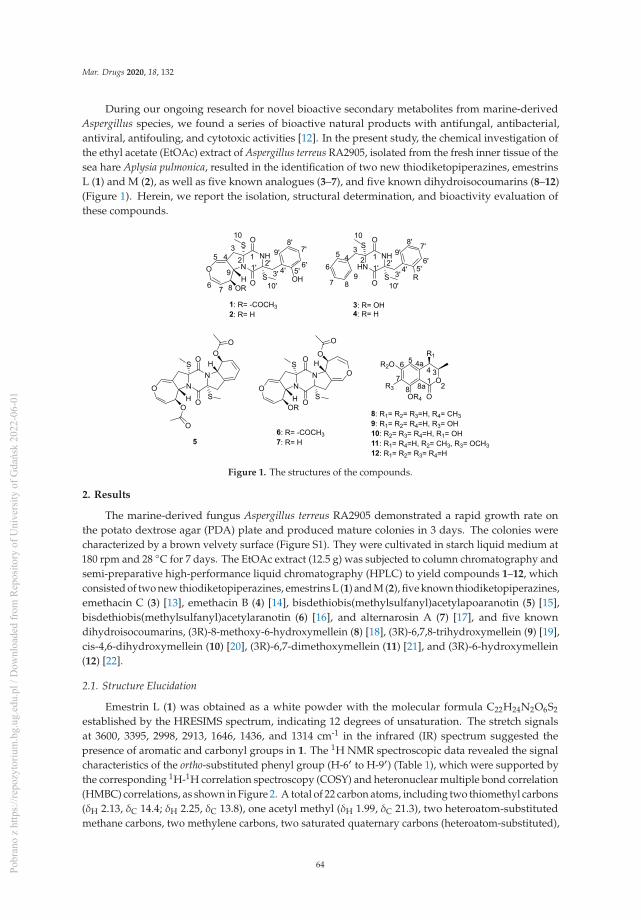
Mar. Drugs 2020, 18, 132
During our ongoing research for novel bioactive secondary metabolites from marine-derivedAspergillus species, we found a series of bioactive natural products with antifungal, antibacterial,antiviral, antifouling, and cytotoxic activities [12]. In the present study, the chemical investigation ofthe ethyl acetate (EtOAc) extract of Aspergillus terreus RA2905, isolated from the fresh inner tissue of thesea hare Aplysia pulmonica, resulted in the identification of two new thiodiketopiperazines, emestrinsL (1) and M (2), as well as five known analogues (3–7), and five known dihydroisocoumarins (8–12)(Figure 1). Herein, we report the isolation, structural determination, and bioactivity evaluation ofthese compounds.
NN
O
O
S
S
O
O
O
OR
5
1: R= -COCH32: R= H
6: R= -COCH37: R= H
8: R1= R2= R3=H, R4= CH39: R1= R2= R4=H, R3= OH10: R2= R3= R4=H, R1= OH11: R1= R4=H, R2= CH3, R3= OCH312: R1= R2= R3= R4=H
O
H
H
123
45
6 7
10
1' 2'3' 4' 5'
6'
7'8'
9'
10'
123
456
7 89
10
1' 2'3' 4' 5'
6'
7'8'9'
10'
NN
O
O
S
SO
O
OO
O
H
H
1 2
344a56
78 8a
O
OOR4
R3
R2OR1
NNH
O
O
S
SO
OROH
8
9HN
NH
O
O
S
S R
3: R= OH4: R= H
H
Figure 1. The structures of the compounds.
2. Results
The marine-derived fungus Aspergillus terreus RA2905 demonstrated a rapid growth rate onthe potato dextrose agar (PDA) plate and produced mature colonies in 3 days. The colonies werecharacterized by a brown velvety surface (Figure S1). They were cultivated in starch liquid medium at180 rpm and 28 ◦C for 7 days. The EtOAc extract (12.5 g) was subjected to column chromatography andsemi-preparative high-performance liquid chromatography (HPLC) to yield compounds 1–12, whichconsisted of two new thiodiketopiperazines, emestrins L (1) and M (2), five known thiodiketopiperazines,emethacin C (3) [13], emethacin B (4) [14], bisdethiobis(methylsulfanyl)acetylapoaranotin (5) [15],bisdethiobis(methylsulfanyl)acetylaranotin (6) [16], and alternarosin A (7) [17], and five knowndihydroisocoumarins, (3R)-8-methoxy-6-hydroxymellein (8) [18], (3R)-6,7,8-trihydroxymellein (9) [19],cis-4,6-dihydroxymellein (10) [20], (3R)-6,7-dimethoxymellein (11) [21], and (3R)-6-hydroxymellein(12) [22].
2.1. Structure Elucidation
Emestrin L (1) was obtained as a white powder with the molecular formula C22H24N2O6S2
established by the HRESIMS spectrum, indicating 12 degrees of unsaturation. The stretch signalsat 3600, 3395, 2998, 2913, 1646, 1436, and 1314 cm-1 in the infrared (IR) spectrum suggested thepresence of aromatic and carbonyl groups in 1. The 1H NMR spectroscopic data revealed the signalcharacteristics of the ortho-substituted phenyl group (H-6′ to H-9′) (Table 1), which were supported bythe corresponding 1H-1H correlation spectroscopy (COSY) and heteronuclear multiple bond correlation(HMBC) correlations, as shown in Figure 2. A total of 22 carbon atoms, including two thiomethyl carbons(δH 2.13, δC 14.4; δH 2.25, δC 13.8), one acetyl methyl (δH 1.99, δC 21.3), two heteroatom-substitutedmethane carbons, two methylene carbons, two saturated quaternary carbons (heteroatom-substituted),
64Pobr
ano
z ht
tps:
//rep
ozyt
oriu
m.b
g.ug
.edu
.pl /
Dow
nloa
ded
from
Rep
osito
ry o
f Uni
vers
ity o
f Gda
ńsk
2022
-06-
01

Mar. Drugs 2020, 18, 132
ten olefinic carbons (seven protonated), and three carbonyl carbons were observed in the 13C NMRspectrum (Table 1). The HMBC correlations from H-10 to C-2, from H-10′ to C-2′, from H-3 to C-1,and from H-3′ to C-1′ indicated the presence of a disulfide diketopiperazine skeleton in 1 (Figure 2).The ortho-substituted aromatic ring was connected with the diketopiperazine moiety via a methylenebridge of C-3′ based on the HMBC correlations from H-3′ to C-4′/C-5′/C-9′. The 1H−1H COSY datashowed the presence of an isolated spin system corresponding to the C-6−C-7−C-8−C-9 fragment. A4,5-dihydrooxepine existed in 1 based on the key HMBC correlations from H-5 to C-3/C-6/C-9 and fromH-6 to C-5/C-7/C-8, along with the chemical shifts of C-5 (δC 137.6), C-6 (δC 140.3), and C-9 (δC 61.5).The HMBC correlations from H-3 to C-1/C-5/C-9 indicated that the 4,5-dihydrooxepine moiety waslocated at C-2 of the diketopiperazine core (Figure 2). As the presence of cyclic dipeptide, aromaticring, 4,5-dihydrooxepine, and the other carbonyl in the molecule occupied 11 degrees of unsaturation,C-9 should be attached to the 2-N atom to form a pyrrolidine ring on the basis of the chemical shifts ofC-9 (δH 4.79, δC 61.5). The carbethoxy was anchored at C-8 on the basis of the HMBC correlation fromH-8 to C-11. Collectively, these data permitted the assignment of the planar structure of 1.
Table 1. 1H Nuclear Magnetic Resonance (NMR) and 13C NMR Data for 1 and 2 a.
Position1 2
δC, Type δH (J in Hz) δC, Type δH (J in Hz)
1 165.0, C 166.6, C2 70.1, C 68.9, C3 39.9, CH2 2.78, d (15.0) 39.9, CH2 2.74, d (15.0)
1.98, d (15.0) 1.92, d (15.0)4 110.4, C 109.5, C5 137.6, CH 6.70, t (2.3) 137.2, CH 6.60, t (2.3)
6 140.3, CH 6.44, dd (8.3, 2.3) 138.2, CH 6.28, dd (8.3,2.3)
7 105.9, CH 4.71, dd (8.3, 2.0) 111.4, CH 4.81, dd (8.3,2.0)
8 71.5, CH 5.67, dt (7.9, 2.0) 71.8, CH 4.43, dt (7.9, 2.0)9 61.5, CH 4.79, d (7.9) 64.9, CH 4.54, d (7.9)10 14.4, CH3 2.13, s 14.4, CH3 2.17, s11 169.9, C12 21.3, CH3 1.99, s1′ 164.8, C 165.0, C2′ 68.2, C 68.4, C3′ 39.1, CH2 3.43, d (13.7) 39.2, CH2 3.45, d (13.7)
3.19, d (13.7) 3.19, d (13.7)4′ 121.7, C 121.4, C5′ 156.3, C 156.2, C
6′ 115.8, CH 6.79, dd (8.0, 1.3) 115.7, CH 6.79, dd (8.0,1.3)
7′ 128.8, CH 7.03, td (8.0, 1.7) 128.8, CH 7.04, td (8.0, 1.7)
8′ 119.3, CH 6.67, td (7.7, 1.3) 119.2, CH 6.68, dd (7.8,1.3)
9′ 131.7, CH 6.92, dd (7.7, 1.7) 131.8, CH 6.95, dd (7.8,1.7)
10′ 13.8, CH3 2.25, s 14.0, CH3 2.29, s8-OH 5.44, d (6.0)
a 500 MHz for 1H NMR and 125 MHz for 13C NMR in Dimethyl Sulfoxide (DMSO)-d6.
65Pobr
ano
z ht
tps:
//rep
ozyt
oriu
m.b
g.ug
.edu
.pl /
Dow
nloa
ded
from
Rep
osito
ry o
f Uni
vers
ity o
f Gda
ńsk
2022
-06-
01

Mar. Drugs 2020, 18, 132
1
1H 1H COSY HMBC
NNH
O
O
S
SO
OOHH
NNH
O
O
S
SO
OHOHH
2O
Figure 2. The 1H-1H Correlation Spectroscopy (COSY) and Key Heteronuclear Multiple BondCorrelation (HMBC) Correlations for 1 and 2.
The coupling constants of 3JH-6–H-7 = 8.3 Hz indicated that the double bond was a Z-configuration.The 7.9 Hz coupling constants between H-8 and H-9 suggested their anti-relationship. The nuclearOverhauser effects (NOE) were observed for H-8 and H-10′ when on irradiation of H-10, whichdemonstrated that they should be on the same face (Figure 3). The absolute configurations of C-2 andC-2′ were assigned as R and R, respectively, determined by the similar electronic circular dichroism(ECD) data and the same biogenetic pathway as the co-isolated known compound 6 (Figures 4 and 5),for which the absolute configuration was confirmed by X-ray data (Figure S41). Thus, the absoluteconfigurations of 1 were established as 2R,2′R,8S,9S.
Figure 3. The Nuclear Overhauser Effect (NOE) Correlations for 1 and 2.
Figure 4. The Electronic Circular Dichroism (ECD) Spectra of 1–3 and 6.
Emestrin M (2) was assigned the molecular formula C20H22N2O5S2, with 11 degrees ofunsaturation, on the basis of its HRESIMS data, less 42 Da compared with 1. The 1H and 13C NMR
66Pobr
ano
z ht
tps:
//rep
ozyt
oriu
m.b
g.ug
.edu
.pl /
Dow
nloa
ded
from
Rep
osito
ry o
f Uni
vers
ity o
f Gda
ńsk
2022
-06-
01

Mar. Drugs 2020, 18, 132
data were very similar to those of 1 (Table 1), which revealed the presence of a thiodiketopiperazine,an ortho-substituted phenyl, and a 4,5-dihydrooxepine structure. The main differences were thedisappearance of the carbethoxy (δH 1.99, δC 21.3; δC 169.9) in 1, and the appearance of one hydroxyl(δH 5.44). The hydroxyl was anchored at C-8 based on the HMBC correlations from 8-OH to C-7/C-8/C-9.Thus, the planar structure of 2 was established. The similar coupling constants of 2 and 1 betweenH-6 and H-7, and between H-8 and H-9, respectively, suggested that the relative configurations of the4,5-dihydrooxepine of 2 were consistent with those of 1 (Figure 3). The absolute configurations of 2
were identical to those of 1 on the basis of the similar ECD data and of the same biogenetic pathway(Figures 4 and 5).
Emethacin C (3) was isolated as a white powder. Its molecular formula was defined asC20H22N2O3S2 by HRESIMS, with more than 16 Da compared with the known compound emethacinB (4). The 1H NMR and 13C NMR spectroscopic data of 3 were closely related to those of 4, whichrevealed the presence of a thiodiketopiperazine structure. The distinction was that one aromatichydrogen in 4 was replaced by one hydroxyl in 3. The hydroxyl was anchored at C-5′ based on the1H−1H COSY correlations of H-6′/H-7′/H-8′/H-9′ and the HMBC correlations from H-3′ to C-5′/C-9′(Figure S40). The absolute configurations of 3 were identical to those of 1 and 2 on the basis of thesimilar ECD data and of the same biogenetic pathway (Figures 4 and 5). It should be noted that theknown compound 3 is listed in SciFinder Scholar with the CAS Registry Number 2166398-50-9, butthis is the first time that its spectroscopic data have been reported.
Figure 5. The Possible Biosynthesis Pathway of Thiodiketopiperazines.
(3R)-8-methoxy-6-hydroxymellein (8) was obtained as a yellow solid with the molecular formulaC11H12O4 determined by the HRESIMS data, with more than 14 Da compared with the co-isolatedknown compound (3R)-6-hydroxymellein (12). The 1H NMR data of 8 displayed the presence of themeta-coupled aromatic protons at δH 5.80 (1H, d, J = 1.9 Hz) and at δH 5.71 (1H, d, J = 1.9 Hz). The 13CNMR spectrum revealed 17 carbons, including one carbonyl, six olefinic carbons (two oxygenated),one oxygenated methine carbon, one methylene, and two methyl carbons (one oxygenated). Thesespectroscopic features were similar to those of (3R)-6-hydroxymellein (12) except for an additionaloxygenated methyl group. The HMBC correlation from H-9 to C-8 was observed for 8 (Figure S42),suggesting that the methoxy group was attached to C-8. The negative Cotton effect at 270 nm in theECD spectrum of 8 suggested that the absolute configuration of C-3 was R (Figure S43) [23]. Compound
67Pobr
ano
z ht
tps:
//rep
ozyt
oriu
m.b
g.ug
.edu
.pl /
Dow
nloa
ded
from
Rep
osito
ry o
f Uni
vers
ity o
f Gda
ńsk
2022
-06-
01
Mar. Drugs 2020, 18, 132
8 is listed in SciFinder Scholar with the CAS Registry Number 2247026-31-7, but this is the first timethat its spectroscopic data have been reported.
(3R)-6,7,8-trihydroxymellein (9) was established as C10H10O5 on the basis of the HRESIMS data,with more than 16 Da compared with compound 12. The 1H NMR and 13C NMR spectroscopic data of9 were nearly identical to those of 12, except that one proton at the aromatic ring in 12 was replaced byone hydroxyl in 9. The HMBC correlations from H-5 to C-4/C-7/C-8a indicated that the hydroxyl groupwas located at C-7 (Figure S42). Similar to 8, the absolute configuration of C-3 in 9 was also determinedas R by the negative Cotton effect at 270 nm (Figure S44). Compound 9 is also listed in SciFinderScholar with the CAS Registry Number 2407423-58-7, but this is the first time that its spectroscopicdata have been reported.
2.2. Bioassays
All of the isolated compounds were tested for their antibacterial, antifungal, cytotoxic, and1,10-diphenyl-2-picryl-hydazyl (DPPH) scavenging activities. Their protein tyrosine phosphatase 1B (PTP1B) inhibitory activities were also measured—PTP1B is an important hypoglycemic target indiabetes. We found that compounds 2 and 3 displayed antibacterial activities against Pseudomonasaeruginosa ATCC 27853 with minimum inhibitory concentration (MIC) values of 64μg/mL and 32μg/mL,respectively. Intriguingly, compound 3 also exhibited antifungal activity against Candida albicansATCC10231 with a MIC value of 32 μg/mL. Compounds 3, 5, and 7 showed PTP1B inhibitory activitieswith inhibitory concentration (IC)50 values of 12.25, 25.70 and 24.32 μM, respectively. In addition,compound 9 exhibited a weak DPPH scavenging activity, with an IC50 value of 147 μM. All of theisolated compounds showed no cytotoxicity.
3. Materials and Methods
3.1. Instrumentation
Optical rotations were measured with a JASCO P-1020 digital polarimeter (Jasco Corp., Tokyo,Japan). UV spectra were recorded with a HITACHI UH 5300 UV spectrophotometer (Hitachi,Tokyo, Japan). ECD data were acquired on a J-815-150S Circular Dichroism spectrometer (JASCOElectric Co., Ltd., Tokyo, Japan). IR spectra were recorded with a Nicolet-Nexus-470 spectrometer(Thermo Electron Co., Madison, WI, USA) using KBr pellets. NMR spectra were acquired by aJEOL JEM-ECP NMR spectrometer (500 MHz for 1H and 125 MHz for 13C, JEOL, Tokyo, Japan),using tetramethylsilane (TMS) as an internal standard. High-resolution electrospray ionization massspectrometry (HRESIMS) was measured with a Thermo MAT95XP high resolution mass spectrometer(Thermo Fisher Scientific, Bremen, Germany), and electron ionization mass spectrometry (EIMS) spectrawith a Thermo DSQ EImass spectrometer (Thermo Fisher Scientific, Bremen, Germany). Single-crystalX-ray crystallographic analysis was performed on an Agilent Xcalibur Eos Gemini diffractometer(Agilent Technologies, Yarnton, England). Samples were analyzed and prepared using a Hitachi L-2000HPLC system coupled with a Hitachi L-2455 photodiode array detector, and using a semi-preparativeC18 column (Kromasil 250 mm × 10 mm, 5 μm). Silica gel (Qing Dao Hai Yang Chemical Group Co.,Qing Dao China; 300−400 mesh) and Sephadex LH-20 (Amersham Biosciences, Inc., USA) were usedfor column chromatography (CC). Precoated silica gel plates (Yan Tai Zi Fu Chemical Group Co., QingDao China; G60, F-254) were used for thin-layer chromatography. PTP1B (human recombinant) waspurchased from Abcam (ab51277).
3.2. Fungal Material
The fungal strain Aspergillus terreus RA2905 was isolated from a piece of fresh tissue from theinner part of the sea hare Aplysia pulmonica, collected from the Weizhou coral reefs in the South ChinaSea in April 2010. The fungus was identified as Aspergillus terreus according to its morphologicaltraits and a molecular protocol by amplification and sequencing of the DNA sequences of the internal
68Pobr
ano
z ht
tps:
//rep
ozyt
oriu
m.b
g.ug
.edu
.pl /
Dow
nloa
ded
from
Rep
osito
ry o
f Uni
vers
ity o
f Gda
ńsk
2022
-06-
01

Mar. Drugs 2020, 18, 132
transcribed spacer (ITS) region of the ribosomal RNA (rRNA) gene by using ITS 1 and ITS 4. ItsGenBank (NCBI) access number is MK611650. The phylogenetic tree of the ITS gene was constructedby the Neighbor-Joining method with the aid of MEGA 7 (Figure S1). The strain was deposited in theKey Laboratory of Marine Drugs, Ministry of Education of China, School of Medicine and Pharmacy,Ocean University of China, Qingdao, P. R. China.
3.3. Extraction and Isolation
Sixty 500-mL Erlenmeyer flasks of the fungal strain were cultivated in a starch liquid medium(soluble starch 10 g/L, peptone 1 g/L, artificial sea salt 30 g/L, 200 mL each flask) at 150 rpm and 28 ◦Cfor 7 days. The fermentation broth was filtered through a cheesecloth and extracted repeatedly with anequal amount of EtOAc three times, and then it was evaporated in vacuo to obtain an EtOAc extract(12.5 g). The crude extract was isolated on silica gel CC using a step gradient elution with petroleumether/EtOAc (10:1 to 1:4, v/v) to provide five fractions (Fr.1−Fr.5). Fr.3 was subjected to a silica gel CCeluted with Hexane/CH2Cl2/MeOH (10:1:0 to 0:1:1) to obtain four subfractions (Fr.3.1−Fr.3.4). Fr.3.2was further separated by the semi-preparative HPLC with MeOH/H2O (60:40) to give compounds 8
(5.5 mg) and 12 (7.1 mg). Fr.3.3 was separated by Sephadex LH-20 CC eluted with CH2Cl2/MeOH (1:1)and the semi-preparative HPLC with MeOH/H2O (45:55) further, to obtain compounds 9−11 (3.2 mg,3.7 mg, and 2.1 mg, respectively). Fr.4 was separated on a silica gel CC eluted with CH2Cl2/MeOH(100:1 to 1:1) to provide four subfractions (Fr.4.1−Fr.4.4). Fr.4.1 was further subjected to SephadexLH-20 CC eluted with CH2Cl2/MeOH (1:1) to obtain compounds 6 and 7. Fr.4.3 was separated by thesemi-preparative HPLC with MeOH/H2O (45:55) to give compounds 1−5 (3.3 mg, 3.1 mg, 4.2 mg, 4.5mg, and 4.3 mg, respectively).
Emestrin L (1). White powder; [α]20D -70.4 (c 0.42, MeOH); UV (MeOH) λmax (log ε) 229 (3.83), 275
(1.61) nm; IR (KBr) vmax 3600, 3395, 2998, 2913, 1646, 1436, 1314 cm−1; 1H and 13C NMR (see Table 1);(–)-HRESIMS m/z 475.1007 [M − H]− (calcd for C22H23N2O6S2, 475.1003).Emestrin M (2). White powder; [α]20
D -85.1 (c 0.43, MeOH); UV (MeOH) λmax (log ε) 229 (3.75), 275 (1.75)nm; IR (KBr) vmax 3600, 3000, 2912, 2138, 1663, 1438, 1315, 1043 cm−1; 1H NMR (500 MHz, DMSO-d6)and 13C NMR (125 MHz, DMSO-d6) (see Table 1); (–)-HRESIMS m/z 433.0896 [M − H]− (calcd forC20H21N2O5S2, 433.0897).
Emethacin C (3). White powder; [α]20D -64.4 (c 0.52, MeOH); UV (MeOH) λmax (log ε) 229 (3.89), 275 (2.54)
nm; IR (KBr) vmax 2952, 2868, 1802, 1715, 1459, 1200, 1139 cm−1; 1H NMR (500 MHz, DMSO-d6): 8.97(1H, s, 2-NH), 8.36 (1H, br s, 2′-NH), 7.18 (1H, m, H-7), 7.10 (4H, m, H-5, H-6, H-8, H-9), 6.89 (1H, td,J = 7.8, 1.7 Hz, H-7′), 6.67 (1H, td, J = 7.8, 1.2 Hz, H-8′), 6.26 (1H, dd, J = 7.8, 1.7 Hz, H-9′), 6.22 (1H,dd, J =7.8, 1.2 Hz, H-6′), 3.47 (1H, d, J = 13.6 Hz, H-3a), 3.05 (1H, d, J = 15.4 Hz, H-3′a), 3.01 (1H, d,J = 15.4 Hz, H-3′b), 2.96 (1H, d, J = 13.6 Hz, H-3b), 2.25 (3H, s, H-10), 2.17 (3H, s, H-10′); 13C NMR(125 MHz, DMSO-d6): 166.3 (C, C-1′), 165.6 (C, C-1), 155.6 (C, C-5′), 135.4 (C, C-4), 130.8 (CH × 2, C-5,C-9), 129.9 (CH, C-9′), 128.3(CH×2, C-6, C-8), 127.7 (CH, C-7′), 127.2 (CH, C-7), 122.3 (C, C-4′), 119.4(CH, C-6′), 115.2 (CH, C-8′), 65.5 (C, C-2), 65.2 (C, C-2′), 43.5 (CH2, C-3), 37.0 (CH2, C-3′), 14.0 (CH3,C-10), 13.9 (CH3, C-10′); (–)-HRESIMS m/z 401.1003 [M − H]− (calcd for C20H21N2O3S2, 401.0999).
(3R)-8-methoxy-6-hydroxymellein (8). Yellow solid; [α]20D -47.8 (c 1.00, MeOH); UV (MeOH) λmax (log ε)
238 (2.11), 251 (0.95), 319 (0.41) nm; IR (KBr) νmax 3749, 3674, 2360, 1736, 1581, 1418 cm−1; 1H NMR(500 MHz, DMSO-d6): 5.80 (1H, d, J = 1.9 Hz, H-5), 5.71 (1H, d, J = 1.9 Hz, H-7), 4.25 (1H, m, H-3), 3.59(3H, s, H-9), 2.57 (1H, dd, J = 15.8, 3.3 Hz, H-4a), 2.55 (1H, m, overlapped, H-4b), 1.26 (3H, d, J = 6.2 Hz,H-10); 13C NMR (125 MHz, DMSO-d6): 172.3 (C, C-1), 163.4 (C-6), 162.1 (C, C-8), 142.5 (C, C-4a), 109.4(CH, C-5), 100.1 (C, C-8a), 97.1 (CH, C-7), 72.0 (CH, C-3), 54.7 (CH3, C-9), 36.2 (CH2, C-4), 20.6 (CH3,C-10); (+)-HRESIMS m/z 209.0809 [M + H]+ (calcd for C11H13O4, 209.0808).
(3R)-6,7,8-trihydroxymellein (9). Yellow solid; [α]20D -50.1 (c 0.37, MeOH); UV (MeOH) λmax (log ε) 238
(2.03), 251 (1.05), 317 (0.23) nm; IR (KBr) νmax 3749, 3674, 2360, 1736, 1651, 1384 cm−1; 1H NMR
69Pobr
ano
z ht
tps:
//rep
ozyt
oriu
m.b
g.ug
.edu
.pl /
Dow
nloa
ded
from
Rep
osito
ry o
f Uni
vers
ity o
f Gda
ńsk
2022
-06-
01
Mar. Drugs 2020, 18, 132
(500 MHz, DMSO-d6): 6.26 (1H, s, H-5), 4.64 (1H, m, H-3), 2.82 (1H, dd, J = 16.2, 3.3 Hz, H-4a), 2.71 (1H,dd, J = 16.2, 1.3 Hz, H-4b), 1.37 (3H, d, J = 6.2 Hz, H-9); 13C NMR (125 MHz, DMSO-d6): 170.0 (C, C-1),153.2 (C-6), 151.1 (C, C-8), 131.3 (C, C-7), 131.0 (C, C-4a), 106.5 (CH, C-5), 100.0 (C, C-8a), 75.9 (CH, C-3),33.2 (CH2, C-4), 20.4 (CH3, C-9); (–)-HRESIMS m/z 209.0458 [M − H]− (calcd for C10H9O5, 209.0455).
Crystal data for 6: C18H18N2O7S2, Mr = 534.11, monoclinic, a = 6.8830(3) Å, b = 12.4383(5) Å, c =14.6020(6) Å, α = 90.00◦, β = 94.415(4)◦, γ = 90.00◦, V = 1246.41(9) Å3, space group P21, Z = 2, Dx= 1.135 mg/m3, μ (Cu Kα) = 0.670 mm–1, and F (000) = 425. Crystal dimensions: 0.12 × 0.11 × 0.11mm3. Independent reflections: 7369/4117 (Rint = 0.0422). The final R1 value was 0.0481, wR2 = 0.1180(I > 2σ(I)). Flack parameter = –0.012(13). Crystallographic data for 6 are deposited at the CambridgeCrystallographic Data Centre as supplementary publication number CCDC 1911625.
3.4. Antibacterial Assays
The antibacterial activity was evaluated following the standards recommended by the Clinicaland Laboratory Standards Institute [24]. Six pathogenic bacterial strains, Staphylococcus epidermidisATCC 12228, Staphylococcus aureus ATCC 25923, Pseudomonas aeruginosa ATCC 27853, Bacillus cereusATCC 14579, Escherichia coli ATCC 25922, and Sarcina lutea ATCC 9341, were used, and vancomycinwas used as a positive control.
3.5. Antifungal Assays
The antifungal bioassays were conducted following the standards recommended by the Clinicaland Laboratory Standards Institute [24]. Three pathogenic fungal strains, Candida albicans ATCC 24433,Candida tropicalis ATCC 20962, and Candida parapsilosis ATCC 22019, were used. Amphotericin B wasused as a positive control.
3.6. PTP1B Inhibition Assays
The PTP1B Inhibition assay was performed in 96-well plates [25]. The compound (10 μL) wasadded to the 99-μL reaction buffer solution, which consisted of 10 mM Tris (pH 7.4), 50 mM NaCl, 2 mMdithiothreitol (DTT), 1 mM MnCl2, and 10 mM para-nitrophenyl phosphate (pNPP). The reaction mixwas pre-warmed using a block heater at 37 ◦C. The recombinant PTP1B solution (1 mg/mL, 1 μL) wasmixed in each well. An NaOH solution (10 μL, 0.1 M) was added to stop the reaction. The absorbancewas recorded at 405 nm using a microplate. Sodium vanadate was used as a positive control.
3.7. DPPH Scavenging Activities
The DPPH scavenging assays were performed using the method described by Aquino et al. [26].The reaction mixture consisted of freshly prepared 100 μM DPPH in methanol, mixed with differentconcentrations of the compounds. The reaction mixture was incubated for 20 min at room temperaturein the dark, and the optical density was recorded at 517 nm.
3.8. Cytotoxicity Assays
The cytotoxic activities were evaluated with the sulforhodamine B (SRB) assay [27], using fivehuman tumor cell lines: A549, HCT116, MCF-7, Hela, and Hep G2. Adriamycin was used as apositive control.
Supplementary Materials: The following are available online at http://www.mdpi.com/1660-3397/18/3/132/s1,Figure S1. Aspergillus terreus RA2905 and its phylogenetic tree of ITS gene, Figures S2−S39: the NMR, ESIMS andHRESIMS spectra, Figure S40: Key HMBC correlations of compound 3, Figure S41: crystal diagram of compound6, Figure S42: key HMBC correlations of compounds 8 and 9, Figure S43: the CD spectrum of compound 8, FigureS44: the CD spectrum of compound 9.
Author Contributions: Conceptualization, C.-Y.W. and H.-S.G.; Methodology, J.-S.W.; Data analysis, J.-S.W.,X.-H.S., C.-L.S., and X.-L.Z.; Bioassays, J.-S.W., X.-H.S., and G.-S.Y.; Writing—Original draft preparation, J.-S.W.
70Pobr
ano
z ht
tps:
//rep
ozyt
oriu
m.b
g.ug
.edu
.pl /
Dow
nloa
ded
from
Rep
osito
ry o
f Uni
vers
ity o
f Gda
ńsk
2022
-06-
01

Mar. Drugs 2020, 18, 132
and X.-H.S.; Writing—Review and editing, J.-S.W., X.-M.F., and C.-Y.W. All authors have read and agreed to thepublished version of the manuscript.
Funding: This research was funded by the Marine S&T Fund of Shandong Province for Pilot National Laboratoryfor Marine Science and Technology (Qingdao, China) (No. 2018SDKJ0406-5), the National Science and TechnologyMajor Project for Significant New Drugs Development, China (No. 2018ZX09735-004), the Shandong ProvincialNatural Science Foundation (Major Basic Research Projects) (No. ZR2019ZD18), the Program of Open Studio forDruggability Research of Marine Natural Products, the Pilot National Laboratory for Marine Science and Technology(Qingdao, China) directed by Kai-Xian Chen and Yue-Wei Guo, and the Taishan Scholars Program, China.
Conflicts of Interest: The authors declare no conflict of interest.
References
1. Carroll, A.R.; Copp, B.R.; Davis, R.A.; Keyzers, R.A.; Prinsep, M.R. Marine natural products. Nat. Prod. Rep.2019, 36, 122. [CrossRef]
2. Jin, L.; Quan, C.; Hou, X.; Fan, S. Potential pharmacological resources: Natural bioactive compounds frommarine-derived fungi. Mar. Drugs 2016, 14, 76. [CrossRef]
3. Wang, K.W.; Ding, P. New bioactive metabolites from the marine-derived fungi Aspergillus. Mini-Rev.Med. Chem. 2018, 18, 1072–1094. [CrossRef]
4. Lee, Y.M.; Kim, M.J.; Li, H.; Zhang, P.; Bao, B.; Lee, K.J.; Jung, J.H. Marine-derived Aspergillus species as asource of bioactive secondary metabolites. Mar. Biotechnol. 2013, 15, 499–519. [CrossRef]
5. Welch, T.R.; Williams, R.M. Epidithiodioxopiperazines. occurrence, synthesis and biogenesis. Nat. Prod. Rep.2014, 31, 1376–1404. [CrossRef]
6. Fujimoto, H.; Sumino, M.; Okuyama, E.; Ishibashi, M. Immunomodulatory constituents from an ascomycete,Chaetomium Seminudum. J. Nat. Prod. 2004, 67, 98–102. [CrossRef]
7. Sun, Y.; Takada, K.; Takemoto, Y.; Yoshida, M.; Nogi, Y.; Okada, S.; Matsunaga, S. Gliotoxin analoguesfrom a marine-derived fungus, Penicillium sp., and their cytotoxic and histone methyltransferase inhibitoryactivities. J. Nat. Prod. 2011, 75, 111–114. [CrossRef]
8. Zheng, C.J.; Kim, C.J.; Bae, K.S.; Kim, Y.H.; Kim, W.G. Bionectins A−C, epidithiodioxopiperazines withanti-MRSA activity, from Bionectra byssicola F120. J. Nat. Prod. 2006, 69, 1816–1819. [CrossRef]
9. Curtis, P.J.; Greatbanks, D.; Hesp, B.; Cameron, A.F.; Freer, A.A. Sirodesmins A, B, C, and G, antiviralepipolythiopiperazine-2,5-diones of fungal origin: X-Ray analysis of sirodesmin A diacetate. J. Chem. Soc.Perkin Trans. 1977, 1, 180–189. [CrossRef]
10. Lee, H.J.; Lee, J.H.; Hwang, B.Y.; Kim, H.S.; Lee, J.J. Anti-angiogenic activities of gliotoxin and itsmethylthio-derivative, fungal metabolites. Arch. Pharmacal Res. 2001, 24, 397. [CrossRef]
11. Mayer, A.; Rodríguez, A.D.; Taglialatela-Scafatim, O.; Fusetani, N. Marine pharmacology in 2012–2013: Marinecompounds with antibacterial, antidiabetic, antifungal, anti-inflammatory, antiprotozoal, antituberculosis,and antiviral activities; affecting the immune and nervous systems, and other miscellaneous mechanisms ofaction. Mar. Drugs 2017, 15, 273.
12. Liu, L.; Zheng, Y.Y.; Shao, C.L.; Wang, C.Y. Metabolites from marine invertebrates and their symbioticmicroorganisms: Molecular diversity discovery, mining, and application. Mar. Life Sci. Tech. 2019, 1, 60–94.[CrossRef]
13. Niu, S.W.; Liu, D.; Shao, Z.; Proksch, P.; Lin, W.H. Eutypellazines N−S, new thiodiketopiperazines from adeep sea sediment derived fungus Eutypella sp. with anti-VRE activities. Tetrahedron Lett. 2017, 58, 3695–3699.[CrossRef]
14. Kawahara, N. Sulfur-containg dioxopiperazine derivatives from Emericella heterothallica. Heterocycles 1989,29, 397–402.
15. Neuss, N.; Nagarajan, R.; Molloy, B.B.; Huckstep, L.L. Aranotin and related metabolites. II. Isolation,characterization, and structures of two new metabolites. Tetrahedron Lett. 1968, 9, 4467–4471. [CrossRef]
16. Kamata, S.; Sakai, H.; Hirota, A. Isolation of acetylaranotin, bisdethiodi (methylthio)-acetylaranotin andterrein as plant growth inhibitors from a strain of Aspergillus terreus. Agric. Biol. Chem. 1983, 47, 2637–2638.[CrossRef]
17. Nagarajan, R.; Huckstep, L.L.; Lively, D.H.; DeLng, D.C.; Marsh, M.M.; Neuss, N. Aranotin and relatedmetabolites from Arachniotus aureus. I. Determination of structure. J. Am. Chem. Soc. 1968, 90, 2980–2982.[CrossRef]
71Pobr
ano
z ht
tps:
//rep
ozyt
oriu
m.b
g.ug
.edu
.pl /
Dow
nloa
ded
from
Rep
osito
ry o
f Uni
vers
ity o
f Gda
ńsk
2022
-06-
01

Mar. Drugs 2020, 18, 132
18. Dethoup, T.; Manoch, L.; Kijjoa, A.; Pinto, M.; Gales, L.; Damas, A.M.; Silva, A.M.S.; Eaton, G.; Herz, W.Merodrimanes and other constituents from Talaromyces thailandiasis. J. Nat. Prod. 2007, 70, 1200–1202.[CrossRef]
19. Zaehle, C.; Gressler, M.; Shelest, E.; Geib, E.; Hertweck, C.; Brock, M. Terrein biosynthesis in Aspergillusterreus and its impact on phytotoxicity. Chem. Biol. 2014, 21, 719–731. [CrossRef]
20. Takenaka, Y.; Morimoto, N.; Hamada, N.; Tanahashi, T. Phenolic compounds from the cultured mycobiontsof Graphis proserpens. Phytochemistry 2011, 72, 1431–1435. [CrossRef]
21. Choudhary, M.I.; Musharraf, S.G.; Mukhmoor, T.; Shaheen, F.; Ali, S.; Rahman, A. Isolation of bioactivecompounds from Aspergillus terreus. Z. Naturforsch. B J. Chem. Sci. 2004, 59, 324–328. [CrossRef]
22. Islam, M.S.; Ishigami, K.; Watanabe, H. Synthesis of (−)-mellein, (+)-ramulosin, and related natural products.Tetrahedron 2006, 63, 1074–1079. [CrossRef]
23. Krohn, K.; Bahramsari, R.; Flörke, U.; Ludewig, K.; Kliche-Spory, C.; Michel, A.; Aust, H.J.; Draeger, S.;Schulz, B.; Antus, S. Dihydroisocoumarins from fungi: Isolation, structure elucidation, circular dichroismand biological activity. Phytochemisty 1997, 45, 313–320. [CrossRef]
24. Clinical and Laboratory Standards Institute. Performance Standards for Antimicrobial Susceptibility Testing;Twenty-Second Informational Supplement. M100-S22; Clinical and Laboratory Standards Institute (CLSI):Wayne, PA, USA, 2012.
25. Tian, J.L.; Liao, X.J.; Wang, Y.H.; Si, X.; Shu, C.; Gong, E.-S.; Xie, X.; Ran, X.L.; Li, B. Identification ofcyanidin-3-arabinoside extracted from blueberry as selective PTP1B inhibitor. J. Agric. Food Chem. 2019, 67,13624–13634. [CrossRef]
26. Aquino, R.; Morelli, S.; Lauro, M.R.; Abdo, S.; Saija, A.; Tomaino, A. Phenolic constituents and antioxidantactivity of an extract of Anthurium versicolor leaves. J. Nat. Prod. 2001, 64, 1019–1023. [CrossRef]
27. Skehan, P.; Storeng, R.; Scudiero, D.; Monks, A.; McMahon, J.; Vistica, D.; Warren, J.T.; Bokesch, H.; Kenney, S.;Boyd, M.R. New colorimetric cytotoxicity assay for anticancer-drug screening. J. Natl. Cancer Inst. 1990, 82,1107–1112. [CrossRef]
© 2020 by the authors. Licensee MDPI, Basel, Switzerland. This article is an open accessarticle distributed under the terms and conditions of the Creative Commons Attribution(CC BY) license (http://creativecommons.org/licenses/by/4.0/).
72Pobr
ano
z ht
tps:
//rep
ozyt
oriu
m.b
g.ug
.edu
.pl /
Dow
nloa
ded
from
Rep
osito
ry o
f Uni
vers
ity o
f Gda
ńsk
2022
-06-
01

marine drugs
Article
Cytotoxic Thiodiketopiperazine Derivatives from theDeep Sea-Derived Fungus Epicoccum nigrum SD-388
Lu-Ping Chi 1,2,3, Xiao-Ming Li 1,2, Li Li 1,2,3, Xin Li 1,2,* and Bin-Gui Wang 1,2,4,*
1 Key Laboratory of Experimental Marine Biology, Institute of Oceanology, Chinese Academy of Sciences,Nanhai Road 7, Qingdao 266071, China; [email protected] (L.-P.C.); [email protected] (X.-M.L.);[email protected] (L.L.)
2 Laboratory of Marine Biology and Biotechnology, Qingdao National Laboratory for Marine Science andTechnology, Wenhai Road 1, Qingdao 266237, China
3 University of Chinese Academy of Sciences, Yuquan Road 19A, Beijing 100049, China4 Center for Ocean Mega-Science, Chinese Academy of Sciences, Nanhai Road 7, Qingdao 266071, China* Correspondence: [email protected] (X.L.); [email protected] (B.-G.W.);
Tel.: +86-532-8289-8890 (X.L.); +86-532-8289-8553 (B.-G.W.)
Received: 8 February 2020; Accepted: 11 March 2020; Published: 13 March 2020
Abstract: Four new thiodiketopiperazine alkaloids, namely, 5’-hydroxy-6’-ene-epicoccin G (1),7-methoxy-7’-hydroxyepicoccin G (2), 8’-acetoxyepicoccin D (3), and 7’-demethoxyrostratin C (4), aswell as a pair of new enantiomeric diketopiperazines, (±)-5-hydroxydiphenylalazine A (5), along withfive known analogues (6–10), were isolated and identified from the culture extract of Epicoccum nigrumSD-388, a fungus obtained from deep-sea sediments (−4500 m). Their structures were established onthe basis of detailed interpretation of the NMR spectroscopic and mass spectrometric data. X-raycrystallographic analysis confirmed the structures and established the absolute configurations ofcompounds 1–3, while the absolute configurations for compounds 4 and 5 were determined by ECDcalculations. Compounds 4 and 10 showed potent activity against Huh7.5 liver tumor cells, whichwere comparable to that of the positive control, sorafenib, and the disulfide bridge at C-2/C-2’ is likelyessential for the activity.
Keywords: Epicoccum nigrum; deep-sea-derived fungus; thiodiketopiperazines; diketopiperazineenantiomers; cytotocxic activity
1. Introduction
Natural products have historically been a rich source of new drugs or drug candidates. Strikingly,deep-sea-derived microorganisms survive under extreme environments, leading to special biologicaldiversity and prolific metabolisms differing from those of terrestrial microorganisms. Recently,deep-sea-sourced microbial natural products have been reported with high hit-rates from bioactivityscreening, particularly in the antitumor area [1,2].
Epicoccum nigrum is a chemically distinct fungal species with potential to produce structurallyunique secondary metabolites including thiodiketopiperazines (TDKPs) [3,4], polyketides [5],and polysaccharides [6]. Some of these metabolites exhibited intriguing biological properties, such asantimicrobial [3], cytotoxic [4], and antioxidant activities [5,6].
The TDKP derivatives are a family of diketopiperazines which have been isolated from severalfungal sources, such as Epicoccum nigrum [3], Exserohilum rostratum [7], Penicillium brocae [8], andPenicillium adametzioides [9].
In continuation of our research aimed at discovery of bioactive metabolites from marine-derivedmicroorganisms [10–12], a fungal strain of Epicoccum nigrum SD-388 was isolated from a deep-seasediment sample collected at a depth of 4500 m. Chemical investigation of the fungus resulted in
Mar. Drugs 2020, 18, 160; doi:10.3390/md18030160 www.mdpi.com/journal/marinedrugs73Pobr
ano
z ht
tps:
//rep
ozyt
oriu
m.b
g.ug
.edu
.pl /
Dow
nloa
ded
from
Rep
osito
ry o
f Uni
vers
ity o
f Gda
ńsk
2022
-06-
01

Mar. Drugs 2020, 18, 160
the isolation of spiroepicoccin A, an unusual spiro-TDKP derivative, whose stereochemistry couldnot be elucidated by conventional NMR methods and was solved based on residual chemicalshift anisotropies [12]. This result encouraged us to perform a further study of the fungusand has led to the isolation of four new TDKPs including 5’-hydroxy-6’-ene-epicoccin G (1),7-methoxy-7’-hydroxyepicoccin G (2), 8’-acetoxyepicoccin D (3), and 7’-demethoxyrostratin C (4),as well as a pair of new enantiomeric diketopiperazines (DKPs), (±)-5-hydroxydiphenylalazine A((±)-5), together with five known analogues including diphenylalazine A (6) [4], emeheterone (7) [13],epicoccins E (8) and G (9) [4], and rostratin C (10) [7] (Figure 1). Details of the isolation and purification,structural elucidation, and cytotoxic potency against Huh7.5 liver tumor cells of compounds 1–10 aredescribed herein.
Figure 1. Structures of the isolated compounds 1–10.
2. Results and Discussion
2.1. Structure Elucidation of the New Compounds
The fungal strain E. nigrum SD-388 was cultured on the rice solid medium, which was furtherexhaustively extracted with EtOAc to afford an extract. Fractionation of the extracts by a combinationof column chromatography (CC) over silica gel, Lobar LiChroprepRP-18, SephadexLH-20, as well assemi-preparative HPLC, yielded compounds 1–10.
Compound 1, initially obtained as colorless gum, gave a pseudomolecular ion peak at m/z 455.1293[M +H]+ by HR-ESI-MS, consistent with a molecular formula of C20H26N2O6S2, indicating 9 degrees ofunsaturation. The 1H-, 13C-NMR, and DEPT spectroscopic data (Tables 1 and 2) revealed the presenceof two methyls, four sp3 hybridized methylenes, nine methines (with five oxygenated/nitrogenated andtwo olefinic), five nonprotonated carbons (with one ketone and two amide carbonyls), as well as threeexchangeable protons. Detailed analysis of the NMR data disclosed that the structure of 1 was similar
74Pobr
ano
z ht
tps:
//rep
ozyt
oriu
m.b
g.ug
.edu
.pl /
Dow
nloa
ded
from
Rep
osito
ry o
f Uni
vers
ity o
f Gda
ńsk
2022
-06-
01

Mar. Drugs 2020, 18, 160
to that of epicoccin G (9), a well described TDKP derivative identified from a Cordyceps-colonizingfungus Epicoccum nigrum XZC04-CS-302 in 2009 [4]. However, signals for two CH2 groups at δH/δC 2.20and 2.59/33.8 (C-6’) and at δH/δC 1.88 and 2.12/25.8 (C-7’) in compound 9, were replaced by two olefinicCH groups at δH/δC 5.68/133.3 (C-6’) and δH/δC 5.53/129.9 (C-7’) in the NMR spectra of 1. Furthermore,the signal for the ketone group (C-5’) of 9 (δC 207.7) was replaced by an oxygenated methine (δH/δC
4.11/71.3) in 1. The COSY correlations for the spin system from H-3’ through H-9’ via H-4’~H-8’ andthe HMBC correlations from H-5’ to C-7’ and C-9’, from H-6’ to C-4’ and C-5’, and from H-7’ to C-9’,confirmed the proposed structure of 1 (Figure 2).
Table 1. 1H NMR spectroscopic data for compounds 1–5 a.
No. 1 2 3 4 5
2 4.17, t (6.4)
3α 2.80, d (13.4) α 2.75, d (13.5) α 3.13, d (17.7) α 2.68, m a 3.02, dd (13.3, 6.4)
β 2.28, m β 2.35, dd (13.5, 8.5) β 2.89, m β 2.99, dd (14.8, 7.9) b 2.95, dd (13.3, 6.4)
4 2.98, t (8.0) 2.92, t (8.5) 3.06, t (8.3) 3.18, t (7.9)
6α 2.22, m α 2.54, dd (17.1, 4.7) α 2.85, m α 2.71, m
6.79, d (7.3)β 2.61, m β 2.65, m β 2.92, m β 2.61, m
7α 2.19, m
3.82, ddd (10.1, 4.7, 2.0) 3.73, m 3.29, ddd (10.9, 4.7, 1.5) 6.96, mβ 1.92, m
8 4.36, m 4.56, m 4.01, dt (4.6, 2.4) 5.03, t (4.7) 6.67, td (7.3, 1.2)
9 4.33, m 4.33, m 4.67, dd (8.3, 2.4) 4.41, dd (7.9, 4.7) 6.98, m
3’α 2.42, dd (12.5,4.9) α 2.73, d (13.4) α 2.93, m α 2.74, m
6.83, sβ 2.26, m β 2.34, dd (13.4, 8.5) β 3.29, d (19.4) β 2.93, dd (14.8, 8.0)
4’ 2.09, m 2.91, t (8.5) 3.17, d (8.1) 3.23, t (8.0)
5’ 4.11, m 7.27, d (7.3)
6’ 5.68, d (9.8)α 2.40, dd (16.8, 4.4) α 2.89, m α 2.66, m
7.40, t (7.3)β 2.61, m β 2.97, m β 2.27, dt (16.7, 4.1)
7’ 5.53, d (9.8) 4.12, ddd (10.2, 4.4, 2.0) 3.92, m α 1.62, td (12.5, 5.0)7.32, t (7.3)
β 1.89, m
8’ 4.08, m 4.29, s 5.04, dd (4.3, 2.0) 4.81, m 7.40, t (7.3)
9’ 3.41, dd (12.0, 8.1) 4.34, m 4.83, dd (8.1, 2.0) 4.36, m 7.27, d (7.3)
1-NMe 2.69, s
1’-NH -b
2-SMe 1.95, s 1.93, s
2’-SMe 2.08, s 1.92, s
2” 2.04, s
5-OH 8.50, br s
8-OH 5.36, d (3.2) 5.44, br s 6.25, d (2.4) 5.64, d (4.7)
5’-OH 5.92, s
7’-OH 5.19, br s
8’-OH 5.28, d (5.8) 5.44, br s 5.49, d (4.0)
7-OMe 3.25, s 3.21, sa Data collected at 500 MHz in DMSO-d6. b Data not detected.
The relative configuration of 1 was deduced from analysis of the NOESY spectrum. NOEcorrelations from H-9 to H-3β and H-4, and from the proton of 8-OH to H-4, H-6β, and H-7β, indicatedthe cofacial orientation of these groups (Figure 3). Besides, NOEs from H-3α to 2-SMe placed themon another face, opposite to that of H-4, H-9, and 8-OH. Moreover, NOE cross-peaks from H-8’ toH-3’α and H-4’, and from H-3’α to 2’-SMe, confirmed them on the same spatial orientation, while NOEcorrelations from H-5’ to H-9’ and H-3’β placed these groups on the opposite face. On the basis of theabove observation, the relative configurations for rings A/B and D/E were determined respectively.However, the relationship between these two units could not be correlated based on the NOESYexperiment, because no diagnostic NOE cross-peak could be detected between rings A/B and D/E.
75Pobr
ano
z ht
tps:
//rep
ozyt
oriu
m.b
g.ug
.edu
.pl /
Dow
nloa
ded
from
Rep
osito
ry o
f Uni
vers
ity o
f Gda
ńsk
2022
-06-
01

Mar. Drugs 2020, 18, 160
Table 2. 13C NMR spectroscopic data for compounds 1–5 a.
No. 1 2 3 4 5
1 168.7, C 165.5, C 158.3, C 162.2, C 167.2, C2 71.4, C 71.6, C 71.1, C 76.2, C 55.6, CH3 34.3, CH2 34.4, CH2 41.4, CH2 32.6, CH2 34.8, CH24 44.0, CH 43.8, CH 45.0, CH 46.4, CH 122.0, C5 207.5, C 206.6, C 207.1, C 207.8, C 156.1, C6 33.8, CH2 40.4, CH2 43.2, CH2 40.7, CH2 115.1, CH7 25.9, CH2 75.8, CH 41.3, CH 75.5, CH 127.8, CH8 63.6, CH 61.5, CH b 65.2, CH 61.9, CH 118.4, CH9 64.8, CH 63.2, CH 60.3, CH 63.2, CH 131.2, CH1’ 165.5, C 165.4, C 158.9, C 162.2, C 162.4, C2’ 72.9, C 71.6, C 71.7, C 76.4, C 132.2, C3’ 35.1, CH2 34.1, CH2 41.5, CH2 32.2, CH2 117.7, CH4’ 43.4, CH 43.5, CH 45.3, CH 46.7, CH 133.8, C5’ 71.3, CH 207.4, C 206.3, C 208.6, C 129.4, CH6’ 133.3, CH 43.3, CH2 43.5, CH2 33.9, CH2 128.1, CH7’ 129.9, CH 65.7, CH 38.8, CH 25.4, CH2 127.9, CH8’ 68.9, CH 68.1, CH b 67.4, CH 60.8, CH 128.1, CH9’ 67.8, CH 62.7, CH 57.5, CH 65.8, CH 129.4, CH1” 168.8, C2” 20.6, CH3
1-NMe 34.5, CH32-SMe 14.4, CH3 14.2, CH32’-SMe 14.2, CH3 13.9, CH37-OMe 55.8, CH3 56.0, CH3
a Data collected at 125 MHz in DMSO-d6. b Assigned by HSQC experiment.
Figure 2. Key 1H-1H COSY (bold lines) and HMBC (red arrows) correlations of compounds 1–5.
To fully assign the configuration of compound 1, efforts toward a single crystal X-ray study wereperformed. By slow evaporation of the solvent (MeOH–H2O, 100:1) under refrigeration, quality crystalsof 1 were obtained, making it feasible for an X-ray crystallographic experiment which confirmed notonly the planar structure, but also the relative configuration of compound 1 (Figure 4). The definedFlack parameter 0.01(3) determined the absolute configuration of 1 as 2R, 4R, 8S, 9S, 2’R, 4’S, 5’S, 8’S,and 9’S, and the trivial name 5’-hydroxy-6’-ene-epicoccin G was assigned to compound 1.
76Pobr
ano
z ht
tps:
//rep
ozyt
oriu
m.b
g.ug
.edu
.pl /
Dow
nloa
ded
from
Rep
osito
ry o
f Uni
vers
ity o
f Gda
ńsk
2022
-06-
01
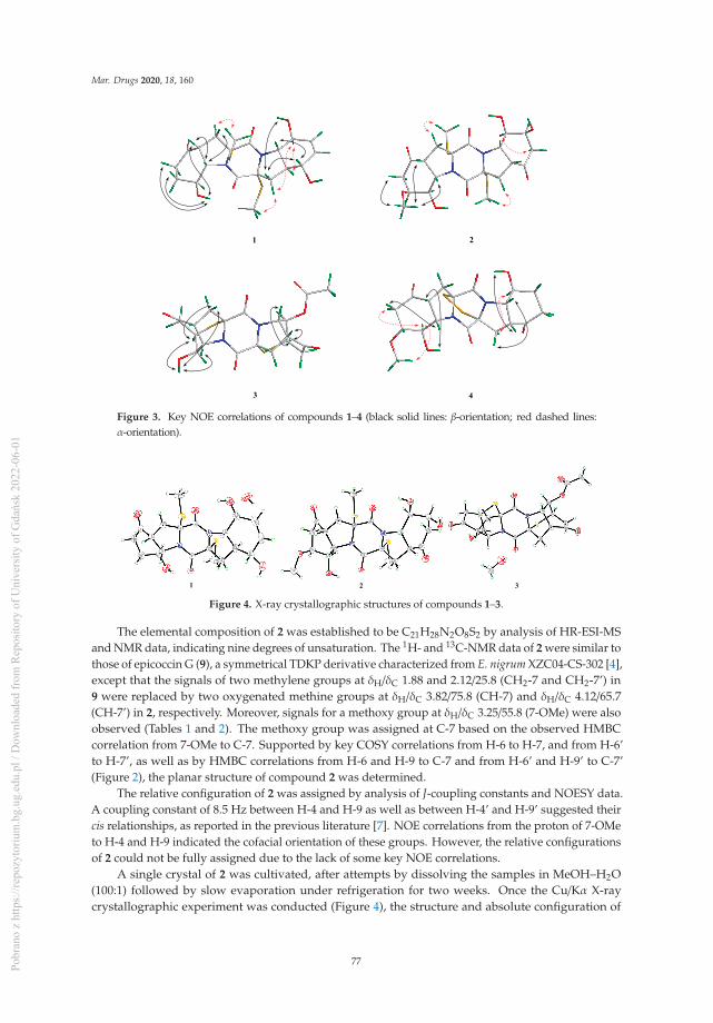
Mar. Drugs 2020, 18, 160
Figure 3. Key NOE correlations of compounds 1–4 (black solid lines: β-orientation; red dashed lines:α-orientation).
Figure 4. X-ray crystallographic structures of compounds 1–3.
The elemental composition of 2 was established to be C21H28N2O8S2 by analysis of HR-ESI-MSand NMR data, indicating nine degrees of unsaturation. The 1H- and 13C-NMR data of 2 were similar tothose of epicoccin G (9), a symmetrical TDKP derivative characterized from E. nigrum XZC04-CS-302 [4],except that the signals of two methylene groups at δH/δC 1.88 and 2.12/25.8 (CH2-7 and CH2-7’) in9 were replaced by two oxygenated methine groups at δH/δC 3.82/75.8 (CH-7) and δH/δC 4.12/65.7(CH-7’) in 2, respectively. Moreover, signals for a methoxy group at δH/δC 3.25/55.8 (7-OMe) were alsoobserved (Tables 1 and 2). The methoxy group was assigned at C-7 based on the observed HMBCcorrelation from 7-OMe to C-7. Supported by key COSY correlations from H-6 to H-7, and from H-6’to H-7’, as well as by HMBC correlations from H-6 and H-9 to C-7 and from H-6’ and H-9’ to C-7’(Figure 2), the planar structure of compound 2 was determined.
The relative configuration of 2 was assigned by analysis of J-coupling constants and NOESY data.A coupling constant of 8.5 Hz between H-4 and H-9 as well as between H-4’ and H-9’ suggested theircis relationships, as reported in the previous literature [7]. NOE correlations from the proton of 7-OMeto H-4 and H-9 indicated the cofacial orientation of these groups. However, the relative configurationsof 2 could not be fully assigned due to the lack of some key NOE correlations.
A single crystal of 2 was cultivated, after attempts by dissolving the samples in MeOH–H2O(100:1) followed by slow evaporation under refrigeration for two weeks. Once the Cu/Kα X-raycrystallographic experiment was conducted (Figure 4), the structure and absolute configuration of
77Pobr
ano
z ht
tps:
//rep
ozyt
oriu
m.b
g.ug
.edu
.pl /
Dow
nloa
ded
from
Rep
osito
ry o
f Uni
vers
ity o
f Gda
ńsk
2022
-06-
01

Mar. Drugs 2020, 18, 160
2 were assigned as 2R, 4R, 7S, 8R, 9S, 2’R, 4’R, 7’S, 8’R, and 9’S, with a Flack parameter of 0.02(4).Compound 2 was named 7-methoxy-7’-hydroxyepicoccin G.
The accurate mass data measured by HR-ESI-MS of compound 3 assigned its molecular formula,C20H20N2O7S2 (12 degrees of unsaturation), and was supported by the NMR data. The 1H- and13C-NMR data of 3 (Tables 1 and 2) showed close similarity to those of epicoccin D, a TDKP derivativeisolated from the fungal strain E. nigrum (2203) in 2007 [3]. However, resonances for an ester carbonylcarbon (δC 168.8, C-1”) and a methyl group (δH/δC 2.04/20.6, CH3-2”) were observed in the NMR spectraof 3. Deshielded shift at δH 5.04 for H-8’ in 3 was detected, compared to that of δH 4.00 in epicoccin D.The above observation suggested that compound 3 was a C-8’ acetylated derivative of epicoccin D.The relative configuration of 3 was assigned on the basis of the NOESY experiment and J-couplingconstants. For ring A of 3, NOE correlations from the proton of OH-8 to H-7 and H-9 revealed thesame orientation of these groups (Figure 3). In addition, the cis relationship between H-4 and H-9was established by the coupling constant (J = 8.3 Hz) which is in agreement with that of rostratin B(J = 7.2 Hz) [7]. However, the relative configurations of ring E could not be solved as it lacked some keyNOE correlations. To unequivocally determine the relative and absolute configurations, single crystalsfor 3 were cultivated upon slow evaporation of the solvent (MeOH) and a Cu/Kα X-ray diffractionanalysis was conducted (Figure 4). The final refinement of the X-ray data resulted in a 0.02(3) Flackparameter, allowing for the assignment of the absolute configuration as 2R, 4R, 7R, 8R, 9S, 2’R, 4’R, 7’R,8’R, and 9’S.
Compound 4 was initially isolated as a colorless powder. Its molecular formula was postulatedas C19H22N2O7S2 through HR-ESI-MS analysis, indicating 10 degrees of unsaturation. The 1D NMRdata of 4 were similar to those of rostratin C (10), a DKP derivative isolated from the marine-derivedfungal strain Exserohilum rostratum CNK-630 [7], with the exception of the disappeared signals for theoxygenated methine (C-7’) and the methoxy group attached to C-7’. In contrast, signals for a methylenegroup at δH 1.62/1.89 and δC 25.4 (CH2-7’) were observed in the NMR spectra of 4 (Tables 1 and 2),indicating that 4 is a 7’-demethoxy derivative of 10. The 2D NMR correlations supported this inferenceby the COSY correlations from H-7’ to H-6’ and H-8’, and HMBC correlations from H-7’ to C-5’ andfrom H-9’ to C-7’ (Figure 2).
The relative configuration for rings A and E of 4 were determined by analysis of NOESY data.NOE correlations, with respect to ring A from H-8 to H-3α and 7-OMe, placed them on the sameface. Meanwhile, NOEs from the proton of 8-OH to H-4, and from H-3β to H-9, disclosed the cofacialorientation of these groups. As for ring E, the coupling constant (J = 8.0 Hz) observed between H-4’ andH-9’ revealed their cis relationship [7]. In addition, NOEs from the proton of 8’-OH to H-4’, and fromH-3’β to H-9’, revealed them on the cofacial orientation. Whereas NOE from H-3’α to H-8’ revealedthat these groups were on the other face (Figure 3).
The assignment of the absolute configurations at C-2/C-2’ were established by analysis of ECDcotton effects (CEs) following the rules reported by the previous reference [14]. The ECD spectrumof 4 showed a positive CE near 265 nm, which was characteristic for the 2R/2’R configurations inTDKPs. The whole absolute configuration of 4 was further studied using the time-dependent densityfunctional (TDDFT)-ECD calculation in Gaussian 09. The ECD spectra of four possible stereoisomersof 4, including (2R, 4R, 7R, 8R, 9S, 2’R, 4’R, 8’S, 9’S)-4, (2R, 4R, 7R, 8R, 9S, 2’R, 4’S, 8’R, 9’R)-4, (2R,4S, 7S, 8S, 9R, 2’R, 4’R, 8’S, 9’S)-4, and (2R, 4S, 7S, 8S, 9R, 2’R, 4’S, 8’R, 9’R)-4, were calculated. Theexperimental ECD spectrum for 4 showed agreement with that calculated for (2R, 4R, 7R, 8R, 9S, 2’R,4’R, 8’S, 9’S)-4 (Figure 5a), allowing the elucidation of whole chiral centers as 2R, 4R, 7R, 8R, 9S, 2’R,4’R, 8’S, and 9’S.
Compound 5, obtained as a yellow oil, was assigned the molecular formula C19H18N2O3 byHR-ESI-MS, and required 12 degrees of unsaturation. Analysis of the 1H- and 13C-NMR data(Tables 1 and 2) revealed that compound 5 had same basic structure as that of the previously reporteddiphenylalazine A (6), which was identified from the fungus E. nigrum XZC04-CS-302 [4]. However,the aromatic methine at C-5 in 6 (δH/δC 7.18/129.82) was replaced by a nonprotonated and hydroxylated
78Pobr
ano
z ht
tps:
//rep
ozyt
oriu
m.b
g.ug
.edu
.pl /
Dow
nloa
ded
from
Rep
osito
ry o
f Uni
vers
ity o
f Gda
ńsk
2022
-06-
01
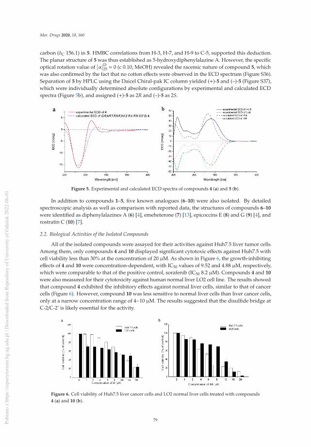
Mar. Drugs 2020, 18, 160
carbon (δC 156.1) in 5. HMBC correlations from H-3, H-7, and H-9 to C-5, supported this deduction.The planar structure of 5 was thus established as 5-hydroxydiphenylalazine A. However, the specificoptical rotation value of [α]D25 = 0 (c 0.10, MeOH) revealed the racemic nature of compound 5, whichwas also confirmed by the fact that no cotton effects were observed in the ECD spectrum (Figure S36).Separation of 5 by HPLC using the Daicel Chiral-pak IC column yielded (+)-5 and (–)-5 (Figure S37),which were individually determined absolute configurations by experimental and calculated ECDspectra (Figure 5b), and assigned (+)-5 as 2R and (–)-5 as 2S.
Figure 5. Experimental and calculated ECD spectra of compounds 4 (a) and 5 (b).
In addition to compounds 1–5, five known analogues (6–10) were also isolated. By detailedspectroscopic analysis as well as comparison with reported data, the structures of compounds 6–10
were identified as diphenylalazines A (6) [4], emeheterone (7) [13], epicoccins E (8) and G (9) [4], androstratin C (10) [7].
2.2. Biological Activities of the Isolated Compounds
All of the isolated compounds were assayed for their activities against Huh7.5 liver tumor cells.Among them, only compounds 4 and 10 displayed significant cytotoxic effects against Huh7.5 withcell viability less than 30% at the concentration of 20 μM. As shown in Figure 6, the growth-inhibitingeffects of 4 and 10 were concentration-dependent, with IC50 values of 9.52 and 4.88 μM, respectively,which were comparable to that of the positive control, sorafenib (IC50 8.2 μM). Compounds 4 and 10
were also measured for their cytotoxicity against human normal liver LO2 cell line. The results showedthat compound 4 exhibited the inhibitory effects against normal liver cells, similar to that of cancercells (Figure 6). However, compound 10 was less sensitive to normal liver cells than liver cancer cells,only at a narrow concentration range of 4~10 μM. The results suggested that the disulfide bridge atC-2/C-2’ is likely essential for the activity.
Figure 6. Cell viability of Huh7.5 liver cancer cells and LO2 normal liver cells treated with compounds4 (a) and 10 (b).
79Pobr
ano
z ht
tps:
//rep
ozyt
oriu
m.b
g.ug
.edu
.pl /
Dow
nloa
ded
from
Rep
osito
ry o
f Uni
vers
ity o
f Gda
ńsk
2022
-06-
01

Mar. Drugs 2020, 18, 160
3. Experimental Section
3.1. General Experimental Procedures
Melting points were acquired through an SGW X-4 micro-melting-point apparatus (ShanghaiShenguang Instrument Co. Ltd, Shanghai, China). Optical rotations were measured using an OpticalActivity AA-55 polarimeter (Optical Activity Ltd., Cambridgeshire, UK). UV spectra were determinedby a PuXi TU-1810 UV-visible spectrophotometer (Shanghai Lengguang Technology Co. Ltd., Shanghai,China), and ECD spectra were obtained with a JASCO J-715 spectropolarimeter (JASCO, Tokyo, Japan).NMR spectra were recorded on a Bruker Avance 500 spectrometer (Bruker Biospin Group, Karlsruhe,Germany), using solvent chemical shifts (DMSO-d6: δH/δC 2.50/39.52) as reference. HR-ESI-MS weremeasured on an API QSTAR Pulsar 1 mass spectrometer (Applied Biosystems, Foster, Waltham, MA,USA). Analytical and semi-preparative reversed-phase HPLC separations were performed by a DionexHPLC system, equipped with P680 pump (Dionex, Sunnyvale, CA, USA), ASI-100 automated sampleinjector, and UVD340U multiple wavelength detector controlled by Chromeleon software (version6.80). Commercially available Lobar LiChroprep RP-18 (40–63 μm, Merck, Darmstadt, Germany), Si gel(200–300 mesh, Qingdao Haiyang Chemical Co., Qingdao, China), and Sephadex LH-20 (Pharmacia,Pittsburgh, PA, USA) were used for column chromatography. Thin-layer chromatography (TLC) wascarried out using precoated Si gel GF254 plates (Merck, Darmstadt, Germany). All solvents used weredistilled prior to use.
3.2. Fungal Material
The fungal strain Epicoccum nigrum SD-388 was isolated from the deep-sea sediment collectedin the West Pacific (depth 4500 m) on March 2015. The fungus was identified using a molecularbiological protocol by DNA amplification and sequencing of the ITS (internal transcript spacer) region.The sequence data for the fungus have been deposited in GenBank with the accession no. MN089646.Through the BLAST searching, the fungus was identified as Epicoccum nigrum according to the ITSregion sequence, which is the same (100%) as that of E. nigrum (accession no. KU254609). The strain ispreserved at the Key Laboratory of Experimental Marine Biology, Institute of Oceanology, ChineseAcademy of Sciences (IOCAS).
3.3. Fermentation
For chemical investigations, the fresh mycelia of the fungus were grown on PDA medium at 28 ◦Cfor one week and were then inoculated into 1 L Erlenmeyer flasks. The fungus was fermented staticallyat room temperature for 35 days in rice solid medium containing rice (70 g/flask), peptone (0.3%),yeast extract (0.5%), corn steep liquor (0.2%), monosodium glutamate (0.1%), Fe2(SO4)3 (0.002%),MgSO4·7H2O (0.07%), ZnSO4 (0.0001%), KH2PO4 (0.025%), and naturally sourced and filtered seawater(obtained from the Huiquan Gulf of the Yellow Sea near the campus of IOCAS, 100 mL/flask).
3.4. Extraction and Isolation
The fermented rice substrate was mechanically fragmented after incubation, and then extractedwith petroleum ether (PE) to remove the low-polarity chemical constituents. The remaining culturewas extracted thoroughly with EtOAc, which was filtered and evaporated under reduced pressure togive EtOAc extract (75.5 g).
The EtOAc extract was fractionated by Si gel VLC (vacuum liquid chromatography), usingsolvents of increasing polarity (PE-EtOAc, 20:1 to 1:1, and then CH2Cl2-MeOH, 20:1 to 1:1) to yieldnine fractions (Frs. 1−9). Fr. 5 (6.6 g) was further separated by CC (Column Chromatography) overLobar LiChroprep RP-18 with a MeOH-H2O gradient (from 1:9 to 10:0) to yield 10 subfractions (Frs.5.1−5.10). Fr. 5.4 (453.8 mg) was subjected to CC on Si gel eluting with a CH2Cl2-MeOH gradient (from500:1 to 200:1) to yield compounds 6 (187.6 mg) and 7 (15.5 mg). Fr. 6 (29.2 g) was repeatedly subjectedto Si gel VLC and then fractionated by solvents of increasing polarity from CH2Cl2 to acetone to yield
80Pobr
ano
z ht
tps:
//rep
ozyt
oriu
m.b
g.ug
.edu
.pl /
Dow
nloa
ded
from
Rep
osito
ry o
f Uni
vers
ity o
f Gda
ńsk
2022
-06-
01
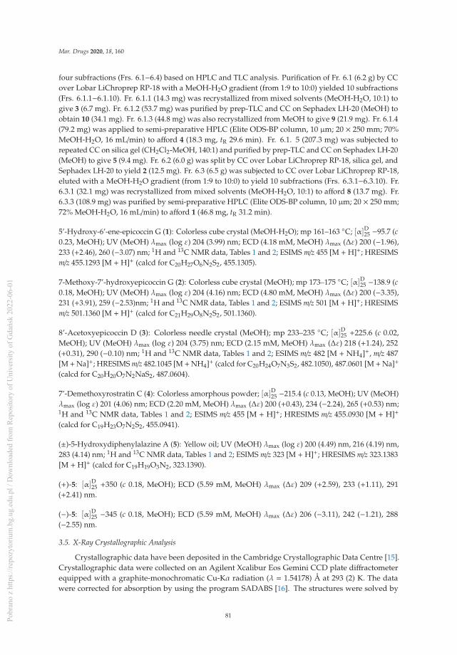
Mar. Drugs 2020, 18, 160
four subfractions (Frs. 6.1−6.4) based on HPLC and TLC analysis. Purification of Fr. 6.1 (6.2 g) by CCover Lobar LiChroprep RP-18 with a MeOH-H2O gradient (from 1:9 to 10:0) yielded 10 subfractions(Frs. 6.1.1−6.1.10). Fr. 6.1.1 (14.3 mg) was recrystallized from mixed solvents (MeOH-H2O, 10:1) togive 3 (6.7 mg). Fr. 6.1.2 (53.7 mg) was purified by prep-TLC and CC on Sephadex LH-20 (MeOH) toobtain 10 (34.1 mg). Fr. 6.1.3 (44.8 mg) was also recrystallized from MeOH to give 9 (21.9 mg). Fr. 6.1.4(79.2 mg) was applied to semi-preparative HPLC (Elite ODS-BP column, 10 μm; 20 × 250 mm; 70%MeOH-H2O, 16 mL/min) to afford 4 (18.3 mg, tR 29.6 min). Fr. 6.1. 5 (207.3 mg) was subjected torepeated CC on silica gel (CH2Cl2-MeOH, 140:1) and purified by prep-TLC and CC on Sephadex LH-20(MeOH) to give 5 (9.4 mg). Fr. 6.2 (6.0 g) was split by CC over Lobar LiChroprep RP-18, silica gel, andSephadex LH-20 to yield 2 (12.5 mg). Fr. 6.3 (6.5 g) was subjected to CC over Lobar LiChroprep RP-18,eluted with a MeOH-H2O gradient (from 1:9 to 10:0) to yield 10 subfractions (Frs. 6.3.1−6.3.10). Fr.6.3.1 (32.1 mg) was recrystallized from mixed solvents (MeOH-H2O, 10:1) to afford 8 (13.7 mg). Fr.6.3.3 (108.9 mg) was purified by semi-preparative HPLC (Elite ODS-BP column, 10 μm; 20 × 250 mm;72% MeOH-H2O, 16 mL/min) to afford 1 (46.8 mg, tR 31.2 min).
5’-Hydroxy-6’-ene-epicoccin G (1): Colorless cube crystal (MeOH-H2O); mp 161–163 ◦C; [α]D25 −95.7 (c0.23, MeOH); UV (MeOH) λmax (log ε) 204 (3.99) nm; ECD (4.18 mM, MeOH) λmax (Δε) 200 (−1.96),233 (+2.46), 260 (−3.07) nm; 1H and 13C NMR data, Tables 1 and 2; ESIMS m/z 455 [M + H]+; HRESIMSm/z 455.1293 [M + H]+ (calcd for C20H27O6N2S2, 455.1305).
7-Methoxy-7’-hydroxyepicoccin G (2): Colorless cube crystal (MeOH); mp 173–175 ◦C; [α]D25 −138.9 (c0.18, MeOH); UV (MeOH) λmax (log ε) 204 (4.16) nm; ECD (4.80 mM, MeOH) λmax (Δε) 200 (−3.35),231 (+3.91), 259 (−2.53)nm; 1H and 13C NMR data, Tables 1 and 2; ESIMS m/z 501 [M + H]+; HRESIMSm/z 501.1360 [M + H]+ (calcd for C21H29O8N2S2, 501.1360).
8’-Acetoxyepicoccin D (3): Colorless needle crystal (MeOH); mp 233–235 ◦C; [α]D25 +225.6 (c 0.02,MeOH); UV (MeOH) λmax (log ε) 204 (3.75) nm; ECD (2.15 mM, MeOH) λmax (Δε) 218 (+1.24), 252(+0.31), 290 (−0.10) nm; 1H and 13C NMR data, Tables 1 and 2; ESIMS m/z 482 [M + NH4]+, m/z 487[M +Na]+; HRESIMS m/z 482.1045 [M +NH4]+ (calcd for C20H24O7N3S2, 482.1050), 487.0601 [M +Na]+
(calcd for C20H20O7N2NaS2, 487.0604).
7’-Demethoxyrostratin C (4): Colorless amorphous powder; [α]D25 −215.4 (c 0.13, MeOH); UV (MeOH)λmax (log ε) 201 (4.06) nm; ECD (2.20 mM, MeOH) λmax (Δε) 200 (+0.43), 234 (−2.24), 265 (+0.53) nm;1H and 13C NMR data, Tables 1 and 2; ESIMS m/z 455 [M + H]+; HRESIMS m/z 455.0930 [M + H]+
(calcd for C19H23O7N2S2, 455.0941).
(±)-5-Hydroxydiphenylalazine A (5): Yellow oil; UV (MeOH) λmax (log ε) 200 (4.49) nm, 216 (4.19) nm,283 (4.14) nm; 1H and 13C NMR data, Tables 1 and 2; ESIMS m/z 323 [M + H]+; HRESIMS m/z 323.1383[M + H]+ (calcd for C19H19O3N2, 323.1390).
(+)-5: [α]D25 +350 (c 0.18, MeOH); ECD (5.59 mM, MeOH) λmax (Δε) 209 (+2.59), 233 (+1.11), 291(+2.41) nm.
(−)-5: [α]D25 −345 (c 0.18, MeOH); ECD (5.59 mM, MeOH) λmax (Δε) 206 (−3.11), 242 (−1.21), 288(−2.55) nm.
3.5. X-Ray Crystallographic Analysis
Crystallographic data have been deposited in the Cambridge Crystallographic Data Centre [15].Crystallographic data were collected on an Agilent Xcalibur Eos Gemini CCD plate diffractometerequipped with a graphite-monochromatic Cu-Kα radiation (λ = 1.54178) Å at 293 (2) K. The datawere corrected for absorption by using the program SADABS [16]. The structures were solved by
81Pobr
ano
z ht
tps:
//rep
ozyt
oriu
m.b
g.ug
.edu
.pl /
Dow
nloa
ded
from
Rep
osito
ry o
f Uni
vers
ity o
f Gda
ńsk
2022
-06-
01

Mar. Drugs 2020, 18, 160
direct methods with the SHELXTL software package [17]. All non-hydrogen atoms were refinedanisotropically. The H atoms connected to C atoms were calculated theoretically, and those to O atomswere assigned by difference Fourier maps [18]. The absolute structure was determined by refinement ofthe Flack parameter [19], based on anomalous scattering. The structures were optimized by full-matrixleast-squares techniques.
Crystal data of compound 1: C20H26N2O6S2·H2O, F.W. = 472.56, orthorhombic space groupP2(1)2(1)2(1), unit cell dimensions a = 8.5746 (5) Å, b = 10.9602 (8) Å, c = 24.3158 (16) Å, V = 2285.2 (3) Å3,α = β = γ = 90◦, Z = 4, dcalcd = 1.374 mg/m3, crystal dimensions 0.40 × 0.21× 0.13 mm, μ = 2.491 mm−1,F(000) = 1000. The 5006 measurements yielded 3321 independent reflections after equivalent data wereaveraged. The final refinement gave R1 = 0.0525 and wR2 = 0.1263 [I > 2σ(I)]. The absolute structureparameter was 0.01(3).
Crystal data of compound 2: C21H28N2O8S2, F.W. = 500.57, monoclinic space group P2(1), unitcell dimensions a = 6.8423 (4) Å, b = 20.5136 (10) Å, c = 8.2231 (5) Å, V = 1130.48 (11) Å3, α = γ = 90◦,β = 101.635 (2)◦, Z = 2, dcalcd = 1.471 mg/m3, crystal dimensions 0.20 × 0.17× 0.10 mm, μ = 2.587 mm−1,F(000) = 528. The 6832 measurements yielded 2789 independent reflections after equivalent data wereaveraged. The final refinement gave R1 = 0.0514 and wR2 = 0.1199 [I > 2σ(I)]. The absolute structureparameter was 0.02(4).
Crystal data of compound 3: C20H20N2O7S2·CH3OH, F.W. = 496.54, orthorhombic space groupP2(1)2(1)2(1), unit cell dimensions a = 10.1030 (6) Å, b = 10.8478 (5) Å, c = 19.6848 (10) Å, V = 2157.4 (2)Å3, α= β= γ= 90◦, Z= 4, dcalcd = 1.529 mg/m3, crystal dimensions 0.25× 0.17× 0.13 mm, μ= 2.711 mm−1,F(000) = 1040. The 4782 measurements yielded 3033 independent reflections after equivalent data wereaveraged. The final refinement gave R1 = 0.0510 and wR2 = 0.1001 [I > 2σ(I)]. The absolute structureparameter was 0.02(3).
3.6. Computational Section
Conformational searches were carried out via molecular mechanics with the MM+ method inHyperChem 8.0 software (Gainesville, FL, USA). Afterwards, the geometries were optimized at thegas-phase B3LYP/6-31G level in Gaussian09 software to afford the energy-minimized conformers.Then, the optimized conformers were subjected to the calculations of ECD spectra using the TD-DFT atBH&HLYP/TZVP level for 4 and PBE0/TZVP level for 5. Simultaneously, solvent effects of the MeOHsolution were evaluated at the same DFT level using the SCRF/PCM method [20].
3.7. Cytotoxic Assays
3.7.1. Cell Culture
Liver cancer Huh7.5 cell line used was obtained from the American Type Culture Collection(ATCC). Human normal liver LO2 cell line used was purchased from China Center for Type CultureCollection (CCTCC). Huh7.5 cells and LO2 cells were cultured at 37 ◦C in RPMI-1640 mediumand DMEM medium, respectively, supplemented with 10% fetal bovine serum (FBS, PAN Biotech,Aidenbach, Germany), 100 U/mL penicillin, and 100 mg/mL streptomycin. All experiments werecarried out with the same batch of cell line between passages 2 and 5 [21].
3.7.2. Cell Proliferation Assay
The cytotoxic activities of compounds 1–10 against Huh7.5 liver tumor cells and human normalliver LO2 cell line were determined by the 3-(4,5-dimethylthiazolyl-2)-2,5-diphenyltetrazolium bromide(MTT) assay. Briefly, 6 × 10³ of logarithmically growing Huh7.5 cells and human normal liver LO2 cellline were plated in the 96-well plate at 37 ◦C for 24 h. Then, cells were treated with DMSO (as thevehicle control) and increasing concentrations of test compounds (with the final concentration of 1,2, 4, 5, 8, 10, 15, 20 μM) for 48 h, respectively. MTT solution (5 mg/mL, 20 μL per well) was addedand incubated for another 4 h. After supernate from the wells were removed, DMSO was added to
82Pobr
ano
z ht
tps:
//rep
ozyt
oriu
m.b
g.ug
.edu
.pl /
Dow
nloa
ded
from
Rep
osito
ry o
f Uni
vers
ity o
f Gda
ńsk
2022
-06-
01

Mar. Drugs 2020, 18, 160
each well to dissolve purple crystals of formazan with gentle shaking for 10 min, and optical densityat 490 nm was read by a multi-detection microplate reader (Infinite M1000 Pro, Tecan, Switzerland).Sorafenib was used as positive control. All the compounds and positive control were dissolved anddiluted in DMSO. All tests were performed in triplicate. The values of relative cell viability werecalculated as percentages of absorbance from the treated samples to absorbance from the vehiclecontrol [21].
4. Conclusions
In conclusion, ten diketopiperazine alkaloids including four new derivatives,5’-hydroxy-6’-ene-epicoccin G (1), 7-methoxy-7’-hydroxyepicoccin G (2), 8’-acetoxyepicoccinD (3), and 7’-demethoxyrostratin C (4), and a pair of new enantiomeric diketopiperazines(±)-5-hydroxydiphenylalazine A ((±)-5), along with five known analogues (6–10) were characterizedfrom the deep sea-derived fungus E. nigrum SD-388. The discovery of these compounds might providefurther insight into the biosynthesis of the diketopiperazine family and provide new targets forsynthetic or biosynthetic studies. Compounds 4 and 10 exhibited potent cytotoxic activities againstHuh7.5 liver cancer cells and may provide useful candidates for further study as antitumor agents.
Supplementary Materials: Selected 1D and 2D NMR, HRESIMS, and ECD spectra of compounds 1–5 are availableonline at: http://www.mdpi.com/1660-3397/18/3/160/s1.
Author Contributions: L.-P.C. performed the experiments for the isolation, structure elucidation, and preparedthe manuscript; X.-M.L. performed the 1D and 2D NMR experiments; L.L. participated in the isolation of thefungus Epicoccum nigrum. X.L. contributed to ECD calculations and revised the manuscript; B.-G.W. All authorshave read and agreed to the published version of the manuscript.
Funding: This research work was financially supported by the Strategic Priority Research Program of the ChineseAcademy of Sciences (XDA22050401) and the Aoshan Scientific and Technological Innovation Project of QingdaoNational Laboratory for Marine Science and Technology (2016ASKJ14). X.L. appreciates the China PostdoctoralScience Foundation (2017M612360) for project funding. B.-G.W. acknowledges the support of the Research VesselKEXUE of the National Major Science and Technology Infrastructure from the Chinese Academy of Sciences(KEXUE2018G28) and the Taishan Scholar Project from Shandong Province.
Acknowledgments: The authors appreciate Chao-min Sun and Ge Liu at the Institute of Oceanology, ChineseAcademy of Sciences, for their support in cytotoxic assays, and the High Performance Computing EnvironmentQingdao Branch of the Chinese Academy of Science (CAS)–High Performance Computing Center of Institute ofOceanology of CAS for CPU time.
Conflicts of Interest: The authors declare no conflicts of interest.
References
1. Niu, S.; Xia, M.; Chen, M.; Liu, X.; Li, Z.; Xie, Y.; Shao, Z.; Zhang, G. Cytotoxic polyketides isolated fromthe deep-sea-derived fungus Penicillium chrysogenum MCCC 3A00292. Mar. Drugs 2019, 17, 686. [CrossRef][PubMed]
2. Liu, Z.; Fan, Z.; Sun, Z.; Liu, H.; Zhang, W. Dechdigliotoxins A–C, three novel disulfide-bridged gliotoxindimers from deep-sea sediment derived fungus Dichotomomyces cejpii. Mar. Drugs 2019, 17, 596. [CrossRef][PubMed]
3. Zhang, Y.; Liu, S.; Che, Y.; Liu, X. Epicoccins A–D, epipolythiodioxopiperazines from a Cordyceps-colonizingisolate of Epicoccum nigrum. J. Nat. Prod. 2007, 70, 1522–1525. [CrossRef] [PubMed]
4. Guo, H.; Sun, B.; Gao, H.; Chen, X.; Liu, S.; Yao, X.; Liu, X.; Che, Y. Diketopiperazines from theCordyceps-colonizing fungus Epicoccum nigrum. J. Nat. Prod. 2009, 72, 2115–2119. [CrossRef] [PubMed]
5. Yan, Z.; Wen, S.; Ding, M.; Guo, H.; Huang, C.; Zhu, X.; Huang, J.; She, Z.; Long, Y. The purification,characterization, and biological activity of new polyketides from mangrove-derived endophytic fungusEpicoccum nigrum SCNU-F0002. Mar. Drugs 2019, 17, 414. [CrossRef] [PubMed]
6. Sun, H.H.; Mao, W.J.; Jiao, J.Y.; Xu, J.C.; Li, H.Y.; Chen, Y.; Xu, J.; Zhao, C.Q.; Hou, Y.J.; Yang, Y.P. Structuralcharacterization of extracellular polysaccharides produced by the marine fungus Epicoccum nigrum JJY-40and their antioxidant activities. Mar. Biotechnol. 2011, 13, 1048–1055. [CrossRef] [PubMed]
83Pobr
ano
z ht
tps:
//rep
ozyt
oriu
m.b
g.ug
.edu
.pl /
Dow
nloa
ded
from
Rep
osito
ry o
f Uni
vers
ity o
f Gda
ńsk
2022
-06-
01

Mar. Drugs 2020, 18, 160
7. Tan, R.X.; Jensen, P.R.; Williams, P.G.; Fenical, W. Isolation and structure assignments of rostratins A−D,cytotoxic disulfides produced by the marine-derived fungus Exserohilum rostratum. J. Nat. Prod. 2004, 67,1374–1382. [CrossRef] [PubMed]
8. Meng, L.H.; Li, X.M.; Lv, C.T.; Huang, C.G.; Wang, B.G. Brocazines A–F, cytotoxic bisthiodiketopiperazinederivatives from Penicillium brocae MA-231, an endophytic fungus derived from the marine mangrove plantAvicennia marina. J. Nat. Prod. 2014, 77, 1921–1927. [CrossRef] [PubMed]
9. Liu, Y.; Li, X.M.; Meng, L.H.; Jiang, W.L.; Xu, G.M.; Huang, C.G.; Wang, B.G. Bisthiodiketopiperazines andacorane sesquiterpenes produced by the marine-derived fungus Penicillium adametzioides AS-53 on differentculture media. J. Nat. Prod. 2015, 78, 1294–1299. [CrossRef] [PubMed]
10. Cao, J.; Li, X.M.; Meng, L.H.; Konuklugil, B.; Li, X.; Li, H.L.; Wang, B.G. Isolation and characterization ofthree pairs of indolediketopiperazine enantiomers containing infrequent N-methoxy substitution from themarine algal-derived endophytic fungus Acrostalagmus luteoalbus TK-43. Bioorg. Chem. 2019, 90, 103030.[CrossRef] [PubMed]
11. Li, X.D.; Li, X.M.; Li, X.; Xu, G.M.; Liu, Y.; Wang, B.G. Aspewentins D–H, 20-nor-isopimarane derivativesfrom the deep sea sediment-derived fungus Aspergillus wentii SD-310. J. Nat. Prod. 2016, 79, 1347–1353.[CrossRef] [PubMed]
12. Li, X.; Chi, L.P.; Navarro-Vázquez, A.; Hwang, S.; Schmieder, P.; Li, X.M.; Li, X.; Yang, S.Q.; Lei, X.; Wang, B.G.;et al. Stereochemical elucidation of natural products from residual chemical shift anisotropies in a liquidcrystalline phase. J. Am. Chem. Soc. 2019. [CrossRef] [PubMed]
13. Kawahara, N.; Nozawa, K.; Nakajima, S.; Kawai, K.I. Emeheterone, a pyrazinone derivative from Emericellaheterothallica. Phytochemistry 1988, 27, 3022–3024. [CrossRef]
14. Wang, J.M.; Jiang, N.; Ma, J.; Yu, S.S.; Tan, R.X.; Dai, J.G.; Si, Y.K.; Ding, G.Z.; Ma, S.G.; Qu, L.; et al. Studyon absolute configurations of α/α′ chiral carbons of thiodiketopiperazines by experimental and calculatedcircular dichroism spectra. Tetrahedron 2013, 69, 1195–1201. [CrossRef]
15. Crystallographic data of compounds 1–3 have been deposited in the Cambridge Crystallographic DataCentre as CCDC 1935572, CCDC 1935573 and CCDC 1935571, respectively. Available online: https://www.ccdc.cam.ac.uk/structures/? (accessed on 13 March 2020).
16. Sheldrick, G.M. SADABS, Software for empirical absorption correction; University of Gottingen: Gottingen,Germany, 1996.
17. Sheldrick, G.M. SHELXTL, Structure determination software programs; Bruker Analytical X-ray System Inc.:Madison, WI, USA, 1997.
18. Sheldrick, G.M. SHELXL-97 and SHELXS-97, Program for X-ray Crystal Structure Solution and Refinement;University of Gottingen: Gottingen, Germany, 1997.
19. Flack, H.D. On enantiomorph-polarity estimation. Acta Cryst. 1983, 39, 876–881. [CrossRef]20. Gaussian09, RevisionC.01. Available online: http://gaussian.com/g09citation/ (accessed on 13 March 2020).21. Liu, G.; Kuang, S.; Cao, R.; Wang, J.; Peng, Q.; Sun, C. Sorafenib kills liver cancer cells by disrupting
SCD1-mediated synthesis of monounsaturated fatty acids via the ATP-AMPK-mTOR-SREBP1 signalingpathway. FASEB J. 2019, 33, 10089–10103. [CrossRef] [PubMed]
© 2020 by the authors. Licensee MDPI, Basel, Switzerland. This article is an open accessarticle distributed under the terms and conditions of the Creative Commons Attribution(CC BY) license (http://creativecommons.org/licenses/by/4.0/).
84Pobr
ano
z ht
tps:
//rep
ozyt
oriu
m.b
g.ug
.edu
.pl /
Dow
nloa
ded
from
Rep
osito
ry o
f Uni
vers
ity o
f Gda
ńsk
2022
-06-
01

marine drugs
Communication
Lysophosphatidylcholines and Chlorophyll-DerivedMolecules from the Diatom Cylindrotheca closteriumwith Anti-Inflammatory Activity
Chiara Lauritano 1,*, Kirsti Helland 2, Gennaro Riccio 1, Jeanette H. Andersen 2,
Adrianna Ianora 1 and Espen H. Hansen 2
1 Department of Marine Biotechnology, Stazione Zoologica Anton Dohrn, CAP80121 Naples, Italy;[email protected] (G.R.); [email protected] (A.I.)
2 Marbio, UiT—The Arctic University of Norway, Breivika N-9037 Tromsø, Norway;[email protected] (K.H.); [email protected] (J.H.A.); [email protected] (E.H.H.)
* Correspondence: [email protected]; Tel.: +39-081-5833-221
Received: 17 January 2020; Accepted: 13 March 2020; Published: 17 March 2020
Abstract: Microalgae have been shown to be excellent producers of lipids, pigments, carbohydrates,and a plethora of secondary metabolites with possible applications in the pharmacological,nutraceutical, and cosmeceutical sectors. Recently, various microalgal raw extracts have beenfound to have anti-inflammatory properties. In this study, we performed the fractionation of rawextracts of the diatom Cylindrotheca closterium, previously shown to have anti-inflammatory properties,obtaining five fractions. Fractions C and D were found to significantly inhibit tumor necrosisfactor alpha (TNF-α) release in LPS-stimulated human monocyte THP-1 cells. A dereplicationanalysis of these two fractions allowed the identification of their main components. Our datasuggest that lysophosphatidylcholines and a breakdown product of chlorophyll, pheophorbide a,were probably responsible for the observed anti-inflammatory activity. Pheophorbide a is knownto have anti-inflammatory properties. We tested and confirmed the anti-inflammatory activity of1-palmitoyl-sn-glycero-3-phosphocholine, the most abundant lysophosphatidylcholine found infraction C. This study demonstrated the importance of proper dereplication of bioactive extracts andfractions before isolation of compounds is commenced.
Keywords: diatoms; marine biotechnology; anti-inflammatory; drug discovery; Cylindrotheca closterium
1. Introduction
Inflammation is a complex set of interactions among soluble factors and cells (e.g., chemokines,cytokines, adhesion molecules, recruitment, and activation of leukocytes) that can arise in any tissuehelping to protect the host from systemic infection and to restore tissue homeostasis after injury,infection, and irritation [1–3]. Therefore, it represents a crucial defense mechanism that is importantfor maintenance of health [1,2]. However, if targeted destruction and assisted repair are not properlycontrolled by its mediators, the so-called “non-resolving inflammation”, they can lead to persistenttissue damage and the insurgence of various pathologies [2]. Inflammation has important pathogenicroles in several pathologies, such as asthma, atherosclerosis, atopic dermatitis, Crohn’s disease, multiplesclerosis, cystic fibrosis, psoriasis, neurodegenerative diseases, as well as cancer [1,4]. It is a protectiveresponse that involves immune cells, blood vessels, and different molecular mediators (e.g., TNF-α,IL1, nitric oxide, and prostaglandins) and anti-inflammatory assays that are used in the literature,generally include the study of one or more of these characteristics and mediators.
Oceans account for 71% of the earth’s surface and represent a huge, relatively untapped, reservoirof new compounds for drug discovery [5]. One such source is the Phytoplankton, photosynthetic
Mar. Drugs 2020, 18, 166; doi:10.3390/md18030166 www.mdpi.com/journal/marinedrugs85Pobr
ano
z ht
tps:
//rep
ozyt
oriu
m.b
g.ug
.edu
.pl /
Dow
nloa
ded
from
Rep
osito
ry o
f Uni
vers
ity o
f Gda
ńsk
2022
-06-
01

Mar. Drugs 2020, 18, 166
eukaryotes at the base of marine and freshwater food webs that are essential in the transfer of organicmaterial to top consumers such as fish [6]. These micro-organisms have shorter generation times ascompared with macro-organisms and can easily be cultivated in closed photobioreactors or in openponds providing access to larger amounts of biomass necessary for an eco-sustainable and eco-friendlyapproach to drug discovery [7]. Diatoms, with over 100,000 species, constitute one of the majorcomponents of marine phytoplankton, comprise up to 40% of annual productivity at sea [8] and represent25% of global carbon-fixation [9]. Different studies have shown that diatoms are excellent sourcesand producers of pigments, lipids, and bioactive compounds [7,10]. Anti-inflammatory propertieshave previously been found for various diatoms, such as Porosira glacialis, Attheya longicornis [11],Cylindrotheca closterium, Odontella mobiliensis, Pseudonitzschia pseudodelicatissima [12], and Phaeodactylumtricornutum [13]. The activity was assessed on lipopolysaccaride (LPS)-stimulated human THP-1macrophages, except for P. tricornutum which was tested on murine RAW 264.7 macrophages. However,there is very little information available on the actual compounds responsible for the observedanti-inflammatory activity.
Anti-inflammatory properties have also been found for flagellates. Extracts of Tetraselmis suecica [14],Chlorella ovalis, Nannochloropsis oculata, and Amphidinium carterae [13], and a sterol-rich fraction ofNannochloropsis oculata [15] were active in LPS-stimulated RAW 264.7 macrophages. Oxylipin-containinglyophilized biomass from Chlamydomonas debaryana have been shown to have anti-inflammatoryproperties in an induced colitis rat model [16–18] and Dunaliella bardawil was found to protectagainst acetic acid-induced small bowel inflammation in rats [16,17]. Regarding studies onLPS-stimulated human THP-1 monocytic leukemia cells, lipid extracts of Pavlova lutheri [19] andmonogalactosyldiacylglycerols (MGDGs) and digalactosyldiacylglycerols (DGDGs) mixtures, and theisolated DGDGs 11 and 12 from Isochrysis galbana [20] were reported as active. Regarding thecompounds responsible for anti-inflammatory properties from flagellates, lycopene was purified fromChlorella marina and the activity was confirmed in a rat model of arthritis [21], and phytosterols fromDunaliella tertiolecta were tested in a sheep model of inflammation [22]. In addition, carotenoids,the most abundant lipid-soluble phytochemicals, have shown anti-inflammatory properties [23].
In this study, we investigated the capacity of extracts of C. closterium to inhibit the release ofone of the main effectors of inflammation, TNF-α [3], in LPS-stimulated human THP-1 monocyticleukemia cells. Bioactivity-guided fractionation was performed, and chemical contents of the activefraction are described for the first time. This study is perfectly aligned with recent trends in analyzingpossible microalgal properties for cancer prevention and improving general human health andwell-being [24,25].
2. Results and Discussion
2.1. Testing for Anti-Inflammatory Activity in Algal Fractions
Previous studies have shown that raw extracts of the diatom C. closterium had anti-inflammatoryproperties [12]. In the present study, a raw extract of C. closterium was pre-fractionated to obtain fivefractions (Fractions A to E). These were amino acids and saccharides rich fraction (named Fraction A),nucleosides rich fraction (named fraction B), glycol- and phospholipid rich fraction (named fraction C),free fatty acids and sterols rich fraction (named fraction D), and triglycerides rich fraction (namedfraction E), as reported in the solid phase extraction (SPE) protocol to fractionate organic extracts ofCutignano et al. [26]. Bioactivity testing of these fractions identified fraction C as the most active,able to inhibit TNF-α release at 100 μg/mL and 50 μg/mL concentrations (Figure 1). In particular,at 100 μg/mL, fraction C showed 60% inhibition of TNF-α release (p < 0.01), and 40% inhibition at50 μg/mL (p < 0.001). Fraction D showed almost 40% TNF-α inhibition at 100 μg/mL (p < 0.01) and30% at 50 μg/mL (p < 0.001). The other fractions did not show any significant TNF-α inhibition activity(p > 0.05). Both fractions C and D were selected for dereplication and further characterization.
86Pobr
ano
z ht
tps:
//rep
ozyt
oriu
m.b
g.ug
.edu
.pl /
Dow
nloa
ded
from
Rep
osito
ry o
f Uni
vers
ity o
f Gda
ńsk
2022
-06-
01

Mar. Drugs 2020, 18, 166
Figure 1. Anti-inflammatory assay. Inhibition of TNF-α secretion from LPS-stimulated THP-1 cellstreated with fractions A, B, C, D, and E of Cylindrotheca closterium extracts (n = 3, ** for p < 0.01 and*** for p < 0.001, Student’s t-test).
2.2. Anti-Proliferative Activity Assay
In order to test if the active anti-inflammatory fractions of C. closterium also had antiproliferativeactivities, the 3-(4,5-dimethyl-2-thizolyl)-2,5-diphenyl-2H-tetrazolium bromide (MTT) assay wasperformed. In particular, A549, A2058, and HepG2 cells were incubated in the presence or inthe absence of three different concentrations (1, 10, and 100 μg/mL) of both fractions C and D.After 72 h of incubation at 37 ◦C, cell survival was measured with the MTT assay. As shown inFigure 2, fractions C and D did not show any significant inhibition of cell proliferation (p > 0.05).These results suggest that these two fractions have no antiproliferative or cytotoxicity activity butspecific anti-inflammatory activity.
Figure 2. Antiproliferative assay. The histograms show the antiproliferative effects of fractions C and Dof C. closterium extracts, on A549, A2058, and HepG2 cell lines. Control sample, containing only DMSO,was also tested (named as control). Results are expressed as percent survival after 72 h exposure (n = 3).
2.3. Dereplication
Since isolation of new compounds is very time consuming and costly [27,28], it is importantto perform an early dereplication to identify already known components. In order to identify thebioactive compounds in the active fractions C and D, they were analyzed by UHPLC-HR-MS/MS
87Pobr
ano
z ht
tps:
//rep
ozyt
oriu
m.b
g.ug
.edu
.pl /
Dow
nloa
ded
from
Rep
osito
ry o
f Uni
vers
ity o
f Gda
ńsk
2022
-06-
01

Mar. Drugs 2020, 18, 166
and compared to the inactive fractions A and B (Figure 3). In fraction C, we found a series ofcompounds that all had a common fragment at m/z 184.0740 corresponding to a molecular formula ofC5H15NO4P (see Figure S1, Supplementary Information). This is a common fragment observed whenthe head group of phosphocholines is cleaved off in tandem mass spectrometry. Phosphocholinesare a class of phospholipids where the phosphocholine head group can be esterified to one or twofatty acids. Phosphocholines with two fatty acids are common membrane-forming phospholipidsknown as phosphatidylcholines (PC). When one fatty acid is removed from a PC, either enzymaticallyor by spontaneous hydrolysis, lysophosphatidylcholines (LysoPCs) are formed. After calculatingthe elemental compositions of the related molecules in fraction C and searching the ChemSpiderdatabase for known compounds, they were all identified as LysoPCs with different fatty acids attached.From the UHPLC-HR-MS/MS data we were able to determine the length of the fatty acids andthe degree of unsaturation, but we were not able to directly determine the position of any doublebonds and if the fatty acid was attached to carbon one or two on the glycerol backbone. In orderto confirm our identification of LysoPCs, we injected a commercial standard of a C16:0 LysoPC(1-palmiotyl-sn-glycero-3-phosphocholine). The standard had the same retention time, mass, collisionalcross section, and fragmentation pattern as one of the most intense compounds in fraction C (seeFigure S2 and S3, Supplementary Information). The dominating LysoPCs in fraction C were 16:0,16:1, 18:1, and 18:2 (approximately equal amounts) and minor LysoPCs were 14:0 and 18:3 (eachapproximately 20% of the most intense LysoPCs). A summary of the most intense LysoPCs andtheir retention times is given in Table S1 (Supplementary Information). There is current interest inLysoPCs because some of these are proposed for treatment of systemic inflammatory disorders [29–35].However, their biological roles are not completely understood and some studies even found aputative pro-inflammatory activity [29]. Plasma LysoPC levels are diminished in human patients withsepsis [31,36], and in rodent models of sepsis and ischemia, LysoPC treatments in ex vivo and in vivostudies suggesting a potential role to relieve serious inflammatory conditions [29]. LysoPCs have alsobeen shown to prevent neuronal death both in an in vivo model of transient global ischemia and inan in vitro model of excitotoxicity using primary cultures of cerebellar granule cells exposed to highextracellular concentrations of glutamate (20 to 40 micromol/L).
In fraction D, trace amounts of the same LysoPCs were present. However, the most intense peakin fraction D had a m/z value of 593.2752 with a corresponding elemental composition of C35H37N4O5
([M +H]+). When searching in the ChemSpider database, the elemental composition, as well as thefragmentation pattern, indicated that the compound was pheophorbide a, a breakdown product ofchlorophyll (see Figure S4, Supplementary Information). Another peak in fraction D was identifiedas a related breakdown product of chlorophyll, hydroxypheophorbide a (C35H36N4O6, m/z 609.2708[M + H]+), see Figure S5 (Supplementary Information). Both pheophorbide a and its derivativesare known to have anti-inflammatory and anticancer properties [37–43], but, to our knowledge,this is the first case in a marine microalgae where the bioactivity was attributed to pheophorbide a.Pheophorbide a has already been extracted from a range of different marine organisms. Examples arethe seaweed Grateloupia ellittica [40], the brown alga Saccharina japonica [39], marine diatoms [44,45],and the freshwater glaucophyte Cyanophora paradoxa [37]. Hydroxypheophorbide a has been previouslyisolated from the terrestrial plants Clerodendrum calamitosum, Neptunia oleracea, the freshwater unicellulargreen alga Chlorella sp., and from the marine tunicate Trididemnum solidum, but never from a marinediatom species. It is mainly known to have anticancer but not anti-inflammatory activity ([38,41],Patents No. 185220/82 and US4709022A). Hence we suggest that the possible anti-inflammatory activityobserved in our experiments was due to the presence of LysoPCs and the known anti-inflammatorypheophorbide a. In fact, pheophorbide a is known to induce a dose-dependent inhibition againstlipopolysaccharide (LPS)-induced nitric oxide (NO) production at nontoxic concentrations in RAW264.7 murine macrophage cells and to suppress the expression of nitric oxide synthase (iNOS) [39].
88Pobr
ano
z ht
tps:
//rep
ozyt
oriu
m.b
g.ug
.edu
.pl /
Dow
nloa
ded
from
Rep
osito
ry o
f Uni
vers
ity o
f Gda
ńsk
2022
-06-
01

Mar. Drugs 2020, 18, 166
Figure 3. Base peak intensity chromatograms of fraction A, B, C, and D from the UHPLC-HR-MS/MSanalysis using positive electrospray.
2.4. Anti-Inflammatory Activity of 1-Palmitoyl-sn-glycero-3-phosphocholine
Considering that the most active fraction mainly contained various phosphocholines, we testedone of these, 1-palmitoyl-sn-glycero-3-phosphocholine (which was the most abundant compoundin fraction C) in our AIF-assay. The effect of 1-palmitoyl-sn-glycero-3-phosphocholine on secretionof TNF-α showed a dose-response relationship and was active at 25 μg/mL (p < 0.05) and 50 μg/mL(p < 0.01), as shown in Figure 4.
Figure 4. Anti-inflammatory assay. Inhibition of TNF-α secretion from LPS-stimulated THP-1 cellstreated with 3.13, 6.25, 12.5, 25, and 50 μg/mL of 1-Palmitoyl-sn-glycero-3-phosphocholine (n = 3;* for p < 0.05 and ** for p < 0.01, Student’s t-test).
89Pobr
ano
z ht
tps:
//rep
ozyt
oriu
m.b
g.ug
.edu
.pl /
Dow
nloa
ded
from
Rep
osito
ry o
f Uni
vers
ity o
f Gda
ńsk
2022
-06-
01

Mar. Drugs 2020, 18, 166
3. Conclusions
Considering that inflammation plays a crucial role in the pathogenicity of several diseases,marine drug discovery is often directed to finding new natural products with anti-inflammatoryproperties [46,47]. Our results indicate that lysophosphatidylcholines (lysoPCs) and a breakdownproduct of chlorophyll, pheophorbide a, were probably responsible for the observed anti-inflammatoryactivity of the diatom C. closterium, giving new insights into microalgal compound bioactivitiesand their possible applications. This is the first time that a marine diatom is reported to producethese anti-inflammatory compounds. Pheophorbide a is known to inhibit the production of NO viainhibition of iNOS protein expression, thereby suggesting its potential use in the treatment of variousinflammatory diseases (37), but there is no further information on its anti-inflammatory mechanism ofaction. Hence our study shows, for the first time, that it can inhibit TNF-α release in THP1 cells.
LysoPCs are products of phospholipase A2 enzyme activity, and similar to the enzyme, have adirect role in pro-inflammatory [48] and anti-inflammatory responses, in a variety of organ systems.Our results indicate that one of these LysoPCs, 1-palmitoyl-sn-glycero-3-phosphocholine (which wasthe most abundant compound in fraction C), had a strong anti-inflammatory activity which has not beendemonstrated before. Microalgae, and in particular diatoms, therefore, can be considered potentiallyimportant producers of compounds to prevent and treat different human pathologies. In recent yearsthey have been shown to have anti-inflammatory, antimicrobial, anticancer, antidiabetic, antiepilepticand even antituberculosis properties [10–12,49–55]. A better understanding of the potential healthbenefits from these marine organisms, the compounds they produce, and the environmental conditionsaffecting their production should allow for the sustainable development of these valuable marineresources in the future.
4. Materials and Methods
4.1. Cell Culturing and Harvesting
The diatom Cylindrotheca closterium (FE2), which has previously been shown to haveanti-inflammatory activity [12], was cultured in Guillard’s f/2 medium [56] in ten-liter polycarbonatecarboys (4 replicates). Cultures were constantly bubbled with air filtered through 0.2 μm membranefilters and kept in a climate chamber at 19 ◦C and a 12:12 h light:dark cycle (100 μmol photons m−2 s−1).Initial cell concentration was about 5000 cells/mL per bottle; culture growth was monitored daily byfixing 1 ml of culture with one drop of Lugol (final concentration of about 2%) and counting cells in aBürker counting chamber under an Axioskop 2 microscope (20×) (Carl Zeiss GmbH, Jena, Germany).At the end of the stationary phase, cultures were centrifuged for 15 min at 4 ◦C at 3900 g using a cooledcentrifuge with a swing-out rotor (DR 15P, Braun Biotechnology International, Allentown, PA, USA).The supernatant was discarded, and pellets freeze-dried and kept at −80 ◦C until chemical extraction.
4.2. Extraction and Fractionation
For extraction and fractionation, the protocol by Cutignano et al. [26] was used, but with somemodifications. Briefly, a methanolic extract was firstly prepared by adding 5 mL methanol (MeOH)for each g of pellet (algae were cultured in three different occasions in triplicate batches), sonicatingthe samples for 30 s, centrifuging them at 4000 rpm for 5 min at room temperature and drying thesupernatant with a rotovapor.
For the fractionation, columns were activated (column type: 6 mL/500 mg resin) with 6 mLmethanol and 17 mL of distilled water. The resin used was a spherical poly(styrene-divinylbenzene)resin for SPE (Chromabond®HR-X, Düren, Germany). One mL of distilled water was added for each20 mg of methanolic extract. Samples were sonicated to obtain a good suspension and added to thecolumns. The column was eluted as follows: (1) wash step with 2 mL of distilled water, throwingaway 1.5 mL to eliminate salts, (2) addition of 18 mL of distilled water to obtain fraction A, (3) additionof 24 mL of methanol (CH3OH)/water (50:50) to obtain fraction B, (4) addition of 18 mL Acetonitrile
90Pobr
ano
z ht
tps:
//rep
ozyt
oriu
m.b
g.ug
.edu
.pl /
Dow
nloa
ded
from
Rep
osito
ry o
f Uni
vers
ity o
f Gda
ńsk
2022
-06-
01

Mar. Drugs 2020, 18, 166
(CH3CN)/water (70:30) to obtain fraction C, (5) addition of 18 mL acetonitrile (100%) to obtain fraction D,(6) and finally addition of 18 mL of dichloromethane/methanol (CH2Cl2/CH3OH; 90:10) to obtainfraction E.
4.3. Anti-Inflammatory Assay
The anti-inflammatory assay was performed as in Lauritano et al. [12]. Briefly, ∼106 humanmonocyte THP-1 cells/mL (ATCC®TIB-202TM) supplemented with 50 ng/mL phorbol 12- myristate13-acetate (PMA, Sigma-Aldrich) were seeded in 96-well plates and incubated at 37 ◦C, 5% CO2 for 48h in RPMI-1640 medium (Biochrom). After 72 h, 80 μL fresh RPMI medium and 10 μL/well of testsample were added. In particular, fractions A, B, C, D, and E were tested at 100 and 50 μg/mL, while1-palmitoyl-sn-glycero-3-phosphocholine (Sigma L5254) was tested at 3.13, 6.25, 12.5, 25, and 50 μg/mL.The tests were performed at least in triplicate. After incubation for 1 h, all samples were incubated with1 ng/mL lipopolysaccharide (LPS) for 6 h at 37 ◦C and plates were then frozen at −80 ◦C. Enzyme-linkedimmunosorbent assay (ELISA) was used to test TNF-α inhibition. ELISA was performed as in Lauritanoet al. [12]. Results were read at 405 nm.
4.4. In Vitro Anti-Proliferative Assay
Human cells were bought at ATCC (https://www.lgcstandards-atcc.org/). Human hepatocellularliver carcinoma cells (HepG2; ATCC®HB-8065™) were cultured in EMEM medium, human melanomacells (A2058; ATCC®CRL-11147TM) were cultured in DMEM, adenocarcinomic human alveolarbasal epithelial cells (A549; ATCC®CL-185TM) were cultured in F-12K medium. The media weresupplemented with 10% fetal bovine serum, 50 U/ml penicillin, and 50 μg/ml streptomycin.
To evaluate the in vitro antiproliferative effects of the fractions, HepG2, A2058, and A549 celllines were seeded in 96-well microtiter plates at a density of 1 × 10⁴ cells/well and incubated at37 ◦C to allow for cell adhesion in the plates. After 16 h, the medium was replaced with freshmedium containing increasing concentrations of the fractions (1, 10, and 100 μg/mL) for 72 h. Eachconcentration was tested at least in triplicate. After 72 h, cell viability was assessed using the MTT test(3-(4,5-dimethyl-2-thizolyl)-2,5-diphenyl-2H-tetrazolium bromide; A2231,0001, AppliChem PanreacTischkalender, Darmstadt, GmbH). Briefly, the medium was replaced with medium containing MTTat 0.5 mg/ml and the plates were incubated for 3 h at 37 ◦C. After incubation, cells were treatedwith isopropyl alcohol (used as MTT solvent) for 30 minutes at room temperature. Absorbance wasmeasured at OD = 570 nm using a microplate reader (Multiskan™ FC Microplate Photometer, ThermoFisher Scientific, Waltham, MA, USA). Cell survival was expressed as a percentage of viable cells in thepresence of the tested samples, with respect to untreated control cultures.
4.5. Statistical Analysis
Student’s t-test was performed using GraphPad Prism statistic software, V4.00 (GraphPadSoftware, San Diego, CA, USA). Data were considered significant when p values were <0.05 (∗ forp < 0.05, ∗∗ for p < 0.01, and ∗∗∗ for p < 0.001).
4.6. Dereplication of Fractions
One mg of each fraction A to E were resuspended in 100 μL 80% aqueous methanol,centrifuged at 13,000 rpm for 5 min, and the supernatants were transferred to UHPLC injectionvials. UHPLC-HR-MS/MS analysis of the fractions was performed using a Waters Acquity I-class UPLCsystem interfaced with a PDA Detector and a VION IMS-qTOF (Milford, MA, USA) using electrosprayionization (ESI) in positive mode. The VION IMS- qTOF was operated with a capillary voltage of0.80 kV, desolvation gas flow (N2) of 800 L/h, desolvation temperature of 450 ◦C, cone gas flow (N2) of50 L/h and an ion source temperature of 120 ◦C. Data were acquired between m/z 50 and 2000 with ascan time of 0.2 s. Fragment data were acquired by ramping the energy of the collision cell from 15 to45 V, and high and low energy data were acquired in the same run. Leucine-enkephalin was used
91Pobr
ano
z ht
tps:
//rep
ozyt
oriu
m.b
g.ug
.edu
.pl /
Dow
nloa
ded
from
Rep
osito
ry o
f Uni
vers
ity o
f Gda
ńsk
2022
-06-
01
Mar. Drugs 2020, 18, 166
for internal calibration and the system was tuned to a resolution of 45,000 (FWHM). The system wascontrolled, and data were processed using UNIFI 1.9.4 (Waters). Chromatographic separation wasachieved by injecting 3 μL of the dissolved fractions on a BEH C18 1.7 μm (2.1 × 100 mm) column(Waters) operated at 40 ◦C. The fractions were eluted with a gradient of 10% to 100% acetonitrile inwater over 10 min (both containing 0.1% formic acid), followed by maintaining 100% acetonitrile until13.5 min.
Supplementary Materials: The following are available online at http://www.mdpi.com/1660-3397/18/3/166/s1,Figure S1: Top: Base peak intensity chromatogram from the UHPLC-HR-MS/MS analysis of fraction C usingpositive electrospray. Bottom: Ion chromatogram of m/z 184. 0740 from fraction C indicating the presence ofphosphocholines, Figure S2: Top: Ion chromatogram of m/z 496.3396 from fraction C showing the presence ofLysoPC. Bottom: Base peak intensity chromatogram from the UHPLC-HR-MS/MS analysis of fraction C usingpositive electrospray, Figure S3: Top: Low energy mass spectrum of 1-Palmiotyl-sn-glycero-3-phosphocholinefrom fraction C. Bottom: High energy mass spectrum of 1-Palmiotyl-sn-glycero-3-phosphocholine showingfragments and mass deviations corresponding with the commercial standard, Figure S4: Top: Low energymass spectrum of pheophorbide A from fraction D. Bottom: High energy mass spectrum of pheophorbide Ashowing fragments and mass deviations corresponding with the theoretical fragmentation of the database hit,Figure S5: Top: Low energy mass spectrum of hydroxypheophorbide A from fraction D. Bottom: High energymass spectrum of hydroxypheophorbide A showing fragments and mass deviations corresponding with thetheoretical fragmentation of the database hit, Table S1: The most abundant lysophosphatidylcholines in fraction C.The type indicates number of carbons and double bonds in the fatty acid moiety of the respective LysoPCs, andthe retention times are given in minutes.
Author Contributions: C.L. and E.H.H. conceived and designed the experiments; C.L., K.H., G.R., and E.H.H.performed the experiments; C.L., K.H., G.R., and E.H.H. analyzed the data. All authors (C.L., K.H., G.R., J.H.A.,A.I. and E.H.H.) co-wrote the paper. All authors have read and agreed to the published version of the manuscript.
Funding: This research was funded by the “Antitumor Drugs and Vaccines from the Sea (ADViSE)” project(PG/2018/0494374).
Acknowledgments: We thank Massimo Perna and Mariano Amoroso for their technical support.
Conflicts of Interest: The authors declare no conflict of interest.
References
1. Nathan, C. Points of control in inflammation. Nature 2002, 420, 846–852. [CrossRef] [PubMed]2. Nathan, C.; Ding, A. Nonresolving inflammation. Cell 2010, 140, 871–882. [CrossRef] [PubMed]3. Newton, K.; Dixit, V.M. Signaling in innate immunity and inflammation. Cold Spring Harb. Perspect. Biol.
2012, 4, a006049. [CrossRef] [PubMed]4. Korniluk, A.; Koper, O.; Kemona, H.; Dymicka-Piekarska, V. From inflammation to cancer. Ir. J. Med. Sci.
2017, 186, 57–62. [CrossRef]5. Jaspars, M.; De Pascale, D.; Andersen, J.H.; Reyes, F.; Crawford, A.D.; Ianora, A. The marine biodiscovery
pipeline and ocean medicines of tomorrow. J. Mar. Biol. Assoc. U. K. 2016, 96, 151–158. [CrossRef]6. Carotenuto, Y.; Esposito, F.; Pisano, F.; Lauritano, C.; Perna, M.; Miralto, A.; Ianora, A. Multi-generation
cultivation of the copepod Calanus helgolandicus in a re-circulating system. J. Exp. Mar. Biol. Ecol. 2012,418–419, 46–58. [CrossRef]
7. Romano, G.; Costantini, M.; Sansone, C.; Lauritano, C.; Ruocco, N.; Ianora, A. Marine microorganisms as apromising and sustainable source of bioactive molecules. Mar. Environ. Res. 2017, 128, 58–69. [CrossRef]
8. Falkowski, P.G. The role of phytoplankton photosynthesis in global biogeochemical cycles. Photosyn. Res.1994, 39, 235–258. [CrossRef]
9. Nelson, D.M.; Tréguer, P.; Brzezinski, M.A.; Leynaert, A.; Quéguiner, B. Production and dissolution ofbiogenic silica in the ocean: Revised global estimates, comparison with regional data and relationship tobiogenic sedimentation. Glob. Biogeochem. Cycles 1995, 9, 359–372. [CrossRef]
10. Martínez Andrade, K.; Lauritano, C.; Romano, G.; Ianora, A. Marine microalgae with anti-cancer properties.Mar. Drugs 2018, 16, 165. [CrossRef]
11. Ingebrigtsen, R.A.; Hansen, E.; Andersen, J.H.; Eilertsen, H.C. Light and temperature effects on bioactivity indiatoms. J. Appl. Phycol. 2016, 28, 939–950. [CrossRef]
92Pobr
ano
z ht
tps:
//rep
ozyt
oriu
m.b
g.ug
.edu
.pl /
Dow
nloa
ded
from
Rep
osito
ry o
f Uni
vers
ity o
f Gda
ńsk
2022
-06-
01

Mar. Drugs 2020, 18, 166
12. Lauritano, C.; Andersen, J.H.; Hansen, E.; Albrigtsen, M.; Escalera, L.; Esposito, F.; Helland, K.; Hanssen, K.Ø.;Romano, G.; Ianora, A. Bioactivity screening of microalgae for antioxidant, anti-inflammatory, anticancer,anti-diabetes, and antibacterial activities. Front. Mar. Sci. 2016, 3, 68. [CrossRef]
13. Samarakoon, K.W.; Ko, J.-Y.; Shah, M.R.; Lee, J.-H.; Kang, M.-C.; O-Nam, K.; Lee, J.-B.; Jeon, Y.-J. In vitrostudies of anti-inflammatory and anticancer activities of organic solvent extracts from cultured marinemicroalgae. Algae 2013, 28, 111–119. [CrossRef]
14. Jo, W.S.; Choi, Y.J.; Kim, H.J.; Nam, B.H.; Hong, S.H.; Lee, G.A.; Lee, S.W.; Seo, S.Y.; Jeong, M.H.Anti-inflammatory effect of microalgal extracts from Tetraselmis suecica. Food Sci. Biotechnol. 2010, 19,1519–1528. [CrossRef]
15. Sanjeewa, K.K.A.; Fernando, I.P.S.; Samarakoon, K.W.; Lakmal, H.H.C.; Kim, E.-A.; Kwon, O.-N.;Dilshara, M.G.; Lee, J.-B.; Jeon, Y.-J. Anti-inflammatory and anti-cancer activities of sterol rich fraction ofcultured marine microalga Nannochloropsis oculata. Algae 2016, 31, 277–287. [CrossRef]
16. Lavy, A.; Naveh, Y.; Coleman, R.; Mokady, S.; Werman, M.J. Dietary Dunaliella bardawil, a beta-carotene-richalga, protects against acetic acid-induced small bowel inflammation in rats. Inflamm. Bowel Dis. 2003, 9,372–379. [CrossRef] [PubMed]
17. Lee, J.-C.; Hou, M.-F.; Huang, H.-W.; Chang, F.-R.; Yeh, C.-C.; Tang, J.-Y.; Chang, H.-W. Marine algal naturalproducts with anti-oxidative, anti-inflammatory, and anti-cancer properties. Cancer Cell Int. 2013, 13, 55.[CrossRef]
18. Ávila-Román, J.; Talero, E.; Rodríguez-Luna, A.; García-Mauriño, S.; Motilva, V. Anti-inflammatory effects ofan oxylipin-containing lyophilised biomass from a microalga in a murine recurrent colitis model. Br. J. Nutr.2016, 116, 2044–2052. [CrossRef]
19. Robertson, R.C.; Guihéneuf, F.; Bahar, B.; Schmid, M.; Stengel, D.B.; Fitzgerald, G.F.; Ross, R.P.; Stanton, C.The anti-inflammatory effect of algae-derived lipid extracts on lipopolysaccharide (LPS)-stimulated humanTHP-1 macrophages. Mar. Drugs 2015, 13, 5402–5424. [CrossRef]
20. de los Reyes, C.; Ortega, M.J.; Rodríguez-Luna, A.; Talero, E.; Motilva, V.; Zubía, E. Molecular characterizationand anti-inflammatory activity of galactosylglycerides and galactosylceramides from the microalga Isochrysisgalbana. J. Agric. Food Chem. 2016, 64, 8783–8794. [CrossRef]
21. Renju, G.L.; Muraleedhara Kurup, G.; Saritha Kumari, C.H. Anti-inflammatory activity of lycopene isolatedfrom Chlorella marina on type II collagen induced arthritis in Sprague Dawley rats. Immunopharmacol.Immunotoxicol. 2013, 35, 282–291. [CrossRef] [PubMed]
22. Caroprese, M.; Albenzio, M.; Ciliberti, M.G.; Francavilla, M.; Sevi, A. A mixture of phytosterols fromDunaliella tertiolecta affects proliferation of peripheral blood mononuclear cells and cytokine production insheep. Vet. Immunol. Immunopathol. 2012, 150, 27–35. [CrossRef] [PubMed]
23. Kaulmann, A.; Bohn, T. Carotenoids, inflammation, and oxidative stress–implications of cellular signalingpathways and relation to chronic disease prevention. Nutr. Res. 2014, 34, 907–929. [CrossRef] [PubMed]
24. Nicoletti, M. Microalgae nutraceuticals. Foods 2016, 5, 54. [CrossRef]25. García, J.L.; de Vicente, M.; Galán, B. Microalgae, old sustainable food and fashion nutraceuticals. Microb.
Biotechnol. 2017, 10, 1017–1024. [CrossRef]26. Cutignano, A.; Nuzzo, G.; Ianora, A.; Luongo, E.; Romano, G.; Gallo, C.; Sansone, C.; Aprea, S.; Mancini, F.;
D’Oro, U.; et al. Development and application of a novel SPE-method for bioassay-guided fractionation ofmarine extracts. Mar. Drugs 2015, 13, 5736–5749. [CrossRef]
27. Cordell, G.A.; Shin, Y.G. Finding the needle in the haystack. The dereplication of natural product extracts.Pure Appl. Chem. 1999, 71, 1089–1094. [CrossRef]
28. Hubert, J.; Nuzillard, J.-M.; Renault, J.-H. Dereplication strategies in natural product research: How manytools and methodologies behind the same concept? Phytochem. Rev. 2017, 16, 55–95. [CrossRef]
29. Cunningham, T.J.; Yao, L.; Lucena, A. Product inhibition of secreted phospholipase A2 may explainlysophosphatidylcholines’ unexpected therapeutic properties. J. Inflamm. 2008, 5, 17. [CrossRef]
30. Mehta, D. Lysophosphatidylcholine: An enigmatic lysolipid. Am. J. Physiol.-Lung Cell. Mol. Physiol. 2005,289, L174–L175. [CrossRef]
31. Drobnik, W.; Liebisch, G.; Audebert, F.-X.; Frohlich, D.; Gluck, T.; Vogel, P.; Rothe, G.; Schmitz, G. Plasmaceramide and lysophosphatidylcholine inversely correlate with mortality in sepsis patients. J. Lipid Res. 2003,44, 754–761. [CrossRef] [PubMed]
93Pobr
ano
z ht
tps:
//rep
ozyt
oriu
m.b
g.ug
.edu
.pl /
Dow
nloa
ded
from
Rep
osito
ry o
f Uni
vers
ity o
f Gda
ńsk
2022
-06-
01
Mar. Drugs 2020, 18, 166
32. Chen, G.; Li, J.; Qiang, X.; Czura, C.J.; Ochani, M.; Ochani, K.; Ulloa, L.; Yang, H.; Tracey, K.J.; Wang, P.; et al.Suppression of HMGB1 release by stearoyl lysophosphatidylcholine: An additional mechanism for itstherapeutic effects in experimental sepsis. J. Lipid Res. 2005, 46, 623–627. [CrossRef] [PubMed]
33. Blondeau, N.; Lauritzen, I.; Widmann, C.; Lazdunski, M.; Heurteaux, C. A potent protective role oflysophospholipids against global cerebral ischemia and glutamate excitotoxicity in neuronal cultures. J.Cereb. Blood Flow Metab. 2002, 22, 821–834. [CrossRef] [PubMed]
34. Yan, J.-J.; Jung, J.-S.; Lee, J.-E.; Lee, J.; Huh, S.-O.; Kim, H.-S.; Jung, K.C.; Cho, J.-Y.; Nam, J.-S.; Suh, H.-W.;et al. Therapeutic effects of lysophosphatidylcholine in experimental sepsis. Nat. Med. 2004, 10, 161–167.[CrossRef]
35. Murch, O.; Collin, M.; Sepodes, B.; Foster, S.J.; Mota-Filipe, H.; Thiemermann, C. Lysophosphatidylcholinereduces the organ injury and dysfunction in rodent models of gram-negative and gram-positive shock. Br. J.Pharmacol. 2006, 148, 769–777. [CrossRef]
36. Lissauer, E.; Johnson, B.G.; Shi, S.; Gentle, T.; Scalea, M. 128: Decreased lysophosphatidylcholine levels areassociated with sepsis compared to uninfected inflammation prior to onset of sepsis. J. Surg. Res. 2007, 137, 206.[CrossRef]
37. Baudelet, P.-H.; Gagez, A.-L.; Bérard, J.-B.; Juin, C.; Bridiau, N.; Kaas, R.; Thiéry, V.; Cadoret, J.-P.; Picot, L.Antiproliferative activity of Cyanophora paradoxa pigments in melanoma, breast and lung cancer cells. Mar.Drugs 2013, 11, 4390–4406. [CrossRef]
38. Choi, S.-E.; Sohn, S.; Cho, J.-W.; Shin, E.-A.; Song, P.-S.; Kang, Y. 9-Hydroxypheophorbide alpha-inducedapoptotic death of MCF-7 breast cancer cells is mediated by c-Jun N-terminal kinase activation. J. Photochem.Photobiol. B Biol. 2004, 73, 101–107. [CrossRef]
39. Islam, M.N.; Ishita, I.J.; Jin, S.E.; Choi, R.J.; Lee, C.M.; Kim, Y.S.; Jung, H.A.; Choi, J.S. Anti-inflammatoryactivity of edible brown alga Saccharina japonica and its constituents pheophorbide a and pheophytin a inLPS-stimulated RAW 264.7 macrophage cells. Food Chem. Toxicol. 2013, 55, 541–548. [CrossRef]
40. Cho, M.; Park, G.-M.; Kim, S.-N.; Amna, T.; Lee, S.; Shin, W.-S. Glioblastoma-specific anticancer activity ofpheophorbide a from the edible red seaweed Grateloupia elliptica. J. Microbiol. Biotechnol. 2014, 24, 346–353.[CrossRef]
41. Nakamura, Y.; Murakami, A.; Koshimizu, K.; Ohigashi, H. Inhibitory effect of pheophorbide a, achlorophyll-related compound, on skin tumor promotion in ICR mouse. Cancer Lett. 1996, 108, 247–255.[CrossRef]
42. Chan, J.Y.-W.; Tang, P.M.-K.; Hon, P.-M.; Au, S.W.-N.; Tsui, S.K.-W.; Waye, M.M.-Y.; Kong, S.-K.; Mak, T.C.-W.;Fung, K.-P. Pheophorbide a, a major antitumor component purified from Scutellaria barbata, induces apoptosisin human hepatocellular carcinoma cells. Planta Med. 2006, 72, 28–33. [CrossRef] [PubMed]
43. Hibasami, H.; Kyohkon, M.; Ohwaki, S.; Katsuzaki, H.; Imai, K.; Nakagawa, M.; Ishi, Y.; Komiya, T.Pheophorbide a, a moiety of chlorophyll a, induces apoptosis in human lymphoid leukemia molt 4B cells.Int. J. Mol. Med. 2000, 6, 277–279. [CrossRef] [PubMed]
44. Veuger, B.; Oevelen, D. Long-term pigment dynamics and diatom survival in dark sediment. Limnol.Oceanogr. 2011, 56, 1065–1074. [CrossRef]
45. Kuczynska, P.; Jemiola-Rzeminska, M.; Strzalka, K. Photosynthetic pigments in diatoms. Mar. Drugs 2015,13, 5847–5881. [CrossRef] [PubMed]
46. Hurst, D.; Børresen, T.; Almesjö, L.; Raedemaecker, F.; Bergseth, S. Marine Biotechnology Strategic Research andInnovation Roadmap-Insights to the Future Direction of European Marine Biotechnology; Marine BiotechnologyERA-NET: Oostende, Belgium, 2016; ISBN 978-94-92043-27-6.
47. Reen, F.J.; Gutiérrez-Barranquero, J.A.; Dobson, A.D.W.; Adams, C.; O’Gara, F. Emerging concepts promisingnew horizons for marine biodiscovery and synthetic biology. Mar. Drugs 2015, 13, 2924–2954. [CrossRef]
48. Law, S.-H.; Chan, M.-L.; Marathe, G.K.; Parveen, F.; Chen, C.-H.; Ke, L.-Y. An updated review oflysophosphatidylcholine metabolism in human diseases. Int. J. Mol. Sci. 2019, 20, 1149. [CrossRef]
49. Lauritano, C.; Martín, J.; de la Cruz, M.; Reyes, F.; Romano, G.; Ianora, A. First identification of marinediatoms with anti-tuberculosis activity. Sci. Rep. 2018, 8, 2284. [CrossRef]
50. Brillatz, T.; Lauritano, C.; Jacmin, M.; Khamma, S.; Marcourt, L.; Righi, D.; Romano, G.; Esposito, F.; Ianora, A.;Queiroz, E.F.; et al. Zebrafish-based identification of the antiseizure nucleoside inosine from the marinediatom Skeletonema marinoi. PLoS ONE 2018, 13, e0196195. [CrossRef]
94Pobr
ano
z ht
tps:
//rep
ozyt
oriu
m.b
g.ug
.edu
.pl /
Dow
nloa
ded
from
Rep
osito
ry o
f Uni
vers
ity o
f Gda
ńsk
2022
-06-
01

Mar. Drugs 2020, 18, 166
51. Giordano, D.; Costantini, M.; Coppola, D.; Lauritano, C.; Núñez Pons, L.; Ruocco, N.; di Prisco, G.; Ianora, A.;Verde, C. Biotechnological applications of bioactive peptides from marine sources. In Advances in MicrobialPhysiology; Elsevier: Amsterdam, The Netherlands, 2018; Volume 73, pp. 171–220. ISBN 978-0-12-815190-7.
52. Riccio, G.; Lauritano, C. Microalgae with immunomodulatory activities. Mar. Drugs 2020, 18, 2. [CrossRef]53. Lauritano, C.; Ianora, A. Marine organisms with anti-diabetes properties. Mar. Drugs 2016, 14, 220. [CrossRef]
[PubMed]54. Sasso, S.; Pohnert, G.; Lohr, M.; Mittag, M.; Hertweck, C. Microalgae in the postgenomic era: A blooming
reservoir for new natural products. FEMS Microbiol. Rev. 2012, 36, 761–785. [CrossRef] [PubMed]55. Plaza, M.; Herrero, M.; Cifuentes, A.; Ibáñez, E. Innovative natural functional ingredients from microalgae. J.
Agric. Food Chem. 2009, 57, 7159–7170. [CrossRef] [PubMed]56. Guillard, R.R.L. Culture of phytoplankton for feeding marine invertebrates. In Culture of Marine Invertebrate
Animals: Proceedings—1st Conference on Culture of Marine Invertebrate Animals Greenport; Smith, W.L.,Chanley, M.H., Eds.; Springer: Boston, MA, USA, 1975; pp. 29–60. ISBN 978-1-4615-8714-9.
© 2020 by the authors. Licensee MDPI, Basel, Switzerland. This article is an open accessarticle distributed under the terms and conditions of the Creative Commons Attribution(CC BY) license (http://creativecommons.org/licenses/by/4.0/).
95Pobr
ano
z ht
tps:
//rep
ozyt
oriu
m.b
g.ug
.edu
.pl /
Dow
nloa
ded
from
Rep
osito
ry o
f Uni
vers
ity o
f Gda
ńsk
2022
-06-
01

Pobr
ano
z ht
tps:
//rep
ozyt
oriu
m.b
g.ug
.edu
.pl /
Dow
nloa
ded
from
Rep
osito
ry o
f Uni
vers
ity o
f Gda
ńsk
2022
-06-
01

marine drugs
Article
Bioactive Molecules from Mangrove Streptomycesqinglanensis 172205
Dongbo Xu 1, Erli Tian 1, Fandong Kong 2 and Kui Hong 1,*
1 Key Laboratory of Combinatorial Biosynthesis and Drug Discovery, Ministry of Education,School of Pharmaceutical Sciences, Wuhan University, Wuhan 430071, China;[email protected] (D.X.); [email protected] (E.T.)
2 Institute of Tropical Bioscience and Biotechnology, Chinese Academy of Tropical Agricultura Sciences,Haikou 571101, China; [email protected]
* Correspondence: [email protected]; Tel.: +86-27-6875-2442
Received: 17 April 2020; Accepted: 12 May 2020; Published: 13 May 2020
Abstract: Five new compounds 15R-17,18-dehydroxantholipin (1), (3E,5E,7E)-3-methyldeca-3,5,7-triene-2,9-dione (2) and qinlactone A–C (3–5) were identified from mangrove Streptomycesqinglanensis 172205 with “genetic dereplication,” which deleted the highly expressed secondarymetabolite-enterocin biosynthetic gene cluster. The chemical structures were established byspectroscopic methods, and the absolute configurations were determined by electronic circulardichroism (ECD). Compound 1 exhibited strong anti-microbial and antiproliferative bioactivities,while compounds 2–4 showed weak antiproliferative activities.
Keywords: mangrove Streptomyces; genetic dereplication; anti-microbial; antiproliferative
1. Introduction
Microbial natural products are an important source of drug lead. Mangrove streptomyceteswere reported as a potential source of plenty of antiproliferative or anti-microbial chemicals withnovel structures [1]. The bioinformatics of easily available genome information from microorganismsbreaks the bottleneck of traditional natural product discovery to a certain extent, and secondarymetabolites isolation guided by genome sequence has increasingly become a research frontier [2].Genome mining and silent gene cluster activation unveil the potential of diverse secondary metabolitesin bacteria [3–5]. The OSMAC (One Strain Many Compounds) approach has been proven to be asimple and powerful tool to mine new natural products [6,7]. Due to complexity profiles of secondarymetabolites including intermediates, a strategy named “genetic dereplication” was also developed tosimplify the profiles by eliminating the major known secondary metabolites’ biosynthetic pathway, sothat more easily detecting other novel compounds and/or reversing the precursor pools for other lowexpressed pathways in the microorganisms [8].
Previous studies reported that enterocin and its metabolites are the main and high-yield productsin Streptomyces qinglanensis 172205 [9]. After the whole genome sequence was obtained, we analyzedthe gene clusters of secondary metabolites, and found that more than 50% of them are coding forunknown compounds. However, enterocin was always detected in all of the media used duringthe OSMAC study. Hence, in this study, to mine the unknown compounds in strain 172205, wecarried out the genetic dereplication strategy by which we deleted the enterocin biosynthetic genecluster in genome and then detected the diversity of secondary metabolites profiles by OSMACmethod. A mutant strain 172205Δenc was generated by the whole enterocin biosynthetic gene clusterdeletion using double-crossover homologous recombination and tested by HPLC fingerprint profilesfor diverse products of crude extracts from 10 kinds of liquid fermentation media. The results showedthat strain 172205Δenc could produce the most diverse peaks on HPLC under fermentation in D.O.
Mar. Drugs 2020, 18, 255; doi:10.3390/md18050255 www.mdpi.com/journal/marinedrugs97Pobr
ano
z ht
tps:
//rep
ozyt
oriu
m.b
g.ug
.edu
.pl /
Dow
nloa
ded
from
Rep
osito
ry o
f Uni
vers
ity o
f Gda
ńsk
2022
-06-
01

Mar. Drugs 2020, 18, 255
(dextrin-oatmeal) medium. Subsequently, a large-scale fermentation with D.O. medium was performed.After isolation and purification of the compounds from the crude extract, five new compounds including15R-17,18-dehydroxantholipin (1), (3E,5E,7E)-3-methyldeca-3,5,7-triene-2,9-dione (2) and qinlactoneA–C (3–5) were identified. Their structures were elucidated by one-dimensional (1D)/two-dimensional(2D) nuclear magnetic resonance spectroscopy (NMR) data, as well as electronic circular dichroism(ECD) calculation. In anti-microbial bioassay tests, compound 1 showed strong anti-Staphylococcusaureus and anti-Candida albicans activities with MIC (minimum inhibitory concentration) values of0.78 μg/mL and 3.13 μg/mL, respectively. For antiproliferative bioactivity, compound 1 exhibitedstrong cytotoxicities against human breast cancer cell line MCF-7 and human cervical cancer cell lineHeLa with IC50 values of 5.78 μM and 6.25 μM, respectively, while compound 2–4 showed weakerantiproliferative activities with IC50 values ranging from 129 to 207 μM. Therefore, the “geneticdereplication” strategy is useful to find compounds that synthesized by low expression gene clustersand would be of interest to colleagues in natural product discovery.
2. Results
Strain 172205Δenc was obtained by the enterocin biosynthetic gene cluster deletion (Figure 1a) andconfirmed by PCR (polymerase chain reaction) amplification (Supplementary Figure S1 and Table S1).The HPLC profile of crude extract in D.O. medium (Figure 1b) showed that enterocin biosynthesiswere totally blocked in mutant strain 172205Δenc, including its intermediate metabolite-cinnamic acid.
(a)
(b)
Figure 1. (a) The organization of enterocin biosynthetic gene cluster before and after deletion.Amplification fragments by verifying primers were labeled by fronts in red; (b) HPLC detection ofcrude extracts of strain 172205 wild type and Δenc in D.O. medium.
Almost 80 grams crude extract was obtained from extraction of 60 L fermentation broth of mutant172205Δenc and subjected to column chromatography and semi-preparative HPLC purification toafford compounds 1–5. The chemical structures were showed in Figure 2.
Figure 2. Chemical structures of compound 1–5.
98Pobr
ano
z ht
tps:
//rep
ozyt
oriu
m.b
g.ug
.edu
.pl /
Dow
nloa
ded
from
Rep
osito
ry o
f Uni
vers
ity o
f Gda
ńsk
2022
-06-
01

Mar. Drugs 2020, 18, 255
15R-17,18-dehydroxantholipin (1) was obtained as a dark red powder with the molecular formulaof C27H16ClNO9 (HRESIMS-high resolution electrospray ionisation mass spectrometry m/z 534.0587,calcd 534.0586 for [M +H]+), implying 20 degrees of unsaturation. Detailed comparison of 1D NMR(Table 1) between compound 1 and the reported xantholipin [10,11] showed that 1 had the same NMRdata with xantholipin, except for the two sp2 quaternary carbons C-17 (δC 136.0) and C-18 (δC 141.5),implying a double bond. The mass spectrum suggested the loss of a H2O group to form a doublebond between C-17 and C-18. The correlations in HMBC (heteronuclear multiple bond coherence)from H-16a to C-13, C-15, C-17 and C-18 also located the placement at C-17 and C-18. Thus, the planarstructure of 1 was determined. The key HMBC and COSY (homonuclear correlation spectroscopy)correlations are shown in Figure 3. Furthermore, the quantum chemical ECD calculation method wasalso used to determine the absolute configuration. The calculated ECD spectrum of 1 was comparedwith the experimental one, which revealed an excellent agreement between them (Figure 4). Therefore,the absolute configuration of 1 was assigned to 15R. Thus, the structure of 1 was determined.
Table 1. 1H and 13C NMR data for Compound 1–2.
Position1 2
δH (J in Hz) δC, Type HMBC δH (J in Hz) δC, Type HMBC
1 133.6, C 2.38 s 26.0, CH3 C-2, 42 7.63 d (8.8) 125.9, CH C-1, 4, 6 202.0, C3 7.95 d (8.4) 120.8, CH C-1, 5, 10 140.7, C4 120.6, C 7.28 d (11.4) 139.6, CH C-2, 6, 115 149.7, C 7.16 dd (11.5, 14.6) 137.9, CH C-3, 76 144.6, C 6.84 dd (11.2, 14.6) 138.8, CH C-4, 7, 87 7.41 dd (11.1, 15.6) 144.5, CH C-5, 6, 98 143.4, C 6.31 d (15.7) 133.7, CH C-6, 9, 109 108.2 *, C 201.3, C10 181.4, C 2.32 s 27.4, CH3 C-7, 8, 911 158.9, C 1.94 d (1.0) 12.0, CH3 C-2, 312 109.5 *, C13 131.4, C14 131.3, C15 5.15 dd (6.4, 14.0) 71.1, CH16 a, 2.37 m 25.3, CH2 C-13, 15, 17, 18
b, 2.54 overlapped17 136.0 **, C18 141.5 **, C19 178.6, C20 117.3, C21 145.1, C22 182.8, C23 6.55 s 99.6, CH C-20, 22, 24, 2724 154.5, C
25-NH 12.67 s C-20, 23, 26, 2726 151.9, C27 2.36 s 19.5, CH3 C-23, 24
28 a, 5.71 d (5.9)b, 5.48 d (5.8) 91.3, CH2
C-14, 15C-15
29 4.08 s 61.6, CH3 C-611-OH 12.67 s C-9
*, ** Assignments are made in comparison with literature data for similar reported compounds. 1 (800 and 200 MHz,DMSO-d6, δ in ppm); 2 (500 and 125 MHz, CD3OD-d4, δ in ppm).
(3E,5E,7E)-3-methyldeca-3,5,7-triene-2,9-dione (2) was obtained as a yellow powder. The molecularformula of 2 was determined as C11H14O2 (HRESIMS m/z 179.1064, calcd 179.1067 for [M + H]+),indicating 5 degrees of unsaturation. The 1D and HSQC (heteronuclear single quantum coherence)NMR data (Table 1) of 2 revealed the presence of three aliphatic methyls, two carbonyls (ketone)(δC 202.0 and 201.3), five olefinic methines (C-4 to C-8) and six olefinic carbons. The 1H-1H COSYcorrelations (Figure 3) from H-4 to H-8 revealed the conjugated system indicated by bold lines inFigure 3. HMBC correlations from H-5 to C-3 and C-7, H-6 to C-4 and C-7, H-7 to C-5 and C-6 and H-8
99Pobr
ano
z ht
tps:
//rep
ozyt
oriu
m.b
g.ug
.edu
.pl /
Dow
nloa
ded
from
Rep
osito
ry o
f Uni
vers
ity o
f Gda
ńsk
2022
-06-
01

Mar. Drugs 2020, 18, 255
to C-6 and C-7 suggested that 2 contained three conjugated double bonds. HMBC correlations fromH-11 to C-2 and C-3, and H-4 to C-2 and C-11 located one methyl (H-11) at C-3. HMBC correlationsfrom H-1 to C-2 and C-4, and H-11 to C-2 and C-3 supported one methyl (C-1) located at C-2 and theconnection between C-2 and C-3. Moreover, HMBC correlation from H-10 to C-8 and C-9, H-8 to C-9,and H-7 to C-9 assigned another methyl ketone location at C-8. JH-5/H-6 = 14.6 Hz and JH-7/H-8 = 15.6 Hzrevealed two (E)-alkene between C-5-C-8. The ROESY (rotating frame overhauser effect spectroscopy)correlation of H-4 and H-1 supported the (E)-alkene between C-3 and C-4. Thus, the (3E,5E,7E)-trienewas identified and chemical structure of 2 was established.
Figure 3. Key heteronuclear multiple bond coherence (HMBC), homonuclear correlation spectroscopy(COSY) and rotating frame overhauser effect spectroscopy (ROESY) correlations of compound 1–5.
Qinlactone A (3) was obtained as a colorless oil. Its HRESIMS data indicated its molecularformula is C16H22O4 (m/z 279.1587, calcd 279.1591 for [M +H]+) with 6 degrees of unsaturation. The1D (Table 2) and HSQC NMR data of 3 revealed the presence of five aliphatic methyl groups, onecarbonyl (ketone, δC 202.2) and one carbonyl (ester, δC 183.3) and five olefinic methines (C-5 to C-9).The 1H-1H correlations of H-5/H-6/H-7 and H-8/H-9 confirmed the same three conjugated doublebonds as 2. The HMBC correlations (Figure 3) from H-12 to C-11 (δC 202.2) and C-10, H-9 to C-11and H-16 to C-8/C-9/C-10/C-11 confirmed the location of one methyl ketone and a methyl (C-16) atC-10. ROESY correlations of H-12/H-9, H-8/H-16 and H-9/H-7 confirmed all the (E)-alkenes from C-5to C-10. Additionally, HMBC correlations from H-13 to C-1/C-2/C-3/C-14 and H-14 to C-1/C-2/C-3/C-13suggested two aliphatic methyl groups located at quaternary carbon C-2, and indicated the connectionfrom C1 to C3. HMBC correlations from H-15 to C-3/C-4 located the last methyl (δC 22.1) at C-4. Twocarbons connected to oxygen atoms (δC 81.5 and 88.0) suggested the OH group at C-3 and ester oxygenconnected with C-4. Thus, based on the unsaturation and ester group (δC 183.8, C-1), a γ-lactonestructure was revealed. The HMBC correlations from H-3 to C-1/C-2/C-4/C-5/C-13/C-14 also confirmedγ-lactone in 3. Meanwhile, the HMBC correlations of H-5 to C-4 and H-6 to C-4 revealed the connectionof lactone and conjugated olefin part. Thus, the planar structure of 3 was established. The relativeconfiguration of 3 was established by ROESY experiment. The ROESY correlation of H-3 and H-14/H-5suggested the relative configuration of 3R*, 4S* (Figure 3). The absolute configuration was confirmedby a good agreement between the calculated ECD spectrum of 3 and experimental one (Figure 4).Therefore, the absolute configuration 3 was assigned to 3R, 4S. Compound 3 was named qinlactone A.
100Pobr
ano
z ht
tps:
//rep
ozyt
oriu
m.b
g.ug
.edu
.pl /
Dow
nloa
ded
from
Rep
osito
ry o
f Uni
vers
ity o
f Gda
ńsk
2022
-06-
01

Mar. Drugs 2020, 18, 255
Ta
ble
2.
1 Han
d13
CN
MR
data
for
Com
poun
d3–5
(500
and
125
MH
z,C
D3O
D-d
4,δ
inpp
m).
Po
siti
on
34
5
δ H(J
inH
z)
δ C,T
yp
eH
MB
Cδ H
(Jin
Hz)
δ C,T
yp
eH
MB
Cδ H
(Jin
Hz)
δ C,T
yp
eH
MB
C
118
3.3,
C18
2.6,
C18
3.4,
C2
45.9
,C44
.9,C
45.0
,C3
3.96
s81
.5,C
HC
-1,2
,4,5
,13,
143.
98s
83.8
,CH
C-2
,4,5
,13,
144.
53s
75.1
,CH
C-2
,4,5
,14,
154
88.0
,C86
.2,C
90.4
,C5
6.14
d(1
5.4)
142.
1,C
HC
-3,4
,7,1
56.
27d
(15.
6)13
9.0,
CH
C-4
,6,7
,15
3.63
d(2
.3)
77.9
,CH
C-3
,4,7
,15
66.
46dd
(9.9
,15.
4)12
9.8,
CH
C-4
,7,8
6.46
dd(9
.8,1
5.6)
130.
2,C
HC
-4,5
,84.
44d
(5.8
)72
.8,C
HC
-7,8
76.
71ov
erla
pped
140.
7,C
HC
-66.
71ov
erla
pped
141.
0,C
HC
-8,9
6.35
dd(5
.6,1
5.3)
144.
4,C
HC
-6,9
86.
71ov
erla
pped
130.
6,C
HC
-6,7
6.71
over
lapp
ed13
0.4,
CH
6.78
dd(1
1.2,
15.1
)12
7.9,
CH
C-6
,9,1
09
7.23
dd(1
.0,1
0.3)
141.
3,C
HC
-6,8
,11,
167.
23dd
(1.0
,10.
0)14
1.5,
CH
C-7
,8,1
1,16
7.22
d(1
1.1)
141.
1,C
HC
-7,8
,11,
1610
137.
7,C
137.
5,C
137.
3,C
1120
2.2,
C20
2.3,
C20
2.5,
C12
2.34
s25
.8,C
H3
C-9
,10,
112.
33s
25.8
,CH
3C
-9,1
0,11
2.34
s25
.8,C
H3
C-9
,10,
1113
1.19
s20
.5,C
H3
C-1
,2,3
,14
1.21
s25
.3,C
H3
C-1
,2,3
,14
1.24
s25
.6,C
H3
C-1
,2,3
,14
141.
22s
26.3
,CH
3C
-1,2
,3,1
31.
04s
19.9
,CH
3C
-1,2
,3,1
31.
20s
21.2
,CH
3C
-1,2
,13
151.
46s
22.1
,CH
3C
-3,4
,5,6
1.52
s27
.4,C
H3
C-3
,4,5
,61.
43s
19.3
,CH
3C
-3,4
,5,
161.
87d
(1.0
)11
.7,C
H3
C-8
,9,1
0,11
1.87
d(0
.8)
11.7
,CH
3C
-8,9
,10,
111.
87s
11.6
,CH
3C
-9,1
0,11
101Pobr
ano
z ht
tps:
//rep
ozyt
oriu
m.b
g.ug
.edu
.pl /
Dow
nloa
ded
from
Rep
osito
ry o
f Uni
vers
ity o
f Gda
ńsk
2022
-06-
01

Mar. Drugs 2020, 18, 255
Qinlactone B (4) was obtained as a colorless oil and assigned the same molecular formula as3 by HRESIMS (m/z 279.1587, [M + H]+). The 1D and 2D NMR data (Table 2) of 4 correspondedclosely to those of 3, which suggested 4 had the same planar structure with 3 as epimer instead ofenantiomer. The ROESY correlations of H-3 and H-15/H-13 suggested the relative configuration of3R*, 4R* (Figure 3). The absolute configuration was determined as 3R, 4R by the similar Cotton effectsbetween the calculated ECD spectrum and experimental one of 4 (Figure 4). Thus, the structure of 4
was determined (Figure 2), named qinlactone B.
Figure 4. Experimental and calculated electronic circular dichroism (ECD) spectra for compound 1, 3
and 4.
Qinlactone C (5) was obtained as a light-yellow oil with the molecular formula C16H24O6,determined by HRESIMS m/z 313.1650 (calcd 313.1646 for [M+H]+), implying 5 degrees of unsaturation.Compared with 3, 1D NMR data (Table 2) of 5 revealed the absence of two olefinic methines, butthe presence of two methines connected with oxygen atoms (δC/H 72.8/4.44 and 77.9/3.63). Based onthe formula and unsaturation analysis, 5 contained a vicinal diol at C-5 and C-6, which was alsoconfirmed by 1H-1H COSY correlations from H-5 to H-9 and HMBC correlations from H-5 to C-3 andC-4 (Figure 3). Therefore, the planar structure of 5 was identified. ROESY correlations of H-3 andH-13/H-5 suggested the relative configuration of 3R*, 4R* in the lactone ring (Figure 3). However,due to the existing vicinal diol structure, the relative and absolute configuration of 5 could not bedetermined based on the present data. Thus, compound 5 was named qinlactone C.
In the anti-microbial bioassay test, only compound 1 exhibited strong bioactivity againstStaphylococcus aureus and Candida albicans, with MIC values of 0.78 μg/mL and 3.13 μg/mL, respectively.Meanwhile, in antiproliferative bio-test, 1 showed strong inhibitory effects on MCF-7 and HeLa celllines with IC50 values of 5.78 μM and 6.25 μM, respectively (Table 3). Compound 2–4 showed weakactivities against MCF-7 and HeLa cell lines with IC50 values ranging from 129 to 207 μM (Table 4).
Table 3. MIC (μg/mL) against pathogenic microbes of compounds 1–5. E. coli: Escherichia coli; S. aureus:Staphylococcus aureus; C. albicans: Candida albicans.
Compound. E. coli S. aureus C. albicans
1 >100 0.78 3.13kanamycin 6.25 6.25 /
nystatin / / 3.13
Table 4. Cytotoxicities against MCF-7 and HeLa cells of compound 1–5 (μM).
Compound MCF-7 (IC50 ± SD, 48 h) HeLa (IC50 ± SD, 48 h)
1 5.78 ± 0.26 6.25 ± 0.292 206.91 ± 9.69 183.03 ± 11.113 >179.86 168.13 ± 13.154 136.87 ± 10.67 129.14 ± 3.98
Paclitaxel <0.46 <0.46
102Pobr
ano
z ht
tps:
//rep
ozyt
oriu
m.b
g.ug
.edu
.pl /
Dow
nloa
ded
from
Rep
osito
ry o
f Uni
vers
ity o
f Gda
ńsk
2022
-06-
01
Mar. Drugs 2020, 18, 255
3. Discussion
In this study, we identified five new compounds with bioactivities from mangrove Streptomycesqinglanensis 172205 with “genetic dereplication.” Compound 1 showed strong anti-Staphylococcus aureusand anti-Candida albicans activities with MIC values of 0.78 μg/mL and 3.13 μg/mL, respectively, andexhibited strong cytotoxicities against MCF-7 and HeLa cell lines with IC50 values of 5.78 μM and 6.25μM, respectively. However, compound 2–4 exhibited only weak antiproliferative activities with IC50
values ranging from 129 to 207 μM.Attempting to activate gene clusters of possible unknown secondary metabolites in strain 172205,
we deleted the whole biosynthetic gene cluster of the main product enterocin. Through HPLC detection,we found that this strategy in strain 172205 did not clearly activate any new metabolites in severalmedia. However, we still focused on some low-yield products, which produced in the wild type strainas well (Figure 1). However, the mutant strain without the main product enterocin facilitated detectionand isolation of the low-yield products, from which we finally identified five new compounds. Thus,“genetic dereplication” did help simplifying the process of isolation and mining the low-yield products.This strategy would be more effective for identifying multiple types of metabolites in one strain, ifcombined with other genome mining tools or methods.
15R-17,18-dehydroxantholipin is an analog of reported xantholipin [10], which exhibited similarstrong antiproliferative and anti-microbial activities. In fact, we firstly identified a gene cluster byantiSMASH analysis in the genome of strain 172205 (Supplementary Figure S41 and Table S2), whichhad a high similarity with the reported xantholipin gene cluster [12]. Then, we tried at least 10 mediato detect the similar UV absorption of xantholipin by HPLC and characterized mass for halogencompounds by HRESIMS, and finally detected analogs in products from D.O. medium, which isthe same recipe with the reported medium to produce xantholipin. Compound with the targetedUV absorption was identified as 15R-17,18-dehydroxantholipin. Lacking the oxidoreductase genexanZ2 which was proposed for the double band reduction at C-17 and C-18 [12], resulted in theproduction of 15R-17,18-dehydroxantholipin in strain 172205. Hence, genome-guided compounddiscovery combined with OSMAC is an effective method for isolation and identification of somewell-known and valuable compounds or potential new analogs. Moreover, multiple strategies ofgenome mining will be helpful to mine the potential of natural products in one strain.
4. Materials and Methods
4.1. General Experimental Procedures
Ultraviolet (UV) spectra were recorded on a Shimadzu UV-2401 PC UV-Visible spectrophotometer.ECD spectra were recorded on an Applied PhotoPhysics Chirascan. IR spectra were recorded onBruker Tensor27 spectrometer. Optical rotations were measured with JASCO P-1020. 1D and 2DNMR spectra were recorded in DMSO-d6 and CD3OD-d4 on Bruker DRX-500. Chemical shifts (δ) wereexpressed in ppm with reference to the solvent signals. High resolution mass spectra were recordedon a Thermo Instruments MS system (LTQ Orbitrap) equipped with a Thermo Instruments HPLCsystem with a Thermo Hypersil GOLD column (150 × 4.6 mm) with electrospray ionization in thepositive-ion mode. Analytical HPLC was performed on a Waters 2998 with a photodiode array detector(PDA) detector with a Phenomenex Gemini (C18 250 × 4.6 mm) column. Semi-preparative HPLC wascarried out on an Agilent 1260 Infinity with a diode array detector (DAD) with an Agilent ZorbaxSBC18 (250 × 9.4 mm) column. Sephadex LH-20 (Shanghai kayon Biological Technology) was used forcolumn chromatography.
4.2. Microorganism Material and Culture
Streptomyces qinglanensis 172205 was isolated from a mangrove soil sample and identified as anovel species [13]. This strain was cultured on ISP2 agar plates at 28 ◦C. Escherichia coli Top 10 was
103Pobr
ano
z ht
tps:
//rep
ozyt
oriu
m.b
g.ug
.edu
.pl /
Dow
nloa
ded
from
Rep
osito
ry o
f Uni
vers
ity o
f Gda
ńsk
2022
-06-
01

Mar. Drugs 2020, 18, 255
used for cloning and E. coli ET12567/pUZ8002 was used for intergeneric conjugation, which culturedon LB agar plates at 37 ◦C.
4.3. Gene Cluster Deletion
Strain 177205 was reported to produce the main product enterocin and its biosynthesis genecluster was located in genome [9]. Hence, strain 172205Δenc was constructed by the whole enterocinbiosynthetic gene cluster deletion with double-crossover homologous recombination. To constructthe plasmid for gene cluster deletion, two DNA fragments, an 1870 bp Avr II-Hind III homologousarm and another 1829 bp Nde I-Hind III homologous arm were cloned from strain 17225 genomeDNA covering both ends of the enterocin biosynthetic gene cluster. DNA fragments were ligated intopMD19-T simple and fragment sequences were identified by DNA sequencing. Two recycled DNAfragments were inserted into the delivery vector pYH7 [14] by Nde I-Hind III restriction sites to yieldpWHU2343 (Supplementary Figure S1). Intergeneric conjugation of plasmid pWHU2343 into strain172205 by E. coli ET12657/pUZ8002 were carried out as protocol described in Practical StreptomycesGenetics [15]. The donor ET12657/pUZ8002 containing plasmid and the recipient spores were mixedand spread on Mannitol-Soy-agar (MS) plates with 10 mM MgCl2 and grown for 14 h at 28 ◦C. Thenthe plates were overlaid with 1 mL sterile water contained 4 μg/mL apramycin and 25 μg/mL nalidixicacid. Single colonies were transferred to a new MS plate with same antibiotics for further confirmationof antibiotic resistance. To screen the double-crossover mutants, single clones from no antibiotics platewere replicated on a MS plates with apramycin. Genome DNA of all candidates that had no apramycinresistance were extracted for PCR identification. Four pair of primers, enc-U, enc-D, enc-UD and enc-M,were used for screening (Supplementary Table S1), and a specific 1080 bp product for enc-UD was onlyamplified in mutant clones with absence of 21.6 kb gene cluster, but specific products for enc-U, enc-Dand enc-M were only amplified in genome of wild-type clones. The mutant was also confirmed bydetecting enterocin production in fermentation.
4.4. Extraction and Isolation
Strain 172205Δenc spores were inoculated into seed broth medium, cultured at 200 rpm, 28 ◦C for3 days. Then seed broth was transferred to 200 of 1 L flasks consisted of 300 mL fermentation medium,shaken at 200 rpm for 7 days (media recipes in literature [12]). The broth was extracted by organicreagent as described [9] and evaporated to dryness (80 g). Samples and fractions were tested by HPLCfingerprint [9]. HPLC fingerprints were carried using the following gradient: H2O (A)/MeOH (B): 0min, 10% B; 15 min, 100% B; 20 min, 100% B; 21 min, 10% B; 30 min, 10% B; flow rate of 1 mL/min.
The crude extract was subjected to column chromatography on silica gel eluted by PE:CH2Cl2(gradient from 1:0, 1:1 and 0:1, v:v) and CH2Cl2:MeOH (gradient from 100:1, 50:1, 5:1, 2:1 to methanol,v:v) to give A–F fractions. Importantly, the mix of crude extract and silica gel on column after elutionwas extracted by DMSO. DMSO layers were mixed with equal volume of NaCl saturated solution,and extracted again with ethyl acetate, then evaporated to fraction G. Fraction G was dissolved inDMSO and purified by HPLC (MeOH: H2O = 75:25, flow rate 3 mL/min) to afford compound 1 (4mg, tR = 23.2 min). Fraction D was subject to silica gel column chromatography using cyclohexane:acetone (20:1 to 0:1 v:v) to afford five subfractions (D1–D5). Fraction D3 was purified by HPLC (MeCN:H2O = 30:70) to afford compound 2 (15 mg, tR = 16.9 min). Fraction D2 was purified by HPLC (MeOH:H2O = 48:52) to afford compound 3 (5 mg, tR = 30.7 min) and 4 (4 mg, tR = 31.9 min). Fraction D5 waspurified by HPLC (MeCN: H2O = 20:80) to afford compound 5 (6 mg).
15R-17,18-dehydroxantholipin (1): dark red powder; [α]20D −168.85 (c 0.046, MeOH/CHCl3 = 5:1);
UV (MeOH) λmax (log ε): 247 (4.34), 279 (4.23), 316 (4.08), 478 (3.80) nm; CD (c 0.041, CHCl3) λmax (Δε):231 (−7.47), 249 (+5.09), 278 (−5.78), 314 (−1.29), 352 (−6.10), 485 (+1.41) nm; IR (KBr) υmax 3428, 2925,1633, 1028 cm−1; 1H and 13C NMR data, see Table 1; positive ion HRESIMS m/z 534.0587 [M + H]+
(calcd for C27H17ClNO9, 534.0586).
104Pobr
ano
z ht
tps:
//rep
ozyt
oriu
m.b
g.ug
.edu
.pl /
Dow
nloa
ded
from
Rep
osito
ry o
f Uni
vers
ity o
f Gda
ńsk
2022
-06-
01

Mar. Drugs 2020, 18, 255
(3E,5E,7E)-3-methyldeca-3,5,7-triene-2,9-dione (2): yellow powder; UV (MeOH) λmax (log ε): 225(3.80), 324 (4.96) nm; IR (KBr) υmax 3430, 2955, 2930, 1709, 1664, 1361, 1253, 998, 604 cm−1; 1H and 13CNMR data, see Table 1; positive ion HRESIMS m/z 179.1064 [M + H]+ (calcd for C11H15O2, 179.1067).
Qinlactone A (3): colorless oil; [α]19D +31.55 (c 0.727, MeOH); UV (MeOH) λmax (log ε): 223 (4.07),
311 (4.40) nm; CD (c 0.013, MeOH) λmax (Δε): 200 (−0.6), 224 (+1.3), 316 (+0.4) nm; IR (KBr) υmax 3437,2983, 2936, 1756, 1640, 1386, 1273, 1224, 1056, 995 cm−1; 1H and 13C NMR data, see Table 2; positive ionHRESIMS m/z 279.1587 [M + H]+ (calcd for C16H23O4, 279.1591).
Qinlactone B (4): colorless oil; [α]19D −93.91 (c 0.553, MeOH); UV (MeOH) λmax (log ε): 221 (4.11),
313 (4.46) nm; CD (c 0.011, MeOH) λmax (Δε): 201 (+1.2), 225 (−2.9), 310 (−3.8) nm; IR (KBr) υmax 3432,2980, 2934, 1774, 1758, 1716, 1640, 1386, 1274, 1225,1113, 1065, 996, 954 cm−1; 1H and 13C NMR data,see Table 2; positive ion HRESIMS m/z 279.1587 [M + H]+ (calcd for C16H23O4, 279.1591).
Qinlactone C (5): light yellow oil; [α]20D −0.18 (c 0.552, MeOH); UV (MeOH) λmax (log ε): 201
(3.81), 229 (3.78), 275 (4.04) nm; CD (c 0.055, MeOH) λmax (Δε): 216 (−3.4), 270 (+0.4) nm; IR (KBr) υmax
3415, 3194, 2986, 2942, 1758, 1678, 1401, 1285, 1205, 1137, 1066, 954, 837, 801, 723 cm−1; 1H and 13CNMR data, see Table 2; positive ion HRESIMS m/z 313.1650 [M + H]+ (calcd for C16H25O6, 313.1646).
4.5. Effect of Compounds on Anti-Microbial and Antiproliferative Bioactivities
Antiproliferative activities against HeLa and MCF-7 cell lines were evaluated by3-(4,5-dimethylthiazol-2-yl)-2,5-diphenyl tetrazolium bromide (MTT) assay as described [9]. Briefly,6000 cells were plated into 96 well plate cultured with 90 μL DMEM (Dulbecco’s Modified EagleMedium) medium supplemented with 10% fetal bovine serum (FBS). After overnight culture, 10 μLcompounds in 5% DMSO culture solutions with gradient final concentrations of 0.39, 0.78, 1.56, 3.13,6.26, 12.5, 25 and 50 μg/mL were added into each well in triplicates, using positive control of paclitaxel.After 48 h incubation, 12 μL MTT solutions (final concentration of 0.5 mg/mL in PBS - phosphatebuffered saline) were added and plates were further incubated for 4 h. Then, the medium was replacedgently by 100 μL DMSO. Plates were shaken and read by a Tecan Infinite M200 Pro reader at 570 nm,and the reference wavelength was 690 nm. The values of IC50 were calculated by GraphPad Prism 7.0by applying nonlinear regression with (inhibitor) versus normalized response.
Anti-microbial activities against Escherichia coli ATCC 25922, Staphylococcus aureus ATCC 51650 andCandida albicans ATCC 10231 were evaluated by microtiter broth dilution method as described [16,17]with some modifications. Briefly, 75 μL Lysogeny broth (LB) or yeast extract-peptone-dextrose (YPD)medium and 20 μL inoculums (5 × 10⁵ CFU/mL, colony-forming unit/mL) was plated into each well in96 well plate. Then 5 μL test compounds with gradient final concentrations of 0.78, 1.56, 3.13, 6.26,12.5, 25, 50 and 100 μg/mL was added into each well with three copies, using the positive control ofkanamycin (for bacteria) and nystatin (for fungus). Plates were shaken at 200 rpm and cultured at30 ◦C for 20 h. At last, plates were examined for bacteria growth by turbidity in daylight. The MICswere defined as the lowest concentration at which no microbial growth could be detected.
4.6. ECD Calculation
The calculations were performed using DFT on Gaussian 03, and the calculation details werefollow strictly as described in literature [18]. Briefly, the calculations were performed by using thedensity functional theory (DFT) as carried out in the Gaussian 03 [19]. The preliminary conformationaldistribution search was performed using Frog2 online version [20]. Further geometrical optimizationwas performed at the B3LYP/6-31G(d) level. Solvent effects of methanol solution were evaluated atthe same DFT level by using the SCRF/PCM method [21]. TDDFT [22–24] at B3LYP/6-31G(d) wasemployed to calculate the electronic excitation energies and rotational strengths in methanol, exceptcompound 1 with calculation in chloroform.
Supplementary Materials: The following are available online at http://www.mdpi.com/1660-3397/18/5/255/s1,Table S1: Oligonucleotide primers used in this study, Table S2: Deduced functions of ORFs in xanq biosyntheticpathway of strain 172205; Figure S1: Confirmation of enterocin gene cluster disruption by PCR, Figures S2–S8:
105Pobr
ano
z ht
tps:
//rep
ozyt
oriu
m.b
g.ug
.edu
.pl /
Dow
nloa
ded
from
Rep
osito
ry o
f Uni
vers
ity o
f Gda
ńsk
2022
-06-
01
Mar. Drugs 2020, 18, 255
HERESIMS, IR, 1D and 2D spectra of compound 1, Figures S9–S16: HERESIMS, IR, 1D and 2D spectra of compound2, Figures S17–S24: HERESIMS, IR, 1D and 2D spectra of compound 3, Figures S25–S32: HERESIMS, IR, 1D and2D spectra of compound 4, Figures S33–S40: HERESIMS, IR, 1D and 2D spectra of compound 5. Figure S41.Comparison of genetic organization of xanq in strain 172205 and xan in S. flavogriseus.
Author Contributions: D.X. and K.H. designed the experiments; D.X. performed the experiments includingfermentation, isolation and identification, as well as the bioassay test; E.T. helped and confirmed with theidentification of part of compounds; F.K. performed the ECD calculations. D.X. wrote the paper, and the otherauthors revised the paper. All authors have read and agreed to the published version of the manuscript.
Funding: This work was partially supported by the National Key R&D Program of China (No. 2018YFC0311000),the National Natural Science Foundation of China (No. 31170467) and EU FP7 project PharmaSea (312184).
Conflicts of Interest: The authors declare no conflict of interest.
References
1. Xu, D.B.; Ye, W.W.; Han, Y.; Deng, Z.X.; Hong, K. Natural products from mangrove actinomycetes. Mar.Drugs 2014, 12, 2590–2613. [CrossRef] [PubMed]
2. Ziemert, N.; Alanjary, M.; Weber, T. The evolution of genome mining in microbes—A review. Nat. Prod. Rep.2016, 33, 988–1005. [CrossRef] [PubMed]
3. Dan, V.M.; Vinodh, J.S.; Sandesh, C.J.; Sanawar, R.; Lekshmi, A.; Kumar, R.A.; Santhosh Kumar, T.R.;Marelli, U.K.; Dastager, S.G.; Pillai, M.R. Molecular networking and whole-genome analysis aid discovery ofan angucycline that inactivates mTORC1/C2 and induces programmed cell death. ACS Chem. Biol. 2020, 15,780–788. [CrossRef]
4. Almeida, E.L.; Kaur, N.; Jennings, L.K.; Carrillo Rincon, A.F.; Jackson, S.A.; Thomas, O.P.; Dobson, A.D.W.Genome mining coupled with OSMAC-based cultivation reveal differential production of surugamide A bythe marine sponge isolate Streptomyces sp. SM17 when compared to its terrestrial relative S. albidoflavusJ1074. Microorganisms 2019, 7, 394. [CrossRef]
5. Zerikly, M.; Challis, G.L. Strategies for the discovery of new natural products by genome mining. Chembiochem2009, 10, 625–633. [CrossRef] [PubMed]
6. Bode, H.B.; Bethe, B.; Hofs, R.; Zeeck, A. Big effects from small changes: Possible ways to explore nature’schemical diversity. Chembiochem 2002, 3, 619–627. [CrossRef]
7. McAlpine, J.B.; Bachmann, B.O.; Piraee, M.; Tremblay, S.; Alarco, A.M.; Zazopoulos, E.; Farnet, C.M. Microbialgenomics as a guide to drug discovery and structural elucidation: ECO-02301, a novel antifungal agent, asan example. J. Nat. Prod. 2005, 68, 493–496. [CrossRef]
8. Chiang, Y.M.; Ahuja, M.; Oakley, C.E.; Entwistle, R.; Asokan, A.; Zutz, C.; Wang, C.C.; Oakley, B.R.Development of genetic dereplication strains in Aspergillus nidulans results in the discovery of aspercryptin.Angew. Chem. Int. Ed. Engl. 2016, 55, 1662–1665. [CrossRef]
9. Xu, D.B.; Ma, M.; Deng, Z.X.; Hong, K. Genotype-driven isolation of enterocin with novel bioactivitiesfrom mangrove-derived Streptomyces qinglanensis 172205. Appl. Microbiol. Biotechnol. 2015, 99, 5825–5832.[CrossRef]
10. Terui, Y.; Yiwen, C.; Jun-ying, L.; Ando, T.; Yamamoto, H.; Kawamura, Y.; Tomishima, Y.; Uchida, S.;Okazaki, T.; Munetomo, E.; et al. Xantholipin, a novel inhibitor of HSP47 gene expression produced byStreptomyces sp. Tetrahedron Lett. 2003, 44, 5427–5430. [CrossRef]
11. Chen, Q.L.; Zhao, Z.H.; Lu, W.; Chu, Y.W. Bromoxantholipin: A novel polycyclic xanthone antibioticproduced by Streptomyces flavogriseus SIIA-A02191. Chin. J. Antibiot. 2011, 36, 566–570.
12. Zhang, W.; Wang, L.; Kong, L.; Wang, T.; Chu, Y.; Deng, Z.; You, D. Unveiling the post-PKS redox tailoringsteps in biosynthesis of the type II polyketide antitumor antibiotic xantholipin. Chem. Biol. 2012, 19, 422–432.[CrossRef] [PubMed]
13. Hu, H.; Lin, H.P.; Xie, Q.; Li, L.; Xie, X.Q.; Hong, K. Streptomyces qinglanensis sp. nov., isolated from mangrovesediment. Int. J. Syst. Evol. Microbiol. 2012, 62, 596–600. [CrossRef] [PubMed]
14. Sun, Y.; He, X.; Liang, J.; Zhou, X.; Deng, Z. Analysis of functions in plasmid pHZ1358 influencing its geneticand structural stability in Streptomyces lividans 1326. Appl. Microbiol. Biotechnol. 2009, 82, 303–310. [CrossRef]
15. Kieser, T.; Bibb, M.J.; Buttner, M.J.; Chater, K.F.; Hopwood, D.A. Practical Streptomyces Genetics; The JohnInnes Foundation: Norwich, UK, 2000; pp. 249–252. ISBN 0-7084-0623-8.
106Pobr
ano
z ht
tps:
//rep
ozyt
oriu
m.b
g.ug
.edu
.pl /
Dow
nloa
ded
from
Rep
osito
ry o
f Uni
vers
ity o
f Gda
ńsk
2022
-06-
01

Mar. Drugs 2020, 18, 255
16. Fu, P.; Kong, F.; Wang, Y.; Wang, Y.; Liu, P.; Zuo, G.; Zhu, W. Antibiotic metabolites from the coral-associatedactinomycete Streptomyces sp. OUCMDZ-1703. Chin. J. Chem. 2013, 31, 100–104. [CrossRef]
17. Hong, K.; Gao, A.H.; Xie, Q.Y.; Gao, H.; Zhuang, L.; Lin, H.P.; Yu, H.P.; Li, J.; Yao, X.S.; Goodfellow, M.; et al.Actinomycetes for marine drug discovery isolated from mangrove soils and plants in China. Mar. Drugs2009, 7, 24–44. [CrossRef]
18. Kong, F.D.; Fan, P.; Zhou, L.M.; Ma, Q.Y.; Xie, Q.Y.; Zheng, H.Z.; Zheng, Z.H.; Zhang, R.S.; Yuan, J.Z.;Dai, H.F.; et al. Penerpenes A-D, Four indole terpenoids with potent protein tyrosine phosphatase inhibitoryactivity from the marine-derived fungus Penicillium sp. KFD28. Org. Lett. 2019, 21, 4864–4867. [CrossRef]
19. Frisch, M. Gaussian 03 Rev. E. 01. 2004. Available online: http://www.gaussian.com/ (accessed on 15 April2020).
20. Miteva, M.A.; Guyon, F.; Tuffery, P. Frog2: Efficient 3D conformation ensemble generator for small compounds.Nucleic. Acids Res. 2010, 38, W622–W627. [CrossRef]
21. Sai, C.M.; Li, D.H.; Xue, C.M.; Wang, K.B.; Hu, P.; Pei, Y.H.; Bai, J.; Jing, Y.K.; Li, Z.L.; Hua, H.M. Two pairs ofenantiomeric alkaloid dimers from Macleaya cordata. Org. Lett. 2015, 17, 4102–4105. [CrossRef]
22. Miertus, S.; Tomasi, J. Approximate evaluations of the electrostatic free energy and internal energy changesin solution processes. Chem. Phys. 1982, 65, 239–245. [CrossRef]
23. Tomasi, J.; Persico, M. Molecular interactions in solution: An overview of methods based on continuousdistributions of the solvent. Chem. Rev. 1994, 94, 2027–2094. [CrossRef]
24. Cammi, R.; Tomasi, J. Remarks on the use of the apparent surface charges (ASC) methods in solvationproblems: Iterative versus matrix-inversion procedures and the renormalization of the apparent charges. J.Comput. Chem. 1995, 16, 1449–1458. [CrossRef]
© 2020 by the authors. Licensee MDPI, Basel, Switzerland. This article is an open accessarticle distributed under the terms and conditions of the Creative Commons Attribution(CC BY) license (http://creativecommons.org/licenses/by/4.0/).
107Pobr
ano
z ht
tps:
//rep
ozyt
oriu
m.b
g.ug
.edu
.pl /
Dow
nloa
ded
from
Rep
osito
ry o
f Uni
vers
ity o
f Gda
ńsk
2022
-06-
01

Pobr
ano
z ht
tps:
//rep
ozyt
oriu
m.b
g.ug
.edu
.pl /
Dow
nloa
ded
from
Rep
osito
ry o
f Uni
vers
ity o
f Gda
ńsk
2022
-06-
01

marine drugs
Article
Bioactive Indolyl Diketopiperazines from the MarineDerived Endophytic Aspergillus versicolor DY180635
Yi Ding, Xiaojing Zhu, Liling Hao, Mengyao Zhao, Qiang Hua and Faliang An *
State Key Laboratory of Bioreactor Engineering, East China University of Science and Technology,130 Mei Long Road, Shanghai 200237, China; [email protected] (Y.D.); [email protected] (X.Z.);[email protected] (L.H.); [email protected] (M.Z.); [email protected] (Q.H.)* Correspondence: [email protected]; Tel.: +86-21-6425-1185
Received: 3 June 2020; Accepted: 25 June 2020; Published: 28 June 2020
Abstract: Four new indolyl diketopiperazines, aspamides A–E (1–4) and two new diketopiperazines,aspamides F–G (5–6), along with 11 known diketopiperazines and intermediates were isolated fromthe solid culture of Aspergillus versicolor, which is an endophyte with the sea crab (Chiromanteshaematocheir). Further chiral high-performance liquid chromatography resolution gave enantiomers(+)- and (−)-4, respectively. The structures and absolute configurations of compounds 1–6 weredetermined by the comprehensive analyses of nuclear magnetic resonance (NMR), high-resolutionmass spectrometry (HR-MS), and electronic circular dichroism (ECD) calculation. All isolatedcompounds were selected for the virtual screening on the coronavirus 3-chymoretpsin-like protease(Mpro) of Severe Acute Respiratory Syndrome Coronavirus 2 (SARS-CoV-2), and the docking scoresof compounds 1–2, 5, 6, 8 and 17 were top among all screened molecules, may be helpful in fightingwith Corona Virus Disease-19 (COVID-19) after further studies.
Keywords: endophyte fungus; Aspergillus versicolor; diketopiperazines; ECD calculation; enantiomers
1. Introduction
Endophytic fungi refer to harmless parasitic fungi that live in the internal organs of plantsand animals without causing any adverse reactions. The host provides nutrients for endophytes,and endophytes produce bioactive substances, giving the host an advantage in survival competition [1].Symbionts coexist with symptomless fish, sponges, algae, and soft corals that grow in a relatively harshmarine environment characterized by high salinity, scarce nutrients, and high osmotic and hydraulicpressures, which provides many environment-specific microorganisms that could coevolve with theirhosts by undergoing the rapid and dynamic change of their genomes [2,3]. Thus, endophytic fungi areconsidered as a treasure trove of unique structural compounds and bioactive metabolites.
Indolyl diketopiperazines (IDKPs), cyclic dipeptides produced by the condensation of l-tryptophanand a second amino acid, were commonly isolated from fungi, especially from the genera Aspergillus andPenicillium [4,5]. IDKPs had drawn considerable attention from synthetic chemists, natural productsresearchers, and synthetic biologists for decades due to their significant biological activities, such asantiviral [6,7], anticancer [8–10], immunomodulatory [11,12], antioxidant [13], and α-glucosidaseinhibitory activities [14]. Specifically, the vascular disrupting and tubulin-depolymerizing agentplinabulin, a synthetic analog based on the natural diketopiperazine (DKP) product halimide generatedby the marine-derived Aspergillus sp. CNC-139, had entered the last stage of clinical study for thetreatment of non-small-cell lung cancer [15,16]. Since the first IDKP alkaloid chaetomin isolatedfrom the fungus Chaetomium cochliodes in the early 1940s, a series of DKPs and their biosynthesisclusters were reported [17–22]. In our continuous investigations of novel bioactive agents fromthe endophytic fungi [23,24], the endophytic strain Aspergillus versicolor DY180635 isolated fromthe sea crab was selected based on the bio-evaluation results. The ethyl acetate extracts of a rice
Mar. Drugs 2020, 18, 338; doi:10.3390/md18070338 www.mdpi.com/journal/marinedrugs109Pobr
ano
z ht
tps:
//rep
ozyt
oriu
m.b
g.ug
.edu
.pl /
Dow
nloa
ded
from
Rep
osito
ry o
f Uni
vers
ity o
f Gda
ńsk
2022
-06-
01

Mar. Drugs 2020, 18, 338
solid culture of A. versicolor DY180635 showed 80% inhibition on the anti-inflammation model of thePropionibacterium acnes-induced THP-1 cells at the concentration of 0.1 mg/mL [25]. High-performanceliquid chromatography (HPLC) analysis of the ethyl acetate extracts indicated the presence of IDKPswith a diode array detector (DAD) through ultraviolet characteristics at λmax 236, 289, and 336 nm [26].Thus, to discover structurally complex and/or bioactive DKPs, the spectroscopic-guided isolation wasperformed in this research.
Spectroscopic-guided isolation resulted in the identification of four new IDPKs, aspamidesA–E (1–4) and two new DPKs, aspamides F–G (5–6), along with 11 known diketopiperazinesand intermediates from the ethyl acetate (EtOAc) extracts of the solid culture of A. versicolor(Figure 1). The couple of epimers 1–2 were the first samples of brevianamides with an oxygenatedaza-acetal structure at the proline motif. All isolated compounds were tested for anti-inflammationin P. acnes-induced THP-1 cells. Unfortunately, none showed active effect. With the appearance andspread of SARS-CoV-2 at the end of 2019, compounds 1–17 were selected for the virtual screeningon the 3CL hydrolase (Mpro) of SARS-CoV-2, which had been exploited as a potential drug targetto fight COVID-19 [27]. The docking scores of compounds 1–2, 5, 6, 8, and 17 were top among allscreened molecules (docking scores: −5.389, −4.772, −5.146, −4.962, −5.158), which may be helpful infighting COVID-19 after further studies. Herein, we reported the isolation, structural identification,and bio-evaluation of isolated compounds.
Figure 1. Structures of compounds 1–17.
2. Results and Discussion
The EtOAc extract of the rice solid culture of A. versicolor DY180635 was fractionated by columnchromatography (CC) on macroporous adsorbent resin, silica gel, and octadecyl silane (ODS), as wellas by preparative HPLC, to afford 15 DKPS and two intermediates. Six new DKPs, named as aspamidesA–F (1–6), were determined by comprehensive spectroscopic analysis including 1H nuclear magneticresonance (NMR), 13C NMR, HSQC, heteronuclear multiple bond correlation (HMBC), rotatingframe Overhauser effect spectroscopy (ROESY), and high resolution electrospray mass spectrometry(HRESIMS) spectra. By comparing the NMR and ESIMS data to the reported literatures in detail,11 known compounds were determined as brevianamides K, N, and M (7, 16, 17) [28], brevianamideQ (8) [29], brevianamides V, U (9–10) [20], brevianamide F (11) [30], deoxybrevianamide E (12) [31],N-Prenyl-cyclo-l-tryptophyl-l-proline (13) [32], 2-(2-methyl-3-en-2-yl)-1H-indole-3-carbaldehyde(14) [33], and 2-(1,1-Dimethyl-allyl)-1H-indol-3-ylmercuric acetate (15) [34]. Herein, the detailsof the isolation, structural elucidation of these new compounds, and their bioactivities are described.
Aspamide A (1) was isolated as a yellowish powder. The UV spectrum with λmax (logε) inmethanol at 200 (6.13), 224 (6.13), 284 (5.46), and 341 (5.68) nm was indicative of indole functionality
110Pobr
ano
z ht
tps:
//rep
ozyt
oriu
m.b
g.ug
.edu
.pl /
Dow
nloa
ded
from
Rep
osito
ry o
f Uni
vers
ity o
f Gda
ńsk
2022
-06-
01

Mar. Drugs 2020, 18, 338
with an extended conjugation [26]. Its molecular formula was determined as C23H27N3O3 on thebasis of high-resolution ESIMS (m/z 394.2117 [M + H]+, calcd. for C23H28N3O3, 394.2125) and 13CNMR data, requiring 12 degrees of unsaturation. The 1H NMR, 13C NMR, and heteronuclear multiplequantum correlation (HMQC) spectra (Table 1 and Figure S3 in Supporting Information) showed threemethyl groups (δC 27.4, δH 1.45; δC 27.8, δH 1.49; δC 15.2, δH 1.13), three sp3 methylenes (including oneoxygenated methylene) (δC 29.6, δH 1.75, δH 1.97; δC 25.9, δH 2.13, δH 2.27; δC 63.6, δH 3.65), two sp3
methine carbon signals (including one oxygen-bearing carbon) (δC 56.5, δH 4.57; δC 86.7, δH 5.59),six sp2 methines, one sp2 methylene, seven sp2, and one sp3 non-protonated carbon. The NMR dataand UV absorptions were close to those of brevianamide V [20], with the exception that there wasan additional oxygenated aza-acetal structure located at the proline motif (δC 86.7, δH 5.59; δC 63.6,δH 3.65; δC 15.2, and δH 1.13).
Further information about the structure was derived from heteronuclear multiple bond correlation(HMBC) spectra analyses (Figure S4). The key HMBC correlations (Figure 2A) from OCH2CH3-6(δH 3.65) to C-6 (δC 86.7), H-6 (δH 5.59) to C-9 (δC 56.5), H-9 (δH 4.57) to C-8 (δC 25.9) and C-1 (δC 165.9),and H-8α (δH 2.27) to C-9 were observed. These data suggested that an oxethyl group was locatedat C-6 and confirmed the oxygenated aza-acetal structure at the proline motif, which was previousunpresented in the brevianamide analogues. Thus, the planar structure of 1 was determined as shownin Figure 1.
In order to determine the relative configuration of 1, the rotating frame Overhauser effectspectroscopy (ROESY, Figure S5) experiment was performed. The ROESY correlation (Figure 2B)between NH-2 (δH 9.01) and H-13 (δH 7.29) revealed the Z configuration about Δ3,10, and the ROESYsignals between H-8β (δH 2.13) and H-6, and between H-8α (δH 2.27) and H-9 suggested that H-6and H-9 were trans form. The absolute configuration of 1 was assigned as (6R,9S) by comparing theexperimental and calculated electronic circular dichroism (ECD) values obtained using Time-dependentDensity functional theory (TD-DFT) at the B3LYP/6–31+g (d, p) level (Figure 2C).
Figure 2. The key heteronuclear multiple bond correlation (HMBC) (A) and rotating frame Overhausereffect spectroscopy (ROESY) (B) correlations, and experimental and calculated electronic circulardichroism (ECD) spectra (C) of compound 1.
Aspamide B (2) was obtained as a yellowish powder. Its molecular formula was determined asC23H27N3O3 on the basis of HRESIMS (m/z 394.2120 [M + H]+, calcd. for C23H28N3O3, 394.2125) and13C NMR data, corresponding to 12 degrees of unsaturation. By comparing the 1H, 13C, and HMQCdata (Table 1, Figure S11) of 2 with those of 1, it was discovered that 2 possessed the identical planarstructure as that of 1. Further analyses of the 2D NMR data of 2, the key HMBC correlations fromH-6 (δH 5.35) to OCH2CH3-6 (δC 64.3) and C-9 (δC 58.7), as well as H-9 (δH 4.44) to C-1 (δC 166.8)revealed that the oxethyl group was located at C-6. Furthermore, the difference between H-6 (δH 5.35,dd, J = 2.8, 1.7 Hz) in 2 and H-6 (δH 5.59, dd, J = 5.7, 1.7 Hz) in 1 indicated that 2 was the C-6 epimerof 1. Furthermore, the same ECD cotton effects (Figure 3C) of 2 compared to 1 indicated that theabsolute configuration of C-9 in 2 was consistent with that in 1. This result suggested that within theused spectral window, the ECD cotton effects were mainly caused by the chiral center of C-9 in both
111Pobr
ano
z ht
tps:
//rep
ozyt
oriu
m.b
g.ug
.edu
.pl /
Dow
nloa
ded
from
Rep
osito
ry o
f Uni
vers
ity o
f Gda
ńsk
2022
-06-
01
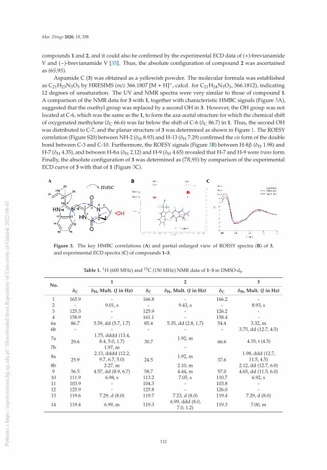
Mar. Drugs 2020, 18, 338
compounds 1 and 2, and it could also be confirmed by the experimental ECD data of (+)-brevianamideV and (−)-brevianamide V [35]. Thus, the absolute configuration of compound 2 was ascertainedas (6S,9S).
Aspamide C (3) was obtained as a yellowish powder. The molecular formula was establishedas C21H23N3O3 by HRESIMS (m/z 366.1807 [M + H]+, calcd. for C21H24N3O3, 366.1812), indicating12 degrees of unsaturation. The UV and NMR spectra were very similar to those of compound 1.A comparison of the NMR data for 3 with 1, together with characteristic HMBC signals (Figure 3A),suggested that the oxethyl group was replaced by a second OH in 3. However, the OH group was notlocated at C-6, which was the same as the 1, to form the aza-acetal structure for which the chemical shiftof oxygenated methylene (δC 66.6) was far below the shift of C-6 (δC 86.7) in 1. Thus, the second OHwas distributed to C-7, and the planar structure of 3 was determined as shown in Figure 1. The ROESYcorrelation (Figure S20) between NH-2 (δH 8.93) and H-13 (δH 7.29) confirmed the cis form of the doublebond between C-3 and C-10. Furthermore, the ROESY signals (Figure 3B) between H-8β (δH 1.98) andH-7 (δH 4.35), and between H-8α (δH 2.12) and H-9 (δH 4.65) revealed that H-7 and H-9 were trans form.Finally, the absolute configuration of 3 was determined as (7R,9S) by comparison of the experimentalECD curve of 3 with that of 1 (Figure 3C).
Figure 3. The key HMBC correlations (A) and partial enlarged view of ROESY spectra (B) of 3,and experimental ECD spectra (C) of compounds 1–3.
Table 1. 1H (600 MHz) and 13C (150 MHz) NMR data of 1–3 in DMSO-d6.
No.1 2 3
δC δH, Mult. (J in Hz) δC δH, Mult. (J in Hz) δC δH, Mult. (J in Hz)
1 165.9 - 166.8 - 166.2 -2 - 9.01, s - 9.43, s - 8.93, s3 125.3 - 125.9 - 126.2 -4 158.9 - 161.1 - 158.4 -6a 86.7 5.59, dd (5.7, 1.7) 85.4 5.35, dd (2.8, 1.7) 54.4 3.32, m6b - - - - - 3.75, dd (12.7, 4.5)
7a29.6
1.75, dddd (13.4,8.4, 5.0, 1.7) 30.7
1.92, m66.6 4.35, t (4.5)
7b 1.97, m -
8a25.9
2.13, dddd (12.2,9.7, 6.7, 5.0) 24.5
1.92, m37.6
1.98, ddd (12.7,11.5, 4.5)
8b 2.27, m 2.10, m 2.12, dd (12.7, 6.0)9 56.5 4.57, dd (8.9, 6.7) 58.7 4.44, m 57.0 4.65, dd (11.5, 6.0)10 111.9 6.98, s 113.2 7.05, s 110.7 6.92, s11 103.9 - 104.3 - 103.8 -12 125.9 - 125.8 - 126.0 -13 119.6 7.29, d (8.0) 119.7 7.23, d (8.0) 119.4 7.29, d (8.0)
14 119.4 6.99, m 119.3 6.99, ddd (8.0,7.0, 1.2) 119.3 7.00, m
112Pobr
ano
z ht
tps:
//rep
ozyt
oriu
m.b
g.ug
.edu
.pl /
Dow
nloa
ded
from
Rep
osito
ry o
f Uni
vers
ity o
f Gda
ńsk
2022
-06-
01
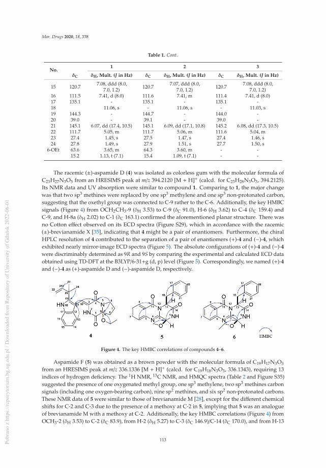
Mar. Drugs 2020, 18, 338
Table 1. Cont.
No.1 2 3
δC δH, Mult. (J in Hz) δC δH, Mult. (J in Hz) δC δH, Mult. (J in Hz)
15 120.7 7.08, ddd (8.0,7.0, 1.2) 120.7 7.07, ddd (8.0,
7.0, 1.2) 120.7 7.08, ddd (8.0,7.0, 1.2)
16 111.5 7.41, d (8.0) 111.6 7.41, m 111.4 7.41, d (8.0)17 135.1 - 135.1 - 135.1 -18 11.06, s - 11.06, s - 11.03, s19 144.3 - 144.7 - 144.0 -20 39.0 - 39.1 - 39.0 -21 145.1 6.07, dd (17.4, 10.5) 145.1 6.09, dd (17.1, 10.8) 145.2 6.08, dd (17.3, 10.5)22 111.7 5.05, m 111.7 5.06, m 111.6 5.04, m23 27.4 1.45, s 27.5 1.47, s 27.4 1.46, s24 27.8 1.49, s 27.9 1.51, s 27.7 1.50, s
6-OEt 63.6 3.65, m 64.3 3.60, m - -15.2 1.13, t (7.1) 15.4 1.09, t (7.1) - -
The racemic (±)-aspamide D (4) was isolated as colorless gum with the molecular formula ofC23H27N3O3 from an HRESIMS peak at m/z 394.2120 [M + H]+ (calcd. for C23H28N3O3, 394.2125).Its NMR data and UV absorption were similar to compound 1. Comparing to 1, the major changewas that two sp3 methines were replaced by one sp3 methylene and one sp3 non-protonated carbon,suggesting that the oxethyl group was connected to C-9 rather to the C-6. Additionally, the key HMBCsignals (Figure 4) from OCH2CH3-9 (δH 3.53) to C-9 (δC 91.0), H-6 (δH 3.62) to C-4 (δC 159.4) andC-9, and H-8a (δH 2.02) to C-1 (δC 163.1) confirmed the aforementioned planar structure. There wasno Cotton effect observed on its ECD spectra (Figure S29), which in accordance with the racemic(±)-brevianamide X [35], indicating that 4 might be a pair of enantiomers. Furthermore, the chiralHPLC resolution of 4 contributed to the separation of a pair of enantiomers (+)-4 and (−)-4, whichexhibited nearly mirror-image ECD spectra (Figure 5). The absolute configurations of (+)-4 and (−)-4were discriminably determined as 9R and 9S by comparing the experimental and calculated ECD dataobtained using TD-DFT at the B3LYP/6-31+g (d, p) level (Figure 5). Correspondingly, we named (+)-4and (−)-4 as (+)-aspamide D and (−)-aspamide D, respectively.
Figure 4. The key HMBC correlations of compounds 4–6.
Aspamide F (5) was obtained as a brown powder with the molecular formula of C19H17N3O3
from an HRESIMS peak at m/z 336.1336 [M + H]+ (calcd. for C19H18N3O3, 336.1343), requiring 13indices of hydrogen deficiency. The 1H NMR, 13C NMR, and HMQC spectra (Table 2 and Figure S35)suggested the presence of one oxygenated methyl group, one sp3 methylene, two sp3 methines carbonsignals (including one oxygen-bearing carbon), nine sp2 methines, and six sp2 non-protonated carbons.These NMR data of 5 were similar to those of brevianamide M [28], except for the different chemicalshifts for C-2 and C-3 due to the presence of a methoxy at C-2 in 5, implying that 5 was an analogueof brevianamide M with a methoxy at C-2. Additionally, the key HMBC correlations (Figure 4) fromOCH3-2 (δH 3.53) to C-2 (δC 83.9), from H-2 (δH 5.27) to C-3 (δC 146.9)/C-14 (δC 170.0), and from H-13
113Pobr
ano
z ht
tps:
//rep
ozyt
oriu
m.b
g.ug
.edu
.pl /
Dow
nloa
ded
from
Rep
osito
ry o
f Uni
vers
ity o
f Gda
ńsk
2022
-06-
01

Mar. Drugs 2020, 18, 338
(δH 5.53) to C-15 (δC 40.0)/C-16 (δC 135.9) confirmed the planner structure of 5 as shown in Figure 4.The absolute configuration of 5 was determined as (2S,13S) via comparing the ECD curve (Figure 6) of5 with the brevianamide M (17).
Figure 5. Experimental and calculated ECD spectra of 4.
Table 2. 1H (600 MHz) and 13C (150 MHz) NMR data of 4–6 (4 in DMSO-d6, 5–6 in CDCl3).
No.4 5 6
δ δH, Mult. (J in Hz) δC δH, Mult. (J in Hz) δC δH, Mult. (J in Hz)
1 163.1 - - - - -2 - 9.43, s 83.9 5.27, d (4.7) 78.3 6.40, d (4.3)3 125.5 - 146.9 - 151.1 -4 159.4 - - - -5 - - 146.7 - 139.0 -6 45.1 3.62, dd (8.6, 5.8) 127.8 7.74, d (8.0) 122.9 8.23, d, (8.0)7a
19.51.92, m
134.9 7.79, t (8.0) 136.7 7.91, t (8.0)7b 1.96, m8a
32.42.02, m
128.0 7.53, t (8.0) 129.7 7.66, t (8.0)8b 2.33, m9 91.0 - 127.0 8.24, d (8.0) 127.9 8.23, d, (8.0)10 112.7 7.02, s 120.8 - 119.1 -11 103.9 - 160.3 - 157.4 -12 126.3 - - - - -13 119.1 7.21, d (8.0) 57.4 5.53, dd (8.8, 6.5) 58.1 5.54, t (7.8)14 119.0 7.00, m 170.0 - 167.6 -15 120.7 7.09, m 40.0 3.43, m 39.9 3.48, m16 111.6 7.43, d (8.0) 135.9 - 134.9 -17 135.1 - 129.8 7.29, m 129.7 7.24, m18 11.10, s 128.6 7.28, m 128.8 7.24, m19 144.4 - 127.0 7.24, m 127.7 7.20, m20 39.0 - 128.6 7.28, m 128.8 7.24, m
21 145.2 6.09, ddd (17.1,10.8, 1.6) 129.8 7.29, m 129.7 7.24, m
22 111.7 5.06, m - - - -23 27.4 1.47, s - - - -24 27.8 1.50, s - - - -
9-OEt 59.2 3.53, qd (7.0, 3.9) - - - -- 15.2 1.22, td (7.0, 1.5) - - - -
2-OMe/2-OEt
- - 56.0 3.53, s 66.24.04, dt (9.0, 7.0)4.11, dt (9.0, 7.0)
- - - - 15.4 1.35, t (7.0)
Aspamide G (6) was isolated as a brown powder. The molecular formula was determined asC20H19N3O3 by HRESIMS (m/z 350.1498 [M + H]+, calcd. for C20H20N3O3, 350.1499), which was14 Dalton more than 5. The 13C NMR data of 6 showed a close resemblance to those of 5, except foran additional oxygenated sp3 methylene, suggesting that there was an ethoxy group located at C-2
114Pobr
ano
z ht
tps:
//rep
ozyt
oriu
m.b
g.ug
.edu
.pl /
Dow
nloa
ded
from
Rep
osito
ry o
f Uni
vers
ity o
f Gda
ńsk
2022
-06-
01

Mar. Drugs 2020, 18, 338
in 6. The key HMBC signal (Figure 4) from H-2 (δH 6.40) to OCH2-2 (δC 66.2) verified that 6 was ananalogue of brevianamide M (17) with an ethoxy motif at C-2. In addition, similar Cotton effects at 212,220, and 237 nm in the ECD spectra (Figure 6) of 6 suggested that 6 and 17 had the same counterpartabsolute configurations. Thus, the absolute configuration of 6 was assigned as (2S,13S), and it waselucidated as 6-ethoxy-aspamide F.
Figure 6. Experimental ECD spectra of 5, 6, and 17.
All isolated compounds were tested for their anti-inflammatory activities in P. acnes-inducedTHP-1 cells; unfortunately, none of the compounds showed moderate anti-inflammatory properties.Aiming to give our contribution to the COVID-19 research, all compounds were selected for the virtualscreening on the 3CL hydrolase (Mpro) of SARS-CoV-2, which had been exploited as a potentialdrug target to fight COVID-19 [27]. The docking scores of compounds 1–2, 5, 6, 8 and 17 were topamong all screened molecules (docking scores: −5.389, −4.772, −5.146, −4.962, −5.158), and the score ofritonavir [36] (a potent inhibitor in vitro of human immunodeficiency virus type 1 protease) was −7.039,which suggested that these compounds may be helpful in fighting COVID-19 after further studies.
3. Materials and Methods
3.1. General Experimental Procedures
Optical rotations were recorded on a JASCO P-1020 polarimeter (JASCO Corporation, Tokyo,Japan) in MeOH at 25 ◦C. UV spectra were measured using a Shimadzu UV-1800 spectrophotometer(Shimadzu Corporation, Tokyo, Japan). High-resolution electrospray ionization (HR-ESI-MS) wereobtained with an Agilent 6529B Q-TOF instrument (Agilent Technologies, Santa Clara, CA, USA).ECD spectra were carried out with Chirascan circular dichroism spectrometers (Applied PhotophysicsLtd., Leatherhead, UK). Both 1D and 2D NMR spectra were acquired on a Bruker AVIII-400 and BrukerAVIII-600 NMR spectrometers with tetramethylsilane (TMS) as an internal standard (Bruker, Karlsruhe,Germany). Preparative high-performance liquid chromatography (Pre-HPLC) was performed utilizinga Shimadzu LC-20 system (Shimadzu, Tokyo, Japan) equipped with a Shim-pack RP-C18 column(20 × 250 mm i.d., 10 μm, Shimadzu, Tokyo, Japan) with a flow rate at 10 mL/min at 25 ◦C, which wasrecorded by a binary channel UV detector at 210 nm and 254 nm. The analytical chiral HPLC usedwas a JASCO LC-2000 system equipped with a Daicel Chiralpak AD-H column (4.6 mm × 250 mm,5 μm) and a CD-2095 chiral detector at 280 nm. The mobile phase was n-hexane/isopropanol (80:20,v/v) used at a flow rate of 0.5 mL/min. Column chromatography (CC) was performed with silicagel (200–300 mesh, Qingdao Marine Chemical Inc., Qingdao, China) and ODS (50 μm, YMC, Kyoto,Japan) on a Flash Chromatograph System (SepaBen machine, Santai Technologies, Changzhou, China).Thin-layer chromatography (TLC) was performed using precoated silica gel GF254 plates (QingdaoMarine Chemical Inc., Qingdao, China).
115Pobr
ano
z ht
tps:
//rep
ozyt
oriu
m.b
g.ug
.edu
.pl /
Dow
nloa
ded
from
Rep
osito
ry o
f Uni
vers
ity o
f Gda
ńsk
2022
-06-
01

Mar. Drugs 2020, 18, 338
3.2. Fungal Material
The endophytic DY180635 was isolated from a sample of crab (Chiromantes haematocheir), whichwas collected from the intertidal zone of Zhoushan, Zhejiang, China, in June 2018. It was incubatedon a potato dextrose agar (PDA) plate at 28 ◦C. The strain DY180635 was identified using ITS rDNAsequence analysis by RuiDi (Shanghai, China) and its DNA sequence using BLAST was compared tothe GenBank data. The result of BLAST searching was closest to that of Aspergillus versicolor NRRL238 (GenBank accession number NG_067623) with 99% sequence identity. The nucleotide sequencesof the ITS gene (accession number MT361076) of A. versicolor DY180635 were deposited in GenBank.A reference culture is stored in State Key Laboratory of Bioreactor Engineering laboratory of Shanghaiat −80 ◦C.
3.3. Fermentation, Extraction, and Isolation
The fungus was incubated on potato dextrose agar (PDA) medium at 28 ◦C for approximately2–3 days; then it was cut into three agar pieces (nearly the size of 0.5 × 0.5 × 0.5 cm) and transferredinto a 500 mL Erlenmeyer flask, containing 200 mL of potato dextrose broth (PDB). The flasks werecultured for 2 days at 28 ◦C on a rotary shaker at 180 rpm for inoculation. The seed cultures wereadded to the 200 × 1 L flasks containing rice medium (110 g rice, 120 mL deionized water), which waspreviously sterilized at 121 ◦C for 25 min. All flasks were incubated at 28 ◦C for four weeks.
Following incubation, the solid rice cultures were extracted three times by EtOAc to give a crudeextract (489.0 g); the crude extract was suspended in water and then partitioned with EtOAc to give anEtOAc soluble fraction (185.2 g) after the solvent was removed to dryness under reduced pressure.The EtOAc fraction was further separated on macroporous adsorbent resin with a stepped gradientelution with EtOH-H2O (30, 50, 70 and 100%). The 50% fraction was sequentially separated by silica gelwith petroleum ether-EtOAc (5:1 to 0:1) to give four subfractions (A–D) using the TLC. The subfractionB (9.0 g) was sequentially loaded onto silica gel CC (petroleum ether-EtOAc, 5:1) and preparative HPLC(MeCN-H2O, 50:50, 10.0 mL/min) to yield compounds 1 (10.4 mg, tR 34.9 min), 2 (6.4 mg, tR 27.1 min),4 (5.9 mg, tR 33.5 min), and 6 (8.7 mg, tR 23.2 min). Subfraction C (15.0 g) was further separated by anODS column (MeCN-H2O, 40:60) and repeated preparative HPLC with MeCN-H2O (50:50, 10 mL/min)to give compounds 3 (4.6 mg, tR 8.8 min) and 5 (4.7 mg, tR 17.1 min).
Aspamide A (1): yellowish powder; [α]25D + 120.0 (c 0.05, MeOH); ECD (5 mg/L, MeOH) λmax (Δε) 212
(92.18), 245 (−41.48) 335 (19.29) nm; UV (MeOH) λmax (logε) 200 (6.13), 224 (6.13), 284 (5.46), 341 (5.68)nm; 1H and 13C NMR (DMSO-d6), see Table 1; positive HR-ESI-MS m/z 394.2117 [M + H] +, (calcd.for C23H28N3O3, 394.2125).
Aspamide B (2): yellowish powder; [α]25D + 112.0 (c 0.05, MeOH); ECD (5 mg/L, MeOH) λmax (Δε) 212
(83.31), 261 (−37.91), 340 (20.41) nm; UV (MeOH) λmax (logε) 200 (6.18), 224 (6.18), 283 (5.45), 345 (5.73)nm; 1H and 13C NMR (DMSO-d6), see Table 1; positive HR-ESI-MS m/z 394.2120 [M + H]+, (calcd.for C23H28N3O3, 394.2125).
Aspamide C (3): yellowish powder; [α]25D + 125.0 (c 0.05, MeOH); ECD (5 mg/L, MeOH) λmax (Δε) 211
(103.75), 255 (−44.38), 334 (24.79) nm; UV (MeOH) λmax (logε) 200 (6.20), 224 (6.19), 284 (5.56), 337 (5.75)nm; 1H and 13C NMR (DMSO-d6), see Table 1; positive HR-ESI-MS m/z 366.1807 [M + H]+, (calcd.for C21H24N3O3, 366.1812).
(±)-Aspamide D (4): colorless gum; [α]25D + 5.0 (c 0.05, MeOH); UV (MeOH) λmax (logε) 200 (6.07),
224 (6.06), 283 (5.33), 346 (5.60) nm; 1H and 13C NMR (DMSO-d6), see Table 2; positive HR-ESI-MS m/z394.2122 [M + H]+, (calcd. for C23H28N3O3, 394.2125).
Aspamide F (5): brown powder; [α]25D + 40.0 (c 0.05, MeOH); ECD (5 mg/L, MeOH) λmax (Δε)
212 (65.33), 220 (62.98), 237 (−52.79) nm; UV (MeOH) λmax (logε) 206 (5.95), 224 (6.15), 270 (5.25),
116Pobr
ano
z ht
tps:
//rep
ozyt
oriu
m.b
g.ug
.edu
.pl /
Dow
nloa
ded
from
Rep
osito
ry o
f Uni
vers
ity o
f Gda
ńsk
2022
-06-
01

Mar. Drugs 2020, 18, 338
278 (5.23) nm; 1H and 13C NMR (CDCl3), see Table 2; positive HR-ESI-MS m/z 336.1336 [M + H]+,(calcd. for C19H18N3O3, 336.1343).
Aspamide G (6): brown powder; [α]25D + 56.0 (c 0.05, MeOH); ECD (5 mg/L, MeOH) λmax (Δε) 212
(76.04), 221 (75.42), 237 (-62.18) nm; UV (MeOH) λmax (logε) 205 (5.92), 222 (5.88), 270 (5.16), 278 (5.13)nm; 1H and 13C NMR (CDCl3), see Table 2; positive HR-ESI-MS m/z 350.1498 [M + H]+, (calcd.for C20H20N3O3, 350.1499).
3.4. ECD Calculation
The relative configuration of 1 was established initially according to its ROESY NMR spectra.Monte Carlo conformational searches were carried out by means of the Spartan’s 14 software using theMerck molecular force field (MMFF). The conformers with Boltzmann population of over 1% werechosen for ECD calculations, and then the conformers were initially optimized at the B3LYP/6-31G(d,p)level in gas. The theoretical calculation of ECD was conducted in MeOH using Time-dependent Densityfunctional theory (TD-DFT) at the B3LYP/6-31+G(d,p) level for all conformers. Rotatory strengths for atotal of 100 excited states were calculated. ECD spectra were generated using the program SpecDis1.6 (University of Würzburg, Würzburg, Germany) and GraphPad Prism 5 (University of CaliforniaSan Diego, USA) from dipole-length rotational strengths by applying Gaussian band shapes withsigma = 0.3 eV. And UV-shift values of all configurations were −10 nm. The spectra of enantiomerswere produced directly by mirror inversions.
3.5. Virtual Screening Against COVID-19 Main Protease
3.5.1. Protein and Ligand Preparation
The 3CL hydrolase (Mpro) of SARS-CoV-2 had been exploited as a potential drug target to fightCOVID-19 [27]. Thus, the virtual screening was conducted by using the SARS-CoV-2 enzyme (PDB ID6LU7) obtained from the Protein Data Bank (PDB, http://www.rcsb.org/pdb), and the structure wasoptimized by using the protein preparation wizard module in Maestro software package (SchrodingerLLC, NY, USA). Specifically, the water and heteroatom were removed, the polar hydrogens were addedto the protein to the protonation state, the entire structure was assumed as a neutral pH, and theenergy of the structure was minimized by using an OPLS2005 force field. The docking grid of 20 Å sizewas generated over the co-crystallized ligand. All compounds 1–17 were implemented by Ligprepsoftware, and the structure energy was minimized using OPL2005 force field.
3.5.2. Virtual Screening
Virtual screening was performed by the Schrodinger glide docking module, and the standard-precision (SP) docking was designated to get accurate results [37]. The results were measured bydocking score, and only the compounds with scores in the top half were subjected to the extra-precision(XP) docking.
3.6. Cell Culture and Cell Viability Assay
The human monocytic cell line, THP-1 (Cell Bank of China Science Academy, Shanghai, China)and P. acnes (ATCC6919, Xiangfu biotech, Shanghai, China), were used for the anti-inflammatoryassay. THP-1 cells were cultured in RPMI1640 medium with 10% fetal bovine serum (FBS, Gibco, NY,USA) in a humidified incubator (37 ◦C, 5% CO2). P. acnes bacteria were incubated in Cooked MeatMedium, containing cooked beef granules (Rishui biotechnology, Qingdao, China) in an anaerobicenvironment. The THP-1 cells were stimulated by the P. acnes, which was harvested at the exponentialphase. The viability of THP-1 cells was evaluated by the MTT assay, specifically, seeding the THP-1cells in 96-well plates at a density of 2 × 10⁵ cells/well and treated with serially diluted compounds for36 h (37 ◦C, 5% CO2). After that, we added 20 μL MTT regent (5 mg/mL, Genetimes Technology Inc.,
117Pobr
ano
z ht
tps:
//rep
ozyt
oriu
m.b
g.ug
.edu
.pl /
Dow
nloa
ded
from
Rep
osito
ry o
f Uni
vers
ity o
f Gda
ńsk
2022
-06-
01

Mar. Drugs 2020, 18, 338
Shanghai, China) to each well and incubated the samples at 37 ◦C for 4 h. Removing the supernatant,the formazan crystals were fully solubilized in DMSO (150 μL), and the absorbance was measured at570 nm and 630 nm.
4. Conclusions
Chemical investigation of a marine-derived fungus Aspergillus versicolor DY180635 led to theisolation and identification of four new IDPKs, aspamides A–E (1–4) and two new DPKs, aspamidesF–G (5–6), along with 11 known diketopiperazines and intermediates. Further chiral high-performanceliquid chromatography resolution gave enantiomers (+)- and (−)-4, respectively. The structures andabsolute configurations of compounds 1–6 were determined by the comprehensive analyses of NMR,HRESIMS, and ECD calculation. Compounds 1–17 were selected for the virtual screening on the 3CLhydrolase (Mpro) of SARS-CoV-2, and compounds 1–2, 5, 6, 8 and 17 possessed top docking scoresand thus may be helpful in fighting COVID-19 after further studies.
Supplementary Materials: The following are available online at: http://www.mdpi.com/1660-3397/18/7/338/s1,Figures S1–S46: 1H, 13C, HSQC, HMBC, ROESY, UV, ECD, and HRESIMS spectra of the new compounds 1–6,Tables S1–S6: Computational data of 1 and 4.
Author Contributions: Y.D. performed the isolation, purification and identification of all compounds. X.Z. testedthe anti-inflammatory activities. L.H. and M.Z. supervised the laboratory work. Q.H. edited the manuscript.F.A. supervised the laboratory work, designed the experiments and edited the manuscript. All authors have readand agree to the published version of the manuscript.
Funding: This work was funded by the National Key R&D Program of China (2018YFC1706200, 2019YFC0312504),and the National Natural Science Foundation of China (41876189, 81703388). This work was also supportedby State Key Laboratory of Bioreactor Engineering. Authors thank Prof. Lixin Zhang (State Key Laboratory ofBioreactor Engineering, East China University of Science and Technology) for assistance in the virtual screeningon the Mpro of SARS-CoV-2.
Conflicts of Interest: The authors declare no conflict of interest.
References
1. Strobel, G.; Daisy, B.; Castillo, U.; Harper, J. Natural products from endophytic microorganisms. J. Nat. Prod.2004, 67, 257–268. [CrossRef]
2. Blunt, J.W.; Copp, B.R.; Keyzers, R.A.; Munro, M.H.G.; Prinsep, M.R. Marine natural products. Nat. Prod.Rep. 2017, 34, 235–294. [CrossRef] [PubMed]
3. Bugni, T.S.; Ireland, C.M. Marine-derived fungi: A chemically and biologically diverse group ofmicroorganisms. Nat. Prod. Rep. 2004, 21, 143–163. [CrossRef]
4. Ma, Y.M.; Liang, X.A.; Kong, Y.; Jia, B. Structural diversity and biological activities of indole diketopiperazinealkaloids from fungi. J. Agric. Food Chem. 2016, 64, 6659–6671. [CrossRef] [PubMed]
5. Borthwick, A.D. 2,5-Diketopiperazines: Synthesis, reactions, medicinal chemistry, and bioactive naturalproducts. Chem. Rev. 2012, 112, 3641–3716. [CrossRef] [PubMed]
6. Wang, W.L.; Lu, Z.Y.; Tao, H.W.; Zhu, T.J.; Fang, Y.C.; Gu, Q.Q.; Zhu, W.M. Isoechinulin-type alkaloids,variecolorins A-L, from halotolerant Aspergillus variecolor. J. Nat. Prod. 2007, 70, 1558–1564. [CrossRef]
7. Cai, S.X.; Sun, S.W.; Peng, J.X.; Kong, X.L.; Zhou, H.N.; Zhu, T.J.; Gu, Q.Q.; Li, D.H. Okaramines S-U,three new indole diketopiperazine alkaloids from Aspergillus taichungensis ZHN-7-07. Tetrahedron 2015, 71,3715–3719. [CrossRef]
8. Wang, F.Z.; Fang, Y.C.; Zhu, T.J.; Zhang, M.; Lin, A.Q.; Gu, Q.Q.; Zhu, W.M. Seven new prenylated indolediketopiperazine alkaloids from holothurian-derived fungus Aspergillus fumigatus. Tetrahedron 2008, 64,7986–7991. [CrossRef]
9. Tsukamoto, S.; Kato, H.; Samizo, M.; Nojiri, Y.; Onuki, H.; Hirota, H.; Ohta, T. Notoamides F−K, prenylatedindole alkaloids isolated from a marine-derived Aspergillus sp. J. Nat. Prod. 2008, 71, 2064–2067. [CrossRef]
10. Kozlovsky, A.G.; Vinokurova, N.G.; Adanin, V.M. Diketopiperazine alkaloids from the fungus Penicilliumpiscarium westling. Appl. Biochem. Microbiol. 2000, 36, 271–275. [CrossRef]
118Pobr
ano
z ht
tps:
//rep
ozyt
oriu
m.b
g.ug
.edu
.pl /
Dow
nloa
ded
from
Rep
osito
ry o
f Uni
vers
ity o
f Gda
ńsk
2022
-06-
01

Mar. Drugs 2020, 18, 338
11. Ravikanth, V.; Niranjan Reddy, V.L.; Ramesh, P.; Prabhakar Rao, T.; Diwan, P.V.; Khar, A.; Venkateswarlu, Y.An immunosuppressive tryptophan-derived alkaloid from Lepidagathis cristata. Phytochemistry 2001, 58,1263–1266. [CrossRef]
12. Fujimoto, H.; Sumino, M.; Okuyama, E.; Ishibashi, M. Immunomodulatory constituents from an Ascomycete.J. Nat. Prod. 2004, 67, 98–102. [CrossRef]
13. Kuramochi, K.; Ohnishi, K.; Fujieda, S.; Nakajima, M.; Saitoh, Y.; Watanabe, N.; Takeuchi, T.; Nakazaki, A.;Sugawara, F.; Arai, T.; et al. Synthesis and biological activities of neoechinulin A derivatives: New aspectsof structure-activity relationships for neoechinulin A. Chem. Pharm. Bull. 2008, 56, 1738–1743. [CrossRef][PubMed]
14. Fan, Z.; Sun, Z.H.; Liu, Z.; Chen, Y.C.; Liu, H.X.; Li, H.H.; Zhang, W.M. Dichotocejpins A-C: Newdiketopiperazines from a deep-sea-derived fungus Dichotomomyces cejpii FS110. Mar. Drugs 2016, 14, 1–9.[CrossRef] [PubMed]
15. Mohanlal, R.W.; Lloyd, K.; Huang, L. Plinabulin, a novel small molecule clinical stage IO agent withanti-cancer activity, to prevent chemo-induced neutropenia and immune related AEs. J. Clin. Oncol. 2018, 36,126. [CrossRef]
16. Gomes, N.G.M.; Lefranc, F.; Kijjoa, A.; Kiss, R. Can some marine-ferived fungal metabolites become actualanticancer agents? Mar. Drugs 2015, 13, 3950–3991. [CrossRef]
17. Geiger, W.B.; Conn, J.E.; Waksman, S.A. Chaetomin, a new antibiotic substance produced by Chaetomiumcochliodes: II. Isolation and Concentration. J. Bacteriol. 1944, 48, 531–536. [CrossRef]
18. Tian, W.; Sun, C.; Zheng, M.; Harmer, J.R.; Yu, M.; Zhang, Y.; Peng, H.; Zhu, D.; Deng, Z.; Chen, S.L.; et al.Efficient biosynthesis of heterodimeric C3-aryl pyrroloindoline alkaloids. Nat. Commun. 2018, 9, 4428.[CrossRef]
19. Ye, Y.; Du, L.; Zhang, X.W.; Newmister, S.A.; McCauley, M.; Alegre-Requena, J.V.; Zhang, W.; Mu, S.;Minami, A.; Fraley, A.E.; et al. Fungal-derived brevianamide assembly by a stereoselective semipinacolase.Nat. Catal. 2020. [CrossRef]
20. Song, F.; Liu, X.; Guo, H.; Ren, B.; Chen, C.; Piggott, A.M.; Yu, K.; Gao, H.; Wang, Q.; Liu, M.; et al.Brevianamides with antitubercular potential from a marine-derived isolate of Aspergillus versicolor. Org. Lett.2012, 14, 4770–4773. [CrossRef]
21. James, E.D.; Knuckley, B.; Alqahtani, N.; Porwal, S.; Ban, J.; Karty, J.A.; Viswanathan, R.; Lane, A.L. Two distinctcyclodipeptide synthases from a marine Actinomycete catalyze biosynthesis of the same diketopiperazinenatural product. ACS Synth. Biol. 2016, 5, 547–553. [CrossRef] [PubMed]
22. Haines, B.E.; Nelson, B.M.; Grandner, J.M.; Kim, J.; Houk, K.N.; Movassaghi, M.; Musaev, D.G. Mechanismof permanganate-promoted dihydroxylation of complex diketopiperazines: Critical roles of counter-cationand ion-pairing. J. Am. Chem. Soc. 2018, 140, 13375–13386. [CrossRef]
23. Ding, Y.; An, F.L.; Zhu, X.J.; Yu, H.Y.; Hao, L.L.; Lu, Y.H. Curdepsidones B-G, six depsidones withanti-inflammatory activities from the marine-derived fungus Curvularia sp. IFB-Z10. Mar. Drugs 2019, 17,266. [CrossRef]
24. Liu, W.H.; Ding, Y.; Ji, X.; An, F.L.; Lu, Y.H. Curvulaide A, a bicyclic polyketide with anti-anaerobic bacteriaactivity from marine-derived Curvularia sp. J. Antibiot. 2019, 72, 111–113. [CrossRef]
25. Guo, M.M.; Lu, Y.; Yang, J.P.; Zhao, X.; Lu, Y.H. Inhibitory effects of Schisandra chinensis extract on acne-relatedinflammation and UVB-induced photoageing. Pharm. Biol. 2016, 54, 2987–2994. [CrossRef] [PubMed]
26. Dillman, R.L.; Cardellina, J.H. Aromatic secondary metabolites from the sponge Tedania ignis. J. Nat. Prod.1991, 54, 1056–1061. [CrossRef]
27. Jin, Z.M.; Du, X.Y.; Xu, Y.C.; Deng, Y.Q.; Liu, M.Q.; Zhao, Y.; Zhang, B.; Li, X.F.; Zhang, L.K.; Peng, C.F.; et al.Structure of Mpro from COVID-19 virus and discovery of its inhibitors. Nature 2020, 582, 289–293. [CrossRef][PubMed]
28. Li, G.Y.; Yang, T.; Luo, Y.G.; Chen, X.Z.; Fang, D.M.; Zhang, G.L. Brevianamide J, a new indole alkaloid dimerfrom fungus Aspergillus versicolor. Org. Lett. 2009, 11, 3714–3717. [CrossRef]
29. Li, G.Y.; Li, L.M.; Yang, T.; Chen, X.Z.; Fang, D.M.; Zhang, G.L. Four new alkaloids, brevianamides O-R,from the fungus Aspergillus versicolor. Helv. Chim. Acta 2010, 93, 2075–2080. [CrossRef]
30. Liu, Y.X.; Ma, S.G.; Wang, X.J.; Zhao, N.; Qu, J.; Yu, S.S.; Dai, J.G.; Wang, Y.H.; Si, Y.K. Diketopiperazinealkaloids produced by the endophytic fungus Aspergillus fumigatus from the Stem of Erythrophloeum fordiiOliv. Helv. Chim. Acta 2012, 95, 1401–1408. [CrossRef]
119Pobr
ano
z ht
tps:
//rep
ozyt
oriu
m.b
g.ug
.edu
.pl /
Dow
nloa
ded
from
Rep
osito
ry o
f Uni
vers
ity o
f Gda
ńsk
2022
-06-
01

Mar. Drugs 2020, 18, 338
31. Schkeryantz, J.M.; Woo, J.C.G.; Siliphaivanh, P.; Depew, K.M.; Danishefsky, S.J. Total synthesis of gypsetin,deoxybrevianamide E, brevianamide E, and tryprostatin B: Novel constructions of 2,3-disubstituted indoles.J. Am. Chem. Soc. 1999, 121, 11964–11975. [CrossRef]
32. Sanz-Cervera, J.F.; Stocking, E.M.; Usui, T.; Osada, H.; Williams, R.M. Synthesis and evaluation of microtubuleassembly inhibition and cytotoxicity of prenylated derivatives of cyclo-l-Trp-l-Pro. Bioorg. Med. Chem. 2000,8, 2407–2415. [CrossRef]
33. May Zin, W.W.; Buttachon, S.; Dethoup, T.; Pereira, J.A.; Gales, L.; Inácio, Â.; Costa, P.M.; Lee, M.;Sekeroglu, N.; Silva, A.M.S.; et al. Antibacterial and antibiofilm activities of the metabolites isolated from theculture of the mangrove-derived endophytic fungus Eurotium chevalieri KUFA 0006. Phytochemistry 2017, 141,86–97. [CrossRef] [PubMed]
34. Pirrung, M.C.; Fujita, K.; Park, K. Organometallic routes to 2,5-dihydroxy-3-(indol-3-yl)-benzoquinones.Synthesis of demethylasterriquinone B4. J. Org. Chem. 2005, 70, 2537–2542. [CrossRef]
35. Liu, W.; Wang, L.P.; Wang, B.; Xu, Y.C.; Zhu, G.L.; Lan, M.M.; Zhu, W.M.; Sun, K.L. Diketopiperazine anddiphenylether derivatives from marine algae-derived Aspergillus versicolor OUCMDZ-2738 by epigeneticactivation. Mar. Drugs 2019, 17, 6. [CrossRef]
36. Markowitz, M.; Saag, M.; Powderly, W.G.; Hurley, A.M.; Hsu, A.; Valdes, J.M.; Henry, D.; Sattler, F.; LaMarca, A.; Leonard, J.M.; et al. A preliminary study of ritonavir, an inhibitor of HIV-1 protease, to treat HIV-1infection. N. Engl. J. Med. 1995, 333, 1534–1539. [CrossRef]
37. Halgren, T.A.; Murphy, R.B.; Friesner, R.A.; Beard, H.S.; Frye, L.L.; Pollard, W.T.; Banks, J.L. Glide: A newapproach for rapid, accurate docking and scoring. 2. Enrichment factors in database screening. J. Med. Chem2004, 47, 1750–1759. [CrossRef]
© 2020 by the authors. Licensee MDPI, Basel, Switzerland. This article is an open accessarticle distributed under the terms and conditions of the Creative Commons Attribution(CC BY) license (http://creativecommons.org/licenses/by/4.0/).
120Pobr
ano
z ht
tps:
//rep
ozyt
oriu
m.b
g.ug
.edu
.pl /
Dow
nloa
ded
from
Rep
osito
ry o
f Uni
vers
ity o
f Gda
ńsk
2022
-06-
01

marine drugs
Article
Bioactive Bianthraquinones and Meroterpenoids froma Marine-Derived Stemphylium sp. Fungus
Ji-Yeon Hwang 1, Sung Chul Park 1, Woong Sub Byun 1, Dong-Chan Oh 1, Sang Kook Lee 1,
Ki-Bong Oh 2,* and Jongheon Shin 1,*
1 Natural Products Research Institute, College of Pharmacy, Seoul National University, San 56-1, Sillim,Gwanak, Seoul 151-742, Korea; [email protected] (J.-Y.H.); [email protected] (S.C.P.);[email protected] (W.S.B.); [email protected] (D.-C.O.); [email protected] (S.K.L.)
2 Department of Agricultural Biotechnology, College of Agriculture and Life Science, Seoul NationalUniversity, San 56-1, Sillim, Gwanak, Seoul 151-921, Korea
* Correspondence: [email protected] (K.-B.O.); [email protected] (J.S.);Tel.: +82-2-880-4646 (K.-B.O.); +82-2-880-2484 (J.S.)
Received: 22 July 2020; Accepted: 19 August 2020; Published: 21 August 2020
Abstract: Three new bianthraquinones, alterporriol Z1–Z3 (1–3), along with three known compoundsof the same structural class, were isolated from the culture broth of a marine-derived Stemphylium sp.fungus. Based upon the results of spectroscopic analyses and ECD measurements, the structures ofnew compounds were determined to be the 6-6′- (1 and 2) and 1-5′- (3) C–C connected pseudo-dimericanthraquinones, respectively. Three new meroterpenoids, tricycloalterfurenes E–G (7–9), isolatedtogether with the bianthraquinones from the same fungal culture broth, were structurally elucidatedby combined spectroscopic methods. The relative and absolute configurations of these meroterpenoidswere determined by modified Mosher’s, phenylglycine methyl ester (PGME), and computationalmethods. The bianthraquinones significantly inhibited nitric oxide (NO) production and suppressedinducible nitric oxide synthase (iNOS) and cyclooxygenase-2 (COX-2) expression in LPS-stimulatedRAW 264.7 cells.
Keywords: bianthraquinones; meroterpenoids; marine-derived fungus; anti-inflammatory activity;Stemphylium sp.
1. Introduction
Polyketides are a representative class of fungal metabolites [1,2]. These compounds possess a vastrange of structural diversity resulting in several subgroups such as anthraquinones, naphthoquinones,benzophenones, xanthones, flavonoids, macrolides, and tropolones [3,4]. Among these, anthraquinonesare biosynthetically derived from an octaketide chain formed by the incorporation of one acetyl-CoAand seven malonyl-CoA units [5–7]. Although widely distributed in fungi, anthraquinone derivativesoccur far frequently in the genera of Alternaria, Aspergillus, Fusarium, Stemphylium, and Trichoderma [8].Anthraquinones typically contain various substituents (methyl, hydroxyl, methoxyl, or more complexsubstituents), often attributed to characteristic colors by their properties and positions at the aromaticcore moiety [9,10].
A frequently encountered structural variation of fungal anthraquinones is the dimerizationthrough a C–C bond formation between two similar units [3]. The dimerization patterns for thesebianthraquinones are also diversified through both homo- and hetero- bond formation, providingadditional structural variation to the anthraquinones. To date, a vast number of fungal exampleswere reported including alterporriols, icterinoidin, rubellin, and skyrin [3,11]. In addition to theirmonomeric precursors, bianthraquinones often co-occur with biosynthetically related compounds of thepolyketide pathway [4,12]. Wide structural diversity, in conjunction with significant bioactivities such as
Mar. Drugs 2020, 18, 436; doi:10.3390/md18090436 www.mdpi.com/journal/marinedrugs121Pobr
ano
z ht
tps:
//rep
ozyt
oriu
m.b
g.ug
.edu
.pl /
Dow
nloa
ded
from
Rep
osito
ry o
f Uni
vers
ity o
f Gda
ńsk
2022
-06-
01

Mar. Drugs 2020, 18, 436
antibacterial, anti-inflammatory, antituberculosis, and cytotoxic activities, designates bianthraquinonesas an important group of fungal polyketides [3,8,11,13].
Meanwhile, tricycloalternarenes (TCAs) are a structural class of fungal meroterpenoids [14].Structurally, these are closely related to the ACTG toxins with differences at the isoprenoid sidechain and the substitution pattern at the C-ring of TCAs [15]. Biosynthetically, these meroterpenoidsare proposed to be generated from a hybrid shikimate–isoprenoid route [16]. Exclusively found inAlternaria and Guignardia, tricycloalternarene-type meroterpenoids are regarded as one of the keychemical characteristics of these fungal genera [17,18]. These compounds were also reported to exhibitdiverse bioactivities, including antimicrobial, cytotoxic, and NF-κB-inhibitory [19,20].
During the course of a search for bioactive compounds from marine-derived fungi, we isolated astrain (strain number FJJ006) of Stemphylium sp. from an unidentified sponge specimen collected offthe coast of the island of Jeju-do, Korea. LC-ESIMS and LC-UV analyses of the culture broth of thisstrain showed the presence of several compounds with various profiles, prompting extensive chemicalinvestigation. The large-scale cultivation, sequentially followed by extraction, solvent-partitioning, andchromatographic separations, afforded six new and three known compounds (Figure 1). Here, we reportthe structures of bianthraquinone alterporriols Z1–Z3 (1–3) and meroterpenoid tricycloalterfurenesE–G (7–9). This is first time isolating not only the tricycloalternarene-type meroterpenoids but alsotheir co-occurrence with polyketide-derived anthracenes from the fungus Stemphylium. Compounds 1
and 2 exhibited moderate anti-inflammatory activity in LPS-stimulated RAW 264.7 cells.
S R
R
S
R
R
S
S
Figure 1. Structures of 1–9.
122Pobr
ano
z ht
tps:
//rep
ozyt
oriu
m.b
g.ug
.edu
.pl /
Dow
nloa
ded
from
Rep
osito
ry o
f Uni
vers
ity o
f Gda
ńsk
2022
-06-
01

Mar. Drugs 2020, 18, 436
2. Results and Discussion
Compound 1 was an orange amorphous powder which was analyzed to have the molecularformula of C32H26O13, with 20 unsaturation degrees, by HRFABMS analysis ([M +H]+ m/z 619.1449,calcd C32H27O13, 619.1446). The 13C NMR data of this compound showed signals of four carbonyl(δc 190.5, 188.7, 185.7, 183.5) and twenty deshielded methine and quaternary carbons (δc 166.9–104.6).Aided by the HSQC data, the chemical shifts of corresponding methine protons at δH 7.65, 7.51, 6.81,and 6.78 in the 1H NMR data revealed the presence of aromatic moieties (Table 1). Since these NMRfeatures were accounted for 14 unsaturation degrees, 1 must be a hexacyclic compound.
Table 1. 13C and 1H NMR data of compounds 1–3 in CD3OD.
No.1 a 2 b 3 c
δC, Type δH (J in Hz) δC, Type δH (J in Hz) δC, Type δH (J in Hz)
1 111.7, CH 7.51, d (0.5) 111.8, CH 7.51, d (0.5) 129.0, C
2 164.0, C 163.7, C 165.5, C d
3 133.8, C 133.8, C 134.5, C
4 131.2, CH 7.65, d (0.5) 131.3, CH 7.65, d (0.5) 130.5, CH 8.00, s
4a 126.8, C 127.1, C 131.2, C
5 165.8, C 166.2, C 106.9, CH 7.20, d (2.5)
6 125.3, C 125.7, C 167.1, C
7 104.6, CH 6.78, s 104.3, CH 6.80, s 105.9, CH 6.52, d (2.5)
8 166.9, C 167.1, C 166.0, C
8a 111.9, C 111.7, C 112.1, C
9 188.7, C 188.6, C 191.0, C
9a 134.7, C 134.6, C 132.8, C
10 183.5, C 183.5, C 182.3, C
10a 133.4, C 132.8, C 137.8, C
11 16.6, CH3 2.23, s 16.6, CH3 2.23, s 17.9, CH3 2.21, s
12 56.9, CH3 3.69, s 56.9, CH3 3.70, s 56.3, CH3 3.89, s
1′ 70.6, CH 4.73, d (7.5) 70.6, CH 4.73, d (7.4) 75.2, CH 4.58, d (4.7)
2′ 75.2, CH 3.79, d (7.5) 75.2, CH 3.76, d (7.4) 70.7, CH 3.93, d (4.7)
3′ 74.6, C 74.7, C 75.4, C
4′ 70.1, CH 4.26, s 70.2, CH 4.26, s 70.6, CH 4.39, s
4a’ 143.8, C 143.9, C 144.7, C
5′ 166.3, C 166.5, C 130.7, C
6′ 123.4, C 123.9, C 166.8, C
7′ 104.6, CH 6.81, s 104.6, CH 6.82, s 104.5, CH 6.79, s
8′ 166.1, C 166.4, C 165.9, C
8a’ 111.0, C 111.0, C 110.9, C
9′ 190.5, C 190.6, C 189.6, C
9a’ 143.9, C 143.7, C 141.0, C
10′ 185.7, C 185.5, C 185.7, C
10a’ 130.8, C 130.7, C n.d. e
11′ 22.3, CH3 1.33, s 22.3, CH3 1.32, s 21.6, CH3 1.33, s
12′ 57.0, CH3 3.70, s 57.0, CH3 3.71, s 56.8, CH3 3.71, s
13′ 62.8, CH3 3.77, sa–c Measured at 150/600, 100/400, and 200/800 MHz for 13C/1H NMR, respectively. d Assigned by HMBC data.e Not detected.
123Pobr
ano
z ht
tps:
//rep
ozyt
oriu
m.b
g.ug
.edu
.pl /
Dow
nloa
ded
from
Rep
osito
ry o
f Uni
vers
ity o
f Gda
ńsk
2022
-06-
01

Mar. Drugs 2020, 18, 436
A combination of 13C and 1H NMR and HSQC data diagnosed the remaining NMR signals asan oxy-quaternary carbon (δc 74.6) and three oxymethine (δC/δH 75.2/3.79, 70.6/4.73, and 70.1/4.26),two oxymethyl (δC/δH 57.0/3.70 and 56.9/3.69), and two methyl (δC/δH 22.3/1.33 and 16.6/2.23) groups.Aided by the literature study, the overall spectroscopic features suggested 1 to be a bianthraquinoneconsisting of each one unit of anthraquinone and tetrahydroanthraquinone.
Having the information above, the structure determination of 1 was mostly accomplished byextensive H–C long-range analyses for this proton-deficient compound (Figure 2). Firstly, long-rangecorrelations of two aromatic protons at δH 7.51 (H-1) and 7.65 (H-4) and a benzylic methyl protonat δH 2.23 (H3-11) with neighboring carbons not only constructed a 2-hydroxy-3-methylbenzenemoiety (C-1-C-4, C-4a, and C-9a) but also placed two carbonyls at δC 188.7 (C-9) and 183.5 (C-10)ortho-substituted to the benzene. Similarly, combined HMBC and LR-HSQMBC correlations of theprotons at δH 6.78 (H-7) and 3.69 (H3-12) with neighboring carbons revealed the presence of an8-hydroxy-5-methoxybenzene moiety (C-5-C-8, C-8a, and C-10a). The linkage between C-8a andC-9 was also secured by crucial H-7/C-9 correlation. Although it was not evidenced by HMBC data,the carbon chemical shifts of C-10 (δC 183.5) and C-10a (δC 133.4), in conjunction with the MS-derivedunsaturation degree, directly linked these carbons, thus, constructing an anthraquinone moiety for 1.
Figure 2. Key correlations of COSY (bold), HMBC (arrow), and LR-HSQMBC (JCH = 2 Hz, dashed arrow)experiments for compounds 1, 3, and 4.
Meanwhile, 1H–1H COSY data showed direct spin coupling (J = 7.5 Hz) between oxymethineprotons at δH 4.73 (H-1′) and 3.79 (H-2′) (Figure 2). Subsequently, HMBC correlations of these protonsand an isolated oxymethine and a methyl proton at δH 4.26 (H-4′) and 1.33 (H-11′), respectively, with theolefinic and hydroxyl-bearing carbons, defined a 3-methyl-2,3,4,5-tetrahydroxy cyclohexene-typemoiety (C-1′–C-4′, C-4a’, and C-9a’). The attachment of two carbonyl carbons at δC 190.5 (C-9′) and185.7 (C-10′) at the cyclohexene unit was also accomplished by their HMBC correlations with H-4′ andH-1′, respectively.
The HMBC correlations of an aromatic and a methoxy proton at δH 6.81 (H-7′) and 3.70 (H3-12′),respectively, with aromatic carbons, revealed an 8′-hydroxy-5′-methoxybenzene (C-5′–C-8′, C-8a’,and C-10a’), analogous to the C-5–C-10a unit. Subsequently, further connection to the diketo-bearingcyclohexene unit constructing a tetrahydroanthraquinone moiety was also accomplished by the HMBC
124Pobr
ano
z ht
tps:
//rep
ozyt
oriu
m.b
g.ug
.edu
.pl /
Dow
nloa
ded
from
Rep
osito
ry o
f Uni
vers
ity o
f Gda
ńsk
2022
-06-
01
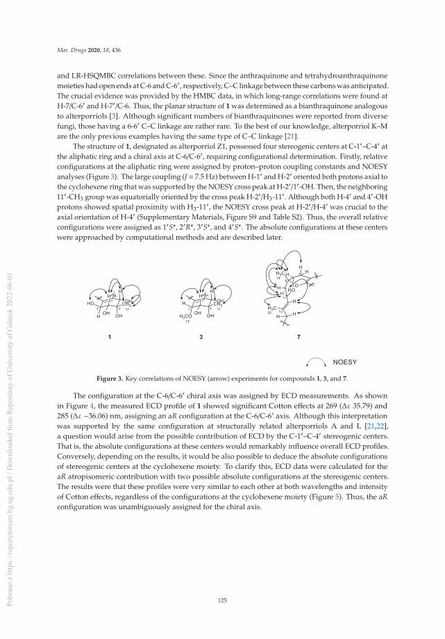
Mar. Drugs 2020, 18, 436
and LR-HSQMBC correlations between these. Since the anthraquinone and tetrahydroanthraquinonemoieties had open ends at C-6 and C-6′, respectively, C–C linkage between these carbons was anticipated.The crucial evidence was provided by the HMBC data, in which long-range correlations were found atH-7/C-6′ and H-7′/C-6. Thus, the planar structure of 1 was determined as a bianthraquinone analogousto alterporriols [3]. Although significant numbers of bianthraquinones were reported from diversefungi, those having a 6-6′ C–C linkage are rather rare. To the best of our knowledge, alterporriol K–Mare the only previous examples having the same type of C–C linkage [21].
The structure of 1, designated as alterporriol Z1, possessed four stereogenic centers at C-1′–C-4′ atthe aliphatic ring and a chiral axis at C-6/C-6′, requiring configurational determination. Firstly, relativeconfigurations at the aliphatic ring were assigned by proton–proton coupling constants and NOESYanalyses (Figure 3). The large coupling (J = 7.5 Hz) between H-1′ and H-2′ oriented both protons axial tothe cyclohexene ring that was supported by the NOESY cross peak at H-2′/1′-OH. Then, the neighboring11′-CH3 group was equatorially oriented by the cross peak H-2′/H3-11′. Although both H-4′ and 4′-OHprotons showed spatial proximity with H3-11′, the NOESY cross peak at H-2′/H-4′ was crucial to theaxial orientation of H-4′ (Supplementary Materials, Figure S9 and Table S2). Thus, the overall relativeconfigurations were assigned as 1′S*, 2′R*, 3′S*, and 4′S*. The absolute configurations at these centerswere approached by computational methods and are described later.
Figure 3. Key correlations of NOESY (arrow) experiments for compounds 1, 3, and 7.
The configuration at the C-6/C-6′ chiral axis was assigned by ECD measurements. As shownin Figure 4, the measured ECD profile of 1 showed significant Cotton effects at 269 (Δε 35.79) and285 (Δε −36.06) nm, assigning an aR configuration at the C-6/C-6′ axis. Although this interpretationwas supported by the same configuration at structurally related alterporriols A and L [21,22],a question would arise from the possible contribution of ECD by the C-1′–C-4′ stereogenic centers.That is, the absolute configurations at these centers would remarkably influence overall ECD profiles.Conversely, depending on the results, it would be also possible to deduce the absolute configurationsof stereogenic centers at the cyclohexene moiety. To clarify this, ECD data were calculated for theaR atropisomeric contribution with two possible absolute configurations at the stereogenic centers.The results were that these profiles were very similar to each other at both wavelengths and intensityof Cotton effects, regardless of the configurations at the cyclohexene moiety (Figure 5). Thus, the aRconfiguration was unambiguously assigned for the chiral axis.
125Pobr
ano
z ht
tps:
//rep
ozyt
oriu
m.b
g.ug
.edu
.pl /
Dow
nloa
ded
from
Rep
osito
ry o
f Uni
vers
ity o
f Gda
ńsk
2022
-06-
01

Mar. Drugs 2020, 18, 436
Figure 4. Experimental ECD spectra of compounds 1–4.
Figure 5. Calculated ECD spectra of compounds 1 and 2.
Having fixed axial chirality, the absolute configurations at C-1′–C-4′were subsequently approachedby DP4 calculations. However, two models derived from the opposite configurations expected verysimilar 13C and 1H NMR data from each other (Supplementary Materials, Figure S52 and Table S1),which was consistent with the results of ECD analysis. Thus, the absolute configurations of thecyclohexene moiety remained unassigned [23]. Overall, the structure of alterporriol Z1 (1) wasdetermined to be a bianthraquinone of the alterporriol class.
The molecular formula of alterporriol Z2 (2) was established to be C32H26O13, identical to 1,by HRFABMS analysis ([M +H]+ m/z 619.1454, calcd for C32H27O13, 619.1446). A comparison of the 13Cand 1H NMR data of this compound with those of 1 revealed a very close structural similarity betweenthem (Table 1). Subsequently, the extensive 1D and 2D NMR analyses of 2 deduced the same planarstructure and relative configurations of the aliphatic ring as those of 1. Accordingly, the structuraldifference was traced to the chiral axis at C-6–C-6′, in which the measured ECD spectrum of 2 showeda quasi-mirror profile from 1 (Figure 4). The noticeably reduced intensities of Cotton effects would beattributed from the stereogenic center-bearing cyclohexene moiety. However, the calculated ECD dataof 2 were virtually identical to each other, regardless of absolute configurations at the cyclohexenestereogenic centers, eradicating the possibility of the reversal of measured ECD by these factors(Figure 5). Thus, the structure of alterporriol Z2 (2) was defined as an atropisomer of alterporriol Z1 (1).
A minor constituent of alterporriol Z3 (3) was isolated as a dark red amorphous solid that wasanalyzed for the molecular formula of C33H28O13 by HRFABMS analysis ([M + Na]+ m/z 655.1430,calcd for C33H28O13Na, 655.1422). The 13C and 1H NMR data of this compound were similar to thoseof 1 and 2, indicating the same bianthraquinone nature (Table 1). An extensive examination of thesedata, in conjunction with a literature and database search, revealed that 3 had close structural similaritywith a congener alterporriol N (6) [23]. The most conspicuous difference in NMR data was the presenceof an additional methoxy group at δC/δH 62.8/3.77 in 3.
126Pobr
ano
z ht
tps:
//rep
ozyt
oriu
m.b
g.ug
.edu
.pl /
Dow
nloa
ded
from
Rep
osito
ry o
f Uni
vers
ity o
f Gda
ńsk
2022
-06-
01

Mar. Drugs 2020, 18, 436
Given this information, the planar structure of 3 was determined by HMBC analyses. Despite thelimited amount of obtained materials, almost all carbons and protons except for unprotonated C-10a’were adequately assigned by HSQC and HMBC correlations (Figure 2). In this way, 3 was defined asconsisting of an anthraquinone and a tetrahydroanthraquinone moiety, as with other compounds. Thenewly appeared methoxy group (C-13′) was also attached at C-1′ by COSY correlation at H-1′/H-2′and HMBC correlation at H3-13′/C-1′. Due to the lack of neighboring protons, the C–C linkage wasnot directly evidenced by HMBC data. However, the diagnostic chemical shifts of the unprotonatedC-1 and C-5′ carbons at δC 129.0 and 130.7, respectively, were indicative of the linkage between theanthraquinone and tetrahydroanthraquinone moiety at these carbons.
The configurations at cyclohexene and C-1/C-6′ chiral axis of 3 were assigned using the samemethods as in 1 and 2. Firstly, the small coupling constants (J = 4.7 Hz) between the vicinal H-1′and H-2′ indicated the orientations of these to be either axial–equatorial or equatorial–equatorialto the cyclohexene ring. Then, the NOESY data showed mutual cross peaks among H-2′, H-4′,and H3-11′, placing these at the same phase of the cyclohexene ring (Figure 3). Therefore, the formertwo oxymethine protons were axially oriented, while the C-11′ methyl group was equatorially orientedto the ring system. Aided with the additional cross peak with H-2′, the H-1′ oxymethine protonwas also equatorially oriented. Thus, the overall relative configurations were assigned as 1′R*, 2′R*,3′S*, and 4′S*. The absolute configuration of the C-1/C-6′ chiral axis of 3 was also assigned by ECDmeasurement. As shown in Figure 4, the ECD data of this compound were similar to 1, assigning thesame aR configuration. Thus, the structure of alteroporriol Z3 (3) was defined as a new bianthraquinoneof the alterporriol class.
In addition to 1–3, the crude extract of Stemphylium sp. contained three known bianthraquinones(4–6). Based upon the results of combined spectroscopic analyses, these were identified as alterporriolsF (4) [24], G (5) [25], and N (6) [23], respectively. The spectroscopic data of these compounds were ingood accordance with those in the literature. Among these, the configuration of C-5/C-5′ chiral axisof 4, previously unassigned, was determined to be aR by the ECD measurement in this work (Figure 4).
In addition to bianthraquinones, the culture broth of Stemphylium sp. contained three newmeroterpenoids. The molecular formula of compound 7, a brown amorphous solid, was deduced to beC21H30O6 with seven unsaturation degrees, by HRFABMS analysis ([M + H]+ m/z 379.2118, calcd forC21H31O6, 379.2115). The 13C NMR data of this compound showed signals of three significantlydeshielded carbons (δC 200.4, 180.9, and 170.4) and three olefinic carbons (δC 139.7, 127.5, and 107.5).The odd numbers of the latter carbons were indicative of a highly differentiated double bond possiblyconsisting of a deshielded carbon (δC 170.4) and a shielded carbon (δC 107.5). Between two carbonylcarbons, the conspicuous one (δC 200.4) was readily assigned as a ketone, while the other one (δC 180.9)was thought to be either a carboxylic acid or an ester group by a strong absorption band at 1755 cm−1
in the IR data. Aided by the 1H NMR and HSQC data, the remaining 15 carbons in the 13C NMRdata were diagnosed to be one oxy-quaternary (δC 85.8), three oxymethine (δC 78.1, 74.5, and 66.8),one methine (δC 40.8), seven methylene (δC 46.8, 40.2, 34.5, 33.6, 30.5, 26.3, and 19.8), and three methyl(δC 20.1, 17.7, and 16.4) carbons (Table 2). Overall, preliminary examination of the spectroscopic datasuggested 7 to be a tricyclic compound possessing two carbonyl groups and two double bonds.
127Pobr
ano
z ht
tps:
//rep
ozyt
oriu
m.b
g.ug
.edu
.pl /
Dow
nloa
ded
from
Rep
osito
ry o
f Uni
vers
ity o
f Gda
ńsk
2022
-06-
01

Mar. Drugs 2020, 18, 436
Table 2. 13C and 1H NMR Data of compounds 7–9 in CD3OD.
No.7 a 8 a 9 b
δC, type δH (J in Hz) δC, type δH (J in Hz) δC, type δH (J in Hz)
1 200.4, C 199.9, C 200.4, C
2 33.6, CH22.62, m
2.32, ddd (16.9, 6.9, 4.9) 72.4, CH 4.07, dd (12.6, 4.9) 33.6, CH22.62, m
2.32, ddd (17.0, 6.8, 5.1)
3 30.5, CH22.18, m1.97, m 30.7, CH2 2.24, m1.83, m 30.5, CH2
2.19, m1.97, m
4 66.8, CH 4.30, t (5.1) 28.8, CH2 2.58, m2.44, m 66.8, CH 4.29, t (5.3)
5 170.4, C 170.8, C 170.4, C
6 107.5, C 105.6, C 107.5, C
7 19.8, CH22.59, d (17.8)
2.22, dd (17.7, 4.2) 19.8, CH22.68, d (17.8)
2.20, dd (17.7, 4.2) 19.8, CH22.59, d (17.8)
2.22, dd (17.7, 4.2)
8 78.1, CH 3.95, dd (4.3, 1.2) 77.9, CH 3.95, dd (4.3, 1.2) 78.1, CH 3.95, dd (4.5, 1.3)
9 85.8, C 85.7, C 85.8, C
10 46.8, CH22.42, dd (13.6, 9.4)2.07, dd (13.6, 3.9) 46.9, CH2
2.39, m2.00, dd (13.7, 3.7) 46.8, CH2
2.42, dd (13.7, 9.5)2.07, dd (13.6, 3.9)
11 74.5, CH 4.80, td (9.0, 3.9) 74.5, CH 4.80, td (9.0, 3.9) 74.4, CH 4.79, m
12 127.5, CH 5.17, br d (8.8) 127.5, CH 5.17, br d (8.8) 127.7, CH 5.17, br d (8.5)
13 139.7, C 139.7, C 139.6, C
14 40.2, CH2 1.99, m 40.3, CH2 1.98, m 40.1, CH2 1.98, m
15 26.3, CH2 1.42, m 26.5, CH2 1.42, m 26.1, CH2 1.39, m
16 34.5, CH21.59, m1.38, m 34.9, CH2
1.58, m1.35, m 34.4, CH2
1.58, m1.38, m
17 40.8, CH 2.37, dq (13.8, 7.0) 41.6, CH 2.37, m 40.5, CH 2.46, m
18 180.9, C 182.1, C 178.9, C
19 20.1, CH3 1.35, s 19.9, CH3 1.30, s 20.1, CH3 1.35, s
20 16.4, CH3 1.65, s 16.4, CH3 1.64, s 16.3, CH3 1.65, s
21 17.7, CH3 1.11, d (7.0) 18.0, CH3 1.11, d (7.0) 17.5, CH3 1.11, d (7.0)
22 52.1, OCH3 3.65, sa , b Measured at 100/800 and 100/500 MHz for 13C/1H, respectively.
The planar structure of 7 was determined by combined 1H–1H COSY and HMBC analyses(Figure 2). That is, COSY data revealed a linear assembly of an oxymethine and two methylenegroups (δC/δH 33.6/2.62 and 2.32, 30.5/2.18 and 1.97, and 66.8/4.30, C-2–C-4). Then, the HMBCcorrelations of these protons with neighboring carbons not only placed a ketone (δC 200.4, C-1) anda tetra-substituted double bond (δC 170.4 and 107.5, C-6 and C-5) at the adjacent locations, but alsoconstructed a 4-oxygenated cyclohexanone moiety (C-1–C-6, ring A): H-4/C-2, C-3, C-5, and C-6,H2-3/C-1, and H2-2/C-1 and C-6. The COSY data also found a direct linkage between methylene andoxymethine (δC/δH 19.8/2.59 and 2.22 and 78.1/3.95, C-7 and C-8). Subsequently, their attachment atC-6 of ring A was secured by the HMBC of H2-7 with ring carbons: H2-7/C-1, C-5, and C-6. Similarly,a quaternary carbon (δC 85.8, C-9) and a methyl group (δC/δH 20.1/1.35, C-19) were linearly connectedat C-8 by a number of key HMBC correlations: H-8/C-9 and H3-19/C-8 and C-9.
The COSY data revealed the presence of another linear spin system consisting of each one ofmethylene, oxymethine, and olefinic methine (δC/δH 46.8/2.42 and 2.07, 74.5/4.80, and 127.5/5.17,C-10–C-12; Figure 2). The accomplishment of a full olefin (with δC 139.7, C-13) as well as the attachmentof a vinyl methyl group (δC/δH 16.4/1.65, C-20) was made by HMBC correlations: H-11/C-13 andH3-20/C-12 and C-13. The linkage of this moiety at the C-9 quaternary carbon was also secured byHMBC data: H3-19/C-10 and H2-10/C-8 and C-9.
Based on the COSY data, the remaining proton signals were found to form a linear assembly ofthree methylenes, a methine, and a methyl group (δC/δH 40.2/1.99 (2H); 26.3/1.42 (2H); 34.5/1.59 and1.38, 40.8/2.37; and 17.7/1.11, C-14-C-17, and C-21). Subsequently, the attachments of this moiety atC-13 and a carbonyl group (δC 180.9, C-18) were secured by a series of HMBC correlations: H3-20/C-14;
128Pobr
ano
z ht
tps:
//rep
ozyt
oriu
m.b
g.ug
.edu
.pl /
Dow
nloa
ded
from
Rep
osito
ry o
f Uni
vers
ity o
f Gda
ńsk
2022
-06-
01

Mar. Drugs 2020, 18, 436
H2-14/C-12; and C-13, H-17/C-18, and H3-21/C-18. Thus, the framework of 7 was constructed as a C21
meroterpenoid of the tricycloalterfurene class.The 2D NMR-based structure elucidation of 7 assigned, in addition to the C-1 ketone, six oxygenated
positions at C-4, C-5, C-8, C-9, C-11, and C-18, accounting for two rings inherent from the mass data.A literature study suggested cyclic ether linkages at C-5/C-9 and C-8/C-11 constructing a dihydropyran(ring B) and a tetrahydrofuran (ring C), respectively, while the remaining carbons were functionalizedas the 4-hydroxy and 21-carboxylic acid group. The comparison of 13C and 1H NMR data of 7 were ingood accordance with tricycloalterfurene A [15], supporting this interpretation. Crucial evidence wasprovided in the process for relative and absolute configurations and described later.
The configurations at the stereogenic centers of 7 were initially approached by NOESY data(Figure 3). The 13E configuration was assigned by cross peaks at H-11/H3-20 and H-12/H2-14 as wellas the diagnostic carbon chemical shifts of C-20 (δC 16.4) and C-14 (δC 40.2). The NOESY data alsoplaced H-7β (δH 2.22), H-8, H3-19, H-10β (δH 2.42), and H-11 at one phase of the B/C ring plane by aseries of cross peaks: H-7β/H3-19, H-8/H3-19, H-8/H-11, H3-19/H-10β, and H-10β/H-11. To have thesecross peaks, the B/C ring juncture must have a cis orientation. Overall, relative configurations at thestereogenic centers were assigned as 8R*, 9R*, and 11R*.
The configurations at the remotely placed C-4 and C-17 stereogenic centers were independentlyassigned by modified Mosher’s and phenylglycine methyl ester (PGME) methods, respectively. That is,the treatments of 7 with (R)- and (S)-α-methoxy-α-(trifluoromethyl)-phenylacetyl chloride (MTPA-Cl)produced corresponding (S)- and (R)-MTPA esters, 7-4S and 7-4R, respectively. The resulting Δδ
(δS − δR) values between these esters assigned the 4R absolute configuration (Figure 6). Similarly,7 was also converted to (S)- and (R)-PGME amides, 7-18S and 7-18R by treatments with (S)- and(R)-PGME, respectively. Subsequently, the Δδ (δS − δR) values between the amides clearly assigned the17S configuration (Figure 7). As described earlier, the productions of 4-esters and 17-amides by thesereactions unambiguously confirmed the presence of 4-hydroxy and 18-carboxylic acid groups.
Figure 6. Δδ values [Δδ = δS − δR] obtained for (S)- and (R)-MTPA esters of 7 and 8.
Figure 7. Δδ values [Δδ = δS − δR] obtained for (S)- and (R)-PGME amide derivatives of 7 and 8.
Finally, given the absolute configurations at the remote stereogenic centers, those at ringsB and C were determined by computational methods. As shown in Figure 7, the measuredECD profile of 7 matched well with the calculated one, with 8R, 9R, and 11R configurationsin both wavelength and intensity of the absorption maximum at 258 nm. Overall, the absoluteconfigurations of 7 were determined to be 4R, 8R, 9R, 11R, and 17S by combined NOESY, chemical
129Pobr
ano
z ht
tps:
//rep
ozyt
oriu
m.b
g.ug
.edu
.pl /
Dow
nloa
ded
from
Rep
osito
ry o
f Uni
vers
ity o
f Gda
ńsk
2022
-06-
01

Mar. Drugs 2020, 18, 436
derivatization, and computational methods. Thus, the structure of 7, designated as tricycloalterfureneE, was determined to be a tricyclic meroterpenoid.
The molecular formula of compound 8 was deduced to be C21H30O6, identical to 7, by HRFABMSanalysis ([M + H]+ m/z 379.2120, calcd for C21H31O6, 379.2115). The spectroscopic data of thiscompound were also very similar to those of 7, suggesting the same tricyclic meroterpenoid nature.However, detailed examination of its 13C and 1H NMR and HSQC data revealed noticeable differencesin the signals of carbons and protons at the hydroxyl-bearing cyclohexanone moiety (ring A) (Table 2).Although 1H-1H COSY data showed the presence of the same linear assembly of a hydroxyl-methineand two methylene protons as 7, the HMBC data revealed grossly different proton−carbon correlations.That is, the C-1 ketone (δC 199.9) was correlated with hydroxyl-methine (δH 4.07) and methylene (δH 2.24and 1.83) protons. The latter methylene protons were additionally correlated to the C-6 (δC 105.6) andC-5 (δC 170.8) olefinic carbons. Therefore, 8 was structurally defined to be a regioisomer of 7 bearing ahydroxyl group at C-2.
The relative and absolute configurations of 8 were pursued stepwise as for 7. First, the NOESYdata of this compound showed identical proton–proton spatial proximities to 7, revealing the samerelative configurations at rings B and C. Then, the absolute configuration at C-2 was also assigned as Rby modified Mosher’s method (Figure 6). Strikingly, however, the 1H NMR spectra of PGME-amidesof 8 from treatments with (S)- and (R)-PGME were virtually identical to each other and consisted ofpairing proton signals. A detailed examination indeed revealed that 5 was a mixture of two 17-epimerswith a ratio of 1:0.57. Based upon the proton intensity, the PGME analysis also concluded the 17S and17R configuration for the major and minor constituents, respectively. The analytical and spectroscopicbehaviors of 8 as not an epimeric mixture but a single compound would be attributed from the farremote location of C-17 from other stereogenic centers. Having this, the absolute configurations of8 were also assigned by ECD measurements. As shown in Figure 8, the measured ECD profile of 8
was very similar to that of 7, defining the same absolute configurations for the ring portion betweenthese compounds. This interpretation was firmly supported by the calculated ECD data of 8 with fixedabsolute configurations and epimeric ratio (1:0.57), which were almost identical to the measured one.Thus, the structure of 8, designated as tricycloalterfurene F, was determined to be an epimeric mixtureof tricyclic meroterpene carboxylic acids.
Figure 8. Experimental and calculated ECD spectra of compounds 7–9.
The molecular formula of tricycloalterfurene G (9) was established as C22H32O6 by HRFABMSanalysis ([M + Na]+ m/z 415.2104, calcd for C22H32O6Na, 415.2091). The 13C and 1H NMR data of thiscompound were very similar to those of 7, with appearance of a methoxy group (δC/δH 52.1/3.65) asthe most noticeable difference (Table 2). This methyl group was placed at the C-18 carboxylic groupby combined 2D NMR data, including crucial HMBC correlation between the methoxy proton andcarboxylic carbon (δC 178.9). After deducing the same NOESY-based relative configurations as 7,the experimental ECD profiles of 9 were also very similar to those of 7, assigning the same absolute
130Pobr
ano
z ht
tps:
//rep
ozyt
oriu
m.b
g.ug
.edu
.pl /
Dow
nloa
ded
from
Rep
osito
ry o
f Uni
vers
ity o
f Gda
ńsk
2022
-06-
01

Mar. Drugs 2020, 18, 436
configurations between these (Figure 8). Thus, the structure of tricycloalterfurene G (9) was defined tobe an ester derivative of tricycloalterfurene E (7).
The obtained bianthraquinones (1–6) and meroterpenoids (7–9) were assayed for their cytotoxicity.However, all of these were inactive (IC50 » 20 μM) against a number of human cancer cell-lines:A549 (lung cancer), HCT116 (colon cancer), MDA-MB-231 (breast cancer), PC3 (prostate cancer),SK-Hep1 (liver cancer), and SNU638 (stomach cancer). In addition, these compounds were inactive(MIC> 128μg/mL) against diverse human pathogenic bacterial strains Enterococcus faecalis (ATCC19433),Enterococcus faecium (ATCC19434), Klebsiella pneumoniae (ATCC10031), Proteus hauseri (NBRC3851),Salmonella enterica (ATCC14028), and Staphylococcus arueus (ATCC6538p). These compounds were alsoinactive (IC50 > 145 μM) against microbial key enzymes sortase A (SrtA) and isocitrate lyase (ICL).
In our further assay using bianthraquinones, the anti-inflammatory activities were indirectlyevaluated by ability to suppress lipopolysaccharide (LPS)-induced nitric oxide (NO) productionin RAW 264.7 mouse macrophages. As shown in Figure 9, compounds 1, 2, and 4–6 showedmoderate anti-inflammatory activity with IC50 values of 11.6 ± 0.7, 16.1 ± 1.1, 9.6 ± 1.6, 8.4 ± 0.4,and 10.7 ± 0.6 μM, respectively, while 3 was inactive. No measurable cytotoxicity was observed againstmouse macrophages at these concentrations (Supplementary Materials, Figure S55). To further elucidatethe inhibitory mechanism on NO production, the effect of compounds on the protein expression levelsof iNOS and COX-2, the key inflammatory mediators, was evaluated. As shown in Figure 10, proteinlevels of iNOS and COX-2 in RAW 264.7 cells incubated with LPS for 18 h increased in comparisonto quiescent cells, but this was significantly down-regulated by treatment of the compounds (20 μM)30 min prior to LPS exposure. These findings showed that the bianthraquinones would be potentialcandidates for the anti-inflammatory related study.
Figure 9. Concentration-dependent effect of 1, 2, and 4–6 on lipopolysaccharide (LPS)-stimulated NOproduction in RAW 264.7 cells. * p < 0.05, ** p < 0.01, and *** p < 0.001 indicate significant differencesrelative to the vehicle-treated control group.
131Pobr
ano
z ht
tps:
//rep
ozyt
oriu
m.b
g.ug
.edu
.pl /
Dow
nloa
ded
from
Rep
osito
ry o
f Uni
vers
ity o
f Gda
ńsk
2022
-06-
01

Mar. Drugs 2020, 18, 436
Figure 10. Effect of compounds 1, 2, and 4–6 on LPS-induced protein expressions of iNOS and COX-2in RAW 264.7 cells. The cells were treated with 20 μM of compounds for 30 min prior to LPS (1 μg/mL)treatment and incubated for 18 h. Protein expression levels were determined by Western blottinganalysis. Glyceraldehyde-3-phosphate dehydrogenase (GAPDH) was used as the internal standard.
3. Materials and Methods
3.1. General Experimental Procedures
Optical rotations were measured on a JASCO P1020 polarimeter (Jasco, Tokyo, Japan) using a 1 cmcell. UV spectra were acquired with a Hitachi U-3010 spectrophotometer (Hitachi High-Technologies,Tokyo, Japan). ECD spectra were recorded on an Applied Photophysics Chirascan plus CD spectrometer.IR spectra were recorded on a JASCO 4200 FT-IR spectrometer (Jasco, Tokyo, Japan) using a ZnSecell. 1H and 13C NMR spectra were measured in CD3OD and THF-d8 solutions on Bruker Avance-500, -800 instruments (Billerica, MA, USA) and JEOL -400, -600 instruments (Peabody, MA, USA).High resolution FAB mass spectrometric data were obtained at the Korea Basic Science Institute(Daegu, Korea), and were acquired using a JEOL JMS 700 mass spectrometer (Jeol, Tokyo, Japan) withmeta-nitrobenzyl alcohol (NBA) as the matrix. Semi-preparative HPLC separations were performed ona Spectrasystem p2000 equipped with a Spectrasystem RI-150 refractive index detector. All solventsused were spectroscopic grade or distilled from glass prior to use.
3.2. Fungal Material
The fungal strain Stemphylium sp. (strain number FJJ006) was isolated from an unidentified spongecollected using SCUBA at a depth of 30 m off the coast of Jeju-do (Island), Korea, on 29 September2014. The sponge specimen was macerated and diluted using sterile seawater. One milliliter of thediluted sample was processed utilizing the spread plate method in YPG agar media (5 g of yeastextract, 5 g of peptone, 10 g of glucose, 0.15 g of penicillin G, 0.15 g of streptomycin sulfate, 24.8 g ofInstant Ocean, and 16 g of agar in 1 L of distilled water) plates. The plates were incubated at 28 ◦C for5 days. The strain was identified using standard molecular biology protocols by DNA amplificationand sequencing of the ITS region. Genomic DNA extraction was performed using Intron’s i-genomicBYF DNA Extraction Mini Kit according to the manufacturer’s protocol. The nucleotide sequence ofFJJ006 was deposited in the GenBank database under accession number KU519425. The 18S rDNAsequence of this strain exhibited 99% identity (577/579) with that of Stemphylium sp. PNYZ13070801(GenBank accession number KJ481209).
3.3. Fermentation
The fungal strain was cultured on a solid YPG medium (5 g of yeast extract, 5 g of peptone, 10 g ofglucose, 24.8 g of Instant Ocean, and 16 g of agar in 1 L of distilled water) for 7 days. An agar plug(1 × 1 cm) was inoculated for 7 days in a 250 mL flask that contained 100 mL of YPG medium. Then,10 mL of each culture was transferred to a 2.8 L Fernbach flask containing semi-solid rice medium(200 g of rice, 0.5 g of yeast extract, 0.5 g of peptone, and 12.4 g of Instant Ocean in 500 mL of distilled
132Pobr
ano
z ht
tps:
//rep
ozyt
oriu
m.b
g.ug
.edu
.pl /
Dow
nloa
ded
from
Rep
osito
ry o
f Uni
vers
ity o
f Gda
ńsk
2022
-06-
01

Mar. Drugs 2020, 18, 436
water). In total, 2000 g of rice media were prepared and cultivated for 35 days at 28 ◦C, agitating onceevery week.
3.4. Extraction and Isolation
The entire culture was macerated and extracted with MeOH (1 L × 3 for each flask). The solventwas evaporated in vacuo to afford a brown organic extract (5.8 g). The extract was separated byC18 reversed-phase vacuum flash chromatography using sequential mixtures of H2O and MeOH(seven fractions of H2O-MeOH, gradient from 60:40 to 0:100), acetone, and finally, EtOAc as theeluents. Based on the results of 1H NMR analysis, the fractions eluted with H2O-MeOH 50:50 (550mg), 40:60 (300 mg), and 20:80 (670 mg) were chosen for further separation. The fraction that elutedwith H2O-MeOH (50:50) was separated by semi-preparative reversed-phase HPLC (YMC-ODS-Acolumn, 250 × 10 mm, 5 μm; gradient from H2O-MeCN (90:10) to (50:50), 1.8 mL/min), to yieldcompound 9 (tR = 52.6 min). The H2O-MeOH (40:60) fraction from vacuum flash chromatographywas separated by semi-preparative reversed-phase HPLC (H2O-MeCN, (80:20) to (30:70), 2.0 mL/min)to afford, in the order of elution, compounds 1 (tR = 44.8 min), 2 (tR = 45.4 min), 4 (tR = 50.8 min),6 (tR = 51.4 min), 7 (tR = 59.4 min), and 8 (tR = 61.3 min). The H2O-MeOH (20:80) fraction fromvacuum flash chromatography was separated by semi-preparative reversed-phase HPLC (H2O-MeCN,(65:35) to (25:75), 2.0 mL/min), affording compounds 3 (tR = 51.2 min) and 5 (tR = 40.6 min). The overallisolated amounts of 1–9 were 7.0, 3.0, 0.7, 3.0, 3.2, 3.2, 6.5, 8.2, and 2.8 mg, respectively.
Alterporriol Z1 (1): orange amorphous solid, [α]D25 +1.8 (c 0.10, MeOH); UV (MeOH) λmax (log ε)223 (3.97), 275 (3.86), 430 (3.36) nm; ECD (c 1.62 × 10⁻⁴ M, MeOH) λmax (Δε), 220 (−4.02), 229 (−5.06),269 (35.79), 285 (−36.06), 317 (−7.91) nm; IR (ZnSe) νmax 3544, 2970, 1622, 1593, 1372 cm−1; 1H and 13CNMR data, see Table 1; HRFABMS m/z 619.1449 [M + H]+ (calcd for C32H27O13, 619.1446).
Alterporriol Z2 (2): orange amorphous solid, [α]D25 −2.5 (c 0.10, MeOH); UV (MeOH) λmax (log ε)227 (3.79), 274 (3.68), 432 (3.09) nm; ECD (c 1.62 × 10⁻⁴ M, MeOH) λmax (Δε), 228 (6.18), 267 (−18.63),284 (12.77), 317 (3.02) nm; IR (ZnSe) νmax 3412, 2973, 2938, 1638, 1580, 1397, 1293 cm−1; 1H and 13CNMR data, see Table 1; HRFABMS m/z 619.1454 [M + H]+ (calcd for C32H27O13, 619.1446).
Alterporriol Z3 (3): dark red amorphous solid, [α]D25 +13.5 (c 0.20, MeOH); UV (MeOH) λmax (log ε)212 (3.83), 278 (3.70), 420 (3.27) nm; ECD (c 1.58 × 10⁻⁴ M, MeOH) λmax (Δε), 218 (−18.94), 235 (8.44),263 (15.31), 287 (−13.9) nm; IR (ZnSe) νmax 3416, 3120, 2973, 2931, 1638, 1605, 1322 cm−1; 1H and 13CNMR data, see Table 1; HRFABMS m/z 655.1430 [M + Na]+ (calcd for C33H28O13Na, 655.1422).
Tricycloalterfurene E (7): brown amorphous solid, [α]D25 +88.2 (c 0.20, MeOH); UV (MeOH) λmax (log ε)220 (3.53), 262 (3.48) nm; ECD (c 2.65 × 10⁻⁴ M, MeOH) λmax (Δε), 258 (9.73), 313 (1.06) nm; IR (ZnSe)νmax 3546, 3347, 2930, 2854, 1748, 1610, 1538, 1371 cm−1; 1H and 13C NMR data, see Table 2; HRFABMSm/z 379.2118 [M + H]+ (calcd for C21H31O6, 379.2115).
Tricycloalterfurene F (8): brown amorphous solid, [α]D25 +74.0 (c 0.20, MeOH); UV (MeOH) λmax (log ε)225 (3.42), 258 (3.35) nm; ECD (c 2.65 × 10⁻⁴ M, MeOH) λmax (Δε), 259 (8.39), 315 (0.17) nm; IR (ZnSe)νmax 3593, 3350, 2928, 2860, 1757, 1674, 1617, 1514, 1221 cm−1; 1H and 13C NMR data, see Table 2;HRFABMS m/z 379.2120 [M + H]+ (calcd for C21H31O6, 379.2115).
Tricycloalterfurene G (9): brown amorphous solid, [α]D25 +58.4 (c 0.20, MeOH); UV (MeOH) λmax (log ε)211 (3.49) nm; ECD (c 2.55 × 10⁻⁴ M, MeOH) λmax (Δε), 258 (8.67), 315 (1.07) nm; IR (ZnSe) νmax 3603,3356, 2933, 2860, 1742, 1602, 1536, 1371 cm−1; 1H and 13C NMR data, see Table 2; HRFABMS m/z415.2104 [M + Na]+ (calcd for C22H32O6Na, 415.2091).
3.5. Preparations of the (S)- and (R)-MTPA Esters of Compounds 7 and 8
To a solution of compound 7 (0.6 mg, 1 μM) in dry pyridine (500 μL), (S)-MTPA chloride (10 μL,5.2 μM) and DMAP (0.5 mg) were successively added. After stirring for 3 h at 40 ◦C under N2,
133Pobr
ano
z ht
tps:
//rep
ozyt
oriu
m.b
g.ug
.edu
.pl /
Dow
nloa
ded
from
Rep
osito
ry o
f Uni
vers
ity o
f Gda
ńsk
2022
-06-
01

Mar. Drugs 2020, 18, 436
the reaction mixture was concentrated under reduced pressure, and the residue was purified byreversed-phase HPLC (YMC-ODS column, 4.6 × 250 mm; H2O-MeCN, 73:27) to give 7-4R (0.3 mg),the (R)-MTPA ester of 7. Compound 7-4S (0.3 mg), the (S)-MTPA ester of 7, was prepared from(R)-MTPA chloride in a similar fashion. Compounds 8-2S and 8-2R (0.3 mg each), the (S)- and(R)-MTPA esters of 5, respectively, were also prepared using this method.
3.5.1. (S)-MTPA Ester of 7 (7-4S)
White amorphous solid; 1H NMR (CD3OD, 800 MHz) δH 7.574–7.566 (2H, m, MTPA-Ar),7.457–7.435 (3H, m, MTPA-Ar), 5.864 (1H, dd, J = 6.5, 5.0 Hz, H-4), 5.227 (1H, dq, J = 8.7, 1.1 Hz, H-12),4.789 (1H, td, J = 9.1, 3.8 Hz, H-11), 3.903 (1H, dd, J = 4.6, 1.5 Hz, H-8), 3.633 (3H, s, H-22), 3.574 (3H,s, MTPA-OMe), 2.591 (1H, d, J = 18.2 Hz, H-7α), 2.512 (1H, dd, J = 8.2, 5.2 Hz, H-2α), 2.488 (1H, m,H-2β), 2.474 (1H, m, H-3α), 2.371 (1H, m, H-17), 2.346 (1H, m, H-10α), 2.238 (1H, m, H-3β), 2.192 (1H,ddd, J = 18.1, 4.5, 1.0 Hz, H-7β), 2.022 (2H, m, H-14), 1.945 (1H, dd, J = 13.7, 3.7 Hz, H-10β), 1.654 (3H,d, J = 1.1 Hz, H-20), 1.612 (1H, m, H-16α), 1.428 (2H, m, H-15), 1.411 (1H, m, H-16β), 1.122 (3H, d,J = 7.0 Hz, H-21), 1.015 (3H, s, H-19); LRESIMS m/z 609.2 [M + H]+.
3.5.2. (R)-MTPA Ester of 7 (7-4R)
White amorphous solid; 1H NMR (CD3OD, 800 MHz) δH 7.575–7.567 (2H, m, MTPA-Ar),7.459–7.431 (3H, m, MTPA-Ar), 5.804 (1H, t, J = 4.9 Hz, H-4), 5.231 (1H, dq, J = 8.7, 1.0 Hz, H-12),4.819 (1H, m, H-11), 3.979 (1H, dd, J = 4.5, 1.4 Hz, H-8), 3.633 (3H, s, H-22), 3.579 (3H, s, MTPA-OMe),2.619 (1H, d, J = 18.3 Hz, H-7α), 2.463 (1H, m, H-17), 2.443 (1H, m, H-10α), 2.371 (1H, dd, J = 6.2, 5.0 Hz,H-2α), 2.343 (1H, dd, J = 8.9, 4.9 Hz, H-2β), 2.300 (1H, m, H-3α), 2.253 (1H, dd, J = 18.6, 4.3 Hz, H-7β),2.149 (1H, m, H-3β), 2.029 (1H, dd, J = 13.7, 3.9 Hz, H-10β), 2.003 (2H, m, H-14), 1.654 (3H, d, J = 1.0 Hz,H-20), 1.603 (1H, m, H-16α), 1.414 (2H, m, H-15), 1.402 (1H, m, H-16β), 1.295 (3H, s, H-19), 1.109 (3H, d,J = 7.0 Hz, H-21); LRESIMS m/z 609.2 [M + H]+.
3.5.3. (S)-MTPA Ester of 8 (8-2S)
White amorphous solid; 1H NMR (CD3OD, 800 MHz) δH 7.680–7.668 (2H, m, MTPA-Ar),7.428–7.416 (3H, m, MTPA-Ar), 5.581 (1H, dd, J = 13.1, 5.3 Hz, H-2), 5.149 (1H, dq, J = 8.6, 1.1 Hz, H-12),4.798 (1H, td, J = 9.0, 3.9 Hz, H-11), 3.966 (1H, dd, J = 4.6, 1.3 Hz, H-8), 3.615 (3H, s, H-22), 3.551 (3H,s, MTPA-OMe), 2.742 (1H, t, J = 14.8 Hz, H-4α), 2.655 (1H, dd, J = 17.8, 1.1 Hz, H-7α), 2.526 (1H, dd,J = 17.2, 4.1 Hz, H-4β), 2.441 (1H, m, H-17), 2.413 (1H, m, H-10α), 2.316 (1H, m, H-3α), 2.232 (1H, m,H-7β), 2.209 (1H, m, H-3β), 2.023 (1H, dd, J = 13.7, 3.9 Hz, H-10β), 1.971 (2H, m, H-14), 1.636 (3H, s,H-20), 1.571 (1H, m, H-16α), 1.381 (2H, m, H-15), 1.372 (1H, m, H-16β), 1.339 (3H, s, H-19), 1.095 (3H, d,J = 7.0 Hz, H-21); LRESIMS m/z 609.2 [M + H]+.
3.5.4. (R)-MTPA Ester of 8 (8-2R)
White amorphous solid; 1H NMR (CD3OD, 800 MHz) δH 7.671–7.659 (2H, m, MTPA-Ar),7.422–7.408 (3H, m, MTPA-Ar), 5.611 (1H, dd, J = 12.8, 5.3 Hz, H-2), 5.133 (1H, br d, J = 8.6 Hz, H-12),4.800 (1H, td, J = 9.0, 3.9 Hz, H-11), 3.973 (1H, d, J = 4.5 Hz, H-8), 3.655 (3H, s, MTPA-OMe), 3.603 (3H,s, H-22), 2.705 (1H, m, H-4α), 2.682 (1H, dd, J = 17.9, 1.2 Hz, H-7α), 2.480 (1H, d, J = 17.2 Hz, H-4β),2.428 (1H, m, H-17), 2.409 (1H, m, H-10α), 2.243 (1H, m, H-7β), 2.165 (1H, m, H-3α), 2.067 (1H, dd,J = 12.4, 5.3 Hz, H-3β), 2.017 (1H, dd, J = 13.7, 3.9 Hz, H-10β), 1.956 (2H, m, H-14), 1.634 (3H, s, H-20),1.555 (1H, m, H-16α), 1.368 (2H, m, H-15), 1.359 (1H, m, H-16β), 1.340 (3H, s, H-19), 1.082 (3H, d,J = 7.0 Hz, H-21); LRESIMS m/z 609.2 [M + H]+.
3.6. Preparations of the (S)- and (R)-PGME Amides of Compounds 7 and 8
To a dry DMF solution (500 μL) of compound 7 (0.6 mg, 1.6 μM) and (S)-PGME (1.4 mg, 7.4 μM),PyBOP (3.8mg, 7.4 μM), HOBT (1.0mg, 7.4 μM), and N-methylmorpholine (100 μL) were added.
134Pobr
ano
z ht
tps:
//rep
ozyt
oriu
m.b
g.ug
.edu
.pl /
Dow
nloa
ded
from
Rep
osito
ry o
f Uni
vers
ity o
f Gda
ńsk
2022
-06-
01

Mar. Drugs 2020, 18, 436
After stirring the mixture for 3 h at room temp, a 5% HCl solution and EtOAc were added to thereaction mixture. The EtOAc layer was subsequently washed with saturated NaHCO3 solution andbrine. The organic layer was dried over anhydrous Na2SO4. After removing the solvent under vacuum,the residue was purified by reversed-phase HPLC (YMC-ODS column, 4.6 × 250 mm; H2O-MeOH,35:65) to give (S)-PGME amide 7-18S (0.3 mg). Compound 7-18R (0.4 mg), the (R)-PGME amide of 7,was prepared from (R)-PGME in a similar fashion. Compounds 8-18S and 8-18R (0.3 mg each), the (S)-and (R)-PGME amides of 5, respectively, were also prepared using this method.
3.6.1. (S)-PGME Amide of 7 (7-18S)
Brown amorphous solid; 1H NMR (CD3OD, 800 MHz) δH 7.371–7.329 (5H, m, PGME-Ar), 5.427 (1H,s, PGME-H-1), 5.202 (1H, dq, J = 8.6, 1.1 Hz, H-12), 4.802 (1H, td, J = 9.0, 4.1 Hz, H-11), 4.299 (1H, t,J = 5.1 Hz, H-4), 3.945 (1H, dd, J = 4.6, 1.6 Hz, H-8), 3.681 (3H, s, PGME-OMe), 2.612 (1H, m, H-2α),2.592 (1H, d, J = 17.8 Hz H-7α), 2.456 (1H, m, H-17), 2.419 (1H, dd, J = 13.7, 9.4 Hz, H-10α), 2.309 (1H,ddd, J = 16.9, 6.9, 4.8 Hz, H-2β), 2.230 (1H, dd, J = 18.0, 4.6 Hz, H-7β), 2.169 (1H, m, H-3α), 2.090 (1H,dd, J = 13.7, 4.1 Hz, H-10β), 2.005 (2H, m, H-14), 1.964 (1H, m, H-3β), 1.669 (3H, d, J = 1.2 Hz, H-20),1.589 (1H, m, H-16α), 1.454 (2H, m, H-15), 1.354 (3H, s, H-19), 1.332 (1H, m, H-16β), 1.049 (3H, d,J = 6.9 Hz, H-21); LRESIMS m/z 526.3 [M + H]+.
3.6.2. (R)-PGME Amide of 7 (7-18R)
Brown amorphous solid; 1H NMR (CD3OD, 800 MHz) δH 7.361–7.318 (5 H, m, PGME-Ar),5.466 (1H, s, PGME-H-1), 5.126 (1H, dq, J = 8.6, 1.1 Hz, H-12), 4.763 (1H, td, J = 9.1, 4.2 Hz, H-11),4.253 (1H, t, J = 5.1 Hz, H-4), 3.933 (1H, dd, J = 4.6, 1.6 Hz, H-8), 3.697 (3H, s, PGME-OMe), 2.603 (1H,m, H-2α), 2.587 (1H, d, J = 17.6 Hz H-7α), 2.460 (1H, m, H-17), 2.393 (1H, dd, J = 13.7, 9.4 Hz, H-10α),2.284 (1H, ddd, J = 16.9, 6.9, 4.9 Hz, H-2β), 2.224 (1H, dd, J = 18.0, 4.7 Hz, H-7β), 2.151 (1H, m, H-3α),2.044 (1H, dd, J = 13.7, 4.1 Hz, H-10β), 1.957 (1H, m, H-3β), 1.918 (2H, m, H-14), 1.576 (3H, d, J = 1.2 Hz,H-20), 1.532 (1H, m, H-16α), 1.345 (3H, s, H-19), 1.321 (1H, m, H-16β), 1.288 (2H, m, H-15), 1.108 (3H, d,J = 6.9 Hz, H-21); LRESIMS m/z 526.3 [M + H]+.
3.6.3. (S)-PGME Amide of 8 (8-18S)
Major isomer; brown amorphous solid; 1H NMR (CD3OD, 800 MHz) δH 7.373–7.331 (5H, m,PGME-Ar), 5.429 (1H, s, PGME-H-1), 5.200 (1H, br d, J = 8.4 Hz, H-12), 4.799 (1H, td, J = 9.0, 4.1 Hz,H-11), 4.067 (1H, dd, J = 12.7, 5.2 Hz, H-2), 3.944 (1H, dd, J = 4.8, 1.3 Hz, H-8), 3.683 (3H, s, PGME-OMe),2.674 (1H, d, J = 17.8 Hz H-7α), 2.572 (1H, m, H-4α), 2.465 (1H, m, H-17), 2.446 (1H, m, H-4β), 2.396 (1H,m, H-10α), 2.238 (1H, m, H-3α), 2.207 (1H, m, H-7β), 2.026 (1H, m, H-10β), 2.007 (2H, m, H-14), 1.823 (1H,m, H-3β), 1.668 (3H, d, J = 0.9 Hz, H-20), 1.596 (1H, m, H-16α), 1.462 (2H, m, H-15), 1.341 (1H, m,H-16β), 1.308 (3H, s, H-19), 1.048 (3H, d, J = 6.8 Hz, H-21); LRESIMS m/z 526.3 [M + H]+.
3.6.4. (R)-PGME Amide of 8 (8-18R)
Major isomer; brown amorphous solid; 1H NMR (CD3OD, 800 MHz) δH 7.367–7.324 (5H, m,PGME-Ar), 5.473 (1H, s, PGME-H-1), 5.111 (1H, br d, J = 8.7 Hz, H-12), 4.759 (1H, td, J = 9.0, 4.0 Hz,H-11), 4.060 (1H, dd, J = 12.4, 5.2 Hz, H-2), 3.933 (1H, dd, J = 4.8, 1.0 Hz, H-8), 3.695 (3H, s, PGME-OMe),2.667 (1H, d, J = 18.0 Hz H-7α), 2.566 (1H, m, H-4α), 2.462 (1H, m, H-17), 2.445 (1H, m, H-4β), 2.370 (1H,m, H-10α), 2.232 (1H, m, H-3α), 2.206 (1H, m, H-7β), 1.975 (1H, m, H-10β), 1.919 (2H, m, H-14), 1.815 (1H,m, H-3β), 1.574 (3H, d, J = 0.5 Hz, H-20), 1.546 (1H, m, H-16α), 1.328 (1H, m, H-16β), 1.304 (2H, m,H-15), 1.300 (3H, s, H-19), 1.108 (3H, d, J = 6.8 Hz, H-21); LRESIMS m/z 526.3 [M + H]+.
3.7. ECD Calculations
The ground-state geometries were optimized with density functional theory (DFT) calculations,using Turbomole with the basis set def-SVP for all atoms and the functional B3-LYP. The ground states
135Pobr
ano
z ht
tps:
//rep
ozyt
oriu
m.b
g.ug
.edu
.pl /
Dow
nloa
ded
from
Rep
osito
ry o
f Uni
vers
ity o
f Gda
ńsk
2022
-06-
01
Mar. Drugs 2020, 18, 436
were further confirmed by the harmonic frequency calculation. The calculated ECD data correspondingto the optimized structures were obtained with the time-dependent density-functional theory (TD-DFT)at the B3-LYP functional. The ECD spectra were simulated by overlapping Gaussian functions for eachtransition, where σ is the width of the band at 1/e height. Values ΔEi and Ri were the excitation energiesand rotatory strengths, respectively, for transition i. In the current work, the value was 0.10 eV.
Δ ∈ (E) = 12.297× 10−39
1√2πσ
A∑
i
ΔEiRie[−(E−ΔEi)2/(2σ)2] (1)
3.8. DP4 Analysis
Conformational searches were performed using MacroModel software (Version 9.9, SchrödingerLLC, New York, NY, USA) interfaced in Maestro (Version 9.9, Schrödinger LLC) with a mixedtorsional/low-mode sampling method. Conformers within 10 kJ/mol found in the MMFF force fieldwere regarded and the geometry of the conformers was optimized at the B3-LYP/6-31G++ level inthe gas phase. Ground state geometry optimization of each conformer was carried out by densityfunctional theory (DFT) modeling with TurbomoleX 4.3.2 software. The basis set for the calculationwas def-SVP for all atoms, and the level of theory was B3-LYP at the functional level in the gas phase.The calculated chemical shift values were averaged by the Boltzmann populations and utilized forDP4 analysis.
3.9. Cytotoxic, Antibacterial, and Enzyme-Inhibitory Activities Assays
Cytotoxicity assay was performed in accordance with the published protocols [26]. The antimicrobialand SrtA and ICL-inhibitory assays were performed according to previously described methods [27–29].
3.10. RAW 264.7 Cell Culture
A RAW 264.7 murine macrophage cell line was purchased from American Type CellCulture (Rockville, MD, USA). Cells were cultured in DMEM with 10% fetal bovine serum andantibiotic−antimycotics (i.e., penicillin G sodium, 100 units/mL; streptomycin, 100 μg/mL; amphotericinB, 250 ng/mL) at 37 ◦C in a humidified incubator with 5% carbon dioxide. All reagents used for cellculture were purchased from Gibco® Invitrogen Corp. (Grand Island, NY, USA).
3.11. Nitrite Production Measurement
The concentration of nitrite in the cultured media was used as a measure of NO production.The assay was performed as previously described [30]. RAW 264.7 cells were plated at a density of5 × 105 per well in a 24-well culture plate and incubated in a 37 ◦C humidified incubator with 5%CO2 in air for 18 h. The incubated cells were pretreated with phenol-red-free medium containingvarious concentrations of tested compounds for 30 min, followed by 1 μg/mL of LPS treatment for18 h more. Aliquots of the supernatant from each well (100 μL) were transferred to a 96-well plate,and nitrite concentration was measured using Griess reagent. After the Griess reaction, MTT solution(final concentration of 500 μg/mL) was added to each well and further incubated for 4 h at 37 ◦C. Eachmedium was discarded, and dimethyl sulfoxide was added to each well to dissolve generated formazan.The absorbance was measured at 570 nm, and percent survival was determined by comparison withLPS treated control group. All reagents used in the nitrite production measurement were purchasedfrom Sigma-Aldrich (St. Louis, MO, USA).
3.12. Western Blotting Analysis
The Western blotting analysis was performed as previously described [31]. Briefly, the sampleswith cell lysates were boiled for 10 min at 100 ◦C. The concentrations of proteins in the cell lysates werequantified using the bicinchoninic acid method. Equal amounts of protein were subjected to 8%–10%
136Pobr
ano
z ht
tps:
//rep
ozyt
oriu
m.b
g.ug
.edu
.pl /
Dow
nloa
ded
from
Rep
osito
ry o
f Uni
vers
ity o
f Gda
ńsk
2022
-06-
01

Mar. Drugs 2020, 18, 436
sodium dodecyl sulfate-polyacrylamide gel electrophoresis and transferred to polyvinylidene fluoridemembranes (Millipore, Bedford, MA, USA) activated with 100% methanol. The membranes wereblocked using 5% BSA in a mixture of Tris-buffered saline and Tween 20 (1×), and subsequently probedwith the following antibodies: anti-iNOS, COX-2, and GAPDH (Cell Signaling Technology, Beverly,MA, USA). The blots were visualized using an enhanced chemiluminescence detection kit (IntronBiotechnology, Sungnam, Korea) and an ImageQuant LAS-4000 Imager (Fujifilm Corp., Tokyo, Japan).
4. Conclusions
Three new bianthraquinones, alterporriol Z1–Z3 (1–3), along with three known compounds (4–6)of the same structural class, were isolated from the culture broth of a marine-derived Stemphylium sp.fungus. Based upon the results of combined spectroscopic analyses and ECD measurements,the structures of new compounds were determined to be the 6-6′- (1 and 2) and 1-5′- (3) C-Cconnected pseudo-dimeric anthraquinones, respectively. The absolute configuration of the chiralaxis of 4 was also assigned similarly. In addition, three new meroterpenoids, tricycloalterfurenesE–G (7–9), isolated from the same fungal culture broth, were structurally elucidated by combinedspectroscopic methods. The relative and absolute configurations of these meroterpenoids weredetermined by modified Mosher’s, PGME, and computational methods. Although none of thesecompounds were active against cytotoxic and antibacterial assays, the bianthraquinones (except 3)exhibited significantly inhibited nitric oxide (NO) production and suppressed the expression of iNOSand COX-2 in LPS-stimulated RAW 264.7 cells.
Supplementary Materials: The following are available online at http://www.mdpi.com/1660-3397/18/9/436/s1.Figures S1–S45: HRFABMS, 1D and 2D NMR spectra of 1–3 and 7–9, Figures S46–S49: 1H NMR spectrum of (S),(R)-MTPA Ester of 7–8, Figures S50-S53: 1H NMR spectrum of (S), (R)-PGME Amide of 7–8, Figure S54: The resultsof DP4 probability analysis of 1, Figure S55: The viability of RAW 264.7 cells was measured using the MTT assay,Table S1: Experimental (Exp.) and calculated (Cal.) chemical shift values of enantiomers A and B on aliphatic ringpart of 1, Table S2: 1H NMR Data of 1 in THF-d8.
Author Contributions: J.-Y.H. and S.C.P. carried out isolation and structural elucidation; W.S.B. and S.K.L.performed the anti-inflammatory activity bioassay; K.-B.O. performed antimicrobial and enzyme inhibitionbioassays; J.S. and D.-C.O. reviewed and evaluated all data; J.S. and K.-B.O. supervised the research work andprepared the paper. All authors have read and agreed to the published version of the manuscript.
Funding: This study was supported by the National Research Foundation (NRF, grant no. 2018R1A4A1021703)funded by the Ministry of Science, ICT, & Future Planning, Korea.
Conflicts of Interest: The authors declare no conflict of interest.
References
1. Cox, R.J.; Simpson, T.J. Fungal type I polyketide synthases. Methods Enzymol. 2009, 459, 49–78. [CrossRef]2. Cox, R.J. Polyketides, proteins and genes in fungi: Programmed nano-machines begin to reveal their secrets.
Org. Biomol. Chem. 2007, 5, 2010–2026. [CrossRef]3. Fouillaud, M.; Venkatachalam, M.; Girard-Valenciennes, E.; Caro, Y.; Dufosse, L. Anthraquinones and
derivatives from marine-derived fungi: Structural diversity and selected biological activities. Mar. Drugs2016, 14, 64. [CrossRef] [PubMed]
4. Hussain, H.; Al-Sadi, A.M.; Schulz, B.; Steinert, M.; Khan, A.; Green, I.R.; Ahmed, I. A fruitful decadefor fungal polyketides from 2007 to 2016: Antimicrobial activity, chemotaxonomy and chemodiversity.Future Med. Chem. 2017, 9, 1631–1648. [CrossRef] [PubMed]
5. Stoessl, A.; Unwin, C.H.; Sthothers, J.B. On the biosynthesis of some polyketide metabolites in Alternaria solani:13C and 2Hmr studies. Can. J. Chem. 1983, 61, 372–377. [CrossRef]
6. Bringmann, G.; Irmer, A.; Feineis, D.; Gulder, T.A.M.; Fiedler, H.-P. Convergence in the biosynthesis ofacetogenic natural products from plants, fungi, and bacteria. Phytochemistry 2009, 70, 1776–1786. [CrossRef]
7. Caro, Y.; Anamale, L.; Fouillaud, M.; Laurent, P.; Petit, T.; Dufosse, L. Natural hydroxyanthraquinoidpigments as potent food grade colorants: An overview. Nat. Prod. Bioprospect. 2012, 2, 174–193. [CrossRef]
137Pobr
ano
z ht
tps:
//rep
ozyt
oriu
m.b
g.ug
.edu
.pl /
Dow
nloa
ded
from
Rep
osito
ry o
f Uni
vers
ity o
f Gda
ńsk
2022
-06-
01
Mar. Drugs 2020, 18, 436
8. Ge, X.; Sun, C.; Feng, Y.; Wang, L.; Peng, J.; Che, Q.; Gu, Q.; Zhu, T.; Li, D.; Zhang, G. Anthraquinonederivatives from a marine-derived fungus Sporendonema casei HDN16-802. Mar. Drugs 2019, 17, 334.[CrossRef]
9. Gessler, N.N.; Egorova, A.S.; Belozerskaia, T.A. Fungal anthraquinones. Appl. Biochem. Microbiol. 2013,49, 85–99. [CrossRef]
10. Fouillaud, M.; Caro, Y.; Venkatachalam, M.; Grondin, I.; Dufossé, L. Anthraquinones. In Phenolic Compoundsin Food Characterization and Analysis; Nollet, L.M.L., Gutierrez-Uribe, J.A., Eds.; CRC Press: Boca Raton, FL,USA, 2018; pp. 130–170.
11. Griffiths, S.; Mesarich, C.H.; Saccomanno, B.; Vaisberg, A.; Wit, P.J.G.M.; Cox, R.; Collemare, J. Elucidation ofcladofulvin biosynthesis reveals a cytochrome P450 monooxygenase required for anthraquinone dimerization.Proc. Natl. Acad. Sci. USA 2016, 113, 6851–6856. [CrossRef]
12. Bräse, S.; Encinas, A.; Keck, J.; Nising, C.F. Chemistry and biology of mycotoxins and related fungalmetabolites. Chem. Rev. 2009, 109, 3903–3990. [CrossRef] [PubMed]
13. Malik, E.M.; Muller, C.E. Anthraquinones as pharmacological tools and drugs. Med. Res. Rev. 2016, 36, 705–748.[CrossRef] [PubMed]
14. Geris, R.; Simpson, T.J. Meroterpenoids produced by fungi. Nat. Prod. Rep. 2009, 26, 1063–1094. [CrossRef][PubMed]
15. Shi, Z.Z.; Miao, F.P.; Fang, S.T.; Liu, X.H.; Yin, X.L.; Ji, N.Y. Sesteralterin and tricycloalterfurenes A–D:terpenes with rarely occurring frameworks from the marine-alga-epiphytic fungus Alternaria alternata k21-1.J. Nat. Prod. 2017, 80, 2524–2529. [CrossRef]
16. Zhang, G.J.; Wu, G.W.; Zhu, T.J.; Kurtán, T.; Mándi, A.; Jiao, J.J.; Li, J.; Qi, X.; Gu, Q.Q.; Li, D.H. Meroterpenoidswith diverse ring systems from the sponge-associated fungus Alternaria sp. JJY-32. J. Nat. Prod. 2013,76, 1946–1957. [CrossRef] [PubMed]
17. Bai, Z.Q.; Lin, X.P.; Wang, J.F.; Zhou, X.F.; Liu, J.; Yang, B.; Yang, X.W.; Liao, S.R.; Wang, L.S.; Liu, Y.H.New meroterpenoids from the endophytic fungus Aspergillus flavipes AIL8 derived from the mangrove plantAcanthus ilicifolius. Mar. Drugs 2015, 13, 237–248. [CrossRef]
18. Lou, J.; Fu, L.; Peng, Y.; Zhou, L. Metabolites from Alternaria fungi and their bioactivities. Molecules 2013,18, 5891–5935. [CrossRef]
19. Shen, L.; Tian, S.-J.; Song, H.-L.; Chen, X.; Guo, H.; Wan, D.; Wang, Y.-R.; Wang, F.-W.; Liu, L.-J. Cytotoxictricycloalternarene compounds from endophyte Alternaria sp. W-1 associated with Laminaria japonica.Mar. Drugs 2018, 16, 402. [CrossRef]
20. Zhang, H.; Zhao, Z.; Chen, J.; Bai, X.; Wang, H. Tricycloalternarene analogs from a symbiotic fungusAspergillus sp. D and their antimicrobial and cytotoxic effects. Molecules 2018, 23, 855. [CrossRef]
21. Huang, C.-H.; Pan, J.-H.; Chen, B.; Yu, M.; Huang, H.-B.; Zhu, X.; Lu, Y.-J.; She, Z.-G.; Lin, Y.-C.Three bianthraquinone derivatives from the mangrove endophytic fungus Alternaria sp. ZJ9-6B fromthe South China Sea. Mar. Drugs 2011, 9, 832–843. [CrossRef]
22. Zhou, X.-M.; Zheng, C.-J.; Chen, G.-Y.; Song, X.-P.; Han, C.-R.; Li, G.-N.; Fu, Y.-H.; Chen, W.-H.; Niu, Z.-G.Bioactive anthraquinone derivatives from the mangrove-derived fungus Stemphylium sp. 33231. J. Nat. Prod.2014, 77, 2021–2028. [CrossRef] [PubMed]
23. Debbab, A.; Aly, A.H.; Edrada-Ebel, R.; Wray, V.; Pretsch, A.; Pescitelli, G.; Kurtan, T.; Proksch, P.New anthracene derivatives—Structure elucidation and antimicrobial activity. Eur. J. Org. Chem. 2012,2012, 1351–1359. [CrossRef]
24. Phuwapraisirisan, P.; Rangsan, J.; Siripong, P.; Tip-pyang, S. New antitumour fungal metabolites fromAlternaria porri. Nat. Prod. Res. 2009, 23, 1063–1071. [CrossRef] [PubMed]
25. Debbab, A.; Aly, A.H.; Edrada-Ebel, R.; Wray, V.; Müller, W.E.G.; Totzke, F.; Zirrgiebel, U.; Schächtele, C.;Kubbutat, M.H.G.; Lin, W.H.; et al. Bioactive metabolites from the endophytic fungus Stemphylium globuliferumisolated from Mentha pulegium. J. Nat. Prod. 2009, 72, 626–631. [CrossRef] [PubMed]
26. Kwon, O.-S.; Kim, C.-K.; Byun, W.S.; Oh, J.; Lee, Y.-J.; Lee, H.-S.; Sim, C.J.; Oh, D.-C.; Lee, S.K.; Oh, K.-B.; et al.Cyclopeptides from the Sponge Stylissa flabelliformis. J. Nat. Prod. 2018, 81, 1426–1434. [CrossRef]
27. Lee, H.-S.; Lee, T.-H.; Yang, S.H.; Shin, H.J.; Shin, J.; Oh, K.-B. Sesterterpene sulfates as isocitrate lyaseinhibitors from tropical sponge Hippospongia sp. Bioorg. Med. Chem. Lett. 2007, 17, 2483–2486. [CrossRef]
28. Oh, K.-B.; Kim, S.-H.; Lee, J.; Cho, W.-J.; Lee, T.; Kim, S. Discovery of diarylacrylonitriles as a novel series ofsmall molecule sortase A inhibitors. J. Med. Chem. 2004, 47, 2418–2421. [CrossRef]
138Pobr
ano
z ht
tps:
//rep
ozyt
oriu
m.b
g.ug
.edu
.pl /
Dow
nloa
ded
from
Rep
osito
ry o
f Uni
vers
ity o
f Gda
ńsk
2022
-06-
01

Mar. Drugs 2020, 18, 436
29. Oh, K.-B.; Lee, J.H.; Chung, S.-C.; Shin, J.; Shin, H.J.; Kim, H.-K.; Lee, H.-S. Antimicrobial activities ofthe bromophenols from the red alga Odonthalia corymbifera and some synthetic derivatives. Bioorg. Med.Chem. Lett. 2008, 18, 104–108. [CrossRef]
30. Hong, S.-H.; Ban, Y.H.; Byun, W.S.; Kim, D.; Jang, Y.-J.; An, J.S.; Shin, B.; Lee, S.K.; Shin, J.; Yoon, Y.J.; et al.Camporidines A and B: antimetastatic and anti-inflammatory polyketide alkaloids from a gut bacterium ofCamponotus kiusiuensis. J. Nat. Prod. 2019, 82, 903–910. [CrossRef]
31. Byun, W.S.; Kim, W.K.; Han, H.J.; Chung, H.-J.; Jang, K.; Kim, H.S.; Kim, S.; Kim, D.; Bae, E.S.; Park, S.; et al.Targeting histone methyltransferase DOT1L by a novel psammaplin A analog inhibits growth and metastasisof triple-negative breast cancer. Mol. Ther. Oncolytics 2019, 15, 140–152. [CrossRef]
© 2020 by the authors. Licensee MDPI, Basel, Switzerland. This article is an open accessarticle distributed under the terms and conditions of the Creative Commons Attribution(CC BY) license (http://creativecommons.org/licenses/by/4.0/).
139Pobr
ano
z ht
tps:
//rep
ozyt
oriu
m.b
g.ug
.edu
.pl /
Dow
nloa
ded
from
Rep
osito
ry o
f Uni
vers
ity o
f Gda
ńsk
2022
-06-
01

Pobr
ano
z ht
tps:
//rep
ozyt
oriu
m.b
g.ug
.edu
.pl /
Dow
nloa
ded
from
Rep
osito
ry o
f Uni
vers
ity o
f Gda
ńsk
2022
-06-
01

marine drugs
Article
Rare Chromone Derivatives from the Marine-Derived Penicilliumcitrinum with Anti-Cancer and Anti-Inflammatory Activities
Yi-Cheng Chu 1, Chun-Hao Chang 2, Hsiang-Ruei Liao 3, Ming-Jen Cheng 4, Ming-Der Wu 4, Shu-Ling Fu 1,* and
Jih-Jung Chen 5,6,*
���������������
Citation: Chu, Y.-C.; Chang, C.-H.;
Liao, H.-R.; Cheng, M.-J.; Wu, M.-D.;
Fu, S.-L.; Chen, J.-J. Rare Chromone
Derivatives from the Marine-Derived
Penicillium citrinum with Anti-Cancer
and Anti-Inflammatory Activities.
Mar. Drugs 2021, 19, 25. https://
doi.org/10.3390/md19010025
Received: 17 November 2020
Accepted: 5 January 2021
Published: 8 January 2021
Publisher’s Note: MDPI stays neu-
tral with regard to jurisdictional clai-
ms in published maps and institutio-
nal affiliations.
Copyright: © 2021 by the authors. Li-
censee MDPI, Basel, Switzerland.
This article is an open access article
distributed under the terms and con-
ditions of the Creative Commons At-
tribution (CC BY) license (https://
creativecommons.org/licenses/by/
4.0/).
1 Institute of Traditional Medicine, School of Medicine, National Yang-Ming University, Taipei 112, Taiwan;[email protected]
2 Institute of Biopharmaceutical Sciences, Pharmaceutical Sciences, National Yang-Ming University,Taipei 112, Taiwan; [email protected]
3 Graduate Institute of Natural Products, College of Medicine, Chang Gung University, Taoyuan 333, Taiwan;[email protected]
4 Bioresource Collection and Research Center (BCRC), Food Industry Research and DevelopmentInstitute (FIRDI), Hsinchu 300, Taiwan; [email protected] (M.-J.C.); [email protected] (M.-D.W.)
5 Faculty of Pharmacy, School of Pharmaceutical Sciences, National Yang-Ming University, Taipei 112, Taiwan6 Department of Medical Research, China Medical University Hospital, China Medical University,
Taichung 404, Taiwan* Correspondence: [email protected] (S.-L.F.); [email protected] (J.-J.C.);
Tel.: +886-2-2826-7177 (S.-L.F.); +886-2-2826-7195 (J.-J.C.); Fax: +886-2-2822-5044 (S.-L.F.);+886-2-2823-2940 (J.-J.C.)
Abstract: Three new and rare chromone derivatives, epiremisporine C (1), epiremisporine D (2),and epiremisporine E (3), were isolated from marine-derived Penicillium citrinum, together withfour known compounds, epiremisporine B (4), penicitrinone A (5), 8-hydroxy-1-methoxycarbonyl-6-methylxanthone (6), and isoconiochaetone C (7). Among the isolated compounds, compounds 2–5
significantly decreased fMLP-induced superoxide anion generation by human neutrophils, with IC50
values of 6.39 ± 0.40, 8.28 ± 0.29, 3.62 ± 0.61, and 2.67 ± 0.10 μM, respectively. Compounds 3 and4 exhibited cytotoxic activities with IC50 values of 43.82 ± 6.33 and 32.29 ± 4.83 μM, respectively,against non-small lung cancer cell (A549), and Western blot assay confirmed that compounds 3 and 4
markedly induced apoptosis of A549 cells, through Bcl-2, Bax, and caspase 3 signaling cascades.
Keywords: Penicillium citrinum; chromone derivatives; anti-inflammatory activity; anti-cancer activity
1. Introduction
There are many natural products isolated from marine-derived fungi. These com-pounds are important source of biologically effective secondary metabolites, which arevery interesting and important for drug discovery. In particular, a large number of nat-ural products with biological activities are found in the genus Penicillium, which hasdiverse biological activities such as antibacterial, antifungal, antitumor, and antiviralactivities [1–18]. Diverse dihydroisocoumarins [4], citrinin [15], benzopyrans [17], ben-zophenones [18] and their derivatives were isolated from Penicillium citrinum in the paststudies. Many of these isolated compounds showed anti-bacterial [4,17,18], anti-fungal [15],and anti-cancer [18] activities.
Human neutrophils are known to play a critical role in the pathogenesis of variousinflammatory diseases [19,20]. In response to different stimuli, activated neutrophils secretea series of cytotoxins, such as superoxide anion (O2
•–), granule proteases, and bioactivelipids [19,21,22]. Suppression of inappropriate activation of neutrophils by drugs wasproposed as a way to combat inflammatory diseases [23].
According to statistics from Taiwan’s Ministry of Health and Welfare, cancer remainedthe top killer in Taiwan for many years [24]. The apoptosis-related proteins, such as Bcl-2,
Mar. Drugs 2021, 19, 25. https://doi.org/10.3390/md19010025 https://www.mdpi.com/journal/marinedrugs
141Pobr
ano
z ht
tps:
//rep
ozyt
oriu
m.b
g.ug
.edu
.pl /
Dow
nloa
ded
from
Rep
osito
ry o
f Uni
vers
ity o
f Gda
ńsk
2022
-06-
01

Mar. Drugs 2021, 19, 25
Bax, and caspase-3, regulate cancer cell apoptosis or survival, which was confirmed to berelated to many cancers and diseases [25–27]. In a preliminary screening, the methanolicextract of P. citrinum showed anti-inflammatory and anti-cancer activities in vitro. Thecurrent chemical investigation of this fungus led to the isolation of three new chromonederivatives, epiremisporine C (1), epiremisporine D (2), and epiremisporine E (3), alongwith four known compounds. The structural elucidation of 1–3 and anti-inflammatory andanti-cancer properties of 1–7 are described herein.
2. Results and Discussion
2.1. Fermentation, Extraction, and Isolation
In this study, the marine-derived fungal strain Penicillium citrinum (BCRC 09F0458)was cultured in solid-state culturing conditions, in order to enrich the diversity of the fungalsecondary metabolites. Chromatographic isolation and purification of the n-BuOH-solublefraction of an EtOH extract of Penicillium citrinum on a silica gel column and preparativethin-layer chromatography (TLC) obtained three new (1–3) and four known compounds(4–7) (Figure 1).
Figure 1. The chemical structures of compounds 1–7 isolated from Penicillium citrinum.
2.2. Structural Elucidation
Compound 1 was isolated as a yellowish amorphous powder. Its molecular formula,C31H26O12, was determined on the basis of the positive HR-ESI-MS ion at m/z 613.13200[M + Na]+ (calcd. 613.13219) and supported by the 1H- and 13C- NMR data. The IRspectrum showed the presence of hydroxyl (3428 cm−1), ester carbonyl (1744 cm−1), andconjugated carbonyl (1655 cm−1) groups. The 1H- and 13C-NMR data of 1 showed thepresence of two hydroxy groups, two methyl groups, three methoxy groups, two pairsof meta-coupling aromatic protons, two methylene protons, and three methine protons.The signals at δ 12.11 and 12.30 exhibited two chelated hydroxyl groups with the carbonylgroup. Comparison of the 1H and 13C NMR data of 1 with those of epiremisporine B [16]suggested that their structures were closely related, except that the 2′-methoxyl group of1 replaced the 2′-hydroxy group of epiremisporine B [16]. This was supported by both
142Pobr
ano
z ht
tps:
//rep
ozyt
oriu
m.b
g.ug
.edu
.pl /
Dow
nloa
ded
from
Rep
osito
ry o
f Uni
vers
ity o
f Gda
ńsk
2022
-06-
01

Mar. Drugs 2021, 19, 25
HMBC correlations between OMe-2′ (δH 3.50) and C-2′ (δC 111.1) and ROESY correlationsbetween OMe-2′ (δH 3.50) and H-3′ (δH 2.86). The relative configuration of 1 was elucidatedon the basis of ROESY experiments. The ROESY cross-peaks between H-3/H-4, H-3/H-3′, H-3/Hα-4′, OMe-2′/H-3′, and H-3/H-16 suggested that H-3, H-4, H-3′, OMe-2′, andCOOMe-2 are α-oriented, and COOMe-2′ is β-oriented. To further confirm the relativeconfiguration of 1, a computer-assisted 3D structure was obtained by using the molecular-modeling program CS CHEM 3D Ultra 16.0, with MM2 force-field calculations for energyminimization. The calculated distances between H-3/H-4 (2.210 Å), H-3/H-3′ (2.491 Å),OMe-2′/H-3′ (2.305 Å), and H-3/H-16 (2.371 Å) were all less than 4 Å (Figure 2). Thiswas consistent with the well-defined ROESY observed for each of these H-atom pairs. Theabsolute configuration of 1 was evidenced by the CD Cotton effects at 332.5 (Δε +8.81),293.0 (Δε −1.23), 258.5 (Δε +15.60), 239.5 (Δε −4.12), and 206.5 (Δε +4.09) nm, in analogywith those of epiremisporine B [16]. The 1H- and 13C-NMR resonances were fully assignedby the 1H–1H COSY, HSQC, ROESY, and HMBC experiments (Figure 3). On the basisof the above data, the structure of 1 was elucidated, as shown in Figure 1, and namedepiremisporine C.
Figure 2. Selected ROESY correlations and relative configuration of 1.
143Pobr
ano
z ht
tps:
//rep
ozyt
oriu
m.b
g.ug
.edu
.pl /
Dow
nloa
ded
from
Rep
osito
ry o
f Uni
vers
ity o
f Gda
ńsk
2022
-06-
01

Mar. Drugs 2021, 19, 25
Figure 3. Key ROESY (a) and HMBC (b) correlations of 1.
Compound 2 was obtained as an amorphous powder. The ESI–MS demonstrated thequasi-molecular ion [M + Na]+ at m/z 627, implying a molecular formula of C32H28O12,which was confirmed by the HR-ESI-MS (m/z 627.12902 [M + Na]+, calcd. 627.14784)and by the 1H- and 13C-NMR data. The IR spectrum showed the presence of hydroxyl(3480 cm−1), ester carbonyl (1763 cm−1), and conjugated carbonyl (1657 cm−1) groups. Thesignal at δ 12.36 exhibited a chelated hydroxyl group with the carbonyl group. Comparisonof the 1H and 13C NMR data of 2 with those of epiremisporine C (1), suggested that theirstructures were closely related, except that the 11-methoxyl group of 2 replaced the 11-hydroxy group of 1. This was supported by both HMBC correlations between OMe-11(δH 3.91) and C-11 (δC 160.0) and ROESY correlations between OMe-11 (δH 3.91) andH-10 (δH 6.58). The relative configuration of 2 was elucidated on the basis of ROESYexperiments. The ROESY cross-peaks between H-3/H-4, H-3/H-3′, H-3/Hα-4′, OMe-2′/H-3′, and H-3/H-16 suggested that H-3, H-4, H-3′, OMe-2′, and COOMe-2 were α-oriented,and COOMe-2′ was β-oriented. To further confirm the relative configuration of 2, acomputer-assisted 3D structure was obtained by using the molecular-modeling programCS CHEM 3D Ultra 16.0, with MM2 force-field calculations for energy minimization.The calculated distances between H-3/H-4 (2.200 Å), H-3/H-3′ (2.484 Å), OMe-2′/H-3′(2.306 Å), and H-3/H-16 (2.329 Å) were all less than 4 Å (Figure 4). This was consistentwith the well-defined ROESY observed for each of these H-atom pairs. Compound 2
showed similar CD Cotton effects [330.5 (Δε +5.39), 290.5 (Δε –6.24), 262.5 (Δε +19.72), 238.5(Δε –2.06), and 207.0 (Δε +13.72) nm] compared with 1 and epiremisporine B [16]. Thus,2 possessed a 2S,3R,2′R,3′S-configuration. On the basis of the above data, the structureof 2 was elucidated, as shown in Figure 1, and named epiremisporine D, which wasfurther confirmed by the 1H-1H COSY, ROESY (Figure 5a), DEPT, HSQC, and HMBC(Figure 5b) experiments.
144Pobr
ano
z ht
tps:
//rep
ozyt
oriu
m.b
g.ug
.edu
.pl /
Dow
nloa
ded
from
Rep
osito
ry o
f Uni
vers
ity o
f Gda
ńsk
2022
-06-
01

Mar. Drugs 2021, 19, 25
Figure 4. Selected ROESY correlations and relative configuration of 2.
Figure 5. Key ROESY (a) and HMBC (b) correlations of 2.
Compound 3 was isolated as an amorphous powder. The ESI–MS demonstrated thequasi-molecular ion [M + Na]+ at m/z 627, implying a molecular formula of C32H28O12,which was confirmed by the HR–ESI–MS (m/z 627.12919 [M + Na]+, calcd. 627.14784)and by the 1H- and 13C-NMR data. The IR spectrum showed the presence of hydroxyl
145Pobr
ano
z ht
tps:
//rep
ozyt
oriu
m.b
g.ug
.edu
.pl /
Dow
nloa
ded
from
Rep
osito
ry o
f Uni
vers
ity o
f Gda
ńsk
2022
-06-
01

Mar. Drugs 2021, 19, 25
(3466 cm−1), ester carbonyl (1761 and 1740 cm−1), and the conjugated carbonyl (1657 cm−1)groups. The signal at δ 12.50 exhibited a chelated hydroxyl group with the carbonyl group.Comparison of the 1H and 13C NMR data of 3 with those of epiremisporine D (2) suggestedthat their structures were closely related, except that the 2′β-methoxyl group of 3 replacedthe 2′α-methoxyl group of 2. This was supported by both HMBC correlations betweenOMe-2′ (δH 3.11) and C-2′ (δC 107.6), and the ROESY correlations between OMe-2′ (δH 3.11)and Hβ-4′ (δH 2.85). The relative configuration of 3 was elucidated on the basis of ROESYexperiments. The ROESY cross-peaks between H-3/H-4, H-3/H-3′, H-3/Hα-4′, OMe-2′/H-4′, and H-3/H-16 suggested that H-3, H-4, H-3′, COOMe-2, and COOMe-2′ wereα-oriented, and OMe-2′ was β-oriented. To further confirm the relative configuration of 3,a computer-assisted 3D structure was obtained by using the molecular-modeling programCS CHEM 3D Ultra 16.0, with MM2 force-field calculations for energy minimization. Thecalculated distances between H-3/H-4 (2.169 Å), H-3/H-16 (2.285Å), H-3/H-3′ (2.445 Å),and OMe-2′/Hβ-4′ (3.682Å) were all less than 4 Å (Figure 6). This was consistent with thewell-defined ROESY observed for each of these H-atom pairs. Compound 3 showed similarCD Cotton effects [331.0 (Δε +4.01), 286.5 (Δε −7.51), 261.5 (Δε +19.77), 230.5 (Δε −4.98),and 207.5 (Δε +12.68) nm], compared to the literature data [16]. Thus, 3 possessed a2S,3R,2′S,3′S-configuration. The 1H- and 13C-NMR resonances were fully assigned by1H–1H COSY, ROESY (Figure 7a), HSQC, and HMBC (Figure 7b) experiments. On thebasis of the above data, the structure of 3 was elucidated, as shown in Figure 1, and namedepiremisporine E.
Figure 6. Selected ROESY correlations and the relative configuration of 3.
146Pobr
ano
z ht
tps:
//rep
ozyt
oriu
m.b
g.ug
.edu
.pl /
Dow
nloa
ded
from
Rep
osito
ry o
f Uni
vers
ity o
f Gda
ńsk
2022
-06-
01

Mar. Drugs 2021, 19, 25
Figure 7. Key ROESY (a) and HMBC (b) correlations of 3.
The correlations between the dihedral angles (H3′-C3′-C4′-H4′α and H3′-C3′-C4′-H4′β) and the vicinal coupling constants (J3′ , 4′α and J3′ , 4′β) of compounds 1–3 and relatedanalogues [16] are summarized in Table 1. The dihedral angles were calculated by usingthe molecular-modeling program CS CHEM 3D Ultra 16.0, with the MM2 force-fieldcalculations for energy minimization. The correlations between dihedral angles (H3′-C3′-C4′-H4′α and H3′-C3′-C4′-H4′β) and vicinal coupling constants (J3′ , 4′α and J3′ , 4′β) ofcompounds 1–3 were consistent with the Karplus relationship. The 2′S,3′S-configurationslightly decreased the J3′ , 4′β value from 11.3~12.8 to 8.3~10.3 compared to the 2′R,3′S-configuration. These data could also support the structural confirmation of the newcompounds 1–3.
Table 1. The correlations between dihedral angles and vicinal coupling constants of compounds 1–3 and related analogues [16].
CompoundsDihedral Angles
J3′ , 4′α (Hz)Dihedral Angles
J3′ , 4′β (Hz)(H3′-C3′-C4′-H4′α) (H3′-C3′-C4′-H4′β)
1 (2′R,3′S) 50.7◦ 5.3 169.8◦ 12.82 (2′R,3′S) 51.1◦ 5.4 170.2◦ 12.73 (2′S,3′S) 54.8◦ 4.7 173.9◦ 8.3
Epiremisporine B(2′R,3′S) 53.9◦ 5.9 173.5◦ 12.5
Epiremisporine B(2′S,3′S) 54.7◦ 6.4 173.8◦ 10.1
Epiremisporine B1(2′R,3′S) 54.2◦ 6.6 173.8◦ 11.3
Epiremisporine B1(2′S,3′S) 56.0◦ 6.5 175.2◦ 10.3
Remisporine B (2′S,3′R) 178.8◦ 12.2 61.0◦ 4.3
2.3. Structure Identification of the Known Isolated Compounds
The known isolated compounds were readily identified by a comparison of physical andspectroscopic data (UV, IR, 1H-NMR, [α]D, and MS) with corresponding authentic samples orliterature values. They included epiremisporine B (4) [16] (Figures S31–S34; Tables S1 and S2),penicitrinone A (5) [15,28] (Figures S35 and S36), 8-hydroxy-1-methoxycarbonyl-6-methylxanthone(6) [10] (Figures S37 and S38), and isoconiochaetone C (7) [16] (Figures S39 and S40).
147Pobr
ano
z ht
tps:
//rep
ozyt
oriu
m.b
g.ug
.edu
.pl /
Dow
nloa
ded
from
Rep
osito
ry o
f Uni
vers
ity o
f Gda
ńsk
2022
-06-
01

Mar. Drugs 2021, 19, 25
2.4. Biological Studies2.4.1. Inhibitory Activities on Neutrophil Pro-Inflammatory Responses
The anti-inflammatory effects of the isolated compounds from Penicillium citrinumwere evaluated by their ability to suppress formyl-L-methionyl-L-leucyl-L-phenylalanine(fMLP)-induced O2
•– generation by human neutrophils. The anti-inflammatory activitydata are shown in Table 2. The clinically used anti-inflammatory agent, ibuprofen, was usedas the positive control. From the results of our anti-inflammatory tests, epiremisporine D(2), epiremisporine E (3), epiremisporine B (4), and penicitrinone A (5) exhibited inhibition(IC50 values ≤ 8.28 μM) of superoxide anion generation by human neutrophils, in responseto fMLP. Thus, our study suggests Penicillium citrinum and its isolated compounds (espe-cially 2, 3, 4, and 5) could be further developed as potential candidates for the treatment orprevention of various inflammatory diseases.
Table 2. Inhibitory effects of compounds 1–7 from Penicillium citrinum on superoxide anion generationby human neutrophils, in response to fMLP.
Compounds IC50 (μM) a
Epiremisporine C (1) >50Epiremisporine D (2) 6.39 ± 0.40 e
Epiremisporine E (3) 8.28 ± 0.29 d
Epiremisporine B (4) 3.62 ± 0.61 e
Penicitrinone A (5) 2.67 ± 0.10 e
8-Hydroxy-1-methoxycarbonyl-6-methylxanthone
(6)>50
Isoconiochaetone C (7) 38.35 ± 0.21 c
Ibuprofen b 27.85 ± 3.56 c
a Concentration necessary for 50% inhibition (IC50). b Ibuprofen (a fMLP receptor antagonist) was used as apositive control. Results are presented as average ± SEM (n = 3). Values are expressed as average ± SEM (n = 3).c p < 0.05; d p < 0.01; e p < 0.001 compared with the control.
2.4.2. Cytotoxic Effects and Selectivity of Compounds 1–7
In this study, the cytotoxic activities of seven compounds (1–7) against A549 (humanlung carcinoma) and HT-29 (human colon carcinoma) cells were studied; shown in Table 3.Among the isolated compounds, compounds 3, 4, and 5 exhibited potent cytotoxic activitieswith IC50 values of 43.82 ± 6.33, 32.29 ± 4.83, and 49.15 ± 6.47 μM against A549 cells,respectively. In addition, compound 4 exhibited cytotoxic activities with an IC50 value of50.88 ± 2.29 μM against HT-29 cells. According to the data in Table 2, compounds 3 and 5
showed selective cytotoxicity against A549 cancer cells. Among the chromone derivatives(1–4), epiremisporine B (4) (with 2′-hydroxy group) exhibited a more effective cytotoxicactivity than its analogue, epiremisporines C–E (1–3) (without 2′-hydroxy group) againstthe A549 and HT-29 cells. New compound, epiremisporines E (3) (with 2′β-methoxylgroup) exhibited a stronger anticancer activity than its analogues, epiremisporines C andD (1 and 2) (with 2′α-methoxyl group) against A549 cells.
148Pobr
ano
z ht
tps:
//rep
ozyt
oriu
m.b
g.ug
.edu
.pl /
Dow
nloa
ded
from
Rep
osito
ry o
f Uni
vers
ity o
f Gda
ńsk
2022
-06-
01

Mar. Drugs 2021, 19, 25
Table 3. Cytotoxic effects of compounds 1–7 against A549 and HT-29 cells.
CompoundsIC50 (μM) a
A549 HT-29
Epiremisporine C (1) >100 >100Epiremisporine D (2) >100 >100Epiremisporine E (3) 43.82 ± 6.33 c >100Epiremisporine B (4) 32.29 ± 4.83 c 50.88 ± 2.29 c
Penicitrinone A (5) 49.15 ± 6.47 >1008-Hydroxy-1-
methoxycarbonyl-6-methylxanthone
(6)
>100 >100
Isoconiochaetone C (7) >100 >1005-FU b 12.52 ± 2.02 d 40.92 ± 3.93 d
a The IC50 values were calculated from the slope of dose-response curves (SigmaPlot). Values are expressed asmean ± SEM (n = 3). c p < 0.05; d p < 0.01 compared with the control. b 5-Fluorouracil (5-FU) was used as apositive control.
2.4.3. New Compound 3 Inhibited Proliferation of A549 Cells
The known compounds, epiremisporine B (4) [16] and penicitrinone A (5) [28], werereported to exhibit anticancer activities in previous studies. Epiremisporine E (3) wasselectively tested for clonogenic assay as it is a new compound and possesses cytotoxicactivity against A549. The effect of compound 3 on colony formation of A549 cells wasexamined using the clonogenic assay (Figure 8). The A549 cell colonies were visualizedas blue discs, through crystal violet staining. It was clearly observed that compound 3
(25 μM) significantly reduced the colony formation of A549 cells. Moreover, compound 3
almost completely inhibited the colony formation at 50 μM.
Figure 8. Effect of epiremisporine E (3) on the colony formation of A549 cells. (A) The effect of 3 against A549 cell colonyformation. Clonogenicity was evaluated by the monolayer colony formation assay. Representative images show the bluecolonies of A549 cells stained with crystal violet. (B) Histogram presentation of A549 cell colony quantification. * p < 0.05;*** p < 0.001 compared with the control. a 5-Fluorouracil (5-FU) was used as a positive control.
149Pobr
ano
z ht
tps:
//rep
ozyt
oriu
m.b
g.ug
.edu
.pl /
Dow
nloa
ded
from
Rep
osito
ry o
f Uni
vers
ity o
f Gda
ńsk
2022
-06-
01
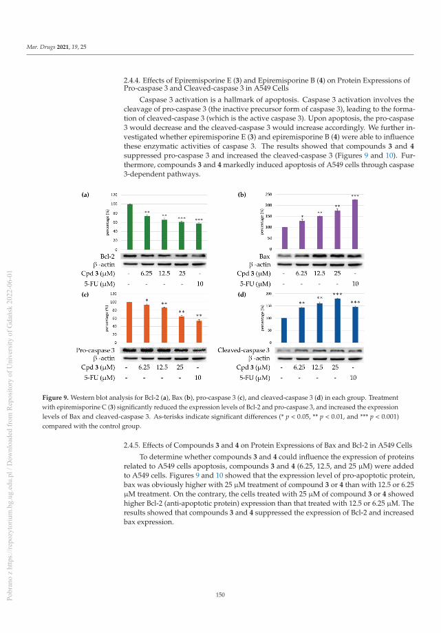
Mar. Drugs 2021, 19, 25
2.4.4. Effects of Epiremisporine E (3) and Epiremisporine B (4) on Protein Expressions ofPro-caspase 3 and Cleaved-caspase 3 in A549 Cells
Caspase 3 activation is a hallmark of apoptosis. Caspase 3 activation involves thecleavage of pro-caspase 3 (the inactive precursor form of caspase 3), leading to the forma-tion of cleaved-caspase 3 (which is the active caspase 3). Upon apoptosis, the pro-caspase3 would decrease and the cleaved-caspase 3 would increase accordingly. We further in-vestigated whether epiremisporine E (3) and epiremisporine B (4) were able to influencethese enzymatic activities of caspase 3. The results showed that compounds 3 and 4
suppressed pro-caspase 3 and increased the cleaved-caspase 3 (Figures 9 and 10). Fur-thermore, compounds 3 and 4 markedly induced apoptosis of A549 cells through caspase3-dependent pathways.
Figure 9. Western blot analysis for Bcl-2 (a), Bax (b), pro-caspase 3 (c), and cleaved-caspase 3 (d) in each group. Treatmentwith epiremisporine C (3) significantly reduced the expression levels of Bcl-2 and pro-caspase 3, and increased the expressionlevels of Bax and cleaved-caspase 3. As-terisks indicate significant differences (* p < 0.05, ** p < 0.01, and *** p < 0.001)compared with the control group.
2.4.5. Effects of Compounds 3 and 4 on Protein Expressions of Bax and Bcl-2 in A549 Cells
To determine whether compounds 3 and 4 could influence the expression of proteinsrelated to A549 cells apoptosis, compounds 3 and 4 (6.25, 12.5, and 25 μM) were addedto A549 cells. Figures 9 and 10 showed that the expression level of pro-apoptotic protein,bax was obviously higher with 25 μM treatment of compound 3 or 4 than with 12.5 or 6.25μM treatment. On the contrary, the cells treated with 25 μM of compound 3 or 4 showedhigher Bcl-2 (anti-apoptotic protein) expression than that treated with 12.5 or 6.25 μM. Theresults showed that compounds 3 and 4 suppressed the expression of Bcl-2 and increasedbax expression.
150Pobr
ano
z ht
tps:
//rep
ozyt
oriu
m.b
g.ug
.edu
.pl /
Dow
nloa
ded
from
Rep
osito
ry o
f Uni
vers
ity o
f Gda
ńsk
2022
-06-
01

Mar. Drugs 2021, 19, 25
Figure 10. Western blot analysis for Bcl-2 (a), Bax (b), pro-caspase 3 (c), and cleaved-caspase 3 (d) in each group. Treatmentwith epiremisporine B (4) significantly reduced the expression levels of Bcl-2 and pro-caspase 3, and increased the expressionlevels of Bax and cleaved-caspase 3. As-terisks indicate significant differences (* p < 0.05 and ** p < 0.01) compared with thecontrol group.
3. Experimental Section
3.1. General Procedures
Optical rotations were measured using a Jasco P-2000 polarimeter (Japan SpectroscopicCorporation, Tokyo, Japan) in CHCl3. Circular dichroism (CD) spectra were obtained on aJ-715 spectropolarimeter (Jasco, Easton, MD, USA). Ultraviolet (UV) spectra were obtainedon a Hitachi U-2800 Double Beam Spectrophotometer (Hitachi High-Technologies Corpo-ration, Tokyo, Japan). Infrared (IR) spectra (neat or KBr) were recorded on a ShimadzuIRAffinity-1S FT-IR Spectrophotometer (Shimadzu Corporation, Kyoto, Japan). Nuclearmagnetic resonance (NMR) spectra, including correlation spectroscopy (COSY), rotatingframe nuclear Overhauser effect spectroscopy (ROESY), heteronuclear multiple-bond cor-relation (HMBC), and heteronuclear single-quantum coherence (HSQC) experiments, wereacquired using a BRUKER AVIII-500 or a BRUKER AVIII-600 spectrometer (Bruker, Bre-men, Germany), operating at 500 or 600 MHz (1H) and 125 or 150 MHz (13C), respectively,with chemical shifts given in the ppm (δ), using CDCl3 as an internal standard (peak at7.263 ppm in 1H NMR and 77.0 ppm in 13C NMR spectrum). Electrospray ionization (ESI)and high-resolution electrospray ionization (HRESI)-mass spectra were recorded on aBruker APEX II Mass Spectrometer (Bruker, Bremen, Germany). Silica gel [70–230 mesh(63–200 μm) and 230–400 mesh (40–63 μm), Merck] was used for column chromatography(CC). Silica gel 60 F-254 (Merck, Darmstadt, Germany) was used for thin-layer chromatog-raphy (TLC) and preparative thin-layer chromatography (PTLC).
3.2. Fungal Material
The fungal strain Penicillium citrinum BCRC 09F458 was isolated from waste water,which was collected from Hazailiao, Dongshi, Chiayi, Taiwan, in 2009. The fungal strainwas identified as Penicillium citrinum (family Trichocomaceae) by the BCRC center, basedon cultural and anamorphic data. The rDNA-ITS (internal transcribed spacer) region wasused for further identification. After searching the GenBank database through BLAST
151Pobr
ano
z ht
tps:
//rep
ozyt
oriu
m.b
g.ug
.edu
.pl /
Dow
nloa
ded
from
Rep
osito
ry o
f Uni
vers
ity o
f Gda
ńsk
2022
-06-
01

Mar. Drugs 2021, 19, 25
(nucleotide sequence comparison program), it was found to have a 100% similarity toP. citrinum. P. citrinum BCRC 09F458 was stored in the Biological Resources Collection andResearch Center (BCRC) of the Food Industry Research and Development Institute (FIRDI).
Cultivation and Preparation of the Fungal Strain
We kept P. citrinum BCRC 09F0458 on potato dextrose agar (PDA), and cultivatedthe strain on PDA at 25 ◦C for one week, and finally harvested the spores with sterilewater. The spores (5 × 10⁵) were seeded into 300 mL shake flasks containing 50 mL RGYmedium (3% rice starch, 7% glycerol, 1.1% polypeptone, 3% soybean powder, 0.1% MgSO4,and 0.2% NaNO3), and cultivated with shaking (150 rpm) at 25 ◦C, for 3 days. After themycelium enrichment step, an inoculum mixing 100 mL mycelium broth and 100 mL RGYmedium was inoculated into plastic boxes (25 cm × 30 cm) containing 1.5 kg sterile riceand cultivated at 25 ◦C for producing rice, and the above mentioned RGY medium wasadded for maintaining the growth. After 21 days of cultivation, the rice was harvested, andused as a sample for further extraction.
3.3. Extraction and Isolation
The rice of the P. citrinum BCRC 09F0458 (1.5 kg) was extracted with 95% EtOH(3 × 10 L, 3 d each) at room temperature. The ethanol extract was concentrated underreduced pressure, and was partitioned with n-BuOH/H2O (1:1, v/v) to afford n-BuOHsoluble fraction (36.2 g), H2O soluble fraction (13.0 g), and insoluble fraction (500 mg). Then-BuOH fraction (fraction A, 36.2 g) was purified by column chromatography (CC) (1.6 kg ofsilica gel, 70–230 mesh ((63–200 μm); n-hexane/EtOAc 25:1–0:1, 1500 mL) to afford 13 frac-tions: A1–A13. Fraction A2 (1.54 g) was subjected to medium pressure liquid chromatogra-phy (MPLC) (69 g of silica gel, 230–400 mesh (40–63 μm)); n-hexane/acetone 1:0–0:1, 700 mL-fractions) to give 11 subfractions: A2-1–A2-11. Fraction A2-6 (126 mg) was further purifiedby semipreparative normal-phase high performance liquid chromatography (HPLC) (silicagel; n-hexane/dichloromethane/EtOAc, 6:3:1) to obtain 8-hydroxy-1-methoxycarbonyl-6-methylxanthone (6) (14.9 mg). Fraction A6 (1.18 g) was subjected to MPLC (53 g of silica gel,230–400 mesh (40–63 μm); n-hexane/acetone 1:0–0:1, 500 mL-fractions) to give 13 subfrac-tions: A6-1–A6-13. Fraction A6-9 (150 mg) was further purified by preparative TLC (silicagel; n-hexane/EtOAc, 1:9) to afford penicitrinone A (5) (18.3 mg). Fraction A9 (1.44 g) wassubjected to MPLC (65 g of silica gel, 230–400 mesh (40–63 μm)); dichloromethane/EtOAc1:0–2:3, 650 mL-fractions) to give 12 subfractions: A9-1–A9-12. Fraction A9-6 (128 mg) wasfurther purified by preparative TLC (silica gel; n-hexane/acetone, 3:2) to afford epiremis-porine C (1) (12.2 mg). Fraction A10 (0.98 g) was subjected to MPLC (44 g of silica gel,230–400 mesh (40–63 μm); n-hexane/acetone 1:0–0:1, 450 mL-fractions) to give 10 sub-fractions: A10-1–A10-10. Fraction A10-4 (120 mg) was further purified by preparativeTLC (silica gel; n-hexane/EtOAc, 7:3) to afford isoconiochaetone C (7) (11.2 mg). FractionA11 (2.38 g) was subjected to MPLC (107 g of silica gel, 230–400 mesh (40–63 μm); n-hexane/acetone 1:0–0:1, 1000 mL-fractions) to give 14 subfractions: A11-1–A11-14. FractionA11-2 (138 mg) was further purified by semipreparative normal-phase HPLC (silica gel;n-hexane/EtOAc, 1:1) to afford epiremisporine D (2) (14.6 mg). Fraction A11-4 (168 mg)was further purified by preparative TLC (silica gel; dichloromethane/methanol, 19:1) toafford epiremisporine B (4) (23.8 mg). Fraction A12 (1.28 g) was subjected to MPLC (58 gof silica gel, 230–400 mesh (40–63 μm); n-hexane/EtOAc 1:0–0:1, 600 mL-fractions) to give10 subfractions: A12-1–A12-10. Fraction A12-1 (122 mg) was further purified by prepara-tive TLC (silica gel; n-hexane/dichloromethane/acetone, 5:3:2) to afford epiremisporine E(3) (13.7 mg).
Epiremisporine C (1): [α]25D = +527.6◦ (c 0.22, CHCl3); UV (MeOH) λmax nm (log ε):
241 (4.35), 326 (3.50) nm; 1H NMR data, see Table 4; 13C NMR data, see Table 5.
152Pobr
ano
z ht
tps:
//rep
ozyt
oriu
m.b
g.ug
.edu
.pl /
Dow
nloa
ded
from
Rep
osito
ry o
f Uni
vers
ity o
f Gda
ńsk
2022
-06-
01

Mar. Drugs 2021, 19, 25
Table 4. 1H NMR data (500 MHz, CDCl3) for 1–3.
Position1 2 3
δH (J in Hz)
3 3.87 (dd, 8.9, 8.6) 3.77 (dd, 8.7, 8.6) 3.48 (dd, 10.5, 8.9)4 5.21 (d, 8.9) 5.14 (d, 8.9) 4.96 (d, 8.9)8 6.67 (br s) 6.76 (br s) 6.81 (br s)10 6.62 (br s) 6.58 (br s) 6.59 (br s)15 2.33 (s) 2.36 (s) 2.38 (s)16 3.83 (s) 3.78 (s) 3.82 (s)3′ 2.86 (ddd, 12.8, 8.6, 5.3) 2.82 (ddd, 12.7, 8.6, 5.4) 3.06 (ddd, 10.5, 8.3, 4.7)
4′α 2.58 (dd, 15.6, 5.3) 2.55 (dd, 15.5, 5.4) 2.99 (dd, 18.6, 4.7)4′β 2.37 (dd, 15.6, 12.8) 2.36 (dd, 15.5, 12.7) 2.85 (dd, 18.6, 8.3)8′ 6.69 (br s) 6.69 (br s) 6.69 (br s)10′ 6.70 (br s) 6.69 (br s) 6.68 (br s)15′ 2.42 (s) 2.41 (s) 2.42 (s)16′ 3.87 (s) 3.84 (s) 3.84 (s)
11-OH 12.11 (br s) - -11-OMe - 3.91 (s) 3.91 (s)2′-OMe 3.50 (s) 3.51 (s) 3.11 (s)11′-OH 12.30 (br s) 12.36 (s) 12.50 (s)
Table 5. 13C NMR data (125 MHz, CDCl3) for 1–3.
Position1 2 3
δC, Type
1 170.8, C 171.2, C 170.5, C2 91.2, C 91.7, C 91.6, C3 46.9, CH 46.8, CH 44.0, CH4 36.6, CH 36.1, CH 37.3, CH5 168.3, C 164.7, C 166.4, C7 157.2, C 159.1, C 159.2, C8 108.3, CH 110.7, CH 110.9, CH9 147.4, C 145.2, C 145.1, C
10 113.1, CH 108.4, CH 108.3, CH11 160.9, C 160.0, C 159.9, C12 109.0, C 112.9, C 112.6, C13 179.0, C 173.7, C 173.9, C14 118.8, C 121.7, C 121.2, C15 22.2, CH3 22.1, CH3 22.1, CH316 53.1, CH3 52.9, CH3 53.1, CH31′ 166.7, C 167.1, C 168.5, C2′ 111.1, C 111.0, C 107.6, C3′ 48.4, CH 48.4, CH 43.3, CH4′ 27.1, CH2 27.1, CH2 25.5, CH25′ 165.5, C 165.5, C 165.4, C7′ 156.0, C 156.1, C 155.9, C8′ 107.6, CH 107.5, CH 107.3, CH9′ 147.7, C 147.5, C 147.3, C10′ 112.7, CH 112.6, CH 112.2, CH11′ 160.5, C 160.5, C 160.4, C12′ 108.5, C 108.5, C 108.3, C13′ 179.7, C 179.8, C 180.6, C14′ 112.8, C 113.0, C 111.7, C15′ 22.4, CH3 22.4, CH3 22.4, CH316′ 52.8, CH3 52.6, CH3 52.8, CH3
11-OMe - 56.3, CH3 56.4, CH32′-OMe 52.7, CH3 52.7, CH3 52.2, CH3
153Pobr
ano
z ht
tps:
//rep
ozyt
oriu
m.b
g.ug
.edu
.pl /
Dow
nloa
ded
from
Rep
osito
ry o
f Uni
vers
ity o
f Gda
ńsk
2022
-06-
01

Mar. Drugs 2021, 19, 25
Epiremisporine D (2): [α]25D = +526.8◦ (c 0.10, CHCl3); UV (MeOH) λmax nm (log ε):
237 (4.25), 317 (3.52) nm; 1H NMR data, see Table 4; 13C NMR data, see Table 5.
Epiremisporine E (3): [α]25D = +561.3◦ (c 0.09, CHCl3); UV (MeOH) λmax nm (log ε):
235 (4.30), 315 (3.54) nm; 1H NMR data, see Table 4; 13C NMR data, see Table 5.
Epiremisporine B (4): Yellow amorphous powder; [α]25D = +523.6◦ (c 0.16, CHCl3); 1H
NMR data, see Table S1; 13C NMR data, see Table S2; HRESIMS: m/z 599.11558 [M + Na]+
(calcd. for C30H24O12 + Na, 599.11654).
Penicitrinone A (5): Orange crystalline powder; [α]25D = +102.6◦ (c 0.12, MeOH); 1H
NMR (600 MHz, CDCl3) δ 1.32 (3H, d, J = 7.2 Hz, Me-4), 1.33 (3H, d, J = 7.0 Hz, Me-3′),1.42 (3H, d, J = 6.4 Hz, Me-2′), 1.44 (3H, d, J = 6.7 Hz, Me-3), 2.11 (3H, s, Me-5), 2.21 (3H,s, Me-4′), 3.12 (1H, qd, J = 7.2, 0.9 Hz, H-4), 3.16 (1H, qd, J = 7.0, 4.1 Hz, H-3′), 4.61 (1H,qd, J = 6.4, 4.1 Hz, H-2′), 4.96 (1H, qd, J = 6.7, 0.9 Hz, H-3), 6.37 (1H, s, H-7), 8.33 (1H, br s,OH-5′); HRESIMS: m/z 381.17094 [M + H]+ (calcd. for C23H24O5 + H, 381.17020).
8-Hydroxy-1-methoxycarbonyl-6-methylxanthone (6): Yellow amorphous solid; 1HNMR (600 MHz, CDCl3) δ 2.44 (3H, s, Me-6), 4.03 (3H, s, COOMe-1), 6.64 (1H, br d,J = 1.4 Hz, H-5), 6.76 (1H, br d, J = 1.4 Hz, H-7), 7.31 (1H, dd, J = 7.3, 1.1 Hz, H-2), 7.53 (1H,dd, J = 8.5, 1.1 Hz, H-4), 7.74 (1H, dd, J = 8.5, 7.3 Hz, H-3), 12.15 (1H, s, OH-8); HRESIMS:m/z 285.07730 [M + H]+ (calcd. for C16H12O5 + H, 285.07630).
Isoconiochaetone C (7): Colorless needles (MeOH), m.p. 99–100.5 ◦C; [α]25D = +79.3◦ (c
0.18, MeOH); 1H NMR (600 MHz, CDCl3) δ 2.15 (1H, dddd, J = 14.0, 8.5, 2.5, 1.4 Hz, Hβ-2),2.32 (1H, dddd, J = 14.0, 9.4, 7.4, 6.8 Hz, Hα-2), 2.40 (3H, s, Me-8), 2.77 (1H, ddd, J = 18.0,9.4, 2.5 Hz, Hβ-3), 3.17 (1H, dddd, J = 18.0, 8.5, 7.4, 1.4 Hz, Hα-3), 3.50 (3H, s, OMe-1), 4.94(1H, dt, J = 6.8, 1.4 Hz, H-1), 6.63 (1H, br s, H-9), 6.71 (1H, br s, H-7), 12.55 (1H, s, OH-10);HRESIMS: m/z 247.09688 [M + H]+ (calcd. for C14H14O4 + H, 247.09703).
3.4. Biological Assay
The anti-inflammatory effects of the isolated compounds from Penicillium citrinumwere evaluated by suppressing fMLP-induced O2
•– generation by human neutrophils. Inaddition, anti-cancer activity was evaluated by cytotoxicity assay and Western blot analysis.
3.4.1. Preparation of Human Neutrophils
Human neutrophils from the venous blood [21] of healthy, adult volunteers (20–35 yearsold) were isolated using a standard method of dextran sedimentation, prior to centrifu-gation in a Ficoll Hypaque gradient and hypotonic lysis of erythrocytes, as previouslydescribed [29]. Purified neutrophils containing >98% viable cells, as determined by thetrypan blue exclusion method, were resuspended in HBSS buffer at pH 7.4 and weremaintained at 4 ◦C, prior to use [30].
3.4.2. Measurement of O2•– Generation
The assay for measurement of O2•– generation was based on the SOD-inhibitable
reduction of ferricytochrome c [31]. In brief, neutrophils (1 × 10⁶ cells/mL) pretreated withthe various test agents at 37 ◦C for 5 min were stimulated with fMLP (1 μmol/L) in thepresence of ferricytochrome c (0.5 mg/mL). Extracellular O2
•– production was assessedwith a UV spectrophotometer at 550 nm (Hitachi U-3010, Tokyo, Japan). The percentage ofsuperoxide inhibition of the test compound was calculated as the percentage of inhibition= {(control − resting) − (compound − resting)}/(control − resting) × 100. The softwareSigmaPlot was used for determining the IC50 values [30].
3.4.3. Chemicals and Antibodies
Fluorouracil (5-FU) and bovine serum albumin (BSA) were purchased from Sigma-Aldrich (St. Louis, MO, USA). The antibodies against Bcl-2, Bax, and β-actin were pur-
154Pobr
ano
z ht
tps:
//rep
ozyt
oriu
m.b
g.ug
.edu
.pl /
Dow
nloa
ded
from
Rep
osito
ry o
f Uni
vers
ity o
f Gda
ńsk
2022
-06-
01

Mar. Drugs 2021, 19, 25
chased from Cell Signaling Technology (Danvers, MA, USA). Caspase-3 was obtained fromGeneTex International Corporation (Hsinchu, Taiwan).
3.4.4. Cells and Culture Medium
A549 (human lung carcinoma) and HT-29 (human colon carcinoma) cells were kindlyprovided by Prof. T. M. Hu and Prof. Y. Su, respectively, of National Yang-Ming University,Taipei, Taiwan.
All cell lines were cultured in Dulbecco’s modified Eagle’s medium supplementedwith 10% fetal bovine serum (FBS), 100 U/mL penicillin, 100 μg/mL streptomycin, 2 μML-glutamine, and 1 mM sodium pyruvate. The cells were incubated in an atmosphereof 37 ◦C and 5% CO2 and passaged twice a week. Cells were stored in liquid nitrogen at−155 ◦C. After the cells were thawed, the experiment was completed before 30 generations.The purpose was to minimize experimental errors. The compound stock solution wasstored in DMSO at a concentration of 10 mM and stored at −20 ◦C, and finally meltedimmediately before use.
3.4.5. Cytotoxicity Assay
A MTT colorimetric assay was used to determine cell viability. The assay was modifiedfrom that of Mosmann [32]. MTT reagent (0.5 mg/mL) was added onto the attached cellsmentioned above (100 μL per 100 μL culture) and incubated at 37 ◦C for 3 h. Then, DMSOwas added and the amount of colored formazan metabolite formed was measured byabsorbance at 570 nm, using an ELISA plate reader (μ Quant). The optical density offormazan formed in control (untreated) cells was taken as 100% viability.
3.4.6. Clonogenic Assay
The clonogenic assay followed as previously described with slight changes [33]. Forthe clonogenic assay, cells at a density of 3000 cells/well were seeded in 6-well plates for24 h. Next, the cells were treated with compound 3 or vehicle (DMSO) and allowed to formcolonies for 14 days. Colonies were washed with PBS, and the cells attached to the plasticsurface were fixed in 99% MeOH for 30 min and stained with 0.2% crystal violet for 15 min.The stained cells were quantified using the ImageJ software (BioTechniques, NY, USA).
3.4.7. Western Blotting Analysis
Western blot analysis was performed according to the method previously reported [34].In brief, A549 (1 × 10⁵ cells) was seeded into 6 wells plate and grown until 85–90% confluent.Then, different concentrations (3.125, 6.25, 12.5, 25, and 50 μM) of compounds 3 and 4 wereadded. Cells were collected and lysed by radioimmunoprecipitation assay (RIPA) buffer.Lysates of total protein were separated by 12.5% sodium dodecyl sulfate-polyacrylamidegels and transferred to polyvinylidene difluoride (PVDF) membranes. After blocking, themembranes were incubated with anti-Bax, anti-Bcl-2 (Cell Signaling Inc., Danvers, MA,USA), anti-caspase-3, and anti-β-actin (GeneTex Inc., Irvine, CA, USA) primary antibodiesat 4 ◦C overnight. Then, each membrane was washed with Tris-buffered saline containing0.1% Tween 20 (TBST) and incubated with horseradish peroxidase (HRP)-conjugatedsecondary antibodies at room temperature, for 1 h, while shaking. Finally, each membranewas developed using an enhanced chemiluminescence (ECL) detection kit, and the imageswere visualized by ImageQuant LAS 4000 Mini biomolecular imager (GE Healthcare,MA, USA). The band densities were quantified using the ImageJ software (BioTechniques,NY, USA).
3.4.8. Statistical Analysis
All data are expressed as mean ± SEM. Statistical analysis was carried out usingStudent’s t-test. A probability of 0.05 or less was considered to be statistically significant.Microsoft Excel 2019 was used for the statistical and graphical evaluation. All experimentswere performed at least 3 times.
155Pobr
ano
z ht
tps:
//rep
ozyt
oriu
m.b
g.ug
.edu
.pl /
Dow
nloa
ded
from
Rep
osito
ry o
f Uni
vers
ity o
f Gda
ńsk
2022
-06-
01

Mar. Drugs 2021, 19, 25
4. Conclusions
Three novel (1–3) and four known compounds were isolated and identified fromPenicillium citrinum. Among the isolated compounds, compounds 2–5 could significantlyinhibit fMLP-induced O2
•− generation, with IC50 values ≤ 8.28 μM. These isolated com-pounds are worth further research, as promising new leads for developing anti-inflammatoryagents. Furthermore, compounds 3 and 4 markedly induced apoptosis of A549 cellsthrough the mitochondrial- and caspase 3-dependent pathways (Figure 11). This suggeststhat compounds 3 and 4 are worth further investigation and might be expectantly devel-oped as the candidates for the treatment or prevention of non-small cell lung cancer andliver cancer.
Figure 11. Schematic diagram for cancer cell apoptosis mechanism of compounds 3 and 4 in A549 cells.
Supplementary Materials: The following are available online at https://www.mdpi.com/1660-3397/19/1/25/s1, Figures S1–S10: HRESIMS, CD, 1D, and 2D NMR spectra for epiremisporine C (1).Figures S11–S20: HRESIMS, CD, 1D, and 2D NMR spectra for epiremisporine D (2). Figures S21–S30:HRESIMS, CD, 1D, and 2D NMR spectra for epiremisporine E (3). Figures S31–S34: HRESIMS, CD,1H and 13C NMR spectra for (4). Figures S35 and S36: HRESIMS, 1H NMR spectra for penicitri-none A (5). Figures S37 and S38: HRESIMS, 1H NMR spectra for 8-hydroxy-1-methoxycarbonyl-6-methylxanthone (6). Figures S39 and S40: HRESIMS, 1H NMR spectra for isoconiochaetone C (7).Figure S41: CD and ECD spectra of epiremisporine B. Tables S1 and S2: 1H and 13C NMR spectrumfor 4 and epiremisporine B. Table S3: The ROESY correlations for compounds 1–3.
Author Contributions: Y.-C.C. performed the isolation and structure elucidation of the constituents,and prepared the manuscript. C.-H.C. and H.-R.L. conducted the bioassay and analyzed the data.M.-J.C. and M.-D.W. performed the cultivation and preparation of the fungal strain. S.-L.F. analyzedbioassay data. J.-J.C. planned, designed, and organized all of the research of this study and reviewedthe manuscript. All authors read and approved the final version of the manuscript.
Funding: This research received no external funding.
Institutional Review Board Statement: Not applicable.
156Pobr
ano
z ht
tps:
//rep
ozyt
oriu
m.b
g.ug
.edu
.pl /
Dow
nloa
ded
from
Rep
osito
ry o
f Uni
vers
ity o
f Gda
ńsk
2022
-06-
01

Mar. Drugs 2021, 19, 25
Informed Consent Statement: Not applicable.
Data Availability Statement: The data presented in this study are available in the main text and thesupplementary materials of this article.
Acknowledgments: This research was supported by grants from the Ministry of Science and Tech-nology (MOST), Taiwan (No. MOST 109-2320-B-010-029-MY3 and MOST 106-2320-B-010-033-MY3),awarded to J.-J. Chen. We gratefully thank Shou-Ling Huang and Iren Wang for the assistance inNMR experiments of the Instrumentation Center at NTU which is supported by the Ministry ofScience and Technology, Taiwan.
Conflicts of Interest: The authors declare no conflict of interest.
References
1. Blunt, J.W.; Copp, B.R.; Keyzers, R.A.; Munro, M.H.G.; Prinsep, M.R. Marine natural products. Nat. Prod. Rep. 2015, 32, 116–211.[CrossRef] [PubMed]
2. Kiuru, P.; D’Auria, M.V.; Muller, C.D.; Tammela, P.; Vuorela, H.; Yli-Kauhaluoma, J. Exploring marine resources for bioactivecompounds. Planta Med. 2014, 80, 1234–1246. [CrossRef] [PubMed]
3. Bao, J.; Wang, J.; Zhang, X.Y.; Nong, X.H.; Qi, S.H. New furanone derivatives and alkaloids from the co-culture of marine-derivedfungi Aspergillus sclerotiorum and Penicillium citrinum. Chem. Biodivers. 2016, 14, 327–335.
4. Huang, G.L.; Zhou, X.M.; Bai, M.; Liu, Y.X.; Zhao, Y.L.; Luo, Y.P.; Niu, Y.Y.; Zheng, C.J.; Chen, G.Y. Dihydroisocoumarins from themangrove-derived fungus Penicillium citrinum. Mar. Drugs 2016, 14, 177–185. [CrossRef]
5. Ibrar, M.; Ullah, M.W.; Manan, S.; Farooq, U.; Rafiq, M.; Hasan, F. Fungi from the extremes of life: An untapped treasure forbioactive compounds. Appl. Microbiol. Biotechnol. 2020, 104, 2777–2801. [CrossRef]
6. Khan, S.A.; Hamayun, M.; Yoon, H.; Kim, H.Y.; Suh, S.J.; Hwang, S.K.; Kim, J.M.; Lee, I.J.; Choo, Y.S.; Yoon, U.H.; et al. Plantgrowth promotion and Penicillium citrinum. BMC Microbiol. 2008, 8, 231–241. [CrossRef]
7. Kong, F.; Carter, G.T. Remisporine B, a novel dimeric chromenone derived from spontaneous Diels-Alder reaction of remisporineA. Tetrahedron Lett. 2003, 44, 3119–3122. [CrossRef]
8. Liu, Q.Y.; Zhou, T.; Zhao, Y.Y.; Chen, L.; Gong, M.W.; Xia, Q.W.; Ying, M.G.; Zheng, Q.H.; Zhang, Q.Q. Antitumor effects andrelated mechanisms of penicitrinine A, a novel alkaloid with a unique spiro skeleton from the marine fungus Penicillium citrinum.Mar. Drugs 2015, 13, 4733–4753. [CrossRef]
9. Liu, F.A.; Lin, X.; Zhou, X.; Chen, M.; Huang, X.; Yang, B.; Tao, H. Xanthones and quinolones derivatives produced by thedeep-sea-derived fungus Penicillium sp. SCSIO Ind16F01. Molecules 2017, 22, 1999. [CrossRef]
10. Lösgen, S.; Magull, J.; Schulz, B.; Draeger, S.; Zeeck, A. Isofusidienols: Novel chromone-3-oxepines produced by the endophyticfungus Chalara sp.. Eur. J. Org. Chem. 2008, 698–703. [CrossRef]
11. Meng, L.H.; Liu, Y.; Li, X.M.; Xu, G.M.; Ji, N.Y.; Wang, B.G. Citrifelins A and B, citrinin adducts with a tetracyclic framework fromcocultures of marine-derived isolates of Penicillium citrinum and Beauveria felina. J. Nat. Prod. 2015, 78, 2301–2305. [CrossRef][PubMed]
12. Mossini, S.A.; Kemmelmeier, C. Inhibition of citrinin production in Penicillium citrinum cultures by neem [Azadirachta indica A.Juss (meliaceae)]. Int. J. Mol. Sci. 2008, 9, 1676–1684. [CrossRef] [PubMed]
13. Sabdaningsih, A.; Liu, Y.; Mettal, U.; Heep, J.; Riyanti; Wang, L.; Cristianawati, O.; Nuryadi, H.; Sibero, M.T.; Marner, M.; et al. Anew citrinin derivative from the Indonesian marine sponge-associated fungus Penicillium citrinum. Mar. Drugs 2020, 18, 227–239.[CrossRef]
14. Shen, H.D.; Wang, C.W.; Chou, H.; Lin, W.L.; Tam, M.F.; Huang, M.H.; Kuo, M.L.; Wang, S.R.; Han, S.H. Complementary DNAcloning and immunologic characterization of a new Penicillium citrinum allergen (Pen c 3). J. Allergy Clin. Immunol. 2000, 105,827–833. [CrossRef]
15. Wakana, D.; Hosoe, T.; Itabashi, T.; Okada, K.; de Campos Takaki, G.M.; Yaguchi, T.; Fukushima, K.; Kawai, K.I. New citrininderivatives isolated from Penicillium citrinum. J. Nat. Med. 2006, 60, 279–284. [CrossRef]
16. Xia, M.W.; Cui, C.B.; Li, C.W.; Wu, C.J.; Peng, J.X.; Li, D.H. Rare chromones from a fungal mutant of the marine-derived Penicilliumpurpurogenum G59. Mar. Drugs 2015, 13, 5219–5236. [CrossRef] [PubMed]
17. Zheng, C.J.; Huang, G.L.; Xu, Y.; Song, X.M.; Yao, J.; Liu, H.; Wang, R.P.; Sun, X.P. A new benzopyrans derivatives from amangrove-derived fungus Penicillium citrinum from the South China Sea. Nat. Prod. Res. 2016, 30, 821–825. [CrossRef] [PubMed]
18. Zheng, C.J.; Liao, H.X.; Mei, R.Q.; Huang, G.L.; Yang, L.J.; Zhou, X.M.; Shao, T.M.; Chen, G.Y.; Wang, C.Y. Two new benzophenonesand one new natural amide alkaloid isolated from a mangrove-derived fungus Penicillium citrinum. Nat. Prod. Res. 2019, 33,1127–1134. [CrossRef]
19. Witko-Sarsat, V.; Rieu, P.; Descamps-Latscha, B.; Lesavre, P.; Halbwachs-Mecarelli, L. Neutrophils: Molecules, functions andpathophysiological aspects. Lab. Invest. 2000, 80, 617–653. [CrossRef]
20. Ennis, M. Neutrophils in asthma pathophysiology. Curr. Allergy Asthma Rep. 2003, 3, 159–165. [CrossRef]21. Borregaard, N. The human neutrophil. Function and dysfunction. Eur. J. Haematol. Suppl. 1988, 41, 401–413. [CrossRef] [PubMed]
157Pobr
ano
z ht
tps:
//rep
ozyt
oriu
m.b
g.ug
.edu
.pl /
Dow
nloa
ded
from
Rep
osito
ry o
f Uni
vers
ity o
f Gda
ńsk
2022
-06-
01

Mar. Drugs 2021, 19, 25
22. Roos, D.; van Bruggen, R.; Meischl, C. Oxidative killing of microbes by neutrophils. Microbes Infect. 2003, 5, 1307–1315. [CrossRef][PubMed]
23. Vane, J.R.; Mitchell, J.A.; Appleton, I.; Tomlinson, A.; Bishop-Bailey, D.; Croxtall, J.; Willoughby, D.A. Inducible isoforms ofcyclooxygenase and nitric-oxide synthase in inflammation. Proc. Natl. Acad. Sci. USA 1994, 91, 2046–2050. [CrossRef]
24. Hsiao, A.J.; Chen, L.H.; Lu, T.H. Ten leading causes of death in Taiwan: A comparison of two grouping lists. J. Formos. Med. Assoc.2015, 114, 679–680. [CrossRef] [PubMed]
25. Yip, K.W.; Reed, J.C. Bcl-2 family proteins and cancer. Oncogene 2008, 27, 6398–6406. [CrossRef] [PubMed]26. Mohan, S.; Abdelwahab, S.I.; Kamalidehghan, B.; Syam, S.; May, K.S.; Harmal, N.S.; Shafifiyaz, N.; Hadi, A.H.; Hashim, N.M.;
Rahmani, M.; et al. Involvement of NF-κB and Bcl2/Bax signaling pathways in the apoptosis of MCF7 cells induced by a xanthonecompound pyranocycloartobiloxanthone A. Phytomedicine 2012, 19, 1007–1015. [CrossRef] [PubMed]
27. Vucicevic, K.; Jakovljevic, V.; Colovic, N.; Tosic, N.; Kostic, T.; Glumac, I.; Pavlovic, S.; Karan-Djurasevic, T.; Colovic, M.Association of Bax expression and Bcl2/Bax ratio with clinical and molecular prognostic markers in chronic lymphocyticleukemia. J. Med. Biochem. 2016, 35, 150–157. [CrossRef] [PubMed]
28. Wu, C.J.; Yi, L.; Cui, C.B.; Li, C.W.; Wang, N.; Han, X. Activation of the silent secondary metabolite production by introducingneomycin-resistance in a marine-derived Penicillium purpurogenum G59. Mar. Drugs 2015, 13, 2465–2487. [CrossRef] [PubMed]
29. English, D.; Andersen, B.R. Single-step separation of red blood cells. Granulocytes and mononuclear leukocytes on discontinuousdensity gradients of Ficoll-Hypaque. J. Immunol. Methods 1974, 5, 249–252. [CrossRef]
30. Chen, L.C.; Liao, H.R.; Chen, P.Y.; Kuo, W.L.; Chang, T.H.; Sung, P.J.; Wen, Z.H.; Chen, J.J. Limonoids from the seeds of Swieteniamacrophylla and their anti-inflammatory activities. Molecules 2015, 20, 18551. [CrossRef]
31. Babior, B.M.; Kipnes, R.S.; Curnutte, J.T. Biological defense mechanisms. The production by leukocytes of superoxide, a potentialbactericidal agent. J. Clin. Invest. 1973, 52, 741–744. [CrossRef] [PubMed]
32. Mosmann, T. Rapid colorimetric assay for cellular growth and survival: Application to proliferation and cytotoxicity assays. J.Immunol. Methods 1983, 65, 55–63. [CrossRef]
33. Su, M.; Zhao, C.; Li, D.; Cao, J.; Ju, Z.; Kim, E.L.; Young-Suk, J.; Jung, J.H. Viriditoxin stabilizes microtubule polymers in SK-OV-3cells and exhibits antimitotic and antimetastatic potential. Mar. Drugs 2020, 18, 445. [CrossRef] [PubMed]
34. Huang, C.Y.; Chang, T.C.; Wu, Y.J.; Chen, Y.; Chen, J.J. Benzophenone and benzoylphloroglucinol derivatives fromHypericum sampsonii with anti-inflammatory mechanism of otogirinin A. Molecules 2020, 25, 4463. [CrossRef] [PubMed]
Sample Availability: Samples of the compounds are available from the authors.
158Pobr
ano
z ht
tps:
//rep
ozyt
oriu
m.b
g.ug
.edu
.pl /
Dow
nloa
ded
from
Rep
osito
ry o
f Uni
vers
ity o
f Gda
ńsk
2022
-06-
01

MDPISt. Alban-Anlage 66
4052 BaselSwitzerland
Tel. +41 61 683 77 34Fax +41 61 302 89 18
www.mdpi.com
Marine Drugs Editorial OfficeE-mail: [email protected]
www.mdpi.com/journal/marinedrugs
Pobr
ano
z ht
tps:
//rep
ozyt
oriu
m.b
g.ug
.edu
.pl /
Dow
nloa
ded
from
Rep
osito
ry o
f Uni
vers
ity o
f Gda
ńsk
2022
-06-
01

Pobr
ano
z ht
tps:
//rep
ozyt
oriu
m.b
g.ug
.edu
.pl /
Dow
nloa
ded
from
Rep
osito
ry o
f Uni
vers
ity o
f Gda
ńsk
2022
-06-
01

MDPI St. Alban-Anlage 66 4052 Basel Switzerland
Tel: +41 61 683 77 34 Fax: +41 61 302 89 18
www.mdpi.com ISBN 978-3-0365-0621-0 Pobr
ano
z ht
tps:
//rep
ozyt
oriu
m.b
g.ug
.edu
.pl /
Dow
nloa
ded
from
Rep
osito
ry o
f Uni
vers
ity o
f Gda
ńsk
2022
-06-
01