One-Pot Biocatalytic Synthesis of Sugar Based Poly ( ε -caprolactone)
Upload
khangminh22Category
view
1download
0
Biocatalytic Synthesis of Bioactive Compounds • Josefina Aleu Biocatalytic
Synthesis of Bioactive Compounds
Printed Edition of the Special Issue Published in Molecules
www.mdpi.com/journal/molecules
Josefina AleuEdited by

Biocatalytic Synthesis of Bioactive Compounds


Biocatalytic Synthesis of Bioactive Compounds
Editor
Josefina Aleu
MDPI • Basel • Beijing • Wuhan • Barcelona • Belgrade • Manchester • Tokyo • Cluj • Tianjin

Editor
Josefina Aleu
University of Cadiz
Spain
Editorial Office
MDPI
St. Alban-Anlage 66
4052 Basel, Switzerland
This is a reprint of articles from the Special Issue published online in the open access journal
Molecules (ISSN 1420-3049) (available at: https://www.mdpi.com/journal/molecules/special
issues/Biocatalytic Synthesis).
For citation purposes, cite each article independently as indicated on the article page online and as
indicated below:
LastName, A.A.; LastName, B.B.; LastName, C.C. Article Title. Journal Name Year, Article Number,
Page Range.
ISBN 978-3-03943-571-5 (Hbk)
ISBN 978-3-03943-572-2 (PDF)
c© 2020 by the authors. Articles in this book are Open Access and distributed under the Creative
Commons Attribution (CC BY) license, which allows users to download, copy and build upon
published articles, as long as the author and publisher are properly credited, which ensures maximum
dissemination and a wider impact of our publications.
The book as a whole is distributed by MDPI under the terms and conditions of the Creative Commons
license CC BY-NC-ND.

Contents
About the Editor . . . . . . . . . . . . . . . . . . . . . . . . . . . . . . . . . . . . . . . . . . . . . . vii
Preface to ”Biocatalytic Synthesis of Bioactive Compounds” . . . . . . . . . . . . . . . . . . . . ix
Te-Sheng Chang, Tzi-Yuan Wang, Szu-Yi Yang, Yu-Han Kao, Jiumn-Yih Wu and Chien-Min Chiang
Potential Industrial Production of a Well-Soluble, Alkaline-Stable, and Anti-Inflammatory Isoflavone Glucoside from 8-Hydroxydaidzein Glucosylated by Recombinant Amylosucrase of Deinococcus geothermalisReprinted from: Molecules 2019, 24, 2236, doi:10.3390/molecules24122236 . . . . . . . . . . . . . . 1
Alexander Veljko Fejzagic, Jan Gebauer, Nikolai Huwa and Thomas Classen
Halogenating Enzymes for Active Agent Synthesis: First Steps Are Done and Many Haveto FollowReprinted from: Molecules 2019, 24, 4008, doi:10.3390/molecules24214008 . . . . . . . . . . . . . . 13
Jainara Santos do Nascimento, Wilson Elias Rozo N u nez, Valmore Henrique Pereira dos Santos, Josefina Aleu, Sılvio Cunha and Eliane de Oliveira Silva
Mapping the Biotransformation of Coumarins through Filamentous FungiReprinted from: Molecules 2019, 24, 3531, doi:10.3390/molecules24193531 . . . . . . . . . . . . . . 47
Jascha Rolf, Mattijs K. Julsing, Katrin Rosenthal and Stephan Lutz
A Gram-Scale Limonene Production Process with Engineered Escherichia coliReprinted from: Molecules 2020, 25, 1881, doi:10.3390/molecules25081881 . . . . . . . . . . . . . . 57
Nina-Katharina Krahe, Ralf G. Berger and Franziska Ersoy
A DyP-Type Peroxidase of Pleurotus sapidus with Alkene Cleaving ActivityReprinted from: Molecules 2020, 25, 1536, doi:10.3390/molecules25071536 . . . . . . . . . . . . . . 69
Young Sung Jung, Ye-Jin Kim, Aaron Taehwan Kim, Davin Jang, Mi-Seon Kim, Dong-Ho Seo, Tae Gyu Nam, Chan-Su Rha, Cheon-Seok Park and Dae-Ok Kim
Enrichment of Polyglucosylated Isoflavones f rom S oybean I soflavone Ag lycones Using Optimized Amylosucrase TransglycosylationReprinted from: Molecules 2020, 25, 181, doi:10.3390/molecules25010181 . . . . . . . . . . . . . . 87
Elisabetta Brenna, Danilo Colombo, Giuseppe Di Lecce, Francesco G. Gatti, Maria Chiara Ghezzi, Francesca Tentori, Davide Tessaro and Mariacristina Viola
Conversion of Oleic Acid into Azelaic and Pelargonic Acid by a Chemo-Enzymatic RouteReprinted from: Molecules 2020, 25, 1882, doi:10.3390/molecules25081882 . . . . . . . . . . . . . . 103
Mateusz Kutyła, Jan Fiedurek, Anna Gromada, Krzysztof Jedrzejewski and Mariusz Trytek Mutagenesis and Adaptation of the Psychrotrophic Fungus Chrysosporium pannorum A-1 as a Method for Improving ββ-pinene BioconversionReprinted from: Molecules 2020, 25, 2589, doi:10.3390/molecules25112589 . . . . . . . . . . . . . . 115
v


About the Editor
Josefina Aleu studied Chemistry at the University of Cadiz, where she graduated in 1991.
After working with Prof. J. R. Hanson at Sussex University for several months in 1995, she then
completed her Ph.D. degree in 1996 under the supervision of Prof. Collado on the design of selective
fungicides. She then worked for 15 months as a postdoctoral fellow with Prof. C. Fuganti at
Politecnico di Milano (Italy) on the biocatalytic synthesis of flavors. She is currently Senior Lecturer of
Organic Chemistry at the University of Cadiz, and her current research interests are directed towards
the biocatalytic synthesis of bioactive molecules, design of selective fungicides, and studies of the
metabolism of marine microorganisms.
vii


Preface to ”Biocatalytic Synthesis of Bioactive
Compounds”
Biocatalysis, the application of enzymes as catalysts for chemical synthesis, has become an increasingly valuable tool for the synthetic chemist. Enzymatic transformations carried out by partially purified e nzymes o r w hole-cell c atalysts a re u sed f or t he p roduction o f a w ide variety of compounds ranging from bulk to fine c hemicals. I n t he fi eld of fin e che micals, the main application clearly lies in the exploitation of the outstanding properties of these biocatalysts with respect to chemoselectivity, regioselectivity, and especially stereoselectivity in the production of enantiomerically pure compounds. Until recently, chemical development companies regarded biocatalysis as a method to be attempted only when all other chemical options failed. Currently, there are clear signs that this view is changing radically, with many of the new process developments revealing the benefits to be gained by using biocatalysis on a commercial scale. Biocatalytic processes have a number of advantages over the corresponding chemical methods. The conditions for such processes are mild and, in the majority of cases, do not require the protection of other functional groups. Moreover, eliminating the need for several chemical synthetic steps has had a dramatic impact on the overall economics. In many cases, biological methods are also enantiospecific, allowing for the production of chiral products from racemic mixtures. Furthermore, the features governing their regiospecificity d iffer f rom t hose c ontrolling c hemical s pecificity an d, in deed, it is possible to obtain biotransformations at centers that are chemically unreactive. Economically, biocatalytic processes are often cheaper and more direct than their chemical counterparts, and the conversions normally proceed under conditions that are regarded as ecologically acceptable.
The aim of this Special Issue is to present studies focused on biocatalysis as applied to the organic synthesis of bioactive compounds and their precursors. The obtained contributions deal with biotransformations, including the stereoselective synthesis of bioactive chemical compounds, active pharmaceutical ingredients, and natural products. The production of interesting bioactive compounds is covered in the eight articles comprising seven research articles and a review.
Two of these papers deal with fungal metabolism and fungi-mediated biotransformations. The first paper deals with improving β -pinene bioconversion by mutagenesis and adaptation of the fungus Chrysosporium pannorum A-1. A second paper describes a study on the biotransformation of bioactive coumarins by filamentous fungi.
On the other hand, the conversion of oleic acid into azelaic and pelargonic acid by a chemoenzymatic route represents a sustainable alternative to ozonolysis, currently employed at the industrial scale to perform the reaction. In addition, the findings on a gram-scale limonene production process using engineered Escherichia coli provide a basis for the development of an economic and industrially relevant bioprocess.
Alkene cleavage is a possibility to generate aldehydes with olfactory properties for the fragrance and flavor i ndustry. A dye-decolorizing peroxidase (DyP) of the basidiomycete Pleurotus sapidus is the first described DyP with alkene cleavage activity towards aryl alkenes and showed potential as biocatalyst for flavor production.
Isoflavones in soybeans are well-known p hytoestrogens. Soy isoflavones present in conjugated forms are converted to aglycone forms during processing and storage. Isoflavone aglycones (IFAs) of soybeans in human diets have poor solubility in water, resulting in low bioavailability and bioactivity. Enzyme-mediated glycosylation is an efficient a nd e nvironmentally f riendly w ay to
ix

modify the physicochemical properties of soy IFAs. Thus, Jung et al. studied the enrichment
of polyglucosylated isoflavones from soybean isoflavone aglycones using optimized amylosucrase
transglycosylation. In the same line, Chang et al. studied the potential industrial production of
a well-soluble, alkaline-stable, and anti-inflammatory isoflavone glucoside from 8-hydroxydaidzein
glucosylated by recombinant amylosucrase of Deinococcus geothermalis.
Finally, the review discusses the use of halogenating enzymes in fine chemical syntheses,
detailing the many steps that are still needed before halogenating enzymes are considered reliable,
flexible, and sustainable catalysts for halogenation.
Overall, these eight contributions provide the reader with relevant fresh insights into the use of
enzymes and whole cells as biocatalysts.
Josefina Aleu
Editor
x

molecules
Article
Potential Industrial Production of a Well-Soluble,Alkaline-Stable, and Anti-Inflammatory IsoflavoneGlucoside from 8-Hydroxydaidzein Glucosylatedby Recombinant Amylosucrase ofDeinococcus geothermalis
Te-Sheng Chang 1,†,*, Tzi-Yuan Wang 2,†, Szu-Yi Yang 1, Yu-Han Kao 1, Jiumn-Yih Wu 3,*
and Chien-Min Chiang 4,*
1 Department of Biological Sciences and Technology, National University of Tainan, Tainan 70005, Taiwan;[email protected] (S.-Y.Y.); [email protected] (Y.-H.K.)
2 Biodiversity Research Center, Academia Sinica, Taipei 115, Taiwan; [email protected] Department of Food Science, National Quemoy University, Kinmen County 892, Taiwan4 Department of Biotechnology, Chia Nan University of Pharmacy and Science, No. 60, Sec. 1, Erh-Jen Rd.,
Jen-Te District, Tainan 71710, Taiwan* Correspondence: [email protected] (T.-S.C.); [email protected] (J.-Y.W.);
[email protected] (C.-M.C.); Tel.: +886-6-2606283 (T.-S.C.); +886-82-313310 (J.-Y.W.);+886-6-2664911 (ext. 2542) (C.-M.C.); Fax: +886-6-2606153 (T.-S.C.); +886-82-313797 (J.-Y.W.);+886-6-2662135 (C.-M.C.)
† These authors contributed equally to this work.
Academic Editor: Josefina AleuReceived: 30 May 2019; Accepted: 14 June 2019; Published: 15 June 2019
Abstract: 8-Hydroxydaidzein (8-OHDe), an ortho-hydroxylation derivative of soy isoflavonedaidzein isolated from some fermented soybean foods, has been demonstrated to possess potentanti-inflammatory activity. However, the isoflavone aglycone is poorly soluble and unstable inalkaline solutions. To improve the aqueous solubility and stability of the functional isoflavone,8-OHDe was glucosylated with recombinant amylosucrase of Deinococcus geothermalis (DgAS) withindustrial sucrose, instead of expensive uridine diphosphate-glucose (UDP-glucose). One majorproduct was produced from the biotransformation, and identified as 8-OHDe-7-α-glucoside, basedon mass and nuclear magnetic resonance spectral analyses. The aqueous solubility and stability ofthe isoflavone glucoside were determined, and the results showed that the isoflavone glucoside wasalmost 4-fold more soluble and more than six-fold higher alkaline-stable than 8-OHDe. In addition,the anti-inflammatory activity of 8-OHDe-7-α-glucoside was also determined by the inhibitionof lipopolysaccharide-induced nitric oxide production in RAW 264.7 cells. The results showedthat 8-OHDe-7-α-glucoside exhibited significant and dose-dependent inhibition on the productionof nitric oxide, with an IC50 value of 173.2 μM, which remained 20% of the anti-inflammatoryactivity of 8-OHDe. In conclusion, the well-soluble and alkaline-stable 8-OHDe-7-α-glucosideproduced by recombinant DgAS with a cheap substrate, sucrose, as a sugar donor retains moderateanti-inflammatory activity, and could be used in industrial applications in the future.
Keywords: 8-hydroxydaidzein; stable; soluble; anti-inflammation; amylosucrase; Deinococcus geothermalis
1. Introduction
Daidzin (D) and genistin (G) are major components of soy isoflavone in soybeans, which are theglucoside derivatives of soy isoflavone aglycone daidzein (De) and genistein (Ge), respectively. De and
Molecules 2019, 24, 2236; doi:10.3390/molecules24122236 www.mdpi.com/journal/molecules1

Molecules 2019, 24, 2236
Ge aglycones have been studied widely and demonstrated multiple bioactivities [1]. In additionto De and Ge, some soy isoflavone derivatives have been isolated from fermented soybean foods,and shown to possess multiple bioactivities [2]. 8-Hydroxydaidzein (8-OHDe), an ortho-hydroxylationderivative of De, was first isolated from the fermentation broth of Streptomyces sp. In the presence ofsoybeans [3], and then from many fermented soybean foods [4–8]. In recent decades, 8-OHDe hasbeen evaluated to possess many bioactivities, such as suppression of multi-drug resistance in Caco-2colon adenocarcinoma cells [9], irreversible inhibition of tyrosinase [10,11], anti-melanogenesis [12,13],inhibition of aldose reductase [14], and anti-inflammation [15,16].
Recently, Seo et al. and Wu et al. developed mass production processes for 8-OHDe frombiotransformation of De by Aspergillus oryzae [17,18]. The availability of a large quantity of 8-OHDeprovides more opportunities for the application of 8-OHDe in the industry. However, although 8-OHDehas many bioactivities, and can be obtained on a large scale, the isoflavone has drawbacks of lowsolubility and high instability in alkaline solutions [19,20]. These drawbacks limit the applications of8-OHDe, unless one can improve the half-life of isoflavone with higher solubility and stability.
Biotransformation of natural products by microorganisms and/or enzymes provides a routeto improve the properties of the original compounds [21,22]. Among different kinds of flavonoidbiotransformation, glycosylation of flavonoids usually holds great promise to increase the solubilityof the original compounds. For example, the aqueous solubility of soy isoflavones is improvedabout 30-fold through glycosylation [23]. Likewise, the glycosyl-biotransformation of 8-OHDe mightimprove its aqueous solubility and stability. In nature, glycosylation of flavonoids is usually catalyzedwith glycosyltransferases (GTs), which use activated uridine diphosphate-glucose (UDP-glucose)as a sugar donor, and transfer the sugar to a flavonoid acceptor [24]. A previous study used therecombinant BsGT110 from Bacillus subtilis to catalyze glucosylation of 8-OHDe [20]. The resultsshowed that the aqueous solubility and stability of the isoflavone glucosides (8-OHDe-7-O-β-glucosideand 8-OHDe-8-O-β-glucoside) were greatly improved. In addition, such biotransformation was noteasily scaled up to the industrial level because of the expensive substrate, UDP-glucose.
However, some glycoside-hydroxylases (GHs) also catalyze glucosylation of flavonoids [25,26].For example, the amylosucrase, GH of the GH13 family, is able to catalyze glucosylation of flavonoidswith cheaper sucrose as a sugar donor, which is one-millionth the cost of UDP-glucose [26].An amylosucrase from Deinococcus geothermalis (DgAS) is one of the promising bio-catalysts inglucosylation of phenolic molecules, because of its thermally-stable and higher activity than otheramylosucrases [27,28]. In the present study, the DgAS enzyme was produced in recombinant Escherichiacoli, and the purified DgAS was detected to catalyze the glucosylation of 8-OHDe. The biotransformedglucosidic product was then purified with chromatography, identified with spectrometric methods.The aqueous solubility, stability, and anti-inflammatory assay of the produced isoflavone glucosidewere determined.
2. Results
2.1. Production of DgAS Protein in Recombinant Escherichia coli
The DgAS gene was amplified from genomic DNA of Deinococcus geothermalis, and subcloned intoan expression plasmid pETDuet-1 fused with six histidine residues in the N-terminal. The constructedpETDuet-DgAS plasmid (Figure 1a) was overexpressed in E. coli, and induced with 0.2 mM of isopropylβ-D-1-thiogalactopyranoside (IPTG). The soluble DgAS proteins were then successfully purifiedwith Ni2+ chelate affinity chromatography, as shown in sodium dodecyl sulfate polyacrylamide gelelectrophoresis (SDS-PAGE) (Figure 1b).
2

Molecules 2019, 24, 2236
(a)
(b)
Figure 1. Production of the DgAS from Deinococcus geothermalis in E. coli. (a) Diagram of the constructedplasmid; (b) SDS-PAGE of the produced DgAS in recombinant E. coli. M: protein marker; lane 1:total protein without IPTG-induction; lane 2: total protein with IPTG-induction for 20 h; lane 3:purified DgAS.
2.2. Biotransformation of 8-OHDe by Recombinant DgAS Protein
The purified DgAS were used to catalyze the biotransformation of 8-OHDe. The reaction contained0.1 mg/mL of 8-OHDe, 12.5 μg/mL of DgAS, and 20 mM of sucrose as the sugar donor. The reactionmixtures at 0 min (the dashed line) and after 30-min incubation were analyzed with ultra-performanceliquid chromatography (UPLC). One major product with a retention time (RT) of 3.1 min was observed(Figure 2).
Figure 2. Biotransformation of 8-OHDe with DgAS. The reaction mixtures at 0 min (dashed line)and 30 min (solid line) were analyzed with UPLC. The UPLC operation conditions are described inMaterials and Methods.
To optimize compound (1) production for further analysis, a standard mixture with 1 mg/mLof 8-OHDe and 125 μg/mL of DgAS was carried out at different temperatures, pH levels, and serialconcentrations of sucrose for 30-min incubation. After incubation, the production conversion of themajor product was determined with UPLC (Figure 3). The results revealed the optimal condition for
3

Molecules 2019, 24, 2236
the production of compound (1) from 8-OHDe by the recombinant DgAS is pH 7, 40 ◦C, and 300 mMof sucrose.
(a)
(b)
(c)
Figure 3. Optimal condition for the production of compound (1) from 8-OHDe by DgAS. (a) 125 μgof the purified DgAS and 1 mg/mL of 8-OHDe were incubated at different temperatures, (b) serialconcentrations of sucrose, and (c) pH levels for 30 min. After incubation, the reaction was analyzedwith UPLC. The conversion was calculated by dividing the amount of the produced compound (1) ineach reaction by the theoretical production value (1.6 mg) for 100% conversion. The mean (n = 3) isshown, and the standard deviations are represented by error bars.
2.3. Identification of the Major Product
To resolve the chemical structure of the product, the biotransformation was scaled up to 100 mL,with 1 mg/mL of 8-OHDe, 125 μg/mL of DgAS, and 300 mM of sucrose at pH 7 and 40 ◦C for30-min incubation. About 90 mg of the product in the 100-mL reaction was purified with preparativehigh-performance liquid chromatography (HPLC). Based on the value of the optimal conversion (89.3%)(Figure 3), the maximum production yield of compound (1) from 100 mg of 8-OHDe is 142.3 mg (160 mg× 0.89); thus, the purification recovery yield is 62.5% (90/142). The chemical structure of the purifiedcompound (1) was resolved with mass and nuclear magnetic resonance (NMR) spectral analysis.The mass analysis of the compound showed an [M +H]+ ion peak at m/z: 433.18 in the electrosprayionization mass (ESI-MS) spectrum corresponding to the molecular formula C21H20O10. Then 1H and
4

Molecules 2019, 24, 2236
13C nmR spectra, including distortionless enhancement by polarization transfer (DEPT), heteronuclearsingle quantum coherence (HSQC), heteronuclear multiple bond connectivity (HMBC), correlationspectroscopy (COSY), and nuclear Overhauser effect spectroscopy (NOESY) spectra, were obtained,and the 1H and 13C nmR signal assignments were conducted accordingly (shown in Figures S1–S7).In addition to the signals of 8-OHDe, seven proton signals (from 3.22 to 5.48 ppm) and six carbon signals(from 60–100 ppm) indicated a glucose moiety. The J coupling constant (3.6 Hz) of the anomeric proton(5.48 ppm) in the 1H nmR spectra of compound (1) indicated the α-configuration for the glucopyranosylmoiety. The cross peak of H-1′′ with C-7 (5.48/148.8 ppm) in the HMBC spectrum, as well as thecross peak of H-1′′ with H-6 (5.48/7.38 ppm) in the NOESY spectrum, demonstrated the structure ofcompound (1) was 8-OHDe-7-O-α-glucoside. The key HMBC and NOESY correlations of compound(1) are shown in Figure S8, and the spectroscopic data is listed in Table S1. The downfield shift of the 1Hsignal H-1′′, the anomeric proton, of compound (1) compared to that of 8-OHDe-7-O-β-glucoside [20],indicated their different microenvironments. Figure 4 illustrates the biotransformation process of8-OHDe by DgAS.
Figure 4. Biotransformation process of 8-OHDe by DgAS.
2.4. Solubility, Stability, and Anti-Inflammatory Activity of 8-OHDe 7-α-glucoside.
The aqueous solubility of 8-OHDe-7-O-α-glucoside was examined (Table 1). The results revealedthat the maximum aqueous solubility of the 8-OHDe-7-O-α-glucoside is 3.92-fold, higher than thatof 8-OHDe.
Table 1. Aqueous solubility of 8-OHDe and 8-OHDe-7-O-α-glucoside.
Compound Aqueous-Solubility (mg/L) Fold
8-OHDe 47.3 18-OHDe-7-O-α-glucoside 185.4 3.92
In addition, 8-OHDe is very unstable in the alkaline condition [19]. This property shortens thestorage time of 8-OHDe in cosmetic or pharmaceutical products, and limits applications of 8-OHDe.Thus, the stability of the 8-OHDe-7-O-α-glucoside and 8-OHDe was compared (Figure 5). The half-timeof 8-OHDe was 15.8 h, and only 6.8% of 8-OHDe remained in 50 mM of Tris buffer (pH 8.0) after 96-hincubation at 20 ◦C. However, 94.6% of 8-OHDe-7-O-α-glucoside still remained after 96-h incubationat the same condition. The half-time of 8-OHDe-7-O-α-glucoside was much longer than 96 h. Thus,8-OHDe-7-O-α-glucoside is more than six-fold more stable than 8-OHDe in an alkaline solution.
5

Molecules 2019, 24, 2236
Figure 5. Alkaline stability of 8-OHDe (open circle) and 8-OHDe-7-O-α-glucoside (closed square).A total of 1 mg/mL of the tested compound was dissolved in 50 mM of Tris buffer at pH 8.0, and storedat 20 ◦C for 96 h. During the storage time, samples were taken out for UPLC at the determined intervaltimes. The mean (n = 3) is shown, and the standard deviations are represented by error bars.
Since the 8-OHDe-7-O-α-glucoside possesses much higher aqueous solubility and alkalinestability than those of 8-OHDe, and 8-OHDe was recently demonstrated with potent anti-inflammatoryactivity [15,16], the anti-inflammatory activity of 8-OHDe-7-O-α-glucoside and 8-OHDe was determinedby the inhibition ability on lipopolysaccharide (LPS)-induced nitric oxide (NO) production inmurine macrophage RAW264.7 cells. Macrophages are involved in chronic inflammation, and oncemacrophages are elicited, an inflammatory mediator, such as NO, is produced. Thus, the NO levelin the culture supernatant could be measured as an index of inflammatory mediators. The resultsof the anti-inflammatory assays indicated 8-OHDe-7-α-glucoside exhibited statistically significantand dose-dependent inhibitory activity with an IC50 value of 173.2 ± 12.9 μM (Figure 6a), while8-OHDe showed potent anti-inflammatory activity with an IC50 value of 34.5 ± 5.3 μM (Figure 6c).In addition, the results of cell survival assay indicated that 100 ng/mL of LPS treatment would notinduce statistically significant cell death as the false-positive signal and reduction of NO by the testedisoflavones was not due to the cytotoxicity of the isoflavones (Figure 6b,d).
(a)
(b)
Figure 6. Cont.
6

Molecules 2019, 24, 2236
(c)
(d)
Figure 6. Effects of 8-OHDe-7-α-glucoside (a,b) and 8-OHDe (c,d) on the inhibition of LPS-induced NOproduction (a,c) and cell survival (b,d) in murine macrophage RAW264.7 cells. Cells were incubatedwith the indicated concentrations of isoflavone for 1 h before treatment with LPS (100 ng/mL) for 24 h.The amounts of NO were determined using the Griess reagent in the culture medium. Cell viabilitywas determined with MTT assay. Each value indicates the mean ± standard deviation (SD), and isrepresentative of the results obtained from four independent experiments. * (p < 0.001) is statisticallysignificantly different from the value for the cells treated with LPS treatment alone.
3. Discussion
Most GTs require additional expensive sugar donors, such as UDP-glucose, to generate glucoseconjugates. In contrast, amylosucrase is particularly useful for glucosylation of flavonoids, due to itsability to use sucrose, an inexpensive and abundant renewable substrate, as a sugar donor. The studiedamylosucrase (DgAS) was first identified by Stephane et al. [27], who expressed DgAS with N-terminalfusion of glutathione-S-transferase (GST) in E. coli, and found that the GST-DgAS was an inactiveprotein insoluble in the inclusion bodies. The N-terminal GST needed to be removed for the functionalglucosylation activity. In contrast, Lee et al. recently produced recombinant DgAS in E. coli with fusionof His-tag in its C-terminal, and successfully purified the fusion DgAS as an active form with a simpleNi2+ affinity chromatography method [29]. The authors used the DgAS to catalyze glucosylation ofhydroquinone forming α-arbutin, and their results showed an optimal reaction condition at pH 7,40 ◦C, and 300 mM of sucrose. In the present study, we expressed DgAS with N-terminal His-tagfusion in E. coli, and successfully purified the fusion DgAS as an active form with a similar method(Figures 1 and 2). After induction, 12.5 mg of well-soluble DgAS could be purified from 150 mL of thecell cultivation. Moreover, the optimal condition of major compound production by the DgAS enzyme(Figure 3) was consistent with that of Lee et al. [29], although we used 8-OHDe as the sugar acceptor.Thus, it seems that either N-terminal (the present study) or C-terminal [29] His-tag fusion is a moresuitable strategy than GST fusion for DgAS production in E. coli.
Based on the naturally catalytic property of DgAS, which syntheses glucan polymer (amylose)from hydrolysis of the sucrose substrate, DgAS prefers catalyzing glucosylation sites at the hydroxylgroup of an existing sugar moiety as the sugar acceptor [27,28]. Thus, it is easy to predict thecatalytic products from the flavonoid glycosides substrate by DgAS, which would add a glucosemoiety from sucrose to the hydroxyl group of sugar moiety in the flavonoid glycoside [30,31].However, it is difficult to predict the O-glucosylation product when a flavonoid aglycone containsmultiple hydroxyl groups as the sugar acceptor, due to the lack of sugar moiety in the structure.Thus far, DgAS has been reported to catalyze O-glucosylation only toward three flavonoidaglycones, catechin (5,7,3′,4′-tetrahydroxyflava-3-ol) [32], baicalein (5,6,7-trihydroxyflavone) [33],
7

Molecules 2019, 24, 2236
and 8-OHDe (7,8,4′-trihydroxyisoflavone, the present study), to form catechin-3′-O-α-glucoside,baicalein-6-O-α-glucoside, and 8-OHDe-7-O-α-glucoside, respectively. The three flavonoid aglyconescontain a hydroxyl group at C7; however, only 8-OHDe was glucosylated at the C7 site by DgAS(Figure 4). It seems that DgAS prefers the two sites C3′ and C6 rather than C7 on flavonoid aglycones forglucosylation. The detail molecular mechanism for the glucosylation sites toward flavonoid aglyconesby DgAS must be systematically studied in the future.
Drugs with poor aqueous solubility exhibit dilution rate-limited absorption in the membrane ofthe gastrointestinal tract; therefore, enhancing the solubility of drugs that have poor water solubility isan important issue in pharmaceutical research [32]. For example, although 8-OHDe is highly valuablein pharmaceutical research [3–16], its applications have been restricted due to its poor water solubilityand alkaline instability in aqueous solution. Furthermore, the stability of flavonoids at various pHlevels is also important for absorption in the gut, because of the sharp increase in pH from the acidicstomach to the slightly alkaline intestine. Glucosylation of flavonoids could improve such limitations.The stability and solubility of the glycosylated product were found to be drastically increased whencompared to their aglycones [20]. In the present study, glucosylation significantly extended the half-lifeof 8-OHDe (Figure 5), and increased 3.92-fold the aqueous solubility of 8-OHDe (Table 1). Therefore,the glucosylated product, 8-OHDe-7-O-α-glucoside, has high potential in the pharmaceutical industry.
However, glucosylated flavonoids sometimes lose the bioactivity of their flavonoid precursors,although glucosylation could improve aqueous solubility, and extend the half-life of flavonoids. Since8-OHDe has been shown to exhibit high anti-inflammatory activity [15,16], we wanted to determinethe effect of glucosylated 8-OHDe on anti-inflammatory activity. As expected, the results showed that8-OHDe-7-O-α-glucoside maintains only 20.1% of the anti-inflammatory activity of 8-OHDe (Figure 6),which is consistent with Hamalainen et al.’s results [34]. Hamalainen et al. also found inhibition of NOproduction in RAW264.7 cells by soy isoflavone genistein (97% of inhibition of NO production at the100-μM concentration) was about five-fold higher than that of genistein-7-β-glucoside, genistin (17%of inhibition of NO production at the 100-μM concentration). The results reveal that the glucosyl groupof the isoflavone skeleton would induce a negative effect on the exhibition of the anti-inflammatoryactivity. The detailed structure-activity relationship needs to be studied in the future. In addition,the signaling pathways involving in the anti-inflammatory activity by 8-OHDe in macrophage cellswas already determined by Wu et al. [15] and Kim et al. [16], who demonstrated that the signals inboth nuclear factor κB (NF- κB) and activator protein 1 (AP1) signaling pathways were inhibited by8-OHDe in the anti-inflammation. Therefore, it is worthy to know if 8-OHDe-7-α-glucoside has similarsignaling mechanism of anti-inflammation in the future. Nevertheless, the trade-off improved solubility,and long half-life alkaline-stability could extend the potential application of 8-OHDe-7-O-α-glucosidein anti-inflammation.
4. Materials and Methods
4.1. Microorganisms, Animal Cells, and Chemicals
Deinococcus geothermalis DSM11300 (BCRC17378) and mouse macrophage cells RAW 264.7(BCRC60001) were obtained from the Bioresources Collection and Research Center (BCRC, FoodIndustry Research and Development Institute, Hsinchu, Taiwan), and cultivated according to theBCRC protocol. E. coli BL21 (DE3) and the pET-Duet-1 expression plasmid were obtained from theNovagen Company (Madison, WI, USA). Restriction enzymes and DNA modified enzymes wereobtained from New England Biolabs (Ipswich, MA, USA). 8-OHDe was prepared according to Dr. Wu’smethod [18]. IPTG, 3-(4,5-dimethylthiazol-2-yl)-2,5-diphenyltetrazolium bromide (MTT), dimethylsulfoxide (DMSO), LPS, and Greiss reagent were purchased from Sigma (St. Louis, MO, USA). The otherreagents and solvents used are commercially available.
8

Molecules 2019, 24, 2236
4.2. Preparation of Recombinant DgAS Enzyme
DgAS was amplified from the genome of Deinococcus geothermalis with polymerase chain reaction(PCR) with a primer set: forward 5′-cccgaattcgCTGAAAGACGTGCTCACTTCTGAAC-3′ and reverse5′-aaactcgagTTATGCTGGAGCCTCCCCGGCGGTC-3′, which contained EcoRI and XhoI restrictionsites, respectively. The amplified DNA fragment (1.95 kb) was digested with EcoRI and XhoI, and ligatedinto the corresponding sites on the pET-Duet-1 expression plasmid to form pETDuet-DgAS (Figure 1a),which was then transformed into E. coli (DE3) with the electroporation method [20]. The recombinantE. coli (DE3) was cultured in Luria-Bertani (LB) medium to optical density at 560 nm (OD560) of 0.6,and then induced with 0.2 mM of IPTG. After further cultivation at 18 ◦C for 20 h, the cells werecentrifuged at 4500× g and 4 ◦C for 20 min. The cell pellet was washed, and spun down twice with50 mM of phosphate buffer (PB) at pH 6.8, and then broken with sonication via a Branson S-450DSonifier (Branson Ultrasonic Corp., Danbury, CT, USA). The sonication program was carried out forfive cycles of 5 sec on and 30 sec off at 4 ◦C. The mixture was then centrifuged at 15,000× g and 4 ◦Cfor 20 min to remove the cell debris. The supernatant containing the produced DgAS fused with aHis-tag in its N-terminal was applied in a Ni2+ affinity column (10 i.d. × 50 mM, Ni Sepharose 6 FastFlow, GE Healthcare, Chicago, IL, USA). The His-tag fused DgAS was washed with PB with 25 mM ofimidazole and eluted with PB containing 250 mM of imidazole. The elution was then concentratedand desalted through Macrosep 10 K centrifugal filters (Pall, Ann Arbor, MI, USA). The concentrationof the purified DgAS was determined with the Bradford method [20], and analyzed with SDS-PAGE(Figure 1b). The purified DgAS was stored in a final concentration of 50% glycerol at –80 ◦C before use.
4.3. Biotransformation of 8-OHDe by the Purified DgAS Enzyme
Biotransformation was carried out in 1 mL of reaction mixture containing 0.1 mg/mL of 8-OHDe,12.5 μg/mL of DgAS, 150 mM of sucrose, and 50 mM of Tris, pH 8.0. The reaction was performedat 40 ◦C for 30 min. After reaction, the reaction mixture was analyzed with UPLC. To optimize themajor compound production, 1 mg/mL of 8-OHDe and 125 μg of DgAS were used as the substrate atdifferent temperatures, pH levels, and sucrose concentrations. To optimize the pH, 50 mM of acetatebuffer (pH 5 and pH 6), phosphate buffer (pH 7), and Tris buffer (pH 8 and pH 9) were used.
4.4. UPLC
UPLC was performed with an Acquity® UPLC system (Waters, Milford, MA, USA). The stationaryphase was the Kinetex® C18 column (1.7 μm, 2.1 i.d. × 100 mM, Phenomenex Inc., Torrance, CA, USA),and the mobile phase was 1% acetic acid in water (A) and methanol (B). The linear gradient elutioncondition was 0 min with 36% B to 7 min with 81% B at a flow rate of 0.2 mL/min. The detectioncondition was set at 254 nm.
4.5. Purification and Identification of the Biotransformation Product
One hundred milliliters of the reaction mixture (1 mg/mL of 8-OHDe, 125 μg/mL of DgAS, 300 mMof sucrose, 50 mM of phosphate buffer, pH 7) was carried out at 40 ◦C for 30 min. At the end ofthe reaction, equal volume of methanol was added into the reaction mixture to stop the reaction.Two hundred milliliters of the reaction mixture with 50% of methanol was applied in a preparativeYL9100 HPLC system (YoungLin, Gyeonggi-do, Korea). The stationary phase was the Inertsil ODS3 column (10 mM, 20 i.d. × 250 mM, GL Sciences, Eindhoven, The Netherlands), and the mobilephase was the same as those in the UPLC system, but with a flow rate of 15 mL/min. The detectioncondition was 254 nm, and the sample volume was 10 mL for one injection. The product from each runwas collected, concentrated under vacuum, and lyophilized with a freeze dryer. From the 100 mL ofreaction, 90 mg of the major compound was purified. The chemical structure of the major compoundwas determined with mass and nmR spectral analysis. The mass spectral analysis was performed on aFinnigan LCQ Duo mass spectrometer (ThermoQuest Corp., San Jose, CA, USA) with ESI. 1H- and
9

Molecules 2019, 24, 2236
13C-NMR, DEPT, HSQC, HMBC, COSY, and NOESY spectra were recorded on a Bruker AV-700 nmRspectrometer (Bruker Corp., Billerica, MA, USA) at ambient temperature. Standard pulse sequencesand parameters were used for the nmR experiments, and all chemical shifts were reported in parts permillion (ppm, δ).
4.6. Determination of Aqueous Solubility and Stability
Aqueous solubility and stability were conducted with the methods we used in a previous study [20].For aqueous solubility assay, the tested compound was vortexed in distilled deionized H2O for 1 hat 25 ◦C. The mixture was analyzed with UPLC. For stability assay, a stock of the tested compound(100 mg/mL in dimethyl sulfoxide) was diluted 100-fold to a concentration of 1 mg/mL in 50 mM of Trisbuffer at pH 8.0. Then, samples were taken out for the UPLC analysis at the determined interval times.
4.7. Determination of Anti-Inflammatory Activity
The murine macrophage RAW 264.7 cell line was maintained in Dulbecco’s Modified Eagle Medium(DMEM) supplemented with 10% fetal bovine serum (FBS), 100 μg/L streptomycin, and 100 IU/mLpenicillin at 37 ◦C in a 5% CO2 atmosphere. The RAW 264.7 cells were seeded at a density of 5 × 10⁵
cells/well in 24-well plates, and incubated for 12 h at 37 ◦C and 5% CO2. Different concentrations of thetested isoflavones were added. After 1-h treatment, the cells were stimulated with 100 ng/mL of LPSfor 24 h. Culture supernatants (equal volumes) were mixed with Greiss reagent at room temperaturefor 10 min, and then the absorbance was measured at the wavelength 540 nm using a microplate reader(Sunrise, Tecan, Männedorf, Switzerland), as described previously [15]. This analysis was performed intetraplicate. Relative inhibition of NO production was calculated with the equation: Relative inhibition(%) = [(OD570 with LPS only–OD570 with both LPS and isoflavones)/(OD570 with LPS only–OD570
without LPS or isoflavone)] × 100%. An IC50 value means a concentration of the drug that exhibited50% of inhibition.
5. Conclusions
8-OHDe-7-α-glucoside is successfully produced from O-glucosylation of 8-OHDe withrecombinant DgAS of Deinococcus geothermalis with a cheap and abundant renewable substrate,sucrose, as a sugar donor. The isoflavone glucoside is more soluble and stable than those of 8-OHDe inworking buffers. The long half-life of 8-OHDe-7-α-glucoside maintains moderate anti-inflammatoryactivity, and could be used for industrial applications in the future.
Supplementary Materials: The following are available online at http://www.mdpi.com/1420-3049/24/12/2236/s1,Table S1. nmR spectroscopic data for compound (1) (in DMSO-d6; 700 MHz); Figure S1. The 1H-NMR (700 MHz,DMSO-d6) spectrum of compound (1); Figure S2. The 13C-NMR (176 MHz, DMSO-d6) spectrum of compound (1);Figure S3. The DEPT-90 and DEPT-135 (176 MHz, DMSO-d6) spectra of compound (1); Figure S4. The HSQC (700MHz, DMSO-d6) spectrum of compound (1); Figure S5. The HMBC (700 MHz, DMSO-d6) spectrum of compound(1); Figure S6. The H-H COSY (700 MHz, DMSO-d6) spectrum of compound (1); Figure S7. The H-H NOESY (700MHz, DMSO-d6) spectrum of compound (1); Figure S8. The key HMBC (blue arrows) and NOESY (pink arrow)correlations of compound (1).
Author Contributions: Conceptualization: T.-S.C.; data curation: T.-S.C. and S.-Y.Y.; methodology: T.-S.C., Y.-H.K.and C.-M.C.; project administration: T.-S.C.; writing—original draft: T.-S.C., T.-Y.W., and C.M.C.; writing—reviewand editing: T.-Y.W., J.-Y.W., and C.-M.C.
Funding: This research was financially supported by grants from the National Scientific Council of Taiwan (projectno. MOST 107-2622-E-024-002-CC3).
Conflicts of Interest: The authors declare no conflict of interest.
References
1. Franke, A.A.; Custer, L.J.; Cerna, C.M.; Narala, K.K. Quantitation of phytoestrogens in legumes by HPLC.J. Agric. Food Chem. 1994, 42, 1905–1913. [CrossRef]
10

Molecules 2019, 24, 2236
2. Chang, T.S. Isolation, bioactivity, and production of ortho-hydroxydaidzein and ortho-hydroxygenistein. Int. J.Mol. Sci. 2014, 15, 5699–5716. [CrossRef] [PubMed]
3. Komiyama, K.; Funayama, S.; Anraku, Y.; Mita, A.; Takahashi, Y.; Omura, S. Isolation of isoflavonoidspossessing antioxidant activity from the fermentation broth of Streptomyces sp. J. Antibiot. 1989, 42, 1344–1349.[CrossRef]
4. Kiriakidis, S.; Högemeier, O.; Starcke, S.; Dombrowski, F.; Hahne, J.C.; Pepper, M.; Jha, H.C.; Wernert, N.Novel tempeh (fermented soybean) isoflavones inhibit in vivo angiogenesis in the chicken chorioallantoicmembrane assay. Br. J. Nutr. 2005, 93, 317–323. [CrossRef] [PubMed]
5. Klus, K.; Barz, W. Formation of polyhydroxylated isoflavones from the soybean seed isoflavones daidzeinand glycitein by bacteria isolated from tempe. Arch. Microbiol. 1995, 164, 428–434. [CrossRef] [PubMed]
6. Esaki, H.; Onozaki, H.; Morimitsu, Y. Potent antioxidative isoflavones isolated from soybean fermented withAspergillus saitoi. Biosci. Biotechnol. Biochem. 1998, 62, 740–746. [CrossRef] [PubMed]
7. Esaki, H.; Kawakishi, S.; Morimitsu, Y.; Osawa, T. New potent antioxidative o-dihydroxyisoflavones infermented Japanese soybean products. Biosci. Biotechnol. Biochem. 1999, 63, 1637–1639. [CrossRef]
8. Hirota, A.; Taki, S.; Kawaii, S.; Yano, M.; Abe, N. 1,1-Diphenyl-2-picrylhydrazyl radical-scavengingcompounds from soybean miso and antiproliferative activity of isoflavones from soybean miso toward thecancer cell lines. Biosci. Biotechnol. Biochem. 2000, 64, 1038–1040. [CrossRef]
9. Lo, Y.L. A potential daidzein derivative enhances cytotoxicity of epirubicin on human colon adenocarcinomaCaco-2 cells. Int. J. Mol. Sci. 2012, 14, 158–176. [CrossRef]
10. Chang, T.S.; Ding, H.Y.; Tai, S.S.K.; Wu, C.Y. Tyrosinase inhibitors isolated from soygerm koji fermented withAspergillus oryzae BCRC 32288. Food Chem. 2007, 105, 1430–1438. [CrossRef]
11. Chang, T.S. Two potent suicide substrates of mushroom tyrosinase: 7,8,4′-trihydroxyisoflavone and5,7,8,4′-tetrahydroxyisoflavone. J. Agric. Food Chem. 2007, 55, 2010–2015. [CrossRef] [PubMed]
12. Goh, M.J.; Park, J.S.; Bae, J.H.; Kim, D.H.; Kim, H.K.; Na, Y.J. Effects of ortho-dihydroxyisoflavone derivativesfrom Korean fermented soybean paste on melanogenesis in B16 melanoma cells and human skin equivalents.Phytother. Res. 2012, 26, 1107–1112. [CrossRef] [PubMed]
13. Tai, S.S.K.; Lin, C.G.; Wu, M.H.; Chang, T.S. Evaluation of depigmenting activity by 8-hydroxydaidzein inmouse B16 melanoma cells and human volunteers. Int. J. Mol. Sci. 2009, 10, 4257–4266. [CrossRef] [PubMed]
14. Fujita, T.; Funako, T.; Hayashi, H. 8-Hydroxydaidzein, an aldose reductase inhibitor from okara fermentedwith Aspergillus sp. HK-388. Biosci. Biotechnol. Biochem. 2004, 68, 1588–1590. [CrossRef] [PubMed]
15. Wu, P.S.; Ding, H.Y.; Yen, J.H.; Chen, S.F.; Lee, K.H.; Wu, M.J. Anti-inflammatory activity of8-hydroxydaidzein in LPS-stimulated BV2 microglial cells via activation of Nrf2-antioxidant and attenuationof Akt/NF-κB-inflammatory signaling pathways, as well as inhibition of COX-2 activity. J. Agric. Food Chem.2018, 66, 5790–5801. [CrossRef] [PubMed]
16. Kim, E.; Kang, Y.G.; Kim, J.H.; Kim, Y.J.; Lee, T.R.; Lee, J.; Kim, D.; Cho, J.Y. The antioxidant andanti-inflammatory activities of 8-hydroxydaidzein (8-HD) in activated macrophage-like RAW 264.7 cells.Int. J. Mol. Sci. 2018, 19, 1828. [CrossRef] [PubMed]
17. Seo, M.H.; Kim, B.N.; Kim, K.R.; Lee, K.W.; Lee, C.H.; Oh, D.K. Production of 8-hydroxydaidzein fromsoybean extract by Aspergillus oryzae KACC 40247. Biosci. Biotechnol. Biochem. 2013, 77, 1245–1250. [CrossRef][PubMed]
18. Wu, S.C.; Chang, C.W.; Lin, C.W.; Hsu, Y.C. Production of 8-hydroxydaidzein polyphenol usingbiotransformation by Aspergillus oryzae. Food Sci. Technol. Res. 2015, 21, 557–562. [CrossRef]
19. Chang, T.S. 8-Hydroxydaidzein is unstable in alkaline solutions. J. Cosmet. Sci. 2009, 60, 353–357. [CrossRef]20. Chiang, C.M.; Wang, T.Y.; Yang, S.Y.; Wu, J.Y.; Chang, T.S. Production of new isoflavone glucosides from
glycosylation of 8-hydroxydaidzein by glycosyltransferase from Bacillus subtilis ATCC 6633. Catalysts 2018, 8,387. [CrossRef]
21. Das, S.; Rosazza, J.P. Microbial and enzymatic transformation of flavonoids. J. Nat. Prod. 2006, 69, 499–508.[CrossRef] [PubMed]
22. Cao, H.; Chen, X.; Jassbi, A.R.; Xiao, J. Microbial biotransformation of bioactive flavonoids. Biotechnol. Adv.2015, 33, 214–223. [CrossRef] [PubMed]
23. Shimoda, K.; Hamada, H.; Hamada, H. Synthesis of xylooligosaccharides of daidzein and their anti-oxidantand anti-allergic activities. Int. J. Mol. Sci. 2011, 12, 5616–5625. [CrossRef] [PubMed]
11

Molecules 2019, 24, 2236
24. Tiwari, P.; Sangwan, R.S.; Sangwan, N.S. Plant secondary metabolism linked glycosyltransferases: an updateon expanding knowledge and scopes. Biotechnol. Adv. 2016, 34, 716–739. [CrossRef] [PubMed]
25. Hofer, B. Recent developments in the enzymatic O-glycosylation of flavonoids. Appl. Microbiol. Biotechnol.2016, 100, 4269–4281. [CrossRef]
26. Claire, M.; Isabelle, A.; Magali, R.S. GH13 amylosucrases and GH70 branching sucrases, atypical enzymes intheir respective families. Cell. Mol. Life Sci. 2016, 73, 2661–2679.
27. Stephane, E.; Sophie, M.; Kais, J.; Isabelle, A.; Pierre, M.; Magali, R.S.; Gabrielle, P.V. Cloning, purificationand characterization of a thermostable amylosucrase from Deinococcus geothermalis. FEMS Microbiol. Lett.2008, 285, 25–32.
28. Seo, D.H.; Jung, J.H.; Ha, S.J.; Cho, H.K.; Jung, D.H.; Kim, T.J.; Baek, N.I.; Yoo, S.H.; Park, C.S. High-yieldenzymatic bioconversion of hydroquinone to α-arbutin, a powerful skin lightening agent, by amylosucrase.Appl. Microbiol. Biotechnol. 2012, 94, 1189–1197. [CrossRef]
29. Lee, H.S.; Kim, T.S.; Parajuli, P.; Pandey, R.P.; Sohng, J.K. Sustainable production of dihydroxybenzeneglucosides using immobilized amylosucrase from Deinococcus geothermalis. J. Microbiol. Biotechnol. 2018, 28,1447–1456.
30. Kim, M.D.; Jung, D.H.; Seo, D.H.; Jung, J.H.; Seo, E.J.; Baek, N.I.; Yoo, S.H.; Park, C.S. Acceptor specificityof amylosucrase from Deinococcus radiopugnans and its application for the synthesis of rutin derivatives.J. Microbiol. Biotechnol. 2016, 26, 1845–1854. [CrossRef]
31. Kim, E.R.; Rha, C.S.; Jung, Y.S.; Choi, J.M.; Kim, G.T.; Jung, D.H.; Kim, T.J.; Seo, D.H.; Kim, D.O. Enzymaticmodification of daidzin using heterologous expressed amylosucrase in Bacillus subtilis. Food Sci. Biotechnol.2018, 28, 165–174. [CrossRef] [PubMed]
32. Cho, H.K.; Kim, H.H.; Seo, D.H.; Jung, J.H.; Park, J.H.; Baek, N.I.; Kim, M.J.; Yoo, S.H.; Cha, J.; Kim, Y.R.; et al.Biosynthesis of catechin glycosides using recombinant amylosucrase from Deinococcus geothermalis DSM11300. Enz. Microbial Tech. 2011, 49, 246–253. [CrossRef] [PubMed]
33. Kim, K.H.; Park, Y.D.; Park, H.; Moon, K.O.; Ha, K.T.; Baek, N.I.; Park, C.S.; Joo, M.; Cha, J. Synthesis andbiological evaluation of a novel baicalein glycoside as an anti-inflammatory agent. Eur. J. Pharm. 2014, 744,147–156. [CrossRef] [PubMed]
34. Hamalainen, M.; Nieminen, R.; Vuorela, P.; Heinonen, M.; Moilanen, E. Anti-inflammatory effects offlavonoids: genistein, kaempferol, quercetin, and daidzein inhibit STAT-1 and NF-κB activations, whereasflavone, isorhamnetin, naringenin, and pelargonidin inhibit only NF-κB activation along with their inhibitoryeffect on iNOS expression and NO production in activated macrophages. Med. Infla. 2007. [CrossRef]
Sample Availability: 1 mg of 8-OHDe-7-α-glucoside for each request is available from the authors.
© 2019 by the authors. Licensee MDPI, Basel, Switzerland. This article is an open accessarticle distributed under the terms and conditions of the Creative Commons Attribution(CC BY) license (http://creativecommons.org/licenses/by/4.0/).
12

molecules
Review
Halogenating Enzymes for Active Agent Synthesis:First Steps Are Done and Many Have to Follow
Alexander Veljko Fejzagic 1, Jan Gebauer 2, Nikolai Huwa 1 and Thomas Classen 1,*
1 Institute for Bio- and Geosciences I: Bioorganic Chemistry, Forschungszentrum Jülich GmbH, D-52426 Jülich,Germany; [email protected] (A.V.F.); [email protected] (N.H.)
2 Institute of Bioorganic Chemistry, Heinrich Heine University Düsseldorf, D-52426 Jülich, Germany;[email protected]
* Correspondence: [email protected]; Tel.: +49-2461-61-2208
Academic Editor: Josefina AleuReceived: 11 October 2019; Accepted: 31 October 2019; Published: 5 November 2019
Abstract: Halogens can be very important for active agents as vital parts of their binding mode,on the one hand, but are on the other hand instrumental in the synthesis of most active agents.However, the primary halogenating compound is molecular chlorine which has two major drawbacks,high energy consumption and hazardous handling. Nature bypassed molecular halogens and evolvedat least six halogenating enzymes: Three kind of haloperoxidases, flavin-dependent halogenases aswell as α-ketoglutarate and S-adenosylmethionine (SAM)-dependent halogenases. This review showswhat is known today on these enzymes in terms of biocatalytic usage. The reader may understandthis review as a plea for the usage of halogenating enzymes for fine chemical syntheses, but thereare many steps to take until halogenating enzymes are reliable, flexible, and sustainable catalystsfor halogenation.
Keywords: bromination; chlorination; pharmaceuticals; active agent synthesis; biocatalysis;haloperoxidase; halogenase
1. Still Up-to-Date—Halogens in Active Agents
For the discovery of new active agents, synthetic chemists frequently look into natural compoundsand deduce lead structures and functionalities for the assembly of active agent libraries. Although mostnatural compounds are not halogenated, halogenation is spread over virtually all classes of secondarymetabolites. Most of the halogenated natural compounds are of marine origin, while some are foundin plants and insects as well [1]. Halogens appear in some form in 40% of all drugs being testedin clinical trials [2–4]. In addition to the fact that halogenations are an important structural motifsin natural substances and thus also in the resulting active substances, halogenations play a majorrole in the synthesis of many active substances. In the following, we want to figure out what is sospecial about the simple halogen moieties within molecules and reactions that make them so desirable,although the synthesis is very energy-demanding and carried out with toxic molecular halogens suchas chlorine gas. In the second half of this review article we would like to show how nature realizeshalogenations enzymatically and where we stand technologically to employ them as tools. In recentyears, these enzymes have become even more prominent and the various scientific advances in this fieldhave already been presented several times in an overview. These reviews also provide an up-to-dateoverview of the different enzymes, their substrate scope and biotechnological developments as well asthe diversity of halo-compounds from all kingdoms of life [5–14]. The aim of this review is—amongother things—to include a further point of view. In addition to the accurate arguments on the toxicity ofelemental halogens and the cost-effectiveness of halide salts, a closer look at the actual costs of chlorinegas production was included, as well as a clear presentation that chemical halogenating reagents are all
Molecules 2019, 24, 4008; doi:10.3390/molecules24214008 www.mdpi.com/journal/molecules13

Molecules 2019, 24, 4008
based on the provision of these halogen gases. In addition, the most recent achievements for industrialapplications e.g., by up-scaling processes, but also the distribution of these enzymes, as well as thebreak with assumed dogmas, such as conserved structural motifs, were taken into account.
1.1. Electronical Properties of Halogen Moieties
The presence of a halogen (Cl, Br, I) usually increases the bulkiness of a compound, blockingfor instance active site pockets or increases membrane permeability, relevant for oral absorption, andblood–brain barrier permeability. Besides their bulkiness, halogens exhibit extraordinary effects onthe polarization of a compound. On the one hand, the halogens of the upper periods (F, Cl, Br) have alarge electronegativity, which leads to a considerable latent polarization in the molecule (see Figure 1A).On the other hand, the polarizability increases with increasing period, so that interactions with softnucleophiles or electrophiles in particular are promoted (see Figure 1B). Although the latent polarizationis depicted in Figure 1A as a homogeneous gradient the model must be refined. Due to the p-orbitalarchitecture there is a hole in the electron density opposing the binding partner of the halogen which iscalled the σ-hole (Figure 1C). Considering this σ-hole, it offers the option to interact with heteroatoms(O, S, N) by so-called halogen bonds as well as hydrogen bonds [15]. The ability to form halogen bondshas been the focus of several pharmacologically-oriented groups in the past years, as it can serve asan alternative non-covalent interaction between atoms (see Figure 1). For a detailed insight into thenature and characteristics of halogen bonds, as well as their possible impact on drug discovery in thefuture, see the corresponding articles [4,15–19]. The importance of halogens for biological activity ofcompounds can be profound. Vancomycin (1, Figure 2), an antibiotic, was shown to exhibit 30% to 50%less activity, based on the chlorine substituents missing, which is remarkable considering how small theportion of the halogens with respect to the entire vancomycin molecule is [20].
Figure 1. Schematic representation of electron distribution in halogens. (A): Latent polarization of acarbon-halogen bond. (B): Polarizability of large halogens (Br, I) bonded with a carbon. The externalelectrical field, for example, caused by an approaching electrophile/nucleophile leads to the distortionof the electron density. (C): Schematic view on the “σ-hole”. The electron density is drawn to thecarbon-halogen bond, with the strength gradually increasing with the size of the halogen (I > Br > Cl>> F). This anisotropic distribution of electrons in the outer orbitals of the halogen creates an areaof higher electron density around the belt of the halogen, allowing interaction with electrophiles orH-bonds. Orthogonal to the direction of the bond is an area of electron deficiency, creating a partiallypositively charged area in the halogen, allowing for nucleophilic attacks, commonly called “σ-hole”.
In terms of drug discovery, halogen substituents are regularly found in promising drug candidateswith 35% in the discovery stage, while they appear in 36% of the candidates in clinical phase II and26% in the drugs launched into the market (data from 2014) [16]. This trend shows that halogens playan important role in the field of drug design and discovery, and usually find their way to the finalproduct assigned for treatment. In the following paragraphs, relevant halogens and some associated
14

Molecules 2019, 24, 4008
drug candidates containing halogen atoms will be discussed regarding their characteristic effectson bioactivity.
Figure 2. Examples for halogenated active agents.
The most prominent halogen introduced into active agents is fluorine with 57% [3]. Due to itssimilar size compared to hydrogen and the extreme electronegativity, C–F bonds are polarized ina distinctive manner and render fluorine a weak halogen bond acceptor in contrast to be a goodhydrogen-bond acceptor [21,22]. The covalent fluorine bond is very strong (456 kJ/mol for CF4),so that these bonds can only be cleaved under extreme and costly conditions in the body, if at all [23].This increases the half time of active agents within the body (and environment) compared to theirnon-fluorinated pendants. Besides the electronic effects of fluorine within a molecule, fluorine alsoprovides stereochemical properties which is summarized as fluorine gauche effect. Briefly, it can bedescribed as a non-bonding weak interaction of the fluorine orbitals and other interacting partners.This reduces the degrees of freedom in rotation and this determines the conformation of a particularfluorinated molecule or guides reaction pathways. A review concerning this topic can be found inreference [24]. Apart from altering molecular characteristics, 18F is used as a common radioactiveisotope label for in vivo study of protein function and enzyme catalysis [25]. Of all halogenated activeagents, ledipasvir (2, see Figure 2) is one of the top-selling drugs, administered for the treatment ofhepatitis C. Another important compound is dacomitinib (3), a single-fluorinated drug, which hasbeen in clinical trials for the treatment of non-small-cell lung cancer [26].
Chlorine is the second prevalent halogen with 38% in halogenated drugs. Due to its increasedsize, it is a moderate halogen bond acceptor, while still being stable when being introduced into a
15

Molecules 2019, 24, 4008
carbon bond (327 kJ/mol for CCl4) [23]. Its presence in a compound alters volume and shape, allowingfor positioning in deep cavities within proteins. These characteristics make it an interesting option forthe functionalization of heterocycles. One of the most prominent chlorine-based natural compounds isrebeccamycin (4), a weak topoisomerase I inhibitor, which showed significant antitumor properties [27].
Brominated compounds are rarely found in drugs, making up only 4% of all halogenatedcompounds. This seems contradictory at first, as most halogenated compounds originate from marineorganisms and are brominated despite chlorine being the more abundant halogen in water. Due to thelower polarization of the carbon-bromine bond and the extended bulkiness, bromine usually formslonger and thereby more labile bonds, not suitable for most drug candidates for a proper inhibition(272 kJ/mol for CBr4) [23]. These characteristics however allow an easier oxidation of bromine andconsequently an easier incorporation into molecules, compared to chlorine. Although there is aprevalence of chlorinated and fluorinated active agents in pharmacology, some brominated compoundsare known to display relevant bioactivity like eudistomin K (5), viable for the treatment of polio andherpes [28].
Iodine is the rarest halogen used (1%), commonly exploited for the synthesis of the active agents.Having a higher size and lower electronegativity, its bonds formed with carbon atoms are more labilethan those of bromine, being easily cleaved off. Iodine is, therefore, preferably suitable for short-livedapplications. An example of the use of iodine in medicine is radioactively labelled 124I in positronemission tomography (PET) as a tracer [29].
1.2. Halogens as Synthetic Tools
Both, bromine and iodine, are rare as functional moieties in active agents due to their labilecovalent bonds. But it is precisely these properties that make halogens of higher periods valuableinstruments for the synthesis of active substances.
A patent application for the production of hypohalous acids was applied for in 1944. C. C.Crawford and T. W. Evans described a process to obtain halide-free solutions of hypochlorous acid.This halogenating reagents were used in industrial applications to produce e.g., halohydrins fromunsaturated organic compounds [30]. In 1993 another patent to produce concentrated slurries of sodiumhypochlorite [35% (w/v)] was accepted [31]. They describe a process for highly pure hypochloriteslurry production. All the processes have the same starting materials in common. The first step is thesolvation of molecular chlorine in water to get hypohalous acid (6) or the solution of sodium hydroxideand chlorine in water to end up with sodium hypochlorite. However, contaminations of sodiumchloride and remaining sodium hydroxide occur in most processes that are carried out in industrialscale. The chlorine is hereby acquired by the chloralkali process where the electrolysis of sodiumchloride produces molecular chlorine gas. Similar processes are state-of-the-art for the productionof sodium bromate, which has the drawback of being a strong oxidizing agent [32,33], but can beused for the bromination of aromatic compounds [34]. The production of stable hypobromous acid israther difficult because it easily oxidizes to bromate. Here, the production is carried out starting fromhypochlorous acid or a modified chlorite [35].
More common halogenating agents are N-bromo-succinimide (NBS) and N-chloro-succinimide(NCS). Interestingly, even these reagents are synthesized from molecular halogens or hypohalousacids [36]. As a conclusion, it is now rather obvious, that all halogenating reagents have their origin inmolecular halogen gases that are produced by cost-intensive procedures like halogen alkali electrolysisfrom halide salts (Figure 3).
16

Molecules 2019, 24, 4008
Figure 3. Workflow for the provision of halogenating reagents from alkali salts. The electrolysis processthus produces molecular halogens (X2), as well as hypohalous acids (HOX, 6) and N-halogenatedsuccinimides (NXS, 8) in further steps.
Having these halogenated building blocks at hand, further synthetic steps can follow to build upactive agents. Not only in academia but also in industry, the synthetic tool in terms of cross-couplingreactions is one of the most common C–C- and C–Y bond formations (Y is in this case N, O, S). With theuse of different transition metals and activated carbon components, it is possible to generate largebioactive natural products and their derivatives. One prominent example is the use of palladium forthe selective preparation of arenes and heterocyclic scaffolds with different substitution patterns [37].However, also non-noble transition metals like copper [38], nickel [39], and nowadays even iron [40–42]are firmly anchored as suitable catalysts. Besides the high chemoselectivity, a profound functionalgroup tolerance is a main advantage of these kind of reactions. Therefore, it is not surprising thatindustry has established approaches to produce pharmaceuticals and fine chemicals at the kilogramscale [43,44]. The following Figure 4 gives an overview of the most popular metal catalyzed namedreactions, that slightly differ in their reactive moieties for both products or starting materials [37,45–48].However, the catalytic cycle and thereby the reaction mechanism is very similar for all (Figure 5).Finally, conversions such as the Appel reaction and the Hell-Volhard-Zelinsky reaction, in which functionalgroups such as alcohols are converted to haloalkanes or carboxylic acids that become acid chlorides,must also be mentioned here.
17

Molecules 2019, 24, 4008
Figure 4. Most common reactions in organic synthesis exploiting halogen moieties. [37,45–48] Besidesorganolithium reactions as well as Grignard/Barbier reactions all of them are Pd-based, but can in manycases be substituted by other transition metals such as nickel.
18

Molecules 2019, 24, 4008
Figure 5. Scheme of the steps in cross-coupling reactions. After oxidative addition of the organo-halogenspecies, the transmetalation occurs. The ligands start rearrange before reductive elimination to the finalproduct is carried out and the catalyst is regenerated.
1.3. Halogen Chemistry is Energy-Demanding
It is estimated that about 55% of chemical and 85% of pharmaceutical end products wereprocessed with key components derived from the chloralkali electrolysis process [49,50]. These includehydrochloric acid to adjust the pH, or chlorinated solvents as part of the synthesis and subsequentisolation. However, this results in the production of the active compounds under hazardous conditionsand high costs, due to toxic waste management. Using enzymes to halogenate pharmaceutical activecompounds in a mild way and with a high efficiency is certainly a desirable aim for a greener chemistry.In general, the production and further processing of chlorine is mostly performed in the very samegeographical region or facility in order to avoid the transportation of toxic and dangerous intermediates.This was reported for German companies and, presumably, this is also the case for other countries.The key component for halogenation (chlorine) is produced by electrolysis and is one of the mostenergy-consuming processes in the chemical industry. The process is responsible for about 2% of thetotal energy consumption yielding 5 million tons per year of chlorine in Germany [50,51]. Obviously, theenergy reduction is an objective of the chloralkali industry, since 50% to 60% of the production costs isspend for the electrical energy [52].
2. Halogenating Enzymes
Although halogenated natural compounds are rare and only found within the regime of secondarymetabolism, at least six types of halogenating enzymes were evolved. Many were evolved frommonooxygenases, since hypohalous acids are the core intermediate of catalysis in these halogenatingenzymes. As diverse the origins of halogenating enzymes are as diverse is their classification.In Figure 6 we tried to give an overview on the categories of halogenating enzymes. Although oftenused synonymously, it can be differentiated between haloperoxidases and halogenases. The firstgroup forms hypohalous acid from the respective halide and hydrogen peroxide via heme-iron-,vanadium-coenzymes, or even without any coenzyme. The hypohalous acid is set free for mostof the enzymes and the very halogenation reaction takes place outside the active site. In contrast,the halogenases generate or simply use halonium species for the halogenation without the use ofhydrogen peroxide.
19

Molecules 2019, 24, 4008
Figure 6. Overview on the categorization of halogenating enzymes.
Haloperoxidases
Haloperoxidases were the first group of halogenating enzymes discovered in the past. Enzymes ofthis family catalyze the oxidation of a halide anion (X−) in presence of hydrogen peroxide to anoxidized halide form, usually believed to be the corresponding hypohalous acid. The class is furthersubdivided into three subclasses, the heme-iron-dependent, vanadium-dependent, and metal-freehaloperoxidases or perhydrolases. In the following section, each class will be discussed briefly withbiocatalytic examples, if they are known.
3. Heme-Iron-Dependent Haloperoxidases
The heme-iron-dependent haloperoxidases were the first and most intensively studiedhaloperoxidases. Back in the 1960s, an enzyme from the fungus Caldariomyces fumago (Leptomyxesfumago) was shown to be responsible for the halogenation of 1,3-cyclopentadion to the naturalcompound caldariomycin (9) [53]. Upon further investigation, it could be shown that it contained aheme-prosthetic group tethered to the enzyme by a distal cysteine ligand, very similar to the P450monooxygenases [54].
The catalytic cycle (Figure 7) displays a key intermediate, the FeIV-oxo-species, to oxidize chlorineto hypochlorite, which is released and may be attacked by an electron-rich substrate serving as anelectrophile. In presence of excess hydrogen peroxide, this complex can alternatively decompose tomolecular oxygen and chloride.
20

Molecules 2019, 24, 4008
Figure 7. Proposed catalytic cycle of heme-iron-dependent haloperoxidases, shown on the exampleof CPO from C. fumago. In the resting state (3 o’ clock), water is bound to the heme-iron, which issubsequently replaced by hydrogen peroxide. After protonation of this complex by a catalytic glutamate(Glu183), water is eliminated, creating the actual active species, the Fe(IV)-oxo complex. A halide, inthis case chloride, binds to the Fe(IV)-oxo species and is released as hypochloric acid, regenerating theheme-site by hydrolysis with water. Alternatively, another molecule hydrogen peroxide may attack,leading to the disproportion of the complex to molecular oxygen, water, and chloride [54,55].
As the enzyme resembled characteristics from peroxidases as well as monooxygenases, it wasclassified as a heme-iron-dependent haloperoxidase and due to its ability to oxidize all halides besidesfluorine was named chloroperoxidase. Recently it was revealed that actually two Cf -cpo genes withinthe C. fumago genome exist, sharing a high sequence identity and both being present in the secretedsupernatant of its host [56]. Since its discovery, the enzyme was target of many mechanistic andbiocatalytic studies. To much surprise, the formed hypohalous acid does not leave the active site freely,but is held back by amino acids placed in the halide entrance tunnel of Cf -CPO, allowing for regio- andenantioselectivity to a certain degree, mainly depending on the nature of the substrate [57]. Its majordrawback, however, was the oxidative inactivation every heme-iron-containing protein suffers afterexposure to oxygen as well as a high sensitivity for high hydrogen peroxide concentrations. As thegenetic modification of the fungus can prove tedious, the application of this enzyme in biocatalysismight seem limited, however due to the fruitful work of Pickard et al., protocols are available for areasonable production and secretion of the enzyme in the native host, C. fumago [58].
As a catalyst, Cf -CPO was shown to be rather robust und allow a variety of different organictransformations, where some are not always bound to a halogenating step. It could be applied in cascadereactions with oxidases leading to halocyclization reactions of allenes (10) and even be immobilized for(semi-)continuous-flow bioreactors [59–61] (see Figure 8). It was used for the halogenation of phenolicmonoterpenes like thymol (12) and carvacrol, excelling with drastically lower catalyst loadings (by fiveorders of magnitude) compared to chemical alternatives like CuII-catalysis [59]. Furthermore, it wasshown to be capable of halogenating trans-cinnamic acid and other unsaturated carboxylic acids,as well as catalyze enantioselective epoxidation of alkenes [62,63]. One bottleneck observed was thelow substrate loading, impairing possible preparative applicability.
21

Molecules 2019, 24, 4008
Figure 8. Example reactions of Cf-CPO involved in biocatalytic conversions of organic molecules(A): Cyclization of allenes (10) to the product 11 induced by halogenation with Br− by Cf-CPO. (B):Unselective chlorination of thymol (12) by Cf-CPO.
Besides chloroperoxidase from C. fumago, not many members of this subclass have been dealtwith. The bromoperoxidases from Pseudomonas aureofaciens and Penicillus capitatus are other examplesof such heme-iron-dependent enzymes [64,65]. However, beside classic characterization experiments,revealing similar properties to Cf -CPO such as high thermal stability and sensitivity to high hydrogenperoxide concentrations, no complex biotransformations were investigated with these enzymes,yet (see Table 1) [66].
Table 1. Enzymological properties of heme-dependent haloperoxidases (* original host).
EnzymeExpression Kinetic
ParametersSubstrates
Host Yield [mg/L]
Cf -CPOC. fumago * 430
[67] 0.78 mm h−1
[59]aromatic,alkenes
E. coli BL21(DE3) n.a.[68]
Aspergillus niger 10[68]
BPOPseudomonas aureofaciens
[65] n.a. n.a.partial
diastereo-selectivity[69]
aromatic,N-hetero-cycles,
alkenesPenicillus capitatus[64,65] n.a.
4. Vanadium-Dependent Haloperoxidases
For several years after the discovery of heme-iron-dependent haloperoxidases, it was assumedthat they are the only enzymes able to oxidize halides for the subsequent halogenation reaction.However, a new halogenating enzyme class was discovered in 1993 by van Schijndel et al.from Curvularia inaequalis using ortho-vanadate cofactor for the oxidation of halides [70,71].Just two years later, a vanadate-dependent homolog from Corallina officinalis was crystallized [72].These vanadium-dependent haloperoxidases became a popular research target as they were shown toexhibit high turnover numbers without suffering an oxidative inactivation and displaying a highertolerance against hydrogen peroxide [73], In contrast to the heme-iron-dependent ones, however, theyusually do not retain the formed hypohalous acid within the active site, leading to a freely diffusiblestrong oxidant. Resulting from this mechanistic aspect, random halogenations occur, even in theprotein itself, leading to its destabilization and inactivation. Because of this free hypohalous species,the selectivity of the subsequent halogenation reaction is independent of the enzyme but from theelectronic properties of the substrate. Most of the vanadium-dependent haloperoxidases originatefrom marine fungi and marine macroalgae (seaweeds) [74].
It is proposed that the catalytic cycle (Figure 9) forms a VV-peroxo-species as the key intermediate,where the halide is added and subsequently hydrolyzed to hypohalous acid. Identically to
22

Molecules 2019, 24, 4008
heme-iron-dependent haloperoxidases, the presence of hydrogen peroxide may lead to the disproportionto singlet oxygen and the halide [55].
disproportion-pathway
productivepathway
Figure 9. Proposed catalytic cycle of vanadium-dependent haloperoxidases. In its resting state (3 ‘oclock), vanadium contains four oxygen ligands, while the free coordination site is occupied by a catalytichistidine residue, resulting in a dative bond. In presence of hydrogen peroxide, a hydroxyl group issubstituted by peroxide. Upon elimination of a hydroxide ion, a cycloperoxo-species is generated, which isstabilized by a catalytic lysine residue. This cyclic intermediate is opened by addition of a halide, in thiscase bromide, which can then be hydrolyzed by water, leading to the release of hypobromic acid, or inpresence of another hydrogen peroxide molecule, be disproportioned to molecular oxygen and bromide.During catalysis, the vanadium does not alter its oxidation state (V) [55].
One of the best-investigated representatives of this class is the vanadium-dependentchloroperoxidase from the phytopathogenic fungus Curvularia inaequalis [70,71,75–77]. Even inabsence of the vanadium-cofactor, the enzyme is stable in its apo-form and can easily be transformed tothe holo-form by external addition of ortho-vanadate [70]. Although the gene can be heterologouslyexpressed in E. coli and activated with vanadate, it was reported that the amount of enzyme obtainedwas very low. As an alternative, Saccharomyces cerevisiae was used as a host, yielding 100 mg/lapo-enzyme [75]. Kinetic experiments lead to a kcat/KM of 2.6 × 10⁶ m−1 s−1 for hydrogen peroxide and5.1 × 107 m−1 s−1 for bromide at pH 4.2, the optimal pH for bromoperoxidase activity [75].
It showed stability at high temperatures (TM of 90 ◦C) and tolerance against organic solventslike methanol, ethanol, and propan-2-ol (up to 40% v/v) [71]. Ci-VClPO was used as a hypohalogenitecatalyst for the halogenation of phenols like thymol, while showing excellent stability towards hydrogenperoxide and organic solvents like methanol and ethyl acetate [76]. Furthermore, it was used for themediation of (Aza-)Achmatowicz reactions in combination with cascades [78] and halofunctionalizationreactions of aromatic and aliphatic alkenes like styrene and hexanol [77,79] (see Figure 10).
23

Molecules 2019, 24, 4008
Figure 10. (A): (Aza-)Achmatowicz reaction transforming the furan 15 to the Michael-system [78] 14.(B): Halofunctionalization of styrene (16) [77,79].
In contrast to the usually scarce selection of vanadium-dependent chloroperoxidases, manyrepresentatives of bromoperoxidases were researched in the past. One of the most prominent membersof this group is the VBrPO from Corralina officinalis, a marine red algae. Similarly to the homolog fromC. inaequalis, it excels with a high stability towards high temperatures up to 90 ◦C and in presenceof organic solvents like ethanol, propanol, and acetone (up to 40% v/v) [72]. However, recombinantexpression of the gene in E. coli BL21(DE3) proved difficult, as the amount of protein formed is high,but insoluble. Coupe et al. notably showed that by using a refolding procedure, 40 mg/L of activeenzyme can be retrieved after expression and isolation [80].
The haloperoxidase was shown to accept a variety of substrates, like nitrogen-containingheterocycles, cyclic β-diketones, phenol, o-hydroxybenzyl alcohols, anisole (19), 1-methoxynaphthaleneand thiophene in addition to alkene halogenations with styrene (16), cyclohexene (22) among others toyield various bromohydrins [81] (see Figure 11 and Table 2).
In most of the cases, no diastereoselectivity for the bromohydrin formation could beobserved, except for the formation of bromohydrin from (E)-4-phenyl-buten-2-ol (24) [69].Besides bromination reactions, haloperoxidases like the Co-VBrPO are able to catalyze sulfoxidationswith 2,3-dihydrobenzothiopene (26), as well [82].
Table 2. Enzymological properties of vanadium-dependent haloperoxidases.
EnzymeExpression Kinetic
ParametersSubstrates
Host Yield [mg/L]
Ci-VClPOC. inaequalis 10 [70]
5.1 × 10⁷ M−1 s−1
for Br− [75]aromatic,alkenesE. coli BL21(DE3) 15 [76]
S. cerevisiae 100 [75]
Co-VBrPO C. officinalis 200 U/mg for MCD[83]
aromatic,N-hetero-cycles,
alkenesE. coli BL21(DE3)(insoluble) 40
24

Molecules 2019, 24, 4008
Figure 11. Selected reactions performed by the Co-VBrPO to illustrate the reaction spectrum [81].
5. Metal-Free Haloperoxidases/Perhydrolases
Although oxidative halogenation reactions are dominated by (transition) metal catalysis in nature, agroup of enzymes was identified catalyzing halogenation without any metal cofactor. These metal-freehaloperoxidases or perhydrolases were found to require hydrogen peroxide and halides as well,while forming percarboxylic acids from carboxylic acids using a catalytic triad of serine, histidine,and aspartate [84,85]. Their striking resemblance to lipases has initiated a general debate over thenature of these enzymes, as their characteristics resemble hydrolases with a halogenating sub-activity.This has led to controversies whether the metal-free haloperoxidases are not simply lipase-like enzymesmoonshining as haloperoxidases. In fact, several lipases were tested positively for haloperoxidaseactivity despite low turnover numbers [81].
The key-step in catalysis is the formation of a peroxo-acid from a carboxylic acid by hydrogenperoxide, which subsequently forms an acylhypohalide acting as the halogenating agent (Figure 12) [86].
Many examples for metal-free HPOs in biotransformations are not known. The majority ofinvestigations of this enzyme class were focused on determining and expanding the tolerance of theseenzymes to organic solvents and temperatures. One recent example of a bioorganic application wasthe halogenation of nucleobases and analogues [87] (see Figure 13).
25

Molecules 2019, 24, 4008
Figure 12. Proposed catalytic cycle of metal-free haloperoxidases/perhydrolases. This mechanism wascompiled from several sources [81,85]. The catalytic cycle is adopted from the common hydrolasecatalysis encountered in lipases and esterases, for instance. In presence of a carboxylic acid, inthis case acetic acid, an ester is formed with the catalytic serin residue upon elimination of water(3 ‘o clock). In presence of hydrogen peroxide, the ester is cleaved, forming a percarboxylic acid. In thefollowing step, a halide binds to the peroxoacid, which is hydrolyzed to the hypohalous acid, while thecharacteristic Ser-His-Asp triad is already regenerated.
Figure 13. Halogenation of indole (28) nucleobase analogs according to Lewkowicz and co-workers [87].
26

Molecules 2019, 24, 4008
5.1. Flavin-Dependent Halogenases
In addition to the long-known haloperoxidases, another class of enzymes has aroused muchinterest. It is suspected that flavin-dependent halogenases (FHals, Fl-Hals, or FDHs) evolved frommonooxygenases that require flavin cofactors as well and, therefore, belong to the superfamily offlavin-dependent monooxygenases [88,89].
According to what is known so far, there are three natural target structures that can be addressed byFHals. The most studied and best understood group are the flavin-dependent tryptophan halogenases.In nature, there is the possibility to halogenate every position of the indole ring. Similar to this structurethere is the group of flavin-dependent pyrrole halogenases and finally the flavin-dependent phenolhalogenases (see Figure 14) [8]. The fact, that each and every position can be addressed by an individualenzyme demonstrates that FHals are selective halogenating catalysts in contrast to the majority ofhaloperoxidases. FHals must be differentiated according to the accessibility of their substrates. While alarge number of these halogenases are involved in biosynthesis clusters of polyketides (PKS) andnon-ribosomal protein synthesis (NRPS), some, such as tryptophan halogenases, can convert freelydiffusible substrates and are not dependent on carrier proteins that activate or merely tether thesubstrate (Figure 14) [13].
For the application of this enzyme group, it is important to keep in mind that they need at leasta two-component electron transport chain and therefore require a suitable flavin reductase [90–92].In addition to the reductases that naturally belong to the biosynthesis clusters e.g., PrnF [93], applicationswith other reductases such as SsuE [91,94,95] or Fre [96,97] from E. coli have also been reported. Toavoid the necessity of a second enzyme—the flavin reductase—or even a third enzyme for cofactorrecycling, photochemical approaches are in the focus of current research in this field as well [98].
Figure 14. Regioselectivity of flavin-dependent halogenases and their dependency on carrierproteins. * Natural products with halogenations are known, but so far, no enzyme is characterized.ˆ This tryptophan halogenase is one of the few examples that is carrier protein-dependent [99].
Figure 14 shows some representatives for the halogenation of the different positions of the differentsubstrates (indoles [92,95,100–102], pyrroles [103] and phenols [104–107]), each with reference to thehalogenating enzyme, the dependence on carrier proteins and the corresponding publication [108].The halogenation of position four of indoles, as for example in 4-chloroindole-3-acetic acid, is known todate only from plants (Pisum sativum, Lens culinaris, Vicia sp., and in particular Vicia faba), as a growthhormone but no enzyme has yet been characterized responsible for its formation [7]. The publications
27
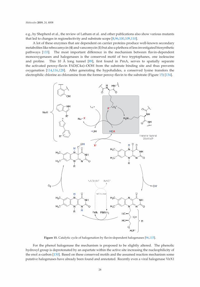
Molecules 2019, 24, 4008
e.g., by Shepherd et al., the review of Latham et al. and other publications also show various mutantsthat led to changes in regioselectivity and substrate scope [8,96,100,109,110].
A lot of these enzymes that are dependent on carrier proteins produce well-known secondarymetabolites like rebeccamycin (4) and vancomycin (1) but also a plethora of less investigated biosyntheticpathways [133]. The most important difference in the mechanism between flavin-dependentmonooxygenases and halogenases is the conserved motif of two tryptophanes, one isoleucineand proline. This 10 Å long tunnel [89], first found in PrnA, serves to spatially separatethe activated peroxy-flavin FAD(C4α)–OOH from the substrate binding site and thus preventsoxygenation [114,116,128]. After generating the hypohalides, a conserved lysine transfers theelectrophilic chlorine as chloramine from the former peroxy-flavin to the substrate (Figure 15) [134].
Figure 15. Catalytic cycle of halogenation by flavin-dependent halogenases [96,115].
For the phenol halogenase the mechanism is proposed to be slightly altered. The phenolichydroxyl group is deprotonated by an aspartate within the active site increasing the nucleophilicity ofthe enol α-carbon [130]. Based on these conserved motifs and the assumed reaction mechanism someputative halogenases have already been found and annotated. Recently even a viral halogenase VirX1
28

Molecules 2019, 24, 4008
from cyanophages was discovered, which is the first FHal capable of in vitro iodination and stands outdue to its broad substrate spectrum and preferred iodination [135].
Although the community has so far agreed that the preserved motif of separation tunnel and anchorlysine seems to be essential for the activity, current research shows a further class of flavin-dependenthalogenases which lack these structural elements completely. One of these examples is the halogenaseKerK that is under investigation by Piel and coworkers but has not yet been published except as aposter presentation on Biotrans 2019 in Groningen, the Netherlands [136].
In addition to the advantages of high regioselectivity and thus only few by-products, there are alsosome disadvantages in the use of this enzyme group. The low conversion rates speak against large-scaleapplication and expression problems often occur. Many of the proteins produced in E. coli BL21(DE3)end up in the insoluble fraction as inclusion bodies. To deal with this issue, strains with co-expressionof chaperones are used regularly (Table 3). The overall stability of these proteins also needs furtheroptimization to be applicable in biocatalysis. As a promising result Kemker et al. the tryptophanhalogenases were successfully scaled up in terms of a biocatalytic process employing immobilizing theenzymes by cross-linked enzyme aggregates (CLEAs). This yielded l-7-bromotryptophan on the gramscale [137].
Table 3. Examples of flavin-dependent tryptophan, pyrrole and phenol halogenases that can becarrier-dependent or independent.
Enzyme OriginHeterologous Expression
Host 1 Product Miscellaneous
PrnA[96]
Pseudomonasfluorescens BL915 E. coli ArcticExpress (DE3)
RebH[92]
Lechevalieriaaerocolonigenes
strain 39243E. coli BL21(DE3)
KtzQ[110] Kutzneria sp. 744
KtzR[110] Kutzneria sp. 744
CmdE[111]
Chondromycescrocatus Cm c5
Post-NRPS(non-ribosomal
peptides)
SSTH[101]
Streptomycestoxytricini NRRL
15443
E. coli BL21 CodonPlus(DE3)-RIL
Thal[112]
Streptomycesalbogriseolus
P. fluorescensBL915 DORF1 and P.
chlororaphis ACN
29

Molecules 2019, 24, 4008
Table 3. Cont.
Enzyme OriginHeterologous Expression
Host 1 Product Miscellaneous
MibH[99,113]
Microbisporacoralline NRRL
30420NRPS-dependent
PyrH[96,114]
StreptomycesrugosporusLL-42D005
E. coli ArcticExpress (DE3);Pseudomonas fluorescens
BL915 ΔORF1
Xcc-BB100XXXX
[115]
Xanthomonascampestris pv.
campestris strainB100
E. coli BL21(DE3) with pGro7plasmid
(Takara) for chaperoneco-expression
Various substitutedindoles and thereby
differingregio-selectivity
BrvH[116]
BrevundimonasBAL3
E. coli BL21(DE3) with pGro7plasmid
(Takara) for chaperoneco-expression
PrnC[93,117]
Pseudomonasfluorescens BL915
Chl[118]
Streptomycesaureofaciens
ChlA[105]
Dictyosteliumdiscoideum
PltA[119]
Pseudomonasfluorescens Pf-5 E. coli BL21(DE3)
Pyr29[120]
Actinosporangiumvitaminophilum
ATCC 31673 andStreptomyces sp.Strain UC 11065
Arm21[121]
Streptomycesarmeniacus
CrpH[104] Nostoc Cyanobionts NRPS-dependent
BhaA[122,123]
AmycolatopsismediterraneiDSM5908
balhimycin
SgcC3[124]
Streptomycesglobisporus
E. coli BL21(DE3)pET-30Xa/LIC
30

Molecules 2019, 24, 4008
Table 3. Cont.
Enzyme OriginHeterologous Expression
Host 1 Product Miscellaneous
HalB[125]
Actinoplanes sp.ATCC 33002
Pseudomonas aureofaciensACN
PltM[126]
Pseudomonasfluorescens Pf-5 E. coli BL21 (DE3)
Bmp5[106]
Pseudoalteromonasluteoviolacea
Bmp2[127]
Pseudoalteromonasluteoviolacea NRPS-dependent
(PltD)[13]
Pseudomonasfluorescens Pf-5 n.a. Not clear if FHal
CmlS[128]
Streptomycesvenezuelae
Flavin covalentlybound to aspartate
via CH3-Group
CndH[129]
Chondromycescrocatus NRPS-dependent
RadH[107,130]
Chaetomiumchiversii E. coli Rosetta 2(DE3) monocillin II
Rdc2[107,131]
Pochoniachlamydosporia
S. cerevisiaestrain BJ5464-NpgE. coli BL21(DE3)
monocillin II
TiaM[132]
Dactylosporangiumaurantiacum NRRL
18085E. coli BL21(DE3) tiacumicin B
intermediate NRPS-dependent
1 if not stated otherwise, the expression took place in the origin host.
5.2. α-Ketoglutarate-Dependent Halogenases
Table 4 shows different natural products that are formed by the iron(II)-α-ketoglutarate-dependent(Fe/αKG)-halogenases. Despite the huge variety in the product structures they share one commonfeature, which is the halogen at a sp3-carbon centre. Hence, the Fe/αKG-halogenase is notlimited to nucleophilic substrates like the previous described enzymes. They belong to theFe/αKG-dependent oxygenase superfamily. The superfamily is known for different transformationssuch as hydroxylation [138], halogenation [139], desaturation [140], or can be used for the productionof ethylene [141]. They all share a structurally conserved metal-binding motif, which in the case of thehalogenase developed an active centre that is eventually able to bind a haloge n [139]. The proposedcatalytic mechanism of Fe(II)/α-KG-dependent-halogenase is illustrated in Figure 16.
31

Molecules 2019, 24, 4008
Table 4. Examples of heterologously expressed Fe(II)/αKG-dependent halogenases.
EnzymeOrigin/Expression Host
and YieldFeatures Product/Biosynthesis
SyrB2[139]
Pseudomonas syringae pv.syringae B301D/
E. coli strain B834(DE3)[139]n.a.
total turnover: 7 ± 2[144]
4-chloro-l-threonine/syringomycin E
CytC3[145]
Streptomyces sp./E. coli BL21(DE3)
[146]n.a.
n.a
4,4-dichloro-l-valine/dichloroaminobutyrate
WelO5[147]
Hapalosiphon welwitschii/E. coli C43(DE3)
[148,149] orBL21(DE3) [150]20 mg L−1 [151]
total turnover:75 [148]
kcat: 1.8 ± 0.2 min−1
[149]
12-epi-fischerindole G/fischerindole &
hapalindole alkaloids
AmbO5
Fischerella ambigua/E. coli C43(DE3) [149] or
BL21(DE3) [150]n.a.
kcat: 1.7 ± 0.1 min−1 [149]
ambiguine A/ambiguine, fischerindole
and hapalindolealkaloids
WelO5* variantisoform of WelO5
(CB2)[152]
Hapalosiphon welwitschiiIC-52-3/
E. coli BL21(DE3)n.a.
KM: 0.67 mM,kcat: 3.0 min−1
martinelline-derivedfragment
Based on the proposed radical C-H functionalization two classes of enzymes have so far beenidentified. The first such reported Fe/αKG-dependent halogenase is the tailoring domain SyrB2 ofthe multimodular nonribosomal peptidsynthetase (NRPS) from Pseudomonas syringae pv. syringaeB301D [133,144]. These NRPS-associated halogenases produce a diversity of secondary metabolitessuch as the chlorinated biosurfactant syringomycin E (31), which is characterized by a selectivemonochlorinated threonine in its structure [128,139]. Another example is the highly selective di-and trichlorination of solely one of the diastereotopic methyl groups of leucine by a combination ofBarB1 and BarB2, which serves as a precursor for the natural compound barbamid (32) in the marinecyanobacteria Lyngbya majuscula [142,153].
32

Molecules 2019, 24, 4008
Figure 16. Proposed mechanism for halogenation reaction by Fe(II)/αKG-dependent halogenase viaa radical C-H functionalization [142]. The highly reactive Fe(IV)-oxo (haloferryl) intermediate isproduced by decarboxylation of αKG to succinate through an oxygen attack. Subsequently abstractionof a hydrogen-atom from the substrate leads to an energetically favourable rearrangement towardsFe(III). Rebound reaction with chloride was shown to depend on the distance and orientation of thesubstrate [143]. The catalytically cycle is re-established by the hexa-coordinated Fe(II) with watermolecules, chloride and histidine.
Recently Moosmann et al. identified different αKG-halogenase homologues and their naturalproducts that are produced via a NRPS (non-ribosomal peptide synthetase) pathway. The halogenasewere identified by screening the genomic sequence of the cyanobacterium Fischerella sp. PCC 9339based on feature comparison. Using this approach, the authors were able to distinguish between aFe/αKG oxygenase and a corresponding halogenase [143,154]. However, large NRPSs characteristicallybind their substrates through an aminoacetylated peptidyl-carrier protein and have a narrow substratescope [144,155]. Furthermore, they generally showed a low total turnover number, which mayresult from the well-known autoxidation of Fe(II) to Fe(III) and hence an auto-inactivation of theenzyme [145,156]. In case of SyrB2, total turnovers of 7 ± 2 were observed [133,144] This limits thepossibility to modify the enzymes in order to use them as suitable biocatalysts for different unnatural
33

Molecules 2019, 24, 4008
substrates. With the discovery of a new Fe(II)/αKG-dependent halogenase (WelO5) by Hillwig andLiu, it was possible to expand the class towards unbound substrates. WelO5 is capable of late stagehalogenation in a regio- and stereoselective manner of different derived isoprenoid-indole alkaloidsin the cyanobacterium Hapalosiphon welwitschii (see Table 4) [137,148]. WelO5 showed also a higherrobustness and catalysis of approximately 75 turnovers in total [137,148]. Strategies such as adding thecosubstrates consecutively or adding antioxidants like catalases or DTT could increase the turnovernumber. The narrow substrate scope of WelO5 was tailored in order to have an increased substrate scopelike the homolog AmbO5 [138,149]. Most modifications were at the external helix, which is responsiblefor closing the entry of the active site upon binding of the substrate. It can be assumed that the helixis partially involved in the substrate recognition and specificity [138,149]. A recent publication fromHayashi et al. showed a WelO5 variant with a reshaped active site that led to improved kinetics and anexpanded substrate scope, which applies beyond the native indole alkaloid-type substrates [141,152].This provides the possibility for targeted enzyme-engineering and a basis for further improvementsin substrate scope. One possibility is the establishment of nitration and azidation as already shownfor SyrB2 [157]. In this regard, it has been shown that WelO5 is able to incorporate the unnaturalhalide Br− [158]. One drawback of engineering Fe/αKG-dependent halogenases is the hydroxylationas a competitive side reaction [155]. Mitchell et al. used this approach backwards and modifieda monooxygenase SadA towards a halogenase [159]. This serves as a proof of concept that withincreasing understanding of the reaction mechanism and the involved amino acids the superfamilyof monooxygenase can be used as a versatile toolbox in biotechnology. In the future, this maylead to the use of different variants of the very same enzyme for different transformations. Table 4shows an overview of different characterized Fe/αKG halogenases and their main published features.Excluded are, for example, halogenase modules of NRPS, where the halogenation is necessary for thesubsequently formation of cyclopropane such as in case of CurA [160] or CmaB (see Figure 17) [161].
Figure 17. Schematically example for formation of cyclopropane initiated by CmaB through halogenation.
5.3. Fluorinase
In contrast to the other described enzymes, the diversity of natural products in case ofthe fluorinase stem from only one characterized enzyme class to date. The involved enzymesare S-adenosylmethionine (SAM) dependent. The first characterized representative was FlA(5′-fluoro-5′-deoxyadenosine synthase) from [162]. The overall family of this enzymes is alsoable to chlorinate or hydroxylate SAM, as described in detail elsewhere [163]. Within the catalytic cyclefluoride acts as a nucleophile in a SN2-reaction, where it attacks the 5′-carbon of SAM-ribose [164].In order to act as a nucleophile, fluoride requires to lose its solvation shell. This is achieved in atwo-step desolvation with a combination of electrostatic stabilization and hydrogen bonding. In thefirst step, fluoride is binding to the active site and exchanges water molecules of its shell in orderto form hydrogen bonds with the enzyme. Upon binding of SAM the desolvation of fluoride iscomplete. The electropositive 5′-carbon attached to the sulfur group in SAM coordinates with thefluoride [165,166]. This electrostatic stabilization facilitates the nucleophilic attack of the fluorideand C−F-bond formation of the reactive 5′-fluoro-5′deoxyadenosine (33, 5′-FDA) intermediate [166].Subsequently, 5′-FDA (33) is further metabolized in order to generate a variety of compounds as shownin Figure 18A [162]. However, this also represents a major obstacle for the application of these enzymesto unnatural small organic molecules, since the product formation follows a cascade of enzymatic steps.Eustáquio et al. tried to use this enzyme for the production of fluorosalinosporamide, an unnatural
34

Molecules 2019, 24, 4008
analog of salinosporamide, which is fluorinated rather than chlorinated, however, the yield wasmoderate [167]. Nevertheless, different approaches have been implemented to increase the substratescope and use the enzyme as rather flexible tool for medical applications. Besides the ability to fluoridecompounds, fluorinase is also able to exchange a chloride at the 5′-carbon of the SAM ribose ringby a fluoride and form 5′-FDA (33, Figure 18B) [168]. This overall trans-halogenation reaction wasused for late-stage fluorination for the production of radiolabeled imaging reagents. Recently, differentpre-targeting strategies have been developed for treatment and imaging of different diseases. Thoseinclude e.g., radiolabeling of the human A2A adenosine receptor [169], a prostate cancer-related membraneprotein [170] or the combined application of biotin and tetrazine-conjugate with antibodies [171]. In allcases, it was shown that the fluorinase (FlA) accepts substrates with different moiety at C-2 of the adeninering. So far two crystal structures of fluorinase homologs from S. cattleya and Streptomyces sp. MA37 areknown and they both have a high structurally conformity [172]. In general all five known fluorinaseshave a high similarity of over 80% and show similar kinetic profiles [173]. Through a directed evolutionapproach of FlA1, different crucial amino acids for substrate binding, halide binding and hence activitywere identified [174]. Additionally, the variants were tested with different unnatural substrates [175].It was shown that the tolerance for the wild type (wt) enzyme is limited to C-2 modified substrates.However, generated variants of FlA1 also demonstrated an activity for an unnatural substrate, which weremodified at C-6 positions of the adenine ring with a chlorine group [175]. These findings show thatdespite a narrow substrate scope of the fluorinase, it was possible to successfully apply different unnaturalsubstrates and lay a base for directed evolution as means to use small organic compounds as substrates.However, the dependence of electrophilic substrate structures remains a drawback for the nucleophilicattack of fluoride. The crystal structure with an unnatural substrate (containing difluoromethyl groups)confirmed the necessity of geometry for activating the fluorine atom for substitution [176]. This outlinesthe challenge to use fluorinases as a versatile tool to generate fluorinated pharmaceutical compounds.Nevertheless, by means of designing appropriate leaving groups in combination with enzyme engineering,fluorinases could be used as tool for future generation of fluorinated pharmaceutical compounds. Data ofknown fluorinases are displayed in Table 5.
Table 5. Examples for heterologously expressed fluorinase. Kinetic data representing the conversion of5′-ClDA into S-adenosylmethionine (SAM).
EnzymeOrigin/Expression Host and
YieldKinetic Parameters
Special SubstrateScope
FlA[165]
Streptomyces cattleya/E. coli BL21(DE3)
50 mg L−1
[173]
KM: 29.4 ± 5.80 μm
kcat: 0.084 ± 0.005 min−1
[173][169–171]
FlA1Streptomyces sp. MA37/
E. coli BL21(DE3)n.a.
KM: 8.36 ± 0.82 μm
kcat: 0.13 min−1
[174]
FlA4Streptomyces xinghaiensis/
E. coli BL21(DE3)n.a.
KM: 29.87 μm
kcat: 0.69 ± 0.01 min−1
[177][177]
35

Molecules 2019, 24, 4008
Figure 18. (A) Schematically sequential mechanism for the F–C bond formation catalysed by thefluorinase from S. cattleya and some products of subsequently cascade reaction. Dashed lines representshydrogen bonding contacts with amino acids in the active pocket or water. (B) Reaction scheme offluorinase-mediated trans-halogenation. Rest R marks position of usual derivation.
6. Conclusion on Halogens in Active Agent (Syntheses)
As we have seen in the previous paragraphs halogens are very important to many active agents asa functional moiety per se due to their physico-chemical properties such as bulkiness, latent polarizationand as important binding partners because of halogen bonds. Organic halogen compounds are,furthermore, instrumental for synthetic purposes in terms of being good leaving groups and facilitatingcross-metatheses by halogen-metal-exchanges. Nevertheless, these indispensable advantages have tobe bought at a high price; namely the energy-intensive production of very toxic and hazardous chlorinegas. The reduction in energy consumption must mainly be managed by technical improvementsof the chloralkali-process and enzymes are likely not able to make a significant impact. A majorreason is that the majority of chlorinated compounds are necessary for different types of plasticmaterials (e.g., PVC) and solvents [51]. Enzymes are limitedly applicable in those areas of bulkchemicals, but there is a potential for fine chemicals. Even though halogenating enzymes will notreplace conventional chlorine production, it is worth taking a look at this group of enzymes or ratherat these groups of enzymes, because nature has invented these amazing enzymes at least six times.The expectations of these biocatalysts are that the conversions become environmentally more benign,the processes skip hazardous compounds such as chlorine gas and that conversions get more selective.However, the research in the field of halogenating enzymes is still at the beginning. Consistentenzymologic data such as kinetic data, measurements on stability or even well studied mutant librariesare rarely available. Many halogenating enzymes from eukaryotic sources suffer from expression
36

Molecules 2019, 24, 4008
challenges. Nevertheless, these enzymes open up a wide horizon of possibilities. Enormous genomedata are revealing more and more halogenating enzymes and even new classes of halogenatingenzymes cannot be excluded at present. Thus, there is a need for detailed and systematic research toemploy halogenating enzymes for active agent synthesis, to alter their substrate scopes and enhancetheir process stability.
Author Contributions: T.C. conceived this review and relegated the manuscript to a complete work. A.V.F., J.G.,and N.H. carried out the initial research and compiled the various chapters. All authors contributed equally tothis review.
Funding: This research was funded by Deutsche Forschungsgemeinschaft grant number 369034981,BioSC(CombiCom) Ministry of Culture and Science North-Rhine-Wesfalia grant number 313/323-400-00213,and European Regional Development Fund: CLIB-Kompetenzzentrum Biotechnologie.
Acknowledgments: For their fruitful discussions we want to thank Jörg Pietruszka, Nora Bitzenhofer, EstherBreuninger, and Denise Detlof as well as the entire institute staff for their support. We want to thank oursponsors for the financial support. AF was funded by the European Regional Development Fund (ERDF) withinthe ‘CLIB-Kompetenzzentrum Biotechnologie’. The scientific activities (JG) of the Bioeconomy Science Center werefinancially supported by the Ministry of Culture and Science within the framework of the NRW Strategieprojekt BioSC(No. 313/323-400-00213). NH is funded by the Deutsche Forschungsgemeinschaft (369034981). Furthermore, we wantto thank the Forschungszentrum Jülich GmbH and the Heinrich Heine University Düsseldorf for their generous support.
Conflicts of Interest: The authors declare no conflict of interest.
Abbreviations
αKG α-ketoglutarateBPO bromoperoxidaseCLEA cross-linked enzyme aggregateCPO chloroperoxidaseFDA 5′-fluoro-5′deoxyadenosineFhal, FDH flavin-dependent halogenaseFlA fluorinase Ag gasousHal halogenHOX hypohalous acidHPO haloperoxidaseMCD monochlorodimedoneMn elemental metalMOH metal cation hydroxid saltsNBS N-bromo-succinimdeNCS N-chloro-succinimideNRPS non-ribosomal protein synthesisNXS N-halogen-succinimidePKS polyketidePVC polyvinyl chlroides solidSAM S-adenosyl methionineX halide ionX2 molecular halogenY non-halogen heteroatom
References
1. Gribble, G.W. Natural Organohalogens: A New Frontier for Medicinal Agents? J. Chem. Educ. 2004, 81, 1441.[CrossRef]
2. Chast, F. A history of drug discovery: From first steps of chemistry to achievements in molecular pharmacology.In The Practice of Medicinal Chemistry; Elsevier: Amsterdam, The Netherlands, 2008; pp. 1–62.
37

Molecules 2019, 24, 4008
3. Hernandes, M.Z.; Cavalcanti, S.M.T.; Moreira, D.R.M.; de Azevedo Junior, W.F.; Leite, A.C.L. Halogen Atomsin the Modern Medicinal Chemistry: Hints for the Drug Design. Curr. Drug Targets 2010, 11, 303–314.[CrossRef] [PubMed]
4. Jiang, S.; Zhang, L.; Cui, D.; Yao, Z.; Gao, B.; Lin, J.; Wei, D. The Important Role of Halogen Bond in SubstrateSelectivity of Enzymatic Catalysis. Sci. Rep. 2016, 6, 34750. [CrossRef] [PubMed]
5. Agarwal, V.; Miles, Z.D.; Winter, J.M.; Eustáquio, A.S.; EL Gamal, A.A.; Moore, B.S. Enzymatic Halogenationand Dehalogenation Reactions: Pervasive and Mechanistically Diverse. Chem. Rev. 2017, 117, 5619–5674.[CrossRef]
6. Blasiak, L.C.; Drennan, C.L. Structural perspective on enzymatic halogenation. Acc. Chem. Res.2008, 42, 147–155. [CrossRef]
7. Gribble, G.W. The diversity of naturally produced organohalogens. Chemosphere 2003, 52, 289–297. [CrossRef]8. Latham, J.; Brandenburger, E.; Shepherd, S.A.; Menon, B.R.K.; Micklefield, J. Development of Halogenase
Enzymes for Use in Synthesis. Chem. Rev. 2018, 118, 232–269. [CrossRef]9. Schnepel, C.; Sewald, N. Enzymatic Halogenation: A Timely Strategy for Regioselective C−H Activation.
Chem. Eur. J. 2017, 23, 12064–12086. [CrossRef]10. Senn, H.M. Insights into enzymatic halogenation from computational studies. Front. Chem. 2014, 2, 98.
[CrossRef]11. Smith, D.R.; Grüschow, S.; Goss, R.J. Scope and potential of halogenases in biosynthetic applications.
Curr. Opin. Chem. Biol. 2013, 17, 276–283. [CrossRef] [PubMed]12. Vaillancourt, F.H.; Yeh, E.; Vosburg, D.A.; Garneau-Tsodikova, S.; Walsh, C.T. Nature’s inventory of
halogenation catalysts: Oxidative strategies predominate. Chem. Rev. 2006, 106, 3364–3378. [CrossRef][PubMed]
13. Weichold, V.; Milbredt, D.; van Pée, K.-H. Specific Enzymatic Halogenation—From the Discovery ofHalogenated Enzymes to Their Applications In Vitro and In Vivo. Angew. Chem. Int. Ed. 2016, 55, 6374–6389.[CrossRef] [PubMed]
14. Zeng, J.; Zhan, J. Chlorinated Natural Products and Related Halogenases. Isr. J. Chem. 2019, 59, 1–17.[CrossRef]
15. Lu, Y.; Wang, Y.; Zhu, W. Nonbonding interactions of organic halogens in biological systems: Implications fordrug discovery and biomolecular design. Phys. Chem. Chem. Phys. 2010, 12, 4543–4551. [CrossRef] [PubMed]
16. Xu, Z.; Yang, Z.; Liu, Y.; Lu, Y.; Chen, K.; Zhu, W. Halogen bond: Its role beyond drug–target binding affinityfor drug discovery and development. J. Chem. Inf. Model. 2014, 54, 69–78. [CrossRef] [PubMed]
17. Sirimulla, S.; Bailey, J.B.; Vegesna, R.; Narayan, M. Halogen interactions in protein–ligand complexes:Implications of halogen bonding for rational drug design. J. Chem. Inf. Model. 2013, 53, 2781–2791. [CrossRef][PubMed]
18. Lu, Y.; Liu, Y.; Xu, Z.; Li, H.; Liu, H.; Zhu, W. Halogen bonding for rational drug design and new drugdiscovery. Expert Opin. Drug Discov. 2012, 7, 375–383. [CrossRef] [PubMed]
19. Xu, Z.; Liu, Z.; Chen, T.; Chen, T.; Wang, Z.; Tian, G.; Shi, J.; Wang, X.; Lu, Y.; Yan, X.; et al. Utilization ofhalogen bond in lead optimization: A case study of rational design of potent phosphodiesterase type 5(PDE5) inhibitors. J. Med. Chem. 2011, 54, 5607–5611. [CrossRef]
20. Harris, C.M.; Kannan, R.; Kopecka, H.; Harris, T.M. The role of the chlorine substituents in the antibioticvancomycin: Preparation and characterization of mono-and didechlorovancomycin. J. Am. Chem. Soc.1985, 107, 6652–6658. [CrossRef]
21. Schlosser, M.; Michel, D. About the “physiological size” of fluorine substituents: Comparison of sensoriallyactive compounds with fluorine and methyl substituted analogues. Tetrahedron 1996, 52, 99–108. [CrossRef]
22. Lima, L.M.; Barreiro, E.J. Bioisosterism: A useful strategy for molecular modification and drug design.Curr. Med. Chem. 2005, 12, 23–49. [CrossRef]
23. Greenwood, N.N.; Earnshaw, A. Chemistry of the Elements; Elsevier: Amsterdam, The Netherlands, 2012.24. Thiehoff, C.; Rey, Y.P.; Gilmour, R. The Fluorine Gauche Effect: A Brief History. Isr. J. Chem. 2017, 57, 92–100.
[CrossRef]25. Berkowitz, D.B.; Karukurichi, K.R.; de la Salud-Bea, R.; Nelson, D.L.; McCune, C.D. Use of fluorinated
functionality in enzyme inhibitor development: Mechanistic and analytical advantages. J. Fluor. Chem.2008, 129, 731–742. [CrossRef]
38

Molecules 2019, 24, 4008
26. Ramalingam, S.S.; Blackhall, F.; Krzakowski, M.; Barrios, C.H.; Park, K.; Bover, I.; Heo, D.S.; Rosell, R.;Talbot, D.C.; Frank, R. Randomized phase II study of dacomitinib (PF-00299804), an irreversible pan–humanepidermal growth factor receptor inhibitor, versus erlotinib in patients with advanced non–small-cell lungcancer. J. Clin. Oncol. 2012, 30, 3337. [CrossRef]
27. Bush, J.A.; Long, B.H.; Catino, J.J.; Bradner, W.T.; Tomita, K. Production and biological activity of rebeccamycin,a novel antitumor agent. J. Antibiot. 1987, 40, 668–678. [CrossRef]
28. Lake, R.J.; Brennan, M.M.; Blunt, J.W.; Munro, M.H.; Pannell, L.K. Eudistomin K sulfoxide-an antiviralsulfoxide from the New Zealand ascidian Ritterella sigillinoides. Tetrahedron Lett. 1988, 29, 2255–2256.[CrossRef]
29. Braghirolli, A.M.S.; Waissmann, W.; da Silva, J.B.; dos Santos, G.R. Production of iodine-124 and itsapplications in nuclear medicine. Appl. Radiat. Isot. 2014, 90, 138–148. [CrossRef]
30. Crawford, C.C.; Evans, T.W. Production of Hypohalous Acid Solutions. U.S. Patent 2347151, 18 April 1944.31. Duncan, B.L.; Ness, R.C. Process for the Production of Highly Pure Concentrated Slurries of Sodium
Hypochlorite. U.S. Patent 5194238A, 16 March 1993.32. Howarth, J.N.; Dadgar, A.; Sergent, R.H. Recovery of Bromine and Preparation of Hypobromous acid from
Bromide Solution. U.S. Patent 5385650A, 31 January 1995.33. Kesner, M. Bromine and Bromine Compounds from the Dead Sea, Israel Products in the Service of People;
The Weizmann Institute of Science, Israel: Rehovot, Israel, 1999.34. Groweiss, A. Use of Sodium Bromate for Aromatic Bromination: Research and Development. Org. Process
Res. Dev. 2000, 4, 30–33. [CrossRef]35. Nishi, Z.; Kudoh, K.; Okamoto, N. Method of Forming Hypobromous Acid in Aqueous System. U.S. PATENT
7785559B2, 31 August 2010.36. Ziegler, K.; Späth, A.; Schaaf, E.; Schumann, W.; Winkelmann, E. Die halogenierung ungesättigter Substanzen
in der Allylstellung. Justus Liebigs Ann. Chem. 1942, 551, 80–119. [CrossRef]37. Torborg, C.; Beller, M. Recent applications of palladium-catalyzed coupling reactions in the pharmaceutical,
agrochemical, and fine chemical industries. Adv. Synth. Catal. 2009, 351, 3027–3043. [CrossRef]38. Beletskaya, I.P.; Cheprakov, A.V. Copper in cross-coupling reactions: The post-Ullmann chemistry.
Coord. Chem. Rev. 2004, 248, 2337–2364. [CrossRef]39. Xi, Z.; Liu, B.; Chen, W. Room-temperature Kumada cross-coupling of unactivated aryl chlorides catalyzed
by N-heterocylic carbene-based nickel (II) complexes. J. Org. Chem. 2008, 73, 3954–3957. [CrossRef]40. Gomes, F.; Echeverria, P.G.; Fürstner, A. Iron-or Palladium-Catalyzed Reaction Cascades Merging
Cycloisomerization and Cross-Coupling Chemistry. Chem. Eur. J. 2018, 24, 16814–16822. [CrossRef]41. Piontek, A.; Bisz, E.; Szostak, M. Iron-Catalyzed Cross-Couplings in the Synthesis of Pharmaceuticals:
In Pursuit of Sustainability. Angew. Chem. Int. Ed. 2018, 57, 11116–11128. [CrossRef]42. Cahiez, G.R.; Lefèvre, G.; Moyeux, A.; Guerret, O.; Gayon, E.; Guillonneau, L.; Lefèvre, N.; Gu, Q.; Zhou, E.
Gram-Scale, Cheap, and Eco-Friendly Iron-Catalyzed Cross-Coupling between Alkyl Grignard Reagentsand Alkenyl or Aryl Halides. Org. Lett. 2019, 21, 2679–2683. [CrossRef]
43. Cooke, J.W.; Bright, R.; Coleman, M.J.; Jenkins, K.P. Process research and development of a dihydropyrimidinedehydrogenase inactivator: Large-scale preparation of eniluracil using a Sonogashira coupling. Org. ProcessRes. Dev. 2001, 5, 383–386. [CrossRef]
44. Wallace, M.D.; McGuire, M.A.; Yu, M.S.; Goldfinger, L.; Liu, L.; Dai, W.; Shilcrat, S. Multi-kiloscaleenantioselective synthesis of a vitronectin receptor antagonist. Org. Process Res. Dev. 2004, 8, 738–743.[CrossRef]
45. Benkeser, R.A.; Siklosi, M.P.; Mozdzen, E.C. Reversible Grignard and organolithium reactions. J. Am. Chem.Soc. 1978, 100, 2134–2139. [CrossRef]
46. Milstein, D.; Stille, J.K. A general, selective, and facile method for ketone synthesis from acid chlorides andorganotin compounds catalyzed by palladium. J. Am. Chem. Soc. 1978, 100, 3636–3638. [CrossRef]
47. King, A.O.; Okukado, N.; Negishi, E.-I. Highly general stereo-, regio-, and chemo-selective synthesis ofterminal and internal conjugated enynes by the Pd-catalysed reaction of alkynylzinc reagents with alkenylhalides. Chem. Commun. 1977, 683–684. [CrossRef]
48. Corriu, R.; Masse, J. Activation of Grignard reagents by transition-metal complexes. A new and simplesynthesis of trans-stilbenes and polyphenyls. Chem. Commun. 1972, 144. [CrossRef]
39
Molecules 2019, 24, 4008
49. Global Chlorine Market Set for Rapid Growth to Reach around USD 38.4 Billion in 2021. Available online:https://www.zionmarketresearch.com/news/global-chlorine-market (accessed on 23 August 2019).
50. Ausfelder, F. Flexibilitätsoptionen in der Grundstoffindustrie: Methodik, Potenziale, Hemmnisse: Bericht des AP V. 6“Flexibilitätsoptionen und Perspektiven in der Grundstoffindustrie ”im Kopernikus-Projekt “SynErgie-Synchronisierteund Energieadaptive Produktionstechnik zur Flexiblen Ausrichtung von Industrieprozessen Auf Eine FluktuierendeEnergieversorgung”; DECHEMA Gesellschaft für Chemische Technik und Biotechnologie eV: Frankfurt,Germany, 2018.
51. Chlor-Alkali Industry Review 2017/2018. Available online: https://www.eurochlor.org/wp-content/uploads/2019/05/euro_chlor_industry_review_FINAL.pdf (accessed on 30 October 2019).
52. The Electrolysis Process and the Real Costs of Production. Available online: https://www.eurochlor.org/wp-content/uploads/2019/04/12-electrolysis_production_costs.pdf (accessed on 23 August 2019).
53. Morris, D.R.; Hager, L.P.; Chloroperoxidase, I. Isolation and properties of the crystalline glycoprotein. J. Biol.Chem. 1966, 241, 1763–1768. [PubMed]
54. Kuhnel, K.; Blankenfeldt, W.; Terner, J.; Schlichting, I. Crystal structures of chloroperoxidase with its boundsubstrates and complexed with formate, acetate, and nitrate. J. Biol. Chem. 2006, 281, 23990–23998. [CrossRef][PubMed]
55. Butler, A.; Sandy, M. Mechanistic considerations of halogenating enzymes. Nature 2009, 460, 848. [CrossRef][PubMed]
56. Buchhaupt, M.; Huttmann, S.; Sachs, C.C.; Bormann, S.; Hannappel, A.; Schrader, J. Caldariomyces fumagoDSM1256 Contains Two Chloroperoxidase Genes, Both Encoding Secreted and Active Enzymes. J. Mol.Microbiol. Biotechnol. 2015, 25, 237–243. [CrossRef] [PubMed]
57. Reddy, C.M.; Xu, L.; Drenzek, N.J.; Sturchio, N.C.; Heraty, L.J.; Kimblin, C.; Butler, A. A chlorine isotopeeffect for enzyme-catalyzed chlorination. J. Am. Chem. Soc. 2002, 124, 14526–14527. [CrossRef]
58. Pickard, M.A. A defined growth medium for the production of chloroperoxidase by Caldariomyces fumago.Can. J. Microbiol. 1981, 27, 1298–1305. [CrossRef]
59. Getrey, L.; Krieg, T.; Hollmann, F.; Schrader, J.; Holtmann, D. Enzymatic halogenation of the phenolicmonoterpenes thymol and carvacrol with chloroperoxidase. Green Chem. 2014, 16, 1104–1108. [CrossRef]
60. Naapuri, J.; Rolfes, J.D.; Keil, J.; Manzuna Sapu, C.; Deska, J. Enzymatic halocyclization of allenic alcoholsand carboxylates: A biocatalytic entry to functionalized O-heterocycles. Green Chem. 2017, 19, 447–452.[CrossRef]
61. Blanke, S.R.; Yi, S.; Hager, L.P. Development of Semi-Continuous and Continuous-Flow Bioreactors for theHigh-Level Production of Chloroperoxidase. Biotechnol. Lett. 1989, 11, 769–774. [CrossRef]
62. Yamada, H.; Itoh, N.; Izumi, Y. Chloroperoxidase-catalyzed halogenation of trans-cinnamic acid and itsderivatives. J. Biol. Chem. 1985, 260, 11962–11969. [PubMed]
63. Allain, E.J.; Hager, L.P.; Deng, L.; Jacobsen, E.N. Highly Enantioselective Epoxidation of DisubstitutedAlkenes with Hydrogen-Peroxide Catalyzed by Chloroperoxidase. J. Am. Chem. Soc. 1993, 115, 4415–4416.[CrossRef]
64. Manthey, J.A.; Hager, L.P. Purification and properties of bromoperoxidase from Penicillus capitatus. J. Biol.Chem. 1981, 256, 11232–11238. [PubMed]
65. van Pée, K.H.; Lingens, F. Purification of bromoperoxidase from Pseudomonas aureofaciens. J. Bacteriol.1985, 161, 1171–1175.
66. Baden, D.G.; Corbett, M.D. Bromoperoxidases from Penicillus capitatus, Penicillus lamourouxii andRhipocephalus phoenix. Biochem. J. 1980, 187, 205–211. [CrossRef]
67. Kaup, B.-A.; Ehrich, K.; Pescheck, M.; Schrader, J. Microparticle-enhanced cultivation of filamentousmicroorganisms: Increased chloroperoxidase formation by Caldariomyces fumago as an example. Biotechnol.Bioeng. 2008, 99, 491–498. [CrossRef]
68. Conesa, A.; van de Velde, F.; van Rantwijk, F.; Sheldon, R.A.; van den Hondel, C.A.M.J.J.; Punt, P.J. Expressionof the Caldariomyces fumago chloroperoxidase in Aspergillus niger and characterization of the recombinantenzyme. J. Biol. Chem. 2001, 276, 17635–17640. [CrossRef]
69. Coughlin, P.; Roberts, S.; Rush, C.; Willetts, A. Biotransformation of Alkenes by Haloperoxidases -Regiospecific Bromohydrin Formation from Cinnamyl Substrates. Biotechnol. Lett. 1993, 15, 907–912.[CrossRef]
40

Molecules 2019, 24, 4008
70. van Schijndel, J.W.; Vollenbroek, E.G.; Wever, R. The chloroperoxidase from the fungus Curvularia inaequalis;a novel vanadium enzyme. Biochim. Biophys. Acta 1993, 1161, 249–256. [CrossRef]
71. Van Schijndel, J.W.P.M.; Barnett, P.; Roelse, J.; Vollenbroek, E.G.M.; Wever, R. The stability andsteady-state kinetics of vanadium chloroperoxidase from the fungus Curvularia inaequalis. Eur. J. Biochem.1994, 225, 151–157. [CrossRef]
72. Rush, C.; Willetts, A.; Davies, G.; Dauter, Z.; Watson, H.; Littlechild, J. Purification, crystallisation andpreliminary X-ray analysis of the vanadium-dependent haloperoxidase from Corallina officinalis. FEBS Lett.1995, 359, 244–246. [CrossRef]
73. Wever, R. Vanadium: Biochemical and Molecular Biological Approaches; Springer: New York, NY, USA, 2012.74. Leblanc, C.; Vilter, H.; Fournier, J.B.; Delage, L.; Potin, P.; Rebuffet, E.; Michel, G.; Solari, P.L.; Feiters, M.C.;
Czjzek, M. Vanadium haloperoxidases: From the discovery 30 years ago to X-ray crystallographic and VK-edge absorption spectroscopic studies. Coord. Chem. Rev. 2015, 301–302, 134–146. [CrossRef]
75. Hemrika, W.; Renirie, R.; Macedo-Ribeiro, S.; Messerschmidt, A.; Wever, R. Heterologous Expressionof the Vanadium-containing Chloroperoxidase from Curvularia inaequalis in Saccharomyces cerevisiae andSite-directed Mutagenesis of the Active Site Residues His496, Lys353, Arg360, and Arg490. J. Biol. Chem.1999, 274, 23820–23827. [CrossRef] [PubMed]
76. Fernandez-Fueyo, E.; van Wingerden, M.; Renirie, R.; Wever, R.; Ni, Y.; Holtmann, D.; Hollmann, F.Chemoenzymatic Halogenation of Phenols by using the Haloperoxidase from Curvularia inaequalis.ChemCatChem 2015, 7, 4035–4038. [CrossRef]
77. Dong, J.J.; Fernandez-Fueyo, E.; Li, J.; Guo, Z.; Renirie, R.; Wever, R.; Hollmann, F. Halofunctionalizationof alkenes by vanadium chloroperoxidase from Curvularia inaequalis. Chem. Commun. 2017, 53, 6207–6210.[CrossRef] [PubMed]
78. Fernández-Fueyo, E.; Younes, S.H.; Rootselaar, S.V.; Aben, R.W.; Renirie, R.; Wever, R.; Holtmann, D.;Rutjes, F.P.; Hollmann, F. A biocatalytic aza-Achmatowicz reaction. ACS Catal. 2016, 6, 5904–5907. [CrossRef]
79. But, A.; van Noord, A.; Poletto, F.; Sanders, J.P.M.; Franssen, M.C.R.; Scott, E.L. Enzymatic halogenationand oxidation using an alcohol oxidase-vanadium chloroperoxidase cascade. Mol. Catal. 2017, 443, 92–100.[CrossRef]
80. Coupe, E.E.; Smyth, M.G.; Fosberry, A.P.; Hall, R.M.; Littlechild, J.A. The dodecameric vanadium-dependenthaloperoxidase from the marine algae Corallina officinalis: Cloning, expression, and refolding of therecombinant enzyme. Protein Expr. Purif. 2007, 52, 265–272. [CrossRef]
81. Littlechild, J. Haloperoxidases and their role in biotransformation reactions. Curr. Opin. Chem. Biol.1999, 3, 28–34. [CrossRef]
82. Andersson, M.; Willetts, A.; Allenmark, S. Asymmetric sulfoxidation catalyzed by a vanadium-containingbromoperoxidase. J. Org. Chem. 1997, 62, 8455–8458. [CrossRef]
83. Carter, J.N.; Beatty, K.E.; Simpson, M.T.; Butler, A. Reactivity of recombinant and mutant vanadiumbromoperoxidase from the red alga Corallina officinalis. J. Inorg. Biochem. 2002, 91, 59–69. [CrossRef]
84. Van Peè, K.-H. Microbial biosynthesis of halometabolites. Arch. Microbiol. 2001, 175, 250–258. [CrossRef][PubMed]
85. Hofmann, B.; Tolzer, S.; Pelletier, I.; Altenbuchner, J.; van Pee, K.H.; Hecht, H.J. Structural investigation ofthe cofactor-free chloroperoxidases. J. Mol. Biol. 1998, 279, 889–900. [CrossRef]
86. van Peé, K.H.; Dong, C.; Flecks, S.; Naismith, J.; Patallo, E.P.; Wage, T. Biological halogenation has moved farbeyond haloperoxidases. Adv. Appl. Microbiol. 2006, 59, 127–157.
87. Medici, R.; Garaycoechea, J.I.; Dettorre, L.A.; Iribarren, A.M.; Lewkowicz, E.S. Biocatalysed halogenation ofnucleobase analogues. Biotechnol. Lett. 2011, 33, 1999–2003. [CrossRef]
88. Mascotti, M.L.; Ayub, M.J.; Furnham, N.; Thornton, J.M.; Laskowski, R.A. Chopping and changing: Theevolution of the flavin-dependent monooxygenases. J. Mol. Biol. 2016, 428, 3131–3146. [CrossRef]
89. Dong, C.; Flecks, S.; Unversucht, S.; Haupt, C.; Van Pee, K.-H.; Naismith, J.H. Tryptophan 7-halogenase (PrnA)structure suggests a mechanism for regioselective chlorination. Science 2005, 309, 2216–2219. [CrossRef]
90. Hohaus, K.; Altmann, A.; Burd, W.; Fischer, I.; Hammer, P.E.; Hill, D.S.; Ligon, J.M.; van Pée, K.H.NADH-Dependent Halogenases Are More Likely To Be Involved in Halometaolite Biosynthesis ThanHaloperoxidases. Angew. Chem. Int. Ed. 1997, 36, 2012–2013. [CrossRef]
41

Molecules 2019, 24, 4008
91. Keller, S.; Wage, T.; Hohaus, K.; Hölzer, M.; Eichhorn, E.; van Pée, K.H. Purification and PartialCharacterization of Tryptophan 7-Halogenase (PrnA) from Pseudomonas fluorescens. Angew. Chem. Int. Ed.2000, 112, 2380–2382. [CrossRef]
92. Yeh, E.; Garneau, S.; Walsh, C.T. Robust in vitro activity of RebF and RebH, a two-componentreductase/halogenase, generating 7-chlorotryptophan during rebeccamycin biosynthesis. Proc. Natl. Acad.Sci. USA 2005, 102, 3960–3965. [CrossRef]
93. Lee, J.K.; Zhao, H. Identification and characterization of the flavin:NADH reductase (PrnF) involved in anovel two-component arylamine oxygenase. J. Bacteriol. 2007, 189, 8556–8563. [CrossRef] [PubMed]
94. Eschenbrenner, M.; Covès, J.; Fontecave, M. The flavin reductase activity of the flavoprotein componentof sulfite reductase from Escherichia coli a new model for the protein structure. J. Biol. Chem.1995, 270, 20550–20555. [CrossRef] [PubMed]
95. Heemstra, J.R., Jr.; Walsh, C.T. Tandem action of the O2-and FADH2-dependent halogenases KtzQ andKtzR produce 6,7-dichlorotryptophan for kutzneride assembly. J. Am. Chem. Soc. 2008, 130, 14024–14025.[CrossRef]
96. Shepherd, S.A.; Karthikeyan, C.; Latham, J.; Struck, A.-W.; Thompson, M.L.; Menon, B.R.; Styles, M.Q.;Levy, C.; Leys, D.; Micklefield, J. Extending the biocatalytic scope of regiocomplementary flavin-dependenthalogenase enzymes. Chem. Sci. 2015, 6, 3454–3460. [CrossRef]
97. Unversucht, S.; Hollmann, F.; Schmid, A.; van Pée, K.H. FADH2-dependence of tryptophan 7-halogenase.Adv. Synth. Catal. 2005, 347, 1163–1167. [CrossRef]
98. Schroeder, L.; Frese, M.; Müller, C.; Sewald, N.; Kottke, T. Photochemically Driven Biocatalysis of Halogenasesfor the Green Production of Chlorinated Compounds. ChemCatChem 2018, 10, 3336–3341. [CrossRef]
99. Ortega, M.A.; Cogan, D.P.; Mukherjee, S.; Garg, N.; Li, B.; Thibodeaux, G.N.; Maffioli, S.I.; Donadio, S.;Sosio, M.; Escano, J.; et al. Two flavoenzymes catalyze the post-translational generation of 5-chlorotryptophanand 2-aminovinyl-cysteine during NAI-107 biosynthesis. ACS Chem. Biol. 2017, 12, 548–557. [CrossRef]
100. Lang, A.; Polnick, S.; Nicke, T.; William, P.; Patallo, E.P.; Naismith, J.H.; van Pee, K.H. Changing theRegioselectivity of the Tryptophan 7-Halogenase PrnA by Site-Directed Mutagenesis. Angew. Chem. Int. Ed.2011, 50, 2951–2953. [CrossRef]
101. Zeng, J.; Zhan, J. Characterization of a tryptophan 6-halogenase from Streptomyces toxytricini. Biotechnol. Lett.2011, 33, 1607–1613. [CrossRef]
102. Hölzer, M.; Burd, W.; Reißig, H.U.; Pée, K.H.V. Substrate specificity and regioselectivity of tryptophan7-halogenase from Pseudomonas fluorescens BL915. Adv. Synth. Catal. 2001, 343, 591–595. [CrossRef]
103. Hammer, P.E.; Hill, D.S.; Lam, S.T.; Van Pée, K.-H.; Ligon, J.M. Four genes from Pseudomonas fluorescens thatencode the biosynthesis of pyrrolnitrin. Appl. Environ. Microb. 1997, 63, 2147–2154.
104. Magarvey, N.A.; Beck, Z.Q.; Golakoti, T.; Ding, Y.; Huber, U.; Hemscheidt, T.K.; Abelson, D.; Moore, R.E.;Sherman, D.H. Biosynthetic characterization and chemoenzymatic assembly of the cryptophycins. Potentanticancer agents from Nostoc cyanobionts. ACS Chem. Biol. 2006, 1, 766–779. [CrossRef] [PubMed]
105. Neumann, C.S.; Walsh, C.T.; Kay, R.R. A flavin-dependent halogenase catalyzes the chlorination stepin the biosynthesis of Dictyostelium differentiation-inducing factor 1. Proc. Natl. Acad. Sci. USA2010, 107, 5798–5803. [CrossRef] [PubMed]
106. Agarwal, V.; El Gamal, A.A.; Yamanaka, K.; Poth, D.; Kersten, R.D.; Schorn, M.; Allen, E.E.; Moore, B.S. Biosynthesisof polybrominated aromatic organic compounds by marine bacteria. Nat. Chem. Biol. 2014, 10, 640. [CrossRef]
107. Zeng, J.; Zhan, J. A Novel Fungal Flavin-Dependent Halogenase for Natural Product Biosynthesis.ChemBioChem 2010, 11, 2119–2123. [CrossRef]
108. Chinnan Velmurugan, K. Halogenases for Biosynthesis and Biocatalysis. Ph.D. Thesis, University ofManchester, Manchester, UK, 2014.
109. Andorfer, M.C.; Grob, J.E.; Hajdin, C.E.; Chael, J.R.; Siuti, P.; Lilly, J.; Tan, K.L.; Lewis, J.C. UnderstandingFlavin-Dependent Halogenase Reactivity via Substrate Activity Profiling. ACS Catal. 2017, 7, 1897–1904.[CrossRef]
110. Fujimori, D.G.; Hrvatin, S.; Neumann, C.S.; Strieker, M.; Marahiel, M.A.; Walsh, C.T. Cloning andcharacterization of the biosynthetic gene cluster for kutznerides. Proc. Nat. Acad. Sci. USA2007, 104, 16498–16503. [CrossRef]
42

Molecules 2019, 24, 4008
111. Rachid, S.; Krug, D.; Kunze, B.; Kochems, I.; Scharfe, M.; Zabriskie, T.M.; Blöcker, H.; Müller, R. Molecularand biochemical studies of chondramide formation—Highly cytotoxic natural products from Chondromycescrocatus Cm c5. Chem. Biol. 2006, 13, 667–681. [CrossRef]
112. Seibold, C.; Schnerr, H.; Rumpf, J.; Kunzendorf, A.; Hatscher, C.; Wage, T.; Ernyei, A.J.; Dong, C.; Naismith, J.H.;Van Pée, K.-H. A flavin-dependent tryptophan 6-halogenase and its use in modification of pyrrolnitrinbiosynthesis. Biocatal. Biotransform. 2006, 24, 401–408. [CrossRef]
113. Foulston, L.C.; Bibb, M.J. Microbisporicin gene cluster reveals unusual features of lantibiotic biosynthesis inactinomycetes. Proc. Nat. Acad. Sci. USA 2010, 107, 13461–13466. [CrossRef]
114. Zehner, S.; Kotzsch, A.; Bister, B.; Süssmuth, R.D.; Méndez, C.; Salas, J.A.; van Peé, K.-H. A regioselectivetryptophan 5-halogenase is involved in pyrroindomycin biosynthesis in Streptomyces rugosporus LL-42D005.Chem. Biol. 2005, 12, 445–452. [CrossRef]
115. Ismail, M.; Frese, M.; Patschkowski, T.; Ortseifen, V.; Niehaus, K.; Sewald, N. Flavin-Dependent Halogenasesfrom Xanthomonas campestris pv. campestris B100 Prefer Bromination over Chlorination. Adv. Synth. Catal.2019, 361, 2475–2486. [CrossRef]
116. Neubauer, P.R.; Widmann, C.; Wibberg, D.; Schroder, L.; Frese, M.; Kottke, T.; Kalinowski, J.; Niemann, H.H.;Sewald, N. A flavin-dependent halogenase from metagenomic analysis prefers bromination over chlorination.PLoS ONE 2018, 13, e0196797. [CrossRef]
117. Kirner, S.; Hammer, P.E.; Hill, D.S.; Altmann, A.; Fischer, I.; Weislo, L.J.; Lanahan, M.; van Pée, K.-H.;Ligon, J.M. Functions encoded by pyrrolnitrin biosynthetic genes from Pseudomonas fluorescens. J. Bacteriol.1998, 180, 1939–1943.
118. Dairi, T.; Nakano, T.; Aisaka, K.; Katsumata, R.; Hasegawa, M. Cloning and nucleotide sequence of the generesponsible for chlorination of tetracycline. Biosci. Biotechnol. Biochem. 1995, 59, 1099–1106. [CrossRef]
119. Dorrestein, P.C.; Yeh, E.; Garneau-Tsodikova, S.; Kelleher, N.L.; Walsh, C.T. Dichlorination of apyrrolyl-S-carrier protein by FADH2-dependent halogenase PltA during pyoluteorin biosynthesis. Proc. Natl.Acad. Sci. USA 2005, 102, 13843–13848. [CrossRef]
120. Zhang, X.; Parry, R.J. Cloning and characterization of the pyrrolomycin biosynthetic gene clusters fromActinosporangium vitaminophilum ATCC 31673 and Streptomyces sp. strain UC 11065. Antimicrob. AgentsChemother. 2007, 51, 946–957. [CrossRef]
121. Qiao, Y.; Yan, J.; Jia, J.; Xue, J.; Qu, X.; Hu, Y.; Deng, Z.; Bi, H.; Zhu, D. Characterization of the BiosyntheticGene Cluster for the Antibiotic Armeniaspirols in Streptomyces armeniacus. J. Nat. Prod. 2019, 82, 318–323.[CrossRef]
122. Bayer, K.; Scheuermayer, M.; Fieseler, L.; Hentschel, U. Genomic mining for novel FADH 2-dependenthalogenases in marine sponge-associated microbial consortia. Mar. Biotechnol. 2013, 15, 63–72. [CrossRef]
123. Pelzer, S.; Süssmuth, R.; Heckmann, D.; Recktenwald, J.; Huber, P.; Jung, G.; Wohlleben, W. Identification andanalysis of the balhimycin biosynthetic gene cluster and its use for manipulating glycopeptide biosynthesisin Amycolatopsis mediterranei DSM5908. Antimicrob. Agents Chemother. 1999, 43, 1565–1573. [CrossRef]
124. Lin, S.; Van Lanen, S.G.; Shen, B. Regiospecific chlorination of (S)-β-tyrosyl-S-carrier protein catalyzedby SgcC3 in the biosynthesis of the enediyne antitumor antibiotic C-1027. J. Am. Chem. Soc.2007, 129, 12432–12438. [CrossRef] [PubMed]
125. Wynands, I.; van Pee, K.H. A novel halogenase gene from the pentachloropseudilin producer Actinoplanessp. ATCC 33002 and detection of in vitro halogenase activity. FEMS Microbiol. Lett. 2004, 237, 363–367.[CrossRef] [PubMed]
126. Yan, Q.; Philmus, B.; Chang, J.H.; Loper, J.E. Novel mechanism of metabolic co-regulation coordinates thebiosynthesis of secondary metabolites in Pseudomonas protegens. eLife 2017, 6, e22835. [CrossRef] [PubMed]
127. El Gamal, A.; Agarwal, V.; Diethelm, S.; Rahman, I.; Schorn, M.A.; Sneed, J.M.; Louie, G.V.; Whalen, K.E.;Mincer, T.J.; Noel, J.P. Biosynthesis of coral settlement cue tetrabromopyrrole in marine bacteria by a uniquelyadapted brominase–thioesterase enzyme pair. Proc. Nat. Acad. Sci. USA 2016, 113, 3797–3802. [CrossRef][PubMed]
128. Podzelinska, K.; Latimer, R.; Bhattacharya, A.; Vining, L.C.; Zechel, D.L.; Jia, Z. Chloramphenicol biosynthesis:The structure of CmlS, a flavin-dependent halogenase showing a covalent flavin–aspartate bond. J. Mol. Biol.2010, 397, 316–331. [CrossRef] [PubMed]
129. Buedenbender, S.; Rachid, S.; Müller, R.; Schulz, G.E. Structure and action of the myxobacterial chondrochlorenhalogenase CndH: A new variant of FAD-dependent halogenases. J. Mol. Biol. 2009, 385, 520–530. [CrossRef]
43

Molecules 2019, 24, 4008
130. Menon, B.R.K.; Brandenburger, E.; Sharif, H.H.; Klemstein, U.; Shepherd, S.A.; Greaney, M.F.; Micklefield, J.RadH: A Versatile Halogenase for Integration into Synthetic Pathways. Angew. Chem. Int. Ed.2017, 56, 11841–11845. [CrossRef]
131. Zhou, H.; Qiao, K.; Gao, Z.; Vederas, J.C.; Tang, Y. Insights into radicicol biosynthesis via heterologoussynthesis of intermediates and analogs. J. Biol. Chem. 2010, 285, 41412–41421. [CrossRef]
132. Xiao, Y.; Li, S.; Niu, S.; Ma, L.; Zhang, G.; Zhang, H.; Zhang, G.; Ju, J.; Zhang, C. Characterization of tiacumicinB biosynthetic gene cluster affording diversified tiacumicin analogues and revealing a tailoring dihalogenase.J. Am. Chem. Soc. 2010, 133, 1092–1105. [CrossRef]
133. Wagner, C.; El Omari, M.; König, G.M. Biohalogenation: Nature’s way to synthesize halogenated metabolites.J. Nat. Prod. 2009, 72, 540–553. [CrossRef]
134. Yeh, E.; Blasiak, L.C.; Koglin, A.; Drennan, C.L.; Walsh, C.T. Chlorination by a long-lived intermediate in themechanism of flavin-dependent halogenases. Biochemistry 2007, 46, 1284–1292. [CrossRef] [PubMed]
135. Gkotsi, D.; Ludewig, H.; Sharma, S.; Connolly, J.; Dhaliwal, J.; Wang, Y.; Unsworth, W.; Taylor, R.;McLachlan, M.; Shanahan, S. A marine viral halogenase that iodinates diverse substrates. Nat. Chem. 2019.[CrossRef] [PubMed]
136. Cahn, J.K.B.; Wilson, M.C.; Leopold-Messer, S.; Piel, J. A novel class of flavin-dependent halogenase from anuncultivated microbe suggests convergent evolution. In Proceedings of the BIOTRANS 14th Internationalsymposium on Biocatalysis and Biotransformations, Poster Abstract, Groningen, The Netherlands, 7–11 July2019; p. 132.
137. Kemker, I.; Schnepel, C.; Schröder, D.C.C.; Marion, A.; Sewald, N. Cyclization of RGD peptides bySuzuki-Miyaura cross-coupling. J. Med. Chem. 2019, 62, 7417–7430. [CrossRef] [PubMed]
138. Mantri, M.; Krojer, T.; Bagg, E.A.; Webby, C.J.; Butler, D.S.; Kochan, G.; Kavanagh, K.L.; Oppermann, U.;McDonough, M.A.; Schofield, C.J. Crystal structure of the 2-oxoglutarate-and Fe (II)-dependent lysylhydroxylase JMJD6. J. Mol. Biol. 2010, 401, 211–222. [CrossRef]
139. Blasiak, L.C.; Vaillancourt, F.H.; Walsh, C.T.; Drennan, C.L. Crystal structure of the non-haem iron halogenaseSyrB2 in syringomycin biosynthesis. Nature 2006, 440, 368. [CrossRef]
140. Zhang, Z.; Ren, J.-S.; Harlos, K.; McKinnon, C.H.; Clifton, I.J.; Schofield, C.J. Crystal structure of a clavaminatesynthase–Fe (II)–2-oxoglutarate–substrate–NO complex: Evidence for metal centred rearrangements.FEBS Lett. 2002, 517, 7–12. [CrossRef]
141. Martinez, S.; Fellner, M.; Herr, C.Q.; Ritchie, A.; Hu, J.; Hausinger, R.P. Structures and mechanisms of thenon-heme Fe (II)-and 2-oxoglutarate-dependent ethylene-forming enzyme: Substrate binding creates a twist.J. Am. Chem. Soc. 2017, 139, 11980–11988. [CrossRef]
142. Wong, S.D.; Srnec, M.; Matthews, M.L.; Liu, L.V.; Kwak, Y.; Park, K.; Bell III, C.B.; Alp, E.E.; Zhao, J.; Yoda, Y.Elucidation of the Fe (IV)=O intermediate in the catalytic cycle of the halogenase SyrB2. Nature 2013, 499, 320.[CrossRef]
143. Matthews, M.L.; Neumann, C.S.; Miles, L.A.; Grove, T.L.; Booker, S.J.; Krebs, C.; Walsh, C.T.; Bollinger, J.M.Substrate positioning controls the partition between halogenation and hydroxylation in the aliphatichalogenase, SyrB2. Proc. Natl. Acad. Sci. USA 2009, 106, 17723–17728. [CrossRef]
144. Vaillancourt, F.H.; Yin, J.; Walsh, C.T. SyrB2 in syringomycin E biosynthesis is a nonheme FeIIα-ketoglutarate-and O2-dependent halogenase. Proc. Nat. Acad. Sci. USA 2005, 102, 10111–10116.[CrossRef]
145. Wong, C.; Fujimori, D.G.; Walsh, C.T.; Drennan, C.L. Structural analysis of an open active site conformationof nonheme iron halogenase CytC3. J. Am. Chem. Soc. 2009, 131, 4872–4879. [CrossRef] [PubMed]
146. Ueki, M.; Galonic, D.P.; Vaillancourt, F.H.; Garneau-Tsodikova, S.; Yeh, E.; Vosburg, D.A.; Schroeder, F.C.;Osada, H.; Walsh, C.T. Enzymatic generation of the antimetabolite γ, γ-dichloroaminobutyrate by NRPSand mononuclear iron halogenase action in a streptomycete. Chem. Biol. 2006, 13, 1183–1191. [CrossRef][PubMed]
147. Mitchell, A.J.; Zhu, Q.; Maggiolo, A.O.; Ananth, N.R.; Hillwig, M.L.; Liu, X.; Boal, A.K. Structural basisfor halogenation by iron-and 2-oxo-glutarate-dependent enzyme WelO5. Nat. Chem. Biol. 2016, 12, 636.[CrossRef] [PubMed]
148. Hillwig, M.L.; Liu, X. A new family of iron-dependent halogenases acts on freestanding substrates. Nat. Chem.Biol. 2014, 10, 921–923. [CrossRef] [PubMed]
44

Molecules 2019, 24, 4008
149. Hillwig, M.L.; Zhu, Q.; Ittiamornkul, K.; Liu, X. Discovery of a Promiscuous Non-Heme Iron Halogenase inAmbiguine Alkaloid Biogenesis: Implication for an Evolvable Enzyme Family for Late-Stage Halogenationof Aliphatic Carbons in Small Molecules. Angew. Chem. Int. Ed. 2016, 55, 5780–5784. [CrossRef]
150. Liu, X. In vitro Analysis of Cyanobacterial Nonheme Iron-Dependent Aliphatic Halogenases WelO5 andAmbO5. In Methods Enzymol; Elsevier: Amsterdam, The Netherlands, 2018; Volume 604, pp. 389–404.
151. Zhu, Q.; Liu, X. Characterization of non-heme iron aliphatic halogenase WelO5 * from Hapalosiphon welwitschiiIC-52-3: Identification of a minimal protein sequence motif that confers enzymatic chlorination specificity inthe biosynthesis of welwitindolelinones. Beilstein J. Org. Chem. 2017, 13, 1168–1173. [CrossRef]
152. Hayashi, T.; Ligibel, M.; Sager, E.; Voss, M.; Hunziker, J.; Schroer, K.; Snajdrova, R.; Buller, R. EvolvedAliphatic Halogenases Enable Regiocomplementary C-H Functionalization of an Added-Value Chemical.Angew. Chem. Int. Ed. 2019, (in press). [CrossRef]
153. Flatt, P.M.; O’Connell, S.J.; McPhail, K.L.; Zeller, G.; Willis, C.L.; Sherman, D.H.; Gerwick, W.H.Characterization of the initial enzymatic steps of barbamide biosynthesis. J. Nat. Prod. 2006, 69, 938–944.[CrossRef]
154. Moosmann, P.; Ueoka, R.; Gugger, M.; Piel, J. Aranazoles: Extensively Chlorinated NonribosomalPeptide–Polyketide Hybrids from the Cyanobacterium Fischerella sp. PCC 9339. Org. Lett. 2018, 20, 5238–5241.[CrossRef]
155. Matthews, M.L.; Krest, C.M.; Barr, E.W.; Vaillancourt, F.H.; Walsh, C.T.; Green, M.T.; Krebs, C.; Bollinger, J.M., Jr.Substrate-triggered formation and remarkable stability of the C− H bond-cleaving chloroferryl intermediatein the aliphatic halogenase, SyrB2. Biochemistry 2009, 48, 4331–4343. [CrossRef]
156. Welch, K.D.; Davis, T.Z.; Aust, S.D. Iron autoxidation and free radical generation: Effects of buffers, ligands,and chelators. Arch. Biochem. Biophys. 2002, 397, 360–369. [CrossRef] [PubMed]
157. Matthews, M.L.; Chang, W.-c.; Layne, A.P.; Miles, L.A.; Krebs, C.; Bollinger, J.M., Jr. Direct nitration andazidation of aliphatic carbons by an iron-dependent halogenase. Nat. Chem. Biol. 2014, 10, 209–215.[CrossRef] [PubMed]
158. Zhu, Q.; Hillwig, M.L.; Doi, Y.; Liu, X. Aliphatic Halogenase Enables Late-Stage C− H Functionalization:Selective Synthesis of a Brominated Fischerindole Alkaloid with Enhanced Antibacterial Activity.ChemBioChem 2016, 17, 466–470. [CrossRef] [PubMed]
159. Mitchell, A.J.; Dunham, N.P.; Bergman, J.A.; Wang, B.; Zhu, Q.; Chang, W.-c.; Liu, X.; Boal, A.K.Structure-guided reprogramming of a hydroxylase to halogenate its small molecule substrate. Biochemistry2017, 56, 441–444. [CrossRef]
160. Khare, D.; Wang, B.; Gu, L.; Razelun, J.; Sherman, D.H.; Gerwick, W.H.; Håkansson, K.; Smith, J.L.Conformational switch triggered by α-ketoglutarate in a halogenase of curacin A biosynthesis. Proc. Nat.Acad. Sci. USA 2010, 107, 14099–14104. [CrossRef]
161. Vaillancourt, F.H.; Yeh, E.; Vosburg, D.A.; O’Connor, S.E.; Walsh, C.T. Cryptic chlorination by a non-haemiron enzyme during cyclopropyl amino acid biosynthesis. Nature 2005, 436, 1191. [CrossRef]
162. Schaffrath, C.; Deng, H.; O’Hagan, D. Isolation and characterisation of 5′-fluorodeoxyadenosine synthase,a fluorination enzyme from Streptomyces cattleya. FEBS Lett. 2003, 547, 111–114. [CrossRef]
163. O’Hagan, D.; Deng, H. Enzymatic fluorination and biotechnological developments of the fluorinase.Chem. Rev. 2014, 115, 634–649. [CrossRef]
164. Cadicamo, C.D.; Courtieu, J.; Deng, H.; Meddour, A.; O’Hagan, D. Enzymatic fluorination in Streptomycescattleya takes place with an inversion of configuration consistent with an SN2 reaction mechanism.ChemBioChem 2004, 5, 685–690. [CrossRef]
165. Dong, C.; Huang, F.; Deng, H.; Schaffrath, C.; Spencer, J.B.; O’Hagan, D.; Naismith, J.H. Crystal structure andmechanism of a bacterial fluorinating enzyme. Nature 2004, 427, 561–565. [CrossRef]
166. Lohman, D.C.; Edwards, D.R.; Wolfenden, R. Catalysis by desolvation: The catalytic prowess ofSAM-dependent halide-alkylating enzymes. J. Am. Chem. Soc. 2013, 135, 14473–14475. [CrossRef][PubMed]
167. Eustáquio, A.S.; O’Hagan, D.; Moore, B.S. Engineering fluorometabolite production: Fluorinase expressionin Salinispora tropica yields fluorosalinosporamide. J. Nat. Prod. 2010, 73, 378–382. [CrossRef] [PubMed]
168. Deng, H.; Cobb, S.L.; McEwan, A.R.; McGlinchey, R.P.; Naismith, J.H.; O’Hagan, D.; Robinson, D.A.;Spencer, J.B. The fluorinase from Streptomyces cattleya is also a chlorinase. Angew. Chem. Int. Ed.2006, 45, 759–762. [CrossRef] [PubMed]
45

Molecules 2019, 24, 4008
169. Lowe, P.T.; Dall’Angelo, S.; Mulder-Krieger, T.; IJzerman, A.P.; Zanda, M.; O’Hagan, D. A New Class ofFluorinated A2A Adenosine Receptor Agonist with Application to Last-Step Enzymatic [18F] Fluorinationfor PET Imaging. ChemBioChem 2017, 18, 2156–2164. [CrossRef] [PubMed]
170. Lowe, P.T.; Dall’Angelo, S.; Fleming, I.N.; Piras, M.; Zanda, M.; O’Hagan, D. Enzymatic radiosynthesisof a 18 F-Glu-Ureido-Lys ligand for the prostate-specific membrane antigen (PSMA). Org. Biomol. Chem.2019, 17, 1480–1486. [CrossRef]
171. Lowe, P.T.; Dall’Angelo, S.; Devine, A.; Zanda, M.; O’Hagan, D. Enzymatic Fluorination of Biotin andTetrazine Conjugates for Pretargeting Approaches to Positron Emission Tomography Imaging. ChemBioChem2018, 19, 1969–1978. [CrossRef]
172. Deng, H.; Ma, L.; Bandaranayaka, N.; Qin, Z.; Mann, G.; Kyeremeh, K.; Yu, Y.; Shepherd, T.; Naismith, J.H.;O’Hagan, D. Identification of fluorinases from Streptomyces sp. MA37, Norcardia brasiliensis, and Actinoplanessp. N902-109 by Genome Mining. ChemBioChem 2014, 15, 364–368. [CrossRef]
173. Ma, L.; Li, Y.; Meng, L.; Deng, H.; Li, Y.; Zhang, Q.; Diao, A. Biological fluorination from the sea: Discoveryof a SAM-dependent nucleophilic fluorinating enzyme from the marine-derived bacterium Streptomycesxinghaiensis NRRL B24674. RSC Adv. 2016, 6, 27047–27051. [CrossRef]
174. Sun, H.; Yeo, W.L.; Lim, Y.H.; Chew, X.; Smith, D.J.; Xue, B.; Chan, K.P.; Robinson, R.C.; Robins, E.G.;Zhao, H. Directed Evolution of a Fluorinase for Improved Fluorination Efficiency with a Non-native Substrate.Angew. Chem. 2016, 128, 14489–14492. [CrossRef]
175. Yeo, W.; Chew, X.; Smith, D.; Chan, K.; Sun, H.; Zhao, H.; Lim, Y.; Ang, E. Probing the molecular determinantsof fluorinase specificity. Chem. Commun. 2017, 53, 2559–2562. [CrossRef]
176. Thompson, S.; McMahon, S.A.; Naismith, J.H.; O’Hagan, D. Exploration of a potentialdifluoromethyl-nucleoside substrate with the fluorinase enzyme. Bioorg. Chem. 2016, 64, 37–41. [CrossRef][PubMed]
177. Sun, H.; Zhao, H.; Ang, E. A coupled chlorinase–fluorinase system with a high efficiency of trans-halogenationand a shared substrate tolerance. Chem. Commun. 2018, 54, 9458–9461. [CrossRef] [PubMed]
© 2019 by the authors. Licensee MDPI, Basel, Switzerland. This article is an open accessarticle distributed under the terms and conditions of the Creative Commons Attribution(CC BY) license (http://creativecommons.org/licenses/by/4.0/).
46

molecules
Article
Mapping the Biotransformation of Coumarinsthrough Filamentous Fungi
Jainara Santos do Nascimento 1, Wilson Elias Rozo Núñez 1,
Valmore Henrique Pereira dos Santos 1, Josefina Aleu 2, Sílvio Cunha 1 and
Eliane de Oliveira Silva 1,*
1 Organic Chemistry Department, Chemistry Institute, Federal University of Bahia,Salvador 40170-115, Bahia, Brazil; [email protected] (J.S.d.N.);[email protected] (W.E.R.N.); [email protected] (V.H.P.d.S.); [email protected] (S.C.)
2 Organic Chemistry Department, Faculty of Sciences, University of Cádiz, 11510 Puerto Real, Cádiz, Spain;[email protected]
* Correspondence: [email protected]; Tel.: +55-71-3283-6893
Received: 11 September 2019; Accepted: 27 September 2019; Published: 29 September 2019
Abstract: Natural coumarins are present in remarkable amounts as secondary metabolites in edible andmedicinal plants, where they display interesting bioactivities. Considering the wide enzymatic arsenalof filamentous fungi, studies on the biotransformation of coumarins using these microorganismshave great importance in green chemical derivatization. Several reports on the biotransformation ofcoumarins using fungi have highlighted the achievement of chemical analogs with high selectivityby using mild and ecofriendly conditions. Prompted by the enormous pharmacological, alimentary,and chemical interest in coumarin-like compounds, this study evaluated the biotransformationof nine coumarin scaffolds using Cunninghamella elegans ATCC 10028b and Aspergillus brasiliensisATCC 16404. The chemical reactions which were catalyzed by the microorganisms were highlyselective. Among the nine studied coumarins, only two of them were biotransformed. One of thecoumarins, 7-hydroxy-2,3-dihydrocyclopenta[c]chromen-4(1H)-one, was biotransformed into thenew 7,9-dihydroxy-2,3-dihydrocyclopenta[c]chromen-4(1H)-one, which was generated by selectivehydroxylation in an unactivated carbon. Our results highlight some chemical features of coumarincores that are important to biotransformation using filamentous fungi.
Keywords: coumarin; biotransformation; filamentous fungi; selective hydroxylation
1. Introduction
Natural heterocyclic products consisting of fused benzene and α-pyrone rings are designed ascoumarins [1]. The great structural diversity of coumarins can be found both in simple coumarins,whose chemical structures contain only two rings, and in coumarins that contain an additional ring,such as furano- and pyranocoumarins. In all coumarin scaffolds, hydroxy or methoxy groups atposition 7 are common structural features [2].
Natural coumarins are widespread in edible and medicinal plants as secondary metabolites [3].These chemicals display broad biological activities [4], including antioxidant [5], antibacterial [6],antiviral [7], anti-inflammatory [8], antidepressant [9], and antitumoral activities [10], among others.In addition to other secondary metabolites, the biosynthesis of coumarins is controlled by severalenzymes, and their accumulation is directly influenced by biotic and abiotic factors [11]. Given thewide pharmaceutical, chemical, and alimentary importance of coumarins, several alternative waysto achieve them have been developed in an attempt to replace the natural compound obtained bybioprospecting [12,13]. As a result, limitations associated with obtaining such substances in low yieldsfrom natural sources may be circumvented.
Molecules 2019, 24, 3531; doi:10.3390/molecules24193531 www.mdpi.com/journal/molecules47

Molecules 2019, 24, 3531
Since the first synthetic coumarin [14], coumarin-like compounds have been mainly used in thepharmaceutical industry as precursors for the synthesis of anticoagulant drugs or in the production offragrances. Several chemical compounds bearing a coumarin moiety continue to be produced by usingsynthetic methodologies [15,16], but some specific structural modifications and substituents insertionsare barely performed. Sometimes, the synthetic methods require corrosive catalysts and long reactiontimes, and they generally generate byproducts along with the desired product [17].
Chemical derivatization using microorganisms (defined as biotransformation) represents apowerful tool for obtaining derivatives for fine chemical, pharmaceutical, and agrochemical industriesaccording to green chemistry principles [18]. Biotransformation using microorganisms may operatein mild conditions (neutral pH, room temperature, and atmospheric pressure), and generally, itis conducive to high regio-, stereo-, and chemoselectivity at low cost [19]. Biotransformationprocesses have an interesting diversity, which allows for diverse products to be obtained from asingle substrate [20]. Moreover, some chemical transformations that cannot be performed through thetraditional synthetic methods are readily obtained using the biotransformation approach in ecofriendlyreactions [21]. Within this context, the microbiological transformation of diterpenes has been studiedto achieve the specific hydroxylation of unactivated C–H bonds, which is difficult to prepare usingchemical methods [22].
Microbial biotransformation can be described as a reaction or a set of simultaneous reactions inwhich a precursor molecule is converted, rather than a fermentation process where molecules areproduced from a carbon and energy source. Biotransformation could involve the use of enzymes orwhole cells, or combinations thereof. In general, whole cells are more popular than isolated enzymesin industrial biotransformation [23] because the former allows for a great quantity of catalysts in smallvolumes and high turnover rates of enzymes and cofactors. Besides, filamentous fungi are especiallyuseful as industrial enzyme sources because of their broad diversity in the production of proteins,which are quickly secreted [24].
Recently, our research group published a review on the biotransformation of simple, furano-,and pyranocoumarins using different genera of filamentous fungi [25]. The survey showed thatCunninghamella sp. and Aspergillus sp. are the most common genera used as catalysts in thebiotransformation of several types of coumarins. These findings prompted us to investigate thechemical specificities of Cunninghamella and Aspergillus genera in transforming coumarin cores.
Therefore, the present study reports on the biotransformation of a variety of coumarin compoundsusing Cunninghamella elegans ATCC 10028b and Aspergillus brasiliensis ATCC 16404. Our results providean interesting way of mapping these microbial systems onto the derivatization of coumarin scaffolds.The approach used by us highlights some chemical features in the coumarin structures that allow fortheir biotransformation. Additionally, the biotransformation of coumarins using the selected fungistrains occurred under chemo- and stereoselectivity. One of the assays led to a selective hydroxylationat an unexpected position of the coumarin core.
2. Results and Discussion
Coumarins represent an important class of natural products and also synthetic oxygen-containingheterocycles. Several reports have claimed that the most common structural pattern in naturalcoumarins is hydroxylation at C-7. Based on this, the present study focused on an evaluation ofthe biotransformation of a panel of coumarins that contained one or two hydroxyl groups in thearomatic ring.
Initial screening of the biotransformation of nine coumarin compounds (1 to 9, Figure 1) wascarried out using the filamentous fungi Cunninghamella elegans ATCC 10028b and Aspergillus brasiliensisATCC 16404. The biotransformations were carried out for 72 h, and samples were analyzed usingHPLC every 24 h. Both fungi were able to transform only two of the coumarins (4 and 7) after 72 hof incubation.
48

Molecules 2019, 24, 3531
Figure 1. Chemical structures of coumarins used as substrates in biotransformation throughCunninghamella elegans ATCC 10028b and Aspergillus brasiliensis ATCC 16404.
HPLC analysis (Figure 2) showed that C. elegans and A. brasiliensis biotransformed (exclusively)the coumarins 4 and 7 into more polar derivatives. The chemical structures of the coumarin substrates 4
and 7 contained two common features: aromatic rings from both contained only one hydroxyl group atC-7, and both coumarins contained a bulky group at C-4. None of the coumarins whose aromatic ringcontained two hydroxyl groups (2, 3, 5, 6, 8, and 9) were biotransformed by C. elegans or A. brasiliensis.Moreover, none of the coumarins with a methyl group at C-4 (1, 2, and 3) were biotransformed by thetwo evaluated filamentous fungi.
Figure 2. Reverse-phase HPLC elution profiles (λ = 211 nm) of the ethyl acetate extracts of C. elegansATCC 10028b cultures incubated for 72 h with coumarin 4 (chromatogram A) and A. brasiliensis ATCC16404 cultures incubated for 72 h with coumarin 7 (chromatogram B). The derivatives of coumarins 4
and 7 can be visualized in peaks C1 and C2, respectively. AU: absorbance unit.
A more detailed analysis of the HPLC chemical profiles of the crude extracts of thebiotransformations catalyzed by C. elegans and A. brasiliensis showed that both fungi biotransformedcoumarins 4 and 7 into the same derivatives. However, the yields of derivatives C1 and C2 weredifferent. Concerning the biotransformation of coumarin 4, the yield of the assay catalyzed by C. eleganswas five-fold higher than that catalyzed by A. brasiliensis. On the other hand, A. brasiliensis was moreefficient in the biotransformation of coumarin 7, generating almost double the yield. Similarly to our
49

Molecules 2019, 24, 3531
study, Lee and coworkers reported the biotransformation of isoflavone by C. elegans and A. niger [26].The authors highlighted that A. niger gave a more complex metabolite profile than did C. elegans, butsome derivatives were found in the crude extracts from both fungi.
Next, the biotransformation experiments were repeated to facilitate the isolation andcharacterization of C1 and C2. Scale-up biotransformations of coumarin-related compounds 4
and 7 by C. elegans and A. brasiliensis, respectively, led to the isolation of their main derivatives.The derivative isolated from the biotransformation of coumarin 4 was identified as
7,9-dihydroxy-2,3-dihydrocyclopenta[c]chromen-4(1H)-one (C1), which is reported for the first timein the present study. Its molecular formula was determined as C12H10O4 by HRESIMS (m/z 217.0507[M −H]−, Figure S6 in the Supplementary Materials), indicating that one new hydroxyl group wasintroduced into 4 as a result of the biotransformation. A comparison between the 4 and C1 1H NMRspectra (Figure S1 in the Supplementary Materials) showed that their chemical structures were verysimilar, except for C-9 (δ 75.1) in the C1 chemical structure. The chemical shift of C-9 (δ 75.1) and thepresence of only one hydrogen attached at C-9 (δ 5.20–5.22, ddd, H-9) indicated hydroxylation at thisposition. Heteronuclear HSQC and HMBC analysis, along with 13C NMR data analysis (see spectra inFigures S2–S4 in the Supplementary Materials), allowed for the unequivocal structural identification ofC1 (Figure 3). The substitution of one hydrogen for the hydroxyl group at C-9 explained the greaterpolarity of C1 compared to its precursor, 4 (see chromatogram A, Figure 2).
The hydroxylation at C-9 generated a chiral center in the C1 chemical structure, which explained themultiplicity of the diastereotopic hydrogens attached at C-10 (δ 2.42–2.48, dddd, H-10a; and 2.00–2.05,dddd, H-10b) and C-11 (δ 3.17–3.23, dddd, H-11a; and 2.95–3.00 ddd, H-11b). The ddd δ 5.20–5.22 wasattributed to the enantiotopic hydrogen attached at C-9. The optical rotation measurement of the C1
sample ([α]20D = −16◦) showed that the biotransformation approach used herein was stereoselective
with a preference for levogyre stereoisomer production.The magnitude of the J between two adjacent C–H bonds (3JHH) is directly dependent on the
dihedral angle α between these two bonds [27]. Therefore, the J analysis of the 1H NMR signals ofthe hydrogens belonging to the C1 cyclopentanol ring led to some conclusions about the relativestereochemistry of its substituents. The largest J value of ddd δ 5.20–5.22 (3JHH = 6.9 Hz) was attributedto the vicinal coupling between H-9 and H-10a (δ 2.42–2.48) that should occupy the opposite faceof the cyclopentanol ring. By the same rationale, the middle J value (3JHH = 2.2 Hz) was attributedto the vicinal coupling between H-9 and H-10b (δ 2.00–2.05) that should occupy the same face ofthe cyclopentanol ring. Finally, the smallest J value of the ddd (4JHH = 1.4 Hz) was attributed to theW-coupling between H-9 and H-11a (δ 3.17–3.23). The W-coupling was herein confirmed throughCOSY homonuclear analysis (Figure S5 in the Supplementary Materials).
Figure 3. Chemical structures of C1 and C2, which were isolated from the biotransformation ofcoumarins 4 and 7, respectively.
Several coumarins and their derivatives have displayed interesting biological activities, and theyare useful as a starting point for drug development [25]. The biotransformation approach employed inthe present study provided chemo- and stereoselective hydroxylation of an unactivated C–H bond
50

Molecules 2019, 24, 3531
(position 9 of coumarin 4). The ability of microorganisms to hydroxylate chemically inaccessible centersis a powerful synthetic tool because the functionalization of unactivated C–H bonds is a true challengeto organic synthesis [28]. Traditional chemical methods generally require highly reactive oxidizingagents, which causes difficulties in the regio- and stereocontrols.
Considering the poor conversion rate of coumarin 4 and the consequent low yield of C1 (9.0%yield), we investigated the biotransformation of 4 by C. elegans, intending to improve its conversionrate. The biotransformation of coumarin 4 by C. elegans was then carried out by using twice theamount of fungus and also by changing the incubation time to 120 and 168 h. We concluded thatthe biotransformation assay that used twice the amount of fungus for 72 h was the best one for thebiotransformation of 4 into C1. Next, we repeated the biotransformation of 4 by C. elegans by using theoptimized conditions, and we isolated C1 with a yield of 22.0%.
The next step of our study was the identification of the chemical structure of the derivative C2,which was achieved through biotransformation of the coumarin 7 by A. brasiliensis with a yield of35.0%. The C2 1H NMR spectrum analysis (Figure S7 in the Supplementary Materials) showed thatthe biotransformation of 7 by A. brasiliensis led to hydrolysis at C-10. The ester group at C-10 of thechemical structure of 7 was converted into a carboxyl acid group in C2. The C2 and 7 1H NMR spectrawere almost identical. Meanwhile, no methyl hydrogen was present in the C2 1H NMR spectrum.The 1H NMR of coumarin 7 contained a singlet at δ 3.83 with an integral value of 3. This signal was notfound in the C2 1H NMR spectrum. All C2 NMR data were in accord with those previously reportedfor 2-(7-hydroxy-2-oxo-2H-chromen-4-yl) acetic acid or 7-hydroxycoumarinyl-4-acetic acid [29].
It was previously established that C-7 substitution in the coumarin nucleus increases itsantioxidant and antifungal activities [30]. Within this context, Molnar and coworkers have synthesizedsome coumarinyl thiosemicarbazides from C2. The synthesized compounds displayed interestingantibacterial activity against Bacillus subtilis [31].
As part of our studies on the biotransformation of coumarins, we recently reported thattransformations at C-7, reductions at C3–C4, and lactone-ring opening were the most frequentreactions in coumarin cores submitted to biotransformation using filamentous fungi [25]. Unlikewhat was expected, the biotransformations described in the present study led to hydroxylation at anunexpected position and hydrolysis reaction.
In summary, our investigations into the biotransformation of coumarin compounds by C. elegansand A. brasiliensis provided useful information about structural features that are important forthe microbial transformation of this kind of compound. Coumarins that contained aromaticmonohydroxylation and a bulky group at C-4 in their chemical structures were efficiently biotransformedby the fungi strains. Although both fungi strains converted the coumarins into the same derivatives,the reaction yields were distinct. We demonstrated the occurrence of a stereoselective hydroxylation atan unactivated carbon of one of the coumarins with a preference for levogyre stereoisomer production.The spectroscopic analysis allowed for the identification of a new chiral coumarin, whose relativestereochemistry was identified.
3. Materials and Methods
3.1. General Analytical Procedures
Nuclear magnetic resonance (NMR) spectra were recorded in CD3OD on a Varian VNMRS 600(1H: 600 MHz; 13C: 150 MHz; Palo Alto, CA, USA) spectrometer operating at 25 ◦C or in DMSO-d6
on a Varian NMR AS 400 (1H: 400 MHz) spectrometer operating at 25 ◦C. The chemical shifts (δ)were assigned in ppm and the coupling constants (J) in Hz. The assignments were based on chemicalshifts, integration, homonuclear (COSY), and heteronuclear (HMQC and HMBC) measurements.Optical rotation was measured at 20 ◦C in a Perkin Elmer 343 Polarimeter (Waltham, MA, USA)at 589 nm (sodium D line). Analytical HPLC analyses were carried out on a Shimadzu Shim-packPREP-ODS(H)KIT 5 μm C18 column (4.6 × 250.0 mm, Kyoto, Japan), and the chemical profiles of the
51

Molecules 2019, 24, 3531
biotransformation and control extracts were obtained using 10% to 100% methanol in water containing0.01% acetic acid over 30 min with a flow rate of 0.8 mL/min. The crude extracts were analyzed throughthe injection of 20 μL at 1 mg/mL on a Shimadzu (SIL-20A) multisolvent delivery system, a ShimadzuSPD-M20A, a photodiode array detector, and an Intel Celeron computer for analytical system control,data collection, and processing. The derivatives were isolated by using a chromatographic column(40 × 1.5 cm) containing silica gel (Sigma-Aldrich, 60 Å, Saint Louis, MO, USA). Mixtures of n-hexane(Synth) and ethyl acetate (Synth) were employed as a mobile phase.
3.2. Substrates
Nine synthetic coumarin analogs were submitted to the biotransformation experiments. Allcoumarins used as substrates were obtained according to known methods through a Pechmannreaction, and all of their spectroscopic data were identical to those previously describedfor 7-hydroxy-4-methyl-2H-2-chromenone (1) [32], 5,7-dihydroxy-4-methyl-2H-chromen- 2-one(2) [33], 7,8-dihydroxy-4-methyl-2H-chromen-2-one (3) [34], 7-hydroxy-2,3-dihydrocyclopenta[c]chromen-4(1H)-one (4) [33], 7,9-dihydroxy-2,3-dihydrocyclopenta[c]chromen-4(1H)-one (5) [33],6,7-dihydroxy-2,3-dihydrocyclopenta[c]chromen-4(1H)-one (6) [35], methyl 2-(7,8-dihydroxy-2-oxo-2H-chromen-4-yl)acetate (7) [36], methyl 2-(5,7-dihydroxy-2-oxo-2H-chromen-4-yl)acetate (8) [37],and methyl 2-(7-hydroxy-2-oxo-2H-chromen-4-yl)acetate (9) [38].
3.3. Biotransformation Assays
The biotransformation of all coumarins (1–9) was done through screening with Cunninghamellaelegans ATCC 10028b and Aspergillus brasiliensis ATCC 16404, which were obtained from the AmericanType Culture Collection (ATCC, Rockville, MD, USA). The filamentous fungi were maintained in 80%glycerol solution at −20 ◦C.
The fungi were grown in a two-step culture procedure. First, each fungus was grown at 28 ◦C inPetri dishes containing malt agar (malt extract 2.0%, glucose 2.0%, peptone 0.1%, agar 1.8%) for 7 days.Next, an inoculum of five 6-mm disks containing mycelia and agar was added to 125-mL Erlenmeyerflasks, each holding 50 mL of Koch’s K1 medium (glucose 0.18%, peptone 0.06%, and yeast extract0.04%). Coumarins (5 mg) were separately added to each flask as a solution in dimethyl sulfoxide (5mg dissolved in 200 μL). Control flasks consisted of a culture medium with dimethyl sulfoxide anda fungus (without coumarin), a culture medium with coumarin and dimethyl sulfoxide (without afungus), and a culture medium by itself. Biotransformation experiments were carried out at 28 ◦C for72 h with shaking at 120 rpm. Samples were analyzed daily by HPLC. The mycelia were separatedby filtration, the fermentation broths were extracted three times with ethyl acetate, and the solventwas evaporated under reduced pressure to yield crude extracts. All experiments were carried outin triplicate.
Biotransformations of two selected coumarins (4 and 7) were separately carried out in 10Erlenmeyer flasks (scale-up biotransformations) using the same aforementioned procedures. Accordingto the yields of the biotransformation assays in the initial screening, the scale-up biotransformations of4 and 7 were carried out by C. elegans and A. brasiliensis, respectively. The extraction of the culturebroths by ethyl acetate was followed by evaporation of the solvent to yield the crude extracts of thebiotransformations of 4 and 7 (41.0 mg and 59.0 mg, respectively).
Additionally, the biotransformation conditions of coumarin 4 were investigated with a viewtoward increasing its conversion. For this, new assays were designed by using a greater quantity of thefungus C. elegans (10 6-mm disks containing mycelia and agar) and by increasing the incubation time(120 and 168 h).
52

Molecules 2019, 24, 3531
3.4. Isolation and Purification of the Derivatives
The extracts from the culture broths of C. elegans or A. brasiliensis (scale-up biotransformations)with coumarins 4 and 7 were submitted to an isolation procedure (as described in Section 3.1) to yieldthe derivatives C1 and C2, respectively.
3′,7-dihydroxy-2,3-dihydrocyclopenta[c]chromen-4(1H)-one (C1): 3.6 mg (9.0% yield) brownpowder; [α]20
D =−16◦ (0.34; CH3OH); 1H NMR (600 MHz, CD3OD) 7.44 (d, J = 8.4 Hz, 1H, H-5), 6.77(dd, J = 8.4 and 2.2 Hz, 1H, H-6), 6.68 (d, J = 2.2 Hz, 1H, H-8), 5.20–5.22 (ddd, J = 6.9, 2.2, and 1.4 Hz,1H, H-9), 3.17–3.23 (dddd, J = 18.0, 8.0, 6.9, and 1.4 Hz, 1H, H-11a), 2.95–3.00 (ddd, J = 18.0, 9.1, and 3.3Hz, 1H, H-11b), 2.42–2.48 (dddd, J = 13.5, 9.1, 6.9, and 6.9 Hz, 1H, H-10a), 2.00–2.05 (dddd, J = 13.5,8.0, 3.3, and 2.2 Hz, 1H, H-10b); 13C NMR (150 MHz, CD3OD) 163.1 (C-2), 162.2 (C-7), 161.3 (C-4),158.2 (C-8a), 127.8 (C-5 and C-3), 115.5 (C-6), 103.8 (C-4a and C-8), 75.1 (C-9), 34.2 (C-10), 30.3 (C-11).HRESIMS m/z 217.0507 [M -H]− (calcd for [M −H]− 217.0501).
2-(7-hydroxy-2-oxo-2H-chromen-4-yl) acetic acid (C2): 13.0 mg (35.0% yield) brown powder; 1HNMR (400 MHz, DMSO-d6) 12.75 (br s, 1H, COOH), 10.57 (s, 1H, OH-7), 7.54 (d, J = 8.7 Hz, 1H, H-5),6.82 (dd, J = 8.7 and 2.3 Hz, 1H, H-6), 6.74 (d, J = 2.3 Hz, 1H, H-8), 6.23 (s, 1H, H-3), 3.83 (s, 2H, H-9).
Supplementary Materials: The following are available online, Figure S1: 1H NMR spectrum of compound C1
(600 MHz, CD3OD); Figure S2: 13C NMR spectrum of compound C1 (150 MHz, CD3OD); Figure S3: 1H-13C HSQC2D NMR correlation spectroscopy of compound C1 (600 MHz/150 MHz, CD3OD); Figure S4: 1H-13C HMBC 2DNMR correlation spectroscopy of compound C1 (600 MHz/150 MHz, CD3OD); Figure S5: 1H-1H COSY 2D NMRcorrelation spectroscopy of compound C1 (600 MHz, CD3OD); Figure S6: HRESIMS spectrum of compound C1
(negative ion mode); Figure S7: 1H NMR spectrum of compound C2 (400 MHz, DMSO-d6).
Author Contributions: J.S.d.N. and V.H.P.d.S. carried out the biotransformation experiments, W.E.R.N. and S.C.synthetized the coumarins, J.A. acquired and interpreted the NMR and MS spectra, and E.d.O.S. designed theexperiments, interpreted the NMR spectra, and wrote the paper.
Funding: The authors are grateful to the “Bahia Research Foundation” (FAPESB), “Conselho Nacional deDesenvolvimento Científico e Tecnológico” (CNPq), and “Coordenacão de Aperfeiçoamento de Pessoal de NívelSuperior” (CAPES, Finance Code 001).
Acknowledgments: E.O.S. thanks Niege Araçari Jacometti Cardoso Furtado for kindly giving the fungal strains.
Conflicts of Interest: The authors declare no conflicts of interest.
References
1. Matos, M.J.; Santana, L.; Uriarte, E.; Abreu, O.A.; Molina, E.; Yordi, E.G. Coumarins—An importantclass of phytochemicals. In Phytochemicals—Isolation, Characterisation and Role in Human Health; Rao, A.V.,Rao, L.G., Eds.; Intech: London, UK, 2015; pp. 113–140.
2. Bone, K.; Mills, S. Principles and Practice of Phytotherapy—Modern Herbal Medicine, 2nd ed.; Elsevier: Atlanta,GA, USA, 2013; p. 1076.
3. Witaicenis, A.; Seito, L.N.; da Silveira Chagas, A.; de Almeida, L.D.; Luchini, A.C.; Rodrigues-Orsi, P.;Cestari, S.H.; Di Stasi, L.C. Antioxidant and intestinal anti-inflammatory effects of plant-derived coumarinderivatives. Phytomedicine 2014, 21, 240–246. [CrossRef] [PubMed]
4. Venugopala, K.N.; Rashmi, V.; Odhav, B. Review on natural coumarin lead compounds for theirpharmacological activity. Biomed Res. Int. 2013, 2013, 963248. [CrossRef] [PubMed]
5. Borges Bubols, G.; da Rocha Vianna, D.; Medina-Remon, A.; von Poser, G.; Maria Lamuela-Raventos, R.;Lucia Eifler-Lima, V.; Cristina Garcia, S. The antioxidant activity of coumarins and flavonoids. Mini Rev.Med. Chem. 2013, 13, 318–334. [CrossRef] [PubMed]
6. de Souza, S.M.; Monache, F.D.; Smânia, A.J. Antibacterial activity of coumarins. Z. Naturforsch. 2005, 60,693–700. [CrossRef] [PubMed]
7. Hassan, M.Z.; Osman, H.; Ali, M.A.; Ahsan, M.J. Therapeutic potential of coumarins as antiviral agents.Eur. J. Med. Chem. 2016, 123, 236–255. [CrossRef] [PubMed]
8. Bansal, Y.; Sethi, P.; Bansal, G. Coumarin: A potential nucleus for anti-inflammatory molecules.Med. Chem. Res. 2013, 22, 3049–3060. [CrossRef]
53

Molecules 2019, 24, 3531
9. Capra, J.C.; Cunha, M.P.; Machado, D.G.; Zomkowski, A.D.E.; Mendes, B.G.; Santos, A.R.S.; Pizzolatti, M.G.;Rodrigues, A.L.S. Antidepressant-like effect of scopoletin, a coumarin isolated from Polygala sabulosa(Polygalaceae) in mice: Evidence for the involvement of monoaminergic systems. Eur. J. Pharmacol. 2010,643, 232–238. [CrossRef]
10. Kawase, M.; Sakagami, H.; Motohashi, N.; Hauer, H.; Chatterjee, S.S.; Spengler, G.; Vigyikanne, A.V.;Molnár, A.; Molnár, J. Coumarin derivatives with tumor-specific cytotoxicity and multidrug resistancereversal activity. In Vivo 2005, 19, 705–712.
11. Gobbo-Neto, L.; Lopes, N.P. Plantas medicinais: Fatores de influência no conteúdo de metabólitos secundários.Quim. Nova 2007, 30, 374–381. [CrossRef]
12. Mira, A.; Alkhiary, W.; Zhu, Q.; Nakagawa, T.; Tran, H.B.; Amen, Y.M.; Shimizu, K. Improved biologicalactivities of isoepoxypteryxin by biotransformation. Chem. Biodivers. 2016, 13, 1307–1315. [CrossRef]
13. Yang, X.; Hou, J.; Liu, D.; Deng, S.; Wang, Z.B.; Kuang, H.X.; Wang, C.; Yao, J.H.; Liu, K.X.; Ma, X.C.Biotransformation of isoimperatorin by Cunninghamella blakesleana AS 3.970. J. Mol. Catal. B Enzym. B 2013,88, 1–6. [CrossRef]
14. Vargas-Soto, F.A.; Céspedes-Acuña, C.L.; Aqueveque- Muñoz, P.M.; Alarcón-Enos, J.E. Toxicity of coumarinssynthesized by Pechmann-Duisberg condensation against Drosophila melanogaster larvae and antibacterialeffects. Food Chem. Toxicol. 2017, 109, 1118–1124. [CrossRef] [PubMed]
15. Zareyee, D.; Serehneh, M. Recyclable CMK-5 supported sulfonic acid as an environmentally benign catalystfor solvent-free one-pot construction of coumarin through Pechmann condensation. J. Mol. Catal. A Chem.2014, 391, 88–91. [CrossRef]
16. Maheswara, M.; Siddaiah, V.; Damu, G.L.V.; Rao, Y.K.; Rao, C.V. A solvent-free synthesis of coumarins viaPechmann condensation using heterogeneous catalyst. J. Mol. Catal. A Chem. 2006, 255, 49–52. [CrossRef]
17. Vekariya, R.H.; Patel, H.D. Recent advances in the synthesis of coumarin derivatives via Knoevenagelcondensation: A review. Synth. Commun. Rev. 2014, 44, 2756–2788. [CrossRef]
18. Watson, W.J.W. How do the fine chemical, pharmaceutical, and related industries approach green chemistryand sustainability? Green Chem. 2012, 14, 251–259. [CrossRef]
19. Hai-Feng, Z.; Guo-Qing, H.; Jing, L.; Hui, R.; Qi-He, C.; Qiang, Z.; Jin-Ling, W.; Hong-Bo, Z. Production ofgastrodin through biotransformation of p-2-hydroxybenzyl alcohol by cultured cells of Armillaria luteo-virensSacc. Enzyme Microb. Technol. 2008, 43, 25–30. [CrossRef]
20. Müller, M. Chemical diversity through biotransformations. Curr. Opin. Biotechnol. 2004, 15, 591–598.[CrossRef]
21. Silva, E.O.; Furtado, N.A.J.C.; Aleu, J.; Collado, I.G. Non-terpenoid biotransformations by Mucor species.Phytochem. Rev. 2015, 14, 745–764. [CrossRef]
22. Fraga, B.M.; Gonzalez-vallejo, V.; Guillermo, R. On the biotransformation of ent-trachylobane toent-kaur-11-ene diterpenes. J. Nat. Prod. 2011, 74, 1985–1989. [CrossRef]
23. Straathof, A.J.; Panke, S.; Schmid, A. The production of fine chemicals by biotransformations.Curr. Opin. Biotechnol. 2002, 13, 548–556. [CrossRef]
24. Corrêa, R.C.G.; Rhoden, S.A.; Mota, T.R.; Azevedo, J.L.; Pamphile, J.A.; de Souza, C.G.M.; de Moraes, M.D.L.T.;Bracht, A.; Peralta, R.M. Endophytic fungi: Expanding the arsenal of industrial enzyme producers. J. Ind.Microbiol. Biotechnol. 2014, 41, 1467–1478. [CrossRef] [PubMed]
25. Do Nascimento, J.S.; Conceição, J.C.S.; de Oliveira Silva, E. Biotransformation of coumarins by filamentousfungi: An alternative way for achievement of bioactive analogs. Mini. Rev. Org. Chem. 2019, 16, 568–577.[CrossRef]
26. Lee, J.H.; Oh, E.T.; Chun, S.C.; Keum, Y.S. Biotransformation of isoflavones by Aspergillus niger andCunninghamella elegans. J. Korean Soc. Appl. Biol. Chem. 2014, 57, 523–527. [CrossRef]
27. Pavia, D.L.; Lampman, G.M.; Kriz, G.S.; Vyvyan, J.A. Introduction to Spectroscopy, 5th ed.; Cengage Learning:Boston, MA, USA, 2014; p. 784.
28. Fraga, B.M.; Guillermo, R.; Herna, M.G.; Garbarino, J.A. Biotransformation of two stemodane diterpenes byMucor plumbeus. Tetrahedron 2004, 60, 7921–7932. [CrossRef]
29. Dubovik, I.P.; Garazd, M.M.; Khilya, V.P. Modified coumarins. 14. Synthesis of 7-hydroxy-[4,3′]dichromenyl-2,2′-dione derivatives. Chem. Nat. Compd. 2004, 40, 434–443. [CrossRef]
30. Molnar, M.; Šarkanj, B.; Cacic, M.; Gille, L.; Strelec, I. Antioxidant properties and growth-inhibitory activityof coumarin Schiff bases against common foodborne fungi. Der Pharma Chem. 2014, 6, 313–320.
54

Molecules 2019, 24, 3531
31. Molnar, M.; Tomic, M.; Pavic, V. Coumarinyl thiosemicarbazides as antimicrobial agents. Pharm. Chem. J.2018, 51, 1078–1081. [CrossRef]
32. Ma, J.; Zhang, G.; Han, X.; Bao, G.; Wang, L.; Zhai, X.; Gong, P. Synthesis and biological evaluation ofbenzothiazole derivatives bearing the ortho-hydroxy-N-acylhydrazone moiety as potent antitumor agents.Arch. Pharm. (Weinh.) 2014, 347, 936–949. [CrossRef]
33. Prateeptongkum, S.; Duangdee, N.; Thongyoo, P. Facile iron(III) chloride hexahydrate catalyzed synthesis ofcoumarins. ARKIVOC 2015, 2015, 248–258. [CrossRef]
34. Shen, Q.; Shao, J.; Peng, Q.; Zhang, W.; Ma, L.; Chan, A.S.C.; Gu, L. Hydroxycoumarin derivatives: Noveland potent α-glucosidase inhibitors. J. Med. Chem. 2010, 53, 8252–8259. [CrossRef] [PubMed]
35. Liu, L.; Zhang, M.; Zhou, L.; Zhu, J.; Guan, S.; Yu, R. Biotransformation of 3,4-cyclocondensed coumarins bytransgenic hairy roots of Polygonum multiflorum. Afr. J. Pharm. Pharmacol. 2012, 6, 3047–3054. [CrossRef]
36. Lizzul-Jurse, A.; Bailly, L.; Hubert-Roux, M.; Afonso, C.; Renard, P.Y.; Sabot, C. Readily functionalizablephosphonium-tagged fluorescent coumarins for enhanced detection of conjugates by mass spectrometry.Org. Biomol. Chem. 2016, 14, 7777–7791. [CrossRef] [PubMed]
37. Smitha, G.; Reddy, C.S. ZrCl4-catalyzed Pechmann reaction: Synthesis of coumarins under solvent-freeconditions. Synth. Commun. 2004, 34, 3997–4003. [CrossRef]
38. Dubovik, I.P.; Garazd, M.M.; Khilya, V.P. Modified coumarins. 14. Synthesis of 7-hydroxy-[4,3′]dichromenyl-2,2′-dione derivatives. Chem. Nat. Compd. 2004, 40, 358–365. [CrossRef]
Sample Availability: Samples of the compounds are available from the authors.
© 2019 by the authors. Licensee MDPI, Basel, Switzerland. This article is an open accessarticle distributed under the terms and conditions of the Creative Commons Attribution(CC BY) license (http://creativecommons.org/licenses/by/4.0/).
55


molecules
Article
A Gram-Scale Limonene Production Process withEngineered Escherichia coli
Jascha Rolf 1, Mattijs K. Julsing 1,2, Katrin Rosenthal 1 and Stephan Lütz 1,*
1 Chair for Bioprocess Engineering, Department of Biochemical and Chemical Engineering, TU DortmundUniversity, D-44227 Dortmund, Germany; [email protected] (J.R.); [email protected] (M.K.J.);[email protected] (K.R.)
2 Wageningen Food & Biobased Research, Wageningen University & Research,6708 WG Wageningen, The Netherlands
* Correspondence: [email protected]; Tel.: +49-231-755-4764
Received: 31 March 2020; Accepted: 16 April 2020; Published: 18 April 2020
Abstract: Monoterpenes, such as the cyclic terpene limonene, are valuable and important naturalproducts widely used in food, cosmetics, household chemicals, and pharmaceutical applications.The biotechnological production of limonene with microorganisms may complement traditionalplant extraction methods. For this purpose, the bioprocess needs to be stable and ought to showhigh titers and space-time yields. In this study, a limonene production process was developed withmetabolically engineered Escherichia coli at the bioreactor scale. Therefore, fed-batch fermentationsin minimal medium and in the presence of a non-toxic organic phase were carried out with E. coliBL21 (DE3) pJBEI-6410 harboring the optimized genes for the mevalonate pathway and the limonenesynthase from Mentha spicata on a single plasmid. The feasibility of glycerol as the sole carbonsource for cell growth and limonene synthesis was examined, and it was applied in an optimizedfermentation setup. Titers on a gram-scale of up to 7.3 g·Lorg
−1 (corresponding to 3.6 g·L−1 in theaqueous production phase) were achieved with industrially viable space-time yields of 0.15 g·L−1·h−1.These are the highest monoterpene concentrations obtained with a microorganism to date, and thesefindings provide the basis for the development of an economic and industrially relevant bioprocess.
Keywords: monoterpenes; limonene; glycerol; mevalonate pathway; reaction engineering; bioprocess;biocatalyst; two-liquid phase fermentation; in situ product removal
1. Introduction
Monoterpenes are volatile, lipophilic compounds in the essential oils of plants, which often findapplication as flavors and fragrances in food, cosmetics, and household chemicals. Limonene is thepredominant monoterpene in the essential oils of citrus fruits and can be found in oaks, pines, andspearmint as well. Recently, limonene has been investigated as a promising alternative or additivefor solvents [1] and jet fuels [2–4]. Limonene also shows antimicrobial properties [5], can be easilyfunctionalized because of its two double bonds [6], and thus finds application as a building blockfor several commodity chemicals and pharmaceuticals. The oxygenated derivatives of limoneneshow potent pharmaceutical activities. As an example, perillyl alcohol, which can be obtainedby the regiospecific oxygenation of limonene via whole-cell biotransformation [7,8], has provenanti-cancer properties [9]. The application of monoterpenes as starting materials for industrially orpharmaceutically relevant compounds requires efficient synthesis routes [10]. Nowadays, limoneneis mainly produced as a by-product of orange juice production. However, the establishment of newapplications will lead to a rapidly growing global market. The low concentrations of monoterpenesin natural sources make their isolation often economically unfeasible. Chemical synthesis mightoffer alternative production strategies. However, the chemical synthesis of these complex and often
Molecules 2020, 25, 1881; doi:10.3390/molecules25081881 www.mdpi.com/journal/molecules57

Molecules 2020, 25, 1881
chiral molecules is typically difficult, involves many synthesis steps, and suffers from low yields.In order to ensure a stable and sustainable limonene supply, the development of a biotechnologicalprocess for limonene synthesis complements the traditional production route. Moreover, such aprocess could serve as a basis for the production of other monoterpenes of interest and subsequentselective functionalization.
During recent years, recombinant microbial strains have been engineered for limonenesynthesis [11]. The production of isoprenoids with bacterial hosts was challenged by the lowsupply of the common precursors isopentenyl pyrophosphate (IPP) and dimethylallyl pyrophosphate(DMAPP) via the native 2-C-methyl-d-erythritol 4-phosphate (MEP) pathway. Higher precursoravailability was realized by the introduction of a heterologous mevalonate (MVA) pathway fromSaccharomyces cerevisiae in Escherichia coli, and isoprenoid titers above 100 mg·L−1 were achieved for thefirst time. A nine-enzyme pathway was constructed on three plasmids to produce amorpha-4,11-diene,which is the sesquiterpene precursor to artemisinin, an antimalarial drug [12]. Based on this study,an equivalent set of plasmids was designed to produce limonene with recombinant E. coli [8]. Thepathway was optimized by balancing the involved enzymes in several iterative steps, and the numberof plasmids was reduced to a single plasmid (Figure 1). Cultivations of the engineered E. coli strain inshake flasks using glucose as carbon source in a complex medium resulted in limonene titers of up to400 mg·L−1.
Figure 1. The heterologous mevalonate (MVA) pathway and limonene synthase introduced intoEscherichia coli for the production of (S)-limonene. Acetoacetyl-CoA synthase from E. coli (atoB),HMG-CoA (hydroxymethylglutaryl-CoA) synthase from Saccharomyces cerevisiae (HMGS), an N-terminaltruncated version of HMG-CoA reductase from S. cerevisiae (HMGR), mevalonate kinase (MK),phosphomevalonate kinase (PMK), phosphomevalonate decarboxylase from S. cerevisiae (PMD),isopentenyl diphosphate isomerase from E. coli (idi), a truncated and codon-optimized version ofgeranyl pyrophosphate synthase from Abies grandis (trGPPS), and a truncated and codon-optimizedversion of limonene synthase from Mentha spicata without the plastidial targeting sequence (LS).
Willrodt et al. constructed another E. coli strain harboring a two-plasmid system (pBAD:LS,pET24:AGPPS2) and operated a two-liquid phase fed-batch setup with a minimal medium in astirred-tank bioreactor [13]. In this study, the addition of an inert organic phase was used to preventproduct inhibition, toxicity effects, and the evaporative loss of limonene. Diisononyl phthalate (DINP)was selected as a biocompatible organic carrier solvent because of its favorable partition coefficient andlack of detectable impact on the growth of E. coli [14]. Final limonene concentrations of 1350 mg·L−1
were reached with glycerol as the sole carbon source, which was an almost 4-fold increase in limoneneformation compared to that from glucose fermentations using the same strain. The use of glycerolresulted in a prolonged growth and production phase, leading to a more stable process with a maximumspace-time yield of about 40 mg·L−1·h−1 for carbon-limited cultivation [13].
Rational strain optimization, as well as reaction engineering, demonstrated the potential of thebiotechnological production of monoterpenes. Nevertheless, space-time yields and product titers arestill not applicable for industrial production. Additionally, data obtained at the bioreactor scale are rare.This study aims at the development of a feasible bioreactor scale process for monoterpene productionwith a recombinant E. coli strain that is genetically optimized for limonene synthesis.
58

Molecules 2020, 25, 1881
2. Results
2.1. Influence of Inducer Concentration on Limonene Yields
Previous studies with a single plasmid strain (E. coli DH1 pJBEI-6409) cultivated in complexmedium elucidated that low inducer concentrations (0.025 mM isopropyl β-d−1-thiogalactopyranoside(IPTG)) resulted in the highest limonene titers [8]. It was hypothesized that the amount of LacIproduced by the single copy of lacI in the vector might not be enough to fully repress all three promotersin pJBEI-6409. The fully expressed MVA and limonene pathway at high IPTG levels could be toostressful for efficient limonene production. In the present study, different IPTG levels (0.025, 0.05, 0.1,0.2, 0.5, and 1 mM) were tested for the optimal expression of heterologous genes. In comparison to thementioned study, a different single plasmid strain was used (E. coli BL21 (DE3) pJBEI-6410), whichcarries a version of pJBEI-6409 harboring ampicillin resistance instead of chloramphenicol resistance.Furthermore, fermentations were carried out in M9 minimal medium instead of a complex medium. Itturned out that the highest biomass specific yields could be obtained with IPTG concentrations of 0.05mM and 0.1 mM (Figure 2). These values are high compared to the reported inducer concentrations forthe producer strain E. coli DH1 pJBEI-6409 [8]. Following the hypothesis of Alonso-Gutierrez et al., thehigher optimal inducer levels could be explained by a higher lacI expression level [8]. In contrast toE. coli DH1, the host strain E. coli BL21 (DE3) carries a Lac regulatory construct in its genome [15]. Thisoperon includes lacIq, which is a mutant of lacI with a 10-fold higher expression level that leads toa lower basal expression of T7 RNA and therefore to a more tightly controlled expression [16]. Forthe following experiments, the inducer concentration of 0.1 mM IPTG was chosen to ensure sufficientinduction during bioreactor experiments.
Figure 2. Biomass specific yields for different concentrations of the inducer isopropylβ-d−1-thiogalactopyranoside (IPTG) after 12 h of cultivation. Two-liquid phase shake flaskfermentations with E. coli BL21 (DE3) pJBEI-6410 in M9 minimal medium with 0.5% w/v glucoseas the sole carbon source. The error bars relate to biological duplicates.
2.2. Glycerol as the Sole Carbon Source for Fermentative Limonene Production
A prolonged growth phase of E. coli and higher product concentrations with glycerol as thesole carbon source were described by Willrodt et al. in an aforementioned study [13]. In order toinvestigate if these influences can also be observed with E. coli BL21 (DE3) pJBEI-6410, shake flaskexperiments were carried out using either glucose or glycerol as the sole carbon source. In the case ofglucose, the substrate was consumed completely after 10 h, whereas glycerol was still present in thefermentation medium after 11 h. Finally, the carbon source was fully consumed in both cultivations.The growth curves were also similar (Appendix, Figure A1). Using glucose for the carbon supplyresulted in a final limonene concentration of 121 ± 1 mg·Lorg
−1 in the organic phase (Figure 3A). Bycomparison, the fermentation with glycerol showed a prolonged production phase, resulting in a
59
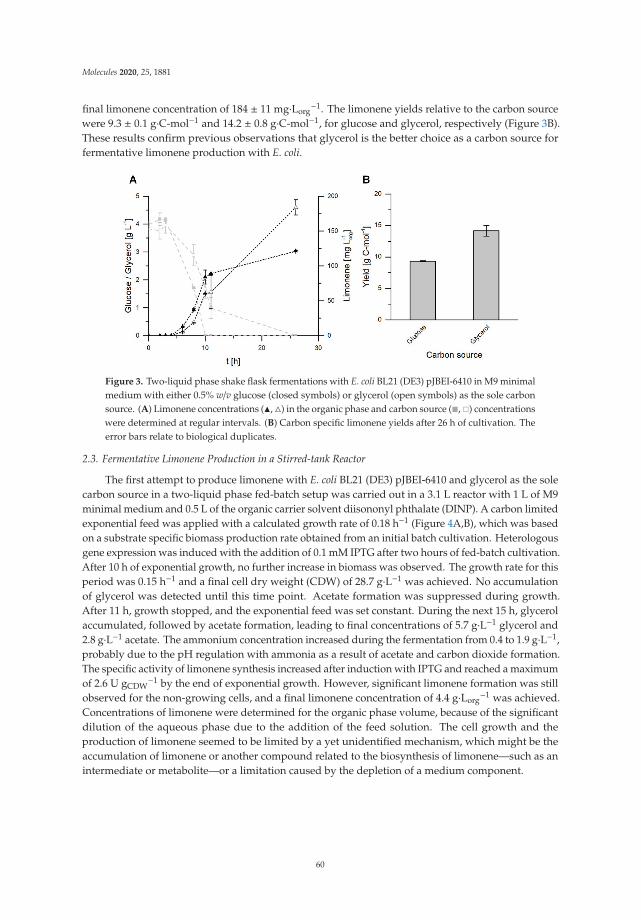
Molecules 2020, 25, 1881
final limonene concentration of 184 ± 11 mg·Lorg−1. The limonene yields relative to the carbon source
were 9.3 ± 0.1 g·C-mol−1 and 14.2 ± 0.8 g·C-mol−1, for glucose and glycerol, respectively (Figure 3B).These results confirm previous observations that glycerol is the better choice as a carbon source forfermentative limonene production with E. coli.
Figure 3. Two-liquid phase shake flask fermentations with E. coli BL21 (DE3) pJBEI-6410 in M9 minimalmedium with either 0.5% w/v glucose (closed symbols) or glycerol (open symbols) as the sole carbonsource. (A) Limonene concentrations (�, �) in the organic phase and carbon source (�, �) concentrationswere determined at regular intervals. (B) Carbon specific limonene yields after 26 h of cultivation. Theerror bars relate to biological duplicates.
2.3. Fermentative Limonene Production in a Stirred-tank Reactor
The first attempt to produce limonene with E. coli BL21 (DE3) pJBEI-6410 and glycerol as the solecarbon source in a two-liquid phase fed-batch setup was carried out in a 3.1 L reactor with 1 L of M9minimal medium and 0.5 L of the organic carrier solvent diisononyl phthalate (DINP). A carbon limitedexponential feed was applied with a calculated growth rate of 0.18 h−1 (Figure 4A,B), which was basedon a substrate specific biomass production rate obtained from an initial batch cultivation. Heterologousgene expression was induced with the addition of 0.1 mM IPTG after two hours of fed-batch cultivation.After 10 h of exponential growth, no further increase in biomass was observed. The growth rate for thisperiod was 0.15 h−1 and a final cell dry weight (CDW) of 28.7 g·L−1 was achieved. No accumulationof glycerol was detected until this time point. Acetate formation was suppressed during growth.After 11 h, growth stopped, and the exponential feed was set constant. During the next 15 h, glycerolaccumulated, followed by acetate formation, leading to final concentrations of 5.7 g·L−1 glycerol and2.8 g·L−1 acetate. The ammonium concentration increased during the fermentation from 0.4 to 1.9 g·L−1,probably due to the pH regulation with ammonia as a result of acetate and carbon dioxide formation.The specific activity of limonene synthesis increased after induction with IPTG and reached a maximumof 2.6 U gCDW
−1 by the end of exponential growth. However, significant limonene formation was stillobserved for the non-growing cells, and a final limonene concentration of 4.4 g·Lorg
−1 was achieved.Concentrations of limonene were determined for the organic phase volume, because of the significantdilution of the aqueous phase due to the addition of the feed solution. The cell growth and theproduction of limonene seemed to be limited by a yet unidentified mechanism, which might be theaccumulation of limonene or another compound related to the biosynthesis of limonene—such as anintermediate or metabolite—or a limitation caused by the depletion of a medium component.
60

Molecules 2020, 25, 1881
Figure 4. Two-liquid phase fed-batch fermentation with E. coli BL21 (DE3) pJBEI-6410 in M9 minimalmedium. Cell dry weight (CDW) (�), limonene concentrations (�) in the organic phase, glycerol (�),acetate (�), and ammonium (�) concentrations were determined at regular intervals. The specificactivities (•) were calculated for distinct time points throughout the fermentation time. The feed rate isdisplayed as well (dotted line). (A) and (B) display the initial fed-batch fermentation (D = 0.18 h−1),whereas (C) and (D) display the optimized fed-batch fermentation with a lower feed rate (D = 0.15 h−1)and additional trace element supply. The error bars for CDW relate to two independent measurements.
To test whether intracellular limonene, terpene intermediates, or the expression of heterologouspathway genes influence cell growth, a glycerol-limited fed-batch fermentation was carried out withoutthe induction of the heterologous pathway (data not shown). Although heterologous gene expressionwas not induced, 222 mg·Lorg
−1 limonene was produced during the exponential growth. These smallamounts are probably caused by leaky expression of the pathway genes. Nearly identical CDW wasachieved, while 20-fold less limonene was produced. Therefore, toxification by intermediates of theMVA pathway or intracellular limonene can be excluded.
In order to overcome the growth limitation, the parameters for the fermentation were changedto permit prolonged exponential growth, coupled with higher limonene concentrations. A carbonlimited exponential feed was set up with a calculated growth rate of 0.15 h−1, which is lower thanin the previous experiments (0.18 h−1) (Figure 4C,D). Furthermore, additional US* trace elementswere supplied. After 6 and 16 h of cultivation, 2 and 1 mL of US* trace element solution were spiked,respectively. The exponential growth could be maintained for nearly 17 h, with a growth rate of 0.12 h−1.Thus, the addition of trace elements prolonged the growth phase, and a CDW of 48.8 g·L−1 was reached,whereas the second trace element spike did not affect the cells in their stationary phase. The specificactivity quickly increased reaching a maximum of 1.8 U gCDW
−1 after 5 h, before it decreased withongoing fermentation. In contrast to in previous experiments, the specific activity could be maintained
61

Molecules 2020, 25, 1881
above 1 U gCDW−1 for a time period of more than 17 h. The prolonged production phase and higher
biomass concentration resulted in 7.3 g·Lorg−1 limonene after 24 h by the end of the fermentation. To
our knowledge, this is the highest limonene titer reported so far.
3. Discussion
3.1. Glycerol is a Suitable Carbon Source for Heterologous Limonene Production in Escherichia coli
The beneficial effect of glycerol as a sole or supplementary carbon source has been reportedbefore for the fermentative production of carotenoids in MEP engineered E. coli strains [17–19], forsesquiterpenes [12] and for limonene [13] in MVA-engineered E. coli strains. The use of glycerol as acarbon source resulted in higher limonene formation rates, a prolonged growth phase, and increasedstability compared to the same whole-cell biocatalyst growing on glucose [13].
In the present study, we were able to transfer this knowledge to a bioprocess with an optimizedone plasmid strain and showed that, compared to glucose, glycerol is definitely the preferred carbonsource for the production of limonene with E. coli. The transferability to the bioreactor scale wasvalidated, and reaction engineering was performed to further increase the limonene titer. This led tothe highest monoterpene concentration obtained with a microorganism to date. Next to availabilityand low cost, various advantages make glycerol an attractive carbon source for fermentation processescompared to glucose. Firstly, beneficial effects on the viability of cells and productivity of recombinantproteins were observed [20]. Secondly, glycerol does not show any catabolic repression in combinationwith lactose, which might be the preferred inducer for heterologous gene expression instead of IPTGdue to a reduced stress level for the production host. Catabolite repression occurs when excessglucose is present and leads to reduced lactose uptake rates, which causes the decreased expressionof recombinant proteins [21]. Finally, glycerol is a suitable carbon source for anaerobic fermentationwith E. coli strains producing biofuels and highly reduced compounds. The high degree of reductionof carbon atoms in glycerol (κ = 4.67) provides a distinct advantage over glucose (κ = 4.00) in theabsence of other electron acceptors [22]. E. coli strains are able to utilize glycerol in such conditionsfor cell growth and need a suitable sink for the excess reducing equivalents generated during theformation of biomass [23]. Therefore, the ability to form a highly reduced product is essential for themicroorganism. Limonene has a high degree of reduction (κ = 5.60), so it would be a suitable productand sink for reducing equivalents in anaerobic glycerol fermentation. The anaerobic environmentcould have another beneficial effect regarding the toxicity of limonene. Whereas limonene itself hasrelatively little toxicity towards E. coli cells, the common oxidation product limonene hydroperoxide,which forms spontaneously in aerobic environments, shows highly antimicrobial effects [24]. In thisstudy, the inhibitory effects of limonene hydroperoxide were not observed, due to efficient productextraction in the organic phase.
3.2. Progess to an Economic Limonene Production Process
The optimized bioreactor process described in this study resulted in limonene productivityexceeding the threshold for developing a profitable production process for fine chemicals(100 mg·L-1·h−1) [25] for the first time (Table 1). However, a techno-economic assessment statedthat a biotechnological production process for limonene needs to have a space-time yield above700 mg·L−1·h−1 and a 45% carbon specific yield to be competitive with established processes [26].While our process already shows more than a fifth of the space-time yield required, the conversion ofthe carbon source into the product is still low, at less than 1%.
62

Molecules 2020, 25, 1881
Ta
ble
1.
Esch
eric
hia
coli-
base
dfe
rmen
tati
onse
tups
for
the
prod
ucti
onof
limon
ene.
Str
ain
Pla
smid
Sy
stem
Med
ium
Setu
pc L
imo
ne
ne
[mg·L−
1]
ST
Y[m
g·L−
1h−1
]R
efe
ren
ce
E.co
liD
H1
pJBE
I-64
09EZ
-Ric
h/
gluc
ose
Batc
h—Sh
ake
flask
435
6[8
]E.
coli
BL21
(DE3
)pB
AD
:LS,
pET2
4:A
GPP
S2M
9/
glyc
erol
Fed-
batc
h—ST
R13
5023
[13]
E.co
liBW
2511
3(D
E3)
pMA
P6,p
ISP6
,pN
LSt1
YM
9/
gluc
ose
Fed-
batc
h—Sh
ake
flask
1290
15[2
7]E.
coli
BL21
(DE3
)pJ
BEI-
6410
M9/
glyc
erol
Fed-
batc
h—ST
R36
3015
1Th
isst
udy
STY:
spac
e-ti
me
yiel
d;ST
R:s
tirr
edta
nkre
acto
r.
63

Molecules 2020, 25, 1881
Different approaches to improve the yield have already been described. Non-growing butmetabolically active E. coli cells can boost the production of the desired product due to reduced energyand carbon loss to biomass formation [28]. A fourfold increase in specific limonene yields relative tobiomass was accomplished with this strategy. Moreover, further pathway debottlenecking and theoptimization of the involved enzymes could increase the economics of the process. For example, theexchange of the geranyl pyrophosphate synthase with a neryl pyrophosphate synthase from Solanumlycopersicum led to increased limonene production with E. coli [27]. Another strategy to increasethe product titer and the specificity of monoterpenes in E. coli was described by Chacón et al. [29].The monoterpene geraniol was converted to the monoterpenoid geranyl acetate with an in vivoesterification and extracted in situ to an organic phase. Toxicity issues and the synthesis of by-productscould be circumvented, resulting in a monoterpenoid concentration of 4.8 g·L−1. Similar approaches tothe coupled synthesis and functionalization of limonene are described, which produce the valuablemonoterpenoid perillyl alcohol [8,30].
The highest yields with more than 95% and titers of 15 g·L−1 were achieved with a cell-free systemconsisting of 27 purified enzymes, which convert glucose into monoterpenes [31]. Other systems whichincorporate acetic acid as a starting building block for the cell-free synthesis of terpenes are describedas well [32]. However, a major drawback is the need for purified enzymes, which are associated withadditional costs and the further input of glucose needed to produce them. The need for enzymepurification can be avoided with the use of enzyme-enriched E. coli lysates, but this approach appearedto suffer from low product titers of 90 mg·L−1 [33]. Moreover, the involved enzymes are considered tohave low stabilities in the in vitro environment, and cofactor regeneration could be a limiting aspectin cell-free applications as well [32]. The expensive cofactors CoA and NADPH must be effectivelyrecycled in such systems, while the use of whole cells circumvents these drawbacks as the cofactors areregenerated by the primary metabolism. Therefore, microorganisms are preferred as the biocatalyst forthe larger biotechnological production of limonene. Microbial hosts other than E. coli were recentlyinvestigated as producer strains, such as the cyanobacterium Synechocystis sp. [34] or the oleaginousyeast Yarrowia lipolytica [35], which was able to produce limonene from waste cooking oil. However,product titers were orders of magnitude lower compared to the processes based on engineered E. coli.
Next to the selection and optimization of the production system, a feasible bioprocess with highlimonene titers involves the integrated development of in situ product removal strategies. Due to thehigh volatility and inhibitory effects on cell growth, the capturing of limonene during fermentationis required. Various methods are available, with two-liquid phase and gas stripping systems beingespecially suitable at higher scales [36]. In particular, two-liquid phase systems have the advantage thatthe products are effectively removed from the fermentation broth [37]. The choice of capturing methodis also dependent on the further application of the product. If limonene is subsequently used as a purecompound, solvent-free systems might be the better choice, whereas application as an additive for, e.g.,solvents might allow the use of the same solvent for in situ extraction [38]. In the present study, the insitu product removal strategy in combination with an engineered E. coli strain and a glycerol-limitedfed-batch fermentation enabled the synthesis of the highest limonene concentration reported to date.Steps towards an economic process were made, and the potential of integrating already generatedknowledge with the biotechnological production of terpenes was demonstrated.
4. Materials and Methods
4.1. Chemicals and Bacterial Strains
All chemicals used in this work were purchased from Carl Roth GmbH & Co. KG (Karlsruhe,Germany) and Merck KGaA (Darmstadt, Germany).
E. coli BL21 (DE3) harboring the plasmid pJBEI-6410 was used for all experiments. pJBEI-6410carries the genes for the MVA pathway, a geranyl pyrophosphate synthase, and the limonene
64

Molecules 2020, 25, 1881
synthase from Mentha spicata [8]. It was a gift from Taek Soon Lee (RRID:Addgene_47049;http://n2t.net/addgene:47049).
4.2. Cultivation and Fermentative Limonene Production in Shake Flasks
For the cultivation in shake flasks, precultures were grown in 5 mL of LB medium (10 g·L−1
tryptone, 5 g·L−1 yeast extract, and 5 g·L−1 NaCl) with 100 μg·mL−1 ampicillin for 4 to 6 h at 37 ◦Cand shaking at 200 rpm. LB precultures were transferred 1:500 to 250 mL baffled Erlenmeyer flaskswith 50 mL M9 minimal medium (8.5 g·L−1 Na2HPO4 · 2H2O, 3 g·L−1 KH2PO4, 0.5 g·L−1 NaCl, 1 g·L−1
NH4Cl, 2 mL of 1 M MgSO4, and 1 mL of L−1 US* trace element solution) with either 0.5% w/v glucoseor 0.5% w/v glycerol as the sole carbon source. The cultures were incubated overnight at 30 ◦C withshaking at 200 rpm. The M9 precultures were used to inoculate 80 mL of M9 minimal medium with the0.5% w/v carbon source in 500 mL baffled shake flasks to an optical density of 0.11 at 600 nm (OD600).The main cultivations were performed at 30 ◦C with shaking at 200 rpm. Heterologous gene expressionwas induced by adding 0.1 mM isopropyl β-d−1-thiogalactopyranoside (IPTG) once an OD600 of0.4–0.6 was reached. After induction, the cultures were overlaid with 20 mL of diisononyl phthalate(DINP), and incubation was continued. For sampling, the phases were separated by centrifugation(2 min, 4 ◦C, 11,000 × g). Prior to gas chromatography (GC) analysis, the organic phase was diluted indiethyl ether with 0.2 mM dodecane and dried over anhydrous Na2SO4. The aqueous phase was usedfor HPLC analysis. The cell dry weight was determined by measurement of the optical density at awavelength of 600 nm (Libra S11 Visible Spectrophotometer, Biochrom, Cambridge, UK). One OD600
unit corresponded to 0.312 gCDW L−1, with a linear range between 0.1 and 1.
4.3. Fermentative Limonene Production in a Stirred-Tank Reactor
Two-liquid phase fermentations at the bioreactor scale were carried out in a 3.1 L stirred-tankreactor (KLF 2000, Bioengineering AG, Wald, Switzerland). The tank reactor was equipped withtwo Rushton turbine stirrers. An M9 preculture was used to inoculate an initial batch cultivation,which was performed in 1 L of M9 minimal medium containing a 1.5% w/v carbon source at a startingOD600 of 0.11. The pH was set to 7.2 and controlled by the automatic addition of either 30% v/vphosphoric acid or 25% v/v ammonia hydroxide solution. Batch cultures were incubated at 30 ◦C, withshaking at 1800 rpm and an aeration rate of 1 vvm, until the batch phase was finished after 13 to 17 h(indicated by a steep pO2 increase). Before the start of the carbon-limited fed-batch fermentation, 1 mLof US* trace element solution was added. To keep the exponential growth rates between 0.18 and0.2 h−1, a carbon-limited and controlled exponential feed with a 73% w/v carbon source and 19.6 g·L−1
MgSO4 · 7 H2O was started. The pO2 value was kept above 30% by adjusting the stirrer speed and theaeration rate. If desired, recombinant gene expression was induced by adding 0.1 mM IPTG after 2 hof exponential feeding, and 0.5 L of DINP were added subsequently. Antifoam A was added only incases of excessive foaming. Regular sampling was carried out as described above.
4.4. Quantification of Limonene
The quantification of limonene in DINP was carried out with GC using a TRACE GC Ultra (ThermoFisher Scientific Inc., Waltham, MA, USA), equipped with a FactorFour-5ms column (30 m, 0.25 mm,0.25 μm, Varian, Inc., Palo Alto, CA, USA) and a flame ionization detector. Nitrogen was used as acarrier gas and the injection volume was set to 1 μL (80 ◦C, 5 min; 80–140 ◦C, 7.5 ◦C·min−1; 140–300 ◦C,40 ◦C·min−1; 300 ◦C, 5 min). Samples of the organic phase were prepared in diethyl ether containing0.2 mM dodecane as an internal standard. The quantification of limonene was performed using astandard curve of the ratio between commercial (S)-limonene (Merck KGaA, Darmstadt, Germany)and the internal standard.
65
Molecules 2020, 25, 1881
4.5. Quantification of Glucose, Glycerol, Acetate, and Ammonia
The quantifications of glucose, glycerol, and acetate concentrations were performed by highperformance liquid chromatography (HPLC) with a LaChrome Elite HPLC system (VWR, Darmstadt,Germany), equipped with a Trentec 308R-Gel.H column (Trentec Analysetechnik, Gerlingen, Germany)and a refractive index detector. The mobile phase consisted of 5 mM sulphuric acid. An isocraticmethod was used, with a flow rate of 1 mL min−1 and an injection volume of 20 μL. The column ovenwas set to 40 ◦C. Eluted components were quantified using standard curves for glucose, glycerol,and acetate.
Ammonia concentrations were determined by the method of Berthelot [39].
Author Contributions: Conceptualization, M.K.J. and S.L.; methodology, J.R. and M.K.J.; formal analysis, J.R.;investigation, J.R.; resources, S.L.; data curation, K.R., M.K.J. and S.L.; writing—original draft preparation, J.R. andK.R.; writing—review and editing, K.R., M.K.J. and S.L.; visualization, J.R.; supervision, M.K.J. and S.L.; projectadministration, M.K.J. and S.L. All authors have read and agreed to the published version of the manuscript.
Funding: This research received no external funding.
Acknowledgments: We acknowledge financial support by Deutsche Forschungsgemeinschaft and TU DortmundUniversity within the funding program Open Access Publishing. We gratefully thank Christian Willrodt forfruitful scientific discussions and Saikai Humaerhan for her support in initial growth studies.
Conflicts of Interest: The authors declare no conflict of interest. The funders had no role in the design of thestudy; in the collection, analyses, or interpretation of data; in the writing of the manuscript, or in the decision topublish the results.
Appendix A
Figure A1. Two-liquid phase shake flask fermentations with E. coli BL21 (DE3) pJBEI-6410 in M9minimal medium with either 0.5% w/v glucose (closed symbols, •) or glycerol (open symbols, �) as thesole carbon source. Cell dry weights were determined at regular intervals. The error bars relate tobiological duplicates.
References
1. Fisher, K. Terpenes replacing BTEX in oil field. Available online: http://www.aogr.com/magazine/editors-choice/terpenes-replacing-btex-in-oil-field (accessed on 12 February 2020).
2. Renninger, N.S.; Ryder, J.; Fisher, K. Jet fuel compositions and methods of making and using same.WO2008133658, 6 November 2008.
3. Chuck, C.J.; Donnelly, J. The compatibility of potential bioderived fuels with Jet A-1 aviation kerosene. Appl.Energy 2014, 118, 83–91. [CrossRef]
4. Zhang, J.; Zhao, C. A new approach for bio-jet fuel generation from palm oil and limonene in the absence ofhydrogen. Chem. Commun. 2015, 51, 17249–17252. [CrossRef] [PubMed]
66
Molecules 2020, 25, 1881
5. Inouye, S.; Takizawa, T.; Yamaguchi, H. Antibacterial activity of essential oils and their major constituentsagainst respiratory tract pathogens by gaseous contact. J. Antimicrob. Chemother. 2001, 47, 565–573. [CrossRef][PubMed]
6. Thomas, A.F.; Bessière, Y. Limonene. Nat. Prod. Rep. 1989, 6, 291–309. [CrossRef]7. Cornelissen, S.; Julsing, M.K.; Volmer, J.; Riechert, O.; Schmid, A.; Bühler, B. Whole-cell-based
CYP153A6-catalyzed (S)-limonene hydroxylation efficiency depends on host background and profitsfrom monoterpene uptake via AlkL. Biotechnol. Bioeng. 2013, 110, 1282–1292. [CrossRef]
8. Alonso-Gutierrez, J.; Chan, R.; Batth, T.S.; Adams, P.D.; Keasling, J.D.; Petzold, C.J.; Lee, T.S. Metabolicengineering of Escherichia coli for limonene and perillyl alcohol production. Metab. Eng. 2013, 19, 33–41.[CrossRef]
9. Belanger, J.T. Perillyl Alcohol: Applications in Oncology. Altern. Med. Rev. 1998, 3, 448–457.10. Rosenthal, K.; Lütz, S. Recent developments and challenges of biocatalytic processes in the pharmaceutical
industry. Curr. Opin. Green Sustain. Chem. 2018, 11, 58–64. [CrossRef]11. Kemper, K.; Hirte, M.; Reinbold, M.; Fuchs, M.; Brück, T. Opportunities and challenges for the sustainable
production of structurally complex diterpenoids in recombinant microbial systems. Beilstein J. Org. Chem.2017, 13, 845–854. [CrossRef]
12. Martin, V.J.J.; Pitera, D.J.; Withers, S.T.; Newman, J.D.; Keasling, J.D. Engineering a mevalonate pathway inEscherichia coli for production of terpenoids. Nat. Biotechnol. 2003, 21, 796–802. [CrossRef]
13. Willrodt, C.; David, C.; Cornelissen, S.; Bühler, B.; Julsing, M.K.; Schmid, A. Engineering the productivity ofrecombinant Escherichia coli for limonene formation from glycerol in minimal media. Biotechnol. J. 2014, 9,1000–1012. [CrossRef] [PubMed]
14. Schewe, H.; Holtmann, D.; Schrader, J. P450BM-3-catalyzed whole-cell biotransformation of α-pinene withrecombinant Escherichia coli in an aqueous–organic two-phase system. Appl. Microbiol. Biotechnol. 2009, 83,849–857. [CrossRef] [PubMed]
15. Studier, F.W.; Moffatt, B.A. Use of bacteriophage T7 RNA polymerase to direct selective high-level expressionof cloned genes. J. Mol. Biol. 1986, 189, 113–130. [CrossRef]
16. Calos, M.P. DNA sequence for a low-level promoter of the lac repressor gene and an ‘up’ promoter mutation.Nature 1978, 274, 762–765. [CrossRef]
17. Lee, P.C.; Mijts, B.N.; Schmidt-Dannert, C. Investigation of factors influencing production of the monocycliccarotenoid torulene in metabolically engineered Escherichia coli. Appl. Microbiol. Biotechnol. 2004, 65, 538–546.[CrossRef]
18. Kim, Y.S.; Lee, J.H.; Kim, N.H.; Yeom, S.J.; Kim, S.W.; Oh, D.K. Increase of lycopene production bysupplementing auxiliary carbon sources in metabolically engineered Escherichia coli. Appl. Microbiol.Biotechnol. 2011, 90, 489–497. [CrossRef]
19. Jang, H.-J.; Yoon, S.-H.; Ryu, H.-K.; Kim, J.-H.; Wang, C.-L.; Kim, J.-Y.; Oh, D.-K.; Kim, S.-W. Retinoidproduction using metabolically engineered Escherichia coli with a two-phase culture system. Microb. Cell Fact.2011, 10, 59. [CrossRef]
20. Kopp, J.; Slouka, C.; Ulonska, S.; Kager, J.; Fricke, J.; Spadiut, O.; Herwig, C. Impact of Glycerol as CarbonSource onto Specific Sugar and Inducer Uptake Rates and Inclusion Body Productivity in E. coli BL21(DE3).Bioengineering 2017, 5, 1. [CrossRef]
21. Wurm, D.J.; Hausjell, J.; Ulonska, S.; Herwig, C.; Spadiut, O. Mechanistic platform knowledge of concomitantsugar uptake in Escherichia coli BL21(DE3) strains. Sci. Rep. 2017, 7, 45072. [CrossRef]
22. Durnin, G.; Clomburg, J.; Yeates, Z.; Alvarez, P.J.J.; Zygourakis, K.; Campbell, P.; Gonzalez, R. Understandingand harnessing the microaerobic metabolism of glycerol in Escherichia coli. Biotechnol. Bioeng. 2009, 103,148–161. [CrossRef]
23. Clomburg, J.M.; Gonzalez, R. Anaerobic fermentation of glycerol: A platform for renewable fuels andchemicals. Trends Biotechnol. 2013, 31, 20–28. [CrossRef] [PubMed]
24. Chubukov, V.; Mingardon, F.; Schackwitz, W.; Baidoo, E.E.K.; Alonso-Gutierrez, J.; Hu, Q.; Lee, T.S.;Keasling, J.D.; Mukhopadhyay, A. Acute limonene toxicity in Escherichia coli is caused by limonenehydroperoxide and alleviated by a point mutation in alkyl hydroperoxidase AhpC. Appl. Environ. Microbiol.2015, 81, 4690–4696. [CrossRef] [PubMed]
25. Straathof, A.J.J.; Panke, S.; Schmid, A. The production of fine chemicals by biotransformations. Curr. Opin.Biotechnol. 2002, 13, 548–556. [CrossRef]
67

Molecules 2020, 25, 1881
26. Sun, C.; Theodoropoulos, C.; Scrutton, N.S. Techno-economic assessment of microbial limonene production.Bioresour. Technol. 2020, 300, 122666. [CrossRef]
27. Wu, J.; Cheng, S.; Cao, J.; Qiao, J.; Zhao, G.-R. Systematic Optimization of Limonene Production in EngineeredEscherichia coli. J. Agric. Food Chem. 2019, 67, 7087–7097. [CrossRef]
28. Willrodt, C.; Hoschek, A.; Bühler, B.; Schmid, A.; Julsing, M.K. Decoupling production from growth bymagnesium sulfate limitation boosts de novo limonene production. Biotechnol. Bioeng. 2016, 113, 1305–1314.[CrossRef]
29. Chacón, M.G.; Marriott, A.; Kendrick, E.G.; Styles, M.Q.; Leak, D.J. Esterification of geraniol as a strategyfor increasing product titre and specificity in engineered Escherichia coli. Microb. Cell Fact. 2019, 18, 105.[CrossRef]
30. Willrodt, C.; Hoschek, A.; Bühler, B.; Schmid, A.; Julsing, M.K. Coupling limonene formation andoxyfunctionalization by mixed-culture resting cell fermentation. Biotechnol. Bioeng. 2015, 112, 1738–1750.[CrossRef]
31. Korman, T.P.; Opgenorth, P.H.; Bowie, J.U. A synthetic biochemistry platform for cell free production ofmonoterpenes from glucose. Nat. Commun. 2017, 8, 15526. [CrossRef]
32. Dirkmann, M.; Nowack, J.; Schulz, F. An in Vitro Biosynthesis of Sesquiterpenes Starting from Acetic Acid.ChemBioChem 2018, 19, 2146–2151. [CrossRef]
33. Dudley, Q.M.; Nash, C.J.; Jewett, M.C. Cell-free biosynthesis of limonene using enzyme-enriched Escherichiacoli lysates. Synth. Biol. 2019, 4, 1–9. [CrossRef] [PubMed]
34. Lin, P.-C.; Saha, R.; Zhang, F.; Pakrasi, H.B. Metabolic engineering of the pentose phosphate pathway forenhanced limonene production in the cyanobacterium Synechocysti s sp. PCC 6803. Sci. Rep. 2017, 7, 17503.[CrossRef] [PubMed]
35. Pang, Y.; Zhao, Y.; Li, S.; Zhao, Y.; Li, J.; Hu, Z.; Zhang, C.; Xiao, D.; Yu, A. Engineering the oleaginous yeastYarrowia lipolytica to produce limonene from waste cooking oil. Biotechnol. Biofuels 2019, 12, 241. [CrossRef][PubMed]
36. Jongedijk, E.; Cankar, K.; Buchhaupt, M.; Schrader, J.; Bouwmeester, H.; Beekwilder, J. Biotechnologicalproduction of limonene in microorganisms. Appl. Microbiol. Biotechnol. 2016, 100, 2927–2938. [CrossRef][PubMed]
37. Xie, S.; Zhu, L.; Qiu, X.; Zhu, C.; Zhu, L. Advances in the Metabolic Engineering of Escherichia coli for theManufacture of Monoterpenes. Catalysts 2019, 9, 433. [CrossRef]
38. Brennan, T.C.R.; Turner, C.D.; Krömer, J.O.; Nielsen, L.K. Alleviating monoterpene toxicity using a two-phaseextractive fermentation for the bioproduction of jet fuel mixtures in Saccharomyces cerevisiae. Biotechnol.Bioeng. 2012, 109, 2513–2522. [CrossRef] [PubMed]
39. Searle, P.L. The berthelot or indophenol reaction and its use in the analytical chemistry of nitrogen. A review.Analyst 1984, 109, 549. [CrossRef]
Sample Availability: Samples of the compound (S)-limonene in DINP are available from the authors.
© 2020 by the authors. Licensee MDPI, Basel, Switzerland. This article is an open accessarticle distributed under the terms and conditions of the Creative Commons Attribution(CC BY) license (http://creativecommons.org/licenses/by/4.0/).
68

molecules
Article
A DyP-Type Peroxidase of Pleurotus sapidus withAlkene Cleaving Activity
Nina-Katharina Krahe *, Ralf G. Berger and Franziska Ersoy
Institut für Lebensmittelchemie, Gottfried Wilhelm Leibniz Universität Hannover, Callinstraße 5,30167 Hannover, Germany; [email protected] (R.G.B.); [email protected] (F.E.)* Correspondence: [email protected]; Tel.: +49-511-762-17257
Received: 20 February 2020; Accepted: 23 March 2020; Published: 27 March 2020
Abstract: Alkene cleavage is a possibility to generate aldehydes with olfactory properties forthe fragrance and flavor industry. A dye-decolorizing peroxidase (DyP) of the basidiomycetePleurotus sapidus (PsaPOX) cleaved the aryl alkene trans-anethole. The PsaPOX was semi-purifiedfrom the mycelium via FPLC, and the corresponding gene was identified. The amino acid sequenceas well as the predicted tertiary structure showed typical characteristics of DyPs as well as anon-canonical Mn2+-oxidation site on its surface. The gene was expressed in Komagataella pfaffiiGS115 yielding activities up to 142 U/L using 2,2′-azino-bis(3-ethylbenzthiazoline-6-sulphonic acid)as substrate. PsaPOX exhibited optima at pH 3.5 and 40 ◦C and showed highest peroxidase activity inthe presence of 100 μM H2O2 and 25 mM Mn2+. PsaPOX lacked the typical activity of DyPs towardsanthraquinone dyes, but oxidized Mn2+ to Mn3+. In addition, bleaching of β-carotene and annattowas observed. Biotransformation experiments verified the alkene cleavage activity towards the arylalkenes (E)-methyl isoeugenol, α-methylstyrene, and trans-anethole, which was increased almosttwofold in the presence of Mn2+. The resultant aldehydes are olfactants used in the fragrance andflavor industry. PsaPOX is the first described DyP with alkene cleavage activity towards aryl alkenesand showed potential as biocatalyst for flavor production.
Keywords: alkene cleavage; aryl alkenes; basidiomycota; biocatalysis; carotene degradation;dye-decolorizing peroxidase (DyP); manganese; Komagataella pfaffii; Pleurotus sapidus
1. Introduction
Many small aromatic aldehydes and ketones are volatiles with olfactory properties and thereforeof high interest to the fragrance and flavor industry [1]. One method to generate aldehydes andketones is the oxidative cleavage of alkenes. Chemical options are ozonolysis, dihydroxylationfollowed by oxidative glycol cleavage, or metal-based methods [2–4]. However, all of these methodshave disadvantages, such as the generation of explosive intermediates, the use of environmentallyunfriendly and/or toxic oxidants and metal catalysts, or low yield and low chemoselectivity [2].An alternative is the application of enzymes due to their high chemo-, regio-, and stereospecificityas well as the possibility to use mild reaction conditions [3]. Another advantage is the generation of“natural” flavors according to effective legislation in Europe and the US. This becomes more and moreimportant considering the rising popularity of natural products [5]. Different proteins of differentenzyme classes, which are heme, non-heme iron, or non-iron metal dependent and have differentprotein structures as well as different reaction mechanism are known to catalyze alkene cleavagereactions [3]. Specifically, an isoeugnol and trans-anethole oxygenase from Pseudomonas putida and twomanganese dependent enzymes from Thermotoga maritima (manganese-dependent Cupin TM1459)and Trametes hirsuta (Mn3+-dependent proteinase A homologue) oxidatively cleaved the benzylicdouble bond of different aryl alkenes, such as isoeugenol and trans-anethole to form the respective
Molecules 2020, 25, 1536; doi:10.3390/molecules25071536 www.mdpi.com/journal/molecules69

Molecules 2020, 25, 1536
aldehydes [6–9]. In addition, alkene cleavage activity towards aryl alkenes was also detected for severalperoxidases. Cleavage of different styrene derivatives was described for Coprinus cinereus peroxidaseand a human myeloperoxidase as minor side reaction [10], while horseradish peroxidase (HRP) showeda chemoselectivity of 92% for the conversion of trans-anethole (90%) to p-anisaldehyde [11]. Furthermore,transformations of o-ethylisoeugenol and trans-anethole to the corresponding benzaldehyde derivativesby lignin peroxidases were described [3,11]. However, to our best knowledge no alkene cleavageactivity of a dye-decolorizing peroxidase (DyP) is known.
DyP-type peroxidases (EC: 1.11.1.19) are a new superfamily of heme peroxidases thatoxidize various dyes, in particular xenobiotic anthraquinone dyes, which are hardly oxidizedby other peroxidases [12]. Furthermore, typical peroxidase substrates, such as ABTS(2,2′-azino-bis(3-ethylbenzthiazoline-6-sulphonic acid) and phenolic compounds are also substrates forDyPs [13,14]. However, amino acid sequences, protein structures, and catalytic residues differ highlybetween DyPs and other classes of heme peroxidases [15]. Typical structural characteristics of DyPsare the ferredoxin-like fold, which is formed by two domains containing α-helices and four-strandedantiparallel β-sheets, and a GXXGD motif [14,15]. The active site (heme pocket) including a catalyticaspartic acid and arginine over the heme plane (distal) and proximal histidine is structurally similar toother heme peroxidases, even though other peroxidases contain histidine instead of aspartic acid [16].The proximal histidine in the heme pocket functions as the fifth ligand of the heme iron, while thedistal aspartic acid and arginine are involved in the activation of the enzyme [14,15]. The deprotonatedaspartic acid (or asparagine) mediates the rearrangement of a proton from hydrogen peroxide afterit enters the heme pocket in the resting state. This results in the heterolytic cleavage of hydrogenperoxide to water and oxidation of the heme to the radicalic-cationic oxoferryl species Compound I bytwo-fold single electron transfer [16]. Even though the distal arginine is not directly involved in therearrangement of hydrogen peroxide, it is essential for the coordination of hydrogen peroxide to theheme iron and the stabilization of Compound I [16]. During the following reaction cycle, Compound Iis reduced by oxidation of two substrate molecules to the state during enzyme resting state in twosequential steps with Compound II as intermediate. However, the existence of Compound II has notbeen confirmed universally for all DyP-type peroxidases [17]. In the presence of excessive hydrogenperoxide suicide inhibition was observed for different DyPs [18,19]. This is also well known for classicalperoxidases as a result of an inactive oxidative state (Compound III) and results from reaction ofhydrogen peroxide with Compound II [20,21].
The objective of the present study was to identify new enzymes of basidiomycetes with alkenecleavage activity towards aryl alkenes. A screening was performed using trans-anethole as modelalkene. A new DyP-type peroxidase from P. sapidus was semi-purified and the coding gene wasidentified. Heterologous expression resulted in the production of soluble protein and allowed thebiochemical characterization of the DyP. The enzyme was able to oxidize Mn2+, but did not catalyze thedegradation of anthraquinone dyes, which is typical for other DyPs. Biotransformation experimentsverified the cleavage activity towards different alkenes. This is the first study describing a DyP withalkene cleavage activity towards aryl alkenes.
2. Results and Discussion
2.1. Purification and Identification of the Alkene Cleavage Activity
Within a screening of 17 basidiomycetes for alkene cleavage activity using the substratetrans-anethole (Table S1) P. sapidus turned out to be a promising candidate for the production ofthe desired activity. The lyophilized mycelium as well as the culture supernatant was examined forthe ability to cleave trans-anethole after submerged cultivation. The culture supernatant showed noactivity, whereas the incubation in the presence of the mycelium resulted in formation of 5.36 mMp-anisaldehyde (molar yield of 79.03%; Figure 1).
70

Molecules 2020, 25, 1536
Figure 1. Bioconversion of trans-anethole by P. sapidus. (a) Alkene cleavage of trans-anethole resultedin the formation of p-anisaldehyde. (b) GC-FID chromatogram of an n-hexane extract of the conversionof the blank sample (blue) and after incubation with lyophilized mycelium of P. sapidus (orange).Retention times: trans-anethole (11.53 min) and p-anisaldehyde (13.45 min).
For the identification of the enzyme catalyzing the trans-anethole cleavage, it was semi-purifiedfrom the rehydrated mycelium by hydrophobic interaction and anion exchange chromatography (IEX).During the purification, a high activity loss occurred, which resulted in low product concentrationsafter conversion (Table S2). This was most likely a result of protein loss, enzyme degradation ordenaturation as described for other enzymes [22–24]. Another possibility is the loss of cofactors or–substrates, such as metal ions or peroxides during the purification steps [6]. Addition of Mn2+ ledto a 15-fold increase in p-anisaldehyde concentration, which was further increased by addition ofhydrogen peroxide, indicating a cosubstrate dependency (Table S2). Chemical conversion by Mn2+
alone was excluded, while product formation was observed with hydrogen peroxide (18 μM), but witha yield around 30-fold lower than the one for the enzymatic reaction (Table S2). Thus, the improvedbioconversion in the presence of Mn2+ and hydrogen peroxide was verified to be the result of anincreased enzyme activity.
A class of fungal enzymes that requires hydrogen peroxide and some of which need Mn2+ forcatalysis are peroxidases [12,25,26]. The anion exchange fractions, which showed alkene cleavageactivity, also exhibited peroxidase activity, thus verifying the presence of a peroxidase. For visualizationof the activity, a semi-native PAGE was performed and stained with ABTS in the presence of hydrogenperoxide (Figure 2). Two peroxidases running at 45 and 52 kDa were detected. The respective proteinbands stained with Coomassie Brilliant Blue were excised for electrospray ionization tandem massspectrometry. Due to the low protein concentration of the 52 kDa band, no meaningful peptides werefound (Figure 2, lane 1). This paper presents the data obtained for the protein running at 45 kDa(Figure 2, arrow).
Three tryptic peptides (EGSELLGAR, DGSFLTFR, and SGAPIEITPLKDDPK) were identified byESI-MS/MS. Homology searches against the public database NCBI using the mascot search engine(Matrix Science, London, UK) identified a DyP-type peroxidase of P. ostreatus PC15 (Pleos-DyP4 [27];GenBank accession no. KDQ22873.1), a close relative of P. sapidus, as the best hit.
71

Molecules 2020, 25, 1536
Figure 2. Semi-native PAGE of the active fraction after purification of the alkene cleavage enzyme fromP. sapidus by IEX. 1: gel stained with Coomassie Brilliant Blue; 2: gel stained with ABTS in the presenceof hydrogen peroxide, M: pre-stained molecular mass marker. An arrow and a box marks the proteinthat was successfully identified by sequencing.
2.2. Amplification and Expression of PsaPOX
Specific primers successfully amplified the 1512 bp coding region of the gene from P. sapidus.The translated amino acid sequence of 504 aa contained the peptide fragments that were obtained byESI-MS/MS and showed highest identity (94%) to the sequence of Pleos-DyP4 (Figure 3), which has notbeen investigated for its alkene cleavage activity against trans-anethole or other substrates before [27,28].In our hands, P. ostreatus showed a weaker trans-anethole cleavage activity, too (Table S1). Identity ofthe P. sapidus peroxidase (PsaPOX) to other DyPs and proteins was lower than 60%. A sequencealignment with other DyPs (Figure 3) confirmed that PsaPOX exhibited the typical GXXGD motif andall important residues known for the catalytic activity of DyPs, such as the proximal histidine (His-334)(fifth ligand of heme iron) and distal Asp-196 and Arg-360 involved in the activation of the enzyme(formation of compound I) by H2O2 cleavage [14,15]. Furthermore, Trp-405 was identified as a homologto the surface exposed Trp-377 of AauDyP of Auricularia auricula-judae, which serves as an oxidationsite for bulky substrates such as Reactive blue 19 (RBB19) using a long-range electron transfer [29].Comparison of the PsaPOX and Pleos-DyP4 sequence indicated that PsaPOX exhibited a non-canonicalMn2+-oxidation site on its surface (Asp-215, Glu-345, Asp-352 and Asp-354; Trp-339 participates in theelectron transport from the oxidation site to the heme) like Pleos-DyP4 [28] and can oxidize Mn2+ toMn3+, which is known for a few fungal DyPs only [24,27,30,31].
A structural homology model of PsaPOX (Figure S1), which was generated using the X-ray crystalstructure of Pleos-DyP4 (PDB-ID 6fsk) on the SWISS-MODEL server, possessed typical characteristicsof the DyP-type peroxidase family (N- and C-terminal ferredoxin-like domain, each formed byfour-stranded antiparallel β-sheets and several α-helices) [15] and supported the classification ofPsaPOX as DyP. Furthermore, the analysis of the amino acid sequence using PeroxiBase [32] relatedthe P. sapidus peroxidase (PsaPOX) to the class “DyP-type peroxidase D”. As known from literature,DyPs differ significantly in amino acid sequence, tertiary structure, and catalytic residues from otherrepresentatives of the heme peroxidases, such as HRP, human myeloperoxidase, lignin peroxidase,or Coprinus cinereus peroxidase [12,15,33], all of which are able to cleave trans-anethole or structurallyrelated alkenes [10,11]. So far no DyP is known to catalyze the mentioned reaction.
72

Molecules 2020, 25, 1536
Figure 3. Alignment of alkene cleaving peroxidase from P. sapidus (PsaPOX) with the Pleos-DyP4of P. ostreatus (PosDyP; KDQ22873.1) and other characterized DyPs. AgaDyP: Armillaria gallica(PBK80505.1), VvoDyP: Volvariella volvacea (AKU04643.1), CtrDyP: Coriolopsis trogii (AUW34346.1),GluDyP: Ganoderma lucidum (ADN05763.1), and TveDyP: Trametes versicolor (XP_008039377.1).Inverted triangles show amino acids important for heme binding (histidine (magenta) functionsas ligand for heme and the four other amino acid residues form a hydrogen peroxide binding pocket).Aspartic acid, which forms a hydrogen bond with histidine to stabilize compound I (oxidized hemeafter transfer of two electrons to H2O2) is shown in grey. The black box indicates the GXXDG motifcontaining the catalytic aspartic acid residue (yellow), which cleaves H2O2 heterolytically with thehelp of the neighboring arginine (green) to form compound I, and the circle presents an exposedtryptophan potentially involved in an LRET (long range electron transfer). Important amino acidsfor Mn2+-oxidation are highlighted in cyan; asterisks indicate conserved residues, colons equivalentresidues and dots partial residue conservation. Peptides identified by protein sequencing are underlined.Alignment was performed with Clustal Omega (European Bioinformatics Institute, Hinxton, UK).
73

Molecules 2020, 25, 1536
2.3. Production and Purification of the Recombinant PsaPOX
The PsaPOX gene was amplified and cloned into the K. pfaffii expression vector pPIC9. The initialexpression of the gene yielded average peroxidase activities of 65 U/L after 72 h of cultivation. The bestperforming colonies produced activities up to 142 U/L, indicating a multiple insertion of the expressionconstruct [34]. Similar results were obtained for the heterologous production of a DyP from Funaliatrogii in K. pfaffii previously [24]. Further experiments were performed using the clone with the highestperoxidase activity for maximum protein production.
The recombinant peroxidase was purified by Ni-NTA affinity. Using SDS-PAGE, a molecularmass of around 61 kDa was determined (Figure 4a), which is slightly higher than the calculatedmolecular mass of 54.9 kDa (ExPASy). In addition, the native recombinant enzyme was detectedat 52 kDa after semi-native PAGE, while the native wild-type enzyme showed a band at 45 kDa(Figures 2 and 4b). Deglycosylation by endoglycosidase H (EndoH) showed that the higher molecularmass was attributed to post-translational modifications by K. pfaffii, as has been described for otherproteins [35,36]. The wild-type peroxidase, on the contrary, was not glycosylated (see Figures 2 and4b). That is uncommon for DyPs, which usually exhibit a carbohydrate content of 9 to 30% [12].The molecular mass of the monomeric PsaPOX was similar to other DyP-type peroxidases [12].
Figure 4. Purification of the recombinant PsaPOX by Ni-IMAC. (a) SDS-PAGE stained with CoomassieBrilliant Blue. 1: flow through, 2: elution fraction, 3: elution fraction incubated with EndoH, 4: EndoH,M1: molecular mass marker. (b) Semi-native PAGE stained with ABTS in the presence of hydrogenperoxide. 5: elution fraction, 6: elution fraction incubated with EndoH, M2: pre-stained molecularmass marker. (c) Isoelectric focusing gel. 7: elution fraction stained with phenylendiamine in thepresence of urea peroxide. M3: standard protein marker for isoelectric focusing stained with CoomassieBrilliant Blue.
Analysis of the purified recombinant peroxidase by isoelectric focusing indicated an isoelectricpoint around pH 6.7 (Figure 4c), which differs slightly from the calculated value of 6.28 (ExPASy),but was similar to the isoelectric point of another DyP-type peroxidase from P. sapidus [19]. Most otherproteins belonging to the DyP-type peroxidase family showed lower values (pI 3.5-4.3, [12]).
2.4. Biochemical Characterization of PsaPOX
The influence of pH and temperature on PsaPOX activity and stability was determined usingABTS in the presence of hydrogen peroxide as substrate (Figure 5). The enzyme showed a pH optimumof 3.5 while more than 50% of activity was conserved between pH 3 and 5 (Figure 5a). At lower orhigher pH values of ≤ 25% of activity remained, most likely due to conformational changes of theenzyme. The results were consistent with the findings for other fungal DyPs, which had pH optima inthe range between pH 2 and 5 [13,19,27]. PsaPOX showed the highest pH stability with a residual
74
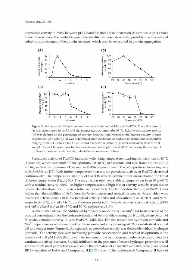
Molecules 2020, 25, 1536
peroxidase activity of ≥90% between pH 2.0 and 5.5 after 1 h of incubation (Figure 5c). At pH valueshigher than six, near the isoelectric point, the stability decreased drastically, probably due to a reducedsolubility and changes of the protein structure, which may have resulted in protein aggregation.
Figure 5. Influence of pH and temperature on activity and stability of PsaPOX. The pH optimum(a) was determined to be 3.5 and the temperature optimum (b) 40 ◦C. Relative peroxidase activity[%] was defined as the percentage of activity detected with respect to the highest activity in eachexperiment. pH stability (c) was determined after incubation of PsaPOX in Britton Robinson bufferranging from pH 2.0 to 9.5 for 1 h at RT and temperature stability (d) after incubation at 20 to 90 ◦Cand pH 3.5 for 1 h. Residual activities were determined at pH 3.5 and 40 ◦C. Values are the average oftriplicate experiments with standard deviations shown as error bars.
Peroxidase activity of PsaPOX increased with rising temperature, reaching its maximum at 40 ◦C(Figure 5b), which was similar to the optimum (30–40 ◦C) of a recombinant DyP from P. ostreatus [13],but higher than the optimum (RT) of another DyP-type peroxidase of P. sapidus produced heterologouslyin Escherichia coli [37]. With further temperature increase, the peroxidase activity of PsaPOX decreasedcontinuously. The temperature stability of PsaPOX was determined after an incubation for 1 h atdifferent temperatures (Figure 5d). The enzyme was relatively stable at temperatures from 20 to 60 ◦Cwith a residual activity ≥80%. At higher temperatures, a high loss of activity was observed due toprotein denaturation, resulting in residual activities <5%. The temperature stability of PsaPOX washigher than the stabilities of DyPs from Bjerkandara adusta and Auricularia auricular-judae, which wereproduced heterologously in E. coli (residual activity ≥80% and <5% after 1 h at 20–50 ◦C and 60 ◦C,respectively [13]), and of a DyP from P. sapidus produced in Trichoderma reesei (residual activity ≥80%and <65% after 5 min at 15-45 ◦C and 50 ◦C, respectively [19]).
As mentioned above, the addition of hydrogen peroxide as well as Mn2+ led to an increase of theproduct concentration for the biotransformation of trans-anethole using the lyophilized mycelium ofP. sapidus containing the wild-type PsaPOX (Table S2). For this reason, the hydrogen peroxide andMn2+ dependencies were examined for the recombinant enzyme using ABTS as substrate at optimalpH and temperature (Figure 6). As expected, no peroxidase activity was detectable without hydrogenperoxide. The activity rose with increasing peroxide concentration and reached its optimum in thepresence of 100 μM H2O2 (Figure 6a). An increase of the hydrogen peroxide concentration led to acontinuous activity decrease. Suicide inhibition in the presence of excess hydrogen peroxide is wellknown for classical peroxidases as a result of the formation of an inactive oxidative state (CompoundIII) by reaction of H2O2 and Compound II [20,21], even if the existence of Compound II has not
75

Molecules 2020, 25, 1536
been confirmed for DyP-type peroxidases universally [17]. However, inhibition of other DyPs in thepresence of higher hydrogen peroxide concentrations has been reported [18,19].
Figure 6. Effect of hydrogen peroxide (a) and Mn2+ concentration (b) on the activity of PsaPOX.Relative peroxidase activity [%] was defined as the percentage of activity detected with respect to thehighest activity obtained in each experiment. Values are the average of triplicate experiments withstandard deviations shown as error bars.
Investigation of the Mn2+ dependency (Figure 6b) showed that PsaPOX activity rose withincreasing Mn2+ concentration, but was not completely dependent on the addition of Mn2+. 30% ofperoxidase activity were detected without addition of Mn2+. PsaPOX reached the maximal activity inthe presence of 25 mM Mn2+. Evaluation of Mn3+ formation by Mn2+ oxidation revealed a manganeseperoxidase activity of 0.4 U compared to 1 U of peroxidase activity using ABTS as substrate. This resultfits the prediction of a Mn2+ oxidation site. Only a few fungal DyPs are known to catalyze the oxidationof Mn2+ [24,27,30,31]. Calculation of kinetic constants (Table 1) showed that the catalytic efficiency ofPsaPOX towards Mn2+ was similar to the one of Pleos-DyP1 from P. ostreatus and Ftr-DyP from Funaliatrogii [24,27]. However, the catalytic efficiency of Pleos-DyP4 was higher [27].
Table 1. Michaelis constants (Km), catalytic constants (kcat), and catalytic efficiencies (kcat/Km) forPsaPOX using ABTS, Mn2+, Reactive blue 19 (RB19), and Reactive black 5 (RB5) as substrate. Values arethe average of triplicate experiments with indication of standard deviations.
Substrate Km (μM) kcat (s−1) kcat/Km (s−1 mM−1)
ABTS 37 ± 4 6.8 ± 0.2 184 ± 5Mn2+ 1025 ± 79 7.2 ± 0.1 7 ± 0.1RB19 n. d. - -RB5 n. d. - -
n. d.: no activity was detected.
Kinetic parameters were also calculated for the oxidation of ABTS at optimal conditions (Table 1).The affinity of PsaPOX to ABTS (37 μM) was similar to a DyP from Irpex lacteus (28 μM), but higher incomparison to the FtrDyP from F. trogii (182 μM) and the Pleos-DyP2 from P. ostreatus (787 μM) [18,24,27].In contrast, the catalytic efficiency of PsaPOX (184 s−1 mM−1) was lower than the efficiency of the DyPfrom Irpex lacteus (8000 s−1 mM−1) and Pleos-DyP4 (352 s−1 mM−1), but higher than the efficiency of theFtrDyP (54 s−1 mM−1).
It is known that DyP-type peroxidases typically oxidize anthraquinones and other dyes. Exemplary,decolorization of Reactive blue 19 (anthraquinone dye) and Reactive black 5 (recalcitrant azo dye)by recombinant PsaPOX (1 U/L) was tested. Unexpectedly, PsaPOX showed activity for neither ofthe substrates (Table 1), although the protein sequence and tertiary structure as well as the presenceof typical catalytic residues and the GXXDG motif identified the enzyme as a DyP-type peroxidase.However, Pleos-DyP1 from P. ostreatus and TvDyP1 from Trametes versicolor also did not oxidize the
76

Molecules 2020, 25, 1536
high redox-potential Reactive black 5, although they degraded Reactive blue 19 [27,30]. A missingactivity against Reactive blue 19 or another anthraquinone has not been described for a fungal or classD type DyP before, but one bacterial DyP of Pseudomonas fluorescens (DyP2B, DyP typ class B) is knownnot to oxidize the anthraquinone dye Reactive blue 4 [38].
2.5. Alkene Cleavage Activity of PsaPOX
To prove the ability of PsaPOX to convert trans-anethole to p-anisaldehyde, biotransformationexperiments were performed at optimal conditions using 1 U/mL peroxidase activity. Substrate cleavagewas detected in the presence of hydrogen peroxide (Table S3), whereas no activity was observed inits absence as expected from the peroxidase activity measurement with ABTS (Figure 6a). Due to thefact that the semi-purified wild-type DyP showed an alkene cleavage activity without addition ofhydrogen peroxide (see Table S2) a low amount of H2O2 must have been present in the analyzed IEXfraction. This was verified by incubation of the fraction with o-dianisidine and HRP in the presence oftrans-anethole. Oxidation of o-dianisidine by HRP, which requires H2O2 as cosubstrate, and formationof a red-brown reaction product occurred (Figure S2). Further, analysis of the IEF fraction regarding ahydrogen peroxide producing enzyme revealed a hypothetical protein from P. ostreatus (KDQ29984.1).It belongs to the glucose-methanol-choline (GMC) oxidoreductase family as evident from the best hit forthe protein band at 75 kDa (Figure 2, lane 1) according to protein sequencing (identified tryptic peptides:AADLIK, AIAVEFVR, ELGGVVDTELR, AQYDAWAELNR, VADASIIPIPVSAHTSSTVYMIGER,DLASGDPHGVGVSPESIDVTNYTR, VLGGSTTINAMLFPR, EVVVSAGTIGTPK) and homology searchagainst the public database NCBI (Figure S3). The protein contained seven of the eight tryptic peptidesidentified for the 75 kDa band. The last one was found with an amino acid exchange (Arg instead ofLys), which is most likely a result of the different fungal strains the proteins originate from. The proteinfrom P. ostreatus showed >92% identity to another hypothetical GMC oxidoreductase from P. ostreatusand ≥55% identity to a glucose oxidase from Moniliophthora roreri and other fungal alcohol oxidases(Figure S3), which belong to the GMC oxidoreductase family and are known for the production ofhydrogen peroxide during substrate oxidation. Thus, the oxidase (75 kDa band) most likely producedthe detected hydrogen peroxide, which was subsequently used as cosubstrate by the wild-type PsaPOX.Due to the fact that the formation of p-anisaldehyde by the oxidase under production of H2O2 seemedhighly unlikely and as no further oxidation products of trans-anethole were detected, trans-anetholewas excluded as substrate. Instead, the buffer component Bis-Tris, which contains several alcoholgroups, or carbohydrate functionalities of other proteins in the IEX fraction were assumed to be usedas substrate by the oxidase.
As described for the wild-type DyP the p-anisaldehyde concentration increased for thebiotransformation with the recombinant enzyme in the presence of 25 mM Mn2+ (Table S3). However,product formation in general was low. The residual peroxidase activity was determined duringthe biotransformation of trans-anethole (Figure S4). After 16 h, 62% of the activity remained,thus inactivation of the enzyme was not responsible for the relatively low product yields.
PsaPOX (1 U/mL) was further examined for alkene cleavage activity regarding other substratesin the presence of hydrogen peroxide and Mn2+. The aryl alkenes (E)-methyl isoeugenol as well asα-methylstyrene, which are derivatives of trans-anethole, were converted to the expected products(veratraldehyde and acetophenone), while piperine was not cleaved (Figure 7a). However, the resultingproduct concentration was fivefold lower for the biotransformation of (E)-methyl isoeugenol andmore than tenfold lower for the conversion of α-methylstyrene than for trans-anethole. Differentsubstrate specificities were also observed for the alkene cleavage by other peroxidases, such as HRP,Coprinus cinereus peroxidase, and a human myeloperoxidase [10,11], but a conversion of aryl alkenesusing a DyP-type peroxidase has not been described before.
77

Molecules 2020, 25, 1536
Figure 7. Alkene cleavage activity of PsaPOX on different substrates in the presence of 100 μM H2O2
and 25 mM MnSO4 at pH 3.5. (a) Product concentration after conversion of trans-anethole (6.7 mM) top-anisaldehyde, (E)-methyl isoeugenol (6.7 mM) to veratraldehyde, and α-methylstyrene (6.7 mM) toacetophenone by PsaPOX (1 U/mL) at RT. The presented product concentrations are the differencesbetween the values determined for the reaction with the active and heat inactivated enzyme (blank) (theoriginal values are shown in Table S4). (b) Decolorization of 7% (v/v) β-carotene and 7% (v/v) annatto byPsaPOX (1 U/L) at 40 ◦C. Cleavage of carotenoids was shown as extinction decrease per min. Valuesare the average of triplicate experiments with standard deviations shown as error bars.
In addition to the described substrates, PsaPOX (1 U/L) also showed an alkene cleavage activitytowards the natural dyes β-carotene and annatto (mixture of the xanthophylls bixin and norbixin),which was detected by substrate bleaching (Figure 7b). The activity for annatto was higher thanfor β-carotene. Cleavage of β-carotene and annatto is also known for other fungal DyPs [19,39–41].For example, cleavage of β-carotene by a DyP from Lepista irina resulted in formation of the volatilesβ-ionone, β-cyclocitral, dihydroactinidiolide, and 2-hydroy-2,6,6-trimethylcyclohexanone [41].
3. Materials and Methods
3.1. Chemicals and Materials
Chemicals were obtained from Sigma-Aldrich (Seelze, Germany), Carl-Roth (Karlsruhe, Germany),or Merck (Darmstadt, Germany) in P. a. quality. Enzymes were from Thermo Fisher Scientific(Braunschweig, Germany), if not stated otherwise. PCR primers were obtained from Eurofins MWGOperon (Ebersberg, Germany).
3.2. Cultivation of P. sapidus
P. sapidus (Deutsche Sammlung von Mikroorganismen und Zellkulturen GmbH, DSMZ,strain no. 2866) was pre-grown on 1.5% (w/v) agar plates with standard nutrient liquid (SNL) mediumand maintained at 4 ◦C until use [39]. For pre-cultivation, 1 cm2 of grown agar was transferred to 100 mLSNL medium and homogenized using an Ultraturrax homogenizer (ART Prozess- & Labortechnik,Müllheim, Germany). The pre-cultures were incubated for 5 days at 150 rpm and 24 ◦C. Afterwards,6.5 g of pre-grown mycelium was used to inoculate 250 mL SNL. The main culture was incubatedat 150 rpm and 24 ◦C. After six days, the mycelium was separated from the culture supernatant bycentrifugation (5000× g, 4 ◦C, 15 min) and lyophilized as described elsewhere [42].
3.3. Purification Strategy
Ten g of lyophilized mycelium were re-suspended in 400 mL buffer A (50 mM Bis-Tris, pH 6.0,1 M (NH4)2SO4) and extracted for 1 h at 4 ◦C in horizontal position in an orbital shaker. Insolublecomponents were removed by centrifugation (5000× g, 4 ◦C, 15 min) followed by filtration (PES filter,
78

Molecules 2020, 25, 1536
0.45 μm, Merck). Subsequently, 80 mL filtered supernatant were applied on a Phenyl Sepharose fastflow column (20 mL, GE Healthcare Bio-Sciences AB, Uppsala, Sweden) pre-equilibrated with buffer A.After the column was washed with buffer A, the active enzyme was eluted with a linear gradient (130 mL,100–0% buffer A) with 100% distilled water at a constant flow rate of 2 mL/min. Active fractions werepooled, desalted and concentrated by ultrafiltration (3 kDa cut off, polyethersulfone (PES), Sartorius,Göttingen, Germany). Concentrate (20 mL) was diluted two times with 20 mM sodium acetate pH4.0 (buffer B) and loaded onto three linked HiTrap SP Sepharose columns (1 mL, GE HealthcareBio-Sciences AB) pre-equilibrated with buffer B. Proteins were eluted with a stepwise ionic strengthgradient (0, 20, 100% buffer C: 20 mM sodium acetate pH 4.0, 1 M NaCl) with 100% buffer C at aconstant flow rate of 1 mL/min.
3.4. Gel Electrophoresis
SDS-PAGE analysis was performed as described elsewhere [43]. Semi-native PAGE was performedunder non-denaturing conditions using 12% gels. For this, samples were prepared with a nativeloading buffer (without DTT and without 2% (w/v; 6.9 mM) SDS) and gel electrophoresis was performedat 10 mA per gel and 4 ◦C. Gels were stained with 0.5 mM ABTS (dissolved in 100 mM sodiumacetate buffer pH 3.5 or 4.5) in the presence of 100 μM hydrogen peroxide for detection of peroxidases.For deglycosylation, samples were treated with 1 μL (500 U) endoglycosidase H (EndoH, New EnglandBioLabs, Ipswich, MA, USA) in 20 μL for 2 h at 37 ◦C before gel electrophoresis.
3.5. Isoelectric Focussing
Analytical isoelectric focusing polyacrylamide gel electrophoresis was performed on a HPETM
BlueHorizonTM system (Serva Electrophoresis GmbH, Heidelberg, Germany) using ServalytTM
PrecotesTM Precast Gels (Serva Electrophoresis GmbH) with an immobilized pH gradient of pH3 to 10. To determine the isoelectric points of the enzymes, marker proteins (IEF-Marker 3-10,Serva Electrophoresis GmbH) were used. Gels were stained with Coomassie Brilliant Blue, or forspecific visualization of peroxidases, with 1% (w/v; 9.2 mM) phenylendiamine and 1% (w/v; 10.6 mM)urea peroxide (dissolved in 100 mM sodium acetate buffer pH 3.5) at RT until a yellow bandwas observed.
3.6. Peptide Mass Fingerprinting
Protein bands were excised from SDS gels, dried, and tryptically hydrolysed. The resultingpeptides were extracted and purified according to standard protocols and the amino acid sequencewas analyzed with electrospray ionization-tandem mass spectrometry (ESI-MS/MS) using a maXisquadrupole time of flight (QTOF) mass spectrometer (Bruker, Bremen, Germany) as describedpreviously [43,44]. The obtained partial sequences of PsaPOX and of the oxidase were used for asimilarity search against public databases (NCBI BlastP).
3.7. cDNA Synthesis and Gene Amplification
Isolation of total RNA from mycelium of P. sapidus at culture day six and cDNA synthesis wereperformed as described previously [13] using the primer 5′-AAGCAGTGGTATCAACGCAGAGTACGCTTTTTTTTTTTTTTTTTTT-3′ for reverse transcription. Specific primers for gene amplificationwere deduced from the ORF-start (P1: 5′-ATGACTACACCTGCACCACCCCTCGACCTC-3′) and -stop(P2: 5′-TCAAGCAGAGATTGGAGCTTGGGTSWGAGGA-3′) region of the homologous peroxidase ofPleurotus ostreatus PC15 (GenBank accession no. KDQ22873.1). PCRs were performed with PhusionHigh-Fidelity DNA Polymerase and the Master Cycler gradient (Eppendorf, Hamburg, Germany)as described elsewhere [45]. The cycler program was as follows: denaturation for 2 min at 98 ◦C,35 cycles at 98 ◦C for 1 min, 62 ◦C for 30 s and 72 ◦C for 90 s, and a final elongation at 72 ◦C for 10 min.Analysis of PCR products, ligation, transformation in Escherichia coli, colony PCR, and sequencingwere performed as described by Behrens et al. [13]. Translation of DNA sequences was performed
79

Molecules 2020, 25, 1536
using SnapGene® (GSL Biotech LLC, Chicago, IL, USA). Sequence homology was examined usingBLAST [46]. Alignments were produced by ClustalOmega [47].
3.8. Heterologous Expression of PsaPOX in Komagataella pfaffii
The gene of PsaPOX was amplified with a C-terminal 6x His tag using theprimers PsaPOX_fw 5′-AAAAAGAATTCatgactacacctgcaccacccctcgacctcaacaa-3′ and PsaPOX_rev5′-atatatGCGGCCGC tcaGTGGTGATGGTGATGATGggtagagatcggagcctgggcctg-3′ (underlined arethe EcoRI and NotI restriction sites, respectively; lower cases represent parts of the coding PsaPOX).In addition, it was inserted in frame with the Saccharomyces cerevisiae α-factor secretion signalsequence into the K. pfaffii pPIC9 expression vector (Invitrogen, Karlsruhe, Germany). The resultingexpression construct pPIC-PsaPOX-His was transformed into E. coli TOP10 for vector propagation,isolated (NucleoSpin, Macherey-Nagel, Düren, Germany), linearized with PmeI, and used fortransformation of K. pfaffii GS115 according to a standard protocol [48]. The linearized emptyvector was transformed in the same way and served as negative control. Forty-eight transformantswere tested for peroxidase activity after selection according to their ability to grow on histidine-deficientagar plates in 96-well plates for 120 h, as described elsewhere [49]. Gene expression was induced bydaily addition of 1% (v/v) methanol.
3.9. His-Tag Purification of Recombinant PsaPOX
For purification of the His-tag labelled recombinant enzyme from K. pfaffii culture supernatant,Ni-NTA affinity chromatography was used according to Nieter et al. [50].
3.10. Biotransformation
Transformation of trans-anethole was carried out in 4 mL gas tight glass vials in horizontal positionat a shaking rate of 200 rpm for 16 h at RT in the absence of light. Reaction mixtures contained 30 mgP. sapidus lyophilisate or 100 μL liquid sample buffered in Bis-Tris (50 mM, pH 6) with or withoutaddition of 1 mM manganese sulfate in a total volume of 1 mL and 1 μL (6.7 mM) trans-anethole. Blanks(chemical: without lyophilisate or liquid sample; biological: with heat inactivated mycelium (1 h at95 ◦C)) were performed the same way. All experiments were performed as duplicates. After incubation,trans-anethole and its conversion product p-anisaldehyde were extracted with 1 mL hexane containing100 mg/L (1 mM) cyclohexanol as internal standard (IS). The organic phase was dried with anhydroussodium sulfate and subsequently analyzed by gas chromatography (GC). GC measurements wereperformed with an Agilent 7890 instrument equipped with a DB-WAX UI column (30 m × 0.32 mm,0.25 μm, Agilent, Santa Clara, CA, USA), a split/splitless injector port (1:5) and a flame ionizationdetection (FID) system. Hydrogen was used as carrier gas at a constant flow rate of 2.1 mL per minute.One μL sample was injected via an autosampler and measured using the following method: 40 ◦C(3 min), a temperature increase of 10 ◦C per minute until 230 ◦C and a final hold time of 10 min.The trans-anethole and p-anisaldehyde were semi-quantified referring to the area of the internalstandard. Biotransformation products were identified using standards and comparison of retentionindices with literature.
3.11. Enzyme Activities
Total peroxidase activity was determined photometrically (EONTM High Performance MicroplateSpectrophotometer, BioTek Instruments GmbH, Bad Friedrichshall, Germany) by monitoring theoxidation of ABTS in the presence of hydrogen peroxide at 420 nm (ε420 = 3.6 × 10⁴ M−1 cm−1) and30 ◦C for 10 min. For this, the samples were mixed with sodium acetate buffer (100 mM, pH 4.0 orpH 3.5), 0.1 mM hydrogen peroxide, and 0.5 mM ABTS in a total volume of 300 μL. One unit of enzymeactivity was defined as 1 μmol substrate oxidized per minute under the experimental conditions.
To determine manganese peroxidase activity, samples were mixed with manganese sulfate (1 mM),malonate buffer (100 mM, pH 3.5), and hydrogen peroxide (0.1 mM) in a total volume of 300 μL.
80

Molecules 2020, 25, 1536
Mn3+ formation was monitored photometrically at 270 nm (ε270 = 1.16 × 10⁴ M−1 cm−1) and 30 ◦C for30 min. One unit of enzyme activity was defined as 1 μmol Mn3+ per minute released by manganeseperoxidases at the given conditions.
Decolorization of Reactive blue 19 (RB19, 150 μM) and Reactive black 5 (RB5, 80 μM) by PsaPOX(1 U/L; 0.25 mg/L) was tested. The respective anthraquinone dye and the enzyme was incubated in thepresence of 100 μM hydrogen peroxide, 25 mM manganese sulfate, and 100 mM sodium acetate bufferpH 3.5 in a total volume of 300 μL at 40 ◦C for 20 min. Decolorization was monitored photometricallyat 595 nm (RB19; ε595 = 1.0 × 10⁴ M−1 cm−1) or 598 nm (RB5; ε598 =3.0 × 10⁴ M−1 cm−1). One unit ofenzyme activity was defined as 1 μmol dye degraded per minute at the given conditions.
All enzyme assays were performed as triplicates. Blanks were carried out with water instead ofenzyme and by omission of hydrogen peroxide.
3.12. Biochemical Characterization of PsaPOX
Effects of pH and temperature on peroxidase activity of PsaPOX (0.25 mg/L) were analyzedwith ABTS as substrate as described above (see “4.11”). Relative activities were normalized to thehighest activity and residual activities to the initial activity prior incubation. The pH optimum wasdetermined using Britton-Robinson buffer [51] in a range of pH 2.0–9.5 instead of sodium acetatebuffer. For determination of the temperature optimum the activity assay was performed at differenttemperatures (20–90 ◦C) at pH 3.5, whilst for analysis of the temperature stability the enzyme wasincubated for 1 h at 20–90 ◦C prior enzyme activity measurement at pH 3.5 and 40 ◦C. For the analysisof pH-stability PsaPOX was incubated in Britton-Robinson buffer from pH 2.0 to 9.5 for 1 h at RT beforethe peroxidase activity was examined at pH 3.5 and 40 ◦C.
Hydrogen peroxide as well as Mn2+ dependency of PsaPOX were determined for PsaPOX byevaluation of peroxidase activity as described above (“4.11”) with changing hydrogen peroxideand manganese sulfate concentrations (H2O2: 0–1 mM H2O2, without addition of MnSO4; Mn2+:100 μM H2O2, 0–100 mM MnSO4) at optimal pH and thermal conditions. Kinetic constants ofPsaPOX were calculated for Mn2+ and ABTS (0–300 μM ABTS in the presence of 100 μM H2O2 and25 mM MnSO4) by SigmaPlot 12.5 (Systat Software Inc., Chicago, IL, USA) with nonlinear regression.Protein concentrations were determined according to Lowry et al. [52] using bovine serum albuminas standard.
3.13. Alkene Cleavage Activity of PsaPOX
The purified recombinant PsaPOX was used for transformation of trans-anethole as mentionedabove (“4.10”) to confirm alkene cleavage activity. For this, 1 U/mL (0.25 mg/mL) of enzyme wasused for biotransformation. Biotransformation was performed with 100 mM sodium acetate bufferpH 3.5 in the presence of 100 μM hydrogen peroxide and 25 mM manganese sulfate at RT for 16 h.Biotransformation of the alkenes methyl isoeugenol (6.7 mM), α-methylstyrene (6.7 mM), and piperine(0.7 mM) was tested accordingly. Blanks were performed without enzyme (chemical blank) or withheat inactivated enzyme (1 h at 95 ◦C, biological blank). The determined product concentrations forthe blanks were subtracted from the concentrations yielded for the reaction with the active enzyme tocalculate the enzymatically generated product concentration. For carotene degradation, a β-caroteneemulsion was prepared according to Linke et al. [43]. 7% (v/v) of β-carotene emulsion or annatto(Chr. Hansen, Nienburg, Germany, Prod. No. 240569), 100 μM hydrogen peroxide, 25 mM manganesesulfate, 100 mM sodium acetate pH 3.5, and 1 U/L (0.25 mg/L) PsaPOX in a total volume of 300 μL wasincubated at 40 ◦C for 20 min. Alkene cleavage of both substrates was measured photometrically asextinction decrease at 455 nm.
3.14. Detection of Hydrogen Peroxide
For the detection of H2O2, 75 μL IEX fraction, 50 mM Bis-Tris pH 6.0, 6.7 mM trans-anethole,10 U/mL HRP (Sigma Aldrich), and 0.5 mM o-dianisidine in a total volume of 300 μL were incubated at
81

Molecules 2020, 25, 1536
RT for 1 h. In the presence of hydrogen peroxide, formation of a red-brown reaction product occurred.Blanks were performed with 50 mM Bis-Tris pH 6.0 instead of IEX fraction.
3.15. Sequence Accession Numbers
The nucleotide sequence of the PsaPOX gene has been deposited in the GenBank database underaccession number MT043310.
4. Conclusions
A DyP-type peroxidase of P. sapidus with alkene cleavage activity as well as the correspondinggene were identified and the gene was heterologously expressed in Komagataella pfaffii. The PsaPOXpossessed typical sequence motifs, structural topology, and catalytic residues as described for DyPs,even though the decolorization of the anthraquinone Reactive blue 19, a common reaction forDyPs, was not observed. A non-canonical Mn2+-oxidation site on the protein surface was detected,which allows PsaPOX to oxidize Mn2+. After biochemical characterization, the alkene cleavageactivity of PsaPOX towards different aryl alkenes was confirmed by biotransformation. PsaPOX is thefirst described DyP-type peroxidase with such an activity. In addition, bleaching of β-carotene andannatto was determined. The results for the alkene cleavage underline the potential of the PsaPOX asbiocatalyst for the generation of aromatic aldehydes with olfactory properties, such as p-anisaldehyde,veratraldehyde, or acetophenone, which are used in the fragrance and flavor industry [1]. Improvementof the conversions and product yields may be accomplished by protein engineering, as has beenshown for the alkene cleaving manganese-dependent Cupin TM1459 from Thermotoga maritima [53].Another application beyond aroma production could be carotene bleaching of whey or wheat dough.
Supplementary Materials: The following are available online, Figure S1: Structural homology model of PsaPOX,Figure S2: Detection of hydrogen peroxide in the IEX fraction with o-dianisidine and HRP in the presenceof trans-anethole after 1 h of incubation at pH 6.0 and RT, Figure S3: Alignment of the hypothetical protein(KDQ29984.1) from P. ostreatus, which was the best hit for the 75 kDa band of the IEX fraction by a homology searchagainst the public database NCBI, and other members of the GMC oxidoreductase family, Figure S4: Stabilityof PsaPOX during biotransformation of trans-anethole over 16 h, Table S1: p-Anisaldehyde concentration afterbiotransformation of trans-anethole with different basidiomycetes, Table S2: p-Anisaldehyde concentration afterbiotransformation of trans-anethole with the active IEX fraction in the presence or absence of Mn2+ and/or H2O2for 16 h at RT, Table S3: p-Anisaldehyde concentration after bioconversion of trans-anethole by recombinantPsaPOX with and without addition of H2O2 and Mn2+ for 16 h at RT, Table S4: p-Anisaldehyde concentrationafter biotransformation of trans-anethole, (E)-methyl isoeugenol, and α-methylstyrene by recombinant PsaPOX(1 U/mL) in the presence of 100 μM H2O2 and 25 mM MnSO4 for 16 h at pH 3.5 and RT.
Author Contributions: Conceptualization, N.-K.K. and R.G.B.; methodology, N.-K.K.; validation, N.-K.K.,R.G.B. and F.E.; formal analysis, N.-K.K.; investigation, N.K.K; writing—original draft preparation, N.-K.K.;writing—review and editing, R.G.B. and F.E.; visualization, N.-K.K.; supervision, F.E.; project administration,R.G.B.; funding acquisition, R.G.B. All authors have read and agreed to the published version of the manuscript.
Funding: This research was funded the BMBF cluster Bioeconomy International 2015, grant number 031B0307A.The APC was funded by the Open Access fund of the Gottfried Wilhelm Leibniz Universität Hannover.
Acknowledgments: B. Fuchs and A. Nieter are thanked for detecting the cleavage reaction and the peroxidaseactivity during the screenings.
Conflicts of Interest: The authors declare no conflict of interest.
References
1. Fahlbusch, K.-G.; Hammerschmidt, F.-J.; Panten, J.; Pickenhagen, W.; Schatkowski, D.; Bauer, K.; Garbe, D.;Surburg, H. Flavors and Fragrances. In Ullmann’s Encyclopedia of Industrial Chemistry; Wiley-VCH VerlagGmbH & Co. KGaA: Weinheim, Germany, 2003; pp. 73–140.
2. Spannring, P.; Bruijnincx, P.C.A.; Weckhuysen, B.M.; Klein Gebbink, R.J.M. Transition metal-catalyzedoxidative double bond cleavage of simple and bio-derived alkenes and unsaturated fatty acids.Catal. Sci. Technol. 2014, 4, 2182–2209. [CrossRef]
82

Molecules 2020, 25, 1536
3. Rajagopalan, A.; Lara, M.; Kroutil, W. Oxidative Alkene Cleavage by Chemical and Enzymatic Methods.Adv. Synth. Catal. 2013, 355, 3321–3335. [CrossRef]
4. Criegee, R. Mechanism of Ozonolysis. Angew. Chemie Int. Ed. Engl. 1975, 14, 745–752. [CrossRef]5. Schrader, J.; Etschmann, M.M.W.; Sell, D.; Hilmer, J.-M.; Rabenhorst, J. Applied biocatalysis for the synthesis
of natural flavour compounds – current industrial processes and future prospects. Biotechnol. Lett. 2004,26, 463–472. [CrossRef] [PubMed]
6. Rajagopalan, A.; Schober, M.; Emmerstorfer, A.; Hammerer, L.; Migglautsch, A.; Seisser, B.; Glueck, S.M.;Niehaus, F.; Eck, J.; Pichler, H.; et al. Enzymatic Aerobic Alkene Cleavage Catalyzed by a Mn3+-DependentProteinase A Homologue. ChemBioChem 2013, 14, 2427–2430. [CrossRef] [PubMed]
7. Han, D.; Ryu, J.-Y.; Kanaly, R.A.; Hur, H.-G. Isolation of a Gene Responsible for the Oxidation oftrans-Anethole to para-Anisaldehyde by Pseudomonas putida JYR-1 and Its Expression in Escherichia coli.Appl. Environ. Microbiol. 2012, 78, 5238–5246. [CrossRef] [PubMed]
8. Yamada, M.; Okada, Y.; Yoshida, T.; Nagasawa, T. Purification, characterization and gene cloning ofisoeugenol-degrading enzyme from Pseudomonas putida IE27. Arch. Microbiol. 2007, 187, 511–517. [CrossRef]
9. Hajnal, I.; Faber, K.; Schwab, H.; Hall, M.; Steiner, K. Oxidative Alkene Cleavage Catalysed byManganese-Dependent Cupin TM1459 from Thermotoga maritima. Adv. Synth. Catal. 2015, 357, 3309–3316.[CrossRef]
10. Tuynman, A.; Spelberg, J.L.; Kooter, I.M.; Schoemaker, H.E.; Wever, R. Enantioselective Epoxidationand Carbon–Carbon Bond Cleavage Catalyzed by Coprinus cinereus Peroxidase and Myeloperoxidase.J. Biol. Chem. 2000, 275, 3025–3030. [CrossRef]
11. Mutti, F.G.; Lara, M.; Kroutil, M.; Kroutil, W. Ostensible Enzyme Promiscuity: Alkene Cleavage byPeroxidases. Chem. Eur. J. 2010, 16, 14142–14148. [CrossRef]
12. Hofrichter, M.; Ullrich, R.; Pecyna, M.J.; Liers, C.; Lundell, T. New and classic families of secreted fungalheme peroxidases. Appl. Microbiol. Biotechnol. 2010, 87, 871–897. [CrossRef] [PubMed]
13. Behrens, C.J.; Zelena, K.; Berger, R.G. Comparative Cold Shock Expression and Characterization of FungalDye-Decolorizing Peroxidases. Appl. Biochem. Biotechnol. 2016, 179, 1404–1417. [CrossRef] [PubMed]
14. Sugano, Y. DyP-type peroxidases comprise a novel heme peroxidase family. Cell. Mol. Life Sci. 2009,66, 1387–1403. [CrossRef] [PubMed]
15. Linde, D.; Ruiz-Dueñas, F.J.; Fernández-Fueyo, E.; Guallar, V.; Hammel, K.E.; Pogni, R.; Martínez, A.T.Basidiomycete DyPs: Genomic diversity, structural–functional aspects, reaction mechanism andenvironmental significance. Arch. Biochem. Biophys. 2015, 574, 66–74. [CrossRef]
16. Strittmatter, E.; Liers, C.; Ullrich, R.; Wachter, S.; Hofrichter, M.; Plattner, D.A.; Piontek, K. First CrystalStructure of a Fungal High-redox Potential Dye-decolorizing Peroxidase. J. Biol. Chem. 2013, 288, 4095–4102.[CrossRef]
17. Rais, D.; Zibek, S. Biotechnological and Biochemical Utilization of Lignin. In Biorefineries; Wagemann, K.,Tippkötter, N., Eds.; Springer International Publishing: Cham, Switzerland, 2017; pp. 469–518.
18. Salvachua, D.; Prieto, A.; Martinez, A.T.; Martinez, M.J. Characterization of a Novel Dye-DecolorizingPeroxidase (DyP)-Type Enzyme from Irpex lacteus and Its Application in Enzymatic Hydrolysis of WheatStraw. Appl. Environ. Microbiol. 2013, 79, 4316–4324. [CrossRef]
19. Lauber, C.; Schwarz, T.; Nguyen, Q.K.; Lorenz, P.; Lochnit, G.; Zorn, H. Identification, heterologousexpression and characterization of a dye-decolorizing peroxidase of Pleurotus sapidus. AMB Express 2017,7, 164. [CrossRef]
20. Busse, N.; Wagner, D.; Kraume, M.; Czermak, P. Reaction Kinetics of Versatile Peroxidase for the Degradationof Lignin Compounds. Am. J. Biochem. Biotechnol. 2013, 9, 365–394. [CrossRef]
21. Arnao, M.B.; Acosta, M.; del Rio, J.A.; Varón, R.; García-Cánovas, F. A kinetic study on the suicide inactivationof peroxidase by hydrogen peroxide. Biochim. Biophys. Acta Protein Struct. Mol. Enzymol. 1990, 1041, 43–47.[CrossRef]
22. Dako, E.; Bernier, A.-M.; Thomas, A.; Jankowski, C.K. The Problems Associated with Enzyme Purification.In Chemical Biology; Ekinci, D., Ed.; InTech: Rijeka, Croatia, 2012; pp. 19–40.
23. Schulz, K.; Nieter, A.; Scheu, A.-K.; Copa-Patiño, J.L.; Thiesing, D.; Popper, L.; Berger, R.G. A type D ferulic acidesterase from Streptomyces werraensis affects the volume of wheat dough pastries. Appl. Microbiol. Biotechnol.2018, 102, 1269–1279. [CrossRef]
83

Molecules 2020, 25, 1536
24. Kolwek, J.; Behrens, C.; Linke, D.; Krings, U.; Berger, R.G. Cell-free one-pot conversion of (+)-valencene to(+)-nootkatone by a unique dye-decolorizing peroxidase combined with a laccase from Funalia trogii. J. Ind.Microbiol. Biotechnol. 2018, 45, 89–101. [CrossRef]
25. Hofrichter, M. Review: Lignin conversion by manganese peroxidase (MnP). Enzyme Microb. Technol. 2002,30, 454–466. [CrossRef]
26. Nakayama, T.; Amachi, T. Fungal peroxidase: Its structure, function, and application. J. Mol. Catal. B Enzym.1999, 6, 185–198. [CrossRef]
27. Fernández-Fueyo, E.; Linde, D.; Almendral, D.; López-Lucendo, M.F.; Ruiz-Dueñas, F.J.;Martínez, A.T. Description of the first fungal dye-decolorizing peroxidase oxidizing manganese(II).Appl. Microbiol. Biotechnol. 2015, 99, 8927–8942. [CrossRef] [PubMed]
28. Fernández-Fueyo, E.; Davó-Siguero, I.; Almendral, D.; Linde, D.; Baratto, M.C.; Pogni, R.; Romero, A.;Guallar, V.; Martínez, A.T. Description of a Non-Canonical Mn(II)-Oxidation Site in Peroxidases. ACS Catal.2018, 8, 8386–8395. [CrossRef]
29. Linde, D.; Pogni, R.; Cañellas, M.; Lucas, F.; Guallar, V.; Baratto, M.C.; Sinicropi, A.; Sáez-Jiménez, V.;Coscolín, C.; Romero, A.; et al. Catalytic surface radical in dye-decolorizing peroxidase: a computational,spectroscopic and site-directed mutagenesis study. Biochem. J. 2015, 466, 253–262. [CrossRef]
30. Amara, S.; Perrot, T.; Navarro, D.; Deroy, A.; Benkhelfallah, A.; Chalak, A.; Daou, M.; Chevret, D.; Faulds, C.B.;Berrin, J.-G.; et al. Enzyme Activities of Two Recombinant Heme-Containing Peroxidases, Tv DyP1 andTv VP2, Identified from the Secretome of Trametes versicolor. Appl. Environ. Microbiol. 2018, 84, e02826-17.[CrossRef]
31. Duan, Z.; Shen, R.; Liu, B.; Yao, M.; Jia, R. Comprehensive investigation of a dye-decolorizing peroxidaseand a manganese peroxidase from Irpex lacteus F17, a lignin-degrading basidiomycete. AMB Express 2018,8, 119. [CrossRef]
32. Passardi, F.; Theiler, G.; Zamocky, M.; Cosio, C.; Rouhier, N.; Teixera, F.; Margis-Pinheiro, M.; Ioannidis, V.;Penel, C.; Falquet, L.; et al. PeroxiBase: The peroxidase database. Phytochemistry 2007, 68, 1605–1611.[CrossRef]
33. Colpa, D.I.; Fraaije, M.W.; van Bloois, E. DyP-type peroxidases: a promising and versatile class of enzymes.J. Ind. Microbiol. Biotechnol. 2014, 41, 1–7. [CrossRef]
34. Romanos, M. Advances in the use of Pichia pastoris for high-level gene expression. Curr. Opin. Biotechnol.1995, 6, 527–533. [CrossRef]
35. Ogawa, S.; Shimizu, T.; Ohki, H.; Araya, T.; Okuno, T.; Miyairi, K. Expression, purification, and analysesof glycosylation and disulfide bonds of Stereum purpureum endopolygalacturonase I in Pichia pastoris.Protein Expr. Purif. 2009, 65, 15–22. [CrossRef] [PubMed]
36. Behrens, C.J.; Linke, D.; Allister, A.B.; Zelena, K.; Berger, R.G. Variants of PpuLcc, a multi-dye decolorizinglaccase from Pleurotus pulmonarius expressed in Pichia pastoris. Protein Expr. Purif. 2017, 137, 34–42. [CrossRef][PubMed]
37. Avram, A.; Sengupta, A.; Pfromm, P.H.; Zorn, H.; Lorenz, P.; Schwarz, T.; Nguyen, K.Q.; Czermak, P. NovelDyP from the basidiomycete Pleurotus sapidus: substrate screening and kinetics. Biocatalysis 2018, 4, 1–13.[CrossRef]
38. Rahmanpour, R.; Bugg, T.D.H. Characterisation of Dyp-type peroxidases from Pseudomonas fluorescens Pf-5:Oxidation of Mn(II) and polymeric lignin by Dyp1B. Arch. Biochem. Biophys. 2015, 574, 93–98. [CrossRef]
39. Linke, D.; Leonhardt, R.; Eisele, N.; Petersen, L.M.; Riemer, S.; Nimtz, M.; Berger, R.G. Carotene-degradingactivities from Bjerkandera adusta possess an application in detergent industries. Bioprocess Biosyst. Eng. 2015,38, 1191–1199. [CrossRef]
40. Scheibner, M.; Hülsdau, B.; Zelena, K.; Nimtz, M.; de Boer, L.; Berger, R.G.; Zorn, H. Novel peroxidases ofMarasmius scorodonius degrade β-carotene. Appl. Microbiol. Biotechnol. 2008, 77, 1241–1250. [CrossRef]
41. Zorn, H.; Langhoff, S.; Scheibner, M.; Nimtz, M.; Berger, R.G. A Peroxidase from Lepista irina Cleaves β,β-Carotene to Flavor Compounds. Biol. Chem. 2003, 384, 1049–1056. [CrossRef]
42. Krings, U.; Lehnert, N.; Fraatz, M.A.; Hardebusch, B.; Zorn, H.; Berger, R.G. Autoxidation versusbiotransformation of α-pinene to flavors with Pleurotus sapidus: Regioselective hydroperoxidation ofα-pinene and stereoselective dehydrogenation of verbenol. J. Agric. Food Chem. 2009, 57, 9944–9950.[CrossRef]
84

Molecules 2020, 25, 1536
43. Linke, D.; Omarini, A.B.; Takenberg, M.; Kelle, S.; Berger, R.G. Long-Term Monokaryotic Cultures ofPleurotus ostreatus var. florida Produce High and Stable Laccase Activity Capable to Degrade ß-Carotene.Appl. Biochem. Biotechnol. 2019, 187, 894–912. [CrossRef]
44. Schulz, K.; Giesler, L.; Linke, D.; Berger, R.G. A prolyl endopeptidase from Flammulina velutipes for thepossible degradation of celiac disease provoking toxic peptides in cereal proteins. Process Biochem. 2018,73, 47–55. [CrossRef]
45. Nieter, A.; Haase-Aschoff, P.; Linke, D.; Nimtz, M.; Berger, R.G. A halotolerant type A feruloyl esterase fromPleurotus eryngii. Fungal Biol. 2014, 118, 348–357. [CrossRef] [PubMed]
46. Altschul, S.F.; Gish, W.; Miller, W.; Myers, E.W.; Lipman, D.J. Basic local alignment search tool. J. Mol. Biol.1990, 215, 403–410. [CrossRef]
47. Sievers, F.; Wilm, A.; Dineen, D.; Gibson, T.J.; Karplus, K.; Li, W.; Lopez, R.; McWilliam, H.; Remmert, M.;Söding, J.; et al. Fast, scalable generation of high-quality protein multiple sequence alignments using ClustalOmega. Mol. Syst. Biol. 2011, 7, 539. [CrossRef] [PubMed]
48. Lin-Cereghino, J.; Wong, W.W.; Xiong, S.; Giang, W.; Luong, L.T.; Vu, J.; Johnson, S.D.; Lin-Cereghino, G.P.Condensed protocol for competent cell preparation and transformation of the methylotrophic yeast Pichiapastoris. Biotechniques 2005, 38, 44–48. [CrossRef]
49. Sygmund, C.; Gutmann, A.; Krondorfer, I.; Kujawa, M.; Glieder, A.; Pscheidt, B.; Haltrich, D.; Peterbauer, C.;Kittl, R. Simple and efficient expression of Agaricus meleagris pyranose dehydrogenase in Pichia pastoris.Appl. Microbiol. Biotechnol. 2012, 94, 695–704. [CrossRef]
50. Nieter, A.; Kelle, S.; Takenberg, M.; Linke, D.; Bunzel, M.; Popper, L.; Berger, R.G. Heterologous productionand characterization of a chlorogenic acid esterase from Ustilago maydis with a potential use in baking.Food Chem. 2016, 209, 1–9. [CrossRef]
51. Britton, H.T.S.; Robinson, R.A. CXCVIII.—Universal buffer solutions and the dissociation constant of veronal.J. Chem. Soc. 1931, 1456–1462. [CrossRef]
52. Lowry, O.H.; Rosebrpugh, N.J.; Randall, R.J. Protein measurement with the Folin phenol reagent. J. Biol. Chem.1951, 193, 265–275.
53. Fink, M.; Trunk, S.; Hall, M.; Schwab, H.; Steiner, K. Engineering of TM1459 from Thermotoga maritima forincreased oxidative alkene cleavage activity. Front. Microbiol. 2016, 7, 1511. [CrossRef]
Sample Availability: Samples of the pPIC9 vector containing the PsaPOX gene are available from the authors.
© 2020 by the authors. Licensee MDPI, Basel, Switzerland. This article is an open accessarticle distributed under the terms and conditions of the Creative Commons Attribution(CC BY) license (http://creativecommons.org/licenses/by/4.0/).
85


molecules
Article
Enrichment of Polyglucosylated Isoflavones fromSoybean Isoflavone Aglycones Using OptimizedAmylosucrase Transglycosylation
Young Sung Jung 1,†, Ye-Jin Kim 2,†, Aaron Taehwan Kim 1, Davin Jang 2, Mi-Seon Kim 2,
Dong-Ho Seo 3, Tae Gyu Nam 4, Chan-Su Rha 1, Cheon-Seok Park 2 and Dae-Ok Kim 1,2,*
1 Department of Food Science and Biotechnology, Kyung Hee University, Yongin 17104, Korea;[email protected] (Y.S.J.); [email protected] (A.T.K.); [email protected] (C.-S.R.)
2 Graduate School of Biotechnology, Kyung Hee University, Yongin 17104, Korea; [email protected] (Y.-J.K.);[email protected] (D.J.); [email protected] (M.-S.K.); [email protected] (C.-S.P.)
3 Department of Food Science and Technology, Jeonbuk National University, Jeonju 54896, Korea;[email protected]
4 Food Analysis Center, Korea Food Research Institute, Wanju 55365, Korea; [email protected]* Correspondence: [email protected]; Tel.: +82-31-201-3796† These authors contributed equally to this work.
Academic Editor: Josefina AleuReceived: 6 December 2019; Accepted: 30 December 2019; Published: 1 January 2020
Abstract: Isoflavones in soybeans are well-known phytoestrogens. Soy isoflavones present inconjugated forms are converted to aglycone forms during processing and storage. Isoflavone aglycones(IFAs) of soybeans in human diets have poor solubility in water, resulting in low bioavailabilityand bioactivity. Enzyme-mediated glycosylation is an efficient and environmentally friendly way tomodify the physicochemical properties of soy IFAs. In this study, we determined the optimal reactionconditions for Deinococcus geothermalis amylosucrase-mediated α-1,4 glycosylation of IFA-rich soybeanextract to improve the bioaccessibility of IFAs. The conversion yields of soy IFAs were in decreasingorder as follows: genistein > daidzein > glycitein. An enzyme quantity of 5 U and donor:acceptorratios of 1000:1 (glycitein) and 400:1 (daidzein and genistein) resulted in high conversion yield(average 95.7%). These optimal reaction conditions for transglycosylation can be used to obtaintransglycosylated IFA-rich functional ingredients from soybeans.
Keywords: Deinococcus geothermalis; glycosyltransferase; Glycine max (L.) Merr.; HPLC/MS; isoflavoneaglycone-rich extract; isoflavone α-glucoside
1. Introduction
Globally, soybean (Glycine max (L.) Merr.) is a large portion of major grain production, alongwith corn, wheat, and rice [1]. Isoflavones, a subgroup of flavonoids, are well-known plantsecondary metabolites that are found mainly in the plant families Fabaceae or Leguminosae, includingsoybean [2,3]. Isoflavones in soybeans are present mainly in conjugated forms [4]. Soy isoflavonesare classified as acetylisoflavones, malonylisoflavones, non-acylated isoflavone glucosides, andisoflavone aglycones (IFAs), depending on the side groups attached to the isoflavone skeleton [5].IFAs have antioxidant properties, interact with epigenetic modifications, and exhibit estrogen-likeactivities [3]. Dietary IFAs can be metabolized to equol (4’,7-isoflavandiol), which has significantestrogenic activity in the intestine [6]. Soy isoflavones exist naturally in conjugated forms ratherthan as aglycones; for example, daidzein [7-hydroxy-3-(4-hydroxyphenyl)chromen-4-one], glycitein[7-hydroxy-3-(4-hydroxyphenyl)-6-methoxychromen-4-one], and genistein [5,7-dihydroxy-3-(4-hydroxyphenyl)chromen-4-one] [5]. However, conjugated isoflavones are converted into aglycone
Molecules 2020, 25, 181; doi:10.3390/molecules25010181 www.mdpi.com/journal/molecules87

Molecules 2020, 25, 181
forms during processing or storage [4]. The bioaccessibility of IFAs is low due to their poor solubilityin water [7]. Therefore, various methods have been developed to overcome these shortcomings ofIFAs [8,9].
Glycosylation via chemical or non-chemical methods can change the physicochemical propertiesof compounds [10,11]. The use of enzymes, such as glycosyltransferases, is a natural way to improvethe solubility of IFA [12]. Enzymatic glycosylation reactions are efficient and environmentallyfriendly [13]. Compared with harsh chemical methods, enzymatic methods typically generate fivetimes less waste and have a 15-fold higher space–time yield [14]. Recently, biological glycosylationmethods using a variety of enzymes to improve the water solubility of lipophilic compounds havebeen developed [11,15,16]. Transglycosylation improved genistein solubility by 3700–44,000 [17] andtransglycosylated catechin, naringin, and rutin were found to have 100-, 1000-, and 30,000-fold higherwater solubility than non-transglycosylated catechin, naringin, and rutin, respectively [18–20]. Theuse of glycosyltransferases from microbial sources for transglycosylation reactions is an efficientand eco-friendly biological alternative to chemical reactions [21]. Flavonoids can be glycosylatedusing enzymes from microbial sources such as cyclodextrin glycosyltransferase (CGTase) and uridinediphosphate glucose (UDP-glucose) [16,22–24]. CGTase is widely employed to increase the solubilityand stability of polyphenols such as glycosylated isoflavones [23,25]. However, the CGTase-reactedcomplex solution is cloudy and contains high molecular weight compounds [26], while the UDP-glucosecomplex has low reaction yields and is expensive [16,27].
Amylosucrase (EC 2.4.1.4; AS) is a glycosyltransferase enzyme that belongs to the glucosidehydrolase family 13 [28]. One of the main characteristics of AS is that it catalyzes transglycosylationreactions to create α-1,4-glycosidic linkages given sucrose as a substrate [28,29]; AS promotes sucrosehydrolysis to release glucose and fructose, and α-1,4-oligosaccharides are formed using the releasedglucose as an acceptor [30]. Deinococcus geothermalis AS (DGAS) synthesizes α-1,4-glycosidic bondsusing various acceptors such as flavonoids [31]. DGAS enhances the water solubility of flavonoidaglycones by transglycosylation [21,31,32]. Compared with glycosyltransferase enzymes such asCGTase, DGAS can react with high substrate specificity using relatively inexpensive donors [27].In addition, the interpretation of experimental results is easier when using DGAS because of fewerimpurities and the limited number of transglycosylated sugar moieties [27,32,33]. These features ofDGAS can be exploited to increase the solubility and bioavailability of bioactive phenolic compoundsin soybeans.
In this study, we investigated the physicochemical changes of major soybean IFAs in responseto DGAS-mediated transglycosylation. To determine what reaction conditions resulted in hightransglycosylated conversion yields of IFAs, factors such as the amount of donors, acceptors, andenzymes were evaluated. Optimal reaction conditions were applied to soybean isoflavone extractto confirm that the IFAs were efficiently transglycosylated. Based on our results, we propose anapplication of the DGAS enzyme process to enrich transglycosylated IFAs in soy-based foods such assoybeans, soybean extracts, and soy products in the industrial field.
2. Results and Discussion
2.1. DGAS Expression and Activities
DGAS was successfully expressed by Escherichia coli transformed with the shuttle vectorpHCXHD-DGAS as confirmed by the detection of dgas expression in transformed cells (Figure 1a).Activity of AS was observed in cell extracts of pHCXHD-DGAS-transformed cells. DGAS fused with6× histidine appeared as a single band with a molecular mass of about 73 kDa (Figure 1b), which is ingood agreement with the estimated molecular mass of DGAS. This result confirmed that DGAS wassuccessfully expressed as a functional protein in E. coli. DGAS was considered to be free of proteintoxicity because it was not harmful to E. coli. Temperature and pH profiles of DGAS expressed inE. coli were investigated in the range of 30 to 55 ◦C and pH 4.0 to 9.0, respectively, based on a previous
88

Molecules 2020, 25, 181
report of DGAS expression in E. coli [30]. It was previously reported that isoflavone glucosides can bethermally transformed to their corresponding aglycone forms during processing [34]. In considerationof the stability of the IFAs, the temperature and pH for the DGAS-mediated transglycosylation of IFAsin this study were set at 45 ◦C and pH 5.0, respectively.
(a)
(b)
Figure 1. (a) Construction of expression vector of Deinococcus geothermalis amylosucrase (DGAS).(b) SDS-PAGE analysis of recombinant DGAS expressed in Escherichia coli and purified on a nickel–nitrilotriacetic acid affinity column. (Lane M, protein molecular standard marker; Lane 1, crude enzyme;Lane 2, crude passing through enzyme; Lane 3, inclusion body; Lane 4, purified enzyme).
2.2. Transglycosylation of IFAs with DGAS
Three IFAs (daidzein, glycitein, and genistein) are found in soybeans, and their glucosides arelinked by β-glycosidic bonds at C-7 (Figure 2) [4]. Glycosylation carried out by glycosyltransferases,such as AS, generates two types of O-glucosides, which are commonly defined as α- and β-linkedglucosides [35]. The detailed glycosylation mechanism has not been clearly elucidated, but theglycosylation reaction usually occurs on the anomeric carbon with the attack of weak nucleophiles,such as the −OH group of sugars [35]. Glycosylation reactions often involve a unimolecular (SN1) orbimolecular (SN2) nucleophilic substitution at the anomeric center [35].
Figure 2. Structures of isoflavone glucosides (daidzin, glycitin, and genistin) present in soybeans.
89

Molecules 2020, 25, 181
DGAS used in this study produced various transglycosylated isoflavones with α-1,4-glycosidiclinkages from IFAs (Figure 3). The transglycosylation of IFAs (such as in genistein) using DGAS arelikely to occur at the C-7 or C-4′ positions rather than the C-5 position. Previous studies reportedthat recombinant AS created various stereostructures of acceptor compounds due to position-specifictransglycosylation [27,33] (Figure 3). Moreover, the transglycosylation of flavonoids using DGAS hasbeen reported to occur primarily at the C-7 position of the A ring and at the C-4′ position of the Bring [27,32,36]. DGAS has been reported to attach four or fewer glucose moieties to flavonoids intransglycosylation [21,32]. Thus, we expected the total number of glucose moieties attached to IFAs inthe DGAS transglycosylation process to be four. For example, if four glucoses bind to the C-7 position,the number of glucoses that can attach to the C-4′ position is zero, and if three glucoses bind to theC-4′ position, one additional glucose may bind to the C-7 position (Figure 3).
Figure 3. Scheme of transglycosylation reactions of isoflavone aglycones (IFAs) and sucrose usingDGAS. G, glucose; F, fructose.
To identify reactants and products after transglycosylation, 12 isoflavone standards found insoybeans were separated by high-performance liquid chromatography (HPLC; peaks labeled: GD,daidzin; GL, glycitin; GG, genistin; MD, malonyldaidzin; ML, malonylglycitin; AD, acetyldaidzin; AL,acetylglycitin; MG, malonylgenistin; DN, daidzein; LN, glycitein; AG, acetylgenistin; GN, genistein)(Figure 4a). The results of transglycosylation using DGAS of the three IFA standards are shownchromatographically in Figure 4b–d. Compared to the 12 isoflavone standards, six to eight new peakswere detected, indicating that various transglycosylated products were produced from each aglyconeduring the enzymatic process (Figure 4b–d). Transglycosylated products from daidzein tended tobe more polar (faster retention time) than transglycosylated glycitein and genistein glucosides forthe same HPLC analysis conditions. This tendency was the same as the elution order of the threeaglycones: daidzein > glycitein > genistein (Figure 4a). We attributed the different elution order oftransglycosylated IFAs to their polarity differences, which are mainly influenced by the polarity of theparent compounds (acceptors in this study). If patterns of bound positions and the number of glucosemoieties were the same in each aglycone, the elution order of transglycosylated IFAs according toincreasing retention time would have been as follows: transglycosylated daidzein > transglycosylatedglycitein > transglycosylated genistein. It was previously reported that α-glycosylisoquercitrin withmore glucose moieties produced by the enzymatic transglycosylation of isoquercitrin eluted earlier thanα-glycosylisoquercitrin with fewer glucose moieties [37]. Furthermore, after the transglycosylation ofdaidzin using DGAS, daidzein triglucoside eluted faster than daidzein diglucoside [36]. Therefore, inthis study, transglycosylated IFAs with more glucose moieties had a faster elution time in reversed-phaseHPLC analysis than transglycosylated IFAs with fewer glucose moieties (Figure 4b–d).
90

Molecules 2020, 25, 181
Figure 4. High-performance liquid chromatography (HPLC) traces (254 nm) of IFA standards aftertransglycosylation using DGAS. (a) Isoflavone standard mixture, (b) transglycosylated glycitein,(c) transglycosylated daidzein, and (d) transglycosylated genistein. Peaks: GD, daidzin; GL, glycitin;GG, genistin; MD, malonyldaidzin; ML, malonylglycitin; AD, acetyldaidzin; AL, acetylglycitin; MG,malonylgenistin; DN, daidzein; LN, glycitein; AG, acetylgenistin; GN, genistein. Peaks labeled LN1
to LN6, DN1 to DN8, and GN1 to GN6 are transglycosylated products from each IFA. Enzymatictransglycosylation reactions were performed using 1.0 mM of glycitein, 5.0 mM of daidzein, or 5.0 mMof genistein with 5.0 U of DGAS and 2.0 M of sucrose.
Peaks observed at the very beginning of elution (up to 12 min of retention time in this study) of thechromatograms were considered to be transglycosylated IFAs with two, three, or four glucose moieties(Figure 4b–d). However, more polar products (transglycosylated IFAs with 2–4 glucose moieties)among the transglycosylated IFAs resulted in lower resolution in the chromatogram. It was previouslyreported that transglycosylated products generated from the DGAS-catalyzed transglycosylation ofisoquercitrin had similar retention times to each other, which resulted in very low resolution andgreater difficultly separating them [32]. In addition, some isoquercitrin glucosides with high polarityhave been reported to exist as isomers with the same number of sugars, the molecular weights ofwhich were the same in mass spectra [32]. Based on previously reported results [32], we hypothesizedthat the transglycosylated IFA glucosides that eluted at similar retention times in this study were likelyto be isomers with the same number of glucose moieties bound at different positions.
Peaks (except for minor peaks< 5% of the total product area) of transglycosylated reaction productsfrom glycitein, daidzein, and genistein after enzymatic modification were labeled (Figure 4b–d). PeakLN5 in Figure 4b had a retention time similar to glycitin (glycitein 7-β-O-glucoside; peak GL of Figure 4a).
91

Molecules 2020, 25, 181
The retention times of peak DN7 in Figure 4c and peak GN6 in Figure 4d were similar to those of daidzin(daidzein 7-β-O-glucoside; peak GD of Figure 4a) and genistin (genistein 7-β-O-glucoside; peak GGof Figure 4a), respectively. Thus, we assumed that peaks LN5, DN7, and GN6 were α-glucosides ofIFA with one glucose moiety attached. Standard compounds (daidzin, glycitin, and genistin) found insoybeans have a β-glycosidic bond between IFA and glucose. When IFAs are transglycosylated usingDGAS, glucose moieties bound to the −OH group at the C-7 or C-4′ position in IFAs are present inan α-glycosidic bond with IFAs [27,33]. The 7-α-monoglucosides (peaks LN5, DN7, and GN6) fromthe enzymatic modification of IFAs had different but similar retention times compared to standardisoflavone 7-β-monoglucosides (Figure 4b–d). Previously, daidzein triglucoside was reported to bemore polar than daidzein diglucoside [36]. Due to their greater number of transglycosylated glucosemoieties, LN1–LN4 from glycitein transglycosylation, DN1–DN6 from daidzein transglycosylation,and GN1–GN5 from genistein transglycosylation had higher polarities than LN5, DN7, and GN6,respectively (Figure 4b–d). DGAS produces higher amounts of flavonoid glucosides attached witha greater number of glucose molecules to parent flavonoids such as isoquercitrin and daidzin thanglycosyltransferases such as CGTase and amyloglucosidase [27,32,36], which produce higher levels offlavonoid glucosides transglycosylated with fewer sugar moieties [22,24]. When UDP-glucose is usedas a donor for glycosidic binding, only one or two new transglycosylated products are produced byglycosyltransferases [16,22]. In contrast, DGAS generated a variety of new transglycosylated productsfrom the IFAs (Figure 4b–d). As shown in Figure 3, the number of transglycosylated glucoses hasbeen reported to range from one to four [27,32], because the free hydroxyl (−OH) groups of flavonoidaglycones and their transglycosylated glucosides can be used as potential transglycosylation sites inthe DGAS enzyme process.
2.3. Effects of Reaction Conditions on DGAS Transglycosylation
Table 1 shows the conversion yields of IFAs to transglycosylated IFA glucosides according tothe concentrations of donor and acceptors as well as the amount of DGAS. Glycitein reacted at lowerconcentrations than daidzein and genistein because of its lower solubility [38]. Regardless of IFA andsucrose concentrations, the conversion yield of each IFA used in this study increased as the amount ofDGAS used increased, whereas the conversion yield of each IFA generally decreased with increasingIFA concentration. In transglycosylation reactions, a large amount of enzymes ensures not only a fastreaction rate, but also an increased reaction yield [33]. However, at a low donor concentration (0.1 M)and high acceptor concentration (20 mM), the conversion yields for daidzein and genistein were thelowest during transglycosylation for the highest amount of enzymes (5.0 U). The ratio of donor toacceptor suggests that the conversion yield is affected even if a sufficient amount of enzyme is supplied.Similar to the results of this study (Table 1), previous studies reported that the conversion efficiencyincreased when the concentration of the donor was higher than that of the acceptor [31,39,40]. Theenzyme-modified glycosylation of lipophilic compounds is known to be complex because of the lowsolubility of lipophilic compounds in aqueous systems [41]. To increase the conversion yield despitelow solubility, various solvents such as ionic liquids have been used in enzyme reactions for flavonoidglucoside synthesis [13,41]. In this study, we used dimethyl sulfoxide (DMSO) as the solvent to dissolveall IFAs (acceptors). However, cloudiness was observed during the enzyme reaction when the amountof acceptor was increased in an aqueous system containing a donor, acceptor, buffer, and enzyme (datanot shown). Acceptor solubility was found to be the most important factor determining the lowestconversion yields at the highest acceptor conditions (glycitein, 4 mM; daidzein and genistein, 20 mM)(Table 1). Therefore, it is reasonable to assume that the reason glycitein had the lowest conversionyield was its relatively lower solubility than that of daidzein and genistein.
92

Molecules 2020, 25, 181
Ta
ble
1.
Con
vers
ion
yiel
dsof
soy
isofl
avon
eag
lyco
nes
afte
rD
eino
cocc
usge
othe
rmal
isam
ylos
ucra
se(D
GA
S)-t
reat
edtr
ansg
lyco
syla
tion
acco
rdin
gto
conc
entr
atio
nsof
dono
ran
dac
cept
ors
and
amou
nts
ofth
eD
GA
Sen
zym
e.
DG
AS
(Un
its)
Su
cro
se(M
)
Gly
cite
in(m
M)
Da
idz
ein
(mM
)G
en
iste
in(m
M)
0.0
51
.04
.00
.25
.02
00
.25
.02
0
0.5
0.1
0.0±0
.0c
11.
9±1
.7c
3.9±0
.2b
0.0±0
.0f
4.3±0
.6de
2.4±0
.4ef
10.9±2
.3c
3.1±1
.3de
2.1±0
.1e
1.0
0.0±0
.0c
6.8±0
.8a
4.2±0
.2b
20.0±2
.5b
8.3±1
.0cd
2.7±0
.1ef
59.8±0
.4b
5.3±0
.3d
0.8±0
.0e
2.0
0.0±0
.0c
6.5±0
.8a
4.9±0
.4ab
28.8±3
.0a
9.2±1
.0c
1.8±0
.2ef
68.6±1
.0a
3.5±0
.1de
1.2±0
.0e
1.0
0.1
0.0±0
.0d
7.0±0
.2c
6.0±0
.3c
22.5±3
.2d
12.0±0
.9e
4.4±1
.1f
19.3±1
.9d
7.1±0
.6e
1.3±0
.0e
1.0
13.3±2
.5b
19.0±1
.7a
8.8±0
.4bc
50.0±2
.4b
32.5±0
.7c
6.9±0
.1f
53.1±4
.2b
28.5±0
.2c
4.8±0
.0e
2.0
8.6±3
.6bc
20.8±0
.4a
23.2±3
.7a
55.5±2
.2a
32.4±0
.8c
4.8±0
.2f
62.5±4
.7a
24.7±0
.2cd
2.9±0
.2e
5.0
0.1
20.8±6
.4e
35.0±1
.6d
28.2±0
.8de
72.0±1
.5b
65.0±0
.8c
23.3±0
.3f
77.3±2
.7b
74.8±0
.5bc
23.4±0
.0e
1.0
16.3±1
.5e
69.5±9
.2b
56.8±2
.2c
97.2±2
.8a
95.0±0
.1a
50.5±0
.0d
92.2±5
.1a
96.6±1
.0a
66.0±0
.2d
2.0
18.0±2
.8e
88.8±3
.8a
52.1±1
.3c
97.5±0
.7a
96.9±0
.2a
36.9±0
.1f
92.1±3
.2a
98.2±0
.1a
70.1±0
.1cd
1D
ata
are
expr
esse
das
mea
ns±s
tand
ard
devi
atio
ns(n=
3).M
eans
wit
hdiff
eren
tsup
ersc
ript
edle
tter
sin
dica
teth
atfo
rth
esa
me
amou
ntof
DG
AS,
each
isofl
avon
eag
lyco
nediff
ered
sign
ifica
ntly
byth
eTu
key–
Kra
mer
hone
stly
sign
ifica
ntdiff
eren
cete
st(p<
0.05
).
93

Molecules 2020, 25, 181
The conversion yields of daidzein tended to be similar to those of genistein for the variousreaction conditions used in this study. Glycitein had a lower conversion yield than genistein anddaidzein (Table 1). Transglycosylation has been reported to occur at the hydroxyl group of the acceptor,resulting in the formation of an O-glycosidic bond between the acceptor and donor [35]. Likewise,a correlation between the number of hydroxyl groups and reactivity after the transglycosylationof hydroxyflavonoids with DGAS was observed [27]. The average conversion yields of daidzeinand genistein were approximately 96.7% and 94.8%, respectively, at the highest amount of enzyme(5.0 U), lower acceptor concentrations (0.2–5.0 mM), and higher donor concentrations (1.0–2.0 M).In general, the highest conversion yield is achieved when sufficient enzymes and sucrose are providedfor transglycosylation [33]. However, excess sucrose has been reported to reduce conversion yields [32].Therefore, it is essential to have an adequate ratio of donor and acceptor as well as a sufficient amountof enzyme to attain higher conversion yields.
2.4. Application of DGAS to IFA-Rich Extract from Soybeans
Ultrasound-assisted extraction is known by its high efficiency, high productivity, lowerconsumption of solvents, and eco-friendly process [42]. In addition, in this study, to preventprotein interference after the extraction of soybeans with organic solvent, proteins were precipitated bydehydration and aggregation using ice-cold acetone [43]. After the successful conversion of IFAs intotransglycosylated IFAs, optimal reaction conditions established for the DGAS process were used forthe transglycosylation of IFA-rich soybean extract (SBE) as the acceptor. IFAs in SBE existed at lowerconcentrations than the other isoflavones such as isoflavone glucosides (Figure 5a and Table 2).
Table 2. Content (mg/g powder) of isoflavones in soybean extract (SBE), cellulase (CE)-treated SBE(CE-SBE; isoflavone aglycone-rich extract), and CE-SBE transglycosylated using DGAS (CE-SBE-DGAS).
Isoflavone SBE CE-SBE CE-SBE-DGAS
Glycitein 0.0 ± 0.0 b 1 3.98 ± 0.2 a 0.0 ± 0.0 b
Daidzein 0.79 ± 0.13 b 41.98 ± 1.26 a 0.0 ± 0.0 c
Genistein 1.0 ± 0.03 b 53.63 ± 4.35 a 0.0 ± 0.0 c
1 Data are expressed as means ± standard deviations (n = 3). Means with different superscripted letters in the samerow differ significantly by the Tukey-Kramer honestly significant difference test (p < 0.05).
Isoflavones in soybeans are mainly present in malonyl forms when extracted under normalconditions such as neutral solvent, ambient temperature, and short extraction time [44]. Similarly,malonyl forms were found to be the major isoflavones in SBE in this study (Figure 5a). Peaks labeledMD, ML, and MG are malonyldaidzin, malonylglycitin, and malonylgenistin, respectively (Figures4a and 5a,b). Therefore, 7-β-O-glucoses (with acetyl and malonyl moieties) bound to IFA should behydrolyzed using cellulase (CE) prior to DGAS transglycosylation. Extraction under harsh conditions(low pH and high temperature) and microbial fermentation has been used previously to obtain IFAs fromsoybeans [44]. However, acid-modified extraction is not suitable for enzymatic glycosylation reactionsand is not environmentally friendly. Conjugated isoflavones (malonylisoflavones, acetylisoflavones,and non-acylated isoflavone glucosides) are converted into their non-conjugated counterparts usingenzymes such as glycosylase [45]. In this study, we obtained IFAs by removing β-1,4-linked glucosemoiety from conjugated isoflavones using CE. After CE processing, malonylisoflavones were degradedinto their corresponding aglycones. As the concentrations of conjugated daidzeins (peaks GD, MD,and AD), conjugated glyciteins (peaks GL, ML, and AL), and conjugated genisteins (peaks GG andMG) in SBE decreased (Figure 5a), the concentrations of their corresponding IFAs increased after CEtreatment (Figure 5b). CE-treated soybean extract (CE-SBE; IFA-rich extract) had the highest content ofgenistein (53.9% of total), daidzein (42.2% of total), and glycitein (4.0% of total) compared with SBEand CE-SBE transglycosylated using DGAS (CE-SBE-DGAS) (Table 2). Consistent with the results
94

Molecules 2020, 25, 181
of this study, the content of glycitein derivatives in soybeans and their processed products has beenreported to be less than 5% of the total isoflavone content [5].
Figure 5. HPLC traces (254 nm) of IFA-rich soybean extract after transglycosylation using DGAS.(a) Soybean extract, (b) cellulase-treated soybean extract, (c) and cellulase-treated soybean extracttransglycosylated using DGAS. Peaks: GD, daidzin; GL, glycitin; GG, genistin; MD, malonyldaidzin;ML, malonylglycitin; AD, acetyldaidzin; AL, acetylglycitin; MG, malonylgenistin; DN, daidzein;LN, glycitein; GN, genistein. Refer to Table 3 for the identification of each numbered peak of the19 compounds.
Soy isoflavones have different ultraviolet-visible spectra depending on their aglycones [3,46]. Thepeaks newly produced by DGAS-treated transglycosylation were tentatively confirmed by comparisonwith the patterns of ultraviolet-visible spectra of isoflavone standards and molecular weight (Table 3).As results of mass spectrometry (MS), 19 products including four unknown compounds of DGAS-treatedCE-SBE were tentatively identified (Table 3). The malonylisoflavones in SBE were not changed by DGAS(peaks 15, 16, and 17 of Figure 5c) in this study. In contrast, IFAs were not detected in CE-SBE-DGAS(Figure 5c and Table 2), indicating that IFAs in CE-SBE are successfully transglycosylated using DGAS.In previous studies, DGAS has been reported to transglycosylate a sugar moiety to the −OH groupof the flavonoid aglycone [27,30,31]. It is also reported that glucose moiety was transferred to theisoflavone monoglucosides such as daidzin [36].
Among the peaks newly generated using DGAS, the peaks 10, 12, and 13 were identified asdaidzein, glycitein, and genistein monoglucosides with different stereospecificity and retention timefrom daidzin, glycitin, and genistin, respectively (Figure 5c and Table 3). The generated isoflavonemonoglucosides can be seen to have a similar polarity compared to the isoflavone 7-β-O-glucosides(peaks GD, GL, and GG) in the SBE (Figure 5a). The MS results of CE-SBE-DGAS indicate that the morepolar components, which are eluted earlier than isoflavone monoglucosides, are transglycosylatedwith two or more glucose moieties (Figure 5c and Table 3). The treatment of IFA standards andIFA-rich extract with DGAS resulted in higher amounts of isoflavone polyglucosides than isoflavone
95

Molecules 2020, 25, 181
monoglucosides (Figures 4b–d and 5c). As mentioned earlier, DGAS creates various transglycosylatedisoflavones from IFAs. Studies of enzyme-modified flavonoids have reported binding of one or moreglucose moieties to aglycones [24,27]. It has also been reported that glucose-added products, whichare more polar than daidzin, are produced when transglycosylation is carried out using daidzin asthe acceptor [36]. Hence, by using DGAS for the transglycosylation of CE-SBE, various isoflavonepolyglucosides with two and three glucose moieties bound to their backbones, as well as isoflavonemonoglucosides, can be generated.
Table 3. Identification of isoflavone derivatives of CE-SBE-DGAS.
Peak No. Time (min) λmax (nm) [M]+ (m/z) Aglycone Identified Compound
1 7.74 219.6, 276.6 n.d 1 Unknown Unknown2 8.38 248.1, 296.5 903.1 Daidzein Daidzein tetraglucoside3 9.04 248.1, 298.1 741.1 Daidzein Daidzein triglucoside4 9.91 248.1, 298.1 741.1 Daidzein Daidzein triglucoside5 10.30 257.1, 322.0 757.2 Genistein Genistein triglucoside6 10.77 257.1, 322.0 757.2 Genistein Genistein triglucoside7 10.83 257.6, 322.0 757.2 Genistein Genistein triglucoside8 11.38 257.6, 329.2 595.2 Genistein Genistein diglucoside9 13.08 248.1, 298.1 417.1 Daidzein Daidzein diglucoside
10 14.77 245.7, 300.5 417.1 Daidzein Daidzein monoglucoside11 15.57 257.6, 322.0 595.2 Genistein Genistein diglucoside12 15.97 252.8, 319.6 447.1 Glycitein Glycitein monoglucoside13 18.90 257.6, 322.0 433.1 Genistein Genistein monoglucoside14 19.45 229.1, 271.8 n.d Unknown Unknown15 21.25 248.1, 298.1 n.d Daidzein Malonyldaidzin16 21.97 257.6, 317.2 n.d Glycitein Malonylglycitin17 26.88 257.6, 324.4 n.d Genistein Malonylgenistin18 33.02 267.1 n.d Unknown Unknown19 35.04 233.8, 276.6 n.d Unknown Unknown
1 n.d Out of the set molecular weight in mass conditions.
3. Materials and Methods
3.1. Chemicals
Sucrose, formic acid, DMSO, and sodium phosphate were purchased from Sigma-Aldrich Co.,LLC (St. Louis, MO, USA). Daidzin, glycitin, genistin, daidzein, glycitein, and genistein werebought from Extrasynthese (Genay, France). Malonyldaidzin, malonylglycitin, malonylgenistin,acetyldaidzin, acetylglycitin, and acetylgenistin were purchased from Wako Chemical Co. (Tokyo,Japan). HPLC-grade water, acetonitrile, and methanol were obtained from Fisher Scientific (Hampton,NH, USA). All other chemicals used in this study were of analytical reagent grade.
3.2. Expression and Activity of DGAS
The dgas gene has been described in previous studies [21,30]. This gene was identified in thegenome of D. geothermalis DSM 11300. E. coli MC1061 was used to express dgas, whereas E. coliDH10B was employed for gene manipulation. DGAS was expressed by E. coli MC1061 harboringdgas in the pHCXHD vector system, as described previously [32,36]. Escherichia coli transformed withpHCXHD-DGAS were incubated for 18 h and then harvested by centrifugation (Hanil Combi 514R;Hanil Centrifuge Co., Gimpo, Korea) at 7000× g for 20 min. Then, the supernatant was discarded, andthe pellet was resuspended in lysis buffer (50 mM NaH2PO4, 300 mM NaCl, and 10 mM imidazole;pH 7.5) and disrupted using an iced ultrasonic bath (Sonifier 450; Branson Ultrasonics Corp., Danbury,CT, USA). Proteins extracted from the solution were collected by centrifugation (Hanil Combi 514R;Hanil Centrifuge Co.) at 7000× g at 4 ◦C for 20 min. Enzymes in the crude cell extract were purifiedwith a nickel–nitrilotriacetic acid affinity column (Poly-Prep; Bio-Rad Laboratories, Inc., Hercules, CA,USA) filled with 500 μL nickel–nitrilotriacetic acid Superflow (Qiagen, Hilden, Germany). A purifiedenzyme was confirmed by performing sodium dodecyl sulfate–polyacrylamide gel electrophoresis
96

Molecules 2020, 25, 181
using 10% (v/v) acrylamide. Protein concentrations were measured using a bicinchoninic acid assay kit(Thermo Fisher Scientific, Agawam, MA, USA) with bovine serum albumin as a standard.
The sucrose hydrolysis activity of DGAS was measured using 3,5-dinitrosalicylic acid solution, asdescribed previously [32,36]. A reaction mixture comprising 50 μL 500 mM Tris-HCl (pH 8.0), 100 μL25% (w/v) sucrose, and 300 μL deionized water was used. The enzymatic reaction was carried out byadding 50 μL of DGAS solution to the reaction mixture at 45 ◦C for 10 min. To stop the reaction, 500 μLof 3,5-dinitrosalicylic acid solution was added. After boiling for 5 min, the absorbance of the finalreaction mixture was measured at 550 nm using a microplate reader (iMark™Microplate AbsorbanceReader; Bio-Rad Laboratories, Inc.). The reducing sugar concentration was calculated using a fructosestandard curve. One unit of DGAS was defined as the amount of enzyme that produced one μmol offructose per min under the assay conditions.
3.3. Transglycosylation of IFAs
Transglycosylation reactions with DGAS were performed using sucrose as the donor in sterilizedwater and IFA as an acceptor in DMSO. The reaction was performed in sodium phosphate buffer(pH 5.0 and 50 mM final concentration). Sucrose was used at concentrations of 10, 100, and 200 mM.Daidzein and genistein were used at concentrations of 20, 500, and 2000 μM, while glycitein was usedat concentrations of 4, 100, and 400 μM. The amount of DGAS used was set to three enzyme unitsat 0.1, 1.0, and 5.0 U. The reaction temperature was 45 ◦C, and the reaction time was 24 h. Thesereaction conditions (temperature, buffer, and pH) for DGAS were based on those reported in previousstudies [31,36]. The reaction was stopped by adding an equal amount of 10% (v/v) DMSO in methanolcontaining 0.1% (v/v) formic acid. The terminated reaction solution was subjected to vigorous mixing,sonicated for 10 min, and filtered through a 0.45 μm polyvinylidene fluoride syringe filter (GH Polypro;Pall Corp., Port Washington, NY, USA). Final samples were stored in the freezer at −20 ◦C priorto analysis.
3.4. Preparation and Transglycosylation of SBE
Soybean seeds (cv. Daewon) were obtained from the Department of Southern Area Crop Science,National Institute of Crop Science, Rural Development Administration (Dalseong, Korea). Soybeanseeds (10 g) were pulverized using a cutting mill (Tube Mill 100 control; IKA®, Staufen, Germany).Extraction was carried out with 250 mL of 70% (v/v) aqueous ethanol using an ultrasonic bath (Sonifier450; Branson Ultrasonics Corp.) at ambient temperature for 1 h. The aqueous ethanol extract ofsoybean seeds was filtered through Whatman no. 4 filter paper (Whatman Inc., Clifton, NJ, USA). Thefiltrate was evaporated using a rotary evaporator (N-1000; Eyela, Tokyo, Japan) in a water bath at40 ◦C. Five hundred milliliters acetone was added to dry SBE, and this mixture was placed at −20 ◦Cfor 24 h. The mixture was filtered through Whatman no. 4 filter paper (Whatman Inc.) and evaporated(N-1000; Eyela). To obtain IFA-rich extract by enzymatically hydrolyzing conjugated isoflavones inSBE, 200 mg of SBE powder and 20 mg of CE (EC 3.2.1.6) (Vision Corp., Seoul, Korea) were reactedin water at 40 ◦C for 24 h. The reactant from the CE-SBE was concentrated in the methanol phase byremoving the matrix using an ODS Sep-Pak cartridge (Oasis HLB; Waters, Milford, MA, USA). PurifiedCE-SBE reactant was evaporated using a rotary evaporator (N-1000; Eyela) in a water bath at 40 ◦C.For transglycosylation, CE-SBE powder (10 g/L) was used as an acceptor, and sucrose (100 mM) wasused as the donor. The reaction temperature, time, buffer, and pH conditions of SBE were the same asthose described in Section 3.3. After termination of the enzyme reaction, the pretreatment process forHPLC analysis proceeded in the same manner described in Section 3.3. The final samples were storedin a freezer at −20 ◦C until further analysis.
3.5. Analysis of Transglycosylated Isoflavones by HPLC and MS
SBE, CE-SBE, and CE-SBE-DGAS were analyzed using a Waters HPLC system (Alliance e2998;Waters) equipped with a ProntoSIL ace-EPS-C18 column (120 Å, 5 μm, 4.6 × 250 mm; Bischoff, Leonberg,
97

Molecules 2020, 25, 181
Germany) and a photodiode array detector (2695; Waters) at 254 nm. Gradient elution was performingusing 0.1% (v/v) formic acid in water (solvent A) and acetonitrile (solvent B). All solvents were filtered,degassed, and kept under pressure. The initial mobile phase was 100% solvent A. The gradient ofmobile phase B was as follows: 0–2.5 min, 0% B; 2.5–5 min, 0–12% B; 5–7 min, 12–18% B; 7–10 min,18–18% B; 10–13 min, 18–26% B; 13–19 min, 26–26% B; 19–23 min, 26–46% B; 23–26 min, 46–46% B;26–30 min, 46–75% B; 30–32 min, 75–0% B; 32–35 min, 0% B. Flow rate, column oven temperature, andinjection volume were 1.0 mL/min, 30 ◦C, and 5 μL, respectively.
The mass detection was measured using the modified method used in our previous study [36].The quadrupole Dalton-based (QDa) detector (Waters) was used to obtain the MS data. An isocraticsolvent manager system split the analyte phase into acetonitrile at a ratio of 8:2. The QDa parametersin positive ion mode were as follows: capillary voltage, 0.8 kV; cone voltage, 5 V; source temperature,600 ◦C; desolvation gas flow, 800 L/h. The mass values to identify the transglycosylated isoflavoneswere used for daidzein glucosides (255.1, 417.1, 579.1, 741.1, and 903.1 m/z), glycitein glucosides(285.1, 447.1, 609.1, 771.1, and 933.1 m/z), and genistein glucosides (271.2, 433.2, 595.2, 757.2, and919.2 m/z), respectively. Empower 3 (Waters) was used to control the HPLC-QDa system and analyzethe data obtained.
3.6. Statistical Analysis
Data are displayed as means ± standard deviations of three replicate determinations. Statisticalanalyses were performed using IBM SPSS software version 23.0 (IBM SPSS Statistics Inc., Chicago, IL,USA). One-way analysis of variance was performed to assess the significance of differences in meanvalues. Significant differences were verified by the Tukey–Kramer honestly significant difference test(p < 0.05).
4. Conclusions
Using the DGAS enzymatic process, IFAs were modified to generate various forms of isoflavoneglucosides with different polarities. In addition, transglycosylated IFAs had α-glycosidic bonds inthe isoflavone backbone because of the enzymatic properties of DGAS. Different conversion yieldsof transglycosylation products were achieved for different reaction conditions. The most efficienttransglycosylation resulted from a low acceptor concentration (IFAs), donor-high concentration(sucrose), and high DGAS enzyme activity. Applying the reaction conditions used in this study,IFA-rich extracts from SBE were obtained by an environmentally friendly process using CE. Thetreatment of IFA standards and IFA-rich extract with DGAS produced higher amounts of isoflavonepolyglucosides than isoflavone monoglucosides. Taken together, the results of this study suggest thatDGAS-treated transglycosylation changes physicochemical properties such as the bioactivity, solubilityand stability of isoflavones in soybeans and produces soybean-based functional ingredients rich intransglycosylated IFA.
Author Contributions: Conceptualization, Y.S.J. and D.-H.S.; methodology, Y.-J.K., D.J., and M.-S.K.; validation,Y.S.J.; formal analysis, T.G.N., and C.-S.R.; data curation, C.-S.R.; investigation, D.J., M.-S.K., D.-H.S., and T.G.N.;writing—original draft preparation, Y.S.J., and Y.-J.K.; writing—review and editing, A.T.K., C.-S.P., and D.-O.K.;supervision, D.-O.K.; project administration, D.-O.K. All authors have read and agreed to the published version ofthe manuscript.
Funding: This research was funded by the Rural Development Administration Agenda of Korea, grant number“PJ01251204”.
Conflicts of Interest: The authors declare no conflict of interest.
98

Molecules 2020, 25, 181
Abbreviations
AS amylosucraseCE cellulaseCE-SBE cellulase-treated soybean extractCE-SBE-DGAS CE-SBE transglycosylated using DGASCGTase cyclodextrin glycosyltransferaseDGAS Deinococcus geothermalis amylosucraseDMSO dimethyl sulfoxideHPLC high-performance liquid chromatographyIFA isoflavone aglyconeMS mass spectrometrySBE soybean extractQDa quadrupole Dalton-basedUDP-glucose uridine diphosphate glucose
References
1. Becker-Reshef, I.; Barkera, B.; Humber, M.; Puricelli, E.; Sanchez, A.; Sahajpal, R.; McGaughey, K.; Justice, C.;Baruth, B.; Wu, B.; et al. The GEOGLAM crop monitor for AMIS: Assessing crop conditions in the context ofglobal markets. Glob. Food. Secur. 2019, 23, 173–181. [CrossRef]
2. Veitch, N.C. Isoflavonoids of the leguminosae. Nat. Prod. Rep. 2013, 30, 988–1027. [CrossRef]3. Ko, K.-P. Isoflavones: Chemistry, analysis, functions and effects on health and cancer. Asian Pac. J. Cancer
Prev. 2014, 15, 7001–7010. [CrossRef]4. Jackson, C.-J.C.; Dini, J.P.; Lavandier, C.; Rupasinghe, H.P.V.; Faulkner, H.; Poysa, V.; Buzzell, D.; DeGrandis, S.
Effects of processing on the content and composition of isoflavones during manufacturing of soy beverageand tofu. Process Biochem. 2002, 37, 1117–1123. [CrossRef]
5. Zaheer, K.; Akhtar, M.H. An updated review of dietary isoflavones: Nutrition, processing, bioavailabilityand impacts on human health. Crit. Rev. Food Sci. Nutr. 2017, 57, 1280–1293. [CrossRef]
6. Muthyala, R.S.; Ju, Y.H.; Sheng, S.; Williams, L.D.; Doerge, D.R.; Katzenellenbogen, B.S.; Helferich, W.G.;Katzenellenbogen, J.A. Equol, a natural estrogenic metabolite from soy isoflavones: Convenient preparationand resolution of R- and S-equols and their differing binding and biological activity through estrogenreceptors alpha and beta. Bioorg. Med. Chem. 2004, 12, 1559–1567. [CrossRef] [PubMed]
7. Stancanelli, R.; Mazzaglia, A.; Tommasini, S.; Calabrò, M.L.; Villari, V.; Guardo, M.; Ficarra, P.; Ficarra, R. Theenhancement of isoflavones water solubility by complexation with modified cyclodextrins: A spectroscopicinvestigation with implications in the pharmaceutical analysis. J. Pharm. Biomed. Anal. 2007, 44, 980–984.[CrossRef] [PubMed]
8. Noguchi, A.; Saito, A.; Homma, Y.; Nakao, M.; Sasaki, N.; Nishino, T.; Takahashi, S.; Nakayama, T.A UDP-glucose: Isoflavone 7-O-glucosyltransferase from the roots of soybean (Glycine max) seedlings:Purification, gene cloning, phylogenetics, and an implication for an alternative strategy of enzyme catalysis.J. Biol. Chem. 2007, 282, 23581–23590. [CrossRef] [PubMed]
9. Szeja, W.; Grynkiewicz, G.; Rusin, A. Isoflavones, their glycosides and glycoconjugates. Synthesis andbiological activity. Curr. Org. Chem. 2017, 21, 218–235. [CrossRef]
10. Moradi, S.V.; Hussein, W.M.; Varamini, P.; Simerska, P.; Toth, I. Glycosylation, an effective synthetic strategyto improve the bioavailability of therapeutic peptides. Chem. Sci. 2016, 7, 2492–2500. [CrossRef]
11. Nidetzky, B.; Gutmann, A.; Zhong, C. Leloir glycosyltransferases as biocatalysts for chemical production.Acs Catal. 2018, 8, 6283–6300. [CrossRef]
12. Yang, M.; Davies, G.J.; Davis, B.G. A glycosynthase catalyst for the synthesis of flavonoid glycosides. Angew.Chem. Int. Ed. 2007, 46, 3885–3888. [CrossRef] [PubMed]
13. De Winter, K.; Verlinden, K.; Kren, V.; Weignerová, L.; Soetaert, W.; Desmet, T. Ionic liquids as cosolvents forglycosylation by sucrose phosphorylase: Balancing acceptor solubility and enzyme stability. Green Chem.2013, 15, 1949–1955. [CrossRef]
14. De Roode, B.M.; Franssen, M.C.R.; van der Padt, A.; Boom, R.M. Perspectives for the industrial enzymaticproduction of glycosides. Biotechnol. Progr. 2003, 19, 1391–1402. [CrossRef]
99

Molecules 2020, 25, 181
15. Wang, Y.; Xu, W.; Bai, Y.; Zhang, T.; Jiang, B.; Mu, W. Identification of an α-(1,4)-glucan-synthesizingamylosucrase from Cellulomonas carboniz T26. J. Agric. Food Chem. 2017, 65, 2110–2119. [CrossRef]
16. Wang, Z.; Wang, S.; Xu, Z.; Li, M.; Chen, K.; Zhang, Y.; Hu, Z.; Zhang, M.; Zhang, Z.; Qiao, X.; et al. Highlypromiscuous flavonoid 3-O-glycosyltransferase from Scutellaria baicalensis. Org. Lett. 2019, 21, 2241–2545.[CrossRef]
17. Li, D.; Roh, S.-A.; Shim, J.-H.; Mikami, B.; Baik, M.-Y.; Park, C.-S.; Park, K.-H. Glycosylation of genistin intosoluble inclusion complex form of cyclic glucans by enzymatic modification. J. Agric. Food Chem. 2005, 53,6516–6524. [CrossRef]
18. Kometani, T.; Nishimura, T.; Nakae, T.; Takii, H.; Okada, S. Synthesis of neohesperidin glycosides andnaringin glycosides by cyclodextrin glucanotransferase from an alkalophilic Bacillus species. Biosci. Biotechnol.Biochem. 1996, 60, 645–649. [CrossRef]
19. Sato, T.; Nakagawa, H.; Kurosu, J.; Yoshida, K.; Tsugane, T.; Shimura, S.; Kirimura, K.; Kino, K.; Usami, S.α-Anomer-selective glucosylation of (+)-catechin by the crude enzyme, showing glucosyl transfer activity, ofXanthomonas campestris WU-9701. J. Biosci. Bioeng. 2000, 90, 625–630. [CrossRef]
20. Kren, V.; Martínková, L. Glycosides in medicine: “The role of glycosidic residue in biological activity”. Curr.Med. Chem. 2001, 8, 1303–1328. [CrossRef]
21. Cho, H.-K.; Kim, H.-H.; Seo, D.-H.; Jung, J.-H.; Park, J.-H.; Baek, N.-I.; Kim, M.-J.; Yoo, S.-H.; Cha, J.; Kim, Y.-R.;et al. Biosynthesis of (+)-catechin glycosides using recombinant amylosucrase from Deinococcus geothermalisDSM 11300. Enzym. Microb. Technol. 2011, 49, 246–253. [CrossRef] [PubMed]
22. Gantt, R.W.; Peltier-Pain, P.; Cournoyer, W.J.; Thorson, J.S. Using simple donors to drive the equilibria ofglycosyltransferase-catalyzed reactions. Nat. Chem. Biol. 2011, 7, 685–691. [CrossRef] [PubMed]
23. Xiao, J.; Muzashvili, T.S.; Georgiev, M.I. Advances in the biotechnological glycosylation of valuable flavonoids.Biotechnol. Adv. 2014, 32, 1145–1156. [CrossRef] [PubMed]
24. Lee, Y.-S.; Woo, J.-B.; Ryu, S.-I.; Moon, S.-K.; Han, N.S.; Lee, S.-B. Glucosylation of flavonol and flavanones byBacillus cyclodextrin glucosyltransferase to enhance their solubility and stability. Food Chem. 2017, 229, 75–83.[CrossRef] [PubMed]
25. Park, H.; Kim, J.; Park, J.-H.; Baek, N.-I.; Park, C.-S.; Lee, H.-S.; Cha, J. Bioconversion of piceid to piceidglucoside using amylosucrase from Alteromonas macleodii deep ecotype. J. Microbiol. Biotech. 2012, 22,1698–1704. [CrossRef] [PubMed]
26. Del Valle, E.M.M. Cyclodextrins and their uses: A review. Process Biochem. 2004, 39, 1033–1046. [CrossRef]27. Rha, C.-S.; Jung, Y.S.; Seo, D.-H.; Kim, D.-O.; Park, C.-S. Site-specific α-glycosylation of hydroxyflavones
and hydroxyflavanones by amylosucrase from Deinococcus geothermalis. Enzym. Microb. Technol. 2019, 129.[CrossRef]
28. Skov, L.K.; Mirza, O.; Henriksen, A.; De Montalk, G.P.; Remaud-Simeon, M.; Sarçabal, P.; Willemot, R.-M.;Monsan, P.; Gajhede, M. Amylosucrase, a glucan-synthesizing enzyme from the α-amylase Family. J. Biol.Chem. 2001, 276, 25273–25278. [CrossRef]
29. Park, C.-S.; Park, I. The structural characteristics of amylosucrase-treated waxy corn starch and relationshipbetween its in vitro digestibility. Food Sci. Biotechnol. 2017, 26, 381–387. [CrossRef]
30. Seo, D.-H.; Jung, J.-H.; Jung, D.-H.; Park, S.; Yoo, S.-H.; Kim, Y.-R.; Park, C.-S. An unusual chimericamylosucrase generated by domain-swapping mutagenesis. Enzym. Microb. Technol. 2016, 86, 7–16.[CrossRef]
31. Kim, M.-D.; Jung, D.-H.; Seo, D.-H.; Jung, J.-H.; Seo, E.-J.; Baek, N.-I.; Yoo, S.-H.; Park, C.-S. Acceptorspecificity of amylosucrase from Deinococcus radiopugnans and its application for synthesis of rutin derivatives.J. Microbiol. Biotech. 2016, 26, 1845–1854. [CrossRef] [PubMed]
32. Rha, C.-S.; Choi, J.-M.; Jung, Y.S.; Kim, E.-R.; Ko, M.J.; Seo, D.-H.; Kim, D.-O.; Park, C.-S. High-efficiencyenzymatic production of α-isoquercitrin glucosides by amylosucrase from Deinococcus geothermalis. Enzym.Microb. Technol. 2019, 120, 84–90. [CrossRef] [PubMed]
33. Jang, S.-W.; Cho, C.H.; Jung, Y.-S.; Rha, C.; Nam, T.-G.; Kim, D.-O.; Lee, Y.-G.; Baek, N.-I.; Park, C.-S.; Lee, B.-H.;et al. Enzymatic synthesis of α-flavone glucoside via regioselective transglucosylation by amylosucrase fromDeinococcus geothermalis. PLoS ONE 2018, 13, e0207466. [CrossRef] [PubMed]
34. Rizzo, G.; Baroni, L. Soy, soy foods and their role in vegetarian diets. Nutrients 2018, 10, 43. [CrossRef]35. Demchenko, A.V. 1,2-cis O-Glycosylation: Methods, strategies, principles. Curr. Org. Chem. 2003, 7, 35–79.
[CrossRef]
100

Molecules 2020, 25, 181
36. Kim, E.-R.; Rha, C.-S.; Jung, Y.S.; Choi, J.-M.; Kim, G.-T.; Jung, D.-H.; Kim, T.-J.; Seo, D.-H.; Kim, D.-O.;Park, C.-S. Enzymatic modification of daidzin using heterologously expressed amylosucrase in Bacillussubtilis. Food Sci. Biotechnol. 2019, 28, 165–174. [CrossRef]
37. Nyska, A.; Hayashi, S.-m.; Koyanagi, M.; Davis, J.P.; Jokinen, M.P.; Ramot, Y.; Maronpot, R.R. Ninety-daytoxicity and single-dose toxicokinetics study of alpha-glycosyl isoquercitrin in Sprague-Dawley rats. FoodChem. Toxicol. 2016, 97, 354–366. [CrossRef]
38. Yatsu, F.K.J.; Koester, L.S.; Lula, I.; Passos, J.J.; Sinisterra, R.; Bassani, V.L. Multiple complexation ofcyclodextrin with soy isoflavones present in an enriched fraction. Carbohydr. Polym. 2013, 98, 726–735.[CrossRef]
39. Overwin, H.; Wray, V.; Hofer, B. Biotransformation of phloretin by amylosucrase yields three noveldihydrochalcone glucosides. J. Biotechnol. 2015, 211, 103–106. [CrossRef]
40. Te Poele, E.M.; Valk, V.; Devlamynck, T.; van Leeuwen, S.S.; Dijkhuizen, L. Catechol glucosides act asdonor/acceptor substrates of glucansucrase enzymes of Lactobacillus reuteri. Appl. Microbiol. Biotechnol. 2017,101, 4495–4505. [CrossRef]
41. Bertrand, A.; Morel, S.; Lefoulon, F.; Rolland, Y.; Monsan, P.; Remaud-Simeon, M. Leuconostoc mesenteroidesglucansucrase synthesis of flavonoid glucosides by acceptor reactions in aqueous-organic solvents. Carbohydr.Res. 2006, 341, 855–863. [CrossRef] [PubMed]
42. Galviz-Quezada, A.; Ochoa-Aristizábal, A.M.; Zabala, M.E.A.; Ochoa, S.; Osorio-Tobón, J.F. Valorizationof iraca (Carludovica palmata, Ruiz & Pav.) infructescence by ultrasound-assisted extraction: An economicevaluation. Food. Bioprod. Process. 2019, 118, 91–102.
43. Fic, E.; Kedracka-Krok, S.; Jankowska, U.; Pirog, A.; Dziedzicka-Wasylewska, M. Comparison of proteinprecipitation methods for various rat brain structures prior to proteomic analysis. Electrophoresis 2010, 31,3573–3579. [CrossRef] [PubMed]
44. Lin, F.; Giusti, M.M. Effect of solvent polarity and acidity on the extraction efficiency of isoflavones fromsoybeans (Glycine max). J. Agric. Food Chem. 2005, 53, 3795–3800. [CrossRef] [PubMed]
45. Simmons, A.L.; Chitchumroonchokchai, C.; Vodovotz, Y.; Failla, M.L. Isoflavone retention during processing,bioaccessibility, and transport by Caco-2 cells: Effects of source and amount of fat in a soy soft pretzel. J.Agric. Food Chem. 2012, 60, 12196–12203. [CrossRef] [PubMed]
46. Sun, J.-M.; Sun, B.-L.; Han, F.-X.; Yan, S.-R.; Yang, H.; Akio, K. Rapid HPLC method for determination of 12isoflavone components in soybean seeds. Agric. Sci. China 2011, 10, 70–77. [CrossRef]
Sample Availability: Samples of the compounds are not available from the authors.
© 2020 by the authors. Licensee MDPI, Basel, Switzerland. This article is an open accessarticle distributed under the terms and conditions of the Creative Commons Attribution(CC BY) license (http://creativecommons.org/licenses/by/4.0/).
101


molecules
Article
Conversion of Oleic Acid into Azelaic and PelargonicAcid by a Chemo-Enzymatic Route
Elisabetta Brenna 1,*, Danilo Colombo 1, Giuseppe Di Lecce 2, Francesco G. Gatti 1,
Maria Chiara Ghezzi 1, Francesca Tentori 1, Davide Tessaro 1 and Mariacristina Viola 1,†
1 Dipartimento di Chimica, Materiali ed Ingegneria Chimica “Giulio Natta”, Politecnico di Milano,Via Mancinelli 7, 20131 Milano, Italy; [email protected] (D.C.); [email protected] (F.G.G.);[email protected] (M.C.G.); [email protected] (F.T.); [email protected] (D.T.);[email protected] (M.V.)
2 Oleificio Zucchi S.p.A., Via Acquaviva, 26100 Cremona, Italy; [email protected]* Correspondence: [email protected]; Tel.: +39-02-2399-3077† The authors are listed in alphabetical order.
Academic Editor: Josefina AleuReceived: 30 March 2020; Accepted: 16 April 2020; Published: 18 April 2020
Abstract: A chemo-enzymatic approach for the conversion of oleic acid into azelaic and pelargonicacid is herein described. It represents a sustainable alternative to ozonolysis, currently employedat the industrial scale to perform the reaction. Azelaic acid is produced in high chemical purity in44% isolation yield after three steps, avoiding column chromatography purifications. In the firststep, the lipase-mediated generation of peroleic acid in the presence of 35% H2O2 is employedfor the self-epoxidation of the unsaturated acid to the corresponding oxirane derivative. Thisintermediate is submitted to in situ acid-catalyzed opening, to afford 9,10-dihydroxystearic acid,which readily crystallizes from the reaction medium. The chemical oxidation of the diol derivative,using atmospheric oxygen as a stoichiometric oxidant with catalytic quantities of Fe(NO3)3·9·H2O,(2,2,6,6-tetramethylpiperidin-1-yl)oxyl (TEMPO), and NaCl, affords 9,10-dioxostearic acid whichis cleaved by the action of 35% H2O2 in mild conditions, without requiring any catalyst, to givepelargonic and azelaic acid.
Keywords: lipase; biocatalysis; unsaturated fatty acid; oxidative cleavage; oxidation
1. Introduction
The employment of renewable feedstocks in the chemical industry is steadily advancing to ensuremore efficient use of natural resources, reduce the dependence on fossil raw materials, and give acontribution to achieving sustainable consumption and production patterns [1].
Fats and oils represent an important class of renewable feedstock from which the so-calledoleochemicals are obtained. They are abundant in nature, biodegradable, and have nontoxic properties.They have long hydrocarbon chains, resembling the structure of petroleum components, but they arealso characterized by several functional groups useful for chemical modification. The major processfor transforming fats and oils into oleochemicals is hydrolysis, converting natural triglycerides intocrude glycerine and mixtures of fatty acids. The latter are then submitted to reactions involving eitherthe carboxylic group (to afford soaps, esters, amides, amines, and alcohols) or the reduction/oxidationof the C=C double bonds, if present. Among these procedures developed to obtain fine chemicals,the oxidative cleavage of unsaturated fatty acids for the production of dicarboxylic acids, hydroxyacids, and amino acids has received great attention in the last decade [2–4]. Until recently, onlytwo dicarboxylic acids prepared from oleochemicals have been commercialized, i.e., sebacic acid (1),obtained by the alkaline cleavage of castor oil [5], and azelaic acid (2), which is produced together with
Molecules 2020, 25, 1882; doi:10.3390/molecules25081882 www.mdpi.com/journal/molecules103

Molecules 2020, 25, 1882
pelargonic acid (3) by ozonolysis of oleic acid (4) (Figure 1) [6]. Sebacic and azelaic acid are extensivelyemployed in the synthesis of new generation biodegradable copolymers [7]. Azelaic acid, naturallyoccurring in wheat, rye, and barley, also finds application as an active ingredient in products for thetopical treatment of acne [8], and for the stimulation of hair regrowth [9]. It works by inhibiting thegrowth of skin bacteria causing acne, and by keeping skin pores clear. Pelargonic acid, found in natureas ester derivatives in the oil of pelargonium, is used as an herbicide to prevent the growth of weedsboth indoors and outdoors, and as a blossom thinner for apple and pear trees [10].
Figure 1. Sebacic acid (1), azelaic acid (2), pelargonic acid (3), and oleic acid (4).
Oleic acid is the most abundant monounsaturated fatty acid in nature [11], present in a widerange of vegetable and animal oils and fats. Several works have been published in recent yearsdescribing alternative methods to the ozone-promoted oxidative scission, most of which are basedon metal catalysis [12]. Among them, some very effective one-pot procedures involve the use ofH2O2 as primary oxidant in the presence of tungsten derivatives: (i) methyltrioctylammoniumtetrakis(oxodiperoxotungsto)phosphate [13] (40% H2O2, 85 ◦C, yields of compounds 2 and 3 by GC/MSanalysis of the crude mixture were 79% and 82%, respectively); (ii) WO3 and Na2SnO3 in t-BuOH [14](31% H2O2, 130 ◦C, sealed glass vial, isolation yields were 89% and 65% for 2 and 3, respectively);(iii) a new hybrid organic/inorganic polyoxotungstate in t-BuOH [15] (30% H2O2, 120 ◦C, yield ofcompounds 2 and 3 by GC/MS analysis of the crude mixture were 79% and 80%, respectively); iv)H2WO4 [16] (60% H2O2, reflux, isolation yield was 60% for 2); (v) Na2WO4 aqueous solution/H3PO4
aqueous solution with a suitable phase transfer catalyst in a sealed flask [17] (30% H2O2, 90 ◦C, yieldof compound 2 by GC/MS analysis of the crude mixture was max 54%); (vi) the peroxo–tungstencomplex [C5H5N(n-C16H33)]3{PO4[WO(O2)2]4} as a phase-transfer catalyst/co-oxidant [18] (30% H2O2,85 ◦C, yields of compounds 2 and 3 by GC/MS analysis of the crude mixture were 79% and 80%,respectively), using in this case oleic acid obtained upon hydrolysis of high oleic sunflower oil byCandida cylindracea lipase.
As for biocatalytic methods, only a few reports have appeared in the literature. Song et al.developed [19,20] a multi-step enzymatic procedure (Figure 2) starting from the hydration of oleic acid(4), followed by the oxidation of the intermediate alcohol 5 to the ketone derivative 6 that was in turnsubmitted to Baeyer–Villiger (BV) oxidation to afford ester 7. The latter was hydrolyzed to afford acid3 and the hydroxy acid 8, which was finally oxidized to azelaic acid (2).
Figure 2. Synthesis of azelaic acid (2) from oleic acid (4) according to references [19,20].
104

Molecules 2020, 25, 1882
In [19] the possibility to obtain a regioisomer of ester 7 giving directly diacid 2 upon hydrolysis wasdescribed, but it was not considered for further study and optimization in the following publication [20].The same research group described the preparation of azelaic acid from ricinoleic acid [21]. The useof linoleic acid for the biocatalyzed production of azelaic acid was reported by Hauer et al. [22,23].A multi-enzymatic one-pot reaction was developed to convert linoleic acid into azelaic acid bycombining a 9S-lipoxygenase and 9/13-hydroperoxide lyase to obtain 9-oxononanoic acid submitted tothe final oxidation to acid 2 catalyzed by an alcohol dehydrogenase. In 2019 the capability of Candidatropicalis ATCC20962 to transform nonanoic acid and its esters into azelaic acid 2 with the aid of nonaneaddition and continuous glucose supply [24] was investigated, to improve the production yield ofdiacid 2 obtained in the ozonolysis process of oleic acid.
Recently, we were involved in a project aimed at the valorization of the side-stream productsgenerated by an Italian plant for vegetable seed oil refining (Oleificio Zucchi, Cremona) by usingbiocatalytic methods. An important step of the refining process (neutralization) is represented by theremoval of free fatty acids, producing a side-product, called soapstock, which is currently disposedof by Zucchi in bio-digesters. The fatty acids profile of this material depends on the nature of thevegetable oil, and, in particular, the one obtained from sunflower oil is highly enriched in oleic acid [25].Thus, we started to investigate a novel chemo-enzymatic oxidative scission of oleic acid, to be appliedto the sunflower soapstock coming from Oleificio Zucchi for its valorization. The preliminary resultsof this study were obtained while working on commercial oleic acid, as a model compound, to studyeach step of the most suitable synthetic procedure more easily using a less complex starting material.The results are herein reported.
2. Results
The enzymatic synthesis of azelaic acid reported in 2013 by Song et al. [19] (Figure 2) consistedof the use of recombinant Escherichia coli cells expressing at the same time the genes encoding anoleate hydratase from Stenotrophomonas maltophilia, an alcohol dehydrogenase (ADH) from Micrococcusluteus, and a BV monooxygenase (BVMO) from Pseudomonas putida KT2440 for the transformation ofoleic acid into 9-(nonanoyloxy)nonanoic acid (7). The hydrolysis of this latter compound, mediatedby a cell extract of E. coli expressing the esterase gene from P. fluorescens, afforded pelargonic acid(3) and 9-hydroxynonanoic acid (8). In a further development of the work [20], the oxidation ofderivative 8 by an ADH from P. putida GPo1 completed the route to azelaic acid. As the final productconcentration in the reaction medium was only a few millimolar, likely because of the toxic effects ofpelargonic acid on the E. coli cells, the authors investigated the hydrolysis of 7 and the subsequentoxidation of derivative 8 into acid 2 by chemical methods [26]. The ester intermediate 7 was purifiedby extraction and column chromatography, hydrolyzed with sodium hydroxide in methanol/water(4/1) at 60 ◦C to afford 9-hydroxynonanoic acid 8, which was separated from pelargonic acid by columnchromatography. Finally, the oxidation of the terminal hydroxy group of derivative 8 was achievedusing NaClO2 (1.2 equiv.), 2,2,6,6-tetramethyl-piperidin-1-yl oxyl (TEMPO) (4 mol%), and NaOCl(2 mol%) in aqueous acetonitrile. After these two steps, no purification was needed. The overall molaryield of azelaic acid from oleic acid was 58%.
We adopted a different strategy (Figure 3), consisting of the epoxidation of oleic acid to derivative 9,followed by the formation of threo-9,10-dihydroxystearic acid (10) due to the acid-catalyzed hydrolyticcleavage of the oxirane ring, promoted directly in the epoxidation medium. The chemical oxidationof the diol afforded 9,10-dioxostearic acid (11), which was submitted to oxidative cleavage to afforda mixture of pelargonic (3) and azelaic (2) acid, through the intermediate anhydride 12. In a recentpublication [27], the oxidation of the methyl ester derivative of compound 10, using a solvent-freeprocedure of dehydrogenative oxidation catalyzed by commercial 64 wt.% Ni/SiO2 in the presence of1-decene as a scavenger, was employed to afford a mixture of the two possible regioisomeric vicinalketols, that were successively cleaved with formic acid/hydrogen peroxide, and afforded up to 80%pelargonic acid and azelaic acid monomethyl ester.
105

Molecules 2020, 25, 1882
Figure 3. The synthesis of azelaic acid (2) from oleic acid (4) described in this paper. (i) H2O2 35%,Novozyme 435, acetonitrile, 5 h, 50 ◦C; (ii) NaHSO3 saturated solution, H2SO4 2 M, 3 h, r.t.; (iii)atmospheric O2, cat. Fe(NO3)3·9 H2O/TEMPO/NaCl, toluene, 5 h, 100 ◦C; (iv) 35% H2O2, toluene, 3 h,30 ◦C.
2.1. Epoxidation of Oleic Acid (4) to 8-(3-Octyloxiran-2-yl)Octanoic Acid (9)
The capability of certain lipases to catalyze the perhydrolysis (i.e., lysis by hydrogen peroxide) ofcarboxylic acid esters, hence forming peroxycarboxylic acids in aqueous hydrogen peroxide solutions,had been already patented by Clorox co. in the late eighties [28]. In 1990, an immobilized form of lipaseB from Candida antarctica (Novozyme 435) was shown [29] to catalyze the formation of peroxycarboxylicacids directly from the corresponding carboxylic acid. In that case, the reaction was combined with theepoxidation of alkenes. A few years later, Warwel et al. [30] described that, when unsaturated fattyacids (or their esters) are treated with hydrogen peroxide in the presence of Novozyme 435, epoxidizedderivatives are obtained through two sequential steps. Firstly, unsaturated fatty acids are convertedinto unsaturated peroxy acids by lipase-catalyzed perhydrolysis. Unsaturated peroxy or carboxylicacids are in turn epoxidized via a classical Prileshajev reaction, which is, in this case, referred to as a“self-epoxidation reaction” even though it proceeds predominantly via an intermolecular process.
The reaction has been widely exploited not only for the epoxidation of fatty acids and estersbut also for the derivatization of vegetable oils [31]. Typically, the reaction medium consists of anaqueous layer containing hydrogen peroxide, an organic layer (usually a toluene solution) containingthe fatty acid derivative, and a solid phase represented by the immobilized enzyme. The mainissue of this chemo-enzymatic procedure is the deactivation of lipase. Temperature, reaction time,concentration of H2O2 in the reaction medium, and the related concentration of peracid generatedin situ are critical parameters to be considered. Temperatures not higher than 50 ◦C, diluted H2O2
solution (max 1% w/w in the final solution) and reaction times not longer than 6 h represent the mostcommon experimental conditions.
We decided to perform the chemo-enzymatic epoxidation of oleic acid in a water-miscible solvent,such as acetonitrile, for the following reasons: i) to promote the dissolution of both oleic acid and H2O2
in the same medium, and ii) to enable the in situ acid-catalyzed hydrolysis of the epoxide derivative atthe end of the reaction, after removal of the enzyme, by addition of a diluted solution of sulfuric acid.The preliminary experiments were carried out with 0.30 mmol of commercial oleic acid (91% purity byGC/MS, the major contaminants are palmitic and stearic acid), changing the molar ratio H2O2/oleicacid (1.8 and 2.2), the temperature (30 ◦C and 50 ◦C), the amount of Novozyme 435 (10 mg and 30 mg),and the solvent volume (2 mL and 6 mL). The reactions were monitored by GC/MS. The results of thisscreening are reported in Tables S1 and S2 (see Supplementary Material). The following conditions werefound to be optimal for running the reaction: 0.15 M oleic acid, 0.27 M H2O2, 5 mg·mL−1 Novozyme435, in acetonitrile at 50 ◦C for 5 h under stirring with final 98% conversion (GC/MS,). Epoxide 9
106

Molecules 2020, 25, 1882
could be recovered from the reaction mixture at 83% isolation yield (see Supplementary Material),starting from commercial oleic acid. The stereochemistry of the oxirane ring of derivative 9 wasbased on the cis configuration of oleic acid and was confirmed by comparison with literature data (seeSupplementary Material).
2.2. Acid–Catalyzed Cleavage of 8-(3-Octyloxiran-2-yl)Octanoic Acid (9) to 9,10-Didhydroxystearic Acid (10)
The ring-opening of epoxystearic acid 9 to the corresponding threo diol derivative 10 (Figure 3)was promoted by diluted sulfuric acid, and the reaction conditions were adjusted to obtain quantitativeconversion after 3 h at room temperature (Table S3 of Supplementary Material). The relativeconfiguration of diol 10 was established following the anti mechanism of the hydrolytic opening ofthe oxirane ring of cis-epoxide 9 and was confirmed by literature data (see Materials and Methods).The acid-catalyzed hydrolysis was then performed, without isolation of the epoxide, by addition of a2.0 M solution of H2SO4 directly to the reaction mixture of the epoxidation step, after removal of theenzyme by filtration and decomposition of peroxy species. This one-pot two-steps sequence led to thespontaneous crystallization of dihydroxystearic acid 10, which was easily recovered from the reactionmedium by filtration as a pure white solid. Satisfactory isolation yields (77%) were obtained, evenwhen the reaction was run at a 2 g scale, starting from commercial 91% oleic acid.
2.3. Oxidation of 9,10-Dihydroxystearic Acid (10) to 9,10-Dioxostearic Acid (11)
The following procedures were tested to achieve the oxidation of either one or both the hydroxygroups of derivative 10: (i) alcohol dehydrogenase—mediated oxidation (commercial kit from EVOXX);(ii) chemo-enzymatic oxidation with laccase and hydroxybenzotriazol (HOBt); (iii) aerobic oxidationwith catalytic Fe(NO3)3·9 H2O, TEMPO, and NaCl. Only the latter was successful and diol 10 couldbe converted into the corresponding dioxo derivative 11 (Figure 3). Starting from 0.50 g of diol 10,according to the literature [32], a loading of 1 mol% for each catalyst was enough to afford completeconversion into the diketone in toluene solution at 100 ◦C in 5 h. After work-up (quenching with waterand extraction), the crude residue was submitted directly to the following step of oxidative cleavage.
2.4. Oxidative Cleavage of 9,10-Dioxostearic Acid (11) to Azelaic (2) and Pelargonic Acid (3)
For the final step of the synthetic procedure, we investigated the Baeyer–Villiger (BV) oxidation ofdiketone 11, to prepare the corresponding anhydride 12, and hydrolyze it to azelaic (2) and pelargonicacid (3). A wide range of oxidants has been employed for the BV reaction, including mineral andorganic peracids. Hydrogen peroxide can be used if suitably activated by a catalyst, or in the presenceof a strong acid, or even in alkaline conditions [33,34]. α-Diketones react readily with BV reagents:in inert solvents anhydrides are formed, while in alkaline or acidic media simple carboxylic acidsare generally produced in good yields [35]. In 1930 Böeseken et al. [36] prepared 9,10-diketostearicacid (11) by oxidation of 9-octadecynoic acid with 70% nitric acid at 10–25% yield and submitted it tothe reaction with 15% excess peracetic acid in acetic acid for one day. They obtained the quantitativeconversion of the diketone into a mixture of acids 2 and 3.
Thus, we first considered the possibility to perform the BV oxidation of compound 11 with thecorresponding peroxycarboxylic acid produced by lipase-mediated perhydrolysis in the presenceof H2O2, using for preliminary experiments a sample of 11 isolated and purified by columnchromatography. We treated dioxostearic acid 11 (50 mg) with 1.6 mol of H2O2 per mol of dioxostearicacid in the presence of 2.5 mg·mL−1 Novozyme 435 in toluene (2 mL), the same solvent employedfor the preceding oxidation. The cleavage was complete after 3 h at 30 ◦C, to give a mixture of 96%pelargonic and azelaic acids, with tiny quantities of octanoic (0.3%), stearic (0.4%), and palmitic acid(2%), with other minor components (GC/MS). To our surprise, when the blank reaction was carried outin parallel in identical conditions without the presence of the enzyme, the same oxidative cleavagewas observed, affording a mixture containing acids 2 and 3, besides 51% of intermediate anhydride 12
(1H NMR). The presence of intermediate 12 was highlighted by NMR spectroscopy. The 13C NMR
107

Molecules 2020, 25, 1882
spectrum of the crude reaction mixture showed the presence of two singlets at 169.73 and 169.67 ppmfor the carboxylic carbon atoms of the anhydride, next to those around 180 ppm which belong to acids2 and 3 and to the COOH group of compound 12. In the 1H NMR spectrum, the triplet of the CH2
groups linked to the CO-O-CO moiety is at 2.44 ppm, a little more deshielded than the triplet of theCH2 beside COOH in compounds 2 and 3 and in the anhydride itself, occurring at 2.35 ppm. The 1Hand 13C NMR spectra of anhydride 12 are not known in the literature, and the spectroscopic data oflauric anhydride reported in [37] were used as reference data.
The reaction was repeated in acetonitrile and only 11% of anhydride was found in the final mixture.When the oxidation was carried out in toluene and 2 M H2SO4 was added to the reaction mixtureduring the workup procedure, after having decomposed peroxy species with NaHSO3 saturatedsolution, complete hydrolysis of intermediate 12 was obtained.
After investigation of every single step, the whole procedure was performed starting from 2 g ofcommercial oleic acid. Acidic hydrolysis was performed soon after epoxidation in a one-pot procedure,to afford diol derivative 10 as a pure compound at 70% isolated yield by filtration of the first crop ofcrystalline material and recovery of other product by further treatment of the mother liquors. Theoxidation to dioxoderivative 11 gave a crude compound (75% purity by GC/MS) that was submitteddirectly to the last step of oxidative cleavage in toluene with only 35% H2O2, to provide a mixture ofazelaic and pelargonic acids. Diacid 2 was recovered following a procedure, which had been alreadydescribed in the literature [16], and based on the solubility of compound 2 in hot water. Repartitionbetween ethyl acetate and hot water afforded an aqueous phase from which azelaic acid crystallisedupon cooling. After three extraction cycles, diacid 2 could be recovered as a pure compound in 73%yield. Pelargonic acid 3 was isolated from the organic phase at 77% isolation yields, showing 91%chemical purity (GC/MS). The separation of diacid 2 from compound 3 was also investigated by usingcolumn chromatography, eluting with hexane–EtOAc mixtures with an increasing amount of the morepolar solvent (see Supplementary Material), affording pure 2 and 3 in slightly higher isolation yields(81% and 84%, respectively).
3. Discussion
Ozonolysis of alkene bonds is a useful chemical transformation which is employed not only at thelaboratory level but also at industrial scale for the rapid and effective oxidative cleavage of C=C doublebonds [38]. The primary concern related to ozonolysis chemistry is represented by the serious safetyissues connected with the reaction, and in particular with the explosive hazard due to the instability ofintermediate ozonides. The present industrial production of azelaic acid is entirely based on ozonolysisof oleic acid, being the global azelaic acid market valued at 94 million USD in 2017 and expected toreach 140 million USD by 2025 [39]. The market is mainly driven by growing demand for plastics andlubricants, which hold above 70% of global azelaic acid consumption.
The growing attention towards the development of safer and more environmentally friendlyproduction technologies has stimulated the investigation of alternative methods for the conversionof oleic acid into azelaic acid. Suitable references have been reported in the Introduction. We gavea contribution to this search by investigating a chemo-enzymatic approach to achieve the targetoxidative scission.
We decided to use the in-situ peroxidation of oleic acid 4 by lipase-mediated perhydrolysis in thepresence of hydrogen peroxide 35% as a safe procedure to afford the peroxycarboxylic acid needed topromote the epoxidation step at the beginning of the synthetic sequence. Oleic acid itself undergoes theconversion into the reactive peroxy species, so it is possible to avoid the use of an additional carboxylicacid that would remain in the reaction mixture as a by-product to be removed from the desired finalcompound. The advantage of the proposed procedure is also that storage and manipulation of peracidare avoided: it is generated in the reaction medium, and the excess is destroyed at the end of its use.The best biocatalyst for this reaction is Novozyme 435 which has the advantage of being an immobilizedform of C. Antarctica B that can be recovered and re-used. Preliminary experiments were performed
108

Molecules 2020, 25, 1882
starting from 1 g of oleic acid, and recovering the enzyme by filtration and washing with water andacetonitrile. The enzyme was kept at 4 ◦C for 18 h and re-used in a subsequent reaction. After fourruns the conversion of oleic acid (GC/MS analysis) into the epoxide was 78%. The evaluation of theenzyme reusability in these experiments can only be used as a first orientation because it is influencedby the effects of manipulation and storage on the enzyme support, overlapping those due to hydrogenperoxide and peracid. Long-term performance of the enzyme should be best studied under continuousprocess conditions, as suggested by recent literature [40]. Very positive results on the stability of thislipase in this type of reaction have been obtained using both a packed-bed reactor [41] and a continuousstirred tank reactor [40]. This kind of investigation is now in progress in our research group.
We considered also the possibility to avoid the isolation and purification of some of theintermediates of our procedure to reduce quantities of waste, solvents and separation aids. Wechose the solvent of the epoxidation reaction to telescope the first two steps of the procedure,and perform the acid-catalyzed opening of the oxirane ring without isolation of epoxide 9. Diolderivative 10 could be obtained as a pure crystalline compound by crystallization from acetonitrile,after quenching the peroxy species with NaHSO3 and promoting epoxide cleavage with catalyticH2SO4 aqueous solution. No column chromatography was needed, thus favoring the isolation yieldof diol 10 and limiting further use of solvents. Even the purification of dioxostearic acid 11 couldbe avoided, and the raw material was submitted directly to the following step to afford azelaic andpelargonic acid. The use of toluene as a solvent for both the diol oxidation and the final oxidativecleavage reaction will be useful in the future for developing the procedure in continuous flow mode.
For the last step of the whole sequence, we discovered the unexpected capability of H2O2 35%w/w to promote the oxidative cleavage of diketone 11 in organic solvents, either toluene or acetonitrile,at 30 ◦C, without the addition of any catalyst. H2O2 is considered as a green oxidant, generating wateras a by-product. It is safely stored and transported, and easily available on the market at a cheap price.
4. Materials and Methods
4.1. General Methods
Chemicals and solvents were purchased from Merck (Merck Life Science S.r.l., Milan, Italy) andused without further purification. Trimethylsilyldiazomethane 10% solution in hexane (TCI EuropeN.V.) was purchased from Zentek Srl (Milan, Italy). Novozyme 435 (Novozymes) was purchasedfrom Strem Chemicals Inc. (Bischheim, France). TLC analyses were performed on Macherey Nagelpre-coated TLS sheets Polygram® SIL G/UV254 purchased from Chimikart s.r.l. (Naples, Italy). 1H and13C NMR spectra were recorded on a 400 or 500 MHz spectrometer in CDCl3 solution at r.t. The chemicalshift scale was based on internal tetramethylsilane. GC/MS analyses were performed using an HP-5MScolumn (30 m × 0.25 mm × 0.25 μm, Agilent Technologies Italia Spa, Cernusco sul Naviglio, Italy). Thefollowing temperature program was employed: 50 ◦C/10 ◦C min−1/250 ◦C (5 min)/50 ◦C min−1/300 ◦C(10 min). The samples for GC/MS were treated with MeOH and trimethylsilyldiazomethane 10% inhexane, to derivatize carboxylic acids by transformation into the respective methyl esters.
4.2. One-Pot Two-Step Synthesis of Threo-9,10-Didhydroxystearic Acid (10)
A suspension of Novozyme 435 (240 mg) in acetonitrile (48 mL), containing oleic acid (2.2 g, 91%purity, 7.1 mmol) and 35% H2O2 w/w (1.1 mL, 12.8 mmol) was shaken in an orbital shaker (160 rpm,50 ◦C) for 5 h. The enzyme was removed by filtration, washed with acetonitrile, and stored at 4 ◦C tobe re-used. A saturated solution of NaHSO3 (2 mL) was added to the filtrate, followed by the additionof 2 M H2SO4 (963 μL). After 15 h at room temperature, diol 10 was recovered by filtration (1.62 g, 72%,sum of two crystallization crops). 1H NMR (CD3OD, 400 MHz) [42]: δ = 3.45–3.35 (2H, m, 2CHOH),2.29 (2H, t with J = 7.4 Hz, CH2COOH), 1.70–1.15 (26H, m, 13 CH2), 0.97–0.82 (3H, m, CH3). 13C NMR(CD3OD, 100.6 MHz) [42]: δ = 177.6, 75.29, 75.26, 35.0, 34.0, 33.9, 33.0, 30.8, 30.7, 30.6, 30.42, 30.37, 30.2,27.04, 26.96, 26.1, 23.7, 14.4. GC/MS (EI) as a methyl ester, obtained by treatment with MeOH and
109

Molecules 2020, 25, 1882
trimethylsilyldiazomethane 10% in hexane, tr = 23.85 min: m/z (%) = 294 (M+ – 36, 1), 187 (48), 155(100), 138 (30).
4.3. Oxidation of 9,10-Dihydroxystearic Acid (9) to 9,10-Dioxostearic Acid (10)
A mixture of diol 10 (1.55 g, 4.9 mmol), Fe(NO3)3·9 H2O (20 mg, 0.049 mmol), TEMPO (8.0 mg,0.049 mmol), and NaCl (3 mg, 0.049 mmol) in toluene (45 mL) was stirred at 100 ◦C for 4–5 h. Thereaction mixture was poured into water and extracted with ethyl acetate. The organic phase was driedand concentrated under reduced pressure to give the crude dioxo derivative 11 (1.83 g, 75% purity byGC/MS analysis, estimated content of compound 11 1.37 g) which was employed in the successivestep without further purification. 1H NMR (CDCl3, 400 MHz) [43]: δ = 2.72 (4H, t with J = 7.3 Hz,2CH2CO), 2.35 (2H, t with J = 7.4 Hz, CH2COOH), 1.69–1.49 (6H, m, 3 CH2), 1.40–1.20 (16H, m, 8 CH2),0.92–0.84 (3H, m, CH3). 13C NMR (CDCl3, 100.6 MHz) [43]: δ 200.3, 200.2, 180.0, 36.2, 36.1, 34.2, 31.9,29.4, 29.3, 29.2, 29.1, 29.04, 28.96, 24.2, 23.2, 23.1, 22.8, 14.2. GC/MS (EI) as a methyl ester, obtained bytreatment with MeOH and trimethylsilyldiazomethane 10% in hexane, tr = 22.48 min: m/z (%) = 326(M+, 1), 295 (5), 185 (100), 141 (54).
4.4. Oxidative Cleavage of 9,10-Dioxostearic Acid (11) to Azelaic (2) and Pelargonic Acid (3)
A mixture of crude dioxo derivative 11 (1.75 g, 75% purity, estimated content of compound 11
4.21 mmol) and 35% H2O2 w/w (579 μL, 6.73 mmol) in toluene (35 mL) was stirred at 30 ◦C for 3h. A saturated solution of NaHSO3 (750 μL) was added, followed by the addition of H2SO4 2 M tillpH = 2. The reaction mixture was then extracted with ethyl acetate. The organic phase was driedand concentrated under reduced pressure to give a crude mixture containing 93% (GC/MS) of acids 2
and 3, which was heated at 50 ◦C for 1 h in a 1:1 mixture of EtOAc and water. Water was separated,and diacid 2 crystallized upon cooling. Other two extractions of the organic phase with hot waterallowed the isolation of diacid 2 as a pure compound. Pelargonic acid 3 was isolated from the organicphase showing 91% chemical purity (GC/MS).
Azelaic acid (2): 578 mg (73%, >99% chemical purity by GC/MS and NMR); 1H NMR (CDCl3,400 MHz) [44]: δ = 2.35 (4H, t with J = 7.4 Hz, 2CH2COOH), 1.75–1.55 (4H, m, 2CH2), 1.4–1.2 (6H, m,3CH2). 13C NMR (CDCl3, 100.6 MHz) [44]: δ = 180.2, 34.2, 29.0, 28.9, 24.3. GC/MS (EI) as a methylester, obtained by treatment with MeOH and trimethylsilyldiazomethane 10% in hexane, tr = 13.9 min:m/z (%) = 185 (M+ - 31, 55), 152 (100), 143 (47), 111 (63).
Pelargonic acid (3): 512 mg (77%, 91% chemical purity by GC/MS); 1H NMR (CDCl3, 400 MHz) [45]:δ = 2.35 (2H, t with J = 7.5Hz, CH2COOH), 1.75–1.55 (2H, m, CH2), 1.4–1.2 (10H, m, 5CH2), 0.80–0.95(3H, m, CH3). 13C NMR (CDCl3, 100.6 MHz) [45]: δ = 180.5, 34.2, 31.9, 29.3, 29.20, 29.18, 24.8, 22.7, 14.2.GC/MS (EI) as a methyl ester, obtained by treatment with MeOH and trimethylsilyldiazomethane 10%in hexane, tr = 9.33 min: m/z (%) = 172 (M+, 0.5), 141 (15), 129 (18), 87 (45), 74 (100).
5. Conclusions
The chemo-enzymatic conversion of oleic acid into azelaic and pelargonic acids herein describedrepresents a sustainable alternative to ozonolysis, currently employed at the industrial scale. Azelaicacid can be produced in high chemical purity in 44% isolation yield after three steps, avoidingcolumn chromatography purifications. Intermediate diol 10 and final azelaic acid 2 are purified bycrystallization from acetonitrile and water, respectively. The procedure shows some valuable aspects,even if it is not a one-pot process, as those using H2O2 and tungsten derivatives already known in theliterature [13–18].
The reagents of the three steps are: (i) H2O2 35% for the epoxidation of acid 4 and the oxidativecleavage of diketone 11, and atmospheric oxygen for diol 10 oxidation, both producing H2O as aby-product; (ii) H2O for the acid-catalyzed hydrolysis of both epoxide 9 and anhydride 12, generatingno side-product, being fully incorporated in the reacting products. The organic solvents used duringthe reactions are limited to acetonitrile and toluene; water and ethyl acetate are employed for quenching
110

Molecules 2020, 25, 1882
and separation procedures. The final oxidative cleavage of dioxo derivative 11 occurs in mild conditionsand generates a very tiny quantity of oxidized impurities, thus increasing the economic value of theprocess, and reducing the complexity and cost of final azelaic acid purification. Hydrogen peroxide isitself very effective in promoting the cleavage with no need for catalysts or harsh acidic or alkalineconditions and generating water as a side-product. The reaction medium can be either toluene oracetonitrile. The use of an enzymatic method to produce in situ H2O2 will be considered for furtherdevelopment of the process.
Studies are now in progress to apply this synthetic procedure to soapstock recovered from theneutralization step during vegetable seed oil refining at Oleificio Zucchi. A pre-treatment step has tobe added where the lipase-mediated hydrolysis of the triglycerides, which are inevitably present inthis by-product, is carried out.
A further development of the process will also be the optimization of the entire sequence incontinuous flow mode, taking advantage of the fact the enzyme employed for the generation of thekey peroxy species is already marketed in immobilized form. Besides an expected higher productivityvalue and an advantage for the process scalability study, the use of a continuous flow reactor will mostlikely increase the stability of lipase, and allow for a more reliable evaluation of lipase reusability.
Supplementary Materials: The following are available online, Table S1: Effect of H2O2/oleic acid molar ratio andtemperature on the chemo-enzymatic epoxidation of oleic acid, Table S2: Effect of the amount of lipase and solventon the chemo-enzymatic epoxidation of oleic acid, Table S3: Effect of the amount of aqueous 2M sulfuric acidon the ring-opening of epoxystearic acid 9, Characterization of 8-((2SR,3RS)-3-octyloxiran-2-yl)octanoic acid (9),Recovery and re-use of Novozyme 435, Procedure for the oxidative cleavage of 9,10-dioxostearic acid (11) to give amixture of 9-(nonanoyloxy)-9-oxononanoic acid (12), azelaic (2), and pelargonic acid (3), Column chromatographyseparation of azelaic (2) and pelargonic acid (3).
Author Contributions: Conceptualization, E.B. and D.T.; methodology, E.B., D.T., and G.D.L.; experimentalprocedure, M.V., M.C.G., F.T., and D.C.; characterization, F.G.G., M.V., M.C.G., and G.D.L.; data analysis, F.T., D.C.,E.B.; writing—original draft preparation, E.B., F.T., D.C.; writing—review and editing, E.B., D.T. All authors haveread and agreed to the published version of the manuscript.
Funding: This research was funded by Fondazione Cariplo—INNOVHUB, project SOAVE (Seed and vegetableOils Active Valorization through Enzymes), grant number 2017-1015.
Conflicts of Interest: The authors declare no conflict of interest.
References
1. Available online: https://sustainabledevelopment.un.org/post2015/transformingourworld (accessed on27 March 2020).
2. Soutelo-Maria, A.; Dubois, J.-L.; Couturier, J.-L.; Giancarlo Cravotto, G. Oxidative Cleavage of Fatty AcidDerivatives for Monomer Synthesis. Catalysts 2018, 8, 464. [CrossRef]
3. Fraile, J.M.; García, J.I.; Herrerías, C.I.; Pires, E. Transformations for the Valorization of Fatty Acid Derivatives.Synthesis 2017, 49, 1444–1460. [CrossRef]
4. Song, J.-W.; Seo, J.-H.; Oh, D.-K.; Bornscheuer, U.T.; Park, J.-B. Design and engineering of whole-cellbiocatalytic cascades for the valorization of fatty acids. Catal. Sci. Technol. 2020, 10, 46–64. [CrossRef]
5. Dubois, J.-L. Arkema’s Integrated Plant-Based Factories. In Chemicals and Fuels from Bio-Based Building Block;Cavani, F., Albonetti, S., Basile, F., Gandini, A., Eds.; Wiley-VCH Verlag GmbH & Co. KGaA: Weinheim,Germany, 2016; Chapter 20; pp. 545–548.
6. Ackman, R.G.; Retson, M.E.; Gallay, L.R.; Vandenheuvel, F.A. Ozonolysis of unsaturated fatty acids. i.Ozonolysis of oleic acid. Can. J. Chem. 1961, 39, 1956–1963. [CrossRef]
7. Brydson, J.A. Polyamides and Polyimides. In Plastics Materials, 7th ed.; Butterworth Heinemann: Oxford,UK, 1999; Chapter 18; pp. 478–530.
8. Available online: https://www.drugs.com/mtm/azelaic-acid-topical.html (accessed on 31 December 2019).9. Sasmaz, S.; Arican, O. Comparison of azelaic acid and anthralin for the therapy of patchy alopecia areata: A
pilot study. Am. J. Clin. Dermatol. 2005, 6, 403–406. [CrossRef]10. Pohanish, R.P. Sittig’s Handbook of Pesticides and Agricultural Chemicals, 2nd ed.; William Andrew: Norwich,
NY, USA, 2015.
111

Molecules 2020, 25, 1882
11. Anneken, D.J.; Both, S.; Christoph, R.; Fieg, G.; Steinberner, U.; Westfechtel, A. Fatty acids. In Ullmann’sEncyclopedia of Industrial Chemistry; Wiley-VCH Verlag GmbH & Co. KGaA: Weinheim, Germany, 2000;pp. 73–116.
12. Kerenkan, A.E.; Béland, F.; Do, T.-O. Chemically catalyzed oxidative cleavage of unsaturated fatty acids andtheir derivatives into valuable products for industrial applications: A review and perspective. Catal. Sci.Technol. 2016, 6, 971–987. [CrossRef]
13. Antonelli, E.; D’Aloisio, R.; Gambaro, M.; Fiorani, T.; Venturello, C. Efficient Oxidative Cleavage ofOlefins to Carboxylic Acids with Hydrogen Peroxide Catalyzed by MethyltrioctylammoniumTetrakis(oxodiperoxotungsto) phosphate (3-) under Two-Phase Conditions. Synthetic Aspects and Investigation ofthe Reaction Course. J. Org. Chem. 1998, 63, 7190–7206. [CrossRef]
14. Li, X.; Syong, J.C.P.; Zhang, Y. Sodium stannate promoted double bond cleavage of oleic acid by hydrogenperoxide over a heterogeneous WO3 catalyst. Green Chem. 2018, 20, 3619–3624. [CrossRef]
15. Enferadi-Kerenkan, A.; Gandon, A.; Do, T.O. Novel tetra-propyl/butylammonium encapsulated Keggin-typepolyoxotungstates: Synthesis, structural characterization, and catalytic capability in oxidative cleavage ofunsaturated fatty acids. Dalton Trans. 2018, 47, 1214–1222. [CrossRef]
16. Benessere, V.; Cucciolito, M.E.; De Santis, A.; Di Serio, M.; Esposito, R.; Ruffo, F.; Turco, R. Sustainable Processfor Production of Azelaic Acid Through Oxidative Cleavage of Oleic Acid. J. Am. Oil. Chem. Soc. 2015, 92,1701–1707. [CrossRef]
17. Kadyrov, R.; Hackenberger, D. Oxidative Cleavage of Long Chain Olefins to Carboxylic Acids with HydrogenPeroxide. Top. Catal. 2014, 57, 1366–1371. [CrossRef]
18. Godard, A.; De Caro, P.; Thiebaud-Roux, S.; Vedrenne, E.; Mouloungui, Z. New Environmentally FriendlyOxidative Scission of Oleic Acid into Azelaic Acid and Pelargonic Acid. J. Am. Oil Chem. Soc. 2013, 90,133–140. [CrossRef]
19. Song, J.-W.; Jeon, E.-Y.; Song, D.-H.; Jang, H.-Y.; Bornscheuer, U.T.; Oh, D.-K.; Park, J.-B. Multistep EnzymaticSynthesis of Long-Chain α,ω-Dicarboxylic and ω-Hydroxycarboxylic Acids from Renewable Fatty Acidsand Plant Oils. Angew. Chem. Int. Ed. 2013, 52, 2534–2537. [CrossRef] [PubMed]
20. Song, J.-W.; Lee, J.-H.; Bornscheuer, U.T.; Park, J.-B. Microbial Synthesis of Medium-Chain α,ω DicarboxylicAcids and ω-Aminocarboxylic Acids from Renewable Long-Chain Fatty Acids. Adv. Synth. Catal. 2014, 356,1782–1788. [CrossRef]
21. Cha, H.J.; Seo, E.-J.; Song, J.-W.; Jo, H.-J.; Kumar, A.R.; Park, J.-B. Simultaneous Enzyme/Whole-CellBiotransformation of C18 Ricinoleic Acid into (R)-3-Hydroxynonanoic Acid, 9-Hydroxynonanoic Acid,and 1,9-Nonanedioic Acid. Adv. Synth. Catal. 2018, 360, 696–703. [CrossRef]
22. Otte, K.B.; Kittelberger, J.; Kirtz, M.; Nestl, B.M.; Hauer, B. Whole-Cell One-Pot Biosynthesis of Azelaic Acid.ChemCatChem 2014, 6, 1003–1009. [CrossRef]
23. Otte, K.B.; Kirtz, M.; Nestl, B.M.; Hauer, B. Synthesis of 9-Oxononanoic Acid, a Precursor for Biopolymers.ChemSusChem 2013, 6, 2149–2156. [CrossRef]
24. Kim, J.-Y.; Jun, M.-W.; Seong, Y.-J.; Park, H.; Ahn, J.; Park, Y.-C. Direct Biotransformation of Nonanoic Acidand Its Esters to Azelaic Acid by Whole Cell Biocatalyst of Candida tropicalis. ACS Sustain. Chem. Eng. 2019,7, 17958–17966. [CrossRef]
25. Oleificio Zucchi S.p.A. (Cremona), Internal Analysis: 55–60% Oleic Acid, 30–35% Linoleic Acid; Oleificio ZucchiS.p.A.: Cremona, Italy.
26. Koppireddi, S.; Seo, J.-H.; Jeon, E.-Y.; Chowdhury, P.S.; Jang, H.-Y.; Park, J.-B.; Kwona, Y.-U. CombinedBiocatalytic and Chemical Transformations of Oleic Acid to ω-Hydroxynonanoic Acid and α,ω-NonanedioicAcid. Adv. Synth. Catal. 2016, 358, 3084–3092. [CrossRef]
27. Guicheret, B.; Bertholo, Y.; Blach, P.; Raoul, Y.; Métay, E.; Lemaire, M. A Two-Step Oxidative Cleavage of1,2-Diol Fatty Esters into Acids or Nitriles by a Dehydrogenation–Oxidative Cleavage Sequence. ChemSusChem2018, 11, 3431–3437. [CrossRef]
28. Activated Bleaching Composition. The Clorox Company (California, US). U.S. Patent EP253487, 4 June 1987.29. Bjorkling, F.; Godtfredsen, S.-E.; Kirk, O. Lipase-mediated Formation of Peroxycarboxylic Acids used in
Catalytic Epoxidation of Alkenes. J. Chem. Soc. Chem. Commun. 1990, 1301–1303. [CrossRef]30. Warwel, S.; Klaas, M.R.G. Chemo-enzymatic epoxidation of unsaturated carboxylic acids. J. Mol. Catal.
B Enzym. 1995, 1, 29–35. [CrossRef]
112

Molecules 2020, 25, 1882
31. Milchert, E.; Malarczyk, K.; Kłos, M. Technological Aspects of Chemoenzymatic Epoxidation of Fatty Acids,Fatty Acid Esters and Vegetable Oils: A Review. Molecules 2015, 20, 21481–21493. [CrossRef] [PubMed]
32. Ma, S.; Liu, J.; Li, S.; Chen, B.; Cheng, J.; Kuang, J.; Liu, Y.; Wan, B.; Wang, Y.; Ye, J.; et al. Development of aGeneral and Practical Iron Nitrate/TEMPO Catalyzed Aerobic Oxidation of Alcohols to Aldehydes/Ketones:Catalysis with Table Salt. Adv. Synth. Catal. 2011, 353, 1005–1017. [CrossRef]
33. ten Brink, G.-J.; Arends, I.W.C.E.; Sheldon, R.A. The Baeyer-Villiger Reaction: New Developments towardGreener Procedures. Chem. Rev. 2004, 104, 4105–4123. [CrossRef]
34. Krow, G.R. The Baeyer–VilligerOxidation of Ketones and Aldehydes. Org. React. 1993, 43, 251–809.[CrossRef]
35. Hassall, C.H. The Baeyer-Villiger Oxidation of Aldehydes and Ketones. Org. React. 1957, 9, 73–94. [CrossRef]36. Böeseken, J.; Slooff, G. L’action de l’acide peracétique sur l’ac. dicéto-9.10-stéarique, le benzile, la
β-naphtaquinoneet l’ortho-quinone simple. Rec. Trav. Chim. 1930, 49, 91–94. [CrossRef]37. Baldwin, L.C.; Davis, M.C.; Hughes, A.M.; Lupton, D.V. Potential Vegetable-Based Diesel Fuels from Perkin
Condensation of Furfuraldehyde and Fatty Acid Anhydrides. J. Am. Oil. Chem. Soc. 2019, 96, 571–583.[CrossRef]
38. Van Ornum, S.G.; Champeau, R.M.; Pariza, R. Ozonolysis Applications in Drug Synthesis. Chem. Rev. 2006,106, 2990–3001. [CrossRef]
39. Available online: http://www.digitaljournal.com/pr/3836508 (accessed on 29 December 2019).40. Meyer, J.; Holtmann, D.; Ansorge-Schumacherc, M.B.; Kraumed, M.; Drews, A. Development of a continuous
process for the lipase-mediated synthesis of peracids. Biochem. Eng. J. 2017, 118, 34–40. [CrossRef]41. Wiles, C.; Hammond, M.J.; Watts, P. The development and evaluation of a continuous flow process for the
lipase-mediated oxidation of alkenes. Beilstein J. Org. Chem. 2009, 5. [CrossRef] [PubMed]42. Awang, R.; Ahmad, S.; Kang, Y.B.; Ismail, R. Characterization of Dihydroxystearic Acid from Palm Oleic
Acid. J. Am. Oil Chem. Soc. 2001, 78, 1249–1252. [CrossRef]43. Kulik, A.; Martin, A.; Pohl, M.-M.; Fischer, C.; Köckritz, A. Insights into gold-catalyzed synthesis of azelaic
acid. Green Chem. 2014, 16, 1799–1806. [CrossRef]44. Schievano, E.; Morelato, E.; Facchin, C.; Mammi, S. Characterization of Markers of Botanical Origin and
Other Compounds Extracted from Unifloral Honeys. J. Agric. Food Chem. 2013, 61, 1747–1755. [CrossRef]45. Liu, Y.; Cornella, J.; Martin, R. Ni-Catalyzed Carboxylation of Unactivated Primary Alkyl Bromides and
Sulfonates with CO2. J. Am. Chem. Soc. 2014, 136, 11212–11215. [CrossRef]
Sample Availability: No samples are available from the authors.
© 2020 by the authors. Licensee MDPI, Basel, Switzerland. This article is an open accessarticle distributed under the terms and conditions of the Creative Commons Attribution(CC BY) license (http://creativecommons.org/licenses/by/4.0/).
113


molecules
Article
Mutagenesis and Adaptation of the PsychrotrophicFungus Chrysosporium pannorum A-1 as a Method forImproving β-pinene Bioconversion
Mateusz Kutyła, Jan Fiedurek, Anna Gromada, Krzysztof Jedrzejewski and Mariusz Trytek *
Department of Industrial and Environmental Microbiology, Faculty of Biology and Biotechnology,Maria Curie-Skłodowska University, Akademicka 19, 20-033 Lublin, Poland; [email protected] (M.K.);[email protected] (J.F.); [email protected] (A.G.); [email protected] (K.J.)* Correspondence: [email protected] or [email protected]; Tel.: +48-81-537-5958
Academic Editor: Josefina AleuReceived: 12 May 2020; Accepted: 1 June 2020; Published: 2 June 2020
Abstract: Mutagenesis and adaptation of the psychrotrophic fungus Chrysosporium pannorum A-1 tothe toxic substrate β-pinene were used to obtain a biocatalyst with increased resistance to this terpeneand improved bioconversion properties. Mutants of the parental strain were induced with UV lightand N-methyl-N′-nitro-N-nitrosoguanidine. Mutants resistant to β-pinene were isolated using agarplates with a linear gradient of substrate concentrations. Active mutants were selected based on theirgeneral metabolic activity (GMA) expressed as oxygen consumption rate. Compared to the parentalstrain, the most active mutant showed an enhanced biotransformation ability to convert β-pinene totrans-pinocarveol (315 mg per g of dry mycelium), a 4.3-fold greater biocatalytic activity, and a higherresistance to H2O2-induced oxidative stress. Biotransformation using adapted mutants yielded twiceas much trans-pinocarveol as the reaction catalyzed by non-adapted mutants. The results indicate thatmutagenesis and adaptation of C. pannorum A-1 is an effective method of enhancing β-bioconversionof terpenes.
Keywords: adaptation; biotransformation; UV/NTG mutagenesis; psychrotrophs; terpenes
1. Introduction
Terpenes are valuable natural substrates commonly used in the production of fine chemicals.Turpentine, obtained from biomass and also as a side product of the softwood industry, is rich inmonoterpenes, such as α-pinene and β-pinene, which are widely used as raw materials in the synthesisof flavors, fragrances, and pharmaceutical compounds [1,2]. Oxidative transformation of abundantand low-priced monoterpenes holds considerable potential for the production of a wide varietyof different terpenoid derivatives which are difficult and often costly to obtain directly from plantspecies. Oxygenated monoterpenes are the main flavor and fragrance impact molecules of essentialoils. Many of them have beneficial effects on health [3–5], including anti-inflammatory activity [6].However, their high volatility, poor solubility in aqueous media, and natural toxicity to microorganismsstill limit the use of these compounds in applied biocatalysis. These limitations can be overcomeby slow/gradual addition of terpene into the reaction medium, use of biphasic systems containingbiocompatible organic solvents or a co-solvent, and in situ product removal techniques [7–9].
β-Pinene is commercially available at US$ 36 per kg, whereas the price of its oxygenatedderivative, trans-pinocarveol, can be as high as US$ 134,000 per kg [10,11]. There are data regarding theantimicrobial properties of β-pinene, used either as a purified chemical or as a constituent of essentialoils [12,13]. Some studies have also demonstrated that β-pinene and trans-pinocarveol, contained inthe oils, show various biological effects [14–18].
Molecules 2020, 25, 2589; doi:10.3390/molecules25112589 www.mdpi.com/journal/molecules115

Molecules 2020, 25, 2589
Some microbial isolates have been found capable of transforming β-pinene into α-terpineol [19].However, the rate and direction of the conversion are often inadequate for the process to be economicallyviable [20].
Strain developers have searched for improved strains among random survivors of mutagenesis.Even though the outcome of classical mutagenesis is difficult to predict, and the selection of mutantsis always phenotypic, it still remains a fact that many producer strains with enhanced productivitycurrently used in industrial processes have been generated by random mutagenesis [21–23]. In contrastto genetic engineering methods, classical mutagenesis facilitates obtaining highly efficient mutantsin an easy and rapid way without specialized knowledge about microbe genomes. To obtainthe required mutants, experimenters have enriched cell populations by culturing them in specialenvironmental conditions, toxic to most cell types but less toxic or non-toxic to a desired minority ofcells [24–28]. Induced mutation has been successfully used in a number of microbial processes (otherthan biotransformation) employed in the production of useful end products [27–29].
Organic-solvent-tolerant mutants of bacteria can be selected using mutagen treatments orgenetically engineered from solvent-sensitive parental strains [30–32]. Microbial tolerance to organicsolvents can be improved by transforming cloned genes which encode various proteins involved inthis tolerance, located in the cytoplasm or the inner or outer membrane [33,34]. Overexpression ofgenes which confer tolerance to specific organic solvents results in enhanced tolerances, which can beput to practical use [35].
Mutants resistant to a particular type of abiotic stress were better adapted to other stress conditions,as expressed by their enzymatic activities. The ability of one stress condition to provide protectionagainst other stresses is referred to as cross-protection [36]. Adaptation processes are used in manybranches of industrial biotechnology, including biotransformation of chemical compounds [37–39],biofuel production [40,41], and polymer synthesis [42].
In a previous paper, we demonstrated that the psychrotrophic fungus Chrysosporium pannorumA-1 showed promise for the biotransformation of (1S)-(−)-α-pinene because it could be used at 20◦C.This is an advantage in bioprocess involving volatile terpenes. Thus far, it has also given the best yieldsof verbenol and verbenone (722 and 176 mg/L, respectively) among the microorganisms studied [43].
In the present experiments, mutagenesis (UV irradiation and N-methyl-N′-nitro-N-nitrosoguanidine (NTG)) and adaptation to a toxic substrate (β-pinene) were used to selectmutants of the psychrotrophic fungus C. pannorum A-1 characterized by an improved efficiencyof biotransformation of β-pinene to the main product trans-pinocarveol.
2. Results and Discussion
Biotransformation of hydrophobic terpenes is limited by their toxicity to microbial biocatalysts.The metabolic activity of microorganisms, which is strongly dependent on environmental parameters,may also be affected by stressful conditions. It has been shown that the effectiveness of biotransformationlargely relies on the interactions between the biocatalyst and environmental stressors, which mayincrease the yields of biotransformation products [44]. The influence of preincubation of the fungusC. pannorum A-1 under different stress conditions (organic solvents, medium pH, and temperatures)on its activity in oxidative bioconversion of α-pinene to verbenone and verbenol was examined inour previous study [44]. Since many genes are responsible for resistance to abiotic stresses, classicalmutagenesis methods combined with adaptation can provide an alternative for fast generation ofefficient mutants. Mutants characterized by resistance to different stress factors can be of potential usefor biotransformation of toxic organic compounds. Classical mutagenesis is especially important whenthe metabolic pathways and the genome in the host strain have not yet been determined, as is the casewith C. pannorum, the species used in the present study.
The lethal effect of UV used along with 0.01% NTG was studied by exposing C. pannorum A-1 tothese mutagens for different time periods. The results show that the survival rate dropped significantlyat a UV exposure time longer than 10 min and an NTG exposure time longer than 5 min. More
116

Molecules 2020, 25, 2589
precisely, it was found that 10 min of exposure to UV and 5 min of exposure to 0.01% NTG resultedin an approximately 9.6% survival rate, while 10 min of UV irradiation combined with 10–15 min ofexposure to 0.01% NTG gave a survival rate of 1.6%. When exposure times were prolonged to 20 minfor 0.01% NTG and 15 min for UV, the survival rate decreased dramatically to 0.37% (Table S1).
The gradient plate technique was found to be applicable to our mutant selection procedure,which uses β-pinene, a compound poorly soluble in water. We confirmed the linear concentrationgradient of this substrate in agar plate regions (by Gas Chromatography (GC) analysis, data notshown). The gradient was consistent with the number of colonies growing in particular agar plateregions (Figure S1). A total of 137 mutants that grew well in the regions with the highest β-pineneconcentration were isolated, and their general metabolic activity (GMA) was determined. Thirtymutants characterized by a higher GMA compared to non-treated strain were selected for furtherexperiments. They were transferred to adaptive medium containing β-pinene and to non-adaptivemalt agar medium (Figure S2).
Then, a second selection round was performed on the basis of the mutants’ GMA. The resultsfor the most active mutants are given in Figure 1 and oxygen uptake rates are shown in Table S2.In this selection round, twelve most active mutants were selected, which constituted 8.8% of allmutants. They were used for β-pinene biotransformation. Some of the mutants showed a substantiallyincreased capacity to transform β-pinene to trans-pinocarveol (Figure 2). The most active non-adaptedmutant (2–3) was characterized by a 2.4-fold increased biotransformation activity, whereas the averageincrease in activity among non-adapted mutants was 1.2-fold compared to the parental strain (p < 0.05)(ANOVA, Tukey‘s test). The efficiency of the biotransformation performed using mutants adapted to1% β-pinene was on average 1.7-fold higher (per 1 g of dry weight of mycelium) than for non-adaptedmutants. Large increases in biotransformation activity were observed for mutants: 2–6 (4.0-fold), 1–6(2.4-fold), and 1–11 (1.8-fold). The most active mutant (1–15) was characterized by a 4.3-fold and 7-foldhigher biotransformation yield in comparison to the adapted control and the non-adapted control,respectively (p < 0.05) (ANOVA, Tukey‘s test). The results are presented in Figure 2.
Figure 1. General metabolic activity (GMA) of 12 mutants of C. pannorum A-1 adapted to toxic β-pineneand selected for their high biotransformation ability. A non-treated wild strain was used as a control.Biomass concentration in each sample was 5 g dry mass per L.
117

Molecules 2020, 25, 2589
Figure 2. Effect of mutagenesis and adaptation to the substrate β-pinene on the biotransformationyield of 12 most active mutants of C. pannorum A-1. Non-treated parental strain and parental strainadapted to pinene were used as controls.
The main product of β-pinene biotransformation by the examined mutants was trans-pinocarveol,except for mutant 9–15, which accumulated substantial amounts of an unidentified product with aretention time of 20.1 min (the mass spectrum of this product is shown in Figure S3). This compoundis probably produced by further metabolization of trans-pinocarveol, which might be catalyzed by anonspecific enzyme induced during pinene biotransformation. This is more likely than not, as thefungal mutants neither grew on trans-pinocarveol as sole carbon source nor biotransformed it (datanot shown). The fact that only one main product, with small total amounts (≤ 35%) of side-products(Figure 3), was produced is advantageous because usually biotransformation processes yield mixturesof different compounds which are then more difficult to separate.
Figure 3. Gas Chromatography with Flame-Ionization Detection (GC-FID) chromatogram of mainterpenoids obtained after 48 h of biotransformation of β-pinene by mutants of C. pannorum A-1.The initial substrate concentration was 1% (v/v). The red chromatogram refers to an abiotic control.
118

Molecules 2020, 25, 2589
For the twelve most active mutants, β-pinene biotransformation efficiency was associated withthe survivability of conidia (in the range from 1.6 to 9.6%) after mutagenesis (Table 1). The highestbiotransformation efficiencies were obtained by the mutants which showed 9.6% conidia survivability.An increase in the biotransformation of α-pinene to verbenol using Aspergillus niger and Penicilliumspp. after treatment with UV irradiation, colchicine, or ethyl methanesulfonate (EMS) was reported byAgrawal et al. [45]. The best results were obtained when UV irradiation was used: the biotransformationefficiency increased 15- and 8-fold, respectively, reaching 58.5 mg/g d.w. × L (mg of the product pergram of dry weight of the mycelium per liter aqueous phase) for A. niger and 19.5 mg/g d.w. × L forPenicillium spp. By contrast, both strains produced substantially lower quantities of verbenone for allthe treatments used, relative to the wild-type strain. In addition, compared to our study, those authorsobtained much lower (5.4–16.2-fold) product concentrations [45].
Table 1. Relationship between survivability of conidia obtained using mutagenesis and the efficiencyof biotransformation of β-pinene to the main product trans-pinocarveol by mutants of C. pannorum A-1.
Variant ofMutagenesis
Survivability [%]The Most
Biotransformation-ActiveMutants
Efficiency ofBiotransformation
[mg/g d.w. × L]
10 min UV + 5 minNTG
9.6
1–15 1 314.7 (±25.3)1–6 1 147.2 (±11.9)1–11 1 121.4 (±9.5)8–3 1 88.5 (±6.3)1–16 1 85.6 (±6.0)
10 min UV + 10 minNTG
1.6
2–6 1 290.8 (±20.4)2–3 2 109.4 (±14.2)2–11 1 82.2 (±13.1)2–10 1 77.8 (±10.1)9–10 1 52.6 (±3.7)
1 adapted mutants; 2 non-adapted mutant. NTG – N-methyl-N′-nitro-N-nitrosoguanidine.
Phenomena associated with the adaptation of microbial cells to high concentrations of toxicsubstrates have been investigated in various studies [46–51]. For example, exposure of yeast cells to astepwise increase in the level of ethanol stress has been shown to be an effective method of obtainingethanol-tolerant yeast strains [46]. Compared with the parental strain, chemically mutagenized andspontaneous mutants exhibited increased acclimation and elevated growth rates when cultivatedin sublethal ethanol concentrations, and they showed an increased survivability in lethal ethanolconcentrations. Their higher tolerance to ethanol was due to the fact that they showed elevated glycerolproduction rates, which, in turn, was associated with an increase in the ratio of oxidized and reducedforms of nicotinamide adenine dinucleotide (NAD+/NADH) in an ethanol-compromised cell, whichstimulated the yeast cells’ glycolytic activity [52].
Until now, no data have been published on the induction of psychrotrophic mutants usingmutagenesis and adaptation to toxic substrates. There are also no reports on the ability of psychrotrophicfungi to biocatalyze the oxidation of β-pinene at temperatures below 25 ◦C. In this study, we attemptedto fill the gap in previous research by establishing whether the mutants of the fungus C. pannorum A-1had the biotechnological capability to catalyze an oxidation reaction at a low temperature and we alsowanted to select mutants which produced high yields of high-value terpenoids from β-pinene. As faras we know, there are also no comprehensive reports regarding the effect of cell oxygen uptake onthe biocatalytic activity of fungi. In our first experiments (Figures 1 and 2), no correlation was foundbetween oxygen consumption by the fungal strains and the efficiency of the β-pinene biotransformationreaction estimated after 48 h. Therefore, the kinetic experiment was performed to analyze differencesin the biocatalytic activities between the parental strain and mutant 1–6, which differed in oxygenuptake rate (k = 0.0271 and k = 0.0445, respectively) (Table S2). Attention was paid to some ofthe most important biocatalyst characteristics, such as resistance to process conditions and a short
119

Molecules 2020, 25, 2589
biotransformation time. An increase in biomass growth was observed during biotransformationon basal medium (BM) (Figure 4A). A higher biomass yield was produced by the parental strain(p < 0.001) (ANOVA, Tukey‘s test). On the rich BM medium, the metabolism of the parental strain wasgeared toward utilization of carbon, phosphorous and nitrogen sources (i.e., primary metabolism).In consequence, the higher biomass yield was accompanied by a higher trans-pinocarveol yield,compared to the mutant (Figure 5). However, in these conditions, the mutant adapted to β-pineneshowed a shorter biotransformation time (36 h) in comparison to the parental strain (48–60 h) (p < 0.05)(ANOVA, Tukey‘s test). In the case of the mutant, a lower yield of trans-pinocarveol was obtainedalong with a higher substrate depletion (Figure 5B). This indicates that further metabolization of theproduct must have occurred.
(a) (b)
Figure 4. Cell growth rate of wild-type strain and mutant 1–6 of the fungus C. pannorum A-1 duringbiotransformation on rich basal medium (BM) (a) and 1% glucose medium containing phosphate buffer(b). Biotransformation conditions: temperature, 20 ◦C; initial β-pinene concentration, 1% (v/v). Barsrepresent the standard deviation of two independent samples.
Figure 5. Time course of β-pinene biotransformation on basal medium (BM) by the wild-type strain(A) and mutant 1–6 (B) of the fungus C. pannorum A-1. An identical biomass concentration (1.5 g drymass per L) was used in each sample. Bars represent the standard deviation of two independent samples.
120

Molecules 2020, 25, 2589
When incubated in phosphate buffer, mutant 1–6 produced an about 1.4-fold higher yield oftrans-pinocarveol from β-pinene compared to the parental strain (Figure 6) (p < 0.05) (ANOVA, Tukey‘stest). These two strains also differed in the optimum biotransformation time (depending on the mainproduct), which varied from 48 to 72 h for the parental strain, and was 48 h for the mutant. The amountof dry mycelium of both strains decreased gradually over a period of 60 h after biotransformation onthe buffer medium (Figure 4B), due to autolysis induced by nutrient deprivation.
Figure 6. Time course of β-pinene biotransformation by the wild-type strain (A) and mutant 1–6(B) of the fungus C. pannorum A-1 in phosphate buffer containing 1% glucose. An identical biomassconcentration (2.35 g dry mass per L) was used in each sample. Bars represent the standard deviationof two independent tests.
The fact that the mutant produced higher yields of the unknown product (RT = 20.1 min) andcaused higher substrate depletion (48 h) compared to the parental strain (72 h), (p < 0.05) (ANOVA,Tukey‘s test) confirmed the higher biocatalytic activity of the former. The results showed thatbiotransformation efficiency could be improved by using psychrotropic mutants characterized byhigher oxygen consumption rates relative to the wild-type strain. Our results are consistent with thefindings of Weber et al., who observed an appreciable increase in the rate of oxygen consumptionby the mesophilic fungus Cladosporium sphaerospermum after addition of biodegradable substrates(i.e., benzyl alcohol, benzaldehyde, and catechol) [53].
A comparison of the biocatalytic efficiencies of the mutants adapted versus non-adapted to thesubstrate showed that the former produced higher yields of the biotransformation product (Figure 3).Cell adaptation to a solvent, substrate, and/or product has been found to be a successful strategyfor reducing reagent inhibition in bioconversion systems [54,55]. Pre-delivery of low amounts of
121

Molecules 2020, 25, 2589
such compounds may result in increased synthesis of enzymes responsible for the transformationof xenobiotics [56]. Psychrotrophic fungi are adapted to survive in cold and very nutrient-poorenvironments, mainly due to the composition of their cell membranes and proteins. In addition, theirenzymes are able to transform many xenobiotics into compounds that can be used as energy sourcesor ones that are less toxic to the cell [57–59]. The appearance of xenobiotics in the environment maycause stress to microorganisms, which, in turn, may induce adaptation mechanisms, such as changes inmembrane composition or activation of genes responsible for the synthesis of chaperone proteins andcatabolic enzymes, mainly cytochromes P450 [60,61]. Adaptation of microorganisms to a toxic substrateor induction with sublethal concentrations thereof often gives them an appropriate phospholipidprofile [62,63]. In addition, bacterial cells (e.g., Pseudomonas rhodesiae) exposed to such harmful solventsas toluene and chloroform often undergo permeabilization, which increases biotransformation ratesby improving the diffusion of products and substrates through the cell membrane [64]. Moreover,adaptation responses to one sub-lethal stress can lead to reduced susceptibility to a different stress(cross-protection or cross-adaptation) [65].
Since sublethal stresses in the β-pinene environment might induce stress-adaptive responses thatcould possibly make fungal cells resistant to other lethal stress factors [66], in the next experiment,we examined the resistance of C. pannorum A-1 strains to hydrogen peroxide. A remarkable increasein the decomposition of H2O2 was observed for mutant 1–6 relative to the wild strain. A 3-foldhigher concentration of H2O2 (1.5% v/v) was required to inhibit the metabolic activity of the mutantcompared to the wild strain (0.5% v/v), as shown by detection of H2O2 (undecomposed by catalase)in the medium after 1 h incubation (Table 2). This result was confirmed by measuring the GMA ofthe H2O2-stressed mycelia (pre-incubated with H2O2 at concentrations of 0.1–5% v/v). The examinedconcentrations of H2O2 above 0.1% caused a gradual fall in the rate of oxygen uptake by parental C.pannorum A-1 (Figure 7A). Incubation in the presence of 1.0% of H2O2 led to an almost total inhibitionof oxygen consumption by the mycelium of this strain. A much smaller respiration-suppressingeffect was observed for mutant 1–6, for which the total inhibitory concentration of H2O2 was as highas 5% (Figure 7B). This finding shows that biotransformation of volatile terpenes can be run underunconventional oxygenation of the culture with hydrogen peroxide (H2O2) as a supplemental oxygensource [67,68].
Table 2. Comparison of resistance of the parental strain and mutant 1–6 of the fungus C. pannorum A-1to hydrogen peroxide as measured by their H2O2 decomposition capacity.
Presence of Hydrogen Peroxide in the Medium After 1-h Incubation
H2O2 Concentration % (v/v) Wild-Type Strain Mutant 1–6
Control − −0.1 − −0.5 + −1.0 + −1.5 + +2.5 + +5.0 + +
Control – mycelium incubated without an addition of H2O2; (–) – not detected; (+) – detected.
We showed that the mutants characterized by a higher GMA exhibited both a higherbiotransformation rate and a higher resistance to H2O2-induced oxidative stress, compared to thewild strain; cell adaptation to monoterpene, β-pinene enhanced this effect. Sequential adaptation of P.digitatum NRRL 1202 to small doses of (R)-(+)-limonene led to a 12-fold increase in the efficiency of thebiotransformation of this compound to α-terpineol in relation to an uninduced strain [69]. Adaptationof Rhodococcus erythropolis cells to increasing concentrations of carveol (substrate) and carvone (product)in n-dodecane prior to biotransformation resulted in an 8.3-fold increase in the carvone productionrate [70]. Induction with styrene of Pseudomonas sp. VLB120DC, a strain capable of processing styrene
122

Molecules 2020, 25, 2589
into pure enantiomeric (S)-styrene oxide, allowed to obtain a specific oxygenase activity of 60–70U/g cell dry weight (CDW), where the activity of uninduced cells remained at the level of 0.7 U/gCDW [42]. Salmonella enterica serovar Enteritidis adapted to ethanol developed cross-tolerance to malicacid, resulting in a doubling of the minimum bactericidal concentration and increased cell survivalrates [71].
Figure 7. Oxygen consumption rate by the mycelium of the wild strain (A) and mutant 1–6 (B) of C.pannorum A-1 pre-incubated with different concentrations of H2O2. ***(p < 0.0001) indicate statisticallysignificant differences with respect to the control (mycelium incubated without the addition of H2O2).Lowercase letters (a, b) indicate that the differences between control wild strain and the 1–6 mutant aresignificant (p ≤ 0.0001).
3. Materials and Methods
3.1. Chemicals
(1S)-(−)-β-Pinene (98%), N-methyl-N′-nitro-N-nitrosoguanidine (NTG) (97%), andtrans-pinocarveol (≥96%) were purchased from Sigma-Aldrich, St Louis, MO, USA. (−)-Linalool, at apurity of >97%, was obtained from Fluka, Buchs, Switzerland, and was stored at 4 ◦C. Hydrogenperoxide (30%) and potassium iodide (>99%) were obtained from POCH, Gliwice, Poland.
3.2. Fungus and Media
The experiments were performed using the psychrotrophic fungus C. pannorum A-1 as thehost strain. Fungal cells had been isolated from soil samples collected in the Arctic tundra (West
123

Molecules 2020, 25, 2589
Spitsbergen) [72]. Prior to the experiments, the cells had been maintained on malt agar slants at 4 ◦C,and subcultured every month. The cultivation and bioconversion procedures were carried out usingliquid basal medium (BM) composed of glucose 1%, malt extract 1%, peptone 0.5%, and yeast extract0.5% (pH = 6.14). Adaptation experiments were carried out in adaptive medium M1 composed ofagarized BM diluted 10-fold with distilled water with an addition of β-pinene at a concentration of 1%(v/v). β-pinene-resistant mutants were selected and isolated on the following solid media: 1. a gradientmedium consisting of BM and water with 1% β-pinene (M2); 2. a minimal medium composed of water,mineral salts [K2HPO4 0.1%, (NH2)2SO4 0.5%, MgSO4 0.02%], and 1% β-pinene (M3); β-pinene wasdispersed as an emulsion in 10-fold diluted BM using 0.01% Tween 80. Agarized BM was used todetermine conidial survival.
3.3. Induction, Adaptation, and Selection of Mutants
Mutants were induced by treating a conidial suspension (2 × 10⁶ spores mL−1) with UV irradiationfor 10 or 15 min (960 erg/mm2) and NTG at a final concentration of 0.01% for 5 to 20 min (UV-NTGmethod) [73] with magnetic stirring (150 rpm) under room temperature according to the programgiven in Table S1. After mutagenization, the surviving spores were spread on dishes with agarized BMand incubated 4 days at 20 ◦C. The number of colonies formed was compared to control plates withnon-treated spores. At this stage, the drop in viability was over 90%. For further experiments, randommutants which grew well in the presence of β-pinene were isolated. For this purpose, the colonies werepassaged several times on solid BM with the addition of 1% β-pinene and then sown on M2 mediumwith a linear gradient of β-pinene concentrations according to the Gradient Plate technique [74].
To confirm the linear concentration of β-pinene in the agar plates, pieces of M2 medium wereextracted and analyzed using GC. Agar discs (6 × 8 in diameter) were cut with a sterile cork borer fromat least five regions. They were then extracted with EtOAc and subjected to pinene quantification.
For initial adaptation, a fragment of the mycelium of each mutant was transferred onto a Petri dishcontaining 10-fold diluted BM (M1) with a hollow in the center filled with β-pinene (50 μL). Colonieswhich grew the best were picked, and their general metabolic activity (GMA) was determined as theoxygen consumption rate in phosphate buffer according to the procedure described below. The mostmetabolically active mutants were transferred onto adaptive medium M1 containing β-pinene (adaptedmutants) or onto wort medium M1 without the substrate (non-adapted mutants). The mutants wereincubated for a month, after which another selection round was carried out based on GMA.
Mutants characterized by the highest GMA were transplanted onto slants with adaptive mediumM1, stored at 4 ◦C, and subcultured every month.
3.4. General Metabolic Activity (GMA) Assay
The general metabolic activity of the mycelium was determined as the oxygen uptake rate using acalibrated VisiFerm DO Arc 225 electrode (Hamilton Robotics, Bonaduz, GR, Switzerland). Shakingflask cultures (150 rpm) of the mutants were grown in liquid BM at 20 ◦C. After two days of growth,25 mL mycelial culture of C. pannorum A-1 containing a fixed amount of fungal mycelium (5 g of dryweight per L) was aseptically transferred into 100 mL beakers containing 50 mL of 0.05 M McIlvainebuffer (pH 6) with an addition of 0.5% glucose. Finally, the samples were aerated to a dissolved oxygen(DO) concentration of 100% air saturation and incubated with magnetic stirring (150 rpm) under acontrolled temperature (20 ◦C). The electrode was calibrated before each experiment by measuring itssignal in the air-saturated McIlvaine buffer prior to the addition of the mycelium. The values of thereadings were expressed as percent of dissolved oxygen saturation and were then converted to oxygenuptake rate k [% × s−1]. In all experiments, mycelium was sampled directly from the pellet culture.Care was taken to obtain samples representative of the different pellet sizes and mycelium weights.Three independent experiments were performed. The results are reported as means of all replicates.
124

Molecules 2020, 25, 2589
3.5. Operation at Shake Flask Scale
To perform biotransformation experiments, 25 mL of sterile basal medium (in 100-mL Erlenmeyerflasks) was uniformly inoculated with 1.5 mL of C. pannorum spore suspension (at concentration of2 × 106 mL−1) and cultivated at 20 ◦C on a rotary shaker (150 rpm). The fungal spore suspension wasprepared as an inoculum using 6-day old spores pre-grown at 20 ◦C on agar slants, which were thenharvested, filtered through glass wool to remove hyphal fragments, and washed twice with steriledistilled water with 0.01% Tween 80, pH 5.0. Biotransformation was started by directly adding 1%v/v of β-pinene to the pre-grown 2 day-old mycelial culture using the procedure described in [43].All β-pinene biotransformations were carried out in parallel with controls, in identical conditions,using heat-inactivated microorganisms which had been autoclaved for 15 min at 121 ◦C.
For the kinetic experiment, mycelia of the parental strain and mutant 1-6 previously cultured for2 d using the procedure described above, were harvested washed twice with sterile 0.1 M McIlvainebuffer, pH 5.0. Next, equal amounts (by weight) of biomass were aseptically transferred to Erlenmeyerflasks containing 25 mL of sterile media. Biotransformation of β-pinene (1% (v/v)) was performed onBM and on phosphate buffer, pH 6, containing 1% glucose, using 1.5 and 2.35 g of dry mass per L,respectively. The entire contents of three flasks were sacrificed each time at regular intervals (0, 12, 24,36, 48, 60, 72, and 96 h) to assay the products of biotransformation. Growth was followed by measuringthe dry weight of mycelium. Mycelial biomass was determined gravimetrically, after washing with0.1 M phosphate buffer, by drying to a constant weight in an oven at 105 ◦C for 3 h. It was expressed asg/L dry weight.
The biotransformation yield was defined as the quantity of the product per gram of dry weight ofthe mycelium per liter aqueous phase (mg/g d.w. × L).
3.6. Effect of Hydrogen Peroxide Concentration on the Metabolic Activity of the Parental and the Mutant Strainof C. pannorum A-1
Identical amounts (2.65 g of dry weight per L) of two-day-old mycelia of the parental strain andmutant 1-6 were washed twice with sterile 0.1 M phosphate buffer and transferred to 100 mL Erlenmeyerflasks containing 40 mL dissolved BM with an addition of hydrogen peroxide at concentrations of0.1, 0.5, 1.0, 1.5, 2.5, and 5% (v/v). After 60-min incubation at 20 ◦C with magnetic stirring (150 rpm),the residual content of H2O2 was estimated in 1 mL aliquots of the medium using potassium iodideand starch solution as an indicator. The pre-incubated biomasses were filtrated, washed twice withsterile 0.9% NaCl solution, and then fixed amounts of the mycelia were suspended uniformly in 0.05 MMcIlvaine buffer (pH 6) with an addition of 1% glucose in order to measure the GMA of living mycelia,according to procedure described in the ‘General metabolic activity assay’.
3.7. Biotransformation Analysis
After the specified biotransformation time, the biomass was harvested by filtration, and the liquidfor product recovery was extracted twice with an equal volume of diethyl ether in a separation funnelfor 10 min. Before extraction, 250 μL of a 0.1% internal standards (IS) solution in hexane was added tothe filtrate. The ether fraction was collected and concentrated on rotary vacuum evaporators (BUCHI,Rotavapor R-200/205, Flawil, Schwitzerland) at a water bath temperature of 40 ◦C under mild pressureof 800 mbar. The residues obtained were dissolved in 6 mL of hexane. Gas chromatography withflame-ionization detector (GC-FID) (VARIAN, Palo Alto, California, USA) and mass specta coupled togas chromatography (GC-MS) (Thermo Finnigan, Trace DSQ GC/MS Ultra Chromatograph, Austin,TX, USA) analyses of terpenes were conducted using the method reported previously [43]. Peaks wereidentified by fitting the mass spectra to those from standard library database systems (HP Wiley,NIST) and the MassFinder library and by comparing the GC retention indices to those of standardcompounds. Biotransformations were performed in three replicate samples, and the analyses werecarried out in duplicate. The error associated with the GC quantification of the samples was ±6% andwas quoted for a confidence interval of 94%. The data are reported as mean values.
125

Molecules 2020, 25, 2589
3.8. Statistical Analysis
The experiments were carried out in triplicate. The results shown in Figures 1 and 4–6 wereanalyzed by a one-way analysis of variance (one-way ANOVA) followed by Tukey‘s post-hoc testfor multiple comparisons. Data from the comparison of the control strain and the 1–6 mutant wereanalyzed statistically using the unpaired Student’s t-test (Figure 7). The statistical analysis was carriedout using GraphPad Prism for Windows version 5.03 (GraphPad Software, San Diego, CA, USA).All results are presented as mean +/- SEM. p < 0.05 was considered statistically significant with aconfidence level of 95%.
4. Conclusions
The present results demonstrate that the adapted mutants of C. pannorum A-1 are characterizedby an enhanced resistance to stressful conditions. So far, however, they have shown moderatebiocatalytic activities, giving relatively high trans-pinocarveol yields, which means they are stillsubject to optimization. The maximum trans-pinocarveol concentrations obtained in the experimentreported in the present paper were in the range of 147.2–314.7 mg/g d.w. × L. In our non-optimizedflask system, we were able to biotransform of β-pinene worth US$ 0.36 to trans-pinocarveol witha value of US$ 67 (per 1 L of batch culture), using the most active mutant. This result indicatesthat C. pannorum A-1 and its mutants are efficient biocatalysts for the conversion of β-pinene to thisvaluable product. Further improvement in β-pinene biotransformation may be achieved by selectingblocked mutants of the isolated fungal strains, which would prevent further metabolization of themain product (trans-pinocarveol). Filtration-enrichment methods for selecting auxotrophs may beused for this purpose [75]. Moreover, optimization of the culture conditions in bioreactors is necessaryfor the strain to be utilized in larger scale bioconversion processes, as some parameters, such as oxygensupply or pH, cannot be controlled in a flask culture.
The results show that it is possible to increase the rate of positive mutations by using media witha gradient of the toxic substrate and performing rapid initial evaluation of the GMA of the examinedmutants of C. pannorum A-1. The use of the gradient plate method and subsequent determination ofthe GMA of the isolated mutants greatly simplifies the selection of substrate-resistant strains. Directscreening has the advantage of significantly reducing the number of cultures isolated from plates,which would normally require productivity to be tested in shake flask cultures. This method mayconsiderably improve the selection of β-pinene biotransformation-active microbial strains and alsoother cultures showing biotransformation activity toward toxic substrates.
Supplementary Materials: The following are available online, Figure S1: Agar plate with linear gradient ofβ-pinene concentration, Figure S2: Flowchart of the procedure for improving C. pannorum A-1 as a biocatalyst forβ-pinene biotransformation, Figure S3: Mass spectra of unknown compounds; RI = 1275, RT = 20.1 min, Table S1:Variants of mutagenesis of the psychrotrophic fungus C. pannorum A-1 and their impact on survivability of conidia.Survivability was expressed as the number of colonies formed compared to control plates with non-treated sporesafter 2 days of incubation at 20 ◦C on agarized BM, Table S2: Oxygen uptake rate k [% × s−1] for the 12 most activeGMA mutants and parental strain. The standard deviation was approximately 3%
Author Contributions: Conceptualization, J.F. and M.T.; methodology, M.K., J.F., A.G. and M.T.; software, M.K.,J.F., K.J. and M.T.; validation, M.K., A.G. and M.T.; formal analysis, M.K., K.J. and M.T.; investigation, M.K., K.J.and A.G.; resources, M.K., J.F., A.G., and M.T.; data curation, M.K. and M.T.; writing—original draft preparation,J.F. and M.T.; writing—review and editing, M.K., J.F. and M.T.; visualization, M.K. and K.J.; supervision, J.F. andM.T.; project administration, J.F.; funding acquisition, M.K, J.F., A.G., K.J. and M.T. All authors have read andagreed to the published version of the manuscript.
Funding: This research received no external funding.
Acknowledgments: The authors would like to thank Maria Curie-Skłodowska University in Lublin, Poland, forproviding institutional funds to support this work.
Conflicts of Interest: The authors declare no conflict of interest.
126

Molecules 2020, 25, 2589
References
1. Vespermann, K.A.C.; Paulino, B.N.; Barcelos, M.C.S.; Pessôa, M.G.; Pastore, G.M.; Molina, G.Biotransformation ofα- andβ-pinene into flavor compounds. Appl. Microbiol. Biotechnol. 2017, 101, 1805–1817.[CrossRef] [PubMed]
2. Surburg, H.; Panten, J. Common Fragrance and Flavor Materials: Preparation, Properties and Uses, CompletelyRevised and Enlarged Edition, 5th ed.; Wiley-VCH Verlag GmbH: Weinheim, Germany, 2006.
3. Paduch, R.; Kandefer-Szerszen, M.; Trytek, M.; Fiedurek, J. Terpenes: Substances useful in human healthcare.Arch. Immunol. Ther. Exp. 2007, 55, 315–327. [CrossRef] [PubMed]
4. Trytek, M.; Paduch, R.; Piet, M.; Kozieł, A.; Kandefer-Szerszen, M.; Szajnecki, Ł.; Gromada, A. Biologicalactivity of oxygenated pinene derivatives on human colon normal and carcinoma cells. Flavour Fragr. J. 2018,33, 428–437. [CrossRef]
5. Viana, A.F.S.C.; Lopes, M.T.P.; Oliveira, F.T.B.; Nunes, P.I.G.; Santos, V.G.; Braga, A.D.; Silva, A.C.A.;Sousa, D.P.; Viana, D.A.; Rao, V.S.; et al. (-)-Myrtenol accelerates healing of acetic-induced gastric ulcers inrats and human gastric adenocarcinoma cells. Eur. J. Pharmacol. 2019, 854, 139–148. [CrossRef] [PubMed]
6. De Cássia da Silveira e Sá, R.; Andrade, L.N.; De Sousa, D.P. A Review on anti-inflammatory activity ofmonoterpenes. Molecules 2013, 18, 1227–1254. [CrossRef]
7. Hollmann, F.; Arends, I.W.; Buehler, K.; Schallmey, A.; Bühler, B. Enzyme-mediated oxidations for thechemist. Green Chem. 2011, 13, 226–265. [CrossRef]
8. Sales, A.; Paulino, B.N.; Pastore, G.M.; Bicas, J.L. Biogeneration of aroma compounds. Curr. Opin. Food Sci.2018, 19, 77–84. [CrossRef]
9. Molina, G.; Pessôa, M.G.; Bicas, J.L.; Fontanille, P.; Larroche, C.; Pastore, G.M. Optimization of limonenebiotransformation for the production of bulk amounts of α-terpineol. Bioresour. Technol. 2019, 294, 122180.[CrossRef]
10. Molbase: Chemical B2B E-Commerce Platform. 2018. Available online: https://www.molbase.com (accessedon 3 October 2018).
11. De Oliveira Felipe, L.; De Oliveira, A.M.; Bicas, J.L. Bioaromas—Perspectives for sustainable development.Trends Food Sci. Technol. 2017, 62, 141–153. [CrossRef]
12. Gavrilov, V.V.; Startseva, V.A.; Nikitina, L.E.; Lodochnikova, O.A.; Gnezdilov, O.I.; Lisovskaya, S.A.;Clushko, N.I.; Klimovitskii, E.N. Synthesis and antifungal activity of sulfides, sulfoxides, and sulfones basedon (1S)-(-)-β-pinene. Pharm. Chem. J. 2010, 44, 126–129. [CrossRef]
13. Silva, A.C.R.; Lopes, P.M.; Azevedo, M.M.B.; Costa, D.C.M.; Alviano, C.S.; Alviano, D.S. Biological activitiesof α-pinene and β-pinene enantiomers. Molecules 2012, 17, 6305–6316. [CrossRef] [PubMed]
14. Guzman-Gutierrez, S.L.; Gomez-Cansino, R.; Garcia-Zebadua, J.C.; Jimenez-Perez, N.C.; Reyes-Chilpa, R.Antidepressant activity of Litsea glaucescens essential oil: Identification of β-pinene and linalool as activeprinciples. J. Ethnopharmacol. 2012, 143, 673–679. [CrossRef] [PubMed]
15. Guzman-Gutierrez, S.L.; Bollina-Jaime, H.; Gomez-Cansino, R.; Reyes-Chilpa, R. Linalool and β-pinene exerttheir antidepressant-like activity through the monoaminergic pathway. Life Sci. 2015, 128, 24–29. [CrossRef][PubMed]
16. Bajpai, V.K.; Rahman, A.; Choi, U.K.; Youn, S.J.; Kang, S.C. Inhibitory parameters of the essential oil andvarious extracts of Metasequoia glyptostroboides Miki ex Hu to reduce food spoilage and food-borne pathogens.Food Chem. 2007, 105, 1061–1066. [CrossRef]
17. Tavares, C.S.; Martins, A.; Faleiro, M.L.; Miguel, M.M.; Duarte, L.C.; Gameiro, J.A.; Roseiro, L.B.;Figueiredo, A.C. Bioproducts from forest biomass: Essential oils and hydrolates from wastes of Cupressuslusitanica Mill. and Cistus ladanifer L. Ind. Crop. Prod. 2020, 144, 112034. [CrossRef]
18. Drosopoulou, E.; Vlastos, D.; Efthimiou, I.; Kyrizaki, P.; Tsamadou, S.; Anagnostopoulou, M.; Kofidou, D.;Gavriilidis, M.; Mademtzoglou, D.; Mavragani-Tsipidou, P. In vitro and in vivo evaluation of the genotoxicand antigenetoxic potential of the major Chios mastic water constituents. Sci. Rep. 2018, 8, 12200. [CrossRef]
19. Rozenbaum, H.F.; Patitucci, M.L.; Antunes, O.A.C.; Pereira, N. Production of aromas and fragrances throughmicrobial oxidation of monoterpenes. Braz. J. Chem. Eng. 2006, 23, 273–279. [CrossRef]
20. Lee, S.Y.; Kim, S.H.; Hong, C.Y.; Kim, H.Y.; Ryu, S.H.; Choi, I.G. Biotransformation of (-)-α-pinene bywhole cells of white rot fungi, Ceriporia sp. ZLY-2010 and Stereum hirsutum. Mycobiology 2015, 43, 297–302.[CrossRef]
127

Molecules 2020, 25, 2589
21. Wang, J.; Li, R.; Lu, D.; Ma, S.; Yan, Y.; Li, W. A quick isolation method for mutants with high lipid yield inoleaginous yeast. World J. Microbiol. Biotechnol. 2009, 25, 921–925. [CrossRef]
22. Harper, M.; Lee, C.J. Genome-wide analysis of mutagenesis bias and context sensitivity ofN-methyl-N-nitro-N-nitrosoguanidine (NTG). Mutat. Res. 2012, 731, 64–67. [CrossRef]
23. Wakai, S.; Arazoe, T.; Ogino, C.; Kondo, A. Future insights in fungal metabolic engineering. Bioresour. Technol.2017, 245, 1314–1326. [CrossRef] [PubMed]
24. Meyer, V. Genetic engineering of filamentous fungi—Progress, obstacles and future trends. Biotechnol. Adv.2008, 26, 177–185. [CrossRef]
25. Nakane, A.; Takamatsu, H.; Oguro, A.; Sodaie, Y.; Nakamura, K.; Yamane, K. Acquisition of azide-resistanceby elevated SecA ATPase activity confers azide-resistance upon cell growth and protein translocation inBacillus subtilis. Microbiology 1995, 141, 113–121. [CrossRef] [PubMed]
26. Skowronek, M.; Fiedurek, J. Selection of biochemical mutants of Aspergillus niger resistant to some abioticstresses with increased inulinase production. J. Appl. Microbiol. 2003, 95, 686–692. [CrossRef]
27. Fiedurek, J.; Trytek, M.; Szczodrak, J. Strain improvement of industrially important microorganisms based onresistance to toxic metabolites and abiotic stresses. J. Basic Microbiol. 2017, 57, 445–459. [CrossRef] [PubMed]
28. Fiedurek, J.; Trytek, M.; Skowronek, M. Strategies for improving the efficiency of bioprocesses involvingtoxic metabolites. Curr. Org. Chem. 2012, 16, 2946–2960. [CrossRef]
29. Kirimura, K.; Sarangbin, S.; Rugsaseel, S.; Usami, S. Citric acid production by 2-deoxyglucose-resistantmutant strains of Aspergillus niger. Appl. Microbiol. Biotechnol. 1992, 36, 573–577. [CrossRef]
30. Aono, R.; Inoue, A. Organic solvent tolerance in microorganisms. In Microbial Life in Extreme Environments;Horikoshi, K., Grant, W.D., Eds.; Wiley-Liss Inc.: New York, NY, USA, 1998; pp. 287–310.
31. Meyer, D.; Buehler, B.; Schmid, A. Process and catalyst design objectives for specific redox biocatalysis.Adv. Appl. Microbiol. 2006, 59, 53–91.
32. Doukyu, N.; Ishikawa, K.; Watanabe, R.; Ogino, H. Improvement in organic solvent tolerance by doubledisruptions of proV and marR genes in Escherichia coli. J. Appl. Microbiol. 2012, 112, 464–474. [CrossRef]
33. Asako, H.; Nakajima, H.; Kobayashi, K.; Kobayashi, M. Organic solvent tolerance and antibiotic resistanceincreased by overexpression of marA in Escherichia coli. Appl. Environ. Microbiol. 1997, 63, 1428–1433.[CrossRef]
34. Volmer, J.; Neumann, C.; Bühler, B.; Schmid, A. Engineering of Pseudomonas taiwanensis VLB120 forconstitutive solvent tolerance and increased specific styrene epoxidation activity. Appl. Environ. Microbiol.2014, 80, 6539–6548. [CrossRef] [PubMed]
35. Nishida, N.; Ozato, N.; Matsui, K.; Kuroda, K. ABC transporters and cell wall proteins involved in organicsolvent tolerance in Saccharomyces cerevisiae. J. Biotechnol. 2013, 165, 145–152. [CrossRef] [PubMed]
36. Shah, M.K.; Bergholz, T.M. Variation in growth and evaluation of cross-protection in Listeria monocytogenesunder salt and bile stress. J. Appl. Microbiol. 2020. [CrossRef] [PubMed]
37. Tai, Y.N.; Xu, M.; Ren, J.N.; Dong, M.; Yang, Z.Y.; Pan, S.Y.; Fan, G. Optimisation of α-terpineol production bylimonene biotransformation using Penicillium digitatum DSM 62840. J. Sci. Food Agric. 2016, 96, 954–961.[CrossRef]
38. Rottava, I.; Cortina, P.F.; Grando, C.E.; Colla, A.R.S.; Martello, E.; Cansian, R.L.; Toniazzo, G.; Treichel, H.;Antunes, O.A.C.; Oestreicher, E.G.; et al. Isolation and screening of microorganisms for R-(+)-limonene and(−)-β-pinene biotransformation. Appl. Biochem. Biotechnol. 2010, 162, 719–732. [CrossRef]
39. Santos, P.M.; Sá-Correia, I. Adaptation to β-myrcene catabolism in Pseudomonas sp. M1: An expressionproteomics analysis. Proteomics 2009, 9, 5101–5111. [CrossRef]
40. Kakuk, B.; Kovács, K.L.; Szuhaj, M.; Rákhely, G.; Bagi, Z. Adaptation of continuous biogas reactors operatingunder wet fermentation conditions to dry conditions with corn stover as substrate. Anaerobe 2017, 46, 78–85.[CrossRef]
41. Van Hamme, J.D.; Singh, A.; Ward, O.P. Recent advances in petroleum microbiology. Microbiol. Mol. Biol. Rev.2003, 67, 503–549. [CrossRef]
42. Park, J.B.; Bühler, B.; Panke, S.; Witholt, B.; Schmid, A. Carbon metabolism and product inhibition determinethe epoxidation efficiency of solvent-tolerant Pseudomonas sp. strain VLB120DC. Biotechnol. Bioeng. 2007, 98,1219–1229. [CrossRef]
43. Trytek, M.; Jedrzejewski, K.; Fiedurek, J. Bioconversion of α-pinene by a novel cold-adapted fungusChrysosporium pannorum. J. Ind. Microbiol. Biotechnol. 2015, 42, 181–188. [CrossRef]
128

Molecules 2020, 25, 2589
44. Trytek, M.; Fiedurek, J.; Gromada, A. Effect of some abiotic stresses on the biotransformation of α-pinene bya psychrotrophic Chrysosporium pannorum. Biochem. Eng. J. 2016, 112, 86–93. [CrossRef]
45. Agrawal, R.; Deepika, N.U.A.; Joseph, R. Strain improvement of Aspergillus sp. and Penicillium sp. byinduced mutation for biotransformation of α-pinene to verbenol. Biotechnol. Bioeng. 1999, 63, 249–252.[CrossRef]
46. Lloyd, D.; Morrell, S.; Carlsen, H.N.; Degn, H.; James, P.E.; Rowlands, C.C. Effects of growth with ethanol onfermentation and membrane fluidity of Saccharomyces cerevisiae. Yeast 1993, 9, 825–833. [CrossRef] [PubMed]
47. Kabelitz, N.; Santos, P.M.; Heipieper, H.J. Effect of aliphatic alcohols on growth and degree of saturation ofmembrane lipids in Acinetobacter calcoaceticus. FEMS Microbiol. Lett. 2003, 220, 223–227. [CrossRef]
48. Neumann, G.; Kabelitz, N.; Zehnsdorf, A.; Miltner, A.; Lippold, H.; Meyer, D.; Schmid, A.; Heipieper, H.J.Prediction of the adaptability of Pseudomonas putida DOT-T1E to a second phase of a solvent for economicallysound two-phase biotransformations. Appl. Environ. Microbiol. 2005, 71, 6606–6612. [CrossRef] [PubMed]
49. Ortiz-Zamora, O.; Cortes-García, R.; Ramírez-Lepe, M.; Gómez-Rodríguez, J.; Aguilar-Uscanga, M.G.Isolation and selection of ethanol-resistant and osmotolerant yeasts from regional agricultural sources inMexico. J. Food Process. Eng. 2008, 32, 775–786. [CrossRef]
50. Loffler, C.; Eberlein, C.; Mausezahl, I.; Kappelmeyer, U.; Heipieper, H.J. Physiological evidence for thepresence of a cis-trans isomerase of unsaturated fatty acids in Methylococcus capsulatus Bath to adapt to thepresence of toxic organic compounds. FEMS Microbiol. Lett. 2010, 308, 68–75. [CrossRef]
51. Naether, D.J.; Slawtschew, S.; Stasik, S.; Engel, M.; Olzog, M.; Wick, L.Y.; Timmis, K.N.; Heipieper, H.J.Adaptation of the hydrocarbonoclastic bacterium Alcanivorax borkumensis SK2 to alkanes and toxic organiccompounds: A physiological and transcriptomic approach. Appl. Environ. Microbiol. 2013, 79, 4282–4293.[CrossRef]
52. Stanley, D.; Fraser, S.; Chambers, P.J.; Rogers, P.; Stanley, G.A. Generation and characterisation of stableethanol-tolerant mutants of Saccharomyces cerevisiae. J. Ind. Microbiol. Biotechnol. 2010, 37, 139–149. [CrossRef]
53. Weber, F.J.; Hage, K.C.; De Bont, J.A.M. Growth of the fungus Cladosporium sphaerospermum with toluene asthe sole carbon and energy source. Appl. Environ. Microbiol. 1995, 61, 3562–3566. [CrossRef]
54. Weber, F.J.; De Bont, J.A.M. Adaptation mechanisms of microorganisms to the toxic effects of organic solventson membranes. Biochim. Biophys. Acta 1996, 1286, 225–245. [CrossRef]
55. Torres, S.; Pandey, A.; Castro, G.R. Organic solvent adaptation of Gram positive bacteria: Applications andbiotechnological potentials. Biotechnol. Adv. 2011, 29, 442–452. [CrossRef] [PubMed]
56. Brandt, B.W.; Kelpin, F.D.L.; Van Leeuwen, I.M.M.; Kooijman, S.A.L.M. Modelling microbial adaptation tochanging availability of substrates. Water Res. 2004, 38, 1003–1013. [CrossRef] [PubMed]
57. Bajaj, S.; Singh, D.K. Biodegradation of persistent organic pollutants in soil, water and pristine sites bycold-adapted microorganisms: Mini review. Int. Biodeterior. Biodegrad. 2015, 100, 98–105. [CrossRef]
58. Králová, S. Role of fatty acids in cold adaptation of Antarctic psychrophilic Flavobacterium spp. Syst. Appl.Microbiol. 2017, 40, 329–333. [CrossRef]
59. Brininger, C.; Spradlin, S.; Cobani, L.; Evilia, C. The more adaptive to change, the more likely you are tosurvive: Protein adaptation in extremophiles. Sem. Cell. Dev. Biol. 2018, 84, 158–169. [CrossRef]
60. Dungait, J.A.J.; Kemmitt, S.J.; Michallon, L.; Guo, S.; Wen, Q.; Brookes, P.C.; Evershed, R.P. The variableresponse of soil microorganisms to trace concentrations of low molecular weight organic substrates ofincreasing complexity. Soil Biol. Biochem. 2013, 64, 57–64. [CrossRef]
61. Wery, J.; De Bont, J.A.M. Solvent-tolerance of pseudomonads: A new degree of freedom in biocatalysis. InPseudomonas, Volume 3: Biosynthesis of Macromolecules and Molecular Metabolism; Ramos, J.L., Ed.; KluwerAcademic; Plenum Publishers: New York, NY, USA, 2004; pp. 609–634.
62. Ramos, J.L.; Duque, E.; Gallegos, M.T.; Godoy, P.; Ramos-Gonzalez, M.I.; Rojas, A.; Teran, W.; Segura, A.Mechanisms of solvent tolerance in gram-negative bacteria. Annu. Rev. Microbiol. 2002, 56, 743–768.[CrossRef]
63. Heipieper, H.J.; Neumann, G.; Cornelissen, S.; Meinhardt, F. Solvent-tolerant bacteria for biotransformationsin two-phase fermentation systems. Appl. Microbiol. Biotechnol. 2007, 74, 961–973. [CrossRef]
64. Fontanille, P.; Larroche, C. Optimization of isonovalal production from α-pinene oxide using permeabilizedcells of Pseudomonas rhodesiae CIP 107491. Appl. Microbiol. Biotechnol. 2003, 60, 534–540. [CrossRef]
65. Alonso-Calleja, C.; Guerrero-Ramos, E.; Alonso-Hernando, A.; Capita, R. Adaptation and cross-adaptationof Escherichia coli ATCC 12806 to several food-grade biocides. Food Control 2015, 56, 86–94. [CrossRef]
129

Molecules 2020, 25, 2589
66. Cotter, P.D.; Hill, C. Surviving the acid test: Responses of gram-positive bacteria to low pH. Microbiol. Mol.Biol. Rev. 2003, 67, 429–453. [CrossRef] [PubMed]
67. Fiorenza, S.; Ward, C.H. Microbial adaptation to hydrogen peroxide and biodegradation of aromatichydrocarbons. J. Ind. Microbiol. Biotechnol. 1997, 18, 140–151. [CrossRef] [PubMed]
68. Trytek, M.; Fiedurek, J.; Skowronek, M. Biotransformation of (R)-(+)-limonene by the psychrotrophic fungusMortierella minutissima in H2O2-oxygenated culture. Food Technol. Biotechnol. 2009, 47, 131–136.
69. Tan, Q.; Day, D.F.; Cadwallader, K.R. Bioconversion of (R)-(+)-limonene by P. digitatum (NRRL 1202).Process Biochem. 1998, 33, 29–31. [CrossRef]
70. De Carvalho, C.C.C.R.; Poretti, A.; Da Fonseca, M.M.R. Cell adaptation to solvent, substrate and product: Asuccessful strategy to overcome product inhibition in a bioconversion system. Appl. Microbiol. Biotechnol.2005, 69, 268–275. [CrossRef]
71. He, S.; Cui, Y.; Qin, X.; Zhang, F.; Shi, C.; Paoli, G.C.; Shi, X. Influence of ethanol adaptation on Salmonellaenteric serovar Enteritidis survival in acidic environments and expression of acid tolerance-related genes.Food Microbiol. 2018, 72, 193–198. [CrossRef]
72. Fiedurek, J.; Gromada, A.; Słomka, A.; Korniłowicz-Kowalska, T.; Kurek, E.; Melke, J. Catalase activity inarctic microfungi grown at different temperatures. Acta Biol. Hung. 2003, 54, 105–112. [CrossRef]
73. Fiedurek, J.; Rogalski, J.; Ilczuk, Z.; Leonowicz, A. Screening and mutagenization of molds for theimprovement of glucose oxidase production. Enzym. Microb. Technol. 1986, 8, 734–736. [CrossRef]
74. Bryson, V.; Szybalski, W. Microbial selection. Science 1952, 116, 45–51. [CrossRef]75. Bos, C.J.; Debets, A.J.M.; Nachtegaal, H.; Slakhorst, S.M.; Swart, K. Isolation of auxotrophic mutants of
Aspergillus niger by filtration enrichment and lytic enzymes. Curr. Genet. 1992, 21, 117–120. [CrossRef]
Sample Availability: Samples of the mutant strains are available from the authors.
© 2020 by the authors. Licensee MDPI, Basel, Switzerland. This article is an open accessarticle distributed under the terms and conditions of the Creative Commons Attribution(CC BY) license (http://creativecommons.org/licenses/by/4.0/).
130

MDPISt. Alban-Anlage 66
4052 BaselSwitzerland
Tel. +41 61 683 77 34Fax +41 61 302 89 18
www.mdpi.com
Molecules Editorial OfficeE-mail: [email protected]
www.mdpi.com/journal/molecules


MDPI St. Alban-Anlage 66 4052 Basel Switzerland
Tel: +41 61 683 77 34 Fax: +41 61 302 89 18
www.mdpi.com ISBN 978-3-03943-572-2
Copyright © 2022 FDOKUMEN