Mutation Induction in Physic Nut (Jatropha curcas L.) by ...
Upload
independentCategory
view
2download
0
Int. J. Mol. Sci. 2011, 12, 5955-5970; doi:10.3390/ijms12095955
International Journal of
Molecular Sciences ISSN 1422-0067
www.mdpi.com/journal/ijms
Article
Bioactive Compounds and Biological Activities of Jatropha curcas L. Kernel Meal Extract
Ehsan Oskoueian 1, Norhani Abdullah 2,3,*, Syahida Ahmad 2, Wan Zuhainis Saad 1,
Abdul Rahman Omar 4 and Yin Wan Ho 4
1 Department of Microbiology, Faculty of Biotechnology and Biomolecular Sciences, Universiti
Putra Malaysia, 43400, Serdang, Selangor, Malaysia; E-Mails: [email protected] (E.O.);
[email protected] (W.Z.S.) 2 Department of Biochemistry, Faculty of Biotechnology and Biomolecular Sciences, Universiti
Putra Malaysia, 43400, Serdang, Selangor, Malaysia; E-Mail: [email protected] 3 Institute of Tropical Agriculture, Universiti Putra Malaysia, 43400, Serdang, Selangor, Malaysia 4 Institute of Bioscience, Universiti Putra Malaysia, 43400, Serdang, Selangor, Malaysia;
E-Mails: [email protected] (A.R.O.); [email protected] (Y.W.H.)
* Author to whom correspondence should be addressed; E-Mail: [email protected];
Tel.: +60-389466700; Fax: +60-389430913.
Received: 24 May 2011; in revised form: 20 July 2011 / Accepted: 22 August 2011 /
Published: 15 September 2011
Abstract: Defatted Jatropha curcas L. (J. curcas) seed kernels contained a high
percentage of crude protein (61.8%) and relatively little acid detergent fiber (4.8%) and
neutral detergent fiber (9.7%). Spectrophotometric analysis of the methanolic extract
showed the presence of phenolics, flavonoids and saponins with values of 3.9, 0.4 and
19.0 mg/g DM, respectively. High performance liquid chromatography (HPLC) analyses
showed the presence of gallic acid and pyrogallol (phenolics), rutin and myricetin
(flavonoids) and daidzein (isoflavonoid). The amount of phorbol esters in the methanolic
extract estimated by HPLC was 3.0 ± 0.1 mg/g DM. Other metabolites detected by GC-MS
include: 2-(hydroxymethyl)-2 nitro-1,3-propanediol, β-sitosterol, 2-furancarboxaldehyde,
5-(hydroxymethy) and acetic acid in the methanolic extract; 2-furancarboxaldehyde,
5-(hydroxymethy), acetic acid and furfural (2-furancarboxaldehyde) in the hot water
extract. Methanolic and hot water extracts of kernel meal showed antimicrobial activity
against both Gram positive and Gram negative pathogenic bacteria (inhibition range:
0–1.63 cm) at the concentrations of 1 and 1.5 mg/disc. Methanolic extract exhibited
antioxidant activities that are higher than hot water extract and comparable to β-carotene.
OPEN ACCESS

Int. J. Mol. Sci. 2011, 12
5956
The extracts tended to scavenge the free radicals in the reduction of ferric ion (Fe3+) to
ferrous ion (Fe2+). Cytotoxicity assay results indicated the potential of methanolic extract
as a source of anticancer therapeutic agents toward breast cancer cells.
Keywords: Jatropha curcas L.; bioactive compounds; antibacterial; antioxidant;
cytotoxicity; kernel meal
1. Introduction
Jatropha curcas L. (J. Curcas) belongs to the Euphorbiaceae family and grows in tropical and
sub-tropical regions like Central and South America, Africa, India and South East Asia [1]. It is a
multipurpose plant with several industrial and medicinal applications. Jatropha curcas L. has been
considered a potential source of seed oil for the production of biofuel. The plant ethnopharmacological
applications are well known, but much of the information is empirical and lacking in scientific
validation [2]. Terpenoid compounds are the major metabolites found in the Euphorbiaceae family.
Among the terpenes, diterpenoids have dominated research in Jatropha species with respect to their
novel chemical structures and medicinal values [3]. Recently, Oskoueian et al. [2] reported that extract
of root and latex of J. curcas plant which contained phenolics, flavonoid and saponins showed notable
antioxidant, anticancer and anti-inflammatory activities. These compounds have been reported to be
involved in the biological activities of the plant [4].
Continuous efforts have been carried out to determine the presence of bioactive compounds in
various plant materials, in particular, the agro-industrial by-products since they are renewable and
abundantly available [4]. In the case of J. curcas kernel, the process of oil extraction produces a
residue called kernel meal. This by-product has not been comprehensively studied, therefore limited
information is available on the feasible applications of the meal as a potential source of bioactive
compounds. Hence, the objective of the present study was to characterize the compounds present in the
kernel meal and to investigate the biological activities of the meal extract. The information gathered
would indicate the potential of the kernel meal as a source of bioactive compounds.
2. Results and Discussion
2.1. Chemical Composition
The chemical composition of kernel meal obtained after oil extraction was determined. As shown in
Table 1, the meal is high in protein (61.8% w/w) but low in neutral detergent fiber (NDF) (9.7% w/w)
and acid detergent fiber (ADF) (4.8% w/w). These results are comparable to those of J. curcas kernel
meal from Mexico, India, Nicaragua and Cape Verde as reported by Makkar and Becker [1]. The
phytochemical analyses of J. curcas kernel meal showed that total phenolics was 3.9 ± 0.23 mg tannic
acid equivalents/g DM and total flavonoid was 0.4 ± 0.15 mg rutin equivalents/g DM (Table 1). Total
phenolic content is comparable to the value of 3.6 mg tannic acid equivalents/g reported by Makkar
and Becker [1], while total flavonoid content of kernel meal has not been reported so far.

Int. J. Mol. Sci. 2011, 12
5957
Table 1. Chemical and phytochemical analyses of J. curcas kernel meal (on dry matter basis) *.
Chemical Analyses (%) Phytochemical Analyses (mg/g DM)
Dry Matter (DM) 94.2 ± 0.53 Total phenolic c 3.9 ± 0.23
Crude Protein 61.8 ± 0.11 Total flavonoid d 0.4 ± 0.15 Organic Matter 91.6 ± 0.04 Total saponin e 19.0 ± 0.48
Ash 8.3 ± 0.07 Total phorbol esters f 3.0 ± 0.16 NDF a 9.7 ± 0.52
ADF b 4.8 ± 0.21 Gross Energy (MJ/kg) 17.7 ± 0.19
a Neutral detergent fiber; b Acid detergent fiber; c Tannic acid equivalents; d Rutin equivalents; e Diosgenin
equivalents; f Equivalent to phorbol 12-myristate,13 acetate; * Each value represents the mean ± S.E.M of
three replicates.
Phenolic and flavonoid compounds occur ubiquitously in plants and a variety of biological activities
have been attributed to them. These advantageous properties include antimicrobial, antiviral,
anti-ulcerogenic, cytotoxic, anti-neoplastic, mutagenic, antioxidant, anti-hepatotoxic, anti-hypertensive,
hypolipidemic, anti-platelet and anti-inflammatory activities. Many of these biological functions
corresponded to their free radical scavenging and antioxidant activities [5].
Total saponin content was 19.0 ± 0.48 mg diosgenin equivalents/g DM. This value was lower when
compared to kernel meal of J. curcas from Mexico, Cape Verde and India with the values of 28.5, 26.0
and 27.3 mg diosgenin equivalents/g DM, respectively [6]. Saponins are widely distributed amongst
plants and it is believed that saponins naturally act to protect the plant against pathogens. Their main
biological activities include haemolytic, molluscicidal, anti-inflammatory, antimicrobial, anti-parasitic,
cytotoxic and anti-tumor [7]. The presence of both polar (sugar) and nonpolar (steroid or triterpene)
groups provide saponins with strong surface-active properties that are responsible for many of their
biological activities such as antioxidant, antimicrobial, anti-inflammatory and cytotoxicity [8].
Phorbol esters are esters of tetra-cyclic diterpenes which are widely distributed in plant species of
the families Euporbiaceae and Thymelaceae and biological activities such as anti-HIV, anti-malaria
and antimicrobial have been reported by Goel et al. [9]. The phorbol esters content of kernel meal was
3.0 ± 0.16 mg phorbol-12-myristate 13-acetate (PMA) equivalents/g DM. Phorbol esters are known to
mimic the action of diacylglycerol to activate the protein kinase C, which regulates different signal
transduction pathways and other cellular metabolic activities including proliferation, malignant
transformation, differentiation and cell death [9]. The multiplicity of effects of phorbol esters on
biological systems is associated with the presence of phorbol esters receptors [10] and the types of
phorbol esters. Fujii et al. [10] and Park et al. [11] reported the induction of apoptosis using phorbol
esters in prostate cancer cells and gastric cancer cells.
2.2. Phenolics and Flavonoids Analyses by HPLC
In this study, high performance liquid chromatography (HPLC) was used to determine the phenolic
and flavonoid compounds in J. curcas kernel meal extracts. The analyses showed the presence of gallic
acid and pyrogallol as phenolics (Figure 1); rutin and myricetin as flavonoids (Figure 2) and daidzein
as isoflavonoid compound (Figure 1) with the concentrations shown in Table 2. Compounds that have

Int. J. Mol. Sci. 2011, 12
5958
been reported from J. curcas leaves including apigenin and its glycosides, vitexin and isovitexin,
stigmasterol, β-sitosterol and gallic acid [12] while the root and stem contained gallic acid, ellagic
acid, quercetin, coumaric acid, benzoic acid and salicylic acid [13].
Figure 1. HPLC chromatogram of phenolic and isoflavonoid compounds from methanolic
extract of J. curcas kernel meal detected at 280 nm.
Figure 2. HPLC chromatogram of flavonoid compounds from methanolic extract of
J. curcas kernel meal detected at 350 nm.
Table 2. Concentrations of phenolic, flavonoid and isoflavonoid compounds in J. curcas
kernel meal methanolic extract (μg/g) *.
Sample Phenolic (μg/g) Flavonoid (μg/g) Isoflavonoid (μg/g)
Gallic Acid Pyrogallol Rutin Myricetin Daidzein
J. curcas
kernel meal 581 ± 0.4 631 ± 0.5 48 ± 0.5 199 ± 0.3 298 ± 0.3
All values are on DM basis; * Each value represents the mean ± S.E.M of three replicates.
2.3. Metabolite Composition Analysis by GC-MS
The GC-MS analysis is a rapid and simple method to determine the compounds present in the plant
materials. Table 3 shows the major organic compounds detected by GC-MS in the methanolic and hot
water extracts. More than half of the organic compounds in the methanolic extract consists of

Int. J. Mol. Sci. 2011, 12
5959
2-(hydroxymethyl)-2 nitro-1,3-propanediol, β-sitosterol, 2-furancarboxaldehyde, 5-(hydroxymethy)
and acetic acid. In the hot water extract, the most common compounds present are
2-furancarboxaldehyde, 5-(hydroxymethy), acetic acid and furfural (2-furancarboxaldehyde) adding up
to about 60% of the total extracted materials. The 2-(Hydroxymethyl)-2 nitro-1,3-propanediol has been
reported as microbicidal and it is used as a bacteriostat in disinfectants [14]. β-sitosterol, a plant sterol,
shows antimicrobial [15], anti-inflammatory and cytotoxic activities [16,17]. The compound
2-furancarboxaldehyde, 5-(hydroxymethy) also shows antibacterial and antifungal activity applicable
in pharmaceutical, cosmetics and pesticides [18]. Furfural is used as a flavor in foods, and in other
products, such as cosmetics, fragrance, pesticide, herbicide, fungicide, insecticide and germicide [19].
According to Ryssel et al. [20], acetic acid also showed excellent bactericidal effects toward
Proteus vulgaris, Pseudomonas aeruginosa and Acinetobacter baumannii. Therefore, the GC-MS
results showed the presence of compounds with known activities indicating the J. curcas kernel meal
as a potential source of functional metabolites.
Table 3. Major organic compounds detected in the methanolic and hot water extracts of
J. Curcas kernel meal.
Methanolic Extract Hot Water Extract
Compounds Content (% w/w) Compounds Content (% w/w)
2-(Hydroxymethyl)-2 nitro-1,3-propanediol
23.1 2-Furancarboxaldehyde,
5-(hydroxymethy) 29.7
β-sitosterol 13.8 Acetic acid 16.2 2-Furancarboxaldehyde,
5-(hydroxymethy) 8.9
Furfural
(2-furancarboxaldehyde) 14.6
Acetic acid 10.7
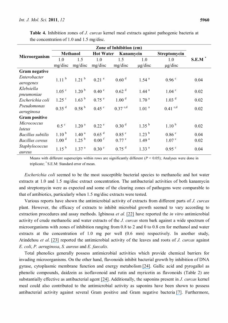
2.4. Antibacterial Activity
Antibacterial activity of methanolic and hot water extracts of J. curcas kernel meal were tested
against four Gram negative and four Gram positive pathogens at two concentrations (1.0 and
1.5 mg/disc). The results shown in Table 4 indicate significant difference (P < 0.05) in inhibitory
activity of methanolic and hot water extracts. The diameter of inhibition zones of methanolic extract
ranged from 0.35 to 1.25 cm while for hot water extract ranged from 0.00 to 0.75 cm at 1.0 mg/disc.
Antimicrobial activity of the methanolic extract appeared to be more effective than water extract, since
methanol could extract a wide variety of active components compared to hot water. The size of
inhibition zones increased with 1.5 mg/disc. At this concentration, the antibacterial effect of
methanolic extract was similar for both Gram negative and Gram positive bacteria, except for
P. aeruginosa, which showed moderate zone of inhibition. This Gram negative bacteria seemed to be
less sensitive to other plant extracts [21].
Int. J. Mol. Sci. 2011, 12
5960
Table 4. Inhibition zones of J. curcas kernel meal extracts against pathogenic bacteria at
the concentration of 1.0 and 1.5 mg/disc.
Microorganism
Zone of Inhibition (cm)
Methanol Hot Water Kanamycin Streptomycin
S.E.M * 1.0 mg/disc
1.5 mg/disc
1.0 mg/disc
1.5 mg/disc
1.0 μg/disc
1.0 μg/disc
Gram negative Enterobacter
aerogenes 1.11 b 1.21 b 0.21 e 0.60 d 1.54 a 0.96 c 0.04
Klebsiella
pneumoniae 1.05 c 1.20 b 0.40 e 0.62 d 1.44 a 1.04 c 0.02
Escherichia coli 1.25 c 1.63 b 0.75 e 1.00 d 1.70 a 1.03 d 0.02
Pseudomonas aeruginosa
0.35 d 0.58 b 0.45 c 0.37 c,d 1.01 a 0.41 c,d 0.02
Gram positive Micrococcus
luteus 0.5 c 1.20 a 0.22 e 0.30 d 1.35 b 1.10 b 0.02
Bacillus subtilis 1.10 b 1.40 a 0.65 d 0.85 c 1.23 b 0.86 c 0.04
Bacillus cereus 1.00 d 1.25 b 0.00 f 0.77 e 1.49 a 1.07 c 0.02 Staphylococcus
aureus 1.15 b 1.37 a 0.30 e 0.75 d 1.33 a 0.95 c 0.04
Means with different superscripts within rows are significantly different (P < 0.05); Analyses were done in
triplicate; * S.E.M: Standard error of mean.
Escherichia coli seemed to be the most susceptible bacterial species to methanolic and hot water
extracts at 1.0 and 1.5 mg/disc extract concentration. The antibacterial activities of both kanamycin
and streptomycin were as expected and some of the clearing zones of pathogens were comparable to
that of antibiotics, particularly when 1.5 mg/disc extracts were tested.
Various reports have shown the antimicrobial activity of extracts from different parts of J. curcas
plant. However, the efficacy of extracts to inhibit microbial growth seemed to vary according to
extraction procedures and assay methods. Igbinosa et al. [22] have reported the in vitro antimicrobial
activity of crude methanolic and water extracts of the J. curcas stem bark against a wide spectrum of
microorganisms with zones of inhibition ranging from 0.8 to 2 and 0 to 0.8 cm for methanol and water
extracts at the concentration of 1.0 mg per well (0.6 mm) respectively. In another study,
Atindehou et al. [23] reported the antimicrobial activity of the leaves and roots of J. curcas against
E. coli, P. aeruginosa, S. aureus and E. faecalis.
Total phenolics generally possess antimicrobial activities which provide chemical barriers for
invading microorganisms. On the other hand, flavonoids inhibit bacterial growth by inhibition of DNA
gyrase, cytoplasmic membrane function and energy metabolism [24]. Gallic acid and pyrogallol as
phenolic compounds, daidzein as isoflavonoid and rutin and myricetin as flavonoids (Table 2) are
substantially effective as antibacterial agent [24]. Additionally, the saponins present in J. curcas kernel
meal could also contributed to the antimicrobial activity as saponins have been shown to possess
antibacterial activity against several Gram positive and Gram negative bacteria [7]. Furthermore,

Int. J. Mol. Sci. 2011, 12
5961
phorbol esters (Table 1) could also act as antimicrobial agent as this property has been demonstrated
by Chumkaew et al. [25].
2.5. Antioxidant Activity
The results shown in Figure 3 indicate the 2,2-diphenyl-1-picrylhydrazyl (DPPH) radical
scavenging activity of J. curcas kernel meal extracts at different concentrations. The inhibition of
DPPH radical scavenging activity increased in a dose-dependent manner. Table 5 shows the IC50
(required concentration to scavenge 50% of DPPH radicals) concentration of extracts. Free radical
scavenging activity of methanolic extract showed a value of 1.6 mg/mL but hot water extract did not
show an IC50 value, even at the highest concentration used in this study. Methanolic extract of
J. curcas kernel meal appeared to be more active as compared to the hot water extract in scavenging
the free radicals. Reference antioxidants vitamin C, BHT and β-carotene showed IC50 values of 0.3, 0.3
and 1.5 mg/mL respectively. Both extracts showed lower activities than vitamin C and BHT due to the
significantly (p < 0.05) higher IC50 values required to inhibit the free radicals (Table 5). The
methanolic extract activity was similar to β-carotene as no significant difference was observed between
their IC50 values (Table 5). It has been reported that the strong free radical scavenging activities in
latex, root and stem of J. curcas was attributed to the presence of phenolic compounds [2,13].
Figure 3. The free radical scavenging activity of J. curcas kernel meal extracts and
reference antioxidants.
0 2 4 6 80
20
40
60
80
100
Methanolic extract
Hot water extract
Vitamin C
BHT
-Carotene
Concentration (mg/mL)
Inh
ibit
ion
(%
)
Ferric reducing antioxidant power (FRAP) of extracts and reference antioxidants (vitamin C, BHT
and β-carotene) is presented in Figure 4. Reference antioxidants showed higher reducing power
followed by methanolic and hot water extract. The IC50 value (the concentration to reduce 50% of Fe3+
to Fe2+ as an index for antioxidant potential) in FRAP for vitamin C, BHT and β-carotene were 0.3, 0.3
and 2.6 mg/mL, respectively (Table 5), while for the methanolic and hot water extracts were 3.0 and
8.0 mg/mL, respectively. As observed in the DPPH assay, FRAP assay also showed similar IC50 values
between methanolic extract and β-carotene (Table 5).

Int. J. Mol. Sci. 2011, 12
5962
Figure 4. Ferric reducing activity of J. curcas kernel meal extracts and reference antioxidants.
0 1 2 3 4 5 6 7 80
20
40
60
80
100
Methanolic extract
Hot water extract
Vitamin C
BHT
-carotene
Concentration (mg/mL)
Red
uct
ion
Po
wer
(%
)
Table 5. The IC50 values of J. curcas kernel meal extracts and reference antioxidants.
Items IC50 (mg/mL)
DPPH FRAP
Methanolic extract 1.6 b 3.0 b
Hot water extract 5.0 a >8 a Vitamin C 0.3 c 0.3 c
BHT 0.3 c 0.3 c β-carotene 1.5 b 2.6 b * S.E.M 0.09 0.07
P value 0.05 0.05
Values with different subscripts in each column are significantly different (p < 0.05); Analyses were done in
triplicate; * S.E.M: Standard error of mean.
The results indicate that the extracts were more active in scavenging free radicals compared to ferric
reducing power activity. In FRAP assay, the general ability of the extracts to donate electrons is tested
whereas, in the DPPH assay, reduction by hydrogen atoms are also involved. Hence, the free radical
scavenging activity assay was a better indicator of antioxidant activity of kernel meal than FRAP based
on the reaction mechanisms proposed. In complex systems, such as food, various different mechanisms
may contribute to oxidative processes. Therefore, it is important to characterize the extracts by a
variety of antioxidant assays [26].
The presence of phenolics (gallic acid, pyrogallol), flavonoids (rutin, myricetin) and isoflavonoid
(daidzein) in the kernel meal (Table 2) may contribute to the antioxidant activity of extracts. These
compounds have been identified as some of the major compounds involved in antioxidant activity [27].
However, saponins in the kernel meal (Table 1) might be also involved, because Sparg et al. [7]
reported the antioxidant activity of these compounds.
2.6. Cytotoxicity Activity
Phytochemical analyses, antimicrobial and antioxidant activities showed that the methanolic extract
was more active compared to hot water extract. Therefore, cytotoxicity assay was performed using the
methanolic extract. It is known that different cell lines might exhibit different sensitivities while

Int. J. Mol. Sci. 2011, 12
5963
treated with the same plant extract, therefore screening plant crude extract against various cell lines is a
first step in determining the most susceptible cancer cells. Apart from that, breast and cervical cancers
are very common manifestations, hence the MCF-7 and HeLa cell lines were used to investigate the
effects of the plant extract. Chang liver cells (human hepatocytes) were also used to determine the
hepatoxicity of extract used in this study. Cytotoxicity assay results demonstrated that the methanolic
extract of J. curcas kernel meal inhibited the growth and proliferation of MCF-7, HeLa and Chang
liver cells in a dose-dependent manner (Figure 5). Methanolic extract at the concentration of
6.25 µg/mL and above decreased the cell viabilities significantly (P < 0.0001) in all cell lines used in
this study as compared to the untreated control.
Figure 5. Effect of methanolic extract of J. curcas kernel meal on MCF-7, HeLa and
Chang liver cell viabilities. All bars represent the mean ± standard error of three
independent experiments. *** P < 0.0001 and ** P < 0.001 indicates significant difference
compared to the untreated control group.
0 3.13 6.25 12.5 25 50 100 2000
50
100
MCF-7
HeLa
Chang**
***
***
***
***
***
***
***
***
***
***
***
***
*** *** ***
****
Concentration (g/ml)
Cel
l via
bilit
y %
In this cytotoxicity activity study, Tamoxifen was used as a positive control. As shown in Figure 6,
Tamoxifen also inhibited the growth and proliferation of MCF-7, HeLa and Chang liver cells in a dose
dependent manner. At 6.25 µg/mL and above, cell viabilities decreased significantly (P < 0.0001) in
all cell lines when compared to the untreated control.
Figure 6. Effect of Tamoxifen as a positive control on MCF-7, HeLa and Chang liver cell
viabilities. All bars represent the mean ± standard error of three independent experiments.
*** P < 0.0001 and ** P < 0.001 indicates significant difference compared to the untreated
control group.
03.
136.
2512
.5 25 50 100
200
0
50
100MCF-7
Chang
HeLa
** *****
*** *** *** ******
*** ********* *** ***
***
***
Concentration (g/ml)
Cel
l vi
abil
ity
%

Int. J. Mol. Sci. 2011, 12
5964
The CC50 values of methanolic extract and Tamoxifen are presented in Table 6. The MCF-7 cells
were found to be more susceptible to the methanolic extract as compared to the HeLa and Chang cells.
Most anticancer drugs have been discovered through random screening of plant materials. The
National Cancer Institute requires a CC50 value of less than 30 μg/mL for the active crude extracts
against cancer cells [28]. Since the methanolic extract showed CC50 values less than National Cancer
Institute requirement toward MCF-7 cells, methanolic extract could be a promising source of bioactive
compounds that could be purified for the development of anticancer drugs in order to treat
breast cancer.
The CC50 concentrations of Tamoxifen (Table 6) for MCF-7, HeLa and Chang liver cell lines were
17.3, 8.89, 33.6 μg/mL, respectively. The activity of Tamoxifen toward MCF-7, HeLa and Chang were
significantly (p < 0.0001) higher than methanolic extract. However, normal Chang liver cells were also
susceptible to both methanolic extract and Tamoxifen. It is interesting to note that the CC50
concentration of Tamoxifen was about half of that methanolic extract. At present it is not known which
particular compound(s) present in the kernel meal methanolic extract exert the cytotoxicity effect in
both cancer and, to some extent, Chang liver cells.
Table 6. The CC50 values of extracts and positive control (Tamoxifen) against MCF-7,
HeLa and Chang liver cell lines.
Sample CC50 value (µg/mL)
MCF-7 HeLa Chang liver cell
Methanolic extract 27.5 a 56.4 a 63.9 a
Tamoxifen 17.3 b 8.89 b 33.6 b * S.E.M 0.35 0.42 0.64
P value 0.0001 0.0001 0.0001
Analyses were done in triplicate; * S.E.M: Standard error of mean.
The presence of bioactive compounds in other J. curcas plant parts has been reported recently.
Oskoueian et al. [2] demonstrated the cytotoxic effect of J. curcas root and latex methanolic extract
with CC50 values of 18.3 ± 0.98 and 70.1 ± 2.06 µg/mL against HT-29 cloned cancer cells,
respectively; and 33.3 ± 0.75 and 60.1 ± 0.89 µg/mL toward Chang liver cells, respectively. Besides
root and latex, leaves methanolic extract was also observed to be effective against B16F10 melanoma
cells with CC50 value of 24.8 μg/mL [29]. Moreover, Fujii et al. [10] reported that PMA induced
apoptosis in prostate cancer cells through overexpression of PKCδ. Park et al. [11] also concluded that
the PMA induce apoptosis in gastric cancer cells through activation of PKC and the activation of
serine protease(s) and caspase-3/CPP32. All these results indicate the potential of J. curcas plant as a
source of bioactive compounds, but, the exact nature of the metabolite involved has to be elucidated,
although it has been reported that phenolics, flavonoids, saponins and phorbol esters have been shown
to be cytotoxic on different cell lines [9].
Int. J. Mol. Sci. 2011, 12
5965
3. Experimental Section
3.1. Sample and Extract Preparation
The Jatropha curcas L. plant was collected from the farm of Faculty of Agriculture, Universiti
Putra Malaysia with the GPS location of 3°0'26.91"N latitude and 101°42'13.24"E longitude for
identification by Mr. Shamsul Khamis. A voucher specimen (SK1764/2010) was deposited in the
Phytomedicinal Herbarium, Institute of Bioscience, Universiti Putra Malaysia, Serdang, Selangor,
Malaysia. Upon confirmation of the plant, the ripe J. curcas seeds were collected from the farm, air
dried, dehulled and kept at 4 °C for further analysis. The kernels isolated were ground, using a
mechanical grinder and oil was extracted by Soxhlet apparatus, using petroleum ether (boiling point of
40–60 °C) for 16 h [30]. Methanolic extract of J. curcas kernel meal was prepared according to
Crozier et al. [27]. Hot water extract was prepared as described by Gulcin et al. [31]. The extracts were
filtered through Whatman No.1 paper and evaporated by using vacuum rotary evaporator (Buchii,
Switzerland). The crude extracts were re-dissolved in methanol for antioxidant, antimicrobial and
HPLC analyses, or in dimethyl sulfoxide for cytotoxicity assay. The extractions were repeated
three times.
3.2. Chemical Constituents
Chemical constituent analyses were conducted according to AOAC [32]. The fiber fractions which
consisted of neutral detergent fiber (NDF) and acid detergent fiber (ADF) were determined according
to Van Soest et al. [33]. The analyses were repeated three times.
3.3. Phytochemical Analyses
3.3.1. Total Phenolics
Total phenolics was determined by using Folin–Ciocalteu reagent according to Ismail et al. [26] and
total phenolic results were expressed as mg tannic acid equivalentss/g DM of the kernel meal. The
analysis was conducted in triplicate.
3.3.2. Total Flavonoids
Total flavonoids was determined based on aluminium chloride colorimetric assay described by
Ismail et al. [26]. Total flavonoid compound of extracts was expressed as mg rutin equivalents/g DM
of the kernel meal. The analysis was conducted in triplicate.
3.3.3. Total Saponins
Total saponin was determined according to Makkar et al. [8] based on vanillin-sulfuric acid
colorimetric reaction and results expressed as mg diosgenin equivalents/g DM of the kernel meal. The
analysis was conducted in triplicate.

Int. J. Mol. Sci. 2011, 12
5966
3.4. Analyses of Phenolic and Flavonoid Compounds by HPLC
The phenolic and flavonoid compounds of J. curcas kernel meal were quantitatively measured by
HPLC based on the method described by Crozier et al. [27] with some modification. Phenolic
standards were gallic acid, syringic acid, vanillic acid, salicylic acid, caffeic acid and pyrogallol.
Flavonoid standards used in this research were quercetin, rutin, myricetin, kaempferol, naringin and
apigenin and isoflavonoid standards were genistein and daidzein. An aliquot of sample extracts was
loaded on the HPLC equipped with an analytical column Intersil ODS-3 (5 μm 4.6 × 150 mm,
Gl Science Inc.). Solvents comprising deionized water (solvent A) and acetonitrile (solvent B) were
used. The pH of water was adjusted to 2.5 with trifluoroacetic acid. The phenolic and isoflavonoid
compounds were detected at 280 nm while flavonoid compounds at 350 nm. The column was
equilibrated by 85% solvent A and 15% solvent B. Then the ratio of solvent B was increased to 85% in
50 min followed by reducing solvent B to 15% in 55 min. This ratio was maintained for 60 min for
each sample analysis with flow rate at 0.6 mL/min. The analysis was repeated three times.
3.5. Analysis of Phorbol Esters
Phorbol esters were determined based on Makkar et al. [8]. An aliquot of sample extracts was
loaded on a high-performance liquid chromatography (HPLC Agilent-1200) series instrument
equipped with a UV-Vis photodiode array (DAD) detector, binary pump, vacuum degasser, auto
sampler and analytical column Agilent Reverse-phase C18 LiChrospher 100 (250 × 4 mm I.D and 5 μm
pore size). The solvents were deionized water (solvent A) and acetonitrile (solvent B). The absorbance
was read at 280 nm and results were expressed as equivalents to phorbol-12-myristate 13-acetate
standard. The analysis was done in triplicate.
3.6. Analysis of Metabolites by Gas Chromatography-Mass Spectrometry (GC-MS)
The chemical composition of the extracts was characterized by GC-MS based on the method
described by Hossain and Rahman [34]. The GC-MS used was a Schimadzu QP2010PLUS system. Six
µL were analysed on a BPX-5 SGE ultra-low-bleed 5% phenyl polydimethylsiloxane capillary column
(30 m × 0.25 mm i.d. × 0.25 μm film thickness). Splitless injection was performed with a purge time of
1.0 min. The carrier gas was helium at a flow rate of 1 mL min−1. The column temperature was
maintained at 50 °C for 3 min, then programmed at 5°C min−1 to 80 °C and then at 10 °C min−1 to
340 °C. The inlet and detector temperature was 250 °C and 340 °C, respectively, and the solvent delay
was 4 min. The identification of the peaks was based on computer matching of the mass spectra with
the National Institute of Standards and Technology (NIST 08 and NIST 08s) library and by direct
comparison with published data. The analyses were carried out in triplicate.
3.7. Antibacterial Activity
The antibacterial assay of J. curcas kernel meal extracts was carried out by the disc diffusion
method as described by Boussaada et al. [21] against Gram positive (Micrococcus luteus, Bacillus
subtilis B145, Bacillus cereus B43 and Staphylococcus aureus S1431) and Gram negative
(Enterobacter aerogenes, Klebsiella pneumonia K36, Escherichia coli E256 and Pseudomonas

Int. J. Mol. Sci. 2011, 12
5967
aeruginosa PI96) pathogens. All the bacteria were purchased from the Institute of Malaysian Research
(IMR) and maintained in the Department of Microbiology, Faculty of Biotechnology and Biomolecular
Sciences, Universiti Putra Malaysia. The positive control without extracts (solvent) and reference
controls using two standard antibiotics (kanamycin and streptomycin) were included in the assay. The
extracts inhibitions were corrected based on positive control and compared with those of reference
control. The experiments were conducted in triplicate.
3.8. Antioxidant Activity
3.8.1. DPPH Radical-Scavenging Activity
Free radical scavenging activity of extracts was determined with 1,1-diphenyl-2-picryl-hydrazil
(DPPH) as free radicals according to Ismail et al. [26]. The absorbance was measured at 515 nm by
using a spectrophotometer. Vitamin C, butylated hydroxytoluene (BHT) and β-carotene were used as
standard antioxidants. The assay was performed in triplicate.
3.8.2. Ferric-Reducing Antioxidant Power (FRAP) Assay
The ferric reducing antioxidant power (FRAP) of the extracts was determined as described by Yen
and Chen [35]. Vitamin C, butylated hydroxytoluene (BHT) and β-carotene were used as standard
antioxidants. The assay was performed in triplicate.
3.9. Cytotoxicity Assay
Two cancer cell line, including human breast cancer cell line (MCF-7 ATCC: HTB-22) and human
cervical adenocarcinoma (HeLa ATCC: CCL-2) and one normal cell line, human hepatocytes (Chang
ATCC: CCL-13) were purchased from the American Type Culture Collection (ATCC). Cell lines were
grown as monolayers in a T-75 cm2 culture flask. The Dulbecco’s Modified Eagle Medium (DMEM)
was supplemented with 2.0 g/L sodium bicarbonate and 10% fetal bovine serum. The cultured cell
lines were maintained in a humidified atmosphere of 5% CO2 at 37 °C and were harvested when they
reached 80% confluency for the cytotoxic test. Cytotoxicity of J. curcas kernel meal extract was
measured by MTT cell proliferation assay [36]. Cytotoxic concentration was expressed as CC50
(concentration of the sample that decreased the number of viable cells to 50% of the control cell)
which was calculated by Graph Pad Prism 5 software. All determinations were performed in triplicate.
3.10. Statistical Analyses
Inhibition zones of antibacterial assay subjected to the general linear models procedure of SAS [37]
in a completely randomized design and the means were compared with Duncan’s Multiple Range test.
The difference was considered significant when the P value was <0.05. GraphPad Prism 5 software
(GraphPad Software Inc., San Diego, CA) was used for all the statistical analyses in cytotoxic assay.
Int. J. Mol. Sci. 2011, 12
5968
4. Conclusions
The kernel meal obtained from J. curcas seeds grown in Malaysia contains high percentage of crude
protein and relatively low fiber. Analysis by HPLC showed the presence of various phenolics and
flavonoids, together with saponins and phorbol esters, while GC-MS analyses indicated the presence of
other metabolites that are probably involved in the antibacterial, antioxidant and cytotoxicity activities
observed in this study. The ability of methanolic extract to inhibit breast cancer cell growth indicates
the potential value of J. curcas kernel meal as an alternative source of therapeutic agents which
requires further investigation.
Acknowledgments
The authors would like to thank the Ministry of Higher Education of Malaysia for the grant provided
under the Fundamental Research Grant Scheme (Project No. 01-11-08-660FR).
References
1. Makkar, H.; Becker, K. Jatropha curcas, a promising crop for the generation of biodiesel and
value-added coproducts. Eur. J. Lipid Sci. Technol. 2009, 111, 773–787.
2. Oskoueian, E.; Abdullah, N.; Saad, W.Z.; Omar, A.R.; Ahmad, S.; Kuan, W.B.; Zolkifli, N.A.;
Hendra, R.; Ho, Y.W. Antioxidant, anti-inflammatory and anticancer activities of methanolic
extracts from Jatropha curcas Linn. J. Med. Plants Res. 2011, 5, 49–57.
3. Devappa, R.K.; Makkar, H.P.S.; Becker, K. Jatropha Diterpenes: a Review. J. Am. Oil Chem. Soc.
2011, 88, 301–322.
4. Balasundram, N.; Sundram, K.; Samman, S. Phenolic compounds in plants and agri-industrial
by-products: Antioxidant activity, occurrence, and potential uses. Food Chem. 2006, 99, 191–203.
5. Muraoka, S.; Miura, T. Inhibition of xanthine oxidase by phytic acid and its antioxidative action.
Life. Sci. 2004, 74, 1691–1700.
6. Devappa, R.K.; Swamylingappa, B. Biochemical and nutritional evaluation of Jatropha protein
isolate prepared by steam injection heating for reduction of toxic and anti nutritional factors.
J. Sci. Food Agric. 2008, 88, 911–919.
7. Sparg, S.G.; Light, M.E.; Van Staden, J. Biological activities and distribution of plant saponins.
J. Ethnopharmacol. 2004, 94, 219–243.
8. Makkar, H.P.S.; Siddhuraju, P.; Becker, K. Methods in Molecular Biology: Plant Secondary
Metabolites; Humana Press: Totowa, NJ, USA, 2007; pp. 93–100.
9. Goel, G.; Makkar, H.P.S.; Francis, G.; Becker, K. Phorbol esters: structure, biological activity,
and toxicity in animals. Int. J. Toxicol. 2007, 26, 279–288.
10. Fujii, T.; Garcia-Bermejo, M.L.; Bernabo, J.L.; Caamano, J.; Ohba, M.; Kuroki, T.; Li, L.;
Yuspa, S.H.; Kazanietz, M.G. Involvement of protein kinase C (PKC) in phorbol ester-induced
apoptosis in LNCaP prostate cancer cells. J. Biol. Chem. 2000, 275, 7574–7582.
11. Park, I.C.; Park, M.J.; Rhee, C.H.; Lee, J.I.; Choe, T.B.; Jang, J.J.; Lee, S.H.; Hong, S.I. Protein
kinase C activation by PMA rapidly induces apoptosis through caspase-3/CPP32 and serine
protease(s) in a gastric cancer cell line. Int. J. Oncol. 2001, 18, 1077–1083.
Int. J. Mol. Sci. 2011, 12
5969
12. Chhabra, S.C.; Mahunnah, R.L.A.; Mshiu, E.N. Plants used in traditional medicine in Eastern
Tanzania. III. Angiosperms (Euphorbiaceae to Menispermaceae). J. Ethnopharmacol. 1990, 28,
255–283.
13. El Diwani, G.; El Rafie, S.; Hawash, S. Antioxidant activity of extracts obtained from residues of
nodes leaves stem and root of Egyptian Jatropha curcas. Afr. J. Pharm. Pharacol. 2009, 3,
521–530.
14. Popendorf, W.; Selim, M.; Lewis, M.Q. Exposure while applying industrial antimicrobial
pesticides. Am. Ind. Hyg. Assoc. J. 1995, 56, 993–1001.
15. Ajaiyeoba, E.O.; Onocha, P.A.; Nwozo, S.O.; Sama, W. Antimicrobial and cytotoxicity
evaluation of Buchholzia coriacea stem bark. Fitoterapia 2003, 74, 706–709.
16. Gupta, M.B.; Nath, R.; Srivastava, N.; Shanker, K.; Kishor, K.; Bhargava, K.P.
Anti-inflammatory and antipyretic activities of β-sitosterol. Planta Med. 1980, 39, 157–163.
17. Awad, A.; Chen, Y.C.; Fink, C.; Hennessey, T. Sitosterol inhibits HT-29 human colon cancer cell
growth and alters membrane lipids. Anticancer Res. 1996, 16, 2797–2804.
18. Rigal, L.; Gaset, A. Direct preparation of 5-hydroxymethyl-2-furancarboxaldehyde from
polyholosides: a chemical valorisation of the Jerusalem artichoke (Helianthus tuberosus L.).
Biomass 1983, 3, 151–163.
19. Morales, F.J. Hydroxymethylfurfural (HMF) and Related Compounds. In Process-Induced Food
Toxicants; Stadler, R.H., Lineback, D.R., Eds.; John Wiley & Sons, Inc.: Hoboken, NJ, USA,
2008; pp. 135–174.
20. Ryssel, H.; Kloeters, O.; Germann, G.; Schafer, T.; Wiedemann, G.; Oehlbauer, M. The
antimicrobial effect of acetic acid-An alternative to common local antiseptics? Burns 2009, 35,
695–700.
21. Boussaada, O.; Chriaa, J.; Nabli, R.; Ammar, S.; Saidana, D.; Mahjoub, M.; Chraeif, I.; Helal, A.;
Mighri, Z. Antimicrobial and antioxidant activities of methanol extracts of Evax pygmaea
(Asteraceae) growing wild in Tunisia. World J. Microb. Biot. 2008, 24, 1289–1296.
22. Igbinosa, O.; Igbinosa, E.; Aiyegoro, O. Antimicrobial activity and phytochemical screening of
stem bark extracts from Jatropha curcas (Linn). Afr. J. Pharm. Pharacol. 2009, 3, 58–62.
23. Atindehou, K.; Kone, M.; Terreaux, C.; Traore, D.; Hostettmann, K.; Dosso, M. Evaluation of the
antimicrobial potential of medicinal plants from the Ivory Coast. Phytother. Res. 2002, 16,
497–502.
24. Cushnie, T.; Lamb, A. Antimicrobial activity of flavonoids. Int. J. Antimicro. Ag. 2005, 26,
343–356.
25. Chumkaew, P.; Karalai, C.; Ponglimanont, C.; Chantrapromma, K. Antimycobacterial activity of
phorbol esters from the fruits of Sapium indicum. J. Nat. Prod. 2003, 66, 540–543.
26. Ismail, H.I.; Chan, K.W.; Mariod, A.A.; Ismail, M. Phenolic content and antioxidant activity of
cantaloupe (cucumis melo) methanolic extracts. Food Chem. 2010, 119, 643–647.
27. Crozier, A.; Lean, M.; McDonald, M.; Black, C. Quantitative analysis of the flavonoid content of
commercial tomatoes, onions, lettuce, and celery. J. Agric. Food Chem. 1997, 45, 590–595.
28. Boik, J.C. Natural Compounds in Cancer Therapy, Oregon Medical Press: Princeton, MN, USA,
2001; pp. 149–190.

Int. J. Mol. Sci. 2011, 12
5970
29. Balaji, R.; Rekha, N.; Deecaraman, M.; Manikandan, L. Antimetastatic and antiproliferative
activity of methanolic fraction of Jatropha curcas against B16F10 melanoma induced lung
metastasis in C57BL/6 mice. Afr. J. Pharm. Pharacol. 2009, 3, 547–555.
30. Martinez-Herrera, J.; Siddhuraju, P.; Francis, G.; Davila-Ortiz, G.; Becker, K. Chemical
composition, toxic/antimetabolic constituents, and effects of different treatments on their levels, in
four provenances of Jatropha curcas L. from Mexico. J. Food Chem. 2006, 96, 80–89.
31. Gulcin, I.; Gungor Sat, I.; Beydemir, S.; Elmastas, M.; Irfan Kufrevioglu, O. Comparison of
antioxidant activity of clove (Eugenia caryophylata Thunb) buds and lavender (Lavandula
stoechas L.). Food Chem. 2004, 87, 393–400.
32. AOAC. Official Methods of Analysis, 15th ed.; Association of Official Analytical
Chemists: Washington, DC, USA, 1990; pp. 64–87.
33. Van Soest, P.J.; Robertson, J.B.; Lewis, B.A. Methods for dietary fiber, neutral detergent fiber,
and nonstarch polysaccharides in relation to animal nutrition. J. Dairy Sci. 1991, 74, 35–83.
34. Hossain, M.; Rahman, A. Chemical composition of bioactive compounds by GC-MS screening
and anti-fungal properties of the crude extracts of cabbage samples. Asian J. Biotechnol. 2011, 3,
68–76.
35. Yen, G.; Chen, H. Antioxidant activity of various tea extracts in relation to their antimutagenicity.
J. Agric. Food Chem. 1995, 43, 27–32.
36. Sharif, R.; Ghazali, A.; Rajab, N.; Haron, H.; Osman, F. Toxicological evaluation of some
Malaysian locally processed raw food products. Food Chem Toxicol. 2008, 46, 368–374.
37. SAS. SAS, Statistical Analysis Institute (Version 9.1.3); SAS Institute Inc.: Cary, NC, USA, 2003.
© 2011 by the authors; licensee MDPI, Basel, Switzerland. This article is an open access article
distributed under the terms and conditions of the Creative Commons Attribution license
(http://creativecommons.org/licenses/by/3.0/).
Copyright © 2022 FDOKUMEN