Jatropha curcas: A potential biofuel plant for sustainable environmental development
Upload
independentCategory
view
4download
0
Effect of nickel on regeneration in Jatropha curcas L.and assessment of genotoxicity using RAPD markers
Tanmoy Sarkar • K. G. Vijay Anand •
M. P. Reddy
Received: 15 September 2009 / Accepted: 23 June 2010 / Published online: 8 July 2010
� Springer Science+Business Media, LLC. 2010
Abstract The aim of the present study was to
determine the effect of nickel on shoot regeneration
in tissue culture as well as to identify polymorphisms
induced in leaf explants exposed to nickel through
random amplified polymorphic DNA (RAPD). In vitro
leaf explants of Jatropha curcas were grown in nickel
amended Murashige and Skoog (MS) medium at four
different concentrations (0, 0.01, 0.1, 1 mM) for
3 weeks. Percent regeneration, number of shoots
produced and genotoxic effects were evaluated by
RAPD using leaf explants obtained from the first three
treatments following 5 weeks of their subsequent
subculture in metal free MS medium. Percent regen-
eration decreased with increase in addition of nickel to
the medium up to 14 days from 42.31% in control to
zero in 1.0 mM. The number of shoot buds scored after
5 weeks was higher in control as compared to all other
treatments except in one of the metal free subculture
medium wherein the shoot number was higher in
0.01 mM treatment (mean = 7.80) than control
(mean = 7.60). RAPD analysis produced only 5
polymorphic bands (3.225%) out of a total of 155
bands from 18 selected primers. Only three primers
OPK-19, OPP-2, OPN-08 produced polymorphic
bands. The dendrogram showed three groups A, B,
and C. Group A samples showed 100% genetic
similarity within them. Samples between groups B
and C were more genetically distant from each other as
compared to samples between groups A and B as well
as groups A and C. Cluster analysis based on RAPD
data correlated with treatments.
Keywords Regeneration � Nickel �Genotoxicity � Molecular markers �Jatropha curcas
Abbreviations
MS Murashige and Skoog medium
TDZ Thidiazuron
BAP 6-Benzylaminopurine
IAA Indole 3-acetic acid
RAPD Random amplified polymorphic DNA
AFLP Amplification fragment length
polymorphism
SSR Simple sequence repeat
AP-PCR Arbitrarily primed PCR
JSC Jaccard’s similarity coefficients
CTAB Cetyl trimethyl ammonium bromide
T. Sarkar � K. G. Vijay Anand (&) � M. P. Reddy (&)
Discipline of Wasteland Research, Central Salt and
Marine Chemicals Research Institute (CSIR), Bhavnagar,
Gujarat 364002, India
e-mail: [email protected]
M. P. Reddy
e-mail: [email protected]
Present Address:M. P. Reddy
Plant Stress Genomics and Technology Center, King
Abdullah University of Science and Technology,
Thuwal 23955-6900, Saudi Arabia
123
Biometals (2010) 23:1149–1158
DOI 10.1007/s10534-010-9364-7
Introduction
Nickel is considered an essential element in plants (at
least for legumes) primarily because of its function as
an irreplaceable component of urease (Gerendas et al.
1999). Among the endemic serpentine flora, ca. 75%
of the plants have shown to hyperaccumulate this
metal (Reeves and Baker 2000) without any toxic
effects. This phenomenon is widely implicated as a
defence mechanism protecting the plants against
herbivores (Boyd 2004, 2007; Palomino et al. 2007).
Increased anthropogenic activities such as vehicular
traffic, burning of fossil fuels, soil fertilization, use of
pesticides, mining and metallurgical activities, and the
disposal of sludge have resulted in widespread
contamination of this metal across different parts of
the globe (Chen et al. 2009). Nickel, a non redox heavy
metal, induces oxidative stress indirectly by acting on
functional groups of proteins thereby producing toxic
symptoms in sensitive, moderately tolerant and even in
hyperaccumulating plants, at various exposure levels
(Sharma and Dietz 2009).
The role of nickel as a potent carcinogen and as a
mutagen has been clearly established in animal and
human cell lines where it is known to inhibit DNA
repair enzymes (Hartwig et al. 1994; Lynn et al.
1997; Kim et al. 2002; Lu et al. 2005). Not only
genotoxins shorten the life expectancy of the organ-
isms but they can also result in alterations in
population dynamics having an effect on intra and
inter species diversity with adverse ecological con-
sequences (Atienzar et al. 2001). Reactive oxygen
species (ROS) generated as a result of oxidative stress
has been widely implicated in damage to cell
membrane and DNA (Sharma and Dietz 2009).
However, the available literature does not clearly
demonstrate nickel induced changes in DNA
sequence and chromosomal aberrations in plants.
Molecular markers, such as random amplified poly-
morphic DNA (RAPD), arbitrarily primed PCR (AP-
PCR), amplification fragment length polymorphism
(AFLP), simple sequence repeat (SSR) have been
widely used in genotoxicity studies to evaluate, detect
and identify changes in the sequence of DNA caused
by various environmental pollutants and mutagens
(Atienzar et al. 2002; Rancelis et al. 2006; Monteiro
et al. 2007; Lu et al. 2007; Enan 2007; Swaileh et al.
2008; Liu et al. 2009; Cenkci et al. 2009). Among
these multilocus markers, RAPD is a simple, fast,
reliable technique than classic genotoxic analysis
tools, as it is capable of detecting not only point
mutations but also temporary alteration of DNA that
may not finally manifest themselves as mutation in
future (Labra et al. 2003) and allow detection of low
doses of pollutants (Liu et al. 2009).
Further nickel is a component of Heller’s medium
(Heller 1953) and has been recommended to be
included as a normal constituent of MS medium
(Witte et al. 2002). Although a wealth of information
is available in the literature about the effect of nickel
on nutrient imbalances, inhibition of growth and
yield, disruption of photosynthesis and oxidative
stress but literature highlighting the effect of higher
doses of nickel on plant regeneration in tissue culture
is very limited. Jatropha curcas has ability to grow
on marginal wastelands thus enabling not only in
vegetating those lands but also serving as a source of
obtaining monetary benefits for the communities and
small farmers of those areas. Recently there have
been several reports suggesting the use of J. curcas in
phytoremediation of polluted soils including metal
contaminated sites and those fertilized with sewage
sludge (Kumar et al. 2008; Mangkoedihardjo and
Surahmaida 2008). These sites are known to contain
high concentration of heavy metals of which nickel is
one of the contaminants. Therefore the present
investigation was carried out to study the effect of
nickel on shoot number, percent regeneration and
determine by RAPD technique as to whether nickel
induces changes in the DNA sequence of J. curcas
which has been identified as a potential biofuel crop
in India, under the programme of National Mission
on Biofuels (http://www.planningcommission.nic.in).
Materials and methods
Culture and growth conditions
Leaves collected from cultures of J. curcas estab-
lished in vitro served as an explant source. The
explants were obtained from 6-week old nodal
cultures maintained on MS medium (Murashige and
Skoog 1962) supplemented with vitamins, sucrose
30 g l-1, 0.5 mg l-1 6-benzylaminopurine (BAP)
and 0.5 mg l-1 indole 3-acetic acid (IAA) and
solidified with 7 g l-1 agar at pH 5.7 set prior to
autoclaving at 121�C for 15 min. Care was taken to
1150 Biometals (2010) 23:1149–1158
123

ensure that neither old senescent nor young leaves
were used for the experiment. The explants thus
obtained were exposed to 0, 0.01, 0.1, 1 mM
concentration of nickel following filter sterilization
though 0.22 lm Millex-GV syringe filters (Milli-
pore). Nickel was supplemented as nickel chloride
hexahydrate (Sigma) at around 45�C to autoclaved
MS medium (MST) amended with vitamins, sucrose
30 g l-1, 0.5 mg l-1 thidiazuron (TDZ) and solidi-
fied with 7 g l-1 agar at pH 5.7 (Sharma 2008). Each
concentration of nickel used was considered as a
treatment. The experiment was completely random-
ized and carried out with a minimum of at least 20
replications per treatment and was repeated twice. A
single in vitro leaf explant in 38 9 200 mm culture
tube containing 40 ml of MST medium served as a
single replication. The number of explants regener-
ating in each of the treatment was scored after 7, 14
and 21 days and percent regeneration was calculated.
All cultures were maintained at 26 ± 2�C under 12 h
light/12 h dark photoperiod in a culture room through
out the experiment.
At least five explants obtained from each treatment
except 1 mM were subsequently subcultured after 3rd
week in nickel free MS medium (proliferation
medium) supplemented with vitamins, sucrose
30 g l-1 and 4 different combinations of BAP and
IAA which was solidified with 7 g l-1 agar at pH 5.7.
After 5 weeks of subculture in proliferation medium,
the number of shoots/shoot buds was scored in each
case and the data recorded. From one of the combi-
nations used in subculture viz., 0.5 mg l-1 BAP and
1.0 mg l-1 IAA, a total of nine tissue samples (leaves)
were collected (3 sample repeats from each treatment
of nickel viz., T1R1,T1R2,T1R3 of control, T2R1, T2R2,
T2R3 of 0.01 mM and T3R1,T3R2,T3R3 of 0.1 mM)
and used for RAPD analysis.
DNA extraction and RAPD analysis
DNA was extracted from each of the samples by
following CTAB method described by Sudheer et al.
(2009a, b). Leaves (100 mg) from each of the nine
samples were ground using liquid nitrogen. To the
ground samples 0.5 ml of extraction buffer (2%
CTAB, 100 mM Tris–HCl, 3.5 M NaCl, 20 mM
EDTA, 0.2 M b-Mercaptoethanol, 2% PVP, pH 8.0)
was added and incubated at 65�C for 90 min. The
above samples were extracted with equal volume of
chloroform: isoamyl alcohol (24:1) and each superna-
tant was transferred into a new tube. The samples were
treated with RNase and extracted with Tris saturated
phenol. The supernatant after extraction with Tris
saturated phenol was taken and extracted further with
chloroform: isoamyl alcohol (24:1) twice, and precip-
itated with 80% of ethanol. The pellet was washed with
70% ethanol, air dried and dissolved in 100 ll of Milli
Q water.
The polymerase chain reaction (PCR) was carried
out using methodology developed by Williams et al.
(1990). A total of 18 selected decamer arbitrary RAPD
primers (Operon technologies Inc, USA; IDT, USA)
produced good amplification profiles and later on used
for RAPD analysis. PCR amplification was carried out
on a Thermal cycler (Master Cycle Epgradient S,
Eppendorf, Germany) with 15 ll reaction mixture
containing 25 ng template DNA, 20 pmol each primer,
1U Taq polymerase, 1X Taq buffer [10 mM Tris–HCl
(pH 9.0),150 mM KCl, 0.1% Triton X 100], 0.12 mM
each dNTPs and 1.8 mM MgCl2 (Biogene, USA). The
amplification reaction was carried out using the
program of initial denaturation at 94�C for 3 min
followed by 42 cycles of denaturation at 94�C for 30 s,
annealing at 30�C for 30 s, extension at 72�C for 2 min
and a final extension at 72�C for 5 min. The amplifi-
cation products were electrophoresed at 80 V in 1.5%
agarose gel in 19 TBE buffer for 90 min. The gel was
stained with ethidium bromide and documented using
gel documentation system (Syngene, UK). The size of
each RAPD band was determined by using Gene Tool
Analysis Software (Syngene, UK).
Data analysis
Statistical analysis of treatments was carried out using
SPSS software version 10 (SPSS Inc., Chicago).
RAPD data (binary matrix) was fed into and the
dendrogram constructed using NTSYS version 2.20
software package. Each profile of RAPD was analyzed
by scoring presence of band as (1) and absence of band
as (0) and the scores were entered into a binary matrix.
Faint and unclear band was not considered for further
analysis. Genetic similarity based on Jaccard’s coef-
ficient were calculated among all possible pairs of the
nine samples using SIMQUAL module and arranged
in a similarity matrix. A dendrogram was constructed
by using UPGMA (Unweighed Pair Group Method
with Arithmetic Average) (Sokal and Sneath 1963)
Biometals (2010) 23:1149–1158 1151
123

option of SHAN module. The percentage of polymor-
phism (PP) was calculated by using the formula
P = total number of polymorphic bands/total number
of bands multiplied with 100.
Results
Effects of nickel on shoot regeneration
from leaf explant
Percent regeneration of leaf explants exposed to nickel
was higher in control (0 mM Nickel) as compared to
other treatments up to 14 days in MST medium. But at
21 days of exposure, percent regeneration in 0.01 mM
treatment was higher (63.83%) as compared to 53.85%
in control (Table 1). Explants remained mostly green
and healthy throughout the experiment (Fig. 1) in all
the treatments, except 1 mM, where cytotoxic symp-
toms were manifested in 25% of the explants within
7 days, followed by complete necrosis on 14th day.
Hence during the second repetition of the experiment
1 mM treatment was excluded from the experiment.
Regeneration of explants in 0.1 mM treatment was
very low at 8% after 14 days and remained the same
even after 21 days of exposure. The total number of
shoots produced in control was higher as compared to
other treatments when subcultured in all but one of the
combinations of nickel free proliferation medium
supplemented with 0.5 mg l-1 BAP and 0.5 mg l-1
IAA. In this combination, the number of shoots in
0.01 mM nickel treated leaves (mean shoots = 7.80)
was higher than that of the control (mean
shoots = 7.60). Of the four different combinations
used, robust growth was visualized in the proliferation
medium with hormonal combination 0.5 mg l-1 BAP
and 1.0 mg l-1 IAA which also induced relatively
higher number of shoots/buds in most of the treatments
as compared to other combinations, with 0.1 mM
nickel exposure also producing mean of 5.60 shoots/
buds (Table 2). Analysis of Variance (ANOVA) for
the number of shoots in different combinations of BAP
and IAA proliferation medium showed that only
treatment was statistically significant (p \ 0.0005)
while other parameters viz., hormonal combination
and treatment 9 hormonal combination were insig-
nificant. Hence explants for subsequent RAPD anal-
ysis were chosen from this combination. Further post
hoc multiple comparisons using least significant
difference (LSD) has revealed that the treatment
means of control and 0.01 mM treatment were insig-
nificant with respect to each other while these two
treatments were significant with that of 0.1 mM
treatment mean (Tables 2, 3).
Evaluation of genotoxicity with RAPD markers
A total of 155 amplicons from 18 primers were
produced in PCR amplification reaction. The ampli-
cons ranged from 273 to 3,095 bp in size and 5 bands
(3.225%) were polymorphic out of total 155 bands
generated. The number of bands scored in each
primer varied from 4 (primer OPP-10) to 15 (OPJ-
20).The number and percentage of monomorphic
bands were 150 and 96.774 respectively. Only three
primers OPK-19 (20%), OPP-2 (16.66%) and OPN-
08 (16.66%) produced polymorphic bands across all
the samples studied. Maximum polymorphism of
20% was shown by the primer OPK-19 where a total
Table 1 Percent
regeneration of leaf
explants obtained following
exposure to various
concentrations of nickel at
different time intervals in
MS medium containing
0.5 mg l-1 thidiazuron
Treatments (mM) Percent regeneration Total number
of explants used7 days 14 days 21 days
Control 17.31 42.31 53.85 52
0.01 14.89 38.30 63.83 47
0.1 2 8 8 50
1.0 0 0 0 24
Fig. 1 Regeneration of shoots buds from leaf explants in
nickel amended MS medium containing 0.5 mg l-1 thidiazu-
ron A control with no nickel; B 0.01 mM nickel; C 0.1 mM
nickel
1152 Biometals (2010) 23:1149–1158
123
of 5 bands were produced (Table 4). On an average
8.61 bands were generated per primer used in this
study. Jaccard’s similarity coefficients (JSC) varied
from 1.00 to 0.974 across all the samples. In the
dendrogram, the samples diverged into three groups
viz. A, B, C. Group A contained 4 samples of
T1R1,T1R2,T1R3 and T2R1, while group B included
T2R2, T2R3 and T3R1,T3R2,T3R3 were in Group C
(Fig. 2). Only three polymorphic bands were shared
by group C samples, only two bands were shared by
group B samples. Group A samples showed 100%
genetic similarity within them. Group C samples
showed JSC ranging from 0.974 to 0.993 respec-
tively. Lowest similarity matrix value of 0.974 was
observed between T2R3/T3R1 and T2R3/T3R2
(Table 5). Maximum number of two polymorphic
bands was produced by each of the primers OPP-02
and OPN-08. Fifteen primers, OPP-15, OPQ-15,
OPR-08, OPO-05, OPJ-09, OPJ-20, OPP-10, OPL-
02, OPN-12, OPQ-11, OPN-10, OPQ-18, OPN-05,
OPQ-12 and OPN-04 out of total eighteen primers
produced no polymorphism across all the samples
(Table 4). Within groups B and C, JSC ranged from
1.000 to 0.993 and 0.993 to 0.987 respectively.
Between groups A and B, JSC ranged from 0.993 to
0.987 and between groups A and C the similarity
matrix values ranged from 0.993 to 0.987, while
between groups B and C, similarity matrix values
ranged from 0.987 to 0.974 indicating samples
between groups B and C are more genetically distant
from each other as compared to samples between
groups A and B as well as groups A and C (Table 5).
Discussion
Effects of nickel on shoot regeneration
from leaf explant
In this study explants exposed to 0.01 mM nickel
treatment showed a higher percent regeneration than
control after 21 days. This is suggestive of the fact
that lower concentrations of nickel (B0.01 mM) may
be required for growth and regeneration. Similar
observations have been made in callus cultures of
J. curcas treated with Nickel (Anand et al. 2008) and
in sugarcane callus stressed with Cadmium (Fornazier
et al. 2002). In Capsicum annuum, Joshi and Kothari
(2007) reported an increase in the frequency of
response and number of shoot buds at higher levels of
Table 2 Total number of shoots produced in different combinations of BAP and IAA of nickel treated leaf explants scored after
5 weeks of culture in MS medium
Hormonal combinations (mg l-1) Treatments
BA IAA Control 0.01 mM 0.1 mM
0.5 0.1 13.00 ± 2.30a 7.00 ± 1.79a 2.20 ± 0.58b
0.5 0.2 10.60 ± 4.08a 9.20 ± 2.01a 2.00 ± 1.05b
0.5 0.5 7.60 ± 2.25a 7.80 ± 2.13a 1.40 ± 0.87b
0.5 1.0 11.00 ± 2.74a 7.20 ± 1.74a 5.60 ± 2.77b
Values represented are mean ± SE of 5 replicates
Means followed by the same letter in a row are not significantly different at P \ 0.05 by post hoc comparison using least significant
difference (LSD)
Table 3 ANOVA summary for the number of the shoots produced in different combinations of BAP and IAA subsequently
following nickel exposure at different concentrations
Source of variation Mean square df F p value
Treatment 308.750 2 12.517 \0.0005
Hormonal combination 15.261 3 0.619 0.606
Treatment 9 hormonal combination 16.261 6 0.659 0.683
Error 24.667 48
Biometals (2010) 23:1149–1158 1153
123

Copper and Silver than at lower levels. No regener-
ation of the explants was observed in 1 mM treatment
indicative of the fact that exposure at this dosage is
lethal and cytotoxic to the explants. The toxicity
observed in 1 mM treatment might be due to
oxidative stress induced ROS with concomitant
Table 4 List of primers, sequences of primers, number of monomorphic and polymorphic bands, number and length of amplicons,
percentage of polymorphism
Primer Sequence of primer (50–30) MBa PBb Total Length of
amplicons (bp)
PPc
OPJ-09 TGAGCCTCAC 10 0 10 575–2,889 0
OPJ-20 AAGCGGCCTC 15 0 15 369–1,900 0
OPK-19 CACAGGCGGA 4 1 5 354–1,746 20
OPL-02 TGGGCGTCAA 9 0 9 682–2,303 0
OPN-12 CACAGACACC 6 0 6 325–2,158 0
OPN-10 ACAACTGGGG 8 0 8 482–2,067 0
OPN-05 ACTGAACGCC 8 0 8 326–1,214 0
OPN-04 GACCGACCCA 9 0 9 372–2,462 0
OPN-08 ACCTCAGCTC 10 2 12 373–2,354 16.66
OPO-05 CCCAGTCACT 10 0 10 670–2,260 0
OPP-02 TCGGCACGCA 10 2 12 486–3,095 16.66
OPP-15 GGAAGCCACC 11 0 11 326–1,291 0
OPP-10 TCCCGCCTAC 4 0 4 609–1,845 0
OPQ-15 GGGTAACGTG 6 0 6 273–1,000 0
OPQ-11 TCTCCGCAAC 6 0 6 634–1,981 0
OPQ-18 AGGCTGGGTG 5 0 5 422–1,526 0
OPQ-12 AGTAGGGCAC 10 0 10 678–2,078 0
OPR-08 CCCGTTGCCT 10 0 10 400–1,395 0
Total 150 5 155
a Number of monomorphic bandsb Number of polymorphic bandsc Percent of polymorphism
Fig. 2 Dendrogram of 9
samples based on Jaccard’s
similarity coefficient
1154 Biometals (2010) 23:1149–1158
123
damage to DNA, proteins and lipids (Boominathan
and Doran 2002; Hao et al. 2006; Sharma and Dietz
2009). The frequency of regeneration observed in 0.1
mM was almost constant throughout the 3rd week
from 14 to 21 days of exposure indicative of the fact
that the threshold level/tolerance level may be at this
dosage. Moreover, explants from 0.1 mM nickel
treatment that failed to regenerate initially also
produced shoots/buds when transferred to nickel free
proliferation medium. This is suggestive of the fact
that nickel has some inhibitory effect upon regener-
ation in J. curcas. A similar result was reported in
shoot cultures of Allysum markgrafii where an arrest
of shoot growth was observed at concentrations
[10 mM nickel followed by its subsequent pro-
liferation upon transfer to nickel free medium
(Vinterhalter and Vinterhalter 2005).
Evaluation of genotoxicity with RAPD markers
In this study, there was only appearance of five
polymorphic bands without disappearance of any
amplified bands in relation to control. New PCR
amplicon may express changes in some oligonucleo-
tide priming sites due to mutation, large deletions and
homologous recombinations (Atienzar et al. 1999).
New amplified fragments are generated as a new site
of genomic sequence becomes accessible to primer
due to alteration at the structural level of DNA
(Pietrasan et al. 2000; Enan 2006; Swaileh et al.
2008). A single point mutation within binding site
can be responsible for changes in RAPD banding
pattern (Williams et al. (1990)). Out of 9 regenerants
(3 control replicates and 6 nickel treated regenerants)
used for RAPD analysis, 5 plantlets (out of 6 nickel
treated regenerants) showed variation in amplification
pattern in relation to control plants corresponding to a
genetic variation frequency of 55.55%. Control
regenerants showed no genetic variation (DNA
polymorphism) within them. Further a higher level
of genetic variation frequency thus generated may be
due to nickel induced mutagenic effect but not as a
result of tissue culture induced DNA polymorphism.
Low level of overall DNA polymorphism (3.125%)
and higher level of similarity coefficient values
(1.00–0.974) shows that nickel might be a weak
mutagen. The level of tissue culture induced DNA
polymorphism may be very less (Munthali et al.
1996; Al-Zahim et al. 1999; Gesteira et al. 2002) to
zero (Devaux et al. 1993; Isabel et al. 1993; Valles
et al. 1993).
In our study, the percentage of polymorphic bands
(PPB) within samples derived from leaf explants
treated with 0.01 and 0.1 mM concentrations of
nickel are 11.11 and 16.66 respectively as compared
to control. This study also showed there was a within
treatment genetic variation within each three sample
of a treatment based on presence or absence of
particular novel band (s) produced by a single primer.
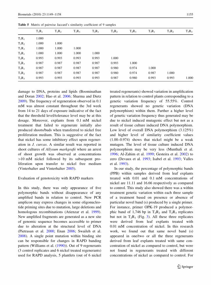
For instance, primer OPK-19 produced a polymor-
phic band of 1,746 bp in T2R2 and T2R3 replicates
but not in T2R1 (Fig. 3). All these three replicates
were derived from leaf explants treated with
0.01 mM concentration of nickel. In this research
work, we found out that same novel band (s)
appeared in one/two or all the three regenerants
derived from leaf explants treated with same con-
centration of nickel as compared to control, but were
not found in regenerants treated with different
concentrations of nickel as compared to control. For
Table 5 Matrix of pairwise Jaccard’s similarity coefficient of 9 samples
T1R1 T1R2 T1R3 T2R1 T2R2 T2R3 T3R1 T3R2 T3R3
T1R1 1.000
T1R2 1.000 1.000
T1R3 1.000 1.000 1.000
T2R1 1.000 1.000 1.000 1.000
T2R2 0.993 0.993 0.993 0.993 1.000
T2R3 0.987 0.987 0.987 0.987 0.993 1.000
T3R1 0.987 0.987 0.987 0.987 0.980 0.974 1.000
T3R2 0.987 0.987 0.987 0.987 0.980 0.974 0.987 1.000
T3R3 0.993 0.993 0.993 0.993 0.987 0.980 0.993 0.993 1.000
Biometals (2010) 23:1149–1158 1155
123

example, primer OPN-08 produced one novel band of
822 bp in T3R1, T3R2, and T3R3 replicates derived
from different leaf explants treated with 0.1 mM
nickel, this primer also produced another novel band
of 445 bp in only T3R2 sample without producing this
band in other two samples T3R1 and T3R3. The two
aforesaid bands were totally absent in samples
derived from leaf explants treated with 0.01 mM
nickel and control. Similar results also obtained in
Chinese narcissus (Lu et al. 2007).This finding
suggests that there might be some specific regions
in J. curcas genome that are sensitive to mutation
induced by nickel treatment in vitro culture condition.
Our work suggests that 0.01 mM nickel induces
genotoxicity to a lesser extent to the sample of group
B without any cytotoxic and inhibitory effect on leaf
regeneration. But 0.1 mM nickel induces genotoxi-
city and shows toxic symptoms correlating with its
inhibitory effect on regeneration. Appearance of new
bands occurred with increased concentration of nickel
indicating that the effect of nickel on DNA is dose
specific. Similar results were obtained in bean
seedlings (Cenkci et al. 2009).
Conclusion
Our data suggests that lower concentration of nickel
(B0.01 mM) may stimulate growth and regeneration
but higher concentrations adversely affects leaf regen-
eration of J. curcas in tissue culture and is responsible
for inducing polymorphism through point mutations
and subsequent changes in DNA sequences. This work
thus provides clear evidence of nickel induced alter-
ations in the sequence of DNA in plants. Genotoxicity
was effectively evaluated by RAPD analysis.
Furthermore in this study, RAPD analysis supports
the data obtained from inhibitory effects of nickel on
leaf regeneration. But further research is required to
ascertain the bands reflecting stable alteration in DNA
is allelic or due to somatic mutation.
Acknowledgements TS and KGV wish to thank Dr. D. V. N.
Sudheer Pamidimarri for his valuable suggestions during
manuscript preparation. The authors are also thankful to
Council of Scientific and Industrial Research (CSIR), India
for financial support.
References
Al-Zahim MA, Ford-Lloyd BV, Newbury HJ (1999) Detection
of somaclonal variation in garlic (Allium sativum L.) using
RAPD and cytological analysis. Plant Cell Rep 18:473–
477
Anand KGV, Prakash CR, Reddy MP (2008) Effect of nickel
on growth and mineral composition in callus culture of
Jatropha curcas L. In: Paper presented in National sym-
posium on plant biotechnology for conservation, charac-
terization and crop improvement, Udaipur, India 8–10
February 2008, p 172
Atienzar FA, Conradi M, Evenden AJ, Jha AN, Depledge MH
(1999) Qualitative assessment of genotoxicity using ran-
dom amplified polymorphic DNA: comparison of geno-
mic template stability with key fitness parameters in
Daphnia magna exposed to benzo[a]pyrene. Environ
Toxicol Chem 18:2275–2282
Atienzar FA, Cheung VV, Jha AN, Depledge MH (2001) Fit-
ness parameters and DNA effects are sensitive indicators
of copper-induced toxicity in Daphnia magna. Toxicol
Sci 59:241–250
Atienzar FA, Venier P, Jha AN, Depledge MH (2002) Evalu-
ation of the random amplified polymorphic DNA (RAPD)
assay for the detection of DNA damage and mutations.
Mutat Res 521:151–163
Boominathan R, Doran PM (2002) Ni-induced oxidative stress
in roots of the Ni hyperaccumulator, Alyssum bertolonii.New Phytol 156:205–215
Fig. 3 a RAPD profile
with primer OPK 19
showing polymorphic bands
in T2R2 and T2R3, b RAPD
profile of primer OPQ11
showing monomorphic
bands. Lane M represents
markers
1156 Biometals (2010) 23:1149–1158
123
Boyd RS (2004) Ecology of metal hyperaccumulation. New
Phytol 162:563–567
Boyd RS (2007) The defense hypothesis of elemental hyper-
accumulation: status, challenges and new directions. Plant
Soil 293:153–176
Cenkci S, Yıldız M, Cigerci IH, Konuk M, Bozdag A (2009)
Toxic chemicals-induced genotoxicity detected by random
amplified polymorphic DNA (RAPD) in bean (Phaseolusvulgaris L.) seedlings. Chemosphere 76:900–906
Chen C, Huang D, Liu J (2009) Functions and toxicity of
nickel in plants: recent advances and future prospects.
Clean 37:304–313
Devaux P, Kilian A, Kleinhofs A (1993) Anther culture and
Hordeum bulbosum-derived barley doubled haploids—
mutations and methylation. Mol Gen Genet 241:674–679
Enan MR (2006) Application of random amplified polymor-
phic DNA (RAPD) to detect the genotoxic effect of heavy
metals. Biotechnol Appl Biochem 43:147–154
Enan MR (2007) Assessment of genotoxic activity of para-
nitrophenol in higher plant using arbitrarily primed-
Polymerase Chain Reaction (AP-PCR). Am J Biotechnol
Biochem 3(2):103–109
Fornazier RF, Ferreira RR, Pereira GJG, Molina SMG, Smith
JR, Lea PJ, Azevedo RA (2002) Cadmium stress in sugar
cane callus cultures: effect on antioxidant enzymes. Plant
Cell Tiss Organ Cult 71:125–131
Gerendas J, Polacco JC, Freyermuth SK, Sattelmacher B
(1999) Significance of nickel for plant growth and
metabolism. J Plant Nutr Soil Sci 162:241–256
Gesteira AS, Otoni WC, Barros EG, Moreira MA (2002)
RAPD-based detection of genomic instability in soybean
plants derived from somatic embryogenesis. Plant Breed
121:269–271
Hao F, Wang X, Chen J (2006) Involvement of plasma-mem-
brane NADPH oxidase in nickel-induced oxidative stress
in roots of wheat seedlings. Plant Sci 170:151–158
Hartwig A, Kruger I, Beyersmann D (1994) Mechanisms in
nickel genotoxicity: the significance of interactions with
DNA repair. Toxicol Lett 72:353–358
Heller R (1953) Recherces sur la nutrition minerale des tissues
vegetaux cultives in vitro. Ann Sci Nat 14:1–223
Isabel N, Tremblay L, Michand M, Tremblay FM, Bousquet J
(1993) RAPD as aids to evaluate the genetic integrity of
somatic embryogenesis-derived populations of Piceamariana (Mill.). Theor Appl Genet 86:81–87
Joshi A, Kothari SL (2007) High copper levels in the medium
improves shoot bud differentiation and elongation from
the cultured cotyledons of Capsicum annuum L. Plant Cell
Tiss Organ Cult 88:127–133
Kim K, Lee SH, Seo YR, Perkins SN, Kasprzak KS (2002)
Nickel(II)-induced apoptosis in murine T cell hybridoma
cells is associated with increased fas ligand expression.
Toxicol Appl Pharmacol 185:41–47
Kumar GP, Yadav SK, Thawale PR, Singh SK, Juwarkar AA
(2008) Growth of Jatropha curcas on heavy metal con-
taminated soil amended with industrial wastes and Azo-
tobacter—a greenhouse study. Bioresource Technol
99:2078–2082
Labra M, Fabio TD, Grassi F (2003) AFLP analysis as bio-
marker of exposure to organic and inorganic genotoxic
substances in plants. Chemosphere 52:1183–1188
Liu W, Yang YS, Li PJ, Zhou QX, Xie LJ, Han YP (2009) Risk
assessment of cadmium-contaminated soil on plant DNA
damage using RAPD and physiological indices. J Hazard
Mater 161:878–883
Lu H, Shi X, Costa M, Huang C (2005) Carcinogenic effect of
nickel compounds. Mol Cell Biochem 279:45–67
Lu G, Zhang X, Zou Y, Zou O, Xiang X, Cao J (2007) Effect of
radiation on regeneration of Chinese narcissus and anal-
ysis of genetic variation with AFLP and RAPD markers.
Plant Cell Tiss Organ Cult 88:319–327
Lynn S, Yew FH, Chen KS, Jan KY (1997) Reactive oxygen
species are involved in nickel inhibition of DNA repair.
Environ Mol Mutagen 29:208–216
Mangkoedihardjo S, Surahmaida (2008) Jatropha curcas L. for
Phytoremediation of lead and cadmium polluted soil.
World App Sci J 4:519–522
Monteiro M, Santos C, Mann RM, Soares AMVM, Lopes T
(2007) Evaluation of cadmium genotoxicity in Lactucasativa L. using nuclear microsatellites. Environ Exp Bot
60:421–427
Munthali MT, Newbury HJ, Ford-Lloyd BV (1996) The
detection of somaclonal variants of beet using RAPD.
Plant Cell Rep 15:474–478
Murashige T, Skoog F (1962) A revised medium for rapid
growth and bioassay with tobacco tissue culture. Physiol
Plant 15:473–497
Palomino M, Kennedy PG, Simms EL (2007) Nickel hyper-accumulation as an anti-herbivore trait: considering the
role of tolerance to damage. Plant Soil 293:189–195
Pietrasan LI, Smith BL, MacLeod MC (2000) A novel
approach for analyzing the structure of DNA modified by
benzo[a]pyrine diol epoxide at single-molecule resolution.
Chem Res Toxicol 13:351–355
Rancelis V, Cesniene T, Zvingila D, Barysas D, Balciuniene L,
Dapkuniene S (2006) Polymorphism of response to cobalt
excess in individual Vicia faba plants. Environ Exp Bot
55:221–234
Reeves RD, Baker AJM (2000) Metal-accumulating plants. In:
Raskin I, Ensley BD (eds) Phytoremediation of toxic
metals: using plants to clean up the environment. Wiley,
New York, pp 193–229
Sharma NK (2008) Studies on regeneration and genetic
transformation in Jatropha curcas. PhD thesis, Bhavnagar
University, India
Sharma SS, Dietz KJ (2009) The relationship between metal
toxicity and cellular redox imbalance. Trends Plant Sci
14:43–50
Sokal RR, Sneath PHA (1963) Principles of numeric taxon-
omy. Freeman, San Francisco, p 359
Sudheer PDVN, Pandya N, Reddy MP, Radhakrishnan T
(2009a) Comparative study of interspecific genetic
divergence and phylogenic analysis of genus Jatropha by
RAPD and AFLP. Mol Biol Rep 36:901–907
Sudheer PDVN, Sarkar R, Meenakshi K, Boricha G, Reddy MP
(2009b) A simple protocol for isolation of high quality
genomic DNA from Jatropha curcas for genetic diversity and
molecular marker studies. Indian J Biotechnol 8:187–192
Swaileh KM, Hussein R, Ezzughayyar A (2008) Evaluating
wastewater-induced plant genotoxicity using randomly
amplified polymorphic DNA. Environ Toxicol 23:
117–122
Biometals (2010) 23:1149–1158 1157
123

Valles MP, Wang ZY, Montavon P, Potrykus I, Spangenberg G
(1993) Analysis of genetic stability of plants regenerated
from suspension cultures and protoplasts of meadow fes-
cue (Festuca pratensis Huds.). Plant Cell Rep 12:101–106
Vinterhalter B, Vinterhalter D (2005) Nickel hyperaccumula-
tion in shoot cultures of Alyssum markgrafii. Biol Plan-
tarum 49:121–124
Williams JG, Kubelik AR, Livak J, Rafalski J, Tingey SV
(1990) DNA polymorphism amplified by arbitrary primers
are useful as genetic markers. Nucleic Acids Res
18:6531–6535
Witte CP, Tiller SA, Taylor MA, Davies HV (2002) Addition
of nickel to Murashige and Skoog medium in plant tissue
culture activates urease and may reduce metabolic stress.
Plant Cell Tiss Organ Cult 68:103–104
1158 Biometals (2010) 23:1149–1158
123
Copyright © 2022 FDOKUMEN