Antimicrobial Compounds from Microorganisms - MDPI
20
Citation: Amaning Danquah, C.; Minkah, P.A.B.; Osei Duah Junior, I.; Amankwah, K.B.; Somuah, S.O. Antimicrobial Compounds from Microorganisms. Antibiotics 2022, 11, 285. https://doi.org/10.3390/ antibiotics11030285 Academic Editor: Federica Pellati Received: 4 January 2022 Accepted: 7 February 2022 Published: 22 February 2022 Publisher’s Note: MDPI stays neutral with regard to jurisdictional claims in published maps and institutional affil- iations. Copyright: © 2022 by the authors. Licensee MDPI, Basel, Switzerland. This article is an open access article distributed under the terms and conditions of the Creative Commons Attribution (CC BY) license (https:// creativecommons.org/licenses/by/ 4.0/). antibiotics Review Antimicrobial Compounds from Microorganisms Cynthia Amaning Danquah 1, * , Prince Amankwah Baffour Minkah 1,2 , Isaiah Osei Duah Junior 3 , Kofi Bonsu Amankwah 4 and Samuel Owusu Somuah 5 1 Department of Pharmacology, Faculty of Pharmacy and Pharmaceutical Sciences, College of Health Sciences, Kwame Nkrumah University of Science and Technology, PMB, Kumasi, Ghana; [email protected] 2 Global Health and Infectious Disease Research Group, Kumasi Centre for Collaborative Research in Tropical Medicine, College of Health Sciences, Kwame Nkrumah University of Science and Technology, PMB, Kumasi, Ghana 3 Department of Optometry and Visual Science, College of Science, Kwame Nkrumah University of Science and Technology, PMB, Kumasi, Ghana; [email protected] 4 Department of Biomedical Sciences, University of Cape Coast, PMB, Cape Coast, Ghana; [email protected] 5 Department of Pharmacy Practice, School of Pharmacy, University of Health and Allied Sciences, PMB, Ho, Ghana; [email protected] * Correspondence: [email protected]; Tel.: +233-26-5458216 Abstract: Antimicrobial resistance is an exigent public health concern owing to the emergence of novel strains of human resistant pathogens and the concurrent rise in multi-drug resistance. An influx of new antimicrobials is urgently required to improve the treatment outcomes of infectious diseases and save lives. Plant metabolites and bioactive compounds from chemical synthesis have found their efficacy to be dwindling, despite some of them being developed as drugs and used to treat human infections for several decades. Microorganisms are considered untapped reservoirs for promising biomolecules with varying structural and functional antimicrobial activity. The advent of cost- effective and convenient model organisms, state-of-the-art molecular biology, omics technology, and machine learning has enhanced the bioprospecting of novel antimicrobial drugs and the identification of new drug targets. This review summarizes antimicrobial compounds isolated from microorganisms and reports on the modern tools and strategies for exploiting promising antimicrobial drug candidates. The investigation identified a plethora of novel compounds from microbial sources with excellent antimicrobial activity against disease-causing human pathogens. Researchers could maximize the use of novel model systems and advanced biomolecular and computational tools in exploiting lead antimicrobials, consequently ameliorating antimicrobial resistance. Keywords: antimicrobial peptides; secondary metabolites; natural products; microorganisms; drug discovery; model organisms; omics-informed drug discovery; structure-activity 1. Introduction The surge in antimicrobial resistant infections and the concurrent increase in multidrug resistant organisms has jeopardized the healthcare system and threatens public health. Annually, thousands of lives are lost due to resistant infections, and without robust systems, the world would experience over 10 million yearly deaths [1]. Currently, the growing antimicrobial resistance has rendered the efficacy of antimicrobials of questionable utility [2]. Therefore, the search for alternate antimicrobial agents has become a necessity. Over the past decade, natural products have been heavily relied upon as sources of therapeutic agents, with antimicrobials being one of the most compelling biomolecules. In particular, they constitute more than two-thirds of newly approved medicinal products used for pharmaceutical applications [3]. Unlike microbial-originated antibiotics, plant-based antimicrobials have been extensively explored and with varied applications in medicine, veterinary, agriculture, and biotechnology. Microorganisms are recognized as producers Antibiotics 2022, 11, 285. https://doi.org/10.3390/antibiotics11030285 https://www.mdpi.com/journal/antibiotics
-
Upload
khangminh22 -
Category
Documents
-
view
3 -
download
0
Transcript of Antimicrobial Compounds from Microorganisms - MDPI

�����������������
Citation: Amaning Danquah, C.;
Minkah, P.A.B.; Osei Duah Junior, I.;
Amankwah, K.B.; Somuah, S.O.
Antimicrobial Compounds from
Microorganisms. Antibiotics 2022, 11,
285. https://doi.org/10.3390/
antibiotics11030285
Academic Editor: Federica Pellati
Received: 4 January 2022
Accepted: 7 February 2022
Published: 22 February 2022
Publisher’s Note: MDPI stays neutral
with regard to jurisdictional claims in
published maps and institutional affil-
iations.
Copyright: © 2022 by the authors.
Licensee MDPI, Basel, Switzerland.
This article is an open access article
distributed under the terms and
conditions of the Creative Commons
Attribution (CC BY) license (https://
creativecommons.org/licenses/by/
4.0/).
antibiotics
Review
Antimicrobial Compounds from MicroorganismsCynthia Amaning Danquah 1,* , Prince Amankwah Baffour Minkah 1,2, Isaiah Osei Duah Junior 3 ,Kofi Bonsu Amankwah 4 and Samuel Owusu Somuah 5
1 Department of Pharmacology, Faculty of Pharmacy and Pharmaceutical Sciences, College of Health Sciences,Kwame Nkrumah University of Science and Technology, PMB, Kumasi, Ghana; [email protected]
2 Global Health and Infectious Disease Research Group, Kumasi Centre for Collaborative Research in TropicalMedicine, College of Health Sciences, Kwame Nkrumah University of Science and Technology, PMB,Kumasi, Ghana
3 Department of Optometry and Visual Science, College of Science, Kwame Nkrumah University of Science andTechnology, PMB, Kumasi, Ghana; [email protected]
4 Department of Biomedical Sciences, University of Cape Coast, PMB, Cape Coast, Ghana;[email protected]
5 Department of Pharmacy Practice, School of Pharmacy, University of Health and Allied Sciences, PMB,Ho, Ghana; [email protected]
* Correspondence: [email protected]; Tel.: +233-26-5458216
Abstract: Antimicrobial resistance is an exigent public health concern owing to the emergence ofnovel strains of human resistant pathogens and the concurrent rise in multi-drug resistance. An influxof new antimicrobials is urgently required to improve the treatment outcomes of infectious diseasesand save lives. Plant metabolites and bioactive compounds from chemical synthesis have found theirefficacy to be dwindling, despite some of them being developed as drugs and used to treat humaninfections for several decades. Microorganisms are considered untapped reservoirs for promisingbiomolecules with varying structural and functional antimicrobial activity. The advent of cost-effective and convenient model organisms, state-of-the-art molecular biology, omics technology, andmachine learning has enhanced the bioprospecting of novel antimicrobial drugs and the identificationof new drug targets. This review summarizes antimicrobial compounds isolated from microorganismsand reports on the modern tools and strategies for exploiting promising antimicrobial drug candidates.The investigation identified a plethora of novel compounds from microbial sources with excellentantimicrobial activity against disease-causing human pathogens. Researchers could maximize theuse of novel model systems and advanced biomolecular and computational tools in exploiting leadantimicrobials, consequently ameliorating antimicrobial resistance.
Keywords: antimicrobial peptides; secondary metabolites; natural products; microorganisms; drugdiscovery; model organisms; omics-informed drug discovery; structure-activity
1. Introduction
The surge in antimicrobial resistant infections and the concurrent increase in multidrugresistant organisms has jeopardized the healthcare system and threatens public health.Annually, thousands of lives are lost due to resistant infections, and without robust systems,the world would experience over 10 million yearly deaths [1]. Currently, the growingantimicrobial resistance has rendered the efficacy of antimicrobials of questionable utility [2].Therefore, the search for alternate antimicrobial agents has become a necessity.
Over the past decade, natural products have been heavily relied upon as sources oftherapeutic agents, with antimicrobials being one of the most compelling biomolecules. Inparticular, they constitute more than two-thirds of newly approved medicinal products usedfor pharmaceutical applications [3]. Unlike microbial-originated antibiotics, plant-basedantimicrobials have been extensively explored and with varied applications in medicine,veterinary, agriculture, and biotechnology. Microorganisms are recognized as producers
Antibiotics 2022, 11, 285. https://doi.org/10.3390/antibiotics11030285 https://www.mdpi.com/journal/antibiotics

Antibiotics 2022, 11, 285 2 of 20
of bioactive compounds with antibacterial, antifungal, and cytotoxic bioactivity [4–7].Again, their production of functionally rich secondary metabolites enables them to thrivein varied environmental conditions. Researchers have recently paid attention to microbesas untapped reservoirs for novel antimicrobial agents due to their distinctive biologicalproperties [8]. Specifically, the invention of state-of-the-art molecular biology, genetic,genomic, and computational tools have facilitated the mining of microbial structuralsystems to enhance drug discovery [9–11].
Microorganisms are biotic, ubiquitous, diverse creatures broadly categorized intoviruses, bacteria, archaea, fungi, and protists. Predominantly, bacteria and fungi are exploredas potential sources of novel antimicrobial agents. For instance, cyclic peptides- mathiapeptide A,destotamide B, Marfomycins A, B, E; spirotetronates polyketides-abyssomycin C, Lobophorin F,H, as well as alkaloids and sesquiterpenes derivatives, caboxamyxin and mafuraquinocinsA, D (Table 1; Figure 1) isolated from bacteria, have antimicrobial properties suicidal againstclinically resistant bacteria, including Staphylococcus aureus (S. aureus), Methicillin-resistantStaphylococcus aureus (MRSA), Micrococcus luteus (M. luteus), Bacillus subtilis (B. subtilis),and Enterococcus faecalis (E. faecalis) [12]. Similarly, ambuic acid analogs, the penicyclonesclasses; depsidone analogs, the spitomastixones groups; xanthones derivatives, emerix-anthones, and engyodontiumsones from fungi, exhibit an anti-infective activity againstGram-negative bacteria, Escherichia coli (E. coli) and Klebsiella pneumoniae (K. pneumoniae),and several other Gram-positive pathogenic bacteria [12]. Furthermore, in vivo and in vitroassays have also demonstrated the anti-infective potentials of other microbial productsextracted from cyanobacteria [13,14], microalgae [14,15], and yeast [16,17].
Table 1. Antimicrobial activity of chemical compounds isolated from microorganisms.
Microorganism ChemicalCompound Molecular Class Antimicrobial
Activity Reference
Marinactinosporathermotolerans Marthiapeptide A Cyclic peptide S. aureus, M. luteus, B.
subtillis, B. thuringiensis [12]
Streptomyces scopuliridis Desotamide B Cyclic peptide S. aureus, S. aureus [12]Streptomycesdrozdowiczii Marfomycins A, B, E Cyclic peptide M. luteus
Verrucosispora spp. Abyssomicin C Spirotetronatepolyketides
Methicillin-resistantStaphylococcus aureus,Vancomycin-resistantStaphylococcus aureus
[12]
Streptomyces spp. Lobophorin F Spirotetronatepolyketides S. aureus, E. feacalis [12]
Streptomyces spp. Lobophorin H Spirotetronatepolyketides B. subtilis [12]
Streptomyces sp. Caboxamycin Alkaloid S. epidermis, S. lentus, B.subtillis [12]
Streptomyces niveus Marfuraquinocin A, D Sesquiterpenederivative
S. aureus,Methicillin-resistantStaphylococcus aureus
[12]
The recent technological advances have primed scientists to produce synthetic antimi-crobials through chemical and structural modification of natural products to overcomeantibiotic resistance. In particular, component-based synthesis, structured-guided designs,and X-ray crystallography have enabled the fabrication and visualization of novel antimi-crobials from primogenitor cell lines [18]. A typified example is oxepanoprolinamide, aderivative of lincosamide [18], which showed a greater propensity to overcome ATP bind-ing cassette (ABC) F-, emerging erm B (Erm-), and Cfr gene-multidrug resistance, and withincreased therapeutic effect against resistant bacterial strains [18]. Given the emergence ofdiverse strains of resistant microorganisms and the advent of modern tools, new evidenceis warranted to enhance bioprospecting of new antimicrobials. Therefore, the overarching

Antibiotics 2022, 11, 285 3 of 20
goal of this review is to report on novel antimicrobial compounds from microorganismsand further explore the contemporary tools used in antimicrobial drug discovery.
1
Figure 1. Antimicrobial compounds of different classes isolated from microorganisms.
2. Bacterial Sources of Antimicrobials
Lactic acid bacteria (LAB) have the tendency to produce antimicrobial compounds (i.e.,bacteriocin, organic acids, diacetyl, and hydrogen peroxide), which are effective againstharmful bacteria [19]. Bacteriocin production by Lactobacillus pentosus (L. pentosus) ST712BZisolated from boza antagonizes the proliferation of Lactobacillus casei (L. casei), E. coli,Pseudomonas aeruginosa (P. aeruginosa), E. faecalis, K. pneumoniae, and Lactobacillus curvatus(L. curvatus) [20]. Bacteriocins are low molecular weight polypeptides synthesized inribosomes and comprise 20–60 amino acid residues [19]. In 1925, Andre Gratias discoveredbacteriocin when he realized that the growth of some E. coli strains was being impeded by anantibacterial compound, which he named colicin V [21]. Although there are different classes

Antibiotics 2022, 11, 285 4 of 20
of bacteriocins produced by other Gram-positive and Gram-negative bacteria as well asarchaea, those produced by LAB are the most studied due to their use as food preservativesas well as the frequent incidence of food-borne infectious diseases [21]. According toKlaenhammer, four groups of bacteriocins exist based on their molecular mass, enzymesensitivity, thermos-stability, presence of post-translationally modified amino acids, andmode of action [22]. Class I is made up of lantibiotics and can further be grouped into Iaor Ib depending on the structure and charge of compound. Class II bacteriocins consistof heat-stable peptides with molecular masses less than 10 kDa and can also further becategorized into classes IIa, IIb, and two other types of IIc [22]. The third class, whichconsists of high molecular weight (usually >30 kDa) thermo-labile peptides, are representedby Helveticin J and the last class IV, comprises a mixture of large peptides and carbohydratesor lipids [23]. However, since there is no standard classification for bacteriocins, studiesby Cotter et al. [24], Drider et al. [25], and others reveal contrasting theories about theirclassification. In modern times, classification of bacteriocins into three classes based ongenetics and biochemical properties is most often used. These classes are class I (lantibiotics),class II (non-lantibiotics), and class III [25]. Each class of bacteriocins has their own way ofexhibiting antimicrobial activity based on their primary structure [26] see Table 2. Somebacteriocins attack energized membrane vesicles of target microbes by tampering withtheir proton motive force [27], while others enter the cell and disrupt gene expression andprotein synthesis [26]. Lantibiotics fight bacteria in two ways. They alter the bacterialcell wall formation process by binding to lipid II, a hydrophobic carrier of peptidoglycanmonomers from the cytoplasm to the cell wall, making the cell unsuitable for certain actions.Lipid II is responsible for membrane insertion and pore formation in the cell membrane ofbacteria [26,28,29].
Non-lantibiotics on the other hand, kill their target cells by binding to MptC and MptDsubunits of mannose phosphotransferase permease (Man-PTS) causing an intra-membranechannel to open and ions to continuously diffuse through [29,30]. Without requiringany receptor molecule circular bacteriocins owing to their high net positive charges areelectrostatically attracted to the negatively charged bacteria membrane. This interactionleads to pore formation, efflux of ions, changes in membrane potential, and eventually celldeath [31].
Bacteriolysins enhance cell wall hydrolysis causing the cell to gradually break down [32,33].Non-bacteriolysins disrupt glucose uptake in target cells, consequently starving themto death [34–36]. Interactions between antimicrobial compounds and their susceptiblemicrobes can be synergistic or antagonistic [37]
In veterinary medicine, bacteriocins, such as nisin, have been clinically used to preventdentobacterial plaque and gingivitis in dogs [38,39], as a result of its brutal action againststrains of E. faecalis and other canine periodontal disease-causing bacteria [26].
Rhamnolipid are popular anionic biosurfactants, generally produced by some speciesof Pseudomonas and Burkhloderia [40,41] These compounds have shown a broad spectrumof biological activities, including activities against microorganisms, biofilm, tumors, andoxidation [42–44]. Of great interest is their activity against Herpes simplex virus 1 and 2(HSV-1 and HSV-2) and bovine coronaviruses, via interactions with viral lipid membranesand thereby altering viral membrane glycoproteins [45,46]. Rhamnolipids (M15RL) pro-duced by the Antarctic bacterium, Pseudomanas gessardii (P.gessardii) M15, has recently beenreported to exert high antiviral activity against Coronaviridae and Herpesviridae families,especially against severe acute respiratory syndrome coronavirus 2 (SARS-CoV-2) [47].

Antibiotics 2022, 11, 285 5 of 20
Table 2. Examples of bacteriocins, organisms that produce them and microbes that are susceptibleto them.
Bacteriocin Producer of Bacteriocin Susceptible Microorganisms Reference(s)
Nisin A Lactococcus lactic subsp. lactis
E. faecalis ssp. Liquefaciens, Streptococcusequinus, Staphylococcus epidermidis
(S. epidermidis), S. aureus, Streptococcus uberis(S. uberis), Streptococcus dysgalactiae
(S. dysgalactiae), Streptococcus agalactiae(S. agalactiae), Streptococcus suis (S. suis)
Mycobacterium avium subsp. Paratuberculosis
[48–50]
Nisin ANisin V L. lactis NZ9700L. lactisNZ9800nisA:M21V Listeria monocytogenes [51]
Pediocin A Pediococcus pentosaceus FBB61 Clostridium perfringens [52]Enterocin M Enterococcus faecium AL41 Campylobacter spp., Clostridium spp. [53]
Enterocin CLE34 Enterococcus faecium CLE34 Salmonella pullorum [26,54]
Enterocin E-760Enterococcus durans,Enterococcus faecium,
Enterococcus hirae
Salmonella enterica serovar Enteritidis,S. enterica serovar Choleraesuis, S. entericaserovar Typhimurium, S. enterica serovar
Gallinarum, E. coli O157:H7, Yersiniaenterocolitica, S. aureus, Campylobacter jejuni
[55]
Lacticin 3147 Lactococcus lactis DPC3147. S. dysgalactiae, S. agalactiae, S. aureus, S. uberis,Mycobacterium avium subsp. paratuberculosis [50,56]
Macedocin ST91KM Streptococcus gallolyticussubsp.macedonicus ST91KM S. agalactiae, S. dysgalactiae, S. uberis, S. aureus [57]
3. Bacterial Sources of Antifungal Compounds
Red pigmented pradimicins A, B, and C are products of the bacteria Actinomadurahibisca (A. hibisca) [58]. These pradimicins exhibit antifungal properties against Candida andAspergillus species as well as other fungi [59] see Table 3. Spectral analysis and chemicaldegradation reveals pradimicins structurally to be a benzo[α]napthacenequinone carryingD alanine and sugars [58]. Pradimicins use specific binding recognition to bind to terminalD mannosides of the cell wall of susceptible microbes to form a D-mannoside, pradimicin,and calcium complex that destroys fungal cell membrane [59].
Actinoplanes species also produce antifungal metabolites. An example is Actinoplanesianthinogenes (A. ianthinogenes), which produces purpuromycin, a compound that hasactivity against Trycophyton mentagrophytes (T. mentagrophytes) [60]. Another species isknown as Octamycini produces octamycin [60]
Soil-occurring Micromonospora species have been identified with the production ofantifungal compounds [60]. Micromonospora species ATCC 53803, through metabolism,produces spartanamycin B as a secondary metabolite, which has activity against Candidaalbicans (C. albicans), Aspergillus cladosporium (A. Cladosporium), and Cryptococcus spp. Mi-cromonospora neiheumicin (M. neiheumicin) produces neihumicin, which is active againstSaccharomyces cerevisae (S. cerevisiae) activity [61]. Sch 37137, a dipeptide formed by Mi-cromonospora species SCC 1792, also fights against dermatophytes and Candida species [62].Lastly, Nishizawa et al. reported that Micromonospora species SF-1917 produces nucleosideantibiotics, dapiramicins A and B. Dapiramicins B inhibits growth of Rhizoctonia solania(R. solania) of rice plants in a greenhouse test [63].
Aerobic Gram-positive branching bacilli, Streptomyces species, yield some antifun-gal compounds. These compounds include nystatin, phoslatomycins [64], UK-2A, B, C,D [65], phthoxazolin A [66], faeriefungin [67], butyrolactols A and B [68], sultriecin [69],polyoxin [70]), and dunaimycins [71].

Antibiotics 2022, 11, 285 6 of 20
Table 3. Bacterial sources of antifungal compounds.
Microorganism Compound(s) Susceptible Organism(s) Reference
A. hibisca Pradimicins A, B, C Candida spp. and Aspergillus spp. [58]
Actinoplanes spp. Purpuromycin T. mentagrophytes [60]
Micromonospora species ATCC 53803 Spartanamycin B C. albicans, A. cladosporium,and Cryptococcus spp. [61]
M. neiheumicin Neihumicin S. cerevisae [61]
Micromonospora species SCC 1792 Sch 37137 Dermatophytes and Candida spp. [62]
B. subtilis Iturin A and related peptides Phytopathogens [60,72]
Micromonospora species SF-1917 Dapiramicins A and B R. solania [63]
B. cereus Azoxybacilin, Bacereutin,Cispentacin, and Mycocerein
Aspergillus spp., Saccharomyces spp,and C. albicans [60]
B. lichenformis Fungicin M-4 Microsporum canis, Mucor spp.,and Sporothrix spp. [73,74]
Some bacilli species are also known to be the source of several antifungal compounds.Bacillus subtilis produces iturin and other closely related peptides, including bacillomycin D,F, and L, mycosubtilin, and mojavensin. These agents have been shown to be active againstphytopathogens and hence, have been commercialized as biological control agents againstfungal plant pathogens. Notably, there has not been any reported resistance against fungifor these compounds. These agents act by creating pores in the membrane of susceptiblefungi, thereby causing leakage of cell contents and subsequent cell death [60,72].
According to Kerr, the compounds; azoxybacilin, bacereutin, cispentacin, and myco-cerein can be isolated from the products of Bacillus cereus (B. cereus) and are active againstAspergillus species, Saccharomyces spp., Candida albicans, and other fungi. [60] Another Bacillispecies, B. licheniformis, produces fungicin M-4 and peptide A12-C [73,74]
The compound, pyrrolnitrin, has been reported by Chernin et al. to be the factorresponsible for the antimicrobial action of Enterobacter agglomerans (E. agglomerans) on theCandida species, Aspergillus niger (A. niger), dermatophytes and phytopathogenic fungi.Enterobacter agglomerans again produces herbicolins A and B. which are active against yeastsand filamentous fungi [75–77]. CB-25-1, a water soluble dipeptide, produced by Serratiaplymuthica (S. plymuthica) is known to inhibit growth of C. albicans [78].
P. aeruginosa present in the gut of a normal person has been identified as the sourceof three antifungal compounds, namely dihydroaeruginoic acid [79], pyocyanin, and1-hydroxyphenazine [80]. Other antimicrobial compounds produced by pseudomonas in-clude 2,4-diacetophluoroglucinol [81], peptide pseudomycin family [82], caryoynencins [83],and cyclic hydroxamic acid, G1549 [84].
Burkholderia species are another bacterial source of antimicrobial compounds. Cepaci-dine A, which antagonizes plant and animal fungi growth, can be generated by B. cepa-cia [85]. B. cepacia also produces cepalycin [86], xylocandins [87], and heptylmethyl-quinolinone [88]. Another group of antibacterial compounds is enacyloxcins, knownto originate from the Burkholderia species [89]. Enacyloxcins consists of eight closelyrelated antibacterial compounds (86–87). Maltophilin is the active compound responsiblefor the antifungal action of the Rhizosphere strain of Stenotrophomonas maltophilia. Polyenicantibiotics produced by the genus Gluconobacter have also been reported to possess someantifungal activity against the fungus Neurospora crassa but not against yeast [90]
4. Fungal Sources of Antimicrobials
The discovery of penicillin G in 1928 from fungal species has led to the exploration ofthese organisms [91]. Their ability to produce a plethora of active secondary metabolitesthat can serve as lead compounds for the synthesis of antimicrobials is significant.

Antibiotics 2022, 11, 285 7 of 20
Hormonema species that yielded enfumafungin, a triterpenoid, was discovered over adecade ago and was shown to be highly effective against Candida spp. and Aspergillus spp.It is still being studied in order to produce a number of developmental compounds [92].Enfumafungin yielded a semisynthetic derivative, SCY-078 that is in phase II clinicaltrial. The biosynthetic encoding genes for this peculiar triterpenoid were only recentlydiscovered, but have shown a lineage of hopene-type cyclases, including ERG7, which isnecessary for the biosynthesis of fungal ergosterol [93], see Table 4.
Testing of metabolites in the strobilurins, known as antifungal agents in agriculture,has not been explored since it was identified in 1999 as being harmful to humans [94]. Inrecent times however, favolon, produced by Favolaschia calocera (F. calocera), a metaboliteof strobilurins has been identified and has been shown to be less toxic but with potentantifungal activity against human pathogens [95].
Fungal metabolites, by their ability to interfere with quorum sensing, inhibits theformation of biofilms. Coprinuslactone, derived from Coprinus comatus (C. comatus), acts onP. aeruginosa biofilms [96]. Microporenic acid A from a Kenyan basidiomycete also inhibitsS. aureus and C. albicans biofilms and has an added advantage of destroying pre-formedbiofilms [97]. Biofilm inhibitors are promising adjuncts to antibiotics.
Mutulins and its derivative, retapamulin from the basidiomycete Clitopilus passecke-rianus, represents a new area in search of antimicrobials. They have been shown to havepotent antibacterial activity, and more derivatives are undergoing clinical trials. The draw-back with them is the difficulty in reaching a large scale since they grow slowly and generatelow yields [96].
A novel rubrolide, rubrolide S, discovered from the marine fungus Aspergillus terreus(A. terreus) OUCMDZ-1925, has been shown to significantly inhibit the activity of InfluenzaA virus (H1N1) [98]. A novel hybrid polyketide, Cladosin C, isolated from Cladosporiumsphaerospermum 2005-01-E3, has also shown activity against Influenza A H1N1 [99]. Penicil-lium chrysogenum PJX-17 has also shown to be the source of two antiviral sorbicillinoids,named sorbicatechols A and B, with significant activity against the H1N1 [100].
Trypilepyrazinol and β-hydroxyergosta-8,14,24 (28)-trien-7-one isolated from extractsof the fungus Penicillium sp. IMB17-046 has shown broad spectrum antiviral activitiesagainst different types of viruses, including human immunodeficiency virus (HIV) andhepatitis C virus (HCV) [101]. Aspergillus niger SCSIO Jcsw6F30 produces aspernigrin C andmalformin C, which exhibited significant antiviral activity against HIV-1 [102]. AntimycinA, an isolate from Streptomyces kaviengensis (F7E2f), has shown strong activity againstWestern equine encephalitis virus (WEEV) via the interruption of mitochondrial electrontransport and pyrimidine biosynthesis [103].
Table 4. Antimicrobial activity of chemical compounds from fungi.
Microorganism Compounds Antimicrobial Activity Reference(s)
Hormonema spp. Enfumafungin Candida spp. and Aspergillus spp. [92]
F. calocera Favolon Candida tenuis and Mucor plumbeus [95]
C. comatus Coprinuslactone P. aeruginosa [96]
Sanghuangporus spp. Microporenic acid A S. aureus and C. albicans [97]
Aspergillus terreus Rubrolide S Influenza A virus (H1N1) [98]
Cladosporium sphaerospermum2005-01-E3 Cladosin C Influenza A H1N1 [99]
Penicillium sp. IMB17-046Trypilepyrazinol and
β-hydroxyergosta-8,14,24(28)-trien-7-one
HIV and HCV [101]

Antibiotics 2022, 11, 285 8 of 20
5. Antimicrobial Peptides
Antimicrobial peptides (AMPs) are a diverse class of naturally occurring moleculesthat are derived from various microorganisms, such as bacteria, fungi, parasites, andviruses that act as host defense for these microorganisms [104]. AMPs are small-sizedpeptides and consist of large numbers of lysine or arginine residues and hence, mostlycationic. This positive nature enables AMPs to interact with microbial membranes thatare largely negatively charged. Some AMPs, however, are anionic in nature [105]. A totalof 3791 AMPs has been reported from various microorganisms [106]. For a long time,treatment of infectious diseases relied heavily on antibiotics, and rightly so, before theissue of antimicrobial resistance (AMR). Recently, AMPs have received significant audienceand have shown excellent antibacterial activity against pathogenic organisms by actingon multiple targets on the plasma membrane and intracellular targets; they have broadspectrum activity and low tendency to induce resistance, high efficacy at low concentrations,and synergistic action with conventional antibiotics, serving as a suitable alternative to thetraditional antimicrobials [107,108]. AMPs have shown antibacterial activity, antifungalactivity, antiviral activity, antiparasitic activity, and immunomodulatory activity [104].
AMPs have a wide inhibitory effect on common pathogens, such as VRE, Acineto-bacter baumannii, and MRSA in clinical medicine, and S. aureus, Listeria monocytogenes,and E. coli in food and Salmonella, Vibrio parahaemolyticus in aquatic products. AMPs,such as nisin, cecropins, and defensins, have shown excellent activity against Gram-positive and Gram-negative bacteria. AMPs P5 (YIRKIRRFFKKLKKILKK-NH2) and P9(SYERKINRHFKTLKKNLKKK-NH2), which are designed based on Aristicluthys nobiliainterferon-I, have been shown to inhibit MRSA [109].
The need for AMPs has seen a significant surge due to AMR and can be employedin human health and agriculture. While applications of AMPs are diverse, and callsfor large-scale production are being made, synthesis of AMPs is low and susceptible toproteolytic degradation due to the L-amino acids in them [107]. Genetic engineeringis one important strategy being employed to increase yield of AMPs [110]. The use ofchloroplast engineering, heterologous expression of AMPs, transgenic expression of AMPsin plants, and the application of gene-editing tools and technologies provide scope forfuture research [111].
6. Antiviral Peptides
Viral infections have been reported since ancient times. It was only in the 19th centurythat scientists were able to isolate viruses. Since then, substantial investigation regardingthe control of viral reproduction and infection in humans, such as the smallpox eradicationsome years ago, has been carried out [112]. Viruses remain as one of the major causes ofhuman disease and this may be due to difficulty in discovery and the time consumed inthe development of new vaccines [112]. Antiviral drugs are being employed; however,there are side effects associated with their usage. Some antivirals also tend to have lowefficacies due to reports of viral resistance and also due to the emergence and re-emergenceof viral epidemics in relatively short periods of time, as observed in H1N1, Ebola, andZika viruses. The demand for production of new antiviral drugs with higher efficacy is onthe rise. Recent studies have highlighted the antiviral proteinaceous compound, antiviralpeptides (AVPs), as a defensive barrier [112]. Antiviral peptides destroy viruses chiefly byinhibiting virus attachment and virus cell membrane fusion, destroying the virus envelope,or inhibiting virus replication [113].
Clavanin is an example of an AVP derived from a tunicate called Styela clava [114].Clavanin A has been tested against rotavirus and adenovirus [115], while clavanin B hasshown inhibitory activity against HIV [116]. Anti-HIV peptides, such as defensins (i.e., α-and β-defensins), LL-37, gramicidin D, carin 1, maximin 3, magainin 2, dermaseptin-S1,dermaseptin-S4, siamycin-I, Siamycin-II, and RP 71955, and antiviral peptide enfuvirtiude,have been commercialized as medications for management of HIV [113].

Antibiotics 2022, 11, 285 9 of 20
Due to the COVID-19 pandemic, antiviral peptides are being produced against the coro-navirus. The lipopeptide, EK1C4, derived from EK1 (SLDQINVTFLDLEYEMKKLEEAIKKLEESYIDLKEL), has been shown to be the most effective fusion inhibitor against COVID-19 Sprotein-mediated membrane fusion [117]. Moreover, research has demonstrated that AMPEpi-1 facilitates the inactivation of virus particles and is effective against the foot-and-mouthdisease virus [113].
Since the majority of viral infections still have no available treatment, novel antiviralmolecules are indispensable and antiviral peptides may present a new phase in the searchof these molecules. The potential problems, such as the cost of production and poor oralabsorption of these compounds, needs to be addressed to ensure AVPs reach the clinicaltrial phase.
7. Other Microbial Sources of Antimicrobial Compounds
Organisms, such as algae, bryozoans, corals, molluscs, sponges, tunicates, and viruses,are considered potential sources of novel antimicrobials [118–127] as seen in Table 5. Theirexternal body structures could serve as an avenue for new bioactive compounds. Addition-ally, the internal enzymatic machinery of some of these microorganisms enables them toproduce secondary metabolites with antimicrobial properties. For example, Pseudovibriosspecies, a marine bacterium of the order Rhodobacterales and class alphaproteobacteria, hasbioactive structural composition [124,128] coupled with harbored polyketide synthases,non-ribosomal peptide synthases, or hybrid enzyme systems that putatively aid themto produce secondary metabolites and new bioactive compounds with antimicrobial ac-tivity against varying clinical strains, notably Staphylococcus aureus, Bacillus subtilis, andEscherichia coli [129,130]. Psychrophiles, extremophilic organisms that tolerate very lowtemperatures, were also investigated as a source of new antimicrobials. Given the varyingenvironmental conditions between psychrophiles and temperate regional dwellers andtheir adaptive evolution, the bioactive compounds produced by the former might pre-sumably differ from the latter, and that merits its consideration as an antimicrobial source.Tadesse and colleagues identified Synoxazolidinones A and B, oxazolidinone derivativeantimicrobial isolates from the Norwegian sea squirt, which showed antibacterial activ-ity against methicillin-resistant Staphylococcus aureus (MRSA) [126]. Sanchez et al. alsoreported the bacteriocin properties of Serraticin A, a bioactive compound produced bySerratia proteomaculans and with antimicrobial activity against Escherichia coli and Salmonellaenterica. This compound is putatively considered to exhibit such activity by inhibitingdeoxyribonucleic acid (DNA) synthesis [131]. Similarly, Phelan and colleagues foundsubtilomycin, a lantibiotic from the marine sponge Haliclona simulans, known to exhibitpolymyxin B activity (cell membrane inhibition or pore formation) against strains of Bacilluscereus, Bacillus megaterium, Clostridium sporogenes, Listeria monocytogenes, Listeria innocua,Staphylococcus aureus, MRSA, and vancomycin-resistant Staphylococcus aureus [122,125].Lobophorin, a spirotetronate antibiotic from seaweed sediments, exhibited activity againstbacteria and fungi. In particular, lobophorins display their antibacterial effect by inhibitingtetrahydrofolate synthesis [118,132]. Kim et al. demonstrated the antibacterial activity ofisolated bioactive compounds from the artic lichen. Specifically, the antimicrobials hadaction against Gram-positives, Staphylococcus aureus, Bacillus subtilis, Micrococcus luteus,Gram-negatives, Pseudomonas aeruginosa, Escherichia coli, and Enterobacter cloacae [133]. Arc-tic bryozoans in the same vein harbored eusynstylamides with antibacterial activity againstPseudomonas aeruginosa, Staphylococcus aureus, Escherichia coli, and Corynebacterium glutam-icum [119,121]. Concurrently, in a study to characterize the antibacterial and antifungalactivity of Antarctic psychrophiles, Giudice et al. retrieved and screened 580 bacteriaisolates from two phylogenic sources (actinobacteria and gamma proteobacteria) againstterrestrial microorganisms, mainly Gram bacteria and eukaryotic yeast [134]. Overall, 22 ofthem showed varying degrees of antibacterial activity against Bacillus subtilis, Escherichia coli,Micrococcus luteus, and Proteus Mirabillis [134]. Similarly, 132 bacteria isolates of the generaAntrobacter, Pseudoalteromas, Psychrobacter, Shewanella, and Roseobacter retrieved from

Antibiotics 2022, 11, 285 10 of 20
the Antartic sponges Anoxycalyx joubini, Haliclonissa verrucosa, and Lissodendoryx nobilis, andscreened against opportunistic bacteria pathogens exhibited a bacteriostatic action againstthe Burkholderia cepacia bacteria complex [135]. The Nocardioides species, a halophilic microbefrom Antarctic soil, produced antimicrobial compounds that demonstrated activity againstGram-positive and Gram-negative bacteria and with the greatest effect on Staphylococcusaureus and Xanthomonas oryzae [136]. While viruses traditionally might seem to pose athreat to humanity, the mining of their protein constituents has revealed their antimicro-bial propensity. An exploration of the antimicrobial peptides (AMP) and cell-penetratingproperties of viral proteins by Miguel Frere and colleagues identified capsid proteins fromencapsulated and non-encapsulated viruses with thousands of AMP amino acid sequences,conferring it an antimicrobial activity [123].
Table 5. Other sources of antimicrobial compounds.
Microbial Sources Compound(s) Susceptible Organism(s) Reference(s)
Synoicum pulmonaria Synoxazolidinones A and B MRSA [126]
Serratia proteomaculan Serraticin A Escherichia coli and Salmonella enterica [131]
Haliclona simulans Subtilomycin
Bacillus cereus, Bacillus megaterium, Clostridiumsporogenes, Listeria monocytogenes, Listeria
innocua, Staphylococcus aureus, MRSA, andVancomycin-resistant Staphylococcus aureus
[122,125]
Ochrolechia spp. PAMC26625
Gram-positives: Staphylococcus aureus, Bacillussubtilis, Micrococcus luteus; Gram-negatives,Pseudomonas aeruginosa, Escherichia coli, and
Enterobacter cloacae
[133]
Tegella cf. spitzbergensis Eusynstylamides Pseudomonas aeruginosa, Staphylococcus aureus,Escherichia coli, and Corynebacterium glutamicum [119,121]
Nocardioides spp. Strain A-1 Staphylococcus aureus and Xanthomonas oryzae [136]
8. Tools and Techniques Used for Antimicrobial Drug Discovery from Microorganisms
The growing antimicrobial resistance merits the search for new bioactive compoundswith activity against disease-causing pathogens. Yet the effort towards achieving thisfeat has been hindered over the years, owing to the decline in the investment and/orhigh cost of drug development and discovery [137,138], limited large-scale production ofantimicrobials from natural sources due to their naturally occurring low concentrations, aswell as a lack of innovative and sophisticated drug discovery tools. Traditional approachesto drug discovery from microorganisms include the following:
- Diffusion methods have several types, including agar disk diffusion, antimicrobialgradient, agar well diffusion, agar plug diffusion, cross streak, and poisoned foodmethods. The agar disk diffusion method is a routine microbial susceptibility test thatwas developed in 1940 [139]. It is conducted to test for certain fastidious bacterialpathogens, such as Streptococci, Haemophilus influenza, Neisseria gonorrhea, Nisseriameningitidis, and Haemophilus parainfluenza [140]. In this test, a desired concentration ofthe test compound is placed on the surface of agar-containing microbes. Antimicrobialagents in the test compound diffuse into the agar and inhibit the proliferation ofsusceptible microbes. The diameter of inhibition growth zones is then measured [141].Currently, this method is used to test for non-dermatophyte filamentous fungi usingthe antifungal disk diffusion approach [142]. Although agar disk diffusion cannotaccurately determine the minimal inhibition concentration (MIC), it is simple and lessexpensive to practice [141].
- The antimicrobial gradient method (Etest) involves a combination of dilution anddiffusion methods to determine the MIC value of antibiotics, antifungals, and antimy-cobacterials. This method can also be used to determine the combined effect of two
Antibiotics 2022, 11, 285 11 of 20
drugs [141,143]. Other diffusion methods, as mentioned, are agar plug diffusion [144],cross streak [145], and poisoned food [146,147] methods [141].
- The dilution method is suitable for determining MIC values of fastidious or non-fastidious bacteria, yeast, and filamentous fungi [141]. Either broth or agar dilution canbe used depending on the test being performed. In testing the action of antifungal drugagents, combinations against Candida sp. Aspergillus, Fusarium, and dermatophytes,agar dilutions are mostly used [148–150].
- The time-kill test [151], ATP bioluminescence assay [152–155], and flow-cytofluorometricmethod [156] are all techniques used to screen and determine the susceptibility ofmicrobes to antimicrobial compounds [141]. ATP bioluminescence has been used to es-timate the amount of ATP present in different cell types [152]. The luciferin–luciferasebioluminescent assay method is mostly preferred due to its high sensitivity [152]. Inthis method, MgATP2+ changes luciferin into a state that can be catalytically oxidizedby the luciferase in high quantum yield chemiluminescent reaction [152]. There isa relationship between light intensity and ATP concentration under the right condi-tions [152]. Cellular ATP can be measured when free ATP released from broken downcell is made to react with the luciferin–luciferase resulting in light emission [152]. Theamount of light emitted is measured by a luminometer [141].
- The time-kill test on the other hand, is suitable for evaluating bactericidal and fungici-dal activity [141]. It provides information about the relationship between the antimi-crobial agent and the microbial strain depending on the time taken for the action tooccur and the concentration of the antimicrobial agent [141].
- The flow cytofluorometric method exposes antimicrobial resistance and predicts theeffect of the tested molecule on cell damage and viability of the tested microbe [141]using a flow cytometer [157]. In performing this procedure, the cells damaged byantimicrobial agents are dyed with an appropriate stain [141]. A known DNA stain ispropidium iodide (PI) [141]. The quantity of damaged cells can be used to determinethe antimicrobial activity of the test compound [141].
However, the availability of the pathogen genomic-scale dataset, modern biomedicineresearch tools, and the presence of novel model organismal systems has paved the way forbioprospecting of new antimicrobial compounds [158–160]. In recent drug development,traditional wet-lab approaches have been substituted by structural bioinformatics, subtrac-tive genomics, and metabolic pathway analyses [161–164]. Despite the prospects of in silicoapproaches in drug discovery, the full spectrum of their capabilities has not been explored.Nonetheless, other molecular and genomic technologies have recently seen some success.Target-based drug discovery, in particular, has enhanced the identification of promisingtherapeutics, including drugs in the management of HIV/AIDS-resistant infections [165],as well as antibacterial inhibitors of peptide deformylase, a metallohydrolase vital in thesurvival of pathogenic strains, such as Mycobacterium smegmatis [166–169]. Similarly, ge-nomic studies of AFN-A1252, a potent inhibitor of enoyl-ACP reductase (FabI) enzymein the fatty acid biosynthesis pathway of Staphylococcus aureus, Burkholderia pseudomallei,and other pathogenic bacteria, has unraveled the FabI as a potential target in drug devel-opment, and more specifically, with in vitro and in vivo biological efficacy [170,171]. Theemergence of omics technologies, notably genomics, transcriptomics, and proteomics, hasfast-tracked the development of bioinformatics tools to identify novel drug targets andlead compounds. The genome mining technique, as it is popularly called, can be used forthe detection and analysis of the biosynthetic gene clusters of the chemical compoundsand then connect those genes to molecules [172]. The advancement of artificial intelligence(AI) and machine learning (ML) technologies has also offered scientists alternate ammuni-tion towards the fight against antimicrobial resistance [173]. Aside from the developmentof halicin, an antimicrobial using ML approaches, AI technologies are significant in allstages of drug discovery, ranging from target validation and identification of predictivebiomarkers, to analyzing pathological data in the various stages of clinical trials [174–176].The advent of model organisms, such as Caenorhabditis elegans [177], zebrafish [178–180],
Antibiotics 2022, 11, 285 12 of 20
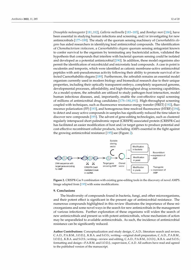
Drosophila melanogaster [181,182], Galleria mellonella [183–185], and Bombyx mori [186], havebeen essential in studying human infections and screening, and/or investigating for newantimicrobials [177–189]. The study of the quorum sensing machinery of Caenorhabditis ele-gans has aided researchers in identifying lead antimicrobial compounds. The identificationof Chomobacterium violaceum, a Caenorhabditis elegans quorum sensing antagonist knownto confer survival to the organism by terminating any bactericidal action, validated thehypothesis that compounds that interfere with bacterial quorum sensing could be isolatedand developed as a potential antimicrobial [190]. In addition, these model organisms alsopermit the identification of microbicidal and microstatic lead compounds. A case in point isesculentin and temporin, which were identified as cationic membrane-active antimicrobialpeptides with anti-pseudomonas activity following their ability to promote survival of in-fected Caenorhabditis elegans [189]. Furthermore, the zebrafish remains an essential modelorganism currently used in modern biology and biomedical research due to their uniqueproperties, including their optically transparent embryo, completely sequenced genome,developmental processes, affordability, and high-throughput drug screening capabilities.As a model system, the zebrafish are utilized to study pathogen-host interaction, modelhuman infectious diseases, and, importantly, enable the cost-effective rapid screeningof millions of antimicrobial drug candidates [178–180,191]. High-throughput screeningcoupled with techniques, such as fluorescence resonance energy transfer (FRET) [192], fluo-rescence polarization (FP) [193], and homogenous time resolved fluorescence (HTRF) [194],to detect and assay active compounds in samples has significantly reduced the time taken todiscover new compounds [195]. The advent of gene-editing technologies, such as clusteredregularly interspaced short palindromic repeat (CRISPR) associated protein (CRISPR-Cas)has facilitated an easier modification of host and/or target genes to produce potential andcost-effective recombinant cellular products, including AMPs essential in the fight againstthe growing antimicrobial resistance [195] see (Figure 2).
Antibiotics 2022, 11, x FOR PEER REVIEW 12 of 20
logical efficacy [170,171]. The emergence of omics technologies, notably genomics, tran-
scriptomics, and proteomics, has fast-tracked the development of bioinformatics tools to
identify novel drug targets and lead compounds. The genome mining technique, as it is
popularly called, can be used for the detection and analysis of the biosynthetic gene clus-
ters of the chemical compounds and then connect those genes to molecules [172]. The ad-
vancement of artificial intelligence (AI) and machine learning (ML) technologies has also
offered scientists alternate ammunition towards the fight against antimicrobial resistance
[173]. Aside from the development of halicin, an antimicrobial using ML approaches, AI
technologies are significant in all stages of drug discovery, ranging from target validation
and identification of predictive biomarkers, to analyzing pathological data in the various
stages of clinical trials [174–176]. The advent of model organisms, such as Caenorhabditis
elegans [177], zebrafish [178–180], Drosophila melanogaster [181,182], Galleria mellonella [183–
185], and Bombyx mori [186], have been essential in studying human infections and screen-
ing, and/or investigating for new antimicrobials [177–189]. The study of the quorum sens-
ing machinery of Caenorhabditis elegans has aided researchers in identifying lead antimi-
crobial compounds. The identification of Chomobacterium violaceum, a Caenorhabditis ele-
gans quorum sensing antagonist known to confer survival to the organism by terminating
any bactericidal action, validated the hypothesis that compounds that interfere with bac-
terial quorum sensing could be isolated and developed as a potential antimicrobial [190].
In addition, these model organisms also permit the identification of microbicidal and mi-
crostatic lead compounds. A case in point is esculentin and temporin, which were identi-
fied as cationic membrane-active antimicrobial peptides with anti-pseudomonas activity
following their ability to promote survival of infected Caenorhabditis elegans [189]. Fur-
thermore, the zebrafish remains an essential model organism currently used in modern
biology and biomedical research due to their unique properties, including their optically
transparent embryo, completely sequenced genome, developmental processes, affordabil-
ity, and high-throughput drug screening capabilities. As a model system, the zebrafish
are utilized to study pathogen-host interaction, model human infectious diseases, and,
importantly, enable the cost-effective rapid screening of millions of antimicrobial drug
candidates [178–180,191]. High-throughput screening coupled with techniques, such as
fluorescence resonance energy transfer (FRET) [192], fluorescence polarization (FP) [193],
and homogenous time resolved fluorescence (HTRF) [194], to detect and assay active com-
pounds in samples has significantly reduced the time taken to discover new compounds
[195]. The advent of gene-editing technologies, such as clustered regularly interspaced
short palindromic repeat (CRISPR) associated protein (CRISPR-Cas) has facilitated an eas-
ier modification of host and/or target genes to produce potential and cost-effective recom-
binant cellular products, including AMPs essential in the fight against the growing anti-
microbial resistance [196] see (Figure 2).
Figure 2. CRISPR-Cas 9 combination with existing gene-editing tools in the discovery of novel
AMPS. Image adapted from [196] with some modifications.
9. Conclusions
Figure 2. CRISPR-Cas 9 combination with existing gene-editing tools in the discovery of novel AMPS.Image adapted from [195] with some modifications.
9. Conclusions
The biodiversity of compounds found in bacteria, fungi, and other microorganisms,and their potent effect is significant in the present age of antimicrobial resistance. Thenumerous compounds highlighted in this review illustrates the importance of these mi-croorganisms and some novel ways in the search for new antimicrobials in the managementof various infections. Further exploration of these organisms will widen the search ofnew antimicrobials and present us with potent antimicrobials, whose mechanism of actionmay be unparalleled to available antimicrobials. As such, the incidence of antimicrobialresistance can be significantly reduced.
Author Contributions: Conceptualization and study design, C.A.D.; literature search and review,C.A.D., P.A.B.M., I.O.D.J., K.B.A. and S.O.S.; writing—original draft preparation, C.A.D., P.A.B.M.,I.O.D.J., K.B.A. and S.O.S.; writing—review and editing, C.A.D., P.A.B.M., I.O.D.J., K.B.A. and S.O.S.;formatting and design—P.A.B.M. and I.O.D.J.; supervision, C.A.D. All authors have read and agreedto the published version of the manuscript.

Antibiotics 2022, 11, 285 13 of 20
Funding: This research received no external funding.
Conflicts of Interest: The authors declare no conflict of interest.
Abbreviations
AMP: antimicrobial peptides; DNA: deoxyribonucleic acid; FP: fluorescence polarization; FRET:fluorescence resonance energy transfer; HTRF: homogenous time resolved fluorescence; LAB: lac-tic acid bacteria; MAN-PTS: mannose phosphotransferase permease; MRSA: methicillin-resistantStaphylococcus aureus; MIC: minimal inhibition concentration; AMPs: antimicrobial peptides; AVPs:antiviral peptides; CRISPR: clustered regularly interspaced short palindromic repeat.
References1. O’Neill, J. Antimicrobial Resistance:Tackling a crisis for the health and wealth of nations. Rev. Antimicrob. Resist. 2014.2. Laws, M.; Shaaban, A.; Rahman, K.M. Antibiotic resistance breakers: Current approaches and future directions. FEMS Microbiol.
Rev. 2019, 43, 490–516. [CrossRef]3. Newman, D.J.; Cragg, G.M. Natural products as sources of new drugs over the 30 years from 1981 to 2010. J. Nat. Prod. 2012, 75,
311–335. [CrossRef]4. Elissawy, A.M.; Dehkordi, E.S.; Mehdinezhad, N.; Ashour, M.L.; Pour, P.M. Cytotoxic Alkaloids Derived from Marine Sponges:
A Comprehensive Review. Biomolecules 2021, 11, 258. [CrossRef]5. El-Demerdash, A.; Tammam, M.A.; Atanasov, A.G.; Hooper, J.N.A.; Al-Mourabit, A.; Kijjoa, A. Chemistry and Biological Activities
of the Marine Sponges of the Genera Mycale (Arenochalina), Biemna and Clathria. Mar. Drugs 2018, 16, 214. [CrossRef]6. Karpinski, T.M. Marine Macrolides with Antibacterial and/or Antifungal Activity. Mar. Drugs 2019, 17, 241. [CrossRef]7. Xu, L.; Meng, W.; Cao, C.; Wang, J.; Shan, W.; Wang, Q. Antibacterial and antifungal compounds from marine fungi. Mar. Drugs
2015, 13, 3479–3513. [CrossRef]8. Swift, C.L.; Louie, K.B.; Bowen, B.P.; Olson, H.M.; Purvine, S.O.; Salamov, A.; Mondo, S.J.; Solomon, K.V.; Wright, A.T.; Northen,
T.R.; et al. Anaerobic gut fungi are an untapped reservoir of natural products. Proc. Natl. Acad. Sci. USA 2021, 118, e2019855118.[CrossRef]
9. Maghembe, R.; Damian, D.; Makaranga, A.; Nyandoro, S.S.; Lyantagaye, S.L.; Kusari, S.; Hatti-Kaul, R. Omics for Bioprospectingand Drug Discovery from Bacteria and Microalgae. Antibiot 2020, 9, 229. [CrossRef]
10. Moir, D.T.; Shaw, K.J.; Hare, R.S.; Vovis, G.F. Genomics and Antimicrobial Drug Discovery. Antimicrob. Agents Chemother. 1999, 43,439. [CrossRef]
11. Schnappinger, D. Genetic Approaches to Facilitate Antibacterial Drug Development. Cold Spring Harb. Perspect. Med. 2015, 5,a021139. [CrossRef]
12. Tortorella, E.; Tedesco, P.; Espositom, F.P.; January, G.G.; Fani, R.; Jaspars, M.; de Pascale, D. Antibiotics from Deep-SeaMicroorganisms: Current Discoveries and Perspectives. Mar. Drugs 2018, 16, 355. [CrossRef]
13. Singh, R.K.; Tiwari, S.P.; Rai, A.K.; Mohapatra, T.M. Cyanobacteria: An emerging source for drug discovery. J. Antibiot. 2011, 64,401–412. [CrossRef]
14. Rojas, V.; Rivas, L.; Cardenas, C.; Guzman, F. Cyanobacteria and Eukaryotic Microalgae as Emerging Sources of AntibacterialPeptides. Molecules 2020, 25, 5804. [CrossRef]
15. Alsenani, F.; Tupally, K.R.; Chua, E.T.; Eltanahy, E.; Alsufyani, H.; Parekh, H.S.; Schenk, P.M. Evaluation of microalgae andcyanobacteria as potential sources of antimicrobial compounds. Saudi. Pharm. J. 2020, 28, 1834–1841. [CrossRef]
16. Santovito, E.; Greco, D.; Marquis, V.; Raspoet, R.; D’Ascanio, V.; Logrieco, A.F.; Avantaggiato, G. Antimicrobial Activity of YeastCell Wall Products Against Clostridium perfringens. Foodborne Pathog. Dis. 2019, 16, 638–647. [CrossRef]
17. Hatoum, R.; Labrie, S.; Fliss, I. Antimicrobial and probiotic properties of yeasts: From fundamental to novel applications. Front.Microbiol. 2012, 3, 421. [CrossRef]
18. Mitcheltree, M.J.; Pisipati, A.; Syroegin, E.A.; Silvestre, K.J.; Klepacki, D.; Mason, J.D.; Terwilliger, D.W.; Testolin, G.; Pote, A.R.;Wu, K.J.Y.; et al. A synthetic antibiotic class overcoming bacterial multidrug resistance. Nature 2021, 599, 507–512. [CrossRef]
19. Tenea, G.N.; Yépez, L. Bioactive Compounds of Lactic Acid Bacteria. Case Study: Evaluation of Antimicrobial Activity ofBacteriocin-producing Lactobacilli Isolated from Native Ecological Niches of Ecuador. Probiotics Prebiotics Hum. Nutr. Health 2016,xxx, 149–167.
20. Todorov, S.D.; Dicks, L.M.T. Bacteriocin production by Lactobacillus pentosus ST712BZ isolated from boza. Braz. J. Microbiol.2007, 38, 166–172. [CrossRef]
21. Oscáriz, J.C.; Pisabarro, A.G. Classification and mode of action of membrane-active bacteriocins produced by Gram-positivebacteria. Int. Microbiol. 2001, 4, 13–19. [CrossRef]
22. Klaenhammer, T.R. Genetics of bacteriocins produced by lactic acid bacteria. FEMS Microbiol. Rev. 1993, 12, 39–85. [CrossRef]23. Güllüce, M.; Karadayı, M.; Barıs, Ö. Bacteriocins: Promising natural antimicrobials. Local Environ. 2013, 3, 6.24. Cotter, P.D.; Hill, C.; Ross, R.P. Food microbiology: Bacteriocins: Developing innate immunity for food. Nat. Rev. Microbiol. 2005,
3, 777–788. [CrossRef]

Antibiotics 2022, 11, 285 14 of 20
25. Drider, D.; Fimland, G.; Héchard, Y.; McMullen, L.M.; Prévost, H. The Continuing Story of Class IIa Bacteriocins. Microbiol. Mol.Biol. Rev. 2006, 70, 564–582. [CrossRef]
26. Hernández-González, J.C.; Martínez-Tapia, A.; Lazcano-Hernández, G.; García-Pérez, B.E.; Castrejón-Jiménez, N.S. Bacteriocinsfrom lactic acid bacteria. A powerful alternative as antimicrobials, probiotics, and immunomodulators in veterinary medicine.Animals 2021, 11, 979. [CrossRef]
27. Parada, J.L.; Caron, C.R.; Medeiros, A.B.P.; Soccol, C.R. Bacteriocins from lactic acid bacteria: Purification, properties and use asbiopreservatives. Braz. Arch. Biol. Technol. 2007, 50, 521–542. [CrossRef]
28. Bauer, R.; Dicks, L.M.T. Mode of action of lipid II-targeting lantibiotics. Int. J. Food Microbiol. 2005, 101, 201–216. [CrossRef]29. Paiva, A.D.; Breukink, E.; Mantovani, H.C. Role of lipid II and membrane thickness in the mechanism of action of the lantibiotic
bovicin HC5. Antimicrob. Agents Chemother. 2011, 55, 5284–5293. [CrossRef]30. Nissen-Meyer, J.; Oppegård, C.; Rogne, P.; Haugen, H.S.; Kristiansen, P.E. Structure and Mode-of-Action of the Two-Peptide
(Class-IIb) Bacteriocins. Probiotics Antimicrob. Proteins 2010, 2, 52–60. [CrossRef]31. Perez, R.H.; Zendo, T.; Sonomoto, K. Circular and Leaderless Bacteriocins: Biosynthesis, Mode of Action, Applications, and
Prospects. Front Microbiol. 2018, 9, 2085. [CrossRef] [PubMed]32. Simmonds, R.S.; Pearson, L.; Kennedy, R.C. Mode of Action of a Lysostaphin-Like Bacteriolytic Agent Produced by Streptococcus
zooepidemicus 4881. Appl. Environ. Microbiol. 1996, 62, 4536–4541. [CrossRef] [PubMed]33. Sun, Z.; Wang, X.; Zhang, X.; Wu, H.; Zou, Y.; Li, P.; Sun, C.; Xu, W. Class III bacteriocin Helveticin-M causes sublethal damage on
target cells through impairment of cell wall and membrane. J. Ind. Microbiol. Biotechnol. 2018, 45, 213–227. [CrossRef] [PubMed]34. Swe, P.M.; Cook, G.M.; Tagg, J.R.; Jack, R.W. Mode of Action of Dysgalacticin: A Large Heat-Labile Bacteriocin.
J. Antimicrob. Chemother. 2009, 63, 679–686. [CrossRef]35. Meade, E.; Slattery, M.A.; Garvey, M. Bacteriocins, Potent Antimicrobial Peptides and the Fight against Multi Drug Resistant
Species: Resistance Is Futile? Antibiotics 2020, 9, 32. [CrossRef]36. Mclller, E.; Radler, F. Caseicin, a Bacteriocin from Lactobacillus casei. Folia Microbiol. 1993, 38, 441–446. [CrossRef]37. Bollenbach, T. Antimicrobial interactions: Mechanisms and implications for drug discovery and resistance evolution.
Curr. Opin. Microbiol. 2015, 27, 1–9. [CrossRef]38. Cunha, E.; Trovão, T.; Pinheiro, A.; Nunes, T.; Santos, R.; Moreira, J.; Braz, B.S.; Tavares, L.; Veiga, A.S.; Oliveira, M. Potential of
Two Delivery Systems for Nisin Topical Application to Dental Plaque Biofilms in Dogs. BMC Vet. Res. 2018, 14, 1–10. [CrossRef]39. Howeir, T.H.; Fioreliinp, J.P. The Effect of a Mouthrinse Based on Nisin, a Bacteriocin, on Developing Plaque and Gingivitis in
Beagle Dogs. J. Clin. Periodontol. 1993, 20, 335–339. [CrossRef]40. Herzog, M.; Tiso, T.; Blank, L.M.; Winter, R. Interaction of rhamnolipids with model biomembranes of varying complexity.
Biochim. Biophys. Acta-Biomembr. 2020, 1862, 183431. [CrossRef]41. Jiang, J.; Zu, Y.; Li, X.; Meng, Q.; Long, X. Recent progress towards industrial rhamnolipids fermentation: Process optimization
and foam control. Bioresour. Technol. 2020, 298, 122394. [CrossRef] [PubMed]42. Thakur, P.; Saini, N.K.; Thakur, V.K.; Gupta, V.K.; Saini, R.V.; Saini, A.K. Rhamnolipid the Glycolipid Biosurfactant: Emerging
trends and promising strategies in the field of biotechnology and biomedicine. Microb. Cell Factories 2021, 20, 1–15. [CrossRef][PubMed]
43. Christova, N.; Tuleva, B.; Kril, A.; Georgieva, M.; Konstantinov, S.; Terziyski, I.; Nikolova, B.; Stoineva, I. Chemical Structure andIn Vitro Antitumor Activity of Rhamnolipids from Pseudomonas aeruginosa BN10. Appl. Biochem. Biotechnol. 2013, 170, 676–689.[CrossRef] [PubMed]
44. Borah, S.N.; Goswami, D.; Sarma, H.K.; Cameotra, S.S.; Deka, S. Rhamnolipid biosurfactant against Fusarium verticillioides tocontrol stalk and ear rot disease of maize. Front. Microbiol. 2016, 7, 1505. [CrossRef]
45. Jin, L.; Black, W.; Sawyer, T. Application of Environment-Friendly Rhamnolipids against Transmission of Enveloped Viruses LikeSARS-CoV2. Viruses 2021, 13, 322. [CrossRef]
46. Remichkova, M.; Galabova, D.; Roeva, I.; Karpenko, E.; Shulga, A.; Galabov, A.S. Anti-herpesvirus activities of Pseudomonas sp.S-17 rhamnolipid and its complex with alginate. Zeitschrift Fur Naturforsch Sect. C J. Biosci. 2008, 63, 75–81. [CrossRef]
47. Giugliano, R.; Buonocore, C.; Zannella, C.; Chianese, A.; Esposito, F.P.; Tedesco, P.; De Filippis, A.; Galdiero, M.; Franci, G.; dePascale, D. Antiviral Activity of the Rhamnolipids Mixture from the Antarctic Bacterium Pseudomonas gessardii M15 againstHerpes Simplex Viruses and Coronaviruses. Pharmaceutics 2021, 13, 2121. [CrossRef]
48. Broadbent, J.R.; Chou, Y.C.; Gillies, K.; Kondo, J.K. Nisin Inhibits Several Gram-Positive, Mastitis-Causing Pathogens. J. Dairy Sci.1989, 72, 3342–3345. [CrossRef]
49. Lebel, G.; Piché, F.; Frenette, M.; Gottschalk, M.; Grenier, D. Antimicrobial activity of nisin against the swine pathogen Streptococ-cus suis and its synergistic interaction with antibiotics. Peptides 2013, 50, 19–23. [CrossRef]
50. Carroll, J.; Draper, L.A.; O’Connor, P.M.; Coffey, A.; Hill, C.; Ross, R.P.; Cotter, P.D.; O’Mahony, J. Comparison of the activitiesof the lantibiotics nisin and lacticin 3147 against clinically significant mycobacteria. Int. J. Antimicrob. Agents 2010, 36, 132–136.[CrossRef]
51. Campion, A.; Casey, P.G.; Field, D.; Cotter, P.D.; Hill, C.; Ross, R.P. In vivo activity of Nisin A and Nisin v against Listeriamonocytogenes in mice. BMC Microbiol. 2013, 13, 23. [CrossRef]
52. Grilli, E.; Messina, M.; Catelli, E.; Morlacchini, M.; Piva, A. Pediocin a improves growth performance of broilers challenged withClostridium perfringens. Poult. Sci. 2009, 88, 2152–2158. [CrossRef] [PubMed]

Antibiotics 2022, 11, 285 15 of 20
53. Lauková, A.; Styková, E.; Kubašová, I.; Gancarcíková, S.; Plachá, I.; Mudronová, D.; Kandricáková, A.; Miltko, R.; Belzecki, G.;Valocký, I. Enterocin M and its Beneficial Effects in Horses—A Pilot Experiment. Probiotics Antimicrob. Proteins 2018, 10, 420–426.[CrossRef] [PubMed]
54. Wang, Q.; Cui, Y.; Wang, W.; Xu, J.; Xu, L. Production of two bacteriocins in various growth conditions produced by gram-positivebacteria isolated from chicken cecum. Can. J. Microbiol. 2012, 58, 93–101. [CrossRef] [PubMed]
55. Line, J.E.; Svetoch, E.A.; Eruslanov, B.V.; Perelygin, V.V.; Mitsevich, V.; Mitsevich, I.P.; Levchuk, V.P.; Svetoch, O.E.; Seal, B.S.;Siragusa, G.R.; et al. Isolation and Purification of Enterocin E-760 with Broad Antimicrobial Activity against Gram-Positive andGram-Negative Bacteria. Antimicrob. Agents Chemother. 2008, 52, 1094–1100. [CrossRef] [PubMed]
56. Ryan, M.P.; Flynn, J.; Hill, C.; Ross, R.P.; Meaney, W.J. The natural food grade inhibitor, lacticin 3147, reduced the incidence ofmastitis after experimental challenge with Streptococcus dysgalactiae in nonlactating dairy cows. J. Dairy Sci. 1999, 82, 2625–2631.[CrossRef]
57. Pieterse, R.; Todorov, S.D.; Dicks, L.M.T. Mode of action and in vitro susceptibility of mastitis pathogens to macedocin ST91KMand preparation of a teat seal containing the bacteriocin. Braz. J. Microbiol. 2010, 41, 133–145. [CrossRef]
58. Tomita, K.; Nishio, M.; Saitoh, K.; Yamamoto, H.; Hoshino, Y.; Ohkuma, H.; Konishi, M.; Miyaki, T.; Oki, T. Pradimicins A, B andC: New antifungal antibiotics. I. Taxonomy, production, isolation and physico-chemical properties. J. Antibiot. 1990, 43, 755–762.[CrossRef]
59. Walsh, T.J.; Giri, N. Pradimicins: A novel class of broad-spectrum antifungal compounds. Eur. J. Clin. Microbiol. Infect. Dis. 1997,16, 93–97. [CrossRef]
60. Kerr, J.R. Bacterial inhibition of fungal growth and pathogenicity. Microb. Ecol. Health Dis. 1999, 11, 129–142.61. Boumehira, A.Z.; El-Enshasy, H.A.; Hacene, H.; Elsayed, E.A.; Aziz, R.; Park, E.Y. Recent progress on the development of
antibiotics from the genus Micromonospora. Biotechnol. Bioprocess Eng. 2016, 21, 199–223. [CrossRef]62. Schwartz, R.E.; Giacobbe, R.A.; Monaghan, R.L. L-671,329, a new antifungal agent. I. Fermentation and isolation. J. Antibiot. 1989,
42, 163–167. [CrossRef] [PubMed]63. Nishizawa, N.; Kondo, Y.; Koyama, M.; Omoto, S.; Iwata, M.; Tsuruoka, T.; Inouye, S. Studies on a new nucleotide antibiotic,
Dapiramicin. J. Antibiot. 1983, 37, 1–5. [CrossRef] [PubMed]64. Fushimi, S.; Furihata, K.; Seto, H. Studies of new phosphate ester antifungal antibiotics phoslactomycins. II. Structure elucidation
of phoslactomycins A to F. J. Antibiot. 1989, 42, 1026–1036. [CrossRef] [PubMed]65. Hanafi, M.; Shibata, K.; Ueki, M.; Taniguchi, M. UK-2A, B, C and D, novel antifungal antibiotics from Streptomyces sp. 517-02: II.
Structural elucidation. J. Antibiot. 1996, 49, 1226–1231. [CrossRef]66. Tanaka, Y.; Kanaya, I.; Takahashi, Y.; Shinose, M.; Tanaka, H.; Omura, S.; Phthoxazolin, A. Phthoxazolin A, a specific inhibitor of
cellulose biosynthesis from microbial origin. I. Discovery, taxonomy of producing microorganism, fermentation, and biologicalactivity. J. Antibiot. 1993, 46, 1208–1213. [CrossRef]
67. Mitsuhashi, S.; Inoue, K. In vitro antibacterial activity of azithromycin, a new macrolide antibiotic. Jpn. J. Chemother. 1995, 43, 1–7.68. Kotake, C.; Yamasaki, T.; Moriyama, T.; Shinoda, M.; Komiyama, N.; Furumai, T.; Konishi, M.; Oki, T. Butyrolactols a and b∆, new
antifungal antibiotics taxonomy, isolation, physico-chemical properties, structure and biological activity. J. Antibiot. 1992, 45,1442–1450. [CrossRef]
69. Ohkuma, H.; Naruse, N.; Nishiyama, Y.; Tsuno, T.; Hoshino, Y.; Sawada, Y.; Konishi, M. Streptomyces roseiscleroticus Taxonomyofthe Producing Organism. J. Antibiot. 1992, 45, 1239–1249. [CrossRef]
70. Uramoto, M.; Uzawa, J.; Suzuki, S.; Isono, K.; Liehr, J.G.; McCloskey, J.A. Isolation and structure of polyoxin N. Nucleic Acids Res.1978, 1, s327–s332. [CrossRef]
71. Hochlowski, J.E.; Mullally, M.M.; Brill, G.M.; Whittern, D.N.; Buko, A.M.; Hill, P.; McAlpine, J.B. Dunaimycins, a new complex ofspiroketal 24-membered macrolides with immunosuppressive activity: II. isolation and elucidation of structures. J. Antibiot. 1991,44, 1318–1330. [CrossRef] [PubMed]
72. Dunlap, C.A.; Bowman, M.J.; Rooney, A.P. Iturinic lipopeptide diversity in the bacillus subtilis species group-important antifungalsfor plant disease biocontrol applications. Front. Microbiol. 2019, 10, 1794. [CrossRef] [PubMed]
73. Lebbadi, M.; Gálvez, A.; Maqueda, M.; Martínez-Bueno, M.; Valdivia, E. Fungicin M4: A narrow spectrum peptide antibioticfrom Bacillus licheniformis M-4. J. Appl. Bacteriol. 1994, 77, 49–53. [CrossRef] [PubMed]
74. Gálvez, A.; Maqueda, M.; Martínez-Bueno, M.; Lebbadi, M.; Valdivia, E. Isolation and physico-chemical characterization ofan antifungal and antibacterial peptide produced by Bacillus licheniformis A12. Appl. Microbiol. Biotechnol. 1993, 39, 438–442.[CrossRef]
75. Chernin, L.; Brandis, A.; Ismailov, Z.; Chet, I. Pyrrolnitrin production by an Enterobacter agglomerans strain with a broadspectrum of antagonistic activity towards fungal and bacterial phytopathogens. Curr. Microbiol. 1996, 32, 208–212. [CrossRef]
76. Greiner, M.; Winkelmann, G. Fermentation and isolation of herbicolin A, a peptide antibiotic produced by Erwinia herbicolastrain A 111. Appl. Microbiol. Biotechnol. 1991, 34, 565–569. [CrossRef]
77. Winkelmann, G.; Lupp, R.; Jung, G. Herbicolins—New peptide antibiotics from erwinia herbicola. J. Antibiot. 1980, 33, 353–358.[CrossRef]
78. Shoji, J.; Hinoo, H.; Sakazaki, R.; Kato, T.; Hattori, T.; Matsumoto, K.; Tawara, K.; Kikuchi, J.; Terui, Y. Isolation of CB-25-I, anantifungal antibiotic, from serratia plymuthica. J. Antibiot. 1989, 42, 869–874. [CrossRef]

Antibiotics 2022, 11, 285 16 of 20
79. Serino, L.; Reimmann, C.; Visca, P.; Beyeler, M.; Chiesa, V.D.; Haas, D. Biosynthesis of pyochelin and dihydroaeruginoic acidrequires the iron-regulated pchDCBA operon in Pseudomonas aeruginosa. J. Bacteriol. 1997, 179, 248–257. [CrossRef]
80. Kerr, J.R.; Taylor, G.W.; Rutman, A.; Høiby, N.; Cole, P.J.; Wilson, R. Pseudomonas aeruginosa pyocyanin and 1-hydroxyphenazineinhibit fungal growth. J. Clin. Pathol. 1999, 52, 385–387. [CrossRef]
81. Vincent, M.N.; Harrison, L.A.; Brackin, J.M.; Kovacevich, P.A.; Mukerji, P.; Weller, D.M.; Pierson, E.A. Genetic analysis of theantifungal activity of a soilborne Pseudomonas aureofaciens strain. Appl. Environ. Microbiol. 1991, 57, 2928–2934. [CrossRef][PubMed]
82. Harrison, L.; Teplow, D.B.; Rinaldi, M.; Strobel, G. Pseudomycins, a family of novel peptides from Pseudomonas syringaepossessing broad-spectrum antifungal activity. J. Gen. Microbiol. 1991, 137, 2857–2865. [CrossRef] [PubMed]
83. Yamaguchi, M.; Park, H.J.; Ishizuka, S.; Omata, K.; Hirama, M. Chemistry and Antimicrobial Activity of Caryoynencins Analogs.J. Med. Chem. 1995, 38, 5015–5022. [CrossRef] [PubMed]
84. Barker, W.R.; Callaghan, C.; Hill, L.; Noble, D.; Acred, P.; Harper, P.B.; Sowa, M.A.; Fletton, R.A. G1549, a new cyclic hydroxamicacid antibiotic, isolated from culture broth of pseudomonas alcaligenes. J. Antibiot. 1979, 32, 1096–1103. [CrossRef] [PubMed]
85. Lim, Y.; Suh, J.W.; Kim, S.; Hyun, B.; Kim, C.; Lee, C. hoon Cepacidine A, A novel antifungal antibiotic produced by pseudomonascepacia. II. Physico-chemical properties and structure elucidation. J. Antibiot. 1994, 47, 1406–1416. [CrossRef] [PubMed]
86. Abe, M.; Nakazawa, T. Characterization of Hemolytic and Antifungal Substance, Cepalycin, from Pseudomonas cepacia.Microbiol. Immunol. 1994, 38, 1–9. [CrossRef] [PubMed]
87. Bisacchi, G.S.; Parker, W.L.; Hockstein, D.R.; Koster, W.H.; Rathnum, M.L.; Unger, S.E. Xylocandin: A new complex of antifungalpeptides. II. Structural studies and chemical modifications. J. Antibiot. 1987, 40, 1520–1529. [CrossRef]
88. Saalim, M.; Villegas-Moreno, J.; Clark, B.R. Bacterial Alkyl-4-quinolones: Discovery, Structural Diversity and Biological Properties.Molecules 2020, 25, 5689. [CrossRef]
89. Knappe, T.A.; Linne, U.; Zirah, S.; Rebuffat, S.; Xie, X.; Marahiel, M.A. Isolation and structural characterization of capistruin, alasso peptide predicted from the genome sequence of Burkholderia thailandensis E264. J. Am. Chem. Soc. 2008, 130, 11446–11454.[CrossRef]
90. Watanabe, T.; Izaki, K.; Takahashi, H. New polyenic antibiotics active against gram-positive and -negative bacteria: I. Isolationand purification of antibiotics produced by gl uconobacter sp. W-315. J. Antibiot. 1982, 35, 1141–1147. [CrossRef]
91. Agrawal, S.; Deshmukh, S.K.; Reddy, M.S.; Prasad, R.; Goel, M. Endolichenic fungi: A hidden source of bioactive metabolites.S. Afr. J. Bot. 2020, 134, 163–186. [CrossRef]
92. Pelaez, F.; Cabello, A.; Platas, G.; Díez, M.T.; Del Val, A.G.; Basilio, A.; Martán, I.; Vicente, F.; Bills, G.F.; Giacobbe, R.A.; et al. Thediscovery of enfumafungin, a novel antifungal compound produced by an endophytic Hormonema species biological activityand taxonomy of the producing organisms. Syst. Appl. Microbiol. 2000, 23, 333–343. [CrossRef]
93. Kuhnert, E.; Li, Y.; Lan, N.; Yue, Q.; Chen, L.; Cox, R.J.; An, Z.; Yokoyama, K.; Bills, G.F. Enfumafungin synthase represents anovel lineage of fungal triterpene cyclases. Environ. Microbiol. 2018, 20, 3325–3342. [CrossRef] [PubMed]
94. Sauter, H.; Steglich, W.; Anke, T. Strobilurins: Evolution of a new class of active substances. Angew. Chem.-Int. Ed. 1999, 38,1328–1349. [CrossRef]
95. Chepkirui, C.; Richter, C.; Matasyoh, J.C.; Stadler, M. Monochlorinated calocerins A–D and 9-oxostrobilurin derivatives from thebasidiomycete Favolaschia calocera. Phytochemistry 2016, 132, 95–101. [CrossRef] [PubMed]
96. Hyde, K.D.; Xu, J.; Rapior, S.; Jeewon, R.; Lumyong, S.; Niego, A.G.T.; Abeywickrama, P.D.; Aluthmuhandiram, J.V.S.; Brahaman-age, R.S.; Brooks, S.; et al. The amazing potential of fungi: 50 ways we can exploit fungi industrially. Fungal Divers. 2019, 97,1–136. [CrossRef]
97. Chepkirui, C.; Cheng, T.; Matasyoh, J.; Decock, C.; Stadler, M. An unprecedented spiro [furan-2,1’-indene]-3-one derivative andother nematicidal and antimicrobial metabolites from Sanghuangporus sp. (Hymenochaetaceae, Basidiomycota) collected inKenya. Phytochem. Lett. 2018, 25, 141–146. [CrossRef]
98. Zhu, T.; Chen, Z.; Liu, P.; Wang, Y.; Xin, Z.; Zhu, W. New rubrolides from the marine-derived fungus Aspergillus terreusOUCMDZ-1925. J. Antibiot. 2014, 67, 315–318. [CrossRef]
99. Wu, G.; Sun, X.; Yu, G.; Wang, W.; Zhu, T.; Gu, Q.; Li, D. Cladosins A–E, hybrid polyketides from a deep-sea-derived fungus,Cladosporium sphaerospermum. J. Nat. Prod. 2014, 77, 270–275. [CrossRef]
100. Peng, J.; Zhang, X.; Du, L.; Wang, W.; Zhu, T.; Gu, Q.; Li, D. Sorbicatechols A and B, antiviral sorbicillinoids from the marine-derived fungus Penicillium chrysogenum PJX-17. J. Nat. Prod. 2014, 77, 424–428. [CrossRef]
101. Li, J.; Wang, Y.; Hao, X.; Li, S.; Jia, J.; Guan, Y.; Peng, Z.; Bi, H.; Xiao, C.; Cen, S.; et al. Broad-Spectrum Antiviral Natural Productsfrom the Marine-Derived Penicillium sp. IMB17-046. Molecules 2019, 24, 2821. [CrossRef] [PubMed]
102. Zhou, X.; Fang, W.; Tan, S.; Lin, X.; Xun, T.; Yang, B.; Liu, S.; Liu, Y. Aspernigrins with anti-HIV-1 activities from the marine-derivedfungus Aspergillus niger SCSIO Jcsw6F30. Bioorg. Med. Chem. Lett. 2016, 26, 361–365. [CrossRef] [PubMed]
103. Raveh, A.; Delekta, P.C.; Dobry, C.J.; Peng, W.; Schultz, P.J.; Blakely, P.K.; Tai, A.W.; Matainaho, T.; Irani, D.N.; Sherman, D.H.;et al. Discovery of potent broad spectrum antivirals derived from marine actinobacteria. PLoS ONE 2013, 8, e82318. [CrossRef][PubMed]
104. Wang, J.; Dou, X.; Song, J.; Lyu, Y.; Zhu, X.; Xu, L.; Li, W.; Shan, A. Antimicrobial peptides: Promising alternatives in the postfeeding antibiotic era. Med. Res. Rev. 2019, 39, 831–859. [CrossRef]
105. Narayana, J.L.; Chen, J.Y. Antimicrobial peptides: Possible anti-infective agents. Peptides 2015, 72, 88–94. [CrossRef]

Antibiotics 2022, 11, 285 17 of 20
106. Kang, X.; Dong, F.; Shi, C.; Liu, S.; Sun, J.; Chen, J.; Li, H.; Xu, H.; Lao, X.; Zheng, H. DRAMP 2.0, an updated data repository ofantimicrobial peptides. Sci. Data 2019, 6, 1–10. [CrossRef]
107. da Cunha, N.B.; Cobacho, N.B.; Viana, J.F.C.; Lima, L.A.; Sampaio, K.B.O.; Dohms, S.S.M.; Ferreira, A.C.R.; de la Fuente-Núñez,C.; Costa, F.F.; Franco, O.L.; et al. The next generation of antimicrobial peptides (AMPs) as molecular therapeutic tools for thetreatment of diseases with social and economic impacts. Drug Discov. Today 2017, 22, 234–248. [CrossRef]
108. Huerta-Cantillo, J.; Navarro-García, F. Properties and design of antimicrobial peptides as potential tools againstpathogens andmalignant cells. Investig. En Discapac. 2016, 5, 96–115.
109. Li, C.; Zhu, C.; Ren, B.; Yin, X.; Shim, S.H.; Gao, Y.; Zhu, J.; Zhao, P.; Liu, C.; Yu, R.; et al. Two optimized antimicrobial peptideswith therapeutic potential for clinical antibiotic-resistant Staphylococcus aureus. Eur. J. Med. Chem. 2019, 183, 111686. [CrossRef]
110. Kosikowska, P.; Lesner, A. Antimicrobial peptides (AMPs) as drug candidates: A patent review (2003–2015). Expert Opin. Ther.Pat. 2016, 26, 689–702. [CrossRef]
111. Boas, L.C.P.V.; Campos„ M.L.; Berlanda, R.L.A.; de Carvalho Neves, N.; Franco, O.L. Antiviral peptides as promising therapeuticdrugs. Cell. Mol. Life Sci. 2019, 76, 3525–3542. [CrossRef] [PubMed]
112. Huan, Y.; Kong, Q.; Mou, H.; Yi, H. Antimicrobial Peptides: Classification, Design, Application and Research Progress in MultipleFields. Front. Microbiol. 2020, 11, 582–779. [CrossRef] [PubMed]
113. Yasin, B.; Pang, M.; Turner, J.S.; Cho, Y.; Dinh, N.N.; Waring, A.J.; Lehrer, R.I.; Wagar, E.A. Evaluation of the inactivation ofinfectious herpes simplex virus by host-defense peptides. Eur. J. Clin. Microbiol. Infect. Dis. 2000, 19, 187–194. [CrossRef][PubMed]
114. Carriel-Gomes, M.C.; Kratz, J.M.; Barracco, M.A.; Bachére, E.; Barardi, C.R.M.; Simões, C.M.O. In vitro antiviral activity ofantimicrobial peptides against herpes simplex virus 1, adenovirus, and rotavirus. Mem. Inst. Oswaldo Cruz 2007, 102, 469–472.[CrossRef]
115. Kobori, T.; Iwamoto, S.; Takeyasu, K.; Ohtani, T. Biopolymers Volume 85/Number 4295. Biopolymers 2007, 85, 392–406.116. Xia, S.; Liu, M.; Wang, C.; Xu, W.; Lan, Q.; Feng, S.; Qi, F.; Bao, L.; Du, L.; Liu, S.; et al. Inhibition of SARS-CoV-2 (previously
2019-nCoV) infection by a highly potent pan-coronavirus fusion inhibitor targeting its spike protein that harbors a high capacityto mediate membrane fusion. Cell Res. 2020, 30, 343–355. [CrossRef]
117. Wang, Q.; Song, F.; Xiao, X.; Huang, P.; Li, L.; Monte, A.; Abdel-Mageed, W.M.; Wang, J.; Guo, H.; He, W.; et al. Abyssomicinsfrom the South China Sea deep-sea sediment Verrucosispora sp.: Natural thioether Michael addition adducts as antitubercularprodrugs. Angew. Chem. Int. Ed. Engl. 2013, 52, 1231–1234. [CrossRef]
118. Tadesse, M.; Tabudravu, J.N.; Jaspars, M.; Strom, M.B.; Hansen, E.; Andersen, J.H.; Kristiansen, P.E.; Haug, T. The antibacterialent-eusynstyelamide B and eusynstyelamides D, E, and F from the Arctic bryozoan Tegella cf. spitzbergensis. J. Nat. Prod. 2011,74, 837–841. [CrossRef]
119. Kennedy, J.; Codling, C.E.; Jones, B.V.; Dobson, A.D.; Marchesi, J.R. Diversity of microbes associated with the marine sponge,Haliclona simulans, isolated from Irish waters and identification of polyketide synthase genes from the sponge metagenome.Environ. Microbiol. 2008, 10, 1888–1902. [CrossRef]
120. Tapiolas, D.M.; Bowden, B.F.; Abou-Mansour, E.; Willis, R.H.; Doyle, J.R.; Muirhead, A.N.; Liptrot, C.; Llewellyn, L.E.; Wolff,C.W.; Wright, A.D.; et al. Eusynstyelamides A, B, and C, nNOS inhibitors, from the ascidian Eusynstyela latericius. J. Nat. Prod.2009, 72, 1115–1120. [CrossRef] [PubMed]
121. McAuliffe, O.; Ross, R.P.; Hill, C. Lantibiotics: Structure, biosynthesis and mode of action. FEMS Microbiol. Rev. 2001, 25, 285–308.[CrossRef] [PubMed]
122. Freire, J.M.; Dias, S.A.; Flores, L.; Veiga, A.S.; Castanho, M.A. Mining viral proteins for antimicrobial and cell-penetrating drugdelivery peptides. Bioinformatics 2015, 31, 2252–2256. [CrossRef] [PubMed]
123. Fukunaga, Y.; Kurahashi, M.; Tanaka, K.; Yanagi, K.; Yokota, A.; Harayama, S. Pseudovibrio ascidiaceicola sp. nov., isolated fromascidians (sea squirts). Int. J. Syst. Evol. Microbiol. 2006, 56, 343–347. [CrossRef]
124. Phelan, R.W.; Barret, M.; Cotter, P.D.; O’Connor, P.M.; Chen, R.; Morrissey, J.P.; Dobson, A.D.; O’Gara, F.; Barbosa, T.M.Subtilomycin: A new lantibiotic from Bacillus subtilis strain MMA7 isolated from the marine sponge Haliclona simulans.Mar. Drugs 2013, 11, 1878–1898. [CrossRef]
125. Tadesse, M.; Strom, M.B.; Svenson, J.; Jaspars, M.; Milne, B.F.; Torfoss, V.; Andersen, J.H.; Hansen, E.; Stensvag, K.; Haug, T.Synoxazolidinones A and B: Novel bioactive alkaloids from the ascidian Synoicum pulmonaria. Org. Lett. 2010, 12, 4752–4755.[CrossRef]
126. Blond, A.; Cheminant, M.; Destoumieux-Garzon, D.; Segalas-Milazzo, I.; Peduzzi, J.; Goulard, C.; Rebuffat, S. Thermolysin-linearized microcin J25 retains the structured core of the native macrocyclic peptide and displays antimicrobial activity.Eur. J. Biochem. 2002, 269, 6212–6222. [CrossRef]
127. Shieh, W.Y.; Lin, Y.T.; Jean, W.D. Pseudovibrio denitrificans gen. nov., sp. nov., a marine, facultatively anaerobic, fermentativebacterium capable of denitrification. Int. J. Syst. Evol. Microbiol. 2004, 54, 2307–2312. [CrossRef]
128. Graca, A.P.; Bondoso, J.; Gaspar, H.; Xavier, J.R.; Monteiro, M.C.; de la Cruz, M.; Oves-Costales, D.; Vicente, F.; Lage, O.M.Antimicrobial activity of heterotrophic bacterial communities from the marine sponge Erylus discophorus (Astrophorida,Geodiidae). PLoS ONE 2013, 8, e78992. [CrossRef]
129. Flemer, B.; Kennedy, J.; Margassery, L.M.; Morrissey, J.P.; O’Gara, F.; Dobson, A.D. Diversity and antimicrobial activities ofmicrobes from two Irish marine sponges, Suberites carnosus and Leucosolenia sp. J. Appl. Microbiol. 2012, 112, 289–301. [CrossRef]

Antibiotics 2022, 11, 285 18 of 20
130. Sanchez, L.A.; Hedstrom, M.; Delgado, M.A.; Delgado, O.D. Production, purification and characterization of serraticin A, a novelcold-active antimicrobial produced by Serratia proteamaculans 136. J. Appl. Microbiol. 2010, 109, 936–945. [CrossRef] [PubMed]
131. Pan, H.Q.; Zhang, S.Y.; Wang, N.; Li, Z.L.; Hua, H.M.; Hu, J.C.; Wang, S.J. New spirotetronate antibiotics, lobophorins H and I,from a South China Sea-derived Streptomyces sp. 12A35. Mar. Drugs 2013, 11, 3891–3901. [CrossRef] [PubMed]
132. Kim, M.K.; Park, H.; Oh, T.J. Antibacterial and antioxidant capacity of polar microorganisms isolated from Arctic lichenOchrolechia sp. Pol. J. Microbiol. 2014, 63, 317–322. [CrossRef] [PubMed]
133. Lo Giudice, A.; Bruni, V.; Michaud, L. Characterization of Antarctic psychrotrophic bacteria with antibacterial activities againstterrestrial microorganisms. J. Basic Microbiol. 2007, 47, 496–505. [CrossRef] [PubMed]
134. Papaleo, M.C.; Fondi, M.; Maida, I.; Perrin, E.; Lo Giudice, A.; Michaud, L.; Mangano, S.; Bartolucci, G.; Romoli, R.; Fani, R.Sponge-associated microbial Antarctic communities exhibiting antimicrobial activity against Burkholderia cepacia complexbacteria. Biotechnol. Adv. 2012, 30, 272–293. [CrossRef]
135. Gesheva, V.; Vasileva-Tonkova, E. Production of enzymes and antimicrobial compounds by halophilic Antarctic Nocardioides sp.grown on different carbon sources. World J. Microbiol. Biotechnol. 2012, 28, 2069–2076. [CrossRef]
136. Hay, M.; Thomas, D.W.; Craighead, J.L.; Economides, C.; Rosenthal, J. Clinical development success rates for investigationaldrugs. Nat. Biotechnol. 2014, 32, 40–51. [CrossRef]
137. DiMasi, J.A.; Grabowski, H.G.; Hansen, R.W. Innovation in the pharmaceutical industry: New estimates of R&D costs.J. Health Econ. 2016, 47, 20–33.
138. Trussell, P.C.; Baird, E.A. A rapid method for the assay of penicillin. Can. J. Res. 1947, 25, 5–8. [CrossRef]139. Rex, J.; Ghannoum, M.A.; Alexander, D.B.; Andes, D.; Brown, D.A.; Diekema, D.J.; Espinel-Ingroff, A.; Fowler, C.L.; Johnson,
E.J.; Knapp, C.C.; et al. Method for Antifungal Disk Diffusion Susceptibility Testing of Nondermatophyte Filamentous Fungi; ApprovedGuideline; CLSI Document M51-A; Clinical and Laboratory Standards Institute: Wayne, PA, USA, 2010; Volume 30, pp. 1–29.
140. Balouiri, M.; Sadiki, M.; Ibnsouda, S.K. Methods for in vitro evaluating antimicrobial activity: A review. J. Pharm. Anal. 2016, 6,71–79. [CrossRef] [PubMed]
141. Weinstein, M.P.; Patel, J.B.; Bobenchik, A.M.; Campeau, S.; Cullen, S.K.; Gallas, M.F.; Gold, H.; Humphries, R.M.; Kirn, T.J.; Lewis,J.S.; et al. Performance Standards for Antimicrobial Disk Susceptibility Tests: Approved Standard, 29th ed.; Clinical and LaboratoryStandards Institute Supplement M100; Clinical and Laboratory Standards Institute: Wayne, PA, USA, 2019; Volume 32.
142. White, R.L.; Burgess, D.S.; Manduru, M.; Bosso, J.A. Comparison of three different in vitro methods of detecting synergy:Time-kill, checkerboard, and E test. Antimicrob. Agents Chemother. 1996, 40, 1914–1918. [CrossRef] [PubMed]
143. Jiménez-Esquilín, A.E.; Roane, T.M. Antifungal activities of actinomycete strains associated with high-altitude sagebrushrhizosphere. J. Ind. Microbiol. Biotechnol. 2005, 32, 378–381. [CrossRef] [PubMed]
144. Sci, W.J. A Comparison of Two Methods Used for Measuring the Antagonistic Activity of Bacillus Species. Culture 2008, 5,161–171.
145. Ali-Shtayeh, M.S.; Abu Ghdeib, S.I. Antifungal activity of plant extracts against dermatophytes. Mycoses 1999, 42, 665–672.[CrossRef]
146. Kumar, S.N.; Nambisan, B.; Sundaresan, A.; Mohandas, C.; Anto, R.J. Isolation and identification of antimicrobial secondarymetabolites from Bacillus cereus associated with a rhabditid entomopathogenic nematode. Ann. Microbiol. 2014, 64, 209–218.[CrossRef]
147. Menon, T.; Umamaheswari, K.; Kumarasamy, N.; Solomon, S.; Thyagarajan, S.P. Efficacy of fluconazole and itraconazole in thetreatment of oral candidiasis in HIV patients. Acta Trop. 2001, 80, 151–154. [CrossRef]
148. Imhof, A.; Balajee, S.A.; Marr, K.A. New Methods To Assess Susceptibilities of Aspergillus Isolates to Caspofungin. J. Clin. Microbiol.2003, 41, 5683–5688.
149. Mock, M.; Monod, M.; Baudraz-Rosselet, F.; Panizzon, R.G. Tinea capitis dermatophytes: Susceptibility to antifungal drugs testedin vitro and in vivo. Dermatology 1998, 197, 361–367. [CrossRef]
150. Pfaller, M.A.; Sheehan, D.J.; Rex, J.H. Determination of Fungicidal Activities against Yeasts and Molds: Lessons Learned fromBactericidal Testing and the Need for Standardization. Clin. Microbiol. Rev. 2004, 17, 268–280. [CrossRef]
151. Crouch, S.P.M.; Kozlowski, R.; Slater, K.J.; Fletcher, J. Use of ATP as a measure of cell proliferation and cell toxicity.J. Immunol. Methods 1993, 160, 81–88. [CrossRef]
152. Paloque, L.; Vidal, N.; Casanova, M.; Dumètre, A.; Verhaeghe, P.; Parzy, D.; Azas, N. A new, rapid and sensitive bioluminescenceassay for drug screening on Leishmania. J. Microbiol. Methods 2013, 95, 320–323. [CrossRef] [PubMed]
153. Finger, S.; Wiegand, C.; Buschmann, H.J.; Hipler, U.C. Antibacterial properties of cyclodextrin-antiseptics-complexes determinedby microplate laser nephelometry and ATP bioluminescence assay. Int. J. Pharm. 2013, 452, 188–193. [CrossRef] [PubMed]
154. Galiger, C.; Brock, M.; Jouvion, G.; Savers, A.; Parlato, M.; Ibrahim-Granet, O. Assessment of efficacy of antifungals againstAspergillus fumigatus: Value of real-time bioluminescence imaging. Antimicrob. Agents Chemother. 2013, 57, 3046–3059. [CrossRef]
155. Peyron, F.; Favel, A.; Guiraud-Dauriac, H.; El Mzibri, M.; Chastin, C.; Duménil, G.; Regli, P. Evaluation of a flow cytofluorometricmethod for rapid determination of amphotericin B susceptibility of yeast isolates. Antimicrob. Agents Chemother. 1997, 41,1537–1540. [CrossRef]
156. Paparella, A.; Taccogna, L.; Aguzzi, I.; Chaves-López, C.; Serio, A.; Marsilio, F.; Suzzi, G. Flow cytometric assessment of theantimicrobial activity of essential oils against Listeria monocytogenes. Food Control 2008, 19, 1174–1182. [CrossRef]

Antibiotics 2022, 11, 285 19 of 20
157. Landaburu, L.U.; Berenstein, A.J.; Videla, S.; Maru, P.; Shanmugam, D.; Chernomoretz, A.; Agüero, F. TDR Targets 6: Drivingdrug discovery for human pathogens through intensive chemogenomic data integration. Nucleic Acids Res. 2020, 48, D992.
158. Bumol, T.F.; Watanabe, A.M. Genetic Information, Genomic Technologies, and the Future of Drug Discovery. JAMA 2001, 285,551–555. [CrossRef]
159. Carroll, P.M.; Dougherty, B.; Ross-Macdonald, P.; Browman, K.; FitzGerald, K. Model systems in drug discovery: Chemicalgenetics meets genomics. Pharmacol. Ther. 2003, 99, 183–220. [CrossRef]
160. Chavali, A.K.; D’Auria, K.M.; Hewlett, E.L.; Pearson, R.D.; Papin, J.A. A metabolic network approach for the identification andprioritization of antimicrobial drug targets. Trends Microbiol. 2012, 20, 113. [CrossRef]
161. Uddin, R.; Saeed, K.; Khan, W.; Azam, S.S.; Wadood, A. Metabolic pathway analysis approach: Identification of novel therapeutictarget against methicillin resistant Staphylococcus aureus. Gene 2015, 556, 213–226. [CrossRef] [PubMed]
162. Khan, M.T.; Mahmud, A.; Iqbal, A.; Hoque, S.F.; Hasan, M. Subtractive genomics approach towards the identification of noveltherapeutic targets against human Bartonella bacilliformis. Inform. Med. Unlocked 2020, 20, 100385. [CrossRef]
163. Shahid, F.; Shehroz, M.; Zaheer, T.; Ali, A. Subtractive Genomics Approaches: Towards Anti-Bacterial Drug Discovery.Front. Anti-Infect. Drug Discov. 2020, 8, 144–158.
164. Zhan, P.; Pannecouque, C.; De Clercq, E.; Liu, X. Anti-HIV Drug Discovery and Development: Current Innovations and FutureTrends. J. Med. Chem. 2016, 59, 2849–2878. [CrossRef]
165. Hackbarth, C.J.; Chen, D.Z.; Lewis, J.G.; Clark, K.; Mangold, J.B.; Cramer, J.A.; Margolis, P.S.; Wang, W.; Koehn, J.; Wu,C.; et al. N-alkyl urea hydroxamic acids as a new class of peptide deformylase inhibitors with antibacterial activity.Antimicrob. Agents Chemother. 2002, 46, 2752–2764. [CrossRef]
166. Naor, N.; Gadot, O.; Meir, M.; Barkan, D. Peptide Deformylase (def) is essential in Mycobacterium smegmatis, but the essentialityis compensated by inactivation of methionine formylation. BMC Microbiol. 2019, 19, 232. [CrossRef]
167. Teo, J.W.; Thayalan, P.; Beer, D.; Yap, A.S.; Nanjundappa, M.; Ngew, X.; Duraiswamy, J.; Liung, S.; Dartois, V.; Schreiber, M.; et al.Peptide deformylase inhibitors as potent antimycobacterial agents. Antimicrob. Agents Chemother. 2006, 50, 3665–3673. [CrossRef]
168. Chen, D.; Hackbarth, C.; Ni, Z.J.; Wu, C.; Wang, W.; Jain, R.; He, Y.; Bracken, K.; Weidmann, B.; Patel, D.V.; et al. Peptidedeformylase inhibitors as antibacterial agents: Identification of VRC3375, a proline-3-alkylsuccinyl hydroxamate derivative, byusing an integrated combinatorial and medicinal chemistry approach. Antimicrob. Agents Chemother. 2004, 48, 250–261. [CrossRef]
169. Kaplan, N.; Albert, M.; Awrey, D.; Bardouniotis, E.; Berman, J.; Clarke, T.; Dorsey, M.; Hafkin, B.; Ramnauth, J.; Romanov,V.; et al. Mode of action, in vitro activity, and in vivo efficacy of AFN-1252, a selective antistaphylococcal FabI inhibitor.Antimicrob. Agents Chemother. 2012, 56, 5865–5874. [CrossRef]
170. Heath, R.J.; White, S.W.; Rock, C.O. Inhibitors of fatty acid synthesis as antimicrobial chemotherapeutics. Appl. Microbiol. Biotechnol.2002, 58, 695–703.
171. Wohlleben, W.; Mast, Y.; Stegmann, E.; Ziemert, N. Antibiotic Drug Discovery. Microb. Biotechnol. 2016, 9, 541–548. [CrossRef][PubMed]
172. Fanelli, U.; Pappalardo, M.; Chine, V.; Gismondi, P.; Neglia, C.; Argentiero, A.; Calderaro, A.; Prati, A.; Esposito, S. Role ofArtificial Intelligence in Fighting Antimicrobial Resistance in Pediatrics. Antibiotics 2020, 9, 767. [CrossRef]
173. Vamathevan, J.; Clark, D.; Czodrowski, P.; Dunham, I.; Ferran, E.; Lee, G.; Li, B.; Madabhushi, A.; Shah, P.; Spitzer, M.; et al.Applications of machine learning in drug discovery and development. Nat. Rev. Drug Discov. 2019, 18, 463–477. [CrossRef][PubMed]
174. Lau, H.J.; Lim, C.H.; Foo, S.C.; Tan, H.S. The role of artificial intelligence in the battle against antimicrobial-resistant bacteria.Curr. Genet. 2021, 67, 421–429. [CrossRef]
175. Stokes, J.M.; Yang, K.; Swanson, K.; Jin, W.; Cubillos-Ruiz, A.; Donghia, N.M.; MacNair, C.R.; French, S.; Carfrae, L.A.; Bloom-Ackermann, Z.; et al. A Deep Learning Approach to Antibiotic Discovery. Cell 2020, 181, 475–483. [CrossRef]
176. Gravato-Nobre, M.J.; Hodgkin, J. Caenorhabditis elegans as a model for innate immunity to pathogens. Cell. Microbiol. 2005, 7,741–751. [CrossRef]
177. Rasheed, S.; Fries, F.; Muller, R.; Herrmann, J. Zebrafish: An Attractive Model to Study Staphylococcus aureus Infection and ItsUse as a Drug Discovery Tool. Pharmaceuticals 2021, 14, 594. [CrossRef]
178. Antoine, T.E.; Jones, K.S.; Dale, R.M.; Shukla, D.; Tiwari, V. Zebrafish: Modeling for herpes simplex virus infections. Zebrafish2014, 11, 17–25. [CrossRef]
179. Burgos, J.S.; Ripoll-Gomez, J.; Alfaro, J.M.; Sastre, I.; Valdivieso, F. Zebrafish as a new model for herpes simplex virus type 1infection. Zebrafish 2008, 5, 323–333. [CrossRef]
180. Ugur, B.; Chen, K.; Bellen, H.J. Drosophila tools and assays for the study of human diseases. Dis. Models Mech. 2016, 9, 235–244.[CrossRef]
181. Chamilos, G.; Samonis, G.; Kontoyiannis, D.P. Drosophila melanogaster as a model host for the study of microbial pathogenicityand the discovery of novel antimicrobial compounds. Curr. Pharm. Des. 2011, 17, 1246–1253. [CrossRef]
182. Champion, O.L.; Wagley, S.; Titball, R.W. Galleria mellonella as a model host for microbiological and toxin research. Virulence2016, 7, 840–845. [CrossRef]
183. Lebreton, F.; Le Bras, F.; Reffuveille, F.; Ladjouzi, R.; Giard, J.C.; Leclercq, R.; Cattoir, V. Galleria mellonella as a model for studyingEnterococcus faecium host persistence. J. Mol. Microbiol. Biotechnol. 2011, 21, 191–196. [CrossRef]

Antibiotics 2022, 11, 285 20 of 20
184. Norville, I.H.; Hartley, M.G.; Martinez, E.; Cantet, F.; Bonazzi, M.; Atkins, T.P. Galleria mellonella as an alternative model ofCoxiella burnetii infection. Microbiology 2014, 160, 1175–1181. [CrossRef]
185. Hamamoto, H.; Tonoike, A.; Narushima, K.; Horie, R.; Sekimizu, K. Silkworm as a model animal to evaluate drug candidatetoxicity and metabolism. Comp. Biochem. Physiol. C Toxicol. Pharmacol. 2009, 149, 334–339. [CrossRef]
186. Giunti, S.; Andersen, N.; Rayes, D.; De Rosa, M.J. Drug discovery: Insights from the invertebrate Caenorhabditis elegans.Pharmacol. Res. Perspect. 2021, 9, e00721. [CrossRef]
187. Artal-Sanz, M.; de Jong, L.; Tavernarakis, N. Caenorhabditis elegans: A versatile platform for drug discovery. Biotechnol. J. 2006,1, 1405–1418. [CrossRef]
188. Uccelletti, D.; Zanni, E.; Marcellini, L.; Palleschi, C.; Barra, D.; Mangoni, M.L. Anti-Pseudomonas activity of frog skinantimicrobial peptides in a Caenorhabditis elegans infection model: A plausible mode of action in vitro and in vivo.Antimicrob. Agents Chemother. 2010, 54, 3853–3860. [CrossRef]
189. Swem, L.R.; Swem, D.L.; O’Loughlin, C.T.; Gatmaitan, R.; Zhao, B.; Ulrich, S.M.; Bassler, B.L. A quorum-sensing antagonist targetsboth membrane-bound and cytoplasmic receptors and controls bacterial pathogenicity. Mol. Cell 2009, 35, 143–153. [CrossRef]
190. Takaki, K.; Cosma, C.L.; Troll, M.A.; Ramakrishnan, L. An in vivo platform for rapid high-throughput antitubercular drugdiscovery. Cell Rep. 2012, 2, 175–184. [CrossRef]
191. Malkani, N.; Schmid, J.A. Some secrets of fluorescent proteins: Distinct bleaching in various mounting fluids and photoactivationof cyan fluorescent proteins at YFP-excitation. PLoS ONE 2011, 6, e18586. [CrossRef]
192. Parker, G.J.; Law, T.L.; Lenoch, F.J.; Bolger, R.E. Development of high throughput screening assays using fluorescence polarization:Nuclear receptor-ligand-binding and kinase/phosphatase assays. J. Biomol. Screen. 2000, 5, 77–88. [CrossRef]
193. Bazin, H.; Préaudat, M.; Trinquet, E.; Mathis, G. Homogeneous time resolved fluorescence resonance energy transfer using rareearth cryptates as a tool for probing molecular interactions in biology. Spectrochim. Acta-Part A Mol. Biomol. Spectrosc. 2001, 57,2197–2211. [CrossRef]
194. Hajare, A.A.; Salunkhe, S.S.; Sachin, M.S.; Gorde, S.S.; Nadaf, J.S.; Pishawikar, A.S. Review on: High-throughput screening is anapproach to drug discovery. Review On: High-Throughput Screening Is An Approach To Drug. Am. J. Pharmtech Res. 2014, 4,113–129.
195. Sinha, R.; Shukla, P. Antimicrobial Peptides: Recent Insights on Biotechnological Interventions and Future Perspectives.Protein Pept. Lett. 2019, 26, 79–87. [CrossRef]