ANTIMICROBIAL ACTIVITY of SPIRULINA PLATENSIS ...
6
1203 ANTIMICROBIAL ACTIVITY of SPIRULINA PLATENSIS AGAINST AQUATIC BACTERIAL ISOLATES Wagih Abd El-Fattah Elshouny 1 , Mostafa Mohammed El-Sheekh 1 , Shawky Zaki Sabae 2 , Maha Abdelfattah Khalil 1,3 , Hanaa Mohamed Badr *1, 2 Address(es): 1 Department of Botany, Faculty of Science, Tanta University, 31527 Tanta, Egypt. 2 Hydrobiology Lab, National Institute of Oceanography and Fisheries, Cairo, Egypt. 3 Departments of Biology, Faculty of Science, Taif University, P.O. Box 888, Taif 21974, Saudi Arabia. *Corresponding author: [email protected] ABSTRACT Keywords: Spirulina platensis, Antimicrobial activity, H-NMR, FT-IR, GC-MS INTRODUCTION Bacterial infection causes high rate of mortality in human population and aquaculture organisms (Kandhasamy and Arunachalam, 2008). Preventing disease outbreaks or treating the disease with drugs or the screening programs for selecting therapeutic chemicals tackles this problem. The search for natural compounds with antimicrobial activity has gained importance in recent years due to growing worldwide concern about alarming increase in the rate of infection by antibiotic resistant microorganisms (Kaushik and Chauhan, 2008). Various strains of cyanobacteria and algae are known to produce intracellular and extracellular metabolites with diverse biological activities such as antialgal, antibacterial, antifungal and antiviral activities (Noaman et al., 2004; Kumar et al., 2011; Al-Wathnani et al., 2012). Numerous substances were identified as antimicrobial agents from algae such as Chlorellin derivatives, acrylic acid, halogenated aliphatic compounds, terpenes, sulphur containing heterocylic compounds, phenolic inhibitors etc. (Lavanya and Veerappan, 2011). It is generally considered that compounds produced naturally, rather than synthetically, will be biodegraded more easily and will therefore be environmentally acceptable (Ozdemir et al., 2004; Colla et al., 2007). Attention is now being focused on the natural components produced by aquatic organisms. Cyanobacteria are potential sources of high value chemicals and pharmaceuticals (Tan, 2007). The cyanobacteriaum, Spirulina platensis has emerged as one of the most promising agents to synthesize potentially new therapeutic compounds. It is known to produce intracellular and extracellular metabolites with diverse biological activities such as antifungal (MacMillan et al., 2002), antiviral (Hayashi and Hayashi, 1996) and antibacterial activities (Kaushik and Chauhan 2008; Kumar et al., 2011). The aim of the study is to: (1) measure in vitro the antibacterial activity of different extracts of some cyanobacterial and algal extracts against different aquatic microbial isolates collected from surface water of Al-Bahr El-Pherony, Menoufia, Egypt and (2) characterize the structure of active compound using different methods including; H-NMR, FT-IR, UV and GC-MS analysis. MATERIALS AND METHODS Bacterial isolation The microorganisms used in the antibacterial assay were isolated from surface water of Al-Bahr El-Pherony, Menoufia, Egypt. The obtained isolates were identified as previously explained by Sabae et al. (2014). One Gram-positive bacterium namely Staphylococcus aureus, four Gram-negative bacteria namely Escherichia coli, Pseudomonas aeruginosa, Salmonella sp, Shigella sp. were tested for the antibacterial activity. Inoculum preparation The bacterial isolates were inoculated on Müller Hinton broth and incubated for 24 h at 30°C then suspended in saline solution 0.85% NaCl, adjusted to yield approximately 1.0 x 10 7 -1.0 x 10 8 CFU/ml by using spectrophotometer (25% transmittance at 530 nm). Cultivation of tested species Spirulina platensis and Chlorella vulgaris were obtained from the culture collection of the Botany Department, Faculty of Science, Tanta University, Tanta, Egypt. Zarrouk`s medium (Zarrouk, 1966) was used for cultivation of S. platensis while, Kuhl`s medium was used for cultivation of Chlorella vulgaris (Kuhl, 1962). Culture temperature was maintained at 30 ±1°C. They were grown until the late exponential phase of the growth at which the cultures were harvested. The collected biomass was dried in a hot air oven at 60°C for 1 h. Collection of macroalgae Saragassum wightii and Saragassum latifolium samples were collected from the rocky areas surface in Red sea beach, Seuz, Egypt during spring 2013. After collection, samples were washed with seawater to remove epiphytes and other marine organisms. The seaweeds were transported to the laboratory in sterile Aquatic organisms are a rich source of novel and bioactive compounds. Cyanobacteria and microalgae being a rich source of bioactive compounds have recently found immense application in human and animal medicine. The present study was attempted to find out the effect of the various extracts of Spirulina platensis, Chlorella vulgaris, Saragassum wightii and Saragassum latifolium using different solvents (methanol, ethanol, ethyl acetate and chloroform) as antimicrobial agents against five bacterial pathogens; S. aureus, E. coli, P. aeruginosa, Salmonella sp, Shigella sp. Results indicate that among the various extracts used, methanol extracts of tested cyanobacterial and algal species appeared to be the most effective ones showing maximum antibacterial activity against the selected bacterial pathogens. Spirulina platensis appeared to be the most effective against all the pathogens studied. The antibacterial substance was purified using column chromatography. The nature of the purified active fractions was detected using different chemical analyses (UV, FT-IR, 1 H NMR and GC-MS) which indicated that it is an aliphatic compound and has different active groups (-OH,-C=O,-CH2 and – CH3). The results of this investigation proved that the tested cyanobacterium could be a good source for the production of promising antimicrobial agents. ARTICLE INFO Received 1. 4. 2015 Revised 12. 1. 2017 Accepted 27. 2. 2017 Published 3. 4. 2017 Regular article doi: 10.15414/jmbfs.2017.6.5.1203-1208
-
Upload
khangminh22 -
Category
Documents
-
view
0 -
download
0
Transcript of ANTIMICROBIAL ACTIVITY of SPIRULINA PLATENSIS ...

1203
ANTIMICROBIAL ACTIVITY of SPIRULINA PLATENSIS AGAINST AQUATIC BACTERIAL ISOLATES
Wagih Abd El-Fattah Elshouny1, Mostafa Mohammed El-Sheekh
1, Shawky Zaki Sabae
2, Maha Abdelfattah Khalil
1,3, Hanaa
Mohamed Badr*1, 2
Address(es): 1Department of Botany, Faculty of Science, Tanta University, 31527 Tanta, Egypt. 2Hydrobiology Lab, National Institute of Oceanography and Fisheries, Cairo, Egypt. 3 Departments of Biology, Faculty of Science, Taif University, P.O. Box 888, Taif 21974, Saudi Arabia.
*Corresponding author: [email protected]
ABSTRACT
Keywords: Spirulina platensis, Antimicrobial activity, H-NMR, FT-IR, GC-MS
INTRODUCTION
Bacterial infection causes high rate of mortality in human population and
aquaculture organisms (Kandhasamy and Arunachalam, 2008). Preventing disease outbreaks or treating the disease with drugs or the screening programs for
selecting therapeutic chemicals tackles this problem. The search for natural
compounds with antimicrobial activity has gained importance in recent years due to growing worldwide concern about alarming increase in the rate of infection by
antibiotic resistant microorganisms (Kaushik and Chauhan, 2008). Various
strains of cyanobacteria and algae are known to produce intracellular and extracellular metabolites with diverse biological activities such as antialgal,
antibacterial, antifungal and antiviral activities (Noaman et al., 2004; Kumar et
al., 2011; Al-Wathnani et al., 2012). Numerous substances were identified as antimicrobial agents from algae such as Chlorellin derivatives, acrylic acid,
halogenated aliphatic compounds, terpenes, sulphur containing heterocylic
compounds, phenolic inhibitors etc. (Lavanya and Veerappan, 2011). It is
generally considered that compounds produced naturally, rather than
synthetically, will be biodegraded more easily and will therefore be environmentally acceptable (Ozdemir et al., 2004; Colla et al., 2007).
Attention is now being focused on the natural components produced by aquatic
organisms. Cyanobacteria are potential sources of high value chemicals and pharmaceuticals (Tan, 2007). The cyanobacteriaum, Spirulina platensis has
emerged as one of the most promising agents to synthesize potentially new
therapeutic compounds. It is known to produce intracellular and extracellular metabolites with diverse biological activities such as antifungal (MacMillan et
al., 2002), antiviral (Hayashi and Hayashi, 1996) and antibacterial activities
(Kaushik and Chauhan 2008; Kumar et al., 2011). The aim of the study is to: (1) measure in vitro the antibacterial activity of
different extracts of some cyanobacterial and algal extracts against different
aquatic microbial isolates collected from surface water of Al-Bahr El-Pherony, Menoufia, Egypt and (2) characterize the structure of active compound using
different methods including; H-NMR, FT-IR, UV and GC-MS analysis.
MATERIALS AND METHODS
Bacterial isolation The microorganisms used in the antibacterial assay were isolated from surface
water of Al-Bahr El-Pherony, Menoufia, Egypt. The obtained isolates were
identified as previously explained by Sabae et al. (2014). One Gram-positive bacterium namely Staphylococcus aureus, four Gram-negative bacteria namely
Escherichia coli, Pseudomonas aeruginosa, Salmonella sp, Shigella sp. were
tested for the antibacterial activity.
Inoculum preparation The bacterial isolates were inoculated on Müller Hinton broth and incubated for
24 h at 30°C then suspended in saline solution 0.85% NaCl, adjusted to yield
approximately 1.0 x 10⁷-1.0 x 10⁸ CFU/ml by using spectrophotometer (25%
transmittance at 530 nm).
Cultivation of tested species
Spirulina platensis and Chlorella vulgaris were obtained from the culture collection of the Botany Department, Faculty of Science, Tanta University,
Tanta, Egypt. Zarrouk`s medium (Zarrouk, 1966) was used for cultivation of S.
platensis while, Kuhl`s medium was used for cultivation of Chlorella vulgaris (Kuhl, 1962). Culture temperature was maintained at 30 ±1°C. They were grown
until the late exponential phase of the growth at which the cultures were
harvested. The collected biomass was dried in a hot air oven at 60°C for 1 h.
Collection of macroalgae Saragassum wightii and Saragassum latifolium samples were collected from the
rocky areas surface in Red sea beach, Seuz, Egypt during spring 2013. After
collection, samples were washed with seawater to remove epiphytes and other marine organisms. The seaweeds were transported to the laboratory in sterile
Aquatic organisms are a rich source of novel and bioactive compounds. Cyanobacteria and microalgae being a rich source of bioactive
compounds have recently found immense application in human and animal medicine. The present study was attempted to find out the
effect of the various extracts of Spirulina platensis, Chlorella vulgaris, Saragassum wightii and Saragassum latifolium using different
solvents (methanol, ethanol, ethyl acetate and chloroform) as antimicrobial agents against five bacterial pathogens; S. aureus, E. coli, P.
aeruginosa, Salmonella sp, Shigella sp. Results indicate that among the various extracts used, methanol extracts of tested cyanobacterial
and algal species appeared to be the most effective ones showing maximum antibacterial activity against the selected bacterial
pathogens. Spirulina platensis appeared to be the most effective against all the pathogens studied. The antibacterial substance was
purified using column chromatography. The nature of the purified active fractions was detected using different chemical analyses (UV,
FT-IR, 1H NMR and GC-MS) which indicated that it is an aliphatic compound and has different active groups (-OH,-C=O,-CH2 and –
CH3). The results of this investigation proved that the tested cyanobacterium could be a good source for the production of promising
antimicrobial agents.
ARTICLE INFO
Received 1. 4. 2015
Revised 12. 1. 2017
Accepted 27. 2. 2017
Published 3. 4. 2017
Regular article
doi: 10.15414/jmbfs.2017.6.5.1203-1208

J Microbiol Biotech Food Sci / Elshouny et al. 2017 : 6 (5) 1203-1208
1204
plastic bags. In the laboratory, samples were rinsed with tap water, identified following Abbott and Hollenberg (1976); Aleem (1993) and Taylor (1985) and
dried on shadow at room temperature (25 to 30°C) Dried sample were cut into
small pieces and powdered in a mixer grinder to get fine powder. Obtained
powdered samples were stored in tight plastic bags.
Preparation of various extracts
Antibacterial extracts were prepared according to the method adopted by
Kaushik and Chanhan (2008) by mixing 10 g of dried cyanobacterial and microalgae biomass to 150 ml of solvents (methanol, ethanol, chloroform and
ethyl acetate) for 5 h at room temperature and sonicated for 15 min, for sea weeds the air dried samples were mixed with the respective solvent (1:15 w/v) for 72 h
at room temperature with occasional shaking (Osman et al., 2013) and then
filtered through Whattman filter paper No.1. The obtained extract was freed from solvent by evaporation under reduced pressure and then resuspended in the
appropriate solvent to make the solution of known concentration of 50 mg/ml.
The extract was stored at 4oC in airtight glass bottle for the antibacterial assay.
Antibiotic susceptibility testing
The susceptibility of the recovered bacterial isolates to 24 different antibiotics
representing 14 different classes was performed by modified Kirby-Bauer single-
disk diffusion technique on Müller Hinton agar (Robert et al., 2003). Ampicillin,
oxicillin, carbenicillin, azteronam, ampicillin-sulbactam, pipracillion-tazobactam,
cephalothin, cefatizidime, cefotixin, impenim, tobramycin, gentamicin,
ciprofloxacin, chloramphenicol, tetracycline, erythromycin, rifampin, erythromycin, streptomycin, norfloxacin, ofloxacin,
trimethoprim/sulfamethoxazole, nitrofurantoin and clindamycin were used for
determination of antibiotic resistance profiles of the isolates. The results of the susceptibility tests were interpreted according to the criteria established by the
Clinical and Laboratory Standards Institute (CLSI, 2010). Selected multidrug
resistant bacteria were used in antibacterial assay.
Antibacterial assay Antibacterial activity of the extracts was determined by microplate reader assay
method according to Bechert et al. (2000) with some modifications. Aliquot of
100 µl of bacterial isolate (106 CFU/ml) in Müller Hinton broth medium was transferred to each well of 96 well plate. Volumes of 50 µl of extracts were added
to each well in triplicate. The plates were incubated at 37oC for 24 h. After
incubation, the absorbances of the plates were determined using automated ELIZA microplate reader adjusted at 620 nm. In every microtiter plate, one raw
was set for positive control (A0) without tested extract against DMSO and fresh
Müller Hinton broth medium as negative controls (A2). While, the used extracts mixed with fresh medium was used as a blank group (A1).The inhibition
percentage of tested extracts was calculated according to the following equation
(Mulyono et al., 2012).
Inhibition percentage = 100 - (A −A 1
A 2−A 0× 100) Where,
A : The absorbance of the treatment group
A1 : The absorbance of the blank.
A2 : The absorbance of the negative control group.
A0 : The absorbance of the positive control group.
Determination of the chemical structure of antagonistic material
Column chromatography
The methanol extract of one gram of Spirulina platensis was applied in a silica
gel column (60-120 mesh), and eluted with a mixture of toluene and ethyl acetate
(10:1 to 1:10). The collected fractions were freed from solvents by evaporation until complete dryness in a rotary evaporator. The dried samples were dissolved
in pure methanol to a final concentration of 5% and assayed for their antibacterial
activity against P. aeruginosa and S. aureus using agar well diffusion assay.
Ultra-Violet spectra (UV)
The UV-spectra of the tested material were determined using UV2101/pc
spectrophotometer. The wavelength ranged from 200 to 800 nm.
Fourier transform-infrared spectroscopy (FT-IR) analysis
The active fractions were analyzed using FT-IR spectroscopy. The unutilized
balance fraction samples were encapsulated in KBr at a ratio of 1:100. The IR
spectra were collected using a Shimadzu spectrometer within the range of 500-5000 cm-1.
Proton Nuclear Magnetic Resonance (1H NMR) spectra
The sample was dissolved in deuterated chloroform. The different functional
groups were identified using NMR (Varian Mercury VX-300).
Gas Chromatography -Mass Spectroscopy (GC-MS) analysis
A sample of the extracted fraction was subjected to GC-MS (Perkin Elmer) analysis. Phytoconstituents of the sample were analyzed using Perkin Elmer
Clarus 580 series gas chromatographic system and capillary column. Rtx-5ms
(5% phenyl, 95% dimethylpolysiloxane-Column length: 30m Column id: 250 μm) was used with helium at a 1.2 ml/min as the carrier gas and the GC oven
temperature was programmed at 270-280°C. Identification of the individual
components was performed by comparison of mass spectra fragmentation pattern with the profiles from the Wiley GC-MS 275 libraries.
Statistical analyses
The results are presented as mean ± standard deviation of the mean (n = 3). The
statistical analyses were carried out using SPSS program version 15. Data obtained were analyzed statistically to determine the degree of significance
between treatments using one and three way analysis of variance (ANOVA) at P
≤ 0.01 and P ≤ 0. 05 levels of significance.
RESULTS
Screening for antibacterial activity
Different recovered bacterial isolates possessed multi-drug resistant (MDR) pattern to different used antibiotics Table (1). Hence, the MDR isolates were
selected for the antibacterial assay against tested extracts. Antibacterial activities
of crude extracts of the tested cyanobacterium and algae were determined by ELISA microtiter plate reader and the results are summarized in Table (2). The
extracts showed varying degrees of antibacterial activity against all five
pathogenic bacteria tested. On a general note, methanolic extracts exhibited higher degree of inhibitory activity than other used solvents. The stated results
indicated that the most promising organism for the production of the antibacterial
agent was S. platensis against all tested bacteria. Therefore, it was selected for further investigations.
Table 1 Percentage rates of resistance of different bacterial isolates to different antimicrobial agents.
No. (%) of resistant isolates
Antimicrobial agent P. aeruginosa
(n=7)
S. aureus
(n=7)
Shigella sp.
(n=7)
Salmonella sp.
(n=7)
E. coli
(n=7)
-
7 (100 %) -
-
6 (85.7 %)
4 (57.14 %)
7 (100 %)
7 (100%)
6 (85.7 %)
7 (100 %)
Ampicillin
Carbincillin
-
-
7 (100 %) 5 (71.4 %)
- -
- -
- -
Oxacillin Amoxacillin
7 (100%) - - - - Azteronam
- 0 (0 %)
0 (0 %)
-
1 (14.3 %)
-
3 (42.86%)
-
0 (0 %) -
Ampicillin/ Sulbactam
Pipracillin/Tazobactam
- 6 (85.7%)
6 (85.7%)
6 (85.7 %)
-
-
5(71.4 %) 7 (100%)
6 (85.7 %)
4 (57.14%) 7 (100%)
7 (100%)
4 (57.1%) 7 (100%)
7 (100%)
Cephalothin Cefatizidime
Cefotixin
0 (0%) 0 (0 %) 1 (14.3%) 2 (28.6%) 0 (0%) Impenim

J Microbiol Biotech Food Sci / Elshouny et al. 2017 : 6 (5) 1203-1208
1205
0 (0%) 0 (0%)
-
-
0 (0%) 2 (28.6 %)
0 (0%) 2 (28.6%)
0 (0%) 0 (0%)
Gentamycin Tobramycin
-
-
3 (42.7%)
6(85.7%)
-
-
-
-
-
-
Streptomycin
Rifampin
1 (14.3%)
1 (14.3%)
0 (0%)
0 (0 %)
-
-
2 (28.6 %)
0 (0 %)
2 (28.6 %)
1 (14.3%)
0 (0%)
1 (14.3%)
1 (14.3%)
0 (0%)
0 (0%)
Ciprofloxacin
Ofloxacin
Norfloxacin 3 (42.86%)
-
-
2(28.6%)
7(100%)
7(100%)
2 (28.6 %)
-
-
2 (28.6 %)
-
-
0 (0%)
-
-
Co-trimoxazole
Clindamycin
Erythromycin - 4(57.14%) 3 (42.86 %) 6 (85.7 %) 6 (85.7 %) Nitrofurantoin
5 (71.4%) 6(85.7%) 5 (71.4%) 3 (42.9 %) 0 (0%) Chloroamphenicol
2 (28.6 %) 1(14.3%) 1 (14.3 %) 0 (0%) 0 (0%) Doxycycline
n: number of bacterial isolates
Table 2 The mean inhibition percentage of different cyanobacterial and algal extracts against bacterial isolates recovered during summer from study area.
Tested extracts
Bacterial isolates
E. coli Salmonella sp. Shigella sp. S. aureus P. aeruginosa
Inhibition percentage (%)
Spirulina platensis
Methanol 97.87±4.1 98.07±9.0 95.84±4.5 93.67±8.7 95.17±4.6
Ethanol 91.07±5.1 75.36±6.7 83.11±2.7 90.7±7.0 86.39± 6.3
Ethyl acetate 77.79±4.8 81.94±8.0 91.37±5.3 88.74±6.2 81.18±7.6
Chloroform 74.33±6.6 82.53±6.4 87.09±4.5 90.83±5.6 66.97±6.2
Chlorella vulgaris
Methanol 92.27±6.8 92.61±5.0 90.29±5.2 85.79±7.4
82.01±5.3
Ethanol 87.51±7.5 90.64±5.1 86.43±7.2 81.96±5.5 71.89±6.4
Ethyl acetate 67.36±5.5 86.37±2.8 66.71±6.7 55.89±3.4 81.19±6.8
Chloroform 65.84±6.3 87.51±9.1 66.21±5.4 85.31±8.4 78.36±5.3
Saragassum wightii
Methanol 89.38±5.6 84.93±6.5 84.56±4.5 92.61±8.6 88.03±4.7
Ethanol 81.43±6.5 87.1±5.1 74.13±5.7 67.7±5.5 83.57±6.3
Ethyl acetate 75.01±6.7 81.64±7.7 79.04±4.1 71.71±6.1 87.14±4.7
Chloroform 73.21±5.9 77.26±7.4 78.87±4.1 89.1±7.8 75±5.4
Saragassum latifolium
Methanol 88.89±5.7
87.94±5.1 89.83±10.3 92.86±8.2 85.39±5.1
Ethanol 79.84±6.6 68.74±5.3 88.74±4.5 90.94±7.2 83.67±8.0
Ethyl acetate 81.44±7.6 86.99±7.3 83.04±6.5 83.56±4.4 70.57±6.3
Chloroform 73.91±5.8 67.31±6.1 81.51±27.1 84.57±8.1 84.1±5.2
Each value is the mean of three readings ± Standard Deviation Purification and characterization of the highly active crude extract of Spirulina platensis
Column chromatography
The methanol extract of Spirulina platensis was applied in a silica gel column
and it was eluted with a mixture of toulene and ethyl acetate (10:1 to 1:10). The obtained 45 fractions were collected, tested for their antibacterial activity against
P. aeruginosa and S. aureus using agar well diffusion. Only four fractions had
antimicrobial activity as shown in Table (3). The UV absorption spectra of these fractions were determined sing spectrophotometer (UV 2101/ pc) at range of 200
to 800 nm. The obtained results are shown in Figure 1. The results indicated that
the four fractions had the same absorption peaks (three absorption peaks at 285, 365 and 510 nm). Therefore, they were pooled together and subjected to various
chemical analyses to reveal its structure as far as possible.
Table 3 The antimicrobial activities of the different fractions obtained from the
silica gel column chromatography against Pseudomonas aeruginosa and
Staphylococcus aureus.
No. of active fractions
Diameter of inhibition zone (mm)
S. aureus P. aeruginosa
10 13±0.04 11±0.03
11 20±0.03 11.5±0.02
12 12±0.06 None
13 14±0.06 12±0.03
Total number of collected fractions= 45
J Microbiol Biotech Food Sci / Elshouny et al. 2017 : 6 (5) 1203-1208
1206
Each value is the mean of three readings ± SD
Figure 1 UV spectrophotometer scanning of different active fractions separated by column chromatography. A: fraction 10, B: fraction11, C: fraction12 and D:
fraction13.
Chemical characterization of the purified active compounds
FT-IR spectroscopy
The FT-IR spectrum showed three absorption bands (Figure 2); the first band
appeared at 3424 cm-1 due to OH group, the second band appeared at 2958 cm-1 referred to the C-H aliphatic and the third band appeared at 1729 cm-1 attributed
to the carbonyl group (C=O).
Figure 2 FT-IR spectrum of the purified antibacterial substance produced by S.
platensis.
Nuclear magnetic resonance spectra
The 1H NMR spectrum of the compound under investigation is measured in
deturated chloroform (CDCl3) as a solvent. The characteristic signals within the 1H NMR spectrum was represented graphically in Figure 3. The proton NMR
spectrum of the compound under investigation showed signals (ppm) at: δ0.9 (t, 3H, CH3), δ 1.3 (t, 2H, CH2), δ 2.3 (s, 2H, CH2) and δ7.2 (COOH group).
Figure 3 1HNMR spectrum of the antibacterial substance obtained from S. platensis
methanolic
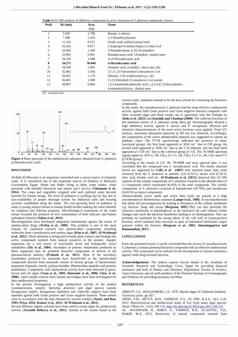
Gas Chromatography-Mass Spectroscopy (GC-MS) analysis
Based on the GC-MS results, thirteen bioactive compounds were identified from
the partially purified fractions of the cyanobacterium S. platensis. The relative
percentages of the compounds are given in Table (4). The most prevalent compound was n-Hexadecanoic acid (34.28%). Mass spectrum indicated that the
molecular weight of the compound under investigation was 256 Dalton (Figure
4). Finally, on the basis of UV, IR, 1H-NMR, and mass spectral data it was concluded that the compound was n- hexadecanoic acid with chemical formula:
C16 H32 O2.
A B
C D
J Microbiol Biotech Food Sci / Elshouny et al. 2017 : 6 (5) 1203-1208
1207
Table 4 GC-MS analysis of different compounds in active fractions of S. platensis methanolic extract.
Peak Rt (min) Area
(%)
Name
1 5.929 2.798 Butane, 1-ethoxy-
2 7.589 1.419 2,2-Dimethoxybutane
3 15.162 0.515 Oxalic acid, isobutyl pentyl ester
4 32.624 0.817 5-Isopropyl-6-methyl-hepta-3,5-dien-2-ol
5 33.056 1.438 2-Pentadecanone, 6,10,14-trimethyl-
6 33.905 0.961 Pentadecanoic acid, 14-methyl-, methyl ester
7 34.105 1.948 cis-9-Hexadecenoic acid
8 34.275 30.604 n-Hexadecanoic acid
9 34.590 1.993 Octanoic acid, 4-methyl-, ethyl ester, (ñ)-
10 35.861 1.030 3,7,11,15-Tetramethyl-2-hexadecen-1-ol
11 36.051 1.179 Ethanol, 2-(9-octadecenyloxy)-, (Z)-
12 36.601 1.008 11,13-Dimethyl-12-tetradecen-1-ol acetate
13 38.807 0.684 1,2-Cinnolinedicarboxylic acid, 1,2,3,5,6,7,8,8aoctahydro-
4-trimethylsilyloxy-, diethyl ester
RT: retention time
Figure 4 Mass spectrum of the antibacterial substance obtained from S. platensis
(n-Hexadecanoic acid)
DISCUSSION
Al-Bahr El-Pheroany is an important watershed and a crucial source of irrigation
water. It is considered one of the important sources of fisheries in Menoufia
Government, Egypt. Plants and fishes living in these water bodies, when poisoned with harmful chemicals and metals can’t survive (Ghannam et al.,
2014). The crops and vegetables irrigated with such polluted water become
harmful for human beings. The level of pollution is swelling day by day due to non-availability of proper drainage system for industrial units and housing
societies established along the banks. The ever-growing level of pollution in
water is posing serious threats to human health besides making the water harmful for irrigation and fisheries purposes. Microbiological assessment of the water
stream revealed the presence of rich communities of both indicator and human pathogenic bacteria (Sabae et al., 2014).
Microorganisms have developed adaptation mechanisms against the action of
antimicrobial drugs (Al-Haj et al., 2009). This problem is one of the main reasons for continued research into antimicrobial compounds, including
molecules from cyanobacteria and marine algae (Kim et al., 2007; Al-Wathnani
et al., 2012). Much attention is being paid towards plant extracts and biologically active compounds isolated from natural resources in the present. Aquatic
organisms are a rich source of structurally novel and biologically active
metabolites (Ely et al., 2004). Secondary or primary metabolites produced by
these organisms may be potential bioactive compounds of interest in the
pharmaceutical industry (Prakash et al., 2011). Most of the secondary
metabolites produced by seaweeds have bactericidal or the antimicrobial compounds derived from seaweeds consist of diverse groups of bacteriostatic
properties terpenols, sterols, polysaccharides, dibutenolides peptides and proteins
metabolites. Compounds with antibacterial activity have been detected in green, brown and red algae (Yuan et al., 2005; Bansemir et al., 2006; Chew et al.,
2008). Lipid soluble extracts from marine macroalgae have been investigated for
their antibacterial properties. In the present investigation, a high antibacterial activity of the studied
cyanobacterium, namely; Spirulina platensis and algal species namely
Saragassum wightii, Saragassum latifolium and Chlorella vulgaris have been reported against both Gram positive and Gram negative bacteria. These results
were in accordance with the data obtained by several workers (Sastry and Rao,
1995; Priya, 2012; Kumar et al., 2011; Al-Wathnani et al., 2012). Several different organic solvents have been used to screen algae for antibacterial
activity (Jeyanthi Rebecca et al., 2012). Similar to the results found in the
current study, methanol seemed to be the best solvent for extracting the bioactive compounds.
In this study, the cyanobacterium S. platensis had the most effective antibacterial activity against both Gram positive and Gram negative bacteria compared with
other screened algae and these results are in agreement with the findings by
Abdo et al. (2012) and Kaushik and Chauhan (2008). The collected fractions of the methanol extract of S. platensis using silica gel chromatography showed a
high inhibitory activity against S. aureus and P. aeruginosa. Physical and
chemical characterizations of the most active fractions were applied. From UV analysis, maximum absorption spectrum at 285 nm was observed. Accordingly,
the composition of the active antimicrobial material was suggested to contain an
aliphatic chain. The FT-IR spectroscopy indicated the presence of many functional groups: the first band appeared at 3424 cm-1 due to OH group, the
second band appeared at 2958 cm-1 due to the C-H aliphatic and the third band
appeared at 1729 cm-1 due to the carbonyl group (C=O). The 1H NMR spectrum signals (ppm) at: δ0.9 (t, 3H, CH3), δ1.3 (t, 2H, CH2), δ 2.3 (s, 2H, CH2) and δ7.2
(COOH group).
According to the results of UV, IR, 1H-NMR and mass spectral data, it was concluded that the compound was n- hexadecanoic acid. The results obtained
herein are supported by Colla et al. (2007) who reported major fatty acids
extracted from the S. platensis as palmitic acid (C16:1), stearic acid (C18:1), oleic acid, linoleic acid etc. Al-Wathnani et al. (2012) observed that GC-MS
analysis of the volatile components of S. platensis resulted in the identification of
15 compounds which constituted 96.45% of the total compounds. The volatile components of S. platensis consisted of heptadecane (39.70%) and tetradecane
(34.61%) as major components.
Antimicrobially active lipids and active fatty acids are present in a high concentration in Skeletonema costatum (Lampe et al., 1998). It was hypothesized
that lipids kill microorganisms by leading to disruption of the cellular membrane
e.g. bacteria, fungi and yeasts (Bergsson, 2005).They can also penetrate the extensive meshwork of peptidoglycan in the bacterial cell wall without visible
changes and reach the bacterial membrane leading to its disintegration. This can
probably be explained by the strong fabric of the cell wall of Gram-positive bacteria, which maintain their structure in spite of substantial hydrostatic turgor
pressure within the bacteria (Bergsson et al., 2002; Shanmugapriya and
Ramanathan, 2011).
CONCLUSIONS From the presented results it can be concluded that the extract of cyanobacterium,
S. platensis, contains potential bioactive compound with an effective antibacterial
activity. This compound can be utilized for the development of natural antibiotics against multi drug resistant bacteria.
Acknowledgements: The authors express sincere thanks to the Academy of Scientific Research and Technology, Cairo, Egypt for providing financial
assistance and both of Botany and chemistry Department, Faculty of Science,
Tanta University and all staff members of the National Institute of Oceanography and Fisheries for providing necessary facilities.
REFERENCES
ABBOTT, I.A., HOLLENBERG, I.G. 1976. Marine algae of California Stanford
University press. pp. 827. ABDO, S.M., HETTA, M.H., SAMHAN, F.A., EL DIN, R.A.S., ALI, G.H.
2012. Phytochemical and antibacterial study of five fresh water algal species. Asian J Plant Sci, 11(3), 109-116. http://dx.doi.org/10.3923/ajps.2012.109.116
AL –WATHNANI, H., ISMET, A., TAHMAZ, R.R., AL-DAYEL, T.H.,
BAKIR, M.A. 2012. Bioactivity of natural compounds isolated from

J Microbiol Biotech Food Sci / Elshouny et al. 2017 : 6 (5) 1203-1208
1208
cyanobacteria and green algae against human pathogenic bacteria and yeast. J Med Plants Res, 6(18), 3425-3433. http://dx.doi.org/10.5897/jmpr11.1746
ALEEM, A.A. 1993. Marine algae of Alexandria, Egypt. Alexandria: Privately
published. pp. [i-iv], [1]-135. AL-HAJ, N.A., MASHAN, N.I., SHAMSUDIN,
M.N., MOHAMAD, H., VAIRAPPAN, C.S., SEKAWI, Z. 2009. Antibacterial
activity in marine algae Eucheuma denticulatum against Staphylococcus aureus
and Streptococcus pyogenes. Res J Biol Sci, 4, 519-524.BANSEMIR, A.,
BLUME, M., SCHRODER, S., LINDEQUIST, U. 2006. Screening of cultivated
seaweeds for antibacterial activity against fish pathogenic bacteria. Aquaculture,
252, 79-84. http://dx.doi.org/10.1016/j.aquaculture.2005.11.051
BECHERT, T., STEINRÜCKE, P., GUGGENBICHLER, J.P. 2000. A new
method for screening anti-infective biomaterials. Nat Med, 6(9), 1053–1056.
http://dx.doi.org/10.1038/79568
BERGSSON, G. 2005. Antimicrobial polypeptides and lipids as a part of innate
defense mechanism of fish and human fetus. Thesis, Karoliska Institute, Stockholm.
BERGSSON, G., STEINGRIMSSON, Ó., THORMAR, H. 2002. Bactericidal
effects of fatty acids and monoglycerides on Helicobacter pylori. Int J Antimicrob Agents, 20, 258 – 262. http://dx.doi.org/10.1016/s0924-
8579(02)00205-4
CHEW, Y.L., LIM, Y., OMAR, M., KHOO, K.S. 2008. Antioxidant activity of three edible seaweeds from two areas in South East Asia. LWT- Food Sci
Technol, 41, 1067-1072. http://dx.doi.org/10.1016/j.lwt.2007.06.013
CLSI, 2010. Performance Standards for Antimicrobial Susceptibility Testing, Twentieth Informational Supplement, CLSI Document M100-S20, Wayne, PA:
Clinical and Laboratory Standards Institute.
COLLA, L.M., REINEHR, C.O., REICHERT, C.J., COSTA, A.V. 2007. Production of biomass and nutraceutical compounds by Spirulina platensis under
different temperature and nitrogen regimes. Bioresource Technol, 98, 1489–1493.
http://dx.doi.org/10.1016/j.biortech.2005.09.030
ELY, R., SUPRIYA, T., NAIK, C.G. 2004. Antimicrobial activity of marine
organisms collected off the coast of South East India. J Exp Mar Biol Ecol, 309,
121-127. http://dx.doi.org/10.1016/j.jembe.2004.03.010 GHANNAM, H.E., TALAB, A.S., JAHIN, H.S., GABER, S.E. 2014. Seasonal
variations in physicochemical parameters and heavy metals in water of El-Bahr
El-Pharaony drain, El-Menoufia Governorate, Egypt. Res J Environ Earth Sci, 6, 174-181.
HAYASHI, T., HAYASHI, K. 1996. Calcium spirulan, an inhibitor of enveloped
virus replication, from a blue green alga Spirulina platensis. J Nat Prod, 59, 83-87. http://dx.doi.org/10.1021/np960017o HIRAHASHI, T., MATSUMOTO, M., HAZEKI, K., SAEKI, Y., UI, M., SEYA,
T. 2002. Activation of the human innate immune system by Spirulina: augmentation of interferon production and NK cytotoxicity by oral
administration of hot water extract of Spirulina platensis. Int Immunopharmacol,
2,423-434. http://dx.doi.org/10.1016/s1567-5769(01)00166-7 JEYANTHI REBECCA, L., DHANALAKSHMI, V., CHANDRA SHEKHAR
2012. Antibacterial activity of Sargassum Ilicifolium and Kappaphycus alvarezii.
J Chem Pharma Res, 4(1), 700-705. KANDASAMY, M., ARUNACHALAM, K.D. 2008. Evaluation of in vitro
antibacterial property of seaweeds of southeast coast of India. African J Biotech,
7(12), 1958-1961. KAUSHIK, P., CHAUHAN, A. 2008. In vitro antibacterial activity of laboratory
grown culture of Spirulina platensis. Indian J Microbiol, 48, 348-352.
http://dx.doi.org/10.1007/s12088-008-0043-0 KIM, I.H., LEE, S.H., HA, J.M., HA, B.J., KIM, S.K., LEE, J.H. 2007.
Antibacterial activity of Ulva lactuta against Methicillin-Resistant
Staphylococcus aureus (MRSA). Biotechnol Bioproc Engg, 12, 579-582. http://dx.doi.org/10.1007/bf02931358
KUHL, A. 1962. Zur physiologie der Speicherung. Kondensierter anorganischer
Phosphate in Chlorella. Vorlrag Bot. Hrsg. Deut. Botan. Ges. (N.C.). 1, 157- 166. KUMAR, V., BHATNAGAR, A.K., SRIVASTAVA, J.N. 2011. Antibacterial
activity of crude extracts of Spirulina platensis and its structural elucidation of
bioactive compound. J Med Plants Res, 5(32), 7043-7048. http://dx.doi.org/10.5897/jmpr11.1175
LAMPE, M.F., BALLWEBER, L.M., ISAACS, C.E., PATTON, D.L., STAMM, W.E. 1998. Killing of Chlamydia trachomatis by novel antimicrobial lipids
adapted from compounds in human breast milk. Antimicrob Agents Chemother,
45, 1239-1244. LAVANYA, R., VEERAPPAN, V. 2011. Antibacterial potential of six seaweeds
collected from Gulf of Mannar of southeast coast of India. Advances in
Biological Research, 5(1), 38-44. MACMILLAN, J.B., ERNST-RUSSELL, M.A., DE ROPP, J.S., MOLINSKI,
T.F. 2002. Lobocyclamides A-C, lipopeptides from a cryptic cyanobacterium mat
containing Lyngbya confervoides. J Org Chem, 67, 8210-8215. http://dx.doi.org/10.1021/jo0261909
MULYONO, N., LAY, B.W., RAHAYU, S., YAPRIANTI, I. 2012. Antibacterial
activity of Petung Bamboo (Dendrocalamus asper) leaf extract against pathogenic Escherichia coli and their chemical identification. International
Journal of Pharmaceutical & Biological Archives, 3(4), 770-778.
NOAMAN, N.H., KHALEAFA, A.F., ZWKY, S.H. 2004. Factors affecting antimicrobial activity of Synechococcus leopoliensis. Microbiol Res, 156, 359-
402. http://dx.doi.org/10.1016/j.micres.2004.09.001
OSMAN, M.E.H., ABOSHADY, A.M., ELSHOBARY, M.E. 2013. Production
and characterization of antimicrobial active substance from some macroalgae
collected from Abu- Qir bay (Alexandria) Egypt, African J Biotech, 12(49),
6847-6858. . http://dx.doi.org/10.5897/AJB10.2150 OZDEMIR, G., KARABAY, N.U., DALAY, M.C., PAZARBASI, B. 2004.
Antibacterial activity of volatile component and various extracts of Spirulina
platensis. Phytother Res, 18, 754-757. http://dx.doi.org/10.1002/ptr.1541 PRAKASH, J.W., MARIMUTHU, J.A., JEEVA, S. 2011. Antimicrobial activity
of certain freshwater microalgae from Thambirabrani River, I.N, India. Asian Pac J Trop Biomed, S170 – S173. http://dx.doi.org/10.1016/s2221-1691(11)60149-4
PRIYA, S. 2012. Analysis of value-added biochemical compounds and
antimicrobial activity of green algae Chlorella vulgaris. J Chem Pharm Res, 4(5), 2577-2579.
ROBERT, S., ANDERS, R.L., NIELS, F., FRABK, E. 2003. Evaluation of
different disk diffusion media for detection of methicillin resistance in Staphylococcus aureus and coagulase-negative Staphylococci. APMIS, 111, 905-
914. http://dx.doi.org/10.1034/j.1600-0463.2003.1110909.x
SABAE, S.Z., EL-SHEEKH, M.M., KHALIL, M.A., EL-SHOUNY, W.A., BADR, H.M. 2014. Seasonal and regional variation of physicochemical and
bacteriological parameters of surface water in El-Bahr El-Pherony, Menoufia,
Egypt. World Journal of Fish and Marine Sciences, 6(4), 328-335.
http://dx.doi.org/10.5829/idosi.wjfms.2014.06.04.84288
SASTRY, V.M.V.S., RAO, G.R.K. 1995. Dioctyl phthalate and antibacterial
compound from marine brown alga Sargassum wightii. J Appl Phycol, 7,185–186. http://dx.doi.org/10.1007/bf00693066
SHANMUGAPRIYA, R., RAMANATHAN, T. 2011. Screening for
antimicrobial activity of crude extracts of Skeletonema costatum. J Appl Pharma Sci, 1(7), 154-157.
SUBHASHINI, J., MAHIPAL, S.V.K., REDDY, M.C., REDDY, M.M.,
RACHAMALLU, A., REDDANNA, P. 2004. Molecular mechanisms in C-Phycocyanin induced apoptosis in human chronic myeloid leukemia cell line-
K562. Biochem Pharmacol, 68,453-462. http://dx.doi.org/10.1016/j.bcp.2004.02.025 TAN, L.T. 2007. Bioactive natural products from marine cyanobacteria for drug
discovery. Phytochem, 68, 954–979 http://dx.doi.org/10.1016/j.phytochem.2007.01.012 TAYLOR, W.S. 1985. Marine algae of the easterntropical and subtrobical coasts
of Americas. ANN Arbor the University of Michigan press. pp. 870.
YANG, Y., PARK, Y., CASSADA, D.A., SNOW, D.D., ROGERS, D.G., LEE, J. 2011. In vitro and in vivo safety assessment of edible blue-green algae, Nostoc
commune var. sphaeroide skützing and Spirulina platensis. Food Chem Toxicol,
49, 1560-1564. http://dx.doi.org/10.1016/j.fct.2011.03.052 YUAN, Y.V., CARRINGTON, M.F., WALSH, N.A. 2005. Extracts from dulse
(Palmaria palmata) are effective antioxidants and inhibitors of cell proliferation
in vitro. Food Chem Toxicol, 43, 1073-1081. http://dx.doi.org/10.1016/j.fct.2005.02.012
ZARROUK, C. 1966. Contribution a l'etude d'une cyanophycee: Influence de
divers facteurs physiques et chimiques sur la croissance et la photosynthese de Spirulina maxima (Setch. et Gardner) Geitler. University of Paris, Paris, France.