Structure–Function Relationships of Antimicrobial Chemokines
36
Structure–Function Relationships of Antimicrobial Chemokines Mauricio Arias, Sebastian A.J. Zaat, and Hans J. Vogel Abstract The chemokines are a group of small chemotactic cytokines that play an important role in the innate and adaptive immune system. Their main function is related to the recruitment of white blood cells to sites of infection. They bind to specific chemokine receptors, which subsequently triggers signaling pathways in the leukocytes. Recently the discovery of chemokines that possess a direct antimicrobial activity against a broad range of pathogenic bacteria has generated increased interest in the role of these proteins in the innate immune system. Prior studies regarding ligand and receptor binding have already established the structural elements important for chemokine interaction and activation of their receptors. In the same manner, it is important to study the structural features required for the antimicrobial activity of this group of chemokines in order to establish key elements related with this new activity. This review will focus on the structure–function relationships that appear to be related to the direct antimicrobial activity of the chemokines. A close similarity of the C-terminal domain of many chemokines to cationic a-helical antimicrobial peptides suggests that this C-terminal helical region is responsible for the chemokine antimicrobial activity. However, for several chemokines, the antimicrobial activity resides in other parts of the protein, indicating that each chemokine needs to be examined individually. We also discuss the role of dimeriza- tion and of linearization of chemokines in their antimicrobial activity. M. Arias • H.J. Vogel (*) Department of Biological Sciences, Biochemistry Research Group, University of Calgary, 2500 University Dr. NW, Calgary, AB, Canada e-mail: [email protected] S.A.J. Zaat Department of Medical Microbiology, Centre for Infection and Immunity Amsterdam (CINIMA), Academic Medical Center, Meibergdreef 15, 1105 AZ Amsterdam, The Netherlands P.S. Hiemstra and S.A.J. Zaat (eds.), Antimicrobial Peptides and Innate Immunity, Progress in Inflammation Research, DOI 10.1007/978-3-0348-0541-4_8, # Springer Basel 2013 183
-
Upload
independent -
Category
Documents
-
view
1 -
download
0
Transcript of Structure–Function Relationships of Antimicrobial Chemokines
Structure–Function Relationships
of Antimicrobial Chemokines
Mauricio Arias, Sebastian A.J. Zaat, and Hans J. Vogel
Abstract The chemokines are a group of small chemotactic cytokines that play an
important role in the innate and adaptive immune system. Their main function is
related to the recruitment of white blood cells to sites of infection. They bind to
specific chemokine receptors, which subsequently triggers signaling pathways in the
leukocytes. Recently the discovery of chemokines that possess a direct antimicrobial
activity against a broad range of pathogenic bacteria has generated increased interest
in the role of these proteins in the innate immune system. Prior studies regarding
ligand and receptor binding have already established the structural elements
important for chemokine interaction and activation of their receptors. In the same
manner, it is important to study the structural features required for the antimicrobial
activity of this group of chemokines in order to establish key elements related with
this new activity. This review will focus on the structure–function relationships that
appear to be related to the direct antimicrobial activity of the chemokines. A close
similarity of the C-terminal domain of many chemokines to cationic a-helicalantimicrobial peptides suggests that this C-terminal helical region is responsible
for the chemokine antimicrobial activity. However, for several chemokines, the
antimicrobial activity resides in other parts of the protein, indicating that each
chemokine needs to be examined individually. We also discuss the role of dimeriza-
tion and of linearization of chemokines in their antimicrobial activity.
M. Arias • H.J. Vogel (*)
Department of Biological Sciences, Biochemistry Research Group, University of Calgary, 2500
University Dr. NW, Calgary, AB, Canada
e-mail: [email protected]
S.A.J. Zaat
Department of Medical Microbiology, Centre for Infection and Immunity Amsterdam (CINIMA),
Academic Medical Center, Meibergdreef 15, 1105 AZ Amsterdam, The Netherlands
P.S. Hiemstra and S.A.J. Zaat (eds.), Antimicrobial Peptides and Innate Immunity,Progress in Inflammation Research, DOI 10.1007/978-3-0348-0541-4_8,# Springer Basel 2013
183
Abbreviations
AMPs Antimicrobial peptides
CA-MRSA Community-associated methicillin-resistant Staphylococcus aureusCTAP-3 Connective tissue-activating protein-3
DC Dendritic cells
DOPE 1,2-Dioleoyl-sn-glycero-3-phosphoethanolamine
DOPG 1,2-Dioleoyl-sn-glycero-3-phospho-(10-rac-glycerol)ENA-78 Epithelial neutrophil-activating protein 78
GCP-2 Granulocyte chemotactic protein-2
GRO Growth-regulated oncogene
hBD Human beta-defensin
IL-8 Interleukin-8
IP-10 Interferon-inducible protein-10
I-TAC Interferon-inducible T-cell alpha chemoattractant
LDL Low-density lipoprotein
MCP-4 Monocyte chemoattractant protein-4
MEC Mucosa-associated epithelial chemokine
MIG Monokine induced IFN-gamma
MIP-3a Human macrophage inflammatory protein-3aNAP-2 Neutrophil-activating peptide-2
NMR Nuclear magnetic resonance
PBP Platelet basic protein
PF-4 Platelet factor-4
PG-1 Protegrin-1
PGLa Peptidyl-glycylleucine-carboxyamide
PMP Platelet microbicidal protein
RANTES Regulated upon activation normal T-cell expressed and secreted
SDS Sodium dodecyl sulfate
SIC Streptococcal inhibitor of complement
SPA Staphylococcal protein A
SpyCEP Streptococcus pyogenes cell envelope proteinase
1 Chemokines
The chemokines are a family of small chemotactic cytokines, with a size ranging
from 8 to 13 kDa (Allen et al. 2007). Their role in the innate and adaptive immunity
has been studied since the first chemokines were discovered 28 years ago (Sauder
et al. 1984; Yoshimura et al. 1987a). Their primary function concerns the activation
and recruitment of specific leukocytes to the site of injury or infection through the
creation of a chemokine concentration gradient. The localization and activation of
184 M. Arias et al.
the white blood cells is achieved by binding of the chemokines to their cognate
chemokine receptors, which are all part of the G-protein-coupled transmembrane
receptor family (Proudfoot 2002; Allen et al. 2007). In addition to their chemotactic
activity, several chemokines and chemokine receptors are known to play a role in
various diseases (Viola and Luster 2008; Proudfoot 2002; Allen et al. 2007). The
chemokine family is usually classified into four groups which can be distinguished
by the organization of the cysteine residues in the N-terminal region of the protein,
i.e., C, CC, CXC, and CX3C, where X represents any nonconserved amino acid
residue (Murphy et al. 2000). The CXC group of chemokines can be further divided
into two subgroups, where the presence or absence of the amino acid sequence ELR
in the N-terminal region creates the ELR and non-ELR CXC chemokine
subfamilies. All chemokines of the CC, CXC, and CX3C classes form two disulfide
bonds, while the C class only possesses one stabilizing disulfide linkage. To date,
approximately 50 chemokines and 20 chemokine receptors have been reported
(Allen et al. 2007). Although the sequence identity among all the members of the
chemokine family is highly variable (20–90 %), their three-dimensional structures
display a remarkable conserved topology. Most chemokines are formed by an
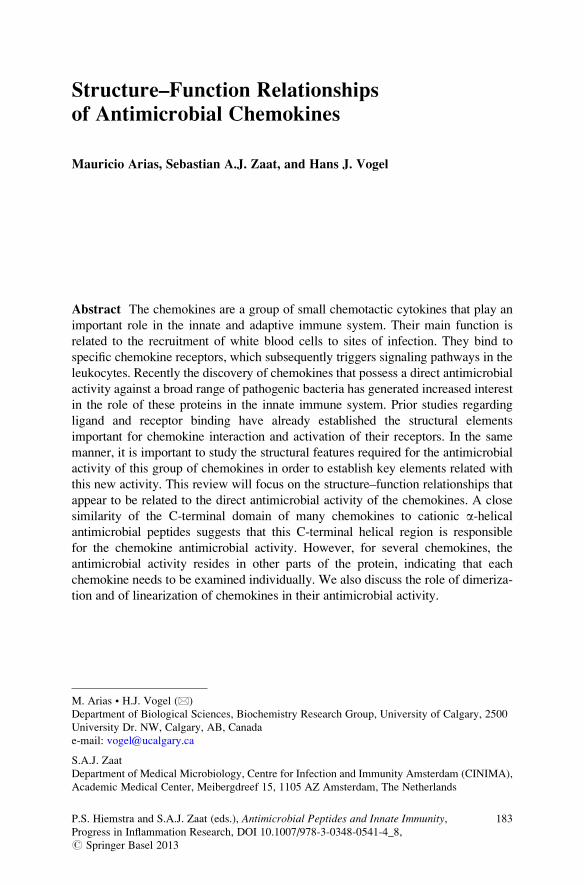
N-terminal extended loop region, in which the first two cysteine residues are
located, a bundle of three antiparallel b-strands, and a C-terminal a-helix (Allen
et al. 2007) (Fig. 1). Several structural features are important for the signaling
induced by the chemokines upon binding to their receptors. The N-terminal domain
has emerged as the main recognition site in these proteins. Deletion of this region
inhibits chemokine-induced signaling and in some cases also affects the interaction
with the receptor. The N-loop located between the first cysteine residues and the
first b-strand is important for the interaction with the receptor and is generally
considered as the first docking site during the two-step binding process of the
chemokines to their receptors. The loops connecting the b-strands also contribute
to the interaction with the receptor. In some cases other residues in the chemokine
structure have also been shown to be important for the interactions with the
receptors (Allen et al. 2007; Clark-Lewis et al. 1995).
Fig. 1 Stereo ribbon diagram
of CCL20 (PDB code 1M8A)
showing the highly conserved
three-dimensional structure of
the chemokine family. The
two disulfide bonds are
represented in yellow, and the
N and C termini are labeled.
The structural elements
normally analyzed for
antimicrobial activity, such as
the N-terminal loop, the
central b-sheet, and the
C-terminal a-helix, areindicated between the arrows.Figure prepared with MolMol
software
Structure–Function Relationships of Antimicrobial Chemokines 185
2 Antimicrobial Activity of Chemokines
A different part of the innate immune defense against bacterial infections is formed
by a large group of antimicrobial peptides (AMPs) which often have a potent
activity against bacteria, fungi, and other organisms, including some viruses. The
AMPs are an important part of the host defense system of widely divergent
organisms including bacteria, plants, insects, and vertebrates. Antimicrobial
peptides act by directly perturbing bacterial membranes or by entering the bacterial
cell and interfering with important processes, e.g., DNA transcription (Epand and
Vogel 1999; Nguyen et al. 2011). However, recently some antimicrobial peptides
have been shown to be involved in the regulation of the immune response as well,
by binding to the same receptors as used by the chemokines (Yang et al. 2004).
Conversely, it has also been demonstrated that numerous chemokines can have a
direct antimicrobial activity against Gram-positive and Gram-negative bacteria, as
well as an antifungal activity (see Table 1). Several studies have reported on the
antimicrobial activity of individual or small groups of chemokines. Unfortunately,
there is no consensus about the methodology and experimental conditions used to
determine the antimicrobial activity of chemokines. The liquid-phase antimicrobial
and microbicidal assay is a solution-phase test widely used in antimicrobial
experiments. In this assay bacteria are incubated with different concentrations of
chemokines or antimicrobial agents in liquid media. The assay is based on the
microtiter broth dilution assays used to assess minimum inhibitory (MIC) and
minimum bactericidal concentrations (MBC) of antibiotics (Amsterdam 1996).
After exposure to the peptide, wells are inspected for growth to assess the minimum
inhibitory concentration, and/or serial dilutions are plated on solidified media and
colony-forming units are counted after incubation, to assess the microbicidal
concentration. Despite the widespread use of this technique, there is considerable
variation in test parameters, such as composition of the incubation solution, period
of exposure, and test strains used. Hancock and colleagues have attempted to
standardize the methodology (Wiegand et al. 2008). Another test to establish
the antimicrobial activity of chemokines and antimicrobial peptides is the radial
diffusion assay. In this assay the microorganisms are incorporated in buffered agar
or agarose-containing media, solidified in plates. Chemokines are added to wells
punched in the agar, and after a defined period in which the compounds diffuse into
the agar, a nutrient overlay medium is applied to the plates. The plates are then
incubated to allow growth of microorganisms, and zones of inhibition are measured
(Lehrer et al. 1991). The chemokines antimicrobial activity collected in Table 1
includes antimicrobial information determined by both assays, solution, and/or
solid-phase test.
The antimicrobial function of the chemokines draws attention to the comple-
mentary roles that these proteins play in immunity. The capacity to exert a direct
and potent antimicrobial activity seems to be an additional mechanism for fighting
bacteria and other pathogens (Eliasson and Egesten 2008). Recently the group of
chemokines with antimicrobial activity have been called “kinocidins” by some
186 M. Arias et al.
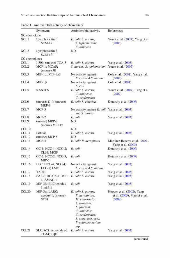
Table 1 Antimicrobial activity of chemokines
Synonyms Antimicrobial activity References
XC chemokine
XCL1 Lymphotactin a;SCM-1a
E. coli; S. aureus;S. typhimurium;C. albicans
Yount et al. (2007), Yang et al.
(2003)
XCL2 Lymphotactin b;SCM-1b
ND
CC chemokines
CCL1 I-309; (mouse) TCA-3 E. coli; S. aureus Yang et al. (2003)
CCL2 MCP-1; MCAF;
(mouse) JE
S. aureus; S. typhimurium Yount et al. (2007)
CCL3 MIP-1a; MIP-1aS No activity against
E. coli and S. aureusCole et al. (2001), Yang et al.
(2003)
CCL4 MIP-1b No activity against
E. coliCole et al. (2001)
CCL5 RANTES E. coli; S. aureus;C. albicans;C. neoformans
Yount et al. (2007), Tang et al.
(2002)
CCL6 (mouse) C10; (mouse)
MRP-1
E. coli; S. enterica Kotarsky et al. (2009)
CCL7 MCP-3 No activity against E. coliand S. aureus
Yang et al. (2003)
CCL8 MCP-2 E. coli Yang et al. (2003)
CCL9 (mouse) MRP-2;
(mouse) MIP-1gND
CCL10 ND
CCL11 Eotaxin E. coli; S. aureus Yang et al. (2003)
CCL12 (mouse) MCP-5 ND
CCL13 MCP-4 E. coli; P. aeruginosa Martınez-Becerra et al. (2007),
Yang et al. (2003)
CCL14 CC-1; HCC-1; NCC-2;
Ckb1; MCIF
E. coli Kotarsky et al. (2009)
CCL15 CC-2; HCC-2; NCC-3;
MIP-5
E. coli Kotarsky et al. (2009)
CCL16 LEC; HCC-4; NCC-4;
LCC-1; LMC
No activity against
E. coli and S. aureusYang et al. (2003)
CCL17 TARC E. coli; S. aureus Yang et al. (2003)
CCL18 PARC; DC-CK-1; MIP-
4; AMAC-1
E. coli; S. aureus Yang et al. (2003)
CCL19 MIP-3b; ELC; exodus-3; ckb11
E. coli Yang et al. (2003)
CCL20 MIP-3a; LARC;exodus-1; (mouse)
ST38
E. coli; S. aureus;P. aeruginosa;M. catarrhalis;S. pyogenes;E. faecium;C. albicans;C. neoformans;S. coag. neg. spp.;Propionibacteriumssp.
Hoover et al. (2002), Yang
et al. (2003), Maerki et al.
(2009)
CCL21 SLC; 6Ckine; exodus-2;
TCA4; ckb9E. coli; S. aureus Yang et al. (2003)
(continued)
Structure–Function Relationships of Antimicrobial Chemokines 187
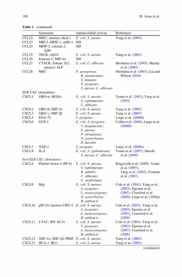
Table 1 (continued)
Synonyms Antimicrobial activity References
CCL22 MDC; (mouse) abcd-1 E. coli; S. aureus Yang et al. (2003)
CCL23 MIP-3; MPIF-1; ckb8-1 ND
CCL24 MPIF-2; eotaxin-2;
ckb6ND
CCL25 TECK; ckb15 E. coli; S. aureus Yang et al. (2003)
CCL26 Eotaxin-3; MIP-4a ND
CCL27 CTACK; Eskine; ILC
(mouse) ALP
E. coli; C. albicans Hieshima et al. (2003), Maerki
et al. (2009)
CCL28 MEC P. aeruginosa;K. pneumoniae;S. mutants;P. pyogenes;S. aureus; C. albicans
Hieshima et al. (2003), Liu and
Wilson (2010)
ELR CXC chemokines
CXCL1 GRO-a; MGSA E. coli; S. aureus;S. typhimurium;C. albicans
Yount et al. (2007), Yang et al.
(2003)
CXCL2 GRO-b; MIP-2a E. coli; S. aureus Yang et al. (2003)
CXCL3 GRO-g; MIP-2b E. coli; S. aureus Yang et al. (2003)
CXCL5 ENA-78 S. pyogenes Linge et al. (2008b)
CXCL6 GCP-2 E. coli; S. pyogenes;S. dysgalactiae;S. aureus;P. aeruginosa;N. gonorrhoeae;E. faecalis
Collin et al. (2008), Linge et al.
(2008b)
CXCL7 NAP-2 S. pyogenes Linge et al. (2008b)
CXCL8 IL-8 E. coli; S. typhimurium;S. aureus; C. albicans
Yount et al. (2007), Maerki
et al. (2009)
Non-ELR CXC chemokinesCXCL4 Platelet factor-4 (PF-4) E. coli; S. aureus;
S. typhimurium;B. subtilis;C. albicans;C. neoformans
Krijgsveld et al. (2000), Yount
et al. (2007),
Tang et al. (2002), Yeaman
et al. (2007)
CXCL9 Mig E. coli; S. aureus;S. pyogenes;L. monocytogenes;N. gonorrhoeae;B. anthracis
Cole et al. (2001), Yang et al.
(2003), Egesten et al.
(2007), Crawford et al.
(2009), Linge et al. (2008a)
CXCL10 gIP-10; (mouse) CRG-2 E. coli; S. aureus;S. pyogenes;L. monocytogenes;B. anthracis
Cole et al. (2001), Yang et al.
(2003), Egesten et al.
(2007), Crawford et al.
(2009)
CXCL11 I-TAC; IP9; H174 E. coli; S. aureus;S. pyogenes;L. monocytogenes;B. anthracis
Cole et al. (2001), Yang et al.
(2003), Egesten et al.
(2007), Crawford et al.
(2009)
CXCL12 SDF-1a; SDF-1b; PBSF E. coli; S. aureus Yang et al. (2003)
CXCL13 BCA-1; BLC E. coli; S. aureus Yang et al. (2003)
(continued)
188 M. Arias et al.
authors (Yount et al. 2004; Yount and Yeaman 2004). One of the structural
elements that may relate this group of chemokines to other disulfide-containing
antimicrobial peptides is the presence of a multidimensional signature composed by
a so-called g-core motif, perhaps suggesting a common ancestry for previously
unrelated groups of antimicrobial agents (Yeaman and Yount 2007; Yount and
Yeaman 2004).
The activity of the majority of the antimicrobial chemokines is markedly
dependent on the ionic strength of the incubation media as is also observed for
antimicrobial peptides. Intriguingly, some of the antimicrobial chemokines, such as
CCL20, CCL28, CXCL9, CXCL10, and CXCL11, are expressed in different
epithelial cells, and it is noteworthy that they are secreted into fluids with a relatively
low salt concentration which allows them to exhibit their full antimicrobial potency
(Moser et al. 2006; Fujiie et al. 2001; Nakayama et al. 2001; Shirane et al. 2004;
Starner et al. 2003; Sauty et al. 1999).
The structural requirements for chemokines with antimicrobial activity are not
clearly identified, but some studies have tried to uncover the main features
involved. A comprehensive study of a group of 30 chemokines has established
that almost 80 % of the chemokines can exhibit antimicrobial activity against
Escherichia coli and Staphylococcus aureus at neutral pH (Yang et al. 2003). An
initial analysis of the biochemical characteristics of the antimicrobial chemokines
showed that normally the chemokines with pI values higher than 9.0 possess
antibacterial activity, which indicates the importance of cationicity as a major
factor for the antimicrobial activity. However, unlike what is observed with many
antimicrobial peptides, the potency of this activity was not correlated with the pI
value of the chemokines. Other factors, such as the cationicity of the N-terminal
tail, and the hydrophobicity of the surface of the chemokines were analyzed, but
again there was no direct correlation between these features and the antimicrobial
activity. However, the three-dimensional structures of the chemokines and in
particular the electrostatic potential surface distribution of the positive charges
revealed a common theme. The presence of a large three-dimensional positively
charged surface patch in the proteins was a characteristic shared by all the antimi-
crobial chemokines. Chemokines without antimicrobial activity either do not have
such a surface or have negatively charged residues interfering with the cationic
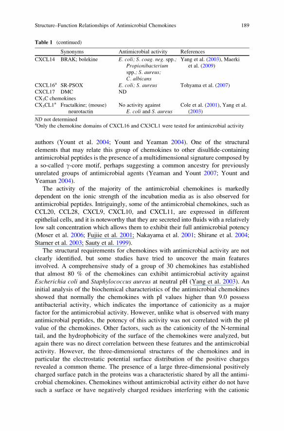
Table 1 (continued)
Synonyms Antimicrobial activity References
CXCL14 BRAK; bolekine E. coli; S. coag. neg. spp.;Propionibacteriumspp.; S. aureus;C. albicans
Yang et al. (2003), Maerki
et al. (2009)
CXCL16a SR-PSOX E. coli; S. aureus Tohyama et al. (2007)
CXCL17 DMC ND
CX3C chemokines
CX3CL1a Fractalkine; (mouse)
neurotactin
No activity against
E. coli and S. aureusCole et al. (2001), Yang et al.
(2003)
ND not determinedaOnly the chemokine domains of CXCL16 and CX3CL1 were tested for antimicrobial activity
Structure–Function Relationships of Antimicrobial Chemokines 189
surface patches. Nonetheless, the potency of the antimicrobial activity seemed not
related with the size of the positively charged surface patches (Yang et al. 2003). In
general, dissecting the structural elements that are responsible for the antimicrobial
activity of the chemokines can be performed by studying peptides resembling
particular domains of the chemokines (Fig. 1). These domains are normally selected
on the basis of biochemical properties, such as cationicity and amphipathicity,
which are important properties for many AMPs (Haney et al. 2009a; Epand
and Vogel 1999). A limitation to this approach obviously is that conformation-
dependent positively charged surface domains of the full-length proteins cannot be
mimicked. Rather, these peptides represent parts of the linearized molecules which
would arise from reduction of their disulfides. Still, a considerable number of such
peptides with antimicrobial activity have been identified.
Interestingly, despite the evidence related to the antimicrobial activity of
chemokines, recently it had been reported that antimicrobial chemokines also
induce the release of the virulence factor protein A (SPA) by a community-
associated methicillin-resistant S. aureus (CA-MRSA) (Yung et al. 2011). It is
possible that this is an example of how bacteria may utilize host defense system
signals (e.g., chemokines) in order to evade the immune response. In addition, the
binding of chemokines to the S. aureus membrane, which had been reported for
CXCL9 and CXCL10, may also contribute to avoid the immune reaction of the
host by restricting the amount of free chemokines available for recruitment and
activation of the immune cells (Yung et al. 2011).
3 Antimicrobial Activity of CC Chemokines
3.1 Monocyte Chemoattractant Protein-4 (MCP-4)/CCL13
The monocyte chemoattractant protein-4 (MCP-4)/CCL13 contains 75 amino acid
residues (Uguccioni et al. 1996). It interacts with the chemokine receptors CCR2,
CCR3, and CCR5 (Leach et al. 2007; Uguccioni et al. 1996) and induces the
migration of monocytes, T lymphocytes, and eosinophils (Garcia-Zepeda et al.
1996; Uguccioni et al. 1996). This particular chemokine has been shown to be
involved in several inflammatory diseases including arthritis and asthma (Rojas-
Ramos et al. 2003; Iwamoto et al. 2006; Kalayci et al. 2004). Although the sequence
identity for the members of the monocyte chemoattractant protein (MCP) subfamily
of CC chemokines (composed of CCL2, CCL7, CCL8, CCL12, and CCL13) is
around 60 %, CCL13 is the only chemokine from this subfamily to exhibit antimi-
crobial activity against Gram-positive and Gram-negative bacteria (Martınez-Becerra
et al. 2007). Studies on 19-mer peptides using CCL13 sequence as a template showed
that the C-terminal peptide CCL1357–75, called CDAP-4, had similar activity as
full-length CCL13, against E. coli (Martınez-Becerra et al. 2007). The antimicrobial
activity of this peptide was further studied against Pseudomonas aeruginosa and
significant morphological changes were observed by transmission electron micros-
copy when lethal concentrations were used (Martınez-Becerra et al. 2007). The
190 M. Arias et al.
antibacterial activity of CDAP-4 was susceptible to the ionic strength of the media,
being greatly reduced at NaCl concentrations higher than 100 mM (Martınez-Becerra
et al. 2007). The stability of the three-dimensional structure of the CDAP-4 peptide
was studied by molecular dynamics simulations. The peptide forms a short amphi-
pathic a-helix with a net positive charge (+5) and a high pI (10.58), characteristics
that are normally observed for a-helical AMPs (Martınez-Becerra et al. 2007). In
addition, an electrostatic potential analysis revealed a large positively charged surface
on the peptide, which may account for its antimicrobial activity (Martınez-Becerra
et al. 2007).
The structure of the full-length CCL13 shows the typical central three-stranded
antiparallel b-sheet flanked by an extended loop in the N-terminal region and an
a-helix in the C-terminal region, which includes residues 57–67, which are part of
the CDAP-4 peptide (Barinka et al. 2008). The crystal structure suggests the
formation of CCL13 dimers (Barinka et al. 2008), although solution experiments
in 100 mM NH4OAc at pH 6.8 show that CCL13 is a monomer or that it forms
heterodimers when combined with other members of the same chemokine family
(Crown et al. 2006). Taken together, all these results indicate that the C-terminal
region of CCL13 is important for the antibacterial activity of this chemokine and
that the full-length molecule may form dimers which can possibly contribute to the
antimicrobial activity.
3.2 Human Macrophage Inflammatory Protein-3a(MIP-3a)/CCL20
The human macrophage inflammatory protein-3a (MIP-3a)/CCL20 is made up of
70 amino acid residues (8 kDa) and bears some resemblance with the antimicrobial
b-defensin peptides (Hoover et al. 2002). This chemokine plays a role in diseases
such as cancer and rheumatoid arthritis, among others (Kleeff et al. 1999; Schutyser
et al. 2003; Matsui et al. 2001). CCL20 is responsible for the migration of immature
dendritic cells (DC), effector/memory T cells, and B cells, upon interaction with the
chemokine receptor CCR6 (Schutyser et al. 2003). The structure of CCL20
resembles that of all CC chemokines (Fig. 1). The N-terminal region, containing
the two first cysteine residues, is fairly flexible, and it is connected by a 3.10 helical
turn to the central region made up of a three-stranded antiparallel b-sheet, which is
followed by the usual C-terminal a-helix (Chan et al. 2008; Hoover et al. 2002;
Malik and Tack 2006). The available crystal structures of CCL20 show the protein
forming a dimer structure (Hoover et al. 2002; Malik and Tack 2006). Diffusion
NMR and pH titration studies established that the dimerization of CCL20 is
markedly pH dependent, CCL20 being a dimer at neutral pH and a monomer at
lower pH (Chan et al. 2008). The CCR6 receptor only interacts with CCL20, while
CCR6 is also the only chemokine receptor for CCL20. This specificity between
chemokine ligand and receptor is an interesting characteristic shared with eight
more chemokine receptor-ligand couples (Viola and Luster 2008). However, the
human b-defensin antimicrobial peptides (hBD 1–3) can also induce the migration
Structure–Function Relationships of Antimicrobial Chemokines 191
of cells that express either CCR6 or CCR2 (Rohrl et al. 2010a, b; Yang et al. 1999).
Like the b-defensins, CCL20 has a direct antimicrobial activity against Gram-
positive and Gram-negative bacteria (Hoover et al. 2002; Yang et al. 2003), as
well as antifungal (Yang et al. 2003) and antiviral activity (Ghosh et al. 2009; Kim
et al. 2007). The activity of CCL20 against E. coli is actually higher than the
activity of human b-defensin 2 (hBD-2) (Hoover et al. 2002). Structurally, there areno obvious similarities between CCL20 and the b-defensins which could account
for the antimicrobial activity of CCL20. The presence of a relatively high number
of positive charges in the N-terminal region of CCL20 was initially thought to
contribute to its antimicrobial activity, but the existence of chemokines with
positively charged N-terminal regions and without antimicrobial activity indicates
that other aspects should be also considered. The overlap in antimicrobial activity
may arise from the presence of similarly localized positively charged patches on the
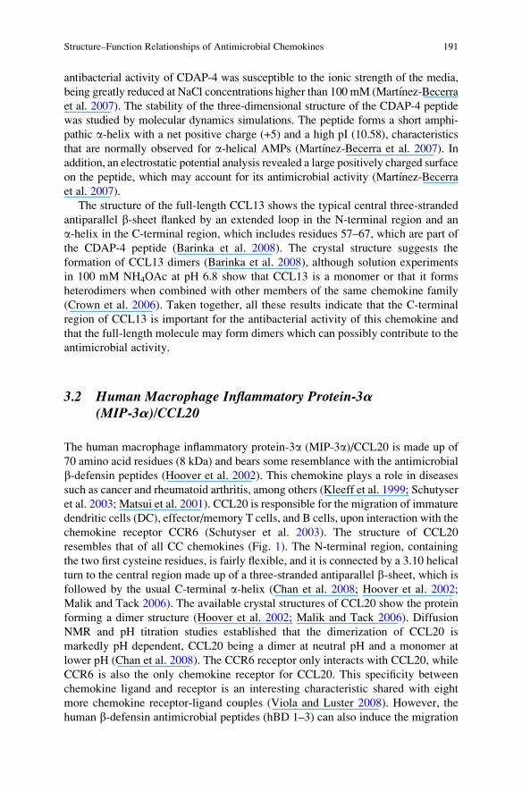
surfaces of CCL20 and hBD-2 (Fig. 2). These regions are located in the turns
between the N-terminal loop and the b1 strand and the turn between the b2 and b3strands (Hoover et al. 2002). Another important structural characteristic for
antimicrobial activity, contributing to possible pore formation in bacterial
membranes, is the amphipathicity of the protein. CCL20 lacks a large hydrophobic
surface, and the region surrounding Leu-8 is the only recognizable hydrophobic
patch on the protein surface (Hoover et al. 2002).
In addition to the antimicrobial activity of the full-length CCL20, antimicrobial
activity is also found in some peptides derived from CCL20. During cancer
progression, the protease cathepsin D generates a C-terminal 12-residue peptide
from CCL20 (CCL2059–70) with antimicrobial activity against E. coli, which is
significantly reduced compared to the activity of intact CCL20 (Hasan et al. 2006).
Further analysis of the CCL20 structure revealed that the C-terminal a-helix starts
after the Pro-51 residue. It is possible that this segment of the protein is also
important for the antimicrobial activity, because of its overall cationicity and
amphipathicity. The antimicrobial activity of a synthetic C-terminal CCL20 peptide
composed of the last 20 amino acids (CCL2051–70) was 60 times higher against
S. aureus and E. coli, and 30 times higher against Bacillus subtilis, than the activityof the naturally occurring C-terminal peptide CCL2059–70 (Chan et al. 2008;
Nguyen et al. 2010). The CCL2051–70 peptide has a large number of positive
charges and forms an amphipathic a-helix upon binding to membrane mimetics
such as SDS micelles (Chan et al. 2008; Nguyen et al. 2010). The shorter 12-residue
CCL2059–70 peptide has a lower positive net charge and does not form a stable
a-helix in the presence of SDS micelles. Additionally its amphipathicity is perpen-
dicular to the length of the peptide instead of parallel, as was found for CCL2051–70(Nguyen et al. 2010). These structural differences account for the large difference in
antimicrobial activity, as a high net positive charge and amphipathicity are shared
characteristics for most members of the a-helical class of antimicrobial peptides
such as the magainins, cecropins, and lactoferrampin (Epand and Vogel 1999;
Haney et al. 2007, 2009b). The lack of antimicrobial activity for the N-terminal
fragments CCL201–52 and CCL201–55 further indicates that the C-terminal region of
CCL20 is mostly responsible for the antimicrobial activity of this chemokine (Chan
et al. 2008; Hasan et al. 2006).
192 M. Arias et al.
3.3 Mucosa-Associated Epithelial Chemokine MEC/CCL28
The mucosa-associated epithelial chemokine (MEC)/CCL28 is a protein of 108
amino acids residues (12.3 kDa) that regulates the migration and activation
of specific leukocytes, such as cutaneous lymphocytes, antigen-positive (Ag+)
memory T cells, and eosinophils. It interacts with the chemokine receptors
CCR10 and CCR3 (Pan et al. 2000). CCL28 is constitutively expressed in human
and mouse epithelial cells of tissues such as the mammary glands, the respiratory
tract, the colon, and the salivary glands (Pan et al. 2000; Wang et al. 2000).
The C-terminal region of CCL28 shares a high sequence identity (53 %) with the
antimicrobial peptide histatin-5, which has a potent antifungal activity against
Candida albicans (Hieshima et al. 2003). This unique feature of CCL28 focused
early attention on its possible antimicrobial activity. Human CCL28 (hCCL28)
exerts a direct antimicrobial activity against Gram-negative and Gram-positive
bacteria, in addition to possessing an antifungal activity (see Table 1). The antimi-
crobial activity against C. albicans and P. aeruginosa was more potent at lower salt
concentrations (Hieshima et al. 2003; Liu and Wilson 2010). The most closely
related chemokine, CCL27 (sequence identity 40 %) (Pan et al. 2000), which lacks
the extended C-terminal region of CCL28, does not have bactericidal activity and
Fig. 2 Ribbon representation and electrostatic potential surface of human b-defensin 2 (hBD-2)
(PDB code 1FD3) and chemokine CCL20 (PDB code 2JYO), with positively charged side chains
in blue and negatively charged side chains in red. Each image is rotated 90� from the vertical axis.
Electrostatic potential surfaces were calculated by adaptive Poisson–Boltzmann Solver (APBS)
using PDB2PQR software and depicted by PyMOL software
Structure–Function Relationships of Antimicrobial Chemokines 193
only low activity against C. albicans at high concentrations (Hieshima et al. 2003;
Yang et al. 2003). In line with this, the first two positively charged residues in the
85–89 region (RKDRK) of murine CCL28 (mCCL28) are highly conserved among
CCL28 homologues in all studied mammalian species and are essential for the
antibacterial activity (Liu and Wilson 2010). These results indicate that the direct
antimicrobial and antifungal activities of CCL28 rely on the C-terminal histatin-
like region. A 28-residue peptide corresponding to this C-terminal region of the
hCCL28 was even more active than histatin-5 against C. albicans but showed a lowantibacterial activity (Hieshima et al. 2003). In a similar fashion, the 52-residue
peptide corresponding to the C-terminal region of mCCL28 had a reduced antimi-
crobial activity against S. aureus and P. aeruginosa, while a 56-residue peptide
resembling the N-terminal region of mCCL28 had no antibacterial activity (Liu and
Wilson 2010). The broader antimicrobial activity of CCL28 compared to its
C-terminal CCL28 peptide established that not only the C-terminal region is
required for killing the bacteria but in addition the interaction with the rest of the
protein is essential for the expression of the full antimicrobial potential, a notion
that was further supported by elegant studies with protein chimeras (Liu andWilson
2010). In addition, the influence of the disulfide bridges that normally stabilize the
chemokine structure of CCL28 was studied, showing that the tertiary structure is
not required for antimicrobial activity but is essential for its chemotactic activity
(Liu and Wilson 2010). Similar results have been obtained for human b-defensin 3
(hBD-3) and tachyplesin I, showing that the cysteine bridges are not mandatory for
their antimicrobial activity (Hoover et al. 2003; Wu et al. 2003; Ramamoorthy et al.
2006) but in the case of hBD-3 are again required for the chemotactic function
(Hoover et al. 2003; Wu et al. 2003).
In addition to the above-mentioned CC chemokines, other members of this
family have been shown to possess antimicrobial activity (see Table 1). Further
studies need to be done in order to establish the structural characteristics that confer
the antimicrobial activity to these chemokines.
4 Antimicrobial Activity of ELR CXC Chemokines
4.1 Granulocyte Chemotactic Protein-2 (GCP-2)/CXCL6
Granulocyte chemotactic protein-2 (GCP-2)/CXCL6 is a chemokine of the ELR
CXC family, made up of 77 amino acids residue (Proost et al. 1993a, b; Froyen et al.
1997; Van Damme et al. 1997). It is mainly involved in chemoattracting neutrophils
(Froyen et al. 1997; Wuyts et al. 1997; Viola and Luster 2008) due to its interactions
with the chemokine receptors CXCR1 and CXCR2 which are also expressed in
monocytes and mast cells (Wuyts et al. 1997, 1998; Wolf et al. 1998). CXCL6 is
expressed by epithelial cells, macrophages and mesenchymal cells (Fillmore et al.
2003; Collin et al. 2008; Mine et al. 2003; Prause et al. 2003; Gijsbers et al. 2004;
194 M. Arias et al.
Wuyts et al. 2003). CXCL6 has been shown to possess an important NaCl-sensitive
antimicrobial activity (Collin et al. 2008), which is comparable with the activity of
LL-37 (Linge et al. 2008b), a well-known human antimicrobial peptide of the
cathelicidin family (Zanetti 2004). Gram-positive and Gram-negative bacteria
that are normally involved in infections of dermis and mucosal surfaces are
susceptible to the bactericidal action of CXCL6 (see Table 1), which appears to
be related to the disruption of the bacterial membranes (Linge et al. 2008b).
Interestingly, previous studies did not detect any antimicrobial activity of CXCL6
against E. coli and S. aureus (Yang et al. 2003). The discrepancies in the
antibacterial activity can likely be attributed to the differences in the incubation
media used in the different studies. Egesten and coworkers showed that under the
same conditions, the antimicrobial activity of CXCL5/ENA-78 and CXCL7/NAP-2
against Streptococcus pyogenes was 30 times less than the activity of CXCL6
(Linge et al. 2008b). The structure of CXCL6 has not been determined yet, but a
structural model can be predicted based on the known structures of other members
of the CXC chemokine family, which showed that the structure resembles the
general fold of chemokines. The N-terminal region is devoid of regular secondary
structure and it contains two cysteine residues located at positions 12 and 14, which
form disulfide bonds with the Cys residues at positions 38 and 54. The central
region is formed by three antiparallel b-strands and the C-terminal region is a short
a-helix. The cationic charge and amphipathicity of the C-terminal a-helixresembles the biochemical properties and secondary structure of some antimi-
crobial peptides, pinpointing this region of the chemokine as being possibly
responsible for the antibacterial activity. Comparisons of the antimicrobial activity
of the full-length CXCL6 protein and peptides resembling the N-terminal or
C-terminal region of CXCL6 established that full-length CXCL6 is more active
than either of these peptides. Interestingly, the bactericidal activity of the peptide
encompassing the N-terminal region was higher than the activity of the C-terminal
a-helix peptide, indicating that the C-terminal region alone is not the major
determinant for the antimicrobial activity. Instead, the N-terminal region seems to
be more relevant for the bactericidal activity in this case. These results also
correlate with the higher leakage induced in DOPE/DOPG liposomes by CXCL6
and its N-terminal region peptide. Circular dichroism experiments showed that
CXCL6 and its C-terminal region share up to 25 % of helical content in the
structure, while the N-terminal region only contains 6 % upon interaction with
PGPE liposomes. These results reveal that helical content is not directly correlated
with the antimicrobial activity of CXCL6-derived peptides. When the net charge of
the CXCL6-derived peptides is compared, the presence of an extra positive charge
in the N-terminal peptide appeared related with the higher antibacterial activity
(Linge et al. 2008b). It is known that the secreted Streptococcus pyogenes cell
envelope proteinase (SpyCEP) cleaves CXCL6 in a position affecting the
chemokine-induced neutrophil activation (Zingaretti et al. 2010; Sumby et al.
2008). Additionally, in vivo studies showed that the non-SpyCEP expressing
bacterial strains gave rise to larger lesions in infected mice (Sumby et al. 2008).
Although studies of the direct antimicrobial activity of the CXCL6-derived peptides
Structure–Function Relationships of Antimicrobial Chemokines 195
resulting from SpyCEP digestion have not yet been reported, it is possible that the
action of this protease also impairs the antimicrobial activity of CXCL6 thereby
contributing to the large lesions observed in the in vivo studies.
4.2 Neutrophil-Activating Peptide-2 (NAP-2)/CXCL7and CTAP-3
Neutrophil-activating peptide-2 (NAP-2)/CXCL7 is a 70 amino acid residue
chemokine of the ELR CXC family (Brandt et al. 2000). Together with connective
tissue-activating protein-3 (CTAP-3), CXCL4/PF-4, and CCL5/RANTES, it forms
the major portion of the platelet-derived chemokines (Flad and Brandt 2010).
CXCL7 is derived from the b-thromboglobulins which represent a group of homo-
logous a-granule-stored proteins (Brandt et al. 2000). The primary sequences of
these proteins are identical except for their N-termini (Fig. 3a). The two main
b-thromboglobulins found in human platelets are the platelet basic protein (PBP)
and CTAP-3 (Brandt et al. 2000). Although both of these CXCL7 precursors
contain the full sequence of the active CXC chemokine (CXCL7) including the
ELR motif, they lack detectable chemotactic activity (Walz et al. 1989). PBP
and CTAP-3 have minor antimicrobial activity against E. coli, S. aureus, andCryptococcus neoformans under slightly acidic conditions (Tang et al. 2002).
Proteases such as cathepsin G, which is membrane-associated or released from
neutrophils and monocytes, can cleave both precursors between a specific Tyr and
an Ala residue in the N-terminal region, thereby releasing the active CXCL7
chemokine (Car et al. 1991; Walz and Baggiolini 1990; Cohen et al. 1992; Brandt
et al. 1991; Harter et al. 1994). CXCL7 exhibits important chemoattracting activity
for neutrophils (Brandt et al. 2000), but its antimicrobial activity is almost negli-
gible (Linge et al. 2008b; Krijgsveld et al. 2000). In addition to the N-terminally
extended precursors of CXCL7, C-terminally truncated variants of CXCL7 have
been described (Fig. 3a). Proteolytic processing of chemokine proteins occurs quite
frequently in vivo and can have a major impact on the biological activity of the
proteins (Wolf et al. 2008). C-terminally truncated CXCL7 has increased chemo-
tactic activity, which is attributed to the removal of negatively charged Asp residues
that are present in the C-terminal end of the full-length CXCL7 protein (Ehlert et al.
1995, 1998; Brandt et al. 1993, 2000). Regarding the antimicrobial activity of
C-terminally truncated CXCL7 variants, a protein with high microbicidal activity,
termed thrombocidin-1 (TC-1), has been isolated from human platelets following
antimicrobial activity-guided purification. It possesses the same primary sequence
as CXCL7, but the last two residues of the C-terminal region have been cleaved off,
which causes an important increase in the microbicidal activity of the chemokine,
which due to the truncation becomes highly active against E. coli, B. subtilis,S. aureus, Lactococcus lactis, and C. neoformans (Krijgsveld et al. 2000).
Additionally, TC-1 is an important factor in the defense against infective endocar-
ditis induced by viridans streptococci (Dankert et al. 2001). The TC-1 protein may
196 M. Arias et al.
not act through membrane perturbation, as indicated by its inability to dissipate the
transmembrane potential of L. lactis bacteria and of liposomes composed of E. colilipids (Krijgsveld et al. 2000). The structure of CXCL7 has been solved by NMR
spectroscopy (Mayo et al. 1994; Yang et al. 1994) and X-ray crystallography
(Young et al. 1999). The structures are in agreement and show the classic CXC
chemokine topology of an N-terminal loop, a three-stranded antiparallel b-sheet,followed by a C-terminal a-helix which includes residues Arg54-Asp66 (Fig. 3b).
Disordered electron density for the last four residues of the crystal structure suggest
a high degree of flexibility for the extreme C-terminal region of this chemokine
(Young et al. 1999). Recent NMR studies have shown that the overall fold of TC-1
closely resembles that of CXCL7 (Nguyen et al. 2011). One of the truncated
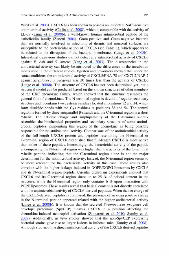
Fig. 3 (a) N- and C-terminal primary sequences of platelet basic protein (PBP) and derived
ß-thromboglobulin (ß-TG) family members. (b) NAP-2/CXCL7 and TC-1 3D structures and
electrostatic potential surfaces. Full-length CXCL7 and TC-1 structures were created by Nguyen
et al. (2011) based on the combination of CXCL7 crystal structure (PDB code 1NAP) and
C-terminal peptides (upper sequences) NMR structures. Negatively and positively charged
residues are colored in red and blue respectively. Electrostatic potential surface were calculated
by Adaptive Poisson–Boltzmann Solver (APBS) using PDB2QR software and depicted by
PyMOL software
Structure–Function Relationships of Antimicrobial Chemokines 197
residues in TC-1 is the negatively charged Asp-70, the removal of which seems to
account for the emergence of the antibacterial activity. Studies of synthetic
C-terminal a-helical peptides of both chemokines (CXCL7 and TC-1) were carried
out in order to identify the influence of these parts of the proteins on the antimi-
crobial activity. Neither of the peptides was antimicrobial, and they showed a
helical structure with a low degree of hydrophobicity and amphipathicity. The
structure of the CXCL7 C-terminal peptide showed that the side chain of the
C-terminal residue Asp-70 actually folds back and interacts with the positively
charged residue Arg-61 located in the a-helical region (Nguyen et al. 2010).
Subsequent NMR relaxation studies, which studied the flexibility of the protein
backbone in CXCL7 and TC1, showed that the Asp-70 residue of CXCL7 is
motionally restricted and interacts with the positive surface region of the protein
(Fig. 3b). In contrast, in TC-1 the last few residues are highly flexible, and the
positively charged surface region is exposed (Fig. 3b), being able to directly interact
with negatively charged bacterial membranes to either perturb the membrane or
enter the cell (Nguyen et al. 2011), explaining the difference in microbicidal
activity with full-length CXCL7.
In the activity-guided isolation of human platelet antimicrobial proteins, a second
protein, thrombocidin-2 (TC-2) was identified (Krijgsveld et al. 2000). TC-2 is
identical to CTAP-3 except for a truncation of the two C-terminal residues. CTAP-
3 has been putatively identified as one of the antimicrobial-active PBP derivatives in
monocytes after detection in gel overlay assays with a highly AMP-susceptible
S. typhimurium phoP mutant strain as the target organism (Schaffner et al. 2004).
As mentioned before, the activity as such of CTAP-3 is not high, with only minor
activity reported against wild-type strains of S. aureus, E. coli, and C. neoformans inslightly acidic conditions (Tang et al. 2002), and no bactericidal activity against
Bacillus subtilis, E. coli, or S. aureus when tested up to 30 mM. A C-terminal two
amino acid truncation, yielding TC-2, however strongly increases the microbicidal
activity (Krijgsveld et al. 2000). Of note, the truncation is identical to that generating
TC-1 from CXCL7 (Fig. 3), suggesting that this is a more general step in generating
antimicrobially active derivatives from members of the PBP protein family.
4.3 Interleukin-8 (IL-8)/CXCL8
Interleukin-8 (IL-8)/CXCL8 was one of the first chemokines discovered (Schroder
et al. 1987; Walz et al. 1987; Yoshimura et al. 1987b). It contains 72 amino acid
residues (8 kDa) and upon stimulation CXCL8 can be produced by a wide variety of
cells, including fibroblasts, epithelial and endothelial cells, hepatocytes and
monocytes, among others (Baggiolini et al. 1989). CXCL8 is important for the
activation and attraction of neutrophils to sites of inflammation, but other cells are
also susceptible to its chemotactic activity (Baggiolini et al. 1989). As expected, the
chemotactic activity arises through interactions with chemokine receptors, in this case
CXCR1 and CXCR2 (Wu et al. 1996; Holmes et al. 1991; Murphy and Tiffany 1991).
CXCR1 and CXCR2 and their associated ligands have been related to a large number
198 M. Arias et al.
of pathologies (Bizzarri et al. 2006). CXCL8 is also important for the promotion of
angiogenesis (Li et al. 2005; Matsuo et al. 2009). As described above for CXCL7, a
C-terminally truncated variant of CXCL8 lacking the last three residues exhibits
higher chemotactic activity than the full-length chemokine (Clark-Lewis et al.
1991). The structure of CXCL8 has been resolved by both NMR spectroscopy and
X-ray crystallography (Baldwin et al. 1991; Clore et al. 1989, 1990). These structures
are in agreement and show an architecture composed of an extended loop followed by
a 3.10 helical turn (involving residues 19–22) and the antiparallel stranded b-sheetconnected to a C-terminal a-helix (corresponding to residues 57–72). In solution
CXCL8 behaves as a dimer, where the monomers are connected by six backbone
hydrogen bonds between residues 25, 27, and 29 (Clore et al. 1989, 1990). The
antibacterial activity of CXCL8 has been somewhat controversial. Bylund and
colleagues, using a modified inhibition zone assay, were not able to observe any
antimicrobial activity for full-length CXCL8 against E. coli (Bjorstad et al. 2005).
Similarly, no antimicrobial activity for CXCL8 against E. coli and S. aureus (Coleet al. 2001; Yang et al. 2003) was found using standard colony-forming unit assays
(Harder et al. 2001) or radial diffusion assays (Steinberg and Lehrer 1997). In contrast,
Yeaman and colleagues observed different levels of pH-dependent antimicrobial
activity of CXCL8 against Salmonella typhimurium and S. aureus in solid-phase
assays, but they did not record bactericidal activity in solution-phase assay. Their
results suggest that the growth inhibition observed in the solid-phase assay is due to
bacteriostatic effects. Additionally, an antifungal activity against C. albicans was
detected in both solid- and solution-phase assays (Yount et al. 2007). The g-coremotif of CXCL8 (IL-8g), located in the central b-sheet region, was tested as a separatepeptide for antimicrobial and antifungal activity. The peptide showed no activity
against S. typhimurium, S. aureus or C. albicans, in neither the solid-phase nor the
solution-phase assay (Yount et al. 2007).
Another interesting structural element of CXCL8 which may exert antimicrobial
activity is the C-terminal a-helix. A 19-residue C-terminal peptide, termed IL-8a,proved to be active against S. typhimurium and C. albicans in solid-phase assays
and in solution-phase assays against C. albicans. In human blood matrices, the
IL-8a 19-mer peptide exerts anti-E. coli activity (Yount et al. 2007). Acid hydroly-sis of CXCL8 in vitro can generate a 20-residue IL-8a peptide with an extra Pro
residue at the N-terminal region compared to the 19-mer peptide (Bjorstad et al.
2005). Antimicrobial assays performed with this IL-8a 20-mer peptide showed
high activity against E. coli and moderate activity against Salmonella enterica,Klebsiella pneumoniae, and S. pyogenes, as established by inhibition zone assays
(Bjorstad et al. 2005). The same peptide was tested for antibacterial activity in
blood agar plates and moderate activity was found for B. subtilis and almost no
activity could be detected with S. aureus and E. coli (Nguyen et al. 2010). The
differences in the antimicrobial activity for IL-8a and CXCL8 in these studies may
have been due to differences in media and incubation conditions. When comparing
the SDS-micelle-bound structure of IL-8a 19-mer peptide and the C-terminal
a-helix in the full-length CXCL8, there are no significant differences (Bourbigot
et al. 2009). The structure of the IL-8a 20-mer peptide, dissolved in a
chloform–ethanol–water (4:4:1) solvent mixture (Nguyen et al. 2010), is also in
Structure–Function Relationships of Antimicrobial Chemokines 199

agreement with the structures of the IL-8a peptide (Bourbigot et al. 2009) and that
of the C-terminal a-helix in intact CXCL8 (Baldwin et al. 1991; Clore et al. 1989,
1990). In summary, the C-terminal region of CXCL8 seems to be important for the
antimicrobial activity of this particular chemokine. As mentioned before, the
S. pyogenes SpyCEP protease can cleave ELR CXC chemokines, including
human CXCL8 (Edwards et al. 2005; Zinkernagel et al. 2008; Zingaretti et al.
2010; Hidalgo-Grass et al. 2006). The action of SpyCEP on CXCL8 releases a
C-terminal peptide composed of residues 60–72 (Edwards et al. 2005). The direct
antimicrobial activity of this peptide has not yet been tested in vitro, but in vivo
and in vitro studies did show that the SpyCEP protease prevents eradication by
neutrophils of the SpyCEP-expressing bacteria and other bacteria at the site of
infection (Zinkernagel et al. 2008; Hidalgo-Grass et al. 2006). This effect has been
attributed to diminished neutrophil recruitment and migration (Edwards et al.
2005; Zinkernagel et al. 2008) in addition to an inhibition of the formation
of neutrophil extracellular traps (NETs) (Zinkernagel et al. 2008), due to digestion
of CXCL8.
4.4 Growth-Regulated Oncogene a (GROa)/CXCL1,b (GROb)/CXCL2 and g (GROg)/CXCL3
Among the members of the ELR CXC chemokine family with antimicrobial
activity, there are three chemokines with similar characteristics: CXCL1/GROa,CXCL2/GROb, and CXCL3/GROg, closely related chemokines that are produced
mainly by macrophages (Becker et al. 1994). These proteins have the capacity to
attract neutrophils (Clark-Lewis et al. 1995) and monocytes (Smith et al. 2005),
through interactions with the chemokine receptor CXCR2 (Katancik et al. 2000).
All three chemokines have antibacterial activity against E. coli and S. aureus(Yang et al. 2003).
In addition to the above-mentioned ELR CXC chemokines, CXCL5 also
exhibits antimicrobial activity against S. pyogenes. In summary, all members
of the ELR CXC chemokine family exhibit a direct antimicrobial activity (see
Table 1).
5 Antimicrobial Activity of Non-ELR CXC Chemokines
5.1 Platelet Factor-4 (PF-4)/CXCL4
Although the platelet factor-4 (PF-4)/CXCL4 is the earliest discovered member of
the CXC chemokine family (Deuel et al. 1977), its biological function was not
known to be related to the chemoattraction of specific cells until recent years.
200 M. Arias et al.
CXCL4 is expressed mainly in the megakaryocytes and in platelets (Slungaard
2005). While other members of the non-ELR CXC chemokines, e.g., CXCL9–11,
induce the migration of lymphocytes (Bonecchi et al. 1998; Sallusto et al. 1998),
CXCL4 was initially thought to be devoid of such an activity (Clark-Lewis et al.
1993). A spliced variant of CXCR3 (named CXCR3-B) was described early on as
the receptor for CXCL4. However, this receptor did not mediate a chemotactic
response but induced an increase in the intracellular cyclic AMP levels instead
(Lasagni et al. 2003; Slungaard 2005). A recent study suggests that migration of
activated T lymphocytes can be induced by CXCL4 and that this is mediated by the
chemokine receptor CXCR3 (Mueller et al. 2008), but these results have not been
confirmed by other groups (Flad and Brandt 2010). In addition, the biological
activity of CXCL4 is broad and includes roles in coagulation and functions such
as inhibition of angiogenesis and hematopoiesis, promotion of neutrophil adhesion
and activation, enhancement of oxy-LDL binding to the LDL receptor, and stimu-
lation of anticoagulant activated protein C generation by the thrombomodulin/
protein C system (Slungaard 2005). The generation of a CXCL4 knockout mouse
has provided support for the role of this chemokine in platelet-dependent throm-
bosis (Slungaard 2005). CXCL4 also exhibits direct antimicrobial activity under
slightly acidic conditions against Gram-positive and Gram-negative bacteria, in
addition to antifungal activity (Tang et al. 2002) (see Table 1). The structure of
human CXCL4 was solved as a tetramer by X-ray crystallography, showing a three-
dimensional structure in agreement with the overall chemokine topology formed by
the usual N-terminal loop, three-stranded antiparallel b-sheet, and C-terminal
a-helix (Zhang et al. 1994). Based on phylogenetic relatedness, similar sequence
motif, and predicted three-dimensional structures, platelet microbicidal protein-1
(PMP-1) was identified as a rabbit analogue of the human CXCL4 (Yount et al.
2004). In earlier studies, prior to their sequence identification, PMP-1 and its
variant tPMP-1 (secreted by platelets after thrombin stimulation) were shown to
exhibit a pH-dependent antimicrobial activity against Gram- positive and Gram-
negative bacteria and fungi (Yeaman et al. 1997). The antimicrobial activities of
PMP-1 and human CXCL4 exhibit similar specificities and efficacy (Yeaman et al.
2007). In the case of S. aureus, PMP-1 activity has been linked with an inhibition of
intracellular macromolecular synthesis (Xiong et al. 2002). A synthetic peptide
corresponding to the last 13 residues of the CXCL4 C-terminal region, which
constitute an a-helix, is active against E. coli in the presence of a sub-MIC
concentration of cefepime (a b-lactam antibiotic) in serum bactericidal assays
(Darveau et al. 1992). RP-1 and RP-11 are peptides designed based on the a-helicalC-terminal region of platelet proteins PMP-1, tPMP-1, and CXCL4, which have
antimicrobial activity in in vitro biological matrices (Yeaman et al. 2002). The
effects of RP-1 on S. aureus resemble the effects induced by tPMP-1, indicating
that the antimicrobial mechanism of certain proteins can be reproduced by the
synthetic peptides modeled upon relevant structural domains (Xiong et al. 2006).
Comparison of the sequences for CXCL4, PMP-1, and their respective variants
led to the creation of a consensus PMP sequence (cPMP), which was used to
Structure–Function Relationships of Antimicrobial Chemokines 201
generate a peptide library constituting of 15 amino acid long peptides,
overlapping by 3 residues each, and larger peptides comprising the N or C-
terminal half of the cPMP (Yeaman et al. 2007). Among the library, the peptides
with the highest antimicrobial activity were cPMP38–74, cPMP49–64, and
cPMP60–74, which represent the C-terminal region of the protein (Yeaman
et al. 2007). Most of the peptides corresponding to the N-terminal and the b-sheet regions did not exhibit considerable antimicrobial activity (Yeaman et al.
2007). These results suggest that the C-terminal region of CXCL4 is important
for the overall antimicrobial activity of this chemokine.
5.2 Monokine Induced by IFN-g (MIG)/CXCL9
The human monokine induced by IFN-g (MIG)/CXCL9 is a protein of 103 residues
of the CXC chemokine family (Liao et al. 1995; Farber 1993). The protein is
chemotactic for monocytes, activated T cells, and NK cells (Liao et al. 1995;
Lazzeri and Romagnani 2005; Loetscher et al. 1996). The chemotactic activity
involves binding to the chemokine receptors CXCR3A and CXCR3B (Loetscher
et al. 1996; Lazzeri and Romagnani 2005). The long C-terminal region of CXCL9
undergoes proteolytic processing by monocytes. This truncation has a profound
effect on its biological activity (Liao et al. 1995; Farber 1997). Interferon-inducible
protein-10 (IP-10)/CXCL10 and interferon-inducible T-cell alpha chemoattractant
(I-TAC)/CXCL11 are closely related to CXCL9/MIG, sharing the same receptor
and subsequently expressing similar biological activity (Farber 1997; Cole et al.
1998). Full-length CXCL9 displays a broad antimicrobial activity against
organisms including E. coli, Listeria monocytogenes, S. aureus (Cole et al. 2001;
Yang et al. 2003), S. pyogenes (Egesten et al. 2007), Neisseria gonorrhoeae (Lingeet al. 2008a), and Bacillus anthracis spores and bacilli (Crawford et al. 2009). This
antibacterial activity was shown to be inhibited by an increase in the ionic strength
of the media in the case of E. coli, L. monocytogenes, and S. pyogenes. Although thethree-dimensional structure of CXCL9 has not yet been solved, a model structure
was created using the NMR structure of a truncated CXCL2/GROb (Qian et al.
1999) as a template (Egesten et al. 2007). Due to the presence of a high number of
positive charges in both the N-terminal and C-terminal regions of CXCL9, the
antimicrobial activity might be embedded in either of these two sections of the
protein. Consequently, peptides resembling the cationic regions of the N-terminal
and C-terminal sections of CXCL9 were tested against S. pyogenes, and the activityof the C-terminal peptide was similar to the antibacterial activity of the full-length
CXCL9. The N-terminal peptide did not have any anti-S. pyogenes activity,
indicating that the antimicrobial activity of CXCL9 resides in its C-terminal region
(Egesten et al. 2007). Streptococcal inhibitor of complement (SIC) is a protein
secreted by S. pyogenes strains of the M1 serotype, which in addition to interfering
with complement function inhibits the activity of human antimicrobial peptides,
202 M. Arias et al.
such as LL-37 and b-defensins (Frick et al. 2003). Additionally SIC interferes with
the antibacterial activity of CXCL9, without disturbing its chemotactic activity
(Egesten et al. 2007), suggesting that SIC interacts with the antimicrobial
C-terminal region of CXCL9. Although CXCL9, CXCL10, and CXCL11 are
interferon-g-inducible related chemokines that interact with the same CXCR3
receptor and have a similar antibacterial spectrum (Cole et al. 2001; Egesten
et al. 2007; Yang et al. 2003; Crawford et al. 2009), the antimicrobial activity of
CXCL10 and CXCL11 against E. coli, L. monocytogenes, and S. pyogenes is
tenfold less than that of CXCL9 (Cole et al. 2001; Egesten et al. 2007). Analysis
of the structures of CXCL10/IP-10 (Swaminathan et al. 2003), IP-10 mutant
(NMeLeu27) (Booth et al. 2002), and CXCL11 (Booth et al. 2004) has shown
that their C-terminal a-helices are smaller than the predicted C-terminal a-helix in
CXCL9, which may account for the difference in the antimicrobial activities
(Egesten et al. 2007; Eliasson and Egesten 2008). SpeB from S. pyogenes is a
cysteine protease that cleaves and inactivates the antimicrobial peptide LL-37
(Schmidtchen et al. 2002). It also inactivates several human chemokines with
antibacterial activity (Egesten et al. 2009). SpeB fully degrades CXCL10 and
CXCL11, completely abolishing their chemotactic as well as antimicrobial activity.
In contrast CXCL9 is only partially digested by SpeB, with cleavage sites in the
N-terminal and C-terminal regions, releasing a smaller version of CXCL9
comprising the residues 18–73. This truncated version of CXCL9 has lost its
chemotactic activity but retains its antibacterial activity against S. pyogenes. Thesynthetic peptide CXCL957–83 and SpeB-produced CXCL918–73 showed similar
antimicrobial activities as the full-length CXCL9 (Egesten et al. 2007, 2009),
indicating that not all the residues in the a-helical C-terminal peptide are
responsible for the antimicrobial activity. Similarly, the truncation of CXCL10
by the furin protein generates a CXCL10 with four C-terminal residues removed.
This variant is as antimicrobial as its precursor, CXCL10, against E. coli andL. monocytogenes (Hensbergen et al. 2004).
Among the group of non-ELR CXC chemokines, most members exert antimi-
crobial activity (see Table 1). Although structural elements as the C-terminal
a-helical region have emerged as possibly being responsible for the antimicrobial
activity in these chemokines, more studies are required.
6 Chemokine Dimerization and Antimicrobial Activity
Although many of the chemokines have the ability to form dimers or even higher-
order oligomers in solution or after binding to glycosaminoglycans (Allen et al.
2007; Salanga and Handel 2011), the relationship of these oligomers with the
antimicrobial activity of the bactericidal chemokines has not yet been studied
much. Oligomerization has been postulated as one of the key steps in the antimi-
crobial activity mechanism of pore-forming antimicrobial peptides (Shai 1999;
Mihajlovic and Lazaridis 2010; Mani et al. 2006). Oligomerization can allow the
Structure–Function Relationships of Antimicrobial Chemokines 203
formation of transmembrane pores leading to bacterial cell death (Shai 1999;
Matsuzaki 1998). For some antimicrobial peptides, such as LLP1 and modified
versions of magainin-2, the formation of disulfide-linked dimers has been shown to
play an important role in the antimicrobial activity against Gram-positive and
Gram-negative bacteria (Tencza et al. 1999; Dempsey et al. 2002). The formation
of dimers prior to the interaction with the bacterial membranes increases the
attraction for negatively charged membranes, probably due to the increase in the
net positive charge of these peptides (Dempsey et al. 2002), generating peptides
with 4–8 times higher activity, compared with the monomeric forms (Tencza et al.
1999). Not only the formation of covalently bound dimers can be related with an
increase in antimicrobial activity. Protegrin-1 (PG-1) is a potent antimicrobial
peptide for which the mechanism of action has been related to the formation of
transmembrane pores (Bolintineanu et al. 2010; Langham et al. 2008). Using NMR,
the dimerization of PG-1 was established as an essential requirement for the
formation of pores in anionic membranes (Mani et al. 2006; Roumestand et al.
1998). Molecular dynamic simulations have confirmed the tendency of PG-1 to
form dimers in the aqueous phase, on a membrane surface, or in the membrane core
(Vivcharuk and Kaznessis 2010b). It was also established that the PG-1 dimer
rather than the PG-1 monomer has a favorable energy for interactions with the
membrane (Vivcharuk and Kaznessis 2010a). Other examples of this are seen in the
b-defensin family of antimicrobial peptides. The tendency of human b-defensin 3
(hBD-3) to form non-covalent dimers may explain the large difference in the
antimicrobial activity among the known human b-defensins (Schibli et al. 2002).When compared with other human b-defensins (hDB-1 and hDB-2), hBD-3 has
been reported to possess a more potent and salt-resistant antimicrobial activity
against a broader spectrum of pathogens (Pazgier et al. 2006; Taylor et al. 2008). It
has been proposed that this increase may be related to the formation of dimers
which exhibit a considerable increase in the size of the positively charged surface,
thereby increasing the attraction for negatively charged membranes (Schibli et al.
2002). Although crystallography studies of the hDB-2 structure in addition to
molecular dynamic studies had shown the possibility of dimerization for this
protein (Suresh and Verma 2006; Hoover et al. 2000), static and dynamic light
scattering experiments in 0.1 M Tris and pH 8.0 showed that only hDB-3 exists as a
dimer in solution (Schibli et al. 2002). Some of the antimicrobial chemokines also
have the ability to form non-covalent dimers in solution, which in addition to
enhancing the net positive charge of the protein also increases the size of the
positively charged surface. The configuration of the chemokines dimers can be
divided into two groups. CC-chemokines normally form dimers by interactions
between the residues of the first b-strand, forming a two-stranded antiparallel b-sheet with the corresponding b-strand from the other monomer, resulting in a more
elongated dimer structure. In contrast, CXC chemokines normally form dimers by
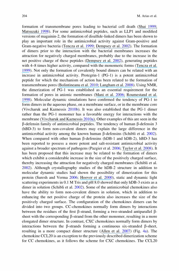
interactions between the b-strands forming a continuous six-stranded b-sheet,resulting in a more compact dimer structure (Allen et al. 2007) (Fig. 4a). The
chemokine CCL20 is an exception to the previously described dimerization scheme
for CC chemokines, as it follows the scheme for CXC chemokines. The CCL20
204 M. Arias et al.
dimer structure characteristics may explain the high antimicrobial activity of this
particular chemokine. In a monomeric form, CCL20 already exhibits a large
positively charged surface. This is even increased considerably when the dimer is
formed by the generation of the six-stranded b-sheet with both C-terminal a-heliceslocated on top, establishing a large contiguous positively charged surface (Fig. 4b).
The dimerization of CCL20 is dependent on the protonation state of the His-40
sidechain. At neutral pH, His-40 is unprotonated, allowing dimer formation of
CCL20; at lower pH, the two His-40 residues become positively charged, and
charge repulsion between these residues prevents the dimer formation (Chan
et al. 2008). The formation of an extended positively charged surface under
physiological conditions may explain the strong antimicrobial activity of CCL20,
Fig. 4 (a) Dimerization schemes for chemokines. CXCL8 chemokines dimer structure (PDB code
IL8) (left). CCL3 chemokine dimer structure (PDB code 2X69) (right). Each monomer is depicted
in a different color and the disulfide bonds are represented in yellow. (b) Chemokine CCL20 (PDB
code 2JYO) and dimer (PDB code 1M8A) structure (upper) with the electrostatic potential surfacedistribution (lower). The disulfide bonds are represented in yellow (upper) and the monomer
interface is depicted by a yellow line (lower, right). Electrostatic potential surfaces were calculatedby Adaptative Poisson–Boltzmann Solver (APBS) using PDB2PQR software and depicted by
PyMOL software
Structure–Function Relationships of Antimicrobial Chemokines 205
and the same could apply to other chemokines. In a recent study, it was also
suggested that the differences in the dimerization behavior of CXCL7 and its
truncated TC-1 derivative contributed to the drastically increased antimicrobial
activity of the latter (Nguyen et al. 2011). Clearly detailed studies addressing the
influence of dimerization on the antimicrobial activity of chemokines are warranted
in order to definitively establish its possible role in the activity of these proteins.
In addition to the formation of homodimers, several chemokines are also able to
form heterodimers. This has been shown to modulate their biological activities
(Allen et al. 2007). For example, the chemotaxis of specific cells has been signifi-
cantly affected by the formation of heterodimers as observed for the CXCL8-
CXCL4 and CXCL4-CCL5 dimers (Nesmelova et al. 2005; von Hundelshausen
et al. 2005). Heterodimers are also observed for other members of the innate
immune system, such as AMPs. Some AMPs can undergo heterodimerization
thereby modifying their antimicrobial activity, membrane permeabilization
activity, and/or their resistance to protease digestion. Distinctin is an example of
a covalently bound heterodimer antimicrobial peptide isolated from the tree frog
Phyllomedusa distincta (Batista et al. 2001; Dalla Serra et al. 2008; Raimondo et al.
2005; Resende et al. 2009). Not only covalently bound dimers are observed among
AMPs, but it is believed that the peptides magainin-2 and peptidyl-glycylleucine-
carboxyamide (PGLa) form a non-covalent dimer upon interaction with membranes
increasing the antimicrobial activity and membrane permeabilization activity but
also increasing their cytotoxicity (Matsuzaki 1998; Hara et al. 2001). Similar to
AMPs, the ability of chemokines to form heterodimers could be related not only
with the modulation of their chemotactic function, but it could also regulate their
microbicidal activity. Further studies are required in order to establish the effects of
chemokine heterodimerization on their direct antimicrobial activity. Finally, it is
known that several chemokines can form larger oligomeric structures (Salanga and
Handel 2011). Such aggregated structures could markedly influence the antimicro-
bial activity. The formation of the oligomers is usually promoted by binding of
glycosaminoglycans. In a recent study the structure of an oligomeric form of CCL5/
RANTES was reported (Wang et al. 2011). This work sets the stage for future
studies with other chemokines. It will be interesting to see if the binding of
negatively charged glycosaminoglycans promotes or competes with the antimicro-
bial activities of the chemokines.
7 Activity of Unfolded Chemokines and of Derived Peptides
The antimicrobial chemokines share a large conformational, positively charged
surface patch (Yang et al. 2003). In thrombocidin-1 (TC-1), disruption of this
positive patch by substitution of the central cationic Lys-17 with an alanine residue
substantially reduced the antimicrobial potency (Kwakman et al. 2011), providing
direct evidence for its importance. Indeed, lysine substitution of negative or neutral
residues bordering the positive patch enhanced the activity. Interestingly, reduced
206 M. Arias et al.
TC-1 had equal antimicrobial activity as the folded protein, even though the
positive patch was disrupted (Kwakman et al. 2011). The structural elements of
unfolded TC-1 were likely localized in the N-terminal region of the protein, since
linear 15-mer synthetic peptides corresponding to the N-terminal part of TC-1 had
very potent antimicrobial activity. This implies that the structural elements
involved in antimicrobial activity of native folded TC-1 strongly differ from
those of the reduced, unfolded protein.
Similar to TC-1, several cationic antimicrobial peptides with positive patches
retain their antimicrobial activity after linearization (Kluver et al. 2006; Liu and
Wilson 2010; Mandal et al. 2002; Wu et al. 2003). For instance, variants of hBD-3
lacking disulfides are still antimicrobial (Hoover et al. 2003; Wu et al. 2003) despite
the loss of most structural elements of the native protein. A variant of the antimi-
crobial chemokine CCL28, lacking both cysteines of the CC sequence, has similar
antimicrobial activity as the native, folded protein (Liu and Wilson 2010). As
CCL28-derived peptides have antimicrobial activity equivalent to the intact, folded
protein (Liu and Wilson 2010), the activity of linear CCL28 may well be due to
peptide regions in the unfolded protein, similar to the case of linearized TC-1.
The strongest example of the influence of unfolding on antimicrobial activity
probably is the case of human b-defensin 1 (hDB-1). This protein is abundantly
expressed by all human epithelia but was considered a low-activity AMP. However,
recent research showed that reduction and unfolding of hBD-1 turns this protein
into a potent antimicrobial (Schroeder et al. 2011). Reduced hBD-1 colocalizes
with thioredoxin in several human tissues, suggesting that this enzyme system may
be involved in hBD-1 reduction in vivo (Schroeder et al. 2011).
Assuming that linearization of chemokines and antimicrobial proteins does
indeed occur in vivo, one may speculate that in addition to the known truncation
peptides from folded proteins, additional linear peptides may be generated by
proteolytic degradation from the now linearized parts, potentially further
contributing to the already impressive role of this class of molecules to host defense.
8 Concluding Remarks
The study of the antimicrobial activity of chemokines is still a relatively unexplored
field, which is not surprising as this “moonlighting” activity of chemokines was
only first reported a little more than 10 years ago. To date, the activity of a number
of CC and CXC chemokines has been dissected in detail, as described in this
contribution. While in quite a few cases the characteristic C-terminal a-helix of
the chemokines plays a role in the antimicrobial activity, this is certainly not
universal, as for selected chemokines the N-terminal region of the proteins seems
to be more important. In some instances extensions beyond the usual ~70 residues
of the typical chemokine structure play a role in the activity, as in the case of the
histatin-like extension of CCL28 and for the 30-residue C-terminal extension
of CXCL9. The latter activities seem to be grafted onto the normal chemokine
structure. A positively charged patch is essential for the activity of a number of
Structure–Function Relationships of Antimicrobial Chemokines 207
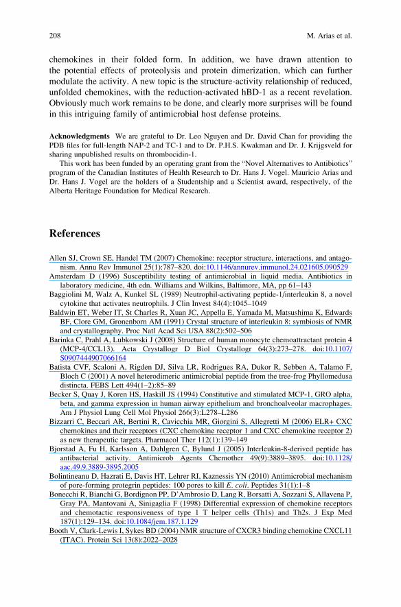
chemokines in their folded form. In addition, we have drawn attention to
the potential effects of proteolysis and protein dimerization, which can further
modulate the activity. A new topic is the structure-activity relationship of reduced,
unfolded chemokines, with the reduction-activated hBD-1 as a recent revelation.
Obviously much work remains to be done, and clearly more surprises will be found
in this intriguing family of antimicrobial host defense proteins.
Acknowledgments We are grateful to Dr. Leo Nguyen and Dr. David Chan for providing the
PDB files for full-length NAP-2 and TC-1 and to Dr. P.H.S. Kwakman and Dr. J. Krijgsveld for
sharing unpublished results on thrombocidin-1.
This work has been funded by an operating grant from the “Novel Alternatives to Antibiotics”
program of the Canadian Institutes of Health Research to Dr. Hans J. Vogel. Mauricio Arias and
Dr. Hans J. Vogel are the holders of a Studentship and a Scientist award, respectively, of the
Alberta Heritage Foundation for Medical Research.
References
Allen SJ, Crown SE, Handel TM (2007) Chemokine: receptor structure, interactions, and antago-
nism. Annu Rev Immunol 25(1):787–820. doi:10.1146/annurev.immunol.24.021605.090529
Amsterdam D (1996) Susceptibility testing of antimicrobial in liquid media. Antibiotics in
laboratory medicine, 4th edn. Williams and Wilkins, Baltimore, MA, pp 61–143
Baggiolini M, Walz A, Kunkel SL (1989) Neutrophil-activating peptide-1/interleukin 8, a novel
cytokine that activates neutrophils. J Clin Invest 84(4):1045–1049
Baldwin ET, Weber IT, St Charles R, Xuan JC, Appella E, Yamada M, Matsushima K, Edwards
BF, Clore GM, Gronenborn AM (1991) Crystal structure of interleukin 8: symbiosis of NMR
and crystallography. Proc Natl Acad Sci USA 88(2):502–506
Barinka C, Prahl A, Lubkowski J (2008) Structure of human monocyte chemoattractant protein 4
(MCP-4/CCL13). Acta Crystallogr D Biol Crystallogr 64(3):273–278. doi:10.1107/
S0907444907066164
Batista CVF, Scaloni A, Rigden DJ, Silva LR, Rodrigues RA, Dukor R, Sebben A, Talamo F,
Bloch C (2001) A novel heterodimeric antimicrobial peptide from the tree-frog Phyllomedusa
distincta. FEBS Lett 494(1–2):85–89
Becker S, Quay J, Koren HS, Haskill JS (1994) Constitutive and stimulated MCP-1, GRO alpha,
beta, and gamma expression in human airway epithelium and bronchoalveolar macrophages.
Am J Physiol Lung Cell Mol Physiol 266(3):L278–L286
Bizzarri C, Beccari AR, Bertini R, Cavicchia MR, Giorgini S, Allegretti M (2006) ELR+ CXC
chemokines and their receptors (CXC chemokine receptor 1 and CXC chemokine receptor 2)
as new therapeutic targets. Pharmacol Ther 112(1):139–149
Bjorstad A, Fu H, Karlsson A, Dahlgren C, Bylund J (2005) Interleukin-8-derived peptide has
antibacterial activity. Antimicrob Agents Chemother 49(9):3889–3895. doi:10.1128/
aac.49.9.3889-3895.2005
Bolintineanu D, Hazrati E, Davis HT, Lehrer RI, Kaznessis YN (2010) Antimicrobial mechanism
of pore-forming protegrin peptides: 100 pores to kill E. coli. Peptides 31(1):1–8Bonecchi R, Bianchi G, Bordignon PP, D’Ambrosio D, Lang R, Borsatti A, Sozzani S, Allavena P,
Gray PA, Mantovani A, Sinigaglia F (1998) Differential expression of chemokine receptors
and chemotactic responsiveness of type 1 T helper cells (Th1s) and Th2s. J Exp Med
187(1):129–134. doi:10.1084/jem.187.1.129
Booth V, Clark-Lewis I, Sykes BD (2004) NMR structure of CXCR3 binding chemokine CXCL11
(ITAC). Protein Sci 13(8):2022–2028
208 M. Arias et al.
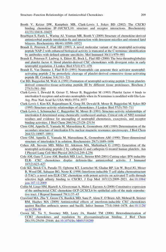
Booth V, Keizer DW, Kamphuis MB, Clark-Lewis I, Sykes BD (2002) The CXCR3
binding chemokine IP-10/CXCL10: structure and receptor interactions. Biochemistry
41(33):10418–10425
Bourbigot S, Fardy L, Waring AJ, Yeaman MR, Booth V (2009) Structure of chemokine-derived
antimicrobial peptide interleukin-8a and interaction with detergent micelles and oriented lipid
bilayers. Biochemistry 48(44):10509–10521
Brandt E, Petersen F, Flad HD (1993) A novel molecular variant of the neutrophil-activating
peptide NAP-2 with enhanced biological activity is truncated at the C-terminus: identification
by antibodies with defined epitope specificity. Mol Immunol 30(11):979–991
Brandt E, Petersen F, Ludwig A, Ehlert JE, Bock L, Flad HD (2000) The beta-thromboglobulins
and platelet factor 4: blood platelet-derived CXC chemokines with divergent roles in early
neutrophil regulation. J Leukoc Biol 67(4):471–478
Brandt E, Van Damme J, Flad H-D (1991) Neutrophils can generate their activator neutrophil-
activating peptide 2 by proteolytic cleavage of platelet-derived connective tissue-activating
peptide III. Cytokine 3(4):311–321
Car BD, Baggiolini M, Walz A (1991) Formation of neutrophil-activating peptide 2 from platelet-
derived connective-tissue-activating peptide III by different tissue proteinases. Biochem J
275(3):581–584
Clark-Lewis I, Dewald B, Geiser T, Moser B, Baggiolini M (1993) Platelet factor 4 binds to
interleukin 8 receptors and activates neutrophils when its N terminus is modified with Glu-Leu-
Arg. Proc Natl Acad Sci USA 90(8):3574–3577
Clark-Lewis I, Kim KS, Rajarathnam K, Gong JH, Dewald B, Moser B, Baggiolini M, Sykes BD
(1995) Structure-activity relationships of chemokines. J Leukoc Biol 57(5):703–711
Clark-Lewis I, Schumacher C, Baggiolini M, Moser B (1991) Structure-activity relationships of
interleukin-8 determined using chemically synthesized analogs. Critical role of NH2-terminal
residues and evidence for uncoupling of neutrophil chemotaxis, exocytosis, and receptor
binding activities. J Biol Chem 266(34):23128–23134
Clore GM, Appella E, Yamada M, Matsushima K, Gronenborn AM (1989) Determination of the
secondary structure of interleukin-8 by nuclear magnetic resonance spectroscopy. J Biol Chem
264(32):18907–18911
Clore GM, Appella E, Yamada M, Matsushima K, Gronenborn AM (1990) Three-dimensional
structure of interleukin 8 in solution. Biochemistry 29(7):1689–1696
Cohen AB, Stevens MD, Miller EJ, Atkinson MA, Mullenbach G (1992) Generation of the
neutrophil-activating peptide-2 by cathepsin G and cathepsin G-treated human platelets. Am
J Physiol Lung Cell Mol Physiol 263(2):L249–L256
Cole AM, Ganz T, Liese AM, BurdickMD, Liu L, Strieter RM (2001) Cutting edge: IFN-inducible
ELR- CXC chemokines display defensin-like antimicrobial activity. J Immunol
167(2):623–627
Cole KE, Strick CA, Paradis TJ, Ogborne KT, Loetscher M, Gladue RP, Lin W, Boyd JG, Moser
B, Wood DE, Sahagan BG, Neote K (1998) Interferon-inducible T cell alpha chemoattractant
(I-TAC): a novel non-ELR CXC chemokine with potent activity on activated T cells through
selective high affinity binding to CXCR3. J Exp Med 187(12):2009–2021. doi:10.1084/
jem.187.12.2009
Collin M, Linge HM, Bjartell A, Giwercman A, Malm J, Egesten A (2008) Constitutive expression
of the antibacterial CXC chemokine GCP-2/CXCL6 by epithelial cells of the male reproduc-
tive tract. J Reprod Immunol 79(1):37–43
Crawford MA, Zhu Y, Green CS, Burdick MD, Sanz P, Alem F, O’Brien AD, Mehrad B, Strieter
RM, Hughes MA (2009) Antimicrobial effects of interferon-inducible CXC chemokines
against Bacillus anthracis spores and bacilli. Infect Immun 77(4):1664–1678. doi:10.1128/
iai.01208-08
Crown SE, Yu Y, Sweeney MD, Leary JA, Handel TM (2006) Heterodimerization of
CCR2 chemokines and regulation by glycosaminoglycan binding. J Biol Chem
281(35):25438–25446. doi:10.1074/jbc.M601518200
Structure–Function Relationships of Antimicrobial Chemokines 209
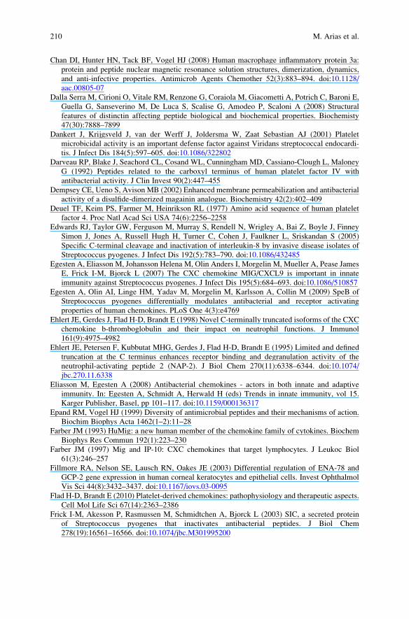
Chan DI, Hunter HN, Tack BF, Vogel HJ (2008) Human macrophage inflammatory protein 3a:
protein and peptide nuclear magnetic resonance solution structures, dimerization, dynamics,
and anti-infective properties. Antimicrob Agents Chemother 52(3):883–894. doi:10.1128/
aac.00805-07
Dalla Serra M, Cirioni O, Vitale RM, Renzone G, Coraiola M, Giacometti A, Potrich C, Baroni E,
Guella G, Sanseverino M, De Luca S, Scalise G, Amodeo P, Scaloni A (2008) Structural
features of distinctin affecting peptide biological and biochemical properties. Biochemisty
47(30):7888–7899
Dankert J, Krijgsveld J, van der Werff J, Joldersma W, Zaat Sebastian AJ (2001) Platelet
microbicidal activity is an important defense factor against Viridans streptococcal endocardi-
tis. J Infect Dis 184(5):597–605. doi:10.1086/322802
Darveau RP, Blake J, Seachord CL, Cosand WL, Cunningham MD, Cassiano-Clough L, Maloney
G (1992) Peptides related to the carboxyl terminus of human platelet factor IV with
antibacterial activity. J Clin Invest 90(2):447–455
Dempsey CE, Ueno S, Avison MB (2002) Enhanced membrane permeabilization and antibacterial
activity of a disulfide-dimerized magainin analogue. Biochemistry 42(2):402–409
Deuel TF, Keim PS, Farmer M, Heinrikson RL (1977) Amino acid sequence of human platelet
factor 4. Proc Natl Acad Sci USA 74(6):2256–2258
Edwards RJ, Taylor GW, Ferguson M, Murray S, Rendell N, Wrigley A, Bai Z, Boyle J, Finney
Simon J, Jones A, Russell Hugh H, Turner C, Cohen J, Faulkner L, Sriskandan S (2005)
Specific C-terminal cleavage and inactivation of interleukin-8 by invasive disease isolates of
Streptococcus pyogenes. J Infect Dis 192(5):783–790. doi:10.1086/432485
Egesten A, Eliasson M, Johansson Helena M, Olin Anders I, Morgelin M, Mueller A, Pease James
E, Frick I-M, Bjorck L (2007) The CXC chemokine MIG/CXCL9 is important in innate
immunity against Streptococcus pyogenes. J Infect Dis 195(5):684–693. doi:10.1086/510857
Egesten A, Olin AI, Linge HM, Yadav M, Morgelin M, Karlsson A, Collin M (2009) SpeB of
Streptococcus pyogenes differentially modulates antibacterial and receptor activating
properties of human chemokines. PLoS One 4(3):e4769
Ehlert JE, Gerdes J, Flad H-D, Brandt E (1998) Novel C-terminally truncated isoforms of the CXC
chemokine b-thromboglobulin and their impact on neutrophil functions. J Immunol
161(9):4975–4982
Ehlert JE, Petersen F, Kubbutat MHG, Gerdes J, Flad H-D, Brandt E (1995) Limited and defined
truncation at the C terminus enhances receptor binding and degranulation activity of the
neutrophil-activating peptide 2 (NAP-2). J Biol Chem 270(11):6338–6344. doi:10.1074/
jbc.270.11.6338
Eliasson M, Egesten A (2008) Antibacterial chemokines - actors in both innate and adaptive
immunity. In: Egesten A, Schmidt A, Herwald H (eds) Trends in innate immunity, vol 15.
Karger Publisher, Basel, pp 101–117. doi:10.1159/000136317
Epand RM, Vogel HJ (1999) Diversity of antimicrobial peptides and their mechanisms of action.
Biochim Biophys Acta 1462(1–2):11–28
Farber JM (1993) HuMig: a new human member of the chemokine family of cytokines. Biochem
Biophys Res Commun 192(1):223–230
Farber JM (1997) Mig and IP-10: CXC chemokines that target lymphocytes. J Leukoc Biol
61(3):246–257
Fillmore RA, Nelson SE, Lausch RN, Oakes JE (2003) Differential regulation of ENA-78 and
GCP-2 gene expression in human corneal keratocytes and epithelial cells. Invest Ophthalmol
Vis Sci 44(8):3432–3437. doi:10.1167/iovs.03-0095
Flad H-D, Brandt E (2010) Platelet-derived chemokines: pathophysiology and therapeutic aspects.
Cell Mol Life Sci 67(14):2363–2386
Frick I-M, Akesson P, Rasmussen M, Schmidtchen A, Bjorck L (2003) SIC, a secreted protein
of Streptococcus pyogenes that inactivates antibacterial peptides. J Biol Chem
278(19):16561–16566. doi:10.1074/jbc.M301995200
210 M. Arias et al.

Froyen G, Proost P, Ronsse I, Mitera T, Haelens A, Wuyts A, Opdenakker G, van Damme J,
Billiau A (1997) Cloning, bacterial expression and biological characterization of recombinant
human granulocyte chemotactic protein-2 and differential expression of granulocyte chemo-
tactic protein-2 and epithelial cell-derived neutrophil activating peptide-78 mRNAs. Eur
J Biochem 243(3):762–769
Fujiie S, Hieshima K, Izawa D, Nakayama T, Fujisawa R, Ohyanagi H, Yoshie O (2001)
Proinflammatory cytokines induce liver and activation-regulated chemokine/macrophage
inflammatory protein-3a/CCL20 in mucosal epithelial cells through NF-kB. Int Immunol
13(10):1255–1263. doi:10.1093/intimm/13.10.1255
Garcia-Zepeda EA, Combadiere C, Rothenberg ME, Sarafi MN, Lavigne F, Hamid Q, Murphy
PM, Luster AD (1996) Human monocyte chemoattractant protein (MCP)-4 is a novel CC
chemokine with activities on monocytes, eosinophils, and basophils induced in allergic and
nonallergic inflammation that signals through the CC chemokine receptors (CCR)-2 and �3.
J Immunol 157(12):5613–5626
Ghosh M, Shen Z, Schaefer TM, Fahey JV, Gupta P, Wira CR (2009) CCL20/MIP3a is a novel
anti-HIV-1 molecule of the human female reproductive tract. Am J Reprod Immunol 62
(1):60–71
Gijsbers K, Van Assche G, Joossens S, Struyf S, Proost P, Rutgeerts P, Geboes K, Van Damme J
(2004) CXCR1-binding chemokines in inflammatory bowel diseases: down-regulated IL-8/
CXCL8 production by leukocytes in Crohn’s disease and selective GCP-2/CXCL6 expression
in inflamed intestinal tissue. Eur J Immunol 34(7):1992–2000
Haney EF, Hunter HN, Matsuzaki K, Vogel HJ (2009a) Solution NMR studies of amphibian
antimicrobial peptides: linking structure to function? Biochim Biophys Acta 1788(8):1639–1655
Haney EF, Lau F, Vogel HJ (2007) Solution structures and model membrane interactions of
lactoferrampin, an antimicrobial peptide derived from bovine lactoferrin. Biochim Biophys
Acta 1768(10):2355–2364
Haney EF, Nazmi K, Lau F, Bolscher JGM, Vogel HJ (2009b) Novel lactoferrampin antimicrobial
peptides derived from human lactoferrin. Biochimie 91(1):141–154
Hara T, Mitani Y, Tanaka K, Uematsu N, Takakura A, Tachi T, Kodama H, Kondo M, Mori H,
Otaka A, Nobutaka F, Matsuzaki K (2001) Heterodimer formation between the antimicrobial
peptides magainin 2 and PGLa in lipid bilayers: A cross-linking study. Biochemistry
40(41):12395–12399
Harder J, Bartels J, Christophers E, Schroder J-M (2001) Isolation and characterization of human
defensin-3, a novel human inducible peptide antibiotic. J Biol Chem 276(8):5707–5713.
doi:10.1074/jbc.M008557200
Harter L, Petersen F, Flad HD, Brandt E (1994) Connective tissue-activating peptide III
desensitizes chemokine receptors on neutrophils. Requirement for proteolytic formation of
the neutrophil-activating peptide 2. J Immunol 153(12):5698–5708
Hasan L, Mazzucchelli L, Liebi M, Lis M, Hunger RE, Tester A, Overall CM, Wolf M (2006)
Function of liver activation-regulated chemokine/CC chemokine ligand 20 is differently
affected by cathepsin B and cathepsin D processing. J Immunol 176(11):6512–6522
Hensbergen PJ, Verzijl D, Balog CIA, Dijkman R, van der Schors RC, van der Raaij-Helmer
EMH, van der Plas MJA, Leurs R, Deelder AM, Smit MJ, Tensen CP (2004) Furin is a
chemokine-modifying enzyme. J Biol Chem 279(14):13402–13411. doi:10.1074/jbc.
M312814200
Hidalgo-Grass C, Mishalian I, Dan-Goor M, Belotserkovsky I, Eran Y, Nizet V, Peled A, Hanski E
(2006) A streptococcal protease that degrades CXC chemokines and impairs bacterial clear-
ance from infected tissues. EMBO J 25(19):4628–4637
Hieshima K, Ohtani H, ShibanoM, Izawa D, Nakayama T, Kawasaki Y, Shiba F, Shiota M, Katou F,
Saito T, Yoshie O (2003) CCL28 has dual roles in mucosal immunity as a chemokine with broad-
spectrum antimicrobial activity. J Immunol 170(3):1452–1461
Holmes WE, Lee J, Kuang WJ, Rice GC, Wood WI (1991) Structure and functional expression of
a human interleukin-8 receptor. Science 253(5025):1278–1280. doi:10.1126/science.1840701
Structure–Function Relationships of Antimicrobial Chemokines 211
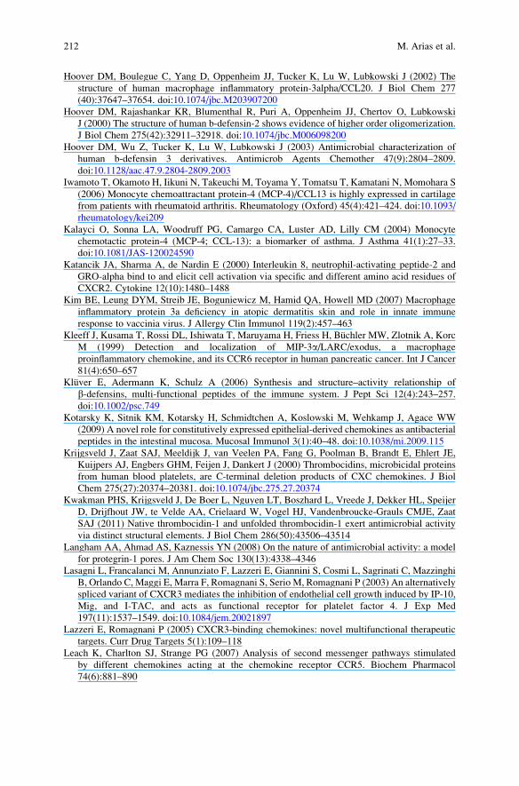
Hoover DM, Boulegue C, Yang D, Oppenheim JJ, Tucker K, Lu W, Lubkowski J (2002) The
structure of human macrophage inflammatory protein-3alpha/CCL20. J Biol Chem 277
(40):37647–37654. doi:10.1074/jbc.M203907200
Hoover DM, Rajashankar KR, Blumenthal R, Puri A, Oppenheim JJ, Chertov O, Lubkowski
J (2000) The structure of human b-defensin-2 shows evidence of higher order oligomerization.
J Biol Chem 275(42):32911–32918. doi:10.1074/jbc.M006098200
Hoover DM, Wu Z, Tucker K, Lu W, Lubkowski J (2003) Antimicrobial characterization of
human b-defensin 3 derivatives. Antimicrob Agents Chemother 47(9):2804–2809.
doi:10.1128/aac.47.9.2804-2809.2003
Iwamoto T, Okamoto H, Iikuni N, Takeuchi M, Toyama Y, Tomatsu T, Kamatani N, Momohara S
(2006) Monocyte chemoattractant protein-4 (MCP-4)/CCL13 is highly expressed in cartilage
from patients with rheumatoid arthritis. Rheumatology (Oxford) 45(4):421–424. doi:10.1093/
rheumatology/kei209
Kalayci O, Sonna LA, Woodruff PG, Camargo CA, Luster AD, Lilly CM (2004) Monocyte
chemotactic protein-4 (MCP-4; CCL-13): a biomarker of asthma. J Asthma 41(1):27–33.
doi:10.1081/JAS-120024590
Katancik JA, Sharma A, de Nardin E (2000) Interleukin 8, neutrophil-activating peptide-2 and
GRO-alpha bind to and elicit cell activation via specific and different amino acid residues of
CXCR2. Cytokine 12(10):1480–1488
Kim BE, Leung DYM, Streib JE, Boguniewicz M, Hamid QA, Howell MD (2007) Macrophage
inflammatory protein 3a deficiency in atopic dermatitis skin and role in innate immune
response to vaccinia virus. J Allergy Clin Immunol 119(2):457–463
Kleeff J, Kusama T, Rossi DL, Ishiwata T, Maruyama H, Friess H, Buchler MW, Zlotnik A, Korc
M (1999) Detection and localization of MIP-3a/LARC/exodus, a macrophage
proinflammatory chemokine, and its CCR6 receptor in human pancreatic cancer. Int J Cancer
81(4):650–657
Kluver E, Adermann K, Schulz A (2006) Synthesis and structure–activity relationship of
b-defensins, multi-functional peptides of the immune system. J Pept Sci 12(4):243–257.
doi:10.1002/psc.749
Kotarsky K, Sitnik KM, Kotarsky H, Schmidtchen A, Koslowski M, Wehkamp J, Agace WW
(2009) A novel role for constitutively expressed epithelial-derived chemokines as antibacterial
peptides in the intestinal mucosa. Mucosal Immunol 3(1):40–48. doi:10.1038/mi.2009.115
Krijgsveld J, Zaat SAJ, Meeldijk J, van Veelen PA, Fang G, Poolman B, Brandt E, Ehlert JE,
Kuijpers AJ, Engbers GHM, Feijen J, Dankert J (2000) Thrombocidins, microbicidal proteins
from human blood platelets, are C-terminal deletion products of CXC chemokines. J Biol
Chem 275(27):20374–20381. doi:10.1074/jbc.275.27.20374
Kwakman PHS, Krijgsveld J, De Boer L, Nguyen LT, Boszhard L, Vreede J, Dekker HL, Speijer
D, Drijfhout JW, te Velde AA, Crielaard W, Vogel HJ, Vandenbroucke-Grauls CMJE, Zaat
SAJ (2011) Native thrombocidin-1 and unfolded thrombocidin-1 exert antimicrobial activity
via distinct structural elements. J Biol Chem 286(50):43506–43514
Langham AA, Ahmad AS, Kaznessis YN (2008) On the nature of antimicrobial activity: a model
for protegrin-1 pores. J Am Chem Soc 130(13):4338–4346
Lasagni L, Francalanci M, Annunziato F, Lazzeri E, Giannini S, Cosmi L, Sagrinati C, Mazzinghi
B, Orlando C, Maggi E, Marra F, Romagnani S, Serio M, Romagnani P (2003) An alternatively
spliced variant of CXCR3 mediates the inhibition of endothelial cell growth induced by IP-10,
Mig, and I-TAC, and acts as functional receptor for platelet factor 4. J Exp Med
197(11):1537–1549. doi:10.1084/jem.20021897
Lazzeri E, Romagnani P (2005) CXCR3-binding chemokines: novel multifunctional therapeutic
targets. Curr Drug Targets 5(1):109–118
Leach K, Charlton SJ, Strange PG (2007) Analysis of second messenger pathways stimulated
by different chemokines acting at the chemokine receptor CCR5. Biochem Pharmacol
74(6):881–890
212 M. Arias et al.

Lehrer RI, Rosenman M, Harwig SSSL, Jackson R, Eisenhauer P (1991) Ultrasensitive assays for
endogenous antimicrobial polypeptides. J Immunol Methods 137(2):167–173. doi:10.1016/
0022-1759(91)90021-7
Li A, Varney ML, Valasek J, Godfrey M, Dave BJ, Singh RK (2005) Autocrine role of
interleukin-8 in induction of endothelial cell proliferation, survival, migration and MMP-2
production and angiogenesis. Angiogenesis 8(1):63–71
Liao F, Rabin RL, Yannelli JR, Koniaris LG, Vanguri P, Farber JM (1995) Human Mig chemo-
kine: biochemical and functional characterization. J Exp Med 182(5):1301–1314. doi:10.1084/
jem.182.5.1301
Linge HM, Collin M, Giwercman A, Malm J, Bjartell A, Egesten A (2008a) The antibacterial
chemokine MIG/CXCL9 is constitutively expressed in epithelial cells of the male urogenital
tract and is present in seminal plasma. J Interferon Cytokine Res 28(3):191–196. doi:10.1089/
jir.2007.0100
Linge HM, Collin M, Nordenfelt P, Morgelin M, Malmsten M, Egesten A (2008b) The human
CXC chemokine granulocyte chemotactic protein 2 (GCP-2)/CXCL6 possesses membrane-
disrupting properties and is antibacterial. Antimicrob Agents Chemother 52(7):2599–2607.
doi:10.1128/aac.00028-08
Liu B, Wilson E (2010) The antimicrobial activity of CCL28 is dependent on C-terminal
positively-charged amino acids. Eur J Immunol 40(1):186–196
Loetscher M, Gerber B, Loetscher P, Jones SA, Piali L, Clark-Lewis I, Baggiolini M, Moser B
(1996) Chemokine receptor specific for IP10 and mig: structure, function, and expression in
activated T-lymphocytes. J Exp Med 184(3):963–969. doi:10.1084/jem.184.3.963
Maerki C, Meuter S, Liebi M, Muhlemann K, Frederick MJ, Yawalkar N, Moser B, Wolf M (2009)
Potent and broad-spectrum antimicrobial activity of CXCL14 suggests an immediate role in
skin infections. J Immunol 182(1):507–514
Malik ZA, Tack BF (2006) Structure of human MIP-3a chemokine. Acta Crystallogr Sect F Struct
Biol Cryst Commun 62(7):631–634. doi:10.1107/S1744309106006890
Mandal M, Jagannadham MV, Nagaraj R (2002) Antibacterial activities and conformations of
bovine [beta]-defensin BNBD-12 and analogs: structural and disulfide bridge requirements for
activity. Peptides 23(3):413–418. doi:10.1016/S0196-9781(01)00628-3
Mani R, Tang M, Wu X, Buffy JJ, Waring AJ, Sherman MA, Hong M (2006) Membrane-bound
dimer structure of a b-hairpin antimicrobial peptide from rotational-echo double-resonance
solid-state NMR. Biochemistry 45(27):8341–8349
Martınez-Becerra F, Silva D-A, Domınguez-Ramırez L, Mendoza-Hernandez G, Lopez-Vidal Y,
Soldevila G, Garcıa-Zepeda EA (2007) Analysis of the antimicrobial activities of a chemokine-
derived peptide (CDAP-4) on Pseudomonas aeruginosa. Biochem Biophys Res Commun
355(2):352–358
Matsui T, Akahoshi T, Namai R, Hashimoto A, Kurihara Y, Rana M, Nishimura A, Endo H,
Kitasato H, Kawai S, Takagishi K, Kondo H (2001) Selective recruitment of CCR6-expressing
cells by increased production of MIP-3a in rheumatoid arthritis. Clin Exp Immunol
125(1):155–161
Matsuo Y, Ochi N, Sawai H, Yasuda A, Takahashi H, Funahashi H, Takeyama H, Tong Z, Guha S
(2009) CXCL8/IL-8 and CXCL12/SDF-1a co-operatively promote invasiveness and angio-
genesis in pancreatic cancer. Int J Cancer 124(4):853–861
Matsuzaki K (1998) Magainins as paradigm for the mode of action of pore forming polypeptides.
Biochim Biophys Acta 1376(3):391–400
Mayo KH, Yang Y, Daly TJ, Barry JK, La Rosa GJ (1994) Secondary structure of neutrophil-
activating peptide-2 determined by 1 H-nuclear magnetic resonance spectroscopy. Biochem J
304(2):371–376
Mihajlovic M, Lazaridis T (2010) Antimicrobial peptides in toroidal and cylindrical pores.
Biochim Biophys Acta 1798(8):1485–1493
Mine S, Nasu K, Fukuda J, Sun B, Miyakawa I (2003) Secretion of granulocyte chemotactic
protein-2 by cultured human endometrial stromal cells. Fertil Steril 79(1):146–150
Structure–Function Relationships of Antimicrobial Chemokines 213

Moser B, Letts G, Neote K, Yoshie O (2006) Antimicrobial and related activities of chemokines.
In: Parnham MJ (ed) Chemokine biology—basic research and clinical application. Progress in
Inflammation Research, Birkhauser Basel, pp 151–164
Mueller A, Meiser A, McDonagh EM, Fox JM, Petit SJ, Xanthou G, Williams TJ, Pease JE (2008)
CXCL4-induced migration of activated T lymphocytes is mediated by the chemokine receptor
CXCR3. J Leukoc Biol 83(4):875–882. doi:10.1189/jlb.1006645
Murphy PM, Baggiolini M, Charo IF, Hebert CA, Horuk R, Matsushima K,Miller LH, Oppenheim
JJ, Power CA (2000) International union of pharmacology. XXII. Nomenclature for chemokine
receptors. Pharmacol Rev 52(1):145–176
Murphy PM, Tiffany HL (1991) Cloning of complementary DNA encoding a functional human
interleukin-8 receptor. Science 253(5025):1280–1283. doi:10.1126/science.1891716
Nakayama T, Fujisawa R, Yamada H, Horikawa T, Kawasaki H, Hieshima K, Izawa D, Fujiie S,
Tezuka T, Yoshie O (2001) Inducible expression of a CC chemokine liver- and activation-
regulated chemokine (LARC)/macrophage inflammatory protein (MIP)-3a/CCL20 by epider-
mal keratinocytes and its role in atopic dermatitis. Int Immunol 13(1):95–103. doi:10.1093/
intimm/13.1.95
Nesmelova IV, Sham Y, Dudek AZ, van Eijk LI, Wu G, Slungaard A, Mortari F, Griffioen AW,
Mayo KH (2005) Platelet factor 4 and interleukin-8 CXC chemokine heterodimer formation
modulates function at the quaternary structural level. J Biol Chem 280(6):4948–4958
Nguyen LT, Chan DI, Boszhard L, Zaat SAJ, Vogel HJ (2010) Structure-function studies of
chemokine-derived carboxy-terminal antimicrobial peptides. Biochim Biophys Acta 1798
(6):1062–1072
Nguyen LT, Kwakman PHS, Chan DI, Liu Z, de Boer L, Zaat SAJ, Vogel HJ (2011) Exploring
platelet chemokine antimicrobial activity: NMR backbone dynamics studies of NAP-2 and
TC-1. Antimicrob Agents Chemother 55(5):2074–2083. doi:10.1128/AAC.01351-10
Pan J, Kunkel EJ, Gosslar U, Lazarus N, Langdon P, Broadwell K, Vierra MA, Genovese MC,
Butcher EC, Soler D (2000) Cutting edge: a novel chemokine ligand for CCR10 and CCR3
expressed by epithelial cells in mucosal tissues. J Immunol 165(6):2943–2949
Pazgier M, Hoover D, Yang D, Lu W, Lubkowski J (2006) Human b-defensins. Cell Mol Life Sci
63(11):1294–1313
Prause O, Laan M, Lotvall J, Linden A (2003) Pharmacological modulation of interleukin-17-
induced GCP-2-, GRO-a- and interleukin-8 release in human bronchial epithelial cells. Eur
J Pharmacol 462(1–3):193–198
Proost P, De Wolf-Peeters C, Conings R, Opdenakker G, Billiau A, Van Damme J (1993a)
Identification of a novel granulocyte chemotactic protein (GCP-2) from human tumor cells.
In vitro and in vivo comparison with natural forms of GRO, IP-10, and IL-8. J Immunol
150(3):1000–1010
Proost P, Wuyts A, Conings R, Lenaerts JP, Billiau A, Opdenakker G, Van Damme J (1993b)
Human and bovine granulocyte chemotactic protein-2: complete amino acid sequence and
functional characterization as chemokines. Biochemistry 32(38):10170–10177
Proudfoot AEI (2002) Chemokine receptors: multifaceted therapeutic targets. Nat Rev Immunol
2(2):106–115
Qian YQ, Johanson KO, McDevitt P (1999) Nuclear magnetic resonance solution structure of
truncated human GROb [5–73] and its structural comparison with CXC chemokine family
members GROa and IL-8. J Mol Biol 294(5):1065–1072
Raimondo D, Andreotti G, Saint N, Amodeo P, Renzone G, Sanseverino M, Socchi I, Molle G,
Motta A, Scaloni A (2005) A folding-dependent mechanism of antimicrobial peptide resis-
tance to degradation unveiled by solution structure of distinctin. Proc Natl Acad Sci USA
102(18):6309–6314. doi:10.1073/pnas.0409004102
Ramamoorthy A, Thennarasu S, Tan A, Gottipati K, Sreekumar S, Heyl DL, An FYP, Shelburne
CE (2006) Deletion of all cysteines in tachyplesin I abolishes hemolytic activity and
retains antimicrobial activity and lipopolysaccharide selective binding. Biochemistry
45(20):6529–6540
214 M. Arias et al.
Resende JM, Mendonca MC, Munhoz VHO, Aisenbrey C, Verly RM, Bertani P, Cesar A,
Pilo-Veloso D, Bechinger B (2009) Membrane structure and conformational changes of the
antibiotic heterodimeric peptide distinctin by solid-state NMR spectroscopy. Proc Natl
Acad Sci USA 106(39):16639–16644. doi:10.1073/pnas.0905069106
Rohrl J, Yang D, Oppenheim JJ, Hehlgans T (2010a) Human b-defensin 2 and 3 and their mouse
orthologs induce chemotaxis through interaction with CCR2. J Immunol 184(12):6688–6694.
doi:10.4049/jimmunol.0903984
Rohrl J, Yang D, Oppenheim JJ, Hehlgans T (2010b) Specific binding and chemotactic activity
of mBD4 and its functional orthologue hBD2 to CCR6-expressing cells. J Biol Chem
285(10):7028–7034. doi:10.1074/jbc.M109.091090
Rojas-Ramos E, Avalos AF, Perez-Fernandez L, Cuevas-Schacht F, Valencia-Maqueda E, Teran
LM (2003) Role of the chemokines RANTES, monocyte chemotactic proteins-3 and �4, and
eotaxins-1 and �2 in childhood asthma. Eur Respir J 22(2):310–316. doi:10.1183/
09031936.03.00084802
Roumestand C, Louis V, Aumelas A, Grassy G, Calas B, Chavanieu A (1998) Oligomerization of
protegrin-1 in the presence of DPC micelles. A proton high-resolution NMR study. FEBS Lett
421(3):263–267
Salanga CL, Handel TM (2011) Chemokine oligomerization and interactions with receptors
and glycosaminoglycans: the role of structural dynamics in function. Exp Cell Res
317(5):590–601. doi:10.1016/j.yexcr.2011.01.004
Sallusto F, Lenig D, Mackay CR, Lanzavecchia A (1998) Flexible programs of chemokine
receptor expression on human polarized T helper 1 and 2 lymphocytes. J Exp Med
187(6):875–883. doi:10.1084/jem.187.6.875
Sauder DN, Mounessa NL, Katz SI, Dinarello CA, Gallin JI (1984) Chemotactic cytokines: the
role of leukocytic pyrogen and epidermal cell thymocyte-activating factor in neutrophil
chemotaxis. J Immunol 132(2):828–832
Sauty A, Dziejman M, Taha RA, Iarossi AS, Neote K, Garcia-Zepeda EA, Hamid Q, Luster AD
(1999) The T cell-specific CXC chemokines IP-10, Mig, and I-TAC are expressed by activated
human bronchial epithelial cells. J Immunol 162(6):3549–3558
Schaffner A, King CC, Schaer D, Guiney DG (2004) Induction and antimicrobial activity of
platelet basic protein derivatives in human monocytes. J Leukoc Biol 76(5):1010–1018.
doi:10.1189/jlb.0404261
Schibli DJ, Hunter HN, Aseyev V, Starner TD, Wiencek JM, McCray PB, Tack BF, Vogel HJ
(2002) The solution structures of the human b-defensins lead to a better understanding of the
potent bactericidal activity of HBD3 against Staphylococcus aureus. J Biol Chem
277(10):8279–8289. doi:10.1074/jbc.M108830200
Schmidtchen A, Frick IM, Andersson E, Tapper H, Bjorck L (2002) Proteinases of common
pathogenic bacteria degrade and inactivate the antibacterial peptide LL-37. Mol Microbiol
46(1):157–168
Schroder JM, Mrowietz U, Morita E, Christophers E (1987) Purification and partial biochemical
characterization of a human monocyte-derived, neutrophil-activating peptide that lacks inter-
leukin 1 activity. J Immunol 139(10):3474–3483
Schroeder BO, Wu Z, Nuding S, Groscurth S, Marcinowski M, Beisner J, Buchner J, Schaller M,
Stange EF, Wehkamp J (2011) Reduction of disulphide bonds unmasks potent antimicrobial
activity of human [beta]-defensin 1. Nature 469(7330):419–423. doi:10.1038/nature09674
Schutyser E, Struyf S, Van Damme J (2003) The CC chemokine CCL20 and its receptor CCR6.
Cytokine Growth Factor Rev 14(5):409–426
Shai Y (1999) Mechanism of the binding, insertion and destabilization of phospholipid bilayer
membranes by [alpha]-helical antimicrobial and cell non-selective membrane-lytic peptides.
Biochim Biophys Acta 1462(1–2):55–70
Shirane J, Nakayama T, Nagakubo D, Izawa D, Hieshima K, Shimomura Y, Yoshie O (2004)
Corneal epithelial cells and stromal keratocytes efficiently produce CC chemokine-ligand 20
Structure–Function Relationships of Antimicrobial Chemokines 215
(CCL20) and attract cells expressing its receptor CCR6 in mouse herpetic stromal keratitis.
Curr Eye Res 28(5):297–306. doi:10.1076/ceyr.28.5.297.28682
Slungaard A (2005) Platelet factor 4: a chemokine enigma. Int J Biochem Cell Biol
37(6):1162–1167
Smith DF, Galkina E, Ley K, Huo Y (2005) GRO family chemokines are specialized for monocyte
arrest from flow. Am J Physiol Heart Circ Physiol 289(5):H1976–H1984. doi:10.1152/
ajpheart.00153.2005
Starner TD, Barker CK, Jia HP, Kang Y, McCray PB Jr (2003) CCL20 is an inducible product
of human airway epithelia with innate immune properties. Am J Respir Cell Mol Biol
29(5):627–633. doi:10.1165/rcmb.2002-0272OC
Steinberg DA, Lehrer RI (1997) Designer assays for antimicrobial peptides: disputing the “one-
size-fits-all”. In: Shafer WM (ed) Antibacterial peptide protocols. Methods in molecular
biology, vol 78. Humana Press, Totowa, NJ pp 169–186
Sumby P, Zhang S, Whitney AR, Falugi F, Grandi G, Graviss EA, DeLeo FR, Musser JM (2008)
A chemokine-degrading extracellular protease made by group A Streptococcus alters patho-
genesis by enhancing evasion of the innate immune response. Infect Immun 76(3):978–985.
doi:10.1128/iai.01354-07
Suresh A, Verma C (2006) Modelling study of dimerization in mammalian defensins. BMC
Bioinformatics 7(Suppl 5):S17
Swaminathan GJ, Holloway DE, Colvin RA, Campanella GK, Papageorgiou AC, Luster AD,
Acharya KR (2003) Crystal structures of oligomeric forms of the IP-10/CXCL10 chemokine.
Structure 11(5):521–532
Tang Y-Q, Yeaman MR, Selsted ME (2002) Antimicrobial peptides from human platelets. Infect
Immun 70(12):6524–6533. doi:10.1128/iai.70.12.6524-6533.2002
Taylor K, Barran PE, Dorin JR (2008) Structure–activity relationships in b-defensin peptides.
Pept Sci 90(1):1–7
Tencza SB, Creighton DJ, Yuan T, Vogel HJ, Montelaro RC, Mietzner TA (1999) Lentivirus-
derived antimicrobial peptides: increased potency by sequence engineering and dimerization.
J Antimicrob Chemother 44(1):33–41. doi:10.1093/jac/44.1.33
TohyamaM, Sayama K, Komatsuzawa H, Hanakawa Y, Shirakata Y, Dai X, Yang L, Tokumaru S,
Nagai H, Hirakawa S, Sugai M, Hashimoto K (2007) CXCL16 is a novel mediator of the innate
immunity of epidermal keratinocytes. Int Immunol 19(9):1095–1102. doi:10.1093/intimm/
dxm083
Uguccioni M, Loetscher P, Forssmann U, Dewald B, Li H, Lima SH, Li Y, Kreider B, Garotta G,
Thelen M, Baggiolini M (1996) Monocyte chemotactic protein 4 (MCP-4), a novel structural
and functional analogue of MCP-3 and eotaxin. J Exp Med 183(5):2379–2384. doi:10.1084/
jem.183.5.2379
Van Damme J, Wuyts A, Froyen G, Van Coillie E, Struyf S, Billiau A, Proost P, Wang JM,
Opdenakker G (1997) Granulocyte chemotactic protein-2 and related CXC chemokines: from
gene regulation to receptor usage. J Leukoc Biol 62(5):563–569
Viola A, Luster AD (2008) Chemokines and their receptors: drug targets in immunity and
inflammation. Annu Rev Pharmacol Toxicol 48(1):171–197. doi:10.1146/annurev.
pharmtox.48.121806.154841
Vivcharuk V, Kaznessis Y (2010a) Free energy profile of the interaction between a monomer or a
dimer of protegrin-1 in a specific binding orientation and a model lipid bilayer. J Phys Chem B
114(8):2790–2797
Vivcharuk V, Kaznessis YN (2010b) Dimerization of protegrin-1 in different environments. Int
J Mol Sci 11(9):3177–3194
von Hundelshausen P, Koenen RR, Sack M, Mause SF, Adriaens W, Proudfoot AEI, Hackeng TM,
Weber C (2005) Heterophilic interactions of platelet factor 4 and RANTES promote monocyte
arrest on endothelium. Blood 105(3):924–930. doi:10.1182/blood-2004-06-2475
216 M. Arias et al.
Walz A, Baggiolini M (1990) Generation of the neutrophil-activating peptide NAP-2 from platelet
basic protein or connective tissue-activating peptide III through monocyte proteases. J Exp
Med 171(2):449–454. doi:10.1084/jem.171.2.449
Walz A, Dewald B, von Tscharner V, Baggiolini M (1989) Effects of the neutrophil-activating
peptide NAP-2, platelet basic protein, connective tissue-activating peptide III and platelet
factor 4 on human neutrophils. J Exp Med 170(5):1745–1750. doi:10.1084/jem.170.5.1745
Walz A, Peveri P, Aschauer H, Baggiolini M (1987) Purification and amino acid sequencing of
NAF, a novel neutrophil-activating factor produced by monocytes. Biochem Biophys Res
Commun 149(2):755–761
Wang W, Soto H, Oldham ER, Buchanan ME, Homey B, Catron D, Jenkins N, Copeland NG,
Gilbert DJ, Nguyen N, Abrams J, Kershenovich D, Smith K, McClanahan T, Vicari AP,
Zlotnik A (2000) Identification of a novel chemokine (CCL28), which binds CCR10
(GPR2). J Biol Chem 275(29):22313–22323. doi:10.1074/jbc.M001461200
Wang X, Watson C, Sharp JS, Handel TM, Prestegard JH (2011) Oligomeric structure of the
chemokine CCL5/RANTES from NMR, MS and SAXS data. Structure 19(8):1138–1148.
doi:10.1016/j.str.2011.06.001
Wiegand I, Hilpert K, Hancock REW (2008) Agar and broth dilution methods to determine the
minimal inhibitory concentration (MIC) of antimicrobial substances. Nat Protoc 3(2):163–175.
doi:10.1038/nprot.2007.521
Wolf M, Albrecht S, Marki C (2008) Proteolytic processing of chemokines: implications in
physiological and pathological conditions. Int J Biochem Cell Biol 40(6–7):1185–1198
Wolf M, Belen Delgado M, Jones SA, Dewald B, Clark-Lewis I, Baggiolini M (1998) Granulocyte
chemotactic protein 2 acts via both IL- 8 receptors, CXCR1 and CXCR2. Eur J Immunol
28(1):164–170
Wu L, Ruffing N, Shi X, NewmanW, Soler D, Mackay CR, Qin S (1996) Discrete steps in binding
and signaling of interleukin-8 with its receptor. J Biol Chem 271(49):31202–31209.
doi:10.1074/jbc.271.49.31202
Wu Z, Hoover DM, Yang D, Boulegue C, Santamaria F, Oppenheim JJ, Lubkowski J, LuW (2003)
Engineering disulfide bridges to dissect antimicrobial and chemotactic activities of human
b-defensin 3. Proc Natl Acad Sci USA 100(15):8880–8885. doi:10.1073/pnas.1533186100
Wuyts A, Proost P, Lenaerts J-P, Ben-Baruch A, Van Damme J, Wang JM (1998) Differential
usage of the CXC chemokine receptors 1 and 2 by interleukin-8, granulocyte chemotactic
protein-2 and epithelial-cell-derived neutrophil attractant-78. Eur J Biochem 255(1):67–73
Wuyts A, Struyf S, Gijsbers K, Schutyser E, Put W, Conings R, Lenaerts JP, Geboes K,
Opdenakker G, Menten P, Proost P, Van Damme J (2003) The CXC chemokine GCP-2/
CXCL6 is predominantly induced in mesenchymal cells by interleukin-1beta and is down-
regulated by interferon-gamma: comparison with interleukin-8/CXCL8. Lab Invest
83(1):23–34
Wuyts A, Van Osselaer N, Haelens A, Samson I, Herdewijn P, Ben-Baruch A, Oppenheim JJ,
Proost P, Van Damme J (1997) Characterization of synthetic human granulocyte chemotactic
protein 2: usage of chemokine receptors CXCR1 and CXCR2 and in vivo inflammatory
properties. Biochemistry 36(9):2716–2723
Xiong Y-Q, Bayer Arnold S, YeamanMichael R (2002) Inhibition of intracellular macromolecular
synthesis in Staphylococcus aureus by thrombin-induced platelet microbicidal proteins.
J Infect Dis 185(3):348–356. doi:10.1086/338514
Xiong YQ, Bayer AS, Elazegui L, Yeaman MR (2006) A synthetic congener modeled on a
microbicidal domain of thrombin- induced platelet microbicidal protein 1 recapitulates
staphylocidal mechanisms of the native molecule. Antimicrob Agents Chemother
50(11):3786–3792. doi:10.1128/aac.00038-06
Yang D, Biragyn A, Hoover DM, Lubkowski J, Oppenheim JJ (2004)Multiple roles of antimicrobial
defensins, cathelicidins, and eosinophil-derived neurotoxin in host defense. Annu Rev Immunol
22(1):181–215
Structure–Function Relationships of Antimicrobial Chemokines 217
Yang D, Chen Q, Hoover DM, Staley P, Tucker KD, Lubkowski J, Oppenheim JJ (2003) Many
chemokines including CCL20/MIP-3a display antimicrobial activity. J Leukoc Biol
74(3):448–455. doi:10.1189/jlb.0103024
Yang D, Chertov O, Bykovskaia SN, Chen Q, Buffo MJ, Shogan J, Anderson M, Schroder JM,
Wang JM, Howard OMZ, Oppenheim JJ (1999) Beta-defensins: linking innate and adaptive
immunity through dendritic and T cell CCR6. Science 286(5439):525–528. doi:10.1126/
science.286.5439.525
Yang Y, Mayo KH, Daly TJ, Barry JK, La Rosa GJ (1994) Subunit association and structural
analysis of platelet basic protein and related proteins investigated by 1 H NMR spectroscopy
and circular dichroism. J Biol Chem 269(31):20110–20118
Yeaman MR, Gank KD, Bayer AS, Brass EP (2002) Synthetic peptides that exert antimicrobial
activities in whole blood and blood-derived matrices. Antimicrob Agents Chemother
46(12):3883–3891. doi:10.1128/aac.46.12.3883-3891.2002
Yeaman MR, Tang YQ, Shen AJ, Bayer AS, Selsted ME (1997) Purification and in vitro activities
of rabbit platelet microbicidal proteins. Infect Immun 65(3):1023–1031
Yeaman MR, Yount NY (2007) Unifying themes in host defence effector polypeptides. Nat Rev
Microbiol 5(9):727–740
Yeaman MR, Yount NY, Waring AJ, Gank KD, Kupferwasser D, Wiese R, Bayer AS, Welch WH
(2007) Modular determinants of antimicrobial activity in platelet factor-4 family kinocidins.
Biochim Biophys Acta 1768(3):609–619
Yoshimura T, Matsushima K, Oppenheim JJ, Leonard EJ (1987a) Neutrophil chemotactic factor
produced by lipopolysaccharide (LPS)- stimulated human blood mononuclear leukocytes:
partial characterization and separation from interleukin 1 (IL 1). J Immunol 139(3):788–793
Yoshimura T, Matsushima K, Tanaka S, Robinson EA, Appella E, Oppenheim JJ, Leonard EJ
(1987b) Purification of a human monocyte-derived neutrophil chemotactic factor that has
peptide sequence similarity to other host defense cytokines. Proc Natl Acad Sci USA
84(24):9233–9237
Young H, Roongta V, Daly TJ, Mayo KH (1999) NMR structure and dynamics of monomeric
neutrophil-activating peptide 2. Biochem J 338(3):591–598
Yount NY, Gank KD, Xiong YQ, Bayer AS, Pender T, Welch WH, Yeaman MR (2004) Platelet
microbicidal protein 1: structural themes of a multifunctional antimicrobial peptide.
Antimicrob Agents Chemother 48(11):4395–4404. doi:10.1128/aac.48.11.4395-4404.2004
Yount NY, Waring AJ, Gank KD, Welch WH, Kupferwasser D, Yeaman MR (2007) Structural
correlates of antimicrobial efficacy in IL-8 and related human kinocidins. Biochim Biophys
Acta 1768(3):598–608
Yount NY, Yeaman MR (2004) Multidimensional signatures in antimicrobial peptides. Proc Natl
Acad Sci USA 101(19):7363–7368. doi:10.1073/pnas.0401567101
Yung SC, Parenti D, Murphy PM (2011) Host chemokines bind to Staphylococcus aureus and
stimulate protein A release. J Biol Chem 286(7):5069–5077. doi:10.1074/jbc.M110.195180
Zanetti M (2004) Cathelicidins, multifunctional peptides of the innate immunity. J Leukoc Biol
75(1):39–48. doi:10.1189/jlb.0403147
Zhang X, Chen L, Bancroft DP, Lai CK, Maione TE (1994) Crystal structure of recombinant
human platelet factor 4. Biochemistry 33(27):8361–8366
Zingaretti C, Falugi F, Nardi-Dei V, Pietrocola G, Mariani M, Liberatori S, Gallotta M, Tontini M,
Tani C, Speziale P, Grandi G, Margarit I (2010) Streptococcus pyogenes SpyCEP: a
chemokine-inactivating protease with unique structural and biochemical features. FASEB J
24(8):2839–2848. doi:10.1096/fj.09-145631
Zinkernagel AS, Timmer AM, Pence MA, Locke JB, Buchanan JT, Turner CE, Mishalian I,
Sriskandan S, Hanski E, Nizet V (2008) The IL-8 protease SpyCEP/ScpC of group A
Streptococcus promotes resistance to neutrophil killing. Cell Host Microbe 4(2):170–178
218 M. Arias et al.