Antimicrobial Peptide Action on Parasites
10
1138 Current Drug Targets, 2012, 13, 1138-1147 1389-4501/12 $58.00+.00 © 2012 Bentham Science Publishers Antimicrobial Peptide Action on Parasites Marc Torrent 1,2 , David Pulido 1 , Luis Rivas 3 and David Andreu 2, * 1 Department of Biochemistry and Molecular Biology, Universitat Autònoma de Barcelona, Biosciences Faculty, 08193, Cerdanyola del Vallès, Spain; 2 Department of Experimental and Health Sciences, Universitat Pompeu Fabra, Biomedi- cal Research Park of Barcelona (PRBB), Aiguader 88, 08003, Barcelona, Spain; 3 Centro de Investigaciones Biologicas (CSIC), Ramiro de Maeztu 9, 28040-Madrid, Spain Abstract: Diseases caused by protozoan parasites can pose a severe thread to human health and are behind some serious neglected tropical diseases like malaria and leishmaniasis. Though several different drugs have been developed in order to eradicate these diseases, a successful candidate has not yet been discovered. Among the most active compounds tested, antimicrobial peptides (AMPs) are particularly appealing because of their wide spectrum of action. AMPs have been de- scribed to perturb protozoan homeostasis by disrupting the cellular membranes but also by interfering with key processes in the parasite metabolism. In this review we describe the diverse mechanisms of action of AMPs on protozoan targets and how they can be exploited to treat diseases. Moreover, we describe with detail the antimicrobial action of AMPs on two major parasitical infections: leishmaniasis and malaria. All the features reviewed here show that AMPs are promising drugs to target protozoan parasites and that further under- standing of the mechanism of action of these compounds will lead to improved drugs that could be worth to test in a clini- cal phase. Keywords: Antimicrobial peptides, leishmaniasis, malaria, protozoa. 1. INTRODUCTION Protozoa are unicellular eukaryotic organisms that usu- ally live in aqueous environments and can display either sex- ual or asexual reproduction. Some of them are obligated parasites that can cause important human diseases such as malaria, dysentery, cryptosporidiosis or leishmaniasis, among others. Despite their severe threat to human health, protozoan diseases have been somehow neglected in basic and applied clinical investigation, one possible reason being the fact that they affect mainly poor tropical and subtropical areas of the world. Another important reason for the com- parative disregard of protozoan-caused diseases is the rather peculiar life cycle of protozoans, involving multiple stages with dramatic differences in metabolism, protein expression and membrane composition [1]. Further, genetic tools are not as developed for protozoan as for mammalian cells, e.g. in Leishmania (excepting L. braziliensis), RNAi is unfeasible due to lack of a functional RNAi pathway [2]. Though many diverse drugs have been used to treat parasitic infections, in this review we will focus on antimicrobial peptides (AMPs), which constitute a first line of defense against pathogen in- vasion and dissemination, including Protozoa, and are cur- rently undergoing intense investigation [3] with a view to improve their druggability [4]. *Address correspondence to this author at the Department of Experimental and Health Sciences, Universitat Pompeu Fabra, Biomedical Research Park of Barcelona (PRBB), Dr. Aiguader 88, 08003, Barcelona, Spain; Tel: +34- 93-3160868; Fax: +34-93-3160901; E-mail: [email protected] 2. ANTIMICROBIAL PEPTIDES AS ANTIPARASITIC COMPOUNDS AMPs are broad-spectrum antimicrobial agents targeting microorganisms by multiple mechanisms: i) directly perturb- ing the plasma membrane; ii) interacting with internal targets and iii) modulating the immune response [5]. As most AMPs are cationic, the expected differences in behavior towards microbial (negatively charged) and eukaryotic (neutral) membranes [6] underlie the preferential action of AMPs against microbial pathogens [7]. Some AMPs, however, are inhibited by salt at physiological concentrations, or neutral- ized by binding to various serum components, or too large for efficient chemical synthesis or difficult to obtain by re- combinant expression [8, 9]. Various experimental and com- putational platforms have been proposed to develop more efficient AMPs [10-15]. In Protozoa, like in other lower eukaryotes, anionic phospholipids at the outer leaflet of the membrane account for the relative specificity of AMPs towards parasite over host cells [16]. Other components of protozoan membranes such as sterols also modulate AMP activity, usually by hin- dering it when in high amounts [17]. Also, and despite the lack of a permanent cell wall (in contrast to fungi or yeast), other structural traits confer Protozoa high resistance to AMPs and other antimicrobials: Encystment Some parasites develop encapsulated ookinetes, or oo- cysts, consisting of a thick-walled capsule made of glycopro- tein [18], of extensively cross-linked protein mesh [19], of
-
Upload
independent -
Category
Documents
-
view
3 -
download
0
Transcript of Antimicrobial Peptide Action on Parasites

1138 Current Drug Targets, 2012, 13, 1138-1147
1389-4501/12 $58.00+.00 © 2012 Bentham Science Publishers
Antimicrobial Peptide Action on Parasites
Marc Torrent1,2
, David Pulido1, Luis Rivas
3 and David Andreu
2,*
1Department of Biochemistry and Molecular Biology, Universitat Autònoma de Barcelona, Biosciences Faculty, 08193,
Cerdanyola del Vallès, Spain; 2
Department of Experimental and Health Sciences, Universitat Pompeu Fabra, Biomedi-
cal Research Park of Barcelona (PRBB), Aiguader 88, 08003, Barcelona, Spain; 3Centro de Investigaciones Biologicas
(CSIC), Ramiro de Maeztu 9, 28040-Madrid, Spain
Abstract: Diseases caused by protozoan parasites can pose a severe thread to human health and are behind some serious
neglected tropical diseases like malaria and leishmaniasis. Though several different drugs have been developed in order to
eradicate these diseases, a successful candidate has not yet been discovered. Among the most active compounds tested,
antimicrobial peptides (AMPs) are particularly appealing because of their wide spectrum of action. AMPs have been de-
scribed to perturb protozoan homeostasis by disrupting the cellular membranes but also by interfering with key processes
in the parasite metabolism.
In this review we describe the diverse mechanisms of action of AMPs on protozoan targets and how they can be exploited
to treat diseases. Moreover, we describe with detail the antimicrobial action of AMPs on two major parasitical infections:
leishmaniasis and malaria.
All the features reviewed here show that AMPs are promising drugs to target protozoan parasites and that further under-
standing of the mechanism of action of these compounds will lead to improved drugs that could be worth to test in a clini-
cal phase.
Keywords: Antimicrobial peptides, leishmaniasis, malaria, protozoa.
1. INTRODUCTION
Protozoa are unicellular eukaryotic organisms that usu-ally live in aqueous environments and can display either sex-ual or asexual reproduction. Some of them are obligated parasites that can cause important human diseases such as malaria, dysentery, cryptosporidiosis or leishmaniasis, among others. Despite their severe threat to human health, protozoan diseases have been somehow neglected in basic and applied clinical investigation, one possible reason being the fact that they affect mainly poor tropical and subtropical areas of the world. Another important reason for the com-parative disregard of protozoan-caused diseases is the rather peculiar life cycle of protozoans, involving multiple stages with dramatic differences in metabolism, protein expression and membrane composition [1]. Further, genetic tools are not as developed for protozoan as for mammalian cells, e.g. in Leishmania (excepting L. braziliensis), RNAi is unfeasible due to lack of a functional RNAi pathway [2]. Though many diverse drugs have been used to treat parasitic infections, in this review we will focus on antimicrobial peptides (AMPs), which constitute a first line of defense against pathogen in-vasion and dissemination, including Protozoa, and are cur-rently undergoing intense investigation [3] with a view to improve their druggability [4].
*Address correspondence to this author at the Department of Experimental
and Health Sciences, Universitat Pompeu Fabra, Biomedical Research Park of Barcelona (PRBB), Dr. Aiguader 88, 08003, Barcelona, Spain; Tel: +34-
93-3160868; Fax: +34-93-3160901; E-mail: [email protected]
2. ANTIMICROBIAL PEPTIDES AS ANTIPARASITIC
COMPOUNDS
AMPs are broad-spectrum antimicrobial agents targeting microorganisms by multiple mechanisms: i) directly perturb-ing the plasma membrane; ii) interacting with internal targets and iii) modulating the immune response [5]. As most AMPs are cationic, the expected differences in behavior towards microbial (negatively charged) and eukaryotic (neutral) membranes [6] underlie the preferential action of AMPs against microbial pathogens [7]. Some AMPs, however, are inhibited by salt at physiological concentrations, or neutral-ized by binding to various serum components, or too large for efficient chemical synthesis or difficult to obtain by re-combinant expression [8, 9]. Various experimental and com-putational platforms have been proposed to develop more efficient AMPs [10-15].
In Protozoa, like in other lower eukaryotes, anionic phospholipids at the outer leaflet of the membrane account for the relative specificity of AMPs towards parasite over host cells [16]. Other components of protozoan membranes such as sterols also modulate AMP activity, usually by hin-dering it when in high amounts [17]. Also, and despite the lack of a permanent cell wall (in contrast to fungi or yeast), other structural traits confer Protozoa high resistance to AMPs and other antimicrobials:
Encystment
Some parasites develop encapsulated ookinetes, or oo-cysts, consisting of a thick-walled capsule made of glycopro-tein [18], of extensively cross-linked protein mesh [19], of
Antimicrobial Peptide Action on Parasites Current Drug Targets, 2012, Vol. 13, No. 9 1139
quinone-tanned protein, or even of chitin [20], all of them highly impermeable to water-soluble substances hence ham-pering access of antiparasitic compounds to the plasma membrane.
Glycocalix
The term describes a glycoconjugate interface between the parasite and the external environment. In Leishmania promastigotes, it is made up of lipophosphoglycan (LPG), a highly anionic component anchored to the membrane through glycosylphosphatidylinositol (GPI) and other glycol-ipids and glycoproteins (see below); it covers ca. 40% of the parasite surface and acts as flypaper by capturing polyca-tionic peptides and preventing their action on the membrane [21].
Membrane-Anchored Proteases
Metalloproteases such as Gp63 of Leishmania promas-tigotes degrade external peptide-based compounds with broad specificity, hence exert a protective effect against AMPs [22].
2.1. Mechanism of AMP Action in Parasites
As stated above, AMPs commonly target the cytoplasmic membrane, disrupting the electrochemical gradient and con-
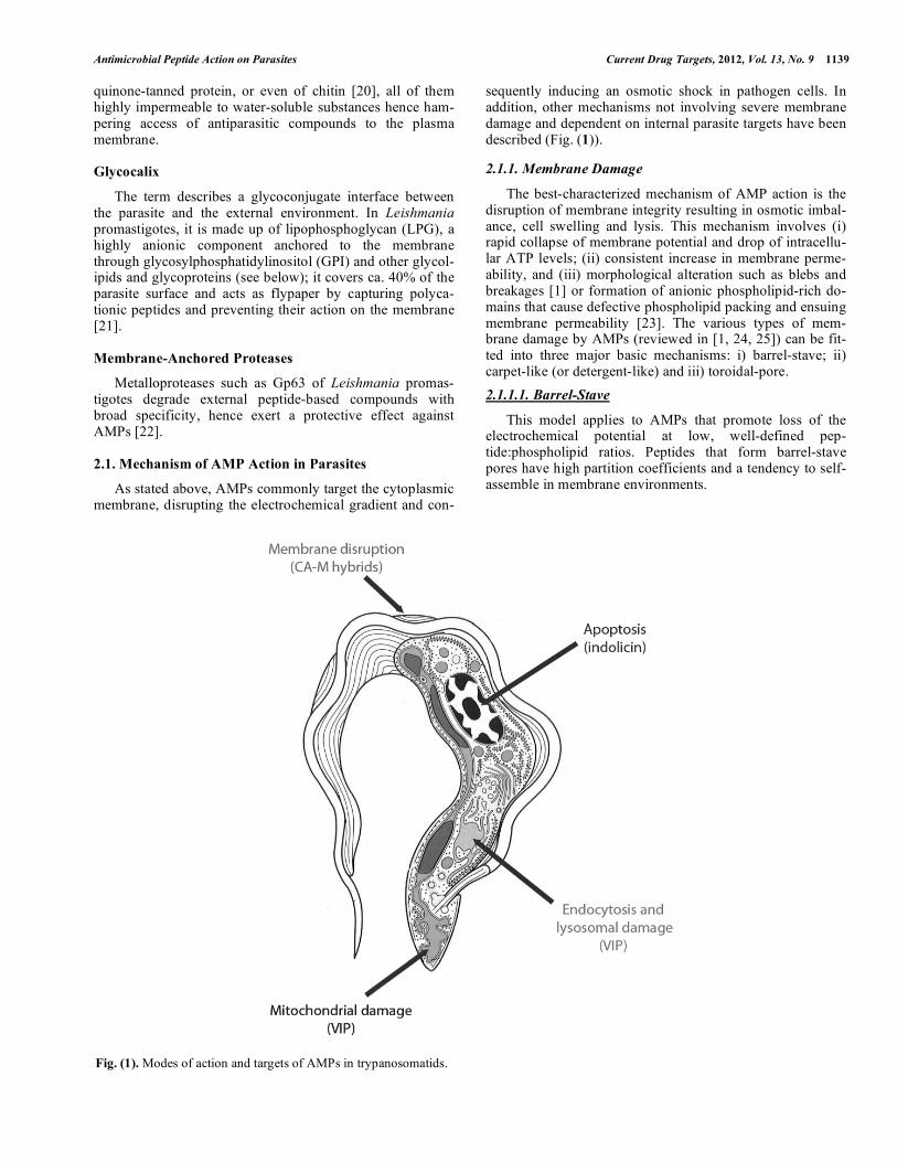
sequently inducing an osmotic shock in pathogen cells. In addition, other mechanisms not involving severe membrane damage and dependent on internal parasite targets have been described (Fig. (1)).
2.1.1. Membrane Damage
The best-characterized mechanism of AMP action is the disruption of membrane integrity resulting in osmotic imbal-ance, cell swelling and lysis. This mechanism involves (i) rapid collapse of membrane potential and drop of intracellu-lar ATP levels; (ii) consistent increase in membrane perme-ability, and (iii) morphological alteration such as blebs and breakages [1] or formation of anionic phospholipid-rich do-mains that cause defective phospholipid packing and ensuing membrane permeability [23]. The various types of mem-brane damage by AMPs (reviewed in [1, 24, 25]) can be fit-ted into three major basic mechanisms: i) barrel-stave; ii) carpet-like (or detergent-like) and iii) toroidal-pore.
2.1.1.1. Barrel-Stave
This model applies to AMPs that promote loss of the electrochemical potential at low, well-defined pep-tide:phospholipid ratios. Peptides that form barrel-stave pores have high partition coefficients and a tendency to self-assemble in membrane environments.
Fig. (1). Modes of action and targets of AMPs in trypanosomatids.
1140 Current Drug Targets, 2012, Vol. 13, No. 9 Torrent et al.
2.1.1.2. Carpet-Like Model
In this model, AMPs accumulate on the membrane sur-face by electrostatic interaction with anionic phospholipids; once a threshold concentration is reached, partial membrane solubilization in a detergent-like fashion takes place, favored by the amphipathic nature of the peptide.
2.1.1.3. Toroidal Pore or Two-State
In this intermediate model, AMPs accumulate parallel to the membrane surface, with partial insertion causing expan-sion of the outer but not the inner leaflet, with ensuing me-chanical stress. Once a critical threshold is reached, tension is relieved by reorientation of the peptides perpendicular to the bilayer, which undergoes a positive curvature and modi-fies its thickness, creating transient pores made of both pep-tide and lipid molecules that in turn catalyze phospholipid flip-flop hence promote further membrane asymmetry.
As examples of membrane-damage mechanisms exerted by AMPs on Protozoa, temporins A and B and cecropin A (CA)-melittin (M) analogs such as CA(1-8)M(1-18) are paradigmatic. The former are short (�13 residues) amphibian AMPs with only one cationic amino acid (lysine or arginine) [26] whereas CA-M analogs are longer and richer in cationic residues [27]. Both AMP types adopt amphipathic helical structures in hydrophobic media and permeabilize bacterial membranes and liposomes. However, their different size and cationicity bear upon their interaction with LPG. Thus, for temporin (low charge) antiparasitic activity is independent of the length of the LPG phosphoglycan moiety [26]: the pep-tide has similar activity against wild type L. donovani and its LPG-defective R2D2 mutant [28]. In contrast, for CA(1-8)M(1-18), with larger size and higher charge, the mutant strain is more susceptible to peptide action [29]. It would thus appear that the small size and low charge of the tem-porins favor diffusion across the glycocalix into the plasma membrane, while the stronger electrostatic interaction of CA(1-8)M(1-18) with the LPG matrix impairs membrane access and worsens activity.
Along with charge, positional hydrophobicity is an im-portant factor for the antiparasitic effects of magainins [30]. Accumulation of hydrophobic residues at a particular stretch of the sequence can promote aggregation; once the aggregate hits the membrane, the resulting high peptide concentration may cause local permeabilization. On the other hand, excess aggregation may result in micelle formation and lead to a complete loss of AMP efficacy.
While all above examples apply to AMPs targeting Leishmania, similar observations have been made for other trypanosomatids. Thus, cathelicidins such as SMAP-29, ovispirin and protegrin disrupt membrane integrity in Afri-can trypanosomes as well as in bacteria [31].
Different AMP antiparasitic activities may be displayed depending on the stage of the parasite targeted. Again in Leishmania, the amastigote is consistently more resistant to AMPs even when in axenic (macrophage-free) form; differ-ent parasite surface compositions most likely account for such differences. Similarly, the surface of bloodstream form of T. brucei contains high amounts of the variable surface glycoprotein, while the most abundant component of the
procyclic form is procyclin. While both proteins are GPI-anchored, differences in their structure [32] may translate into different AMP susceptibility [33].
Other membrane-based mechanisms, though not actually involving mechanical damage, have been observed. For ex-ample, the small hydrophobic peptides (SHP) derived from the signal sequences of human apolipoproteins [34] interca-late in the acyl chain region of the membrane of T. brucei and modify membrane fluidity. It is known that many mem-brane proteins are highly dependent on lipid fluidity for their activity; hence a severe perturbation of this parameter will lead to impaired protein functionality. Finally, other possible ways in which AMPs might interact with membranes include redistribution of membrane components such as sterols, for-mation of specific phospholipid microdomains, or even di-rect interaction with membrane proteins and alteration of their function.
2.1.2. Action on Internal Targets
Aside from membrane injury, some AMPs gain access into the cytoplasm without significant, persistent damage to the bilayer, causing instead parasite death by interaction with internal targets. This requires their translocation across the lipid bilayer, in a manner similar to cell-penetrating peptides (CPPs). Among the pathways proposed to explain how CPPs can cross pathogen membranes [35], spontaneous lipid-assisted translocation, closely similar to the two-state model (see 2.1.1) of AMP action, is prevalent. As discussed above, hydrophobic residues of peptides laying parallel to the bi-layer would perturb phospholipid packing, thereby inducing bilayer thinning and curvature, and partial peptide internali-zation. Next, peptides would adopt an orientation perpen-dicular to the membrane plane and form transient toroidal pores, thus favoring low-level peptide translocation to the inner leaflet of the membrane. Given the transient nature of these structures, some peptide molecules might dissociate from the membrane and eventually reach the cytoplasm.
Given that the boundaries between AMPs and CPPs are somewhat fuzzy [36], and that some CPPs have indeed been described to possess antimicrobial activities, it seems worthwhile to discuss some examples and their purported mechanism of action on intracellular targets (see Fig. (1)).
2.1.2.1. Bioenergetic Exhaustion
Histatin 5 (Hst5), a human salivary AMP [37] that pro-motes reversible depolarization of Leishmania plasma mem-branes, actually causes minimal membrane damage, suggest-ing that membranolysis plays a minor role, if any, in leishmanicidal action. The mitochondrion has been shown to be a major target for Hst5 in Leishmania [38], based on vari-ous evidences: i) Hst-5-treated parasites show extensive mi-tochondrial morphological alterations including a swollen matrix and poor cristae definition; ii) fluorescein-labeled Hst5 distributes largely in overlap with mitochondrial mark-ers, and Hst5 uptake is precluded in metabolically inhibited parasites; iii) respiration rate and mitochondrial electro-chemical potential decrease upon Hst5 incubation. The final outcome of the Hst5 attack is bioenergetic collapse of the parasite. Interestingly, the all-D Hst5 enantiomer has stronger activity than the natural peptide, possibly due to
Antimicrobial Peptide Action on Parasites Current Drug Targets, 2012, Vol. 13, No. 9 1141
higher buildup inside the parasite resulting from increased proteolytic resistance.
2.1.2.2. Endocytosis-Mediated Killing
Some cationic �-helical neuropeptides bind anionic gly-coproteins such as the variant surface glycoprotein (VSG) of African trypanosomes [39], upon which they are quickly endocytosed through the flagellar pocket, entering the main trafficking pathway of the parasite [40], eventually accessing the lysosome and disrupting its membrane, which releases hydrolases that cause parasite death.
2.1.2.3. Apoptosis-Mediated Killing
While AMPs killing by membrane disruption interact mainly with the parasite surface, setting off osmotic lysis as described before [31], those killing by apoptosis interact with intracellular organelles or proteins, usually by caspase-3/7-like activation. The precise apoptosis mechanism is unclear but may involve leakage of cytochrome c from mitochondria and activation of a caspase-9-like enzyme, which in turn activates caspase-3/7. Alternatively, activation of a caspase-8-like enzyme via a yet unknown death signal similar to the Fas/FADD system of mammalian cells, could also lead to caspase-3/7 activation [41].
3. ANTIMICROBIAL PEPTIDES AGAINST NE-
GLECTED TROPICAL DISEASES
Tables 1 and 2 show a representative list of AMPs target-ing Leishmania and Plasmodium, the two protozoan para-sites causing diseases with higher mortality and morbidity.
3.1. AMPs Against Leishmania
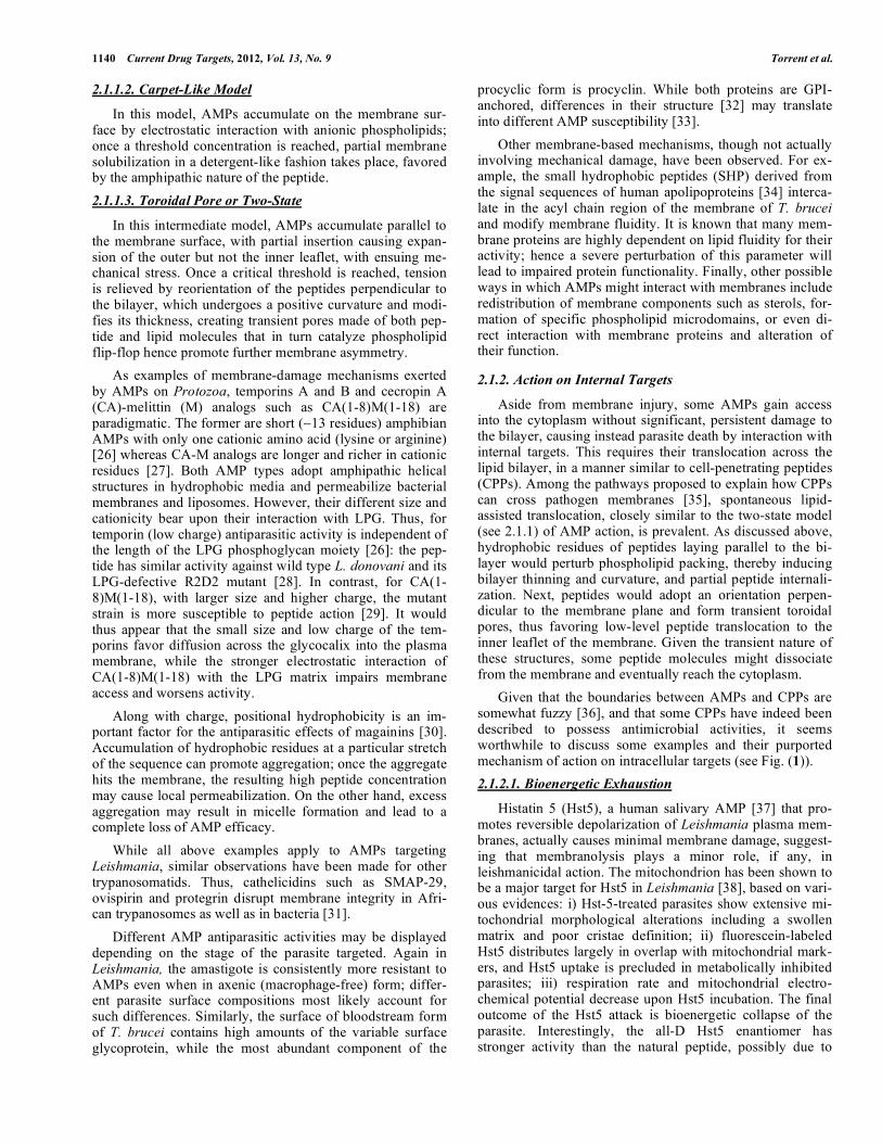
Leishmaniasis, a neglected tropical disease endemic in over 80 countries, affects 12 million people a year with more than 350 million at risk of infection. It is one of the most prevalent causes of death and animal morbidity in underde-veloped countries. Infection is caused by several species of the genus Leishmania, intracellular obligate parasites with a dimorphic life cycle (Fig. (2A)). The extracellular promas-tigote stage infects a sandfly vector and, after massive multi-plication in the gut of the insect, migrates into the salivary glands and is transmitted by the bite. In the mammalian host promastigotes are taken up by mononuclear phagocytes of the parasitophorus vacuole and transformed into amastigotes [42]. Current treatments against leishmaniasis are based on rather outdated, highly toxic drugs (e.g., antimonials) whose efficacy –with the possible exception of amphotericin B– is threatened by increasing resistance ([43]; WHO. WHO Ne-glected Tropical Diseases: htpp://www.who.int).
The discovery of AMPs opened new prospects for antim-icrobial agents (Table 1, Fig. (2A)). For instance, chimeric CA(1-8)M(1-18) [44] showed improved activity over the parental peptides, causing collapse of membrane potential and subsequent ATP loss [29]. Substantial efforts have been invested into designing more effective CA-M hybrids, with shortened sequences [45], lipidated N-termini [46], or N-methylated Lys residues [47]. The therapeutic potential of such CA-M analogues against canine leishmaniasis was evi-denced by the 80% reduction in parasitemia and transitory
clearance of disease symptoms achieved by octanoyl-CA(1-7)M(2-9) given intravenously to infected dogs [48].
Defensin-type antileishmanial peptides such as 18-residue gomesin from the spider Acanthoscurria gomesiana reduced to 50% the population of L. amazonensis promas-tigotes at micromolar concentration [49]. SD-1, a defensin expressed in the sandfly vector Phlebotomus duboscqi, showed only moderate in vitro activity against L. major promastigotes but was intriguingly active on bacteria, so it may enhance Leishmania parasite proliferation in infected sand flies by clearing other microorganisms [50].
Amphibian cutaneous secretions are one of the most abundant AMP reservoirs in nature, often with potent an-tileishmanial activities. For instance, 20-residue bombinins H2 and H4 from Bombina frogs inhibited L. donovani and L. pifanoi proliferation in a submicromolar range in both pro-mastigote and amastigote stages [51, 52]. Similarly, Phyl-lomedusa frog-derived peptides dermaseptin and phyl-loseptin displayed a characteristic biphasic killing of Leishmania amastigotes and promastigotes [53-55]. The ac-tivity of temporins, short peptides from the genus Rana, against Leishmania promastigotes and axenic amastigotes, has been mentioned above [26]. Particularly interesting is temporin-1Sa which, lacking cationic residues, is nonethe-less able to inhibit Leishmania proliferation at micromolar range [51, 56, 57].
Leishmanicidal AMPs from mamamalian sources have been more recently reported. Thus cathelicidins such as SMAP-29, with a conserved N-terminal cathelin-like domain and a C-terminal highly variable antimicrobial domain, in-hibit Leishmania at low or submicromolar concentration. Similar behavior has been described for other cathelicidins such as indolicin, myeloid AMP-18 and AMP-28, protegrin-1 and LL-37 [31, 50, 58-60].
Mammalian defensins are structurally classified into three families, named �-, ß- and �-. Both �- and ß-defensins adopt a triple-stranded antiparallel ß-sheet structure stabi-lized by three disulfide bridges whereas �-defensins display a cyclized peptide backbone, made by ligation of two identi-cal or similar nonapeptides and stabilized by three disulfide bridges [61]. Cryptdin-1 and -4, ß-defensin-1, -2 and -4 and �-defensin-II [22] have been reported to have leishmanicidal activities. Other human mammalian AMPs with leishm-anicidal activity include histatin-5 [38] and seminalplasmin [58].
Plants also represent an important source of antileishma-nial agents. Some examples are wheat thionin, barley lipid-transfer protein, or defensins and snakins from potato, all of them cysteine-rich peptides able to disrupt L. donovani pro-mastigotes and amastigotes without altering mitochondrial respiration [62, 63].
Leishmanicidal AMPs from marine organisms include (i) tachyplesin from the limulid Tachypleus tridentatus, active against L. braziliensis promastigotes and amastigotes [64] (ii) mytilin A from the mussel Mytilus edulis, with an-tileishmanial activity against L. braziliensis [64], (iii) dragomide E, a linear lipopeptide isolated from the cyano-bacteria Lyngbya majuscula with an antileishmanial activity against L. donovani promastigotes [65]; and (iv) the cy-

1142 Current Drug Targets, 2012, Vol. 13, No. 9 Torrent et al.
Table 1. Activity of Antimicrobial Peptides on Leishmania
AMP Leishmania spp. Parasite Stage Antiparasitic Activity References
Invertebrates
CA(1-8)M(1-18) L. donovani Promastigotes IC50 1-5 �M [29]
CA(1-7)M(2-9) L. donovani /L. pifanoi Axenic amastigotes IC50<1 �M [46]
Oct-CA(1-7)M(2-9) L. infantum Blood circulating amastigo-
tes IC50 <1 �M [48]
Gomesin L. amazonensis Promastigotes IC50 2.5 �M [49]
SD-1 L. amazonensis / L. major Promastigotes IC50 >50 �M [89]
Amphibians
Bombinin H2 and H4 L. donovani /L. pifanoi Promastigotes/axenic amasti-
gotes IC50 7/11 �M [26, 51, 52]
Dermaseptin-S1 and H-3 L. amazonensis / L. major Promastigotes/Amastigotes IC50 4.5/13.5 �M [53]
SPYY L. major Promastigotes/amastigotes MIC 5.9/6.2 �M [90]
Phylloseptin-1 L. amazonensis Promastigotes IC50 0.5 �M [91]
Temporins A and B L. infantum /L. mexicana Promastigotes/amastigotes IC50 12/50 �M [56]
Mammalians
Cathelicidins
Indolicin L.donovani
Myeloid AMP-18 L. major Promastigotes/amastigotes IC50 0.5/12.5 �M
Myeloid AMP-28 [31, 50, 58-60]
Protegrin-1
CRAMP
Defensins
Cryptdin-1 and -4
ß-defensin-1, -2 and 4 L. amazonensis/L, major Promastigotes/amastigotes IC50 20/50 �M [22]
�-defensin-II
Histatin 5 L. donovani Promastigotes/amastigotes IC50 7.3/14.2�M [38]
Seminal plasmin L. donovani Promastigotes IC50 <1 �M [58]
Plants
Wheat thionin L. donovani Promastigotes/amastigotes IC50 1/42 �M [62, 63]
Barley lipid transfer
Protein L.donovani IC50 > 50 �M
PTH-1
Potato snakin 1
Other
Tachyplesin L. braziliens Promastigotes/amastigotes IC50 6 -20 �M [64]
Mytilin A L. braziliens Promastigotes/amastigotes IC5 50 �M [64]
Dragomide E L. donovani Promastigotes IC50 5 �M [65]

Antimicrobial Peptide Action on Parasites Current Drug Targets, 2012, Vol. 13, No. 9 1143
Table 2. Activity of Antimicrobial Peptides on Plasmodium
AMP Plasmodium spp. Parasite Stage Antiparasitic Effect References
Invertebrates
CA(1-8)M(1-18) P. falciparum Erythrocytic stages IC50 10 �M [71]
Cecropin A and B
P. falciparum, P. berghei,
P. knowlesi and
P. cymonolgi
Oocyst and ookinete IC50 1.5 �M [72, 77]
Shiva 3 P. berghei Oocyst and ookinete MIC 100 �M [78]
Shiva 1 + anti-Pbs21 P. berghei Ookinete 90% inhibition at 10 �M [85]
Drosomycin P. berghei Ookinete IC50 10 �M [81]
Scorpine P. berghei Gametocyte 98% inhibition at 15 �M [79]
Meucine-24 and -25 P. berghei Ookinetes IC50 �10-20 �M [80]
Gomesin P. falciparum and berghei Gametocyte and oocyst 56% and 53% reduction at
50 �M [82]
Gambicin P. berghei Ookinetes 54-64% lethality at 10-100
�M [92]
Defensins P. gallinaceum All mosquito stages IC50 200 �M [83]
TP10 P. falciparum Gametocyte, zygote and
ookinete 35-45% reduction at 30 �M [84]
Amphibian
Magainin II P. falciparum, knowlesi and
cymonolgi
Oocyst, trophozoite and
schizont 90% inhibition at 100 �M [71, 72]
PGla P. falciparum Trophozoite and schizont IC50 40 �M [71]
Dermaseptin S3 and S4 P. falciparum Trophozoite IC50 1.4 �M [73]
K4K20-S4 and K4S4(1-13) P. falciparum Trophozoite IC50 0.2 -3.3 �M [75]
Mammalian
Angiotensin II and VC5 P. gallinaceum Sporozoites 88% and 76% reduction at
5-60 �M [69]
NK-2 P. falciparum Erythrocytic stages IC50 6.2 �M [70]
Other
CEL-III P. falciparum and berghei Oocyst and ookinete IC50 15 nM [85]
Tyrothricin P. falciparum Erythrocytic stages IC50 0.6 nM [86]
AdDLP P. berghei Ookinetes IC50 20 �M [80]
clodepsipeptide IB-012012 from the marine fungus Clonostachys [66].
4. AMPS AGAINST PLASMODIUM
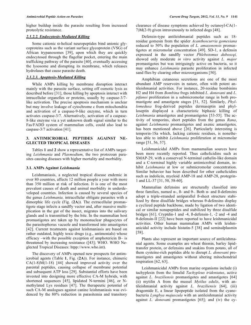
Malaria, with 225 million cases reported worldwide in 2009 (World Malaria Report summary. WHO, 5 November 2011) and 1,238,000 estimated deaths in 2010 [67], is caused by several species of Plasmodium parasites and transmitted by an invertebrate vector of the genus Anopheles. The para-site life cycle is complex, involving multiple developing
stages and locations both in mosquitos and humans. After a mosquito female ingests intracellular gametocytes from a human, these differentiate into gametes that, upon fertiliza-tion and via a diploid zygote, give rise to a motile ookinete that infects the midgut epithelia of the mosquito and differ-entiates into an oocyst. After replication, oocysts release large numbers of sporozoites that invade the human host upon the mosquito bite and reach the bloodstream by trans-migration through the dermis. Once in circulation, sporozoi-tes reach liver hepatocytes in whose parasitophorous vacuole they multiply and eventually invade erythrocytes, subse-
1144 Current Drug Targets, 2012, Vol. 13, No. 9 Torrent et al.
Fig. (2). The activity of some AMPs on specific stages of the life cycles of (A) Leishmania and (B) Plasmodium. Adapted (A and B, respec-
tively) from http://www.cdc.gov/malaria/about/biology/index.htmland http://www.cdc.gov/parasites/leishmaniasis/biology.html.
Antimicrobial Peptide Action on Parasites Current Drug Targets, 2012, Vol. 13, No. 9 1145
quently differentiating into trophozoites. Through an asexual process of schizogony these yield thousands of trophozoites that can further differentiate into gametocytes and close the cycle [68] (Fig. (2B)).
Finding a universal drug against malaria has proven a challenging task. Nonetheless, its complex infectious cycle offers multiple sites where specific drugs, including AMPs, can act (Table 2, Fig. (2B)). AMPs such as angiotensin II and its related vanicere-5 peptide can target disease trans-mission by selectively disrupting the cell membrane of P. gallinaceum sporozoites. Parasite load on salivary glands of infected mosquitoes is reduced by 75% when treated with these AMPs, with a dramatic decrease in infectivity in the vertebrate host [69]. Erythrocyte stages, composed of infec-tive trophozoites and replicative schizonts responsible for human pathology, are also possible AMP targets. Thus, NK-lysin–derived NK-2 peptide permeabilizes infected cells –selectively exposing phosphatidylserine residues– and drasti-cally reduces P. falciparum viability while almost totally sparing non-infected –phosphatidylserine non-exposing– cells [70]. CA-M hybrids are also active on P. falciparum-infected erythrocytes at low culture [71].
Amphibian AMPs have also been reported as good anti-malarial agents. For example, magainin II and its amidated homolog promote oocyst degeneration in P. falciparum, P. cymonolgi and P. knowlesi infections. In addition, magainin II inhibits reinfection of erythrocytes by P. falciparum tro-phozoites and schizonts at 100 �M concentration [71, 72] and PGLa, another amphibian peptide, also displays anti-parasitic activity against P. falciparum [71]. Dermaseptins S3 and S4 kill P. falciparum trophozoites although their se-lectivity between parasites and erythrocytes is not as high as for other peptides [73, 74]. Optimization of S4 has led to the 28-mer K4K20-S4 or the short K4S4(1-13) analog with en-hanced activity towards infected red blood cells [75, 76].
AMPs can also target mosquito stages. Thus, cecropins A and B are active on ookinetes and oocysts of P. falciparum, P. berghei, P. knowlesi and P. cymonolgi [72, 77], and Shiva 3, a CA-M derivative, is harmful to P. berghei ookinetes and oocytes in the mosquito midgut after a few minutes of expo-sure [78].
Venom peptides are also an important source for antima-larial AMPs. Scorpine, similar to cecropin and isolated from the tarantula Pandinus imperator, inhibits proliferation of P. berghei gametocytes [79]; meucine-24, an �-helical peptide with an N-terminal homologous to melittin, and meucine-25, a singular peptide devoid of similarity with any other, have been isolated from the venom of of the scorpion Mesobuthus eupeus and shown to inhibit ookinete stages in P. berghei [80].
Several insect defensins also display antimalarial activity. Drosomycin, a 44 mer from Drosophila, inhibits the devel-opment of P. berghei ookinetes at micromolar concentrations [81], and gomesin, an insect defensin, causes a dramatic re-duction of gametocyte exflagelation and oocyst population in both P. falciparum and P. berghei [82]. Other insect defens-ins from dragon fly and flesh fly reduce gametocyte viability in mosquitos infected by P. gallinaceum [83]. Finally, TP10, a CPP isolated from wasp venom, is very effective against zygote/ookinete stage of P. falciparum [84].
Other miscellaneous examples of antimalarial AMPs are CEL-III from sea cucumber, active at 15 nM against P. ber-ghei ookinetes and oocysts, but with only moderate inhibi-tion of P. falciparum [85]. Tyrocidins, a heterologous family of cyclic decapeptides from Bacillus brevis, showed a high percentage of lysis for the bloodstages of P. falciparum [86].
Finally, de novo peptides mimicking the structure and/or function of native counterparts were tested against Plasmo-dium. Thus, oligoacyl-lysyls (OAKs), with an alternance of cationic residues an acylamino chains recreating an amphi-patic pattern, showed both in vivo and in vitro antimalarial activity with low hemolytic activity [87]. Synthetic ana-logues of the LAH4 peptide showed high (>100) selectivity index high potency against P. falciparum [88]. Other syn-thetic peptides such as Vida 1-3 have been designed to target ookinete development of P. berghei and P. yoelii nigeriensis, thus preventing oocyst infection in Anopheles gambiae [84].
5. CONCLUSIONS AND FUTURE DIRECTIONS
As extensively described in this review, there are multi-ple protozoan-targeting AMPs that can be pharmaceutically attractive to fight parasitic diseases. It is, however, difficult to predict if AMPs will stimulate future therapeutic strategies against diseases such as malaria or leishmaniasis. At present, an obvious shortcoming of AMP therapy is the higher pro-duction cost of synthetic peptides over conventional organic molecule drugs. However, computational and experimental strategies aimed at size reduction, proteolytic stabilization and other druggability issues are likely to cut prices down in the near future. Even so, mechanisms of action typically re-quiring concentrations in the �M range will continue to bur-den AMP-based therapies vs. often nanomolar-active classi-cal drugs. The fact that parasitic diseases, despite their high prevalence, usually affect low-income communities also bears on the issue, as returns on pharma investment are un-likely to be plentiful. This lack of financial incentives might hopefully be corrected by governmental or not-for-profit interventions targeting neglected diseases at a global level, in a context where AMPs as antiparasitic agents receive ade-quate attention.
6. CONFLICT OF INTEREST
The authors confirm that this article content has no con-flict of interest.
7. ACKNOWLEDGEMENTS
Supported by the European Union (HEALTH-2007-223414 to L.R. and D.A.), Fondo de Investigaciones Sani-tarias (RICET RD 06/0021/0006 and PI09-01928 to L.R.; RD 06/0021/0010 to M.N.), MINECO (SAF2011-24899 to D.A.) and Generalitat de Catalunya (SGR09-00492 to D.A.). M.T. and D.P. are recipients post- and FPU predoctoral fel-lowships fom Alianza Cuatro Universidades and MICINN, respectively.
REFERENCES
[1] Rivas L, Luque-Ortega JR, Andreu D. Amphibian antimicrobial peptides and Protozoa: lessons from parasites. Biochim Biophys Acta 2009; 1788: 1570-81.
1146 Current Drug Targets, 2012, Vol. 13, No. 9 Torrent et al.
[2] Kolev NG, Tschudi C, Ullu E. RNA interference in protozoan parasites: achievements and challenges. Eukaryot Cell 2011; 10: 1156-63.
[3] Jenssen H, Hamill P, Hancock RE. Peptide antimicrobial agents. Clin Microbiol Rev 2006; 19: 491-511.
[4] Afacan NJ, Yeung AT, Pena OM, Hancock RE. Therapeutic Potential of Host Defense Peptides in Antibiotic-resistant Infections. Curr Pharm Des 2012; 18: 807-19.
[5] Otvos L, Jr. Antibacterial peptides and proteins with multiple cellular targets. J Pept Sci 2005; 11: 697-706.
[6] Giuliani A, Pirri G, Bozzi A, Di Giulio A, Aschi M, Rinaldi AC. Antimicrobial peptides: natural templates for synthetic membrane-active compounds. Cell Mol Life Sci 2008; 65: 2450-60.
[7] Kraus D, Peschel A. Molecular mechanisms of bacterial resistance to antimicrobial peptides. Curr Top Microbiol Immunol 2006; 306: 231-50.
[8] Gordon YJ, Romanowski EG, McDermott AM. A review of antimicrobial peptides and their therapeutic potential as anti-infective drugs. Curr Eye Res 2005; 30: 505-15.
[9] Nagaoka I, Hirota S, Yomogida S, Ohwada A, Hirata M. Synergistic actions of antibacterial neutrophil defensins and cathelicidins. Inflamm Res 2000; 49: 73-9.
[10] Torrent M, Nogues VM, Boix E. A theoretical approach to spot active regions in antimicrobial proteins. BMC Bioinformatics 2009; 10: 373.
[11] Torrent M, Di Tommaso P, Pulido D, et al. AMPA: an automated web server for prediction of protein antimicrobial regions. Bioinformatics 2012; 28: 130-1.
[12] Fjell CD, Hiss JA, Hancock RE, Schneider G. Designing antimicrobial peptides: form follows function. Nat Rev Drug Discov 2012; 11: 37-51.
[13] Lata S, Sharma BK, Raghava GP. Analysis and prediction of antibacterial peptides. BMC Bioinformatics 2007; 8: 263.
[14] Hilpert K, Volkmer-Engert R, Walter T, Hancock RE. High-throughput generation of small antibacterial peptides with improved activity. Nat Biotechnol 2005; 23: 1008-12.
[15] Wang Z, Wang G. APD: the Antimicrobial Peptide Database. Nucleic Acids Res 2004; 32: D590-592.
[16] Wanderley JL, Moreira ME, Benjamin A, Bonomo AC, Barcinski MA. Mimicry of apoptotic cells by exposing phosphatidylserine participates in the establishment of amastigotes of Leishmania (L) amazonensis in mammalian hosts. J Immunol 2006; 176: 1834-9.
[17] Mason AJ, Marquette A, Bechinger B. Zwitterionic phospholipids and sterols modulate antimicrobial peptide-induced membrane destabilization. Biophys J 2007; 93: 4289-99.
[18] Liu Y, Kuhlenschmidt MS, Kuhlenschmidt TB, Nguyen TH. Composition and conformation of Cryptosporidium parvum oocyst wall surface macromolecules and their effect on adhesion kinetics of oocysts on quartz surface. Biomacromolecules 2010; 11: 2109-15.
[19] Templeton TJ, Lancto CA, Vigdorovich V, et al. The Cryptosporidium oocyst wall protein is a member of a multigene family and has a homolog in Toxoplasma. Infect Immun 2004; 72: 980-7.
[20] Monne LH, G. On the properties of the shells of Coccidian oocysts. Arkives on Zoology 1954; 7: 251-6.
[21] Homans SW, Mehlert A, Turco SJ. Solution structure of the lipophosphoglycan of Leishmania donovani. Biochemistry 1992; 31: 654-61.
[22] Kulkarni MM, McMaster WR, Kamysz E, Kamysz W, Engman DM, McGwire BS. The major surface-metalloprotease of the parasitic protozoan, Leishmania, protects against antimicrobial peptide-induced apoptotic killing. Mol Microbiol 2006; 62: 1484-97.
[23] Epand RM, Epand RF. Bacterial membrane lipids in the action of antimicrobial agents. J Pept Sci 2011; 17: 298-305.
[24] Chan DI, Prenner EJ, Vogel HJ. Tryptophan- and arginine-rich antimicrobial peptides: structures and mechanisms of action. Biochim Biophys Acta 2006; 1758: 1184-202.
[25] Papo N, Shai Y. Can we predict biological activity of antimicrobial peptides from their interactions with model phospholipid membranes? Peptides 2003; 24: 1693-703.
[26] Mangoni ML, Saugar JM, Dellisanti M, Barra D, Simmaco M, Rivas L. Temporins, small antimicrobial peptides with leishmanicidal activity. J Biol Chem 2005; 280: 984-90.
[27] Bechinger B. Structure and functions of channel-forming peptides: magainins, cecropins, melittin and alamethicin. J Membr Biol 1997; 156: 197-211.
[28] King DL, Turco SJ. A ricin agglutinin-resistant clone of Leishmania donovani deficient in lipophosphoglycan. Mol Biochem Parasitol 1988; 28: 285-93.
[29] Diaz-Achirica P, Ubach J, Guinea A, Andreu D, Rivas L. The plasma membrane of Leishmania donovani promastigotes is the main target for CA(1-8)M(1-18), a synthetic cecropin A-melittin hybrid peptide. Biochem J 1998; 330 (Pt 1): 453-60.
[30] Guerrero E, Saugar JM, Matsuzaki K, Rivas L. Role of positional hydrophobicity in the leishmanicidal activity of magainin 2. Antimicrob Agents Chemother 2004; 48: 2980-6.
[31] McGwire BS, Olson CL, Tack BF, Engman DM. Killing of African trypanosomes by antimicrobial peptides. J Infect Dis 2003; 188: 146-52.
[32] Cardoso de Almeida ML, Turner MJ. The membrane form of variant surface glycoproteins of Trypanosoma brucei. Nature 1983; 302: 349-52.
[33] Acosta-Serrano A, Cole RN, Mehlert A, Lee MG, Ferguson MA, Englund PT. The procyclin repertoire of Trypanosoma brucei. Identification and structural characterization of the Glu-Pro-rich polypeptides. J Biol Chem 1999; 274: 29763-71.
[34] Harrington JM, Widener J, Stephens N, et al. The plasma membrane of bloodstream-form African trypanosomes confers susceptibility and specificity to killing by hydrophobic peptides. J Biol Chem 2010; 285: 28659-66.
[35] Nicolas P. Multifunctional host defense peptides: intracellular-targeting antimicrobial peptides. FEBS J 2009; 276: 6483-96.
[36] Henriques ST, Melo MN, Castanho MA. Cell-penetrating peptides and antimicrobial peptides: how different are they? Biochem J 2006; 399: 1-7.
[37] Edgerton M, Koshlukova SE. Salivary histatin 5 and its similarities to the other antimicrobial proteins in human saliva. Adv Dent Res 2000; 14: 16-21.
[38] Luque-Ortega JR, van't Hof W, Veerman EC, Saugar JM, Rivas L. Human antimicrobial peptide histatin 5 is a cell-penetrating peptide targeting mitochondrial ATP synthesis in Leishmania. FASEB J 2008; 22: 1817-28.
[39] Gonzalez-Rey E, Ganea D, Delgado M. Neuropeptides: keeping the balance between pathogen immunity and immune tolerance. Curr Opin Pharmacol 2010; 10: 473-81.
[40] Delgado M, Anderson P, Garcia-Salcedo JA, Caro M, Gonzalez-Rey E. Neuropeptides kill African trypanosomes by targeting intracellular compartments and inducing autophagic-like cell death. Cell Death Differ 2009; 16: 406-16.
[41] Kulkarni MM, McMaster WR, Kamysz W, McGwire BS. Antimicrobial peptide-induced apoptotic death of leishmania results from calcium-de pend ent, caspase-independent mitochondrial toxicity. J Biol Chem 2009; 284: 15496-504.
[42] Mougneau E, Bihl F, Glaichenhaus N. Cell biology and immunology of Leishmania. Immunol Rev 2011; 240: 286-96.
[43] Stuart K, Brun R, Croft S, et al. Kinetoplastids: related protozoan pathogens, different diseases. J Clin Invest 2008; 118: 1301-10.
[44] Akuffo H, Hultmark D, Engstom A, Frohlich D, Kimbrell D. Drosophila antibacterial protein, cecropin A, differentially affects non-bacterial organisms such as Leishmania in a manner different from other amphipathic peptides. Int J Mol Med 1998; 1: 77-82.
[45] Luque-Ortega JR, Saugar JM, Chiva C, Andreu D, Rivas L. Identification of new leishmanicidal peptide lead structures by automated real-time monitoring of changes in intracellular ATP. Biochem J 2003; 375: 221-30.
[46] Chicharro C, Granata C, Lozano R, Andreu D, Rivas L. N-terminal fatty acid substitution increases the leishmanicidal activity of CA(1-7)M(2-9), a cecropin-melittin hybrid peptide. Antimicrob Agents Chemother 2001; 45: 2441-9.
[47] Fernandez-Reyes M, Diaz D, de la Torre BG, et al. Lysine N(epsilon)-trimethylation, a tool for improving the selectivity of antimicrobial peptides. J Med Chem 2010; 53: 5587-96.
[48] Alberola J, Rodriguez A, Francino O, Roura X, Rivas L, Andreu D. Safety and efficacy of antimicrobial peptides against naturally acquired leishmaniasis. Antimicrob Agents Chemother 2004; 48: 641-3.
[49] Silva PI, Jr., Daffre S, Bulet P. Isolation and characterization of gomesin, an 18-residue cysteine-rich defense peptide from the spider Acanthoscurria gomesiana hemocytes with sequence similarities to horseshoe crab antimicrobial peptides of the tachyplesin family. J Biol Chem 2000; 275: 33464-70.
[50] McGwire BS, Kulkarni MM. Interactions of antimicrobial peptides with Leishmania and trypanosomes and their functional role in host parasitism. Exp Parasitol 2010; 126: 397-405.
[51] Mangoni ML, Papo N, Saugar JM, et al. Effect of natural L- to D-amino acid conversion on the organization, membrane binding, and
Antimicrobial Peptide Action on Parasites Current Drug Targets, 2012, Vol. 13, No. 9 1147
biological function of the antimicrobial peptides bombinins H. Biochemistry 2006; 45: 4266-76.
[52] Mangoni ML, Marcellini HG, Simmaco M. Biological characterization and modes of action of temporins and bombinins H, multiple forms of short and mildly cationic anti-microbial peptides from amphibian skin. J Pept Sci 2007; 13: 603-13.
[53] Gaidukov L, Fish A, Mor A. Analysis of membrane-binding properties of dermaseptin analogues: relationships between binding and cytotoxicity. Biochemistry 2003; 42: 12866-74.
[54] Brand GD, Leite JR, Silva LP, et al. Dermaseptins from Phyllomedusa oreades and Phyllomedusa distincta. Anti-Trypanosoma cruzi activity without cytotoxicity to mammalian cells. J Biol Chem 2002; 277: 49332-40.
[55] Brand GD, Leite JR, de Sa Mandel SM, et al. Novel dermaseptins from Phyllomedusa hypochondrialis (Amphibia). Biochem Biophys Res Commun 2006; 347: 739-46.
[56] Abbassi F, Oury B, Blasco T, et al. Isolation, characterization and molecular cloning of new temporins from the skin of the North African ranid Pelophylax saharica. Peptides 2008; 29: 1526-33.
[57] Chadbourne FL, Raleigh C, Ali HZ, Denny PW, Cobb SL. Studies on the antileishmanial properties of the antimicrobial peptides temporin A, B and 1Sa. J Pept Sci 2011; 17: 751-5.
[58] Bera A, Singh S, Nagaraj R, Vaidya T. Induction of autophagic cell death in Leishmania donovani by antimicrobial peptides. Mol Biochem Parasitol 2003; 127: 23-35.
[59] Haines LR, Thomas JM, Jackson AM, et al. Killing of trypanosomatid parasites by a modified bovine host defense peptide, BMAP-18. PLoS Negl Trop Dis 2009; 3: e373.
[60] Lynn MA, Kindrachuk J, Marr AK, et al. Effect of BMAP-28 antimicrobial peptides on Leishmania major promastigote and amastigote growth: role of leishmanolysin in parasite survival. PLoS Negl Trop Dis 2011; 5: e1141.
[61] Wilmes M, Cammue BP, Sahl HG, Thevissen K. Antibiotic activities of host defense peptides: more to it than lipid bilayer perturbation. Nat Prod Rep 2011; 28: 1350-8.
[62] Castro MS, Fontes W. Plant defense and antimicrobial peptides. Protein Pept Lett 2005; 12: 13-8.
[63] Berrocal-Lobo M, Molina A, Rodriguez-Palenzuela P, Garcia-Olmedo F, Rivas L. Leishmania donovani: thionins, plant antimicrobial peptides with leishmanicidal activity. Exp Parasitol 2009; 122: 247-9.
[64] Lofgren SE, Miletti LC, Steindel M, Bachere E, Barracco MA. Trypanocidal and leishmanicidal activities of different antimicrobial peptides (AMPs) isolated from aquatic animals. Exp Parasitol 2008; 118: 197-202.
[65] Balunas MJ, Linington RG, Tidgewell K, et al. Dragonamide E, a modified linear lipopeptide from Lyngbya majuscula with antileishmanial activity. J Nat Prod 2010; 73: 60-6.
[66] Luque-Ortega JR, Cruz LJ, Albericio F, Rivas L. The antitumoral depsipeptide IB-01212 kills leishmania through an apoptosis-like process involving intracellular targets. Mol Pharm 2010 Aug 17. [Epub ahead of print].
[67] Murray CJ, Rosenfeld LC, Lim SS, et al. Global malaria mortality between 1980 and 2010: a systematic analysis. Lancet 2012; 379: 413-31.
[68] Hafalla JC, Silvie O, Matuschewski K. Cell biology and immunology of malaria. Immunol Rev 2011; 240: 297-316.
[69] Maciel C, de Oliveira Junior VX, Fazio MA, et al. Anti-plasmodium activity of angiotensin II and related synthetic peptides. PLoS One 2008; 3: e3296.
[70] Jacobs T, Bruhn H, Gaworski I, Fleischer B, Leippe M. NK-lysin and its shortened analog NK-2 exhibit potent activities against Trypanosoma cruzi. Antimicrob Agents Chemother 2003; 47: 607-13.
[71] Boman HG, Wade D, Boman IA, Wahlin B, Merrifield RB. Antibacterial and antimalarial properties of peptides that are cecropin-melittin hybrids. FEBS Lett 1989; 259: 103-6.
[72] Gwadz RW, Kaslow D, Lee JY, Maloy WL, Zasloff M, Miller LH. Effects of magainins and cecropins on the sporogonic development of malaria parasites in mosquitoes. Infect Immun 1989; 57: 2628-33.
[73] Ghosh JK, Shaool D, Guillaud P, et al. Selective cytotoxicity of dermaseptin S3 toward intraerythrocytic Plasmodium falciparum and the underlying molecular basis. J Biol Chem 1997; 272: 31609-16.
[74] Krugliak M, Feder R, Zolotarev VY, et al. Antimalarial activities of dermaseptin S4 derivatives. Antimicrob Agents Chemother 2000; 44: 2442-51.
[75] Dagan A, Efron L, Gaidukov L, Mor A, Ginsburg H. In vitro antiplasmodium effects of dermaseptin S4 derivatives. Antimicrob Agents Chemother 2002; 46: 1059-66.
[76] Efron L, Dagan A, Gaidukov L, Ginsburg H, Mor A. Direct interaction of dermaseptin S4 aminoheptanoyl derivative with intraerythrocytic malaria parasite leading to increased specific antiparasitic activity in culture. J Biol Chem 2002; 277: 24067-72.
[77] Kim W, Koo H, Richman AM, et al. Ectopic expression of a cecropin transgene in the human malaria vector mosquito Anopheles gambiae (Diptera: Culicidae): effects on susceptibility to Plasmodium. J Med Entomol 2004; 41: 447-55.
[78] Rodriguez MC, Zamudio F, Torres JA, Gonzalez-Ceron L, Possani LD, Rodriguez MH. Effect of a cecropin-like synthetic peptide (Shiva-3) on the sporogonic development of Plasmodium berghei. Exp Parasitol 1995; 80: 596-604.
[79] Carballar-Lejarazu R, Rodriguez MH, de la Cruz Hernandez-Hernandez F, et al. Recombinant scorpine: a multifunctional antimicrobial peptide with activity against different pathogens. Cell Mol Life Sci 2008; 65: 3081-92.
[80] Gao B, Xu J, Rodriguez Mdel C, et al. Characterization of two linear cationic antimalarial peptides in the scorpion Mesobuthus eupeus. Biochimie 2010; 92: 350-9.
[81] Tian C, Gao B, Rodriguez Mdel C, Lanz-Mendoza H, Ma B, Zhu S. Gene expression, antiparasitic activity, and functional evolution of the drosomycin family. Mol Immunol 2008; 45: 3909-16.
[82] Moreira CK, Rodrigues FG, Ghosh A, et al. Effect of the antimicrobial peptide gomesin against different life stages of Plasmodium spp. Exp Parasitol 2007; 116: 346-53.
[83] Shahabuddin M, Fields I, Bulet P, Hoffmann JA, Miller LH. Plasmodium gallinaceum: differential killing of some mosquito stages of the parasite by insect defensin. Exp Parasitol 1998; 89: 103-12.
[84] Arrighi RB, Nakamura C, Miyake J, Hurd H, Burgess JG. Design and activity of antimicrobial peptides against sporogonic-stage parasites causing murine malarias. Antimicrob Agents Chemother 2002; 46: 2104-10.
[85] Yoshida S, Shimada Y, Kondoh D, et al. Hemolytic C-type lectin CEL-III from sea cucumber expressed in transgenic mosquitoes impairs malaria parasite development. PLoS Pathog 2007; 3: e192.
[86] Rautenbach M, Vlok NM, Stander M, Hoppe HC. Inhibition of malaria parasite blood stages by tyrocidines, membrane-active cyclic peptide antibiotics from Bacillus brevis. Biochim Biophys Acta 2007; 1768: 1488-97.
[87] Radzishevsky I, Krugliak M, Ginsburg H, Mor A. Antiplasmodial activity of lauryl-lysine oligomers. Antimicrob Agents Chemother 2007; 51: 1753-9.
[88] Mason AJ, Moussaoui W, Abdelrahman T, et al. Structural determinants of antimicrobial and antiplasmodial activity and selectivity in histidine-rich amphipathic cationic peptides. J Biol Chem 2009; 284: 119-33.
[89] Boulanger N, Lowenberger C, Volf P, et al. Characterization of a defensin from the sand fly Phlebotomus duboscqi induced by challenge with bacteria or the protozoan parasite Leishmania major. Infect Immun 2004; 72: 7140-6.
[90] Vouldoukis I, Shai Y, Nicolas P, Mor A. Broad spectrum antibiotic activity of the skin-PYY. FEBS Lett 1996; 380: 237-40.
[91] Kuckelhaus SA, Leite JR, Muniz-Junqueira MI, Sampaio RN, Bloch C, Jr., Tosta CE. Antiplasmodial and antileishmanial activities of phylloseptin-1, an antimicrobial peptide from the skin secretion of Phyllomedusa azurea (Amphibia). Exp Parasitol 2009; 123: 11-6.
[92] Vizioli J, Bulet P, Hoffmann JA, Kafatos FC, Muller HM, Dimopoulos G. Gambicin: a novel immune responsive antimicrobial peptide from the malaria vector Anopheles gambiae. Proc Natl Acad Sci USA 2001; 98: 12630-5.
Received: March 12, 2012 Revised: April 13, 2012 Accepted: May 18, 2012
PMID: 22664071