LyeTx I, a potent antimicrobial peptide from the venom of the spider Lycosa erythrognatha
10
ORIGINAL ARTICLE LyeTx I, a potent antimicrobial peptide from the venom of the spider Lycosa erythrognatha D. M. Santos • R. M. Verly • D. Pilo ´-Veloso • M. de Maria • M. A. R. de Carvalho • P. S. Cisalpino • B. M. Soares • C. G. Diniz • L. M. Farias • D. F. F. Moreira • F. Fre ´zard • M. P. Bemquerer • A. M. C. Pimenta • M. E. de Lima Received: 19 September 2009 / Accepted: 29 October 2009 Ó Springer-Verlag 2009 Abstract LyeTx I, an antimicrobial peptide isolated from the venom of Lycosa erythrognatha, known as wolf spider, has been synthesised and its structural profile studied by using the CD and NMR techniques. LyeTx I has shown to be active against bacteria (Escherichia coli and Staphylo- coccus aureus) and fungi (Candida krusei and Crypto- coccus neoformans) and able to alter the permeabilisation of L-a-phosphatidylcholine-liposomes (POPC) in a dose- dependent manner. In POPC containing cholesterol or ergosterol, permeabilisation has either decreased about five times or remained unchanged, respectively. These results, along with the observed low haemolytic activity, indicated that antimicrobial membranes, rather than vertebrate membranes seem to be the preferential targets. However, the complexity of biological membranes compared to lip- osomes must be taken in account. Besides, other membrane components, such as proteins and even specific lipids, cannot be discarded to be important to the preferential action of the LyeTx I to the tested microorganisms. The secondary structure of LyeTx I shows a small random-coil region at the N-terminus followed by an a-helix that reached the amidated C-terminus, which might favour the peptide-membrane interaction. The high activity against bacteria together with the moderate activity against fungi A.M.C. Pimenta and M.E. de Lima have contributed equally to this work Electronic supplementary material The online version of this article (doi:10.1007/s00726-009-0385-x) contains supplementary material, which is available to authorized users. D. M. Santos Á D. F. F. Moreira Á A. M. C. Pimenta Á M. E. de Lima (&) Lab. Venenos e Toxinas Animais, Departamento de Bioquı ´mica e Imunologia, Instituto de Cie ˆncias Biolo ´gicas, Universidade Federal de Minas Gerais, Av. Anto ˆnio Carlos, 6627, 31270-901 Belo Horizonte, Minas Gerais, Brazil e-mail: [email protected]; [email protected] R. M. Verly Á D. Pilo ´-Veloso Departamento de Quı ´mica, ICEX, Universidade Federal de Minas Gerais, Av. Anto ˆnio Carlos, 6627, 31270-901 Belo Horizonte, Minas Gerais, Brazil M. de Maria Departamento de Zoologia, Instituto de Cie ˆncias Biolo ´gicas, Universidade Federal de Minas Gerais, Av. Anto ˆnio Carlos, 6627, 31270-901 Belo Horizonte, Minas Gerais, Brazil M. A. R. de Carvalho Á P. S. Cisalpino Á B. M. Soares Á L. M. Farias Departamento de Microbiologia, Instituto de Cie ˆncias Biolo ´gicas, Universidade Federal de Minas Gerais, Av. Anto ˆnio Carlos, 6627, 31270-901 Belo Horizonte, Minas Gerais, Brazil C. G. Diniz Departamento de Parasitologia, Microbiologia e Imunologia, ICB, Universidade Federal de Juiz de Fora, 36036-900 Juiz de Fora, Minas Gerais, Brazil F. Fre ´zard Departamento de Fisiologia e Biofı ´sica, Instituto de Cie ˆncias Biolo ´gicas, Universidade Federal de Minas Gerais, Av. Anto ˆnio Carlos, 6627, 31270-901 Belo Horizonte, Minas Gerais, Brazil M. P. Bemquerer Embrapa Recursos Gene ´ticos e Biotecnologia, PqEB, Av. W5 Norte (final), 70770-900 Brası ´lia, Distrito Federal, Brazil 123 Amino Acids DOI 10.1007/s00726-009-0385-x
Transcript of LyeTx I, a potent antimicrobial peptide from the venom of the spider Lycosa erythrognatha
ORIGINAL ARTICLE
LyeTx I, a potent antimicrobial peptide from the venomof the spider Lycosa erythrognatha
D. M. Santos • R. M. Verly • D. Pilo-Veloso • M. de Maria • M. A. R. de Carvalho •
P. S. Cisalpino • B. M. Soares • C. G. Diniz • L. M. Farias • D. F. F. Moreira •
F. Frezard • M. P. Bemquerer • A. M. C. Pimenta • M. E. de Lima
Received: 19 September 2009 / Accepted: 29 October 2009
� Springer-Verlag 2009
Abstract LyeTx I, an antimicrobial peptide isolated from
the venom of Lycosa erythrognatha, known as wolf spider,
has been synthesised and its structural profile studied by
using the CD and NMR techniques. LyeTx I has shown to
be active against bacteria (Escherichia coli and Staphylo-
coccus aureus) and fungi (Candida krusei and Crypto-
coccus neoformans) and able to alter the permeabilisation
of L-a-phosphatidylcholine-liposomes (POPC) in a dose-
dependent manner. In POPC containing cholesterol or
ergosterol, permeabilisation has either decreased about five
times or remained unchanged, respectively. These results,
along with the observed low haemolytic activity, indicated
that antimicrobial membranes, rather than vertebrate
membranes seem to be the preferential targets. However,
the complexity of biological membranes compared to lip-
osomes must be taken in account. Besides, other membrane
components, such as proteins and even specific lipids,
cannot be discarded to be important to the preferential
action of the LyeTx I to the tested microorganisms. The
secondary structure of LyeTx I shows a small random-coil
region at the N-terminus followed by an a-helix that
reached the amidated C-terminus, which might favour the
peptide-membrane interaction. The high activity against
bacteria together with the moderate activity against fungi
A.M.C. Pimenta and M.E. de Lima have contributed equally to this
work
Electronic supplementary material The online version of thisarticle (doi:10.1007/s00726-009-0385-x) contains supplementarymaterial, which is available to authorized users.
D. M. Santos � D. F. F. Moreira � A. M. C. Pimenta �M. E. de Lima (&)
Lab. Venenos e Toxinas Animais,
Departamento de Bioquımica e Imunologia,
Instituto de Ciencias Biologicas,
Universidade Federal de Minas Gerais,
Av. Antonio Carlos, 6627, 31270-901 Belo Horizonte,
Minas Gerais, Brazil
e-mail: [email protected]; [email protected]
R. M. Verly � D. Pilo-Veloso
Departamento de Quımica, ICEX,
Universidade Federal de Minas Gerais,
Av. Antonio Carlos, 6627, 31270-901 Belo Horizonte,
Minas Gerais, Brazil
M. de Maria
Departamento de Zoologia,
Instituto de Ciencias Biologicas,
Universidade Federal de Minas Gerais,
Av. Antonio Carlos, 6627, 31270-901 Belo Horizonte,
Minas Gerais, Brazil
M. A. R. de Carvalho � P. S. Cisalpino �B. M. Soares � L. M. Farias
Departamento de Microbiologia, Instituto de Ciencias
Biologicas, Universidade Federal de Minas Gerais,
Av. Antonio Carlos, 6627, 31270-901 Belo Horizonte,
Minas Gerais, Brazil
C. G. Diniz
Departamento de Parasitologia, Microbiologia e Imunologia,
ICB, Universidade Federal de Juiz de Fora,
36036-900 Juiz de Fora, Minas Gerais, Brazil
F. Frezard
Departamento de Fisiologia e Biofısica, Instituto de Ciencias
Biologicas, Universidade Federal de Minas Gerais,
Av. Antonio Carlos, 6627, 31270-901 Belo Horizonte,
Minas Gerais, Brazil
M. P. Bemquerer
Embrapa Recursos Geneticos e Biotecnologia, PqEB,
Av. W5 Norte (final), 70770-900 Brasılia,
Distrito Federal, Brazil
123
Amino Acids
DOI 10.1007/s00726-009-0385-x
and the low haemolytic activity have indicated LyeTx I as
a good prototype for developing new antibiotic peptides.
Keywords Lycosa erythrognatha �Antimicrobial peptide � LyeTx I � Spider venom
Introduction
Many antimicrobial peptides (AMPs) have been isolated
from a wide range of eukaryotic and prokaryotic organ-
isms (Kastin 2006) and may also occur as fragments of
proteins, such as Buforin II (Park et al. 1994). AMPs show
activity against a wide range of pathogens such as Gram-
positive and Gram-negative bacteria, fungi and viruses
(Hernandez-Ledesma et al. 2008; Kastin 2006; Zasloff
2002; Mor and Nicolas 1994). These peptides usually
show a high variability in their amino acid sequences,
spanning from 7 to 55 amino acid residues, as well as a
conservative net positive charge and an amphipathic sec-
ondary structure such as a-helix or b-sheet in lipid
membrane milieu.
The action of eukaryotic AMPs is mediated by peptide-
microbial cell membrane interactions resulting in mem-
brane permeation and cell lysis, rather than by receptors
(Zasloff 2002). The detailed mechanism of membrane
disruption is not well understood, although physico-
chemical data indicate that many peptides act as deter-
gents while others act by promoting the formation of
transient pores (Bechinger 2004; Verly et al. 2009).
Nevertheless, there is no consensus up to now as to
whether one or all of these models are correct. Further-
more, the AMP mechanism of action also depends upon a
peptide secondary structure and aggregation tendency, as
well as on the lipid composition of target membranes
(Sanderson 2005).
Antimicrobial peptides have been found in arthropod
venoms, mainly from spiders and scorpions (for reviewing
see Kuhn-Nentwig 2009). The first description of anti-
microbial activity in the venom of Lycosa singoriensis has
been published in 1989 (Xu et al. 1989). Lycotoxins I and
II, isolated from the wolf spider L. carolinensis venom
were also reported (Yan and Adams 1998). These pep-
tides display an amphipathic a-helix, typical of many very
active AMPs, and show activity against Gram-negative
bacteria (Escherichia coli) and yeast (Candida glabrata).
AMPs from L. singoriensis venom (Xu et al. 1989;
Budnik et al. 2004; Liu et al. 2009) with molecular
masses in the range of 2,000–3,000 Da have also been
isolated.
In this paper, we describe the primary and secondary
structure together with the biological activity of LyeTx I,
a new toxin from L. erythrognatha venom.
Material and methods
Spiders and venom collection and LyeTx I isolation
For the present study, spiders were collected in the regions
of Belo Horizonte and Santa Barbara (state of Minas
Gerais, Brazil) and their venoms were collected via electrical
stimulation of the fangs. Venoms were diluted in Milli-Q�
water, immediately frozen in liquid nitrogen and then
lyophilised.
Chromatographic purification of LyeTx I
Cation exchange chromatography
The lyophilised venom was dissolved in Milli-Q water and
loaded in a Cation exchange chromatography (CIEX) col-
umn (TSK gel CM-SW, 250 mm 9 4.6 mm, Tosoh)
equilibrated with solution A (10 mM sodium acetate buf-
fer, pH 5). A linear salt gradient was made by increasing
the solution B (10 mM sodium acetate buffer with 1 M
NaCl, pH 5). 18–118 min, gradient of 0–85% solution
B; 118–125 min, gradient of 85–100% solution B;
125–128 min 100% of solution B. The flow rate was set to
0.75 ml min-1 with detection at 214 nm.
Reversed-phase chromatography
The lyophilised fractions obtained from CIEX were dis-
solved in 0.1% aqueous trifluoroacetic acid (TFA) in Milli-
Q water and loaded in a reversed-phase chromatography
(RPC) column (SupelcosilTM C18, 25 cm 9 4.6 mm)
equilibrated with 0.1% aqueous TFA and eluted at a flow
rate of 5 ml min-1 with the following solutions: 0–5 min,
a gradient of 0–45% acetonitrile in 0.1% TFA in water;
5–40 min, a gradient of 45–60% acetonitrile in 0.1% TFA
in water; 40–45 min, gradient of 60–100% acetonitrile in
0.1% TFA in water; 45–55 min, 100% acetonitrile in 0.1%
TFA; 55–60 min, 100–0% acetonitrile with 0.1% TFA in
water. The flow rate was 1 ml min-1 with detection at
214 nm.
ESI-Q-TOF mass spectrometry analyses
ESI-Q-TOF mass spectrometry analyses were carried out by
using a Q-TOF Micro (Micromass, Manchester, UK)
equipped with an electrospray ionisation source operated in
positive ion mode, as previously described (Pimenta et al.
2005). Briefly, capillary voltage was 2.5–3.0 kV and sam-
ple cone voltages were 40–60 V. Mass spectrometer cali-
brations were made by using sodium iodide with caesium
iodide in 2,000 Da range. Samples were diluted in aqueous
(Milli-Q water) acetonitrile solution (50% by volume)
D. M. Santos et al.
123

containing 0.1% TFA and introduced by using a syringe
pump with flow rates of 5–10 ml min-1 in the electrospray
source. Data were analysed by MassLynx� 4.0 software.
MALDI-ToF/ToFMS analyses
MALDI-ToF/ToFMS analyses were performed using an
ABI 4700 proteomics analyser with ToF/ToF optics
(Applied Biosystems, USA), as previously described
(Prates et al. 2004). Briefly, samples diluted in acetic acid
(0.1%) were spotted onto a sample plate mixed with a
saturated solution of a-cyano-4-hydroxycinnamic acid
(CHCA) and allowed to dry at room temperature (dried-
droplet method). The MS and the MS/MS spectra were
acquired in the reflector mode with external calibration,
using the calibration mixture Sequazyme standard kit
(Applied Biosystems, USA). De novo peptide sequencing
was performed by precursor ion fragmentation using N2 as
CID gas with collision cell pressure kept at 2.8 9 10-6
Torr. Post-source decay spectra were obtained by increas-
ing the laser energy and turning off the collision gas, as
specified by the manufacturer.
Edman degradation
The purified peptide (1–5 nmol) was sequenced by Edman
degradation (PPSQ-21A protein sequencer, Shimadzu,
Tokyo, Japan) coupled to reversed-phase separation of the
PTH-amino acids on a WAKOSIL-PTH (4.6 mm 9
250 mm) column (Wako, Osaka, Japan).
Peptide synthesis and purification
The peptide was synthesised by stepwise solid-phase
using the N-9-fluorenylmethyloxycarbonyl (Fmoc) strat-
egy (Chan and White 2000) on a Rink amide resin
(0.68 mmol g-1). Side chain protecting groups were as
follows: t-butyl for threonine, t-butyloxycarbonyl for lysine
and tryptophan, (triphenyl)methyl for histidine, asparagine
and glutamine. Couplings were performed with 1,3-diiso-
propylcarbodiimide/1-hydroxybenzotriazole in N,N-dimethyl-
formamide (DMF) for 60–180 min. Deprotections (15 min,
twice) were conducted by piperidine:DMF (1:4; v:v).
Cleavage from the resin and final deprotection were per-
formed with TFA/thioanisole/water/1,2-ethanedithiol/trii-
sopropylsilane, 86.5/5.0/5.0/2.5/1.0, by volume) at room
temperature during 90 min. After precipitating the product
with cold diisopropyl ether, the crude peptide was extracted
with aqueous acetonitrile at 50% by volume, freeze-dried
and purified by RP-HPLC on a C18 semi-preparative col-
umn (Supelco, 5 lm, 250 mm 9 10 mm) equilibrated with
0.1% aqueous TFA and eluted by a linear gradient of
acetonitrile in 0.1% TFA (0–4 min, 0.1% TFA in water;
4–14 min, a gradient of 0–30% acetonitrile in 0.1% TFA in
water; 14–82 min, gradient of 30–52% acetonitrile con-
taining 0.1% TFA in water; 82–92 min, 52–100% aceto-
nitrile containing 0.1% TFA in water; 92–102 min, 100%
acetonitrile with 0.1% TFA. The flow was 3.0 ml min-1
and detection at 214 nm.
Antimicrobial tests
Reference bacteria strains, representative of Gram-positive
(Staphylococcus aureus ATCC 33591) and Gram-negative
(Escherichia coli ATCC 25922) bacteria, were cultured on
brain heart infusion. The peptide susceptibility patterns
were determined through the disk-diffusion method, based
on the recommendations of the Clinical and Laboratory
Standards Institute (CLSI 2007). Peptide stock solutions
were applied on 0.5 mm diameter sterile filter discs and
added to Muller-Hinton Agar plates (Difco Laboratories,
USA). Bacterial strains were inoculated from a 0.5
McFarland bacterial suspensions to get a confluent growth.
Inhibition growth zones were observed, measured and
recorded.
The antifungal susceptibility tests were performed by
the microdilution test according to CLSI (2002). The read-
outs were carried by determination of minimum inhibitory
concentrations (MIC) defined as a reduction of 100% in
growth after incubation at 35�C for 48 h (Candida krusei)
and 72 h (Cryptococcus neoformans). Fluconazole was
used as a control.
Haemolytic assay
Rabbit erythrocytes, suspended in a phosphate-buffered
saline (0.14 M NaCl; 2.7 mM KCl; 10 mM Na2HPO4,
1.8 mM KH2PO4, pH 7), were incubated with various
peptide concentrations for 1 h at 37�C. Erythrocytes were
then spun down and the released haemoglobin was mea-
sured spectrophotometrically at 405 nm. As a positive
control for 100% of erythrocyte lysis, an aqueous Triton X-
100 solution (1% by volume) was used instead of the
peptide solution.
Preparation of L-a-phosphatidylcholine-liposomes
(POPC) and leakage of calcein assay
Small unilamellar vesicles (SUVs) were prepared as
described previously (Frezard et al. 1994), through ultra-
sonication of multilamellar vesicles in the presence of
75 mM calcein at pH 7.2 and used within 48 h after
preparation. Non-encapsulated calcein was removed from
the calcein-containing liposomes through a Sephadex G-50
column equilibrated with 0.02 HEPES buffer containing
0.15 M NaCl, pH 7.2.
LyeTx I, a potent antimicrobial peptide
123
The peptide membrane-permeabilising activity was
determined at 37�C by calcein leakage from SUVs that was
monitored in HEPES buffer by fluorescence measurement
(Cary Eclipse spectrofluorimetry, Varian, Palo Alto, USA)
with excitation wavelength at 490 nm and emission
wavelength at 515 nm. The maximum fluorescence inten-
sity (100% leakage) was determined by adding 10 ll of an
aqueous Triton X-100 solution (1% by volume) to the
liposome sample (2.5 ml).
Circular dichroism spectroscopy
Secondary structure preferences of LyeTx I were investi-
gated at 298 K in the following milieu: aqueous media
(100 lM phosphate buffer solution, pH 7.0 and 100 lM
Tris–HCl buffer solution, pH 8.0), water/2,2,2-trifluoro-
ethanol (TFE) mixtures, dodecylphosphocholine (DPC)
micelles, 1-palmitoyl-2-oleoyl-sn-glycero-3-phosphocho-
line (POPC) and POPC/POPG (1-palmitoyl-2-oleoyl-sn-
glycero-3-phosphoglycerol), at 3:1(w:w) SUVs. The
equipment used was a Jasco-715 spectropolarimeter (Jasco,
Tokyo, Japan) with a 1.0-mm rectangular quartz cuvette
(Uvonic Instruments, Plainview, USA). Spectra were
recorded from 260 to 190 nm using a 1.0-nm spectral
bandwidth, 0.1 nm step resolution, 100 nm min-1 scan
rate and 4 s response time.
The peptide concentration, as determined from the
tryptophan molar absorptivity (e = 5,550 M-1 cm-1 at
280 nm), was at 10 lM in all CD studies. The obtained
spectra were analysed using the CDPro software.
NMR
The sample was prepared by dissolving the peptide at
2 mM in a solution of 400 mM DPCd38, 5% (by volume)
D2O and 10 lL 2,2-dimethyl-2-silapentane sulfonate and
pH was adjusted to 7.0 with 20 mM phosphate buffer.
The NMR experiments were conducted at 20�C on a
Bruker Avance DRX spectrometer [Centro Nacional de
Ressonancia Magnetica Nuclear Jiri Jonas (CNRMN), Rio
de Janeiro, Brazil] operating at 600,043 MHz for the 1H
frequency and using a triple-resonance (1H/13C/15N) gra-
dient probe (5 mm diameter)].
The Total Correlation Spectroscopy (TOCSY) spectra
were acquired by using the MLEV-17 pulse sequence.
The spectral width was determined as 6,900 Hz, and the
512 t1 increments were collected with eight transients of
4,096 points. NOESY spectra were acquired using mixing
times of 80, 100, 120, 140 and 160 ms. For this experi-
ment, the spectral width was 6,900 Hz and the 512 t1increments were collected with 16 transients of 4,096
points for each F1. 1H–13C HSQC spectra were acquired
with F1 and F2 spectral widths of 27,160 and 8,993 Hz
respectively. 400 t1 increments were collected with
56 transients of 1,024 points. The experiment was carried
out in an edited mode in such a way that CH and CH3
correlations could show a positive phase and CH2 corre-
lations, a negative phase. 1H–15N HSQC spectra were
acquired with F1 and F2 spectral widths of 27,160 and
8,993 Hz, respectively. Eighty t1 increments were col-
lected (with 400 transients of 1,024 points), for each free
induction decay. The NMR spectra were analysed using
NMRVIEW, version 5.0.3.
NOE data and structure calculations
The NMR spectra were analysed by using NMRVIEW,
version 5.0.3. NOE intensities obtained at 120 ms mixing
times were converted into semi-quantitative distance
restraints using the calibration by Hyberts et al. (1992).
The upper limits of the distance restraints thus obtained
were 2.8, 3.4 and 5.0 A´
for strong, medium and weak
NOEs, respectively. Structure calculations were performed
using the Xplor-NIH software, version 2.17.0. Starting
with the extended structure, 200 structures were generated
using a simulated annealing protocol. This was followed
by 18,000 steps of simulated annealing at 1,000 K and a
subsequent decrease in temperature in 9,000 steps in the
first slow-cool annealing stage. The display, analysis and
manipulation of the three-dimensional structures were
performed with the programme MOLMOL. The stereo-
chemical quality of the lowest energy structures was
analysed by using the PROCHECK-NMR software
(Laskowski et al. 1996).
Results and discussion
The crude venom of the spider L. erythrognatha was
fractionated by CIEX in HPLC and 16 fractions were
manually collected and vacuum-dried, as shown in Fig. 1a.
The main fraction (F12) was resuspended in distilled water
and purified by RPC and six major subfractions were
pooled (Fig. 1b). Subfraction 12.4 was analysed by mass
spectrometry and a pure peptide with m/z 2831.1 was
detected and named LyeTx I.
The primary structure of LyeTx I was determined up to
the 19th amino acid residue by automated Edman degra-
dation. Its molecular mass estimated from this partial
sequence indicated that five to seven amino acid residues
were still lacking. The sequence was completed by both
ESI-Q-TOF and MALDI-TOF-TOF mass spectrometry
analyses. The LyeTx I complete amino acid sequence was
identified as
D. M. Santos et al.
123

H-IWLTALKFLGKNLGKHLAKQQLAKL-NH2 and,
as compared to those of other AMPs, is highly similar
(84%) to the Lycotoxin I (Yan and Adams 1998) (Table 1).
In order to study the antimicrobial activity and further
explore its biological properties, LyeTx I was synthesised,
using the Fmoc solid-phase strategy, and purified by
reversed-phase HPLC (Fig. 2). Synthetic product identity
was confirmed by ESI-Q-TOF MS observed (M ? H?)
2831.9; calculated (M ? H?) 2831.7.
b
aA
bs 2
14nm
500
400
300
200
100
0
Abs
214n
m
%of N
aCl [1M
]
100
80
60
40
20
Time (min)
Time (min)
0
%ofA
CN
0 20 40 60
F12.4
Fig. 1 Purification of LyeTx I from spider venom by HPLC. a The
supernatant of the centrifuged crude venom was applied to cation
exchange HPLC (TSK gel CM-SW column, 4.6 mm 9 250 mm,
Tosoh), with a linear NaCl gradient up to 1 M. A linear salt gradient
was made by increasing the solution B (10 mM sodium acetate buffer
with 1 M NaCl, pH 5) 18–118 min, gradient of 0–85% solution B;
118–125 min, gradient of 85–100% solution B; 125–128 min 100%
of solution B. The flow rate was 0.75 ml min-1 and detection was at
214 nm. b The fraction F12 was re-purified by reversed-phase HPLC
in a C18 Supercosil column (4.6 mm 9 250 mm, Supelco) equili-
brated with 0.1% aqueous TFA followed by a linear gradient of
acetonitrile in 0.1% TFA. The elution was at a flow rate of
5 ml min-1 with the following solutions: 0–5 min, a gradient
of 0–45% acetonitrile in 0.1% TFA in water; 5–40 min, a gradient
of 45–60% acetonitrile in 0.1% TFA in water; 40–45 min, gradient of
60–100% acetonitrile in 0.1% TFA in water; 45–55 min, 100%
acetonitrile containing 0.1% TFA; 55–60 min, 100–0% acetonitrile
with 0.1% TFA in water. The flow was 0.75 ml min-1 and detection
was at 214 nm
Table 1 Amino acid sequences of antimicrobial toxins
Sequence alignment of LyeTx I against other amphipathic antimi-
crobial peptides. These sequences were aligned with Clustal W, using
a PAM 250 matrix. Sequences are from the following references:
Lycotoxin 1 and 2 (Yan and Adams 1998), Lycocitin (Budnik et al.
2004), Dermaseptin 4 (Mor and Nicolas 1994), Pleurocidin (Syvitski
et al. 2005), Moricin (Hemmi et al. 2002), Magainin 2 (Zasloff 1987),
Cecropin E (Boman and Hultmark 1987)
LyeTx I --IWLTALKFLGKNLGKHLAKQQLAKL---------------
Lycotoxin 1 --IWLTALKFLGKHAAKHLAKQQLSKL---------------
Lycocitin KIKWFKTMKSLAKFLAKEQMKKHLGE----------------
Lycotoxin 2 KIKWFKTMKSIAKFIAKEQMKKHLGGE---------------
Dermaseptin 4 -ALWMTLLKKVLKAAAKALNAVLVGANA--------------
Pleurocidin --GWGSFFKKAA-HVGKHVGKAALTHY--------------
Moricin AKIPIKAIKTVGKAVGKGLRAINIASTANDVFNFLKPKKRKA
Magainin 2 ---GIGKFLHSAKKFGKAFVGEIMNS----------------
Cecropin E RWKIFKKIEKVGQNIRDGIVKAGPAVAVVGQAATI-------
0 20 40 60 80 100
-100
0
100
200
300
400
500
600
700
Time (min.)
0
20
40
60
80
100
%A
CN
300 400 500 600 700 800 900 1000 1100 1200 1300 1400 1500 1600m/z0
100
%
A: 2831.93±0.01945.3132A4709.2372
A4708.9862
A5567.5920
A5567.3913
A6473.1611300.3029383.7386544.7667
567.7926
567.9946
568.1956
568.3947705.7356
709.4900A3
944.9809
709.7413
709.9929
710.2460
710.5015 944.6517770.2908
945.6501
945.9879
1417.4741946.3263
A21416.9812
946.6606946.9943
1027.0565 1416.5009
1418.4716
1418.9797
1419.48291501.9861
a
b
Abs
214n
m
Fig. 2 Purification of synthetic LyeTx I by HPLC. a Reversed-phase
profile of the synthetic crude product. Supelcosil column
(10 mm 9 250 mm C18 Supelco) equilibrated with 0.1% aqueous
TFA and eluted by a linear gradient of acetonitrile in 0.1% TFA. The
flow was 3.0 ml min-1 and detection was at 214 nm. b The fraction
F12.4 was analysed by ESI mass spectrometry. A molecular mass
was measured at 2831.93 Da, obtained by deconvolution of the
MS-spectra
LyeTx I, a potent antimicrobial peptide
123

Antimicrobial and haemolytic assays of the synthetic
peptide
The synthetic LyeTx I showed antimicrobial activities,
although a comparison between the MIC values reported
here and the results obtained by other authors with related
peptides is rather difficult as different assay conditions and
other strains of microorganisms are commonly employed.
LyeTx I was active against the Gram-positive bacteria
S. aureus (3.79 lM), the Gram-negative E. coli (7.81 lM)
and against the yeasts Candida krusei (26.30 lM) and
Cryptococcus neoformans (13.20 lM). Its antifungal
activity is thus lower than its antibacterial activity when
these organisms are taken into account. These data are
similar to those reported for the Lycotoxins isolated from
L. carolinensis and also for other AMPs (Yan and Adams
1998; Giacometti et al. 1999).
LyeTx I presented haemolytic activity (Fig. 3) in higher
concentrations (ED50 = 1.3 9 10-4 M), when compared
to values from antibacterial assays (i.e. in the micromolar
range). The high activity of LyeTx I against bacteria,
together with its moderate activity against yeasts and low
haemolytic activity have set LyeTx I as a good candidate
prototype for the development of new antibiotic peptides,
especially for treating skin and mucosa bacterial infections.
Chen et al. (2005) proposed that an increment in both
positive charge and amphipathicity of peptide is important
for bacterial lysis, while an increased hydrophobicity is
correlated with erythrocyte lysis. Martins et al. (2006),
working on peptides derived from Trialysin, a pore-form-
ing protein found in the salive of Triatoma infestans, the
insect vector of Chagas disease—proposed that the diver-
sified lytic activity of some peptides against selected tar-
gets may be associated with the presence of specific
structural details, while amphipathicity is essential for the
lytic activity. Additionally, these authors have suggested
that selectivity of active peptides for specific organisms
appears to be associated with the structural features of their
N-and C-termini (Martins et al. 2006). These structural
%he
mol
ysis
[M]
Fig. 3 Hemolytic activity of LyeTx I. Rabbit erythrocytes suspended
in PBS were incubated with increasing concentrations of synthetic
LyeTx I for 1 h. The haemoglobin released in presence of Triton X-
100 (1% by volume) was taken as 100% of cell lysis. Haemoglobin
released was measured at 405 nm. ED50 = 1.3 9 10-4 M
a
b
Rat
io o
f lys
isR
atio
of l
ysis
Time (Min)
phosphatidylcholinephosphatidylcholine:cholesterol 7:3phosphatidylcholine:ergosterol 7:3
[M]
Fig. 4 Leakage induced by LyeTx I of the fluorescent probe calcein
from liposomes. a Kinetics of leakage of calcein from liposomes
containing. L-a-phosphatidylcholine. LyeTx I at increasing concen-
trations (0.05, 0.1, 0.2, 0.4, 0.8, 1.6, 3.2, 6.4 and 12.8 lg ml-1) was
applied to the liposome suspension 6 min after incubation at 37�C.
b Curves of concentration-dependence of calcein permeabilisation by
LyeTx I in L-a-phosphatidylcholine liposomes, with or without
addition of cholesterol or ergosterol at 7:3 molar ratio. Fluorescence
emission was measured 2 min after incubation with the toxin. ED50
for phosphatidylcholine liposomes was calculated at 2.7 9 10-10 M,
for phosphatidylcholine/cholesterol (7:3, molar ratio) liposomes at
8.2 9 10-10 M and for phosphatidylcholine/ergosterol (7:3, molar
ratio) lipids at 2.5 9 10-10 M
D. M. Santos et al.
123
features should be explored in the case of LyeTx I, in
searching for analogues with higher specificity toward
microorganisms of medical interest.
LyeTx I induced permeabilisation of liposomes
Upon addition of LyeTx I (0.2 lg ml-1) to L-a-phospha-
tidylcholine liposomes (POPC), the entrapped self-quen-
ched calcein was immediately released, as it became
evident by the increase of fluorescence intensity (Fig. 4a).
The increments of fluorescence 2 min after adding the
peptide are shown in Fig. 4b. The kinetics of calcein
release was dose-dependent and that indicates a coopera-
tive behaviour (Takeuchi et al. 2004).
The presence of cholesterol in POPC resulted in a less
favourable insertion of AMPs into lipidic membranes,
mainly by reducing the membrane fluidity (Matsuzaki et al.
1995; Verly et al. 2009). We observed that liposomes
containing L-a-phosphatidylcholine:cholesterol (7:3 molar
ratio) were approximately five times more resistant to the
action of LyeTx I (Fig. 4b), when compared to those
constituted only by L-a-phosphatidylcholine or of L-a-
phosphatidylcholine:ergosterol (7:3 molar ratio).
Circular dichroism spectroscopy
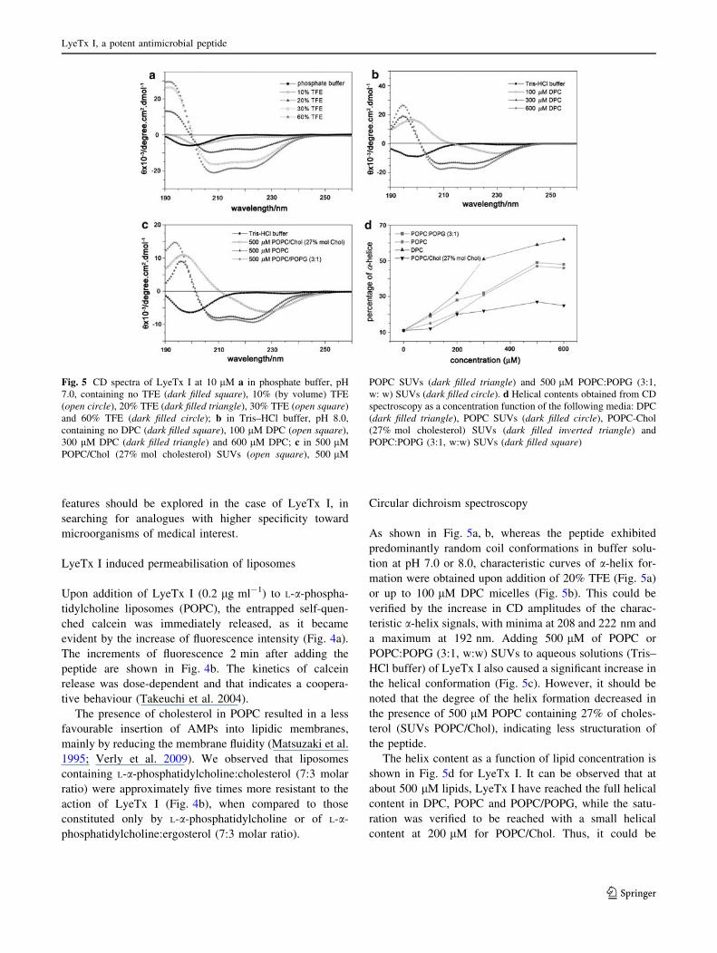
As shown in Fig. 5a, b, whereas the peptide exhibited
predominantly random coil conformations in buffer solu-
tion at pH 7.0 or 8.0, characteristic curves of a-helix for-
mation were obtained upon addition of 20% TFE (Fig. 5a)
or up to 100 lM DPC micelles (Fig. 5b). This could be
verified by the increase in CD amplitudes of the charac-
teristic a-helix signals, with minima at 208 and 222 nm and
a maximum at 192 nm. Adding 500 lM of POPC or
POPC:POPG (3:1, w:w) SUVs to aqueous solutions (Tris–
HCl buffer) of LyeTx I also caused a significant increase in
the helical conformation (Fig. 5c). However, it should be
noted that the degree of the helix formation decreased in
the presence of 500 lM POPC containing 27% of choles-
terol (SUVs POPC/Chol), indicating less structuration of
the peptide.
The helix content as a function of lipid concentration is
shown in Fig. 5d for LyeTx I. It can be observed that at
about 500 lM lipids, LyeTx I have reached the full helical
content in DPC, POPC and POPC/POPG, while the satu-
ration was verified to be reached with a small helical
content at 200 lM for POPC/Chol. Thus, it could be
Fig. 5 CD spectra of LyeTx I at 10 lM a in phosphate buffer, pH
7.0, containing no TFE (dark filled square), 10% (by volume) TFE
(open circle), 20% TFE (dark filled triangle), 30% TFE (open square)
and 60% TFE (dark filled circle); b in Tris–HCl buffer, pH 8.0,
containing no DPC (dark filled square), 100 lM DPC (open square),
300 lM DPC (dark filled triangle) and 600 lM DPC; c in 500 lM
POPC/Chol (27% mol cholesterol) SUVs (open square), 500 lM
POPC SUVs (dark filled triangle) and 500 lM POPC:POPG (3:1,
w: w) SUVs (dark filled circle). d Helical contents obtained from CD
spectroscopy as a concentration function of the following media: DPC
(dark filled triangle), POPC SUVs (dark filled circle), POPC-Chol
(27% mol cholesterol) SUVs (dark filled inverted triangle) and
POPC:POPG (3:1, w:w) SUVs (dark filled square)
LyeTx I, a potent antimicrobial peptide
123

concluded that the presence of cholesterol decreases the
peptide–lipid interaction.
NMR
The structure of LyeTx I was investigated by NMR spec-
troscopy in DPC, since CD studies indicate an elevated
a-helical content of the peptide in these micelles (Fig. 5).
Furthermore, the use of micelles is a better choice than
TFE for membrane interface simulation. Sequence-specific
chemical shift assignments were performed for LyeTx I
from the correlations observed in TOCSY and NOESY
spectra using standard procedures. The analysis of 1H–15N
and 1H–13C HSQC spectra allowed the unequivocal
assignment of all cross-peaks. A table showing 1H, 13C and15N chemical shifts is given on supplementary material
(SM-1). The fingerprint region of the TOCSY spectrum is
presented in Fig. 6a, while the correlation between the
amidic protons is shown in Fig. 6b. Other regions of
spectra are given on supplementary material: tocsy-
supplementary (SM-2), noesy-supplementary (SM-3), hsqc-1H15N-supplementary (SM-4), hsqc-1H13C-supplementary
(SM-5). The high number of correlations between the
amidic protons observed in the NOESY spectrum was
consistent with a significant structural stability of the
peptide in the micellar environment. Through-space and
through-bond correlations between different residues were
observed from the N-terminus up to the C-terminus
(Fig. 7). Medium range NOE cross peaks involving the
amidic protons were observed from the second residue up
to the C-terminus, indicating a great ordering degree of the
peptide structure. However, the NOESY spectrum exhibits
more correlations between protons close to the amidated
C-terminus than close to the unmodified N-terminus. This
behaviour was already observed for other C-terminal am-
idated AMP peptides (Verly et al. 2009) and, moreover,
those peptides often exhibited a higher antimicrobial
activity than their non-amidated counterparts (Sforca et al.
2004). A total of 370 NOEs was observed posing distance
restraints for estimating the twenty lowest energy struc-
tures for the LyeTx I molecule in the presence of DPC
micelles (Fig. 8a). These structures were conformed as
a slightly bent a-helix. Some observed inter-residue cor-
relations involving Ile-1, Trp-2 and Leu-3 indicated that
the N-terminus exhibited some structural order, albeit to
a lesser extent than that of the C-terminus, as may be seen
in Fig. 8a. Two correlations of the sort dN,N(i, i ? 2)
observed - involving the Phe-8 and Gly-10, and Asn-12 and
Gly-14 residues—are experimental evidences of the bend
in the structures.
Therefore, the LyeTx I NMR structure shows similarity
to that observed for the amphibian peptide DD K (Verly
et al. 2009): a small random-coil region in the N-terminus
followed by a helical segment that (for LyeTx I) is
extended from Leu-6 up to the C-terminus. Nevertheless,
LyeTx I structure shows a less amphipathic profile in the
Fig. 6 a NH–Ha region of the TOCSY spectrum of LyeTx I (at
2 mM) in 400 mM DPCd38 micellar solution in H2O:D2O (95:5, v:v).
b NH–HN and H–H aromatic side chain region of the NOESY
spectrum of LyeTx I (2 mM) in 400 mM DPCd38 micellar solution in
H2O:D2O (95:5, v:v)
Fig. 7 NOE connectivity diagram: Sequential NH(i)–NH(i ? 1),
Ha(i)–NH(i ? 1), Hb(i)–NH(i ? 1) and medium range NH(i)–NH(i ? 2), Ha(i)–NH(i ? n), Ha(i)–Hb(i ? n)
D. M. Santos et al.
123
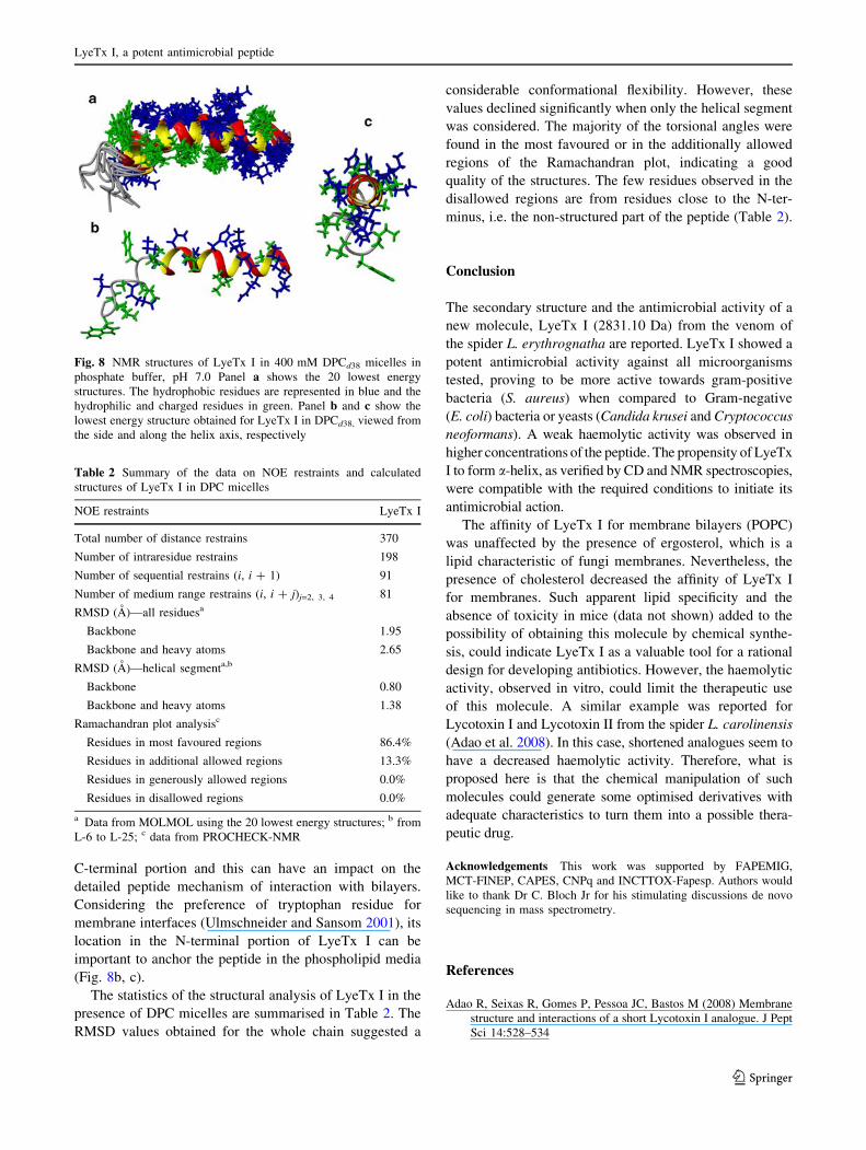
C-terminal portion and this can have an impact on the
detailed peptide mechanism of interaction with bilayers.
Considering the preference of tryptophan residue for
membrane interfaces (Ulmschneider and Sansom 2001), its
location in the N-terminal portion of LyeTx I can be
important to anchor the peptide in the phospholipid media
(Fig. 8b, c).
The statistics of the structural analysis of LyeTx I in the
presence of DPC micelles are summarised in Table 2. The
RMSD values obtained for the whole chain suggested a
considerable conformational flexibility. However, these
values declined significantly when only the helical segment
was considered. The majority of the torsional angles were
found in the most favoured or in the additionally allowed
regions of the Ramachandran plot, indicating a good
quality of the structures. The few residues observed in the
disallowed regions are from residues close to the N-ter-
minus, i.e. the non-structured part of the peptide (Table 2).
Conclusion
The secondary structure and the antimicrobial activity of a
new molecule, LyeTx I (2831.10 Da) from the venom of
the spider L. erythrognatha are reported. LyeTx I showed a
potent antimicrobial activity against all microorganisms
tested, proving to be more active towards gram-positive
bacteria (S. aureus) when compared to Gram-negative
(E. coli) bacteria or yeasts (Candida krusei and Cryptococcus
neoformans). A weak haemolytic activity was observed in
higher concentrations of the peptide. The propensity of LyeTx
I to form a-helix, as verified by CD and NMR spectroscopies,
were compatible with the required conditions to initiate its
antimicrobial action.
The affinity of LyeTx I for membrane bilayers (POPC)
was unaffected by the presence of ergosterol, which is a
lipid characteristic of fungi membranes. Nevertheless, the
presence of cholesterol decreased the affinity of LyeTx I
for membranes. Such apparent lipid specificity and the
absence of toxicity in mice (data not shown) added to the
possibility of obtaining this molecule by chemical synthe-
sis, could indicate LyeTx I as a valuable tool for a rational
design for developing antibiotics. However, the haemolytic
activity, observed in vitro, could limit the therapeutic use
of this molecule. A similar example was reported for
Lycotoxin I and Lycotoxin II from the spider L. carolinensis
(Adao et al. 2008). In this case, shortened analogues seem to
have a decreased haemolytic activity. Therefore, what is
proposed here is that the chemical manipulation of such
molecules could generate some optimised derivatives with
adequate characteristics to turn them into a possible thera-
peutic drug.
Acknowledgements This work was supported by FAPEMIG,
MCT-FINEP, CAPES, CNPq and INCTTOX-Fapesp. Authors would
like to thank Dr C. Bloch Jr for his stimulating discussions de novo
sequencing in mass spectrometry.
References
Adao R, Seixas R, Gomes P, Pessoa JC, Bastos M (2008) Membrane
structure and interactions of a short Lycotoxin I analogue. J Pept
Sci 14:528–534
Fig. 8 NMR structures of LyeTx I in 400 mM DPCd38 micelles in
phosphate buffer, pH 7.0 Panel a shows the 20 lowest energy
structures. The hydrophobic residues are represented in blue and the
hydrophilic and charged residues in green. Panel b and c show the
lowest energy structure obtained for LyeTx I in DPCd38, viewed from
the side and along the helix axis, respectively
Table 2 Summary of the data on NOE restraints and calculated
structures of LyeTx I in DPC micelles
NOE restraints LyeTx I
Total number of distance restrains 370
Number of intraresidue restrains 198
Number of sequential restrains (i, i ? 1) 91
Number of medium range restrains (i, i ? j)j=2, 3, 4 81
RMSD (A)—all residuesa
Backbone 1.95
Backbone and heavy atoms 2.65
RMSD (A)—helical segmenta,b
Backbone 0.80
Backbone and heavy atoms 1.38
Ramachandran plot analysisc
Residues in most favoured regions 86.4%
Residues in additional allowed regions 13.3%
Residues in generously allowed regions 0.0%
Residues in disallowed regions 0.0%
a Data from MOLMOL using the 20 lowest energy structures; b from
L-6 to L-25; c data from PROCHECK-NMR
LyeTx I, a potent antimicrobial peptide
123

Bechinger B (2004) Membrane-lytic peptides. Crit Rev Plant Sci
23:271–292
Boman HG, Hultmark D (1987) Cell-free immunity in insects. Annu
Rev Microbiol 41:103–126
Budnik BA, Olsen JV, Egorov TA, Anisimova VE, Galkina TG,
Musolyamov AK, Grishin EV, Zubarev RA (2004) De novo
sequencing of antimicrobial peptides isolated from the venom
glands of the wolf spider Lycosa singoriensis. J Mass Spectrom
39:193–201
Chan WC, White PD (2000) Fmoc solid phase peptide synthesis:
a practical approach. Oxford University Press, Oxford
Chen Y, Mant CT, Farmer SW, Hancock RE, Vasil ML, Hodges RS
(2005) Rational design of a-helical antimicrobial peptides with
enhanced activities and specificity/therapeutic index. J Biol
Chem 280:12316–12329
Clinical and Laboratory Standards Institute (2002) Reference method
for broth dilution antifungal susceptibility testing of yeasts;
Approved Standard (M27-A2), 2nd edn. Clinical and Laboratory
Standards Institute, Wayne
Clinical and Laboratory Standards Institute (2007) Performance
standards for antimicrobial susceptibility testing; seventeenth
informational supplement. CLSI document M100-S17. Clinical
and Laboratory Standards Institute, Wayne
Frezard F, Santaella C, Vierling P, Riess JG (1994) Permeability and
stability in buffer and in human serum of fluorinated phospho-
lipid-based liposomes. Biochim Biophys Acta 1192:61–70
Giacometti A, Cirioni O, Barchiesi F, Del Prete MS, Scalise G (1999)
Antimicrobial activity of polycationic peptides. Peptides
20:1265–1273
Hemmi H, Ishibashi J, Hara S, Yamakawa M (2002) Solution
structure of moricin, an antibacterial peptide, isolated from the
silkworm Bombyx mori. FEBS Lett 518:33–38
Hernandez-Ledesma B, Recio I, Amigo R (2008) b-Lactoglobulin as
source of bioactive peptides. Amino Acids 35:257–265
Hyberts SG, Goldberg MS, Havel TF, Wagner G (1992) The solution
structure of eglin C based on measurements of many NOEs and
coupling constants and its comparison with X-Ray structures.
Protein Sci 1:136–151
Kastin AJ (2006) Handbook of biologically active peptides. Aca-
demic, Amsterdam
Kuhn-Nentwig L (2009) Cytolytic and antimicrobial peptides in the
venom of scorpions and spiders. In: De Lima ME, Pimenta
AMC, Martin-Eauclaire MF, Zingali R, Rochat H(eds) Animal
toxins: state of the art. Perspectives in health and biotechnology,
1st edn edn. Editora UFMG, Belo Horizonte, pp 153–172
Laskowski RA, Rullmann JA, MacArthur MW, Kaptein R, Thornton
JM (1996) AUA and PROCHECK-NMR: programs for checking
the quality of protein structures solved by NMR. J Biomol NMR
8:477–486
Liu ZH, Qian W, Li J, Zhang Y, Liang S (2009) Biochemical and
pharmacological study of venom of the wolf spider Lycosasingoriensis. J Venom Anim Toxins incl Trop Dis 15:79–92
Martins RM, Sforca ML, Amino R, Juliano MA, Oyama S Jr, Juliano
L, Pertinhez TA, Spisni A, Schenkman S (2006) Lytic activity
and structural differences of amphipathic peptides derived from
trialysin. Biochemistry 45:1765–1774
Matsuzaki K, Sugishita K, Fujii N, Miyajima K (1995) Molecular
basis for membrane selectivity of an antimicrobial peptide,
magainin 2. Biochemistry 34:3423–3429
Mor A, Nicolas P (1994) Isolation and structure of novel defensive
peptides from frog skin. Eur J Biochem 219:145–154
Park JM, Jung JE, Lee BJ (1994) Antimicrobial peptides from the skin
of a Korean frog, Rana rugosa. Biochem Biophys Res Commun
205:948–954
Pimenta AM, Rates B, Bloch C Jr, Gomes PC, Santoro MM, de Lima
ME, Richardson M, Cordeiro Mdo N (2005) Electrospray
ionization quadrupole time-of-flight and matrix-assisted laser
desorption/ionization tandem time-of-flight mass spectrometric
analyses to solve micro-heterogeneity in post-translationally
modified peptides from Phoneutria nigriventer (Aranea, Cteni-
dae) venom. Rapid Commun Mass Spectrom 19:31–37
Prates MV, Sforca ML, Regis WC, Leite JR, Silva LP, Pertinhez TA,
Araujo AL, Azevedo RB, Spisni A, Bloch C Jr (2004) The
NMR-derived solution structure of a new cationic antimicrobial
peptide from the skin secretion of the anuran Hyla punctata.
J Biol Chem 279:13018–13026
Sanderson JM (2005) Peptide–lipid interactions: insights and per-
spectives. Org Biomol Chem 3:201–212
Sforca ML, Oyama S Jr, Canduri F, Lorenzi CCB, Pertinhez TA,
Konno K, Palma Souza BM, MS Ruggiero, Neto J, Azevedo WF
Jr, Spisni A (2004) How C-terminal carboxyamidation alters the
biological activity of peptides from the venom of the eumenine
solitary wasp. Biochemistry 43:5608–5617
Syvitski RT, Burton I, Mattatall NR, Douglas SE, Jakeman DL (2005)
Structural characterization of the antimicrobial peptide pleuroc-
idin from winter flounder. Biochemistry 44:7282–7293
Takeuchi K, Takahashi H, Sugai, M, Iwai H, Kohno T, Sekimizu K,
Natori S, Shimada I (2004) Channel-forming membrane perme-
abilization by an antibacterial protein, sapecin: determination of
membrane-buried and oligomerization surfaces by NMR. J Biol
Chem 279:4981–4987
Ulmschneider MB, Sansom MS (2001) Amino acid distributions in
integral membrane protein structures. Biochim Biophys Acta
1512:1–14
Verly RM, de Moraes CM, Resende JM, Aisenbrey C, Bemquerer
MP, Pilo-Veloso D, Valente AP, Almeida FC, Bechinger B
(2009) Structure and membrane interactions of the antibiotic
peptide dermadistinctin K by multidimensional solution and
oriented 15N and 31P solid-state NMR spectroscopy. Biophys J
96:2194–2203
Xu K, Ji Y, Qu X (1989) Purification and characterization of an
antibacterial peptide from venom of Lycosa singoriensis. Acta
Zool Sin 35:300–305
Yan L, Adams ME (1998) Lycotoxins, antimicrobial peptides from
venom of the wolf spider Lycosa carolinensis. J Biol Chem
273:2059–2066
Zasloff M (1987) Magainins, a class of antimicrobial peptides from
Xenopus skin: isolation, characterization of two active forms,
and partial cDNA sequence of a precursor. Proc Natl Acad Sci
USA 84:5449–5453
Zasloff M (2002) Antimicrobial peptides of multicellular organisms.
Nature 415:389–395
D. M. Santos et al.
123