The foraging gene, behavioral plasticity, and honeybee division of labor
Upload
khangminh22Category
view
0download
0
Chapter
Toxicosis of Snake, Scorpion,Honeybee, Spider, and WaspVenoms: Part 1Saganuwan Alhaji Saganuwan
Abstract
Toxicosis is a poisoning caused by venomous animals such as snake, scorpion,honeybee, spider, and wasp. Their poisons contain amino acids, peptides, proteins,enzymes, and metallic ions that are responsible for neurotoxicity, hemotoxicity,and myotoxicity. Because of in vivo therapeutic challenges posed by toxicosis, thereis need for ideal therapeutic agents against envenomation caused by venomousanimals. Findings have shown that toxicosis could be treated symptomatically.Snake and scorpion antivenins could be used for treatment of poisoning caused bysnake, scorpion, honeybee, spider, and wasp. The amount of antivenin is dependenton the quantity of venom injected into the affected individuals. More so, symptom-atic treatments are also done according to the systems affected. Hospitalization isnecessary for assessment of therapeutic success.
Keywords: toxicosis, snake, scorpion, toxin, antivenin, lethality, hemotoxicity,neurotoxicity, myotoxicity, hospitalization
1. Introduction
Venomous animals such as snake, scorpion, honeybee, spider, and wasp constitutevery significant health hazard in the world. The snake venom contains many toxicand non-toxic molecules [1]. Forty-seven out of 50 US States have venomous snakes.Southwestern US are mostly affected. About 4700 venomous snakes bite human and150,000 primarily dogs and cats are bitten by venomous snakes every year in the US,with human mortality of 0.06% and that of dog is 1–30% [2]. Scorpionism is causedby many poisonous scorpions including Tityus species endemic to Panama, whereasCentruroides are endemic to Guatemala, Belize, El Salvador, Nicaragua, and CostaRica. They are wildly toxic via unchannel active toxins. In Panama, the incidence was52 cases per 100,000 in 2007 and 28 deaths were recorded between 1998 and 2006,respectively. Tityus species present in the Atlantic coast of Costa Rica is responsiblefor fatalities in Panama. Tityus pachyurus [3] and Parabuthus granulatus are of themost medical importance in the Western Cape of South Africa. P. transvaalicusvenom is used for production of P. granulatus venom [4]. About 200,000 cases ofscorpionism are reported in Mexico and cause 310 deaths every year and 20,000 outof 38,068 affected persons were successfully treated using equine antiserum(serotherapy) and no life was lost [5]. The first case of scorpionism was reported inCanada in a 36 year old man in 1962. The rate of scorpionism in Amazon region ofBrazil is 8.14–273 cases per 100,000. Most species involved in envenomation belong
1

to the genus of Tityus [6]. M. gibbosus is endemic to a small geographic area of Erbaljon terra rossa soil [7]. The incidence of scorpion sting in Iran was 61.2 per 100,000populations [8], as against 1.2 million people with estimated 3250 death per year. Themean annual rate was 17.4 per woman population [9]. The highest rate of stingoccurred in Iran among individuals of 25- to 34-year-old [8]. The global mortality ratewas 10 per 1000 cases. Most of the stings were seen on lower limbs (58.6%) andupper limbs (34.3%) during the hot season [10]. The scorpion that had envenomatedfor the first time may have less toxic envenomation for the second time as reported inthe case of sting from Leiurus abdullahbayrami [11]. In India, envenomation wasmore common in males than in females [12].
Honeybee (Apis mellifera) constitutes a significant nuisance and of medical impor-tance in Africa, Europe and other parts of the world. Other subspecies are A. melliferacarnica, A. mellifera ligustica, and A. mellifera scutellata [13]. Honeybee stings reportedin Ceara, Brazil showed 1307 cases affecting men between 20 and 29 years of age [14]translating to 19 cases per 100,000 in Campina Grade [15]. Bee envenomation is aproblem in India, China, Latin America, Middle East, and North and South Africa[16]. About 200 stings from Apis mellifera could cause envenoming syndrome inchildren and elderly [17] as multiple stings, not increased venom potency or deliverycause serious reactions [18]. Bee venoms differ in weight and concentrations of phos-pholipase and melittin [19]. Unfortunately, no specific antivenom for bee envenom-ation; hence, proper removal of stings, first aid treatment and chemotherapy shouldbe considered as medical emergency [20]. Administration of hydrocortisone, calcium,analgesic, and 0.9% sodium chloride and application of ice to the site of stung preg-nant woman resulted in recovery from the pain and the fetus was stable and delivered3 months after treatment without sequela [21]. There are 42,473 species of spidersgrouped into 110 families (Platnick) [22].Hadronyche formidabilis andH. cerberea havevery high envenoming rates [23]. Black widow spider (Latrodectus mactans) andbrown recluse spider (Loxosceles reclusa) are of most concern [24]. In view of theincreased challenges and negligence of envenomation caused by venomous snake,scorpion, spider, wasp, and bee, there is need for thorough search for their therapeuticregimens with a view to having lasting solution against fatality.
2. Methodology
Literatures were searched on venomous snakes, scorpions, honeybees, spiders,and wasps with an intent to identifying their toxicity potentials, epidemiology oftheir toxicosis, signs of toxicity, treatment, and development of vaccines againsttheir venoms. Sought also are information on medicinal plants, phytochemicals andother therapeutic agents, structures of some chemicals present in the venoms, andtheir medical applications and medicinal uses. Mathematical formulas were alsoderived for calculation of body weight, body surface area, packed cell volume,hemoglobin, total blood volume, lost blood volume, median lethal dose (LD50),median effective dose (ED50), number of bee stings, total dose of bee venom, andrelationship between renotoxicity and hemotoxicity.
3. Results
Findings have shown that venoms from poisonous species of snakes, scorpions,honeybees, spiders and wasps are highly toxic and could cause various degrees ofhemotoxicity, myotoxicity, and neurotoxicity including death (Tables 1 and 2 andFigures 1–77).
2
Medical Toxicology

System Age(year)
Sting(s) Sex Signs Treatment
Nervous 6,7070
1 each1
FemaleMale
Axonal polyneuropathy,seizure
Brachial plexitis, Parkinsonism
Immunoglobulin I.V.,anti-inflammatory,
oxygen
Muscular 34 3 Male Rhabdomyolysis, muscle pain,allergy
Analgesic, anti-inflammatory
Circulatory 35–42 Many Male Myocardial infarction,ischemic attack, cardiac arrest,Kounis syndrome, anaphylaxis
Angioplasty, steroids,antihistaminics
Renal 2 1 Male Nephrotic syndrome, anasarca,acute renal failure
Corticosteriod
Ocular 21–50 1 MaleFemale
Cataract, glaucoma Surgery, keratoplasty,antibiotics,
corticosteroid
Miscellaneous 40 Multiple Male Intravascular coagulopathy,coma
Hydrocortisone,analgesic, pheniramine
Table 1.Toxicity signs of honeybee sting in human.
Animal Weight(kg)
Number ofstings
Dose of stings(mg)
LD50
(mg/kg)ED50
(mg/kg)Lethal time
(hour)
Child 10 94 28.2 1.41 11.2 2.10
Adult 60 1000 300 2.5 7.5 3.72
Table 2.Calculated median lethal dose and effective dose 50 of honeybee venom and antivenom in human.
Figure 1.Metalloproteinase of rattlesnake’s venom.
Figure 2.Hyaluronidase of black mamba’s venom enzyme.
3
Toxicosis of Snake, Scorpion, Honeybee, Spider, and Wasp Venoms: Part 1DOI: http://dx.doi.org/10.5772/intechopen.92804

Figure 3.Eastern diamondback rattlesnake (Crotalus adamanteus) found in southeastern United States.
Figure 4.Western diamondback rattlesnake (Crotalus atrox) can be found, from California to West Texas,Oklahoma, the southern parts of New Mexico and Arizona and northern parts of Mexico. This species is alsofound in several islands in the Gulf of California.
Figure 5.Mojave rattlesnake (Crotalus scutulatus) found in the deserts of the southwestern United States and centralMexico.
Figure 6.Sidewinder (Crotalus cerastes) found in the desert regions of the southwestern United States and northwesternMexico.
4
Medical Toxicology

Figure 7.Timber rattlesnake (Crotalus horridus) also commonly known as canebrake rattlesnake or bandedrattlesnake found in the eastern region of the United States.
Figure 8.Pigmy rattlesnake (Sistrurus miliarius) found in the southeastern part of the United States.
Figure 9.Massasauga rattlesnake (Sistrurus catenatus) found in several states of the United States, southern Ontarioin Canada and in northern Mexico on the border with Texas.
Figure 10.Prairie rattlesnake or Great Plains rattlesnake (Crotalus viridis) found in the western United States. Theyare also found in southwestern Canada and northern regions of Mexico.
5
Toxicosis of Snake, Scorpion, Honeybee, Spider, and Wasp Venoms: Part 1DOI: http://dx.doi.org/10.5772/intechopen.92804

Figure 11.Inland taipan (Oxyuranus microlepidotus) found in semi-arid regions of central east Australia.
Figure 12.Coastal taipan also known as the common or eastern taipan (Oxyuranus scutellatus). It’s found throughoutthe coastal regions of northern and eastern Australia and also on the island of New Guinea.
Figure 13.Central Ranges taipan (Oxyuranus temporalis).
Figure 14.King cobra (Ophiophagus hannah). King cobras live in Southeast Asia mainly in the plains and rainforests ofIndia where they are abundant and revered in some places, southern China, Malaysia, and the Philippines.
6
Medical Toxicology
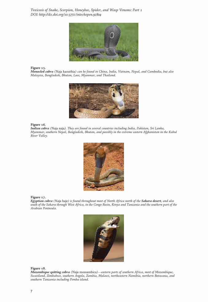
Figure 15.Monocled cobra (Naja kaouthia) can be found in China, India, Vietnam, Nepal, and Cambodia, but alsoMalaysia, Bangladesh, Bhutan, Laos, Myanmar, and Thailand.
Figure 16.Indian cobra (Naja naja). They are found in several countries including India, Pakistan, Sri Lanka,Myanmar, southern Nepal, Bangladesh, Bhutan, and possibly in the extreme eastern Afghanistan in the KabulRiver Valley.
Figure 17.Egyptian cobra (Naja haje) is found throughout most of North Africa north of the Sahara desert, and alsosouth of the Sahara through West Africa, in the Congo Basin, Kenya and Tanzania and the southern part of theArabian Peninsula.
Figure 18.Mozambique spitting cobra (Naja mossambica)—eastern parts of southern Africa, most of Mozambique,Swaziland, Zimbabwe, southern Angola, Zambia, Malawi, northeastern Namibia, northern Botswana, andsouthern Tanzania including Pemba island.
7
Toxicosis of Snake, Scorpion, Honeybee, Spider, and Wasp Venoms: Part 1DOI: http://dx.doi.org/10.5772/intechopen.92804

Figure 19.Cape cobra (Naja nivea) found in southern Africa, particularly in South Africa and in parts of Botswana andsouth part of Namibia.
Figure 20.Common krait (Bungarus caeruleus) found in the Indian subcontinent. These snakes are found almost allover Peninsular India but not in the offshore Islands. They are also found in other neighboring countries such asPakistan, Bangladesh, Nepal, and Sri Lanka.
Figure 21.Blue krait is also known as theMalayan krait (Bungarus candidus). These snakes are found in PeninsularMalaysia, central Vietnam,Thailand, Bali, Lao People’s Democratic Republic, Indonesia, Singapore, and Sumatra.
Figure 22.Black mamba (Dendroaspis polylepis). The black mamba lives in the savannas and rocky hills of southernand eastern Africa. Its fragmented range includes many African countries like the Democratic Republic of theCongo, Sudan, Ethiopia, Eritrea, Somalia, Kenya, Uganda,Tanzania, Burundi, Rwanda, Angola,Mozambique, Swaziland, Malawi, Zambia, Zimbabwe, Botswana, Namibia, and South Africa.
8
Medical Toxicology

4. Discussion
4.1 Signs of ophidism
Difference between cobra and viper venom in terms of molecular weight, routeof administration and nature of toxin could account for differences in their venoms
Figure 23.Eastern green mamba (Dendroaspis angusticeps). Their range stretches from the eastern Cape in SouthAfrica through Kenya, Mozambique,Tanzania, eastern Zimbabwe, and southern Malawi.
Figure 24.Eastern gartersnake or Eastern garter snake(Thamnophis sirtalis sirtalis). They have a wide range acrosseastern North America, extending as far north as southern Ontario and Quebec in Canada, to the Gulf ofMexico in the south, along the eastern shores of America to the Mississippi River.
Figure 25.California red-sided garter snake (Thamnophis sirtalis infernalis) found in California.
9
Toxicosis of Snake, Scorpion, Honeybee, Spider, and Wasp Venoms: Part 1DOI: http://dx.doi.org/10.5772/intechopen.92804

lethality. Venom of Cerastes cerastes is more toxic than that of Bitis arietans andMacrovipera lebetina and toxin of Naja haje is more toxic than that of C. cerastes, M.lebetina and B. occitanus. Vipers have toxins with high molecular weight [25].Therefore, venom quality has to be standardized for development of efficient anti-venom, [26]. When the time of injected antivenom is shorter than fatal limit time,
Figure 26.Checkered garter snake (Thamnophis marcianus) found in the southwestern United States southwards intoMexico and Central America as far south as Costa Rica.
Figure 27.Eastern brown snake (Pseudonaja textilis) found in the eastern half of Australia, except in Tasmania.
Figure 28.King brown snake ormulga snake (Pseudechis australis) found over most of mainland Australia, except forthe extreme south and the southeast coastal regions.
10
Medical Toxicology

the envenomated may be protected by increasing the dose of antivenom. But whenthe antivenom is injected closer to the fatal limit time, the chance of death is one-half [27].
4.2 Treatment of snake envenomations
Rational dosage of snake antivenom requires larger randomized controlled trialsand further strategies are required to reduce morbidity in children bitten by Najaatra [28]. Neurotoxic envenomations and complications thereafter correlate posi-tively with snake antivenom dosage; hence higher doses are required which may
Figure 29.Death adder (genus Acanthophis) found in Australia, Indonesia, New Guinea and its nearby islands.
Figure 30.Red-bellied black snake (Pseudechis porphyriacus) native to eastern Australia. The red-bellied black snakesare found in a more or less continuous range from southeastern Queensland south through eastern New SouthWales and Victoria.
Figure 31.Gaboon viper (Bitis gabonica) found along the equatorial belt of Africa, East and Central Africa, andsoutheast Africa. In the African Portuguese-speaking countries, it can be found in Guinea-Bissau, Angola, andnorthern Mozambique.
Figure 32.Tiger snake (Notechis) found in Australia.
11
Toxicosis of Snake, Scorpion, Honeybee, Spider, and Wasp Venoms: Part 1DOI: http://dx.doi.org/10.5772/intechopen.92804

Figure 33.Puff adder (Bitis arietans) found in African savannah and grasslands.
Figure 34.Horned viper (Cerastes cerastes). It is found in many north African countries like Morocco, Mauritania,Mali, eastward through Algeria, Niger,Tunisia, Libya and Egypt, Chad, Sudan, Ethiopia, Somalia, andnorthern Israel.
Figure 35.Boomslang (Dispholidus typus) found in sub-Saharan Africa in the central and southern regions of thecontinent. The boomslang is most abundant in Botswana, Swaziland, Namibia, Mozambique, and Zimbabwe,but the species has been reported as far north as southern Chad and Nigeria, and as far east as eastern Guinea.
Figure 36.Rinkhals (Hemachatus haemachatus). The rinkhals is found in most provinces of South Africa, like westernand eastern Cape along the south coast, Mpumalanga, Free State, southern Gauteng and Kwazulu Natal.
12
Medical Toxicology

also cause adverse reactions [29]. Neutralization capacity of antivenom may berelated to geographic proximity of snake species [30]. Cerestes cerestes antivenomand Macrovipera mauritanica antivenom cross react with Bitis arietans antivenomdue to presence of antigens common to them [31]. Tenerplasminin-1 (TP1), aplasmin inhibitor isolated from Micrurus tener tener venom was similar to Kunitz-type serine peptidase inhibitors [32]. Rauwolfia serpentina inhibits Daboia russeliivenom [33].
The therapeutic dose of antivenom is relative to the quantity of venom [34].Therefore, postmortem lesions are required to provide the cause of death [35].
Figure 37.Copperhead or water moccasin (Agkistrodon contortrix). These snakes are found in the United States andin northern Mexico. In the USA, they are found in the states of Alabama, Arkansas, Florida, Georgia, Illinois,Connecticut, Delaware, Indiana, Iowa, Kansas, Kentucky, Louisiana, Ohio, Mississippi, Missouri, Oklahoma,Maryland, Massachusetts, New Jersey, New York, North Carolina,Tennessee,Texas, Pennsylvania, SouthCarolina, Virginia, and West Virginia. In Mexico, it occurs in Coahuila and Chihuahua regions.
Figure 38.Cottonmouth (Agkistrodon piscivorus) found in the southeastern United States.
Figure 39.Mamushi or Japanese Mamushi (Gloydius blomhoffii) found in Japan, China, and Korea.
13
Toxicosis of Snake, Scorpion, Honeybee, Spider, and Wasp Venoms: Part 1DOI: http://dx.doi.org/10.5772/intechopen.92804
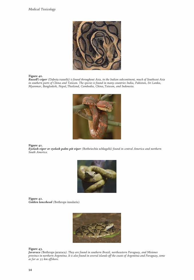
Figure 40.Russell’s viper (Daboia russelii) is found throughout Asia, in the Indian subcontinent, much of Southeast Asiain southern parts of China and Taiwan. The species is found in many countries India, Pakistan, Sri Lanka,Myanmar, Bangladesh, Nepal,Thailand, Cambodia, China,Taiwan, and Indonesia.
Figure 41.Eyelash viper or eyelash palm-pit viper (Bothriechis schlegelii) found in central America and northernSouth America.
Figure 42.Golden lancehead (Bothrops insularis).
Figure 43.Jararaca (Bothrops jararaca). They are found in southern Brazil, northeastern Paraguay, and Misionesprovince in northern Argentina. It is also found in several islands off the coasts of Argentina and Paraguay, someas far as 35 km offshore.
14
Medical Toxicology

Hence, neutralizing capacity of antivenins must be standardized [36] because ofcomplex nature of venom composition [37]. Pro-coagulant is used to assess anti-venom activity and the results of antivenin test in rodents may not prove efficaciousin human [37] which is considered very important [38]. Hence, changes in dosing
Figure 44.Fer-de-lance or terciopelo (Bothrops asper) inhabits the region from southern Mexico to northern SouthAmerica.
Figure 45.Bushmaster (Lachesis muta) found in southern Central America and the northern half of South Americaincluding the island of Trinidad.
Figure 46.Mangrove snake (Boiga dendrophila) found in Indonesia, Malaysia,Thailand, Singapore, Vietnam,Cambodia and Philippines.
Figure 47.Ringneck snake (Diadophis punctatus) found in southeastern Canada and throughout most of the UnitedStates southward into Central Mexico.
Figure 48.European cat snake or European Catsnake (Telescopus fallax).
15
Toxicosis of Snake, Scorpion, Honeybee, Spider, and Wasp Venoms: Part 1DOI: http://dx.doi.org/10.5772/intechopen.92804

Figure 49.Indian red scorpion (Hottentotta tamulus) found throughout most of India, eastern Pakistan and the easternlowlands of Nepal.
Figure 50.Deathstalker scorpion (Leiurus quinquestriatus). The deathstalker scorpion’s range covers a wide sweep ofterritory in the Sahara, Arabian Desert,Thar Desert, and Central Asia, from Algeria and Mali in the westthrough to Egypt, Ethiopia, Asia Minor and the Arabian Peninsula, eastward to Kazakhstan, and westernIndia.
Figure 51.Arabian fat-tailed scorpion (Androctonus crassicauda) found mainly in the Palaearctic region. It iscommonly found in Saudi Arabia, Kuwait, Qatar, Iraq, Iran,Turkey, and in north African nations.
Figure 52.Yellow fat-tailed scorpion (Androctonus australis). The yellow fat-tailed scorpion is found in north andwest Africa, the Middle East, and eastward to the Hindu Kush region. Countries where Androctonus specieslive include Armenia, Morocco, Algeria,Tunisia, Libya, Egypt,Togo, Palestine, Israel, India, Lebanon,Turkey,Jordan, Saudi Arabia, Yemen, Oman, United Arab Emirates, Qatar, Kuwait, Iraq, Iran, Afghanistan,Bahrain, and Pakistan.
16
Medical Toxicology

and development of new antivenins have been recommended [39]. However, loweffective dose can be used with beneficial results [40]. The most effective treatmentfor snake envenomation is the specific heterologous serum [41] and additional doseis unnecessary in brown snake envenomation [42]. Antivenom cause anaphylactic
Figure 53.Black spitting thick-tailed scorpion (Parabuthus transvaalicus)—southern Africa.
Figure 54.Striped bark scorpion (Centruroides vittatus) is distributed throughout the South-Central U.S. states andthroughout northern Mexico. Beginning in the northern Mexico Border States, Chihuahua, Coahuila, NuevoLeón, and Tamaulipas, C. vittatus’ range extends upward longitudinally through Texas, Oklahoma, andKansas, to reach as far north as Thayer County, Nebraska.
Figure 55.Arizona bark scorpion (Centruroides exilicauda) found in the deserts of Arizona, California and Utah.
Figure 56.Brazilian yellow scorpion (Tityus serrulatus)—South America.
17
Toxicosis of Snake, Scorpion, Honeybee, Spider, and Wasp Venoms: Part 1DOI: http://dx.doi.org/10.5772/intechopen.92804

Figure 57.Yellow-legged burrowing scorpion (Opistophthalmus glabrifrons)—southern Africa.
Figure 58.Tanzanian red clawed scorpion (Pandinus cavimanus)—Tanzanian, Africa.
Figure 59.Emperor scorpion (Pandinus imperator) found living in the rain forest or wet savannah, throughout Africafrom Mauritania to Zaire.
Figure 60.Brown widow spider (Latrodectus geometricus) found throughout the world, including Africa, the UnitedStates, Europe, Asia, the Middle East, and South America.
18
Medical Toxicology

Figure 61.Yellow sac spider (Cheiracanthium) is a species endemic to the Americas.
Figure 62.Indian ornamental tarantula (Poecilotheria regalis) is a species of spider found in South Asia, as well assoutheastern India.
Figure 63.Brown recluse (Loxosceles reclusa) primarily found in North America (throughout the Midwest andSouthern United States in particular).
Figure 64.Black widow spider (Latrodectus) found on every continent of the world (with the exception of Antarctica).
19
Toxicosis of Snake, Scorpion, Honeybee, Spider, and Wasp Venoms: Part 1DOI: http://dx.doi.org/10.5772/intechopen.92804
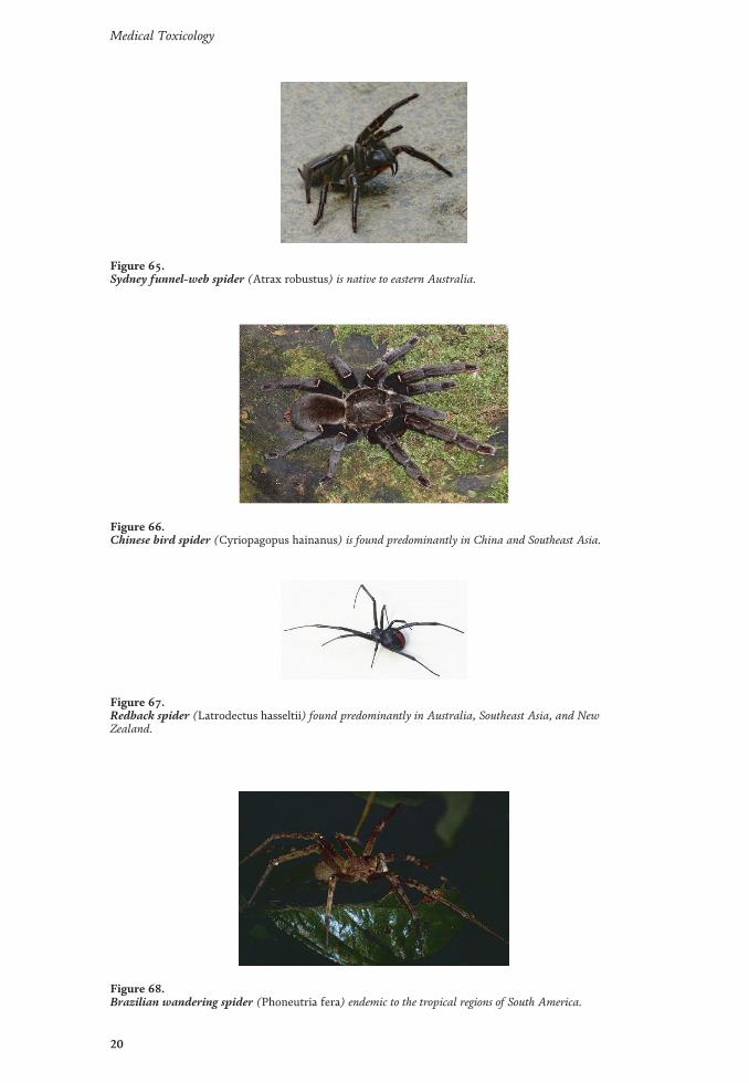
Figure 65.Sydney funnel-web spider (Atrax robustus) is native to eastern Australia.
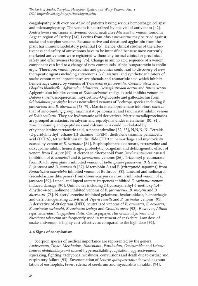
Figure 66.Chinese bird spider (Cyriopagopus hainanus) is found predominantly in China and Southeast Asia.
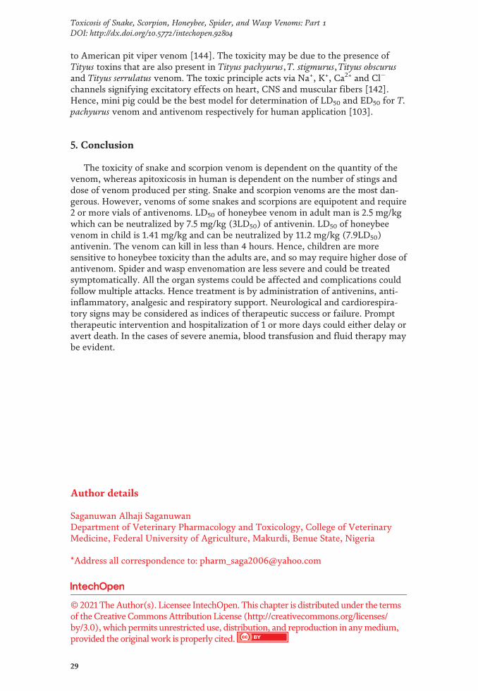
Figure 67.Redback spider (Latrodectus hasseltii) found predominantly in Australia, Southeast Asia, and NewZealand.
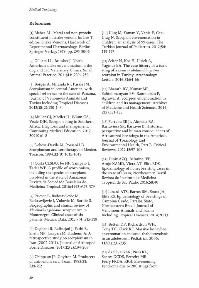
Figure 68.Brazilian wandering spider (Phoneutria fera) endemic to the tropical regions of South America.
20
Medical Toxicology

Figure 69.Six-eyed sand spider (Hexophthalma) is found predominantly in the deserts of southern Africa.
Figure 70.Tarantula hawks (Pepsis species) found across South and Central America and in the southern United States.
Figure 71.Giant Japanese hornet.
Figure 72.Bald-faced hornet (Dolichovespula maculata) is found throughout North America.
21
Toxicosis of Snake, Scorpion, Honeybee, Spider, and Wasp Venoms: Part 1DOI: http://dx.doi.org/10.5772/intechopen.92804

Figure 73.Crypt-keeper wasp (Euderus).
Figure 74.German yellowjacket Paravespula germanica (Linnaeus) found throughout North America.
Figure 75.Paper wasps (Polistes species)—southern United States.
Figure 76.Africanized honey bee (Apis mellifera scutellata Lepeletier) occurs naturally in sub-Saharan Africa but hasbeen introduced into the Americas.
22
Medical Toxicology

reactions [43] and serum sickness necessitating balance between treatment benefitand the risk of adverse reactions [42]. Failure of snake antivenin may be due to thefact that different snake venoms contain varieties of potent hemotoxins, neuro-toxins, and other toxins [44]. Therefore, the viper venom is more toxic than elapinevenom due to the nature and molecular weight of toxins. Geographical variability,the species of snakes, body weight, and the route of administration of antivenin arevery vital to successful treatment [45]. Toxins with molecular weight (<7KDa)diffuse quickly into the blood stream [25], which may not affect efficacy of snakeantivenin IgY elevated by ion exchange chromatography [46].
But antivenin affects pharmacokinetic and pharmacodynamic properties ofvenom. Hence, the quality of preparation and optimization of the use of antiveninmust be standardized [47]. The amount of antivenin is determined by clinical signs,size of snake, and the known efficacy of available antivenin [9]. The route ofadministration of antivenin is controversial, but intravenous route has been con-sidered the most efficient [47]. Polyclonal antibodies are more effective againstCerastes cerastes snake venoms in laying hens than in mammals [48]. Chickenimmunoglobulin (IgY) is more sensitive, easy to assay, does not activate humancomplement system and does not react with human anti venomous IgG antibodiesor human Fc receptors [49]. The immunoglobulin IgY offers protection againstembryo infections [50]. The effective IgY dose required to prevent mortality inrabbit was four times the dose of injected venom using Lowy’s protein assay [51].Milk whey lactoferrin increased antibody levels and immune-stimulatory effectsagainst snake venom [52] whereas adjuvant sustains the release of antigen, interactswith immune cells [53], activates nonspecific mediators of the immune system andenhance macrophage phagocytic activity. IgG antibody neutralized activity againstT. albolabris venom [54]. Thai neuro polyvalent antivenin is considered second-linetreatment for Hydrophis schistosus and Hydrophis curtus [55]. Both IgG and F(ab0)2antivenins activated human complement system with IgG having significantlyhigher anti-complementary activity than F(ab0)2 antivenin [56]. Agkistrodon halysantivenin is more efficacious than green pit viper antivenin [30]. Anti-Cc and Anti-Mm F(ab0)2 cross reacted extensively with Bitis arietans venom, perhaps due to thepresence of venom antigens common to the both snakes [57]. Gamma irradiatedNaja haje antivenin showed higher neutralizing capacity [58]. Equine antivenomneutralized coagulant and hemorrhagic activities against Rhabdophis tigrinus snakevenom [59]. Pseudo naja antivenin could not neutralize afbrinogenemia with seriousconsequences [60]. Hence clotting factor replacement therapy using fresh frozenplasma is associated with afibrinogenemia [61]. Gold nano-particle-based lateralflow assay is used for detection of snake envenomation [62]. Therefore, pharmaco-kinetics may be useful in design and optimization of antivenins [63]. Purificationand characterization of new bioactive compounds in snake venoms would be of helpin diagnosis and treatment of snake envenomation [64]. Sea snakes such as
Figure 77.Bumblebee Bombus terrestris.
23
Toxicosis of Snake, Scorpion, Honeybee, Spider, and Wasp Venoms: Part 1DOI: http://dx.doi.org/10.5772/intechopen.92804

Hydrophis schistosus and Hydrophis curtus produce venoms that could be neutralizedby their neuropolyvalent antivenom (NPAV) and cross neutralization should serveas basis for antivenom purification [55]. Non-irradiated and gamma-irradiatedpolyvalent antivenoms could neutralize Naja haje [65]. Lack of potent monovalentand polyvalent antivenins could be responsible for gross disparity in the manage-ment of snakebite. Treatment of snake envenomation was introduced by AlbertCalmette of the Institute Pasteur Saigon in the 1890s. The antivenom is either wholeIgG or pepsin refined F(ab) fragments of IgG derived from plasma of horse, don-key, mule, sheep immunized with venom of one or more species of snakes lyophi-lized venoms that have shelf-life of about 5 years and should be stored at ≤250C,whereas liquid antivenom have shelf-life of 2–3 years and should be stored at 2–8°Cand not frozen. Antivenoms against Naja naja, Bungarus caeruleus, Daboia russelliand Echis carinatus are produced in India [66]. About 8–10 vials of antivenomagainst Russell’s viper that injects 63 mg of venom is required. Each vial neutralizes6 mg of the snake venom. Children should receive the same dose as adults, butshould be observed closely for antivenin post administration reaction. Normaliza-tion of blood pressure in 15–30 minutes, stopping of coagulopathy in 6-hour, rever-sal of neurotoxicity in 30 minutes and recovery within 24–48 hours are thecharacteristics of effective snake antivenom. If there is coagulopathy, administra-tion of anti-snake venom can be repeated every 6 hours or after 1–2 hours of theinitial dose. Worsening of cardiovascular signs requires repeating dose of snakeantivenom every 1–2 hours [67]. About 70% of all snakebites are by non-venomoussnakes and 50% of bites by venomous species are dry bites [68]. Serum producedfrom irradiated Naja haje is more potent in venom neutralization than the serumproduced from native venom. The two sera inhibit cardiotoxic and hepatotoxiceffects [58]. Crescentia cujete fruit has significant neutralizing capacity againstVipera russelli venom [69]. Snake envenomation detection immune assay (SEDIA)is a potential diagnostic test for snake envenomation [52]. Milk whey (lactoferrin)could be an adjuvant to snake antisera [52]. Pharmacokinetics of venom fromHypnale hypnale are not changed by intramuscular injection of the venom, althoughmay reduce systemic bioavailability of the venom [63]. Structures, cytotoxicities,and affinities to phospholipids of cytotoxins from the venom of Naja haje differ[70]. Acorus calamus and Withania somnifera root extract have neutralizing poten-tial against venom of Echis carinatus [71]. Isolated chicken immunoglobulin (IgY)could neutralize viper venom [51]. Low dose snake antivenom with supportivetreatment is effective, less cost, and has low level of adverse reactions [72]. IgYantibody could neutralize venom of Trimeresurus albolabris [54] and Cerestes cerestesvenoms [48]. Indian snake antivenoms (VINS and BHARAT) are effective againstD. russelli, E. carinatus, B. caeruleus and N. naja with VINS being more superior toBHARAT [37]. Serum harvested from polyvalent venom from different species ofsnakes could neutralize snake venom such as Rhabdophis tigrinus [59]. Too smallinitial doses of snake antivenom could not neutralize venom of Pseudo naja; hencethe patient remains afribrinogenemic for long period of time [60]. Neutralizingpotential of snake antivenom (IgY) could be improved by ion exchange chroma-tography. Therefore, IgG antivenom has significant complementary antivenomactivity than F(ab0)2 antivenom [56].
4.3 Medicinal plants and phytochemicals against ophidism
Andrographis paniculata and Aristolochia indica could neutralize venom ofDaboia russelli [41]. Higher dose of Haffkine polyvalent antivenom could neutralizevenom of Naja sumatrana [40]. Calotropis gigantea could neutralize venom ofVipera russelli [73]. Brown snake, Pseudo naja causes venom induced consumption
24
Medical Toxicology
coagulopathy with over one-third of patients having serious hemorrhagic collapseand microangiopathy. The venom is neutralized by one vial of antivenom [42].Androctonus crassicauda antivenom could neutralize Mesobuthus venom found inAegean region of Turkey [74]. Lectins from Abrus precatorius may be tried againstsnake and scorpion venoms. Because native and denatured agglutinin from theplant has immunomodulatory potential [75]. Hence, clinical studies of the effec-tiveness and safety of antivenoms have to be intensified because most currentlymarketed antivenoms were registered without any formal clinical or preclinicalsafety and effectiveness testing [76]. Change in amino acid sequence of a venomcomponent can lead to a change of new compounds. Alpha bungarotoxin is cholin-ergic. Therefore, venom proteomics and genomics could lead to discovery of newtherapeutic agents including antivenoms [77]. Natural and synthetic inhibitors ofsnake venom metalloproteinases are phenols and rosmarinic acid which inhibitshemorrhage caused by venoms of Trimeresurus flavoviridis, Crotalus atrox andGloydius blomhoffii, Agkistrodon bilineatus, Deinagkistrodon acutus and Bitis arietans.Apigenin also inhibits venom of Echis carinatus and gallic acid inhibits venom ofDaboia russelli, isoquercitrin, myricetin-B-O-glucoside and gallocatechin fromSchizolobium parahyba leaves neutralized venoms of Bothrops species including B.jararacussu and B. alternatus [78, 79]. Matrix metalloproteinase inhibitors such asthat of zinc-binding group, marimastat, prinomastat and tanomastat inhibit venomof Echis ocellatus. They are hydroxamic acid derivatives. Matrix metalloproteinasesare grouped as astacins, serralysins and reprolysins under metzincins [80, 81].Zinc containing endopeptidases and calcium ions could be chelated byethylenediamine-tetraacetic acid, o-phenanthroline [82, 83], N,N,N,0N0-Tetrakis(2-pyridylmethyl) ethane-1,2-diamine (TPEN), diethylene triamine pentaaceticacid (DTPA), tetraethylthiuram disulfide (TID) in hemorrhage and myotoxicitycaused by venom of E. carinatus [84]. Bisphosphonate clodronate, tetracycline anddoxycydine inhibit hemorrhagic, proteolytic, coagulant and defibrigenotic effect ofvenom from B. asper [85]. A clerodane diterpenoid from Baccharis trimera causedinhibition of B. neuwiedi and B. jararacussu venoms [86]. Triacontyl p-coumaratefrom Bombacopsis glabra inhibited venom of Bothropoides pauloensis, B. leucurus,B. jararaca and B. pautoensis [87]. Macrolobin A and B (triterpnoid saponins) fromPentaclethra macroloba inhibited venom of Bothrops [88]. Linearol and isolinearol(secodolastane diterpenes) from Canistrocarpus cervicornis inhibited venom of B.jararaca [89]. Lupeol and lupeol acetate (terpenes) inhibited E. carinatus-venominduced damage [90]. Quinolones including 2-hydroxymethyl-6-methoxy-1,4-dihydro-4-equinolinone inhibited venoms of B. jararacussu, B. moojeni and B.alternatus [78]. N-acetyl cysteine inhibited gelatinase, hyaluronidase, hemorrhagicand defribrinogenating activities of Vipera russelli and E. carinatus venoms [91].A derivative of citalopram (DFD) neutralized venoms of E. carinatus, E. ocellatus,E. carinatus sochureki, E. carinatus leakeyi and Crotalus atrox [92]. However, Alliumcepa, Securidaca longipedunculata, Carica papaya, Harrisonia abyssinica andNicotiana tobaccum are frequently used in treatment of snakebite. Low dose ofsnake antivenom is highly cost-effective as compared to the high dose [92].
4.4 Signs of scorpionism
Scorpion species of medical importance are represented by the generaAndroctonus,Tityus, Mesobuthus, Hottentotta, Parabuthus, Centruroides and Leiurus.Leiurus abdullahbayrami caused hyperexcitability, agitation, aggressiveness,squeaking, fighting, tachypnea, weakness, convulsions and death due to cardiac andrespiratory failure [93]. Envenomation of Leiurus quinquestriatus showed degranu-lation of eosinophils, fever, edema of cerebrum and myocarditis in rabbit [94].
25
Toxicosis of Snake, Scorpion, Honeybee, Spider, and Wasp Venoms: Part 1DOI: http://dx.doi.org/10.5772/intechopen.92804

Tityus pachyurus polock envenomation was characterized by sialorrhea, respiratorydistress, profuse sweating, ataxia, restlessness, somnolence and hypoglycemia [95].The toxicity of venom is relative to the maturity and weight of the scorpion [96].Tityus stigmurus caused, cardiogenic shock, pulmonary edema, severe neurologicalsymptoms and death [9]. Spontaneous glycinergic and glutamatergic post synapticcurrents, suggest that scorpion toxin act on inhibitory and excitatory presynapticnerves. A. australis venom is more toxic followed by T. pachyurus, A. crassicauda, L.quinquestriatus, M. eupeus, L. abdullahbayrami and H. sauleyi [93]. A. crassicaudascorpion venom could induce activation of human monocytes leading to promotionof expression of IL-12 [97] with molecular peptides (Acra 1 and Acra 2) that aresimilar to known sodium-channel specific toxins of other scorpions [98]. M.gibbosus endemic to Mediterranean area causes scorpionism characterized by pain,pulsating and gloving sensations, cold, sweat, paleness, excitation, occasional spasmof the affected part, tortuousness of the vein, which lasted for more than 4 years.Periodical tingling, and occasional muscle twitches were observed during night. Thetreatment was symptomatic [99].
4.5 Treatment of scorpionism
Local anesthetic is effective, while opioids are ineffective and increase the risk ofrespiratory depression. Non-steroidal anti-inflammatory drugs may be disappoint-ing. Bio 10 ml of 10% calcium gluconate administered over 5–10 minutes relievesmuscle pain and cramps and the effects last 20–30 minutes, hence safe limit dose isrequired. SA IMR scorpion venom antiserum is equine anti-scorpion globulin sup-plement in 5 ml per ampoule and 5–10 ml is required for adults and children. Itsplateau effect is achieved in 2–6 hours, hence respiratory support is of paramountimportance during the period. If case-response to first dose is inadequate, another5 ml could be administered. The victim should be kept under close observation forat least a period of 6–12 hours. Electrolytes, pH, acid-base balance arterial bloodgases, and electrocardiography should be used for assessment of scorpionism toavoid fatality. The rule of thumb is that scorpions with thick tails and slenderpincers (e.g. Buthidae) produce more venoms than those with slender tails and largepincers. Hence speed of scorpion and a wave of potential neurotoxic effects are veryimportant. Bioclon and Butantan are very effective antivenins against scorpionism[95]. Treatment of scorpion envenomation requires specific antiserum. Antiserumagainst Buthus quinquestriatus from immunization of horses with crude venom asantigen has been proven to be effective [100], and high amount of antivenins isrequired to achieve satisfactory neutralization [101]. A. crassicauda antivenin couldprevent, neutralize and cure M. eupeus scorpionism if applied at optimum time,dose, and route [102]. The LD50 of A. crassicauda venom (1.1 mg/kg) and 39.19 mg/kg [103] make it highly toxic. The long half-life of venom in the body might requireantivenin that has long half-life [101].
In Saudi Arabia, scorpionism is treated using 5 ml of antivenom diluted in 5–20 mlof saline and the solution was administered intravenously. Adjunct chemotherapycould be instituted when required. However, a 12-year-old boy inadequately treatedwith antivenom died from pulmonary edema, hematemesis, severe neurotoxicity,and circulatory failure. All the patients treated in Saudi Arabia stayed in hospitals for1–2 days. The incidence of antivenom reaction was 1.7–6.6%, low protein level ofantivenom, and high quality of catecholamine could lead to antivenom failure [101].
Two purified toxic fractions of Mesobuthus eupeus toxin were quickly eliminatedfrom tissue [104] signifying that M. eupeus toxicity may not last long in the body.Dissociation of the toxin-channel complex during depolarization is determined bythe difference between electrical energies of the activated states of normal and
26
Medical Toxicology

toxin-modified channels [105]. The partially purified toxic fractions, when injectedto rabbits, gave rise to more potent antivenoms against whole venoms as comparedto presently commercially available antivenoms [106]. A. crassicauda has LD50 ofvenom (15.45 μg/kg) in mice, making it one of the highly toxic species of scorpionsin the world [103]. A. crassicauda antivenom could neutralize M. gibbosus venom(20 LD50) in Aegean region of Turkey [107]. Thus, highly potent antivenom couldbe produced from about 238 telsons in 51 days [74].
Comparing the calculated ratio of 29213:6 and
33013:6 which gives equivalent weight of
21.5 kg and 24.3 kg respectively shows that Butantan (292 μg/ml) and Bioclon(330 μg/ml) can be used effectively in the treatment of human weighing 21.5 and24.3 kg body weight, respectively. Signifying that age plays role on the dispositionof Tityus toxin, the toxic principle of Tityus species. Therefore, the difference inseverity of symptoms observed in children and adults may be due to difference inpharmacokinetics of the toxin. Mesobuthus eupeus venom can be neutralized bynanovalent, polyvalent and anti-idiotype antivenom, respectively. They are non-toxicants and can be used as a vaccine in people at the risk of scorpion stings [108].Scorpion anti-venoms are specific antigens, detoxified venoms or toxins, purifiedvenom fractions, natural toxoids, recombinant toxins, synthetic peptides, mono-clonal and recombinant antibodies [100]. Using peptides derived from the sequenceof scorpion toxins, the penetration of antipeptide antibodies can neutralize thecognate venom [109]. Turkish antivenom against A. crassicauda is effective againstother species of scorpions. Minimum lethal dose and minimum effective dose wereused to evaluate the effect of Turkish antivenom on M. gibbosus envenomation [96]suggesting applicability of the new formula for calculation of effective dose forantivenoms. Scorpion sting results in adult morbidity and pediatric mortality [110].The most lethal species are T. serrulatus,T. bahiensis in Brazil, Centruroides suffusus,C. lionpidus, C. sculpturatus in Mexico, Leiurus quinquestriatus, A. crassicauda, A.australis, A. amoreuni, Buthus occitanus in Middle East and North Africa, Parabuthusgrauntatus and P. transvaalicus in South Africa, Mesobuthus tamulus and Palamneusswammerdance in India [111], respectively. One milliliter of Androctonus crassicaudaantivenom could neutralize Mesobuthus eupeus venom. The antivenom is monova-lent with immune activity and neutralizing capacity. The venom is produced byRefik Saydam Hygiene Centre in Turkey [103]. Also A. crassicauda antivenom couldneutralize Mesobuthus gibbosus venom [96]. Stings from Leiurus abdullahbayramicauses hyperexcitability, agitation, aggressive behavior, squeaking, fighting,tachypnea, weakness, convulsions, and death due to cardiorespiratory failure.However, the venom has two kinds of protein with molecular masses of 4 and6 kDa, respectively [93]. The condition was treated by dipping the affected hand inice water, adrenaline (1:1000) was injected around the site of the sting and chlor-pheniramine was injected intramuscularly into the upper arm. Snake antivenommade by J. Wyeth and Brother Ltd., Canada was administered intramuscularly [112]and produced desired therapeutic effect against scorpionism. Scorpion envenom-ation may be more dangerous in pregnant woman [113]. However, Meu TxKα3, ascorpion toxin-like peptide could undergo mutation at site 30 and help improve itsK+ channel-blocking and antibacterial function [114]. The designed bispecificNbF12–10 neutralized AahI and AahII toxins and could be used in the treatment ofAndroctonus australis envenomation. About 100-kDa horse antivenom serum couldneutralize 7 kDa scorpion toxin and 15 kDa antivenom could neutralize AahI toxin.The NbAch10 F12 fully neutralized 100 LD50 of AahI toxin [115]. Children withabnormal electrocardiography may require high dose of antivenom [116].Aminotoxin could activate T lymphocytes and may be used as immunomodulatorsin infection and cancer [117] whereas Ca2+-activated K+ channel (ISK2) is sensitiveto apamin [118]. Nevertheless alpha-KTx peptides from the venom of Centruroides
27
Toxicosis of Snake, Scorpion, Honeybee, Spider, and Wasp Venoms: Part 1DOI: http://dx.doi.org/10.5772/intechopen.92804

elegans block Kv1.3 of T lymphocytes [119] whereas Tst26 peptide from T. stigmurusblock Kv1.2 and Kv1.3 channels [120] respectively. T. serrulatus could be detectedusing molecular mass of the venom [121]. Androctonus mauretanicus mauretanicustoxin could cross-react with the serum of Androctonus australis [122]. Androctonusmauretanicus and Buthus occitanus contain mycotoxins and post synaptic neuro-toxins with the first being more toxic. But the polyclonal antivenom preventedlethality from A. mauretanicus, B. occitanus and A. crassicauda venoms [123]. Venomof A. mauretanicus is more toxic than that of A. australis hector that is more toxicthan that of B. occitanus. Lethality of scorpion venom in mammals is dependent onage and species of the animals [124]. That is why the pharmacokinetics of a Tityustoxin from T. serrulatus scorpion venom is dependent on the age of affected indi-viduals [125]. Death due to scorpionism is secondary to cardiorespiratory failure[126]. Hence quick detection and quantification of venom is necessary for rationaltherapy, which is highly beneficial to reduce management costs and patients risk[127]. Equine F(ab0)2, IgG recombinant toxin, synthetic apitoxin, mAb, Fab, ScFv,chFab and rFab are of great benefit in treatment of scorpion and spider enveno-mation [128]. Minimum effective dose of antivenom is administered precociouslyby intravenous route to achieve efficient immunotherapy [129].
A. australis has complex venom that contains cytotoxic principles with very fastresultant fatal effects [130]. The effective monoclonal antibodies (mAbs) specificfor the α-neurotoxin 1 (Aah1) from A. australis hector venom has been reported[131]. A. australis has recombinant toxin II with immunological and biologicalproperties [132]. In addition, Androctonus australis hector (Aah) envenomation ismediated by cytokines and complement system, which in turn activate leukocyte todamage tissue [133]. But kinins are involved in cardiovascular toxicity and lethalityof L. quinquestriatus venom in rabbits. Androctonus australis garzonii venom(100 μg/kg) was neutralized by 4 mg/kg of antivenom injected intravenously [128].Antivenoms against a number of scorpion venoms have been reported [127],suggesting that the potency of antivenom should be investigated in relation to thescorpion venom [93] and both LD50 and ED50 should be determined paradoxicallyand canonically [134].
Intravenous LD50 of Vipera berus berus (0.4 μg/kg) with signs including head-drop, floppy neck, flaccid paralysis of limb, respiratory paralysis and death [135]and that of Laticauda colubrine, 0.05–0.13 μg/g [136], Sri Lankan B. caeruleus0.07 μg/g [137], and Naja sputatrix [138] disagree with the reported LD50 (0.5 ng)of Androctonus australis [93] signifying that the venom of V. berus berus is lesspotent than with that of A. australis. Similar signs were observed for V. nikolskiivenom (1.0 μg/kg) but the signs caused by phospholipase A2 were lost after themice were injected strontium [139], which may become antivenom against V. berusberus and A. australis venoms in future. The newly developed dot-ELISA for detec-tion of venoms of Indian venomous snakes, Naja naja, Bungarus caeruleus, Daboiarusselli and Echis carinatus [140] with comparative proteomic enzymes [141] may bealso used to detect scorpion venoms. High level of toxicity ofMontivipera raddei andMontivipera bubjardahica venoms is responsible for, high activity against A549human lung carcinoma [142] signifying that scorpion venom may have anticanceractivity. Lethal doses of L. quinquestriatus were 0.5 mg/kg i.v. and 3 μg/kg i.m.[143]. The LD50 of T. pachyurus venom in monogastric animals show that mouse(4.8 μg/kg) is highly sensitive to T. pachyurus venom, Hamster (3.48 μg/kg), guineapig (2.40 μg/kg), rat (2.32 μg/kg), rabbit (1.16 μg/kg), monkey (1.11 μg/kg), mar-moset (2.40 μg/kg), squirrel monkey (2.08 μg/kg), ferret (1.99 μg/kg), cat (0.74 μg/kg), dog and baboon (0.70 μg/kg), child (0.56 μg/kg), micro pig (0.52 μg/kg), minipig (0.40 μg/kg) and adult human (0.37 mg/kg) respectively [103] indicating thatTityus pachyurus toxin is more toxic to all the species of animals [139] as compared
28
Medical Toxicology
to American pit viper venom [144]. The toxicity may be due to the presence ofTityus toxins that are also present in Tityus pachyurus,T. stigmurus,Tityus obscurusand Tityus serrulatus venom. The toxic principle acts via Na+, K+, Ca2+ and Cl�
channels signifying excitatory effects on heart, CNS and muscular fibers [142].Hence, mini pig could be the best model for determination of LD50 and ED50 for T.pachyurus venom and antivenom respectively for human application [103].
5. Conclusion
The toxicity of snake and scorpion venom is dependent on the quantity of thevenom, whereas apitoxicosis in human is dependent on the number of stings anddose of venom produced per sting. Snake and scorpion venoms are the most dan-gerous. However, venoms of some snakes and scorpions are equipotent and require2 or more vials of antivenoms. LD50 of honeybee venom in adult man is 2.5 mg/kgwhich can be neutralized by 7.5 mg/kg (3LD50) of antivenin. LD50 of honeybeevenom in child is 1.41 mg/kg and can be neutralized by 11.2 mg/kg (7.9LD50)antivenin. The venom can kill in less than 4 hours. Hence, children are moresensitive to honeybee toxicity than the adults are, and so may require higher dose ofantivenom. Spider and wasp envenomation are less severe and could be treatedsymptomatically. All the organ systems could be affected and complications couldfollow multiple attacks. Hence treatment is by administration of antivenins, anti-inflammatory, analgesic and respiratory support. Neurological and cardiorespira-tory signs may be considered as indices of therapeutic success or failure. Prompttherapeutic intervention and hospitalization of 1 or more days could either delay oravert death. In the cases of severe anemia, blood transfusion and fluid therapy maybe evident.
Author details
Saganuwan Alhaji SaganuwanDepartment of Veterinary Pharmacology and Toxicology, College of VeterinaryMedicine, Federal University of Agriculture, Makurdi, Benue State, Nigeria
*Address all correspondence to: [email protected]
©2021 TheAuthor(s). Licensee IntechOpen. This chapter is distributed under the termsof theCreativeCommonsAttribution License (http://creativecommons.org/licenses/by/3.0),which permits unrestricted use, distribution, and reproduction in anymedium,provided the original work is properly cited.
29
Toxicosis of Snake, Scorpion, Honeybee, Spider, and Wasp Venoms: Part 1DOI: http://dx.doi.org/10.5772/intechopen.92804
References
[1] Bieber AL. Metal and non-proteinconstituent in snake venom. In: Lee T,editor. Snake Venoms: Hardwork ofExperimental Pharmacology. Berlin:Springer Verlag; 1979. pp. 295-3006
[2] Gilliam LL, Brunker J. NorthAmerican snake envenomation in thedog and cat. Veterinary Clinics: SmallAnimal Practice. 2011;41:1239-1259
[3] Borges A, Miranda RJ, Pasale JM.Scorpionism in central America, withspecial reference to the case of Panama.Journal of Venomous Animals andToxins Including Tropical Diseases.2012;18(2):130-143
[4] Muller GJ, Modler H, Wium CA,Veale DJH. Scorpion sting in SouthernAfrica: Diagnosis and management.Continuing Medical Education. 2012;30(10):1-8
[5] Dehesa-Davila M, Possani LD.Scorpionism and serotherapy in Mexico.Toxicon. 1994;32(9):1015-1018
[6] Costa CLSDO, Fe NF, Sampaio I,Tadei WP. A profile of scorpionism,including the species of scorpionsinvolved in the state of Amazonas.Revista da Sociedade Brasileira deMedicina Tropical. 2016;49(3):376-379
[7] Pajovic B, Radosavljevic M,Radosavljevic I, Vukovic M, Boricic S.Biogeographic and clinical review ofMesobuthus gibbosus scorpionism inMontenegro: Clinical cases of sixpatients. Medical Data. 2015;7(4):315-318
[8] Deghani R, Rafinejad J, Fathi B,Shehi MP, Jazayeri M, Hashemi A. Aretrospective study on scorpionism inIran (2002-2011). Journal of Arthropod-Borne Diseases. 2017;11(2):194-203
[9] Chippaux JP, Goyfton M. Producersof antivenom sera. Toxin. 1983;21:739-752
[10] Ulug M, Yaman Y, Yapia F, Can-Ulug N. Scorpion envenomation inchildren: an analysis of 99 cases. TheTurkish Journal of Pediatrics. 2012;54:119-127
[11] Seiter N, Koc H, Ulrich A,Yagmur EA. The case history of a toxicsting of a Leiurus abdullahbayramiscorpion in Turkey. ArachnologyLetters. 2016;51:64-66
[12] Bharath RV, Kumar MR,Subrahmanyam BV, Rammohan P,Agrawal A. Scorpion envenomation inchildren and its management. Archivesof Medicine and Health Sciences. 2014;2(2):131-135
[13] Ferreira SR Jr, Almeida RA,Barraviera SR, Barravie B. Historicalperspective and human consequences ofAfricanized bee stings in the Americas.Journal of Toxicology andEnvironmental Health, Part B: CriticalReviews. 2012;15:97-108
[14] Diniz AZQ, Belmino JFB,Arujo KAMD, Viera AT, Elite RDS.Epidemiology of honeybee sting cases inthe state of Ceara, Northeastern Brazil.Revista do Instituto de MedicinaTropical de Sao Paulo. 2016;58:40
[15] Linard ATS, Barros RM, Sousa JA,Elite RS. Epidemiology of bee stings inCampina Grade, Paraiba State,Northeastern Brazil. Journal ofVenomous Animals and ToxinsIncluding Tropical Diseases. 2014;20:13
[16] Betten DP, Richardson WH,Tong TC, Clark RF. Massive honeybeeenvenomation-induced rhabdomyolysisin an adolescent. Pediatrics. 2006;117(1):231-235
[17] da Silva GAR, Pires KL,Soares DCDS, Ferreira MR,Ferry FRDA. RRH: Envenomingsyndrome due to 200 stings from
30
Medical Toxicology

Africanized honeybees. Revista doInstituto de Medicina Tropical de SãoPaulo. 2013;55(1):61-64
[18] Schumacher MJ, Schmidt JO,Egen NB, Lowry JE. Quantity analysisand lethality of European andAfricanized honeybee venom. TheAmerican Journal of Tropical Medicineand Hygiene. 1990;43(1):79-86
[19] Schumacher MJ, Schmidt JO,Egen NB, Dilon KA. Biochemicalvariability of venoms from individualEuropean and Africanized honeybees(Apis mellifera). The Journal of Allergyand Clinical Immunology. 1992;90:59-65
[20] Almeida RAMDB, Olivo TET,Mendes RP, Barraviera SRCS,Souza LDR, Martins JG, et al.Africanized honey bee stings: How totreat them. Revista da SociedadeBrasileira de Medicina Tropical. 2011;44(6):755-761
[21] Kluz-Zawadzka J, Hartman-Ksycinska A, Lewandowska B.Emergent management of scorpionsting. Przeglad Epidemiologiczny. 2014;68:655-688
[22] Platnick AI. TheWorld SpiderCatalog Version 12.0. New York, USA:American Nugarm of Natural History;2011. Available from: http://research.amnh.org/ig/spiders/catalog
[23] Isbister GK, Gray MR, Balit CR,Raven RJ, Stokes BJ, Porges K, et al.Funnel-web spider bite; a systematicreview of recorded clinical cases. TheMedical Journal of Australia. 2015;182:407-411
[24] Gibb TJ, Bennett GW. Spiders.Extension Entomologists. 2017;72:1-2.Available from: www.extension.purdue.edu
[25] Okkache N, El Jaoudi R, Ghalim N,Chgoury F, Bouhaouala B, Mdaghri NE,et al. Evaluation of the lethal potency of
scorpion and snake venoms andcomparison between intraperitoneal andintravenous injection routes. Toxin(Basel). 2014;6(6):1873-1881
[26] Krifi MN, Marrakchi N, El Ayeb M,Dellagi K. Effect of some variables onthe in vivo determination of scorpionand viper venom toxicities. Biologicals.1998;26(4):277-288
[27] Krifi MN, El Ayeb M, Dellagi K.New procedures and parameters forbetter evaluation of Androctonusaustralis garzonii (Aag) and Buthusoccitanus tunetanus (Bot) scorpionenvenomation and specific scrotherapytreatment. Toxicon. 1996;34(2):252-266
[28] Wang JD, Tsan YT, Mao YC,Wag LM. Venomous snake bites andantivenom, treatment according to aprotocol for pediatric patients inTaiwan. Journal of Venomous Animalsand Toxins Including Tropical Diseases.2009;15(4):667-679
[29] Chich-Fan C, Tzeng-Jih L, Wen-ChiH, Hua-Wei Y. Appropriate antivenomdoses for six-types of envenomationscaused by snakes in Taiwan. Journal ofVenomous Animals and ToxinsIncluding Tropical Diseases. 2009;15(3):479-490
[30] Fung HT, Yung WH, Crow P,Lam KW, Tan KS, Gironi A, et al. Greenviper antivenom from Thailand andAgkistrodon halys antivenin from chiriacompared in treating Cryptelytropsalbolabris envenomation of mice. HongKong Medical Journal. 2012;18:40-45
[31] Khaddach FZ, Benaji B, Djebari FL,Chgoury F, Boussada L, Wadi A, et al.Assessment of preclinical efficacy ofantivenoms produced in rabbit byimmunological methods andneutralization assays. Journal ofChemical and Pharmaceutical Research.2014;6(12):446-455
[32] Vivas J, Ibarra C, Salazar AM,Neves-Ferreira AGC, Sanchez EE,
31
Toxicosis of Snake, Scorpion, Honeybee, Spider, and Wasp Venoms: Part 1DOI: http://dx.doi.org/10.5772/intechopen.92804
Perales J, et al. Purification andcharacterization of tener-plasoninin-1, aserine peptidase inhibitor withantiplasmin activity from the coralsnake (Micrurus tener tener) venom.Comparative Biochemistry andPhysiology Part C. 2016;179:107-115
[33] James TRJ, Danesh M, Vadivelan R,Shrestha A, Shanmugan V. In vivo andin vitro neutralizing potential ofRauwolfia serpentina plant extractagainst Daboia russelli venom. Biology.2013;7(6):276-281. ID 58940648
[34] Silverstein D, Hopper K. SmallAnimal Critical Care Medicine. 2nd ed.Amsterdam: Elsevier Health Science;2008. p. 1000
[35] Fry BG. Venomous Reptiles andTheir Toxins: Evolution, Pathophysiologyand Biochemistry. Oxford: OxfordUniversity Press; 2015. p. 546
[36] Gleen JJ, Straight RC. Mojaverattlesnake (Crotalus scutulatusscutulatus) venom variation in toxicitywith geographical origin. Toxicon. 1978;18:81-84
[37] Maduwager K, Siva A, Oleavy MA,Hodgson WC, Isbister GK. Efficacy ofIndian polyvalent snake antivenomsagainst Sri Lankan snake venoms:Lethality studies or clinically focusedin vitro studies. Scientific Reports. 2016;6:1-11
[38] Isbister GK, Brown SG,MacDonald E, White J, Alcurine BJ.Current use of Australian snakeantivenoms and frequency ofimmediate-type hypersensitivityreactions and anaphylaxis. The MedicalJournal of Australia. 2008;188:473-476
[39] Abubakar IS. Randomizedcontrolled double-blind non-infirmitytrial of two antivenins for saw-scaled orold carpet viper (Eschris ocellatus)envenoms in Nigeria. PLoS NeglectedTropical Diseases. 2010;4:e767
[40] Cham G, Lim F, Earnest A,Gopalakrishnakone P. Cross-reactivityagainst Naja sumatrana (black spittingcobra) envenoming from the Haffkineantivenom in a mouse model. ISRNToxicology. 2013;247645:1-6
[41] Meenatchisundaram S,Parameswari G, Michael A. Studies onantivenom activity of Andrographispaniculata and Aristolochia indica.Indian Journal of Science andTechnology. 2009;2(4):76-79
[42] Allen GE, Brown GA, Buckley NA.Clinical effects and antivenom dosing inbrown snake (Pseudonaja spp.)envenoming Australian snakebite project(ASP-14). PLoS One. 2012;7(12):1
[43] Isbister GK. Antivenom efficacy oreffectiveness. The Australianexperience. Toxicology. 2010;268:148-154
[44] Birell GW, Isbister GK, Masci PP,de Jersey J, Wallis TP. Moleculardiversity in venom from the Australianbrown snake, Pseudonaja textilis.Molecular & Cellular Proteomics. 2006;5:379-389
[45] Segura A, Herreva M, Vargars M.Intra specific variation and crossneutralization by antivenom. Toxicon.2012;59:158-162
[46] Liu W, Wang WD, Wang W, Bai S,Dybowski C. Influence of structure onthe spectroscopic properties of thepolymorphs of piroxicam. The Journalof Physical Chemistry. 2010;114(49):16641-16649
[47] Riviere G, Choumet V, Audebert F,Saboraud A, Debray M, Scherrmann JN,et al. Effect of antivenom on venompharmacokinetics in experimentallyenvenomed rabbits towards anoptimization of antivenom therapy. TheJournal of Pharmacology andExperimental Therapeutics. 1997;281(1):1
32
Medical Toxicology

[48] Moussa IN, Hassan AM,Alesisa AM, Al Arfaj AA, Salem BMM,Alrejai SA. Protective efficacy ofimmunoglobulins Y prepared againstCerastes cerastes snake venom in thekingdom of Saudi Arabia. Saudi MedicalJournal. 2012;33(8):846-851
[49] De Almeda CN, da Silva CL,Couto HP, Escocard RC, da Rocha DG,Sentinelli I. Development of press toproduce polyvalent IgG antibodies anti-African snake venom. Toxin. 2008;52:293-301
[50] Larsson A, Sjoquist J. Chicken IgYutilizing evolutionary difference.Comparative Immunology,Microbiology & Infectious Diseases.1990;13:199-201
[51] Krishnan LK, Saroja JB,Rajalingam M, John V, Valappil MP,Sreelathal V. Rabbit snake bite model toassess safety and efficacy of anti-viperchicken antibodies (IofG). AmericanJournal of Clinical and ExperimentalMedicine. 2015;3(1):32-38
[52] Elaraby AK, El-Jakee J, Fahmy A,Kandid MM, Mahmood Z, Saida AA.Lactoferrin: a novel strategy forantivenom therapy. InternationalJournal of Pharmaceutical Research andAllied Sciences. 2016;5(1):50-57
[53] Mahan T, Venom P, Rav DN. Noveladjuvants and delivery vehicles forvaccines development: a road ahead.The Indian Journal of Medical Research.2013;138(5):779-795
[54] Duan HL, He Q-Y, Zhuu B,Wang WW, Li B, Zhang YZ, et al. Anti-Trimeresurus albolabris efficacyantibodies: Preparation, purification andneutralization efficacy. Journal ofVenomous Animals and ToxinsIncluding Tropical Diseases. 2016;22(23):1-6
[55] Tan C, Tan NH, Tan KY, Kong KO.Antivenom cross-neutralization of the
venoms of Hydrophis schistosus andHydrophis curtis, two common seasnakes in Malaysian waters. Toxin. 2015;7:572-581
[56] Pakmanee N, Noiphrom J, Kay A,Pormuttakun D, Sakolparp L,HemmaleW, et al. Comparative abilitiesof phospholipase A2, and coagulantactivities induced by Daboia siamensisvenom and their antiinflammatoryactivity. Science Asia. 2013;39:160-166
[57] Khaddah FZ, Benaiji B, Djebari FL,Chgoury F, Boussadda L, Wadi A, et al.Assessment of preclinical efficacyantivenoms produced in rabbit byimmunological methods andneutralization assays. Journal ofChemical and Pharmaceutical Research.2014;6(12):446-455
[58] Karam HM, Shaaban EA,Mohamed AF, Zaki HF, Kenawy SA.New approach for improvingproduction of Naja haje snakeantivenom. International Journal ofScientific and Research Publications(IJSRP). 2015;5(3):1-11
[59] Morokuma K, Kobori N, Fukuda T,Takashira M. Experimentalmanufacture of equine antivenomagainst yamakagashi (Rhabdophistigrinus). Japanese Journal of InfectiousDiseases. 2011;64(5):397-402
[60] Yeung JM, Little M, Murray LM,Jelinek GA, Daly FFS. Antivenomdosing in 35 patients with severe brownsnake (Pseudonaja) envenoming inWestern Australia over 10 years. TheMedical Journal of Australia. 2004;181:703-705
[61] Jelinek GA, Tweed C, Lynch D,Celenza T, Bush B, Michalopoulos N.Cross reactivity between venomous,mildly venomous, and non-venomoussnake venoms with the CommonwealthSerum Laboratories Venom DetectionKit. Emergency Medicine Australasia.2004;16(5–6):384-386
33
Toxicosis of Snake, Scorpion, Honeybee, Spider, and Wasp Venoms: Part 1DOI: http://dx.doi.org/10.5772/intechopen.92804

[62] Pawode BS, Sivi NC, Shaikh IK,Waghmare AB, Jalhar ND, Wagh VB,et al. Rapid and selective detection ofexperimental snake envenomation-useof gold nanoparticle based literal flowassay. Toxicon. 2016;119(1):299-306
[63] Tan CH, Sim SM, Gnanathasan CA,Fung SY, Tan NG. Pharmacokinetics ofthe Sri Lankan hump-nosed pit viperintramuscular injections of the venominto rabbits. Toxicon. 2014;79:37-44
[64] Fatima LD, Fatah C.Pathophysiological and pharmacologicaleffects of snake venom components:molecular targets. Clinical Toxicology.2014;4(2):1-9
[65] Shaban EA, Hafez MN. Ability ofgamma-irradiated polyvalentantivenom to neutralize the toxicity ofthe Egyptian cobra (Naja haje) venom.Egyptian Journal of Hospital Medicine.2003;13:135-152
[66] Chaudhary S, Singh S,Chaudhary N, Mahato SK. Snake-bite inNepal. Journal of Universal College ofMedical Sciences. 2014;2(3):45-53
[67] Ahmed SM, Ahmed M, Nadeem A,Mahajan J, Choudhary A, Pal J.Emergency treatment of a snake bite.Pearls from literature. Journal ofEmergencies, Trauma, and Shock. 2008;1(2):97-105
[68] Theakston RD, Warrell DA,Griffiths E. Report of a WHO workshopon the standardization and control ofantivenoms. Toxicon. 2003;41:541-547
[69] Shastry CS, Aswathanarayana BJ,Maulik MB. Antivenom activity ofethanolic extract of Crescentia cujetefruit. International Journal ofPhytomedicine. 2012;4(1):108-114
[70] Suzuki-Matsubara M, Athanda SBP,Suzuki Y, Matsubara K, Moriyama A.Comparison of the primary structures,cytotoxicities, and affinities to
phospholipids of five kinds of cytotoxinsfrom the venom of Indian cobra.Najahaje. Comparative Biochemistry andPhysiology Part C. 2016;179:158-164
[71] Meanatchisasudaram S,Parameswari G, Michael A. Studies onantivenom activity of Andrographispaniculata and Aristolochia indica plantextracts against Daboia russelii venomby in vivo and in vitro methods. IndianJournal of Science and Technology.2009;2(4):76-79
[72] Hodek P, Trefil P, Simunek J,Hudecek J, Stiborova M. Optimizedprotocol of chicken antibody (IgY)purification providingelectrophoretically homogenouspreparations. International Journal ofElectrochemical Science. 2013;8:113-124
[73] Chacko N, Ibrahim M, Shetty P,Shastry CS. Evaluation of antivenomactivity of Calotropis gigantea plantextract against Vipera russelli snakevenom. International Journal ofPharmaceutical Sciences and Research.2017;3(7):2272-2279
[74] Ozkan O, Adiguzel S, Yakistiran S,Filazi A. Study of the relationshipbetween Androctonus crassicauda (Oliver,1807; Scorpiones, Buthidae), venomtoxicity and telson size weight andstoring condition. Journal of VenomousAnimals and Toxins including TropicalDiseases. 2006;12(2):297-309
[75] Tripathi S, Maiti TK.Immunomodulatory role of nature anddenatured agglutinin from Abrusprecatorius. The International Journal ofBiochemistry & Cell Biology. 2005;37:451-462
[76] Williams DJ, Habib AG,Warrell DA. Clinical studies of theeffectiveness and safety of antivenoms.Toxicon. 2018;150:1-10
[77] Utkin YN. Animal venom studies:Current benefits and future
34
Medical Toxicology

development. World Journal ofBiological Chemistry. 2015;6(2):28-33
[78] Preciado LM, Pereanez JA. Lowmolecular mass natural and syntheticinhibitors of snake venommetalloproteinases. Toxin Reviews.2018;37(1):19-26
[79] doVale FLH,MendesMM,Fernandes RS, Costa TR,Hage-Melim LI,SousaMA, et al. Protective effect ofSchizolobium parahyba flavonoids againstsnake venoms and isolated toxins. CurrentTopics inMedicinal Chemistry. 2011;11:2566-7577
[80] Rucavado A, Escaknte T,Gulierrez JM. Effect of themetalloproteinase inhibitors batimastatin the systemic toxicity induced byBothrops asper snake venom:Understanding the role ofmetalloproteinases in envenomation.Toxicon. 2004;43:417-424
[81] BodeW,Gomis-Ruth FX, StocklerW.Astacins, serralysins, snake venom andmatrixmetalloproteinases exhibit identicalzinc-binding environment(HEXXHXXGXXH) andmet-turn, andtopologies and should be grouped into acommon family, the ‘metzincin’. FEBSLetters. 1993;331(1–2):134-140
[82] Patino AC, Pereanez JA, Nunez V,Benjumea DM, Fernandez M, RucavadoA, et al. Isolation and biologicalcharacterization of Batx-1, a weakhaemorrhagic and fibrinogenolytic PImetalloproteinase from ColombianBothrops atrox venom. Toxicon. 2010;56:936-943
[83] Mazzi MV, Marcussi S, Carlos GB,Stabeli RG, Frano JJ, Ticli FK, et al. Anew haemorrhagic metalloproteasefrom Bothrops jararacussu snake venom:Isolation and biochemicalcharacterization. Toxicon. 2004;44:215-223
[84] Patra A, Kalita B, Chanda A,Mukherjee AK. Proteomics and
antivenomics of Echis carinatus venom:Correlation with pharmacologicalproperties and pathophysiology ofenvenomation. Scientific Reports. 2017;7:1-17
[85] Rucavado A, Henriquez M, Garcia J,Gutierrez JM. Assessment ofmetalloproteinase inhibitors clodronateand doxycycline in the neutralization ofhemorrhage and coagulopathy inducedby Bothrops asper snake venom. Toxicon.2008;52:754-759
[86] Januario AH, Maraisa S, Santos SL,Mazzi MV. Neoderodane diterpenoid, anew metalloprotease snake venominhibitor from Bacharis trimera(Asteraceae): Anti-proteolytic and anti-haemorrhagic properties. Chemico-Biological Interactions. 2005;150(3):243-251
[87] Mendes MM, Vieira SAPB,Gomes MSR, Paula VF, Alcantara TM,Homsi-Brandeburgo MI, et al.Triacontyl p-coumarate: an inhibitor ofsnake venom metalloproteinases.Phytochemistry. 2013;86:72-82
[88] da Silva JO, Fernandes RS,Ticli FK, Oliveira CZ, Mazzi MV, FrancoJJ, et al. Triterpenoid saponins, newmetalloproteinase snakevenom inhibitors isolated fromPentaclethra macroloba. Toxicon. 2007;50:283-291
[89] Domingos TFS, Vallim MA,Cavalcanti DN, Sanchez EF, TeixeiraVL, Fuly AL, et al. Effect of diterpenesisolated of the marine algaCanistrocarpus cervicornis against sometoxic effects of the venom of theBothrops jararaca snake. Molecules.2015;20:515-526
[90] Katkar GD, Sharma RD,Vishalakshi GJ, Naveenkumar SK,Madhur G, Thushara RM, et al. Lupeolderivative mitigates Echis carinatusvenom induced tissue destruction byneutralizing venom toxins and
35
Toxicosis of Snake, Scorpion, Honeybee, Spider, and Wasp Venoms: Part 1DOI: http://dx.doi.org/10.5772/intechopen.92804

protecting collagen and angiogenicreceptors on inflammatory cells.Biochimica et Biophysica Acta. 2015;185(12):2393-2409
[91] Sunitha K, Hemshekhar M,Santhosh MS, Kumar MS, Kemparaju K,Girish KS. Inhibition hemorrhagicactivity of viper venoms by N-acetyland thiol groups. Current Topics inMedicinal Chemistry. 2011;11(2):2589-2600
[92] Ramaswamy M, Solaimuthu C,Duraikannu S. Medicinal plants for thetreatment of snakebites among the ruralpopulations of Indian subcontinent; anindication from the traditional use topharmacological confirmation. Journalof Drug Delivery and Therapeutics.2018;8(5):62-68
[93] Ozkan O, Yagmur EA, Ark M. Anewly described scorpion speciesLeiurus abdullahbayrami (Scorpion:Buthidae), and the lethal potency andin vivo effects of its venom. Journal ofVenomous Animals and ToxinsIncluding Tropical Diseases. 2011;17(4):414-421
[94] Afifi SH, Elkashef R, Seddek AS,Salem DA. Light and transmissionelection microscopical changesassociated with Leiurus quinquestriatusvenom in rabbits. MacedonianVeterinary Review. 2010;39(1):51-57
[95] Barona J, Otero R, Núñez V.Toxicological and immunologicalaspects of scorpion venom (Tityusparachyurus) neutralizing capacity ofantivenom produced in Latin America.Bidondica. 2004;24(1):42-49
[96] Ozkan O, Adiguzel S, Kar S,Yakistiran S, Cesaretli KY, Karaer KZ.Determination of potency andparaspecific effects of Androctonuscrassicauda (Oliver, 807) antivenomagainst Mesobuthus gibbosus (Brulle,1832) venom (Scorpiones: Buthidae).Journal of Venomous Animals and
Toxins Including Tropical Diseases.2007;13(2):500-508
[97] Saadi S, Assarehzadegan MA,Pipelzadeh MH, Hadaddezfuli R.Induction of IL-12 from humanmonocytes after stimulation withAndroctonus crassicauda scorpionvenom. Toxicon. 2015;106:117-121
[98] Caliskan F, García BI, Coronas FI,Batista CV, Zamudio FZ, Possani LD.Characterization of venom componentsfrom the scorpionAndroctonus crassicaudaof Turkey. Toxicon. 2006;48(1):12-22
[99] Nazari M, Hassan R. Study ondistribution of scorpions to provideprevention and interventions incombating scorpionism in Poldokhtarcounty, Lorestan Province, Iran. Journalof Clinical and Diagnostic Research.2016;10(12):05-09
[100] Carno AO, Chatzaki M,Horta RCC, Mogalhaes BF, Oliveira-Mendes BBR, Chavez-Olortegui C, et al.Alternative methodologies of scorpionantivenomous production. Toxicon.2015;97:64-74
[101] Ismail M, Abdelsalam MA,Ahaidisb MS. Androctonus crassicauda(Olivier), a dangerous and undulyneglected scorpion-1. Pharmacologicaland clinical studies. Toxicon. 1994;32(12):1599-1618
[102] Zayerzadeh E, KoohMK,Mirakabadi AZ, Fardipoor A, Kassaian SE,Rabbani S, et al. Amelioration of cardio-respiratory perturbations followingMesobuthus eupeus envenomation inanaesthetized rabbits with commercialpolyvalent F(ab0)2 antivenom. Toxicon.2012;59(2):249-256
[103] Saganuwan SA. Determination ofmedian effective dose fifty (ED50) ofscorpion antivenom against scorpionenvenomation using a newly developedformula. Animal Models andExperimental Medicine. 2018;1:1-7
36
Medical Toxicology

[104] Shirmardi SP, Shamsaei M,Gandomkar M, Sarie E, Ghannadi M,Zare A. Comparison of two purifiedtoxic fractions from Mesobuthus eupeusscorpion. Journal of Venomous Animalsand Toxins Including Tropical Diseases.2010;16(4):639-646
[105] Mozhaeva GN, Naumov AP.Knietics of interaction of scorpion withsodium channels on the Ranvier nodemembrane. Neurophysiology. 1980;12(6):619-626
[106] Delari P, van Rietschoten J,Rochat H. Scorpion venoms andneurotoxins: An immunological study.Toxicon. 1981;19(3):393-407
[107] Ozkan O, Carhan A. Theneutralization capacity of Androctonuscrassicauda antivenom againstMesobuthus eupeus scorpion venom.Toxicon. 2008;52(2):375-379
[108] Khoobdel M, Zahraei-Salehi T,Nayeri-Fasaei B, Khosravi M, OmidianZ, Motedayen MH, et al. Purification ofthe immunogenic fractions anddetermination of toxicity in Mesobuthuseupeus (Scorpionida: Buthidae) venom.Journal of Arthropod-Borne Diseases.2013;7(2):139-146
[109] Alvarenga LM, Dining CR,Grainer C, Chávez-Olórtegui C.Induction of neutralizing antibodiesagainst Tityus serrulatus scorpion toxinsby immunization with a mixture ofdefined synthetic epitopes. Toxicon.2002;40(1):89-95
[110] Santo KS, Stephano MA,Marcelona JR, Ferreira VMR, Rocha T,Carricati C, et al. Production of the firsteffective hyperimmune equine serumantivenom against Africanized bees.PLoS One. 2013;8(11):e79971
[111] Salama MW, Sharshar KM.Surveillance study on scorpionspecies in Egypt and comparison of theircrude venom protein files. Journal of
Basic and Applied Zoology. 2013;66(2):76-86
[112] Wyshynski PE, Little JA.Scorpionism: The first case reported inCanada. Canadian Medical AssociationJournal. 1962;87:974-975
[113] Kamalak Z, Kosus N. Scorpion stingduring pregnancy: a case reportaccompanied by literature. CumhuriyetMedical Journal. 2013;35:605-607
[114] Wang X, Gao B, Zhu S. A single-point mutation enhances dualfunctionality of scorpion toxin.Comparative Biochemistry andPhysiology - Part C. 2016;179:72-78
[115] Hmila L, Saerens D, Abderrazek RB,Vincke C, Abidi N, Benlasfar Z, et al. Abispecific nanobody to provide fullprotection against lethal scorpionenvenoming. FASEB Journal. 2010
[116] Abimannane A, Rameshkumar R,Satheesh P, Mahadevan S. Second doseof scorpion antivenom in children withIndian red scorpion (Mesobuthustamulus) sting envenomation. IndianPediatrics. 2018;55:315-318
[117] Casella-Martins A, Ayres LA,Burin SM,Morais FR, Pereira JC,Faccioli LH, et al. Immunomodulatoryactivity ofTityus serrulatus scorpionvenom in humanT lymphocytes. Journalof VenomousAnimals and Toxinsincluding Tropical Diseases. 2015;21:1-8
[118] Jager H, Grissmer S.Characterization of the outer poreregion of the apamin-sensitive Ca2+-activated K+ channel rsk2. Toxicon.2004;43(8):951-960
[119] Olamendi-Portugal T, Somodi S,Fernandez JA, Zamudio FZ, Bererril B,Varga Z, et al. Novel alpha-KTxpeptides from the venom of thescorpion Centruroides elegans selectivelyblockade Kv1.3 over IKCa1 K+ channelsof T cells. Toxicon. 2005;46(4):418-429
37
Toxicosis of Snake, Scorpion, Honeybee, Spider, and Wasp Venoms: Part 1DOI: http://dx.doi.org/10.5772/intechopen.92804

[120] Papp F, Batista CV, Varga Z,Herceg M, Roman-Gonzalez SA,Gasper R, et al. Tst26, a novel peptideblocker of Kv1.2 and kv1.3 channelsfrom the venom of Tityus stigmurus.Toxicon. 2009;54(4):379-389
[121] Pimento AM, Stocklin R,Favreau P, Bouqis PE, MartinaEauclaire MF. Moving pieces in aproteomic puzzle: mass finger printingof toxic fractions from the venom ofTityus serrulatus (Scorpiones, Buthidae).Rapid Communications in MassSpectrometry. 2001;15(17):1562-1572
[122] Okkache N, Rosso JP, Alami M,Ghalim N, Saile R, Hasar M, et al. Newanalysis of the toxic compounds fromthe Androctonus mauretanicusmauretanicus scorpion venom. Toxicon.2008;51(5):835-852
[123] Okkache N, Ahmad RMR,Othman I, Ghalim N, Chgoury F,Boussadda L, et al. Comparison of theneurotoxic and myotoxic effects of twoMoroccan scorpion venoms and theirneutralization by experimentalpolyclonal antivenom. Life Sciences.2015;124:1-7
[124] Tiwari AK, Desphande SB. Toxicityof scorpion (Buthus tamulus) venom inmammals is influenced by the age andspecies. Toxicon. 1993;31(12):1619-1622
[125] Nunan EA, Arya V, Hocchaus G,Cardigo VN, Moraes-Santos T. Ageeffects on the pharmacokinetics oftityustoxin from Tityus serrulatusscorpion venom in rats. BrazilianJournal of Medical and BiologicalResearch. 2004;37:385-390
[126] Das-Lopes C, Paiva AL, Querra-Duarte C, Molina F, Felicori L.Venomous arachnid diagnosis assays,lessons from past attempts. Toxin. 2013;10(365):1-26
[127] Laustsen AH, Solà M, Jappe EC,Oscoz S, Lauridsen LP, Engmark M.
Biotechnological trends in spider andscorpion antivenom development.Toxins (Basel). 2016;8(8):1-33
[128] Krifi MN, Savin S, Debray M,Bon C, El Ayeb M, Choumet V.Pharmacokinetic studies of scorpionvenom before and after antivenomimmunotherapy. Toxicon. 2005;45(2):187-198
[129] Nafie MS, Daim MMA, Ali IAI,Abdel-Rahman MA, Nabil ZI. Proteomicand biochemical characterization of theEgyptian scorpion “Androctonusaustralis” venom. In: Abstract of 6thInternational Conference on NaturalToxins; December 2014; Ismailia. 2014.pp. 1-2
[130] Clot-Faybesse O, Juin M, Rochat H,Devaux C. Monoclonal antibodiesagainst the Androctonus australis hectorscorpion neurotoxin 1: Characterizationand use for venom neutralisation. FEBSLetters. 1999;458(3):313-318
[131] Bougis PE, Rochart H, Smith LA.Precursors of Androctonus australisscorpion neurotoxins. Structures ofprecursors, processing outcomes, andexpression of a functional recombinanttoxin II. The Journal of BiologicalChemistry. 1989;264(32):19259-19265
[132] Adi-Bessalem S, Hammoudi-TrikiD, Laraba-Djebari F. Pathophysiologicaleffects of Androctonus australis hectorscorpion venom: Tissue damages andinflammatory response. Experimentaland Toxicologic Pathology. 2008;60(4–5):373-380
[133] Fatani AJ, Furman BL, Zeitlin IJ.The involvement of plasma kinins in thecardiovascular effects of Leiurusquinquestriatus scorpion venom inanaesthetized rabbits. Toxicon. 1998;36(3):523-536
[134] Saganuwan SA, Onyeyili PA. Theparadox of human equivalent doseformula: A canonical case study of Abrus
38
Medical Toxicology

precatorius aqueous leaf extract inmonogastric animals. MacedonianVeterinary Review. 2016;39(1):23-32
[135] Malina T, Krecsák L,Westerström A, Szeman-Nagy G,Gyemant G, Hamvas MM, et al.Individual variability of venom from theEuropean adder (Vipera berus berus)from one locality in Eastern Hungary.Toxicon. 2017;135:59-70
[136] Tan CH, Wong KY, Tan KY,Tan NH. Venom proteome of theyellow-lipped sea krait, Laticaudacolubrina from Bali: insights intosubvenomic diversity venomantigenicity and cross-neutralization byantivenom. Journal of Proteomics. 2017;166:48-58
[137] Amf O, Tan CH, Ariaranee GC,Quraishi N, Tan NH. Venomics ofBungarus caeruleus (Indian krait):comparable venom profiles, variableimmune re-activities among specimensfrom Sri Lanka, India and Pakistan.Journal of Proteomics. 2017;164:1-18
[138] Tan NH, Wong KY, Tan CH.Venomics of Naja sputatrix, the Javan-spitting cobra: a short neurotoxin-driven venom needing improvedantivenom neutralization. Journal ofProteomics. 2017;157:18-32
[139] Shaikh IK, Dixit PP, Pawade BS,Waykar IG. Development of dot-ELISAfor the detection of venoms of majorIndian venomous snakes. Toxicon. 2017;139:66-73
[140] Choudhury M, McCleary RJR,Kesherwani M, Kini RM,Velmurugan D. Comparison ofproteomic profiles of two of the big foursnakes of India, the Indian cobra (Najanaja) and the common krait (Bungaruscaeruleus) and analyses of their toxins.Toxicon. 2017;135:33-42
[141] Nalbantsoy A, Hempel BF,Petras D, Heiss P, Gocmen B, Igci N,
et al. Combined venom profiling andcytotoxicity screening of the Radde'smountain viper (Montivipera raddei)and Mount Bulgar viper (Montiviperabulgardaghica) with potent cytotoxicityagainst human A549 lung carcinomacells. Toxicon. 2017;135:71-83
[142] Batista CV, Román-González SA,Salas-Castillo SP, Zamudio FZ, Gómez-Lagunas F, Possani LD. Proteomicanalysis of the venom from the scorpionTityus stigmurus: biochemical andphysiological comparison with otherTityus species. ComparativeBiochemistry and Physiology, Part C:Toxicology & Pharmacology. 2007;146(1–2):147-157
[143] Chippaux JP. Epidemiologicalinvestigation on envenomation: Fromtheory to practice. Journal of VenomousAnimals and Toxins Including TropicalDiseases. 2012;18(4):446-450
[144] Saganuwan SA. Calculation ofeffective dose fifty (ED50) of antivenomfor American pit viper envenomation.Comparative Clinical Pathology. 2018a;27:1321-1325
39
Toxicosis of Snake, Scorpion, Honeybee, Spider, and Wasp Venoms: Part 1DOI: http://dx.doi.org/10.5772/intechopen.92804
Copyright © 2022 FDOKUMEN