PREVALENCE OF GASTRO-INTESTINAL PARASITES OF HORSES ...
59
PREVALENCE OF GASTRO-INTESTINAL PARASITES OF HORSES (Equus caballus Linnaeus, 1758) IN SEVEN VDCS OF RUKUM DISTRICT, NEPAL NARESH OLI T.U. Registration No: 5-2-54-233-2009 T.U. Examination Roll No: Zoo. 28 Batch: 2070/71 A thesis submitted In partial fulfillments of the requirements for the award of the degree of Master of Science in Zoology with special paper Parasitology Submitted To Central Department of Zoology Institute of Science and Technology Tribhuvan University Kirtipur, Kathmandu Nepal December, 2016
-
Upload
khangminh22 -
Category
Documents
-
view
0 -
download
0
Transcript of PREVALENCE OF GASTRO-INTESTINAL PARASITES OF HORSES ...

PREVALENCE OF GASTRO-INTESTINAL PARASITES OF
HORSES (Equus caballus Linnaeus, 1758) IN SEVEN VDCS OF
RUKUM DISTRICT, NEPAL
NARESH OLI
T.U. Registration No: 5-2-54-233-2009
T.U. Examination Roll No: Zoo. 28
Batch: 2070/71
A thesis submitted
In partial fulfillments of the requirements for the award of the degree of Master of
Science in Zoology with special paper Parasitology
Submitted To
Central Department of Zoology
Institute of Science and Technology
Tribhuvan University
Kirtipur, Kathmandu
Nepal
December, 2016

i
DECLARATION
I hereby declare that the work presented in this thesis entitled “Prevalence of Gastro-
intestinal Parasites of Horses (Equus caballus Linnaeus, 1758) in Seven VDCs of
Rukum District, Nepal” has been done by myself, and has not been submitted elsewhere
for the award of any degree. All the sources of the information have been specifically
acknowledged by reference to the author(s) or institution(s).
Date:
Naresh Oli

ii
RECOMMENDATION
This is to recommend that the thesis entitled “Prevalence of Gastro-intestinal Parasites
of Horses (Equus caballus Linnaeus, 1758) in Seven VDCs of Rukum District,
Nepal” has been carried out by Mr. Naresh Oli for the partial fulfillment of Master’s
Degree of Science in Zoology with special paper ‘Parasitology’. This is his original work
and has been carried out under my supervision. To the best of my knowledge, this thesis
work has not been submitted for any other degree in any institutions.
Date:
Supervisor
Mr. Janak Raj Subedi
Lecturer
Central Department of Zoology
Tribhuvan University
Kathmandu, Nepal
iii
LETTER OF APPROVAL
On the recommendation of supervisor Lecturer Mr. Janak Raj Subedi, this thesis
submitted by Mr. Naresh Oli entitled “Prevalence of Gastro-intestinal Parasites of
Horses (Equus caballus Linnaeus, 1758) in Seven VDCs of Rukum District, Nepal” is
approved for the examination and submitted to the Tribhuvan University in partial
fulfillment of the requirements for Master’s Degree of Science in Zoology with special
paper Parasitology.
Date:
Prof. Dr. Ranjana Gupta
Head of Department
Central Department of Zoology
Tribhuvan University
Kathmandu, Nepal

iv
CERTIFICATE OF ACCEPTANCE
This thesis work submitted by Mr. Naresh Oli entitled “Prevalence of Gastro-intestinal
Parasites of Horses (Equus caballus Linnaeus, 1758) in Seven VDCs of Rukum
District, Nepal” has been accepted as a partial fulfillment for the requirements of
Master’s Degree of Science in Zoology with special paper Parasitology.
EVALUATION COMMITTEE
Supervisor Head of Department
Mr. Janak Raj Subedi Prof. Dr. Ranjana Gupta
Lecturer CDZ, TU
CDZ, TU
External Examiner Internal Examiner
Date of Examination:

v
ACKNOWLEDGEMENTS
I would like to express my gratitude to my honorable supervisor Lecturer Mr. Janak
Raj Subedi, Central Department of Zoology, Tribhuvan University for his valuable
suggestions to carry on and complete this dissertation work.
I would also like to extend my sincere gratitude to Prof. Dr. Ranjana Gupta, the Head
of the Central Department Zoology, Tribhuvan, University, for providing all
administrative supports during the dissertation work. I would also express my
appreciation to all my teachers. I would also like to express to my thanks and best regards
to all the staffs of Central Department of Zoology, Tribhuvan Univesrsity.
I would like to express my deepest gratitude my respected parents and my family
members for their supports and inspirations in my whole academic career. I would also
express my thankful to all my relatives and friends Mr. Umesh Kumar Pariyar, Amrit
Budha Magar and Ramesh Khadka, who helped me during the faecal collection.
Last but not least, I would like to acknowledge to my friends Pujan Adhikari, Amrit
Gurung, Bishnu Achhami for their kind supports throughout my dissertation work and
all those persons who helped me directly or indirectly to complete this work.
Naresh Oli
T.U. Registration No: 5-2-54-233-2009
T.U. Examination Roll No: Zoo.28
Batch: 2070/71
vi
CONTENTS
Pages
Declaration i
Recommendation ii
Letter of Approval iii
Certificate of Acceptance iv
Acknowledgements v
List of Tables viii
List of Figures viii
List of Photographs viii-ix
List of Abbreviations ix
Abstract x
1. INTRODUCTION 1-6
1.1 Background: 1-3
1.2 Endoparasites: 3-4
1.3 Intestinal protozoan parasites: 4
1.4 Intestinal helminth parasites: 4
1.5 Trematode parasites: 4
1.6 Cestode parasites: 4-5
1.7 Nematode parasites: 5
1.8 Objectives: 5
1.8.1 General objective: 5
1.8.2 Specific objectives: 5
1.9 Justification of the study: 5-6
1.10 Limitations of the study: 6
1.11 Hypothesis: 6
1.11.1 Null hypothesis (H0): 6
1.11.2 Research hypothesis (H1): 6
2. LITERATURE REVIEW 7-12
2.1 In the global context: 8-11
2.2 In context of Nepal: 11-12
3. MATERIALS AND METHODS 13-17
3.1 Study area: 13-14
3.2 Materials used: 14
3.2.1 Materials for field: 14
3.2.2 Materials for laboratory: 14
3.2.3 Chemicals: 14
3.3 Research design: 15
3.3.1 Study period: 15
3.3.2 Sample and data collection methods: 15
3.3.3 Preservation of faecal samples: 15

vii
3.3.4 Sample size: 15-16
3.4 Laboratory examination: 16-17
3.4.1 Concentration techniques: 16
3.4.1.1 Differentiation Flotation Technique: 16
3.4.1.2 Sedimentation Technique: 17
3.5 Eggs, cysts and larva size measurement: 17
3.6 Eggs, cysts and larva size identification: 17
3.7 Data analysis: 17
4. RESULTS 18-31
4.1 Overall prevalence of gastro-intestinal parasites in horses: 18
4.2. Prevalence of GI parasites in horses: 18-21
4.2.1 Protozoan parasites: 18
4.2.2 Helminth parasites: 18-19
4.2.3 VDC-wise prevalence of GI parasites in horses: 19
4.2.4 VDC-wise Comparative prevalence of parasite species: 20
4.3 Overall sex-wise prevalence of GI parasites in horses: 21
4.3.1 Sex-wise prevalence of GI parasites in VDCs: 21-22
4.4. Overall infection-wise prevalence of GI parasites in horses: 22
4.4.1 VDC infection-wise prevalence of GI parasites in horses: 22-23
4.5 Identified eggs/cysts/larvae of parasites found in horses: 23
4.6 Husbandry practices in the study area based on questionnaires survey: 25
5. DISCUSSION 32-37
6. CONCLUSION AND RECOMMENDATIONS 38
6.1 Conclusion: 38
6.2 Recommendations: 38
7. REFERENCES 39-47
8. APPENDIX 48

viii
LIST OF TABLES
Table No. Title of table Pages
Table 1: Proportion of faecal samples of horses collected from seven VDC of Rukum 16
Table 2: Overall protozoan parasites in horses 18
Table 3: Overall helminth parasites in horses 18
Table 4: VDC-wise prevalence of GI parasites in horses 19
Table 5: VDC-wise comparative prevalence of parasite species 20
Table 6: Sex-wise prevalence of GI parasites in horses in VDCs 21
Table 7: VDC-wise infection status of GI parasites in horses 23
Table 8: Identified eggs and cysts of parasites found in horses 24
Table 9: Unidentified larvae of parasites found in horses 25
LIST OF FIGURES
Figure No. Title of figures Pages
Figure 1: Map of Rukum district showing the study area (Source: OCHA 2008) 14
Figure 2: Overall prevalence 18
Figure 3: Overall class-wise helminth parasites in horses 19
Figure 4: Overall sex-wise prevalence of GI parasites in hors 21
Figure 5: Overall infection status of GI parasites in horses 22
Figure 6: Educational status of respondents 26
Figure 7: Feed of water sources for equines 26
Figure 8: Utilization or purpose of equines 26
Figure 9: Major feed sources for equines 26
Figure 10: Housing conditions of equines 26
Figure 11: Veterinary clinic available 26
Figure 12: Treatment of equines 27
Figure 13: Cleaning frequent of equines farm 27
Figure 14: Feed times in a working day 27
Figure 15: Feed times in a resting day 27
LIST OF PHOTOGRAPHS
Photo No. Title of photograph Pages
Photo 1: Entamoeba sp. 28
Photo 2: Balantidium sp. (cyst) 28
Photo 3: Balantidium sp. (trophozoite) 28
Photo 4: Eimeria sp. 28
Photo 5: Gastrodiscus sp. 28
Photo 6: Schistosoma sp. 28
Photo 7: Parascaris equorum (corticated) 29
Photo 8: Parascaris equorum (decorticated) 29
Photo 9: Strongylus sp. 29

ix
Photo 10: Trichonema sp. 29
Photo 11: Triodontophorus sp. 29
Photo 12: Dictyocaulus sp. 29
Photo 13: Oxyuris equi 30
Photo 14: Trichostrongylus sp. 30
Photo 15: Rhabdiasoidae larva 30
Photo 16: Dictyocaulidae larva 30
Photo 17: Strongyloidae larva 30
Photo 18: Ready to centrifuge 30
Photo 19: Saturated solution addition 30
Photo 20: Microscopic faecal observation 31
Photo 21: Faecal collection in field 31
Photo 22: Click with horse owner 31
Photo 23: Horse riding enjoy in field 31
LIST OF ABBREVIATIONS
Abbreviated form Details of Abbreviations
H Hukam
K Kankri
Ko Kol
M Mahat
Mo Morawang
OCHA Office for the Coordination of Humanitarian Affairs
Rm Ranmamaikot
rpm Revolution per minute
SIONA Statistical Information of Nepalese Agriculture
T Taksera

x
ABSTRACT
Horse has been a loyal friend and trusted partner of human being. Horses are prone to
infestation with both internal and external parasites. Present study was conducted in
eastern seven VDCs of Rukum district to determine the prevalence of gastro-intestinal
parasites in horses. The study was carried out from March 2016 to November 2016. A
total of 105 faecal samples of horses (79 males and 26 females) were collected by using
opportunistic random method during the month of April 2016. The collected faecal
samples were preserved in 2.5% potassium dichromate and microscopically examined
using concentration techniques. The overall prevalence of gastro-intestinal parasites was
84.76% (89/105). The total numbers of genera observed during faecal examination were
12 in numbers. Among them, the Strongylus sp. showed the highest prevalence (51.42%)
followed by Eimeria sp. (20%), Trichostrongylus sp. (14.28%), Trichonema sp. (13.33%),
Parascaris equorum (10.47%), Balantidium sp. (9.52%), Dictyocaulus sp. (8.57%),
Triodontophorus sp. (7.61%), Gastrodiscus sp. (6.66%), Oxyuris equi (4.76%),
Entamoeba sp. (3.80%), Shistosoma sp. (1.90%) and unidentified nematode larvae
(7.61%). Three genera of parasites: Shistosoma sp., Triodontophorus sp. and
Dictyocaulus sp. have been reported first time for Nepal in horses. Present study showed
higher infection rate in females (92.30%) than in male horses (82.27%). No significant
associations were observed between the prevalence of parasite rate with VDC-wise (χ2 =
5.7161; p>0.05) and prevalence rate with sex-wise (χ2 = 0.3346; p>0.05). But a
significant association was observed between the infection status and study area (χ2 =
84.277; p<0.05).
1
1. INTRODUCTION
1.1 Background:
Ruminants possess unique types of digestive system compared to other higher mammals.
Instead of one compartment of the stomach they have four compartments. Among four
compartments, rumen is the largest part in the rumen partially chewed grass is stored and
broken down into balls of cud. Many different species of ruminants are found around the
world. Some of them are domesticated by human beings such as buffalo, cattle, goat,
sheep, equines and most of them are in wild state like thar, deer, giraffes, ghoral,
elephants, monkeys etc.
Horses, donkeys, mules, zebras and camels belong to the equine group. They are found
mainly in temperate, semi-arid or highland areas (Alemayehu, 2004; Shiret and Samuel,
2015). Horses and donkeys are herd animals and will happily live in groups with donkeys
or animals of a different species such as, sheep and goats. Donkeys and horse are very
friendly animals and enjoy the company of humans. They are easily trained and are
suitable for handling by children especially donkey. Before the development of firearms,
the horse was crucial to warfare and before the invention of the steam engine, it was the
fastest and most reliable form of land transport. Today its importance has scarcely
diminished in parts of South America, Asia, Africa and Eastern Europe (Fitsum and
Ahmed, 2015), and even elsewhere it is of great economic importance to sport and leisure
industries.
Equines play an important role as working animals in many parts of the world, employed
for packing, riding, carting and ploughing. Equine power is vital for both rural and urban
transport system which is cheap and provides the best alternatives in places where the
road network is insufficiently developed, the terrain is rugged and mountainous and in the
cities where narrow streets mountainous and in the cities where narrow streets (Samuel et
al., 2015). Equids play crucial role in both urban and rural areas, providing agricultural
energy and transport, and in many cases, the sole means of income generating for their
resource limited owners (Getachew et al., 2014). Ever since it‟s domestication (Suteu,
1994; Khan et al., 2015), the horse has been a loyal friend and trusted partner of man in
day to day life, playing a vital role in many aspects of human life and evolution. Beauty,
boldness and benefits of horse have been mentioned in the Bible and Quran. In the
developed world, horses have great economic importance to sport and leisure industries.
The world population of equines (Abayneh et al., 2002; Haimanot et al., 2015) is 122.4
million (43.3 million horses, 40 million donkeys, 15 million mules, 24.1 million zebras
and camel). Over 95% of all donkeys and mules and 60% of all horses are found in
developing countries, which are playing a vital role in economies of developing countries
through provision of 75% of traction energy (USCOTA, 1998; Goraya et al., 2013).
Equines (horses, donkeys and mules) are raised primarily for draught and game purposes.
As draught animals, equines play extremely important role in the daily livelihood of poor
people. These animals are used for transportation of people, carriage and agricultural
purposes.
2
Horse is a large land mammal notable for its speed, strength and endurance. Horses are
members of the Equidae family, which also includes Zebras and Asses. Like all equids,
the horse is extremely well adapted to travelling long distances with great efficiency and
to surviving on a diet of nutrient-poor, high-fibre grasses. Horses were domesticated in
Eurasia around 6,000 years ago (Biu et al., 2014), they have provided humans with
mobility and have served in agriculture, warfare, and sport. Horses were also used for
drawing water from deep wells and in every kind of agricultural implement. They were
also used for sports such as polo, hunting, racing, traction, teaching demonstration and
scientific research, recreation purposes and as well as slaughtered for meat production
and consume in some countries. The mounted troop police used them to help in crime
control and land security.
Most equines are found in zone of high human population (highland) when agricultural
system in which livestock involves and where these animals are used as source of draft
powe (Alemayehu, 2004; Shiret and Samuel, 2015). In contrast, only 20% of all equines
are known to live on low land areas (Alemayehu, 2004; Shiret and Samuel, 2015). Equine
provides of power source for transportation, field operation and post harvest activities.
Nepal has a total of 52,577 horses/asses population that is reported from 51 districts of
Nepal. Among these horses/asses population, Dolpa district has the highest numbers and
Pyuthan district has the least number of horses/asses populations. In my study area,
Rukum district has a total of 1,732 horses/asses population (SIONA, 2013/14). In our
country Nepal, horses/asses are mainly kept for agriculture work and transportation work.
In Nepal, the low level of development of the road transport network and the rough road
of the country make the donkeys and the horses the most valuable, appropriate and
affordable pack animals under the small holder farming or house wise rearing system. In
my study area, VDCs are completely away from roads in rainy season but some VDCs
always away from roads throughout the year. Therefore, many people use horses and
mules to transport food and other supplies to villages. Long working hours and difficult
conditions are experienced by horses and mules. These animals are often engaged in work
for long hours and when get free they are left to browse and feed on grassland. These
have the potential to affect negatively on their welfare of life and health. Despite their
huge numbers and significant contribution to communities and the national economy, the
attention given to the health aspects of working horses (equids) in Nepal is very minimal.
Rudolf Leuckart considered as “father of parasitology”. The word „parasites‟ derived
from Greek which means situated beside. All over the world, horses are exposed to
gastrointestinal parasites from many orders and genera resulting in significant morbidity
and mortality (Hodgkinson, 2006; Goraya et al., 2013; Tilahun et al., 2014). Parasitism is
the single most important impediment in successful horse rearing all over the world
(Urquhart et al., 1996; Saeed et al., 2010) and many species of parasites are found to
infect horses (studies on prevalence of horse helminthes) in different parts of world have
indicated varied prevalence under different management and parasite control systems
(Chaudhry et al., 1991, Montinario et al., 2002, Champman et al., 2002, Boxell et al.,
2004; Capewell et al., 2005; Saeed et al., 2010). Horses are prone to infestation by a
mixture of internal and external parasites. An animal can harbor a great number of
parasites without exhibiting any clinical signs (Claire and Masterson., 1987; Krecek et al.,
3
1987; Tolliver et al., 1987; Gasser et al., 2004; Hubert et al., 2004; Martin et al., 2007;
Toscan et al., 2012; Khan et al., 2015). Health issues affecting welfare of animals include
acute diseases and disorders that cause immediate suffering and long-term progressive
conditions leading to chronic pain. Equine parasitism is one of the important menaces
affecting their working capacity and may result in mortality, For example, ticks, mites,
lice, flies etc. cause irritation, weakness, emaciation, anemia, rough hair coat and disease
transmission resulting in poor efficiency, stunted growth and even death of the animals.
Mortality in equines (Hodgkinson, 2006; Goraya et al., 2013; Tilahun et al., 2014) has
been frequently reported due to strongyles, tapeworms, ascarids, trypanosomes and
Babesia sp.
1.2 Endoparasites:
Endoparasites are those organisms living within hosts i.e in the gut, body cavity, livers,
lungs, gall bladder, blood or within the internal cavities, tissues or cells of their owns
host. They totally depend up on the host for growth, development and other physiological
processes and cause infection to them. Horses at times, suffer from various diseases such as
parasitic infection. Endoparasites are significant threat to horses. They are susceptible to
more than 60 internal parasites and harbor several species of worms at any time
(Hodgkingson et al., 2001; Lichtenfels et al., 2008; Bodecek et al., 2010; McWilliam et
al., 2010; Lamb et al., 2012; Guzel et al., 2014). The common gastrointestinal parasites
of equine are Cryptosporidium parvum, Giardia sp. (Butty, 2010), Trypanosoma sp.,
Entamoeba sp., Balantidium sp. (Wannas et al., 2012), Anoplocephala sp.,
Paranoplocephala sp., Cyathostomum sp., Trichonema Sp., Habronema sp., Draschia
sp., Triodontophorus sp., Craterostomum sp., Oesophagodontus sp., Cylicocycus sp.,
Strongyles species, Oxyuris sp., Strongyloides, Trichostrongylus sp., Fasciola hepatica,
Dicrocoelium sp. etc (Taylor et al., 2007).
Gastrointestinal parasites are one of the greatest limiting factors to successful horse rising
throughout the world (Herd, 1986; Shiret and Samuel, 2015). They are worldwide
problem for both small and large scale farmer with a greatest impact due to the
availability of a wide range of agro-ecological factors suitable for diversified hosts and
parasitic species. The majority of nematodes and other notable internal parasites such as
cestodes, trematodes and coccidian are the major gastrointestinal parasites of horses
(Taylor et al., 2007; Alemayehu, 2004; Butty, 2010; Wannas et al., 2012). The trematoda
of important veterinary medicine may be found as adult in the intestine, bile duct, blood
vessel or other organ of their final host. Adult cestodes are parasite of the intestine of
vertebrate (Alemayehu, 2004; Shiret and Samuel, 2015).
The development and survival of helminthes and protozoans egg of larvae with faeces
and on pasture are depending on temperature and moisture thus forming suitable
environment for development of larvae of nematode and trematode to infected stage.
Inadequate quality of water stored in the dam from which livestock area using directly for
drink may also form suitable way for transmission of cestoda and coccidian. Many factors
are known to influence the transmission and prevalence of gastrointestinal infection in
grazing animals. Broadly the three influencing factors (Alemayehu, 2004; Shiret and
Samuel, 2015) that can determine the occurrence of gastrointestinal tract infection could
4
be mentioned as environmental host interaction, environmental parasitic interaction and
host parasitic interaction. Larval stages are responsible for the damage done to the host
animal (Claire and Masterson., 1987; Krecek et al., 1987; Tolliver et al., 1987; Gasser et
al., 2004; Hubert et al., 2004; Martin et al., 2007; Toscan et al., 2012; Khan et al., 2015).
The effects of gastrointestinal parasites are more evident in young and under nourished
horses. Small numbers causes minimal damage, but large number pose a risk for colic and
other symptoms. As a rule, older horses appear to develop immunity against the common
gastrointestinal parasites and tend not be affected by parasite related problems as
commonly as younger horses (Alemayehu, 2004; Shiret and Samuel, 2015).
Strongyles are considered to be the most harmful, affecting horses of all ages and causing
weight loss, weakness, anemia, diarrhea and even death. Larval stages are responsible for
the damage done to the host animal (Nielsen et al., 2006; Khan et al., 2015). Strongylus
vulgaris has long been considered as one of the most common and pathogenic parasites of
the horse (Claire and Masterson., 1987; Krecek et al., 1987; Tolliver et al., 1987; Gasser
et al., 2004; Hubert et al., 2004; Martin et al., 2007; Toscan et al., 2012; Khan et al.,
2015). It is estimated that 45 to 90 percent of horses harbor S. vulgaris (McCraw and
Slocombe, 1976; Jubb et al., 1985; Khan et al., 2015). Ascarid worms have a high
prevalence in foals, but can also affect young adult horses (Mitrea, 2011).
1.3 Intestinal protozoan parasites:
Protozoans are single celled organisms that can affect the body at a cellular level, causing
problems especially in the circulatory, endocrine and gastrointestinal system (Goraya
2013). Protozoan parasites that commonly found in intestinal tract of equines are
Cryptosporidium sp., Isospora sp. Giardia sp. (Butty, 2010), Eimeria sp., Balantidium
sp., and Entamoeba sp. (Wannas et al., 2012).
1.4 Intestinal helminth parasites:
Helminth are most important endoparaites mostly causing the infection in intestinal, and
also in liver, lungs, lymphatic system, circulatory system, blood tissues, and skin.
Helminths are parasitic worms, are large multi-cellular organisms. Helminths are
generally endoparasites and parasitic forms except annelid worms (Morariu et al., 2012).
1.5 Trematode parasites:
Trematode species play a vital role in the health of domesticate livestock and for wild
animals. In general, trematodes commonly known as a flukes that live in the bile duct and
intestine and can affect lungs also. Some of the infective stages of the trematode are
ingested and some others penetrate the skins of host for entrance. The eggs are paased out
thorough faeces of hosts. The major trematode parasites (Taylor et al., 2007) of equines
are: Fasciola sp, Dicrocoelium sp., Gastrodiscus sp. etc.
1.6 Cestode parasites:
These parasites are mainly found in the gut of hosts and hosts get infection by ingestion
of contaminated food or water. The major cestode parasites (Taylor et al., 2007) of

5
equines are Anoplocephala sp., Echinococcus granulosus, Paranoplocephala sp., Taenia
sp., Moniezia sp. etc.
1.7 Nematode parasites:
The most common internal nematode parasites of the equines are strongyles
(Trichostrongyylus sp., Trichonema sp., Triodontophorus sp. and Strongylus sp.), ascarids
(Parascaris equorum), pinworms (Oxyuris equi), Dictyocaulus sp., Probstmayria
vivipara, Micronema deletrix, Draschia magastoma, Habronema sp., Onchocera sp.,
Craterostomum sp., Cylicocylus sp., Cylicodontophorus sp., Gyalocephalus sp.,
Cylindropharynx sp., Oesophagodontus sp., Poteriostomum sp. and bots (Gasterophilus
sp.) have the highest prevalence (Taylor et al., 2007).
1.8 Objectives:
1.8.1 General objective:
To find-out overall prevalence of gastro-intestinal parasites in horses of seven
VDCs of Rukum district.
1.8.2 Specific objectives:
To identify the gastro-intestinal parasites of horse (Equus caballus Linnaeus,
1758) by morphometry of eggs and larvae.
To determine the rate of prevalence of gastro-intestinal parasites of horse in seven
VDCs of Rukum.
To compare the VDC-wise, sex-wise and infection-wise distributions of gastro-
intestinal parasites of horse.
To know the husbandry practice of horse and mules (equines).
1.9 Justification of the study:
Parasites and diseases can take a toll on both the individuals as well as herd population of
animals. Nepal being a developing country, mostly goods are transport to backward of
hilly and mountain regions by horses/asses. Horses are suffering from different parasitic
diseases due to which their population had been decreasing in order. There had no study
about gastrointestinal parasites of horses in relation to husbandry practice in national
context of Nepal which may be one of the factors in declining population of the horses.
Horses/asses population had been declining in order in my research studied area. In
Rukum district, there had no research or study till now on horses/asses for gastrointestinal
parasites of horses in relation to husbandry practice. For continued declining of
horses/asses population in my studied area would make no population to Rukum district
for the new research in the future time. The number of population had been declined due
to different reason that is transportation facilities, poor husbandry practices, lack of
manpower for care, mountain shifting, machine-able agro-facilities, nutritional factors
and health related factors. Eastern part of Rukum district is major habitat of equines or
horses. This study could be new research from Rukum district regarding the topic, which
gives the suggestive guideline for the further research, locally horse reared people and
also for veterinary practitioner. So it was realized that the research study should be

6
launched for health related diseases prevalence of gastrointestinal parasites in horses in
relation to husbandry practices according to faecal examination in eastern seven VDCs
(Mahat, Morawang, Kankri, Kol, Taksera, Hukam and Maikot) of Rukum district, Nepal.
1.10 Limitations of the study:
This study was designed to find-out prevalence of gastrointestinal parasites in horses in
relation to husbandry practice with VDCs wise and sex wise. The identification of
parasites was based on morphology and size of parasitic eggs and larva. The study
doesn‟t reveal why some parasites were more predominant and others were not. This
study was only limited to certain parameters related to the topic due to cost and time
constraints. This study had been carried out for the partial fulfillment of the requirements
for the Master‟s Degree in Zoology at Tribhuvan University, Kathmandu, Nepal.
1.11 Hypothesis:
1.11.1 Null hypothesis (H0):
H0: There were no significant associations of gastro-intestinal parasites and risk factors.
1.11.2 Research hypothesis (H1):
H1: There were significant associations of gastro-intestinal parasites and risk factors.

7
2. LITERATURE REVIEW
Domestic ruminants may get parasitic infections each other by sharing the food pastures
or by grazing in the same habitats. Most of the researches have been carried out regarding
to the other domesticated animals however few researches have been done to parasites of
horse world widely. But in case of our country, very few studies have been done to
parasites of horses. In case of Horses (Equus caballus), no more studies have been carried
out regarding to the parasitic infection.
A large study looking at the association between poverty and animal disease (Perry et al.,
2002; Ali and Yagoob 2015) identified gastrointestinal (GI) parasitism as one of the most
important problems for equids in developing countries. Mortality in equines has been
frequently reported due to strongyles, tapeworms, ascarids, trypanosomiasis and
babesiosis (Hodgkinson, 2006; Goraya, 2013; Tilahun et al., 2014). The natural host of
the parasite (Dictyocaulus arnfieldi) is donkey, and comparably, large numbers of
parasites can accumulate in the lungs of this host without clinical signs. Donkey can act
as a reservoir for horses (Sharifi et al., 2010). Strongylosis has been reported from all
parts of world and almost affects more than 90 % of horse population and S. vulgaris has
long been considered as one of the most common and pathogenic parasites of the horse
(Nielsen et al., 2006; Toscan et al., 2012). Infestation with strongyles is complex and
produces an inflammatory enteropathy which results in impaired intestinal motility and
microcirculation (Bechera et al., 2010; Neils et al., 2011; Pilo et al., 2012). After
ingestion, larvae travel through the digestive system to the large intestine, S. vulgaris
migrate to the anterior mesenteric artery, S. endentatus to the liver / flank area and S.
equinus migrate to the liver and pancreas (Kuzmina et al., 2012). Higher egg excretion
has been recorded in spring and summer and higher EPG were recorded in young horse (≤
3 year) as compared with older horses, no difference in the prevalence of strongyle
infections as influenced by sex and excretion of eggs was also not affected by the sex of
the animals (Saeed et al., 2010). Season has no impact on the prevalence of strongyle
infections but shedding intensity of strongyle eggs is affected by season and significantly
higher egg excretion was recorded in spring and summer (Nielsen, 2012). Parascaris
equorum are highly prolific parasites, producing millions of extremely resistant eggs daily
(Mitrea, 2011). Several studies have shown a marked decrease of S. vulgaris infection
worldwide (Pilo et al., 2012). Determining the number of strongyle eggs per gram of
faeces (EPG) has been the most widely used method for diagnosing infection with adult
strongyles (Mahmood and Ashraf, 2010). Small strongyles involve 80% of the total
parasite population in a horse and the highest incidence of infection in yearlings (nearly
90%) and the lowest (46.6%) in foals has been recorded (Kaplan and Nielsen, 2010;
Andersen et al., 2013). Strongylidae family affecting more than 80% equids in the world
and egg production varies seasonally and it has been demonstrated that least egg
production occurs in winter, rising during the spring with maximal production during
August / September (Claire and Masterson., 1987; Krecek et al., 1987; Tolliver et al.,
1987; Peter and Waller, 1997; Gasser et al., 2004; Hubert et al., 2004; Martin et al., 2007;
Toscan et al., 2012; Khan et al., 2015). However, besides being species identification of

8
eggs is not possible, the identification of larvae is difficult and time-consuming using
morphological parameters (Bodecek et al., 2010; Lamb et al., 2012; Guzel et al., 2014). It
is estimated that 45 to 90 percent of horses harbor S. vulgaris (McCraw and Slocombe,
1976; Jubb et al., 1985; Khan et al., 2015). Definitive diagnosis of strongylosis can be
made by prove the presence of adult or larvae in the gastrointestinal tract during
postmortem examinations (Hodgkingson et al., 2001; Lichtenfels et al., 2008; Guzel et
al., 2014). Infections with Nematodes of strongyles parasites are complex and more than
60 species have been documented (Hodgkingson et al., 2001; Lichtenfels et al., 2008;
Bodecek et al., 2010; McWilliam et al., 2010; Lamb et al., 2012; Guzel et al., 2014). All
over the world, horses are exposed to helminth nonparasites from many orders and genera
resulting in significant morbidity and mortality (Hodgkinson, 2006; Tilahun et al., 2014).
2.1 In the global context:
Overall positive prevalence 84.4% had been observed by using Direct and flotation
techniques from Sokoto metropolis where Strongylus , Ascaris sp, Strongyloide sp,
Panaplocephala sp, Dictyocaulus sp, and Gastrodiscus had been observed in where
Strongylus prevalence 75.5% had the highest among identified helminth paraites but
Oxyuris had not been observed (Alayande et al., 2003). Overall 100% positive
prevalence had been observed in horses and donkeys from Konya region of Turkey where
prevalence of Strongylidae, Parascaris equorum, Strongyloides westeri, Fasciola ,
Anoplocephalidae, Oxyuris equi, Trichuris , Dicrocoelium dendriticum, Eimeria leucarti,
and Eimeria had 100%, 10.81%, 7.2%, 3.6%, 2.7%, 1.8%, 0.9%, 0.9%, 4.5%, and
12.61%, respectively (Uslu and Guclu, 2007). Overall 93.26% positive prevalence had
been observed in equines from Kashmir valley of Jammu and Kashmir state and
Strongylus having the highest prevalence 81.19% followed by Triodontophorus
(41.39%), Dictyocaulus (14.10%), Oxyuris (9.40%), Paranoplocephala (8.14%),
Strongyloides (6.19%), Parascaris (4.01%), Amphistome (0.91%) and Eimeria (0.34%)
had been reported (Pandit et al., 2008). From slaughtered house of Poland, highest
infection intensity had been observed in foals and yearlings of horses followed by 10.6%
of Gasterophilus intestinalis, 7.9% of Parascaris equorum and 0.8% of mixed infection
of Oxyuris equi and Anoplocephala perfoliata (Kornas et al., 2010). The strongyles
(small cyathostomes) had been considered as parasites with minimal pathogenic influence
on the equine organisms and Cyathostomosis infection (Bodecek et al., 2010) during the
late winter and early spring months had been observed highest in young horses (≤ 6
years). 50 species of subfamily Strongylinae and cyathostominae (family Strongylidae)
parasites of nematode had been reported in which Strongylus vulgaris was the most
pathogenic member of the large strongyles (Hassan et al., 2010). In Kurfa chale of
Ethiopia, 65.51% of positive infections of helminthes had been observed in horses from
200 faecal samples examination in which the highest prevalence infection of strongyle
(58.5%) and diifferent species of helminth like Parascaris equorum, Dictyocalus
arnifieldi, Anoplocephla species, Gastrodiscus aegypticus, Strongyloides westeri,
Trichstrongylus axei, Strongylus vulgaris , Strongylus edentatus, Strongylus equinus and
Trichonema sp. parasites had been observed from a year long research (Saeed et al.,
2010). Giardia doudenalis with 19.63% and Cryptosporidium sp. with 27.10% parasitic
9
infections had been observed (Butty, 2011) in equines from Nineveh, Iraq by using
different methods (wet mount, flotation, lugol's iodine modified Ziehl Nelsecn (hot) and
Giemsa stain). In Al Diwaniyah Governorate, 100% positive prevalence had been
observed in horses and donkeys (Age between 2-6 years) where the prevalence of
Strongylidae species had a highest (50%) followed by Parascaris equorum (40.90%),
Strongyloides westri (22.72%), Trichostrongylus axei (25%), Oxyuris equi (11.36%),
Cryptosporidium sp. (20.45%), Balantidium coli (15.90%) and Eimeria sp. (6.81%) in
horses and the prevalence of Strongylidae species had a highest (57.14%) followed by
Parascaris equorum (32.14%), Strongyloides westri (28.57%), Trichostrongylus axei
(17.85%), Oxyuris equi (17.85%), Dictyocaulus arnfieldi (17.85%), Cryptosporidium sp.
(19.64), Balantidium coli (17.85%), Eimeria sp. (10.71% ) and Entamoeba coli (3.57%)
in donkeys (Wannas et al., 2012). Strongyles (87.97%), Parascaris equorum (13.9%),
Strongyloides westeri (5.06%) and Eimeria leuckarti (1.09%), single parasite infection
(79.85%), two multiple infection (19.42%) and three multiple infection (0.72%) had been
observed (Ionita et al., 2013) in horses from from Romanian. 48 faecal samples had been
collected from horses in Nigeria and examined by using floatation and sedimentation
techniques and the highest prevalence rate of ciliates (81.3%) followed by Strongylus sp.
(68.8%), Oxyuris equi (27.1%), Strongyloides sp. (25%), Dictyocaulus sp. (10.4%) and
Parascaris equorum (6.3%) of different species had been observed where the higher
infection had in young horses (5-16 years) than old horses (17-20 years) and in male
horses than that of female horses (Umar et al., 2013).
Out of 100 samples (53 horses, 41 donkeys and 6 mules) examination, 88.6% of horses,
85.3% of donkeys and 83.3% of mules had been observed from Aydin region of Torkey
where the immature Habronema species had the highest prevalence (71.6%) followed by
Habronema muscae (54%), Trichostrongylus axei (28.3%) and Habronema majus
(50.9%) and the infection had not been affected by age factor but affected by sex factor
(Aypak and Bergu, 2013). In North Darfun, 24.6% positive prevalence had been observed
in equines from 1,400 animal examinations where the mild infection had the highest
incidence (69.7%) and 84% followed by moderate infection (15.6%) and (8%), severe
infection (14.7%) and (8%) for donkeys and horses respectively and Strongylus sp.,
Trichostrongylus sp., Cyathostomes sp. and Strongyloides westeri were the dominant
genera of nematodes (Adam et al., 2013). Out of 385 faecal samples (154 donkeys, 130
horses and 101 mule), overall prevalence (70.04%) followed by species specific
prevalence 77.3%, 72.3% and 60.68% for donkeys, mules and horses respectively had
been reported from south wolla, Ethiopia where Strongyle species had the highest
prevalence 70.9%, 58.5% and 67.3% followed by 10.4%, 5.4% and 3.0% of Parascaris
quorum, 4.5%, 3.8% and 4.0% of Oxyuris equi and 5.2%, 9.2% and 5.9%, of Fasciola sp.
for donkeys, horses and mules respectively (Regassa and Yimer, 2013). Out of 384
samples of equines, 92.71% overall positive prevalence followed by species specific
prevalence 80.95% of horses and 97.13% of donkeys had been observed from Gondar
town, Ethiopia where prevalence of strongyle had the highest 66.67% followed by
Parascaris equorum (43.8%), Oxyuris equi (0.95%), Gastrodiscus aegyptiacus (2.86%)
and Gastrophilus intestinalis (0.95%) in horses and there was no statistical significant
difference in prevalence of gastrointestinal parasites between age, sex and among body
10
condition (Mezgebu et al., 2013). In Jabalpur, overall gastrointestinal nematode
prevalence (59.25%) in horses where the highest prevalence in unorganized sector
(96.45%) than organized sector (32%) and the mixed prevalence (47.50%) followed by
Strongyle (25%), Parascaris equorum (18.75%). The maximum infection in age group of
1-6 years (69.09%) followed by 6-12 years (62.96%) and 12-18 years (34.78%) and the
infection in females (60.97%) higher than in males (58.51%) had been observed (Yadav
et al., 2014). Out of 52 faecal samples (32 females and 20 males), 100% found to be
positive (from 5 genera of nematode) in horses from Baquba city where the prevalence of
Strongylus sp. (44.2%) was the highest and Parascaris sp. (3.9 %) the lowest followed by
highest monthly prevalence (29.9%) in March and lowest monthly prevalence (14.3%) in
December and highest infection (61.5%) in females and lowest infection (38.5%) in males
(Hasson, 2014). From Formiga western city of Brazil, the prevalence of Cyathostominae
was the highest (94.85%) in horses and identified species were Trichostrongylus axei,
Oxyuris equi, Triodontophorus serratus, Strongyloides westeri, Strongylus edentatus,
Habronema muscae, Parascaris equorum, Probstmayria vivipara, Strongylus vulgaris,
Gasterophilus nasalis, Anoplocephala magna and Anoplocephala perfoliata (Weslen et
al., 2014). In schimoga region of Karnataka state, 84% of gastrointestinal helminthes
from horses had been observed where the Strongylus sp. had the highest eggs prevalence
(52.38%) followed by Parascaris equorum (10.71%), Gastrodiscus sp. (7.14%), Oxyuris
equi (4.76% ) and mixed infection (25%) of strongylus, Strongyloides and gastrodiscus
of different species (Adeppa et al., 2014).
In Hawassa town, overall prevalence of gastrointestinal parasites of horse and donkey
(72.7%) and species specific prevalence of horses (63.7%) and donkeys (78.5%) had been
observed by using floatation and McMaster techniques where the highest prevalence of
strongyle had (76%) and (64.9%) and Parascaris equorum (26.2%) and (4.6%) in
donkeys and horses respectively and the infection had been influenced by species and age
factors but not by sex and body condition of the equines (Tesfu et al., 2014). In Taiwan,
161 horse samples were positive from faecal examination of a total 436 samples by using
acid-fast staining methods for Cryptosporidium (Guo et al., 2014). Out of 159 faecal
samples examination, 76.1% positive prevalence had been observed where Strongylus
had the highest prevalence (55.3%) followed by Oxyuris (30.2%) and Strongyloides
(10.7%) and the higher infection (80.8%) in females than male infection (58.8%) with
mixed infection (17.6%) had been observed (Wosu and Udobi, 2014). In Kurfa chale of
Ethiopia, 307 samples (75.62%) were positive (Sultan et al., 2014) from a total of 406
samples where the species specific prevalence had (14.43%) in horses and (94.82%) in
donkey and the strongyle had the highest prevalence (72.66%) followed by Parascaris
equorum (15.51%), Gastrophilus (6.89%), Anoplocephala (2.2%) and Oxiuris equi
(1.47%). Out of 190 faecal samples examination, 97.9% positive (Tilahun et al., 2014)
had been observed from Hawasa Town, Southern Ethiopia where the infection with one
parasite (14.17%), two parasites (29.5%), three parasites (35.8%) and four parasites
(17.9%) had and the Parascaris equorum (55.8%) had the highest infection followed by
Strongylus vulgaris (44.7%), Strongyle edentates (36.8%), Strongyle equines (16.3%),
Oxyrus equi (34.2%), Strongyloides westeri (28.4%), Triodonthophorus tencollis (33.2%)
and Dictyocaulus arnfieldi (3.7%). In around Mekelle, 59.3% positive prevalence of
11
gastrointestinal parasites had been observed from horses by using direct, floatation and
sedimentation techniques where strongyles sp. (27%), Oxyuris equi (8.8%),
Anoplocephala sp. (2%), Parascaris equirem (1.8%), Gstrophilus (1.3%), Strongyloid
westeri (0.8%), Fasciola sp. (0.5%), Eimeria species (0.5%) and mixed infections
(16.8%) had been observed (Shiret and Samuel, 2015). From Kombolcha town, out of
384 samples (253 donkeys and 131 horses), 292 samples were positive after faecal
examination by using floatation technique where the species specific prevalence had for
donkeys (86.5%) and horses for (55.7%) and the Strongyle species was the highest
prevalence (47.4%) followed by Parascaris equorum (4.7%), Oxyuris equi (2.8%), mixed
infection (12.5% ) of Strongyle and Parascaris equarum, (6%) of Parascaris equi and
Oxyuris equi and (5.5%) of strongyle and Oxyuris equi (Samuel et al., 2015).
Strongylosis had been reported from all parts of the world and almost affects more than
90% of horse population (Shite et al., 2015).
Out of 390 equines (204 donkeys and 174 horses and 12 mules) from North-eastern
Ethiopia, 64.61% (62.3%, 69%, and 41.7% in donkeys, horses and mules) of equines
were positive and Strongyle infection was widely distributed (Molla et al., 2015). In
Israel, out of 485 samples, 144 samples were positives (Tiroshlevy et al., 2015) and
identified species were strongyle eggs (24%), Ascarids eggs (5%) and Anoplocephala
(0.0042%). Out of 372 samples, 169 (51.7%) samples were positive for eggs of
cyathostominae, Strongylus sp., Parascaris equorum, Strongyloides westeri and
Anoplocephala from Warmia and Mazury (Sokol et al., 2015) and the higher prevalence
was in individual farms than in agrotouristic farms. In Danglia town, overall prevalence
(5.73%) of strongyle was found to be positive in both species of horses and donkeys
species and species specific prevalence (4.92%) and (5.83%) for horse and donkey
respectively by using flotation technique (Haimanot et al., 2015). Overall 20.40%
positive prevalence of Equines from a total of 1,304 faecal samples examination in
Mumbai and Pune region where Strongyloides westeri had the highest prevalence 13.19%
followed by Strongyles (10.81%), Parascaris equorum (0.23%), Dictyocaulus arnfieldi
(0.23%), Amhistomes (1.38%), Schistosoma indicum (0.31%) and Anoplocephala
(0.07%) respectively had been observed (Matto et al., 2015).
2.2 In context of Nepal:
Parasitic infestation is a major cause of illness but report of parasitic infestation of horses
is lacking in our country Nepal.
Out of 33 samples examinations, 100% samples found to be positive (Karki and
Manandhar, 2006) in mules from Udaypur district and the Strongyle species was
encountered 100% infection followed by Parascaris equorum (50%), Anoplocephala sp.
(7.4%), Gastrodiscus aegypticus (6%), Oxyuris equi (3%) and Fasciola (1.5%). In sainik
stud farm of Bharatpur Chitwan, the gastrointestinal parasites of Trichostrongylus species
having the highest prevalence 80.48% followed by Parascaris 21.95%, Trochonema
17.07%, Gastrodiscus 7.31%, Habronema 4.87% and Strongylus 48.78% had been
observed (Paudel, 2007). Out of 60 faecal samples examination, 45% positive prevalence
had been observed from Brick Kiln of Lalitpur district by using concentration technique
12
and found highest prevalence Gastrodiscus (30%) followed by Strongylus (22%), Oxyuris
(30%), Dictyocalus (7%) and Triodontophorus (11%) had been observed (Sapkota, 2009).
In Banke district, the highest infection (81%), moderate infection (78%) and lowest
infection (53%) had been observed in May/June, August/September and
December/January respectively from mules where has trematode of Dicrocoelium
(9.24%), Fasciola (9.82%), Gastrodiscus (2.89%) and Schistosoma (32.36%), Cestode
parasites of Anoplocephala (3.36%), Diphylidium (2.37%), Moniezia (2.31%) and Taenia
(8.67%) and nematode parasites of Ancyclostoma (1.15%), Capillaria (9.82%), Chabertia
(4.04%), Cooperia (4.62%), Dictyocaulus (11.56%0, Oxyuris (13.37%), Parascaris
(10.40%), Strongylus (15.02%), Strongyloides (8.67%), Trichostrongylus (13.29%),
Trichuris (9.24%), Triodontophorus (1.15%), Toxocara (5.78%) and 83.95%, 69.23%
and 47.16% of multiple infections had been observed in summer, rainy and winter season
respectively (Rani, 2010). The gastro-intestinal parasites had been researched (Shrestha,
2015) in horses from Illam community forest from where he had taken a total of 5
samples and found Eimeria (100%) having the highest followed by Oxyuris (80%),
Strongyloide (80%), Hookworn (20%), Ascaris (80%), Entamoeba (40%) and
Balantidium (20%).
13
3. MATERIALS AND METHODS
3.1 Study area:
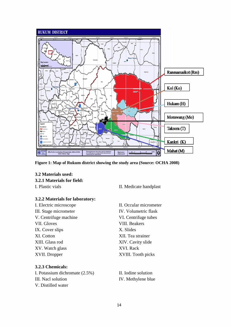
Rukum district is a hilly mountain district which is situated in the Mid-Western
Development Region of Nepal partially belonging to province number five and partially
to province number six. It is extended over 28o29‟N to 29
o0‟N latitude and 82
o12‟E to
82o53‟E longitude. The altitude of this district ranges between 762 m to 6,072 m. It
covers an area of 2,877 km2
with population of 207,290. The temperature ranges between
0oc to 34.4
oc and annual rainfall ranges between 1,600 mm to 2,400 mm. Rukum district
is located at 571.86 km far distance from capital city Kathmandu (Source:
https://en.m.wikipedia.org/wiki/Rukum-District, 2016).
Present study area is located in province number five that is eastern part of Rukum
district. The present study area (Mahat, Morawang, Kankri, Kol, Taksera, Hukam and
Ranmamaikot) is situated in the eastern part of Rukum district, which is located at 58 km
to 125 km far distance from headquarter (Musikot Khalanga) of Rukum. The altitude of
study area varies from 1,000 m to 4,000 m from sea level. Altitudinal range of
Morawang, Mahat and Kankri VDCs ranges from 1000 m to 3000 m with subtropical to
temperate climate but altitudinal range of Kol, Taksera, Hukam and Ranmamaikot VDCs
ranges from 2,500 m to 4,000 m with temperate to subalpine climate. Majority of people
live in these VDCs are Magar, Chhettri, Gurung and Dalit (Source:
https://en.m.wikipedia.org/wiki/Rukum-District, 2016).
The study areas selected have contrasting features relating to geographic location,
climatic condition, veterinary clinic, husbandry practice, fodder feeding and grazing land
availability. Climatic condition of study area varies from subtropical climate to subalpine
climate. Among all seven VDCs, veterinary clinic is only situated in Morawang VDC but
all other six VDCs have no any clinic for veterinarian purpose. Most of people are not
awareness towards veterinary treatments because veterinary clinics are available in very
far away from local people of VDCs and available of only limited veterinary medicines
and facilities. There is no horse farming supported by any other institution but only
farmers own their private horses reared with very simple rearing management like cow
and buffalo. Most of horses used for transportation purpose and feed on grains,
agricultural byproducts, grass fodder and feed on open grass field during at evening and
when they are in rest period. There has been observed no any veterinary related health
program performed by both government sector as well as private sector in the study area
till from many years during the study period. Horses get parasitic infection when they are
grazing in contaminated open grass field and nearby rivers. The district has different
domesticated animal‟s diseases like haemorrhagic septicemia, anthrax, pneumonia and
worm‟s diseases according to office of Rukum district for animal‟s service (Source:
https://en.m.wikipedia.org/wiki/Rukum-District, 2016).
14
Figure 1: Map of Rukum district showing the study area (Source: OCHA 2008)
3.2 Materials used:
3.2.1 Materials for field:
I. Plastic vials II. Medicate handplast
3.2.2 Materials for laboratory:
I. Electric microscope II. Occular micrometer
III. Stage micrometer IV. Volumetric flask
V. Centrifuge machine VI. Centrifuge tubes
VII. Gloves VIII. Beakers
IX. Cover slips X. Slides
XI. Cotton XII. Tea strainer
XIII. Glass rod XIV. Cavity slide
XV. Watch glass XVI. Rack
XVII. Dropper XVIII. Tooth picks
3.2.3 Chemicals:
I. Potassium dichromate (2.5%) II. Iodine solution
III. Nacl solution IV. Methylene blue
V. Distilled water

15
3.3 Research design:
The present study was designed to assess the gastrointestinal parasitic infection in horse
and mules in seven VDCs of Rukum district. The research comprises:
a) Selection of more horse habitat VDCs by direct observation.
b) Collection of 10 gram fresh faecal samples in sterile plastic vials (25 ml) in
preservative by opportunistic random sampling.
c) Preservation of faecal samples in 2.5% of Potassium dichromate solution.
d) Examination of faecal samples by using concentration techniques that is flotation and
sedimentation techniques.
e) Identification and measurement of eggs, cysts and larva of parasites.
f) Questionnaire survey (interview) for equines owners to know husbandry practice.
3.3.1 Study period:
The study was carried out from March 2016 to November 2016.
3.3.2 Sample and data collection methods:
The eastern parts of seven VDCs (Mahat, Morawang, Kankri, Kol, Taksera, Hukam and
Ranmamaikot) of Rukum district were considered as the major habitats of horses/mules.
During the collection of faecal samples in seven VDCs, three day was given to each of
them. Faecal samples were collected with two relatives and supported by other people in
studied area. Fresh faecal samples were taken from behind just below of individual horse
at early of the morning during time period between 4 am to 6 am. About 10 gram faecal
sample from each horse was taken with help of disposable gloves and transferred inside
the clean plastic vial having 25 ml. The same collection process was repeated for all
collected faecal samples. Necessary informations were noted clearly, such as faecal
samples collection date, collection VDC and sex of horse.
Both primary and secondary data sources were used in the study. Primary data was
collected by using semi-structured questionnaire and direct field observation. Secondary
data was collected from district. A formal type of questionnaire was used to collect data.
The questionnaire was prepared and pretested before the actual beginning of the survey.
A total of 48 respondent equines owners in the study area (VDCs) selected were
interviewed. Data collected using questionnaire includes the major feed resources,
management practice and healthcare. Field observation was made to enrich the data for
husbandry practice.
3.3.3 Preservation of faecal samples:
Collected faecal samples of horse were preserved in 2.5% Potassium dichromate that help
in maintaining morphology of protozoan parasites and preventing further development of
helminth eggs and larva.
3.3.4 Sample size:
A total of 105 faecal samples of horses (79 from males and 26 from females) were
collected from seven VDCs (Mahat, Morawang, Kankri, Kol, Taksera, Hukam and
Ranmamaikot) of Rukum district during the month of April 2016. The whole population
16
of seven VDCs was about 185. The sample size 105 which was occupied more than
56.75% as of whole population of seven VDCs. Among them, four VDCs (Mahat,
Morawang, Kankri and Kol) were comprised 100% of total population, two VDCs
(Taksera and Hukam) were comprised more than 90% of total population and one VDC
(Ranmamaikot) was comprised more than 25% of total population (Table 1).
Table 1: Proportion of faecal samples of horses collected from seven VDCs of Rukum
VDC name Collected sample
number
Sample proportion
(%)
Estimated population
of horses in VDC
Morawang 9 8.57 9
Mahat 17 16.19 17
Kankri 5 4.76 5
Taksera 21 20 23
Kol 10 9.52 10
Hukam 15 14.28 16
Ranmamaikot 28 26.66 105
3.4 Laboratory examination:
The faecal samples were collected in plastic vials (25 ml), transported by bus from
Rukum to Kathmandu and then brought to laboratory of Central Department of Zoology,
Kirtipur, Kathmandu. The faecal samples were subjected to coprological examination by
concentration technique (flotation and sedimentation).
3.4.1 Concentration techniques:
Eggs/cysts were often low number in faeces that they were difficult to be detected in
direct smear. Therefore fecal samples were examined using flotation and sedimentation
techniques (Soulsby, 1982; Zajac and Conboy, 2012).
3.4.1.1 Differentiation Flotation Technique:
Nematode and cestode eggs present in horses faeces were detected through this technique.
This technique ensures the eggs float in the floatation liquid, which helps to identify the
eggs.
Approximately 3 gram of faecal sample was placed in a beaker and added 42 ml of water
then the samples was grinded lightly with the help of rod and filter the solution by tea
strainer. The filtrate solution was poured in to a centrifuge tube of 15 ml and centrifuged
at 1,000 rpm for 5 minutes. The tube‟s water was replaced with more saturated Nacl
solution and again centrifuged.
After centrifuged, super saturated Nacl solution was added to develop convex meniscus at
the top of the tube and one drop of Methylene Blue (to stained) was added where a cover-
slip can placed for a 5 minutes and then cover-slip was removed from tube and placed on
slide and examined at 10Xx10X and 10Xx40X objectives. The photographs of eggs, cysts
and larva of parasites were taken and identified based on colour, shape and size
(morphometry).
17
3.4.1.2 Sedimentation Technique:
This technique is used for detection of trematode eggs. It provides a better result as the
eggs of trematode are bit heavier than the other, sediments of centrifuged contents was
taken for eggs detection.
Saturated Nacl solution was removed gently from the centrifuge tube after examined the
flotation portion and pour the sediment content into the watch glass and stirred the
content gently to mix it. One drop from the mixture was taken to prepare a second slide.
The specimen was stained with Iodine wet mount‟s solution and examined at 10Xx10X
and 10Xx40X objectives.
In this way, two slides were prepared from one sample (one from flotation and one from
sedimentation) were examined at 10Xx10X and 10Xx40X objectives of microscope to
detect eggs of protozoan, helminthes, protozoan‟s trophozoites or cysts of gastro-
intestinal parasites.
3.5 Eggs, cysts and larva size measurement:
By using ocular and stage micrometer, the length and breadth (eggs, cysts and larvae) of
parasites measured with calibration.
3.6 Eggs, cysts and larva size identification:
On the basis of shape and size of published literature journals and books (Gardiner et al.,
1988; Bevilaqua et al., 1993; Lichtenfels et al., 2002; Taylor et al., 2007; Wannas et al.,
2012; Matto et al., 2015; Ahmed et al., 2011; Rahman et al., 2014; Soulsby, 1982;
Urquhart et al., 1996; Hendrix and Robinson, 2006).
3.7 Data analysis:
For this study, prevalence was measured as the percentage of host individuals infected
with a particular parasite (Margolis et al., 1982; Bush et al., 1997). The collected data
were coded and entered into Microsoft Excel spread sheet. Data were statistically
analyzed using Pearson‟s Chi-square test with Yates‟ continuity correction, performed by
“R”, version 3.3.1 software packages. Percentage was used to calculate prevalence. Data
were statistically analyzed using Chi-square. In all cases 95% confidence interval (CI)
and p<0.05 was considered for statistically significant difference.

18
4. RESULTS
4.1 Overall prevalence of gastro-intestinal parasites in horses:
Out of 105 samples, 89 (84.76%) samples were found to be positive (Figure 2) for
parasitic egg, cyst and larvae.
Figure 2: Overall prevalence
4.2. Prevalence of GI parasites in horses:
4.2.1 Protozoan parasites:
Overall, horses were found to be infected with protozoan parasites belonging to the three
classes. Among them, Eimeria sp. showed the highest prevalence (20%) and Entamoeba
sp. showed the lowest prevalence (3.80%) in Table 2.
Table 2: Overall protozoan parasites in horses
S.N
.
Class Parasite Name Prevalence (%)
1. Sporozoa Eimeria sp. 21 (20%)
2. Litostomatea Balantidium sp. 10 (9.52%)
3. Sarcodina Entamoeba sp. 4 (3.80%)
4.2.2 Helminth parasites:
Table 3: Overall helminth parasites in horses
S.N
.
Class Parasite Name Prevalence (%)
1. Nematoda Strongylus sp. 54 (51.42%)
2. Trichostrongylus sp. 15 (14.28%)
3. Trichonema sp. 14 (13.33%)
4. Parascaris equorum 11 (10.47%)
5. Dictyocaulus sp. 9 (8.57%)
6. Triodontophorus sp. 8 (7.61%)
7. Oxyuris equi 5 (4.76%)
8. Unidentified nematode
larva
8 (7.61%)
9. Trematoda Gastrodiscus sp. 7 (6.66%)
10. Schistosoma sp. 2 (1.90%)
84.76%
15.23%
Prevalence
Positive
Negative

19
Overall, horses were found to be infected with helminth parasites belonging to two
classes. Cestode parasites were not found during faecal examination (Figure 2).
Trematode parasite revealed to be least infected to horses with two genera Gastrodiscus
sp. (6.66%) and Schistosoma sp. (1.90%) but, horses were found to be highly infected
with nematode parasites belonging to seven different genera (Table 3).
Figure 3: Overall class-wise helminth parasites in horses
Among nematode parasites, Strongylus sp. showed the highest prevalence (51.42%)
followed by Trichostrongylus sp. (14.28%), Trichonema sp. (13.33%), Triodontophorus
sp. (7.61%), Parascaris equorum (10.47%), Oxyuris equi (4.76%), Dictyocaulus sp.
(8.57%) and nematode typed larva (7.61%) (Table 3).
4.2.3 VDC-wise prevalence of GI parasites in horses:
The highest prevalence (100%) was revealed in Kankri VDC followed by Ranmamaikot
(96.42%), Kol (90%), Taksera (85.71%), Hukam (80%), Morawang (77.77%) and Mahat
(64.70%) respectively (Table 4).
Table 4: VDC-wise prevalence of GI parasites in horses
VDC name No. of
examined
No. of
positive
Prevalence
(%)
χ2 P-value
Kankri 5 5 100% 5.7161 0.5733
Ranmamaikot 28 27 96.42%
Kol 10 9 90%
Taksera 21 18 85.71%
Hukam 15 12 80%
Morawang 9 7 77.77%
Mahat 17 11 64.70%
The study showed effects of VDCs (study areas) on the prevalence of gastro-intestinal
parasite but, there was no statistical significant difference of the prevalence of intestinal
parasite infection among VDCs (χ2 = 5.7161; p>0.05) (Table 4).
82.85%
8.57% 0% 0.00%
20.00%
40.00%
60.00%
80.00%
100.00%
Nematode Trematode Cestode
Prevalence
20
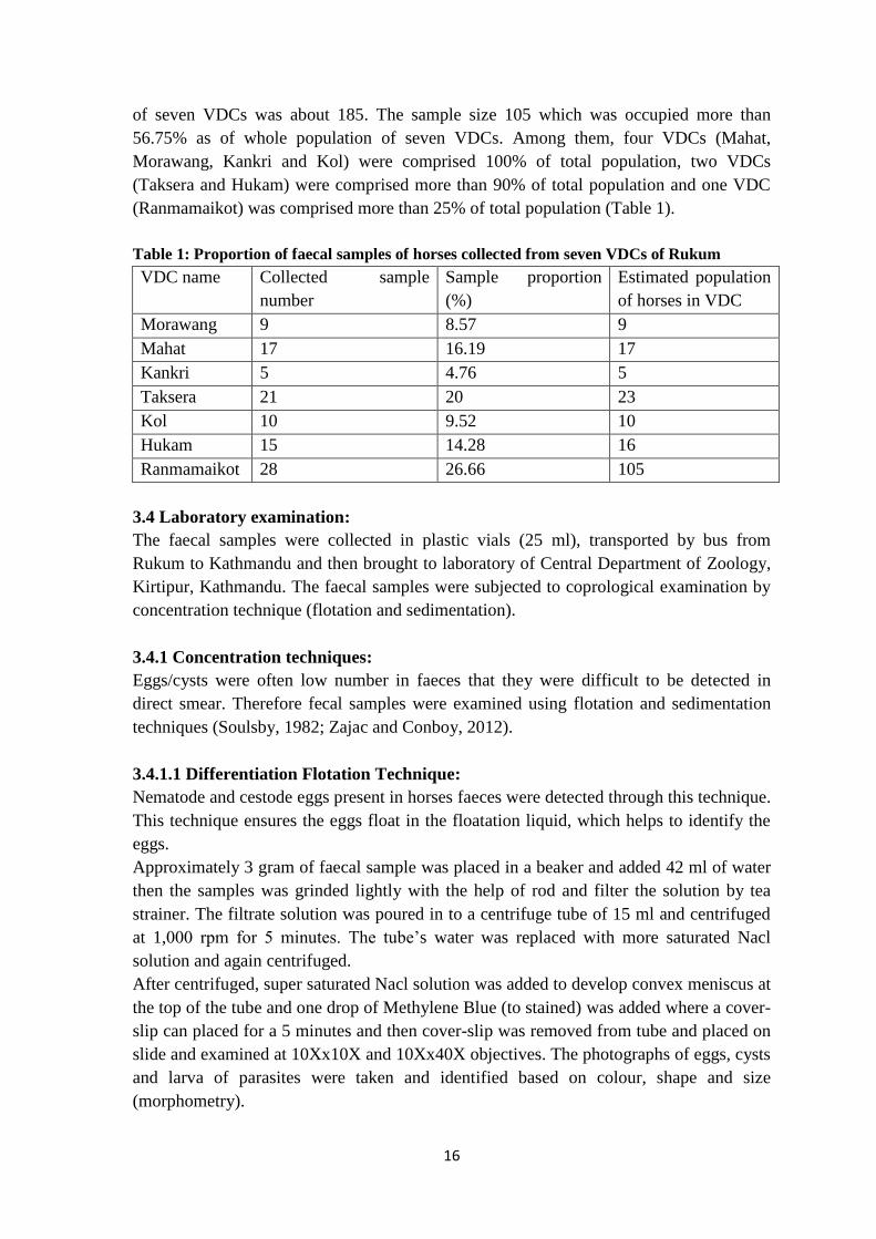
4.2.4 VDC-wise comparative prevalence of parasite species:
VDC wise, the highest prevalence of class sarcodina revealed in Kol (10%), sporozoa in
Ranmamaikot (32.14%) and litostomatea in Kankri VDC (20%) but all classes of
protozoan parasites were not found from Morawang VDC (Table 5). The highest
prevalence of Entamoeba sp. revealed in Kol (10%), Eimeria sp. in Ranmamaikot
(32.14%) and Balantidium sp. in Kankri VDC (20%) but protozoan parasites were not
found from Morawang VDC. The highest infection of trematode of Gastrodiscus sp.
revealed in Hukam (13.33%) and Schistosoma sp. in Ranmamaikot (7.14%) (Table 5).
Table 5: VDC-wise comparative prevalence of parasite species
Class Parasite Name K
(n=5)
Rm
(n=28)
Ko
(n=10)
T
(n=21)
H
(n=15)
M
(n=9) M
(n=17)
Sporozoa Eimeria sp. 1
(20%)
9
(32.14%)
1
(10%)
2
(9.52%)
4
(26.66%)
- 4
(23.52%)
Litostomatea Balantidium sp. 1
(20%)
4
(14.28%)
1
(10%)
2
(9.52%)
1
(6.66%)
- 1
(5.88%)
Sarcodina Entamoeba sp. - 1
(3.57%)
1
(10%)
1
(4.76%)
- - 1
(5.88%)
Nematoda Strongylus sp. 3
(60%)
19
(67.85%)
5
(50%)
11
(52.38%)
4
(26.66%)
6
(66.66%)
6
(35.29%)
Trichostrongylus sp. - 3
(10.71%)
3
(30%)
2
(9.52%)
2
(13.33%)
3
(33.33%)
2
(11.76%)
Trichonema sp. 1
(20%)
3
(10.71%)
1
(10%)
2
(9.52%)
4
(26.66%)
1
(11.11%)
2
(11.76%)
Parascaris equorum - 3
(10.71%)
1
(10%)
3
(14.28%)
3
(13.33%)
- 1
(5.88%)
Dictyocaulus sp. - 4
(14.28%)
- 1
(4.76%)
2
(13.33%)
- 2
(11.76%)
Triodontophorus sp. 1
(20%)
2
(7.14%)
1
(10%)
1
(4.76%)
2
(13.33%)
- 1
(5.88%)
Oxyuris equi - 2
(7.14%)
- 1
(4.76%)
2
(13.33%)
- -
Unidentified
nematode larva
1
(20%)
2
(7.14%)
- 2
(9.52%)
1
(6.66%)
- 2
(11.76%)
Trematoda Gastrodiscus sp. - 3
(10.71%)
- 2
(9.52%)
2
(13.33%)
- -
Schistosoma sp. - 2
(7.14%)
- - - - -
VDC-wise, the highest prevalence of class nematode revealed in Kankri VDC (100%)
followed by Ranmamaikot (92.85%), Kol (90%), Taksera (80.95%), Hukam (80%),
Morawang (77.77%) and Mahat (64.70%) (Table 5). The highest prevalence of
Strongylus sp. (67.85%) revealed in Ranmamaikot followed by lowest in Hukam VDC
(26.66%) and highest prevalence of Trichonema sp. (26.66%) in Hukam followed by
lowest in Kol VDC (10%). Similarly, the highest prevalence of Trichostrongylus sp.
revealed in Morawang VDC (33.33%) followed by equal prevalence rate of
Triodontophorus sp. and nematode typed larva in Kankri (20%), Parascaris equorum in
Taksera (14.28%), Oxyuris equi in Hukam (13.33%), Dictyocaulus sp. in Ranmamaikot
(14.28%) and Nematode typed larva in Kankri VDC (20%) respectively (Table 5).
21
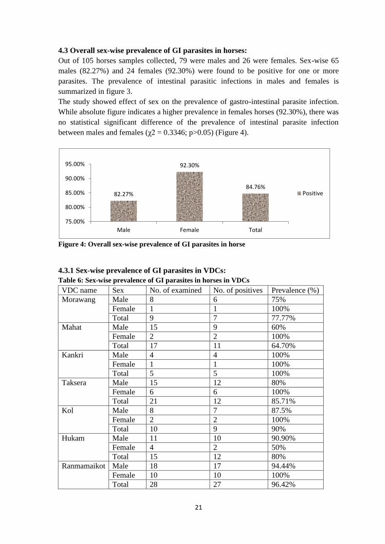
4.3 Overall sex-wise prevalence of GI parasites in horses:
Out of 105 horses samples collected, 79 were males and 26 were females. Sex-wise 65
males (82.27%) and 24 females (92.30%) were found to be positive for one or more
parasites. The prevalence of intestinal parasitic infections in males and females is
summarized in figure 3.
The study showed effect of sex on the prevalence of gastro-intestinal parasite infection.
While absolute figure indicates a higher prevalence in females horses (92.30%), there was
no statistical significant difference of the prevalence of intestinal parasite infection
between males and females (χ2 = 0.3346; p>0.05) (Figure 4).
Figure 4: Overall sex-wise prevalence of GI parasites in horse
4.3.1 Sex-wise prevalence of GI parasites in VDCs:
Table 6: Sex-wise prevalence of GI parasites in horses in VDCs
VDC name Sex No. of examined No. of positives Prevalence (%)
Morawang Male 8 6 75%
Female 1 1 100%
Total 9 7 77.77%
Mahat Male 15 9 60%
Female 2 2 100%
Total 17 11 64.70%
Kankri Male 4 4 100%
Female 1 1 100%
Total 5 5 100%
Taksera Male 15 12 80%
Female 6 6 100%
Total 21 12 85.71%
Kol Male 8 7 87.5%
Female 2 2 100%
Total 10 9 90%
Hukam Male 11 10 90.90%
Female 4 2 50%
Total 15 12 80%
Ranmamaikot Male 18 17 94.44%
Female 10 10 100%
Total 28 27 96.42%
82.27%
92.30%
84.76%
75.00%
80.00%
85.00%
90.00%
95.00%
Male Female Total
Positive

22
Male sex-wise, the highest prevalence (100%) revealed in Kankri VDC and least
prevalence (60%) in Mahat VDC and female sex-wise, the highest prevalence (100%)
was revealed in six VDCs (MoMKTKoRm) and least prevalence (50%) revealed in
Hukam VDC (Table 6).
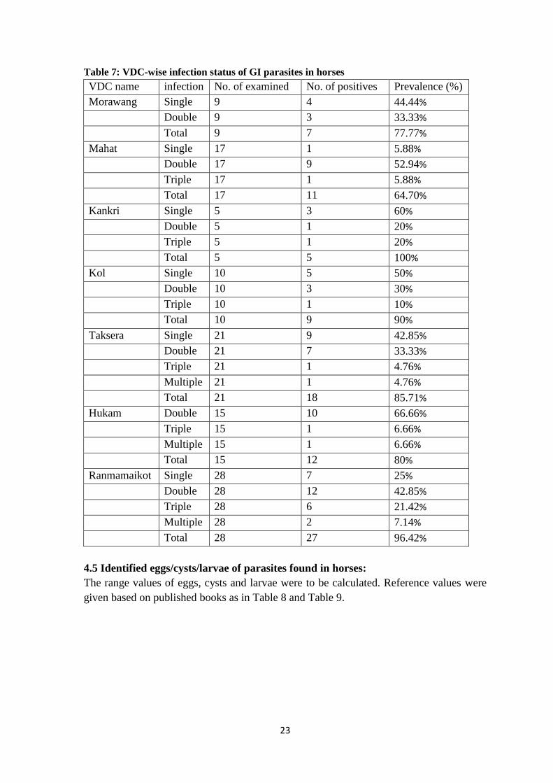
4.4. Overall infection-wise prevalence of GI parasites in horses:
Overall, horses were found to be infected with single, double, triple and multiple gastro-
intestinal parasites infections (Figure 4). The highest prevalence rate (42.85%) was noted
for double infection followed by single (27.61%), triple (10.47%) and multiple (3.80%).
The study showed effects of study area on infections status of parasites and there was
statistical significant difference among infection status in study area (χ2 = 84.277;
p<0.05) (Figure 5).
Figure 5: Overall infection status of GI parasites in horses
4.4.1 VDC infection-wise prevalence of GI parasites in horses:
Horses were found to be infected with single, double, triple and multiple GI parasites
(Table 7). The highest prevalence of single infection revealed in Kankri VDC (60%)
followed by no prevalence in Hukam VDC, highest double infection in Hukam VDC
(60%) followed by lowest in Kankri VDC (20%), highest triple infection in Ranmamaikot
VDC (21.42%) followed by no prevalence in Morawang VDC and highest multiple
infection in Ranmamaikot VDC (7.14%) followed by no prevalence from four VDCs
(MoMKKo).
27.61%
42.85%
10.47%
3.80%
0.00%
5.00%
10.00%
15.00%
20.00%
25.00%
30.00%
35.00%
40.00%
45.00%
Single Infection Double Infection Triple Infection Multiple Infection
Prevalence %
23
Table 7: VDC-wise infection status of GI parasites in horses
VDC name infection No. of examined No. of positives Prevalence (%)
Morawang Single 9 4 44.44%
Double 9 3 33.33%
Total 9 7 77.77%
Mahat Single 17 1 5.88%
Double 17 9 52.94%
Triple 17 1 5.88%
Total 17 11 64.70%
Kankri Single 5 3 60%
Double 5 1 20%
Triple 5 1 20%
Total 5 5 100%
Kol Single 10 5 50%
Double 10 3 30%
Triple 10 1 10%
Total 10 9 90%
Taksera Single 21 9 42.85%
Double 21 7 33.33%
Triple 21 1 4.76%
Multiple 21 1 4.76%
Total 21 18 85.71%
Hukam Double 15 10 66.66%
Triple 15 1 6.66%
Multiple 15 1 6.66%
Total 15 12 80%
Ranmamaikot Single 28 7 25%
Double 28 12 42.85%
Triple 28 6 21.42%
Multiple 28 2 7.14%
Total 28 27 96.42%
4.5 Identified eggs/cysts/larvae of parasites found in horses:
The range values of eggs, cysts and larvae were to be calculated. Reference values were
given based on published books as in Table 8 and Table 9.
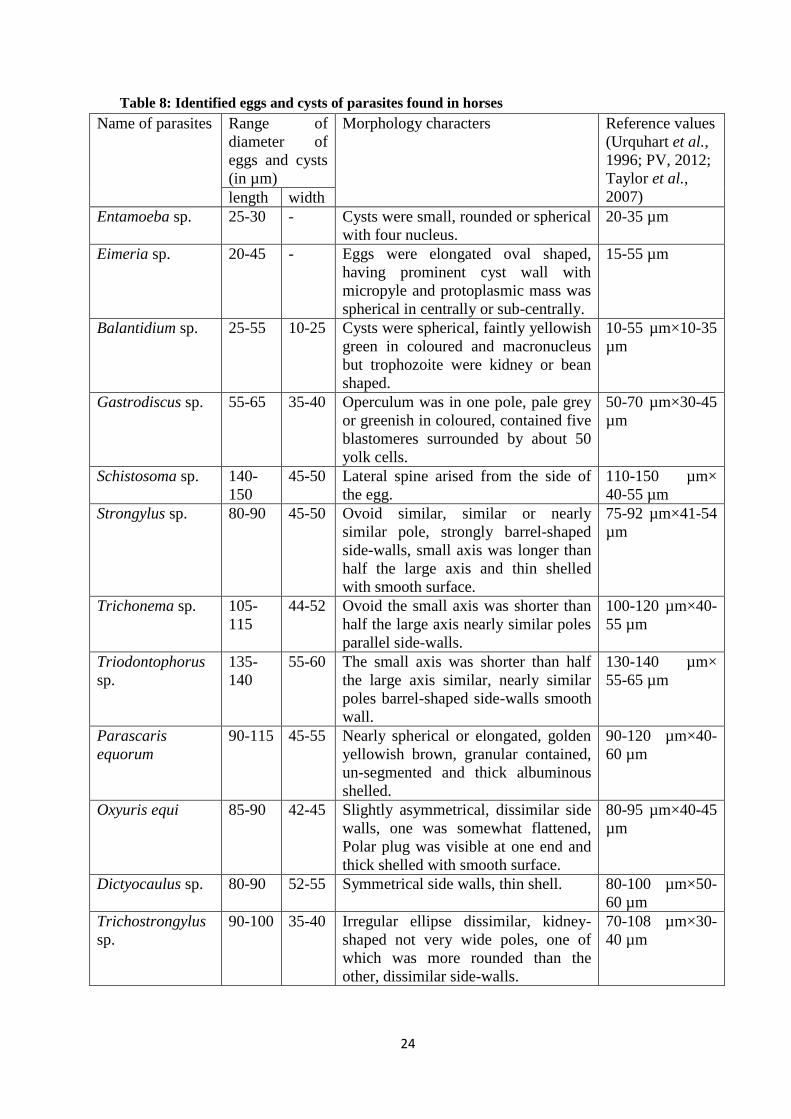
24
Table 8: Identified eggs and cysts of parasites found in horses
Name of parasites Range of
diameter of
eggs and cysts
(in µm)
Morphology characters Reference values
(Urquhart et al.,
1996; PV, 2012;
Taylor et al.,
2007) length width
Entamoeba sp. 25-30 - Cysts were small, rounded or spherical
with four nucleus.
20-35 µm
Eimeria sp. 20-45 - Eggs were elongated oval shaped,
having prominent cyst wall with
micropyle and protoplasmic mass was
spherical in centrally or sub-centrally.
15-55 µm
Balantidium sp. 25-55 10-25 Cysts were spherical, faintly yellowish
green in coloured and macronucleus
but trophozoite were kidney or bean
shaped.
10-55 µm×10-35
µm
Gastrodiscus sp. 55-65 35-40 Operculum was in one pole, pale grey
or greenish in coloured, contained five
blastomeres surrounded by about 50
yolk cells.
50-70 µm×30-45
µm
Schistosoma sp. 140-
150
45-50 Lateral spine arised from the side of
the egg.
110-150 µm×
40-55 µm
Strongylus sp. 80-90 45-50 Ovoid similar, similar or nearly
similar pole, strongly barrel-shaped
side-walls, small axis was longer than
half the large axis and thin shelled
with smooth surface.
75-92 µm×41-54
µm
Trichonema sp. 105-
115
44-52 Ovoid the small axis was shorter than
half the large axis nearly similar poles
parallel side-walls.
100-120 µm×40-
55 µm
Triodontophorus
sp.
135-
140
55-60 The small axis was shorter than half
the large axis similar, nearly similar
poles barrel-shaped side-walls smooth
wall.
130-140 µm×
55-65 µm
Parascaris
equorum
90-115 45-55 Nearly spherical or elongated, golden
yellowish brown, granular contained,
un-segmented and thick albuminous
shelled.
90-120 µm×40-
60 µm
Oxyuris equi 85-90 42-45 Slightly asymmetrical, dissimilar side
walls, one was somewhat flattened,
Polar plug was visible at one end and
thick shelled with smooth surface.
80-95 µm×40-45
µm
Dictyocaulus sp. 80-90 52-55 Symmetrical side walls, thin shell. 80-100 µm×50-
60 µm
Trichostrongylus
sp.
90-100 35-40 Irregular ellipse dissimilar, kidney-
shaped not very wide poles, one of
which was more rounded than the
other, dissimilar side-walls.
70-108 µm×30-
40 µm

25
Table 9: Unidentified larvae of parasites found in horses
Name of parasites Range of diameter
of larvae found
(in µm)
Morphology characters Reference values
(Taylor et al.,
2007)
length width
Rhabdiasoidae
larvae
350-500 15-20 Cuticle and smooth body, mouth
beared lips and were short tail
than others larvae.
300-600 µm×15-
25 µm
Strongyloidae
larvae
530-700 15-18 Mouth was without lips with leaf
crowns. Buccal capsule well
developed.
300-800 µm×15-
20 µm
Dictyocaulidae
larvae
200-400 10-15 Tail was a punctiform and
transparent projection, no
intestinal cells and grandular
contents for L2-larvae.
200-400 µm×10-
15 µm
4.6 Husbandry practices in the study area based on questionnaires survey:
Out of 48 respondents interviewed, 47.91% were found to be the highest proportion that
can read and write followed by primary (27.08%), above primary (20.83%) and illiterate
(4.16%) respectively (Figure 6). 72.91% of respondents were found to be freshwater
users, 22.91% river water users and 4.16% tap or pond water users for equines
respectively (Figure 7). 62.5% of respondents were found to be used equines for transport
income and 37.5% used for fertilizer as in soil to increase the soil fertility (Figure 8).
Mixed feeding source was found to be highest proportion (56.25%) followed by natural
(20.83%), shrubs and forest (12.5%) and grain residue (10.41%) respectively (Figure 9).
Out of 48 respondents, 50% were found to be kept equines together with family, 31.25%
with simple shed and 18.75% with separate house respectively (Figure 10). In the study
area, 95.83% respondents were found to be responded that there was no veterinary clinic
and only 4.16% respondents responded with available of veterinary clinic (Figure 11).
66.66% respondents were found to treat equines at home, 29.16% after serious infection
and 4.16% in VDC veterinary clinic (Figure 12). The highest cleaning rate (52.08%) was
noted for 4-7 days followed by 1-3 days (37.5%) and 8-15 days (10.41%) respectively
(Figure 13). From the questionnaire, it was known that most of equines owner feed their
equines twice a day (70.83%) followed by thrice (20.83) and four times a day (8.33%)
during working days (Figure 14). Out of 48 respondents, 52.08% feed their equines thrice
a day, 27.08% feed four times a day and 20.83% feed twice a day during resting period
(Figure 15).

26
Figure 6: Educational status of respondents Figure 7: Feed of water sources for equines
Figure 8: Utilization or purpose of equines Figure 9: Major feed sources for equines
Figure 10: Housing conditions of equines Figure 11: Veterinary clinic available
47.91%
27.08%
20.83%
4.14
Educational status
Read andwrite
Primary
Aboveprimary
Illiterate
72.91%
22.91% 4.16%
Water source
Smallfreshwater
Rivers
Tap or pondwater
62.50%
37.50%
Utilization
Transportincome
Fertilizer56.25%
20.83%
12.50% 10.41%
Feed source supply
Mixed typed
Naturalpasture
Shrubs andforest
Grain residue
50% 31.25%
18.75%
Housing condition
Together withfamily
Simple shed
Separate
95.83%
4.16%
Veterinary clinic availalble
No
Yes
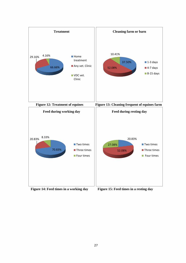
27
Figure 12: Treatment of equines Figure 13: Cleaning frequent of equines farm
Figure 14: Feed times in a working day Figure 15: Feed times in a resting day
66.66%
29.16% 4.16%
Treatment
Hometreatment
Any vet. Clinic
VDC vet.Clinic
37.50%
52.08%
10.41%
Cleaning farm or barn
1-3 days
4-7 days
8-15 days
70.83%
20.83% 8.33%
Feed during working day
Two times
Three times
Four times
20.83%
52.08%
27.08%
Feed during resting day
Two times
Three times
Four times

28
Photographs of identified eggs/cysts/larvae of parasites found in horses
Protozoan parasites (with 10Xx40X):
Photo 1: Entamoeba sp. (25 µm)
Photo 2: Balantidium sp. (35 µm) cyst
Photo 3: Balantidium sp. (45x22 µm)
trophozoite
Photo 4: Eimeria sp. (22 µm)
Trematode parasites (with 10Xx 40X and 10Xx10X):
Photo 5: Gastrodiscus sp. (60x36 µm)
Photo 6: Schistosoma sp. (145x45 µm)
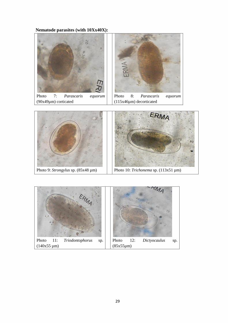
29
Nematode parasites (with 10Xx40X):
Photo 7: Parascaris equorum
(90x49µm) corticated
Photo 8: Parascaris equorum
(115x46µm) decorticated
Photo 9: Strongylus sp. (85x48 µm)
Photo 10: Trichonema sp. (113x51 µm)
Photo 11: Triodontophorus sp.
(140x55 µm)
Photo 12: Dictyocaulus sp.
(85x55µm)
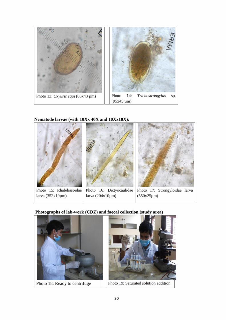
30
Photo 13: Oxyuris equi (85x43 µm)
Photo 14: Trichostrongylus sp.
(95x45 µm)
Nematode larvae (with 10Xx 40X and 10Xx10X):
Photo 15: Rhabdiasoidae
larva (352x19µm)
Photo 16: Dictyocaulidae
larva (204x10µm)
Photo 17: Strongyloidae larva
(550x25µm)
Photographs of lab-work (CDZ) and faecal collection (study area)
Photo 18: Ready to centrifuge
Photo 19: Saturated solution addition
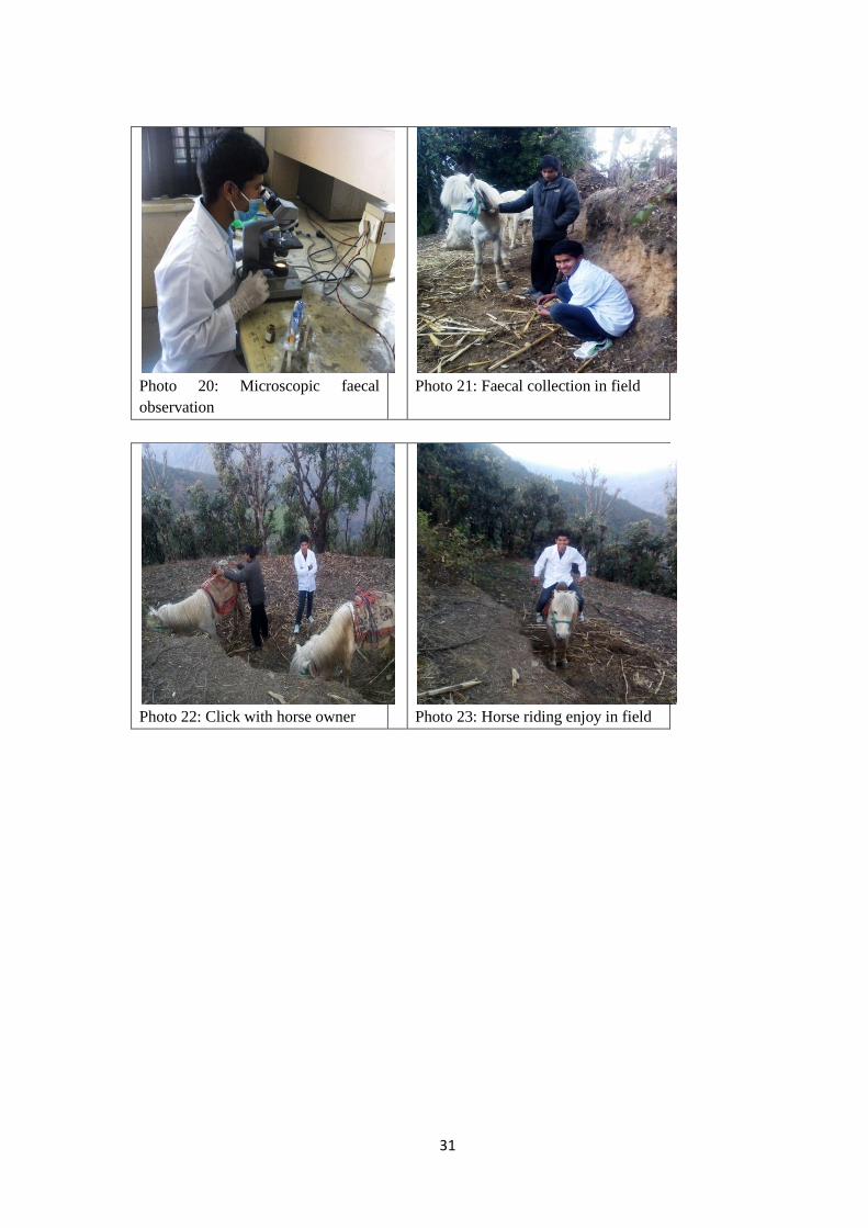
31
Photo 20: Microscopic faecal
observation
Photo 21: Faecal collection in field
Photo 22: Click with horse owner
Photo 23: Horse riding enjoy in field
32
5. DISCUSSION
It is well known that parasites are cosmopolitan in distribution and all animals bear
different kind of parasites. Many studies have been carried out regarding the gastro-
intestinal parasites of domestic livestock. Veterinarians are always interested to research
about domestic animals but very few researches have been carried out to equines
population than other domestic animals. Decrease of grazing areas, transport facilities,
lack of pony farmer, nutritional factors, machine-able agro-facilities, lack of manpower
for care, lack of breeding and health related factors are identified as the major parameters
for declining of horse population in context of our country. Horses are suffering from
different type of parasites and they shed parasites mainly by defecation and urination and
when other horses get infection from the contaminated area. Most of protozoa don‟t cause
poor body condition but helminth parasites can cause death of horses when they are
severely infected.
In the present study, the prevalence of gastro-intestinal parasites of horses (Equus
caballus) has been carried out for the first time in Nepal as comprising large population
samples and first time from Rukum district. The research so far has not been adequate
enough, prevalence figures of the present research work is mostly compared with the
research work done in other countries and only few with our national context regarding
the topic. The present study explored parasites prevalence in relation to husbandry
practice as in general.
The present study revealed that high prevalence of gastro-intestinal tract (GIT) parasites
in horses (Equus caballus) from seven VDCs (Morawang, Mahat, Kankri, Taksera, Kol,
Hukam and Ranmamaikot) of Rukum district. Overall positive prevalence rate was found
to be 84.76%. The total numbers of genera observed during faecal examination were 12 in
numbers. Among identified parasites, the Strongylus sp. showed the highest prevalence
(51.42%) followed by Eimeria sp. (20%), Trichostrongylus sp. (14.28%), Trichonema sp.
(13.33%), Parascaris equorum (10.47%), Balantidium sp. (9.52%), Dictyocaulus sp.
(8.57%), Triodontophorus sp. (7.61%), Gastrodiscus sp. (6.66%), Oxyuris equi (4.76%),
Entamoeba sp. (3.80%), Shistosoma sp. (1.90%) and unidentified nematode larvae
(7.61%).
The prevalence of gastro-intestinal parasites in the horses used for the study was high
(84.76%). The high prevalence of infection observed in the study agrees with works of
Aypak and bergu (2013), Adeppa et al. (2014), Mezgebu et al. (2013), Sultan et al.
(2014) and Wosu and Udobi (2014), who reported 88.6%, 84%, 92.71%, 75.62% and
76.1% in Aydin region of Torkey, Schimoga of Karnataka, Gondar town and Savannath
zone of Nigeria respectively. The prevalence in horses (84.76%) observed in the present
study is lower than Wannas et al. (2012), Tilahun et al. (2014) and Hassan (2014), who
reported 100%, 97.9% and 100% in Aldiwaniyah Governate, Hawassa Town and Baquba
city respectively and higher than Saeed et al. (2010), Adam et al. (2013), Regassa and
Yimer (2013), Yadav et al. (2014), Tesfu et al. (2014), Shiret and Samuel (2015), Samuel
et al. (2015), Molla et al. (2015) and Sokol et al. (2015), who reported 65.1%, 24.6%,
60.68%, 59.25%, 63.7%, 59.3%, 55.7%, 69% and 51.7% in Kurfa Chale of Ethiopia,
33
North Darfun, South Wolla Ethiopia, Jabalpur of North Indea, Hawassa Town, Mekelle
Region, Kombolchu Town, Menzkeya Gerbil district and Warmia of Mazury
respectively. The high prevalence rate of present study could be due to samples collection
during spring because eggs excretion during spring and summer season was high (Saeed
et al., 2010; Nielsen, 2012). All most all horses used in the study were working horses
that mainly fed poorly to grain and byproducts of grains and they are less exposed to
pasture grazing in the rest time period. The high prevalence rate (84.76%) of present
study characterized by also poor husbandry practice of the study area (Adebabay, 2009;
Sisay et al., 2007; Mezgebu et al., 2013; Adeppa et al., 2014). The difference among
these findings from different regions might be due to variation in management system,
geo-graphical climatic condition, sample size, sample collection period and sampling
method differences (Wannas et al., 2012; Tilahun et al., 2014; Yadav et al., 2014).
The prevalence rates of present study from seven VDCs were 100% (Kankri), 96.42%
(Ranmamaikot), 90% (Kol), 85.71% (Taksera), 80% (Hukam), 77.77% (Morawang) and
64.70% (Mahat) respectively. Four VDCs (Taksera, Kol, Hukam and Ranmamaikot) were
situated in the highland and three VDCs (Kankri, Morawang and Mahat) were situated in
the low land of Rukum district. The prevalence rate variations in the present study from
seven VDCs could be due to mainly altitudinal variations and habitat variations in their
respective VDCs. From present study, highland of cooler region and habitat near to river
or water source was more infected than lowland (Courtney, 1999; Holland et al., 2001).
Detection of highest prevalence of Strongyle type was 72.38% in the present study agrees
with works of Sultan et al. (2014), Mezgebu et al. (2013), Paudel (2007) and Tesfu et al.
(2014), who reported 72.66%, 66.67%, 65.85% and 64.9% in Kurfa chale, Gondar Town,
Bharatpur Chitwan and Hawassa respectively. The present result was also similar with the
reports of different researchers (Relf et al., 2013; Dopfer et al., 2004; Gurler et al., 2010;
Hinney et al., 2011; Kornas et al., 2010; Papazahariadou et al., 2009; Rehbein et al.,
2013), who reported the prevalence rate ranging from 61-100% of horses in most
European countries. The present result is higher than works of Saeed et al. (2010),
Wannas et al. (2012), Regassa and Yimer (2013) and Samuel et al. (2015), who reported
58.5%, 50%, 58.5% and 47.4% in Kurfa chale, Aldiwaniyah Governorate, South wollo
and Kombolcha Town respectively but very low result with 25% (Yadav et al., 2015) and
24% (Tiroshlevy et al., 2015) reported from Jabalpur and Israel respectively. But this
report is very less than the reports of Alemayehu (2004) and Getachew et al. (2010) from
East Shewa and Adau and Bost of East Shewa that revealed 100% and 99% prevalence
respectively. Over 94% of the horses passed strongyle eggs reported by Lan et al. (1996),
Holland et al. (2001). The high prevalence of strongyle in the present study clearly
demonstrates that strongyle infections are high in the study area. The high faecal eggs
prevalence could be explained by the climate, geo-graphical area that are suitable to
development, survival and transmission of pre-parasitic stages on pasture during most of
the year and by the absence of anti-parasitic treatments (Nielsen et al., 2006; Kaplan and
Nielsen, 2010; Toscan et al., 2012; Andersen et al., 2013).
The highest prevalence of Strongylus sp. was 51.42% in the current study which is close
agreement with works of Wosu and Udobi (2014) and Adeppa et al. (2014) and by Poudel
(2007), who reported 55.3%, 52.38% and 48.78% in North Nigeria, Schimoga Kornataka
34
and Sainik stud farm of Bharatpur Chitwan respectively but was lower result than Nigeria
(68.8%) by Umar et al. (2013) and was higher result than Baquba city (44%) by Hassan
(2014). Although equine strongylosis was well documented in cooler climate region of
North Vietnam but little information was available from tropical countries (Courtney,
1999; Holland et al., 2001).
Trichonema sp. is another common nematode parasite and also called as small strongyle
that prevalent in Horses. Small strongyles are considered to be the main parasitic
pathogen of the Horses (Khan et al., 2015). The parasite has been reported from Ethiopia
(Saeed et al., 2010), North Darfun (Adam et al., 2013) and Chitwan (Paudel, 2007). The
prevalence of Trichonema sp. was 13.33% which is similar with 17.07% recorded by
Paudel (2007) from Bharatpur Chitwan. It was felt difficult do discuss present result with
others because most of researchers has not specified the parasite and has included in
strongyle type. The present similar result with Paudel (2007) indicates that Trichonema
sp. was not affected by variation of climate, altitude and geographical areas.
Triodontophorus sp. is a large strongyle nematode prevalent in horses or equines. The
Triodontophorus sp. reported first time for Nepal in horse. The prevalence of
Triodontophorus sp. (7.61%) reported in the present study is close agreement with 11%
(Sapkota, 2009) but was lower than with 33.2% (Tilahun et al., 2014) and was higher
than with 1.15% (Rani, 2010) prevalence reported in Lalitpur, Hawassa Town and Banke
district respectively. Difference in prevalence rate could be due to climatic condition,
altitudinal and geographical variances. The low prevalence rate of Triodontophorus sp.
from present finding indicates that study area is less contaminated with parasite.
Gastrodiscus sp. had been reported from different authors (Karki and Manandhar, 2006;
Paudel, 2007; Sapkota, 2009; Saeed et al., 2010; Mezgebu et al., 2013; Adeppa et al.,
2014) from different countries. The prevalence (6.66%) of Gastrodiscus sp. reported in
present study is close agreement with 7.14% (Adeppa et al., 2014), 7.31% (Paudel, 2007)
6% (Karki and Manandhar, 2006) from Schimoga of Kornataka, Bharatpur Chitwan and
Udyapur district respectively. Present finding result was higher than findings of Mezgebu
et al. (2013) and Rani (2010), who reported 2.86% and 2.89% from Gondar Ethiopia and
Banke district respectively and was lower than with 30% (Sapkota, 2009). From these
results, it was seen that cooler and temperate climate is suitable for development of
Gastrodiscus sp. than warm and tropical climate.
The Shistosoma sp. was reported first time for Nepal in horse. The prevalence (1.90%) of
Shistosoma sp. reported in present study close agrees with 0.31% (Matto et al., 2015)
from Mumbai and Pune state but was very less than with 32.36% (Rani, 2010) reported
from Banke district. The result showed that present study area is also suitable for snail
(intermediate host) development. Lower result of present study than Rani (2010) could be
due to cooler and temperate climate is present in the present study area.
The prevalence of Trichostrongylus sp. in horses was found 14.28%. Trichostrongylus sp.
reported in the present study was very less than finding of Paudel (2007) who reported
80.48% from Bharatpur Chitwan and was less than findings of Wannas et al. (2012) and
Aypak and Bergu (2013), who reported 25% and 28.3% from Al Diwaniyah Governorate
and Aydin Torkey respectively. The lower result of present study could be due to low
temperature and cooler climatic geographic area than other regions as mentioned above.
35
The prevalence rate of Parascaris equorum is (10.47%) almost similar 10.71% (Adeppa
et al., 2014) 7.9% (Kornas et al., 2010), 6.3% (Umar et al., 2013), 13.9% (Ionita et al.,
2013) and 15.51% (Sultan et al., 2014). But lower than 40.90% (Wannas et al., 2012),
43.8% (Mezgebu et al., 2013) 18.75% (Yadav et al., 2014), 26.2% (Tesfu et al., 2014),
55.8% (Tilahun et al., 2014) and was higher than 4.5% (Regassa and Yimer, 2013), 3.9%
(Hasson, 2014), 1.8% (Shiret and Samuel, 2015), 4.7% (Samuel et al., 2015) and 5%
(Tiroshlevy et al., 2015). The lower prevalence of Parascaris equorum in present study
could be due to collection of faecal samples mostly from adult working horses and few
only from young horses. P. equorum mainly occurs in young horses less than 3 years old
and infection with this parasite is restricted to animals less than 5 years old (Mfitilodze
and Hutchinson, 1989; Bucknell et al., 1995). The difference in prevalence of Parascaris
equorum from different reports in developing countries is somewhat conflicting and this
could be due to compromised immune responses relating to concurrent diseases, but is
worthy of further investigation (Melissa et al., 2010).
The prevalence of Oxyuris equi was 4.76% in horses. This result is similar with Regassa
and Yimer (2013), Adeppa et al. (2014), Mezgebu et al. (2013), Sultan et al. (2014),
Samuel et al., (2015) and Fikru et al., (2005), Shiret and Samuel (2015) who reported 4%,
4.7%, 0.95%, 1.47%, 2.8% , 2.1% and 8.8% respectively. But, lower than results of
Wannas et al. (2012), Umar et al. (2013), Wosu and Udobi (2014) and Tilahun et al.
(2014) who reported 11.36%, 27.1%, 30.2% and 34.2% respectively. The lower result
(4.76%) of Oxyuris equi in present study might be due to low temperature and collection
methods because all samples were collected from captivated horses from behind of
individual horse (not from the direct rectum) of each of them. This parasite differs from
most other equine nematodes since the worm does not shed any eggs into the faeces,
instead the female deposit her eggs at the skin around the anus. Eggs stick to the
surroundings when the horse rubs its tail. Eventually the skin dry and fall of eggs in the
surrounding when the horse rubs it‟s tail (Reinemeyer and Nielsen, 2013; Taylor et al.,
2007).
The Dictyocaulus sp. was reported first time for Nepal in horse. In several studies, 50-
80% of donkeys have been found infected with Dictyocaulus sp. because donkeys act as
reservoir host for the parasite (Clayton and Duncan, 1981). Dictyocaulus sp. was reported
from different authors from different countries (Saeed et al., 2010; Umar et al., 2013;
Tolossa and Ashenafi, 2013; Tilahun et al., 2014). The prevalence of 8.57% of
Dictyocaulus sp. was recorded in the present study is similar with report of Umar et al.,
(2013), who reported 10.4% in Nigeria but, was higher than with 3.7% (Tilahun et al.,
2014), 2.5% (Saeed et al., 2010) and 0.5% (Tolossa and Ashenafi, 2013). The findings
are relatively lower than the present finding, this could be due to good husbandry
practice, climatic and environmental differences, between countries and differences in
access to drugs may partly explain the variation (Mezgebu et al., 2013; Adeppa et al.,
2014)..
Only few studies have to be done regarding protozoan parasites to horses all over the
world. The prevalence of 20% of Eimeria sp. was recorded in the present study is very
less than the report of Shrestha (2015), who reported 100% in Illam community forest.
But higher than reports of Wannas et al. (2012), Alharis (2001) and Alaloust et al. (1994),
36
who reported 6.81%, 10.96% and 4% and was very high than reports of Epe et al. (1993)
and Tavassoli et al. (2010), who reported 0.6% and 0.5% in Dtsch Tierazll and Poland
respectively.
The Balantidium sp. was reported by Wannas et al. (2012) and Shrestha (2015). The
prevalence rate of Balantidium sp. (9.52%) is nearly similar with 15.90% (Wannas et al.,
2012) in Al Diwaniyah Governorate and was lower than report of Shrestha (2015), who
reported 20% in Illam community forest.
It was felt difficult to discuss about the present study of Entamoeba sp. (3.80%) with
others due to lack of species finding regarding the parasite. But, Wannas et al. (2012)
reported 3.57% in donkeys in Governorate which is almost similar with present result.
From Nepal, Shrestha (2015) reported Entamoeba sp. (40%) from a total of 5 samples
examination of horses which was very high than the present finding result.
The lack of cestodes in current study provides evidence to back up the scarcity of
infection by this class of parasites which was also reported by Meana et al. (1998). Meana
et al. (1998) pointed to the coprological methods have a lower likelihood of diagnosing
cestode infection when horses have less than 100 tapeworms.
There was no significant association (χ2 = 5.7161; p>0.05) of VDCs (study areas) and
parasitic prevalence, which was 7 (77.77%), 11 (64.70%), 5 (100%), 9 (90%), 18
(85.71%), 12 (80%) and 27 (96.42%). This type of result has also been reported by Shiret
and Samuel (2015).
The infection rate in females (92.30%) higher than in male horses (82.27%) reported in
present study is similar with 60.97% and 58.51% (Yadav et al., 2014), 61.5% and 38.5%
(Hasson, 2014) and 80.8% and 58.8% (Wosu and Udobi, 2014) for females and males
respectively. Similar finding have been reported by Singh et al. (2012), who have found
higher prevalence in female equines (75.73%) as compared to male (72.30%). The
present result is in agreement with those reported by Love and Duncan (1992) and Smith
(2002). But, Umar et al. (2013) reported male infection is higher than female infection.
The prevalence of female horses than male horses could be due to hormonal activities as
mentioned by Dietz et al. (1984). Since pregnant and lactating female are more
susceptible to parasitic infections this may be the cause of higher prevalence in females
than males. There was no significant association (χ2 = 0.3346; p>0.05) between male and
female parasitic prevalence, which was 65 (82.27%) and 24 (92.30%) respectively. The
finding of this type of result was reported by Saeed et al. (2010), Mezgebu et al. ( 2013),
Aypak and Bergu (2013), Tesfu et al. (2014) and Haimainot et al. (2015) but, opposite
result i.e. significant different result reported by Umar et al. (2013), Aypak and Bergu
(2013) and Hasson (2014). The study showed effects of study area on infections status of
parasites and there was statistical significant difference among infection status in study
area (χ2 = 84.277; p<0.05).
In current study, mixed infection were detected in 57.14% of horses which is almost
similar with the finding of Tolossa and Ashenafi (2013) in horses of Arsi-Bale highlands
of Oromiya region and Uslu and Guclu (2007) in Torkey, who reported 59.1% and 50%
respectively. Tilahun et al. (2014) reported 83.2% of mixed infection from Hawassa
Town, which is higher than present study but, present result was higher than with 25%
(Adeppa et al., 2014), 20.14% (Ionita et al., 2014), 16.8% (Shiret and Samuel, 2015) and
37
12.5% (Samuel et al., 2015) in Schimoga, Romania, Mekelle and Kombolcha
respectively. These lower result than present study could be due to use of anti-parasitic
drugs and de-worming programs in their studied area (Wannas et al., 2012; Regassa and
Yimer, 2013). But present higher mixed infection (57.14%) could be due to lack of horse
de-worming, very less use of anti-parasitic drugs, dietary deficiency, poor management
system, lack of knowledge for sanitation and climatic and geographical variation of the
present study area (Saeed et al., 2010; Sisay et al., 2007; Mezgebu et al., 2013).
In the present study area, most of respondents were not found educationally qualified for
management of equines. Most of respondents were found used open river water source for
equines which made them easy to get different parasitic infections. The main purpose of
keeping horses in their house was to get income from transportations of human needs
where small road to be developed. Mainly mixed type of food particles was found to feed
their horses especially residue of grains, grasses and shrubs that made also possibility of
high parasitic infection to them. Most of respondents found to be kept their horses with
together family that made easy to exchange of human parasites. All most all respondents
responded that there was no available of veterinary clinic for treatments. In the study area,
horses found to be treated at home with turmeric powder, garlic and plant medicines.
Most of respondents found to be cleaned their barns within a week. It means that there
was very poor management of horses. The carrier horse mainly found to feed (food grain
particles) twice during working day but resting horse found to feed (mainly natural food
particles) thrice a day. Here feeding frequent found more in resting horse than working
horse because working horse gets more food particles at a time to transport different
human needs. From these husbandry practices, there was possibility of high parasitic
infection. The high parasitic prevalence rate of present study could be also related with
poor management or poor husbandry practices of study area. The result noted in the
current study related to husbandry practice was generally in agreement with the reports of
Solomon (2004) from Sinana and Dinsho districts of Bale highland, Asamenew (2007)
from Bahir and Belete (2006) from highland and rift valley of Ethiopia.
38
6. CONCLUSION AND RECOMMENDATIONS
6.1 Conclusion:
Overall, present study demonstrates the prevalence of GI parasitic infection in horses in
seven VDCs of Rukum district was 84.76% using floatation and sedimentation technique.
The total numbers of genera observed during faecal examination were 12 in numbers.
Among them, the Strongylus sp. showed the highest prevalence (51.42%) followed by
Eimeria sp. (20%), Trichostrongylus sp. (14.28%), Trichonema sp. (13.33%), Parascaris
equorum (10.47%), Balantidium sp. (9.52%), Dictyocaulus sp. (8.57%), Triodontophorus
sp. (7.61%), Gastrodiscus sp. (6.66%), Oxyuris equi (4.76%), Entamoeba sp. (3.80%),
Shistosoma sp. (1.90%) and unidentified nematode larvae (7.61%). Three genera of
parasites: Shistosoma sp., Triodontophorus sp. and Dictyocaulus sp. have been reported
first time for Nepal in horses.
The present study revealed infection rate higher in females (92.30%) than in male horses
(82.27%). The present study shows horses were more infected with mixed infection than
single infection. The prevalence is higher in the study area as a normal and has been
characterized by poor husbandry practices due to lack of veterinarian information and
lack of veterinary hospital. This study revealed that the predominant parasites were
Strongylus sp. and Eimeria sp. The study confirmed that horses were found to be most
susceptible and infested by various GI parasites. Management practices and different
locations can be considered as the important factors which influence the prevalence of GI
parasites. In present study, sex and location had statistically no influences on prevalence
of parasites but infection status showed significant association with study area. The
higher prevalence of present study shows an alarming situation in the study area. This
finding might be considered while designing control strategies of gastro-intestinal
parasites in horses and also be useful to other equines and ruminants as well.
6.2 Recommendations:
Based on the above conclusion, the following recommendations are forwarded:
Further study or molecular level characterization of parasites should be done.
Improved basic animal‟s husbandry management system should be practiced in
the study area.
Educate the horse owners regarding correct way to improve animal‟s
management system, importance of parasites and it is preventive and controlling
systems.
Sufficient feed supply and minimizing contaminated open grazing of horse is
important.
Balancing of the work load and duration should be managed.
By implementing veterinary health program, appropriate prevention and control
strategy supported by both government as well as private sector to minimize the
gastro-intestinal parasites of horses.
39
7. REFERENCES
Abayneh, T.F., Gebreab, B., Zekarias, G. and Tadess, A. 2002. The potential role
of donkeys in land fillage in Central Ethiopia. Bulletin of Animals Health and Production
in Africa, 50: 172-178.
Adam, A.A., Suleiman, S.E. and Seri, H.I. 2013. The prevalence and intensity of
gastrointestinal helminths in North Dardfur, Sudan. College of Vet. Med., Journal of
Sciences and Tecnology, 14.
Adebabay, K.B. 2009. Characterization of milk production systems, marketing
and on farm evaluation of the effect of feed supplementation on milk yield and milk
composition of cows at Bure district, Ethiopia. M.Sc. Thesis. Bahir dar University.
Adeppa, J., Ananda, K.J., Krishna C.M. and Satheesha, G.M. 2014. Incidence of
gastrointestinal parasites in horses of schimoga region, Karnataka state. Journal of
Parasitic Diseases, 1-3.
Ahmed, N.E., Lubna, M.E.A., Ramadan, M.Y. and Radwan, A.M.M. 2011. Study
on helminth parasites in necropsied donkeys in Egypt. Benha Veterinary Medical Journal,
1: 153-162.
Alaloust, T.I., Arselan, S.H. and Zongana, I.Q. 1994. Study of some intestinal
infection of horses in Mosul, Iraq. Vet. Med. Jouranal, 2: 85-91.
Alayande, M.O., Onakpa, M.M. and Lawal, M.D. 2003. Prevalence of intestinal
helminths of horses in Sokoto. Nigerian Journal of Parasitology, 24: 191-193.
Alemayehu, L. 2004. Case study on reproductive activity of equines in relation to
environment factors in Central Ethiopia, Berlin: hum bold university of Berlin, Ph.D.
Thesis. Autopsied in Poland. Veterinary Parasitology, 58: 99-108.
Alharis, K.M. 2001. Survey of the intestinal protozoa in the horses in Baghdad
city. M.Sc. Thesis. Veterinary Medicine Baghdad.
Ali, S. and Yagoob, G. 2015. Survey on fecal gastrointestinal helminthes in horses
of Jocky clubs in Aradabil City, Iran. Biological forum. An International Journal, 7(1):
106-110.
Andersen, U.V., Hakansson, I.T., Roust, T., Rhod, M. Baptiste, E.K. and Nielsen,
M.K. 2013. Developmental stage of strongyle eggs affects the outcome variations of real-
time PCR analysis. Vet. Parasitol., 191: 191-196.
Asaminew, T. 2007. Production, handling, traditional processing practices and
quality of milk in Bahir Dar milk shed area, Ethiopia. M.Sc. Thesis. Alemaya University,
Ethiopia.
Aypak, S. and Bergu, A. 2013. Prevalence of stomach helminths in equines. Vet.
Med., 24(1): 29-35.
Bechera, A.M., Mahlingc, M., Nielsend, M.K., Pfisterb, K. and Salzburg, S. 2010.
Selective anthelmintic therapy of horses in the federal states of Bavaria, Germany. An
investigation into strongyle egg shedding consistency. Vet. Parasitol., 171: 116-122.
Belete, A. 2006. Studies on cattle milk and meat production in fogera woreda:
production system, constraints and opportunities for development. M.Sc. Thesis. Debub
University, Awassa.
40
Bevilaqua, C.M.L., Rodrigues, M.L. and Concordet, D. 1993. Identification of
infective larvae of some common nematode strongylids of horses. Revue Med. Ved.,
144(12): 989-995.
Biu, A.A., Mohaammed, G.A., Yachilla, B.K.M., Edet, B.E., Aibu, U.U.S.,
Makinta, A. et al. 2014. Comparision parasitological diagnosis techniques of
gastrointestinal parasitic infection of horses in Maiduguri, North Eastern Nigeria.
American Journal of Biomedical and Life Sciences, 2(6): 150-155.
Bodecek, S., Jahn, P., Dobesova, O. and vavrouchova, E. 2010. Equine
cyathostomosis: case reports. Veterinary Medicine, 55: 187-193.
Boxell, A.S., Gibson, K.T., Hobbs, R.P. and Thompson, R.C.A. 2004. Occurrence
of gastrointestinal parasites in horses in metropolitan Perth, Western Australia. Aust. Vet.
J., 82: 91-95.
Bucknell, D.G., Gasser, R.B. and Beveridge, I. 1995. The prevalence and
epidemiology of gastrointestinal parasites of horses in Victoria, Australia. International
Journal for Parasitology, 25(6): 711-724.
Bush, A.O., Lafferty, K.D., Lotz, J.M. and Shostak, A.W. 1997. Parasitology
meets ecology on its own terms: Margolis et al. revisited. Journal of Parasitology, 83(4):
575-583.
Butty, E.T. 2011. Detection of Cryptosporidium and Giardia duodenalis in
equines in Nineveh, Iraq. Iraqui Journal of Veterinary Sciences, 25(2): 43-46.
Capewell, L.G., Hunt, D., Guerrero, J., Newcomb, K. and Root, T. 2005. The
prevalence of strongyles in stabled and pastured horses in Vermont and efficacy of
anthelmintic programs in these horses. Intern. J. Appl. Res. Vet Med., 3: 227-232.
Chapman, M.R., French, D.D. and Klei, T.R. 2002. Gastrointestinal helminths of
ponies in Louisiana: a comparison of species currently prevalent with those present 20
years ago. J. Parasito., 88: 1130-1134.
Chaudhry, A.H., Sohail, E. and Iqbal, Z. 1991. Studies on the prevalence and
taxonomy of the members of genus Strongylus and their effect on blood picture in
equines in Faisalabad (Pakistan). Pakistan Vet. J., 11: 179-181.
Claire, N.I. and Masterson, W.J. 1987. Molecular and Biochemical
Characterisation of surface antigens of Strongylus vulgaris of potential immunodiagnostic
importance. Parasitology, 25: 29-38.
Clayton, H.M. and Duncan, J.L. 1981. Natural infection with Dictyocaulus
arnfieldi in pony and donkey foals. Research in Veterinary Military Veterinaria, 26: 125-
128.
Courtney, C.H. 1999. Seasonal transmission of equine cyathostomes in warm
climates. Vet. Parasitol., 85: 173-180.
Dietz, O., Wiesner, E., Karger, S. and Turner, A.S. 1984. Diseases of the horse: A
hand book for science and practice Part II Basel Munchen, Paris, 303-354 pp.
Dopfer, D., Kerssens, C.M., Meijer, Y.G.M., Boersema, J.H. and Eysker, M.
2004. Shedding consistency of Strongyle-type eggs in Dutch boarding horses. Vet.
Parasitol., 124: 249-258.

41
Ehizibolo, J.O., Kamani, J., Ehizibolo, P.O., Egwu, K.O., Dogo, G.I. and Salami,
S.J.O. 2012. Prevalence and significance of parasites of horses in some states of Northern
Nigeria. J. Equine Sci., 23(1): 1-4.
Epe, C., Ising, V.S. and Stoye, M. 1993. Parasitological studies of equids, dogs,
cats, and hedgehogs during the year 1984-1991. Dtsch wochenschr, 100(11): 426-428.
Fikru, R., Reta, D. and Bizunesh, M. 2005. Prevalence of equine gastrointestinal
parasites in western high lands of Oromia, Ethiopia. Bulletin of the Animal Health
Production in Africa, 53(3): 161-166.
Fitsum, M. and Ahmed, K.M. 2015. Population dynamic production statistics of
horse and Ass in Ethiopia: A Review. Journal of Biology, Agriculture and Healthcare,
5(1): 57-62.
Francisco, I., Arias, M., Cortinas, F.J., Francisco, R., Mochales, E., Dacal, V. et
al. 2009. Intrinsic factors influencing the infection by helminth parasites in horses under
an oceanic climate area (NW Spain). J. Parasitol. Res., 5(6): 161-173.
Gardiner, C.H., Fayer, R. and Dubey, J.P. 1988. An atlas of protozoan parasites in
animals tissues. 1st edition U.S. Government printing office, Washington, D.C.
Gasser, R.B., Hung, G.C., Chilton, N.B. and Beveridge, I. 2004. Advances in
developing molecular diagnostic tools for strongyloid nematodes of equids: fundamental
and applied implications. Molecular and Cellular Probes, 18: 3-16.
Getachew, M., Alemayehu, F., Chala, C., Amare, B., Kassa, D., Burden, F. et al.
2014. A cross sectional sero-survey of some infectious diseases of working equids in
Central Ethiopia. Journal of Veterinary Medicine and Animal Health, 6(9): 231-238.
Goraya, K., Iqbal, Z., Sajid, M.S. and Muhammad, G. 2013. Frequency
distribution of equine diseases in three metropolises of the upper Punjab, Pakistan.
International journal of agriculture & biology, 15(6): 1067-1074.
Guo, P.F., Chen, T.T.W., Tsaihong, J.C., Ho, G.D., Cheng, P.C., Tseng, Y.C., et
al. 2014. Prevalence and species identification of Cryptosporidium from fecal samples of
horses in Taiwan. Taiwan Institute of Medical Sciences, 45(10): 6-12.
Gurler, A.T., Bolukbaş, C.S., Acici, M. and Umur, S. 2010. Check list of the
helminths of equines in Turkey. Turkiye. Parasitol. Derg., 34: 40-44.
Guzel, S., Tanriverdi, M. and Senlik, B. 2014. Selected biochemical parameters in
strongyles infections of horses and its relationship with the parasite burden. Bulgarian
Journal of Agricultural Science, 20(5): 1233-1236.
Haimanot, D., Addise, A., Tilahun, Z. and Girma, K. 2015. Prevalence of
strongyle infection in horses and donkeys in and around Dangila Town, Northwest
Ethiopia. School of Veterinary Medicine. Acta Parasitologica Globalis, 6(1): 14-19.
Hassan, S.E., Ghazy, A.A. and Rahma, E.H.A. 2010. Isolation and
characterization of immunodiagnostic antigen from Strongylus vulgaris infecting horses.
World Applied Sciences Journal, 8(2): 235-240.
Hasson, R.H. 2014. A study in the prevalence of horse‟s helminthes parasitic
infection in Baquba city, Digala. International Journal of Recent Scientific Research,
5(10): 1810-1813.
Hendrix, C.M. and Robinson, E. 2006. Diagnostic parasitology for veterinary
technicians. 3rd
edition. Philadelphia, Mosby.
42
Hinney, B., Wirtherle, N.C., Kyule, M., Miethe, N., Zessin, K.H. and Clausen,
P.H. 2011. Prevalence of helminths in horses in the state of Brandenburg, Germany.
Parasitol. Res., 108: 1083-1091.
Hodgkinson, J.E. 2006. Molecular diagnosis and equine parasitology. Veterinary
Parasitology, 136(2): 109-116.
Hodgkinson, J.E., Love, S., Lichtenfels, J.R., Palfreman, S., Ramsey, Y.H. and
Matthews, J.B. 2001. Evaluation of the specificity of fiveve oligoprobes for identification
of cyathostomin species from horses. International Journal of Parasitology, 31: 197-204.
Holland, W.G., Geurden, T., Do, T.T., Dorny, P. and Vercruysse, J. 2001.
Strongyle infections in horses from North Vietnam. Revue Elev. Med. Vet.Pays Trop.,
54(1): 29-31.
Hubert, J.D., Seahorn, T.L., Klei, T.R., Hosgood, G., Horohov, D.W. and Moore,
R.M. 2004. Clinical signs and hematologic, cytokine, and plasma nitric oxide alterations
in response to Strongylus vulgaris infection in helminth-naïve ponies. Can. J. Vet. Res.,
68: 193-200.
Ionita, M., Buzato, M.C., Enachescu, V. and Mitrea, I.L. 2013. Coprological
prevalence and intensity of gastrointestinal parasites in horses in Romanian studs:
preliminary data. Agrolite Scientific Journal, 2(1): 207-212.
Jubb, K.V.F., Kennedy, P.C. and Palmer, N. 1985. Pathology of domestic animals
3rd
ed. Academic Press, Orlando, 50 pp.
Kaplan, R.M. and Nielsen, M.K. 2010. An evidence based approach to equine
parasite control. Equine Veterinary Education, 22(6): 306-16.
Karki, K. and Manandhar, P. 2006. Preliminary investigation of prevalence of
gastro-intestinal parasites of mules in Udayapur district.
Khan, M.A., Roohi, N. and Rana, M.A.A. 2015. Strongylosis in equines: A
review. Journal of Animal and Plant Sciences, 25 (1): 1-9.
Kornas, S., Cabaret, J., Skalska, M. and Nowosad, B. 2010. Horse infection with
intestinal helminths in relation to age, sex, access to grass and farm system. Vet.
Parasitol., 174: 285-291.
Kornas, S., Marta, B., Vitalij, K. and Marta, S. 2010. Infection with large
gastrointestinal parasites in slaughtered horses. Bull Veterinary Institute polawy, 54: 577-
580.
Krecek, R.C., Malan, F.S., Reinecke, R.K. and Vos, V. 1987. Nematode parasites
from Burchell‟s zebras in South Africa. J. Wildl. Dis., 23: 404-411.
Kuzmina, T.A., Kuzmin, Y.I. and Kharchenko, V.A. 2006. Field study on the
survival, migration and overwintering of infective larvae of horse strongyles on pasture in
central Ukraine. Veterinary Parasitology, 141(3-4): 264-272.
Kuzmina, T.A., Lyons, E.T., Tolliver, S.C., Dzeverin, I.I. and Kharchenko, V.A.
2012. Fecundity of various species of strongylids (Nematoda: Strongylidae) parasites of
domestic horses. Parasitol Res., 111: 2265-2271.
Lamb, J., Hallowell, G.D., Harris, P., Zhua, X.Q. and Elsheikha, H.M. 2012.
Lipidomic analysis of serum from horses with strongyle infection. Journal of Veterinary
Science and Technology, 3: 1-4.
43
Lan, P.D., Lang, P.S., Phuc, D.V. 1996. Nematodes and nematodic disease of
domestic animals in Vietnam. Hanoi, Vietnam, Agriculture Press, 212 pp.
Lichtenfels, J.K., Gibbons, L.M. and Krecek, R.C. 2002. Recommended
terminology and advances in the systematic of the cyathostominea (nematode:
strongyloidea) of horses. Veterinary Parasitol., 107: 337-342.
Lichtenfels, J.R., Kharchenko, V.A. and Dvojnos, G. 2008. Illustrated
identification keys to strongylid parasites (strongylidae: Nematoda) of horses, zebras and
asses (Equidae). Veterinary Parasitology, 156: 4-161.
Love, S. and Duncan, J.I. 1992. The development of naturally acquired
Cyathostome infection in ponies. Vet. Parasitol., 44:227-142.
Love, S., Murphy, D. and Mellor, D. 1999. Pathogenicity of cyathostome
infection. Veterinary Parasitology, 85(2-3): 113-122.
Lyons, E.T., Swerczek, T.W., Tolliver, S.C., Bair, H.D., Drudge, J.H. and Ennis,
L.E. 2000. Prevalence of selected species of internal parasites in equids at necropsy in
central Kentucky (1995–1999). Veterinary Parasitology, 92(1): 51-62.
Mahmood, K.T. and Ashraf, M. 2010. Pharmacokinetics of ecofriendly
meloxicam in healthy horses. Pakistan Journal Science, 62(3): 198-201.
Margolis, L., Esch, G.W., Holmes, J.C., Kuris, A.M. and Schad, G.A. 1982. The
use of ecological terms in parasitology (report of an hoc committee of the American
Society of Parasitologists). The Journal of Parasitology, 68(1): 131-133.
Martin, K., Ray, N., Kaplan, M., Thamsborg, M., Susanne, J.M. and Olsen, N.
2007. Climatic influences on development and survival of free-living stages of equine
strongyles. Vet. J., 174: 23-32.
Matto, T.N., Bharkad, G.P. and Bhat, A. 2015. Prevalence of gastrointestinal
helminth parasites of equids from organized farms of Mumbai and Pune. Journal of
Parasitic Diseases, 39(2): 179-185.
McCraw, B.M. and J. Slocombe, O.D. 1976. Strongylus vulgaris in the horse: a
review. Canadian Vet. J., 17: 150-157.
Mcwilliam, H.E.G., Nisbet, A.J., Dowdall, S.M.J., Hodgkingson, J.E. and
Mathews, J.B. 2010. Identification and characterization of an immunodiagnostic marker
for cyathostomin developing stage larvae. International Journal of Parasitology, 40: 265-
275.
Meana, A., Luzon, M., Corchero, J. and Bautista, M.G.O. 1998. Reliability of
coprological diagnosis of Anoplocephala perfoliata infection. Veterinary Parasitology,
74(1): 79-83.
Meara, B.O. and Mulcahy, G. 2002. A survey of helminth control practices in
equine establishments in Ireland. Veterinary Parasitology, 109(1-2): 101-110.
Melissa, U., Kate, S., Thabo, L., Gillian, A. and Kristein, V. 2010. Coprological
prevalence and intensity of helminth infection in working horses in Lesotho. Tropical
Animal Health Production, 42(8): 2010.
Mezgebu, T., Tafesi, K. and Tamiru, F. 2013. Prevalence of gastro-intestinal
parasites of horses and donkeys in and around Gondar town, Ethiopia. Open Journal of
Veterinary Medicine, 2: 267-272.

44
Mfitilodze, M.W. and Hutchinson, G.W. 1989. Prevalence and intensity of non-
strongyle intestinal parasites of horses in Northern Queensland. Australian Veterinary
Journal, 66(1): 23-26.
Mitrea, L.I. 2011. Parazitologie si Boli Parazitare, Ceres Publishing House,
Bucharest.
Molla, B., Worku, Y., Shewaye, A. and Mamo, A. 2015. Prevalence of strongyles
infection and associated risk factors in equine in Menz keya gerbil district, North-eastern
Ethiopia. Journal of Veterinary Medicine and Animal Health, 7(4): 117-121.
Montenaro, S., Scala, S., Batelli, G. and Stancamplano, L. 2002. Epidemiology of
gastrointestinal nematode infections in horses in Sardinia. Obiettivie Documenti
Veterinari., 23: 35-42.
Niels, C., Kyvsgaarda, J., Jenny, L., Luz, A., Olivaresb, L., Martin, K., et al. 2011.
Prevalence of strongyles and efficacy of fenbendazole and ivermectin in working horses
in El Sauce, Nicaragua. Vet Parasitology, 181: 248-254.
Nielsen, M.K., Monard, J. and Olsen, S.N. 2006. Prescription only anthelmintics-
a questionnaire surveillance and control of equine strongyles in Denmark. Vet. Parasitol.,
135: 47-55.
Nielsen, M.K. 2012. Sustainable equine parasite control: Perspectives and
research needs. Vet. Parasitol., 185: 32-44.
Pandit, B.A, Shahardar, R.A, Jeyabal, L. 2008. Prevalence of gastro-intestinal
parasitic infestation in equines of Kashmir valley. Veterinary Journal of Agricultural
Sciences, 3(1): 22.
Papazahariadou, M., Papadopoulos, E., Diakou, A. and Ptochos, S. 2009.
Gastrointestinal parasites of stabled and grazing horses in central and northern Greece. J.
Equine Vet. Sci., 29(4): 233-236.
Pereira, J.R. and Vianna, S.S.S. 2006. Gastrointestinal parasitic worms in equines
in the Para´ıba Valley, State of S˜ao Paulo, Brazil. Veterinary Parasitology, 140(3-4):
289-295.
Perry, B.D., Randolph, T.F., Mcdermott, J.J., Sones, K.R. and Thornton, P.K.
2002. Investing in animal research to alleviate poverty. International Livestock Research
Institute, Nairobi.
Pilo, C., Alteaa, A., Pirinob, S., Nicolussic, P., varcasiaa, A., Genchid, M. et al.
2012. Strongylus vulgaris in horses in Italy. Vet. Parasitol., 184: 161-67.
Poudel, S. 2007. Prevalence of gastrointestinal parasites in horses with special
references to Strongylus species of sainik stud farm centre Bharatpur, chitwan. Blue –
cross Annu. Bull. NVSA., 9: 04-15.
PV. 2015. Veterinary parasitology. The practical veterinarian, pp 77-319.
Rahman, S.M., Dey, A.R., Kundu, U.K. and Begum, N. 2014. Investigation of
gastrointestinal parasites of herbivores at Dhaka national zoological garden of
Bangladesh. Journal Bangladesh Agril. Univ., 12(1): 79-85.
Rani, R. 2010. Determine the prevalence of helminths parasites in mules from
Nepalgunj of District Banke. M.Sc. Thesis. Central Department of Zoology, Tribhuvan
University, Kathmandu, Nepal.
45
Regassa, A. and Yimer, E. 2013. Gastrointestinal parasites of equines in south
Wollo zone, north eastern Ethiopia. Hawasa University, School of Vet. Med., Global
Veterinaria, 11(6): 824-830.
Rehbein, S., Visser, M. and Winter, R. 2013. Prevalence, intensity and seasonality
of gastro-intestinal parasites in abattoir horses in Germany. Parasitol. Res., 112: 407-413.
Reinemeyer, C.R. and Nielsen, M.K. 2013. Handbook of equine parasite control.
ames, Iowa. John Wiley & Sons, Inc.
Relf, V.E., Morgan, E.R., Hodgkinson, J.E. and Matthews, J.B. 2013. Helminth
egg excretion with regard to age, gender and management practices on U.K.
Thoroughbred studs. Parasitol., 140: 641-652.
Romaniuk, K., Reszka, K. and Lasota, E. 2004. Influence of animal breeding
manner on the occurrence of internal parasites. Wiadomosci Parazytologiczne, 50(3):
647-651.
Saeed, K., Qadir, Z., Ashraf, K. and Ahmad, N. 2010. Role of intrinsic and
extrinsic epidemiological factors on Strongylosis in horses. Journal of Animals and Plant
Sciences, 20(4): 277-280.
Samuel, E., Ashenafi, A., Natnael, M. and Shiret, B. 2015. Prevalence of Gastro-
intestinal nematode parasitic infections of horses and donkeys in and around Kombolcha
town. American-Eurasian Journal of Scientific Research, 10(4): 228-234.
Sapkota, R.C. 2009. Prevalence of helminthes parasites in mules in Brick Kiln of
Lalitpur district. B.V.Sc. Thesis. Himalayan College of Agricultural Sciences and
Technology, Purbanchal University, Gatthaghar, Bhaktapur.
Sharifi, K., Hassan, B. and Peyman, M. 2010. First Report of Dictyocaulus
arnfieldi Infestation in a horse in Mashhad, Iran. Iranian Journal of Veterinary Science
and Technology, 2(1): 46-49.
Shiret, B. and Samuel, D. 2015. Prevalence of major gastro-intestinal parasites of
horses in around Mekelle. World Journal of Animals and Sciences Research, 3(3): 1-10.
Shite, A., Admassu, B. and Abere, A. 2015. Large strongyle parastes in equine: A
review. Advances in Biological Research, 9(4): 247-252.
Shrestha, S. 2015. Gastro-intestinal parasites of Red Panda (Ailurus fulgens
cavier, 1825) and livestocks in community forest of Illam, Nepal. M.Sc. Thesis. Central
Department of Zoology, Tribhuvan university, Kathmandu, Nepal.
Singh, G. Soodan, J.S., Singla, L.D. and Khajuria, J.K. 2012. Epidemiological
studies on gastro-intestinal helminthes in horses and mules. Vet. Practitioner, 13: 23-27.
SIONA. 2013/14. Agri-business promotion and statistics section, Singh Durbar,
Kathmandu, Nepal.
Sisay, A., Maria, W., Azage, T. and Wemer, Z. 2007. Urban and peri-urban
farming systems and utilization of the natural resources in the North Ethiopian Highlands,
5 pp.
Smith, B.P. 2002. Large animal internal medicine 3rd
edition Mosby, 1438-1440
pp.
Sokol, R., Malgorzata, R.N., Moria, M., Andezej, R., Anna, R.L. and Sylwia, K.
2015. Eestimation of infection of internal parasites in horses from different types of
farms. Annals of Parasitology Journal, 28(2): 76-78.
46
Solomon, B. 2004. Assessment of livestock production systems and feed resource
base in Sinana dinsho district of Bale highlands, Southeast Oromia. M.Sc. Thesis.
Alemaya University of Agriculture.
Soulsby, E.J.L. 1982. Text of veterinary clinical parasitology vol. I, Helminth
Oxford Blackwell scientific, London.
Sultan, A., Ayele, G., Tadese, B. and Ahmad, A. 2014. Prevalence of
gastrointestinal parasites of horeses and donkeys in Kurfa chale district, east Hararghe
Ethiopia. school of vet. Med., college of medical and health sciences, Wollega university,
Nekemte, Ethiopia.
Tavassoli, M., Dalir, N.B. and Esmaelli, S.S. 2010. Prevalence of gastro-intestinal
parasites in working horses. Turk Journal of Veterinary Animal Sciences, 29: 175-181.
Taylor, M.A., Coop, R.L. and Wall, R.L. 2007. Text book of veterinary
parasitology 3rd
edition Blackwell publishing Ltd. Oxford, U.K.
Tesfu, N., Asrade, B., Abebe, R. and Kasaye, S. 2014. Prevalence and risk factors
of gastro-intestinal nematodes of horses and donkeys in Hawassa, Town, Ethiopia.
Veterinary Science Technology, 5(50): 1-4.
Tilahun, B., Nuraddis, I., Benti, D. and Tadele, T. 2014. Prevalence of helminth
parasites of Horses in and around Hawasa town, southern Ethiopia. College of agriculture
and Vet. Med., 5(1): 07-11.
Tiroshlevy, S., Kaminiski, P.Y., Hom, M.H., Sutton, G.A., Marcovics, A. and
Steinman, A. 2015. Prevalence and risk factors analysis of equine infection with gastro-
intestional parasites in Israel. Israel Journal of Veterinary Medicine, 70(3): 32-40.
Tolliver, S.C., Lyons, E.T. and Drudge, J.H. 1987. Prevalence of internal parasites
in horses in critical tests of activity of parasiticides over 28 year period (1956-1983) in
Kentucky. Vet. Parasitol., 23: 273-284.
Tolossa, Y.H. and Ashenafi, H. 2013. Epidemiological study on gastro-intestinal
helminthes of horses in Arsi-bale highlands of Oromiya region, Ethiopia. Ethop. Vet.
Journal, 17: 51-62.
Toscan, G., Acezar, A.S., Pereir, R.C.F, Silva, G.B., Sangioni, L.A., Oliveir,
L.S.S. et al. 2012. Comparative performance of macrocyclic lactones against large
strongyles in horses. Parasitology, 61: 550-553.
Umar, Y.A., Maikaje, D.B., Garba, U.M. and Alhassan, M.A.F. 2013. Prevalence
of gastro-intestinal parasites in horses used for cadets training in Nigeria. Journal of
Veterinary Adv., 3(2): 43-48.
Urquhart, G.M., Armour, J., Dunncan, A.M. and Jennings, F.W. 1996. Veterinary
parasitology. 2nd
edition. Blackwell publishing, Oxford.
USCOTA. 1998. Enhancing Agriculture in Africa. A role of U.S. development
assistance, OTA-F-356. U.S. Government Printing Office, Washington, D.C., chambers
10, pp. 238.
Useh, N.M., Oladele, S.B., Ibrahim, N.D., Nok, A.J. and Esievo, K.A. 2005.
Prevalence of equine diseases in the northern Guinea savannah of zaria, Nigeria. J.
Equine Sci., 16: 27-28.
Uslu, U. and Guclu, F. 2007. Prevalence of endo-parasites in horses and donkeys
in Turkey. Vet. Inst. Pulawy, 51: 237-240.

47
Wannans, H.Y., Dawood, K.A. and Gassem, G.A. 2012. Prevalence of gastro-
intestinal parasites of horses and donkeys in Diwaniyah. Qudisiya journal of Vet. Med.
Sci., 11(1): 147-155.
Weslen, F.P.T., Felippelli, G., Cruzl, B.C., Maciel, W.G., Favero, F.C. and
Gomes, V.C.G. 2014. Endo-parasites of horses from the Formiga city, located in center-
west region of the state of minas gerais, Brazil. Brazil Journal of Veterinary Parasitology,
23( 4): 534-538.
Wosu, M. and Udobi, S.O. 2014. Prevalence of gastro-intestinal helminths of
horses (Equus caballus) in the southern Guiea savannanth zone or northeern Nigeria.
College of Vet. Med., 20(4): 277-280.
Yadav, K.S., Shukla, P.C., Gupta, D.V. and Mishra, A. 2014. Prevalence of
gastro-intestinal nematodes in horses of Jabalapur region. Research Journal for Veterinary
Practioner, 2(3): 44-48.
Zajac, A.M. and Conboy, G.A. 2012. Veterinary clinical parasitology. Eighth
edition. American association of veterinary parasitologist. Blackwell publishing, Oxford,
U.K.
https://en.m.wikipedia.org/wiki/Rukum-District. Accessed on 29 December, 2016.
Johnstone, C. 2000. Parasites and parasitic diseases of domestic animals.
University of Pennslvania school of veterinary medicine and merck.
http://cal.vet.upenn.edu/merial/index.html. Accessed on 15 June, 2016.
OCHA. 2008. Office for the coordination of humanitarian affairs. United nations,
Nepal. https://en.m.wikipedia.org/wiki/file.NepalRukumDistrictmap.png. Accessed on 23
November, 2016.

48
8. APPENDIX: QUESTIONNAIRES
Questionnaires (interview typed) survey for husbandry practices in the study area
(Eastern seven VDCs) of Rukum district, Nepal.
I. General information
1. Date……………………………….. 2. VDC Name…………………………
3. Owner‟s Name…………………….. 4. Sex………………………………….
5. Educational status
A. Illiterate B. Read and write
C. Primary level D. Above primary level
6. For what purpose do you keep or rear your equines?
A. Transportation B. Fertilizer
7. What is the major feed resource for your equines?
A. Natural pasture B. Shrubs and forest
C. Grain residue D. Mixed type
8. What are the major water resources for your equines?
A. Small fresh water B. Rivers
C. Pond or tap water
9. What type of house do you have for your equines?
A. Together with family B. Simple shed
C. Separate house or cottage
10. How frequent do you clean your equine‟s barn or farm?
A. 1-3 days B. 4-7 days
C. 8-15 days
11. Where do you treat your equines after get diseased?
A. Home treatment B. Any veterinary clinic
C. VDC veterinary clinic
12. Is veterinary clinic available on your own village development committee?
A. No B. Yes
13. How many times do you feed your equines in a day during working period?
A. Two times B. Three times
C. Four times
14. How many times do you feed your equines in a day during resting period?
A. Two times B. Three times
C. Four times