A Systematic Review of the Methods of Assessment of Gastro ...
16
animals Systematic Review A Systematic Review of the Methods of Assessment of Gastro-Oesophageal Reflux in Anaesthetized Dogs Anna Carolina Fernandez Alasia 1 , Olivier Levionnois 2 and Mathieu Raillard 1, * ,† Citation: Fernandez Alasia, A.C.; Levionnois, O.; Raillard, M. A Systematic Review of the Methods of Assessment of Gastro-Oesophageal Reflux in Anaesthetized Dogs. Animals 2021, 11, 852. https:// doi.org/10.3390/ani11030852 Academic Editor: Robert E. Meyer Received: 23 February 2021 Accepted: 16 March 2021 Published: 18 March 2021 Publisher’s Note: MDPI stays neutral with regard to jurisdictional claims in published maps and institutional affil- iations. Copyright: © 2021 by the authors. Licensee MDPI, Basel, Switzerland. This article is an open access article distributed under the terms and conditions of the Creative Commons Attribution (CC BY) license (https:// creativecommons.org/licenses/by/ 4.0/). 1 School of Veterinary Science, Faculty of Science, Evelyn Williams Building No B10, The University of Sydney, Sydney, NSW 2006, Australia; [email protected] 2 Anaesthesiology Section, Department of Clinical Veterinary Sciences, Vetsuisse Faculty, University of Berne, 3012 Berne, Switzerland; [email protected] * Correspondence: [email protected] † Current address: AniCura Regiondjursjukhuset Bagarmossen, Ljusnevägen 17, 128 48 Bagarmossen, Sweden. Simple Summary: Regurgitation and gastro-oesophageal reflux (GOR) are common complications in dogs under anaesthesia. We reviewed the definitions and methods of GOR assessment in anaes- thetized dogs published in 22 scientific papers to assess if studies were comparable (i.e., looking at the same thing). The definition of GOR implied the presence of fluids not reaching the mouth or nose in the oesophagus in all studies. Most studies measured the acidity in the oesophagus to state if fluids were present or not. The probes were not always placed in the same location and definitions varied. This means that it is complicated to compare findings of the different studies. Abstract: We reviewed the definitions and methods of assessment of gastro-oesophageal reflux (GOR) in anaesthetized dogs. Three databases were used. Titles and abstracts were screened by two of the authors independently. A total of 22 studies was included in the analysis. The definition of GOR implied the presence of fluids not reaching the mouth or nose in the oesophagus in all studies. Most studies considered a change in pH using oesophageal pH meters as the sole method of assessment. Calibration of the pH probe was inconsistently reported. The position of the tip of the oesophageal probe was inconsistent and not always precisely described. The correct positioning in the intended location was verified in a limited number of studies. Some studies considered that GOR had happened for changes in pH below 4.0 or above 7.5 while others considered that GOR had happened when the pH dropped below 4.0 only. Some studies stated that the pH change had to be sustained for a minimum period of time (20 or 30 s) whereas others did not mention any duration. The variability of definitions and methods of assessment of GOR in anaesthetized dogs precludes meaningful comparison of the findings. Re-evaluation and uniformization of the methods appear necessary. Keywords: anaesthesia; complications; dogs; gastro-oesophageal reflux; regurgitation; risk 1. Introduction Regurgitation and gastro-oesophageal reflux (GOR) are common complications in dogs undergoing general anaesthesia and can lead to significant morbidity and mortality. The literature highlights several risk factors (i.e., age, body weight, and type of surgery) and reports that the incidence of regurgitation and GOR may be influenced by a number of interventions (i.e., pre-operative fasting, positioning, and drugs). Reported incidences seem to vary reasonably for regurgitation [from 0.96% [1] to 5.5% [2],] but enormously for GOR [from 5% [3] to 87.5% [4]]. Such a huge variability is rather surprising. Before contrasting published findings and interventions aimed to reduce the development of regurgitation and GOR, it seems legitimate to question the methods used in the scientific literature. The aim of this study was to review the definitions and methods of assessment of GOR in anaesthetized dogs in clinical veterinary practice. Animals 2021, 11, 852. https://doi.org/10.3390/ani11030852 https://www.mdpi.com/journal/animals
-
Upload
khangminh22 -
Category
Documents
-
view
0 -
download
0
Transcript of A Systematic Review of the Methods of Assessment of Gastro ...
animals
Systematic Review
A Systematic Review of the Methods of Assessment ofGastro-Oesophageal Reflux in Anaesthetized Dogs
Anna Carolina Fernandez Alasia 1, Olivier Levionnois 2 and Mathieu Raillard 1,*,†
�����������������
Citation: Fernandez Alasia, A.C.;
Levionnois, O.; Raillard, M. A
Systematic Review of the Methods of
Assessment of Gastro-Oesophageal
Reflux in Anaesthetized Dogs.
Animals 2021, 11, 852. https://
doi.org/10.3390/ani11030852
Academic Editor: Robert E. Meyer
Received: 23 February 2021
Accepted: 16 March 2021
Published: 18 March 2021
Publisher’s Note: MDPI stays neutral
with regard to jurisdictional claims in
published maps and institutional affil-
iations.
Copyright: © 2021 by the authors.
Licensee MDPI, Basel, Switzerland.
This article is an open access article
distributed under the terms and
conditions of the Creative Commons
Attribution (CC BY) license (https://
creativecommons.org/licenses/by/
4.0/).
1 School of Veterinary Science, Faculty of Science, Evelyn Williams Building No B10, The University of Sydney,Sydney, NSW 2006, Australia; [email protected]
2 Anaesthesiology Section, Department of Clinical Veterinary Sciences, Vetsuisse Faculty, University of Berne,3012 Berne, Switzerland; [email protected]
* Correspondence: [email protected]† Current address: AniCura Regiondjursjukhuset Bagarmossen, Ljusnevägen 17, 128 48 Bagarmossen, Sweden.
Simple Summary: Regurgitation and gastro-oesophageal reflux (GOR) are common complicationsin dogs under anaesthesia. We reviewed the definitions and methods of GOR assessment in anaes-thetized dogs published in 22 scientific papers to assess if studies were comparable (i.e., looking atthe same thing). The definition of GOR implied the presence of fluids not reaching the mouth ornose in the oesophagus in all studies. Most studies measured the acidity in the oesophagus to state iffluids were present or not. The probes were not always placed in the same location and definitionsvaried. This means that it is complicated to compare findings of the different studies.
Abstract: We reviewed the definitions and methods of assessment of gastro-oesophageal reflux(GOR) in anaesthetized dogs. Three databases were used. Titles and abstracts were screened bytwo of the authors independently. A total of 22 studies was included in the analysis. The definitionof GOR implied the presence of fluids not reaching the mouth or nose in the oesophagus in allstudies. Most studies considered a change in pH using oesophageal pH meters as the sole methodof assessment. Calibration of the pH probe was inconsistently reported. The position of the tip ofthe oesophageal probe was inconsistent and not always precisely described. The correct positioningin the intended location was verified in a limited number of studies. Some studies considered thatGOR had happened for changes in pH below 4.0 or above 7.5 while others considered that GORhad happened when the pH dropped below 4.0 only. Some studies stated that the pH change hadto be sustained for a minimum period of time (20 or 30 s) whereas others did not mention anyduration. The variability of definitions and methods of assessment of GOR in anaesthetized dogsprecludes meaningful comparison of the findings. Re-evaluation and uniformization of the methodsappear necessary.
Keywords: anaesthesia; complications; dogs; gastro-oesophageal reflux; regurgitation; risk
1. Introduction
Regurgitation and gastro-oesophageal reflux (GOR) are common complications indogs undergoing general anaesthesia and can lead to significant morbidity and mortality.The literature highlights several risk factors (i.e., age, body weight, and type of surgery)and reports that the incidence of regurgitation and GOR may be influenced by a number ofinterventions (i.e., pre-operative fasting, positioning, and drugs). Reported incidences seemto vary reasonably for regurgitation [from 0.96% [1] to 5.5% [2],] but enormously for GOR[from 5% [3] to 87.5% [4]]. Such a huge variability is rather surprising. Before contrastingpublished findings and interventions aimed to reduce the development of regurgitationand GOR, it seems legitimate to question the methods used in the scientific literature.
The aim of this study was to review the definitions and methods of assessment ofGOR in anaesthetized dogs in clinical veterinary practice.
Animals 2021, 11, 852. https://doi.org/10.3390/ani11030852 https://www.mdpi.com/journal/animals

Animals 2021, 11, 852 2 of 16
2. Materials and Methods
The PRISMA (Preferred Reporting Items for Systematic Reviews and Meta-Analyses)checklist was used.
The review protocol was not registered. The search was electronic. The searchstrategy was as follows: ((dogs OR canines OR dog OR canine) AND (anaesthetizedOR anesthetized OR anaesthetize OR anesthetize OR anaesthetise OR anaesthesia ORanesthesia) AND (gastro-oesophageal reflux OR GER OR GOR OR gastroesophageal refluxor gastro-oesophageal reflux)). Three databases were used: Pubmed, Embase, and Scopus.The search was last performed on the 6 December 2020. References of relevant publicationswere also consulted. Titles and abstracts were screened by two of the authors independently.There was no a priori year restriction. Case reports, case series, conference papers. andnon-English publications were excluded. The focus of this review being anaesthesia inclinical veterinary practice, studies including dogs as an experimental model and studiesnot focusing on GOR during the intra-anaesthetic period were excluded.
Information extracted included: (1) definitions and endpoints used in the article forregurgitation; (2) definitions and endpoints used in the article for GOR; (3) methods ofassessment of regurgitation and GOR; (4) pH probe calibration if appropriate (method andtiming); (5) equipment positioning, time of insertion, and removal; (6) verification of theappropriate location of the probe and timing of the check; (7) frequency of measurements;and (8) particular precautions (to limit probe dislodgement or effort to limit the iatrogenicGOR and regurgitation).
Descriptive statistics were performed where appropriate.
3. Results
The consort diagram for the search strategy is presented in Figure 1. A total of 164studies were assessed for eligibility: 76 in Pubmed (56 were excluded: 54 not related to thequestion, one non-primary research, one abstract), 69 in Embase (48 were excluded: 43 notrelated to the question, two non-primary research, three abstracts); 19 in SCOPUS (9 wereexcluded as not related to question). Duplicates were removed. A total of 22 submissionswas reviewed.
Figure 1. Consort diagram for search strategy.
The 22 manuscripts assessed in this review are reported in Table 1. A total of 10/22studies did not talk about regurgitation. The definition of regurgitation generally impliedthe passive and visible discharge of fluid from the mouth or nose. However, one studyconsidered a change in pH in the pharynx as an episode of regurgitation. In that study,

Animals 2021, 11, 852 3 of 16
pharyngeal pH was measured when GOR episodes were identified, to evaluate the spreadof the reflux.
Table 1. Reference number, authors, year of publication, and journal of the 22 manuscripts included in the review of themethods of assessment of gastro-oesophageal reflux (GOR) in anaesthetized dogs.
Ref. Authors Year Journal
[5] Roush JK, Keene BW, Eicker SW et al. 1990 Vet. Surg.
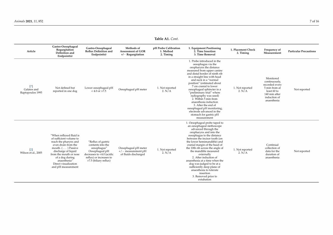
[6] Galatos AD, Raptopoulos D 1995 Vet. Rec.
[7] Galatos AD, Raptopoulos D 1995 Vet. Rec.
[2] Wilson DV, Evans AT, Miller R 2005 Am. J. Vet. Res.
[8] Wilson DV, Boruta DT, Evans AT 2006 Am. J. Vet. Res.
[9] Wilson DV, Evans AT, Mauer WA 2006 Am. J. Vet. Res.
[10] Wilson DV, Tom Evans A, Mauer WA 2007 Vet. Anaesth. Analg.
[11] Wilson DV, Evans AT 2007 Vet. Anaesth. Analg.
[12] Anagnostou TL, Savvas I, Kazakos GM et al. 2009 Vet. Anaesth. Analg.
[13] Panti A, Bennett RC, Corletto F et al. 2009 J. Small Anim. Pract.
[14] Favarato ES, de Souza MV, dos Santos Costa PR et al. 2011 Vet. Res. Commun.
[15] Favarato ES, Souza MV, Costa PR et al. 2012 Res. Vet. Sci.
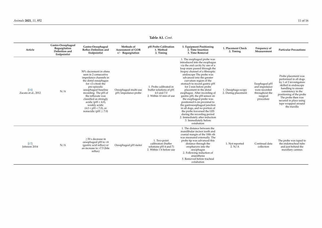
[16] Zacuto AC, Marks SL, Osborn J et al. 2012 J. Vet. Intern. Med.
[17] Johnson RA 2014 Vet. Anaesth. Analg.
[18] Anagnostou TL, Savvas I, Kazakos GM et al. 2015 Vet. Anaesth. Analg.
[3] Savvas I, Raptopoulos D, Rallis T 2016 J. Am. Anim. Hosp. Assoc.
[19] Anagnostou TL, Kazakos GM, Savvas I et al. 2017 Vet. Anaesth. Analg.
[20] Shaver SL, Barbur LA, Jimenez DA et al. 2017 J. Am. Anim. Hosp. Assoc.
[21] Torrente C, Vigueras I, Manzanilla EG et al. 2017 J. Vet. Emerg. Crit. Care
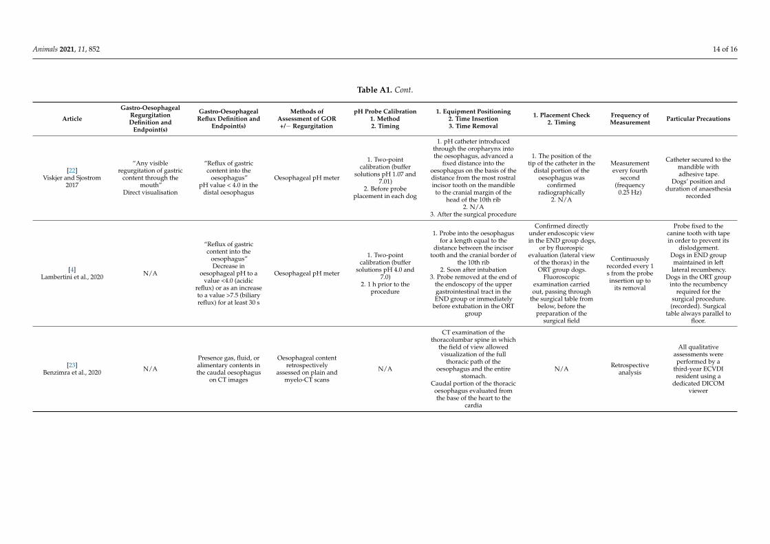
[22] Viskjer S, Sjostrom L 2017 Am. J. Vet. Res.
[4] Lambertini C, Pietra M, Galiazzo G et al. 2020 Vet. Sci.
[23] Benzimra C, Cerasoli I, Rault D et al. 2020 J. Vet. Sci.
The definition of GOR implied the presence of fluids not reaching the mouth or nose inthe oesophagus in all studies. The portion of the oesophagus considered was infrequentlyreported in the definition: “distal” or “caudal” or “lower” oesophagus in 8/22 papers.Most studies (20/22) identified the development of GOR through a change in oesophagealpH. This was the sole method of assessment in 18/22 studies, while two studies usedoesophagoscopy on top of the pH meter to visualise if reflux was present (i.e., non-acidreflux not causing pH changes). One study used a combined pH/impedance probe andconsidered a 50% decrement in Ohms seen in two consecutive impedance channels in thedistal oesophagus for >2 s compared with the pre-episodic oesophageal baseline recording.One study retrospectively assessed the presence of gas, fluid, or alimentary content on CT(computed tomography) images.
In the 20 studies considering pH changes, 14 considered that GOR had happened forchanges in pH “below 4.0 or above 7.5” while six considered that GOR had happened whenthe pH dropped below 4.0 only. The study using impedance changes categorized the pHof the reflux but this was not the criteria used to state if GOR had happened. In addition,for GOR to be confirmed, six studies stated that the pH change had to be sustained for aminimum period of time (20 or 30 s) whereas the others did not mention any duration inpH change.

Animals 2021, 11, 852 4 of 16
Irrespective of definitions and main outcome measures, 21/22 of the studies used pHmeters. Calibration was reported in only 14 studies. Two-point calibration (generally withbuffers of pH 1 or 4 and pH 7) was used.
The position of the tip of the oesophageal probe was inconsistent and not alwaysdescribed with precision. The distance between the lower incisors and the cranial marginof the 10th rib was externally measured with the dogs in lateral recumbency in 14 studies.However, the probe was advanced that distance in only 10/14 studies, whereas it wasadvanced that distance minus 5 cm in 4/14. The distance between the upper canine and thedistal border of the ninth rib (in a straight line with the dogs’ head and neck in a “normalposition”) was measured in two studies where the probe was advanced that distance. Itwas estimated to position the tip of the probe about 7 cm cranially to the lower oesophagealsphincter in a “preliminary trial” where radiography was used. The probe was advanceduntil the ninth rib in one study. It was retrieved from the stomach, 6 cm cranially to thelower oesophageal sphincter (visualised through video-oesophagoscopy) in one study. ThepH was measured at three different levels in the oesophagus (thoracic inlet, fifth and ninthrib) in one study. The location of the probe placement was not reported in 2/21 studies. Thecorrect positioning of the probe in the intended location was verified through endoscopy,chest radiographs, or fluoroscopy in only 5/21 studies. The permanence of the probe inthe appropriate location at the beginning and at the end or throughout the study wasnever checked.
The full results are presented in Appendix A.
4. Discussion
A variety of definitions and methods of assessment of GOR in anaesthetized dogsis present in the literature. Although oesophageal pH measurement is the method mostcommonly used, calibration, position of the probe, and cut-off values of pH to differentiatebetween GOR or not are inconsistent.
The method of analysis of pH is relative. This means that pH meters need to beappropriately calibrated [24]. The pH calibration curve is a combination of two curves,the pH and the pOH curves and is not linear. A slight deviation in the range of pH 6–8is expected [24]. Generally, a pH meter should be calibrated every 2 to 3 h using at leasttwo buffer solutions with known pH values close to the expected pH to be measured [24].When considering a wide range of pH, two-point calibration is not sufficient [24]. Also,given the effects of temperature on pH measurements [24], the question of calibratingthe instruments at body temperature exists. In the present review, several studies usedoesophageal pH meters without reporting any calibration. Studies reporting calibrationused a two-point calibration, mostly using buffers of 1 or 4 and 7 while eight studieswere defining GOR as a pH change below 4 or above 7.5. The accuracy of relevant pHvalues presented is questionable. Each pH unit change represents a 10-fold change inthe hydrogen or hydroxyl ions concentration. The calibration might be less important ifdefinitions considered sudden changes in pH as a proxy biomarker of GOR instead ofnumerical cut-off values.
Anatomic landmarks were commonly used to estimate the position of the loweroesophageal sphincter and the length of the probe to advance in the oesophagus. Moststudies used the description proposed by Waterman and Hashim [25]. However, the lengthof the oesophageal probes advanced was variable. Also, the position of the tip of the probewas rarely checked. Depending on the volume of the refluxate, actual GOR could be missed,for example, in the case of pH measurements in the proximal oesophagus if material ispresent only caudally or if the tip of the probe is not in the liquid phase in the dependentoesophagus. Furthermore, there was no consensus on the definition of GOR and pH cut-offvalues of the refluxate. Although acid reflux seems to be observed more frequently thanalkaline reflux in the articles examined, actual incidence of GOR might be underestimatedin some studies. These points make comparisons between studies challenging from theperspective of actual incidence or efficacy of measures taken.

Animals 2021, 11, 852 5 of 16
Consequences of GOR can include oesophagitis, oesophageal stricture, and aspirationpneumonia [26,27]. Evidence in humans suggests that mixed reflux (acid mixed with bileacids) is more harmful to the oesophageal mucosa than acid reflux alone [28] and thatbile reflux in the oesophagus can occur over a wide range of pH (2–8) [29]. Although theactual importance of this information in dogs undergoing a single anaesthesia and GORepisode is not known, the clinical relevance of changes in oesophageal pH as a sole markerof GOR is questionable. The position of the tip of the probe as well as the cut-off values ofpH used to assess the presence vs absence of GOR might require re-evaluation. AlthoughpH-metry seems commonly used, likely because of its wide availability and low cost, thistechnique alone might be insufficient. Combined pH/impedance probes with multipleimpedance channels to evaluate the spread of the refluxate in the oesophagus, similar tothe product used by Zacuto et al. (2012) [16] or Tarvin et al. (2016) [30], calibrated and in awell identified (checked) location, associated with biological analysis of the refluxate mightoffer a more relevant picture of GOR in anaesthetized dogs.
5. Conclusions
The variability of the GOR incidence found in the literature is likely due to a variety offactors (i.e., anaesthetic depth, transport, and position). However, the multiple definitionsand methods of assessment of GOR in anaesthetized dogs present in the literature precludemeaningful comparison of the findings. Re-evaluation and uniformization of the methodsseem necessary. Some aspects might warrant further investigation: (1) the relevance of thevolume of the material regurgitated and how long the reflux remains in the oesophagusfor; (2) the impact of anaesthesia on oesophageal motility; and (3) the importance ofanaesthetic depth and the presence of monitoring equipment in the oesophageal lumen onthe incidence of GOR.
Author Contributions: Conceptualization, M.R.; methodology, A.C.F.A., O.L., and M.R.; formalanalysis, A.C.F.A., O.L., and M.R.; writing—original draft preparation, A.C.F.A.; writing—reviewand editing, M.R. and O.L.; supervision, M.R. All authors have read and agreed to the publishedversion of the manuscript.
Funding: This research received no external funding.
Institutional Review Board Statement: Not applicable.
Informed Consent Statement: Not applicable.
Data Availability Statement: Data is contained within the article.
Conflicts of Interest: The authors declare no conflict of interest.
Animals 2021, 11, 852 6 of 16
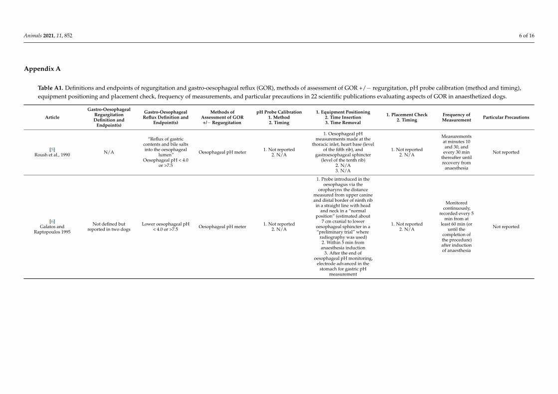
Appendix A
Table A1. Definitions and endpoints of regurgitation and gastro-oesophageal reflux (GOR), methods of assessment of GOR +/− regurgitation, pH probe calibration (method and timing),equipment positioning and placement check, frequency of measurements, and particular precautions in 22 scientific publications evaluating aspects of GOR in anaesthetized dogs.
Article
Gastro-OesophagealRegurgitationDefinition and
Endpoint(s)
Gastro-OesophagealReflux Definition and
Endpoint(s)
Methods ofAssessment of GOR+/− Regurgitation
pH Probe Calibration1. Method2. Timing
1. Equipment Positioning2. Time Insertion3. Time Removal
1. Placement Check2. Timing
Frequency ofMeasurement Particular Precautions
[5]Roush et al., 1990 N/A
“Reflux of gastriccontents and bile saltsinto the oesophageal
lumen”Oesophageal pH < 4.0
or >7.5
Oesophageal pH meter 1. Not reported2. N/A
1. Oesophageal pHmeasurements made at the
thoracic inlet, heart base (levelof the fifth rib), and
gastroesophageal sphincter(level of the tenth rib)
2. N/A3. N/A
1. Not reported2. N/A
Measurementsat minutes 10and 30, and
every 30 minthereafter untilrecovery from
anaesthesia
Not reported
[6]Galatos and
Raptopoulos 1995
Not defined butreported in two dogs
Lower oesophageal pH< 4.0 or >7.5 Oesophageal pH meter 1. Not reported
2. N/A
1. Probe introduced in theoesophagus via the
oropharynx the distancemeasured from upper canineand distal border of ninth ribin a straight line with head
and neck in a “normalposition” (estimated about
7 cm cranial to loweroesophageal sphincter in a“preliminary trial” where
radiography was used)2. Within 5 min fromanaesthesia induction
3. After the end ofoesophageal pH monitoring,
electrode advanced in thestomach for gastric pH
measurement
1. Not reported2. N/A
Monitoredcontinuously,
recorded every 5min from at
least 60 min (oruntil the
completion ofthe procedure)after inductionof anaesthesia
Not reported
Animals 2021, 11, 852 7 of 16
Table A1. Cont.
Article
Gastro-OesophagealRegurgitationDefinition and
Endpoint(s)
Gastro-OesophagealReflux Definition and
Endpoint(s)
Methods ofAssessment of GOR+/− Regurgitation
pH Probe Calibration1. Method2. Timing
1. Equipment Positioning2. Time Insertion3. Time Removal
1. Placement Check2. Timing
Frequency ofMeasurement Particular Precautions
[7]Galatos and
Raptopoulos 1995
Not defined butreported in one dog
Lower oesophageal pH< 4.0 or >7.5 Oesophageal pH meter 1. Not reported
2. N/A
1. Probe introduced in theoesophagus via the
oropharynx the distancemeasured from upper canineand distal border of ninth ribin a straight line with head
and neck in a “normalposition” (estimated about
7 cm cranial to loweroesophageal sphincter in a“preliminary trial” where
radiography was used)2. Within 5 min fromanaesthesia induction
3. After the end ofoesophageal pH monitoring,
electrode advanced in thestomach for gastric pH
measurement
1. Not reported2. N/A
Monitoredcontinuously,
recorded every5 min from at
least 60 to140 min afterinduction ofanaesthesia
Not reported
[2]Wilson et al., 2005
“When refluxed fluid isof sufficient volume toreach the pharynx and
even drain from themouth. ( . . . ) Passive
discharge of liquidfrom the mouth or nose
of a dog duringanaesthesia”
Direct visualizationand pH measurement
“Reflux of gastriccontents into the
oesophagus”Oesophageal pH
decreases to <4.0 (acidicreflux) or increases to>7.5 (biliary reflux)
Oesophageal pH meter+/− measurement pH
of fluids discharged
1. Not reported2. N/A
1. Oesophageal probe taped toan oesophageal stethoscope
advanced through theoropharynx and into the
oesophagus to the distancebetween the incisor tooth (onthe lower hemimandible) andcranial margin of the head of
the 10th rib across the angle ofthe mandible measured
externally2. After induction of
anaesthesia at a time when thedog was judged to be at asufficiently deep plane of
anaesthesia to tolerateinsertion
3. Removed prior toextubation
1. Not reported2. N/A
Continualcollection ofdata for theduration ofanaesthesia
Not reported

Animals 2021, 11, 852 8 of 16
Table A1. Cont.
Article
Gastro-OesophagealRegurgitationDefinition and
Endpoint(s)
Gastro-OesophagealReflux Definition and
Endpoint(s)
Methods ofAssessment of GOR+/− Regurgitation
pH Probe Calibration1. Method2. Timing
1. Equipment Positioning2. Time Insertion3. Time Removal
1. Placement Check2. Timing
Frequency ofMeasurement Particular Precautions
[8]Wilson et al., 2006
“Passive discharge ofliquid from the mouth
or nose”Direct visualizationand pH of any fluid
that dripped from themouth or nose
measured
Decrease inoesophageal pH to <4(reflux of gastric acid)or an increase to >7.5(reflux of bile) for a
period of ≥30 s
Oesophageal pH meter
1. Two-pointcalibration (buffer
solutions pH 1 and 7)2. Within 2 h prior to
use
1. Tip of the probe taped to anoesophageal stethoscope and
advanced through theoropharynx into the
oesophagus the distancebetween the incisor tooth onthe lower jaw and the cranial
margin of the 10th rib(measured externally)2. After induction of
anaesthesia and endotrachealintubation
3. Prior to extubation
1. Not reported2. N/A
Continuousmonitoring
Probe placement wasperformed by 1 of 3
trained peopleThe probe was affixed
in place
[9]Wilson et al., 2006
“Passive discharge ofliquid from the mouth
or nose”Direct visualizationand pH of any fluid
that dripped from themouth or nose
measured
Decrease inoesophageal pH to <4(reflux of gastric acid)or an increase to >7.5(reflux of bile) for a
period of ≥30 s
Oesophageal pH meter
1. Two-pointcalibration (buffer
solutions pH 1 and 7)2. Within 2 h prior to
use
1. Tip of the probe taped to anoesophageal stethoscope and
advanced through theoropharynx into the
oesophagus the distancebetween the incisor tooth onthe lower jaw and the cranial
margin of the 10th rib(measured externally)2. After induction of
anaesthesia and endotrachealintubation
3. Prior to extubation
1. Not reported2. N/A
Continuousmonitoring
Probe placement wasperformed by 1 of 3
trained peopleThe probe was affixed
in place
[10]Wilson et al., 2007
“Passive discharge ofliquid from the mouth
or nose”Direct visualizationand pH of any fluid
that dripped from themouth or nose
measured
Decrease inoesophageal pH to <4(reflux of gastric acid)or an increase to >7.5(reflux of bile) for a
period of ≥30 s
Oesophageal pH meter
1. Two-pointcalibration (buffer
solutions pH 1 and 7)2. Within 2 h prior to
use
1. Tip of the probe taped to anoesophageal stethoscope and
advanced through theoropharynx into the
oesophagus the distancebetween the incisor tooth onthe lower jaw and the cranial
margin of the 10th rib(measured externally)2. After induction of
anaesthesia and endotrachealintubation
3. Prior to extubation
1. Not reported2. N/A
Continuousmonitoring
Probe placement wasperformed by 1 of 3
trained peopleThe probe was affixed
in place

Animals 2021, 11, 852 9 of 16
Table A1. Cont.
Article
Gastro-OesophagealRegurgitationDefinition and
Endpoint(s)
Gastro-OesophagealReflux Definition and
Endpoint(s)
Methods ofAssessment of GOR+/− Regurgitation
pH Probe Calibration1. Method2. Timing
1. Equipment Positioning2. Time Insertion3. Time Removal
1. Placement Check2. Timing
Frequency ofMeasurement Particular Precautions
[11]Wilson and Evans 2007
“Passive discharge ofliquid from the mouthor nose of a dog during
general anaesthesia”Direct visualization
Observation of 30 s orlonger of a decrease inoesophageal pH to <4(reflux of gastric acid)
Oesophageal pH meter
1. Two-pointcalibration (buffer
solutions pH 1 and 7)2. Within 2 h prior to
use
1. Tip of the probe advancedthrough the oropharynx into
the oesophagus thepre-measured distance
between the incisor tooth onthe lower jaw and the cranialmargin of the head of the 10th
rib measured externally2. Probe inserted after
induction of anesthesia andendotracheal intubation
3. Probe removed prior toextubation
1. Not reported2. N/A
Continual datacollection
Probe placementperformed by one ofthree trained people.
The probe was affixedin place.
[12]Anagnostou et al., 2009 N/A
pH values of >7.5(alkaline reflux) or <4
(acid reflux) in thelower oesophagus
Oesophageal pH meter
1. Two-pointcalibration (buffer
solutions pH 4 and 7)2. “Previously”
1. pH-meter inserted into theoesophagus through the oral
cavity. Length determinedsubtracting 5 cm frompre-measured distance
between lower incisor tooth(animal in left lateral
recumbency) and anteriorborder of the head of the 10thrib through the angle of the
mandible2. Immediately after
intubation of the trachea andconnection of the endotracheal
tube to the anaestheticmachine
3. After completion of 1 h ofcontinuous oesophageal pH
monitoring
1. Not reported2. N/A
Monitoredcontinuously for
60 min afterinduction ofanaesthesia
Dogs placed in dorsalrecumbency
immediately aftersecuring the probe.
During procedures andposition changes,
special attention waspaid to avoiding
application of pressureto the abdominal wall

Animals 2021, 11, 852 10 of 16
Table A1. Cont.
Article
Gastro-OesophagealRegurgitationDefinition and
Endpoint(s)
Gastro-OesophagealReflux Definition and
Endpoint(s)
Methods ofAssessment of GOR+/− Regurgitation
pH Probe Calibration1. Method2. Timing
1. Equipment Positioning2. Time Insertion3. Time Removal
1. Placement Check2. Timing
Frequency ofMeasurement Particular Precautions
[13]Panti et al., 2009
“Return of partiallydigested food from thestomach to the mouth”
Abrupt decrease indistal oesophageal pH
below 4Oesophageal pH meter
1. Two-pointcalibration (buffer
solutions pH 4 and 7)2. Approximately everythree cases and at least
once a week
Probe placed inside aprotective polythene tube. and
inserted into the distaloesophagus. The probe was
inserted into the oesophagus,with the tip at the level of theninth rib, which is about 7 cmrostral to the LOS (Position of
the lower oesophagealsphincter estimated the length
between the incisor of thelower jaw and the cranial
border of the head of the 10thrib, measured externally with
the animal in lateralrecumbency)
2. Immediately after inductionof anaesthesia
3. Not reported
1. Not reported2. N/A
Recorded everyfive minutes
duringanaesthesia
The same operatorpositioned the
oesophageal pH probein each dog
[14]Favarato et al., 2011 N/A
“Presence of acid refluxin the oesophagus”
Oesophageal pH < 4and visualisation of
content throughvideo-oesophagoscopy
Oesophageal pH meterand
video-oesophago-scopy
1. Two-pointcalibration (buffer
solutions pH 1.0 and7.0)
2. Maximum 1 h beforethe procedure
1. Close and cranially to theoesophago-gastric juntion(measuring the distance
between the mandible incisorteeth and the cranial border ofthe tenth rib through the angle
of the mandible after thepre-anaesthetic medication,
the animals positioned in leftlateral recumbency)
2. “Intra-operatively”3. Catheter removed
immediately afteresophagoscopy
1.Video-oesophago-scopy
2. Immediately aftersurgery
Constantlymonitored,variationsrecorded
Dogs maintained in adorsal horizontalrecumbency, on a
surgical table, duringthe surgical procedure.Lateral decubitus afterthe end of surgery. No
position changesallowed during theevaluation period
[15]Favarato et al., 2012 N/A
pH lower than 4considered an acid
reflux episode;confirmation of the
non-acid refluxobtained by
esophagoscopyconducted on all theanimals immediately
after surgery toevaluate the presenceof visible reflux in the
oesophageal lumen
Oesophageal pH meterand
video-oesophago-scopy
1. Not reported2. N/A
1. “Close and cranially to theoesophago-gastric junction”
2. N/A3. N/A
1. Not reported2. N/A
Monitoringthroughout the
anaestheticprocedure, with
all the pHvariationsrecorded
N/A
Animals 2021, 11, 852 11 of 16
Table A1. Cont.
Article
Gastro-OesophagealRegurgitationDefinition and
Endpoint(s)
Gastro-OesophagealReflux Definition and
Endpoint(s)
Methods ofAssessment of GOR+/− Regurgitation
pH Probe Calibration1. Method2. Timing
1. Equipment Positioning2. Time Insertion3. Time Removal
1. Placement Check2. Timing
Frequency ofMeasurement Particular Precautions
[16]Zacuto et al., 2012 N/A
50% decrement in ohmsseen in 2 consecutive
impedance channels inthe distal oesophagus
for >2 s from thepre-episodic
oesophageal baselinerecording. The pH of
the refluxate wasclassified as strongly
acidic (pH < 4.0),weakly acidic
(4.0 < pH < 7.0), ornonacidic (pH ≥ 7.0)
Oesophageal multi-usepH/impedance probe
1. Probe calibrated inbuffer solutions of pH
4.0 and 7.02. Within 10 min of use
1. The esophageal probe wasintroduced into the esophagusvia the oral cavity by use of aloop snare passed through thebiopsy channel of a fibreoptic
endoscope The probe wasadvanced into the greater
curvature region of thestomach to record gastric pH
for 2 min before probeplacement in the distal
esophagus. After recording ofgastric pH, the pH sensor on
the esophageal probe waspositioned 6 cm proximal to
the gastroesophageal junctionin all dogs, and no portion ofthe probe traversed the LESduring the recording period
2. Immediately after induction3. Immediately before
extubation
1. Oesophago-scopy2. During placement
Esophageal pHand impedancewere recordedthroughout the
surgicalprocedure
Probe placement wasperformed in all dogsby 1 of 2 investigatorsskilled in endoscopehandling to ensureconsistency in the
positioning of the probeThe probe then was
secured in place usingtape wrapped around
the maxilla
[17]Johnson 2014 N/A
≥30 s decrease inoesophageal pH to <4(gastric acid reflux) or
an increase to >7.5 (bilereflux)
Oesophageal pH meter
1. Two-pointcalibration (buffer
solutions pH 4 and 7)2. Within 1 h before use
1. The distance between themandibular incisor tooth andcranial margin of the 10th ribwas measured externally. Theprobe tip was advanced this
distance through theoropharynx into the
oesophagus2. Following induction of
anaesthesia3. Removed before tracheal
extubation
1. Not reported2. N/A
Continual datacollection
The probe was taped tothe endotracheal tube
and just behind themaxillary canines

Animals 2021, 11, 852 12 of 16
Table A1. Cont.
Article
Gastro-OesophagealRegurgitationDefinition and
Endpoint(s)
Gastro-OesophagealReflux Definition and
Endpoint(s)
Methods ofAssessment of GOR+/− Regurgitation
pH Probe Calibration1. Method2. Timing
1. Equipment Positioning2. Time Insertion3. Time Removal
1. Placement Check2. Timing
Frequency ofMeasurement Particular Precautions
[18]Anagnostou et al., 2015 N/A
Lower oesophageal pHvalues of >7.5 (alkaline
reflux) or <4 (acidreflux)
Oesophageal pH meter
1. Two-pointcalibration (buffer
solutions pH 4 and 7)2. “previously”
1. Probe inserted into theoesophagus through the oral
cavity; length subtracting 5 cmfrom the pre-measured
distance between the lowerincisor tooth and the anteriorborder of the head of the 10thrib through the angle of the
mandible (animal in left lateralrecumbency)
2. Immediately afterintubation of the trachea and
connection of the endotrachealtube to the anaesthetic
machine3. Oesophageal probe
removed on completion ofsurgery before discontinuing
administration
1. Not reported2. N/A
Monitoredcontinuously
after inductionof anaesthesia
and throughoutsurgery
Immediately aftersecuring the probe inplace, the animal was
placed in dorsalrecumbency.
During introduction ofthe pH-measuring
probe, change of theanimals’ position, and
aseptic preparation(clipping, scrubbing),special attention was
paid to avoidingapplication of excessive
pressure to theabdominal wall
[3]Savvas et al., 2016
pH-change at thepharynx, measured
when GOR wasobserved
Oesophageal pH < 4 or>7.5
Oesophageal +/−upper oesophageal pH
meter
1. Not reported2. Not reported
1. Oesophageal probe: 5 cmabove the lower oesophageal
sphincter, estimated bymeasuring the length from
lower jaw incisor tooth to theanterior border of the head of
the tenth rib2. Following intubation of the
trachea3. pH recording was
discontinued just prior toextubation
NB: Second pH probe with itstip at the upper oesophagealsphincter (at the level of thelarynx) when GOR observed
1. Not reported2. N/A
Constantlymonitored,
recorded every5 min
No transportation ofthe animals to another
operation room. Allpossible precautions
were taken to preventincreases in
intra-abdominalpressure (from
manipulations of theanimals during
handling and surgery)

Animals 2021, 11, 852 13 of 16
Table A1. Cont.
Article
Gastro-OesophagealRegurgitationDefinition and
Endpoint(s)
Gastro-OesophagealReflux Definition and
Endpoint(s)
Methods ofAssessment of GOR+/− Regurgitation
pH Probe Calibration1. Method2. Timing
1. Equipment Positioning2. Time Insertion3. Time Removal
1. Placement Check2. Timing
Frequency ofMeasurement Particular Precautions
[19]Anagnostou et al., 2017
“Reflux materialobserved at the externalnares or in the mouth”
Whenever a pH value >7.5 (alkaline reflux) or<4.0 (acid reflux) was
recorded
Oesophageal pH meter
1. Two-pointcalibration (buffer
solutions pH 4 and 7)2. Before each use
1. Probe introduced into theoesophageal lumen throughthe oral cavity. The distancebetween the lower incisorteeth (animal in left lateral
recumbency) and the anteriorborder of the head of the 10thrib through the angle of the
mandible was measuredconsidered to correspond tothe approximate location ofthe posterior oesophageal
sphincter; the final length ofthe pH measuring probe that
was inserted into theoesophageal lumen was
calculated by subtracting 5 cmfrom this measured distance2. Immediately after trachealintubation and connection ofthe endotracheal tube to the
anaesthetic machine3. After completion of surgery,
administration of halothanediscontinued and pH probe
withdrawn
1. Not reported2. N/A
Continuousmonitoring
Immediately aftersecuring the probe inplace, the animal was
placed in sternalrecumbency on ahorizontal table.Application of
excessive pressure tothe abdominal wall or
to the surgical area thatcould potentially causeGOR was avoided at all
times and especiallyduring introduction of
the pH measuringprobe, change of
recumbency, clippingand scrubbing.
[20]Shaver et al., 2017
“Gastric contentsrefluxing to the
oropharynx”
Prolonged (>20 s)decreases (<4.0) orincreases (>7.5) inoesophageal pH
Oesophageal pH meter
1. Two-pointcalibration (buffer
solutions pH 4 and 7)2. Immediately before
use
1. Probe inserted orally andadvanced a distance measuredfrom the incisors to the cranial
margin of the tenth rib toresult in a predictable location
just proximal to the loweroesophageal sphincter2. After induction of
anaesthesia3. Just prior to patient
endotracheal extubation
1. Thoracic radiograph2. Immediately after
placement
Continuousmonitoring of
oesophageal pH,recorded at
5 min intervals
Probe placed by asingle surgeon
Medical tape was fixedto the probe at the level
of the first premolarand stapled to the
dog’s upper lip usingsurgical staples
[21]Torrente et al., 2017
“Passive ejection ofgastric or oesophageal
content from the mouthor nose”
Direct visualisation
Oesophageal pH < 4was considered an acid
reflux eventOesophageal pH meter 1. Not reported
2. N/A
1. Oesophageal probe duringanaesthesia and just beforereturn to full consciousness
(no more detail)2. N/A3. N/A
1. Not reported2. N/A Not reported Not reported
Animals 2021, 11, 852 14 of 16
Table A1. Cont.
Article
Gastro-OesophagealRegurgitationDefinition and
Endpoint(s)
Gastro-OesophagealReflux Definition and
Endpoint(s)
Methods ofAssessment of GOR+/− Regurgitation
pH Probe Calibration1. Method2. Timing
1. Equipment Positioning2. Time Insertion3. Time Removal
1. Placement Check2. Timing
Frequency ofMeasurement Particular Precautions
[22]Viskjer and Sjostrom
2017
“Any visibleregurgitation of gastric
content through themouth”
Direct visualisation
“Reflux of gastriccontent into the
oesophagus”pH value < 4.0 in the
distal oesophagus
Oesophageal pH meter
1. Two-pointcalibration (buffer
solutions pH 1.07 and7.01)
2. Before probeplacement in each dog
1. pH catheter introducedthrough the oropharynx intothe oesophagus, advanced a
fixed distance into theoesophagus on the basis of thedistance from the most rostralincisor tooth on the mandible
to the cranial margin of thehead of the 10th rib
2. N/A3. After the surgical procedure
1. The position of thetip of the catheter in the
distal portion of theoesophagus was
confirmedradiographically
2. N/A
Measurementevery fourth
second(frequency
0.25 Hz)
Catheter secured to themandible withadhesive tape.
Dogs’ position andduration of anaesthesia
recorded
[4]Lambertini et al., 2020 N/A
“Reflux of gastriccontent into the
oesophagus”Decrease in
oesophageal pH to avalue <4.0 (acidic
reflux) or as an increaseto a value >7.5 (biliaryreflux) for at least 30 s
Oesophageal pH meter
1. Two-pointcalibration (buffer
solutions pH 4.0 and7.0)
2. 1 h prior to theprocedure
1. Probe into the oesophagusfor a length equal to the
distance between the incisortooth and the cranial border of
the 10th rib2. Soon after intubation
3. Probe removed at the end ofthe endoscopy of the uppergastrointestinal tract in theEND group or immediately
before extubation in the ORTgroup
Confirmed directlyunder endoscopic viewin the END group dogs,
or by fluorospicevaluation (lateral view
of the thorax) in theORT group dogs.
Fluoroscopicexamination carriedout, passing through
the surgical table frombelow, before thepreparation of the
surgical field
Continuouslyrecorded every 1s from the probeinsertion up to
its removal
Probe fixed to thecanine tooth with tapein order to prevent its
dislodgement.Dogs in END groupmaintained in left
lateral recumbency.Dogs in the ORT group
into the recumbencyrequired for the
surgical procedure.(recorded). Surgical
table always parallel tofloor.
[23]Benzimra et al., 2020 N/A
Presence gas, fluid, oralimentary contents inthe caudal oesophagus
on CT images
Oesophageal contentretrospectively
assessed on plain andmyelo-CT scans
N/A
CT examination of thethoracolumbar spine in which
the field of view allowedvisualization of the full
thoracic path of theoesophagus and the entire
stomach.Caudal portion of the thoracic
oesophagus evaluated fromthe base of the heart to the
cardia
N/A Retrospectiveanalysis
All qualitativeassessments wereperformed by a
third-year ECVDIresident using a
dedicated DICOMviewer

Animals 2021, 11, 852 15 of 16
References1. Lamata, C.; Loughton, V.; Jones, M.; Alibhai, H.; Armitage-Chan, E.; Walsh, K.; Brodbelt, D. The risk of passive regurgitation
during general anaesthesia in a population of referred dogs in the UK. Vet. Anaesth. Analg. 2012, 39, 266–274. [CrossRef]2. Wilson, D.V.; Evans, A.T.; Miller, R. Effects of preanesthetic administration of morphine on gastroesophageal reflux and
regurgitation during anesthesia in dogs. Am. J. Vet. Res. 2005, 66, 386–390. [CrossRef]3. Savvas, I.; Raptopoulos, D.; Rallis, T. A “Light Meal” Three Hours Preoperatively Decreases the Incidence of Gastro-Esophageal
Reflux in Dogs. J. Am. Anim. Hosp. Assoc. 2016, 52, 357–363. [CrossRef] [PubMed]4. Lambertini, C.; Pietra, M.; Galiazzo, G.; Torresan, F.; Pinna, S.; Pisoni, L.; Romagnoli, N. Incidence of Gastroesophageal Reflux
in Dogs Undergoing Orthopaedic Surgery or Endoscopic Evaluation of the Upper Gastrointestinal Tract. Vet. Sci. 2020, 7, 144.[CrossRef] [PubMed]
5. Roush, J.K.; Keene, B.W.; Eicker, S.W.; Bjorling, D.E. Effects of atropine and glycopyrrolate on esophageal, gastric, and trachealpH in anesthetized dogs. Vet. Surg. 1990, 19, 88–92. [CrossRef] [PubMed]
6. Galatos, A.D.; Raptopoulos, D. Gastro-oesophageal reflux during anaesthesia in the dog: The effect of age, positioning and typeof surgical procedure. Vet. Rec. 1995, 137, 513–516. [CrossRef] [PubMed]
7. Galatos, A.D.; Raptopoulos, D. Gastro-oesophageal reflux during anaesthesia in the dog: The effect of preoperative fasting andpremedication. Vet. Rec. 1995, 137, 479–483. [CrossRef]
8. Wilson, D.V.; Boruta, D.T.; Evans, A.T. Influence of halothane, isoflurane, and sevoflurane on gastroesophageal reflux duringanesthesia in dogs. Am. J. Vet. Res. 2006, 67, 1821–1825. [CrossRef]
9. Wilson, D.V.; Evans, A.T.; Mauer, W.A. Influence of metoclopramide on gastroesophageal reflux in anesthetized dogs. Am. J. Vet.Res. 2006, 67, 26–31. [CrossRef]
10. Wilson, D.V.; Tom Evans, A.; Mauer, W.A. Pre-anesthetic meperidine: Associated vomiting and gastroesophageal reflux duringthe subsequent anesthetic in dogs. Vet. Anaesth. Analg. 2007, 34, 15–22. [CrossRef]
11. Wilson, D.V.; Evans, A.T. The effect of topical treatment on esophageal pH during acid reflux in dogs. Vet. Anaesth. Analg. 2007,34, 339–343. [CrossRef]
12. Anagnostou, T.L.; Savvas, I.; Kazakos, G.M.; Ververidis, H.N.; Haritopoulou, M.R.; Rallis, T.S.; Raptopoulos, D. Effect ofendogenous progesterone and oestradiol-17beta on the incidence of gastro-oesophageal reflux and on the barrier pressure duringgeneral anaesthesia in the female dog. Vet. Anaesth. Analg. 2009, 36, 308–318. [CrossRef] [PubMed]
13. Panti, A.; Bennett, R.C.; Corletto, F.; Brearley, J.; Jeffery, N.; Mellanby, R.J. The effect of omeprazole on oesophageal pH in dogsduring anaesthesia. J. Small Anim. Pract. 2009, 50, 540–544. [CrossRef]
14. Favarato, E.S.; de Souza, M.V.; dos Santos Costa, P.R.; Pompermayer, L.G.; Campos Favarato, L.S.; Ribeiro Júnior, J.I. Ambulatoryesophageal pHmetry in healthy dogs with and without the influence of general anesthesia. Vet. Res. Commun. 2011, 35, 271–282.[CrossRef]
15. Favarato, E.S.; Souza, M.V.; Costa, P.R.; Favarato, L.S.; Nehme, R.C.; Monteiro, B.S.; Bonfa, L.P. Evaluation of metoclopramide andranitidine on the prevention of gastroesophageal reflux episodes in anesthetized dogs. Res. Vet. Sci. 2012, 93, 466–467. [CrossRef]
16. Zacuto, A.C.; Marks, S.L.; Osborn, J.; Douthitt, K.L.; Hollingshead, K.L.; Hayashi, K.; Kapatkin, A.S.; Pypendop, B.H.; Belafsky,P.C. The influence of esomeprazole and cisapride on gastroesophageal reflux during anesthesia in dogs. J. Vet. Intern. Med. 2012,26, 518–525. [CrossRef] [PubMed]
17. Johnson, R.A. Maropitant prevented vomiting but not gastroesophageal reflux in anesthetized dogs premedicated withacepromazine-hydromorphone. Vet. Anaesth. Analg. 2014, 41, 406–410. [CrossRef] [PubMed]
18. Anagnostou, T.L.; Savvas, I.; Kazakos, G.M.; Ververidis, H.N.; Psalla, D.; Kostakis, C.; Skepastianos, P.; Raptopoulos, D. The effectof the stage of the ovarian cycle (anoestrus or dioestrus) and of pregnancy on the incidence of gastro-oesophageal reflux in dogsundergoing ovariohysterectomy. Vet. Anaesth. Analg. 2015, 42, 502–511. [CrossRef] [PubMed]
19. Anagnostou, T.L.; Kazakos, G.M.; Savvas, I.; Kostakis, C.; Papadopoulou, P. Gastro-oesophageal reflux in large-sized, deep-chested versus small-sized, barrel-chested dogs undergoing spinal surgery in sternal recumbency. Vet. Anaesth. Analg. 2017, 44,35–41. [CrossRef]
20. Shaver, S.L.; Barbur, L.A.; Jimenez, D.A.; Brainard, B.M.; Cornell, K.K.; Radlinsky, M.G.; Schmiedt, C.W. Evaluation of Gastroe-sophageal Reflux in Anesthetized Dogs with Brachycephalic Syndrome. J. Am. Anim. Hosp. Assoc. 2017, 53, 24–31. [CrossRef][PubMed]
21. Torrente, C.; Vigueras, I.; Manzanilla, E.G.; Villaverde, C.; Fresno, L.; Carvajal, B.; Finana, M.; Costa-Farre, C. Prevalence of andrisk factors for intraoperative gastroesophageal reflux and postanesthetic vomiting and diarrhea in dogs undergoing generalanesthesia. J. Vet. Emerg. Crit. Care (San Antonio) 2017, 27, 397–408. [CrossRef]
22. Viskjer, S.; Sjostrom, L. Effect of the duration of food withholding prior to anesthesia on gastroesophageal reflux and regurgitationin healthy dogs undergoing elective orthopedic surgery. Am. J. Vet. Res. 2017, 78, 144–150. [CrossRef] [PubMed]
23. Benzimra, C.; Cerasoli, I.; Rault, D.; Chalvet-Monfray, K.; Cauvin, E.; Couturier, L.; Gatel, L. Computed tomographic features ofgastric and esophageal content in dogs undergoing CT myelography and factors influencing the presence of esophageal fluid. J.Vet. Sci. 2020, 21, e84. [CrossRef]
24. Cheng, K.L.; Zhu, D.-M. On Calibration of pH Meters. Sensors 2005, 5, 209–219. [CrossRef]

Animals 2021, 11, 852 16 of 16
25. Waterman, A.E.; Hashim, M.A. Measurement of the length and position of the lower oesophageal sphincter by correlation ofexternal measurements and radiographic estimations in dogs. Vet. Rec. 1991, 129, 261–264. [CrossRef]
26. Pearson, H.; Darke, P.G.; Gibbs, C.; Kelly, D.F.; Orr, C.M. Reflux oesophagitis and stricture formation after anaesthesia: A reviewof seven cases in dogs and cats. J. Small Anim. Pract. 1978, 19, 507–519. [CrossRef]
27. Ovbey, D.H.; Wilson, D.V.; Bednarski, R.M.; Hauptman, J.G.; Stanley, B.J.; Radlinsky, M.G.; Larenza, M.P.; Pypendop, B.H.;Rezende, M.L. Prevalence and risk factors for canine post-anesthetic aspiration pneumonia (1999-2009): A multicenter study. Vet.Anaesth. Analg. 2014, 41, 127–136. [CrossRef]
28. Nehra, D.; Howell, P.; Williams, C.P.; Pye, J.K.; Beynon, J. Toxic bile acids in gastro-oesophageal reflux disease: Influence of gastricacidity. Gut 1999, 44, 598–602. [CrossRef]
29. Nehra, D.; Howell, P.; Pye, J.K.; Beynon, J. Assessment of combined bile acid and pH profiles using an automated samplingdevice in gastro-oesophageal reflux disease. Br. J. Surg. 1998, 85, 134–137. [CrossRef]
30. Tarvin, K.M.; Twedt, D.C.; Monnet, E. Prospective Controlled Study of Gastroesophageal Reflux in Dogs with Naturally OccurringLaryngeal Paralysis. Vet. Surg. 2016, 45, 916–921. [CrossRef]