A spectrally formulated plate element for wave propagation analysis in anisotropic material
Upload
independentCategory
view
0download
0
Food Research International 45 (2012) 351–361
Contents lists available at SciVerse ScienceDirect
Food Research International
j ourna l homepage: www.e lsev ie r .com/ locate / foodres
Features and performance of edible films, obtained from whey protein isolateformulated with antimicrobial compounds
Óscar L. Ramos a,b, Sara I. Silva a, José C. Soares a, João C. Fernandes a, M. Fátima Poças a,Manuela E. Pintado a, F. Xavier Malcata b,c,⁎a CBQF/Escola Superior de Biotecnologia, Rua Dr. António Bernardino de Almeida, P-4200-072 Porto, Portugalb Instituto de Tecnologia Química e Biológica, Universidade Nova de Lisboa, Avenida da República, P-2780-157 Oeiras, Portugalc ISMAI — Instituto Superior da Maia, Avenida Carlos Oliveira Campos, P-4475-690 Avioso S. Pedro, Portugal
⁎ Corresponding author at: Instituto Superior da Maiapos, P-4475690 Avioso S. Pedro, Portugal. Tel.: +351 9653 31.
E-mail address: [email protected] (F.X. Malcata).
0963-9969/$ – see front matter © 2011 Elsevier Ltd. Alldoi:10.1016/j.foodres.2011.09.016
a b s t r a c t
a r t i c l e i n f oArticle history:Received 4 June 2011Accepted 20 September 2011
Keywords:Whey protein isolateAntimicrobial agentsActive packagingPhysical propertiesFood safetyAntimicrobial packaging
The goal of this research effort was to assess the efficacy of edible films produced fromwhey protein isolate (WPI)and glycerol, including incorporation of lactic acid (LA) and propionic acid (PRO), chitooligosaccharideswith nom-inal MW of 3 kDa (COS) and natamycin (NA) as antimicrobial agents. Their features were evaluated in vitro viaagar diffusion and viable cell counting, against spoilage microflora often found contaminating cheese surfaces.The effect of incorporating the aforementioned compounds upon thickness, moisture content (MC), solubility(S), density (ρs), water activity (aw) andwater vapor permeability (WVP), aswell as upon tensile and optical prop-erties of those films were also evaluated. Films formulated with LA, PRO or COS exhibited antimicrobial activityagainst all microorganisms tested, yet the viable cell count assay was more sensitive and reproducible. COS wasthe most active against Gram-negative bacteria, whereas LA was the most active against Gram-positive ones. NAwas not active against bacteria, but displayed the strongest effect against yeasts. Incorporation of said antimicrobialcompounds did not significantly (pN0.05) affect film thickness, yet it significantly (pb0.05) reduced tensilestrength (TS). Incorporation of LA and NA in particular did not significantly (pb0.05) affect MC, S, ρs, WVP, elon-gation at break (EB) and Young's modulus (YM) values; however, a statistically significant increase (pb0.05) ofMC, S andWVP, together with a statistically significant decrease (pb0.05) of ρs were attained upon incorporationof PRO or COS. Moreover, PRO produced the highest variation (pb0.05) in EB, TS and YM, whereas COS producedthe highest change (pb0.05) in optical properties.
, Avenida Carlos Oliveira Cam-8017 411; fax: +351 22 982
rights reserved.
© 2011 Elsevier Ltd. All rights reserved.
1. Introduction
Foods are normally susceptible to physical, chemical and microbi-ological deterioration throughout storage and distribution, both as afunction of their composition and the environmental conditionsthey are exposed to (Cha & Chinnan, 2004). An adequate selectionof packaging materials can prevent food quality loss by providing bar-rier, or otherwise protective features thereto (Campos et al., 2011). Ifpackaging films are in addition edible— e.g. those manufactured frompolysaccharides, proteins or lipids, they will convey an extra set of ad-vantages, viz. biodegradability, non-toxicity and biocompatibility, be-sides esthetic appearance (Bourtoom, 2009; Khwaldia, Perez, Banon,Desobry, & Hardy, 2004; Tharanathan, 2003).
Whey proteins have been successfully employed as raw materialfor biodegradable packaging because they come from a renewablesource and are a by-product of the cheesemaking industry; hence,
they are widely available, relatively easy to handle and essentially in-expensive. Whey protein isolates (WPI) represent the purer form ofsuch whey proteins (Mulvihill & Ennis, 2003), and have shown prom-ising mechanical features, as well as moderate moisture permeability(McHugh, Aujard, &Krochta, 1994) and good oxygen barrier properties—comparable to those exhibited by the best synthetic polymer-based filmsavailable e.g. low-density polyethylene (LDPE), high density polyethyl-ene, ethylene vinyl alcohol, vinyl alcohol, polyvinylidene chloride(PVDC), cellophane and polyester (Khwaldia et al., 2004; Perez-Gago &Krochta, 2002).
Furthermore, those films proved excellent biomaterials for use ascarriers of such food additives as antioxidants, antimicrobials, color-ants, flavors, fortifying nutrients and spices; these additives improvethe functionality of the packaging by bringing about novel (orextra) features (Pranoto, Salokhe, & Rakshit, 2005; Salmieri & Lacroix,2006). In particular, addition of antimicrobial agents may enable ex-tension of the shelf-life and safety of packaged foods, by reducing(or even preventing) growth of pathogenic and spoilage microorgan-isms (Franssen & Krochta, 2003). Moreover, their relatively low, butstable rates of diffusion from the packaging material onto the productassist in keeping the concentration of the active ingredient relatively

352 Ó.L. Ramos et al. / Food Research International 45 (2012) 351–361
high as time elapses (Kristo, Koutsoumanis, & Biliaderis, 2008; Min &Krochta, 2005). The antimicrobials more often incorporated in foodpackaging films are organic acids (e.g. citric, lactic, acetic and propio-nic acids), enzymes (e.g. lysozyme), bacteriocins (e.g. nisin), polysac-charides (e.g. chitosan), fungicides (e.g. benomyl and imazalil), andsome plant extracts and their essential oils (Cagri, Ustunol, & Ryser,2004; Min, Harris, Han, & Krochta, 2005; Tharanathan, 2003).
Lactic acid (LA) is frequently added to foods for preservation pur-poses, via reduction (or elimination) of growth of spoilage and path-ogenic bacteria (Alakomi et al., 2000). However, it may not exhibit asignificant antimicrobial activity against yeasts and molds (Dibner &Butin, 2002; Ray, 2004). In alternative, propionic (PRO) acid hasshown a good antifungal performance, and proved capable of inhibit-ing the growth of Gram-negative and -positive bacteria. This com-pound is usually applied to control mold growth on cheese, butterand bakery products, as well as to hamper growth of bacteria andyeasts in syrup, apple sauce and some fresh fruits (Ray, 2004).
Chitooligosaccharide (COS) is the oligosaccharide fraction pre-pared via enzymatic hydrolysis of chitosan (Fernandes et al., 2008);it is known to possess several antifungal (Hirano&Nagao, 1989; Kendra,Christian, & Hadwiger, 1989) and antibacterial (Hirano & Nagao, 1989;Uchida, Izume, & Ohtakara, 1989) features.
Natamycin (NA) is a natural antimycotic polyene, which has metwith commercial success to prevent growth of molds and yeasts onfood products (e.g. cheeses and sausages); hence, a GRAS status hasbeen granted by the U. S. Food and Drug Administration, and it isalso considered as a natural preservative by the European Union(EEC no. 235) for application on cheese surfaces or on slices thereof(Amefia, Abu-Ali, & Barringer, 2006).
Although extensive information on the antimicrobial properties ofthe aforementioned compounds is available in the literature (Cagri etal., 2004; Cha & Chinnan, 2004; Coma, 2008), scarce data exist pertain-ing to the activity of LA andPRO (Manab, Sawitri, al Awwaly, & Purnomo,2011) and NA (Pintado, Ferreira, & Sousa, 2010) when incorporated inWPI films; and essentially no data at all encompassing incorporation ofCOS in those films. Furthermore, a lack of information is apparent onthe effect of those antimicrobial compounds upon the physical proper-ties of WPI films. On the other hand, selection of an antimicrobialagent entails not only assessment of its effectiveness against target mi-croorganisms, but also of interactions with the film-forming biopoly-mer; such interactions may indeed hamper the actual antimicrobialactivity further to the characteristics of the film itself – both of whichare key factors for development of commercially successful active films(Campos et al., 2011).
Therefore, themain purpose of this research effortwas tofind, fromanumber of experimental antimicrobial agents (i.e. LA and PRO, COS andNA), those that would exhibit the highest effectiveness against a hetero-geneous set of spoilage/pathogenic microflora frequently found on thecheese surface — via incorporation into edible, 10%(w/w) WPI filmsplasticized with 5%(w/w) glycerol, without significantly compromisingthe functional properties exhibited by said films. Therefore, the antimi-crobial performance of those edible active films was ascertained invitro via agar diffusion and viable cell counting, against a model Gram-negative bacterium— Escherichia coli, a model Gram-positive bacterium— Staphylococcus aureus, and amodel yeast— Yarrowia lipolytica. The ef-fect of incorporating such compounds upon thickness,moisture content,solubility, density, water activity, water vapor permeability, and tensileand optical properties of those films was also assessed.
2. Materials and methods
2.1. Materials
Whey protein isolate (WPI) was obtained from Armor Proteines(Saint Brice en Coglés, France), and had been characterized previously(Ramos et al., 2011) —with the following composition data, on a dry-
weight basis: 92.0%(w/w) protein, 1.0%(w/w) lipid, 1.0%(w/w) lac-tose, 2.0%(w/w) ash and 3.0%(w/w) moisture, as well as 389.1 mg cal-cium, 100.1 mg sodium and 31.1 mg potassium per 100 g. Glycerol(99% purity) was supplied by Panreac (Barcelona, Spain), and pep-tone (P7750) was obtained from Sigma (St. Louis MO, USA). Chitooli-gosaccharide— COS, a pure fraction with a nominal MW of 3 kDa, waspurchased from Nicechem (Shanghai, China) and used as received.Such COS had been obtained via enzymatic hydrolysis of chitosanfrom crab shells; its deacetylation degree lied in the range 80–85%,as indicated by the supplier. Lactic acid — LA (98% purity, L1750)and propionic acid — PRO (99% purity, P1386), were both obtainedfrom Sigma (St. Louis MO, USA), whereas natamycin— NA (50% purity)was provided by Mapril (Maia, Portugal). All other chemicals werereagent-grade or better, and were used without further purification.
2.2. Antimicrobial solution preparation
The COS solution was prepared by dissolving COS to 200 g L−1 indeionized water, under stirring; its pH was adjusted to 5.8 (which isthe most appropriate for solubilization, and devoid of any significantantibacterial effect), using 10 mol L−1 NaOH. After stirring overnight,the solution was autoclaved at 120 °C for 15 min; the thermostabilityunder these conditions had been checked in advance (Fernandes et al.,2008). The solutions of LA and PRO were prepared by dissolving LA to150 g L−1 and PRO to 200 g L−1 in deionized water, under stirring –
whereas NA was prepared by dissolving NA to 250 g L−1 in sterile 0.02mol L−1 HCl under stirring. Subsequently, pH was adjusted to between5.5 and 6.0 with 1 mol L−1 HCl or NaOH (as appropriate). Finally,these solutions were sterilized via filtration through a 0.22 μm filter(Orange Scientific, Belgium).
2.3. Culture preparation
The target microorganisms selected were: one Gram-negativebacterium — E. coli (NCTC 9001), one Gram-positive bacterium — S.aureus (NCTC 8532), and one yeast — Y. lipolytica (previously isolatedfrom cheese within our group). Bacterial cultures were pre-activatedby overnight incubation at 37 °C on Muller-Hinton (M-H) broth (Bio-kar Diagnostics, France), whereas the yeast was grown on Yeast Malt(YM) broth (Difco, USA), at 30 °C.
2.4. Determination of minimum inhibitory and minimum lethalconcentrations
The minimum inhibitory concentrations (MICs) towards the threeaforementioned microorganisms were determined following the brothmacrodilution method (National Committee for Clinical Lab Standards,2000), with the following modifications: the strains were inoculatedin M-H broth (in the case of bacteria) or YM broth (in the case of theyeast), and incubated at 37 and 30 °C, respectively, until the exponen-tial growth phasewas reached. The inoculumdensitywas then adjustedto match a MacFarland 0.5 standard (ca. 108 CFUmL−1); then, dilutionwas done in appropriatemedia, and 105 CFUmL−1was eventually usedas experimental inoculum. Afterwards, several concentrations of LA(1.5, 3.0, 6.0, 9.0 and 15.0 g L−1), PRO (1.0, 2.5, 5.0, 10.0 and20.0 g L−1), COS (1.0, 2.5, 5.0, 10.0 and 20.0 g L−1) and NA (0.025,0.05, 0.25, 2.5 and 25.0 g L−1) were tested, by preparing decreasingconcentrations in the aforementionedmedia. EachMICwas determinedas the lowest concentration of an antimicrobial agent in the presence ofwhich the microorganism selected could not grow, as ascertained bythe absence of visual turbidity — following classical recommendations(Fernandes et al., 2008; NCCLS, 2000; Ohsaki et al., 2003).
Each minimum lethal concentration (MLC) was determined as thelowest concentration of the antimicrobial agent at which microbialgrowth was prevented, and the initial viability was further reducedby at least 99.9% within 24 h. Microbial viability was determined by
353Ó.L. Ramos et al. / Food Research International 45 (2012) 351–361
enumeration of viable cells on M-H agar (Biokar Diagnostics, France)in the case of bacteria, and on YM agar (Difco, USA) in the case of theyeast, after inoculation of 100 μL of negative tubes (i.e. showing noturbidity in the MIC determination assays). The incubation was car-ried out at 37 °C for both bacteria, and 30 °C for the yeast.
2.5. Film preparation
Film-forming solutions were prepared by slowly dissolving 10%(w/w) WPI powder in deionized water, following the procedurereported by Perez-Gago and Krochta (2002). Glycerol was added at5%(w/w) as plasticizer, and the resulting solutions were magneticallystirred for ca. 2 h. Subsequently, they were heated in a water bath at80±2 °C, for 20 min under continuous agitation; this step is essentialfor formation of intermolecular bonds, which will in turn assist in es-tablishment of a cross-linked polymeric network structure (le Tien etal., 2000). The solutions were cooled to room temperature (30 °C) for1.5 h. Afterwards, 10%(w/w) of each antimicrobial compound wasadded to obtain the corresponding MLC values (determined above),and then vacuum was applied for 30 min to remove dissolved air(Seydim & Sarikus, 2006). Finally, the solutions were adjusted to pH7.0 using 0.1 mol L−1 NaOH, and poured onto level Teflon-coatedplates (38×34 cm). To control film thickness, the amounts of eachfilm-forming solution poured onto the plate were the same(300 mL). The solutions were allowed to dry at room conditions (ca.23 °C and 50% relative humidity) for 24 h, according to the procedureby Gounga, Xu, andWang (2007) and Osés, Fernández-Pan, Mendoza,and Mate (2009). Once formed, the films were peeled off and condi-tioned at 23±2 °C and 50±2% RH, in a controlled temperature andhumidity storage room (Packaging Center, Porto Portugal), for atleast 72 h prior to testing (ASTM, 2000). All physical measurementsdescribed below were conducted also at 23±2 °C and 50±2% RH.
2.6. Antimicrobial activity
The antimicrobial activity of WPI edible films was carried outusing two complementary approaches: agar diffusion assay and via-ble cell count assay.
2.6.1. Agar diffusion assayThe qualitative antimicrobial activity of each WPI film was evalu-
ated following the procedure described by Pranoto et al. (2005). Filmswere cut into 17.0±0.1 mm diameter disks using a circular knife, andexposed to UV light for 10 min on each side (Melo, 2003). They werethen placed on M-H agar plates for bacteria, and on YM agar plates forthe yeast — which had previously been seeded with 0.1 mL of inocu-lum, containing 105 CFU mL−1 (as recommended by NCCLS, 2000) ofeach target microorganism. WPI film disks, without incorporation ofantimicrobial compounds, were also tested under similar conditions(negative control). The plates were incubated at 37 °C for 24 h, or30 °C for 48 h, for the bacteria or the yeast, respectively. Afterwards,the zones of inhibition of the film disks on the plates were examinedvia measuring their diameter. The sensitivity to the different antimi-crobial films was rated following Ponce, Fritz, del Valle, and Roura(2003), based on the diameter of the zone of inhibition generated:not sensitive, sensitive, very sensitive and extremely sensitive, if thediameter was less than 8 mm, between 9 and 14 mm, between 15and 19 mm, and greater than 20 mm, respectively. The test wasperformed in triplicate, in two separate experimental runs.
2.6.2. Viable cell count assayThe quantitative antimicrobial activity of each WPI film was eval-
uated using the AATCC test method 100–2004 (1961) — which wasoriginally designed for evaluation of antimicrobial activity of textilematerials, and adapted hereby to edible films: WPI films (with andwithout incorporation of antimicrobial compound) were thus cut
into 50.0±1.0 mm diameter disks using a circular knife, and were ex-posed to UV light for 10 min on each side (Melo, 2003). Each film diskwas then placed in a 125 mL-sterilized flask, to which 1.0 mL of inoc-ulum containing 105 CFU mL−1 of each microorganism was added, soas to cover the entire disk. Flasks were incubated at 37 or 30 °C, in thecase of the bacteria or the yeast, respectively. Afterwards, 99.0 mL ofsterile peptone water (1 g L−1), used as neutralizing solution, wasaseptically added to each flask at 0, 3, 6, 12, and 24 h (samplingtime). The flask content was then aseptically transferred to a400 mL-homogenizing bag, and blended in a Stomacher 400 recipro-cal homogenizer (Seward Medical, London, UK) for 1.0 min at260 rpm. Appropriate sequential 10-fold dilutions of the homogenatewere done in sterile peptone water (in triplicate), and plated(0.02 mL per plate— in duplicate) onto M-H agar plates for the bacte-ria, and on YM agar plates for the yeast. The plates were then incubat-ed as described above. Enumeration of colonies was performed, andinhibition of microorganism growth was expressed as reduction ofcell number using log (N/N0) — where N is the viable cell number ata given time and N0 is its counterpart at time zero (Fernandes et al.,2008). The test was performed in triplicate, in two separate experi-mental runs.
2.7. Film characterization
2.7.1. ThicknessThe film thickness was measured with a micrometer (Model
M120, from Adamel Lhomargy, Roissy en Brie, France), to the nearest0.001 mm. The mean thickness was calculated from five measure-ments, taken randomly at various locations on each film sample.
2.7.2. Moisture content and film solubilityThe moisture content (MC) of the protein films was determined
after drying in an oven at 105 °C, under forced air circulation for24 h. Small specimens (0.200 g) of film were cut after conditioning,and placed on Petri dishes that were weighed before and after ovendrying. MC values were determined as a fraction of the initial filmweight lost during drying (ASTM, 1994), and were reported on awet basis. The film solubility in water (S) was determined followingGounga et al. (2007). The determinations of MC and S were both per-formed in triplicate.
2.7.3. DensityThe film density (ρs) was calculated directly from the film weight
and dimensions (Salgado, Ortiz, Petruccelli, & Mauri, 2010), accordingto:
ρs ¼ m= Axδð Þ ð1Þ
where A is the film area (12.6 cm2, in our case), δ the thickness (cm),mthe dry mass (g) and ρs the dry matter density (g cm−3). The film den-sity was expressed as the average of five independent determinations.
2.7.4. Water activityThe water activity (aw) of preconditioned films was measured
using a HygroLab 2 (from Rotronic, Bassersdrof, Germany). Pieces offilm (ca. 0.5 g) were placed on the sample holder of the water activitydevice; a sealed system was formed by placing the water activityprobe on top of the sample holder. This probe was equipped with asmall fan that circulated air within the sample container, a thin filmcapacitance sensor able to measure RH from 0 to 100±1.5%, and aplatinum resistance temperature detector with a precision of ±0.3 °C. When aw became constant (ca. 1 h), its value was recorded.Calibration resorted to six saturated solutions of known aw (viz.LiCl=0.114, MgCl2=0.329, K2CO3=0.443, Mg(NO3)2=0.536,NaBr=0.653 and KCl=0.821). These measurements were carriedout in quadruplicate.

354 Ó.L. Ramos et al. / Food Research International 45 (2012) 351–361
2.7.5. Water vapor permeabilityThe water vapor permeability (WVP) was gravimetrically mea-
sured according to the protocol B of ASTM (1995), with the adapta-tions proposed by Debeaufort, Martin-Polo, and Voilley (1993)specifically for edible films. Circular aluminum cups, with a diameterof 8 cm and a depth of 5 cm, were accordingly used. Distilled water(30 mL) was placed in each test cup, to expose the lower film faceto a high RH. The film samples were mounted with the upper surfacefacing the RH (50±2%) of the environment-controlled room. Theweight loss of the cups was monitored over a 72 h-period, withweights recorded at 4 h-intervals. The WVP (g mmm−2 d−1 kPa−1)of the film was calculated as follows:
WVP ¼ ΔW×FTð Þ= S×Δpð Þ ð2Þ
where ΔW is the weight loss of the cup per day (g d−1) (i.e. slope ofthe linear behavior), FT is the film thickness (mm), S is the area of ex-posed film (m2) and Δp is the vapor pressure differential across thetest film (kPa). At least 3 replicates were produced from each filmtype.
2.7.6. Tensile propertiesThe tensile properties of films— i.e. tensile strength (TS), elongation
at break (EB) and Young's modulus (YM), were measured according tothe referencemethod (ASTM, 2002), using a Universal Testing machinemodel 4501 (from Instron, CantonMA, USA), equippedwith fixed Grips(test method A) and a 100 N-static load cell. The film samples were cutinto strips (80×15 mm). The initial grip separation was set at 50 mm,and the crosshead speed at 4.8 mmmin−1. The TS, EB and YM valueswere determined using the Series IX Automated Materials Testing Sys-tem software, v. 809.00 (Instron). At least ten strips of each film samplewere analyzed.
2.7.7. Optical properties
2.7.7.1. Light transmission and film transparency. The ultraviolet (UV)and visible light barrier properties were measured on dried filmsat selected wavelengths (in the 200–800 nm range), using an UV-VIS Spectrophotometer (SPECORD S 600, from AnalytikJena, Jena,Germany). The film samples were cut into strips (4×1 cm) andwere attached to one side of a colorimetric cup — while the emptycolorimetric cup was used as control. The relative transparency ofthe film was measured at 600 nm, and calculated as (Han & Floros,1997):
Transparency ¼ A600=δ ð3Þ
where A600 is the absorbance at 600 nm and X the film thickness(mm). At least five strips of each film type were analyzed.
2.7.7.2. Color. The film color was evaluated using a portable Chroma-meter CR-400 (from Minolta Chroma, Osaka, Japan). A CIELab colorscale was used to measure the degree of lightness (L), redness (+a)or greenness (−a), and yellowness (+b) or blueness (−b) of the
Table 1Minimum inhibitory (MIC, g L−1) and lethal (MLC, g L−1) concentrations of antimicrobial a
Antimicrobial agent(range of concentration)
Model microorganism
Escherichia coli
MIC MLC
LA (1.5–15 g L−1) 3 6PRO (1.0–20 g L−1) 5 10COS 3 kDa (1.0–20 g L−1) 5 10NA (0.025–25 g L−1) – –
Note: (–) not found.
films, under D65 (daylight). Film specimens were measured onthe surface of the white standard plate, with color coordinatesLstandard=97.6, astandard=0.01 and bstandard=1.60. The color ofeach film was expressed as the total difference in color (ΔE), andwas calculated according to:
ΔE ¼ Lfilm–Lstandardð Þ2 þ afilm–astandardð Þ2 þ bfilm–bstandardð Þ2h i1=2
: ð4Þ
For every film incorporated with each of the different antimicrobialcompounds tested, four samples were taken and, on each film sample,four readings were made on each side.
2.8. Statistical analyses
Statistical analyses were performed using the Statistical Packagefor Social Sciences, v. 17.0 for Windows (SPSS, Chicago IL, USA), viaone-way analysis of variance. The difference of means betweenpairs was resolved via confidence intervals, using Tukey's test. Thesignificance level was set at pb0.05.
3. Results and discussion
3.1. Minimum inhibitory and lethal concentrations
The MIC and MLC associated with each antimicrobial agent andmicroorganism tested are depicted in Table 1. For each antimicrobialcompound, both MIC and MLC depended on the target microorgan-ism. In general, the MLC values obtained were higher than their MICcounterparts, except for LA against S. aureus (in which case theywere similar).
LA led to the lowest MIC and MLC values against bacteria. Thiscompound showed similar MICs (3 g L−1) toward all microorgan-isms, but higher MLCs for the Gram-negative bacterium and theyeast (6 g L−1). PRO and COS showed similar MIC and MLC valuesagainst the Gram-negative bacterium; however, PRO producedlower MIC and MLC values against the Gram-positive bacterium andthe yeast. COS demonstrated lower MIC and MLC values against theGram-negative bacterium than the Gram-positive one or the yeast(Table 1); this is consistent with Fernandes et al. (2008), who usedCOS with identical MW.
On the other hand, NA did not inhibit the bacteria, but displayedthe lowest MIC and MLC values against the yeast. This is in agreementwith results reported by Welscher et al. (2008), who showed that NA(as antimycotic compound) exhibits activity preferentially againstyeasts and molds. Comparing our MIC values with those available inthe literature, the former were lower for LA (i.e. 3 g L−1) than thoseby Skrivanova, Marounek, Benda, and Brezinha (2006) and by Hsiaoand Siebert (1999) — ca. 5 and 3.7 g L−1, respectively, againstE. coli. In the case of PRO, the MIC found here (i.e. 2.5 g L−1 againstY. lipolytica) was significantly lower than those reported by Lind, Jonsson,and Schnürer (2005) — i.e. 37.0 g L−1, against three yeasts (i.e. Pichiaanomala, Rhodotorula mucilaginosa and Kluyveromyces marxianus). Inthe case of COS, the MIC obtained was similar to that found by Xia, Liu,
gents against model microorganisms.
Staphylococcus aureus Yarrowia lipolytica
MIC MLC MIC MLC
3 3 3 65 10 2.5 510 20 10 20– – 0.05 0.25

355Ó.L. Ramos et al. / Food Research International 45 (2012) 351–361
Zhang, and Chen (2011)— i.e. 5 g L−1 (pertaining to COSwith a degree ofpolymerization of 3–6), but lower than that reported by Gerasimenko,Avdienko, Bannikova, Zueva, and Varlamov (2003) — i.e. 10 g L−1 (per-taining to COS with a MW of 5 kDa) against E. coli.
Moreover, our MICs associated with all microorganisms (seeTable 1) are in agreement with those claimed by Xia et al. (2011) —
who concluded that MICs vary from 1 to 10 g L−1 against commonbacteria, molds and yeasts, depending on the degrees of polymeriza-tion and molecular weight of COS. On the other hand, our MIC values(i.e. 5 and 10 g L−1 against E. coli and S. aureus, respectively — seeTable 1) are apparently high when compared with those obtainedby Jeon, Park, and Kim (2001)— i.e. 1.2 g L−1 against E. coli and S. aureus;however, these authors used COS with different MWs (ranging from 1.5to 6 kDa). For NA, the MIC obtained against Y. lipolytica (i.e. 0.05 g L−1)is clearly lower than that reported by Pintado et al. (2010) — i.e.20 g L−1 against the same microorganism.
Differences in MIC (or MLC) values are useful in studies encom-passing antimicrobial agents, especially when different methods arecompared— in attempts to find the agent able to exert the highest an-timicrobial effect. However, scarce information relative to MLC valuesby the agents selected for our study, as well as MIC and MLC valuesagainst only Y. lipolytica can be retrieved from the literature —
which obviously hampers more extensive conclusions to be drawn.In our case, the differences in MICs relative to those conveyed in theliterature are probably a result of the distinct experimental conditionsused— e.g. the concentration range, the final pH and the target micro-bial strain. In the case of COS, the differences observed in MIC valuesmay also be accounted for by different MWs or degrees of deacetyla-tion — since both parameters affect the content in protonated aminogroups of the COS molecule, and consequently its charge. The antimi-crobial activity of COS has been attributed mainly to its positivecharge, which allows strong binding to the negatively charged sur-faces of microorganisms (Fernandes et al., 2008).
3.2. Antimicrobial activity
The inhibitory activity of WPI films incorporated with several an-timicrobial compounds was measured using two distinct assays: aqualitative one, based on formation of a clear zone surrounding thecircular film disk — the agar diffusion assay; and a quantitative one,based on quantification of the inhibitory activity of those films— viablecell count assay.
3.2.1. Agar diffusion assayThe results of the agar diffusion assays are depicted in Table 2— as
obtained for 10%(w/w) WPI edible films with 5%(w/w) glycerol, in-corporated with LA and PRO, COS (3 kDa) and NA, and tested againstone Gram-negative (i.e. E. coli) and one Gram-positive (i.e. S. aureus)bacteria, as well as one yeast (i.e. Y. lipolytica).
Table 2Antimicrobial activity, expressed as inhibition zone (mm) (average±standard devia-tion, n=3) of 10%(w/w) WPI edible films with 5%(w/w) glycerol, incorporated with6 g L−1 LA and 10 g L−1 PRO, 20 g L−1 COS 3 kDa or 0.25 g L−1 NA (as appropriate),against model microorganisms.
Antimicrobialagent
Model microorganism
Escherichia coli Staphylococcus aureus Yarrowia lipolytica
None 0.0±0.0a 0.0±0.0a 0.0±0.0a
LA 11.4±0.5b 16.2±0.8d 9.3±0.4e
PRO 10.6±0.4b 15.0±0.7d 18.5±0.4f
COS 23.2±1.3c 11.9±1.0b 9.6±0.3e
NA 0.0±0.0a 0.0±0.0a 21.5±0.8c
a,b,c,d,e,fMeans within the same column, labeled with the same letter, do not statisticallydiffer from each other (pN0.05).
Antimicrobial activity was not observed in the negative control,consisting of a WPI film disk without previous incorporation of anyantimicrobial compound. WPI films added with either organic acidshowed the highest inhibition zones against the Gram-positive bacte-rium; statistically significant differences were indeed found (pb0.05)relative to other antimicrobial compounds. However, significant dif-ferences were not observed (pN0.05) among LA and PRO againstthe Gram-positive and -negative bacteria. In both cases, the Gram-positive bacterium was significantly more sensitive to these com-pounds than its Gram-negative counterpart (pb0.05).
On the other hand, PRO exhibited significantly higher inhibition(pb0.05) against the yeast than LA or COS (see Table 2). Accordingto Ponce et al. (2003), E. coli is sensitive and S. aureus is very sensitiveto LA and PRO, whereas Y. lipolytica is sensitive and very sensitive toLA and PRO, respectively.
WPI films incorporated with COS exhibited the highest inhibitionzone (pb0.05) against the Gram-negative bacterium — which wasstatistically higher (pb0.05) than that exhibited against the Gram-positive bacterium and the yeast (see Table 2). Moreover, Y. lipolytica,S. aureus and E. coli appeared to be sensitive, very sensitive and ex-tremely sensitive to COS, respectively (Ponce et al., 2003).
Finally, WPI films incorporated with NA proved the most effectiveagainst the yeast; statistically significant differences (pb0.05) wererecorded relative to the other antimicrobial compounds, yet no inhi-bition was found against bacteria — see again Table 2. NA was an ex-tremely strong compound against Y. lipolytica, but not against E. colior S. aureus (Ponce et al., 2003). This result is consistent with the ob-served above regarding MICs and MLCs, as well as with that reportedby Pintado et al. (2010) — who only observed inhibition of the yeast(i.e. Y. lipolytica).
3.2.2. Viable cell count assayThe antimicrobial activity is plotted in Fig. 1 — as determined for
10%(w/w) WPI edible films with 5%(w/w) glycerol, incorporatedwith LA and PRO, COS (3 kDa) and NA, against one Gram-negative(i.e. E. coli) and one Gram-positive (i.e. S. aureus) bacteria, and oneyeast (i.e. Y. lipolytica) over 24 h of contact. Once again, it was possi-ble to observe that control WPI film disks did not entail any antimi-crobial activity: each bacterium grew ca. 3 log cycles, while theyeast grew ca. 2 log cycles during 24 h.
WPI films incorporated with both organic acids exhibited — as al-ready shown via the agar diffusion assay, the highest antimicrobialactivity against Gram-positive bacteria, when compared with thatexhibited by the other antimicrobial compounds; statistically signifi-cant differences (pb0.05) were indeed obtained. Moreover, LA pro-duced a higher antimicrobial activity than that displayed by PROagainst both bacteria; however, statistically significant differenceswere observed (pb0.05) in this assay among the two acids after 3 h(Fig. 1).
LA displayed a bacteriocidal effect (i.e. a reduction by 99.9% of theinitial viable numbers) against both bacteria, which was statisticallyhigher (pb0.05) against S. aureus than E. coli. Significant differences(pb0.05) were observed within 6 h of contact with regard to thetotal reduction of viable cells attained within 12 h for the former,and within 24 h for the latter bacterium. On the other hand, LA exhib-ited the lowest activity against the yeast — with a reduction of ca. 3log cycles within 24 h; statistically significant differences (pb0.05)were found relative to the other antimicrobial compounds, as soonas after 3 h — see Fig. 1. The lower effectiveness of LA against yeastswas somehow expected, due to the strong response capacity of yeaststo the mode of action of such a compound— arising from the differentstructures and chemical composition of their cell wall relative to thatof bacteria (Dibner & Butin, 2002; Ray, 2004).
Unlike observed in the agar diffusion assay, this analytical meth-odology appears more accurate and precise — as it showed statistical-ly significant differences (pb0.05) that could not be observed in the

0.000001
0.00001
0.0001
0.001
0.01
0.1
1
10
100
1000
10000
0.000001
0.00001
0.0001
0.001
0.01
0.1
1
10
100
1000
10000
0.000001
0.00001
0.0001
0.001
0.01
0.1
1
10
100
1000
10000
0 5 10 15 20 25
0 5 10 15 20 25
0 5 10 15 20 25
Incubation time (h)
Incubation time (h)
Incubation time (h)
b)
c)
Nor
mal
ized
loga
rith
mic
via
ble
num
bers
(lo
g(N
/N0)
)N
orm
aliz
ed lo
gari
thm
ic v
iabl
enu
mbe
rs (
log(
N/N
0))
Nor
mal
ized
loga
rith
mic
via
ble
num
bers
(lo
g(N
/N0)
)
a)
Fig. 1. Effect (average±standard deviation, n=3) of antimicrobial agent, viz. 6 g L−1
LA (●), 10 g L−1 PRO (○), 20 g L−1 COS 3 kDa (■) or 0.25 g L−1 NA (□) (as appropri-ate), and none (▲), upon survival of E. coli (a), S. aureus (b) and Y. lipolytica (c), at105 CFU mL−1, in 10%(w/w) WPI edible films with 5%(w/w) glycerol.
356 Ó.L. Ramos et al. / Food Research International 45 (2012) 351–361
previous assay, e.g. between LA and PRO against bacteria, and be-tween LA and COS against Y. lipolytica (Fig. 1c).
PRO exhibited a statistically higher activity (pb0.05) against theGram-positive than the -negative bacteria. That compound displayeda bacteriocidal effect against S. aureus, and produced a reduction of ca.4 log cycles against E. coli over 24 h. Unlike happened with LA andCOS, PRO produced a statistically higher activity (pb0.05) againstthe yeast — which could be observed as soon as after 3 h of contact,with a bacteriocidal effect attained by 24 h (see Fig. 1). This resultcame not as a surprise, since said compound is known to be an effec-tive agent in preventing growth of yeasts on the surface of food prod-ucts (Ray, 2004).
The antimicrobial properties of the two weak organic acids testedhere has been attributed to their undissociated form, which can easilypenetrate the lipid membrane of the microbial cell; once in the cyto-plasm, it dissociates into anions and protons, thus leading to decreaseof intracellular pH, coupled with disruption of the transmembraneproton motive force via changing the permeability of the cell mem-brane (Lind et al., 2005). Hence, the energy that would otherwise beused for microbial growth is wasted in sustaining the homeostaticpH value, which will directly hamper viability (Eswaranandam,Hettiarachchy, & Johnson, 2004; Ray, 2004).
Furthermore, the statistically higher antibacterial activity(pb0.05) of LA than PRO, only distinguishable via the viable cellcount assay, may be explained by their different dissociation con-stants (pKa) — i.e. 3.86 and 4.87, for LA and PRO, respectively (Camposet al., 2011; Lind et al., 2005; Ray, 2004). According to Sundberg andJonsson (2005), the effectiveness in inhibition brought about by weakorganic acids is higher as their pKa values are lower, since the fractionof dissociated molecules (anions and protons) inside the microbialcells will accordingly be higher at any given pH.
On the other hand, the higher antimicrobial activity associatedwith both organic acids against the Gram-positive bacterium (usingthe two different assays) is consistent with that reported by Rayand Sandine (1992) — according to whom Gram-negative bacteriapossess an extra resistance mechanism relative to Gram-positiveones, arising from the outer membrane in the former that acts as anextra barrier to the action of such compounds upon the cytoplasmicmembrane (Montville & Bruno, 1994). However, the antimicrobial ac-tivity attained by both organic acids against Gram-negative bacteria isnot unexpected at all, if one considers that a lower amount of itswater-soluble molecules gain access to the periplasm through thewater-filled channels formed by transmembrane proteins of theouter membrane (Nikaido, 2003).
COS exhibited the highest antimicrobial activity against the Gram-negative bacterium—which was statistically higher (pb0.05) alreadyby 3 h than that against the Gram-positive bacterium and the yeast, orwhen compared with those exhibited by the other antimicrobial com-pounds tested— see Fig. 1. Moreover, this compound exhibited a bac-teriocidal effect against both bacteria, which was attained by 6 and24 h, against E. coli and S. aureus, respectively. In the case of Y. lipoly-tica, COS produced a reduction of 4 log-cycles over 24 h. These behav-iors are consistent with Fernandes et al. (2008) and Xia et al. (2011),who showed a higher antimicrobial activity of COS (3 kDa) and (5–10 kDa), respectively, in M-H broth against Gram-negative than-positive bacteria.
The antimicrobial activity of COS has been attributed especially toits positive charge — which allows a strong binding to the negativelycharged surfaces of microorganisms (as mentioned above). Conse-quently, COS restrains the movement of microbiological substancesand penetrates into microbial cells, thus preventing growth by avoid-ing translation of DNA into RNA (Fernandes et al., 2008). The highesteffectiveness against Gram-negative bacteria may derive from thesurface characteristics of the cell wall — which holds larger negativecharges in the case of Gram-negative bacteria, hence allowing stron-ger binding to the positively charged COS, and consequently a higherextent of penetration of this compound (Chung et al., 2004). Note,once again, that poor information regarding the antimicrobial activityof COS upon incorporation into edible films, and no information at allregarding incorporation into whey protein films is available to date,so more extensive conclusions cannot be formulated at this stage.
NA held the highest effectiveness against the yeast; statisticallysignificant differences (pb0.05) were achieved when comparedwith the other agents. This compound led Y. lipolytica to depletionwithin 3 h; however, only a bacteriostatic effect was observed againstGram-negative and -positive bacteria over 24 h of contact. This resultis consistent with that obtained via the disk diffusion method — andwas expected since NA does not inhibit microbial cells by permeabi-lizing their plasma membrane: instead, it blocks microorganismgrowth by binding to their cell membrane sterols (primarily ergosterol),the principal (and almost exclusively) sterol present in membranes ofyeasts and molds (Welscher et al., 2008).
3.3. Film characterization
3.3.1. Film appearanceAll films formulated were flexible, homogeneous and transparent—
except for those incorporated with COS, which exhibited a slight
357Ó.L. Ramos et al. / Food Research International 45 (2012) 351–361
yellow-brownish color. Their surfaces were smooth, without visiblepores or cracks. The films could easily be separated from their castingplates — except for those incorporated with PRO, which became rathersticky.
Appearance of the two sides of the film was different. The film sidefacing the casting plate was typically shiny, whereas the other wasdull; this is likely an indication of some phase separation occurringin the solution during drying. Similar results were reported previouslyby Fernández, Apodaca, Cebrián, Villarán, and Maté (2007), as well asMcHugh and Krochta (1994) in the case also of whey protein films.
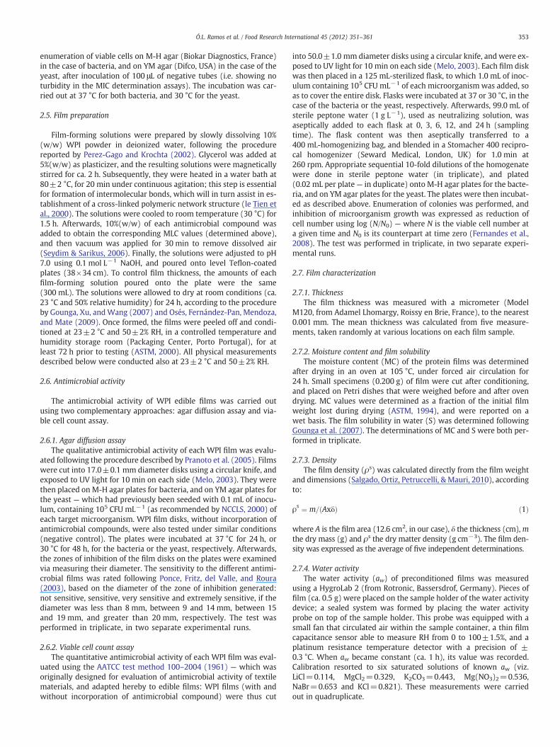
3.3.2. Moisture content, solubility, density, water activity, thickness andwater vapor permeability
The data tabulated in Table 3 pertain to the moisture content (MC),solubility (S), density (ρs), water activity (aw), thickness and watervapor permeability (WVP) of WPI edible films (10%,w/w) containingglycerol (5%,w/w) — as film matrix base, as affected by incorporationof the various antimicrobial compounds tested.
Incorporation of LA and NA into WPI films did not significantly(pN0.05) affect the MC, S, WVP and ρs values, relative to plain WPIfilms (control films). In turn, when PRO and COS were incorporatedin said films, a statistically significant increase (pb0.05) of MC, Sand WVP, together with a statistically significant decrease (pb0.05)of ρs were attained with regard to the control film and those incorpo-rated with other antimicrobial compounds — see Table 3.
The statistically significant differences (pb0.05) in MC, S, WVPand ρs values, when LA or PRO were added into WPI films, were cor-roborated by Manab et al. (2011), in particular regarding S and WVP.This is probably explained by the different pKa values of the two organicacids, aswell as by the presence of two binding groups – carboxyl and hy-droxyl (i.e.\COOH and\OH, respectively) –which are characteristic ofLA, instead of a single binding group (i.e. \COOH) — as typical of PRO.The higher dissociation of LA (as a result of its lower pKa), coupled withthe existence of two binding groups in dissociated form will contributeto establishment of a higher density network with the NH3
+ groupson the whey protein backbone via hydrogen bonding/hydrophobicinteractions — which will, in turn, lead to the significantly lowervalues of MC, S and WVP, and the significantly higher values of ρs
when LA is present (Manab et al., 2011).On the other hand, the highest differences in MC, S, WVP and ρs
values (pb0.05), relative to control films, were observed upon incor-poration of COS. This observation may be rationalized by the hydro-philic nature of this compound, attributable to the large fraction offree amino groups in D-glucosamine units. The high reactivity of theNH3
+ group of COS (which is a positively charged molecule) will likelycontribute to destabilization of the protein structure, thereby chang-ing the molecular organization of the film network and eventually in-creasing the number of free amino or hydroxyl groups of proteins. Inaddition, the amino groups of COS and of non-crosslinked proteinsare expected to form hydrogen bonds with \OH groups of watermolecules, thus increasing the susceptibility to hydration — and soleading to increases in moisture content, solubility and permeability
Table 3Physical properties (average±standard deviation), viz. moisture content (MC), solubility (10%(w/w) WPI edible films with 5%(w/w) glycerol, incorporated with 6 g L−1 LA, 10 g L−1
Antimicrobial agent MC(%, n=3)
S(%, n=3)
ρs
(g cm−3, n=5)
None 16.8±0.25a 67.6±0.44a 1.29±0.02a
LA 17.4±0.71a 69.0±1.30a 1.27±0.02a
PRO 22.2±0.70b 78.6±1.24b 1.19±0.02b
COS 23.4±0.65b 80.2±1.34b 1.16±0.03b
NA 18.2±1.30a 71.0±3.14a 1.24±0.03a
a,bMeans within the same column, labeled with the same letter, do not statistically differ fr
(Gontard, Duchez, Cuq, & Guilbert, 1993; McHugh et al., 1994). Sorbal,Menegalli, Hubinger, and Roques (2001) reported that the hydrophilic-ity of the antimicrobial additives considered will increase the watercontent of the film,whichwould thus affect the solubility therein. How-ever, theWVP values exhibited by our WPI films incorporated with thevarious antimicrobial compounds (Table 3) appeared lower than thosereported elsewhere for edible filmsmanufactured fromothermaterials:e.g. Chana-Thaworn, Chanthachum, and Wittaya (2011) found WVPvalues of 15.1 g mmm−2d−1kPa−1 using 1%(w/w) hydroxypropylmethylcellulose films incorporated with 0.3 g L−1 kiam wood extract,whereas Pranoto et al. (2005) obtained WVP values of 18.7 and14.9 g mmm−2d−1kPa−1 for films obtained from 1%(w/w) alginateand 1%(w/w) chitosan, respectively, after incorporation of 0.1%(w/w)garlic oil.
The statistically nonsignificant (pN0.05) changes in MC, S, WVPand ρs values (pN0.05) of WPI films when NA was incorporated(using the control films as reference) were also observed by Fajardoet al. (2010) and by Türe, Eroğlu, Özen, and Soyer (2009) for WVP— when the same compound was incorporated into chitosan, or intowheat gluten and methyl cellulose films, respectively. These resultsprobably arise from the low hydrophilic nature of the NA molecule(Fajardo et al., 2010).
The partial insolubility of WPI films, as observed here, has beenreported elsewhere (Fairley, Monahan, German, & Krochta, 1996a;McHugh & Krochta, 1994) — and may be rationalized by the presenceof stronger intermolecular bonds (e.g. disulfide bonds, as a result ofheat treatment) between the protein molecules within the matrix ofWPI films (McHugh, Avena-Bustillos, & Krochta, 1993; McHugh &Krochta, 1994). The incorporation of PRO and COS increases solubilitysignificantly (pb0.05) — see Table 3; this may indicate that thosecompounds interfere significantly with the protein polymeric net-work. In addition, the significant decrease (pb0.05) observed in ρs
of such films corroborates the fact that PRO and COS likely decreasenetworking within those films, thus producing films with lower den-sity (Hart, Craine, & Hart, 2003; Yoshida & Antunes, 2004). In general,a higher solubility of edible films indicates a lower water resistance,and thus a higher WVP. However, a high solubility of edible filmsmay appear as an advantage for specific applications (Stuchell &Krochta, 1994).
Regarding aw, incorporation of LA did not produce significant(pN0.05) changes relative to the control films. On the other hand,when PRO, COS and NA were incorporated in the WPI films, no signif-icant differences arose (pN0.05) relative to those films incorporatedwith LA; however, a statistically significant increase (pb0.05) was ap-parent relative to control films.
On the other hand, incorporation of the antimicrobial compoundstested did not significantly (pN0.05) affect the thickness of the WPIfilms. These results are similar to those reported by Kokoszka,Debeaufort, Lenart, and Voilley (2010), Osés et al. (2009), andSimelane and Ustunol (2005) — i.e. 0.12±0.08, 0.13±0.01 and0.14±0.02 mm, respectively, for WPI films with similar protein andglycerol concentrations.
S), density (ρs), water activity (aw), thickness and water vapor permeability (WVP), ofPRO, 20 g L−1 COS 3 kDa or 0.25 g L−1 NA (as appropriate).
aw(n=4)
Thickness(mm, n=5)
WVP(g mm m−2d−1kPa−1, n=3)
0.47±0.00a 0.13±0.01a 10.1±0.20a
0.49±0.02a,b 0.14±0.02a 10.9±0.75a
0.53±0.02b 0.17±0.03a 12.8±0.22b
0.54±0.03b 0.19±0.04a 13.4±0.41b
0.50±0.01b 0.15±0.02a 11.1±1.04a
om each other (pN0.05).

358 Ó.L. Ramos et al. / Food Research International 45 (2012) 351–361
3.3.3. Tensile propertiesResults of the tensile testing of 10%(w/w) WPI films containing 5%
(w/w) glycerol and several antimicrobial compounds are shown inFig. 2. Incorporation of said antimicrobial compounds produced sta-tistically significant differences (pb0.05) in tensile strength (TS),elongation at break (EB) and Young's modulus (YM) relative to thecontrol films; the magnitude of such differences was dependent onthe compound added. Incorporation of such antimicrobial com-pounds into the WPI films significantly (pb0.05) reduced their TS(mechanical resistance), thus resulting in weaker films. These resultsare in agreement with those conveyed by Cagri et al. (2004), whostated that incorporation of additives other than cross-linking agentsgenerally lowers TS of edible films.
The incorporation of LA and NA produced the lowest reduction inTS; however, statistically significant (pb0.05) differences wereobtained with regard to control films. On the other hand, the incorpo-ration of such compounds did not significantly (pN0.05) change theEB (or extensibility) and YM (or stiffness) properties of the WPIfilms. This result may be rationalized by the fact that those com-pounds, when incorporated into such films, do not destabilize theotherwise stable structure of the proteinaceous network — so theydid not increase the free volume and mobility of the protein chains(Hart et al., 2003).
The incorporation of PRO demonstrated, in turn, to produce thehighest variation in tensile properties of WPI films, being highest(pb0.05) for EB and lowest (pb0.05) for TS and YM; this led to ex-tremely fragile films (see Fig. 2). This result is consistent with ourfinding reported above, and based on visual appearance; such a dif-ference between the two organic acids may be attributed to their dif-ferent pKa values, as well as to the presence of one versus twobinding groups. Therefore, the lower dissociation of PRO associatedwith the presence of a single binding group may support establish-ment of a lower density network with the protein polymer, so higherintermolecular spacing within, and mobility of the polymer chainsthemselves will lead to more fragile films (Bodnár, Alting, &Verschueren, 2007; Krochta & de Mulder-Johnston, 1997; Manab etal., 2011).
WPI films incorporated with COS showed values of YM statisticallysimilar (pN0.05) to those obtained for the control films, and for filmsincorporated with LA and NA; however, a significantly (pb0.05)
0.0
0.1
0.2
0.3
0.4
0.5
0.6
0.7
0
10
20
30
40
50
60
70
80
90
100
EB
(%)
TS
(MPa
) an
dYM
(10
-2 M
Pa)
LA PRO None COS NA
a
e
f
g
h
TS YM EB
d
e
h h
i
c
e b b
h
Fig. 2. Tensile properties (average±standard deviation, n=10), viz. tensile strength(TS), elongation at break (EB) and Young's modulus (YM), of 10%(w/w) WPI ediblefilms with 5%(w/w) glycerol, incorporated with 6 g L−1 LA, 10 g L−1 PRO, 20 g L−1
COS 3 kDa or 0.25 g L−1 NA (as appropriate). Means with the same color labeledwith the same letter do not statistically differ from each other (pN0.05).
lower TS and a significantly (pb0.05) higher EB was observed (seeFig. 2). The aforementioned result may be accounted for by the highreactivity of the NH3
+ groups of the COS molecule — which probablyinterfere with the cross-linked network of native proteins, thus lead-ing to molecular reorganization of the interactions in the filmmatrix, and increasing the intermolecular spacing, and consequentlythe intrinsic chain mobility (Bodnár et al., 2007; Krochta & de Mulder-Johnston, 1997).
The reduction in TS, as affected by incorporation of additives, haspreviously been investigated in various hydrocolloid-based films(Gontard et al., 1993; Park & Chinnan, 1990); changes in tensile prop-erties, characterized by decreases in density and reversibility of inter-molecular interactions, have also been reported by Yang and Paulson(2000). These phenomena increase the mean free volume betweenpolymer chains (Gontard et al., 1993). The effect of adding spice ex-tracts to films has been also tackled — and, in all cases, significant de-creases in TS and YM were observed (Chana-Thaworn et al., 2011;Fang, Tung, Britt, Yada, & Dalgleish, 2002; Rojas-Graü et al., 2007).
3.3.4. Optical properties
3.3.4.1. Light transmission and film transparency. Light transmission (T)in the UV–vis range and transparency values — of 10%(w/w) WPIfilms containing 5%(w/w) glycerol and incorporated with differentantimicrobial compounds, are presented in Table 4.
No values of Twere noted in the UV light range (at 200 nm), for allWPI films; however, at 280 nm, such values ranged from 1.3±0.0% to2.3±0.2%, depending on the antimicrobial compound considered.Statistically significant differences (pb0.05) in T were not recorded,relative to the control films, when LA, PRO and NA were incorporated —
unlike what happened when COS was added (see Table 4). In any case,these results are low when compared with those exhibited bysome synthetic polymer films at 280 nm — i.e. 67.5, 80.0 and 79.1%,for LDPE, oriented polypropylene (OPP) and PVDC, respectively(Shiku, Hamaguchi, & Tanaka, 2003). The aforementioned resultssuggest that WPI films possess excellent barrier properties in the200–280 nm UV light region, probably owing to the high contentof aromatic amino acids in their protein-based structure that can ab-sorb UV-light (Limpan, Prodpran, Benjakul, & Prasarpran, 2010).
On the other hand, T ranged from 10.9±0.2 to 58.9±0.5% in thevisible range (350–800 nm) — once again depending on the antimi-crobial compound incorporated into the WPI film (see Table 4). Sta-tistically significant differences (pN0.05) were not obtained in termsof T values for WPI films incorporated with LA, PRO or NA, relativeto control films; however, a statistically significant increase(pb0.05) in T was observed upon COS incorporation. The aforemen-tioned difference may be associated with the yellow-brownish colorexhibited by WPI films containing COS (as mentioned before).
Nevertheless, the T values obtained here for WPI films upon incor-poration with the antimicrobial compounds were significantly lowerthan those reported by Gounga et al. (2007) — for 7%(w/w) WPIwith 20%(w/w) glycerol upon addition with pullulan, and by Fanget al. (2002) — for 12%(w/w) WPI with 40%(w/w) glycerol and10 mM Ca2+; this means that ourWPI films blocked passage of visiblelight in a more effective way. These differences may arise from thedistinct formulations of the film solution, or from the differences inthe film-forming WPI product itself.
Finally, the transparency of the WPI films incorporated with anti-microbial compounds ranged from 1.35 to 3.09% (see Table 4). Statis-tically significant differences (pN0.05) were not recorded when LA orNA were added relative to the control films, whereas statistically sig-nificant differences (pb0.05) were observed with PRO and COS.Moreover, LA produced the lowest change in WPI film transparency,whereas COS displayed the highest one. In addition, WPI films withLA showed a slightly higher transparency than LDPE films — i.e.

Table 4Optical properties (average±standard deviation, n=5), viz. light transmission (%) and transparency (A600/mm), of 10%(w/w) WPI edible films with 5%(w/w) glycerol, incorporat-ed with 6 g L−1 LA, 10 g L−1 PRO, 20 g L−1 COS 3 kDa or 0.25 g L−1 NA (as appropriate).
Antimicrobialagent
Wavelength (nm) Transparency
200 280 350 400 500 600 700 800
None 0.0±0.0a 1.3±0.0a 10.9±0.2a 24.4±0.4a 31.4±0.5a 35.5±0.6a 37.3±0.9a 38.9±1.1a 3.43±0.38a
LA 0.0±0.0a 1.4±0.2a 11.5±0.5a 25.3±0.7a 32.6±1.5a 36.9±1.3a 38.1±1.7a 40.6±0.6a 3.09±0.16a
PRO 0.0±0.0a 1.6±0.3a 14.0±3.1a 28.0±3.4a 36.3±4.6a 39.1±3.1a 41.1±3.0a 44.1±3.0a 2.40±0.14b
COS 0.0±0.0a 2.3±0.2b 27.8±0.5b 45.9±2.2b 52.0±1.5b 55.4±0.6b 56.7±0.7b 58.9±0.5b 1.35±0.31c
NA 0.0±0.0a 1.5±0.2a 12.6±1.7a 26.3±1.7a 35.0±3.3a 37.9±2.0a 39.1±1.9a 42.4±2.6a 2.81±0.25a
a,bMeans within the same column, labeled with the same letter, do not statistically differ from each other (pN0.05).
359Ó.L. Ramos et al. / Food Research International 45 (2012) 351–361
3.05, whereas PRO and NA led to higher transparency than OPP andPVDC films — i.e. 1.67 and 1.51, respectively (Shiku et al., 2003).
3.3.4.2. Color. The color measurements using L, a, b and ΔE factors,pertaining to WPI films incorporated with LA, PRO, COS and NA, areshown in Table 5— for the upper and lower surfaces. WPI films incor-porated with LA and PRO, as well as with NA were significantly(pb0.05) clearer and brighter (i.e. with a higher mean L value) thancontrol films. On the other hand, WPI films incorporated with COSappeared to be significantly (pb0.05) darker (i.e. with a lower meanL value), more red (i.e. with a greater mean positive a value) andmore yellow (i.e. with a greater mean positive b value) than theother four types of films — see Table 5. This result is consistent withthe yellow-brownish color exhibited by these films; and was antici-pated since the natural color of COS (3 kDa) in solution is yellow-brownish.
The total color difference was expressed via ΔE values; incorpora-tion of LA produced statistically significant (pb0.05) lower values ofΔE as compared with control films, so color changed less when thisagent was added to WPI films. This result is not surprising, since LAis often used as acidulant to reduce variation in color (Cagri et al.,2004). When NA and PRO were incorporated, statistically significantdifferences (pb0.05) were not found relative to the control films.On the other hand, WPI films incorporated with COS showed thehighest color change (pb0.05). Therefore, incorporation of LA, PROand NA inWPI films will not likely affect appearance of the food prod-uct, unlike will happen if COS is used. This is consistent with thetransparency values obtained before (Table 4), showing lower trans-parency of the films incorporated with COS. However, addition of COSwould provide an advantage in terms of optical properties to WPIfilms, if the main purpose were to cover defects that certain productsmay typically develop on their surface.
Finally, significant differences (pN0.05) were not observed in ΔEvalues among the lower and upper surfaces of WPI films, under allconditions.
Table 5Color properties (average±standard deviation, n=4), viz. L (black–white), a (green–red), bedible films with 5%(w/w) glycerol, incorporated with 6 g L−1 LA, 10 g L−1 PRO, 20 g L−1 C
Antimicrobial agent Surface L
None Upper 87.84±0.41a
Lower 87.94±0.36a
LA Upper 91.72±0.12b
Lower 91.71±0.17b
PRO Upper 90.16±0.49b
Lower 90.70±0.24b
COS Upper 50.50±1.12c
Lower 50.86±1.34c
NA Upper 90.93±0.07b
Lower 91.31±0.11b
a,b,cMeans within the same column, labeled with the same letter, do not statistically differ f
4. Conclusions
This study demonstrated thatWPI, following incorporation of distinctantimicrobial compounds, exhibits different degrees of effectivenessagainst several targetmicroorganisms. Organic acids, and LA in particular,lead to the highest antimicrobial activity against the Gram-positive bacte-rium, whereas COS was strongest against its Gram-negative counterpart.NA could not inhibit bacteria, but displayed the highest effectivenessagainst the yeast.
The complementary utilization of two antimicrobial assays — onemore qualitative and one more quantitative in nature, provides amore complete picture of the antimicrobial effectiveness of each ac-tive compound. The viable cell count assay was successfully adaptedto evaluate the antimicrobial activity of active edible films; it demon-strated a high sensitivity and a good reproducibility, and allowed abetter differentiation between the various antimicrobial edible filmsthan the agar diffusion assay.
Incorporation of LA andNAproduced the lowest change in all physicalpropertiesmeasured. Conversely, incorporation of COS and PRO led to thehighest change (pb0.05) in optical and tensile properties, respectively.
The overall results of our antimicrobial assays and physical tests back-up the following formulation for an active edible film: 10%(w/w) WPIwith 5%(w/w) glycerol (as base matrix), incorporated with 6 g L−1 LAand 0.25 g L−1 NA. This formulation is tentatively suggested for applica-tion in dairy products, namely cheese wrapping; however, specific testsare to be done to confirm its effectiveness in common practice. Selectionof these two antimicrobial compounds stems from their good synergisticperformance against microorganisms commonly found on cheese sur-faces — i.e. bacteria and yeasts, without significantly compromising thetensile, barrier and optical properties of the resulting WPI films.
Acknowledgments
Partial funding for this research work was provided by projectMilkfilm, administered by Agência de Inovação — POCTI: Programa
(blue–yellow) and ΔE (color difference) for upper and lower surface, of 10%(w/w)WPIOS 3 kDa or 0.25 g L−1 NA (as appropriate).
a b ΔE
0.73±0.04a 8.56±0.96a 12.10±0.24a
0.80±0.09a 8.82±0.88a 12.00±0.35a
0.12±0.01b 3.01±0.06b 6.39±0.05b
0.11±0.03b 2.91±0.17b 6.30±0.08b
−0.05±0.01c 5.69±0.28ª,b 9.44±0.14ª,b
0.00±0.03c 5.48±0.11ª,b 9.24±0.18ª,b
27.19±0.78d 49.40±1.92c 72.70±0.56c
26.89±0.73d 48.28±2.26c 71.60±0.63c
−0.03±0.02c 4.80±0.05a,b 8.34±0.14a,b
−0.04±0.02c 4.72±0.09a,b 8.15±0.08a,b
rom each other (pN0.05).
360 Ó.L. Ramos et al. / Food Research International 45 (2012) 351–361
Operacional de Ciência, Tecnologia e Inovação (Portugal). Funding forauthor O. L. Ramoswas via a Ph.D. fellowship (ref. SFRH/BD/30827/2006),administered by Fundação para a Ciência e a Tecnologia (Portugal) andsupervised by author F. X. Malcata. Author F. X. Malcata acknowledgespermission to use CBQF facilities for performance of a few experimentaltests.
References
Alakomi, H. -L., Skytta, E., Saarela, M., Mattila-Sandholm, T., Latva-Kala, K., & Helander,I. M. (2000). Lactic acid permeabilizes Gram-negative bacteria by disrupting theouter membrane. Applied and Environmental Microbiology, 66, 2001–2005.
Amefia, A. E., Abu-Ali, J. M., & Barringer, S. A. (2006). Improved functionality of food ad-ditives with electrostatic coating. Innovative Food Science and Emerging Technolo-gies, 7, 176–181.
ASTM (1994). Standard test method for moisture content of paper and paperboard byoven drying. Annual Book of ASTM Standards (pp. 644–694). Philadelphia, PA: ASTMD.
ASTM (1995). Standard test methods for water vapor transmission of materials. AnnualBook of ASTM Standards (pp. 697–704). Philadelphia, PA: ASTM E96-95.
ASTM (2000). Standard practice for conditioning plastics for testing. Annual Book ofASTM Standards. West Conshohocken, PA: ASTM D 618–00.
ASTM (2002). Standard test method for tensile properties of thin plastic sheeting. An-nual Book of ASTM Standards (pp. 1–9). Philadelphia, PA: ASTM D 882–02.
AATCC Test method 100–2004 (1961). Antibacterial finishes on textile materials: Assess-ment of AATCC Committee RA31.
Bourtoom, T. (2009). Review article: Protein edible film: Properties enhancement. In-ternational Food Research Journal, 16, 1–9.
Bodnár, I., Alting, A. C., & Verschueren, M. (2007). Structural effects on the permeabilityof whey protein films in an aqueous environment. Food Hydrocolloids, 21, 889–895.
Campos, C. A., Gerschenson, L. N., & Flores, S. K. (2011). Development of ediblefilms and coat-ings with antimicrobial activity. Food and Bioprocess Technology, 4, 849–8753.
Cagri, A., Ustunol, Z., & Ryser, E. T. (2004). Antimicrobial edible films and coatings. Jour-nal of Food Protection, 67, 833–848.
Cha, D. S., & Chinnan, M. S. (2004). Biopolymer-based antimicrobial packaging: A re-view. Critical Reviews in Food Science and Nutrition, 44, 223–237.
Chana-Thaworn, J., Chanthachum, S., &Wittaya, T. (2011). Properties and antimicrobialactivity of edible films incorporated with kiam wood (Cotyleobium lanceotatum)extract. Food Science and Technology, 44, 284–292.
Chung, Y. C., Su, Y. P., Chen, C. C., Jia, G., Wang, H. L., Wu, J. C. G., & Lin, J. G. (2004). Re-lationship between antibacterial activity of chitosan and surface characteristics ofcell wall. Acta Pharmacologica Sinica, 25, 932–936.
Coma, V. (2008). Bioactive packaging technologies for extended shelf life of meat-based products. Meat Science, 78, 90–103.
Debeaufort, F., Martin-Polo, M., & Voilley, A. (1993). Polarity and structure affect watervapour permeability of model edible films. Journal of Food Science, 58, 428–434.
Dibner, J. J., & Butin, P. (2002). Use of organic acids as a model to study the impact of gutmicroflora on nutrition and metabolism. Journal of Applied Poultry Research, 11,453–463.
Eswaranandam, S., Hettiarachchy, N. S., & Johnson, M. G. (2004). Antimicrobial activityof citric, lactic, malic, or tartaric acids and nisin-incorporated soy protein filmagainst Listeria monocytogenes, Escherichia coli O157:H7 and Salmonella gaminara.Journal of Food Science, 69, 79–84.
Fairley, P., Monahan, F. J., German, J. B., & Krochta, J. M. (1996). Mechanical propertiesand water vapor permeability of edible films from whey protein isolate and sodi-um dodecyl sulfate. Journal of Agricultural and Food Chemistry, 44, 438–443.
Fajardo, P., Martins, J. T., Fuciños, C., Pastrana, L., Teixeira, J. A., & Vicente, A. A. (2010).Evaluation of a chitosan-based edible film as carrier of natamycin to improve thestorability of Saloio cheese. Journal of Food Engineering, 101, 349–356.
Fang, Y., Tung, M. A., Britt, I. J., Yada, S., & Dalgleish, D. G. (2002). Tensile and barrierproperties of edible films made from whey proteins. Journal of Food Science, 67,188–193.
Fernandes, J. C., Tavaria, F. K., Soares, J. C., Ramos, O. S., Monteiro, M. J., Pintado, M. E., &Malcata, F. X. (2008). Antimicrobial effects of chitosans and chitooligosaccharidesupon Staphylococcus aureus and Escherichia coli in food model systems. Food Micro-biology, 25, 922–928.
Fernández, L., Apodaca, E. D., Cebrián, M., Villarán, M. C., & Maté, J. I. (2007). Effect ofthe unsaturation degree and concentration of fatty acids on the properties ofWPI-based edible films. European Food Research and Technology, 224, 415–420.
Franssen, L. R., & Krochta, J. M. (2003). Edible coatings containing natural antimicro-bials for processed foods. In S. Roller (Ed.), Natural Antimicrobials for the MinimalProcessing of Foods. Cambridge, UK: Woodhead Publishing.
Gerasimenko, D. V., Avdienko, I. D., Bannikova, G. E., Zueva, O. Y., & Varlamov, V. P.(2003). Antibacterial effects of water-soluble low molecular-weight chitosans ondifferent microorganisms. Applied Biochemistry and Microbiology, 40, 253–257.
Gontard, N., Duchez, C., Cuq, J. L., & Guilbert, S. (1993). Water and glycerol as plasti-cizers affect mechanical and water vapor barrier properties of an edible wheat glu-ten film. Journal of Food Science, 49, 1482–1485.
Gounga, M. E., Xu, S. -Y., & Wang, Z. (2007). Whey protein isolate-based edible films asaffected by protein concentration, glycerol ratio and pullulan addition in film for-mation. Journal of Food Engineering, 83, 521–530.
Han, J. H., & Floros, J. D. (1997). Casting antimicrobial packaging films and measuringtheir physical properties and antimicrobial activity. Journal of Plastic Film andSheet, 13, 287–298.
Hart, H., Craine, L. E., & Hart, D. J. (2003). Organic Chemistry— A Short Course (11th ed).Boston, NY: Houghton Mifflin.
Hirano, S., & Nagao, N. (1989). Effects of chitosan, peptic acid, lysozyme and chitinaseon the growth of several phytopathogens. Agricultural Biology and Chemistry, 53,3065–3066.
Hsiao, C. P., & Siebert, K. J. (1999). Modeling the inhibitory effects of organic acids onbacteria. International Journal of Food Microbiology, 47, 189–201.
Jeon, Y. J., Park, P. J., & Kim, S. K. (2001). Antimicrobial effect of chitooligosaccharidesproduced by bioreactor. Carbohydrate Polymers, 44, 71–76.
Kendra, D. F., Christian, D., & Hadwiger, L. A. (1989). Chitosan oligomers from Fusariumsolani/pea interactions, chitinase/β-glucanase digestion of sporelings and fromfungal wall chitin actively inhibit fungal growth and enhance disease resistance.Physiological and Molecular Plant Pathology, 35, 215–230.
Khwaldia, K., Perez, C., Banon, S., Desobry, S., & Hardy, J. (2004). Milk proteins for ediblefilms and coatings. Critical Reviews in Food Science and Nutrition, 44, 239–251.
Kokoszka, S., Debeaufort, F., Lenart, A., & Voilley, A. (2010). Water vapour permeability,thermal and wetting properties of whey protein isolate based edible films. Interna-tional Dairy Journal, 20, 53–60.
Kristo, E., Koutsoumanis, K. P., & Biliaderis, C. G. (2008). Thermal, mechanical andwater vapor barrier properties of sodium caseinate films containing antimicrobialsand their inhibitory action on Listeria monocytogenes. Food Hydrocolloids, 22,373–386.
Krochta, J. M., & de Mulder-Johnston, C. D. (1997). Edible and biodegradable polymerfilms: Challenges and opportunities. Food Technology, 51, 61–74.
Limpan, N., Prodpran, T., Benjakul, S., & Prasarpran, S. (2010). Properties of biodegrad-able blend films based on fish myofibrillar protein and polyvinyl alcohol asinfluenced by blend composition and pH level. Journal of Food Engineering, 100,85–92.
Lind, H., Jonsson, H., & Schnürer, J. (2005). Antifungal effect of dairy propionibacteria —
Contribution of organic acids. International Journal of Food Microbiology, 98, 157–165.Manab, A., Sawitri, M. E., al Awwaly, K. U., & Purnomo, H. (2011). Antimicrobial activity
of whey protein based edible film incorporated with organic acids. African Journalof Food Science, 5, 6–11.
Melo, N. R. (2003). Avaliação de embalagem ativa por incorporação de nisina na inibiçãode Staphylococcus sp.Mestrado — Dissertação, Universidade Federal de Viçosa, Brazil.
McHugh, T. H., Avena-Bustillos, R. D., & Krochta, J. M. (1993). Hydrophilic edible films:Modified procedure for water vapor permeability and explanation of thickness ef-fects. Journal of Food Science, 58, 899–903.
McHugh, T. H., Aujard, J. F., & Krochta, J. M. (1994). Plasticized whey protein ediblefilms: Water vapor permeability properties. Journal of Food Science, 59, 416–419.
McHugh, T. H., & Krochta, J. M. (1994). Plasticized whey protein edible films: Watervapor permeability properties. Journal of Food Science, 59, 416–423.
Min, S., Harris, L. J., Han, J. H., & Krochta, J. M. (2005). Listeria monocytogenes inhibitionby whey protein films and coatings incorporating lysozyme. Journal of Food Protec-tion, 68, 2317–2325.
Min, S., & Krochta, J. M. (2005). Antimicrobial films and coatings for fresh fruits andvegetables. In W. Jongen (Ed.), Raw Material Safety: Fruit and Vegetables. Cam-bridge, UK: Woodhead Publishing.
Montville, T. J., & Bruno, M. E. C. (1994). Evidence that dissipation of proton motiveforce is a common mechanism of action for bacteriocins and antimicrobial pro-teins. International Journal of Food Microbiology, 24, 53–74.
National Committee for Clinical Lab Standards (2000). Methods for dilution and anti-microbial susceptibility tests for bacteria that grow aerobically. Approved Standard,20 M7–A5.
Mulvihill, D. M., & Ennis, M. P. (2003). Functional milk proteins: Production and utilisa-tion. In P. F. Fox, & P. L. H. McSweeney (Eds.), Advanced Dairy Chemistry. Part B, 1.(pp. 1128–1176) New York NY, USA: Kluwer.
Nikaido, H. (2003). Molecular basis of bacterial outer membrane permeability revis-ited. Molecular Biology Reviews, 67, 593–656.
Ohsaki, Y., Tachibana, M., Nakanishi, K., Nakao, S., Saito, K., Toyoshima, E., Sato, M.,Takahashi, T., Osanai, S., Itoh, Y., & Kikuchi, K. (2003). Alterations in penicillin bind-ing protein gene of Streptococcus pneumoniae and their correlation with suscepti-bility patterns. International Journal of Antimicrobial Agents, 22, 140–146.
Osés, J., Fernández-Pan, I., Mendoza, M., & Mate, J. I. (2009). Stability of the mechanicalproperties of edible films based on whey protein isolate during storage at differentrelative humidity. Food Hydrocolloids, 23, 125–131.
Park, H. J., & Chinnan, M. S. (1990). Properties of edible coatings for fruits and vegeta-bles. (Paper 90–6510) International Winter Meeting. Chicago, IL: American Societyof Agricultural and Biological Engineers.
Perez-Gago, M. B., & Krochta, J. M. (2002). Formation and properties of whey proteinfilms and coatings. In A. Gennadios (Ed.), Protein-based Films and Coatings(pp. 159–180). Boca Raton FL, USA: CRC Press.
Pintado, C. M. B. S., Ferreira, M. A. S. S., & Sousa, I. (2010). Control of pathogenic andspoilage microorganisms from cheese surface by whey protein films containingmalic acid, nisin and natamycin. Food Control, 21, 240–246.
Ponce, A., Fritz, R., del Valle, C., & Roura, S. (2003). Antimicrobial activity of essentialoils on the native microflora of organic Swiss chard. Food Science and Technology,36, 679–684.
Pranoto, Y., Salokhe, V. M., & Rakshit, S. K. (2005). Physical and antibacterial propertiesof alginate-based edible film incorporated with garlic oil. Food Research Interna-tional, 38, 267–272.
Ramos, O. L., Pereira, J. O., Baptista da Silva, S. I., Amorim, M., Fernandes, J. C., Lopes-da-Silva, J. A., Pintado, M. E., & Malcata, F. X. (2011). Effect of composition of commer-cial whey protein preparations upon gelation at various pH values. Submitted toInternational Journal of Biological Macromolecules, unpublished results.
Ray, B. (2004). Fundamental Food Microbiology (3rd ed.). Boca Ratton FL, USA: CRC Press.

361Ó.L. Ramos et al. / Food Research International 45 (2012) 351–361
Ray, B., & Sandine, W. E. (1992). Acetic, propionic, and lactic acids of starter culturebacteria as biopreservatives. In B. Ray, & M. Daeschel (Eds.), Food Preservatives ofMicrobial Origin (pp. 103–136). Boca Ratton FL, USA: CRC Press.
Rojas-Graü, M. A., Avena-Bustillos, R. J., Olsen, C., Friedman, M., Henika, P. R., & Martín-Belloso, O. (2007). Effects of plant essential oils and oil compounds on mechanical,barrier and antimicrobial properties of alginate-apple puree edible films. Journal ofFood Engineering, 81, 634–641.
Salgado, P. R., Ortiz, S. E. M., Petruccelli, S., & Mauri, A. N. (2010). Biodegradable sun-flower protein films naturally activated with antioxidant compounds. Food Hydro-colloids, 25, 525–533.
Salmieri, S., & Lacroix, M. (2006). Physicochemical properties of alginate/polycaprolac-tone-based films containing essential oils. Journal of Agricultural and Food Chemis-try, 54, 10205–10214.
Seydim, A. C., & Sarikus, G. (2006). Antimicrobial activity of whey protein based ediblefilms incorporated with oregano, rosemary and garlic essential oils. Food ResearchInternational, 39, 639–644.
Shiku, Y., Hamaguchi, P. Y., & Tanaka, M. (2003). Effect of pH on the preparation of ed-ible films based on fish myofibrillar proteins. Fisheries Science, 69, 1026–1032.
Simelane, S., & Ustunol, Z. (2005). Mechanical properties of heat-cured whey protein-based edible films compared with collagen casings under sausage manufacturingconditions. Journal of Food Science, 70, 131–134.
Skrivanova, E., Marounek, M., Benda, V., & Brezinha, P. (2006). Susceptibility of Escher-ichia coli, Salmonella spp. and Clostridium perfringens to organic acids and mono-laurin. Veterinary Medicine, 51, 81–88.
Sorbal, P. J. A., Menegalli, F. C., Hubinger, M. D., & Roques, M. A. (2001). Mechanical,water vapor barrier and thermal properties of gelatin based edible film. Food Hy-drocolloids, 15, 423–432.
Stuchell, Y. M., & Krochta, J. M. (1994). Enzymatic treatments and thermal effects onedible soy protein films. Journal of Food Science, 59, 1322–1337.
Sundberg, C., & Jonsson, H. (2005). Process inhibition due to organic acids in fed-batchcomposting of food waste influences starting culture. Biodegradation, 16, 205–213.
Tharanathan, R. N. (2003). Biodegradable films and composite coatings: Past, presentand future. Trends in Food Science and Technology, 14, 71–78.
le Tien, C., Letendre, M., Ispas-Szabo, P., Mateescu, M. A., Delmas-Patterson, G., Yu, H. L.,& Lacroix, M. (2000). Development of biodegradable films from whey proteins bycross-linking and entrapment in cellulose. Journal of Agricultural and Food Chemis-try, 48, 5566–5575.
Türe, H., Eroğlu, E., Özen, B., & Soyer, F. (2009). Physical properties of biopolymers con-taining natamycin and rosemary extract. International Journal of Food Science andTechnology, 44, 402–408.
Uchida, Y., Izume, M., & Ohtakara, A. (1989). Preparation of chitosan oligomers withpurified chitosanase and its application. In G. Skjak-Broek, T. Anthonsen, & P. Sand-ford (Eds.), Chitin and Chitosan: Sources, Chemistry, Biochemistry, Physical Propertiesand Applications (pp. 373–382). London, UK: Elsevier.
Xia, W., Liu, P., Zhang, J., & Chen, J. (2011). Biological activities of chitosan and chitoo-ligosaccharides. Food Hydrocolloids, 25, 170–179.
Welscher, Y. M., Napel, H. H., Balagué, M. M., Souza, C. M., Riezman, H., & Kruijff, B.(2008). Natamycin blocks fungal growth by binding specifically to ergosterol with-out permeabilizing the membrane. Journal of Biological Chemistry, 283, 6393–6401.
Yang, L., & Paulson, A. T. (2000). Effects of lipids on mechanical and moisture barrierproperties of edible gellan film. Food Research International, 33, 571–578.
Yoshida, C. M. P., & Antunes, A. J. (2004). Characterization of whey protein emulsionfilms. Brazilian Journal of Chemical Engineering, 21, 247–252.
Copyright © 2022 FDOKUMEN