Potentially Bioactive Metabolites from Pineapple Waste ...
19
foods Article Potentially Bioactive Metabolites from Pineapple Waste Extracts and Their Antioxidant and α-Glucosidase Inhibitory Activities by 1 H NMR Awanis Azizan 1 , Ai Xin Lee 2 , Nur Ashikin Abdul Hamid 1 , Maulidiani Maulidiani 3 , Ahmed Mediani 4 , Siti Zulaikha Abdul Ghafar 2 , Nur Khaleeda Zulaikha Zolkeflee 1 and Faridah Abas 1,2, * 1 Laboratory of Natural Products, Institute of Bioscience, Universiti Putra Malaysia, Serdang 43400, Selangor, Malaysia; [email protected] (A.A.); [email protected] (N.A.A.H.); [email protected] (N.K.Z.Z.) 2 Department of Food Science, Faculty of Food Science and Technology, Universiti Putra Malaysia, Serdang 43400, Selangor, Malaysia; [email protected] (A.X.L.); [email protected] (S.Z.A.G.) 3 School of Fundamental Science, Universiti Malaysia Terengganu, Kuala Nerus 21030, Terengganu, Malaysia; [email protected] 4 Atta-ur-Rahman Institute for Natural Product Discovery, Universiti Teknologi MARA, Puncak Alam Campus, Bandar Puncak Alam 42300, Selangor, Malaysia; [email protected] * Correspondence: [email protected]; Tel.: +603-97698343 Received: 16 January 2020; Accepted: 7 February 2020; Published: 11 February 2020 Abstract: Pineapple (Ananas comosus) waste is a promising source of metabolites for therapeutics, functional foods, and cosmeceutical applications. This study strives to characterize the complete metabolite profiles of a variety of MD2 pineapple waste extracts. Metabolomics strategies were utilized to identify bioactive metabolites of this variety prepared with different solvent ratios. Each pineapple waste extract was first screened for total phenolic content, 2,2-diphenyl-1-picrylhydrazyl free radical scavenging, nitric oxide scavenging, and α-glucosidase inhibitory activities. The highest TPC was found in all samples of the peel, crown, and core extracted using a 50% ethanol ratio, even though the results were fairly significant than those obtained for other ethanol ratios. Additionally, crown extracted with a 100% ethanol ratio demonstrated the highest potency in DPPH and NO scavenging activity, with IC 50 values of 296.31 and 338.52 μg/mL, respectively. Peel extracted with 100% ethanol exhibited the highest α-glucosidase inhibitory activity with an IC 50 value of 92.95 μg/mL. Then, the extracts were analyzed and the data from 1 H NMR were processed using multivariate data analysis. A partial least squares and correlogram plot suggested that 3-methylglutaric acid, threonine, valine, and α-linolenic acid were the main contributors to the antioxidant activities, whereas epicatechin was responsible for the α-glucosidase inhibitory activity. Relative quantification further supported that 100% crown extract was among the extracts that possessed the most abundant potential metabolites. The present study demonstrated that the crown and peel parts of MD2 pineapple extracted with 100% ethanol are potentially natural sources of antioxidants and α-glucosidase inhibitors, respectively. Keywords: MD2 pineapple waste; ethanol ratios; antioxidants; α-glucosidase inhibitory; metabolomics 1. Introduction To date, biowaste products derived from various sources, including fruits have been widely recycled into valuable products ranging from agricultural compost, citric acid production, biofuel, pigment, and bioactive compound production [1]. This recycling method can also effectively resolve Foods 2020, 9, 173; doi:10.3390/foods9020173 www.mdpi.com/journal/foods
-
Upload
khangminh22 -
Category
Documents
-
view
1 -
download
0
Transcript of Potentially Bioactive Metabolites from Pineapple Waste ...

foods
Article
Potentially Bioactive Metabolites from PineappleWaste Extracts and Their Antioxidant andα-Glucosidase Inhibitory Activities by 1H NMR
Awanis Azizan 1, Ai Xin Lee 2, Nur Ashikin Abdul Hamid 1, Maulidiani Maulidiani 3,Ahmed Mediani 4, Siti Zulaikha Abdul Ghafar 2, Nur Khaleeda Zulaikha Zolkeflee 1 andFaridah Abas 1,2,*
1 Laboratory of Natural Products, Institute of Bioscience, Universiti Putra Malaysia, Serdang 43400, Selangor,Malaysia; [email protected] (A.A.); [email protected] (N.A.A.H.);[email protected] (N.K.Z.Z.)
2 Department of Food Science, Faculty of Food Science and Technology, Universiti Putra Malaysia,Serdang 43400, Selangor, Malaysia; [email protected] (A.X.L.); [email protected] (S.Z.A.G.)
3 School of Fundamental Science, Universiti Malaysia Terengganu, Kuala Nerus 21030, Terengganu, Malaysia;[email protected]
4 Atta-ur-Rahman Institute for Natural Product Discovery, Universiti Teknologi MARA, Puncak AlamCampus, Bandar Puncak Alam 42300, Selangor, Malaysia; [email protected]
* Correspondence: [email protected]; Tel.: +603-97698343
Received: 16 January 2020; Accepted: 7 February 2020; Published: 11 February 2020�����������������
Abstract: Pineapple (Ananas comosus) waste is a promising source of metabolites for therapeutics,functional foods, and cosmeceutical applications. This study strives to characterize the completemetabolite profiles of a variety of MD2 pineapple waste extracts. Metabolomics strategies were utilizedto identify bioactive metabolites of this variety prepared with different solvent ratios. Each pineapplewaste extract was first screened for total phenolic content, 2,2-diphenyl-1-picrylhydrazyl free radicalscavenging, nitric oxide scavenging, and α-glucosidase inhibitory activities. The highest TPC wasfound in all samples of the peel, crown, and core extracted using a 50% ethanol ratio, even thoughthe results were fairly significant than those obtained for other ethanol ratios. Additionally, crownextracted with a 100% ethanol ratio demonstrated the highest potency in DPPH and NO scavengingactivity, with IC50 values of 296.31 and 338.52 µg/mL, respectively. Peel extracted with 100% ethanolexhibited the highest α-glucosidase inhibitory activity with an IC50 value of 92.95 µg/mL. Then, theextracts were analyzed and the data from 1H NMR were processed using multivariate data analysis.A partial least squares and correlogram plot suggested that 3-methylglutaric acid, threonine, valine,and α-linolenic acid were the main contributors to the antioxidant activities, whereas epicatechin wasresponsible for the α-glucosidase inhibitory activity. Relative quantification further supported that100% crown extract was among the extracts that possessed the most abundant potential metabolites.The present study demonstrated that the crown and peel parts of MD2 pineapple extracted with 100%ethanol are potentially natural sources of antioxidants and α-glucosidase inhibitors, respectively.
Keywords: MD2 pineapple waste; ethanol ratios; antioxidants; α-glucosidase inhibitory;metabolomics
1. Introduction
To date, biowaste products derived from various sources, including fruits have been widelyrecycled into valuable products ranging from agricultural compost, citric acid production, biofuel,pigment, and bioactive compound production [1]. This recycling method can also effectively resolve
Foods 2020, 9, 173; doi:10.3390/foods9020173 www.mdpi.com/journal/foods

Foods 2020, 9, 173 2 of 19
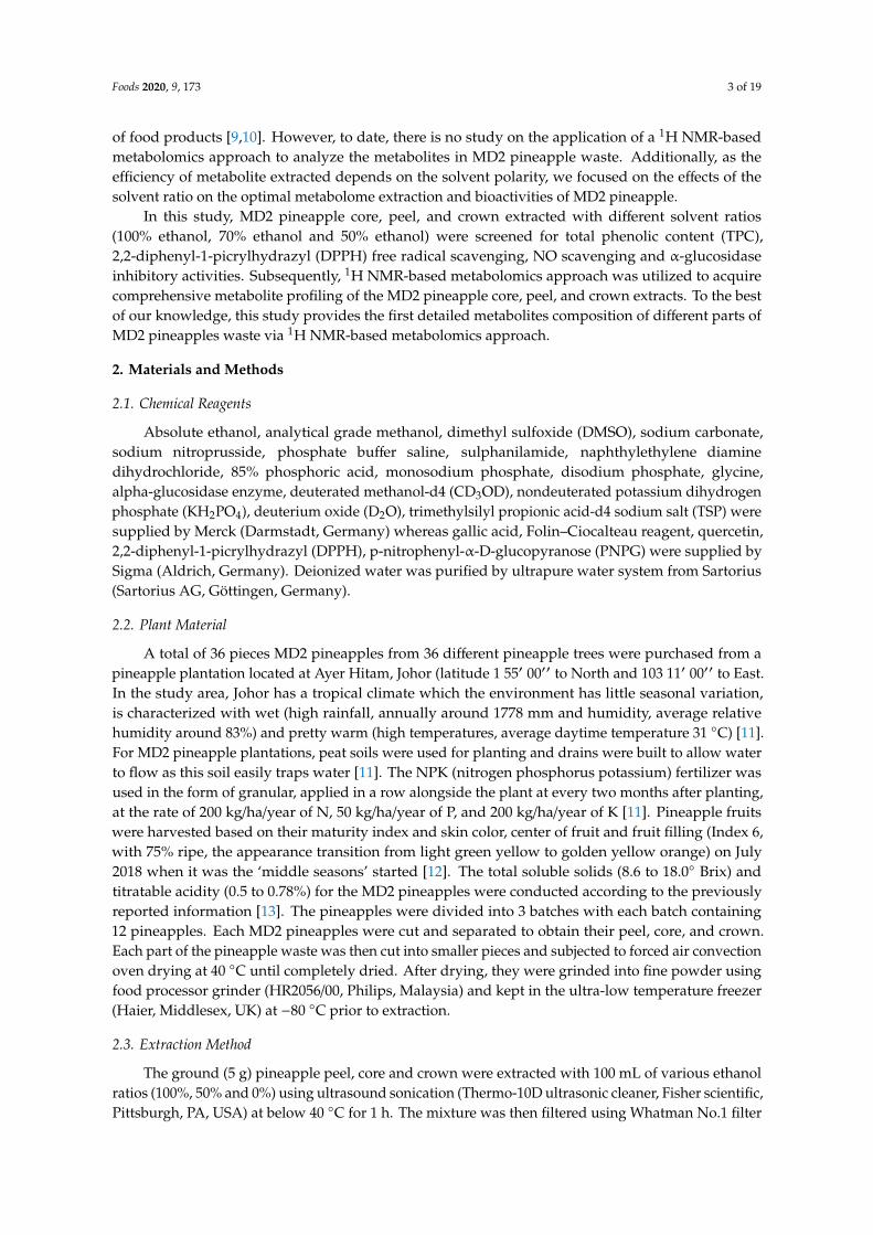
an environmental imbalance. An example of such fruit is pineapple (Ananas comosus), which belongsto the Bromeliaceae family. Among many other pineapple species planted in Malaysia, the MD2pineapple is a high-quality hybrid type with an exceptionally sweet taste and flavour and is uniform insize and ripeness [2]. Because of these traits, MD2 is also referred to as “Golden Ripe”, “Super Sweet”and “Gold”. Apart from fresh consumption, the MD2 pineapple has been successfully processed andcommercialized as it has own nutritional value including essential mineral composition, vitamins,organic acids and total polyphenol content that offers health benefits [3]. However, the inedible partsof MD2 pineapple waste, including its core, crown, and peel are mostly dumped and then pollutethe environment (Figure 1). Recently, research efforts have focused on finding better uses for MD2pineapple waste as such types of waste generally are rich in phenolic compounds, which are antioxidantagents [4].
Foods 2020, 9, 173 2 of 20
in size and ripeness [2]. Because of these traits, MD2 is also referred to as “Golden Ripe”, “Super
Sweet” and “Gold”. Apart from fresh consumption, the MD2 pineapple has been successfully
processed and commercialized as it has own nutritional value including essential mineral
composition, vitamins, organic acids and total polyphenol content that offers health benefits [3].
However, the inedible parts of MD2 pineapple waste, including its core, crown, and peel are mostly
dumped and then pollute the environment (Figure 1). Recently, research efforts have focused on
finding better uses for MD2 pineapple waste as such types of waste generally are rich in phenolic
compounds, which are antioxidant agents [4].
Figure 1. MD2 pineapple
Pineapple waste could be a valuable source of important bioactive compounds that have
countless beneficial for therapeutic application, including antioxidant, anti-inflammatory and anti-
microbial properties [5,6]. These characteristics might be due to the bioactive metabolites such as
pigments, sugars, organic acids, amino acids and proteolytic enzyme, bromelain [6]. The ability of
pineapple core extracts to scavenge oxidants has been reported in a few studies [7]. Li et al. [4]
identified polyphenolics metabolites from pineapple peel (i.e., catechin, epicatechin, gallic acid, and
ferulic acid) that contributed to the reduction of oxidative stress-related diseases [4] These
metabolites could be integrated into the bioprocess to produce valuable by-products or food
ingredients. Despite the growing importance of pineapple waste due to its nutritional, functional and
biological properties, information on the primary and especially the secondary metabolites of
different parts of MD2 pineapple waste is extremely scarce in the literature, and currently, little is
known about the bioactive compounds. The bioactive compounds can have a wide range of variations
depending on the environmental conditions, such as the characteristics of the soil, level of humidity,
rain intensity, water stress, temperature, fertilizers, and sunlight fluctuation. As a result of variation,
the metabolome and the bioactivity of each part of the plant also will be affected [8]. However, despite
the importance of studying the MD2 pineapple, little information is available in the literature about
its metabolite composition. Furthermore, few studies have addressed the identification of crucial
metabolites closely associated with the bioactivities of different parts of the MD2 pineapple waste.
The present study attempts to obtain a comprehensive idea of the key metabolite composition
present in waste from different parts of MD2 pineapples. A nuclear magnetic resonance (NMR)-based
metabolomics approach will allow us to identify the metabolome, which is important for the studied
activities. Recently, metabolomics approach has provided insight into the physiological status of an
organism and in the food science field, has been used to assess the quality, authenticity, and safety of
food products [9,10]. However, to date, there is no study on the application of a 1H NMR-based
metabolomics approach to analyze the metabolites in MD2 pineapple waste. Additionally, as the
Crown
Peel
Core
Figure 1. MD2 pineapple.
Pineapple waste could be a valuable source of important bioactive compounds that have countlessbeneficial for therapeutic application, including antioxidant, anti-inflammatory and anti-microbialproperties [5,6]. These characteristics might be due to the bioactive metabolites such as pigments,sugars, organic acids, amino acids and proteolytic enzyme, bromelain [6]. The ability of pineapple coreextracts to scavenge oxidants has been reported in a few studies [7]. Li et al. [4] identified polyphenolicsmetabolites from pineapple peel (i.e., catechin, epicatechin, gallic acid, and ferulic acid) that contributedto the reduction of oxidative stress-related diseases [4] These metabolites could be integrated into thebioprocess to produce valuable by-products or food ingredients. Despite the growing importance ofpineapple waste due to its nutritional, functional and biological properties, information on the primaryand especially the secondary metabolites of different parts of MD2 pineapple waste is extremelyscarce in the literature, and currently, little is known about the bioactive compounds. The bioactivecompounds can have a wide range of variations depending on the environmental conditions, such asthe characteristics of the soil, level of humidity, rain intensity, water stress, temperature, fertilizers, andsunlight fluctuation. As a result of variation, the metabolome and the bioactivity of each part of theplant also will be affected [8]. However, despite the importance of studying the MD2 pineapple, littleinformation is available in the literature about its metabolite composition. Furthermore, few studieshave addressed the identification of crucial metabolites closely associated with the bioactivities ofdifferent parts of the MD2 pineapple waste.
The present study attempts to obtain a comprehensive idea of the key metabolite compositionpresent in waste from different parts of MD2 pineapples. A nuclear magnetic resonance (NMR)-basedmetabolomics approach will allow us to identify the metabolome, which is important for the studiedactivities. Recently, metabolomics approach has provided insight into the physiological status of anorganism and in the food science field, has been used to assess the quality, authenticity, and safety
Foods 2020, 9, 173 3 of 19
of food products [9,10]. However, to date, there is no study on the application of a 1H NMR-basedmetabolomics approach to analyze the metabolites in MD2 pineapple waste. Additionally, as theefficiency of metabolite extracted depends on the solvent polarity, we focused on the effects of thesolvent ratio on the optimal metabolome extraction and bioactivities of MD2 pineapple.
In this study, MD2 pineapple core, peel, and crown extracted with different solvent ratios(100% ethanol, 70% ethanol and 50% ethanol) were screened for total phenolic content (TPC),2,2-diphenyl-1-picrylhydrazyl (DPPH) free radical scavenging, NO scavenging and α-glucosidaseinhibitory activities. Subsequently, 1H NMR-based metabolomics approach was utilized to acquirecomprehensive metabolite profiling of the MD2 pineapple core, peel, and crown extracts. To the bestof our knowledge, this study provides the first detailed metabolites composition of different parts ofMD2 pineapples waste via 1H NMR-based metabolomics approach.
2. Materials and Methods
2.1. Chemical Reagents
Absolute ethanol, analytical grade methanol, dimethyl sulfoxide (DMSO), sodium carbonate,sodium nitroprusside, phosphate buffer saline, sulphanilamide, naphthylethylene diaminedihydrochloride, 85% phosphoric acid, monosodium phosphate, disodium phosphate, glycine,alpha-glucosidase enzyme, deuterated methanol-d4 (CD3OD), nondeuterated potassium dihydrogenphosphate (KH2PO4), deuterium oxide (D2O), trimethylsilyl propionic acid-d4 sodium salt (TSP) weresupplied by Merck (Darmstadt, Germany) whereas gallic acid, Folin–Ciocalteau reagent, quercetin,2,2-diphenyl-1-picrylhydrazyl (DPPH), p-nitrophenyl-α-D-glucopyranose (PNPG) were supplied bySigma (Aldrich, Germany). Deionized water was purified by ultrapure water system from Sartorius(Sartorius AG, Göttingen, Germany).
2.2. Plant Material
A total of 36 pieces MD2 pineapples from 36 different pineapple trees were purchased from apineapple plantation located at Ayer Hitam, Johor (latitude 1 55′ 00′′ to North and 103 11′ 00′′ to East.In the study area, Johor has a tropical climate which the environment has little seasonal variation,is characterized with wet (high rainfall, annually around 1778 mm and humidity, average relativehumidity around 83%) and pretty warm (high temperatures, average daytime temperature 31 ◦C) [11].For MD2 pineapple plantations, peat soils were used for planting and drains were built to allow waterto flow as this soil easily traps water [11]. The NPK (nitrogen phosphorus potassium) fertilizer wasused in the form of granular, applied in a row alongside the plant at every two months after planting,at the rate of 200 kg/ha/year of N, 50 kg/ha/year of P, and 200 kg/ha/year of K [11]. Pineapple fruitswere harvested based on their maturity index and skin color, center of fruit and fruit filling (Index 6,with 75% ripe, the appearance transition from light green yellow to golden yellow orange) on July2018 when it was the ‘middle seasons’ started [12]. The total soluble solids (8.6 to 18.0◦ Brix) andtitratable acidity (0.5 to 0.78%) for the MD2 pineapples were conducted according to the previouslyreported information [13]. The pineapples were divided into 3 batches with each batch containing12 pineapples. Each MD2 pineapples were cut and separated to obtain their peel, core, and crown.Each part of the pineapple waste was then cut into smaller pieces and subjected to forced air convectionoven drying at 40 ◦C until completely dried. After drying, they were grinded into fine powder usingfood processor grinder (HR2056/00, Philips, Malaysia) and kept in the ultra-low temperature freezer(Haier, Middlesex, UK) at −80 ◦C prior to extraction.
2.3. Extraction Method
The ground (5 g) pineapple peel, core and crown were extracted with 100 mL of various ethanolratios (100%, 50% and 0%) using ultrasound sonication (Thermo-10D ultrasonic cleaner, Fisher scientific,Pittsburgh, PA, USA) at below 40 ◦C for 1 h. The mixture was then filtered using Whatman No.1 filter

Foods 2020, 9, 173 4 of 19
paper with 125 mm diameter. After filtration, another 50 mL of solvent was added to the residuesfor second time sonication under the same condition to maximize the yield. The first and secondfiltrates were then pooled and concentrated using a rotary evaporator (Hei-Vap, Heidolph, Schwabach,Germany) and lyophilized to remove excess water using freeze dryer (55-4 system Scanvac CoolSafe,Astech, Ireland). All of extracts were stored in the freezer at −80 ◦C until further analysis.
2.4. Total Phenolic Content (TPC) Assay
The TPC of pineapple peel, crown and core extracts was examined using Folin–Ciocalteu methodin a 96-well microplate according to the procedure previously reported with some modifications [11].A volume of 20 µL of sample (100%, 50% and 0% ethanol extracts at 5000 µg/mL concentration) and100 µL of Folin–Ciocalteu reagent were added into each well and incubated for 5 min. Then, 80 µL of0.75% sodium carbonate was added to each well and incubated for 20 min in the dark. The absorbancewas then measured at 765 nm using Tecan Infinite F200 Pro plate reader (Tecan Group Ltd., Männedorf,Switzerland). This analysis was performed in triplicate. Gallic acid was used as a standard. The sameprocedure was repeated by replacing the samples with gallic acid at serial dilution with concentrationof 0, 0.625, 1.25, 2.5, 5, and 10 µg/mL in the well to plot a standard curve. A standard curve wascalibrated with gallic acid and the results were expressed in mg GAE/g crude extract.
2.5. 2,2-Diphenyl-1-picrylhydrazyl (DPPH) Radical Scavenging Activity Assay
The antioxidant potential of the pineapple peel, crown and core extracts was determined basedon DPPH free radical scavenging assay as previously described [14]. Before the analysis, 3000 µg/mLof stock sample was prepared. The assay was conducted in a 96-well microplate with 50 µL of the testsamples prepared in 7 serial dilutions starting with 1000 µg/mL in the well. An aliquot of 100 µL of0.15 mM DPPH was then added into the well. For the blank sample, 100 µL of methanol was added toobtain accurate sample absorbance. The plate was then incubated for 30 min before measuring theabsorbance at 517 nm against the reagent blank by Tecan Infinite F200 Pro plate reader (Tecan GroupLtd., Männedorf, Switzerland). The analysis was performed in triplicate. The same procedure wasapplied for quercetin which acts as a positive control. The scavenging capacity of the test sample wascalculated as the percentage of DPPH inhibition (%) = [(Ao − As)/Ao] × 100 where Ao indicated theabsorbance of the reagent blank and As indicated the absorbance of the test samples. The results wereexpressed in IC50 value (µg/mL), which denotes the concentration of sample required to scavenge 50%DPPH free radicals.
2.6. Nitric oxide (NO) Radical Scavenging Activity Assay
Another approach of examining the antioxidant capacity of the pineapple peel, crown and coreextracts was determined by NO radical scavenging activity assay described previously with slightmodifications [15]. Before the analysis, 3000 µg/mL of stock sample was prepared. The assay was alsoconducted in a 96-well microplate with 60 µL of the test samples prepared in 7 serial dilutions startingwith 1000 µg/mL in the well. Then, a volume of 60 µL of 10 mM sodium nitroprusside in phosphatebuffer saline was added into the well. For the blank sample, a volume of 60 µL of the phosphate buffersaline was added. The plate was then incubated under light for 180 min. After incubation, 60 µLof freshly prepared Griess reagent (combination of 1% sulphanilamide and 0.1% naphthylethylenediamine dihydrochloride in 2.5% phosphoric acid) was added into each well. The absorbance wasthen measured at 550 nm by Tecan Infinite F200 Pro plate reader (Tecan Group Ltd., Männedorf,Switzerland). The analysis was performed in triplicate. Gallic acid was employed as the positivecontrol. The percentage of nitrite radical scavenging activity was calculated using the followingformula: Percentage of nitric oxide inhibition (%) = [(Ao − As)/Ao] × 100 where Ao indicated theabsorbance of the reagent blank and As indicated the absorbance of the test samples. The results werealso expressed IC50 value (µg/mL), which denotes the concentration of sample required to scavenge50% nitric oxide free radicals.

Foods 2020, 9, 173 5 of 19
2.7. α-Glucosidase Inhibition Assay
The α-glucosidase inhibition assay was performed in 96-well microplate as previouslydescribed [16]. First, both 30 mM and 50 mM phosphate buffer were prepared at pH 6.5. Then,ρ-nitrophenyl-β-D-glucopyranosidase (PNPG) as a substrate was dissolved in 50 mM phosphate buffermimics the intestinal fluid. A concentration of 2 M glycine which act as the stopping agent of thereaction was prepared at pH 10. The stock sample extract was prepared at 3000 µg/mL and 7 timesserial dilution was performed with starting concentration of 1000 µg/mL in the well. For this assay, analiquot of 10 µL of sample extract was added into the well, followed by addition of 130 µL of 30 mMphosphate buffer and 10 µL of enzyme. Meanwhile, the extract sample was substituted by DMSOsolvent for the negative control. For the blank sample, a volume of 140 µL of 30 mM buffer and 10 µLsample were loaded into the well whereas 140 µL of 30 mM buffer and 10 µL DMSO solvent wereloaded for blank solvent. Positive control was added with 130 µL of 30 mM phosphate buffer, 10 µL ofquercetin and 10 µL enzyme. The plate was then incubated for 5 min. After incubation period, eachwell of sample, negative and positive controls were added with 50 µL of PNPG while the rest wasadded with 50 µL of 30 mM buffer and further incubated for another 15 min. The reaction mixtureswere stopped by adding 50 µL of 2 M glycine at pH 10. The absorbance was measured at 405 nm usinga Tecan Infinite F200 Pro plate reader (Tecan Group Ltd., Männedorf, Switzerland). The α-glucosidaseinhibition activity was expressed as percentage of α-glucosidase inhibition (%) = [(An − As)/An] ×100% where An is the difference in absorbance of the negative control and all the blanks while Asindicated the absorbance of the test sample.
2.8. 1H NMR Analysis
The 1H NMR were implemented following reported method using 500 MHz Varian INOVA NMRspectrometer (Varian Inc, Palo Alto, CA, USA) functioning at temperature of 26 ◦C with frequency of499.887 MHz [16]. Each 1H NMR spectrum acquired a width of 20 ppm, which consisted of 64 scans witha 3.53 min acquisition time. For each sample, a relaxation delay of 1.0 s was recorded. The preparationprocedure was done by weighing 10 mg of crude extract and placed in a 1 mL Eppendorf tube. Then,375 µL of CD3OD and the same volume of KH2PO4 buffer in D2O (pH 6.0) containing 0.1% TSP wereadded. The TSP was used as internal reference, TSP = 0.0 ppm. The mixture was then vortexedfor 1 min followed by 10 min of ultra-sonication. Next, the mixture was centrifuged at 130× g for10 min to obtain a clear supernatant. A volume of 600 µL of the supernatant was transferred to NMRtube and subjected to 1H NMR analysis. All the NMR tubes were labeled and subjected to 1H NMRmeasurements using a predetermined setting for all the samples.
2.9. The Bucketing of 1H-NMR Spectra and Multivariate Statistical Analysis
Phasing and baseline corrections were performed manually for all the spectra using software builtin Varian INOVA NMR spectrometer (Varian Inc., Palo Alto, CA, USA). The bucketing and binning ofall the 1H NMR spectra were performed automatically into ASCII files by Chenomx software (version6.2, Edmonton, AB, Canada). The spectra were bucketed at δ 0.04 covering the range from δ 0.50–10.00.The regions of δ 4.70–4.90 and δ 3.23–3.36, which corresponding to the residual signals of water andmethanol respectively, were excluded from the analysis.
2.10. Statistical Analysis
The experimental results were expressed in mean ± standard deviation of four biological replicates.Analysis of variance (ANOVA) was used to evaluate the significant difference in the results. Pearsoncorrelation test was evaluated to show the relationship between assays. All the statistical analysiswas performed using Minitab software (Version 16, Minitab Inc, State College, PA, USA). Principalcomponent analysis (PCA) and partial least square analysis (PLS) were performed by SIMCA-P+
software (version 14.1, Umetrics AB, Umea, Sweden) using Pareto scaling method. The correlogram plot

Foods 2020, 9, 173 6 of 19
was done using RStudio version 1.0.13 (RStudio, Boston, MA, USA). The size of correlation of tentativelyidentified metabolites with the bioactivities was determined using the following rules: r values between−0.3 to 0.3 means negligible correlation; from 0.3 to 0.5 (or −0.3 to −0.5) represent low correlation; rvalues from 0.5 to 0.7 (or −0.5 to −0.7) indicate a moderate correlation; r values from 0.7 to 0.9 (or −0.7to −0.9) mean a strong or high correlation and, finally, r values between 0.9 and 1.0 (or −0.9 to −1.0)imply a very strong or highest correlation [17]. The changes in metabolite levels were quantitativelyevaluated and displayed in a box plot using MetaboAnalyst 3.0 (http://www.metaboanalyst.ca). Tukey’ssignificant difference multiple comparison test was applied to evaluate the significant differencesbetween the extracts.
3. Results and Discussion
3.1. TPC of MD2 Pineapple Peel, Crown and Core Extracts
The TPC content of MD2 pineapple peel, crown and core subjected to different ethanol ratioextractions is shown in Table 1, ranging from 3.53 to 12.71 mg GAE/g crude extract. The resultsdemonstrated that crown extracts exhibited the highest TPC recorded at 12.71 mg GAE/g crude extract,followed by peel extracts with 10.73 mg GAE/g crude extract, which both extracts were prepared from50% ethanol ratio. Core extracts derived from 50% ethanol ratio exhibited the lowest TPC with 4.80 mgGAE/g crude extract.
Abiotic stress, such as temperature and light intensity could also lead to different concentrationsof secondary metabolites in plant parts [18]. A study by Nur Asniyati et al. [19] reported that leaves ofMD2 pineapple grown in their natural environment showed significantly higher TPC (0.433 mg GAE/gof extract) than those grown in vitro (0.296 mg GAE/g of extract). This is because the light intensity intheir natural environment (30,000 to 100,000 lux) was much higher than that in the in vitro environment(approximately 1000 lux). This could explain why the crown extracts exhibited the highest TPC in thecurrent study, their structure and position at the top most part of the pineapple fruit, allows them toreceive a greater amount of light than the peel and core. Since photosynthesis takes place in the leaves,the most abundant phenolic biosynthetic pathway precursors can be found in the crown extracts,which has also been well as confirming the accumulation of phenolic compound [20].
In terms of different ethanol ratio extractions, it was observed that the 50% ethanol contributed tothe highest TPC in all the three parts of MD2 pineapple waste with significant difference (p < 0.05).The result of the current study is in agreement with that of Wang et al. [21], in which they reportedthat a 50% ethanol ratio extraction under ultrasound sonication treatment for 30 min contribute to thehighest TPC from apple pomace. Water and ethanol mixtures are commonly used to extract phenoliccompounds due to their ability to dissolve a wide range of compounds, as reported in numerousstudies [22,23]. However, the solubility of the phenolic compounds in the solvent is not the onlypredominant factor in the extraction of TPC. This could explain why a 100% ethanol ratio is not thebest extraction solvent for phenolic, rather, a 50% to 80% ratios yields the best extraction of targetedcompounds [24]. This is compatible with the results of the current study, in which a 100% ethanol ratioextraction yielded lower TPC than the 50% ethanol out of all the crude extracts.
3.2. DPPH Free Radical Scavenging Activity of MD2 Pineapple Peel, Crown and Core Extracts
The results of DPPH free radical scavenging activity of MD2 pineapple peel, crown and coreextracts subjected to different ethanol ratio extractions is presented in Table 1. The percentage ofinhibition of MD2 pineapple peel, crown and core extracts were varied from 30.13% to 75.57%.Detectable IC50 values ranged from 296.31 µg/mL to 386.70 µg/mL, with a quercetin standard of11.25 µg/mL. Similar to the trend in TPC, the plant parts that showed the most active DPPH freeradical scavenging activity were the crown extracted with 100% ethanol ratio (75.57%), followedby the peel (72.67%) and core (49.14%) extracted with 50% ethanol ratio. This could be explainedby the fact that phenolic compounds are the major contributors to the antioxidant activity of plant

Foods 2020, 9, 173 7 of 19
extracts. The chemical structure and spatial conformation of the phenolic compounds, which determinethe availability of the hydroxyl group to react with the free radical, is important in evaluating theirantioxidant potential [25].
Table 1. The bioactivities of pineapple peel, crown and core extracted with 0, 50 and 100% ethanol.
Part Ethanol TPC DPPH Scavenging Activity Nitric Oxide Scavenging Activity α-Glucosidase Inhibition
Ratio (%) (mg GAE/gcrude extract) % inhibition IC50(µg/mL) % inhibition IC50(µg/mL) % inhibition IC50 (µg/mL)
Peel0 10.62 ± 0.37 a
b 68.96 ± 1.07 ba 386.70 ± 17.55 a 63.60 ± 0.85 a
a 658.19 ± 16.06 b 54.99 ± 2.88 bb 878.75 ± 96.19 a
a50 10.73 ± 0.14 a
b 72.67 ± 1.51 aa 339.23 ± 14.61 b
a 46.74 ± 1.56 bb ND 44.51 ± 3.35 c
a ND
100 7.97 ± 0.63 bb 71.24 ± 1.38 ab
b353.10 ± 21.34
aba
44.43 ± 0.75 cb ND 73.86 ± 5.39 a
a 92.95 ± 6.09 bb
Crown0 12.15 ± 0.62 a
a 46.56 ± 1.51 cb ND 51.16 ± 1.01 b
b 848.87 ± 34.18 aa 65.24 ± 1.47 a
a 552.44 ± 68.55 ab
50 12.71 ± 1.15 aa 72.19 ± 1.91 b
a 341.44 ± 32.26 aa 65.86 ± 7.38 a
a 328.77 ± 30.17 b 23.59 ± 1.67 bb ND
100 12.22 ± 0.83 aa 75.57 ± 0.81 a
a 296.31 ± 12.74 bb 70.21 ± 1.72 a
a 338.52 ± 25.32 b 63.44 ± 4.38 ab 651.49 ± 57.35 a
a
Core0 3.53 ± 0.12 c
c 30.13 ± 2.00 cc ND 27.82 ± 1.87 b
c ND 32.26 ± 3.01 cc ND
50 4.80 ± 0.23 ac 49.14 ± 0.61 a
b ND 31.62 ± 1.69 ac ND 46.16 ± 1.88 a
a ND100 4.15 ± 0.20 b
c 35.00 ± 1.32 bc ND 31.31 ± 1.73 a
c ND 41.10 ± 1.49 bc ND
Standard 11.24 ± 0.39(Quercetin)
3.51 ± 0.51(Gallic Acid)
0.99 ± 0.14(Quercetin)
The values represent means ± standard deviation based on four biological replicates. The superscript is to comparethe same part of plant with different ethanol ratio; the subscript is to compare different parts of the plant with sameethanol ratio. Mean with different superscript and subscript letters are significantly difference (p < 0.05); ND isnot determined.
In terms of ethanol ratio extraction, the crown extracted with a 100% ethanol ratio possessed thehighest DPPH scavenging activity and resulted in the lowest IC50 value (296.31 µg/mL) compared topeel and core extracts. It was found that crown extracted with 0% ethanol showed significantly lowerpercentage of DPPH inhibition (46.56%) compared to the crown extracted from 50% and 100% ethanolratio extracts, with no IC50 determined. Based on the results, it revealed that low antioxidant activityof 0% ethanol was due to low solubility of the metabolites in water compared to ethanol. The resultsare interrelated with the TPC content examined in the previous section. It was observed that crownextracted with a 50% ethanol ratio (highest in TPC) did not correspond directly to the highest DPPHfree radical scavenging activity in this study. This is because no significant difference (p < 0.05) wasobserved for crown extracted with different ethanol ratios in TPC, suggesting that extracted phenoliccompounds varied in property and nature, even though the amounts of yield were relatively equal.Thus, they acted differently in scavenging of DPPH free radical scavenging.
It was also noted that 0% ethanol of all types MD2 pineapple waste extracts were significantlylowest in the DPPH free radicals’ scavenging activity. Similar results were observed in the study bySun et al. [26], who reported that the 75% ethanol extracted propolis illustrated stronger antioxidantactivities than water extract [26]. However, it is also important to note that not only concentration,but also the synergistic effect stemming from the interactions between extracted phenolic compoundsdirectly affected the extract’s antioxidant capacity [27]. The results of the current findings varycompared to those of the few previous studies. Rathnakumar et al. [28] reported that the IC50 values ofpineapple core and peel were 38.65 µg/mL and 738.3 µg/mL, respectively [29]. The maturation stagesof the pineapple could also result in differences in antioxidant activity [13].
3.3. Nitric Oxide (NO) Free Radical Scavenging Activity of MD2 Pineapple Peel, Crown and Core Extracts
Another bioassay to test the antioxidant potential of MD2 pineapple peel, crown and core subjectedto different ethanol ratio extractions was examined through the NO free radical scavenging activity, theresults of which are shown in Table 1. Based on the results, the percentage of NO inhibitory activity ofMD2 pineapple peel, crown and core extracted with three different ethanol ratios varied from 27.82 to70.21%, with a significant difference (p < 0.05). The highest percentage of NO scavenging activity wasseen in 100% ethanol ratio of crown extracts (70.21%), followed by 0% ethanol ratio of peel (63.60%) and50% ethanol ratio of core (31.62%) extracts. The detectable IC50 ranged from 328.77 to 848.87 µg/mL,with that of gallic acid standard recorded at 3.5 µg/mL.

Foods 2020, 9, 173 8 of 19
Generally, the percentage of NO scavenging in all the parts examined was lower compared tothe DPPH percentage. This is because the mode of action and scavenging capacity of antioxidantsfrom these plant extracts toward DPPH and NO free radicals are different [29]. Regarding the differentethanol ratio extractions, crown extracted with 100% ethanol contributed to the highest percentage ofNO scavenging activity (70.21%) with IC50 value of 338.52 µg/mL (Table 1), demonstrating a trendsimilar to that of DPPH inhibition activity. All core extracts were significantly (p < 0.05) lowest in theirNO scavenging activity, with no IC50 determined.
3.4. α-Glucosidase Inhibitory Activity of MD2 Pineapple Peel, Crown and Core Extracts
The α-glucosidase inhibitory activity of MD2 pineapple peel, crown and core extracted with threedifferent ethanol ratios is shown in Table 1. Based on the results, the percentage of α-glucosidaseinhibitory activity of MD2 pineapple peel, crown and core extracted with different ethanol ratiosranged from 23.59% to 73.86%. The detectable IC50 values ranged from 92.95 µg/mL to 878.75 µg/mL,with a quercetin standard of 0.99 µg/mL (Figure S1). The 100% ethanol ratio peel extract significantly(p < 0.05) possessed the highest percentage of α-glucosidase inhibitory activity compared to the 50%and 0% ethanol ratio extracts. It possessed the lowest IC50 value (92.95 µg/mL), indicating the strongestcapability of inhibiting the action of the α-glucosidase enzyme (Table 1).
From these findings, the 100% ethanol was the most capable solvent of extracting the phytochemicalconstituents responsible for the α-glucosidase inhibition of MD2 pineapple peel. The results wereconsistent with those of previous studies in which 100% ethanol extract was found to be the mostefficient in α-glucosidase inhibitory activity of Cosmos caudatus leaves [30]. While the 50% ethanol ratioextract of crown showed relatively good activities in TPC, DPPH and NO free radical scavenging, it wasweak in α-glucosidase inhibitory activity with only 23.59% inhibition capability, which is the lowestamong all the plant parts and different ethanol ratio extractions. It is worth noting that although the50% ethanol ratio extracts of peel, crown and core extracts showed the highest TPC, as discussed earlier,they were weak in α-glucosidase inhibitory activity with inhibition percentages of 44.51%, 23.59%and 46.16%, respectively. This may be due to the fact that the major contributors to α-glucosidaseinhibitory activity may not be phenolic compounds, but rather non-phenolic compounds [31]. Furtheridentification of the compounds is required to support this finding. Regardless of the differencein ethanol ratio extractions, all core extracts were found to be significantly (p < 0.05) weak in theirα-glucosidase inhibitory activity, with no IC50 determined.
3.5. Visual Inspection of The 1H NMR Spectra and Metabolite Identification of MD2 Pineapple Peel, Crown andCore Extracts
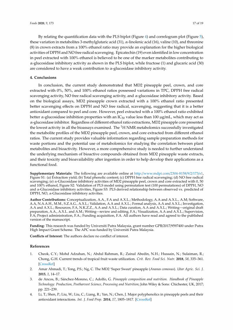
A detailed analysis based on 1H NMR was performed to further study the variation of metabolitesand biological activities in MD2 pineapple peel, crown and core extracted using different ethanol ratios.Based on previous studies on pineapple, numerous primary and secondary metabolites were reportedprimarily on the fruit and leaves, including sugars [32], amino acids [33], fatty acids [34] and phenoliccompounds [35,36]. There is limited published data on the metabolites in pineapple crown, peel, andcore based on the 1H NMR metabolomics approach in particular, and no studies have been publishedregarding the MD2 pineapple. The representative 1H NMR spectra of MD2 pineapple peel, crown andcore extracted with 0%, 50% and 100% ethanol ratios are shown in Figure 2. A total of 31 metaboliteswere identified with their corresponding chemical shifts presented in Table 2.

Foods 2020, 9, 173 9 of 19Foods 2020, 9, 173 9 of 20
Figure 2. 500MHz representative 1H NMR spectra of MD2 pineapple peel, crown and core extracted
with 0, 50 and 100% ethanol ratio. (A) 0% ethanol peel; (B) 50% ethanol peel; (C) 100% ethanol peel;
(D) 0% ethanol crown; (E) 50% ethanol crown; (F) 100% ethanol crown; (G) 0% ethanol core; (H) 50%
ethanol core and (I) 100% ethanol core. See Table 1 for the assignments of the identified metabolites
signals.
(A)
(B)
(C)
(D)
(E)
(F)
(G)
(H)
(I)
3 5
1
2
6 11
9 15
16 20
7 20 14
8, 10
21 13 17 23 18
19
4
6
22
26
30, 28
29
26
3
14
31
11
5 23
25
Figure 2. 500MHz representative 1H NMR spectra of MD2 pineapple peel, crown and core extractedwith 0, 50 and 100% ethanol ratio. (A) 0% ethanol peel; (B) 50% ethanol peel; (C) 100% ethanolpeel; (D) 0% ethanol crown; (E) 50% ethanol crown; (F) 100% ethanol crown; (G) 0% ethanol core;(H) 50% ethanol core and (I) 100% ethanol core. See Table 1 for the assignments of the identifiedmetabolites signals.

Foods 2020, 9, 173 10 of 19
Table 2. Tentatively identified metabolites of 1H NMR spectra from MD2 pineapple peel, crown andcore extracted with 0, 50 and 100% ethanol ratio.
MetabolitesChemicalShift (δ)
Parts of MD2 Pineapple Waste/Ethanol Ratio (%)
Peel Crown Core
0 50 100 0 50 100 0 50 100
Sugar
(1) Fructose 4.17(d) + + + + + + + + +
(2) Sucrose 5.38(d),4.02(t) + + + + + + + + +
(3) α-D-glucose 5.18(d) + + + + + + + + +(4) β-D-glucose 4.58(d) + + + + + + + + +
Amino Acids(5) Alanine 1.47(d) + + + + + + + + +(6) Arginine 3.78(t) + + + + + + + + +
(7) Asparagine 2.93(d) - - - + + + - - -(8) Threonine 1.34(d) + + + + + + + + +
(9) Glycine 3.57(s) + + + + + + + + +(10) Valine 1.06(d) + + + + + + + + +
(11) Isoleucine 3.66(d) + + + + + + + + +
(12) Phenyalanine 7.30(d),7.36(d) + + + + + + + + +
(13) Tryptophan 7.54(d) + + + + + + + + +
Organic acids
(14) Citric acid 2.72(d) + + + + + + - - -(15) Malic Acid 4.30(dd) + + + + + + + + +
Lipids
(16) α-linolenic acid 1.26(s) + + + + + + + + +
Phenolic compounds
(17) Ferulic acid
7.34(d),7.27(s),
7.10(dd),6.93(d),6.39(d)
+ + + + + + + + +
(18) Gallic acid 7.04(s) + + + + + + + + +
(19) Epicatechin
6.12(d),6.09(d),5.00(s),4.34(m)
+ + + + + + + + +
(20) Catechin 2.82(dd) - - - + + + - - -
(21) Protocathechuic acid 7.40 (d)6.90 (d) - - - + + + - - -
(22) Benzoic acid 7.70(t)7.46 (dd) - - - + + + - - -
(23) Vanillic acid7.52 (d)7.02(d)3.90 (s)
- - - + + + - - -
(24) Syringic acid 7.37(s)3.98 (s) + + + + + + + + +
(25) Phenylacetic acid7.42 (m)7.30 (m)3.50 (s)
+ + + + + + + + +
(26) Malonic acid 3.10(s) + + + + + + - - -(27) Succinic acid 2.42(s) + + + + + + + + +
(28) Glyceric acid 4.14(m)3.82(m) + + + + + + + + +
(29) Fumaric acid 6.50(s) - - - + + + - - -
(30) Glucaric acid4.22(d)4.10(d)3.92 (t)
+ + + + + + + + +
(31) 3-methylglutaric acid 2.18(m)0.90(d) - - - + + + - - -
+: presence; -: absence; d: double; dd: doublet of doublet, m: multiplet, s: singlet; t: triplet.

Foods 2020, 9, 173 11 of 19
In the current study, metabolites were identified based on a comparison of chemical shiftsassigned in 1H NMR spectra with several publicly accessible metabolomics databases, such as theMadison Metabolomics Consortium Database (MMCD; http://mmcd.nmrfam.wisc.edu/), the HumanMetabolome Database (HMDB; http://www.hmdb.ca/) peak fitting routine of Chenomx database (v. 8.1,Edmonton, AB, Canada) and previous literature data [16,31,36,37]. Based on visual inspection of therepresentative 1H NMR spectra, different classes of metabolites including sugars, amino acids, organicacids and fatty acids were found to be present. The variation of signal intensity among differentparts of MD2 pineapple waste extracted with different ethanol ratios was obvious, especially in thecarbohydrate regions. The most intense signals were located in the carbohydrate regions, with chemicalshift ranging from δ 3.0 to δ 5.50. This region was dominated by sugars such as fructose, δ sucrose (δ5.38), α-D-glucose (δ 5.18) and β-D-glucose (δ 4.58). Sukruansuwan and Napathorn [38] demonstratedan abundance of sugars present in pineapple peel and core that are beneficial to fermentation.
The amino acids identified correspond to alanine (δ 1.47), arginine (δ 3.78), asparagine (δ 2.93),threonine (δ 1.34), glycine (δ 3.57), valine (δ 1.06), isoleucine (δ 3.66), phenylalanine (δ 7.30) andtryptophan (δ 7.54). Most of the amino acids identified fall into the regions between δ 1.0 and 3.6,except phenylalanine (δ 7.30) and tryptophan (δ 7.54) (aromatic amino acids). Meanwhile, two organicacids and a lipid, known as malic acid (δ 4.30), citric acid (δ 2.72) and α-linolenic acid (δ 1.26) werealso identified, respectively. In addition, signals in aromatic regions were the least intense observedin the 1H NMR spectra for all parts of MD2 pineapple waste regardless of the ethanol ratio used.Phenolic compounds in pineapple waste extracts were also tentatively identified and corresponded tothe aromatic regions in the crude extract based on their 1D NMR and in previous A. comosus [L.] Merr.studies [39]; they were ferulic acid (δ 7.34), gallic acid (δ 7.04), epicatechin (δ 6.12), protocatechuic acid(δ 7.40 and δ 6.90), benzoic acid (δ 7.70 and δ 7.46), syringic acid (7 δ.37), phenylacetic acid (7.42 m) andfumaric acid (6.50). The doublets at δ 7.52 and δ 7.02 may be attributed to vanilic acid (protons H-2and H-6). Referring to Table 2, it is interesting to note that regardless of the ethanol ratio used in theextraction, the MD2 pineapple crown showed variation compared to peel and core as demonstrated bythe presence of amino acid (asparagine, δ 2.93) and phenolics (catechin, δ 2.82). Otherwise, citric acidwas not identified in MD2 pineapple core.
3.6. Metabolite Variations in MD2 Pineapple Peel, Crown and Core Extracts
In the current study, 1H NMR data were subjected to principal component analysis (PCA) todiscriminate metabolite variations of different parts of MD2 pineapple waste extracted with differentethanol ratios. PCA provides the primary evaluation of the relationships between the samples usingthe unsupervised clustering method [14]. The separation of samples into clusters can be examined inthe PCA score plot whereas metabolites that contributed to the separation are revealed in the PCAloading plot. Both the PCA score and loading plots of MD2 pineapple peel, crown and core extractedwith different ethanol ratios are illustrated in Figure 3. The PCA model showed good fitness and highpredictability with an R2X of 0.99 and a Q2 of 0.96, respectively. The total variance of the model was82.7%, for which PC1 contributed 68.2% and PC2 contributed 14.5%.
Based on the score plot, MD2 pineapple crown extracts were well clustered and clearly separatedfrom peel and core extracts by PC1 (Figure 3a). Meanwhile, crown extracted with 50% and 100%ethanol were clearly distinguishable from crown extracted with a 0% ethanol by PC2. Peel extractedwith a 100% ethanol was well clustered and separated from the 50% and 0% ethanol ratio peel extractsby PC2. Among the core extracts, the 100% ethanol showed good cluster, distinguishing them from the50% and 0% ethanol ratios of core extracts by PC2. Compared to the 100% ethanol extracts, core andpeel extracts from 50% and 0% were more dispersed and less clustered.

Foods 2020, 9, 173 12 of 19
Foods 2020, 9, 173 11 of 20
8.1, Edmonton, AB, Canada) and previous literature data [16,31,36,37]. Based on visual inspection of
the representative 1H NMR spectra, different classes of metabolites including sugars, amino acids,
organic acids and fatty acids were found to be present. The variation of signal intensity among
different parts of MD2 pineapple waste extracted with different ethanol ratios was obvious, especially
in the carbohydrate regions. The most intense signals were located in the carbohydrate regions, with
chemical shift ranging from δ 3.0 to δ 5.50. This region was dominated by sugars such as fructose, δ
sucrose (δ 5.38), α-D-glucose (δ 5.18) and β-D-glucose (δ 4.58). Sukruansuwan and Napathorn [38]
demonstrated an abundance of sugars present in pineapple peel and core that are beneficial to
fermentation.
The amino acids identified correspond to alanine (δ 1.47), arginine (δ 3.78), asparagine (δ 2.93),
threonine (δ 1.34), glycine (δ 3.57), valine (δ 1.06), isoleucine (δ 3.66), phenylalanine (δ 7.30) and
tryptophan (δ 7.54). Most of the amino acids identified fall into the regions between δ 1.0 and 3.6,
except phenylalanine (δ 7.30) and tryptophan (δ 7.54) (aromatic amino acids). Meanwhile, two
organic acids and a lipid, known as malic acid (δ 4.30), citric acid (δ 2.72) and α-linolenic acid (δ 1.26)
were also identified, respectively. In addition, signals in aromatic regions were the least intense
observed in the 1H NMR spectra for all parts of MD2 pineapple waste regardless of the ethanol ratio
used. Phenolic compounds in pineapple waste extracts were also tentatively identified and
corresponded to the aromatic regions in the crude extract based on their 1D NMR and in previous A.
comosus [L.] Merr. studies [39]; they were ferulic acid (δ 7.34), gallic acid (δ 7.04), epicatechin (δ 6.12),
protocatechuic acid (δ 7.40 and δ 6.90), benzoic acid (δ 7.70 and δ 7.46), syringic acid (7 δ.37),
phenylacetic acid (7.42 m) and fumaric acid (6.50). The doublets at δ 7.52 and δ 7.02 may be attributed
to vanilic acid (protons H-2 and H-6). Referring to Table 2, it is interesting to note that regardless of
the ethanol ratio used in the extraction, the MD2 pineapple crown showed variation compared to
peel and core as demonstrated by the presence of amino acid (asparagine, δ 2.93) and phenolics
(catechin, δ 2.82). Otherwise, citric acid was not identified in MD2 pineapple core.
3.6. Metabolite Variations in MD2 Pineapple Peel, Crown and Core Extracts
In the current study, 1H NMR data were subjected to principal component analysis (PCA) to
discriminate metabolite variations of different parts of MD2 pineapple waste extracted with different
ethanol ratios. PCA provides the primary evaluation of the relationships between the samples using
the unsupervised clustering method [14]. The separation of samples into clusters can be examined in
the PCA score plot whereas metabolites that contributed to the separation are revealed in the PCA
loading plot. Both the PCA score and loading plots of MD2 pineapple peel, crown and core extracted
with different ethanol ratios are illustrated in Figure 3. The PCA model showed good fitness and high
predictability with an R2X of 0.99 and a Q2 of 0.96, respectively. The total variance of the model was
82.7%, for which PC1 contributed 68.2% and PC2 contributed 14.5%.
(a)
Foods 2020, 9, 173 12 of 20
(b)
Figure 3. (a) The PCA score plot for MD2 pineapple peel, crown and core extracted with 0, 50 and
100% ethanol ratio. (b) The PCA loading plot. See Table 1 for the assignments of metabolites.
Based on the score plot, MD2 pineapple crown extracts were well clustered and clearly separated
from peel and core extracts by PC1 (Figure 3a). Meanwhile, crown extracted with 50% and 100%
ethanol were clearly distinguishable from crown extracted with a 0% ethanol by PC2. Peel extracted
with a 100% ethanol was well clustered and separated from the 50% and 0% ethanol ratio peel extracts
by PC2. Among the core extracts, the 100% ethanol showed good cluster, distinguishing them from
the 50% and 0% ethanol ratios of core extracts by PC2. Compared to the 100% ethanol extracts, core
and peel extracts from 50% and 0% were more dispersed and less clustered.
According to the PCA loading plot, the majority of the metabolites were loaded at the left
quadrant which corresponds to higher metabolites constituents in MD2 pineapple crown extracts
(Figure 3b). The metabolites that resulted from separation of crown extracts from peel and core
extracts by PC1 including sucrose (2), glyceric acid (28), syringic acid (24), phenylacetic acid (25), α-
D-glucose (3), glycine (9), β-D-glucose (4) and isoleucine (11). In the upper left quadrant, the
metabolites are more concentrated which are attributed by 50% and 100% crown extracts. Other
metabolites, including fructose (1) and α-linolenic acid (16), were located in the upper left quadrant
of the PC2 loading plot also contributed to the separation of crown extracts.
3.7. Classification of MD2 Pineapple Peel, Crown and Core Extracts by Partial Least Squares Analysis (PLS)
PLS analysis as a supervised approach was applied to further investigate the association
between metabolites and biological activities in MD2 pineapple peel, crown and core extracted with
different ethanol ratios. A PLS biplot with a combination of score and loading plots was constructed
to illustrate the distinction between samples and variables contributing to the separations [31]. The
chemical shifts of metabolites from the 1H NMR dataset were represented by X variables, and the
biological activities (DPPH, NO and AG) were represented by Y variables. As shown in Figure 4a,
the PLS biplot showed good fitness (R2Y = 0.95) and moderate predictive ability (Q2 = 0.67).
Figure 3. (a) The PCA score plot for MD2 pineapple peel, crown and core extracted with 0, 50 and 100%ethanol ratio. (b) The PCA loading plot. See Table 1 for the assignments of metabolites.
According to the PCA loading plot, the majority of the metabolites were loaded at the left quadrantwhich corresponds to higher metabolites constituents in MD2 pineapple crown extracts (Figure 3b).The metabolites that resulted from separation of crown extracts from peel and core extracts by PC1including sucrose (2), glyceric acid (28), syringic acid (24), phenylacetic acid (25), α-D-glucose (3),glycine (9), β-D-glucose (4) and isoleucine (11). In the upper left quadrant, the metabolites are moreconcentrated which are attributed by 50% and 100% crown extracts. Other metabolites, includingfructose (1) and α-linolenic acid (16), were located in the upper left quadrant of the PC2 loading plotalso contributed to the separation of crown extracts.
3.7. Classification of MD2 Pineapple Peel, Crown and Core Extracts by Partial Least Squares Analysis (PLS)
PLS analysis as a supervised approach was applied to further investigate the association betweenmetabolites and biological activities in MD2 pineapple peel, crown and core extracted with differentethanol ratios. A PLS biplot with a combination of score and loading plots was constructed to illustratethe distinction between samples and variables contributing to the separations [31]. The chemical shiftsof metabolites from the 1H NMR dataset were represented by X variables, and the biological activities(DPPH, NO and AG) were represented by Y variables. As shown in Figure 4a, the PLS biplot showedgood fitness (R2Y = 0.95) and moderate predictive ability (Q2 = 0.67).

Foods 2020, 9, 173 13 of 19
Foods 2020, 9, 173 13 of 20
(a)
(b)
Figure 4. (a) The PLS biplot describing the correlation of MD2 pineapple peel, crown and core
extracted with 0, 50 and 100% ethanol ratio and biological activities. See Table 1 for the assignments
of metabolites. (b) Metabolites with variable importance in the projection (VIP) value ≥ 0.7 identified
from MD2 pineapple peel, crown and core extracted with 0, 50 and 100% ethanol ratio.
The DPPH and NO free radical scavenging activities (Y variables) were located at the positive
side of the plot and were also projected close to 100% ethanol ratio-extracted crown suggesting that
they were strongly correlated. This is consistent with results in bioassay studies where 100% ethanol
ratio extracted crown was the most active in both DPPH and NO scavenging activities. Although
crown extracted with a 50% ethanol ratio was projected nearest to the NO variable, there was no
significant difference found in the bioassay with 100% ethanol ratio extracted crown. The α-
glucosidase inhibitory variable is positioned nearest to the peel extracted with the 100% ethanol ratio,
suggesting their strong correlation. Cores extracted with different ethanol ratios were clustered on
the negative sides of the plot and furthest apart from the Y variables, indicating weak correlation
with the biological activities.
Based on the PLS biplot, the metabolites that contributed the most to the higher DPPH and NO
free radical scavenging activities were 3-methylglutaric acid (31), catechin (20), gallic acid (18),
threonine (8), valine (10), fructose (1), and α-linolenic acid (16) (compounds with a VIP value ≥ 0.7,
Figure 4b) which were found in the 100% ethanol ratio crown extracts. Catechin (20) and gallic acid
(18) are suggested as the phenolic compound responsible for the DPPH and NO free radical
scavenging activities due to its closest projection to the Y variables. Furthermore, it can be observed
that ferulic acid (17), tryptophan (13) and protocatechuic acid (21) also had positive correlations with
DPPH and NO free radical scavenging. However, they have VIP values less than 0.7 which might
Figure 4. (a) The PLS biplot describing the correlation of MD2 pineapple peel, crown and core extractedwith 0, 50 and 100% ethanol ratio and biological activities. See Table 1 for the assignments of metabolites.(b) Metabolites with variable importance in the projection (VIP) value ≥ 0.7 identified from MD2pineapple peel, crown and core extracted with 0, 50 and 100% ethanol ratio.
The DPPH and NO free radical scavenging activities (Y variables) were located at the positive sideof the plot and were also projected close to 100% ethanol ratio-extracted crown suggesting that theywere strongly correlated. This is consistent with results in bioassay studies where 100% ethanol ratioextracted crown was the most active in both DPPH and NO scavenging activities. Although crownextracted with a 50% ethanol ratio was projected nearest to the NO variable, there was no significantdifference found in the bioassay with 100% ethanol ratio extracted crown. The α-glucosidase inhibitoryvariable is positioned nearest to the peel extracted with the 100% ethanol ratio, suggesting their strongcorrelation. Cores extracted with different ethanol ratios were clustered on the negative sides of theplot and furthest apart from the Y variables, indicating weak correlation with the biological activities.
Based on the PLS biplot, the metabolites that contributed the most to the higher DPPH andNO free radical scavenging activities were 3-methylglutaric acid (31), catechin (20), gallic acid (18),threonine (8), valine (10), fructose (1), and α-linolenic acid (16) (compounds with a VIP value ≥ 0.7,Figure 4b) which were found in the 100% ethanol ratio crown extracts. Catechin (20) and gallic acid (18)are suggested as the phenolic compound responsible for the DPPH and NO free radical scavengingactivities due to its closest projection to the Y variables. Furthermore, it can be observed that ferulicacid (17), tryptophan (13) and protocatechuic acid (21) also had positive correlations with DPPH andNO free radical scavenging. However, they have VIP values less than 0.7 which might indicate that

Foods 2020, 9, 173 14 of 19
they are compounds that have a lower contribution to the bioactivities. The lower chamber of thepositive side of PC1 was occupied by the 50% and 0% ethanol ratio crown extracts. The metabolitesthat had a positive correlation with the DPPH and NO activities were asparagine (7), citric acid (14),succinic acid (27) and epicatechin (19). Malonic acid (26), benzoic acid (22) and phenylalanine (12)were still considered to contribute comparably for the DPPH and NO activities of the 50% and 0%ethanol ratio crown extract.
In addition, epicatechin which was positioned closed to the clusters of peel extracts is believedto be one of the metabolites contributing to the α-glucosidase inhibitory activity, (regardless of theethanol ratio used). Previous studies have shown that flavonoid acts as a good α-glucosidase inhibitordue to the saturation of hydroxyl groups at ring B; and hydroxyl substitution at position at 3′and 5′ ofring A and C [40]. Epicatechin was also reported to inhibit the α-glucosidase enzyme [36]. Glycine,sucrose, α-D-glucose, β-D-glucose, glucaric acid, vanillic acid, phenylacetic acid, syringic acid, glycericacid, arginine, and isoleucine (VIP value ≥ 0.7, Figure 4b) were well correlated with the peel and coreextracts (regardless of the ethanol ratio used) but were located on the negative side of PC1 and theywere negatively correlated with the observed Y variables. These compounds are worth analyzingas they might indirectly affect the bioactivities. Metabolites including glycine (9), α-D-glucose (3)β-D-glucose (4), vanillic acid (23), phenylacetic acid (25), syringic acid (24), isoleucine (11) located onthe positive side of PC2 are believed to have some indirect positive effects on improving the testedbioactivities, unlike the metabolites located on the negative side of PC2.
The PLS model was validated using 200 permutation tests to evaluate its goodness of fit andpredictive power. The permutation test results of the DPPH scavenging activity (Supplementary, FigureS2a) showed that Y-intercepts R2 and Q2 were 0.342 and −0.715; while those of the NO scavengingactivity (Supplementary, Figure S2b) were 0.338 and −0.694, respectively, and those of the α-glucosidaseinhibitory activity (Supplementary, Figure S2c), the Y-intercepts of R2 and Q2 were 0.243 and −0.627.These results indicated that the constructed PLS model was valid and not over fitting, exhibiting goodpredictive abilities. Based on Figure S3a–c, the relationships between the observed versus the predictedplots of the Y variable, DPPH, NO scavenging activity and α-glucosidase inhibitory activity showedregression lines with R2 values of 0.81, 0.78 and 0.85, respectively. The DPPH scavenging activityshowed a relatively low root mean square error of estimation (RMSEE) value of 8.06687 and a root meansquare of error of cross-validation (RMSEcv) value of 8.3657; the NO scavenging activity demonstratedan RMSEE value of 7.89406 and RMSEcv value of 7.89853 and the α-glucosidase inhibition activityshowed an RMSEE value of 6.71054 and an RMSEcv value of 10.6637 (Table 3). The current resultsshow that these models are good and can be applied to predict outcomes of future experiments [41].
Table 3. PLS model validation.
Biological Assays No. ofComponents R2Y Q2Y R2Y
InterceptsQ2Y
Intercepts RMSEE RMSEcv
DPPH 2 0.817 0.668 0.342 −0.715 8.06687 8.3657Nitric oxidescavenging 2 0.817 0.668 0.338 −0.694 7.89406 7.89853
α-glucoside 2 0.817 0.668 0.243 −0.627 6.71054 10.6637
3.8. Relationship Between Antioxidant and α-Glucosidase Inhibitory Activities with Bioactive Metabolites
The correlation was further scrutinized by computing the Pearson correlation coefficient (p < 0.05)and a correlogram was constructed (Figure 5) to verify the strength of the relationship among severalvariables [17]. However, the analysis was narrowed down by emphasizing metabolites with VIPvalues ≥ 0.7; hence, a total of 24 metabolites was selected (Figure 4b). The obtained correlogramshowed strong and significant correlations among alanine (5), threonine (8), valine (10), gallic acid(18) and 3-methylglutaric acid (31) and two biological activities (DPPH and NO). Glyceric acid (28),asparagine (7), catechin (20), succinic acid (27) and isoleucine (11) were mildly correlated with DPPHbut strongly correlated with NO. In contrast, epicatechin (19), malic acid (15) and citric acid (14) were

Foods 2020, 9, 173 15 of 19
mildly correlated with the NO but has no correlation observed with DPPH. α-Linolenic acid (16) wassignificantly correlated with both DPPH and NO scavenging but not with α-glucosidase inhibitoryactivity. This compound possessed good correlation with DPPH scavenging activity, suggesting it is asa good antioxidant. Previous study found that wheat leaves with an abundance of α-linolenic acidexhibit considerable antioxidant activity [42]. Fructose (1) and glucaric acid (30) were weakly linkedwith α-glucosidase inhibitory activity. All of these metabolites displayed good VIP values; hence, it canbe suggested that they contribute significantly to the observed bioactivities.
Foods 2020, 9, 173 15 of 20
Table 3. PLS model validation.
Biological Assays No. of
Components R2Y Q2Y R2Y Intercepts Q2Y Intercepts RMSEE RMSEcv
DPPH 2 0.817 0.668 0.342 −0.715 8.06687 8.3657
Nitric oxide scavenging 2 0.817 0.668 0.338 −0.694 7.89406 7.89853
α-glucoside 2 0.817 0.668 0.243 −0.627 6.71054 10.6637
3.8. Relationship Between Antioxidant and α-Glucosidase Inhibitory Activities with Bioactive Metabolites
The correlation was further scrutinized by computing the Pearson correlation coefficient (p <
0.05) and a correlogram was constructed (Figure 5) to verify the strength of the relationship among
several variables [17]. However, the analysis was narrowed down by emphasizing metabolites with
VIP values ≥ 0.7; hence, a total of 24 metabolites was selected (Figure 4b). The obtained correlogram
showed strong and significant correlations among alanine (5), threonine (8), valine (10), gallic acid
(18) and 3-methylglutaric acid (31) and two biological activities (DPPH and NO). Glyceric acid (28),
asparagine (7), catechin (20), succinic acid (27) and isoleucine (11) were mildly correlated with DPPH
but strongly correlated with NO. In contrast, epicatechin (19), malic acid (15) and citric acid (14) were
mildly correlated with the NO but has no correlation observed with DPPH. α-Linolenic acid (16) was
significantly correlated with both DPPH and NO scavenging but not with α-glucosidase inhibitory
activity. This compound possessed good correlation with DPPH scavenging activity, suggesting it is
as a good antioxidant. Previous study found that wheat leaves with an abundance of α-linolenic acid
exhibit considerable antioxidant activity [42]. Fructose (1) and glucaric acid (30) were weakly linked
with α-glucosidase inhibitory activity. All of these metabolites displayed good VIP values; hence, it
can be suggested that they contribute significantly to the observed bioactivities.
Figure 5. Correlogram visualizing correlation between metabolites analyzed using 1H NMR and
biological activities. Correlation with p-value > 0.05 are considered insignificant and are represented
by the blank white space. Color and size of the squares are proportional to the correlation coefficients.
Figure 5. Correlogram visualizing correlation between metabolites analyzed using 1H NMR andbiological activities. Correlation with p-value > 0.05 are considered insignificant and are represented bythe blank white space. Color and size of the squares are proportional to the correlation coefficients.Positive correlations are shown in blue (different shades; dark blue with the strongest correlation)whereas negative correlations in red (ranging from light red to red; dark red with the weakest correlation).Assignment of metabolites: 1, fructose; 2, sucrose; 3, α-D-glucose; 4, β-D-glucose;6, arginine; 5, alanine;7, asparagine; 8, threonine; 9, glycine; 10, valine; 11, isoleucine; 12, phenylalanine; 13, tryptophan; 14,citric acid; 15, malic acid; 14, citric acid; 15, malic acid; 16, α-linolenic acid; 17, ferulic acid; 18, gallicacid; 19, epicatechin; 20, catechin; 23, vanillic acid; 24, syringic acid; 27, succinic acid; 28, glyceric acid;31, 3-methylglucaric acid.
3.9. Quantification of Metabolites in MD2 Pineapple Peel, Crown and Core Extracts
Relative quantification was performed through normalization to the internal reference (TSP)and depicted in box plots (Figure 6). The mean peak area calculated from 1H NMR was used todetermine the intensity of those metabolites exhibiting PLS VIPs ≥ 0.7 [31]. This discussion focuseson crown extracts from the 100% ethanol ratios that showed the highest potency in DPPH and NOscavenging activity and on peel extracted with 100% ethanol that exhibited the highest α-glucosidaseinhibitory activity. The high levels of the carboxylic acids and essential amino acids may be linked tothe quantities of 3-methylglutaric acid, threonine and valine in the crown extracts of 100% ethanolratio. The α-linolenic acid was found in significant amount in the crown extracts which was reportedearlier to possess antioxidant and anti-inflammatory properties [43].

Foods 2020, 9, 173 16 of 19
Foods 2020, 9, 173 16 of 20
Positive correlations are shown in blue (different shades; dark blue with the strongest correlation)
whereas negative correlations in red (ranging from light red to red; dark red with the weakest
correlation). Assignment of metabolites: 1, fructose; 2, sucrose; 3, α-D-glucose; 4, β-D-glucose;6,
arginine; 5, alanine; 7, asparagine; 8, threonine; 9, glycine; 10, valine; 11, isoleucine; 12, phenylalanine;
13, tryptophan; 14, citric acid; 15, malic acid; 14, citric acid; 15, malic acid; 16, α-linolenic acid; 17,
ferulic acid; 18, gallic acid; 19, epicatechin; 20, catechin; 23, vanillic acid; 24, syringic acid; 27, succinic
acid; 28, glyceric acid; 31, 3-methylglucaric acid.
3.9. Quantification of Metabolites in MD2 Pineapple Peel, Crown and Core Extracts
Relative quantification was performed through normalization to the internal reference (TSP) and
depicted in box plots (Figure 6). The mean peak area calculated from 1H NMR was used to determine
the intensity of those metabolites exhibiting PLS VIPs ≥ 0.7 [31]. This discussion focuses on crown
extracts from the 100% ethanol ratios that showed the highest potency in DPPH and NO scavenging
activity and on peel extracted with 100% ethanol that exhibited the highest α-glucosidase inhibitory
activity. The high levels of the carboxylic acids and essential amino acids may be linked to the
quantities of 3-methylglutaric acid, threonine and valine in the crown extracts of 100% ethanol ratio.
The α-linolenic acid was found in significant amount in the crown extracts which was reported earlier
to possess antioxidant and anti-inflammatory properties [43].
arginine glyceric acid isoleucine glycine
phenylacetic acid fructose Sucrose α-linolenic acid
syringic acid vanilic acid citric acid glucaric acid
asparagine alanine succinic acid β-D-glucose
Foods 2020, 9, 173 17 of 20
Figure 6. Boxplots of tentatively identified metabolites showing their relative quantification
associated with MD2 pineapple waste extracts using 1
H NMR spectra binned data. The metabolites
are those with variable importance in the projection (VIP) value >0.7 of MD2 pineapple extracted with
0, 50 and 100% ethanol ratio Waste extracts are prepared from CO = core; CR = crown and PL = peel.
By relating the quantification data with the PLS biplot (Figure 4) and correlogram plot (Figure
5), these variation in metabolites 3-methylglutaric acid (31), α-linolenic acid (16), valine (10), and
threonine (8) in crown extracts from a 100% ethanol ratio may provide an explanation for the higher
biological activities of DPPH and NO free radical scavenging. Epicatechin (19) even identified in low
concentration in peel extracted with 100% ethanol is believed to be one of the marker metabolites
contributing to α-glucosidase inhibitory activity as shown in the PLS biplot, while fructose (1) and
glucaric acid (30) are considered to have a weak contribution to α-glucosidase inhibitory activity.
4. Conclusions
In conclusion, the current study demonstrated that MD2 pineapple peel, crown, and core
extracted with 0%, 50%, and 100% ethanol ratios possessed variations in TPC, DPPH free radical
scavenging activity, NO free radical scavenging activity, and α-glucosidase inhibitory activity. Based
on the biological assays, MD2 pineapple crown extracted with a 100% ethanol ratio presented better
scavenging effects on DPPH and NO free radical, scavenging, suggesting that it is a better antioxidant
compared to peel and core. However, peel extracted with a 100% ethanol ratio exhibited better α-
glucosidase inhibition properties with an IC50 value less than 100 µg/mL, which may act as α-
glucosidase inhibitor. Regardless of different ethanol ratio extractions, MD2 pineapple core presented
the lowest activity in all the bioassays examined. The 1H NMR metabolomics successfully
investigated the metabolite profiles of the MD2 pineapple peel, crown, and core extracted from
different ethanol ratios. The current study provides valuable information regarding sample
preparation methods for waste portions and the potential use of metabolomics for studying the
correlation between plant metabolites and bioactivity. However, a more comprehensive study is
needed to further understand the underlying mechanism of bioactive compounds obtained from
MD2 pineapple waste extracts, and their toxicity and bioavailability after ingestion in order to help
develop their applications as a functional food.
α-D-glucose 3-methylglutaric acid valine malic acid
epicatechin threonine catechin gallic acid
Figure 6. Boxplots of tentatively identified metabolites showing their relative quantification associatedwith MD2 pineapple waste extracts using 1H NMR spectra binned data. The metabolites are thosewith variable importance in the projection (VIP) value >0.7 of MD2 pineapple extracted with 0, 50 and100% ethanol ratio Waste extracts are prepared from CO = core; CR = crown and PL = peel.
Foods 2020, 9, 173 17 of 19
By relating the quantification data with the PLS biplot (Figure 4) and correlogram plot (Figure 5),these variation in metabolites 3-methylglutaric acid (31), α-linolenic acid (16), valine (10), and threonine(8) in crown extracts from a 100% ethanol ratio may provide an explanation for the higher biologicalactivities of DPPH and NO free radical scavenging. Epicatechin (19) even identified in low concentrationin peel extracted with 100% ethanol is believed to be one of the marker metabolites contributing toα-glucosidase inhibitory activity as shown in the PLS biplot, while fructose (1) and glucaric acid (30)are considered to have a weak contribution to α-glucosidase inhibitory activity.
4. Conclusions
In conclusion, the current study demonstrated that MD2 pineapple peel, crown, and coreextracted with 0%, 50%, and 100% ethanol ratios possessed variations in TPC, DPPH free radicalscavenging activity, NO free radical scavenging activity, and α-glucosidase inhibitory activity. Basedon the biological assays, MD2 pineapple crown extracted with a 100% ethanol ratio presentedbetter scavenging effects on DPPH and NO free radical, scavenging, suggesting that it is a betterantioxidant compared to peel and core. However, peel extracted with a 100% ethanol ratio exhibitedbetter α-glucosidase inhibition properties with an IC50 value less than 100 µg/mL, which may act asα-glucosidase inhibitor. Regardless of different ethanol ratio extractions, MD2 pineapple core presentedthe lowest activity in all the bioassays examined. The 1H NMR metabolomics successfully investigatedthe metabolite profiles of the MD2 pineapple peel, crown, and core extracted from different ethanolratios. The current study provides valuable information regarding sample preparation methods forwaste portions and the potential use of metabolomics for studying the correlation between plantmetabolites and bioactivity. However, a more comprehensive study is needed to further understandthe underlying mechanism of bioactive compounds obtained from MD2 pineapple waste extracts,and their toxicity and bioavailability after ingestion in order to help develop their applications as afunctional food.
Supplementary Materials: The following are available online at http://www.mdpi.com/2304-8158/9/2/173/s1,Figure S1: (a) Extraction yield; (b) Total phenolic content; (c) DPPH free radical scavenging; (d) NO free radicalscavenging; (e) α-Glucosidase inhibitory activities of MD2 pineapple peel, crown and core extracted with 0, 50and 100% ethanol, Figure S2: Validation of PLS model using permutation test (100 permutations) of DPPH, NOand α-Glucosidase inhibitory activities, Figure S3: PLS derived relationship between observed vs. predicted ofDPPH, NO, α-Glucosidase inhibitory activities.
Author Contributions: Conceptualization, A.A., F.A and A.X.L.; Methodology, A.A and A.X.L., A.M; Software,A.A, N.A.A.H., M.M., S.Z.A.G., A.X.L.; Validation, A.A and A.X.L.; Formal analysis, A.A and A.X.L.; Investigation,A.A and A.X.L.; Resources, F.A, N.K.Z.Z., A.A and A.X.L.; Data curation, A.A and A.X.L.; Writing—original draftpreparation, A.A., A.X.L. and A.M.; Writing—review and editing, F.A.; Visualization, A.A and A.X.L.; Supervision,F.A; Project administration, F.A.; Funding acquisition, F.A. All authors have read and agreed to the publishedversion of the manuscript.
Funding: This research was funded by Universiti Putra Malaysia, grant number GPB/2017/9597400 under PutraHigh Impact Grant Scheme. The APC was funded by Universiti Putra Malaysia.
Conflicts of Interest: The authors declare no conflict of interest.
References
1. Cheok, C.Y.; Mohd Adzahan, N.; Abdul Rahman, R.; Zainal Abedin, N.H.; Hussain, N.; Sulaiman, R.;Chong, G.H. Current trends of tropical fruit waste utilization. Crit. Rev. Food Sci. Nutr. 2018, 58, 335–361.[CrossRef]
2. Amar Ahmadi, T.; Tong, P.S.; Ng, C. The MD2 ‘Super Sweet’ pineapple (Ananas comosus). Utar Agric. Sci. J.2015, 1, 14–17.
3. de Ancos, B.; Sánchez-Moreno, C.; Adolfo, G. Pineapple composition and nutrition. Handbook of PineappleTechnology: Production, Postharvest Science, Processing and Nutrition; John Wiley & Sons: Chichester, UK, 2017;pp. 221–239.
4. Li, T.; Shen, P.; Liu, W.; Liu, C.; Liang, R.; Yan, N.; Chen, J. Major polyphenolics in pineapple peels and theirantioxidant interactions. Int. J. Food Prop. 2014, 17, 1805–1817. [CrossRef]

Foods 2020, 9, 173 18 of 19
5. Roda, A.; Lucini, L.; Torchio, F.; Dordoni, R.; De Faveri, D.M.; Lambri, M. Metabolite profiling and volatilesof pineapple wine and vinegar obtained from pineapple waste. Food Chem. 2017, 229, 734–742. [CrossRef][PubMed]
6. Gil, L.S.; Maupoey, P.F. An integrated approach for pineapple waste valorisation. Bioethanol production andbromelain extraction from pineapple residues. J. Clean. Prod. 2018, 172, 1224–1231.
7. Rashad, M.M.; Mahmoud, A.E.; Ali, M.M.; Nooman, M.U.; Al-Kashef, A.S. Antioxidant and anticanceragents produced from pineapple waste by solid state fermentation. Int. J. Toxicol. Pharmacol. Res. 2015, 7,287–296.
8. Mediani, A.; Abas, F.; Tan, C.P.; Khatib, A.; Lajis, N.H. Influence of Growth Stage and Season on theAntioxidant Constituents of Cosmos caudatus. Plant Foods Hum. Nutr. 2012, 67, 344–350. [CrossRef]
9. Ramautar, R.; Berger, R.; van der Greef, J.; Hankemeier, T. Human metabolomics: Strategies to understandbiology. Curr. Opin. Chem. Biol. 2013, 17, 841–846. [CrossRef]
10. Wishart, D.S. Metabolomics: Applications to food science and nutrition research. Food Sci. Technol. 2008, 19,482–493. [CrossRef]
11. Malaysian Pineapple Industry Board. Available online: http://www.mpib.gov.my/en/pineapple-farm-management/?lang=en (accessed on 3 February 2020).
12. Siti Rashima, R.; Maizura, M.; Wan Nur Hafzan, W.M.; Hazzeman, H. Physicochemical properties andsensory acceptability of pineapples of different varieties and stages of maturity. Food Res. 2019, 5, 491–500.
13. Ding, P.; Syazwani, S. Maturity stages affect antioxidant activity of “MD2” pineapple Ananas cosmosus (L.).Acta Hortic. 2015, 1088, 223–226. [CrossRef]
14. Lee, S.Y.; Mediani, A.; Nur Ashikin, A.H.; Azliana, A.B.S.; Abas, F. Antioxidant and α-glucosidase inhibitoryactivities of the leaf and stem of selected traditional medicinal plants. Int. Food Res. J. 2014, 21, 165–172.
15. Boora, F.; Chirisa, E.; Mukanganyama, S. Evaluation of nitrite radical scavenging properties of selectedZimbabwean plant extracts and their phytoconstituents. J. Food Process. 2014, 2014, 918018. [CrossRef]
16. Pramai, P.; Hamid, N.A.A.; Mediani, A.; Maulidiani, M.; Abas, F.; Jiamyangyuen, S. Metabolite profiling,antioxidant, and α-glucosidase inhibitory activities of germinated rice: Nuclear-magnetic-resonance-basedmetabolomics study. J. Food Drug Anal. 2018, 26, 47–57. [CrossRef]
17. Mukaka, M.M. A guide to appropriate use of correlation coefficient in medical research. Malawi Med. J. 2012,24, 69–71.
18. Akula, R.; Ravishankar, G.A. Influence of abiotic stress signals on secondary metabolites in plants. Plant Signal.Behav. 2011, 6, 1720–1731. [CrossRef]
19. Nur Asniyati, A.H.; Ramasamy, S.; Tan, B.C.; Norzulaani, K.; Jamilah, S.Y. In vitro shoot regeneration andanalysis of biochemical, antioxidant and anticancer properties of Ananas comosus var. MD2. Malays. J.Fundam. Appl. Sci. 2018, 14, 263–268.
20. Liu, J.; He, C.; Shen, F.; Zhang, K.; Zhu, S. The crown plays an important role in maintaining quality ofharvested pineapple. Postharvest Biol. Tech. 2017, 124, 18–24. [CrossRef]
21. Wang, L.; Boussetta, N.; Lebovka, N.; Vorobiev, E. Effects of ultrasound treatment and concentration ofethanol on selectivity of phenolic extraction from apple pomace. Int. J. Food Sci. Technol. 2018, 53, 2104–2109.[CrossRef]
22. Prasad, K.N.; Hassan, F.A.; Yang, B.; Kong, K.W.; Ramanan, R.N.; Azlan, A.; Ismail, A. Response surfaceoptimisation for the extraction of phenolic compounds and antioxidant capacities of underutilised Mangiferapajang Kosterm. peels. Food Chem. 2011, 128, 1121–1127. [CrossRef]
23. Verma, M.; Rai, G.K.; Kaur, D. Effect of extraction solvents on phenolic content and antioxidant activities ofIndian gooseberry and guava. Int. Food Res. J. 2018, 25, 762–768.
24. Jacotet-Navarro, M.; Laguerre, M.; Fabiano-Tixier, A.-S.; Tenon, M.; Feuillère, N.; Bily, A.; Chemat, F. What isthe best ethanol-water ratio for the extraction of antioxidants from rosemary? Impact of the solvent on yield,composition, and activity of the extracts. Electrophoresis 2018, 39, 1946–1956. [CrossRef] [PubMed]
25. Pinelo, M.; Manzocco, L.; Nuñez, M.J.; Nicoli, M.C. Interaction among phenols in food fortification: Negativesynergism on antioxidant capacity. J. Agric. Food Chem. 2004, 52, 1177–1180. [CrossRef] [PubMed]
26. Sun, C.; Wu, Z.; Wang, Z.; Zhang, H. Effect of ethanol/water solvents on phenolic profiles and antioxidantproperties of Beijing propolis extracts. J. Evid. -Based Complementary Altern. Med. 2015, 2015, 595393.[CrossRef]

Foods 2020, 9, 173 19 of 19
27. Saint-Cricq de Gaulejac, N.; Provost, C.; Vivas, N. Comparative Study of Polyphenol Scavenging ActivitiesAssessed by Different Methods. J. Agric. Food Chem. 1999, 47, 425–431. [CrossRef]
28. Rathnakumar, K.; Anal, A.K.; Lakshmi, K. Optimization of ultrasonic assisted extraction of bioactivecomponents from different parts of Pineapple Waste. Int. J. Agri. Environ. Biotechnol. 2017, 10, 553. [CrossRef]
29. Patel Rajesh, M.; Patel Natvar, J. In vitro antioxidant activity of coumarin compounds by DPPH, super oxideand nitric oxide free radical scavenging methods. J. Adv. Pharm. Educ. Res. 2011, 1, 52–68.
30. Javadi, N.; Abas, F.; Hamid, A.A.; Simoh, S.; Shaari, K.; Ismail, I.S.; Mediani, A.; Khatib, A. GCMS Basedmetabolite profiling of Cosmos caudatus leaves possessing alpha-Glucosidase inhibitory activity. J. Food Sci.2014, 79, 1130–1136. [CrossRef]
31. Khoo, L.W.; Mediani, A.; Zolkeflee, N.K.Z.; Leong, S.W.; Ismail, I.S.; Khatib, A.; Shaari, K.; Abas, F.Phytochemical diversity of Clinacanthus nutans extracts and their bioactivity correlations elucidated by NMRbased metabolomics. Phytochem. Lett. 2015, 14, 123–133. [CrossRef]
32. Siti Roha, A.M.; Zainal, S.; Noriham, A.; Nadzirah, K.Z. Determination of sugar content in pineapple wastevariety N36. Int. Food Res. J. 2013, 20, 1941–1943.
33. Arshad, Z.I.M.; Amid, A.; Yusof, F.; Jaswir, I.; Ahmad, K.; Loke, S.P. Bromelain: An overview of industrialapplication and purification strategies. Appl. Microbiol. Biotechnol. 2014, 98, 7283–7297. [CrossRef] [PubMed]
34. Gupta, P.; Sharma, A.; K Verma, A. Evaluation of nutritional constituent and fatty acid profiles of differenttropical fruit residues. Curr. Nutr. Food Sci. 2012, 8, 80–85. [CrossRef]
35. Wen, L.; Wrolstad, R.E.; Hsu, V.L. Characterization of sinapyl derivatives in pineapple (Ananas comosus [L.]Merill) Juice. J. Agric. Food Chem. 1999, 47, 850–853. [CrossRef] [PubMed]
36. Lawal, U.; Maulidiani, M.; Shaari, K.; Ismail, I.S.; Khatib, A.; Abas, F. Discrimination of Ipomoea aquaticacultivars and bioactivity correlations using NMR-based metabolomics approach. Plant Biosyst. 2016, 151,833–843. [CrossRef]
37. Abdul-Hamid, N.A.; Mediani, A.; Maulidiani, M.; Shadid, K.; Ismail, I.S.; Abas, F.; Lajis, N.H. Metabolitecharacterization of different palm date varieties and the correlation with their NO inhibitory activity, textureand sweetness. J. Food Sci. Technol. 2018, 55, 1541–1551. [CrossRef] [PubMed]
38. Sukruansuwan, V.; Napathorn, S.C. Use of agro-industrial residue from the canned pineapple industryfor polyhydroxybutyrate production by Cupriavidus necator strain A-04. Biotechnol. Biofuels 2018, 11, 202.[CrossRef]
39. Steingass, C.B.; Glock, M.P.; Schweiggert, R.M.; Carle, R. Studies into the phenolic patterns of differenttissues of pineapple (Ananas comosus [L.] Merr.) infructescence by HPLC-DAD-ESI-MSn and GC-MS analysis.Anal. Bioanal. Chem. 2015, 407, 6463–6479. [CrossRef]
40. Tadera, K.; Minami, Y.; Takamatsu, K.; Matsuoka, T. Inhibition of α-glucosidase and α-amylase by flavonoids.J. Nutr. Sci. Vitaminol. 2006, 52, 149–153. [CrossRef]
41. Eriksson, L.; Johansson, E.; Kettaneh-Wold, N.; Trygg, J.; Wikström, C.; Wold, S. Multi-and Megavariate DataAnalysis; Umetrics: Umeå, Sweden, 2006; Volume 1, p. 1.
42. Aguiar, A.C.D.; Boroski, M.; Bonafé, E.G.; Almeida, V.V.D.; Souza, N.E.D.; Visentainer, J.V. Evaluation ofomega-3 fatty acids content and antioxidant activity in wheat (Triticum aestivum L.) leaves. Sci. Agrotechnol.2011, 35, 735–741. [CrossRef]
43. Rahimi, V.B.; Ajam, F.; Rakhshandeh, H.; Askari, V.R. A pharmacological review on Portulaca oleracea L.:Focusing on anti-inflammatory, anti-oxidant, immuno-modulatory and antitumor activities. J. Pharmacopunct.2019, 22, 7–15.
© 2020 by the authors. Licensee MDPI, Basel, Switzerland. This article is an open accessarticle distributed under the terms and conditions of the Creative Commons Attribution(CC BY) license (http://creativecommons.org/licenses/by/4.0/).