Social correlates of testosterone and ornamentation in male mandrills
Upload
khangminh22Category
view
3download
0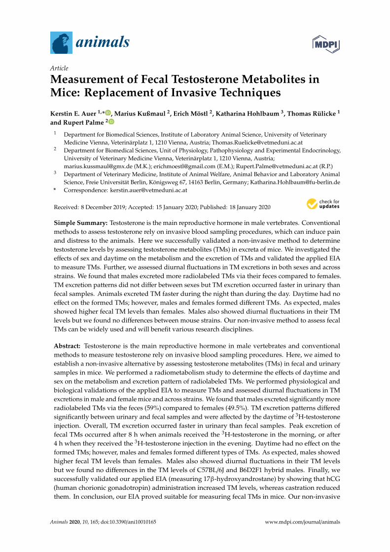
animals
Article
Measurement of Fecal Testosterone Metabolites inMice: Replacement of Invasive Techniques
Kerstin E. Auer 1,* , Marius Kußmaul 2, Erich Möstl 2, Katharina Hohlbaum 3, Thomas Rülicke 1
and Rupert Palme 2
1 Department for Biomedical Sciences, Institute of Laboratory Animal Science, University of VeterinaryMedicine Vienna, Veterinärplatz 1, 1210 Vienna, Austria; [email protected]
2 Department for Biomedical Sciences, Unit of Physiology, Pathophysiology and Experimental Endocrinology,University of Veterinary Medicine Vienna, Veterinärplatz 1, 1210 Vienna, Austria;[email protected] (M.K.); [email protected] (E.M.); [email protected] (R.P.)
3 Department of Veterinary Medicine, Institute of Animal Welfare, Animal Behavior and Laboratory AnimalScience, Freie Universität Berlin, Königsweg 67, 14163 Berlin, Germany; [email protected]
* Correspondence: [email protected]
Received: 8 December 2019; Accepted: 15 January 2020; Published: 18 January 2020�����������������
Simple Summary: Testosterone is the main reproductive hormone in male vertebrates. Conventionalmethods to assess testosterone rely on invasive blood sampling procedures, which can induce painand distress to the animals. Here we successfully validated a non-invasive method to determinetestosterone levels by assessing testosterone metabolites (TMs) in excreta of mice. We investigated theeffects of sex and daytime on the metabolism and the excretion of TMs and validated the applied EIAto measure TMs. Further, we assessed diurnal fluctuations in TM excretions in both sexes and acrossstrains. We found that males excreted more radiolabeled TMs via their feces compared to females.TM excretion patterns did not differ between sexes but TM excretion occurred faster in urinary thanfecal samples. Animals excreted TM faster during the night than during the day. Daytime had noeffect on the formed TMs; however, males and females formed different TMs. As expected, malesshowed higher fecal TM levels than females. Males also showed diurnal fluctuations in their TMlevels but we found no differences between mouse strains. Our non-invasive method to assess fecalTMs can be widely used and will benefit various research disciplines.
Abstract: Testosterone is the main reproductive hormone in male vertebrates and conventionalmethods to measure testosterone rely on invasive blood sampling procedures. Here, we aimed toestablish a non-invasive alternative by assessing testosterone metabolites (TMs) in fecal and urinarysamples in mice. We performed a radiometabolism study to determine the effects of daytime andsex on the metabolism and excretion pattern of radiolabeled TMs. We performed physiological andbiological validations of the applied EIA to measure TMs and assessed diurnal fluctuations in TMexcretions in male and female mice and across strains. We found that males excreted significantly moreradiolabeled TMs via the feces (59%) compared to females (49.5%). TM excretion patterns differedsignificantly between urinary and fecal samples and were affected by the daytime of 3H-testosteroneinjection. Overall, TM excretion occurred faster in urinary than fecal samples. Peak excretion offecal TMs occurred after 8 h when animals received the 3H-testosterone in the morning, or after4 h when they received the 3H-testosterone injection in the evening. Daytime had no effect on theformed TMs; however, males and females formed different types of TMs. As expected, males showedhigher fecal TM levels than females. Males also showed diurnal fluctuations in their TM levelsbut we found no differences in the TM levels of C57BL/6J and B6D2F1 hybrid males. Finally, wesuccessfully validated our applied EIA (measuring 17β-hydroxyandrostane) by showing that hCG(human chorionic gonadotropin) administration increased TM levels, whereas castration reducedthem. In conclusion, our EIA proved suitable for measuring fecal TMs in mice. Our non-invasive
Animals 2020, 10, 165; doi:10.3390/ani10010165 www.mdpi.com/journal/animals
Animals 2020, 10, 165 2 of 17
method to assess fecal TMs can be widely used in various research disciplines like animal behavior,reproduction, animal welfare, ecology, conservation, and biomedicine.
Keywords: testosterone; mice; androgens; non-invasive; feces
1. Introduction
Testosterone is one of the major sex hormones produced by the body, occurring in both malesand females. In males, testosterone promotes territorial aggression, courtship, and sexual behavior, aswell as sperm and secondary sexual trait production [1]. Also, testosterone has been shown to directlyaffect other fitness parameters such as immune function [1]. In females, the role of testosterone isless well-investigated, though effects on female health [2], sexual behavior [3,4], and aggression havebeen reported in various species [5–7], highlighting the overall significance of testosterone in behavior,reproduction, and health.
Conventional methods to measure testosterone and other hormones rely on invasive bloodsampling procedures. Besides raising ethical concerns, blood sampling can be problematic—especiallyfor small animals like mice and rats—as the invasive sampling procedure itself can alter hormonesecretion, only small volumes can be collected and repeated sampling is not advisable or sometimeseven impossible [8,9]. Moreover, blood sampled hormone concentrations represent a snapshot ofthe hormone status and whenever hormone levels show strong episodic or diurnal fluctuations suchresults will be misleading. A non-invasive alternative that circumvents all these limitations is thedetermination of hormone metabolites in excreta. Collecting feces and urine is easy and can berepeatedly performed without disturbing the animal’s behavior or interfering with its endocrinologicalstatus. Also, fecal and urinary samples are less affected by episodic fluctuations in hormone secretionsand thus might reflect hormone levels more robustly [10–13].
Even though there are several advantages for using excreta to assess hormone levels, this methodis not straight-forward and needs careful establishment. Firstly, as the circulating hormone is heavilymetabolized prior to excretion, it is essential to find and validate a group-specific enzyme immunoassay(EIA) for the quantification of these metabolites [14,15]. Immunoassays designed for blood samplestarget actual hormones rather than their metabolites and are thus less suited for analyzing fecal orurinary samples. Secondly, when measuring hormone metabolites in excreta one has to consider therequired intestinal gut passage time, i.e., time delay between hormone secretion and peak excretion ofits metabolites, to correctly interpret results [13]. The intestinal gut passage time is species specificand can vary depending on sex, age, activity pattern and even diet (for review see [12,13,15]). Also,hormone metabolites can be excreted via feces or urine and differences in the route of excretion havebeen reported between species and sexes [13,16]. Taken together, because metabolism and excretion ofhormones differ significantly between species and sometimes even between sexes, these non-invasivemethods for measuring hormone metabolites in excreta have to be validated for each species beforeapplication [10,12,13,15,17].
In the last years, the assessment of fecal steroid hormone metabolites has been established inan increasing number of species although most studies focus on glucocorticoids (for review see [12])rather than sex steroids. So far, the assessment of fecal testosterone metabolites (TMs) has beensuccessfully validated in male and female red squirrels (Tamiasciurus hudsonicus) [18,19], chinchilla(Chinchilla sp.) [20], bank voles (Myodes glareolus) [21], baboons (Papio cynocephalus) [22], and threeother species of nonhuman primates [23]. Few studies have used fecal and urinary samples forTM measurement in mice and to our knowledge a proper physiological and biological validationof the method to assess TMs in this species is still lacking. Billitti et al. [24] took a first approach inperforming a radiometabolism study; however, the small number of animals used (n = 3) and the lackof information regarding the experimental mice and the sampling procedure (e.g., the authors did not

Animals 2020, 10, 165 3 of 17
state which mouse strain was used or at what day time feces were collected) limits the applicabilityof their findings. Another working group adapted an EIA used to target plasma steroid hormonesincluding testosterone for fecal and urinary samples [25]. The authors did not attempt to identifyrespective steroid hormone metabolites and did not perform any validation for their assay besidesproviding standard curves. In subsequent studies the authors then used their assay for urinary ratherthan fecal samples [26–28], which seems problematic as steroid conjugates prevail in urinary samplesand are more complicated to quantify.
Here we performed a comprehensive study where we investigated testosterone metabolism inmice and established and validated an enzyme immunoassay to reliably assess individual testosteronelevels. We injected radiolabeled testosterone to assess the effects of sex and time of day on theroute and excretion pattern of TMs. There is only little information available about sex-specific anddiurnal effects on patterns of TM excretion in any species even though such factors can be relevantfor studies investigating hormone metabolites in excreta in various species, not only in mice. Wefurther characterized the radiolabeled metabolites in feces with a reversed-phase HPLC to validateour assay. As an additional validation approach, we performed a pharmacological stimulation withhuman chorionic gonadotropin (hCG) and performed castrations to verify that the TMs detected withour EIA follow the respective manipulations [14,17]. Finally, we also determined normal diurnalfluctuations in fecal TMs in males and females and collected feces from two different isogenic mousestrains to determine whether strain specific variation in excretion patterns exists. Mice are the mostcommonly-used model organism in biomedical research and our study provides a non-invasivealternative to assess individual testosterone levels and extends the area of application of mice as amodel organism. Also, a validated method for fecal TM measurement will be useful for other fields ofresearch like reproductive physiology and behavioral and wildlife ecology.
2. Materials and Methods
2.1. Experimental Animals
2.1.1. Radiometabolism Study, Diurnal Fluctuations and hCG Challenge
In total, we used 32 C57BL/6JRj (Mus musculus f. domesticus, 16 males, 16 females) and 8male B6D2(C57BL/6JRj × DBA/2JRj)F1 hybrid mice which were purchased from Janvier Laboratories(Staint-Berthevin Cedex, France) at the age of 8 weeks. Sample sizes were estimated with powerstatistics based on similar studies assessing fecal corticosterone metabolites in mice [16,29]. Uponarrival, animals from the same strain were housed in same sex groups in mouse open top cages (typeIIL, 36.5 × 20.7 × 14 cm, Tecniplast, Buguggiate, Italy). Cages were equipped with wooden bedding(LIGNOCEL® 3–4 S, J. Rettenmaier and Söhne GmbH + Co. KG, Rosenberg, Germany), nestingmaterial (Pur-Zellin 4 × 5 cm; Paul Hartmann GmbH, Wiener Neudorf, Austria) and cardboard tubes(7.6 × 3.8 cm diameter, Special Diet Service, Claus GmbH, Limburgerhof, Germany). Commercialmouse diet (ssniff®, V1534, Soest, Germany) and tab water were provided ad libitum. Ten daysprior to the start of the experimental sample collections, mice were housed individually to controlfluctuations in testosterone levels related to social interactions and to habituate mice to the singlehousing condition during the experiments. Standard laboratory conditions (temperature 21 ± 1 ◦C,humidity 40–55%, light-dark cycle: 12:12 h, lights on at 8:00 a.m.) were maintained in the colony andduring the experiment.
2.1.2. Castration Experiment
In total 19 male C57BL/6JRj mice which were purchased from Janvier Laboratories (Staint-BerthevinCedex, France) were used at the age of 18 to 42 weeks. These mice had previously been used in anotherexperiment [30] and had been individually housed for 6 to 32 weeks in mouse type III open top cages(42.5 × 27.6 × 15.3 cm, Tecniplast, Buguggiate, Italy). Cages were equipped with wooden bedding

Animals 2020, 10, 165 4 of 17
(LIGNOCEL® 3–4 S, J. Rettenmaier and Söhne GmbH + Co. KG, Rosenberg, Germany), cotton nestlets(Ancare, UK agents, Lillico, United Kingdom), cocoons (ZOONLAB GmbH, Castrop-Rauxel, Germany),one red plastic house (10 × 9 × 5.5 cm; ZOONLAB GmbH, Castrop-Rauxel, Germany) and tunnels(metal; 12.5 × 5 cm diameter, ZOONLAB GmbH, Castrop-Rauxel, Germany). The animals were keptunder standardized laboratory conditions (room temperature 22 ± 2 ◦C; relative humidity 55 ± 10%)on a light:dark cycle of 12:12 h of artificial light with a 5 min twilight transition phase (lights on from6:00 a.m. to 6:00 p.m.). Food (ssniff®, V1534, Soest, Germany) and tap water were provided ad libitum.
2.2. Radiometabolism Study
We performed a radiometabolism study to assess the effects of sex and daytime on the route andexcretion pattern of TMs. We divided the 32 C57BL/6J experimental mice in two groups containing 16mice each (8 males and 8 females). At the onset of the study all experimental animals were transferredto sample collection cages equipped with a grid floor (TecniplastTM Raised Wire Floor). In samplecollection cages animals were housed individually and given one day for cage habituation, beforeintraperitoneally (i.p.) injected with 250µL of 740 kBq (=20µCi) 3H-testosterone (1,2,6,7-3H-testosterone,Amersham Pharmacia Biotech Europe GmbH, Germany) diluted in 1 mL of sterile isotonic salinesolution containing 5% ethanol. Group one received the injection at 9:00 a.m. (one hour after thebeginning of the light phase; ‘morning group’), whereas the second group received the injection at9:00 p.m. (one hour after beginning of the dark phase; ‘evening group’). Evening injections wereperformed under red light. Within the first 24 h we collected feces and urine at 0, 2, 4, 6, 8, 10, 12, 14,16, 20, and 24 h post injection. Afterwards all excreta were sampled in 12 h intervals until day 5 of theexperiment. Collected samples were frozen immediately and stored at −20 ◦C until analysis.
2.3. Diurnal Fluctuations
To assess normal diurnal fluctuations in TM excretion, we collected feces from 16 C57BL/6J (8 malesand 8 females) and 8 male B6D2F1 hybrid mice. B6D2F1 hybrids were used as an out-group comparisonto assess whether testosterone excretion patterns show strain specific variation. The C57BL/6J mice hadpreviously been used in the radiometabolism experiment and were randomly selected within sexes.Between experiments the re-used mice were provided 8 days to recover where they were individuallyhoused under the conditions described above. At the onset of the study all experimental animals weretransferred to sample collection cages, where they were given one day for cage habituation beforesample collection started. Sample collection was performed according to the following time intervalsstarting at 9:00 a.m.: 0, 2, 4, 6, 8, 10, 12, 14, 16, 20, and 24 h.
2.4. Human Chorionic Gonadotropin (hCG) Challenge
To physiologically validate the applied EIA we performed a pharmacological stimulation withhCG (Chorulon, Intervet, Vienna, Austria). As hCG mimics the activity of luteinizing hormone (LH),its administration induces testosterone production in Leydig cells and leads to a testosterone surge.The hCG administration was performed in the 8 male C57BL/6J mice, which had been used to assessdiurnal fluctuations. The hCG administration was conducted on the following day and we applied awithin-subject design to distinguish between the effects of the pharmacological stimulation comparedto the naturally occurring circadian rhythms. Each male received 2.5 IU hCG/g body mass injected i.p.at 9:00 a.m. and feces were collected at 0, 2, 4, 6, 8, 10, 12, 14, 16, 20, and 24 h post injection. This dosagehas previously been confirmed to induce a testosterone surge [31]. Samples were frozen immediatelyand stored at −20 ◦C until analysis.
2.5. Castration Experiment
Castration was performed as an additional biological validation for the applied EIA. As castrationprevents testicular testosterone production it should reduce male testosterone levels. To confirm theeffect of castration on fecal TM levels, fecal samples were collected at three different time points: (a)

Animals 2020, 10, 165 5 of 17
Two days prior to castration, (b) two days post castration, and (c) two weeks post castration. In total 19male C57BL/6J mice were castrated at the age of 18 to 42 weeks.
For castration, anesthesia was induced with 4% isoflurane (Isofluran CP®, CP-PharmaHandelsgesellschaft mbH, Burgdorf, Germany) in 100% oxygen in an induction chamber and maintainedwith 1.5–2.5% isoflurane via nose cone. Sterile eye ointment (Artelac® Splash MDO®, Bausch andLomb GmbH, Berlin, Germany) was used to prevent dehydration. Systemic analgesia was provided bymeloxicam (1 mg/kg body mass, Metacam 2 mg/mL injection solution for cats, Boehringer IngelheimVetmedica GmbH, Ingelheim, Germany), a long-acting non-steroidal anti-inflammatory drug (NSAID)which requires a single dosing a day [32]. Lidocaine/prilocaine was used as topical local anestheticfor the scrotum (Emla Creme, AstraZeneca GmbH, Wedel, Germany). It should be noted here thata single daily dosing with meloxicam could be insufficient to fully control post-op pain in mice [33]and that a subcutaneous injection of lidocaine/bupivacaine at the incision site and/or higher doses ofmeloxicam (e.g., 5 mg/kg, every 12 h) may improve analgesia post castration [34]. Surgical castrationwas performed according to Behringer et al. [35]. In short, the scrotum was disinfected and mice wereplaced in the supine position on a heating pad. Both testicles were pushed down into the scrotal sacsby gently applying pressure to the abdomen. Then, an incision with the length of approximately 1 cmwas made through the skin at a right angle to the midline of the scrotal sac. The testes were removedone by one. The membrane covering the testicle was incised and the testicle was carefully pushed out.The vas deferens with the blood vessels running along it was pinched off using two artery forceps.An absorbable suture (3–0) was passed in between the two forceps for ligation. After the forceps wereremoved, the testicle was dissected from the fat pad and removed. The fat was returned to the scrotalsac and the skin was stitched with a single button suture. After surgery, mice received food pellets thatwere soaked in water for five days and regular health checks were performed.
2.6. Sample Collection
2.6.1. Radiometabolism Study, Diurnal Fluctuations and hCG Challenge
To enable individual sampling and quantitative collection of all voided urine and feces we applieda similar method as described in Touma et al. [16]. Experimental animals were placed individually intype II mouse cages (26.8 × 21.5 × 14 cm, Tecniplast, Buguggiate, Italy) containing a stainless steel wiregrid floor (TecniplastTM Raised Wire Floor, mesh width 7 mm), which was placed in a mouse cage ofthe same size. All excreta dropped through the bars of the wire floor and were collected in the cagelocated beneath, which was covered with filter paper to immediately absorb the urine. At each samplepoint the wire floor cage was placed into a new cage and all excreted fecal pellets were collected forfurther analyses. To provide time to habituate to the grid floor and the sampling procedure, all animalswere transferred to the grid floor cages one day prior to the start of the experiments and sampleswere collected at regular intervals during this time. To habituate the animals to the single housingcondition during the experiments, all mice were separated and placed individually in type IIL mousecages (36.5 × 20.7 × 14 cm, Tecniplast, Buguggiate, Italy) 10 days prior to the start of the experiment.Cages were equipped with wooden bedding, nesting material, and cardboard tubes as described above.To mitigate potential effects of social isolation and to stimulate reproductive physiology we providedbedding (odor) stimulation of opposite sex conspecifics twice during the habituation period.
2.6.2. Castration Experiment
Since mice had been individually housed for 6 to 32 weeks in type III mouse cages(42.5 × 27.6 × 15.3 cm, Tecniplast, Buguggiate, Italy), they were already habituated to this housingcondition. For fecal sample collection mice were transferred to a new home cage, i.e., cages containedwooden bedding, nest material and standard enrichment as described above. Soiled bedding materialwithout feces was scattered on the new bedding to minimize distress accompanied with the cagechange. After a period of 24 h, all dry fecal pellets were collected using forceps. Pellets contaminated

Animals 2020, 10, 165 6 of 17
with urine were excluded. Collected samples were frozen immediately and stored at −20 ◦C untilfurther analyses were performed [30,36].
2.7. Sample Analysis
To determine the route and time course of testosterone excretion, we processed the samplescollected in the radiometabolism experiment: Urine spots were cut out from the filter paper and slitinto 2–3 cm2 pieces. Fecal samples were homogenized with a mortar and pestle before extractedaccording to the protocol described in Touma et al. [16]. After extraction, the radioactivity of eachsample was determined with a liquid scintillation counter (Tri-Carb 2100TR, Packard Instruments,Meriden, CT, USA). Delay times were assessed based on peak excretion patterns and the ratio ofrecovered radioactivity in urine and feces provided information about the excretion route. The totalrecovery rate of the radiolabeled metabolites was calculated as the sum of recovered radioactivity inurine and feces divided by the total amount of administered radioactivity.
To characterize the type and relative abundance of 3H-testosterone metabolites we performeda reversed-phase HPLC. Only feces containing peak concentrations of radioactivity were used andthe analyses were conducted according to the methods described in Touma et al. [16]. Briefly, aNovopack C18 column (3.9 × 150 mm, WAT086344, Waters) and a linear water/methanol gradient(20–100%, flow rate: 1 mL/min, three fractions per min) was used for separation of the metabolites.The immunoreactivity of the HPLC fractions was measured with two different enzyme immunoassays(EIAs): A testosterone EIA and an epiandrosterone EIA. Details of both EIAs including cross-reactionsare given in Palme and Möstl [37]. The better performing testosterone EIA was then selected todetermine the amount of TM in feces collected to assess diurnal fluctuations, as well as after the hCGadministration and the castration. Sample extraction was performed as described above and accordingto the protocol given in Palme et al. [38]. All assays were conducted blinded and samples were run induplicates to receive results.
The testosterone EIA utilizes an antibody produced in rabbits against testerone-3-CMO:BSA(working dilution 1:20,000) and 5α-androstane-3β,17β-diol-3-HS-DADOO-biotin as label (dilution1:160,000). Further details of the EIA procedure are given in Palme and Möstl [37]. The sensitivity ofthe whole analysis was 0.21 ng/0.05 g feces. Intra- and inter-assays coefficients of variation of high/lowconcentration pool samples were 2.1/2.9% and 6.7/7.2%, respectively.
2.8. Statistical Analysis
Statistical analyses were performed with IBM SPSS Statistics 24 and R 3.3.2 and figures werecreated in Sigma Plot 13.0.
2.8.1. Radiometabolism Study
To test if the recovery rate of the radiolabeled testosterone differed between the sexes or theexperimental treatment groups we applied a Mann–Whitney U test. We applied a paired T-Test tocompare the amount of excreted feces between day and night times. To determine which factorsinfluenced the excretion pattern in fecal or urinary samples we ran general linear mixed effects models(LMM) with the amount of excreted testosterone (per 50 mg feces) or urine (total amount of collectedurine for the given collection period) as the dependent variable and time point of sample collection,sex and experimental treatment group (morning or evening injection) as fixed factors. Animal IDwas included as random factor to control for non-independence of samples from the same individual.To assess which factors influenced the delay times to peak excretion in fecal and urinary samples, weran linear models (LM) with the delay times to peak excretion as the dependent variable and sex andtreatment group as fixed factors.

Animals 2020, 10, 165 7 of 17
2.8.2. Diurnal Fluctuations
To test whether sex influenced TM levels in C57BL/6J mice, we ran a LMM with testosteronelevels (ng/50 mg feces) as dependent variable, sex as fixed factor and animal ID as random factor. Weassessed the effect of daytime on TM levels separately for male and female C57BL/6J mice in performingLMMs with TM levels as dependent, time point of sample collection as fixed and animal ID as randomfactor. To assess whether male fluctuations in TM levels were similar across genetic backgrounds weperformed a Kendall W test.
2.8.3. hCG Challenge and Castration Experiment
To test if hCG administration increased TM levels, we compared individual peak levels beforehCG administration with respective TM levels at the same daytime after hCG administration in aWilcoxon Signed Rank test. Finally, to assess whether castration reduced male TM levels, we performeda One Way Repeated Measures Analysis of Variance on ranks.
2.9. Animal Welfare
The radiometabolism study, the diurnal fluctuations study and the hCG challenge experiment hasbeen discussed by the Institutional Ethics Committee of the University of Veterinary Medicine Viennain accordance with Good Scientific Practice guidelines and has been approved by the Austrian FederalMinistry for Science and Research (Reference number: BMWFW-68.205/0056- WF/V/3b/2017).
The castration study was performed according to the German Animal Welfare Act and theDirective 2010/63/EU for the protection of animals used for scientific purposes. Animal housing andhusbandry were approved by the Berlin State Authority (“Landesamt für Gesundheit und Soziales”,permit number: G 0053/15). Mice were previously used in an experiment classified as mild withregards to animal suffering (as defined by current EU regulations on severity classification of animalstudies). After castration and re-socialization in groups of three to four, the mice were rehomed.
3. Results
3.1. Radiometabolism Study
3.1.1. Time Course and Route of 3H-testosterone Excretion
The mean total recovery of the administered radioactivity was 57 ± 8% in feces and urine samplescombined over all animals. No difference in the recovery rate was detected between the experimentaltreatment groups (morning injection: 56.6 ± 7.5%, evening injection: 57.1 ± 4.8%; Mann–Whitney U test:U = 125.5, N = 32, p = 0.94) or between sexes (males: 56.4± 6.3%, females: 57.3± 6.3% Mann–Whitney Utest: U = 145.5, N = 32, p = 0.52). Interestingly though, we found that males secreted significantly moreradiolabeled TMs via the feces than females (males: 59.0 ± 7.3%, females: 49.5 ± 5.5%; Mann–WhitneyU test: U = 39, N = 32, p < 0.001), whereas females showed higher proportions of radioactivity in theurine compared to males (males: 41.0 ± 3.7%, females: 50.3 ± 5.5%; p < 0.001).
Overall, animal defecation rates were significantly dependent on daytime (Paired t-Test: t = −35.95,p < 0.001): Experimental mice produced on average 8.02 g feces during the 12 h collection periodsfrom 9 p.m. to 9 a.m. compared to 3.22 g feces during the 12 h collection periods from 9 a.m. to 9 p.m.The excretion of radiolabeled TMs varied significantly over time and showed different patterns inurinary and fecal samples (LMM feces: F15, 464 = 39.33, p < 0.001; LMM urine: F15, 464 = 57.65, p < 0.001).Within both, urinary and fecal samples, we found a significant effect of the time of injection on theexcretion pattern (LMM feces: F1, 30 = 48.21, p < 0.001; LMM urine: F1, 30 = 19.07, p < 0.001): In fecalsamples, animals that received the radiolabeled testosterone in the evening showed a faster rise inexcretion rates and excreted the radiolabeled testosterone more quickly compared to animals thatreceived the radiolabeled testosterone in the morning (Figure 1b). In urinary samples, animals fromboth treatment groups showed an immediate rise in excretion rates, but animals from the evening group

Animals 2020, 10, 165 8 of 17
excreted the radiolabeled testosterone faster compared to animals from the morning group (Figure 1a).Within both sample types no sex-specific differences in the excretion patterns were detected (LMMfeces: F1, 29 = 1.64, p = 0.21; LMM urine: F1, 29 = 1.55, p = 0.22) and the vast majority of radiolabeledmetabolites in feces (97.3 ± 1.5 %) and urine (84.6 ± 12.2%) was excreted within the first 24 h.Animals 2020, 10, x FOR PEER REVIEW 9 of 18
Figure 1. Time course (first 48 h) of 3H-testosterone excretion in (a) urinary and (b) fecal samples of male and female C57BL/6J mice. White boxplots (n = 16) represent data obtained from mice that received the 3H-testosterone in the morning (one hour after the onset of the light phase) and grey boxplots (n = 16) represent data from mice that received the 3H-testosterone in the evening (one hour after the onset of the dark phase). Dots are outliers with values between 1.5‒3.0× interquartile range.
Figure 1. Time course (first 48 h) of 3H-testosterone excretion in (a) urinary and (b) fecal samplesof male and female C57BL/6J mice. White boxplots (n = 16) represent data obtained from mice thatreceived the 3H-testosterone in the morning (one hour after the onset of the light phase) and greyboxplots (n = 16) represent data from mice that received the 3H-testosterone in the evening (one hourafter the onset of the dark phase). Dots are outliers with values between 1.5-3.0× interquartile range.
We further determined the delay time to peak excretion in urinary and fecal samples. In urine, peakradioactivity was recovered in the first samples collected after the administration of 3H-testosterone,i.e., after 2 h (range 2–8). No differences were found between sexes (LM: F1, 29 = 1.22, p = 0.28), orbetween animals from the morning or evening injection group (LM: F1, 29 = 2.17, p = 0.15; Figure 2).In feces, peak excretion times were significantly affected by the time of injection (LM: F1, 30 = 51.91,

Animals 2020, 10, 165 9 of 17
p < 0.001; Figure 2): In the evening group peak radioactivity was recovered after 4 h (no range) and inthe morning group after 8 h (range 4–10). Again, no sex-specific differences were discovered in fecalpeak excretion times (LM: F1, 29 = 0.06, p = 0.81).
3.1.2. Characterization of Fecal 3H-testosterone Metabolites
The injected 3H-testosterone was heavily metabolized and HPLC separations revealed severalpeaks of radioactivity, indicating that a large number of metabolites were formed. We found cleardifferences in the excreted metabolites between males and females, but the pattern was identicalbetween animals that received the radiolabeled testosterone in the morning or evening (Figure 3).Females showed a broader spectrum of metabolites of different polarity compared to males and alsoshowed a pronounced peak eluting around androstenedione, which was absent in males. Several3H-metabolites also reacted with the testosterone EIA.Animals 2020, 10, x FOR PEER REVIEW 10 of 18
Figure 2. Delay time in hours to peak excretion in urinary and fecal samples of C57BL/6J mice. Peak excretion times are depicted separately for the morning (n = 16) and evening (n = 16) injection group. Mice from the morning group received the 3H-testosterone one hour after the onset of the light phase, whereas mice from the evening group received the ³H-testosterone one hour after the onset of the dark phase. Dots are outliers with values between 1.5‒3.0× interquartile range.
3.2. Evaluation of Diurnal Fluctuations
When we assessed fecal TM levels of C57BL/6J mice, we found that testosterone levels differed significantly between sexes over the course of a day (LMM: F1, 14 = 49.87, p < 0.001; Figure 4a). Males had twice the concentration of females, as males showed an average testosterone level of 4.65 ng/50 mg feces (± 1.34 SD) compared to 2.35 (± 0.9 SD) in females. Males also showed significant variation in TM levels over the course of the day (LMM: F10, 65 = 4.74, p < 0.001; Figure 4a), whereas female TM levels did not show such fluctuations (LMM: F10, 69 = 1.17, p = 0.324; Figure 4a). We found no evidence that genetic background affected TM levels, as absolute levels and the pattern of episodic fluctuations were comparable between C57BL/6J and B6D2F1 hybrid males (Kendall W test: W = 0.38, df = 10, p < 0.001; Figure 4b). B6D2F1 hybrid males had an average testosterone level of 4.92 ng/50 mg feces (± 1.44 SD). Both strains showed two peaks in their testosterone levels, the first one occurred between 3 p.m. and 5 p.m. and the second and most pronounced peak was reached at approximately 9 p.m., one hour after the onset of the dark phase. Nadir levels of testosterone were observed in fecal samples that were collected around midnight (see Figure 4b).
3.3. Physiological and Biological Validation
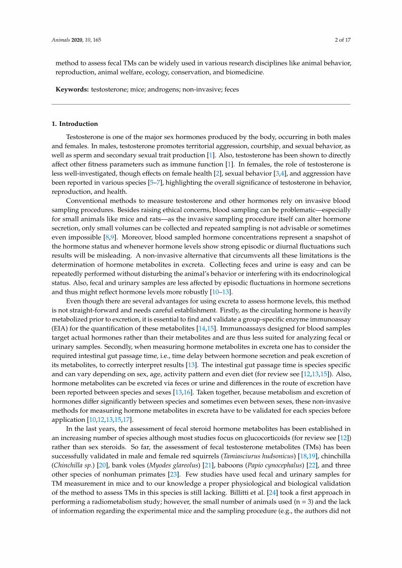
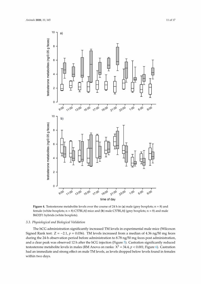
The hCG administration significantly increased TM levels in experimental male mice (Wilcoxon Signed Rank test: Z = −2.1, p = 0.036). TM levels increased from a median of 4.36 ng/50 mg feces during the 24-h observation period before administration to 8.78 ng/50 mg feces post administration, and a clear peak was observed 12 h after the hCG injection (Figure 5). Castration significantly reduced testosterone metabolite levels in males (RM Anova on ranks: Χ2 = 34.4, p < 0.001; Figure 6). Castration had an immediate and strong effect on male TM levels, as levels dropped below levels found in females within two days.
Figure 2. Delay time in hours to peak excretion in urinary and fecal samples of C57BL/6J mice. Peakexcretion times are depicted separately for the morning (n = 16) and evening (n = 16) injection group.Mice from the morning group received the 3H-testosterone one hour after the onset of the light phase,whereas mice from the evening group received the 3H-testosterone one hour after the onset of the darkphase. Dots are outliers with values between 1.5-3.0× interquartile range.
3.2. Evaluation of Diurnal Fluctuations
When we assessed fecal TM levels of C57BL/6J mice, we found that testosterone levels differedsignificantly between sexes over the course of a day (LMM: F1, 14 = 49.87, p < 0.001; Figure 4a). Maleshad twice the concentration of females, as males showed an average testosterone level of 4.65 ng/50 mgfeces (± 1.34 SD) compared to 2.35 (± 0.9 SD) in females. Males also showed significant variation in TMlevels over the course of the day (LMM: F10, 65 = 4.74, p < 0.001; Figure 4a), whereas female TM levelsdid not show such fluctuations (LMM: F10, 69 = 1.17, p = 0.324; Figure 4a). We found no evidence thatgenetic background affected TM levels, as absolute levels and the pattern of episodic fluctuations werecomparable between C57BL/6J and B6D2F1 hybrid males (Kendall W test: W = 0.38, df = 10, p < 0.001;Figure 4b). B6D2F1 hybrid males had an average testosterone level of 4.92 ng/50 mg feces (± 1.44 SD).Both strains showed two peaks in their testosterone levels, the first one occurred between 3 p.m. and5 p.m. and the second and most pronounced peak was reached at approximately 9 p.m., one hour afterthe onset of the dark phase. Nadir levels of testosterone were observed in fecal samples that werecollected around midnight (see Figure 4b).

Animals 2020, 10, 165 10 of 17Animals 2020, 10, x FOR PEER REVIEW 11 of 18
Figure 3. Reversed-phase HPLC separation of fecal 3H-testosterone metabolites of male (A,B) and female (C,D) mice from the morning (A,C) and evening (B,D) injection group. Radioactivity (solid line) of each fraction was determined by liquid scintillation counting. Immunoreactivity (dashed line) was tested with the testosterone enzyme immunoassays. Open triangles mark the approximate elution positions of respective standards (E2-diSO4 = 17β-estradiol-disulfate, E1-G = estrone-glucuronide, E1S = estrone-sulfate, C = cortisol, Cc = corticosterone, A = androstenedione, T = testosterone, ET = epitestosterone).
Figure 3. Reversed-phase HPLC separation of fecal 3H-testosterone metabolites of male (A,B) and female (C,D) mice from the morning (A,C) and evening (B,D)injection group. Radioactivity (solid line) of each fraction was determined by liquid scintillation counting. Immunoreactivity (dashed line) was tested with thetestosterone enzyme immunoassays. Open triangles mark the approximate elution positions of respective standards (E2-diSO4 = 17β-estradiol-disulfate, E1-G =
estrone-glucuronide, E1S = estrone-sulfate, C = cortisol, Cc = corticosterone, A = androstenedione, T = testosterone, ET = epitestosterone).
Animals 2020, 10, 165 11 of 17Animals 2019, 9, x FOR PEER REVIEW 12 of 18
Figure 4. Testosterone metabolite levels over the course of 24 h in (a) male (grey boxplots; n = 8) and female (white boxplots; n = 8) C57BL/6J mice and (b) male C57BL/6J (grey boxplots; n = 8) and male B6D2F1 hybrids (white boxplots).
Figure 4. Testosterone metabolite levels over the course of 24 h in (a) male (grey boxplots; n = 8) andfemale (white boxplots; n = 8) C57BL/6J mice and (b) male C57BL/6J (grey boxplots; n = 8) and maleB6D2F1 hybrids (white boxplots).
3.3. Physiological and Biological Validation
The hCG administration significantly increased TM levels in experimental male mice (WilcoxonSigned Rank test: Z = −2.1, p = 0.036). TM levels increased from a median of 4.36 ng/50 mg fecesduring the 24-h observation period before administration to 8.78 ng/50 mg feces post administration,and a clear peak was observed 12 h after the hCG injection (Figure 5). Castration significantly reducedtestosterone metabolite levels in males (RM Anova on ranks: X2 = 34.4, p < 0.001; Figure 6). Castrationhad an immediate and strong effect on male TM levels, as levels dropped below levels found in femaleswithin two days.
Animals 2020, 10, 165 12 of 17Animals 2019, 9, x FOR PEER REVIEW 13 of 18
Figure 5. Testosterone metabolite levels in male C57BL/6J mice (n = 8) for 24 h without prior hCG administration (baseline; white boxplots) and for 24 h post hCG administration (grey boxplots).
Figure 6. Testosterone metabolite levels in male C57BL/6J mice before, two days and two weeks after castration. Dots are outliers with values between 1.5‒3.0× interquartile range.
4. Discussion
Our multiphase validation experiment for the non-invasive measurement of TMs in fecal and urinary samples of mice provided several interesting results and we start our discussion in highlighting the sex-specific differences we discovered. First, we found a significant sex difference in the route of TM excretion. Males excreted 59% of the radiolabeled testosterone via the feces. This rate was about 10% higher compared to females, which in turn, excreted a bigger proportion of
Figure 5. Testosterone metabolite levels in male C57BL/6J mice (n = 8) for 24 h without prior hCGadministration (baseline; white boxplots) and for 24 h post hCG administration (grey boxplots).
Animals 2019, 9, x FOR PEER REVIEW 13 of 18
Figure 5. Testosterone metabolite levels in male C57BL/6J mice (n = 8) for 24 h without prior hCG administration (baseline; white boxplots) and for 24 h post hCG administration (grey boxplots).
Figure 6. Testosterone metabolite levels in male C57BL/6J mice before, two days and two weeks after castration. Dots are outliers with values between 1.5‒3.0× interquartile range.
4. Discussion
Our multiphase validation experiment for the non-invasive measurement of TMs in fecal and urinary samples of mice provided several interesting results and we start our discussion in highlighting the sex-specific differences we discovered. First, we found a significant sex difference in the route of TM excretion. Males excreted 59% of the radiolabeled testosterone via the feces. This rate was about 10% higher compared to females, which in turn, excreted a bigger proportion of
Figure 6. Testosterone metabolite levels in male C57BL/6J mice before, two days and two weeks aftercastration. Dots are outliers with values between 1.5-3.0× interquartile range.
4. Discussion
Our multiphase validation experiment for the non-invasive measurement of TMs in fecal andurinary samples of mice provided several interesting results and we start our discussion in highlightingthe sex-specific differences we discovered. First, we found a significant sex difference in the routeof TM excretion. Males excreted 59% of the radiolabeled testosterone via the feces. This rate wasabout 10% higher compared to females, which in turn, excreted a bigger proportion of radiolabeledtestosterone via the urine. A previous study in laboratory mice has also reported sex-specific differencesin the excretion route of corticosterone metabolites (CMs), where males excreted higher proportions ofradiolabeled CMs via the feces compared to females [16]. This difference was even more pronounced,

Animals 2020, 10, 165 13 of 17
as males excreted about 73% of radioactive corticosterone via the feces, whereas females excretedonly about 53%. These results show that sex-specific differences in hormone metabolite excretions arecommon in mice and that males on average might excrete a bigger proportion of hormone metabolitesvia feces compared to females. The reason for this difference is unclear. Similar studies investigatingTMs excretion in red squirrels [18] and bank voles [21] did not find differences between males andfemales in the route of excretion. Interestingly though, the study in bank voles found sex-specificdifferences in the excretion of CMs, as males excreted significantly more radiolabeled corticosteronevia feces than females [21], highlighting the importance to test for sex-specific differences in excretionroutes in each species.
Secondly, we also found that males and females differed in the formed fecal TMs. Femalereversed-phase HPLC immunograms showed a more diverse pattern, they contained more anddifferent peaks compared to male immunograms, indicating that females form a broader range ofdifferent TMs compared to males. Similar to our finding in mice, the type of TMs excreted in feces of redsquirrels also showed sex-specific differences [18,19]. Females showed three peaks of immunoreactiveTMs in immunograms, whereas males only showed two. Also in bank voles, testosterone was heavilymetabolized with clear sex-specific differences [21], suggesting that sex-specific differences in theformed fecal TMs might be common in rodents. In summary, these results urge caution in comparingfecal TMs between males and females but do not diminish the applicability of the EIA to measurefecal TM in male and female mice. The reversed-phase HPLC in our study also showed that plasmatestosterone is heavily metabolized and that there is hardly any testosterone left in feces, only itsmetabolites. This result emphasizes the use of group specific EIAs designed to specifically detecthormone metabolites rather than their actual plasma hormones in fecal samples [12,14].
Overall, we found much variation in the excretion pattern of radiolabeled TMs in urinary andfecal samples but within the excretion routes the time course of radiolabeled testosterone excretiondid not differ between sexes. The vast majority of radiolabeled metabolites were excreted within thefirst 24 h and day time of the injection significantly affected the excretion patterns. In both fecal andurinary samples, animals that received the 3H-testosterone in the evening excreted the radiolabeledtestosterone faster compared to animals that received the 3H-testosterone in the morning, indicating afaster excretion when animals are more active, i.e., during the dark phase for nocturnal animals. Thismay be mainly related to the higher (in this study on average 2.5 times) defecation rates during thisphase. Most steroids are quickly removed from the circulation and are heavily metabolized by the liverand excreted via the kidneys into the urine or via the bile into the duodenum. The delay time betweenthe secretion of a hormone and the excretion of its metabolites is dependent on the individuals’ gutpassage time. Depending on the species, this time interval can be less than 30 min or more than aday [13]. In mice, the gut passage time is estimated to be approximately 9–10 h [39,40] and our dataclearly show that even within a species this time interval can vary strongly depending on the circadianrhythm. Knowing the intestinal gut passage time of a species is highly important when examining theendocrine status of animals based on excreta [13] and studies have to consider animal activity patternsin order to properly assess TM levels.
We also found that the elimination of radioactivity via urine occurs faster than via feces. Urinaryexcretion peaks were already observed after 2 h. This finding is in line with other studies thatinvestigated the excretion pattern of radiolabeled CM in mice and rats, which discovered that excretionpeaks are usually found within the first few hours of sample collection [16,41]. In comparison, excretionpeaks in feces were observed later and either occurred after 4 h when the samples were collectedduring the dark phase, i.e., when animals were more active, or after 8 h when samples were collectedduring light phase, i.e., when animals were in their resting period. Mice show phases of activityscattered all around the day; however, they are nocturnal and their main activity phase is duringthe dark phase. When animals are more active, more feces can be collected. This could explain theearlier detection of radiolabeled TMs in those samples collected during the dark phase. Importantly,
Animals 2020, 10, 165 14 of 17
fluctuations in defecation rate can result in distortions of fecal hormone metabolite concentrationswithout any changes in bioactive hormone levels.
Recently it has been demonstrated that different steroids can show different time courses intheir excretion. For example, in bank voles corticosterone was excreted faster than testosterone [21].In urinary samples excretion peaks were observed after 4 h for corticosterone and after 6 h fortestosterone. In fecal samples, excretion peaks occurred after 6 and 8 h, respectively. In mice, ifwe compare our results with those reported in Touma et al. [16], no difference in the time course ofcorticosterone and testosterone excretion was observed in urine and solely a small difference occurredin feces: Only when animals were inactive, peak excretion differed between steroids. Surprisinglyhowever, we showed that testosterone was excreted faster compared to corticosterone, i.e., after 8versus 10 h. It is unclear what could have postponed the metabolism of corticosterone over testosteronebut given that the animals were kept under similar conditions one potential explanation for the slightlydelayed excretion of corticosterone could be plasma binding proteins. Plasma binding proteins leadto a slower removal of steroids from the circulation and are known modulators of steroid actionin mammals [42]. Alternatively and not mutually exclusive, this difference in peak excretion timebetween corticosterone and testosterone could also be explained by an enterohepatic recirculation ofsome metabolites [13].
Our experiment clearly showed that testosterone metabolism in mice is not affected by the timeof injection, i.e., depending on the activity pattern of the animals, as there were no differences in themetabolite patterns between injection groups in the reversed-phase HPLC immunograms. Thus, thedelay time between injection and excretion via feces had no effect on the type of formed TMs. Wecould also show that the chosen testosterone EIA (measuring 17β-hydroxyandrostane), first describedby Palme and Möstl [37], is suitable for reliably measuring fecal TM in mice. The EIA was able todetect several metabolites in the radiometabolism study and the hCG challenge experiment confirmedthe physiological sensitivity of the selected EIA as we detected a clear and significant increase in thefecal TM concentrations after hCG administration. Peak values were detected approximately 12 hafter injection. Thus, peak excretion occurred 4 h later compared to the radiometabolism study. Thisdelay time can most likely be explained by the time required for the hypothalamic–pituitary–gonadalaxis to induce testosterone secretion. We could further confirm the biological validity of the appliedEIA, in measuring an immediate and steep decline in TM levels after castration. We are confidentthat our applied EIA can be used to reliably assess male and female TM levels, even though thephysiological and biological validations were performed solely in males. Validation of the assay infemales is not straight forward, as females do not carry a grand scale testosterone producing organ likemales, that could be removed or pharmacologically stimulated to trigger a significant alteration intestosterone production. However, to test the sensitivity of the selected EIA for TM levels in femalesand to further test its sensitivity for TM overall, we compared the natural TM levels of males withthose of non-pregnant females. The EIA was able to detect the low concentrations found in femalesand showed the expected differences between the sexes: Males had on average double the TM levelsthan females and showed clear, episodic fluctuations in TM levels over the course of a day. The highestTM concentrations were found between 3 p.m. and 5 p.m. and at 9 p.m. (shortly after the onsetof the dark phase). Considering the intestinal gut passage time, testosterone peaks in males likelyoccur early in the morning around 7 a.m., which is at the end of the dark phase when they are mostactive and around noon, in the middle of the light phase. Similarly to our result, in bank voles thehighest testosterone production also seems to occur around morning and before noon [21]. The dailyfluctuations in TM concentrations were highly similar in male C57BL/6J and B6D2F1 hybrid males,indicating that male genetic diversity had no effect on TM levels and that there are no strain specificdifferences in TM excretion patterns in mice.
Animals 2020, 10, 165 15 of 17
5. Conclusions
Analyzing hormone metabolites in excreta appears to be a quick and an easy solution for manyproblems. Unfortunately, this is not true and there is a strong need to carefully validate assaysanalytically, physiologically, and biologically in every new species as the metabolism and excretion ofhormone metabolites differ significantly between species and sometimes even between sexes [10]. Ourmulti-phase validation experiment identified and confirmed the testosterone EIA to be suitable formeasuring TM in male and female mice. Furthermore, we were able to extend our knowledge on theendocrine physiology of mice. Our non-invasive method to assess fecal TMs can be widely used toinvestigate hormone–behavior relationships, and it can be applied in various other research areas suchas reproduction, animal welfare, ecology, conservation and biomedicine.
Author Contributions: Conceptualization, K.E.A., T.R., and R.P.; Formal analysis, K.E.A. and K.H.; Investigation,K.E.A., M.K., and K.H.; Methodology, R.P. and E.M.; Project administration, K.E.A.; Supervision, K.E.A., T.R., andR.P.; Visualization, K.E.A. and R.P.; Writing—original draft, K.E.A. and R.P.; Writing—review and editing; K.E.A.,E.M., K.H., T.R., and R.P. All authors have read and agreed to the published version of the manuscript.
Funding: This research received no external funding. Open Access Funding by the University of VeterinaryMedicine Vienna.
Acknowledgments: We thank Denise Klein for her effort and support in data collection and Samy El-Makaremand Edith Klobetz-Rassam for help with HPLC and EIA analyses. Also, we want to thank two anonymousreviewers who helped to improve this manuscript.
Conflicts of Interest: The authors declare no conflicts of interest.
References
1. Hau, M. Regulation of male traits by testosterone: Implications for the evolution of vertebrate life histories.BioEssays 2007, 29, 133–144. [CrossRef] [PubMed]
2. Davis, S.R.; McCloud, P.; Strauss, B.J.G.; Burger, H. Testosterone enhances estradiol’s effects onpostmenopausal bone density and sexuality. Maturitas 1995, 21, 227–236. [CrossRef]
3. Shifren, J.L.; Braunstein, G.D.; Simon, J.A.; Casson, P.R.; Buster, J.E.; Redmond, G.P.; Burki, R.E.; Ginsburg, E.S.;Rosen, R.C.; Leiblum, S.R.; et al. Transdermal testosterone treatment in women with impaired sexual functionafter oophorectomy. N. Engl. J. Med. 2000, 343, 682–688. [CrossRef] [PubMed]
4. Goldstat, R.; Briganti, E.; Tran, J.; Wolfe, R.; Davis, S.R. Transdermal testosterone therapy improves well-being,mood, and sexual function in premenopausal women. Menopause 2003, 10, 390–398. [CrossRef]
5. Beehner, J.C.; Phillips-Conroy, J.E.; Whitten, P.L. Female testosterone, dominance rank, and aggression in anEthiopian population of hybrid baboons. Am. J. Primatol. 2005, 67, 101–119. [CrossRef]
6. Gill, S.A.; Alfson, E.D.; Hau, M. Context matters: Female aggression and testosterone in a year-roundterritorial neotropical songbird (Thryothorus leucotis). Proc. R. Soc. B 2007, 274, 2187–2194. [CrossRef]
7. Engelhard, N.v.; Kappeler, P.M.; Heistermann, M. Androgen levels and female social dominance in Lemur catta.Proc. R. Soc. B 2000, 267, 1533–1539. [CrossRef]
8. Gärtner, K.; Buttner, D.; Dohler, K.; Friedel, R.; Lindena, J.; Trautschold, I. Stress response of rats to handlingand experimental procedures. Lab. Anim. 1980, 14, 267–274. [CrossRef]
9. Haemisch, A.; Guerra, G.; Furkert, J. Adaptation of corticosterone—But not beta-endorphin—Secretion torepeated blood sampling in rats. Lab. Anim. 1999, 33, 185–191. [CrossRef]
10. Palme, R. Measuring fecal steroids—Guidelines for practical application. Ann. N. Y. Acad. Sci. 2005, 1046,75–80. [CrossRef]
11. Palme, R. Monitoring stress hormone metabolites as a useful, non-invasive tool for welfare assessment infarm animals. Anim. Welf. 2012, 21, 331–337. [CrossRef]
12. Palme, R. Non-invasive measurement of glucocorticoids: Advances and problems. Physiol. Behav. 2019, 199,229–243. [CrossRef] [PubMed]
13. Palme, R.; Rettenbacher, S.; Touma, C.; El-Bahr, S.M.; Möstl, E. Stress hormones in mammals and birds:Comparative aspects regarding metabolism, excretion, and noninvasive measurement in fecal samples.Ann. N. Y. Acad. Sci. 2005, 1040, 162–171. [CrossRef] [PubMed]
Animals 2020, 10, 165 16 of 17
14. Möstl, E.; Rettenbacher, S.; Palme, R. Measurement of corticosterone metabolites in birds’ droppings: Ananalytical approach. Ann. N. Y. Acad. Sci. 2005, 1046, 17–34. [CrossRef]
15. Goymann, W. Noninvasive monitoring of hormones in bird droppings: Physiological validation, sampling,extraction, sex differences, and the influence of diet on hormone metabolite levels. Ann. N. Y. Acad. Sci. 2005,1046, 35–53. [CrossRef]
16. Touma, C.; Sachser, N.; Möstl, E.; Palme, R. Effects of sex and time of day on metabolism and excretion ofcorticosterone in urine and feces of mice. Gen. Comp. Endocr. 2003, 130, 267–278. [CrossRef]
17. Touma, C.; Palme, R. Measuring fecal glucocorticoid metabolites in mammals and birds: The importance ofvalidation. Ann. N. Y. Acad. Sci. 2005, 1046, 54–74. [CrossRef]
18. Dantzer, B.; McAdam, A.G.; Palme, R.; Boutin, S.; Boonstra, R. How does diet affect fecal steroid hormonemetabolite concentrations? An experimental examination in red squirrels. Gen. Comp. Endocr. 2011, 174,124–131. [CrossRef]
19. Dantzer, B.; McAdam, A.G.; Palme, R.; Humphries, M.M.; Boutin, S.; Boonstra, R. Maternal androgens andbehaviour in free-ranging North American red squirrels. Anim. Behav. 2011, 81, 469–479. [CrossRef]
20. Busso, J.M.; Ponzio, M.F.; Dabbene, V.; Cuneo, M.F.d.; Ruiz, R.D. Assessment of urine and fecal testosteronemetabolite excretion in Chinchilla lanigera males. Anim. Reprod. Sci. 2005, 86, 339–351. [CrossRef]
21. Sipari, S.; Ylönen, H.; Palme, R. Excretion and measurement of corticosterone and testosterone metabolites inbank voles (Myodes glareolus). Gen. Comp. Endocr. 2017, 243, 39–50. [CrossRef] [PubMed]
22. Gesquiere, L.R.; Ziegler, T.E.; Chen, P.A.; Epstein, K.A.; Alberts, S.C.; Altmann, J. Measuring fecal testosteronein females and fecal estrogens in males: Comparison of RIA and LC/MS/MS methods for wild baboons(Papio cynocephalus). Gen. Comp. Endocr. 2014, 204, 141–149. [CrossRef] [PubMed]
23. Möhle, U.; Heistermann, M.; Palme, R.; Hodges, J.K. Characterization of urinary and fecal metabolites oftestosterone and their measurement for assessing gonadal endocrine function in male nonhuman primates.Gen. Comp. Endocr. 2002, 129, 135–145. [CrossRef]
24. Billitti, J.E.; Lasley, B.L.; Wilson, B.W. Development and validation of a fecal testosterone biomarker in Musmusculus and Peromyscus maniculatus. Biol. Reprod. 1998, 59, 1023–1028. [CrossRef] [PubMed]
25. Muir, C.; Spironello-Vella, E.; Pisani, N.; deCatanzaro, D. Enzyme immunoassay of 17 beta-estradiol, estroneconjugates, and testosterone in urinary and fecal samples from male and female mice. Horm. Metab. Res.2001, 33, 653–658. [CrossRef] [PubMed]
26. deCatanzaro, D.; Muir, C.; Beaton, E.; Jetha, M.; Nadella, K. Enzymeimmunoassay of oestradiol, testosteroneand progesterone in urine samples from female mice before and after insemination. Reproduction 2003, 126,407–414. [CrossRef] [PubMed]
27. deCatanzaro, D.; Muir, C.; Beaton, E.A.; Jetha, M. Non-invasive repeated measurement of urinaryprogesterone, 17β-estradiol, and testosterone in developing, cycling, pregnant, and postpartum female mice.Steroids 2004, 69, 687–696. [CrossRef] [PubMed]
28. Spironello Vella, E.; deCatanzaro, D. Novel male mice show gradual decline in the capacity to disruptearly pregnancy and in urinary excretion of testosterone and 17β-Estradiol during the weeks immediatelyfollowing castration. Horm. Metab. Res. 2001, 33, 681–686. [CrossRef]
29. Touma, C.; Palme, R.; Sachser, N. Analyzing corticosterone metabolites in fecal samples of mice: A noninvasivetechnique to monitor stress hormones. Horm. Behav. 2004, 45, 10–22. [CrossRef]
30. Hohlbaum, K.; Bert, B.; Dietze, S.; Palme, R.; Fink, H.; Thöne-Reineke, C. Impact of repeated anesthesia withketamine and xylazine on the well-being of C57BL/6JRj mice. PLoS ONE 2018, 13, e0203559. [CrossRef]
31. Fail, P.A.; Whitsett, J.M. Influence of photoperiod, ambient temperature and melatonin on testosteronesynthesis and release during reproductive maturation in male deer mice. J. Androl. 1988, 9, 21–30. [CrossRef][PubMed]
32. Tubbs, J.T.; Kissling, G.E.; Travlos, G.S.; Goulding, D.R.; Clark, J.A.; King-Herbert, A.P.; Blankenship-Paris, T.L.Effects of buprenorphine, meloxicam, and flunixin meglumine as postoperative analgesia in mice. J. Am.Assoc. Lab. Anim. 2011, 50, 185–191.
33. Foley, P.L.; Kendall, L.V.; Turner, P.V. Clinical management of pain in rodents. Comp. Med. 2019, 46, 97–108.[CrossRef] [PubMed]
34. Wright-Williams, S.L.; Courade, J.-P.; Richardson, C.A.; Roughan, J.V.; Flecknell, P.A. Effects of vasectomysurgery and meloxicam treatment on faecal corticosterone levels and behaviour in two strains of laboratorymouse. PAIN 2007, 130, 108–118. [CrossRef] [PubMed]

Animals 2020, 10, 165 17 of 17
35. Behringer, R.; Gertsenstein, M.; Vintersten Nagy, K.; Nagy, A. Manipulating the Mouse Embryo: A LaboratoryManual. Protocol 11: Castration of Mice, 4th ed.; Cold Spring Habor Laboratory Press: Cold Spring Harbor,NY, USA, 2014.
36. Hohlbaum, K.; Bert, B.; Dietze, S.; Palme, R.; Fink, H.; Thöne-Reineke, C. Systematic assessment of well-beingin mice for procedures using general anesthesia. J. Vis. Exp. 2018. [CrossRef] [PubMed]
37. Palme, R.; Möstl, E. Biotin-streptavidin enzyme immunoassay for the determination of oestrogens andandrogens in boar faeces. In Proceedings of the 5th Symposium on the Analysis of Steroids, Budapest,Hungary, 3–5 May 1993; pp. 111–117.
38. Palme, R.; Touma, C.; Arias, N.; Dominchin, M.F.; Lepschy, M. Steroid extraction: Get the best out of faecalsamples. Wiener Tierärztliche Monatsschrift 2013, 100, 238–246.
39. Koopman, J.P.; Kennis, H.M. Two methods to assess the gastrointestinal transit time in mice. J. Exp. Anim. Sci.1977, 19, 298–303.
40. Warner, A.C.I. Rate of passage of digesta through the gut of mammals and birds. Nutr. Abstr. Rev. 1981, 51,789–820.
41. Lepschy, M.; Touma, C.; Hruby, R.; Palme, R. Non-invasive measurement of adrenocortical activity in maleand female rats. Lab. Anim. 2007, 41, 372–387. [CrossRef]
42. Hammond, G.L. Diverse roles for rex hormone-binding globulin in reproduction. Biol. Reprod. 2011, 85,431–441. [CrossRef]
© 2020 by the authors. Licensee MDPI, Basel, Switzerland. This article is an open accessarticle distributed under the terms and conditions of the Creative Commons Attribution(CC BY) license (http://creativecommons.org/licenses/by/4.0/).
Copyright © 2022 FDOKUMEN