Therapeutic Potential of Plant Secondary Metabolites in the ...
336
Edited by Therapeutic Potential of Plant Secondary Metabolites in the Treatment of Diseases and Drug Development Pavel B. Drašar Printed Edition of the Special Issue Published in Biomedicines www.mdpi.com/journal/biomedicines
-
Upload
khangminh22 -
Category
Documents
-
view
0 -
download
0
Transcript of Therapeutic Potential of Plant Secondary Metabolites in the ...

Edited by
Therapeutic Potential of Plant Secondary Metabolites in the Treatment of Diseases and Drug Development
Pavel B. Drašar
Printed Edition of the Special Issue Published in Biomedicines
www.mdpi.com/journal/biomedicines

Therapeutic Potential of PlantSecondary Metabolites in theTreatment of Diseases and DrugDevelopment


Therapeutic Potential of PlantSecondary Metabolites in theTreatment of Diseases and DrugDevelopment
Editor
Pavel B. Drasar
MDPI • Basel • Beijing • Wuhan • Barcelona • Belgrade • Manchester • Tokyo • Cluj • Tianjin

Editor
Pavel B. Drasar
University of Chemistry and
Technology
Czech Republic
Editorial Office
MDPI
St. Alban-Anlage 66
4052 Basel, Switzerland
This is a reprint of articles from the Special Issue published online in the open access journal
Biomedicines (ISSN 2227-9059) (available at: https://www.mdpi.com/journal/biomedicines/special
issues/Plant Secondary Metabolites Drug).
For citation purposes, cite each article independently as indicated on the article page online and as
indicated below:
LastName, A.A.; LastName, B.B.; LastName, C.C. Article Title. Journal Name Year, Volume Number,
Page Range.
ISBN 978-3-0365-3881-5 (Hbk)
ISBN 978-3-0365-3882-2 (PDF)
© 2022 by the authors. Articles in this book are Open Access and distributed under the Creative
Commons Attribution (CC BY) license, which allows users to download, copy and build upon
published articles, as long as the author and publisher are properly credited, which ensures maximum
dissemination and a wider impact of our publications.
The book as a whole is distributed by MDPI under the terms and conditions of the Creative Commons
license CC BY-NC-ND.

Contents
About the Editor . . . . . . . . . . . . . . . . . . . . . . . . . . . . . . . . . . . . . . . . . . . . . . ix
Pavel B. Drasar
Plant Secondary Metabolites Used for the Treatment of Diseases and Drug DevelopmentReprinted from: Biomedicines 2022, 10, 576, doi:10.3390/biomedicines10030576 . . . . . . . . . . . 1
Thomas Linder, Eleni Papaplioura, Diyana Ogurlu, Sophie Geyrhofer, Scarlet
Hummelbrunner, Daniel Schachner, Atanas G. Atanasov, Marko D. Mihovilovic, Verena M.
Dirsch and Michael Schnurch
Investigation of Leoligin Derivatives as NF-κB Inhibitory AgentsReprinted from: Biomedicines 2022, 10, 62, doi:10.3390/biomedicines10010062 . . . . . . . . . . . 3
Egor V. Shekunov, Svetlana S. Efimova, Natalia M. Yudintceva, Anna A. Muryleva, Vladimir
V. Zarubaev, Alexander V. Slita and Olga S. Ostroumova
Plant Alkaloids Inhibit Membrane Fusion Mediated by Calcium and Fragments of MERS-CoVand SARS-CoV/SARS-CoV-2 Fusion PeptidesReprinted from: Biomedicines 2021, 9, 1434, doi:10.3390/biomedicines9101434 . . . . . . . . . . . 19
Claudia Manca, Sebastien Lacroix, Francine Perusse, Nicolas Flamand, Yvon Chagnon, Vicky
Drapeau, Angelo Tremblay, Vincenzo Di Marzo and Cristoforo Silvestri
Oral Capsaicinoid Administration Alters the Plasma Endocannabinoidome and FecalMicrobiota of Reproductive- Aged Women Living with Overweight and ObesityReprinted from: Biomedicines 2021, 9, 1246, doi:10.3390/biomedicines9091246 . . . . . . . . . . . 39
David Kodr, Jarmila Stankova, Michaela Rumlova, Petr Dzubak, Jirı Rehulka, Tomas
Zimmermann, Ivana Krızova, Sona Gurska, Marian Hajduch, Pavel B. Drasar and Michal
Jurasek
Betulinic Acid Decorated with Polar Groups and Blue Emitting BODIPY Dye: Synthesis,Cytotoxicity, Cell-Cycle Analysis and Anti-HIV ProfilingReprinted from: Biomedicines 2021, 9, 1104, doi:10.3390/biomedicines9091104 . . . . . . . . . . . 61
Daiana Mattoteia, Aniello Schiano Moriello, Orazio Taglialatela-Scafati, Pietro Amodeo,
Luciano De Petrocellis, Giovanni Appendino, Rosa Maria Vitale and Diego Caprioglio
The Combined Effect of Branching and Elongation on the Bioactivity Profile ofPhytocannabinoids. Part I: Thermo-TRPsReprinted from: Biomedicines 2021, 9, 1070, doi:10.3390/ biomedicines9081070 . . . . . . . . . . . 85
Zulal Ozdemir, Uladzimir Bildziukevich, Martina Capkova, Petra Lovecka, Lucie Rarova,
David Saman, Michala Zgarbova, Barbora Lapunıkova, Jan Weber, Oxana Kazakova and
Zdenek Wimmer
Triterpenoid–PEG Ribbons Targeting Selectivity in Pharmacological EffectsReprinted from: Biomedicines 2021, 9, 951, doi:10.3390/biomedicines9080951 . . . . . . . . . . . . 97
Wan-Yun Gao, Pei-Yi Chen, Hao-Jen Hsu, Ching-Yen Lin, Ming-Jiuan Wu and Jui-Hung Yen
Tanshinone IIA Downregulates Lipogenic Gene Expression and Attenuates Lipid Accumulationthrough the Modulation of LXRα/SREBP1 Pathway in HepG2 CellsReprinted from: Biomedicines 2021, 9, 326, doi:10.3390/biomedicines9030326 . . . . . . . . . . . . 111
Dong Young Kang, Nipin Sp, Jin-Moo Lee and Kyoung-Jin Jang
Antitumor Effects of Ursolic Acid through Mediating the Inhibition of STAT3/PD-L1 Signalingin Non-Small Cell Lung Cancer CellsReprinted from: Biomedicines 2021, 9, 297, doi:10.3390/biomedicines9030297 . . . . . . . . . . . . 131
vi
Marta Claudia Nocito, Arianna De Luca, Francesca Prestia, Paola Avena, Davide La Padula,
Lucia Zavaglia, Rosa Sirianni, Ivan Casaburi, Francesco Puoci, Adele Chimento and
Vincenzo Pezzi
Antitumoral Activities of Curcumin and Recent Advancesto ImProve Its Oral BioavailabilityReprinted from: Biomedicines 2021, 9, 1476, doi:10.3390/biomedicines9101476 . . . . . . . . . . . 149
Liyan Yang and Zhonglei Wang
Natural Products, Alone or in Combination with FDA-Approved Drugs, to Treat COVID-19 andLung CancerReprinted from: Biomedicines 2021, 9, 689, doi:10.3390/biomedicines9060689 . . . . . . . . . . . . 187
Laurence Dinan, Waly Dioh, Stanislas Veillet and Rene Lafont
20-Hydroxyecdysone, from Plant Extracts to Clinical Use: Therapeutic Potential for theTreatment of Neuromuscular, Cardio-Metabolic and Respiratory DiseasesReprinted from: Biomedicines 2021, 9, 492, doi:10.3390/biomedicines9050492 . . . . . . . . . . . . 215
Søren Brøgger Christensen
Natural Products That Changed SocietyReprinted from: Biomedicines 2021, 9, 472, doi:10.3390/biomedicines9050472 . . . . . . . . . . . . 249
Jackson M. J. Oultram, Joseph L. Pegler, Timothy A. Bowser, Luke J. Ney, Andrew L. Eamens
and Christopher P. L. Grof
Cannabis sativa: Interdisciplinary Strategies and Avenues for Medical and CommercialProgression Outside of CBD and THCReprinted from: Biomedicines 2021, 9, 234, doi:10.3390/biomedicines9030234 . . . . . . . . . . . . 281
vii


About the Editor
Pavel B. Drasar
Education and recognition of professional experience: 2008—chartered scientist; 2004—full
professor of organic chemistry; 2004—DSc in organic chemistry; 1997—EurChem; and 1993—CChem,
FRSC. Scientific activity and professional positions: 2002—UCT Praha, educator and research worker;
1972–2002—Institute of Organic Chemistry and Biochemistry (IOCB), CAS. Board and committee
membership: 2019—Chairman of the ECTN Label Committee; 2015–2018—ECTN President;
2004–2019—EuChemS ExComm Member; 1996—Vice President of the Czech Chemical Society;
1997—Chemicke Listy, Editor; 2015—Steroids (Elsevier) Editorial Board Member, Managing Guest
Editor; 2018—MDPI Molecules, Guest Editor, and MDPI Biomedicines, Guest Editor; 1994—Alfred
Bader Prize Committees, Member; and 2014—Isoprenoid Society General Secretary. Main areas of
scientific interest: synthesis and evaluation of steroids and their conjugates, terpenes, alkaloids,
brassinosteroids, carbohydrates, fluorescent labels for bioimaging, and the targeting of biologically
active compounds by peptide vectors. Publication activity: 283 documents and 1669/1243 citations
in WoS, h-index of 18, over 160 conferences, 18 books, and 39 patents.
ix


Citation: Drašar, P.B. Plant Secondary
Metabolites Used for the Treatment
of Diseases and Drug Development.
Biomedicines 2022, 10, 576.
https://doi.org/10.3390/
biomedicines10030576
Received: 8 February 2022
Accepted: 21 February 2022
Published: 1 March 2022
Publisher’s Note: MDPI stays neutral
with regard to jurisdictional claims in
published maps and institutional affil-
iations.
Copyright: © 2022 by the author.
Licensee MDPI, Basel, Switzerland.
This article is an open access article
distributed under the terms and
conditions of the Creative Commons
Attribution (CC BY) license (https://
creativecommons.org/licenses/by/
4.0/).
biomedicines
Editorial
Plant Secondary Metabolites Used for the Treatment of Diseasesand Drug Development
Pavel B. Drašar
Department of Chemistry of Natural Compounds, University of Chemistry and Technology, Technicka 5,166 28 Prague, Czech Republic; [email protected]; Tel.: +420-220-443-698
The importance of natural products in medicine, and in particular, plant secondarymetabolites used for the treatment of diseases and drug development, has been obvious forseveral thousands of years. Thus, this Special Issue of MDPI’s Biomedicines has collectedthe eight top issues from the field as regular full papers, namely, e.g., the investigation ofleoligin derivatives as the transcription factor NF-κB, an essential mediator of inflammationNF-κB, inhibitory agents. A broad study was made possible using the modular totalsynthesis method of leoligin, which enabled modifications at two positions, yielding theinvestigation of the influence of these modifications on the biological activity [1].
Another study showed plant alkaloids inhibiting membrane fusion mediated bycalcium and fragments of MERS-CoV and SARS-CoV/SARS-CoV-2 fusion peptides insearch of the rationalization of the antiviral actions of plant alkaloids [2].
A further study showed that the oral use of the capsaicinoids (the pungent principlesof chilli peppers and prototypical activators of the transient receptor potential of thevanilloid type-1 channel administration) alters the plasma endocannabinoidome and faecalmicrobiota of reproductive-aged women living with overweight and obesity [3].
An interesting paper described betulinic acid decorated with polar groups and blue-emitting BODIPY dye. This paper described the synthesis of betulinic acid derivatives, theircytotoxicity, cell-cycle analysis, and anti-HIV profiling. The results of this study suggestthat betulinic acid has a similar mechanism of inhibition as described in bevirimat [4].
Other topics that were studied include the combined effects of branching and elon-gation of phytocannabinoids on their bioactivity profile [5], triterpenoid–PEG ribbonstargeting selectivity in pharmacological effects [6] showing antimicrobial activity, especiallyon Staphylococcus aureus, Pseudomonas aeruginosa, and Enterococcus faecalis, and the downreg-ulation of lipogenic gene expression and the attenuation of lipid accumulation through themodulation of the LXRα/SREBP1 pathway in HepG2 cells via tanshinone IIA [7].
A paper in this Special Issue also described the antitumor effects of ursolic acid,a pentacyclic triterpenoid derived from medicinal herbs, through the mediation of theinhibition of STAT3/PD-L1 signalling in non-small cell lung cancer cells [8].
Finally, this Special Issue contains five reviews. One concerns the antitumor activi-ties of curcumin, a main bioactive component of the Curcuma longa L. rhizome, and therecent advancements to improve its oral bioavailability that mainly limited its use [9].A second review shows natural products, alone or in combination with US Food andDrug Administration-approved drugs, used to treat COVID-19, which is a public healthemergency of international concern, and lung cancer, a malignant tumour with the high-est mortality rate, which has presented significant challenges to both human health andeconomic development [10]. The third review describes 20-hydroxyecdysone, a polyhy-droxylated steroid, and its path from plant extracts to clinical use, mainly showing itstherapeutic potential for the treatment of neuromuscular, cardio-metabolic, and respira-tory diseases [11]. This review is connected to the fourth one, which names the naturalproducts that “changed society”, as, despite the impressive results achieved within theart of synthetic chemistry, natural products or modified natural products still constitute
Biomedicines 2022, 10, 576. https://doi.org/10.3390/biomedicines10030576 https://www.mdpi.com/journal/biomedicines1

Biomedicines 2022, 10, 576
almost half of the drugs used for the treatment of cancer and diseases such as malaria,onchocerciasis (river blindness), and lymphatic filariasis (caused by parasites) [12]. Thefifth review discusses Cannabis sativa in terms of interdisciplinary strategies and avenuesfor medical and commercial progression outside of CBD and THC use [13].
Conflicts of Interest: The author declares no conflict of interest.
References
1. Linder, T.; Papaplioura, E.; Ogurlu, D.; Geyrhofer, S.; Hummelbrunner, S.; Schachner, D.; Atanasov, A.G.; Mihovilovic, M.D.;Dirsch, V.M.; Schnürch, M. Investigation of Leoligin Derivatives as NF-κB Inhibitory Agents. Biomedicines 2022, 10, 62. [CrossRef][PubMed]
2. Shekunov, E.V.; Efimova, S.S.; Yudintceva, N.M.; Muryleva, A.A.; Zarubaev, V.V.; Slita, A.V.; Ostroumova, O.S. SARS-CoV/SARS-CoV-2 Fusion Peptides. Biomedicines 2021, 9, 1434. [CrossRef] [PubMed]
3. Manca, C.; Lacroix, S.; Pérusse, F.; Flamand, N.; Chagnon, Y.; Drapeau, V.; Tremblay, A.; Di Marzo, V.; Silvestri, C. OralCapsaicinoid Administration Alters the Plasma Endocannabinoidome and Fecal Microbiota of Reproductive-Aged Women Livingwith Overweight and Obesity. Biomedicines 2021, 9, 1246. [CrossRef] [PubMed]
4. Kodr, D.; Stanková, J.; Rumlová, M.; Džubák, P.; Rehulka, J.; Zimmermann, T.; Krížová, I.; Gurská, S.; Hajdúch, M.; Drašar, P.B.;et al. Betulinic Acid Decorated with Polar Groups and Blue Emitting BODIPY Dye: Synthesis, Cytotoxicity, Cell-Cycle Analysisand Anti-HIV Profiling. Biomedicines 2021, 9, 1104. [CrossRef] [PubMed]
5. Mattoteia, D.; Schiano Moriello, A.; Taglialatela-Scafati, O.; Amodeo, P.; De Petrocellis, L.; Appendino, G.; Vitale, R.M.; Caprioglio, D.The Combined Effect of Branching and Elongation on the Bioactivity Profile of Phytocannabinoids. Part I: Thermo-TRPs.Biomedicines 2021, 9, 1070. [CrossRef] [PubMed]
6. Özdemir, Z.; Bildziukevich, U.; Capková, M.; Lovecká, P.; Rárová, L.; Šaman, D.; Zgarbová, M.; Lapuníková, B.; Weber, J.;Kazakova, O.; et al. Triterpenoid–PEG Ribbons Targeting Selectivity in Pharmacological Effects. Biomedicines 2021, 9, 951.[CrossRef] [PubMed]
7. Gao, W.-Y.; Chen, P.-Y.; Hsu, H.-J.; Lin, C.-Y.; Wu, M.-J.; Yen, J.-H. Tanshinone IIA Downregulates Lipogenic Gene Expression andAttenuates Lipid Accumulation through the Modulation of LXRα/SREBP1 Pathway in HepG2 Cells. Biomedicines 2021, 9, 326.[CrossRef] [PubMed]
8. Young Kang, D.; Sp, N.; Lee, J.-M.; Jang, K.-J. Antitumor Effects of Ursolic Acid through Mediating the Inhibition of STAT3/PD-L1Signaling in Non-Small Cell Lung Cancer Cells. Biomedicines 2021, 9, 297. [CrossRef] [PubMed]
9. Nocito, M.C.; De Luca, A.; Prestia, F.; Avena, P.; La Padula, D.; Zavaglia, L.; Sirianni, R.; Casaburi, I.; Puoci, F.; Chimento, A.;et al. Antitumoral Activities of Curcumin and Recent Advances to Improve Its Oral Bioavailability. Biomedicines 2021, 9, 1476.[CrossRef] [PubMed]
10. Yang, L.; Wang, Z. Natural Products, Alone or in Combination with FDA-Approved Drugs, to Treat COVID-19 and Lung Cancer.Biomedicines 2021, 9, 689. [CrossRef] [PubMed]
11. Dinan, L.; Dioh, W.; Veillet, S.; Lafont, R. 20-Hydroxyecdysone, from Plant Extracts to Clinical Use: Therapeutic Potential for theTreatment of Neuromuscular, Cardio-Metabolic and Respiratory Diseases. Biomedicines 2021, 9, 492. [CrossRef] [PubMed]
12. Christensen, S.B. Natural Products That Changed Society. Biomedicines 2021, 9, 472. [CrossRef] [PubMed]13. Oultram, J.M.J.; Pegler, J.L.; Bowser, T.A.; Ney, L.J.; Eamens, A.L.; Grof, C.P.L. Cannabis sativa: Interdisciplinary Strategies and
Avenues for Medical and Commercial Progression Outside of CBD and THC. Biomedicines 2021, 9, 234. [CrossRef] [PubMed]
2

Citation: Linder, T.; Papaplioura, E.;
Ogurlu, D.; Geyrhofer, S.;
Hummelbrunner, S.; Schachner, D.;
Atanasov, A.G.; Mihovilovic, M.D.;
Dirsch, V.M.; Schnürch, M.
Investigation of Leoligin Derivatives
as NF-κB Inhibitory Agents.
Biomedicines 2022, 10, 62.
https://doi.org/10.3390/
biomedicines10010062
Academic Editors: Jun Lu and
Pavel B. Drašar
Received: 23 October 2021
Accepted: 23 December 2021
Published: 28 December 2021
Publisher’s Note: MDPI stays neutral
with regard to jurisdictional claims in
published maps and institutional affil-
iations.
Copyright: © 2021 by the authors.
Licensee MDPI, Basel, Switzerland.
This article is an open access article
distributed under the terms and
conditions of the Creative Commons
Attribution (CC BY) license (https://
creativecommons.org/licenses/by/
4.0/).
biomedicines
Article
Investigation of Leoligin Derivatives as NF-κB Inhibitory Agents
Thomas Linder 1, Eleni Papaplioura 1, Diyana Ogurlu 2, Sophie Geyrhofer 1, Scarlet Hummelbrunner 2,
Daniel Schachner 2, Atanas G. Atanasov 2,3,4, Marko D. Mihovilovic 1, Verena M. Dirsch 2,*
and Michael Schnürch 1,*
1 Institute for Applied Synthetic Chemistry, TU Wien, Getreidemarkt 9/163, 1060 Vienna, Austria;[email protected] (T.L.); [email protected] (E.P.);[email protected] (S.G.); [email protected] (M.D.M.)
2 Department of Pharmaceutical Sciences, University of Vienna, Althanstraße 14, 1090 Vienna, Austria;[email protected] (D.O.); [email protected] (S.H.);[email protected] (D.S.); [email protected] (A.G.A.)
3 Ludwig Boltzmann Institute for Digital Health and Patient Safety, Medical University of Vienna,Spitalgasse 23, 1090 Vienna, Austria
4 Institute of Genetics and Animal Biotechnology of the Polish Academy of Sciences, Jastrzebiec,05-552 Magdalenka, Poland
* Correspondence: [email protected] (V.M.D.); [email protected] (M.S.)
Abstract: The transcription factor NF-κB is an essential mediator of inflammation; thus, the identifica-tion of compounds that interfere with the NF-κB signaling pathway is an important topic. The naturalproducts leoligin and 5-methoxyleoligin have served as a starting point for the development of NF-κBinhibitors. Using our modular total synthesis method of leoligin, modifications at two positions wereundertaken and the effects of these modifications on the biological activity were investigated. Thefirst modification concerned the ester functionality, where it was found that variations in this positionhave a significant influence, with bulky esters lacking Michael-acceptor properties being favored.Additionally, the substituents on the aryl group in position 2 of the tetrahydrofuran scaffold can varyto some extent, where it was found that a 3,4-dimethoxy and a 4-fluoro substitution pattern showcomparable inhibitory efficiency.
Keywords: natural product synthesis; lignans; inflammation; NF-κB inhibition
1. Introduction
Leoligin [1] (Figure 1), a naturally occurring furan-type lignan, found in the roots ofEdelweiss (Leontopodium nivale ssp. alpinum), was shown to display a pharmacologicalprofile that suggests an overall anti-inflammatory activity. Reisinger et al. demonstratedthat leoligin is able to reduce intimal hyperplasia and to decrease TNF-α (tumor necrosisfactor α)-induced vascular cell adhesion molecule (VCAM)-1 expression in primary humanendothelial cells [2], which is highly regulated by the nuclear factor kappa-light-chain-enhancer of activated B-cells (NF-κB) [3]. Leoligin was also identified as an inducerof macrophage cholesterol efflux, an activity that renders it a promising candidate inatherosclerosis-related experimental models [4].
In the context of a multi-disciplinary project on the synthesis of furan-type lignanswith anti-inflammatory activity, several leoligin analogs were synthesized in our group andsubjected to cell-based assays, which led to analogs that selectively inhibited VSMC (vascu-lar smooth muscle cell) versus EC (endothelial cell) proliferation, which is advantageous inthe treatment of vascular neointima formation [5]. Based on this finding and taking intoaccount that drug-eluting stents releasing immunosuppressant drugs are able to reducelocal VSMC proliferation, leoligin and derivatives thereof were exploited in drug-releasingexperiments using an inexpensive stent model, aiming to determine their relative releasing
Biomedicines 2022, 10, 62. https://doi.org/10.3390/biomedicines10010062 https://www.mdpi.com/journal/biomedicines3

Biomedicines 2022, 10, 62
properties [6]. Additionally, particular emphasis has been put on 5-methoxyleoligin, an in-teresting lignan that induces CYP26B1-dependent angiogenesis in vitro, and arteriogenesisin vivo, which are important benefits for post-myocardial infarction therapy [7].
Figure 1. Structure of leoligin.
The nuclear factor-κB (NF-κB) family of transcription factors consists of several proteinmembers: NF-κB1 p50, NF-κB2 p52, p65 (also called RelA), RELB and c-REL [8]. NF-κBexists as an inactive complex bound to members of the inhibitor of κB (IκB) protein family.Proinflammatory stimuli, such as cytokines, activate the NF-κB signaling cascade, whichleads to proteasomal degradation of the IκB protein, release of the transcription factorand its translocation into the nucleus, where it binds to respective response elementsin the DNA as a dimeric complex [8]. Target genes regulated by NF-κB include a widevariety of proinflammatory genes. Thus, NF-κB plays a fundamental role in mediating allclassical attributes of inflammation [9]. Hence, suppression of NF-κB is a topic of significantinterest in medicinal chemistry. Over the years, a plethora of inhibitors has been identifiedwith different mechanisms interfering with the NF-κB signaling pathway. They rangefrom proteasome inhibitors such as bortezomib to IKK inhibitors such as parthenolide.Additionally, established drugs such as acetylsalicylic acid are reported to act as NF-κBinhibitors [10–12].
Leoligin itself is a weak inhibitor of the NF-κB pathway. It is therefore interesting to in-vestigate how modifications to the parent leoligin motif would impact this activity. Within aprevious study, we studied leoligin and a small set of leoligin analogs to identify structuralfeatures [4–6,13], that can improve either NF-κB inhibition or inhibition of the prolifera-tion of VSMCs [5]. The present contribution is dedicated to reveal the structure–activityrelationships of leoligin-like lignans affecting NF-κB. Herein, we report our investigationstowards modifications of the leoligin motif that might impact such an activity.
2. Materials and Methods
2.1. General Information
Unless noted otherwise, reactants and reagents were purchased from commercialsources and used without further purification. Dry CH2Cl2, Et2O, THF and MeOH wereobtained from a dispensing system by passing commercial material through a cartridgecontaining activated alumina (PURESOLV, Innovative Technology, Oldham, UK), storedunder dry nitrogen, and then used as such without further drying unless specified. DMSO
was dried by treating commercial material with CaH2 mesh at 150 ◦C under argon, followedby distillation under reduced pressure [14]. Deoxygenated and dry THF was obtained byrefluxing and distilling pre-dried material (as described above) from sodium and benzophe-none under argon.
Molecular sieves were activated by heating them to 200 ◦C for approximately 6 h in ahigh vacuum and were then stored under argon [15].
Melting ranges were determined using a Kofler-type Leica Galen III micro hot stagemicroscope or an SRS OptiMelt Automated Melting Point System and are uncorrected.Temperatures are reported in intervals of 0.5 ◦C.
Aluminum-backed Merck silica gel 60 with the fluorescence indicator F254 was usedfor thin layer chromatography (TLC). Spots were visualized under UV light (254 nm)
4

Biomedicines 2022, 10, 62
and by staining with cerium ammonium molybdate (CAM) solution (20 g of ammoniumpentamolybdate, 0.8 g of cerium(IV) ammonium sulfate, 400 mL of 10 v/v % sulfuricacid) as a general purpose reagent. Alcohols were also visualized with p-anisaldehydesolution (3.5 g p-anisaldehyde, 1.5 mL acetic acid, 5 mL sulfuric acid, 120 mL ethanol),and compounds pertaining double bonds were visualized with potassium permanganatesolution (1.5 g potassium permanganate, 10 g potassium carbonate, 1 mL 10 w/w % NaOH,200 mL water).
Specific rotation was measured using an Anton Parr MCP500 polarimeter (AntonPaarGmbH, Graz, Austria) and HPLC-grade solvents under conditions as specified individ-ually. Values are reported in the form + or—specific rotation (concentration in terms ofg/100 mL, solvent).
Analytical chromatography–spectroscopy gas chromatography–mass spectroscopy(GC-MS) was used to analyze samples of reaction products with sufficient volatility.The following instruments and columns were used: Thermo Scientific (Fisher ScientificGmbH, Schwerte, Germany) Finnigan Focus GC/Quadrupole DSQ II device using a he-lium flow of 2.0 mL/min, analyzing an m/z range from 50 to 650; BGB 5 (0.25 μm film;30 m × 0.25 mm ID). Temperature gradients are as follows: Method A: 100 ◦C (2 min), to280 ◦C in 10 min, 11 min hold-time at 280 ◦C (23 min); Method B: 80 ◦C (2 min), to 280 ◦Cin 10 min (20 ◦C/min), 12 min hold-time at 280 ◦C (24 min); Method C: 100 ◦C (2 min), to280 ◦C in 4.5 min (40 ◦C/min), 16.5 min hold-time at 280 ◦C (23 min); Method D: 100 ◦C(2 min), to 280 ◦C in 4.5 min (40 ◦C/min), 31.5 min hold-time at 280 ◦C (38 min); MethodE: 100 ◦C (2 min), to 280 ◦C in 4.5 min (40 ◦C/min), 41.5 min hold-time at 280 ◦C (48 min).Data are reported in the form retention time; m/z1 (relative intensity in %), m/z2 (relativeintensity in %), Only signals with m/z ≥ 90 and relative intensity ≥ 15% are given, exceptfor the signal at 100% relative intensity, which is always given. Additionally, the molecularion signal M+ is given regardless of its intensity or m/z; in cases where M+ was not visibledue to excessive fragmentation, a characteristic fragment signal is identified instead.
High-pressure liquid chromatography (HPLC) was used to determine the enantiomericexcess of reaction products, using a Dionex UltiMate 3000 device (RS Diode Array DetectorFisher Scientific GmbH, Schwerte, Germany). Chiral separation columns and analysisconditions are specified individually. In all cases, retention times include appropriate guardcartridges containing the same stationary phase as the separation column.
Liquid chromatography–high-resolution mass spectroscopy (LC-HRMS) was usedto confirm the exact molecular mass of reaction products by their quasi-molecular ions(M + H+ or M + Na+). The following two instruments were used: (1) Shimadzu ProminenceHPLC device (DGU-20 A3 degassing unit, 2 × LC20AD binary gradient pump, SIL-20 Aauto injector, CTO-20AC column oven, CBM-20A control module, and SPD-M20A diodearray detector). Samples were eluted through a Phenomenex Kinetex precolumn (5 μmcore shell ODS(3) phase; 4 mm × 2 mm ID) at 40 ◦C under conditions comprising gradientsof H2O/MeOH containing formic acid (0.1 v/v %), and then detected using a ShimadzuIT-TOF-MS by electrospray ionization (ESI) or atmospheric pressure chemical ionization(APCI), as indicated individually. Analyses were performed by E. Rosenberg (CTA, VUT)and L. Czollner (IAS, VUT); (2) Agilent 1100/1200 HPLC device (degassing unit, 1200SLbinary gradient pump, column thermostat, and CTC Analytics HTC PAL autosampler).Samples were eluted through a silica-based Phenomenex C-18 Security guard cartridge(1.7 μm PD; 2.1 mm ID) at 40 ◦C under isocratic conditions comprising H2O containingformic acid (0.1 v/v %)/MeOH containing formic acid (0.1 v/v %) in a ratio of 30:70 at a flowrate of 0.5 mL/min, and then detected using an Agilent 6230 LC-TOF-MS equipped withan Agilent Dual AJS ESI source by electrospray ionization (ESI). Analyses were performedby L. Czollner (IAS, VUT). Flash column chromatography was carried out on Merck silicagel 60 (40–63 μm), and separations were performed using a Büchi Sepacore system (dualPump Module C-605, Pump Manager C-615, Fraction Collector C-660, and UV MonitorC-630 or UV Photometer C635).
5
Biomedicines 2022, 10, 62
Preparative high-performance liquid chromatography (preparative HPLC) was car-ried out on a Phenomenex Luna reverse-phase column (10 μm C18(2) phase, 100 A;250 mm × 21.20 mm ID), and separations were performed using a Shimadzu LC-8A device(SIL-10AP autosampler, SPD-20 detector, and FRC-10A fraction collector (Shimadzu Öster-reich, Korneuburg, Austria). Reaction temperatures were measured externally (electronicthermometer connected to a heater–stirrer or low-temperature thermometer in the case ofcryogenic reactions), unless otherwise noted.
Nuclear magnetic resonance spectroscopy (NMR) spectra were recorded from CDCl3or DMSO-d6 solutions on a Bruker AC 200 (200 MHz proton resonance frequency) or aBruker Advanced UltraShield (400 MHz) spectrometer (as indicated individually) fromBruker Daltonik GmbH, Bremen, Germany, and chemical shifts are reported in ascendingorder in ppm relative to the nominal residual solvent signals, i.e., 1H: δ = 2.50 ppm (DMSO-d6); 13C: δ = 77.16 ppm (CDCl3), δ = 39.52 ppm (DMSO-d6) [5,6]. For all 1H spectra inCDCl3, however, shifts are reported relative to tetramethyl silane (TMS) as an internalstandard (δ = 0 ppm) due to the interference of aromatic signals of many samples with theresidual solvent signal of CDCl3. For the 13C spectra, the J-modulated attached proton test(APT) or distortionless enhancement by polarization transfer (DEPT-135) pulse sequenceswere used to aid in the assignment.
NF-κB transactivation activity was determined by a luciferase reporter gene assay inHEK293 cells stably transfected with the NF-κB-driven luciferase reporter gene NF-κB-luc(293/NF-κB-luc cells, Panomics, Fremont, CA, USA, RC0014) [16]. Cells were stained with2 μM cell tracker green (CTG, Thermo Scientific). After one hour, 4 × 10⁴ cells per wellwere seeded in a 96-well plate in serum-free DMEM (4.5 g/L glucose) obtained from Lonzaand supplemented with 2 mM glutamine, 100 U/mL benzylpenicillin and 100 μg/mLstreptomycin. After incubation at 37 ◦C, 5% CO2 overnight, cells were pre-treated onthe next day with test samples for 1 h. Thereafter, cells were stimulated with 2 ng/mLhuman recombinant TNF-α (Sigma-Aldrich Handels Gmbh, Vienna, Austria) for 4 h toactivate the NF-κB signaling pathway. Then, the medium was removed, and cells werelysed with luciferase reporter lysis buffer (E3971, Promega, Madison, WI, USA). Leoliginand its derivatives were tested at a concentration of 20 μM (screening) in at least threeindependent experiments. The sesquiterpene lactone parthenolide, an effective inhibitor ofthe NF-κB pathway [12], was used as a positive control at a concentration of 5 μM and 0.1%DMSO served as vehicle control. The luminescence of the firefly luciferase product andthe CTG-derived fluorescence were quantified on a Tecan GeniosPro plate reader (Tecan,Grödig, Austria). The ratio of luminescence units to fluorescence units was calculated toaccount for differences in cell number. Results were expressed as fold changed relative tothe vehicle control with TNFα, which was set to 1 [16]. CTG-fluorescence values used toestimate cell viability were also normalized to the vehicle control with TNFα. Comparedto the vehicle control, treatments with fluorescence values below 0.75 were consideredas toxic.
IC50 values were determined by the luciferase reporter assay, as described above.Dose–response curves were established by measuring compounds at concentrations of20 μM, 10 μM, 5 μM and 1 μM, each concentration in three independent experiments.For statistical analyses, the IC50 values were calculated using nonlinear regression, withsigmoidal dose responses (GraphPad Software 4.03., Inc., San Diego, CA, USA).
2.2. Syntheses of Target Compounds via Steglich Esterification2.2.1. ((2S,3R,4R)-4-(3,4-Dimethoxybenzyl)-2-(3,4-dimethoxyphenyl)tetrahydrofuran-3-yl)methyl cyclohex-1-enecarboxylate (3)
A reaction vessel was charged with a stirring bar, cyclohex-1-enecarboxylic acid(45.4 mg, 0.360 mmol, 4.0 equiv.) and 4-DMAP (1.1 mg, 9.0 μmol, 0.1 equiv.), and thenevacuated and back-filled with argon using standard Schlenk technique. Dry CH2Cl2(1.0 mL) was then added via syringe and the solution was cooled to 0 ◦C in an ice bath.The vessel was briefly opened, EDCI.HCl (63.8 mg, 0.333 mmol, 3.7 equiv.) was added
6

Biomedicines 2022, 10, 62
in one go, and the mixture was stirred for 3 h at 0 ◦C. Meanwhile, a second vessel wascharged with a stirring bar and starting material 18 (35.0 mg, 0.090 mmol, 1.00 equiv.),evacuated and back-filled with argon (3×), and DIPEA (78 μL, 0.45 mmol, 5.0 equiv.) wasadded via syringe. After 3 h, the solution containing the activated carboxylic acid wastransferred to the second vial via syringe and stirred for 24 h at room temperature. Thereaction solution was used directly for flash column chromatography (9 g silica, flow rate20 mL/min, EtOAc/LP, 10:90 to 22:78 in 9 min, then 22:78 isocratically for 6 min, then to62:38 in 30 min) to afford the title compound 3 with an overall yield of 60% as a colorlessoil (Scheme 1). [α]D
20: +20.5 (c 2.64, MeOH) LC-HRMS (ESI): calculated for M + Na+:519.2353, found: 519.2378, Δ: 4.81 ppm. (log P)calc: 5.95 ± 0.45. GC-MS (EI, 70 eV, MethodE): 48.95 min; 496.3 (M+, 1), 370.2 (15), 219.1 (32), 207.0 (24), 206.1 (16), 189.1 (17), 177.1(17), 166.1 (15), 165.1 (89), 152.1 (16), 151.1 (100), 109.1 (39), 107.1 (19). 1H NMR (200 MHz,CDCl3): δ 1.51–1.71 (m, 4H), 2.10–2.27 (m, 4H), 2.50–2.95 (m, 4H), 3.76 (dd, 2J = 8.5 Hz,3J = 6.2 Hz, 1H), 3.86 (s, 3H), 3.87 (s, 6H), 3.88 (s, 3H), 4.08 (dd, 2J = 8.5 Hz, 3J = 6.2 Hz,1H), 4.26 (dd, 2J = 11.3 Hz, 3J = 7.1 Hz, 1), 4.42 (dd, 2J = 11.3 Hz, 3J = 6.5 Hz, 1H), 4.83 (d,3J = 6.3 Hz, 1H), 6.67–6.92 (m, 7H). 13C NMR (50 MHz, CDCl3): δ 21.5 (d), 22.1 (d), 24.2 (d),25.9 (d), 33.4 (t), 42.8 (d), 49.2 (d), 56.00 (q), 56.02 (q), 56.03 (q), 56.04 (q), 62.6 (t), 72.9 (t),83.3 (d), 109.1 (d), 111.1 (d), 111.4 (d), 112.0 (d), 118.3 (d), 120.6 (d), 130.1 (s), 132.8 (s),135.1 (s), 140.5 (d), 147.6 (s), 148.5 (s), 149.1 (s), 149.1 (s), 167.5 (s).
Scheme 1. Synthesis of cyclohexenyl derivative 3.
2.2.2. ((2S,3R,4R)-4-(3,4-Dimethoxybenzyl)-2-(3,4-dimethoxyphenyl)tetrahydrofuran-3-yl)methyl cyclobutanecarboxylate (7)
Analogous to 3, using starting material 18 (21.5 mg, 0.055 mmol, 1.00 equiv.), cyclobu-tanecarboxylic acid (12.7 mg, 0.127 mmol, 2.3 equiv.), EDCI.HCl (21.2 mg, 0.111 mmol,2.0 equiv.), 4-DMAP (0.7 mg, 5.5 μmol, 0.1 equiv.) and DIPEA (24 μL, 0.14 mmol, 2.5 equiv.),and stirring the reaction for 24 h. The reaction solution was used directly for flash columnchromatography (9 g silica, flow rate 20 mL/min, EtOAc/LP, 10:90 to 15:85 in 10 min, thento 25:75 in 5 min, then to 50:50 in 7 min) to afford the title compound 7 with an overall yieldof 83% as a colorless oil. [α]D
23: +22.8 (c 1.27, MeOH). LC-HRMS (APCI): calculated for M+ Na+: 493.2197, found: 493.2214, Δ: 3.45 ppm. (log P)calc: 4.62 ± 0.44. GC-MS (EI, 70 eV,Method D): 22.60 min; 470.1 (M+, 4), 207.0 (52), 177.1 (16), 166.1 (17), 165.1 (50), 151.0 (100),107.1 (15). 1H NMR (200 MHz, CDCl3): δ 1.79–2.06 (m, 2H), 2.07–2.37 (m, 4H), 2.47–2.93 (m,4H), 3.11 (quint, 3J = 8.6 Hz, 1H), 3.75 (dd, 2J = 8.5 Hz, 3J = 6.3 Hz, 1H), 3.86 (s, 3H), 3.87 (s,6H), 3.89 (s, 3H), 4.07 (dd, 2J = 8.5 Hz, 3J = 6.3 Hz, 1H), 4.19 (dd, 2J = 11.2 Hz, 3J = 7.1 Hz,1H), 4.38 (dd, 2J = 11.2 Hz, 3J = 6.9 Hz, 1H), 4.80 (d, 3J = 6.4 Hz, 1H), 6.66–6.90 (m, 6H). 13CNMR (50 MHz, CDCl3): δ 18.6 (t), 25.4 (t), 25.4 (t), 33.3 (t), 38.2 (d), 42.7 (d), 49.2 (d), 56.0 (q),62.6 (t), 72.9 (t), 83.1 (d), 109.0 (d), 111.1 (d), 111.5 (d), 112.0 (d), 118.2 (d), 120.6 (d), 132.7 (s),135.1 (s), 147.6 (s), 148.6 (s), 149.1 (s), 149.2 (s), 175.4 (s).
2.2.3. ((2S,3R,4R)-4-(3,4-Dimethoxybenzyl)-2-(3,4-dimethoxyphenyl)tetrahydrofuran-3-yl)methylbutyrate (8)
Analogous to 3, using starting material 18 (35.0 mg, 0.090 mmol, 1.00 equiv.), butyricacid (18.2 mg, 0.207 mmol, 2.3 equiv.), EDCI.HCl (34.5 mg, 0.180 mmol, 2.0 equiv.), 4-DMAP (1.1 mg, 9.0 μmol, 0.1 equiv.) and DIPEA (39 μL, 0.23 mmol, 2.5 equiv.). Thereaction solution was used directly for flash column chromatography (9 g silica, flow rate
7
Biomedicines 2022, 10, 62
20 mL/min, EtOAc/LP, 10:90 to 22:78 in 9 min, then 22:78 isocratically for 6 min, then to62:38 in 30 min) to afford the title compound 8 with an overall yield of 85% as a colorlessoil. [α]D
20: +20.3 (c 1.77, MeOH). LC-HRMS (ESI): calculated for M + Na+: 481.2197, found:481.2207, Δ: 2.08 ppm. (log P)calc:4.75 ± 0.44. GC-MS (EI, 70 eV, Method D): 20.50 min;458.2 (M+, 6), 219.1 (21), 165.1 (52), 152.1 (15), 151.1 (100). 1H NMR (200 MHz, CDCl3): δ0.94 (t, 3J = 7.4 Hz, 3H), 1.64 (sext, 3J = 7.4 Hz, 2H), 2.26 (t, 3J = 7.4 Hz, 2H), 2.47–2.93 (m,4H), 3.75 (dd, 2J = 8.6 Hz, 3J = 6.2 Hz, 1H), 3.86 (s, 3H), 3.87 (s, 6H), 3.89 (s, 3H), 4.07 (dd,2J = 8.6 Hz, 3J = 6.3 Hz, 1H), 4.19 (dd, 2J = 11.2 Hz, 3J = 7.1 Hz, 1H), 4.38 (dd, 2J = 11.2 Hz,3J = 6.9 Hz, 1H), 4.79 (d, 3J = 6.4 Hz, 1H), 6.64–6.92 (m, 6H). 13C NMR (50 MHz, CDCl3): δ13.8 (q), 18.5 (t), 33.3 (t), 36.3 (t), 42.6 (d), 49.1 (d), 56.0 (q), 62.5 (t), 72.8 (t), 83.0 (d), 109.0 (d),111.1 (d), 111.4 (d), 112.0 (d), 118.2 (d), 120.5 (d), 132.7 (s), 135.0 (s), 147.6 (s), 148.6 (s),149.0 (s), 149.1 (s), 173.6 (s).
2.2.4. ((2S,3R,4R)-4-(3,4-Dimethoxybenzyl)-2-(3,4-dimethoxyphenyl)tetrahydrofuran-3-yl)methyl 3,3-dimethylbutanoate (10)
Analogous to 3, using starting material 18 (30.0 mg, 0.077 mmol, 1.00 equiv.) and 3,3-dimethylbutanoic acid (20.6 mg, 0.178 mmol, 2.3 equiv.), EDCI.HCl (29.6 mg, 0.154 mmol,2.0 equiv.), 4-DMAP (0.9 mg, 7.7 μmol, 0.1 equiv.) and DIPEA (34 μL, 0.19 mmol, 2.5 equiv.).The reaction solution was used directly for flash column chromatography (9 g silica, flowrate 20 mL/min, EtOAc/LP, 10:90 to 30:70 in 30 min) to afford the title compound 10 withan overall yield of 51% as a colorless oil. [α]D
23: +24.7 (c 1.01, MeOH). LC-HRMS (APCI):calculated for M+Na+: 509.2510, found: 509.2512, Δ: 0.39 ppm. (log P)calc: 5.45 ± 0.45.GC-MS (EI, 70 eV, Method D): 18.75 min; 486.1 (M+, 4), 219.0 (17), 166.0 (17), 165.0 (45),152.1 (15), 151.0 (100). 1H NMR (200 MHz, CDCl3): δ 1.03 (s, 9H), 2.20 (s, 2H), 2.47–2.63(m, 2H), 2.64–2.82 (m, 1H), 2.88 (dd, 2J = 12.5 Hz, 3J = 4.2 Hz, 1H), 3.76 (dd, 2J = 8.6 Hz,3J = 6.1 Hz, 1H), 3.86 (s, 3H), 3.87 (s, 3H), 3.87 (s, 3H), 3.89 (s, 3H), 4.06 (dd, 2J = 8.6 Hz,3J = 6.2 Hz, 1H), 4.16 (dd, 2J = 11.3 Hz, 3J = 6.9 Hz, 1H), 4.36 (dd, 2J = 11.3 Hz, 3J = 7.2 Hz,1H), 4.80 (d, 3J = 6.4 Hz, 1H), 6.66–6.90 (m, 6H). 13C NMR (50 MHz, CDCl3): δ 29.8 (q),30.9 (s), 33.2 (t), 42.6 (d), 48.1 (t), 49.3 (d), 56.0 (q), 62.3 (t), 72.8 (t), 82.9 (d), 109.0 (d), 111.2(d), 111.5 (d), 112.0 (d), 118.2 (d), 120.6 (d), 132.7 (s), 135.1 (s), 147.7 (s), 148.6 (s), 149.1 (s),149.2 (s), 172.4 (s).
2.2.5. ((2S,3R,4R)-4-(3,4-Dimethoxybenzyl)-2-(3,4-dimethoxyphenyl)tetrahydrofuran-3-yl)methyl2-ethylbutanoate (11)
Analogous to 3, using starting material 18 (20.3 mg, 0.052 mmol, 1.00 equiv.) and2-ethylbutanoic acid (14.0 mg, 0.120 mmol, 2.3 equiv.), EDCI.HCl (20.0 mg, 0.105 mmol,2.0 equiv.), 4-DMAP (0.6 mg, 5.2 μmol, 0.1 equiv.) and DIPEA (23 μL, 0.13 mmol, 2.5 equiv.).The reaction solution was used directly for flash column chromatography (9 g silica, flowrate 20 mL/min, EtOAc/LP, 10:90 to 30:70 in 30 min) to afford the title compound 11 withan overall yield of 87% as a colorless oil. [α]D
23: +23.3 (c 0.88, MeOH).LC-HRMS (APCI):calculated for M + Na+: 509.2510, found: 509.2533, Δ: 4.52 ppm. (log P)calc: 5.63 ± 0.45.GC-MS (EI, 70 eV, Method E): 23.47 min; 486.2 (M+, 7), 219.1 (32), 189.1 (15), 165.1 (56), 151.0(100). 1H NMR (200 MHz, CDCl3): δ 0.90 (t, 3J = 7.4 Hz, 6H), 1.42–1.75 (m, 4H), 2.15–2.30(m, 1H), 2.47–2.82 (m, 3H), 2.88 (dd, 2J = 12.6 Hz, 3J = 4.0 Hz, 1H), 3.76 (dd, 2J = 8.6 Hz,3J = 6.1 Hz, 1H), 3.87 (s, 6H), 3.87 (s, 3H), 3.89 (s, 3H), 4.06 (dd, 2J = 8.6 Hz, 3J = 6.2 Hz,1H), 4.18 (dd, 2J = 11.3 Hz, 3J = 6.8 Hz, 1H), 4.41 (dd, 2J = 11.3 Hz, 3J = 7.1 Hz, 1H), 4.81 (d,3J = 6.4 Hz, 1H), 6.64–6.91 (m, 6H).
2.2.6. ((2S,3R,4R)-4-(3,4-Dimethoxybenzyl)-2-(4-fluorophenyl)tetrahydrofuran-3-yl)methyl 3,3-dimethylbutanoate (14)
Analogous to 3, using starting material 19 (22.0 mg, 0.064 mmol, 1.00 equiv.), 3,3-dimethylbutanoic acid (16.9 mg, 0.146 mmol, 2.3 equiv.), EDCI.HCl (24.3 mg, 0.127 mmol,2.0 equiv.), 4-DMAP (0.8 mg, 6.4 μmol, 0.1 equiv.) and DIPEA (28 μL, 0.16 mmol, 2.5 equiv.),and stirring for 13.5 in place of 24 h. The reaction solution was used directly for flash columnchromatography (9 g silica, flow rate 20 mL/min, EtOAc/LP, 9:91 to 25:75 in 60 min) to
8

Biomedicines 2022, 10, 62
afford the title compound 14 with an overall yield of 92% as a colorless oil (Scheme 2).[α]D
23: +15.7 (c 2.07, MeOH). LC-HRMS (APCI): calculated for M + H+: 445.2385, found:445.2398, Δ: 2.92 ppm. (log P)calc: 5.76 ± 0.50. GC-MS (EI, 70 eV, Method D): 11.19 min;444.1 (M+, 7), 194.0 (15), 190.1 (17), 189.1 (19), 177.0 (39), 164.1 (19), 152.0 (28), 151.0 (100),123.0 (42), 109.0 (24), 107.0 (16). 1H NMR (200 MHz, CDCl3): δ 1.03 (s, 9H), 2.19 (s, 2H),2.43–2.62 (m, 2H), 2.62–2.82 (m, 1H), 2.86 (dd, 2J = 12.5 Hz, 3J = 4.2 Hz, 1H), 3.77 (dd,2J = 8.6 Hz, 3J = 6.3 Hz, 1H), 3.86 (s, 3H), 3.86 (s, 3H), 4.06 (dd, 2J = 8.6 Hz, 3J = 6.2 Hz, 1H),4.16 (dd, 2J = 11.1 Hz, 3J = 7.2 Hz, 1H), 4.37 (dd, 2J = 11.3 Hz, 3J = 6.9 Hz, 1H), 4.85 (d,3J = 6.1 Hz, 1H), 6.64–6.84 (m, 3H), 6.96–7.09 (m, 2H), 7.23–7.34 (m, 2H). 13C NMR (50 MHz,CDCl3): δ 29.8 (q), 30.9 (s), 33.1 (t), 42.6 (d), 48.1 (t), 49.5 (d), 56.00 (q), 56.04 (q), 62.2 (t), 72.9(t), 82.7 (d), 111.5 (d), 112.0 (d), 115.4 (dd), 120.6 (d), 127.5 (dd), 132.6 (s), 138.5 (d), 147.7 (s),149.1 (s), 162.4 (d), 172.4 (s).
Scheme 2. Synthesis of para-fluoro derivative 14.
2.2.7. ((2S,3R,4R)-4-(3,4-Dimethoxybenzyl)-2-(4-fluorophenyl)tetrahydrofuran-3-yl)methyl cyclohexanecarboxylate (17)
Analogous to 3, using starting material 19 (31.2 mg, 0.090 mmol, 1.00 equiv.), cyclo-hexanecarboxylic acid (26.5 mg, 0.207 mmol, 2.3 equiv.), EDCI.HCl (34.5 mg, 0.180 mmol,2.0 equiv.), 4-DMAP (1.1 mg, 9.0 μmol, 0.1 equiv.) and DIPEA (39 μL, 0.23 mmol, 2.5 equiv.).The reaction solution was used directly for flash column chromatography (9 g silica, flowrate 20 mL/min, EtOAc/LP, 0:100 to 15:85 in 16 min, then to 22:78 in 4 min) to afford the titlecompound 17 with an overall yield of 91% as a colorless oil. [α]D
20: +15.1 (c 2.10, MeOH).LC-HRMS (ESI): calculated for M+Na+: 479.2204, found: 479.2185, Δ: −3.96 ppm. (logP)calc: 6.06 ± 0.49. GC-MS (EI, 70 eV, Method C): 19.53 min; 456.3 (M+, 4), 194.1 (24), 190.1(24), 189.1 (23), 177.1 (44), 164.1 (26), 163.1 (16), 152.1 (30), 151.1 (100), 123.0 (47), 109.1 (33),107.1 (18). 1H NMR (200 MHz, CDCl3): δ 1.11–1.95 (m, 10H), 2.17–2.34 (m, 1H), 2.42–2.62(m, 2H), 2.62–2.79 (m, 1H), 2.85 (dd, 2J = 12.4 Hz, 3J = 4.3 Hz, 1H), 3.77 (dd, 2J = 8.6 Hz,3J = 6.3 Hz, 1H), 3.86 (s, 3H), 3.87 (s, 3H), 4.07 (dd, 2J = 8.6 Hz, 3J = 6.3 Hz, 1H), 4.17 (dd,2J = 11.3 Hz, 3J = 7.3 Hz, 1H), 4.38 (dd, 2J = 11.3 Hz, 3J = 6.7 Hz, 1H), 4.84 (d, 3J = 6.2 Hz,1H), 6.65–6.75 (m, 2H), 6.81 (d, 3J = 7.9 Hz, 1H), 7.02 (dd, 3J = 8.7 Hz, 3JH-F = 8.7 Hz, 2H),7.29 (dd, 3J = 8.6 Hz, 4JH-F = 5.4 Hz, 2H). 13C NMR (50 MHz, CDCl3): δ 25.5 (t), 25.8 (t), 29.1(t), 29.1 (t), 33.2 (t), 42.7 (d), 43.3 (d), 49.5 (d), 55.97 (q), 56.00 (q), 62.3 (t), 72.9 (t), 82.7 (d),111.4 (d), 111.9 (d), 115.4 (dd), 120.5 (d), 127.5 (dd), 132.6 (s), 138.5 (d), 147.7 (s), 149.1 (s),162.3 (d), 176.0 (s).
3. Results
Molecules that could be used as NF-κB-inhibiting tool compounds for further investi-gation should possess an inhibitory potency comparable to that of parthenolide [17–20], asesquiterpene lactone from feverfew Tanacetum parthenium, which modulates the NF-κB-mediated inflammatory response, e.g., in experimental atherosclerosis [21], and inducesapoptosis in acute myelogenous leukemia (AML) cells [22,23]. Therefore, parthenolide wasused as a positive control for comparing NF-κB IC50 values of the synthetic lignan library.The IC50 value of parthenolide in NF-κB inhibition was determined to be 1.7 μM in ourassay system. The IC50 of leoligin itself is around 20 μM [5]. For the purpose of this study,compounds which did not exert appreciable activity (no more than 50% inhibition at a
9

Biomedicines 2022, 10, 62
single-dose concentration of 20 μM) were not considered for more detailed investigation.Thus, we screened a subset of synthetic analogs for NF-κB inhibition, modified at twopositions, either at the ester functionality or at the aryl group in position 2.
The complete total synthesis of leoligin has previously been described by our group [5].In this paper, it was hypothesized that ester functionalities which possess Michael-acceptorproperties are detrimental for high NF-κB inhibition properties. However, this hypothesiswas based only on the assessment of leoligin itself and four additional ester derivatives.Hence, the evaluation of more ester derivatives has become mandatory, in order to proveor disprove this theory. Consequently, a set of compounds with different ester groupswas synthesized (experimental and analytical details of synthesized compounds can befound in the supporting information, Supplementary Materials). Various saturated, un-saturated and aromatic carboxylic acids were employed to carry out the esterificationfollowing either a Steglich or Mitsunobu protocol [5]. Several α,β-unsaturated carboxylicacids underwent successful Steglich esterification under mild conditions. However, leoliginhad to be prepared by following a Mitsunobu protocol to avoid the double-bond isomer-ization in the ester moiety. This was otherwise likely under typical Steglich conditionsdue to EDCl-mediated (N-(3-dimethylaminopropyl)-N′-ethylcarbodiimide hydrochloride)acid activation and reversible Michael addition by the 4-dimethylaminophenol (4-DMAP)acylation catalyst, resulting in bond rotation to the thermodynamically more stable (E)configuration (Scheme 3).
Scheme 3. General scheme for Steglich and Mitsunobu esterification.
Thus, to preserve the integrity of the Z-configuration of the α,β-unsaturated systemduring esterification, a Mitsunobu protocol was applied for the generation of leoligin (andgenerally, of angelic acid-bearing lignans), using diethyl azodicarboxylate (DEAD) as thereagent. As a side note, DEAD can be replaced by 1,1′-(azodicarbonyl)dipiperidine (ADDP)in order to simplify purification by column chromatography in some cases, because itsreduction byproduct possesses significantly different polarity as compared with the targetproducts [24,25]. Overall, 17 different leoligin derivatives were prepared, whereas ourgroup has previously described the synthesis of some compounds reported here in theliterature, but in a different context. For instance, leoligin and compounds 1, 9 and 12
have already been investigated regarding NF-κB and VSMC proliferation inhibition [5],and the IC50 values for those compounds are reproduced in the present manuscript. 5-Methoxyleoligin, derivatives 4 and 5, have been explored as potential cholesterol effluxpromoters [4], whereas compounds 2 and 4 have been characterized as farnesoid X receptoragonists and modulators of cholesterol transport [26]. Last but not least, compounds 6, 13,15 and 16 have been included in a patent application [27].
The synthesized ester derivatives were screened in a first round at a single dose of20 μM. IC50 values of promising analogs were determined by dose–response measurementsat 1 to 20 μM. Higher concentrations were not tested, because only compounds withIC50 values lower than that of the natural occurring leoligin were of interest. The parentcompounds leoligin and 5-methoxyleoligin showed only a very moderate inhibitory effecton NF-κB, with 19.7 μM and >20 μM, respectively (Figure 2).
According to our previous report [5], the initial hypothesis was that leoligin and 5-methoxyleoligin might very well inhibit the NF-κB pathway more effectively, if it were notfor the angelic acid ester providing a point of attack in the metabolism of the cell because itis a good Michael acceptor in which addition to the double bond system can easily occur
10

Biomedicines 2022, 10, 62
with a range of nucleophiles. For this purpose, esters 1, 2 and 3 were evaluated (Table 1).Indeed, we observed significantly higher NF-κB inhibition for all three derivatives, withIC50 values of 5.3 μM for 1 (data already reported in [5]), 6.5 μM for 2 and 4.9 μM for 3,respectively. Compound 1 is likely least prone to Michael addition due to steric shieldingof the β-carbon in the α,β-unsaturated system, but this position should also be less reactivein compounds 2 and 3, because embedding the double bond in a cyclic system increases itsstability. To further test this hypothesis, compound 4 was prepared, which carries a phenylring in the ester moiety and is not a Michael acceptor at all. Interestingly, no inhibitoryactivity at 20 μM concentration was observed in this case (Table 1).
Figure 2. Structures of leoligin and 5-methoxyleoligin.
Table 1. Structures and IC50 values of tested leologin derivatives.
Entry Structure/No IC50/μM
1
1
5.3
2
2
6.5
3
3
4.9
11

Biomedicines 2022, 10, 62
Table 1. Cont.
4
4
>20
5
5
3.2
6
6
8.0
7
7
6.4
8
8
6.3
12

Biomedicines 2022, 10, 62
Table 1. Cont.
9
9
2.2
10
10
4.7
11
11
4.9
12
12
1.6
13
13
>20
13

Biomedicines 2022, 10, 62
Table 1. Cont.
14
14
3.7
15
15
5.3
16
16
3.9
17
17
6.0
The question arose as to whether the extended π-system in 4 is detrimental for thedesired activity and whether a π-system is required at all. Therefore, we tested compound5 as the saturated analog of 4 where the phenyl ring is replaced by a cyclohexyl ring. Theresult was remarkable, because 5 gave an IC50 value as low as 3.2 μM, indicating thatthe π-system in 4 is indeed detrimental (Table 1). Decreasing the ring size to cyclopentyl(compound 6, IC50 8.0 μM) and cyclobutyl (compound 7, IC50 6.4 μM) still gave activecompounds with somewhat higher IC50 values (Table 1). Additionally, an open chainderivative, n-propyl derivative 8, was tested as well and showed inhibitory activity, withan IC50 value of 6.3 μM, demonstrating that neither a π-system nor a cyclic system isnecessarily required for NF-κB inhibition.
Out of the derivatives investigated thus far, cyclohexylester 5 is the most stericallydemanding. Hence, in the next step we investigated how much steric bulk is tolerated inthe ester position and whether even larger groups could have a beneficial effect. The t-butylgroup was a natural choice to test for steric effects, and in fact, compound 9 exhibiteda very low IC50 of 2.2 μM (Table 1). Similarly, 3,3-dimethylbutanoate 10 also showedsignificant NF-κB inhibition with an IC50 of 4.7 μM as well as 3-pentyl derivative 11 (IC50:4.9 μM). However, the lowest IC50 of 1.6 μM we observed in this study was found when we
14

Biomedicines 2022, 10, 62
installed the cycloheptyl ring in compound 12 (Table 1). At this point, we could concludethat aliphatic esters of substantial size are required to obtain a strong inhibitory effect onNF-κB activation.
Next, we investigated whether modifications to the substitution pattern on the C2-arylring influence NF-κB inhibition. First, 5-methoxyleoligin analog 13 (akin to compound 8
from the leoligin series, Table 1) was prepared in order to see whether aliphatic esters havea beneficial effect on this scaffold as well. However, in this case, no inhibitory activity wasobserved at 20 μM. Consequently, no further derivatives of this series were investigated.
In a previous study, we had already investigated modifications to the aryl ring atposition 2. Therein, an activity profile in favor of VSMC proliferation inhibition withoutcytotoxicity was observed when substituting the 3,4-dimethoxy groups with p-fluorine [5].NF-κB inhibition was rather weak, but for these derivatives, we still relied on the an-gelic acid ester which was shown to be detrimental for NF-κB inhibitory activity in thepresent study. Hence, we prepared p-fluorine derivatives with different ester functionali-ties and subjected them to our test assay. Table 1 compares the observed activities of thecorresponding 3,4-dimethoxy ester variations with their 4-fluoro counterparts. In this com-parison, similar potencies were observed for cyclic and acyclic aliphatic esters. For instance,compounds 14 and 10 (bearing the 3,3-dimethylbutanoate) exhibited very similar NF-κBinhibition (IC50 of 3.7 μM and 4.7 μM, respectively). The same was true for compounds 15
and 11 (IC50 of 5.3 μM and 4.9 μM, respectively) and all other pairs of compounds (IC50 of3.9 μM for 16 vs. 8.0 μM for 6, and 6.0 μM for 17 vs. 3.2 μM for 5).
4. Conclusions
In conclusion, we could show that modifications to the ester functionality have atremendous impact on the inhibition of NF-κB activation. Metabolic stability of the estergroup plays an important role and steric bulk is well tolerated and even required in thisposition. On the other hand, a π-system, either in the form of a phenyl ring or an isolateddouble bond, is not required to obtain the desired activity. Additionally, the substituentson the aryl ring in C2-position have an effect because an additional methoxy group wasdetrimental, but a single fluoro substituent in para-position led to similar activity, againwith the proper ester in place. Overall, we succeeded in revealing the structure–activityrelationship of leoligin-derived derivatives in the context of NF-κB inhibition, paving theway for further structural refinements, e.g., via investigating further changes to the C2-arylmoiety as well as to the C4-aryl ring.
Supplementary Materials: The following supporting information can be downloaded at: https://www.mdpi.com/article/10.3390/biomedicines10010062/s1, Experimental details for all preparedcompounds including analytical data.
Author Contributions: T.L., investigation, writing—review and editing; E.P., writing—original draftpreparation; D.O., investigation; S.G., investigation; S.H., formal analysis, visualization, investigation;D.S., formal analysis, visualization, investigation; A.G.A., methodology, writing—review and editing,investigation; M.D.M., conceptualization, methodology, supervision, project administration, fundingacquisition; V.M.D., conceptualization, methodology, writing—original draft preparation, supervision,writing—review and editing, project administration, funding acquisition; and M.S., conceptualization,methodology, writing—original draft preparation, writing—review and editing, supervision. Allauthors have read and agreed to the published version of the manuscript.
Funding: This research was funded by the Austrian Science Fund (FWF) within research grantsS10710 and S10704 (NFN ‘Drugs from Nature Targeting Inflammation). Open Access Funding by theUniversity of Vienna.
Institutional Review Board Statement: Not applicable.
Informed Consent Statement: Not applicable.
Data Availability Statement: The data that support the findings of this study are available from thecorresponding author upon reasonable request.
15
Biomedicines 2022, 10, 62
Conflicts of Interest: The authors declare no conflict of interest.
References
1. Schwaiger, S.; Adams, M.; Seger, C.; Ellmerer, E.P.; Bauer, R.; Stuppner, H. New Constituents of Leontopodium alpinum and theirin vitro Leukotriene Biosynthesis Inhibitory Activity. Planta Med. 2004, 70, 978–985. [CrossRef]
2. Reisinger, U.; Schwaiger, S.; Zeller, I.; Messner, B.; Stigler, R.; Wiedemann, D.; Mayr, T.; Seger, C.; Schachner, T.; Dirsch, V.M.; et al.Leoligin, the major lignan from Edelweiss, inhibits intimal hyperplasia of venous bypass grafts. Cardiovasc. Res. 2009, 82, 542–549.[CrossRef]
3. Weber, C.; Erl, W.; Pietsch, A.; Ströbel, M.; Ziegler-Heitbrock, H.W.; Weber, P.C. Antioxidants inhibit monocyte adhesion bysuppressing nuclear factor-kappa B mobilization and induction of vascular cell adhesion molecule-1 in endothelial cells stimulatedto generate radicals. Arter. Thromb. J. Vasc. Biol. 1994, 14, 1665–1673. [CrossRef]
4. Linder, T.; Geyrhofer, S.; Papaplioura, E.; Wang, L.; Atanasov, A.G.; Stuppner, H.; Dirsch, V.M.; Schnürch, M.; Mihovilovic, M.D.Design and Synthesis of a Compound Library Exploiting 5-Methoxyleoligin as Potential Cholesterol Efflux Promoter. Molecules2020, 25, 662. [CrossRef]
5. Linder, T.; Liu, R.; Atanasov, A.G.; Li, Y.; Geyrhofer, S.; Schwaiger, S.; Stuppner, H.; Schnürch, M.; Dirsch, V.M.; Mihovilovic, M.D.Leoligin-inspired synthetic lignans with selectivity for cell-type and bioactivity relevant for cardiovascular disease. Chem. Sci.2019, 10, 5815–5820. [CrossRef] [PubMed]
6. Czollner, L.; Papaplioura, E.; Linder, T.; Liu, R.; Li, Y.; Atanasov, A.G.; Dirsch, V.M.; Schnürch, M.; Mihovilovic, M.D. A silver-coated copper wire as inexpensive drug eluting stent model: Determination of the relative releasing properties of leoligin andderivatives. Mon. Chem.-Chem. Mon. 2020. [CrossRef]
7. Messner, B.; Kern, J.; Wiedemann, D.; Schwaiger, S.; Türkcan, A.; Ploner, C.; Trockenbacher, A.; Aumayr, K.; Bonaros, N.; Laufer,G.; et al. 5-Methoxyleoligin, a Lignan from Edelweiss, Stimulates CYP26B1-Dependent Angiogenesis In Vitro and InducesArteriogenesis in Infarcted Rat Hearts In Vivo. PLoS ONE 2013, 8, e58342. [CrossRef]
8. Sun, S.-C. The non-canonical NF-κB pathway in immunity and inflammation. Nat. Rev. Immunol. 2017, 17, 545–558. [CrossRef]9. Ghosh, S.; Hayden, M.S. New regulators of NF-kappaB in inflammation. Nat. Rev. Immunol. 2008, 8, 837–848. [CrossRef]
[PubMed]10. Gilmore, T.D.; Herscovitch, M. Inhibitors of NF-κB signaling: 785 and counting. Oncogene 2006, 25, 6887–6899. [CrossRef]11. Freitas, R.H.C.N.; Fraga, C.A.M. NF-κB-IKKβ Pathway as a Target for Drug Development: Realities, Challenges and Perspectives.
Curr. Drug Targets 2018, 19, 1933–1942. [CrossRef] [PubMed]12. Hehner, S.P.; Hofmann, T.G.; Dröge, W.; Schmitz, M.L. The antiinflammatory sesquiterpene lactone parthenolide inhibits
NF-kappa B by targeting the I kappa B kinase complex. J. Immunol. 1999, 163, 5617–5623.13. Wang, L.; Ladurner, A.; Latkolik, S.; Schwaiger, S.; Linder, T.; Hošek, J.; Palme, V.; Schilcher, N.; Polanský, O.; Heiss, E.H.;
et al. Leoligin, the Major Lignan from Edelweiss (Leontopodium nivale subsp. alpinum), Promotes Cholesterol Efflux from THP-1Macrophages. J. Nat. Prod. 2016, 79, 1651–1657. [CrossRef]
14. Schwetlick, K. Organikum; Wiley-VCH: Weinheim, Germany, 2009.15. Gao, Y.; Klunder, J.M.; Hanson, R.M.; Masamune, H.; Ko, S.Y.; Sharpless, K.B. Catalytic asymmetric epoxidation and kinetic
resolution: Modified procedures including in situ derivatization. J. Am. Chem. Soc. 1987, 109, 5765–5780. [CrossRef]16. Fakhrudin, N.; Waltenberger, B.; Cabaravdic, M.; Atanasov, A.G.; Malainer, C.; Schachner, D.; Heiss, E.H.; Liu, R.; Noha, S.M.;
Grzywacz, A.M.; et al. Identification of plumericin as a potent new inhibitor of the NF-κB pathway with anti-inflammatoryactivity in vitro and in vivo. Br. J. Pharmacol. 2014, 171, 1676–1686. [CrossRef] [PubMed]
17. Wang, M.; Li, Q. Parthenolide could become a promising and stable drug with anti-inflammatory effects. Nat. Prod. Res. 2015, 29,1092–1101. [CrossRef]
18. Seca, A.M.L.; Silva, A.M.S.; Pinto, D.C.G.A. Parthenolide and Parthenolide-Like Sesquiterpene Lactones as Multiple TargetsDrugs. Stud. Nat. Prod. Chem. 2017, 52, 337–372.
19. Agatonovic-Kustrin, S.; Morton, D.W. The Current and Potential Therapeutic Uses of Parthenolide. Stud. Nat. Prod. Chem. 2018,58, 61–91.
20. Freund, R.R.A.; Gobrecht, P.; Fischer, D.; Arndt, H.-D. Advances in chemistry and bioactivity of parthenolide. Nat. Prod. Rep.2020, 37, 541–565. [CrossRef] [PubMed]
21. Lopez-Franco, O.; Hernández-Vargas, P.; Ortiz-Muñoz, G.; Sanjuán, G.; Suzuki, Y.; Ortega, L.; Blanco, J.; Egido, J.; Gómez-Guerrero, C. Parthenolide Modulates the NF-κB–Mediated Inflammatory Responses in Experimental Atherosclerosis. Arter.Thromb. Vasc. Biol. 2006, 26, 1864–1870. [CrossRef] [PubMed]
22. Guzman, M.L.; Rossi, R.M.; Karnischky, L.; Li, X.; Peterson, D.R.; Howard, D.S.; Jordan, C.T. The sesquiterpene lactoneparthenolide induces apoptosis of human acute myelogenous leukemia stem and progenitor cells. Blood 2005, 105, 4163–4169.[CrossRef]
23. Ghantous, A.; Sinjab, A.; Herceg, Z.; Darwiche, N. Parthenolide: From plant shoots to cancer roots. Drug Discov. Today 2013, 18,894–905. [CrossRef]
24. Mitsunobu, O. The Use of Diethyl Azodicarboxylate and Triphenylphosphine in Synthesis and Transformation of NaturalProducts. Synthesis 1981, 1981, 1–28. [CrossRef]
16

Biomedicines 2022, 10, 62
25. Swamy, K.C.K.; Kumar, N.N.B.; Balaraman, E.; Kumar, K.V.P.P. Mitsunobu and Related Reactions: Advances and Applications.Chem. Rev. 2009, 109, 2551–2651. [CrossRef] [PubMed]
26. Ladurner, A.; Linder, T.; Wang, L.; Hiebl, V.; Schuster, D.; Schnürch, M.; Mihovilovic, M.D.; Atanasov, A.G.; Dirsch, V.M.Characterization of a Structural Leoligin Analog as Farnesoid X Receptor Agonist and Modulator of Cholesterol Transport. PlantaMed. 2020, 86, 1097–1107. [CrossRef] [PubMed]
27. Mihovilovic, M.; Linder, T.; Dirsch, V.M.; Atanasov, A.; Bernhard, D.; Stuppner, H.; Schwaiger, S. Leoligin Derivatives as SmoothMuscle Cell Proliferation Inhibitors. U.S. Patent Application WO2015196228; CAN:164:148815, 30 December 2015.
17


biomedicines
Article
Plant Alkaloids Inhibit Membrane Fusion Mediated byCalcium and Fragments of MERS-CoV andSARS-CoV/SARS-CoV-2 Fusion Peptides
Egor V. Shekunov 1, Svetlana S. Efimova 1,*, Natalia M. Yudintceva 1, Anna A. Muryleva 2, Vladimir V. Zarubaev 2,
Alexander V. Slita 2 and Olga S. Ostroumova 1
Citation: Shekunov, E.V.; Efimova,
S.S.; Yudintceva, N.M.; Muryleva,
A.A.; Zarubaev, V.V.; Slita, A.V.;
Ostroumova, O.S. Plant Alkaloids
Inhibit Membrane Fusion Mediated
by Calcium and Fragments of
MERS-CoV and SARS-CoV/
SARS-CoV-2 Fusion Peptides.
Biomedicines 2021, 9, 1434. https://
doi.org/10.3390/biomedicines9101434
Academic Editor: Pavel B. Drašar
Received: 26 August 2021
Accepted: 7 October 2021
Published: 10 October 2021
Publisher’s Note: MDPI stays neutral
with regard to jurisdictional claims in
published maps and institutional affil-
iations.
Copyright: © 2021 by the authors.
Licensee MDPI, Basel, Switzerland.
This article is an open access article
distributed under the terms and
conditions of the Creative Commons
Attribution (CC BY) license (https://
creativecommons.org/licenses/by/
4.0/).
1 Institute of Cytology of Russian Academy of Sciences, Tikhoretsky 4, 194064 Saint Petersburg, Russia;[email protected] (E.V.S.); [email protected] (N.M.Y.); [email protected] (O.S.O.)
2 Saint-Petersburg Pasteur Institute of Epidemiology and Microbiology, Mira 14,197101 Saint Petersburg, Russia; [email protected] (A.A.M.); [email protected] (V.V.Z.);[email protected] (A.V.S.)
* Correspondence: [email protected]
Abstract: To rationalize the antiviral actions of plant alkaloids, the ability of 20 compounds to inhibitcalcium-mediated fusion of lipid vesicles composed of phosphatidylglycerol and cholesterol wasinvestigated using the calcein release assay and dynamic light scattering. Piperine, tabersonine,hordenine, lupinine, quinine, and 3-isobutyl-1-methylxanthine demonstrated the most potent effects(inhibition index greater than 50%). The introduction of phosphatidylcholine into the phosphatidyl-glycerol/cholesterol mixture led to significant changes in quinine, hordenine, and 3-isobutyl-1-methylxanthine efficiency. Comparison of the fusion inhibitory ability of the tested alkaloids, and theresults of the measurements of alkaloid-induced alterations in the physical properties of model mem-branes indicated a potent relationship between a decrease in the cooperativity of the phase transitionof lipids and the ability of alkaloids to prevent calcium-mediated vesicle fusion. In order to use thisknowledge to combat the novel coronavirus pandemic, the ability of the most effective compounds tosuppress membrane fusion induced by fragments of MERS-CoV and SARS-CoV/SARS-CoV-2 fusionpeptides was studied using the calcein release assay and confocal fluorescence microscopy. Piperinewas shown to inhibit vesicle fusion mediated by both coronavirus peptides. Moreover, piperine wasshown to significantly reduce the titer of SARS-CoV2 progeny in vitro in Vero cells when used innon-toxic concentrations.
Keywords: alkaloids; membrane fusion; viral fusion inhibitor; antiviral therapy; COVID-19
1. Introduction
More than a third of FDA-approved compounds are natural substances of plant ori-gins or their derivatives [1]. Plant compounds play a major role in the pharmaceuticalindustry due to their safety and relative cheapness [2–4]. Alkaloids are a wide class ofnitrogen-containing compounds with basic properties numbering more than 8000 repre-sentatives [5]. Alkaloids demonstrate multiple biological activities, including antiviral [6],antibacterial [7], and antifungal actions [8]. Alkaloids were shown to be effective againstRetroviridae [9–12], Togaviridae [13], Herpesviridae [14,15], Flaviviridae [16–19], Filoviridae [20],and Orthomyxoviridae [21,22]. Due to the global epidemiological situation, the ability ofalkaloids to effectively fight viruses of the Coronaviridae family, including SARS-CoV-2, is ofparticular interest [23–27]. The use of alkaloids against COVID-19 were recently explored inclinical practices [28]. The literature indicates that the mechanisms of the antiviral activitiesof alkaloids are pleiotropic. For example, suppression of HIV by alkaloids, fagaronine,nitidine, columbamine, berberine, palmatine, and coraline is mediated by the inhibition ofreverse transcriptase [29], while antiviral activity against the hepatitis B virus is related to
Biomedicines 2021, 9, 1434. https://doi.org/10.3390/biomedicines9101434 https://www.mdpi.com/journal/biomedicines19

Biomedicines 2021, 9, 1434
the reduction of the activity of the p38 MAPK protein [30]. The extensive antiviral activityof alkaloids might suggest a more general and fundamental mechanism of their activity.Many viruses (influenza virus, HIV, SARS-CoV-2, Ebola virus, Zika virus, etc.), againstwhich alkaloids are effective, are enveloped, i.e., they contain a lipid membrane [31]. Thefusion of viral and cell membranes is a necessary stage for the viral development cycle,and blocking this stage is an effective therapeutic strategy [32]. Currently, there are severalworks devoted to the inhibition of viral fusion through alkaloid–peptide interactions [33,34].It was revealed that alkaloids can inhibit viral fusion through interactions with cellularfactors [35] and viral proteins [17,18,25,26,36]. However, the contribution of the lipid ma-trix and its biophysical properties to the fusion remains underestimated [37,38]. Researchdevoted to the study of fusion inhibitors, targeting the physical properties of viral and cellmembranes, is still not sufficient [39].
Model lipid bilayers are usually used to rationalize and better understand the mem-brane fusion process [40–42]. Namely, the use of systems with specified physical parame-ters provides an accurate interpretation of the results and make it possible to reveal themolecular basis of the studied processes [42–44].
The aim of this work was to determine whether the antiviral activity of alkaloids isrelated to the ability to inhibit the fusion of the viral and cell membranes. The ability of 20structurally different alkaloids to inhibit the calcium-induced fusion of lipid vesicles wasevaluated, and the dependence of the alkaloid inhibitory activity on the lipid compositionof fusing liposomes was studied. In order to demonstrate the relationship between changesin the physical properties of the lipid matrix and the suppression of membrane fusion underthe action of alkaloids, the alkaloid-induced changes in the transmembrane distributionof lateral pressure and electrostatic potential were also assessed. To show that the dataobtained could have direct implications for clinical practices, in terms of using plantalkaloids as fusion inhibitors, the alkaloid ability to inhibit lipid vesicle fusion triggeredby crucial fragments of MERS-CoV and SARS-CoV/SARS-CoV-2 fusion peptides wasadditionally tested. The most effective compound, piperine, was also evaluated for in vitroantiviral activity against SARS-CoV-2 for Vero cells.
2. Materials and Methods
2.1. Materials
All chemicals used were of reagent grade. Synthetic 1,2-dioleoyl-sn-glycero-3-phospho-(1′-rac-glycerol) (DOPG), 1,2-dioleoyl-sn-glycero-3-phosphocholine (DOPC), 1-palmitoyl-2-oleoyl-sn-glycero-3-phosphocholine (POPC), sphingomyelin (Brain, Porcine) (SM), 1,2-dipalmitoyl-sn-glycero-3-phospho-(1′-rac-glycerol) (DPPG), cholesterol (CHOL), and 1,2-dipalmitoyl-sn-glycero-3-phosphoethanolamine-N-(lissamine rhodamine B sulfonyl) (Rh-DPPE) were obtained from Avanti® Polar Lipids. Nonactin, KCl, CaCl2, PEG-8000, calcein,Triton X-100, Sephadex G-50, HEPES, DMSO, phosphate buffer solution (PBS), sorbitol,and alkaloids, atropine, (−)-lupinine, (−)-cotinine, berberine chloride, quinine, mela-tonin, caffeine, 1,7-dimethylxanthine, 3,9-dimethylxanthine, theophylline, 3-isobutyl-1-methylxanthine, 7-(β-hydroxyethyl) theophylline, pentoxifylline, hordenine, (±)-synephrine,colchicine, capsaicin, dihydrocapsaicin, and tabersonine, were purchased from Sigma-Aldrich Company Ltd. (Gillingham, UK). The chemical structures of the tested alkaloidsare presented in Figure S1 (Supplementary Materials).
DMEM and DMEM/F12 culture mediums, fetal bovine serum (FBS), trypsin solution,and Dulbecco’s phosphate buffer solution (DPBS) were purchased from GibcoTM (LifeTechnologies, Paisley, Scotland).
The modeling fusion peptides, FP-SARS-CoV-2 (RSFIEDLLFNKVT) and FP-MERS-CoV (RSAIEDLLFDKVT), were synthesized by the IQ Chemical LLC (Saint-Petersburg,Russia). The purity of peptides was ≥98%.
20

Biomedicines 2021, 9, 1434
2.2. Calcein Leakage Assay
The choice of membrane-forming lipids is related to the enrichment of the virallipid envelope with negatively charged and neutral glycerophospholipids, CHOL, andSM [45–47]. Small unilamellar vesicles were prepared from the mixtures of DOPG/CHOL(80/20 mol%), DOPC/DOPG/CHOL (40/40/20 mol%), and POPC/SM/CHOL (60/20/20mol%) by extrusion. The resulting liposome suspension contained 3 mM lipid. Lipid stockin chloroform was dried under a gentle stream of nitrogen. Dry lipid film was hydratedby a buffer (35 mM calcein, 10 mM HEPES, pH 7.4). The suspension was subjected to fivefreeze–thaw cycles and then passed through a 100 nm nuclepore polycarbonate membranefor 13 times. The calcein that was not entrapped in the vesicles was removed by gelfiltration with a Sephadex G-50 column to replace the buffer outside the liposomes withcalcein-free solution (0.15 M NaCl, 10 mM HEPES, pH 7.4). Calcein inside the vesiclesfluorescence very poorly, because of strong self-quenching at millimolar concentrations,while the fluorescence of disengaged calcein in the surrounding media is due to liposomefusion [41].
Here, CaCl2 and PEG-8000 were used as widely recognized fusion inducers [48–51]. Theresults of adding the different concentrations of CaCl2 into aqueous solutions, bathingcalcein-loaded DOPG/CHOL (80/20 mol%) and DOPC/DOPG/CHOL (40/40/20 mol%)liposomes, are shown in Figure S2a,b (Supplementary Materials), respectively. Moreover, 20and 40 mM CaCl2 was used to induce the fusion of DOPG/CHOL and DOPC/DOPG/CHOLliposomes, respectively.
The structure/function study performed by [49] showed that the fragment of MERS-CoV spike protein S2 subunit (RSARSAIEDLLFDKV), which is highly conserved through-out the coronavirus family, induces the dipalmitoylphosphatidylcholine/SM/CHOL lipo-some syncytium formation, and the IEDLLF core sequence has a key role in membraneperturbation. A shorter α-helical fragment of MERS-CoV fusion peptide (RSAIEDLLFD-KVT) and the homologous highly conserved fragment of SARS-CoV/SARS-CoV-2 fusionpeptides (RSFIEDLLFNKVT) [52] were chosen to mimic viral associated fusion. Thechosen peptides were further referred to FP-MERS-CoV and FP-SARS-CoV-2, respec-tively. FP-SARS-CoV-2 and FP-MERS-CoV were added to POPC/SM/CHOL vesicles(60/20/20 mol%) to induce membrane fusion. The results of adding the different concentra-tions of peptides into aqueous solutions, bathing calcein-loaded POPC/SM/CHOL vesicles,are shown in Figure S2c,d (Supplementary Materials). Moreover, 50 μM FP-SARS-CoV-2and 200 μM FP-MERS-CoV were used to induce liposome fusion to test the inhibitoryaction of alkaloids.
To test the fusion inhibitory action of alkaloids, a fusion inducer (calcium or modelingfusion peptides at the appropriate concentrations) was added to liposomes, pre-incubatedwith 400 μM of alkaloids (except for 40 μM of tabersonine). Since tabersonine at a concen-tration of 400 μM completely inhibited the calcium-mediated fusion of both DOPG/CHOLand DOPC/DOPG/CHOL vesicles, in order to understand whether its inhibitory effectdepended on the lipid composition of liposomes, we reduced its concentration by 10-fold.Both concentrations of tabersonine (40 and 400 μM) were used to test its inhibitory actionon the FP-SARS-CoV-2 and FP-MERS-CoV mediated fusion.
We found that the osmotic pressure gradient resulting from the addition of a fusioninducer or an inhibitor did not affect the leakage of calcein from liposomes (data notshown).
The degree of calcein release was determined using a Fluorat-02-Panorama spectroflu-orometer (Lumex, Saint-Petersburg, Russia). The excitation wavelength was 490 nm andthe emission wavelength was 520 nm. Triton X-100 was added to a final concentrationof 1% to each sample in order to completely disrupt liposomes, and the intensity afterreleasing the total amount of calcein from liposomes was measured.
21

Biomedicines 2021, 9, 1434
To describe the fusion of the liposomal membranes, the relative fluorescence of leakedcalcein (RF, %) was calculated using the following formula:
RF =I − I0
Imax/0.9 − I0·100%, (1)
where I and I0 were the calcein fluorescence intensities in the sample, in the presenceand absence of the fusion inducer/inhibitor, respectively, and Imax was the maximumfluorescence of the sample after lysis of liposomes by Triton X-100. Factor of 0.9 wasintroduced to calculate the dilution of the sample by Triton X-100.
The inhibition index (II) was calculated to characterize the relative efficiency of testedalkaloids to inhibit the fusion of liposomes compared to the action of the fusion induceralone on the same liposome preparation:
I I =RFU − RFS
RFU·100%, (2)
where RFU and RFS were the maximum relative fluorescence of the leaked calcein atthe vesicle fusion induced by the calcium or modeling fusion peptides in the absence(RFU, U = Ca2+/FP-MERS-CoV/FP-SARS-CoV-2) and presence of alkaloids (RFS), respec-tively. The estimation of RFS was made, taking into account the intrinsic effect of alkaloids(the calcein leakage due to the potent lipid disordering action of alkaloid alone, RFA).Figure S3 and Table S1 (Supplementary Materials) present the kinetics and maximumvalues of RFA at the addition of different alkaloids into aqueous solutions, bathing calcein-loaded DOPG/CHOL (80/20 mol%) (Figure S3a), DOPC/DOPG/CHOL (40/40/20 mol%)(Figure S3b), and POPC/SM/CHOL (60/20/20 mol%) vesicles (Figure S3c). Time depen-dences of calcein leakage were fitted with two-exponential functions with characteristictimes, t1 and t2, related to the fast and slow components of the release process.
2.3. Confocal Fluorescence Microscopy
The visualization of changes in the morphological features and behavior of vesiclesin the suspension under the action of fusion inducers was performed by marking theliposome membranes by the fluorescent labeled lipid, Rh-DPPE. Giant unilamellar vesicleswere prepared from the mixture of POPC/SM/CHOL (60/20/20 mol%) and 1 mol% offluorescent lipid probe Rh-DPPE by the electroformation method (standard protocol, 3 V,10 Hz, 1 h, 25 ◦C) using Nanion Vesicle Prep Pro (Munich, Germany). The resultingliposome suspension contained 1 mM lipid in 0.5 M sorbitol solution. The measurements ofthe liposome fusion were added; 10% of PEG-8000, 50 μM of FP-SARS-CoV-2, and 200 μMof MERS-CoV were used to induce membrane fusion and were incubated for 1–2 min atroom temperature (25 ± 1 ◦C). Vesicles were imaged through an oil immersion objective(65 ×/1.4HCX PL) using an Olympus (Hamburg, Germany). A helium-neon laser with awavelength of 561 nm was used to excite Rh-DPPE. The temperature during observationwas controlled by air heating/cooling in a thermally insulated camera.
2.4. Dynamic Light Scattering
Small unilamellar vesicles composed of DOPG/CHOL (80/20 mol%) and POPC/SM/CHOL (60/20/20 mol%) were prepared by the extrusion and were treated with 400 μMof cotinine, melatonin, 3-isobutyl-1-methylxanthine, lupinine, piperine, hordenine, and40 or 400 μM of tabersonine for 30 min before the addition of 20 mM CaCl2, 50 μM FP-SARS-CoV-2, and 200 μM MERS-CoV solution. Three different control samples were used:unmodified liposomes and vesicles incubated with fusion inducer, and the inhibitor alone.We found that the addition of alkaloid alone into the liposome suspension did not resultin a noticeable alteration in vesicle size (data not shown). The hydrodynamic diameter(d, nm) and zeta-potential (ζ, mV) were determined on a Malvern Zetasizer Nano ZS 90(Malvern Instruments Ltd., Malvern, United Kingdom) by gradual titration of liposomesuspension in PBS at 25 ◦C.
22
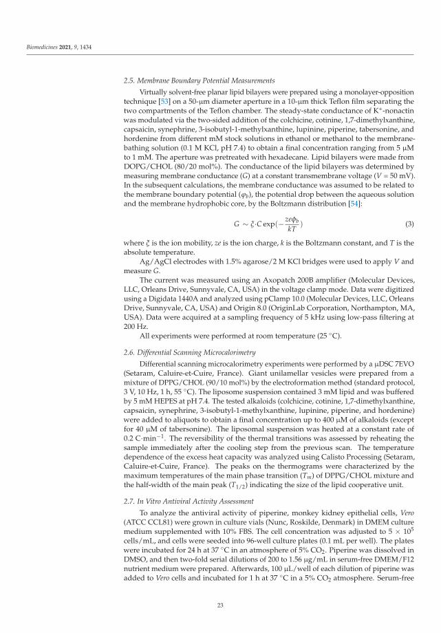
Biomedicines 2021, 9, 1434
2.5. Membrane Boundary Potential Measurements
Virtually solvent-free planar lipid bilayers were prepared using a monolayer-oppositiontechnique [53] on a 50-μm diameter aperture in a 10-μm thick Teflon film separating thetwo compartments of the Teflon chamber. The steady-state conductance of K+-nonactinwas modulated via the two-sided addition of the colchicine, cotinine, 1,7-dimethylxanthine,capsaicin, synephrine, 3-isobutyl-1-methylxanthine, lupinine, piperine, tabersonine, andhordenine from different mM stock solutions in ethanol or methanol to the membrane-bathing solution (0.1 M KCl, pH 7.4) to obtain a final concentration ranging from 5 μMto 1 mM. The aperture was pretreated with hexadecane. Lipid bilayers were made fromDOPG/CHOL (80/20 mol%). The conductance of the lipid bilayers was determined bymeasuring membrane conductance (G) at a constant transmembrane voltage (V = 50 mV).In the subsequent calculations, the membrane conductance was assumed to be related tothe membrane boundary potential (ϕb), the potential drop between the aqueous solutionand the membrane hydrophobic core, by the Boltzmann distribution [54]:
G ∼ ξ·C exp(− zeφbkT
) (3)
where ξ is the ion mobility, ze is the ion charge, k is the Boltzmann constant, and T is theabsolute temperature.
Ag/AgCl electrodes with 1.5% agarose/2 M KCl bridges were used to apply V andmeasure G.
The current was measured using an Axopatch 200B amplifier (Molecular Devices,LLC, Orleans Drive, Sunnyvale, CA, USA) in the voltage clamp mode. Data were digitizedusing a Digidata 1440A and analyzed using pClamp 10.0 (Molecular Devices, LLC, OrleansDrive, Sunnyvale, CA, USA) and Origin 8.0 (OriginLab Corporation, Northampton, MA,USA). Data were acquired at a sampling frequency of 5 kHz using low-pass filtering at200 Hz.
All experiments were performed at room temperature (25 ◦C).
2.6. Differential Scanning Microcalorimetry
Differential scanning microcalorimetry experiments were performed by a μDSC 7EVO(Setaram, Caluire-et-Cuire, France). Giant unilamellar vesicles were prepared from amixture of DPPG/CHOL (90/10 mol%) by the electroformation method (standard protocol,3 V, 10 Hz, 1 h, 55 ◦C). The liposome suspension contained 3 mM lipid and was bufferedby 5 mM HEPES at pH 7.4. The tested alkaloids (colchicine, cotinine, 1,7-dimethylxanthine,capsaicin, synephrine, 3-isobutyl-1-methylxanthine, lupinine, piperine, and hordenine)were added to aliquots to obtain a final concentration up to 400 μM of alkaloids (exceptfor 40 μM of tabersonine). The liposomal suspension was heated at a constant rate of0.2 C·min−1. The reversibility of the thermal transitions was assessed by reheating thesample immediately after the cooling step from the previous scan. The temperaturedependence of the excess heat capacity was analyzed using Calisto Processing (Setaram,Caluire-et-Cuire, France). The peaks on the thermograms were characterized by themaximum temperatures of the main phase transition (Tm) of DPPG/CHOL mixture andthe half-width of the main peak (T1/2) indicating the size of the lipid cooperative unit.
2.7. In Vitro Antiviral Activity Assessment
To analyze the antiviral activity of piperine, monkey kidney epithelial cells, Vero(ATCC CCL81) were grown in culture vials (Nunc, Roskilde, Denmark) in DMEM culturemedium supplemented with 10% FBS. The cell concentration was adjusted to 5 × 10⁵
cells/mL, and cells were seeded into 96-well culture plates (0.1 mL per well). The plateswere incubated for 24 h at 37 ◦C in an atmosphere of 5% CO2. Piperine was dissolved inDMSO, and then two-fold serial dilutions of 200 to 1.56 μg/mL in serum-free DMEM/F12nutrient medium were prepared. Afterwards, 100 μL/well of each dilution of piperine wasadded to Vero cells and incubated for 1 h at 37 ◦C in a 5% CO2 atmosphere. Serum-free
23

Biomedicines 2021, 9, 1434
DMEM/F12 medium was added to control wells instead of piperine. Aliquots of SARS-CoV-2 (103 TCID50/mL) (isolate 17612) were mixed 1:1 with dilutions of piperine andincubated for 1 h at 37 ◦C. After 1 h incubation, piperine was removed from the plates,100 μL/well of the SARS-CoV2/piperine mixture was added to the corresponding wellsand incubated for 1 h at 37 ◦C in a 5% CO2 atmosphere, the virus-containing mediumwas added to the wells of the viral control, and serum-free DMEM/F12 was added to thecell control. The virus was then removed, the cells were washed three times with DPBS,100 μL/well of dilutions of piperine (200–1.56 μg/mL) were added to the wells, serum-freeDMEM/F12 medium was added to the control wells, and the plates were incubated for24 h at 37 ◦C in a 5% CO2 atmosphere. After 24 h, supernatants from the experimental andcontrol wells with the virus were collected and titrated by TCID50 assay in Vero cells afterincubation for 72 h. The visual assessment of the cytopathogenic effect (CPE) of the viruswas performed using an Olympus CKX41 light inverted microscope (Olympus, Tokyo,Japan).
2.8. Statistical Analysis
The control RFCa2+-, RFFP-MERS-CoV-, RFFP-SARS-CoV-2-, t1-, and t2-values, characterizingthe vesicle fusion induced by CaCl2, FP-SARS-CoV-2 or FP-MERS-CoV in the absenceof alkaloids, were averaged from 5 to 9 independent experiments. The experimentalRFS-, t1-, and t2-values, characterizing the CaCl2-, FP-SARS-CoV-2- and FP-MERS-CoV-mediated fusion of liposomes pretreated with different alkaloids, were averaged from 2 to4 independent experiments. All RF-, t1-, and t2-values were presented as mean ± standarderror of the mean (p ≤ 0.05). The magnitudes of d, ζ, Tm, T1/2, and Δϕb were averaged from3 to 5 independent experiments and presented as mean ± standard deviation (p ≤ 0.05).
To prove the statistical significance of difference between RF-, t1-, t2-, d-, ζ-, Tm-, andT1/2-values at the addition of alkaloids and the control values of related parameters (in theabsence of alkaloids) the nonparametric signed-rank U-test (Mann–Whitney–Wilcoxon’sU-test) was employed (*—p ≤ 0.01, **—p ≤ 0.05).
Pearson’s correlation coefficient was applied to estimate the potent relationship be-tween the inhibitory action of alkaloids on the vesicle fusion and the alkaloid-inducedalterations in physical properties of lipid bilayers (ΔTm, ΔT1/2, Δϕb). The coefficients werecalculated using Microsoft Excel (Microsoft Corp., Redmond, WA, USA).
3. Results and Discussion
3.1. Effect of Alkaloids on CaCl2-Mediated Fusion of Negatively Charged Liposomes
Figure S4a–c (Supplementary Materials) shows the calcein leakage resulted from20 mM CaCl2-mediated fusion of DOPG/CHOL-liposomes in the absence and presenceof different alkaloids at 400 μM concentration (40 μM of tabersonine). Some alkaloidswere able to inhibit calcium-mediated fusion, and this ability strictly depended on thealkaloid type (Figure S4b,c). Table 1 summarizes the mean values of maximum markerleakage caused by calcium-induced fusion of DOPG/CHOL vesicles pretreated by dif-ferent alkaloids (RFS). The mean maximum leakage at the addition of fusion induceralone is about 90%. A significant and reliable decrease in RFS (indicating the inhibitionof calcium-mediated fusion) is caused by pretreatment of DOPG/CHOL vesicles withdihydrocapsaicin (RFS is about 70%), capsaicin, synephrine (RFS is about 50%), 3-isobutyl-1-methylxanthine, quinine, piperine (RFS is about 30%), tabersonine, hordenine (RFS isabout 20%), and lupinine (RFS is about 10%).
24

Biomedicines 2021, 9, 1434
Table 1. The parameters characterized the effects of alkaloids on the calcein release caused by the calcium-mediated fusionof liposomes composed of DOPG/CHOL (80/20 mol%) and DOPC/DOPG/CHOL (40/40/20 mol%).
DOPG/CHOL DOPC/DOPG/CHOL
Alkaloid RFS, % t1, min t2, min RFS, % t1, min t2, min
CaCl2 92 ± 7 9 ± 2 68 ± 6 82 ± 1 5 ± 3 51 ± 12
3,9-dimethylxanthine 96 ± 2 2 ± 1 * 20 ± 1 * 69 ± 7 2 ± 1 82 ± 21
caffeine 94 ± 5 3 ± 2 38 ± 20 65 ± 8* 1 ± 1 94 ± 22
7-(β-hydroxyethyl)theophylline 94 ± 6 3 ± 1 36 ± 1 * 75 ± 1 1 ± 1 75 ± 31
colchicine 94 ± 4 1 ± 1 * 7 ± 1 * 87 ± 3 2 ± 1 14 ± 1 *
pentoxifylline 92 ± 6 2 ± 1 * 36 ± 13 64 ± 8 * 1 ± 1 60 ± 14
cotinine 87 ± 12 7 ± 2 46 ± 6 ** 68 ± 2 * 3 ± 1 20 ± 9 *
1,7-dimethylxanthine 85 ± 3 8 ± 4 33 ± 5 * 60 ± 15 1 ± 1 64 ± 30
atropine 81 ± 6 7 ± 1 50 ± 9 ** 52 ± 6 * 8 ± 1 119 ± 9 *
theophylline 79 ± 8 7 ± 3 61 ± 19 83 ± 2 11 ± 1 97 ± 7 *
melatonin 76 ± 9 10 ± 1 119 ± 24 * 84 ± 8 5 ± 3 51 ± 28
berberine 74 ± 11 2 ± 1 * 27 ± 6 * 92 ± 6 5 ± 2 33 ± 13
dihydrocapsaicin 67 ± 2 * 7 ± 1 48 ± 21 56 ± 4 * 2 ± 1 24 ± 11
capsaicin 53 ± 9 * 2 ± 1 * 14 ± 2 * 56 ± 2 * 3 ± 1 14 ± 5 *
synephrine 46 ± 20 * 5 ± 1 85 ± 12 77 ± 5 12 ± 4 64 ± 30
3-isobutyl-1-methylxanthine 32 ± 12 * 3 ± 1 * 68 ± 12 84 ± 4 9 ± 1 80 ± 24
quinine 27 ± 4 * 3 ± 1 ** 118 ± 23 ** 66 ± 3 * 3 ± 1 27 ± 10
piperine 27 ± 10 * 2 ± 1 * 27± 11 ** 24 ± 1 * 2 ± 1 8 ± 2 *
tabersonine 21 ± 7 * 2 ± 1 * 11 ± 2 * 1 ± 1 * – & – &
hordenine 20 ± 16 * 1 ± 1 * 17 ± 6 * 82 ± 4 4 ± 3 50 ± 17
lupinine 13 ± 10 * 1 ± 1 * 90 ± 12 38 ± 4 * 6 ± 3 42 ± 3
RFS—the maximum leakage of fluorescent marker caused by the fusion of liposomes composed of DOPG/CHOL (80/20 mol%) andDOPC/DOPG/CHOL (40/40/20 mol%) mediated by 20 and 40 mM CaCl2 respectively. Liposomes have been incubated with 400 μM ofalkaloids (except for 40 μM of tabersonine) for 30 min before the addition of CaCl2. t1 and t2—the time constants characterizing the fast (1)and slow (2) components of marker release kinetics (the time dependences of marker leakage were fitted by two-exponential functions).*—p ≤ 0.01, **—p ≤ 0.05 (Mann–Whitney–Wilcoxon’s test, CaCl2 alone vs. CaCl2 + alkaloid). &—cannot be determined.
Table 1 also demonstrates the kinetic parameters of liposome fusion in the presenceof alkaloids. It should be noted that the time dependences of the marker release due tovesicle fusion are well-described by two-exponential dependences with characteristic timesrelated to fast and slow components (t1 and t2, respectively). Analysis of Table 1 indicatesthat t1 and t2 values vary in the ranges of about 1–10 and 10–120 min, respectively. Thefast component might characterize the calcium sorption on the negatively charged DOPG-enriched membranes and the subsequent aggregation of the lipid vesicles, while the slowone might be related to the topological rearrangements of membrane lipids and furtherdye leakage at the liposome fusion. Colchicine, capsaicin, and tabersonine significantly ac-celerated the marker release kinetics at calcium-mediated fusion of DOPG/CHOL vesicles(about 5–10-fold decrease in both characteristic times is observed) (Table 1). Melatonin andquinine drastically slowed down the fusion process increasing t2 from about 70 to morethan 100 min (Table 1).
To characterize the relative efficiency of different alkaloids to inhibit calcium-mediatedDOPG/CHOL liposome fusion the inhibition index (II) was calculated using Formula (2)for single preparation of liposome suspension, and then was averaged between the variouspreparations (Figure 1a). Taking into account the II value, the tested alkaloids were dividedinto three groups: ineffective (II does not exceed 15%); of low and medium efficiency
25

Biomedicines 2021, 9, 1434
(II is in the range of 16–50%), and of high efficiency (II is more than 50%). The firstgroup includes 3,9-dimethylxanthine, caffeine, 7-(β-hydroxyethyl)theophylline, colchicine,pentoxifylline, cotinine, 1,7-dimethylxanthine, atropine, theophylline, melatonin, andberberine. The efficiency in the second group increases in the series: dihydrocapsaicin (IIis about 25%) ≤ capsaicin ≈ synephrine (II is about 45%). The latter (the most effective)group includes 3-isobutyl-1-methylxanthine (II is about 65%), piperine ≈ quinine (II isabout 70%), tabersonine (II is about 75%), hordenine (II is about 80%), and lupinine (II isabout 85%).
Figure 1. The inhibition index (II) characterizing the ability of tested alkaloids to suppress the fusion of vesicles made fromDOPG/CHOL (80/20 mol%) (a) and DOPC/DOPG/CHOL (40/40/20 mol%) (b). Liposomes were incubated with 400 μMof alkaloids (except for 40 μM of tabersonine) for 30 min before the addition of CaCl2.
Figure 2a shows the results of size measurements after calcium addition to unmodifiedDOPG/CHOL liposomes and vesicles pretreated by alkaloids of various anti-fusogenicactivity, cotinine, melatonin, 3-isobutyl-1-methylxanthine, lupinine, piperine, tabersonine,and hordenine. The diameter of DOPG/CHOL liposomes in the absence of any modifiersis equal to 80 ± 20 nm. CaCl2 induces liposome fusion, and the vesicle size increased upto 145 ± 40 nm (Figure 2a). Cotinine and melatonin practically did not affect the size-increasing effect of calcium, as the liposome diameter was about 140 nm. The other testedcompounds (lupinine, 3-isobutyl-1-methylxanthine, tabersonine, piperine, and hordenine)significantly inhibited calcium-mediated liposome fusion: the diameter of the vesicles wasequal to 77 ± 13 nm (Figure 2a). The data obtained are in good agreement with the IIvalues assessed by the calcein release assay (Figure 1a).
Figure 2b presents the results of estimation of the ζ-potential of DOPG/CHOL li-posomes in the presence of 20 mM CaCl2 without and with different alkaloids. Theζ-potential of unmodified DOPG/CHOL liposomes was equal to about −70 mV. The ad-dition of calcium led to an increase in ζ-potential up to about −50 mV. The ζ-potentialvalue of DOPG/CHOL vesicles pretreated with alkaloids was 10–20 mV less than thatof untreated ones (Figure 2b). The observed incomplete compensation of negative sur-face charge of DOPG-enriched liposomes was expected at the introduction of positivelycharged calcium ions and alkaloid molecules (lupinine, tabersonine, and hordenine). Thedisagreement of alkaloid abilities to inhibit calcium-mediated vesicle fusion and to increaseζ-potential (Figure 2a,b) clearly demonstrates that the anti-fusogenic activity of alkaloidsis not due to partial compensation of the negative membrane surface charge by alkaloidsor a competition between alkaloids and calcium ions for the interaction with negativelycharged lipid groups.
26
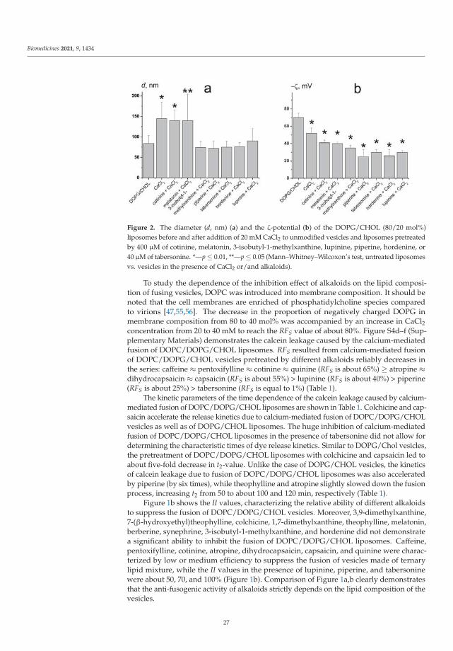
Biomedicines 2021, 9, 1434
Figure 2. The diameter (d, nm) (a) and the ζ-potential (b) of the DOPG/CHOL (80/20 mol%)liposomes before and after addition of 20 mM CaCl2 to unmodified vesicles and liposomes pretreatedby 400 μM of cotinine, melatonin, 3-isobutyl-1-methylxanthine, lupinine, piperine, hordenine, or40 μM of tabersonine. *—p ≤ 0.01, **—p ≤ 0.05 (Mann–Whitney–Wilcoxon’s test, untreated liposomesvs. vesicles in the presence of CaCl2 or/and alkaloids).
To study the dependence of the inhibition effect of alkaloids on the lipid composi-tion of fusing vesicles, DOPC was introduced into membrane composition. It should benoted that the cell membranes are enriched of phosphatidylcholine species comparedto virions [47,55,56]. The decrease in the proportion of negatively charged DOPG inmembrane composition from 80 to 40 mol% was accompanied by an increase in CaCl2concentration from 20 to 40 mM to reach the RFS value of about 80%. Figure S4d–f (Sup-plementary Materials) demonstrates the calcein leakage caused by the calcium-mediatedfusion of DOPC/DOPG/CHOL liposomes. RFS resulted from calcium-mediated fusionof DOPC/DOPG/CHOL vesicles pretreated by different alkaloids reliably decreases inthe series: caffeine ≈ pentoxifylline ≈ cotinine ≈ quinine (RFS is about 65%) ≥ atropine ≈dihydrocapsaicin ≈ capsaicin (RFS is about 55%) > lupinine (RFS is about 40%) > piperine(RFS is about 25%) > tabersonine (RFS is equal to 1%) (Table 1).
The kinetic parameters of the time dependence of the calcein leakage caused by calcium-mediated fusion of DOPC/DOPG/CHOL liposomes are shown in Table 1. Colchicine and cap-saicin accelerate the release kinetics due to calcium-mediated fusion of DOPC/DOPG/CHOLvesicles as well as of DOPG/CHOL liposomes. The huge inhibition of calcium-mediatedfusion of DOPC/DOPG/CHOL liposomes in the presence of tabersonine did not allow fordetermining the characteristic times of dye release kinetics. Similar to DOPG/Chol vesicles,the pretreatment of DOPC/DOPG/CHOL liposomes with colchicine and capsaicin led toabout five-fold decrease in t2-value. Unlike the case of DOPG/CHOL vesicles, the kineticsof calcein leakage due to fusion of DOPC/DOPG/CHOL liposomes was also acceleratedby piperine (by six times), while theophylline and atropine slightly slowed down the fusionprocess, increasing t2 from 50 to about 100 and 120 min, respectively (Table 1).
Figure 1b shows the II values, characterizing the relative ability of different alkaloidsto suppress the fusion of DOPC/DOPG/CHOL vesicles. Moreover, 3,9-dimethylxanthine,7-(β-hydroxyethyl)theophylline, colchicine, 1,7-dimethylxanthine, theophylline, melatonin,berberine, synephrine, 3-isobutyl-1-methylxanthine, and hordenine did not demonstratea significant ability to inhibit the fusion of DOPC/DOPG/CHOL liposomes. Caffeine,pentoxifylline, cotinine, atropine, dihydrocapsaicin, capsaicin, and quinine were charac-terized by low or medium efficiency to suppress the fusion of vesicles made of ternarylipid mixture, while the II values in the presence of lupinine, piperine, and tabersoninewere about 50, 70, and 100% (Figure 1b). Comparison of Figure 1a,b clearly demonstratesthat the anti-fusogenic activity of alkaloids strictly depends on the lipid composition of thevesicles.
27

Biomedicines 2021, 9, 1434
3.2. The Influence of Alkaloids on the Physical Properties of the Model Lipid Membrane
It has been repeatedly shown that the elastic characteristics of the lipid bilayer play ahuge role in the membrane fusion. The ordering of the lipid head groups and acyl chains,transbilayer lateral pressure profile, phase state of lipids, spontaneous curvature of themembrane, area per lipid molecule, and other characteristics can influence fusion [57].Many of these parameters influence each other, but all of them are directly dependenton the lipid composition of the membrane. The key role of lipids is also supported bythe fact that viral infections can alter cell lipid synthesis, regulating it according to theirneeds [58]. The ability of alkaloids and their derivatives to change the physical properties ofa bilayer upon intercalation was demonstrated in a number of studies [59–62]. In particular,alkaloids affect the lipid packing [63,64], change lipid phase transition temperatures [65,66],etc. Recently, we performed a detailed study of alkaloid effects on the physical propertiesof the POPC membrane [67]. To understand the relationship between the alkaloid effectson the fusion and modulation of lipid matrix under their action, the influence of testedalkaloids on the properties of PG and CHOL-enriched bilayers were performed.
Figure 3 presents the typical heating thermograms of DPPG/CHOL-liposomes in theabsence (control) and in the presence of tested alkaloids. The value of the phase transitiontemperature (Tm) of DPPG/CHOL mixture, and the half-width of the main peak (T1/2) aresummarized in Table 2. Tm is the point at which thermally induced lipid melting occurs [68],while T1/2 describes the sharpness of the phase transition or the inverse cooperativity ofthis process [69]. The temperature of the main transition (Tm) of untreated DPPG/CHOLvesicles is equal to 40.4 ◦C, with a half-width of the peak (T1/2) of about 0.8 ◦C (Figure 3,control).
Figure 3. Heating thermograms of DPPG/CHOL (90/10 mol%) liposomes in the absence and pres-ence of 400 μM of 1,7-dimethylxanthine, piperine, hordenine, cotinine, 3-isobutyl-1-methylxanthine,lupinine, synephrine, capsaicin, colchicine, and 40 μM of tabersonine.
28
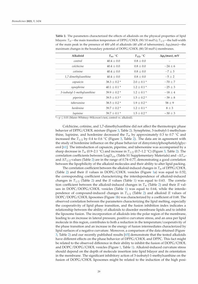
Biomedicines 2021, 9, 1434
Table 2. The parameters characterized the effects of alkaloids on the physical properties of lipidbilayers: Tm—the main transition temperature of DPPG/CHOL (90/10 mol%); T1/2—the half-widthof the main peak in the presence of 400 μM of alkaloids (40 μM of tabersonine); Δϕb(max)—themaximum changes in the boundary potential of DOPG/CHOL (80/20 mol%) membranes.
Alkaloid Tm, ◦C T1/2, ◦C Δϕb(max), mV
control 40.4 ± 0.0 0.8 ± 0.0 –
colchicine 40.4 ± 0.0 0.8 ± 0.0 −24 ± 6
cotinine 40.4 ± 0.0 0.8 ± 0.0 −7 ± 3
1,7-dimethylxanthine 40.4 ± 0.0 0.8 ± 0.0 −5 ± 2
capsaicin 38.3 ± 0.2 * 2.0 ± 0.1 * −70 ± 7
synephrine 40.1 ± 0.1 * 1.2 ± 0.1 * −25 ± 3
3-isobutyl-1-methylxanthine 39.9 ± 0.2 * 1.2 ± 0.1 * −18 ± 4
piperine 39.5 ± 0.3 * 1.5 ± 0.2 * −39 ± 8
tabersonine 38.5 ± 0.2 * 1.9 ± 0.2 * 58 ± 9
hordenine 39.7 ± 0.2 * 1.2 ± 0.1 * 8 ± 3
lupinine 39.7 ± 0.1 * 1.5 ± 0.2 * −30 ± 3*—p ≤ 0.01 (Mann–Whitney–Wilcoxon’s test, control vs. alkaloid).
Colchicine, cotinine, and 1,7-dimethylxanthine did not affect the thermotropic phasebehavior of DPPG/CHOL mixture (Figure 3, Table 2). Synephrine, 3-isobutyl-1-methylxan-thine, lupinine, and hordenine decreased the Tm by approximately 0.3 to 0.7 ◦C andincreased the T1/2 by 0.4 to 0.6 ◦C (Figure 3, Table 2). The data are in agreement withthe study of hordenine influence on the phase behavior of dimyristoylphosphatidylglyc-erol [61]. The introduction of capsaicin, piperine, and tabersonine was accompanied by asharp decrease in Tm (0.9–2.1 ◦C) and increase in T1/2 (0.7–1.2 ◦C) (Figure 3, Table 2). Thecorrelation coefficients between LogDo/w (Table S1 Supplementary Materials) and –ΔTm-and ΔT1/2-values (Table 2) are in the range of 0.74–0.77, demonstrating a good correlationbetween the lipophilicity of the alkaloid molecules and their ability to alter lipid packing.
The correlation coefficient between the alkaloid-induced changes in Tm of DPPG/CHOL(Table 2) and their II values in DOPG/CHOL vesicles (Figure 1a) was equal to 0.52,the corresponding coefficient characterizing the interdependence of alkaloid-inducedchanges in T1/2 (Table 2) and the II values (Table 1) was equal to 0.63. The correla-tion coefficient between the alkaloid-induced changes in Tm (Table 2) and their II val-ues in DOPC/DOPG/CHOL vesicles (Table 1) was equal to 0.64, while the interde-pendence of compound-induced changes in T1/2 (Table 2) and alkaloid II values ofDOPC/DOPG/CHOL liposomes (Figure 1b) was characterized by a coefficient of 0.68. Theobserved correlation between the parameters characterizing the lipid melting, especiallythe cooperativity of lipid phase transition, and the fusion inhibition index indicates arelationship between the ability of alkaloids to disorder membrane lipids and to inhibitthe liposome fusion. The incorporation of alkaloids into the polar region of the membrane,leading to an increase in lateral pressure, positive curvature stress, and an area per lipidmolecule in this region, contributes to both a reduction in the temperature/cooperativity ofthe phase transition and an increase in the energy of fusion intermediates characterized bylipid surfaces of a negative curvature. Moreover, a comparison of the data obtained (Figure3, Table 2) and our recently published results [65] demonstrate that the tested alkaloidshave different effects on the phase behavior of DPPG/CHOL and DPPC. This fact mightbe related to the observed difference in their ability to inhibit the fusion of DOPG/CHOLand DOPC/DOPG/CHOL vesicles (Figure 1, Table 1). Alkaloid-induced curvature stressshould depend on the depth of molecule insertion into lipid bilayer and its orientationin the membrane. The significant inhibitory action of 3-isobutyl-1-methylxanthine on thefusion of DOPG/CHOL liposomes might be related to the induction of the high posi-
29
Biomedicines 2021, 9, 1434
tive curvature stress due to both the electrostatic interaction between xanthine residueand DOPG on the membrane surface and the tendency of the isobutyl side chain to beembedded into the membrane hydrocarbon core. Xanthine derivatives with lower hy-drophobicity, i.e., caffeine, pentoxifylline, 1,7-dimethylxanthine, 3,9-dimethylxanthine,and 7-(β-hydroxyethyl)theophylline (Table S1, Supplementary Materials) did not inhibitcalcium-mediated fusion of DOPG/CHOL liposomes. Introduction of DOPC into themembrane lipid composition was accompanied by a two-fold decrease in the DOPG con-tent, which led to significant changes in the membrane orientation and embedment ofxanthines. Thus, caffeine and pentoxifylline demonstrated a weak inhibitory effect on afusion of DOPC/DOPG/CHOL vesicles, while 3-isobutyl-1-methylxanthine did not affectthis process. The complete loss of the inhibiting ability of β-phenylethylamine derivatives,synephrine and hordenine, in membranes with lower content of DOPG compared to DOPG-enriched bilayers, could be explained in a similar way. An increase in the inhibitory effectof tabersonine on the fusion of DOPC/DOPG/CHOL vesicles compared to DOPG/CHOLliposomes might indicate its deeper immersion into the DOPC/DOPG/CHOL membranesthan into DOPG/CHOL bilayers due to a decrease in the electrostatic interactions betweenpositively charged tabersonine and negatively charged DOPG molecules.
Considering that liposome fusion is triggered by calcium ions, we also consideredthe possibility of alkaloids influencing fusion by changing the boundary potential of themembranes. Table 2 shows the maximum changes in ϕb at the adsorption of differentalkaloids (–Δϕb(max)). The dependences of Δϕb on the concentrations of tested alkaloidsare presented in Figure S5 (Supplementary Materials). Tabersonine significantly increasedthe ϕb of DOPG/CHOL membranes (by about 60 mV), while capsaicin dramatically de-creased this value (by about −70 mV) (Figure S5 in the Supplementary Materials, Table 2).Colchicine, synephrine, 3-isobutyl-1-methylxanthine, lupinine, and piperine led to reduc-tion in ϕb by about 20–40 mV (Figure S5 in the Supplementary Materials, Table 2). Cotinine,1,7-dimethylxanthine, and hordenine practically did not alter the ϕb-value (Figure S5 Sup-plementary Materials, Table 2). There was no correlation between Δϕb of DOPG/CHOLmembranes after the addition of alkaloids and their II values. This fact demonstrates theminor role of changes in the bilayer electrical properties by alkaloids in their ability toinhibit the calcium-mediated membrane fusion.
3.3. Liposome Fusion Mediated by Fragments of Coronavirus Fusion Peptides
The COVID-19 pandemic has caused major challenges to healthcare systems acrossthe globe. The lack of specific treatment, virus mutations, the emergence of new strains thathave become resistant to vaccines, and many other factors, have led to the search for newdrugs to treat COVID-19. To demonstrate the pharmacological applications of alkaloids ascoronavirus fusion inhibitors—the abilities of the most effective compounds to suppressthe membrane fusion induced by FP-MERS-CoV and FP-SARS-CoV-2 were studied. Thesepeptides are able to induce calcein release due to fusion of POPC/SM/CHOL liposomes(Figure S2c,d Supplementary Materials), and are not effective to produce the fusion ofDOPG/CHOL vesicles (data not shown).
To further validate the fusogenic properties of FP-MERS-CoV and FP-SARS-CoV-2, theconfocal fluorescence microscopy of giant unilamellar vesicles composed of POPC/SM/CHOLwas performed. Figure 4 presents the fluorescence micrographs of POPC/SM/CHOL li-posomes in the absence (Figure 4a) and presence of 10% of the well-established fusioninducer PEG-8000 (Figure 4b), 50 μM FP-SARS-CoV-2 (Figure 4c), and 200 μM FP-MERS-CoV (Figure 4d). Both peptides at the indicated concentrations caused deformation andaggregation of lipid vesicles, and increased in size. FP-SARS-CoV-2, unlike FP-MERS-CoV,also induced the formation of multilamellar and multivesicular liposomes (Figure 4c). Ingeneral, the morphological picture under the action of both peptides is similar to thatwhich occurred when PEG-8000 was added (Figure 4b). Thus, the results obtained confirmthe fusogenic activity of FP-SARS-CoV-2 and FP-MERS-CoV.
30

Biomedicines 2021, 9, 1434
Figure 4. Fluorescence micrographs of giant unilamellar vesicles made from POPC/SM/CHOL (60/20/20 mol%) and1 mol% of fluorescent lipid probe Rh-DPPE in the absence of any modifiers (a), and in the presence of 10% of PEG-8000 (b),50 μM of FP-SARS-CoV-2 (c), and 200 μM of FP-MERS-CoV (d).
Figure 5a,d present the effects of alkaloids that effectively inhibit calcium-mediatedfusion of DOPG/CHOL vesicles, 3-isobutyl-1-methylxanthine, piperine, tabersonine, hor-denine, and lupinine, on the calcein leakage caused by FP-SARS-CoV-2- and FP-MERS-CoV-mediated fusion of POPC/SM/CHOL liposomes, respectively. In the absence of alkaloidsRFs-value produced by 50 μM of FP-SARS-CoV-2 and 200 μM of FP-MERS-CoV was about70 (Figure 5a) and 80% (Figure 5d), respectively. Table 3 summarizes the mean values ofmaximum marker leakage caused by peptide-induced fusion of POPC/SM/CHOL vesiclespretreated by different alkaloids and the kinetic parameters of liposome fusion in the pres-ence of alkaloids. RFS caused by FP-SARS-CoV-2-mediated fusion of POPC/SM/CHOLvesicles significantly decreased in the presence of piperine (RFS is about 30%) and taber-sonine (RFS is about 50%). Moreover, piperine was also effective against FP-MERS-CoV-mediated fusion (RFS is about 30%), while tabersonine was not characterized by an abilityto inhibit FP-MERS-CoV-mediated fusion of POPC/SM/CHOL liposomes. The fusion-induced leakage in the presence of 3-isobutyl-1-methylxanthine, tabersonine, hordenine,and lupinine was characterized by faster kinetics compared to the absence of alkaloids,while highly active piperine slightly slowed down kinetics of FP-MERS-CoV-inducedfusion by increasing t2-value.
Figure 5b,e show the alkaloid II-values clearly demonstrating the ability of piper-ine to significantly inhibit both FP-SARS-CoV-2- and FP-MERS-CoV-mediated fusion ofPOPC/SM/CHOL liposomes, and the selectivity of tabersonine anti-fusogenic action. Theobserved selectivity of tabersonine action against liposome fusion induced by FP-SARS-CoV-2 and FP-MERS-CoV should be researched further.
To further demonstrate the anti-fusogenic activity of piperine, the vesicle size beforeand after the addition of FP-SARS-CoV-2 and FP-MERS-CoV to the unmodified and alka-loid pretreated POPC/SM/CHOL-liposomes was measured by dynamic light scattering(Figure 5c,f). In the absence of the any modifiers, the diameters of POPC/SM/CHOL-vesicles were equal to 110 ± 15 nm. Addition of the peptide FP-SARS-CoV-2 and FP-MERS-CoV led to an increased in size, up to 290 ± 40 nm and 190 ± 15 nm, respectively(Figure 5c,f). In the presence of piperine, the modeling fusion peptides were not able toincrease the diameter of POPC/SM/CHOL-liposomes due to strong inhibition of vesiclefusion by the alkaloid. Figure 5c also demonstrates the ability of tabersonine to prevent theincrease of liposome size under FP-SARS-CoV-2 action. The invariability of the liposome ζ-potential upon the addition of FP-SARS-CoV-2- and FP-MERS-CoV to both unmodified andalkaloid pretreated POPC/SM/CHOL liposomes is shown in Figure S6 (SupplementaryMaterials).
31

Biomedicines 2021, 9, 1434
Figure 5. The effects of the alkaloids on FP-SARS-CoV-2 (upper panel) and FP-MERS-CoV (lower panel) mediated fusion ofPOPC/SM/CHOL (60/20/20 mol%) vesicles. (a,d) The time dependence of relative fluorescence of calcein (RFS, %) leakeddue to the fusion induced by 50 μM of FP-SARS-CoV-2; (a) and 200 μM of FP-MERS-CoV; (d) in the absence and presence ofalkaloids. Liposomes were incubated with 400 μM of alkaloids for 30 min before addition of the peptides. The relationshipbetween the color line and the alkaloid is given in the figure. (b,e) The inhibition index (II) characterizing the ability oftested alkaloids to suppress the fusion induced by 50 μM of FP-SARS-CoV-2 (b); and 200 μM of FP-MERS-CoV (e). (c,f) Thediameter (d, nm) of the POPC/SM/CHOL liposomes before and after addition of 50 μM of FP-SARS-CoV-2 (c) and 200 μMof FP-MERS-CoV (f) to vesicles pretreated with 400 μM of piperine or tabersonine. *—p ≤ 0.01 (Mann–Whitney–Wilcoxon’stest, untreated liposomes vs. vesicles in the presence of fusion peptides or/and alkaloids).
Table 3. The parameters characterized the effects of alkaloids on the calcein release caused by the FP-SARS-CoV-2 andFP-MERS-CoV-mediated fusion of liposomes composed of POPC/SM/CHOL.
50 μM FP-SARS-CoV-2 200 μM FP-MERS-CoV
Alkaloid RFS, % t1, min t2, min RFS, % t1, min t2, min
inducer 77 ± 6 11 ± 1 70 ± 4 80 ± 5 3 ± 2 22 ± 14
3-isobutyl-1-methylxanthine 66 ± 5 3 ± 1 * 15 ± 2 * 78 ± 6 2 ± 1 15± 5
piperine 33 ± 3 * 9 ± 3 73 ± 6 29 ± 2* 4 ± 1 48 ± 1 *
tabersonine 53 ± 9 ** 1 ± 1* 27 ± 13 * 79 ± 7 2 ± 1 12 ± 2
hordenine 74 ± 8 3 ± 1* 18 ± 4 * 75 ± 10 2 ± 1 21 ± 1
lupinine 76 ± 4 1 ± 1* 31 ± 9 * 80 ± 6 1 ± 1 26 ± 1
RFS—the maximum leakage of fluorescent marker caused by the fusion of liposomes composed of POPC/SM/CHOL (60/20/20 mol%)mediated by 50 μM FP-SARS-CoV-2 or 200 μM FP-MERS-CoV. Liposomes have been incubated with 400 μM of alkaloids for 30 min beforethe addition of CaCl2. t1 and t2—the time constants characterizing the fast (1) and slow (2) components of marker release kinetics (thetime dependences of marker leakage were fitted by two-exponential functions). *—p ≤ 0.01, **—p ≤ 0.05 (Mann–Whitney–Wilcoxon’s test,fusion peptide alone vs. fusion peptide + alkaloid).
32

Biomedicines 2021, 9, 1434
3.4. Antiviral Evaluation
Piperine is an alkaloid found in black pepper, one of the most widely used spices. Thein vitro and in vivo antiviral activity of the alkaloid against the MERS-CoV in Vero cells,and in a mice model, was already demonstrated [70]. Here, we evaluated in vitro antiviralactivity of piperine against the SARS-CoV-2 virus.
According to results of the cytotoxicity analysis performed by Hegeto et al. [71], thevalue of IC50 of piperine against Vero cells was equal to 183.33 μg/mL. Based on thisfinding, the maximum piperine concentration of 200 μg/mL was chosen. The incubationwith 200 μg/mL of piperine for 72 h led to death of all cells in wells; all lower piperineconcentrations used were nontoxic to Vero cells (data not shown).
To determine the infectious activity of viral progeny, ten-fold dilutions of the super-natants collected from the experimental wells and viral controls in serum-free DMEM/F12medium were prepared. The resulting dilutions were added to a 96-well culture plate with80–90% Vero cell monolayer and incubated for 72 h. After that, visual assessment of thecytopathogenic effect (CPE) of the virus was performed. The control samples without anyadditives demonstrated that the virus titer was 104 TCID50/mL. Samples with a piper-ine concentration of 1.56 μg/mL resulted in 103 TCID50/mL; alkaloid concentrations of3.12–25 μg/mL had 102 TCID50/mL, and in samples with piperine, concentrations of 50and 100 μg/mL CPE were not observed at all. Thus, the significant reduction of the titer ofSARS-CoV2 progeny in Vero cells at 1.56–100 μg/mL of piperine was clearly demonstrated.
4. Conclusions
New broad-spectrum antiviral drugs are needed due to the increase in the number ofviral infections. Virus fusion inhibitors could be effective antiviral agents because fusionis a necessary step for the lifecycle of the virus. We used plant secondary metabolites, inparticular, alkaloids, as a new class of fusion inhibitors. It was shown that the ability ofalkaloids to inhibit calcium-mediated fusion of liposomes depends on their lipid composi-tion. A correlation between the anti-fusogenic activity of alkaloids and their disorderingeffects on membrane lipids was found. Additionally, the ability of piperine to suppressthe fusion induced by fragments of the coronavirus fusion peptides (MERS-CoV andSARS-CoV/SARS-CoV-2) was demonstrated. We also showed that piperine dramaticallyreduced the titer of SARS-CoV2 progeny in vitro in Vero cells when used in non-toxicconcentrations. Thus, we hypothesize that the antiviral activity of piperine is related to itslipid-associated action. Moreover, to our knowledge, except for one bioassay with SARS-CoV-2-S pseudotyped particles [72], this is the first study demonstrating the in vitro activityof piperine against SARS-CoV-2 virus. It should be noted that piperine with curcumin isbeing studied in several clinical trials for the treatment of COVID-19 [73–75]. Accordingto [75], administration of oral curcumin with piperine substantially reduced the morbidityand mortality due to COVID-19. We hope that, over time, the number of effective alkaloidsin the treatment of COVID-19 will increase. The use of plant metabolites can successfullycomplete the existing therapeutic strategies, which will make it possible to combat theSARS-CoV-2 virus more effectively.
Supplementary Materials: The following are available online at https://www.mdpi.com/article/10.3390/biomedicines9101434/s1, Figure S1: The chemical structures of the tested alkaloids, Figure S2:The time dependence of relative fluorescence of calcein leaked from DOPG/CHOL (80/20 mol%) (a),DOPC/DOPG/CHOL (40/40/20 mol%) (b), and POPC/SM/CHOL (60/20/20 mol%) (c,d) vesiclesinduced by different concentrations of CaCl2 (a,b), FP-SARS-CoV-2 (c) and FP-MERS-CoV (d). Therelationship between the color line and the concentration of the fusion inducers is given in thefigure; Figure S3: The time dependence of relative fluorescence of calcein (RFA, %) leaked fromDOPG/CHOL (80/20 mol%) (a), DOPC/DOPG/CHOL (40/40/20 mol%) (b), and POPC/SM/CHOL(60/20/20 mol%) (c) vesicles induced by alkaloids alone. Alkaloids were added into the liposomalsuspension up to a concentration of 400 μM (except for 40 μM of tabersonine in DOPG/CHOL andDOPC/DOPG/CHOL vesicles) at the initial moment. The relationship between the color line andthe compound is given in the figure; Figure S4. The time dependence of relative fluorescence of
33

Biomedicines 2021, 9, 1434
calcein (RFS, %) leaked by the fusion of DOPG/CHOL (80/20 mol%) (a–c) and DOPC/DOPG/CHOL(40/40/20 mol%) (d–f) vesicles induced by 20 mM of CaCl2 in the absence (black line) and presence ofalkaloids. Liposomes were incubated with 400 μM of alkaloids (except for 40 μM of tabersonine) for30 min before the addition of CaCl2. The relationship between the color line and the alkaloid is givenin the figure; Figure S5: The dependence of the changes in the membrane boundary potential (Δϕb) onthe concentration of colchicine (*), cotinine (�), 1,7-dimethylxanthine (�), capsaicin (�), synephrine(♦), 3-isobutyl-1-methylxanthine (•), lupinine (�), piperine (�), tabersonine (�), and hordenine (�).The membranes were composed of DOPG/CHOL (80/20 mol%) and bathed in 0.1 M KCl at pH7.4. V = 50 mV, Figure S6: The ζ-potential of the POPC/SM/CHOL (60/20/20 mol%) liposomesbefore and after addition of 50 μM of FP-SARS-CoV-2 or 200 μM of FP-MERS-CoV to unmodifiedand pretreated by 400 μM of piperine vesicles; Table S1: The parameters characterized the alkaloidmolecules and their influence on the calcein leakage from liposomes of different composition.
Author Contributions: E.V.S.—investigation, analysis, validation, and writing (original draft);S.S.E.—investigation, analysis, and validation; N.M.Y.—investigation, A.A.M.—investigation, V.V.Z.—investigation, A.V.S.—investigation, analysis, and writing (review draft), O.S.O.—conceptualization,project administration, supervision, analysis, and writing (original draft, review and editing). Allauthors have read and agreed to the published version of the manuscript.
Funding: This research was financially supported by the joint-stock company Evalar.
Conflicts of Interest: The authors declare that the research was conducted in the absence of anycommercial or financial relationships that could be construed as a potential conflict of interest.
References
1. Patridge, E.; Gareiss, P.; Kinch, M.S.; Hoyer, D. An analysis of FDA-approved drugs: Natural products and their derivatives.Drug Discov. Today 2016, 21, 204–207. [CrossRef] [PubMed]
2. Zhao, J.; Shan, T.; Mou, Y.; Zhou, L. Plant-derived bioactive compounds produced by endophytic fungi. Mini Rev. Med. Chem.2011, 11, 159–168. [CrossRef] [PubMed]
3. Mani, J.S.; Johnson, J.B.; Steel, J.C.; Broszczak, D.A.; Neilsen, P.M.; Walsh, K.B.; Naiker, M. Natural product-derived phytochemi-cals as potential agents against coronaviruses: A review. Virus Res. 2020, 284, 197989. [CrossRef]
4. Ngwa, W.; Kumar, R.; Thompson, D.; Lyerly, W.; Moore, R.; Reid, T.E.; Lowe, H.; Toyang, N. Potential of flavonoid-inspiredphytomedicines against COVID-19. Molecules 2020, 25, 2707. [CrossRef]
5. Kishimoto, S.; Sato, M.; Tsunematsu, Y.; Watanabe, K. Evaluation of biosynthetic pathway and engineered biosynthesis ofalkaloids. Molecules 2016, 21, 1078. [CrossRef]
6. Xu, W.; Zhang, M.; Liu, H.; Wei, K.; He, M.; Li, X.; Hu, D.; Yang, S.; Zheng, Y. Antiviral activity of aconite alkaloids from Aconitumcarmichaelii Debx. Nat. Rrod. Res. 2019, 33, 1486–1490. [CrossRef]
7. Cushnie, T.P.; Cushnie, B.; Lamb, A.J. Alkaloids: An overview of their antibacterial, antibiotic-enhancing and antivirulenceactivities. Int. J. Antimicrob. Agents 2014, 44, 377–386. [CrossRef]
8. Khan, H.; Mubarak, M.S.; Amin, S. Antifungal potential of alkaloids as an emerging therapeutic target. Curr. Drug Targets 2017,18, 1825–1835. [CrossRef]
9. Boyd, M.R.; Hallock, Y.F.; Cardellina, J.H., 2nd; Manfredi, K.P.; Blunt, J.W.; McMahon, J.B.; Buckheit, R.W., Jr.; Bringmann, G.;Schäffer, M.; Cragg, G.M. Anti-HIV michellamines from Ancistrocladus korupensis. J. Med. Chem. 1994, 37, 1740–1745. [CrossRef]
10. Houghton, P.J.; Woldemariam, T.Z.; Khan, A.I.; Burke, A.; Mahmood, N. Antiviral activity of natural and semi-synthetic chromonealkaloids. Antivir. Res. 1994, 25, 235–244. [CrossRef]
11. Bodiwala, H.S.; Sabde, S.; Mitra, D.; Bhutani, K.K.; Singh, I.P. Synthesis of 9-substituted derivatives of berberine as anti-HIVagents. Eur. J. Med. Chem. 2011, 46, 1045–1049. [CrossRef]
12. Wan, Z.; Lu, Y.; Liao, Q.; Wu, Y.; Chen, X. Fangchinoline inhibits human immunodeficiency virus type 1 replication by interferingwith gp160 proteolytic processing. PLoS ONE 2012, 7, e39225. [CrossRef]
13. Varghese, F.S.; Kaukinen, P.; Gläsker, S.; Bespalov, M.; Hanski, L.; Wennerberg, K.; Kümmerer, B.M.; Ahola, T. Discovery ofberberine, abamectin and ivermectin as antivirals against chikungunya and other alphaviruses. Antivir. Res. 2016, 126, 117–124.[CrossRef]
14. Liu, X.; Wang, Y.; Zhang, M.; Li, G.; Cen, Y. Study on the inhibitory effect of cepharanthine on herpes simplex type-1 virus (HSV-1)in vitro. Zhong Yao Cai 2004, 27, 107–110. [PubMed]
15. Chin, L.W.; Cheng, Y.W.; Lin, S.S.; Lai, Y.Y.; Lin, L.Y.; Chou, M.Y.; Chou, M.C.; Yang, C.C. Anti-herpes simplex virus effectsof berberine from Coptidis rhizoma, a major component of a Chinese herbal medicine, Ching-Wei-San. Arch. Virol. 2010, 155,1933–1941. [CrossRef] [PubMed]
16. Liou, J.T.; Chen, Z.Y.; Ho, L.J.; Yang, S.P.; Chang, D.M.; Liang, C.C.; Lai, J.H. Differential effects of triptolide and tetrandrine onactivation of COX-2, NF-kappaB, and AP-1 and virus production in dengue virus-infected human lung cells. Eur. J. Pharmacol.2008, 589, 288–298. [CrossRef]
34

Biomedicines 2021, 9, 1434
17. Hung, T.C.; Jassey, A.; Liu, C.H.; Lin, C.J.; Lin, C.C.; Wong, S.H.; Wang, J.Y.; Yen, M.H.; Lin, L.T. Berberine inhibits hepatitis Cvirus entry by targeting the viral E2 glycoprotein. Phytomedicine 2019, 53, 62–69. [CrossRef]
18. Gao, Y.; Tai, W.; Wang, N.; Li, X.; Jiang, S.; Debnath, A.K.; Du, L.; Chen, S. Identification of novel natural products as effective andbroad-spectrum anti-zika virus inhibitors. Viruses 2019, 11, 1019. [CrossRef]
19. Warowicka, A.; Nawrot, R.; Gozdzicka-Józefiak, A. Antiviral activity of berberine. Arch. Virol. 2020, 165, 1935–1945. [CrossRef]20. Sakurai, Y.; Kolokoltsov, A.A.; Chen, C.C.; Tidwell, M.W.; Bauta, W.E.; Klugbauer, N.; Grimm, C.; Wahl-Schott, C.; Biel, M.; Davey,
R.A. Ebola virus. Two-pore channels control Ebola virus host cell entry and are drug targets for disease treatment. Science 2015,347, 995–998. [CrossRef]
21. Dang, Z.; Jung, K.; Zhu, L.; Lai, W.; Xie, H.; Lee, K.H.; Huang, L.; Chen, C.H. Identification and synthesis of quinolizidines withanti-influenza a virus activity. ACS Med. Chem. Lett. 2014, 5, 942–946. [CrossRef]
22. Dai, J.P.; Wang, Q.W.; Su, Y.; Gu, L.M.; Deng, H.X.; Chen, X.X.; Li, W.Z.; Li, K.S. Oxymatrine inhibits influenza a virus replicationand inflammation via TLR4, p38 MAPK and NF-κB pathways. Int. J. Mol. Sci. 2018, 19, 965. [CrossRef]
23. Li, S.Y.; Chen, C.; Zhang, H.Q.; Guo, H.Y.; Wang, H.; Wang, L.; Zhang, X.; Hua, S.N.; Yu, J.; Xiao, P.G.; et al. Identificationof natural compounds with antiviral activities against SARS-associated coronavirus. Antivir. Res. 2005, 67, 18–23. [CrossRef][PubMed]
24. Kim, H.Y.; Shin, H.S.; Park, H.; Kim, Y.C.; Yun, Y.G.; Park, S.; Shin, H.J.; Kim, K. In vitro inhibition of coronavirus replications bythe traditionally used medicinal herbal extracts, Cimicifuga rhizoma, Meliae cortex, Coptidis rhizoma, and Phellodendron cortex. J. Clin.Virol. 2008, 41, 122–128. [CrossRef] [PubMed]
25. Kim, D.E.; Min, J.S.; Jang, M.S.; Lee, J.Y.; Shin, Y.S.; Song, J.H.; Kim, H.R.; Kim, S.; Jin, Y.H.; Kwon, S. Natural bis-benzylisoquinolinealkaloids-tetrandrine, fangchinoline, and cepharanthine, inhibit human coronavirus OC43 infection of MRC-5 human lung cells.Biomolecules 2019, 9, 696. [CrossRef]
26. Fielding, B.C.; da Silva Maia Bezerra Filho, C.; Ismail, N.; Sousa, D.P. Alkaloids: Therapeutic potential against human coron-aviruses. Molecules 2020, 25, 5496. [CrossRef] [PubMed]
27. Wu, R.; Wang, L.; Kuo, H.D.; Shannar, A.; Peter, R.; Chou, P.J.; Li, S.; Hudlikar, R.; Liu, X.; Liu, Z.; et al. An update on currenttherapeutic drugs treating COVID-19. Curr. Pharmacol. Rep. 2020, 11, 1–15. [CrossRef] [PubMed]
28. Della-Torre, E.; Della-Torre, F.; Kusanovic, M.; Scotti, R.; Ramirez, G.A.; Dagna, L.; Tresoldi, M. Treating COVID-19 with colchicinein community healthcare setting. Clin. Immunol. 2020, 217, 108490. [CrossRef]
29. Tan, G.T.; Pezzuto, J.M.; Kinghorn, A.D.; Hughes, S.H. Evaluation of natural products as inhibitors of human immunodeficiencyvirus type 1 (HIV-1) reverse transcriptase. J. Nat. Prod. 1991, 54, 143–154. [CrossRef]
30. Kim, S.Y.; Kim, H.; Kim, S.W.; Lee, N.R.; Yi, C.M.; Heo, J.; Kim, B.J.; Kim, N.J.; Inn, K.S. An effective antiviral approach targetinghepatitis B virus with NJK14047, a novel and selective biphenyl amide p38 mitogen-activated protein kinase inhibitor. Antimicrob.Agents Chemother. 2017, 61, e00214–e00217. [CrossRef]
31. Rey, F.A.; Lok, S.M. Common features of enveloped viruses and implications for immunogen design for next-generation vaccines.Cell 2018, 172, 1319–1334. [CrossRef]
32. Vigant, F.; Santos, N.C.; Lee, B. Broad-spectrum antivirals against viral fusion. Nat. Rev. Microbiol. 2015, 13, 426–437. [CrossRef]33. Rout, J.; Swain, B.C.; Tripathy, U. In silico investigation of spice molecules as potent inhibitor of SARS-CoV-2. J. Biomol. Struct.
Dyn. 2020, 1–15. [CrossRef]34. Junior, A.G.; Tolouei, S.; Dos Reis Lívero, F.A.; Gasparotto, F.; Boeing, T.; de Souza, P. Natural agents modulating ACE-2: A review
of compounds with potential against SARS-CoV-2 infections. Curr. Pharm. Des. 2021, 27, 1588–1596. [CrossRef]35. Lv, X.Q.; Zou, L.L.; Tan, J.L.; Li, H.; Li, J.R.; Liu, N.N.; Dong, B.; Song, D.Q.; Peng, Z.G. Aloperine inhibits hepatitis C virus entry
into cells by disturbing internalisation from endocytosis to the membrane fusion process. Eur. J. Pharmacol. 2020, 883, 173323.[CrossRef]
36. Enkhtaivan, G.; Muthuraman, P.; Kim, D.H.; Mistry, B. Discovery of berberine based derivatives as anti-influenza agent throughblocking of neuraminidase. Bioorg. Med. Chem. 2017, 25, 5185–5193. [CrossRef]
37. Buzón, V.; Cladera, J. Effect of cholesterol on the interaction of the HIV GP41 fusion peptide with model membranes. Importanceof the membrane dipole potential. Biochemistry 2006, 45, 15768–15775. [CrossRef] [PubMed]
38. Barz, B.; Wong, T.C.; Kosztin, I. Membrane curvature and surface area per lipid affect the conformation and oligomeric state ofHIV-1 fusion peptide: A combined FTIR and MD simulation study. Biochim. Biophys. Acta 2008, 1778, 945–953. [CrossRef]
39. Matsuda, K.; Hattori, S.; Komizu, Y.; Kariya, R.; Ueoka, R.; Okada, S. Cepharanthine inhibited HIV-1 cell-cell transmission andcell-free infection via modification of cell membrane fluidity. Bioorg. Med. Chem. Lett. 2014, 24, 2115–2117. [CrossRef] [PubMed]
40. Ashkenazi, A.; Viard, M.; Unger, L.; Blumenthal, R.; Shai, Y. Sphingopeptides: Dihydrosphingosine-based fusion inhibitorsagainst wild-type and enfuvirtide-resistant HIV-1. FASEB J. 2012, 26, 4628–4636. [CrossRef] [PubMed]
41. Pattnaik, G.P.; Chakraborty, H. Coronin 1 derived tryptophan-aspartic acid containing peptides inhibit membrane fusion. Chem.Phys. Lipids 2018, 217, 35–42. [CrossRef]
42. Sardar, A.; Lahiri, A.; Kamble, M.; Mallick, A.I.; Tarafdar, P.K. Translation of mycobacterium survival strategy to develop alipopeptide based fusion inhibitor. Angew. Chem. Int. Ed. Engl. 2021, 60, 6101–6106. [CrossRef]
43. Moreno, M.R.; Guillén, J.; Pérez-Berna, A.J.; Amorós, D.; Gómez, A.I.; Bernabeu, A.; Villalaín, J. Characterization of the interactionof two peptides from the N terminus of the NHR domain of HIV-1 gp41 with phospholipid membranes. Biochemistry 2007, 46,10572–10584. [CrossRef]
35

Biomedicines 2021, 9, 1434
44. Franquelim, H.G.; Veiga, A.S.; Weissmüller, G.; Santos, N.C.; Castanho, M.A. Unravelling the molecular basis of the selectivityof the HIV-1 fusion inhibitor sifuvirtide towards phosphatidylcholine-rich rigid membranes. Biochim. Biophys. Acta 2010, 1798,1234–1243. [CrossRef] [PubMed]
45. Kalvodova, L.; Sampaio, J.L.; Cordo, S.; Ejsing, C.S.; Shevchenko, A.; Simons, K. The lipidomes of vesicular stomatitis virus,semliki forest virus, and the host plasma membrane analyzed by quantitative shotgun mass spectrometry. J. Virol. 2009, 83,7996–8003. [CrossRef]
46. Merz, A.; Long, G.; Hiet, M.S.; Brügger, B.; Chlanda, P.; Andre, P.; Wieland, F.; Krijnse-Locker, J.; Bartenschlager, R. Biochemicaland morphological properties of hepatitis C virus particles and determination of their lipidome. J. Biol. Chem. 2011, 286, 3018–3032.[CrossRef]
47. Gerl, M.J.; Sampaio, J.L.; Urban, S.; Kalvodova, L.; Verbavatz, J.M.; Binnington, B.; Lindemann, D.; Lingwood, C.A.; Shevchenko,A.; Schroeder, C.; et al. Quantitative analysis of the lipidomes of the influenza virus envelope and MDCK cell apical membrane. J.Cell Biol. 2012, 196, 213–221. [CrossRef]
48. Chicka, M.C.; Hui, E.; Liu, H.; Chapman, E.R. Synaptotagmin arrests the SNARE complex before triggering fast, efficientmembrane fusion in response to Ca2+. Nat. Struct. Mol. Biol. 2008, 15, 827–835. [CrossRef]
49. Lai, A.L.; Millet, J.K.; Daniel, S.; Freed, J.H.; Whittaker, G.R. The SARS-CoV fusion peptide forms an extended bipartite fusionplatform that perturbs membrane order in a calcium-dependent manner. J. Mol. Biol. 2017, 429, 3875–3892. [CrossRef]
50. Yang, Q.; Guo, Y.; Li, L.; Hui, S.W. Effects of lipid headgroup and packing stress on poly(ethylene glycol)-induced phospholipidvesicle aggregation and fusion. Biophys. J. 1997, 73, 277–282. [CrossRef]
51. Lentz, B.R. PEG as a tool to gain insight into membrane fusion. Eur. Biophys. J. 2007, 36, 315–326. [CrossRef]52. Khelashvili, G.; Plante, A.; Doktorova, M.; Weinstein, H. Ca2+-dependent mechanism of membrane insertion and destabilization
by the SARS-CoV-2 fusion peptide. Biophys. J. 2021, 120, 1105–1119. [CrossRef]53. Montal, M.; Muller, P. Formation of bimolecular membranes from lipid monolayers and study of their electrical properties. Proc.
Nat. Acad. Sci. USA 1972, 65, 3561–3566. [CrossRef] [PubMed]54. Andersen, O.S.; Finkelstein, A.; Katz, I.; Cass, A. Effect of phloretin on the permeability of thin lipid membranes. J. Gen. Physiol.
1976, 67, 749–771. [CrossRef] [PubMed]55. Folli, C.; Calderone, V.; Ottonello, S.; Bolchi, A.; Zanotti, G.; Stoppini, M.; Berni, R. Identification, retinoid binding, and x-ray
analysis of a human retinol-binding protein. Proc. Natl. Acad. Sci. USA 2001, 98, 3710–3715. [CrossRef] [PubMed]56. Martín-Acebes, M.A.; Merino-Ramos, T.; Blázquez, A.B.; Casas, J.; Escribano-Romero, E.; Sobrino, F.; Saiz, J.C. The composition of
West Nile virus lipid envelope unveils a role of sphingolipid metabolism in flavivirus biogenesis. J. Virol. 2014, 88, 12041–12054.[CrossRef] [PubMed]
57. Akimov, S.A.; Molotkovsky, R.J.; Kuzmin, P.I.; Galimzyanov, T.R.; Batishchev, O.V. Continuum models of membrane fusion:Evolution of the theory. Int. J. Mol. Sci. 2020, 21, 3875. [CrossRef]
58. Ketter, E.; Randall, G. Virus impact on lipids and membranes. Annu. Rew. Virol. 2019, 6, 319–340. [CrossRef]59. de Lima, V.R.; Caro, M.S.; Munford, M.L.; Desbat, B.; Dufourc, E.; Pasa, A.A.; Creczynski-Pasa, T.B. Influence of melatonin on the
order of phosphatidylcholine-based membranes. J. Pineal Res. 2010, 49, 169–175. [CrossRef]60. Torrecillas, A.; Schneider, M.; Fernández-Martínez, A.M.; Ausili, A.; de Godos, A.M.; Corbalán-García, S.; Gómez-Fernández, J.C.
Capsaicin fluidifies the membrane and localizes itself near the lipid-water interface. ACS Chem. Neurosci. 2015, 6, 1741–1750.[CrossRef]
61. Lebecque, S.; Crowet, J.M.; Lins, L.; Delory, B.M.; du Jardin, P.; Fauconnier, M.L.; Deleu, M. Interaction between the barleyallelochemical compounds gramine and hordenine and artificial lipid bilayers mimicking the plant plasma membrane. Sci. Rep.2018, 8, 9784. [CrossRef]
62. Ashrafuzzaman, M. Amphiphiles capsaicin and triton X-100 regulate the chemotherapy drug colchicine’s membrane adsorptionand ion pore formation potency. Saudi J. Biol. Sci. 2021, 28, 3100–3109. [CrossRef] [PubMed]
63. Ashrafuzzaman, M.; Tseng, C.Y.; Duszyk, M.; Tuszynski, J.A. Chemotherapy drugs form ion pores in membranes due to physicalinteractions with lipids. Chem. Biol. Drug Des. 2012, 80, 992–1002. [CrossRef] [PubMed]
64. Pentak, D. In vitro spectroscopic study of piperine-encapsulated nanosize liposomes. Eur. Biophys. J. 2016, 45, 175–186. [CrossRef]65. Zidovetzki, R.; Sherman, I.W.; Atiya, A.; De Boeck, H. A nuclear magnetic resonance study of the interactions of the antimalarials
chloroquine, quinacrine, quinine and mefloquine with dipalmitoylphosphatidylcholine bilayers. Mol. Biochem. Parasitol. 1989, 35,199–207. [CrossRef]
66. Swain, J.; Kumar Mishra, A. Location, partitioning behavior, and interaction of capsaicin with lipid bilayer membrane: Studyusing its intrinsic fluorescence. J. Phys. Chem. B 2015, 119, 12086–12093. [CrossRef]
67. Efimova, S.S.; Zakharova, A.A.; Ostroumova, O.S. Alkaloids modulate the functioning of ion channels produced by antimicrobialagents via an influence on the lipid host. Front. Cell Dev. Biol. 2020, 8, 537. [CrossRef]
68. Taylor, K.M.; Morris, R.M. Thermal analysis of phase transition behaviour in liposomes. Thermochim. Acta. 1995, 248, 289–301.[CrossRef]
69. McElhaney, R.N. The use of differential scanning calorimetry and differential thermal analysis in studies of model and biologicalmembranes. Chem. Phys. Lipids 1982, 30, 229–259. [CrossRef]
36
Biomedicines 2021, 9, 1434
70. Zakaria, M.Y.; Fayad, E.; Althobaiti, F.; Zaki, I.; Abu Almaaty, A.H. Statistical optimization of bile salt deployed nanovesicles as apotential platform for oral delivery of piperine: Accentuated antiviral and anti-inflammatory activity in MERS-CoV challengedmice. Drug Deliv. 2021, 28, 1150–1165. [CrossRef]
71. Hegeto, L.A.; Caleffi-Ferracioli, K.R.; Nakamura-Vasconcelos, S.S.; Almeida, A.L.; Baldin, V.P.; Nakamura, C.V.; Siqueira, V.L.D.;Scodro, R.B.L.; Cardoso, R.F. In vitro combinatory activity of piperine and anti-tuberculosis drugs in Mycobacterium tuberculosis.Tuberculosis (Edinb.) 2018, 111, 35–40. [CrossRef] [PubMed]
72. Primary qHTS to Identify Inhibitors of SARS-CoV-2 Cell Entry. Available online: https://pubchem.ncbi.nlm.nih.gov/bioassay/1645846 (accessed on 14 July 2021).
73. Miryan, M.; Bagherniya, M.; Sahebkar, A.; Soleimani, D.; Rouhani, M.H.; Iraj, B.; Askari, G. Effects of curcumin-piperineco-supplementation on clinical signs, duration, severity, and inflammatory factors in patients with COVID-19: A structuredsummary of a study protocol for a randomised controlled trial. Trials 2020, 21, 1027. [CrossRef]
74. Askari, G.; Alikiaii, B.; Soleimani, D.; Sahebkar, A.; Mirjalili, M.; Feizi, A.; Iraj, B.; Bagherniya, M. Effect of curcumin-pipeinesupplementation on clinical status, mortality rate, oxidative stress, and inflammatory markers in critically ill ICU patients withCOVID-19: A structured summary of a study protocol for a randomized controlled trial. Trials 2021, 22, 434. [CrossRef] [PubMed]
75. Pawar, K.S.; Mastud, R.N.; Pawar, S.K.; Pawar, S.S.; Bhoite, R.R.; Bhoite, R.R.; Kulkarni, M.V.; Deshpande, A.R. Oral curcuminwith piperine as adjuvant therapy for the treatment of COVID-19: A randomized clinical trial. Front. Pharmacol. 2021, 12, 669362.[CrossRef] [PubMed]
37


biomedicines
Article
Oral Capsaicinoid Administration Alters the PlasmaEndocannabinoidome and Fecal Microbiota of Reproductive-Aged Women Living with Overweight and Obesity
Claudia Manca 1,2,3,†,‡, Sébastien Lacroix 2,4,†, Francine Pérusse 4,5, Nicolas Flamand 1,2,3, Yvon Chagnon 1,2,3,
Vicky Drapeau 3,4,6, Angelo Tremblay 3,4,5, Vincenzo Di Marzo 1,2,3,4,7,8,* and Cristoforo Silvestri 1,2,3,4,*
Citation: Manca, C.; Lacroix, S.;
Pérusse, F.; Flamand, N.; Chagnon, Y.;
Drapeau, V.; Tremblay, A.; Di Marzo,
V.; Silvestri, C. Oral Capsaicinoid
Administration Alters the Plasma
Endocannabinoidome and Fecal
Microbiota of Reproductive-Aged
Women Living with Overweight and
Obesity. Biomedicines 2021, 9, 1246.
https://doi.org/10.3390/biomedicines
9091246
Academic Editor: Pavel B. Drašar
Received: 30 July 2021
Accepted: 13 September 2021
Published: 17 September 2021
Publisher’s Note: MDPI stays neutral
with regard to jurisdictional claims in
published maps and institutional affil-
iations.
Copyright: © 2021 by the authors.
Licensee MDPI, Basel, Switzerland.
This article is an open access article
distributed under the terms and
conditions of the Creative Commons
Attribution (CC BY) license (https://
creativecommons.org/licenses/by/
4.0/).
1 Département de Médecine, Faculté de Médecine, Université Laval, Québec, QC G1V 0A6, Canada;[email protected] (C.M.); [email protected] (N.F.); [email protected] (Y.C.)
2 Canada Excellence Research Chair on the Microbiome-Endocannabinoidome Axis in Metabolic Health,Québec, QC G1V 4G5, Canada; [email protected]
3 Institut Universitaire de Cardiologie et de Pneumologie de Québec, Université Laval, Québec,QC G1V 4G5, Canada; [email protected] (V.D.); [email protected] (A.T.)
4 Nutrition, Santé et Société (NUTRISS), Institut sur la Nutrition et les Aliments Fonctionnels (INAF),Université Laval, Québec, QC G1V 0A6, Canada; [email protected]
5 Département de Kinésiologie, Faculté de Médecine, Université Laval, Québec, QC G1V 0A6, Canada6 Département d’éducation Physique, Faculté des Sciences de l’éducation, Université Laval, Québec,
QC G1V 0A6, Canada7 Faculté des Sciences de l’Agriculture et de l’Alimentation, Université Laval, Québec, QC G1V 0A6, Canada8 Institute of Biomolecular Chemistry, National Research Council, 80078 Pozzuoli, Italy* Correspondence: [email protected] (V.D.M.); [email protected] (C.S.);
Tel.: +418-656-8711 (ext. 7263) (V.D.M.); +418-656-8711 (ext. 7229) (C.S.)† These authors contributed equally.‡ Current address: Department of Biomedical Science, University of Cagliari, 09042 Monserrato, Italy.
Abstract: Capsaicinoids, the pungent principles of chili peppers and prototypical activators ofthe transient receptor potential of the vanilloid type-1 (TRPV1) channel, which is a member ofthe expanded endocannabinoid system known as the endocannabinoidome (eCBome), counteractfood intake and obesity. In this exploratory study, we examined the blood and stools from asubset of the participants in a cohort of reproductive-aged women with overweight/obesity whounderwent a 12-week caloric restriction of 500 kcal/day with the administration of capsaicinoids(two capsules containing 100 mg of a capsicum annuum extract (CAE) each for a daily dose of 4 mgof capsaicinoids) or a placebo. Samples were collected immediately before and after the intervention,and plasma eCBome mediator levels (from 23 participants in total, 13 placebo and 10 CAE) andfecal microbiota taxa (from 15 participants in total, 9 placebo and 6 CAE) were profiled usingLC–MS/MS and 16S metagenomic sequencing, respectively. CAE prevented the reduced caloric-intake-induced decrease in beneficial eCBome mediators, i.e., the TRPV1, GPR119 and/or PPARαagonists, N-oleoyl-ethanolamine, N-linoleoyl-ethanolamine and 2-oleoyl-glycerol, as well as theanti-inflammatory N-acyl-ethanolamines N-docosapentaenyl-ethanolamine and N-docosahexaenoyl-ethanolamine. CAE produced few but important alterations in the fecal microbiota, such as anincreased relative abundance of the genus Flavonifractor, which is known to be inversely associatedwith obesity. Correlations between eCBome mediators and other potentially beneficial taxa were alsoobserved, thus reinforcing the hypothesis of the existence of a link between the eCBome and the gutmicrobiome in obesity.
Keywords: capsaicinoids; endocannabinoidome; microbiota; overweight; obesity; food intake;lipidomics; metabolism
Biomedicines 2021, 9, 1246. https://doi.org/10.3390/biomedicines9091246 https://www.mdpi.com/journal/biomedicines39

Biomedicines 2021, 9, 1246
1. Introduction
Non-communicable diseases, such as heart disease, stroke, cancer, chronic respiratorydiseases and diabetes, are the leading causes of mortality in the world [1]. Among thecommon, modifiable risk factors, such as alcohol consumption, unhealthy diets, insufficientphysical activity, increased blood pressure, blood sugar and cholesterol, obesity representsa growing major public health problem in most countries, with an incidence that has nearlytripled since 1975 [2]. As such, obesity (particularly visceral obesity in subjects living withoverweight) and the related comorbidities impose an enormous burden at the individual,public health and economic levels [3].
Notwithstanding the idea that some nutrients are more obesogenic than others, over-weight and obesity occur when energy intake exceeds energy expenditure, causing a stateof positive energy balance and a consequent increase in body mass, of which 60–80 percentis usually body fat [4]. Since a positive energy balance is a problem for many people inWestern countries, a healthy lifestyle, including a balanced diet, is necessary to preventor reverse an excess energy intake over expenditure. However, due to the multifactorialnature of obesity—encompassing hereditary factors, ethnic differences, environment andindividual behavior—efficient strategies to regulate energy intake that lead to significantand sustained weight loss are not well defined, and, to date, no clear non-surgical solutionhas been developed [4,5]. Indeed, pharmaceutical drugs, such as orlistat, which is a potentand specific inhibitor of intestinal lipases that can reduce body weight with variable efficacy,can lead to gastrointestinal, hepatic and kidney injuries [6,7]. Recently, the glucagon-likepeptide-1 analog semaglutide, in conjunction with calorie restriction, induced significantlymore weight loss than a placebo [8]. Other anti-obesity drugs were withdrawn fromthe market due to severe adverse effects, including increased cardiovascular risks, mooddisorders and even suicidal susceptibility [9]. Increasing efforts are therefore aimed atdeveloping novel strategies, including active natural ingredients and functional foods,which could facilitate appetite control, to promote favorable changes in body energy storesand exert long-term benefits on metabolic health [6].
Among a plethora of identified natural anti-obesity compounds, such as celastrol (fromthe roots of the thunder god vine) [10], the stilbenoid resveratrol (from grapes and redwine), genistein (an isoflavone from soy), glycyrrhizin (from licorice), quercetin, ethanolicextracts from ginseng roots and green tea extract [11], chili pepper, with its more than 200active constituents, was suggested to display energy-expenditure-stimulating, anorexigenic,anti-hypertensive, vasodilatory, cardioprotective and, finally, anti-obesity actions [12]. Inparticular, capsaicin, a non-nutritional food constituent and the main pungent principle inhot red peppers and its major active compound, seems to have multiple beneficial effectsin the treatment of pain, inflammation, cancer and oxidative stress, among others [12–17],and was suggested as an anti-obesity therapeutic [18]. Capsaicin was observed to decreaseappetite and subsequent protein and fat intake in Japanese females and to lower energyintake in Caucasian males [19]. These effects were potentially related to an increase insympathetic nervous system (SNS) activity. This was in agreement with the observationthat pre-prandial intake of capsaicin, be it in capsules or diluted in tomato juice, increasessatiety, thus reducing energy intake [20]. Capsaicin was also found to decrease energyintake when tested in combination with caffeine [21]. Epidemiological evidence suggestedthat the incidence of obesity is lower in individuals who consume capsaicinoid-containingfoods [22]. Furthermore, post-weight loss fat oxidation following a 4-week very lowenergy dietary intervention was better maintained over 3 months by individuals that weresupplemented with capsaicin [23]. Finally, a recent meta-analysis of controlled human trialsevidenced how dietary capsaicinoids produce beneficial effects on metabolic parameters,such as serum total cholesterol levels, also in non-obese subjects and independently fromeffects on BMI and body fat [24].
Capsaicin’s positive metabolic effects appear to be due, at least in part, to the modula-tion of the transient receptor potential vanilloid subtype 1 (TRPV1) channel, which plays acritical role in the regulation of metabolic health, including body weight, glucose and lipid
40

Biomedicines 2021, 9, 1246
metabolism, white and brown adipose tissue biology and the cardiovascular system [25,26].It was demonstrated that the activation of TRPV1 by capsaicin can attenuate abnormalglucose homeostasis by stimulating insulin secretion and increase glucagon-like peptide 1(GLP-1) levels [27,28]. Although it is well accepted that much of the effect is caused bystimulation of the TRPV1 receptor, other mechanisms of action were described. Capsaicincan indeed play its role in a receptor-independent manner through the inactivation ofnuclear factor κB (NF-κB) and activation of peroxisome proliferator-activated receptorγ (PPARγ) [29]. Another mechanism is represented by the regulation of the productionof the endocannabinoids (eCBs), which are lipid mediators that act as major regulatorsof energy homeostasis [30–32] and, together with a plethora of eCB-related molecules,receptors and metabolic enzymes, constitute the endocannabinoidome (eCBome), a com-plex lipid-signaling system [33]. Some of these molecules, such as long-chain unsaturatedmono-acyl-glycerols (MAGs) and, particularly, N-acyl-ethanolamines (NAEs), are well-established endogenous activators of TRPV1 channels [34,35]. Interestingly, the eCBomeis increasingly understood to communicate with the gut microbiome in the context ofmetabolic disorders [36–38]. Similarly, capsaicin modulates the gut microbiota population,which represents a critical factor for the anti-obesity effects that are exerted by this naturalcompound, contributing to improving glucose homeostasis through increasing short-chainfatty acids, regulating gastrointestinal hormones and inhibiting pro-inflammatory cy-tokines [39,40]. A very recent study suggested that such effects might be at least in partmediated by the activation of TRPV1 since the ablation of this ligand-activated channelfrom the rat intestine was found to cause gut dysbiosis, as evidenced by the decreased abun-dance of beneficial taxa and production of short-chain fatty acids, and by the impairmentof colonic mucus secretion [41].
These elements prompted us to investigate the impact of the intake of capsaicinoids(i.e., capsaicin and its TRPV1-active analogs that are present in Capsicum annuum) onthe plasma eCBome and gut microbiome in individuals with overweight/obesity. Forthis purpose, we took advantage of the availability of a limited number of blood andfecal samples from a clinical trial where the effect of a combination of dietary capsaicin(provided in the form of Capsicum annuum extract (CAE) capsules) with a low-caloricintervention was tested on appetite sensations, energy intake and expenditure, bodyweight and fat distribution in free-living, physically non-trained reproductive-aged womenwith overweight and obesity.
2. Materials and Methods
2.1. Ethical Approval
This study was conducted according to the guidelines laid down in the Declarationof Helsinki and all procedures involving human subjects were approved by the ResearchEthics Committee of Health Sciences of Laval University (protocol 2015-041 A-4 R-2/01-05-2018). Written informed consent was obtained from all the participants before enteringthe study. The clinical trial was registered in the public trials registry ClinicalTrials.gov(accessed on 30 July 2021) (registration identification number NCT04874701).
2.2. Subjects and Treatment Protocol
Sixty-onereproductive-agedwomenwithoverweightorobesity (25 kg/m2 ≤ BMI ≤ 35 kg/m2)aged between 18 and 50 years had their health status assessed by a physician to confirmtheir eligibility against inclusion/exclusion criteria. Eligible volunteers could not be preg-nant, had to have stable body weight, needed to be free from chronic diseases, could nottake any medication or have restrictive dietary habits or food allergies. Sixty-one volunteerswere enrolled and underwent a first session (visit 1 (V1)), during which overnight-fastedparticipants were assessed for their resting metabolic rate and substrate oxidation. Bloodand stool samples were collected, preferably before breakfast. Volunteers were randomlydistributed into two groups: the CAE group (30 participants that took two CAE capsulesper day (100 mg Capsicum annuum/capsule containing 2% capsaicinoids; 1.2% capsaicin,
41

Biomedicines 2021, 9, 1246
0.7% dihydrocapsaicin and 0.1% nordihydrocapsaicin; OmniActive Health Technologies))and the placebo group (31 participants). The capsules were taken at breakfast. The compo-sition of CAE and placebo capsules are detailed in Table 1.
Table 1. Composition of the OmniActive Health Technologies CAE and placebo capsules.
OmniActive HealthTechnologies CAE Capsules
Placebo Capsules
Medicinal ingredient Capsicum annuum extract (100 mg) Corn starch (230 mg)
Extract (per capsule) 15:1 DHE 1.5 mg (dry herb)
Potency (per capsule)0.7% Dihydrocapsaicin,
0.1% nordihydrocapsaicin and1.2% capsaicin (2% capsaicinoids)
Non-medicinalingredients
Cellulose gum, hypromellose Cellulose gum, hypromellose
Pharmaceutical glaze Sucrose Sucrose
Each participant received a 12-week personalized dietary plan with a 500 kcal/dayenergy restriction (RMR × physical activity level − 500 kcal). Specifically, in order torespect the restriction, the volunteers were provided with a precise meal plan. Everytwo weeks, the participants met the dietitian in order to ensure adequate compliancewith the prescribed diet, to measure their heart rate and blood pressure, to verify theadequacy of capsule consumption and to provide motivational support. After the 12 weeksof supplementation (visit 7 (V7)), the subjects participated in a second testing session thatwas the same as the first one. Body composition (body fat and fat-free mass) was measuredusing dual X-ray absorptiometry at the beginning and the end of the program. Seventeenparticipants did not complete the study, resulting in a dropout rate of 27%. Furthermore,one subject displayed a body weight gain of 8 kg, which was considered as an unacceptableoutlier from a clinical and a statistical standpoint and was removed from the analysis.Twenty-two placebo- and 21 CAE-treated women completed the study. However, onlya limited subset of participants could be subjected to an assessment of fecal microbiotacomposition (15 participants in total, 9 placebo and 6 CAE) and plasma eCBome mediatorlevels (23 participants in total, 13 placebo and 10 CAE) at V1 and V7 due to the fact thatsuch measurements were introduced in the protocol only after the start of the study (seeTable S1 for an overview of CAE and placebo samples).
2.3. Assessment of Anthropometric Parameters
Height, weight and waist circumference were measured at baseline and every twoweeks over the course of the intervention. Height measurements were performed using aStadiometer Holtain. Body weight was measured with a Tanita Body Composition Monitorscale and waist circumference (located between the bottom of the ribs and the iliac crest)was measured using a tape graduated in centimeters.
2.4. Measurement of Appetite Sensations and Eating Behaviors
Appetite sensations (hunger, desire to eat, fullness, prospective food consumption)were assessed using a visual analog scale (VAS) before a standardized breakfast containing599 kcal was offered using the procedures of Drapeau et al. [42]. Participants then receivedtwo CAE capsules or a placebo and were then monitored for appetite sensations using theVAS, calorimetry measurements and blood pressure every 30 min during a 180-min period.After 3.5 h, participants were offered a lunch buffet that was used to measure ad libitumenergy and macronutrient intake [43]. The VAS was used to measure appetite every 30 minduring a 240 min period following consumption of the meal. At the end of the afternoon,the participants were invited to consume an ad libitum standardized dinner. Appetite and
42

Biomedicines 2021, 9, 1246
blood pressure were measured before and immediately after consumption of the meal.These procedures were repeated on the last visit following the 12-week intervention period.
As for the first testing session of the protocol, which assessed the effects of a stan-dardized breakfast on post-prandial appetite sensations, satiety was measured as a scoreof post-prandial appetite feelings. As for the second testing session, which was aimedat evaluating the buffet-type meal served at lunchtime, as well as variations in appetitesensations in the afternoon and energy/macronutrient intake at dinner time, satiety wasmostly measured as the ad libitum energy intake at lunchtime. Since the ad libitum energyintake was not the same for each subject, we calculated the satiety quotient, which wasdetermined as the change in afternoon appetite sensations relative to the ad libitum energyintake at lunchtime [42,44]. These observations were completed using the measurement ofenergy intake at dinner time, which provided a more complete outcome on cumulativeenergy intake at a time that CAE was entirely made available.
2.5. Measurement of Eating Behavior
Eating behaviors were assessed at baseline and at 12 weeks with the 51-item Three-Factor Eating Questionnaire (TFEQ), which measures habitual cognitive restraint, disinhi-bition and susceptibility for hunger [45], and with the Food Cravings Questionnaire [46].
2.6. Lipid Extraction and HPLC–MS/MS for the Analysis of eCBome Mediators
eCBome mediators were extracted from V1 and V7 plasma samples of a total of23 participants (13 placebo and 10 CAE). Sample processing and LC–MS/MS parameterswere exactly as described by Turcotte and collaborators [47]. The quantification of eCBome-related mediators (Table S2) was carried out by an HPLC system interfaced with theelectrospray source of a Shimadzu 8050 triple quadrupole mass spectrometer and usingmultiple reaction monitoring in positive ion mode for the compounds and their deuteratedhomologs.
In the case of unsaturated monoacylglycerols, the data are presented as1/2-monoacylglycerols (2-MAGs) but represent the combined signals from the 2- and1(3)-isomers since the latter were most likely generated from the former via acyl migrationfrom the sn-2 to the sn-1 or sn-3 position.
2.7. DNA Extraction and 16S rRNA Gene Sequencing
DNA was extracted from the V1 and V7 fecal samples of a total of 18 participants(12 placebo and 6 CAE) using the QIAmp PowerFecal DNA kit (Qiagen, Hilden, Germany)according to the manufacturers’ instructions. The DNA concentrations of the extractswere measured fluorometrically with the Quant-iT PicoGreen dsDNA Kit (Thermo FisherScientific, Waltham, MA, USA) and the DNA samples were stored at −20 ◦C until the 16SrDNA library preparation. Briefly, 1 ng of DNA was used as a template and the V3–V4region of the 16S rRNA gene was amplified by polymerase chain reaction (PCR) using theQIAseq 16S Region Panel protocol in conjunction with the QIAseq 16S/ITS 384-Index I(Sets A, B, C, D) kit (Qiagen, Hilden, Germany). The 16S metagenomic libraries were elutedin 30 μL of nuclease-free water and 1 μL was qualified with a Bioanalyser DNA 1000 Chip(Agilent, Santa Clara, CA, USA) to verify the amplicon size (expected size ~600 bp) andquantified with a Qubit (Thermo Fisher Scientific, Waltham, MA, USA). Libraries were thennormalized and pooled to 4 nM, denatured and diluted to a final concentration of 10 pMand supplemented with 5% PhiX control (Illumina, San Diego, CA, USA). Sequencing(2 × 300 bp paired-end) was performed using the MiSeq Reagent Kit V3 (600 cycles) on anIllumina MiSeq System. Data were processed using the DADA2 pipeline [48] and assig-nation to bacterial taxa was obtained using the Silva v132 reference database. Sequencesthat were present in fewer than 3 samples were filtered out and bacterial abundanceswere normalized using the Cumulative Sum Scaling (CSS, MetagenomeSeq R package) [49].Alpha-diversity indices were obtained using the Phyloseq R package.
43

Biomedicines 2021, 9, 1246
2.8. Statistical Analyses
Values are presented as mean ± standard error. The effects of interventions on eCBomemediators were assessed using Student’s t-test comparing the pre- and post-intervention(i.e., visit 1 (V1) and visit 7 (V7), respectively) levels, for placebo and CAE individually.Post-intervention levels were also compared between placebo and CAE groups. Variationsin fecal microbiota compositions were evaluated using two-way analysis of variance(ANOVA) and Tukey’s HSD post hoc tests to assess the treatment (placebo vs. CAE), time(before vs. after dietary intervention, i.e., visit 1 (V1) and visit 7 (V7), respectively) andtreatment × time interactions. Correlations between the eCBome mediators, microbialtaxa, anthropometric parameters and appetite sensations were obtained using repeatedmeasures correlation tests (rmcorr package), which accounted for the within-individualrelationships between paired measures [50]. A p-value of 0.05 was selected as reflecting astatistically significant effect.
3. Results
3.1. Trial Completion and Side Effects
As shown in Table S1, 43 participants completed the trial and 17 dropped out of thestudy. Out of the 61 participants, 39 (14 placebo and 25 CAE) reported symptoms. Table S1presents the list of symptoms reported by the two groups of participants. Most side-effectswere minor and related to gastrointestinal disturbance and were more frequent in the CAEgroup. One subject experienced an adverse event that necessitated hospitalization. Of the17 participants that dropped out of the study (6 placebo and 11 CAE), only three did sobecause of side effects, whilst the other dropouts were due to personal reasons.
3.2. Effect of CAE and Caloric Restriction on Body Weight, BMI, Waist Circumference andFat Mass
Full details of the effects of CAE on these endpoints, which were assessed in thewhole cohort, will be reported elsewhere. For the purpose of this article, we believe thatit will suffice to state that caloric restriction, alone or supplemented with CAE, inducedsignificant reductions in body weight, BMI, waist circumference and fat mass percentagefrom baseline to V7. Reductions of anthropometric variables associated with CAE werenon-significantly different than for the placebo treatment: body weight (↓ 3.81% vs. ↓3.08%), BMI (↓ 3.97% vs. ↓ 2.34%), waist circumference (↓ 2.83% vs. ↓ 2.18%) and fat masspercentage (↓ 4.03% vs. ↓ 3.20%), though trends toward significance were observed fordecreased body weight (p = 0.1) and fat-free mass (p = 0.08) (Table 2).
Table 2. Response to weight loss and CAE interventions. The t-test (paired where appropriate) p-values were computed forselected anthropometric parameters (BMI, body weight, waist circumference, fat mass percentage, fat-free mass); ns: notstatistically significant.
Variable TreatmentV1 V7 p-Value p-Value p-Value
Mean SE Mean SE Treatment Time Interaction
BMIPlacebo 29.47 0.7 28.78 0.7 ns 0.0001 ns
CAE 28.99 0.7 27.84 0.8
Body weight(kg)
Placebo 81.2 2.1 78.7 2.10.1 <0.0001 0.1
CAE 76.1 2.4 73.2 2.4
WaistCircumference
(cm)
Placebo 91.8 1.8 89.8 1.8 ns <0.0001 nsCAE 88.4 2.1 85.9 2.1
Fat mass (%)Placebo 40.6 0.9 39.3 0.9 ns <0.0001 ns
CAE 39.7 1.1 38.1 1.1
Fat-free mass (kg)Placebo 45.5 1.0 45.2 1.0
0.08 0.02 ns (0.08)CAE 43.1 1.1 42.5 1.1
44
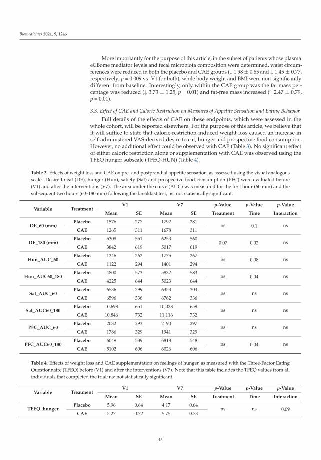
Biomedicines 2021, 9, 1246
More importantly for the purpose of this article, in the subset of patients whose plasmaeCBome mediator levels and fecal microbiota composition were determined, waist circum-ferences were reduced in both the placebo and CAE groups (↓ 1.98 ± 0.65 and ↓ 1.45 ± 0.77,respectively; p = 0.009 vs. V1 for both), while body weight and BMI were non-significantlydifferent from baseline. Interestingly, only within the CAE group was the fat mass per-centage was reduced (↓ 3.73 ± 1.25, p = 0.01) and fat-free mass increased (↑ 2.47 ± 0.79,p = 0.01).
3.3. Effect of CAE and Caloric Restriction on Measures of Appetite Sensation and Eating Behavior
Full details of the effects of CAE on these endpoints, which were assessed in thewhole cohort, will be reported elsewhere. For the purpose of this article, we believe thatit will suffice to state that caloric-restriction-induced weight loss caused an increase inself-administered VAS-derived desire to eat, hunger and prospective food consumption.However, no additional effect could be observed with CAE (Table 3). No significant effectof either caloric restriction alone or supplementation with CAE was observed using theTFEQ hunger subscale (TFEQ-HUN) (Table 4).
Table 3. Effects of weight loss and CAE on pre- and postprandial appetite sensation, as assessed using the visual analogousscale. Desire to eat (DE), hunger (Hun), satiety (Sat) and prospective food consumption (PFC) were evaluated before(V1) and after the interventions (V7). The area under the curve (AUC) was measured for the first hour (60 min) and thesubsequent two hours (60–180 min) following the breakfast test; ns: not statistically significant.
Variable TreatmentV1 V7 p-Value p-Value p-Value
Mean SE Mean SE Treatment Time Interaction
DE_60 (mm)Placebo 1576 277 1792 281
ns 0.1 nsCAE 1265 311 1678 311
DE_180 (mm)Placebo 5308 551 6253 560
0.07 0.02 nsCAE 3842 619 5017 619
Hun_AUC_60Placebo 1246 262 1775 267
ns 0.08 nsCAE 1122 294 1401 294
Hun_AUC60_180Placebo 4800 573 5832 583
ns 0.04 nsCAE 4225 644 5023 644
Sat_AUC_60Placebo 6536 299 6353 304
ns ns nsCAE 6596 336 6762 336
Sat_AUC60_180Placebo 10,698 651 10,028 659
ns ns nsCAE 10,846 732 11,116 732
PFC_AUC_60Placebo 2032 293 2190 297
ns ns nsCAE 1786 329 1941 329
PFC_AUC60_180Placebo 6049 539 6818 548
ns 0.04 nsCAE 5102 606 6026 606
Table 4. Effects of weight loss and CAE supplementation on feelings of hunger, as measured with the Three-Factor EatingQuestionnaire (TFEQ) before (V1) and after the interventions (V7). Note that this table includes the TFEQ values from allindividuals that completed the trial; ns: not statistically significant.
Variable TreatmentV1 V7 p-Value p-Value p-Value
Mean SE Mean SE Treatment Time Interaction
TFEQ_hungerPlacebo 5.96 0.64 4.17 0.64
ns ns 0.09CAE 5.27 0.72 5.75 0.73
45

Biomedicines 2021, 9, 1246
We then sought to investigate whether these observations could also be reported in thesubset of participants that had both their plasma eCBome and fecal microbiota compositioncharacterized (Table 5). Pre-meal evaluation of the amount of food that could be eaten(DQuaN) was drastically increased by weight loss that was either induced by caloric restric-tion alone or in conjunction with CAE supplementation. The pre-meal feeling of hunger(BSenF) increased during caloric restriction-induced weight loss (placebo V1 vs. V7). TheCAE supplementation did not seem to influence any subjective measures of hunger orsatiety. Conversely, susceptibility to hunger, as derived from the TFEQ hunger subscale(TFEQ-HUN) was reduced by caloric restriction only (placebo V1 vs. V7), which was aneffect that seemed to be due to reduced feelings of hunger arising from internal sources(TFEQ-HIL).
Table 5. Effects of weight loss and CAE supplementation on appetite sensations in self-administered VAS-derived andThree-Factor Eating Questionnaire (TFEQ) measured variables before (V1) and after the interventions (V7). DQuaN,pre-meal evaluation of the amount of food that could be eaten; BEnvM, pre-meal desire to eat; BSenF, feeling of hunger;TFEQ-HUN, habitual feelings of hunger; TFEQ-HEL, feelings of hunger arising from external sources; TFEQ-HIL, feelingsof hunger arising from internal sources. This table represents the measurements made in the subset of participants that hadboth their plasma eCBome and fecal microbiota composition characterized. Effects of the Capsimax supplementation onappetite sensations were measured before (0) and 30 or 180 min following breakfast; ns: not statistically significant.
Variable TreatmentV1 V7 p-Value p-Value p-Value
Mean SE Mean SE Treatment Time Interaction
DQuaN0Placebo 28.1 5.0 104.6 4.9 ns <0.001 ns
CAE 26.6 5.9 99.8 4.7 ns <0.001 ns
DQuaN180Placebo 78.3 6.8 80.5 4.8 ns ns ns
CAE 70.6 6.8 76.7 5.2 ns ns ns
BEnvM_30Placebo 85.9 6.9 99.9 3.6 ns ns ns
CAE 82.6 7.2 84.5 6.7 ns ns ns
BEnvM180Placebo 39.2 5.9 40.5 4.7 ns ns ns
CAE 43.5 6.5 42.2 6.8 ns ns ns
BSenF_30Placebo 81.3 7.1 94.4 5.6 ns ns ns
CAE 86.7 6.9 80.8 6.0 ns ns ns
BSenF180Placebo 34.2 5.1 42.7 4.5 ns ns ns
CAE 47.2 6.6 39.7 5.4 ns ns ns
TFEQ-HUNPlacebo 5.5 0.7 4.2 0.5 ns 0.04 ns
CAE 5.7 0.7 5.9 0.9 ns ns ns
TFEQ-HILPlacebo 1.8 0.4 1.3 0.3 ns 0.05 ns
CAE 1.8 0.4 1.9 0.5 ns ns ns
TFEQ-HELPlacebo 2.4 0.3 1.8 0.3 ns ns ns
CAE 2.7 0.3 2.7 0.4 ns ns ns
3.4. Effect of CAE and Caloric Restriction on Plasma ECBome Mediator Levels
As mentioned above, this endpoint could be measured only in a relatively smallsubset of the participants from the larger study (i.e., 13 placebo and 10 CAE); nevertheless,its assessment was one of the two major objectives of this article. Caloric restrictionalone (placebo V1 vs. V7) induced important reductions in the plasma levels of N-oleoyl-ethanolamine (OEA), N-oleoyl-ethanolamine (LEA), N-docosapentaenoyl-ethanolamine(DPEA) and 1,2-oleoyl-glycerol (1/2-OG), as well as in the levels of the omega-3 fatty
46

Biomedicines 2021, 9, 1246
acid docosahexaenoic acid and its eCBome derivative N-docosahexaenoyl-ethanolamine(DHEA) (Figure 1). However, the addition of CAE prevented all these decreases.
Figure 1. Effect of caloric restriction without or with CAE on plasma eCBome mediator levels. eCBome mediator levelsin the plasma of a subset of 23 participants (13 placebo and 10 CAE) before (visit 1 (V1)) and after (visit 7 (V7)) theinterventions. The levels of the eCBome-related mediators measured using HPLC-MS/MS are expressed in femtomolesper microlitre of plasma. Each median is represented by the middle line of each boxplot, Q1 and Q3 are representedby the bottom and top of the boxes, respectively, and whiskers denote 1.5 × IQR. OEA, N-oleoyl-ethanolamine; LEA,N-linoleoyl-ethanolamine; DPEA, N-docosapentaenoyl-ethanolamine; 1/2-OG, 1/2-oleoyl-glycerol; DHA, docosahexaenoicacid; DHEA, N-docosahexaenoyl-ethanolamine.
3.5. Effect of CAE and Caloric Restriction on Fecal Microbiota Composition
As mentioned above, this endpoint could be measured only in a relatively small subsetof the participants to the larger study (i.e., 9 placebo and 6 CAE), where its assessmentwas the other major objective of this article. Caloric restriction caused an increase in thealpha diversity of the fecal microbiota at V7, which was, however, counteracted by CAEadministration, possibly also due to a trending difference between the V1 placebo andV1 CAE groups (Figure 2A). The Firmicutes/Bacteriodetes ratio was not significantlyreduced following caloric restriction, either in the presence or absence of CAE (Figure 2B).Accordingly, no changes in the relative abundance of fecal microbiota phyla were observed
47

Biomedicines 2021, 9, 1246
following caloric restriction. However, a trend (p = 0.07) toward increased Bacteroideteswas observed following CAE administration (Figure 2C). At the class level, Clostridia(p = 0.043) and Actinobacteria (p = 0.021) were increased at visit 7 with CAE as compared tothe placebo controls (Figure 2D). Finally, and perhaps more importantly, at the genus level,Flavonifractor was increased by CAE over the course of the treatment but not by caloricrestriction alone (Figure 2E). However, Faecalibacterium, Ruminococcaceae_UCG-004 andRuminococcaceae_UCG-010 showed significant differences in their relative abundances atbaseline (V1) between placebo and CAE groups, which may have obfuscated CAE-inducedeffects (Figure S1). Nevertheless, Faecalibacterium showed a trend toward significance(p = 0.14) for increased abundance in response to CAE treatment (V1 vs. V7).
Figure 2. Effect of caloric restriction without or with CAE on the fecal microbiota composition. Alphadiversity of the fecal microbiota (A), Firmicutes:Bacteriodetes ratio (B) and the relative abundances offecal microbiota phyla (C), classes (D) and the Flavonifractor genera (E). The data were obtained fromstool samples of a subset of 25 (19 placebo and 6 CAE) reproductive-aged women with overweight orobesity before (visit 1 (V1)) and after (visit 7 (V7)) the interventions.
48
Biomedicines 2021, 9, 1246
3.6. Correlations between Plasma ECBome Mediator Levels, Anthropometric Measures and FecalMicrobiota Composition
Next, we assessed whether any correlation existed between the plasma levels ofeCBome mediators and other outcome measures within individuals for which the appropri-ate data were collected. Several eCBome mediators correlated with the relative abundancesof the phyla, families and genera of the fecal microbiota (Figure 3). In particular, among thepyla, MAGs appeared to correlate negatively with Actinobacteria and Firmicutes (in thecase of 1/2-OG and 1/2-DPG the correlations were statistically significant), whereas NAEscorrelated positively with Bacteroidetes (significantly for the endocannabinoid anandamide(AEA) and for DHEA), Synergistetes (significantly for AEA, OEA, LEA and DHEA) andVerrucomicrobia (significantly for DHEA, N-palmitoyl-ethanolamine (PEA) and N-stearoyl-ethanolamine (SEA)). Significant lipid-class-independent correlations were also found withsome phyla, i.e., for Cyanobacteria (negative), which was only significantly correlated withthe LEA levels, and Verrucomicrobia (positive), which was significantly correlated with theendocannabinoid 2-arachidonoyl-glycerol (2-AG) (calculated together with its 1-isomer(1/2-AG)) and several NAEs (Figure 3A). Stronger correlations were found at the familyand genus levels (Figure 3B,C). Most relevant correlations, namely, those that occurredbetween the eCBome mediators that were found here to be differentially affected by caloricrestriction and CAE treatment (Figure 1), include: (1) At the family level, Akkermansiaceaepositively correlated with DHEA; Desulfovibrionaceae with DPEA and DHEA; Synergis-taceae with OEA, LEA, DPEA and DHEA; and Tannerellaceae with OEA, DHEA and 2-OG.Meanwhile, Atopobiaceae and Prevotellaceae correlated negatively with 2-OG. (2) At thegenus level, Akkermansia, Coprococcus 2, Escherichia/Shigetta, Lachnospiraceae UCG-008and Merdibacter positively correlated with PEA, Bacteroides, Erysipelotrichaceae UCG003,Dialister, Flavonifractor, Parabacteroides and 2-OG; Marvinbryantia with OEA and DHEA;Marvinbryantia and Ruminococcus 2 with LEA; Parabacteroides and the RuminococcaceaeNK4A214 group with DHEA; and Turicibacter with 2-OG.
We also found significant correlations between several fecal microbiota taxa and bodyweight, BMI and fat mass (FM) (Figure S2). Interestingly, families that negatively correlatedwith BMI, weight and FM were positively correlated with fat-free mass (FFM) (even whenonly non-statistically significant trends were observed and vice versa). Therefore, some ofthe above-mentioned correlations might, at least in part, be mediated by variations of theseanthropometric measures. However, of the taxa mentioned above, only the Akkermansiagenus (from the Verrucomicrobia phylum and Akkermansiaceae family) was correlated(negatively) with body weight, BMI and fat mass (Figure S2C), and not with the eCBomemediators that correlated with these parameters (except for 1/2-AG). This suggests thatthese correlations were independent of the variations in these anthropometric measures.Conversely, the positive correlations between Akkermansia (and associated phylogeniclevels) and 1/2-AG levels (Figure 3B) suggest that this mediator correlated negatively withBMI and body weight (Figure S3).
49

Biomedicines 2021, 9, 1246
Figure 3. Correlations between the plasma levels of eCBome mediators and the relative abundance of phyla (A), families (B)and genera (C) of the fecal microbiota of 15 (9 placebo and 6 CAE) reproductive-aged women with overweight or obesity.The correlation analysis was performed using repeated measures correlation tests (rmcorr package). * indicates a correlationcoefficient with p-value < 0.05.
50

Biomedicines 2021, 9, 1246
3.7. Correlations between Plasma ECBome Mediator Levels and Appetite Sensations andEating Behaviors
Our investigation of the associations between plasma eCBome mediator levels andappetite and satiety measures were motivated by previously reported roles of some eCBomemediators and receptors on regulating food intake [51] (Figure 4). The amount of food thatcould be eaten (DQuaN) was inversely related to levels of LEA, while pre-meal desire toeat (BEnvM) and feeling of hunger (BSenF) were inversely related to DPA and both DPAand DHA, respectively.
Figure 4. Correlations between the plasma levels of eCBome mediators and the appetite and satietymeasures of 15 (9 placebo and 6 CAE) reproductive-aged women with overweight or obesity. Thecorrelation analysis was performed using repeated measures correlation tests (rmcorr package). Onlycorrelation coefficients with p-value < 0.05 are displayed. DQuaN, pre-meal evaluation of the amountof food that could be eaten; BEnvM, pre-meal desire to eat; BSenF, feeling of hunger; TFEQ-HUN,habitual feelings of hunger; TFEQ-HIL, feelings of hunger arising from internal sources. * indicates acorrelation coefficient with p-value < 0.05.
A habitual feeling of hunger (TFEQ-HUN) was inversely related to circulating lev-els of 1/2-LG and subscale of hunger and hunger from internal (e.g., affect, arousal orstress) cues (TFEQ-HUN and HIL) were positively correlated to the levels of long-chainomega-3 precursors EPA and DPA. Conversely, hunger from external food cues, whichare mostly related to situations or objects that have previously been associated with foodintake, did not show any correlation with the mediators analyzed.
4. Discussion
The main novel result of this exploratory study is that a capsaicinoid-based sup-plement produced effects on the eCBome and the gut microbiota, potentially facilitatingan amelioration of the metabolic status in women with overweight and obesity. Theseeffects may represent early adaptations that could ultimately result in beneficial effectson food intake, body weight and fat accumulation, as possibly suggested by the presentfinding showing that, in the subset of patients that underwent plasma eCBome and gutmicrobiota assessment, CAE performed better than the placebo at enhancing caloric-restriction-induced fat mass reduction. However, in the whole cohort (whose plasmaeCBome and fecal microbiome could not be entirely assessed for logistical reasons), CAEenhanced the effects of caloric restriction on body weight, waist circumference and fatmass only in a non-statistically significant manner. It is possible that either a more or aless pronounced caloric-restriction-induced weight/fat loss must be achieved in order toallow for the beneficial effects on the eCBome and gut microbiota that are induced byCAE to produce a stronger impact also on these two anthropometric measures. Indeed,previous studies showed that the same CAE used here, under the same dosing protocol,does produce reductions in body weight and fat mass in a larger cohort of both male andfemale individuals with lower starting BMI and in the absence of concomitant caloric
51
Biomedicines 2021, 9, 1246
restriction [52,53]. Future clinical studies, as well as a more thorough examination of thedata from the whole cohort than that performed in this article, which was focused on theeffects of CAE on the plasma eCBome and fecal microbiota composition, need to be awaitedin order to draw any definitive conclusion as to what extent these natural products can beused as a therapy against obesity and hyperphagia.
Indeed, capsaicin is one of the most studied natural products and food (spice) com-ponents in the context of metabolic disorders and related cardiovascular risk [3,54,55].Epidemiological studies suggested that hot chili pepper consumption may positively af-fect some parameters of the metabolic syndrome, particularly hypertension and bodyfat [12,56]. A meta-analysis of clinical studies supported the notion that, especially at highdoses, capsaicin positively affects energy expenditure and appetite regulation, thoughcontradictory results were often observed in studies with subjects living with overweightand obesity [57]. Several preclinical studies with oral capsaicin supported the possible useof this compound to treat metabolic disorders, also due to effects on food intake, satiety andenergy expenditure [28,58,59]. In some of these animal studies, capsaicin effects were as-cribed, at least in part, to its capability of modifying the composition of the gut microbiota,for example, in a manner that enhances short-chain fatty acid-producing bacteria [39,40,60].In one case, these beneficial and gut-microbiota-mediated effects of capsaicin were shownto be due to counteracting the CB1 overactivation by endocannabinoids in obesity [39].However, in only a few of these experimental studies, the role of TRPV1, which is the majortarget for capsaicin, was investigated and demonstrated [41], despite the fact that also thisligand-activated channel is known to be strongly implicated in several aspects of energymetabolism control, ranging from food intake and satiety mechanisms to adipogenesis,white adipocyte browning and brown adipocyte activation [25,61–64]. In the present study,we assessed the effects of a 12 weeks dietary supplementation of CAE (equivalent to 4 mgcapsaicinoids/day, of which 2.4 mg were capsaicin, 1.4 mg were dihydrocapsaicin and0.2 mg were nordihydrocapsaicin) in reproductive-aged women with overweight andobesity undergoing a calorie-reduced diet (−500 kcal/day) on the gut microbiota and theexpanded endocannabinoid system known as the eCBome. These are two major playersthat control energy homeostasis, independent from their effects on body weight and foodintake, and that are disrupted in obesity and other metabolic disorders [32,37]. We reportfor the first time that CAE inhibited changes in circulating eCBome lipids induced by thecaloric restriction in control participants and induced alterations in the gut microbiotain directions that may favor beneficial actions on the metabolic consequences of obesity.Indeed, some eCBome mediators, such as OEA and other non-CB1-activating NAEs, canbe considered as biomarkers of metabolic health since their endogenous concentrations inblood or tissues correlate with, among others, increased lipolysis and insulin sensitivity andreduced body fat, dyslipidemia and systemic inflammation, with or without body-weight-reducing interventions [65–67], as well as with the dietary intake of the corresponding fattyacids [68].
As mentioned above, capsaicin was shown in experimental models of obesity toaffect both endocannabinoid signaling by counteracting CB1 activity, and gut microbiotacomposition [39,60]. TRPV1 activation by this compound in isolated cells expressing thehuman recombinant protein was found to lead to the biosynthesis of NAEs, includingAEA, OEA and PEA [30]. More recently, TRPV1 activation was also shown to increaseMAG levels [69]. Here we observed that CAE counteracted a time-dependent reductionin the circulating levels of NAEs observed in placebo-treated women. In particular, thetreatment reversed the reduction in the levels of the anorectic and lipolysis-inducing andenergy-expenditure-inducing mediator OEA, which acts via the activation of TRPV1, aswell as of the pro-lipolytic peroxisome proliferator-activated receptor α (PPARα) and,possibly, the GLP-1-release stimulating GPCR, GPR119 [70–72], with no effect on AEA noron the other CB1-activating mediator, 2-AG. CAE also counteracted the reduction of thelevels of LEA and 2-OG, two other endogenous TRPV1 and GPR119 agonists [73,74], and ofthe anti-inflammatory NAEs, namely, DHEA and DPEA [74–76]. The decrease in non-CB1
52
Biomedicines 2021, 9, 1246
activating NAEs by caloric restriction is likely to represent an adaptive, negative feedbackmechanism, and was observed for OEA also in the small intestine of rats following fooddeprivation [77]. By being partly in agreement with the aforementioned in vitro studiesshowing that TRPV1 activation by capsaicin stimulates NAE biosynthesis [30,69], ourpresent findings may suggest that, in the subset of participants of our cohort in whomthe plasma eCBome was measured, capsaicinoids, administered as CAE, were acting viaTRPV1 channels. More importantly, these findings indicate that CAE positively affectedthe peripheral levels of metabolically beneficial NAEs without changing those of CB1-activating, and hence potentially obesity-exacerbating, endocannabinoids, i.e., AEA and2-AG.
Several studies have linked the anti-obesity effects of capsaicin to modulation of thegut microbiome, including increasing the abundance of the “anti-obesity” species Akker-mansia muciniphila [40,60,78]. Results from faecal microbiota transplantation, as well asantibiotics treatments experiments, suggest that capsaicin-induced anti-metabolic endotox-emia effects were at least partly dependent on modulation of the microbiome in mice [39].CAE also produced some effects on gut microbiota composition in our study by bothcounteracting the increased diversity observed with caloric restriction only and affectingthe relative abundance of some taxa. In particular, it tended to increase the Bacteroidetesphylum, increase the Clostridia class and increase the Flavonifractor genus. While the formertwo might exert beneficial metabolic effects, the increase in Flavonifractor might relate to thecapability of this genus to metabolize flavonoids and, in particular, curcumin, with whichcapsaicin has structural homology. Accordingly, dietary curcumin or curcumin-containingturmeric were also shown to increase the abundance of Flavonifractor sp. in human fecal mi-crobiota in vitro [79] or in vivo [80]. Species belonging to this genus, such as F. plautii, weresuggested to be decreased in obesity/increased in leanness [81] and play anti-inflammatoryactions [82]. Interestingly, it appears that an individual’s gut microbiota composition canmodulate the response to capsaicin. In a trial involving healthy adults, it was observedthat a broad separation of participants into Bacteroides and Prevotella enterotypes similarlystratified the participants into either strong or weak capsaicin responders, respectively,with respect to capsaicin-induced increases in Faecalibacterium abundance and serum in-cretin (GIP and GLP-1) levels [83]. While we did observe a trend toward an increasein Faecalibacterium abundance in response to CAE (data not shown; p = 0.14, V1 vs. V7CAE), the results may have been confounded by the fact that baseline Faecalibacteriumabundance was significantly lower in the CAE V1 group as compared to the placebo V1group (p = 0.014). We previously proposed that this difference between enterotypes may bedue to microbiome-mediated modulation of TRPV1 responsiveness to capsaicin [37], sinceit was shown that the probiotic Lactobacillus reuteri can inhibit TRPV1 activity in mesentericneurons [84]. Thus, the relatively small changes in the gut microbiome that we observedhere may in part be due to the participants harboring capsaicin-resistant microbiomes,which may be generalizable and explain why capsaicin effects on energy expenditure werereported to be blunted in obese as compared to lean individuals [57]. Nevertheless, wewere expecting to see stronger effects of CAE on fecal microbiota composition. It is possiblethat some effects, as mentioned above, were masked by the fact that the relative abundanceof some taxa were different at the baseline (V1) in the two control groups, which was a lim-itation of this study. However, in agreement with previous studies showing the existenceof a bilateral communication between the host eCBome and the gut microbiome [85], wedid find several correlations between the eCBome mediators whose concentrations in theplasma were modified by CAE and numerous gut bacterial taxa, also at the genus level.Of these, some seemed to be particularly relevant to the potential beneficial metaboliceffects of capsaicin. The most obvious of these was undoubtedly Akkermansia, whichpositively correlated with PEA levels (as did the Akkermansiaceae family). Akkermansiaabundance is decreased in obesity and a recent clinical trial showed that supplementation,especially with heat-inactivated Akkermansia, improved metabolic parameters in obeseparticipants, including decreasing hepatic inflammation markers [86]. Interestingly, PEA is
53

Biomedicines 2021, 9, 1246
a potent anti-inflammatory compound with reported metabolic benefits in models of obe-sity, including a reduction in high-fat diet-induced liver inflammation [87,88]. In contrast,Enterobacteriaceae, a lipopolysaccharide-producing family that is associated with obesityand metabolic endotoxemia [89], was also positively correlated with PEA. Flavonifractor,the only genus that was found to be specifically upregulated by CAE in our study, waspositively associated with levels of the GPR119 agonist 2-OG (see above). Flavonifractorwas found to be associated with non-obese as compared to obese individuals and, whenprovided to mice on a high-fat diet, it attenuated adipose tissue inflammation [81,82].Similarly, Parabacteroides and Dialister were also correlated with 2-OG, and different speciesof the former were tied to the beneficial metabolic effects of fiber and to ameliorate diet-induced metabolic dysfunction [90,91], while the latter was associated with improvedinsulin sensitivity [92], though paradoxically also with resistance to weight loss in responseto a volumetric diet [93]. Taken together, it is clear that the assessment of the significanceof these correlations will require further investigation in order to understand whether theyhave any causal effects on obesity and associated complications.
The CAE-encapsulating technology used here allowed for a controlled release thatdelivered effective levels of capsaicinoids in the small intestine without the stomach upsetthat may result from unprotected extracts of red chili peppers. Although we did notaim to compare the difference between such technology and capsaicinoids ingested assupplements with some specific foods, we believe that it is possible that such differencemight exist. In particular, capsule release in the small intestine may bypass mouth- andstomach-specific activation of TRPV1 and modify the sensory detection of taste differencesin the mouth.
5. Conclusions
In summary, in the present exploratory clinical study, the oral administration of CAEto reproductive-aged women living with overweight and obesity produced importantchanges in the plasma eCBome, which may be indicative of potential beneficial effects atthe level of hyperphagia (through increased levels of an anorectic factor, such as OEA),insulin sensitivity (through increases of endogenous agonists of GPR119) and inflammation(through the increase of anti-inflammatory mediators like DPEA and DHEA). However,while we did not see any significant correlation between eCBome mediators and theappetite and satiety measures, here we also did not assess measures of insulin sensitivityor chronic inflammation. This, together with the relatively small number of participantsfrom whom we were allowed to sample blood and stools for eCBome and gut microbiomemeasurements, represent limitations of this study. Nevertheless, we suggest that some ofthe observed effects of CAE on eCBome mediators and the gut microbiome, the latter ofwhich probably also represents an attempt of the gut microbial ecosystem to deal withcapsaicin and its metabolism, may contribute to potential metabolically beneficial effects ofthese natural products. In agreement with this hypothesis, in the subset of patients thatunderwent plasma eCBome and fecal microbiota assessments, CAE performed significantlybetter than the placebo at reducing fat mass. The finding of strong correlations betweenseveral eCBome mediators and fecal microbiota taxa should also be considered in thiscontext and reinforces the hypothesis of the existence of a connection between these twometabolically relevant signaling systems in the framework of overweight and obesity.
Supplementary Materials: The following are available online at https://www.mdpi.com/article/10.3390/biomedicines9091246/s1. Table S1: Reported symptoms and inventory of collected samples,Table S2: List of the lipid mediators used as deuterated internal standards for LC/MS-MS analyses,Figure S1: Effect of Capsimax and caloric restriction on relative abundance of fecal bacterial genera,Figure S2: Correlation between fecal microbiota taxa at phylum (A), classed (B) and genera (C)levels and BMI, waist circumference, fat mass (FM) and fat-free mass (FFM), Figure S3: Correlationsbetween the plasma levels of eCBome mediators and BMI, waist circumference, fat mass (FM) andfat-free mass (FFM).
54

Biomedicines 2021, 9, 1246
Author Contributions: Conceptualization, V.D., A.T., V.D.M. and C.S.; Data curation, C.M., S.L., N.F.and Y.C.; Formal analysis, C.M., S.L., N.F. and Y.C.; Methodology, F.P. and V.D.; Writing—originaldraft, C.M., S.L., A.T., V.D.M. and C.S.; Writing—review and editing, C.M., S.L., F.P., N.F., Y.C., V.D.,A.T., V.D.M. and C.S. All authors have read and agreed to the published version of the manuscript.
Funding: This work was supported by the Canada Research Excellence Chair in the Microbiome-Endocannabinoidome Axis in Metabolic Health (CERC-MEND), which is funded by the Tri-Agencyof the Canadian Federal Government (The Canadian Institutes of Health Research (CIHR), theNatural Sciences and Engineering Research Council of Canada (NSERC) and the Social Sciences andHumanities Research Council of Canada (SSHRC)) to V.D., as well as by the Canadian Foundation ofInnovation (to V.D.) and the Sentinelle Nord-Apogée program (to Université Laval). C.M. receiveda post-doctoral grant from the Joint International Research Unit for Chemical and BiomolecularResearch on the Microbiome and its impact on Metabolic Health and Nutrition (UMI-MicroMeNu),which is partly supported by the Sentinelle Nord/Apogée program.
Institutional Review Board Statement: This study was conducted according to the guidelines laiddown in the Declaration of Helsinki and all procedures involving human subjects in the studyprotocol were approved by the Research Ethics Committee of Health Sciences of Laval University(protocol 2015-041 A-4 R-2/01-05-2018). The clinical trial was registered in the public trials registryClinicalTrials.gov (accessed on 30 July 2021) (registration identification number NCT04874701).
Informed Consent Statement: Written informed consent was obtained from all the participantsbefore entering the study.
Data Availability Statement: Data is contained within the article or Supplementary Materials.
Acknowledgments: The authors wish to thank Cyril Martin for his help with the LC–MS/MSmeasurements.
Conflicts of Interest: The authors declare no conflict of interest. The funders had no role in the designof the study; in the collection, analyses, or interpretation of data; in the writing of the manuscript; orin the decision to publish the results.
References
1. Non Communicable Diseases. Available online: https://www.who.int/news-room/fact-sheets/detail/noncommunicable-diseases (accessed on 26 January 2021).
2. Obesity and Overweight. Available online: https://www.who.int/news-room/fact-sheets/detail/obesity-and-overweight(accessed on 26 January 2021).
3. Zheng, J.; Zheng, S.; Feng, Q.; Zhang, Q.; Xiao, X. Dietary Capsaicin and Its Anti-Obesity Potency: From Mechanism to ClinicalImplications. Biosci. Rep. 2017, 37. [CrossRef]
4. Hill, J.O.; Wyatt, H.R.; Peters, J.C. Energy Balance and Obesity. Circulation 2012, 126, 126–132. [CrossRef]5. Hruby, A.; Hu, F.B. The Epidemiology of Obesity: A Big Picture. Pharmacoeconomics 2015, 33, 673–689. [CrossRef]6. Yanovski, S.Z.; Yanovski, J.A. Long-Term Drug Treatment for Obesity: A Systematic and Clinical Review. JAMA 2014, 311, 74.
[CrossRef]7. Filippatos, T.D.; Derdemezis, C.S.; Gazi, I.F.; Nakou, E.S.; Mikhailidis, D.P.; Elisaf, M.S. Orlistat-Associated Adverse Effects and
Drug Interactions: A Critical Review. Drug Saf. 2008, 31, 53–65. [CrossRef] [PubMed]8. Wilding, J.P.H.; Batterham, R.L.; Calanna, S.; Davies, M.; van Gaal, L.F.; Lingvay, I.; McGowan, B.M.; Rosenstock, J.; Tran, M.T.D.;
Wadden, T.A.; et al. Once-Weekly Semaglutide in Adults with Overweight or Obesity. N. Engl. J. Med. 2021. [CrossRef] [PubMed]9. Dietrich, M.O.; Horvath, T.L. Limitations in Anti-Obesity Drug Development: The Critical Role of Hunger-Promoting Neurons.
Nat. Rev. Drug Discov. 2012, 11, 675–691. [CrossRef] [PubMed]10. Liu, J.; Lee, J.; Salazar Hernandez, M.A.; Mazitschek, R.; Ozcan, U. Treatment of Obesity with Celastrol. Cell 2015, 161, 999–1011.
[CrossRef] [PubMed]11. Martel, J.; Ojcius, D.M.; Chang, C.-J.; Lin, C.-S.; Lu, C.-C.; Ko, Y.-F.; Tseng, S.-F.; Lai, H.-C.; Young, J.D. Anti-Obesogenic and
Antidiabetic Effects of Plants and Mushrooms. Nat. Rev. Endocrinol. 2017, 13, 149–160. [CrossRef]12. Varghese, S.; Kubatka, P.; Rodrigo, L.; Gazdikova, K.; Caprnda, M.; Fedotova, J.; Zulli, A.; Kruzliak, P.; Büsselberg, D. Chili Pepper
as a Body Weight-Loss Food. Int. J. Food Sci. Nutr. 2017, 68, 392–401. [CrossRef]13. Richards, B.L.; Whittle, S.L.; Buchbinder, R. Neuromodulators for Pain Management in Rheumatoid Arthritis. Cochrane Database
Syst. Rev. 2012, 1. [CrossRef]14. Zheng, L.; Chen, J.; Ma, Z.; Liu, W.; Yang, F.; Yang, Z.; Wang, K.; Wang, X.; He, D.; Li, L.; et al. Capsaicin Enhances Anti-
Proliferation Efficacy of Pirarubicin via Activating TRPV1 and Inhibiting PCNA Nuclear Translocation in 5637 Cells. Mol. Med.Rep. 2016, 13, 881–887. [CrossRef]
55

Biomedicines 2021, 9, 1246
15. Liu, Y.-P.; Dong, F.-X.; Chai, X.; Zhu, S.; Zhang, B.-L.; Gao, D.-S. Role of Autophagy in Capsaicin-Induced Apoptosis in U251Glioma Cells. Cell. Mol. Neurobiol. 2016, 36, 737–743. [CrossRef]
16. Dairam, A.; Fogel, R.; Daya, S.; Limson, J.L. Antioxidant and Iron-Binding Properties of Curcumin, Capsaicin, and S -AllylcysteineReduce Oxidative Stress in Rat Brain Homogenate. J. Agric. Food Chem. 2008, 56, 3350–3356. [CrossRef]
17. Singh, U.; Bernstein, J.A. Intranasal Capsaicin in Management of Nonallergic (Vasomotor) Rhinitis. Prog Drug Res. 2014, 68,147–170. [CrossRef] [PubMed]
18. Tremblay, A.; Arguin, H.; Panahi, S. Capsaicinoids: A Spicy Solution to the Management of Obesity? Int J. Obes 2016, 40,1198–1204. [CrossRef] [PubMed]
19. Yoshioka, M.; St-Pierre, S.; Drapeau, V.; Dionne, I.; Doucet, E.; Suzuki, M.; Tremblay, A. Effects of Red Pepper on Appetite andEnergy Intake. Br. J. Nutr. 1999, 82, 115–123. [CrossRef] [PubMed]
20. Westerterp-Plantenga, M.S.; Smeets, A.; Lejeune, M.P.G. Sensory and Gastrointestinal Satiety Effects of Capsaicin on Food Intake.Int. J. Obes. 2005, 29, 682–688. [CrossRef]
21. Yoshioka, M.; Doucet, E.; Drapeau, V.; Dionne, I.; Tremblay, A. Combined Effects of Red Pepper and Caffeine Consumption on24 h Energy Balance in Subjects given Free Access to Foods. Br. J. Nutr. 2001, 85, 203–211. [CrossRef]
22. Wahlqvist, M.L.; Wattanapenpaiboon, N. Hot Foods—Unexpected Help with Energy Balance? Lancet 2001, 358, 348–349.[CrossRef]
23. Lejeune, M.P.G.M.; Kovacs, E.M.R.; Westerterp-Plantenga, M.S. Effect of Capsaicin on Substrate Oxidation and Weight Mainte-nance after Modest Body-Weight Loss in Human Subjects. Br. J. Nutr. 2003, 90, 651–659. [CrossRef] [PubMed]
24. Kelava, L.; Nemeth, D.; Hegyi, P.; Keringer, P.; Kovacs, D.K.; Balasko, M.; Solymar, M.; Pakai, E.; Rumbus, Z.; Garami, A.Dietary Supplementation of Transient Receptor Potential Vanilloid-1 Channel Agonists Reduces Serum Total Cholesterol Level: AMeta-Analysis of Controlled Human Trials. Crit. Rev. Food Sci. Nutr. 2021, 1–11. [CrossRef]
25. Chen, J.; Li, L.; Li, Y.; Liang, X.; Sun, Q.; Yu, H.; Zhong, J.; Ni, Y.; Chen, J.; Zhao, Z.; et al. Activation of TRPV1 Channel byDietary Capsaicin Improves Visceral Fat Remodeling through Connexin43-Mediated Ca2+ Influx. Cardiovasc. Diabetol. 2015, 14,22. [CrossRef]
26. Sun, F.; Xiong, S.; Zhu, Z. Dietary Capsaicin Protects Cardiometabolic Organs from Dysfunction. Nutrients 2016, 8, 174. [CrossRef]27. Gram, D.X.; Ahrén, B.; Nagy, I.; Olsen, U.B.; Brand, C.L.; Sundler, F.; Tabanera, R.; Svendsen, O.; Carr, R.D.; Santha, P.; et al.
Capsaicin-Sensitive Sensory Fibers in the Islets of Langerhans Contribute to Defective Insulin Secretion in Zucker Diabetic Rat, anAnimal Model for Some Aspects of Human Type 2 Diabetes: Systemic Capsaicin in ZDF Rats. Eur. J. Neurosci. 2007, 25, 213–223.[CrossRef] [PubMed]
28. Wang, P.; Yan, Z.; Zhong, J.; Chen, J.; Ni, Y.; Li, L.; Ma, L.; Zhao, Z.; Liu, D.; Zhu, Z. Transient Receptor Potential Vanilloid 1Activation Enhances Gut Glucagon-Like Peptide-1 Secretion and Improves Glucose Homeostasis. Diabetes 2012, 61, 2155–2165.[CrossRef] [PubMed]
29. Kang, J.-H.; Kim, C.-S.; Han, I.-S.; Kawada, T.; Yu, R. Capsaicin, a Spicy Component of Hot Peppers, Modulates Adipokine GeneExpression and Protein Release from Obese-Mouse Adipose Tissues and Isolated Adipocytes, and Suppresses the InflammatoryResponses of Adipose Tissue Macrophages. FEBS Lett. 2007, 581, 4389–4396. [CrossRef]
30. Di Marzo, V.; Lastres-Becker, I.; Bisogno, T.; De Petrocellis, L.; Milone, A.; Davis, J.B.; Fernandez-Ruiz, J.J. Hypolocomotor Effectsin Rats of Capsaicin and Two Long Chain Capsaicin Homologues. Eur. J. Pharmacol. 2001, 420, 123–131. [CrossRef]
31. Acharya, N.; Penukonda, S.; Shcheglova, T.; Hagymasi, A.T.; Basu, S.; Srivastava, P.K. Endocannabinoid System Acts as aRegulator of Immune Homeostasis in the Gut. Proc. Natl. Acad. Sci. USA 2017, 114, 5005–5010. [CrossRef]
32. Silvestri, C.; di Marzo, V. The Endocannabinoid System in Energy Homeostasis and the Etiopathology of Metabolic Disorders.Cell Metab. 2013, 17, 475–490. [CrossRef]
33. Di Marzo, V.; Wang, J. (Eds.) The Endocannabinoidome the World of Endocannabinoids and Related Mediators; Academic Press: London,UK, 2014.
34. Zygmunt, P.M.; Ermund, A.; Movahed, P.; Andersson, D.A.; Simonsen, C.; Jönsson, B.A.G.; Blomgren, A.; Birnir, B.; Bevan, S.;Eschalier, A.; et al. Monoacylglycerols Activate TRPV1—A Link between Phospholipase C and TRPV. PLoS ONE 2013, 8, e81618.[CrossRef]
35. Movahed, P.; Jönsson, B.A.G.; Birnir, B.; Wingstrand, J.A.; Jørgensen, T.D.; Ermund, A.; Sterner, O.; Zygmunt, P.M.; Högestätt,E.D. Endogenous Unsaturated C18 N-Acylethanolamines Are Vanilloid Receptor (TRPV1) Agonists. J. Biol. Chem. 2005, 280,38496–38504. [CrossRef] [PubMed]
36. Cani, P.D.; Plovier, H.; Van Hul, M.; Geurts, L.; Delzenne, N.M.; Druart, C.; Everard, A. Endocannabinoids—At the Crossroadsbetween the Gut Microbiota and Host Metabolism. Nat. Rev. Endocrinol. 2016, 12, 133–143. [CrossRef] [PubMed]
37. Di Marzo, V.; Silvestri, C. Lifestyle and Metabolic Syndrome: Contribution of the Endocannabinoidome. Nutrients 2019, 11, 1956.[CrossRef] [PubMed]
38. Dione, N.; Lacroix, S.; Taschler, U.; Deschênes, T.; Abolghasemi, A.; Leblanc, N.; Di Marzo, V.; Silvestri, C. Mgll Knockout MouseResistance to Diet-Induced Dysmetabolism Is Associated with Altered Gut Microbiota. Cells 2020, 9, 2705. [CrossRef] [PubMed]
39. Kang, C.; Wang, B.; Kaliannan, K.; Wang, X.; Lang, H.; Hui, S.; Huang, L.; Zhang, Y.; Zhou, M.; Chen, M.; et al. Gut MicrobiotaMediates the Protective Effects of Dietary Capsaicin against Chronic Low-Grade Inflammation and Associated Obesity Inducedby High-Fat Diet. mBio 2017, 8, e00470-17. [CrossRef]
56

Biomedicines 2021, 9, 1246
40. Song, J.-X.; Ren, H.; Gao, Y.-F.; Lee, C.-Y.; Li, S.-F.; Zhang, F.; Li, L.; Chen, H. Dietary Capsaicin Improves Glucose Homeostasisand Alters the Gut Microbiota in Obese Diabetic Ob/Ob Mice. Front. Physiol. 2017, 8, 602. [CrossRef]
41. Kumar, V.; Mahajan, N.; Khare, P.; Kondepudi, K.K.; Bishnoi, M. Role of TRPV1 in Colonic Mucin Production and Gut MicrobiotaProfile. Eur. J. Pharmacol. 2020, 888, 173567. [CrossRef]
42. Drapeau, V.; King, N.; Hetherington, M.; Doucet, E.; Blundell, J.; Tremblay, A. Appetite Sensations and Satiety Quotient: Predictorsof Energy Intake and Weight Loss. Appetite 2007, 48, 159–166. [CrossRef]
43. Arvaniti, K.; Richard, D.; Tremblay, A. Reproducibility of Energy and Macronutrient Intake and Related Substrate OxidationRates in a Buffet-Type Meal. Br. J. Nutr 2000, 83, 489–495. [CrossRef]
44. Green, S.M.; Delargy, H.J.; Joanes, D.; Blundell, J.E. A Satiety Quotient: A Formulation to Assess the Satiating Effect of Food.Appetite 1997, 29, 291–304. [CrossRef]
45. Stunkard, A.J.; Messick, S. The Three-Factor Eating Questionnaire to Measure Dietary Restraint, Disinhibition and Hunger. J.Psychosom. Res. 1985, 29, 71–83. [CrossRef]
46. Cepeda-Benito, A.; Gleaves, D.H.; Fernández, M.C.; Vila, J.; Williams, T.L.; Reynoso, J. The Development and Validation ofSpanish Versions of the State and Trait Food Cravings Questionnaires. Behav. Res. Ther. 2000, 38, 1125–1138. [CrossRef]
47. Turcotte, C.; Archambault, A.; Dumais, É.; Martin, C.; Blanchet, M.; Bissonnette, E.; Ohashi, N.; Yamamoto, K.; Itoh, T.; Laviolette,M.; et al. Endocannabinoid Hydrolysis Inhibition Unmasks That Unsaturated Fatty Acids Induce a Robust Biosynthesis of2-arachidonoyl-glycerol and Its Congeners in Human Myeloid Leukocytes. FASEB J. 2020, 34, 4253–4265. [CrossRef] [PubMed]
48. Callahan, B.J.; McMurdie, P.J.; Rosen, M.J.; Han, A.W.; Johnson, A.J.A.; Holmes, S.P. DADA2: High-Resolution Sample Inferencefrom Illumina Amplicon Data. Nat. Methods 2016, 13, 581–583. [CrossRef] [PubMed]
49. Paulson, J.N.; Stine, O.C.; Bravo, H.C.; Pop, M. Differential Abundance Analysis for Microbial Marker-Gene Surveys. Nat. Methods2013, 10, 1200–1202. [CrossRef] [PubMed]
50. Bakdash, J.Z.; Marusich, L.R. Repeated Measures Correlation. Front. Psychol. 2017, 8, 456. [CrossRef]51. Rochefort, G.; Provencher, V.; Castonguay-Paradis, S.; Perron, J.; Lacroix, S.; Martin, C.; Flamand, N.; Di Marzo, V.; Veilleux, A.
Intuitive Eating Is Associated with Elevated Levels of Circulating Omega-3-Polyunsaturated Fatty Acid-Derived Endocannabi-noidome Mediators. Appetite 2021, 156, 104973. [CrossRef] [PubMed]
52. Urbina, S.L.; Roberts, M.D.; Kephart, W.C.; Villa, K.B.; Santos, E.N.; Olivencia, A.M.; Bennett, H.M.; Lara, M.D.; Foster, C.A.;Purpura, M.; et al. Effects of Twelve Weeks of Capsaicinoid Supplementation on Body Composition, Appetite and Self-ReportedCaloric Intake in Overweight Individuals. Appetite 2017, 113, 264–273. [CrossRef] [PubMed]
53. Rogers, J.; Urbina, S.L.; Taylor, L.W.; Wilborn, C.D.; Purpura, M.; Jäger, R.; Juturu, V. Capsaicinoids Supplementation DecreasesPercent Body Fat and Fat Mass: Adjustment Using Covariates in a Post Hoc Analysis. BMC Obes. 2018, 5, 22. [CrossRef]
54. McCarty, M.F.; DiNicolantonio, J.J.; O’Keefe, J.H. Capsaicin May Have Important Potential for Promoting Vascular and MetabolicHealth. Open Heart 2015, 2, e000262. [CrossRef]
55. Sanati, S.; Razavi, B.M.; Hosseinzadeh, H. A Review of the Effects of Capsicum Annuum L. and Its Constituent, Capsaicin, inMetabolic Syndrome. Iran. J. Basic Med. Sci. 2018, 21, 439. [CrossRef]
56. Shi, Z.; Riley, M.; Brown, A.; Page, A. Chilli Intake Is Inversely Associated with Hypertension among Adults. Clin. Nutr. ESPEN2018, 23, 67–72. [CrossRef] [PubMed]
57. Ludy, M.-J.; Moore, G.E.; Mattes, R.D. The Effects of Capsaicin and Capsiate on Energy Balance: Critical Review and Meta-Analyses of Studies in Humans. Chem. Senses 2012, 37, 103–121. [CrossRef] [PubMed]
58. Kang, J.-H.; Tsuyoshi, G.; Han, I.-S.; Kawada, T.; Kim, Y.M.; Yu, R. Dietary Capsaicin Reduces Obesity-Induced Insulin Resistanceand Hepatic Steatosis in Obese Mice Fed a High-Fat Diet. Obesity 2010, 18, 780–787. [CrossRef] [PubMed]
59. Kwon, D.Y.; Kim, Y.S.; Ryu, S.Y.; Cha, M.-R.; Yon, G.H.; Yang, H.J.; Kim, M.J.; Kang, S.; Park, S. Capsiate Improves GlucoseMetabolism by Improving Insulin Sensitivity Better than Capsaicin in Diabetic Rats. J. Nutr. Biochem. 2013, 24, 1078–1085.[CrossRef]
60. Shen, W.; Shen, M.; Zhao, X.; Zhu, H.; Yang, Y.; Lu, S.; Tan, Y.; Li, G.; Li, M.; Wang, J.; et al. Anti-Obesity Effect of Capsaicin inMice Fed with High-Fat Diet Is Associated with an Increase in Population of the Gut Bacterium Akkermansia Muciniphila. Front.Microbiol. 2017, 8, 272. [CrossRef] [PubMed]
61. Lee, E.; Jung, D.Y.; Kim, J.H.; Patel, P.R.; Hu, X.; Lee, Y.; Azuma, Y.; Wang, H.-F.; Tsitsilianos, N.; Shafiq, U.; et al. TransientReceptor Potential Vanilloid Type-1 Channel Regulates Diet-Induced Obesity, Insulin Resistance, and Leptin Resistance. FASEB J.2015, 29, 3182–3192. [CrossRef] [PubMed]
62. Zhang, L.L.; Yan Liu, D.; Ma, L.Q.; Luo, Z.D.; Cao, T.B.; Zhong, J.; Yan, Z.C.; Wang, L.J.; Zhao, Z.G.; Zhu, S.J.; et al. Activationof Transient Receptor Potential Vanilloid Type-1 Channel Prevents Adipogenesis and Obesity. Circ. Res. 2007, 100, 1063–1070.[CrossRef]
63. Baboota, R.K.; Singh, D.P.; Sarma, S.M.; Kaur, J.; Sandhir, R.; Boparai, R.K.; Kondepudi, K.K.; Bishnoi, M. Capsaicin Induces“Brite” Phenotype in Differentiating 3T3-L1 Preadipocytes. PLoS ONE 2014, 9, e103093. [CrossRef]
64. Baskaran, P.; Krishnan, V.; Ren, J.; Thyagarajan, B. Capsaicin Induces Browning of White Adipose Tissue and Counters Obesity byActivating TRPV1 Channel-Dependent Mechanisms: TRPV1 Activates Browning of WAT to Counter Obesity. Br. J. Pharmacol.2016, 173, 2369–2389. [CrossRef]
57

Biomedicines 2021, 9, 1246
65. Montecucco, F.; Lenglet, S.; Quercioli, A.; Burger, F.; Thomas, A.; Lauer, E.; Silva, D.A.R.; Mach, F.; Vuilleumier, N.; Bobbioni-Harsch, E.; et al. Gastric Bypass in Morbid Obese Patients Is Associated with Reduction in Adipose Tissue Inflammation viaN-Oleoylethanolamide (OEA)-Mediated Pathways. Thromb. Haemost. 2015, 113, 838–850. [CrossRef]
66. Fanelli, F.; Mezzullo, M.; Repaci, A.; Belluomo, I.; Ibarra Gasparini, D.; Di Dalmazi, G.; Mastroroberto, M.; Vicennati, V.;Gambineri, A.; Morselli-Labate, A.M.; et al. Profiling Plasma N-Acylethanolamine Levels and Their Ratios as a Biomarker ofObesity and Dysmetabolism. Mol. Metab. 2018, 14, 82–94. [CrossRef]
67. Azar, S.; Sherf-Dagan, S.; Nemirovski, A.; Webb, M.; Raziel, A.; Keidar, A.; Goitein, D.; Sakran, N.; Shibolet, O.; Tam, J.; et al.Circulating Endocannabinoids Are Reduced Following Bariatric Surgery and Associated with Improved Metabolic Homeostasisin Humans. Obes. Surg. 2019, 29, 268–276. [CrossRef] [PubMed]
68. Castonguay-Paradis, S.; Lacroix, S.; Rochefort, G.; Parent, L.; Perron, J.; Martin, C.; Lamarche, B.; Raymond, F.; Flamand, N.; diMarzo, V.; et al. Dietary Fatty Acid Intake and Gut Microbiota Determine Circulating Endocannabinoidome Signaling beyond theEffect of Body Fat. Sci. Rep. 2020, 10, 15975. [CrossRef] [PubMed]
69. Manchanda, M.; Leishman, E.; Sangani, K.; Alamri, A.; Bradshaw, H.B. Activation of TRPV1 by Capsaicin or Heat Drives Changesin 2-Acyl Glycerols and N-Acyl Ethanolamines in a Time, Dose, and Temperature Dependent Manner. Front. Cell Dev. Biol. 2021,9, 611952. [CrossRef] [PubMed]
70. Ahern, G.P. Activation of TRPV1 by the Satiety Factor Oleoylethanolamide. J. Biol. Chem. 2003, 278, 30429–30434. [CrossRef][PubMed]
71. Lauffer, L.M.; Iakoubov, R.; Brubaker, P.L. GPR119 Is Essential for Oleoylethanolamide-Induced Glucagon-like Peptide-1 Secretionfrom the Intestinal Enteroendocrine L-Cell. Diabetes 2009, 58, 1058–1066. [CrossRef]
72. Im, D.-S. GPR119 and GPR55 as Receptors for Fatty Acid Ethanolamides, Oleoylethanolamide and Palmitoylethanolamide. Int. J.Mol. Sci. 2021, 22, 1034. [CrossRef] [PubMed]
73. Syed, S.K.; Bui, H.H.; Beavers, L.S.; Farb, T.B.; Ficorilli, J.; Chesterfield, A.K.; Kuo, M.-S.; Bokvist, K.; Barrett, D.G.; Efanov,A.M. Regulation of GPR119 Receptor Activity with Endocannabinoid-like Lipids. Am. J. Physiol. Endocrinol. Metab. 2012, 303,E1469–E1478. [CrossRef]
74. Di Marzo, V. New Approaches and Challenges to Targeting the Endocannabinoid System. Nat. Rev. Drug Discov 2018, 17, 623–639.[CrossRef] [PubMed]
75. Figueroa, J.D.; Cordero, K.; Serrano-Illan, M.; Almeyda, A.; Baldeosingh, K.; Almaguel, F.G.; De Leon, M. Metabolomics UncoversDietary Omega-3 Fatty Acid-Derived Metabolites Implicated in Anti-Nociceptive Responses after Experimental Spinal CordInjury. Neuroscience 2013, 255, 1–18. [CrossRef] [PubMed]
76. Wojdasiewicz, P.; Poniatowski, Ł.A.; Turczyn, P.; Frasunska, J.; Paradowska-Gorycka, A.; Tarnacka, B. Significance of Omega-3Fatty Acids in the Prophylaxis and Treatment after Spinal Cord Injury in Rodent Models. Mediat. Inflamm. 2020, 2020, 3164260.[CrossRef] [PubMed]
77. Izzo, A.A.; Piscitelli, F.; Capasso, R.; Marini, P.; Cristino, L.; Petrosino, S.; di Marzo, V. Basal and Fasting/Refeeding-RegulatedTissue Levels of Endogenous PPAR-α Ligands in Zucker Rats. Obesity 2010, 18, 55–62. [CrossRef] [PubMed]
78. Baboota, R.K.; Murtaza, N.; Jagtap, S.; Singh, D.P.; Karmase, A.; Kaur, J.; Bhutani, K.K.; Boparai, R.K.; Premkumar, L.S.; Kondepudi,K.K.; et al. Capsaicin-Induced Transcriptional Changes in Hypothalamus and Alterations in Gut Microbial Count in High FatDiet Fed Mice. J. Nutr. Biochem. 2014, 25, 893–902. [CrossRef] [PubMed]
79. Peterson, C.T.; Rodionov, D.A.; Iablokov, S.N.; Pung, M.A.; Chopra, D.; Mills, P.J.; Peterson, S.N. Prebiotic Potential of CulinarySpices Used to Support Digestion and Bioabsorption. Evid. Based Complement. Altern. Med. 2019, 2019, 8973704. [CrossRef][PubMed]
80. Leyva-Diaz, A.A.; Hernandez-Patlan, D.; Solis-Cruz, B.; Adhikari, B.; Kwon, Y.M.; Latorre, J.D.; Hernandez-Velasco, X.; Fuente-Martinez, B.; Hargis, B.M.; Lopez-Arellano, R.; et al. Evaluation of Curcumin and Copper Acetate against Salmonella Ty-phimurium Infection, Intestinal Permeability, and Cecal Microbiota Composition in Broiler Chickens. J. Anim. Sci. Biotechnol.2021, 12, 1–12. [CrossRef]
81. Kasai, C.; Sugimoto, K.; Moritani, I.; Tanaka, J.; Oya, Y.; Inoue, H.; Tameda, M.; Shiraki, K.; Ito, M.; Takei, Y.; et al. Comparison ofthe Gut Microbiota Composition between Obese and Non-Obese Individuals in a Japanese Population, as Analyzed by TerminalRestriction Fragment Length Polymorphism and next-Generation Sequencing. BMC Gastroenterol. 2015, 15, 100. [CrossRef]
82. Mikami, A.; Ogita, T.; Namai, F.; Shigemori, S.; Sato, T.; Shimosato, T. Oral Administration of Flavonifractor Plautii AttenuatesInflammatory Responses in Obese Adipose Tissue. Mol. Biol. Rep. 2020, 47, 6717–6725. [CrossRef]
83. Kang, C.; Zhang, Y.; Zhu, X.; Liu, K.; Wang, X.; Chen, M.; Wang, J.; Chen, H.; Hui, S.; Huang, L.; et al. Healthy SubjectsDifferentially Respond to Dietary Capsaicin Correlating with Specific Gut Enterotypes. J. Clin. Endocrinol. Metab. 2016, 101,4681–4689. [CrossRef]
84. Perez-Burgos, A.; Wang, L.; McVey Neufeld, K.-A.; Mao, Y.-K.; Ahmadzai, M.; Janssen, L.J.; Stanisz, A.M.; Bienenstock, J.; Kunze,W.A. The TRPV1 Channel in Rodents Is a Major Target for Antinociceptive Effect of the Probiotic Lactobacillus Reuteri DSM 17938:Visceral Analgesic Effect of a Lactobacillus Depends on TRPV. J. Physiol. 2015, 593, 3943–3957. [CrossRef] [PubMed]
85. Iannotti, F.A.; Di Marzo, V. The Gut Microbiome, Endocannabinoids and Metabolic Disorders. J. Endocrinol. 2021, 248, R83–R97.[CrossRef]
58

Biomedicines 2021, 9, 1246
86. Depommier, C.; Everard, A.; Druart, C.; Plovier, H.; Van Hul, M.; Vieira-Silva, S.; Falony, G.; Raes, J.; Maiter, D.; Delzenne,N.M.; et al. Supplementation with Akkermansia Muciniphila in Overweight and Obese Human Volunteers: A Proof-of-ConceptExploratory Study. Nat. Med. 2019, 25, 1096–1103. [CrossRef]
87. Hoareau, L.; Buyse, M.; Festy, F.; Ravanan, P.; Gonthier, M.-P.; Matias, I.; Petrosino, S.; Tallet, F.; D’Hellencourt, C.L.; Cesari, M.;et al. Anti-Inflammatory Effect of Palmitoylethanolamide on Human Adipocytes. Obesity 2009, 17, 431–438. [CrossRef]
88. Annunziata, C.; Lama, A.; Pirozzi, C.; Cavaliere, G.; Trinchese, G.; Di Guida, F.; Nitrato Izzo, A.; Cimmino, F.; Paciello, O.; DeBiase, D.; et al. Palmitoylethanolamide Counteracts Hepatic Metabolic Inflexibility Modulating Mitochondrial Function andEfficiency in Diet-induced Obese Mice. FASEB J. 2020, 34, 350–364. [CrossRef]
89. Peters, B.A.; Shapiro, J.A.; Church, T.R.; Miller, G.; Trinh-Shevrin, C.; Yuen, E.; Friedlander, C.; Hayes, R.B.; Ahn, J. A TaxonomicSignature of Obesity in a Large Study of American Adults. Sci. Rep. 2018, 8, 9749. [CrossRef] [PubMed]
90. Wu, T.-R.; Lin, C.-S.; Chang, C.-J.; Lin, T.-L.; Martel, J.; Ko, Y.-F.; Ojcius, D.M.; Lu, C.-C.; Young, J.D.; Lai, H.-C. Gut CommensalParabacteroides Goldsteinii Plays a Predominant Role in the Anti-Obesity Effects of Polysaccharides Isolated from Hirsutella Sinensis.Gut 2019, 68, 248–262. [CrossRef]
91. Wang, K.; Liao, M.; Zhou, N.; Bao, L.; Ma, K.; Zheng, Z.; Wang, Y.; Liu, C.; Wang, W.; Wang, J.; et al. Parabacteroides DistasonisAlleviates Obesity and Metabolic Dysfunctions via Production of Succinate and Secondary Bile Acids. Cell Rep. 2019, 26,222–235.e5. [CrossRef]
92. Naderpoor, N.; Mousa, A.; Gomez-Arango, L.; Barrett, H.; Dekker Nitert, M.; de Courten, B. Faecal Microbiota Are Related toInsulin Sensitivity and Secretion in Overweight or Obese Adults. JCM 2019, 8, 452. [CrossRef] [PubMed]
93. Muñiz Pedrogo, D.A.; Jensen, M.D.; Van Dyke, C.T.; Murray, J.A.; Woods, J.A.; Chen, J.; Kashyap, P.C.; Nehra, V. Gut MicrobialCarbohydrate Metabolism Hinders Weight Loss in Overweight Adults Undergoing Lifestyle Intervention With a Volumetric Diet.Mayo Clin. Proc. 2018, 93, 1104–1110. [CrossRef]
59


biomedicines
Article
Betulinic Acid Decorated with Polar Groups and Blue EmittingBODIPY Dye: Synthesis, Cytotoxicity, Cell-Cycle Analysis andAnti-HIV Profiling
David Kodr 1,†, Jarmila Stanková 2,†, Michaela Rumlová 3, Petr Džubák 2, Jirí Rehulka 2, Tomáš Zimmermann 1,
Ivana Krížová 3, Sona Gurská 2, Marián Hajdúch 2, Pavel B. Drašar 1 and Michal Jurášek 1,*
Citation: Kodr, D.; Stanková, J.;
Rumlová, M.; Džubák, P.; Rehulka, J.;
Zimmermann, T.; Krížová, I.; Gurská,
S.; Hajdúch, M.; Drašar, P.B.; et al.
Betulinic Acid Decorated with Polar
Groups and Blue Emitting BODIPY
Dye: Synthesis, Cytotoxicity,
Cell-Cycle Analysis and Anti-HIV
Profiling. Biomedicines 2021, 9, 1104.
https://doi.org/10.3390/
biomedicines9091104
Academic Editor: Jun Lu
Received: 3 August 2021
Accepted: 21 August 2021
Published: 28 August 2021
Publisher’s Note: MDPI stays neutral
with regard to jurisdictional claims in
published maps and institutional affil-
iations.
Copyright: © 2021 by the authors.
Licensee MDPI, Basel, Switzerland.
This article is an open access article
distributed under the terms and
conditions of the Creative Commons
Attribution (CC BY) license (https://
creativecommons.org/licenses/by/
4.0/).
1 Department of Chemistry of Natural Compounds, University of Chemistry and Technology Prague,16628 Prague, Czech Republic; [email protected] (D.K.); [email protected] (T.Z.);[email protected] (P.B.D.)
2 Institute of Molecular and Translational Medicine, Faculty of Medicine and Dentistry,Palacký University and University Hospital in Olomouc, 77900 Olomouc, Czech Republic;[email protected] (J.S.); [email protected] (P.D.); [email protected] (J.R.);[email protected] (S.G.); [email protected] (M.H.)
3 Department of Biotechnology, University of Chemistry and Technology Prague,16628 Prague, Czech Republic; [email protected] (M.R.); [email protected] (I.K.)
* Correspondence: [email protected]† These authors have contributed equally to this work.
Abstract: Betulinic acid (BA) is a potent triterpene, which has shown promising potential in cancerand HIV-1 treatment. Here, we report a synthesis and biological evaluation of 17 new compounds,including BODIPY labelled analogues derived from BA. The analogues terminated by amino moietyshowed increased cytotoxicity (e.g., BA had on CCRF-CEM IC50 > 50 μM, amine 3 IC50 0.21 andamine 14 IC50 0.29). The cell-cycle arrest was evaluated and did not show general features for all thetested compounds. A fluorescence microscopy study of six derivatives revealed that only 4 and 6
were detected in living cells. These compounds were colocalized with the endoplasmic reticulumand mitochondria, indicating possible targets in these organelles. The study of anti-HIV-1 activityshowed that 8, 10, 16, 17 and 18 have had IC50i > 10 μM. Only completely processed p24 CA wasidentified in the viruses formed in the presence of compounds 4 and 12. In the cases of 2, 8, 9, 10,16, 17 and 18, we identified not fully processed p24 CA and p25 CA-SP1 protein. This observationsuggests a similar mechanism of inhibition as described for bevirimat.
Keywords: betulinic acid; BODIPY; bevirimat; cytotoxicity; cancer; cell-cycle; fluorescent microscopy;maturation inhibitor
1. Introduction
Betulinic acid (BA) is a natural pentacyclic triterpene of the lupane type (Figure 1).Despite its low solubility in aqueous solutions, this substance is gaining attention with itswide range of interesting biological activity. BA is often derivatized to increase solubility,enhance the therapeutic effect, and target the drug to the specific site of action [1]. BAshows a significant degree of selectivity for cytotoxicity against a variety of tumour cellsmboxciteB2-biomedicines-1332342,B3-biomedicines-1332342,B4-biomedicines-1332342 andactivity against HIV-1 [5]. There are several possible mechanisms of action of BA (reviewedin [6]), which provide an advantage in the development of resistance to one of the mech-anisms and may thus find application in the treatment of tumours resistant to currentchemotherapeutics [6]. One is the direct action of BA on the mitochondrial membrane,leading to an increase of outer membrane permeability, its depolarization and release ofcytochrome c into the cytosol. It is then responsible for triggering apoptosis [7]. Amongother effects of BA, reactive oxygen species can be formed causing non-specific damage
Biomedicines 2021, 9, 1104. https://doi.org/10.3390/biomedicines9091104 https://www.mdpi.com/journal/biomedicines61
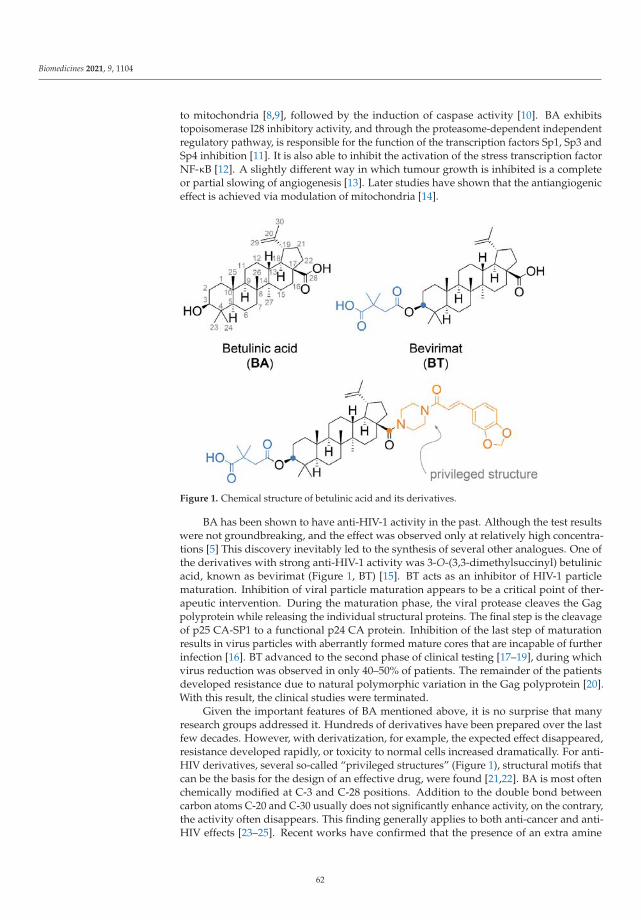
Biomedicines 2021, 9, 1104
to mitochondria [8,9], followed by the induction of caspase activity [10]. BA exhibitstopoisomerase I28 inhibitory activity, and through the proteasome-dependent independentregulatory pathway, is responsible for the function of the transcription factors Sp1, Sp3 andSp4 inhibition [11]. It is also able to inhibit the activation of the stress transcription factorNF-κB [12]. A slightly different way in which tumour growth is inhibited is a completeor partial slowing of angiogenesis [13]. Later studies have shown that the antiangiogeniceffect is achieved via modulation of mitochondria [14].
Figure 1. Chemical structure of betulinic acid and its derivatives.
BA has been shown to have anti-HIV-1 activity in the past. Although the test resultswere not groundbreaking, and the effect was observed only at relatively high concentra-tions [5] This discovery inevitably led to the synthesis of several other analogues. One ofthe derivatives with strong anti-HIV-1 activity was 3-O-(3,3-dimethylsuccinyl) betulinicacid, known as bevirimat (Figure 1, BT) [15]. BT acts as an inhibitor of HIV-1 particlematuration. Inhibition of viral particle maturation appears to be a critical point of ther-apeutic intervention. During the maturation phase, the viral protease cleaves the Gagpolyprotein while releasing the individual structural proteins. The final step is the cleavageof p25 CA-SP1 to a functional p24 CA protein. Inhibition of the last step of maturationresults in virus particles with aberrantly formed mature cores that are incapable of furtherinfection [16]. BT advanced to the second phase of clinical testing [17–19], during whichvirus reduction was observed in only 40–50% of patients. The remainder of the patientsdeveloped resistance due to natural polymorphic variation in the Gag polyprotein [20].With this result, the clinical studies were terminated.
Given the important features of BA mentioned above, it is no surprise that manyresearch groups addressed it. Hundreds of derivatives have been prepared over the lastfew decades. However, with derivatization, for example, the expected effect disappeared,resistance developed rapidly, or toxicity to normal cells increased dramatically. For anti-HIV derivatives, several so-called “privileged structures” (Figure 1), structural motifs thatcan be the basis for the design of an effective drug, were found [21,22]. BA is most oftenchemically modified at C-3 and C-28 positions. Addition to the double bond betweencarbon atoms C-20 and C-30 usually does not significantly enhance activity, on the contrary,the activity often disappears. This finding generally applies to both anti-cancer and anti-HIV effects [23–25]. Recent works have confirmed that the presence of an extra amine
62

Biomedicines 2021, 9, 1104
group introduced by conjugation into a BA molecule can significantly increase antitumourpotency [26,27].
This work presents the preparation and biological evaluation of new analogues ofBA and BT containing an amino group. In the past, fluorescent analogues of BA labelledwith green-emitting BODIPY (4,4-difluoro-4-bora-3a,4a-diaza-s-indacene) [28,29] and red-emitting Rhodamine B [30] were synthesized to study its localization and trafficking inliving cells. In this work, we synthesized and studied new derivatives of BA and BTlabelled at C-3 and C-28 positions using a small blue-emitting BODIPY dye.
2. Materials and Methods
2.1. Chemical Synthesis
Aluminium silica gel sheets for detection in UV light (TLC Silica gel 60 F254, Merck,Darmstadt, Denmark) were used for thin-layer chromatography (TLC), subsequent visual-ization was proceeded by a diluted solution of sulfuric acid in methanol and plates wereheated. Silica gel (30–60 μm, SiliTech, MP Biomedicals, Costa Mesa, CA, USA) was usedfor column chromatography. NMR Spectra were recorded by Agilent-MR DDR2 (SantaClara, CA, USA). HRMS were measured by LTQ ORBITRAP VELOS with HESI+/HESI-ionization (Thermo Scientific, Waltham, MA, USA). For microwave synthesis, an InitiatorClassic 355,301 (Biotage, Uppsala, Sweden) was used.
The following chemicals were purchased from TCI Europe (Zwijndrecht, Belgium):N,N,N-triethylamine—Et3N (>99%), 4-dimethylaminopyridine—4-DMAP (>99%), 1-(3-dimethylaminopropyl)-3-ethylcarbodiimide hydrochloride—EDCI (>98%), N,N′-dicyclohe-xylcarbodiimide—DCC (>98%), 1-hydroxybenzotriazole monohydrate—HOBt (>97%),triphenylphosphine—PPh3 (>95%), p-toluenesulfonic acid monohydrate—p-TsOH (>98%),and palladium on carbon—Pd/C (10%). The following chemicals were purchased fromSigma-Aldrich (Prague, Czech Republic): 3-azidopropylamine (≥95%), 1-(2-N-boc-aminoe-thyl)piperazine (≥95%), β-alanine (99%). Betulinic acid (BA) was purchased from Betulin-ines (Stríbrná skalice, Czech Republic).
The solvents for column chromatography and reactions were purchased from PENTA(Praha, Czech Republic) and were used without further distillation.
Compound Synthesis and Characterization
8-N-(3-Azidopropyl)amino-4,4-difluoro-4-bora-3a,4a-diaza-s-indacene (BODIPY-N3)To a solution of BODIPY-SMe (205 mg, 0.86 mmol) in DCM (10 mL), 3-azidopropylamine
(95 mg, 0.95 mmol) was added and the mixture was stirred for 30 min at RT. The solventswere evaporated under reduced pressure and the residue was taken up with AcOEt andthe product was precipitated by the addition of hexanes. BODIPY-N3 (243 mg, 0.83 mmol)was obtained as a yellowish solid in 97% yield. RF = 0.55 in hexanes-AcOEt 1:1. 1H NMR(400 MHz, CD3OD) δ ppm: 2.11 (quin, J = 6.7 Hz, 2 H), 3.56 (t, J = 6.5 Hz, 2 H), 3.87 (t,J = 7.4 Hz, 2 H), 6.39 (br. s., 1 H), 6.55 (br. s, 1 H), 7.32 (br. s, 1 H), 7.36 (br. s., 2 H), 7.57 (s,1 H). 13C NMR (101 MHz, CD3OD) δ ppm: 27.00, 44.08, 48.76, 112.67, 113.99, 115.78, 123.16,130.78, 133.76, 148.87. HRMS-ESI: calculated 290.12628 Da, found m/z 291.13312 [M+H]+.
8-N-(3-Aminopropyl)amino-4,4-difluoro-4-bora-3a,4a-diaza-s-indacene (BODIPY-NH2)To a solution of BODIPY-N3 (150 mg, 0.52 mmol) in AcOEt (8 mL), was added Pd/C
(80 mg) and the mixture was stirred under hydrogen atmosphere for 2 h. The catalystwas filtered off and the solvents were evaporated under reduced pressure. The residuewas taken up with AcOEt and the product was obtained after precipitation with hexane.BODIPY-NH2 (96 mg, 0.36 mmol) was obtained as yellow solid in 70% yield. RF = 0.15 inDCM-MeOH 20:1 (v/v). 1H NMR (400 MHz, CD3OD) δ ppm: 1.98 (quin, J = 6.7 Hz, 2 H),2.87 (t, J = 6.7 Hz, 2 H), 3.83 (t, J = 7.0 Hz, 2 H), 6.37 (br. s, 1 H), 6.52 (br. s, 1 H), 7.27 (br.s, 1 H), 7.30–7.36 (m, 2 H), 7.55 (br. s, 1 H). 13C NMR (101 MHz, CD3OD) δ ppm: 29.67,39.12, 45.50, 112.53, 113.78, 115.51, 123.15, 130.40, 133.46, 148.73. HRMS-ESI: calculated264.13578 Da, found m/z 263.12810 [M-H]−.
8-N-(β-Alanyl)amino-4,4-difluoro-4-bora-3a,4a-diaza-s-indacene (BODIPY-CO2H)
63
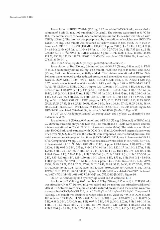
Biomedicines 2021, 9, 1104
To a solution of BODIPY-SMe (220 mg, 0.92 mmol) in DMSO (5 mL), was added asolution of β-Ala (91 mg, 1.02 mmol) in H2O (2 mL). The mixture was stirred at 30 ◦C for16 h. The solvents were removed under reduced pressure and the residue was diluted withCHCl3 (100 mL). The product was precipitated by the addition of cyclohexane. BODIPY-
CO2H (175 mg, 0.63 mmol) was obtained as yellow solids in 68% yield. RF = 0.38 inhexanes-AcOEt 1:1. 1H NMR (400 MHz, CD3OD) δ ppm: 2.87 (t, J = 6.9 Hz, 2 H), 4.00 (t,J = 6.9 Hz, 2 H), 6.35 (br. s., 1 H), 6.53 (br. s., 1 H), 7.27–7.31 (m, 1 H), 7.33 (br. s., 2 H),7.55 (br. s., 1 H). 13C NMR (101 MHz, CD3OD) δ ppm: 31.71, 42.48, 112.67, 114.02, 115.89,123.26, 130.78, 133.83, 148.75, 173.03. HRMS-ESI: calculated 279.09906 Da, found m/z278.09139 [M-H]−.
(3β)-N-(3-Azidopropyl)-3-hydroxylup-20(29)-ene-28-amide (1)To a solution of BA (200 mg, 0.44 mmol) and 4-DMAP (59 mg, 0.48 mmol) in DMF
(3 mL), 3-azidopropylamine (53 mg, 0.53 mmol), HOBt (65 mg, 0.48 mmol) and EDCI(93 mg, 0.48 mmol) were sequentially added. The mixture was stirred at RT for 36 h.Solvents were removed under reduced pressure and the residue was chromatographedtwice (i. DCM-MeOH 100:1, v/v ii. DCM→DCM-MeOH 70:1, v/v). Azide 1 (200 mg,0.37 mmol) was obtained as white solids in 84% yield. RF = 0.48 in DCM-MeOH 40:1(v/v). 1H NMR (400 MHz, CDCl3) δ ppm: 0.63–0.70 (m, 1 H), 0.75 (s, 3 H), 0.81 (s, 3 H),0.83–0.91 (m, 1 H), 0.93 (s, 3 H), 0.96 (s, 3 H), 0.96 (s, 3 H), 0.97–1.05 (m, 1 H), 1.12–1.65 (m,19 H), 1.67 (s, 3 H), 1.69–1.72 (m, 1 H), 1.75–1.82 (m, 2 H), 1.90–1.95 (m, 1 H), 2.38–2.48 (m,1 H), 3.08 3.20 (m, 2 H), 3.32 (s, 4 H), 4.58 (s, 1 H), 4.73 (s, 1 H), 5.85 (t, J = 5.9 Hz, 1 H);Figure S1. 13C NMR (101 MHz, CDCl3) δ ppm: 14.29, 15.01, 15.79, 15.80, 17.93, 19.13, 20.57,25.26, 27.05, 27.63, 28.68, 29.10, 30.51, 33.40, 34.04, 36.61, 36.84, 37.40, 38.05, 38.36, 38.49,40.40, 42.11, 46.38, 49.31, 49.74, 50.27, 55.02, 55.30, 78.58, 109.01, 150.50, 175.96; Figure S3.HRMS-ESI: calculated 538.42468 Da, found m/z 561.41394 [M+Na]+; Figure S2.
4-{[(3β)-28-[(3-Azidopropyl)amino]-28-oxolup-20(29)-ene-3-yl]oxy}-2,2-dimethyl-4-oxo-butanoic acid (2)
To a solution of 1 (200 mg, 0.37 mmol) and 4-DMAP (73 mg, 0.59 mmol) in THF (2 mL),2,2-dimethylsucccinic anhydride (238 mg, 1.86 mmol) and p-TsOH were added and themixture was stirred for 2 h at 130 ◦C in microwave reactor (MW). The mixture was dilutedwith H2O (20 mL) and extracted with DCM (4 × 15 mL). Combined organic layers weredried over Na2SO4, filtered and the solvents were evaporated under reduced pressure. Theresidue was chromatographed two times (i. DCM-MeOH 100:1, v/v; ii. hexanes-AcOEt 3:1,v/v). Compound 2 (94 mg, 0.14 mmol) was obtained as white solids in 38% yield. RF = 0.60in hexanes-AcOEt, 1:1. 1H NMR (400 MHz, CDCl3) δ ppm: 0.73–0.76 (m, 1 H), 0.79 (s, 3 H),0.82 (s, 6 H), 0.92 (s, 3 H), 0.95 (s, 3 H), 0.97–1.01 (m, 1 H), 1.12 1.17 (m, 1 H), 1.27 (s, 3 H),1.29 (s, 3 H), 1.30–1.67 (m, 17 H), 1.67 (s, 3 H), 1.71 (d, J = 7.0 Hz, 1 H), 1.75–1.81 (m, 2 H),1.90–1.93 (m, 1 H), 2.39–2.46 (m, 1 H), 2.52–2.68 (m, 2 H), 3.08 3.14 (m, 1 H), 3.22–3.33 (m,2 H), 3.33–3.43 (m, 4 H), 4.45–4.50 (m, 1 H), 4.58 (s, 1 H), 4.73 (s, 1 H), 5.86 (t, J = 5.9 Hz,1 H); Figure S4. 13C NMR (101 MHz, CDCl3) δ ppm: 14.60, 16.14, 16.46, 18.13, 19.44, 20.92,23.59, 24.99, 25.27, 25.55, 25.58, 27.88, 29.02, 29.42, 30.83, 33.73, 34.30, 36.97, 37.08, 37.70,37.72, 38.40, 40.44, 40.76, 42.45, 44.69, 46.74, 49.65, 50.06, 50.50, 55.47, 55.65, 81.51, 109.42,109.99, 150.81, 170.95, 176.38, 182.48; Figure S5. HRMS-ESI: calculated 666.47202 Da, foundm/z 667.47921 [M+H]+, 689.46125 [M+Na]+ and 705.43463 [M+K]+; Figure S6.
(3β)-N-(3-Aminopropyl)-3-hydroxylup-20(29)-ene-28-amide (3) [31]A solution of 2 (339 mg, 0.63 mmol) and PPh3 (248 mg, 0.95 mmol) in THF (10 mL)
was stirred for 3h at RT. Water (1 mL) was added and the mixture was stirred for additional20 h at RT. Solvents were evaporated under reduced pressure and the residue was chro-matographed (CHCl3-MeOH 20:1, v/v + 0.5% Et3N → 10:1, v/v + 0.5% Et3N). Compound 3
(278 mg, 0.54 mmol) was obtained as white solids in 86% yield. RF = 0.15 in DCM-MeOH10:1 (v/v) + 0.5% Et3N. 1H NMR (400 MHz, CD3OD) δ ppm: 0.70–0.75 (m, 1 H), 0.77 (s,3 H), 0.88 (s, 3 H), 0.91–0.96 (m, 1 H), 0.97 (s, 3 H), 0.99 (s, 3 H), 1.02 (s, 3 H), 1.04–1.10 (m,1 H), 1.15–1.69 (m, 20 H), 1.71 (s, 3 H), 1.80–1.95 (m, 2 H), 2.10–2.19 (m, 1 H), 2.55–2.64 (m,1 H), 2.69 (t, J = 6.9 Hz, 2 H), 3.07–3.18 (m, 2 H), 3.20–3.32 (m, 2 H), 4.60 (s, 1 H), 4.72 (s, 1 H);
64
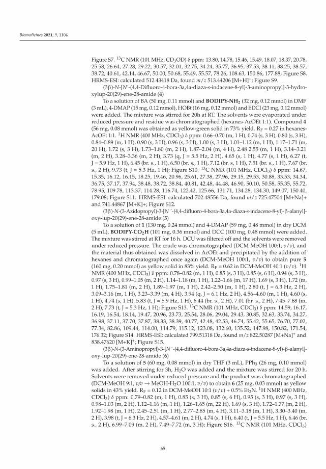
Biomedicines 2021, 9, 1104
Figure S7. 13C NMR (101 MHz, CD3OD) δ ppm: 13.80, 14.78, 15.46, 15.49, 18.07, 18.37, 20.78,25.58, 26.64, 27.28, 29.22, 30.57, 32.01, 32.75, 34.24, 35.77, 36.95, 37.53, 38.11, 38.25, 38.57,38.72, 40.61, 42.14, 46.67, 50.00, 50.68, 55.49, 55.57, 78.26, 108.63, 150.86, 177.88; Figure S8.HRMS-ESI: calculated 512.43418 Da, found m/z 513.44206 [M+H]+; Figure S9.
(3β)-N-[N’-(4,4-Difluoro-4-bora-3a,4a-diaza-s-indacene-8-yl)-3-aminopropyl]-3-hydro-xylup-20(29)-ene-28-amide (4)
To a solution of BA (50 mg, 0.11 mmol) and BODIPY-NH2 (32 mg, 0.12 mmol) in DMF(3 mL), 4-DMAP (15 mg, 0.12 mmol), HOBt (16 mg, 0.12 mmol) and EDCI (23 mg, 0.12 mmol)were added. The mixture was stirred for 20h at RT. The solvents were evaporated underreduced pressure and residue was chromatographed (hexanes-AcOEt 1:1). Compound 4
(56 mg, 0.08 mmol) was obtained as yellow-green solid in 73% yield. RF = 0.27 in hexanes-AcOEt 1:1. 1H NMR (400 MHz, CDCl3) δ ppm: 0.66–0.70 (m, 1 H), 0.74 (s, 3 H), 0.80 (s, 3 H),0.84–0.89 (m, 1 H), 0.90 (s, 3 H), 0.96 (s, 3 H), 1.00 (s, 3 H), 1.01–1.12 (m, 1 H), 1.17–1.71 (m,20 H), 1.72 (s, 3 H), 1.73–1.80 (m, 2 H), 1.87–2.04 (m, 4 H), 2.48 2.55 (m, 1 H), 3.14–3.21(m, 2 H), 3.28–3.36 (m, 2 H), 3.73 (q, J = 5.5 Hz, 2 H), 4.65 (s, 1 H), 4.77 (s, 1 H), 6.27 (t,J = 5.9 Hz, 1 H), 6.45 (br. s., 1 H), 6.50 (br. s., 1 H), 7.12 (br. s, 1 H), 7.51 (br. s., 1 H), 7.67 (br.s., 2 H), 9.73 (t, J = 5.3 Hz, 1 H); Figure S10. 13C NMR (101 MHz, CDCl3) δ ppm: 14.67,15.35, 16.12, 16.15, 18.25, 19.46, 20.96, 25.61, 27.38, 27.96, 29.15, 29.53, 30.88, 33.53, 34.34,36.75, 37.17, 37.94, 38.48, 38.72, 38.84, 40.81, 42.48, 44.48, 46.90, 50.10, 50.58, 55.35, 55.72,78.95, 109.78, 113.37, 114.28, 116.74, 122.42, 125.66, 131.71, 134.28, 134.30, 149.07, 150.40,179.08; Figure S11. HRMS-ESI: calculated 702.48556 Da, found m/z 725.47504 [M+Na]+and 741.44867 [M+K]+; Figure S12.
(3β)-N-(3-Azidopropyl)-3-[N´-(4,4-difluoro-4-bora-3a,4a-diaza-s-indacene-8-yl)-β-alanyl]-oxy-lup-20(29)-ene-28-amide (5)
To a solution of 1 (130 mg, 0.24 mmol) and 4-DMAP (59 mg, 0.48 mmol) in dry DCM(5 mL), BODIPY-CO2H (101 mg, 0.36 mmol) and DCC (100 mg, 0.48 mmol) were added.The mixture was stirred at RT for 16 h. DCU was filtered off and the solvents were removedunder reduced pressure. The crude was chromatographed (DCM-MeOH 100:1, v/v), andthe material thus obtained was dissolved in AcOEt and precipitated by the addition ofhexanes and chromatographed once again (DCM-MeOH 100:1, v/v) to obtain pure 5
(160 mg, 0.20 mmol) as yellow solid in 83% yield. RF = 0.62 in DCM-MeOH 40:1 (v/v). 1HNMR (400 MHz, CDCl3) δ ppm: 0.78–0.82 (m, 1 H), 0.85 (s, 3 H), 0.85 (s, 6 H), 0.94 (s, 3 H),0.97 (s, 3 H), 0.99–1.05 (m, 2 H), 1.14–1.18 (m, 1 H), 1.22–1.66 (m, 17 H), 1.69 (s, 3 H), 1.72 (m,1 H), 1.75–1.81 (m, 2 H), 1.89–1.97 (m, 1 H), 2.42–2.50 (m, 1 H), 2.80 (t, J = 6.3 Hz, 2 H),3.09–3.16 (m, 1 H), 3.23–3.39 (m, 4 H), 3.94 (q, J = 6.1 Hz, 2 H), 4.56–4.60 (m, 1 H), 4.60 (s,1 H), 4.74 (s, 1 H), 5.83 (t, J = 5.9 Hz, 1 H), 6.44 (br. s., 2 H), 7.01 (br. s., 2 H), 7.45–7.68 (m,2 H), 7.73 (t, J = 5.3 Hz, 1 H); Figure S13. 13C NMR (101 MHz, CDCl3) δ ppm: 14.59, 16.17,16.19, 16.54, 18.14, 19.47, 20.96, 23.73, 25.54, 28.06, 29.04, 29.43, 30.85, 32.63, 33.74, 34.27,36.98, 37.11, 37.70, 37.87, 38.33, 38.39, 40.77, 42.48, 42.53, 46.74, 55.42, 55.65, 76.70, 77.02,77.34, 82.86, 109.44, 114.00, 114.79, 115.12, 123.08, 132.60, 135.52, 147.98, 150.82, 171.54,176.32; Figure S14. HRMS-ESI: calculated 799.51318 Da, found m/z 822.50287 [M+Na]+ and838.47620 [M+K]+; Figure S15.
(3β)-N-(3-Aminopropyl)-3-[N´-(4,4-difluoro-4-bora-3a,4a-diaza-s-indacene-8-yl)-β-alanyl]-oxy-lup-20(29)-ene-28-amide (6)
To a solution of 5 (60 mg, 0.08 mmol) in dry THF (3 mL), PPh3 (26 mg, 0.10 mmol)was added. After stirring for 3h, H2O was added and the mixture was stirred for 20 h.Solvents were removed under reduced pressure and the product was chromatographed(DCM-MeOH 9:1, v/v → MeOH-H2O 100:1, v/v) to obtain 6 (25 mg, 0.03 mmol) as yellowsolids in 43% yield. RF = 0.12 in DCM-MeOH 10:1 (v/v) + 0.5% Et3N. 1H NMR (400 MHz,CDCl3) δ ppm: 0.79–0.82 (m, 1 H), 0.85 (s, 3 H), 0.85 (s, 6 H), 0.95 (s, 3 H), 0.97 (s, 3 H),0.98–1.03 (m, 2 H), 1.12–1.16 (m, 1 H), 1.26–1.65 (m, 22 H), 1.69 (s, 3 H), 1.72–1.77 (m, 2 H),1.92–1.98 (m, 1 H), 2.45–2.51 (m, 1 H), 2.77–2.85 (m, 4 H), 3.11–3.18 (m, 1 H), 3.30–3.40 (m,2 H), 3.98 (t, J = 6.3 Hz, 2 H), 4.57–4.61 (m, 2 H), 4.74 (s, 1 H), 6.40 (t, J = 5.5 Hz, 1 H), 6.46 (br.s., 2 H), 6.99–7.09 (m, 2 H), 7.49–7.72 (m, 3 H); Figure S16. 13C NMR (101 MHz, CDCl3)
65

Biomedicines 2021, 9, 1104
δ ppm: 14.59, 16.17, 16.19, 16.54, 18.14, 19.48, 20.97, 23.74, 25.56, 28.07, 29.43, 30.89, 32.44,32.67, 33.70, 34.28, 37.11, 37.52, 37.66, 37.87, 38.33, 38.49, 40.09, 40.76, 42.48, 42.52, 46.74,50.07, 50.55, 55.42, 55.61, 82.90, 109.34, 114.15, 114.82, 115.40, 122.45, 132.64, 135.57, 148.06,150.99, 171.58, 176.32; Figure S17. HRMS-ESI: monoisotopic mass 773.52268 Da, found m/z774.53046 [M+H]+ and 772.51587 [M-H]−; Figure S18.
Tert-butyl-(3-{[(3β)-3-hydroxy-28-oxolup-20(29)-ene-28-yl]amino}propyl)-carbamate (7) [23]To a solution of BA (1.00 g, 2.19 mmol) and 4-DMAP (295 mg, 2.41 mmol) in DMF
(12 mL), N-boc-1,3-diaminopropane (459 mg, 2.63 mmol), HOBt (326 mg, 2.41 mmol) andEDCI (462 mg, 2.41 mmol) were added. The mixture was stirred for 48 h at RT. Thesolvents were evaporated under reduced pressure and the residue was chromatographed(DCM-MeOH 100:1, v/v). Compound 7 (900 mg, 1.47 mmol) was obtained as white solidin 67% yield. RF = 0.34 in DCM-MeOH 40:1 (v/v). 1H NMR (400 MHz, CDCl3) δ ppm:0.65 0.70 (m, 1 H), 0.75 (s, 3 H), 0.81 (s, 3 H), 0.85–0.90 (m, 1 H), 0.93 (s, 3 H), 0.96 (s,3 H), 0.97 (s, 3 H), 0.98–1.05 (m, 2 H), 1.13–1.20 (m, 1 H), 1.22–1.44 (m, 8 H), 1.45 (s, 9 H),1.46 1.68 (m, 11 H), 1.69 (s, 3 H), 1.69–1.81 (m, 2 H), 1.88–2.00 (m, 1 H), 2.00–2.07 (m, 1 H),2.42–2.50 (m, 1 H), 3.10–3.28 (m, 5 H), 3.30–3.39 (m, 1 H), 4.59 (s, 1 H), 4.74 (s, 1 H), 4.91 (br.s., 1 H), 6.33 (br. s., 1 H); Figure S19. 13C NMR (101 MHz, CDCl3) δ ppm: 14.64, 15.35, 16.12,16.14, 18.28, 19.48, 20.94, 25.64, 27.41, 27.97, 28.40 (s, 3 C) 29.48, 30.54, 30.92, 33.65, 34.39,35.28, 37.20, 37.75, 38.51, 38.72, 38.84, 40.76, 42.47, 46.66, 50.04, 50.64, 55.38, 55.76, 67.07,78.96, 79.32, 109.27, 151.05, 156.65, 176.50; Figure S20. HRMS-ESI: calculated 612.48661 Da,found m/z 613.49394 [M+H]+, 635.47609 [M+Na]+ and 651.44943 [M+K]+; Figure S21.
4-{[(3β)-28-({3-[(Tert-butoxycarbonyl)amino]propyl}amino)-28-oxolup-20(29)-ene-3–yl]oxy}-2,2-dimethyl-4-oxobutanoic acid (8)
To a solution of 7 (400 mg, 0.65 mmol) and 4-DMAP (128 mg, 1.04 mmol) in THF(4 mL), 2,2-dimethylsuccinic anhydride (418 mg, 3.26 mmol) and p-TsOH were added. Themixture was stirred for 2 h at 130 ◦C in a microwave reactor. The mixture was pouredinto water and extracted with DCM (4 × 20 mL). Combined organic layers were washedwith KHSO4 (3 × 5 mL) and brine. The organic layer was dried over Na2SO4 and thesolvents were evaporated under reduced pressure. The residue was chromatographed(DCM-MeOH 40:1, v/v + 1% Et3N) to obtain 8 (260 mg, 0.35 mmol) as white solids in54% yield. RF = 0.18 in DCM-MeOH 40:1 (v/v) + 1% Et3N. 1H NMR (400 MHz, CDCl3) δppm: 0.74–0.77 (m, 1 H), 0.80 (s, 3 H), 0.82 (s, 6 H), 0.92 (s, 3 H), 0.96 (s, 3 H), 0.97–1.02 (m,1 H), 1.13–1.17 (m, 1 H), 1.25–1.27 (m, 2 H), 1.28 (s, 3 H), 1.30 (s, 3 H), 1.31–1.44 (m, 7 H),1.45 (s, 9 H), 1.46–1.68 (m, 10 H), 1.69 (s, 3 H), 1.70–1.82 (m, 2 H), 1.90–1.97 (m, 1 H),2.03 2.07 (m, 1 H), 2.41–2.48 (m, 1 H), 2.53–2.70 (m, 2 H), 3.10–3.40 (m, 7 H), 4.46–4.51 (m,1 H), 4.59 (s, 1 H), 4.74 (s, 1 H), 4.94 (br. s., 1 H), 6.43 (br. s., 1 H); Figure S22. 13C NMR(101 MHz, CDCl3) δ ppm: 14.61, 16.09, 16.15, 16.47, 18.14, 19.45, 20.95, 23.60, 25.02, 25.58,25.61, 27.90, 28.39, 29.46, 30.49, 30.88, 33.61, 34.30, 35.35, 37.08, 37.70, 37.92, 38.40, 38.52,40.44, 40.72, 40.76, 42.46, 44.71, 46.64, 49.99, 50.52, 55.47, 55.80, 79.40, 81.55, 109.37, 150.97,156.80, 171.00, 176.72, 182.24; Figure S23. HRMS-ESI: calculated 740.53395 Da, found m/z741.54119 [M+H]+ and 763.52302 [M+Na]+; Figure S24.
4-{[(3β)-28-[(3-Aminopropyl)amino]-28-oxolup-20(29)-ene-3-yl]oxy}-2,2-dimethyl-4-oxo-butanooic acid hydrochloride (9)
Compound 6 (130 mg, 0.18 mmol) was dissolved in CHCl3 (1.5 mL) and 2 M HClsolution in Et2O (3 mL) was added slowly. The mixture was stirred for 1 h at RT underargon atmosphere. Solvents were evaporated under reduced pressure and the residue wassonicated for 20 min in Et2O (5 mL). The product was collected by filtration and driedin vacuo. Compound 9 (102 mg, 0.16 mmol) was obtained as white solids in 91% yield.RF = 0.1 in DCM-MeOH 9:1 (v/v). 1H NMR (400 MHz, CD3OD) δ ppm: 0.81–0.83 (m,1 H), 0.86 (s, 6 H), 0.88 (s, 1 H), 0.89 (s, 3 H), 0.98 (s, 3 H), 1.02 (s, 3 H), 1.03–1.11 (m, 1 H),1.19 (m, 1 H), 1.25 (s, 3 H), 1.26 (s, 3 H), 1.27–1.32 (m, 1 H), 1.36–1.65 (m, 15 H), 1.69 (s, 3 H),1.70–1.76 (m, 2 H), 1.80–1.90 (m, 5 H), 2.10–2.15 (m, 1 H), 2.52–2.65 (m, 3 H), 2.90 2.96 (m,2 H), 3.06–3.13 (m, 1 H), 3.23–3.30 (m, 2 H), 4.43–4.48 (m, 1 H), 4.59 (s, 1 H), 4.70 (s, 1 H),7.89 (t, J = 5.9 Hz, 1 H); Figure S25. 13C NMR (101 MHz, CD3OD) δ ppm: 13.64, 15.36, 15.43,
66

Biomedicines 2021, 9, 1104
15.66, 17.85, 18.13, 20.76, 23.25, 24.43, 24.79, 25.49, 27.06, 27.71, 29.22, 30.51, 32.59, 34.06,35.22, 36.85, 36.86, 37.42, 37.58, 38.02, 38.03, 38.18, 39.94, 40.60, 42.13, 44.23, 49.90, 50.50,55.46, 55.65, 81.22, 108.66, 150.77, 171.53, 179.14, 179.17; Figure S26. HRMS-ESI: calculated640.48152 Da, found m/z 641.48955 [M+H]+; Figure S27.
4-{[(3β)-28-{[N-(4,4-Difluoro-4-bora-3a,4a-diaza-s-indacene-8-yl)-3-aminopropyl]amino}-28-oxolup-20(29)-ene-3-yl]oxy}-2,2-dimethyl-4-oxobutanoic acid (10)
Compound 9 (15 mg, 0.02 mmol) and BODIPY-SMe (6 mg, 0.03 mmol) were dissolvedin the mixture of CHCl3 (2 mL) and THF (1 mL). To this solution, one drop of Et3N wasadded. The mixture was stirred 30 min at RT. Solvents were evaporated under reducedpressure and the residue was chromatographed (DCM-MeOH 19:1, v/v + 1% Et3N). Themixture was dissolved in AcOEt and washed with KHSO4 (10% solution, 3×5 mL) andbrine (1 × 5 mL). Organic layer was dried over Na2SO4 and the solvents were evaporated.Compound 10 (14 mg, 0.02 mmol) as yellow-green solid in 74% yield. RF = 0.73 in DCM-MeOH 9:1 (v/v). 1H NMR (400 MHz, CDCl3) δ ppm: 0.73–0.77 (m, 1 H), 0.78 (s, 3 H),0.81 (s, 3 H), 0.82 (s, 3 H), 0.83–0.85 (m, 1 H), 0.89 (s, 3 H), 0.99 (s, 3 H), 1.00–1.10 (m, 1 H),1.16–1.21 (m, 1 H), 1.29 (s, 3 H), 1.31 (s, 3 H), 1.34–1.67 (m, 15 H), 1.72 (s, 3 H), 1.74 1.79 (m,2 H), 1.87–2.03 (m, 5 H), 2.49–2.71 (m, 3 H), 3.14–3.20 (m, 1 H), 3.33 (q, J = 5.4 Hz, 2 H),3.74 (q, J = 5.5 Hz, 2 H), 4.46–4.51 (m, 1 H), 4.65 (s, 1 H), 4.77 (s, 1 H), 6.24 (t, J = 6.1 Hz, 1 H),6.44 (br. s., 1 H), 6.51 (br. s., 1 H), 7.12 (br. s., 1 H), 7.51 (br. s., 1 H), 7.68 (br. s., 2 H), 9.71 (t,J = 5.5 Hz, 1 H); Figure S28. 13C NMR (101 MHz, CDCl3) δ ppm: 14.63, 16.11, 16.15, 16.47,18.09, 19.43, 20.99, 23.59, 24.97, 25.62, 27.88, 29.16, 29.51, 30.86, 33.56, 34.26, 36.73, 37.06,37.70, 37.89, 38.40, 38.49, 40.45, 40.81, 42.47, 44.40, 44.70, 46.88, 50.08, 50.47, 55.46, 55.71,81.51, 109.83, 113.40, 114.28, 116.67, 122.36, 125.65, 131.78, 134.36, 149.06, 150.35, 171.00,179.13, 182.28; Figure S29. HRMS-APCI: calculated 830.53291 Da, found m/z 829.52699[M-H]−; Figure S30.
(3β)-28-(4-{2-[(Tert-butoxycarbonyl)amino]ethyl}piperazine-1-yl)-28-oxolup-20(29)-ene-3-yl acetate (12)
To a solution of compound 11 (1.27 g, 2.55 mmol) in DCM (20 mL), oxalyl chloride(1.2 mL) in DCM (10 mL) and 3 drops of DMF were added. After stirring for 2 h at RT,the solvents were co-evaporated with toluene (3 × 20 mL). Chloride thus obtained wasdissolved in DCM (35 mL), and 1-(2-N-boc-aminoethyl)piperazine (876 mg, 3.82 mmol)followed by Et3N (0.42 mL) were added. After stirring for 16 h at RT, the mixture wasdiluted with DCM (20 mL) and washed with brine (3 × 30 mL). The organic layer wasdried over Na2SO4 and the solvents were evaporated under reduced pressure. The residuewas chromatographed (CHCl3-MeOH 100:1 → 50:1, v/v) to obtain product 12 (720 mg,1.01 mmol) as white solids in 40% yield. RF = 0.16 in DCM-MeOH 100:1 (v/v). 1H NMR(400 MHz, CDCl3) δ ppm: 0.77–0.80 (m, 1 H), 0.83 (s, 3 H), 0.84 (s, 3 H), 0.85 (s, 3 H), 0.94 (s,3 H), 0.95 (s, 3 H), 0.97–1.00 (m, 1 H), 1.12–1.17 (m, 1 H), 1.28–1.42 (m, 9 H), 1.46 (s, 9 H),1.48–1.66 (m, 7 H), 1.68 (s, 3 H), 1.70–1.74 (m, 1 H), 1.81–1.87 (m, 1 H), 1.93–1.98 (m, 1 H),2.04 (s, 3 H), 2.07–2.11 (m, 1 H), 2.42 (br. s., 4 H), 2.49 (br. s., 2 H), 2.83–2.90 (m, 1 H),2.94–3.01 (m, 1 H), 3.25 (br. s., 2 H), 3.61 (br. s., 4 H), 4.44–4.49 (m, 1 H), 4.58 (s, 1 H), 4.72 (s,1 H), 4.98 (br. s., 1 H); Figure S31. 13C NMR (101 MHz, CDCl3) δ ppm: 14.62, 16.11, 16.24,16.46, 18.18, 19.63, 21.15, 21.30, 23.70, 25.61, 27.93, 28.41, 29.79, 31.30, 32.46, 32.49, 34.31,35.91, 36.86, 36.95, 37.14, 37.80, 38.41, 40.68, 41.85, 45.65, 50.76, 52.65, 53.12, 54.52, 55.52,57.15, 79.30, 80.97, 109.16, 151.30, 155.90, 170.99, 173.46; Figure S32. HRMS-ESI: calculated709.53937 Da, found m/z 710.54562 [M+H]+ and 732.52546 [M+Na]+; Figure S33.
Tert-butyl-(2-{4-[(3β)-3-hydroxy-28-oxolup-20(29)-ene-28-yl]piperazine-1-yl}ethyl)-carbamate (13)
To compound 12 (700 mg, 0.99 mmol) in MeOH (18 mL) and THF (9 mL), 4 M NaOHsolution (9 mL) was added. The mixture was stirred for 2 h at RT. The mixture wasneutralized by 1 M HCl solution and extracted with DCM (4 × 40 mL). Combined organiclayers were washed with saturated brine (2 × 50 mL) and dried over Na2SO4. Solventswere evaporated under reduced pressure and the residue was chromatographed (DCM-MeOH 40:1, v/v) to obtain 13 (310 mg, 0.46 mmol) as white solids in 47% yield. RF = 0.33 in
67

Biomedicines 2021, 9, 1104
DCM-MeOH 20:1 (v/v). 1H NMR (400 MHz, CDCl3) δ ppm: 0.66–0.69 (m, 1 H), 0.75 (s, 3 H),0.82 (s, 3 H), 0.86–0.90 (m, 1 H), 0.93 (s, 3 H), 0.96 (br. s, 6 H), 1.12–1.17 (m, 1 H), 1.23–1.29(m, 2 H), 1.29–1.42 (m, 8 H), 1.45 (s, 9 H), 1.48–1.66 (m, 7 H), 1.68 (s, 3 H), 1.69–1.73 (m,1 H), 1.81–1.87 (m, 1 H), 1.93–1.98 (m, 1 H), 2.06–2.11 (m, 1 H), 2.42 (br. s., 4 H), 2.48 (br. s.,2 H), 2.84–2.90 (m, 1 H), 2.94–3.00 (m, 1 H), 3.15–3.20 (m, 1 H), 3.25 (br. s, 2 H), 3.61 (br. s.,4 H), 4.57 (s, 1 H), 4.72 (s, 1 H), 4.99 (br. s., 1 H); Figure S34. 13C NMR (101 MHz, CDCl3)δ ppm: 14.67, 15.34, 16.11, 16.18, 18.30, 19.66, 21.14, 25.66, 27.43, 27.98, 28.41, 29.80, 31.32,32.48, 32.50, 34.39, 35.91, 36.88, 36.97, 37.23, 38.74, 38.85, 40.67, 41.87, 45.62, 50.86, 52.67,53.11, 54.53, 55.47, 57.15, 78.97, 79.28, 109.10, 151.33, 155.91, 173.45; Figure S35. HRMS-ESI:calculated 667.52881 Da, found m/z 668.53693 [M+H]+; Figure S36.
(3β)-28-[4-(2-Aminoethyl)piperazine-1-yl]-3-hydroxylup-20(29)-ene-28-one hydrochlo-ride (14)
To compound 13 (200 mg, 0.30 mmol) in CHCl3 (2.8 mL), 2 M HCl solution in Et2O(5.1 mL) was slowly added. The mixture was stirred for 16 h at RT under argon. Solventswere removed under reduced pressure and the crude was sonicated for 20min in Et2O(5 mL), collected by filtration and dried. Compound 14 (142 mg, 0.25 mmol) was isolatedas white solid in 84% yield. 1H NMR (400 MHz, CD3OD) δ ppm: 0.69–0.73 (m, 1 H),0.75 (s, 3 H), 0.86 (s, 3 H), 0.88–0.93 (m, 1 H), 0.95 (s, 3 H), 0.96 (s, 3 H), 1.01 (s, 3 H),1.02–1.07 (m, 1 H), 1.22–1.67 (m, 18 H), 1.70 (s, 3 H), 1.73–1.77 (m, 1 H), 1.82–1.88 (m, 1 H),1.97–2.02 (m, 1 H), 2.11–2.15 (m, 1 H), 2.79–2.86 (m, 1 H), 2.89–2.95 (m, 1 H), 3.11–3.15 (m,1 H), 3.35–3.81 (m, 11 H), 4.59 (s, 1 H), 4.70 (s, 1 H); Figure S37. 13C NMR (101 MHz,CD3OD) δ ppm: 13.65, 14.68, 15.27, 15.38, 18.04, 18.29, 20.86, 25.52, 26.63, 26.67, 27.18, 29.66,30.92, 31.88, 33.62, 33.63, 34.19, 36.94, 36.96, 38.53, 38.68, 40.54, 41.64, 45.78, 50.78, 52.19,52.37, 53.15, 54.58, 55.52, 78.24, 108.66, 150.85, 174.45; Figure S38. HRMS-ESI: calculated567.47638 Da, found m/z 568.4832 [M+H]+; Figure S39.
(3β)-28-{4-[N-(4,4-Difluoro-4-bora-3a,4a-diaza-s-indacene-8-yl)-2-aminoethyl]piperazine-1-yl}-3-hydroxylup-20(29)-ene-28-one (15)
To compound 14 (45 mg, 0.08 mmol) and BODIPY-SMe (21 mg, 0.09 mmol) in themixture of DCM (5 mL) and THF (2.5 mL), was added one drop of Et3N and the mixturewas stirred 30 min at RT. Solvents were evaporated under reduced pressure and the residuewas chromatographed (DCM-MeOH 100:1, v/v) to obtain 15 (42 mg, 0.06 mmol) as yellowsolids in 70% yield. RF = 0.19 in DCM-MeOH 100:1 (v/v). 1H NMR (400 MHz, CDCl3) δppm: 0.67–0.71 (m, 1 H), 0.76 (s, 3 H), 0.83 (s, 3 H), 0.87–0.91 (m, 1 H), 0.95 (s, 3 H), 0.97 (s,6 H), 1.16–1.21 (m, 1 H), 1.21–1.32 (m, 3 H), 1.38–1.66 (m, 15 H), 1.69 (s, 3 H), 1.72–1.76 (m,1 H), 1.82–1.89 (m, 1 H), 1.93–1.98 (m, 1 H), 2.06–2.12 (m, 1 H), 2.55 (br. s., 4 H), 2.82–2.90(m, 3 H), 2.95–3.01 (m, 1 H), 3.17–3.21 (m, 1 H), 3.70 (br. s., 4 H), 3.75 (br. s., 2 H), 4.60 (s,1 H), 4.74 (s, 1 H), 6.41 (br. s., 1 H), 6.53 (br. s., 1 H), 6.91 (br. s., 1 H), 7.13 (br. s., 1 H), 7.51(br. s., 1 H), 7.72 (br. s., 1 H), 7.92 (br. s., 1 H); Figure S40. 13C NMR (101 MHz, CDCl3)δ ppm: 14.68, 15.34, 16.18, 18.32, 19.64, 21.14, 25.45, 25.64, 27.43, 27.97, 29.86, 31.31, 32.52,34.42, 35.93, 36.92, 37.24, 38.74, 38.86, 40.69, 41.80, 41.90, 45.62, 50.84, 52.53, 52.65, 54.26,54.60, 55.46, 78.98, 109.29, 113.76, 114.93, 122.67, 123.28, 125.04, 132.47, 135.78, 147.77, 151.12,173.64; Figure S41. HRMS-ESI: calculated 757.52776 Da, found m/z 780.5171 [M+Na]+ and796.4904 [M+K]+; Figure S42.
4-{[(3β)-28-(4-{2-[(Tert-butoxycarbonyl)amino]ethyl}piperazine-1-yl)-28-oxolup-20(29)-ene-3-yl]oxy}-2,2-dimethyl-4-oxobutanoic acid (16)
To a solution of 13 (500 mg, 0.75 mmol) and 4-DMAP (146 mg, 1.20 mmol) in THF(5 mL), 2,2-dimethylsuccinic anhydride (480 mg, 3.74 mmol) and a catalytic amount ofp-TsOH were added. The reaction was stirred for 2 h at 130 ◦C in a microwave reactor. Themixture was diluted with H2O and extracted with DCM (4 × 20 mL). Combined organiclayers were dried over Na2SO4 and the solvents were removed under reduced pressure.The crude was chromatographed twice (i. toluene-AcOEt 1:1 → AcOEt; ii. toluene-AcOEt1:1 + 1% Et3N → AcOEt → DCM-MeOH 9:1, v/v). Compound 16 (328 mg, 0.41 mmol)was obtained as white solids in 53% yield. RF = 0.43 in DCM-MeOH 9:1 (v/v). 1H NMR(400 MHz, CDCl3) δ ppm: 0.74–0.77 (m, 1 H), 0.81 (s, 6 H), 0.83 (s, 3 H), 0.93 (s, 3 H), 0.95 (s,
68

Biomedicines 2021, 9, 1104
3 H), 1.12–1.16 (m, 1 H), 1.27 (br. s., 6 H), 1.32–1.41 (m, 8 H), 1.44 (s, 9 H), 1.47–1.65 (m,7 H), 1.68 (s, 3 H), 1.70–1.74 (m, 1 H), 1.80–1.86 (m, 1 H), 1.90–1.95 (m, 1 H), 2.05–2.09 (m,1 H), 2.49–2.69 (m, 6 H), 2.82–2.89 (m, 1 H), 2.93–3.00 (m, 1 H), 3.29 (br. s., 2 H), 3.61 (br.s., 4 H), 3.71 (br. s., 2 H), 4.45–4.50 (m, 1 H), 4.58 (s, 1 H), 4.72 (s, 1 H), 5.15 (br. s., 1 H);Figure S43. 13C NMR (101 MHz, CDCl3) δ ppm: 14.67, 16.06, 16.21, 16.57, 18.23, 19.64, 21.14,23.66, 23.68, 25.57, 25.58, 25.92, 25.94, 27.91, 28.40, 29.82, 31.30, 32.36, 32.39, 34.26, 35.96,36.69, 36.82, 37.12, 37.76, 38.39, 38.41, 40.63, 41.87, 45.60, 50.72, 52.60, 52.94, 54.49, 55.53,57.19, 57.20, 109.24, 128.19, 129.00, 151.20, 155.99, 173.43; Figure S44. HRMS-ESI: calculated795.57615 Da, found m/z 796.58292 [M+H]+; Figure S45.
4-{[(3β)-28-[4-(2-Aminoethyl)piperazine-1-yl]-28-oxolup-20(29)-ene-3-yl]oxy}-2,2-dimethyl-4-oxobutanoic acid hydrochloride (17)
To compound 16 (150 mg, 0.19 mmol) in CHCl3 (2 mL), 2 M HCl solution in Et2O(3.2 mL) was added. The mixture was stirred for 2 h at RT under argon atmosphere.Solvents were evaporated and the crude was sonicated for 20 min in Et2O (5 mL). Theprecipitate was collected and dried in vacuo. Compound 17 (102 mg, 0.16 mmol) wasobtained as white solids in 91% yield. 1H NMR (400 MHz, CD3OD) δ ppm: 0.82–0.85 (m,1 H), 0.87 (br. s, 6 H), 0.91 (s, 3 H), 0.98 (s, 3 H), 0.99–1.02 (m, 1 H), 1.04 (s, 3 H), 1.05–1.10 (m,1 H), 1.27 (s, 3 H), 1.28 (s, 3 H), 1.29–1.69 (m, 8 H), 1.71 (s, 3 H), 1.73–1.78 (m, 2 H),1.83–1.89 (m, 1 H), 1.98–2.04 (m, 1 H), 2.13–2.18 (m, 1 H), 2.54–2.67 (m, 2 H), 2.81–2.87 (m,1 H), 2.91–2.97 (m, 1 H), 3.05–3.77 (m, 11 H), 4.45–4.49 (m, 1 H), 4.61 (s, 1 H), 4.72 (s, 1 H);Figure S46. 13C NMR (101 MHz, CD3OD) δ ppm: 13.72, 15.25, 15.40, 15.67, 17.87, 18.29,20.87, 20.90, 23.27, 24.44, 24.79, 25.43, 25.45, 27.09, 29.66, 30.91, 31.85, 33.65, 34.06, 36.89,36.92, 37.44, 38.21, 39.94, 40.54, 41.67, 44.24, 45.78, 50.65, 52.19, 52.32, 53.16, 54.56, 55.52,81.25, 108.69, 150.83, 171.53, 174.40, 179.15; Figure S47. HRMS-ESI: calculated 695.52372 Da,found m/z 696.53153 [M+H]+; Figure S48.
4-{[(3β)-28-{4-[N-(4,4-Difluoro-4-bora-3a,4a-diaza-s-indacene-8-yl)-2-aminoethyl]piper-azine-1-yl}-28-oxolup-20(29)-ene-3-yl]oxy}-2,2-dimethyl-4-oxobutanoic acid (18)
To compound 17 (50 mg, 0.07 mmol) and BODIPY-SMe (19 mg, 0.08 mmol) in themixture of CHCl3 (5 mL) and THF (3 mL), a drop of Et3N was added. The mixture wasstirred 30 min at RT. Solvents were evaporated and the residuum was chromatographed(DCM-MeOH 20:1, v/v + 0.5% Et3N). Compound 18 (39 mg, 0.04 mmol) was obtained asyellowish solid in 62% yield. RF = 0.22 in DCM-MeOH 20:1 (v/v) + 0.5% Et3N. 1H NMR(400 MHz, CDCl3) δ ppm: 0.75–0.78 (m, 1 H), 0.81 (s, 3 H), 0.83 (s, 3 H), 0.84 (s, 3 H),0.88 0.92 (m, 1 H), 0.94 (s, 3 H), 0.96 (s, 3 H), 1.15–1.19 (m, 1 H), 1.28 (s, 3 H), 1.30 (s, 3 H),1.35 1.42 (m, 7 H), 1.45–1.51 (m, 2 H), 1.51–1.64 (m, 6 H), 1.69 (s, 3 H), 1.72–1.76 (m, 1 H),1.83 1.89 (m, 1 H), 1.93–1.98 (m, 1 H), 2.06–2.12 (m, 1 H), 2.51–2.70 (m, 6 H), 2.82–2.89 (m,3 H), 2.94–3.00 (m, 1 H), 3.68 (br. s., 4 H), 3.75 (br. s., 2 H), 4.46–4.51 (m, 1 H), 4.59 (s, 1 H),4.73 (s, 1 H), 6.39 (br. s., 1 H), 6.52 (br. s., 1 H), 6.91 (br. s., 1 H), 7.13 (br. s., 1 H), 7.50 (br. s.,1 H), 7.71 (br. s., 1 H), 7.96 (br. s., 1 H); Figure S49. 13C NMR (101 MHz, CDCl3) δ ppm:14.67, 16.13, 16.21, 16.50, 18.18, 19.60, 21.17, 23.65, 25.05, 25.60, 27.92, 29.68, 29.84, 31.29,32.49, 34.32, 35.94, 36.88, 37.12, 37.74, 38.42, 40.48, 40.68, 41.88, 41.96, 44.75, 45.65, 50.71,52.57, 52.61, 54.28, 54.57, 55.55, 81.47, 109.33, 113.68, 114.87, 122.61, 123.38, 125.04, 132.41,135.68, 147.79, 151.11, 171.11, 173.64, 182.29; Figure S50. HRMS-ESI: calculated 885.57511Da, found m/z 884.56855 [M-H]−; Figure S51.
2.2. Biochemistry2.2.1. Cell Lines
All cells (if not indicated otherwise) were purchased from the American Tissue CultureCollection (ATCC; Manassas, VA, USA). The highly chemosensitive CCRF-CEM line isderived from T lymphoblastic leukaemia, K562 represent cells of chronic myelogenousleukaemia. Colorectal adenocarcinoma HCT116 cell line and its p53 gene knockout coun-terpart (HCT116p53−/−, Horizon Discovery Ltd., Cambridge, UK) were used as modelsto assess the impact of p53 deficiency on cell line sensitivity. A549 cells are derived fromlung adenocarcinoma and U2OS from human osteosarcoma. CEM-DNR and K562-Tax
69

Biomedicines 2021, 9, 1104
are well-characterized daunorubicin and paclitaxel-resistant sublines of CCRF-CEM andK562. The CEM-DNR resistant cells overexpress the P-glycoprotein and LRP protein, theK562-Tax overexpress P-glycoprotein but is losing the expression of LRP, which is presentat parental K562 cell line. P-glycoprotein belongs to the ABC transporters’ family andis involved in the primary and acquired multidrug resistance phenomenon by the effluxof toxic compounds, LRP protein is involved in the lysosomal degradation. MRC-5 andBJ cell lines were used as a non-tumour control and represent human fibroblasts. Thecells were maintained in Nunc/Corning 80 cm2 plastic tissue culture flasks and culturedin cell culture medium according to ATCC or Horizon recommendations (DMEM/RPMI1640 with 5 g/L-glucose, 2 mM glutamine, 100 U/mL penicillin, 100 mg/mL streptomycin,10% fetal calf serum, and NaHCO3).
2.2.2. MTS Assay
To perform the cytotoxicity MTS assay, cell suspensions were prepared and dilutedaccording to the cell type and the expected target cell density (25,000–35,000 cells/mL)based on cell growth characteristics. Cells were added by an automatic pipettor (30 μL)into 384 well microtiter plates. All tested compounds were dissolved in 100% DMSO andfour-fold dilutions of the intended test concentration were added in 0.15 μL aliquots attime zero to the microtiter plate wells by the echo-acoustic liquid handler Echo550 (Labcyte,San Jose, CA, USA). The experiments were performed in technical duplicates and at leastthree biological replicates. The cells were incubated with the tested compounds for 72 hat 37 ◦C, in a 5% CO2 atmosphere at 99% humidity. At the end of the incubation period,the cells were assayed by using the MTS test. Aliquots (5 μL) of the MTS stock solutionwere pipetted into each well and incubated for an additional 1–4 h. After this incubationperiod, the optical density (OD) was measured at 490 nm with an Envision microplatereader (Perkin Elmer, Waltham, Massachusetts, USA). Tumour cell survival (TCS) wascalculated using the following equation: TCS = (ODdrug-exposed well/mean ODcontrol wells)× 100%. The IC50 value, the drug concentration that is lethal to 50% of the tumour cells,was calculated from the appropriate dose-response curves in Dotmatics software (The OldMonastery, Windhill, Bishop´s Stortford, Herts, UK).
2.2.3. Cell Cycle and Apoptosis Analysis
CCRF-CEM cells were seeded in 6-well plates at a density of 1 × 10⁶/well. After24 h, compounds at concentrations corresponding to 1× or 5 × IC50 were added to thewells and incubated for 24 h. Cells were then harvested, washed with cold 1 × PBS andfixed in ice-cold 70% ethanol. Fixed cells were incubated overnight at −20 ◦C, washed inhypotonic citrate buffer, treated with RNase (50 μg mL−1) and incubated with propidiumiodide for 15 min. DNA content was analysed using Becton Dickinson flow cytometerand cell cycle data were analysed in the program ModFitLT (Verity, Carrollton, TX, USA).Apoptosis was measured in a logarithmic model expressing the percentage of the particleswith propidium content lower than cells in G0/G1 phase (<G1) of the cell cycle. Themitotic marker pH3Ser10 antibody (Sigma) and secondary anti-mouse-FITC antibody(Sigma) were used for labelling and subsequent flow cytometry analysis of ethanol-fixedCCRF-CEM cells.
2.2.4. BrDU Incorporation Analysis
Cells were cultivated as in the method above and pulse-labelled with 10 μM 5-bromo-2-deoxyuridine (BrDU) for 30 min before collection to the test tubes. The cells were washedwith cold 1 × PBS and fixed in ice-cold 70% ethanol. Before analysis, they were washedwith 1 × PBS and incubated in 2M HCl for 30 min at room temperature. Followingneutralization with 0.1M Na2B4O7 (borax), the cells were washed with 0.5% Tween-20 and1% BSA in 1 × PBS. The cell pellets were stained using a primary anti-BrdU antibody (Exbio,Vestec, Czech Republic) for 30 min at room temperature and a secondary anti-mouse-FITCantibody (Sigma). The samples were then incubated with propidium iodide (0.1 mg mL−1),
70

Biomedicines 2021, 9, 1104
treated with RNase A (0.5 mg mL−1) for 1 h at room temperature in the dark and analysedas above.
2.2.5. BrU Incorporation Analysis
Cells were cultured, treated as above, pulse-labelled with 1 mM 5-bromouridine(BrU) for 30 min and fixed in 1% buffered paraformaldehyde with 0.05% NP-40 at roomtemperature for 15 min. Following overnight incubation at 4 ◦C, they were washed with1% glycine in 1 × PBS, washed with 1 × PBS again and stained with primary anti-BrdUantibody cross-reacting to BrU (Exbio) for 30 min and secondary anti-mouse-FITC antibody(Sigma). The analysis was performed similarly to the BrDU analysis.
2.2.6. Fluorescent Microscopy
U2OS cell line (ATCC, USA) was transduced with premade lentiviral particles (Vectalis-TaKaRa, Japan) with sequences that express fluorescent protein tag mCherry targeted tospecific subcellular locations. All cell lines were prepared according to the vendor’sinstructions. The U2OS-Nuc cell line was prepared by using rLV.EF1.mCherry-Nuc-9(cat. n. 0023VCT), containing a NLS sequence that imports protein into the nucleus. TheU2OS-ER cell line was transduced by rLV.EF1.mCherry-ER-9 (cat. n. 0025VCT), whichcontains a calreticulin signal sequence and a KDEL sequence that associates protein withthe endoplasmic reticulum. The U2OS-GA cell line was transduced by rLV.EF1.mCherry-Golgi-9 (cat. n. 0022VCT), containing a human GT precursor, a protein localized in GolgiApparatus. The U2OS-Mito cell line was prepared by using rLV.EF1.mCherry-Mito-9 (cat.n. 0024VCT), containing a mitochondrial targeting sequence.
U2OS cells with fluorescent fusion proteins (density 1.0 × 10³ per well) were seededinto 384 CellCarrier plates (Perkin Elmer, Waltham, MA, USA) and pre-incubated for24 h at 37 ◦C and 5% CO2. The attached cells were treated with tested compounds inconcentration 10 μM for 1 h and subsequently rinsed with fresh media. The live-cellimaging was performed by Cell Voyager CV7000 (Yokogawa, Tokyo, Japan) spinning discconfocal microscopy system at 37 ◦C in a 5% CO2 atmosphere. Live cells were monitoredby a 60 × water immersion objective. The fluorescent signal was excited by lasers (405 nmand 561 nm) and the emission was filtered by bandpass filters (BP 445/45 and BP 595/20).All images were post-processed, and Pearson’s and Mander’s coefficients were calculatedusing the JACoP plugin in Image-J software.
2.2.7. VSV-G Pseudotyped HIV-1 Particles Production
HIV-1 particles were obtained from HEK 293 cells, cotransfected by a combinationof three vectors: packaging psPAX2 vector encoding HIV Gag, Pol, Tat and Rev, re-porter/transfer pWPXLd-GFP vector encoding LTR, RRE and GFP as a reporter, andenvelope pHEF-VSV-G vector, encoding vesicular stomatitis virus Env, VSV-G. The psPAX2vector [32] was kindly provided by Dr. Luban, the pWPXLd-GFP and pHEF-VSV-G vectorswere purchased from Addgene (Watertown, MA, USA).
HEK-293 cells were grown in Dulbecco’s Modified Eagle Medium (DMEM, Sigma)supplemented with 10% fetal bovine serum (Sigma) and 1% L-glutamine (Sigma) at 37 ◦Cunder 5% CO2. A day before transfection, cells were plated at 3 × 10⁵ cells per well.The following day, cells were transfected with the appropriate vectors using polyethylen-imine (PEI, 1 mg/mL) at a 2:1 PEI:DNA ratio. Four hours post-transfection, the culturemedium was replaced with fresh DMEM, containing various concentrations of testedcompounds, solubilized in DMSO. At 48 h post-transfection, the culture media containingreleased virions were harvested, filtered through 0.45-μm pores membrane and used forimmunochemical quantification and characterization by ELISA and Western blot usingrabbit anti-HIV-1 CA antibody.
71

Biomedicines 2021, 9, 1104
2.2.8. Single-Round Infectivity Assay
The infectivity was determined similarly as described earlier [33–35]. Briefly, 48 hpost-transfection, the culture media from HEK 293 cells transfected with psPAX2, pWPXLd-GFP and pHEF-VSV-G vectors at a ratio 1:1:1 in the presence of tested compounds werecollected and filtered through a 0.45-μm filter. HIV-1 CA content was determined byELISA [33]. The freshly seeded HEK 293 cells were infected with ELISA-normalizedamounts of VSV-G pseudotyped HIV-1 particles and incubated for 48 h. The cells werefixed with 2% paraformaldehyde and transferred to a FACS tube. Quantification of GFP-positive cells was performed using a BD FACS Aria III flow cytometer (BD Life Sciences,San Jose, CA, USA).
2.2.9. Western Blot
At 48 h post-transfection, 100 μL aliquots of virus-containing culture media werecombined with 20 μL of PLB (6×) and the samples were analysed by Western blot usingrabbit anti-HIV-1 CA (in house production). Proteins were resolved by reducing SDS-PAGE(12%) and blotted onto a nitrocellulose membrane. The antigen-antibody complexes weredetected by Clarity™ Western ECL Substrate (Biorad, Hercules, CA, USA) and visualizedusing the FUSION 7S system (Vilber Lourmat, Marne-la-Vallée, France).
3. Results and Discussion
3.1. Chemistry
The synthesis of fluorescent labels was based on 8-thiomethyl BODIPY (BODIPY-SMe;Figure 2), which was prepared in our laboratory, according to the procedure previouslydescribed in the literature [36]. The thiomethyl group is reactive towards amines. Afterthis reaction, secondary amines are formed with significant fluorescence characterizedby emission in the blue region of the spectrum. For the preparation of betulinic acidconjugates, a carboxy-terminated derivative (BODIPY-CO2H, Figure 2) was preparedby reaction of BODIPY-SMe and β-alanine [37] and an amino-terminated derivative byreaction with 3-azidopropan-1-amine [38] and reduction of azide (BODIPY-N3, Figure 2)to amine (BODIPY-NH2, Figure 2) by catalytic hydrogenation.
Figure 2. Synthesis of functionalized BODIPY dyes. Reagents and conditions: (a) β-Ala, DMSO-H2O,30 ◦C, 12 h; (b) 3-azidopropylamine, DCM, 30 min, RT; (c) H2, Pd/C, AcOEt, 2h, RT.
Betulinoyl azidopropylamide (N-(3-azidopropyl)-3β-hydroxylup-20(29)-en-28-amide)1 was prepared by reacting BA with 3-azidopropan-1-amine using carbodiimide chemistry(Figure 3A). The reaction was catalysed by EDCI (1-ethyl-3-(3-dimethylaminopropyl)carbo-diimide) in the presence of 4-DMAP (4-dimethylaminopyridine) and HOBt (1-hydroxybenz-otriazole). The bevirimat derivative 2 was prepared from compound 1 by reaction with2,2-dimethylsuccinic anhydride according to a protocol reported in the literature [39]. ByStaudinger reduction [40] catalysed by triphenylphosphine in aqueous THF, the azidogroup of compound 1 was reduced to amino derivative 3. In an effort to reduce derivative 2
by the same method, a non-separable mixture of products was obtained. By the reaction ofBA with BODIPY-NH2 catalysed by DCC (N,N′-dicyclohexylcarbodiimide) in the presenceof 4-DMAP, derivative 4 was obtained. This reaction proceeded without difficulty in good
72

Biomedicines 2021, 9, 1104
yield (Figure 3A). Azide 1 was further conjugated at the C-3 position with BODIPY-CO2H
by Steglich esterification [41] to produce derivative 5. The azide group of derivative 5 wasreduced by Staudinger reduction to amine 6. When attempting to modify compound 4 withdimethylsuccinic anhydride under the conditions used to prepare derivative 2, degradationof the fluorescent label occurred, probably due to too high a temperature. Therefore, anothersynthetic procedure using a tert-butoxycarbonyl protecting group (Boc) on the terminalamino group was chosen for the synthesis of other “aminopropyl” derivatives (Figure 3B).The N-Boc-1,3-diaminopropane linker was conjugated to BA to give compound 7, whichcould already be used to prepare the bevirimat derivative 8. The protecting group wasremoved in an acidic environment to give amine 9. From compound 9, a fluorescentlylabelled derivative of bevirimat was prepared by the reaction with BODIPY-SMe.
Figure 3. Synthesis of “aminopropyl” derivatives by azide reduction (panel A) and Boc chemistry (panel B). Reagents andconditions: (a) 3-azidopropylamine, 4-DMAP, HOBt, EDC, DMF, 36 h, RT; (b) 2,2-dimethylsuccinic anhydride, 4-DMAP, p-TsOH, THF, 2 h, MW-130 ◦C; (c) PPh3, THF/H2O, 23 h; (d) BODIPY-NH2, 4-DMAP, DCC, DCM, 12 h, RT; (e) BODIPY-CO2H,DCC, 4-DMAP, DCM, 12 h, RT; (f) N-Boc-1,3-diaminopropane, EDC, HOBt, 4-DMAP, DMF, 48 h, RT; (g) 2M HCl/Et2O, 12 h,RT (under argon); (h) BODIPY-SMe, CHCl3-THF, Et3N, 30 min, RT.
73
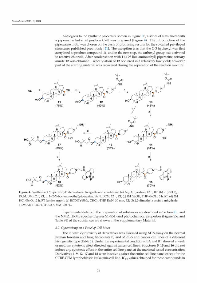
Biomedicines 2021, 9, 1104
Analogous to the synthetic procedure shown in Figure 3B, a series of substances witha piperazine linker at position C-28 was prepared (Figure 4). The introduction of thepiperazine motif was chosen on the basis of promising results for the so-called privilegedstructures published previously [21]. The exception was that the C-3 hydroxyl was firstacetylated to produce compound 11, and in the next step, the carboxyl group was activatedto reactive chloride. After condensation with 1-(2-N-Boc-aminoethyl) piperazine, tertiaryamide 12 was obtained. Deacetylation of 12 occurred in a relatively low yield; however,part of the starting material was recovered during the separation of the reaction mixture.
Figure 4. Synthesis of “piperazinyl” derivatives. Reagents and conditions: (a) Ac2O, pyridine, 12 h, RT; (b) i. (COCl)2,DCM, DMF, 2 h, RT, ii. 1-(2-N-boc-aminoethyl)piperazine, Et3N, DCM, 12 h, RT; (c) 4M NaOH, THF-MeOH, 3 h, RT; (d) 2MHCl/Et2O, 12 h, RT (under argon); (e) BODIPY-SMe, CHCl3-THF, Et3N, 30 min, RT; (f) 2,2-dimethyl succinic anhydride,4-DMAP, p-TsOH, THF, 2 h, MW-130 ◦C.
Experimental details of the preparation of substances are described in Section 2.1. andthe NMR, HRMS spectra (Figures S1–S51) and photochemical properties (Figure S52 andTable S1) of the substances are shown in the Supplementary Material.
3.2. Cytotoxicity on a Panel of Cell Lines
The in vitro cytotoxicity of derivatives was assessed using MTS assay on the normalhuman foreskin and lung fibroblasts BJ and MRC-5 and cancer cell lines of a differenthistogenetic type (Table 1). Under the experimental conditions, BA and BT showed a weakor medium cytotoxic effect directed against cancer cell lines. Structures 5, 15 and 16 did notinduce any cytotoxic effect in the entire cell line panel at the maximal tested concentration.Derivatives 4, 9, 12, 17 and 18 were inactive against the entire cell line panel except for theCCRF-CEM lymphoblastic leukaemia cell line. IC50 values obtained for these compounds in
74

Biomedicines 2021, 9, 1104
the sensitive cell line CCRF-CEM were between 5.76 and 23.65 μM. Derivatives 3, 6, 13 and14 exerted high cytotoxicity against the entire cell line panel, including normal fibroblasts.The most potent compounds in the study were structures 3 and 14 with IC50 values 0.21 and0.29 μM in CCRF-CEM. Derivatives 2, 8 and 10 displayed medium cytotoxicity across thecell line panel. Derivatives 1 and 7 showed activity only against selected cell lines inthe panel. Betulinic acid intermediate 11 was not tested. The MTS assay did not revealany effect directed specifically against cancer cell lines, IC50 values calculated for normalfibroblast and cancer cell lines were highly comparable. Resistant sublines CEM-DNR andK562-Tax displayed for some compounds different sensitivity compared to their parentalcell lines. As expected, a lower sensitivity was observed in the CEM-DNR resistant subline.The biggest difference in favour of CEM-DNR was observed for derivatives 6 and 3. BA
and 13 showed an opposite profile in CEM-DNR and 1, 2, 3, 8 and 10 in the K562-Taxresistant subline, proposing better activity in resistant cell lines. Based on this data, we canspeculate that there is a different mechanism in the elimination of cytotoxic derivatives.Several tested compounds are probably substrates of the P-glycoprotein as 4, 6, 13, 16, 17
and 18. However, not all data are in conclusion with P-glycoprotein transport, and we thinkthat several tested derivatives could be substrates for LRP protein. Higher LRP expressionin CEM-DNR and lower in K562-Tax correlates with cytotoxicity of the derivatives 1, 2,3, 8, 10. Derivative 13 is not active in the highly chemosensitive CCRF-CEM cell line, butcomparable activity was observed in all tested cell lines, including non-tumour lines.
3.3. Cell Cycle, Apoptosis and DNA/RNA Synthesis
To reveal cytostatic effects, we examined proliferation markers and cell cycle profile ofthe sensitive CCRF-CEM cell line following a 24 h incubation with the derivatives (Table 2).
Exposure to 1 × IC50 and 5 × IC50 concentrations of derivatives did not induce DNAfragmentation with the exception of high doses of 2 and 8. Treatment with 1 × IC50concentrations did not modulate cell cycle profile while 5 × IC50 concentration led in allsamples to a more pronounced effect. The treatment with 5 × IC50 derivatives 2, 12 and14 increased the percentage of cells in the S-phase by about 50% compared to untreatedcontrol. Nevertheless, there was not any other prominent effect on the cell cycle profile orcell cycle arrest. To assess the impact of structures on CCRF-CEM proliferation potential, wemonitored mitotic marker pH3Ser10 and proliferation marker BrDU after 24 h incubationwith the compounds. Analysis of mitotic marker showed a low rate of cell division incells treated with 5 × IC50 concentration of derivatives 2, 3, 6, 12 and 14. Derivatives 3,6, 8, 10, and 14 reduced the fraction of proliferating BrDU positive CCRF-CEM cells. Incontrast, structures 2, 12 and 18 increased the percentage of cells incorporating BrDU intothe DNA during pulse labelling. The complementary BrU based method of monitoringnewly synthesized RNA in cells pre-incubated for 24 with the selected derivatives revealedstalled RNA synthesis induced by 5 × IC50 concentration of derivatives 2, 3, 8, 10, 14 and 17.Compound 18 at a high concentration increased the percentage of BrU positive cells. Suchan increase indicates the high transcription activity as a mark of replication stress leadingto DNA damage and cell death [42]. Although there was observed a slight modulation ofcell cycle profile induced by compound derivatives 2, 12 and 14, the overall cell cycle dataindicates that there is no general cytostatic effect of tested betulinic acid derivatives.
75

Biomedicines 2021, 9, 1104
Ta
ble
1.
Sum
mar
yof
cyto
toxi
cac
tivi
ties
(IC
50,μ
M).
Co
mp
ou
nd
Ce
llL
ine
aB
AB
T1
23
46
78
91
01
21
31
41
61
71
8
CC
RF
-CE
M>5
012
.82
8.98
8.14
0.21
23.6
51.
55>5
08.
1822
.48
2.92
9.62
>50
0.29
>50
5.76
8.61
CE
M-D
NR
23.0
522
.17
16.8
410
.23
1.22
>50
11.5
3>5
09.
24>5
07.
00>5
04.
760.
35>5
0>5
0>5
0K
56
2>5
023
.60
>50
25.9
90.
90>5
05.
25>5
019
.18
>50
23.1
9>5
05.
090.
4047
.90
>50
>50
K5
62
-Ta
x>5
022
.03
10.5
815
.94
0.37
>50
31.8
0>5
013
.30
>50
12.2
1>5
08.
770.
52>5
0>5
0>5
0A
54
922
.68
23.0
6>5
022
.87
1.80
>50
6.65
21.2
918
.84
>50
13.5
5>5
05.
151.
2644
.93
>50
47.8
0H
CT
11
6>5
014
.17
>50
19.4
00.
82>5
03.
85>5
013
.21
>50
7.92
>50
6.02
0.39
46.6
3>5
030
.84
HC
T1
16
p5
3−
/−>5
018
.20
>50
29.2
40.
44>5
03.
39>5
021
.56
>50
8.80
>50
4.99
0.44
44.7
6>5
045
.50
U2
OS
29.6
927
.63
29.0
422
.22
0.89
>50
5.00
18.3
917
.16
>50
12.3
8>5
04.
160.
4244
.62
44.5
8>5
0M
RC
-5>5
0>5
0>5
024
.19
2.59
>50
8.07
17.5
823
.14
>50
14.1
2>5
05.
181.
5844
.61
>50
>50
BJ
>50
>50
>50
25.3
31.
91>5
08.
3720
.69
21.5
4>5
015
.49
>50
5.36
1.59
47.6
3>5
0>5
0a
Cyt
otox
icac
tivity
was
dete
rmin
edby
MTS
assa
yfo
llow
ing
3-da
yin
cuba
tion.
Val
ues
repr
esen
tmea
nsof
IC50
from
thre
ein
depe
nden
texp
erim
ents
with
SDra
ngin
gfr
om10
–25%
ofth
eav
erag
eva
lues
.Tes
ted
cell
lines
:CC
RF-
CE
M(c
hild
hood
Tac
ute
lym
phob
last
icle
ukae
mia
),C
EM
-DN
R(C
CR
F-C
EM
dau
noru
bici
nre
sist
ant)
,K56
2(c
hron
icm
yelo
geno
usle
ukae
mia
),K
562-
Tax
(K56
2pa
clit
axel
-res
ista
nt),
A54
9(l
ung
aden
ocar
cino
ma)
,HC
T11
6(c
olor
ecta
lcan
cer)
,HC
T11
6p53−/
−(n
ullp
53ge
ne),
and
U2O
S(o
steo
sarc
oma)
.Nor
mal
hum
ance
lllin
es:
MR
C-5
and
BJ(n
orm
alcy
clin
gfib
robl
asts
).BA
,bet
ulin
icac
id;B
T,be
viri
mat
.
76

Biomedicines 2021, 9, 1104
Table 2. Effect of cytotoxic compounds on cell cycle, apoptosis and DNA/RNA synthesis in CCRF-CEM lymphoblasts (% ofpositive cells).
Compound <G1 G0/G1 S G2/M pH3Ser10 a BrDU b BrU c
control 2.20 40.77 37.64 21.60 1.77 37.22 40.771 1.36 37.03 44.13 18.84 1.53 36.54 52.691 1.93 42.33 35.87 21.80 1.71 37.99 44.472 4.33 39.88 46.65 13.47 1.33 52.07 28.602 41.17 23.94 56.21 19.85 0.58 22.75 4.053 1.30 35.07 36.54 28.40 2.04 41.88 50.563 3.53 31.73 41.18 27.09 0.16 2.06 0.406 2.23 33.39 49.74 16.87 1.50 44.97 42.256 5.24 37.16 39.52 23.32 0.50 7.10 26.058 2.64 31.93 47.72 20.35 1.70 46.44 33.618 39.65 40.78 37.66 21.56 1.25 8.51 1.8510 3.80 37.98 44.59 17.43 1.44 42.25 35.0410 10.69 44.58 42.32 13.10 1.25 10.18 13.0212 2.45 33.64 43.16 23.20 1.40 59.73 41.9812 3.01 29.75 56.69 13.55 0.68 64.07 34.0014 8.63 21.46 56.29 22.25 0.15 24.55 1.5614 8.16 27.28 51.82 20.90 0.18 0.52 1.5317 2.89 34.83 46.96 18.21 1.56 48.07 26.7017 4.04 43.42 38.47 18.10 1.49 29.41 3.3918 2.06 36.06 44.31 19.63 1.25 55.81 46.9818 2.97 38.01 44.12 17.87 1.55 49.64 70.23
Flow cytometry analysis was used for quantification of cell cycle distribution and apoptotic cells with a concentration of compounds equalto 1 × IC50 ( ) and 5 × IC50 ( ) values. a phospho-Histone (Ser10); b 5-bromo-2-deoxyuridine; c BrU, 5-bromouridine.
3.4. Live Cells Imaging
The group of six derivatives of BA and BODIPY was studied on the U2OS-Nuc cellline with the nucleus labelled by fluorescein protein mCherry. The functionalized BODIPYdyes (BODIPY-CO2H and BODIPY-NH2), as well as precursor BODIPY-SMe, were usedas a control. All fluorescent microscopic images of this pilot experiment are shown inFigure S53. To achieve a better specificity of the staining, we have focused on the shortincubation with the fluorescent conjugates. After short incubation (1 h), conjugates 4 and 6
out of this group of derivatives were localized in living cells, but only with the weak signalin the nucleus of the studied cell line (Figure 5B—Pearson’s and Mander’s coefficients).The functionalized BODIPY dyes were not detected in the U2OS-Nuc cell line and thus itis highly possible that cellular uptake of conjugates 4 and 6 is due to their groups on BAresidue. Other studied derivatives of BA and BODIPY were not detected in living cellsunder our experimental conditions; however, it is possible that the signal can be observedat later intervals. BODIPY-SMe is reactive due to the 8-thiomethyl group and it waspredicted to penetrate cell compartments; this was confirmed by fluorescent microscopy.
To further study the cellular localization of conjugates 4 and 6, we decided to continuewith fluorescent microscopy on cell lines with fluorescently labelled structures of mitochon-dria, endoplasmic reticulum, and Golgi apparatus, which are the most published targetsof BA [29,30]. The results of these colocalization experiments are shown in Figure 5. Bothconjugates demonstrated presence in multiple cellular structures. Pearson’s coefficient(Figure 5B) showed the highest correlation of signal in U2OS-ER cell line, and then inU2OS-Mito cell line and the lowest correlation was measured in U2OS-GA cell line. Whenwe expressed colocalization by Mander’s coefficient (overlap of red channel compared tothe blue channel), which is more specific for colocalization calculation of signal presentedin multiple cellular structures, the obtained data showed both conjugates 4 and 6 almostperfectly label mitochondria and endoplasmic reticulum. The lowest colocalization signalwas again detected in the U2OS-GA cell line. BODIPY-SMe was used based on the data
77
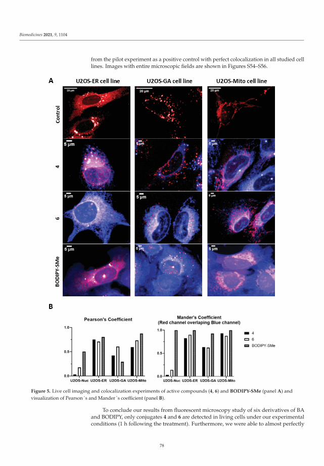
Biomedicines 2021, 9, 1104
from the pilot experiment as a positive control with perfect colocalization in all studied celllines. Images with entire microscopic fields are shown in Figures S54–S56.
Figure 5. Live cell imaging and colocalization experiments of active compounds (4, 6) and BODIPY-SMe (panel A) andvisualization of Pearson´s and Mander´s coefficient (panel B).
To conclude our results from fluorescent microscopy study of six derivatives of BAand BODIPY, only conjugates 4 and 6 are detected in living cells under our experimentalconditions (1 h following the treatment). Furthermore, we were able to almost perfectly
78

Biomedicines 2021, 9, 1104
colocalize both conjugates with cellular structures as endoplasmic reticulum and mito-chondria, which is in agreement with data published in the past [21]. Compound 4 has,in its structure, fluorophore attached to the carboxyl group at the C-28 position, thusit is more similar to the pristine structure of BA and has low cytotoxicity (close to freeBA). Conversely, compound 6 contains a conjugated amine at the C-28 position and thefluorophore is attached to the hydroxyl group at the C-3 position of BA. Its cytotoxicityis markedly more pronounced than in the case of substance 4. It is clear that the “polarhead” of the molecule is responsible for cytotoxicity. Moreover, this moiety can be used forthe intracellular targeted delivery, or organelle/mitochondrion targeting, as is describedby the previous research works [43,44]. As the localization of both compounds is similar,it is likely that the direct target remained unchanged, but the effect of compound 6 waspotentiated by the presence of free amine moiety in the molecule. The localization of thecompounds in lipid rich compartments (mitochondria, endoplasmic reticulum) can alsobe explained by the lipid character of the BA and its analogues. The calculated values oflipophilicity (logP) of the substances are close to BA (Table S2). The acidity constants (pKa)are indicative and their reproducibility is difficult because, in comparison with BA, thecompounds described here are mostly in the form of amide or ester derivatives.
3.5. Anti-HIV Activity
Bevirimat (3-O-(3′,3′-dimethylsuccinyl) betulinic acid) and its derivatives were shownto be maturation inhibitors of HIV-1 [45–47]. By binding to the CA-SP1 region of HIV-1 Gag polyprotein, bevirimat prevents HIV-1 protease-mediated release of C-terminalpart of CA from a spacer peptide 1 (SP1) [48]. This results in a block of the final step ofvirus maturation and subsequently abolishes HIV-1 infectivity. An atomic model of HIV-1CA-SP1 suggested that this inhibitor stabilizes the CA-SP1 structure, thus preventing theproteolytic cleavage [49]. Although bevirimat is a potent inhibitor of HIV-1 maturation, itsclinical development was discontinued in 2010 due to the bevirimat resistance caused byGag SP1 natural polymorphism (Q6, V7 and T8) [50–52]. However, bevirimat derivativeswith modification at the C-28 position seem to overcome the problem with HIV-1 resis-tance [53,54]. Here, using VSV-G pseudotyped HIV-1 particles, we tested the effect of 17 BAderivatives on HIV-1 maturation and infectivity. The 50% cytotoxic concentration (IC50)of the compounds was first evaluated by Resazurin assay. Two of the tested compounds,3 and 14, were highly toxic to HEK 293 cells at a concentration lower than 5 μM andsignificant cytotoxicity was also found for compound 6 (IC50 12 μM) (Table 3).
Table 3. Cytotoxicity and anti-HIV-1 activity of the tested compounds a.
Compd. 1 2 4 5 6 7 8 9 10 12 13 15 16 17 18
IC50 [μM] >40 36.4 >40 >40 12.0 >40 37.8 >40 >40 >40 >40 >40 >40 >40 >40IC50i [μM] >50 11.7 44.1 >50 n.d. >50 1.4 14.0 8.4 31.9 >50 >50 9.1 7.6 7.1
a HEK 293 cells were grown in the presence or absence of tested compounds at a concentration ranging from 5 to 40 μM. The viability of thecells was determined by Resazurin assay 48 h later ( ). To determine the effect of the compounds on HIV-1 infectivity ( ), HEK 293 cellswere transfected with the lentiviral vectors and treated with the tested compounds. The cells producing HIV-1 particles in the presenceor absence of DMSO (at a final concentration of 1%) were used as controls. At 48 h post-transfection, the content of HIV-1 capsid (CA)protein from the culture media was quantified by ELISA and normalized amounts of VSV-G pseudotyped HIV-1 viruses were used to infectfresh HEK 293 cells. HIV-1 infectivity was determined 48 h later by quantification of GFP-producing cells by flow cytometry. The 50%infection inhibition (IC50i) was defined as the concentration of the compound that reduced the HIV-1 infectivity by 50% compared to theuntreated controls.
Apart from these three cytotoxic compounds, 14 fewer toxic compounds were usedin the HIV-1 single-round infectivity assay. HIV-1 particles pseudotyped with VSV gly-coproteins were produced in HEK 293 cells in the presence of tested compounds. At48 h post-transfection, the content of HIV-1 capsid (CA) protein from the culture mediawas quantified by ELISA and normalized amounts of VSV-G pseudotyped HIV-1 viruseswere used to infect fresh HEK 293 cells. At 48 h post-infection, the HIV-1 infectivity wasdetermined by quantification of GFP-producing cells by flow cytometry. The 50% infection
79

Biomedicines 2021, 9, 1104
inhibition (IC50i) was defined as the concentration of the compound that reduced the HIV-1infectivity by 50% compared to the untreated controls (Table 3). The compounds 1, 7,13, 15 and 5 did not exhibit any potent anti-HIV-1 activity (data not shown). Conversely,compounds 2, 4, 9 and 12 inhibited anti-HIV-1 activity with IC50i from 11.7 to 44.1 μM.The compounds 8, 10, 16, 17 and 18 inhibited HIV-1 with IC50i below 10 μM (Table 3). Toanalyse whether these bevirimat derivatives also act as maturation inhibitors of CA-SP1cleavage, the HIV-1 virions released from the HEK 293 cells treated with the selectedcompounds (2, 4, 8, 9, 10, 12, 16, 17 and 18) were analysed by Western blot using anti-HIV-1CA antibody (Figure 6).
Figure 6. Effect of selected tested compounds on CA-SP1 processing of HIV-1 Gag polyprotein. HEK 293 cells producedHIV-1 particles pseudotyped with VSV-glycoproteins in the absence (lanes 2 and 3) or presence of selected tested compounds(lanes 4–12). At 48 h post-transfection, VSV-G pseudotyped HIV-1 viruses released from the HEK 293 cells were analysed byWestern blot using an anti-HIV-1 CA antibody (duplicate of blot shown in Figure S57).
Only completely processed p24 CA of molecular weight of 24 kDa was identifiedin the viruses formed in the presence of compounds 4 and 12. However, in the samplestreated with compounds 2, 8, 9, 10, 16, 17 and 18, we identified not only fully processedp24 CA, but also p25 CA-SP1 protein. This observation suggests a similar mechanism ofinhibition as described for bevirimat, i.e., the block of the final step of HIV-1 maturation.
4. Conclusions
This study describes the synthesis and biological evaluation of 17 betulinic acidderivatives. The biological profiling revealed that BA derivatives 3 and 14 with modificationat C-28 show increased cytotoxicity. However, the cytotoxicity was not specifically directedagainst cancer cell lines and was not associated with cell cycle arrest. The most effectivecompounds with sub-micromolar IC50 values 3 and 14 possess a hydroxyl group at C-3,whereas structures with a succinyl hemiester group displayed medium cytotoxicity orwere inactive. The study introduced six original structures with BODIPY moiety linkedto the lupane skeleton. BODIPY conjugates 4, 5, 15 and 18 showed low or no cytotoxicactivity. In contrast, BODIPY derivative 6 induced strong and derivative 10 mediumcytotoxicity in the entire cell line panel, although they do not share any similar substituentsat positions C-3 and C-28. The cellular localization of BODIPY conjugates was furtherstudied in U2OS cells using fluorescent microscopy. Fluorescent derivatives 4 and 6
colocalized with endoplasmic reticulum and mitochondria, which is in agreement withprevious studies showing interaction with the processes and proteins localized in theseorganelles [55,56]. Uncoupling of the mitochondrial respiration, followed by radical burstand mitochondrial membrane disruption, is one of the well-described effects of betulin andbetulinic acid [57–60]. Thus, we believe that reliable tools to study the derivatives of BA onliving cells were established. The anti-HIV-1 activity showed that compounds 2, 8, 9, 10,16 and 18 with IC50i lower than 10 μM did not fully process the p24 CA and p25 CA-SP1proteins, suggesting a similar mechanism of inhibition as described for bevirimat.
80
Biomedicines 2021, 9, 1104
Supplementary Materials: The following are available online at https://www.mdpi.com/article/10.3390/biomedicines9091104/s1. Supplementary Figures S1–S52 and Table S1 document the analyticalidentification (NMR, HRMS, UV-Vis and fluorescence). Table S2: Calculated physical properties(pKa and logP) of the derivatives. Figures S53–S56 show supplementary pictures from fluorescentmicroscopy. Figure S57: Effect of selected tested compounds on CA-SP1 processing of HIV-1 Gagpolyprotein (a duplicate of western blot showed in Figure 5. in the article).
Author Contributions: M.J., P.D., P.B.D., M.R. and M.H. conceived and designed the experiments;D.K., T.Z., M.J., P.D., I.K., J.S., S.G., J.R. and P.D. performed the experiments; M.J., P.D. and M.R.analysed the data; M.J., P.D. and M.R. prepared the manuscript; P.B.D. and M.H. edited the article.All authors have read and agreed to the published version of the manuscript.
Funding: This work was supported by GA CR (CZ) GA20-19906S, by the Czech Ministry of Education,Youth and Sports (CZ-OPENSCREEN-LM2018130, and EATRIS-CZ-LM2018133, Czech-BioImaging-LM2018129) and internal grant of Palacky University (IGA_LF_2021_038).
Conflicts of Interest: The authors declare that they have no known competing financial interests orpersonal relationships that could have appeared to influence the work reported in this paper.
References
1. Sousa, J.L.C.; Freire, C.S.R.; Silvestre, A.J.D.; Silva, A.M.S. Recent developments in the functionalization of betulinic acid and itsnatural analogues: A route to new bioactive compounds. Molecules 2019, 24, 355. [CrossRef]
2. Zuco, V.; Supino, R.; Righetti, S.C.; Cleris, L.; Marchesi, E.; Gambacorti-Passerini, C.; Formelli, F. Selective cytotoxicity of betulinicacid on tumor cell lines, but not on normal cells. Cancer Lett. 2002, 175, 17–25. [CrossRef]
3. Pisha, E.; Chai, H.; Lee, I.S.; Chagwedera, T.E.; Farnsworth, N.R.; Cordell, G.A.; Beecher, C.W.W.; Fong, H.H.S.; Kinghorn,A.D.; Brown, D.M.; et al. Discovery of betulinic acid as a selective inhibitor of human melanoma that functions by induction ofapoptosis. Nat. Med. 1995, 1, 1046–1051. [CrossRef] [PubMed]
4. Noda, Y.; Kaiya, T.; Kohda, K.; Kawazoe, Y. Enhanced cytotoxicity of some triterpenes toward leukemia L1210 cells cultured inlow pH media: Possibility of a New Mode of Cell Killing. Chem. Pharm. Bull. 1997, 45, 1665–1670. [CrossRef] [PubMed]
5. Fujioka, T.; Kashiwada, Y.; Kilkuskie, R.E.; Cosentino, L.M.; Ballas, L.M.; Jiang, J.B.; Janzen, W.P.; Chen, I.-S.; Lee, K.-H. Anti-AIDSagents, 11. Betulinic acid and platanic acid as anti-HIV principles from Syzigium claviflorum, and the anti-HIV activity ofstructurally related triterpenoids. J. Nat. Prod. 1994, 57, 243–247. [CrossRef]
6. Kodr, D.; Rumlová, M.; Zimmermann, T.; Džubák, P.; Drašar, P.; Jurášek, M. Antitumor and anti-HIV derivatives of betulinic acid.Chem. Listy 2020, 114, 658–667.
7. Fulda, S.; Friesen, C.; Los, M.; Scaffidi, C.; Mier, W.; Benedict, M.; Nunez, G.; Krammer, P.H.; Peter, M.E.; Debatin, K.M. Betulinicacid triggers CD95 (APO-1/Fas)- and p53-independent apoptosis via activation of caspases in neuroectodermal tumors. CancerRes. 1997, 57, 4956–4964. [PubMed]
8. Fulda, S.; Scaffidi, C.; Susin, S.A.; Krammer, P.H.; Kroemer, G.; Peter, M.E.; Debatin, K.M. Activation of mitochondria and releaseof mitochondrial apoptogenic factors by betulinic acid. J. Biol. Chem. 1998, 273, 33942–33948. [CrossRef]
9. Liu, W.K.; Ho, J.C.K.; Cheung, F.W.K.; Liu, B.P.L.; Ye, W.C.; Che, C.T. Apoptotic activity of betulinic acid derivatives on murinemelanoma B16 cell line. Eur. J. Pharmacol. 2004, 498, 71–78. [CrossRef]
10. Fulda, S.; Jeremias, I.; Steiner, H.H.; Pietsch, T.; Debatin, K.M. Betulinic acid: A new cytotoxic agent against malignant brain-tumorcells. Int. J. Cancer 1999, 82, 435–441. [CrossRef]
11. Mullauer, F.B.; van Bloois, L.; Daalhuisen, J.B.; Ten Brink, M.S.; Storm, G.; Medema, J.P.; Schiffelers, R.M.; Kessler, J.H. Betulinicacid delivered in liposomes reduces growth of human lung and colon cancers in mice without causing systemic toxicity.Anti-Cancer Drug 2011, 22, 223–233. [CrossRef]
12. Takada, Y.; Aggarwal, B.B. Betulinic acid suppresses carcinogen-induced NF-kappa B activation through inhibition of I kappa Balpha kinase and p65 phosphorylation: Abrogation of cyclooxygenase-2 and matrix metalloprotease-9. J. Immunol. 2003, 171,3278–3286. [CrossRef]
13. Melzig, M.F.; Bormann, H. Betulinic acid inhibits aminopeptidase N activity. Planta Med. 1998, 64, 655–657. [CrossRef]14. Kwon, H.J.; Shim, J.S.; Kim, J.H.; Cho, H.Y.; Yum, Y.N.; Kim, S.H.; Yu, J. Betulinic acid inhibits growth factor-induced in vitro
angiogenesis via the modulation of mitochondrial function in endothelial cells. Jpn. J. Cancer Res. 2002, 93, 417–425. [CrossRef]15. Kashiwada, Y.; Nagao, T.; Hashimoto, A.; Ikeshiro, Y.; Okabe, H.; Cosentino, L.M.; Lee, K.-H. Anti-AIDS agents 38. Anti-HIV
activity of 3-O-acyl ursolic acid derivatives. J. Nat. Prod. 2000, 63, 1619–1622. [CrossRef] [PubMed]16. Sundquist, W.I.; Krausslich, H.G. HIV-1 Assembly, budding, and maturation. Cold Spring Harb. Perspect. Med. 2012, 2, a006924.
[CrossRef] [PubMed]17. Smith, P.F.; Ogundele, A.; Forrest, A.; Wilton, J.; Salzwedel, K.; Doto, J.; Allaway, G.P.; Martin, D.E. Phase I and II study of
the safety, virologic effect, and pharmacokinetics/pharmacodynamics of single-dose 3-O-(3′,3′-dimethylsuccinyl)betulinic acid(bevirimat) against human immunodeficiency virus infection. Antimicrob. Agents Chemother. 2007, 51, 3574–3581. [CrossRef][PubMed]
81
Biomedicines 2021, 9, 1104
18. Martin, D.E.; Blum, R.; Wilton, J.; Doto, J.; Galbraith, H.; Burgess, G.L.; Smith, P.C.; Ballow, C. Safety and pharmacokinetics ofbevirimat (PA-457), a novel inhibitor of human immunodeficiency virus maturation, in healthy volunteers. Antimicrob. AgentsChemother. 2007, 51, 3063. [CrossRef]
19. Martin, D.E.; Blum, R.; Doto, J.; Galbraith, H.; Ballow, C. Multiple-Dose Pharmacokinetics and safety of bevirimat, a novelinhibitor of HIV maturation, in healthy volunteers. Clin. Pharmacokinet. 2007, 46, 589–598. [CrossRef] [PubMed]
20. Margot, N.A.; Gibbs, C.S.; Miller, M.D. Phenotypic susceptibility to bevirimat in isolates from HIV-1-infected patients withoutprior exposure to bevirimat. Antimicrob. Agents Chemother. 2010, 54, 2345–2353. [CrossRef] [PubMed]
21. Zhao, Y.; Gu, Q.; Morris-Natschke, S.L.; Chen, C.-H.; Lee, K.-H. Incorporation of privileged structures into bevirimat can improveactivity against wild-type and bevirimat-resistant HIV-1. J. Med. Chem. 2016, 59, 9262–9268. [CrossRef]
22. Zhao, Y.; Chen, C.-H.; Morris-Natschke, S.L.; Lee, K.-H. Design, synthesis, and structure activity relationship analysis of newbetulinic acid derivatives as potent HIV inhibitors. Eur. J. Med. Chem. 2021, 215, 113287. [CrossRef]
23. Mukherjee, R.; Jaggi, M.; Rajendran, P.; Siddiqui, M.J.A.; Srivastava, S.K.; Vardhan, A.; Burman, A.C. Betulinic acid and itsderivatives as anti-angiogenic agents. Bioorg. Med. Chem. Lett. 2004, 14, 2181–2184. [CrossRef]
24. Kim, J.Y.; Koo, H.M.; Kim, D.S.H.L. Development of C-20 modified betulinic acid derivatives as antitumor agents. Bioorg. Med.Chem. Lett. 2001, 11, 2405–2408. [CrossRef]
25. Chowdhury, A.R.; Mandal, S.; Mittra, B.; Sharma, S.; Mukhopadhyay, S.; Majumder, H.K. Betulinic acid, a potent inhibitor ofeukaryotic topoisomerase I: Identification of the inhibitory step, the major functional group responsible and development ofmore potent derivatives. Med. Sci. Monit. 2002, 8, BR254–BR265. [PubMed]
26. Bildziukevich, U.; Rarova, L.; Janovska, L.; Saman, D.; Wimmer, Z. Enhancing effect of cystamine in its amides with betulinic acidas antimicrobial and antitumor agent in vitro. Steroids 2019, 148, 91–98. [CrossRef] [PubMed]
27. Bildziukevich, U.; Vida, N.; Rárová, L.; Kolár, M.; Šaman, D.; Havlícek, L.; Drašar, P.; Wimmer, Z. Polyamine derivatives ofbetulinic acid and beta-sitosterol: A comparative investigation. Steroids 2015, 100, 27–35. [CrossRef] [PubMed]
28. Brandes, B.; Hoenke, S.; Fischer, L.; Csuk, R. Design, synthesis and cytotoxicity of BODIPY-FL labelled triterpenoids. Eur. J. Med.Chem. 2020, 185, 111858. [CrossRef] [PubMed]
29. Krajcovicová, S.; Stanková, J.; Džubák, P.; Hajdúch, M.; Soural, M.; Urban, M. A synthetic approach for the rapid preparationof BODIPY conjugates and their use in imaging of cellular drug uptake and distribution. Chem. Eur. J. 2018, 24, 4957–4966.[CrossRef] [PubMed]
30. Sommerwerk, S.; Heller, L.; Kerzig, C.; Kramell, A.E.; Csuk, R. Rhodamine B conjugates of triterpenoic acids are cytotoxicmitocans even at nanomolar concentrations. Eur. J. Med. Chem. 2017, 127, 1–9. [CrossRef]
31. Pal, A.; Ganguly, A.; Chowdhuri, S.; Yousuf, M.; Ghosh, A.; Barui, A.K.; Kotcherlakota, R.; Adhikari, S.; Banerjee, R. Bis-arylideneoxindole-betulinic acid conjugate: A fluorescent cancer cell detector with potent anticancer activity. ACS Med. Chem. Lett. 2015, 6,612–616. [CrossRef] [PubMed]
32. Rumlová, M.; Krížová, I.; Keprová, A.; Hadravová, R.; Doležal, M.; Strohalmová, K.; Pichová, I.; Hájek, M.; Ruml, T. HIV-1protease-induced apoptosis. Retrovirology 2014, 11, 37. [CrossRef]
33. Dostálková, A.; Kaufman, F.; Krížová, I.; Kultová, A.; Strohalmová, K.; Hadravová, R.; Ruml, T.; Rumlová, M. Mutations in thebasic region of the Mason-Pfizer monkey virus nucleocapsid protein affect reverse transcription, genomic RNA packaging, andthe virus assembly site. J. Virol. 2018, 92, e00106-18. [CrossRef]
34. Krížová, I.; Hadravová, R.; Štokrová, J.; Günterová, J.; Doležal, M.; Ruml, T.; Rumlová, M.; Pichová, I. The G-patch domain ofMason-Pfizer monkey virus is a part of reverse transcriptase. J. Virol. 2012, 86, 1988. [CrossRef] [PubMed]
35. Strohalmová-Bohmová, K.; Spiwok, V.; Lepšík, M.; Hadravová, R.; Krížová, I.; Ulbrich, P.; Pichová, I.; Bednárová, L.; Ruml, T.;Rumlová, M. Role of Mason-Pfizer monkey virus CA-NC spacer peptide-like domain in assembly of immature particles. J. Virol.2014, 88, 14148. [CrossRef]
36. Goud, T.V.; Tutar, A.; Biellmann, J.-F. Synthesis of 8-heteroatom-substituted 4,4-difluoro-4-bora-3a,4a-diaza-s-indacene dyes(BODIPY). Tetrahedron 2006, 62, 5084–5091. [CrossRef]
37. Kim, D.; Ma, D.; Kim, M.; Jung, Y.; Kim, N.H.; Lee, C.; Cho, S.W.; Park, S.; Huh, Y.; Jung, J.; et al. Fluorescent labeling of proteinusing blue-emitting 8-amino-BODIPY derivatives. J. Fluoresc. 2017, 27, 2231–2238. [CrossRef]
38. Chang, Y.-T.; Alamudi, S.H.; Satapathy, R.; Su, D. Background-free fluorescent probes for live cell imaging. US PatentWO2017078623A1, 2017.
39. Qian, K.; Bori, I.D.; Chen, C.-H.; Huang, L.; Lee, K.-H. Anti-AIDS agents 90. Novel C-28 modified bevirimat analogues as potentHIV maturation inhibitors. J. Med. Chem. 2012, 55, 8128–8136. [CrossRef] [PubMed]
40. Staudinger, H.; Meyer, J. Über neue organische Phosphorverbindungen III. Phosphinmethylenderivate und Phosphinimine. Helv.Chim. Acta 1919, 2, 635–646. [CrossRef]
41. Neises, B.; Steglich, W. Simple method for the esterification of carboxylic acids. Angew. Chem. Int. Ed. 1978, 17, 522–524. [CrossRef]42. Kotsantis, P.; Silva, L.M.; Irmscher, S.; Jones, R.M.; Folkes, L.; Gromak, N.; Petermann, E. Increased global transcription activity as
a mechanism of replication stress in cancer. Nat. Commun. 2016, 7, 13087. [CrossRef] [PubMed]43. Fantin, V.R.; St-Pierre, J.; Leder, P. Attenuation of LDH-A expression uncovers a link between glycolysis, mitochondrial physiology,
and tumor maintenance. Cancer Cell 2006, 9, 425–434. [CrossRef] [PubMed]
82

Biomedicines 2021, 9, 1104
44. Zielonka, J.; Joseph, J.; Sikora, A.; Hardy, M.; Ouari, O.; Vasquez-Vivar, J.; Cheng, G.; Lopez, M.; Kalyanaraman, B. Mitochondria-targeted triphenylphosphonium-based compounds: Syntheses, mechanisms of action, and therapeutic and diagnostic applications.Chem. Rev. 2017, 117, 10043–10120. [CrossRef] [PubMed]
45. Evers, M.; Poujade, C.; Soler, F.; Ribeill, Y.; James, C.; Lelievre, Y.; Gueguen, J.C.; Reisdorf, D.; Morize, I.; Pauwels, R.; et al.Betulinic acid derivatives: A new class of human immunodeficiency virus type 1 specific inhibitors with a new mode of action.J. Med. Chem. 1996, 39, 1056–1068. [CrossRef]
46. Kashiwada, Y.; Hashimoto, F.; Cosentino, L.M.; Chen, C.H.; Garrett, P.E.; Lee, K.H. Betulinic acid and dihydrobetulinic acidderivatives as potent anti-HIV agents. J. Med. Chem. 1996, 39, 1016–1017. [CrossRef]
47. Soler, F.; Poujade, C.; Evers, M.; Carry, J.C.; Henin, Y.; Bousseau, A.; Huet, T.; Pauwels, R.; DeClercq, E.; Mayaux, J.F.; et al.Betulinic acid derivatives: A new class of specific inhibitors of human immunodeficiency virus type 1 entry. J. Med. Chem. 1996,39, 1069–1083. [CrossRef]
48. Li, F.; Goila-Gaur, R.; Salzwedel, K.; Kilgore, N.R.; Reddick, M.; Matallana, C.; Castillo, A.; Zoumplis, D.; Martin, D.E.; Orenstein,J.M.; et al. PA-457: A potent HIV inhibitor that disrupts core condensation by targeting a late step in Gag processing. Proc. Natl.Acad. Sci. USA 2003, 100, 13555–13560. [CrossRef]
49. Schur, F.K.M.; Obr, M.; Hagen, W.J.H.; Wan, W.; Jakobi, A.J.; Kirkpatrick, J.M.; Sachse, C.; Krausslich, H.G.; Briggs, J.A.G. Anatomic model of HIV-1 capsid-SP1 reveals structures regulating assembly and maturation. Science 2016, 353, 506–508. [CrossRef]
50. Adamson, C.S.; Sakalian, M.; Salzwedel, K.; Freed, E.O. Polymorphisms in Gag spacer peptide 1 confer varying levels of resistanceto the HIV-1 maturation inhibitor bevirimat. Retrovirology 2010, 7, 1–8. [CrossRef]
51. Lu, W.X.; Salzwedel, K.; Wang, D.; Chakravarty, S.; Freed, E.O.; Wild, C.T.; Li, F. A single polymorphism in HIV-1 subtype C SP1is sufficient to confer natural resistance to the maturation inhibitor bevirimat. Antimicrob. Agents Chemother. 2011, 55, 3324–3329.[CrossRef]
52. Van Baelen, K.; Salzwedel, K.; Rondelez, E.; Van Eygen, V.; De Vos, S.; Verheyen, A.; Steegen, K.; Verlinden, Y.; Allaway, G.P.;Stuyver, L.J. Susceptibility of human immunodeficiency virus type 1 to the maturation inhibitor bevirimat is modulated bybaseline polymorphisms in Gag spacer peptide 1. Antimicrob. Agents Chemother. 2009, 53, 2185–2188. [CrossRef]
53. Coric, P.; Turcaud, S.; Souquet, F.; Briant, L.; Gay, B.; Royer, J.; Chazal, N.; Bouaziz, S. Synthesis and biological evaluation of a newderivative of bevirimat that targets the Gag CA-SP1 cleavage site. Eur. J. Med. Chem. 2013, 62, 453–465. [CrossRef]
54. Wang, D.; Lu, W.X.; Li, F. Pharmacological intervention of HIV-1 maturation. Acta Pharm. Sin. B 2015, 5, 493–499. [CrossRef]55. Gu, M.; Zhao, P.; Zhang, S.Y.; Fan, S.J.; Yang, L.; Tong, Q.C.; Ji, G.; Huan, C. Betulinic acid alleviates endoplasmic reticulum
stress-mediated nonalcoholic fatty liver disease through activation of farnesoid X receptors in mice. Brit. J. Pharmacol. 2019, 176,847–863. [CrossRef]
56. Ye, Y.Q.; Zhang, T.; Yuan, H.Q.; Li, D.F.; Lou, H.X.; Fan, P.H. Mitochondria-targeted lupane triterpenoid derivatives and theirselective apoptosis-inducing anticancer mechanisms. J. Med. Chem. 2017, 60, 6353–6363. [CrossRef] [PubMed]
57. Dubinin, M.V.; Semenova, A.A.; Ilzorkina, A.I.; Mikheeva, I.B.; Yashin, V.A.; Penkov, N.V.; Vydrina, V.A.; Ishmuratov, G.Y.;Sharapov, V.A.; Khoroshavina, E.I.; et al. Effect of betulin and betulonic acid on isolated rat liver mitochondria and liposomes.Biochim. Biophys. Acta-Biomembr. 2020, 1862. [CrossRef] [PubMed]
58. Dubinin, M.V.; Semenova, A.A.; Nedopekina, D.A.; Davletshin, E.V.; Spivak, A.Y.; Belosludtsev, K.N. Effect of F16-betulinconjugate on mitochondrial membranes and its role in cell death initiation. Membranes 2021, 11, 352. [CrossRef]
59. Dubinin, M.V.; Semenova, A.A.; Ilzorkina, A.I.; Penkov, N.V.; Nedopekina, D.A.; Sharapov, V.A.; Khoroshavina, E.I.; Davletshin,E.V.; Belosludtseva, N.V.; Spivak, A.Y.; et al. Mitochondria-targeted prooxidant effects of betulinic acid conjugated withdelocalized lipophilic cation F16. Free Radic. Bio. Med. 2021, 168, 55–69. [CrossRef] [PubMed]
60. Wang, X.; Lu, X.C.; Zhu, R.L.; Zhang, K.X.; Li, S.; Chen, Z.J.; Li, L.X. Betulinic acid induces apoptosis in differentiated PC12 cellsvia ROS-mediated mitochondrial pathway. Neurochem. Res. 2017, 42, 1130–1140. [CrossRef] [PubMed]
83

biomedicines
Article
The Combined Effect of Branching and Elongation on theBioactivity Profile of Phytocannabinoids. Part I: Thermo-TRPs
Daiana Mattoteia 1,†, Aniello Schiano Moriello 2,3,†, Orazio Taglialatela-Scafati 4, Pietro Amodeo 2,
Luciano De Petrocellis 2, Giovanni Appendino 1, Rosa Maria Vitale 2,* and Diego Caprioglio 1,*
Citation: Mattoteia, D.; Schiano
Moriello, A.; Taglialatela-Scafati, O.;
Amodeo, P.; De Petrocellis, L.;
Appendino, G.; Vitale, R.M.;
Caprioglio, D. The Combined Effect
of Branching and Elongation on the
Bioactivity Profile of
Phytocannabinoids. Part I:
Thermo-TRPs. Biomedicines 2021, 9,
1070. https://doi.org/10.3390/
biomedicines9081070
Academic Editor: Pavel B. Drašar
Received: 28 July 2021
Accepted: 18 August 2021
Published: 23 August 2021
Publisher’s Note: MDPI stays neutral
with regard to jurisdictional claims in
published maps and institutional affil-
iations.
Copyright: © 2021 by the authors.
Licensee MDPI, Basel, Switzerland.
This article is an open access article
distributed under the terms and
conditions of the Creative Commons
Attribution (CC BY) license (https://
creativecommons.org/licenses/by/
4.0/).
1 Dipartimento di Scienze del Farmaco, Università del Piemonte Orientale, Largo Donegani 2,28100 Novara, Italy; [email protected] (D.M.); [email protected] (G.A.)
2 Institute of Biomolecular Chemistry, National Research Council (ICB-CNR), Via Campi Flegrei 34,80078 Pozzuoli, Italy; [email protected] (A.S.M.); [email protected] (P.A.);[email protected] (L.D.P.)
3 Epitech Group SpA, Saccolongo, 35100 Padova, Italy4 Dipartimento di Farmacia, Università di Napoli Federico II, Via Montesano 49, 80131 Napoli, Italy;
[email protected]* Correspondence: [email protected] (R.M.V.); [email protected] (D.C.);
Tel.: +39-081-8675316 (R.M.V.); +39-0321-375843 (D.C.)† These authors contributed equally to this work.
Abstract: The affinity of cannabinoids for their CB1 and CB2 metabotropic receptors is dramaticallyaffected by a combination of α-branching and elongation of their alkyl substituent, a maneuverexemplified by the n-pentyl -> α,α-dimethylheptyl (DMH) swap. The effect of this change on othercannabinoid end-points is still unknown, an observation surprising since thermo-TRPs are targetedby phytocannabinoids with often sub-micromolar affinity. To fill this gap, the α,α-dimethylheptylanalogues of the five major phytocannabinoids [CBD (1a), Δ8-THC (6a), CBG (7a), CBC (8a) andCBN (9a)] were prepared by total synthesis, and their activity on thermo-TRPs (TRPV1-4, TRPM8,and TRPA1) was compared with that of one of their natural analogues. Surprisingly, the DMHchain promoted a shift in the selectivity toward TRPA1, a target involved in pain and inflammatorydiseases, in all investigated compounds. A comparative study of the putative binding modes atTRPA1 between DMH-CBC (8b), the most active compound within the series, and CBC (8a) wascarried out by molecular docking, allowing the rationalization of their activity in terms of structure–activity relationships. Taken together, these observations qualify DMH-CBC (8b) as a non-covalentTRPA1-selective cannabinoid lead that is worthy of additional investigation as an analgesic andanti-inflammatory agent.
Keywords: phytocannabinoids; cannabichromene; thermos-TRPs; TRPA1; α,α-dimethylheptyl effect
1. Introduction
Phytocannabinoids from cannabis (Cannabis sativa L.) are characterized by a linearalkyl substituent spanning one to six carbons bound to their resorcinol core, with thepentyl substitution being by far the most common one [1]. The existence of branchedchain cannabinoids of the iso- and ante-iso series is, in principle, plausible from a biogeneticstandpoint, since the alkylresorcinol moiety of phytocannabinoids is of ketide derivation,and branching would therefore simply require the replacement of the acetate-derived starterwith one derived from a branched amino acid. However, branched phytocannabinoidsof this type have only been tentatively detected as trace constituents of cannabis, largelyremaining unconfirmed curiosities in its inventory of constituents [1]. Conversely, non-biogenetic branching at the benzyl carbon of the alkyl residue has played a critical role inresearch on cannabis and cannabinoids ever since the early synthesis of these compoundsby Adams in the early forties of the past century [2]. Adams was unsuccessful in theisolation of the narcotic principle of cannabis, but the generation of a mixture of narcotic
Biomedicines 2021, 9, 1070. https://doi.org/10.3390/biomedicines9081070 https://www.mdpi.com/journal/biomedicines85

Biomedicines 2021, 9, 1070
tetrahydrocannabinols from the acidic treatment of cannabidiol (CBD, Figure 1, 1a) madehim correctly postulate a structure of this type for this elusive principle, whose structurewas eventually established as 2 by Mechoulam two decades later [3].
Figure 1. Structures of CBD (1a), its α,α-dimethylheptyl analogue (1b), Δ9-THC (2), Δ6a,10a THC (3a),pyrahexyl (3b), CP-55.940 (4), and cannabicyclohexanol (5).
In the course of these studies, Adams discovered that Δ6a,10a-tetrahydrocannabinol (3a),an unnatural compound relatively easily available by total synthesis, could replicate mostof the activity of the elusive narcotic principle of cannabis, and investigated the activity ofanalogues where the n-pentyl substituent of the model compound was replaced by longer,shorter, and branched alkyl groups [2]. During these studies, the remarkable potency ofcompounds bearing substituents on the benzyl and the homobenzyl positions of the alkylgroup was discovered. One of these compounds, named pyrahexyl (3b) [4], was next devel-oped as an incapacitating non-lethal war weapon and as anti-riot agent, but these studiesterminated when the US signed the convention on the ban of chemical weapons in the earlysixties [2]. A decade later, capitalizing on these observations, Pfizer chemists developeda series of ultra-potent and less lipophilic analogues of Δ9-tetrahydrocannabinol (2) thatproved instrumental in identifying the two cannabinoid receptors, and that are still usedtoday as a reference for cannabinoid activity, as exemplified by CP-55,940 (4). Unfortu-nately, these compounds are also popular designer drugs for herbal incenses (spices), asshown by cannabicyclohexanol (5), the first synthetic cannabinoid discovered in this typeof product [4].
Phytocannabinoids are multi-target agents, capable of modulating a variety of end-points that includes not only metabotropic receptors (CB1, CB2), but also enzymes, nuclearreceptors, and ionotropic receptors belonging to the family of thermo-TRPs (TRPV1-V4,TRPM8, TRPA1) [5]. In particular, thermo-TRPs represent relevant pharmacological targetsin a wide array of pathological conditions ranging from pain to inflammatory and respi-ratory diseases [6], including those linked to COVID-19 [7]. Surprisingly, no systematicattempt has been made so far to investigate the effect of branching and elongation on theoverall biological profile of phytocannabinoids. To fill this gap, we have synthesized theα,α-dimethylheptyl analogues of the five major phytocannabinoids (cannabidiol (CBD, 1a),Δ8-tetrahydrocannabinol (Δ8-THC, Figure 2, 6a) ( This biologically similar Δ8-analogueof 3 was used because of its major stability and ease of synthesis.), cannabigerol (CBG,7a), cannabichromene (CBC, 8a), and cannabinol (CBN, 9a)) and compared their profile ofactivity against thermo-TRPs with the one of their natural analogues.
86
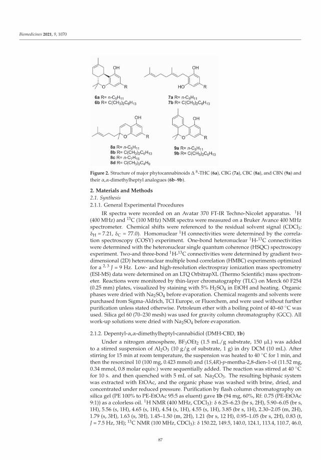
Biomedicines 2021, 9, 1070
Figure 2. Structure of major phytocannabinoids Δ 8-THC (6a), CBG (7a), CBC (8a), and CBN (9a) andtheir α,α-dimethylheptyl analogues (6b–9b).
2. Materials and Methods
2.1. Synthesis2.1.1. General Experimental Procedures
IR spectra were recorded on an Avatar 370 FT-IR Techno-Nicolet apparatus. 1H(400 MHz) and 13C (100 MHz) NMR spectra were measured on a Bruker Avance 400 MHzspectrometer. Chemical shifts were referenced to the residual solvent signal (CDCl3:δH = 7.21, δC = 77.0). Homonuclear 1H connectivities were determined by the correla-tion spectroscopy (COSY) experiment. One-bond heteronuclear 1H-13C connectivitieswere determined with the heteronuclear single quantum coherence (HSQC) spectroscopyexperiment. Two-and three-bond 1H-13C connectivities were determined by gradient two-dimensional (2D) heteronuclear multiple bond correlation (HMBC) experiments optimizedfor a 2, 3 J = 9 Hz. Low- and high-resolution electrospray ionization mass spectrometry(ESI-MS) data were determined on an LTQ OrbitrapXL (Thermo Scientific) mass spectrom-eter. Reactions were monitored by thin-layer chromatography (TLC) on Merck 60 F254(0.25 mm) plates, visualized by staining with 5% H2SO4 in EtOH and heating. Organicphases were dried with Na2SO4 before evaporation. Chemical reagents and solvents werepurchased from Sigma-Aldrich, TCI Europe, or Fluorchem, and were used without furtherpurification unless stated otherwise. Petroleum ether with a boiling point of 40–60 ◦C wasused. Silica gel 60 (70–230 mesh) was used for gravity column chromatography (GCC). Allwork-up solutions were dried with Na2SO4 before evaporation.
2.1.2. Depentyl-α,α-dimethylheptyl-cannabidiol (DMH-CBD, 1b)
Under a nitrogen atmosphere, BF3OEt2 (1.5 mL/g substrate, 150 μL) was addedto a stirred suspension of Al2O3 (10 g/g of substrate, 1 g) in dry DCM (10 mL). Afterstirring for 15 min at room temperature, the suspension was heated to 40 ◦C for 1 min, andthen the resorcinol 10 (100 mg, 0.423 mmol) and (1S,4R)-p-mentha-2,8-dien-1-ol (11.52 mg,0.34 mmol, 0.8 molar equiv.) were sequentially added. The reaction was stirred at 40 ◦Cfor 10 s. and then quenched with 5 mL of sat. Na2CO3. The resulting biphasic systemwas extracted with EtOAc, and the organic phase was washed with brine, dried, andconcentrated under reduced pressure. Purification by flash column chromatography onsilica gel (PE 100% to PE-EtOAc 95:5 as eluent) gave 1b (94 mg, 60%, Rf: 0.75 (PE-EtOAc9:1)) as a colorless oil. 1H NMR (400 MHz, CDCl3): δ 6.25–6.23 (br s, 2H), 5.90–6.05 (br s,1H), 5.56 (s, 1H), 4.65 (s, 1H), 4.54 (s, 1H), 4.55 (s, 1H), 3.85 (br s, 1H), 2.30–2.05 (m, 2H),1.79 (s, 3H), 1.63 (s, 3H), 1.45–1.50 (m, 2H), 1.21 (br s, 12 H), 0.95–1.05 (br s, 2H), 0.83 (t,J = 7.5 Hz, 3H); 13C NMR (100 MHz, CDCl3): δ 150.22, 149.5, 140.0, 124.1, 113.4, 110.7, 46.0,
87

Biomedicines 2021, 9, 1070
44.6, 37.5, 37.3, 31.8, 30.4, 29.9, 28.7, 28.6, 28.4, 24.6, 23.6, 22.6, 20.7, 14.0; HRESIMS m/z[M + H]+ 371.2934 (calcd for C25H39O2, 371.2950).
2.1.3. Depentyl-α,α-dimethylheptyl-Δ8-tetrahydrocannabinol (DMH-Δ8-THC, 6b)
To a stirred suspension of 10 (450 mg, 1.69 mmol) in toluene (2.5 mL), p-toluensulfonicacid (PTSA, 60 mg, 0.34 mmol, 0.2 molar equiv.), and (1S,4R)-p-mentha-2,8-dien-1-ol(11,290 mg, 1.86 mmol, 1.1 molar equiv.) were sequentially added. The solution washeated at 120 ◦C for two hours and then cooled to room temperature and quenched withbrine. The biphasic system was extracted with EtOAc, and the organic phase was driedand evaporated. Purification by flash column chromatography on silica gel (PE 100% toPE/DCM 8:2 as eluent) gave 6b (532 mg, 85%, Rf: 0.90 (PE-EtOAc 9:1)) as a brown oil. 1HNMR (400 MHz, CDCl3): δ 6.42 (d, J = 1.8 Hz, 1H), 6.25 (d, J = 1.8 Hz, 1H), 5.49–5.41 (m,1H), 4.79–4.71 (m, 1H), 3.22 (dd, J = 16.4, 4.4 Hz, 1H), 2.72 (td, J = 10.9, 4.7 Hz, 1H), 2.19–2.13(m, 1H), 1.96–1.78 (m, 4H), 1.73 (s, 3H), 1.54–1.50 (m, 2H), 1.41 (s, 3H), 1.33–1.16 (m, 10H),1.14 (s, 3H), 1.11–1.06 (m, 2H), 0.87 (t, J = 6.9 Hz, 3H); 13C NMR (100 MHz, CDCl3): δ
154.5, 154.45, 150.0, 134.7, 119.3, 110.1, 108.0, 105.4, 44.8, 44.4, 37.3, 36.0, 31.8, 31.5, 30.0,28.7, 28.6, 27.9, 27.6, 24.6, 23.5, 22.6, 18.5, 14.1; HRESIMS m/z [M + H]+ 371.2934 (calcd forC25H39O2, 371.2950).
2.1.4. Depentyl-α,α-dimethylheptyl-cannabigerol (DMH-CBG, 7b)
Under a nitrogen atmosphere, BF3Et2O (1.5 mL/g substrate, 150 μL) was added to astirred suspension of Al2O3 (10 g/g of substrate, 1 g) in dry DCM (10 mL). After stirring15 min at room temperature, the suspension was heated to 40 ◦C for 1 min, and then theresorcinol 10 (100 mg, 0.42 mmol) and geraniol (12, 150 μL, 130 mg, 0.87 mmol, 2 mol.equivalents) were sequentially added. The reaction was stirred at 40 ◦C for 48 h, and thenquenched with 20 mL of 2 M H2SO4. The reaction mixture was extracted with EtOAc, andthe organic phase was washed with brine, dried, and concentrated under reduced pressure.Purification by flash column chromatography on silica gel (PE 100% to PE-EtOAc 95:5 aseluent) gave 1b [94 mg, 60%, Rf: 0.75 (PE-EtOAc 9:1)] as a colorless oil. Purification byflash column chromatography on silica gel (PE 100% to PE-EtOAc 95:5 as eluent) gave 7b
(101 mg, 64%, Rf: 0.75 (PE-EtOAc 9:1)) as a colorless oil. 1H NMR (400 MHz, CDCl3): δ6.40 (s, 2H), 5.32 (t, J = 6.6 Hz, 1H), 5.11 (br s, 2H), 5.08 (t, J = 6.7 Hz, 1H), 3.42 (d, J = 7.1 Hz,2H), 2.21–2.07 (m, 4H), 1.84 (s, 3H), 1.70 (s, 3H), 1.62 (s, 3H), 1.55–1.51 (m, 2H), 1.29–1.16(m, 12H) 1.12–1.05 (m, 2H), 0.87 (t, J = 6.9 Hz, 3H); 13C NMR (100 MHz, CDCl3): δ 154.5,150.0, 139.0, 132.0, 123.7, 121.7, 110.2, 106.1, 44.4, 39.7, 37.4, 31.8, 30.0, 28.8, 26.4, 25.6, 24.6,22.7, 22.3, 17.7, 16.2, 14.0; HRESIMS m/z [M + H]+ 373.3084 (calcd for C25H41O2, 373.3106).
2.1.5. Depentyl-α,α-dimethylheptyl-cannabichromene (DMH-CBC, 8b)
To a stirred suspension of 10 (350 mg, 1.48 mmol) in toluene (10 mL), n-butylamine(141 μL, 104 mg, 1.42 mmol, 1 molar equiv.) was added. The solution was heated to 60 ◦Cfor 10 min, then citral (13, 241 μL, 217 mg, 1.42 mmol, 1 molar equiv.) was added andthe solution was refluxed overnight. The reaction was cooled to room temperature andquenched with 20 mL 2M H2SO4, and then extracted with EtOAc. The organic phase waswashed with brine, dried, and concentrated under reduced pressure. Purification by flashcolumn chromatography on silica gel (PE 100% to PE-EtOAc 95:5 as eluent) gave 210 mg 8b
(210 mg, 40%, Rf: 0.85 (PE-EtOAc 9:1)) as a brown oil. 1H NMR (400 MHz, CDCl3): δ 6.61(d, J = 10.0 Hz, 1H), 6.39 (s, 1H), 6.25 (s, 1H), 5.51 (d, J = 10.0 Hz, 1H), 5.09 (t, J = 7.4 Hz, 1H),2.32–2.02 (m, 2H), 1.76–1.62 (m, 2H), 1.65 (s, 3H), 1.57 (s, 3H), 1.55–1.44 (m, 2H), 1.39 (s, 3H),1.31–1.12 (m, 6H), 1.19 (s, 6H), 1.11–0.99 (m, 2H), 0.94–0.60 (m, 3H); 13C NMR (100 MHz,CDCl3): δ 153.7, 152.0, 150.7, 131.6, 127.4, 124.1, 116.7, 107.0, 106.7, 105.5, 78.2, 44.4, 41.0,37.7, 31.7, 30.0, 28.7, 26.2, 25.6, 24.6, 22.6, 17.6, 14.0; HRESIMS m/z [M + H]+ 371.2935(calcd for C25H39O2, 371.2950). The same protocol was used for the preparation of 8c and8d, starting, from 5-n-heptylresorcinol and 5-α,α-dimethylpentylresorcinol, respectively.Depentyl-5-n-heptylcannabichromene (8c): brown oil (42%, Rf: 0.87 (PE-EtOAc 9:1)). 1H
88

Biomedicines 2021, 9, 1070
NMR (400 MHz, CDCl3): δ 6.66 (d, J = 10.0 Hz, 1H), 6.26 (s, 1H), 6.16 (s, 1H), 5.50 (d,J = 10.0 Hz, 1H), 5.12 (t, J = 7.7 Hz, 1H), 2.45 (t, J = 7.9 Hz, 2H), 2.18–2.08 (m, 2H), 1.81–1.67(m, 2H), 1.69 (s, 3H), 1.60 (s, 3H), 1. 59–1.50 (m, 2H), 1.40 (s, 3H), 1.37–1.16 (m, 8H), 0.90(t, J = 6.9 Hz 3H); 13C NMR (100 MHz, CDCl3): δ 154.0, 151.3, 144.7, 131.6, 127.0, 124.2,117.0, 108.9, 107.8, 107.1, 78.1, 41.0, 36.0, 31.8, 31.0, 29.3, 29.2, 26.2, 25.7, 22.7, 22.6, 17.6,14.1; HRESIMS m/z [M + H]+ 346.2624 (calcd for C23H35O2, 343.2637). Depentyl-5-α,α-dimethylpentylcannabichromene (8d): brown oil [45%, Rf: 0.88 (PE-EtOAc 9:1)]. 1H NMR(400 MHz, CDCl3): δ 6.67 (d, J = 10.0 Hz, 1H), 6.42 (s, 1H), 6.31 (s, 1H), 5.52 (d, J = 10.0 Hz,1H), 5.13 (t, J = 7.2 Hz, 1H), 2.18–2.12 (m, 2H), 1.80–1.65 (m, 2H), 1.69 (s, 3H), 1.55 (s, 3H),1.56–1.49 (m, 2H), 1.43 (s, 3H), 1.27–1.19 (m, 2H), 1.24 (s, 6H), 1.13–1.03 (m, 2H), 0.85 (t,J = 7.3 Hz 3H); 13C NMR (100 MHz, CDCl3): δ 153.7, 151.9, 151.1, 131.6, 127.3, 124.2, 117.0,106.8, 106.7, 105.7, 78.2, 46.8, 44.2, 41.0, 37.7, 35.4, 28.8, 26.9, 26.3, 25.7, 23.4, 22.7, 14.1;HRESIMS m/z [M + H]+ 343.2625 (calcd for C23H35O2, 343.2637).
2.1.6. Depentyl-α,α-dimethylheptyl-cannabinol (DMH-CBN, 9b)
To a stirred suspension of 8b (210 mg, 0.57 mmol) in toluene (40 mL), iodine (291 mg,1.15 mmol, 2 eq) was added. The solution was refluxed for three hours then cooled toroom temperature and quenched with sat. Na2SO3. The mixture was extracted withEtOAc, dried, and concentrated under reduced pressure. Purification by flash columnchromatography on silica gel (PE 100% to PE-CH2Cl2 9:1 as eluent) gave 9b (173 mg, 83%,Rf: 0.85 (PE-EtOAc 9:1)) as a brown oil. 1H NMR (400 MHz, CDCl3): δ 8.20 (s, 1H), 7.18(d, J = 7.9 Hz, 1H), 7.10 (d, J = 8.9 Hz, 1H), 6.59 (d, J = 1.8 Hz, 1H), 6.44 (d, J = 1.8 Hz, 1H),2.41 (s, 3H), 1.63 (s, 6H), 1.58–1.54 (m, 2H), 1.27 (s, 6H), 1.26–1.13 (m, 6H), 0.86 (t, J = 6.9 Hz,3H); 13C NMR (100 MHz, CDCl3): δ 154.3, 152.7, 151.8, 136.9, 136.9, 127.6, 127.5, 126.3,122.6, 108.6, 108.3, 107.6, 44.4, 37.6, 31.7, 29.9, 28.6, 27.1, 24.6, 22.6, 21.5, 14.0. HRESIMS m/z[M + H]+ 367.2623 (calcd for C25H35O2, 367.2637).
2.2. TRP Modulatory Activity
Effects of compounds on intracellular Ca2+ concentration ([Ca2+]i) were determinedusing Fluo-4, a selective intracellular fluorescent probe for Ca 2+. Assays of rat TRPA1,TRPV2, TRPV3, TRPV4, and TRPM8 or human TRPV1 mediated elevation of intracellularCa 2+ in transfected HEK-293 cells were performed with a continuous monitoring of [Ca 2+]iduring the experiments [8]. Briefly, human embryonic kidney (HEK-293) cells, stably trans-fected with rat TRPA1, TRPV2, TRPV3, TRPV4, and TRPM8 or human TRPV1 (selected bygeneticin 600 μg mL −1) or not transfected, were cultured in EMEM+ 2 mM glutamine + 1%nonessential amino acids +10% FBS and maintained at 37 ◦C with 5% CO2. TRP-HEK-293cells express stably high levels of TRP transcripts, while these transcripts were virtuallyabsent in wild-type HEK-293 cells as checked by real-time PCR. On the day of the experi-ment, the cells were loaded for 1 h in the dark at room temperature with Fluo-4 AM (4 μMin DMSO containin 0.02% Pluronic F-127). The cells were rinsed, resuspended in Tyrode’ssolution (145 mM NaCl, 2.5 mM KCl, 1.5 mM CaCl2, 1.2 mM MgCl2, 10 mM D-glucose,and 10 mM HEPES, pH 7.4), and transferred to a quartz cuvette of a spectrofluorimeter(PerkinElmer LS50B; λEX = 488 nm, λEM = 516 nm) equipped with a PTP-1 fluorescencePeltier system (PerkinElmer Life and Analytical Sciences, Waltham, MA, USA) under con-tinuous stirring. Cell fluorescence before and after the addition of various concentrations oftest compounds was measured, normalizing the effects against the response to ionomycin(IM, 4 μM). The values of the effect on [Ca2+]i in HEK-293 cells not transfected were usedas baseline and subtracted from the values obtained from transfected cells. For TRPA1,agonist efficacy was expressed as a percentage of the effect on [Ca 2+]i observed with100 μM allylisothiocyanate. The potency of the compounds (EC50 values) is determinedas the concentration required to produce half-maximal increases in [Ca 2+]i. In the TRPV3assay, TRPV3-expressing HEK293 cells were first sensitized with the structurally unrelatedagonist 2-aminoethoxydiphenyl borate (100 μM). Antagonist/desensitizing behavior isevaluated against the agonist of the TRP analyzed by adding the compounds directly in
89
Biomedicines 2021, 9, 1070
the quartz cuvette 5 min before stimulation of cells with the agonist. Allylisothiocyanate(100 μM) was used for TRPA1, capsaicin (0.1 μM) for TRPV1, cannabidiol (2 μM) forTRPV2, thymol (100 μM) for TRPV3, GSK1016790A (10 nM) for TRPV4, and icilin (0.25 μM)for TRPM8. The IC50 value was expressed as the concentration exerting a half-maximalinhibition of agonist effect, taking as 100% the effect on [Ca2+]i exerted by the agonistalone. Dose-response curve fitting (sigmoidal dose-response variable slope) and parameterestimation were performed with Graph-Pad Prism8 (GraphPad Software Inc., San Diego,CA, USA). All determinations were performed at least in triplicate.
2.3. Molecular Docking
Docking studies were performed with AutoDock 4.21 [9]. The rTRPA1 model, alongwith the ligands, were processed with AutoDock Tools (ADT) package version 1.5.6rc1 tomerge non polar hydrogens, calculate Gasteiger charges, and select rotatable sidechainbonds. Grid dimensions of 60 × 60 × 50, centered in the putative binding pocket, weregenerated with the program AutoGrid 4.2 included in Auto-dock 4.2 distribution, witha spacing of 0.375 Å. Docking runs were carried out by either keeping the whole proteinfixed or allowing the rotation of selected residues (Leu873, Leu884, Phe912, Met915 andMet949). A total of 100 molecular AutoDock docking runs for each docking calculationwere performed adopting a Lamarckian genetic algorithm (LGA) and the following associ-ated parameters: 100 individuals in a population with a maximum of 15 million energyevaluations and a maximum of 37,000 generations, followed by 300 iterations of Solis andWets local search. Flexibility was used for all rotatable bonds of both docked ligands. Therepresentative poses for each receptor were selected for the subsequent energy minimiza-tion with Amber16 package [10] using ff14SB force field for the protein, and gaff parametersfor the ligand [11,12].
3. Results
3.1. Synthesis
All α,α-DMH analogs were prepared from 5-(1,1-dimethylhepty)-resorcinol (10), inturn prepared from 3,5-dimethoxybenzoic acid [13]. Terpenylation was then carried outmutuating chemistry developed for the synthesis of the corresponding natural n-pentylphytocannabinoids. C-Menthylation with (4R)-2,9-p-menthadien-1-ol (11) afforded eitherDMH-CBD (1b) or DMH-Δ8-THC (6b) depending on the reaction conditions [14], whilegeranylation with BF3 on alumina gave DMH-CBG (7b) [15], and chromenyation with citral(13) under basic conditions generated DMH-CBC (8b) [16], next aromatized to DMH-CBN(9b) by treatment with iodine (Scheme 1) [17].
3.2. Biological Evaluation
All DMH-derivatives retained the full agonistic profile on TRPA1 typical of theirnaturally occurring n-pentyl analogues, along with a comparable efficacy and ability todesensitize this channel (Table 1). Conversely, the replacement of the pentyl chain withthe branched DMH group was detrimental for the activity toward TRPV1–4 and TRPM8channels (Tables 1 and 2). Thus, while CBD (1a) and CBG (7a) are TRPV1 antagonists in asub/low micromolar range, their correspondent DMH derivatives (1b and 7b, respectively)were inactive against this receptor. Similarly, while the natural derivatives are antagonistsin a low micromolar range at TRPV4, the activity was lost for all DMH-derivatives, thatwere only weak inhibitors of TRPV2, with IC50 ranging from 9 to >50 μM. As for TRPV3,DMH-Δ8-THC (6b) was the only compound active as an inhibitor at a low micromolarrange (IC50 of ~2 μM), with a slightly better potency than Δ8-THC (6a). At TRPM8, alDMH analogues acted as pure antagonists albeit with a lower potency than the naturalderivatives, except DMH-CBC (8b), which was totally inactive, and with only DMH-CBN(9b) retaining a certain affinity (IC50~1 μM).
90

Biomedicines 2021, 9, 1070
Scheme 1. Synthesis of α,α-dimethylheptyl analogues of major phytocannabinoids DMH-CBD (1b), DMH-Δ8-THC (6b),DMH-CBG (7b), DMH-CBC (8b) and DMH-CBN (9b).
Table 1. Efficacy, potency, and inhibitory effect on TRPA1, TRPV1, and TRPM8 channels.
TRPA1 TRPV1 TRPM8
Efficacy(% ITC 100 μM)
PotencyEC50 μM
IC50a
TRPA1 μMEfficacy
(% IM 4 μM)Potency
EC50
IC50b
TRPV1 μM
IC50c
TRPM8 μM
1b1a [18]
133.4 ± 5.3115.9 ± 4.6
0.40 ± 0.150.11 ± 0.05
0.51 ± 0.030.16 ± 0.05
<1044.7 ± 0.02
NA1.0 ± 0.1
>1000.6 ± 0.05
13.4 ± 2.10.06 ± 0.01
6b6a [18]
124.2 ± 4.0117.0 ± 12.0
4.8 ± 1.10.23 ± 0.03 6.0 ± 0.9 15.3
<10>100NA
>100 39.9 ± 6.90.16 ± 0.01
9b9a [18]
132.3 ± 7.983.3 ± 12.0
2.1 ± 0.90.18 ± 0.02
3.2 ± 0.60.40 ± 0.04
<10<10
NA >100>50
0.98 ± 0.120.21 ± 0.05
7b7a [18]
99.2 ± 4.599.9 ± 1.1
9.1 ± 2.00.70 ± 0.03
1.7 ± 0.1513.0 ± 4.8
<1033.8 ± 2.3
NA1.3 ± 0.5
>1002.6 ± 0.5
9.2 ± 0.80.16 ± 0.02
8b8a [18]
120.4 ± 2.8119.4 ± 3.1
0.76 ± 0.120.09 ± 0.01
0.32 ± 0.010.37 ± 0.05
18.9 ± 0.2<10 12.8 ± 0.9 >100
>50>100
40.7 ± 0.6
8c 97.8 ± 3.3 1.2 ± 0.2 3.8 ± 0.35 <10 NA >100 >100
8d 98.7 ± 1.9 0.15 ± 0.03 0.40 ± 0.01 <10 NA >100 23.6 ± 7.4
Reference agonists: a AITC 100 μM; b Capsaicin 0.1 μM; c Icilin 0.25 μM.
91

Biomedicines 2021, 9, 1070
Table 2. Efficacy, potency, and inhibitory effect on TRPA1, TRPV1, and TRPM8 channels.
TRPV2 TRPV3 TRPV4
Efficacy(% IM4 μM)
PotencyEC50 μM
IC50a
TRPV2 μM
Efficacy(% IM4 μM)
PotencyEC50 μM
IC50c
TRPV3 μM
Efficacy(% IM 4μM)
PotencyEC50 μM
IC50e
TRPV4 μM
1b1a [18,19]
<1040.5 ± 1.6
NA1.25 ± 0.23
16.8 ± 0.24.5 ± 0.7 b
15.8 ± 0.450.1 ± 4.8
11.0 ± 1.13.7 ± 1.6
32.6 ± 5.10.9 ± 0.3 d
34.0 ± 1.316.7 ± 1.0
NA0.8 ± 0.3
>1001.1 ± 0.1 f
6b6a [18,19]
<1053.0 ± 1.4
NA0.65 ± 0.05
45.8 ± 3.90.8 ± 0.01
53.8 ± 1.618.2 ± 1.0
0.14 ± 0.039.5 ± 1.9
2.1 ± 0.632.3 ± 2.1
<10<10
NANA
>10015.2 ± 2.7
6b6a [18,19]
10.5 ± 0.0239.9 ± 2.1
0.25 ± 0.0319.0 ± 3.7
>5015.7 ± 2.1
20.9 ± 3.013.1 ± 2.4
41.4 ± 21.65.3 ± 2.7
>1009.4 ± 0.1
12.1 ± 0.215.3 ± 1.5
16.1 ± 21.616.1 ± 4.5
>1005.4 ± 0.8
7b7a [18,19]
65.3 ± 0.773.6 ± 1.2
9.8 ±0.61.7 ± 0.08
39.4 ± 4.51.5 ± 0.2
64.6 ± 4.818.5 ± 0.7
39.7 ± 0.031.0 ± 0.2
35.4 ± 3.6>50
34.4 ± 5.223.7 ± 1.8
39.0 ± 21.65.1 ± 1.6
35.4 ± 3.21.3 ± 0.1
8b8a [18,19]
76.1 ± 0.6<10
>50NA
22.6 ± 0.26.5 ± 1.6
41.6 ± 1.820.2 ± 0.4
>501.9 ± 0.2
>50>100
<1022.9 ± 1.2
NA0.6 ± 0.2
>1009.9 ± 1.2
8c <10 NA 9.0± 0.8 <10 NA >100 <10 NA >100
8d <10 NA 13.3± 1.1 <10 NA >100 <10 NA >100
Reference agonists: a CBD 2 μM; b LPC 3 μM; c Thymol 100 μM; d Carvacrol 1 mM; e GSK1016790A 10 nM; f 4αPDD 1 μM.
To better rationalize the selectivity of the DMH-derivatives toward TRPA1 and dissectthe relative contribution of chain elongation and branching, two point-mutated analoguesof the most potent compound (DMH-CBC (8b)) were prepared according to the generalsynthetic sequence summarized in Scheme 1 starting from a different 5-alkylresorcinol. Inthe n-heptyl chromene 8c, the alkyl chain is linear and expanded by two carbon atomscompared to CBC (8a), while in the α,α-dimethylpentyl analogue 8d the benzyl branchingis implanted on a pentyl chain. As shown in Table 1, the n-heptyl derivative 8c was lesspotent than the dimethyl-pentyl analogue 8d (IC50 = 3.8 and 0.40 μM, respectively). Hence,the α,α-dimethyl substitution is the major contributor to the activity of the DHM derivative(IC50 = 0.32 μM).
3.3. Molecular Docking Studies on CBC and CBC-DMH at rTRPA1
To shed light on the putative binding modes of DMH-CBC (8a) on TRPA1 channels,a molecular docking study was carried out using our previous homology model of rat-TRPA1 [20]. Since the compound is a racemate, both enantiomers were considered, usingCBC (8a) as a reference compound. Different docking runs were carried out by eitherkeeping the protein side chains rigid or allowing flexibility for selected residues, as de-scribed in detail in the Materials and Methods section. The best docking poses for bothCBC and DMH-CBC, selected on the basis of binding energy value and visual inspection,were subjected to energy minimization (Figure 3). Both enantiomers of CBC and DMH-CBC engage H-bonding with their phenolic hydroxyl and Thr877 (monB). (R)-CBC pointsits pentyl chain in a hydrophobic cleft formed by Ile953 (monA), Met956 (monB), andPhe880 (monB), while the methyl group of the pentyl chain forms a CH-π interaction withPhe880 and the chromene methyl is surrounded by the hydrophobic residues Ile881 (monB),Leu885 (monB), Phe912 (monB), and Ile945 (monA). Conversely, (S)-CBC adopts a flippedorientation with the pentyl chain now pointing toward Phe912 (monB), engaging a CH-πinteraction with the aromatic ring of this amino acid. The same arrangement of (S)-CBCis adopted by both enantiomers of CBC-DMH (8b), with the gem-dimethyls sandwichedbetween Phe912 (monB) and Ile881 (mon B). In this arrangement, the alkyl chain stabilizesthe complex by hydrophobic interactions, counteracting the higher flexibility of the heptylchain in comparison with the pentyl one and in accordance with the lower activity of then-heptyl chain analogue, with the length of the CBC pentyl chain being consistent withalternative CH-π interaction with Phe880/Phe912 within the binding pocket. The twoDMH-CBC enantiomers share the same orientation of the DMH-chain but show a distinctorientation of the chromene methyl group: in the R enantiomer, the methyl group pointstoward Phe880, and the terpenoid chain is hosted in a hydrophobic cleft at the interfaces
92

Biomedicines 2021, 9, 1070
between two monomers. Conversely, in the S enantiomer the orientation of these group isjust the opposite.
Figure 3. Representative complexes of the rTRPA1 model with (R)-CBC (colored in salmon, panel (A)),(S)-CBC (colored in tan, panel (B)), (R)-CBC-DMH (colored olive drab, panel (C)) and (S)-CBC-DMH(colored in dark green, panel (D)). A ribbon representation is used for the protein backbone andsticks for protein side chains of residues within 5 Å from the ligand, in ball & stick representation.H-bonds are shown as green sticks. Carbon atoms are painted according to receptor subunits. Sulfur,oxygen, and polar hydrogen atoms are painted yellow, red, and white, respectively.
4. Discussion
The serendipitous discovery by Adams that a combination of branching and elon-gation of the alkyl chain boosts the narcotic potency of tetrahydrocannabinols providedthe basis for the synthesis of highly potent molecular probes, eventually leading to thecharacterization of the two cannabinoid receptors (CB1 and CB2) and to the synthesisof nabilone (Cesamet), a clinically efficacious synthetic analogue of Δ9-THC [2]. Despitethe relevance of this maneuver for CB1 and CB2 affinity, and its potential to bias activ-ity toward specific targets, its effect has not yet been investigated on other high-affinitytargets of phytocannabinoids like the thermo-TRPs (TRPV1-V4, TRPM8 and TRPA1) [5].This is surprising, since one major issue plaguing the bench-to-bed translation of thebiological activity of cannabinoids is their promiscuous end-point profile, which spansmetabotropic and ionotropic receptors as well as enzymes and transcription factors. ForΔ9-THC, the interaction with the two cannabinoid receptors dominates, albeit not fullyrecapping, the clinical profile, while the other phytocannabinoids show a constellation ofend-points characterized by a generally modest affinity for macromolecular end-points,with sub-micromolar potency basically limited to some thermo-TRPs [5].
To systematically address this issue, we have synthesized the α,α-dimethylheptylanalogues of the five major phytocannabinoids and have comparatively evaluated theiractivity on thermo-TRPs. The results showed that, by quenching affinity for the otherthermo-TRPs, the replacement of the linear pentyl substituent with an α,α-dimethylheptylresidue induces a selectivity switch toward TRPA1 in all major phytocannabinoid chemo-types. Molecular docking studies rationalized these results in terms of binding, capitalizingour previously developed homology model of ratTRPA1 [20]. Structural and mutagenicexperiments have identified two binding sites for non-electrophilic agonists, centered, re-spectively, on hThr874 (rThr877, uniprot ID:Q6RI86, Site 1) and hTyr840 (Site 2). However,only Site1 was considered, since, unlike phytocannabinoids and their DMH analogues, theselective Site 2 agonist GNE551 lacks channel desensitizing properties. Moreover, Tyr840,
93

Biomedicines 2021, 9, 1070
critical for GNE551 binding, is not conserved between human and rat hortologues, beingreplaced by a Phe residue in the latter. The results of the docking studies suggest thegem-dimethyls on the benzyl carbon provide an additional anchoring site to engage thebinding pocket into non-covalent interactions, rationalizing the retention of affinity forTRPA1. This maneuver is detrimental for the other thermos-TRPs presumably because ofthe incapacity to accommodate the steric and lipophilicity changes associated with benzylbranching into less broadly tuned binding sites [21].
Non-covalent modulators of TRPA1 are relatively few compared to the covalentones [22], and their biological profile is critically dependent on their binding mode. Site 2ligands like GNE551 do not elicit channel desensitization and tachyphylaxis, thus inducingpersistent pain insensitive to TRPA1 antagonists [23]. Conversely, Site 1 ligands induceonly transient pain, followed by desensitization and analgesia [24]. The desensitization ofTRPA1 has the potential to translate into beneficial effects for a wide range of inflammatoryconditions [25], traditionally focused in the realm of inflammation and pain [20]. However,growing evidence of involvement in the SARS-CoV-2 infection through the modulation ofinflammation, pain, and fever, has provided an additional therapeutic area of investigation,with documented proof-of-concept clinical cases of the beneficial effects of sulforafane, aTRPA1 and Nrf2-interacting nutrient, on the course of the infection [7].
5. Conclusions
Replacement of the n-pentyl side chain with an α,α-dimethylheptyl group quenchesthe exuberant interaction of phytocannabinoids with thermo-TRPs, making them selectivenon-covalent modulators of TRPA1. Within the DMH-analogues of the most commonphytocannabinoids, DMH-CBC (8b) emerged as the most potent compound in the series,and the molecular details of its interaction with this ion channel were investigated andcompared to those of the parent compound CBC (8a). Both compounds are racemic,but only the enantiomers of the natural product CBC showed a distinct orientation atTRPA1 binding sites. These results provide a basis for a broader comparison that will alsoinclude additional end points of phytocannabinoids and their endogenous analogues, likeanandamide and palmitoylethanolamide.
Author Contributions: Conceptualization, G.A., D.C., R.M.V., O.T.-S. and L.D.P.; methodology,R.M.V., P.A. and L.D.P.; formal analysis, A.S.M., R.M.V. and L.D.P.; investigation, P.A., R.M.V., A.S.M.,L.D.P. and D.M.; visualization, P.A., writing—original draft preparation, D.C. and R.M.V.; writing—review and editing, G.A., L.D.P. and O.T.-S.; funding acquisition, G.A., O.T.-S. and L.D.P. All authorshave read and agreed to the published version of the manuscript.
Funding: This research was funded by MIUR Italy (PRIN2017, Project 2017WN73PL, bioactivity-directed exploration of the phytocannabinoid chemical space).
Institutional Review Board Statement: Not applicable.
Informed Consent Statement: Not applicable.
Data Availability Statement: Not applicable.
Conflicts of Interest: The authors declare no conflict of interest. The funder had no role in the designof the study; in the collection, analyses, or interpretation of data; in the writing of the manuscript; orin the decision to publish the results.
References
1. Hanuš, L.O.; Meyer, S.M.; Munoz, E.; Taglialatela-Scafati, O.; Appendino, G. Phytocannabinoids: A unified critical inventory. Nat.Prod. Rep. 2016, 33, 1357–1392. [CrossRef] [PubMed]
2. Appendino, G. The early history of cannabinoid research. Rend. Fis. Acc. Lincei 2020, 31, 919–929. [CrossRef]3. Gaoni, Y.; Mechoulam, R. Isolation, Structure, and Partial Synthesis of an Active Constituent of Hashish. J. Am. Chem. Soc. 1964,
86, 1646–1647. [CrossRef]4. Appendino, G.; Minassi, A.; Taglialatela-Scafati, O. Recreational drug discovery: Natural products as lead structures for the
synthesis of smart drugs. Nat. Prod. Rep. 2014, 31, 880–904. [CrossRef]
94

Biomedicines 2021, 9, 1070
5. Ligresti, A.; De Petrocellis, L.; Di Marzo, V. From phytocannabinoids to cannabinoid receptors and endocannabinoids: Pleiotropicphysiological and pathological roles through complex pharmacology. Physiol. Rev. 2016, 96, 1593–1659. [CrossRef]
6. Vitale, R.M.; Schiano Moriello, A.; De Petrocellis, L. Natural compounds and synthetic drugs targeting the ion-otropic cannabinoidmembers of Transient Receptor Potential (TRP) channels. In New Tools to Interrogate En-docannabinoid Signalling: From NaturalCompounds to Synthetic Drugs; Maccarrone, M., Ed.; Royal Society of Chemistry: London, UK, 2020; pp. 201–300.
7. Bousquet, J.; Czarlewski, W.; Zuberbier, T.; Mullol, J.; Blain, H.; Cristol, J.-P.; De La Torre, R.; Pizarro Lozano, N.; Le Moing,V.; Bedbrook, A.; et al. Potential Interplay between Nrf2, TRPA1, and TRPV1. Int. Arch. Allergy Immunol. 2021, 182, 324–338.[CrossRef]
8. Moriello, A.S.; De Petrocellis, L. Assay of TRPV1 Receptor Signaling. Methods Mol. Biol. 2016, 1412, 65–76. [CrossRef] [PubMed]9. Morris, G.; Huey, R.; Lindstrom, W.; Sanner, M.F.; Belew, R.K.; Goodsell, D.S.; Olson, A.J. AutoDock4 and AutoDockTools4:
Automated docking with selective receptor flexibility. J. Comput. Chem. 2009, 30, 2785–2791. [CrossRef]10. Case, D.A.; Betz, R.M.; Cerutti, D.S.; Cheatham, T.E.; Darden, T.A.; Duke, R.E.; Giese, T.J.; Gohlke, H.; Goetz, A.W.; Homeyer,
N.; et al. AMBER 2016; University of California: San Francisco, CA, USA, 2016.11. James, A.; Maier, J.A.; Martinez, C.; Kasavajhala, K.; Wickstrom, L.; Hauser, K.E.; Simmerling, C. ff14SB: Im-proving the accuracy
of protein side chain and backbone parameters from ff99SB. J. Chem. Theory Comput. 2015, 11, 3696–3713. [CrossRef]12. Wang, J.; Wolf, R.M.; Caldwell, J.W.; Kollman, P.A.; Case, D.A. Development and testing of a general amber force field. J. Comput.
Chem. 2004, 25, 1157–1174. [CrossRef] [PubMed]13. Harrington, P.E.; Stergiades, I.A.; Erickson, J.; Makriyannis, A.; Tius, M. Synthesis of functionalized canna-binoids. J. Org. Chem.
2000, 65, 6576–6582. [CrossRef]14. Bloemendal, V.R.L.J.; Van Hest, J.C.M.; Rutjes, F.P.J.T. Synthetic pathways to tetrahydrocannabinol (THC): An overview. Org.
Biomol. Chem. 2020, 18, 3203–3215. [CrossRef]15. Baek, S.-H.; Du Han, S.; Yook, C.N.; Kim, Y.C.; Kwak, J.S. Synthesis and antitumor activity of cannabigerol. Arch. Pharmacal Res.
1996, 19, 228–230. [CrossRef]16. Pollastro, F.; Caprioglio, D.; Del Prete, D.; Rogati, F.; Minassi, A.; Taglialatela-Scafati, O.; Munoz, E.; Appendino, G.
Cannabichromene. Nat. Prod. Commun. 2018, 13, 1189–1194. [CrossRef]17. Pollastro, F.; Caprioglio, D.; Marotta, P.; Schiano Moriello, A.; De Petrocellis, L.; Taglialatela-Scafati, O.; Appendino, G. Iodine-
promoted aromatization of p-menthane-type phytocannabinoid. J. Nat. Prod. 2018, 81, 630–633. [CrossRef] [PubMed]18. De Petrocellis, L.; Ligresti, A.; Moriello, A.S.; Allarà, M.; Bisogno, T.; Petrosino, S.; Stott, C.G.; Di Marzo, V. Effects of canna-binoids
and cannabinoid-enriched cannabis extracts on TRP channels and endocannabinoid metabolic enzymes. Br. J. Phar-macol. 2011,162, 1479–1494. [CrossRef]
19. De Petrocellis, L.; Orlando, P.; Moriello, A.S.; Aviello, G.; Stott, C.; Izzo, A.; Di Marzo, V. Cannabinoid actions at TRPV channels:Effects on TRPV3 and TRPV4 and their potential relevance to gastrointestinal inflammation. Acta Physiol. 2011, 204, 255–266.[CrossRef] [PubMed]
20. Chianese, G.; Lopatriello, A.; Schiano-Moriello, A.; Caprioglio, D.; Mattoteia, D.; Benetti, E.; Ciceri, D.; Arnoldi, L.; De Combarieu,E.; Vitale, R.M.; et al. Cannabitwinol, a Dimeric Phytocannabinoid from Hemp, Cannabis sativa L., Is a Selective Thermo-TRPModulator. J. Nat. Prod. 2020, 83. [CrossRef] [PubMed]
21. Zhao, Y.; McVeigh, B.M.; Moiseenkova-Bell, V.Y. Structural Pharmacology of TRP Channels. J. Mol. Biol. 2021, 433, 166914.[CrossRef]
22. Nilius, B.; Appendino, G.; Owsianik, G. The transient receptor potential channel TRPA1: From gene to patho-physiology. PflugersArch. 2012, 464, 425–458. [CrossRef] [PubMed]
23. Liu, C.; Reese, R.; Vu, S.; Rougé, L.; Shields, S.D.; Kakiuchi-Kiyota, S.; Chen, H.; Johnson, K.; Shi, Y.P.; Chernov-Rogan, T.; et al. ANon-covalent Ligand Reveals Biased Agonism of the TRPA1 Ion Channel. Neuron 2020, 109, 273–284.e4. [CrossRef] [PubMed]
24. Chandrabalan, A.; McPhillie, M.J.; Morice, A.H.; Boa, A.N.; Sadofsky, L.R. N-Cinnamoylanthranilates as hu-man TRPA1modulators: Structure–activity relationships and channel binding sites. Eur. J. Med. Chem. 2019, 170, 141–156. [CrossRef][PubMed]
25. Nilius, B.; Appendino, G. Spices: The Savory and Beneficial Science of Pungency. Rev. Physiol. Biochem. Pharmacol. 2013, 164, 1–76.[CrossRef] [PubMed]
95

biomedicines
Article
Triterpenoid–PEG Ribbons Targeting Selectivity inPharmacological Effects
Zulal Özdemir 1,2,†, Uladzimir Bildziukevich 1,2,†, Martina Capková 1,†, Petra Lovecká 3, Lucie Rárová 4,‡,
David Šaman 5, Michala Zgarbová 5,‡, Barbora Lapuníková 5,‡, Jan Weber 5, Oxana Kazakova 6
and Zdenek Wimmer 1,2,*
Citation: Özdemir, Z.; Bildziukevich,
U.; Capková, M.; Lovecká, P.; Rárová,
L.; Šaman, D.; Zgarbová, M.;
Lapuníková, B.; Weber, J.; Kazakova,
O.; et al. Triterpenoid–PEG Ribbons
Targeting Selectivity in
Pharmacological Effects. Biomedicines
2021, 9, 951. https://doi.org/
10.3390/biomedicines9080951
Academic Editor: Jun Lu
Received: 18 June 2021
Accepted: 30 July 2021
Published: 3 August 2021
Publisher’s Note: MDPI stays neutral
with regard to jurisdictional claims in
published maps and institutional affil-
iations.
Copyright: © 2021 by the authors.
Licensee MDPI, Basel, Switzerland.
This article is an open access article
distributed under the terms and
conditions of the Creative Commons
Attribution (CC BY) license (https://
creativecommons.org/licenses/by/
4.0/).
1 Department of Chemistry of Natural Compounds, University of Chemistry and Technology in Prague,Technická 5, 16628 Prague 6, Czech Republic; [email protected] (Z.Ö.); [email protected] (U.B.);[email protected] (M.C.)
2 Isotope Laboratory, Institute of Experimental Botany of the Czech Academy of Sciences, Vídenská 1083,14220 Prague 4, Czech Republic
3 Department of Biochemistry and Microbiology, University of Chemistry and Technology in Prague,Technická 5, 16628 Prague 6, Czech Republic; [email protected]
4 Department of Experimental Biology, Faculty of Science, Palacký University, Šlechtitelu 27,78371 Olomouc, Czech Republic; [email protected]
5 Institute of Organic Chemistry and Biochemistry of the Czech Academy of Sciences, Flemingovo námestí 2,16610 Prague 6, Czech Republic; [email protected] (D.Š.); [email protected] (M.Z.);[email protected] (B.L.); [email protected] (J.W.)
6 Ufa Institute of Chemistry of the Ufa Federal Research Centre of the Russian Academy of Sciences,71, pr. Oktyabrya, 450054 Ufa, Russia; [email protected]
* Correspondence: [email protected] or [email protected]† Z.Ö., U.B. and M.C. contributed equally.‡ L.R., cytotoxicity screening tests; M.Z. and B.L., antiviral tests.
Abstract: (1) Background: To compare the effect of selected triterpenoids with their structurallyresembling derivatives, designing of the molecular ribbons was targeted to develop compounds withselectivity in their pharmacological effects. (2) Methods: In the synthetic procedures, Huisgen 1,3-dipolar cycloaddition was applied as a key synthetic step for introducing a 1,2,3-triazole ring as a partof a junction unit in the molecular ribbons. (3) Results: The antimicrobial activity, antiviral activity,and cytotoxicity of the prepared compounds were studied. Most of the molecular ribbons showedantimicrobial activity, especially on Staphylococcus aureus, Pseudomonas aeruginosa, and Enterococcusfaecalis, with a 50–90% inhibition effect (c = 25 μg·mL−1). No target compound was effective againstHSV-1, but 8a displayed activity against HIV-1 (EC50 = 50.6 ± 7.8 μM). Cytotoxicity was tested onseveral cancer cell lines, and 6d showed cytotoxicity in the malignant melanoma cancer cell line(G-361; IC50 = 20.0 ± 0.6 μM). Physicochemical characteristics of the prepared compounds wereinvestigated, namely a formation of supramolecular gels and a self-assembly potential in general,with positive results achieved with several target compounds. (4) Conclusions: Several compoundsof a series of triterpenoid molecular ribbons showed better pharmacological profiles than the parentcompounds and displayed certain selectivity in their effects.
Keywords: triterpenoid; molecular ribbon; Huisgen 1,3-dipolar cycloaddition; amide bond; multi-functional PEG3 derivative; antimicrobial activity; anti-HIV activity; cytotoxicity; supramolecularself-assembly
1. Introduction
Triterpenoids represent plant secondary metabolites with increasing importancedue to the spectra of their pharmacological activity and ability to self-assemble into thesupramolecular systems, often forming gels in different solvents, including aqueous me-dia [1]. Oleanolic acid (1a), morolic acid (1b), and moronic acid (1c) are triterpenoids of the
Biomedicines 2021, 9, 951. https://doi.org/10.3390/biomedicines9080951 https://www.mdpi.com/journal/biomedicines97

Biomedicines 2021, 9, 951
oleanane series, bearing a 6-6-6-6-6 pentacyclic skeleton (Figure 1). While oleanolic acidhas been one of the most frequently investigated triterpenoids, morolic and moronic acidhave been so far much less studied.
Figure 1. Structures of oleanolic acid (1a), morolic acid (1b), and moronic acid (1c).
Oleanolic acid (1a) has been isolated from several hundred of plant species, and it isfrequently used in the pharmaceutical and food industry, as reviewed recently [2]. Themost important source of 1a is plants of the genus Oleaceae [2]. Due to its lipophilic nature,it appears mainly in olive oil, but it is also present in the olive leaves [3]. Oleanolic acid(1a) has been proven to display inhibition activity against HIV and HSV [4–6]. It showsimportant antimicrobial activity, namely against G+ bacteria (Staphylococcus aureus, S. epi-dermidis, Enterococcus faecalis, E. faecium, Streptococcus pneumoniae, or Bacillus subtilis) [7–9].It is also active against Mycobacterium tuberculosis, a Gram staining-type of pathogenicbacterium [10]. Its activity against G– bacteria is low or nil [11]. Oleanolic acid (1a) alsodisplays an antidiabetic effect [12]. One of the possible mechanisms consists in the inhibi-tion of glycogen phosphorylase, which is an enzyme capable of liberating glucose fromglycogen, and the glucose level in blood is affected [13]. The other possible mechanismis connected with a possible inhibition of proteintyrosine phosphatase (PTP-1B), which isa part of a de-phosphorylation process of the insulin receptor [14]. The last but not leastpossible mechanism consists of a direct support of insulin secretion [15]. Oleanolic acid(1a) has been proven to display an anti-inflammatory effect, either due to the inhibition oftransaminases or due to the inhibition of histamine secretion from mastocytes [16], and italso shows antitumor activity, as proven in several tumor cell lines [17]. It is also activeagainst hepatitis of the C type through depressing of the gene types HCV-1b and 2a-JFH1virus replication [18–20], and it inhibits HCV proteases [18]. However, the mechanism hasnot yet been clarified [19].
Morolic acid (1b) was first isolated from the wood of Mora excelsa in 1950 [21], andmoronic acid (1c) was found in the roots of Ozoroa mucronata in 1979 [22]. Later on, bothtriterpenoids were found in other plants as well, often together [23]. Morolic acid (1b)and moronic acid (1c) are active against HIV and HSV [24]. Their antimicrobial activityis different: 1c is more potential than 1b in the number of affected microorganisms [25].They also display anti-hypertensive and hypoglycemic effects [26]. Antidiabetic effect is ex-plained either by a mechanism of inhibition of tyrosin phosphatase (PTP-1B; cf. above) [27]or by inhibiting 11β-hydroxysteroid dehydrogenase 1 (11β-HSD 1) that is a mediator ofintracellular production of cortisol [28]. Both triterpenoids also display anti-inflammatoryeffect. Moronic acid (1c) was proven to show inhibition of tumor cells, in contrast to1b [29]. Moronic acid (1c) can also be synthesized in several steps from betulin, which is alupane-type triterpenoid [30,31].
Pentacyclic triterpenoids have been used to control biofilm-related infections causedby S. aureus and other pathogens [32]. Patients, which were intubated due to the SARS-CoV-2 infection nowadays, often suffer from infections caused by microorganisms formingbacterial films that occur in the intubation equipment [33,34]. Therefore, an investigation ofnovel antimicrobial agents potentially capable of controlling bacterial films may contributeto augmenting the health of the patients seriously infected with SARS-CoV-2. These find-ings make this investigation even more important, because the designed molecular ribbonsare based solely on natural products from sustainable sources, and their pharmacologicaleffect is targeted to enhance the effect of their parent triterpenoids.
98

Biomedicines 2021, 9, 951
Since plant triterpenoids can also be found in vegetables, medicinal plants, spices,etc., they become natural constituents of the human diet. The literature data show that theaverage daily intake varies between 250 and 400 mg per day in humans, depending on thespecific part of the world [35]. This is an important item of information in consequencewith the different types of pharmacological activity resulting from this investigation.
The objectives of the presented investigation consisted of several partial tasks. (a) Thefirst is to design procedures for the synthesis of selected molecular ribbons based on theselected triterpenoids connected through the multifunctional PEG3 junction units, and bythe newly formed amide bonds and the 1,4-disubstituted 1,2,3-triazole rings. Generally,PEG-mediated motifs have been used in pharmacologically active compounds, such assteroids [36], alkaloids [37], or triterpenoids [38,39]. A modification of biomolecules bythe PEG motifs resulted in compounds having a longer blood-circulation time than thecorresponding parent biomolecules [40,41]. A formation of a covalent bond between thePEG spacer and the biologically active compounds is one of the promising techniques todesign novel drug molecules with improving therapeutic effect [40,41]. Due to the ester-based protecting groups to be employed during the synthetic procedures described here,using any other ester group in the target molecules is excluded, while a combination ofamide bonds and 1,4-disubstituted 1,2,3-triazole rings does not interfere with introducingand removing any protecting group. (b) The second objective is to target the investigationon antimicrobial and antiviral activity, and cytotoxicity screening tests of the triterpenoidmolecular ribbons to show their advantages and/or disadvantages in comparison with theirparent single triterpenoid molecules. (c) The third objective is to investigate supramolecularcharacteristics of the target molecular ribbons and to evaluate the importance of the formedsupramolecular systems for potential pharmacological applications in comparison withtheir parent triterpenoids.
2. Materials and Methods
2.1. General
The NMR measurements (Bruker, Berlin, Germany) were performed on a BrukerAVANCE II 600 MHz spectrometer equipped with a 5 mm TCI cryoprobe in a 5 mm tubein different solvents. The 1H NMR and the 13C NMR spectra were recorded at 600.13 MHzand 150.90 MHz in CDCl3 or CD3OD using tetramethylsilane (δ = 0.0–CDCl3) or signalof solvent (δ = 3.31 or 49.50 for 1H/13C–CD3OD) as internal references. 1H NMR dataare presented in the following order: chemical shift (δ) expressed in ppm, multiplicity(s, singlet; d, doublet; t, triplet; q, quartet; m, multiplet), number of protons, couplingconstants in Hertz. For unambiguous assignment of both 1H and 13C signals, 2D NMR,1H,13C gHSQC, and gHMBC spectra were measured using standard parameters sets andpulse programs delivered by the producer of the spectrometer. Infrared spectra weremeasured with a Nicolet iS-5 FT-IR spectrometer (Thermofisher Scientific, Waltham, MA,USA). UV-VIS spectra were measured on a Specord 210 spectrometer (Jena Analytical,Germany). TLC was carried out on silica gel plates (Merck 60F254), and the visualizationwas performed by both the UV detection and spraying with the methanolic solution ofphosphomolybdic acid (5%), followed by heating. For column chromatography, silica gel60 (0.063–0.200 mm) from Merck was used. Analytical HPLC was carried out on a TSP(Thermoseparation Products, Boston, MA, USA) instrument equipped with a ConstaMetric4100 Bio pump and a SpectroMonitor 5000 UV DAD. The analyses of the products wereperformed on a reverse phase Nucleosil 120-5 C18 column (250 × 4 mm; Watrex, Prague,Czech Republic) using a methanol/water mixture (9:1, v/v) as the mobile phase at 0.5 to1.0 mL.min−1. The eluate was monitored at 220, 254, and 275 nm, and the UV spectra wererun from 200 to 300 nm. All compounds were purified until their purity was >99%. Theiranalytical data are summarized in the Supplementary material, part 1, Synthetic proceduresand analytical data. All chemicals and solvents were purchased from regular commercialsources in analytical grade, and the solvents were purified by general methods before
99
Biomedicines 2021, 9, 951
use. Triterpenoids were purchased from Dr. Jan Šarek–Betulinines (www.betulinines.com,(accessed on 14 June 2021).
2.2. Synthetic Procedures2.2.1. Procedure A
Benzyl bromide (2.5 eq) and potassium carbonate (3 eq) were added to a solutionof the starting compound (1 eq) in DMF (15 mL), and the reaction mixture was stirred atr.t. for 24 h. The mixture was washed with brine, the organic layer was extracted withbenzene, and then, it was dried over sodium sulfate. The solid phase was filtered off, andthe solvent was evaporated under reduced pressure. The crude residue was purified bycolumn chromatography on silica gel, using a chloroform/ethanol mixture (200:1) as amobile phase.
2.2.2. Procedure B
Sodium hydride (8 eq) was added to a solution of the starting compound (1 eq) inTHF (20 mL), and the reaction mixture was stirred at r.t. for 1 h. Then, an additionalamount of sodium hydride (8 eq) was added, and stirring continued for an additional 2 h.Finally, propargyl bromide (3 eq) was added, and the reaction mixture was stirred at r.t. for7 days. The solvent was evaporated, and the residue was washed with brine and extractedwith chloroform. After drying over sodium sulfate, the solvent was evaporated, and thecrude residue was purified by column chromatography on silica gel, using chloroform as amobile phase.
2.2.3. Procedure C
Acetic anhydride (1.44 eq), EDIPA (1.8 eq), and DMAP (0.13 eq) were added to asolution of the starting compound (1 eq) in THF (20 mL). The reaction mixture was stirredat 100 ◦C for 3 h. Then, an additional amount of acetic anhydride (0.77 eq) and EDIPA(0.9 eq) were added, and heating continued for an additional 1 h. The reaction was stoppedby adding water. The organic layer was extracted with chloroform, and the extract waswashed with saturated solution of sodium bicarbonate and dried over sodium sulfate.After filtering off the solid phase, solvents were evaporated, and the crude residue waspurified by column chromatography on silica gel, using a chloroform/ethanol mixture(100:1) as a mobile phase.
2.2.4. Procedure D
Propargyl bromide (1.5 eq) and potassium carbonate (3 eq) were added to a solutionof the starting compound in DMF (15 mL). The reaction mixture was stirred at r.t. for24 h. It was quenched with brine and extracted with benzene. After drying the extractover sodium sulfate, the solid phase was filtered off, and the solvent was evaporated.The crude residue was purified by column chromatography on silica gel, using a chloro-form/petroleum ether mixture (1:3) as a mobile phase.
2.2.5. Procedure E
Oxalyl chloride (7 eq) was added to a solution of the starting compound (1 eq) in DCM(30 mL), and the reaction mixture was stirred at r.t. for 3 h. The solvent was evaporatedunder reduced pressure, and the residue was re-dissolved in freshly distilled DCM (30 mL).Azido-PEG3-amide (1.1 eq) and EDIPA (2.5 eq) were added, and the reaction mixture wasstirred at r.t. for 24 h. The reaction mixture was quenched with brine, extracted withchloroform, and the extract was dried over sodium sulfate. The solid phase was filteredoff, and the solvents were evaporated under reduced pressure. The residue was purifiedby column chromatography on silica gel using a chloroform/ethanol mixture (200:1) as amobile phase.
100

Biomedicines 2021, 9, 951
2.2.6. Procedure F
The starting compounds bearing either a propargyl group (1 eq) or azide group(1.05 eq) were dissolved in DCM (30 mL). Then, a mixture of aqueous solution of coppersulfate pentahydrate and TBTA (0.05 M) was added, which was followed by the additionof sodium ascorbate (1 eq). The reaction mixture was stirred at r.t. for 24 h, and then, it wasquenched with water. The organic layer was extracted with chloroform, and the extract wasdried over sodium sulfate. The solid phase was filtered off, and the solvent was evaporatedunder reduced pressure. The crude residue was purified by column chromatography onsilica gel, using a chloroform/ethanol mixture (100:1) as a mobile phase.
2.2.7. Procedure G
Catalyst (10% Pd/C, 0.67 eq) was added to a solution of the starting compound (1 eq)in a mixture of ethanol/THF (1:1, 30 mL) in a bottle equipped with a septum. Air wasremoved from the bottle, it was filled with hydrogen, and the reaction mixture was stirredat r.t. for 24 h. To stop the hydrogenation reaction, hydrogen was removed, and the catalystwas filtered off. Solvents were evaporated under reduced pressure, and the crude residuewas purified by column chromatography on silica gel, using a chloroform/ethanol mixture(100:1) as a mobile phase.
2.2.8. Procedure H
Lithium hydroxide monohydrate (5 eq) was added to a solution of the starting com-pound (1 eq) in methanol (30 mL). The reaction mixture was heated to 80 ◦C for 5 h. Then,the solvent was evaporated under reduced pressure, and the crude residue was purifiedby column chromatography on silica gel, using a chloroform/ethanol mixture (100:1) as amobile phase.
2.3. Antimicrobial Screening Tests
The antimicrobial activity of 1a–1c, 8a, 12a, 8b, 12b, 4c, 6c, 6d, and 8d was studiedin vitro, using Bacillus cereus, Escherichia coli, Pseudomonas aeruginosa, Staphylococcus aureus,and Enterococcus faecalis. The resazurine test was used to evaluate this activity.
The cell inoculum was made in Mueller–Hinton broth (10 mL). Colonies of cellswere inoculated from the solid medium that was cultivated for 24 h under optimumconditions. A part of the inoculum was diluted to get suspension with an optical density0.6 to 0.9 McF. An aliquot amount (1 mL) was taken and diluted with additional volumeof the medium (19 mL). The stored solutions of the tested compounds were made ata concentration c = 200 μg.mL−1. The stored suspensions of the cells, MHB medium,solutions of the antibiotics specific for the given bacteria (vancomycin for S. aureus andE. faecalis, kanamycin for E. coli and P. aeruginosa, and tetracycline for B. cereus), and thesolutions of the compounds were pipetted into the microtitration plates. Subsequently, aconcentration sequence of the tested compounds was made (c = 100, 50, and 25 μg.mL−1).The content of the microtitration plates was cultivated for 24 h under optimum conditionsfor the given bacteria. A solution of resazurine was made, based on a dilution of its storedsolution (c = 0.15 mg.mL−1; 2 mL) into the MHB medium (18 mL). Then, it was pipettedinto the microtitration plates that were cultivated for an additional 1 h. Finally, fluorescencemeasurement (excitation at 560 nm, emission at 590 nm) was made, and the inhibition ofthe bacterium was determined.
2.4. Cytotoxicity Screening Tests and Cell Cultures Used
Description of the experimental procedure used in the cytotoxicity screening test wasalready published [42,43]. The following cancer cell lines were used in the investigation:T-lymphoblastic leukemia, CEM; breast carcinoma, MCF7; cervical carcinoma, HeLa; andmalignant melanoma, G-361. Human foreskin fibroblasts, BJ, were used as referencecells. All of the above cell lines were obtained from the American Type Culture Collection(Manassas, VA, USA). African green monkey kidney Vero cells were obtained from the
101

Biomedicines 2021, 9, 951
European Collection of Cell Cultures (Salisbury, UK) and human T-cell leukemia MT-4 cellswere obtained through the NIH AIDS Reagent Program, Division of AIDS, NIAID, NIH,from Dr. Douglas Richman. All cells, but MT-4 cells, were cultured in DMEM (Dulbecco’sModified Eagle Medium, Sigma, St. Louis, MO, USA). MT-4 cells were maintained inRPMI1640 medium. Media used were supplemented with 10% fetal bovine serum, 2 mML-glutamine, and 1% penicillin–streptomycin (all Sigma, St. Louis, MO, USA). The celllines were maintained under standard cell culture conditions at 37 ◦C and 5% CO2 in ahumid environment. Cells were subcultured twice or three times a week using the standardtrypsinization procedure.
2.5. Antiviral Tests
The anti-HIV-1 and anti-HSV-1 activity and cytotoxicity in MT-4 cells and Vero cells,respectively, were determined as previously described [44,45]. Three-fold dilution of thecompounds were tested in triplicate, using HIV-1 (laboratory strain NL4-3) and HSV-1 (strain HF), respectively. Tenofovir alafenamide and acyclovir were used as controls.Compounds were not toxic up to 50 μM against MT-4 and Vero cells.
2.6. Supramolecular Self-Assembly Studies
Ability of the target compounds 8a, 12a, 8b, 12b, 4c, 6c, 6d, and 8d was investigatedby (a) measuring UV-VIS absorption in methanol/water mixtures with a constant con-centration of the studied compound, and (b) investigating their ability to form gel indifferent solvents.
2.6.1. Ability to Form Supramolecular Gel
The solutions of the studied compounds 8a, 12a, 8b, 12b, 4c, 6c, 6d, and 8d (1%, w/v)in the appropriate solvent were mixed with ultrasound for 30 min, then heated to theboiling points of the used solvents, and then allowed to cool down to the r.t. for 24 h.An example of the resulting gels that were produced from 8a, 4c, 6c, and 6d is shown inthe Supplementary Material, Figure S1.
2.6.2. UV-VIS Spectrometry
The self-assembly of the selected compounds 8a, 12a, 8b, 12b, 4c, 6c, 6d, and 8d wasmeasured using UV absorption in methanol/water mixtures with a constant concentrationof the studied compound for 7 days in one-hour intervals on the first day, and in 24 hintervals all following days. The stock solutions of the studied compounds were preparedat a concentration of 1 mg·mL−1 in water. Then, a series of methanol/water mixtures wereprepared starting from a methanol/water ratio 100/0 down to 0/100 in 20% steps. Thestock solutions of the studied compounds (0.15 mL) were added separately to each vialcontaining methanol/water mixtures (3 mL), and the UV spectra were recorded in thewavelength range of 200–400 nm. The most remarkable changes in these spectra wereobserved for methanol/water ratios 40/60 and 60/40 in their mixtures. For the compounds8a, 12a, 12b and 6d, the details are presented in the Supplementary Material; see Figure S2.
3. Results and Discussion
3.1. Synthetic Procedures3.1.1. Procedure A
Esterification of the carboxyl group of the starting triterpenoid (1a or 1b) was madeby a substitution reaction using benzyl bromide in DMF under the presence of potassiumcarbonate [46]. Compounds 2a/2b were prepared using the procedure A (Scheme 1).
3.1.2. Procedure B
Synthesis of propargyl ether of the starting triterpenoid (1a or 1b) consisted in aformation of the corresponding alcoholate using sodium hydride in THF, followed by
102

Biomedicines 2021, 9, 951
its reaction with propargylic bromide [46]. Compounds 3a/3b were prepared using theprocedure B (Scheme 1).
3.1.3. Procedure C
Esterification of the free hydroxyl group of the starting triterpenoid (1a or 1b) wasmade with acetic anhydride in THF, using EDIPA as a base and DMAP as a reactionenhancer [43]. Compounds 4a/4b were prepared using the procedure C (Scheme 1).
3.1.4. Procedure D
Synthesis of propargyl ester of the starting triterpenoid (1c) was made by a substitutionreaction of the carboxyl group of 1c with propargylic bromide in DMF in the presence ofpotassium carbonate [47]. Compound 2c was prepared using the procedure D (Scheme 2).
Scheme 1. Synthetic procedure, part 1. Reagents and conditions. (i) BnBr, K2CO3, DMF, r.t., 24 h; (ii) NaH, propargylbromide, THF, r.t., 7 days; (iii) Ac2O, EDIPA, DMAP, THF, 100 ◦C, 4 h; (iv) (a) oxalyl chloride, DCM, r.t., 3 h, (b) N3-PEG3-NH2, EDIPA, r.t., 24 h; (v) 3a/3b, CuSO4·5H2O, TBTA, sodium ascorbate, DCM/H2O, r.t., 24 h; (vi) 10% Pd/C, H2,ethanol/THF (1:1), r.t., 24 h; (vii) LiOH·H2O, methanol, 80 ◦C, 5 h.
3.1.5. Procedure E
An amide bond formation was made by converting free carboxyl group of the startingtriterpenoid (1a–1c) into the corresponding acyl chloride using oxalyl chloride in DCM,followed by its reaction with the corresponding amine in DCM using EDIPA as a base [43].Compounds 5a/5b, 9a/9b, 3c, and 7d were prepared using the procedure E (Schemes 1–3).
3.1.6. Procedure F
Cu(I)-Catalyzed Huisgen 1,3-dipolar cycloaddition was made using componentsbearing either a propargylic group (3a/3b, 2c) or azide group (5a/5b, 9a/9b, 3c, and 7d).Cu(I) ions were generated in situ by reducing copper(II) sulfate by sodium ascorbate. TBTA
103

Biomedicines 2021, 9, 951
was used as reaction initiator [46]. Compounds 6a/6b, 10a/10b, 4c, 5c/5d, and 8d wereprepared using the procedure F (Schemes 1–3).
Scheme 2. Synthetic procedure, part 2. Reagents and conditions. (i) propargyl bromide, K2CO3, DMF, r.t., 24 h; (ii) (a) oxalylchloride, DCM, r.t., 3 h, (b) N3-PEG3-NH2, EDIPA, r.t., 24 h; (iii) 2c, CuSO4·5H2O, TBTA, sodium ascorbate, DCM/H2O,r.t., 24 h; (iv) 3a/3b, CuSO4·5H2O, TBTA, sodium ascorbate, DCM/H2O, r.t., 24 h; (v) 10% Pd/C, H2, ethanol/THF (1:1),r.t., 24 h; (vi) (a) oxalyl chloride, DCM, r.t., 3 h, (b) N3-PEG3-NH2, EDIPA, r.t., 24 h; (vii) 2c, CuSO4·5H2O, TBTA, sodiumascorbate, DCM/H2O, r.t., 24 h.
3.1.7. Procedure G
Removing of the protecting benzyl group was made by palladium-catalyzed hydro-genation of the selected starting compound (6a/6b, 10a/10b, and 5c/5d) [46]. Compounds7a/7b, 11a/11b, and 6c/6d were prepared using the procedure G (Schemes 1–3).
3.1.8. Procedure H
Removing of the protecting acetyl group was made by alkaline hydrolysis of theesters (7a/7b and 11a/11b) by lithium hydroxide in methanol [43]. Compounds 8a/8b
and 12a/12b were prepared using the procedure H (Schemes 1 and 3).
3.2. Cytotoxicity
Cytotoxicity in the malignant melanoma cancer cell line (G-361) was only found for6d (IC50 = 20.0 ± 0.6 μM), even if all target compounds were subjected to the cytotoxicityscreening tests. The compound 6d, a hybrid molecular ribbon based on 1b and 1c, wasinactive in CEM, MCF7, and HeLa cancer cell lines (IC50 > 50 μM), and was not toxic tothe human fibroblasts (BJ; IC50 > 50 μM). Therefore, 6d is selectively cytotoxic in the G-361cancer cell line.
104

Biomedicines 2021, 9, 951
Scheme 3. Synthetic procedure, part 3. Reagents and conditions. (i) (a) oxalyl chloride, DCM, r.t., 3 h, (b) N3-PEG3-NH2,EDIPA, r.t., 24 h; (ii) 3a/3b, CuSO4·5H2O, TBTA, sodium ascorbate, DCM/H2O, r.t., 24 h; (iii) 10% Pd/C, H2, ethanol/THF(1:1), r.t., 24 h; (iv) LiOH·H2O, methanol, 80 ◦C, 5 h.
3.3. Antimicrobial Activity
Antimicrobial activity of the target compounds was tested on Staphylococcus aureus,Escherichia coli, Bacillus cereus, Pseudomonas aeruginosa, and Enterococcus faecalis using threedifferent concentrations (c = 25, 50, 100 μg·mL−1). The results achieved with the lowest con-centration of the tested compound (c = 25 μg·mL−1) are presented in Table 1. When higherconcentrations of the tested compounds were used, inhibition of microorganism growthincreased as well, even though for some compounds, certain variations were observed(cf. Supplementary Material, Table S1). The irregularity in the antimicrobial activity valuesin a dependence on the concentrations of the tested compounds may be connected withthe supramolecular characteristics of the molecular ribbons (cf. Supplementary Material,Table S1). We had already observed such behavior in the investigation of cytotoxicity ofanother series of triterpenoid-based compounds [43,47]. The highest inhibition activityfor S. aureus was observed when it is treated with 8a, which is a dimer-like molecularribbon based on 1a. While 1a was not active, dimer-like molecular ribbon 8a showedincreasing inhibition activity. However, trimer-like molecular ribbon 12a, also based on 1a,showed lower activity than 8a, which can be caused either by a decrease in solubility or bysize of the compound for possible penetration into the microorganism or by the effect ofsupramolecular self-assembly (cf. Supplementary Material). The same trend of inhibitionactivity was observed when E. coli was treated with 1a or its ribbons. Interestingly, in thetests with S. aureus, when 1a was replaced by 1b, which only differs from 1a by the positionof the double bond in the skeleton, the formed trimer-like molecular ribbon 12b exhibitedthe highest activity in comparison with 1b and 8b, presenting the importance of smallstructural variations on the inhibition activity. While 1c was not active on S. aureus, itsdimer-like molecular ribbon 4c was slightly active. Hybrid dimer-like molecular ribbons6c and 6d were synthesized as well, by combining 1c with 1a or 1b. While the hybrid6c was less active than 8a, hybrid 6d was more active than 8d. It can be assumed that
105

Biomedicines 2021, 9, 951
the hybridization of 1c (moronic acid) with 1b (morolic acid) was more favorable thanits hybridization with 1a (oleanolic acid) when the inhibition activity results were takeninto consideration.
Table 1. Selected data of the antimicrobial activity a,b.
Microorganism Compound Inhibition [%] (c = 25 μg·mL−1) c
Staphylococcus aureus (+) 1a • –Staphylococcus aureus 8a •• 52 ± 7.8 e
Staphylococcus aureus 6c •• 13 ± 2.0Staphylococcus aureus 12a ••• 28 ± 4.2Staphylococcus aureus 1b • 21 ± 3.2Staphylococcus aureus 8b •• –Staphylococcus aureus 6d •• 25 ± 3.8Staphylococcus aureus 12b ••• 40 ± 6.0 e
Staphylococcus aureus 1c • –Staphylococcus aureus 4c •• 11 ± 1.7
Escherichia coli (+) 1a • –Escherichia coli 8a •• 51 ± 7.7 e
Escherichia coli 12a ••• –
Pseudomonas aeruginosa (−) 1c • –Pseudomonas aeruginosa 4c •• 50 ± 7.5 e
Enterococcus faecalis (+) 1a d • 87 ± 13.0
Enterococcus faecalis 1b • 38 ± 5.7Enterococcus faecalis 1c • –Enterococcus faecalis 4c •• 62 ± 9.3 e
Enterococcus faecalis 8d ••• 38 ± 5.7
Bacillus cereus (+) 1a d • 76 ± 11.4
Bacillus cereus 1b d • 78 ± 11.7
Bacillus cereus 1c • –a A comparison of the antimicrobial activity of a target compound is always followed by the antimicrobialactivity of its parent triterpenoid; b antibiotics (c = 25 μg·mL−1, causing 99% inhibition) were used as positivecontrol: vancomycin for S. aureus and E. faecalis, kanamycin for E. coli and P. aeruginosa, and tetracycline for B.cereus); c hyphen represents no inhibition; d the highest inhibition of a microorganism found for 1a and 1b at thegiven concentration; e These molecular ribbons displayed higher inhibition of the microorganism than its parenttriterpenoid(s).
In turn, for B. cereus, the highest inhibition activity was achieved when it is treatedwith 1a or 1b rather than by the trimer-like or dimer-like molecular ribbons, although 1c
(moronic acid) did not show any activity. Similar inhibition activities were observed with1a (oleanolic acid) and 1b (morolic acid), showing the importance of the presence of the(C3)-OH group of triterpenoid for possible activity on B. cereus. This group is replacedwith a ketone group in case of 1c (moronic acid). In contrast to these results, dimer-likemolecular ribbon 4c showed the highest activity against P. aeruginosa, which is the only G–bacterial strain used in this investigation, while the other dimer-like molecular ribbonsexhibited lower inhibition activity values than 4c. Parent triterpenoids 1a–1c were notactive at all, which is in accordance with the literature data [11], demonstrating no or nilantimicrobial activity of this types of triterpenoids on the G– bacteria. The dimer-likemolecular ribbon 4c displayed much higher antimicrobial activity on E. faecalis than 1c thatshowed no inhibition of the tested microorganisms. However, the highest activity was stillobserved when E. faecalis was treated with 1a.
The target molecular ribbons are the new compounds, and no data on their phar-macological activity have been so far available in the literature. Therefore, the foundresults cannot be discussed in the context with the literature data, with the only exceptionpresented above. The data have proven that most of the target molecular ribbons displayhigher inhibition of the studied microorganisms than their parent triterpenoids 1a–1c. TableS1 (cf. Supplementary material) shows data on the dose–response dependence of antimi-
106

Biomedicines 2021, 9, 951
crobial activity of the target molecular ribbons 8a, 12a, 8b, 12b, 4c, 6c, 6d, and 8d versustheir parent compounds 1a–1c. All doses mentioned there at the maximal concentrationused in the screening tests (c = 100 μg·mL−1) are much lower doses than humans mayconsume with plant-based food (vegetables, medicinal plants, spices, etc.) through whichthey may receive 250–400 mg per day in average [35].
3.4. Antiviral Activity
The only compound of this series (8a) showed low activity against HIV-1 in MT-4(EC50 ± SE = 50.6 ± 7.8 μM). However, the found EC50 value is several orders of magnitudehigher than those for reference compound, tenofovir alafenamide (EC50 ± SE = 0.0097 ±0.0023 μM). No target compound displayed activity against HSV-1.
3.5. Gel formation and Supramolecular Self-Assembly
The target compounds were tested to show their ability to form supramolecular gels.This investigation was made because we have found a relationship between cytotoxicityand the ability of compounds to form supramolecular aggregates capable of affecting cyto-toxicity [43,47], and similar results have recently been published by other authors [48,49].Among the studied series of compounds, the only 4c, 6c, and 6d produced a gel in glycerol;8a produced a gel in ethylene glycol (Supplementary material, Figure S1). In turn, self-assembly can be achieved with other target compounds based on spectral measurements.UV spectra measured in a mixture of water/methanol with changing ratio of both solventsin 20% steps, but containing a constant concentration of the studied compound, showirregularities in the absorbance maxima sequence or even in wavelengths of the signalmaximum, if self-assembly takes place. This effect of irregularity in the absorbance maximasequence was observed with several target compounds 8a and 12a. For more details, seethe Supplementary Material (Supplementary material, Figure S2).
4. Conclusions
During this investigation, we have designed several molecular ribbons based onthe combinations of oleanolic, morolic, and moronic acid (1a–1c) in the ribbons. Themultifunctional PEG3 unit was used as a junction chain, together with the 1,2,3-triazolering and the amide bond, between the triterpenoid molecules to form molecular ribbons.The synthetic procedures recently developed in the team were used, and some of them wereoptimized. The target structural ribbons were subjected to the antimicrobial and antiviralactivity as well as cytotoxicity screening tests. The only target compound 6d displayedmild and selective cytotoxicity against melanoma cells G-361 (IC50 = 20.0 ± 0.6 μM). Theonly compound 8a showed low but again selective activity against HIV-1 in MT-4 cells(EC50 ± SE = 50.6 ± 7.8 μM). However, more target compounds displayed antimicrobialactivity. Compounds were tested in three concentrations (c = 100, 50, and 25 μg·mL−1), andthe complete results are presented in the Supplementary Material, Table S1, while selectedresults are shown in Table 1. Among the highest observed inhibition, 8a inhibited S. aureusand E. coli in >50% (c = 25 μg·mL−1), 12b inhibited S. aureus in >40% (c = 25 μg·mL−1), and4c inhibited E. faecalis in >62% (c = 25 μg·mL−1). In turn, 1a and 1b inhibited B. cereus in>77% (c = 25 μg·mL−1), and 1a inhibited E. faecalis in >87% (c = 25 μg·mL−1).
Since the inhibition of several types of microorganisms, including S. aureus, has beenfound, the molecular ribbons, namely 8a and 12b, displaying higher antimicrobial activityon S. aureus than their parent triterpenoids 1a and 1b, can also be considered for use incontrolling bacterial films of S. aureus in the intubation equipment of patients sufferingfrom complicated SARS-CoV-2 infections, as reported recently [33,34], to prevent additionalmicrobial infection from the intubation equipment, using molecular ribbons based on thenatural products (cf. Table 1 and Table S1 in the Supplementary Material).
Four target compounds (4c, 6c, 6d, and 8a) formed gels either in glycerol or in ethyleneglycol. Data from the UV spectrometry achieved under the described conditions gave proof
107

Biomedicines 2021, 9, 951
that compounds 8a and 12a formed self-aggregated systems detected by the instrument inthe methanol/water mixtures.
Several of the synthesized triterpenoid–PEG molecular ribbons gave evidence that theinvestigation of more complex structures based on plant triterpenoids can be important,because several of those structures either displayed enhancing pharmacological effectsdifferent from those shown by their parent monomeric triterpenoids or displayed existingsupramolecular characteristics, including the ability to form supramolecular gels in contrastto their parent triterpenoids. However, a clear relation between the antimicrobial activityand supramolecular characteristics cannot be presented because we do not have enoughindisputable data at present, and, therefore, this topic has been currently under moredetailed investigation.
Supplementary Materials: The following material is available online at https://www.mdpi.com/article/10.3390/biomedicines9080951/s1: Details on the synthetic procedures and analytical dataof the prepared compounds, including the scanned NMR spectra of the target molecular ribbons,additional details on antimicrobial screening tests, and details on gel formation and other supramolec-ular characteristics.
Author Contributions: Conceptualization, Z.W., P.L. and O.K.; methodology, Z.Ö. and U.B.; val-idation, L.R., D.Š. and J.W.; formal analysis, Z.W.; investigation, Z.Ö., U.B., M.C., M.Z. and B.L.;resources, Z.W.; data curation, Z.Ö., U.B. and M.C.; writing—original draft preparation, Z.W., Z.Ö.,U.B., P.L. and J.W.; writing—review and editing, Z.W. and O.K.; supervision, Z.W.; project adminis-tration, Z.W.; funding acquisition, Z.W. All authors have read and agreed to the published version ofthe manuscript.
Funding: FV30300 (MPO: Z.Ö., U.B., P.L. and Z.W.), and CZ.02.1.01/0.0/0.0/16_019/0000738 (Eu-ropean Regional Developmental Fund Project “Centre for Experimental Plant Biology”; L.R.). U.B.thanks the Academy of Sciences of the Czech Republic for funding his post-doctoral stay (grant No.L200381952). Antiviral testing was funded by the Institute of Organic Chemistry and Biochemistry(RVO 61388963). This work was also supported from the grant (No. A1_FPBT_2021_002) of theSpecific University Research.
Institutional Review Board Statement: Not applicable.
Informed Consent Statement: Not applicable.
Data Availability Statement: The data presented in this study are available on request from thecorresponding author.
Conflicts of Interest: The authors declare no conflict of interest.
References
1. Bag, B.G.; Majumdar, R. Self-assembly of renewable nano-sized triterpenoids. Chem. Rec. 2017, 17, 841–873. [CrossRef]2. Pollier, J.; Goossens, A. Oleanolic acid. Phytochemistry 2012, 77, 10–15. [CrossRef]3. Jaeger, S.; Trojan, H.; Kopp, T.; Laszczyk, M.N.; Scheffler, A. Pentacyclic triterpene distribution in various plants—Rich sources
for a new group of multi-potent plant extracts. Molecules 2009, 14, 2016–2031. [CrossRef] [PubMed]4. Kashiwada, Y.; Wang, H.-K.; Nagao, T.; Kitanaka, S.; Yasuda, I.; Fujioka, T.; Yamagishi, T.; Cosentino, L.M.; Kozuka, M.;
Okabe, H.; et al. Anti-AIDS agents. 30. Anti-HIV activity of oleanolic acid, pomolic acid, and structurally related triterpenoids.J. Nat. Prod. 1998, 61, 1090–1095. [CrossRef]
5. Mengoni, F.; Lichtner, M.; Battinelli, L.; Marzi, M.; Mastroianni, C.M.; Vullo, V.; Mazzanti, G. In vitro anti-HIV activity of oleanolicacid on infected human mononuclear cells. Planta Med. 2002, 68, 111–114. [CrossRef] [PubMed]
6. Mukherjee, H.; Ojha, D.; Bag, P.; Chandel, H.S.; Bhattacharyya, S.; Chatterjee, T.K.; Mukherjee, P.K.; Chakraborti, S.;Chattopadhyay, D. Anti-herpes virus activities of Achyranthes aspera: An Indian ethnomedicine, and its triterpene acid.Microbiol. Res. 2013, 168, 238–244. [CrossRef] [PubMed]
7. Woldemichael, G.M.; Singh, M.P.; Maiese, W.M.; Timmermann, B.N. Constituents of antibacterial extract of Caesalpinia paraguar-iensis Burk. Z. Naturforsch. C J. Biosci. 2003, 58, 70–75. [CrossRef]
8. Horiuchi, K.; Shiota, S.; Hatano, T.; Yoshida, T.; Kuroda, T.; Tsuchiya, T. Antimicrobial activity of oleanolic acid from Salviaofficinalis and related compounds on vancomycin-resistant enterococci (VRE). Biol. Pharm. Bull. 2007, 30, 1147–1149. [CrossRef][PubMed]
108

Biomedicines 2021, 9, 951
9. Szakiel, A.; Ruszkowski, D.; Grudniak, A.; Kurek, A.; Wolska, K.I.; Doligalska, M.; Janiszowska, W. Antibacterial and antiparasiticactivity of oleanolic acid and its glycosides isolated from marigold (Calendula officinalis). Planta Med. 2008, 74, 1709–1715.[CrossRef]
10. Bamuamba, K.; Gammon, D.W.; Meyers, P.; Dijoux-Franca, M.-G.; Scott, G. Anti-mycobacterial activity of five plant species usedas traditional medicines in the Western Cape Province (South Africa). J. Ethnopharmacol. 2008, 117, 385–390. [CrossRef]
11. Kuete, V.; Wabo, G.F.; Ngameni, B.; Mbaveng, A.T.; Metuno, R.; Etoa, F.-X.; Ngadjui, B.T.; Beng, V.P.; Meyer, J.J.M.; Lall, N. Antimi-crobial activity of the methanolic extract, fractions and compounds from the stem bark of Irvingia gabonensis (Ixonanthaceae).J. Ethnopharmacol. 2007, 114, 54–60. [CrossRef]
12. Wu, D.; Zhang, Q.; Yu, Y.; Zhang, Y.; Zhang, M.; Liu, Q.; Zhang, E.; Li, S.; Song, G. Oleanolic acid, a novel endothelin A receptorantagonist, alleviated high glucose-induced cardiomyocytes injury. Am. J. Chin. Med. 2018, 46, 1187–1201. [CrossRef]
13. Kurukulasuriya, R.; Link, J.T.; Madar, D.J.; Pei, Z.; Richards, S.J.; Rohde, J.J.; Souers, A.J.; Szczepankiewicz, B.G. Potential drugtargets and progress towards pharmacologic inhibition of hepatic glucose production. Curr. Med. Chem. 2003, 10, 123–153.[CrossRef] [PubMed]
14. Klaman, L.D.; Boss, O.; Peroni, O.D.; Kim, J.K.; Martino, J.L.; Zabolotny, J.M.; Moghal, N.; Lubkin, M.; Kim, Y.-B.;Sharpe, A.H.; et al. Increased energy expenditure, decreased adiposity, and tissue-specific insulin sensitivity in protein-tyrosinephosphatase 1B-deficient mice. Mol. Cell. Biol. 2000, 20, 5479–5489. [CrossRef]
15. Teodoro, T.; Zhang, L.; Alexander, T.; Yue, J.; Vranic, M.; Volchuk, A. Oleanolic acid enhances insulin secretion in pancreaticβ-cells. FEBS Lett. 2008, 582, 1375–1380. [CrossRef] [PubMed]
16. Singh, G.B.; Singh, S.; Bani, S.; Gupta, B.D.; Banerjee, S.K. Anti-inflammatory activity of oleanolic acid in rats and mice.J. Pharm. Pharmacol. 1992, 44, 456–458. [CrossRef]
17. Akihisa, T.; Kamo, S.; Uchiyama, T.; Akazawa, H.; Banno, N.; Taguchi, Y.; Yasukawa, K. Cytotoxic activity of Perilla frutescens var.japonica leaf extract is due to high concentrations of oleanolic and ursolic acids. J. Nat. Med. 2006, 60, 331–333. [CrossRef]
18. Ma, C.-M.; Wu, X.-H.; Masao, H.; Wang, X.-J.; Kano, Y. HCV protease inhibitory, cytotoxic and apoptosis-inducing effects ofoleanolic acid derivatives. J. Pharm. Pharm. Sci. 2009, 12, 243–248. [CrossRef] [PubMed]
19. Wang, H.; Wang, Q.; Xiao, S.-L.; Yu, F.; Ye, M.; Zheng, Y.-X.; Zhao, C.-K.; Sun, D.-A.; Zhang, L.-H.; Zhou, D.-M. Elucidation of thepharmacophore of echinocystic acid, a new lead for blocking HCV entry. Eur. J. Med. Chem. 2013, 64, 160–168. [CrossRef]
20. Kong, L.; Li, S.; Liao, Q.; Zhang, Y.; Sun, R.; Zhu, X.; Zhang, Q.; Wang, J.; Wu, X.; Fang, X.; et al. Oleanolic acid and ursolic acid:Novel hepatitis C virus antivirals that inhibit NS5B activity. Antivir. Res. 2013, 98, 44–53. [CrossRef]
21. Barton, D.H.R.; Brooks, C.J.W. Morolic acid, a triterpenoid sapogenin. J. Am. Chem. Soc. 1950, 72, 3314. [CrossRef]22. Hostettmann-Kaldas, M.; Nakanishi, K. Moronic acid, a simple triterpenoid keto acid with antimicrobial activity isolated from
Ozoroa mucronata. Planta Med. 1979, 37, 358–360. [CrossRef] [PubMed]23. Hamburger, M.; Riese, U.; Graf, H.; Melzig, M.F.; Ciesielski, S.; Baumann, D.; Dittmann, K.; Wegner, C. Constituents in evening
primrose oil with radical scavenging, cyclooxygenase, and neutrophil elastase inhibitory activities. J. Agric. Food Chem. 2002, 50,5533–5538. [CrossRef]
24. Paduch, R.; Kandefer-Szerzen, M.; Trytek, M.; Fiedurek, J. Terpenes: Substances useful in human healthcare. Arch. Immunol.Ther. Exp. 2007, 55, 315–327. [CrossRef]
25. Gehrke, I.T.S.; Neto, A.T.; Pedroso, M.; Mostardeiro, C.P.; Da Cruz, I.B.M.; Silva, U.F.; Ilha, V.; Dalcol, I.I.; Morel, A.F. Antimicrobialactivity of Schinus lentiscifolius (Anacardiaceae). J. Ethnopharmacol. 2013, 148, 486–491. [CrossRef] [PubMed]
26. Lopez-Martinez, S.; Navarrete-Vazquez, G.; Estrada-Soto, S.; Leon-Rivera, I.; Rios, M.Y. Chemical constituents of the hemiparasiticplant Phoradendron brachystachyum DC Nutt (Viscaceae). Nat. Prod. Res. 2013, 27, 130–136. [CrossRef]
27. Ramirez-Espinosa, J.J.; Rios, M.Y.; Lopez-Martinez, S.; Lopez-Vallejo, F.; Medina-Franco, J.L.; Paoli, P.; Camici, G.;Navarrete-Vazquez, G.; Ortiz-Andrade, R.; Estrada-Soto, S. Antidiabetic activity of some pentacyclic acid triterpenoids,role of PTP-1B: In vitro, in silico, and in vivo approaches. Eur. J. Med. Chem. 2011, 46, 2243–2251. [CrossRef] [PubMed]
28. Ramirez-Espinosa, J.J.; Garcia-Jimenez, S.; Rios, M.Y.; Medina-Franco, J.L.; Lopez-Vallejo, F.; Webster, S.P.; Binnie, M.; Ibarra-Barajas, M.; Ortiz-Andrade, R.; Estrada-Soto, S. Antihyperglycemic and sub-chronic antidiabetic actions of morolic and moronicacids, in vitro and in silico inhibition of 11β-HSD 1. Phytomedicine 2013, 20, 571–576. [CrossRef]
29. Giner-Larza, E.M.; Manez, S.; Giner, R.M.; Recio, M.C.; Prieto, J.M.; Cerda-Nicolas, M.; Rios, J.L. Anti-inflammatory triterpenesfrom Pistacia terebinthus Galls. Planta Med. 2002, 68, 311–315. [CrossRef] [PubMed]
30. Flekhter, O.B.; Medvedeva, N.I.; Tolstikov, G.A.; Galin, F.Z.; Yunusov, M.S.; Mai, H.N.T.; Tien, L.V.; Savinova, I.V.; Boreko, E.I.;Titov, L.P.; et al. Synthesis of olean-18(19)-ene derivatives from botulin. Russ. J. Bioorg. Chem. 2009, 35, 233–239. [CrossRef][PubMed]
31. Khusnutdinova, E.F.; Medvedeva, N.I.; Kazakov, D.V.; Kukovinets, O.S.; Lobov, A.N.; Suponitsky, K.Y.; Kazakova, O.B. An efficientsynthesis of moronic and heterobetulonic acids from allobetulin. Tetrahedron Lett. 2016, 57, 148–151. [CrossRef]
32. Chung, P.Y. Novel targets of pentacyclic triterpenoids in Staphylococcus aureus: A systematic review. Phytomedicine 2020,73, 152933. [CrossRef]
33. Knight, G.M.; Glover, R.E.; McQuaid, C.F.; Olaru, I.D.; Gallandat, K.; Leclerc, Q.J.; Fuller, N.M.; Willcocks, S.J.; Hasan, R.;van Kleef, E.; et al. Antimicrobial resistance and COVID-19: Intersections and implications. eLife 2021, 10, e64139. [CrossRef][PubMed]
109

Biomedicines 2021, 9, 951
34. Ferrando, M.L.; Coghe, F.; Scano, A.; Carta, K.G.; Orru, G. Co-infection of Streptococcus pneumoniae in respiratory infectionscaused by SARS-CoV-2. Biointerface Res. Appl. Chem. 2021, 11, 12170–12177.
35. Furtado, N.A.J.C.; Pirson, L.; Edelberg, H.; Miranda, L.M.; Loira-Pastoriza, C.; Preat, V.; Larondelle, Y.; André, C.M. Pentacyclictriterpene bioavailability: An overview of in vitro and in vivo studies. Molecules 2017, 22, 400. [CrossRef]
36. Le Devedec, F.; Fuentealba, D.; Strandman, S.; Bohne, C.; Zhu, X.X. Aggregation behavior of pegylated bile acid derivatives.Langmuir 2012, 28, 13431–13440. [CrossRef]
37. Castillo, P.M.; de la Mata, M.; Casula, M.F.; Sanchez-Alcazar, J.A.; Zaderenko, A.P. PEGylated versus non-PEGylated magneticnanoparticles as camptothecin delivery system. Beilstein J. Nanotechnol. 2014, 5, 1312–1319. [CrossRef]
38. Zacchigna, M.; Cateni, F.; Drioli, S.; Procida, G.; Altieri, T. PEG–ursolic acid conjugate: Synthesis and in vitro release studies.Sci. Pharm. 2014, 82, 411–422. [CrossRef]
39. Medina-O’Donnell, M.; Rivas, F.; Reyes-Zurita, F.J.; Martinez, A.; Martin-Fonseca, S.; Garcia-Granados, A.; Ferrer-Martin, R.M.;Lupianez, J.A.; Parra, A. Semi-synthesis and antiproliferative evaluation of PEGylated pentacyclic triterpenes. Eur. J. Med. Chem.2016, 118, 64–78. [CrossRef] [PubMed]
40. Pasut, G.; Veronese, F.M. State of the art in PEGylation: The great versatility achieved after forty years of research. J. Control. Release2012, 161, 461–472. [CrossRef] [PubMed]
41. Kolate, A.; Baradia, D.; Patil, S.; Vhora, I.; Kore, G.; Misra, A. PEG—A versatile conjugating ligand for drugs and drug deliverysystems. J. Control. Release 2014, 192, 67–81. [CrossRef] [PubMed]
42. Bildziukevich, U.; Vida, N.; Rárová, L.; Kolár, M.; Šaman, D.; Havlícek, L.; Drašar, P.; Wimmer, Z. Polyamine derivatives ofbetulinic acid and β-sitosterol: A comparative investigation. Steroids 2015, 100, 27–35. [CrossRef] [PubMed]
43. Bildziukevich, U.; Malík, M.; Özdemir, Z.; Rárová, L.; Janovská, L.; Šlouf, M.; Šaman, D.; Šarek, J.; Nonappa, N.; Wimmer, Z.Spermine amides of selected triterpenoid acids: Dynamic supramolecular system formation influences the cytotoxicity of thedrugs. J. Mater. Chem. B 2020, 8, 484–491. [CrossRef]
44. Humpolícková, J.; Weber, J.; Stárková, J.; Mašínová, E.; Günterová, J.; Flaisigová, I.; Konvalinka, J.; Majerová, T. Inhibition of theprecursor and mature forms of HIV-1 protease as a tool for drug evaluation. Sci. Rep. 2018, 8, 10438. [CrossRef] [PubMed]
45. Konc, J.; Tichý, M.; Pohl, R.; Hodek, J.; Džubák, P.; Hajdúch, M.; Hocek, M. Sugar modified pyrimido[4,5-b]indole nucleosides:Synthesis and antiviral activity. Med. Chem. Commun. 2017, 8, 1856–1862. [CrossRef]
46. Özdemir, Z.; Rybková, M.; Vlk, M.; Šaman, D.; Rárová, L.; Wimmer, Z. Synthesis and pharmacological effects of diosgenin-betulinic acid conjugates. Molecules 2020, 25, 3546. [CrossRef]
47. Özdemir, Z.; Šaman, D.; Bertula, K.; Lahtinen, M.; Bednárová, L.; Pazderková, M.; Rárová, L.; Wimmer, Z. Rapid self-healing andthixotropic organogelation of amphiphilic oleanolic acid–spermine conjugates. Langmuir 2021, 37, 2693–2706. [CrossRef]
48. Ha, W.; Zhao, X.-B.; Zhao, W.-H.; Tang, J.-J.; Shi, Y.P. A colon-targeted podophyllotoxin nanoprodrug: Synthesis, characterization,and supramolecular hydrogel formation for the drug combination. J. Mater. Chem. B 2021, 9, 3200–3209. [CrossRef]
49. Keum, C.; Hong, J.; Kim, D.; Lee, S.-Y.; Kim, H. Lysozyme-instructed self-assembly of amino-acid-functionalized perylene diimidefor multidrug-resistant cancer cells. ACS Appl. Mater. Interfaces 2021, 13, 14866–14874. [CrossRef]
110

biomedicines
Article
Tanshinone IIA Downregulates Lipogenic Gene Expression andAttenuates Lipid Accumulation through the Modulation ofLXRα/SREBP1 Pathway in HepG2 Cells
Wan-Yun Gao 1,†, Pei-Yi Chen 2,3,†, Hao-Jen Hsu 4, Ching-Yen Lin 3, Ming-Jiuan Wu 5 and Jui-Hung Yen 1,3,*
Citation: Gao, W.-Y.; Chen, P.-Y.;
Hsu, H.-J.; Lin, C.-Y.; Wu, M.-J.; Yen,
J.-H. Tanshinone IIA Downregulates
Lipogenic Gene Expression and
Attenuates Lipid Accumulation
through the Modulation of
LXRα/SREBP1 Pathway in HepG2
Cells. Biomedicines 2021, 9, 326.
https://doi.org/10.3390/
biomedicines9030326
Academic Editor: Pavel B. Drašar
Received: 21 February 2021
Accepted: 21 March 2021
Published: 23 March 2021
Publisher’s Note: MDPI stays neutral
with regard to jurisdictional claims in
published maps and institutional affil-
iations.
Copyright: © 2021 by the authors.
Licensee MDPI, Basel, Switzerland.
This article is an open access article
distributed under the terms and
conditions of the Creative Commons
Attribution (CC BY) license (https://
creativecommons.org/licenses/by/
4.0/).
1 Institute of Medical Sciences, Tzu Chi University, Hualien 970, Taiwan; [email protected] Center of Medical Genetics, Hualien Tzu Chi Hospital, Buddhist Tzu Chi Medical Foundation,
Hualien 970, Taiwan; [email protected] Department of Molecular Biology and Human Genetics, Tzu Chi University, Hualien 970, Taiwan;
[email protected] Department of Life Science, Tzu Chi University, Hualien 970, Taiwan; [email protected] Department of Biotechnology, Chia Nan University of Pharmacy and Science, Tainan 717, Taiwan;
[email protected]* Correspondence: [email protected] or [email protected]; Tel.: +886-3-856-5301 (ext. 2683)† These authors contributed equally.
Abstract: Abnormal and excessive accumulation of lipid droplets within hepatic cells is the mainfeature of steatosis and nonalcoholic fatty liver disease (NAFLD) or metabolic-associated fatty liverdisease (MAFLD). Dysregulation of lipogenesis contributes to hepatic steatosis and plays an essentialrole in the pathological progress of MAFLD. Tanshinone IIA is a bioactive phytochemical isolated fromSalvia miltiorrhiza Bunge and exhibits anti-inflammatory, antiatherosclerotic and antihyperlipidemiceffects. In this study, we aimed to investigate the lipid-lowering effects of tanshinone IIA on theregulation of lipogenesis, lipid accumulation, and the underlying mechanisms in hepatic cells. Wedemonstrated that tanshinone IIA can significantly inhibit the gene expression involved in de novolipogenesis including FASN, ACC1, and SCD1, in HepG2 and Huh 7 cells. Tanshinone IIA couldincrease phosphorylation of ACC1 protein in HepG2 cells. We further demonstrated that tanshinoneIIA also could suppress the fatty-acid-induced lipogenesis and TG accumulation in HepG2 cells.Furthermore, tanshinone IIA markedly downregulated the mRNA and protein expression of SREBP1,an essential transcription factor regulating lipogenesis in hepatic cells. Moreover, we found thattanshinone IIA attenuated liver X receptor α (LXRα)-mediated lipogenic gene expression and lipiddroplet accumulation, but did not change the levels of LXRα mRNA or protein in HepG2 cells.The molecular docking data predicted tanshinone IIA binding to the ligand-binding domain ofLXRα, which may result in the attenuation of LXRα-induced transcriptional activation. Our findingssupport the supposition that tanshinone IIA possesses a lipid-modulating effect that suppresseslipogenesis and attenuates lipid accumulation by modulating the LXRα/SREBP1 pathway in hepaticcells. Tanshinone IIA can be potentially used as a supplement or drug for the prevention or treatmentof MAFLD.
Keywords: MAFLD; tanshinone IIA; phytochemical; lipogenesis; lipid accumulation; LXRα
1. Introduction
The liver is a major organ in the regulation of lipid metabolism and is critical for lipidacquisition, storage, export, and utilization as energy upon the oxidation of fatty acids [1].The disruption of hepatic lipid homeostasis, such as that realized when balance betweenlipid acquisition through fatty-acid uptake and de novo lipogenesis is disrupted, and lipiddeposition may lead to fat retention within the liver and the consequential developmentof nonalcoholic fatty liver disease (NAFLD), renamed as metabolic-associated fatty liverdisease (MAFLD) [2–5]. MAFLD is recognized as the most common form of chronic liver
Biomedicines 2021, 9, 326. https://doi.org/10.3390/biomedicines9030326 https://www.mdpi.com/journal/biomedicines111

Biomedicines 2021, 9, 326
disease worldwide, and it can cause deleterious clinical problems, including end-stage liverdiseases and cancer, in some cases. MAFLD is frequently associated with insulin resistance,type 2 diabetes mellitus, hyperlipidemia, and obesity [6,7] The main feature of MAFLDpathogenesis is the accumulation of lipids in the liver. MAFLD manifests in liver conditionsranging from simple triglyceride (TG) and cholesterol accumulation to steatosis. Abnormaland excessive lipid accumulation in the liver has the potential to induce inflammatoryinjury in hepatocytes and lead to nonalcoholic steatohepatitis (NASH). NASH can progressto advanced fibrosis, cirrhosis, and hepatocellular carcinoma, and result in increases inliver-related mortality [8,9]. To date, no approved agents are available for the preventionand treatment of MAFLD. Thus, the development of therapeutic compounds for MAFLDis currently being actively pursued [10,11].
The lipid biosynthesis process plays the most important role in the regulation of fataccumulation in the liver. In hepatic cells, the first step of de novo fatty-acid synthesisis the conversion of acetyl-CoA to malonyl-CoA by acetyl-CoA carboxylase 1 (ACC1),then malonyl-CoA is converted to palmitate through a reaction catalyzed by a multifunc-tional fatty-acid synthase (FASN). Palmitate may further undergo elongation catalyzedby long-chain elongase, desaturation catalyzed by stearoyl-CoA desaturase 1 (SCD1), andesterification for triglyceride production or exportation by very low-density lipoprotein(VLDL) particles [12–15]. It has been demonstrated that increases in de novo lipogen-esis in the liver may cause hypertriglyceridemia and play an essential role in MAFLDpathogenesis [16]. MAFLD patients were reported to have increased abnormal de novolipogenesis even in fasting condition. The clinical data also showed that failure to regulatethe process of de novo lipogenesis may cause hepatic lipid accumulation in obese patientswith MAFLD [17]. These findings indicate that modulation of de novo lipogenesis in theliver can provide useful strategies to prevent hepatic steatosis and the development ofMAFLD.
The transcriptional regulation of de novo lipogenesis is predominantly regulated bysterol regulatory element binding protein 1 (SREBP1), which binds to the sterol regulatoryelement (SRE) and upregulates the gene expression of FASN, ACC1, and SCD1, therebyincreasing TG production and accumulation in hepatic cells [18]. It is known that SREBP-1cexpression is stimulated by the transcription factor liver X receptor α (LXRα), a ligand-activated nuclear hormone receptor [19]. LXRα and retinoid X receptor (RXR) can forma heterodimer that binds to LXR-responsive elements (LXREs) in the promoter of targetgenes. LXRα binding to ligands causes the release of repressors and the recruitment ofcoactivators that interact with the LXRα/RXR heterodimer for the induction of target genetranscription [20,21]. It has been demonstrated that distinct LXRα ligands, such as syntheticnonsteroidal ligands or endogenous oxysterols, induce LXRα activation and promote denovo lipogenesis in the liver, which may lead to elevated levels of plasma and hepaticlipid [22–25]. Modulation of the LXRα/SREBP1-dependent lipogenesis pathway may bebeneficial for reducing hepatic lipid accumulation and treating MAFLD.
Tanshinone IIA is a main lipophilic diterpenoids isolated from Salvia miltiorrhizaBunge, also known as Danshen, with antioxidant, anti-inflammation, antiatherosclerosis,and anticancer properties that can be used in the prevention or treatment of hepatic fi-brosis, neurodegeneration, and cardiovascular diseases [26,27]. Recently, several in vitroand in vivo studies have suggested that tanshinone IIA can modulate lipid homeostasis.Tanshinone IIA can attenuate oxidized-LDL (oxLDL) uptake by macrophage and inhibitfoam-cell formation [28,29]. Tanshinone IIA can also increase LDLR levels and LDL uptakeby cells through the downregulation of PCSK9 expression in HepG2 cells [30] TanshinoneIIA was reported to attenuate atherosclerotic lesions in hyperlipidemic and hypercholes-terolemic animal models [31,32] Tanshinone IIA regulated the SREBP2/PCSK9 pathway,increased the levels of HDL, and decreased lipid deposition in the livers of hyperlipidemicrats [33,34]. In rats fed a high-fat diet, tanshinone IIA treatment reduced fat accumulationand partly ameliorated oxidative stress, inflammation, and apoptosis in the livers [35].
112
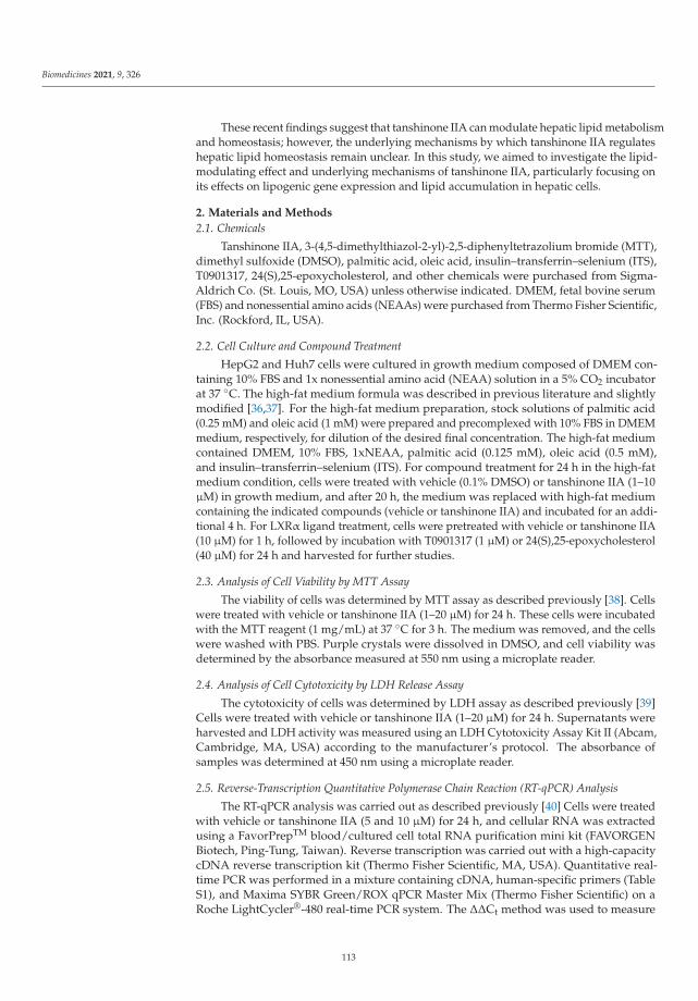
Biomedicines 2021, 9, 326
These recent findings suggest that tanshinone IIA can modulate hepatic lipid metabolismand homeostasis; however, the underlying mechanisms by which tanshinone IIA regulateshepatic lipid homeostasis remain unclear. In this study, we aimed to investigate the lipid-modulating effect and underlying mechanisms of tanshinone IIA, particularly focusing onits effects on lipogenic gene expression and lipid accumulation in hepatic cells.
2. Materials and Methods
2.1. Chemicals
Tanshinone IIA, 3-(4,5-dimethylthiazol-2-yl)-2,5-diphenyltetrazolium bromide (MTT),dimethyl sulfoxide (DMSO), palmitic acid, oleic acid, insulin–transferrin–selenium (ITS),T0901317, 24(S),25-epoxycholesterol, and other chemicals were purchased from Sigma-Aldrich Co. (St. Louis, MO, USA) unless otherwise indicated. DMEM, fetal bovine serum(FBS) and nonessential amino acids (NEAAs) were purchased from Thermo Fisher Scientific,Inc. (Rockford, IL, USA).
2.2. Cell Culture and Compound Treatment
HepG2 and Huh7 cells were cultured in growth medium composed of DMEM con-taining 10% FBS and 1x nonessential amino acid (NEAA) solution in a 5% CO2 incubatorat 37 ◦C. The high-fat medium formula was described in previous literature and slightlymodified [36,37]. For the high-fat medium preparation, stock solutions of palmitic acid(0.25 mM) and oleic acid (1 mM) were prepared and precomplexed with 10% FBS in DMEMmedium, respectively, for dilution of the desired final concentration. The high-fat mediumcontained DMEM, 10% FBS, 1xNEAA, palmitic acid (0.125 mM), oleic acid (0.5 mM),and insulin–transferrin–selenium (ITS). For compound treatment for 24 h in the high-fatmedium condition, cells were treated with vehicle (0.1% DMSO) or tanshinone IIA (1–10μM) in growth medium, and after 20 h, the medium was replaced with high-fat mediumcontaining the indicated compounds (vehicle or tanshinone IIA) and incubated for an addi-tional 4 h. For LXRα ligand treatment, cells were pretreated with vehicle or tanshinone IIA(10 μM) for 1 h, followed by incubation with T0901317 (1 μM) or 24(S),25-epoxycholesterol(40 μM) for 24 h and harvested for further studies.
2.3. Analysis of Cell Viability by MTT Assay
The viability of cells was determined by MTT assay as described previously [38]. Cellswere treated with vehicle or tanshinone IIA (1–20 μM) for 24 h. These cells were incubatedwith the MTT reagent (1 mg/mL) at 37 ◦C for 3 h. The medium was removed, and the cellswere washed with PBS. Purple crystals were dissolved in DMSO, and cell viability wasdetermined by the absorbance measured at 550 nm using a microplate reader.
2.4. Analysis of Cell Cytotoxicity by LDH Release Assay
The cytotoxicity of cells was determined by LDH assay as described previously [39]Cells were treated with vehicle or tanshinone IIA (1–20 μM) for 24 h. Supernatants wereharvested and LDH activity was measured using an LDH Cytotoxicity Assay Kit II (Abcam,Cambridge, MA, USA) according to the manufacturer’s protocol. The absorbance ofsamples was determined at 450 nm using a microplate reader.
2.5. Reverse-Transcription Quantitative Polymerase Chain Reaction (RT-qPCR) Analysis
The RT-qPCR analysis was carried out as described previously [40] Cells were treatedwith vehicle or tanshinone IIA (5 and 10 μM) for 24 h, and cellular RNA was extractedusing a FavorPrepTM blood/cultured cell total RNA purification mini kit (FAVORGENBiotech, Ping-Tung, Taiwan). Reverse transcription was carried out with a high-capacitycDNA reverse transcription kit (Thermo Fisher Scientific, MA, USA). Quantitative real-time PCR was performed in a mixture containing cDNA, human-specific primers (TableS1), and Maxima SYBR Green/ROX qPCR Master Mix (Thermo Fisher Scientific) on aRoche LightCycler®-480 real-time PCR system. The ΔΔCt method was used to measure
113
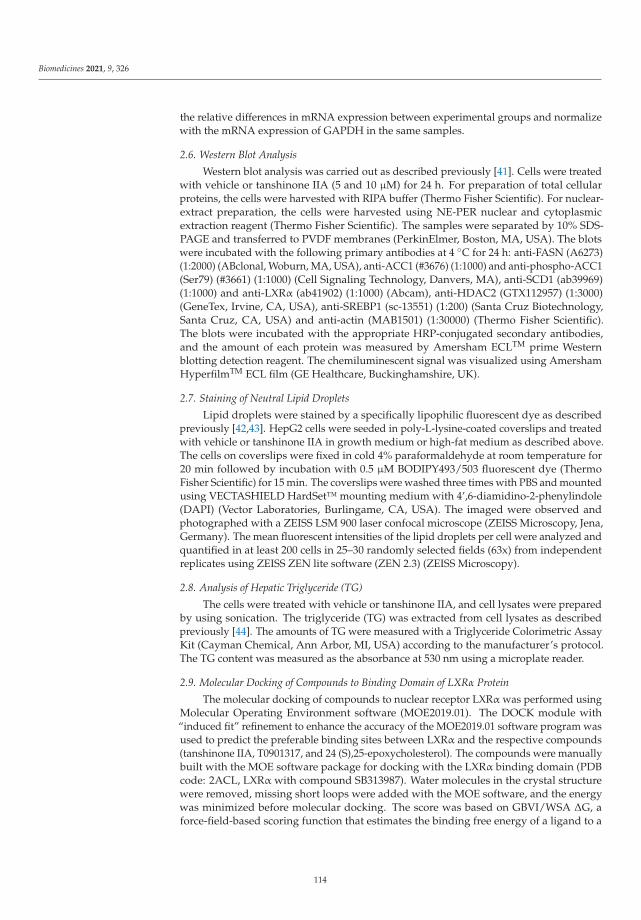
Biomedicines 2021, 9, 326
the relative differences in mRNA expression between experimental groups and normalizewith the mRNA expression of GAPDH in the same samples.
2.6. Western Blot Analysis
Western blot analysis was carried out as described previously [41]. Cells were treatedwith vehicle or tanshinone IIA (5 and 10 μM) for 24 h. For preparation of total cellularproteins, the cells were harvested with RIPA buffer (Thermo Fisher Scientific). For nuclear-extract preparation, the cells were harvested using NE-PER nuclear and cytoplasmicextraction reagent (Thermo Fisher Scientific). The samples were separated by 10% SDS-PAGE and transferred to PVDF membranes (PerkinElmer, Boston, MA, USA). The blotswere incubated with the following primary antibodies at 4 ◦C for 24 h: anti-FASN (A6273)(1:2000) (ABclonal, Woburn, MA, USA), anti-ACC1 (#3676) (1:1000) and anti-phospho-ACC1(Ser79) (#3661) (1:1000) (Cell Signaling Technology, Danvers, MA), anti-SCD1 (ab39969)(1:1000) and anti-LXRα (ab41902) (1:1000) (Abcam), anti-HDAC2 (GTX112957) (1:3000)(GeneTex, Irvine, CA, USA), anti-SREBP1 (sc-13551) (1:200) (Santa Cruz Biotechnology,Santa Cruz, CA, USA) and anti-actin (MAB1501) (1:30000) (Thermo Fisher Scientific).The blots were incubated with the appropriate HRP-conjugated secondary antibodies,and the amount of each protein was measured by Amersham ECLTM prime Westernblotting detection reagent. The chemiluminescent signal was visualized using AmershamHyperfilmTM ECL film (GE Healthcare, Buckinghamshire, UK).
2.7. Staining of Neutral Lipid Droplets
Lipid droplets were stained by a specifically lipophilic fluorescent dye as describedpreviously [42,43]. HepG2 cells were seeded in poly-L-lysine-coated coverslips and treatedwith vehicle or tanshinone IIA in growth medium or high-fat medium as described above.The cells on coverslips were fixed in cold 4% paraformaldehyde at room temperature for20 min followed by incubation with 0.5 μM BODIPY493/503 fluorescent dye (ThermoFisher Scientific) for 15 min. The coverslips were washed three times with PBS and mountedusing VECTASHIELD HardSet™ mounting medium with 4’,6-diamidino-2-phenylindole(DAPI) (Vector Laboratories, Burlingame, CA, USA). The imaged were observed andphotographed with a ZEISS LSM 900 laser confocal microscope (ZEISS Microscopy, Jena,Germany). The mean fluorescent intensities of the lipid droplets per cell were analyzed andquantified in at least 200 cells in 25–30 randomly selected fields (63x) from independentreplicates using ZEISS ZEN lite software (ZEN 2.3) (ZEISS Microscopy).
2.8. Analysis of Hepatic Triglyceride (TG)
The cells were treated with vehicle or tanshinone IIA, and cell lysates were preparedby using sonication. The triglyceride (TG) was extracted from cell lysates as describedpreviously [44]. The amounts of TG were measured with a Triglyceride Colorimetric AssayKit (Cayman Chemical, Ann Arbor, MI, USA) according to the manufacturer’s protocol.The TG content was measured as the absorbance at 530 nm using a microplate reader.
2.9. Molecular Docking of Compounds to Binding Domain of LXRα Protein
The molecular docking of compounds to nuclear receptor LXRα was performed usingMolecular Operating Environment software (MOE2019.01). The DOCK module with“induced fit” refinement to enhance the accuracy of the MOE2019.01 software program wasused to predict the preferable binding sites between LXRα and the respective compounds(tanshinone IIA, T0901317, and 24 (S),25-epoxycholesterol). The compounds were manuallybuilt with the MOE software package for docking with the LXRα binding domain (PDBcode: 2ACL, LXRα with compound SB313987). Water molecules in the crystal structurewere removed, missing short loops were added with the MOE software, and the energywas minimized before molecular docking. The score was based on GBVI/WSA ΔG, aforce-field-based scoring function that estimates the binding free energy of a ligand to a
114

Biomedicines 2021, 9, 326
receptor. The preferable binding sites for each compound were determined based on thelowest binding free energy, which was the lowest S value in the scoring function.
2.10. Statistical Analysis
At least three independent experiments were performed, and each experiment wasrepeated three times. The data are expressed as the means ± SD. The data were analyzedusing Student’s t-test (for comparisons of data with two groups) and one-way ANOVAwith Tukey’s test for post hoc analysis (for comparisons of data with multiple groups), anda p-value of <0.05 was considered statistically significant.
3. Results
3.1. Effects of Tanshinone IIA on the Viability of Hepatic Cell Lines
To investigate the cytotoxic effect of tanshinone IIA (Figure 1a) on hepatic cells, HepG2cells were treated with vehicle (0.1% DMSO) or tanshinone IIA (1, 2, 5, 10, and 20 μM)for 24 h. Cell cytotoxicity induced by tanshinone IIA was determined using an MTTassay and validated by an LDH release assay. The data showed that vehicle (0.1% DMSO)and tanshinone IIA (1–10 μM) induced no cytotoxicity in the HepG2 cells (Figure 1b,c).However, higher concentration of tanshinone IIA (20 μM) slightly reduced the cell viabilityas compared with vehicle-treated group. Similar results for cell viability also were foundin the tanshinone IIA-treated Huh 7 hepatic cell line (Figure S1). Therefore, doses of 5 and10 μM of tanshinone IIA were used in the subsequent experiments.
(a) (b) (c)
Figure 1. Effects of tanshinone IIA on HepG2 cell viability. (a) The chemical structure of tanshinone IIA. HepG2 cells weretreated with vehicle (0.1% DMSO) or tanshinone IIA (1, 2, 5, 10, and 20 μM) for 24 h. (b) Cell viability was measuredusing an MTT assay. (c) Cytotoxicity was measured using LDH release assay. The data represent the mean ± SD of threeindependent experiments. ## p < 0.01 indicates significant differences compared to the vehicle-treated cells.
3.2. Effects of Tanshinone IIA on FASN, ACC1, and SCD1 Expression in Hepatic Cell Lines
De novo lipogenesis plays a critical role in regulating the amount of lipid accumula-tion in hepatic cells. To investigate whether tanshinone IIA can regulate the expressionof endogenous de novo lipogenesis-related genes, we examined the effect of tanshinoneIIA treatment on the mRNA expression of fatty-acid synthase (FASN), acetyl-CoA car-boxylase 1 (ACC1), and stearoyl-CoA desaturase 1 (SCD1) in HepG2 cells. Tanshinone IIA(5 and 10 μM) significantly reduced the mRNA expression of FASN, ACC1, and SCD1 by0.85 ± 0.09- and 0.55 ± 0.18-fold; 0.81 ± 0.08- and 0.67 ± 0.10-fold; and 0.64 ± 0.14- and0.45 ± 0.13-fold in HepG2 cells, respectively, when compared to the vehicle-treated cells(1.00 ± 0.05, 1.00 ± 0.08, and 1.01 ± 0.14) (p < 0.05 and p < 0.01, respectively) (Figure 2a–c).Similar results showed that tanshinone IIA could significantly inhibit the mRNA expressionof FASN, ACC1, and SCD1 genes in Huh 7 cells (Figure S2a–c).
115

Biomedicines 2021, 9, 326
(a) (b) (c)
Figure 2. Effects of tanshinone IIA on FASN, ACC1, and SCD1 mRNA expression in HepG2 cells. HepG2 cells weretreated with vehicle (0.1% DMSO) or tanshinone IIA (5 and 10 μM) for 24 h. The mRNA expression of (a) FASN, (b) ACC1,and (c) SCD1 was measured by RT-qPCR analysis. The data represent the mean ± SD of three independent experiments.# p < 0.05 and ## p < 0.01 indicate significant differences compared to the vehicle-treated cells.
Furthermore, we examined the levels of the FASN, ACC1, and SCD1 proteins intanshinone IIA-treated HepG2 cells. The cells treated with tanshinone IIA (5 and 10 μM)had significantly decreased levels of FASN, ACC1, and SCD1 protein expression, by0.80 ± 0.13- and 0.67 ± 0.18-fold; 0.57 ± 0.19- and 0.45 ± 0.17-fold; and 0.66 ± 0.19- and0.43 ± 0.14-fold in HepG2 cells, respectively, when compared to the vehicle-treated cells(1.00 ± 0.03, 1.01 ± 0.08, and 1.00 ± 0.08) (p < 0.05 and p < 0.01, respectively) (Figure 3a–f).These results indicated that tanshinone IIA can significantly inhibit the mRNA and proteinexpression of these genes, which are essential for de novo lipogenesis in hepatic cells.
(a)
(b) (c)
(d)
(e)
(f)
Figure 3. Effects of tanshinone IIA on FASN, ACC1, and SCD1 protein expression in HepG2 cells. HepG2 cells were treatedwith vehicle (0.1% DMSO) or tanshinone IIA (5 and 10 μM) for 24 h. The protein expression of (a) FASN, (b) ACC1, and(c) SCD1 was measured by Western blot analysis. A representative blot is shown. The normalized intensities of (d) FASN,(e) ACC1, and (f) SCD1 proteins versus actin are presented as the mean ± SD of three independent experiments. # p < 0.05and ## p < 0.01 indicate significant differences compared to the vehicle-treated cells.
3.3. Effects of Tanshinone IIA on ACC1 Phosphorylation in HepG2 Cells
The enzyme ACC1 plays a critical role in fatty-acid synthesis and has been identifiedto be inactivated by phosphorylation of serine 79, leading to lipogenesis inhibition [45].Therefore, we investigated the effect of tanshinone IIA on ACC1 phosphorylation in HepG2cells. Tanshinone IIA (5 and 10 μM) significantly increased the ratio of phosphorylatedACC1 (p-ACC1)/ACC1 proteins by 1.41 ± 0.01- and 2.11 ± 0.05-fold in HepG2 cells, respec-tively, when compared to the vehicle-treated group (1.00 ± 0.08) (p < 0.01) (Figure 4a,b).
116
Biomedicines 2021, 9, 326
Moreover, we examined the effect of tanshinone IIA on ACC1 phosphorylation in HepG2cells incubated with high-fat medium. The ratio of p-ACC1/ACC1 in the cells treated withhigh-fat medium was slightly reduced by 0.80 ± 0.04-fold as compared to cells treated withnormal growth medium (1.00 ± 0.07) (p < 0.01). Tanshinone IIA significantly increasedthe p-ACC1/ACC1 ratio by 2.47 ± 0.08-fold, when compared to the vehicle-treated group(0.80 ± 0.04) (p < 0.01) (Figure 4c,d). This data indicated that tanshinone IIA could promotephosphorylation of ACC1 protein in hepatic cells.
(a) (b)
(c) (d)
Figure 4. Effects of tanshinone IIA on ACC1 phosphorylation in HepG2 cells. (a) HepG2 cells were treated with vehicle(0.1% DMSO) or tanshinone IIA (5 and 10 μM) in normal growth medium. The protein levels of phosphorylated ACC1at serine 79 (p-ACC1), ACC1, and actin was measured by Western blot analysis. A representative blot is shown. (b) Thenormalized intensity of p-ACC1 protein versus ACC1 is presented as the mean ± SD of three independent experiments.** p < 0.01 indicates significant differences compared to the vehicle-treated cells. (c) HepG2 cells were treated with vehicle(0.1% DMSO) or tanshinone IIA (10 μM) in normal growth medium or high-fat medium. The phosphorylated ACC1(p-ACC1), ACC1, and actin was measured by Western blot analysis. A representative blot is shown. (d) The normalizedintensity of p-ACC1 versus ACC1 is presented as the mean ± SD of three independent experiments. ** p < 0.01 indicatessignificant differences compared to the vehicle-treated cells incubated with growth medium. ## p < 0.01 indicates significantdifferences compared to the vehicle-treated cells.
3.4. Effects of Tanshinone IIA on the Fatty-Acid-Induced Lipogenesis and TG Accumulation inHepG2 Cells
To investigate the effect of tanshinone IIA on the fatty-acid-induced lipogenesis andTG accumulation, first, we examined whether tanshinone IIA affected cell viability ofHepG2 cells incubated in high-fat medium. The MTT assay data showed that the viabilityof cells treated with tanshinone IIA (1, 2, 5, and 10 μM) was 97.7 ± 3.1%, 96.8 ± 5.6%,94.6 ± 1.8%, and 92.4 ± 4.5% as compared to cells incubated in normal growth medium(100.4 ± 1.8%), respectively (Figure 5a). This data showed that tanshinone IIA did notinduce significant cytotoxicity in cells incubated with high-fat medium. Next, the effect oftanshinone IIA on the palmitate/oleate-induced lipogenesis was investigated. The HepG2cells incubated with high-fat medium showed increased levels of FASN, ACC1, and SCD1mRNA expression as compared with that cultured in growth medium. Tanshinone IIAsignificantly decreased the amounts of FASN, ACC1, and SCD1 mRNA in cells incubated
117

Biomedicines 2021, 9, 326
with normal growth or high-fat medium, when compared to their effects on vehicle-treatedcells (p < 0.01) (Figure 5b–d). The effect of tanshinone IIA on intracellular triglyceride (TG)accumulation was examined, and the data showed that TG contents in cells incubated withhigh-fat medium were significantly increased as compared with cells treated with normalgrowth medium (p < 0.01). The TG contents were significantly reduced by tanshinoneIIA (10 μM) treatment in HepG2 cells cultured in growth medium or high-fat medium(Figure 5e). These data indicated that tanshinone IIA can significantly ameliorate fatty-acid-induced lipogenesis and TG accumulation in hepatic cells.
(a)
(b)
(c)
(d)
(e)
Figure 5. Effects of tanshinone IIA on the fatty-acid-induced lipogenesis and TG accumulation in HepG2 cells. HepG2cells were treated with vehicle (0.1% DMSO) or tanshinone IIA (1–10 μM) in growth medium (DMEM with 10% FBS and1xNEAA), and after 20 h, the medium was replaced with high-fat medium containing the indicated compounds (vehicleor tanshinone IIA). (a) Cell viability was measured using an MTT assay. The mRNA expression of (b) FASN, (c) ACC1,and (d) SCD1 was measured by RT-qPCR analysis. The data represent the mean ± SD of three independent experiments.# p < 0.05 and ## p < 0.01 indicate significant differences compared to the vehicle-treated cells. (e) Intracellular triglyceride(TG) levels of vehicle- or tanshinone-IIA-treated HepG2 cells were measured from three independent replicates, and thedata represent the mean ± SD. ** p < 0.01 indicates significant differences compared to the vehicle-treated cells incubatedwith growth medium. ## p < 0.01 indicate significant differences compared to the tanshinone IIA-untreated cells.
118
Biomedicines 2021, 9, 326
3.5. Effects of Tanshinone IIA on SREBP1 Expression in HepG2 Cells
SREBP-1c is an important transcriptional regulator of FASN, ACC1, and SCD1 geneexpression in hepatic cells. Therefore, we further investigated the effect of tanshinoneIIA on SREBP-1c expression in HepG2 cells. Tanshinone IIA (5 and 10 μM) significantlyreduced the level of SREBP-1c mRNA by 0.65 ± 0.15- and 0.37 ± 0.10-fold in HepG2 cells,respectively, when compared to the vehicle-treated group (1.01 ± 0.11) (p < 0.01) (Figure 6a).Because the SREBP1 antibody used was unable to distinguish between the SREBP-1a and-1c, we used the SREBP1 to mention in this data.. We found that tanshinone IIA (5 and10 μM) decreased the amount of nuclear mature SREBP1 protein by 0.73 ± 0.19- and0.35 ± 0.17-fold, respectively, when compared to the vehicle-treated group (1.01 ± 0.03)(p < 0.05 and p < 0.01, respectively) (Figure 6b,c).
(a) (b) (c)
(d)
Figure 6. Effects of tanshinone IIA on SREBP1 gene expression. HepG2 cells were treated with vehicle (0.1% DMSO) ortanshinone IIA (5 and 10 μM) for 24 h. (a) The mRNA expression of SREBP-1c was measured by RT-qPCR analysis. Thedata represent the mean ± SD of three independent experiments. ## p < 0.01 indicates significant differences compared tothe vehicle-treated cells. (b) Nuclear extracts were prepared from vehicle- or tanshinone-IIA-treated cells, and the levelof nuclear SREBP1 protein was analyzed by Western blot analysis. A representative blot is shown. (c) The normalizedintensity of SREBP1 protein versus HDAC2 is presented as the mean ± SD of three independent experiments. # p < 0.05and ## p < 0.01 indicate significant differences compared to the vehicle-treated cells. (d) HepG2 cells were treated withvehicle (0.1% DMSO) or tanshinone IIA (10 μM) in normal growth medium or high-fat medium. The mRNA expressionof SREBP-1c was measured by RT-qPCR analysis. The data represent the mean ± SD of three independent experiments.** p < 0.01 indicates significant differences compared to the vehicle-treated cells incubated with growth medium. ## p < 0.01indicates significant differences compared to the vehicle-treated cells.
Furthermore, we investigated the effect of tanshinone IIA on SREBP1c expression inHepG2 cells incubated with high-fat medium. The cells incubated with high-fat mediumshowed an elevated level of SREBP1c mRNA expression by 1.87 ± 0.16-fold as comparedwith the cells cultured in growth medium (1.01 ± 0.15) (p < 0.01). Tanshinone IIA signifi-cantly decreased the amount of SREBP1c mRNA by 0.64 ± 0.13-fold in cells incubated withhigh-fat medium, when compared to its effect on vehicle-treated cells (p < 0.01) (Figure 6d).In addition to SREBP1c, recently, carbohydrate regulatory binding protein (ChREBP) also
119

Biomedicines 2021, 9, 326
was reported to regulate the FASN and ACC1 gene expression for glucose-induced lipoge-nesis [2,46]. We further examined the effect of tanshinone IIA on ChREBP expression inHepG2 cells. We found that tanshinone IIA (5 and 10 μM) could not change the level ofChREBP mRNA expression (Figure S3). These above results indicated that tanshinone IIAdownregulates SREBP1 gene expression, which may lead to reduce expression of lipogenicgenes in hepatic cells.
3.6. Effects of Tanshinone IIA on LXRα-Mediated Transcriptional Activity in HepG2 Cells
SREBP-1c and some lipogenic enzymes such as FASN are the direct targets for tran-scriptional activation by LXRα in hepatic cells. To explore the effect of tanshinone IIAon LXRα-mediated SREBP1 expression, we first examined whether tanshinone IIA couldchange LXRα gene expression in HepG2 cells. We found that tanshinone IIA did notchange the mRNA or nuclear protein levels of LXRα in the HepG2 cells (Figure 7a–c).We further examined the effect of tanshinone IIA on the ligand-induced LXR activityfor SREBP-1c mRNA expression. HepG2 cells were pretreated with vehicle or tanshi-none IIA (10 μM) for 1 h followed by treatment with LXRα agonists; namely, T0901317(Figure 7d) and 24(S),25-epoxycholesterol (Figure 7e), for an additional 24 h. As shown inFigure 7f, the level of SREBP-1c mRNA was significantly increased by T0901317 (1 μM)and 24(S),25-epoxycholesterol (40 μM) treatment of the HepG2 cells. When cells werecotreated with tanshinone IIA and agonists, the T0901317- and 24(S),25-epoxycholesterol-induced SREBP-1c mRNA expression was significantly attenuated by approximately 60%and 67%, respectively (p < 0.01). Furthermore, we found that T0901317- and 24(S),25-epoxycholesterol-induced FASN mRNA expression was also significantly decreased by ap-proximately 56% and 45%, respectively, in tanshinone IIA-treated cells (p < 0.01) (Figure 7g).Moreover, to validate the regulatory effect of tanshinone IIA on LXRα-mediated transcrip-tion, mRNA expression of other downstream target genes, such as ACC1 and SCD1 [24,47],and GLUT1 [48], a negatively regulated gene by LXRα ligands, were also examined.T0901317-induced ACC1 and SCD1 mRNA expression was significantly decreased bytanshinone IIA in HepG2 cells (Figure S4a,b). Tanshinone IIA also could reverse theT0901317-decreased GLUT1 expression to basal level in HepG2 cells (Figure S4c). Theseabove results verified that tanshinone IIA was involved in the regulation of LXRα-mediatedtranscriptional activity in hepatic cells.
(a) (b) (c)
T0901317
24(S),25-Epoxycholesterol
(d) (e)
Figure 7. Cont.
120

Biomedicines 2021, 9, 326
(f) (g)
Figure 7. Effects of tanshinone IIA on LXRα expression and ligand-induced LXRα activation in HepG2 cells. HepG2 cells were treatedwith vehicle (0.1% DMSO) or tanshinone IIA (5 and 10 μM) for 24 h. (a) The mRNA expression of LXRα was measured by RT-qPCRanalysis. The data are presented as the mean ± SD of three independent experiments. (b) The nuclear protein expression of LXRα andHDAC2 was measured by Western blot analysis. A represent blot is shown. (c) Normalized intensities of LXRα versus HDAC2 ispresented as the mean ± SD of three independent experiments. The chemical structure of (d) T0901317 and (e) 24(S),25-epoxycholesterolHepG2 cells were pretreated with vehicle (0.1% DMSO) or tanshinone IIA (10 μM) for 1 h followed by treatment with agonist T0901317(1 μM) or 24(S),25-epoxycholesterol (40 μM) for 24 h. The mRNA levels of (f) SREBP-1c and (g) FASN were measured by RT-qPCRanalysis. ** p < 0.01 indicates a significant difference compared to compound-untreated group. ## p < 0.01 indicates significantdifferences compared to cells treated with the agonist alone.
3.7. Tanshinone IIA Attenuates LXRα-Mediated Lipid-Droplet Accumulation in HepG2 Cells
To investigate the effect of tanshinone IIA on lipid accumulation in HepG2 cells, theneutral lipid droplets in HepG2 cells were assessed using BODIPY493/503 fluorescent dyestaining and analyzed under a confocal microscope. The cells incubated with high-fat mediumshowed an increased level of lipid droplets as compared with the cells cultured in normalgrowth medium (Figure 8a,b). Tanshinone IIA could significantly reduce the amount of lipiddroplets in cells incubated with growth medium and high-fat medium by approximately 37%and 32%, respectively, when compared to its effect on vehicle-treated cells (Figure 8c). We fur-ther investigated the effect of tanshinone IIA on LXRα-mediated lipid-droplet accumulationin HepG2 cells. The data from Figure 8a–c showed that lipid accumulation induced by theT0901317 (1 μM) in cells with growth or high-fat medium was increased by approximately25% and 33%, respectively, when compared to vehicle-treated groups. When cells werecotreated with T0901317 and tanshinone IIA (10 μM), the T0901317-induced lipid dropletswere significantly attenuated by approximately 52% and 49% in cells incubated with growthmedium and high-fat medium, respectively. These data indicated that tanshinone IIA canattenuate LXRα-mediated lipid droplets accumulation in HepG2 cells.
(a)
(b)
Figure 8. Cont.
121

Biomedicines 2021, 9, 326
(c)
Figure 8. Effects of tanshinone IIA on lipid-droplet accumulation in HepG2 cells. HepG2 cells were pretreated with vehicle (0.1%DMSO) or tanshinone IIA (10 μM) for 1 h followed by treatment with T0901317 (1 μM) in (a) growth medium or (b) high-fat medium.The lipid droplets were stained by BODIPY493/503 fluorescent dye (green), and nuclei were stained by DAPI (blue). Stained cells wereobserved and photographed with a confocal microscope. The scale bar is 10 μm. (c) Quantification of lipid droplets accumulation. Themean fluorescent intensities of the lipid droplets per cell were quantified in 25–30 randomly selected fields (>200 cells) from threeindependent replicates, and the data represent the mean ± SD from three independent experiments. ** p < 0.01 indicates significantdifferences compared to the vehicle-treated cells. ## p < 0.01 indicates significant differences compared to the cells treated withT0901317 alone.
3.8. Tanshinone IIA Docks to the Ligand-Binding Domain of LXRα
To investigate whether tanshinone IIA can bind to LXRα, the molecular dockingprogram was performed as described in the Materials and Methods section. The dockingresults showed that tanshinone IIA, T0901317, and 24(S),25-epoxycholesterol can dockto LXRα binding pocket with different docking scores (tanshinone IIA: −7.33; T0901317:−8.47; and 24(S),25-epoxycholesterol: −10.32) (Figure 9a–c). The superposition resultshowed that all these compounds docked into the ligand-binding domain of LXRα atsimilar poses (Figure 9d). The ligand-receptor interaction maps showed that the tanshinoneIIA was surrounded by many hydrophobic amino-acid residues such as F255, L258, A259,M296, and F313 (Figure 9e). These data indicated that the binding pocket of LXRα issuitable for lipophilic molecule interactions and tanshinone IIA may serve as a ligandspecifically bound to this ligand-binding domain for modulation of LXRα activity.
(a) (b)
Figure 9. Cont.
122
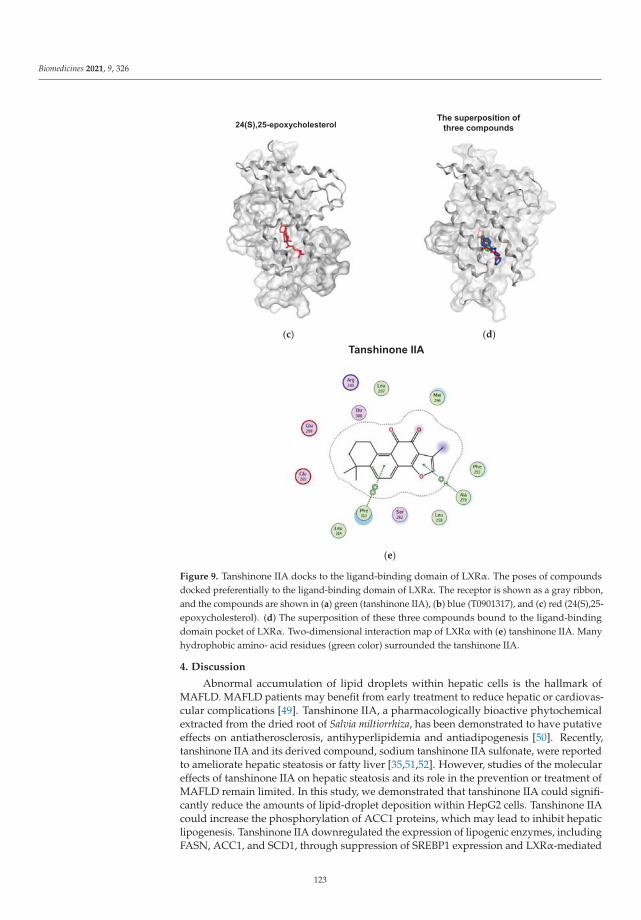
Biomedicines 2021, 9, 326
(c) (d)
(e)
Figure 9. Tanshinone IIA docks to the ligand-binding domain of LXRα. The poses of compoundsdocked preferentially to the ligand-binding domain of LXRα. The receptor is shown as a gray ribbon,and the compounds are shown in (a) green (tanshinone IIA), (b) blue (T0901317), and (c) red (24(S),25-epoxycholesterol). (d) The superposition of these three compounds bound to the ligand-bindingdomain pocket of LXRα. Two-dimensional interaction map of LXRα with (e) tanshinone IIA. Manyhydrophobic amino- acid residues (green color) surrounded the tanshinone IIA.
4. Discussion
Abnormal accumulation of lipid droplets within hepatic cells is the hallmark ofMAFLD. MAFLD patients may benefit from early treatment to reduce hepatic or cardiovas-cular complications [49]. Tanshinone IIA, a pharmacologically bioactive phytochemicalextracted from the dried root of Salvia miltiorrhiza, has been demonstrated to have putativeeffects on antiatherosclerosis, antihyperlipidemia and antiadipogenesis [50]. Recently,tanshinone IIA and its derived compound, sodium tanshinone IIA sulfonate, were reportedto ameliorate hepatic steatosis or fatty liver [35,51,52]. However, studies of the moleculareffects of tanshinone IIA on hepatic steatosis and its role in the prevention or treatment ofMAFLD remain limited. In this study, we demonstrated that tanshinone IIA could signifi-cantly reduce the amounts of lipid-droplet deposition within HepG2 cells. Tanshinone IIAcould increase the phosphorylation of ACC1 proteins, which may lead to inhibit hepaticlipogenesis. Tanshinone IIA downregulated the expression of lipogenic enzymes, includingFASN, ACC1, and SCD1, through suppression of SREBP1 expression and LXRα-mediated
123

Biomedicines 2021, 9, 326
transcriptional activation, resulting in reducing lipogenesis and lipid accumulation in theHepG2 cells. Moreover, we predicted that tanshinone IIA binds to the ligand-bindingdomain of LXRα to suppress its transcriptional activity (Figure 10). These findings supportthe hypothesis that tanshinone IIA possesses lipid-modulating activity and can potentiallyserve as a novel agent for the prevention or treatment of hepatic steatosis or MAFLD.
SRELXRELXRE
p
Figure 10. A hypothetical model for the tanshinone-IIA-mediated attenuation of lipid accumulationin hepatic cells. Tanshinone IIA promotes ACC1 phosphorylation to attenuate fatty-acid synthesisand lipogenesis. Tanshinone IIA down-regulates FASN, ACC1, and SCD1 expression by inhibitingthe LXRα-mediated transcriptional activation, resulting in a reduction in endogenous and fatty-acid-induced lipogenesis as well as lipid-droplet accumulation in hepatic cells.
Hepatic steatosis results from an imbalance between lipid supplementation and clear-ance in the liver. Emerging studies have demonstrated that dysregulation of lipogenesiscontributes to hepatic steatosis and plays an essential role in the pathologic progressof MAFLD. The increases in hepatic TG content measured in MAFLD patients showedthat approximately 30% of the contribution came from de novo biosynthesis [17]. In thisstudy, tanshinone IIA downregulated the mRNA and protein expression of lipogenicenzymes, including FASN, ACC1, and SCD1, in HepG2 cells. Our data also showedtanshinone IIA markedly reduced TG contents and lipid-droplet accumulation. It hasbeen reported that modulation of de novo lipogenesis in hepatocytes is important for theattenuation of hepatic steatosis and treatment of MAFLD. Recent studies using animalmodels have shown that knocking out or knocking down the hepatic enzymes involvedin lipogenesis or desaturation of long-chain fatty acids may reduce TG concentration andattenuate steatosis [16,53,54]. The present study revealed that tanshinone IIA can reducethe levels of lipogenic enzymes in hepatocytes, which may lead to decreased hepatic TGcontents and lipid accumulation. Saturated-fat diets could increase lipogenic gene expres-sion and hepatic steatosis in mice [55]. The TG-derived fatty-acid profile showed thathigher saturated fatty-acid content and low levels of polyunsaturated fatty acid increasedthe proinflammatory cytokine to enhance inflammation [56]. In this study, inhibition ofpalmitate/oleate-induced lipogenesis by tanshinone IIA may be associated with reductionof TG accumulation. Whether tanshinone IIA could increase the unsaturated fatty-acidlevels and reduce saturated fatty-acid content in cells treated with high-fat medium remainsunclear. Analysis of the profile of TG-derived saturated and mono/polyunsaturated fattyacid in lipid droplets from tanshinone IIA-treated cells needs to be further investigated.
In this study, tanshinone IIA increased the ratio of p-ACC1/ACC1 in HepG2 cells.These data indicated that tanshinone IIA could promote ACC1 phosphorylation, which may
124

Biomedicines 2021, 9, 326
lead to inhibit its enzyme activity for the decrease of de novo lipid biosynthesis. It is knownthat AMPK activation directly phosphorylated ACC1 at serine 79 and suppressed its activity,and reduced FASN expression to attenuate fatty-acid-induced hepatic steatosis. AMPKactivation also decreased fatty-acid biosynthesis through reducing the SREBP1c levels ornuclear translocation to repress lipogenic gene expression and lipid accumulation [57,58].Recently, AMPK activator was reported to modulate hepatic saturated/unsaturated fatty-acid composition, and suppress hepatic macrophage infiltration in the development ofMAFLD [59]. Whether tanshinone IIA can activate AMPK signaling to regulate lipogenicgene expression and reduce hepatic steatosis remains unclear and needs to be furtherinvestigated.
Transcriptional modulation of the lipogenic enzymes in hepatocytes is an interestingidea for developing therapeutic strategies for hepatic steatosis. The transcription factorSREBP1 regulates the transcription rate of genes involved in the fatty-acid synthesis path-way. SREBP1 is expressed in two isoforms, SREBP-1a and SREBP-1c, can be translatedas a precursor protein bound to the endoplasmic reticulum (ER) membrane, and mustbe transported to the Golgi and activated by a cleavage process to release a mature formto the nucleus [60]. SREBP1 mediates the regulation of hepatic genes involved in carbo-hydrate and lipid metabolism. SREBP-1c was reported to control the mRNA expressionof FASN, ACC1, and SCD1 in hepatocytes [61,62]. Additionally, hepatic SREBP-1c washighly expressed in patients with MAFLD [63]. In this study, we found that tanshinoneIIA significantly decreased the levels of SREBP-1c mRNA and the mature form of nuclearSREBP1 protein. These results revealed that tanshinone IIA can inhibit the transcription ofFASN, ACC1, and SCD1 via the modulation of nuclear SREBP1 levels in hepatic cells.
The essential role of SREBP1 in lipogenesis has been demonstrated in gain- and loss-of-function studies in vitro and in vivo. Overexpressing a mature form of SREBP-1c inhepatocytes or mice can activate the lipogenic pathway and induce steatosis [64,65]. In con-trast, mice lacking SREBP-1c fail to express of lipogenic enzymes or undergo TG synthesisin response to fasting and refeeding [23]. The data we presented here demonstrated thattanshinone IIA could cause a significant decrease in the level of the mature SREBP1 protein.These findings indicate that tanshinone-IIA-mediated nuclear SREBP1 reduction leads tothe downregulated expression of lipogenic enzymes and attenuates lipid accumulation inhepatic cells. SREBP1 protein is also known to play a critical role in hepatic carbohydratemetabolism [66]. Whether tanshinone-IIA-mediated SREBP1 downregulation modulatesglucose metabolism in hepatic cells needs to be further investigated.
The SREBP-1c gene is upregulated by the nuclear receptor LXRα. LXR is expressedin two isoforms, LXRα and LXRβ, and LXRs are ligand-dependent nuclear receptors thatregulate cholesterol and lipid metabolism [67,68]. LXRα is mainly expressed in the liver,kidney, adipose tissue, and intestine, whereas LXRβ is ubiquitously expressed [69]. LXRαagonist treatment has been reported to induce lipogenesis, because they bind to LXREswithin the promoter region of SREBP-1c, FASN, ACC1, and SCD1 to increase mRNAtranscription in hepatic cells [24,47,70,71]. LXRα and its lipogenic target genes were alsoreported to be highly expressed in patients with MAFLD [72]. In this study, we foundthat the LXR agonist enhanced lipogenic genes and contributed to lipid accumulation asexpected in HepG2 cells. In tanshinone-IIA-treated cells, LXRα-induced lipid-droplet accu-mulation can be significantly attenuated. Lipid droplets are dynamic lipid-rich organellesthat have been shown to function in TG and energy storage, and as a protective strategyfor fatty-acid-induced lipotoxicity in the liver. However, an imbalance of lipid-droplet for-mation and mobilization can lead to excess lipid droplets and TG accumulation in hepaticcells, which is a pathological condition for MAFLD [73,74]. In this study, tanshinone IIAsignificantly ameliorated both fatty-acid- and LXRα-induced lipid-droplet accumulationin HepG2 cells. This finding suggested that tanshinone IIA may modulate lipid-dropletformation and homeostasis in the liver.
The LXRα protein is composed of an N-terminal-activating domain, a DNA-bindingdomain, a ligand-binding domain (LBD), and a C-terminal activation domain. The LBD
125

Biomedicines 2021, 9, 326
of LXRα has been reported to form a hydrophobic pocket and may interact with small-molecule compounds. The binding affinity of LXR to its ligand could be predicted bymolecular-docking analysis [75]. In this study, molecular docking predicted that tanshi-none IIA specifically binds to the LBD of LXRα. The lipophilic tanshinone IIA seemspreferentially to interact with hydrophobic residues within the LBD pocket of LXRα. Thus,we suggested that the LXRα-ligand-induced lipid accumulation could be significantlyameliorated by tanshinone IIA. LXR agonists were reported to increase expression of theirdownstream target genes such as ACC1 and SCD1, and decrease GLUT1 expression. Inthis study, tanshinone IIA could counteract the effect of the LXRα ligand T0901317 on thesetarget genes’ expression; however, LXRα mRNA and protein levels were not changed.These results supported that tanshinone IIA could serve as a novel LXRα ligand to mod-ulate the LXRα–SREBP1 axis in hepatic cells. The precise mechanisms underlying thesuppression of LXRα activation by tanshinone IIA remain unclear. The determination oftanshinone IIA as an LXR antagonist, an inhibitor, or a ligand competitor that blocks theLXR-RXR heterodimer needs further functional studies, with subjects such as GLUT1 orother downstream targets involved in glucose or lipid metabolism.
LXRα plays essential roles in fatty-acid and cholesterol homeostasis, and in control ofinflammation and immunity [76,77]. LXR activation may control cholesterol efflux fromfoam cells, induce cholesterol transporter expression, and inhibit inflammatory mediatorsin the artery wall and macrophage, which may lead to reduced vascular inflammation andatherosclerosis [49]. Therefore, LXR agonists are an option to be used for the treatmentof atherosclerosis, diabetes, and other disorders [78]. However, LXR agonists activatedhepatic lipogenesis and promoted TG accumulation in MAFLD, which limit the valueof clinical application. In this study, tanshinone IIA may have served as a ligand andcounteracted the LXRα–SREBP1 axis to reduce hepatic lipid accumulation. Recently, tan-shinone IIA treatment was reported to reduced macrophage infiltration and nuclear factor(NF)-κB activation in macrophages for inhibiting inflammatory responses [79]. Tan et al.reported that tanshinone IIA increased cholesterol efflux and reduced lipid accumulation inmacrophages, leading to a reduction in the development of aortic atherosclerosis [80]. Wenet al. reported that tanshinone IIA inhibited oxidized LDL-induced NLRP3 inflammasomeactivation in mouse macrophages and ameliorated atherogenesis [81]. These findingsindicated that tanshinone IIA possessed the potential activities for anti-inflammation andantiatherosclerosis. In this study, we suggested that tanshinone IIA could attenuate ligand-induced LXRα activation and may be an LXRα antagonist or a ligand competitor fordownregulation of lipogenic gene expression in hepatic cells. Whether tanshinone IIAaffects the anti-inflammatory effect and cholesterol accumulation by LXRα-dependentpathways in extra-hepatocytes remains to be clarified, and needs to be considered withcaution due to its adverse effect in therapeutic strategy for treatment of MAFLD. Recently,Griffett et al. reported that a hepato-selective LXR inverse agonist could specifically sup-press LXR activity in hepatic cells to reduce lipogenesis and prevent MAFLD [82]. Whethertanshinone IIA could serve as an inverse agonist to reduce lipogenesis without promotingcholesterol accumulation in extra-hepatic tissue remains to be further investigated. Inthis study, we realized our results were found only on the in vitro lipid-lowering effectsof tanshinone IIA on hepatic cancer cell lines. To verify the impact of treatment withtanshinone IIA in vivo, the primary human hepatocytes and animal models of MAFLDcan be used in further studies of tanshinone IIA on modulation of lipogenesis and lipidaccumulation.
5. Conclusions
In this study, for the first time, we demonstrated that tanshinone IIA exerts a promisinglipid-modulating effect to attenuate lipid-droplet accumulation and downregulate theexpression of genes involved in de novo lipogenesis by regulating the LXRα/SREBP1pathway in hepatic cells. Our findings revealed that tanshinone IIA may serve as a novel
126

Biomedicines 2021, 9, 326
supplement or drug to regulate LXRα activity and counter unwanted lipid overproductionin the liver for the prevention or treatment of MAFLD.
Supplementary Materials: The following are available online at https://www.mdpi.com/2227-9059/9/3/326/s1, Table S1: The primer pairs used in RT-qPCR, Figure S1: Effects of tanshinone IIA onHuh 7 cell viability, Figure S2: Effects of tanshinone IIA on FASN, ACC1, and SCD1 mRNA expressionin Huh 7 cells, Figure S3: Effects of tanshinone IIA on ChREBP mRNA expression in HepG2 cells,Figure S4: Effects of tanshinone IIA on LXRα-mediated transcriptional activity in HepG2 cells.
Author Contributions: J.-H.Y. and P.-Y.C. conceived and designed the experiments; W.-Y.G., H.-J.H.,and C.-Y.L. performed the experiments; W.-Y.G., H.-J.H., P.-Y.C., and J.-H.Y. analyzed the data; M.-J.W., J.-H.Y., and P.-Y.C. contributed reagents/materials; W.-Y.G., P.-Y.C., and J.-H.Y. wrote the paper.All authors have read and agreed to the published version of the manuscript.
Funding: This research was supported by grants MOST-105-2320-B-320-004-MY3 (funding approvaldate 1 August 2016) and MOST 108-2320-B-320-002-MY3 (to J.-H.Y.) (funding approval date 1 August2019), and MOST 108-2635-B-303-001 (to P.-Y.C.) (funding approval date 1 August 2019) from theMinistry of Science and Technology, Taiwan.
Institutional Review Board Statement: Not applicable.
Informed Consent Statement: Not applicable.
Data Availability Statement: Not applicable.
Acknowledgments: We are grateful for the support from the Core Research Laboratory, Tzu ChiUniversity for the assistance in the confocal image analysis.
Conflicts of Interest: The authors declare no conflict of interest.
References
1. Nguyen, P.; Leray, V.; Diez, M.; Serisier, S.; Le Bloc’H, J.; Siliart, B.; Dumon, H. Liver lipid metabolism. J. Anim. Physiol. Anim.Nutr. 2008, 92, 272–283. [CrossRef]
2. Ipsen, D.H.; Lykkesfeldt, J.; Tveden-Nyborg, P. Molecular mechanisms of hepatic lipid accumulation in non-alcoholic fatty liverdisease. Cell. Mol. Life Sci. 2018, 75, 3313–3327. [CrossRef]
3. Byrne, C.D.; Targher, G. NAFLD: A multisystem disease. J. Hepatol. 2015, 62, S47–S64. [CrossRef] [PubMed]4. Shiha, G.; Alswat, K.; Al Khatry, M.; I Sharara, A.; Örmeci, N.; Waked, I.; Benazzouz, M.; Al-Ali, F.; Hamed, A.E.; Hamoudi, W.;
et al. Nomenclature and definition of metabolic-associated fatty liver disease: A consensus from the Middle East and north Africa.Lancet Gastroenterol. Hepatol. 2021, 6, 57–64. [CrossRef]
5. Eslam, M.; Newsome, P.N.; Sarin, S.K.; Anstee, Q.M.; Targher, G.; Romero-Gomez, M.; Zelber-Sagi, S.; Wong, V.W.-S.; Dufour,J.-F.; Schattenberg, J.M.; et al. A new definition for metabolic dysfunction-associated fatty liver disease: An international expertconsensus statement. J. Hepatol. 2020, 73, 202–209. [CrossRef]
6. Younossi, Z.M.; Koenig, A.B.; Abdelatif, D.; Fazel, Y.; Henry, L.; Wymer, M. Global epidemiology of nonalcoholic fatty liverdisease-Meta-analytic assessment of prevalence, incidence, and outcomes. Hepatology 2016, 64, 73–84. [CrossRef]
7. Adams, L.A.; Anstee, Q.M.; Tilg, H.; Targher, G. Non-alcoholic fatty liver disease and its relationship with cardiovascular diseaseand other extrahepatic diseases. Gut 2017, 66, 1138–1153. [CrossRef]
8. Fazel, Y.; Koenig, A.B.; Sayiner, M.; Goodman, Z.D.; Younossi, Z.M. Epidemiology and natural history of non-alcoholic fatty liverdisease. Metab. Clin. Exp. 2016, 65, 1017–1025. [CrossRef]
9. Loomba, R.; Chalasani, N. The Hierarchical Model of NAFLD: Prognostic Significance of Histologic Features in NASH. Gastroen-terology 2015, 149, 278–281. [CrossRef]
10. Sumida, Y.; Yoneda, M. Current and future pharmacological therapies for NAFLD/NASH. J. Gastroenterol. 2018, 53, 362–376.[CrossRef] [PubMed]
11. Adams, L.A.; Angulo, P. Treatment of non-alcoholic fatty liver disease. Postgrad. Med. J. 2006, 82, 315–322. [CrossRef]12. Katsurada, A.; Iritani, N.; Fukuda, H.; Matsumura, Y.; Nishimoto, N.; Noguchi, T.; Tanaka, T. Effects of nutrients and hormones
on transcriptional and post-transcriptional regulation of fatty acid synthase in rat liver. JBIC J. Biol. Inorg. Chem. 1990, 190,427–433. [CrossRef] [PubMed]
13. Katsurada, A.; Iritani, N.; Fukuda, H.; Matsumura, Y.; Nishimoto, N.; Noguchi, T.; Tanaka, T. Effects of nutrients and hormoneson transcriptional and post-transcriptional regulation of acetyl-CoA carboxylase in rat liver. Eur. J. Biochem. 1990, 190, 435–441.[CrossRef]
14. Jakobsson, A.; Westerberg, R.; Jacobsson, A. Fatty acid elongases in mammals: Their regulation and roles in metabolism. Prog.Lipid Res. 2006, 45, 237–249. [CrossRef] [PubMed]
127
Biomedicines 2021, 9, 326
15. Ntambi, J.M. Dietary regulation of stearoyl-CoA desaturase 1 gene expression in mouse liver. J. Biol. Chem. 1992, 267, 10925–10930.[CrossRef]
16. Postic, C.; Girard, J. Contribution of de novo fatty acid synthesis to hepatic steatosis and insulin resistance: Lessons fromgenetically engineered mice. J. Clin. Investig. 2008, 118, 829–838. [CrossRef] [PubMed]
17. Donnelly, K.L.; Smith, C.I.; Schwarzenberg, S.J.; Jessurun, J.; Boldt, M.D.; Parks, E.J. Sources of fatty acids stored in liver andsecreted via lipoproteins in patients with nonalcoholic fatty liver disease. J. Clin. Investig. 2005, 115, 1343–1351. [CrossRef]
18. Eberlé, D.; Hegarty, B.; Bossard, P.; Ferré, P.; Foufelle, F. SREBP transcription factors: Master regulators of lipid homeostasis.Biochimie 2004, 86, 839–848. [CrossRef]
19. Higuchi, N.; Kato, M.; Shundo, Y.; Tajiri, H.; Tanaka, M.; Yamashita, N.; Kohjima, M.; Kotoh, K.; Nakamuta, M.; Takayanagi,R.; et al. Liver X receptor in cooperation with SREBP-1c is a major lipid synthesis regulator in nonalcoholic fatty liver disease.Hepatol. Res. 2008, 38, 1122–1129. [CrossRef]
20. Edwards, P.A.; Kennedy, M.A.; Mak, P.A. LXRs; oxysterol-activated nuclear receptors that regulate genes controlling lipidhomeostasis. Vasc. Pharm. 2002, 38, 249–256. [CrossRef]
21. Perissi, V.; Aggarwal, A.; Glass, C.K.; Rose, D.W.; Rosenfeld, M.G. A Corepressor/Coactivator Exchange Complex Required forTranscriptional Activation by Nuclear Receptors and Other Regulated Transcription Factors. Cell 2004, 116, 511–526. [CrossRef]
22. Schultz, J.R.; Tu, H.; Luk, A.; Repa, J.J.; Medina, J.C.; Li, L.; Schwendner, S.; Wang, S.; Thoolen, M.; Mangelsdorf, D.J.; et al. Role ofLXRs in control of lipogenesis. Genes Dev. 2000, 14, 2831–2838. [CrossRef]
23. Liang, G.; Yang, J.; Horton, J.D.; Hammer, R.E.; Goldstein, J.L.; Brown, M.S. Diminished Hepatic Response to Fasting/Refeedingand Liver X Receptor Agonists in Mice with Selective Deficiency of Sterol Regulatory Element-binding Protein-1c. J. Biol. Chem.2002, 277, 9520–9528. [CrossRef] [PubMed]
24. Chu, K.; Miyazaki, M.; Man, W.C.; Ntambi, J.M. Stearoyl-Coenzyme A Desaturase 1 Deficiency Protects against Hypertriglyc-eridemia and Increases Plasma High-Density Lipoprotein Cholesterol Induced by Liver X Receptor Activation. Mol. Cell. Biol.2006, 26, 6786–6798. [CrossRef] [PubMed]
25. Wagner, M.; Zollner, G.; Trauner, M. Nuclear receptors in liver disease. Hepatology 2011, 53, 1023–1034. [CrossRef] [PubMed]26. Chen, Z.; Xu, H. Anti-Inflammatory and Immunomodulatory Mechanism of Tanshinone IIA for Atherosclerosis. Evid. Based
Complement. Altern. Med. 2014, 2014, 1–6. [CrossRef] [PubMed]27. Shang, Q.; Xu, H.; Huang, L. Tanshinone IIA: A Promising Natural Cardioprotective Agent. Evid. Based Complement. Altern. Med.
2012, 2012, 1–7. [CrossRef]28. Xu, S.; Liu, Z.; Huang, Y.; Le, K.; Tang, F.; Huang, H.; Ogura, S.; Little, P.J.; Shen, X.; Liu, P. Tanshinone II-A inhibits oxidized LDL-
induced LOX-1 expression in macrophages by reducing intracellular superoxide radical generation and NF-kappaB activation.Transl. Res. 2012, 160, 114–124. [CrossRef]
29. Liu, Z.; Wang, J.; Huang, E.; Gao, S.; Li, H.; Lu, J.; Tian, K.; Little, P.J.; Shen, X.; Xu, S.; et al. Tanshinone IIA suppresses cholesterolaccumulation in human macrophages: Role of heme oxygenase-1. J. Lipid Res. 2014, 55, 201–213. [CrossRef]
30. Chen, H.-C.; Chen, P.-Y.; Wu, M.-J.; Tai, M.-H.; Yen, J.-H. Tanshinone IIA Modulates Low Density Lipoprotein Uptake viaDown-Regulation of PCSK9 Gene Expression in HepG2 Cells. PLoS ONE 2016, 11, e0162414. [CrossRef]
31. Chen, W.; Tang, F.; Xie, B.; Chen, S.; Huang, H.; Liu, P. Amelioration of atherosclerosis by tanshinone IIA in hyperlipidemicrabbits through attenuation of oxidative stress. Eur. J. Pharmacol. 2012, 674, 359–364. [CrossRef] [PubMed]
32. Tang, F.; Wu, X.; Wang, T.; Wang, P.; Li, R.; Zhang, H.; Gao, J.; Chen, S.; Bao, L.; Huang, H.; et al. Tanshinone II A attenuatesatherosclerotic calcification in rat model by inhibition of oxidative stress. Vasc. Pharmacol. 2007, 46, 427–438. [CrossRef] [PubMed]
33. Jia, L.; Song, N.; Yang, G.; Ma, Y.; Li, X.; Lu, R.; Cao, H.; Zhang, N.; Zhu, M.; Wang, J.; et al. Effects of Tanshinone IIA on themodulation of miR-33a and the SREBP-2/Pcsk9 signaling pathway in hyperlipidemic rats. Mol. Med. Rep. 2016, 13, 4627–4635.[CrossRef] [PubMed]
34. Jia, L.-Q.; Zhang, N.; Xu, Y.; Chen, W.-N.; Zhu, M.-L.; Song, N.; Ren, L.; Cao, H.-M.; Wang, J.-Y.; Yang, G.-L. Tanshinone IIA affectsthe HDL subfractions distribution not serum lipid levels: Involving in intake and efflux of cholesterol. Arch. Biochem. Biophys.2016, 592, 50–59. [CrossRef] [PubMed]
35. Huang, L.; Ding, W.; Wang, M.-Q.; Wang, Z.-G.; Chen, H.-H.; Chen, W.; Yang, Q.; Lu, T.-N.; He, J.-M. Tanshinone IIA amelioratesnon-alcoholic fatty liver disease through targeting peroxisome proliferator-activated receptor gamma and toll-like receptor 4. J.Int. Med. Res. 2019, 47, 5239–5255. [CrossRef]
36. Luo, W.-J.; Cheng, T.-Y.; Wong, K.-I.; Fang, W.-H.; Liao, K.-M.; Hsieh, Y.-T.; Su, K.-Y. Novel therapeutic drug identification andgene correlation for fatty liver disease using high-content screening: Proof of concept. Eur. J. Pharm. Sci. 2018, 121, 106–117.[CrossRef]
37. Oliveira, A.F.M.; Da Cunha, D.A.; Ladriere, L.; Igoillo-Esteve, M.; Bugliani, M.; Marchetti, P.; Cnop, M. In vitro use of free fattyacids bound to albumin: A comparison of protocols. BioTechniques 2015, 58, 228–233. [CrossRef]
38. Yen, J.-H.; Lin, C.-Y.; Chuang, C.-H.; Chin, H.-K.; Wu, M.-J.; Chen, P.-Y. Nobiletin Promotes Megakaryocytic Differentiationthrough the MAPK/ERK-Dependent EGR1 Expression and Exerts Anti-Leukemic Effects in Human Chronic Myeloid Leukemia(CML) K562 Cells. Cells 2020, 9, 877. [CrossRef]
39. McKenzie, B.A.; Mamik, M.K.; Saito, L.B.; Boghozian, R.; Monaco, M.C.; Major, E.O.; Lu, J.-Q.; Branton, W.G.; Power, C. Caspase-1inhibition prevents glial inflammasome activation and pyroptosis in models of multiple sclerosis. Proc. Natl. Acad. Sci. USA 2018,115, E6065–E6074. [CrossRef] [PubMed]
128

Biomedicines 2021, 9, 326
40. Chang, H.-Y.; Wu, J.-R.; Gao, W.-Y.; Lin, H.-R.; Chen, P.-Y.; Chen, C.-I.; Wu, M.-J.; Yen, J.-H. The Cholesterol-Modulating Effect ofMethanol Extract of Pigeon Pea (Cajanus cajan (L.) Millsp.) Leaves on Regulating LDLR and PCSK9 Expression in HepG2 Cells.Molecules 2019, 24, 493. [CrossRef] [PubMed]
41. Chen, S.-F.; Chen, P.-Y.; Hsu, H.-J.; Wu, M.-J.; Yen, J.-H. Xanthohumol Suppresses Mylip/Idol Gene Expression and ModulatesLDLR Abundance and Activity in HepG2 Cells. J. Agric. Food Chem. 2017, 65, 7908–7918. [CrossRef]
42. Spangenburg, E.E.; Pratt, S.J.P.; Wohlers, L.M.; Lovering, R.M. Use of BODIPY (493/503) to visualize intramuscular lipid dropletsin skeletal muscle. J. Biomed. Biotechnol. 2011, 2011, 598358. [CrossRef]
43. Durandt, C.; van Vollenstee, F.A.; Dessels, C.; Kallmeyer, K.; de Villiers, D.; Murdoch, C.; Potgieter, M.; Pepper, M.S. Novel flowcytometric approach for the detection of adipocyte subpopulations during adipogenesis. J. Lipid Res. 2016, 57, 729–742. [CrossRef][PubMed]
44. Zhu, X.; Bian, H.; Wang, L.; Sun, X.; Xu, X.; Yan, H.; Xia, M.; Chang, X.; Lu, Y.; Li, Y.; et al. Berberine attenuates nonalcoholichepatic steatosis through the AMPK-SREBP-1c-SCD1 pathway. Free Radic. Biol. Med. 2019, 141, 192–204. [CrossRef] [PubMed]
45. Hardie, D.G. AMPK: A key regulator of energy balance in the single cell and the whole organism. Int. J. Obes. 2008, 32, S7–S12.[CrossRef] [PubMed]
46. Iizuka, K.; Horikawa, Y. ChREBP: A Glucose-activated Transcription Factor Involved in the Development of Metabolic Syndrome.Endocr. J. 2008, 55, 617–624. [CrossRef] [PubMed]
47. Talukdar, S.; Hillgartner, F.B. The mechanism mediating the activation of acetyl-coenzyme A carboxylase-α gene transcription bythe liver X receptor agonist T0-901317. J. Lipid Res. 2006, 47, 2451–2461. [CrossRef] [PubMed]
48. Xiong, T.; Li, Z.; Huang, X.; Lu, K.; Xie, W.; Zhou, Z.; Tu, J. TO901317 inhibits the development of hepatocellular carcinoma byLXRα/Glut1 decreasing glycometabolism. Am. J. Physiol. Liver Physiol. 2019, 316, G598–G607. [CrossRef]
49. Ducheix, S.; Montagner, A.; Theodorou, V.; Ferrier, L.; Guillou, H. The liver X receptor: A master regulator of the gut–liver axisand a target for non alcoholic fatty liver disease. Biochem. Pharmacol. 2013, 86, 96–105. [CrossRef]
50. Ren, J.; Fu, L.; Nile, S.H.; Zhang, J.; Kai, G. Salvia miltiorrhiza in Treating Cardiovascular Diseases: A Review on Its Pharmacolog-ical and Clinical Applications. Front. Pharmacol. 2019, 10, 753. [CrossRef]
51. Li, X.-X.; Lu, X.-Y.; Zhang, S.-J.; Chiu, A.P.; Lo, L.H.; Largaespada, D.A.; Chen, Q.-B.; Keng, V.W. Sodium tanshinone IIA sulfonateameliorates hepatic steatosis by inhibiting lipogenesis and inflammation. Biomed. Pharmacother. 2019, 111, 68–75. [CrossRef]
52. Yang, G.; Jia, L.; Wu, J.; Ma, Y.; Cao, H.; Song, N.; Zhang, N. Effect of tanshinone IIA on oxidative stress and apoptosis in a ratmodel of fatty liver. Exp. Ther. Med. 2017, 14, 4639–4646. [CrossRef]
53. Mao, J.; DeMayo, F.J.; Li, H.; Abu-Elheiga, L.; Gu, Z.; Shaikenov, T.E.; Kordari, P.; Chirala, S.S.; Heird, W.C.; Wakil, S.J. Liver-specific deletion of acetyl-CoA carboxylase 1 reduces hepatic triglyceride accumulation without affecting glucose homeostasis.Proc. Natl. Acad. Sci. USA 2006, 103, 8552–8557. [CrossRef] [PubMed]
54. Miyazaki, M.; Flowers, M.T.; Sampath, H.; Chu, K.; Otzelberger, C.; Liu, X.; Ntambi, J.M. Hepatic stearoyl-CoA desaturase-1deficiency protects mice from carbohydrate-induced adiposity and hepatic steatosis. Cell Metab. 2007, 6, 484–496. [CrossRef][PubMed]
55. Lin, J.; Yang, R.; Tarr, P.T.; Wu, P.H.; Handschin, C.; Li, S.; Yang, W.; Pei, L.; Uldry, M.; Tontonoz, P.; et al. Hyperlipidemic effects ofdietary saturated fats mediated through PGC-1beta coactivation of SREBP. Cell 2005, 120, 261–273. [CrossRef]
56. Yamada, K.; Mizukoshi, E.; Sunagozaka, H.; Arai, K.; Yamashita, T.; Takeshita, Y.; Misu, H.; Takamura, T.; Kitamura, S.; Zen,Y.; et al. Response to Importance of confounding factors in assessing fatty acid compositions in patients with non-alcoholicsteatohepatitis. Liver Int. 2014, 35, 1773. [CrossRef]
57. Ahmed, M.H.; Byrne, C.D. Modulation of sterol regulatory element binding proteins (SREBPs) as potential treatments fornon-alcoholic fatty liver disease (NAFLD). Drug Discov. Today 2007, 12, 740–747. [CrossRef]
58. Li, Y.; Xu, S.; Mihaylova, M.M.; Zheng, B.; Hou, X.; Jiang, B.; Park, O.; Luo, Z.; Lefai, E.; Shyy, J.Y.-J.; et al. AMPK Phosphorylatesand Inhibits SREBP Activity to Attenuate Hepatic Steatosis and Atherosclerosis in Diet-Induced Insulin-Resistant Mice. CellMetab. 2011, 13, 376–388. [CrossRef] [PubMed]
59. Peng, X.; Li, J.; Wang, M.; Qu, K.; Zhu, H. A novel AMPK activator improves hepatic lipid metabolism and leukocyte traffickingin experimental hepatic steatosis. J. Pharmacol. Sci. 2019, 140, 153–161. [CrossRef]
60. Shimano, H.; Yahagi, N.; Amemiya-Kudo, M.; Hasty, A.H.; Osuga, J.-I.; Tamura, Y.; Shionoiri, F.; Iizuka, Y.; Ohashi, K.; Harada, K.;et al. Sterol Regulatory Element-binding Protein-1 as a Key Transcription Factor for Nutritional Induction of Lipogenic EnzymeGenes. J. Biol. Chem. 1999, 274, 35832–35839. [CrossRef]
61. Horton, J.D.; Shah, N.A.; Warrington, J.A.; Anderson, N.N.; Park, S.W.; Brown, M.S.; Goldstein, J.L. Combined analysis ofoligonucleotide microarray data from transgenic and knockout mice identifies direct SREBP target genes. Proc. Natl. Acad. Sci.USA 2003, 100, 12027–12032. [CrossRef]
62. Ferré, P.; Foufelle, F. Hepatic steatosis: A role for de novo lipogenesis and the transcription factor SREBP-1c. Diabetes Obes. Metab.2010, 12, 83–92. [CrossRef] [PubMed]
63. Kohjima, M.; Enjoji, M.; Higuchi, N.; Kato, M.; Kotoh, K.; Yoshimoto, T.; Fujino, T.; Yada, M.; Yada, R.; Harada, N.; et al.Re-evaluation of fatty acid metabolism-related gene expression in nonalcoholic fatty liver disease. Int. J. Mol. Med. 2007, 20,351–358. [CrossRef]
64. Foretz, M.; Guichard, C.; Ferré, P.; Foufelle, F. Sterol regulatory element binding protein-1c is a major mediator of insulin action onthe hepatic expression of glucokinase and lipogenesis-related genes. Proc. Natl. Acad. Sci. USA 1999, 96, 12737–12742. [CrossRef]
129

Biomedicines 2021, 9, 326
65. Bécard, D.; Hainault, I.; Azzout-Marniche, D.; Bertry-Coussot, L.; Ferré, P.; Foufelle, F. Adenovirus-mediated overexpressionof sterol regulatory element binding protein-1c mimics insulin effects on hepatic gene expression and glucose homeostasis indiabetic mice. Diabetes 2001, 50, 2425–2430. [CrossRef] [PubMed]
66. Wong, R.H.F.; Sul, H.S. Insulin signaling in fatty acid and fat synthesis: A transcriptional perspective. Curr. Opin. Pharmacol. 2010,10, 684–691. [CrossRef]
67. Zhao, C.; Dahlman-Wright, K. Liver X receptor in cholesterol metabolism. J. Endocrinol. 2009, 204, 233–240. [CrossRef]68. Bełtowski, J. Liver X Receptors (LXR) as Therapeutic Targets in Dyslipidemia. Cardiovasc. Ther. 2008, 26, 297–316. [CrossRef]69. Auboeuf, D.; Rieusset, J.; Fajas, L.; Vallier, P.; Frering, V.; Riou, J.P.; Staels, B.; Auwerx, J.; Laville, M.; Vidal, H. Tissue Distribution
and Quantification of the Expression of mRNAs of Peroxisome Proliferator-Activated Receptors and Liver X Receptor- in Humans:No Alteration in Adipose Tissue of Obese and NIDDM Patients. Diabetes 1997, 46, 1319–1327. [CrossRef]
70. Repa, J.J.; Liang, G.; Ou, J.; Bashmakov, Y.; Lobaccaro, J.-M.A.; Shimomura, I.; Shan, B.; Brown, M.S.; Goldstein, J.L.; Mangelsdorf,D.J. Regulation of mouse sterol regulatory element-binding protein-1c gene (SREBP-1c) by oxysterol receptors, LXRalpha andLXRbeta. Genes Dev. 2000, 14, 2819–2830. [CrossRef] [PubMed]
71. Joseph, S.B.; Laffitte, B.A.; Patel, P.H.; Watson, M.A.; Matsukuma, K.E.; Walczak, R.; Collins, J.L.; Osborne, T.F.; Tontonoz, P. Directand Indirect Mechanisms for Regulation of Fatty Acid Synthase Gene Expression by Liver X Receptors. J. Biol. Chem. 2002, 277,11019–11025. [CrossRef] [PubMed]
72. Lima-Cabello, E.; García-Mediavilla, M.V.; Miquilena-Colina, M.E.; Vargas-Castrillón, J.; Lozano-Rodríguez, T.; Fernández-Bermejo, M.; Olcoz, J.L.; González-Gallego, J.; García-Monzón, C.; Sánchez-Campos, S. Enhanced expression of pro-inflammatorymediators and liver X-receptor-regulated lipogenic genes in non-alcoholic fatty liver disease and hepatitis C. Clin. Sci. 2010, 120,239–250. [CrossRef]
73. Seebacher, F.; Zeigerer, A.; Kory, N.; Krahmer, N. Hepatic lipid droplet homeostasis and fatty liver disease. Semin. Cell Dev. Biol.2020, 108, 72–81. [CrossRef]
74. Gluchowski, N.L.; Becuwe, M.; Walther, T.C.; Farese, R.V., Jr. Lipid droplets and liver disease: From basic biology to clinicalimplications. Nat. Rev. Gastroenterol. Hepatol. 2017, 14, 343–355. [CrossRef] [PubMed]
75. Wang, M.; Thomas, J.; Burris, T.P.; Schkeryantz, J.; Michael, L.F. Molecular determinants of LXRalpha agonism. J. Mol. Graph.Model. 2003, 22, 173–181. [CrossRef]
76. Lee, S.D.; Tontonoz, P. Liver X receptors at the intersection of lipid metabolism and atherogenesis. Atheroscler. 2015, 242, 29–36.[CrossRef] [PubMed]
77. Pascual-García, M.; Valledor, A.F. Biological Roles of Liver X Receptors in Immune Cells. Arch. Immunol. Ther. Exp. 2012, 60,235–249. [CrossRef] [PubMed]
78. Viennois, E.; Mouzat, K.; Dufour, J.; Morel, L.; Lobaccaro, J.-M.; Baron, S. Selective liver X receptor modulators (SLiMs): What usein human health? Mol. Cell. Endocrinol. 2012, 351, 129–141. [CrossRef] [PubMed]
79. Ma, J.; Hou, D.; Wei, Z.; Zhu, J.; Lu, H.; Li, Z.; Wang, X.; Li, Y.; Qiao, G.; Liu, N. Tanshinone IIA attenuates cerebral aneurysmformation by inhibiting the NFkappaBmediated inflammatory response. Mol. Med. Rep. 2019, 20, 1621–1628. [PubMed]
80. Tan, Y.L.; Ou, H.X.; Zhang, M.; Gong, D.; Zhao, Z.W.; Chen, L.Y.; Xia, X.D.; Mo, Z.C.; Tang, C.K. Tanshinone IIA PromotesMacrophage Cholesterol Efflux and Attenuates Atherosclerosis of apoE-/- Mice by Omentin-1/ABCA1 Pathway. Curr. Pharm.Biotechnol. 2019, 20, 422–432. [CrossRef]
81. Wen, J.; Chang, Y.; Huo, S.; Li, W.; Huang, H.; Gao, Y.; Lin, H.; Zhang, J.; Zhang, Y.; Zuo, Y.; et al. Tanshinone IIA attenuatesatherosclerosis via inhibiting NLRP3 inflammasome activation. Aging 2020, 13, 910–932. [CrossRef] [PubMed]
82. Griffett, K.; Solt, L.A.; El-Gendy, B.E.-D.M.; Kamenecka, T.M.; Burris, T.P. A Liver-Selective LXR Inverse Agonist That SuppressesHepatic Steatosis. ACS Chem. Biol. 2012, 8, 559–567. [CrossRef] [PubMed]
130

biomedicines
Article
Antitumor Effects of Ursolic Acid through Mediating theInhibition of STAT3/PD-L1 Signaling in Non-Small Cell LungCancer Cells
Dong Young Kang 1,†, Nipin Sp 1,†, Jin-Moo Lee 2 and Kyoung-Jin Jang 1,*
Citation: Kang, D.Y.; Sp, N.; Lee,
J.-M.; Jang, K.-J. Antitumor Effects of
Ursolic Acid through Mediating the
Inhibition of STAT3/PD-L1 Signaling
in Non-Small Cell Lung Cancer Cells.
Biomedicines 2021, 9, 297. https://
doi.org/10.3390/biomedicines9030297
Academic Editor: Pavel B. Drašar
Received: 12 February 2021
Accepted: 11 March 2021
Published: 13 March 2021
Publisher’s Note: MDPI stays neutral
with regard to jurisdictional claims in
published maps and institutional affil-
iations.
Copyright: © 2021 by the authors.
Licensee MDPI, Basel, Switzerland.
This article is an open access article
distributed under the terms and
conditions of the Creative Commons
Attribution (CC BY) license (https://
creativecommons.org/licenses/by/
4.0/).
1 Department of Pathology, School of Medicine, Institute of Biomedical Science and Technology,Konkuk University, Chungju 27478, Korea; [email protected] (D.Y.K.); [email protected] (N.S.)
2 Pharmacological Research Division, National Institute of Food and Drug Safety Evaluation, Osong HealthTechnology Administration Complex, Cheongju-si 28159, Korea; [email protected]
* Correspondence: [email protected]; Tel.: +82-2-2030-7839† Authors contributed equally to this paper.
Abstract: Targeted therapy based on natural compounds is one of the best approaches against non-small cell lung cancer. Ursolic acid (UA), a pentacyclic triterpenoid derived from medicinal herbs,has anticancer activity. Studies on the molecular mechanism underlying UA’s anticancer activityare ongoing. Here, we demonstrated UA’s anticancer activity and the underlying signaling mecha-nisms. We used Western blotting and real-time quantitative polymerase chain reaction for molecularsignaling analysis. We also used in vitro angiogenesis, wound healing, and invasion assays to studyUA’s anticancer activity. In addition, we used tumorsphere formation and chromatin immuno-precipitation assays for binding studies. The results showed that UA inhibited the proliferationof A549 and H460 cells in a concentration-dependent manner. UA exerted anticancer effects byinducing G0/G1 cell cycle arrest and apoptosis. It also inhibited tumor angiogenesis, migration,invasion, and tumorsphere formation. The molecular mechanism underlying UA activity involvesUA’s binding to epidermal growth factor receptor (EGFR), reducing the level of phospho-EGFR,and thus inhibiting the downstream JAK2/STAT3 pathway. Furthermore, UA reduced the expres-sions of vascular endothelial growth factor (VEGF), metalloproteinases (MMPs) and programmeddeath ligand-1 (PD-L1), as well as the formation of STAT3/MMP2 and STAT3/PD-L1 complexes.Altogether, UA exhibits anticancer activities by inhibiting MMP2 and PD-L1 expression throughEGFR/JAK2/STAT3 signaling.
Keywords: ursolic acid; NSCLC; tumorsphere; EGFR; STAT3; MMP2; PD-L1
1. Introduction
Non-small cell lung cancer (NSCLC), one of the common cancers, accounts for ap-proximately 85% of all lung cancer incidence, 70% of which may lead to metastasis, result-ing in death. Various factors can induce tumorigenesis in NSCLC; tobacco is considered theprimary factor [1]. There are some standard treatment methods for cancer. Targeted therapyis an effective method, as the alteration of molecular signaling pathways is a well-knownfactor of NSCLC tumorigenesis. Aberrations in major signaling pathways, such as epider-mal growth factor receptor (EGFR), tyrosine kinases, and molecular pathways that controlcancer hallmarks such as angiogenesis, cell cycle, apoptosis, and proteosome regulation,finally result in metastasis. EGFR overexpression is associated with approximately 80% ofNSCLC incidence with a poor prognosis rate [2]. Several commercially available inhibitorsthat target EGFR are used to treat lung cancer, but they cause some side effects and lacksurvival benefits [3]. Vascular endothelial growth factor (VEGF) is a ligand that binds toVEGF receptors, especially VEGF-R2, which is necessary for tumor progression processessuch as vascular permeability and angiogenesis. VEGF is also considered an impactfultarget in lung cancer treatment [4].
Biomedicines 2021, 9, 297. https://doi.org/10.3390/biomedicines9030297 https://www.mdpi.com/journal/biomedicines131

Biomedicines 2021, 9, 297
The major checkpoints are uncontrolled in tumorigenesis, leading to cell cycle pro-gression, the inability to induce apoptosis, and the suppression of antitumor moleculesor tumor suppressor genes. Aberrations in cell division or cell cycle process could induceuncontrolled cell division, causing tumorigenesis. Cyclin-dependent kinases (CDKs) arecritical regulators of the cell cycle; CDK inhibitors play a suppressive role and preventnormal cells from becoming cancerous. A CDK inhibitor, p21 (p21WAF1/Cip1), arrests thecell cycle when DNA damages occur [5]. It acts as a tumor suppressor in response to stimuli.It can also inhibit tumor suppressor protein 53 (p53)-independent cell proliferation; as such,p21 is also known as a master effector of the tumor suppressor pathways [6]. The proteinsp21 and p27 (KIP1) can inhibit CDK proteins as well as cyclin proteins such as cyclin D1;therefore, their anticancer activity depends on the enhancement of p21 or p27 and thedown-regulation of cyclin and CDK proteins [7]. Cell cycle arrest leads to apoptosis andDNA damage response mechanisms. When a normal cell cannot induce apoptosis or DNArepair mechanisms by stimuli such as mutation, this condition promotes tumorigenesis.The loss of the ability to induce apoptosis is a critical factor in tumor development, and themethod could also be used for anticancer treatments [8,9].
Promoting angiogenesis is considered one of the hallmarks of cancer [10,11]. Can-cer cell growth, differentiation, and tumor metastasis mainly depend on angiogenesistriggered by stimuli [12]. Tumor cells need vascular support for the blood flow to supplyenough oxygen for cell growth; the lack of vascular support may even lead to apoptosis [13].During angiogenesis, multiple angiogenic activators such as VEGF, basic fibroblast growthfactor (bFGF), angiogenin, transforming growth factor (TGF)-α, TGF-β, and tumor necrosisfactor (TNF)-α become activated to promote the molecular signaling of tumor angiogen-esis [14]. VEGF activation also promotes the activation of metalloproteinases (MMPs),which destroy the extracellular matrix and allow endothelial cells to migrate and invade,resulting in tumor metastasis [15]. Thus, VEGF and MMP proteins are considered criticaltargets in cancer treatment [16].
Cancer stem cells (CSCs) cause cancer recurrence and promote tumorigenesis. Gene mu-tations or stimuli can activate CSCs by activating angiogenesis, suppressing cell cycle arrest,inducing apoptosis, and finally resulting in metastasis [17]. Traditional cancer therapiesreduce tumor size, but CSCs are resistant to conventional therapies. Targeted therapyagainst CSCs could eliminate the risk of cancer recurrence [18]. Tumorsphere formationhas been widely used to evaluate the stemness properties of cancer cells, whereby differen-tiated cells undergo cell death, and CSCs survive and proliferate to form tumorspheres [19].Tumorsphere formation is considered a treatment platform for CSCs in NSCLC cells.Lung tumorspheres are highly resistant to many chemotherapeutic agents. This idea couldbe useful for developing new drugs against NSCLC [20,21]. The tumorspheres from cancercells give support to the independence of external stimuli. This can provide an experimen-tal system for identifying the factors that play vital roles in the molecular signaling of CSCsduring the screening of a new drug against NSCLC [22].
EGFR mutation or overexpression is often observed in NSCLC cells. It signals towardits downstream targets, which then translocate into the nucleus to promote transcriptionand tumor progression. Janus kinase 2 (JAK2) and signal transducer and activator of tran-scription 3 (STAT3) signaling is an essential pathway in human cancers, as well as CSCs, act-ing by regulating inflammatory cytokines such as interleukin (IL)-6 [23]. The JAK2/STAT3pathway participates in cancer cell survival, proliferation and progression by regulatingmultiple processes, such as epithelial–mesenchymal transition (EMT), which is required fortumor metastasis, and molecular signals that control other cancer hallmarks [24]. The pro-grammed death ligand-1 (PD-L1)/programmed cell death protein 1 (PD-1) pathway isa vital checkpoint for tumor-induced immune escape that is mediated through T-cell ex-haustion. In NSCLC, PD-L1 (CD274) is found to be overexpressed and regulated throughEGFR/JAK/STAT3 signaling [25,26]. Some studies showed that high PD-L1 expressionwas associated with tumor metastasis, cancer recurrence, and tumor invasion; PD-L1 couldbe considered an independent element in evaluating immunotherapy during metasta-
132

Biomedicines 2021, 9, 297
sis [27,28]. As such, PD-L1 could play a crucial role in the immune microenvironmentbetween the primary tumor and the secondary metastatic tumor; PD-L1 can help increasethe understanding of cancer’s response to immunotherapy and develop PD-L-targetedtherapy [29].
Targeted anticancer therapy using natural compounds is an effective approach becausethe natural compounds are efficacious and have fewer adverse effects. Ursolic acid (UA) isa pentacyclic triterpenoid derived from fruits and medicinal herbs with pharmaceuticaland biological effects [30]. It can act against various cancer-related processes, such asthe induction of apoptosis, the suppression of inflammatory responses, tumor metastasis,angiogenesis, and antioxidation. On the other hand, UA derivatives are also found to havepharmacological applications related to disease prevention [31]. The molecular signaling ofUA is primarily linked to pro-inflammatory cytokines such as IL-7, IL-17, IL-1β, TNF-α orcyclooxygenase-2, and nitric oxide synthase through nuclear factor-κB, the primary factorin inflammatory responses to external stimuli [32]. In breast cancer and gastric cancer cells,UA induces cell cycle arrest and inhibits cell proliferation by inducing intrinsic and extrinsicpathways of apoptosis in vitro as well as in vivo [33,34]. UA can also induce cancer celldeath and reduced tumor growth by regulating the autophagy-related gene 5-dependentautophagy in cervical cancer cells [35]. In NSCLC, UA has been found to have anticancereffects through the inhibition of autophagy and the suppression of TGF-β1-induced EMT,via regulating integrin αVβ5/MMPs signaling [36,37]. However, the role of UA signalingin the inhibition of PD-L1 in NSCLC remains to be elucidated.
In this study, we aim to determine UA’s anticancer effects on processes such as cellcycle arrest, apoptosis, angiogenesis, migration, invasion, and tumorsphere formationin NSCLC cells. We also aimed to investigate PD-L1’s role in UA-mediated anticanceractivities and the underlying molecular mechanisms.
2. Materials and Methods
2.1. Antibodies and Cell Culture Reagents
Roswell Park Memorial Institute-1640 (RPMI-1640) medium, penicillin–streptomycinsolution, and trypsin-EDTA (0.05%) (Gibco, Thermo Fisher Scientific, Inc., Waltham,MA, USA) were purchased. UA (U6753) and fetal bovine serum (FBS) (Sigma-Aldrich,Merck KGaA, St. Louis, MO, USA) were obtained. The primary antibodies against CDK4(sc-260), cyclin E (sc-481), VEGF (sc-507), MMP9 (sc-13520), and β-actin (sc-47778) withanti-mouse (sc-516102) and anti-rabbit (sc-2357) secondary antibodies (Santa Cruz Biotech-nology, Dallas, TX, USA) were procured. The antibodies against p21 (#2974), p27 (#3686),pEGFR (#3777), EGFR (#4267), pJAK2 (#3776), JAK2 (#3230), pSTAT3 (#9145), and STAT3(#9139) (Cell Signaling Technology, Beverly, MA, USA) were obtained. The antibodiesagainst SOX2 (#MAB4423), OCT4 (#MABD76), NANOG (#MABD24), and MMP3 (#AB2963)were supplied by Merck Millipore (Burlington, MA, USA). The Cyclin D1 (ab6152) antibody(Abcam, Cambridge, MA, USA), MMP2 (E90317) antibody (EnoGene, New York, NY, USA),and the PD-L1 (R30949) antibody (NSJ Bioreagents, San Diego, CA, USA) were procured.
2.2. Cell Culture and Treatment
A549 (no. 10185) and H460 (no. 30177, Korean Cell Line Bank, Seoul, South Korea)cell lines were cultured in RPMI-1640 supplemented with 10% FBS and 1% penicillin–streptomycin at 37 ◦C and 5% CO2. The cells were grown to 80% confluency, gently washedtwice with phosphate-buffered saline (PBS), and treated with various concentrations of UAat 37 ◦C for different durations according to the experimental design.
2.3. Cell Proliferation Inhibition
Cell viability was measured using the 3-(4,5-Dimethylthiazol-2-yl)-2,5-DiphenyltetrazoliumBromide (MTT) assay. Briefly, the cells were resuspended in RPMI-1640 and seeded in96-well culture plates at a density of 3 × 10³ cells per well 1 day before drug treatment.During drug treatment, the cells were incubated with increasing concentrations of UA from
133

Biomedicines 2021, 9, 297
1 to 100 μM for 24 h. The vehicle control was incubated with a fresh medium containingdimethyl sulfoxide (DMSO). Following drug treatment, MTT at 5 mg/mL was added to thecells to incubate for 4 h at 37 ◦C. The resulting formazan crystals were dissolved in DMSOand read at the absorbance of 560 nm on an ultra-multifunctional microplate reader (Tecan,Durham, NC, USA). All measurements were performed in triplicates, and the experimentswere repeated at least three times.
2.4. DAPI Staining and Morphological Analysis
Chromatin condensation during apoptosis was examined with DAPI staining solution(ab228549, Abcam, Cambridge, MA, USA). A549 and H460 cells were seeded in 6-wellplates at a density of 1.5 × 10⁵ cells/well, treated with 10 or 20 μM UA for 24 h, and washedtwice with PBS. They were then fixed with 200 μL of 100% methanol for 10 min, and washedtwice with PBS. Then, 500 μL of 300 nM DAPI staining solution was added. The cellswere washed twice with PBS, mounted with the mounting solution on microscope slides,and observed with fluorescence microscopy (Olympus IX71/DP72, Tokyo, Japan).
2.5. Western Blotting
Whole-cell lysates were prepared by incubating the untreated or UA-treated A549 andH460 cells on ice with the RIPA lysis buffer (20–188; EMD Millipore) containing proteaseand phosphatase inhibitors. Protein concentrations were measured using the Bradford assay(Thermo Fisher Scientific, Inc., Waltham, MA, USA). Equal amounts of protein at 100 μg/wellwere resolved with 10–15% sodium dodecyl sulphate–polyacrylamide gel electrophore-sis (SDS-PAGE). The separated proteins were then transferred onto nitrocellulose membranes.The blots were blocked for 1 h with 5% skim milk (BD Biosciences, San Jose, CA, USA) inTBS-T buffer (20 mM Tris–HCl) (Sigma-Aldrich; Merck KGaA, St. Louis, MO, USA), pH 7.6,137 mM NaCl (Formedium, Norfolk, UK; NaCO3), and 0.1 × Tween 20 (Scientific Sales,Inc., Oak Ridge, TN, USA). Next, the membranes were incubated with primary antibodies(0.5–1 μg/mL) diluted in 5% bovine serum albumin (EMD Millipore) overnight at 4 ◦Con a shaker, washed with TBS-T, incubated with HRP-conjugated secondary antibodies(1–2 μg/mL) for 1 h at room temperature, and detected using a Femto clean enhancedchemiluminescence solution kit (GenDEPOT; 77449; Katy, TX, USA) and an LAS-4000imaging device (Fujifilm, Tokyo, Japan).
2.6. Reverse Transcriptase–Quantitative Polymerase Chain Reaction (RT-qPCR)
Total RNA was extracted from the cells using the RNeasy Mini Kit (Qiagen GmbH,Hilden, Germany). The extracted RNA was quantified with a spectrophotometer (Nan-oDrop™ 1000, Thermo Fisher Scientific, Inc., Waltham, MA, USA) at 230 nm and re-verse transcribed into cDNA at 42 ◦C for 1 h and 95 ◦C for 5 min using a first-strandcDNA synthesis kit (Bioneer Corporation, Daejeon, South Korea) and oligo d(T) primers.The RT-PCR premix kit (Bioneer Corporation, Daejeon, Korea) was used to amplifyCCND1, CCNE1, CDK4, CDKN1A, CDKN1B, MMP2, MMP3, MMP9, VEGF, CD274, SOX2,OCT4 (POU5F1), NANOG and GAPDH cDNA with the corresponding primers (BioneerCorporation) (Table S1). The qPCR mix consisted of 2 μL of diluted cDNA, 1 μL each ofthe forward and reverse primers at 100 pM, and 10 μL of the TB Green Advantage Premix(Takara Bio, Shiga, Japan). The qPCR process included initial denaturation at 95 ◦C for5 min, followed by 40 cycles of denaturation at 95 ◦C for 40 s, annealing at 58 ◦C for 40 s,extension at 72 ◦C for 40 s, and a final extension at 72 ◦C for 5 min in a thermal cycler(C1000 Thermal Cycler, Bio-Rad, Hercules, CA, USA). The relative expression of the targetgenes was normalized to the expression of GAPDH and calculated using the Cp values.All the measurements were performed in triplicates.
2.7. Cell Cycle Analysis
The DNA content of the UA-treated and non-treated cells was determined using aBD Cycletest Plus DNA Reagent Kit (BD Biosciences, CA, USA). Approximately 5 × 10⁵
134

Biomedicines 2021, 9, 297
cells were treated with or without UA for 24 h, washed with PBS, and permeabilized withtrypsin. The RNA interaction with propidium iodide (PI) was neutralized by treating thecells with RNase and trypsin inhibitor. The samples were then stained with PI for 30 minin the dark at room temperature and analyzed using a FACSCalibur flow cytometer (BDBiosciences, San Jose, CA, USA).
2.8. Apoptosis Analysis
Annexin V-FITC (BD556547, BD Pharmingen, CA, USA) was used to measure the levelof apoptosis in A549 and H460 cells. UA-treated cells were washed with PBS, resuspendedin the binding buffer to a concentration of 1 × 10⁶ cells, and stained with annexin V-FITCand PI for 10 min in the dark at room temperature. The percentage of apoptotic cells wasmeasured by flow cytometry using the FACSCalibur (BD) and analyzed using the FlowJosoftware (v10).
2.9. In Vitro Angiogenesis Assay
ECMatrix (ECM625, Merck KGaA, 64293 Darmstadt, Germany) was thawed at4 ◦C overnight. Then, pre-chilled 96-well plates were incubated with 50 μL diluted ECMa-trix at 37 ◦C for 1 h for ECMatrix to solidify. Next, 150 μL of human vascular endothelialcells (HUVECs) at 1 × 10⁴ with or without UA were added to the solidified matrix andincubated at 37 ◦C for 12 h. Afterward, endothelial cell formation was assayed with thein vitro angiogenesis kit (Millipore, Billerica, MA, USA) using a microscope. The focuswas placed on distinct areas, and the tubes that formed were counted according to thekit’s instructions.
2.10. Matrigel Invasion Assay
The invasion assay was conducted with ready-to-use Transwell invasion chambers pre-coated with Matrigel (BD Biocoat, MA, USA). First, 5 × 10⁴ cells were added to each insert.The drug-containing media were added to the receiver plate, and the inserts containingcells were placed onto it. After a 24 h incubation in a humidified chamber at 37 ◦C, the cellson the upper surface were removed using a cotton swab, and the cells that had invaded theinserts’ apical surface were stained with crystal violet and observed under a microscope.The focus was placed on four distinct areas, and the cells were counted.
2.11. Wound Healing Assay
A549 and H460 cells were seeded in 6-well plates at 1 × 10⁵ cells/well in RPMI-1640 media and incubated for 24 h. After the cells became confluent, the cell layers werescratched with a pipette tip, washed with PBS to remove the debris, and treated withUA for 24 h. The control cells were not treated. The wound edges were photographed atdifferent time intervals under a microscope to measure the relative area of wound closureusing ImageJ software (NIH Image, Bethesda, MD, USA).
2.12. Tumorsphere Formation Assay
The A549 and H460 cells were cultured with or without UA and STAT3 siRNA inDMEM/F12 media containing growth supplements, EGF, bFGF, and B27, in low-attachment6-well plates. The treatment day was considered day 0, and the incubation with UAcontinued for 14 days. Photographs were taken on days 0, 7, and 14 under a microscope.Total RNA and protein were isolated from the tumorsphere and analyzed using real-timeqPCR and western blotting, respectively.
2.13. siRNA Transfection
The A549 and H460 cells were seeded in 6-well plates at 1 × 10⁶ cells per well, grownto 60% confluence, and transfected with STAT3 siRNA (sc-29493; Santa Cruz Biotechnology,Dallas, TX, USA) using the lipofectamine transfection reagent (Thermo Fisher Scientific,
135

Biomedicines 2021, 9, 297
Inc., Waltham, MA, USA). After 24 h, the transfected cells were then cultured with orwithout UA for another 24 h under the same cell culture conditions.
2.14. Molecular Docking
Molecular docking was used to identify UA’s binding site in the EGF receptor (EGFR)using PyRx software (v0.99) in the AuotDock Vina platform (Scripps Research Institute,San Diego, CA, USA). UA’s 3D structure was obtained from PubChem (ID: 64945) andEGFR’s 3D structure from Protein Data Bank (PDB ID: 2GS2). The obtained dockedproducts were visualized using PyMol software (0.8).
2.15. Chromatin Immunoprecipitation (ChIP) Assay
A ChIP assay was performed using an imprint chromatin immunoprecipitation kitaccording to the manufacturer’s protocol. The A549 and H460 cells were fixed with 1%formaldehyde, quenched with 1.25 M glycine, resuspended in the nuclei preparation buffer,and sonicated in shearing buffer under optimized conditions. The sheared DNA wasdiluted in dilution buffer at the ratio of 1:1. The diluted supernatant was then incubatedwith the antibody against STAT3 in pre-coated wells for 90 min. Normal mouse IgGand anti-RNA polymerase II were used for negative and positive controls, respectively.The unbound DNA was washed off with the IP wash buffer, whereas the bound DNAwas collected by cross-link reversal using a DNA release buffer containing proteinase K.The released DNAs and the DNA from the internal controls were purified using theGenElute Binding Column G and quantified using specific MMP2 and PD-L1 primers(Table S1) by real-time qPCR.
2.16. Statistical Analyses
All the experiments were performed at least three times. The results are expressed asthe mean ± standard error of the mean. Statistical analyses were conducted using one-wayanalysis of variance (ANOVA) or Student’s t-test. One-way ANOVA was performed withTukey’s test for post hoc analysis. The analyses were performed using the SAS 9.3 softwareprogram (SAS Institute, Inc., Cary, NC, USA). A difference with a p-value <0.05 (*) wasconsidered statistically significant.
3. Results
3.1. UA Inhibits NSCLC Cell Proliferation in a Concentration-Dependent Manner
First, we hypothesized that UA (structure in Figure 1A) could induce the apoptosisof NSCLC cells, A549 and H460. We examined UA’s inhibition of A549 and H460 cellproliferation at increasing concentrations from 1 to 100 μM for 24 h. We observed aconcentration-dependent inhibition of cell proliferation starting at 20 μM UA on A549 andH460 cells (Figure 1B). Approximately 50% of the A549 and H460 cells died when treatedwith 20 and 30 μM UA, respectively. Thus, we used 10 and 20 μM UA for our furtherstudies to show UA’s concentration-dependent effects. We also evaluated the apoptosis ofthe A549 and H460 cells at 10 and 20 μM UA, respectively, using morphological analysiswith DAPI staining (Figure 1C). We observed a reduction in cell number at increasingconcentrations of UA, confirming the anticancer activity of the natural compound, UA.
136

Biomedicines 2021, 9, 297
Figure 1. Ursolic acid (UA) inhibited non-small cell lung cancer (NSCLC) cell proliferation. (A) The structure of UA.(B) The MTT assay showed that the cell proliferation of A549 and H460 was inhibited by increasing UA concentrations for24 h. (C) UA induced nuclear aberrations in NSCLC cells. Phase-contrast microscopy images showing abnormal nucleusformation induced by 10 and 20 μM UA in A549 and H460 cells. Representative photographs are presented (scale bar:100 μm). The values of three independent experiments performed in triplicates (n = 3) were represented as mean ± SEM.The controls were set to 100. # p < 0.001 vs. control.
3.2. UA Induces Cell Cycle Arrest and Apoptosis in NSCLC Cells
We then examined the mechanisms underlying the inhibition of NSCLC cell prolifera-tion by UA. We hypothesized that UA could induce cell cycle arrest and apoptosis. First,we analyzed the cell cycle checkpoint proteins in the UA-treated A549 and H460 cells usingwestern blotting, and found reduced levels of CDK4, cyclin D1 and cyclin E, and increasedlevels of tumor suppressor proteins p21 and p27, with UA treatment (Figure 2A). We con-firmed these patterns in the mRNA levels of the genes by observing that UA significantlyinhibited the expressions of CDK4, CCND1 and CCNE1, and upregulated the expressions ofCDKN1A and CDKN1B in the NSCLC cells (Figure 2B). These results suggested UA’s abilityto induce cell cycle arrest in NSCLC cells. Then, we analyzed the cell cycle distributionof the UA-treated A549 and H460 cells using flow cytometry. We found an arrest in theG0/G1 phase in the cell cycle caused by UA treatment (Figure 2C), suggesting UA’s abilityto induce apoptosis. Therefore, we analyzed apoptosis induction by UA in NSCLC cellsusing flow cytometry, and observed an increased number of dead cells caused by 20 μMUA in both the A549 and H460 cells (Figure 2D). These results suggested that UA could bea candidate drug against NSCLC.
137

Biomedicines 2021, 9, 297
Figure 2. UA induced cell cycle arrest and apoptosis in NSCLC cells. (A) The western blotting for CDK4, cyclin D1,cyclin E1, p21 (CDKN1A), and p27 (CDKN1B) in A549 and H460 cells treated with 10 and 20 μM UA, respectively, for 24 h.β-actin was used as the housekeeping gene. (B) RT-qPCR analysis of the expression of cell cycle checkpoint genes CCND1,CCNE1, CDK4, CDKN1A, and CDKN1B in A549 and H460 cells after treatment with 10 and 20 μM UA, respectively, for 24 h.The Cp values were normalized to GAPDH mRNA. Controls are set to 100. (C) Flow cytometry analysis with PI staining ofthe cell cycle distribution in A549 and H460 cells after treatment with 20 μM UA for 24 h, with a graphical representation ofG0/G1 arrest by UA in NSCLC cells. (D) Flow cytometry analysis using annexin V and PI staining in A549 and H460 cellsafter treatment with 20 μM UA for 24 h. *** p < 0.001 (ANOVA test), and # p < 0.001 vs. control.
3.3. UA Inhibits Angiogenesis, Invasion, and Migration of NSCLC Cells
We have observed the induction of cell cycle arrest and apoptosis by UA in A549and H460 NSCLC cells. Because UA plays a role against other cancer processes, such astumor angiogenesis, migration, and invasion, we evaluated UA’s ability to suppress an-giogenesis. HUVEC were used for in vitro angiogenesis assay with an extracellular matrixsolution (Figure 3A). Angiogenesis was measured via tube formation with HUVECs in anextracellular matrix gel. The treatments of 10 and 20 μM of UA significantly reduced the
138

Biomedicines 2021, 9, 297
tube formations in A549 and H460 cells, respectively (Figure 3B), indicating UA’s inhibitionof angiogenesis. We examined UA for the inhibition of angiogenesis, and analyzed the pro-teins derived from HUVECs treated with or without UA with western blotting. UA-treatedcells showed decreased VEGF levels and phosphorylated STAT3, which played a crucialrole in angiogenesis; the total amount of STAT3 remained the same for the control andUA treatments (Figure 3C). These results suggest that UA can inhibit angiogenesis. Then,we studied whether UA could suppress tumor metastasis using an invasion assay withMatrigel (Figure 3D) and a wound healing migration assay (Figure 3E). We found decreasedcell invasion and wound closure with increasing concentrations of UA in both A549 andH460 cells. These results suggest the antimetastatic activity of UA against NSCLC cells.
Figure 3. UA inhibited NSCLC cell angiogenesis, invasion, and migration. (A) The inhibition of angiogenesis by 10 and20 μM UA during the in vitro angiogenesis assay of A549 and H460 cells, respectively. (B) Graphical representation ofthe in vitro angiogenesis assay showing the relative inhibition of tube formation. (C) Western blotting of A549 and H460cells treated with 10 and 20 μM UA, respectively, for 24 h for phospho-STAT3, STAT3, and VEGF. The relative levels ofthe proteins were determined by densitometry and normalized to that of β-actin, which was set to 100. The data wereconfirmed by repeating the experiment three times. (D) The Matrigel invasion assay showing the inhibition of invasion inA549 and H460 cells treated with 10 and 20 μM UA, respectively, for 24 h. Scale bars: 100 μm. (E) The wound healing assaydemonstrated the inhibition of migration in A549 and H460 cells treated with 10 and 20 μM UA, respectively, for 0 and 24 h.Scale bars: 50 μm. Statistical analysis was conducted using ANOVA test; *** p < 0.001; # p < 0.001 vs. control.
139

Biomedicines 2021, 9, 297
3.4. UA Suppresses Cancer Stemness by Inhibiting Tumorsphere Formation in NSCLC Cells
We have found that UA can inhibit the hallmarks of cancer in NSCLC cells. However,its ability to inhibit CSCs is unknown. Tumorspheres can only form from CSCs. Thus,we conducted a tumorsphere formation assay to analyze UA’s ability to inhibit cancerstemness in NSCLC cells. We cultured A549 and H460 cells in tumorsphere media in thepresence of UA and 100 μM S3I-201, a STAT3 inhibitor, for 14 days, and photographed themunder a microscope. After 14 days, the UA- and S31-201-treated cells showed a significantreduction in tumorsphere compared with untreated A549 and H460 cells (Figure 4A).These data suggest that UA can inhibit CSCs through STAT3 signaling. We confirmedCSC formation by analyzing the expression levels of CSC markers, NANOG, POU5F1 andSOX2, in the UA-treated NSCLC cells, and found these genes to be significantly inhibited(Figure 4B). The inhibition of the CSC markers by UA was confirmed by probing the levelsof NANOG, OCT4 and SOX2 in the tumorspheres using western blotting (Figure 4C).These results suggest UA’s ability to target CSCs.
Figure 4. UA inhibited tumorsphere formation from NSCLC cells. (A) Tumorsphere formation was inhibited by 20 μMUA or 100 μM S3I-201 treatment after the A549 and H460 cells were cultured in DMEM/F12 media containing epidermalgrowth factor (EGF), basic fibroblast growth factor (bFGF) and B27 for 14 days. Photographs were taken on days 0, 7, and 14.Black arrows indicate the single cells on day 0. (B) RT-qPCR of the mRNA isolated from the A549 and H460 tumorspheresto demonstrate the expression of CSC marker genes after treatment with 20 μM UA for 24 h. The representative expressionsof NANOG, OCT4 and SOX2 are shown. The Cp values were normalized to the level of GAPDH mRNA, which was setto 100. (C) Western blotting of the CSC marker proteins, NANOG, OCT4, and SOX2, in A549 and H460 tumorspheresafter treatment with 20 μM UA for 24 h. The relative levels of proteins were determined by densitometry and normalizedto that of β-actin, which was set to 100. The data were confirmed after repeating the experiment 3 times. ** p < 0.01 and*** p < 0.001 (Student’s t-test).
140

Biomedicines 2021, 9, 297
3.5. UA Inhibits PD-L1 Expression Through the EGFR/JAK2/STAT3 Pathway in NSCLC Cells
We observed UA’s ability to inhibit hallmarks of cancer as well as tumorsphere for-mation. Here, we investigated the molecular signaling involved in these mechanisms.First, we examined whether UA could bind to the EGF receptor. Molecular docking anal-ysis suggested an interaction between UA and EGFR with a strong binding affinity of−8.6 kcal/mol, suggesting that UA acted through EGFR signaling (Figure 5A). We testedthis possibility by treating A549 and H460 cells with recombinant human EGF for 1 h,then treated the cells with or without UA, and analyzed the cell lysates using western blot-ting. We found an increase in the level of phosphorylated EGFR in the EGF-treated cells;such an increase was significantly reduced by 20 μM UA (Figure 5B). The level of EGFRremained the same for the control, UA-treated, and EGF-treated cells, suggesting thatUA was bound to the EGF receptor and blocked its activation. Next, we analyzed UA’seffect on the JAK2/STAT3 pathway, one of the critical downstream targets of EGFR sig-naling. We found a suppression in the levels of phosphorylated EGFR, JAK2, and STAT3by UA treatment in NSCLC cells. At the same time, the amount of total protein remainedunchanged with increasing UA concentrations (Figure 5C). PD-L1 is also considered adownstream target for STAT3, which acts as a transcription factor for PD-L1. The inhibi-tion of the expression of POU5F1 by UA treatment also indicated that UA might act byregulating EGFR/JAK2/STAT3/PD-L1 signaling. Our previous results showed that UAcould inhibit tumor angiogenesis, migration, and invasion (Figure 3). MMPs and VEGFplay a vital role in tumor invasion and angiogenesis, respectively. As such, we analyzedthe levels of MMP2, MMP3, MMP9, and VEGF in the NSCLC cells treated with UA andfound a decreasing pattern for all these proteins (Figure 5D). We confirmed these results byanalyzing the mRNA of these genes using real-time PCR, and found all these genes weresimilarly inhibited, much like PD-L1 (CD274) (Figure 5E). These results have delineatedthe molecular mechanism underlying the anticancer activities of UA.
3.6. UA Downregulates the Binding of STAT3 to MMP2 and PD-L1 Promoters
Previously, we showed that UA acted through the EGFR/JAK2/STAT3 signalingpathway through MMP2 or PD-L1 (Figure 5). Here, we hypothesized that the activatedSTAT3 was translocated to the nucleus and bound to the MMP2 promoter to stimulatetumor cell invasion, and was bound to the PD-L1 promoter to boost tumor metastasisor regulate immune escape. We extracted chromatin DNA from the NSCLC cells withor without UA treatment, and analyzed the binding of STAT3 to the MMP2 and PD-L1 promoters. We found that UA significantly inhibited the binding of STAT3 to bothpromoters in the NSCLC cells (Figure 6A). These results suggest the vital role of STAT3in the anticancer activity of UA. We tested STAT3’s role in UA’s activity by comparingthe effect of STAT3 silencing with that of UA treatment. We found a similar inhibition ofphosphor STAT3, PD-L1, and MMP2 proteins in the cells treated with UA and the cellstreated with STAT3 siRNA to that exhibited by the untreated control cells (Figure 6B).These results confirm that UA’s mechanism underlying its anticancer activity depends onSTAT3 signaling, and that STAT3 acts as a bridge molecule between EGFR signaling andPD-L1 expression.
141

Biomedicines 2021, 9, 297
Figure 5. UA regulated EGFR/JAK2/STAT3 signaling in NSCLC cells. (A) Molecular docking using AutoDock Vinasoftware showed UA binding (PubChem ID: 64945) to the ATP-binding domain of EGFR (PDB ID: 2GS2). (B) Westernblotting of the lysate of the A549 and H460 cells pre-treated with recombinant EGF (25 ng/mL) for 1 h and then with 20 μMUA for 24 h, showing the levels of phosphorylated EGFR and total EGFR. The representative levels of the proteins weredetermined by densitometry and normalized to β-actin, which was set to 100. The data were confirmed after repeatingthe experiment 3 times. (C) Western blotting of the proteins involved in EGFR/JAK2/STAT3 signaling and PD-L1 in theA549 and H460 cells treated with 10 and 20 μM UA, respectively, for 24 h. β-Actin was used as the housekeeping protein.(D) Western blotting for MMPs and VEGF in the A549 and H460 cells treated with 10 and 20 μM UA, respectively, for 24 h.β-actin used as a housekeeping protein. (E) RT-qPCR analysis for the expression of MMP2, MMP3, MMP9, VEGF, and PD-L1in the A549 and H460 cells treated with 10 and 20 μM UA, respectively, for 24 h. The representative expressions of MMP2,MMP3, MMP9, VEGF and PD-L1 were shown. The Cp values were normalized to that of GAPDH, which was set to 100.*** p < 0.001 (ANOVA test) and # p < 0.001 vs. control.
142

Biomedicines 2021, 9, 297
Figure 6. UA inhibited the binding of STAT3 to the MMP2 and PD-L1 promoters. (A) ChIP assay showed that treatment with20 μM UA inhibited the formation of the STAT3/MMP2 and STAT3/PD-L1 complexes in A549 and H460 cells. The relativebinding of STAT3 to the MMP2 and PD-L1 promoters was expressed as a percentage of the control. Statistical analysis wasperformed using Student’s t-test (** p < 0.01). (B) Western blotting of A549 and H460 cells treated with 30 pM STAT3 siRNAor 20 μM UA for 24 h showing the patterns of phosphorylated STAT3, STAT3, PD-L1, and MMP2 protein levels. β-actin wasused as a housekeeping protein.
Altogether, the molecular mechanism underlying UA’s anticancer activity dependson EGFR signaling, which then signals to JAK2/STAT3, causing STAT3 to translocateto the nucleus to bind with the VEGF, MMP2, and PD-L1 promoters in order to blocktheir transcription, thus inhibiting tumor angiogenesis, invasion, metastasis, and tumor-sphere formation (Figure 7).
Figure 7. The molecular regulatory mechanism underlying UA’s anticancer activity in NSCLC cells, and UA’s inhibitionof the EGFR/JAK2/STAT3 signaling pathway and PD-L1. Continuous black point arrow denotes the activity of UA,dotted point arrows denotes blockage of signals, and red block arrow denotes the inhibition of EGFR signaling by UA.
143
Biomedicines 2021, 9, 297
4. Discussion
Natural compounds have been used for cancer treatment for a long time. They couldbe used for targeted therapy and have the added advantage of causing no or fewer sideeffects [38,39]. UA is derived from several medicinal herbs, such as Oldenlandia diffusa,Calluna vulgaris, Rosemarinus officinalis, and Eribotrya japonica [40]. Many studies havedemonstrated UA’s anticancer activities, such as inhibiting tumor angiogenesis and tu-morigenesis, and promoting and inducing cell cycle arrest and apoptosis against variouscancer cells, such as NSCLC, hepatocellular carcinoma, breast cancer, and gastric can-cer [41–43]. UA also has an in vivo role; it has been reported to inhibit tumor growth invarious animal models [44,45]. Although these studies suggest UA to be a drug candidateagainst NSCLC, the exact molecular mechanism of UA’s STAT3-dependent anticanceractivity remains unclear.
The hallmarks of cancer mainly consist of the lost ability to induce apoptosis andthe induction of angiogenesis, resulting in metastasis via tumor cell migration, invasion,and EMT. A drug that can inhibit these processes in a cancer cell will be suitable foranticancer studies [46]. We found that UA can induce cell death in A549 and H460 cells,which are both considered metastatic cancer cells. We observed that 20 and 30 μM UAinduced almost 50% cell death in the A549 and H460 cells, respectively. From these,we chose 10 and 20 μM UA for the A549 and H460 cells, respectively, for further studies todemonstrate the concentration-dependent effects of UA. UA was also shown to alter themorphology of cells, inducing cell death. These results suggest UA as a drug candidateagainst NSCLC.
Uncontrolled cell cycle progression and escape from apoptosis leads to tumorigenesis.The ability to control cell cycle progression and apoptosis induction decides a cancercell’s fate [47]. A natural compound that could induce cell cycle arrest and apoptosisagainst metastatic lung cancer cell lines is an appropriate candidate for anticancer activities.UA’s induction of G0/G1 arrest in A549 and H460 cells suggests its ability to act as ananticancer drug; cell cycle arrest leads to cell death. Inducing cell death with a naturalcompound is an effective therapeutic approach. Our results showed that UA also inducedapoptosis in both NSCLC cells. Thus, these results provide strong evidence for UA’santicancer activity.
When a tumor starts to grow, it may lack an adequate supply of oxygen due to itsincreasing size, thus creating hypoxic conditions through the hypoxia-inducible factor 1α,and then activating VEGF for angiogenesis. As a result of angiogenesis, oxygen willreach the tumor microenvironment through the blood supply [48]. Once the primarytumor forms, it starts to move to another location to create a secondary tumor. The tumorcells migrate and invade into blood vessels, and traverse through the bloodstream to spreadthe tumor. As such, tumor cell migration and invasion are considered as the initial steps inmetastasis [49]. Our results showed that UA decreased in vitro angiogenesis by inhibitingtube formation in HUVECs. UA may disrupt tube formation, block vessel formation,reduce the oxygen supply to the tumor cells, and cause hypoxia. The analysis of UA’smolecular mechanism in HUVECs also showed UA’s inhibition of the VEGF and phospho-STAT3 expression. STAT3 plays a significant role in tumor angiogenesis by regulatingVEGF activity [50]. UA was also shown to inhibit the migration and invasion of A549 andH292 cells, suggesting its role in suppressing tumor metastasis.
Anticancer drugs can induce apoptosis and inhibit angiogenesis and metastasis,while CSCs may remain inside silently. Upon stimulation, the CSCs differentiate intothe primary tumor again. This condition is dangerous because the new tumor may beresistant to that particular drug [51]. Tumorsphere assays are efficient methods for studyingCSCs and performing anticancer drug screening, as tumorspheres have the same char-acteristics as CSCs [52]. Here, we seeded A549 and H460 cells into DMEM/F12 mediacontaining EGF, bFGF, and B27 in low-attachment plates with or without UA for the selec-tive growth of tumorsphere. The bulk formation of the tumorsphere was observed in thecontrol cells, indicating the presence of CSCs in the NSCLC cells. The presence of UA or
144
Biomedicines 2021, 9, 297
STAT3 siRNA reduced the size of the tumorspheres, suggesting UA’s ability to inhibit CSCsvia a STAT3-dependent signaling mechanism. SOX2, OCT4, and NANOG are consideredthe stem cell markers for determining tumorsphere formation [53]. The downregulationof the mRNA and protein levels of these genes in A549- and H460-derived tumorspheresby UA treatment indicated that UA could inhibit CSC markers. These results provideevidence supporting UA’s capability to target cancer cells as well as CSCs.
The knowledge of the molecular signaling mechanism underlying a natural com-pound’s activity is as essential as understanding its pharmaceutical effects. Molecular sig-naling begins with receptor binding, followed by the addition of the drug. EGFR is a crucialreceptor in tumorigenesis, and mutations in EGFR result in tumorigenesis. It also acts asan intracellular tyrosine kinase domain essential for the signaling pathways that regulatecancer cell proliferation [54]. EGFR overexpression was observed in most NSCLC cells; reg-ulating EGFR signaling could help control tumorigenesis or tumor progression. We hypoth-esized that UA could bind to EGFR, blocking its downstream signals. Molecular dockinganalysis suggested a strong binding between UA and EGFR. This prediction was confirmedby the observation that the level of phospho-EGFR was increased by treatment with humanrecombinant EGF, but was decreased by UA treatment. Thus, UA likely replaces EGF in itsbinding to the EGFR, thus blocking the activation of EGFR and abolishing downstreamsignaling through tyrosine kinases. JAK2/STAT3, a critical signaling pathway in tumori-genesis, is mediated through EGFR signaling; STAT3 can act as a transcription factor fortumor progression [55]. Consistent with our prediction of EGFR’s binding to UA, a reduc-tion in the levels of phospho-JAK2 and STAT3 caused by UA was observed, whereas thetotal amount of JAK2 and STAT3 remained unchanged. These results demonstrate that theEGFR/JAK2/STAT3 signaling pathway is responsible for the anticancer activity of UA,and STAT3 plays a crucial role in the UA-dependent transcription processes.
STAT3 is an oncogene that participates in most of the tumor development phasesas a transcription factor. It is involved in tumor angiogenesis by regulating VEGF [56]and in tumor invasion by mediating MMP proteins [57]. The downregulation of activatedSTAT3 by UA was observed in NSCLC cells, suggesting that UA might block VEGF mRNAexpression as it inhibited angiogenesis. Furthermore, UA might block MMP mRNA ex-pression, as it inhibited tumor migration and invasion. As predicted, UA significantlydownregulated the expression of VEGF, MMP2, MMP3, and MMP9 mRNA, confirmingits anticancer ability. CD274, encoding the immune checkpoint ligand PD-L1, is oftenoverexpressed in NSCLC cells. Studies have shown that PD-L1 actively participates intumor metastasis processes other than immune escape regulation [28]. Because of STAT3’srole in tumor metastasis, we hypothesized that STAT3 might also act as a transcriptionfactor for PD-L1. Our results demonstrated the concentration-dependent inhibition of PD-L1 levels in UA-treated A549 and H460 cells, suggesting a possible relationship betweenSTAT3 and PD-L1. As such, we analyzed the DNA-binding activity of STAT3 to the MMP2promoter for tumor invasion, and the PD-L1 promoter for tumor metastasis.
We observed a significant reduction in STAT3/MMP2 and STAT3/PD-L1 complexformation in UA-treated NSCLC cells. Therefore, STAT3 plays an essential role in reg-ulating PD-L1 in NSCLC cells. Moreover, UA exhibits anticancer activity by regulatingEGFR/JAK2/STAT3 signaling and the expression of MMP2 and PD-L1.
5. Conclusions
In this study, we have demonstrated UA’s anticancer activity against NSCLC cellsA549 and H460. We have determined that UA induces G0/G1 cell cycle arrest and apopto-sis, thus inhibiting tumor angiogenesis, migration, invasion, and metastasis. Furthermore,UA has been found to inhibit the expression levels of VEGF, MMP2, and PD-L1 by regulat-ing the EGFR/JAK2/STAT3 pathway. STAT3 has been found to act as a critical regulatorin the anticancer activity of UA. Altogether, UA can be a drug candidate for PD-L1-basedtargeted therapy for NSCLC.
145
Biomedicines 2021, 9, 297
Supplementary Materials: The following are available online at https://www.mdpi.com/2227-9059/9/3/297/s1, Table S1: q-PCR primer sequences and annealing temperature.
Author Contributions: K.-J.J. designed the experiments. D.Y.K., N.S., and K.-J.J. performed allthe experiments. J.-M.L. served as scientific advisors and participated in the technical editing ofthe manuscript. D.Y.K., N.S. and K.-J.J. wrote the manuscript. D.Y.K., N.S., and K.-J.J. analyzedthe data. All authors helped in revising the manuscript and approved the final version for publication.All authors have read and agreed to the published version of the manuscript.
Funding: This work was supported by the National Research Foundation of Korea (NRF) grantfunded by the Ministry of Education (No.2019R1I1A1A01060399, 2019R1I1A1A01060537,and 2020R1I1A2073517).
Institutional Review Board Statement: Not applicable.
Informed Consent Statement: Not applicable.
Data Availability Statement: The data presented in this study are available on request from thecorresponding author. The data are not publicly available due to privacy.
Conflicts of Interest: The authors declare no conflict of interest.
References
1. Molina, J.R.; Yang, P.; Cassivi, S.D.; Schild, S.E.; Adjei, A.A. Non-small cell lung cancer: Epidemiology, risk factors, treatment,and survivorship. Mayo Clin. Proc. 2008, 83, 584–594. [CrossRef]
2. Mendelsohn, J.; Baselga, J. Status of epidermal growth factor receptor antagonists in the biology and treatment of cancer.J. Clin. Oncol. 2003, 21, 2787–2799. [CrossRef] [PubMed]
3. Thatcher, N.; Chang, A.; Parikh, P.; Rodrigues Pereira, J.; Ciuleanu, T.; von Pawel, J.; Thongprasert, S.; Tan, E.H.; Pemberton, K.;Archer, V.; et al. Gefitinib plus best supportive care in previously treated patients with refractory advanced non-small-cell lungcancer: Results from a randomised, placebo-controlled, multicentre study (Iressa Survival Evaluation in Lung Cancer). Lancet2005, 366, 1527–1537. [CrossRef]
4. Chatterjee, S.; Heukamp, L.C.; Siobal, M.; Schottle, J.; Wieczorek, C.; Peifer, M.; Frasca, D.; Koker, M.; Konig, K.; Meder, L.; et al.Tumor VEGF:VEGFR2 autocrine feed-forward loop triggers angiogenesis in lung cancer. J. Clin. Investig. 2013, 123, 1732–1740.[CrossRef]
5. Deng, C.; Zhang, P.; Harper, J.W.; Elledge, S.J.; Leder, P. Mice lacking p21CIP1/WAF1 undergo normal development, but aredefective in G1 checkpoint control. Cell 1995, 82, 675–684. [CrossRef]
6. Abbas, T.; Dutta, A. p21 in cancer: Intricate networks and multiple activities. Nat. Rev. Cancer 2009, 9, 400–414. [CrossRef]7. Musgrove, E.A.; Hunter, L.J.; Lee, C.S.; Swarbrick, A.; Hui, R.; Sutherland, R.L. Cyclin D1 overexpression induces progestin
resistance in T-47D breast cancer cells despite p27(Kip1) association with cyclin E-Cdk2. J. Biol. Chem. 2001, 276, 47675–47683.[CrossRef]
8. Pfeffer, C.M.; Singh, A.T.K. Apoptosis: A target for anticancer therapy. Int. J. Mol. Sci. 2018, 19, 448. [CrossRef]9. Sp, N.; Kang, D.Y.; Kim, D.H.; Yoo, J.S.; Jo, E.S.; Rugamba, A.; Jang, K.J.; Yang, Y.M. Tannic acid inhibits non-small cell lung cancer
(NSCLC) stemness by inducing G0/G1 cell cycle arrest and intrinsic apoptosis. Anticancer Res. 2020, 40, 3209–3220. [CrossRef]10. Sp, N.; Kang, D.Y.; Kim, D.H.; Park, J.H.; Lee, H.G.; Kim, H.J.; Darvin, P.; Park, Y.M.; Yang, Y.M. Nobiletin inhibits CD36-dependent
tumor angiogenesis, migration, invasion, and sphere formation through the Cd36/Stat3/Nf-Kb signaling axis. Nutrients 2018,10, 772. [CrossRef] [PubMed]
11. Sp, N.; Kang, D.Y.; Joung, Y.H.; Park, J.H.; Kim, W.S.; Lee, H.K.; Song, K.D.; Park, Y.M.; Yang, Y.M. Nobiletin inhibits angiogenesisby regulating Src/FAK/STAT3-mediated signaling through PXN in ER(+) breast cancer cells. Int. J. Mol. Sci. 2017, 18, 935.[CrossRef] [PubMed]
12. Folkman, J. Tumor angiogenesis: Therapeutic implications. N. Engl. J. Med. 1971, 285, 1182–1186. [CrossRef]13. Parangi, S.; O’Reilly, M.; Christofori, G.; Holmgren, L.; Grosfeld, J.; Folkman, J.; Hanahan, D. Antiangiogenic therapy of transgenic
mice impairs de novo tumor growth. Proc. Natl. Acad. Sci. USA 1996, 93, 2002–2007. [CrossRef] [PubMed]14. Nishida, N.; Yano, H.; Nishida, T.; Kamura, T.; Kojiro, M. Angiogenesis in cancer. Vasc. Health Risk Manag. 2006, 2, 213–219.
[CrossRef] [PubMed]15. Nelson, A.R.; Fingleton, B.; Rothenberg, M.L.; Matrisian, L.M. Matrix metalloproteinases: Biologic activity and clinical implica-
tions. J. Clin. Oncol. 2000, 18, 1135–1149. [CrossRef]16. P, N.S.; Darvin, P.; Yoo, Y.B.; Joung, Y.H.; Kang, D.Y.; Kim, D.N.; Hwang, T.S.; Kim, S.Y.; Kim, W.S.; Lee, H.K.; et al. The combination
of methylsulfonylmethane and tamoxifen inhibits the Jak2/STAT5b pathway and synergistically inhibits tumor growth andmetastasis in ER-positive breast cancer xenografts. BMC Cancer 2015, 15, 474. [CrossRef]
17. Yang, L.; Shi, P.; Zhao, G.; Xu, J.; Peng, W.; Zhang, J.; Zhang, G.; Wang, X.; Dong, Z.; Chen, F.; et al. Targeting cancer stem cellpathways for cancer therapy. Signal Transduct. Target. Ther. 2020, 5, 8. [CrossRef]
146

Biomedicines 2021, 9, 297
18. Shibue, T.; Weinberg, R.A. EMT, CSCs, and drug resistance: The mechanistic link and clinical implications. Nat. Rev. Clin. Oncol.2017, 14, 611–629. [CrossRef]
19. Ma, X.L.; Sun, Y.F.; Wang, B.L.; Shen, M.N.; Zhou, Y.; Chen, J.W.; Hu, B.; Gong, Z.J.; Zhang, X.; Cao, Y.; et al. Sphere-formingculture enriches liver cancer stem cells and reveals Stearoyl-CoA desaturase 1 as a potential therapeutic target. BMC Cancer 2019,19, 760. [CrossRef] [PubMed]
20. Larzabal, L.; El-Nikhely, N.; Redrado, M.; Seeger, W.; Savai, R.; Calvo, A. Differential effects of drugs targeting cancer stem cell(CSC) and non-CSC populations on lung primary tumors and metastasis. PLoS ONE 2013, 8, e79798. [CrossRef] [PubMed]
21. Gomez-Casal, R.; Bhattacharya, C.; Ganesh, N.; Bailey, L.; Basse, P.; Gibson, M.; Epperly, M.; Levina, V. Non-small cell lungcancer cells survived ionizing radiation treatment display cancer stem cell and epithelial-mesenchymal transition phenotypes.Mol. Cancer 2013, 12, 94. [CrossRef]
22. Yakisich, J.S.; Azad, N.; Venkatadri, R.; Kulkarni, Y.; Wright, C.; Kaushik, V.; Iyer, A.K. Formation of tumorspheres with increasedstemness without external mitogens in a lung cancer model. Stem Cells Int. 2016, 2016, 5603135. [CrossRef]
23. Marotta, L.L.; Almendro, V.; Marusyk, A.; Shipitsin, M.; Schemme, J.; Walker, S.R.; Bloushtain-Qimron, N.; Kim, J.J.;Choudhury, S.A.; Maruyama, R.; et al. The JAK2/STAT3 signaling pathway is required for growth of CD44(+)CD24(-) stemcell-like breast cancer cells in human tumors. J. Clin. Investig. 2011, 121, 2723–2735. [CrossRef] [PubMed]
24. Jin, W. Role of JAK/STAT3 signaling in the regulation of metastasis, the transition of cancer stem cells, and chemoresistance ofcancer by epithelial-mesenchymal transition. Cells 2020, 9, 217. [CrossRef] [PubMed]
25. Ikeda, S.; Okamoto, T.; Okano, S.; Umemoto, Y.; Tagawa, T.; Morodomi, Y.; Kohno, M.; Shimamatsu, S.; Kitahara, H.;Suzuki, Y.; et al. PD-L1 is upregulated by simultaneous amplification of the PD-L1 and JAK2 genes in non-small cell lung cancer.J. Thorac. Oncol. 2016, 11, 62–71. [CrossRef] [PubMed]
26. Zhang, N.; Zeng, Y.; Du, W.; Zhu, J.; Shen, D.; Liu, Z.; Huang, J.A. The EGFR pathway is involved in the regulation of PD-L1 expression via the IL-6/JAK/STAT3 signaling pathway in EGFR-mutated non-small cell lung cancer. Int. J. Oncol. 2016,49, 1360–1368. [CrossRef]
27. Garcia-Diez, I.; Hernandez-Ruiz, E.; Andrades, E.; Gimeno, J.; Ferrandiz-Pulido, C.; Yebenes, M.; Garcia-Patos, V.; Pujol, R.M.;Hernandez-Munoz, I.; Toll, A. PD-L1 expression is increased in metastasizing squamous cell carcinomas and their metastases.Am. J. Dermatopathol. 2018, 40, 647–654. [CrossRef] [PubMed]
28. Wang, H.B.; Yao, H.; Li, C.S.; Liang, L.X.; Zhang, Y.; Chen, Y.X.; Fang, J.Y.; Xu, J. Rise of PD-L1 expression during metastasis ofcolorectal cancer: Implications for immunotherapy. J. Dig. Dis. 2017, 18, 574–581. [CrossRef]
29. Wei, X.L.; Luo, X.; Sheng, H.; Wang, Y.; Chen, D.L.; Li, J.N.; Wang, F.H.; Xu, R.H. PD-L1 expression in liver metastasis: Its clinicalsignificance and discordance with primary tumor in colorectal cancer. J. Transl. Med. 2020, 18, 475. [CrossRef]
30. Feng, X.M.; Su, X.L. Anticancer effect of ursolic acid via mitochondria-dependent pathways. Oncol. Lett. 2019, 17, 4761–4767.[CrossRef]
31. Kashyap, D.; Tuli, H.S.; Sharma, A.K. Ursolic acid (UA): A metabolite with promising therapeutic potential. Life Sci. 2016,146, 201–213. [CrossRef]
32. Wei, Z.Y.; Chi, K.Q.; Wang, K.S.; Wu, J.; Liu, L.P.; Piao, H.R. Design, synthesis, evaluation, and molecular docking of ursolicacid derivatives containing a nitrogen heterocycle as anti-inflammatory agents. Bioorganic Med. Chem. Lett. 2018, 28, 1797–1803.[CrossRef] [PubMed]
33. Yin, R.; Li, T.; Tian, J.X.; Xi, P.; Liu, R.H. Ursolic acid, a potential anticancer compound for breast cancer therapy.Crit. Rev. Food Sci. Nutr. 2018, 58, 568–574. [CrossRef]
34. Wang, X.; Zhang, F.; Yang, L.; Mei, Y.; Long, H.; Zhang, X.; Zhang, J.; Qimuge, S.; Su, X. Ursolic acid inhibits proliferation andinduces apoptosis of cancer cells in vitro and in vivo. J. Biomed. Biotechnol. 2011, 2011, 419343. [CrossRef] [PubMed]
35. Leng, S.; Hao, Y.; Du, D.; Xie, S.; Hong, L.; Gu, H.; Zhu, X.; Zhang, J.; Fan, D.; Kung, H.F. Ursolic acid promotes cancer cell deathby inducing Atg5-dependent autophagy. Int. J. Cancer 2013, 133, 2781–2790. [CrossRef]
36. Ruan, J.S.; Zhou, H.; Yang, L.; Wang, L.; Jiang, Z.S.; Sun, H.; Wang, S.M. Ursolic acid attenuates TGF-beta1-induced epithelial-mesenchymal transition in NSCLC by targeting integrin alphaVbeta5/MMPs signaling. Oncol. Res. 2019, 27, 593–600. [CrossRef]
37. Wang, M.; Yu, H.; Wu, R.; Chen, Z.Y.; Hu, Q.; Zhang, Y.F.; Gao, S.H.; Zhou, G.B. Autophagy inhibition enhances the inhibitoryeffects of ursolic acid on lung cancer cells. Int. J. Mol. Med. 2020, 46, 1816–1826. [CrossRef] [PubMed]
38. Sp, N.; Kang, D.Y.; Jo, E.S.; Rugamba, A.; Kim, W.S.; Park, Y.M.; Hwang, D.Y.; Yoo, J.S.; Liu, Q.; Jang, K.J.; et al. Tannic acidpromotes TRAIL-induced extrinsic apoptosis by regulating mitochondrial ROS in human embryonic carcinoma cells. Cells 2020,9, 282. [CrossRef]
39. Nippin, S.P.; Kang, D.Y.; Kim, B.J.; Joung, Y.H.; Darvin, P.; Byun, H.J.; Kim, J.G.; Park, J.U.; Yang, Y.M. Methylsulfonylmethaneinduces G1 arrest and mitochondrial apoptosis in YD-38 gingival cancer cells. Anticancer Res. 2017, 37, 1637–1646. [CrossRef]
40. Wu, S.; Zhang, T.; Du, J. Ursolic acid sensitizes cisplatin-resistant HepG2/DDP cells to cisplatin via inhibiting Nrf2/ARE pathway.Drug Des. Dev. Ther. 2016, 10, 3471–3481. [CrossRef] [PubMed]
41. Lai, M.Y.; Leung, H.W.; Yang, W.H.; Chen, W.H.; Lee, H.Z. Up-regulation of matrix metalloproteinase family gene involvement inursolic acid-induced human lung non-small carcinoma cell apoptosis. Anticancer Res. 2007, 27, 145–153. [PubMed]
42. Liu, T.; Ma, H.; Shi, W.; Duan, J.; Wang, Y.; Zhang, C.; Li, C.; Lin, J.; Li, S.; Lv, J.; et al. Inhibition of STAT3 signaling pathway byursolic acid suppresses growth of hepatocellular carcinoma. Int. J. Oncol. 2017, 51, 555–562. [CrossRef]
147

Biomedicines 2021, 9, 297
43. Wang, S.; Meng, X.; Dong, Y. Ursolic acid nanoparticles inhibit cervical cancer growth in vitro and in vivo via apoptosis induction.Int. J. Oncol. 2017, 50, 1330–1340. [CrossRef]
44. Zhang, J.; Wang, W.; Qian, L.; Zhang, Q.; Lai, D.; Qi, C. Ursolic acid inhibits the proliferation of human ovarian cancer stem-likecells through epithelial-mesenchymal transition. Oncol. Rep. 2015, 34, 2375–2384. [CrossRef] [PubMed]
45. Jin, H.; Pi, J.; Yang, F.; Jiang, J.; Wang, X.; Bai, H.; Shao, M.; Huang, L.; Zhu, H.; Yang, P.; et al. Folate-chitosan nanoparticlesloaded with ursolic acid confer anti-breast cancer activities in vitro and in vivo. Sci. Rep. 2016, 6, 30782. [CrossRef] [PubMed]
46. Kang, D.Y.; Sp, N.; Jo, E.S.; Rugamba, A.; Hong, D.Y.; Lee, H.G.; Yoo, J.S.; Liu, Q.; Jang, K.J.; Yang, Y.M. The inhibitory mechanismsof tumor PD-L1 expression by natural bioactive gallic acid in non-small-cell lung cancer (NSCLC) cells. Cancers 2020, 12, 727.[CrossRef]
47. Kim, D.H.; Sp, N.; Kang, D.Y.; Jo, E.S.; Rugamba, A.; Jang, K.J.; Yang, Y.M. Effect of methylsulfonylmethane on proliferationand apoptosis of A549 lung cancer cells through G2/M cell-cycle arrest and intrinsic cell death pathway. Anticancer Res. 2020,40, 1905–1913. [CrossRef]
48. Krock, B.L.; Skuli, N.; Simon, M.C. Hypoxia-induced angiogenesis: Good and evil. Genes Cancer 2011, 2, 1117–1133. [CrossRef][PubMed]
49. Van Zijl, F.; Krupitza, G.; Mikulits, W. Initial steps of metastasis: Cell invasion and endothelial transmigration. Mutat. Res. 2011,728, 23–34. [CrossRef]
50. Niu, G.; Wright, K.L.; Huang, M.; Song, L.; Haura, E.; Turkson, J.; Zhang, S.; Wang, T.; Sinibaldi, D.; Coppola, D.; et al. ConstitutiveStat3 activity up-regulates VEGF expression and tumor angiogenesis. Oncogene 2002, 21, 2000–2008. [CrossRef] [PubMed]
51. Phi, L.T.H.; Sari, I.N.; Yang, Y.G.; Lee, S.H.; Jun, N.; Kim, K.S.; Lee, Y.K.; Kwon, H.Y. Cancer stem cells (CSCs) in drug resistanceand their therapeutic implications in cancer treatment. Stem Cells Int. 2018, 2018, 5416923. [CrossRef]
52. Lee, C.H.; Yu, C.C.; Wang, B.Y.; Chang, W.W. Tumorsphere as an effective in vitro platform for screening anti-cancer stemcell drugs. Oncotarget 2016, 7, 1215–1226. [CrossRef]
53. Amini, S.; Fathi, F.; Mobalegi, J.; Sofimajidpour, H.; Ghadimi, T. The expressions of stem cell markers: Oct4, Nanog, Sox2,nucleostemin, Bmi, Zfx, Tcl1, Tbx3, Dppa4, and Esrrb in bladder, colon, and prostate cancer, and certain cancer cell lines.Anat. Cell Biol. 2014, 47, 1–11. [CrossRef] [PubMed]
54. Bethune, G.; Bethune, D.; Ridgway, N.; Xu, Z. Epidermal growth factor receptor (EGFR) in lung cancer: An overview and update.J. Thorac. Dis. 2010, 2, 48–51. [PubMed]
55. Colomiere, M.; Ward, A.C.; Riley, C.; Trenerry, M.K.; Cameron-Smith, D.; Findlay, J.; Ackland, L.; Ahmed, N. Cross talk of signalsbetween EGFR and IL-6R through JAK2/STAT3 mediate epithelial-mesenchymal transition in ovarian carcinomas. Br. J. Cancer2009, 100, 134–144. [CrossRef] [PubMed]
56. Chen, S.H.; Murphy, D.A.; Lassoued, W.; Thurston, G.; Feldman, M.D.; Lee, W.M. Activated STAT3 is a mediator and biomarkerof VEGF endothelial activation. Cancer Biol. Ther. 2008, 7, 1994–2003. [CrossRef]
57. Byun, H.J.; Darvin, P.; Kang, D.Y.; Sp, N.; Joung, Y.H.; Park, J.H.; Kim, S.J.; Yang, Y.M. Silibinin downregulates MMP2 expressionvia Jak2/STAT3 pathway and inhibits the migration and invasive potential in MDA-MB-231 cells. Oncol. Rep. 2017, 37, 3270–3278.[CrossRef]
148
biomedicines
Review
Antitumoral Activities of Curcumin and Recent Advancesto ImProve Its Oral Bioavailability
Marta Claudia Nocito †, Arianna De Luca †, Francesca Prestia, Paola Avena, Davide La Padula, Lucia Zavaglia,
Rosa Sirianni, Ivan Casaburi, Francesco Puoci , Adele Chimento *,‡ and Vincenzo Pezzi *,‡
Citation: Nocito, M.C.; De Luca, A.;
Prestia, F.; Avena, P.; La Padula, D.;
Zavaglia, L.; Sirianni, R.; Casaburi, I.;
Puoci, F.; Chimento, A.; et al.
Antitumoral Activities of Curcumin
and Recent Advances to ImProve Its
Oral Bioavailability. Biomedicines 2021,
9, 1476. https://doi.org/10.3390/
biomedicines9101476
Academic Editor: Pavel B. Drašar
Received: 22 September 2021
Accepted: 7 October 2021
Published: 14 October 2021
Publisher’s Note: MDPI stays neutral
with regard to jurisdictional claims in
published maps and institutional affil-
iations.
Copyright: © 2021 by the authors.
Licensee MDPI, Basel, Switzerland.
This article is an open access article
distributed under the terms and
conditions of the Creative Commons
Attribution (CC BY) license (https://
creativecommons.org/licenses/by/
4.0/).
Department of Pharmacy and Health and Nutritional Sciences, University of Calabria, Via Pietro Bucci,Arcavacata di Rende, 87036 Cosenza, Italy; [email protected] (M.C.N.); [email protected] (A.D.L.);[email protected] (F.P.); [email protected] (P.A.); [email protected] (D.L.P.);[email protected] (L.Z.); [email protected] (R.S.); [email protected] (I.C.);[email protected] (F.P.)* Correspondence: [email protected] (A.C.); [email protected] (V.P.); Tel.: +39-0984-493184 (A.C.);
+39-0984-493148 (V.P.)† These authors contributed equally to this work.‡ These authors shared senior authorship.
Abstract: Curcumin, a main bioactive component of the Curcuma longa L. rhizome, is a phenoliccompound that exerts a wide range of beneficial effects, acting as an antimicrobial, antioxidant,anti-inflammatory and anticancer agent. This review summarizes recent data on curcumin’s abilityto interfere with the multiple cell signaling pathways involved in cell cycle regulation, apoptosis andthe migration of several cancer cell types. However, although curcumin displays anticancer potential,its clinical application is limited by its low absorption, rapid metabolism and poor bioavailability.To overcome these limitations, several curcumin-based derivatives/analogues and different drugdelivery approaches have been developed. Here, we also report the anticancer mechanisms andpharmacokinetic characteristics of some derivatives/analogues and the delivery systems used.These strategies, although encouraging, require additional in vivo studies to support curcuminclinical applications.
Keywords: curcumin; cancer cells; bioavailability; curcumin derivatives; curcumin analogues; cur-cumin delivery systems
1. Introduction
Curcuma is one of the largest genera in the Zingiberaceae family which comprisesapproximately 133 species [1]. It is widely distributed in the tropical regions spanningIndia to Southern China and Northern Australia [1]. The species of greatest interestis Curcuma longa L., which is cultivated particularly in India. It consists of an under-ground root (rhizome) from which, once dried and ground, a powder with a character-istic yellow-orange color is obtained. Curcumin extract is composed of three curcumi-noids at different proportions such as curcumin (1, 7-bis (hydroxyl-3-methoxyphenyl)-1,6-heptadiene-3, 5-dione) (curcumin I) (~ 77%), demethoxy curcumin (curcumin II) (17–18%)and bis-demethoxycurcumin (curcumin III) (3–5%) (Figure 1) [2]. The volatile fraction isquantitatively important, which mainly contains several terpenic compounds includingα-zingiberene, curlone and α-turmerone [3]. Among the curcuminoids, the polyphenolcurcumin is the most active. It possesses a wide range of pharmacological proprieties [4,5],acting as an antimicrobial [6], antiviral [7], antifungal [8], antioxidant [9], antimalarial [10],anti-inflammatory [11], antiaging [12] and antitumoral [13] agent. In recent years, extensiveresearch indicated that this polyphenol can prevent and suppress tumor initiation, promo-tion and progression and can be used to treat cancer by interfering with several signalingpathways [13,14]. At a molecular level, curcumin’s anticarcinogenic effects are attributed
Biomedicines 2021, 9, 1476. https://doi.org/10.3390/biomedicines9101476 https://www.mdpi.com/journal/biomedicines149

Biomedicines 2021, 9, 1476
to its ability to modulate transcription factors, growth regulators, adhesion molecules,apoptotic genes, angiogenesis regulators, and more [14]. In vitro studies demonstratedthat curcumin suppresses the proliferation of a wide variety of tumor cells; downregu-lates the expression of transcription factors (NF-kB, AP-1 and Egr-1), COX2, MMPs, TNF,chemokines, cell surface adhesion molecules and cyclins, growth factor receptors (suchas EGFR and HER2); and inhibits tyrosine and serine/threonine kinases (i.e., JNK) activ-ity [14]. By inhibiting STAT3, NF-kB and WNT/β-catenin signaling pathways, curcumininterferes with cancer development and progression [15–17]. In addition, the inhibitionof Sp-1 and its regulated genes may serve as an important mechanism to prevent cancerformation, migration, and invasion [14]. Additionally, curcumin induces apoptosis throughboth mitochondria-dependent [18] as well as mitochondria-independent mechanisms [19],depending on the cell type.
Figure 1. Chemical structures of curcumin I, II, III.
Despite curcumin’s anticancer potential, its therapeutic application is limited by itspoor bioavailability, due to the low or very low absorption after oral intake. After oraladministration, curcumin is metabolized into curcumin glucuronide and curcumin sul-fonate by the liver during phase II reactions. Glucuronidation and sulfation increasecurcumin’s water solubility, but decrease its effectiveness and accelerate its removal viaurine [20]. Specifically, the different factors contributing to the low bioavailability include
150

Biomedicines 2021, 9, 1476
the low plasma level, tissue distribution, poor absorption, high rate of metabolism, in-activity of metabolic products, rapid elimination and clearance from the body [21]. Inorder to improve curcumin pharmacokinetic characteristics and then biological activity,synthetic derivatives/analogues [22–24] and various drug delivery strategies have beendeveloped [25–30].
In this review, we summarize the recent advances in molecular mechanisms activatedby curcumin and the elicited inhibition of cancer cell growth and progression. Additionally,the improved pharmacokinetic proprieties of curcumin derivatives/analogues, and theattempted targeted and triggered drug delivery systems, are reviewed.
2. Antitumoral Activities of Curcumin
Dietary polyphenolic phytochemicals are able to block tumor initiation or progres-sion [31]. Several studies indicated that curcumin can effectively prevent and treat cancerat the initiation, promotion and progression level [32]. This propriety is due to its ability totarget critical processes primarily involved in cancer development, such as proliferation,apoptosis and metastasis.
2.1. Antiproliferative Effects of Curcumin
Some molecular alterations associated with carcinogenesis occur in signaling pathwaysregulating cell proliferation [31]. Accumulating data show that curcumin displays in vitroantiproliferative effects on several types of cancer such as breast, colorectal, bladder, brainand gastrointestinal through several mechanisms of action [33] (see Table 1 for a summaryof the data). Generally, the antiproliferative effects of curcumin could be attributed toits ability to regulate cell cycle progression, protein kinases activity and transcriptionfactor expression.
In breast cancer cell lines, curcumin inhibits proliferation by reducing cyclin D1expression at mRNA and protein levels, consequently decreasing CDK4 activity [34,35].In MCF-7 breast cancer cells, curcumin caused G1 cell cycle arrest, cyclin E proteosomaldegradation, p53 upregulation and an increase in p21 and p27 CDK inhibitor levels [36]. InMCF-7 and MDA-MB-231 breast cancer cells, a G2/M cell cycle arrest by curcumin was alsoreported. Specifically, following GSK3β upregulation, a loss of nuclear β-catenin produceda downregulation of its downstream target, cyclin D1 [37]. Additionally, curcumin is apotent inhibitor of NF-kB [16], a critical regulator of cell proliferation and survival [38].Liu et al. demonstrated that in MCF-7 cells, curcumin markedly decreased cell proliferationin a concentration- and time-dependent manner by regulating the NF-kB signaling, asshown by a decrease in p65 and an increase in IκB protein expression [16]. Moreover,Zhou et al. demonstrated that, in MCF-7 cells and MCF-7 xenografts, a combination ofcurcumin with the chemotherapeutic agent mitomycin C produced cell cycle arrest at theG1 phase as a consequence of decreased cyclin D1, cyclin E, cyclin A, CDK2, and CDK4expression, along with the induction of the cell cycle inhibitor p21 and p27 [35]. Thepotent inhibitory effect produced by curcumin and mitomycin C combination on MCF-7growth was also observed in MCF-7- and MDA-MB-231-derived cancer stem cells [39].In addition, curcumin was proven to be effective to sensitize MDA-MB-231 and MCF-7cells to commonly used chemotherapeutic agents paclitaxel, cisplatin and doxorubicin [39].In a recent work, it has been shown that curcumin inhibited the growth of MDA-MB-231and MDA-MB-468 triple-negative breast cancer cells by reducing the expression of histonemethyl transferase EZH2 and reactivating that of DLC1 [40]. EZH2 is an oncogenic factorcommonly upregulated in human cancers [41], while DLC1 is a downregulated tumorsuppressor in many malignant tumors [42].
In vitro studies showed that curcumin significantly inhibited colon cancer cell growthmodulating multiple molecular targets and distinct signaling pathways [43,44]. A studyperformed by Lim et al. demonstrated that in a HCT-116 human colon cancer cell line,curcumin downregulated CDK2 expression, causing G1 phase arrest [45]. Moreover, in thesame cell line, curcumin enhanced ROS generation and downregulated the transcription
151

Biomedicines 2021, 9, 1476
factor E2F4 and its target genes, including cyclin A, p21 and p27 [46]. Another study byWatson et al. investigated curcumin cytotoxicity in HCT-116 and HT29 cell lines, showinga time- and dose-dependent cell proliferation inhibition when p53 was upregulated [47].Curcumin prevented cell proliferation by cell cycle arrest at the G2/M phase in bothHCT116 p53+/+ and p53−/− cells in a concentration-dependent manner and inducedsenescence accompanied by autophagy [48]. Recently, Calibasi-Kocal et al. demonstratedantiproliferative, wound healing, anti-invasive and antimigratory effects of curcumin notonly on HCT-116 cells, but also on the LoVo metastatic colorectal cancer cell line, confirmingits anticancer activity [44].
The antitumoral effects of curcumin were also demonstrated in bladder cancer cells.Curcumin’s inhibitory effects were observed in T24 [49,50], 5637 [50] and in RT4 [49] humanbladder cancer cell lines. In T24 and RT4 cells, a decrease in Trop2, a cell surface receptorthat transduces via calcium signals, is required for curcumin-mediated effects includinginhibition of cell proliferation and migration, apoptosis and cell cycle arrest. A Trop2decrease caused a reduction in the expression levels of its downstream target cyclin E1,and an increase in p27 levels [49].
Curcumin has been shown to decrease malignant characteristics of glioblastoma cellsacting on several targets, including growth factor receptors, kinases, transcriptional factorsand inflammatory cytokines [51]. In the U87 human glioma cell line, curcumin inhibitedproliferation-suppressing cyclin D1 and induced p21 expression. In these cells, curcuminvia ERK and JNK signaling induced expression of the transcription factor Egr-1 which, inturn, increased p21 gene expression [52]. In the same cells, curcumin induced G2/M cellcycle arrest and apoptosis by increasing FoxO1 expression [53]. In U251 and SNB19 humanglioblastoma cells curcumin reduced the expression of Skp2 and upregulated p57 [54]. Skp2,a component of the ubiquitin proteasome system, is responsible for ubiquitin-mediateddegradation of the cell cycle inhibitors p21, p27 and p57 [55]. Recently, Luo et al. demon-strated that curcumin decreased proliferation of U251 and LN229 human glioblastomacells via inhibition of HDGF [56], an angiogenesis-promoting growth factor commonlyupregulated in gliomas. In human glioma cell lines U251 and LN229, HDGF forms acomplex with β-catenin promoting tumor growth and metastasis. Curcumin significantlyreduced HDGF expression, and consequently its binding to β-catenin [56]. Moreover,several studies provided evidence that curcumin potentiated the effects of chemotherapyand radiation therapy while protecting normal tissue, selectively inducing cell death inglioblastoma cancer cells [57,58].
In gastrointestinal cancer cell lines, curcumin inhibited ODC and increased SMOXactivity, two key enzymes in polyamine synthesis and catabolism, respectively. Importantly,elevated levels of polyamines have been associated with several cancers, and altered levelsof the rate limiting enzymes in both biosynthesis and catabolism have been observed.The combination of curcumin with ODC inhibitor DFMO significantly potentiated ODCinhibition decreasing AGS gastric adenocarcinoma cell growth [59]. Evidence regardingcurcumin’s potential in gastric cancer prevention has been accumulating. Curcumininduced cell-cycle arrest at the G1 phase by downregulating cyclin D1 expression inBGC823, SGC7901, MKN1 and MGC803 gastric cancer cells by inhibiting EGF/PAK1/NF-kB pathway [60]. Moreover, curcumin exerted antiproliferative effects by inhibiting theWnt/β-catenin signaling pathway. In SNU1, SNU-5 and AGS human gastric cancer cells,curcumin caused a reduction in Wnt3a, LRP6, phospho-LRP6, β-catenin, phospho-β-catenin, c-Myc and survivin [61]. The antiproliferative effects of curcumin were alsodemonstrated in SGC-7901 and BGC-823 gastric cancer cells by activating p53/p21 andinhibiting PI3K signaling pathways [62]. In another work, SGC7901 gastric cancer cellproliferation was reduced after curcumin treatment via c-Myc/long non-coding RNA(lncRNA) H19 downregulation and p53 upregulation [63]. Studies reported that H19,an oncogenic lncRNA [64], is abnormally upregulated in gastric cancer and contributesto cellular proliferation by directly inactivating p53 [65]. Furthermore, the oncogene c-Myc was shown to directly induce H19 expression by binding to the H19 promoter, and
152

Biomedicines 2021, 9, 1476
thereby promoting proliferation of gastric cancer cells [66]. A recent investigation definedcurcumin effects on microRNAs expression related to gastric cancer cell proliferation [67].In SGC-7901 cells, curcumin inhibited cell cycle progression and induced apoptosis byupregulating miR-34a, which decreased CDK4 and cyclin D1 protein expression. Thesame effects were obtained by transfecting the cells with specific miR-34a agomir [67].In BGC-823 and SGC7901 gastric cancer cells, curcumin inhibited cell proliferation andincreased cell death by upregulating miR-33b which, in turn, decreased expression of theapoptosis inhibitor XIAP by targeting its 3′ UTR [68].
Mounting evidence indicates that curcumin affects several molecular pathways in-volved in melanoma pathogenesis, making it a promising therapeutic agent to be usedagainst this type of cancer [69]. This phytochemical compound was able to arrest cellcycle at the G2/M phase by inhibiting NF-kB and iNOS activity in human melanomaA375 and MeWo cells [70]. Another study postulated that curcumin’s effect on cell cyclearrest at the G2/M phase was dependent on PDE inhibition, an enzyme that catalyzedcAMP and/or cGMP hydrolysis. Specifically, curcumin decreased cell proliferation andcell cycle progression by inhibiting PDE1A, cyclin A, UHRF1 and DNMT1 expressionwhile increasing that of p21 and p27 in B16F10 murine melanoma cells [71]. Anotherreport showed that curcumin caused cell cycle arrest at the G2/M phase, and inducedautophagy by downregulating Akt/mTOR axis in human melanoma A375 and C8161 celllines [72]. Curcumin antiproliferative effects were also observed in other three melanomacell lines (C32, G-361, and WM 266-4), all of which are characterized by B-Raf mutations.In these cells, curcumin antitumor effects were associated with the suppression of NF-kBand IKK activity but were independent from the inhibition of B-Raf/MEK/ERK and Aktpathways [73].
2.2. Pro-Apoptotic Effects of Curcumin
In addition to antiproliferative properties, curcumin shows extensive therapeuticpotential as an apoptotic inducer through several mechanisms demonstrated in differentcancer cell models (see Table 1 for a summary of the data). In MCF-7 breast cancer cells, asub-cytotoxic dose of curcumin induced apoptotic cell death through an increase in histoneH3 acetylation and glutathionylation which, in turn, promoted the transcriptional activa-tion of different proapoptotic genes [74]. Several reports indicated that, in breast cancercells, curcumin induced apoptosis via p53-dependent and -independent pathways [75,76].Patel et al. demonstrated that, in MCF-7 cells, curcumin enhanced p53 expression and acti-vated Parp-1-mediated apoptotic pathways [77]. The p53-independent effects of curcuminwere observed in cancer cells lacking a functional p53 protein such as MDA-MB-231. Inthese cells, curcumin induced ROS generation which altered mitochondrial membranepermeability, reduced intracellular GSH levels, increased Bax/Bcl-2 ratio and cleaved-caspase 3 expression [78]. In MDA-MB-231 cells, curcumin inhibited NF-kB p65, triggeringapoptosis [79]. Moreover, in the same cell line, curcumin was able to mediate apoptotic celldeath through FAS inhibition [80].
The apoptotic effects of curcumin were evident in melanoma. In A375 cells, curcuminpromoted tumor cell apoptosis by inhibiting the JAK-2/STAT-3 signaling pathway anddownregulating Bcl-2 [81]. Moreover, the ability of curcumin to induce apoptosis wasdemonstrated in four human melanoma cell lines (PMWK, Sk-mel-2, Sk-mel-28, andMewo) with mutant p53 through the activation of FAS/caspase 8 pathway [82]. Curcumin-dependent apoptosis was also observed in HEY ovarian cancer cells where p53 knockdownor p53 inhibition did not prevent curcumin’s inhibitory effect. In these cells, apoptosisoccurred through the activation of p38 MAPK, the inhibition of pro-survival Akt signaling,along with a decreased expression of Bcl-2 and survivin [83]. Additionally, in the multiplemyeloma RPMI 8226 cell line, curcumin upregulated p53 and Bax protein levels anddownregulated MDM2, a known p53 inhibitor [84].
In colorectal cancer cells, curcumin triggered the apoptotic process via the subsequentmodulation of various target molecules [43]. In HT-29 colon cancer cells, curcumin-induced
153

Biomedicines 2021, 9, 1476
apoptosis was related to a decreased COX2 expression and AKT phosphorylation alongwith an increased activation of AMPK signaling [85]. Moreover, this polyphenol promotedapoptosis in HCT116, HT29, and SW620 colorectal cancer cell lines by suppressing con-stitutive and inducible NF-kB activity and NF-kB-regulated gene products such as Bcl-2,Bcl-xL, IAP-2, COX2 and cyclin D1 [86]. Additionally, Narayan et al. showed that curcumininhibited the Wnt/β-catenin pathway by inducing the caspase 3-mediated cleavage ofbeta-catenin, E-cadherin and APC, leading to loss of cell–cell adhesion and apoptosis inHCT-116 colon cancer cells [87]. It has been reported that curcumin can activate extrinsicapoptotic pathway, upregulating DR5 protein in HCT-116 and HT-29 colon cancer cells [88].Furthermore, curcumin triggered Fas-mediated caspase 8 activation in HT-29 cells [89]. Inthese cells, treatment with curcumin caused a mithocondrial [Ca2+] increase, cytochrome crelease from mitochondria to cytosol, mithocondrial membrane potential reduction, Baxincrease and Bcl-2 as well as caspase 3 and 7 activation [89]. Curcumin-induced Bcl-2downregulation and Bax upregulation in both HCT-116 [90] and COLO-205 colon cancercells [91].
Caspase 3/7 activity was investigated by Shi et al. in bladder cancer cells. The authorsdemonstrated the ability of curcumin to induce apoptosis through a caspase-dependentmechanism in two human urinary bladder carcinoma cells [50]. The same apoptoticmechanism was also observed in other cancer types such as glioblastoma [92]. A similarfeature also occurs in human osteosarcoma (HOS) cells, where curcumin caused cell cyclearrest determining apoptosis, as demonstrated by caspase 3 and PARP-1 cleavage [93].Recently, it was observed that curcumin inhibited ODC activity and polyamine biosynthesisin AGS gastric adenocarcinoma cells. In this type of cell, curcumin caused an increase inROS levels responsible for DNA damage and, thus, apoptosis [59].
The pro-apoptotic effects of curcumin are also mediated by the modulation of miRNAsin several cancer cells. Curcumin reduced Bcl-2 expression by upregulating miR-15aand miR-16 in MCF-7 cells [94] and promoted apoptosis through the miR-186 signalingpathway in human lung adenocarcinoma cells [95]. Recently, in RT4 schwannoma cells,Sohn et al. demonstrated that curcumin enhanced the expression of apoptotic protein Baxand decreased Bcl-2, as well as determined caspase 3/9 activation. All of these events wererelated to curcumin-mediated miRNA 344a-3p upregulation [96].
2.3. Antimetastatic Effects of Curcumin
In addition to the antiproliferative and apoptotic effects, curcumin acts as an an-timetastatic agent (see Table 1 for a summary of the data) [15,44]. The metastatic cascadestarts with the loss of cell-to-cell and cell-to-substrate adhesion, a feature of the EMTprocess, which allows the acquisition of a mobile phenotype, the dissociation of cells fromprimary tumor and the spread to distant tissues and organs [97].
The effects of curcumin on genes involved in EMT was evaluated in breast cancercells. It was demonstrated that curcumin decreased the gene transcription and proteinexpression of Axl, Slug, CD24 and RhoA, which regulate EMT and, consequently, migrationand invasion of MCF-10F and MDA-MB-231 breast cancer cells. Curcumin elicited theseeffects through the upregulation of miR-34a, which acts as a tumor suppressor gene in bothcell lines [98]. The antimetastatic effects of curcumin occur through the modulation of sev-eral signaling pathways, including NF-kB. NF-kB/p65 transcriptionally regulates TWIST1,SLUG and SIP1 which, in turn, repress E-cadherin while activating the mesenchymal mark-ers N-cadherin and MMP11, resulting in EMT progression [99]. In MCF-7 cells, curcuminwas able to inhibit uPA production by preventing NF-kB activation [100]. Through thesame mechanism, curcumin downregulated CXCL1 and 2, two inflammatory cytokinesinvolved in MDA-MB-231 breast cancer cells migration [101]. A critical event in tumor cellinvasion and metastasis is the degradation of the extracellular matrix by MMPs, enzymesthat degrade a range of extracellular matrix proteins, allowing cancer cells to migrate andinvade [102]. Curcumin was able to inhibit LPA-activated invasion by attenuating theRhoA/ROCK/MMPs pathway in MCF-7 cells [103]. Similarly, in SO-Rb50 and Y79 human
154

Biomedicines 2021, 9, 1476
retinoblastoma cell lines, curcumin reduced migration and invasion by decreasing MMP2,RhoA, ROCK1 and vimentin expression. The authors provided evidence that, in these cells,curcumin’s antitumor activity requires miR-99a upregulation and JAK/STAT3 pathwayinhibition [104]. MMP’s decrease after curcumin treatment was also observed in T24 and5637 human bladder cancer cell lines [50]. Additionally, in T24 and RT4 bladder cancercells, curcumin’s antimetastatic mechanism included a Trop2 decrease [49], a gene alsoinvolved in tumor aggressiveness and metastasis formation [105]. In prostate cancer cells,curcumin treatment suppressed EGF, heregulin-stimulated PC-3 and androgen-inducedLNCaP cell invasion. Particularly, curcumin significantly reduced MMP9 activity anddownregulated cellular matriptase, a membrane-anchored serine protease involved intumor formation and invasion [106]. Recently, in an HCT-116 human colorectal carcinomacell line, it has been demonstrated that the expression of proteins related to cell migration,including MMP9 and claudin-3, was downregulated by increasing doses of curcumin [107].These data were confirmed by an independent group using the same cell line in additionto LoVo human metastatic colon cancer cells, establishing curcumin anti-invasive andantimigratory properties [44]. Furthermore, in human melanoma A375 cells, curcumindecreased MMP2 and MMP9 expression while increasing TIMP-2, a tissue inhibitor of met-alloproteinases [81]. Activation of melanoma cell migration and invasion by OPN was alsocounteracted by curcumin. Specifically, curcumin was able to downregulate pro-MMP2activation by preventing OPN-mediated NF-kB nuclear translocation [108]. The abilityof curcumin to interfere with NFk-B pathway was also evident in Hela cervical cancercells, where it was also demonstrated an effect on Wnt/β-catenin signaling, two pathwaysinvolved in proliferation and invasion of cervical cancer [17]. It has been shown that STAT3activation is associated with metastasis formation in several tumors [109]. In SCLC cellscurcumin inhibited cell migration, invasion and angiogenesis by inhibiting JAK/STAT3signaling activated in response to IL-6. As a consequence, curcumin downregulated theexpression of ICAM, VEGF, MMP2 and MMP7 STAT3-regulated genes involved in tumorinvasion [110]. Curcumin inhibited the JAK/STAT3 signaling pathway also in SKOV3human ovarian cancer cells, causing a decrease in fascin, an actin-binding protein involvedin cell adhesion, migration, and invasion [15]. Curcumin is able to prevent invasion byinhibiting AKT, mTOR and P70S6K phosphorylation, as demonstrated in human melanomaA375 and C8161 cells [72] and TC1889 human thymic carcinoma cells [111]. Additionally,it has been demonstrated that curcumin reduced cell invasion and migration in NSCLCA549 cells by increasing miR-206, which further suppressed PI3K/AKT/mTOR pathwayactivation [112]. The observation that curcumin inhibited NEDD4-mediated signaling inSNB19 and A1207 glioma cells, thus interfering with cell motility, is very interesting [113].NEDD4 is a E3-ubiquitin ligase involved in the degradation of CNrasGEFs, guanine nu-cleotide exchange factors (GEFs), that serve as RAS activators, thus, promoting glioma cellmigration and invasion [114].
Table 1. Antitumoral activities of curcumin.
Biological Effects Mechanisms of Action Cancer Type References
Antiproliferative
CDK2 decrease
Breast
[35]CDK4 decrease [34,35]Cell cycle arrest at G1 phase [16,35,36]Cell cycle arrest at G2/M phase [37]Cell viability decrease [39]Cyclin A decrease [35]Cyclin D1 decrease [34,35,37]Cyclin E decrease [35,36]DLC1 increase [40]EZH2 decrease [40]GSK3β increase [37]Increased sensibility to chemotherapeutic agents [39]IκB increase [16]
155

Biomedicines 2021, 9, 1476
Table 1. Cont.
Biological Effects Mechanisms of Action Cancer Type References
Antiproliferative
NF-kB p65 decrease [16]p21 and p27 increase [35,36]p53 increase [36]β-catenin decrease [37]
CDK2 decrease
Colon
[45]Cell cycle arrest at G1 phase [45]Cell cycle arrest at G2/M phase [47,48]Cell viability decrease [44]Cyclin A decrease [46]E2F4, decrease [46]p21 and p27 increase [46]p53 increase [47]ROS increase [46]
Cell cycle arrest at G2/M phase
Bladder
[49]Cell viability decrease [49,50]Cyclin E1 decrease [49]
p27 increase [49]Trop2 decrease [49]
Cell cycle arrest at G2/M phase
Glioma
[53]Cyclin D1 decrease [52]Egr-1 increase [52]FoxO1 increase [53]p21 increase [52]
Cell cycle arrest at G2/M phase
Glioblastoma
[54]HDGF / β-catenin complex inhibition [56]p57 increase [54]Skp2 decrease [54]
CDK4 decrease
Gastric
[67]Cell cycle arrest at G1 phase [60]Cell cycle arrest at G0/G1-S phase [67]c-myc decrease [61]c-myc/(lncRNA) H19 pathway downregulation [63]Cyclin D1 decrease [67]EGF/PAK1/NF-kB/cyclin D1 pathway inhibition [60]LRP6 ans phospho-LRP6 decrease [61]miR-33b increase [68]miR-34a increase [67]ODC activity decrease [59]p21 increase [62]p53 increase [62,63]PI3K signaling inhibition [62]SMOX mRNA and activity increase [59]Wnt3a decrease [61]XIAP decrease [68]β-catenin and phospho β-catenin decrease [61]
Akt/mTOR pathway downregulation
Melanoma
[72]Cell cycle arrest at G2/M phase [70–72]Cyclin A decrease [71]DNMT1 decrease [71]IKK inhibition [73]iNOS inhibition [70]NF-kB inhibition [70,73]p21 and p27 increase [71]PDE decrease [71]UHRF1 decrease [71]
156

Biomedicines 2021, 9, 1476
Table 1. Cont.
Biological Effects Mechanisms of Action Cancer Type References
Pro-apoptotic
Bad increase
Breast
[77]Bax increase [79]Bax/Bcl-2 ratio increase [78]Bcl-2 decrease [79,94]Cleaved caspase 3 increase [78]Cleaved Parp-1 increase [77]FAS inhibition [80]GSH decrease [78]Histone H3 acetylation and glutathionylation increase [74]
miR-15a and miR-16 increase [94]NF-kBp65 decrease [79]p53 increase [77]ROS increase [78]
Bcl-2 decreaseMelanoma
[81]JAK-2/STAT-3 signaling inhibition [81]p53-independent Fas/caspase 8 pathway activation [82]
Akt signaling inhibitionOvarian
[83]Bcl-2 and survivin decrease [83]p38 MAPK activation [83]
Bax and p53 increase Myeloma [84]MDM2 decrease [84]
AMPK increase
Colon
[85]APC decrease [87]Bax increase [89–91]Bcl-2 decrease [86,89–91]Bcl-xL decrease [86]Caspase 3 activation [87,89]Caspase 7 activation [89]COX2 decrease [86]Cyclin D1 decrease [86]DR5 upregulation [88]E-cadherin decrease [87]Fas-mediated caspase 8 activation [89]IAP-2 decrease [86]Mithocondrial [Ca2+] increase [89]Mithocondrial cytochrome c release [89]Mithocondrial membrane potential reduction [89]pAKT decrease [85]β-catenin decrease [87]
Caspase 3/7 activation Bladder [50]
Caspase 3/7 activationGlioblastoma
[92]Bax increase [92]Bcl-2 decrease [92]
Caspase 3 activationHosteosarcoma
[93]Parp-1 cleavage [93]
DNA damageGastric
[59]ODC activity decrease [59]ROS production [59]
miR-186 pathway activation Lung [95]
Bax and cleaved caspase 3/9 increaseBladder
[96]Bcl2 decrease [96]miRNA 344a-3p increase [96]
157

Biomedicines 2021, 9, 1476
Table 1. Cont.
Biological Effects Mechanisms of Action Cancer Type References
Antimetastatic
Axl decrease
Breast
[98]CD24 decrease [98]CXCL1 and 2 decrease [101]miR-34a increase [98]NF-kB inhibition [100,101]
Rho-A decrease [98]RhoA/ROCK/MMPs/Vimentin pathway inhibition [103]Slug decrease [98]uPA decrease [100]
JAK/STAT3 pathway inhibition
Retinoblastoma
[104]miR-99a increase [104]MMP2 decrease [104]RhoA decrease [104]ROCK1 decrease [104]Vimentin decrease [104]
MMPs signaling pathways inhibitionBladder
[50]Trop2 decrease [49]
Cellular matriptase downregulationProstate
[106]MMP9 decrease [106]
Angiogenesis inhibition
Colon
[44]Claudin-3 decrease [107]Metastasis inhibition [44]MMP9 decrease [107]
MMP2 decrease
Melanoma
[81,108]MMP9 decrease [81]NF-kB signaling pathways inhibition [108]TIMP-2 increase [81]
NF-kB and Wnt/βcatenin pathways inhibition Cervical [17]
ICAM decrease
SCLC
[110]VEGF decrease [110]MMP2 and MMP7 decrease [110]STAT3 decrease [110]IL-6-inducible JAK/STAT3 phosphorylation reduction [110]
Fascin decreaseOvarian
[15]JAK/STAT3 signaling pathway inhibition [15]
pAkt, pmTOR, pP70S6K downregulation Melanoma [72]
miR-27a decrease Thymic [111]mTOR and Notch-1 pathways inhibition [111]
miR-206 increaseNSCLC
[112]PI3K/AKT/mTOR pathway inhibition [112]
NEDD4 signaling pathways inhibition Glioma [113]
3. Bioavailability of Curcumin and Therapeutic Promises
Although the beneficial effects of curcumin are known, it has not yet been approvedas a therapeutic agent due to its low bioavailability [21]. Among factors contributingto this limit, the following can be included: low water solubility, poor absorption, lowtissue distribution, high rate of metabolism, inactivity of metabolic products and/or rapidelimination and clearance from the body [115]. Curcumin undergoes extensive phase I andII biotransformation [116]. The primary site of metabolism for curcumin is the liver, togetherwith the intestine and gut microbiota; curcumin double bonds are reduced in enterocytes
158

Biomedicines 2021, 9, 1476
and hepatocytes by a reductase to produce dihydrocurcumin, tetrahydrocurcumin, hexa-hydrocurcumin and octahydrocurcumin. Phase II metabolism that occurs in the intestinaland hepatic cytosol is quite active on both curcumin and its phase I metabolites, especiallythrough conjugation reaction with glucuronic acid and sulfate at the phenolic site catalyzedby UGTs and SULTs enzymes, respectively [116].
Over the years, in order to improve curcumin pharmacokinetic profile and cellu-lar uptake, several strategies have been developed. These include curcumin structuralderivatives, analogues preparation and novel drug delivery systems that could enhance itssolubility and extend its plasma residence time.
3.1. Curcumin Structural Derivatives and Analogues
Structural modifications on the curcumin chemical backbone led to curcumin derivativesand analogues [22–24,117,118]. The derivatives category includes all those compounds thatmaintain the basic structure of pharmacophore and, specifically, the two phenolic rings andthe α, β-unsaturated dichetonic bridge which are the portions responsible for the moleculepharmacological activities (Figure 2) [119]. Curcumin derivatives are generally synthesizedby modification of the hydroxyl group of the phenol ring, which can be acylated, alkylated,glycosylated and aminoacylated (Figure 2A). Studies on the kinetic stability of synthetic cur-cumin derivatives showed that glycosylation of the pharmacophore aromatic ring improvesthe water solubility of the compound, which increases its kinetic stability and leads to a bettertherapeutic response. In the II phase of curcumin metabolism, conjugation reactions take placeon the hydroxyl groups (4-OH) attached to the phenyl rings of curcumin [116]. Thus, curcuminstability can be increased by masking the 4-OH groups, thereby extending the active moleculeretention time in the body. Benzyl rings are also crucial for inhibiting tumor growth; theirmodification with hydrophobic substituents such as CH3 groups have been linked with anenhancement of the curcumin derivatives antitumor activity [120]. O-methoxy substitutionwas found to be more effective in suppressing the NF-kB activity, although this modificationsimultaneously affected curcumin lipophilicity [2]. Methoxy groups could be demethylated tohydroxyl groups. The reactive chain methylene group, responsible for conformational flexibility,is important for its antitumor/anticancer activity but not for redox regulatory or apoptotic activ-ities. This group could be acylated, alkylated or substituted with an arylidene group (Ar-CH),thereby introducing substituents on the C7 chain [121]. The hydrogenation reaction of doublebonds and carbonyl groups on the C7 chain allows the simplest derivatives to be obtained(Figure 2B), such as DHC, THC, HHC and OHC [119]. A comparative study on curcumin andits derivatives demonstrated greater antioxidant activity for several hydrogenated curcuminderivatives compared to the original compound [122]. Tetrahydrocurcumin, a non-electrophiliccurcumin derivative, showed greater antioxidant activity than DHC and unmodified curcumin,although it failed to suppress STAT3 signaling pathway and to induce apoptosis [123]. Thisis evidence that the electrophilic nature of curcumin is essential for STAT3 signaling pathwayinhibition. Other curcumin derivatives also include those obtained by exploiting the reactivityof the central β-diketone with hydrazine (Figure 2C). Such heterocyclizations reactions leadto a masking and stiffening of the central 1,3-diketone 1,3-ketoenol system [124] and, afterevaluation of the antioxidant activity of these compounds, it is possible to assert that several ofthese azoles are better antioxidants than curcumin [125]. Oxidation and cleavage are furtherpossible modifications to the β-diketone functional group, all suitably operated to improvethe characteristics of the original pharmacophore (Figure 2C). Additionally, it is possible toadopt another approach which may help to increase the curcumin bioavailability, such as theformation of metal complexes, or coordination compounds, which are adducts formed by thereaction of Lewis acids and bases [126]. Metal complexes are generally obtained by reactingcurcumin, which exploits the chelating capacity of the β-diketone group with a metal salt. Themetals most used for this purpose are Boron, Copper, Iron, Gallium, Manganese, Palladium,Vanadium, Zinc and Magnesium. By complexing curcumin with metal ions, such as Zn2+, Cu2+,Mg2+, an increase in water/glycerol solubility (1:1) and fair stability to light and heat wereobserved [22,126].
159

Biomedicines 2021, 9, 1476
Numerous curcumin analogues have been synthesized and tested to study theirinteraction with known biological targets and improve the pharmacological profile of thisnatural product [24,127]. Some curcumin analogues are not obtained starting from theoriginal molecule, but they are synthesized following a condensation reaction between aryl-aldehydes and acetylacetone; through this biosynthetic route, many curcumin analogueshave been obtained. The use of acetylacetone derivatives, bearing substituents on thecentral carbon, leads to analogues with alkyl substituents on the central carbon of the C7chain [128]. Another strategy concerns the modification of the carbon atom number thatmakes up the central C7 chain [128]. A greater antitumor activity than curcumin has beenobserved with the use of a variety of newly synthesized DAP curcumin analogues. Thesecompounds, which possess two aromatic rings (aryl groups) joined by five carbon atoms,were able to suppress cancer growth through modulation of several factors such as NF-kB,MAPK, STAT, AKT-PTEN [127]. DAPs anticancer effects in different cancer cell lines weresummarized by Paulraj and co-workers [127].
Figure 2. General chemical structure of curcumin derivatives. Curcumin chemical structure include two aromatic rings (A)linked to a β-dichetonic group (C) through a double bond (B).
160

Biomedicines 2021, 9, 1476
A particular scientific interest has been shown towards the EF24 analogue, which displayeda better antitumor activity, a lower toxicity in normal cells, a marked increase in bioavailabilityand a lower metabolic rate compared to the natural compound [23,24]. In vivo studies haveshown that, while dietary curcumin is poorly absorbed through the intestinal tract and thereforedoes not have a therapeutic effect at low doses, on the contrary, EF24 has greater oral bioavail-ability in mice [129], explaining, to some extent, its improved in vivo activity compared tocurcumin. Several studies indicated that EF24 reduces cancer cell growth by inducing cell cyclearrest followed by caspase-mediated apoptosis [130,131]. These actions occur by modulatingmultiple pathways that determining the inhibition of NF-kB [132] and HIF-1α activity [133]and regulating reactive oxygen species (ROS) [131,134]. NF-kB signaling suppression has beenfound to be a fundamental aspect for its anticancer activity, since NF-kB is a transcriptionfactor involved in the regulation of genes that monitor cell proliferation, differentiation, cellcycle control and metastasis [135]. According to a discovery by Yin et al. [136], EF24 is able toinhibit the catalytic activity of the IKK protein complex, which blocks the phosphorylation ofIkB and causes its degradation while preventing the nuclear translocation of the p65 subunit.EF24 conferred radiation-induced cell death mainly by inhibiting radiation-induced NF-kBsignaling in MCF-7 cells [137]. Similar effects were also observed in human neuroblastomacells [138]. EF24 also regulates HIF-1α expression, which is closely associated with the out-come of chemotherapy in cancer treatment. EF24 overcomes sorafenib resistance through VHLtumor suppressor-dependent HIF-1α degradation and NF-kB inactivation in hepatocellularcarcinoma cells [139]. Another notable EF24 antitumor mechanism is ROS production regu-lation. Tan et al. showed that EF24 inhibited ROS generation and activated ARE-dependentgene transcription in platinum-sensitive (IGROV1) and platinum-resistant (SK-OV-3) humanovarian cancer cells [134]. On the contrary, the ability of EF24 to increase ROS production andthen induce apoptosis in cancer cells via a redox-dependent mechanism was found in breastMDA-MB-231, prostate DU-145 [140], gastric SGC-7901 and BGC-823 [141], and colon HCT-116and SW-620 human cancer cells [131], suggesting that the EF24 role in ROS induction maybe cell type dependent. EF24 ability to interfere with the targeted inhibition of antiapoptoticproteins belonging to the Bcl-2 family is being exploited in clinical settings for the treatment ofage-related diseases. Although this mechanism has not been fully elucidated, the function ofEF24 and other curcumin analogues as senolytic agents is well demonstrated, as they are ableto selectively kill the senescent cells (cells that are no longer able to replicate) that accumulate invarious organs and tissues as a result of the progress of the damage that occurs with aging. EF24exerts its senolytic effect by inducing apoptotic death in target cells via proteasome-mediateddownregulation from Bcl-2 family proteins, which represents a protection factor for senescentcells, as they are resistant to the induction of apoptosis as a result of the expression of theseproteins [23].
Selvendiran et al. [142] demonstrated the ability of curcumin analog HO-3867 toreduce in vitro and in vivo ovarian cancer growth. In particular, this compound enhancedthe therapeutic potential of cisplatin in A2780R drug-resistant ovarian cancer cells. Theresults confirmed that the co-administration of HO-3867 with cisplatin resulted in greaterinhibitory effects than cisplatin alone, with cell cycle arrest and apoptotic mechanism beingsignificantly induced by targeting the STAT3 pathway in both in vitro cells and in vivoxenograft tumors [142].
A recent study investigated the antitumor properties of MS13, another diarylpen-tanoid curcumin analog, on primary (SW480) and metastatic (SW620) human colon cancercells [143]. MS13 was more cytotoxic in a dose-dependent manner and had a higher growthinhibitory effect towards SW480 and SW620 cells compared to curcumin. Several factorsmay explain its increased cytotoxicity activity; these include removal of β-diketone, thereduction in the electron donation capacity of OH at position 4′ [144] or the 3′,4′-dimethoxyor 3′-methoxy-4′-hydroxy substituents on the phenyl rings [145].
Recently, Shen et al. [146] tested the efficacy of the B14 analog in MCF-7 and MDA-MB-2310 breast cancer cells. The results indicated that B14 was more potent than curcumin ininhibiting cell viability, colony formation, migration and invasion. Particularly, this analog,
161

Biomedicines 2021, 9, 1476
functioning simultaneously in multiple pathways, displayed a selective antitumor activityon MCF-7 and MDA-MB-231 cells, but not on MCF-10A breast epithelial cells. Furthermore,in tumor-bearing mice, analog B14 significantly reduced tumor growth and inhibited cellproliferation and angiogenesis. Additionally, pharmacokinetic tests revealed that B14 wasmore stable and bioavailable than curcumin in vivo [146].
3.2. Curcumin Delivery Systems
As already stated, despite curcumin’s remarkable beneficial biological effects, itshowed low water solubility in acid and neutral conditions, chemical instability in neutraland alkaline environments, and rapid enzymatic metabolism, which limit its bioavailability.Curcumin bioavailability can be enhanced by:
(a). Delaying metabolism through its entrapment within the hydrophobic phases thatisolate it from aqueous phase or cell membranes enzymes;
(b). Improving its bioaccessibility through an increase in the quantity that is solubilizedinside the mixed micelles present in the small intestine; this can be achieved byinserting surfactants, phospholipids, fatty acids or monoglycerides into the curcumin-loaded carrier particles;
(c). Promoting its absorption by loading curcumin into particles carrier that contain substancesable to increase epithelium cell membranes permeability or block efflux transporters [147].Therefore, in order to ameliorate curcumin’s pharmacokinetic characteristics, variousmethodological approaches have been attempted, such as polymeric approaches, magneticapproaches, solid lipid nanoparticles, liposomes, phytosomes, micelles, β-cyclodextrinsand solid dispersions [21,25–30,148–150] (Figure 3) [151–154]. In addition to these ap-proaches, curcumin conjugation with substances, such as piperine, which is able toinhibit its metabolism [27,155], has emerged as a prominent solution to increase curcuminserum concentration.
Figure 3. Principal delivery systems to enhance curcumin oral bioavailability. In the figure the polymericnanoparticles, inorganic nanoparticles and micelles images were adapted from Praditya et al. [151], thephytosomes and liposomes images were adapted from Yang et al. [152], the solid lipid na-noparticlesimage was adapted from Li et al. [153] and the β cyclodextrin complexes image was adapted fromYallapu et al. [154].
162

Biomedicines 2021, 9, 1476
3.2.1. Nanoparticles
Nanotechnology is a fast-developing field that is attracting an increasing amount of at-tention in the fields of drug delivery and cancer therapy, which provides an important routeto develop the aqueous formulations of hydrophobic drugs [156]. Nanoparticles, whichare 1000 times smaller than the human cell average, possess unique physical, chemical andbiological properties that can be useful for both controlled and targeted drug delivery, andfor improving the pharmacokinetics and solubility of drugs [156]. It is important to notethat the particle sizes of the neosynthesized carriers may influence the therapeutic effectsand drug biocompatibility, as well as the chemical-physical characteristics of the devicesused [156]. Various types of nanoparticle, such as polymeric, solid lipid and inorganicnanoparticles [156], are widely used to enhance the therapeutic applications of curcumin.Using nanoparticle formulations, it is possible to increase the curcumin water solubility,ensure its intracellular delivery [157], improve the efficacy and limit the toxicity in thesettings of cancer therapy, as well as inducing the chemo and radio sensitization of cancercells [158].
Polymeric Nanoparticles
Polymeric nanoparticles (NPs), which possess the advantage of being small andbiocompatible, are prepared using either natural or synthetic biodegradable polymers suchas silk fibroin, chitosan, PEG, PLA), PGA, PCL and PLGA [156,157,159] (see Table 2 for asummary of the data).
Formulations of nanoparticles based on polymer PLGA have been shown to be effec-tive for enhancing curcumin’s therapeutic effects against cervical cancer [160]. Specifically,the results provided show that nano-curcumin effectively inhibited the growth of Caskiand SiHa cervical cancer cells, arrested the cell cycle in the G1-S transition phase andinduced apoptosis. Both cell lines revealed a significantly more evident inhibitory effectof curcumin/nano-curcumin on cell proliferation at higher concentrations (20 and 25 μM)and nano-curcumin was found to be more effective than free curcumin at reducing theclonogenic capacity of cervical cancer cells. Furthermore, treatment with nano-curcumincaused a marked decrease in miRNA-21 levels, an onco-miRNA associated with chemore-sistance, in vitro and in vivo models, and improved expression of miRNA-214 tumorsuppressor [160]. In another study, the anticancer potential of curcumin-encapsulatedPLGA nano-formulation was investigated [161]. This formulation showed a superior celluptake, retention and release in A2780CP highly metastatic ovarian and MDA-MB-231breast cancer cells [161]. Additionally, it has a greater antiproliferative effect than freecurcumin, as demonstrated by the IC50 values; the IC50 of nano-curcumin is 13.9 μM and9.1 μM in A2780CP and MDA-MB-231 tumor cells, respectively, while the free curcuminshowed higher IC50 values (15.2 μM in A2780CP and 16.4 μM in MDA-MB-231 cells com-pared to nano-curcumin in metastatic tumor cells [161]. The curcumin PLGA nanoparticleseffect on cellular viability have been evaluated in LNCaP, PC3 and DU145 cancer andPWR1E non-tumorigenic prostate cell lines [162]. The results showed that the IC50 ofthis formulation ranged from 20 μM to 22.5 μM, while that of free curcumin ranged from32 μM to 34 μM across all tumor cell lines, representing an almost 35% reduction in theIC50 value with curcumin-loaded nanoparticles. The evaluation of molecular mechanismof curcumin PLGA nanoparticles displayed that they were able to arrest cell cycle andinduce apopotosis by interfering with the NF-kB activity [162]. To increase the anticancerefficiency of curcumin, Bisht et al. designed curcumin polymeric nanoparticles using themicellar aggregates of cross-linked and random copolymers of NIPAAM, with VP andPEG-A [163]. The authors revealed that these NPs were heavily absorbed and were able toblock the clonogenicity of the MiaPaca pancreatic cancer cell lines compared to untreatedcells or cells exposed to empty polymeric nanoparticles [163]. The superior anticancereffects of curcumin-loaded nanoparticles prepared with amphilic methoxy poly(ethyleneglycol)-polycaprolactone (mPEG–PCL) block copolymers compared to free curcumin wasconfirmed in a human lung adenocarcinoma A549 transplanted mice model [164]. More-
163

Biomedicines 2021, 9, 1476
over, curcumin NPs showed little toxicity to normal tissues including bone marrow, liverand kidney at a therapeutic dose. Curcumin silk fibroin nanoparticles (CUR-SF NPs)provided a more stable release and generated much more emphasized anticancer effectsthan the results obtained with the curcumin free in HCT116 colon cancer cells [165]. Inthis study, the methods underlying how the controlled release of CUR-SF NPs is able toimprove the curcumin cellular uptake in tumor cells were elucidated. Interestingly, theanticancer effect of CUR-SF NPs that involved cell-cycle arrest in the G0/G1 and G2/Mphases and apoptosis induction in cancer cells, was improved, while the side effect on nor-mal human colon mucosal epithelial cells was reduced [165]. In another work, curcumin-and piperine-loaded zein-chitosan nanoparticles were characterized by investigating theirshapes, morphologies, particle sizes and cell cytotoxicity [166]. The results showed thatzein-chitosan nanoparticles loaded with curcumin and piperine have an average size ofabout 500 nm and high encapsulation efficiencies for curcumin (89%) and piperine (87%).Furthermore, this formulation showed good cytotoxic effects on SH-SY5Y neuroblastomacell line [166]. A recent study confirmed the enhanced solubility and bioavailability ofcurcumin within chitosan nanoparticles [167]. This formulation exhibited a sustainedrelease of drug from NPs and a four-fold higher cytotoxic activity on HeLa cervical cancercells; in fact, DNA damage, cell cycle blockage and elevated ROS levels confirmed theanticancer activity following apoptotic pathways caused by this formulation.
Solid Lipid Nanoparticles
Solid lipid nanoparticles (SLNs) are colloidal lipid carriers of size between 50 and1000 nm and composed by biodegradable physiological lipids [168]. Unlike liposomes,they are rigid particles used both for hydrophobic drugs loading and for their controlledand targeted delivery to the reticuloendothelial system. SLNs possess the advantages ofhigh drug load capacity, good stability, excellent biocompatibility and increased bioavail-ability [169]. In SLN preparation, lipids are used with a low melting point and solids atroom or body temperature and surfactants through various methods including HPH [170].Among the solid lipids used in SLN preparation there are monostearin, glyceryl monos-tearate, precyrene ATO 5 (mono, di, triglycerides of C16-C18 fatty acids), compritol ATO888, stearic acid and glyceryl trioleate which can improve the curcumin chemical stabil-ity [168]. Surfactants such as poloxamer 188, Tween 80 and DDAB, are able to reduceinterfacial tension between lipid hydrophobic surface and aqueous environment by act-ing as surface stabilizers [168]. Several studies have evaluated not only curcumin SLNsphysico-chemical properties, stability, bioenhancement, bioavailability, but also cellularuptake in vitro and antitumor efficacy in vitro and in vivo [171–174] (see Table 2 for asummary of the data). Curcumin SLNs using tristearin and PEGylated surfactants haverecently been prepared [174]. The study showed that SLN-loaded curcumin with longPEGylates showed increased absorption and long-term stability after oral administrationin rats. Furthermore, the bioavailability of curcumin was also 12 times higher in SLNsformulated with long PEGylates than in those formulated with shorter PEGylates [174]. Inanother study, it has been demonstrated that the presence of sodium caseinate (NaCas) andsodium caseinate-lactose (NaCas-Lac) conjugates as bioemulsifiers to stabilize curcuminSLNs provided a steric hindrance, allowing dispersibility and greater curcumin stability atpH acid. In addition, this formulation displayed a better antioxidant activity comparedto that of free curcumin [175]. However, the SLNs use as oral delivery system is limitedby drug burst release from SLNs in acid environment. In order to inhibit curcumin rapidrelease in acid conditions and improve curcumin bioavailability, Baek et al. preparedcurcumin SLNs coated with N-carboxymethyl chitosan [171]. The results showed that thisformulation showed a prolonged release in simulated intestinal fluid and greater absorp-tion and oral bioavailability compared to free curcumin. Furthermore, curcumin exhibiteda strong cytotoxicity compared to curcumin solution in MCF-7 breast cancer cells [171].Using glyceryl monostearate and poloxamer 188 surfactant were developed curcuminSLNs and evaluated its efficiency in MDA-MB-231 breast cancer cells [176]. The results
164

Biomedicines 2021, 9, 1476
confirmed that curcumin was stably encapsulated in the lipid matrix and its solubility andrelease were improved compared to the free curcumin solution. Moreover, curcumin SLNsexhibited higher cellular uptake and higher cytotoxicity by apoptosis induction comparedto the free drug [176]. Guorgui et al. demonstrated that plasma levels of curcumin en-capsulated in SLNs and in d-α-Tocopheryl polyethylene glycol 1000 succinate-stabilizedcurcumin (TPGS-CUR) increased when administrated in mice [172]. Additionally, theseformulations reduced growth of Hodgkin’s lymphoma xenograft models by 50.5% and43.0%, respectively, compared to free curcumin [172]. Similarly, inhibitory effects wereobserved in Hodgkin lymphoma L-540 cells, as proven by curcumin SLNs ability to reducethe expression of XIAP and Mcl-1, proteins involved in cell proliferation and apoptosis,and of cytokines IL-6 and TNF-α as well as to enhance the growth inhibitory effect ofbleomycin, doxorubicin and vinblastine chemotherapeutic drugs [172].
Inorganic Nanoparticles
Recently, inorganic nanoparticles have received considerable attention, particularly inthe oncology field for diagnostic and therapeutic applications [177,178]. They have specificphysical size-dependent properties, such as the contrasting effect and magnetism as wellas good microbial resistance and excellent storage capacity [179].
The in vitro and in vivo therapeutic efficacy of curcumin-loaded magnetic nanoparticles(MNPs) was evaluated in several tumors including pancreatic cancer [180] (see Table 2 fora summary of the data). Specifically, HPAF-II and Panc-1 human pancreatic cancer cells ex-hibited efficient internalization of this formulation in a dose-dependent manner. Moreover,they inhibited both in vitro HPAF-II and Panc-1 cell proliferation and in vivo tumor growthin an HPAF-II xenograft mouse model. The growth-inhibitory effect of MNPs-CUR formula-tion correlated with the suppression of PCNA, Bcl-xL, induced Mcl-1, MUC1, collagen I, andenhanced membrane β-catenin expression. Interestingly, MNPs-CUR formulation improvedserum bioavailability of curcumin in mice up to 2.5-fold as compared with free curcumin [180].Folic-acid-tagged aminated-starch-/ZnO-coated iron oxide nanoparticles were prepared forthe targeted delivery of curcumin. Particularly, the authors provided evidence that ZnO-incorporated aminated-starch-coated iron oxide nanoparticles showed a significant controlledrelease of curcumin in vitro and reduced HepG2 liver and MCF-7 breast cancer cell viabilitywithout toxic effects on human lymphocytes [181]. In addition, folic acid taging to the nanopar-ticles led to both an increase in cellular uptake and in ROS generation [181]. The curcuminpluronic stabilized Fe3O4 magnetic nanoparticles (CUR-PSMNPs) showed aqueous colloidalstability, biocompatibility as well high loading affinity for curcumin and better curcumin releasein acidic conditions [182]. Additionally, cell viability studies confirmed that curcumin and CUR–PSMNPs significantly reduced MCF-7 cell proliferation with IC50 values of 25.1 and 18.4 μM,respectively. The higher toxicity of CUR–PSMNPs were due to its significant greater cellularuptake respect to pure drug [182]. PEGylated curcumin was used as the surface modificationof magnetic nanoparticles (MNP@PEG-CUR) in order to simultaneously ameliorate magnetictargeting characteristic of nanoparticles and PEG conjugated drug. The results indicated thatMNP@PEG-CUR showed higher drug release in acidic conditions, biocompatibility and lowcytotoxicity at physiological pH [183]. Gangwar et al. prepared curcumin conjugated silicacoated nanoparticles and observed good stability in aqueous medium, sustained drug releaseand greater anticancer properties. Particularly, cytotoxicity analysis revealed that conjugate wasmore toxic on HeLa cervical cancer cells compared to normal fibroblasts [184]. In another work,Manju et al. reported the synthesis of water-soluble HA-CUR@AuNPs attached to curcuminand explored their targeted delivery onto cancer cells (C6 glyoma, HeLa cervical and Caco-2colon cancer cells). The results indicated that this formulation displayed good aqueous solubility,superior cellular uptake and anticancer effects respect to free curcumin. The authors highlightedhow the greater targeting efficacy via hyaluronic acid- and folate receptor-mediated endocytosiscould increase the overall intracellular accumulation of HA-CUR@AuNPs [185].
165

Biomedicines 2021, 9, 1476
3.2.2. Liposomes
Liposomes are self-assembled spherical vesicles of different sizes ranging from 20 nmto several microns in diameter and composed of one or more bilayers surrounding aqueousunit [186]. Liposomes that can carry both hydrophilic and hydrophobic molecules are ex-cellent drug delivery systems characterized by high biocompatibility and biodegradability,high stability, low toxicity, better solubility, targeting for specific cells, controlled distribu-tion, flexibility and ease of preparation [186]. However, in some cases, they can undergorapid elimination from the bloodstream, physical and chemical instability, aggregation,fusion, degradation, hydrolysis and phospholipid oxidation [187]. The liposomal systemsare designed with the aim of allowing curcumin to be distributed over aqueous mediumand increasing its bioavailability and therapeutic effect [188] (see Table 2 for a summary ofthe data). The preparation method used affects the nature of the formed liposomes, as wellas curcumin-carrying capacity. One study compared the characteristics of curcumin-loadedliposomes prepared using three different methods such as thin film, ethanol injection andpH-based methods [189]. The results indicated that both liposomes initial diameters andencapsulation efficiency decreased with the following trend, respectively: thin film (453 nm;78%)> pH-driven (217 nm; 66%)> injection of ethanol (115 nm; 39%). Furthermore, it hasalso been shown that the source of phospholipids can also affect the physicochemicalcharacteristics of curcumin-loaded liposomes. Liposomes produced from milk fat globulemembranes (MFGMs) offered better protection than those of soy lecithin [190]. However,the instability of conventional liposomes under the physiological conditions found inthe gastrointestinal tract hindered clinical applications. Indeed, coated liposomes withthiolated chitosan or other agents are made in order to improve liposome stability andthen curcumin bioavailability [187,191]. It has been observed that liposome formulationsimproved curcumin aqueous solubility and bioavailability by 700-fold and to 8–20-fold,respectively, in tumor-bearing mice [192]. Moreover, antiproliferative activity of DMPCliposomal curcumin was evaluated in LNCaP and C4-2B prostate cancer cell lines [193].The results showed that liposomal curcumin caused a greater inhibition of cell proliferationthan free curcumin and even at doses 10 times lower [193]. Dhule et al. showed that the2-hydroxypropyl-γ-cyclodextrin/curcumin-liposome complex was able to inhibit KHOSosteosarcoma cancer cell proliferation by inducing apoptosis in vitro as well as reducethe growth of a xenograft osteosarcoma model in vivo [194]. Furthermore, liposomesencapsulating doxorubicin and curcumin, reduced C26 colon cancer cell proliferation toa greater extent than those loaded with doxorubicin alone [195]. A liposomal curcuminformulation also resulted in the dose-dependent inhibition of the proliferation and motilityand apoptosis induction in Ishikawa and HEC-1 endometrial cancer cells. These inhibitoryeffects occurred probably through negative regulation of the NF-kB pathway [196]. More-over, a recent study demonstrated that small curcumin molecules encapsulated in liposomenanocarriers significantly increased the blue-light-emitting diode (BLED)-induced photo-dynamic therapy (BLED-PDT) effect, by increasing intracellular ROS levels and apoptosisin human lung A549 cancer cells [197].
3.2.3. Phytosomes
Curcumin is also often complexed with different types of phospholipids such asphosphatidylcholine, or other lipid carriers, in order to create phytosomal formulations.The use of phosphatidylcholine in the molecule complexation is useful in allowing theefficient transport across cell membranes and then absorption. In the phytosome, it ispossible to observe an interaction between the active substance (i.e., curcumin) and thepolar head of the phospholipid; this bond permits curcumin to become an integral part ofthe membrane [198]. Phytosomes application for the curcumin delivery has the advantageof improved its absorption and bioavailability as well as enhanced its therapeutic bene-fits [199–201] (see Table 2 for a summary of the data). The relative absorption of standard-ized curcuminoid mixture (curcumin, demethoxycurcumin, and bisdemethoxycurcumin)and its corresponding lecithin formulation (Meriva) was investigated in a randomized,
166

Biomedicines 2021, 9, 1476
double-blind, crossover human study. The results indicated that total curcuminoid absorp-tion was about 29-fold higher for Meriva compared to unformulated curcuminoid mixture.Therefore, the improved curcuminoids absorption could explain the Meriva clinical effi-cacy at significantly lower doses than unformulated curcuminoids [202]. A recent study,which a CPC was prepared and solidified with Soluplus [polyvinyl caprolactam-polyvinylacetate polyethylene glycol graft copolymer] carrier, demonstrated that solydifing processenhanced CPC flowability and dissolution rate as well as curcumin oral bioavailabilty inrats [203].
3.2.4. Micelles
Micelles consist of self-assembled aggregates of surfactants or block copolymers rang-ing in size from 10 to 100 nm [204]; they can be formed by dissolution, dialysis, emulsion,solvent evaporation and lyophilization [204]. Micelles, given their small molecular sizethat ensure an effective macromolecules transport across cell membrane, are prepared tosolubilize curcumin [205]. Micelles, are systems capable to improve the gastro-intestinalabsorption of curcumin, increasing plasma levels and decreasing the kinetics of elimination,with a consequent enhancement in bioavailability and a hepato-protective effect likened tofree curcumin [205] (see Table 2 for a summary of the data). In addition, an increase in watersolubility facilitates the development of a stable and homogenous solution for intravenousapplications. Additionally, the nanoscale of micelles and presentation of the hydrophilicstabilizing interface can prolong their circulation time in vivo and enhance the cellularuptake [205]. Letchford et al. formulated curcumin micelles that contained copolymers ofdeblock MePEG-b-PCL that achieved an approximately 13 × 105-fold increase in watersolubility [206]. Similarly, a previous study confirmed a 60-fold increase in the biologicalhalf-life of curcumin polymeric micellar compared to the free form when administeredorally in rat models [207]. Schiborr et al. also observed that the use of curcumin micelles involunteers determined a significant increase in drug plasma concentration formulated inmicelles compared to that of native curcuminoids [208]. The uptake and transepithelialtransport of native curcumin and micellar formulation (Sol-CUR) have been evaluated us-ing a Caco-2 cell model [209]. The results showed that Sol-CUR caused a 185-fold increasein AUC compared to native curcumin. This curcumin oral bioavailability improvementwas dependent on Sol-CUR’s higher intestinal absorption and distribution [209]. Recently,pharmacokinetic studies confirmed a significant enhancement of aqueous solubility, as wellas stability, dissolution, and permeability of curcumin formulated in micelles compared tofree drug [210]. A study by Chen et al. focused on the synthesis, physico-chemical charac-terization and in vitro evaluation of curcumin loaded in the super hydrophilic zwitterionicpolymers PSBMA micelles. The results demonstrated that this innovative formulationexhibited greater stability, cellular uptake and tumor cytotoxicity compared to native cur-cumin [211]. Interestingly, various studies using curcumin polymer micelles may providean indication of very promising therapeutic potential in cancer treatment. Curcumin de-livery from nano-sized polymer micelles formulated with MPEG-P [CL-co-PDO] wereevaluated in PC-3 human prostate cancer cell line. The results indicated that mixed micellecopolymers possessed a higher encapsulation efficiency (>95%), a prolonged drug releaseprofile, and a dose-dependent cytotoxicity effect on tumor cells in contrast to that of freecurcumin [212]. Patil et al. prepared CUR-MM of Pluronic F-127 (PF127) and Gelucire®
44/14 (GL44) in order to enhance its oral bioavailability and cytotoxicity in A549 humanlung cancer cell line [213]. The in vitro dissolution profile of CUR-MMs revealed a con-trolled curcumin release. Furthermore, this formulation showed a significant improvementin in vitro cytotoxic activity and in vivo oral bioavailability of curcumin by approximately3 and 55 times, respectively, compared to curcumin alone. These effects could be attributedto solubilization of hydrophobic curcumin into micelle core as well as to the ability of PF127and GL44 to inhibit P-gp mediated efflux. In another work, it has been demonstrated thatCUR-MPP-TPGS-MMs showed small size, high drug-loading and sustained release profile.
167
Biomedicines 2021, 9, 1476
Furthermore, they improved the intestinal absorption of curcumin after oral administrationand then oral bioavailability in rats [214].
3.2.5. Curcumin/β-Cyclodextrin and Solid Dispersions Formulations
Among various strategies performed to improve oral curcumin solubility there arecyclodextrin (CD) inclusion complexes and solid dispersions (SDs) [215] (see Table 2 for asummary of the data). Cyclodextrins are bucket-shaped oligosaccharides, consisting of six(a-), seven (b-) or eight (g-) units of D-glucopyranose linked through an a-1,4-glycosidicbond to form macrocycles and are widely known as solubilizing and stabilizing agents [216].β-CD, with its hydrophilic outer surface and hydrophobic inner cavity, has become a bench-mark for nanotechnology research due to its modern lumen size, high drug load, low costof production, best stability of the lipophilic drug, easy modification by molecules andgood biocompatibility characteristics [217]. Mangolim et al. demonstrated that curcumin-β-CD complex exhibited a sunlight stability 18% higher and a 31-fold increased solubilitycompared to the pure drug [218]. Liquid-type β-cyclodextrins curcumin delivery carrieris one of the most widely used types in the food industry [219]. Particularly, the deliverycarrier of hydroxypropyl β-CD curcumin prepared by saturated aqueous solution increasedcurcumin solubility in water by 276 times [220] and oral bioavailability by 3 times [221].In addition, the solid granule-based β-cyclodextrins curcumin delivery vector had verygood storage stability and increased its bioavailability significantly [222]. The formula-tions of CD combined with curcumin (CUR-CD) have been shown to have enhanced theantiproliferative, anti-inflammatory and anticancer effects of the nutraceutical agent [22].The therapeutic effect of CUR-CD encapsulated into positively charged biodegradablechitosan (CUR-CD-CS) nanoparticles was investigated on the SCC25 skin cancer cell line.The study demonstrated that CD presence not only increased curcumin solubility andcellular absorption, but also promoted cell cytotoxicity [223]. Yallapu et al. showed thatβ-CD-curcumin inclusion complex (CD30) possessed an improved uptake and a greaterpotent therapeutic efficacy in DU145 prostate cancer cells compared to the free drug [154].Similarly, the formulation of β-cyclodextrin-curcumin complex (CD15) enhanced curcumindelivery and improved its therapeutic efficacy compared to free curcumin in in vitro A549,NCI-H446 and NCI-H520 human lung carcinoma cell lines and in vivo mouse hepatomaH22 xenograft models. Particularly, through regulation of MAPK/NF-kB pathway, CD15upregulated p53/p21 pathway, downregulated Cyclin E-CDK2 combination and increasedBax/caspase 3 expression to induce cellar apoptosis and G1-phase arrest [224]. A studydemonstrated that in HeLa cervical cancer cells, curcumin encapsulated in crosslinkedβ-CD nanoparticles acted extremely rapidly on cell metabolism resulting in a significantcancer cell growth inhibition [225]. Recently, a nano-drug system namely FA-CUR-NPsconsisting of FA, β-CD, εCL and curcumin was performed to improve curcumin deliveryin cervical cancer tissues which overexpress FRs and to achieve controllable release in vitroand in vivo. In this system, FA binding to FRs was used as a targeting molecule, β-CD mod-ified by ε-CL as delivery carrier and for controlling drug release and curcumin as a modeldrug to limit multidrug resistance after administration [226]. The study demonstratedthat in vitro curcumin release rate from FA-CUR-NPs under tumor microenvironmentconditions (pH 6.4), was three times faster than that under systemic circulation conditions(pH 7.4). Additionally, in vitro cytotoxicity was proportional to cellular uptake efficiencyand the in vivo marked accumulation in tumor site was responsible of greater antitumoractivity. These findings indicated FA-CUR-NPs could represent a promising approach forimproving cancer therapy through active targeting and controllable release [226]. Moreover,in another work the use of hydroxypropyl-β-CD as a carrier-solubilizer improved solubilityof the curcumin–piperine system, its permeability through biological membranes (gastroin-testinal tract, blood–brain barrier), the antioxidant and antimicrobial activities and as wellas the enzymatic inhibition against acetylcholinesterase and butyrylcholinesterase [227].
The SDs technology transforms crystalline materials into amorphous materials: anactive substance is incorporated into a carrier, which is generally selected from suitable
168
Biomedicines 2021, 9, 1476
polymers [228]. The active ingredient, originally crystalline, is usually transformed intoamorphous form in the dispersion and it is for these reasons that SD is a consolidatedmethod applied to improve the solubility and bioavailability of drugs with poor watersolubility such as curcumin [228]. The major outcomes of this technique include prolongedsurvival, antitumor and antimetastasis, anti-inflammatory, antibacterial activity, enhancedstability and bioavailability [229,230]. Seo et al. prepared solid dispersions of curcuminwith Solutol® HS15 SD showing greater solubility and bioavailability compared to freecurcumin [231]. Similarly, curcumin SDs with cellulose acetate and mannitol showeda better solubility in water and an improvement in oral bioavailability of about seventimes compared to curcumin [232]. In another work, Texeira et al. demonstrated howsolid dispersions of Gelucire®50/13-Aerosil curcumin possess stability over time up to9 months and an improvement in water solubility and in dissolution rate of about 3600-and 7.3-fold, respectively. Furthermore, the major curcumin gastrointestinal absorptionresulted in a systemic bioavailability increase and greater anti-inflammatory activity inrats [229]. Antioxidant and antigenotoxic effects of curcumin formulated in SDs comparedto unmodified drug were evaluated in Wistar rats. The results showed that curcuminSDs, even if they did not alter the antigenotoxic effects observed with free curcumin,displayed a better water solubility, with a maximum of absorption in the gastrointestinaltract [233]. Recently, curcumin SDs were obtained using Poloxamer 407 as the encapsulantagent and their cytotoxic effects were evaluated against tumoral (breast, lung, cervicaland hepatocellular carcinoma) and PLP2 non-tumoral cells. These formulations that werereadily dispersible in water had cytotoxicity against all the tested tumor cell lines withouttoxic effects for non-tumor cells [234].
3.2.6. Curcumin Conjugates Formulations
Combinations of curcumin with other compounds increase its solubility and cellularabsorption, extend the residence in plasma improving the pharmacokinetic profile andthen oral bioavailability (see Table 2 for a summary of the data). Curcumin conjugationby covalent bond with piperine, an alkaloid of black pepper and known as an inhibitorof hepatic and intestinal glucuronidation [235], enhanced curcumin serum level and asignificantly reduced elimination half-life and clearance, producing 154% and 2000% in-crease in bioavailability in rats and humans, respectively [236]. Another study also showedthat piperine (20 mg/kg orally) when administrated with curcumin (2 g/kg orally) in-creased the bioavailability of the latter up to 20 times more in epileptic rats [237]. Similarly,both intestinal absorption and curcumin bioavailability has been found to be improvedfollowing concomitantly oral intake with piperine in rats [238]. In a clinical study, thecurcumin-lecithin-piperine formulation, namely BCM-95CG (Biocurcumax®), adminis-trated to healthy subjects aged 28–50 years significantly improved curcumin bioavailabilityand showed a better pharmacokinetic profile than pure curcumin [239]. An in vitro studyconducted by Sing et al. demonstrated that conjugates of curcumin, piperic acid andglycine, prepared by esterifying the sites of metabolic conjugation 4 and 4′ phenolic hydrox-yls, triggered a mitochondrion-dependent apoptosis in MCF-7 and MDA-MB-231 breastcancer cell lines; however, the authors highlighted that the conjugation did not affect theefficacy of parent molecule, while these compounds could work in vivo as prodrugs withimproved pharmacokinetic profile. This suggested other in vivo studies were needed toelucidate the therapeutic potential of curcumin conjugates in breast cancer [240].
169

Biomedicines 2021, 9, 1476
Table 2. Strategies to enhance bioavailability and/or antitumoral effects of curcumin.
Composition Outcomes References
Polymeric Nanoparticles (NPs)
PLGA + CUR Superior anticancer effects respect to free curcumin in cervical cancercells [160]
PLGA + CUR Superior cellular uptake and anticancer effects respect to freecurcumin in ovarian and breast cancer cells [161]
PLGA + CUR Superior cellular uptake and anticancer effects respect to freecurcumin in prostate cancer cells [162]
NIPAAM+ VP + PEG-A + CUR Superior cellular absorbition and anticancer effects respect to freecurcumin in pancreatic cancer cells [163]
mPEG-PCL + CUR Superior anticancer effects respect to free curcumin in human lungadenocarcinoma cancer cells [164]
silk fibroin + CUR Superior cellular uptake and anticancer effects respect to freecurcumin in colon cancer cells [165]
zein-chitosan +CURHigh encapsulation efficiencies for curcumin.Superior anticancer effects respect to free curcumin in neuroblastomacell line
[166]
chitosan +CUREnhanced curcumin solubility and bioavailability.Sustained drug release from NPs and anticancer effects with respectto free curcumin in cervical cancer cells
[167]
Solid Lipid Nanoparticles (SLNs)
SLNs + N-carboxymethyl chitosan+ CUR
Prolonged release in simulated intestinal fluid, greater absorptionand oral bioavailability compared to free curcumin.Superior anticancer effects respect to free curcumin in breast cancercells
[171]
SLNs + CUR, d-α-Tocopheryl polyethyleneglycol 1000 succinate-stabilized curcumin(TPGS) + CUR
Superior curcumin plasma levels in mice.Superior anticancer effects respect to free curcumin in Hodgkinlymphoma cells and in Hodgkin’s lymphoma xenograft models
[172]
SLNs + tristearin + PEGylated + CUR Superior bioavailability, absorption and long-term stability after oraladministration in the rats [174]
SLNs + NaCas + NaCas-Lac + CUR Superior stability at pH acid and antioxidant activity with respect tofree curcumin [175]
SLNs + glyceryl monostearate + poloxamer188 + CUR
Superior stability, solubility, cellular uptake, release and anticancereffects respect to free curcumin in breast cancer cells [176]
Inorganic Nanoparticles
MNPs + CUR
Superior cellular uptake in vitro.Superior bioavailability in vivo.Superior in vitro and in vivo therapeutic efficacy respect to freecurcumin in pancreatic cancer cells and in pancreatic cancerxenografts model
[180]
Folic-acid-taggedaminated-starch-/ZnO-coated iron oxidenanoparticles + CUR
Significant controlled release of curcumin and reduced hepatic andbreast cancer cells viability in vitro.Cellular uptake increase in vitro
[181]
PSMNPs + CUR
Aqueous colloidal stability, biocompatibility, high loading affinity forcurcumin and better curcumin release in acidic conditions.Superior cellular uptake and anticancer effects respect to freecurcumin in breast cancer cells
[182]
170

Biomedicines 2021, 9, 1476
Table 2. Cont.
Composition Outcomes References
Inorganic Nanoparticles
MNP@PEG + CUR Higher drug release in acidic conditions, biocompatibility and lowcytotoxicity at physiological pH [183]
Silica + CURGood stability in aqueous medium, sustained drug release andgreater anticancer properties in cervical cancer cells compared tonormal fibroblasts
[184]
HA-CUR@AuNPsGood aqueous solubility, superior cellular uptake and anticancereffects respect to free curcumin in cervical, glioma and colon cancercells
[185]
Liposomes
Liposome + CUR Improved curcumin aqueous solubility and bioavailability intumor-bearing mice [192]
DMPC + CUR Superior anticancer effects respect to free curcumin in prostate cancercells [193]
2-hydroxypropyl-γ-cyclodextrin/ liposome +CUR
Superior in vitro and in vivo anticancer effects respect to freecurcumin in osteosarcoma cancer cells [194]
Liposome + doxorubicin + CUR Superior anticancer effects respect to those loaded with doxorubicinalone in colon cancer cells [195]
Liposome + CUR Superior anticancer effects respect to free curcumin in endometrialcancer cells [196]
Liposome + CUR + BLED-PDT therapy Enhancement of BLED-PDT therapy effect by curcumin liposome inlung cancer cells [197]
Phytosomes
Curcuminoids + lecithin (Meriva ®)Improved absorption and clinical efficacy respect to unformulatedcurcuminoid mixtures [198]
Soluplus® [polyvinyl caprolactam-polyvinylacetate polyethylene glycol graft copolymer]+ CPC
Improved flowability, dissolution rate and oral bioavailability in rats [203]
Micelles
MePEG-b-PCL + CUR Improved water solubility [206]
MePEG-b-PCL + CUR Improved biological half-life with respect to the free curcumin in ratmodels [207]
Tween-80 micelles + CUR Improved drug plasma concentration with respect to free curcuminin volunteers [208]
Micellar formulation + CUR (Sol-CUR) Superior uptake, transepithelial transport, distribution andbioavailability in colon cancer cell model [209]
Micelles + CUREnhancement of aqueous solubility, stability, dissolution andpermeability of curcumin formulated in micelles compared to freedrug
[210]
PSBMA + CUR Greater stability, cellular uptake and tumor cytotoxicity compared tofree curcumin [211]
MPEG-P [CL-co-PDO] + CUR Superior encapsulation efficiency, prolonged drug release profile andantitumor effects respect to free curcumin in prostate cancer cells [212]
Pluronic F-127 + Gelucire® 44/14 micelles +CUR
Controlled curcumin release, superior oral bioavailability in vivo andin vitro antitumor effects in lung cancer cells respect to free curcumin [213]
CUR-MPP-TPGS-MMs Small size, high drug-loading and sustained release.Improved intestinal absorption and oral bioavailabil-ity in rats [214]
171

Biomedicines 2021, 9, 1476
Table 2. Cont.
Composition Outcomes References
Curcumin/β-Cyclodextrin (β-CD) andSolid Dispersions (SDs)
β-CD + CUR Superior sunlight stability and solubility respect to pure colourant [218]
Liquid-type β-CD + CUR Improved solubility in water and bioavailability [220,221]
Solid type β-CD + CUR Improved storage stability and biovailability [222]
CUR-CD-CS Superior solubility, cellular absorption and antitumor effectscompared to free curcumin in skin cancer cells [223]
β-CD + CUR Improved uptake and therapeutic efficacy in prostate cancer cells [154]
β-CD + CURImproved delivery and therapeutic efficacy compared to freecurcumin in both in vitro lung carcinoma cell lines and in vivomouse hepatoma xenograft models
[224]
β-CD + CUR Superior anticancer effects respect to free curcumin in cervical cancercells [225]
FA-CUR-NPsImproved drug release rate, cellular uptake efficiency and in vitroand in vivo antitumor activity respect to free curcumin in cervicalcancer cells
[226]
Hydroxypropyl-β-CD+ CUR+piperineImproved solubility of the curcumin–piperine system, itspermeability through biological membranes, antioxidant andantimicrobial activities and enzymatic inhibition
[227]
SDs (with Gelucire®50/13-Aerosil) + CURImproved stability, water solubility, dissolution rate, bioavailabilityand anti-inflammatory activity in rats [229]
Solutol® HS15 SDs + CUR Improved solubility and bioavailability compared to free curcumin [231]
SDs (with cellulose acetate and mannitol) +CUR
Improved water solubility and oral bioavailability compared to freecurcumin [232]
SDs + CUR Superior water solubility and gastrointestinal absorption in rats [233]
SDs (with Poloxamer 407) + CUR Improved water dispersibility and cytotoxic effects against breast,lung, cervical and hepatocellular cancer cells [234]
Curcumin Conjugates Formulations
Piperine + CUR Curcumin enhanced serum levels, reduced elimination half-life andclearance, increased bioavailability in rats and humans [236]
Piperine + CUR Curcumin increased bioavailability in epileptic rats [237]
Piperine + CUR Curcumin enhanced intestinal absorption and bioavailability in rats [238]
BCM-95CG (Biocurcumax®): piperine +lecithin + CUR
Curcumin improved bioavailability and pharmacokinetic profilerespect to free drug in healthy subjects [239]
4. In Situ Implant Systems
Unlike blood circulation nanosystems that are injected directly into blood vessels,nanoimplant systems have been used as large containers of drugs that must be releaseddirectly and exclusively at the application site for the treatment of topical diseases. Whenloaded into a membrane made with biodegradable nanofibers, curcumin may be helpful inpreventing post-operative relapse of solid tumor. Different researchers have attempted tocharge curcumin into nanofiber membranes by electrospinning and, by adjusting the cur-cumin dosage, they have found that it is possible to control the drug’s loading capacity [241].Curcumin-loaded nanofiber membrane showed a good local delivery of hydrophobic drug,sustained release, biocompatibility and biodegradability and strong cytotoxicity against ratglioma 9L cells [242]. Therefore, this formulation could represent an ideal candidate for theprevention of postoperative tumor relapse after excision [243]. Further to the applicationof nanocrystallized curcumin in cancer therapy, tissue engineering is another biomedical
172
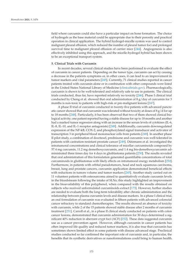
Biomedicines 2021, 9, 1476
field where curcumin could also have a particular impact on bone formation. The choiceof hydrogels as the base material could be appropriate due to their porosity and practicaloperation in clinical application. The hybrid hydrogel-micelle system was used to controlmalignant pleural effusion, which reduced the number of pleural tumor foci and prolongedsurvival time to malignant pleural effusion of carrier mice [244]. Angiogenesis is alsoeffectively inhibited using this approach, and the micelle-hydrogel hybrid has been shownto be an exceptional transport system.
5. Clinical Trials with Curcumin
In recent decades, several clinical studies have been performed to evaluate the effectof curcumin in cancer patients. Depending on the tumor type, curcumin can act by causinga decrease in the patients symptoms or, in other cases, it can lead to an improvement intumor markers and vital parameters [245]. Currently, 71 clinical studies reported in cancerpatients treated with curcumin alone or in combination with other compounds were listedin the United States National Library of Medicine (clinicaltrials.gov). Pharmacologically,curcumin is shown to be well-tolerated and relatively safe to use in patients. The clinicaltrials conducted, thus far, have reported relatively no toxicity [246]. Phase I clinical trialconducted by Cheng et al. showed that oral administration of 8 g/day of curcumin for 3months is non-toxic to patients with high-risk or pre-malignant lesions [247].
A phase II trial of curcumin conducted in twenty-five patients with advanced pancre-atic cancer showed that oral curcumin was tolerated without toxicity at doses of 8 g/d for upto 18 months [248]. Particularly, it has been observed that two of them showed clinical bio-logical activity; one patient reported having a stable disease for up to 18 months and anotherhad a marked tumor regression along with an increase in the serum levels of cytokines (IL-6,IL-8, IL-10, and IL-1 receptor antagonists) [248]. Additionally, curcumin downregulatedexpression of the NF-kB, COX-2, and phosphorylated signal transducer and activator oftranscription 3 in peripheral blood mononuclear cells from patients [248]. In another phaseII pilot study, a combination of docetaxel, prednisone and curcumin was well-tolerated inpatients with castration-resistant prostate cancer [249]. Dützmann et al. investigated theintratumoral concentrations and clinical tolerance of micellar curcuminoids composed by57.4 mg curcumin, 11.2 mg demethoxycurcumin, and 1.4 mg bis-demethoxycurcumin ad-ministrated three times day for 4 days in glioblastoma patients [250]. The results revealedthat oral administration of this formulation generated quantifiable concentrations of totalcurcuminoids in glioblastomas with likely effects on intratumoral energy metabolism [250].Furthermore, in patients with orbital pseudotumors, head and neck squamous carcinoma,breast, lung and prostate cancers, curcumin application demonstrated beneficial effectswith reductions in tumors volume and tumor markers [245]. Another study carried out on11 volunteer patients with osteosarcoma aimed to quantitatively evaluate curcumin levelsin the bloodstream following the intake of SLNs; this study highlighted an improvementin the bioavailability of this polyphenol, when compared with the results obtained onsubjects who received unformulated curcuminoids extract [173]. However, further studiesare needed to evaluate both the long-term tolerability after chronic administration and therelationship between plasma curcumin levels and disease markers. In a phase I clinical trial,an oral formulation of curcumin was evaluated in fifteen patients with advanced colorectalcancer refractory to standard chemotherapies. The results showed an absence of toxicitywith curcumin, while 2 of the 15 patients showed stable disease after 2 months of curcumintreatment [251]. Carroll et al., in a phase II clinical study conducted on patients with coloncancer lesions, demonstrated that curcumin administration for 30 days determined a sig-nificant 40% reduction in aberrant crypt foci (ACF) [252]. These data suggested curcuminuse as a cancer prevention agent. However, although curcumin in cancer patients hasoften improved life quality and reduced tumor markers, it is also true that curcumin hassometimes shown limited effect in some patients with disease advanced stage. Preclinicalstudies conducted so far confirmed the important role of curcumin and, in particular, thebenefits that its synthetic derivatives or nanoformulations could bring to human health.
173

Biomedicines 2021, 9, 1476
New efforts are needed to confirm the efficacy of curcumin-based products for the treat-ment of human diseases and to ensure that the products are non-toxic once introducedinto the body. Several ongoing clinical trials should provide a deeper understanding ofcurcumin-based formulation efficacy and mechanism of action against human diseases.
6. Conclusions
Curcumin is one of the most promising clinical compounds of the last few decades.Its therapeutic benefits have been demonstrated in various chronic diseases and, above all,in various cancers. Precisely in the antitumor field, curcumin is able to modulate the actionof growth factors, cytokines, transcription factors and genes that regulate cell prolifera-tion, apoptosis and the metastatic process. Some of curcumin’s multiple pharmacologicalactivities have been used experimentally and clinically in both humans and animals. No-table among these are the antioxidant, anti-inflammatory and anticarcinogenic properties,which all seem related. It is encouraging that curcumin is of low toxicity. Most of thedata supporting the antitumour activity of curcumin was obtained in vitro. Unfortunately,the challenges of low solubility and rapid elimination and then poor bioavailability, havedelayed its adoption as a therapeutic agent. These efforts have led to the developmentof several curcumin formulations that have been systematically prepared to improve theabsorption, water solubility, distribution and, hence, the bioavailability of curcumin. Thesemethods have been promising with respect to preclinical and clinical efficacy and involvethe use of both synthetic derivatives and analogues of curcumin as well as formulations ofnanoparticles, liposomes, phytosomes, micelles and natural adjuvants such as piperine. Allof these approaches generated a significant improvement in the bioavailability, absorptionand retention time of curcumin, along with increased delivery to target tissues, as widelyreported. However, further investigations are needed to fulfil the significant promisethey currently hold and reveal new perspectives for the further enhancement of the thera-peutic capacity of this interesting natural molecule. In particular, more epidemiologicaland clinical trials involving large numbers of subjects should be conducted to support tothe obtained in vivo and in vitro results and to confirm curcumin’s usefulness in cancertreatment and/or prevention.
Author Contributions: Conceptualization, original draft preparation, writing, search and identifi-cation of articles, A.C. and V.P.; screening articles, writing, M.C.N., A.D.L., F.P. (Francesca Prestia);figures preparation, M.C.N., F.P. (Francesca Prestia), F.P. (Francesco Puoci); tables preparation, M.C.N.,A.D.L., F.P. (Francesca Prestia) and A.C.; writing and editing, R.S., I.C., F.P. (Francesco Puoci); editing,P.A., D.L.P., L.Z., A.D.L. and A.C.; supervision, A.C. and V.P. All authors have read and agreed to thepublished version of the manuscript.
Funding: This work was supported by a special award (Department of Excellence, Italian Law232/2016) from the Italian Ministry of Research and University (MIUR) to the Department of Phar-macy, Health and Nutritional Sciences of the University of Calabria (Italy), and by MIUR ex 60%(VP, AC) and by AIRC (Associazione Italiana per la Ricerca sul Cancro), projects n. IG20122. A.D.L.was supported by a fellowship from PAC (Progetto Strategico Regionale Calabria Alta Formazione)Calabria 2014/2020—Asse Prioritario 12, Linea B, Azione 10.5.12; P.A. was supported by a post-docfellowship 2021 from Fondazione Umberto Veronesi (FUV).
Data Availability Statement: Not applicable.
Conflicts of Interest: The authors declare no conflict of interest.
Abbreviations
AKT protein kinase BAMPK adenosine monophosphate (AMP)-activated protein kinaseAP-1 activator protein1APC adenomatous polyposis coli
174
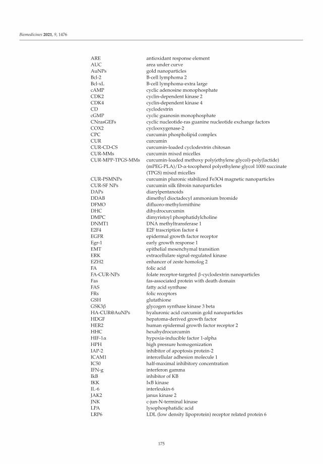
Biomedicines 2021, 9, 1476
ARE antioxidant response elementAUC area under curveAuNPs gold nanoparticlesBcl-2 B-cell lymphoma 2Bcl-xL B-cell lymphoma-extra largecAMP cyclic adenosine monophosphateCDK2 cyclin-dependent kinase 2CDK4 cyclin-dependent kinase 4CD cyclodextrincGMP cyclic guanosin monophosphateCNrasGEFs cyclic nucleotide-ras guanine nucleotide exchange factorsCOX2 cyclooxygenase-2CPC curcumin phospholipid complexCUR curcuminCUR-CD-CS curcumin-loaded cyclodextrin chitosanCUR-MMs curcumin mixed micellesCUR-MPP-TPGS-MMs curcumin-loaded methoxy poly(ethylene glycol)-poly(lactide)
(mPEG-PLA)/D-α-tocopherol polyethylene glycol 1000 succinate(TPGS) mixed micelles
CUR-PSMNPs curcumin pluronic stabilized Fe3O4 magnetic nanoparticlesCUR-SF NPs curcumin silk fibroin nanoparticlesDAPs diarylpentanoidsDDAB dimethyl dioctadecyl ammonium bromideDFMO difluoro-methylornithineDHC dihydrocurcuminDMPC dimyristoyl phosphatidylcholineDNMT1 DNA methyltransferase 1E2F4 E2F trascription factor 4EGFR epidermal growth factor receptorEgr-1 early growth response 1EMT epithelial mesenchymal transitionERK extracellulare signal-regulated kinaseEZH2 enhancer of zeste homolog 2FA folic acidFA-CUR-NPs folate receptor-targeted β-cyclodextrin nanoparticlesFas fas-associated protein with death domainFAS fatty acid synthaseFRs folic receptorsGSH glutathioneGSK3β glycogen synthase kinase 3 betaHA-CUR@AuNPs hyaluronic acid curcumin gold nanoparticlesHDGF hepatoma-derived growth factorHER2 human epidermal growth factor receptor 2HHC hexahydrocurcuminHIF-1α hypoxia-inducible factor 1-alphaHPH high pressure homogenizationIAP-2 inhibitor of apoptosis protein-2ICAM1 intercellular adhesion molecule 1IC50 half-maximal inhibitory concentrationIFN-g interferon gammaIkB inhibitor of KBIKK IκB kinaseIL-6 interleukin-6JAK2 janus kinase 2JNK c-jun-N-terminal kinaseLPA lysophosphatidic acidLRP6 LDL (low density lipoprotein) receptor related protein 6
175

Biomedicines 2021, 9, 1476
MAPK mitogen-activated protein kinaseMcl-1 myeloid cell leukemia 1MDM2 mouse double minute 2 homologMePEG-b-PCL methoxy poly (ethylene glycol) -block-polycaprolactonemiRNA microRNAMMPs matrix metalloproteinasesMNPs magnetic nanoparticlesMPEG-P [CL-co-PDO] methoxy poly (ethylene glycol) -b-poly (ε-caprolactone-co-dioxanone)mPEG–PCL methoxy poly(ethylene glycol)–polycaprolactonemTOR mechanistic target of rapamycinMUC1 mucin 1NaCas sodium caseinateNaCas-Lac sodium caseinate-lactoseNF-κB nuclear factor-κBNIPAAM N-isopropylacrylamideNPs nanoparticlesNSCLC non-small cell lung cancerNVP N-vinyl-2-pyrrolidoneODC ornithine decarboxylaseOHC octahydrocurcuminOPN osteopontinPAK1 p21 (RAC1) activated kinase 1PARP-1 poly (ADP-ribose) polymerase 1PCL poly-(ε-caprolactone)PCNA proliferating cell nuclear antigenPDE cyclic nucleotide phosphodiesterasePDE1A phosphodiesterase 1APEG polyethylene glycolPEG-A poly(ethylene glycol) monoacrylatePGA polyglycolide acidP-gp p-glycoprotein 1PI3K phosphoinositide 3-kinasePLA polylactidePLGA polylactic-co-glycolic acidPSMNPs pluronic stabilized Fe3O4 magnetic nanoparticlesPSBMA poly (sulphobetaine methacrylate)RhoA ras homolog family member AROCK1 rho associated coiled-coil containing protein kinase 1ROS reactive oxygen speciesSCLC small cell lung cancerSDs solid dispersionsSIP1 smad interacting protein 1SLNs solid lipid nanoparticlesSMOX spermine oxidaseSTAT3 signal transducer and activator of transcription 3SULT sulfotransferaseTHC tetrahydrocurcuminTNF-α tumor necrosis factor-aTPGS d-α-Tocopheryl polyethylene glycol 1000 succinateUGT UDP-glucurosyltransferaseUHRF1 ubiquitin like with PHD and ring finger domain 1u-PA urokinase-type plasminogen activatorVEGF vascular endothelial growth factorVHL Von Hippel-Lindau tumor suppressorVP N-vinyl-2-pyrrolidoneXIAP X-linked inhibitor of apoptosis proteinβ-CD β- cyclodextrinsεCL ε-caprolactone
176

Biomedicines 2021, 9, 1476
References
1. Prasad, S.; Aggarwal, B.B. Turmeric, the golden spice: From traditional medicine to modern medicine. In Herbal Medicine:Biomolecular and Clinical Aspects; Benzie, I.F.F., Wachtel-Galor, S., Eds.; CRC Press: Boca Raton, FL, USA, 2011.
2. Sandur, S.K.; Pandey, M.K.; Sung, B.; Ahn, K.S.; Murakami, A.; Sethi, G.; Limtrakul, P.; Badmaev, V.; Aggarwal, B.B. Curcumin,demethoxycurcumin, bisdemethoxycurcumin, tetrahydrocurcumin and turmerones differentially regulate anti-inflammatory andanti-proliferative responses through a ROS-independent mechanism. Carcinogenesis 2007, 28, 1765–1773. [CrossRef]
3. Dosoky, N.S.; Satyal, P.; Setzer, W.N. Variations in the volatile compositions of Curcuma species. Foods 2019, 8, 53. [CrossRef][PubMed]
4. Kunnumakkara, A.B.; Bordoloi, D.; Padmavathi, G.; Monisha, J.; Roy, N.K.; Prasad, S.; Aggarwal, B.B. Curcumin, the goldennutraceutical: Multitargeting for multiple chronic diseases. Br. J. Pharm. 2017, 174, 1325–1348. [CrossRef] [PubMed]
5. Patel, S.S.; Acharya, A.; Ray, R.S.; Agrawal, R.; Raghuwanshi, R.; Jain, P. Cellular and molecular mechanisms of curcumin inprevention and treatment of disease. Crit. Rev. Food Sci. Nutr. 2020, 60, 887–939. [CrossRef] [PubMed]
6. Adamczak, A.; Ozarowski, M.; Karpinski, T.M. Curcumin, a natural antimicrobial agent with strain-specific activity. Pharmaceuti-cals 2020, 13, 153. [CrossRef]
7. Jennings, M.R.; Parks, R.J. Curcumin as an antiviral agent. Viruses 2020, 12, 1242. [CrossRef]8. Chen, C.; Long, L.; Zhang, F.; Chen, Q.; Chen, C.; Yu, X.; Liu, Q.; Bao, J.; Long, Z. Antifungal activity, main active components and
mechanism of Curcuma longa extract against Fusarium graminearum. PLoS ONE 2018, 13, e0194284. [CrossRef]9. Jagetia, G.C. Antioxidant activity of curcumin protects against the radiation-induced micronuclei formation in cultured human
peripheral blood lymphocytes exposed to various doses of gamma-Radiation. Int. J. Radiat. Biol. 2021, 97, 485–493. [CrossRef]10. Busari, Z.A.; Dauda, K.A.; Morenikeji, O.A.; Afolayan, F.; Oyeyemi, O.T.; Meena, J.; Sahu, D.; Panda, A.K. Antiplasmodial activity
and toxicological assessment of curcumin PLGA-encapsulated nanoparticles. Front. Pharm. 2017, 8, 622. [CrossRef]11. Fadus, M.C.; Lau, C.; Bikhchandani, J.; Lynch, H.T. Curcumin: An age-old anti-inflammatory and anti-neoplastic agent. J. Tradit.
Complement. Med. 2017, 7, 339–346. [CrossRef]12. Zia, A.; Farkhondeh, T.; Pourbagher-Shahri, A.M.; Samarghandian, S. The role of curcumin in aging and senescence: Molecular
mechanisms. Biomed. Pharmacother. Biomed. Pharmacother. 2021, 134, 111119. [CrossRef]13. Wang, H.; Zhang, K.; Liu, J.; Yang, J.; Tian, Y.; Yang, C.; Li, Y.; Shao, M.; Su, W.; Song, N. Curcumin regulates cancer progression:
Focus on ncRNAs and molecular signaling pathways. Front. Oncol. 2021, 11, 660712. [CrossRef] [PubMed]14. Kunnumakkara, A.B.; Bordoloi, D.; Harsha, C.; Banik, K.; Gupta, S.C.; Aggarwal, B.B. Curcumin mediates anticancer effects by
modulating multiple cell signaling pathways. Clin. Sci. 2017, 131, 1781–1799. [CrossRef] [PubMed]15. Kim, M.J.; Park, K.S.; Kim, K.T.; Gil, E.Y. The inhibitory effect of curcumin via fascin suppression through JAK/STAT3 pathway
on metastasis and recurrence of ovary cancer cells. BMC Women’s Health 2020, 20, 256. [CrossRef]16. Liu, J.L.; Pan, Y.Y.; Chen, O.; Luan, Y.; Xue, X.; Zhao, J.J.; Liu, L.; Jia, H.Y. Curcumin inhibits MCF-7 cells by modulating the
NF-kappaB signaling pathway. Oncol. Lett. 2017, 14, 5581–5584. [CrossRef] [PubMed]17. Ghasemi, F.; Shafiee, M.; Banikazemi, Z.; Pourhanifeh, M.H.; Khanbabaei, H.; Shamshirian, A.; Amiri Moghadam, S.; ArefNezhad,
R.; Sahebkar, A.; Avan, A.; et al. Curcumin inhibits NF-kB and Wnt/beta-catenin pathways in cervical cancer cells. Pathol. Res.Pract. 2019, 215, 152556. [CrossRef] [PubMed]
18. Kuttikrishnan, S.; Siveen, K.S.; Prabhu, K.S.; Khan, A.Q.; Ahmed, E.I.; Akhtar, S.; Ali, T.A.; Merhi, M.; Dermime, S.; Steinhoff,M.; et al. Curcumin induces apoptotic cell death via inhibition of PI3-kinase/AKT pathway in B-precursor acute lymphoblasticleukemia. Front. Oncol. 2019, 9, 484. [CrossRef]
19. Mortezaee, K.; Salehi, E.; Mirtavoos-Mahyari, H.; Motevaseli, E.; Najafi, M.; Farhood, B.; Rosengren, R.J.; Sahebkar, A. Mechanismsof apoptosis modulation by curcumin: Implications for cancer therapy. J. Cell. Physiol. 2019, 234, 12537–12550. [CrossRef]
20. Wahlstrom, B.; Blennow, G. A study on the fate of curcumin in the rat. Acta Pharmacol. Toxicol. 1978, 43, 86–92. [CrossRef]21. Anand, P.; Kunnumakkara, A.B.; Newman, R.A.; Aggarwal, B.B. Bioavailability of curcumin: Problems and promises. Mol. Pharm.
2007, 4, 807–818. [CrossRef]22. Tomeh, M.A.; Hadianamrei, R.; Zhao, X. A review of curcumin and its derivatives as anticancer agents. Int. J. Mol. Sci. 2019, 20,
1033. [CrossRef] [PubMed]23. Li, W.; He, Y.; Zhang, R.; Zheng, G.; Zhou, D. The curcumin analog EF24 is a novel senolytic agent. Aging 2019, 11, 771–782.
[CrossRef] [PubMed]24. He, Y.; Li, W.; Hu, G.; Sun, H.; Kong, Q. Bioactivities of EF24, a novel curcumin analog: A review. Front. Oncol. 2018, 8, 614.
[CrossRef] [PubMed]25. Jamwal, R. Bioavailable curcumin formulations: A review of pharmacokinetic studies in healthy volunteers. J. Integr. Med. 2018,
16, 367–374. [CrossRef] [PubMed]26. Adiwidjaja, J.; McLachlan, A.J.; Boddy, A.V. Curcumin as a clinically-promising anti-cancer agent: Pharmacokinetics and drug
interactions. Expert Opin. Drug Metab. Toxicol. 2017, 13, 953–972. [CrossRef] [PubMed]27. Olotu, F.; Agoni, C.; Soremekun, O.; Soliman, M.E.S. An update on the pharmacological usage of curcumin: Has it failed in the
drug discovery pipeline? Cell Biochem. Biophys. 2020, 78, 267–289. [CrossRef]28. Yallapu, M.M.; Nagesh, P.K.; Jaggi, M.; Chauhan, S.C. Therapeutic applications of curcumin nanoformulations. AAPS J. 2015, 17,
1341–1356. [CrossRef]
177

Biomedicines 2021, 9, 1476
29. Karthikeyan, A.; Senthil, N.; Min, T. Nanocurcumin: A promising candidate for therapeutic applications. Front. Pharmacol. 2020,11, 487. [CrossRef]
30. Ma, Z.; Wang, N.; He, H.; Tang, X. Pharmaceutical strategies of improving oral systemic bioavailability of curcumin for clinicalapplication. J. Control. Release Off. J. Control. Release Soc. 2019, 316, 359–380. [CrossRef]
31. Kotecha, R.; Takami, A.; Espinoza, J.L. Dietary phytochemicals and cancer chemoprevention: A review of the clinical evidence.Oncotarget 2016, 7, 52517–52529. [CrossRef]
32. Rahmani, A.H.; Al Zohairy, M.A.; Aly, S.M.; Khan, M.A. Curcumin: A potential candidate in prevention of cancer via modulationof molecular pathways. BioMed Res. Int. 2014, 2014, 761608. [CrossRef] [PubMed]
33. Song, X.; Zhang, M.; Dai, E.; Luo, Y. Molecular targets of curcumin in breast cancer (Review). Mol. Med. Rep. 2019, 19, 23–29.[CrossRef] [PubMed]
34. Mukhopadhyay, A.; Banerjee, S.; Stafford, L.J.; Xia, C.; Liu, M.; Aggarwal, B.B. Curcumin-induced suppression of cell proliferationcorrelates with down-regulation of cyclin D1 expression and CDK4-mediated retinoblastoma protein phosphorylation. Oncogene2002, 21, 8852–8861. [CrossRef] [PubMed]
35. Zhou, Q.M.; Wang, X.F.; Liu, X.J.; Zhang, H.; Lu, Y.Y.; Su, S.B. Curcumin enhanced antiproliferative effect of mitomycin C inhuman breast cancer MCF-7 cells in vitro and in vivo. Acta Pharmacol. Sin. 2011, 32, 1402–1410. [CrossRef]
36. Aggarwal, B.B.; Banerjee, S.; Bharadwaj, U.; Sung, B.; Shishodia, S.; Sethi, G. Curcumin induces the degradation of cyclin Eexpression through ubiquitin-dependent pathway and up-regulates cyclin-dependent kinase inhibitors p21 and p27 in multiplehuman tumor cell lines. Biochem. Pharmacol. 2007, 73, 1024–1032. [CrossRef]
37. Prasad, C.P.; Rath, G.; Mathur, S.; Bhatnagar, D.; Ralhan, R. Potent growth suppressive activity of curcumin in human breastcancer cells: Modulation of Wnt/beta-catenin signaling. Chem. Biol. Interact. 2009, 181, 263–271. [CrossRef]
38. Colomer, C.; Marruecos, L.; Vert, A.; Bigas, A.; Espinosa, L. NF-kappaB members left home: NF-kappaB-independent roles incancer. Biomedicines 2017, 5, 26. [CrossRef]
39. Zhou, Q.; Ye, M.; Lu, Y.; Zhang, H.; Chen, Q.; Huang, S.; Su, S. Curcumin improves the tumoricidal effect of mitomycin C bysuppressing ABCG2 expression in stem cell-like breast cancer cells. PLoS ONE 2015, 10, e0136694. [CrossRef]
40. Zhou, X.; Jiao, D.; Dou, M.; Zhang, W.; Lv, L.; Chen, J.; Li, L.; Wang, L.; Han, X. Curcumin inhibits the growth of triple-negativebreast cancer cells by silencing EZH2 and restoring DLC1 expression. J. Cell. Mol. Med. 2020, 24, 10648–10662. [CrossRef]
41. Chase, A.; Cross, N.C. Aberrations of EZH2 in cancer. Clin. Cancer Res. 2011, 17, 2613–2618. [CrossRef] [PubMed]42. Ren, G.; Li, G. Tumor suppressor gene DLC1: Its modifications, interactive molecules, and potential prospects for clinical cancer
application. Int. J. Biol. Macromol. 2021, 182, 264–275. [CrossRef] [PubMed]43. Pricci, M.; Girardi, B.; Giorgio, F.; Losurdo, G.; Ierardi, E.; Di Leo, A. Curcumin and colorectal cancer: From basic to clinical
evidences. Int. J. Mol. Sci. 2020, 21, 2364. [CrossRef]44. Calibasi-Kocal, G.; Pakdemirli, A.; Bayrak, S.; Ozupek, N.M.; Sever, T.; Basbinar, Y.; Ellidokuz, H.; Yigitbasi, T. Curcumin effects
on cell proliferation, angiogenesis and metastasis in colorectal cancer. J. BUON Off. J. Balk. Union Oncol. 2019, 24, 1482–1487.45. Lim, T.G.; Lee, S.Y.; Huang, Z.; Lim, D.Y.; Chen, H.; Jung, S.K.; Bode, A.M.; Lee, K.W.; Dong, Z. Curcumin suppresses proliferation
of colon cancer cells by targeting CDK2. Cancer Prev. Res. 2014, 7, 466–474. [CrossRef]46. Kim, K.C.; Lee, C. Curcumin induces downregulation of E2F4 expression and apoptotic cell death in HCT116 human colon cancer
cells; Involvement of reactive oxygen species. Korean J. Physiol. Pharmacol. Off. J. Korean Physiol. Soc. Korean Soc. Pharmacol. 2010,14, 391–397. [CrossRef]
47. Watson, J.L.; Hill, R.; Yaffe, P.B.; Greenshields, A.; Walsh, M.; Lee, P.W.; Giacomantonio, C.A.; Hoskin, D.W. Curcumin causessuperoxide anion production and p53-independent apoptosis in human colon cancer cells. Cancer Lett. 2010, 297, 1–8. [CrossRef][PubMed]
48. Mosieniak, G.; Adamowicz, M.; Alster, O.; Jaskowiak, H.; Szczepankiewicz, A.A.; Wilczynski, G.M.; Ciechomska, I.A.; Sikora, E.Curcumin induces permanent growth arrest of human colon cancer cells: Link between senescence and autophagy. Mech. AgeingDev. 2012, 133, 444–455. [CrossRef] [PubMed]
49. Zhang, L.; Yang, G.; Zhang, R.; Dong, L.; Chen, H.; Bo, J.; Xue, W.; Huang, Y. Curcumin inhibits cell proliferation and motility viasuppression of TROP2 in bladder cancer cells. Int. J. Oncol. 2018, 53, 515–526. [CrossRef]
50. Shi, J.; Zhang, X.; Shi, T.; Li, H. Antitumor effects of curcumin in human bladder cancer in vitro. Oncol. Lett. 2017, 14, 1157–1161.[CrossRef] [PubMed]
51. Walker, B.C.; Mittal, S. Antitumor activity of curcumin in glioblastoma. Int. J. Mol. Sci. 2020, 21, 9435. [CrossRef]52. Choi, B.H.; Kim, C.G.; Bae, Y.S.; Lim, Y.; Lee, Y.H.; Shin, S.Y. P21(Waf1/Cip1) expression by curcumin in U-87MG human glioma
cells: Role of early growth response-1 expression. Cancer Res. 2008, 68, 1369–1377. [CrossRef] [PubMed]53. Cheng, C.; Jiao, J.T.; Qian, Y.; Guo, X.Y.; Huang, J.; Dai, M.C.; Zhang, L.; Ding, X.P.; Zong, D.; Shao, J.F. Curcumin induces G2/M
arrest and triggers apoptosis via FoxO1 signaling in U87 human glioma cells. Mol. Med. Rep. 2016, 13, 3763–3770. [CrossRef][PubMed]
54. Wang, L.; Ye, X.; Cai, X.; Su, J.; Ma, R.; Yin, X.; Zhou, X.; Li, H.; Wang, Z. Curcumin suppresses cell growth and invasion andinduces apoptosis by down-regulation of Skp2 pathway in glioma cells. Oncotarget 2015, 6, 18027–18037. [CrossRef] [PubMed]
55. Wu, J.; Su, H.K.; Yu, Z.H.; Xi, S.Y.; Guo, C.C.; Hu, Z.Y.; Qu, Y.; Cai, H.P.; Zhao, Y.Y.; Zhao, H.F.; et al. Skp2 modulates proliferation,senescence and tumorigenesis of glioma. Cancer Cell Int. 2020, 20, 71. [CrossRef]
178

Biomedicines 2021, 9, 1476
56. Luo, Q.; Luo, H.; Fu, H.; Huang, H.; Huang, H.; Luo, K.; Li, C.; Hu, R.; Zheng, C.; Lan, C.; et al. Curcumin suppresses invasivenessand migration of human glioma cells in vitro by inhibiting HDGF/beta-catenin complex. Nan Fang Yi Ke Da Xue Xue Bao J. South.Med. Univ. 2019, 39, 911–916. [CrossRef]
57. Fratantonio, D.; Molonia, M.S.; Bashllari, R.; Muscara, C.; Ferlazzo, G.; Costa, G.; Saija, A.; Cimino, F.; Speciale, A. Curcuminpotentiates the antitumor activity of Paclitaxel in rat glioma C6 cells. Phytomed. Int. J. Phytother. Phytopharm. 2019, 55, 23–30.[CrossRef]
58. Seyithanoglu, M.H.; Abdallah, A.; Kitis, S.; Guler, E.M.; Kocyigit, A.; Dundar, T.T.; Gundag Papaker, M. Investigation of cytotoxic,genotoxic, and apoptotic effects of curcumin on glioma cells. Cell. Mol. Biol. 2019, 65, 101–108. [CrossRef]
59. Murray-Stewart, T.; Dunworth, M.; Lui, Y.; Giardiello, F.M.; Woster, P.M.; Casero, R.A., Jr. Curcumin mediates polyaminemetabolism and sensitizes gastrointestinal cancer cells to antitumor polyamine-targeted therapies. PLoS ONE 2018, 13, e0202677.[CrossRef]
60. Cai, X.Z.; Wang, J.; Li, X.D.; Wang, G.L.; Liu, F.N.; Cheng, M.S.; Li, F. Curcumin suppresses proliferation and invasion in humangastric cancer cells by downregulation of PAK1 activity and cyclin D1 expression. Cancer Biol. Ther. 2009, 8, 1360–1368. [CrossRef]
61. Zheng, R.; Deng, Q.; Liu, Y.; Zhao, P. Curcumin inhibits gastric carcinoma cell growth and induces apoptosis by suppressing theWnt/beta-catenin signaling pathway. Med. Sci. Monit. Int. Med. J. Exp. Clin. Res. 2017, 23, 163–171. [CrossRef]
62. Fu, H.; Wang, C.; Yang, D.; Wei, Z.; Xu, J.; Hu, Z.; Zhang, Y.; Wang, W.; Yan, R.; Cai, Q. Curcumin regulates proliferation,autophagy, and apoptosis in gastric cancer cells by affecting PI3K and P53 signaling. J. Cell. Physiol. 2018, 233, 4634–4642.[CrossRef]
63. Liu, G.; Xiang, T.; Wu, Q.F.; Wang, W.X. Curcumin suppresses the proliferation of gastric cancer cells by downregulating H19.Oncol. Lett. 2016, 12, 5156–5162. [CrossRef]
64. Ghafouri-Fard, S.; Esmaeili, M.; Taheri, M. H19 lncRNA: Roles in tumorigenesis. Biomed. Pharmacother. 2020, 123, 109774.[CrossRef]
65. Yang, F.; Bi, J.; Xue, X.; Zheng, L.; Zhi, K.; Hua, J.; Fang, G. Up-regulated long non-coding RNA H19 contributes to proliferation ofgastric cancer cells. FEBS J. 2012, 279, 3159–3165. [CrossRef]
66. Yoshimura, H.; Matsuda, Y.; Yamamoto, M.; Kamiya, S.; Ishiwata, T. Expression and role of long non-coding RNA H19 incarcinogenesis. Front. Biosci. 2018, 23, 614–625. [CrossRef]
67. Sun, C.; Zhang, S.; Liu, C.; Liu, X. Curcumin promoted miR-34a expression and suppressed proliferation of gastric cancer cells.Cancer Biother. Radiopharm. 2019, 34, 634–641. [CrossRef]
68. Sun, Q.; Zhang, W.; Guo, Y.; Li, Z.; Chen, X.; Wang, Y.; Du, Y.; Zang, W.; Zhao, G. Curcumin inhibits cell growth and induces cellapoptosis through upregulation of miR-33b in gastric cancer. Tumor Biol. J. Int. Soc. Oncodev. Biol. Med. 2016, 37, 13177–13184.[CrossRef] [PubMed]
69. Nabavi, S.M.; Russo, G.L.; Tedesco, I.; Daglia, M.; Orhan, I.E.; Nabavi, S.F.; Bishayee, A.; Nagulapalli Venkata, K.C.; Abdollahi,M.; Hajheydari, Z. Curcumin and melanoma: From chemistry to medicine. Nutr. Cancer 2018, 70, 164–175. [CrossRef] [PubMed]
70. Zheng, M.; Ekmekcioglu, S.; Walch, E.T.; Tang, C.H.; Grimm, E.A. Inhibition of nuclear factor-kappaB and nitric oxide bycurcumin induces G2/M cell cycle arrest and apoptosis in human melanoma cells. Melanoma Res. 2004, 14, 165–171. [CrossRef][PubMed]
71. Abusnina, A.; Keravis, T.; Yougbare, I.; Bronner, C.; Lugnier, C. Anti-proliferative effect of curcumin on melanoma cells ismediated by PDE1A inhibition that regulates the epigenetic integrator UHRF1. Mol. Nutr. Food Res. 2011, 55, 1677–1689.[CrossRef]
72. Zhao, G.; Han, X.; Zheng, S.; Li, Z.; Sha, Y.; Ni, J.; Sun, Z.; Qiao, S.; Song, Z. Curcumin induces autophagy, inhibits proliferationand invasion by downregulating AKT/mTOR signaling pathway in human melanoma cells. Oncol. Rep. 2016, 35, 1065–1074.[CrossRef] [PubMed]
73. Siwak, D.R.; Shishodia, S.; Aggarwal, B.B.; Kurzrock, R. Curcumin-induced antiproliferative and proapoptotic effects in melanomacells are associated with suppression of I kappa B kinase and nuclear factor KB activity and are independent of the B-Raf/mitogen-activated/extracellular signal-regulated protein kinase pathway and the Akt pathway. Cancer 2005, 104, 879–890. [CrossRef][PubMed]
74. Cianfruglia, L.; Minnelli, C.; Laudadio, E.; Scire, A.; Armeni, T. Side effects of curcumin: Epigenetic and antiproliferativeimplications for normal dermal fibroblast and breast cancer cells. Antioxidants 2019, 8, 382. [CrossRef] [PubMed]
75. Talib, W.H.; Al-Hadid, S.A.; Ali, M.B.W.; Al-Yasari, I.H.; Ali, M.R.A. Role of curcumin in regulating p53 in breast cancer: Anoverview of the mechanism of action. Breast Cancer 2018, 10, 207–217. [CrossRef]
76. Bae, Y.H.; Ryu, J.H.; Park, H.J.; Kim, K.R.; Wee, H.J.; Lee, O.H.; Jang, H.O.; Bae, M.K.; Kim, K.W.; Bae, S.K. Mutant p53-Notch1signaling axis is involved in curcumin-induced apoptosis of breast cancer cells. Korean J. Physiol. Pharmacol. Off. J. Korean Physiol.Soc. Korean Soc. Pharmacol. 2013, 17, 291–297. [CrossRef]
77. Patel, P.B.; Thakkar, V.R.; Patel, J.S. Cellular effect of curcumin and citral combination on breast cancer cells: Induction of apoptosisand cell cycle arrest. J. Breast Cancer 2015, 18, 225–234. [CrossRef]
78. Moghtaderi, H.; Sepehri, H.; Attari, F. Combination of arabinogalactan and curcumin induces apoptosis in breast cancer cellsin vitro and inhibits tumor growth via overexpression of p53 level in vivo. Biomed. Pharmacother. 2017, 88, 582–594. [CrossRef]
79. Chiu, T.L.; Su, C.C. Curcumin inhibits proliferation and migration by increasing the Bax to Bcl-2 ratio and decreasing NF-kappaBp65 expression in breast cancer MDA-MB-231 cells. Int. J. Mol. Med. 2009, 23, 469–475. [CrossRef]
179
Biomedicines 2021, 9, 1476
80. Fan, H.; Liang, Y.; Jiang, B.; Li, X.; Xun, H.; Sun, J.; He, W.; Lau, H.T.; Ma, X. Curcumin inhibits intracellular fatty acid synthaseand induces apoptosis in human breast cancer MDA-MB-231 cells. Oncol. Rep. 2016, 35, 2651–2656. [CrossRef]
81. Zhang, Y.P.; Li, Y.Q.; Lv, Y.T.; Wang, J.M. Effect of curcumin on the proliferation, apoptosis, migration, and invasion of humanmelanoma A375 cells. Genet. Mol. Res. 2015, 14, 1056–1067. [CrossRef]
82. Bush, J.A.; Cheung, K.J., Jr.; Li, G. Curcumin induces apoptosis in human melanoma cells through a Fas receptor/caspase-8pathway independent of p53. Exp. Cell Res. 2001, 271, 305–314. [CrossRef] [PubMed]
83. Watson, J.L.; Greenshields, A.; Hill, R.; Hilchie, A.; Lee, P.W.; Giacomantonio, C.A.; Hoskin, D.W. Curcumin-induced apoptosis inovarian carcinoma cells is p53-independent and involves p38 mitogen-activated protein kinase activation and downregulation ofBcl-2 and survivin expression and Akt signaling. Mol. Carcinog. 2010, 49, 13–24. [CrossRef] [PubMed]
84. Li, W.; Wang, Y.; Song, Y.; Xu, L.; Zhao, J.; Fang, B. A preliminary study of the effect of curcumin on the expression of p53 proteinin a human multiple myeloma cell line. Oncol. Lett. 2015, 9, 1719–1724. [CrossRef] [PubMed]
85. Lee, Y.K.; Park, S.Y.; Kim, Y.M.; Park, O.J. Regulatory effect of the AMPK-COX-2 signaling pathway in curcumin-inducedapoptosis in HT-29 colon cancer cells. Ann. N. Y. Acad. Sci. 2009, 1171, 489–494. [CrossRef]
86. Sandur, S.K.; Deorukhkar, A.; Pandey, M.K.; Pabon, A.M.; Shentu, S.; Guha, S.; Aggarwal, B.B.; Krishnan, S. Curcumin modulatesthe radiosensitivity of colorectal cancer cells by suppressing constitutive and inducible NF-kappaB activity. Int. J. Radiat. Oncol.Biol. Phys. 2009, 75, 534–542. [CrossRef]
87. Narayan, S. Curcumin, a multi-functional chemopreventive agent, blocks growth of colon cancer cells by targeting beta-catenin-mediated transactivation and cell-cell adhesion pathways. J. Mol. Histol. 2004, 35, 301–307. [CrossRef]
88. Jung, E.M.; Lim, J.H.; Lee, T.J.; Park, J.W.; Choi, K.S.; Kwon, T.K. Curcumin sensitizes tumor necrosis factor-related apoptosis-inducing ligand (TRAIL)-induced apoptosis through reactive oxygen species-mediated upregulation of death receptor 5 (DR5).Carcinogenesis 2005, 26, 1905–1913. [CrossRef]
89. Cao, A.; Li, Q.; Yin, P.; Dong, Y.; Shi, H.; Wang, L.; Ji, G.; Xie, J.; Wu, D. Curcumin induces apoptosis in human gastric carcinomaAGS cells and colon carcinoma HT-29 cells through mitochondrial dysfunction and endoplasmic reticulum stress. Apoptosis 2013,18, 1391–1402. [CrossRef]
90. Moragoda, L.; Jaszewski, R.; Majumdar, A.P. Curcumin induced modulation of cell cycle and apoptosis in gastric and coloncancer cells. Anticancer Res. 2001, 21, 873–878.
91. Su, C.C.; Lin, J.G.; Li, T.M.; Chung, J.G.; Yang, J.S.; Ip, S.W.; Lin, W.C.; Chen, G.W. Curcumin-induced apoptosis of human coloncancer colo 205 cells through the production of ROS, Ca2+ and the activation of caspase-3. Anticancer Res. 2006, 26, 4379–4389.
92. Klinger, N.V.; Mittal, S. Therapeutic potential of curcumin for the treatment of brain tumors. Oxidative Med. Cell. Longev. 2016,2016, 9324085. [CrossRef]
93. Lee, D.S.; Lee, M.K.; Kim, J.H. Curcumin induces cell cycle arrest and apoptosis in human osteosarcoma (HOS) cells. AnticancerRes. 2009, 29, 5039–5044. [PubMed]
94. Yang, J.; Cao, Y.; Sun, J.; Zhang, Y. Curcumin reduces the expression of Bcl-2 by upregulating miR-15a and miR-16 in MCF-7 cells.Med. Oncol. 2010, 27, 1114–1118. [CrossRef]
95. Zhang, J.; Du, Y.; Wu, C.; Ren, X.; Ti, X.; Shi, J.; Zhao, F.; Yin, H. Curcumin promotes apoptosis in human lung adenocarcinomacells through miR-186* signaling pathway. Oncol. Rep. 2010, 24, 1217–1223. [CrossRef]
96. Sohn, E.J.; Bak, K.M.; Nam, Y.K.; Park, H.T. Upregulation of microRNA 344a-3p is involved in curcumin induced apoptosis inRT4 schwannoma cells. Cancer Cell Int. 2018, 18, 199. [CrossRef]
97. Gao, D.; Mittal, V.; Ban, Y.; Lourenco, A.R.; Yomtoubian, S.; Lee, S. Metastatic tumor cells—Genotypes and phenotypes. Front.Biol. 2018, 13, 277–286. [CrossRef] [PubMed]
98. Gallardo, M.; Kemmerling, U.; Aguayo, F.; Bleak, T.C.; Munoz, J.P.; Calaf, G.M. Curcumin rescues breast cells from epithelialmes-enchymal transition and invasion induced by antimiR34a. Int. J. Oncol. 2020, 56, 480–493. [CrossRef] [PubMed]
99. Pires, B.R.; Mencalha, A.L.; Ferreira, G.M.; de Souza, W.F.; Morgado-Diaz, J.A.; Maia, A.M.; Correa, S.; Abdelhay, E.S. NF-kappaBis involved in the regulation of EMT genes in breast cancer cells. PLoS ONE 2017, 12, e0169622. [CrossRef] [PubMed]
100. Zong, H.; Wang, F.; Fan, Q.X.; Wang, L.X. Curcumin inhibits metastatic progression of breast cancer cell through suppression ofurokinase-type plasminogen activator by NF-kappa B signaling pathways. Mol. Biol. Rep. 2012, 39, 4803–4808. [CrossRef]
101. Bachmeier, B.E.; Mohrenz, I.V.; Mirisola, V.; Schleicher, E.; Romeo, F.; Hohneke, C.; Jochum, M.; Nerlich, A.G.; Pfeffer, U. Curcumindownregulates the inflammatory cytokines CXCL1 and -2 in breast cancer cells via NFkappaB. Carcinogenesis 2008, 29, 779–789.[CrossRef]
102. Irani, S. Emerging insights into the biology of metastasis: A review article. Iran. J. Basic Med. Sci. 2019, 22, 833–847. [CrossRef][PubMed]
103. Sun, K.; Duan, X.; Cai, H.; Liu, X.; Yang, Y.; Li, M.; Zhang, X.; Wang, J. Curcumin inhibits LPA-induced invasion by attenuatingRhoA/ROCK/MMPs pathway in MCF7 breast cancer cells. Clin. Exp. Med. 2016, 16, 37–47. [CrossRef] [PubMed]
104. Li, Y.; Sun, W.; Han, N.; Zou, Y.; Yin, D. Curcumin inhibits proliferation, migration, invasion and promotes apoptosis ofretinoblastoma cell lines through modulation of miR-99a and JAK/STAT pathway. BMC Cancer 2018, 18, 1230. [CrossRef]
105. Zaman, S.; Jadid, H.; Denson, A.C.; Gray, J.E. Targeting Trop-2 in solid tumors: Future prospects. Onco Targets Ther. 2019, 12,1781–1790. [CrossRef] [PubMed]
180

Biomedicines 2021, 9, 1476
106. Cheng, T.S.; Chen, W.C.; Lin, Y.Y.; Tsai, C.H.; Liao, C.I.; Shyu, H.Y.; Ko, C.J.; Tzeng, S.F.; Huang, C.Y.; Yang, P.C.; et al. Curcumin-targeting pericellular serine protease matriptase role in suppression of prostate cancer cell invasion, tumor growth, and metastasis.Cancer Prev. Res. 2013, 6, 495–505. [CrossRef] [PubMed]
107. Xiang, L.; He, B.; Liu, Q.; Hu, D.; Liao, W.; Li, R.; Peng, X.; Wang, Q.; Zhao, G. Antitumor effects of curcumin on the proliferation,migration and apoptosis of human colorectal carcinoma HCT116 cells. Oncol. Rep. 2020, 44, 1997–2008. [CrossRef]
108. Philip, S.; Kundu, G.C. Osteopontin induces nuclear factor kappa B-mediated promatrix metalloproteinase-2 activation through Ikappa B alpha /IKK signaling pathways, and curcumin (diferulolylmethane) down-regulates these pathways. J. Biol. Chem. 2003,278, 14487–14497. [CrossRef]
109. Jin, W. Role of JAK/STAT3 signaling in the regulation of metastasis, the transition of cancer stem cells, and chemoresistance ofcancer by epithelial-mesenchymal transition. Cells 2020, 9, 217. [CrossRef]
110. Yang, C.L.; Liu, Y.Y.; Ma, Y.G.; Xue, Y.X.; Liu, D.G.; Ren, Y.; Liu, X.B.; Li, Y.; Li, Z. Curcumin blocks small cell lung cancer cellsmigration, invasion, angiogenesis, cell cycle and neoplasia through Janus kinase-STAT3 signalling pathway. PLoS ONE 2012, 7,e37960. [CrossRef]
111. Han, Z.; Zhang, J.; Zhang, K.; Zhao, Y. Curcumin inhibits cell viability, migration, and invasion of thymic carcinoma cells viadownregulation of microRNA-27a. Phytother. Res. PTR 2020, 34, 1629–1637. [CrossRef]
112. Wang, N.; Feng, T.; Liu, X.; Liu, Q. Curcumin inhibits migration and invasion of non-small cell lung cancer cells throughup-regulation of miR-206 and suppression of PI3K/AKT/mTOR signaling pathway. Acta Pharm. 2020, 70, 399–409. [CrossRef][PubMed]
113. Wang, X.; Deng, J.; Yuan, J.; Tang, X.; Wang, Y.; Chen, H.; Liu, Y.; Zhou, L. Curcumin exerts its tumor suppressive function viainhibition of NEDD4 oncoprotein in glioma cancer cells. Int. J. Oncol. 2017, 51, 467–477. [CrossRef]
114. Zhang, H.; Nie, W.; Zhang, X.; Zhang, G.; Li, Z.; Wu, H.; Shi, Q.; Chen, Y.; Ding, Z.; Zhou, X.; et al. NEDD4-1 regulates migrationand invasion of glioma cells through CNrasGEF ubiquitination in vitro. PLoS ONE 2013, 8, e82789. [CrossRef]
115. Ravindranath, V.; Chandrasekhara, N. Absorption and tissue distribution of curcumin in rats. Toxicology 1980, 16, 259–265.[CrossRef]
116. Pan, M.H.; Huang, T.M.; Lin, J.K. Biotransformation of curcumin through reduction and glucuronidation in mice. Drug Metab.Dispos. 1999, 27, 486–494.
117. Basile, V.; Ferrari, E.; Lazzari, S.; Belluti, S.; Pignedoli, F.; Imbriano, C. Curcumin derivatives: Molecular basis of their anti-canceractivity. Biochem. Pharmacol. 2009, 78, 1305–1315. [CrossRef]
118. Sribalan, R.; Kirubavathi, M.; Banuppriya, G.; Padmini, V. Synthesis and biological evaluation of new symmetric curcuminderivatives. Bioorg. Med. Chem. Lett. 2015, 25, 4282–4286. [CrossRef]
119. Bayomi, S.M.; El-Kashef, H.A.; El-Ashmawy, M.B.; Nasr, M.N.A.; El-Sherbeny, M.A.; Badria, F.A.; Abou-Zeid, L.A.; Ghaly, M.A.;Abdel-Aziz, N.I. Synthesis and biological evaluation of new curcumin derivatives as antioxidant and antitumor agents. Med.Chem. Res. 2013, 22, 1147–1162. [CrossRef]
120. Xu, G.; Chu, Y.; Jiang, N.; Yang, J.; Li, F. The three dimensional quantitative structure activity relationships (3D-QSAR) anddocking studies of curcumin derivatives as androgen receptor antagonists. Int. J. Mol. Sci. 2012, 13, 6138–6155. [CrossRef][PubMed]
121. Zhao, C.; Liu, Z.; Liang, G. Promising curcumin-based drug design: Mono-carbonyl analogues of curcumin (MACs). Curr. Pharm.Des. 2013, 19, 2114–2135. [PubMed]
122. Somparn, P.; Phisalaphong, C.; Nakornchai, S.; Unchern, S.; Morales, N.P. Comparative antioxidant activities of curcumin and itsdemethoxy and hydrogenated derivatives. Biol. Pharm. Bull. 2007, 30, 74–78. [CrossRef] [PubMed]
123. Anand, P.; Thomas, S.G.; Kunnumakkara, A.B.; Sundaram, C.; Harikumar, K.B.; Sung, B.; Tharakan, S.T.; Misra, K.; Priyadarsini,I.K.; Rajasekharan, K.N.; et al. Biological activities of curcumin and its analogues (Congeners) made by man and Mother Nature.Biochem. Pharmacol. 2008, 76, 1590–1611. [CrossRef] [PubMed]
124. Reddy, A.R.; Dinesh, P.; Prabhakar, A.S.; Umasankar, K.; Shireesha, B.; Raju, M.B. A comprehensive review on SAR of curcumin.Mini Rev. Med. Chem. 2013, 13, 1769–1777. [CrossRef] [PubMed]
125. Sherin, D.R.; Rajasekharan, K.N. Mechanochemical synthesis and antioxidant activity of curcumin-templated azoles. Arch. Pharm.2015, 348, 908–914. [CrossRef]
126. Wanninger, S.; Lorenz, V.; Subhan, A.; Edelmann, F.T. Metal complexes of curcumin–synthetic strategies, structures and medicinalapplications. Chem. Soc. Rev. 2015, 44, 4986–5002. [CrossRef]
127. Paulraj, F.; Abas, F.; Lajis, N.H.; Othman, I.; Naidu, R. Molecular pathways modulated by curcumin analogue, diarylpentanoidsin cancer. Biomolecules 2019, 9, 270. [CrossRef]
128. Yerdelen, K.O.; Gul, H.I.; Sakagami, H.; Umemura, N.; Sukuroglu, M. Synthesis and cytotoxic activities of a curcumin analogueand its bis-mannich derivatives. Lett. Drug Des. Discov. 2015, 12, 643–649. [CrossRef]
129. Reid, J.M.; Buhrow, S.A.; Gilbert, J.A.; Jia, L.; Shoji, M.; Snyder, J.P.; Ames, M.M. Mouse pharmacokinetics and metabolism ofthe curcumin analog, 4-piperidinone,3,5-bis[(2-fluorophenyl)methylene]-acetate(3E,5E) (EF-24; NSC 716993). Cancer Chemother.Pharmacol. 2014, 73, 1137–1146. [CrossRef]
130. Selvendiran, K.; Tong, L.; Vishwanath, S.; Bratasz, A.; Trigg, N.J.; Kutala, V.K.; Hideg, K.; Kuppusamy, P. EF24 induces G2/Marrest and apoptosis in cisplatin-resistant human ovarian cancer cells by increasing PTEN expression. J. Biol. Chem. 2007, 282,28609–28618. [CrossRef]
181
Biomedicines 2021, 9, 1476
131. He, G.; Feng, C.; Vinothkumar, R.; Chen, W.; Dai, X.; Chen, X.; Ye, Q.; Qiu, C.; Zhou, H.; Wang, Y.; et al. Curcumin analog EF24induces apoptosis via ROS-dependent mitochondrial dysfunction in human colorectal cancer cells. Cancer Chemother. Pharmacol.2016, 78, 1151–1161. [CrossRef]
132. Kasinski, A.L.; Du, Y.; Thomas, S.L.; Zhao, J.; Sun, S.Y.; Khuri, F.R.; Wang, C.Y.; Shoji, M.; Sun, A.; Snyder, J.P.; et al. Inhibitionof IkappaB kinase-nuclear factor-kappaB signaling pathway by 3,5-bis(2-flurobenzylidene)piperidin-4-one (EF24), a novelmonoketone analog of curcumin. Mol. Pharmacol. 2008, 74, 654–661. [CrossRef]
133. Thomas, S.L.; Zhong, D.; Zhou, W.; Malik, S.; Liotta, D.; Snyder, J.P.; Hamel, E.; Giannakakou, P. EF24, a novel curcumin analog,disrupts the microtubule cytoskeleton and inhibits HIF-1. Cell Cycle 2008, 7, 2409–2417. [CrossRef]
134. Tan, X.; Sidell, N.; Mancini, A.; Huang, R.P.; Shenming, W.; Horowitz, I.R.; Liotta, D.C.; Taylor, R.N.; Wieser, F. Multiple anticanceractivities of EF24, a novel curcumin analog, on human ovarian carcinoma cells. Reprod. Sci. 2010, 17, 931–940. [CrossRef]
135. Yu, H.; Lin, L.; Zhang, Z.; Zhang, H.; Hu, H. Targeting NF-kappaB pathway for the therapy of diseases: Mechanism and clinicalstudy. Signal Transduct. Target. Ther. 2020, 5, 209. [CrossRef] [PubMed]
136. Yin, D.L.; Liang, Y.J.; Zheng, T.S.; Song, R.P.; Wang, J.B.; Sun, B.S.; Pan, S.H.; Qu, L.D.; Liu, J.R.; Jiang, H.C.; et al. EF24 inhibitstumor growth and metastasis via suppressing NF-kappaB dependent pathways in human cholangiocarcinoma. Sci. Rep. 2016, 6,32167. [CrossRef] [PubMed]
137. Aravindan, S.; Natarajan, M.; Herman, T.S.; Awasthi, V.; Aravindan, N. Molecular basis of ‘hypoxic’ breast cancer cell radio-sensitization: Phytochemicals converge on radiation induced Rel signaling. Radiat. Oncol 2013, 8, 46. [CrossRef] [PubMed]
138. Aravindan, S.; Natarajan, M.; Awasthi, V.; Herman, T.S.; Aravindan, N. Novel synthetic monoketone transmute radiation-triggered NFkappaB-dependent TNFalpha cross-signaling feedback maintained NFkappaB and favors neuroblastoma regression.PLoS ONE 2013, 8, e72464. [CrossRef]
139. Liang, Y.; Zheng, T.; Song, R.; Wang, J.; Yin, D.; Wang, L.; Liu, H.; Tian, L.; Fang, X.; Meng, X.; et al. Hypoxia-mediatedsorafenib resistance can be overcome by EF24 through Von Hippel-Lindau tumor suppressor-dependent HIF-1alpha inhibition inhepatocellular carcinoma. Hepatology 2013, 57, 1847–1857. [CrossRef] [PubMed]
140. Adams, B.K.; Cai, J.; Armstrong, J.; Herold, M.; Lu, Y.J.; Sun, A.; Snyder, J.P.; Liotta, D.C.; Jones, D.P.; Shoji, M. EF24, a novelsynthetic curcumin analog, induces apoptosis in cancer cells via a redox-dependent mechanism. Anti-Cancer Drugs 2005, 16,263–275. [CrossRef] [PubMed]
141. Zou, P.; Xia, Y.; Chen, W.; Chen, X.; Ying, S.; Feng, Z.; Chen, T.; Ye, Q.; Wang, Z.; Qiu, C.; et al. EF24 induces ROS-mediatedapoptosis via targeting thioredoxin reductase 1 in gastric cancer cells. Oncotarget 2016, 7, 18050–18064. [CrossRef] [PubMed]
142. Selvendiran, K.; Ahmed, S.; Dayton, A.; Kuppusamy, M.L.; Rivera, B.K.; Kalai, T.; Hideg, K.; Kuppusamy, P. HO-3867, a curcuminanalog, sensitizes cisplatin-resistant ovarian carcinoma, leading to therapeutic synergy through STAT3 inhibition. Cancer Biol.Ther. 2011, 12, 837–845. [CrossRef]
143. Ismail, N.I.; Othman, I.; Abas, F.; Lajis, N.H.; Naidu, R. The curcumin analogue, MS13 (1,5-Bis(4-hydroxy-3- methoxyphenyl)-1,4-pentadiene-3-one), inhibits cell proliferation and induces apoptosis in primary and metastatic human colon cancer cells. Molecules2020, 25, 3798. [CrossRef] [PubMed]
144. Liang, G.; Shao, L.; Wang, Y.; Zhao, C.; Chu, Y.; Xiao, J.; Zhao, Y.; Li, X.; Yang, S. Exploration and synthesis of curcumin analogueswith improved structural stability both in vitro and in vivo as cytotoxic agents. Bioorg. Med. Chem. 2009, 17, 2623–2631. [CrossRef]
145. Lin, L.; Shi, Q.; Nyarko, A.K.; Bastow, K.F.; Wu, C.C.; Su, C.Y.; Shih, C.C.; Lee, K.H. Antitumor agents. 250. Design and synthesisof new curcumin analogues as potential anti-prostate cancer agents. J. Med. Chem. 2006, 49, 3963–3972. [CrossRef] [PubMed]
146. Shen, H.; Shen, J.; Pan, H.; Xu, L.; Sheng, H.; Liu, B.; Yao, M. Curcumin analog B14 has high bioavailability and enhances theeffect of anti-breast cancer cells in vitro and in vivo. Cancer Sci. 2021, 112, 815–827. [CrossRef] [PubMed]
147. Zheng, B.; McClements, D.J. Formulation of more efficacious curcumin delivery systems using colloid science: Enhancedsolubility, stability, and bioavailability. Molecules 2020, 25, 2791. [CrossRef]
148. Bansal, S.S.; Goel, M.; Aqil, F.; Vadhanam, M.V.; Gupta, R.C. Advanced drug delivery systems of curcumin for cancer chemopre-vention. Cancer Prev. Res. 2011, 4, 1158–1171. [CrossRef] [PubMed]
149. Munjal, B.; Pawar, Y.B.; Patel, S.B.; Bansal, A.K. Comparative oral bioavailability advantage from curcumin formulations. DrugDeliv. Transl. Res. 2011, 1, 322–331. [CrossRef]
150. Sun, M.; Su, X.; Ding, B.; He, X.; Liu, X.; Yu, A.; Lou, H.; Zhai, G. Advances in nanotechnology-based delivery systems forcurcumin. Nanomedicine 2012, 7, 1085–1100. [CrossRef]
151. Praditya, D.; Kirchhoff, L.; Bruning, J.; Rachmawati, H.; Steinmann, J.; Steinmann, E. Anti-infective Properties of the Golden SpiceCurcumin. Front. Microbiol. 2019, 10, 912. [CrossRef]
152. Yang, S.; Liu, L.; Han, J.; Tang, Y. Encapsulating plant ingredients for dermocosmetic application: An updated review of deliverysystems and characterization techniques. Int. J. Cosmet. Sci. 2020, 42, 16–28. [CrossRef] [PubMed]
153. Li, R.; Lim, S.J.; Choi, H.G.; Lee, M.K. Solid lipid nanoparticles as drug delivery system for water-insoluble drugs. J. Pharm.Investig. 2010, 40, 63–73. [CrossRef]
154. Yallapu, M.M.; Jaggi, M.; Chauhan, S.C. beta-Cyclodextrin-curcumin self-assembly enhances curcumin delivery in prostate cancercells. Colloids Surf. B Biointerfaces 2010, 79, 113–125. [CrossRef] [PubMed]
155. Hewlings, S.J.; Kalman, D.S. Curcumin: A review of its effects on human health. Foods 2017, 6, 92. [CrossRef] [PubMed]156. Mitchell, M.J.; Billingsley, M.M.; Haley, R.M.; Wechsler, M.E.; Peppas, N.A.; Langer, R. Engineering precision nanoparticles for
drug delivery. Nat. Rev. Drug Discov. 2021, 20, 101–124. [CrossRef] [PubMed]
182

Biomedicines 2021, 9, 1476
157. Umerska, A.; Gaucher, C.; Oyarzun-Ampuero, F.; Fries-Raeth, I.; Colin, F.; Villamizar-Sarmiento, M.G.; Maincent, P.; Sapin-Minet,A. Polymeric nanoparticles for increasing oral bioavailability of curcumin. Antioxidants 2018, 7, 46. [CrossRef]
158. Ferrari, R.; Sponchioni, M.; Morbidelli, M.; Moscatelli, D. Polymer nanoparticles for the intravenous delivery of anticancer drugs:The checkpoints on the road from the synthesis to clinical translation. Nanoscale 2018, 10, 22701–22719. [CrossRef] [PubMed]
159. Li, Z.; Shi, M.; Li, N.; Xu, R. Application of functional biocompatible nanomaterials to improve curcumin bioavailability. Front.Chem. 2020, 8, 589957. [CrossRef]
160. Zaman, M.S.; Chauhan, N.; Yallapu, M.M.; Gara, R.K.; Maher, D.M.; Kumari, S.; Sikander, M.; Khan, S.; Zafar, N.; Jaggi, M.; et al.Curcumin nanoformulation for cervical cancer treatment. Sci. Rep. 2016, 6, 20051. [CrossRef]
161. Yallapu, M.M.; Gupta, B.K.; Jaggi, M.; Chauhan, S.C. Fabrication of curcumin encapsulated PLGA nanoparticles for improvedtherapeutic effects in metastatic cancer cells. J. Colloid Interface Sci. 2010, 351, 19–29. [CrossRef]
162. Mukerjee, A.; Vishwanatha, J.K. Formulation, characterization and evaluation of curcumin-loaded PLGA nanospheres for cancertherapy. Anticancer Res. 2009, 29, 3867–3875.
163. Bisht, S.; Feldmann, G.; Soni, S.; Ravi, R.; Karikar, C.; Maitra, A.; Maitra, A. Polymeric nanoparticle-encapsulated curcumin(“nanocurcumin”): A novel strategy for human cancer therapy. J. Nanobiotechnol. 2007, 5, 3. [CrossRef] [PubMed]
164. Yin, H.T.; Zhang, D.G.; Wu, X.L.; Huang, X.E.; Chen, G. In vivo evaluation of curcumin-loaded nanoparticles in a A549 xenograftmice model. Asian Pac. J. Cancer Prev. APJCP 2013, 14, 409–412. [CrossRef] [PubMed]
165. Xie, M.; Fan, D.; Li, Y.; He, X.; Chen, X.; Chen, Y.; Zhu, J.; Xu, G.; Wu, X.; Lan, P. Supercritical carbon dioxide-developed silkfibroin nanoplatform for smart colon cancer therapy. Int. J. Nanomed. 2017, 12, 7751–7761. [CrossRef] [PubMed]
166. Baspinar, Y.; Ustundas, M.; Bayraktar, O.; Sezgin, C. Curcumin and piperine loaded zein-chitosan nanoparticles: Developmentand in-vitro characterisation. Saudi Pharm. J. SPJ Off. Publ. Saudi Pharm. Soc. 2018, 26, 323–334. [CrossRef] [PubMed]
167. Yadav, P.; Bandyopadhyay, A.; Chakraborty, A.; Sarkar, K. Enhancement of anticancer activity and drug delivery of chitosan-curcumin nanoparticle via molecular docking and simulation analysis. Carbohydr. Polym. 2018, 182, 188–198. [CrossRef][PubMed]
168. Manjunath, K.; Reddy, J.S.; Venkateswarlu, V. Solid lipid nanoparticles as drug delivery systems. Methods Find. Exp. Clin.Pharmacol. 2005, 27, 127–144. [CrossRef]
169. Mukherjee, S.; Ray, S.; Thakur, R.S. Solid lipid nanoparticles: A modern formulation approach in drug delivery system. Indian J.Pharm. Sci. 2009, 71, 349–358. [CrossRef]
170. Jenning, V.; Lippacher, A.; Gohla, S.H. Medium scale production of solid lipid nanoparticles (SLN) by high pressure homogeniza-tion. J. Microencapsul. 2002, 19, 1–10. [CrossRef]
171. Baek, J.S.; Cho, C.W. Surface modification of solid lipid nanoparticles for oral delivery of curcumin: Improvement of bioavailabilitythrough enhanced cellular uptake, and lymphatic uptake. Eur. J. Pharm. Biopharm. 2017, 117, 132–140. [CrossRef]
172. Guorgui, J.; Wang, R.; Mattheolabakis, G.; Mackenzie, G.G. Curcumin formulated in solid lipid nanoparticles has enhancedefficacy in Hodgkin’s lymphoma in mice. Arch. Biochem. Biophys. 2018, 648, 12–19. [CrossRef] [PubMed]
173. Gota, V.S.; Maru, G.B.; Soni, T.G.; Gandhi, T.R.; Kochar, N.; Agarwal, M.G. Safety and pharmacokinetics of a solid lipid curcuminparticle formulation in osteosarcoma patients and healthy volunteers. J. Agric. Food Chem. 2010, 58, 2095–2099. [CrossRef]
174. Ban, C.; Jo, M.; Park, Y.H.; Kim, J.H.; Han, J.Y.; Lee, K.W.; Kweon, D.H.; Choi, Y.J. Enhancing the oral bioavailability of curcuminusing solid lipid nanoparticles. Food Chem. 2020, 302, 125328. [CrossRef]
175. Huang, S.; He, J.; Cao, L.; Lin, H.; Zhang, W.; Zhong, Q. Improved physicochemical properties of curcumin-loaded solid lipidnanoparticles stabilized by sodium caseinate-lactose Maillard conjugate. J. Agric. Food Chem. 2020, 68, 7072–7081. [CrossRef]
176. Bhatt, H.; Rompicharla, S.V.K.; Komanduri, N.; Aashma, S.; Paradkar, S.; Ghosh, B.; Biswas, S. Development of curcumin-loadedsolid lipid nanoparticles utilizing glyceryl monostearate as single lipid using QbD approach: Characterization and evaluation ofanticancer activity against human breast cancer cell line. Curr. Drug Deliv. 2018, 15, 1271–1283. [CrossRef]
177. Wang, F.; Li, C.; Cheng, J.; Yuan, Z. Recent advances on inorganic nanoparticle-based cancer therapeutic agents. Int. J. Environ.Res. Public Health 2016, 13, 1182. [CrossRef]
178. Huynh, K.H.; Pham, X.H.; Kim, J.; Lee, S.H.; Chang, H.; Rho, W.Y.; Jun, B.H. Synthesis, properties, and biological applications ofmetallic alloy nanoparticles. Int. J. Mol. Sci. 2020, 21, 5174. [CrossRef]
179. Kouhpanji, M.R.Z.; Stadler, B.J.H. A guideline for effectively synthesizing and characterizing magnetic nanoparticles for advancingnanobiotechnology: A review. Sensors 2020, 20, 2554. [CrossRef] [PubMed]
180. Yallapu, M.M.; Ebeling, M.C.; Khan, S.; Sundram, V.; Chauhan, N.; Gupta, B.K.; Puumala, S.E.; Jaggi, M.; Chauhan, S.C. Novelcurcumin-loaded magnetic nanoparticles for pancreatic cancer treatment. Mol. Cancer Ther. 2013, 12, 1471–1480. [CrossRef][PubMed]
181. Saikia, C.; Das, M.K.; Ramteke, A.; Maji, T.K. Evaluation of folic acid tagged aminated starch/ZnO coated iron oxide nanoparticlesas targeted curcumin delivery system. Carbohydr. Polym. 2017, 157, 391–399. [CrossRef] [PubMed]
182. Barick, K.C.; Ekta; Gawali, S.L.; Sarkar, A.; Kunwar, A.; Priyadarsinicd, K.I.; Hassan, P.A. Pluronic stabilized Fe3O4 magneticnanoparticles for intracellular delivery of curcumin. RSC Adv. 2016, 6, 98674–98681. [CrossRef]
183. Ayubi, M.; Karimi, M.; Abdpour, S.; Rostamizadeh, K.; Parsa, M.; Zamani, M.; Saedi, A. Magnetic nanoparticles decorated withPEGylated curcumin as dual targeted drug delivery: Synthesis, toxicity and biocompatibility study. Mater. Sci. Eng. C Mater. Biol.Appl. 2019, 104, 109810. [CrossRef] [PubMed]
183

Biomedicines 2021, 9, 1476
184. Gangwar, R.K.; Tomar, G.B.; Dhumale, V.A.; Zinjarde, S.; Sharma, R.B.; Datar, S. Curcumin conjugated silica nanoparticles forimproving bioavailability and its anticancer applications. J. Agric. Food Chem. 2013, 61, 9632–9637. [CrossRef]
185. Manju, S.; Sreenivasan, K. Gold nanoparticles generated and stabilized by water soluble curcumin-polymer conjugate: Bloodcompatibility evaluation and targeted drug delivery onto cancer cells. J. Colloid Interface Sci. 2012, 368, 144–151. [CrossRef]
186. Sercombe, L.; Veerati, T.; Moheimani, F.; Wu, S.Y.; Sood, A.K.; Hua, S. Advances and challenges of liposome assisted drug delivery.Front. Pharm. 2015, 6, 286. [CrossRef]
187. Li, R.; Deng, L.; Cai, Z.; Zhang, S.; Wang, K.; Li, L.; Ding, S.; Zhou, C. Liposomes coated with thiolated chitosan as drug carriersof curcumin. Mater. Sci. Eng. C Mater. Biol. Appl. 2017, 80, 156–164. [CrossRef]
188. Huang, M.G.; Liang, C.P.; Tan, C.; Huang, S.; Ying, R.F.; Wang, Y.S.; Wang, Z.J.; Zhang, Y.F. Liposome co-encapsulation as astrategy for the delivery of curcumin and resveratrol. Food Funct. 2019, 10, 6447–6458. [CrossRef]
189. Cheng, C.; Peng, S.; Li, Z.; Zou, L.; Liu, W.; Liu, C. Improved bioavailability of curcumin in liposomes prepared using a pH-driven,organic solvent-free, easily scalable process. RSC Adv. 2017, 7, 25978–25986. [CrossRef]
190. Jin, H.H.; Lu, Q.; Jiang, J.G. Curcumin liposomes prepared with milk fat globule membrane phospholipids and soybean lecithin.J. Dairy Sci. 2016, 99, 1780–1790. [CrossRef] [PubMed]
191. Cuomo, F.; Cofelice, M.; Venditti, F.; Ceglie, A.; Miguel, M.; Lindman, B.; Lopez, F. In-vitro digestion of curcumin loadedchitosan-coated liposomes. Colloids Surf. B Biointerfaces 2018, 168, 29–34. [CrossRef] [PubMed]
192. Hamano, N.; Bottger, R.; Lee, S.E.; Yang, Y.; Kulkarni, J.A.; Ip, S.; Cullis, P.R.; Li, S.D. Robust microfluidic technology and newlipid composition for fabrication of curcumin-loaded liposomes: Effect on the anticancer activity and safety of cisplatin. Mol.Pharm. 2019, 16, 3957–3967. [CrossRef]
193. Thangapazham, R.L.; Puri, A.; Tele, S.; Blumenthal, R.; Maheshwari, R.K. Evaluation of a nanotechnology-based carrier fordelivery of curcumin in prostate cancer cells. Int. J. Oncol. 2008, 32, 1119–1123. [CrossRef]
194. Dhule, S.S.; Penfornis, P.; Frazier, T.; Walker, R.; Feldman, J.; Tan, G.; He, J.; Alb, A.; John, V.; Pochampally, R. Curcumin-loadedgamma-cyclodextrin liposomal nanoparticles as delivery vehicles for osteosarcoma. Nanomed. Nanotechnol. Biol. Med. 2012, 8,440–451. [CrossRef]
195. Tefas, L.R.; Sylvester, B.; Tomuta, I.; Sesarman, A.; Licarete, E.; Banciu, M.; Porfire, A. Development of antiproliferative long-circulating liposomes co-encapsulating doxorubicin and curcumin, through the use of a quality-by-design approach. Drug Des.Dev. Ther. 2017, 11, 1605–1621. [CrossRef]
196. Xu, H.; Gong, Z.; Zhou, S.; Yang, S.; Wang, D.; Chen, X.; Wu, J.; Liu, L.; Zhong, S.; Zhao, J.; et al. Liposomal curcumin targetingendometrial cancer through the NF-kappaB pathway. Cell. Physiol. Biochem. 2018, 48, 569–582. [CrossRef]
197. Swami Vetha, B.S.; Oh, P.S.; Kim, S.H.; Jeong, H.J. Curcuminoids encapsulated liposome nanoparticles as a blue light emittingdiode induced photodynamic therapeutic system for cancer treatment. J. Photochem. Photobiol. B: Biol. 2020, 205, 111840.[CrossRef] [PubMed]
198. Lu, M.; Qiu, Q.; Luo, X.; Liu, X.; Sun, J.; Wang, C.; Lin, X.; Deng, Y.; Song, Y. Phyto-phospholipid complexes (phytosomes): Anovel strategy to improve the bioavailability of active constituents. Asian J. Pharm. Sci. 2019, 14, 265–274. [CrossRef] [PubMed]
199. Marczylo, T.H.; Verschoyle, R.D.; Cooke, D.N.; Morazzoni, P.; Steward, W.P.; Gescher, A.J. Comparison of systemic availability ofcurcumin with that of curcumin formulated with phosphatidylcholine. Cancer Chemother. Pharmacol. 2007, 60, 171–177. [CrossRef][PubMed]
200. Maiti, K.; Mukherjee, K.; Gantait, A.; Saha, B.P.; Mukherjee, P.K. Curcumin-phospholipid complex: Preparation, therapeuticevaluation and pharmacokinetic study in rats. Int. J. Pharm. 2007, 330, 155–163. [CrossRef]
201. Stohs, S.J.; Ji, J.; Bucci, L.R.; Preuss, H.G. A comparative pharmacokinetic assessment of a novel highly bioavailable curcuminformulation with 95% curcumin: A randomized, double-blind, crossover study. J. Am. Coll. Nutr. 2018, 37, 51–59. [CrossRef]
202. Cuomo, J.; Appendino, G.; Dern, A.S.; Schneider, E.; McKinnon, T.P.; Brown, M.J.; Togni, S.; Dixon, B.M. Comparative absorptionof a standardized curcuminoid mixture and its lecithin formulation. J. Nat. Prod. 2011, 74, 664–669. [CrossRef]
203. Wang, J.; Wang, L.; Zhang, L.; He, D.; Ju, J.; Li, W. Studies on the curcumin phospholipid complex solidified with Soluplus®. J.Pharm. Pharm. 2018, 70, 242–249. [CrossRef] [PubMed]
204. Ghezzi, M.; Pescina, S.; Padula, C.; Santi, P.; Del Favero, E.; Cantu, L.; Nicoli, S. Polymeric micelles in drug delivery: An insight ofthe techniques for their characterization and assessment in biorelevant conditions. J. Control Release 2021, 332, 312–336. [CrossRef][PubMed]
205. Kheiri Manjili, H.; Ghasemi, P.; Malvandi, H.; Mousavi, M.S.; Attari, E.; Danafar, H. Pharmacokinetics and in vivo delivery ofcurcumin by copolymeric mPEG-PCL micelles. Eur. J. Pharm. Biopharm. 2017, 116, 17–30. [CrossRef]
206. Letchford, K.; Liggins, R.; Burt, H. Solubilization of hydrophobic drugs by methoxy poly(ethylene glycol)-block-polycaprolactonediblock copolymer micelles: Theoretical and experimental data and correlations. J. Pharm. Sci. 2008, 97, 1179–1190. [CrossRef][PubMed]
207. Ma, Z.; Shayeganpour, A.; Brocks, D.R.; Lavasanifar, A.; Samuel, J. High-performance liquid chromatography analysis ofcurcumin in rat plasma: Application to pharmacokinetics of polymeric micellar formulation of curcumin. Biomed. Chromatogr.BMC 2007, 21, 546–552. [CrossRef]
208. Schiborr, C.; Kocher, A.; Behnam, D.; Jandasek, J.; Toelstede, S.; Frank, J. The oral bioavailability of curcumin from micronizedpowder and liquid micelles is significantly increased in healthy humans and differs between sexes. Mol. Nutr. Food Res. 2014, 58,516–527. [CrossRef]
184
Biomedicines 2021, 9, 1476
209. Frank, J.; Schiborr, C.; Kocher, A.; Meins, J.; Behnam, D.; Schubert-Zsilavecz, M.; Abdel-Tawab, M. Transepithelial transport ofcurcumin in Caco-2 cells is significantly enhanced by micellar solubilisation. Plant Foods Hum. Nutr. 2017, 72, 48–53. [CrossRef]
210. Parikh, A.; Kathawala, K.; Song, Y.; Zhou, X.F.; Garg, S. Curcumin-loaded self-nanomicellizing solid dispersion system: Part I:Development, optimization, characterization, and oral bioavailability. Drug Deliv. Transl. Res. 2018, 8, 1389–1405. [CrossRef]
211. Chen, S.; Li, Q.; Li, H.; Yang, L.; Yi, J.Z.; Xie, M.; Zhang, L.M. Long-circulating zein-polysulfobetaine conjugate-based nanocarriersfor enhancing the stability and pharmacokinetics of curcumin. Mater. Sci. Eng. C Mater. Biol. Appl. 2020, 109, 110636. [CrossRef]
212. Song, L.; Shen, Y.Y.; Hou, J.W.; Lei, L.; Guo, S.R.; Qian, C.Y. Polymeric micelles for parenteral delivery of curcumin: Preparation,characterization and in vitro evaluation. Colloid Surf. A 2011, 390, 25–32. [CrossRef]
213. Patil, S.; Choudhary, B.; Rathore, A.; Roy, K.; Mahadik, K. Enhanced oral bioavailability and anticancer activity of novel curcuminloaded mixed micelles in human lung cancer cells. Phytomed. Int. J. Phytother. Phytopharm. 2015, 22, 1103–1111. [CrossRef]
214. Duan, Y.; Zhang, B.; Chu, L.; Tong, H.H.; Liu, W.; Zhai, G. Evaluation in vitro and in vivo of curcumin-loaded mPEG-PLA/TPGSmixed micelles for oral administration. Colloids Surf. B Biointerfaces 2016, 141, 345–354. [CrossRef] [PubMed]
215. Hani, U.; Shivakumar, H.G. Solubility enhancement and delivery systems of curcumin a herbal medicine: A review. Curr. DrugDeliv. 2014, 11, 792–804. [CrossRef]
216. Szejtli, J. Introduction and general overview of cyclodextrin chemistry. Chem. Rev. 1998, 98, 1743–1754. [CrossRef]217. Bakshi, P.R.; Londhe, V.Y. Widespread applications of host-guest interactive cyclodextrin functionalized polymer nanocomposites:
Its meta-analysis and review. Carbohydr. Polym. 2020, 242, 116430. [CrossRef]218. Mangolim, C.S.; Moriwaki, C.; Nogueira, A.C.; Sato, F.; Baesso, M.L.; Neto, A.M.; Matioli, G. Curcumin-beta-cyclodextrin
inclusion complex: Stability, solubility, characterisation by FT-IR, FT-Raman, X-ray diffraction and photoacoustic spectroscopy,and food application. Food Chem. 2014, 153, 361–370. [CrossRef] [PubMed]
219. Guo, S. Encapsulation of curcumin into β-cyclodextrins inclusion: A review. E3S Web Conf. 2019, 131, 01100. [CrossRef]220. Jantarat, C.; Sirathanarun, P.; Ratanapongsai, S.; Watcharakan, P.; Sunyapong, S.; Wadu, A. Curcumin-hydroxypropyl-β-
cyclodextrin inclusion complex preparation methods: Effect of common solvent evaporation, freeze drying, and pH shift onsolubility and stability of curcumin. Trop. J. Pharm. Res. 2014, 13, 1215–1223. [CrossRef]
221. Li, N.; Wang, N.; Wu, T.; Qiu, C.; Wang, X.; Jiang, S.; Zhang, Z.; Liu, T.; Wei, C.; Wang, T. Preparation of curcumin-hydroxypropyl-beta-cyclodextrin inclusion complex by cosolvency-lyophilization procedure to enhance oral bioavailability of the drug. DrugDev. Ind. Pharm. 2018, 44, 1966–1974. [CrossRef]
222. Ghanghoria, R.; Kesharwani, P.; Agashe, H.B.; Jain, N.K. Transdermal delivery of cyclodextrin-solubilized curcumin. Drug Deliv.Transl. Res. 2013, 3, 272–285. [CrossRef] [PubMed]
223. Popat, A.; Karmakar, S.; Jambhrunkar, S.; Xu, C.; Yu, C. Curcumin-cyclodextrin encapsulated chitosan nanoconjugates withenhanced solubility and cell cytotoxicity. Colloids Surf. B Biointerfaces 2014, 117, 520–527. [CrossRef] [PubMed]
224. Zhang, L.; Man, S.; Qiu, H.; Liu, Z.; Zhang, M.; Ma, L.; Gao, W. Curcumin-cyclodextrin complexes enhanced the anti-cancereffects of curcumin. Environ. Toxicol. Pharmacol. 2016, 48, 31–38. [CrossRef] [PubMed]
225. Moller, K.; Macaulay, B.; Bein, T. Curcumin encapsulated in crosslinked cyclodextrin nanoparticles enables immediate inhibitionof cell growth and efficient killing of cancer cells. Nanomaterials 2021, 11, 489. [CrossRef] [PubMed]
226. Hong, W.; Guo, F.; Yu, N.; Ying, S.; Lou, B.; Wu, J.; Gao, Y.; Ji, X.; Wang, H.; Li, A.; et al. A novel folic acid receptor-targeted drugdelivery system based on curcumin-loaded beta-cyclodextrin nanoparticles for cancer treatment. Drug Des. Dev. Ther. 2021, 15,2843–2855. [CrossRef]
227. Stasilowicz, A.; Tykarska, E.; Lewandowska, K.; Kozak, M.; Miklaszewski, A.; Kobus-Cisowska, J.; Szymanowska, D.; Plech,T.; Jenczyk, J.; Cielecka-Piontek, J. Hydroxypropyl-beta-cyclodextrin as an effective carrier of curcumin–piperine nutraceuticalsystem with improved enzyme inhibition properties. J. Enzym. Inhib. Med. Chem. 2020, 35, 1811–1821. [CrossRef]
228. Tran, P.H.L.; Tran, T.T.D. Developmental strategies of curcumin solid dispersions for enhancing bioavailability. Anti-Cancer AgentsMed. Chem. 2020, 20, 1874–1882. [CrossRef]
229. Teixeira, C.C.; Mendonca, L.M.; Bergamaschi, M.M.; Queiroz, R.H.; Souza, G.E.; Antunes, L.M.; Freitas, L.A. Microparticlescontaining curcumin solid dispersion: Stability, bioavailability and anti-inflammatory activity. AAPS PharmSciTech 2016, 17,252–261. [CrossRef]
230. Basniwal, R.K.; Khosla, R.; Jain, N. Improving the anticancer activity of curcumin using nanocurcumin dispersion in water. Nutr.Cancer 2014, 66, 1015–1022. [CrossRef]
231. Seo, S.W.; Han, H.K.; Chun, M.K.; Choi, H.K. Preparation and pharmacokinetic evaluation of curcumin solid dispersion usingSolutol® HS15 as a carrier. Int. J. Pharm. 2012, 424, 18–25. [CrossRef] [PubMed]
232. Wan, S.; Sun, Y.; Qi, X.; Tan, F. Improved bioavailability of poorly water-soluble drug curcumin in cellulose acetate solid dispersion.AAPS PharmSciTech 2012, 13, 159–166. [CrossRef]
233. Mendonca, L.M.; Machado Cda, S.; Teixeira, C.C.; Freitas, L.A.; Bianchi, M.L.; Antunes, L.M. Comparative study of curcuminand curcumin formulated in a solid dispersion: Evaluation of their antigenotoxic effects. Genet. Mol. Biol. 2015, 38, 490–498.[CrossRef]
234. Silva de Sa, I.; Peron, A.P.; Leimann, F.V.; Bressan, G.N.; Krum, B.N.; Fachinetto, R.; Pinela, J.; Calhelha, R.C.; Barreiro, M.F.;Ferreira, I.; et al. In vitro and in vivo evaluation of enzymatic and antioxidant activity, cytotoxicity and genotoxicity of curcumin-loaded solid dispersions. Food Chem. Toxicol. 2019, 125, 29–37. [CrossRef]
185

Biomedicines 2021, 9, 1476
235. Han, H.K. The effects of black pepper on the intestinal absorption and hepatic metabolism of drugs. Expert Opin. Drug Metab.Toxicol. 2011, 7, 721–729. [CrossRef]
236. Shoba, G.; Joy, D.; Joseph, T.; Majeed, M.; Rajendran, R.; Srinivas, P.S. Influence of piperine on the pharmacokinetics of curcuminin animals and human volunteers. Planta Med. 1998, 64, 353–356. [CrossRef] [PubMed]
237. Sharma, V.; Nehru, B.; Munshi, A.; Jyothy, A. Antioxidant potential of curcumin against oxidative insult induced by pentylenete-trazol in epileptic rats. Methods Find. Exp. Clin. Pharmacol. 2010, 32, 227–232. [CrossRef]
238. Suresh, D.; Srinivasan, K. Tissue distribution & elimination of capsaicin, piperine & curcumin following oral intake in rats. IndianJ. Med. Res. 2010, 131, 682–691.
239. Antony, B.; Merina, B.; Iyer, V.S.; Judy, N.; Lennertz, K.; Joyal, S. A pilot cross-over study to evaluate human oral bioavailability ofBCM-95®CG (Biocurcumax™), a novel bioenhanced preparation of curcumin. Indian J. Pharm. Sci. 2008, 70, 445–449. [CrossRef][PubMed]
240. Singh, D.V.; Agarwal, S.; Singh, P.; Godbole, M.M.; Misra, K. Curcumin conjugates induce apoptosis via a mitochondriondependent pathway in MCF-7 and MDA-MB-231 cell lines. Asian Pac. J. Cancer Prev. APJCP 2013, 14, 5797–5804. [CrossRef][PubMed]
241. Peng, J.R.; Qian, Z.Y. Drug delivery systems for overcoming the bioavailability of curcumin: Not only the nanoparticle matters.Nanomed. Nanotechnol. Biol. Med. 2014, 9, 747–750. [CrossRef]
242. Guo, G.; Fu, S.; Zhou, L.; Liang, H.; Fan, M.; Luo, F.; Qian, Z.; Wei, Y. Preparation of curcumin loaded poly(epsilon-caprolactone)-poly(ethylene glycol)-poly(epsilon-caprolactone) nanofibers and their in vitro antitumor activity against Glioma 9L cells. Nanoscale2011, 3, 3825–3832. [CrossRef]
243. Wang, C.; Ma, C.; Wu, Z.K.; Liang, H.; Yan, P.; Song, J.; Ma, N.; Zhao, Q.H. Enhanced bioavailability and anticancer effect ofcurcumin-loaded electrospun nanofiber: In vitro and in vivo study. Nanoscale Res. Lett. 2015, 10, 439. [CrossRef] [PubMed]
244. Qin, X.; Xu, Y.; Zhou, X.; Gong, T.; Zhang, Z.R.; Fu, Y. An injectable micelle-hydrogel hybrid for localized and prolonged drugdelivery in the management of renal fibrosis. Acta Pharm. Sin. B 2021, 11, 835–847. [CrossRef] [PubMed]
245. Salehi, B.; Stojanovic-Radic, Z.; Matejic, J.; Sharifi-Rad, M.; Anil Kumar, N.V.; Martins, N.; Sharifi-Rad, J. The therapeutic potentialof curcumin: A review of clinical trials. Eur. J. Med. Chem. 2019, 163, 527–545. [CrossRef] [PubMed]
246. Gupta, S.C.; Patchva, S.; Aggarwal, B.B. Therapeutic roles of curcumin: Lessons learned from clinical trials. AAPS J. 2013, 15,195–218. [CrossRef]
247. Cheng, A.L.; Hsu, C.H.; Lin, J.K.; Hsu, M.M.; Ho, Y.F.; Shen, T.S.; Ko, J.Y.; Lin, J.T.; Lin, B.R.; Ming-Shiang, W.; et al. Phase Iclinical trial of curcumin, a chemopreventive agent, in patients with high-risk or pre-malignant lesions. Anticancer Res. 2001, 21,2895–2900.
248. Dhillon, N.; Aggarwal, B.B.; Newman, R.A.; Wolff, R.A.; Kunnumakkara, A.B.; Abbruzzese, J.L.; Ng, C.S.; Badmaev, V.; Kurzrock,R. Phase II trial of curcumin in patients with advanced pancreatic cancer. Clin. Cancer Res. 2008, 14, 4491–4499. [CrossRef]
249. Mahammedi, H.; Planchat, E.; Pouget, M.; Durando, X.; Cure, H.; Guy, L.; Van-Praagh, I.; Savareux, L.; Atger, M.; Bayet-Robert,M.; et al. The new combination docetaxel, prednisone and curcumin in patients with castration-resistant prostate cancer: A pilotphase II study. Oncology 2016, 90, 69–78. [CrossRef]
250. Dutzmann, S.; Schiborr, C.; Kocher, A.; Pilatus, U.; Hattingen, E.; Weissenberger, J.; Gessler, F.; Quick-Weller, J.; Franz, K.; Seifert,V.; et al. Intratumoral concentrations and effects of orally administered micellar curcuminoids in glioblastoma patients. Nutr.Cancer 2016, 68, 943–948. [CrossRef]
251. Sharma, R.A.; Euden, S.A.; Platton, S.L.; Cooke, D.N.; Shafayat, A.; Hewitt, H.R.; Marczylo, T.H.; Morgan, B.; Hemingway, D.;Plummer, S.M.; et al. Phase I clinical trial of oral curcumin: Biomarkers of systemic activity and compliance. Clin. Cancer Res. Off.J. Am. Assoc. Cancer Res. 2004, 10, 6847–6854. [CrossRef]
252. Carroll, R.E.; Benya, R.V.; Turgeon, D.K.; Vareed, S.; Neuman, M.; Rodriguez, L.; Kakarala, M.; Carpenter, P.M.; McLaren, C.;Meyskens, F.L., Jr.; et al. Phase IIa clinical trial of curcumin for the prevention of colorectal neoplasia. Cancer Prev. Res. 2011, 4,354–364. [CrossRef] [PubMed]
186

biomedicines
Review
Natural Products, Alone or in Combination withFDA-Approved Drugs, to Treat COVID-19 and Lung Cancer
Liyan Yang 1 and Zhonglei Wang 2,3,4,*
Citation: Yang, L.; Wang, Z. Natural
Products, Alone or in Combination
with FDA-Approved Drugs, to Treat
COVID-19 and Lung Cancer.
Biomedicines 2021, 9, 689.
https://doi.org/10.3390/
biomedicines9060689
Academic Editors: Pavel B. Drašar
and Fabio Altieri
Received: 18 May 2021
Accepted: 15 June 2021
Published: 18 June 2021
Publisher’s Note: MDPI stays neutral
with regard to jurisdictional claims in
published maps and institutional affil-
iations.
Copyright: © 2021 by the authors.
Licensee MDPI, Basel, Switzerland.
This article is an open access article
distributed under the terms and
conditions of the Creative Commons
Attribution (CC BY) license (https://
creativecommons.org/licenses/by/
4.0/).
1 School of Physics and Physical Engineering, Qufu Normal University, Qufu 273165, China; [email protected] Key Laboratory of Green Natural Products and Pharmaceutical Intermediates in Colleges and Universities of
Shandong Province, School of Chemistry and Chemical Engineering, Qufu Normal University,Qufu 273165, China
3 Key Laboratory of Life-Organic Analysis of Shandong Province, School of Chemistry and ChemicalEngineering, Qufu Normal University, Qufu 273165, China
4 School of Pharmaceutical Sciences, Tsinghua University, Beijing 100084, China* Correspondence: [email protected]
Abstract: As a public health emergency of international concern, the highly contagious coronavirusdisease 2019 (COVID-19) pandemic has been identified as a severe threat to the lives of billions ofindividuals. Lung cancer, a malignant tumor with the highest mortality rate, has brought significantchallenges to both human health and economic development. Natural products may play a pivotalrole in treating lung diseases. We reviewed published studies relating to natural products, used aloneor in combination with US Food and Drug Administration-approved drugs, active against severeacute respiratory syndrome coronavirus 2 (SARS-CoV-2) and lung cancer from 1 January 2020 to31 May 2021. A wide range of natural products can be considered promising anti-COVID-19 or anti-lung cancer agents have gained widespread attention, including natural products as monotherapyfor the treatment of SARS-CoV-2 (ginkgolic acid, shiraiachrome A, resveratrol, and baicalein) or lungcancer (daurisoline, graveospene A, deguelin, and erianin) or in combination with FDA-approvedanti-SARS-CoV-2 agents (cepharanthine plus nelfinavir, linoleic acid plus remdesivir) and anti-lungcancer agents (curcumin and cisplatin, celastrol and gefitinib). Natural products have demonstratedpotential value and with the assistance of nanotechnology, combination drug therapies, and thecodrug strategy, this “natural remedy” could serve as a starting point for further drug developmentin treating these lung diseases.
Keywords: natural product; SARS-CoV-2; lung cancer; United States Food and Drug Administration-approved drug; natural remedy
1. Introduction
As a traditional source for modern pharmaceutical discovery and potential drug leads,natural products have played an integral role in treating patients due to their uniquestructural, chemical, and biological diversity [1–3]. The current race to identify efficaciousdrugs, natural products with promising therapeutic effects has attracted significant at-tention, especially for the prevention and treatment of lung diseases, such as pulmonaryfibrosis [4], asthma [5], acute lung injury [6], chronic obstructive pulmonary disease [7],defective pulmonary innate immunity [8], coronavirus disease 2019 (COVID-19) [9], andlung cancer [10]. Among the myriad of known lung maladies, COVID-19 and lung cancerare currently the most important public health concerns and burdens worldwide [11,12].
The highly contagious COVID-19 pandemic, caused by severe acute respiratorysyndrome coronavirus 2 (SARS-CoV-2), has spread quickly across all continents [13,14].Presently, this global pandemic has posed a significant threat to the lives of billions ofindividuals through human-to-human transmission [15,16]. In this scenario, the rapiddiscovery of efficacious agents against the fast-spreading COVID-19 pandemic is currently
Biomedicines 2021, 9, 689. https://doi.org/10.3390/biomedicines9060689 https://www.mdpi.com/journal/biomedicines187

Biomedicines 2021, 9, 689
a top priority of research across the world [17]. Lung cancer, globally, is a malignant tumorwith the highest mortality rate (accounting for 18% of all cancer deaths), and the five-yearsurvival rate is very low (only 10% to 20%) [18]. Non-small cell lung cancer (NSCLC), asubtype of lung cancer with the highest incidence rate (accounting for about 85% of lungcancer [19]), has brought significant threats and challenges to human life and health as wellas social and economic development. In this context, more aggressive drug trial protocolsinvestigating anti-lung cancer agents are another top research priority [20].
Significant progress had been made in the understanding of natural products activeagainst COVID-19 and lung cancer. However, there has been no hierarchical review (naturalproduct, monotherapy, or in combination with a US Food and Drug Administration (FDA)-approved drug) covering the use of natural products (including natural product-basednanoparticles) as high-quality therapeutic agents for the treatment of COVID-19 or lungcancer in the literature. To underline systematically the potential importance of naturalproducts, including their biological activity and underlying molecular mechanisms, thisreview will focus on the current knowledge of potential anti-COVID-19 or anti-lung canceragents. To explore the therapeutic value of natural products better, we have focused onthe current progress in representative chemical components against SARS-CoV-2 and lungcancer based on evidence from promising in vitro studies published from 1 January 2020 to31 May 2021 by interrogating online databases (such as Google Scholar, ACS Publications,Wiley, MDPI, Web of Science, Science Direct, Springer, PubMed, and X-MOL), rather thantaking an exhaustively literature-driven approach. Our purpose is to provide a promising“natural remedy” for the treatment of lung cancer and COVID-19.
2. Natural Products as Monotherapy for the Treatment of SARS-CoV-2
Natural products have demonstrated potential value, which supports this strategy asan indispensable research focus in the fight against the COVID-19 epidemic [21,22]. Thechemical structures of the components described in this section are shown in Figure 1. TheSARS-CoV-2 main protease (Mpro), also called the 3C-like protease (3CLpro), has a vitalfunction in viral replication and is, therefore, a preferred drug target [23]. The papain-likeprotease (PLpro), another prime therapeutic target, plays an essential role in maturingviral RNA polyproteins and dysregulation of host inflammation [24]. Ginkgolic acid, aphenolic acid, is an essential component of the traditional herbal medicine Ginkgo biloba(EGb) [25]. A study has demonstrated that ginkgolic acid is characterized by half-maximalinhibitory concentration (IC50) values of 1.79 μM and 16.3 μM against SARS-CoV-2 Mpro
and SARS-CoV-2 PLpro, respectively [26]. The study unambiguously showed that ginkgolicacid exerts good dual-inhibitory effects through its irreversible binding to SARS-CoV-2cysteine proteases [26].
Angiotensin-converting enzyme 2 (ACE2), an essential ingredient of the renin–angiotensin–aldosterone system (RAAS), is a critical host cell surface receptor for viralinfection [27]. The glycosylated spike protein (S protein) plays an essential role in medi-ating viral entry via interactions with the ACE2 cell surface receptor [28]. Hypocrellin Aand shiraiachrome A, two-axial chiral perylenequinones, have been reported to exhibitpotent effects on the infected monkey Vero E6 cell line by inhibiting the activity of theSARS-CoV-2 S protein at EC50 values of 0.22 μM and 0.21 μM, respectively, while at dosesof up to 10 μM, these presented no observable cytotoxicity against these cells [29].
Transmembrane protease serine 2 (TMPRSS2), a critical factor enabling SARS-CoV-2infection, can interact with ACE2 [30]. It has been reported that platycodin D, a triter-penoid saponin isolated from Platycodon grandiflorum, prevents TMPRSS2-driven infectionin vitro by impairing membrane fusion [31]. Platycodin D has IC50 values of 0.69 μM and0.72 μM for SARS-CoV-2 pseudovirus (pSARS-CoV-2) overexpression of ACE2 (ACE2+)and ACE2/TMPRSS2+, respectively, and IC50 values of 1.19 μM and 4.76 μM for SARS-CoV-2 in TMPRSS2-negative Vero cells and TMPRSS2-positive Calu-3 cells, respectively [31].Resveratrol, a remarkable phytoalexin, may effectively inhibit the replication of SARS-CoV-
188

Biomedicines 2021, 9, 689
2 S protein in Vero E6 cells at an EC50 of 4.48 μM [32], and has an excellent safety trackingrecord, with no cytotoxicity even up to a concentration of 150 μM [33].
Figure 1. Promising natural products for treating SARS-CoV-2.
The RNA-dependent RNA polymerase (RdRp) of SARS-CoV-2 is another promisingtarget that regulates the replication of the viral genome [34]. Corilagin, a non-nucleosideinhibitor, is a gallotannin isolated from the medicinal plant Phmllanthi Fructus [35]. Corila-gin has been reported to inhibit SARS-CoV-2 infection with an EC50 value of 0.13 μM in aconcentration-dependent manner by preventing the conformational change of RdRp andinhibits SARS-CoV-2 replication [36]. Furthermore, corilagin, as identified via moleculardynamics simulation-guided studies, could also be used as an endogenous Mpro candidate,with an 88% anti-SARS-CoV-2 Mpro activity at concentrations of 20 μM in vitro [37].
Bafilomycin B2, which can be isolated from Streptomyces sp. HTL16, indicates enhancedinhibitory potency against SARS-CoV-2 at IC50 values of 5.11 nM (in the full-time approach)and 8.32 nM (in the pretreatment-of-virus approach) in Vero E6 cells, respectively [38].While bafilomycin B2 has demonstrated potential effectiveness in inhibiting the viral entryprocess, evidence of its utility as anti-SARS-CoV-2 agents in vivo is currently insufficient.
The above evidence supports the potential value of the above natural products astherapeutic agents for the treatment of the novel SARS-CoV-2 infection, suggesting morevalidation studies (both in vitro and in animal models as well as on humans) could beencouraged to perform. Besides the above-mentioned molecules, several other naturalproducts have also been shown to exhibit potent anti-SARS-CoV-2 activities in vitro. Table 1
189

Biomedicines 2021, 9, 689
summarizes a range of studies investigating the in vitro effects of anti-SARS-CoV-2 agentssince 2020.
Table 1. Other natural products with anti-SARS-CoV-2 activities in vitro.
No. Name Structure EC50 or IC50 (μM) Strain Refs
1 Acetoside
0.043 Vero E6 cells [39]
2 Anacardic acid
2.07 USA-WA1/2020 [26]
3 Andrographolide
0.034 Calu-3 cells [40,41]
4 Apigenin-7-O-glucoside
0.074 Vero E6 cells [39]
5 Artemisinin
64.45 Vero E6 cells [41,42]
6 Azithromycin
2.12 Caco-2 cells [43]
7 Baicalin
7.98 Vero E6 cells [44]
8 Cannabidiol
7.91 Vero E6 cells [45,46]
9 Catechin-3-O-gallate
2.98 Vero E6 cells [47]
10 Chebulagic acid
9.76 Vero E6 cells [48]
190

Biomedicines 2021, 9, 689
Table 1. Cont.
No. Name Structure EC50 or IC50 (μM) Strain Refs
11 Daurisoline 3.66 Vero E6 cells [49]
12 EGCG
0.874 Vero E6 cells [44,50]
13 Emetine
0.46 Vero E6 cells [51,52]
14 Epicatechin-3-O-gallate
5.21 Vero E6 cells [47]
15 Gallinamide A
0.028 Vero E6 cells [53]
16 Gallocatechin-3-O-gallate
6.38 Vero E6 cells [47]
17 Hopeaphenol
2.3 B.1.351 [54]
18 Ipomoeassin F
semi-permeabilizedmammalian cells [55]
19 Kobophenol A
1.81 Vero E6 cells [56]
20 Myricetin
0.22 Vero E6 cells [57,58]
191

Biomedicines 2021, 9, 689
Table 1. Cont.
No. Name Structure EC50 or IC50 (μM) Strain Refs
21 Naringenin
0.092 Vero E6 cells [39,59]
22 Osajin
3.87 Vero E6 cells [60]
23 2,3′,4,5′,6-Pentahydroxybenzophenone
0.102 Vero E6 cells [39]
24 Procyanidin B2
75.3 Vero E6 cells [47]
25 Punicalagin
7.20 Vero E6 cells [48]
26 Sennoside B
0.104 Vero E6 cells [39]
27 Shikonin
15.75 Vero E6 cells [61]
28 Δ9-Tetrahydrocannabinol
10.25 Vero E6 cells [45]
29 Tetrandrine
3.00 Vero E6 cells [60]
30 Theaflavin 8.44 HEK293T humanembryonic kidney cells [62]
Traditional Chinese medicines have attracted considerable attention due to their abil-ity to effectively inhibit SARS-CoV-2 [63–65]. For example, the Qingfei Paidu decoction(QFPD) has shown an ability to treat COVID-19 patients at all stages with excellent clinical
192

Biomedicines 2021, 9, 689
efficacy (cure rate >90%) [66,67]. Shuanghuanglian oral liquid or injection (SHL), anotherwell-known traditional Chinese medicine, dose-dependently inhibits SARS-CoV-2 Mpro
replication [68]. In addition to the above-mentioned QFPD and SHL, several other tra-ditional Chinese medicines (such as Kegan Liyan oral liquid and Toujie Quwen granule)listed in Table 2 contain Scutellaria baicalensis Georgi (Chinese name: Huangqin), whosemajor component is baicalein, exerts a marked anti-SARS-CoV-2 effect (IC50 of 0.94 μM,and SI > 212) [69]. Furthermore, it is crucial to investigate how herbal medicine affectsSARS-CoV-2 infection by studying its active ingredients. To elucidate the underlyingmolecular mechanisms, a crystal structure of SARS-CoV-2 Mpro complexed with baicaleinwas constructed at a resolution of 2.2 Å (the Protein Data Bank (PDB) ID: 6M2N) [68].Analysis of the core of the substrate-binding pocket revealed multiple interactions (such ashydrogen bonding with Leu141/Gly143 and Ser144/His163, π–π interactions with Cys145and His4, and hydrophobic interactions with Met49 and His41), which effectively blockedSARS-CoV-2 replication via noncovalent incorporation [68]. The relevant studies [70–72]provided direct data for a better understanding of the molecular mechanisms of Chineseherbal medicine by studying its active ingredients.
Table 2. Registered clinical trials relating to traditional Chinese medicine prescriptions containing baicalein (active ingredientof Huangqin) for treatment of COVID-19 patients (Chinese Clinical Trial Registry, www.chictr.org/cn/ (accessed on31 January 2021).
Baicalein (The ActiveIngredient of
Huangqin)
Molecular Mechanismsof Baicalein
Herbal Formula ContainingHuangqin
RegistrationNumber
Sample Size ofthe Control
Group
RdRp inhibitor vianoncovalent
incorporation [73], potentantagonists against
TMPRSS2 [70],improving respiratoryfunction, decreasing
IL-1β and TNF-α levels,and inhibiting cellinfiltration [71,72].
Qingfei Paidu decoctionChiCTR2000029433 120ChiCTR2000030883 100ChiCTR2000032767 782
Xinguan I decoction ChiCTR2000029637 50
Tanreqing capsules ChiCTR2000029813 36
Tanreqing injection ChiCTR2000029432 72
Kegan Liyan oral liquidChiCTR2000033720 240ChiCTR2000033745 240ChiCTR2000031982 240
Shuanghuanglian oral liquid ChiCTR2000033133 30ChiCTR2000029605 100
Toujie Quwen granule ChiCTR2000031888 150
3. Natural Products as Monotherapy for the Treatment of Lung Cancer
There is no doubt that natural products have always been recognized as promising anti-lung cancer agents. Daurisoline, an autophagy blocker, is a bisbenzylisoquinoline alkaloidextracted from the herbal medicine Nelumbo nucifera Gaertn [74]. The chemical structuresof the molecules discussed in this section are shown in Figure 2. Daurisoline increases thedegradation of β-catenin by targeting heat shock protein 90 (HSP90) directly and decreasesthe expression of MYC proto-oncogene (c-MYC) and cyclin D1, which resulted in cellcycle arrest at the G1 phase in human lung cancer A549 cells and Hop62 cells lines toexert its anti-lung cancer activity [75]. More importantly, in animals, daurisoline has beenreported to be a promising anti-lung cancer agent (by inhibiting tumor growth in lungcancer xenografts) with no observable side effects, thus highlighting a potential role fordaurisoline in the treatment of lung cancer [75]. Another recent study has shown thatdaurisoline can effectively inhibit SARS-CoV-2 replication at IC50 values of 3.664 μM and0.875 μM in Vero E6 cells and in human pulmonary alveolar epithelial cells (HPAEpiC),respectively [49].
193

Biomedicines 2021, 9, 689
Figure 2. Promising natural products for treating lung cancer.
Graveospene A, isolated from the leaves of Casearia graveolens, is a new clerodanediterpenoid that has been reported to induce apoptosis in A549 cells with an IC50 valueof 1.9 μM by inducing cell cycle arrest in phase G0/G1 [76]. Deguelin, a protein kinase B(AKT) kinase inhibitor, is isolated from the African plant Mundulea sericea (Leguminosae)and is commonly used to inhibit the growth of several types of human cancer cell lines [77].Deguelin promoted the phosphorylation of myeloid cell leukemia sequence-1 (Mcl-1)protein and induced the inhibition of the wildtype and mutated epidermal growth factorreceptor (EGFR)-Akt signaling pathway, which resulted in activation of downstreamGSK3β/FBW7 and profound anti-NSCLC activity with no obvious side effects in vivo [78].
Licochalcone A is a natural flavonoid derived from Xinjiang licorice and Glycyrrhizainflata. Licochalcone A is known to possess a broad spectrum of activities with importantpharmacological effects in various cancer cell lines [79]. Licochalcone A can significantlyincrease autophagic cytotoxicity (in both A549 and H460 cell lines) and downregulatedthe expression of c-IAP1, c-IAP2, XIAP, survivin, c-FLIPL, and RIP1, apoptosis-relatedproteins via inhibiting the activity of phosphorylated extracellular signal-regulated kinase(ERK) and autophagy [80]. In addition, licochalcone A has been reported to abolish theexpression of programmed death ligand-1 (PD-L1) by increasing reactive oxygen species(ROS) levels in a time-dependent manner and interfering with protein translation in cancercells [81]. Further, licochalcone A can inhibit PD-L1 translation likely through the inhibitionof the phosphorylation of 4EBP1 and activation of the PERK-eIF2α signaling pathway [81].Licochalcone A plays a vital role in reversing the ectopic expression of key microRNA(miR-144-3p, miR-20a-5p, miR-29c-3p, let-7d-3p, and miR-328-3p) to elicit lung cancerchemopreventive activities both in vivo and in vitro [82]. In addition, licochalcone A hasbeen reported to inhibit EGFR signaling and reduced the expression of Survivin proteinin a cap-dependent translation manner to exhibit profound activity in mutated NSCLCcells [83].
Erianin, a novel dibenzyl compound, can be isolated from the traditional herbalmedicine Dendrobium chrysotoxum Lindl and has been proposed as an apoptosis-inducingagent in human lung cancer cells [84]. The main mechanisms of its anti-lung cancer activity
194

Biomedicines 2021, 9, 689
involve the induction of ferroptosis by activating Ca2+/calmodulin signaling, inhibition ofcell proliferation and metastasis, and induction of cell cycle arrest in phase G2/M [85].
Tutuilamide A, isolated from marine cyanobacteria Schizothrix sp., is a novel cyclic pep-tide reported to exhibit moderate cytotoxicity activity in the H-460 human lung cancer cellline with an IC50 value of 0.53 μM [86]. Tutuilamide A, with the help of the vinyl chlorideside chain, showed enhanced inhibitory potency with high selectivity (IC50 0.73 nM) forhuman neutrophil elastase, which is associated mainly with the migration and metastasisof lung cancer cells [87]. Besides the above-mentioned molecules, Table 3 also exhibitsother natural products (including their underlying molecular mechanisms) with notableanti-lung cancer activities reported since 2020.
Table 3. The mechanism involved in anticancer activities of other natural products (reported since 2020).
No. Name Structure Mechanism of Anti-Lung Cancer Refs
1 Acovenoside A
Inhibit the adenosine triphosphate(ATP)-dependent Na+/K+ exchange
through the Na+/K+-ATPase[88]
2 Asiatic acid
Inhibited the ionizingradiation-induced migration and
invasion[89]
3 Baicalein
Restrained ezrin tension by decreasinginducible nitric oxide synthase
expression levels, suppress invasion,reduced vasculogenic mimicry
formation
[90–92]
4 Baicalin
Inhibited the invasion, migration,angiogenesis, and Akt/mTOR
pathway[93,94]
5 Casticin
Induced the expressions and nucleartranslocation of phosphorylation of
H2AX[95]
6 Dioscin
Down-regulated signal transducer andactivator of transcription 3 and c-Jun
N-terminal kinase signaling pathways[96]
7 EGCG
Regulated CTR1 expression throughthe ERK1/2/NEAT1 signaling
pathway[97,98]
8 Ellagic acid
Inhibited tumor growth, increasedp-AMPK, and suppressed
hypoxia-inducible factor 1α levels[99]
195

Biomedicines 2021, 9, 689
Table 3. Cont.
No. Name Structure Mechanism of Anti-Lung Cancer Refs
9 Erianthridin
Attenuated extracellularsignal-regulated kinase activity and
mediated apoptosis, matrix-degradingmetalloproteinases (MMPs)
expression
[100,101]
10 Eugenol
Restriction of β-catenin nucleartransportation [102]
11 Formononetin
Inhibited EGFR-Akt signaling, whichin turn activates GSK3β and promotesMcl-1 phosphorylation in NSCLC cells
[103,104]
12 Gallic Acid
Inhibited of EGFR activation andimpairment, inhibition of
phosphoinositide 3-kinase (PI3K) andAKT phosphorylation
[105,106]
13 Glochidiol
Inhibited tubulin polymerization [107]
14 Gracillin
Inhibited both glycolysis andmitochondria-mediated bioenergetics,
induced apoptosis through themitochondrial pathway
[108,109]
15 Hispidulin
Promoted apoptosis by hispidulin viaincreased generation of ROS [110]
16 Icaritin
Downregulated theimmunosuppressive cytokine (TNF-α,IL10, IL6) and upregulated chemotaxis
(CXCL9 and CXCL10)
[111]
17 Isoharringtonine
Induced death tumor spheroids byactivating the intrinsic apoptosis
pathway[112]
18 Kaempferol
Inhibitor of nuclear factor erythroid2-related factor 2 [113]
19 Liriopesides B
Reduced proliferation, and inducedapoptosis and cell cycle arrest,
inhibited the progression of the cellcycle from the G1 to the S phase
[114]
196

Biomedicines 2021, 9, 689
Table 3. Cont.
No. Name Structure Mechanism of Anti-Lung Cancer Refs
20 Nagilactone E
Activated the c-Jun N-terminalkinases, increased the
phosphorylation, and promoted thelocalization of c-Jun in the nucleus
[115,116]
21 8-Oxo-epiberberine
Inhibited TGF-β1-inducedepithelial-mesenchymal transition(EMT) possibly by interfering with
Smad3
[117]
22 Parthenolide
Reduced the phosphorylation ofEGFR and downstream signaling
pathways mitogen-activated proteinkinase (MAPK)/ERK, inhibitedPI3K/Akt/FoxO3α signaling
[118–120]
23 PDB-1
Suppressed lung cancer cell migrationand invasion via FAK/Src and MAPK
signaling pathways[121]
24 Polyphyllin I
Induced autophagy by activatingAMPK and then inhibited mTOR
signaling, promoted apoptosis,modulated the PI3K/Akt signaling
[122,123]
25 Quercetin
Inhibited proliferation and inducedapoptosis [124]
26 Silibinin
Inhibited cell proliferation, migration,invasion, and EMT expression [125]
27 Sinomenine
Downregulated expression of MMPsand miR-21, suppressed α7 nicotinicacetylcholine receptors expression
[126–128]
28 Toxicarioside O
Decreased the expression oftrophoblast cell surface antigen 2,
resulting in inhibition of the PI3K/Aktpathway and EMT program
[129]
29 Vincamine
Interaction with the apoptotic proteincaspase-3 [130]
30 Xanthohumol
Suppressed ERK1/2 signaling andreduced the protein levels of
FOS-related antigen 1, decreased themRNA level of cyclin D1
[131]
197

Biomedicines 2021, 9, 689
4. Natural Products in Combination with the FDA-Approved DrugsInhibit SARS-CoV-2
The bisbenzylisoquinoline alkaloid cepharanthine can be isolated from the traditionalherbal medicine Stephania cephalantha Hayata [132]. Cepharanthine exhibits a range ofpromising bioactivity. It has IC50 values of 0.026 μM, 9.5 μg/mL, and 0.83 μM againstthe human immunodeficiency virus type 1 (HIV-1) [133], SARS-CoV [134], and humancoronavirus OC43 (HCoV-OC43) [135], respectively. This alkaloid inhibits SARS-CoV-2entry in vitro at an IC50 of 0.35 μM without any evident toxicity profile (selectivity index,[SI] > 70) [136]. Furthermore, the cell death cascade induced by the cellular stress response isanother key target for SARS-CoV-2 [137]. It is worth noting that this bisbenzylisoquinolinealkaloid, with a good safety profile, is an approved drug in Japan since the 1950s and isused to treat acute and chronic diseases [132], highlighting that cepharanthine can serve asa potential therapeutic candidate for the treatment of SARS-CoV-2 infection.
Nelfinavir (Viracept), the first HIV-1 protease inhibitor developed by Agouron Phar-maceuticals, was approved by the FDA in March 1997 for the treatment of HIV-AIDS [138].Recently, nelfinavir was shown to be effective at inhibiting SARS-CoV-2 Mpro infection(IC50 = 3.3 μM) with a low level of toxicity (SI = 3.7) [139]. In addition, nelfinavir inhibitedSARS-CoV-2 replication in vitro with an EC50 of 1.13 μM [140]. Nelfinavir was also effectiveat dose-dependently inhibiting SARS-CoV-2 S protein—complete inhibition at the concen-tration of 10 μM—with no evidence of cellular cytotoxicity [141]. Remarkably, nelfinavircan also improve lung pathology caused by SARS-CoV-2 infection [142]. Nonetheless,nelfinavir might not benefit SARS-CoV-2-infected patients by reducing viral loads in thelungs, just as it does not reduce viral load in hamsters [142].
Taken together, numerous studies have demonstrated the in vitro anti-SARS-CoV-2activity of cepharanthine (via inhibition of SARS-CoV-2 S protein) and nelfinavir (via inhibi-tion SARS-CoV-2 Mpro and partly S protein). To reveal the synergistic efficacy (Figure 3) ofthe above two molecules in SARS-CoV-2 infected patients, based on models of pharmacoki-netics, pharmacodynamics, and viral-dynamics, Ohashi et al. constructed a mathematicalprediction model of the therapeutic effects and revealed that the combination of cepharan-thine (intravenous) and nelfinavir (oral) showed excellent synergistic effects in COVID-19patients (with viral clearance occurring 1.23 days earlier than with nelfinavir alone; cepha-ranthine alone showed a minimal effect) [136]. Considering all these factors, including thecritical value of cepharanthine and nelfinavir in anti-SARS-CoV-2 infection, both in vitroand in animal models and mathematical prediction modeling, further research is needed toexplore whether these molecules exert synergistically augmented activity for the treatmentof SARS-CoV-2 infection in patients. It is worth noting that further research is needed toexplore whether they have anti-SARS-CoV-2 activity in vivo.
Remdesivir (GS-5734, Veklury®), an RdRp inhibitor developed by Gilead Science, wasthe first, and currently the only, anti-SARS-CoV-2 drug approved by the FDA (approvalon 22 October 2020) for the treatment of COVID-19 [143–145]. Remdesivir exhibits broad-spectrum activity against multiple viral infections in vitro, including SARS-CoV, MiddleEast respiratory syndrome coronavirus (MERS-CoV), Ebola virus (EBOV), and SARS-CoV-2,with EC50 values of 0.069 μM, 0.090 μM, 0.012 μM, and 0.77 μM, respectively [146–149].Furthermore, remdesivir has also been thoroughly explored in animal models. Remdesivirreduced lung viral loads in MERS-CoV-infected rhesus monkeys [150] and transgenicCes1c−/− hDPP4 mice [147], protected Nipah virus-infected African green monkeys [151]and rhesus macaques from SARS-CoV-2 infection [152]. Moreover, since 2016, the efficacyand safety of remdesivir have been clinically investigated for the treatment of EBOVinfection [153]. Nonetheless, the FDA-approved remdesivir does not appear highly effectivein the fight against the COVID-19 pandemic [154–156]. In this scenario, the combination ofremdesivir with other small molecules, including natural products and natural-product-inspired potential anti-SARS-CoV-2 agents, may exhibit a synergistic effect, compared toremdesivir alone in COVID-19 patients.
198

Biomedicines 2021, 9, 689
Figure 3. Cepharanthine in combination with FDA-approved nelfinavir inhibits SARS-CoV-2.
Linoleic acid, an inflammatory response modulator [157] isolated from the traditionalmeal Vicia faba [158], significantly suppresses MERS-CoV replication [159]. Toelzer et al. hy-pothesized that the combination of remdesivir and linoleic acid, an essential diunsaturatedfatty acid, may be superior for treating COVID-19 patients over remdesivir alone [160].Indeed, the combination of linoleic acid (50 μM) and remdesivir (20 to 200 nM) exerted asynergistic effect on SARS-CoV-2 replication in human Caco-2 ACE2+ cells in vitro [160].
The synergistic mechanisms involved in the combination of linoleic acid and remde-sivir shown in Figure 4. To clarify the underlying inhibitory mechanisms of action oflinoleic acid, a cryo-electron microscopy (cryo-EM) model of SARS-CoV-2 S protein com-plexed with linoleic acid was determined at 2.85 Å resolution (Electron Microscopy Data(EMD) ID: 11145) [160]. Further analysis of the linoleic acid binding pocket within the Sprotein revealed that the hydrocarbon tail of linoleic acid binds to hydrophobic aminoacids. At the same time, the acidic headgroup interacts with a positively charged anchor(Arg408 and Gln409) to lock the S protein irreversibly. The hydrophobic pocket with atube-like shape of the S protein allows a good fit for linoleic acid, and results in reducedACE2 interactions, and thus sets the stage for an intervention strategy that targets linoleicacid binding to SARS-CoV-2 S protein [160].
As for remdesivir, it is a phosphoramidate prodrug, which requires conversion fromthe parent drug into the active triphosphate form (GS-443902) [161]. In cells, the triphos-phate form, GS-443902, can block SARS-CoV-2 replication by evading the “proofreading”activity of viral RNA sequences [162]. In addition, Yin et al. [34] revealed the cryo-EM struc-ture of SARS-CoV-2 RdRp in complex with remdesivir (using its triphosphate metaboliteGS-443902) at 2.5 Å resolution (PDB ID: 7BV2) [34]. The cryo-EM structure unambiguously
199

Biomedicines 2021, 9, 689
demonstrated that GS-443902 could positioned itself at the center of the catalytic site of theprimer RNA, covalently binding to the primer at the 1+ position of the template strand toterminate chain elongation. Three strong H-bonds with active site residues (ribose -OHgroups: Asp623, Ser682, and Asn691; sugar 2′-OH: Asn691) were identified [34]. Furtherresearch is warranted to establish whether linoleic acid and remdesivir exert synergisticanti-SARS-CoV-2 effects in vivo. At present, a more well-designed combination drug ther-apy that exhibits better additive or synergistic effects against COVID-19 is a promisingstrategy. However, for COVID-19, the nanodrug strategy (containing natural products andFDA-approved drugs) remains an open question, and undoubtedly, it has a long way to go.
Figure 4. Linoleic acid in combination with FDA-approved remdesivir inhibit SARS-CoV-2.
5. Natural Products in Combination with the FDA-Approved Anti-Lung Cancer Drugs
As regards lung cancer, significant progress has been made in the research of naturalproduct-based nanomedicines [163,164] and combination drug therapies [165,166], whichcan provide some reference for the related drug discovery and development for COVID-19.In this section, we mainly focused on the nanodrug strategy (containing natural productsand FDA-approved drugs) to reveal its unique advantage in the research and developmentof anti-lung cancer drugs.
Curcumin is one of the main products of the Curcuma longa L. (turmeric) rhizomeextract and has been proposed for its antimicrobial, antimutagenic, antiproliferative, andneuroprotective activities [167]. Curcumin is considered an ideal scaffold for lung cancerdrug discovery due to its potent antitumor effects against NSCLC [168]. In particular,several crucial molecular pathways involved in the efficacy of curcumin as an anti-lungcancer drug involve the vascular endothelial growth factor (VEGF), EGFR, nuclear factor-κB (NF-κB), and mammalian target of rapamycin (mTOR) pathways [169]. Nonetheless,the biomedical application of curcumin is currently hindered by its poor aqueous solubilityand low bioavailability [170]. In contrast, cisplatin, already marketed as the first platinum-based complex approved by the US FDA, has been used therapeutically for a broad range ofcancers such as lung, lymphomas, melanoma, head, and neck cancer [171]. Unfortunately,the routine clinical practice of cisplatin is often coupled with severe toxic side effects (suchas nephrotoxicity [172], severe hearing loss [173], and cardiotoxicity [174]) and intrinsic oracquired drug resistance [175].
200
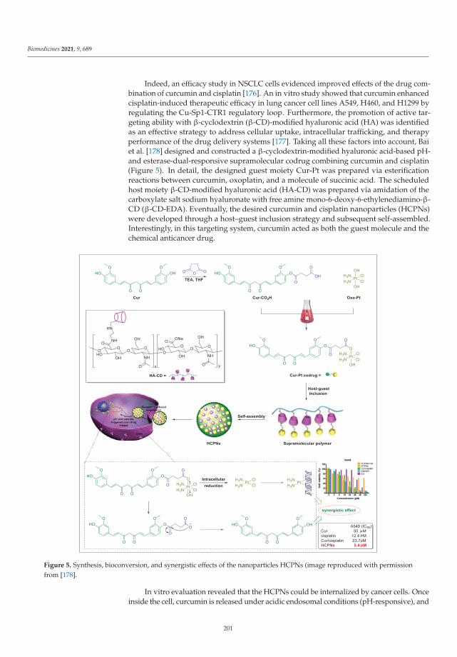
Biomedicines 2021, 9, 689
Indeed, an efficacy study in NSCLC cells evidenced improved effects of the drug com-bination of curcumin and cisplatin [176]. An in vitro study showed that curcumin enhancedcisplatin-induced therapeutic efficacy in lung cancer cell lines A549, H460, and H1299 byregulating the Cu-Sp1-CTR1 regulatory loop. Furthermore, the promotion of active tar-geting ability with β-cyclodextrin (β-CD)-modified hyaluronic acid (HA) was identifiedas an effective strategy to address cellular uptake, intracellular trafficking, and therapyperformance of the drug delivery systems [177]. Taking all these factors into account, Baiet al. [178] designed and constructed a β-cyclodextrin-modified hyaluronic acid-based pH-and esterase-dual-responsive supramolecular codrug combining curcumin and cisplatin(Figure 5). In detail, the designed guest moiety Cur-Pt was prepared via esterificationreactions between curcumin, oxoplatin, and a molecule of succinic acid. The scheduledhost moiety β-CD-modified hyaluronic acid (HA-CD) was prepared via amidation of thecarboxylate salt sodium hyaluronate with free amine mono-6-deoxy-6-ethylenediamino-β-CD (β-CD-EDA). Eventually, the desired curcumin and cisplatin nanoparticles (HCPNs)were developed through a host–guest inclusion strategy and subsequent self-assembled.Interestingly, in this targeting system, curcumin acted as both the guest molecule and thechemical anticancer drug.
Figure 5. Synthesis, bioconversion, and synergistic effects of the nanoparticles HCPNs (image reproduced with permissionfrom [178].
In vitro evaluation revealed that the HCPNs could be internalized by cancer cells. Onceinside the cell, curcumin is released under acidic endosomal conditions (pH-responsive), and
201

Biomedicines 2021, 9, 689
cisplatin is released via reducing of oxoplatin under higher expressed glutathione (GSH)conditions (esterase-responsive). Moreover, cell-based experiments revealed the effectivecellular toxicity (high efficiency, the IC50 value of 5.4 μM in A549 cells) and active targetingability (low toxicity, with low expression levels in normal LO-2 cells) of this novel drug-delivery system. Given the observed positive synergistic effect in the study, the authorsconcluded that HCPNs exhibited improved effects, compared with either monotherapywith curcumin or cisplatin [178]. The drug delivery and sustained release behavior of Curfrom HCPNs were investigated in vitro at pH 7.4 after 48 h (11% Cur was released) andpH 5.0 after 48 h (79% Cur was released), respectively, proving the better stability thanCur alone [178]. Meanwhile, although the Tian group did not proceed further with theirin vivo studies; we suggest additional in vivo studies should be performed to identify thepharmacokinetic or pharmacodynamic profile of the HCPNs and the synergistic activityagainst lung cancer of this codrug.
The disulfide bond, a promising redox-reactive switch in vivo, plays an essential rolein many biological processes [179]. To reduce adverse effects resulting from chemotherapyregimens, the disulfide-based drug design has attracted great enthusiasm in the synthesisof prodrug or codrug, and especially for the preparation of functional nanodrugs due totheir high selectivity and biocompatibility [180,181]. The nontoxic nanodrugs are activatedby the excess of GSH in the tumor microenvironment, which provides an essential strategyfor lung cancer-targeting treatment [182].
Celastrol, a typical pentacyclic triterpenoid, can be extracted from traditional herbalmedicines of the Celastraceae family [183]. Celastrol is considered another up-and-comingnatural product for lung cancer treatment due to its potent anti-NSCLC activity via its sup-pression of Axl protein expression [184], initiating tumor necrosis factor-related apoptosis-inducing ligand (TRAIL)-mediated apoptotic cell death [185], and suppressing cell inva-sion [186]. However, the clinical translation and biomedical application of celastrol arehindered due to its low bioavailability and physiological instability [187].
Gefitinib, approved by US FDA, has been used therapeutically as the first-line agentin patients with advanced lung cancer [188]. Unfortunately, the routine clinical practiceof gefitinib is often coupled with severe adverse effects, such as pulmonary toxicity [189],respiratory failure, and severe comorbidities [190]. Following a reasonable design, Wu et al.developed a GSH-responsive nanodrug (identified as CEL@G-SS-NIR in Figure 6), whichpossesses unique therapeutic efficacy for NSCLC in mice models by inhibiting upstreamand downstream EGFR signaling pathways [191]. The nanodrug CEL@G-SS-NIR wasprepared in two steps: preparation of the prodrug and acquisition of the nanocomplex.As shown in Figure 6, the main molecule G-SS-NIR of the nanodrug CEL@G-SS-NIR wassynthesized through a two-step reaction. First, the key intermediate G-SS was synthesizedsuccessfully in the presence of gefitinib (G), 2-hydroxyethyl disulfide (-SS-), and tiphosgenevia covalent linkage. Next, the near-infrared (NIR-OH) chromophore was bound to theside chain of the G-SS to form the prodrug G-SS-NIR. The amphiphilic G-SS-NIR readilyself-assembled into spherical nanomicelles in an aqueous medium (driven by the disulfidebond and the π–π interaction) and was encapsulated concomitantly the hydrophobic serine-threonine protein kinase (Akt) inhibitor celastrol (marked as CEL) to form CEL@G-SS-NIR.
This novel nanodrug CEL@G-SS-NIR possesses a suitable size (average diameter119 ± 6 nm), outstanding overall drug loading (64.0 ± 1.4 wt.%), and excellent stability inthe blood circulation, and has a rapid release rate of the free molecules (gefitinib, celastrol,and NIR-OH) at tumor region due to the breaking of the disulfide bonds in the presence ofhigh levels of GSH [191].
202

Biomedicines 2021, 9, 689
Figure 6. Synthesis, bioconversion, and synergistic effect of the nanodrug CEL@G-SS-NIR.
In vitro, the nanodrug CEL@G-SS-NIR formulation could effectively target the tumorregion due to its enhanced permeability and retention effect and also allowed fluorescentimaging in vivo, at a predetermined timepoint after tail vein injection, in orthotopic lungtumors [191]. In the treatment protocol, the mice were randomly divided into five groups(five mice per treatment group), and after a single treatment cycle, the CEL@G-SS-NIRgroup (13.4 mg/kg, intravenously, for 20 days), compared to the control groups, exhibitedstronger NSCLC tumor-suppressive effects [191]. As for the response mechanism involved,the entire process can be divided into four steps: (i) CEL@G-SS-NIR accumulates in thelung tumor region, (ii) CEL@G-SS-NIR releases the drug celastrol and the protonatedintermediates (and) through the deprotonated glutathione (GS¯) nucleophilic attack of thedisulfide bond on G-SS-NIR bonds, (iii) this further induces the synchronous releases ofthe parent drug gefitinib and the fluorescent dye NIR-OH via an intramolecular cyclizationreaction (thiolate anion moiety reacts with the adjacent carbonyl group), and (iv) finally,the synergistic anticancer activity is activated by suppressing the phosphatidylinositol3-kinase/serine threonine protein kinase (PI3K/Akt) signaling pathway by celastrol anddownregulating EGFR signaling pathway by gefitinib. Simultaneously, a fluorescent andmultispectral optoacoustic tomography imaging signal is generated by NIR-OH [192]. Thisstudy showed that disulfide-based and targeted fluorescent nano-prodrugs for treatingNSCLC and tracking drug delivery systems are particularly advantageous.
6. Conclusions and Future Perspectives
COVID-19 and lung cancer, the two most critical lung diseases presenting high mortal-ity rates, have posed a great challenge and a serious threat to human health and economic
203

Biomedicines 2021, 9, 689
development. Since 2020, as is well-known, the scientific community has made great effortsand remarkable inroads in developing promising anti-SARS-CoV-2 and anti-lung canceragents through various approaches. In this scenario, numerous natural products havefueled significant attention and have shown good results as potential therapeutics forthe above-mentioned lung diseases. This review highlighted state-of-the-art of importantnatural products (including their underlying molecular mechanisms), covering studies pub-lished between 1 January 2020 and 31 May 2021, in the treatment of the above-mentionedlung diseases. We found that natural products can be applied in vitro as monotherapy forthe treatment of SARS-CoV-2 (ginkgolic acid, resveratrol, and baicalein) and lung cancer(graveospene A, deguelin, and erianin), as well as in combination with the FDA-approveddrug inhibit SARS-CoV-2 (cepharanthine plus nelfinavir, linoleic acid plus remdesivir) andas codrug formulations with anti-lung cancer activity in vitro (codrug of curcumin and cis-platin). The evidence revealed herein that natural products could serve as a starting pointfor further drug development both in COVID-19 and lung cancer. It is worth noting, how-ever, that some natural products could be pan-assay interference compounds, which cangive false readouts, and close attention should be paid to decrease futile attempts [193,194].There is currently very little direct data associated with the clinical effect of natural productsagainst SARS-CoV-2 infection. To understand better and explore systematically the activityof natural products, more validation studies, with high-quality evidence (both in vitro andin animal models as well as on humans), are now needed.
To improve the use of natural products, many intensive research efforts (both in vitroand in vivo) are still needed to explore the limitations of these agents, such as poor watersolubility, limited oral absorption, low bioavailability, and the poor first-pass effect, whichrepresent the first step to develop promising anti-COVID-19 or anti-lung cancer agents. Itis clear that a long way is still ahead for us to realize natural product-based drug discoveryand development, as only phase 1–3 clinical trials can ensure that any small moleculeinhibitor can be used as a drug.
More aggressive and well-designed combination drug therapies that exhibit betteradditive or synergistic effects against COVID-19 and lung cancer are a promising strategy.For example, shiraiachrome A exhibits potent effects in Vero E6 cells by inhibiting theactivity of the SARS-CoV-2 S protein at EC50 values of 0.21 μM; bafilomycin B2 presentsenhanced inhibitory potency against SARS-CoV-2 at IC50 values of 5.11 nM in Vero E6cells by inhibiting the viral entry process; ginkgolic acid has IC50 values of 1.79 μM and16.3 μM against SARS-CoV-2 Mpro and SARS-CoV-2 PLpro. Combining the propertiesof the above-mentioned natural products with FDA-approved drugs (for example, withnelfinavir or remdesivir) could achieve optimal COVID-19 treatment through multitargetedmechanisms of action. In addition, a codrug of a natural product with an FDA-approveddrug could achieve a combination booster through multitargeted activity. However, thecodrug strategy remains an open question in the treatment of patients with COVID-19.Thus, we suggest researchers pay considerable attention to the development of emergingcodrug therapy strategies.
In contrast, precisely fabricated nanodrugs may be a more potent weapon to enhancebiocompatibility, minimize toxicity as well as side effects, achieve long-term circulation inthe body, as well as sustained release, overcome undesired adverse effects, and expand themodalities of administration (intravenous injection or inhalation). However, for COVID-19,the nanodrug strategy (containing natural products and FDA-approved drugs) remainsanother open question. Fortunately, significant progress has been made in the researchof lung cancer nanomedicines, which can provide some reference for the related drugdiscovery and development for COVID-19. There is no doubt that there is a long way to goand many difficulties to overcome. Nonetheless, natural products have their advantages.We sincerely hope natural products will be proven a safe and effective “natural remedy”for the treatment of the above-mentioned lung diseases with the assistance of multipletechniques and strategies.
204

Biomedicines 2021, 9, 689
Author Contributions: Conceptualization, Z.W.; investigation, L.Y.; resources, L.Y.; writing—originaldraft preparation, L.Y.; writing—review and editing, Z.W.; project administration, Z.W. All authorshave read and agreed to the published version of the manuscript.
Funding: This work was supported by the project of the PhD research start-up funds of Qufu NormalUniversity, China (Grant No. 614901, and 615201). The authors, therefore, gratefully acknowledgethe Qufu Normal University for the financial supports.
Institutional Review Board Statement: Not applicable.
Informed Consent Statement: Not applicable.
Data Availability Statement: Not applicable.
Conflicts of Interest: The authors declare no conflict of interest.
Abbreviations
ACE2 angiotensin-converting enzyme 2ACE2+ overexpression of ACE2AKT protein kinase BATP adenosine triphosphateβ-CD β-cyclodextrin3CLpro 3C-Like proteaseCOVID-19 coronavirus disease 2019cryo-EM cryo-electron microscopyEBOV Ebola virusEC50 half-maximal effective concentrationEGFR epidermal growth factor receptorEMT epithelial-mesenchymal transitionERK extracellular signal-regulated kinaseFDA US Food and Drug AdministrationGSH glutathioneHA hyaluronic acidHCoV-229E human coronavirus 229EHCPNs curcumin and cisplatin nanoparticlesHIV-1 human immunodeficiency virus type 1IC50 half-maximal inhibitory concentrationMAPK mitogen-activated protein kinaseMcl-1 myeloid cell leukemia sequence-1MERS-CoV Middle East respiratory syndrome coronavirusMMPs matrix-degrading metalloproteinasesMpro main proteasemTOR mammalian target of rapamycinNSCLC non-small cell lung cancerPDB Protein Data BankPD-L1 programmed death ligand-1PI3K phosphoinositide 3-kinasePLpro papain-like proteasepSARS-CoV-2 SARS-CoV-2 pseudovirusQFPD Qingfei Paidu decoctionRdRp RNA-dependent RNA polymeraseROS reactive oxygen speciesS protein spike proteinSARS-CoV severe acute respiratory syndrome coronavirusSARS-CoV-2 severe acute respiratory syndrome coronavirus 2SHL Shuanghuanglian oral liquid or injectionSI selectivity indexTMPRSS2 transmembrane protease serine 2
205

Biomedicines 2021, 9, 689
References
1. Newman, D.J.; Cragg, G.M. Natural products as sources of new drugs over the nearly four decades from 01/1981 to 09/2019. J.Nat. Prod. 2020, 83, 770–803. [CrossRef] [PubMed]
2. Atanasov, A.G.; Zotchev, S.B.; Dirsch, V.M.; Supuran, C.T. Natural products in drug discovery: Advances and opportunities. Nat.Rev. Drug Discov. 2021, 20, 200–216. [CrossRef]
3. Porras, G.; Chassagne, F.; Lyles, J.T.; Marquez, L.; Dettweiler, M.; Salam, A.M.; Samarakoon, T.; Shabih, S.; Farrokhi, D.R.; Quave,C.L. Ethnobotany and the role of plant natural products in antibiotic drug discovery. Chem. Rev. 2021, 121, 3495–3560. [CrossRef]
4. Wang, L.; Li, S.; Yao, Y.; Yin, W.; Ye, T. The role of natural products in the prevention and treatment of pulmonary fibrosis: Areview. Food Funct. 2021, 12, 990–1007. [CrossRef]
5. Wang, W.; Yao, Q.; Teng, F.; Cui, J.; Dong, J.; Wei, Y. Active ingredients from Chinese medicine plants as therapeutic strategies forasthma: Overview and challenges. Biomed. Pharmacother. 2021, 137, 111383. [CrossRef]
6. He, Y.Q.; Zhou, C.C.; Yu, L.Y.; Wang, L.; Deng, J.L.; Tao, Y.L.; Zhang, F.; Chen, W.S. Natural product derived phytochemicals inmanaging acute lung injury by multiple mechanisms. Pharmacol. Res. 2021, 163, 105224. [CrossRef]
7. Santoro, A.; Tomino, C.; Prinzi, G.; Cardaci, V.; Fini, M.; Macera, L.; Russo, P.; Maggi, F. Microbiome in chronic obstructivepulmonary disease: Role of natural products against microbial pathogens. Curr. Med. Chem. 2020, 27, 2931–2948. [CrossRef][PubMed]
8. Belchamber, K.B.R.; Donnelly, L.E. Targeting defective pulmonary innate immunity-A new therapeutic option? Pharmacol.Therapeut. 2020, 209, 107500. [CrossRef]
9. Christy, M.P.; Uekusa, Y.; Gerwick, L.; Gerwick, W.H. Natural products with potential to treat RNA virus pathogens includingSARS-CoV-2. J. Nat. Prod. 2021, 84, 161–182. [CrossRef] [PubMed]
10. Wen, T.; Song, L.; Hua, S. Perspectives and controversies regarding the use of natural products for the treatment of lung cancer.Cancer Med. 2021, 10, 2396–2422. [CrossRef]
11. Nalbandian, A.; Sehgal, K.; Gupta, A.; Madhavan, M.V.; McGroder, C.; Stevens, J.S.; Cook, J.R.; Nordvig, A.S.; Shalev, D.;Sehrawat, T.S.; et al. Post-acute COVID-19 syndrome. Nature Med. 2021, 27, 601–615. [CrossRef] [PubMed]
12. Siegel, R.L.; Miller, K.D.; Fuchs, H.E.; Jemal, A. Cancer Statistics. CA Cancer J. Clin. 2021, 71, 7–33.13. Eurosurveillance Editorial Team. Note from the editors: World Health Organization declares novel coronavirus (2019-nCoV)
sixth public health emergency of international concern. Euro. Surveil. 2020, 25, 200131e.14. Wang, C.; Horby, P.W.; Hayden, F.G.; Gao, G.F. A novel coronavirus outbreak of global health concern. Lancet 2020, 395, 470–473.
[CrossRef]15. Chan, J.F.; Yuan, S.; Kok, K.H.; To, K.K.; Chu, H.; Yang, J.; Xing, F.F.; Liu, J.L.; Yip, C.C.; Poon, R.W.; et al. A familial cluster of
pneumonia associated with the 2019 novel coronavirus indicating person-to-person transmission: A study of a family cluster.Lancet 2020, 395, 514–523. [CrossRef]
16. Phan, L.T.; Nguyen, T.V.; Luong, Q.C.; Nguyen, T.V.; Nguyen, H.T.; Le, H.Q.; Nguyen, T.T.; Cao, T.M.; Pham, Q.D. Importationand human-to-human transmission of a novel coronavirus in Vietnam. N. Engl. J. Med. 2020, 382, 872–874. [CrossRef]
17. Asselah, T.; Durantel, D.; Pasmant, E.; Lau, G.; Schinazi, R.F. COVID-19: Discovery, diagnostics and drug development. J. Hepatol.2021, 74, 168–184. [CrossRef] [PubMed]
18. Sung, H.; Ferlay, J.; Siegel, R.L.; Laversanne, M.; Soerjomataram, I.; Jemal, A.; Bray, F. Global cancer statistics 2020: GLOBOCANestimates of incidence and mortality worldwide for 36 cancers in 185 countries. CA Cancer J. Clin. 2021, 71, 209–249. [CrossRef]
19. Singh, S.S.; Mattheolabakis, G.; Gu, X.; Withers, S.; Dahal, A.; Jois, S. A grafted peptidomimetic for EGFR heterodimerizationinhibition: Implications in NSCLC models. Eur. J. Med. Chem. 2021, 216, 113312. [CrossRef]
20. Dawkins, J.B.N.; Webster, R.M. The small-cell lung cancer drug market. Nat. Rev. Drug Discov. 2020, 19, 507–508. [CrossRef][PubMed]
21. Verma, S.; Twilley, D.; Esmear, T.; Oosthuizen, C.B.; Reid, A.M.; Nel, M.; Lall, N. Anti-SARS-CoV natural products with thepotential to inhibit SARS-CoV-2 (COVID-19). Front. Pharmacol. 2020, 11, 561334. [CrossRef] [PubMed]
22. Wang, Z.; Yang, L. Turning the tide: Natural products and natural-product-inspired chemicals as potential counters to SARS-CoV-2infection. Front. Pharmacol. 2020, 11, 1013. [CrossRef] [PubMed]
23. Qiao, J.; Li, Y.S.; Zeng, R.; Liu, F.L.; Luo, R.H.; Huang, C.; Wang, Y.F.; Zhang, J.; Quan, B.; Shen, C.; et al. SARS-CoV-2 Mpro
inhibitors with antiviral activity in a transgenic mouse model. Science 2021, 371, 1374–1378. [CrossRef]24. Shin, D.; Mukherjee, R.; Grewe, D.; Bojkova, D.; Baek, K.; Bhattacharya, A.; Schulz, L.; Widera, M.; Mehdipour, A.R.; Tascher,
G.; et al. Papain-like protease regulates SARS-CoV-2 viral spread and innate immunity. Nature 2020, 587, 657–662. [CrossRef][PubMed]
25. Yang, J.; Feng, Z.; Liu, W.; Wang, Y.; Wang, G.; Yu, W.; Yang, G.; Yang, T.; Wang, Y.; Li, M. Exogenous hormone on epispermdevelopment and ginkgolic acid accumulation in Ginkgo biloba L. Ind. Crop. Prod. 2021, 160, 113140. [CrossRef]
26. Chen, Z.; Cui, Q.; Cooper, L.; Zhang, P.; Lee, H.; Chen, Z.; Wang, Y.; Liu, X.; Rong, L.; Du, R. Ginkgolic acid and anacardic acid arespecific covalent inhibitors of SARS-CoV-2 cysteine proteases. Cell Biosci. 2021, 11, 45. [CrossRef] [PubMed]
27. Medina-Enríquez, M.M.; Lopez-León, S.; Carlos-Escalante, J.A.; Aponte-Torres, Z.; Cuapio, A.; Wegman-Ostrosky, T. ACE2: Themolecular doorway to SARS-CoV-2. Cell Biosci. 2020, 10, 148. [CrossRef]
28. Benton, D.J.; Wrobel, A.G.; Xu, P.; Roustan, C.; Martin, S.R.; Rosenthal, P.B.; Skehel, J.J.; Gamblin, S.J. Receptor binding andpriming of the spike protein of SARS-CoV-2 for membrane fusion. Nature 2020, 588, 327–330. [CrossRef] [PubMed]
206

Biomedicines 2021, 9, 689
29. Li, Y.T.; Yang, C.; Wu, Y.; Lv, J.J.; Feng, X.; Tian, X.; Zhou, Z.; Pan, X.; Liu, S.; Tian, L.W. Axial chiral binaphthoquinone andperylenequinones from the stromata of hypocrella bambusae are SARS-CoV-2 entry inhibitors. J. Nat. Prod. 2021, 84, 436–443.[CrossRef]
30. Stopsack, K.H.; Mucci, L.A.; Antonarakis, E.S.; Nelson, P.S.; Kantoff, P.W. TMPRSS2 and COVID-19: Serendipity or opportunityfor intervention? Cancer Discov. 2020, 10, 779–782. [CrossRef]
31. Kim, T.Y.; Jeon, S.; Jang, Y.; Gotina, L.; Won, J.; Ju, Y.H.; Kim, S.; Jang, M.W.; Won, W.; Park, M.G.; et al. Platycodin D prevents bothlysosome- and TMPRSS2-driven SARS-CoV-2 infection in vitro by hindering membrane fusion. Exp. Mol. Med. 2021. [CrossRef]
32. Yang, M.; Wei, J.; Huang, T.; Lei, L.; Shen, C.; Lai, J.; Yang, M.; Liu, L.; Yang, Y.; Liu, G.; et al. Resveratrol inhibits the replicationof severe acute respiratory syndrome coronavirus 2 (SARS-CoV-2) in cultured Vero cells. Phytother. Res. 2021, 35, 1127–1129.[CrossRef]
33. Ter Ellen, B.M.; Dinesh Kumar, N.; Bouma, E.M.; Troost, B.; van de Pol, D.P.I.; van der Ende-Metselaar, H.H.; Apperloo, L.; vanGosliga, D.; van den Berge, M.; Nawijn, M.C.; et al. Resveratrol and pterostilbene potently inhibit SARS-CoV-2 replication in vitro.bioRxiv 2020. [CrossRef]
34. Yin, W.; Mao, C.; Luan, X.; Shen, D.D.; Shen, Q.; Su, H.; Wang, X.; Zhou, F.; Zhao, W.; Gao, M.; et al. Structural basis for inhibitionof the RNA-dependent RNA polymerase from SARS-CoV-2 by remdesivir. Science 2020, 368, 1499–1504. [CrossRef] [PubMed]
35. Wu, C.; Huang, H.; Choi, H.Y.; Ma, Y.; Zhou, T.; Peng, Y.; Pang, K.; Shu, G.; Yang, X. Anti-esophageal cancer effect of corilaginextracted from Phmllanthi fructus via the mitochondrial and endoplasmic reticulum stress pathways. J. Ethnopharmacol. 2021,269, 113700. [CrossRef] [PubMed]
36. Li, Q.; Yi, D.; Lei, X.; Zhao, J.; Zhang, Y.; Cui, X.; Xiao, X.; Jiao, T.; Dong, X.; Zhao, X.; et al. Corilagin inhibits SARS-CoV-2replication by targeting viral RNA-dependent RNA polymerase. Acta Pharm. Sin. B 2021. [CrossRef]
37. Loschwitz, J.; Jäckering, A.; Keutmann, M.; Olagunju, M.; Eberle, R.J.; Coronado, M.A.; Olubiyi, O.O.; Strodel, B. Novel inhibitorsof the main protease enzyme of SARS-CoV-2 identified via molecular dynamics simulation-guided in vitro assay. Bioorg. Chem.2021, 111, 104862. [CrossRef]
38. Xie, X.; Lu, S.; Pan, X.; Zou, M.; Li, F.; Lin, H.; Hu, J.; Fan, S.; He, J. Antiviral bafilomycins from a feces-inhabiting Streptomyces sp.J. Nat. Prod. 2021, 84, 537–543. [CrossRef]
39. Abdallah, H.; El-Halawany, A.; Sirwi, A.; El-Araby, A.; Mohamed, G.; Ibrahim, S.; Koshak, A.; Asfour, H.; Awan, Z.; Elfaky, M.A.Repurposing of Some Natural Product Isolates as SARS-COV-2 Main Protease Inhibitors via In Vitro Cell Free and Cell-BasedAntiviral Assessments and Molecular Modeling Approaches. Pharmaceuticals 2021, 14, 213. [CrossRef] [PubMed]
40. Sa-ngiamsuntorn, K.; Suksatu, A.; Pewkliang, Y.; Thongsri, P.; Kanjanasirirat, P.; Manopwisedjaroen, S.; Charoensutthivarakul, S.;Wongtrakoongate, P.; Pitiporn, S.; Chaopreecha, J.; et al. Anti-SARS-CoV-2 Aactivity of Andrographis paniculata extract and itsmajor component andrographolide in human lung epithelial cells and cytotoxicity evaluation in major organ cell representatives.J. Nat. Prod. 2021, 84, 1261–1270. [CrossRef]
41. Hu, Y.; Liu, M.; Qin, H.; Lin, H.; An, X.; Shi, Z.; Song, L.; Yang, X.; Fan, H.; Tong, Y. Artemether, artesunate, arteannuin B,echinatin, licochalcone B and andrographolide effectively inhibit SARS-CoV-2 and related viruses in vitro. Front. Cell. Infect.Microbiol. 2021. [CrossRef]
42. Cao, R.; Hu, H.; Li, Y.; Wang, X.; Xu, M.; Liu, J.; Zhang, H.; Yan, Y.; Zhao, L.; Li, W.; et al. Anti-SARS-CoV-2 potential ofartemisinins in vitro. ACS Infect. Dis. 2020, 6, 2524–2531. [CrossRef] [PubMed]
43. Touret, F.; Gilles, M.; Barral, K.; Nougairède, A.; Decroly, E.; de Lamballerie, X.; Coutard, B. In vitro screening of a FDA approvedchemical library reveals potential inhibitors of SARS-CoV-2 replication. Sci. Rep. 2020, 10, 13093. [CrossRef] [PubMed]
44. Hong, S.; Seo, S.H.; Woo, S.J.; Kwon, Y.; Song, M.; Ha, N.C. Epigallocatechin gallate inhibits the uridylate-specific endoribonucleaseNsp15 and efficiently neutralizes the SARS-CoV-2 strain. J. Agric. Food Chem. 2021. [CrossRef] [PubMed]
45. Raj, V.; Park, J.G.; Cho, K.H.; Choi, P.; Kim, T.; Ham, J.; Lee, J. Assessment of antiviral potencies of cannabinoids againstSARSCoV-2 using computational and in vitro approaches. Int. J. Biol. Macromol. 2021, 168, 474–485. [CrossRef]
46. Nguyen, L.C.; Yang, D.; Nicolaescu, V.; Best, T.; Chen, S.; Friesen, J.B.; Drayman, N.; Mohamed, A.; Dann, C.; Silva, D.; et al.Cannabidiol inhibits SARS-CoV-2 replication and promotes the host innate immune response. bioRxiv 2021. [CrossRef]
47. Zhu, Y.; Xie, D.Y. Docking characterization and in vitro inhibitory activity of flavan-3-ols and dimeric proanthocyanidins againstthe main protease activity of SARS-CoV-2. Front. Plant Sci. 2020, 11, 601316. [CrossRef] [PubMed]
48. Du, R.; Cooper, L.; Chen, Z.; Lee, H.; Rong, L.; Cui, Q. Discovery of chebulagic acid and punicalagin as novel allosteric inhibitorsof SARS-CoV-2 3CLpro. Antivir. Res. 2021, 190, 105075. [CrossRef] [PubMed]
49. Wang, P.; Luo, R.; Zhang, M.; Wang, Y.; Song, T.; Tao, T.; Li, Z.; Jin, L.; Zheng, H.; Chen, W.; et al. A cross-talk betweenepithelium and endothelium mediates human alveolar-capillary injury during SARS-CoV-2 infection. Cell Death Dis. 2020, 11,1042. [CrossRef]
50. Du, A.; Zheng, R.; Disoma, C.; Li, S.; Chen, Z.; Li, S.; Liu, P.; Zhou, Y.; Shen, Y.; Liu, S.; et al. Epigallocatechin-3-gallate, an activeingredient of Traditional Chinese Medicines, inhibits the 3CLpro activity of SARS-CoV-2. Int. J. Biol. Macromol. 2021, 176, 1–12.[CrossRef]
51. Choy, K.T.; Wong, A.Y.L.; Kaewpreedee, P.; Sia, S.F.; Chen, D.D.; Hui, K.P.Y.; Chu, D.K.W.; Chan, M.C.W.; Cheung, P.P.H.; Huang,X.; et al. Remdesivir, lopinavir, emetine, and homoharringtonine inhibit SARS-CoV-2 replication in vitro. Antivir. Res. 2020, 178,104786. [CrossRef] [PubMed]
207
Biomedicines 2021, 9, 689
52. Kumar, R.; Afsar, M.; Khandelwal, N.; Chander, Y.; Riyesh, T.; Dedar, R.K.; Gulati, B.R.; Pal, Y.; Barua, S.; Tripathi, B.N.; et al.Emetine suppresses SARS-CoV-2 replication by inhibiting interaction of viral mRNA with eIF4E. Antivir. Res. 2021, 189, 105056.[CrossRef]
53. Ashhurst, A.; Tang, A.; Fajtova, P.; Yoon, M.; Aggarwal, A.; Stoye, A.; Larance, M.; Beretta, L.; Drelich, A.; Skinner, D.; et al. Potentin vitro anti-SARS-CoV-2 activity by gallinamide A and analogues via inhibition of cathepsin L. bioRxiv 2020. [CrossRef]
54. Tietjen, I.; Cassel, J.; Register, E.T.; Zhou, X.Y.; Messick, T.E.; Keeney, F.; Lu, L.D.; Beattie, K.D.; Rali, T.; Ertl, H.C.J.; et al. Thenatural stilbenoid (-)-hopeaphenol inhibits cellular entry of SARS-CoV-2 USA-WA1/2020, B.1.1.7 and B.1.351 variants. bioRxiv2021. [CrossRef]
55. O’Keefe, S.; Roboti, P.; Duah, K.B.; Zong, G.; Schneider, H.; Shi, W.Q.; High, S. Ipomoeassin-F inhibits the in vitro biogenesis ofthe SARS-CoV-2 spike protein and its host cell membrane receptor. J. Cell Sci. 2021, 134, jcs257758. [CrossRef] [PubMed]
56. Gangadevi, S.; Badavath, V.N.; Thakur, A.; Yin, N.; Jonghe, S.D.; Acevedo, O.; Jochmans, D.; Leyssen, P.; Wang, K.; Neyts, J.; et al.Kobophenol A inhibits binding of host ACE2 receptor with Spike RBD domain of SARS-CoV-2, a lead compound for blockingCOVID-19. J. Phys. Chem. Lett. 2021, 12, 1793–1802. [CrossRef]
57. Kuzikov, M.; Costanzi, E.; Reinshagen, J.; Esposito, F.; Vangeel, L.; Wolf, M.; Ellinger, B.; Claussen, C.; Geisslinger, G.; Corona, A.;et al. Identification of inhibitors of SARS-CoV-2 3CL-Pro enzymatic activity using a small molecule in-vitro repurposing screen.ACS Pharmacol. Transl. Sci. 2021. [CrossRef]
58. Xiao, T.; Cui, M.; Zheng, C.; Wang, M.; Sun, R.; Gao, D.; Bao, J.; Ren, S.; Yang, B.; Lin, J.; et al. Myricetin inhibit SARS-CoV-2 viralreplication by targeting Mpro and ameliorates pulmonary inflammation. Front. Pharmacol. 2021. [CrossRef]
59. Clementi, N.; Scagnolari, C.; D’Amore, A.; Palombi, F.; Criscuolo, E.; Frasca, F.; Pierangeli, A.; Mancini, N.; Antonelli, G.; Clementi,M.; et al. Naringenin is a powerful inhibitor of SARS-CoV-2 infection in vitro. Pharmacol. Res. 2021, 163, 105255. [CrossRef][PubMed]
60. Jeon, S.; Ko, M.; Lee, J.; Choi, I.; Byun, S.Y.; Park, S.; Shum, D.; Kim, S. Identification of antiviral drug candidates againstSARS-CoV-2 from FDA-approved drugs. Antimicrob. Agents Chemother. 2020, 64, e00819-20. [CrossRef]
61. Jin, Z.; Du, X.; Xu, Y.; Deng, Y.; Liu, M.; Zhao, Y.; Zhang, B.; Li, X.; Zhang, L.; Peng, C.; et al. Structure of Mpro from SARS-CoV-2and discovery of its inhibitors. Nature 2020, 582, 289–293. [CrossRef]
62. Jang, M.; Park, Y.I.; Cha, Y.E.; Park, R.; Namkoong, S.; Lee, J.I.; Park, J. Tea polyphenols EGCG and theaflavin inhibit the activityof SARS-CoV-2 3CL-protease in vitro. Evid. Based Compl. Alt. Med. 2020, 2020, 5630838. [CrossRef] [PubMed]
63. Choudhry, N.; Zhao, X.; Xu, D.; Zanin, M.; Chen, W.; Yang, Z.; Chen, J. Chinese Therapeutic Strategy for Fighting COVID-19 andPotential Small-Molecule Inhibitors against Severe Acute Respiratory Syndrome Coronavirus 2 (SARS-CoV-2). J. Med. Chem.2020, 63, 13205–13227. [CrossRef]
64. Wang, Z.; Yang, L. Chinese herbal medicine: Fighting SARS-CoV-2 infection on all fronts. J. Ethnopharmacol. 2021, 270, 113869.[CrossRef]
65. Huang, K.; Zhang, P.; Zhang, Z.; Youn, J.Y.; Zhang, H.; Cai, H.L. Traditional Chinese Medicine (TCM) in the treatment of viralinfections: Efficacies and mechanisms. Pharm. Ther. 2021, 225, 107843. [CrossRef] [PubMed]
66. National Health Commission of the People’s Republic of China. Notice on the Issunance of Guidelines of Diagnosis and Treatmentfor 2019-nCoV Infected Pneumonia (Version 7). 2020. Available online: http://www.nhc.gov.cn/yzygj/s7653p/202003/46c9294a7dfe4cef80dc7f5912eb1989/files/ce3e6945832a438eaae415350a8ce964.pdf (accessed on 3 March 2020).
67. Xinhua Net. Academician Xiaolin Tong: The Total Effective Rate of Qingfeipaidu Formula was 97%, none Transfer from Mild toSevere Cases. 2020. Available online: http://www.kunlunce.com/ssjj/fl1/2020-03-18/141570.html (accessed on 18 March 2020).
68. Su, H.X.; Yao, S.; Zhao, W.F.; Li, M.J.; Liu, J.; Shang, W.J.; Xie, H.; Ke, C.Q.; Hu, H.C.; Gao, M.N.; et al. Anti-SARS-CoV-2activities in vitro of Shuanghuanglian preparations and bioactive ingredients. Acta Pharmacol. Sin. 2020, 41, 1167–1177. [CrossRef][PubMed]
69. Liu, H.; Ye, F.; Sun, Q.; Liang, H.; Li, C.; Li, S.; Lu, R.; Huang, B.; Tan, W.; Lai, L. Scutellaria baicalensis extract and baicaleininhibit replication of SARS-CoV-2 and its 3C-like protease in vitro. J. Enzym. Inhib. Med. Chemother. 2021, 36, 497–503. [CrossRef]
70. Pooja, M.; Reddy, G.J.; Hema, K.; Dodoala, S.; Koganti, B. Unravelling high-affinity binding compounds towards transmembraneprotease serine 2 enzyme in treating SARS-CoV-2 infection using molecular modelling and docking studies. Eur. J. Pharmacol.2021, 890, 173688.
71. Song, J.; Zhang, L.; Xu, Y.; Yang, D.; Zhang, L.; Yang, S.; Zhang, W.; Wang, J.; Tian, S.; Yang, S.; et al. The comprehensive studyon the therapeutic effects of baicalein for the treatment of COVID-19 in vivo and in vitro. Biochem. Pharmacol. 2021, 183, 114302.[CrossRef]
72. Ibrahim, M.A.A.; Mohamed, E.A.R.; Abdelrahman, A.H.M.; Allemailem, K.S.; Moustafa, M.F.; Shawky, A.M.; Mahzari, A.;Hakami, A.R.; Abdeljawaad, K.A.A.; Atia, M.A.M. Rutin and flavone analogs as prospective SARS-CoV-2 main protease inhibitors:In silico drug discovery study. J. Mol. Graph. Model. 2021, 105, 107904. [CrossRef] [PubMed]
73. Zandi, K.; Musall, K.; Oo, A.; Cao, D.; Liang, B.; Hassandarvish, P.; Lan, S.; Slack, R.L.; Kirby, K.A.; Bassit, L.; et al. Baicalein andbaicalin inhibit SARS-CoV-2 RNA-dependent-RNA polymerase. Microorganisms 2021, 9, 893. [CrossRef] [PubMed]
74. Qian, J.Q. Cardiovascular pharmacological effects of bisbenzylisoquinoline alkaloid derivatives. Acta Pharm. Sin. 2002, 23,1086–1092.
75. Huang, X.H.; Yan, X.; Zhang, Q.H.; Hong, P.; Zhang, W.X.; Liu, Y.P.; Xu, W.W.; Li, B.; He, Q.Y. Direct targeting of HSP90 withdaurisoline destabilizes β-catenin to suppress lung cancer tumorigenesis. Cancer Lett. 2020, 489, 66–78. [CrossRef]
208

Biomedicines 2021, 9, 689
76. Liu, F.; Ma, J.; Shi, Z.; Zhang, Q.; Wang, H.; Li, D.; Song, Z.; Wang, C.; Jin, J.; Xu, J.; et al. Clerodane diterpenoids isolated from theleaves of Casearia graveolens. J. Nat. Prod. 2020, 83, 36–44. [CrossRef] [PubMed]
77. Tuli, H.S.; Mittal, S.; Loka, M.; Aggarwal, V.; Aggarwal, D.; Masurkar, A.; Kaur, G.; Varol, M.; Sak, K.; Kumar, M.; et al. Deguelintargets multiple oncogenic signaling pathways to combat human malignancies. Pharmacol. Res. 2021, 166, 105487. [CrossRef]
78. Gao, F.; Yu, X.; Li, M.; Zhou, L.; Liu, W.; Li, W.; Liu, H. Deguelin suppresses non-small cell lung cancer by inhibiting EGFRsignaling and promoting GSK3β/FBW7-mediated Mcl-1 destabilization. Cell Death Dis. 2020, 11, 143. [CrossRef] [PubMed]
79. Wu, C.P.; Lusvarghi, S.; Hsiao, S.H.; Liu, T.C.; Li, Y.Q.; Huang, Y.H.; Hung, T.H.; Ambudka, S.V. Licochalcone A selectivelyresensitizes ABCG2-overexpressing multidrug-resistant cancer cells to chemotherapeutic drugs. J. Nat. Prod. 2020, 83, 1461–1472.[CrossRef]
80. Luo, W.; Sun, R.; Chen, X.; Li, J.; Jiang, J.; He, Y.; Shi, S.; Wen, H. ERK activation-mediated autophagy induction resists licochalconeA-induced anticancer activities in lung cancer cells in vitro. OncoTargets Ther. 2020, 13, 13437–13450. [CrossRef]
81. Yuan, L.W.; Jiang, X.M.; Xu, Y.L.; Huang, M.Y.; Chen, Y.C.; Yu, W.B.; Su, M.X.; Ye, Z.H.; Chen, X.; Wang, Y.; et al. Licochalcone Ainhibits interferon-gamma-induced programmed death-ligand 1 in lung cancer cells. Phytomedicine 2021, 80, 153394. [CrossRef]
82. Li, B.; Zhou, D.; Li, S.; Feng, Y.; Li, X.; Chang, W.; Zhang, J.; Sun, Y.; Qing, D.; Chen, G.; et al. Licochalcone A reversesNNK-induced ectopic miRNA expression to elicit in vitro and in vivo chemopreventive effects. Phytomedicine 2020, 76, 153245.[CrossRef]
83. Gao, F.; Li, M.; Yu, X.; Liu, W.; Zhou, L.; Li, W. Licochalcone A inhibits EGFR signalling and translationally suppresses survivinexpression in human cancer cells. J. Cell Mol. Med. 2021, 25, 813–826. [CrossRef]
84. Yang, A.; Li, M.Y.; Zhang, Z.H.; Wang, J.Y.; Xing, Y.; Ri, M.; Jin, C.H.; Xu, G.H.; Piao, L.X.; Jin, H.L.; et al. Erianin regulatesprogrammed cell death ligand 1 expression and enhances cytotoxic T lymphocyte activity. J. Ethnopharmacol. 2021, 273, 113598.[CrossRef]
85. Chen, P.; Wu, Q.; Feng, J.; Yan, L.; Sun, Y.; Liu, S.; Xiang, Y.; Zhang, M.; Pan, T.; Chen, X.; et al. Erianin, a novel dibenzyl compoundin Dendrobium extract, inhibits lung cancer cell growth and migration via calcium/calmodulin-dependent ferroptosis. SignalTransduct. Tar. 2020, 5, 51. [CrossRef] [PubMed]
86. Keller, L.; Canuto, K.M.; Liu, C.; Suzuki, B.M.; Almaliti, J.; Sikandar, A.; Naman, C.B.; Glukhov, E.; Luo, D.; Duggan, B.M.; et al.Tutuilamides A-C: Vinyl-chloride-containing cyclodepsipeptides from marine cyanobacteria with potent elastase inhibitoryproperties. ACS Chem. Biol. 2020, 15, 751–757. [CrossRef] [PubMed]
87. Chen, Q.Y.; Luo, D.; Seabra, G.M.; Luesch, H. Ahp-Cyclodepsipeptides as tunable inhibitors of human neutrophil elastase andkallikrein 7: Total synthesis of tutuilamide A, serine protease selectivity profile and comparison with lyngbyastatin 7. Bioorg. Med.Chem. 2020, 28, 115756. [CrossRef] [PubMed]
88. Hafner, S.; Lang, S.J.; Gaafary, M.E.; Schmiech, M.; Simmet, T.; Syrovets, T. The cardenolide glycoside acovenoside A interfereswith epidermal growth factor receptor (EGFR) trafficking in non-small cell lung cancer cells. Front. Pharmacol. 2021, 11, 611657.[CrossRef] [PubMed]
89. Han, A.R.; Lee, S.; Han, S.; Lee, Y.J.; Kim, J.B.; Seo, E.K.; Jung, C.H. Triterpenoids from the leaves of Centella asiatica inhibitionizing radiation-induced migration and invasion of human lung cancer cells. Evid. Based Compl. Alt. 2020, 2020, 3683460.[CrossRef]
90. Zhang, X.; Ruan, Q.; Zhai, Y.; Lu, D.; Li, C.; Fu, Y.; Zheng, Z.; Song, Y.; Guo, J. Baicalein inhibits non-small-cell lung cancer invasionand metastasis by reducing ezrin tension in inflammation microenvironment. Cancer Sci. 2020, 111, 3802–3812. [CrossRef][PubMed]
91. Zhang, Z.; Nong, L.; Chen, M.; Gu, X.; Zhao, W.; Liu, M.; Cheng, W. Baicalein suppresses vasculogenic mimicry through inhibitingRhoA/ROCK expression in lung cancer A549 cell line. Acta Bioch. Bioph. Sin. 2020, 52, 1007–1015. [CrossRef]
92. Li, J.; Yan, L.; Luo, J.; Tong, L.; Gao, Y.; Feng, W.; Wang, F.; Cui, W.; Li, S.; Sun, Z. Baicalein suppresses growth of non-small celllung carcinoma by targeting MAP4K3. Biomed. Pharmacother. 2021, 133, 110965. [CrossRef]
93. Sui, X.; Han, X.; Chen, P.; Wu, Q.; Feng, J.; Duan, T.; Chen, X.; Pan, T.; Yan, L.; Jin, T.; et al. Baicalin induces apoptosis andsuppresses the cell cycle progression of lung cancer cells through downregulating Akt/mTOR signaling pathway. Front. Mol.Biosci. 2021, 7, 602282. [CrossRef] [PubMed]
94. Yan, Y.; Yao, L.; Sun, H.; Pang, S.; Kong, X.; Zhao, S.; Xu, S. Effects of wogonoside on invasion and migration of lung cancer A549cells and angiogenesis in xenograft tumors of nude mice. J. Thorac. Dis. 2020, 12, 1552–1560. [CrossRef] [PubMed]
95. Cheng, Z.Y.; Hsiao, Y.T.; Huang, Y.P.; Peng, S.F.; Huang, W.W.; Liu, K.C.; Hsia, T.C.; Way, T.D.; Chung, J.G. Casticin inducesDNA damage and affects DNA repair associated protein expression in human lung cancer A549 cells. Molecules 2020, 25, 341.[CrossRef] [PubMed]
96. Cui, L.; Yang, G.; Ye, J.; Yao, Y.; Lu, G.; Chen, J.; Fang, L.; Lu, S.; Zhou, J. Dioscin elicits anti-tumour immunity by inhibitingmacrophage M2 polarization via JNK and STAT3 pathways in lung cancer. J. Cell. Mol. Med. 2020, 24, 9217–9230. [CrossRef]
97. Chen, A.; Jiang, P.; Zeb, F.; Wu, X.; Xu, C.; Chen, L.; Feng, Q. EGCG regulates CTR1 expression through its pro-oxidative propertyin non-small-cell lung cancer cells. J. Cell. Physiol. 2020, 235, 7970–7981. [CrossRef]
98. Wei, R.; Wirkus, J.; Yang, Z.; Machuca, J.; Esparza, Y.; Mackenzie, G.G. EGCG sensitizes chemotherapeutic-induced cytotoxicityby targeting the ERK pathway in multiple cancer cell lines. Arch. Biochem. Biophys. 2020, 692, 108546. [CrossRef]
99. Duan, J.; Li, Y.; Gao, H.; Yang, D.; He, X.; Fang, Y.; Zhou, G. Phenolic compound ellagic acid inhibits mitochondrial respirationand tumor growth in lung cancer. Food Funct. 2020, 11, 6332–6339. [CrossRef]
209

Biomedicines 2021, 9, 689
100. Boonjing, S.; Pothongsrisit, S.; Wattanathamsan, O.; Sritularak, B.; Pongrakhananon, V. Erianthridin induces non-small cell lungcancer cell apoptosis through the suppression of extracellular signal-regulated kinase activity. Planta Med. 2021, 87, 283–293.
101. Pothongsrisit, S.; Arunrungvichian, K.; Hayakawa, Y.; Sritularak, B.; Mangmool, S.; Pongrakhananon, V. Erianthridin suppressesnon-small-cell lung cancer cell metastasis through inhibition of Akt/mTOR/p70 S6K signaling pathway. Sci. Rep. 2021, 11, 6618.[CrossRef]
102. Choudhury, P.; Barua, A.; Roy, A.; Pattanayak, R.; Bhattacharyya, M.; Saha, P. Eugenol emerges as an elixir by targeting β-catenin,the central cancer stem cell regulator in lung carcinogenesis: An in vivo and in vitro rationale. Food Funct. 2021, 12, 1063–1078.[CrossRef] [PubMed]
103. Yu, X.; Gao, F.; Li, W.; Zhou, L.; Liu, W.; Li, M. Formononetin inhibits tumor growth by suppression of EGFR-Akt-Mcl-1 axis innon-small cell lung cancer. J. Exp. Clin. Canc. Res. 2020, 39, 62. [CrossRef]
104. Yang, B.; Yang, N.; Chen, Y.; Maomao, Z.; Lian, Y.; Xiong, Z.; Wang, B.; Feng, L.; Jia, X. An integrated strategy for effective-component discovery of Astragali Radix in the treatment of lung cancer. Front. Pharmacol. 2021, 12, 580978. [CrossRef]
105. Kang, D.Y.; Sp, N.; Jo, E.S.; Rugamba, A.; Hong, D.Y.; Lee, H.G.; Yoo, J.S.; Liu, Q.; Jang, K.J.; Yang, Y.M. The inhibitory mechanismsof tumor PD-L1 expression by natural bioactive gallic acid in non-small-cell lung cancer (NSCLC) cells. Cancers 2020, 12, 727.[CrossRef] [PubMed]
106. Wang, D.; Bao, B. Gallic acid impedes non-small cell lung cancer progression via suppression of EGFR-dependent CARM1-PELP1complex. Drug Des. Dev. Ther. 2020, 14, 1583–1592. [CrossRef] [PubMed]
107. Chen, H.; Miao, L.; Huang, F.; Yu, Y.; Peng, Q.; Liu, Y.; Li, X.; Liu, H. Glochidiol, a natural triterpenoid, exerts its anti-cancereffects by targeting the colchicine binding site of tubulin. Invest. N. Drug. 2021, 39, 578–586. [CrossRef]
108. Min, H.Y.; Pei, H.; Hyun, S.Y.; Boo, H.J.; Jang, H.J.; Cho, J.; Kim, J.H.; Son, J.; Lee, H.Y. Potent anticancer effect of the naturalsteroidal saponin gracillin is produced by inhibiting glycolysis and oxidative phosphorylation-mediated bioenergetics. Cancers2020, 12, 913. [CrossRef]
109. Yang, J.; Cao, L.; Li, Y.; Liu, H.; Zhang, M.; Ma, H.; Wang, B.; Yuan, X.; Liu, Q. Gracillin isolated from Reineckia carnea inducesapoptosis of A549 Cells via the mitochondrial pathway. Drug Des. Dev. Ther. 2021, 2021, 233–243. [CrossRef]
110. Lv, L.; Zhang, W.; Li, T.; Jiang, L.; Lu, X.; Lin, J. Hispidulin exhibits potent anticancer activity in vitro and in vivo throughactivating ER stress in non-small-cell lung cancer cells. Oncol. Rep. 2020, 43, 1995–2003. [CrossRef]
111. Zhao, X.; Lin, Y.; Jiang, B.; Yin, J.; Lu, C.; Wang, J.; Zeng, J. Icaritin inhibits lung cancer-induced osteoclastogenesis by suppressingthe expression of IL-6 and TNF-a and through AMPK/mTOR signaling pathway. Anti-Cancer Drug. 2020, 31, 1004–1011.[CrossRef] [PubMed]
112. Lee, J.H.; Park, S.Y.; Hwang, W.; Sung, J.Y.; Shim, M.L.C.J.; Kim, Y.N.; Yoon, K. Isoharringtonine induces apoptosis of non-smallcell lung cancer cells in tumorspheroids via the intrinsic pathway. Biomolecules 2020, 10, 1521. [CrossRef] [PubMed]
113. Fouzder, C.; Mukhuty, A.; Kundu, R. Kaempferol inhibits Nrf2 signalling pathway via downregulation of Nrf2 mRNA andinduces apoptosis in NSCLC cells. Arch. Biochem. Biophys. 2021, 697, 108700. [CrossRef]
114. Sheng, H.; Lv, W.; Zhu, L.; Wang, L.; Wang, Z.; Han, J.; Hu, J. Liriopesides B induces apoptosis and cell cycle arrest in humannon-small cell lung cancer cells. Int. J. Mol. Med. 2020, 46, 1039–1050. [CrossRef] [PubMed]
115. Chen, Y.C.; Huang, M.Y.; Zhang, L.L.; Feng, Z.L.; Jiang, X.M.; Yuan, L.W.; Huang, R.Y.; Liu, B.; Yu, H.; Wang, Y.T.; et al.Nagilactone E increases PD-L1 expression through activation of c-Jun in lung cancer cells. Chin. J. Nat. Med. 2020, 18, 517–525.[CrossRef]
116. Zhang, L.L.; Guo, J.; Jiang, X.M.; Chen, X.P.; Wang, Y.T.; Li, A.; Lin, L.G.; Li, H.; Lu, J.J. Identification of nagilactone E as a proteinsynthesis inhibitor with anticancer activity. Acta Pharmacol. Sin. 2020, 41, 698–705. [CrossRef] [PubMed]
117. Liu, X.; Zhang, Y.; Zhou, G.J.; Hou, Y.; Kong, Q.; Lu, J.J.; Zhang, Q.; Chen, X. Natural alkaloid 8-oxo-epiberberine inhibitedTGF-β1-triggred epithelial-mesenchymal transition by interfering Smad3. Toxicol. Appl. Pharm. 2020, 404, 115179. [CrossRef][PubMed]
118. Li, X.; Huang, R.; Li, M.; Zhu, Z.; Chen, Z.; Cui, L.; Luo, H.; Luo, L. Parthenolide inhibits the growth of non-small cell lung cancerby targeting epidermal growth factor receptor. Cancer Cell Int. 2020, 20, 561. [CrossRef] [PubMed]
119. Sun, L.; Yuan, W.; Wen, G.; Yu, B.; Xu, F.; Gan, X.; Tang, J.; Zeng, Q.; Zhu, L.; Chen, C.; et al. Parthenolide inhibits humanlung cancer cell growth by modulating the IGF-1R/PI3K/Akt signaling pathway. Oncol. Rep. 2020, 44, 1184–1193. [CrossRef][PubMed]
120. Wu, L.M.; Liao, X.Z.; Zhang, Y.; He, Z.R.; Nie, S.Q.; Ke, B.; Shi, L.; Zhao, J.F.; Chen, W.H. Parthenolide augments the chemosen-sitivity of non-small-cell lung cancer to cisplatin via the PI3K/AKT signaling pathway. Front. Cell Dev. Biol. 2021, 8, 610097.[CrossRef] [PubMed]
121. Meng, N.; Zhang, R.; Liu, C.; Wang, Q.; Wang, X.; Guo, X.; Wang, P.; Sun, J. PDB-1 from potentilla discolor bunge suppresses lungcancer cell migration and invasion via FAK/Src and MAPK signaling pathways. Med. Chem. Res. 2020, 29, 887–896. [CrossRef]
122. Wu, Y.; Si, Y.; Xiang, Y.; Zhou, T.; Liu, X.; Wu, M.; Li, W.; Zhang, T.; Xiang, K.; Zhang, L.; et al. Polyphyllin I activates AMPKto suppress the growth of non-small-cell lung cancer via induction of autophagy. Arch. Biochem. Biophys. 2020, 687, 108285.[CrossRef]
123. Lai, L.; Shen, Q.; Wang, Y.; Chen, L.; Lai, J.; Wu, Z.; Jiang, H. Polyphyllin I reverses the resistance of osimertinib in non-small celllung cancer cell through regulation of PI3K/Akt signaling. Toxicol. Appl. Pharm. 2021, 419, 115518. [CrossRef] [PubMed]
210
Biomedicines 2021, 9, 689
124. Guo, H.; Ding, H.; Tang, X.; Liang, M.; Li, S.; Zhang, J.; Cao, J. Quercetin induces pro-apoptotic autophagy via SIRT1/AMPKsignaling pathway in human lung cancer cell lines A549 and H1299 in vitro. Thoracic Cancer 2021, 12, 1415–1422. [CrossRef][PubMed]
125. Xu, S.; Zhang, H.; Wang, A.; Ma, Y.; Gan, Y.; Li, G. Silibinin suppresses epithelial-mesenchymal transition in human non-smallcell lung cancer cells by restraining RHBDD1. Cell. Mol. Biol. Lett. 2020, 25, 36. [CrossRef]
126. Shen, K.H.; Hung, J.H.; Liao, Y.C.; Tsai, S.T.; Wu, M.J.; Chen, P.S. Sinomenine inhibits migration and invasion of human lungcancer cell through downregulating expression of miR-21 and MMPs. Int. J. Mol. Sci. 2020, 21, 3080. [CrossRef] [PubMed]
127. Bai, S.; Wen, W.; Hou, X.; Wu, J.; Yi, L.; Zhi, Y.; Lv, Y.; Tan, X.; Liu, L.; Wang, P.; et al. Inhibitory effect of sinomenine on lungcancer cells via negative regulation of α7 nicotinic acetylcholine receptor. J. Leukocyte Biol. 2021, 109, 843–852. [CrossRef]
128. Liu, W.; Yu, X.; Zhou, L.; Li, J.; Li, M.; Li, W.; Gao, F. Sinomenine inhibits non-small cell lung cancer via downregulation ofhexokinases II-mediated aerobic glycolysis. OncoTargets Ther. 2020, 13, 3209–3221. [CrossRef]
129. Zheng, W.; Huang, F.Y.; Dai, S.Z.; Wang, J.Y.; Lin, Y.Y.; Sun, Y.; Tan, G.H.; Huang, Y.H. Toxicarioside O inhibits cell proliferationand epithelial-mesenchymal transition by downregulation of Trop2 in lung cancer cells. Front. Oncol. 2020, 10, 609275. [CrossRef][PubMed]
130. Al-Rashed, S.; Baker, A.; Ahmad, S.S.; Syed, A.; Bahkali, A.H.; Elgorban, A.M.; Khan, M.S. Vincamine, a safe natural alkaloid,represents a novel anticancer agent. Bioorg. Chem. 2021, 107, 104626. [CrossRef] [PubMed]
131. Gao, F.; Li, M.; Zhou, L.; Liu, W.; Zuo, H.; Li, W. Xanthohumol targets the ERK1/2-Fra1 signaling axis to reduce cyclin D1expression and inhibit non-small cell lung cancer. Oncol. Rep. 2020, 44, 1365–1374. [CrossRef] [PubMed]
132. Bailly, C. Cepharanthine: An update of its mode of action, pharmacological properties and medical applications. Phytomedicine2019, 62, 152956. [CrossRef]
133. Okamoto, M.; Ono, M.; Baba, M. Potent inhibition of HIV type 1 replication by an antiinflammatory alkaloid, cepharanthine, inchronically infected monocytic cells. AIDS Res. Hum. Retrovir. 1998, 14, 1239–1245. [CrossRef]
134. Zhang, C.H.; Wang, Y.F.; Liu, X.J.; Lu, J.H.; Qian, C.W.; Wan, Z.Y. Antiviral activity of cepharanthine against severe acuterespiratory syndrome coronavirus in vitro. Chin. Med. J. 2005, 118, 493–496.
135. Kim, D.E.; Min, J.S.; Jang, M.S.; Lee, J.Y.; Shin, Y.S.; Song, J.H.; Kim, H.R.; Kim Se Jin, Y.H.; Kwon, S. Natural bis-benzylisoquinolinealkaloids-tetrandrine, fangchinoline, and cepharanthine, inhibit human coronavirus OC43 infection of MRC-5 human lung cells.Biomolecules 2019, 9, 696. [CrossRef]
136. Ohashi, H.; Watashi, K.; Saso, W.; Shionoya, K.; Iwanami, S.; Hirokawa, T.; Shirai, T.; Kanaya, S.; Ito, Y.; Kim, K.S.; et al.Potential anti-COVID-19 agents, cepharanthine and nelfinavir, and their usage for combination treatment. Science 2021, 24, 102367.[CrossRef]
137. Li, S.; Liu, W.; Chen, Y.; Wang, L.; An, W.; An, X.; Song, L.; Tong, Y.; Fan, H.; Lu, C. Transcriptome analysis of cepharanthineagainst a SARS-CoV-2-related coronavirus. Brief. Bioinform. 2021, 22, 1378–1386. [CrossRef] [PubMed]
138. Bihani, S.C.; Hosur, M.V. Molecular basis for reduced cleavage activity and drug resistance in D30N HIV-1 protease. bioRxiv 2021.[CrossRef]
139. Jan, J.T.; Cheng, T.J.R.; Juang, Y.P.; Ma, H.H.; Wu, Y.T.; Yang, W.B.; Cheng, C.W.; Chen, X.; Chou, T.H.; Shie, J.J.; et al. Identificationof existing pharmaceuticals and herbal medicines as inhibitors of SARS-CoV-2 infection. Proc. Natl. Acad. Sci. USA 2021, 118,e2021579118. [CrossRef] [PubMed]
140. Yamamoto, N.; Matsuyama, S.; Hoshino, T.; Yamamoto, N. Nelfinavir inhibits replication of severe acute respiratory syndromecoronavirus 2 in vitro. bioRxiv 2020. [CrossRef]
141. Musarrat, F.; Chouljenko, V.; Dahal, A.; Nabi, R.; Chouljenko, T.; Jois, S.D.; Kousoulas, K.G. The anti-HIV drug nelfinavir mesylate(Viracept) is a potent inhibitor of cell fusion caused by the SARSCoV-2 spike (S) glycoprotein warranting further evaluation as anantiviral against COVID-19 infections. J. Med. Virol. 2020, 92, 2087–2095. [CrossRef]
142. Foo, C.S.; Abdelnabi, R.; Kaptein, S.J.F.; Zhang, X.; ter Horst, S.; Mols, R.; Delang, L.; Rocha-Pereira, J.; Coelmont, L.; Leyssen, P.;et al. Nelfinavir markedly improves lung pathology in SARS-CoV-2-infected Syrian hamsters despite lack of an antiviral effect.bioRxiv 2021. [CrossRef]
143. Warren, T.K.; Jordan, R.; Lo, M.K.; Ray, A.S.; Mackman, R.L.; Soloveva, V.; Siegel, D.; Perron, M.; Bannister, R.; Hui, H.C.; et al.Therapeutic efficacy of the small molecule GS-5734 against Ebola virus in rhesus monkeys. Nature 2016, 531, 381–385. [CrossRef][PubMed]
144. Li, Y.; Cao, L.; Li, G.; Cong, F.; Li, Y.; Sun, J.; Luo, Y.; Chen, G.; Li, G.; Wang, P.; et al. Remdesivir metabolite GS-441524 effectivelyinhibits SARS-CoV-2 infection in mouse models. J. Med. Chem. 2021. [CrossRef]
145. Rubin, D.; Chan-Tack, K.; Farley, J.; Sherwat, A. FDA approval of remdesivir—A step in the right direction. N. Engl. J. Med. 2020,383, 2598–2600. [CrossRef] [PubMed]
146. Sheahan, T.P.; Sims, A.C.; Graham, R.L.; Menachery, V.D.; Gralinski, L.E.; Case, J.a.B.; Leist, S.R.; Pyrc, K.; Feng, J.Y.; Trantcheva,I.; et al. Broad-spectrum antiviral GS-5734 inhibits both epidemic and zoonotic coronaviruses. Sci. Transl. Med. 2017, 9, eaal3653.[CrossRef] [PubMed]
147. Sheahan, T.P.; Sims, A.C.; Leist, S.R.; Schäfer, A.; Won, J.; Brown, A.J.; Montgomery, S.A.; Hogg, A.; Babusis, D.; Clarke, M.O.; et al.Comparative therapeutic efficacy of remdesivir and combination lopinavir, ritonavir, and interferon beta against MERS-CoV. Nat.Commun. 2020, 11, 222. [CrossRef]
211

Biomedicines 2021, 9, 689
148. McMullan, L.K.; Flint, M.; Chakrabarti, A.; Guerrero, L.; Lo, M.K.; Porter, D.; Nichol, S.T.; Spiropoulou, C.F.; Albariño, C.Characterisation of infectious Ebola virus from the ongoing outbreak to guide response activities in the Democratic Republic ofthe Congo: A phylogenetic and in vitro analysis. Lancet Infect. Dis. 2019, 19, 1023–1032. [CrossRef]
149. Wang, M.; Cao, R.; Zhang, L.; Yang, X.; Liu, J.; Xu, M.; Shi, Z.; Hu, Z.; Zhong, W.; Xiao, G. Remdesivir and chloroquine effectivelyinhibit the recently emerged novel coronavirus (2019-nCoV) in vitro. Cell Res. 2020, 30, 269–271. [CrossRef] [PubMed]
150. De Wit, E.; Feldmann, F.; Cronin, J.; Jordan, R.; Okumura, A.; Thomas, T.; Scott, D.; Cihlar, T.; Feldmann, H. Prophylactic andtherapeutic remdesivir (GS-5734) treatment in the rhesus macaque model of MERS-CoV infection. Proc. Natl. Acad. Sci. USA 2020,117, 6771–6776. [CrossRef] [PubMed]
151. Lo, M.K.; Feldmann, F.; Gary, J.M.; Jordan, R.; Bannister, R.; Cronin, J.; Patel, N.R.; Klena, J.D.; Nichol, S.T.; Cihlar, T.; et al.Remdesivir (GS-5734) protects African green monkeys from Nipah virus challenge. Sci. Transl. Med. 2019, 11, eaau9242. [CrossRef]
152. Williamson, B.N.; Feldmann, F.; Schwarz, B.; Meade-White, K.; Porter, D.P.; Schulz, J.; van Doremalen, N.; Leighton, I.; Yinda,C.K.; Pérez-Pérez, L.; et al. Clinical benefit of remdesivir in rhesus macaques infected with SARS-CoV-2. Nature 2020, 585, 273–276.[CrossRef]
153. Jacobs, M.; Rodger, A.; Bell, D.J.; Bhagani, S.; Cropley, I.; Filipe, A.; Gifford, R.J.; Hopkins, S.; Hughes, J.; Jabeen, F.; et al. LateEbola virus relapse causing meningoencephalitis: A case report. Lancet 2016, 388, 498–503. [CrossRef]
154. Schwartz, I.S.; Heil, E.L.; McCreary, E.K. Remdesivir: A pendulum in a pandemic. BMJ 2020, 371, m4560. [CrossRef]155. Wang, Y.; Zhang, D.; Du, G.; Du, R.; Zhao, J.; Jin, Y.; Fu, S.; Gao, L.; Cheng, Z.; Lu, Q.; et al. Remdesivir in adults with severe
COVID-19: A randomised, double-blind, placebo-controlled, multicentre trial. Lancet 2020, 395, 1569–1578. [CrossRef]156. McCreary, E.K.; Angus, D.C. Efficacy of remdesivir in COVID-19. JAMA 2020, 324, 1041–1042. [CrossRef] [PubMed]157. Yan, B.; Chu, H.; Yang, D.; Sze, K.H.; Lai, P.M.; Yuan, S.; Shuai, H.; Wang, Y.; Kao, R.Y.T.; Chan, J.F.W.; et al. Characterization
of lipidomic profile of human coronavirus -infected cells: Implications for lipid metabolism remodeling upon coronavirusreplication. Viruses 2019, 11, 73. [CrossRef]
158. Khalil, M.I.; Salih, M.A.; Mustafa, A.A. Broad beans (Vicia faba) and the potential to protect from COVID-19 coronavirus infection.Sudan J. Paediatr. 2020, 20, 10–12. [CrossRef] [PubMed]
159. Simopoulos, A.P.; Serhan, C.N.; Bazinet, R.P. The need for precision nutrition, genetic variation and resolution in Covid-19patients. Mol. Aspects Med. 2021, 77, 100943. [CrossRef]
160. Toelzer, C.; Gupta, K.; Yadav, S.K.N.; Borucu, U.; Davidson, A.D.; Williamson, M.K.; Shoemark, D.K.; Garzoni, F.; Staufer, O.;Milligan, R.; et al. Free fatty acid binding pocket in the locked structure of SARS-CoV-2 spike protein. Science 2020, 370, 725–730.[CrossRef] [PubMed]
161. Wang, Z.; Yang, L. GS-5734: A potentially approved drug by FDA against SARS-CoV-2. N. J. Chem. 2020, 44, 12417–12429.[CrossRef]
162. Yan, V.C.; Muller, F.L. Advantages of the parent nucleoside GS-441524 over remdesivir for Covid-19 treatment. ACS Med. Chem.Lett. 2020, 11, 1361–1366. [CrossRef] [PubMed]
163. Yuan, R.; Huang, Y.; Chan, L.; He, D.; Chen, T. Engineering EHD1-targeted natural borneol nanoemulsion potentiates therapeuticefficacy of gefitinib against non-small lung cancer. ACS Appl. Mater. Interfaces 2020, 12, 45714–45727. [CrossRef] [PubMed]
164. Wang, Y.; Yu, H.; Wang, S.; Gai, C.; Cui, X.; Xu, Z.; Li, W.; Zhang, W. Targeted delivery of quercetin by nanoparticles based onchitosan sensitizing paclitaxel-resistant lung cancer cells to paclitaxel. Mater. Sci. Eng. C 2021, 119, 111442. [CrossRef]
165. Zang, H.; Qian, G.; Arbiser, J.; Owonikoko, T.K.; Ramalingam, S.S.; Fan, S.; Sun, S.Y. Overcoming acquired resistance of EGFR-mutant NSCLC cells to the third generation EGFR inhibitor, osimertinib, with the natural product honokiol. Mol. Oncol. 2020, 14,882–895. [CrossRef]
166. Dai, C.H.; Zhu, L.R.; Wang, Y.; Tang, X.P.; Du, Y.J.; Chen, Y.C.; Li, J. Celastrol acts synergistically with afatinib to suppressnon-small cell lung cancer cell proliferation by inducing paraptosis. J. Cell. Physiol. 2021, 236, 4538–4554. [CrossRef]
167. Silva, A.C.D.; Santos, P.D.D.F.; Silva, J.T.D.P.; Leimann, F.V.; Bracht, L.; Gonçalves, O.H. Impact of curcumin nanoformulation onits antimicrobial activity. Trends Food Sci. Tech. 2018, 72, 74–82. [CrossRef]
168. Zhang, L.; Qiang, P.; Yu, J.; Miao, Y.M.; Chen, Z.Q.; Qu, J.; Zhao, Q.B.; Chen, Z.; Liu, Y.; Yao, X.; et al. Identification of compoundCA-5f as a novel late-stage autophagy inhibitor with potent anti-tumor effect against non-small cell lung cancer. Autophagy 2019,15, 391–406. [CrossRef]
169. Ashrafizadeh, M.; Najafi, M.; Makvandi, P.; Zarrabi, A.; Farkhondeh, T.; Samarghandian, S. Versatile role of curcumin and itsderivatives in lung cancer therapy. J. Cell Physiol. 2020, 235, 9241–9268. [CrossRef] [PubMed]
170. Ganguly, R.; Kumar, S.; Kunwar, A.; Nath, S.; Sarma, H.D.; Tripathi, A.; Verma, G.; Chaudhari, D.P.; Aswal, V.K.; Melo, J.S.Structural and therapeutic properties of curcumin solubilized pluronic F127 micellar solutions and hydrogels. J. Mol. Liq. 2020,314, 113591. [CrossRef]
171. Ghosh, S. Cisplatin: The first metal based anticancer drug. Bioorg. Chem. 2019, 88, 102925. [CrossRef]172. Zha, M.; Tian, T.; Xu, W.; Liu, S.; Jia, J.; Wang, L.; Yan, Q.; Li, N.; Yu, J.; Huang, L. The circadian clock gene Bmal1 facilitates
cisplatin-induced renal injury and hepatization. Cell Death Dis. 2020, 11, 446. [CrossRef]173. Hazlitt, R.A.; Min, J.; Zuo, J. Progress in the development of preventative drugs for cisplatin-induced hearing loss. J. Med. Chem.
2018, 61, 5512–5524. [CrossRef] [PubMed]174. Ma, W.; Wei, S.; Zhang, B.; Li, W. Molecular mechanisms of cardiomyocyte death in drug-induced cardiotoxicity. Front. Cell Dev.
Biol. 2020, 8, 434. [CrossRef] [PubMed]
212
Biomedicines 2021, 9, 689
175. Wu, C.; Chen, L.; Tao, H.; Kong, L.; Hu, Y. Ring finger protein 38 induces the drug resistance of cisplatin in non-small cell lungcancer. Cell Biol. Int. 2020. [CrossRef] [PubMed]
176. Zhang, W.; Shi, H.; Chen, C.; Ren, K.; Xu, Y.; Liu, X.; He, L. Curcumin enhances cisplatin sensitivity of human NSCLC cell linesthrough influencing Cu-Sp1-CTR1 regulatory loop. Phytomedicine 2018, 48, 51–61. [CrossRef] [PubMed]
177. Yang, Y.; Zhang, Y.M.; Chen, Y.; Chen, J.T.; Liu, Y. Targeted polysaccharide nanoparticle for adamplatin prodrug delivery. J. Med.Chem. 2013, 56, 9725–9736. [CrossRef]
178. Bai, Y.; Liu, C.P.; Chen, D.; Liu, C.F.; Zhuo, L.H.; Li, H.; Wang, C.; Bu, H.T.; Tian, W. β-Cyclodextrin-modified hyaluronicacid-based supramolecular self-assemblies for pH- and esterase- dual-responsive drug delivery. Carbohydr. Polym. 2020, 246,116654. [CrossRef] [PubMed]
179. Gao, J.; Ma, X.; Zhang, L.; Yan, J.; Cui, H.; Zhang, Y.; Wang, D.; Zhang, H. Self-Assembled disulfide bond bearing paclitaxel-camptothecin prodrug nanoparticle for lung cancer therapy. Pharmaceutics 2020, 12, 1169. [CrossRef] [PubMed]
180. Lou, X.; Zhang, D.; Ling, H.; He, Z.; Sun, J.; Sun, M.; Liu, D. Pure redox-sensitive paclitaxel-maleimide prodrug nanoparticles:Endogenous albumin-induced size switching and improved antitumor efficiency. Acta Pharm. Sin. B 2021. [CrossRef]
181. Wang, J.; Pei, Q.; Xia, R.; Liu, S.; Hu, X.; Xie, Z.; Jing, X. Comparison of redox responsiveness and antitumor capability of paclitaxeldimeric nanoparticles with different linkers. Chem. Mater. 2020, 32, 10719–10727. [CrossRef]
182. Lan, J.S.; Liu, L.; Zeng, R.F.; Qin, Y.H.; Hou, J.W.; Xie, S.S.; Yue, S.; Yang, J.; Ho, R.J.Y.; Ding, Y.; et al. Tumor-specific carrier-freenanodrugs with GSH depletion and enhanced ROS generation for endogenous synergistic anti-tumor by a chemotherapy-photodynamic therapy. Chem. Eng. J. 2021, 407, 127212. [CrossRef]
183. Venkatesha, S.H.; Dudics, S.; Astry, B.; Moudgil, K.D. Control of autoimmune inflammation by celastrol, a natural triterpenoid.Pathog. Dis. 2016, 74, ftw059. [CrossRef] [PubMed]
184. Lee, Y.J.; Kim, S.Y.; Lee, C. Axl is a novel target of celastrol that inhibits cell proliferation and migration, and increases thecytotoxicity of gefitinib in EGFR mutant non-small cell lung cancer cells. Mol. Med. Rep. 2019, 19, 3230–3236. [CrossRef]
185. Nazim, U.M.; Yin, H.; Park, S.Y. Autophagy flux inhibition mediated by celastrol sensitized lung cancer cells to TRAIL-inducedapoptosis via regulation of mitochondrial transmembrane potential and reactive oxygen species. Mol. Med. Rep. 2019, 19, 984–993.[CrossRef] [PubMed]
186. Wang, Y.; Liu, Q.; Chen, H.; You, J.; Peng, B.; Cao, F.; Zhang, X.; Chen, Q.; Uzan, G.; Xu, L.; et al. Celastrol improves the therapeuticefficacy of EGFR-TKIs for non-small-cell lung cancer by overcoming EGFR T790M drug resistance. Anti-Cancer Drug. 2018, 29,748–755. [CrossRef] [PubMed]
187. Shukla, S.K.; Chan, A.; Parvathaneni, V.; Kanabar, D.D.; Patel, K.; Ayehunie, S.; Muth, A.; Gupta, V. Enhanced solubility, stability,permeation and anti-cancer efficacy of Celastrol-β-cyclodextrin inclusion complex. J. Mol. Liq. 2020, 318, 113936. [CrossRef]
188. Cao, L.; Hong, W.; Cai, P.; Xu, C.; Bai, X.; Zhao, Z.; Huang, M.; Jin, J. Cryptotanshinone strengthens the effect of gefitinib againstnon-small cell lung cancer through inhibiting transketolase. Eur. J. Pharmacol. 2021, 890, 173647. [CrossRef] [PubMed]
189. Du, J.; Li, G.; Jiang, L.; Zhang, X.; Xu, Z.; Yan, H.; Zhou, Z.; He, Q.; Yang, X.; Luo, P. Crosstalk between alveolar macrophages andalveolar epithelial cells/fibroblasts contributes to the pulmonary toxicity of gefitinib. Toxicol. Lett. 2021, 338, 1–9. [CrossRef]
190. Xie, X.; Wang, X.; Wu, S.; Yang, H.; Liu, J.; Chen, H.; Ding, Y.; Ling, L.; Lin, H. Fatal toxic effects related to EGFR tyrosine kinaseinhibitors based on 53 cohorts with 9569 participants. J. Thorac. Dis. 2020, 12, 4057–4069. [CrossRef] [PubMed]
191. Xie, X.; Zhan, C.; Wang, J.; Zeng, F.; Wu, S. An activatable nano-prodrug for treating tyrosine-kinase-inhibitor-resistant non-smallcell lung cancer and for optoacoustic and fluorescent imaging. Small 2020, 16, 2003451. [CrossRef]
192. Xu, S.; Zhou, J.; Dong, X.; Zhao, W.; Zhu, Q. Fluorescent probe for sensitive discrimination of Hcy and Cys/GSH in living cellsvia dual-emission. Anal. Chim. Acta 2019, 1074, 123–130. [CrossRef]
193. Baell, J.; Walters, M.A. Chemistry: Chemical con artists foil drug discovery. Nature 2014, 513, 481–483. [CrossRef] [PubMed]194. Heinrich, M.; Appendino, G.; Efferth, T.; Fürst, R.; Izzo, A.A.; Kayser, O.; Pezzuto, J.M.; Viljoen, A. Best practice in research—
Overcoming common challenges in phytopharmacological research. J. Ethnopharmacol. 2020, 246, 112230. [CrossRef] [PubMed]
213


biomedicines
Review
20-Hydroxyecdysone, from Plant Extracts to Clinical Use:Therapeutic Potential for the Treatment of Neuromuscular,Cardio-Metabolic and Respiratory Diseases
Laurence Dinan 1, Waly Dioh 1, Stanislas Veillet 1 and Rene Lafont 1,2,*
Citation: Dinan, L.; Dioh, W.; Veillet,
S.; Lafont, R. 20-Hydroxyecdysone,
from Plant Extracts to Clinical Use:
Therapeutic Potential for the
Treatment of Neuromuscular,
Cardio-Metabolic and Respiratory
Diseases. Biomedicines 2021, 9, 492.
https://doi.org/10.3390/
biomedicines9050492
Academic Editor: Pavel B. Drašar
Received: 2 April 2021
Accepted: 26 April 2021
Published: 29 April 2021
Publisher’s Note: MDPI stays neutral
with regard to jurisdictional claims in
published maps and institutional affil-
iations.
Copyright: © 2021 by the authors.
Licensee MDPI, Basel, Switzerland.
This article is an open access article
distributed under the terms and
conditions of the Creative Commons
Attribution (CC BY) license (https://
creativecommons.org/licenses/by/
4.0/).
1 Biophytis, Sorbonne Université, BC9, 4 place Jussieu, 75005 Paris, France;[email protected] (L.D.); [email protected] (W.D.); [email protected] (S.V.)
2 BIOSIPE, IBPS, Sorbonne Université, UPMC, 75005 Paris, France* Correspondence: [email protected]
Abstract: There is growing interest in the pharmaceutical and medical applications of 20-hydroxyecdysone (20E), a polyhydroxylated steroid which naturally occurs in low but very sig-nificant amounts in invertebrates, where it has hormonal roles, and in certain plant species, where itis believed to contribute to the deterrence of invertebrate predators. Studies in vivo and in vitro haverevealed beneficial effects in mammals: anabolic, hypolipidemic, anti-diabetic, anti-inflammatory,hepatoprotective, etc. The possible mode of action in mammals has been determined recently,with the main mechanism involving the activation of the Mas1 receptor, a key component of therenin–angiotensin system, which would explain many of the pleiotropic effects observed in thedifferent animal models. Processes have been developed to produce large amounts of pharmaceu-tical grade 20E, and regulatory preclinical studies have assessed its lack of toxicity. The effects of20E have been evaluated in early stage clinical trials in healthy volunteers and in patients for thetreatment of neuromuscular, cardio-metabolic or respiratory diseases. The prospects and limitationsof developing 20E as a drug are discussed, including the requirement for a better evaluation ofits safety and pharmacological profile and for developing a production process compliant withpharmaceutical standards.
Keywords: anabolic; diabetes; Duchenne muscular dystrophy; β-ecdysone; ecdysteroid; ecdysterone;Mas1; osteoporosis; sarcopenia; COVID-19; cardiometabolic diseases; respiratory diseases
1. Purpose
The aim of this review is to describe the potential and challenges in developing 20-hydroxyecdysone (20E) as a pharmaceutical agent for the future. It is a natural plantsteroid with low mammalian toxicity and accumulated data from academic and pre-clinicalstudies showing that it possesses many beneficial pharmacological effects in mammals,including humans, but few of these properties have yet been substantiated by clinical trials.Further, the bioavailability of oral 20E is low. We shall describe and critically assess theapproaches to the sourcing, purification, quality control, and assessment of the activity ofthe compound in mammalian systems and the clinical trials which are currently underway.
2. Ecdysteroids
Ecdysteroids are a family of invertebrate steroid hormones which are involved in theregulation of moulting, development and reproduction [1]. They differ significantly intheir structure from vertebrate steroid hormones, since they are characteristically polyhy-droxylated, retain the full C8 sterol side-chain, possess a 14α-hydroxy-7-en-6-one chro-mophoric group located in the B-ring and possess an A/B-cis-ring junction. Thus, theymarkedly differ from vertebrate steroid hormones in their polarity, bulk and shape, andthere is no convincing evidence that ecdysteroids interact with nuclear receptors for
Biomedicines 2021, 9, 492. https://doi.org/10.3390/biomedicines9050492 https://www.mdpi.com/journal/biomedicines215

Biomedicines 2021, 9, 492
the vertebrate steroids in mammals. It is generally accepted that 20E is the major bi-ologically active form in insects, but other analogues act as biosynthetic intermediates(e.g., 2-deoxyecdysone), pro-hormones (ecdysone and/or 3-dehydroecdysone) or metabo-lites (e.g., 20,26-dihydroxyecdysone) or storage forms (e.g., ecdysteroid phosphates). Otherecdysteroids may be hormonally active in other invertebrates (e.g., ponasterone A in crus-taceans). In accord with their hormonal role, the concentrations of ecdysteroids found inarthropods and other invertebrates are generally rather low (nM to μM), with the storageforms being present in the highest amounts where they occur.
In addition to ecdysteroids occurring in invertebrates (zooecdysteroids), they arealso present in certain plant species as phytoecdysteroids, where they are believed tocontribute to the deterrence of invertebrate predators. They are present in detectableamounts in the seeds of 5–6% of investigated plant species, and in leaves of an even greaterproportion of species [2]. Concentrations vary from just detectable to high, dependingon the species, the plant part and the stage of development, accounting for 1–2% of thedry weight in high accumulators. Phytoecdysteroid profiles may vary from simple (thepresence of one or two major components), through intermediate (a mixture of majorand minor components) to complex (a cocktail of many analogues) [3]. 20E (Figure 1)is the most frequently encountered phytoecdysteroid and very frequently is the majorphytoecdysteroid present in the plant. Currently, 526 natural ecdysteroid analogues havebeen identified (Ecdybase [4]), most of which have been isolated only from plants, probablyin large part because of the higher concentrations found in plant sources; some are found ininvertebrates and plants, and a few have only so far been detected in invertebrates. To date,the only ecdysteroid which is commercially available at a reasonable cost and in large (kg)amounts is 20E (Figure 1), which can be isolated efficiently from specific high accumulatingspecies (see later).
Figure 1. 20-hydroxyecdysone (20E; β-ecdysone; crustecdysone; ecdysterone; BIO101; CAS 5289-74-7; IUPAC2β,3β,14α,20R,22R,25-hexahydroxy-5β-cholest-7-en-6-one).
3. A Traditional Medicinal Product
Research in this area has a long history and started soon after the structural identifi-cation of ecdysone (E) and 20-hydroxyecdysone (20E) from arthropods and the discoveryof analogues in plants in 1965 [5], when it was thought that the compounds might beuseful invertebrate control agents. As a consequence, considerable effort was put into thetotal chemical synthesis of E and 20E (reviewed in [6]), determining the toxicity and initialpharmacokinetic studies in mammals. Synthesis proved possible but inefficient, and thephysico-chemical properties (polarity, UV stability, etc.) were not really compatible withfield application. Western scientists tended to focus on the metabolism and mode of actionof ecdysteroids within invertebrates, while Eastern European and Central Asian scientistscontinued to explore the pharmaceutical uses of ecdysteroids. The findings of these latterstudies were published in Slavic and Turkic languages, so their significance was not fullyappreciated for quite some time [7]. In the past two decades, there has been a considerableincrease in studies focusing on the effects and potential applications around the world,which has been enabled by the concommitant commercial availability of larger amounts of
216

Biomedicines 2021, 9, 492
pure 20E, such that it is now fair to say that this is currently the major focus of ecdysteroidresearch, leaving invertebrate ecdysteroid research rather in the shade.
As the phytochemical analysis of traditional medicinal plants from around the worldhas advanced, it has become apparent that some of them contain significant amountsof ecdysteroids (Table 1). This interesting association may indicate that ecdysteroids,alone or in conjunction with other plant secondary compounds, may have very widetherepeutic applications, but it must be borne in mind that in very few of these cases havethe ecdysteroids been proven to be responsible for the pharmacological activity.
Table 1. Selected examples of ecdysteroid-rich medicinal plants and their traditional uses. The names of ecdysteroids areunderlined (see www.ecdybase.org for structures, accessed on 1 April 2021). Note that it is not established that all thereported biological activities are due totally or partly to the presence of ecdysteroids.
ScientificName
Plant Part Major ConstituentsClaimed Therapeutical
ValueWorld Area References
Achyranthesbidentata root 20E, Inokosterone,
polysaccharides
Anticancer,anti-inflammatory, diuretic,
anti-osteoporotic
India, ChinaTaiwan [8,9]
Achyranthesjaponica root, leaf
Inokosterone, 20E,saponin, oleanolic acid,
calcium oxalate
Antirheumatic, foramenorrhea, carbuncles,fever, dystocia, urinary
ailments
Korea, Japan,China [10]
Ajuga bracteosaA. decumbens whole plant
20E, ajugasterone Cajugalactone, cyasterone,
kiransin, β-sitosterol,cerotic acid, palmitic acid,
luteolin
Antitussive, antipyretic,anti-inflammatory,
antiphlogistic, antibacterial;treats bladder ailments,
diarrhea, bronchitis
Taiwan [11]
Ajuga iva aerial parts
20E, cyasterone,ajugasterone C, apigenin,
apigenin dihexoside,carvacrol
Diabetes, rheumatism,allergy, cancer, renal,metabolic disorders,
digestive, cardiovascular,and respiratory disorders
Africa [12,13]
Ajugaturkestanica aerial parts 20E, turkesterone,
cyasterone, iridoids
Weight deficiency, ulcers,burns, wound healing, heart
protective, hair growth
Uzbekistan,Tadzhikistan [14]
Boerhaaviadiffusa
root, aerialparts
20E, flavonoids, rotenoids,punarnavoside
Immunomodulatory,anticancer, antidiabetic,
anti-inflammatory, diuretic,hepatoprotective,
antimicrobial, antifungal,anticonvulsant, antioxidant
Brazil, India,Iran, Angola,
Ghana, Congo[15]
Cyathulaprostrata leaf, root
20E, cyasterone,terpenoids, flavonols,
tannins
Laxative, antitoxic,analgesic, alleviates flu,
cough, rheumatism,dysentery, syphilis
Tropical Africa,China, Australia [8,16,17]
Cyanotisarachnoidea
aerial parts,root
20E, ajugasterone C,poststerone, rubrosterone,
daucosterol
Skin diseases, psoriasis,gastritis, tuberctulosis China [18]
Diploclisiaglaucescens stem, leaf
paristerone, 20E,capitasterone, oleanane
glycosides
Rheumatism, snake venom,biliousness, venereal
diseasesChina [19]
Helleborus niger rhizomes,leaves
20E, polypodine B,bufadienolides,
Stomachic, tonic, analgesic,antirheumatic Romania [20]
217

Biomedicines 2021, 9, 492
Table 1. Cont.
ScientificName
Plant Part Major ConstituentsClaimed Therapeutical
ValueWorld Area References
Microsorummembranifolium fronds
20E, ecdysone,2-deoxy-20E,
2-deoxyecdysone,various conjugates
Asthma, purgative,antiemetic, healing of
fractured bonesFrench Polynesia [21]
Paris polyphylla aerial parts20E, calonysterone,steroidal saponins,luteolin, quercetin
Antibiotic,anti-inflammatory,
liver cancerSouthwest China [22]
Pfaffia glomerata root20E, pterosterone,
polypodine B,ginsenosides
General stimulant, analgesic,anabolic, anti-inflammatory,immunostimulant, sedative,
hypocholesterolemic
Brazil [23]
Podocarpusmacrophyllus
var. nakaii
stem bark, leaf,root, fruit
20E, ponasterone A,makisterones, pinene,camphene, cadinene,
podocarpene, kaurene
Antihelminthic, blooddisorders; tonic for heart,kidneys, lungs, stomach
South Africa [8,24]
Polypodiumvulgare rhizome
20E, polypodine B,polypodaurein,
polypodosaponin,flavonoids
Expectorant, cough,pertussis, diuretic Poland [25]
Rhaponticumcarthamoides leaf, root
20E, makisterones,triterpenes, sesquiterpenelactones, phenolic acids,flavonoids, thiophenes,
lignans
Tonic, roborant,adaptogenic, antidepressive,
antiparasiticEastern Europe [26,27]
Serratulachinensis roots
20E glycosides,sphingolipids,cerebrosides
Pharyngitis, measles,anti-inflammatory,
hypocholesterolemic,anti-cancer,
Southern China [8,28]
Sida rhombifolia root, seed20E, ecdysone,
ecdysteroid glycosides,cyclopropenoid fatty acids
Enteritis, hepatitis, flu,pneumonia, improves bloodcirculation, resolves phlegm,
alleviates pain
India [8,29]
Tinosporacordifolia aerial parts
20E, polypodine B,alkaloids, diterpenoidlactones, sinapic acid
Anti-osteoporotic,anti-inflammatory,
anti-stress,immuno-modulator,
anti-spasmodic, chemo- andradio-protective,
anti-anxiety,neuroprotective
India [30,31]
Vitex scabra bark, leaf20E, turkesterone,
khainaoside,syringaresinol
Astringent, antihelminthic,gastrointestinal disorders,
wound healing, sexualenhancer
Thailand [32]
4. Pharmacologial Effects of 20E in Animals
Table 2 provides a summary of the effects attributed to 20E in mammals. This topichas been extensively reviewed previously [7,33–45], so here we shall just present some datafrom more recent studies on the anabolic and hypolipidemic effects by way of example.
218

Biomedicines 2021, 9, 492
Table 2. Summary of the literature concerning 20E’s pharmacological effects in mammals.
Effect 20E and/or Other Ecdysteroid Preparations
Anabolic (muscle) [46–54]
Fat-reducing/Hypolipidaemic [55–62]
Anti-diabetic [56,62–66]
Anti-fibrotic [67]
Anti-inflammatory [68,69]
Anti-oxidant [70]
Anti-thrombotic [71,72]
Vasorelaxant [73]
Hematopoiesis stimulation [74]
Angiogenic [75,76]
Cardioprotective [62,77–79]
Neuromuscular protective [80,81]
Neuroprotective [81–84]
Liver protective [85,86]
Lung protective [69,72,87,88]
Kidney protective [58,67,89]
Gastric protective [90,91]
Bone, cartilage protective [92–99]
Skin protective/repairing [100–102]
5. Protein Synthesis Stimulatory Effect In Vitro
The murine C2C12 cell line is an accepted model system for myotubule formationand action and consequently for the investigation of the activity of anabolic and anti-sarcopenic agents. The cells in culture can be induced to differentiate into myotubulesand then be treated with the test agents. The incorporation of radioactive leucine intoprotein is a measure of the anabolic activity of the test compound. Figure 2A illustratesthe dose–response protein synthesis stimulatory activity of 20E, which is equally active asinsulin-like growth factor-1 (IGF-1) in this system. Alternatively, the anabolic effects can beevaluated by measuring myotube diameters. Similar results were observed, e.g., by [49,54](Figure 2B).
Myotube growth in C2C12 cells is mediated negatively by myostatin, and IGF-1markedly reduces myostatin gene expression (Figure 3). 20E mimics the effect of IGF-1 in adose-dependent manner, with significant difference from the control occurring at 1–10 μM.Such an effect was also observed by [53].
219
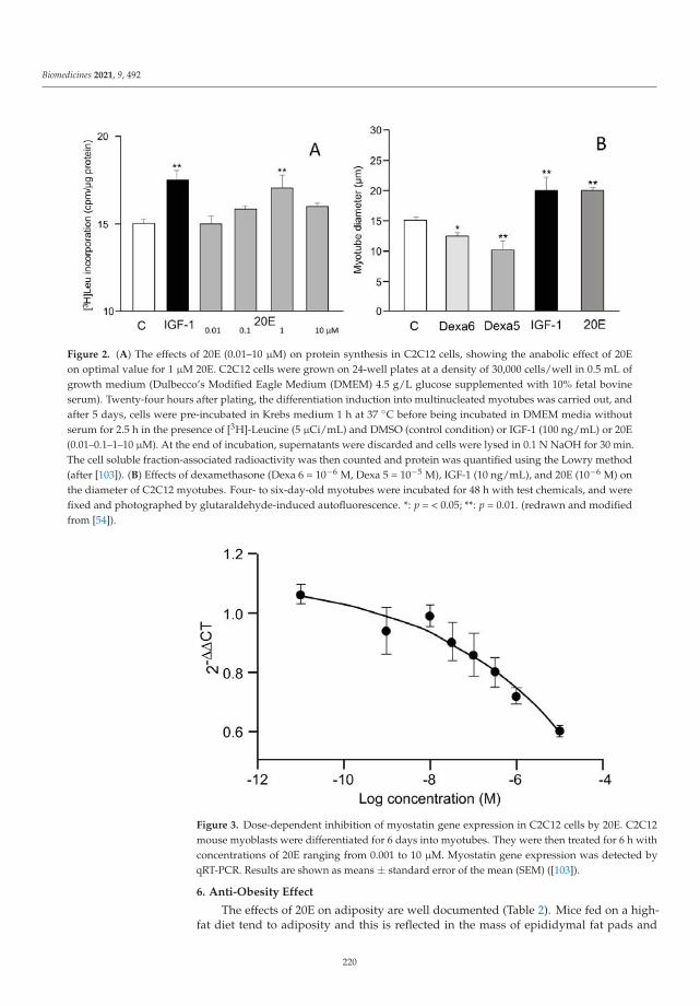
Biomedicines 2021, 9, 492
Figure 2. (A) The effects of 20E (0.01–10 μM) on protein synthesis in C2C12 cells, showing the anabolic effect of 20Eon optimal value for 1 μM 20E. C2C12 cells were grown on 24-well plates at a density of 30,000 cells/well in 0.5 mL ofgrowth medium (Dulbecco’s Modified Eagle Medium (DMEM) 4.5 g/L glucose supplemented with 10% fetal bovineserum). Twenty-four hours after plating, the differentiation induction into multinucleated myotubes was carried out, andafter 5 days, cells were pre-incubated in Krebs medium 1 h at 37 ◦C before being incubated in DMEM media withoutserum for 2.5 h in the presence of [3H]-Leucine (5 μCi/mL) and DMSO (control condition) or IGF-1 (100 ng/mL) or 20E(0.01–0.1–1–10 μM). At the end of incubation, supernatants were discarded and cells were lysed in 0.1 N NaOH for 30 min.The cell soluble fraction-associated radioactivity was then counted and protein was quantified using the Lowry method(after [103]). (B) Effects of dexamethasone (Dexa 6 = 10−6 M, Dexa 5 = 10−5 M), IGF-1 (10 ng/mL), and 20E (10−6 M) onthe diameter of C2C12 myotubes. Four- to six-day-old myotubes were incubated for 48 h with test chemicals, and werefixed and photographed by glutaraldehyde-induced autofluorescence. *: p = < 0.05; **: p = 0.01. (redrawn and modifiedfrom [54]).
Figure 3. Dose-dependent inhibition of myostatin gene expression in C2C12 cells by 20E. C2C12mouse myoblasts were differentiated for 6 days into myotubes. They were then treated for 6 h withconcentrations of 20E ranging from 0.001 to 10 μM. Myostatin gene expression was detected byqRT-PCR. Results are shown as means ± standard error of the mean (SEM) ([103]).
6. Anti-Obesity Effect
The effects of 20E on adiposity are well documented (Table 2). Mice fed on a high-fat diet tend to adiposity and this is reflected in the mass of epididymal fat pads and
220

Biomedicines 2021, 9, 492
the diameter of the adipocytes. Figure 4 compares these parameters in mice on low-fat,high-fat and high-fat +20E diets. 20E counteracts both the increase in the mass of the fatpads and retains the median adipocyte diameter close to that in mice fed a low-fat diet.Similar findings have been reported by other authors, e.g., [49]. 20E is also efficient inhigh-carbohydrate (high fructose) fed gerbils [61].
Figure 4. Effect of 20E on mice fed a high-fat diet (HF), when compared to mice fed a low-fat diet (LF). The animals receivedeither pure 20E (50 mg/kg/day) or the same amount of 20E as a quinoa extract (Q). Panel (A) shows the impact on the massof epididymal adipose tissue (*** p < 0.01 when compared to LF; ## p < 0.01 and ### p < 0.001 when compared to HF) andpanel (B) shows the effect on adipocyte diameter (reproduced, with permission, from [59]).
7. Mechanism of Action of 20E in Mammals
Early studies showed that 20E displays pleiotropic effects on mammals [104]:
• Increases protein synthesis in skeletal muscles and heart [46,105]• Increases ATP synthesis in muscles [106]• Stimulates production of erythrocytes [74]• Decreases hyperglycemia in diabetic animals [63]• Reduces plasma cholesterol levels [107]• Decreases the activity of triglyceride lipase [108]• Protects against experimental atherosclerosis in rabbits [109]• Activates acetylcholinesterase in the brain [110]• Activates glutamate decarboxylase (=GABA synthesis) in the brain [111]• Possesses immunomodulatory activity [112].
In view of these diverse effects, it could be expected that 20E might have variousmodes of action at the cellular level. There are no homologues to the arthropod ecdysteroidreceptor (EcR) in mammals, and attempts to show high affinity binding of ecdysteroidsto vertebrate nuclear receptors have not been successful [88,113], but see the possibleinvolvement of estrogen receptor-β (ERβ) below. The mode(s) of action of 20E in mammalshas not been fully elucidated for any responsive cell type. In part, this is because the actionappears to be non-genomic and also because relatively high concentrations (0.1–10 μM) of20E are required to observe effects, implying relatively low affinity interaction of 20E withits cellular target(s).
Several models for the modes of action of 20E at responding mammalian cells arebeing pursued. The models are not mutually exclusive, and it is possible that they could
221

Biomedicines 2021, 9, 492
operate simultaneously or in different tissues. Given the pleiotropic effects describedfor 20E on mammalian cells, it seems possible that it operates through several signallingpathways, which could be more or less tissue-specific. Gorelick-Feldman et al. [50] clearlyestablished that 20E acts through a membrane GPCR receptor, and this has been confirmedusing albumin-bound 20E, which cannot cross cell membranes (Figure 5).
Figure 5. 20E reduction of myostatin gene expression in C2C12 cells (differentiated for 6 daysinto myotubules) is mediated by binding to receptor sites on the external surface of the cells. Thehistogram compares the activities of IGF-1 (100 nM) and 20E (10 μM) with those of conjugates of 20Ecovalently bound to protein (HSA or BSA) through different C-atoms (C-2 and C-22-hemisuccinatesor C-6 [6-carboxymethoxime]), all at nominal 10 μM hapten concentration. BSA bovine serumalbumin; HSA: human serum albumin; error bars = standard error of the mean [103,114].
The renin–angiotensin system (RAS) is strongly implicated in maintaining musclefunction, and one of the peptide products of this system, angiotensin II, targets skeletalmuscle cells via the AT1R receptor and has been implicated in the development of sar-copenia [115], both directly through AT1R, which results in increased resistance to insulinand IGF-1, and indirectly by increased production of myostatin, glucocorticoids, TNF-α,and IL-6. Consequently, ACE inhibitors (which inhibit the production of angiotensin II)have found some application in the treatment of sarcopenia [116]. Another componentof the RAS is angiotensin 1-7, which has been identified as the natural ligand for anotherGPCR, Mas1, the activation of which has been hypothesized to enhance protein synthesisin muscle cells. Thus, RAS would have a “harmful arm” acting through AT1R where theactivation of the receptor enhances proteolysis, but this can be counteracted by a “pro-tective arm”, acting through Mas1, where the activation of the receptor enhances proteinsynthesis. According to this hypothesis, protection against muscle wasting can be achievedby reducing the activation of AT1R, or the activation of Mas1, or a combination of thetwo. There are reasons to believe that the activation of Mas1 would be more effective atstimulating muscle anabolism and reducing adipose tissue than an ATIR antagonist, andit would have fewer side-effects than ACE inhibitors. The Mas1 receptor is expressed inmany tissues, and its activation in different tissues (e.g., heart, kidney, CNS) may evokevarious protective effects [117]. The activation of Mas1 by 20E could explain the anaboliceffects of 20E on muscle cells [103,113,114].
Angiotensin(1-7) partially inhibits myostatin gene expression and this inhibitionis abolished by the angiotensin(1-7) antagonists D-Pro7-Ang(1-7) and D-Ala7-Ang(1-7)
222

Biomedicines 2021, 9, 492
(A779). These antagonists are also effective at preventing the inhibition brought aboutby 20E, indicating that Ang(1-7) and 20E operate through Mas1 activation [113]. Thegene interference of the Mas1 receptor using silencing RNA (siRNAMas1) provided furtherevidence for the involvement of Mas1 in the mode of action of 20E: siRNAMas1 was found toreverse the inhibition in myostatin gene expression brought about by Ang(1-7) or 20E [113].
Parr et al. [54] have put forward an alternative model for the mode of action of 20E inbringing about the hypertrophy of mammalian skeletal muscle cells, which involves theinteraction of the steroid with nuclear estrogen receptor-β (ERβ). Using C2C12 cells, theyobserved that both 20E (1 μM) and E2 (1 nM) enhanced myotube diameter. Co-treatmentwith the anti-estrogen ZK191703 (ZK) antagonized the hypertrophy brought about not onlyby E2, but also by 20E, indicating that E2 and 20E share a common mode of action. Theauthors next showed that a reporter gene under the control of an estrogen response elementcould be activated dose dependently by E2 or 20E interacting with ERα or Erβ, and thatthis activation could be prevented if ZK was also present. Use of the estrogen receptor-specific agonists, ALPHA (for ERα) and BETA (for ERβ), indicated that ERβ mediatedthe hypertrophy of the C2C12 myotubes, but a note of caution is required because theseagonists are only selective at low concentrations and the BETA dose–response curve wasbell-shaped. Finally, the selective ERβ-antagonist (ANTIBETA) antagonized the effectsof E2 and 20E in the C2C12 cells. The authors suggest that, as it had previously beenshown that ERβ can modulate Akt phosphorylation, this could be the link by which 20Ebrings about hypertrophy in the C2C12 cells. In silico docking studies suggest that 20Ecan interact with the ligand-binding domains of ERα and ERβ but has the potential forstronger interaction with ERβ [118]. However, the exact nature of the receptor involved inthe observed effects can be questioned, because all binding studies performed with nuclearreceptors ERα and ERβ were negative [e.g., 38], and truncated membrane-bound formsof ER receptors have been described that may display different ligand specificity [119].Finally, the recent article by Sobrino et al. [120] provides evidence that in primary humanumbilical vein endothelial cells cultures, E2 acts via a membrane-bound ER to bring aboutNO-dependent vasodilation, and they show that this effect also requires Mas activationas it is abolished by a Mas inhibitor. Although this is a different system to the musclecells above, it might perhaps give an indication of how both 20E and E2 can bring aboutmuscle hypertrophy.
Further experiments with C2C12 cells [113] indicate that the two hypotheses for themode of action of 20E on these cells might be unified. Estradiol (E2) was found to inhibitmyostatin gene expression in a dose-dependent manner (0.1–10 μM). To determine if theaction of E2 is dependent on interaction with a membrane receptor, the activity of E2 wascompared with that of E2-CMO (estradiol 6-carboxymethyloxime) and E2-CMO-BSA (E2-CMO covalently coupled to BSA). E2-CMO was found to inhibit myostatin gene expressionin C2C12 cells like E2, showing that the carboxymethyl oxime derivative retained biologicalactivity. In contrast, the E2-CMO-BSA conjugate was inactive, so it appears that E2 mustenter the cell to be active. In addition to the classical nuclear receptors (ERα and ERβ),several non-nuclear forms of ER are known, and it is presumably one of these whichis involved in the regulation of myostatin gene expression in C2C12 cells. Additionally,17-epi-estradiol (17αE2), a close analogue of E2 which does not bind the nuclear receptorsERα and ERβ, was found to inhibit myostatin gene expression in C2C12 cells, providingfurther evidence for the involvement of a non-nuclear ER [113]. By combining the availableexperimental data, it has been possible to put together a hypothesis for how both 20E andE2 can regulate myostatin gene expression, whereby 20E interacts externally to the cellwith the integral membrane Mas1 receptor, which in turn interacts with ERx located on theinner side of the plasma membrane, while E2 enters the cell to interact with ERx, bringingabout myostatin regulation in a more direct way (Figure 6).
This finding may have important consequences because the Mas receptor is expressedin many tissues, which fits with the large array of physiological effects of its endogenousligand Ang(1-7), which are also observed for 20E [113].
223

Biomedicines 2021, 9, 492
Figure 6. Diagrammatic representation of the proposed mode of action of 20E in the regulation of protein synthesis inC2C12 muscle cells in vitro ([113]).
8. Toxicity, Bioavailability, Pharmacokinetics and Metabolism in Animal Models
Toxicity
Ogawa et al. [121] determined LD50s for ingested 20E or inokosterone in mice of>9 g/kg and LD50s of 6.4 g/kg and 7.8 g/kg for i.p.-injected 20E and inokosterone, respec-tively. Thus, ecdysteroids are regarded as non-toxic to mammals. Similarly, no subacutetoxicity was observed in long-term feeding experiments (0.2–2 g/kg/day) in rats. Addi-tionally, no effects were seen after the administration of these two ecdysteroids to bullfrogs(up to 600 mg/kg in the lymph sinus) or rabbits (up to 100 mg/kg). The two ecdysteroidsdid not have sex hormonal activity or, interestingly in the context of more recent studies,anti-inflammatory or anabolic effects in rats. Additionally, rats and birds can feed on seedsof Leuzea carthamoides, which contain 2% (w/w) ecdysteroids (mainly 20E), and thrive [7].
Additionally, Seidlova-Wuttke et al. [57,94] fed ovariectomized rats with up to500 mg/kg daily for 3 months with no stated detrimental effects, but no data on survivalare presented in the articles. More recently, a purified to pharmaceutical grade 20E (≥97%20E purity) was used as a drug candidate in preclinical regulatory studies including safetypharmacology, genotoxicity and repeated toxicology in rodents (rats) and non-rodents(dog). This drug candidate depicted a good safety profile with no genotoxic effects andNo Observed Adverse Effect Levels (NOAEL) established at 1000 mg/kg in repeated dosetoxicity studies in rats (26-week) and dogs (39-week) (Biophytis, unpublished data).
9. Pharmacokinetics and Metabolism in Mice—Early Studies
The earliest study on the fate of radiolabelled 20E in a mammalian species was per-formed on mice [122] and showed after oral administration that the bioavailability waslow and that elimination was essentially fecal, while after i.p. administration, a somewhat
224

Biomedicines 2021, 9, 492
higher level was found in the urine, even though fecal excretion still predominated. Of theradioactivity crossing the intestinal tract after oral administration (<2% of that adminis-tered), most was associated with the liver and bile duct, providing the first evidence for anentero-hepatic cycle for ingested ecdysteroids.
Lafont et al. [123] studied the metabolism of ecdysone, the only commercially availableradioactive molecule when the study was made, in mice after intraperitoneal injectionsof the radioactive molecule. Ecdysone levels increased transiently in the liver and thenaccumulated in the intestine, which, within one or two hours after injection, containedalmost all the radioactivity. One day after injection, most of the radioactivity had beeneliminated from the body. Moreover, the metabolism of 20E was faster after intraperitonealinjection than after ingestion [122]. As with ecdysone, and whatever the mode of adminis-tration, radioactivity was rapidly taken up by the liver and then excreted into the intestinevia the bile [122]. Excretion was again primarily fecal in mice [123]. The distribution ofradioactivity from 3H-labelled 20E after injection into the caudal vein of mice has beenstudied by Wu et al. [124]. The kinetics of distribution showed a rapid elimination in urineand an uptake by the liver and bile, then most radioactivity accumulated into the intestinelumen before being eliminated in feces. Unfortunately, no information was provided aboutthe associated metabolism [124].
Studies on the metabolism radiolabelled ecdysone in i.p.-injected mice [123,125]showed that the radioactivity was excreted fully within 24h unless it was diluted withunlabelled ecdysone, when the excretion of radioactivity was extended over 3 days. Theinjected radioactivity was rapidly captured by the liver and transferred to the intestine,where essentially all of it was located within 45–60 min. The principal metabolites formedwere single or multiple products of 14-dehydroxylation, reduction of the 6-oxo group (6α-OH), reduction of the 7,8-double bond and 3-epimerisation. It is significant that ecdysonedoes not undergo side-chain cleavage because it does not possess a 20,22-diol and cannotbe readily hydroxylated at C-20 after injection or ingestion in mice.
10. Pharmacokinetics after Oral Administration in Mice—More Recent Studies
The metabolism of radiolabelled 20E has been more extensively studied [126,127].These studies involved 20E radiolabelled in the nucleus, so as to be able to follow side-chain cleavage. It was found that the fate of ingested 20E was initially simple, with mostof the 20E remaining unaltered as it passed through the esophagus, stomach and smallintestine, with only a small proportion crossing into the blood and being captured by theliver (Figure 7).
After ca. 30 min, 20E starts to reach the large intestine, where it begins to be signif-icantly metabolized primarily to 14d20E. Further, the 20E and 14d20E start to undergocomplex metabolism including side-chain cleavage. Absorbed 20E and its metabolites aretaken by up the liver, enter the bile duct and are brought back to the intestine. Entero-hepatic cycling then occurs, during which 20E and the metabolites leave the gut, enterthe blood, are taken up by the liver and returned through the bile duct to the intestine.This result is fully consistent with the observations of Wu et al. [124] of a sustained highradioactivity in bile for up to 12 h (Table 3). As a consequence, the duration of 20E presencein the plasma is prolonged at a low, but physiologically significant, concentration (in theμM range—depending on the dose administered), and the extent and complexity of themetabolism of 20E increase markedly, such that the excreted feces contain unaltered 20E anda wide range of metabolites, produced by permutations of several biochemical reactions.
225

Biomedicines 2021, 9, 492
Figure 7. Time-course of the distribution of radioactivity in the stomach/intestine/feces in mice after the oral applicationof [1α,2α-3H2]20E. Note the logarithmic scale for abscissa. Each value is a mean of 2 animals. Note the plateau of smallintestine content that is best explained by an entero-hepatic cycle and consistent with the prolonged concentration ofradioactivity in bile as observed by Wu et al. [124] (reproduced, with permission, from [127]).
Table 3. The effect of 3H-Achyranthes bidentata ecdysterone on the quantitative distribution of differenttissues in different tracking phases of mice (mean, n = 5) expressed in μg g−1 (translated fromWu et al. [124]).
5 min 10 min 30 min 1 h 3 h 6 h 12 h 24 h
Blood plasma 0.061 0.057 0.052 0.047 0.032 0.023 0.015 0.011
Urine 0.482 0.921 1.534 0.281 0.102 0.096 0.087 0.046
Feces 0.015 0.019 0.035 0.068 0.099 0.931 0.312 0.041
Bile 0.421 0.456 1.042 0.901 0.301 0.209 0.198 0.094
Liver 0.312 0.251 0.213 0.112 0.078 0.061 0.056 0.046
Heart 0.062 0.052 0.035 0.033 0.031 0.029 0.023 0.022
Spleen 0.041 0.026 0.024 0.018 0.023 0.027 0.032 0.027
Lungs 0.116 0.069 0.057 0.053 0.049 0.045 0.036 0.028
Kidney 0.137 0.123 0.098 0.071 0.055 0.043 0.037 0.032
Adrenals 0.184 0.139 0.098 0.081 0.073 0.065 0.052 0.043
Testis 0.048 0.036 0.029 0.026 0.023 0.021 0.019 0.017
Skeletal muscle 0.021 0.023 0.028 0.017 0.015 0.013 0.011 0.010
Spinal cord 0.019 0.028 0.067 0.041 0.033 0.024 0.020 0.018
Brain 0.013 0.014 0.015 0.013 0.012 0.01 0.008 0.007
Small intestine 0.254 0.139 0.094 0.083 0.076 0.061 0.05 0.033
Stomach 0.034 0.047 0.075 0.061 0.053 0.041 0.032 0.023
Large intestine 0.011 0.023 0.029 0.031 0.035 0.048 0.038 0.023
Bladder 0.013 0.024 0.038 0.026 0.023 0.018 0.015 0.013
The bioavailabilities of 20E (ca. 1%) and Post (ca. 19%) have been determined [127,128].As in mice, the initial fate of ingested 20E is uncomplicated, remaining almost completelyassociated with the gut in unchanged form, until it reaches the large intestine, where
226

Biomedicines 2021, 9, 492
microbial metabolism and metabolism associated with the enterohepatic cycle come intoplay to rapidly increase the extent and diversity of metabolites. Evidence has been providedthat glucuronide conjugates of 20E are present in the bile fluid, but these are hydrolysed onreaching the large intestine. The major metabolites have been conclusively identified as14d20E, Post, 14dPost, 3-epi-Post, 16αOHPost, and 21OHPost [128], but other permutationsof these biochemical reactions almost certainly exist. Additionally, other metabolites wherethe 6-one-7-ene chromatophore has been modified, so that the products are no longerUV-absorbing, are likely to be present amongst the complex mixture of metabolites presentin the large intestine and excreted in the feces. The elimination of ingested or injected20E is predominantly fecal (Figure 8A,B). The microbial origin of the 14d20E metaboliteis demonstrated by the absence of this metabolite in axenic rats fed 20E [129] and by itsformation from 20E in incubations of intestinal contents under anaerobic conditions [127].That 14-dehydroxylation is associated with the microbiote of the large intestine is inaccord with previous findings that dehydroxylations of steroids and bile acids are onlybacterial [130,131]. The locations of the other metabolic reactions are not currently knownwith certainty, but it is expected that they will be associated with specific enzymes ofvertebrate steroid catabolism.
Figure 8. Comparison of the temporal (A) and cumulative (B) urinary and fecal elimination of radioactivity after the oralapplication of [5,7,9-3H]20E to 6–7 week-old male Wistar rats. After oral application of [3H]20E, elimination of radioactivelylabelled 20E and its metabolites is completed within 48 h. The amount of radioactivity recovered from the feces is by farthe major route of excretion, since the amount in the urine corresponds to only 1.40% of the total radioactivity recovered.(Reproduced, with permission, from [127]).
11. Pharmacokinetics and Metabolism in Humans
11.1. Early Studies
Although ecdysteroids are not biosynthesized in mammals, sensitive methods candetect them in mammalian body fluids (plasma, urine). Thus, ecdysteroid-specific radioim-munoassay (RIA) was used to detect immunoreactive ecdysteroids in the serum of sevenmammals (dog, rhesus monkey, sheep, cow, rabbit, mouse and rat) and humans in nMconcentrations [132], which originate from the food. Ingested ecdysteroids (20E or E) reacha plasma titre maximum after ca. 30 min in humans and sheep and are rapidly cleared fromthe blood and urine, but the proportions of the applied dose found in these are very low,accounting only for a few percent. The effective half-time of elimination from the bloodin humans was found to be 4 h for E and 9 h for 20E, and 3.1 h for E and 3.3 h for 20E forelimination from the urine [132].
227

Biomedicines 2021, 9, 492
Pharmacokinetic studies in humans are more restricted owing to the limitationson the use of radiolabelled molecules. Consequently, only a few 20E pharmacokineticstudies have been performed. In humans, Simon and Koolman [132] showed that ingestionof ca. 15 mg 20E induced a urinary peak level of approximately 0.5 μM. Significantimmunoreactivity was detected during the first 8 h following intake, but weak urinarylevels were still observed until 24 h after ingestion [133]. After a single oral intake of20 mg 20E, a more detailed study by RIA and liquid chromatography-mass spectrometry(LC-MS) showed the existence in urine of a peak of detectable ecdysteroids after 3–4 h anda total quantity of 1–2 mg—the exact nature of the compounds was not fully determined,although one could be tentatively identified as 14d20E. In another study [134], the ingestionof 434 mg 20E resulted in the urinary excretion of a total of 5 mg of 20E, corresponding toapproximately 1% of the ingested amount. This study also indicated that urinary excretionseems somewhat more significant in humans than in rodents [135], even though the fecalcontent was not quantified and small amounts were recovered from urine as compared tothe administered dose.
11.2. Recent Studies
Biophytis has recently undertaken the first in-human, randomized, double-blind,single-centre study to determine the safety of 20E and its major metabolites in healthyyoung (18–55 years) and elderly (>65 years) adult subjects [136,137]. The study comprisedtwo parts: a single ascending dose component (SAD; 16 subjects) and a multiple ascendingdose component (MAD; 30 subjects). 20E showed a good safety and pharmacokineticprofile, which allowed the doses for a Phase 2 Sarcopenia clinical trial to be defined.Pharmacokinetics in subjects treated with increasing amounts of orally delivered 20E(Table 4) showed that the level found in the plasma is dose-related and also apparentlysaturable. The kinetics of plasma 20E was similar between young and older subjects(Figure 9).
Figure 9. Pharmacokinetics of 20E in young and elderly humans given a single dose of 1400 mg. 20Ewas quantified by HPLC-MS [136,137].
About 2% of the administered 20E was excreted in the urine within 12 h of ingestionof a single dose, and half of this was excreted within 4 h. 20E and its metabolites wereidentified and quantified in the plasma of individual patients who had received oral 20E.These metabolites corresponded to the major ones previously found in rodents (14dPost,14d20E and Post).
228

Biomedicines 2021, 9, 492
Table 4. Kinetic parameters of BIO101 (=97% pure 20E) after a single oral administration (n = 6,fasted). Values in parentheses indicate the SEM (Cmax, AUC) or the range (tmax).
B1O101 PKParameter (Unit)
100 mg 350 mg 700 mg 1400 mg
Cmax (ng/mL) 141 (16.6) 317 (37.9) 399 (24.7) 710 (20.2)
tmax (h) 2.03 (1.00–3.02) 3.00 (1. 05–4.00) 3.00 (2.00–4.02) 3.50 (2.00–4.02)
AUC0-t(ng·h/mL) 767 (31.1) 1924 (40.1) 2578 (22.9) 4148 (15.9)
12. The Major Routes of 20E Metabolism in Rodents and Humans
Figure 10 summarizes the metabolic reactions currently known to occur for 20Ein mammals to produce a complex array of metabolites arising from combinations ofthe metabolic possibilities, although the flux through the network of combinations willdepend on the exposure to and the activity of the different enzymes depending on themammalian species. On ingestion, bioavailability is very low, as is initial metabolism, and itis only on reaching the large intestine that 14-dehydroxylation occurs, but this is a reactionperformed by the gut flora. Only ecdysteroids with a 20,22-diol (such as 20E) undergoside-chain cleavage, as demonstrated by the absence of this reaction with oral or injectedE. Side-chain cleavage and 14-dehydroxylation significantly enhance bioavailability (seebelow) and an enterohepatic cycle maintains ecdysteroid plasma levels and enables moreextensive metabolism (e.g., hydroxylations, reductions, epimerizations—the two latteralready observed with ecdysone [125]).
Figure 10. The principal routes of metabolism of 20E in rodents and humans (modified from Kumpun et al., 2011). Thecircles and arrows in red highlight the changes associated with 14-dehydroxylation, while the boxes and arrows in bluehighlight the changes associated with side-chain cleavage.
229

Biomedicines 2021, 9, 492
13. Large-Scale Production of Pure 20E for Drug Development
General Considerations
Various theoretical possibilities for ensuring the supply of 20E in adequate amountsand at reasonable cost have to be considered:
1. Total chemical synthesis2. Isolation from the current plant source, e.g., Cyanotis sp. following cultivation and
collection in China (Yunnan province)3. Cultivation of another species already identified as a good accumulator with a simple
ecdysteroid profile (Rhaponticum, Serratula or Pfaffia sp.)4. Use plant cell or hairy-root cultures of an ecdysteroid-producing plant5. Generate recombinant yeasts using insect and/or plant enzymes of ecdysteroid
biosynthesis.
Of these, possibilities 1 and 4 can be discounted; 1 because of the complexity (≥18 steps)and poor overall yield (ca. 1%) of the process, and the rate-limiting access to a suitablestarting material, and 4 probably because of the high cost of the media and low yieldsnormally attained for plant secondary compounds, even when using elicitors [138]. Variousresearchers have started to explore a range of culture methods in vitro (e.g., micropropaga-tion, callus cultures, plant cell culture, hairy-root cultures, transformed yeast fermentation)in the hope of obtaining more amenable ecdysteroid-producing systems. To date, onlyhairy roots of Ajuga reptans var. atropurpurea (Fujimoto et al. [139]) consistently and reliablyproduce ecdysteroids, but the culture is too expensive to provide a commercial source of20E. Additionally, hairy roots of some other ecdysteroid-producing species do not containecdysteroids, so this cannot be viewed as a general approach.
Possibility 5 will for sure face a number of difficulties, but, once established, itwould provide relatively easy scale-up. The use of modified yeasts to produce bioac-tive molecules is rapidly developing [140] and proved efficient even for multiple-stepbiosyntheses (e.g., [141]). Transforming yeast to biosynthesize ecdysteroids, as has beenpreviously performed for certain vertebrate steroids [142,143], is an attractive prospect,but is currently confounded by our incomplete knowledge of the ecdysteroid biosyntheticpathways in either invertebrates or plants and the probable high number of genes involvedin the complete pathway(s) [3].
Possibility 2 may make the supply dependent on a single country and on the difficultyto secure access to the material in case of competition for a limited supply. Possibility 3might displace the problem to another country and requires the development of a large-scale culture.
At the moment, the molecule is extracted from suitable plant materials. It is perhapsworth reminding ourselves of the criteria that an ideal plant source should fulfil:
• The plant should accumulate a high amount of 20E• The plant should have a simple ecdysteroid profile (ideally just 20E)• The plant should be easy and rapid to grow in accessible areas of the world• The plant matrix should be amenable to the ready purification of ecdysteroids• The purification and isolation of 20E should not involve expensive chromatographic methods• The plant should not be susceptible to pests and diseases• The species should not be rare or protected• Culture, harvesting and processing costs should be low; initial processing should take
place close to the culture site
Clearly, no plant species will fulfil all these criteria, but the closer it comes, themore culturally and comercially viable the challenge of isolating adequate amonts of 20Ewill become. Major screening of plants for the presence of ecdysteroids began by usingvery time-consuming, semi-quantitative whole insect bioassays (Chilo dipping test; Muscaassay; Sarcophaga assay), moved on to the combined use of ecdysteroid-specific RIAs and amicroplate assay based on an ecdysteroid-responsive cell line, and most recently progressedto analysis by HPLC/DAD/MS-MS, which allows the simultaneous identification and
230

Biomedicines 2021, 9, 492
quantification of common phytoecdysteroids and dereplication to detect the presence ofpreviously unknown ecdysteroids [144].
Most ecdysteroid-accumulating species contain between 0.01 and 0.1% of the dryweight as ecdysteroids, whereas the few identified high-accumulators contain 1% andabove. The roots of Cyanotis spp. can contain up to 5% of the dry weight as ecdysteroids,largely as 20E [145]. Mature stems of Diploclisia glaucescens were found to contain 3.2% oftheir dry weight as 20E [146], but the collection of the stems of this South Asian climberprecludes this as a feasible source of large amounts of 20E.
14. Plants Species Presently Used to Produce Purified 20E
The relationship between the presence of phytoecdysteroids and plant taxonomy iscomplex, but high accumulation is associated with certain species in the genera Achyranthes(Amaranthacea), Cyanotis (Commelinaceae), Pfaffia (Amaranthaceae), Rhaponticum (syn.Leuzea/Stemmacantha; Asteraceae), and Serratula (Asteraceae), which can be considered asgood sources of 20E.
Achyranthes (A. aspera, A. bidentata, A. fauriei, A. japonica): Perennial or HHA; warmtemperate and sub-tropical plants; native to South-east Asia and/or Africa; all plant partscontain ecdysteroids with seeds containing the highest level (0.25%) in A. aspera and roots(1.74%) being the best source in A. bidentata;Cyanotis (C. arachnoidea, C. vaga): Perennial; tropical and sub-tropical plants; growing intropical Africa, the Indian subcontinent and southern China; roots accumulate very highlevels of 20E (up to 5.5% of the dw; Wang et al. [145]); simple ecdysteroid profile—majorsource for commercial 20E cultivated on a large scale in China;Pfaffia (P. glomerata, P. iresinoides): Perennial; tropical plants; native to South America;P. glomerata contains ecdysteroids throughout the plant; roots have a very simple ecdys-teroid profile, consisting solely of 20E (0.9% of the dw);Rhaponticum (R. carthamoides): Perennial; sub-alpine plant; native to the Altai and SayanMountains in Central Asia; roots, flowers and seeds accumulate high levels of 20E (1–2%of dw); complex ecdysteroid profile, but >80% as 20E—used to prepare ECDYSTEN pillscontaining 5 mg of 20E (OPIH, Uzbekistan) [147];Serratula (S. centauroides, S. coronata, S. tinctoria, S. wolfii): Perennial; temperate plants; na-tive to western and central Europe; S. tinctoria accumulates up to 2% of dw as ecdysteroids;the major ecdysteroids are 20E 3-Acetate, 20E and PolB; S. coronata is used to prepareSERPISTEN, a 8:1 mixture of 20E and 25S-inokosterone [148].
We should mention that only 2% (ca. 6000) of terrestrial plant species have beenassessed for the presence of phytoecdysteroids and far fewer than this have undergoneextensive phytochemical analysis to identify the ecdysteroid analogues present [144]. Thus,there are certainly many other as yet unidentified species which are high accumlators of20E, but it has to be borne in mind that the species already tested represent the most readilyavailable species and accessing samples of new untested, but validated, species wouldrequire the investment of a large amount of time and effort.
Owing to the wide taxonomic diversity of plant species known to accumulate phy-toecdysteroids, it has been proposed that most, if not all, plants have the genetic capacityto produce ecdysteroids, but in the majority (the non-producers) the expression of thebiosynthetic pathway is down-regulated [2]. Additionally, it has been shown in severalecdysteroid-accumulating species that ecdysteroid levels are additionally influenced by en-vironmental factors (temperature, nutritional factors, invertebrate predation, etc.) [149,150].Thus, to maximize 20E content in a chosen species, it is not only necessary to select ahigh producing genetic line (cultivar), but also to optimize the growth conditions and toconsider treating the plants with appropriate elicitors (e.g., methyl jasmonate to mimicinsect or fungal attack) at suitable times in the development.
231

Biomedicines 2021, 9, 492
15. Purification Process
The major requirements in the processing of plant material to obtain a drug productin adequate amounts for therapeutic use are simplicity, efficiency of extraction and purifica-tion, reproducibility and cost-effectiveness. As explained above, the choice of plant materialis key, since not only should it contain a large amount of the target molecule, but the plantmatrix should be readily extractable and not contain components (e.g., large amounts ofpolysaccharides) which make processing difficult. Given that a suitable plant source willcontain typically 1–2% of the dry weight as 20E, and that a pharmaceutical/medicinal doseof 20E is likely to be in the 100 mg–1 g/day range, it is necessary to be able to processtonnes of plant material. Clearly, it would be highly advantageous if the harvested plantmaterial can be cleaned, dried, broken up (to reduce volume and increase the surface area)and subjected to initial extraction as close to the site of harvesting as possible, as the massof the initial extract is probably only 1–2% of the fresh weight of the plant material and,therefore, much more readily and cost-effectively transportable.
The extraction and purification have to be optimized with regard to the physico-chemical properties of the target molecule (generally already known or readily deter-minable) and the nature of the plant matrix (generally only vaguely understood). Owingto the large number of hydroxyl groups in 20E, it is highly soluble in alcohols, so thedried, powdered plant material will usually be extracted with methanol (preferred as it ischeaper) or ethanol (if the product is to be BIO/Organic), with or without prior extractionwith a non-polar solvent, such as petroleum ether, to de-fat the plant material. Extractionmay occur with heating, stirring or maceration for a defined time, all of which need to beoptimized. Other methods, such as super critical fluid extraction or bi-phasic extraction, maybe used, but these are generally more expensive and would only be cost-effective if the targetmolecule is potent (low daily dose required) and high-value, which is not the case for 20E.
Figure 11 provides a flow-diagram of a representative processing method for Cyanotissp. roots, where the dried plant material is refluxed thrice with ethanol and the pooledextracts are filtered before being passed through a macroporous resin to absorb plantcompounds and vacuum-dried to yield a powder containing 90% 20E. This preparationcan then be recrystallized twice to bring the purity of the 20E to >97%.
Figure 11. A representative flow-diagram for the large-scale extraction and purification of 20E from roots of Cyanotis sp.
232
Biomedicines 2021, 9, 492
A comparison of the reversed-phase (RP)-HPLC profiles of the 90% and 98% 20Epreparations is shown in the inset to Figure 11. Most of the minor peaks in the chro-matograms correspond to other ecdysteroids, which are very difficult to separate fullyfrom 20E by crystallization. This underlines the need to start with plant material whichcontains essentially only 20E. Whenever the plant contains a high proportion of a veryclose molecule (e.g., inokosterone or polypodine B), it would be very difficult to achievethe required purity.
Owing to the need for optimization at each stage of the extraction and purification, theprocess is developed in stages going from small-scale (100 g—a few kg dry plant material)in the laboratory through increasing medium-scale stages (e.g., 10–500 kg) before beingapplied at the industrial scale (>1 tonne), so that difficulties can be identified and resolvedearly on and any problems of scale-up can be dealt with. A thorough cost analysis needs tobe performed throughout the scale-up procedure to ensure that the target molecule can bebrought to market at a viable cost.
16. Quality Control and Stability
Using 20E as the active ingredient of a pharmaceutical grade drug candidate requiresthe complete control of the raw material (roots, aerial parts or whole plant harvestedfrom the field) following the GAP (Good Agricultural Practices; http://www.cfsan.fda.gov/~dms/prodguid.html, accessed on 1 April 2021) or the guidelines on the quality ofherbal medicinal products/traditional medicinal products. This involves the descriptionof the active substance preparation including geographical origin, cultivation, harvesting,and postharvest treatments (possible pesticides and fumigants used and/or formation ofgenotoxic impurities, solvents, presence of aflatoxins, traces of heavy metals and possibleradioactive contamination).
The pharmaceutical grade compound (purity ≥ 97%) must be fully characterized,and all its impurities ≥ 0.1% must be identified, with none of them being above 0.5%,otherwise it would require specific toxicological studies. In the present case, given theprocess used and the fact that ecdysteroid-containing plants contain a complex cocktail ofthese molecules (e.g., [26,151]), most remaining impurities correspond to other phytoecdys-teroids, the nature of which will depend on the plant used.
Compound stability studies also have to be performed, which does not raise problemsin this case, as ecdysteroids are very stable molecules when stored in dry state in the darkat room temperature.
17. Regulatory Preclinical Studies
Preclinal Regulatory Requirements
Nutritional studies can be allowed with limited prerequisite regulatory elements,provided that 20E comes from an edible plant (e.g., spinach, quinoa or Leuzea—a plant onthe Belgian list) and the amounts administered are not too high as compared with thosepresent in a diet comprising the above plants (e.g., 50 g quinoa contains 20–30 mg 20E). Suchstudies were performed in relation to anabolic or anti-obesity effects on healthy subjects.
On the other hand, clinical studies aimed at treating human diseases with possiblyhigher doses and for extended periods require a lot of information about the moleculeadministered and its long-term effects on model animals, typically one rodent and onenon-rodent species (Table 5).
Safety pharmacology studies must thus be conducted to investigate acute effects on thecentral nervous system, respiratory function, cardiovascular system including hERG potas-sium channel on blood pressure, heart rate, core body temperature and electro-cardiogram.
Long-term toxicity studies also have to be performed in one rodent and one non-rodentspecies, the duration of which must correspond to the expected duration of administrationduring the clinical trial.
233

Biomedicines 2021, 9, 492
Table 5. Non-clinical regulatory requirements and submission dossier for clinical trial approval.
Non-ClinicalActivities
StudiesClinical Trial
Non Medicinal Drug Trial Phase 1 Phase 2
Regulatory requirements
Absorption
Pharmacokinetics NA Toxicokinetics NA
Transporters (Caco-2 cells) NA
Metabolism
Microsomal metabolism NA NA ADME $ study NA NA
CYP inhibition/induction NA Metabolite identification NA
Toxicology
Phototoxicity NA Genotoxicity tests (Ames; micronuclei) NA
Repeated toxicology studies NA Micronuclei tests NA
DistributionRed blood cell partitioning NA
Plasma protein binding NA
PharmacologyIn vitro studies In vivo studies
Safety pharmacology Cardiovascular system NA Central nervous system and respiratory function NA
Documents required for submission dossier
Investigational Medicinal Product Dossier (IMPD) NA Investigation Brochure (IB) NA Technical Product Dossier NA NA
Literature related to the product NA NA
Clinical Trial Protocol Informed consent
Investigative New Drug (IND) Package for the FDA NA NA: not-applicable; : required; $ “adsorption, distribution, metabolism and excretion”.
18. Regulatory Studies with 20E Metabolites
20E undergoes an extensive metabolism after administration, in part at least performedby the gut microbiote, and probably also in the liver, which is known to metabolizeendogenous steroid hormones. Whenever metabolites would circulate at significant levels(area under the curve (AUC) ≥ 10% that of 20E), regulatory studies would be required toassess their lack of toxicity. In addition, it is relevant to determine their biological activity,i.e., whether they have a similar/lower/higher activity to that of the parent compound,possess a different (specific?) activity, or if they just represent inactivation products.
18.1. Some Drugability Calculations
As indicated above, the bioavailability of 20E is low, while that of its metabolitesmay be significantly higher. In a first step to relate the bioavailability of ecdysteroids totheir physicochemical properties and structures, we have calculated the ADMET TrafficLight Scores for ecdysteroids according to Lobell et al. [152] based on values for MW,LogP, the polar surface area (PSA), and the number of rotatable bonds. However, rather
234

Biomedicines 2021, 9, 492
than using solubility (which has to be experimentally determined), the number of N + Oatoms (as a measure of H-acceptors) and the number of NH + OH groups (as a measure ofH-donor groups) were used as in the original method of Lipinski et al. [153]. The proceduregenerates a score (explained in the legend of Table 6) between 0 and 10 for each compound;the lower the value, the more suitable the physicochemical properties are for an orallyadministered drug.
Table 6. Drugability scores for ecdysteroids.
Compound MW LogPPSA(Å2)
RotatableBonds
H-Acceptors H-Donors TL Score
20E 480.30 1.36 138.45 5 7 6 320,26E 496.64 0.35 158.67 6 8 7 414d20E 464.64 2.30 118.21 5 6 5 1
6αOH20E 482.66 1.54 141.60 5 7 7 46αOH14d20E 466.66 2.49 121.37 5 6 6 3
Post 362.46 1.03 94.83 1 5 3 014dPost 346.47 1.97 74.60 1 4 2 0
6αOHPost 364.48 1.22 97.98 1 5 4 06αOH14dPost 348.48 2.16 77.75 1 4 3 0
The canonical isomeric SMILES string was created for each ecdysteroid at http://endocrinedisruptome.ki.si (accessed on 15 July 2019),which allows full stereochemical specification of the structure (including C-5 and C-9), and pasted into the Molinspiration calculator(www.molinspiration.com, accessed on 15 July 2019) to obtain values for the above parameters. Scoring was as follows: MW: <400 = 0;400–500 = 1; >500 = 2; Calculated LogP (calculated by fragment summation method): <3 = 0; 3–5 = 1; >5 = 2; Polar surface area PSA (Å2):<120 = 0; 120–140 = 1; >140 = 2; Rotatable bonds (excluding C-O-H): <7 = 0; 8–10 = 1; >11 = 2; Number of NH + OH (H-bond donors):≤5 = 0; >5 = 1; Number of N+O (H-bond acceptors): ≤10 = 0; >10 = 1.
Each parameter (MW, LogP, PSA, Rotatable Bonds, H-acceptors and H-donors) wasscored as described in the legend to Table 6. The allocated score is represented by thebackground colour in the above table: 0 = green, 1 = orange and 2 = red. The scores areadded to give the Traffic Light Score (0–10), where 0–2 is deemed green (good drugability),3–6 orange (moderate drugability) and 7–10 red (poor drugability).
Table 6 indicates that, once formed, C21-metabolites should be readily absorbed fromthe gastrointestinal tract, since they all have a TL Score of zero, whereas 20E and most ofits C27-metabolites should be absorbed far less readily (with scores of 4 or 5). The onlyexception to this is 14d20E (which is the major initial intestinal metabolite of ingested 20E),with a TL Score of 1, owing to its lower PSA and lower number of H-bond donor groupsafter the removal of the 14α-hydroxyl group. This is in accord with the experimentalobservation that this metabolite is found in the urine of rats and mice after the ingestion of20E, implying that at least a portion of the 14d20E formed in the large intestine is absorbedinto the blood to reach the kidneys [127].
18.2. SAR Studies
Only a few structure-activities studies are already available for some natural phy-toecdysteroids (e.g., [35,38,47]), but almost no data are available for the above-mentionedmetabolites, except for Post and for rubrosterone, a potential C19 metabolite not yet ob-served in metabolic studies. Both Post and rubrosterone seem to retain some anabolicactivity [154,155]. Thus, data are still lacking for 20E metabolites.
For those major human metabolites, specific toxicological studies may be required,unless they would be formed in similar amounts in an animal model. Such studies mayinclude off-target binding studies as previously performed with 20E [113]. In addition, itappears relevant to assess their biological activity at least in vitro in order to understandwhether they participate to the overall effects observed in vivo.
19. Clinical Studies
Early experimental studies and clinical trials on humans have indicated that 20E orplant extracts containing significant amounts of ecdysteroids [104]:
235

Biomedicines 2021, 9, 492
• Prevent sleep disorders [156]• Improve nerve functioning in the CNS [157]• Improve sexual function [158]• Improve hepatic function in hepatitis [159]• Improve learning and memory [160]• Improve cardio-vascular function [161]
A major aim of early human studies was to improve physical performances of healthyyoung people, and they showed a significant improvement in muscle mass and/or en-durance after a few days/weeks. Such experiments paved the way, directly or indirectly,for use as a “doping substance” by sportsmen or bodybuilders based on the consumptionof uncontrolled dietary supplements [162]. The recent confirmation of these anabolic re-sults [163] led the authors to propose the inscription of 20E on the list of doping prohibitedsubstances [164].
We have chosen to concentrate here on trials aimed at improving human health froma medicinal perspective using pure 20E, as concentrated plant extracts might contain otherbioactive substances, e.g., flavonoids. Those selected trials are summarized in Table 7.
Table 7. Clinical studies on humans using pure 20E.
Aim Age N Dose Duration Output Ref
Sexual dis-adaptation 27–61 93
20F, 73M 7.5–10 mg/day 1 month Improvement of libido and sexual activity in 75%of patients [165]
Chonicglomeru-
lonephritis35 ± 7 35 15 mg/day 10 days Improvement of kidney function and of
microcirculation [166]
Sexualfunction
N/A, M 60 5 mg, 2×/day 30 daysImproved sperm quality and copulative functionin patients with disturbed spermatogenesis as a
complication of urologic diseasesImprovement of sexual function during recovery
from myocardial infarction
[158]40–60 M 48 5 mg 3×/day
Giardiasis N/A 35 5 mg 3× or4×/day 10 days Parasite elimination in 68.7% of patients [167]
Hepatitis N/A N/A 5 mg b.i.d. 30 days In case of hepatitis B, improvement of liver state [159]
Lambliasis N/A N/A 5 mg 4×/day 10 days Therapy eliminated most parasites within10 days [168]
Hymenolepiasis N/A 22 5 mg 3×/day 2 weeks Reduction in symptoms and parasitologicalefficacy of 36.4% [169]
Metabolicsyndrome Overweight 39 2 × 50 mg/day 3 months?
Reduction in body weight (−1.3%) waistcircumference (−3.2%), body fat (−7.6%),
C-reactive protein (−38%), total cholesterol(−17%), triglycerides (−37%), muscle
increase (+2.9%)
[170]
Menopausedisorders Overweight N/A 100 or
200 mg/day 3 months
Prevention of metabolic syndrome andosteoporosis, reduction in body weight,
reduction in plasma cholesterol and CRP;proposed for hormone replacement therapy
[171,172]
Sarcopenia ≥65 231 175/350 mg b.i.d. 6–9 monthsExpected: change from baseline of gait speed(400MW test), appendicular lean mass and
handgrip strength[173]
Metabolicsyndrome >18 64 test 28
Control 40 or 90 mg/day 3–6–9 monthsReductions in body mass, proportion of body fat,
waist cicumference and hsCRP, retention ofmuscle mass
[174]
Prediabetes 30–60 34 300 mg/day 12 weeks Expected: changes in micronuclei, reduction infasted glycemia, glycated hemoglobin [175]
ARDS inCOVID 19 ≥55 310 350 mg b.i.d. 28 days Expected: Prevention of respiratory deterioration
in severe COVID-19 patients [176]
Celiac disease Children3–14 25 2.5 mg/kg/day 14 days Reduction in symptoms
Improvement of energy metabolism [177]
M: male; F: female; b.i.d.: bis in die (twice a day); N/A: information not available.
236

Biomedicines 2021, 9, 492
20. Neuro-Muscular Diseases
Sarcopenia
Sarcopenia is an age-related skeletal muscle disorder characterized by a progres-sive loss of muscle mass and strength that leads to reduced mobility. Sarcopenia alsohas a neuronal component linked to the progressive death of motoneurones [178,179].20-Hydroxyecdysone has previously shown beneficial effects in several disease animalmodels including aging/sarcopenia. It has been purified to pharmaceutical grade (≥97%purity = BIO101) for use as a drug candidate and proved safe in rat and dog models uponchronic treatment. For Single Ascending Dose (SAD) study, BIO101 was administeredorally to 24 subjects from two age groups: young adults (18 ≤ age ≤ 55 years) at escalatingdoses (100 to 1400 mg), and older adults (65 ≤ age ≤ 85 years) at 1400 mg. For MultipleAscending Dose (MAD) study, three doses of BIO101 (350 mg once a day; 350 mg twice aday and 450 mg twice a day) were administered to older adults over 14 days. The primaryobjective was to evaluate the safety and pharmacokinetics of BIO101 and to identify itsmain metabolites. The effects of BIO101 were also investigated by measuring selectedbiomarkers during MAD. BIO101 showed a good safety profile with no serious adverseevents during SAD and MAD. All adverse events were of mild or moderate intensity.The bioavailability of BIO101 was rather low, and plasma levels increased less that dose-proportionally. Plasma BIO101 half-life ranged between 2.4 h and 4.9 h, and mean renalclearance ranged between 4.05 and 5.05 L/h. Several metabolites were identified and mea-sured in plasma. Elimination used mainly the fecal route. After 14 days of administration,serum biomarkers of muscular catabolism (myoglobin, CK-MB) were reduced in subjectsadministered the highest dose. This Phase 1 study confirmed the safety of BIO101 andallowed us to define the most appropriate oral doses for the ongoing interventional Phase2 clinical trial (ClinicalTrials #NCT03452488) [180].
SARA-INT is a Phase 2 interventional study performed in Europe and the USA aimedto evaluate the clinical benefits, safety and tolerability of the investigational drug BIO101administered orally for a six-month (26 weeks) duration to older patients, communitydwelling men and women aged ≥65 years, suffering from age-related sarcopenia (includingsarcopenic obesity), and at risk of mobility disability. The double-blind, placebo controlledclinical trial will collect and analyze data on physical performance and body compositionand will specifically focus on the change of one functional measurement, the gait speedmeasured during the 400 metre walking test plus the change of a highly standardizedpatient reported outcome, the physical function domain PF-10 at the SF-36 auto-evaluationquestionnaire, in order to estimate the efficacy of BIO101 administered over 26 weeks inpreventing mobility disability in the target population [173].
21. Cardio-Metabolic Diseases
21.1. Pre-Diabetes
Prediabetes is the term used for people whose glucose levels do not meet the criteria fordiabetes but are too high to be considered normal. This is defined by the presence of bloodglucose between 100 and 125 mg/dL, values per glucose tolerance curve of 140–199 mg/dLand/or HbA1c 5.7–6.4%. Prediabetes should not be considered as a clinical entity in itself,but as a risk factor for diabetes and cardiovascular disease. Prediabetes is associated withobesity (especially abdominal or visceral obesity), dyslipidemia with elevated triglyceridesand/or low HDL cholesterol, and hypertension. Subjects with a diagnosis of prediabetesare included according to the criteria of the American Diabetes Association in its version2019, between 30 and 60 years old and residents of the city of Guadalajara, Jalisco, Mexicowho come to clinical nutrition consultation in the University Hospital Fray Antonio Alcaldefrom the city of Guadalajara, Jalisco, Mexico. The study design is a randomized clinicaltrial with a control group in two groups: an intervention group with 20E 300 mg every24 h for 12 weeks and an approved placebo control group (magnesium stearate) at 300 mgevery 24 h for 12 weeks [175].
237

Biomedicines 2021, 9, 492
21.2. Obesity
20E prevents obesity and osteoporosis following ovariectomy in rats. Whether it alsoprotects joint cartilage was compared with the effects of estradiol (E2). Furthermore, theeffects of 20E in 20 slightly overweight male and female persons were also determined.In ovariectomized rats, 20E reduced the amount of abdominal, bone marrow and jointfat (p < 0.05), and this resulted in better trabecular and joint cartilage architecture. In theopen clinical trial with slightly overweight persons, a daily intake of 200 mg 20E reducedwaist circumference by 2.7 cm, serum cholesterol by 10.2%, LDL by 13.9% and triglyceridesby 31.1%, whereas HDL increased by 10.8%. It is concluded that 20E has similar osteo-and chondro-protective effects as E2 and prevents fat accumulation in the abdomen, bonemarrow and joints. Hence, 20E may prevent metabolic syndrome and accompanyingosteoporosis and osteoarthritis [171,172].
21.3. Menopause
Many postmenopausal women, but increasingly also young females and males, de-velop obesity. Today, two types of obesity are differentiated: the gynoid “pear” type withfat large gluteal and thigh fat and the android “apple” type with large visceral fat depots.Metabolic syndrome develops primarily in obese people with large visceral fat depots.Adipocytes of the visceral type secrete proinflammatory cytokines which, in the apple-type obese, set the whole body in an inflammatory condition with high oxidative stresswith harmful effects in many organs including the arteries, which results in hypertension.Insulin receptors are desensitized, leading to the development of type-2 diabetes. Obesepersons also suffer often from hyperlipidemia, which may lead to arteriosclerosis and heartattacks. Metabolic syndrome is not only dangerous for the cardiovascular system, but alsofor bones, joints, and musculature because there are accumulating fat cells in bone marrow,fat pads in joints, and adipocytes in muscles also secrete proinflammatory cytokines whichinhibit the formation of bone osteoblasts, cartilage chondroblasts, and muscle myoblasts.In bone, cytokines even stimulate the formation of bone-resorbing osteoclasts. These effectsof adipocytes result in epiphenomena of metabolic syndrome namely in osteoporosis,arthrosis, and sarcopenia. 20E has proven inhibitory effects on the formation of adipocytes.Therefore, 20E is able to reduce the amount of body fat and thereby simultaneously inhibitthe reduction in muscle, bone and joint cartilage [172].
21.4. Metabolic Syndrome
The aim of the study was to analyze the interactions between body fat, especiallyvisceral fat depots, on parameters such as high-sensitivity C-reactive protein (hsCRP) andthe lipid profile. The effects of 20E on the body parameters fat percentage, muscle mass,body weight and abdominal circumference as well as on the serum parameters cholesterol,triglycerides, LDL, HDL and the hsCRP of obese people with metabolic syndrome wereretrospectively investigated. The control group included data from a patient populationwith metabolic syndrome who did not take 20E. In addition, the vitamin D metabolism ofobese people was analyzed. From this retrospective study, it is clear that the abdominalcircumference, as an indicator of visceral obesity, correlates positively with TG and hsCRP.Patients with an abdominal circumference of ≥100 cm have vitamin D deficiency, wherebythe deficiency correlates positively with the size of the abdominal girth. By taking 20E, acontinuous and significant loss of body mass, fat percentage, and waist diameter can beachieved while preserving muscle mass in patients with metabolic syndrome comparedto the control group. 20E has no significant effect on serum lipids. In contrast, the hsCRPlevel undergoes a marked reduction. In summary, 20E exerts a positive influence onbody parameters, in particular the visceral fat depot and on the hsCRP level. Influenceon the lipid profile of patients with metabolic syndrome requires further investigation.Patients with a high visceral fat depot are also exposed to a risk of vitamin D deficiency orundersupply [174].
238

Biomedicines 2021, 9, 492
22. Respiratory Diseases
22.1. Respiratory Failure in COVID-19 Patients
The clinical picture of COVID-19 including acute respiratory distress syndrome(ARDS), related to interstitial pulmonary fibrotic inflammation, cardiomyopathy andshock, strongly suggests that the renin–angiotensin system (RAS) is dysfunctional owingto interaction between angiotensin converting enzyme 2 (ACE2) and SARS-CoV2 spikeprotein. It is proposed that RAS balance could be restored in COVID-19 patients throughMas1 activation downstream of ACE2 activity, with 20E (BIO101), a non-peptidic Masreceptor (Mas1) activator developed by Biophytis (Figure 12). Indeed, Mas1 activation by20-hydroxyecdysone harbours anti-inflammatory, anti-thrombotic, and anti-fibrotic proper-ties. BIO101, a 97% pharmaceutical grade 20E, could then offer a new therapeutic optionby improving the respiratory function and ultimately promoting survival in COVID-19patients that develop severe forms of this devastating disease. Therefore, the objective ofthis COVA study is to evaluate the safety and efficacy of BIO101 in COVID-19 patientswith severe pneumonia [180].
Figure 12. SARS-CoV-2 infection will result in a strong impairment of the activity of angiotensinconverting enzyme 2 (ACE2), hence a lack of angiotensin-(1-7) production and a disequilibriumbetween the harmful and protective arms of the renin–angiotensin system (RAS). Treatment withBIO101 (20E) is expected to activate Mas receptor and the protective arm of RAS, thus preventinginflammation and lung damage.
22.2. COVID-19
The COVA clinical study is a global multicentric, double-blind, placebo-controlled,group sequential and adaptive two parts phase 2–3 study targeting patients with SARS-CoV-2 pneumonia. Part 1 is a Phase 2 exploratory Proof of Concept study to providepreliminary data on the activity, safety and tolerability of BIO101 in the target population.Part 2 is a Phase 3 pivotal randomized study to provide further evidence of the safety andefficacy of BIO101 after 28 days of double-blind dosing. BIO101 is the investigational newdrug that activates the Mas1 through the protective arm of the renin–angiotensin system(RAS) [176,181].
239

Biomedicines 2021, 9, 492
23. Other Diseases
23.1. Hepatitis
The inclusion of Ecdysten into a complex therapy scheme for patients with chronicviral hepatitis B (5-mg tablets twice per day over a period of 30 days) substantially improvedthe clinical and biochemical indices of the functional state of the liver, positively influencedthe humoral, cell immunity and the resistance factors, and normalized the course ofautoimmune processes accompanying the liver pathology [159].
23.2. Chronic Glomerulonephritis
The administration of Ecdysten (3 tablets/day (=15 mg 20E/day) for 10 days) to pa-tients (18 vs. 17 controls) with chronic glomerulonephritis (inflammation of the glomeruliof the kidney) improved the morphometric indices of the microcirculation of the bulbo-conjunctiva in the majority of patients examined. The frequency of vascular, intravascularand perivascular changes in the bulbar conjunctiva decreased. The frequency of twisting,irregularity, aneurism, and reticulation of the vessels and the phenomena of stagnation ofthe venous networks and of zones of degeneration fell substantially. In addition, a distincttendency to a normalization of the diameter of the arterioles, capillaries, and venulesappeared. The most pronounced effect was observed in patients with the nephrotic form ofthe disease [166].
23.3. Celiac Disease
This autoimmune disorder is due to gluten intolerance and primarily affects the smallintestine, causing chronic diarrhea, impaired absorption and loss of appetite. It is expectedthat reduced intestinal absorption is caused by energy metabolism impairment in intestinalcells, and that it results in overall negative effects on general metabolism [177]. Thetreatment with Ecdysten for 2 weeks resulted in a tendency to improve energy metabolismas assessed by lowered lactic acid plasma concentrations.
23.4. Sexual Dysfunction
Ecdysten has been shown to improve sexual function in men suffering from low spermcount, poor sperm motility, erectile dysfunction or low libido when recovering from heartattack [158,165].
23.5. Parasitoses (Giardiasis, Hymenolepiasis, Lambliasis)
This area has been documented by several trials using Ecdysten [167–169]. We expectthat the mechanism involved here is totally different, as it is most probably a direct effectof 20E on the parasites themselves.
24. Conclusions and Prospects
20E shows many beneficial pharmaceutical effects in mammals and is non-toxic.To date, the only ecdysteroid which has been investigated to any extent is 20E, for thesimple reason that it is the only analogue which is readily commercially available at areasonable cost. Where it has been investigated, the effects are brought about by lowμM concentrations of 20E, but to achieve maintained concentrations of this level in theplasma it is necessary that 20E should be supplied orally in large amounts (100–1000 mg)at least twice a day, because the bioavailability of 20E is poor and the half-life in theplasma is short. In the future, it will be necessary to seek ways to overcome the poorbioavailability by alternative application routes (e.g., nasal) and improve formulation byidentifying biologically active analogues with greater inherent bioavailability. It is fortunatethat a wide range of ecdysteroid structural analogues are already known (Ecdybase [4])and samples of many of these are available by isolation from plants or semi-synthesis, atleast in the amounts required for method development and structure-activity relationshipstudies. Thus, it is to be expected that one of the areas of focus in the near future will bethe development of more amenable and specific bioassays for each of the major effects
240

Biomedicines 2021, 9, 492
of ecdysteroids in mammals, leading to a clearer understanding of the SARs for activity,potency and uptake from the gut.
A further driving force for such studies will be the growing need for an increasedsupply of 20E, since extraction from natural plant sources is a viable approach for kgamounts of pure substance (as required for thorough clinical trials), but it is difficult toenvisage how this could produce multi-tonne amounts of 20E (as could be required to treatsarcopenia or metabolic syndrome on a world-wide basis). This will provide a spur toseek ways of elevating 20E accumulation in ecdysteroid-accumulating plants above the1–2% of the dry weight typical of the sources currently used, or to develop much moreefficient and greener chemical syntheses of 20E or its analogues or molecular approachestransforming yeast and developing high-yielding lines for fermentation, and to seek moreeffective analogues and better ways of delivery. Fundamental to both the improved yieldin plants and the yeast fermentation is a thorough understanding of phytoecdysteroidbiosynthesis, not only the biochemistry and molecular biology of the individual steps, butalso the integration and regulation of these steps to form the complete pathway(s) and tounderstand the flux and dynamics through the pathway to maximize 20E formation, all ofwhich are currently only sketchy.
In the past, the most extensively studied effect of 20E in mammals was the anaboliceffect, which was exploited initially rather empirically to improve the stamina and perfor-mance of sportsmen and -women, but is now starting to gain a mechanistic understanding.The regulatory agencies have recently started to analyze those uses and those will possiblybecome prohibited in the near future. 20E is presently included in the 2020 MonitoringProgram of the World Antidoping Agency (WADA [182]). They do not represent the mostpromising uses for this molecule, and the recent clinical trials aim to treat different humandiseases with a rationale based on 20E mechanism of action.
Indeed, the protective arm of the RAS system has a wide number of target tissues,which may explain the multiple effects of 20E as listed in Table 2 and the activity of somemedicinal plants listed in Table 1. Thus, the potential of exploiting the anabolic effects of20E to contribute to medical conditions and syndromes where declining muscle mass andperformance are serious components (sarcopenia, cachexia, Duchenne muscular dystrophy)is being realized and the first clinical trials are being performed to validate the use of 20Eas a drug for these conditions. Metabolic syndrome is a further area of immense interestwhere the anabolic, hypolipidemic and anti-diabetic effects of 20E may find application.Moreover, it has recently appeared that given its cellular target, 20E could represent anadequate molecule to prevent the appearance of severe forms of COVID-19, and a clinicaltrial is under way.
20E is an intriguing molecule which appears to have significant potential to contributeto human health, both nutritionally and in the combat against chronic diseases and condi-tions. Aspects, such as its low oral bioavailability, need improvement, and it may be thatmore effective analogues can be identified in the future, but for the moment 20E is leadingthe way.
Author Contributions: Conceptualization, R.L., L.D.; Literature, R.L., L.D.; Graphics, R.L., W.D.;Original draft, L.D.; Review and editing, R.L., L.D., W.D., S.V. All authors have read and agreed tothe published version of the manuscript.
Funding: The preparation of the manuscript was funded by Biophytis SA.
Institutional Review Board Statement: Not applicable.
Informed Consent Statement: Not applicable.
Data Availability Statement: Not applicable.
Acknowledgments: The authors wish to thank their Biophytis colleagues for scientific discussions.
Conflicts of Interest: The authors declare no conflict of interest.
241

Biomedicines 2021, 9, 492
Abbreviations
E ecdysone20E 20-hydroxyecdysone14d20E 14-deoxy-20-hydroxyecdysonePost poststerone14dPost 14-deoxypoststerone3-epi-Post 3-epi-poststerone16αOHPost 16α-hydroxypoststerone20R/SPost 20-dihydropoststerone21OHPost 21-hydroxypoststerone20,26E 20,26-dihydroxyecdysonePolB polypodine B
References
1. Koolman, J. (Ed.) Ecdysone: From Chemistry of Mode of Action; Thieme Verlag: Stuttgart, Germany, 1989; 482p.2. Dinan, L.; Savchenko, T.; Whiting, P. On the distribution of phytoecdysteroids in plants. Cell Mol. Life Sci 2001, 58, 1121–1132.
[CrossRef] [PubMed]3. Dinan, L.; Harmatha, J.; Volodin, V.; Lafont, R. Phytoecdysteroids: Diversity, biosynthesis and distribution. In Ecdysone: Structures
and Functions; Smagghe, G., Ed.; Springer Science & Business Media B.V.: Berlin, Germany, 2009; pp. 3–45.4. Lafont, R.; Harmatha, J.; Marion-Poll, F.; Dinan, L.; Wilson, I.D. The Ecdysone Handbook, 3rd ed. 2002. continuously updated.
Available online: http://ecdybase.org/ (accessed on 1 April 2021).5. Dinan, L.; Mamadalieva, N.; Lafont, R. Dietary Phytoecdysteroids. In Handbook of Dietary Phytochemicals; Xiao, J., Sarker, S.D.,
Asakawa, Y., Eds.; Springer Nature Singapore Pte Ltd.: Singapore, 2020; pp. 1–54.6. Kametani, T.; Tsubuki, M. Strategies for the synthesis of ecdysteroids. In Ecdysone. From Chemistry to Mode of Action;
Koolman, J., Ed.; Thieme Verlag: Stuttgart, Germany, 1989; pp. 74–96.7. Sláma, K.; Lafont, R. Insect hormones—ecdysteroids: Their presence and action in vertebrates. Eur J. Entomol 1995, 92, 355–377.8. Li, T.S.C. Taiwanese Native Medicinal Plants; Taylor & Francis: London, UK, 2006; 328p.9. He, X.; Wang, X.; Fang, J.; Chang, Y.; Ning, N.; Guo, H.; Huang, L.; Huang, X. The genus Achyranthes: A review on traditional
uses, phytochemistry, and pharmacological activities. J. Ethnopharmacol. 2017, 203, 260–278. [CrossRef] [PubMed]10. Choi, H.S.; Lee, M.J.; Na, M.S.; Lee, M.Y.; Choi, D. Antioxidant properties of Achyranthis radix extract in rats. J. Ind. Eng. Chem.
2019, 15, 275–280. [CrossRef]11. Hsieh, W.T.; Liu, Y.T.; Lin, W.C. Anti-inflammatory properties of Ajuga bracteosa in vivo and in vitro study and their effects on
mouse models of liver fibrosis. J. Ethnopharmacol. 2011, 135, 116–125. [CrossRef]12. Israili, Z.H.; Lyoussi, B. Ethnopharmacology of the plants of genus Ajuga. Pak. J. Pharm Sci 2009, 22, 425–462.13. Bouyahya, A.; El Omari, N.; Elmenyiy, N.; Guaouguaou, F.E.; Balahbid, A.; El-Shazly, M.; Chamkhi, I. Ethnopharmacological use,
phytochemistry, pharmacology, and toxicology of Ajuga iva (L.) Schreb. J. Ethnopharmacol. 2020, 258, 112875. [CrossRef]14. Cheng, D.M.; Yousef, G.G.; Grace, M.H.; Rogers, R.B.; Gorelick-Feldman, J.; Raskin, I.; Lila, M.A. In vitro production of
metabolism-enhancing phytoecdysteroids from Ajuga turkestanica. Plant Cell Tiss Organ Cult. 2008, 93, 73–83. [CrossRef]15. Patil, K.S.; Bhaising, S.R. Ethnomedicinal uses, phytochemistry and pharmacological properties of the genus Boerhaavia.
J. Ethnopharmacol. 2016, 182, 200–220. [CrossRef]16. Ibrahim, B.; Sowemimo, A.; van Rooyen, A.; Van de Venter, M. Antiinflammatory, analgesic and antioxidant activities of Cyathula
prostrata (Linn.) Blume (Amaranthaceae). J. Ethnopharmacol. 2012, 141, 282–289. [CrossRef]17. Ajuogu, P.K.; Ere, R.; Nodu, M.B.; Nwachikwu, C.U.; Lgbere, O.O. The influence of graded levels of Cyathula prostrata (Linn.)
Blume on semen quality characteristics of adult New Zealand white bucks. Transl. Anim. Sci. 2020, 4, 1134–1139. [CrossRef]18. Lee, S.; Xiao, C.; Pei, S. Ethnobotanical survey of medicinal plants at periodic market of Honhe Prefecture in Yunnan Province,
SW China. J. Ethnopharmacol. 2008, 117, 362–377. [CrossRef]19. Fang, L.; Li, J.; Zhou, J.; Wang, X.; Guo, L. Isolation and purification of three ecdysteroids from the stems of Diploclisia glaucescens
by high-speed countercurrent chromatography and their anti-inflammatory activities in vitro. Molecules 2017, 22, 1310. [CrossRef]20. Schink, M.; Garcia-Käufer, M.; Bertrams, J.; Duckstein, S.M.; Müller, M.B.; Huber, R.; Stintzing, F.C.; Gründemann, C. Differential
cytotoxic properties of Helleborus niger L. on tumour and immunocompetent cells. J. Ethnopharmacol. 2015, 159, 129–136. [CrossRef]21. Ho, H.; Teai, T.; Bianchini, J.P.; Lafont, R.; Raharivelomanana, P. Ferns: From traditional uses to pharmacological develop-
ment, chemical identification of active principles. In Working with Ferns: Issues and Applications; Fernández, H., Ed.; SpringerScience+Business Media B.V.: Berlin, Germany, 2011; pp. 321–346.
22. Zhu, L.; Tan, J.; Wang, B.; Guan, L.; Liu, Y.; Zheng, C. In vitro antitumor activity and antifungal activity of Pennogenin steroidalsaponins from Paris polyphylla var. yunnanensis. Iranian J. Pharm. Res. 2011, 10, 279–286.
23. Franco, R.R.; de Almeida Takata, L.; Chagas, K.; Justino, A.B.; Saraiva, A.L.; Goulart, L.R.; de Melo Rodrigues Ávila, V.; Otoni,W.C.; Espindola, F.A.; da Silva, C.R. A 20-hydroxyecdysone-enriched fraction from Pfaffia glomerata (Spreng.) pedersen rootsalleviates stress, anxiety, and depression in mice. J. Ethnopharmacol. 2021, 267, 113599.
242

Biomedicines 2021, 9, 492
24. Abdillahi, H.S.; Finnie, J.F.; Van Staden, J. Anti-inflammatory, antioxidant, anti-tyrosinase and phenolic contents of four Podocarpusspecies used in traditional medicine in South Africa. J. Ethnopharmacol. 2011, 136, 496–503. [CrossRef]
25. Glensk, M.; Dudek, M.K.; Ciach, M.; Wlodarczyk, M. Isolation and structural determination of flavan-3-ol derivatives from thePolypodium vulgare L. rhizomes water extract. Nat. Prod. Res. 2019, in press. [CrossRef]
26. Kokoska, L.; Janikova, D. Chemistry and pharmacology of Rhaponticum carthamoides: A review. Phytochemistry 2009, 70, 842–855.[CrossRef]
27. Shikov, A.N.; Narkevich, I.A.; Flisyuk, E.V.; Luzhanin, V.G.; Pozharitskaya, O.N. Medicinal plants from the 14th edition of theRussia, Pharmacopoeia, recent updates. J. Ethnopharmacol. 2021, 268, 113685. [CrossRef]
28. Zhang, Z.-Y.; Yang, W.-Q.; Fan, C.-L.; Zhao, H.-N.; Huang, X.-J.; Wang, Y.; Ye, W.-C. New ecdysteroid and ecdysteroid glycosidesfrom the roots of Serratula chinensis. J. Asian Nat. Prod. Res. 2017, 19, 208–214. [CrossRef] [PubMed]
29. Dinda, B.; Das, N.; Dinda, S.; Dinda, M.; SilSarma, I. The genus Sida, L. A traditional medicine: Its ethnopharmacologicalphytochemical and pharmacological data for commercial exploitation in herbal drug industry. J. Ethnopharmacol. 2015, 176,135–176. [CrossRef] [PubMed]
30. Bala, M.; Pratap, K.; Verma, P.K.; Singh, B.; Padwad, Y. Validation of ethnopharmacological potential of Tinospora cordifolia foranticancer and immunomodulatory activities and quantification of bioactive molecules by HPTLC. J. Ethnopharmacol. 2015, 175,131–137. [CrossRef] [PubMed]
31. Sharma, B.; Dutt, V.; Kaur, N.; Mittal, A.; Dabur, R. Tinospora cordifolia protects from skeletal muscle atrophyby alleviatingoxidative stress and inflammation induced by sciatic denervation. J. Ethnopharmacol. 2020, 254, 112720. [CrossRef]
32. Suksamrarn, A.; Kumpun, S.; Yingyongnarongkul, B. Ecdysteroids from Vitex scabra bark. J. Nat. Prod. 2002, 65, 1690–1692.[CrossRef]
33. Báthori, M. Phytoecdysteroids effects on mammalians, isolation and analysis. Mini Rev. Med. Chem. 2002, 2, 285–293. [CrossRef]34. Lafont, R.; Dinan, L. Practical uses for ecdysteroids in mammals including humans: An update. J. Insect. Sci. 2003, 3/7, 30.35. Báthori, M.; Pongrácz, Z. Phytoecdysteroids—From isolation to their effects on Humans. Curr. Med. Chem. 2005, 12, 153–172.
[CrossRef]36. Dinan, L.; Lafont, R. Effects and application of arthropod steroid hormones (ecdysteroids) in mammals. J. Endocrinol. 2006, 191,
1–8. [CrossRef]37. Zou, D.; Cao, L.; Chen, Q. Advances in pharmacological research on ecdysteroids. Zhongguo Xinyao Zazhi 2008, 17, 371–374.38. Báthori, M.; Tóth, N.; Hunyadi, A.; Márki, A.; Zador, E. Phytoecdysteroids and anabolic-androgenic steroids—Structure and
effects on Humans. Curr. Med. Chem. 2008, 15, 75–91. [CrossRef]39. Lafont, R.; Dinan, L. Innovative and future applications of ecdysteroids. In Ecdysone: Structures and Functions; Smagghe, G., Ed.;
Springer Science & Business Media B.V.: Berlin, Germany, 2009; pp. 551–578.40. Cahliková, L.; Macáková, K.; Chlebek, J.; Hošt’álková, A.; Kulkhánková, A.; Opletal, L. Ecdysterone and its activity on some
degenerative diseases. Nat. Prod. Commun. 2011, 6, 707–718. [CrossRef]41. Lafont, R. Recent progress in ecdysteroid pharmacology. Theor Appl. Ecol. 2012, 6–12.42. Laekeman, G.; Vlietinck, A. Phytoecdysteroids: Phytochemistry and pharmacological activity. In Natural Products; Ramawat,
K.G., Mérillon, J.M., Eds.; Springer: Berlin, Germany, 2013; pp. 3827–3849.43. Bajguz, A.; Bakala, I.; Talarek, M. Ecdysteroids in plants and their pharmacological effects in vertebrates and Humans. In Studies
in Natural Product Chemistry; Atta-ur-Rahman, F.R.S., Ed.; Elsevier, B.V.: Amsterdam, The Netherlands, 2015; Volume 45, Chapter 5;pp. 121–145.
44. Dinan, L.; Lafont, R. Phytoecdysteroids occurrence, distribution, biosynthesis, metabolism, mode of action and applications:Developments from 2005 to 2015. Pharm Bull. J. (Kazakhstan) 2015, 9–37.
45. Das, N.; Mishra, S.K.; Bishayee, A.; Ali, E.S.; Bishayee, A. The phytochemical, biological, and medicinal attributes of phytoecdys-teroids: An updated review. Acta Pharm. Sinica B 2020, in press. [CrossRef]
46. Chermnykh, N.S.; Shimanovsky, N.L.; Shutko, G.V.; Syrov, V.N. Effects of methandrostenolone and ecdysterone on physicalendurance of animals and protein metabolism in the skeletal muscles. Farmakol. Toksikol. 1988, 6, 57–62.
47. Tóth, N.; Szabó, A.; Kacsala, P.; Héger, J.; Zádor, E. 20-Hydroxyecdysone increases fiber size in a muscle-specific fashion in rat.Phytomedicine 2008, 15, 691–698. [CrossRef]
48. Syrov, V.N. Comparative experimental investigations of the anabolic activity of ecdysteroids and steranabols. Pharm. Chem. J.2000, 34, 193–197. [CrossRef]
49. Gorelick-Feldman, J.; MacLean, D.; Ilic, N.; Poulev, A.; Lila, M.A.; Cheng, D.; Raskin, I. Phytoecdysteroids increase proteinsynthesis in skeletal muscle cells. J. Agric. Food Chem. 2008, 56, 3532–3537. [CrossRef]
50. Gorelick-Feldman, J.; Cohick, W.; Raskin, I. Ecdysteroids elicit a rapid Ca2+ flux leading to Akt activation and increased proteinsynthesis in skeletal muscle cells. Steroids 2010, 70, 632–637. [CrossRef]
51. Goreclick-Feldman, J.I. Phytoecdysteroids: Understanding their Anabolic Activity. Ph.D. Thesis, Rutgers University, Newark, NJ,USA, 2009. Available online: https://rucore.libraries.rutgers.edu/rutgers-lib/25806/ (accessed on 1 April 2021).
52. Lawrence, M.M. Ajuga turkestanica as a Countermeasure against Sarcopenia and Dynapenia. Master’s Thesis, Appalachian StateUniversity, Boone, NC, USA, 2012.
243

Biomedicines 2021, 9, 492
53. Zubeldia, J.M.; Hernández-Santana, A.; Jiménez-del-Rio, M.; Pérez-López, V.; Pérez-Machín, R.; Garcia-Castellano, J.M. In vitrocharacterization of the efficacy and safety profile of a proprietary Ajuga turkestanica extract. Chinese Med. 2012, 3, 215–222.[CrossRef]
54. Parr, M.K.; Zhao, P.; Haupt, O.; Tchoukouegno Ngueu, S.; Hengevoss, J.; Fritzemeier, K.H.; Piechotta, M.; Schlörer, N.; Muhn, P.;Zheng, W.Y.; et al. Estrogen receptor beta is involved in skeletal muscle hypertrophy induced by the phytoecdysteroid ecdysterone.Mol. Nutr. Food Res. 2014, 58, 1861–1872. [CrossRef] [PubMed]
55. Syrov, V.N.; Khushbaktova, Z.A.; Abzalova, M.K.; Sultanov, M.B. On the hypolipidemic and antiatherosclerotic action ofphytoecdysteroids. Doklady Akademii Nauk Uzbekistan SSR 1983, 44–45.
56. Kizelsztein, P.; Govorko, D.; Komarnytsky, S.; Evans, A.; Wang, Z.; Cefalu, W.T.; Raskin, I. 20-Hydroxyecdysone decreases weightand hyperglycemia in a diet-induced obesity mice model. Am. J. Physiol. Endocrinol. Metab. 2009, 296, E433–E439. [CrossRef]
57. Seidlova-Wuttke, D.; Ehrhardt, C.; Wuttke, W. Metabolic effects of 20-OH-ecdysone in ovariectomized rats. J. Steroid Biochem Mol.Biol 2010, 119, 121–126. [CrossRef]
58. Syrov, V.N.; Khushbaktova, Z.A.; Nabiev, A.N. An experimental study of the hepatoprotective properties of phytoecdysteroidsand nerobol in carbon tetrachloride—Induced liver injury. Eksp. Klin. Farmakol. 1992, 55, 61–65.
59. Foucault, A.-S.; Mathé, V.; Lafont, R.; Even, P.; Dioh, W.; Veillet, S.; Tomé, D.; Huneau, D.; Hermier, D.; Quignard-Boulangé, A.Quinoa extract enriched in 20-hydroxyecdysone protects mice from diet-induced obesity and modulates adipokines expression.Obesity 2012, 20, 270–277. [CrossRef]
60. Foucault, A.-S.; Even, P.; Lafont, R.; DIoh, W.; Veillet, S.; Tomé, D.; Huneau, J.F.; Hermier, D.; Quignard-Boulangé, A. Quinoaextract enriched in 20-hydroxyecdysone affects energy homeostasis and intestinal fat absorption in mice fed a high-fat diet.Physiol. Behav. 2014, 128, 226–231. [CrossRef]
61. Agoun, H.; Semiane, N.; Mallek, A.; Bellahreche, Z.; Hammadi, S.; Madjerab, M.; Abdlalli, M.; Khalkhal, A.; Dahmani, Y.High-carbohydrate diet-induced metabolic disorders in Gerbillus tarabuli (a new model of non-alcoholic fatty liver disease).Protective effects of 20-hydroxyecdysone. Arch. Physiol. Biochem. 2019, 127, 127–135. [CrossRef]
62. Buniam, J.; Chukijrungroat, N.; Rattanavichit, Y.; Surapongchai, J.; Weerachayaphom, J.; Bupha-Intr, T.; Saengsirisuwan, V.20-Hydroxyecdysone ameliorates metabolic and cardiovascular dysfunction in high-fat-high-fructose-fed ovariectomized rats.BMC Complement. Med. Ther. 2020, 20, 140. [CrossRef]
63. Yoshida, T.; Otaka, T.; Uchiyama, M.; Ogawa, S. Effect of ecdysterone on hyperglycemia in experimental animals.Biochem. Pharmacol. 1971, 20, 3263–3268. [CrossRef]
64. Sundaram, R.; Naresh, R.; Shanthi, P.; Sachdanandam, P. Efficacy of 20-OH-ecdysone on hepatic key enzymes of carbohydratemetabolism in streptozotocin induced diabetic rats. Phytomedicine 2012, 19, 725–729. [CrossRef]
65. Sundaram, R.; Naresh, R.; Shanthi, P.; Sachdanandam, P. Ameliorative effect of 20-OH ecdysone on streptozotocin inducedoxidative stress and β-cell damage in experimental hyperglycemic rats. Process. Biochem. 2012, 47, 2072–2080. [CrossRef]
66. Chen, L.; Zheng, S.; Huang, M.; Ma, X.; Yang, J.; Deng, S.; Huang, Y.; Wen, Y.; Yang, X. β-edysterone from Cyanotis arachnoideaexerts hypoglycemic effects through activation of IRS-1/Akt/GLUT4 and IRS-1/Akt/GLUT2 signal pathways in KK-Ay mice.J. Funct. Foods 2017, 39, 123–132. [CrossRef]
67. Hung, T.J.; Chen, W.M.; Liu, S.F.; Liao, T.N.; Lee, T.C.; Chuang, L.Y.; Guh, J.Y.; Hung, C.Y.; Hung, H.J.; Chen, P.Y.; et al. 20-Hydroxyecdysone attenuates TGF-β1-induced renal cellular fibrosis in proximal tubule cells. J. Diabetes Complicat. 2012, 26,463–469. [CrossRef]
68. Kurmukov, A.G.; Syrov, V.N. Anti-inflammatory properties of ecdysterone. Meditsinskii Zhurnal Uzb. 1988, 68–70.69. Song, G.; Xia, X.C.; Zhang, K.; Yu, R.; Li, B.; Li, M.; Yu, X.; Zhang, J.; Xue, S. Protective effect of 20-hydroxy-ecdysterone against
lipopolysaccharides-induced acute lung injury in mice. J. Pharm. Drug Res. 2019, 2, 109–114.70. Cai, Y.J.; Dai, J.Q.; Fang, J.G.; Ma, L.P.; Hou, L.F.; Yang, L.; Liu, Z.L. Antioxidative and free radical scavenging effects of
ecdysteroids from Serratula strangulata. Can. J. Physiol. Pharmacol. 2002, 80, 1187–1194. [CrossRef]71. Azizov, A.P. Effects of eleutherococcus, elton, leuzea, and leveton on the blood coagulation system during training in athletes.
Eksp Klin Farmakol 1997, 60, 58–60.72. Xia, X.C.; Xue, S.P.; Wang, X.Y.; Liu, R. Effects of 20-hydroxyecdysone on expression of inflammatory cytokines in acute lung
injury mice. Mod. Prev. Med. 2016, 5.73. Dongmo, A.B.; Nkeng-Efouet, P.A.; Devkota, K.P.; Wegener, J.W.; Sewald, N.; Wagner, H.; Vierling, W. Tetra-acetylajugasterone a
new constituent of Vitex cienkowskii with vasorelaxant activity. Phytomed 2014, 21, 787–792. [CrossRef]74. Syrov, V.N.; Nasyrova, S.S.; Khushbaktova, Z.A. The results of experimental study of phytoecdysteroids as erythropoiesis
stimulators in laboratory animals. Eksperimental’naia i Klin. Farmakol. 1997, 60, 41–44.75. Chen, Z.; Zhu, G.; Zhang, J.H.; Liu, Z.; Tang, W.; Feng, H. Ecdysterone-sensitive smooth muscle cell proliferation stimulated by
conditioned medium of endothelial cells cultured with bloody cerebrospinal fluid. Acta Neurochir Suppl. 2008, 104, 183–187.76. Luo, H.; Yi, B.; Fan, W.; Chen, K.; Gui, L.; Chen, Z.; Li, L.; Feng, H.; Chi, L. Enhanced angiogenesis and astrocyte activation by
ecdysterone treatment in a focal cerebral ischemia rat model. Acta Neurochirurgica Suppl. 2011, 110, 151–155.77. Kurmukov, A.G.; Ermishina, O.A. The effect of ecdysterone on experimental arrhythmias and changes in the hemodynamics and
myocardial contractility induced by coronary artery occlusion. Farmakol. Toksikol. 1991, 54, 27–29. [PubMed]78. Korkach, Y.P.; Kotsyuruba, A.V.; Psryslazhnam, O.D.; Mohyl’nyts’ka, L.D.; Sahach, V.F. NO-dependent mechanisms of ecdysterone
protective action on the heart and vessels in streptozotocin-induced diabetes mellitus in rats. Fiziol. Zhurnal 2007, 53, 3–8.
244

Biomedicines 2021, 9, 492
79. Xia, X.; Zhang, Q.; Liang, G.; Lu, S.; Yang, Y.; Tian, Y. Role of 20-hydroxyecdysone in protecting rats against diabetic cardiomyopa-thy. Chln J. Geriatr Heart Brain Vessel Dis. 2013, 15, 412–415.
80. Dilda, P.; Latil, M.; Didry-Barca, B.; On, S.; Serova, M.; Veillet, S.; Lafont, R. BIO101 demonstrates combined beneficial effects onskeletal muscle and respiratory functions in a mouse model of Duchenne muscular dystrophy. World Muscle Society WMS 2019,Copenhagen, Denmark (1-5/10/2019). Neuromuscul. Disord. 2019, 29, S158. [CrossRef]
81. Latil, M.; Bézier, C.; Cottin, S.; Lafont, R.; Veillet, S.; Dilda, P.; Charbonnier, F.; Biondi, O. BIO101 demonstrates combined beneficialeffects on muscle and motor neurons in a mouse model of severe spinal muscular atrophy. World Muscle Society WMS 2019,Copenhagen, (1-5/10/2019). Neuromuscul. Disord. 2019, 29, S189. [CrossRef]
82. Luo, H.; Luo, C.; Zhang, Y.; Chi, L.; Li, L.; Chen, K. Effect of ecdysterone on injury of lipid peroxidation following focal cerebralischemia in rats. Zhongguo Yaoye 2009, 18, 12–14.
83. Liu, Z.; Chen, Y.; Chen, Z.; Tang, W.; Zhu, G.; Wang, X.; Feng, H. Effect of ecdysterone on the nervous lesions of rabbits acquiredafter subarachnoid hemorrhage. Med. J. Chin. People’s Lib. Army 2011, 36, 1351–1353.
84. Hu, J.; Luo, C.X.; Chu, W.H.; Shen, Y.A.; Qian, Z.-M.; Zhu, G.; Yu, Y.B.; Feng, H. 20-Hydroxyecdysone protects against oxidativestress-induced neuronal injury by scavenging free radicals and modulating NF-kB and JNK pathways. PLoS ONE 2012, 7, e50764.[CrossRef]
85. Shakhmurova, G.A.; Khushbaktova, Z.A.; Syrov, V.N. Estimation of hepatoprotective and immunocorrecting effects of the sum ofphytoecdysteroids from Silene viridiflora in experimental animals treated with tetrachlormethan. O’zbekiston Biol. J. 2010, 16–20.
86. Xia, X.; Zhang, Q.; Wang, Z.; Gui, G.; Liang, G.; Liu, R. Protective effect of 20-hydroxyecdysone on diabetic hepathopathy of rats.Xiandai Yufang Yixue 2013, 40, 4031–4034.
87. Wu, X.; Jiang, Y.; Fan, S.; Wang, R.; Xiang, M.; Niu, H.; Li, T. Effects of ecdysterone on rat lung reperfusion injury. Chin. Pharm.Bull. 1998, 14, 256–258.
88. Li, J.; Wu, X.; Zhang, J.; Wu, X.; Gao, D.; Shen, T.; Gu, C. Effect of ecdysterone on the expression of toll-like receptor 4 andsurfactant protein A in lung tissue of rats with acute lung injury. Infect. Inflamm. Repair 2013, 22–26.
89. Zou, D.; Xu, Z.; Cao, L.; Chen, Q. Effects of ecdysterone on early stage diabetic nephropathy in streptozotocin-induced diabeticrats. Chin. J. New Drugs Clin. Remedies 2010, 29, 842–846.
90. Shakhmurova, G.A.; Syrov, V.N.; Khushbaktova, Z.A. Immunomodulating and antistress activity of ecdysterone and turkesteroneunder immobilization-induced stress conditions in mice. Pharm. Chem. J. 2010, 44, 7–9. [CrossRef]
91. Zhou, Y.; Wu, X.; Liao, J.; Wu, C.; Zhang, Y.; Zhang, Z. Effect of ecdysterone on the healing of gastric ulcer in model rats.China Pharm. 2010, 21, 2332–2335.
92. Gao, L.; Cai, G.; Shi, X. β-Ecdysterone induces osteogenic differentiation in mouse mesenchymal stem cells and relievesosteoporosis. Biol. Pharm. Bull. 2009, 31, 2245–2249. [CrossRef]
93. Kapur, P.; Wuttke, W.; Jarry, H.; Seidlova-Wuttke, D. Beneficial effects of β-ecdysone on the joint, epiphyseal cartilage tissue andtrabecular bone in ovariectomized rats. Phytomed 2010, 17, 350–355. [CrossRef] [PubMed]
94. Seidlova-Wuttke, D.; Christel, D.; Kapur, P.; Nguyen, B.T.; Jarry, H.; Wuttke, W. β-Ecdysone has bone protective but no estrogeniceffects in ovariectomized rats. Phytomedicine 2010, 17, 884–899. [CrossRef]
95. Dai, W.; Zhang, H.; Zhong, Z.A.; Jiang, L.; Chen, H.; Lay, Y.A.; Kot, A.; Ritchie, R.O.; Lane, N.E.; Yao, W. β-Ecdysone augmentspeak bone mass in mice of both sexes. Clin. Orthop Relat Res. 2015, 473, 2495–2504. [CrossRef] [PubMed]
96. Dai, W.; Jiang, L.; Evan Lay, Y.-A.; Chen, H.; Jin, G.; Zhang, H.; Kot, A.; Ritchie, R.O.; Lane, N.E.; Yao, W. Prevention ofglucocorticoid induced bone changes with beta-ecdysone. Bone 2015, 74, 48–57. [CrossRef]
97. Wang, G.; Zhang, X.; Zhang, W.; Xia, L. Protective effect of ecdysterone on rabbits chondrocytes that is injured by lipopolysaccha-ride. Tianjin Med J. 2015, 587–590.
98. Wen, F.; Yu, J.; He, C.J.; Zhang, Z.W.; Yang, A.F. β-ecdysterone protects against apoptosis by promoting autophagy in nucleuspulposus cells and ameliorates disc degeneration. Mol. Med. Rep. 2019, 19, 2440–2448. [CrossRef]
99. Zhao, Q.; Wang, Z.; He, J. Effect of β-ecdysterone on the proliferation, differentiation and apoptosis of rat osteoblasts induced byhigh glucose. Chin. J. Clin. Pharmacol. 2021, 443–446.
100. Detmar, M.; Dumas, M.; Bonté, F.; Meybeck, A.; Orfanos, C.E. Effects of ecdysterone on the differentiation of normal keratinocytesin vitro. Eur. J. Dermatol. 1994, 4, 558–562.
101. Zhegn, G.Y.; Wu, X.; Li, Y.L.; Zfang, J.H.; Wang, W.J. Preparation and dose-effect analysis of ecdysterone cream for promotingwound healing. J. Southern Med. Univ. 2008, 28, 828–831.
102. Ehrhardt, C.; Wessels, J.T.; Wuttke, W.; Seidlova-Wuttke, D. The effects of 20-hydroxyecdysone and 17β-estradiol on the skin ofovariectomized rats. Menopause 2011, 18, 323–327. [CrossRef]
103. Dilda, P.; Foucault, A.S.; Serova, M.; On, S.; Raynal, S.; Veillet, S.; Dioh, W.; Lafont, R. BIO101, a drug candidate targeting Masreceptor for the treatment of age-reated muscle degeneration. From molecular target identification to clinical development.J. Cachexia Sarcopenia Muscle 2016, 7, 655.
104. Antoshechkin, A.G. Selective plant extracts and their combination as nutritional therapeutic remedies. J. Nutr Ther. 2016, 5, 1–11.[CrossRef]
105. Syrov, V.N.; Kurmukov, A.G. Anabolic activity of phytoecdysone—ecdysterone—isolated from Rhaponticum carthamoides (Will.)Illjin. Farmakol. Toksikol. 1967, 39, 690–693.
245

Biomedicines 2021, 9, 492
106. Kholodova, I.D.; Tugai, V.A.; Zimina, V.P. Effect of vitamin D3 and 20-hydroxyecdysone on the content of ATP, creatine phosphate,carnosine and Ca2+ in skeletal muscles. Ukr. Biokhimicheskii Zhurnal 1997, 69, 3–9.
107. Lupien, P.J.; Hinse, C.; Chaudhary, K.D. Ecdysterone as a hypoholesterolemic agent. Arch. Int. Physiol. Biochim. 1969, 77, 206–212.108. Catalan, R.E.; Martinez, A.M.; Aragones, M.D.; Miguel, B.G.; Robles, A.; Godoy, J.E. Alterations in rat lipid metabolism following
ecdysterone treatment. Comp. Biochem. Physiol. 1985, 81b, 771–775. [CrossRef]109. Matsuda, H.; Kawaba, T.; Yamamoto, Y.; Ogawa, S. Effect of ecdysterone on experimental atherosclerosis in rabbits.
Nippon Yakurigaku Zasshi 1974, 70, 325–339. [CrossRef]110. Catalan, R.E.; Aragones, M.D.; Godoy, J.E.; Martinez, A.M. Ecdysterone induces acetylcholinesterase in mammalian brain.
Comp. Biochem. Physiol. 1984, 78c, 193–195. [CrossRef]111. Chaudhary, K.D.; Lupien, P.J.; Hinse, C. Effect of ecdysone on glutamic decarboxylase in rat brain. Experientia 1969, 25, 250–251.
[CrossRef]112. Chiang, H.C.; Wang, J.J.; Wu, R.T. Immunomodulating effects of the hydrolysis products of formosamin C and β-ecdysone from
Paris formosana Hayata. Anticancer Res. 1992, 12, 1475–1478.113. Lafont, R.; Raynal, S.; Serova, M.; Didry-Barca, B.; Guibout, L.; Dinan, L.; Latil, M.; Dilda, P.J.; Dioh, W.; Veillet, S.
20-Hydroxyecdysone activates the protective arm of the renin angiotensin system via Mas receptor. bioRxiv 2020. [CrossRef]114. Raynal, S.; Foucault, A.-S.; Ben Massoud, S.; Dioh, W.; Lafont, R.; Veillet, S. BIO101, a drug candidate targeting sarcopenic obesity
through MAS receptor activation. J. Cachexia Sarcopenia Muscle 2015, 6, 429.115. Yoshida, T.; Galvez, S.; Tiwari, S.; Rezk, B.M.; Semprun-Prieto, L.; Higashi, Y.; Sukhanov, S.; Yablonka-Reuveni, Z.; Delafontaine, P.
Angiotensin II inhibits satellite cell proliferation and prevents skeletal muscle regeneration. J. Biol. Chem. 2013, 288, 23823–23832.[CrossRef]
116. Band, M.M.; Sumukadas, D.; Struthers, A.D.; Avenell, A.; Donnan, P.T.; Kemp, P.R.; Smith, K.T.; Hume, C.L.; Hapca, A.;Witham, M.D. Leucine and ACE inhibitors as therapies for sarcopenia (LACE trial): Study protocol for a randomised controlledtrial. Trials 2018, 19, 6. [CrossRef]
117. Höcht, C.; Mayer, M.; Taira, C.A. Therapeutic perspectives of angiotensin-(1-7) in the treatment of cardiovascular diseases.Open Pharmacol. J. 2009, 3, 21–31. [CrossRef]
118. Parr, M.K.; Botré, F.; Naß, A.; Hengevoss, J.; Diel, P.; Wolber, G. Ecdysteroids: A novel class of anabolic agents? Biol. Sport 2015,32, 169–173. [CrossRef]
119. Schreihofer, D.A.; Duong, P.; Cunningham, R.L. N-terminal truncations in sex steroid receptors and rapid steroid actions. Steroids2018, 133, 15–20. [CrossRef]
120. Sobrino, A.; Vallejo, S.; Novella, S.; Lázaro-Franco, M.; Mompeón, A.; Bueno-Betí, C.; Walther, T.; Sánchez-Ferrer, C.; Peiró, C. Masreceptor is involved in the estrogen-receptor induced nitric oxide-dependent vasorelaxation. Biochem. Pharmacol. 2017, 129, 67–72.[CrossRef]
121. Ogawa, S.; Nishimoto, N.; Matsuda, H. Pharmacology of ecdysones un vertebrates. In Invertebrate Endocrinology and HormonalHeterophylly; Burdette, W.J., Ed.; Springer: Berlin, Germany, 1974; pp. 341–344.
122. Hikino, H.; Oizumi, Y.; Takemoto, T. Absorption, distribution, metabolism, and excretion of insect-metamorphosing hormoneecdysterone in mice. Chem. Pharm. Bull. (Tokyo) 1972, 20, 2454–2458. [CrossRef]
123. Lafont, R.; Girault, J.-P.; Kerb, U. Excretion and metabolism of injected ecdysone in the white mouse. Biochem. Pharmacol. 1988, 37,1174–1177. [CrossRef]
124. Wu, M.; Zhao, S.; Ren, L.; Wang, R.; Bai, X.; Han, H.; Li, B.; Chen, H. Research on the relationship between tissue quantitativedistribution of 3H-Achyranthes bidentata ecdysterone and channel-tropism of herbal drugs in mice. China J. Chin. Mater. Med. 2011,36, 3018–3022.
125. Girault, J.-P.; Lafont, R.; Kerb, U. Ecdysone catabolism in the white mouse. Drug Metab. Dispos. 1988, 16, 716–720. [PubMed]126. Kumpun, S.; Girault, J.-P.; Dinan, L.; Blais, C.; Maria, A.; Dauphin-Villemant, C.; Yingyongnarongkul, B.; Suksamrarn, A.;
Lafont, R. The metabolism of 20-hydroxyecdysone in mice: Relevance to pharmacological effects and gene switch applications ofecdysteroids. J. Steroid Biochem. Mol. Biol. 2011, 126, 1–9. [CrossRef] [PubMed]
127. Dinan, L.; Balducci, C.; Guibout, L.; Foucault, A.S.; Bakrim, A.; Kumpun, S.; Girault, J.-P.; Tourette, C.; Dioh, W.; Dilda, P.J.; et al.Ecdysteroid metabolism in mammals: The fate of ingested 20-hydroxyecdysone in mice and rats. J. Steroid Biochem. Mol. Biol.2021, in press. [CrossRef]
128. Balducci, C.; Dinan, L.; Guibout, L.; Foucault, A.S.; Carbonne, C.; Durand, J.-D.; Caradeux, C.; Bertho, G.; Girault, J.-P.; Lafont, R.The complex metabolism of poststerone in male rats. J. Steroid Biochem. Mol. Biol. 2021, in press. [CrossRef]
129. Gharib, B.; Nugon-Baudon, L.; Lafont, R.; De Reggi, M. Ecdysteroids, a new pathological marker in man. In Biologie Prospective:Compte-rendus du 8è colloque de Pont-à-Mousson; John Libbey Eurotext: Arcueil, France, 1993; pp. 203–206.
130. Schiffer, L.; Barnard, L.; Baranowski, E.S.; Gilligan, L.C.; Taylor, A.E.; Arit, W.; Shackleton, C.H.L.; Storbeck, K.-H. Human steroidbiosynthesis, metabolism and excretion are differentially reflected by serum and urine steroid metabolomes: A comprehensivereview. J. Steroid Biochem. Mol. Biol. 2019, 194, 105439. [CrossRef]
131. Wells, J.E.; Hylemon, P.B. dentification and characterization of a bile acid 7alpha-dehydroxylation operon in Clostridium sp. strainTO-931, a highly active 7alpha-dehydroxylating strain isolated from human feces. Appl. Environ. Microbiol. 2000, 66, 1107–1113.[CrossRef]
246

Biomedicines 2021, 9, 492
132. Simon, P.; Koolman, J. Ecdysteroids in vertebrates: Pharmalogical aspects. In Ecdysone—from Chemistry to Mode of Action;Koolman, J., Ed.; Georg Thieme Verlag: Stuttgart, Germany, 1989; pp. 254–259.
133. Simon, P. Ecdysteroids in the mammalian organism and their detection as a means of diagnosis antihelmintic infections(Ecdysteroide im Säugerorganismus und ihr Nachweis als Möglichkeit der Diagnose helmintischer Infektionen). Ph.D. Thesis,University of Marburg, Marburg, Germany, 1988.
134. Bolduc, T.M. Human Urinary Excretion Profiles after Exposure to ecdysterone. Master’s Thesis, University of Utah, Salt LakeCity, UT, USA, 2008.
135. Brandt, W. Pharmakokinetik und Metabolismus des 20-Hydroxyecdysons im Menschen. Ph.D. Thesis, Marburg, Germany, 2003.136. Dioh, W.; Del Signore, S.; Dupont, P.; Dilda, P.; Lafont, R.; Veillet, S. SARA-PK: A Single and Multiple Ascending Oral Doses Study to
Assess the Safety and Evaluate the Pharmacokinetics of BIO101 in Healthy Young and Older Volunteers; ICFSR: Barcelona, Spain, 2017.137. Dioh, W.; Tourette, C.; Del Signore, S.; Daudigny, L.; Balducci, C.; Dupont, P.; Dilda, P.; Agus, S.; Lafont, R.; Veillet, S. A phase
I, combined study of the safety and pharmacokinetics of BIO101 (20-hydroxyecdysone) in healthy young and elderly adultvolunteers after single ascending and multiple ascending oral doses for 14 days. 2021. Manuscript in preparation.
138. Thiem, B.; Kikowska, M.; Malinski, M.P.; Kruszka, D.; Napierala, M.; Florek, E. Ecdysteroids: Production in plant in vitro cultures.Phytochem. Rev. 2017, 16, 603–622. [CrossRef]
139. Fujimoto, Y.; Ohyama, K.; Nomura, K.; Hyodo, R.; Takahashi, K.; Yamada, J.; Morisaki, M. Biosynthesis of sterols and ecdysteroidsin Ajuga hairy roots. Lipids 2000, 35, 279–288. [CrossRef]
140. Chen, R.; Yang, S.; Zhang, L.; Zhou, Y.J. Advanced strategies for the production of natural products in yeast. iScience 2000, 23,100879. [CrossRef]
141. Yan, X.; Yan, Y.; Wei, W.; Wang, P.; Liu, Q.; Wei, Y.; Zhang, L.; Zhao, G.; Yue, J.; Zhou, Z. Production of bioactive ginsenosidecompound K in metabolically engineered yeast. Cell Res. 2014, 24, 770–773. [CrossRef]
142. Duport, C.; Spagnoli, R.; Degryse, E.; Pompon, D. Self-sufficient biosynthesis of pregnenolone and progesterone in engineeredyeast. Nat. Biotechnol. 1998, 16, 186–189. [CrossRef]
143. Szczebara, F.M.; Chandelier, C.; Villeret, C.; Masurel, A.; Bourot, S.; Duport, C.; Blanchard, S.; Groisillier, A.; Testet, E.;Costaglioli, P.; et al. Total biosynthesis of hydrocortisone from a simple carbon source in yeast. Nat. Biotechnol. 2003, 21, 143–149.[CrossRef]
144. Dinan, L.; Balducci, C.; Guibout, L.; Lafont, R. Small-scale analysis of phytoecdysteroids in seeds by HPLC-DAD-MS for theidentification and quantification of specific analogues, dereplication and chemotaxonomy. Phytochem. Anal. 2020, 31, 643–661.[CrossRef]
145. Wang, J.-L.; Ruan, D.-C.; Cheng, Z.-Y.; Yang, C.-R. The dynamic variations of 20-hydroxyecdysone in Cyanotis arachnoidea.Acta Bot. Yunnanica 1996, 18, 459–464.
146. Bandara, B.M.R.; Jayasinghe, L.; Karunaratne, V.; Wanningama, G.P.; Bokel, M.; Kraus, W. Ecdysterone from stem of Diploclisiaglaucescens. Phytochemistry 1989, 28, 1073–1075. [CrossRef]
147. Ramazonov, N.S.H.; Bobaev, I.D.; Syrov, V.N.; Sagdullaev, S.H.; Mamatkhanov, A.U. Chemistry, biology and productiontechnology of phytoecdysteroids. Sci. Technol. Tashkent 2016, 258. (In Russian)
148. Volodin, V.V.; Pchelenko, L.D.; Volodina, S.O.; Kudyasheva, A.G.; Shevchenko, O.G.; Zagorskaya, N.P. Pharmacological estimateof new ecdysteroid-containing substance “Serpisten”. Rastit Resur. 2006, 42, 113–130.
149. Martnussen, I.; Volodin, V.; Volodina, S.; Uleberg, E. Effect of climate on plant growth and level od adaptogenic compounds inMaral root (Leuzea carthamoides Willd., DC), crowned saw-wort (Serratula coronata L.) and roseroot (Rhodiola rosea L.). Eur. J. Plant.Sci. Biotechnol. 2011, 5, 72–77.
150. Dinan, L. Phytoecdysteroids: Biological aspects. Phytochem 2001, 57, 325–339. [CrossRef]151. Báthori, M.; Girault, J.P.; Kalasz, H.; Mathé, I.; Dinan, L.N.; Lafont, R. Complex phytoecdysteroid cocktail of Silene otites
(Caryophyllaceae). Arch. Insect Biochem. Physiol. 1999, 41, 1–8. [CrossRef]152. Lobell, M.; Hendrix, M.; Hinzen, B.; Keldenich, J.; Meier, H.; Schmeck, C.; Schohe-Loop, R.; Wunberg, T.; Hillisch, A. In silico
ADMET traffic lights as a tool for the prioritization of HTS hits. ChemMedChem 2006, 1, 1229–1236. [CrossRef]153. Lipinski, C.A.; Lombardo, F.; Dominy, B.W.; Feeney, P.J. Experimental and computational approaches to estimate solubility and
permeability in drug discovery and developments settings. Adv. Drug Deliv. Rev. 2001, 46, 3–26. [CrossRef]154. Otaka, T.; Uchiyama, M.; Okui, S.; Takemoto, T.; Hikino, H.; Ogawa, S.; Nishimoto, N. Stimulatory effect of insect metamorphosing
steroids from Achyranthes and Cyathula on protein synthesis in mouse liver. Chem. Pharm. Bull. 1968, 16, 2426–2429. [CrossRef]155. Issaadi, H.M.; Csábi, J.; Hsieh, T.J.; Gáti, T.; Tóth, G.; Hunyadi, A. Side-chain cleaved phytoecdysteroid metabolites as activators
of protein kinase B. Bioorg. Chem. 2019, 82, 405–413. [CrossRef]156. Novikov, V.S.; Shamarin, I.A.; Bortnovskii, V.N. A trial of the pharmacological correction of sleep disorders in sailors during a
cruise. Voen Med. Zhurnal 1992, 47–49.157. Marina, T.F. Influence of CNS stimulators of plant origin on reflex activity of spinal cord. In: Stimulators of the Central Nervous
System. Tomsk 1966, 31–36.158. Mirzaev, Y.R.; Syrov, V.N.; Krushev, S.A.; Iskanderova, S.D. Study of the effects of ecdysten on the sexual function under
experimental and clinical conditions. Eksp. Klin. Farm. 2000, 63, 35–37.159. Syrov, V.N.; Khushbaktova, Z.A.; Komarin, A.S.; Abidov, A.B.; Pechenitsina, T.V.; Aripkhodzhaeva, F.A. Experimental and clinical
evaluation of the efficacy of ecdysten in the treatment of hepatitis. Eksp. Klin. Farmakol. 2004, 67, 56–59. [PubMed]
247

Biomedicines 2021, 9, 492
160. Mosharrof, A.H. Effects of extract from Rhaponticum carthamoides (Willd) Iljin (Leuzea) on learning and memory in rats.Acta Physiol. Pharmacol. Bulg. 1987, 13, 37–42.
161. Opletal, L.; Sovova, M.; Dittrich, M.; Solich, P.; Dvorák, J.; Krátký, F.; Cerovský, J.; Hofbauer, J. Phytotherapeutic aspects ofdiseases of the circulatory system. Leuzea carthamoides (WILLD.) DC: The status of research and possible use of the taxon.Ceska Slov. Farm. 1997, 46, 247–255.
162. Ambrosio, G.; Wirth, D.; Joseph, J.F.; Mazzarino, M.; de la Torre, X.; Botrè, F.; Parr, M.K. How reliable is dietary supplementlabelling?—Experiences from the analysis of ecdysterone supplements. J. Pharm. Biomed. Anal. 2020, 177, 112877. [CrossRef]
163. Isenmann, E.; Ambrosio, G.; Joseph, J.F.; Mazzarino, M.; de la Torre, X.; Zimmer, P.; Kazlauskas, R.; Goebel, C.; Botrè, F.;Diel, P.; et al. Ecdysteroids as non-conventional anabolic agent: Performance enhancement by ecdysterone supplementation inhumans. Arch. Toxicol. 2019, 93, 1807–1816. [CrossRef]
164. Parr, M.K.; Ambrosio, G.; Wuest, B.; Mazzarino, M.; de la Torre, X.; Sibilia, F.; Joseph, J.F.; Diel, P.; Botré, F. Targeting theadministration of ecdysterone in doping control samples. Forensic Toxicol. 2020, 38, 172–184. [CrossRef]
165. Kibrik, N.D.; Reshetnyak, J.A. Therapeutical approaches to sexual disadaption. Eur. Neuropsychopharmacol. 1996, 6, 167. [CrossRef]166. Saatov, Z.; Agzamkhodjaeva, D.A.; Syrov, V.N. Distribution of phytoecdysteroids in plants of Uzbekistan and the possibility of
using drugs based on them in nephrological practice. Chem. Nat. Comp. 1999, 35, 186–191. [CrossRef]167. Osipova, S.O.; Islamova, Z.I.; Syrov, V.N.; Badalova, N.S.; Khushbaktova, Z.A. Ecdysten in the treatment of giardiasis.
Med. Parazitol. (Mosk) 2002, 29–33.168. Islamova, Z.I.; Syrov, V.N.; Khushbaktova, Z.A.; Osipova, S.O. The efficacy of ecdystene versus metronidazole in the treatment of
lambliasis. Med. Parazitol. (Mosc.) 2010, 14–17.169. Makhmudova, L.B. Experience of using ecdisten in the treatment of hymenolepiasis. Med. Parazitol. (Mosc.) 2012, 45–47.170. Seidlova-Wuttke, D.; Wuttke, W. In a placebo-controlled study β-ecdysone (ECD) prevented the development of the metabolic
syndrome. Planta Med. 2012, 78, CL37. [CrossRef]171. Wuttke, M.; Seidlova-Wuttke, D. Beta-ecdysone (Ecd) prevents visceral, bone marrow and joint fat accumulation and has positive
effects on serum lipids, bone and joint cartilage. Planta Med. 2012, 78, PD68. [CrossRef]172. Wuttke, W.; Seidlova-Wuttke, D. Eine neue Alternative für die Prävention und Therapie postmenopausaler Erkrankungen,
insbesondere des metabolischen Syndroms. J. Gynäkol. Endokrinol. 2015, 25, 6–12.173. Agus, S.; Dioh, W. A Double-blind, Placebo Controlled, Randomized INTerventional Clinical Trial (SARA-INT). Clinicaltrials.gov
NCT03452488 2018.174. Thole, S.W. The Metabolic Syndrome: The Effects of β-ecdysone on Selected Body Parameters and Serum Lipids on the Metabolic
Syndrome. Ph.D. Thesis, University of Göttingen, Göttingen, Germany, 2018.175. Rayas, A.L.F. Effect of phytoecdysterone administration in subjects ith prediabetes. Clinicaltrials.gov identifier NCT03906201 2019.176. Agus, S.; Dioh, W. Testing the Efficacy and Safety of BIO101 for the Prevention of Respiratory Deterioration in COVID-19 Patients
(COVA). Clinicaltrials.gov NCT04472728 2020, ongoing.177. Dustmukhamedova, D.K.H.; Kamilovz, A.T. The characteristic of energy metabolism disorders and its correction in children with
celiac disease. Am. J. Med. Medic. Sci. 2020, 10, 780–783.178. Kwan, P. Sarcopenia: Neurological point of view. J. Bone Metab. 2013, 24, 83–89. [CrossRef] [PubMed]179. Drey, M.; Krieger, B.; Sieber, C.C.; Bauer, J.M.; Hettwer, S.; Bertsch, T.; DISARCO Study Group. Motoneuron loss is associated
with sarcopenia. J. Am. Med. Dir. Assoc. 2014, 15, 435–439. [CrossRef] [PubMed]180. Dioh, W.; Chabane, M.; Tourette, C.; Azbekyan, A.; Morelot-Panzini, C.; Hajjar, L.A.; Lins, M.; Nair, G.B.; Whitehouse, T.;
Mariani, J.; et al. Testing the efficacy and safety of BIO101, for the prevention of respiratory deterioration, in patients withCOVID-19 pneumonia (COVA study): A structured summary of a study protocol for a randomised controlled trial. Trials 2021, 22,42. [CrossRef] [PubMed]
181. Latil, M.; Camelo, S.; Veillet, S.; Lafont, R.; Dilda, P.J. Developing new drugs that activate the protective arm of the renin-angiotensin system as a potential treatment for repiratory failure in COVID-19 patients. Drug Discov. Today 2021, in press.[CrossRef]
182. WADA. Summary of Major Modifications and Explanatory Notes. 2020 prohibited list. Available online: https://www.wada-ama.org/sites/default/files/wada_2020_english_summary_of_modifications_.pdf (accessed on 1 April 2021).
248

biomedicines
Review
Natural Products That Changed Society
Søren Brøgger Christensen
Citation: Christensen, S.B. Natural
Products That Changed Society.
Biomedicines 2021, 9, 472. https://
doi.org/10.3390/biomedicines9050472
Academic Editor: Shaker A. Mousa
Received: 15 March 2021
Accepted: 24 April 2021
Published: 26 April 2021
Publisher’s Note: MDPI stays neutral
with regard to jurisdictional claims in
published maps and institutional affil-
iations.
Copyright: © 2021 by the author.
Licensee MDPI, Basel, Switzerland.
This article is an open access article
distributed under the terms and
conditions of the Creative Commons
Attribution (CC BY) license (https://
creativecommons.org/licenses/by/
4.0/).
The Museum of Natural Medicine & The Pharmacognostic Collection, University of Copenhagen,DK-2100 Copenhagen, Denmark; [email protected]; Tel.: +45-3533-6253
Abstract: Until the end of the 19th century all drugs were natural products or minerals. Duringthe 19th century chemists succeeded in isolating pure natural products such as quinine, morphine,codeine and other compounds with beneficial effects. Pure compounds enabled accurate dosing toachieve serum levels within the pharmacological window and reproducible clinical effects. Duringthe 20th and the 21st century synthetic compounds became the major source of drugs. In spite of theimpressive results achieved within the art of synthetic chemistry, natural products or modified naturalproducts still constitute almost half of drugs used for treatment of cancer and diseases like malaria,onchocerciasis and lymphatic filariasis caused by parasites. A turning point in the fight against thedevastating burden of malaria was obtained in the 17th century by the discovery that bark from treesbelonging to the genus Cinchona could be used for treatment with varying success. However isolationand use of the active principle, quinine, in 1820, afforded a breakthrough in the treatment. In the 20thcentury the synthetic drug chloroquine severely reduced the burden of malaria. However, resistancemade this drug obsolete. Subsequently artemisinin isolated from traditional Chinese medicine turnedout to be an efficient antimalarial drug overcoming the problem of chloroquine resistance for a while.The use of synthetic analogues such as chloroquine or semisynthetic drugs such as artemether orartesunate further improved the possibilities for healing malaria. Onchocerciasis (river blindness)made life in large parts of Africa and South America miserable. The discovery of the healing effectsof the macrocyclic lactone ivermectin enabled control and partly elimination of the disease by annualmass distribution of the drug. Also in the case of ivermectin improved semisynthetic derivativeshave found their way into the clinic. Ivermectin also is an efficient drug for treatment of lymphaticfilariasis. The serendipitous discovery of the ability of the spindle toxins to control the growthof fast proliferating cancer cells armed physicians with a new efficient tool for treatment of somecancer diseases. These possibilities have been elaborated through preparation of semisyntheticanalogues. Today vincristine and vinblastine and semisynthetic analogues are powerful weaponsagainst cancer diseases.
Keywords: malaria; quinine; chloroquine; artemisinin; onchocerciasis; ivermectin; moxidectin;cancer; vincristine; vinblastine
1. Introduction
Until the end of the 19th century only natural products and minerals were availableas drugs. Nature today still continuously surprises by offering molecules outside the scopeof human creativity to give drugs enabling healing of previous incurable diseases [1–3].Natural products still are the basis for half of all new drugs, either as the parent naturalproducts or optimized derivatives [3,4]. Marine organism and cultures of fungi includingendophytes have added new possibilities for finding miraculous natural products [3,5,6].Until the 20th century malaria was an almost worldwide burden causing death and disabil-ity. Quinine and artemisimin made treatment of malaria successful [7–9]. Onchocerciasis(river blindness) prevented cultivation of riverbanks particularly in Sub-Sahara Africa butalso in Latin-America. Lymphatic filariasis made life miserable for millions of people inAfrica and Latin America. The natural product ivermectin enabled treatment of onchocerci-asis, lymphatic filariasis and other parasitic diseases [10]. Fast proliferating cancer diseases
Biomedicines 2021, 9, 472. https://doi.org/10.3390/biomedicines9050472 https://www.mdpi.com/journal/biomedicines249
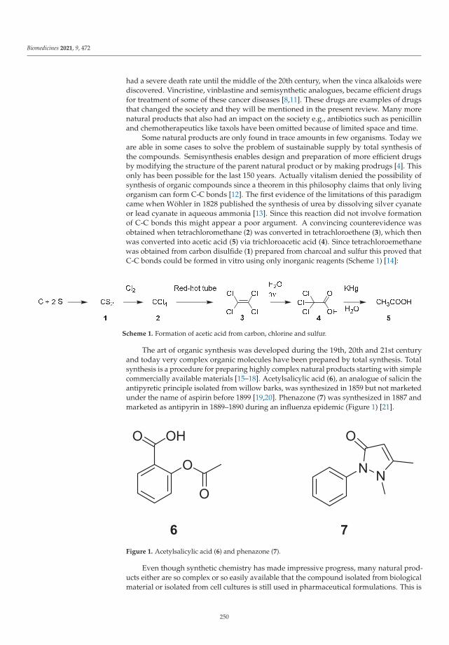
Biomedicines 2021, 9, 472
had a severe death rate until the middle of the 20th century, when the vinca alkaloids werediscovered. Vincristine, vinblastine and semisynthetic analogues, became efficient drugsfor treatment of some of these cancer diseases [8,11]. These drugs are examples of drugsthat changed the society and they will be mentioned in the present review. Many morenatural products that also had an impact on the society e.g., antibiotics such as penicillinand chemotherapeutics like taxols have been omitted because of limited space and time.
Some natural products are only found in trace amounts in few organisms. Today weare able in some cases to solve the problem of sustainable supply by total synthesis ofthe compounds. Semisynthesis enables design and preparation of more efficient drugsby modifying the structure of the parent natural product or by making prodrugs [4]. Thisonly has been possible for the last 150 years. Actually vitalism denied the possibility ofsynthesis of organic compounds since a theorem in this philosophy claims that only livingorganism can form C-C bonds [12]. The first evidence of the limitations of this paradigmcame when Wöhler in 1828 published the synthesis of urea by dissolving silver cyanateor lead cyanate in aqueous ammonia [13]. Since this reaction did not involve formationof C-C bonds this might appear a poor argument. A convincing counterevidence wasobtained when tetrachloromethane (2) was converted in tetrachloroethene (3), which thenwas converted into acetic acid (5) via trichloroacetic acid (4). Since tetrachloroemethanewas obtained from carbon disulfide (1) prepared from charcoal and sulfur this proved thatC-C bonds could be formed in vitro using only inorganic reagents (Scheme 1) [14]:
Scheme 1. Formation of acetic acid from carbon, chlorine and sulfur.
The art of organic synthesis was developed during the 19th, 20th and 21st centuryand today very complex organic molecules have been prepared by total synthesis. Totalsynthesis is a procedure for preparing highly complex natural products starting with simplecommercially available materials [15–18]. Acetylsalicylic acid (6), an analogue of salicin theantipyretic principle isolated from willow barks, was synthesized in 1859 but not marketedunder the name of aspirin before 1899 [19,20]. Phenazone (7) was synthesized in 1887 andmarketed as antipyrin in 1889–1890 during an influenza epidemic (Figure 1) [21].
Figure 1. Acetylsalicylic acid (6) and phenazone (7).
Even though synthetic chemistry has made impressive progress, many natural prod-ucts either are so complex or so easily available that the compound isolated from biologicalmaterial or isolated from cell cultures is still used in pharmaceutical formulations. This is
250

Biomedicines 2021, 9, 472
the case for e.g., taxol, morphine, codeine, vincristine, vinblastine and quinine [22]. One ofthe most complicated natural products prepared by synthesis is eribulin [23]. Even thougha procedure for total synthesis of a natural product has been developed this frequent is socomplicated and with such a poor yield that it is not economically feasible for productionof the compound on kg scale. A procedure for synthesis of quinine was described in 1944.This achievement though highly praised during the war never made synthetic quinineavailable, because the many steps in the synthesis. It still is debated whether the synthesisfrom 1944 leads to quinine. An undisputed total synthesis of quinine was not developeduntil 2001, and even this synthesis is not feasible for production of quinine in kg scale [24].
The most important of all natural products, defined as compounds produced by aliving organism, is dioxygen, normally just mentioned as oxygen. Dioxygen is formedby photosynthetic cleavage of water (Scheme 2) [25]. Dioxygen was only present in traceamount in the atmosphere of the earth before living organism developed photosynthesisand, thus, must be considered a natural product [25,26].
Scheme 2. Simplified presentation of the photosynthesis [25].
2. Malaria
Malaria is a disease caused by five different protozoan parasites of the genus Plam-odium (P. malaria causing quartan malaria, P. ovale causing ovale tertian malaria, P. vivaxcausing benign tertian malaria, P. knowlesi, which only recently has been shown to infecthumans, and P. falciparum responsible for the majority of fatalities caused by malaria) [7].The life cycle of the parasite is depicted in Figure 2.
Figure 2. Life cycle of Plasmodium parasites: When an infected female Anopheles mosquito takes ablood meal approximately eight to 10 sporozoites are introduced into the blood. Either the sporozoiteshave entered hepatocytes after 45 min. or they are cleared. In the liver the sporozoites proliferateasexually for between 5 days (P. falciparum) and 15 days (P. malariae). A proportion of hepatic schizontsmay rest in the liver as hyonozoites in the case of P. ovale and P. vivax infections. The hypnozoitesmay awake weeks or months after the infection. The merozoites delivered to the blood invadeerythrocytes, feed on the proteins mainly hemoglobin and proliferate asexually. After 36 hours(P. malariae, P. ovales, P. vivax and P. knowlesi) or 54 hours (P. malariae) the cells rupture and releasebetween 6 and 36 merozoites, which invade healthy erythrocytes. The characteristic fever attacksappear when the erythrocytes rupture. After a series of asexual proliferation gametocytes appear.Gametocytes are ingested during a blood meal of an Anopheles mosquito, in which they undergo asexual proliferation [7].
251

Biomedicines 2021, 9, 472
Protozoan parasites are parasites living inside the cells of the host. From the Plasmod-ium genus the most deadly parasite is P. falciparum causing malignant tertian malaria, alsoknown as cerebral malaria. This species is only transmitted in the tropics. Two studiesformed the basis for our understanding of the disease, the first was the discovery of para-sites in the erythrocytes of malaria patients by Alphonse Laveran in 1880 and the secondthe role of Anopheles mosquitoes as vectors described by Ronald Ross in 1897 [7,27,28].Both researchers were awarded the Nobel Prize in 1907 and 1902, respectively. In contrastto P. falciparum the other parasites are also infectious in subtropical and temperate areas.About year 1900 malaria had its maximal distribution reaching latitudinal extremes of64◦ norths and 32◦ south encompassing all continents Europe, the Americas, Asia andAustralia. In year 2004 the area in which humans were at risk of being infected with malariawas decreased from 53% to 27% of the Earth’s land surface [29]. In the period 2010 to2018 the estimated cases of malaria has decreased from 251 million to 228 million andthe number of deaths from 585,000 to 405,000, which still are scaring high numbers [30].Previous the presence of P. falciparum meant that Europeans were prevented from enteringtropic Africa, because of the mortality from cerebral malaria. An about seven times higherdeath rate for French and British soldiers were observed in the tropic African colonies inthe first half of the 19th century than seen elsewhere [31]. In 1805 a party of 44 Europeanssailed up the river Niger but only five survived the journey [32] illustrating the mortalityof malaria. No doubt the high mortality rate prevented colonization of Africa (Figure 3).
Figure 3. (A): World map 1660. A large part of South America was colonized by Spain (light green) and Portugal.(Green)Russia is dark blue (B): World map 1754. Almost all South America is colonized British colored red, whereas only tinyparts of Africa. (C): World map 1822. Even though Australia was known since 1606 it was not colonized until James Cookdescribed the continent in 1770. (D): World map 1898. The major part of Africa is colonized and by 1914 all of Africa wascolonized (Russia black, German and Belgium colonies blakc, Turkish colonies Light green , British colonies red, Francecolonies dark blue, Dutch colonies light red (comons.wikimedia.org/wiki/File:World_1898_empires_colonies_territory.png,Accessed 1 April 2021)
252

Biomedicines 2021, 9, 472
Even though mortality of malaria in Europe was not comparable with that in Africa,the disease still was a severe burden. Malaria (known as ague or intermittent fever, inDanish: koldfeber, cold fever) was prevalent in Southern England in Kent, Essex and partsof London in the 17th century. Rome was also reputed for malaria [32].
Malaria and a number of other diseases well known in Europe, Asia and Africa suchas smallpox, influenza, measles and yellow fever were no burdens for the pre-ColumbianAmerican population [33]. Consequently the endemic population had no immunologicdefense. The epidemic infections brought to America by infected Spaniards led to de-mographic falls. The Indian population on the island Espanola decreased from 3,770,000in 1496 to 125 in 1570. Similar catastrophic falls were observed in Mexico and Peru [33].Mating between Spaniards and Indians created Mestizos, who were less sensitive to thediseases and soon outnumbered the original population [33].
2.1. Drugs to Treat and Prevent Malaria2.1.1. Cinchona Bark
After the conquest of the Inca Empire in South America the Jesuit monks followed theconquistadores as missionaries. Jesuits, who were the apothecaries at that time, noticedthat the Incas used the bark of cinchona trees when working in cold streams to preventshivering [34]. They hypothesized that the bark also might prevent shivering provokedby intermittent fever (malaria). A source mentions that the first European patient treatedwith cinchona bark was a Spaniard treated in the territory of Loja, in southern Ecuadorin 1631 [33,35]. A successful outcome of this case study encouraged the Jesuits monksto export the bark to Europe [32,36]. In 1632 Jesuits send the first batch to Europe [37].The bark was imported to Europe under different names, Jesuit bark acknowledging theJesuits, who discovered the healing effects, Cardinal’s bark, honoring Cardinal Lugo, orPeruvian bark. The genus name Cinchona was invented by Linnaeus in his Systema Naturae(1742) [37]. The genus was grouped in the plant family Rubiaceae (Figure 4)
Figure 4. Cinchona officinalis L. and C. pubescens Vahl. [38].
Already in 1649 a recipe for treatment of malaria, Schedula Romana, summarized trialscarried out using cinchona bark [34,35,37]. Protestant Europe only reluctantly used theCatholic drug. Another obstacle was the doctrine of Hippocrates that well-being depends
253
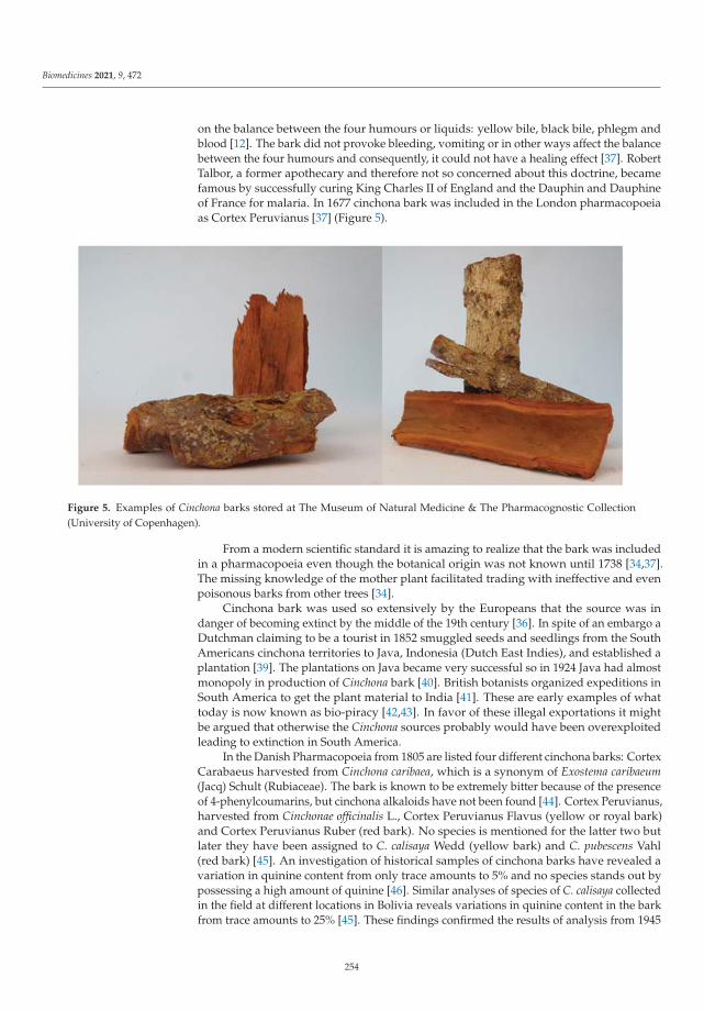
Biomedicines 2021, 9, 472
on the balance between the four humours or liquids: yellow bile, black bile, phlegm andblood [12]. The bark did not provoke bleeding, vomiting or in other ways affect the balancebetween the four humours and consequently, it could not have a healing effect [37]. RobertTalbor, a former apothecary and therefore not so concerned about this doctrine, becamefamous by successfully curing King Charles II of England and the Dauphin and Dauphineof France for malaria. In 1677 cinchona bark was included in the London pharmacopoeiaas Cortex Peruvianus [37] (Figure 5).
Figure 5. Examples of Cinchona barks stored at The Museum of Natural Medicine & The Pharmacognostic Collection(University of Copenhagen).
From a modern scientific standard it is amazing to realize that the bark was includedin a pharmacopoeia even though the botanical origin was not known until 1738 [34,37].The missing knowledge of the mother plant facilitated trading with ineffective and evenpoisonous barks from other trees [34].
Cinchona bark was used so extensively by the Europeans that the source was indanger of becoming extinct by the middle of the 19th century [36]. In spite of an embargo aDutchman claiming to be a tourist in 1852 smuggled seeds and seedlings from the SouthAmericans cinchona territories to Java, Indonesia (Dutch East Indies), and established aplantation [39]. The plantations on Java became very successful so in 1924 Java had almostmonopoly in production of Cinchona bark [40]. British botanists organized expeditions inSouth America to get the plant material to India [41]. These are early examples of whattoday is now known as bio-piracy [42,43]. In favor of these illegal exportations it mightbe argued that otherwise the Cinchona sources probably would have been overexploitedleading to extinction in South America.
In the Danish Pharmacopoeia from 1805 are listed four different cinchona barks: CortexCarabaeus harvested from Cinchona caribaea, which is a synonym of Exostema caribaeum(Jacq) Schult (Rubiaceae). The bark is known to be extremely bitter because of the presenceof 4-phenylcoumarins, but cinchona alkaloids have not been found [44]. Cortex Peruvianus,harvested from Cinchonae officinalis L., Cortex Peruvianus Flavus (yellow or royal bark)and Cortex Peruvianus Ruber (red bark). No species is mentioned for the latter two butlater they have been assigned to C. calisaya Wedd (yellow bark) and C. pubescens Vahl(red bark) [45]. An investigation of historical samples of cinchona barks have revealed avariation in quinine content from only trace amounts to 5% and no species stands out bypossessing a high amount of quinine [46]. Similar analyses of species of C. calisaya collectedin the field at different locations in Bolivia reveals variations in quinine content in the barkfrom trace amounts to 25% [45]. These findings confirmed the results of analysis from 1945
254

Biomedicines 2021, 9, 472
revealing variation in the quinine content in C. officinalis and C. pubescens species collectedin Ecuador, Bolivia and Peru from trace amounts to 7% [47]. Such inhomogeneity musthave made correct dosing of the bark a problem considering the poor analytical techniquespresent in the 18th and 19th centuries and considering the limited therapeutic windowfor quinine [7]. In the Danish Pharmacopoeia from 1907 only Cortex Chinae is mentioned,which is defined as bark originating from different preferential cultivated Cinchona species.The bark must contain at least 4% of alkaloids and at least 1% of quinine. Quinine chloridewas included in the Danish Pharmacopoeia from 1893.
2.1.2. Quinine
In 1820 Pelletier and Caventou succeeded in isolating an active principle from Cin-chona bark [34,48]. The compound was named quinine (Figure 6, 8). To isolate quinine asuspension of ground bark slurried in water mixed with slaked lime was extracted withan organic solvent like hot toluene. The organic layer was extracted with diluted sulfuricacid and upon concentration of the aqueous phase quinine sulfate crystallized [37,39]. Thisprocedure enabled large scale production. Already early comparisons of the use of Cin-chona bark or quinine sulfate revealed advantages of using the isolated active principle [34].Quinine sulfate was given in a pill in a dose of 1 g each day. In contrast several grams of thecrude bark had to be swallowed and even though wine was offered to make swallowingeasier it must have been unpleasant. Decoctions and tinctures of the bark were also used,but probably not standardized.
Figure 6. Major Cinchona alkaloids. Quinine and quinidine are included on WHO list of essentialmedicines (https://www.who.int/medicines/publications/essentialmedicines/en/ine, accessed on1 April 2021).
In addition to being easier to swallow, the most important consequence of the use of theisolated compound was that it enabled administration of well-defined doses. Consideringthe variation of quinine content in the different barks and taking into account the analyticaltechniques available in the early 19th century the use of purified quinine facilitated correctdosing, even though a skilled apothecary probably would have a good feeling of thequality of the bark [34,35]. In Denmark a price for quinine sulfate was fixed in 1840 by thegovernment (Interims Taxt 1840).
Inspection of the relocation costs, defined as the increased mortality among soldiersby relocation to tropical areas (“white man’s grave”), reveals a dramatic fall around 1840.This increased survival has been related to the use of purified quinine [31,49] instead ofcinchona bark. During the 19th century the mortality of sailors on the naval operationsstopping slave trade was severely reduced after introduction of quinine for prevention andcure [31,49]. Earlier attempts using cinchona bark never gave convincing results [49]. Itis interesting to notice that the Berlin Conference regulating the European colonizationand trade in Africa took place 1884–1885 after the prophylactic use of quinine had beengenerally accepted. Quinine also played an important role during the American Civil War.
255

Biomedicines 2021, 9, 472
The war might have ended with a victory to the Union troops already in the first year hadmalaria and typhoid fever not made the soldiers unable to fight [36].
In 1928 the total consumption of quinine in India was estimated to be 160,000 lbs(72 tons) [50]. In 1924 the world consumption of quinine sulfate as 500 ton, of which95% was produced from 10,000 ton of bark grown on Java giving the colony an incomeof 16–22 million £. India was the second most important supplier for 10 million US$corresponding to 25 ton of quinine sulfate [36,39]. From the mid-19th century until 1940quinine was the standard treatment of malaria [36]. Even though quinine had been usedextensively since Pelletier and Cavantou isolated the compound in 1820 the first suggestionof the constitution was published by Paul Rabe in 1908 [51] and the relative configurationin 1950 (Figure 6, 8) [52]. In spite of the missing knowledge about the structure, salts ofquinine had been used as drugs since 1820.
2.1.3. Quinine Resistance
Resistance to quinine appears sporadically, but failure of treatment is only seen inSoutheast Asia and New Guinea [53].
2.1.4. Synthetic Malaria Drugs
An early attempt to produce synthetic quinine was made by William Perkin in 1856based on a hypothesis which today appears naïve. The attempt did not lead to quininebut instead the dye mauve. After Queen Victoria appeared public dressed in mauve dyedrobes the dye became an enormous commercial success [54].
Paul Ehrlich in 1891 treated a malaria patient with methylene blue (Figure 7, 12).However, this dye never became useful in the clinic as an antimalarial drug. It was,however, used as a template for developing chloroquine (13) [55,56]. The first syntheticantimalarial used in the clinic was pamaquine (plasmoquine, 16) from 1924, which wasfollowed by mepacrine (atabrine, quinacrine, 17) in 1932 and resochin (chloroquine) in 1934(13) [7]. Atabrine was first marketed in Germany in 1932 [57]. Chloroquine was evaluatedto be too toxic for clinical use and consequently efforts to make a less toxic analogue wereundertaken to give the 2-methylated analogue sontochin (14) (1936) [57].
256

Biomedicines 2021, 9, 472
Figure 7. Early synthetic antimalarial drugs. Chloroquine and primaquine are included on WHO listof essential medicines (https://www.who.int/medicines/publications/essentialmedicines/en/ine,accessed on 1 April 2021).
2.1.5. Malaria during the Pacific War
It is generally known that after the Japanese bombing of Pearl Harbor in 1941 theUSA entered World War 2. Less known is that the mortality due to malaria on the PacificIslands was far beyond that caused by Japanese troops [36]. The Japanese occupation ofDutch East Indies, in particular Java, meant an interruption of the supply of sufficientamounts of quinine to the Allies. Consequently intensive research programs were initiatedin order to find alternative antimalarial drugs [57]. As a first attempt totaquine, a mixtureof alkaloids extracted from Cinchona bark was investigated [57]. However, the amountsof Cinchona bark available could not meet the demand. Like the Allies the Japanese usedthe synthetic primaquine (15) for prophylaxis but the Japanese in insufficient doses [36].In spite of these precautions malaria caused unacceptable losses. A group at the Nationalinstitute of Health was organized in 1939 to develop alternative antimalarial drugs. Thegroup organized a Committee on Medical Research, which collaborated with universities,industry and private individuals, the US Army and Navy and appropriate scientists inBritain and Australia. The collaboration resulted in synthesis of about 16,000 compounds.Some 80 compounds were selected. The most promising compound turned out to bechloroquine (13) already patented, but discarded by Bayer in Germany [57]. The 4-methylderivative sontochin (14) was for a period preferred to chloroquine.
Plantations were started in Tanzania, Kenya, Cameroon and Rwanda. Also a jointventure between USA and Guatemala resulted in 400 ha with 1.75 million trees by mid-1948 [39]. These new plantations, however, did not solve the problem with quinine supplyduring the war since the amount of bark which can be harvested from a tree is verydependent on the age of the tree. From a four year old C. ledgeriana 0.25 kg, from aneight year old tree 4 kg, from a 15 year old 10 kg and from a 25 year old 20 kg of driedbark can be harvested [40]. After harvest of the bark the trees are covered up enablingcontinued growth.
257

Biomedicines 2021, 9, 472
Woodward in 1944 published a total synthesis of quinine but even though it createdhope for overcoming the Japanese disruption of quinine supply during the Second WorldWar, the many steps in this procedure, however, prevented its commercial use and eventoday quinine is still produced from Cinchona bark [54].
2.1.6. Malaria Control
Intensive studies performed by the Allies established in 1944 that chloroquine wasa safe antimalarial. By combination of chloroquine and insecticides like DDT programsto eliminate malaria were initiated [58–60]. In Africa chloroquine became a pillar formalaria eradication [61,62]. In the late 1950s chloroquine-resistant parasites developedin six independent regions in India, Thailand, Indonesia, New Guinea, Venezuela andGuyana and spread over most of the areas infected with malaria [63]. As a consequencemalaria re-emerged India [58], Kenya [64], and Africa [61,62].
A contribution to the decreased chloroquine sensitivity is mutations in the parasitegenome to form a pfcrt gene. This gene encodes the Plasmodium falciparum chloroquine-resistant transporter (Pf CRT) a 424 amino acid protein with 10 transmembrane helicesthat facilitates transport of chloroquine away from ferriprotoporphyrin IX [59,63,65,66].Today resistance has made chloroquine obsolete to control P. falciparum infections; butit still is used against P. vivax. Even though the use of chloroquine today is limited,the previous importance of chloroquine to reduce the burden of malaria should not beunderestimated [60]. In the interim from appearance of chloroquine resistant-parasitesto the appearance of the artemisnins synthetic a number of drugs were used to controlmalaria. Among these drugs are mefloquine (Figure 8, 17), halofantrine (22), lumefantrine(23), primaquine (15), atovaquone (20), sulfadoxine (21), tetracycline (25) and proguanil(18), a prodrug which in the liver is transformed into the active drug cycloguanil (19).Plasmodium parasites host an endoparasite, the apicoplast. The apicoplast has a bacterialancestry. Consequently the naturally occurring antibiotic tetracycline, which binds tothe ribosome of bacteria, can be used for treatment of malaria. Terpene and type II fattyacid biosynthesis also proceed in the apicoplasts. Blockage of this system will not kill themother parasites but daughter parasites with malfunctioning apicoplasts are affected [67].Tafenoquine (24) was approved for treatment of malaria in 2018 [68,69]. Attempts to usethe natural product fosfidomycin, which targets the deoxyxylose pathway in the apicoplast,have not lead to clinical useful drugs [70–72].
258

Biomedicines 2021, 9, 472
Figure 8. Antimalarial drugs. Except for tetracycline all the compounds are synthetic drugs.Proguanil, sulfadoxime, lumefanthrine and tetracycline are included in WHO list of essentialmedicines (https://www.who.int/medicines/publications/essentialmedicines/en/, accessed on1 April 2021).
Ferroquine (Figure 9, 26) possesses the same pharmacophore as chloroquine butcontains a ferrocene moiety, which prevents the compound from being a substrate for thePf CRT transporter [73,74]. The compound was brought to clinical trials II [75] but has notbeen approved as a drug by U.S. Food and Drug Administration (FDA) USA.
259

Biomedicines 2021, 9, 472
Figure 9. Ferroquine (26).
2.1.7. Mechanism of Action of Quinolines
Both quinine and chloroquine are amines and consequently they will be protonatedin acidic media. The target molecule of both compounds is heme (ferriprotoporphyrin IX,Figure 10, 29).
Figure 10. Digestion of hemoglobin (27) to give ferroprotoporphyrin (28), which spontaneouslyoxidizes to ferriprotoporphyrin IX (29). Precipitation of hemozoin (30) prevents the toxic effects of29 [76,77].
Heme is formed in the food vacuole of the parasite when hemoglobin is digested tosupply the parasite with amino acids. The pH of the food vacuole varies from 5.0 to 5.2, 1to 2 units lower than that of the cytosol in the parasite. Consequently the neutral base willdiffuse into the food vacuole where it accumulates after protonation [76]. After cleavageof hemoglobin the ferroprotoporphyrin 28 remains. In the ferrprotophorphytin the Fe2+
is quickly oxidized to Fe3+ affording ferriprotoporphyrin IX (29), which will provokeformation of reactive oxygen species unless inactivated [60]. Under physiologic conditionsthe ferriprotophorphyrin IX will polymerize to hemozoin (30), the characteristic malariapigment seen in infected erythrocytes as dark spots. In the presence of chloroquine orquinine this polymerization is prevented, leading to the death of the parasite [76,77].
260

Biomedicines 2021, 9, 472
Even though many isoquinoline antimalarial drugs are chiral, resolution is not per-formed. The biological target molecule for these compounds ferriprotoporphyrin IX, (29) isachiral meaning that the two enantiomers have the same affinity. In contrast the volume ofdistribution, clearance and adverse effects do depend on the stereochemistry [78].
2.1.8. Artemisinins
According to a legend a meeting between Mao Zedong and Ho Chi Minh led to asecret research program to find a new drug for controlling malaria among the soldiersfighting for North Vietnam in the Vietnam civil war [79]. The program was initiated in 1967as Project 523. In contrast to the program set up by the Allies in 1939 this project resulted ina novel class of antimalarial drugs. The program was managed by the Chinese chemist You-You Tu, who in 2015 was awarded the Nobel Prize for the outcome. A starting point wastaken in traditional Chinese medicine. Screening 2000 herb preparations afforded 640 hits.An extract from Artemisia annua L. (Asteraceae) showed promising but non-reproducibleantiplasmodial effect (Figure 11).
Figure 11. Artemisia annua. (http://www.plantsoftheworldonline.org/taxon/urn:lsid:ipni.org:names:304416-2, accessed on 1 April 2021).
Studies of the ancient Ge Hong’s A Handbook of Prescriptions for Emergencies(340 AD) revealed that extraction was performed by wringing out the juice of the plantin cold water. Inspired by this information the extracts were made at low temperatureand an active extract was obtained. In 1972 a colorless crystalline substance was obtainedand named quinghaosu, meaning the active principle in quinghao, the Chinese name forA. annua. Today the name artemisinin (Scheme 3, 31) is preferred, at least outside China.Reduction of the lactone to an acetal afforded a mixture of the two dihydroartemisnins32a and 32b, which turned out to be a better drug than the mother compound [80,81].Artemether (33) and arteether (34) are lipid soluble. Artesunate (35) is soluble in water.
261

Biomedicines 2021, 9, 472
Scheme 3. Conversion of artemisinin to clinical used artemisinins: artemether (33), arteether (34) andartesunate (35) [80,81]. Artesunate and artemether are included in WHO list of essential medicines(https://www.who.int/medicines/publications/essentialmedicines/en/, accessed on 1 April 2021).
The first report in English of this amazing study appeared in 1979, some years after theend of the Vietnam War [82]. Later it has been realized that the structure of artemisinin hadbeen previously published by Stefanivic et al. in 1972 at a Symposium on the Chemistry ofNatural Products in New Delhi. The compound was named arteannuin. Unfortunatelythis group never realized the importance of the discovery [83]. This story emphasize thatyou do not become an outstanding scientist just because you are lucky. You have to realizethat you have been lucky.
2.1.9. Mechanism of Action of Artemisinins
The pharmacophore of artemisinin is the 1,2,4-trioxane ring containing an endocyclicperoxide. Removal of this peroxide means loss of all antiplasmodial activity [77]. Whereasthe artemisinins kills plasmodium parasites in low nanomolar concentration they are onlytoxic towards mammalian cells including fast proliferating cancer cell lines in micromolarconcentration. Erythrocytes infected with plasmodium parasites have a rapid selectiveuptake of artemisinins in contrast to non-infected erythrocytes. After uptake the drug canbe detected in the cytosol and the food vacuole. The Fe2+ in ferroprotoporphyrin (Scheme 4,27) might interact with the peroxide bridge in the artemisinins to give a reactive C-radical,which may alkylate the porphyrin skeleton preventing it from precipitation as heme (28)and consequently enabling the formation of ROS fatal for the parasite [77,84].
In addition to ferroprotoporphyrin (27) alkylation, the formed C-radicals also alkylateproteins and lipids, causing extensive cellular damage. The buildup of damaged proteinsinduces ER stress and attenuation of translation, which affords a lethal level of ubiquiti-nated proteins. Eventually merozoite death occurs. This mechanism of action differentiatesartemisinins from other malaria drugs, which typically have a well-defined target [85]. Theselectivity of the artemisinins originates in the Fe2+ needed for generation of the C-radicals.
262

Biomedicines 2021, 9, 472
Scheme 4. The iron(II) ion of ferroprotoporphyrin (27) activates artemisinin (31) to give a reactiveC-radical (36), which might alkylate ferroporotporphyrin (27) preventing precipitation [84]. The exactmechanism of alkylation, C-radial formation and the possibility of degradation of other biomoleculesare still debated [77].
2.1.10. Clinical Use of Artemisinins
Poor absorbance of artemisinin (31) makes artesunate (35) the preferred drug amongthe arteminsinins. Artemether (33) and artesunate (35) are both prodrugs of dihydroartemis-nin (32a and 32b). Artesuante is soluble in water and consequently preferred. However,artemether can have advantages by rectal administration. Because of very rapid killing ofparasites the artemisinins are very efficient for treatment of falciparum malaria [7,81].
In the presence of dihydroartemisin in the blood the number of parasites will bereduced 10,000 fold for each asexual cell cycle (approximately 36 h), whereas antimalarial
263

Biomedicines 2021, 9, 472
antibiotics such as tetracycline only reveal a 10-fold reduction of parasitaemia. Thisrapid reduction is in particular advantageous for treatment of falciparum malaria sincedeath might occur within hours after the first symptoms appear [7]. A drawback of theartemisinins is a very short half-live of about 1 hour. Rapid elimination from the bloodalso increases the risk of recrudescence [7,85]. In contrast a compound like chloroquinehas a half-life of 30–60 days, proguanil 16 hours, primaquine 6 hours and lumefantrine86 hours [7]. The short half-live of the arteminisins creates a risk for a long period withlow blood level and consequently a high risk for development of resistance. To avoid thisrisk artemisinin combination therapy (ACT) is recommended meaning that artemisininsare given with drugs possessing a long half-live such as sulfadoxine (21)-pyrimethamine,mefloquine (Figure 12, 38), amodiaquine (39) or lumefantrine (23) [7]. ACT has now becomefirst choice for treatment of uncomplicated falciparum malaria [85]. The use of artemisininshas thus become an important tool for malaria control.
Figure 12. Synthetic antimalarial drugs. Mefloquine and amodiaquine are included in WHO listof essential medicines. (https://www.who.int/medicines/publications/essentialmedicines/en/,accessed on 1 April 2021).
2.1.11. Resistance toward Artemsinins
In spite of the precautions artemisinin resistance defined a decreased sensitivity forartemisinins have occurred in South East Asia from China to India, in Equitorial Guineaand Uganda in Africa [85]. Considering the burden of malaria in Africa the development onthis continent is particularly alarming. Parasites resistant to artemisinins are characterizedby the presence of PfK13 mutations. The Kelch 13 proteins are substrate for an E3-ligase,which bind to phosphatidylinositol-3-kinase and marks it for ubiquitination and eventuallycleavage. Mutations in Kelch13 decrease the ubiquitination and consequent an increasedlevel of Kelch13, thereby an increase level of phosphatidylinositol-3-kinase and finally ofthe product phosphatidylinositol-3-phosphate. An increased level of this lipid makes theparasite less sensitive to oxidative damages [85]. In spite of the decreased response ACTstill is the first choice for treatment of uncomplicated falciparum malaria.
2.1.12. Sustainable Supply of Artemisnin
WHO has recommended ACT as the drug of first choice for treatment of malaria.The recognition has created an increased demand for the agents. The current source iscultivated A. annua. Attempts to increase the artemisinin yield have afforded two cultivarswith about 2% contents. Unfortunately some of the established varieties are not stableover generations. Attempts to produce artemisinin by bioengineering using cell cultureshave not been successful at the present. Heterologous production of precursors whichby semi-synthesis can be converted into the target compounds has only been partiallycommercially successful [86].
264

Biomedicines 2021, 9, 472
2.1.13. New Drugs for Treatment and Prophylaxis of Malaria
The Medicine for Malaria Venture, a non-profit, private-public partnership was es-tablished in 1999. The current portfolio contains a number of promising drug candidateslike arterolane (OZ277, Figure 13, 40), artefenomel (OZ439, 41) and ferroquine (30) [73,74].Artefenomel (40) and arterolane (39), like the artemisinins, contain a 1,2,4-trioxane ringand has the same mechanism [87]. The advantages of these drugs are longer half-lives inthe body, 4 hours for arterolane [87] and 23 hours for artefenomel [73,87] in contrast tothe 1 hour half-life of artemisinins. Long time exposure to the drugs is expected to makeKelch13 mutated parasites more sensitive [73]. Arterolane (40) is presently used clinicallyin combination with primaquine in India [73,87]. Other 1,2,4-trioxanes are also tested.
Figure 13. Recent antimalarial drugs expected to overcome resistance among mutated parasites.
Both ferroquine (26) and artefenomel (41) are synthetic drugs, which can be preparedon a large scale.
Other drugs under development and the status of the development by Medicinefor Malaria Venture can be seen on their homepage https://www.mmv.org/research-development/mmv-supported-projects, accessed on 1 April 2021. The ultimate goal of theproject is to find at drug that can cure malaria in a single dose.
3. Onchocerciasis
Onchocerca volvulus a nematode living in in nodules under the skin of the patientis the causative agent for onchocerciasis. The nodules are large granulomas formed bytissue reaction around adult worms. Each nodule might host several worms of bothsexes. The male worms have a length of 2 to 5 cm and the females 35 to 70 cm. Thepathogenic organisms are microfilariae produced in numbers of 500 to 1500 per day fromeach female. The female, however, only can produce microfilariae if it lives symbiotic withbacteria belonging to the genus Wolbachia. The females are productive in about 10 yearsand the microfilariae live about 1 to 2 years. The microfilariae have a length of 300 μm.Severely infected patients may carry 2000 microfilariae in 1 mg upper dermis, whereasthe microfilariae are seldom found in the blood or in other body fluids. Patients infectedwith Onchocerca nematodes suffer from dermatitis conditions such as leopard skin anddepigmentation. These alterations of the skin are caused by reactions to dead or dyingmicrofilariae [88]. Fibroblast proliferation leads to fibrosis and elastic fibers are replacedwith hyalinized scar tissue. Some scars might also be caused by scratching. In advancedstages the skin resembles skin of very old individuals. Microfilariae might be visible inthe cornea of patients. Dead microfilariae provoke inflammation leading to punctuate(snowflake) keratitis. Chronic inflammation causes opacification of the cornea. If nottreated onchocerciasis might provoke optic nerve atrophy and chorioretinitis leading toloss of sight, hence the name river blindness [88,89]. The disease was first described ascraw-craw in 1875 [88,89].
The life cycle (Figure 14) of the parasite involves as a vector blackfly belonging to thegenus Simulium. The fly becomes infected with microfilariae when taking a blood meal.A new blood meal from another person might transfer the parasites to this person. Thefemale flies taking blood meals restrict their flight within a few km from rivers. Thus,the transmission is limited to areas near rivers explaining the name river blindness. Theoriginal hotspots of the disease were river banks in Africa and Yemen. The slave tradeprobably also brought the disease to Central and South America [88]. A particular nasty
265

Biomedicines 2021, 9, 472
consequence is that the disease makes the fertile soils often found on river banks inhabitable.The importance of Simulium blackflies as vectors was described in 1927 [89].
Figure 14. Life cycle of Onchocerca volvus. The patient is infected by a blood meal from a blackflybelonging to the genus Similium. The nematode (worm) is situated in nodules in the skin. The malenematode is up to 5 cm and the female nematode up to 70 cm long. The female worms produce anumber of microfilariae, which cause the symptoms [88,89]. Like in the case of malaria transmissionis performed by an insect.
More than 99% of persons infected with onchocerciasis are found in Sub-saharanAfrica. Isolated foci are found in Latina America and in Yemen. The transmission has beenstopped in Ecuador, Columbia, Mexico and parts of Guatemala [88].
3.1. Ivermectin
For decades scientists at the Kitasato Institute, Japan, have grown unusual microorgan-isms and isolated new compounds from the culture media leading to the discovery of theantimicrobial pyrindicin, the protein kinase C inhibitor staurosporin and the proteasomeinhibitor lactacystin. In 1973s. Omura became head of the Kitasato Institute and initiateda collaboration with MSD Research Laboratory, At the Kitasato Institute extraordinarymicroorganisms were cultivated and bioactivity of isolated compounds screened for in vivobioactivity. The most promising compounds were forwarded to MSD research laboratoryfor in vivo tests. A product isolated from the broth of the soil bacteria Streptomyces avermect-inus was tested in mice infected with the nematode Nematospiroides dubius. The productshowed excellent anthelmintic activity with no toxic effects on mice. The active princi-ples turned out to be eight avermectins (Figure 15, 42–49). The most active compoundsappeared to be the compounds of the B-series containing a 5-hydroxyl group [90].
Reduction of the C-22-C-23 double bond improved the activity. A mixture of 80%hydrogenated avermectin B1a and B1b was named ivermectin (50). The mixture possesses abroad activity against a broad range of nematodes. The agent killed external and internalparasites in horses, cattle, pigs, sheep and heartworms in dogs. The uncompelled anti-parasitic effects led to creation of a new term endectocide to describe compounds, which arecapable of killing a wide variety of parasitic and health-threatening organisms both insideand outside the body [90–92]. Ivermectin interacts by preventing closure of glutamate andto a minor extent GABA-gated ion channels leading to hyperpolarization of the neuralmembrane and eventually killing of the parasite. The adult nematodes in general are notkilled by the drugs. However, after administration of the drug female worms are preventedfrom releasing pathogenic amounts of microfilariae for 12 months even though the half liveof the drug in humans is only 12 to 36 hours. GABA and glutamate receptors only occur in
266
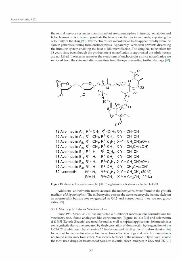
Biomedicines 2021, 9, 472
the central nervous system in mammalian but are commonplace in insects, nematodes andticks. Ivermectin is unable to penetrate the blood-brain barrier in mammals, explaining theselectivity of the drug [90]. Ivermectin causes microfilariae to disappear rapidly from theskin in patients suffering from onchocerciasis. Apparently ivermectin prevents disarmingthe immune system enabling the host to kill microfilariae. The drug has to be taken for18 years since even though the production of microfilariae is suppressed the adult wormsare not killed. Ivermectin removes the symptoms of onchocerciasis since microfilariae areremoved from the skin and after some time from the eye preventing further damage [90].
Figure 15. Avermectins and ivermectin [90]. The glycoside side chain is attached to C-13.
Additional anthelmintic macrolactones, the milbemycins, were found in the growthmedium of S.hygroscopicus. The milbemycins possess the same macrocyclic lactone skeletonas avermectins but are not oxygenated at C-13 and consequently they are not glyco-sides [93].
3.1.1. Macrocyclic Latones Veterinary Use
Since 1981 Merck & Co. has marketed a number of macrolactone formulations forveterinary use. Some analogues like eprinomectin (Figure 16, 51) [94] and selamectin(52) [95] (Revolt, Zoaetis) are used for oral as well as topical application. Selamectin is asemisynthetic derivative prepared by deglycosylation of doramectin, hydrogenation of theC-22-C23 double bond, transforming C5 to a ketone and reacting it with hydroxylamine [96].In contrast to ivermectin selamectin has no toxic effects on dogs and cats. Eprinomectin isnot found in the milk from cows. Macrocyclic lactones of the ivermectin type have becomethe most used drugs for treatment of parasites in cattle, sheep, and pets in USA and UK [91].
267

Biomedicines 2021, 9, 472
In depth description of the different macrocyclic lactones, use and properties have beenpresented by Vercruy and Rew [97].
Figure 16. Macrolactone endectocides [97].
3.1.2. Ivermectin for Human Use
After approval of the drug by the French Medical Agency in 1987 the drug was donatedfree of charge under the name of Mectizan by Merck & Co. Inc. (Fort Kenneworth, NJ, USA)to NGO organizations for treatment of onchocerciasis in as long as needed in amounts asmuch as needed [90]. The drawback of ivermectin is that treatment for keeping patientsfree from symptoms has to last for up to 18 years. Estimates of the number of infectedpersons in 1995 varied from 17 to 35 million cases globally. Not less than 0.27 millionpatients were blinded with 99% in in Sub-Saharan Africa [98,99]. Massive clinical trials inAfrica have revealed that only sporadic side effects are observed and that the drug onlyneeds to be administered orally once a year to keep the patient free from symptoms and tointerrupt transmission.
The African Program for Onchocerciasis Control was established in 1995 to admin-ister mass drug administration to 19 African countries. The program was closed in 2015.The number of patients was in 2017 estimated to 1.34 million [98]. The final evaluationstated that onchocerciasis had been bee reduced in all African countries including conflictareas. Success of the program has been obtained in Sudan, Mali, Senegal, Burundi Malawi,Nigeria and Ethiopia where onchocerciasis is almost eliminated [99]. In western Ugandaonchocerciasis was observed among 88% of persons older than 19 years in 1991 in Kabaroleand Kyenjojo districts. An investigation in 2012 found no positive cases [99,100]. Theactivities have been taken over by WHO African Region Expanded Special Program forElimination of Neglected Tropical Diseases. The program is estimated to prevent 17.4 mil-lion from suffering from the symptoms of onchocerciasis. Combined use of mectizan andvector control afforded better results than only distribution of drugs [99]. The Onchocer-ciasis Elimination Program for the Americas has succeeded in eliminating the disease inAmerika except in a cross-border region deep in the Amazon forest between Venezuelaand Brasilia. About 30,000 people are at risk. The mass drug administration of ivermectinthus has severely reduced the burden of onchocerciasis [89,99]. Appearance of resistancehave called for new combination therapies including ivermectin [101].
3.1.3. Moxidectin
In 2018 FDA approved moxidectin (Figure 17, 53) for treatment of onchocerciasis [69].
268

Biomedicines 2021, 9, 472
Figure 17. Moxidectin (53) [102].
Moxidectin is a semisynthetic milbemycin. Milbemycins are not oxygenated at C-13and consequently the compound does not possess the disaccharide side chain of avermectins.The absence of the carbohydrate moiety makes the compound more lipophilic. [93,98,103].Moxidectin prepared from nemadectin isolated from the broth of Streptomyce cyaneogriseussubsp. Noncyanogenous. The 5-hydroxyl group of nemadectin is protected, the 23-hydroxy groupoxidized, the methoxime introduced and the 5-hydroxy protecting group removed [103]. Mox-idectin has been approved for treatment of onchocerciasis in humans and possesses a strongerand longer lasting suppression of microfilarial expression than ivermectin. Consequently itmight be a better tool for elimination of onchocerciasis [98].
4. Lymphatic Filariasis
The pathogenic organisms in lymphatic filariasis caused are the nematodes Wuchereriabancrofti, Brugia malayi or B. timori. Lymphatic filariasis is characterized by lymphedema.After some years the edema may become non-pitting with thickening of the skin andloss of skin elasticity. Further progression leads to elephantitis [88] characterized by largedisabling edemas. Over a billion people in more than 80 countries are threatened by thisdisease. The causative nematode W. bancrofti is found in tropical regions lf Africa, Asia, theAmericas and the Pacific. The disease is particular frequent in hot and humid climates. B.malayi dominates in South-east Asia and South-west India. B. tumori is limited to islandsin eastern Indonesia [88]. The life cycle of the parasite is similar to that of Onchocercanematodes. The worms are found in lymphatic tissue of the human host. The female W.bancrofti measures 100 mm long and 0.25 mm in diameter. The microfilariae are releasedinto the lymphatic vessels and from here they enter the veins. Female mosquitoes ingestthe microfilariae and transmit them to a new human host by taking a new blood meal.The principal vector for W. bacrofti is Culex quinquefasciatus. In Africa Anofeles sp. also cantransmit W. bancrofti. B. malayi uses bot Mansonia and Anopheles sp. as vectors. The time ofdevelopment into the mosquito is about 12 days. In the human host the nematode mightlive and produce microfilariae for 20 years but the average lifespan is much shorter. Themicrofilariae have a lifespan of about 1 year. In the blood the density of microfilariae mightreach 10,000/ml [88].
In addition to having reduced the burden of onchocerciasis ivermectin also is effi-cient in treating the parasitic diseases lymphatic filariasis. Ivermectin was approved fortreatment of lymphatic filarisasis in 1998 by the French Medical Agency. Combination
269

Biomedicines 2021, 9, 472
of albenzol and ivermectin was found 99% effective in elimination microfilaria from theblood. After GSK donated albenzol to the society and Merck extended the ivermectindonation to include lymphatic filariasis WHO initiated the Global Program to EliminateLymphatic Filariasis. From 2000 to 2020 7.7 billions doses against lymphatic filariasis havebeen distributed to 910 million persons in 68 countries reducing the number of peopleneeding treatment from 1420 million to 597 million [104]. In the Americas Costa, Suriname,Trinidad and Tobago were removed from the WHO list of countries endemic for lymphaticfilariasis in 2011 and it is hoped that Brazil, The Dominican Republic, Haiti and Guyanasoon can be removed. WHO today recommends treatment with combination therapyconsisting of ivermectin 200 μg/kg, diethylcarbamzine citrate (6 mg/kg) and albendazole400 mg. In 2019 WHO has launched the ambition that onchocerciasis is eliminated in2030 [98]. Sathoshi Omura and William C. Campbell together with You-you Tu shared the2015 Nobel Prize in Medicine, the first two being mentioned for their work on ivermectin.
5. Cancer Diseases
A number of hallmarks are characteristic for cancer cells: (1) sustained prolifera-tive signaling, (2) evading growth suppressors, (3) replicative immortality, (4) activatedinvasion and metastasis, (5) induction of angiogenesis, (6) resistance against cell death,(7) deregulation of cellular energetics and (8) not sensitive to immune destruction [105].High rate of proliferation is characteristic for a number of cancer diseases and some of theseare controlled by drugs targeting cells in the proliferative state. Spindle toxins offered anew efficient way of treating some cancer diseases like lymphomas and leukemia [106].Thefirst spindle toxins vincristine and vinblastine were only discovered about 1960 [11].
5.1. Vinca Alkaloids
The leaves of Madagascan periwinkle [Chataranthus roseus (L.) G.Don Apocynaceae,named Vinca rosea by Linnaeus, Figure 18)] were used to control diabetes mellitus intraditional medicine on Jamaica.
Figure 18. Catharanthus roseus (The Museum of Natural Medicine & The Pharmacognostic Collection,University of Copenhagen).
A product named vinculin had been marketed in England for treatment of diabetes.Injection of extracts from the leaves showed no antidiabetic activity, but, unexpectedly,killed rabbits [107]. Autopsy revealed multiple abscesses, particular in the liver and
270

Biomedicines 2021, 9, 472
kidneys [107]. The rabbits died from infections becoming fatal because of a suppressedimmune system. The white blood count was severely reduced and neutrophil leukocyteshad virtually disappeared. In addition the bone marrow was destroyed [108]. It wasrealized that the active principle was partitioned into an acidic aqueous solution from abenzene extract. Chromatographic separation on almost neutral alumina afforded a brownmixture, from which a crystalline hydrosulfate was obtained. Injection of a solution ofthe crystals into rats caused a severe drop in the number of granulocytes after 36 hours,which lasted for three to six days [107]. Structural elucidation revealed that he compoundsbelonged to a previously unknown type of dimeric indole alkaloids [109]. The relativeconfiguration of vinblastine Figure 19, 54) and vincristine (55) was published in 1964 [110]and confirmed by an X-ray analysis [111], which also revealed the absolute configuration.Among the 120 alkaloids present in the leaves only two are used in approved drugs [112].Three semisynthetic analouges also have been approved vindesine (56), vinorelbine (57)and vinflunine (59) [11].
Figure 19. Vinblastine (54) and vincristine (55) are native natural products, whereas vindesine (56),vinorelbine (57) and vinflunine (58) are semisynthetic analogues [11].
Inspection of the structures of all the clinical used alkaloids reveals that they all possessthe same carbon skeleton. Vincristine (55) is a formamide of dihydroindole, and vinblastine(54) is the N-methyldihydroindole. A formation of a bond from the α-methylester carbonof catharanthine (Scheme 5, 60) and the carbon ortho to the methoxy group of vindoline(59) created the dimeric carbon skeleton. The biosynthetic reaction is catalyzed by α-3’.4’-anhydrovinbalse synthase (Scheme 5) [22]. The dimerization is a complex reaction, whichis described in details elsewhere.
271
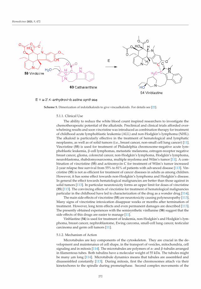
Biomedicines 2021, 9, 472
Scheme 5. Dimerization of indolalkaloids to give vincaalkaloids. For details see [22].
5.1.1. Clinical Use
The ability to reduce the white blood count inspired researchers to investigate thechemotherapeutic potential of the alkaloids. Preclinical and clinical trials afforded over-whelming results and soon vincristine was introduced as combination therapy for treatmentof childhood acute lymphoblastic leukemia (ALL) and non-Hodgkin’s lymphoma (NHL).The alkaloid is particularly effective in the treatment of hematological and lymphaticneoplasms, as well as of solid tumors (i.e., breast cancer, non-small cell lung cancer) [11].Vincristine (55) is used for treatment of Philadelphia chromosome-negative acute lym-phoblastic leukemia, β-cell lymphomas, metastatic melanoma, estrogen-receptor negativebreast cancer, glioma, colorectal cancer, non-Hodgkin’s lymphoma, Hodgkin’s lymphoma,neuroblastoma, rhabdomyosarcsoma, multiple myeloma and Wilm’s tumor [11]. A com-bination of vincristine (55) and actinomycin C for treatment of Wilm’s tumor increased2-year relapse free survival from 55% to 81% of patients with advanced disease [113]. Vin-cristine (55) is not as efficient for treatment of cancer diseases in adults as among children.However, it has some effect towards non-Hodgkin’s lymphoma and Hodgkin’s disease.In general the effect towards hematological malignancies are better than those against insolid tumors [113]. In particular neurotoxicity forms an upper limit for doses of vincristine(55) [113]. The convincing effects of vincristine for treatment of hematological malignanciesparticular in the childhood have led to characterization of the drug as a wonder drug [114].
The main side effects of vincristine (55) are neurotoxicity causing polyneuropathy [113].Many signs of vincristine intoxication disappear weeks or months after termination oftreatment. However, long term effects and even permanent damages are described [113].The presently obtained experiences with the semisynthetic vinflunine (58) suggest that theside effects of this drugs are easier to manage [11].
Vinblastine (54) is used for treatment of leukemia, non-Hodgkin’s and Hodgkin’s lym-phoma, breast cancer, nephroblastome, Ewing carcoma, small-cell lung cancer, testicularcarcinoma and germ cell tumors [11].
5.1.2. Mechanism of Action
Microtubules are key components of the cytoskeleton. They are crucial in the de-velopment and maintenance of cell shape, in the transport of vesicles, mitochondria, cellsignaling and in mitosis [114]. The microtubules are polymers of α- and β-tubulin arrangedin filamentous tubes. Both tubulins have a molecular weight of 55 kDa. The tubules mightbe many μm long [114]. Microtubule dynamics means that tubules are assembled anddisassembled constantly [113]. During mitosis, first the chromosomes attach via theirkinetochores to the spindle during prometaphase. Second complex movements of the
272
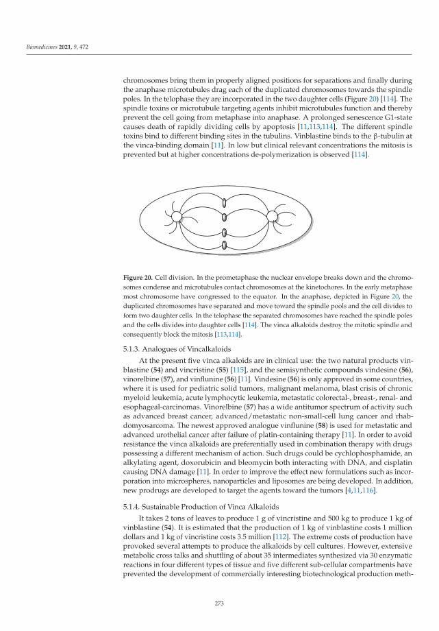
Biomedicines 2021, 9, 472
chromosomes bring them in properly aligned positions for separations and finally duringthe anaphase microtubules drag each of the duplicated chromosomes towards the spindlepoles. In the telophase they are incorporated in the two daughter cells (Figure 20) [114]. Thespindle toxins or microtubule targeting agents inhibit microtubules function and therebyprevent the cell going from metaphase into anaphase. A prolonged senescence G1-statecauses death of rapidly dividing cells by apoptosis [11,113,114]. The different spindletoxins bind to different binding sites in the tubulins. Vinblastine binds to the β-tubulin atthe vinca-binding domain [11]. In low but clinical relevant concentrations the mitosis isprevented but at higher concentrations de-polymerization is observed [114].
Figure 20. Cell division. In the prometaphase the nuclear envelope breaks down and the chromo-somes condense and microtubules contact chromosomes at the kinetochores. In the early metaphasemost chromosome have congressed to the equator. In the anaphase, depicted in Figure 20, theduplicated chromosomes have separated and move toward the spindle pools and the cell divides toform two daughter cells. In the telophase the separated chromosomes have reached the spindle polesand the cells divides into daughter cells [114]. The vinca alkaloids destroy the mitotic spindle andconsequently block the mitosis [113,114].
5.1.3. Analogues of Vincalkaloids
At the present five vinca alkaloids are in clinical use: the two natural products vin-blastine (54) and vincristine (55) [115], and the semisynthetic compounds vindesine (56),vinorelbine (57), and vinflunine (56) [11]. Vindesine (56) is only approved in some countries,where it is used for pediatric solid tumors, malignant melanoma, blast crisis of chronicmyeloid leukemia, acute lymphocytic leukemia, metastatic colorectal-, breast-, renal- andesophageal-carcinomas. Vinorelbine (57) has a wide antitumor spectrum of activity suchas advanced breast cancer, advanced/metastatic non-small-cell lung cancer and rhab-domyosarcoma. The newest approved analogue vinflunine (58) is used for metastatic andadvanced urothelial cancer after failure of platin-containing therapy [11]. In order to avoidresistance the vinca alkaloids are preferentially used in combination therapy with drugspossessing a different mechanism of action. Such drugs could be cychlophosphamide, analkylating agent, doxorubicin and bleomycin both interacting with DNA, and cisplatincausing DNA damage [11]. In order to improve the effect new formulations such as incor-poration into microspheres, nanoparticles and liposomes are being developed. In addition,new prodrugs are developed to target the agents toward the tumors [4,11,116].
5.1.4. Sustainable Production of Vinca Alkaloids
It takes 2 tons of leaves to produce 1 g of vincristine and 500 kg to produce 1 kg ofvinblastine (54). It is estimated that the production of 1 kg of vinblastine costs 1 milliondollars and 1 kg of vincristine costs 3.5 million [112]. The extreme costs of production haveprovoked several attempts to produce the alkaloids by cell cultures. However, extensivemetabolic cross talks and shuttling of about 35 intermediates synthesized via 30 enzymaticreactions in four different types of tissue and five different sub-cellular compartments haveprevented the development of commercially interesting biotechnological production meth-
273
Biomedicines 2021, 9, 472
ods [117]. Hopefully a breakthrough can afford a feasible method for kg scale productionof vinca alkaloids [115,118].
6. Conclusions
Natural products and natural products chemistry have had an impressive impacton drug development and consequently on our society [3]. An outstanding example ofthis is the fight against malaria. Until the discovery of quinine mankind had no efficientdrug to control malaria, which was a devastating disease putting a severe burden notonly on the population in tropical Africa but also on Europe and Asia [12,29]. It mightbe considered as a irony of history that the breakthrough to solve the problem of malariawas found in South America, where malaria was not a problem [33,35]. Open mindedstudies revealed that the bark of trees used to prevent muscle shivering turned out to bea warhead to combat malaria. Botanical studies let to the discovery of the mother trees,and a new genus, Cinchona, was created to enabling scientific description of the specieswith the healing bark [34,35]. A recipe for the use of the bark as a drug against malaria,Schedula Romana, was developed [34]. However, besides the unpleasant swallowing ofthe crude bark, an additional severe drawback of the bark was the variation of the level ofthe active principles. Isolation of the active alkaloid, quinine, in a pure state enabled forthe first time development of an efficient drug with reproducible effects [31,49]. As withmany other natural products development of a sustainable production of quinine becamean absolute requirement for successful use of the drug. Until the Japanese occupationduring the Second World War the sustainable production was maintained by plantationson Java and to a smaller extent in India. When the supply from Java was discontinuedscientists developed synthetic antimalarial drugs, in particular chloroquine, which actuallyoutcompeted quinine [57]. The development of chloroquine, however, was not the finalstroke against malaria. After a few decades the parasites developed resistance and newdrugs were desperately needed. In spite of all the disasters caused by the Vietnam Warmilitary need for a new drug led to the discovery of the artemisinins [80]. Artesunate andartemether are used in combination with other antimalarial drugs to postpone develop-ment of resistance. However, reports of lower sensitivities of parasites for artemisinincombination therapy are reported [85].
Onchocerciasis made fertile riverbanks in Africa inhospitable because of the threatof river blindness. The search for a drug to combat parasites in livestock led to thediscovery of the macrocyclic lactone ivermectin [92]. Ivermectin is a very efficient drug forelimination of parasites in animals. The pathogenic agents in onchocerciasis in animalsas well as in humans are Onchocerca nematodes. Fortunately ivermectin was found to beefficient for removal of the symptoms not only in animals but also in humans. Under thename of mectizan ivermectin is now offered free of charge for mass distribution in Africaand other parts of the world to combat onchocerciasis [90,99]. Even though impressiveprogresses have been made for relieving the world for the burden of onchocerciasis isstill not eliminated but hopefully new drugs such as moxidectin and continued massdistribution will further reduce the burden [98].
Beside parasitic diseases cancer causes mortality and reduced life quality for a largepart of mankind [119,120]. Until the 1960s hematological malignancies particular in thechildhood had a high death rate [114]. Attempts to understand the antidiabetic effectsof leaves from C. rosesus revealed that active agent had a poor effect on diabetes butseverely suppressed the number of white blood cells. Intensive studies revealed thatthe active principles were spindle toxins and this opened a new path for treatment ofdiseases caused by fast proliferating cells [11,108]. Further studies revealed the structuresof the compounds and enabled large scale isolation. However, as with many other naturalproducts, sustainable supply in an economic feasible way is still an important restriction forthe use of the compounds. Maybe studies may enable heterologous methods for productionof the compounds in cell cultures [112,115,117]. Today many cancer diseases for which nocure existed before discovery of the spindle toxins are cured efficiently.
274
Biomedicines 2021, 9, 472
The importance of natural products chemistry has been revealed by discovery ofdrugs with new mechanisms of actions like the spindle toxins for treatment of cancerdiseases, the trioxolanes for treatment of malaria and the macrocyclic lactones for treatmentof parasitic diseases. Many new drugs developed by inspiration from traditional uses turnsout not have a curative effect on diseases for which their mother plants are used [33,35,108].Open minded researchers realized that the active principles enabled treatment of incurablediseases, as in the case of quinine and vinca alkaloids. A drawback of natural products,however, is their limited supply. Sustainable production on a ton scale may enforce theuse of alternative sources. Bioengineering of other organisms offers alternative possibili-ties [121] but are only possible if the biosynthesis is known in details [86,122].
In some cases preparation of derivatives of the natural product affords optimizeddrugs. This has been the case for the artemisinins like artemether (33) and artesunate(35) [7,81], macrocyclic lactones like moxidectin (53) [93] and vinca alkaloids like vindesine(56), vinorelbine (57), and vinflunine (56) [11]
Modern techniques have now enabled cultivation of fungi, marine organisms, endo-phytes and bacteria, which might open new paths for discovery of new families of naturalproducts [5,6,123,124].
Above all an absolute requirement for new wonder drugs is devoted interdisciplinaryresearch. As illustrated in the cases of vinca alkaloids and artemisinin, the isolation of anactive principle is only the first step on the long and complicated path to registration of anew drug.
Funding: This research received no external funding.
Institutional Review Board Statement: Not applicable.
Informed Consent Statement: Not applicable.
Conflicts of Interest: The author declares no conflict of interest.
References
1. Cragg, G.M.; Grothaus, P.G.; Newman, D.J. Impact of natural products on developing new anti-cancer agents. Chem. Rev. 2009,109, 3012–3043. [CrossRef]
2. Newman, D.J.; Cragg, G.M. Natural products as drugs and leads to drugs: The historical perspective. In Natural Product Chemistryfor Drug Discovery; Royal Society of Chemistry: London, UK, 2010; pp. 3–27.
3. Newman, D.J.; Cragg, G.M. Natural Products as Sources of New Drugs over the Nearly Four Decades from 01/1981 to 09/2019. J.Nat. Prod. 2020, 83, 770–803. [CrossRef] [PubMed]
4. Franzyk, H.; Christensen, S.B. Targeting Toxins towards Tumors. Molecules 2021, 26, 1292. [CrossRef] [PubMed]5. Hyde, K.D.; Xu, J.; Rapior, S.; Jeewon, R.; Lumyong, S.; Niego, A.G.A.T.; Abeywickrama, P.D.; Aluthmuhandiram, J.V.S.;
Brahamanage, R.S.; Brooks, S.; et al. The amazing potential of fungi: 50 ways we can exploit fungi industrially. Fungal Divers.2019, 97, 1–136.
6. Blunt, J.W.; Carroll, A.R.; Copp, B.R.; Davis, R.A.; Keyzers, R.A.; Prinsep, M.R. Marine natural products. Nat. Prod. Rep. 2018, 35,8–53. [CrossRef] [PubMed]
7. White, N.J. Malaria. In Mansons Tropical Diseases, 23rd ed.; Farrar, J., Hotez, P., Junghanss, T., Kang, G., Lalloo, D., White, N.J., Eds.;Elsevier: Amsterdam, The Netherlands, 2014; Volume 9, pp. 523–601.
8. Seca, A.M.L.; Pinto, D.C.G.A. Plant secondary metabolites as anticancer agents: Successes in clinical trials and therapeuticapplication. Int. J. Mol. Sci. 2018, 19, 263. [CrossRef]
9. Trendafilova, A.; Moujir, L.M.; Sousa, P.M.C.; Seca, A.M.L. Research Advances on Health Effects of Edible Artemisia Species andSome Sesquiterpene Lactones Constituents. Foods 2020, 10, 65. [CrossRef]
10. Omura, S. Ivermectin: 25 years and still going strong. Int. J. Antimicrob. Agents 2008, 31, 91–98. [CrossRef] [PubMed]11. Martino, E.; Casamassima, G.; Castiglione, S.; Cellupica, E.; Pantalone, S.; Papagni, F.; Rui, M.; Siciliano, A.M.; Collina, S. Vinca
alkaloids and analogues as anti-cancer agents: Looking back, peering ahead. Bioorg. Med. Chem. Lett. 2018, 28, 2816–2826.[CrossRef]
12. Singer, C.; Underwood, E.A. A Short History of Medicine, 2nd ed.; Clarendon Press: Oxford, UK, 1962.13. Wöhler, F. Ueber künstliche Bildung des Harnstoffs. Ann. Phys. Chem. 1828, 12, 253–257. [CrossRef]14. McKie, D. Wöhlers “synthetic” urea and the rejection of vitalism: A chemical legend. Nature 1944, 153, 608–610. [CrossRef]15. Nicolaou, K.C.; Sorenson, E.J. Classics in Total Synthesis: Targets, Strategies, Methods; Wiley: Weinheim, Germany, 1996.16. Nicolaou, K.C.; Snyder, S.A. Classics in Total Synthesis II: More Targets, Stratgeis and Methods; Wiley: Weinheim, Germany, 2003.17. Nicolaou, K.C.; Montagnon, T. Molcules that Changed the World; Wiley: Weinheim, Germany, 2008; p. 366.
275

Biomedicines 2021, 9, 472
18. Nicolaou, K.C.; Chen, J.S. Classics in Total Synthesis III: Further Targets, Strategies, Methods; Wiley: Weinheim, Germany, 2011.19. Mahdi, J.G.; Mahdi, A.J.; Bowen, I.D. The historical analysis of aspirin discovery, its relation to the willow tree and antiproliferative
and anticancer potential. Cell Prolif. 2006, 39, 147–155. [CrossRef]20. Marko, V. Fraom Aspitin to Viagara; Springer Nature: Cham, Schwitzerland, 2020.21. Appendino, G.; Pollastro, F. Plants: Revamping the oldest source of medicines with modern science. In Natural Product Chemistry
for Drug Discovery; Royal Society of Chemistry: London, UK, 2010; pp. 140–173.22. Dewick, P.M. Medicinal Natural Products, 3rd ed.; John Wiley and Sons Ltd.: Chicester, UK, 2009.23. Hayashi, Y. Time Economy in Total Synthesis. J. Org. Chem. 2021, 86, 1–23. [CrossRef] [PubMed]24. Seeman, J.I. The Woodward-Doering/Rabe-Kindler total synthesis of quinine: Setting the record straight. Angew. Chem. Int. Ed.
2007, 46, 1378–1413. [CrossRef] [PubMed]25. Barber, J. Photosystem II: The water splitting enzyme of photosynthesis and the origin of oxygen in our atmosphere. Q. Rev.
Biophys. 2016, 49, e14. [CrossRef]26. Oro, J. Chemical evolution and the origin of life. Adv. Space Res. 1983, 3, 77–94. [CrossRef]27. Nye, E.R. Alphonse Laveran (1845-1922): Discoverer of the malarial parasite and Nobel laureate, 1907. J. Med. Biogr. 2002, 10,
81–87. [CrossRef]28. Yoeli, M. Sir Ronald Ross and the evolution of malaria research. Bull. N. Y. Acad. Med. 1973, 49, 722–735.29. Hay, S.I.; Guerra, C.A.; Tatem, A.J.; Noor, A.M.; Snow, R.W. The global distribution and population at risk of malaria: Past,
present, and future. Lancet Infect. Dis. 2004, 4, 327–336. [CrossRef]30. WHO. World Malaria Report; WHO: Geneva, Switzerland, 2019.31. Curtin, P.D. The End of the “White Man’s Grave”? Nineteenth-Century Mortality in West Africa. J. Interdiscip. Hist. 1990, 21,
63–88. [CrossRef]32. Rocco, F. The Miraculous Fever-Tree. Malaria, Medicine and the Cure that Changed the World; HarperCollins: London, UK, 2003.33. Naranjo, P. Epidemic hecatomb in the New World. Allergy Proc. 1992, 13, 237–241. [CrossRef]34. Gachelin, G.; Garner, P.; Ferroni, E.; Trohler, U.; Chalmers, I. Evaluating Cinchona bark and quinine for treating and preventing
malaria. J. R. Soc. Med. 2017, 110, 31–40. [CrossRef]35. Gachelin, G.; Garner, P.; Ferroni, E.; Trohler, U.; Chalmers, I. Evaluating Cinchona bark and quinine for treating and preventing
malaria. J. R. Soc. Med. 2017, 110, 73–82. [CrossRef]36. Meshnick, S.R.; Dobson, M.J. The History of Antimalarial Drugs. In Antimalarial Chemotherapy; Rosenthal, P.J., Ed.; Humana Press:
Towota, NJ, USA, 2001; pp. 15–25.37. McHale, D. The cinchona tree. Biologist 1986, 33, 45–53.38. Pabst, G. Köhler’s Medizinal-Pflanzen; Verlag von Fr. Eugen Köhler: Gera-Untermhaus, Germany, 1887; Volume 1.39. Nair, K.P.P. Cinchona (Cinchona sp.). In The Agronomy and Economy of Important Tree Crops of the Developing World; Nair, K.P.P., Ed.;
Elsevier: Amsterdam, The Netherlands, 2010.40. Cross, S.H. Quinine, Production and Marketing; Bureau of Foreign and Domestic Commerce: Washington, DC, USA, 1924.41. Williams, D. Clements Robert Markham and the Introduction of the Cinchona Tree into British India, 1861. Georg. J. 1962, 128,
431–442. [CrossRef]42. Efferth, T.; Banerjee, M.; Paul, N.W.; Abdelfatah, S.; Arend, J.; Hamdoun, S.; Hamm, R.; Hong, C.; Kadioglu, O.; Nass, J.; et al.
Biopiracy of natural products and good bioprospecting practice. Phytomedicine 2016, 23, 166–173. [CrossRef] [PubMed]43. Cragg, G.M.; Katz, F.; Newman, D.J.; Rosenthal, J. The impact of the United Nations Convention on Biological Diversity on
natural products research. Nat. Prod. Rep. 2012, 29, 1407–1423. [CrossRef]44. Rivero-Cruz, I.; Cristians, S.; Ovalle-Magallanes, B.; Mata, R. Mexican copalchis of the Rubiaceae family: More than a century of
pharmacological and chemical investigations. Phytochem. Rev. 2019, 18, 1435–1455. [CrossRef]45. Maldonado, C.; Barnes, C.J.; Cornett, C.; Holmfred, E.; Hansen, S.H.; Persson, C.; Antonelli, A.; Rønsted, N. Phylogeny Predicts
the Quantity of Antimalarial Alkaloids within the Iconic Yellow Cinchona Bark (Rubiaceae: Cinchona calisaya). Front. Plant Sci.2017, 8, 391. [CrossRef]
46. Canales, N.A.; Gress Hansen, T.N.; Cornett, C.; Walker, K.; Driver, F.; Antonelli, A.; Maldonado, C.; Nesbitt, M.; Barnes, C.J.;Roensted, N. Historical chemical annotations of Cinchona bark collections are comparable to results from current day high-pressureliquid chromatography technologies. J. Ethnopharmacol. 2020, 249, 112375. [CrossRef] [PubMed]
47. Martin, W.E.; Gandara, J.A. Alkaloid content of Ecuadoran and other American cinchona barks. Bot. Gaz. 1945, 107, 184–199.[CrossRef]
48. Karamanou, M.; Tsoucalas, G.; Pantos, K.; Androutsos, G. Isolating colchicine in 19th century: An old drug revisited. Curr. Pharm.Des. 2018, 24, 654–658. [CrossRef]
49. Mitcham, J.C. Patrolling the White Man’s Grave: The impact of disease on Anglo-American naval operations against the slavetrade, 1841–1862. North. Mar. 2010, 20, 37–56.
50. Holland, J.H. Ledger Bark and Red Bark. Bull. Misc. Inf. 1932, 1932, 1–17. [CrossRef]51. Streller, S.; Roth, K. Eine Rinde erobert die Welt. Chem. Unserer Zeit 2012, 46, 228–247. [CrossRef]52. Prelog, V. Häfliger, Über China-alkaloide. Helv. Chim Acta 1950, 23, 257–2578.53. Baird, J.K. Effectiveness of antimalarial drugs. N. Engl. J. Med. 2005, 352, 1565–1577. [CrossRef]
276
Biomedicines 2021, 9, 472
54. Kaufman, T.S.; Ruveda, E.A. The quest for quinine: Those who won the battles and those who won the war. Angew. Chem. Int. Ed.2005, 44, 854–885. [CrossRef] [PubMed]
55. Gensini, G.F.; Conti, A.A.; Lippi, D. The contributions of Paul Ehrlich to infectious disease. J. Infect. 2007, 54, 221–224. [CrossRef]56. Winau, F.; Westphal, O.; Winau, R. Paul Ehrlich—In search of the magic bullet. Microbes Infect. 2004, 6, 786–789. [CrossRef]
[PubMed]57. Coatney, G.R. Pitfalls in a discovery: The chronicle of chloroquine. Am. J. Trop. Med. Hyg. 1963, 12, 121–128. [CrossRef]58. Sharma, V.P. Re-emergence of malaria in India. Indian J. Med. Res. 1996, 103, 26–45. [PubMed]59. Wellems, T.E.; Plowe, C.V. Chloroquine-resistant malaria. J. Infect. Dis. 2001, 184, 770–776. [CrossRef] [PubMed]60. Ridley, R.G. Medical need, scientific opportunity and the drive for antimalarial drugs. Nature 2002, 415, 686–693. [CrossRef]
[PubMed]61. Frosch, A.E.P.; Venkatesan, M.; Laufer, M.K. Patterns of chloroquine use and resistance in sub-Saharan Africa: A systematic
review of household survey and molecular data. Malar. J. 2011, 10, 116. [CrossRef] [PubMed]62. Trape, J.F. The public health impact of chloroquine resistance in Africa. Am. J. Trop. Med. Hyg. 2001, 64, 12–17. [CrossRef]63. Ecker, A.; Lehane, A.M.; Clain, J.; Fidock, D.A. PfCRT and its role in antimalarial drug resistance. Trends Parasitol. 2012, 28,
504–514. [CrossRef]64. Shanks, G.D.; Biomndo, K.; Hay, S.I.; Snow, R.W. Changing patterns of clinical malaria since 1965 among a tea estate population
located in the Kenyan highlands. Trans. R. Soc. Trop. Med. Hyg. 2000, 94, 253–255. [CrossRef]65. Sidhu, A.b.S.; Verdier-Pinard, D.; Fidock, D.A. Chloroquine resistance in Plasmodium falciparum malaria parasites conferred by
pfcrt mutations. Science 2002, 298, 210–213. [CrossRef] [PubMed]66. Dorsey, G.; Fidock, D.A.; Wellems, T.E.; Rosenthal, P.J. Mechanisms of Quinoline Resistance. In Antimalarial Chemotherapy;
Rosenthal, P.J., Ed.; Humana Press: Totowa, NJ, USA, 2001; pp. 153–172.67. Dahl, E.L.; Shock, J.L.; Shenai, B.R.; Gut, J.; DeRisi, J.L.; Rosenthal, P.J. Tetracyclines specifically target the apicoplast of the malaria
parasite Plasmodium falciparum. Antimicrob.Agents Chemother. 2006, 50, 3124–3131. [CrossRef]68. Berman, J.; Brown, T.; Dow, G.; Toovey, S. Tafenoquine and primaquine do not exhibit clinical neurologic signs associated with
central nervous system lesions in the same manner as earlier 8-aminoquinolines. Malar. J. 2018, 17, 407. [CrossRef] [PubMed]69. Araujo, E.; Braun, M.-G.; Dragovich, P.S.; Converso, A.; Nantermet, P.G.; Roecker, A.J.; Dhar, T.G.M.; Haile, P.; Hurtley, A.; Merritt,
J.R.; et al. To Market, To Market—2018: Small Molecules. In 2019 Medicinal Chemistry Reviews—2019; Bonson, J.J., Ed.; AmericanChemical Society: Washington, DC, USA, 2019; Volume 54, pp. 469–596.
70. Christensen, S.B. Negelected Diseases. In Textbook of Drug Desing and Discovery, 4th ed.; Krogsgaard-Larsen, P., Strømgård, K.,Madsen, U., Eds.; CRC Press: Boca Raton, FL, USA, 2010; pp. 314–358.
71. Uddin, T.; McFadden, G.I.; Goodman, C.D. Validation of putative apicoplast-targeting drugs using a chemical supplementationassay in cultured human malaria parasites. Antimicrob. Agents Chemother. 2018, 62, 1–17. [CrossRef]
72. Schlitzer, M. Malaria Chemotherapeutics Part I: History of Antimalarial Drug Development, Currently Used Therapeutics, andDrugs in Clinical Development. ChemMedChem 2007, 2, 944–986. [CrossRef] [PubMed]
73. Mathews, E.S.; John, A.R.O. Tackling resistance: Emerging antimalarials and new parasite targets in the era of elimination.F1000Research 2018, 7, 1170. [CrossRef] [PubMed]
74. Blank, B.R.; Gonciarz, R.L.; Talukder, P.; Gut, J.; Legac, J.; Rosenthal, P.J.; Renslo, A.R. Antimalarial Trioxolanes with SuperiorDrug-Like Properties and In Vivo Efficacy. ACS Infect. Dis. 2020, 6, 1827–1835. [CrossRef] [PubMed]
75. Biot, C.; Nosten, F.; Fraisse, L.; Ter-Minassian, D.; Khalife, J.; Dive, D. The antimalarial ferroquine: From bench to clinic. Parasite2011, 18, 207–214. [CrossRef] [PubMed]
76. Tilley, L.; Loria, P.; Foley, M. Chloroquine and Other Quinoline Antimalarials. In Antimalarial Chemotherapy; Rosenthal, P.J., Ed.;Humana Press: Totowa, NJ, USA, 2001; pp. 87–121.
77. Haynes, R.K.; Cheu, K.-W.; N’Da, D.; Coghi, P.; Monti, D. Considerations on the Mechanism of Action of Artemisinin Antimalarials:Part 1—The ‘Carbon Radical’ and ‘Heme’ Hypotheses. Infect. Dis. Drug Targets 2013, 13, 217–277. [CrossRef] [PubMed]
78. Brocks, D.R.; Mehvar, R. Stereoselectivity in the pharmacodynamics and pharmacokinetics of the chiral antimalarial drugs. Clin.Pharmacokinet. 2003, 42, 1359–1382. [CrossRef]
79. Cooper, R.; Deakin, J. Botanical Miracles. Chemistry of Plants that Changed the World; CRC Press: Boca Raton, FL, USA, 2016; p. 271.80. Tu, Y. The discovery of artemisinin (qinghaosu) and gifts from Chinese medicine. Nat. Med. 2011, 17, 1217–1220. [CrossRef]81. Klayman, D.L. Qinhaosu (Artemisinin): An Antimalarial Drug from China. Science 1985, 228, 1049–1055. [CrossRef]82. Group, Q.A.C.R. Antimalaria studies on qinhaosu. Chin. Med. J. 1979, 92, 811–816.83. Vajs, V.; Jokic, A.; Milosavljevic, S. Artemisinin Story from the Balkans. Nat. Prod. Commun. 2017, 12. [CrossRef]84. Christensen, S.B.; Bygbjerg, I.C. Neglected Diseases. In Textbook of Drug Design and Discovery, 5th ed.; Strømgård, K., Krogsgaard-
Larsen, P., Madsen, U., Eds.; CRC Press: Boca Raton, FL, USA, 2017; pp. 347–368.85. Lu, F.; He, X.-L.; Richard, C.; Cao, J. A brief history of artemisinin: Modes of action and mechanisms of resistance. Chin. J. Nat.
Med. 2019, 17, 331–336. [CrossRef]86. Ikram, N.K.B.K.; Simonsen, H.T. A Review of Biotechnological Artemisinin Production in Plants. Front. Plant Sci. 2017, 8, 1966.
[CrossRef] [PubMed]
277

Biomedicines 2021, 9, 472
87. Giannangelo, C.; Siddiqui, G.; De Paoli, A.; Anderson, B.M.; Edgington-Mitchell, L.E.; Charman, S.A.; Creek, D.J. System-widebiochemical analysis reveals ozonide antimalarials initially act by disrupting Plasmodium falciparum haemoglobin digestion.PLoS Pathog. 2020, 16, e1008485. [CrossRef] [PubMed]
88. Simonsen, P.E.; Fischer, P.U.; Hoerauf, A.; Weil, G.J. The Filariases. In Manson’s Tropical Diseases; Farrar, J., Hotez, P., Junghanss, T.,Kang, G., Lalloo, D., White, N., Eds.; Elservier: Amsterdam, The Netherlands, 2014; pp. 737–765.e5.
89. Hopkins, A.D. Onchocerciasis: Status and progress towards elimination. CAB Rev. 2020, 15, 021. [CrossRef]90. Omura, S. A Splendid Gift from the Earth: The Origins and Impact of the Avermectins (Nobel Lecture). Angew. Chem. Int. Ed.
2016, 55, 10190–10209. [CrossRef] [PubMed]91. Laing, R.; Gillan, V.; Devaney, E. Ivermectin—Old Drug, New Tricks? Trends Parasitol. 2017, 33, 463–472. [CrossRef]92. Omura, S.; Crump, A. The live an times of ivermectin—A success story. Nat. Rev. Microbiol. 2004, 2, 984–989. [CrossRef]93. Prichard, R.; Menez, C.; Lespine, A. Moxidectin and the avermectins: Consanguinity but not identity. Int. J. Parasitol. Drugs Drug
Resist. 2012, 2, 134–153. [CrossRef]94. Shoop, W.L.; Egerton, J.R.; Eary, C.H.; Haines, H.W.; Michael, B.F.; Mrozik, H.; Eskola, P.; Fisher, M.H.; Slayton, L.; Ostlind, D.A.;
et al. Eprinomectin: A novel avermectin for use as a topical endectocide for cattle. Int. J. Parasitol. 1996, 26, 1237–1242. [CrossRef]95. Bishop, B.F.; Bruce, C.I.; Evans, N.A.; Goudie, A.C.; Gration, K.A.F.; Gibson, S.P.; Pacey, M.S.; Perry, D.A.; Walshe, N.D.A.; Witty,
M.J. Selamectin: A novel broad-spectrum endectocide for dogs and cats. Vet. Parasitol. 2000, 91, 163–176. [CrossRef]96. Lee, S.R.; Kim, H.U.; Kang, H.M.; Choi, I.S. Preparation of Selamectin intermediate and process for Selamectin. KR2019056863A,
2019.97. Vercruysse, J.; Rew, R.S. Macrocyclic Lactones in Antiparasitic Therapy; CABI Publishing: Wallingford, UK, 2002.98. Milton, P.; Hamley, J.I.D.; Walker, M.; Basanez, M.-G. Moxidectin: An oral treatment for human onchocerciasis. Expert Rev. Anti
Infect. Ther. 2020, 18, 1067–1081. [CrossRef]99. Brattig, N.W.; Cheke, R.A.; Garms, R. Onchocerciasis (River Blindness)—More than a Century of Research and Control. Acta Trop.
2020, 218, 105677. [CrossRef]100. Lakwo, T.L.; Garms, R.; Rubaale, T.; Katabarwa, M.; Walsh, F.; Habomugisha, P.; Oguttu, D.; Unnasch, T.; Namanya, H.; Tukesiga,
E.; et al. The disappearance of onchocerciasis from the Itwara focus, western Uganda after elimination of the vector Simuliumneavei and 19 years of annual ivermectin treatments. Acta Trop. 2013, 126, 218–221. [CrossRef] [PubMed]
101. Sainas, S.; Dosio, F.; Boschi, D.; Lolli, M.L. Targeting Human Onchocerciasis: Recent Advances Beyond Ivermectin. Ann. Rep.Med. Chem. 2018, 51, 1–38.
102. Verdu, J.R.; Cortez, V.; Martinez-Pinna, J.; Ortiz, A.J.; Lumaret, J.-P.; Lobo, J.M.; Sanchez-Pinero, F.; Numa, C. First assessment ofthe comparative toxicity of ivermectin and moxidectin in adult dung beetles: Sub-lethal symptoms and pre-lethal consequences.Sci. Rep. 2018, 8, 14885. [CrossRef] [PubMed]
103. Awasthi, A.; Razzak, M.; Al-Kassas, R.; Harvey, J.; Garg, S. Analytical profile of moxidectin. Profiles Drug Subst. Excipients Relat.Methodol. 2013, 38, 315–366.
104. WHO. Lymphatic Filariasis. 2020. Available online: https://www.who.int/news-room/fact-sheets/detail/lymphatic-filariasis(accessed on 1 April 2021).
105. Björkling, F.; Moreira, J.; Stenvang, J. Anticancer Agents. In Textbook of Drug Design and Discovery, 5th ed.; Strømgaard, K.,Krogsgaard-Larsen, P., Madsen, U., Eds.; CRC Press: Boca Raton, FL, USA, 2017; pp. 369–386.
106. Xie, S.; Zhou, J. Harnessing Plant Biodiversity for the Discovery of Novel Anticancer Drugs Targeting Microtubules. Front. Plant.Sci. 2017, 8, 720. [CrossRef]
107. Noble, R.L.; Beer, C.T.; Cutts, J.H. Role of chance observations in chemotherapy: Vinca rosea. Ann. N. Y. Acad. Sci. 1958, 76,882–894. [CrossRef]
108. Noble, R.L. The Discovery of the Vinca Alkaloids—Chemotherapeutic-Agents Against Cancer. Biochem. Cell Biol. Biochim. Biol.Cell. 1990, 68, 1344–1351. [CrossRef]
109. Neuss, N.; Gorman, M.; Svoboda, G.H.; Maciak, G.; Beer, C.T. Vinca alkaloids. III. Characterization of leurosine and vin-caleukoblastine, new alkaloids from Vinca rosea. J. Am. Chem. Soc. 1959, 81, 4754–4755. [CrossRef]
110. Neuss, N.; Gorman, M.; Hargrove, W.; Cone, N.J.; Biemann, K.; Buechi, G.; Manning, R.E. Vinca alkaloids. XXI. Structures of theoncolytic alkaloids vinblastine and vincristine. J. Am. Chem. Soc. 1964, 86, 1440–1442. [CrossRef]
111. Moncrief, W.J.; Lipscomb, W.N. Structure of leurocristine methiodide dihydrate by anomalous scattering methods; relation toleurocristine (vincristine) and vincaleukoblastine (vinblastine). Acta Crystallogr. 1966, 21, 322–331. [CrossRef]
112. Barrales-Cureño, H.J.; Reyes, C.R.; García, I.V.; Valdez, L.G.L.; De Jesús, A.G.; Ruíz, J.A.C.; Herrera, L.M.S.; Caballero, M.C.C.;Magallón, J.A.S.; Perez, J.E.; et al. Alkaloids of Pharmacological Importance in Catharanthus roseus. In Alkaloids. Their Importance inNatur and Human Life; Kurek, J., Ed.; IntechOpen: London, UK, 2019.
113. Gidding, C.E.; Kellie, S.J.; Kamps, W.A.; de Graaf, S.S. Vincristine revisited. Crit. Rev. Oncol. Hematol. 1999, 29, 267–287. [CrossRef]114. Jordan, M.A.; Wilson, L. Microtubules as a target for anticancer drugs. Nat. Rev. Cancer 2004, 4, 253–265. [CrossRef] [PubMed]115. Almagro, L.; Fernandez-Perez, F.; Pedreno, M.A. Indole alkaloids from Catharanthus roseus: Bioproduction and their effect of
human health. Molecules 2015, 20, 2973–3000. [CrossRef] [PubMed]116. Brady, S.F.; Pawluczyk, J.M.; Lumma, P.K.; Feng, D.M.; Wai, J.M.; Jones, R.; Feo-Jones, D.; Wong, B.K.; Miller-Stein, C.;
Lin, J.H.; et al. Design and synthesis of a pro-drug of vinblastine targeted at treatment of prostate cancer with enhanced efficacyand reduced systemic toxicity. J. Med. Chem. 2002, 45, 4706–4715. [CrossRef] [PubMed]
278

Biomedicines 2021, 9, 472
117. Verma, P.; Khan, S.A.; Parasharami, V.; Mathur, A.K. Biotechnological Interventions to Modulate Terpenoid Indole AlkaloidPathway in Catharanthus roseus Using In Vitro Tools and Approaches. In Cathararanthus roseus. Current Research and FutureProspects; Naeem, M., Aftab, T., Masroor, M., Khan, A., Eds.; Springer International Publishing: Berlin/Heidelberg, Germany,2017; pp. 247–275.
118. Li, C.; Galani, S.; Hassan, F.-U.; Rashid, Z.; Naveed, M.; Fang, D.; Ashraf, A.; Qi, W.; Arif, A.; Saeed, M.; et al. Biotechnologicalapproaches to the production of plant-derived promising anticancer agents: An update and overview. Biomed. Pharmacother. 2020,132, 110918.
119. Torre, L.A.; Bray, F.; Siegel, R.L.; Ferlay, J.; Lortet-Tieulent, J.; Jemal, A. Global cancer statistics, 2012. CA Cancer J. Clin. 2015, 65,87–108. [CrossRef]
120. Bray, F.; Ferlay, J.; Soerjomataram, I.; Siegel, R.L.; Torre, L.A.; Jemal, A. Global cancer statistics 2018: GLOBOCAN estimates ofincidence and mortality worldwide for 36 cancers in 185 countries. CA Cancer J. Clin. 2018, 68, 394–424. [CrossRef] [PubMed]
121. Arya, S.S.; Rookes, J.E.; Cahill, D.M.; Lenka, S.K. Next-generation metabolic engineering approaches towards development ofplant cell suspension cultures as specialized metabolite producing biofactories. Biotechnol. Adv. 2020, 45, 107635. [CrossRef]
122. Atanasov, A.G.; Waltenberger, B.; Pferschy-Wenzig, E.-M.; Linder, T.; Wawrosch, C.; Uhrin, P.; Temml, V.; Wang, L.; Schwaiger, S.;Heiss, E.H.; et al. Discovery and resupply of pharmacologically active plant-derived natural products: A review. Biotechnol. Adv.2015, 33, 1582–1614. [CrossRef] [PubMed]
123. Mayer, A.M.S.; Glaser, K.B.; Cuevas, C.; Jacobs, R.S.; Kem, W.; Little, R.D.; McIntosh, J.M.; Newman, D.J.; Potts, B.C.; Shuster,D.E. The odyssey of marine pharmaceuticals: A current pipeline perspective. Trends Pharmacol. Sci. 2010, 31, 255–265. [CrossRef][PubMed]
124. Li, S.-J.; Zhang, X.; Wang, X.-H.; Zhao, C.-Q. Novel natural compounds from endophytic fungi with anticancer activity. Eur. J.Med. Chem. 2018, 156, 316–343. [CrossRef]
279


biomedicines
Review
Cannabis sativa: Interdisciplinary Strategies and Avenues forMedical and Commercial Progression Outside of CBD and THC
Jackson M. J. Oultram 1, Joseph L. Pegler 1, Timothy A. Bowser 2, Luke J. Ney 3, Andrew L. Eamens 1 and
Christopher P. L. Grof 1,2,*
Citation: Oultram, J.M.J.; Pegler, J.L.;
Bowser, T.A.; Ney, L.J.; Eamens, A.L.;
Grof, C.P.L. Cannabis sativa:
Interdisciplinary Strategies and
Avenues for Medical and Commercial
Progression Outside of CBD and
THC. Biomedicines 2021, 9, 234.
https://doi.org/10.3390/
biomedicines9030234
Academic Editors: Pavel B. Drašar
and Raffaele Capasso
Received: 1 February 2021
Accepted: 23 February 2021
Published: 26 February 2021
Publisher’s Note: MDPI stays neutral
with regard to jurisdictional claims in
published maps and institutional affil-
iations.
Copyright: © 2021 by the authors.
Licensee MDPI, Basel, Switzerland.
This article is an open access article
distributed under the terms and
conditions of the Creative Commons
Attribution (CC BY) license (https://
creativecommons.org/licenses/by/
4.0/).
1 Centre for Plant Science, University of Newcastle, University Drive, Callaghan, NSW 2308, Australia;[email protected] (J.M.J.O.); [email protected] (J.L.P.);[email protected] (A.L.E.)
2 CannaPacific Pty Ltd., 109 Ocean Street, Dudley, NSW 2290, Australia; [email protected] School of Psychological Sciences, University of Tasmania, Hobart, TAS 7005, Australia; [email protected]* Correspondence: [email protected]; Tel.: +612-4921-5858
Abstract: Cannabis sativa (Cannabis) is one of the world’s most well-known, yet maligned plantspecies. However, significant recent research is starting to unveil the potential of Cannabis to pro-duce secondary compounds that may offer a suite of medical benefits, elevating this unique plantspecies from its illicit narcotic status into a genuine biopharmaceutical. This review summarises thelengthy history of Cannabis and details the molecular pathways that underpin the production of keysecondary metabolites that may confer medical efficacy. We also provide an up-to-date summaryof the molecular targets and potential of the relatively unknown minor compounds offered by theCannabis plant. Furthermore, we detail the recent advances in plant science, as well as syntheticbiology, and the pharmacology surrounding Cannabis. Given the relative infancy of Cannabis research,we go on to highlight the parallels to previous research conducted in another medically relevant andversatile plant, Papaver somniferum (opium poppy), as an indicator of the possible future directionof Cannabis plant biology. Overall, this review highlights the future directions of cannabis researchoutside of the medical biology aspects of its well-characterised constituents and explores additionalavenues for the potential improvement of the medical potential of the Cannabis plant.
Keywords: Cannabis sativa (Cannabis); cannabinoids; tetrahydrocannabinol (THC); cannabidiol (CBD);cannabinoid receptors (CB1 and CB2); Papaver somniferum (opium poppy); secondary metabolites
1. Introduction
Cannabis sativa (Cannabis) is arguably one of the world’s most versatile crops. While thegenetic origin and evolution of Cannabis is a long-standing and heavily debated topic [1–4],in broad terms, today, Cannabis can be separated into two distinct categories, specifically‘hemp’ and ‘marijuana’. Much like other agricultural crop commodities, Cannabis has beendomesticated and bred for thousands of years to produce phenotypic and/or chemotypictraits of value to humans [2–5]. The chemotypic distinction between hemp and marijuanapredominantly stems from the abundance of the principal psychoactive cannabinoid, Δ9-tetrahydrocannabinol (THC), present in the plant as the acidic form, Δ9-tetrahydrocannabinolicacid (THCA) [6]. To be considered hemp, Cannabis must possess a low percentage of THCrelative to the total dry weight of flowers, with this low THC percentage varying from coun-try to country. In order to be legally cultivated as hemp, the cultivated plants must possessless than 0.3% THC (w/w) in Canada [4,7] and China [8], whereas since 2001, the EuropeanUnion determined that the THC content (w/w) of hemp must be below 0.2% [6].
Hemp has traditionally been bred as a source for textile products due to the strong,elongated bast fibres present in the phloem of the stem. More recently, the elevated cel-lulosic content of hemp cell walls has garnered interest in the plant as a source for the
Biomedicines 2021, 9, 234. https://doi.org/10.3390/biomedicines9030234 https://www.mdpi.com/journal/biomedicines281

Biomedicines 2021, 9, 234
development of sustainable biofuel production [6]. Hempseed, and hempseed oil, have his-torically been utilised as a food source, with more contemporary research revealing theirunique dietary value. In particular, the essential polyunsaturated fatty acids (PUFAs),linoleic acid (LA) and linolenic acid (LNA), comprise 50–70% and 15–25% of the total fattyacid content of hempseed, respectively; a 3:1 ratio promoted as nutritionally optimal [9–13].PUFAs found in hempseed oil are incorporated into phospholipid bilayers and are integralto membrane fluidity and the maintenance of its permeability [14]. Moreover, the two pro-teins, edestin and albumin found in hempseed, contain rich amino acid profiles comparableto that of high-quality soybean and egg white [15]. Given the functions and importanceof both fatty and amino acids, hempseed and hempseed oil may have some potential,albeit minor, for reducing the incidence of certain diseases, while in parallel conferring arange of health benefits [15–17]. Alternatively, marijuana has traditionally been bred forits recreational intoxication properties derived from the THCA-containing resin producedon the protruding secretory hair-like structures known as trichomes which are predom-inantly located on female reproductive parts of the Cannabis plant [18,19]. The stickyresin produced from these specialised epidermal glands is a rich mix of cannabinoidand non-cannabinoid constituents, numbering at least 104 and 441, respectively [20,21].Most recently, two novel cannabinoids, namely Δ9-tetrahydrocannabiphorol (Δ9-THCP)and cannabidiphorol (CBDP), near identical in structure to THC and cannabidiol (CBD),respectively, were identified [22]. Notably, Δ9-THCP was demonstrated to possess highercannabimimetic activity than THC, and its recent discovery is therefore postulated as apotential candidate cannabinoid responsible for variation in pharmacological properties ob-served in uncharacterised Cannabis varieties. This also identifies the likelihood of secondarymetabolites present in Cannabis resin that remain to be discovered.
In addition to possessing a range of phenotypic and chemotypic traits of interestto the textile, medicinal, food and energy industries as an agricultural crop, Cannabis isextremely versatile and hardy, hence the application of the colloquial term for this species,‘weed’. The phenotypic flexibility of Cannabis provides it with the capacity to adapt andsurvive a range of abiotic and biotic insults, such as drought [23], heavy metal stress [24],high temperature [25], poor soil nutrient content [3], high plant density [26], and stemdamage from the larva of Ostrinia nubilalis, the European corn borer [27]. Tolerance toa range of abiotic stress conditions is exemplified by the tap root of Cannabis which isable to adapt to highly variable edaphic conditions, either penetrating deep (greater than2 metres) into dry soil, or developing an extensive lateral root network in response to itsgrowth in soil that has a high moisture content [26]. Further, the widespread legalisationof medicinal application and recreational use of Cannabis is driving the growth of diverseresearch programs encompassing the broad scope, from plant breeding to clinical trials.In the United States of America (USA), for example, to date, 33 states have approved themedicinal use of Cannabis, while 14 states and territories have legalised the recreationaluse of marijuana by adults. At the federal level in the USA, however, Cannabis remainsa ‘Schedule I Substance’. In direct contrast to the heavy legislation of Cannabis in the USA,its direct neighbour, Canada, legalised the use of Cannabis across the country in 2018 underthe ‘Cannabis Act’ [28]. As the legislative approval of Cannabis use increases worldwide,there will be an increasing need for interdisciplinary research to characterise secondarymetabolites of interest and to increase the production of Cannabis to meet the demand formedicinal and recreational products.
Currently, there exists an extant literature on the medical potential for the best charac-terised cannabinoids, THC and CBD [29–34]. Significantly less attention in medical researchhas been paid to the potential for the minor phytocannabinoids to treat illnesses, and thereis still the need for methods to produce these cannabinoids cost-effectively for commercialproduction. In particular, the medical Cannabis industry faces significant challenges inmultiple aspects of product development. For instance, THC is associated with multipleside effects, and furthermore, pharmaceutical-standard THC and CBD are expensive toproduce. Due to these hurdles, many companies around the world which have attempted
282

Biomedicines 2021, 9, 234
to capitalise on the increasing legality of Cannabis have been unsuccessful [35]. Therefore,here we review the current literature describing emerging research concerning the medicalpotential of the minor cannabinoids, as well as to outline the agricultural and productionconsiderations that will be necessary to meet the needs of the growing medical market.Readers interested primarily in the effects of CBD and THC should consult any of thesubstantial reviews on these topics that are published elsewhere and referred to here inSection 2.2. It should also be noted that there are some recent review articles on the molecu-lar targets of the minor cannabinoids [36,37], but to the best of our knowledge, no publishedreview of the current literature has combined this research with the potential for improv-ing Cannabis yield and extraction efficacy to make these possibilities economically andlogistically pragmatic. This review therefore presents a novel, interdisciplinary perspectiveon the practical possibilities for improving the Cannabis species for its utilisation in thecannabinoid industry in the near future.
2. The Endocannabinoid System and Its Associated Molecular Targets
2.1. An Overview of the Endocannabinoid System
The discovery of the endogenous cannabinoid system followed the initial isolation [38]and synthesis [39] of the primary psychoactive compound in Cannabis, THC. Following onfrom this in the late 1980s, and into the early 1990s, two cannabinoid receptors, CB1 andCB2, were identified [40,41]. Surprisingly, it was discovered that CB1 was highly abundantin the central nervous system (CNS), and in the CNS, CB1 is one of the most profuse Gprotein-coupled receptors [42]. The identification of these two CB receptors subsequentlyled to the discovery of an endogenous receptor ligand termed arachidonylethanolamide(anandamide), a receptor ligand accurately predicted to exist based on the presence of theCB receptors themselves [43]. A second receptor ligand, 2-arachidonoylglycerol (2-AG)was later identified [44,45]. Anandamide and 2-AG are both synthesised from arachi-donic acid. Synthesis of anandamide is complex, and therefore remains to be elucidated,though it is thought to occur largely via the cleavage of arachidonic acid by a phospho-lipase D from its membrane precursor, N-arachidonoyl phosphatidylethanolamine [46].The synthesis of 2-AG occurs following the conversion of diacylglycerol by the metabolicenzyme, diacylglycerol lipase (DAGL). Hydrolysis of anandamide occurs via the enzymeactivity of fatty acid amide hydrolase (FAAH), whereas 2-AG is hydrolysed by both FAAHand monoacylglycerol lipase (MAGL) [47]. Inhibition of these enzymes increases anan-damide and 2-AG concentrations and has therapeutic potential [48–50]. Similarly, it ispossible that modulation of precursory compounds of anandamide and 2-AG may havetherapeutic potential [51].
Previous investigations into CB receptor distribution within the fetal, neonatal andadult human brain revealed that the CB receptors were primarily localised to areas re-sponsible for; (1) higher cognitive function; (2) movement, and; (3) control of sensory andmotor functions of the autonomic nervous system [52]. Protein crystallisation has revealedthe structure of CB1 [53] and CB2 [54] to assist in the characterisation of the molecularbinding of ligands, such as THC, and potentially other key cannabinoids, both naturallyor synthetically produced. Using radiolabelled synthetic cannabinoids, it was shown thatthe highest density of cannabinoid binding, and thus CB receptor localisation, appeared inthe basal ganglia, hippocampus and cerebellum [42]. Cannabinoids were shown to func-tion on hippocampal presynaptic receptors, via regulating the release of γ-aminobutyricacid (GABA) to modulate higher cognitive functions, while also increasing the activity ofp38 mitogen-activated protein kinases [55,56]. Similarly, GABA modulation in the basalganglia, specifically the presynaptic striatal projection neuron axons and their termini,was found to be stimulated to differing degrees by either endocannabinoids or syntheticcannabinoids [57,58]. The binding of the CB1 receptor by both endogenous and exogenouscannabinoids also modulates excitatory synaptic transmission in Purkinje cells located inthe cerebellum [59–62]. Crucially, endocannabinoid signalling was recognised as the medi-atory secondary messenger responsible for long-term potentiation, and depression [49,63],
283

Biomedicines 2021, 9, 234
which are both fundamental to the control of synaptic transmission. CB1 receptors andendocannabinoid signalling also interacts with other systems in the brain, such as thedopaminergic [64], and glucocorticoid [65] pathways, to modulate stress response andassociative learning processes.
While early understanding of receptor distribution suggested exclusive ‘central’ aggre-gation in specific regions of the brain, it is now understood that there is a more extensivepresence of CB1 type receptors in peripheral tissues. Two CB1 receptor isoforms have sincebeen identified, both of which display distinct expression patterns in pancreatic β-cells andliver hepatocytes [66]. Antagonism of peripheral CB receptors located in skeletal muscleswas shown to trigger glucose uptake, while simultaneously initiating lipid mobilisation inwhite adipose tissue [67]. Though the protein expression pattern of CB1 does show someoverlap with CB2 in peripheral tissues, and conversely some CB2 receptors are cerebrallypositioned [68–72], peripheral receptors are predominantly CB2 type receptors. Analy-sis of CB2 transcript levels has previously revealed its expression in the tonsils, spleen,and peripheral blood mononuclear cells, where further cell isolation showed detectable CB2transcript levels in polymorphonuclear neutrophils (PMN), T4 cells, T8 cells, natural killer(NK) cells, macrophages, and B cells. However, at the protein level, the CB2 receptorappears to be restricted to B cells [73]. Similarly, CB2 receptor binding has been observedin other immune system regions, namely the lymph node cortex, as well as in the Peyer’spatches, which are areas of B lymphocyte aggregation [74]. The expression and/or local-isation of functional CB2 protein has also been reported for mast cells, modulating theirinitial activation, or downregulating their activity post their initial activation, an activitychange which can in turn provoke an anti-inflammatory response [75]. Anandamide and2-AG, as well as their metabolic enzymes, are detectable in blood [76,77], hair [78–80],saliva [81–83], breast milk [84,85], and reproductive fluids [84,86]. Compounded withthe peripheral anti-inflammatory response, CB2 receptor agonists can mediate peripheralantinociception without the psychotropic CNS effects associated with phytocannabinoidCB1 receptor binding [87,88]. This characteristic of exerting medically beneficial effects,while simultaneously avoiding any psychotropic responses, is likely to form a key focus offuture cannabinoid research.
2.2. The Expanded Cannabinoid System and Its Less Characterised Receptors
It has been clearly demonstrated that the collective effects of cannabinoid administra-tion cannot be explained solely by the presence of CB receptors. Conversely, it has beenincreasingly recognised that cannabinoids have the potential to affect other molecular tar-gets and receptor types, particularly given their role as presynaptic secondary messengerson various neuron species [89,90] (Table 1). One such receptor is the G protein-coupledreceptor (GPCR), GPR55, with the GPR55 transcript identified in the adrenals, jejunum,and ileum in mammalian systems [91]. Studies on canine, rat and mouse gastrointestinalsystems collectively suggest that GPR55 may be involved in smooth muscle contractionsand colonic motility, especially when activated by CBD, pointing to a potential targetfor treatment of some gastrointestinal disorders [92–95]. Human embryonic kidney 293(HEK293) cells expressing the GPR55 protein have been assessed for their response whentreated with the lysolipid, L-α-lysophosphatidylinositol (LPI), as well as following theirtreatment with endogenous, synthetic or phytocannabinoids. LPI was found to induce phos-phorylation of the protein, extracellular signal-related kinase (ERK) in GPR55-expressingcells, while also initiating a transient Ca2+ signal involved in downstream messaging andintracellular processing [96]. The degree of elevation in the concentration of Ca2+ increasesin HEK293 cells when mediated by GPR55-phospholipase C coupling varied depending onwhether THC, anandamide, methanandamide or the CB2 agonist, JWH015 was adminis-tered [97]. However, there was no Ca2+ response initiated by CBD, the CBD regioisomerabnormal CBD, the endogenous cannabinoids, 2-arachidonoylglycerol and O-arachidonoylethanolamine, or the synthetic cannabinoids, WIN55,212-2 and CP55,940 [97]. Beyond Ca2+
transients, cannabinoid ligand interaction with the GPR55 receptor promotes ERK phos-
284

Biomedicines 2021, 9, 234
phorylation, as well as the varied activation of cyclic adenosine monophosphate (cAMP)response element binding protein (CREB), nuclear factor-κB (NF-κB) and nuclear factorof activated T-cell (NFAT) transcription factors, the latter two of which are involved ininflammation of endothelial cells and irritable bowel syndrome (IBS) [98–101]. The GPR55transcript can also be found in the basal ganglia, hippocampus, forebrain, cerebellum,cortex and large dorsal root ganglion (DRG) [97,102–104]. The expression of GPR55 in thesetissues significantly broadens the potential for its therapeutic application. For instance,activation of the GPR55 receptor by THC enhances neuronal excitability and reduces theM-type potassium current, which when combined with the expression pattern of GPR55in the large DRG, indicates a nociceptive role [97]. Inflammatory pain was modulated byabnormal CBD through GPR55 antagonism in acute arthritis models in rats [105]. Evi-dence of pro-nociception was observed in rats when the abundance of GPR55-dependentCa2+ increased in periaqueductal grey neurons and which preceded a pain threshold re-duction [106]. However, another study [107] reported that GPR55 knockout mice show nodifference to wild-type mice in neuropathic pain models.
Another seven-transmembrane G protein-coupled receptor, termed GPR18, was firstidentified in canine gastric mucosa and a human colonic cancer cell line, with a high abun-dance of the GPR18 transcript detected in human testis and spleen tissue [108]. The candi-date ligand was later suggested to be N-arachidonoyl glycine (NAGly), an anandamidemetabolite, which was first detected when GPR18-expressing cell lines, including the L929,K562 and Chinese hamster ovary (CHO) cell lines produced, high levels of intracellularCa2+ and inhibited the production of cAMP following NAGly exposure [109]. In addition,quantitative real-time PCR analysis revealed high levels of GPR18 expression in peripherallymphocytes, further supporting the suggestion of a role in immune system function [109].
The transient receptor potential vanilloid (TRPV) channels are a subfamily of trans-membrane ligand-gated ion channels that mediate signal transduction processes initiatedby a broad range of noxious stimuli in animals, with the TRPVs, TRPV1 through to TRPV4,activated to varying degrees via cannabinoid application. TRPV expression in severalhuman tissues and the documented role of TRPVs in human disease is a current av-enue of interest. The capsaicin and temperature (~42 ◦C) responsive TRPV1, displays anambiguous expression profile. However, the weight of evidence suggests that its ex-pression domain is rather broad in animal systems. Specifically, the TRPV1 protein wasobserved to be localised to the dorsal root and trigeminal ganglions [110], thermoregula-tory tissue smooth muscle cells [111], urothelial cells [112], corneal fibroblasts [113], and abroad distribution profile in the brain, including the hippocampus, cortex and olfactorybulb [114]. Sharing 50% sequence identity to TRPV1, TRPV2 has been demonstrated torespond to high-intensity thermal stimuli (~52 ◦C). However, unlike TRPV1, TRPV2 isinsensitive to capsaicin [115]. Given its sensory involvement, TRPV2 localisation in theganglia is unsurprising. However, TRPV2 is also localised to the brain, lung, spleen, in-testine, mast cells and lymphocytes [115–118], which, when considered together, infersadditional TRPV2 function beyond heat sensing, and by extension, activation by non-thermal receptor modulators. The initiation of signal cascades via TRPV2 are potentiallyinvolved in diseases and physiological responses including cancer [119], the innate andadaptive immune responses [116,117,120,121], cardiomyopathy [122,123], muscular dystro-phy [124,125], and insulin secretion response [126–128].
The cannabinoid-responsive TRPVs, TRPV3 and TRPV4, are also temperature sen-sitive proteins. The responsive temperature range (27–40 ◦C) for these two receptors isbelow that of TRPV1 and TRPV2, but they do closely overlap with one another [129–132].Their thermosensory involvement localises these two TRPVs to keratinocytes, where theysense warmth on the skin and transmit a signal to nearby neurons [133–138]. In the tongueand nasal epithelium, TRPV3 is activated by the ‘pungent’ carvacrol as well as by thymoland camphor [133,139], whereas the mevalonate (MVA) pathway product and cannabi-noid/terpenoid precursor, isopentenyl diphosphate (IPP), has been shown to inhibit TRPV3activity [140]. TRPV4, in association with aquaporin 5 (AQP5), is additionally involved
285

Biomedicines 2021, 9, 234
in osmosensing and regulatory volume decrease in cells following swelling in hypotonicenvironments [141–144]. Located in the brain [145,146], kidneys [147], CNS [148], and en-docardium [149], TRPV4 activity is also modulated by phorbol esters and arachidonic acidexpanding its activation beyond physical stimuli [150,151].
In addition to the vanilloid subtype of the transient receptor potential channels arethe melastatin and ankyrin subtypes. Of the melastatin type, transient receptor potentialmelastatin 8 (TRPM8) is a cold/menthol-responsive channel located in the DRG andtrigeminal ganglia [152,153]. Of the ankyrin subtype, transient receptor potential ankyrin 1(TRPA1) acts similarly to TRPM8 in response to cold stimuli covering a similar temperaturerange (~8–28 ◦C). However, it is suggested that TRPA1 contributes to sensation of lowertemperatures, and is also similarly localised in sensory neurons [154–157]. TRPA1 isadditionally activated by formalin and allyl isothiocyanates such as mustard oil [158,159],and has further been implicated in eliciting inflammatory pain [160–163].
Multiple other targets show notable interactions with the endocannabinoid system;however, a comprehensive description of all interactions is beyond the scope of this review.Briefly, other notable molecular interactions include glycine receptors with anandamide,and in addition, CBD and THC have also been shown to activate glycine receptors [164,165].Further, THC appears to exhibit dose-dependent effects on glycine receptor activation [166].The activation of peroxisome proliferator-activated receptors (PPAR), in particular the α andγ subtypes, is responsible for many of the metabolic, analgesic, neuroprotective, and otherhealth-related benefits of cannabinoids [167]. Cannabinoids have also been shown tointeract with serotonergic sites, particularly with the 5-HT1A [168] and 5-HT2A [169,170]receptors, and these interactions are strongly associated with disorders such as anxietyand post-traumatic stress [171,172]. Consequently, the spectrum of potential therapeuticapplications is very broad for cannabinoids and would require a specifically dedicated andlengthy review in its own right. Currently lacking are robust, double-blind in vivo andclinical studies of the constituents of the broader cannabinoid profile that target specificdiseases, and/or can be used to treat the symptoms of these diseases, possibly via targetingthe interactions between cannabinoids and these other putative or lesser-known receptors.
Table 1. Receptor modulation by cannabinoids and studies outlining their potential involvement in disease treatment.
Receptor Cannabinoid Disease/Interaction Study Type Reference
CB1
Anandamide Appetite Murine models [173,174]
Met-F-AEA Thyroid cancer in vitro human [175]
THCB (PA) Pain Murine models [176]
THC (PA)Epilepsy Murine models [177]
Sleep Various studies [178]
THCP (Ag)Pain, anxiety, hypothermia,
catalepsy Murine models [22]
THCV (ˆ)
Pain, anxiety, hypothermia,catalepsy Murine models [179,180]
Parkinson’s disease Murine models [181]
Obesity Murine models [182]
Epilepsy in vitro murine [183]
THC, WIN55,212-2,CP55, 940 Emesis Animal models [184–188]
WIN55,212-2Parkinson’s disease Murine model [189]
Prostate cancer in vitro human [190]
WIN55,212-2, JWH-133 Breast, lung cancer in vitro human [191,192]
286

Biomedicines 2021, 9, 234
Table 1. Cont.
Receptor Cannabinoid Disease/Interaction Study Type Reference
CB2
CBC (Ag) Inflammation in vitro models [193]
CBG (PA) Inflammatory bowel disease Murine models [194]
HU-308, AM630 Parkinson’s disease Murine models [195,196]
THCP (Ag)Pain, anxiety, hypothermia,
catalepsy Murine models [22]
THCV (ˆ) Inflammation Murine models [180]
CB2
THCV (ˆ)Parkinson’s disease Murine models [181]
Pain, anxiety, hypothermia,catalepsy Murine models [179]
WIN55,212-2 Prostate cancer in vitro human [190]
WIN55,212-2, JWH-133 Breast, lung cancer in vitro human [191,192]
GPR55 Abnormal CBD Parkinson’s disease Murine models [103]
GPR55
Abnormal CBD Pain/arthritis Murine models [105]
CBD (An) Gastrointestinal disorders Canine, murine models [93–96]
CBDV (An)Rett syndrome Murine models [197]
LPI inhibitor in vitro [198]
THC, anandamide,JWH015 Pain in vitro HEK239 [97]
TRPV1
CBDV (Ag) Anti-seizure in vitro HEK239 [199]
CBG (Ag), CBGV, CBD (Ag),CBDV (Ag), THCV (Ag)
Receptor desensitisation in vitro HEK239 [200]
TRPV2CBD (Ag), CBGV, CBG (Ag),
THCV (Ag), CBDV (Ag),CBN (Ag)
Receptor desensitisation in vitro HEK239 [200]
TRPV3 CBGV, CBGA (Ag) Receptor desensitisation in vitro HEK239 [201]
TRPV4 CBGV, CBGA, CBN, CBG Receptor desensitisation in vitro HEK239 [201]
TRPM8CBG (An), CBC (An), CBD
(An), CBDV (An), THC (An),THCA (An)
Colorectal cancer in vitro model [200,202,203]
TRPA1
CBC (Ag), CBN (Ag), THC(Ag), THCV (Ag), THCA
(Ag), CBDA, CBG (Ag)
Receptor desensitisation in vitro HEK239 [200,202]
CBDV (Ag)Ulcerative colitis in vitro human [204]
Muscular dystrophy in vitro studies [205]
PA = Partial Agonist, Ag = Agonist, ˆ = Dose Dependent, An = Antagonist.
2.3. Examples of the Potential Medicinal Use of Cannabinoids
While research into the cannabinoids and their role in human disease is still in itsinfancy, the field abounds in promising preliminary studies. Cannabinoids, both of theendo- and phytocannabinoid categories, have been demonstrated to provide protectionagainst further neurodegeneration in lesioned neurons post-treatment with toxic doses of 6-hydroxydopamine, as well as the neuron degeneration linked to Parkinson’s disease [189,206].Moreover, symptoms of dyskinesia associated with Parkinson’s disease and other movementdisorders, originating from deficiencies in the cannabinoid receptor-rich basal ganglia inmarmosets, and reserpine-treated rats, have been reduced by CB1 receptor stimulation-mediated suppression of involuntary motor behaviour [189,207–210]. Central nervous system
287

Biomedicines 2021, 9, 234
activation of the CB2 receptor has exhibited promising results in combating the inflammationand oxidative stress of Parkinson’s disease which is associated with dopaminergic neuronloss in the substantia nigra pars compacta in nonhuman models [195,196].
Studies into the treatment of a variety of cancers through cannabinoid use havealso proved valuable. For example, CB1 and CB2 activation by either endogenous orsynthetic receptor ligands has inhibited prostate [190] and pancreatic [211] adenocarcinomagrowth, as well as breast [191] and thyroid [175] tumour growth. Modulation of non-CB receptors by the minor cannabinoids is also under investigation for their role in theinitiation of oncogenic signalling cascades that may induce the arrest of the cell cycle,or inhibit the growth of tumours [212]. Endocannabinoid-mediated breast cancer cellproliferation has been inhibited by a reduction in prolactin action at the receptor level [213],and CB1 and CB2 receptor activation has induced apoptosis of cancerous cells in thebreast [191] and colon [214]. In non-small-cell lung cancer cell lines, treatment withagonists targeting CB1 and CB2, or specifically CB2, were demonstrated to induce apoptosis,and to attenuate chemotaxis, metastatic growth and development, metastatic proliferation,and angiogenesis [192]. Similarly, cannabinoid activity against vascularization was alsoobserved in human grade glioma cells in mice, with CB2 activation reducing tumourangiogenesis by inhibiting vascular endothelial cell migration and the suppression ofpro-angiogenic factors in tumour cells [215].
First alluded to over 40 years ago, the use of Cannabis as a treatment for epilepsy hasgarnered traction in recent years and several comprehensive reviews have recently describedthe efficacy of cannabinoids in the treatment and/or management of epilepsy [216–218].Further evidence of the involvement of the endocannabinoid systems in seizure mitigation issuggested with inactivation of the endocannabinoid degrading, FAAH, with FAAH shownto reduce both kainic acid associated seizure activity, and synaptic decline and damage tocytoskeletal elements in the hippocampus of rat models [219,220]. A double-blind, placebo-controlled study of 218 patients in which CBD was administered at a dose of 10 and 20 mg perkg reduced the frequency of drop seizures in both children and adults with Lennox-Gastautsyndrome, when compared to conventional epilepsy treatment [221]. A similar double-blind, placebo-controlled study of 120 children with the epilepsy disorder, Dravet syndrome,saw a significant reduction in the frequency of convulsive seizures when treated with CBD,as compared with those administered the placebo [222]. In a retrospective, open-labelledstudy, Press et al. [223] reported improvements in seizure control and frequency reduction inpaediatric patients using oral Cannabis extracts, as well as additional improvements in someoff-target metrics, including alertness and motor skill usage also observed. Use of a THCextract has attenuated seizure duration and termination via the activation of CB1. However,inhibition of CB1 receptor activity has also been demonstrated to increase the frequency andduration of seizures in non-human models, findings which firmly identify a role for CB1in seizure responses [177]. Indeed, transgenic CB1 overexpressing mice were reported tohave reduced kainic acid-induced seizure severity and mortality with reduced hippocampalneuron damage [224]. While these examples suggest promise in the efficacy of cannabinoids,or the modulation of cannabinoid receptor activity against epilepsy, there currently remainsdeficiencies in access to data emerging from large, controlled clinical studies.
The treatment of Parkinson’s disease, cancer and epilepsy are persistently pursuedand remain ‘high-value’ targets for researchers. However, the importance of treating otherless deleterious ailments, or the treatment of the negative side effects that originate from theaggressive treatment strategies of major diseases such as cancer, chemotherapy for example,is not without utility. A suite of clinical trials have supported the ability of Cannabis-derived metabolite constituents to (1) act as effective antiemetics [184–188], (2) ease thespasticity symptoms associated with Motor Neuron Disease and Multiple Sclerosis [225],(3) stimulate appetite [173,174,226–229], (4) help regulate sleep patterns [178,230–232],(5) initiate analgesia [233–236], (6) act as an anxiolytic to alleviate the psychotic symptomsof schizophrenia [237–241], (7) treat anxiety and post-traumatic stress disorders [31,171,242],
288

Biomedicines 2021, 9, 234
(8) be utilised as palliative care agents [243,244], (9) aid in the acute inflammatory responseand its protracted recovery [245], and (10) mitigate the effects of opioid addiction [246,247].
A full review of the current understanding of cannabis in the medical sphere is beyond thescope of this review and has been published elsewhere [90,248]. Despite much of the currentresearch remaining in the preliminary stages, requiring a greater amount of more stringent,double-blind studies, the medicinal promise of Cannabis is readily evident. Meta-analysesrelating to the legitimacy of medical Cannabis, specifically the use of CBD and THC in controlrandomised trials, have been conducted. Studies surrounding the use of CBD indicate thatthe drug is well tolerated with minimal serious adverse side effects and drug–drug interac-tions [249]. CBD is described as effective in the treatment of refractory seizures, but scientificallystringent data are lacking to claim effectiveness for other indications, with concerns remainingabout the quality control in drug preparation and long-term safety [250]. It has been notedthat inconsistencies across current studies relating to dosage and administration methods limitthe conclusions that can be drawn to direct medical intervention using CBD [251]. Currently,cannabinoid therapies for sleep quality and mental health-related disorders also suggest thatwhile preliminary evidence may indicate positive outcomes, the collation of eligible studiesprovides insufficient evidence to suggest efficacy or promote usage until additional, and morestringent studies have been conducted [252,253]. Although more stringent studies on the effec-tiveness of cannabinoids to control pain and spasticity exist, additional comprehensive studiesdemonstrating improvements in the treatment of chemotherapy associated nausea, sleep disor-ders, weight gain, and Tourette’s syndrome, and which also note the risk of short-term adverseevents of cannabinoid treatment, are still required [32].
3. The Cannabinoid and Terpene Pathways of Cannabis
It is clear that modulation of the endocannabinoid system can be achieved outside ofTHC, CBD, and their CB receptors. Despite this, the majority of research conducted to date hassought to understand how these two cannabinoids interact with the various constituents of theexpanded endocannabinoid system. However, significant knowledge exists concerning whatfurther compounds can be extracted from Cannabis as well as an emerging understanding ofhow such compounds can be efficiently extracted from the Cannabis plant. To date, the moststudied phytochemicals in Cannabis are the cannabinoids and terpenes. Together, thesetwo classes of phytochemical comprise approximately 41% of the total number of knownsecondary metabolites identified in Cannabis [21,22]. Cannabinoid and terpenoid biosynthesisoccurs in hair-like capitate stalked glandular trichomes [254,255], which cover the femalefloral organs, and exhibit a particularly high density on the bracts (a specialised leaf of thefloral organs; Figure 1).
In trichome development, a protodermal cell is enlarged vertically out from theepidermis and subsequently undergoes anticlinal division, prior to a series of periclinaldivision events to create a secretory and auxiliary tier of cells atop the epidermal basalcells [256–259]. Additional division events develop the secretory tier of disc cells thatform a cavity on the external surface of the trichome from a portion of the outer wall.This cavity then enlarges as the secretory vesicles that harbour a diverse payload ofsecondary metabolites are extruded into the expanding waxy cavity. Post their cellularrelease, the secreted vesicles disintegrate upon contact with the thickened outer cuticlewall to release their contents [256–259].
The complete biosynthetic pathway of how the prenylated polyketides, particularlyminor cannabinoids, are derived from precursor molecules still requires further elucida-tion, particularly in view of the recent discovery of the two novel cannabinoids, THCP andCBDP [22]. Cannabigerolic acid (CBGA), the key intermediate substrate required for thesynthesis of the three primary cannabinoids—cannabichromenic acid (CBCA), THCA andCBDA—arises from molecular products of the polyketide and methylerythritol 4-phosphate(MEP) pathways. A schematic representation of the MEP pathway is provided in Figure 2A.More specifically, the MEP pathway begins in the plastid via the condensation of the substrates,pyruvate and triose phosphate, a reaction that is catalysed by 1-deoxy-D-xylulose-5-synthase
289

Biomedicines 2021, 9, 234
(DXS), and which produces 1-deoxy-D-xylulose-5-phosphate (DXP) [260–262]. Via the actionof 1-deoxy-D-xylulose-5-reductase (DXR) in the presence of the co-factor NADPH, DXP isnext reduced to MEP [263] and subsequently, MEP is converted to CDP-ME by the action ofthe enzyme, 4-diphosphocytidyl-2-C-methyl-D-erythritol (CDP-ME) synthase. The kinase,DCP-ME kinase then phosphorylates CDP-ME to produce 4-diphospho-cytidyl-2-C-methyl-D-erythritol-2-phosphate (CDP-ME2P) [264,265]. CDP-ME2P is subsequently converted to2-C-methyl-D-erythritol 2,4-cyclodiphosphate (ME-2,4cPP) via the activity of the enzyme,ME-2,4cPP synthase, prior to another synthase, 4-hydroxy-3-methylbut-2-enyl diphosphatesynthase (HDS), converting ME-2,4cPP to 1-hydroxy-2-methyl-2-(E)-butenyl 4-diphosphate(HDMPP). In the final step of the MEP pathway, HDMPP is used as a substrate by 4-hydroxy-3-methylbut-2-enyl diphosphate reductase (HDR) to produce IPP and dimethylallyl diphos-phate (DMAPP) [264–266].
The HDR enzyme is essential for the in planta production of IPP and DMAPP,with over 98% of these two molecules produced by the MEP pathway. IPP and DMAPPboth form essential precursor substrates for the biosynthesis of cannabinoids and ter-penoids [261]. In the cytosol, IPP is also produced by the MVA pathway (Figure 2B).At the start of the MVA pathway, acetyl-CoA is converted to 3-hydroxy-3-methylglutaryl-CoA (HMG-CoA) by the enzyme, HMG-CoA synthase. Next, HMG-CoA is converted toMVA in the highly rate-limiting step of the MVA pathway, a step that is regulated via theactivity of 3-hydroxy-3-methylglutaryl-CoA reductase (HMGR) [267–269]. MVA is thenconverted to MVA phosphate by MVA kinase (MVK), and subsequently, MVA phosphateis converted to its diphosphate form via the activity of phospho-MVA kinase (PMK). MVAdiphosphate is subsequently converted to IPP via its decarboxylation by mevalonate 5-diphosphate decarboxylase (MVD) [270–272]. Via the use of yellow fluorescent protein(YFP) fusion constructs, the activity of PMK and MVD has been observed in the peroxisomein Catharanthus roseus (Madagascar periwinkle) and Arabidopsis thaliana (Arabidopsis) tostrongly indicate peroxisomal localisation of these two enzymes in planta, and not in thecytosol [270,271,273]. IPP isomerase catalyses the conversion between IPP and DMAPP,a conversion reaction that provides the building blocks for terpene biosynthesis [274–276].Geranyl diphosphate synthase (GPPS) catalyses the production of the ten-carbon (C10)molecule, geranyl diphosphate (GPP), via the condensation of one molecule each of DMAPPand IPP [277,278]. Similarly, formation of the C15 molecule, farnesyl diphosphate (FPP),and the C20 molecule, geranylgeranyl-diphosphate (GGPP), is catalysed by their specificsynthases, farnesyl diphosphate synthase (FPPS) and geranylgeranyl diphosphate synthase(GGPPS), respectively, which condense either 2 or 3 molecules of IPP together with a singlemolecule of DMAPP [279–281]. Together, GPP, FPP and GGPP form the precursors neces-sary for monoterpene or CBGA biosynthesis (GPP precursor), or the numerous sesqui-, di-,tri-, or tetra-terpene products (FPP or GGPP precursors) found in Cannabis [282,283].
Figure 1. A close up of the female floral architecture of mature Cannabis sativa plants. The cannabinoid-containing glandular trichomes are visible in the magnified image, and are characterised by a globularhead which is connected to the plant via a stalk. Colouration of the heads ranges from translucent,to a creamy white, to brown.
290
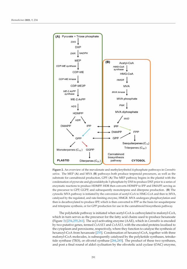
Biomedicines 2021, 9, 234
Figure 2. An overview of the mevalonate and methylerythritol 4-phosphate pathways in Cannabissativa. The MEP (A) and MVA (B) pathways both produce terpenoid precursors, as well as thesubstrate for cannabinoid production, GPP. (A) The MEP pathway begins in the plastid with thecondensation of pyruvate and glyceraldehyde 3-phosphate by DXS to produce DXP, prior to a series ofenzymatic reactions to produce HDMPP. HDR then converts HDMPP to IPP and DMAPP, serving asthe precursor to GPP, GGPP, and subsequently monoterpene and diterpene production. (B) Thecytosolic MVA pathway is initiated by the conversion of acetyl-CoA to HMG-CoA and then to MVA,catalysed by the regulated, and rate-limiting enzyme, HMGR. MVA undergoes phosphorylation andthen is decarboxylated to produce IPP, which is then converted to FPP as the basis for sesquiterpeneand triterpene synthesis, or for GPP production for use in the cannabinoid biosynthesis pathway.
The polyketide pathway is initiated when acetyl-CoA is carboxylated to malonyl-CoA,which in turn serves as the precursor for the fatty acid chains used to produce hexanoate(Figure 3) [254,255,261]. The acyl-activating enzyme (AAE), which in Cannabis is encodedby two putative genes, termed CsAAE1 and CsAAE3, with the encoded proteins localised tothe cytoplasm and peroxisome, respectively, where they function to catalyse the synthesis ofhexanoyl-CoA from hexanoate [255]. Condensation of hexanoyl-CoA, together with threemalonyl-CoA molecules, is subsequently catalysed by the polyketide synthases, tetrake-tide synthase (TKS), or olivetol synthase [284,285]. The product of these two synthases,and post a final round of aldol cyclisation by the olivetolic acid cyclase (OAC) enzyme,
291

Biomedicines 2021, 9, 234
is olivetolic acid (OA) [284]. Via the utilisation of GPP from the MVA pathway, OA isthen prenylated by geranylpyrophosphate:olivetolate geranyltransferase (GOT), to pro-duce CBGA [286–288]. The cis isomer of GPP, neryl diphosphate (NPP), can be used as asubstrate by GOT in place of GPP, to produce cannabinolic acid (CBNA) [289]. CBGA thenserves as the primary cannabinoid precursor for the synthesis of cannabichromenic acid(CBCA), THCA and CBDA, with the production of each of these three acids catalysed by aspecific oxidocyclisation enzyme, namely the CBCA, THCA and CBDA synthases [289–293].The use of divarinic acid as a substitute for OA by GOT, putatively produces the propylcannabinoid homolog, cannabigerovarinic acid (CBGVA) [286,294]. The aforementionedcannabinoid-specific synthases that yield CBCA, CBDA, and THCA can all recruit CBGVAto produce cannabidivarinic acid (CBDVA), cannabichromevarinic acid (CBCVA) andΔ9-tetrahydrocannabivarinic acid (THCVA), respectively [294–296]. The resulting cannabi-noids are maintained in their acidic forms until they are thermally decarboxylated toconvert them into their neutral forms [297–300].
Figure 3. An overview of the cannabinoid biosynthesis pathway in Cannabis sativa. Malonyl-CoA,formed from acetyl-CoA, is used downstream with hexanoyl-CoA to produce olivetolic acid (OA).Next, OA is used as substrate along with other biomolecules by the GOT enzyme to produce themajor cannabinoid precursor, CBGA. When GOT uses substrates additional to OA, such as divarinicacid or nerylpyrophosphate, a range of other minor cannabinoids are produced.
Research to date has primarily focused on the biosynthetic pathways and putativemedical benefits of the two major cannabinoids, THC and CBD. Therefore, the medicaland biological potential of the minor cannabinoids that also contribute to the total cannabi-noid profile of the Cannabis plant have been largely overlooked. The small proportionthat these minor cannabinoids contribute to the total cannabinoid profile of the Cannabis
292

Biomedicines 2021, 9, 234
plant presents a significant obstacle for in-depth analysis of their effects when consumed.A comprehensive, and ever-increasing list of naturally occurring minor phytocannabinoidshas been compiled based upon their derivation from THC, CBD, CBG (cannabigerol) andCBC, which represent the diversity that stems from variations to the three fundamentalcomponents of cannabinoids, including the (1) resorcinyl core; (2) isoprenyl residue, and;(3) resorcinyl side chain [20,301]. Eighty-two individual cannabinoids from 10 cannabinoidtypes, specifically the (1) CBG; (2) CBC; (3) CBD; (4) Δ9-THC; (5) Δ8-THC; (6) cannabicyclol(CBL); (7) cannabielsoin (CBE); (8) cannabinol (CBN); (9) cannabinodol (CBND), and; (10)cannabitriol (CBT) types, in addition to the miscellaneous types, and their transforma-tion products, as well as terpenoids, hydrocarbons, sugars and fatty acids are amongthe constituents that comprise the chemical cornucopia of glandular trichomes. Further,several minor oxygenated cannabinoids, cannabinoid metabolites, and cannabinoid esterspresent in Cannabis have yet to be isolated and/or experimentally validated but have beenidentified using a variety of spectroscopic techniques [302–304]. In addition, a number ofinteresting structural formations have been observed in some of the minor cannabinoids.For example, cannabioxepane (CBX) has a tetracyclic skeleton with a seven-memberedring, a structure not previously reported for a characterised cannabinoid, while cannabisolis a Δ9-THC dimer with a methylene bridge. However, it must be noted that the bind-ing affinity for specific CB receptors for these minor cannabinoids remains unknown,with some potentially not recognised, and therefore not bound by any known CB recep-tor [305,306]. The CBD derivative, cannabimovone, and the farnesyl prenylogue of CBG,sesquicannabigerol, were also spectroscopically characterised, with CB receptor bindingassays predicting receptor–cannabinoid affinity, highlighting the structural and poten-tial psychoactive diversity among the minor phytocannabinoids [307,308]. In addition tothe identification of their parent cannabinoid precursors, plausible biochemistry behindthe synthesis of these compounds is offered. However, the actual enzymatic productionof many of these minor cannabinoids remains to be determined. Furthermore, the non-enzymatic formation of some of the minor cannabinoids is certainly likely, but it remainsof interest to understand whether there is a greater portion of enzyme-catalysed reactionsin the production of the minor cannabinoids, or indeed whether there are alternativepathways, or even additional pathway entry points in the biosynthesis of cannabinoids,both minor and primary.
4. Minor Cannabinoids and Their Biological Interactions
There is mounting evidence that the minor cannabinoids described above share com-binations of many of the same molecular targets as THC and CBD, and therefore maypotentially have unique medical applications that cannot be achieved by THC or CBDalone. The THC propyl homologue, THCV, is a CB1 and CB2 competitive antagonistagainst CP55,940 and WIN55,21–2, acting with similar potency to that of THC [309,310].THCV also antagonised anandamide and methanandamide in mice vas deferens, atten-uating stimulated contractile responses [309]. More recently, THCV was shown to sim-ilarly displace CP55,940 from CB1 and CB2 in CHO cells, and contrary to previous as-sumptions, was shown to be a weak partial CB1 agonist at high doses [179]. Moreover,Zagzoog et al. [310] showed THCV to produce anxiolytic, hypothermic, anti-nociceptive,hypolocomotive, and cataleptic effects in vivo in mice. CB2 agonism by THCV was demon-strated to reduce inflammation and attenuate hyperalgesia in mice following injection ofcarrageenan and formalin, respectively [180]. Neuroprotective properties were observedin 6-hydroxydopamine lesioned rats, where THCV administration preceded maintenanceof tyrosine hydroxylase-positive neurons in this Parkinsonian model [311]. Similarly,THCV delayed onset of abnormal involuntary movements associated with Parkinson’sdisease in mice, and reduced their severity after administration following symptom on-set [181]. The in vitro demonstrated inhibition of GABA release by WIN55,21-2 at Purkinjecell synapses was reversed by THCV, which also prevented the action of WIN55,212–2when used in pre-incubation [312,313]. In vitro studies of insulin-resistant human hepa-
293

Biomedicines 2021, 9, 234
tocytes showed THCV restoration of insulin signaling mediated by CB1, while also im-proving glucose tolerance and increased sensitivity to insulin in mice obesity models [182].Antiepileptic properties were also established in vitro, specifically when THCV reducedboth the frequency and amplitude of epileptiform activity in rat piriform cortex slices [183].The majority of published studies have focused on the CB1 and CB2 receptors, but thein vitro activity of THCV has been observed for the TRPV1 to TRPV4 group of receptors, aswell as for the TRPA1 receptor [200–202]. THCV can enhance 5-HT1A receptor activation toproduce antipsychotic-like effects in rats [314], but does not affect other endocannabinoidsystem constituents such as PPARγ [315], FAAH [200], or MAGL [200]. One clinical trial inhumans where THCV was administered once daily for five days followed by intravenousadministration of THC suggested that THCV inhibited an increase in heart rate, protectedagainst verbal recall impairment, and reduced the subjective psychoactive intensity in-duced by THC [316]. Further, THCV affects brain regions associated with reward andaversive stimuli, as well as areas associated with cognitive control [317,318]
Recently, a four-carbon side chain variant, Δ9-tetrahydrocannabutol (THCB), was iso-lated and which showed CB1 and CB2 binding affinities similar to those of THC, within vivo mice studies suggesting potential analgesic and anti-inflammatory properties [176].Similarly, the recently identified seven-carbon side chain variant, THCP, was shown tobe able to bind to both CB1 and CB2 with 33 and 5 times greater affinity than THC, re-spectively, as well as to initiate catalepsy, hypothermia, analgesia, and reduce locomotion;all indications of potent full CB1 agonism [22]. THCA has been shown in rodent culturesupernatants to reduce the abundance of inflammatory and oxidant markers [319,320],though no other research to our knowledge of this nature has been published. In addition,Δ8-THC has been shown to possess higher antiemetic effects than THC [188], and hasbeen successfully trialed for repressing emesis in children [184]. Furthermore, in humans,Δ8-THC appears required to be administered at higher doses than THC to display a sim-ilar degree of psychoactive properties [321]. THCA is a 5-HT1A agonist [322], a PPARγagonist [323], and displays the same properties against TRP channels as does THC [200].However, little pharmacological, pharmacokinetic, or recent safety data are available forany of these compounds.
Improvements in seizure frequency has been reported in an epileptic patient coincid-ing with increased CBDV serum levels, after which in vitro studies confirmed that CBDV,at least, possesses the ability to influence GABA receptors; a finding that indicates a poten-tial avenue for anticonvulsant properties [324]. Further, in vitro analyses revealed CBDV tohave anticonvulsant effects in four seizure models, namely the (1) maximal electroshock-,(2) audiogenic-, (3) penytylenetetrazole-(PTZ), and (4) pilocarpine-induced seizure mod-els [325,326]. Using rat brain tissue samples, PTZ-induced seizures coincided with anincrease in Early growth response 1 (Egr1), Activity-regulated cytoskeleton-associated protein(Arc), Chemokine (C-C motif ) ligand 4 (Ccl4), Brain-derived neurotrophic factor (Bdnf ), and FBJosteosarcoma oncogene (Fos) gene expression [327]. Interestingly, the administration of CBDVwas shown to reduce the expression of all of these genes [327]. Additional seizure studiesidentified TRPV1 as the potential receptor modulating anti-seizure effects via the use oftrpv1 knockout mice which showed a reduced response to CBDV [328]. Desensitisation ofTRPV1, in addition to TRPV2, by both CBDV and CBD has been observed [199], while Ca2+
transients were induced in TRVP2-expressing HEK293 cells more potently by CBD than byCBDV. However, THC was a more potent inducer of Ca2+ transients than either CBDV orCBD [200]. Another study did alternately suggest that CBD was the more potent agonist ofTRPV2 than THC, but this study did not include the assessment of CBDV [329]. Cannabi-noid administration improved symptoms in mice models of Rett syndrome, includingmotor control and sociability [197], and through TRPA1, CBDV mediates anti-inflammatoryeffects in intestinal tissue of humans with ulcerative colitis [204]. Similar to CBD, CBDV in-hibits FAAH and anandamide reuptake [200]. However, unlike CBD, CBDV does not showaffinity for the CB1 or CB2 receptors [180]. CBDV may confer some benefit in patients withAutism Spectrum Disorder [330] and Duchenne muscular dystrophy [205]. CBDV did,
294

Biomedicines 2021, 9, 234
however, fail to alleviate the neuropathic pain associated with human immunodeficiencyvirus (HIV) [331], and in another study, the administration of CBDV induced DNA damagein human cell lines at concentrations similar to those observed in Cannabis consumers [332],indicating carcinogenicity potential for CBDV. However, CBDV has been safely trialed inhumans at a single 600 mg oral dose [330], and it remains to be determined whether CBDVwill be efficacious for other illnesses in clinical trial.
CBG has shown partial agonism of CB1 and CB2, α2-adrenceptor agonism and 5-HT1A antagonism, while exerting some minor anti-nociceptive and anxiolytic propertiesin vivo [179,333]. Mice models of inflammatory bowel disease (IBD) showed positive out-comes with CBG treatment including reductions in the level of reactive oxygen species inintestinal cells, as well as reduced nitric oxide concentration in macrophages through CB2modulation [194]. Further in vivo animal studies provided evidence for neuroprotectivityagainst symptoms of Huntington’s disease in 3-nitropropionate treated mice, with improve-ment in motor function, reduction in proinflammatory marker upregulation and increasedantioxidant defenses, with R6/2 mice showing a reduction in the expression profiles of sev-eral genes linked to the disease following CBG treatment [334]. Similarly, in vitro analysisof NSC-34 neuronal cells showed that CBG pre-treatment reduced both inflammation andthe expression of pro-inflammatory cytokines, and inhibited cell death resulting from thecell culture medium of lipopolysaccharide (LPS) stimulated RAW 264.7 macrophages [335].CBG shows a similar profile at TRP channels compared to CBD, with agonist propertiesat TRPV1 through to TRPV4, and at TRPA1, but antagonism at TRPM8 [200]. It is also ananandamide reuptake inhibitor [336], and an LPI inhibitor at GRP55 [97]. As for the propylanalogue of CBG, CBGV, very little information surrounding its clinical application exists,except to show that CBGV has activity at GPR55, TRPV3 and TRPV4 [198,201].
CBC use in a clinical setting, or in human trials, appears to be untested currently,and additionally, cannabichromevarin (CBCV) currently has even fewer studies dedicatedto it. However, CBC has seen some use in animal models and in vitro studies. CBC hasbeen shown to inhibit FAAH, MAGL, and anandamide reuptake [200,337], but has beendemonstrated to have no effect at TRPV1 or TRPV2. Further, CBC is a very weak CB1agonist [338–340], and only exhibits modest agonist properties at CB2. An early studysuggested that CBC, CBCV, and a CBC variant which lacks a carbon side chain, pos-sessed anti-inflammatory properties in rat edema models and varying anti-bacterial andanti-fungal properties [341]. More recently, CBC was seen to produce anti-inflammatoryeffects in LPS paw edema models in mice in CB1- and CB2-independent pathways and alsoproduce hypothermia, catalepsy, and locomotor suppression [342]. The authors went on tosuggest that the effects of CBC were altered in the presence of THC, with an additive effectagainst inflammation [342] and similarly, tail-flick tests revealed that subtle analgesic prop-erties of CBC were potentiated by its combination with THC [343]. Selective CB2, but notCB1 agonism, was exhibited by CBC on mouse pituitary tumour cells, and the persistentadministration of CBC caused desensitisation of CB2 receptors [344]. Intestinal studiessuggest that CBC confers some benefit against inflammation. However, this was potentiallyindependent of CB1, CB2, or TRPA1, the expression of which were all downregulated in thepresence of CBC in one study, but shown to be unchanged in another study [193,345]. Col-orectal cancer cell viability was attenuated through TRPM8 antagonism by CBG, as well asby the administration of CBD, CBDV, and CBC, albeit to lesser degrees [203]. Other studieshave indicated that CBC is not a potent antagonist of TRPM8, and instead suggest that CBD,CBG, THC, and THCA are more effective antagonists of TRPM8 [200,202]. Additionally,CBC, CBN, THC, THCV, THCA, CBDA, and CBG all induced intracellular Ca2+ increasesin HEK293 and rat DRG neurons through TRPA1 [200,202]. CBC has also shown promisein increasing neural stem cell viability in animal models (in vitro), mediated through ERKphosphorylation [346]. However, it is concerning that large amounts of CBC are required toproduce pharmacological effects [90], which implies that CBC may be difficult to implementin a human health context.
295

Biomedicines 2021, 9, 234
The binding affinity of CBN, and of its primary derivatives, to the two main cannabi-noid receptors was established in 2000, and showed rather unsurprisingly that alterations atcarbon atom positions 1, 3, and 9, resulted in significantly different affinities at both recep-tors [347]. An earlier study indicated CBN to have cataleptic, hypothermic, and locomotiveeffects, as did 11-hydroxy-CBN; a hepatic microsome CYP2C- and CYP3A4-catalysedmetabolite [348,349]. Additionally, CBN directly inhibited the activity of the human cy-tochrome P450 family 1 (CYP1) enzymes, CYP1A2 and CYP1B1 [350]. Assays of culturedneuronal cells expressing an inducible disease conferring huntingtin (Htt) protein, sug-gest that CBN has protective effects against cell death in vivo, with low toxicity even atthe high concentrations required for protectivity [351]. Interestingly, cannabinoid receptorloss has been indicated as a pathophysiology of Huntington’s disease [352,353], which maysuggest that the purported protective action of cannabinoids is independent of cannabinoidreceptor binding. Subcutaneously delivered CBN delayed the onset of amyotrophic lateralsclerosis (ALS) symptoms in murine models but failed to affect survival, so was postu-lated to mask the early spasticity associations without affecting disease progression [354].A synergistic effect of CBN with CBD at reducing mechanical sensitisation in rat massetermuscles was observed in one study, however high concentrations of CBD ameliorated theefficacy of CBN [355]. CBN has been reported to have no effect at FAAH, MAGL, or TRPV1,but acts as an agonist at TRPA1 and TRPV2 [200].
Via the use of in silico analyses, the even lesser-known cannabinoids, cannabiripsol(CBR) and CBT, are predicted to have cytochrome P450 inhibitor activity [356]. In anotherin silico study, CBL, CBT, and CBE were assessed, and ranked in this order, to haveacetylcholinesterase-inhibiting function. However, their inhibitory effects were less thanthose of THC, CBN, and CBDV [357]. Exactly how well in silico studies translate toclinical relevance, or even to in vitro and/or in vivo studies, restricts what conclusionscan be accurately drawn. Minor phytocannabinoids do represent an understudied portionof the Cannabis plant. Very few studies exist that have utilised an in vivo approach toascertain the viability of minor cannabinoids to potentially produce any significant medicalbenefits, and fewer still cover any human clinical trials. There has been indication that somecannabinoids exhibit synergistic action, and as a result there may be value in investigatingthe interactions among cannabinoids or constituents of the Cannabis plant.
5. Directions in Cannabis Development for Secondary Metabolite Production
The establishment of superior varieties of Cannabis has been the target for plant breed-ers since the domestication of this species. To produce new medically relevant Cannabisvarieties with elevated concentrations of specific minor cannabinoids, or to develop tech-niques to manipulate the cannabinoid biosynthetic pathway in other organisms, a deeperunderstanding of the genetics of the Cannabis plant is first required. Here we outline theprogress in relation to (1) the sequencing of the Cannabis genome, and (2) the potentialto molecularly manipulate the Cannabis plant itself for the altered production of specificcannabinoids. In this regard, we highlight the established success in Papaver somniferum(opium poppy), as a parallel example for maximising yield and the concentration of keysecondary metabolites of medical and commercial relevance.
5.1. Next-Generation Sequencing of the Cannabis Plant and Its Potential for Genetic Manipulation
Over the last 25 years, various experimental approaches have been employed to unveilthe wealth of information contained in the Cannabis genome. Using early DNA sequencingand karyotyping techniques, the X and Y sex chromosome characteristics of Cannabis wereuncovered, as were the diploid (2n = 20) genome sizes for male and female plants [358,359].The female Cannabis plant was revealed to have a genome size of 818 megabase (Mb),while the male Cannabis plant was determined to have a larger genome size of 843 Mb;specifically due to the larger size of the Y chromosome, compared to the X chromosome offemale plants [358]. Microsatellite markers have been employed as a tool for DNA typingCannabis, and these polymorphic short tandem repeat (STR) markers have been utilised as
296

Biomedicines 2021, 9, 234
a measurement of genetic relationships among cultivars [360–362]. More recently, the rapidchange in technologies surrounding Next-Generation Sequencing (NGS) platforms hasmeant that studies can unravel whole genomes in a fraction of the time required via the useof older methods. As a result, the first draft Cannabis reference genome, and transcriptome,were constructed in 2011 using the high THCA, low CBDA cultivar, ‘Purple Kush’, and thehigh CBDA, low THCA hemp strains, ‘Finola’ and ‘USO-31’ [363]. Using a PacBio long-readsequencing platform, the Purple Kush and Finola genomes were again sequenced in 2019to generate a physical and genetic map for Cannabis, and further distinguish the genes,and importantly the gene products (specifically, the encoded enzymes), underpinning thesecondary metabolite profiles responsible for the divergent chemotype between hemp andmarijuana cultivars [364,365].
Earlier work surrounding the chemotypic variance of cannabinoids observed inCannabis unveiled the relationship between THCA and CBDA synthase expression, describ-ing a single locus (B), with two codominant alleles, BD and BT [295]. A 1:1:2 segregationratio results in the production of three chemotypes of the B locus, including the (1) pureCBD (BD/BD homozygote), (2) pure THC (BT/BT homozygote), and (3) mixed CBD/THC(BD/BT heterozygote) chemotypes [295]. However, later studies based around NGS plat-forms indicated an alternate genetic model of synthase gene duplication and rearrangementat multiple linked loci, and that CBDA synthase is more ancient, has a greater affinityfor the CBGA substrate, and that the CBDA synthase locus is solely responsible for thecannabinoid chemotypes observed in Cannabis [363,365–369]. In an attempt to classifyvariability in chemotypes, and to associate genotype to chemotype in a diverse germplasmcollection, DNA sequence characterised amplified region (SCAR) markers associated withTHCA/CBDA synthases were assessed in 22 Cannabis varieties representing 2 fibre and 1drug type plants from East (n = 8), Central (n = 1), and South (n = 2) Asia, as well as fromEurope (n = 7) and of mixed (n = 4) domestication status [370]. This approach revealed avariability in cannabinoid profiles (CBD:THC) across ‘chemotype II’, or BD/BT equivalentplants, more than three-fold greater than previously observed, supporting the allelic variantand multiple loci prediction, when assuming that a heterozygote plant in a single locusmodel would have a 1:1 CBD:THC ratio [370].
Other large-scale genetic diversity studies using NGS, and which compared the evo-lutionary relationships between 340 Cannabis varieties from existing datasets, and fromother novel multiplexed libraries, highlighted the murky ancestry of the Cannabis plantresulting from generations of repeated rounds of selective breeding, and also provides anextensive data platform for future genotyping efforts [371]. Moreover, Lynch et al. [371]classed their assessed Cannabis varieties into three genetic groups, including (1) hemp, (2)narrow leaflet, and (3) broad leaflet drug types, in order to determine the genomic andgenetic variation of their population for the potential use of varieties from each group ineither agricultural or medicinal applications. The authors indicated unique cannabinoidand terpenoid profiles for each group, structured loosely around geographic origin of eachspecies, and noted the requirement for the inclusion of the putative Cannabis species, C. rud-eralis, in future studies to fully elucidate their genetic distinction and ancestral lineage [371].The development of expressed sequence tag simple sequence repeat (EST-SSR) markersto assess genetic diversity of 115 Cannabis genotypes also revealed geographical-basedclustering into 4 groupings, including the Northern China, Southern China, Central Chinaand Europe groupings [372]. Interestingly, a genetic similarity coefficient derived from45 of 117 randomly selected EST-SSRs markers revealed that despite physical proximityto the other Chinese varieties, Northern Chinese varieties had a greater similarity coeffi-cient to the European grouping, predicted to be related to latitude and day length [372].The analysis of inter simple sequence repeats (ISSR) of 27 native Chinese hemp varietiesidentified a similar geographic distribution to genetic distance relationship, while alsorevealing the hemp varieties were genetically diverse, yet primitive, a finding which addsfurther weight to the suggestion that the Cannabis plant originated in southern China andthen spread north [373].
297

Biomedicines 2021, 9, 234
The recent assembly and annotation of the mitochondrial genome of Cannabis usingNGS methods will also allow for similar studies to be performed to determine the extentof the genetic diversity among Cannabis varieties [374]. In addition, the assembly of twochloroplast genomes from different Cannabis varieties will aid in validating the phylogeneticrelationship of Cannabis among the Rosales order of the Plantae kingdom [375]. However,as with all sequencing, repeated efforts across diverse genotypic populations comparedagainst reference genomes will increase the accuracy and reliability of publicly availablerepositories. RNA sequencing as a tool for differentiating strains has been used withsome success, where the transcriptome isolated from cannabinoid-containing glandulartrichomes from different varieties allows for comparative analysis based on the cannabinoidand terpenoid chemical profiles [376,377]. As the regulatory landscape surrounding theuse of Cannabis evolves, and the value of the unique chemical profile of specific Cannabisvarieties is realised, breeders are likely to use these sequencing techniques to rapidlycharacterise and protect their ‘strains’. The development of such highly targeted databasesprovides the platform for precise manipulation of phenotypic or chemotypic traits inCannabis to deliver improved medical efficacy or novel therapeutics.
A forward and/or reverse genetics approach with the application of chemical muta-genesis agents, such as ethyl methanesulfonate (EMS), a mutagen that introduces pointmutations into the plant genome, is an effective approach for functional genomic assess-ments and effective plant breeding regimes, and has been successfully demonstrated in avariety of plant species, including hemp [9,378–383]. The application of alkylating agentssuch as EMS in a time-dependent manner causes a larger number of point mutationsacross the genome, compared to an irradiating method such as X-ray, or fast neutronbombardment, both of which produce much larger genome deletions and/or chromosomerearrangements [384–386]. Deletions ranging from 0.8 to 12 kilobases (kb) were producedin Arabidopsis using fast neutron bombardment, a widely used model plant species with anaverage gene density of one gene per 4.8 kb. The size of the genome alterations producedby this approach can, however, potentially cause the loss of function, or significantly alteredexpression of more than one gene. Therefore, a considerable drawback of using such anapproach is the time and effort required post-mutagenesis to identify a ‘causative mutation’.While the Arabidopsis genome is comparatively smaller than that of Cannabis, a similarpost-mutagenesis investigative strategy would likely be required in other plant specieswith nuclear genomes either of a similar or significantly larger-size [385,387]. Regardless,these types of methods require rather large numbers of plants to be effective as deletionsand point mutations are not site directed, which is a considerable limitation as even rapidstandard screening techniques demand intensive laboratory work [388–390].
Since the advent of the CRISPR/Cas9 gene-editing system in late 2012 [391], the abil-ity to manipulate plant genomes has become more cost efficient and less experimen-tally tedious when compared to the traditional genetic engineering approaches used byplant breeders in other crop species [392]. The CRISPR/Cas9 system effectively directssite-specific genome editing using RNA-guided, microbial-derived nucleases that initiatedouble-stranded DNA breaks in eukaryotic and bacterial systems [391,393]. The speci-ficity of this system greatly reduces the amount of off-target genome alterations comparedto more traditional transformation techniques. However, off-targeting has also been ob-served with CRISPR/Cas9 use, an inherent challenge when manipulating any biologicalsystem [394–397]. Earlier work was directed towards human applications, but increas-ingly this system has been utilised in plant systems, with examples in Arabidopsis, tobacco(Nicotiana tabacum), rice (Oryza sativa), lettuce (Lactuca sativa), maize (Zea mays), soybean(Glycine max) and wheat (Triticum aestivum) now documented [398–410]. By no meansan exhaustive list of CRISPR/Cas9-facilitated manipulation in plants, the above does,however, highlight the potential applicability of this targeted mutagenesis approach tomodulate specific biosynthetic pathways in Cannabis to produce superior varieties thatdisplay phenotypic and chemotypic traits of interest, and as a tool to discover key genesinvolved the production of minor cannabinoids. Transformation technologies has thus far
298

Biomedicines 2021, 9, 234
been conducted in hemp varieties only, and therefore require further development and con-siderable refinement for application in other Cannabis varieties. The first report of successfulhemp transformation emerged in 2001 [411], and two years later, a protocol for successfulAgrobacterium tumefaciens-mediated transformation of tissue cultured hemp callus wasimplemented [412]. More recently, Wahby et al. [413,414] successfully transformed hempusing both A. tumefaciens and A. rhizogenes, establishing the initial protocol for hairy rootculture in Cannabis, a system used for the production of key phytochemicals. Despite thesesuccesses, Cannabis has proven to be a difficult plant species to transform with such vari-ables as variety, plant age and the explant used for callus production, all demonstrated tobe crucial factors underpinning transformant regeneration efficiency [415]. As with anynovel plant transformation system, in order to overcome poor transformation efficiency,optimised protocols with respect to culture media, experimental approach, and selectedexplant material, will be required for routine and robust transformation of Cannabis.
5.2. Synthetic Production of Cannabinoids
Recently, the synthetic biology approach utilising microorganisms to produce high-quality cannabinoid products has removed the requirement for plant material [287,416].Luo and colleagues [287] were successful in producing CBG, CBD, THC and Δ9-THCV fromgalactose, via manipulation of the native MVA pathway of the yeast Saccharomyces cerevisiaepost the introduction of Cannabis genes encoding cannabinoid synthases, olivetolic acidsynthase and geranylpyrophosphate: olivetolate geranyltransferase. Production of THCAfrom CBGA through functional THCA synthase expression in the two yeast species, S.cerevisiae and Pichia pastoris, has been demonstrated. However, attempts to introduce thesame functionality in Escherichia coli, a bacterium, have proved unsuccessful [293,417].Over-expression of genes encoding enzymes in the MVA and prenyl diphosphate path-ways, also in S. cerevisiae, produced prenyl alcohol precursors required for terpenoid andcannabinoid synthesis [418], while expression of a functional aromatic prenyltransferasefrom Streptomyces resulted in THCA production from OA and DPP in the yeast, Koma-gataella phaffi [419]. These approaches present an attractive alternative with the abilityto conceivably produce large quantities of minor cannabinoids that are only found intrace amounts in planta, while also reducing and/or removing the costs, carbon emissions(associated with indoor growth; [420]) and environmental variables associated with theagricultural crop production. However, it should be noted that due to the criminalisationof Cannabis since the early 1930s, there are very few studies analysing water and energyuse associated with the cultivation of Cannabis, although undoubtedly, as research in thisarea becomes more prevalent, efficient horticultural practices will reduce the consumptionof water and energy for the large-scale cultivation of Cannabis.
5.3. Phenotypic Parameters Affecting Cannabis Yield and Potency
In Cannabis plants exhibiting an illicit drug chemotype (high THC), a primary con-cern, in conjunction with desired cannabinoid content, is overall biomass yield of femalefloral tissue. Consistent with other agriculturally significant species, Cannabis is sensi-tive to environmental variations which alter physiological characteristics affecting plantgrowth and yield potential. Early work on Cannabis flowering, uncovered the response tophotoperiodism [421,422], which has subsequently been exploited, particularly by illicitindoor growers, who can cultivate Cannabis year-round by manipulating the response toreduced photoperiod length [423]. Photoperiodism is a well-known biological responsecritical for development of branching and floral architecture in Cannabis, and as a result,has implications for yield potential [423,424]. A reduction in day length from 18 to 12 hinduces flowering, and maintenance of this regime for 8 weeks produces an acceptablefloral yield [423]. Elevated light intensity from 400 watts per square metre (W m−2),to 600 W m−2, produced a higher yield of floral tissue per plant in several chemotypeswhen grown indoors [424]. In addition, an increase in plant density from 16 to 20 plants m−2
reduced biomass yield of floral tissue in all 600 W m−2 treated plants [425]; a finding that
299
Biomedicines 2021, 9, 234
indicates that light interception is compromised at the lower canopy level in crowdedgrowth conditions. The use of different artificial lighting systems in controlled environ-ment greenhouse applications also affects yield, but there are ‘trade offs’ when using lightemitting diode (LED), versus high-intensity discharge (HID) light sources. HID lighting isgenerally of lower cost and generates greater photon flux density between 400 and 700 nm,while LED lighting has greater configurability for specified needs and emits substantiallyless heat than HID lighting; with both lighting options having similar electricity to photo-synthetic photon conversion efficiencies, expressed as, μmol J−1 [426–428]. The importanceof light quality has been demonstrated in cucumber (Cucumis sativus) where a significantincrease in dry weight was measured in plants grown under an ‘artificial solar spectrum’,produced by sulfur plasma and quartz-halogen lamps irradiating a light spectrum thatemulated standard sunlight, when compared with those plants provided with either flu-orescent or HID lighting [429]. Photosynthetic photon flux density significantly affectsharvestable floral biomass yield, while elevated UV-B radiation and electrical lightingpower density (W m−2) increased the ‘potency’ of Cannabis through an elevation in THCconcentration; all of which highlight the importance of light quantity and quality capture bythe photosynthetic apparatus of this species to improve the harvestable output of cultivatedCannabis [423,430–433].
Manipulating temperature conditions in indoor growth facilities has revealed a re-lationship with factors affecting plant growth and development. Rate of photosynthesis,water use efficiency, rate of transpiration, and leaf stomatal conductance, all increasedin Cannabis plants with a temperature increase from 20 to 30 ◦C, suggesting an optimaltemperature range for cultivation [431]. Temperature and photosynthetic rate are tightlylinked with the photosynthetic apparatus sensitive to fluctuations in temperature, re-sponding particularly with reduced Ribulose 1,5-bisphosphate (RuBP) regeneration andlowered stomatal aperture, which together decreased CO2 uptake; both rate reducingoutcomes [434–436]. It is worthwhile to note that Cannabis varieties are similarly sen-sitive to temperature where photosynthetic rate, water use efficiency, leaf number, andstem elongation, are modulated in response to temperature change [431,437,438]. Min-eral supplementation via fertilizer application has produced mixed results in terms ofbiomass and secondary metabolite concentration and/or profile composition in Cannabis.Cannabis was shown to be sensitive to nitrogen (N), phosphorus (P) and potassium (K)(NPK) supplementation, as well as the plant biostimulant, humic acid. The applicationof NPK reduced THC, CBN and CBD content, but increased CBG content in the Cannabisinflorescence, while the application of humic acid was found to significantly lower the THC,CBD, CBG, THCV, CBC, CBL and CBT content of the Cannabis inflorescence [439]. However,N supplementation alone increased hemp seed yield, plant height, chlorophyll content,while decreasing fibre yield [440]. The application of exogenous hormones during distinctdevelopmental phases of Cannabis growth has also produced mixed results in relation tosecondary metabolite content and biomass. Gibberellic acid (GA) application to wholeflowering plants with developed, resinous trichomes reduced chlorophyll levels, DXS ac-tivity, mono- and sesquiterpene levels, and THC content, while increasing HMGR activity,to suggest a degree of interference (either directly or indirectly) by GA to both the MVAand MEP pathways [441,442]. Abscisic acid (ABA) application at the vegetative stage ofCannabis development, increased chlorophyll a content, but reduced HMGR, THC and CBDcontent. In contrast, ABA application at the flowering stage of development decreased totalchlorophyll and HMGR content, and increased DXS activity and the content of THC in theflowers of female Cannabis plants, findings which again indicated either direct or indirectphytohormone-mediated interference of both the MVA and MEP pathways [441,442].
Alterations of the architecture of the Cannabis flower via the application of molecular-assisted breeding, or genetic engineering, are potential strategies to increase the floralyield of Cannabis. Alternatively, directed manipulation of the biosynthetic pathways byapplication of similar approaches leading to increased cannabinoid or terpenoid contentwould provide greater value via the targeted elevation of the exact concentration of spe-
300

Biomedicines 2021, 9, 234
cific secondary metabolites. Currently, research describing the implementation of suchstrategies in Cannabis are scarce. However, investigations of trichome development inArabidopsis and other plant species are not. The extremely well-annotated genome ofArabidopsis, combined with the ease that Arabidopsis can be genetically manipulated, identi-fies Arabidopsis for use in baseline studies that are potentially applicable to more valuableagricultural species. Indeed, Arabidopsis-based studies of trichome development haverevealed a cohort of genes of interest. As with the development of any specialised cell type,it is underpinned by a complex gene network, and in Arabidopsis, the protein productsencoded by the GLABROUS1 (GL1), GL2, GL3 and TRANSPARENT TESTA GLABROUSloci are responsible for various aspects of trichome morphogenesis, maturation, branchingand spatial variation [443–446]. Additional gene products have been identified as essentialfor correct branching patterns and trichome responses to hormones, with EMS-inducedmutation to the MYB encoding gene, TRIPTYCHON, resulting in the ‘nesting’, or groupingof trichomes with higher local densities [447,448]. A gene encoding a zinc-finger transcrip-tion factor from Arabidopsis, GLABROUS INFLORESCENCE STEMS, increased glandulartrichome density on the leaves, sepals, inflorescence and its branches, while also increas-ing the content of nicotine secretion into the glandular heads when over-expressed intobacco plants [449]. Similarly, overexpression of a serine proteinase inhibitor, SaPIN2a,from American nightshade (Solanum americanum) in transformed tobacco, significantlyincreased the branching and density of glandular trichomes [450]. Regulation of the ex-pression of the gene encoding the DXS synthase 2 (DXS2) enzyme, which is active in theMEP pathway in Cannabis, and also in tomato (Solanum lycopersicum) via a RNA silenc-ing approach, resulted in an increase in trichome density on tomato leaves and reducedthe accumulation of the monoterpene, β-phellandrene [451]. In cotton (Gossypium spp.),a mutation in the PIGMENT GLAND FORMATION locus, resulted in the expression of theglandless phenotype: a strategy adopted to remove toxic gossypol from cotton seeds forhuman consumption [452]. While the opposite phenotypic outcome of increased trichomedensity would be the desired result in Cannabis experimentation, when taken together,these findings highlight the importance of targeting specific genetic networks for molecularmanipulation to initiate the expression of desired and/or designer plant phenotypes.
Increasing the biomass of agriculturally valuable species is not a novel undertaking,and anthropogenic selection has perhaps inadvertently, been conducted by humans sincethe dawn of agriculture. Plant height is identified as a target for manipulation in relation tooverall biomass yield in maize and sorghum (Sorghum bicolor) [453], and in Cannabis grownfor fibre, stem length is an important parameter for fibre yield which is affected by plantdensity and soil N content [454,455]. The inverse is true for Cannabis varieties grown fortheir cannabinoid content, where reduced stem lengths produce a shorter overall plantstature and correlates with a greater photoassimilate input into reproductive tissues leadingto the development of floral architecture with increased accumulation of cannabinoidsand terpenoids [456]. Small [3] suggests that the value of drug chemotype varieties islinked to the development of ‘semi-dwarf Cannabis germplasm’, characterised by compact,congested flowers on short branches. Such plants ultimately produce more cannabinoidsdue to greater resource partitioning into floral and trichome development and are ofshort enough stature that they can be grown at high indoor densities where the artificialenvironment is readily manipulated to produce greater amounts of secondary metabolites.The combination of key phenotypic traits associated with increased secondary metaboliteaccumulation, including dense compact floral arrangements, and semi-dwarf stature,and with novel chemotypic traits that confer targeted medical efficacy epitomises thenew varieties (chemovars) to be pursued as part of a highly focused research strategy.Similar strategies that use marker assisted breeding and EMS to provide the molecularbasis to generate plants that produce elevated levels of desired compounds have beenundertaken in other medically significant plant species. Quantitative trait loci mapping ofArtemisia annua L. (sweet wormwood), a plant species which produces the anti-malarialcompound, artemisinin, provided the platform for marker assisted breeding programs to
301

Biomedicines 2021, 9, 234
increase artemisinin yield [457], and by extension, revealed both the pathway for similarresearch that would later be undertaken in opium poppy and the avenues for the futuredevelopment of similar strategies in Cannabis.
5.4. Papaver somniferum: Potential Parallels for Future Cannabis Research
With significant change surrounding the societal views and scientific inquiry intoCannabis on the horizon, it is important to look at past endeavours to envisage futuredirections. While Cannabis is a unique plant for its utility, Papaver somniferum (Papaver;opium poppy) rivals the versatility seen across Cannabis varieties, and given its long historyof human use, it is an excellent comparison to investigate. Papaver, otherwise known asopium poppy, is responsible for the production of the most medically significant alka-loids, including morphine, codeine, thebaine, oripavine and noscapine. These opioidsaccumulate in the phloem, particularly the mesocarp capsule of Papaver aerial tissues inspecialised cells called lactifers, which join to form a latex-containing network of anas-tomosing vessels [458–460]. The therapeutic efficacy of Papaver-derived opioids is betterunderstood than the secondary metabolites of Cannabis, and the scope of their effects isfar reaching. Morphine has been utilised for decades as one of the most widely used anal-gesics, effective in the post-operative clinical setting [461–463]. Codeine has been shownto be a less effective analgesic than morphine [464,465] but has historically been acceptedas the prevailing antitussive [466]. More recent evidence suggests however, that thereare more effective treatments, especially for chronic coughing disorders [467–469]. Addi-tionally, noscapine, another Papaver alkaloid, displays antitussive properties, and is alsosuggested to potentially mitigate stroke mortality and induce apoptosis in a broad setof cancers [470–473]. Thebaine and oripavine are not themselves used therapeutically.However, they are precursors for a wide range of semi-synthetic opioids including, but notlimited to, hydrocodone, oxycodone and hydromorphone, as well as naloxone, which isinterestingly employed to treat the acute effects of opioid overdose [474–479].
Given the multitude of efficacious compounds produced by Papaver, and the com-mercial value emanating from such, the desire to generate plant varieties that producespecific chemical profiles is one that is mirrored in Cannabis. While the latter is currentlyreliant on years of predominantly illicit breeding programs to produce plants with in-creased psychoactive properties, the development of novel Papaver varieties has alreadybeen established. EMS treatment of poppy seeds preceded the identification of a varietytermed top1 (thebaine oripavine poppy 1) which harboured a mutation leading to prema-ture arrest of the morphine and codeine biosynthesis pathway. The resulting top1 plantsdisplayed a pigmented latex, and the enhanced accumulation of thebaine and oripavine,but failed to produce either codeine or morphine [480]. Similarly, a reduction in codeine3-O-demethylase (CODM) activity, via either a viral-induced gene silencing (VIGS) strat-egy, or a fast neutron bombardment mutagenesis approach, yielded Papaver plants withenhanced codeine accumulation, but which were unable to synthesise morphine from acodeine substrate [481–483]. These high codeine Papaver varieties that harbour CODMpolymorphisms, provided a basis for a marker-assisted breeding platform, and to producePapaver chemotypes accumulating novel alkaloid profiles [483]. Similar actions utilisingCannabis may also mediate alterations to the cannabinoid biosynthesis pathways to producevarieties with elevated minor cannabinoid content. Sequencing of a high noscapine varietyof Papaver, termed HN1, led to the discovery of a 10 gene cluster responsible for noscapinebiosynthesis that was absent in either a high morphine (HM1)- or high thebaine (HT1)-producing variety of Papaver [484]. Generation of an F2 mapping population from HN1 andHM1 parents showed tight linkage of this gene cluster, revealing high noscapine-producingprogeny that were homozygous for the HN1 gene cluster, while heterozygosity, or absenceof the HN1 gene cluster, was associated with plant lines that produced low or undetectablelevels of noscapine, respectively [484]. The identification of the STORR ([S]- to [R]-reticuline)locus led to the development of high noscapine Papaver varieties with a non-functioningcytochrome P450-oxidoreductase fusion protein, inhibiting the [S]-reticuline conversion
302

Biomedicines 2021, 9, 234
to [R]-reticuline necessary for completion of the morphinan pathway [485–488]. A VIGSapproach has been successfully utilised to individually regulate the expression of six genesencoding enzymes involved in the final six conversion steps of [R]-reticuline to morphine,each of which were shown to alter the major alkaloid profile [489]. An RNA silencingapproach which employed a chimeric hairpin RNA to target all members of the multi-gene codeinone reductase family produced a non-narcotic, [S]-reticuline-accumulatingvariety of Papaver [490]. In the exploitation of the versatility of Papaver beyond narcotics,varieties with high food value have been established through EMS and gamma ray mu-tagenesis breeding programs to produce increased seed yield (5.66 g/capsule versus the3.39 g/capsule of control plants) with elevated levels of unsaturated seed oil and no nar-cotic production [491]. While this is not an exhaustive list of selectively bred, or engineeredPapaver varieties, the long-standing and successful development of Papaver varieties withsuperior phenotypic and/or chemotypic traits of interest certainly provides a reference forguiding Cannabis research strategies, which at present are comparatively in their infancy.Development of varieties producing high levels of alkaloid biosynthetic pathway inter-mediates is a promising indicator for the potential production of Cannabis varieties thatreliably produce high levels of minor cannabinoids or intermediates in the cannabinoidbiosynthesis pathway.
6. Conclusions
In summary, we have reviewed the current literature of several important aspectsof cannabinoid research outside of THC and CBD, which dominate discussion in theCannabis research field. Emerging research has begun to reveal the pharmacology andmolecular targets of the minor cannabinoids. Due to the wide spectrum of moleculareffects involved with cannabinoid consumption, it is clear that there are a range of medicalailments that could be addressed through endocannabinoid augmentation using secondarymetabolites of Cannabis. Here, we have illustrated that via the utilisation of specific minorcannabinoids, which share some, but not all targets of THC and CBD, the medical reach ofcannabinoid-containing pharmaceuticals could potentially be broadened. However, thereare many challenges that currently impede this possibility, even outside of the internationallegal environment. Firstly, there is further room for significant characterisation of minorcannabinoid pharmacology, and currently, disease-orientated preclinical and clinical trialsare lacking. Critically, techniques for producing cannabinoid isolates—even CBD andTHC—are still in their infancy, and this remains a clear barrier to large-scale commerciali-sation of pharmaceutical cannabinoids. Here, we have reviewed the currently availableliterature which covers the processes involved in the biosynthesis of cannabinoids, as wellas the techniques involved in the production of novel Cannabis chemotypes, includingmethods of improving yield that might be adopted from historically similar cases, suchas the opioid industry. Based on this historical example, and the existing literature, it islikely that a molecular genetic modification approach will be applied to Cannabis to gener-ate new opportunities for the improved yield of specific minor and major cannabinoidsin the near future. In conclusion, there are multiple enticing and potentially profitableopportunities for commercial and academic growth in the Cannabis market outside of THCand CBD, and here, we highlight some of the most important current perspectives of thisgrowing industry.
Author Contributions: Conceptualization and design, J.M.J.O., J.L.P., T.A.B., A.L.E. and C.P.L.G.;writing, J.M.J.O., J.L.P., T.A.B., A.L.E., L.J.N. and C.P.L.G.; review and editing, J.M.J.O., J.L.P.,T.A.B., A.L.E., L.J.N. and C.P.L.G. All authors have read and agreed to the published version ofthe manuscript.
Funding: J.M.J.O.’s research is supported by CannaPacific Pty Ltd., Australia.
Conflicts of Interest: The authors declare no conflict of interest.
303

Biomedicines 2021, 9, 234
References
1. Clarke, R.C.; Merlin, M.D. Letter to the Editor: Small, Ernest. 2015. Evolution and Classification of Cannabis sativa (Marijuana,Hemp) in Relation to Human Utilization. Bot. Rev. 2015, 81, 295–305. [CrossRef]
2. Schultes, R.E.; Klein, W.M.; Plowman, T.; Lockwood, T.E. Cannabis: An Example of Taxonomic Neglect. Bot. Museum Leafl.Harvard Univ. 1974, 23, 337–367.
3. Small, E. Evolution and Classification of Cannabis sativa (Marijuana, Hemp) in Relation to Human Utilization. Bot. Rev. 2015, 81,189–294. [CrossRef]
4. Small, E.; Cronquist, A. A Practical and Natural Taxonomy for Cannabis. Taxon 1976, 25, 405–435. [CrossRef]5. Small, E.; Naraine, S.G.U. Size Matters: Evolution of Large Drug-Secreting Resin Glands in Elite Pharmaceutical Strains of
Cannabis sativa (Marijuana). Genet. Resour. Crop Evol. 2016, 254, 349–359. [CrossRef]6. Salentijn, E.M.J.; Zhang, Q.; Amaducci, S.; Yang, M.; Trindade, L.M. New Developments in Fiber Hemp (Cannabis sativa L.)
Breeding. Ind. Crops Prod. 2015, 68, 32–41. [CrossRef]7. Mead, A. The Legal Status of Cannabis (Marijuana) and Cannabidiol (CBD) under U.S. Law. Epilepsy Behav. 2017, 70, 288–291.
[CrossRef]8. Amaducci, S.; Scordia, D.; Liu, F.H.; Zhang, Q.; Guo, H.; Testa, G.; Cosentino, S.L. Key Cultivation Techniques for Hemp in
Europe and China. Ind. Crop. Prod. 2015, 68, 2–16. [CrossRef]9. Bielecka, M.; Kaminski, F.; Adams, I.; Poulson, H.; Sloan, R.; Li, Y.; Larson, T.R.; Winzer, T.; Graham, I.A. Targeted Mutation of
Δ12 and Δ15 Desaturase Genes in Hemp Produce Major Alterations in Seed Fatty Acid Composition Including a High OleicHemp Oil. Plant Biotechnol. J. 2014, 12, 613–623. [CrossRef] [PubMed]
10. Callaway, J.C.; Tennilä, T.; Pate, D.W. Occurence of “Omega 3” Stearidonic Acid (Cis-6,9,12,15-Ocadecatetraenoic Acid) in Hemp(Cannabis sativa L.) Seed. J. Int. Hemp Assoc. 1996, 3, 61–63.
11. Dimic, E.; Romanic, R.; Vujasinovic, V. Essential Fatty Acids, Nutritive Value and Oxidative Stability of Cold Pressed Hempseed(Cannabis sativa L.) Oil from Different Varieties. Acta Aliment. 2009, 38, 229–236. [CrossRef]
12. Deferne, J.; Pate, D.W. Hemp Seed Oil: A Source of Valuable Essential Fatty Acids. J. Int. Hemp Assoc. 1996, 3, 4–7.13. Erasmus, U. Fats That Heal, Fats That Kill: The Complete Guide to Fats, Oils, Cholesterol, and Human Health, 3rd ed.; Alive Books:
Burnaby, BC, Canada, 1993.14. Stubbs, C.D.; Smith, A.D. The Modification of Mammalian Membrane Polyunsaturated Fatty Acid Composition in Relation to
Membrane Fluidity and Function. Biochim. Biophys. Acta 1984, 779, 89–137. [CrossRef]15. Callaway, J.C. Hempseed as a Nutritional Resource: An Overview. Euphytica 2004, 140, 65–72. [CrossRef]16. Harbige, L.S.; Layward, L.; Morris-Downes, M.M.; Dumonde, D.C.; Amor, S. The Protective Effects of Omega-6 Fatty Acids in
Experimental Autoimmune Encephalomyelitis (EAE) in Relation to Transforming Growth Factor-Beta 1 (TGF-B1) up-Regulationand Increased Prostaglandin E2 (PGE2) Production. Clin. Exp. Immunol. 2000, 122, 445–452. [CrossRef] [PubMed]
17. Prociuk, M.A.; Edel, A.L.; Richard, M.N.; Gavel, N.T.; Ander, B.P.; Dupasquier, C.M.C.; Pierce, G.N. Cholesterol-InducedStimulation of Platelet Aggregation Is Prevented by a Hempseed-Enriched Diet. Can. J. Physiol. Pharmacol. 2008, 86, 153–159.[CrossRef]
18. Clarke, R.C.; Merlin, M.D. Cannabis Domestication, Breeding History, Present-Day Genetic Diversity, and Future Prospects. CRCCrit. Rev. Plant Sci. 2016, 35, 293–327. [CrossRef]
19. Dayanandan, P.; Kaufman, P.B. Trichomes of Cannabis sativa L. (Cannabaceae). Am. J. Bot. 1976, 63, 578–591. [CrossRef]20. ElSohly, M.A.; Slade, D. Chemical Constituents of Marijuana: The Complex Mixture of Natural Cannabinoids. Life Sci. 2005, 78,
539–548. [CrossRef]21. Pertwee, R.G. (Ed.) Handbook of Cannabis; Oxford University Press: Oxford, UK, 2015. [CrossRef]22. Citti, C.; Linciano, P.; Russo, F.; Luongo, L.; Iannotta, M.; Maione, S.; Laganà, A.; Capriotti, A.L.; Forni, F.; Vandelli,
M.A.; et al. A Novel Phytocannabinoid Isolated from Cannabis sativa L. with an in Vivo Cannabimimetic Activity Higher thanΔ9-Tetrahydrocannabinol: Δ9-Tetrahydrocannabiphorol. Sci. Rep. 2019, 9, 20335. [CrossRef] [PubMed]
23. Babaei, M.; Ajdanian, L. Screening of Different Iranian Ecotypes of Cannabis under Water Deficit Stress. Sci. Hortic. 2020, 260.[CrossRef]
24. Linger, P.; Ostwald, A.; Haensler, J. Cannabis sativa L. Growing on Heavy Metal Contaminated Soil: Growth, Cadmium Uptakeand Photosynthesis. Biol. Plant. 2005, 49, 567–576. [CrossRef]
25. Bouquet, R.J. Cannabis. Bull. Narc. 1950, 2, 14–30.26. Amaducci, S.; Zatta, A.; Raffanini, M.; Venturi, G. Characterisation of Hemp (Cannabis sativa L.) Roots under Different Growing
Conditions. Plant Soil 2008, 313. [CrossRef]27. Small, E.; Marcus, D.; Butler, G.; McElroy, A.R. Apparent Increase in Biomass and Seed Productivity in Hemp (Cannabis sativa)
Resulting from Branch Proliferation Caused by the European Corn Borer (Ostrinia nubilalis). J. Ind. Hemp 2007, 12, 15–26.[CrossRef]
28. Government of Canada. Department of Justice. Available online: https://www.justice.gc.ca/eng/cj-jp/cannabis/ (accessed on15 January 2020).
29. Ney, L.J.; Matthews, A.; Bruno, R.; Felmingham, K.L. Cannabinoid Interventions for PTSD: Where to Next? Prog. Neuro-Psychopharmacol. Biol. Psychiatry 2019, 93, 124–140. [CrossRef]
304

Biomedicines 2021, 9, 234
30. Lattanzi, S.; Brigo, F.; Trinka, E.; Zaccara, G.; Cagnetti, C.; Del Giovane, C.; Silvestrini, M. Efficacy and Safety of Cannabidiol inEpilepsy: A Systematic Review and Meta-Analysis. Drugs 2018, 78, 1791–1804. [CrossRef] [PubMed]
31. Patel, S.; Hill, M.N.; Cheer, J.F.; Wotjak, C.T.; Holmes, A. The Endocannabinoid System as a Target for Novel Anxiolytic Drugs.Neurosci. Biobehav. Rev. 2017, 76, 56–66. [CrossRef] [PubMed]
32. Whiting, P.F.; Wolff, R.F.; Deshpande, S.; Di Nisio, M.; Duffy, S.; Hernandez, A.V.; Keurentjes, J.C.; Lang, S.; Misso, K.; Ryder,S.; et al. Cannabinoids for Medical Use: A Systematic Review and Meta-Analysis. JAMA J. Am. Med. Assoc. 2015, 313, 2456–2473.[CrossRef] [PubMed]
33. Wade, D.T.; Collin, C.; Stott, C.; Duncombe, P. Meta-Analysis of the Efficacy and Safety of Sativex (Nabiximols), on Spasticity inPeople with Multiple Sclerosis. Mult. Scler. 2010, 16, 707–714. [CrossRef] [PubMed]
34. Stevens, A.J.; Higgins, M.D. A Systematic Review of the Analgesic Efficacy of Cannabinoid Medications in the Management ofAcute Pain. Acta Anaesthesiol. Scand. 2017, 61, 268–280. [CrossRef]
35. Morales, P.; Jagerovic, N. Novel Approaches and Current Challenges with Targeting the Endocannabinoid System. Expert Opin.Drug Discov. 2020, 15, 917–930. [CrossRef]
36. Morales, P.; Hurst, D.P.; Reggio, P.H. Molecular Targets of the Phytocannabinoids: A Complex Picture. Prog. Chem. Org. Nat. Prod.2017, 103, 103–131. [CrossRef]
37. Sampson, P.B. Phytocannabinoid Pharmacology: Medicinal Properties of Cannabis sativa Constituents Aside from the “Big Two”.J. Nat. Prod. 2020, 84, 142–160. [CrossRef]
38. Gaoni, Y.; Mechoulam, R. Isolation, Structure, and Partial Synthesis of an Active Constituent of Hashish. J. Am. Chem. Soc. 1964,86, 1646–1647. [CrossRef]
39. Mechoulam, R.; Gaoni, Y. A Total Synthesis of Dl-Δ1-Tetrahydrocannabinol, the Active Constituent of Hashish. J. Am. Chem. Soc.1965, 87, 3273–3275. [CrossRef] [PubMed]
40. Devane, W.A.; Dysarz, F.A.; Johnson, M.R.; Melvin, L.S.; Howlett, A.C. Determination and Characterization of a CannabinoidReceptor in Rat Brain. Mol. Pharmacol. 1988, 34, 605–613.
41. Munro, S.; Thomas, K.L.; Abu-Shaar, M. Molecular Characterization of a Peripheral Receptor for Cannabinoids. Nature 1993, 365,61–65. [CrossRef]
42. Herkenham, M.; Lynn, A.B.; Little, M.D.; Johnson, M.R.; Melvin, L.S.; De Costa, B.R.; Rice, K.C. Cannabinoid Receptor Localizationin Brain. Proc. Natl. Acad. Sci. USA 1990, 87, 1932–1936. [CrossRef] [PubMed]
43. Devane, W.A.; Hanuš, L.; Breuer, A.; Pertwee, R.G.; Stevenson, L.A.; Griffin, G.; Gibson, D.; Mandelbaum, A.; Etinger, A.;Mechoulam, R. Isolation and Structure of a Brain Constituent That Binds to the Cannabinoid Receptor. Science 1992, 258,1946–1949. [CrossRef]
44. Mechoulam, R.; Ben-Shabat, S.; Hanus, L.; Ligumsky, M.; Kaminski, N.E.; Schatz, A.R.; Gopher, A.; Almog, S.; Martin, B.R.;Compton, D.R.; et al. Identification of an Endogenous 2-Monoglyceride, Present in Canine Gut, That Binds to CannabinoidReceptors. Biochem. Pharmacol. 1995, 50, 83–90. [CrossRef]
45. Sugiura, T.; Kondo, S.; Sukagawa, A.; Nakane, S.; Shinoda, A.; Itoh, K.; Yamashita, A.; Waku, K. 2-Arachidonoylglycerol: APossible Endogenous Cannabinoid Receptor Ligand in Brain. Biochem. Biophys. Res. Commun. 1995, 215, 89–97. [CrossRef][PubMed]
46. Liu, J.; Wang, L.; Harvey-White, J.; Osei-Hyiaman, D.; Razdan, R.; Gong, Q.; Chan, A.C.; Zhou, Z.; Huang, B.X.; Kim, H.Y.; et al. ABiosynthetic Pathway for Anandamide. Proc. Natl. Acad. Sci. USA 2006, 103, 13345–13350. [CrossRef]
47. Mechoulam, R.; Parker, L.A. The Endocannabinoid System and the Brain. Annu. Rev. Psychol. 2013, 64, 21–47. [CrossRef]48. Di Marzo, V. The Endocannabinoid System: Its General Strategy of Action, Tools for Its Pharmacological Manipulation and
Potential Therapeutic Exploitation. Pharmacol. Res. 2009, 60, 77–84. [CrossRef] [PubMed]49. Kano, M.; Ohno-Shosaku, T.; Hashimotodani, Y.; Uchigashima, M.; Watanabe, M. Endocannabinoid-Mediated Control of Synaptic
Transmission. Physiol. Rev. 2009, 89, 309–380. [CrossRef] [PubMed]50. Tripathi, R.K.P. A Perspective Review on Fatty Acid Amide Hydrolase (FAAH) Inhibitors as Potential Therapeutic Agents. Eur. J.
Med. Chem. 2020, 188, 111953. [CrossRef] [PubMed]51. Ligresti, A.; Cascio, M.G.; Pryce, G.; Kulasegram, S.; Beletskaya, I.; De Petrocellis, L.; Saha, B.; Mahadevan, A.; Visintin, C.; Wiley,
J.L.; et al. New Potent and Selective Inhibitors of Anandamide Reuptake with Antispastic Activity in a Mouse Model of MultipleSclerosis. Br. J. Pharmacol. 2006, 147, 83–91. [CrossRef] [PubMed]
52. Glass, M.; Dragunow, M.; Faull, R.L.M. Cannabinoid Receptors in the Human Brain: A Detailed Anatomical and QuantitativeAutoradiographic Study in the Fetal, Neonatal and Adult Human Brain. Neuroscience 1997, 77, 299–318. [CrossRef]
53. Hua, T.; Vemuri, K.; Pu, M.; Qu, L.; Han, G.W.; Wu, Y.; Zhao, S.; Shui, W.; Li, S.; Korde, A.; et al. Crystal Structure of the HumanCannabinoid Receptor CB1. Cell 2016, 167, 750–762. [CrossRef]
54. Li, X.; Hua, T.; Vemuri, K.; Ho, J.H.; Wu, Y.; Wu, L.; Popov, P.; Benchama, O.; Zvonok, N.; Locke, K.; et al. Crystal Structure of theHuman Cannabinoid Receptor CB2. Cell 2019, 176, 459–467. [CrossRef]
55. Katona, I.; Sperlágh, B.; Sík, A.; Käfalvi, A.; Vizi, E.S.; Mackie, K.; Freund, T.F. Presynaptically Located CB1 Cannabinoid ReceptorsRegulate GABA Release from Axon Terminals of Specific Hippocampal Interneurons. J. Neurosci. 1999, 19, 4544–4558. [CrossRef]
56. Derkinderen, P.; Ledent, C.; Parmentier, M.; Girault, J.A. Cannabinoids Activate P38 Mitogen-Activated Protein Kinases throughCB1 Receptors in Hippocampus. J. Neurochem. 2001, 77, 957–960. [CrossRef]
305

Biomedicines 2021, 9, 234
57. Herkenham, M.; Lynn, A.B.; de Costa, B.R.; Richfield, E.K. Neuronal Localization of Cannabinoid Receptors in the Basal Gangliaof the Rat. Brain Res. 1991, 547, 267–274. [CrossRef]
58. Mátyás, F.; Yanovsky, Y.; Mackie, K.; Kelsch, W.; Misgeld, U.; Freund, T.F. Subcellular Localization of Type 1 CannabinoidReceptors in the Rat Basal Ganglia. Neuroscience 2006, 137, 337–361. [CrossRef]
59. Bidaut-Russell, M.; Devane, W.A.; Howlett, A.C. Cannabinoid Receptors and Modulation of Cyclic AMP Accumulation in the RatBrain. J. Neurochem. 1990, 55, 21–26. [CrossRef]
60. Kawamura, Y.; Fukaya, M.; Maejima, T.; Yoshida, T.; Miura, E.; Watanabe, M.; Ohno-Shosaku, T.; Kano, M. The CB1 CannabinoidReceptor Is the Major Cannabinoid Receptor at Excitatory Presynaptic Sites in the Hippocampus and Cerebellum. J. Neurosci.2006, 26, 2991–3001. [CrossRef]
61. Kreitzer, A.C.; Regehr, W.G. Cerebellar Depolarization-Induced Suppression of Inhibition Is Mediated by Endogenous Cannabi-noids. J. Neurosci. 2001, 21, RC174. [CrossRef] [PubMed]
62. Lévénès, C.; Daniel, H.; Soubrié, P.; Crépel, F. Cannabinoids Decrease Excitatory Synaptic Transmission and Impair Long-TermDepression in Rat Cerebellar Purkinje Cells. J. Physiol. 1998, 510, 867–879. [CrossRef]
63. Ohno-Shosaku, T.; Maejima, T.; Kano, M. Endogenous Cannabinoids Mediate Retrograde Signals from Depolarized PostsynapticNeurons to Presynaptic Terminals. Neuron 2001, 29, 729–738. [CrossRef]
64. Ney, L.J.; Akhurst, J.; Bruno, R.; Laing, P.A.F.; Matthews, A.; Felmingham, K.L. Dopamine, Endocannabinoids and TheirInteraction in Fear Extinction and Negative Affect in PTSD. Prog. Neuro-Psychopharmacol. Biol. Psychiatry 2021, 105, 110118.[CrossRef] [PubMed]
65. Balsevich, G.; Petrie, G.N.; Hill, M.N. Endocannabinoids: Effectors of Glucocorticoid Signaling. Front. Neuroendocrinol. 2017, 47,86–108. [CrossRef] [PubMed]
66. González-Mariscal, I.; Krzysik-Walker, S.M.; Doyle, M.E.; Liu, Q.R.; Cimbro, R.; Santa-Cruz Calvo, S.; Ghosh, S.; Cieala, A.;Moaddel, R.; Carlson, O.D.; et al. Human CB1 Receptor Isoforms, Present in Hepatocytes and β-Cells, Are Involved in RegulatingMetabolism. Sci. Rep. 2016, 6, 33302. [CrossRef]
67. Nogueiras, R.; Veyrat-Durebex, C.; Suchanek, P.M.; Klein, M.; Tschöp, J.; Caldwell, C.; Woods, S.C.; Wittmann, G.; Watanabe,M.; Liposits, Z.; et al. Peripheral, but Not Central, CB1 Antagonism Provides Food Intake-Independent Metabolic Benefits inDiet-Induced Obese Rats. Diabetes 2008, 57, 2977–2991. [CrossRef]
68. Ashton, J.C.; Friberg, D.; Darlington, C.L.; Smith, P.F. Expression of the Cannabinoid CB2 Receptor in the Rat Cerebellum: AnImmunohistochemical Study. Neurosci. Lett. 2006, 396, 113–116. [CrossRef] [PubMed]
69. Núñez, E.; Benito, C.; Pazos, M.R.; Barbachano, A.; Fajardo, O.; González, S.; Tolón, R.M.; Romero, J. Cannabinoid CB2 ReceptorsAre Expressed by Perivascular Microglial Cells in the Human Brain: An Immunohistochemical Study. Synapse 2004, 53, 208–213.[CrossRef] [PubMed]
70. Ross, R.A.; Coutts, A.A.; McFarlane, S.M.; Anavi-Goffer, S.; Irving, A.J.; Pertwee, R.G.; MacEwan, D.J.; Scott, R.H. Actions ofCannabinoid Receptor Ligands on Rat Cultured Sensory Neurones: Implications for Antinociception. Neuropharmacology 2001, 40,221–232. [CrossRef]
71. Van Sickle, M.D.; Duncan, M.; Kingsley, P.J.; Mouihate, A.; Urbani, P.; Mackie, K.; Stella, N.; Makriyannis, A.; Piomelli, D.;Davison, J.S.; et al. Identification and Functional Characterization of Brainstem Cannabinoid CB2 Receptors. Science 2005, 310,329–332. [CrossRef]
72. Wotherspoon, G.; Fox, A.; McIntyre, P.; Colley, S.; Bevan, S.; Winter, J. Peripheral Nerve Injury Induces Cannabinoid Receptor 2Protein Expression in Rat Sensory Neurons. Neuroscience 2005, 135, 235–245. [CrossRef]
73. Galiègue, S.; Mary, S.; Marchand, J.; Dussossoy, D.; Carrière, D.; Carayon, P.; Bouaboula, M.; Shire, D.; LE Fur, G.; Casellas, P.Expression of Central and Peripheral Cannabinoid Receptors in Human Immune Tissues and Leukocyte Subpopulations. Eur. J.Biochem. 1995, 232, 54–61. [CrossRef] [PubMed]
74. Lynn, A.B.; Herkenham, M. Localization of Cannabinoid Receptors and Nonsaturable High-Density Cannabinoid Binding Sitesin Peripheral Tissues of the Rat: Implications for Receptor-Mediated Immune Modulation by Cannabinoids. J. Pharmacol. Exp.Ther. 1994, 268, 1612–1623.
75. Facci, L.; Dal Toso, R.; Romanello, S.; Buriani, A.; Skaper, S.D.; Leon, A. Mast Cells Express a Peripheral Cannabinoid Receptorwith Differential Sensitivity to Anandamide and Palmitoylethanolamide. Proc. Natl. Acad. Sci. USA 1995, 92, 3376–3380.[CrossRef] [PubMed]
76. Lam, P.M.W.; Marczylo, T.H.; El-Talatini, M.; Finney, M.; Nallendran, V.; Taylor, A.H.; Konje, J.C. Ultra Performance LiquidChromatography Tandem Mass Spectrometry Method for the Measurement of Anandamide in Human Plasma. Anal. Biochem.2008, 380, 195–201. [CrossRef] [PubMed]
77. Fanelli, F.; Di Lallo, V.D.; Belluomo, I.; De Iasio, R.; Baccini, M.; Casadio, E.; Gasparini, D.I.; Colavita, M.; Gambineri, A.; Grossi,G.; et al. Estimation of Reference Intervals of Five Endocannabinoids and Endocannabinoid Related Compounds in HumanPlasma by Two Dimensional-LC/MS/MS. J. Lipid Res. 2012, 53, 481–493. [CrossRef] [PubMed]
78. Krumbholz, A.; Anielski, P.; Reisch, N.; Schelling, G.; Thieme, D. Diagnostic Value of Concentration Profiles of Glucocorticos-teroids and Endocannabinoids in Hair. Ther. Drug Monit. 2013, 35, 600–607. [CrossRef]
79. Mwanza, C.; Chen, Z.; Zhang, Q.; Chen, S.; Wang, W.; Deng, H. Simultaneous HPLC-APCI-MS/MS Quantification of EndogenousCannabinoids and Glucocorticoids in Hair. J. Chromatogr. B Anal. Technol. Biomed. Life Sci. 2016, 1028, 1–10. [CrossRef]
306

Biomedicines 2021, 9, 234
80. Voegel, C.D.; Baumgartner, M.R.; Kraemer, T.; Wüst, S.; Binz, T.M. Simultaneous Quantification of Steroid Hormones andEndocannabinoids (ECs) in Human Hair Using an Automated Supported Liquid Extraction (SLE) and LC-MS/MS—Insights intoEC Baseline Values and Correlation to Steroid Concentrations. Talanta 2021, 222, 121499. [CrossRef]
81. Ney, L.J.; Felmingham, K.L.; Bruno, R.; Matthews, A.; Nichols, D.S. Simultaneous Quantification of Endocannabinoids,Oleoylethanolamide and Steroid Hormones in Human Plasma and Saliva. J. Chromatogr. B Anal. Technol. Biomed. Life Sci.2020, 1152, 122252. [CrossRef]
82. Ney, L.J.; Stone, C.; Nichols, D.; Felmingham, K.L.; Bruno, R.; Matthews, A. Endocannabinoid Reactivity to Acute Stress:Investigation of the Relationship between Salivary and Plasma Levels. Biol. Psychol. 2021, 159, 108022. [CrossRef]
83. Matias, I.; Gatta-Cherifi, B.; Tabarin, A.; Clark, S.; Leste-Lasserre, T.; Marsicano, G.; Piazza, P.V.; Cota, D. EndocannabinoidsMeasurement in Human Saliva as Potential Biomarker of Obesity. PLoS ONE 2012, 7, e42399. [CrossRef] [PubMed]
84. Schuel, H.; Burkman, L.J.; Lippes, J.; Crickard, K.; Forester, E.; Piomelli, D.; Giuffrida, A. N-Acylethanolamines in HumanReproductive Fluids. Chem. Phys. Lipids 2002, 121, 211–227. [CrossRef]
85. Lam, M.P.Y.; Siu, S.O.; Lau, E.; Mao, X.; Sun, H.Z.; Chiu, P.C.N.; Yeung, W.S.B.; Cox, D.M.; Chu, I.K. Online Coupling ofReverse-Phase and Hydrophilic Interaction Liquid Chromatography for Protein and Glycoprotein Characterization. Anal. Bioanal.Chem. 2010, 398, 791–804. [CrossRef] [PubMed]
86. Lewis, S.E.M.; Rapino, C.; Di Tommaso, M.; Pucci, M.; Battista, N.; Paro, R.; Simon, L.; Lutton, D.; Maccarrone, M. Differences inthe Endocannabinoid System of Sperm from Fertile and Infertile Men. PLoS ONE 2012, 7, e47704. [CrossRef]
87. Malan, T.P.; Ibrahim, M.M.; Deng, H.; Liu, Q.; Mata, H.P.; Vanderah, T.; Porreca, F.; Makriyannis, A. CB2 Cannabinoid Receptor-Mediated Peripheral Antinociception. Pain 2001, 93, 239–245. [CrossRef]
88. Jaggar, S.I.; Hasnie, F.S.; Sellaturay, S.; Rice, A.S.C. The Anti-Hyperalgesic Actions of the Cannabinoid Anandamide and thePutative CB2 Receptor Agonist Palmitoylethanolamide in Visceral and Somatic Inflammatory Pain. Pain 1998, 76, 189–199.[CrossRef]
89. Katona, I.; Freund, T.F. Multiple Functions of Endocannabinoid Signaling in the Brain. Annu. Rev. Neurosci. 2012, 35, 529–558.[CrossRef] [PubMed]
90. Ligresti, A.; De Petrocellis, L.; Di Marzo, V. From Phytocannabinoids to Cannabinoid Receptors and Endocannabinoids: PleiotropicPhysiological and Pathological Roles through Complex Pharmacology. Physiol. Rev. 2016, 96, 1593–1659. [CrossRef]
91. Ryberg, E.; Larsson, N.; Sjögren, S.; Hjorth, S.; Hermansson, N.O.; Leonova, J.; Elebring, T.; Nilsson, K.; Drmota, T.; Greasley, P.J.The Orphan Receptor GPR55 Is a Novel Cannabinoid Receptor. Br. J. Pharmacol. 2007, 152, 1092–1101. [CrossRef] [PubMed]
92. Li, K.; Fichna, J.; Schicho, R.; Saur, D.; Bashashati, M.; MacKie, K.; Li, Y.; Zimmer, A.; Göke, B.; Sharkey, K.A.; et al. A Role forO-1602 and G Protein-Coupled Receptor GPR55 in the Control of Colonic Motility in Mice. Neuropharmacology 2013, 71, 255–263.[CrossRef]
93. Ross, G.R.; Lichtman, A.; Dewey, W.L.; Akbarali, H.I. Evidence for the Putative Cannabinoid Receptor (GPR55)-MediatedInhibitory Effects on Intestinal Contractility in Mice. Pharmacology 2012, 90, 55–65. [CrossRef]
94. Lin, X.H.; Yuece, B.; Li, Y.Y.; Feng, Y.J.; Feng, J.Y.; Yu, L.Y.; Li, K.; Li, Y.N.; Storr, M. A Novel CB Receptor GPR55 and Its LigandsAre Involved in Regulation of Gut Movement in Rodents. Neurogastroenterol. Motil. 2011, 23, 862-e342. [CrossRef] [PubMed]
95. Galiazzo, G.; Giancola, F.; Stanzani, A.; Fracassi, F.; Bernardini, C.; Forni, M.; Pietra, M.; Chiocchetti, R. Localization ofCannabinoid Receptors CB1, CB2, GPR55, and PPARα in the Canine Gastrointestinal Tract. Histochem. Cell Biol. 2018, 150, 187–205.[CrossRef] [PubMed]
96. Oka, S.; Nakajima, K.; Yamashita, A.; Kishimoto, S.; Sugiura, T. Identification of GPR55 as a Lysophosphatidylinositol Receptor.Biochem. Biophys. Res. Commun. 2007, 362, 928–934. [CrossRef] [PubMed]
97. Lauckner, J.E.; Jensen, J.B.; Chen, H.Y.; Lu, H.C.; Hille, B.; Mackie, K. GPR55 Is a Cannabinoid Receptor That Increases IntracellularCalcium and Inhibits M Current. Proc. Natl. Acad. Sci. USA 2008, 105, 2699–2704. [CrossRef] [PubMed]
98. Waldeck-Welermair, M.; Zoratti, C.; Osibow, K.; Balenga, N.; Goessnitzer, E.; Waldhoer, M.; Malli, R.; Graier, W.F. IntegrinClustering Enables Anandamide-Induced Ca2+ Signaling in Endothelial Cells via GPR55 by Protection against CB1-Receptor-Triggered Repression. J. Cell Sci. 2008, 121, 1704–1717. [CrossRef]
99. Liu, Z.; Lee, J.; Krummey, S.; Lu, W.; Cai, H.; Lenardo, M.J. The Kinase LRRK2 Is a Regulator of the Transcription Factor NFATThat Modulates the Severity of Inflammatory Bowel Disease. Nat. Immunol. 2011, 12, 1063–1070. [CrossRef]
100. Henstridge, C.M.; Balenga, N.A.; Schröder, R.; Kargl, J.K.; Platzer, W.; Martini, L.; Arthur, S.; Penman, J.; Whistler, J.L.; Kostenis,E.; et al. GPR55 Ligands Promote Receptor Coupling to Multiple Signalling Pathways. Br. J. Pharmacol. 2010, 160, 604–614.[CrossRef]
101. Badrichani, A.Z.; Stroka, D.M.; Bilbao, G.; Curiel, D.T.; Bach, F.H.; Ferran, C. Bcl-2 and Bcl-X(L) Serve an Anti-InflammatoryFunction in Endothelial Cells through Inhibition of NF-KB. J. Clin. Investig. 1999, 103, 543–553. [CrossRef]
102. Wu, C.S.; Chen, H.; Sun, H.; Zhu, J.; Jew, C.P.; Wager-Miller, J.; Straiker, A.; Spencer, C.; Bradshaw, H.; Mackie, K.; et al. GPR55,a G-Protein Coupled Receptor for Lysophosphatidylinositol, Plays a Role in Motor Coordination. PLoS ONE 2013, 8, e60314.[CrossRef]
103. Celorrio, M.; Rojo-Bustamante, E.; Fernández-Suárez, D.; Sáez, E.; Estella-Hermoso de Mendoza, A.; Müller, C.E.; Ramírez, M.J.;Oyarzábal, J.; Franco, R.; Aymerich, M.S. GPR55: A Therapeutic Target for Parkinson’s Disease? Neuropharmacology 2017, 125,319–332. [CrossRef]
307
Biomedicines 2021, 9, 234
104. Sawzdargo, M.; Nguyen, T.; Lee, D.K.; Lynch, K.R.; Cheng, R.; Heng, H.H.Q.; George, S.R.; O’Dowd, B.F. Identification andCloning of Three Novel Human G Protein-Coupled Receptor Genes GPR52, ΨGPR53 and GPR55: GPR55 Is Extensively Expressedin Human Brain. Mol. Brain Res. 1999, 64, 193–198. [CrossRef]
105. Schuelert, N.; McDougall, J.J. The Abnormal Cannabidiol Analogue O-1602 Reduces Nociception in a Rat Model of Acute Arthritisvia the Putative Cannabinoid Receptor GPR55. Neurosci. Lett. 2011, 500, 72–76. [CrossRef]
106. Deliu, E.; Sperow, M.; Console-Bram, L.; Carter, R.L.; Tilley, D.G.; Kalamarides, D.J.; Kirby, L.G.; Brailoiu, G.C.; Brailoiu, E.;Benamar, K.; et al. The Lysophosphatidylinositol Receptor GPR55 Modulates Pain Perception in the Periaqueductal Gray. Mol.Pharmacol. 2015, 88, 265–272. [CrossRef]
107. Carey, L.M.; Gutierrez, T.; Deng, L.; Lee, W.H.; Mackie, K.; Hohmann, A.G. Inflammatory and Neuropathic Nociception IsPreserved in GPR55 Knockout Mice. Sci. Rep. 2017, 7, 944. [CrossRef]
108. Gantz, I.; Muraoka, A.; Yang, Y.K.; Samuelson, L.C.; Zimmerman, E.M.; Cook, H.; Yamada, T. Cloning and ChromosomalLocalization of a Gene (GPR18) Encoding a Novel Seven Transmembrane Receptor Highly Expressed in Spleen and Testis.Genomics 1997, 42, 462–466. [CrossRef] [PubMed]
109. Kohno, M.; Hasegawa, H.; Inoue, A.; Muraoka, M.; Miyazaki, T.; Oka, K.; Yasukawa, M. Identification of N-Arachidonylglycineas the Endogenous Ligand for Orphan G-Protein-Coupled Receptor GPR18. Biochem. Biophys. Res. Commun. 2006, 347, 827–832.[CrossRef] [PubMed]
110. Kobayashi, K.; Fukuoka, T.; Obata, K.; Yamanaka, H.; Dai, Y.; Tokunaga, A.; Noguchi, K. Distinct Expression of TRPM8, TRPA1,and TRPV1 MRNAs in Rat Primary Afferent Neurons with Aδ/C-Fibers and Colocalization with Trk Receptors. J. Comp. Neurol.2005, 493, 596–606. [CrossRef]
111. Cavanaugh, D.J.; Chesler, A.T.; Jackson, A.C.; Sigal, Y.M.; Yamanaka, H.; Grant, R.; O’Donnell, D.; Nicoll, R.A.; Shah, N.M.; Julius,D.; et al. Trpv1 Reporter Mice Reveal Highly Restricted Brain Distribution and Functional Expression in Arteriolar SmoothMuscle Cells. J. Neurosci. 2011, 31, 5067–5077. [CrossRef] [PubMed]
112. Avelino, A.; Cruz, F. TRPV1 (Vanilloid Receptor) in the Urinary Tract: Expression, Function and Clinical Applications. Naunyn.Schmiedebergs. Arch. Pharmacol. 2006, 373, 287–299. [CrossRef]
113. Yang, Y.; Yang, H.; Wang, Z.; Mergler, S.; Wolosin, J.M.; Reinach, P.S. Functional TRPV1 Expression in Human Corneal Fibroblasts.Exp. Eye Res. 2013, 107, 121–129. [CrossRef] [PubMed]
114. Tóth, A.; Boczán, J.; Kedei, N.; Lizanecz, E.; Bagi, Z.; Papp, Z.; Édes, I.; Csiba, L.; Blumberg, P.M. Expression and Distribution ofVanilloid Receptor 1 (TRPV1) in the Adult Rat Brain. Mol. Brain Res. 2005, 135, 162–168. [CrossRef]
115. Caterina, M.J.; Rosen, T.A.; Tominaga, M.; Brake, A.J.; Julius, D. A Capsaicin-Receptor Homologue with a High Threshold forNoxious Heat. Nature 1999, 398, 436–441. [CrossRef]
116. Frederick, J.; Buck, M.E.; Matson, D.J.; Cortright, D.N. Increased TRPA1, TRPM8, and TRPV2 Expression in Dorsal Root Gangliaby Nerve Injury. Biochem. Biophys. Res. Commun. 2007, 358, 1058–1064. [CrossRef] [PubMed]
117. Shimosato, G.; Amaya, F.; Ueda, M.; Tanaka, Y.; Decosterd, I.; Tanaka, M. Peripheral Inflammation Induces Up-Regulation ofTRPV2 Expression in Rat DRG. Pain 2005, 119, 225–232. [CrossRef] [PubMed]
118. Saunders, C.I.; Kunde, D.A.; Crawford, A.; Geraghty, D.P. Expression of Transient Receptor Potential Vanilloid 1 (TRPV1) and 2(TRPV2) in Human Peripheral Blood. Mol. Immunol. 2007, 44, 1429–1435. [CrossRef]
119. Santoni, G.; Amantini, C.; Maggi, F.; Marinelli, O.; Santoni, M.; Nabissi, M.; Morelli, M.B. The TRPV2 Cation Channels: FromUrothelial Cancer Invasiveness to Glioblastoma Multiforme Interactome Signature. Lab. Investig. 2020, 100, 186–198. [CrossRef][PubMed]
120. Link, T.M.; Park, U.; Vonakis, B.M.; Raben, D.M.; Soloski, M.J.; Caterina, M.J. TRPV2 Has a Pivotal Role in Macrophage ParticleBinding and Phagocytosis. Nat. Immunol. 2010, 11, 232–241. [CrossRef]
121. Zhang, D.; Spielmann, A.; Wang, L.; Ding, G.; Huang, F.; Gu, Q.; Schwarz, W. Mast-Cell Degranulation Induced by PhysicalStimuli Involves the Activation of Transient-Receptor-Potential Channel TRPV2. Physiol. Res. 2012, 61, 113–124. [CrossRef]
122. Iwata, Y.; Ohtake, H.; Suzuki, O.; Matsuda, J.; Komamura, K.; Wakabayashi, S. Blockade of Sarcolemmal TRPV2 AccumulationInhibits Progression of Dilated Cardiomyopathy. Cardiovasc. Res. 2013, 99, 760–768. [CrossRef] [PubMed]
123. Lorin, C.; Vögeli, I.; Niggli, E. Dystrophic Cardiomyopathy: Role of TRPV2 Channels in Stretch-Induced Cell Damage. Cardiovasc.Res. 2015, 106, 153–162. [CrossRef]
124. Iwata, Y.; Katanosaka, Y.; Arai, Y.; Shigekawa, M.; Wakabayashi, S. Dominant-Negative Inhibition of Ca2+ Influx via TRPV2Ameliorates Muscular Dystrophy in Animal Models. Hum. Mol. Genet. 2009, 18, 84–834. [CrossRef]
125. Iwata, Y.; Wakabayashi, S.; Ito, S.; Kitakaze, M. Production of TRPV2-Targeting Functional Antibody Ameliorating DilatedCardiomyopathy and Muscular Dystrophy in Animal Models. Lab. Investig. 2020, 100, 324–337. [CrossRef]
126. Hisanaga, E.; Nagasawa, M.; Ueki, K.; Kulkarni, R.N.; Mori, M.; Kojima, I. Regulation of Calcium-Permeable TRPV2 Channel byInsulin in Pancreatic β-Cells. Diabetes 2009, 58, 174–184. [CrossRef] [PubMed]
127. Kanzaki, M.; Zhang, Y.Q.; Mashima, H.; Li, L.; Shibata, H.; Kojima, I. Translocation of a Calcium-Permeable Cation ChannelInduced by Insulin-like Growth Factor-I. Nat. Cell Biol. 1999, 1, 165–170. [CrossRef] [PubMed]
128. Aoyagi, K.; Ohara-Imaizumi, M.; Nishiwaki, C.; Nakamichi, Y.; Nagamatsu, S. Insulin/Phosphoinositide 3-Kinase PathwayAccelerates the Glucose-Induced First-Phase Insulin Secretion through TrpV2 Recruitment in Pancreatic β-Cells. Biochem. J. 2010,432, 375–386. [CrossRef] [PubMed]
308

Biomedicines 2021, 9, 234
129. Caterina, M.J.; Schumacher, M.A.; Tominaga, M.; Rosen, T.A.; Levine, J.D.; Julius, D. The Capsaicin Receptor: A Heat-ActivatedIon Channel in the Pain Pathway. Nature 1997, 389, 816–824. [CrossRef] [PubMed]
130. Smith, G.D.; Gunthorpe, M.J.; Kelsell, R.E.; Hayes, P.D.; Reilly, P.; Facer, P.; Wright, J.E.; Jerman, J.C.; Walhin, J.P.; Ooi, L.; et al.TRPV3 Is a Temperature-Sensitive Vanilloid Receptor-like Protein. Nature 2002, 418, 186–190. [CrossRef]
131. Güler, A.D.; Lee, H.; Iida, T.; Shimizu, I.; Tominaga, M.; Caterina, M. Heat-Evoked Activation of the Ion Channel, TRPV4.J. Neurosci. 2002, 22, 6408–6414. [CrossRef]
132. Xu, H.; Ramsey, I.S.; Kotecha, S.A.; Moran, M.M.; Chong, J.A.; Lawson, D.; Ge, P.; Lilly, J.; Silos-Santiago, I.; Xie, Y.; et al. TRPV3 Isa Calcium-Permeable Temperature-Sensitive Cation Channel. Nature 2002, 418, 181–186. [CrossRef]
133. Moqrich, A.; Hwang, S.W.; Earley, T.J.; Petrus, M.J.; Murray, A.N.; Spencer, K.S.R.; Andahazy, M.; Story, G.M.; Patapoutian,A. Impaired Thermosensation in Mice Lacking TRPV3, a Heat and Camphor Sensor in the Skin. Science 2005, 307, 1468–1472.[CrossRef] [PubMed]
134. Mandadi, S.; Sokabe, T.; Shibasaki, K.; Katanosaka, K.; Mizuno, A.; Moqrich, A.; Patapoutian, A.; Fukumi-Tominaga, T.;Mizumura, K.; Tominaga, M. TRPV3 in Keratinocytes Transmits Temperature Information to Sensory Neurons via ATP. PflugersArch. Eur. J. Physiol. 2009, 458, 1093–1102. [CrossRef] [PubMed]
135. Chung, M.K.; Lee, H.; Mizuno, A.; Suzuki, M.; Caterina, M.J. TRPV3 and TRPV4 Mediate Warmth-Evoked Currents in PrimaryMouse Keratinocytes. J. Biol. Chem. 2004, 279, 21569–21575. [CrossRef]
136. Todaka, H.; Taniguchi, J.; Satoh, J.I.; Mizuno, A.; Suzuki, M. Warm Temperature-Sensitive Transient Receptor Potential Vanilloid 4(TRPV4) Plays an Essential Role in Thermal Hyperalgesia. J. Biol. Chem. 2004, 279, 35133–35138. [CrossRef]
137. Watanabe, H.; Vriens, J.; Suh, S.H.; Benham, C.D.; Droogmans, G.; Nilius, B. Heat-Evoked Activation of TRPV4 Channels in aHEK293 Cell Expression System and in Native Mouse Aorta Endothelial Cells. J. Biol. Chem. 2002, 277, 47044–47051. [CrossRef][PubMed]
138. Peier, A.M.; Reeve, A.J.; Andersson, D.A.; Moqrich, A.; Earley, T.J.; Hergarden, A.C.; Story, G.M.; Colley, S.; Hogenesch, J.B.;McIntyre, P.; et al. A Heat-Sensitive TRP Channel Expressed in Keratinocytes. Science 2002, 296, 2046–2049. [CrossRef] [PubMed]
139. Xu, H.; Delling, M.; Jun, J.C.; Clapham, D.E. Oregano, Thyme and Clove-Derived Flavors and Skin Sensitizers Activate SpecificTRP Channels. Nat. Neurosci. 2006, 9, 628–635. [CrossRef]
140. Bang, S.; Yoo, S.; Yang, T.J.; Cho, H.; Hwang, S.W. Isopentenyl Pyrophosphate Is a Novel Antinociceptive Substance That InhibitsTRPV3 and TRPA1 Ion Channels. Pain 2011, 152, 1156–1164. [CrossRef]
141. Liu, X.; Bandyopadhyay, B.; Nakamoto, T.; Singh, B.; Liedtke, W.; Melvin, J.E.; Ambudkar, I. A Role for AQP5 in Activation ofTRPV4 by Hypotonicity: Concerted Involvement of AQP5 and TRPV4 in Regulation of Cell Volume Recovery. J. Biol. Chem. 2006,281, 15485–15495. [CrossRef]
142. Becker, D.; Blase, C.; Bereiter-Hahn, J.; Jendrach, M. TRPV4 Exhibits a Functional Role in Cell-Volume Regulation. J. Cell Sci. 2005.[CrossRef]
143. Vriens, J.; Watanabe, H.; Janssens, A.; Droogmans, G.; Voets, T.; Nilius, B. Cell Swelling, Heat, and Chemical Agonists Use DistinctPathways for the Activation of the Cation Channel TRPV4. Proc. Natl. Acad. Sci. USA 2004, 101, 396–401. [CrossRef] [PubMed]
144. Liedtke, W.; Choe, Y.; Martí-Renom, M.A.; Bell, A.M.; Denis, C.S.; Šali, A.; Hudspeth, A.J.; Friedman, J.M.; Heller, S. VanilloidReceptor-Related Osmotically Activated Channel (VR-OAC), a Candidate Vertebrate Osmoreceptor. Cell 2000, 103, 525–535.[CrossRef]
145. Shibasaki, K.; Tominaga, M.; Ishizaki, Y. Hippocampal Neuronal Maturation Triggers Post-Synaptic Clustering of BrainTemperature-Sensor TRPV4. Biochem. Biophys. Res. Commun. 2015, 458, 168–173. [CrossRef] [PubMed]
146. Zhang, L.; Papadopoulos, P.; Hamel, E. Endothelial TRPV4 Channels Mediate Dilation of Cerebral Arteries: Impairment andRecovery in Cerebrovascular Pathologies Related to Alzheimer’s Disease. Br. J. Pharmacol. 2013, 170, 661–670. [CrossRef]
147. Wissenbach, U.; Bödding, M.; Freichel, M.; Flockerzi, V. Trp12, a Novel Trp Related Protein from Kidney. FEBS Lett. 2000, 485,127–134. [CrossRef]
148. Liedtke, W.; Friedman, J.M. Abnormal Osmotic Regulation in Trpv4-/- Mice. Proc. Natl. Acad. Sci. USA 2003, 100, 13698–13703.[CrossRef] [PubMed]
149. Heckel, E.; Boselli, F.; Roth, S.; Krudewig, A.; Belting, H.G.; Charvin, G.; Vermot, J. Oscillatory Flow Modulates MechanosensitiveKlf2a Expression through Trpv4 and Trpp2 during Heart Valve Development. Curr. Biol. 2015, 25, 1354–1361. [CrossRef][PubMed]
150. Watanabe, H.; Davis, J.B.; Smart, D.; Jerman, J.C.; Smith, G.D.; Hayes, P.; Vriens, J.; Cairns, W.; Wissenbach, U.; Prenen, J.; et al.Activation of TRPV4 Channels (HVRL-2/MTRP12) by Phorbol Derivatives. J. Biol. Chem. 2002, 277, 13569–13577. [CrossRef]
151. Watanabe, H.; Vriens, J.; Prenen, J.; Droogmans, G.; Voets, T.; Nillus, B. Anandamide and Arachidonic Acid Use Epoxye-icosatrienoic Acids to Activate TRPV4 Channels. Nature 2003, 424, 434–438. [CrossRef] [PubMed]
152. McKemy, D.D.; Neuhausser, W.M.; Julius, D. Identification of a Cold Receptor Reveals a General Role for TRP Channels inThermosensation. Nature 2002, 416, 52–58. [CrossRef]
153. Peier, A.M.; Moqrich, A.; Hergarden, A.C.; Reeve, A.J.; Andersson, D.A.; Story, G.M.; Earley, T.J.; Dragoni, I.; McIntyre, P.; Bevan,S.; et al. A TRP Channel That Senses Cold Stimuli and Menthol. Cell 2002, 108, 705–715. [CrossRef]
154. Del Camino, D.; Murphy, S.; Heiry, M.; Barrett, L.B.; Earley, T.J.; Cook, C.A.; Petrus, M.J.; Zhao, M.; D’Amours, M.; Deering,N.; et al. TRPA1 Contributes to Cold Hypersensitivity. J. Neurosci. 2010, 30, 15165–15174. [CrossRef] [PubMed]
309

Biomedicines 2021, 9, 234
155. Story, G.M.; Peier, A.M.; Reeve, A.J.; Eid, S.R.; Mosbacher, J.; Hricik, T.R.; Earley, T.J.; Hergarden, A.C.; Andersson, D.A.; Hwang,S.W.; et al. ANKTM1, a TRP-like Channel Expressed in Nociceptive Neurons, Is Activated by Cold Temperatures. Cell 2003, 112,819–829. [CrossRef]
156. Bandell, M.; Story, G.M.; Hwang, S.W.; Viswanath, V.; Eid, S.R.; Petrus, M.J.; Earley, T.J.; Patapoutian, A. Noxious Cold IonChannel TRPA1 Is Activated by Pungent Compounds and Bradykinin. Neuron 2004, 41, 849–857. [CrossRef]
157. Karashima, Y.; Talavera, K.; Everaerts, W.; Janssens, A.; Kwan, K.Y.; Vennekens, R.; Nilius, B.; Voets, T. TRPA1 Acts as a ColdSensor in Vitro and in Vivo. Proc. Natl. Acad. Sci. USA 2009, 106, 1273–1278. [CrossRef]
158. McNamara, C.R.; Mandel-Brehm, J.; Bautista, D.M.; Siemens, J.; Deranian, K.L.; Zhao, M.; Hayward, N.J.; Chong, J.A.; Julius,D.; Moran, M.M.; et al. TRPA1 Mediates Formalin-Induced Pain. Proc. Natl. Acad. Sci. USA 2007, 104, 13525–13530. [CrossRef][PubMed]
159. Jordt, S.E.; Bautista, D.M.; Chuang, H.H.; McKemy, D.D.; Zygmunt, P.M.; Högestätt, E.D.; Meng, I.D.; Julius, D. Mustard Oils andCannabinoids Excite Sensory Nerve Fibres through the TRP Channel ANKTM1. Nature 2004, 427, 260–265. [CrossRef]
160. Dai, Y.; Wang, S.; Tominaga, M.; Yamamoto, S.; Fukuoka, T.; Higashi, T.; Kobayashi, K.; Obata, K.; Yamanaka, H.; Noguchi,K. Sensitization of TRPA1 by PAR2 Contributes to the Sensation of Inflammatory Pain. J. Clin. Investig. 2007, 117, 1979–1987.[CrossRef] [PubMed]
161. Nagata, K.; Duggan, A.; Kumar, G.; García-Añoveros, J. Nociceptor and Hair Cell Transducer Properties of TRPA1, a Channel forPain and Hearing. J. Neurosci. 2005, 25, 4052–4061. [CrossRef]
162. Bautista, D.M.; Jordt, S.E.; Nikai, T.; Tsuruda, P.R.; Read, A.J.; Poblete, J.; Yamoah, E.N.; Basbaum, A.I.; Julius, D. TRPA1 Mediatesthe Inflammatory Actions of Environmental Irritants and Proalgesic Agents. Cell 2006, 124, 1269–1282. [CrossRef]
163. Kremeyer, B.; Lopera, F.; Cox, J.J.; Momin, A.; Rugiero, F.; Marsh, S.; Woods, C.G.; Jones, N.G.; Paterson, K.J.; Fricker, F.R.; et al.A Gain-of-Function Mutation in TRPA1 Causes Familial Episodic Pain Syndrome. Neuron 2010, 66, 671–680. [CrossRef]
164. Xiong, W.; Cui, T.; Cheng, K.; Yang, F.; Chen, S.R.; Willenbring, D.; Guan, Y.; Pan, H.L.; Ren, K.; Xu, Y.; et al. CannabinoidsSuppress Inflammatory and Neuropathic Pain by Targeting A3 Glycine Receptors. J. Exp. Med. 2012, 209, 1121–1134. [CrossRef]
165. Hejazi, N.; Zhou, C.; Oz, M.; Sun, H.; Jiang, H.Y.; Zhang, L. Δ9-Tetrahydrocannabinol and Endogenous Cannabinoid AnandamideDirectly Potentiate the Function of Glycine Receptors. Mol. Pharmacol. 2006, 69, 991–997. [CrossRef] [PubMed]
166. Xiong, W.; Cheng, K.; Cui, T.; Godlewski, G.; Rice, K.C.; Xu, Y.; Zhang, L. Cannabinoid Potentiation of Glycine ReceptorsContributes to Cannabis-Induced Analgesia. Nat. Chem. Biol. 2011, 7, 296–303. [CrossRef]
167. O’Sullivan, S.E. An Update on PPAR Activation by Cannabinoids. Br. J. Pharmacol. 2016, 173, 1899–1910. [CrossRef]168. Russo, E.B.; Burnett, A.; Hall, B.; Parker, K.K. Agonistic Properties of Cannabidiol at 5-HT1a Receptors. Neurochem. Res. 2005, 30,
1037–1043. [CrossRef] [PubMed]169. Franklin, J.M.; Carrasco, G.A. Cannabinoid-Induced Enhanced Interaction and Protein Levels of Serotonin 5-HT2A and Dopamine
D2 Receptors in Rat Prefrontal Cortex. J. Psychopharmacol. 2012, 26, 1333–1347. [CrossRef]170. Franklin, J.M.; Carrasco, G.A. Cannabinoid Receptor Agonists Upregulate and Enhance Serotonin 2A (5-HT2A) Receptor Activity
via ERK1/2 Signaling. Synapse 2013, 67, 145–159. [CrossRef] [PubMed]171. Hill, M.N.; Campolongo, P.; Yehuda, R.; Patel, S. Integrating Endocannabinoid Signaling and Cannabinoids into the Biology and
Treatment of Posttraumatic Stress Disorder. Neuropsychopharmacology 2018, 43, 80–102. [CrossRef]172. Blessing, E.M.; Steenkamp, M.M.; Manzanares, J.; Marmar, C.R. Cannabidiol as a Potential Treatment for Anxiety Disorders.
Neurotherapeutics 2015, 12, 825–836. [CrossRef] [PubMed]173. Harrold, J.A.; Elliott, J.C.; King, P.J.; Widdowson, P.S.; Williams, G. Down-Regulation of Cannabinoid-1 (CB-1) Receptors in
Specific Extrahypothalamic Regions of Rats with Dietary Obesity: A Role for Endogenous Cannabinoids in Driving Appetite forPalatable Food? Brain Res. 2002, 952, 232–238. [CrossRef]
174. Jamshidi, N.; Taylor, D.A. Anandamide Administration into the Ventromedial Hypothalamus Stimulates Appetite in Rats. Br. J.Pharmacol. 2001, 134, 1151–1154. [CrossRef]
175. Portella, G.; Laezza, C.; Laccetti, P.; De Petrocellis, L.; Di Marzo, V.; Bifulco, M. Inhibitory Effects of Cannabinoid CB1 ReceptorStimulation on Tumor Growth and Metastatic Spreading: Actions on Signals Involved in Angiogenesis and Metastasis. FASEB J.2003, 17, 1771–1773. [CrossRef]
176. Linciano, P.; Citti, C.; Luongo, L.; Belardo, C.; Maione, S.; Vandelli, M.A.; Forni, F.; Gigli, G.; Laganà, A.; Montone,C.M.; et al. Isolation of a High-Affinity Cannabinoid for the Human CB1 Receptor from a Medicinal Cannabis sativa Variety:Δ9-Tetrahydrocannabutol, the Butyl Homologue of Δ9-Tetrahydrocannabinol. J. Nat. Prod. 2020, 83, 88–98. [CrossRef]
177. Wallace, M.J.; Blair, R.E.; Falenski, K.W.; Martin, B.R.; DeLorenzo, R.J. The Endogenous Cannabinoid System Regulates SeizureFrequency and Duration in a Model of Temporal Lobe Epilepsy. J. Pharmacol. Exp. Ther. 2003, 307, 129–137. [CrossRef]
178. Murillo-Rodríguez, E. The Role of the CB1 Receptor in the Regulation of Sleep. Prog. Neuro-Psychopharmacol. Biol. Psychiatry 2008,32, 1420–1427. [CrossRef]
179. Zagzoog, A.; Mohamed, K.A.; Kim, H.J.J.; Kim, E.D.; Frank, C.S.; Black, T.; Jadhav, P.D.; Holbrook, L.A.; Laprairie, R.B. In Vitroand in Vivo Pharmacological Activity of Minor Cannabinoids Isolated from Cannabis sativa. Sci. Rep. 2020, 10, 20405. [CrossRef]
180. Bolognini, D.; Costa, B.; Maione, S.; Comelli, F.; Marini, P.; Di Marzo, V.; Parolaro, D.; Ross, R.A.; Gauson, L.A.; Cascio, M.G.; et al.The Plant Cannabinoid Δ 9-Tetrahydrocannabivarin Can Decrease Signs of Inflammation and Inflammatory Pain in Mice. Br. J.Pharmacol. 2010, 160, 677–687. [CrossRef] [PubMed]
310
Biomedicines 2021, 9, 234
181. Espadas, I.; Keifman, E.; Palomo-Garo, C.; Burgaz, S.; García, C.; Fernández-Ruiz, J.; Moratalla, R. Beneficial Effects of thePhytocannabinoid Δ9-THCV in L-DOPA-Induced Dyskinesia in Parkinson’s Disease. Neurobiol. Dis. 2020, 141, 104892. [CrossRef][PubMed]
182. Wargent, E.T.; Zaibi, M.S.; Silvestri, C.; Hislop, D.C.; Stocker, C.J.; Stott, C.G.; Guy, G.W.; Duncan, M.; Di Marzo, V.; Cawthorne,M.A. The Cannabinoid Δ9-Tetrahydrocannabivarin (THCV) Ameliorates Insulin Sensitivity in Two Mouse Models of Obesity.Nutr. Diabetes 2013, 3, e68. [CrossRef] [PubMed]
183. Hill, A.J.; Weston, S.E.; Jones, N.A.; Smith, I.; Bevan, S.A.; Williamson, E.M.; Stephens, G.J.; Williams, C.M.; Whalley, B.J. 9-Tetrahydrocannabivarin Suppresses in Vitro Epileptiform and in Vivo Seizure Activity in Adult Rats. Epilepsia 2010, 51, 1522–1532.[CrossRef]
184. Abrahamov, A.; Abrahamov, A.; Mechoulam, R. An Efficient New Cannabinoid Antiemetic in Pediatric Oncology. Life Sci. 1995,56, 2097–2102. [CrossRef]
185. Darmani, N.A. Δ9-Tetrahydrocannabinol and Synthetic Cannabinoids Prevent Emesis Produced by the Cannabinoid CB1 ReceptorAntagonist/Inverse Agonist SR 141716A. Neuropsychopharmacology 2001, 24, 198–203. [CrossRef]
186. Darmani, N.A. The Cannabinoid CB1 Receptor Antagonist SR 141716A Reverses the Antiemetic and Motor Depressant Actionsof WIN 55, 212–2. Eur. J. Pharmacol. 2001, 430, 49–58. [CrossRef]
187. Darmani, N.A.; Sim-Selley, L.J.; Martin, B.R.; Janoyan, J.J.; Crim, J.L.; Parekh, B.; Breivogel, C.S. Antiemetic and Motor-DepressiveActions of CP55,940: Cannabinoid CB1 Receptor Characterization, Distribution, and G-Protein Activation. Eur. J. Pharmacol. 2003,459, 83–95. [CrossRef]
188. Darmani, N.A.; Janoyan, J.J.; Crim, J.; Ramirez, J. Receptor Mechanism and Antiemetic Activity of Structurally-Diverse Cannabi-noids against Radiation-Induced Emesis in the Least Shrew. Eur. J. Pharmacol. 2007, 563, 187–196. [CrossRef]
189. Lastres-Becker, I.; Cebeira, M.; De Ceballos, M.L.; Zeng, B.Y.; Jenner, P.; Ramos, J.A.; Fernández-Ruiz, J.J. Increased CannabinoidCB1 Receptor Binding and Activation of GTP-Binding Proteins in the Basal Ganglia of Patients with Parkinson’s Syndrome andof MPTP-Treated Marmosets. Eur. J. Neurosci. 2001, 14, 1827–1832. [CrossRef] [PubMed]
190. Sarfaraz, S.; Afaq, F.; Adhami, V.M.; Mukhtar, H. Cannabinoid Receptor as a Novel Target for the Treatment of Prostate Cancer.Cancer Res. 2005, 65, 1635–1641. [CrossRef]
191. Qamri, Z.; Preet, A.; Nasser, M.W.; Bass, C.E.; Leone, G.; Barsky, S.H.; Ganju, R.K. Synthetic Cannabinoid Receptor AgonistsInhibit Tumor Growth and Metastasis of Breast Cancer. Mol. Cancer Ther. 2009, 8, 3117–3129. [CrossRef]
192. Preet, A.; Qamri, Z.; Nasser, M.W.; Prasad, A.; Shilo, K.; Zou, X.; Groopman, J.E.; Ganju, R.K. Cannabinoid Receptors, CB1 andCB2, as Novel Targets for Inhibition of Non-Small Cell Lung Cancer Growth and Metastasis. Cancer Prev. Res. 2011, 4, 65–75.[CrossRef]
193. Izzo, A.A.; Capasso, R.; Aviello, G.; Borrelli, F.; Romano, B.; Piscitelli, F.; Gallo, L.; Capasso, F.; Orlando, P.; Di Marzo, V. InhibitoryEffect of Cannabichromene, a Major Non-Psychotropic Cannabinoid Extracted from Cannabis sativa, on Inflammation-InducedHypermotility in Mice. Br. J. Pharmacol. 2012, 166, 1444–1460. [CrossRef]
194. Borrelli, F.; Fasolino, I.; Romano, B.; Capasso, R.; Maiello, F.; Coppola, D.; Orlando, P.; Battista, G.; Pagano, E.; Di Marzo, V.;et al. Beneficial Effect of the Non-Psychotropic Plant Cannabinoid Cannabigerol on Experimental Inflammatory Bowel Disease.Biochem. Pharmacol. 2013, 85, 1306–1316. [CrossRef]
195. Gómez-Gálvez, Y.; Palomo-Garo, C.; Fernández-Ruiz, J.; García, C. Potential of the Cannabinoid CB2 Receptor as a Pharmacologi-cal Target against Inflammation in Parkinson’s Disease. Prog. Neuropsychopharmacol. Biol. Psychiatry 2016, 64, 200–208. [CrossRef][PubMed]
196. Javed, H.; Azimullah, S.; Haque, M.E.; Ojha, S.K. Cannabinoid Type 2 (CB2) Receptors Activation Protects against OxidativeStress and Neuroinflammation Associated Dopaminergic Neurodegeneration in Rotenone Model of Parkinson’s Disease. Front.Neurosci. 2016, 10, 321. [CrossRef]
197. Vigli, D.; Cosentino, L.; Raggi, C.; Laviola, G.; Woolley-Roberts, M.; De Filippis, B. Chronic Treatment with the PhytocannabinoidCannabidivarin (CBDV) Rescues Behavioural Alterations and Brain Atrophy in a Mouse Model of Rett Syndrome. Neuropharma-cology 2018, 140, 121–129. [CrossRef] [PubMed]
198. Anavi-Goffer, S.; Baillie, G.; Irving, A.J.; Gertsch, J.; Greig, I.R.; Pertwee, R.G.; Ross, R.A. Modulation of L-α-Lysophosphatidylinositol/GPR55 Mitogen-Activated Protein Kinase (MAPK) Signaling by Cannabinoids. J. Biol. Chem. 2012, 287, 91–104. [CrossRef]
199. Iannotti, F.A.; Hill, C.L.; Leo, A.; Alhusaini, A.; Soubrane, C.; Mazzarella, E.; Russo, E.; Whalley, B.J.; Di Marzo, V.; Stephens,G.J. Nonpsychotropic Plant Cannabinoids, Cannabidivarin (CBDV) and Cannabidiol (CBD), Activate and Desensitize TransientReceptor Potential Vanilloid 1 (TRPV1) Channels in Vitro: Potential for the Treatment of Neuronal Hyperexcitability. ACS Chem.Neurosci. 2014, 5, 1131–1141. [CrossRef]
200. De Petrocellis, L.; Ligresti, A.; Moriello, A.S.; Allarà, M.; Bisogno, T.; Petrosino, S.; Stott, C.G.; Di Marzo, V. Effects of Cannabinoidsand Cannabinoid-Enriched Cannabis Extracts on TRP Channels and Endocannabinoid Metabolic Enzymes. Br. J. Pharmacol. 2011,163, 1479–1494. [CrossRef]
201. De Petrocellis, L.; Orlando, P.; Moriello, A.S.; Aviello, G.; Stott, C.; Izzo, A.A.; di Marzo, V. Cannabinoid Actions at TRPV Channels:Effects on TRPV3 and TRPV4 and Their Potential Relevance to Gastrointestinal Inflammation. Acta Physiol. 2012, 204, 255–266.[CrossRef]
311

Biomedicines 2021, 9, 234
202. De Petrocellis, L.; Vellani, V.; Schiano-Moriello, A.; Marini, P.; Magherini, P.C.; Orlando, P.; Di Marzo, V. Plant-DerivedCannabinoids Modulate the Activity of Transient Receptor Potential Channels of Ankyrin Type-1 and Melastatin Type-8.J. Pharmacol. Exp. Ther. 2008, 325, 1007–1015. [CrossRef]
203. Borrelli, F.; Pagano, E.; Romano, B.; Panzera, S.; Maiello, F.; Coppola, D.; De Petrocellis, L.; Buono, L.; Orlando, P.; Izzo, A.A.Colon Carcinogenesis Is Inhibited by the TRPM8 Antagonist Cannabigerol, a Cannabis-Derived Non-Psychotropic Cannabinoid.Carcinogenesis 2014, 35, 2787–2797. [CrossRef] [PubMed]
204. Pagano, E.; Romano, B.; Iannotti, F.A.; Parisi, O.A.; D’Armiento, M.; Pignatiello, S.; Coretti, L.; Lucafò, M.; Venneri, T.; Stocco,G.; et al. The Non-Euphoric Phytocannabinoid Cannabidivarin Counteracts Intestinal Inflammation in Mice and CytokineExpression in Biopsies from UC Pediatric Patients. Pharmacol. Res. 2019, 149, 104464. [CrossRef]
205. Iannotti, F.A.; Pagano, E.; Moriello, A.S.; Alvino, F.G.; Sorrentino, N.C.; D’Orsi, L.; Gazzerro, E.; Capasso, R.; De Leonibus, E.; DePetrocellis, L.; et al. Effects of Non-Euphoric Plant Cannabinoids on Muscle Quality and Performance of Dystrophic Mdx Mice.Br. J. Pharmacol. 2019, 176, 1568–1584. [CrossRef]
206. García-Arencibia, M.; González, S.; de Lago, E.; Ramos, J.A.; Mechoulam, R.; Fernández-Ruiz, J. Evaluation of the NeuroprotectiveEffect of Cannabinoids in a Rat Model of Parkinson’s Disease: Importance of Antioxidant and Cannabinoid Receptor-IndependentProperties. Brain Res. 2007, 1134, 162–170. [CrossRef]
207. Di Marzo, V. Enhanced Levels of Endogenous Cannabinoids in the Globus Pallidus Are Associated with a Reduction in Movementin an Animal Model of Parkinson’s Disease. FASEB J. 2000, 14, 1432–1438. [CrossRef] [PubMed]
208. Fox, S.H.; Henry, B.; Hill, M.; Crossman, A.; Brotchie, J. Stimulation of Cannabinoid Receptors Reduces Levodopa-InducedDyskinesia in the MPTP-Lesioned Nonhuman Primate Model of Parkinson’s Disease. Mov. Disord. 2002, 17, 1180–1187. [CrossRef]
209. Morgese, M.G.; Cassano, T.; Cuomo, V.; Giuffrida, A. Anti-Dyskinetic Effects of Cannabinoids in a Rat Model of Parkinson’sDisease: Role of CB1 and TRPV1 Receptors. Exp. Neurol. 2007, 208, 110–119. [CrossRef]
210. Sañudo-Peña, M.C.; Patrick, S.L.; Khen, S.; Patrick, R.L.; Tsou, K.; Walker, J.M. Cannabinoid Effects in Basal Ganglia in a RatModel of Parkinson’s Disease. Neurosci. Lett. 1998, 248, 171–174. [CrossRef]
211. Donadelli, M.; Dando, I.; Zaniboni, T.; Costanzo, C.; Dalla Pozza, E.; Scupoli, M.T.; Scarpa, A.; Zappavigna, S.; Marra, M.;Abbruzzese, A.; et al. Gemcitabine/Cannabinoid Combination Triggers Autophagy in Pancreatic Cancer Cells through aROS-Mediated Mechanism. Cell Death Dis. 2011, 2, E152. [CrossRef]
212. Afrin, F.; Chi, M.; Eamens, A.L.; Duchatel, R.J.; Douglas, A.M.; Schneider, J.; Gedye, C.; Woldu, A.S.; Dun, M.D. Can Hemp Help?Low-THC Cannabis and Non-THC Cannabinoids for the Treatment of Cancer. Cancers 2020, 12. [CrossRef] [PubMed]
213. De Petrocellis, L.; Melck, D.; Palmisano, A.; Bisogno, T.; Laezza, C.; Bifulco, M.; Di Marzo, V. The Endogenous CannabinoidAnandamide Inhibits Human Breast Cancer Cell Proliferation. Proc. Natl. Acad. Sci. USA 1998, 95, 8375–8380. [CrossRef]
214. Cianchi, F.; Papucci, L.; Schiavone, N.; Lulli, M.; Magnelli, L.; Vinci, M.C.; Messerini, L.; Manera, C.; Ronconi, E.; Romagnani,P.; et al. Cannabinoid Receptor Activation Induces Apoptosis through Tumor Necrosis Factor α-Mediated Ceramide de NovoSynthesis in Colon Cancer Cells. Clin. Cancer Res. 2008, 14, 7691–7700. [CrossRef]
215. Blázquez, C.; Casanova, M.L.; Planas, A.; Del Pulgar, T.G.; Villanueva, C.; Fernández-Aceñero, M.J.; Aragonés, J.; Huffman, J.W.;Jorcano, J.L.; Guzmán, M. Inhibition of Tumor Angiogenesis by Cannabinoids. FASEB J. 2003, 17, 529–531. [CrossRef] [PubMed]
216. Cunha, J.M.; Carlini, E.A.; Pereira, A.E.; Ramos, O.L.; Pimentel, C.; Gagliardi, R.; Sanvito, W.L.; Lander, N.; Mechoulam, R.Chronic Administration of Cannabidiol to Healthy Volunteers and Epileptic Patients. Pharmacology 1980, 21, 175–185. [CrossRef][PubMed]
217. Rosenberg, E.C.; Tsien, R.W.; Whalley, B.J.; Devinsky, O. Cannabinoids and Epilepsy. Neurotherapeutics 2015, 12, 747–768.[CrossRef] [PubMed]
218. Stockings, E.; Zagic, D.; Campbell, G.; Weier, M.; Hall, W.D.; Nielsen, S.; Herkes, G.K.; Farrell, M.; Degenhardt, L. Evidence forCannabis and Cannabinoids for Epilepsy: A Systematic Review of Controlled and Observational Evidence. J. Neurol. Neurosurg.Psychiatry 2018, 89, 741–753. [CrossRef]
219. Karanian, D.A.; Karim, S.L.; Wood, J.A.T.; Williams, J.S.; Lin, S.; Makriyannis, A.; Bahr, B.A. Endocannabinoid EnhancementProtects against Kainic Acid-Induced Seizures and Associated Brain Damage. J. Pharmacol. Exp. Ther. 2007, 322, 1059–1066.[CrossRef]
220. Naidoo, V.; Karanian, D.A.; Vadivel, S.K.; Locklear, J.R.; Wood, J.A.T.; Nasr, M.; Quizon, P.M.P.; Graves, E.E.; Shukla, V.;Makriyannis, A.; et al. Equipotent Inhibition of Fatty Acid Amide Hydrolase and Monoacylglycerol Lipase—Dual Targets of theEndocannabinoid System to Protect against Seizure Pathology. Neurotherapeutics 2012, 9, 801–813. [CrossRef]
221. Devinsky, O.; Patel, A.D.; Cross, J.H.; Villanueva, V.; Wirrell, E.C.; Privitera, M.; Greenwood, S.M.; Roberts, C.; Checketts, D.;VanLandingham, K.E.; et al. Effect of Cannabidiol on Drop Seizures in the Lennox–Gastaut Syndrome. N. Engl. J. Med. 2018, 378,1888–1897. [CrossRef]
222. Devinsky, O.; Cross, J.H.; Laux, L.; Marsh, E.; Miller, I.; Nabbout, R.; Scheffer, I.E.; Thiele, E.A.; Wright, S. Trial of Cannabidiol forDrug-Resistant Seizures in the Dravet Syndrome. N. Engl. J. Med. 2017, 376, 2011–2020. [CrossRef] [PubMed]
223. Press, C.A.; Knupp, K.G.; Chapman, K.E. Parental Reporting of Response to Oral Cannabis Extracts for Treatment of RefractoryEpilepsy. Epilepsy Behav. 2015, 45, 49–52. [CrossRef] [PubMed]
224. Guggenhuber, S.; Monory, K.; Lutz, B.; Klugmann, M. AAV Vector-Mediated Overexpression of CB1 Cannabinoid Receptor inPyramidal Neurons of the Hippocampus Protects against Seizure-Induced Excitoxicity. PLoS ONE 2010, 5, e15707. [CrossRef][PubMed]
312
Biomedicines 2021, 9, 234
225. Riva, N.; Mora, G.; Sorarù, G.; Lunetta, C.; Ferraro, O.E.; Falzone, Y.; Leocani, L.; Fazio, R.; Comola, M.; Comi, G.; et al. Safety andEfficacy of Nabiximols on Spasticity Symptoms in Patients with Motor Neuron Disease (CANALS): A Multicentre, Double-Blind,Randomised, Placebo-Controlled, Phase 2 Trial. Lancet Neurol. 2019, 18, 155–164. [CrossRef]
226. Beal, J.E.; Olson, R.; Laubenstein, L.; Morales, J.O.; Bellman, P.; Yangco, B.; Lefkowitz, L.; Plasse, T.F.; Shepard, K.V. Dronabinol asa Treatment for Anorexia Associated with Weight Loss in Patients with AIDS. J. Pain Symptom Manag. 1995, 10, 89–97. [CrossRef]
227. Foltin, R.W.; Fischman, M.W.; Byrne, M.F. Effects of Smoked Marijuana on Food Intake and Body Weight of Humans Living in aResidential Laboratory. Appetite 1988, 11, 1–14. [CrossRef]
228. Mattes, R.D.; Engelman, K.; Shaw, L.M.; Elsohly, M.A. Cannabinoids and Appetite Stimulation. Pharmacol. Biochem. Behav. 1994,49, 187–195. [CrossRef]
229. Williams, C.M.; Rogers, P.J.; Kirkham, T.C. Hyperphagia in Pre-Fed Rats Following Oral Δ9-THC. Physiol. Behav. 1998, 65, 343–346.[CrossRef]
230. Feinberg, I.; Jones, R.; Walker, J.M.; Cavness, C.; March, J. Effects of High Dosage Delta-9-Tetrahydrocannabinol on Sleep Patternsin Man. Clin. Pharmacol. Ther. 1975, 14, 458–466. [CrossRef]
231. Freemon, F.R. The Effect of Chronically Administered Delta-9-Tetrahydrocannabinol upon the Polygraphically Monitored Sleepof Normal Volunteers. Drug Alcohol Depend. 1982, 10, 345–353. [CrossRef]
232. Pivik, R.T.; Zarcone, V.; Dement, W.C.; Hollister, L.E. Delta-9-Tetrahydrocannabinol and Synhexl: Effects on Human SleepPatterns. Clin. Pharmacol. Ther. 1972, 13, 426–435. [CrossRef]
233. Agarwal, N.; Pacher, P.; Tegeder, I.; Amaya, F.; Constantin, C.E.; Brenner, G.J.; Rubino, T.; Michalski, C.W.; Marsicano, G.; Monory,K.; et al. Cannabinoids Mediate Analgesia Largely via Peripheral Type 1 Cannabinoid Receptors in Nociceptors. Nat. Neurosci.2007, 10, 870–879. [CrossRef] [PubMed]
234. Meng, I.D.; Manning, B.H.; Martin, W.J.; Fields, H.L. An Analgesia Circuit Activated by Cannabinoids. Nature 1998, 395, 381–383.[CrossRef] [PubMed]
235. Ottani, A.; Leone, S.; Sandrini, M.; Ferrari, A.; Bertolini, A. The Analgesic Activity of Paracetamol Is Prevented by the Blockade ofCannabinoid CB1 Receptors. Eur. J. Pharmacol. 2006, 531, 280–281. [CrossRef]
236. Walker, J.M.; Hohmann, A.G.; Martin, W.J.; Strangman, N.M.; Huang, S.M.; Tsou, K. The Neurobiology of Cannabinoid Analgesia.Life Sci. 1999, 65, 665–673. [CrossRef]
237. Leweke, F.M.; Piomelli, D.; Pahlisch, F.; Muhl, D.; Gerth, C.W.; Hoyer, C.; Klosterkötter, J.; Hellmich, M.; Koethe, D. CannabidiolEnhances Anandamide Signaling and Alleviates Psychotic Symptoms of Schizophrenia. Transl. Psychiatry 2012, 2, e94. [CrossRef]
238. Lutz, B.; Marsicano, G.; Maldonado, R.; Hillard, C.J. The Endocannabinoid System in Guarding against Fear, Anxiety and Stress.Nat. Rev. Neurosci. 2015, 16, 705–718. [CrossRef] [PubMed]
239. Navarro, M.; Hernández, E.; Muñoz, R.M.; Del Arco, I.; Villanúa, M.A.; Carrera, M.R.A.; Rodríguez De Fonseca, F. AcuteAdministration of the CB1 Cannabinoid Receptor Antagonist SR 141716A Induces Anxiety-like Responses in the Rat. Neuroreport1997, 8, 491–496. [CrossRef]
240. Moreira, F.A.; Aguiar, D.C.; Guimarães, F.S. Anxiolytic-like Effect of Cannabinoids Injected into the Rat Dorsolateral Periaqueduc-tal Gray. Neuropharmacology 2007, 52, 958–965. [CrossRef]
241. Rey, A.A.; Purrio, M.; Viveros, M.P.; Lutz, B. Biphasic Effects of Cannabinoids in Anxiety Responses: CB1 and GABA B Receptorsin the Balance of Gabaergic and Glutamatergic Neurotransmission. Neuropsychopharmacology 2012, 37, 2624–2634. [CrossRef]
242. Ney, L.J.; Matthews, A.; Bruno, R.; Felmingham, K.L. Modulation of the Endocannabinoid System by Sex Hormones: Implicationsfor Posttraumatic Stress Disorder. Neurosci. Biobehav. Rev. 2018, 94, 302–320. [CrossRef]
243. Carter, G.T.; Flanagan, A.M.; Earleywine, M.; Abrams, D.I.; Aggarwal, S.K.; Grinspoon, L. Cannabis in Palliative Medicine:Improving Care and Reducing Opioid-Related Morbidity. Am. J. Hosp. Palliat. Med. 2011, 28, 297–303. [CrossRef]
244. Bar-Sela, G.; Vorobeichik, M.; Drawsheh, S.; Omer, A.; Goldberg, V.; Muller, E. The Medical Necessity for Medicinal Cannabis:Prospective, Observational Study Evaluating the Treatment in Cancer Patients on Supportive or Palliative Care. Evid.-BasedComplement. Altern. Med. 2013, 2013, 510392. [CrossRef] [PubMed]
245. Motwani, M.P.; Bennett, F.; Norris, P.C.; Maini, A.A.; George, M.J.; Newson, J.; Henderson, A.; Hobbs, A.J.; Tepper, M.;White, B.; et al. Potent Anti-Inflammatory and Pro-Resolving Effects of Anabasum in a Human Model of Self-Resolving AcuteInflammation. Clin. Pharmacol. Ther. 2018, 104, 675–686. [CrossRef]
246. Lucas, P. Rationale for Cannabis-Based Interventions in the Opioid Overdose Crisis. Harm Reduct. J. 2017, 14, 58. [CrossRef]247. Boehnke, K.F.; Litinas, E.; Clauw, D.J. Medical Cannabis Use Is Associated with Decreased Opiate Medication Use in a Retrospec-
tive Cross-Sectional Survey of Patients with Chronic Pain. J. Pain 2016, 17, 739–744. [CrossRef]248. Groce, E. The Health Effects of Cannabis and Cannabinoids: The Current State of Evidence and Recommendations for Research; National
Academies Press: Washington, DC, USA, 2017. [CrossRef]249. Dos Santos, R.G.; Guimarães, F.S.; Crippa, J.A.S.; Hallak, J.E.C.; Rossi, G.N.; Rocha, J.M.; Zuardi, A.W. Serious Adverse Effects of
Cannabidiol (CBD): A Review of Randomized Controlled Trials. Expert Opin. Drug Metab. Toxicol. 2020, 16, 517–526. [CrossRef][PubMed]
250. White, C.M. A Review of Human Studies Assessing Cannabidiol’s (CBD) Therapeutic Actions and Potential. J. Clin. Pharmacol.2019, 59, 923–934. [CrossRef] [PubMed]
251. Pauli, C.S.; Conroy, M.; Vanden Heuvel, B.D.; Park, S.H. Cannabidiol Drugs Clinical Trial Outcomes and Adverse Effects. Front.Pharmacol. 2020, 11, 63. [CrossRef]
313
Biomedicines 2021, 9, 234
252. Kuhathasan, N.; Dufort, A.; MacKillop, J.; Gottschalk, R.; Minuzzi, L.; Frey, B.N. The Use of Cannabinoids for Sleep: A CriticalReview on Clinical Trials. Exp. Clin. Psychopharmacol. 2019, 27, 383–401. [CrossRef] [PubMed]
253. Black, N.; Stockings, E.; Campbell, G.; Tran, L.T.; Zagic, D.; Hall, W.D.; Farrell, M.; Degenhardt, L. Cannabinoids for the Treatmentof Mental Disorders and Symptoms of Mental Disorders: A Systematic Review and Meta-Analysis. Lancet Psychiatry 2019, 6,995–1010. [CrossRef]
254. Marks, M.D.; Tian, L.; Wenger, J.P.; Omburo, S.N.; Soto-Fuentes, W.; He, J.; Gang, D.R.; Weiblen, G.D.; Dixon, R.A. Identification ofCandidate Genes Affecting Δ9-Tetrahydrocannabinol Biosynthesis in Cannabis sativa. J. Exp. Bot. 2009, 60, 3715–3726. [CrossRef]
255. Stout, J.M.; Boubakir, Z.; Ambrose, S.J.; Purves, R.W.; Page, J.E. The Hexanoyl-CoA Precursor for Cannabinoid Biosynthesis IsFormed by an Acyl-Activating Enzyme in Cannabis sativa Trichomes. Plant J. 2012, 71, 353–365. [CrossRef]
256. Kim, E.-S.; Mahlberg, P.G. Secretory Cavity Development in Glandular Trichomes of Cannabis sativa L. (Cannabaceae). Am. J. Bot.1991, 78, 220–229. [CrossRef]
257. Kim, E.S.; Mahlberg, P.G. Secretory Vesicle Formation in the Secretory Cavity of Glandular Trichomes of Cannabis sativa L.(Cannabaceae). Mol. Cells 2003, 15, 387–395. [CrossRef]
258. Mahlberg, P.G.; Eun, S.K. Accumulation of Cannabinoids in Glandular Trichomes of Cannabis (Cannabaceae). J. Ind. Hemp 2004,9, 15–36. [CrossRef]
259. Mahlberg, P.G.; Kim, E.-S. Cuticle Development on Glandular Trichomes of Cannabis sativa (Cannabaceae). Am. J. Bot. 1991, 78,1113–1122. [CrossRef]
260. Arigoni, D.; Sagner, S.; Latzel, C.; Eisenreich, W.; Bacher, A.; Zenk, M.H. Terpenoid Biosynthesis from 1-Deoxy-D-Xylulose inHigher Plants by Intramolecular Skeletal Rearrangement. Proc. Natl. Acad. Sci. USA 1997, 94, 10600–10605. [CrossRef] [PubMed]
261. Fellermeier, M.; Eisenreich, W.; Bacher, A.; Zenk, M.H. Biosynthesis of Cannabinoids: Incorporation Experiments with 13C-Labeled Glucoses. Eur. J. Biochem. 2001, 268, 1596–1604. [CrossRef]
262. Schwender, J.; Zeidler, J.; Gröner, R.; Müller, C.; Focke, M.; Braun, S.; Lichtenthaler, F.W.; Lichtenthaler, H.K. Incorporation of1-Deoxy-D-Xylulose into Isoprene and Phytol by Higher Plants and Algae. FEBS Lett. 1997. [CrossRef]
263. Botella-Pavía, P.; Besumbes, Ó.; Phillips, M.A.; Carretero-Paulet, L.; Boronat, A.; Rodríguez-Concepción, M. Regulation ofCarotenoid Biosynthesis in Plants: Evidence for a Key Role of Hydroxymethylbutenyl Diphosphate Reductase in Controlling theSupply of Plastidial Isoprenoid Precursors. Plant J. 2004, 40, 188–199. [CrossRef]
264. Phillips, M.A.; León, P.; Boronat, A.; Rodríguez-Concepción, M. The Plastidial MEP Pathway: Unified Nomenclature andResources. Trends Plant Sci. 2008, 13, 619–623. [CrossRef] [PubMed]
265. Hsieh, M.H.; Chang, C.Y.; Hsu, S.J.; Chen, J.J. Chloroplast Localization of Methylerythritol 4-Phosphate Pathway Enzymes andRegulation of Mitochondrial Genes in IspD and IspE Albino Mutants in Arabidopsis. Plant Mol. Biol. 2008, 66, 663–673. [CrossRef]
266. Bick, J.A.; Lange, B.M. Metabolic Cross Talk between Cytosolic and Plastidial Pathways of Isoprenoid Biosynthesis: UnidirectionalTransport of Intermediates across the Chloroplast Envelope Membrane. Arch. Biochem. Biophys. 2003, 415, 146–154. [CrossRef]
267. Buhaescu, I.; Izzedine, H. Mevalonate Pathway: A Review of Clinical and Therapeutical Implications. Clin. Biochem. 2007, 40,575–584. [CrossRef]
268. Goldstein, J.L.; Brown, M.S. Regulation of the Mevalonate Pathway. Nature 1990, 343, 425–430. [CrossRef]269. Miziorko, H.M. Enzymes of the Mevalonate Pathway of Isoprenoid Biosynthesis. Arch. Biochem. Biophys. 2011, 505, 131–143.
[CrossRef] [PubMed]270. Guirimand, G.; Simkin, A.J.; Papon, N.; Besseau, S.; Burlat, V.; St-Pierre, B.; Giglioli-Guivarc’h, N.; Clastre, M.; Courdavault, V.
Cycloheximide as a Tool to Investigate Protein Import in Peroxisomes: A Case Study of the Subcellular Localization of IsoprenoidBiosynthetic Enzymes. J. Plant Physiol. 2012, 169, 825–829. [CrossRef]
271. Simkin, A.J.; Guirimand, G.; Papon, N.; Courdavault, V.; Thabet, I.; Ginis, O.; Bouzid, S.; Giglioli-Guivarc’h, N.; Clastre, M.Peroxisomal Localisation of the Final Steps of the Mevalonic Acid Pathway in Planta. Planta 2011, 234, 903–914. [CrossRef][PubMed]
272. Vranová, E.; Coman, D.; Gruissem, W. Network Analysis of the MVA and MEP Pathways for Isoprenoid Synthesis. Annu. Rev.Plant Biol. 2013, 64, 665–700. [CrossRef]
273. Thabet, I.; Guirimand, G.; Courdavault, V.; Papon, N.; Godet, S.; Dutilleul, C.; Bouzid, S.; Giglioli-Guivarc’h, N.; Clastre,M.; Simkin, A.J. The Subcellular Localization of Periwinkle Farnesyl Diphosphate Synthase Provides Insight into the Role ofPeroxisome in Isoprenoid Biosynthesis. J. Plant Physiol. 2011, 168, 2110–2116. [CrossRef] [PubMed]
274. Campbell, M.; Hahn, F.M.; Poulter, C.D.; Leustek, T. Analysis of the Isopentenyl Disphosphate Isomerase Gene from Arabidopsisthaliana. Plant Mol. Biol. 1998, 36, 323–328. [CrossRef] [PubMed]
275. Okada, K.; Kasahara, H.; Yamaguchi, S.; Kawaide, H.; Kamiya, Y.; Nojiri, H.; Yamane, H. Genetic Evidence for the Role ofIsopentenyl Diphosphate Isomerases in the Mevalonate Pathway and Plant Development in Arabidopsis. Plant Cell Physiol. 2008,49, 604–616. [CrossRef]
276. Sapir-Mir, M.; Mett, A.; Belausov, E.; Tal-Meshulam, S.; Frydman, A.; Gidoni, D.; Eya, Y. Peroxisomal Localization of ArabidopsisIsopentenyl Diphosphate Isomerases Suggests That Part of the Plant Isoprenoid Mevalonic Acid Pathway Is Compartmentalizedto Peroxisomes. Plant Physiol. 2008, 148, 1219–1228. [CrossRef]
277. Burke, C.C.; Wildung, M.R.; Croteau, R. Geranyl Diphosphate Synthase: Cloning, Expression, and Characterization of ThisPrenyltransferase as a Heterodimer. Proc. Natl. Acad. Sci. USA 1999, 96, 13062–13067. [CrossRef]
314

Biomedicines 2021, 9, 234
278. Bouvier, F.; Suire, C.; D’Harlingue, A.; Backhaus, R.A.; Camara, B. Molecular Cloning of Geranyl Diphosphate Synthase andCompartmentation of Monoterpene Synthesis in Plant Cells. Plant J. 2000, 24, 241–252. [CrossRef] [PubMed]
279. Ogura, K.; Koyama, T. Enzymatic Aspects of Isoprenoid Chain Elongation. Chem. Rev. 1998, 98, 1263–1276. [CrossRef] [PubMed]280. Ohnuma, S.I.; Hirooka, K.; Tsuruoka, N.; Yano, M.; Ohto, C.; Nakane, H.; Nishino, T. A Pathway Where Polyprenyl Diphosphate
Elongates in Prenyltransferase: Insight into a Common Mechanism of Chain Length Determination of Prenyltransferases. J. Biol.Chem. 1998, 273, 26705–26713. [CrossRef] [PubMed]
281. Wang, K.; Ohnuma, S.I. Chain-Length Determination Mechanism of Isoprenyl Diphosphate Synthases and Implications forMolecular Evolution. Trends Biochem. Sci. 1999, 24, 445–451. [CrossRef]
282. Booth, J.K.; Bohlmann, J. Terpenes in Cannabis sativa—From Plant Genome to Humans. Plant Sci. 2019, 284, 67–72. [CrossRef]283. Oldfield, E.; Lin, F.Y. Terpene Biosynthesis: Modularity Rules. Angew. Chem. Int. Ed. Engl. 2012, 51, 1124–1137. [CrossRef]284. Gagne, S.J.; Stout, J.M.; Liu, E.; Boubakir, Z.; Clark, S.M.; Page, J.E. Identification of Olivetolic Acid Cyclase from Cannabis sativa
Reveals a Unique Catalytic Route to Plant Polyketides. Proc. Natl. Acad. Sci. USA 2012, 109, 12811–12816. [CrossRef]285. Taura, F.; Tanaka, S.; Taguchi, C.; Fukamizu, T.; Tanaka, H.; Shoyama, Y.; Morimoto, S. Characterization of Olivetol Synthase, a
Polyketide Synthase Putatively Involved in Cannabinoid Biosynthetic Pathway. FEBS Lett. 2009, 583, 2061–2066. [CrossRef]286. Fellermeier, M.; Zenk, M.H. Prenylation of Olivetolate by a Hemp Transferase Yields Cannabigerolic Acid, the Precursor of
Tetrahydrocannabinol. FEBS Lett. 1998, 427, 283–285. [CrossRef]287. Luo, X.; Reiter, M.A.; D’Espaux, L.; Wong, J.; Denby, C.M.; Lechner, A.; Zhang, Y.; Grzybowski, A.T.; Harth, S.; Lin, W.; et al.
Complete Biosynthesis of Cannabinoids and Their Unnatural Analogues in Yeast. Nature 2019, 567, 123–126. [CrossRef]288. Valliere, M.A.; Korman, T.P.; Woodall, N.B.; Khitrov, G.A.; Taylor, R.E.; Baker, D.; Bowie, J.U. A Cell-Free Platform for the
Prenylation of Natural Products and Application to Cannabinoid Production. Nat. Commun. 2019, 10, 565. [CrossRef]289. Taura, F.; Morimoto, S.; Shoyama, Y. Purification and Characterization of Cannabidiolic-Acid Synthase from Cannabis sativa L.
Biochemical Analysis of a Novel Enzyme That Catalyzes the Oxidocyclization of Cannabigerolic Acid to Cannabidiolic Acid.J. Biol. Chem. 1996, 271, 17411–17416. [CrossRef]
290. Morimoto, S.; Komatsu, K.; Taura, F.; Shoyama, Y. Purification and Characterization of Cannabichromenic Acid Synthase fromCannabis sativa. Phytochemistry 1998, 49, 1525–1529. [CrossRef]
291. Shoyama, Y.; Tamada, T.; Kurihara, K.; Takeuchi, A.; Taura, F.; Arai, S.; Blaber, M.; Shoyama, Y.; Morimoto, S.; Kuroki, R. Structureand Function of Δ1-Tetrahydrocannabinolic Acid (THCA) Synthase, the Enzyme Controlling the Psychoactivity of Cannabis sativa.J. Mol. Biol. 2012, 423, 96–105. [CrossRef]
292. Taura, F.; Morimoto, S.; Shoyama, Y.; Mechoulam, R. First Direct Evidence for the Mechanism of Δ1-Tetrahydrocannabinolie AcidBiosynthesis. J. Am. Chem. Soc. 1995, 117, 9766–9767. [CrossRef]
293. Taura, F.; Dono, E.; Sirikantaramas, S.; Yoshimura, K.; Shoyama, Y.; Morimoto, S. Production of Δ1-TetrahydrocannabinolicAcid by the Biosynthetic Enzyme Secreted from Transgenic Pichia Pastoris. Biochem. Biophys. Res. Commun. 2007, 361, 675–680.[CrossRef] [PubMed]
294. De Zeeuw, R.A.; Wijsbeek, J.; Brejmer, D.D.; Vree, T.B.; Van Ginneken, C.A.M.; Van Rossum, J.M. Cannabinoids with a Propyl SideChain in Cannabis: Occurrence and Chromatographic Behavior. Science 1972, 175, 778–779. [CrossRef] [PubMed]
295. De Meijer, E.P.M.; Bagatta, M.; Carboni, A.; Crucitti, P.; Moliterni, V.M.C.; Ranalli, P.; Mandolino, G. The Inheritance of ChemicalPhenotype in Cannabis sativa L. Genetics 2003, 163, 335–346.
296. Shoyama, Y.; Hirano, H.; Nishioka, I. Biosynthesis of Propyl Cannabinoid Acid and Its Biosynthetic Relationship with Pentyl andMethyl Cannabinoid Acids. Phytochemistry 1984, 23, 1909–1984. [CrossRef]
297. Kanter, S.L.; Musumeci, M.R.; Hollister, L.E. Quantitative Determination of Δ9-Tetrahydrocannabinol and Δ9-TetrahydrocannabinolicAcid in Marihuana by High-Pressure Liquid Chromatography. J. Chromatogr. A 1979, 171, 504–508. [CrossRef]
298. Perrotin-Brunel, H.; Buijs, W.; Van Spronsen, J.; Roosmalen, M.J.E.V.; Peters, C.J.; Verpoorte, R.; Witkamp, G.J. Decarboxylation ofΔ9-Tetrahydrocannabinol: Kinetics and Molecular Modeling. J. Mol. Struct. 2011, 987, 67–73. [CrossRef]
299. Shoyama, Y.; Yagi, M.; Nishioka, I.; Yamauchi, T. Biosynthesis of Cannabinoid Acids. Phytochemistry 1975, 14, 2189–2192.[CrossRef]
300. Veress, T.; Szanto, J.I.; Leisztner, L. Determination of Cannabinoid Acids by High-Performance Liquid Chromatography of TheirNeutral Derivatives Formed by Thermal Decarboxylation. I. Study of the Decarboxylation Process in Open Reactors. J. Chromatogr.A 1990. [CrossRef]
301. Hanuš, L.O.; Meyer, S.M.; Muñoz, E.; Taglialatela-Scafati, O.; Appendino, G. Phytocannabinoids: A Unified Critical Inventory.Nat. Prod. Rep. 2016, 33, 1357–1392. [CrossRef] [PubMed]
302. Ahmed, S.A.; Ross, S.A.; Slade, D.; Radwan, M.M.; Khan, I.A.; ElSohly, M.A. Structure Determination and Absolute Configurationof Cannabichromanone Derivatives from High Potency Cannabis sativa. Tetrahedron Lett. 2008, 49, 6050–6053. [CrossRef] [PubMed]
303. Ahmed, S.A.; Ross, S.A.; Slade, D.; Radwan, M.M.; Zulfiqar, F.; ElSohly, M.A. Cannabinoid Ester Constituents from High-PotencyCannabis sativa. J. Nat. Prod. 2008, 71, 536–542. [CrossRef]
304. Radwan, M.M.; Ross, S.A.; Slade, D.; Ahmed, S.A.; Zulfiqar, F.; Elsohly, M.A. Isolation and Characterization of New CannabisConstituents from a High Potency Variety. Planta Med. 2008, 74, 267–272. [CrossRef]
305. Pagani, A.; Scala, F.; Chianese, G.; Grassi, G.; Appendino, G.; Taglialatela-Scafati, O. Cannabioxepane, a Novel TetracyclicCannabinoid from Hemp, Cannabis sativa L. Tetrahedron 2011, 67, 3369–3373. [CrossRef]
315

Biomedicines 2021, 9, 234
306. Zulfiqar, F.; Ross, S.A.; Slade, D.; Ahmed, S.A.; Radwan, M.M.; Ali, Z.; Khan, I.A.; Elsohly, M.A. Cannabisol, a Novel Δ 9-THCDimer Possessing a Unique Methylene Bridge, Isolated from Cannabis sativa. Tetrahedron Lett. 2012, 53, 3560–3562. [CrossRef][PubMed]
307. Pollastro, F.; Taglialatela-Scafati, O.; Allarà, M.; Muñoz, E.; Di Marzo, V.; De Petrocellis, L.; Appendino, G. Bioactive PrenylogousCannabinoid from Fiber Hemp (Cannabis sativa). J. Nat. Prod. 2011, 74, 2019–2022. [CrossRef]
308. Taglialatela-Scafati, O.; Pagani, A.; Scala, F.; De Petrocellis, L.; Di Marzo, V.; Grassi, G.; Appendino, G. Cannabimovone,a Cannabinoid with a Rearranged Terpenoid Skeleton from Hemp (Eur. J. Org. Chem. 11/2010). Eur. J. Org. Chem. 2010, 2010,2023. [CrossRef]
309. Thomas, A.; Stevenson, L.A.; Wease, K.N.; Price, M.R.; Baillie, G.; Ross, R.A.; Pertwee, R.G. Evidence That the Plant CannabinoidΔ 9-Tetrahydrocannabivarin Is a Cannabinoid CB 1 and CB 2 Receptor Antagonist. Br. J. Pharmacol. 2005, 146, 917–926. [CrossRef][PubMed]
310. Pertwee, R.G.; Thomas, A.; Stevenson, L.A.; Ross, R.A.; Varvel, S.A.; Lichtman, A.H.; Martin, B.R.; Razdan, R.K. The PsychoactivePlant Cannabinoid, Δ 9-Tetrahydrocannabinol, Is Antagonized by Δ 8- and Δ 9-Tetrahydrocannabivarin in Mice in Vivo. Br. J.Pharmacol. 2007, 150, 586–594. [CrossRef]
311. García, C.; Palomo-Garo, C.; García-Arencibia, M.; Ramos, J.A.; Pertwee, R.G.; Fernández-Ruiz, J. Symptom-Relieving andNeuroprotective Effects of the Phytocannabinoid Δ 9-THCV in Animal Models of Parkinson’s Disease. Br. J. Pharmacol. 2011, 163,1495–1506. [CrossRef]
312. Ma, Y.L.; Weston, S.E.; Whalley, B.J.; Stephens, G.J. The Phytocannabinoid Δ 9-Tetrahydrocannabivarin Modulates InhibitoryNeurotransmission in the Cerebellum. Br. J. Pharmacol. 2008, 154, 204–215. [CrossRef]
313. Dennis, I.; Whalley, B.J.; Stephens, G.J. Effects of Δ 9-Tetrahydrocannabivarin on [35S]GTPγS Binding in Mouse Brain Cerebellumand Piriform Cortex Membranes. Br. J. Pharmacol. 2008. [CrossRef] [PubMed]
314. Cascio, M.G.; Zamberletti, E.; Marini, P.; Parolaro, D.; Pertwee, R.G. The Phytocannabinoid, Δ9-Tetrahydrocannabivarin, Can Actthrough 5-HT1A Receptors to Produce Antipsychotic Effects. Br. J. Pharmacol. 2015, 172, 1305–1318. [CrossRef]
315. O’Sullivan, S.E.; Bennett, A.J.; Kendall, D.A.; Randall, M.D. Cannabinoids and Peroxisome Proliferator-Activated Receptor γ(PPARg). In Proceedings of the 16th Annual Symposium on the Cannabinoids, Tihany, Hungary, 24–28 June 2006; Volume 59.
316. Englund, A.; Atakan, Z.; Kralj, A.; Tunstall, N.; Murray, R.; Morrison, P. The Effect of Five Day Dosing with THCV on THC-InducedCognitive, Psychological and Physiological Effects in Healthy Male Human Volunteers: A Placebo-Controlled, Double-Blind,Crossover Pilot Trial. J. Psychopharmacol. 2016, 30, 140–151. [CrossRef] [PubMed]
317. Tudge, L.; Williams, C.; Cowen, P.J.; McCabe, C. Neural Effects of Cannabinoid CB1 Neutral Antagonist Tetrahydrocannabivarinon Food Reward and Aversion in Healthy Volunteers. Int. J. Neuropsychopharmacol. 2015, 18, Pyu094. [CrossRef]
318. Rzepa, E.; Tudge, L.; McCabe, C. The CB1 Neutral Antagonist Tetrahydrocannabivarin Reduces Default Mode Network andIncreases Executive Control Network Resting State Functional Connectivity in Healthy Volunteers. Int. J. Neuropsychopharmacol.2016, 19, Pyv092. [CrossRef] [PubMed]
319. Moldzio, R.; Pacher, T.; Krewenka, C.; Kranner, B.; Novak, J.; Duvigneau, J.C.; Rausch, W.D. Effects of Cannabinoids Δ(9)-Tetrahydrocannabinol, Δ(9)-Tetrahydrocannabinolic Acid and Cannabidiol in MPP+ Affected Murine Mesencephalic Cultures.Phytomedicine 2012, 19, 819–824. [CrossRef]
320. Verhoeckx, K.C.M.; Korthout, H.A.A.J.; Van Meeteren-Kreikamp, A.P.; Ehlert, K.A.; Wang, M.; Van Der Greef, J.; Rodenburg,R.J.T.; Witkamp, R.F. Unheated Cannabis sativa Extracts and Its Major Compound THC-Acid Have Potential Immuno-ModulatingProperties Not Mediated by CB1 and CB2 Receptor Coupled Pathways. Int. Immunopharmacol. 2006, 6, 656–665. [CrossRef]
321. Hollister, L.E.; Gillespie, H.K. Delta-8- and Delta-9-Tetrahydrocannabinol Comparison in Man by Oral and Intravenous Adminis-tration. Clin. Pharmacol. Ther. 1973, 14, 353–357. [CrossRef] [PubMed]
322. Rock, E.M.; Limebeer, C.L.; Navaratnam, R.; Sticht, M.A.; Bonner, N.; Engeland, K.; Downey, R.; Morris, H.; Jackson, M.; Parker,L.A. A Comparison of Cannabidiolic Acid with Other Treatments for Anticipatory Nausea Using a Rat Model of ContextuallyElicited Conditioned Gaping. Psychopharmacology 2014, 231, 3207–3215. [CrossRef] [PubMed]
323. Nadal, X.; del Río, C.; Casano, S.; Palomares, B.; Ferreiro-Vera, C.; Navarrete, C.; Sánchez-Carnerero, C.; Cantarero, I.; Bellido,M.L.; Meyer, S.; et al. Tetrahydrocannabinolic Acid Is a Potent PPARγ Agonist with Neuroprotective Activity. Br. J. Pharmacol.2017, 174, 4263–4276. [CrossRef] [PubMed]
324. Morano, A.; Cifelli, P.; Nencini, P.; Antonilli, L.; Fattouch, J.; Ruffolo, G.; Roseti, C.; Aronica, E.; Limatola, C.; Di Bonaventura,C.; et al. Cannabis in Epilepsy: From Clinical Practice to Basic Research Focusing on the Possible Role of Cannabidivarin. EpilepsiaOpen 2016, 1, 145–151. [CrossRef]
325. Hill, T.D.M.; Cascio, M.G.; Romano, B.; Duncan, M.; Pertwee, R.G.; Williams, C.M.; Whalley, B.J.; Hill, A.J. Cannabidivarin-RichCannabis Extracts Are Anticonvulsant in Mouse and Rat via a CB1 Receptor-Independent Mechanism. Br. J. Pharmacol. 2013, 170,679–692. [CrossRef] [PubMed]
326. Hill, A.J.; Mercier, M.S.; Hill, T.D.M.; Glyn, S.E.; Jones, N.A.; Yamasaki, Y.; Futamura, T.; Duncan, M.; Stott, C.G.; Stephens,G.J.; et al. Cannabidivarin Is Anticonvulsant in Mouse and Rat. Br. J. Pharmacol. 2012, 167, 1629–1642. [CrossRef] [PubMed]
327. Amada, N.; Yamasaki, Y.; Williams, C.M.; Whalley, B.J. Cannabidivarin (CBDV) Suppresses Pentylenetetrazole (PTZ)-InducedIncreases in Epilepsy-Related Gene Expression. PeerJ 2013, 1, E214. [CrossRef] [PubMed]
328. Huizenga, M.N.; Sepulveda-Rodriguez, A.; Forcelli, P.A. Preclinical Safety and Efficacy of Cannabidivarin for Early Life Seizures.Neuropharmacology 2019, 148, 189–198. [CrossRef]
316

Biomedicines 2021, 9, 234
329. Qin, N.; Neeper, M.P.; Liu, Y.; Hutchinson, T.L.; Lubin, M.L.; Flores, C.M. TRPV2 Is Activated by Cannabidiol and MediatesCGRP Release in Cultured Rat Dorsal Root Ganglion Neurons. J. Neurosci. 2008, 28, 6231–6238. [CrossRef] [PubMed]
330. Pretzsch, C.M.; Voinescu, B.; Lythgoe, D.; Horder, J.; Mendez, M.A.; Wichers, R.; Ajram, L.; Ivin, G.; Heasman, M.; Edden,R.A.E.; et al. Effects of Cannabidivarin (CBDV) on Brain Excitation and Inhibition Systems in Adults with and without AutismSpectrum Disorder (ASD): A Single Dose Trial during Magnetic Resonance Spectroscopy. Transl. Psychiatry 2019, 9, 313. [CrossRef]
331. Eibach, L.; Scheffel, S.; Cardebring, M.; Lettau, M.; Özgür Celik, M.; Morguet, A.; Roehle, R.; Stein, C. Cannabidivarin forHIV-Associated Neuropathic Pain: A Randomized, Blinded, Controlled Clinical Trial. Clin. Pharmacol. Ther. 2020, 1–8. [CrossRef]
332. Russo, C.; Ferk, F.; Mišík, M.; Ropek, N.; Nersesyan, A.; Mejri, D.; Holzmann, K.; Lavorgna, M.; Isidori, M.; Knasmüller, S.Low Doses of Widely Consumed Cannabinoids (Cannabidiol and Cannabidivarin) Cause DNA Damage and ChromosomalAberrations in Human-Derived Cells. Arch. Toxicol. 2019, 93, 179–188. [CrossRef]
333. Cascio, M.G.; Gauson, L.A.; Stevenson, L.A.; Ross, R.A.; Pertwee, R.G. Evidence That the Plant Cannabinoid Cannabigerol Isa Highly Potent α 2-Adrenoceptor Agonist and Moderately Potent 5HT 1A Receptor Antagonist. Br. J. Pharmacol. 2010, 159,129–141. [CrossRef]
334. Valdeolivas, S.; Navarrete, C.; Cantarero, I.; Bellido, M.L.; Muñoz, E.; Sagredo, O. Neuroprotective Properties of Cannabigerol inHuntington’s Disease: Studies in R6/2 Mice and 3-Nitropropionate-Lesioned Mice. Neurotherapeutics 2015, 12, 185–199. [CrossRef]
335. Gugliandolo, A.; Pollastro, F.; Grassi, G.; Bramanti, P.; Mazzon, E. In Vitro Model of Neuroinflammation: Efficacy of Cannabigerol,a Non-Psychoactive Cannabinoid. Int. J. Mol. Sci. 2018, 19, 1992. [CrossRef] [PubMed]
336. Kathmann, M.; Flau, K.; Redmer, A.; Tränkle, C.; Schlicker, E. Cannabidiol Is an Allosteric Modulator at Mu- and Delta-OpioidReceptors. Naunyn. Schmiedebergs. Arch. Pharmacol. 2006, 372, 354–361. [CrossRef] [PubMed]
337. Ligresti, A.; Moriello, A.S.; Starowicz, K.; Matias, I.; Pisanti, S.; De Petrocellis, L.; Laezza, C.; Portella, G.; Bifulco, M.; Di Marzo, V.Antitumor Activity of Plant Cannabinoids with Emphasis on the Effect of Cannabidiol on Human Breast Carcinoma. J. Pharmacol.Exp. Ther. 2006, 318, 1375–1387. [CrossRef]
338. Booker, L.; Naidu, P.S.; Razdan, R.K.; Mahadevan, A.; Lichtman, A.H. Evaluation of Prevalent Phytocannabinoids in the AceticAcid Model of Visceral Nociception. Drug Alcohol Depend. 2009, 105, 42–47. [CrossRef] [PubMed]
339. Esposito, G.; Scuderi, C.; Valenza, M.; Togna, G.I.; Latina, V.; de Filippis, D.; Cipriano, M.; Carratù, M.R.; Iuvone, T.; Steardo, L.Cannabidiol Reduces Aβ-Induced Neuroinflammation and Promotes Hippocampal Neurogenesis through PPARγ Involvement.PLoS ONE 2011, 6, e28668. [CrossRef]
340. Rosenthaler, S.; Pöhn, B.; Kolmanz, C.; Nguyen Huu, C.; Krewenka, C.; Huber, A.; Kranner, B.; Rausch, W.D.; Moldzio, R.Differences in Receptor Binding Affinity of Several Phytocannabinoids Do Not Explain Their Effects on Neural Cell Cultures.Neurotoxicol. Teratol. 2014, 46, 49–56. [CrossRef]
341. Turner, C.E.; Elsohly, M.A. Biological Activity of Cannabichromene, Its Homologs and Isomers. J. Clin. Pharmacol. 1981, 21,283S–291S. [CrossRef] [PubMed]
342. DeLong, G.T.; Wolf, C.E.; Poklis, A.; Lichtman, A.H. Pharmacological Evaluation of the Natural Constituent of Cannabis sativa,Cannabichromene and Its Modulation by Δ9-Tetrahydrocannabinol. Drug Alcohol Depend. 2010, 112, 126–133. [CrossRef]
343. Davis, W.M.; Hatoum, N.S. Neurobehavioral Actions of Cannabichromene and Interactions with Δ9-Tetrahydrocannabinol. Gen.Pharmacol. 1983, 14, 247–252. [CrossRef]
344. Udoh, M.; Santiago, M.; Devenish, S.; McGregor, I.S.; Connor, M. Cannabichromene Is a Cannabinoid CB2 Receptor Agonist. Br. J.Pharmacol. 2019, 176, 4537–4547. [CrossRef]
345. Romano, B.; Borrelli, F.; Fasolino, I.; Capasso, R.; Piscitelli, F.; Cascio, M.G.; Pertwee, R.G.; Coppola, D.; Vassallo, L.; Orlando, P.;et al. The Cannabinoid TRPA1 Agonist Cannabichromene Inhibits Nitric Oxide Production in Macrophages and AmelioratesMurine Colitis. Br. J. Pharmacol. 2013, 169, 213–229. [CrossRef] [PubMed]
346. Shinjyo, N.; Di Marzo, V. The Effect of Cannabichromene on Adult Neural Stem/Progenitor Cells. Neurochem. Int. 2013, 63,432–437. [CrossRef] [PubMed]
347. Mahadevan, A.; Siegel, C.; Martin, B.R.; Abood, M.E.; Beletskaya, I.; Razdan, R.K. Novel Cannabinol Probes for CB1 and CB2Cannabinoid Receptors. J. Med. Chem. 2000, 43, 3778–3785. [CrossRef] [PubMed]
348. Yamamoto, I.; Watanabe, K.; Kuzuoka, K.; Narimatsu, S.; Yoshimura, H. The Pharmacological Activity of Cannabinol and ItsMajor Metabolite, 11-Hydroxycannabinol. Chem. Pharm. Bull. 1987, 35, 2144–2147. [CrossRef]
349. Watanabe, K.; Yamaori, S.; Funahashi, T.; Kimura, T.; Yamamoto, I. Cytochrome P450 Enzymes Involved in the Metabolism ofTetrahydrocannabinols and Cannabinol by Human Hepatic Microsomes. Life Sci. 2007, 80, 1415–1419. [CrossRef]
350. Yamaori, S.; Kushihara, M.; Yamamoto, I.; Watanabe, K. Characterization of Major Phytocannabinoids, Cannabidiol andCannabinol, as Isoform-Selective and Potent Inhibitors of Human CYP1 Enzymes. Biochem. Pharmacol. 2010, 79, 1691–1698.[CrossRef]
351. Aiken, C.T.; Tobin, A.J.; Schweitzer, E.S. A Cell-Based Screen for Drugs to Treat Huntington’s Disease. Neurobiol. Dis. 2004, 16,546–555. [CrossRef]
352. Glass, M.; Faull, R.L.M.; Dragunow, M. Loss of Cannabinoid Receptors in the Substantia Nigra in Huntington’s Disease.Neuroscience 1993, 56, 523–527. [CrossRef]
353. Blázquez, C.; Chiarlone, A.; Sagredo, O.; Aguado, T.; Pazos, M.R.; Resel, E.; Palazuelos, J.; Julien, B.; Salazar, M.; Börner, C.; et al.Loss of Striatal Type 1 Cannabinoid Receptors Is a Key Pathogenic Factor in Huntington’s Disease. Brain 2011, 134, 119–136.[CrossRef] [PubMed]
317

Biomedicines 2021, 9, 234
354. Weydt, P.; Hong, S.; Witting, A.; Möller, T.; Stella, N.; Kliot, M. Cannabinol Delays Symptom Onset in SOD1 (G93A) TransgenicMice without Affecting Survival. Amyotroph. Lateral Scler. Other Mot. Neuron Disord. 2005, 6, 182–184. [CrossRef] [PubMed]
355. Wong, H.; Cairns, B.E. Cannabidiol, Cannabinol and Their Combinations Act as Peripheral Analgesics in a Rat Model ofMyofascial Pain. Arch. Oral Biol. 2019, 104, 33–39. [CrossRef]
356. Baroi, S.; Saha, A.; Bachar, R.; Bachar, S.C. Cannabinoid as Potential Aromatase Inhibitor through Molecular Modeling andScreening for Anti-Cancer Activity. Dhaka Univ. J. Pharm. Sci. 2020, 19, 47–58. [CrossRef]
357. Furqan, T.; Batool, S.; Habib, R.; Shah, M.; Kalasz, H.; Darvas, F.; Kuca, K.; Nepovimova, E.; Batool, S.; Nurulain, S.M. CannabisConstituents and Acetylcholinesterase Interaction: Molecular Docking, in Vitro Studies and Association with CNR1 RS806368and ACHE RS17228602. Biomolecules 2020, 10, 758. [CrossRef] [PubMed]
358. Sakamoto, K.; Akiyama, Y.; Fukui, K.; Kamada, H.; Satoh, S. Characterization; Genome Sizes and Morphology of Sex Chromo-somes in Hemp (Cannabis sativa L.). Cytologia 1998, 635, 459–464. [CrossRef]
359. Sakamoto, K.; Shimomura, K.; Komeda, Y.; Kamada, H.; Satoh, S. A Male-Associated DNA Sequence in a Dioecious Plant,Cannabis sativa L. Plant Cell Physiol. 1995, 36, 1549–1554. [CrossRef] [PubMed]
360. Alghanim, H.J.; Almirall, J.R. Development of Microsatellite Markers in Cannabis sativa for DNA Typing and Genetic RelatednessAnalyses. Anal. Bioanal. Chem. 2003, 376, 1225–1233. [CrossRef]
361. Gilmore, S.; Peakall, R.; Robertson, J. Short Tandem Repeat (STR) DNA Markers Are Hypervariable and Informative in Cannabissativa: Implications for Forensic Investigations. Forensic Sci. Int. 2003, 131, 65–74. [CrossRef]
362. Hsieh, H.M.; Hou, R.J.; Tsai, L.C.; Wei, C.S.; Liu, S.W.; Huang, L.H.; Kuo, Y.C.; Linacre, A.; Lee, J.C.I. A Highly Polymorphic STRLocus in Cannabis sativa. Forensic Sci. Int. 2003, 131, 53–58. [CrossRef]
363. Van Bakel, H.; Stout, J.M.; Cote, A.G.; Tallon, C.M.; Sharpe, A.G.; Hughes, T.R.; Page, J.E. The Draft Genome and Transcriptome ofCannabis sativa. Genome Biol. 2011, 12, R102. [CrossRef] [PubMed]
364. Gao, S.; Wang, B.; Xie, S.; Xu, X.; Zhang, J.; Pei, L.; Yu, Y.; Yang, W.; Zhang, Y. A High-Quality Reference Genome of Wild Cannabissativa. Hortic. Res. 2020, 7, 73. [CrossRef]
365. Laverty, K.U.; Stout, J.M.; Sullivan, M.J.; Shah, H.; Gill, N.; Holbrook, L.; Deikus, G.; Sebra, R.; Hughes, T.R.; Page, J.E.; et al.A Physical and Genetic Map of Cannabis sativa Identifies Extensive Rearrangements at the THC/CBD Acid Synthase Loci. GenomeRes. 2019, 29, 146–156. [CrossRef]
366. Kojoma, M.; Seki, H.; Yoshida, S.; Muranaka, T. DNA Polymorphisms in the Tetrahydrocannabinolic Acid (THCA) Synthase Genein “Drug-Type” and “Fiber-Type” Cannabis sativa L. Forensic Sci. Int. 2006, 159, 132–140. [CrossRef] [PubMed]
367. McKernan, K.; Helbert, Y.; Tadigotla, V.; McLaughlin, S.; Spangler, J.; Zhang, L.; Smith, D. Single Molecule Sequencing of THCASynthase Reveals Copy Number Variation in Modern Drug-Type Cannabis sativa L. bioRxiv 2015, 28654. [CrossRef]
368. Onofri, C.; De Meijer, E.P.M.; Mandolino, G. Sequence Heterogeneity of Cannabidiolic- and Tetrahydrocannabinolic Acid-Synthasein Cannabis sativa L. and Its Relationship with Chemical Phenotype. Phytochemistry 2015, 116, 57–68. [CrossRef] [PubMed]
369. Weiblen, G.D.; Wenger, J.P.; Craft, K.J.; ElSohly, M.A.; Mehmedic, Z.; Treiber, E.L.; Marks, M.D. Gene Duplication and DivergenceAffecting Drug Content in Cannabis sativa. New Phytol. 2015, 208, 1241–1250. [CrossRef] [PubMed]
370. Welling, M.T.; Liu, L.; Shapter, T.; Raymond, C.A.; King, G.J. Characterisation of Cannabinoid Composition in a Diverse Cannabissativa L. Germplasm Collection. Euphytica 2016, 208, 463–475. [CrossRef]
371. Lynch, R.C.; Vergara, D.; Tittes, S.; White, K.; Schwartz, C.J.; Gibbs, M.J.; Ruthenburg, T.C.; DeCesare, K.; Land, D.P.; Kane, N.C.Genomic and Chemical Diversity in Cannabis. CRC. Crit. Rev. Plant Sci. 2016, 35, 349–363. [CrossRef]
372. Gao, C.; Xin, P.; Cheng, C.; Tang, Q.; Chen, P.; Wang, C.; Zang, G.; Zhao, L. Diversity Analysis in Cannabis sativa based onLarge-Scale Development of Expressed Sequence Tag-Derived Simple Sequence Repeat Markers. PLoS ONE 2014, 9, e110638.[CrossRef]
373. Zhang, L.G.; Chang, Y.; Zhang, X.F.; Guan, F.Z.; Yuan, H.M.; Yu, Y.; Zhao, L.J. Analysis of the Genetic Diversity of Chinese NativeCannabis sativa Cultivars by Using ISSR and Chromosome Markers. Genet. Mol. Res. 2014, 13, 10490–10500. [CrossRef]
374. White, K.H.; Vergara, D.; Keepers, K.G.; Kane, N.C. The Complete Mitochondrial Genome for Cannabis sativa. Mitochondrial DNAPart B Resour. 2016, 1, 715–716. [CrossRef]
375. Oh, H.; Seo, B.; Lee, S.; Ahn, D.H.; Jo, E.; Park, J.K.; Min, G.S. Two Complete Chloroplast Genome Sequences of Cannabis sativaVarieties. Mitochondrial DNA 2015, 27, 2835–2837. [CrossRef]
376. Booth, J.K.; Page, J.E.; Bohlmann, J. Terpene Synthases from Cannabis sativa. PLoS ONE 2017, 12, e0173911. [CrossRef]377. Zager, J.J.; Lange, I.; Srividya, N.; Smith, A.; Markus Lange, B. Gene Networks Underlying Cannabinoid and Terpenoid
Accumulation in Cannabis. Plant Physiol. 2019, 180, 1877–1897. [CrossRef] [PubMed]378. Amano, E.; Smith, H.H. Mutations Induced by Ethyl Methanesulfonate in Maize. Mutat. Res. Fundam. Mol. Mech. Mutagen. 1965,
2, 344–351. [CrossRef]379. Froese-Gertzen, E.E.; Konzak, C.F.; Nilan, R.A.; Heiner, R.E. The Effect of Ethyl Methanesulfonate on the Growth Response,
Chromosome Structure and Mutation Rate in Barley. Radiat. Bot. 1964, 4, 61–69. [CrossRef]380. Jander, G.; Baerson, S.R.; Hudak, J.A.; Gonzalez, K.A.; Gruys, K.J.; Last, R.L. Ethylmethanesulfonate Saturation Mutagenesis in
Arabidopsis to Determine Frequency of Herbicide Resistance. Plant Physiol. 2003, 131, 139–146. [CrossRef] [PubMed]381. Luan, Y.S.; Zhang, J.; Gao, X.R.; An, L.J. Mutation Induced by Ethylmethanesulphonate (EMS), in Vitro Screening for Salt Tolerance
and Plant Regeneration of Sweet Potato (Ipomoea batatas L.). Plant Cell. Tissue Organ Cult. 2007, 88, 77–81. [CrossRef]
318

Biomedicines 2021, 9, 234
382. Stavreva, D.A.; Ptácek, O.; Plewa, M.J.; Gichner, T. Single Cell Gel Electrophoresis Analysis of Genomic Damage Induced byEthyl Methanesulfonate in Cultured Tobacco Cells. Mutat Res. 1998, 422, 323–330. [CrossRef]
383. Watanabe, S.; Mizoguchi, T.; Aoki, K.; Kubo, Y.; Mori, H.; Imanishi, S.; Yamazaki, Y.; Shibata, D.; Ezura, H. Ethylmethanesulfonate(EMS) Mutagenesis of Solanum Lycopersicum Cv. Micro-Tom for Large-Scale Mutant Screens. Plant Biotechnol. 2007, 24, 33–38.[CrossRef]
384. Koornneeff, M.; Dellaert, L.W.M.; van der Veen, J.H. EMS- and Relation-Induced Mutation Frequencies at Individual Loci inArabidopsis thaliana (L.) Heynh. Mutat. Res. Fundam. Mol. Mech. Mutagen. 1982, 93, 109–123. [CrossRef]
385. Li, X.; Song, Y.; Century, K.; Straight, S.; Ronald, P.; Dong, X.; Lassner, M.; Zhang, Y. A Fast Neutron Deletion Mutagenesis-BasedReverse Genetics System for Plants. Plant J. 2001, 27, 235–242. [CrossRef]
386. Shirley, B.W.; Hanley, S.; Goodman, H.M. Effects of Ionizing Radiation on a Plant Genome: Analysis of Two ArabidopsisTransparent Testa Mutations. Plant Cell 1992, 4, 333–347. [CrossRef] [PubMed]
387. Bevan, M.; Bancroft, I.; Bent, E.; Love, K.; Goodman, H.; Dean, C.; Bergkamp, R.; Dirkse, W.; Van Staveren, M.; Stiekema, W.; et al.Analysis of 1.9 Mb of Contiguous Sequence from Chromosome 4 of Arabidopsis thaliana. Nature 1998, 391, 485–488. [CrossRef]
388. Bolon, Y.T.; Haun, W.J.; Xu, W.W.; Grant, D.; Stacey, M.G.; Nelson, R.T.; Gerhardt, D.J.; Jeddeloh, J.A.; Stacey, G.; Muehlbauer, G.J.;et al. Phenotypic and Genomic Analyses of a Fast Neutron Mutant Population Resource in Soybean. Plant Physiol. 2011, 156,240–253. [CrossRef] [PubMed]
389. Colbert, T.; Till, B.J.; Tompa, R.; Reynolds, S.; Steine, M.N.; Yeung, A.T.; McCallum, C.M.; Comai, L.; Henikoff, S. High-ThroughputScreening for Induced Point Mutations. Plant Physiol. 2001, 126, 480–484. [CrossRef] [PubMed]
390. Jander, G.; Norris, S.R.; Joshi, V.; Fraga, M.; Rugg, A.; Yu, S.; Li, L.; Last, R.L. Application of a High-Throughput HPLC-MS/MSAssay to Arabidopsis Mutant Screening; Evidence That Threonine Aldolase Plays a Role in Seed Nutritional Quality. Plant J. 2004,39, 465–475. [CrossRef] [PubMed]
391. Jinek, M.; Chylinski, K.; Fonfara, I.; Hauer, M.; Doudna, J.A.; Charpentier, E. A Programmable Dual-RNA-Guided DNAEndonuclease in Adaptive Bacterial Immunity. Science 2012, 337, 816–821. [CrossRef] [PubMed]
392. Jaganathan, D.; Ramasamy, K.; Sellamuthu, G.; Jayabalan, S.; Venkataraman, G. CRISPR for Crop Improvement: An UpdateReview. Front. Plant Sci. 2018, 9, 985. [CrossRef]
393. Mali, P.; Yang, L.; Esvelt, K.M.; Aach, J.; Guell, M.; DiCarlo, J.E.; Norville, J.E.; Church, G.M. RNA-Guided Human GenomeEngineering via Cas9. Science 2013, 339, 823–826. [CrossRef]
394. Fu, Y.; Foden, J.A.; Khayter, C.; Maeder, M.L.; Reyon, D.; Joung, J.K.; Sander, J.D. High-Frequency off-Target Mutagenesis Inducedby CRISPR-Cas Nucleases in Human Cells. Nat. Biotechnol. 2013, 31, 822–826. [CrossRef]
395. Hsu, P.D.; Scott, D.A.; Weinstein, J.A.; Ran, F.A.; Konermann, S.; Agarwala, V.; Li, Y.; Fine, E.J.; Wu, X.; Shalem, O.; et al.DNA Targeting Specificity of RNA-Guided Cas9 Nucleases. Nat. Biotechnol. 2013, 31, 827–832. [CrossRef] [PubMed]
396. Ran, F.A.; Hsu, P.D.; Wright, J.; Agarwala, V.; Scott, D.A.; Zhang, F. Genome Engineering Using the CRISPR-Cas9 System. Nat.Protoc. 2013, 8, 2281–2308. [CrossRef]
397. Tsai, S.Q.; Nguyen, N.T.; Malagon-Lopez, J.; Topkar, V.V.; Aryee, M.J.; Joung, J.K. CIRCLE-Seq: A Highly Sensitive in Vitro Screenfor Genome-Wide CRISPR-Cas9 Nuclease off-Targets. Nat. Methods 2017, 14, 607–614. [CrossRef] [PubMed]
398. Woo, J.W.; Kim, J.; Kwon, S.I.; Corvalán, C.; Cho, S.W.; Kim, H.; Kim, S.G.; Kim, S.T.; Choe, S.; Kim, J.S. DNA-Free GenomeEditing in Plants with Preassembled CRISPR-Cas9 Ribonucleoproteins. Nat. Biotechnol. 2015, 33, 1162–1164. [CrossRef] [PubMed]
399. Lowder, L.G.; Zhang, D.; Baltes, N.J.; Paul, J.W.; Tang, X.; Zheng, X.; Voytas, D.F.; Hsieh, T.F.; Zhang, Y.; Qi, Y. A CRISPR/Cas9Toolbox for Multiplexed Plant Genome Editing and Transcriptional Regulation. Plant Physiol. 2015, 169, 971–985. [CrossRef][PubMed]
400. Jiang, W.; Zhou, H.; Bi, H.; Fromm, M.; Yang, B.; Weeks, D.P. Demonstration of CRISPR/Cas9/SgRNA-Mediated Targeted GeneModification in Arabidopsis, Tobacco, Sorghum and Rice. Nucleic Acids Res. 2013, 41, e188. [CrossRef]
401. Feng, Z.; Zhang, B.; Ding, W.; Liu, X.; Yang, D.L.; Wei, P.; Cao, F.; Zhu, S.; Zhang, F.; Mao, Y.; et al. Efficient Genome Editing inPlants Using a CRISPR/Cas System. Cell Res. 2013, 23, 1229–1232. [CrossRef]
402. Bortesi, L.; Fischer, R. The CRISPR/Cas9 System for Plant Genome Editing and Beyond. Biotechnol. Adv. 2015, 33, 41–52.[CrossRef]
403. Podevin, N.; Davies, H.V.; Hartung, F.; Nogué, F.; Casacuberta, J.M. Site-Directed Nucleases: A Paradigm Shift in Predictable,Knowledge-Based Plant Breeding. Trends Biotechnol. 2013, 31, 375–383. [CrossRef]
404. Ma, X.; Zhang, Q.; Zhu, Q.; Liu, W.; Chen, Y.; Qiu, R.; Wang, B.; Yang, Z.; Li, H.; Lin, Y.; et al. A Robust CRISPR/Cas9 System forConvenient, High-Efficiency Multiplex Genome Editing in Monocot and Dicot Plants. Mol. Plant 2015, 8, 1274–1284. [CrossRef]
405. Ali, Z.; Abulfaraj, A.; Idris, A.; Ali, S.; Tashkandi, M.; Mahfouz, M.M. CRISPR/Cas9-Mediated Viral Interference in Plants. GenomeBiol. 2015, 16. [CrossRef]
406. Xing, H.L.; Dong, L.; Wang, Z.P.; Zhang, H.Y.; Han, C.Y.; Liu, B.; Wang, X.C.; Chen, Q.J. A CRISPR/Cas9 Toolkit for MultiplexGenome Editing in Plants. BMC Plant Biol. 2014, 14, 327. [CrossRef] [PubMed]
407. Wang, Y.; Cheng, X.; Shan, Q.; Zhang, Y.; Liu, J.; Gao, C.; Qiu, J.L. Simultaneous Editing of Three Homoeoalleles in HexaploidBread Wheat Confers Heritable Resistance to Powdery Mildew. Nat. Biotechnol. 2014, 32, 947–951. [CrossRef] [PubMed]
408. Li, J.F.; Norville, J.E.; Aach, J.; McCormack, M.; Zhang, D.; Bush, J.; Church, G.M.; Sheen, J. Multiplex and HomologousRecombination-Mediated Genome Editing in Arabidopsis and Nicotiana Benthamiana Using Guide RNA and Cas9. Nat.Biotechnol. 2013, 31, 688–691. [CrossRef] [PubMed]
319
Biomedicines 2021, 9, 234
409. Belhaj, K.; Chaparro-Garcia, A.; Kamoun, S.; Patron, N.J.; Nekrasov, V. Editing Plant Genomes with CRISPR/Cas9. Curr. Opin.Biotechnol. 2015, 32, 76–84. [CrossRef]
410. Li, Z.; Liu, Z.B.; Xing, A.; Moon, B.P.; Koellhoffer, J.P.; Huang, L.; Ward, R.T.; Clifton, E.; Falco, S.C.; Cigan, A.M. Cas9-Guide RNADirected Genome Editing in Soybean. Plant Physiol. 2015, 169, 960–970. [CrossRef]
411. Mackinnon, L.; McDougall, G.; Aziz, N.; Millam, S. Progress Towards Transformation of Fibre Hemp; Scottish Crop Research InstituteAnnual Report 2000/2001; Scottish Crop Research Institute: Dundee, UK, 2000; pp. 84–86.
412. Feeney, M.; Punja, Z.K. Tissue Culture and Agrobacterium-Mediated Transformation of Hemp (Cannabis sativa L.). Vitr. Cell. Dev.Biol. Plant 2003, 39, 578–585. [CrossRef]
413. Wahby, I.; Caba, J.M.; Ligero, F. Agrobacterium Infection of Hemp (Cannabis sativa L.): Establishment of Hairy Root Cultures.J. Plant Interact. 2013, 8, 312–320. [CrossRef]
414. Srivastava, S.; Srivastava, A.K. Hairy Root Culture for Mass-Production of High-Value Secondary Metabolites. Crit. Rev. Biotechnol.2007, 27, 29–43. [CrossRef]
415. Slusarkiewicz-Jarzina, A.; Ponitka, A.; Kaczmarek, Z. Influence of Cultivar, Explant Source and Plant Growth Regulator on CallusInduction and Plant Regeneration of Cannabis sativa L. Acta Biol. Cracoviensia Ser. Bot. 2005, 47, 145–151.
416. Carvalho, Â.; Hansen, E.H.; Kayser, O.; Carlsen, S.; Stehle, F. Designing Microorganisms for Heterologous Biosynthesis ofCannabinoids. FEMS Yeast Res. 2017, 17, Fox037. [CrossRef] [PubMed]
417. Zirpel, B.; Stehle, F.; Kayser, O. Production of Δ9-Tetrahydrocannabinolic Acid from Cannabigerolic Acid by Whole Cells of Pichia(Komagataella) Pastoris Expressing Δ9-Tetrahydrocannabinolic Acid Synthase from Cannabis sativa L. Biotechnol. Lett. 2015, 37,1869–1875. [CrossRef] [PubMed]
418. Ohto, C.; Muramatsu, M.; Obata, S.; Sakuradani, E.; Shimizu, S. Overexpression of the Gene Encoding HMG-CoA Reductase inSaccharomyces Cerevisiae for Production of Prenyl Alcohols. Appl. Microbiol. Biotechnol. 2009, 82, 837–845. [CrossRef] [PubMed]
419. Zirpel, B.; Degenhardt, F.; Martin, C.; Kayser, O.; Stehle, F. Engineering Yeasts as Platform Organisms for Cannabinoid Biosynthesis.J. Biotechnol. 2017, 259, 204–212. [CrossRef]
420. Mills, E. The Carbon Footprint of Indoor Cannabis Production. Energy Policy 2012, 46, 58–67. [CrossRef]421. Borthwick, H.A.; Scully, N.J. Photoperiodic Responses of Hemp. Bot. Gaz. 1954, 116, 14–29. [CrossRef]422. Schaffner, J.H. The Influence of Relative Length of Daylight on the Reversal of Sex in Hemp. Ecology 1923, 4, 323–334. [CrossRef]423. Potter, D.J.; Duncombe, P. The Effect of Electrical Lighting Power and Irradiance on Indoor-Grown Cannabis Potency and Yield. J.
Forensic Sci. 2012, 57, 618–622. [CrossRef]424. Spitzer-Rimon, B.; Duchin, S.; Bernstein, N.; Kamenetsky, R. Architecture and Florogenesis in Female Cannabis sativa Plants. Front.
Plant Sci. 2019, 10, 350. [CrossRef]425. Vanhove, W.; Van Damme, P.; Meert, N. Factors Determining Yield and Quality of Illicit Indoor Cannabis (Cannabis Spp.)
Production. Forensic Sci. Int. 2011, 212, 1. [CrossRef] [PubMed]426. Viršile, A.; Olle, M.; Duchovskis, P. LED Lighting in Horticulture. In Light Emitting Diodes for Agriculture: Smart Lighting; Springer:
Singapore, 2017; pp. 113–147. [CrossRef]427. Nelson, J.A.; Bugbee, B. Economic Analysis of Greenhouse Lighting: Light Emitting Diodes vs. High Intensity Discharge Fixtures.
PLoS ONE 2014, 9, e99010. [CrossRef]428. Tamulaitis, G.; Duchovskis, P.; Bliznikas, Z.; Breive, K.; Ulinskaite, R.; Brazaityte, A.; Novickovas, A.; Žukauskas, A. High-Power
Light-Emitting Diode Based Facility for Plant Cultivation. J. Phys. D. Appl. Phys. 2005, 38, 3182–3187. [CrossRef]429. Hogewoning, S.W.; Douwstra, P.; Trouwborst, G.; Van Ieperen, W.; Harbinson, J. An Artificial Solar Spectrum Substantially Alters
Plant Development Compared with Usual Climate Room Irradiance Spectra. J. Exp. Bot. 2010, 61, 1267–1276. [CrossRef]430. Backer, R.; Schwinghamer, T.; Rosenbaum, P.; McCarty, V.; Eichhorn Bilodeau, S.; Lyu, D.; Ahmed, M.B.; Robinson, G.; Lefsrud,
M.; Wilkins, O.; et al. Closing the Yield Gap for Cannabis: A Meta-Analysis of Factors Determining Cannabis Yield. Front. PlantSci. 2019, 10. [CrossRef]
431. Chandra, S.; Lata, H.; Khan, I.A.; Elsohly, M.A. Photosynthetic Response of Cannabis sativa L. to Variations in PhotosyntheticPhoton Flux Densities, Temperature and CO2 Conditions. Physiol. Mol. Biol. Plants 2008, 14, 299–306. [CrossRef] [PubMed]
432. Chandra, S.; Lata, H.; Mehmedic, Z.; Khan, I.A.; ElSohly, M.A. Light Dependence of Photosynthesis and Water Vapor ExchangeCharacteristics in Different High Δ9-THC Yielding Varieties of Cannabis sativa L. J. Appl. Res. Med. Aromat. Plants 2015, 2, 39–47.[CrossRef]
433. Lydon, J.; Teramura, A.H.; Coffman, C.B. UV-B Radiation Effects on Photosynthesis, Growth and Cannabinoid Production of TwoCannabis sativa Chemotypes. Photochem. Photobiol. 1987, 46, 201–206. [CrossRef]
434. Berry, J.; Bjorkman, O. Photosynthetic Response and Adaptation to Temperature in Higher Plants. Annu. Rev. Plant Physiol. 1980,31, 491–543. [CrossRef]
435. Larcher, W. Photosynthesis as a Tool for Indicating Temperature Stress Events. In Ecophysiology of Photosynthesis; Schulze, E.,Caldwell, M.M., Eds.; Springer: Berlin/Heidelberg, Germany, 1995; pp. 261–277. [CrossRef]
436. Hikosaka, K.; Ishikawa, K.; Borjigidai, A.; Muller, O.; Onoda, Y. Temperature Acclimation of Photosynthesis: MechanismsInvolved in the Changes in Temperature Dependence of Photosynthetic Rate. J. Exp. Bot. 2006, 57, 291–302. [CrossRef]
437. Chandra, S.; Lata, H.; Khan, I.A.; ElSohly, M.A. Temperature Response of Photosynthesis in Different Drug and Fiber Varieties ofCannabis sativa L. Physiol. Mol. Biol. Plants 2011, 17, 297–303. [CrossRef]
320
Biomedicines 2021, 9, 234
438. Van der Werf, H.M.G.; Brouwer, K.; Wijlhuizen, M.; Withagen, J.C.M. The Effect of Temperature on Leaf Appearance and CanopyEstablishment in Fibre Hemp (Cannabis sativa L.). Ann. Appl. Biol. 1995, 126, 551–561. [CrossRef]
439. Bernstein, N.; Gorelick, J.; Zerahia, R.; Koch, S. Impact of N, P, K, and Humic Acid Supplementation on the Chemical Profile ofMedical Cannabis (Cannabis sativa L). Front. Plant Sci. 2019, 10. [CrossRef] [PubMed]
440. Mal,ceva, M.; Vikmane, M.; Stramkale, V. Changes of Photosynthesis-Related Parameters and Productivity of Cannabis sativaunder Different Nitrogen Supply. Environ. Exp. Biol. 2011, 9, 61–69.
441. Mansouri, H.; Asrar, Z. Effects of Abscisic Acid on Content and Biosynthesis of Terpenoids in Cannabis sativa at Vegetative Stage.Biol. Plant. 2012, 56, 153–156. [CrossRef]
442. Mansouri, H.; Asrar, Z.; Mehrabani, M. Effects of Gibberellic Acid on Primary Terpenoids and Δ9-Tetrahydrocannabinol inCannabis sativa at Flowering Stage. J. Integr. Plant Biol. 2009, 51, 553–561. [CrossRef]
443. Larkin, J.C.; Oppenheimer, D.G.; Lloyd, A.M.; Paparozzi, E.T.; Marks, M.D. Roles of the GLABROUS1 and TRANSPARENTTESTA GLABRA Genes in Arabidopsis Trichome Development. Plant Cell 1994, 6, 1065–1076. [CrossRef] [PubMed]
444. Payne, C.T.; Zhang, F.; Lloyd, A.M. GL3 Encodes a BHLH Protein That Regulates Trichome Development in Arabidopsis throughInteraction with GL1 and TTG1. Genetics 2000, 156, 1349–1362.
445. Rerie, W.G.; Feldmann, K.A.; Marks, M.D. The GLABRA2 Gene Encodes a Homeo Domain Protein Required for Normal TrichomeDevelopment in Arabidopsis. Genes Dev. 1994, 8, 1388–1399. [CrossRef]
446. Szymanski, D.B.; Jilk, R.A.; Pollock, S.M.; Marks, M.D. Control of GL2 Expression in Arabidopsis Leaves and Trichomes.Development 1998, 125, 1161–1171.
447. Hülskamp, M.; Miséra, S.; Jürgens, G. Genetic Dissection of Trichome Cell Development in Arabidopsis. Cell 1994, 76, 555–566.[CrossRef]
448. Perazza, D.; Herzog, M.; Hülskamp, M.; Brown, S.; Dorne, A.M.; Bonneville, J.M. Trichome Cell Growth in Arabidopsis thalianaCan Be Derepressed by Mutations in at Least Five Genes. Genetics 1999, 152, 461–476.
449. Liu, Y.; Liu, D.; Hu, R.; Hua, C.; Ali, I.; Zhang, A.; Liu, B.; Wu, M.; Huang, L.; Gan, Y. AtGIS, a C2H2 Zinc-Finger TranscriptionFactor from Arabidopsis Regulates Glandular Trichome Development through GA Signaling in Tobacco. Biochem. Biophys. Res.Commun. 2017, 483, 209–215. [CrossRef]
450. Luo, M.; Wang, Z.; Li, H.; Xia, K.F.; Cai, Y.; Xu, Z.F. Overexpression of a Weed (Solanum Americanum) Proteinase Inhibitor inTransgenic Tobacco Results in Increased Glandular Trichome Density and Enhanced Resistance to Helicoverpa Armigera andSpodoptera Litura. Int. J. Mol. Sci. 2009, 10, 1896–1910. [CrossRef] [PubMed]
451. Paetzold, H.; Garms, S.; Bartram, S.; Wieczorek, J.; Urós-Gracia, E.M.; Rodríguez-Concepción, M.; Boland, W.; Strack, D.; Hause,B.; Walter, M.H. The Isogene 1-Deoxy-D-Xylulose 5-Phosphate Synthase 2 Controls Isoprenoid Profiles, Precursor PathwayAllocation, and Density of Tomato Trichomes. Mol. Plant 2010, 3, 904–916. [CrossRef]
452. Ma, D.; Hu, Y.; Yang, C.; Liu, B.; Fang, L.; Wan, Q.; Liang, W.; Mei, G.; Wang, L.; Wang, H.; et al. Genetic Basis for GlandularTrichome Formation in Cotton. Nat. Commun. 2016, 7, 10456. [CrossRef] [PubMed]
453. Salas Fernandez, M.G.; Becraft, P.W.; Yin, Y.; Lübberstedt, T. From Dwarves to Giants? Plant Height Manipulation for BiomassYield. Trends Plant Sci. 2009, 14, 454–461. [CrossRef]
454. Campiglia, E.; Radicetti, E.; Mancinelli, R. Plant Density and Nitrogen Fertilization Affect Agronomic Performance of IndustrialHemp (Cannabis sativa L.) in Mediterranean Environment. Ind. Crop. Prod 2017, 100, 246–254. [CrossRef]
455. Van der Werf, H.M.G.; Wijlhuizen, M.; de Schutter, J.A.A. Plant Density and Self-Thinning Affect Yield and Quality of FibreHemp (Cannabis sativa L.). Field Crop. Res. 1995, 40, 153–164. [CrossRef]
456. Small, E. Dwarf Germplasm: The Key to Giant Cannabis Hempseed and Cannabinoid Crops. Genet. Resour. Crop Evol. 2018, 65,1071–1107. [CrossRef]
457. Graham, L.A.; Besser, K.; Blumer, S.; Branigan, C.A.; Czechowski, T.; Elias, L.; Guterman, I.; Harvey, D.; Isaac, P.G.; Khan,A.M.; et al. The Genetic Map of Artemisia Annua L Identifies Loci Affecting Yield of the Antimalarial Drug Artemisinin. Science2010, 327, 328–331. [CrossRef] [PubMed]
458. Fairbairn, J.; Kapoor, L. The Lactiferous Vessels of Papaver Somniferum L. Planta Med. 1960, 8, 49–61. [CrossRef]459. Nessler, C.L.; Mahlberg, P.G. Laticifers in Stamens of Papaver Somniferum L. Planta 1976, 129, 83–85. [CrossRef]460. Weid, M.; Ziegler, J.; Kutchan, T.M. The Roles of Latex and the Vascular Bundle in Morphine Biosynthesis in the Opium Poppy,
Papaver Somniferum. Proc. Natl. Acad. Sci. USA 2004, 101, 13957–13962. [CrossRef] [PubMed]461. Fuller, J.G.; McMorland, G.H.; Douglas, M.J.; Palmer, L. Epidural Morphine for Analgesia after Caesarean Section: A Report of
4880 Patients. Can. J. Anaesth. 1990, 37, 636–640. [CrossRef] [PubMed]462. Stenseth, K.; Sellevold, O.; Breivik, H. Epidural Morphine for Postoperative Pain: Experience with 1085 Patients. Acta Anaesthesiol.
Scand. 1985, 29, 148–156. [CrossRef] [PubMed]463. Walder, B.; Schafer, M.; Henzi, I.; Tramèr, M.R. Efficacy and Safety of Patient-Controlled Opioid Analgesia for Acute Postoperative
Pain. Acta Anaesthesiol. Scand. 2001, 45, 795–804. [CrossRef]464. Goldsack, C.; Scuplak, S.M.; Smith, M. A Double-Blind Comparison of Codeine and Morphine for Postoperative Analgesia
Following Intracranial Surgery. Anaesthesia 1996, 51, 1029–1032. [CrossRef]465. Walker, D.J.; Zacny, J.P. Subjective, Psychomotor, and Analgesic Effects of Oral Codeine and Morphine in Healthy Volunteers.
Psychopharmacology 1998, 140, 191–201. [CrossRef]
321

Biomedicines 2021, 9, 234
466. Sevelius, H.; McCoy, J.F.; Colmore, J.P. Dose Response to Codeine in Patients with Chronic Cough. Clin. Pharmacol. Ther. 1971, 12,449–455. [CrossRef]
467. Bolser, D.C.; Davenport, P.W. Codeine and Cough: An Ineffective Gold Standard. Curr. Opin. Allergy Clin. Immunol. 2007, 7,32–36. [CrossRef]
468. Freestone, C.; Eccles, R. Assessment of the Antitussive Efficacy of Codeine in Cough Associated with Common Cold. J. Pharm.Pharmacol. 1997, 49, 1045–1049. [CrossRef]
469. Takahama, K.; Shirasaki, T. Central and Peripheral Mechanisms of Narcotic Antitussives: Codeine-Sensitive and -ResistantCoughs. Cough 2007, 152, 349–355. [CrossRef]
470. Jackson, T.; Chougule, M.B.; Ichite, N.; Patlolla, R.R.; Singh, M. Antitumor Activity of Noscapine in Human Non-Small Cell LungCancer Xenograft Model. Cancer Chemother. Pharmacol. 2008, 63, 117–126. [CrossRef] [PubMed]
471. Joshi, H.C.; Zhou, J. Noscapine and Analogues as Potential Chemotherapeutic Agents. Drug News Perspect. 2000, 13, 543–546.[CrossRef] [PubMed]
472. Rida, P.C.G.; Livecche, D.; Ogden, A.; Zhou, J.; Aneja, R. The Noscapine Chronicle: A Pharmaco-Historic Biography of the OpiateAlkaloid Family and Its Clinical Applications. Med. Res. Rev. 2015, 35, 1072–1096. [CrossRef]
473. Ye, K.; Ke, Y.; Keshava, N.; Shanks, J.; Kapp, J.A.; Tekmal, R.R.; Petros, J.; Joshi, H.C. Opium Alkaloid Noscapine Is an AntitumorAgent That Arrests Metaphase and Induces Apoptosis in Dividing Cells. Proc. Natl. Acad. Sci. USA 1998, 95, 1601–1606.[CrossRef] [PubMed]
474. Carroll, R.J.; Leisch, H.; Rochon, L.; Hudlicky, T.; Cox, D.P. One-Pot Conversion of Thebaine to Hydrocodone and Synthesis ofNeopinone Ketal. J. Org. Chem. 2009, 74, 747–752. [CrossRef]
475. Endoma-Arias, M.A.A.; Cox, D.P.; Hudlicky, T. General Method of Synthesis for Naloxone, Naltrexone, Nalbuphone, andNalbuphine by the Reaction of Grignard Reagents with an Oxazolidine Derived from Oxymorphone. Adv. Synth. Catal. 2013, 355,1869–1873. [CrossRef]
476. MacHara, A.; Werner, L.; Endoma-Arias, M.A.; Cox, D.P.; Hudlicky, T. Improved Synthesis of Buprenorphine from Thebaineand/or Oripavine via Palladium-Catalyzed N-Demethylation/Acylation and/or Concomitant O-Demethylation. Adv. Synth.Catal. 2012, 354, 613–626. [CrossRef]
477. Murphy, B.; Šnajdr, I.; Machara, A.; Endoma-Arias, M.A.A.; Stamatatos, T.C.; Cox, D.P.; Hudlický, T. Conversion of Thebaine toOripavine and Other Useful Intermediates for the Semisynthesis of Opiate-Derived Agents: Synthesis of Hydromorphone. Adv.Synth. Catal. 2014, 356, 2679–2687. [CrossRef]
478. Orman, J.S.; Keating, G.M. Buprenorphine/Naloxone: A Review of Its Use in the Treatment of Opioid Dependence. Drugs 2009,69, 577–607. [CrossRef] [PubMed]
479. Werner, L.; Wernerova, M.; MacHara, A.; Endoma-Arias, M.A.; Duchek, J.; Adams, D.R.; Cox, D.P.; Hudlicky, T. UnexpectedN-Demethylation of Oxymorphone and Oxycodone N-Oxides Mediated by the Burgess Reagent: Direct Synthesis of Naltrexone,Naloxone, and Other Antagonists from Oxymorphone. Adv. Synth. Catal. 2012, 354, 2706–2712. [CrossRef]
480. Millgate, A.G.; Pogson, B.J.; Wilson, I.W.; Kutchan, T.M.; Zenk, M.H.; Gerlach, W.L.; Fist, A.J.; Larkin, P.J. Morphine-PathwayBlock in Top1 Poppies. Nature 2004, 431, 413–414. [CrossRef]
481. Hagel, J.M.; Facchini, P.J. Dioxygenases Catalyze the O-Demethylation Steps of Morphine Biosynthesis in Opium Poppy. Nat.Chem. Biol. 2010, 6, 273–275. [CrossRef] [PubMed]
482. Fist, A.J.; Miller, J.A.C.; Gregory, D. Papaver Somniferum with High Concentration of Codeine. WO2009143574, 3 December 2009.483. Winzer, T.; Walker, T.C.; Meade, F.; Larson, T.R.; Graham, I.A. Modified Plant. WO2017122011, 20 July 2017.484. Winzer, T.; Gazda, V.; He, Z.; Kaminski, F.; Kern, M.; Larson, T.R.; Li, Y.; Meade, F.; Teodor, R.; Vaistij, F.E.; et al. A Papaver
Somniferum 10-Gene Cluster for Synthesis of the Anticancer Alkaloid Noscapine. Science 2012, 336, 1704–1708. [CrossRef]485. Guo, L.; Winzer, T.; Yang, X.; Li, Y.; Ning, Z.; He, Z.; Teodor, R.; Lu, Y.; Bowser, T.A.; Graham, I.A.; et al. The Opium Poppy
Genome and Morphinan Production. Science 2018, 362, 343–347. [CrossRef] [PubMed]486. Winzer, T.; Graham, I.A.; Walker, T.C. Genes Involved in Noscapine Production. WO2013136057, 19 September 2013.487. Winzer, T.; Kern, M.; King, A.J.; Larson, T.R.; Teodor, R.I.; Donninger, S.L.; Li, Y.; Dowle, A.A.; Cartwright, J.; Bates, R.; et al.
Morphinan Biosynthesis in Opium Poppy Requires a P450-Oxidoreductase Fusion Protein. Science 2015, 349, 309–3012. [CrossRef][PubMed]
488. Winzer, T.; Graham, I.A.; Walker, T.C. Production of Noscapine. WO2016207643, 29 December 2016.489. Wijekoon, C.P.; Facchini, P.J. Systematic Knockdown of Morphine Pathway Enzymes in Opium Poppy Using Virus-Induced Gene
Silencing. Plant J. 2012, 69, 1052–1063. [CrossRef] [PubMed]490. Allen, R.S.; Millgate, A.G.; Chitty, J.A.; Thisleton, J.; Miller, J.A.C.; Fist, A.J.; Gerlach, W.L.; Larkin, P.J. RNAi-Mediated
Replacement of Morphine with the Nonnarcotic Alkaloid Reticuline in Opium Poppy. Nat. Biotechnol. 2004, 22, 1559–1566.[CrossRef] [PubMed]
491. Sharma, J.R.; Lal, R.K.; Gupta, A.P.; Misra, H.O.; Pant, V.; Singh, N.K.; Pandey, V. Development of Non-Narcotic (Opiumless andAlkaloid-Free) Opium Poppy, Papaver Somniferum. Plant Breed. 1999, 118, 449–452. [CrossRef]
322

MDPISt. Alban-Anlage 66
4052 BaselSwitzerland
Tel. +41 61 683 77 34Fax +41 61 302 89 18
www.mdpi.com
Biomedicines Editorial OfficeE-mail: [email protected]
www.mdpi.com/journal/biomedicines


MDPI
St. Alban-Anlage 66
4052 Basel
Switzerland
Tel: +41 61 683 77 34
Fax: +41 61 302 89 18
www.mdpi.com ISBN 978-3-0365-3882-2