Therapeutic decisions in multiple sclerosis: moving beyond efficacy
Upload
independentCategory
view
3download
0
Clinical Nephrology and Urology ScienceISSN 2054-7161
Review Open Access
Therapeutic apheresis in nephrology
Rolf Bambauer1*, Carolin Bambauer2, Reinhard Latza3 and Ralf Schiel4
*Correspondence: [email protected]: Institute for Blood Purification, 66424 Homburg, Germany.2Main Hospital Darmstadt, 64283 Darmstadt, Germany.3Laboratorium of Medicine, 66386 St. Ingbert, Germany.4Inselklinik Heringsdorf GmbH, 17424 Seeheilbad Heringsdorf, Germany.
AbstractTherapeutic plasma exchange (TPE) with hollow fiber modules is used in different severe diseases since more than 35 years. Based on many years of experience with the extracorporeal circulation in end-stage renal disease, the authors try to give an overview of therapeutic apheresis (TA) in renal diseases. The updated information on immunology and molecular biology of different renal diseases are discussed in relation to the rationale for apheresis therapy and its place in combination with other modern treatments. The different renal diseases can be treated by various apheresis methods such as TPE with substitution solution, or with online plasma or blood purification using adsorption columns which contain biological or non biological agents. The following diseases are discussed: rapidly progressive glomerulonephritis (RPGN) including anti-glomerular basement membrane antibody glomerulonephritis (anit-GBM RPGN), RPGN with or without glomerular deposition (ANCA-ab), pauci-immune RPGN, immune complex nephritis (ICN), and various glomerulonephritis with nephrotic syndrome (NS), hemolytic-uremic syndrome (HUS), myoglobulinemic renal failure, acute kidney injury (AKI), and kidney transplant rejection. For the renal diseases which can be treated with TA the guidelines of the Apheresis Applications Committee (AAC) of the American Society for Apheresis (ASFA) are cited.Keywords: Therapeutic apheresis, rapidly progressive glomerulonephritis, anti-basement membrane antibody glomerulonephritis, immune complex nephritis, nephrotic syndrome, hemolytic uremic syndrome, myoglobulinemic renal failure, acute kidney injury, kidney transplant rejection
© 2014 Bambauer et al; licensee Herbert Publications Ltd. This is an Open Access article distributed under the terms of Creative Commons Attribution License (http://creativecommons.org/licenses/by/3.0). This permits unrestricted use, distribution, and reproduction in any medium, provided the original work is properly cited.
IntroductionSince the introduction of hollow fiber modules in TPE, this therapy method is mostly used in nephrology, as many of these membranes can be used with currently available dialysis equipment. Nephrologists have an extensive training in the management of blood purification treatments including vascular access, anticoagulation, volume management and prescription for solute clearance [1]. The renal indications for TPE expand the clinical practice of nephrologists. Before the authors discuss the efficacy of TA in renal diseases, several general considerations that may enrich their interpretation of the data deserve mention [2].
There are only a few prospective controlled trials available that are of adequate statistical power to allow definitive conclusions to be reached regarding the therapeutic value of plasma exchange. This drawback reflects, in part, the relative rarity of most of the disorders under investigation. To compensate, many investigators have understandably grouped heterogenous diseases together, often retrospectively, and used historical controls. The latter design is potentially hazardous, given that earlier diagnosis, recognition of milder cases, and improved general care over time may be lost as a benefit of plasma exchange. Most histories of many diseases commonly treated by TA (e.g., cryoglobulinemia, SLE) are characterized by episodes of exacerbation and remission, further underscoring
the importance of adequate concurrent controls: 1. The thresholds for intervention and the details of treatment protocols may vary widely between centers, rendering it difficult to compare studies.2. TA is primarily used in the treatment of inflammatory renal diseases as an adjunct to conventional immunosuppressive therapy and might be expected a priori to confer only small additional benefit that require large sample size for its de- tection. 3. Negative studies are inevitably less likely to be published and estimations of efficacy made on the basis of published reports may be based in favour of TA.
For those diseases for which the use of therapeutic apheresis is discussed, the guidelines on the use of TA from the German Working Group of Clinical Nephrology from 2002, and the Apheresis Applications Committee (AAC) of the American Society for Apheresis (ASFA) are cited [3-7]. Especially the categorization and indications of different diseases of the AAC are mentioned. The TA methods such as TPE and different semi- or selective plasma exchange methods are discussed by Bambauer et al., [8].
ReviewRapidly progressive glomerulonephritis (RPGN)RPGN is a diffuse glomerulonephritis that frequently begins
Bambauer et al. Clinical Nephrology and Urology Science 2014, http://www.hoajonline.com/journals/pdf/2054-7161-1-2.pdf
2
doi: 10.7243/2054-7161-1-2
acutely. RPGN is a histologic diagnosis, and can occur from a number of etiologies, including ABM-ab-GN, which is very rare, ANCA, even IgA nephritis. Its histological characteristics are usually capillary emboli with necrosis of the capillary walls and semi-lunar formation, and deposition of IgG and C3 along the glomerular basement membrane. Most cases are simultaneously accompanied by acute kidney injury [9]. More than 90 percent of patients with RPGN due to Goodpasture´s/anti-GBM RPGN have anti-GBM antibodies in their circulation. The latter are directed against the 28-kd non-colagenous C-terminus of the α3 chain of the type IV collagen, an epitope that is relatively restricted to glomerular and alveolar basement membrane [10]. ANCA GN can be a RPGN, and very few of these patients will also have anti-GBM antibodies. In general, disease activity correlates with the titer of circulating antibodies, and passive transfer experiments have provided compelling evidence that circulating anti-GBM antibodies are nephrotoxic.
The type of lesions in the kidneys depends on the size of immune complexes [11]. If the serum contains a large antigen excess, the complexes are small and therefore pass readily not only through the endothelial layer lining the capillaries in the kidney glomerulus but also across the underlying basement membrane. The complexes end up outside the blood vessels under the epithelial cells that surround them. The immune complex deposits stimulate the epithelial cells to swell and proliferate. If the serum contains a small antigen excess, the large soluble immune complexes penetrate the endothelial layer but not the basement membrane and are therefore deposited inside the blood vessels under the endothelial cells to swell and proliferate. Simultaneously, the tissue-bound complexes outside and inside the blood vessel activate the complement cascade and through it initiate an inflammatory response that leads to kidney damage to glomerulonephritis [11].
The deposition of circulating immune complexes causes an immune complex nephritis. This results in glomerular immune complex deposits, type IgG. It often occurs after bacterial infections, in the case of systemic lupus erythematosis and other autoimmune diseases (e.g., Schönlein-Henoch purpura, IgA nephropathy, cryoglobulinemia). It has further been observed as a progression of membranoproliferative or–very seldom–of membranous glomerulonephritis. The frequency of immune complex nephritis is 15 percent [7,12].
The formation of circulating immune complexes is a physiological process. During the formation of antibodies in the presence of antigens, excessive formation of middle-sized immune complexes occurs. It is possible that genetic disposition plays a large role here. The circulating immune complexes with a molecular weight of approximately 106 Daltons can now, through complementary activation at the basal membranes, lead to respective changes with linear or granular deposits of IgG, IgA, IgE, C3 and to reactive tissue reaction, including necrosis [13].
Other authors found that Interleukin-4 (Il-4) has been recently implicated in the pathogenesis of glomerulonephritis. Different others authors presented in vitro the activation by antibodies to proteinase 3 and myeloperoxidase from patients with crescentic glomerulonehritis. They showed that anti-proteinase 3 is more potent in activating neutrophils than in anti-myeloperoxidase in crescentic GN [14].
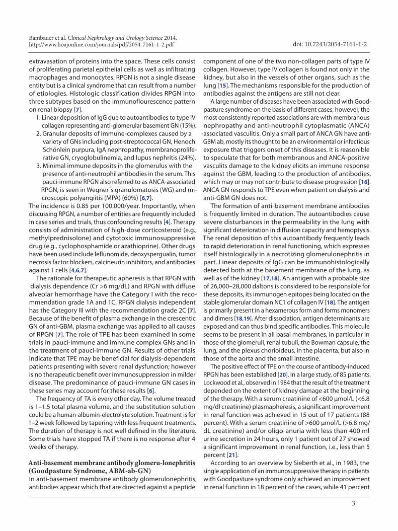
(Table 1) provides an overview of the immunopathogenetic causes of RPGN. In most cases granular or linear IgG and C3 deposits can be detected in the kidney biopsy by means of the immunofluorescence technique. Granular fluorescence of the glomerular capillary walls indicates a form of immune complex nephritis, while when linear immunoglobulin deposits are presented the term anti-basement membrane antibody glomerulonephritis (Goodpasture syndrome) is used.
RPGN type Frequency (%)
I RPGN with linear deposition of IgG due to autoantibodies to type IV collagen representing anti-glomerular basement GN (anti-GBM)
15
1. Goodpasture syndrome2. Idiopathic anti-GBM nephritis3. Membranous nephropathy mostly associated with PLA2R antibodies
II RPGN with granular deposits of immune-complexes
24
1. Post-infectiousPost streptococcal GNAbscessesBacterial endocarditis2. Non-infectiousSystemic lupus erythematosusSchönlein-Henoch purpuraCryoglobulinemia.Wegner´s granulomatosis Polyarteriitis other forms of vasculitis Solid tumors3. Primary renal diseasesIgG – IgA – nephritisMembranoproliferative GNIdiopathic immune complex nephritis
III RPGN with or without minimal glomerular deposition (ANCA ab): Pauci-immune Glomerulonephritis
60
1. Microscopic vasculitis 2. Wegner´s granulomatosis 3. Polyarthritis nodosa
Table 1. Immunopathological classification of RPGN (modified after Szczepiorkowski et al., [4,6]).
The guidelines on the use of TA in clinical practice-evidence-based approach of the AAC of the ASFA described the disease as follows [7].
“ANCA-associated RPGN is one case of the clinico-pathologic entitiy, RPGN. RPGN consists of rapid loss of renal function with the histologic finding of crescent formation in over 50% of glomeruli. These crescents represent a proliferation of cells within Bowman´s space of the glomerulus due to the
Bambauer et al. Clinical Nephrology and Urology Science 2014, http://www.hoajonline.com/journals/pdf/2054-7161-1-2.pdf
3
doi: 10.7243/2054-7161-1-2
extravasation of proteins into the space. These cells consist of proliferating parietal epithelial cells as well as infiltrating macrophages and monocytes. RPGN is not a single disease entity but is a clinical syndrome that can result from a number of etiologies. Histologic classification divides RPGN into three subtypes based on the immunoflourescence pattern on renal biopsy [7].
1. Linear deposition of IgG due to autoantibodies to type IV collagen representing anti-glomerular basement GN (15%).
2. Granular deposits of immune-complexes caused by a variety of GNs including post-streptococcal GN, Henoch Schönlein purpura, IgA nephropathy, membranoprolife- rative GN, cryoglobulinemia, and lupus nephritis (24%).
3. Minimal immune deposits in the glomerulus with the presence of anti-neutrophil antibodies in the serum. This pauci-immune RPGN also referred to as ANCA-associated RPGN, is seen in Wegner´s granulomatosis (WG) and mi- croscopic polyangiitis (MPA) (60%) [6,7].The incidence is 0.85 per 100.000/year. Importantly, when discussing RPGN, a number of entities are frequently included in case series and trials, thus confounding results [4]. Therapy consists of administration of high-dose corticosteroid (e.g., methylprednisolone) and cytotoxic immunosuppressive drug (e.g., cyclophosphamide or azathioprine). Other drugs have been used include leflunomide, deoxyspergualin, tumor necrosis factor blockers, calcineurin inhibitors, and antibodies against T cells [4,6,7].
The rationale for therapeutic apheresis is that RPGN with dialysis dependence (Cr >6 mg/dL) and RPGN with diffuse alveolar hemorrhage have the Category I with the reco-mmendation grade 1A and 1C. RPGN dialysis independent has the Category III with the recommendation grade 2C [7]. Because of the benefit of plasma exchange in the crescentic GN of anti-GBM, plasma exchange was applied to all causes of RPGN [7]. The role of TPE has been examined in some trials in pauci-immune and immune complex GNs and in the treatment of pauci-immune GN. Results of other trials indicate that TPE may be beneficial for dialysis-dependent patients presenting with severe renal dysfunction; however is no therapeutic benefit over immunosuppression in milder disease. The predominance of pauci-immune GN cases in these series may account for these results [6].
The frequency of TA is every other day. The volume treated is 1–1.5 total plasma volume, and the substitution solution could be a human-albumin-electrolyte solution. Treatment is for 1–2 week followed by tapering with less frequent treatments. The duration of therapy is not well defined in the literature. Some trials have stopped TA if there is no response after 4 weeks of therapy.
Anti-basement membrane antibody glomeru-lonephritis (Goodpasture Syndrome, ABM-ab-GN)In anti-basement membrane antibody glomerulonephritis, antibodies appear which that are directed against a peptide
component of one of the two non-collagen parts of type IV collagen. However, type IV collagen is found not only in the kidney, but also in the vessels of other organs, such as the lung [15]. The mechanisms responsible for the production of antibodies against the antigens are still not clear.
A large number of diseases have been associated with Good- pasture syndrome on the basis of different cases; however, the most consistently reported associations are with membranous nephropathy and anti-neutrophil cytoplasmatic (ANCA)
-associated vasculitis. Only a small part of ANCA GN have anti-GBM ab, mostly its thought to be an environmental or infectious exposure that triggers onset of this diseases. It is reasonible to speculate that for both membranous and ANCA-positive vasculits damage to the kidney elicits an immune response against the GBM, leading to the production of antibodies, which may or may not contribute to disease progression [16]. ANCA GN responds to TPE even when patient on dialysis and anti-GBM GN does not.
The formation of anti-basement membrane antibodies is frequently limited in duration. The autoantibodies cause severe disturbances in the permeability in the lung with significant deterioration in diffusion capacity and hemoptysis. The renal deposition of this autoantibody frequently leads to rapid deterioration in renal functioning, which expresses itself histologically in a necrotizing glomerulonephritis in part. Linear deposits of IgG can be immunohistologically detected both at the basement membrane of the lung, as well as of the kidney [17,18]. An antigen with a probable size of 26,000–28,000 daltons is considered to be responsible for these deposits, its immunogen epitopes being located on the stable glomerular domain NC1 of collagen IV [18]. The antigen is primarily present in a hexamerous form and forms monomers and dimers [18,19]. After dissociation, antigen determinants are exposed and can thus bind specific antibodies. This molecule seems to be present in all basal membranes, in particular in those of the glomeruli, renal tubuli, the Bowman capsule, the lung, and the plexus chorioideus, in the placenta, but also in those of the aorta and the small intestine.
The positive effect of TPE on the course of antibody-induced RPGN has been established [20]. In a large study, of 85 patients, Lockwood et al., observed in 1984 that the result of the treatment depended on the extent of kidney damage at the beginning of the therapy. With a serum creatinine of <600 μmol/L (<6.8 mg/dl creatinine) plasmapheresis, a significant improvement in renal function was achieved in 15 out of 17 patients (88 percent). With a serum creatinine of >600 μmol/L (>6.8 mg/dL creatinine) and/or oligo-anuria with less than 400 ml urine secretion in 24 hours, only 1 patient out of 27 showed a significant improvement in renal function, i.e., less than 5 percent [21].
According to an overview by Sieberth et al., in 1983, the single application of an immunosuppressive therapy in patients with Goodpasture syndrome only achieved an improvement in renal function in 18 percent of the cases, while 41 percent
Bambauer et al. Clinical Nephrology and Urology Science 2014, http://www.hoajonline.com/journals/pdf/2054-7161-1-2.pdf
4
doi: 10.7243/2054-7161-1-2
of the patients displayed this improvement with a combined therapy of TPE and immunosuppression [22]. With serum creatinin under six mg/dl prior to treatment, the combined therapy achieved a significant improvement in renal function in 66 percent of the cases. Jayne et al., presented in their study a creatinine level lower than 5.8 mg/dl in patients with rapidly progressive glomerulonephritis which will be beneficial with TA [23].
Treatment with TPE also provides the possibility of imp-rovement in cases of pulmonary bleeding, which is based on the same immunological process, even when renal function is already irreversibly impaired. A final long-term prognosis for patients whose condition improved after TPE cannot be made. As basement membrane antibody formation often ceases during treatment, recovery or at least partial recovery is possible.
Despite many new findings, the causes of primary glom-erulonephritis are still not fully explained. Therapy cannot in most cases really be purposefully applied, therefore. Thus, the various forms of glomerulonephritis are treated with immunosuppression not only with corticoids, alkylating agents, and cyclosporin A, but also with combinations of almost all of these drugs. Trials with anticoagulants, cyclooxygenase inhibitors, and ACE inhibitors suggest that in addition to an immunological genesis of glomerulonephritis, other factors must also be considered [24]. The combination of corticoids, immunosuppressives, and plasmapheresis in varying combinations was the first step in improving the overall prognosis for rapidly progressive glomerulonephritis [25]. In subsequent years, RPGN has been treated with the combination of immunosuppressive drugs and immunoadsorption with excellent results. But Kaplan reported, that several controlled studies have failed to show a generalized benefit of TPE for all patients with RPGN; however, subset analysis of all these studies showed TPE to be beneficial for patients presenting with severe disease or dialysis dependency [26]. A more resent study of Jayne et al., limited to patients presenting with creatinine levels greater than 5.8 mg/dl [23].
In another study based on a plasmapheresis trial, de Lind van Wijngaarden et al., observed that chronic and acute tubolointerstitial lesions predict the glomerular filtration rate (GFR) at 12 months, yet is the use of plasma exchange and the number of normal glomeruli on biopsy that remained positive predictors of dialysis independence in the same time interval [27]. This finding is important because it suggests that unaffected glomeruli determine long-term renal outcome at 1 year. In a second study the same group of investigators extended their work in determining the rate of renal recovery [28]. In 69 dialysis-dependent patients who were part of the plasmapheresis trial, plasma exchange was superior to pulse methylprednisolone with respect to the change of coming of dialysis. The outcome measure depended on the relative number of normal glomeruli (MEPEX study).
Renal biopsy is very important not only diagnostic, but
also has prognostic value in patients with Goodpasture syndrome [29]. When less than 30 percent of the glomeruli underwent crescent formation and the renal function is fairly well-preserved, significant response to therapy and good survival can be expected. In contrast, when more than 70 percent of the glomeruli are crescentic and renal insufficiency is present, the renal disease is often progressive and dialysis and possibly renal transplantation are required. Severe renal failure that causes oliguria reduces the survival rate to 50 percent at 6 months [29].
Although in 1983 Pusey et al., achieved very good results in the treatment of more than 40 Goodpasture syndrome patients with TPE and immunosuppressive therapy [31]. Thus, Balow proposes that mildly progressing Goodpasture syndrome should be treated with immunosuppressives and that plasmapheresis should only be carried out as an additional form of therapy [32]. After the authors opinion, TPE should also be applied in advanced renal insufficiency with oligoanuria, even if, according to Lockwood et al., an improvement in renal function can only be expected in five percent of the cases [21]. In these severe cases plasma exchange treatment and an immunosuppressive therapy should be carried out until no further anti-basement membrane antibodies can be detected in the blood. Immunosuppression should subsequently be continued for a further 6 six months, and with persisting renal insufficiency plasmapheresis should be carried out over a further period of six to eight weeks.
Before plasmapheresis was availble to remove autoantibodies, prognosis was poor, and most patients die or were left with permanent renal impairment. In patients with diffuse alveolar hemorrhage (DAH) alone, corticosteroids may effective [29]. Cytotoxic agents like cyclophosphamide or azathioprine, may occasionally reverse the renal failure, but their main function is to control DAH. The current combination therapy of plasma- pheresis and immunosuppressive drugs is successful if applicated early, i.e., in patients without oliguria who do not require dialysis.
Some oliguric dialysis-dependent patients also show a significant response to this combination therapy, as a result of which dialysis can be discontinued. In contrast, anuric patients do not improve in renal function, and continued dialysis is required; renal transplantation may be considered in these often young patients. Fortunately, the autoimmune process in Goodpasture´s syndrome seems to be limited, as demonstrated by the small number of reported cases of recurrent disease [29,33].
The authors published in 2009 some results of TA and immunosuppressive therapy in Goodpasture syndrome. According to this set of data, the renal function was positively influenced in 166 out of 302 patients (55 percent), and, additionally, hemoptysis in 45 patients (15 percent). In 107 patients (35.5 percent) the combined therapy consisting of TPE and immunosuppression had no effect either on renal function or on hemoptysis [34]. In addition, the antibody is
Bambauer et al. Clinical Nephrology and Urology Science 2014, http://www.hoajonline.com/journals/pdf/2054-7161-1-2.pdf
5
doi: 10.7243/2054-7161-1-2
pathogenic, and the goal treatment is the rapid reduction of those antibodies. Even though there have been only small controlled trials of plasmapheresis in the treatment of Goodpasture syndrome, it is now an approved therapy for all patients with rapidly progressive glomerulonephritis and/or pulmonary hemorrhage [35].
To be able to evaluate the final status of TPE in Goodpasture syndrome, however, controlled, randomized prospective studies and comparative studies are necessary. Given that TPE can have a positive effect on this serious condition, however, controlled studies are, for ethical reasons, beyond discussion. Furthermore, there are only approximately six to eight new cases per year in Germany; and, up to now, only just over 300-400 patients with Goodpasture syndrome have been registered worldwide (incidence: <1per 100,000 per year). Such a low number of cases make controlled studies practically impossible.
After the available results and trials, TA is provided a more rapid decrease in anti-GBM antibodies, lower post-treatment serum creatinine level, and decreased incidence of end-stage renal disease (ESRD). Given these results and the integral role of the anti-GBM antibody, TA as a means of rapidly decreasing anti-GBM titers has become the standard of care [26]. Other authors showed in their findings highlight the safety, efficacy and feasibility of TPE using membrane filtration [36-38].A treatment strategy can be:
1. Early initiation of TA is essential to avoid ESRD.2. Initial prescription is 14 daily 3–4 liters exchange.3. Continued apheresis may be required if antibody titers remain increased.4. Steroids, cyclophosphamide, or azathioprine are added to decrease production of anti-GBM antibody and minimize
the inflammatory response.The guidelines on the use of TA in clinical practice-evidence-based approach of the AAC of the ASFA describe the disease as follows and see under RPGN [4,6,7].
“The pulmonary symptoms include breathlessness to overt hemoptysis. Chest radiographs are nonspecific. Predisposing factors for anti-GBM included the presence of HLA DRB1*1501 allele, exposure to hydrocarbons, and cigarette smoking [4,6]. Almost all patients have anti-GBM antibodies detectable in their blood. This is directed towards the α3 type IV collagen, which is found in renal and alveolar basement membrane. In addition, 30 percent of patients will also have detectable ANCA. Patients exhibiting both antibodies behave more like anti-GBM than ANCA-vasculitis in the short-term but more like ANCA-vasculitis in the long-term. Anti-GBM is characterized by linear deposits of IgG and complement on renal biopsy” [4,6,7].In anti-GBM GN, treatment includes the combination of TA, cyclophosphamide, and corticosteroids. In general, the disease does not relapse and therefore patients do not need chronic immunosuppression [7]. The exception is patients with ANCA in addition to anti-GBM antibodies. These patients respond rapidly to treatment, like anti-GBM, but can relapse, like
ANCA-associated RPGN. These patients require long-term immunosuppression [4,6,7]. The category for TA is I with the recommendation grade 1B–1C in anti-GBM disease with dialysis independence and in DAH [7].
Walsh and the European Vasculitis Study Group (EUVAS) found in 2013 that patients with antineutrophil cytoplasm antibody-associated vasculitis requiring dialysis at diagnosis are at risk for developing and ESRD or dying [39]. Short-term results of a trial comparing TPE to intravenous methylprednisolone suggested TPE improved renal recovery. But after Walsh and the EUVAS the long-term follow-up of patients with severe ANCA-associated vasculitis comparing TPE to intravenous methylprednisolone treatment is unclear. Further research is required to determine the role of TPE in this disease.
“In most patients undergoing TA and immunosuppression, anti-GBM antibodies fall to undetectable levels within 2 weeks and the minimum course of TA should be 14 days. The presence or absence of antibody itself should not be used to initiate or terminate therapy, because antibody is not demonstrable in a small percentage of people with the disease and the antibody may be present in patients without active disease. In those patients with active disease and anti-GBM present, TA should be continues until antibodies fall to undetectable levels” [4,6].
Immune complex nephritis (ICN)Many types of glomerulonephritis are initiated by the deposition of immune complexes, which induce tissue injury via either engagement of Fc receptors on effector cells or via complement activation [40]. The generation of antibody and subsequent tissue deposition of immune complexes (IC) is thought to trigger the pathogenic consequences of systemic autoimmune disease. Modulation of the autoantibody response disrupts pathogenesis by preventing the formation of ICs; however, uncoupling IC formation from subsequent inflammatory response seems unlikely because of the apparent complexity of the IC-triggered inflammatory cascade [41].
In idiopathic symptomatic RPGN, which is frequently caused by an immune complex nephritis, the therapeutic concept is not as clear-cut as with anti-glomerular basement membrane antibody nephritis. Sieberth et al., demonstrated in a study that a combined therapy of TPE and immunosuppression is superior to immunosuppressive therapy alone [42]. They also found that an improvement in renal function is possible in more than 60 percent of cases, if either pulse therapy (high dose therapy with corticosteroids) or TPE is administered. They therefore propose to first implement the pulse therapy and, if unsuccessful, to then apply TPE. Matic et al., reported their results with the treatment of immunoadsorption (IA) based on specific binding to either staphyloccoccal protein protein-A or sheep polyclonal antibodies directed against human IgG [43]. In 26 patients with different types of immune complex nephritis, 602 IA sessions were performed. Between 60 and 80 percent of IgG was eliminated, depending on the treated plasma.
Bambauer et al. Clinical Nephrology and Urology Science 2014, http://www.hoajonline.com/journals/pdf/2054-7161-1-2.pdf
6
doi: 10.7243/2054-7161-1-2
In view of the devastating pathohysiologic consequences of interaction between circulation immune complexes and the basement membrane, the authors share the opinions of Lockwood et al., that TPE in combination with immuno-suppression should be carried out as quickly as possible [21]. The authors published in 2009 a compilation of some published therapeutic results from different authors. Not included was RPGN resulting from diseases such as systemic lupus erythematosus, Schonlein-Henoch purpura, cryoglobulinemia, Wegener’s granulomatosis, or vasculitis [34]. In almost 64 percent of the cases, an improvement or recovery was achieved with the combined TPE and immunosuppression therapy. It is decisive to commence the therapy at as early a stage as possible, while renal function is not yet seriously impaired. Pusey et al., recommended TPE for severe cases of immune complex nephritis [44].
RPGN with or without glomerular deposition (ANCA ab) pauci–immune RPGNApproximately 60 percent of patients with RPGN present with crescentic glomerulonephritis characterized by few or absent immune deposits, the so-called pauci-immune RPGN. Patients with this disease have either Wegner´s granulomatosis, ANCA-ab associated vasculitis, polyarthritis nodosa, or “renal-limited” pauci-immune GN (Table 1). These diagnoses may represent a spectrum of manifestations of a single disease, because there is marked overlap of clinical and histopathologic features, and several patients have anti-neutrophil cytoplasmatic antibodies (ANCA) in their blood which are more common that anti-GBM. The concentration of circulating ANCA correlate with the disease activity in some patients, and ANCA may contribute to the pathophysiology of pauci-immune RPGN through reactivity with neutrophils or endothelial cells, and other inflammatory mechanisms [17,45,46].
The prognosis of pauci-immune RPGN in general has been poor. Precise therapy therapeutic recommendations are difficult to obtain from the literature, because most series comprise patients with different types of RPGN. However, available data suggest that 80 percent of such patients progress to ESRD without therapy with high dose immunosuppression or cytotoxic drugs [14]. Some trials have evaluated the efficacy of TA as an adjunct to conventional immunosuppressive in patients with pauci- immune RPGN [23,46-48].
The results of the randomized trials argue against a role for TA in milder forms of pauci–immune RPGN, but suggest a potential benefit when TA is used as an adjunct to conventional immunosuppressive therapy in patients with severe disease. This relative lack of efficacy probably reflects the efficiency of conventional immunosuppressive agents in halting inflammation and preserving renal function in most patients. These conclusions are supported by the results of uncontrolled trials, suggesting a response rate of 70 percent in patients with RPGN treated with TA, similar to that of patients treated with immunosuppressive therapy with a response rate of 60
percent. In most cases of RPGN, a treatment of TA in the early phase of the disease seems to be necessary.
Hasegawa et al., reported successful treatment using a combinatination of cytapheresis and standard immunso-suppressive therapy of prednisolone and cyclophosmide. In five patients with a myeloperoxidase antineutrophil cytoplasmatic antibody associated vasculitis, the renal function improved and the pulmonary hemorrhage disappeared [45]. Other authors reported of successful treatments with immunoadsorption and immunosuppressive therapy [48,49]. In the above mentioned MEPEX study, de Lind van Wingaarden et al., showed that in patients with dialysis-dependent, ANCA-associated vasculitis, the chances of recovery differ depending on the type of adjunctive treatment, the percentage of normal glomeruli and glomerulosclerosis, the extent of tubular atrophy, and the presence of arteriosclerosis. Even with an ominous biopsy at diagnosis in combination with dialysis dependence, the chance of renal recovery exceeds the chance of therapy-related death when the patient is treated with plasma exchange as adjunctive therapy [28]. But more further studies are necessary.
Therapy recommendations for RPGNRPGN therapy possibilities were extended in recent years to include TA. Antigens, antigen-antibody complexes, and immune complexes can be eliminated from the blood with the aid of plasma exchange. A corresponding therapy enables immunomodulation through suppression or stimulation of antibody formation, as well as a temporary remission of the inflammation through inhibition of the mediators. A combined TPE and immunosuppression therapy seems to us to be advisable, particularly in view of the unfavorable prognosis for RPGN, with its complex causes.
The following therapy recommendation is based on the few uncontrolled and controlled studies available [14,23,26,31,47,50-52]. TPE is indicated in combination with an immunosuppressive therapy with prednisolone (intravenous pulse therapy, or oral therapy), cyclophosphamide (intravenous pulse therapy or oral therapy), or azathioprine in indicated in the following cases:
1. RPGN with serum creatinine under 5.8mg/dl without ol- iguria in anti-GBM disease.2. All severe forms of RPGN with or without ANCA ab, like the pauci-immune complexes, (Cr >6 or patient on dia- lysis). 3. Goodpasture syndrome with life-threatening hemoptysis, or diffuse alveolar hemorrhage from ANCA or MPA inde- pendent of renal function status.4. Preparation for kidney transplant with anti-basement me- mbrane antibodies still detectable in the serum.
Although Lockwood et al., and Pusey et al., found that an im-provement in renal function only occurred in five percent of the patients with RPGN and oligo-anuria and a serum creatinin of <600 μmol/l, despite possible side-effects, it is advisable, to commence plasmapheresis treatment as soon as possible to
Bambauer et al. Clinical Nephrology and Urology Science 2014, http://www.hoajonline.com/journals/pdf/2054-7161-1-2.pdf
7
doi: 10.7243/2054-7161-1-2
achieve an improvement [21,31]. A more recent study limited to patients presenting with creatinine levels greater than 5.8 mg/dl (>512 μmol/l) appears to support this conclusion. Madore et al., recommended to treating all severe forms of RPGN with or without ANCA antibodies with a combination therapy of conventional immunosuppressive therapy and plasma exchange [2]. The authors recommend at least four plasma exchange sessions during the first week of immunosuppressive therapy, using four liter exchanges and albumin-electrolyte solution as replacement fluid.
With high titers of circulating immune complexes or other antibodies, which could damage the kidney and other organs, IA with protein-A or sheep polyclonal antibodies can be more effective than the plasmapheresis procedure. Table 2 summ-arizes the guidelines on the use of TA in RPGN [3,6,7].
In summary, TA used for renal indications, even in elderly patients is relatively safe. Trends towards death in elderly patients may be multi-factoriel and not necessary related to TA [53]. TA may be decrease end point of end-stage renal disease or death in patients with RPGN [54]. TA in combination with immunosuppressive therapies including biologics seems to be more effective as TA alone, but additional trials are required.
Glomerulonephritis with nephrotic syndrome (NS)Classification is classified morphologically, and thus does not provide a uniform description of the disease. Differing etiologies can result in considerable variations in the clinical features, as well as course and prognosis. Consequently, it is difficult to establish generally applicable therapeutic concepts and customized treatment for the individual patient is the
German Working Group of ClinicalNephrology, 2002 [3]
Apheresis Applications Committee of the ASFA, 2010, 2013 [3,6,7]
TA modality Evidenceclass
Severity grade
TA modality Category Recommendation grade
RRGN (ANCA associated)dialysis dependenceDAH Immune complexnephritis, dialysis- independence
TPE,IA-Protein-A,Peptid-GAM®
I b A TPE II
III
1A1C2B
Anti-glomerular basement disease (Goodpasture´s syndrome), dialysis- dependent, no DAH DAHDialysis-independent
TPE,IA-Protein-A,Peptid-GAM®
II a B TPE IIIII
2B1C1B
Focal segmental GN(FSGN)primarysecondaryrecurrent (in trans-planted kidney)
TPE,IA-Protein-A,Peptid-GAM®,Tryptophan,
Anti-human-Ig
IVIII---
CB---
TPE IIIIIII
------IB
Table 2. Guidelines on the use of TA in clinical practice-evidence-based appraoch.
norm [55]. The variable clinical courses of this heterogenous disease group render it almost impossible to carry out controlled therapy studies. Both clinical successes and failures are to be found, as are therapy-produced complications, e.g., infections, sterility, loss of hair, and others [56]. The benefits of immunosuppressive therapy must be weighed against these complications. As the aim of therapy for glomerulonephritis is to prevent terminal renal insufficiency and the risks of nephrotic syndrome, some therapeutic possibilities are discussed here.
It is postulated that the cause of nephrotic syndrome lies in changes in the electrophysiological characteristics of the filtration barriers and of the plasma proteins. The anionic charge on albumin is retained by the negative charge of the glomerular filter-including the basement membrane and the epithelium-obviously play a decisive role [57]. Hemodynamic changes, such as increase in venous pressure, can favor the filtration of proteins. Strutz et al., drew attention to an
impressive correlation between the degree of proteinuria and the level of the LDL/HDL quotients, as a means of measuring the atherogenic risk [57].
Nephrotic syndrome of various GN often reacts to corticosteroids in varying doses, administered over a period of 4-8 weeks. Patients with frequent relapses are also treated with 2-3 mg/kg BW/day cyclophosphamide [58,59]. Cyclosporin A has also been successfully applied in nephrotic syndrome [60,61]. Palla et al., reported on high doses of immunoglobulin (IgG) for nephrotic syndrome. They administered 0.4 g/kg BW IgG on three successive days and repeated this every 21 days over a period of one year [61]. Other therapeutic measures for nephrotic syndrome are anticoagulants, thrombocyte inhibitors, ACE inhibitors, immunosuppressive drugs, lipid reducers, biologics, and diets [62-64].
The prognosis for focal sclerosing glomerulosclerosis (FSGS), which is usually accompanied by nephrotic syndrome, is
Bambauer et al. Clinical Nephrology and Urology Science 2014, http://www.hoajonline.com/journals/pdf/2054-7161-1-2.pdf
8
doi: 10.7243/2054-7161-1-2
considerably less favorable. Cases with nephrotic syndrome are recorded as having a survival rate of 70 percent after six years. Without nephrotic syndrome, this rate reaches 85 percent. Patients with this form of glomerulonephritis are comprised of steriod–sensitive and a steroid-non-sensitive groups, and a appropriate therapy must be selected. Non-reaction to steroids is an indication for a trial therapy with cyclophosphamide, chlorambucil, or cyclosporin or other immunosuppressive therapy [65,66]. FSGS is caused by a variety of factors, however, one type that recurs after transplantation and has been associated with circulating factors, can be treated with TPE.
Briggs et al., previously reported in 1998 the use of my-cophenolate mofetil in 7 partients, whom a substantial improvement in proteinuria was observed [67]. Cattran et la., performed in 2004 an open-label, 6 months trial of mycophenolate mofetil in 18 patients with biopsy-proven FSGS who were resistent to corticosteroid therapy. Seventy-five percent had also failed to respond to a cytotoxic agent and/or cyclosporine A. An improvement in proteinuria was seen in 44 percent of the patients after 6 months. However, no patient achieved a complete remission. In addition, relapses were common after therapy was discontinued [68]. Other therapies have been used in patients with FSGS who prove resistant to standard treatment. Partial remission has been observed in a few case reports using tacrolimus [69].
In the case of resistance to medication or severe progression of the disease, additional TA therapy should be considered, as a continuing treatment given once a week, or every two weeks, or once a month. After transplantation, as many as 40 percent of patients with nephrotic syndrome have recurrences. The glomerular abnormalities in patients with established disease include focal and segmental glomerulosclerosis and hyalinosis, although fusion of epithelial-cell foot processes may be the only abnormality early in the course of disease [70,71]. It has been proposed that because some patients with recurrent focal glomerulosclerosis have a response to treatment with plasma exchange, LDL apheresis and IA there may be different circulating factors that alter the glomerular barrier to protein filtration [72,73].
In six patients with focal-sclerosing glomerulosclerosis, whose renal insufficiency increased despite immunosuppressive therapy, an additional plasma exchange treatment was carried out. Three patients already showed rapid improvement after six or seven sessions of TPE treatment, both with regard to proteinuria and renal insufficiency. The other three patients with acute nephrotic syndrome and protein loss of 20–35 grams in 24 hrs were, after an intensive initial exchange phase, given chronic TPE treatment for 2-4 months, depending on the symptoms. One patient died in the postoperative phase after a stomach operation. All patients were additionally treated with cyclosporin A in a dosage of 2.5-3.5 mg/kg BW/day. In four out of the patients terminal renal insufficiency has been prevented through the administration of additional TPE treatment over a period of 16-20 months [34].
Dantal et al., reported that adsorption of plasma protein decrease urinary protein excretion in patients with recurrence of the NS after renal transplantation [74]. They postulated that the presence of immune complexes in which the factors conductive to albuminuria were active only when they were dissociated from immunoglobulin. They also noted, and that the possibility that the factors could be attached to immunoglobulin through binding to the constant heavy-chain part of the molecule. Pusey et al., recommended in patients with FSGS TPE as a rapid onset of proteinuria following renal transplantation [44].
In the guidelines on the use of TA in clinical practice-evidence-based approach from the AAC of the ASFA has the primary and secondary FSGS the Category III with the recommendation grade 1C, and for the FSGS recurrent the category I with the recommendation grade 1B [6]. The disease is described as follows [4,6,7].
“Instead of a specific diagnosis, FSGS is a histologically charac- teristic finding in renal biopsies characterized by focal areas of sclerosis of some glomeruli adjacent to other intact glomeruli. Several FSGS histological variants (cellular, collapsing, tip lesion, perihilar, and not otherwise specified) have been described” [7]. FSGS is more precisely a glomerular lesion than a specific diagnosis, with the common feature of steroid-resistant syndrome. FSGS can be primary or secondary to a variety of entities such as obesity, reflux nephropathy, HIV infections, and heroin use. Since at least 50 percent of patients with FSGS progress to renal failure within 5 years, many undergo renal transplants. Unfortunately, about 20 percent of patient will experience a recurence in the renal allograft, especially in children. Recurrent disease is diagnosed by new onset of proteinuria, which should be aggressively treated to slow or arrest progression to renal insufficiency and graft loss. Patients who lost grafts due to recurrent FSGS have >80 percent chance of developing the same lesion in subsequently transplanted kidneys” [4].
The treatment in native kidneys with FSGS is primarily with corticosteroids for at least 6 months prior to trying second-line agents such as cyclophosphamide, chlorambucil, or azathioprine. For resistant cases TPE is being currently an option. On the other hand several investigators worldwide have used TPE in the management of patients with FSGS in transplanted organs, in an attempt to save the graft. Although there is no standardized treatment for recurrent FSGS posttransplant, the majority of regimens use a combination of an immuno-suppressant such as cyclophosphamide, biologics, and TPE. Other therapeutic options include high-dose cyclosporine, angiotensin converting enzyme inhibitors, and indomethacin and/or tacrolimus. Another approach to prevent recurrent FSGS is several sessions of pre-emptive TPE immediately prior to and following the transplant [4]. More recently, rituximab and mycophenolate mofetil have also been used in conjunction with diagnosed in order to halt the process and maintain renal function [6,7].
Bambauer et al. Clinical Nephrology and Urology Science 2014, http://www.hoajonline.com/journals/pdf/2054-7161-1-2.pdf
9
doi: 10.7243/2054-7161-1-2
In only certain FSGS patients appears to contain an ill defined “permeability factor”, probably a glycoprotein of molecular weight of 30–50 kDa that includes profound leakage of albumin when incubated with isolated rat glomeruli. Such factor is removed by TPE and the decrease in serum concentration coincides with improvement in proteinuria. Since it is thought that the immediate onset of proteinuria following transplant is mediated by this factor, prophylactic TPE may be instituted in high risk patients. A few reports describe the use of Staphylococcal protein-A columns in recurrent FSGS. The duration of the procedure is to begin with three daily exchanges followed by at least six more TPE in the subsequent 2 weeks, for minimum of nine procedures. Tapering should be decided on a case by case basis and is guided by the degree of proteinuria. Timing of clinical response is quite variable and control of proteinuria may take several weeks to months. Some patients have received long-term monthly exchanges as maintenance therapy [4,6,7].
The category III for primary FSGS and secondary FSGS was assigned to this disease based on limited and conflicting data available in the literature [4]. Recurrent FSGS in transplanted Kidney has the category I and the recommendation grade 1B (Table 2) [7]. The treated volume is 1–1.5 total plasma volume. The replacement fluid is an electrolyte-albumin solution, and the frequency daily or every other day.
The nephrotic syndrome consisting of massive proteinuria, hypoalbuminemia, edema, and hyperlipidemia, is a common complication of glomerular disease in children and adults. The annual incidence of nephrotic syndrome ranges from 2–7 per 100,000 children, and prevalance from 12–16 per 100,000. There is epidemiological evidence of a higher incidence of NS in children aged below 10 years from South ASIA [63]. The primary cause of NS is idiopathic [71]. There is evidence pointing to a role of the immune system in pediatric minimal change glomerulonephritis (MCGN). Another hypothesis has described an association between allergy and MCGN in children. Relapses in this of syndrome are triggered commonly by minor infections and occasionally by reactions to be stings or poisoning. Abnormalities of both humoral and cellular immunity have been described. Finally, the induction of remissions by corticosteroid, alkylating agents, or cyclosporine therapy provides indirect evidence for an immune etiology. None of these observations, however, provides direct evidence of immunologically mediated pathogenesis [75].
Although they are massively proteinuric, patients with MCGN, do not have a generalized glomerular leak to macromolecules. The clearance of neutral macromolecules in MCGN is actually less than normal over a range of molecular radii. In contrast, the clearance of anionic macromolecules is significantly increased. This and several other lines of evidence suggest that proteinuria results from a loss of fixed negative charges of anionic glycosaminoglycans in the glomerular capillary wall [76]. The mechanisms through which these charges are lost are unknown. There are different hypotheses. The traditional
view is that massive albuminuria, in NS causes a decrease in intravascular oncotic pressure, which allows extravasation of fluid and hypovolemia, increased aldosterone and antidiuretic hormone secretion, and renal salt and water retention. An alternative explanation for retention of salt and water in NS is a decreased glomerular filtration rate, with a decreased filtration fraction [75].
Minimal change glomerulonephritis usually takes a benign course and can be well treated with customary therapy measures. In severe cases, therapy with prednisolone and cyclophosphamide over a period of 8 to 12 weeks is indicated [77,78]. Cyclosporin has shown some efficacy in steroid-resistant NS [61,79]. Musco et al., reported significantly rapid faster relief from steroid–resistant NS by using LDL apheresis than from steroid monotherapy [79]. They showed that a rapid improvement of hypercholesterolemia by LDL apheresis in steroid–resistant NS will provides more rapid relief from NS than from steroid therapy alone. Other authors recommended in steroid-resistant NS intravenous steroids in high dose with alkylating agents, cyclophosphamide oral or pulse cyclophosphamide and mycophenolate mofetil [61,80,81].
Membranoproliferative glomerulonephrits (MPGN) usually occurs in combination with nephrotic syndrome and hypertension. The occurrence of nephrotic syndrome signifies a poorer prognosis. The effectiveness of medication with corticosteroid, cyclophosphamides, anticoagulants, and intravenous immunoglobulins has not yet been established. This is also true for pulse therapy [81]. Experience with TA, especially with protein-A immunoadsorption has been presented by Esnault et al., and Dantal et al., [82,83]. They reported of successful treatment with protein-A IA in patients with relapsing nephrotic syndrome. MPGN from cyoglobulinemia could be an indication for TA, too.
Nephrotic syndrome is the main symptom in perimem-branous glomerulonephritis. In the case of acute nephrotic syndrome, it is advisable to undertake therapy with high doses of prednisolone as a pulse therapy over a period of 3 to 5 days or with 2 mg/kg BW in decreasing dosage for 2 to 3 months. A combination with TPE should be considered especially with the more selective procedures like cascade filtration, IA, and LDL-apheresis [79,82,84].
The symptoms displayed in mesangioproliferative glomerulonephritis are not usually homogeneous. The prognosis is poorer if the condition is accompanied by nephrotic syndrome and hypertension. Here also, there are varying opinions exist with regard to corticosteroid and cytostatic therapy. Nephrotic syndrome justifies a trial therapy with cyclophosphamide.
In view of the uncertainty in medicational therapy in this form of glomerulonephritis, three patients were treated additionally with TPE. In the case of two patients, only a few treatment sessions were required in order to normalize renal function, although one patient had to be temporarily hemodialysed due to acute kidney injury. The third patient with accompanying
Bambauer et al. Clinical Nephrology and Urology Science 2014, http://www.hoajonline.com/journals/pdf/2054-7161-1-2.pdf
10
doi: 10.7243/2054-7161-1-2
acute nephrotic syndrome (proteinuria 20-25 g/24 hrs.) received TPE treatment regularly over a period of two years. Nevertheless terminal renal insufficiency occurred, which finally made chronic hemodialysis necessary [34]. Although, TA is indicated in severe cases of various types of glomerulonephritis. In severe, drug therapy-resistent cases, a combined TA and immunosuppression therapy is recommended, regardless of the degree of renal insufficiency [3,72].
Acute nephrotic syndrome in particular seems to be favorably influenced by regular TA treatment, for on the one hand dysproteinemia and thus the edema can be improved and, on the other hand, human albumin can be administered in larger doses. TA is theorectically a way of achieving an improved effect on the basal membrane. The elimination of cholesterol, LDL, and triglycerides might also reduce the atherogenic risk for these patients and thus also prevent progression. TA should be considered as a useful therapeutic tool in the management of this disease [73]. The reports of the therapy of NS with more selective TA procedures like cascade filtration, IA, and LDL apheresis are very encouraging and show a possibility for treating severe cases of NS, if drug therapy fails [82-85]. As in the case of other renal diseases, controlled prospective studies are needed.
Further renal diseases such as light chain nephropathy, dense deposit diseases and others as shown in Table 1 can be in severe cases and if the conservative therapy has failed threated with TPE.
Hemolytic-uremic syndrome (HUS)Hemolytic-uremic syndrome is a disease that can lead to acute kidney injury (AKI) and often to other serious sequelae, including death. The disease is characterized by microan-giopathic haemolytic anemia, thrombocytopenia and acute kidney injury. The etiology and pathogenesis of HUS are not completely understood, and the therapy of HUS is complicated. After introduction of therapeutic apheresis as a supportive therapy in HUS, several authors reported successful treatment using TA in HUS in more than 87 percent of treated patients. The supportive therapy is indicated basically in severe courses of HUS and is superior to available therapy interventions [86]. The pathophysiologic aspects of the different pathogenic types of HUS are discussed by Bambauer et al., [86].
Most cases are associated with infections with entero-hemorrhagic E. coli (EHEC). These bacteria can be transmitted through contaminated food, animal and person to person contact. HUS is one of the most severe complications of a potentially avoidable food-borne infection. Other causes of HUS described as “typical” have to be differentiated since other factors including genetic disorders are of importance. In view of the different courses of HUS, a minimum of three different pathogenetic types which lead to HUS are subdivided. HUS caused by infection, idiopathic HUS (non-Shiga toxin HUS), and HUS in systemic diseases and after toxin exposure [87].
There have been reports of spontaneous recovery from HUS.
The various etiological and pathogenetic assumptions have produced diverse therapy concepts. But the total lethality in HUS was first reduced to 20 percent with the introduction of dialysis [88]. If the therapy is administered early enough, two-thirds of cases recover without any impairment. In 10-20 percent of cases, however, lasting renal damage occurs.
Other authors reported successful in HUS using therapeutic plasma exchange [89–91]. In the following years other authors reported successful treatment in HUS using IA with protein-A [92]. Bambauer et al., showed a compilation of therapeutic concepts for HUS implemented up to 2009, that the success of HUS therapy with TPE/HD or IA/HD has been constantly increasing, as numerous reports indicated [86].
While Remuzzi et al., and Misiani et al., consider simple plasma infusion as sufficient it is not adequate, since various pathophysiological mechanisms are observed that cannot be explained solely by Remuzzi et al.,´s theory [93,94]. However, substitution of plasma or coagulation factors is often necessary due to the severe coagulation problems in HUS. Therapeutic apheresis might be more effective than infusions alone, as it removes potentially toxic substances from the circulation. TPE or IA should be considered first-line therapy in situations that limit the amount of plasma that can be infused, such as renal or heart failure. Plasma infusion treatment is contraindicated in S. pneumonia induced non-stx-HUS. It may exacerbate the disease because adult plasma contains antibodies against Thomson-Friedenreich antigen [95].
Michael et al., found in different randomized controlled trials that TPE and/or dialysis as supportive therapy are still the most effective treatments in HUS [96]. They looked for randomized controlled trials of any intervention for hemolytic-uremic syndrome and thrombotic thrombocytopenic purpura between 1966 and 2006. The outcome was listed for HUS, all cause mortality, chronic reduced kidney function, and persistent proteinuria or hypertension at last follow up. None of the evaluated interventions such as fresh frozen plasma transfusion or dipyridamole, Shiga toxin binding protein and steroids were superior to supportive therapy alone for any outcomes [96].
The advantage of TA over other therapeutic procedures is that it intervenes at an early stage in the pathogenetic processes by quickly removing immune complexes and toxins. Furthermore, it eliminates fibrinogen, fibrinogen degradation products, and other high molecular complexes, all of which can both support and inhibit coagulation. All other toxins produced by bacteriae and viruses like Shiga-toxin, the pathogenic pathway which follows the activation of the complement system of the factor HF1 with a partial HF1 deficiency and all other toxic substances can be quickly removed by therapeutic apheresis.
The therapeutic apheresis methods which are introduced in HUS as a supportive therapy are TPE and immunoadsorption with protein-A columns. Both methods are described else-where [86,90,91]. The rationale for TA in HUS is discussed
Bambauer et al. Clinical Nephrology and Urology Science 2014, http://www.hoajonline.com/journals/pdf/2054-7161-1-2.pdf
11
doi: 10.7243/2054-7161-1-2
controversially because of the limited and or conflicting data available in the literature. The rationale is that TA can effectively tremove antibody or mutated circulating complements regulators [7]. TA seems a reasonable option considering the poor prognosis of HUS in adults [4]. The role of TA is uncertain but this treatment may be appropriate as supportive therapy under certain circumstances and with a defined therapeutic endpoint because of the high mortality.
In 2002, the German Working Group for Clinical Nephrology has given HUS/TTP the evidence grade I b and the severity grade A [3]. The indications for TA are symptoms like cerebral edema without stroke, hemolysis, thrombopenia, and renal insufficiency. The duration of the treatment with TA is recommended for a minimum of 2 weeks until the severe symptoms disappeared.
In 2010 and 2013 the AAC of the ASFA divided HUS in 3 groups for TPE: Group 1 (diarrhea associated HUS) is a HUS due to complement factor gene mutations has the catregory II with the recommendation grade (RG) 2C. Group 2 is a HUS due to autoantibody to factor H (atypical HUS), and has the category I with the RG 2C. Group 3 is the typical HUS <18 years. Group 3 has the category IV with the RG 1C [6,7]. Due to the various and very different causes, which can lead to a hemolytic-uremic syndrome, there are no exact guidelines available for the therapy of HUS. This will acknowledge that choosing evidence-based therapies are often limited by our incomplete understanding of the various pathogenic cascade.
In HUS a supportive therapy is indicated which include control of fluid and electrolyte imbalance, use of dialysis if required, control of hypertension, blood and plasma transfusion as required. Antibiotic treatment of E. coli O157:H7 colitis may stimulate further verotoxin production and there by increase the risk of HUS. The use of dialysis like hemodialysis or peritoneal dialysis as required daily. However untreated HUS in adults and children may progress to end in organ damage [97]. Platelet transfusion may actually worsen outcome.
TPE or IA is generally performed daily until the platelet count is normal. In the TPE the replacement fluid consists of human albumin-electrolyte solution (5%) in 30 to 70 percent and fresh frozen plasma (FFP) in the remainder. The exchange volume per treatment should be 1–1.5 total plasma volume depending on the severity of the HUS. TPE may reverse the ongoing platelet consumption. By using IA no replacement fluid is necessary only between the treatments FFP or coagulation factors may be transfused if required. An exchange volume of 3–4 L plasma corresponding to whole blood is recommended. The hemodialysis treatment can be combined with the TA.
A large outbreak of diarrhea and the HUS caused by an unusual serotype of Shiga-toxin-prducing Escherichia coli (O104:H4) was in Germany in May to July 2011 with 3,167 without HUS and 16 deaths in the patients, and 908 with HUS and 34 deaths [98]. 241 patients with HUS were treated with TPE and 193 patients with TPE and eculizumab. The treatment strategy was dependent on disease severity [99].
TPE and eculizumab in combination seems to be prudent and necessary prior to establishing new treatment guidelines.
Myoglobulinemic renal failureAn acute myolysis can induce severe disturbances of the renal function. Free myoglobin with a molecular weight of 17,800 Dalton can be eliminated rapidly by a normal functioning kidney. Massive increase of myoglobin and their derivates in the blood as in acute crushing injuries and mass trauma is highly nephrotoxicity and causes a decreased circulation in the kidney and/and a metabolic acidosis. Acute kidney injury can develop rapidly [100]. Observations showed that, by along with myoglobin, peroxide free radicals of can be released. These can induce a dissiminated intravascular coagulation (DIC), and can damage thrombocytes, the endothelium of the vessels, and disturb the metabolism of prostglandine synthesis [101].
Rhabdomyolysis is also a clinical syndrome in which the contents of injured muscle cells leak into the circulation. This leakage results in electrolyte abnormalities, acidosis, clotting disorders, hypovolemia, and acute kidney injury. A lot of conditions, both traumatic and non-traumatic, can lead to rhabdomyolysis. Intervention consists of early detection, treatment of the underlying cause, volume replacement, urinary alkalinization, and aggressive diuresis or hemolysis. Patients with rhabdomyolysis often require intensive care [102].
Elimination of myoglobin from plasma may be enhanced by plasmapheresis in patients with acute kidney injury [103]. Endothelin, a vasoconstrictive peptide which includes 21 aminoacids, has a strong vasoconstrictive effect in the glomeruli. It leads to hypoxia and hypotension of the endothelium which causes the endothelium to an increased release of endothelin. Cornellisen et al., evalatued 4 patients with rhadomyolysis. They showed that a single 2 liter TPE has no beneficial effects in the treatment in rhabdomyolysis. It is necessary to note only one TPE with only 2 liters was done; all other case reports have used multiple daily TPE [104]. Siebenlist et al., reported that in three patients with severe myoglobulinemia sufficient to cause renal failure, hemodialysis treatment could be prevented when TPE was used in an early stage [105].
The effect of TPE in the case of three patients with myolosis was very impressive. After operative removal of an abdominal glioblastoma, a one-year-old girl with myoglobulinemic muscular dystrophy and acute kidney injury displayed rapid normalisation of renal function, after three TPE and four HD treatment sessions [34]. Similar normalization in renal functioning was observed in the case of a 77-year-old patient with myoglobulinemic AKI after one TPE session with three liters. Also in another case, a 19-year-old female patient, who suffered from malignant hyperthermia and AKI after administration of an anaesthetic during tonsillectomy, was cured after three sessions of plasmapheresis. In particular in the case of malignant hyperthermia, which is rare but reaches

Bambauer et al. Clinical Nephrology and Urology Science 2014, http://www.hoajonline.com/journals/pdf/2054-7161-1-2.pdf
12
doi: 10.7243/2054-7161-1-2
a mortality rate of 60–70 percent, TPE seems to improve the poor prognosis, if applied at an early stage.
Yang et al., used TPE in a case of rhabdomyolysis complicated with increased serum bezafibrat level. They advocated that bezafibrate being highly protein bound is unlikely to be cleared by hemodialysis. TPE was safe and effective in addition to supportive care for rhabdomyolysis associated with bezafibrate [106]. Ronco reported that attempts to use TPE in myoglobulinemia have resulted in higher sieving coefficients, but notes limitations due to low volume exchanges [107]. Swaroop et al., described a case of an 82-years old male patient who developed rhabdomyolysis while taking a combination of simvastatin and gemfibrozil and was successfully treated with TPE [108]. Improvement in kidney function when it does occur does so slowly over months of supportive care and dialysis. But there are only a few cases of rhabdomyolysis reported in literature. Most are case reports. Other authors showed that neither plasmapheresis nor hemodiafiltration has been successful in patients with myoglobulinemic renal failure. Fortunately most patients eventually regain normal kidney function [109]. In the case of myoglobulinemic renal failure: TPE interrupts, stops, or eliminates the:
1. The toxic effects of myoglobulin and its derivates.2. the occurrence of disseminated intravascular coagulation through released tissue thromboplastin.3. non-physiological synthesis of coagulation factors in the liver, the sequestration of active coagulation factors, and metabolic products from the affected tissue.
Acute kidney injury (AKI)The renal diseases are concluded with some notes of acute kidney injury as an independent disease [110]. The variety of the causes that can trigger AKI justifies a close examination of this disease. Acute renal insufficiency means reversible renal damage with oligo-anuria, which is now generally called acute kidney injury. Acute kidney injury presents unique, life threatening and organ threatening therapeutics challenges that require prompt accurate diagnosis and treatment. In rare cases, AKI can also take a polyuric course. Damage to the kidneys varies depending on the degree and duration of pre-renal, renal or post-renal disorderds (noxae). It is reversible only after the elimination of the noxae; in the case of structural disorders only after its repair; or it can remain irreversible.
AKI is also defined as an acute over hours or days developing renal function damage, which is measured by the glomerular filtration rate (GFR). Other renal functions are changed and decreased in AKI, such as the excretion of metabolic products and drugs, the reabsorption of filtrated substances, the regulation in acid-base and electrolyte disorders, and different endocrinological functions. The incidence of AKI is 2–5 percent in inpatients and up to 10-30 percent with intensive medical. The mortality has essentially remained unchanged in the last four decades and at 30 to 80 percent is very high. Although in the last years considerable progress has been made in the
dialysis technology and intensive medical care therapy. AKI is the most frequent and expensive renal disease with the highest course of morbidity and mortality in hospitals [111,112].
As the causative noxae of AKI must be eliminated at the time of insult to the kidney and before it has completely destroyed, they can be influenced primarily during the period in which they act on the kidney. Thereafter, further measures against the noxae are no longer effective; all that then remains is life-long dialysis and/or kidney transplantation [112]. Given the new treatment possibilities, the customary classification of these factors in pre-renal, post-renal, and renal disorders is simplistic from a therapeutic point of view [110].
1. A reduction in renal blood supply that is, having a quan- titative effect: this group comprises blood flow disorders or noxae. It covers only a part of the pre-renal disorders as commonly classified: circulatory-ischemic disorders such as reduced blood pressure or volume, which can be directly influenced therapeutically, if at all. 2. A qualitative change in renal blood supply: this group in- cludes endogenous or exogenous substances that circula- te in the blood and have a damaging effect on kidney ti- ssue. This group comprises plasma disorders or noxae and includes all endogenous and exogenous toxins, metabolic, and decomposition products, as well as immunologically active substances that circulate in the blood and can dama- ge the kidneys [111]. Many of these plasma disorders can be influenced by therapeutic apheresis. This justifies classification of these pathogenetic factors of an acute kidney injury in a group of their own.3. Postrenal disorders and damage to the parenchyma of the kidney via obstruction: this is the group urination disor- ders or noxae. It comprises postrenal disorders, with the exception of intratubular obstruction through substances originally circulating in the blood that precipitate in the tubule lumen (urates, hemoglobin, myoglobin) as a result of urine concentration. Therapy varies according to the nature of the postrenal disorder and is usually treated by urologists [110].
With regard to the treatment of plasma disorders in particular, plasmapheresis opens up a new approach. Conventional therapeutical methods are to be applied to blood flow and postrenal disorders. Of course, in AKI it is important to eliminate or to influence all factors which can lead to AKI and to ensure sufficient administration of parenteral calories, including amino acids, glucose, and fatty solutions [112]. There are no guideline available for the therapy of AKI. In specially severe or therapy resistant course of AKI the following TA methods could be discussed and implemented:
1. conventional plasma exchange with hollow fibers could be implemented alone or in combination with dialysis treatments, and 1–5 treatments are recommended every 1–2 days depending on the course and severity of the AKI [110].2. selective plasma separation, such as cascade filtration, bio-
Bambauer et al. Clinical Nephrology and Urology Science 2014, http://www.hoajonline.com/journals/pdf/2054-7161-1-2.pdf
13
doi: 10.7243/2054-7161-1-2
logical or non-biological adsorption methods, and whole blood adsorption are available. One to maximum of 3 tr- of 3 treatments are recommended with an exchange vol- ume of 3 to 4 L plasma corresponding to whole blood.
Up to now, there have not been enough controlled studies of TA used in the treatment of AKI: therefore the physician who treats patients with AKI must decide for himself, wether the introduction of TA in AKI is indicated or not. It is useful to discuss the risk factors and therapeutic modalities with all persons involved in such cases.
Despite the intensive and costly therapeutic modalities in AKI, the mortality of the AKI is always high, at 30–80 percent, and this has not essentially changed in the last four decades [112]. Approximately twice as many patients with severe diseases, such as multi-organ failure and AKI, die in intensive care units when compared with patients without AKI. These patients die not as a result of AKI, but because of the different complications that follow AKI.
Considering these facts and the inevitable unfavorable prognosis of the AKI, it is possible to add the option of therapeutic apheresis to the conservative and extracorporeal therapies [70]. More of controlled studies should be done to improve the clinical outcome and decrease the high costs of this therapeutic method. Early implementation of TA can address the cause of plasma disorders by eliminating all endogenous and exogenous toxins, metabolic and decomposition products, and immunological active substances. Consistent implementation of plasmapheresis combined with dialysis and other conventional techniques, may help to improves the as yet poor prognosis for AKI.
Kidney transplant rejectionIn chronic renal failure, a kidney transplantation is the decisive alternative to permanent dialysis. Rejection of the transplanted kidney is a grave problem. Although various therapeutic interventions to delay or prevent rejection exist and use steroids, immunoglobulins, immunosuppressives, cyclosporine A, tripple drug, OKT3, and other new developed immunosuppressive therapies. Infections and rejection reactions are, the most frequent complications of modern transplantation [113,114]. Thus, acute kidney transplant rejection is considered as an indication for plasmapheresis [115,116]. TA is indicated in the management of rejection crisis due to preformed specific antibodies or a high degree of immunization [117].
Immunological problems like performed donor-specific antibodies or a high degree of immunization complicate the outcome of donor transplantation. Postoperatively the antibody-mediated rejection or drug-related side-effects of the medication can limit the therapeutic success of transplantation. Acute allograft rejection is one of the important complications after renal transplantation, and it is a deleterious factor for long-term graft survival. Rejection is a complex pathophysiologic process, which has been explained by transcriptome and
proteome in RNA transcripts and proteins level respectively [118]. Therefore therapeutic strategies include a primary avoidance of immunization, careful patient selection, a meticulous immunological workup and a proper follow up and therapeutic apheresis as improved therapy [119,120]. In the following the indications for therapeutic apheresis in kidney transplantation after Teschner et al., are shown [117]:
After the blood group barrier had been successfully crossed in Japan in the 1980s, different protocols were developed for ABO-incompatible kidney transplantation and the procedure has gained widespread acceptance and has been implemented in most transplant centers [121-123]. Immunosuppression consists of tacrolimus, mycophenolate and steroids together with induction therapy with an IL-2-receptor blocking agent. The isoagglutinine antibodies against the donor can be eliminated. Firstly, the CD 19/20-positive pre-B cells with a single infusion of rituximab four weeks prior to transplantation and in a second step, the already existing antibodies are depleted by using therapeutic apheresis such as TPE or IA. Novel sensitization and production of antibodies is thereby efficiently prevented [124,125].
The disadvantage by using TPE is the elimination of physio- logical proteins, the limitation to 1–1.5 total plasma volume (TPV) as treating dose and the potential for infectious complications such as HIV or hepatitis B or C by using plasma as substitution solution. Therefore various groups use the IA with unselective IgG columns. Patients with performed HLA-antibodies, i.e., a high percentage of panel reactive antibodies, accumulate on the waiting list for kidney transplantation and can experience a substantially longer waiting time [113,119]. Therefore center specific desensitization protocols were developed in order to transplant these highly immunized patients within a reasonable time frame.
The transplantation procedure is problematic with deceased donor organs as the time for pre-conditioning of the recipient is extremely limited and the accompanying procedures are difficult to perform in time. If transplantation from a living donor with DSA is planed, different protocols were published to desensitize the recipient. These strategies require an intensive procedure, mostly consisting of the administration of intravenous immunoglobulins (IVIG), of intensified immunosuppression, pre- and postoperative TPE or IA and carry a higher risk for antibody-mediated rejection [117,126-128].
TA in all forms can be applied to remove DSA and multiple HLA antibodies. No selective secondary adsorbers exist, and available columns with a selectivity for immunoglobulins would be considered the best option. Some treatments are usually needed to deplete to recipient of the DSA- and/or anti-HLA titer.
Acute antibody rejection of organ allografts usually presents as severe dysfunction with a high risk of allografts loss. HLA antibodies are involved in AMR [129]. The renal biopsy often cannot rule out one cause or the other with sufficient certainty,
Bambauer et al. Clinical Nephrology and Urology Science 2014, http://www.hoajonline.com/journals/pdf/2054-7161-1-2.pdf
14
doi: 10.7243/2054-7161-1-2
leaving the physician with the decision how to treat vascular rejection that can be caused by antibodies or cellular infiltration [130]. TA accompanied by T cell depletion (ATG, ALG, or OKT3) conversion to a tacrolimus-based imunosuppression and pulsed steroids, are used to limit the interstitial and vascular damage [128]. The use of IA targeted against IgG has been used successfully. It is not possible, due to conflicting and limited data, to give general recommendations in regard to the treatment of TPE or IA, the number of apheresis sessions and the best immunosuppressive therapy [131]. A screening for donor-specific antibodies should be performed to monitor the antibody titer during treatment, until 10 sessions with daily treatments initially followed by apheresis every other day can be necessary in a patient with vascular rejection (Banff IIb-III or AMR) [117,127].
Recurrence or de novo thrombotic microangiopathy (TMA) in the transient patient is observed rarely with the use of calcineurin inhibitors or mTOR inhibitors or acute vascular rejection. Infectious disesases such as HIV, CMV, paravirus B 19, an inhibited or decreased activity of the von Willebrand factor-cleaving metalloprotease ADAMTS13 or mutations in complement receptors may also trigger microangiopathy with either limited or systemic manifestations [117].
TA can be attempted to ameliorate the course of the disease and subsequent graft damage, if switching to a different immunosuppressive regimen or the treatment of an underlying infection does not lead to an improvement of the TMA [130]. The treatment regimen is comparable to TMA in non-transplanted patients. The treated volume is usually one TPV with human albumin and/or fresh frozen plasma as substitution fluid and anticoagulation with heparin on a daily basis until platelet count and lactate dehydrogenase have normalized. Up to 50 percent of patients demonstrate a prompt exacerbation if daily TA is stopped. Continuation of TA on an alternate day strategy for at least two additional treatments can reduce the recurrence rate. Nevertheless TMA reduces graft survival both in recurring or de novo TMA and treatment might not alter the progression of the disease [117].
Goodpasture syndrome or anti-GBM disease can occur de novo in patients following transplantation or as a manifestation of underlying Alport disease, but is rare (e.g., 3 percent of transplanted male Alport patients) [132-134]. The recipient´s immune system is exposed to a collagen component carried by the transplanted organ that is lacking in Alport patients and, consequently, the patient might develop antibodies against this antigen in the glomerular basement membrane. These antibodies may then induce post-transplantation anti-GBM disease.
The treatment of this condition and of de novo disease is identical to the strategy applied to non-transplanted patients. TA is used in order to remove the causative antibody. Both TPE and IA have been shown to deplete the patient effectively of antibodies and halt disease progression [135,136]. The TA should be a rapid removal of the antibodies with daily
treatments. Treatment frequency should be tapered later to antibody titer measurements. TA is accompanied by an intensified immunosuppressive regimen to suppress further antibody formation [117,137].
Only few information are available about long-term results of kidney transplantation in adults with focal segmental glomerulosclerosis. But primary FSGS recurs with uncertain incidence after kidney transplantation (presumably 20 percent). A circulating factor is assumed to play a causative role and TA has been successfully applied in patients with recurrent FSGS. In patients treated with a protein-A adsorption column or TPE, a dramatic but usually transient reduction in proteinuria has been observed [136]. This effect was larger with the use of IA, but more prolonged remissions were reported with the use of TPE with or without combination with cyclophosphamide [117,137].
Therapeutic apheresis in transplantation as an important part of different therapy strategies like for therapy of several conditions such as AMR or ABOi transplantation is accepted today. TA enables the physicians to develop strategies to provide the best organ replacement to patients with high degree of immunization or performed DSA thereby expanding the use of living donation. The standard method has been TPE but it is currently more and more replaced by the more selective methods provided by immunoadsorption. Due to the considerable costs of IA the selection and application of an adsorber and device for IA should be preceded by a judicious effort to characterize and plan the treatment. The specific characteristics of the clinical problem, the capabilities of the choice available and the current evidence have to be known to avoid high costs or inadequate therapy.
The guidelines on the use of TA in clinical practice-evidence-based approach of the AAC of the ASFA describe the antibody-mediated rejection and HLA desensitization as follows and give for the AMR renal transplant recipients and desensitization living donor due to donor specific HLA antibody the category I with the RG 1B. The desensitization high PRA deceased donor has the category III with the RG 2C [6,7].
“Barriers to transplantation include antibodies to human leukocyte antigens and ABO incompatibility with the donor because there is an increased risk for graft loss secondary to hyperacute rejection of the organ due to endotheial damage. A and B antigens are expressed on vascular endothelium [4,7]. “Patients with elevated HLA antibody screen have difficulty finding an HLA compatible donor and remain on the transplantation list significantly longer than unsensitized patients. The goal of desensitization protocols is to allow these individuals to be transplanted using a donor kidney that would otherwise not be usable due to the high likelihood of graft loss. Allograft rejection has traditionally focused on T cell mediated process causing cellular rejection. Acute vascular rejection has been thought of as antibody mediated based on correlating histological findings with the identification of donor specific antibody. Recently a clear histological diagnosis
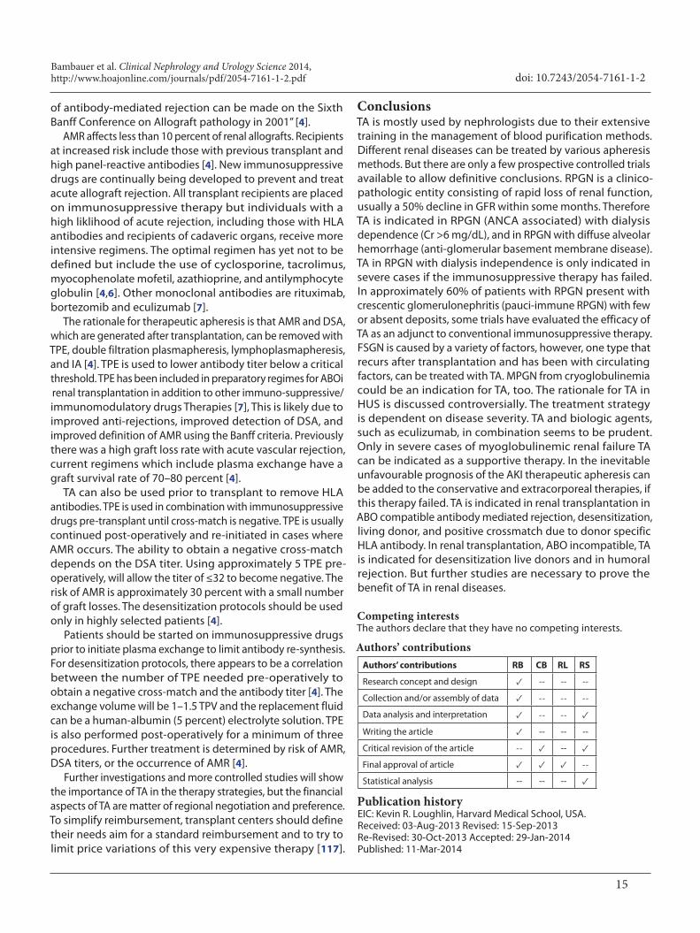
Bambauer et al. Clinical Nephrology and Urology Science 2014, http://www.hoajonline.com/journals/pdf/2054-7161-1-2.pdf
15
doi: 10.7243/2054-7161-1-2
of antibody-mediated rejection can be made on the Sixth Banff Conference on Allograft pathology in 2001” [4].
AMR affects less than 10 percent of renal allografts. Recipients at increased risk include those with previous transplant and high panel-reactive antibodies [4]. New immunosuppressive drugs are continually being developed to prevent and treat acute allograft rejection. All transplant recipients are placed on immunosuppressive therapy but individuals with a high liklihood of acute rejection, including those with HLA antibodies and recipients of cadaveric organs, receive more intensive regimens. The optimal regimen has yet not to be defined but include the use of cyclosporine, tacrolimus, myocophenolate mofetil, azathioprine, and antilymphocyte globulin [4,6]. Other monoclonal antibodies are rituximab, bortezomib and eculizumab [7].
The rationale for therapeutic apheresis is that AMR and DSA, which are generated after transplantation, can be removed with TPE, double filtration plasmapheresis, lymphoplasmapheresis, and IA [4]. TPE is used to lower antibody titer below a critical threshold. TPE has been included in preparatory regimes for ABOi renal transplantation in addition to other immuno-suppressive/immunomodulatory drugs Therapies [7], This is likely due to improved anti-rejections, improved detection of DSA, and improved definition of AMR using the Banff criteria. Previously there was a high graft loss rate with acute vascular rejection, current regimens which include plasma exchange have a graft survival rate of 70–80 percent [4].
TA can also be used prior to transplant to remove HLA antibodies. TPE is used in combination with immunosuppressive drugs pre-transplant until cross-match is negative. TPE is usually continued post-operatively and re-initiated in cases where AMR occurs. The ability to obtain a negative cross-match depends on the DSA titer. Using approximately 5 TPE pre-operatively, will allow the titer of ≤32 to become negative. The risk of AMR is approximately 30 percent with a small number of graft losses. The desensitization protocols should be used only in highly selected patients [4].
Patients should be started on immunosuppressive drugs prior to initiate plasma exchange to limit antibody re-synthesis. For desensitization protocols, there appears to be a correlation between the number of TPE needed pre-operatively to obtain a negative cross-match and the antibody titer [4]. The exchange volume will be 1–1.5 TPV and the replacement fluid can be a human-albumin (5 percent) electrolyte solution. TPE is also performed post-operatively for a minimum of three procedures. Further treatment is determined by risk of AMR, DSA titers, or the occurrence of AMR [4].
Further investigations and more controlled studies will show the importance of TA in the therapy strategies, but the financial aspects of TA are matter of regional negotiation and preference. To simplify reimbursement, transplant centers should define their needs aim for a standard reimbursement and to try to limit price variations of this very expensive therapy [117].
Authors’ contributions RB CB RL RS
Research concept and design ✓ -- -- --
Collection and/or assembly of data ✓ -- -- --
Data analysis and interpretation ✓ -- -- ✓
Writing the article ✓ -- -- --
Critical revision of the article -- ✓ -- ✓
Final approval of article ✓ ✓ ✓ --
Statistical analysis -- -- -- ✓
ConclusionsTA is mostly used by nephrologists due to their extensive training in the management of blood purification methods. Different renal diseases can be treated by various apheresis methods. But there are only a few prospective controlled trials available to allow definitive conclusions. RPGN is a clinico-pathologic entity consisting of rapid loss of renal function, usually a 50% decline in GFR within some months. Therefore TA is indicated in RPGN (ANCA associated) with dialysis dependence (Cr >6 mg/dL), and in RPGN with diffuse alveolar hemorrhage (anti-glomerular basement membrane disease). TA in RPGN with dialysis independence is only indicated in severe cases if the immunosuppressive therapy has failed. In approximately 60% of patients with RPGN present with crescentic glomerulonephritis (pauci-immune RPGN) with few or absent deposits, some trials have evaluated the efficacy of TA as an adjunct to conventional immunosuppressive therapy. FSGN is caused by a variety of factors, however, one type that recurs after transplantation and has been with circulating factors, can be treated with TA. MPGN from cryoglobulinemia could be an indication for TA, too. The rationale for TA in HUS is discussed controversially. The treatment strategy is dependent on disease severity. TA and biologic agents, such as eculizumab, in combination seems to be prudent. Only in severe cases of myoglobulinemic renal failure TA can be indicated as a supportive therapy. In the inevitable unfavourable prognosis of the AKI therapeutic apheresis can be added to the conservative and extracorporeal therapies, if this therapy failed. TA is indicated in renal transplantation in ABO compatible antibody mediated rejection, desensitization, living donor, and positive crossmatch due to donor specific HLA antibody. In renal transplantation, ABO incompatible, TA is indicated for desensitization live donors and in humoral rejection. But further studies are necessary to prove the benefit of TA in renal diseases.
Competing interestsThe authors declare that they have no competing interests.
Authors’ contributions
Publication historyEIC: Kevin R. Loughlin, Harvard Medical School, USA.Received: 03-Aug-2013 Revised: 15-Sep-2013Re-Revised: 30-Oct-2013 Accepted: 29-Jan-2014 Published: 11-Mar-2014
Bambauer et al. Clinical Nephrology and Urology Science 2014, http://www.hoajonline.com/journals/pdf/2054-7161-1-2.pdf
16
doi: 10.7243/2054-7161-1-2
References1. Kaplan AA. Why nephrologists should perform therapeutic plasma
exchange. Dial Transplant. 2009; 38:65–70. | Article
2. Madore F, Lazarus JM and Brady HR. Therapeutic plasma exchange in renal disease. J AM Soc Nephrol. 1996; 7:367–386. | Pdf
3. Mann K, Bosch T, Braun N, Fassbinder W, Klingel R, Klinkmann J, Lonnemann G, Querfeld U, Ramlow W and Schettler V. Apherese Standard der Deutschen Arbeitsgemeinschaft für Klinische Nephrologie e.V. Mitt Klin Nephrol 2002; 31: 103–138.
4. Szczepiorkowski ZM, Winters JL, Bandarenko N, Kim HC, Linenberger ML, Marques MB, Sarode R, Schwartz J, Weinstein R and Shaz BH. Guidelines on the use of therapeutic apheresis in clinical practice--evidence-based approach from the Apheresis Applications Committee of the American Society for Apheresis. J Clin Apher. 2010; 25:83-177. | Article | PubMed
5. Szczepiorkowski ZM, Shaz BH, Bandarenko N and Winters JL. The new approach to assignment of ASFA categories--introduction to the fourth special issue: clinical applications of therapeutic apheresis. J Clin Apher. 2007; 22:96-105. | Article | PubMed
6. Szczepiorkowski ZM, Winters JL, Bandarenko N, Kim HC, Linenberger ML, Marques MB, Sarode R, Schwartz J, Weinstein R and Shaz BH. Guidelines on the use of therapeutic apheresis in clinical practice--evidence-based approach from the Apheresis Applications Committee of the American Society for Apheresis. J Clin Apher. 2010; 25:83-177. | Article | PubMed
7. Schwartz J, Winters JL, Padmanabhan A, Balogun RA, Delaney M, Linenberger ML, Szczepiorkowski ZM, Williams ME, Wu Y and Shaz BH. Guidelines on the use of therapeutic apheresis in clinical practice-evidence-based approach from the Writing Committee of the American Society for Apheresis: the sixth special issue. J Clin Apher. 2013; 28:145-284. | Article | PubMed
8. Bambauer R, Schiel R, Lehmann B and Bambauer C. Therapeutic Aphaeresis. Technical Aspects. ARPN J Sci Technol. 2012; 2:399-421. | Website
9. Bach D, Specker C and Horstkotte D Werth H and Vester E. Einsatz von Pseudomonas-Immunglobulin auf einer internistischen Intensivstation–zwei Fallberichte. Intensivmedizin. 1989; 26:144–148.
10. Tischer CC and Brenner BM (eds.). Renal pathology with clinical and functional correlation. J B Lippincott, Philadelphia, USA, 1989. | Book
11. Klein J and Horejsi V (eds.). Immunology. Blackwell Science, Oxford, Berlin, Tokyo, 1997:348-392
12. Davison AM, Cameron JS and Grunfeld JP Kerr D, Ritz F and Winerarls CG. (eds.). Oxford Textbook of Nephrology. 2nd Edition, CG Winearls University Press, Oxford, 1998. | Book
13. Tao K, Nicholls K, Rockman S and Kincaid-Smith P. Expression of complement 3 receptors (CR1 and CR3) on neutrophils and erythrocytes in patients with IgA nephropathy. Clin Nephrol. 1989; 32:203-8. | Article | PubMed
14. Couser WG. Rapidly progressive glomerulonephritis: classification, pathogenetic mechanisms, and therapy. Am J Kidney Dis. 1988; 11:449-64. | PubMed
15. Wieslander J, Langeveld J, Butkowski R, Jodlowski M, Noelken M and Hudson BG. Physical and immunochemical studies of the globular domain of type IV collagen. Cryptic properties of the Goodpasture antigen. J Biol Chem. 1985; 260:8564-70. | Article | PubMed
16. Kluth DC and Rees AJ. Anti-glomerular basement membrane disease. J Am Soc Nephrol. 1999; 10:2446-53. | Article | PubMed
17. Lin J, Markowitz GS, Valeri AM, Kambham N, Sherman WH, Appel GB and D‘Agati VD. Renal monoclonal immunoglobulin deposition disease: the disease spectrum. J Am Soc Nephrol. 2001; 12:1482-92. | Article | PubMed
18. Wieslander J, Bygren P and Heinegard D. Isolation of the specific glomerular basement membrane antigen involved in Goodpasture syndrome. Proc Natl Acad Sci U S A. 1984; 81:1544-8. | Article | PubMed Abstract | PubMed Full Text
19. Muller GA, Seipel L and Risler T. Treatment of non anti-GBM-antibody
mediated, rapidly progressive glomerulonephritis by plasmapheresis and immunosuppression. Klin Wochenschr. 1986; 64:231-8. | Article | PubMed
20. Risler T. Therapie der Glomerulonephritis? Mitt Klein Nephrol. 1992; 21:26–33. | Article
21. Lockwood CM. Controlled trial of plasma-exchange in non-antibody mediated rapidly progressive glomerulonephritis. Annales de Medicine. 1984; 135:37.
22. Siebert HG. Stellenwert der Plasmaseparation zur Behandlung von Nierenerkrankungen. Mitt Klin Nephrol. 1983; 12:19–36.
23. Jayne DR, Gaskin G, Rasmussen N, Abramowicz D, Ferrario F, Guillevin L, Mirapeix E, Savage CO, Sinico RA, Stegeman CA, Westman KW, van der Woude FJ, de Lind van Wijngaarden RA and Pusey CD. Randomized trial of plasma exchange or high-dosage methylprednisolone as adjunctive therapy for severe renal vasculitis. J Am Soc Nephrol. 2007; 18:2180-8. | Article | PubMed
24. Gärtner HV, Vetter W and Wehrmann M. IgA-Nephritis. Nieren- Hochdruckkrankh. 1990; 19:54–60.
25. Harada T, Ozono Y, Miyazaki M, Sasaki O, Miyazaki K, Abe K, Nagashima J, Tukazaki S, Shioshita T, Ichinose H, Shimamine R, Nishikawa Y, Nishikido M, Yamaguchi K, Kohno S and Taguchi T. Plasmapheresis in the treatment of rapidly progressive glomerulonephritis. Ther Apher. 1997; 1:366-9. | Article | PubMed
26. Kaplan AA. Therapeutic Plasma Exchange. Core Curriculum 2008. Core cirriculum in Nephrology Am J Kid Dis. 2008; 52:1180 – 1196. | Pdf
27. de Lind van Wijngaarden RA, Hauer HA, Wolterbeek R, Jayne DR, Gaskin G, Rasmussen N, Noel LH, Ferrario F, Waldherr R, Hagen EC, Bruijn JA and Bajema IM. Clinical and histologic determinants of renal outcome in ANCA-associated vasculitis: A prospective analysis of 100 patients with severe renal involvement. J Am Soc Nephrol. 2006; 17:2264-74. | Article | PubMed
28. de Lind van Wijngaarden RA, Hauer HA, Wolterbeek R, Jayne DR, Gaskin G, Rasmussen N, Noel LH, Ferrario F, Waldherr R, Bruijn JA, Bajema IM, Hagen EC and Pusey CD. Chances of renal recovery for dialysis-dependent ANCA-associated glomerulonephritis. J Am Soc Nephrol. 2007; 18:2189-97. | Article | PubMed
29. Johnson JP, Moore J, Jr., Austin HA, 3rd, Balow JE, Antonovych TT and Wilson CB. Therapy of anti-glomerular basement membrane antibody disease: analysis of prognostic significance of clinical, pathologic and treatment factors. Medicine (Baltimore). 1985; 64:219-27. | Article | PubMed
30. Grabensee B, Bach D and Heerig P. Immunosuppresion in der Therapie der Glomerulonephritis. Internist. 1997; 38:562–573.
31. Pusey CD. Plasma exchange in glomerulonephritis related to immune complex deposition. Plasma Ther Transfus Technol. 1983: 4:271–280.
32. Balow JE. Role of immunosuppressive agents in plasmapheresis. Plasma Ther Transfus Technol. 1983; 4:189–198.
33. Brusselle GG. Pulmonary-renal syndromes. Acta Clin Belg. 2007; 62:88-96. | PubMed
34. Bambauer R, Latza R and Lentz MR. Therapeutic Plasma Exchange and Selective Plasma Separation methods. Fundamental Technologies, Pathology and Clinical Results. 3rd Edition. Pabst Science Publishers, Lengerich, Berlin, Bremen, Miami, Riga, Viernheim, Zagreb, 2009. | Pdf
35. Levy JB, Hammad T, Coulthart A, Dougan T and Pusey CD. Clinical features and outcome of patients with both ANCA and anti-GBM antibodies. Kidney Int. 2004; 66:1535-40. | Article | PubMed
36. Sanchez AP and Ward DM. Therapeutic apheresis for renal disorders. Semin Dial. 2012; 25:119-31. | Article | PubMed
37. Sinha A, Tiwari AN, Chanchlani R, Seetharamanjaneyulu V, Hari P and Bagga A. Therapeutic plasmapheresis using membrane plasma separation. Indian J Pediatr. 2012; 79:1084-6. | Article | PubMed
38. Stegmayr B, Abdel-Rahman EM and Balogun RA. Septic shock with multiorgan failure: from conventional apheresis to adsorption therapies. Semin Dial. 2012; 25:171-5. | Article | PubMed
39. Walsh M, Casian A, Flossmann O, Westman K, Hoglund P, Pusey C and Jayne DR. Long-term follow-up of patients with severe ANCA-
Bambauer et al. Clinical Nephrology and Urology Science 2014, http://www.hoajonline.com/journals/pdf/2054-7161-1-2.pdf
17
doi: 10.7243/2054-7161-1-2
associated vasculitis comparing plasma exchange to intravenous methylprednisolone treatment is unclear. Kidney Int. 2013; 84:397-402. | Article | PubMed
40. Guo S, Muhlfeld AS, Wietecha TA, Peutz-Kootstra CJ, Kowalewska J, Yi K, Spencer M, Pichaiwong W, Nimmerjahn F, Hudkins KL and Alpers CE. Deletion of activating Fcgamma receptors does not confer protection in murine cryoglobulinemia-associated membranoproliferative glomerulonephritis. Am J Pathol. 2009; 175:107-18. | Article | PubMed Abstract | PubMed Full Text
41. Clynes R, Dumitru C and Ravetch JV. Uncoupling of immune complex formation and kidney damage in autoimmune glomerulonephritis. Science. 1998; 279:1052-4. | Article | PubMed
42. Sieberth HG and Maurin N. The therapy of rapidly progressive glomerulonephritis. Klin Wochenschr. 1983; 61:1001-10. | Article | PubMed
43. Matic G, Hofmann D, Winkler R, Tiess M, Michelsen A, Schneidewind JM, Hebestreit G, Keysser M, Muller W, Kinze EM and Ramlow W. Removal of immunoglobulins by a protein A versus an antihuman immunoglobulin G-based system: evaluation of 602 sessions of extracorporeal immunoadsorption. Artif Organs. 2000; 24:103-7. | Article | PubMed
44. Pusey CD and Levy JB. Plasmapheresis in immunologic renal disease. Blood Purif. 2012; 33:190-8. | Article | PubMed
45. Hasegawa M, Kawamura N, Kasugai M, Koide S, Murase M, Asano S, Toba T, Kushimoto H, Murakami K, Tomita M, Shikano M and Sugiyama S. Cytapheresis for the treatment of myeloperoxidase antineutrophil cytoplasmic antibody-associated vasculitis: report of five cases. Ther Apher. 2002; 6:443-9. | Article | PubMed
46. Ewert BH, Jennette JC and Falk RJ. Anti-myeloperoxidase antibodies stimulate neutrophils to damage human endothelial cells. Kidney Int. 1992; 41:375-83. | Article | PubMed
47. Cole E, Cattran D, Magil A, Greenwood C, Churchill D, Sutton D, Clark W, Morrin P, Posen G, Bernstein K and et al. A prospective randomized trial of plasma exchange as additive therapy in idiopathic crescentic glomerulonephritis. The Canadian Apheresis Study Group. Am J Kidney Dis. 1992; 20:261-9. | Article | PubMed
48. Rifle G and Dechelette E. Treatment of rapidly progressive glomerulonephritis by plasma exchange and methylprednisolone pulses. A prospective randomized trial of cyclophosphamide. Interim analysis. The French Cooperative Group. Prog Clin Biol Res. 1990; 337:263-7. | PubMed
49. Palmer A, Cairns T, Dische F, Gluck G, Gjorstrup P, Parsons V, Welsh K and Taube D. Treatment of rapidly progressive glomerulonephritis by extracorporeal immunoadsorption, prednisolone and cyclophosphamide. Nephrol Dial Transplant. 1991; 6:536-42. | Article | PubMed
50. Stegmayr BG, Almroth G, Berlin G, Fehrman I, Kurkus J, Norda R, Olander R, Sterner G, Thysell H, Wikstrom B and Wiren JE. Plasma exchange or immunoadsorption in patients with rapidly progressive crescentic glomerulonephritis. A Swedish multi-center study. Int J Artif Organs. 1999; 22:81-7. | Article | PubMed
51. Cascian AL and Jayne DRW. Role of plasma exchange in the treatment of primary vasculitis. Int J Clin Rheumatol. 2010; 5:339–344. | Article
52. Hayes JS, Balogun RA, Chang J and Abdel-Rahman EM. Therapeutic plasma exchange for renal-related conditions in the elderly: ten years experience in one center. Semin Dial. 2012; 25:159-64. | Article | PubMed
53. Walsh M, Catapano F, Szpirt W, Thorlund K, Bruchfeld A, Guillevin L, Haubitz M, Merkel PA, Peh CA, Pusey C and Jayne D. Plasma exchange for renal vasculitis and idiopathic rapidly progressive glomerulonephritis: a meta-analysis. Am J Kidney Dis. 2011; 57:566-74. | Article | PubMed Abstract | PubMed Full Text
54. Held E, Schiffl H and Feucht E. Befundgraduierung und Stadieneinteilung in der Nephrologie. Internist. 1992; 33:552–556.
55. Tang AL, Davies DR and Wing AJ. Remission of nephrotic syndrome in amyloidosis associated with a hypernephroma. Clin Nephrol. 1989; 32:225-8. | Article | PubMed
56. Paczek L, Teschner mM and Schaeffer RM et al. Intraglomerular proteolytic enzymes in Heymann nephritis. J Nephrol. 1991; 4:221–228.
57. Strutz F, Lueg HO and Risler T et al. Proteinurie und atherogenes Risiko. Dtsch Med Wschr. 1992; 117: 1267–1272. | Pdf
58. Gans ROB, Brentjens JRH and Donker AJM. Therapeutic approach to nephritis syndrome. J Nephrol. 1990; 2:117–122.
59. DeSanto NG, Capodicasa G and Giordano C. Treatment of idiopathic membranous nephropathy unresponsive to methylprednisolone and chlorambucil with cyclosporin. Am J Nephrol. 1987; 7:74-6. | Article | PubMed
60. Tarshish P, Tobin JN, Bernstein J and Edelmann CM, Jr. Prognostic significance of the early course of minimal change nephrotic syndrome: report of the International Study of Kidney Disease in Children. J Am Soc Nephrol. 1997; 8:769-76. | Article | PubMed
61. Palla R, Cirami C, Panichi V, Bianchi AM, Parrini M and Grazi G. Intravenous immunoglobulin therapy of membranous nephropathy: efficacy and safety. Clin Nephrol. 1991; 35:98-104. | Article | PubMed
62. Bagga A, Hari P, Moudgil A and Jordan SC. Mycophenolate mofetil and prednisolone therapy in children with steroid-dependent nephrotic syndrome. Am J Kidney Dis. 2003; 42:1114-20. | Article | PubMed
63. Bagga A and Mantan M. Nephrotic syndrome in children. Indian J Med Res. 2005; 122:13-28. | PubMed
64. Habashy D, Hodson EM and Craig JC. Interventions for steroid-resistant nephrotic syndrome: a systematic review. Pediatr Nephrol. 2003; 18:906-12. | Article | PubMed
65. Burgharg R, Klein W and Leititis JU et al. Cycosporin A (CyA) treated of nephrotic syndrome (NS) in children. Am Soc Nephrol. 1987; 38: A25–A32.
66. Kashtan C, Melvin T and Kim Y. Long-term follow-up of patients with steroid-dependent minimal change glomerulonephrits. Clin Nephrol. 1988; 29:79–85.
67. Briggs WA, Choi MJ and Scheel PJ, Jr. Successful mycophenolate mofetil treatment of glomerular disease. Am J Kidney Dis. 1998; 31:213-7. | Article | PubMed
68. Cattran DC, Wang MM, Appel G, Matalon A and Briggs W. Mycophenolate mofetil in the treatment of focal segmental glomerulosclerosis. Clin Nephrol. 2004; 62:405-11. | Article | PubMed
69. McCauley J, Tzakis AG, Fung JJ, Todo S and Starzl TE. FK 506 in steroid-resistant focal sclerosing glomerulonephritis of childhood. Lancet. 1990; 335:674. | Article | PubMed
70. Hirasawa H, Sugai T, Oda S, Shiga H, Matsuda K, Ueno H and Sadahiro T. Efficacy and limitation of apheresis therapy in critical care. Ther Apher. 1997; 1:228-32. | Article | PubMed
71. Savin VJ, Sharma R, Sharma M, McCarthy ET, Swan SK, Ellis E, Lovell H, Warady B, Gunwar S, Chonko AM, Artero M and Vincenti F. Circulating factor associated with increased glomerular permeability to albumin in recurrent focal segmental glomerulosclerosis. N Engl J Med. 1996; 334:878-83. | Article | PubMed
72. Haas M, Godfrin Y, Oberbauer R, Yilmaz N, Borchhardt K, Regele H, Druml W, Derfler K and Mayer G. Plasma immunadsorption treatment in patients with primary focal and segmental glomerulosclerosis. Nephrol Dial Transplant. 1998; 13:2013-6. | Article | PubMed
73. Ronco PM, Alyanakian MA, Mougenot B and Aucouturier P. Light chain deposition disease: a model of glomerulosclerosis defined at the molecular level. J Am Soc Nephrol. 2001; 12:1558-65. | Article | PubMed
74. Dantal J, Bigot E, Bogers W, Testa A, Kriaa F, Jacques Y, Hurault de Ligny B, Niaudet P, Charpentier B and Soulillou JP. Effect of plasma protein adsorption on protein excretion in kidney-transplant recipients with recurrent nephrotic syndrome. N Engl J Med. 1994; 330:7-14. | Article | PubMed
75. Tune BM and Mendoza SA. Treatment of the idiopathic nephrotic syndrome: regimens and outcomes in children and adults. J Am Soc Nephrol. 1997; 8:824-32. | Article | PubMed
76. Müller GA and Rodemann HP. Untersuchungen zur in vitro
Bambauer et al. Clinical Nephrology and Urology Science 2014, http://www.hoajonline.com/journals/pdf/2054-7161-1-2.pdf
18
doi: 10.7243/2054-7161-1-2
Proliferation von Fibroblasten, etabliert aus mit interstitieller Fibrose. Mitt Klin Nephrol. 1991; 20:29–36.
77. Mattoo TK and Mahmoud MA. Increased maintenance corticosteroids during upper respiratory infection decrease the risk of relapse in nephrotic syndrome. Nephron. 2000; 85:343-5. | Article | PubMed
78. Nakayama M, Katafuchi R, Yanase T, Ikeda K, Tanaka H and Fujimi S. Steroid responsiveness and frequency of relapse in adult-onset minimal change nephrotic syndrome. Am J Kidney Dis. 2002; 39:503-12. | Article | PubMed
79. Muso E, Mune M, Fujii Y, Imai E, Ueda N, Hatta K, Imada A, Takemura T, Miki S, Kuwahara T, Takamitsu Y and Tsubakihara Y. Significantly rapid relief from steroid-resistant nephrotic syndrome by LDL apheresis compared with steroid monotherapy. Nephron. 2001; 89:408-15. | Article | PubMed
80. Alshaya HO, Al Maghrabi JA and Kari JA. Intravenous pulse cyclophosphamide–is it effective in children with steroid-resistent nephrotic syndrome? Pediatr Nephrol. 2006; 18:1143–1146. | Article
81. Schifferli J, Leski M, Favre H, Imbach P, Nydegger U and Davies K. High-dose intravenous IgG treatment and renal function. Lancet. 1991; 337:457-8. | Article | PubMed
82. Dantal J, Godfrin Y, Koll R, Perretto S, Naulet J, Bouhours JF and Soulillou JP. Antihuman immunoglobulin affinity immunoadsorption strongly decreases proteinuria in patients with relapsing nephrotic syndrome. J Am Soc Nephrol. 1998; 9:1709-15. | Article | PubMed
83. Esnault VL, Besnier D, Testa A, Coville P, Simon P, Subra JF and Audrain MA. Effect of protein A immunoadsorption in nephrotic syndrome of various etiologies. J Am Soc Nephrol. 1999; 10:2014-7. | Article | PubMed
84. Russo GE, Bonello M, Bauco B, Bosco M, Tedaldi M, Bisciglia MF, Vitaliano E, Russo R and DePaola A. Nephrotic syndrome and plasmapheresis. Int J Artif Organs. 2000; 23:111-3. | PubMed
85. Hattori M, Chikamoto H, Akioka Y, Nakakura H, Ogino D, Matsunaga A, Fukazawa A, Miyakawa S, Khono M, Kawaguchi H and Ito K. A combined low-density lipoprotein apheresis and prednisone therapy for steroid-resistant primary focal segmental glomerulosclerosis in children. Am J Kidney Dis. 2003; 42:1121-30. | Article | PubMed
86. Bambauer R, Latza R and Schiel R. Therapeutic apheresis in the treatment of hemolytic uremic syndrome in view of pathophysiological aspects. Ther Apher Dial. 2011; 15:10-9. | Article | PubMed
87. Zimmerhackl, LB, Verweyen H and Gerber A et al. Das hämolytisch-urämische Syndrom. Dtsch Ärztebl. 2002; 99:A197–A203.
88. Bläker F, Altrooge H and Hellwege HH et al. Dialysebehandlung des schweren hämolytisch-urämischen Syndroms. Dtsch Med Wschr. 1978; 103:1229–1233. | Article
89. Bambauer R, Jutzler GA and Jesberger HJ et al. Therapeutischer Plasma-Austausch beim hämolytisch-urämischen Syndrom. Dtsch Med Wschr. 1988; 113:1245–1249. | Article
90. Gillor A, Roth B and Bulla M et al. Plasmapherese bei der Behandlung von Kindern mit hämolytisch-urämischen Syndrom. Nieren- Hochdruckkrankh. 1986; 15:118–123.
91. Spencer CD, Crane FM and Kumar JR et al. Treatment of postpartum haemolytic uremic syndrome with plasma exchange. JAMA. 1982; 247:2808–2809. | Article
92. Dittrich E, Schmaldienst S and Derfler K. Plasmaaustausch und Immunadsorption. Wien Klin Wschr Educ. 2007; 2:39–54. | Article
93. Remuzzi G, Misiani R, Marchesi D, Livio M, Mecca G, de Gaetano G and Donati MB. Haemolytic-uraemic syndrome: deficiency of plasma factor(s) regulating prostacyclin activity? Lancet. 1978; 2:871-2. | Article | PubMed
94. Misiani R, Appiani AC, Edefonti A, Gotti E, Bettinelli A, Giani M, Rossi E, Remuzzi G and Mecca G. Haemolytic uraemic syndrome: therapeutic effect of plasma infusion. Br Med J (Clin Res Ed). 1982; 285:1304-6. | Article | PubMed Abstract | PubMed Full Text
95. Kaplan AA. Therapeutic apheresis for cancer related hemolytic uremic syndrome. Ther Apher. 2000; 4:201-6. | Article | PubMed
96. Michael M, Elliott EJ, Ridley GF, Hodson EM and Craig JC. Interventions for haemolytic uraemic syndrome and thrombotic thrombocytopenic purpura. Cochrane Database Syst Rev. 2009; CD003595. | Article | PubMed
97. O‘Regan S, Blais N, Russo P, Pison CF and Rousseau E. Hemolytic uremic syndrome: glomerular filtration rate, 6 to 11 years later measured by 99mTc DTPA plasma slope clearance. Clin Nephrol. 1989; 32:217-20. | PubMed
98. Rasco DA, Webster DR and Sahl JW et al. Origins of the E. coli strain causing outbtreak of haemolytic-uremic syndrome in Germany. N Engl J Med. 2011; 365:709–717. | Article
99. Kielstein JT, Beutel G, Fleig S, Steinhoff J, Meyer TN, Hafer C, Kuhlmann U, Bramstedt J, Panzer U and Vischedyk M et al. Best supportive care and therapeutic plasma exchange with or without eculizumab in Shiga-toxin-producing E. coli O104:H4 induced haemolytic-uraemic syndrome: an analysis of the German STEC-HUS registry. Nephrol Dial Transplant. 2012; 27:3807-15. | Article | PubMed
100. Russell TA. Acute renal failure related to rhabdomyolysis: pathophysiology, diagnosis, and collaborative management. Nephrol Nurs J. 2000; 27:567-75. | PubMed
101. Kuthan P, Amson K and Cryan J. Heroininduzierte Kompartmentsyndrom mit Rabdomyolyse und akutem Nierenversagen. Intensivmedizin. 1993; 30:20–24. | Article
102. Criddle LA. Rabdomyolysis. Crit Care Nurse. 2003; 23:14–30.
103. Maccario M, Fumagalli C, Dottori V, Bruzzone M, Ciani A, Diana A, Giambuzzi M and Parodi E. Rhabdomyolytic acute renal failure in cardiac surgery. A clinical case. Minerva Anestesiol. 1995; 61:397-400. | Article | PubMed
104. Cornelissen JJ, Haanstra W, Haarman HJ and Derksen RH. Plasma exchange in rhabdomyolysis. Intensive Care Med. 1989; 15:528-9. | Article | PubMed
105. Siebenlist D, Kobe R and Gartenlöhner W. Plasmapherese in der Therapie des myoglobulinämischen Nierenversagens. Intensivmedizin. 1993; 30:15–19.
106. Yang KC, Fang CC, Su TC and Lee YT. Treatment of fibrate-induced rhabdomyolysis with plasma exchange in ESRD. Am J Kidney Dis. 2005; 45:e57-60. | Article | PubMed
107. Ronco C. Extracorporeal therapies in acute rhabdomyolysis and myoglobin clearance. Crit Care. 2005; 9:141-2. | Article | PubMed Abstract | PubMed Full Text
108. Swaroop R, Zabaneh R and Parimoo N. Plasmapheresis in a patient with rhabdomyolysis: a case report. Cases J. 2009; 2:8138. | Article | PubMed Abstract | PubMed Full Text
109. Rubanyi GM. The role of endothelium in cardiovascular homeostasis and diseases. J Cardiovasc Pharmacol. 1993; 22 Suppl 4:S1-14. | PubMed
110. Bambauer R. New approaches in the treatment of acute kidney injury. Ther Apher Dial. 2009; 13:248-53. | Article | PubMed
111. Cameron S, Davison AM and Grünfeld JP et al. (eds.). Oxford Textbook of Clinical Nephrology. University Press Oxford, Oxford, New York, Tokyo, 1992. | Book
112. Kribben A. Pathogenese des akuten Nierenversagens. Mitt Klin Nephrol. 2004; XXXIII:33 – 35.
113. Fischer JE, Kirstle G and Blümke M. OKT 3-Dosis-Reduktion als effektives Therapeutikum steroidresistenter Abstoßungen nach Nierentransplantation. Nieren- Hochdruckkrankh. 1991; 20:8–12.
114. Kaczmarek I, Deutsch MA, Sadoni S, Brenner P, Schmauss D, Daebritz SH, Weiss M, Meiser BM and Reichart B. Successful management of antibody-mediated cardiac allograft rejection with combined immunoadsorption and anti-CD20 monoclonal antibody treatment: case report and literature review. J Heart Lung Transplant. 2007; 26:511-5. | Article | PubMed
115. Cuellar-Cabrera H, Ramirez-Fernandez M, Guerra-Rosales MJ and Sanchez-Avila MA. [Plasmapheresis, immunosuppressive therapy and kidney transplant in a pre-sensitized patient]. Bol Med Hosp Infant Mex. 1989; 46:246-50. | PubMed
Bambauer et al. Clinical Nephrology and Urology Science 2014, http://www.hoajonline.com/journals/pdf/2054-7161-1-2.pdf
19
doi: 10.7243/2054-7161-1-2
116. Schwenger V and Morath C. Immunoadsorption in nephrology and kidney transplantation. Nephrol Dial Transplant. 2010; 25:2407-13. | Article | PubMed
117. Teschner S, Kurschat C abd Burst V. Therapeutic apheresis in transplantation: overview and critical evaluation of available modalities in respect to indications, evidence and costs. Transplantationsmedizin. 2010; 22:266–272. | Pdf
118. Mao YY, Bai JQ, Chen JH, Shou ZF, He Q, Wu JY, Chen Y and Cheng YY. A pilot study of GC/MS-based serum metabolic profiling of acute rejection in renal transplantation. Transpl Immunol. 2008; 19:74-80. | Article | PubMed
119. Gloor JM, Winters JL, Cornell LD, Fix LA, DeGoey SR, Knauer RM, Cosio FG, Gandhi MJ, Kremers W and Stegall MD. Baseline donor-specific antibody levels and outcomes in positive crossmatch kidney transplantation. Am J Transplant. 2010; 10:582-9. | Article | PubMed
120. Ichimaru N and Takahara S. Japan’s experience with living-donor kidney transplantation across ABO barriers. Nat Clin Pract Nephrol. 2008; 4:682-92. | Article | PubMed
121. Sawada T, Fuchinoue S and Teraoka S. Successful A1-to O ABO-incompatibility kidney transplantation after a preconditioning regimen consisting of anti-CD20 monoclonal antibody infusions, splenectomy, and double filtration. Transplantation. 2009; 74:1207–1214. | Article | PubMed
122. Donauer J, Wilpert J, Geyer M, Schwertfeger E, Kirste G, Drognitz O, Walz G and Pisarski P. ABO-incompatible kidney transplantation using antigen-specific immunoadsorption and rituximab: a single center experience. Xenotransplantation. 2006; 13:108-10. | Article | PubMed
123. Scharma A, Bummerts J and Gomez-Navarro D et al. Clearance for monoclonal antibody (mab) CP-675,206 by therapeutic plasma exchange (TPE) or plasmapheresis. J Clin Oncol. 2007; 25:1515 – 1519. | Article
124. Claas FH, Rahmel A and Doxiadis, II. Enhanced kidney allocation to highly sensitized patients by the acceptable mismatch program. Transplantation. 2009; 88:447-52. | Article | PubMed
125. Gloor JM, DeGoey SR and Pineda AA. Overcoming a positive cross-match in liver donor kidney transplantation. Am J Transplant. 2003; 3:1017–1022.
126. Jordan SC, Tyan D, Stablein D, McIntosh M, Rose S, Vo A, Toyoda M, Davis C, Shapiro R, Adey D, Milliner D, Graff R, Steiner R, Ciancio G, Sahney S and Light J. Evaluation of intravenous immunoglobulin as an agent to lower allosensitization and improve transplantation in highly sensitized adult patients with end-stage renal disease: report of the NIH IG02 trial. J Am Soc Nephrol. 2004; 15:3256-62. | Article | PubMed
127. Vo AA, Lukovsky M, Toyoda M, Wang J, Reinsmoen NL, Lai CH, Peng A, Villicana R and Jordan SC. Rituximab and intravenous immune globulin for desensitization during renal transplantation. N Engl J Med. 2008; 359:242-51. | Article | PubMed
128. Venetz JP and Pascual M. New treatments for acute humoral rejection of kidney allografts. Expert Opin Investig Drugs. 2007; 16:625-33. | Article | PubMed
129. Higgins R, Zehnder D, Chen K, Lowe D, McKinnell J, Lam FT, Kashi H, Tan LC, Imray C, Fletcher S, Krishnan N, Hamer R and Briggs D. The histological development of acute antibody-mediated rejection in HLA antibody-incompatible renal transplantation. Nephrol Dial Transplant. 2010; 25:1306-12. | Article | PubMed
130. Takemoto SK, Zeevi A, Feng S, Colvin RB, Jordan S, Kobashigawa J, Kupiec-Weglinski J, Matas A, Montgomery RA, Nickerson P, Platt JL, Rabb H, Thistlethwaite R, Tyan D and Delmonico FL. National conference to assess antibody-mediated rejection in solid organ transplantation. Am J Transplant. 2004; 4:1033-41. | Article | PubMed
131. Bohmig GA, Wahrmann M, Regele H, Exner M, Robl B, Derfler K, Soliman T, Bauer P, Mullner M and Druml W. Immunoadsorption in severe C4d-positive acute kidney allograft rejection: a randomized controlled trial. Am J Transplant. 2007; 7:117-21. | Article | PubMed
132. Bell WR, Braine HG, Ness PM and Kickler TS. Improved survival in thrombotic thrombocytopenic purpura-hemolytic uremic syndrome. Clinical experience in 108 patients. N Engl J Med. 1991; 325:398-403. |
Article | PubMed
133. Byrne MC, Budisavljevic MN, Fan Z, Self SE and Ploth DW. Renal transplant in patients with Alport’s syndrome. Am J Kidney Dis. 2002; 39:769-75. | Article | PubMed
134. Laczika K, Knapp S, Derfler K, Soleiman A, Horl WH and Druml W. Immunoadsorption in Goodpasture’s syndrome. Am J Kidney Dis. 2000; 36:392-5. | Article | PubMed
135. Bolton WK. Goodpasture´s syndrome. Kid Int. 1996; 50:1753–1759.
136. Dantal J, Bigot E, Bogers W, Testa A, Kriaa F, Jacques Y, Hurault de Ligny B, Niaudet P, Charpentier B and Soulillou JP. Effect of plasma protein adsorption on protein excretion in kidney-transplant recipients with recurrent nephrotic syndrome. N Engl J Med. 1994; 330:7-14. | Article | PubMed
137. Dall‘Amico R, Ghiggeri G, Carraro M, Artero M, Ghio L, Zamorani E, Zennaro C, Basile G, Montini G, Rivabella L, Cardillo M, Scalamogna M and Ginevri F. Prediction and treatment of recurrent focal segmental glomerulosclerosis after renal transplantation in children. Am J Kidney Dis. 1999; 34:1048-55. | Article | PubMed
Citation:Bambauer R, Bambauer C, Latza R and Schiel R. Therapeutic apheresis in nephrology. Clin Nephrol Urol Sci. 2014; 1:2. http://dx.doi.org/10.7243/2054-7161-1-2
Copyright © 2022 FDOKUMEN