MKSAP - Nephrology and hypertension
89
AMERICAN COLLEGE OF PHYSICIANS Nephrology and Hypertension Paul E. Epstein, EDITOR IN CHIEF Phillip M. Hall, Book Editor Virginia U. Collier, Associate Editor Contributors Richard A. Fatica Paul L. Kimmel Joseph V. Nally, Jr. Sharon G. Adler Michael E. Falkenhain
-
Upload
guadalajara -
Category
Documents
-
view
1 -
download
0
Transcript of MKSAP - Nephrology and hypertension

AMERICAN COLLEGE OF PHYS IC IANS
Nephrology andHypertension
Paul E. Epstein, EDITOR IN CHIEF
Phillip M. Hall, Book EditorVirginia U. Collier, Associate Editor
Contributors
Richard A. FaticaPaul L. Kimmel
Joseph V. Nally, Jr.Sharon G. Adler
Michael E. Falkenhain

AMERICAN COLLEGE OF PHYS IC IANS
Nephrology andHypertension


ContributorsPhillip M. Hall, MD, Book Editor 1Director, Renal Function LaboratoryDepartment of Nephrology and HypertensionThe Cleveland Clinic FoundationCleveland, Ohio
Virginia U. Collier, MD, FACP, Associate Editor 1Vice Chair and Residency Program DirectorDepartment of MedicineChristiana Care Health SystemNewark, Delaware
Richard A. Fatica, MD 1Associate StaffDepartment of Nephrology and HypertensionThe Cleveland Clinic FoundationCleveland, Ohio
Paul L. Kimmel, MD, FACP 2Professor of MedicineDivision of Renal Diseases and HypertensionDepartment of MedicineGeorge Washington University Medical CenterWashington, DC
Joseph V. Nally Jr., MD 2Fellowship Director of Nephrology and HypertensionDepartment of Nephrology and HypertensionThe Cleveland Clinic FoundationCleveland, Ohio
Consulting AuthorsSharon G. Adler, MD, FACP 2Professor of MedicineThe Geffen School of Medicine at UCLAAssociate ChiefDivision of Nephrology and HypertensionHarbor – UCLA Medical CenterTorrance, California
Michael E. Falkenhain, MD 1Associate Professor of Medicine – ClinicalThe Ohio State University Medical CenterColumbus, Ohio
Editor in ChiefPaul E. Epstein, MD, FACP 1Clinical Professor of MedicineUniversity of Pennsylvania School of MedicinePhiladelphia, Pennsylvania
__________________________________________________________________
1 Has no significant relationship with relevant commercial companies/organizations.
2 Has disclosed significant financial relationship(s) with relevant organizations.
3 Has refused to disclose any significant financial relationship with relevantcommercial companies/organizations.
Disclosure of Significant Relationships with RelevantCommercial Companies and Organizations
Sharon G. Adler, MD, FACPStock Options/HoldingsPfizer, AmgenResearch Grants/ContractsAlexion Pharmaceuticals
Paul L. Kimmel, MD, FACPStock Options/HoldingsGlaxoSmithKlineResearch Grants/ContractsOrtho Biotech
Joseph V. Nally, Jr., MDSpeakers BureauNovartis, Merck
MKSAP 13Nephrology and Hypertension

Principal StaffVice President, Medical Knowledge and Education
D. Theresa Kanya, MBA
Director, Self-Assessment ProgramsSean McKinney
Managing EditorCharles Rossi
Staff EditorShannon Donovan, Editor
Production AdministratorSheila F. O’Steen
Program AdministratorValerie Dangovetsky
Editorial CoordinatorsJaime M. AvonDale Thuesen
Developed by the American College of Physicians
AcknowledgementsThe American College of Physicians (ACP) gratefullyacknowledges the special contributions to the developmentand production of the Medical Knowledge Self-AssessmentProgram® 13 of Scott Thomas Hurd (systems analyst),Ricki Jo Kauffman (senior systems analyst/developer),Michael Ripca (graphics technical administrator), and Sean O’Donnell (software developer). Computer scoringand reporting are being done by ACT, Iowa City, Iowa.The College also wishes to acknowledge that many otherpersons, too numerous to mention, have contributed to theproduction of this program. Without their dedicatedefforts, this program would not have been possible.
Continuing EducationThe American College of Physicians is accredited by theAccreditation Council for Continuing Medical Education(ACCME) to provide continuing medical education forphysicians.
The American College of Physicians designates this edu-cational activity, including all 10 books that constituteMKSAP 13, for a maximum of 140 category 1 creditstoward the American Medical Association Physician’sRecognition Award. Each physician should claim onlycredits that he/she actually spent in the educationalactivity.
Learning ObjectivesThe learning objectives of the Medical Knowledge Self-Assessment Program are to assess the current state of yourknowledge in clinical medicine, compare your perfor-mance on the self-assessment tests with that of your peers,
update your knowledge in 14 key areas of internal medi-cine, apply new clinical problem-solving skills to improvethe health of your patients, and pursue further in-depthstudy using critically reviewed evidence-based references.
Target Audience• General internists and primary care physicians• Subspecialists who need to remain up to date in
internal medicine • Residents preparing for the certifying examination in
internal medicine • Physicians preparing for recertification in internal
medicine
NoteThe editors and publisher of Medical Knowledge Self-Assessment Program 13 recognize that the developmentof new material offers many opportunities for error.Despite our best efforts, some errors may persist in print.Drug dosage schedules are, we believe, accurate and inaccordance with current standards. Readers are advised,however, to ensure that the recommended dosages inMKSAP 13 concur with the information provided in theproduct information material. This is especially importantin cases of new, infrequently used, or highly toxic drugs.
Publisher’s InformationCopyright © 2003 American College of Physicians. Allrights reserved.
This publication is protected by copyright. No part of thispublication may be reproduced, stored in a retrieval sys-tem, or transmitted in any form or by any means, elec-tronic or mechanical, including photocopy, without theexpress consent of the ACP.
Important copyright information from the ACP:Unauthorized reproduction of this publication is unlaw-ful. The ACP prohibits reproduction of this publication inits entirety in any form either for individual use or for dis-tribution.
The ACP will consider granting an individual permissionto reproduce only limited portions of this publication forhis or her own exclusive use. Send requests in writing toMKSAP® Permissions, American College of Physicians,190 N. Independence Mall West, Philadelphia, PA19106-1572.
ISBN: 1-930513-39-9
Library of Congress Catalog Number: 2002112096
Printed in the United States of America.
For order information call 800-523-1546, extension 2600 (inPA call 215-351-2600), or fax inquiries to 215-351-2799.

Dear Colleagues:
As authors of this book, we have made every attempt to include the latest information regarding new conceptsof disease pathophysiology and information from treatment trials, while at the same time providing briefreviews of basic information in each section.
In the hypertension section, we have emphasized current recommendations for management of hypertension,including supporting data from recent large clinical trials. New recommendations for staging patients withchronic kidney disease by renal function and levels of proteinuria are included in the renal function andchronic kidney disease sections.
A concise review of the clinical features, diagnosis, and current management of glomerular and tubulointerstitialdisorders is followed by a brief update regarding the growing knowledge of genetic disorders of the kidney.This section also includes information regarding a National Institutes of Health (NIH) Web site for you to get the latest genetic renal diseases information.
A clinician’s guide to evaluation and treatment of common electrolyte and acid-base disorders follows.
In the acute renal failure section, the results of therapy trials to prevent contrast-induced acute renal failure areincluded. Extensive clinical trial information regarding treatments to retard the progression of kidney diseasemakes up an important part of the chronic kidney disease section.
You can read about the role of dietary calcium in the prevention of kidney stones in the nephrolithiasis section.
The management and diagnosis of hypertension and renal failure in the peripartum woman constitutes the lastsection of this book.
Phillip M. Hall, MD, Book Editor
v
Introduction


HypertensionDefinition . . . . . . . . . . . . . . . . . . . . . . . . . . . . . . . . 1Initial Evaluation . . . . . . . . . . . . . . . . . . . . . . . . . . . 2Initial Management . . . . . . . . . . . . . . . . . . . . . . . . . 3
Lifestyle Modifications . . . . . . . . . . . . . . . . . . . . 3Initiation of Pharmacologic Therapy . . . . . . . . . . 3Follow-up . . . . . . . . . . . . . . . . . . . . . . . . . . . . . 5
Secondary Hypertension . . . . . . . . . . . . . . . . . . . . . . 5Renovascular Hypertension . . . . . . . . . . . . . . . . 6
Indications for Therapy . . . . . . . . . . . . . . . . . . . . . . . 8Diabetes Mellitus with Proteinuria . . . . . . . . . . . 9Heart Failure . . . . . . . . . . . . . . . . . . . . . . . . . . . 9After Myocardial Infarction . . . . . . . . . . . . . . . 10
Clinical Assessment of Kidney Function Laboratory Evaluation . . . . . . . . . . . . . . . . . . . . . . . 10
Glomerular Filtration Rate . . . . . . . . . . . . . . . . 10Serum Creatinine and Creatinine Clearance . . . . 11
Urinalysis . . . . . . . . . . . . . . . . . . . . . . . . . . . . . . . . 12Proteinuria . . . . . . . . . . . . . . . . . . . . . . . . . . . 13Hematuria . . . . . . . . . . . . . . . . . . . . . . . . . . . . 13Leukocytes and Other Formed Elements . . . . . . 15
Imaging . . . . . . . . . . . . . . . . . . . . . . . . . . . . . . . . . 15Ultrasonography . . . . . . . . . . . . . . . . . . . . . . . 15Computed Tomography . . . . . . . . . . . . . . . . . . 16Magnetic Resonance Imaging . . . . . . . . . . . . . . 16Radionuclide Scanning . . . . . . . . . . . . . . . . . . . 16
Kidney Biopsy . . . . . . . . . . . . . . . . . . . . . . . . . . . . . 16
Glomerular Diseases Glomerular Anatomy and Its Relation to Glomerular Disease . . . . . . . . . . . . . . . . . . . . . . 17Clinical Syndromes of Glomerular Disease . . . . . . . . 19
The Nephrotic Syndrome . . . . . . . . . . . . . . . . . 19Minimal Change Disease . . . . . . . . . . . . . . . . . 21Focal and Segmental Glomerulosclerosis . . . . . . 21Membranous Nephropathy . . . . . . . . . . . . . . . 22Membranoproliferative Glomerulonephritis . . . . 23
Secondary Causes of Glomerular Diseases . . . . . . . . 24Amyloidosis . . . . . . . . . . . . . . . . . . . . . . . . . . . 24HIV-Associated Nephropathy . . . . . . . . . . . . . . 25Diabetic Nephropathy . . . . . . . . . . . . . . . . . . . 25
Acute Glomerulonephritis . . . . . . . . . . . . . . . . . . . . 26IgA Nephropathy (Berger’s Disease) . . . . . . . . . 26Poststreptococcal Glomerulonephritis and Other Bacterial Infections . . . . . . . . . . . . . . . . 27Lupus Nephritis . . . . . . . . . . . . . . . . . . . . . . . . 28Rapidly Progressive Glomerulonephritis . . . . . . 29Goodpasture’s Syndrome . . . . . . . . . . . . . . . . . 30Wegener’s Granulomatosis . . . . . . . . . . . . . . . . 31
Tubulointerstitial Diseases Causes and Diagnosis . . . . . . . . . . . . . . . . . . . . . . . 32Nephrosclerosis . . . . . . . . . . . . . . . . . . . . . . . . . . . 32Myeloma Kidney . . . . . . . . . . . . . . . . . . . . . . . . . . 32Analgesic Nephropathy . . . . . . . . . . . . . . . . . . . . . . 33
Genetic Disorders and Renal Disease Genetic Disorders That Cause Direct Renal Effects . . . 34Genetic Disorders That Cause Systemic Abnormalities Affecting The Kidney . . . . . . . . . . . . 36Genetic Factors in Diabetic Nephropathy . . . . . . . . 36
Fluid and Electrolytes Hyponatremia . . . . . . . . . . . . . . . . . . . . . . . . . . . . 38Hypernatremia . . . . . . . . . . . . . . . . . . . . . . . . . . . . 40Potassium Metabolism . . . . . . . . . . . . . . . . . . . . . . 41
Hypokalemia . . . . . . . . . . . . . . . . . . . . . . . . . . 41Hyperkalemia . . . . . . . . . . . . . . . . . . . . . . . . . 42
Hypophosphatemia . . . . . . . . . . . . . . . . . . . . . . . . 43Hypomagnesemia . . . . . . . . . . . . . . . . . . . . . . . . . . 44
Acid–Base Disorders Approach to Acid–Base Problem Solving . . . . . . . . . 45Delta–Delta . . . . . . . . . . . . . . . . . . . . . . . . . . . . . . 47Metabolic Acidosis . . . . . . . . . . . . . . . . . . . . . . . . . 47
Non–Anion Gap Metabolic Acidosis . . . . . . . . . 47Anion Gap Metabolic Acidosis . . . . . . . . . . . . . 49Lactic Acidosis . . . . . . . . . . . . . . . . . . . . . . . . . 49Ketoacidosis . . . . . . . . . . . . . . . . . . . . . . . . . . 50
Metabolic Alkalosis . . . . . . . . . . . . . . . . . . . . . . . . . 50Respiratory Acidosis . . . . . . . . . . . . . . . . . . . . . . . . 53Respiratory Alkalosis . . . . . . . . . . . . . . . . . . . . . . . . 53Mixed Acid–Base Disorders . . . . . . . . . . . . . . . . . . . 53
vii
Table of Contents

viii
Acute Renal Failure Prerenal Azotemia . . . . . . . . . . . . . . . . . . . . . . . . . 54Postrenal Azotemia (Urinary Tract Obstruction) . . . 57Intrinsic Acute Renal Failure . . . . . . . . . . . . . . . . . . 57
Nephrotoxicity . . . . . . . . . . . . . . . . . . . . . . . . 60Drug-Induced Nephrotoxicity . . . . . . . . . . . . . 61HIV Infection . . . . . . . . . . . . . . . . . . . . . . . . . 62
Acute Renal Failure in Patients with Cancer . . . . . . . 63Other Causes of Acute Renal Failure . . . . . . . . . . . . 64
Chronic Kidney Disease Management Issues . . . . . . . . . . . . . . . . . . . . . . . . 68
Progression of Kidney Disease . . . . . . . . . . . . . 68Hypertension . . . . . . . . . . . . . . . . . . . . . . . . . 68Dietary Protein . . . . . . . . . . . . . . . . . . . . . . . . 69Anemia . . . . . . . . . . . . . . . . . . . . . . . . . . . . . . 69Hyperparathyroidism and Renal Osteodystrophy . . . . . . . . . . . . . . . . . . . . . . . . 70
Medical Management of the Uremic State . . . . . . . . 70Treatment of End-Stage Renal Disease . . . . . . . . . . 72
Dialysis versus Renal Transplantation . . . . . . . . 72Dialysis Techniques . . . . . . . . . . . . . . . . . . . . . 72Medical Problems in Patients Undergoing Dialysis . . . . . . . . . . . . . . . . . . . . 73Kidney Transplantation . . . . . . . . . . . . . . . . . . 73
Nephrolithiasis Calcium Stone Disease . . . . . . . . . . . . . . . . . . . . . . 75Struvite (Infection) Stone Disease . . . . . . . . . . . . . . 76Uric Acid Stone Disease . . . . . . . . . . . . . . . . . . . . . 76Cystine Stone Disease . . . . . . . . . . . . . . . . . . . . . . . 76Work-up and Management of Nephrolithiasis . . . . . 77
Renal Function and Disease in Pregnancy Normal Renal Function . . . . . . . . . . . . . . . . . . . . . 77Hypertension during Pregnancy . . . . . . . . . . . . . . . 78
Chronic Hypertension . . . . . . . . . . . . . . . . . . . 78Gestational Hypertension . . . . . . . . . . . . . . . . . 79Preeclampsia and Eclampsia . . . . . . . . . . . . . . . 80
Chronic Renal Insufficiency in Pregnant Patients . . . 81Acute Renal Failure in Pregnant Patients . . . . . . . . . 82
Self-Assessment Test . . . . . . . . . . . . . . . . . . . . . 83
Index . . . . . . . . . . . . . . . . . . . . . . . . . . . . . . . . . 143

Nephrology and Hypertension
Hypertension
An estimated 50 million Americans—about 20% of adults and 60% of personsolder than 65 years of age—have hypertension. The risk of cardiovascular com-plications escalates in a continuous, graded, and predictable manner withincreases in systemic blood pressure. Systolic blood pressure (and pulse pressurein patients older than 50 years of age) correlates better with cardiovascular riskthan does diastolic blood pressure.
DefinitionTable 1 shows the definition of hypertension by the Sixth Joint NationalCommittee (JNC-VI) on Detection, Prevention and Evaluation of High BloodPressure. Of note, the report introduces a new stratification of normal bloodpressure (<140/90 mm Hg) that includes the categories “optimal,” “normal,”and “high normal” (The Sixth Report of the Joint National Committee).In a recent analysis of Framingham Study patients, a twofold to threefoldincrease in risk for coronary heart disease events was documented in patientswith high normal blood pressure compared with those with optimal blood pres-sure (Vasan et al.). For this reason, the stages of hypertension as traditionallydefined were modified in JNC-VI to include three stages based on systolicblood pressure or diastolic blood pressure. This emphasizes that the importantobjective of identifying and treating hypertensive patients is not only to controlblood pressure but also to reduce cardiovascular morbidity and mortality.
The recognition, treatment, and control of hypertension have improved inthe past 25 years. Age-adjusted death rates from stroke and coronary heart dis-ease have decreased by 60% and 53%, respectively. However, the recent JNC-VI report offers several sobering notes of caution.
The JNC-VI proposed a more intensive effort to identify and stage hyper-tension on the basis of blood pressure, cardiovascular risk, and clinical targetorgan damage (Table 2). Emphasis is directed toward more intensive therapyfor patients with higher risk, since greater benefit is expected. For example, a
1
The sixth report of the Joint NationalCommittee on prevention, detection, evalua-tion, and treatment of high blood pressure.Arch Intern Med. 1997;157:2413-46.PMID: 9385294Vasan RS, Larson MG, Leip EP, EvansJC, O’Donnell CJ, Kannel WB, et al.Impact of high-normal blood pressure on therisk of cardiovascular disease. N Engl J Med.2001;345:1291-1.PMID: 11794147
TABLE 1 Classification of Blood Pressure in Adults 18 Years of Age or Older
Category Systolic (mm Hg) Diastolic (mm Hg) Follow-up
Optimal <120 and <80
Normal <130 and <85 Recheck in 2 years
High normal 130–139 or 85–89 Recheck in 1 year
Hypertension
Stage 1 140–159 or 90–99 Confirm in 2 months
Stage 2 160–179 or 100–109 Check in 1 month
Stage 3 ≥180 or ≥110 Immediate follow-up
Adapted from: The sixth report of the Joint National Committee on prevention, detection, evaluation, and treatment ofhigh blood pressure. Arch Intern Med. 1997;157:2413-46.

Initial Evaluation
2
recent study reexamined the effect of antihypertensive therapy on mortalityrates on the basis of the presence or absence of target organ damage at the timeof initiation of therapy in patients with stage 1 hypertension. Although the relative risk reduction was similar in both groups (22%), the absolute benefit of lives saved per 100 patients treated was greater among those with targetorgan damage.
Initial EvaluationInitial evaluation of patients with hypertension should establish the etiology andseverity of the hypertension, document target organ damage, and identify othercardiovascular risk factors.
Case 1A 53-year-old black man presents for “high blood pressure.”He had a series of elevated blood pressure readings averaging150/95 mm Hg at his local community center over 2 months.The medical history is significant for an 8-year history of type 2diabetes mellitus without retinopathy or nephropathy. He hasno history of cardiovascular or renal disease. He is a formersmoker, and he does not use alcohol or recreational drugs. Hisfamily history is positive for hypertension and stroke. He takesglyburide, 5 mg/d.
Examination reveals blood pressure 148/92 mm Hg whileseated and standing. His body weight is 70 kg (154 lb). Adetailed physical examination is normal. Laboratory studiesshow hemoglobin A1C,8%; blood urea nitrogen, 18 mg/dL;serum creatinine, 1.0 mg/dL; plasma glucose, 119 mg/dL;serum sodium, 138 meq/L; serum potassium, 4 meq/L; serumchloride, 100 meq/L; serum bicarbonate, 25 meq/L; serumtotal cholesterol, 189 mg/dL; serum low-density cholesterol,164 mg/dL; serum high-density lipoprotein cholesterol, 38 mg/dL; serum triglycerides, 180 mg/dL.
Levels of thyroid-stimulating hormone are normal, as areresults of urinalysis and electrocardiography. Microalbuminuriais documented by a urine albumin-to-creatinine ratio of 118 mg/g.
The clinical presentation, positive family history of hypertension, results of uri-nalysis, electrolyte levels, and renal function suggest that primary hypertension
TABLE 2 Components of Cardiovascular Risk Stratification in Patients with Hypertension
Target Organ Damage/Clinical Major Risk Factors Cardiovascular Disease
Smoking Heart disease
Dyslipidemia Left ventricular hypertrophy
Diabetes mellitus Angina/prior myocardial infarction
Age older than 60 years Prior coronary revascularization
Sex (men and postmenopausal women) Heart failure
Family history of cardiovascular disease Stroke or transient ischemic attack
Women < age 65 years Nephropathy
Men < age 55 years Peripheral arterial disease
Retinopathy
Adapted from: The sixth report of the Joint National Committee on prevention, detection, evaluation, and treatment of high blood pressure. Arch Intern Med. 1997;157:2413-46.
K E Y P O I N T S
• Only 53% of hypertensive patients arereceiving treatment, and only 27% haveadequately controlled disease.
• Of patients taking active therapy, only45% maintain a blood pressure less than140/90 mm Hg.
• Since 1993, age-adjusted rates of strokehave increased slightly and the rate ofdecrease in coronary heart diseaseappears to be leveling off.
• The incidence of end-stage renal dis-ease, for which hypertension is the sec-ond most common cause, has increased.Hypertension also contributes to theprogression of renal disease in diabeticnephropathy and other glomerular diseases.
• The prevalence of congestive heart fail-ure, a condition in which most patientshave had antecedent hypertension, hasincreased among elderly persons in theUnited States.

Initial Management
is the likely diagnosis. There is little evidence for an overt secondary cause ofhypertension. However, the presence of microalbuminuria suggests incipienttype 2 diabetic nephropathy.
The patient in case 1 has stage 1 hypertension in the setting of diabeteswith microalbuminuria and no overt target organ damage. The JNC-VI equatesthe presence of diabetes with the presence of other target organ damage. Incontrast to previous reports, the JNC-VI recommends both lifestyle modifica-tions and drug therapy for diabetic patients. Current recommendations alsosuggest a similar approach to diabetic patients with high normal blood pressuregiven their increased cardiovascular risk. Microalbuminuria in a patient withtype 2 diabetes is an indication for therapy with angiotensin receptor blockers(Parving et al.).
Initial ManagementManagement of hypertension is designed to decrease blood pressure and reducecardiovascular risk.
Lifestyle ModificationsThe most beneficial lifestyle modifications for decreasing blood pressure areweight loss, reduction of alcohol intake, a low-sodium diet, and exercise.Weight reduction in a patient whose weight is 10% above ideal body weight willlower blood pressure by an average of 5 to 7 mm Hg. Alcohol intake should belimited to two drinks daily. Reduction of dietary sodium intake has a modesteffect on blood pressure, although some patients (such as African Americanpatients and elderly persons) may respond dramatically to a low-salt diet. Recentresults of the Dietary Approaches to Stop Hypertension (DASH) trial demon-strate that the DASH diet (one in which intake of total and saturated fat isreduced and fruits, vegetables, and low-fat dairy foods is increased) in conjunc-tion with reduced sodium intake decreased blood pressure by 7/4 mm Hg inpatients older than 45 years of age (Vollmer et al.). A low-salt diet may alsopotentiate the effect of some antihypertensive medications (especially diureticsand angiotensin-converting enzyme inhibitors). Regular aerobic physical activ-ity increases weight loss, reduces cardiovascular risk factors, and modestlydecreases blood pressure. Potassium supplements and relaxation therapies haveinconsistent effects on blood pressure. Results from randomized trials havefailed to establish convincing evidence that calcium, magnesium, fish oil, or gar-lic supplements are beneficial.
Initiation of Pharmacologic TherapyEffective antihypertensive therapy reduces the likelihood of stroke, coronaryevents, heart failure, and all-cause mortality; slows progression of renal disease;and prevents progression to more severe hypertension.
Several factors should be considered in selecting an initial agent; theseinclude efficacy, side effects, convenience, cost, and the patient’s comorbid con-ditions and response to therapy. The current evidence supports the recommen-dation of the JNC-VI that β-blockers and diuretics are the preferred initialagents for patients with primary hypertension but no target organ damage or other complications. Randomized controlled trials have demonstrated theefficacy (and perhaps lower cost) in reducing cardiovascular complications insuch patients.
Thiazide diuretics may be effective in doses as low as 12.5 to 25 mg/d forhydrochlorothiazide. At these low dosages, the metabolic effects (such ashyperglycemia, hyperuricemia, or hypokalemia) are negligible, and maximal
3
Parving HH, Lehnert H, Brochner-Mortensen J, Gomis R, Andersen S,Arner P. The effect of irbesartan on thedevelopment of diabetic nephropathy inpatients with type 2 diabetes. N Engl J Med.2001;345:870-8. PMID: 11565519
Vollmer WM, Sacks FM, Ard J, Appel LJ,Bray GA, Simons-Morton DG, et al.Effects of diet and sodium intake on bloodpressure: subgroup analysis of the DASH-sodium trial. Ann Intern Med.2001;135:1019-28. PMID: 11747380
K E Y P O I N T S
• The Sixth Joint National Committee onDetection, Prevention and Evaluation ofHigh Blood Pressure (JNC-VI) introduceda new stratification of blood pressuresto include optimal (<120/80 mm Hg),normal (<130/85 mm Hg), and high nor-mal (>130–139/85–89 mm Hg).
• During the initial evaluation, the physi-cian should identify and stage hyperten-sive patients on the basis of blood pres-sure measurement, cardiovascular riskfactors (especially diabetes mellitus),and clinical target organ damage.

Initial Management
4
antihypertensive effects can be achieved. Results of ALLHAT also showed thatthiazide diuretics (chlorthalidone) are equivalent to amlodipine and lisinopril in reducing acute myocardial infarction and death from coronary disease (The Antihypertensive and Lipid-Lowering Treatment to Prevent HeartAttack Trial).
β-Blockers are well tolerated by many older patients and do not affect cog-nition or mental status. β-Blockers should be avoided in diabetic patients whorequire insulin and those with asthma, heart block, or depression. The JNC-VIrecommended angiotensin-converting enzyme inhibitors, angiotensin receptorblockers, calcium antagonists, α-blockers, and α-β–blockers as alternative initialmonotherapy when a diuretic or β-blocker (or their combination) is contra-indicated or poorly tolerated, as well as in special clinical situations.
Angiotensin-converting enzyme inhibitors are especially indicated to treathypertension in patients with diabetes mellitus, renal disease, or congestiveheart failure (see below). Furthermore, the recent African American Study ofKidney Disease and Hypertension demonstrated a renoprotective effective ofangiotensin-converting enzyme inhibitors in African-American patients withproteinuria and renal insufficiency (Agodoa et al.). Angiotensin-convertingenzyme inhibitors may induce angioedema, hyperkalemia, and renal failure, andpatients taking them should be closely monitored when therapy is first given.
Angiotensin receptor blockers are a new class of agents that pharmacolog-ically block the angiotensin effect at the receptor level. Like angiotensin-converting enzyme inhibitors, angiotensin receptor blockers may preserve renalfunction and can reduce proteinuria. Three recent trials demonstrated thatangiotensin receptor blockers slow the development of incipient nephropathyor progression of overt nephropathy in patients with type 2 diabetes.Angiotensin receptor blockers may also provide benefit in treating patients withcongestive heart failure (see below) (Brenner et al.; Lewis et al.; Cohn andTognoni) In the recent LIFE trial, patients with primary hypertension andelectrocardiographic evidence of left ventricular hypertrophy treated with theangiotensin receptor blocker losartan had a significantly reduced the rate ofstroke and a trend toward less coronary heart disease compared to patientstreated with β-blockers (Dahlof et al.).
Earlier retrospective studies suggested that treatment of hypertension withshort-acting calcium channel blockers is associated with an increased risk formyocardial infarction. These agents are known to increase sympathetic tone,which in turn may increase the risk for cardiovascular events. Although the ideais still controversial, most authorities agree that short-acting calcium channelblockers should be avoided in the treatment of hypertension. More recentobservations in large prospective trials have not settled the controversy. In theAppropriate Blood Pressure Control in Diabetes trial (Estacio et al.), diabeticpatients randomized to receive a long-acting dihydropyridine calcium channelblocker (amlodipine) had a greater frequency of myocardial infarctions than didpatients treated with angiotensin-converting enzyme inhibitors. In contrast, inthe multinational Hypertension Optimal Therapy trial (Hansson et al.), thelong-acting dihydropyridine calcium channel blocker felodipine was used asstep 1 therapy for nearly 19,000 patients, and fewer than predicted cardiovas-cular events were noted. In the Systolic Hypertension in Europe trial(Tuomilehto et al.), the long-acting dihydropyridine calcium channel blockernitrendipine reduced the incidence of cardiovascular morbidity and mortality.
For patients with type 2 diabetes, the presence of microalbuminuria affectstherapeutic decision making and estimation of cardiovascular risk. From theperspective of cardiovascular risk, examination of renal variables in the HeartOutcomes and Prevention Evaluation trial demonstrated that patients with
Major outcomes in high-risk hypertensivepatients randomized to angiotensin-convert-ing enzyme inhibitor or calcium channelblocker vs diuretic: The Antihypertensive andLipid-Lowering Treatment to Prevent HeartAttack Trial (ALLHAT). JAMA.2002;288:2981-97. PMID: 12479763Agodoa LY, Appel L, Bakris GL, Beck G,Bourgoignie J, Briggs JP, et al. Effect oframipril vs amlodipine on renal outcomes inhypertensive nephrosclerosis: a randomizedcontrolled trial. JAMA. 2001;285:2719–28.PMID: 11386927Brenner BM, Cooper ME, de Zeeuw D,Keane WF, Mitch WE, Parving HH, et al.Effects of losartan on renal and cardiovascu-lar outcomes in patients with type 2 diabetesand nephropathy. N Engl J Med.2001;345:861-9. PMID: 11565518Lewis EJ, Hunsicker LG, Clarke WR, BerlT, Pohl MA, Lewis JB, et al.Renoprotective effect of the angiotensin-receptor antagonist irbesartan in patientswith nephropathy due to type 2 diabetes. NEngl J Med. 2001;345:851-60.PMID: 11565517Cohn JN, Tognoni G. A randomized trialof the angiotensin-receptor blocker valsartanin chronic heart failure. N Engl J Med.2001;345:1667-75. PMID: 11759645Dahlof B, Devereux RB, Kjeldsen SE,Julius S, Beevers G, Faire U, et al.Cardiovascular morbidity and mortality inthe Losartan Intervention For Endpointreduction in hypertension study (LIFE): arandomised trial against atenolol. Lancet.2002;359:995-1003. PMID: 11937178Estacio RO, Jeffers BW, Hiatt WR,Biggerstaff SL, Gifford N, Schrier RW.The effect of nisoldipine as compared withenalapril on cardiovascular outcomes inpatients with non-insulin-dependent diabetesand hypertension. N Engl J Med.1998;338:645–52. PMID: 9486993Hansson L, Zanchetti A, Carruthers SG,Dahlof B, Elmfeldt D, Julius S, et al.Effects of intensive blood-pressure loweringand low-dose aspirin in patients with hyper-tension: principal results of the HypertensionOptimal Treatment (HOT) randomised trial.HOT Study Group. Lancet. 1998;351:1755-62. PMID: 963594Tuomilehto J, Rastenyte D, BirkenhagerWH, Thijs L, Antikainen R, Bulpitt CJ, etal. Effects of calcium-channel blockade inolder patients with diabetes and systolichypertension. Systolic Hypertension inEurope Trial Investigators. N Engl J Med.1999;340:677-84. PMID: 10053176

Secondary Hypertension
microalbuminuria have increased risk for cardiovascular disease, death, and hos-pitalization. Similarly, patients with renal insufficiency (serum creatinine level≥1.4 mg/dL) have increased cardiovascular and all-cause mortality comparedwith patients with normal renal function (Mann et al.; Gerstein et al.). Giventhe development of incipient diabetic nephropathy with microalbuminuria, the results of the recent Irbesartan in Patients with Type 2 Diabetes andMicroalbuminuria trial suggest that effective antihypertensive therapy with anangiotensin receptor blocker slows the development of overt nephropathy andpreserves renal function.
Follow-upIf after 1 to 3 months the response to initial therapy with a particular agent isinadequate, three options are available: 1) increase the dose of the first drug tomaximal levels, if tolerated; 2) add a second agent from another class; or 3) sub-stitute an agent from another class. Combining antihypertensive drugs allowsuse of lower dosages of each drug, which may minimize side effects.
Since publication of the most recent JNC-VI report, review of such stud-ies as the United Kingdom Prospective Diabetes Study and the HypertensionOptimal Treatment trial has led to recommendations of a lower target bloodpressure of 130/80 mm Hg in patients with type 1 or type 2 diabetes. Theobjective of this recommendation is to reduce cardiovascular complications ofhypertension. In the Hypertension Optimal Treatment trial, lower target bloodpressures were associated with fewer myocardial infarctions and cardiovascularevents in a cohort of 1,500 diabetic patients. In the United KingdomProspective Diabetes study, lower blood pressure targets were associated with a reduction in macrovascular (cerebrovascular accident and coronary heart disease) and microvascular (retinopathy and nephropathy) events (UKProspective Diabetes Study Group).
Secondary HypertensionMost cases of secondary hypertension fall into three categories: renal, renovas-cular, and endocrine (Table 3). These cases can be detected from the history,physical examination, and simple laboratory tests. Fewer than 10% of patientshave secondary forms of hypertension.
The following case illustrates more problematic issues in a hypertensive patient.
Case 2A 69-year-old executive is referred for severe hypertension.Before his referral, he had had a severe headache and was exam-ined in a local emergency department, where his blood pressurewas 210/120 mm Hg. He had no history of hypertension. Heis a long-term smoker who had a myocardial infarction 2 yearsago with subsequent therapy, including atenolol and aspirin.His medical history was negative for congestive heart failure,stroke, diabetes mellitus, or renal disease.
On examination, his blood pressure is 216/118 mm Hgseated and standing, and his body weight is 70 kg (154 lb).Optic funduscopy demonstrates grade II hypertensive changeswithout hemorrhages, exudate, or papilledema. A left carotidbruit is auscultated. Cardiac examination shows a normal sinusrhythm without murmur or gallop. Lungs are clear. Abdominalexamination reveals only a systolic epigastric bruit. Neuro-muscular examination is unremarkable. Laboratory studiesshow plasma glucose, 96 mg/dL; blood urea nitrogen,
5
K E Y P O I N T S
• Lower target blood pressure values arerecommended in patients with diabetesmellitus (<130/80 mm Hg) and thosewith renal disease (<130/85 mm Hg, or<125/75 mm Hg if proteinuria >1 g/d ispresent).
Mann JF, Gerstein HC, Pogue J, Bosch J,Yusuf S. Renal insufficiency as a predictor ofcardiovascular outcomes and the impact oframipril: the HOPE randomized trial. AnnIntern Med. 2001;134:629-36.PMID: 11304102Gerstein HC, Mann JF, Yi Q, Zinman B,Dinneen SF, Hoogwerf B, et al.Albuminuria and risk of cardiovascularevents, death, and heart failure in diabeticand nondiabetic individuals. JAMA.2001;286:421-6. PMID: 11466120Tight blood pressure control and risk ofmacrovascular and microvascular complica-tions in type 2 diabetes: UKPDS 38. UKProspective Diabetes Study Group. BMJ.1998;317:703-13. PMID: 9732337

Secondary Hypertension
6
20 mg/dL; serum creatinine, 1.4 mg/dL; serum sodium, 138 meq/L; serum potassium, 3.3 meq/L; serum chloride, 100 meq/L; serum bicarbonate, 28 meq/L; serum cholesterol,230 mg/dL; serum low-density lipoprotein cholesterol, 150 mg/dL. Serum thyroid-stimulating hormone level and urinalysis are normal
Electrocardiography shows normal sinus rhythm with inferior-wall myocardial infarction (remote).
The patient in case 2 presents with stage 3 hypertension and clinical targetorgan damage, as evidenced by previous myocardial infarction. Presentation ofabrupt-onset, severe hypertension in an older man with diffuse atherosclerosisobliterans is atypical for primary hypertension and suggests a secondary form ofhypertension (Table 4). The presentation above is typical of renovascularhypertension.
Renovascular HypertensionRenovascular hypertension is caused by hemodynamically significant unilateralor bilateral renal artery stenosis, with or without occlusion. In more than twothirds of cases, the cause is atherosclerotic disease in the renal arteries. Other,less common causes are fibromuscular disease of the renal arteries, arteritis, andarterial dissection. Because renovascular hypertension is uncommon, the diag-nosis should only be considered in patients with certain clinical features thatindicate secondary hypertension.
TABLE 4 Features SuggestingSecondary Hypertension
Clinical Features
Age at onset < 30 or > 55 years
Abrupt-onset, severe hypertension (≥ stage 3)
Hypertension resistant to effectivemedical therapy
Target organ damage
Fundi with acute hemorrhages orexudates
Renal dysfunction
Left ventricular hypertrophy
Other Features
Unprovoked hypokalemia
Abdominal bruit or diffuse atherosclerosis
ACE inhibitor–induced renal dysfunction
Labile hypertension, sweats, tremor,headache
Family history of renal disease
Palpable polycystic kidneys
ACE = angiotensin-converting enzyme.
TABLE 3 Classification of Hypertension
Type Prevalence
Essential (primary) hypertension 90%–95%
Secondary hypertension 5%–10%
Renal 2.5%–6.0%
Renal parenchymal disease
Polycystic kidney disease
Urinary tract obstruction
Renin-producing tumor
Liddle’s syndrome
Renovascular hypertension or renal infarction 0.2%–4.0%
Coarctation of the aorta Rare
Endocrine 1%–2%
Oral contraceptives
Adrenal
Primary aldosteronism
Cushing’s syndrome
Pheochromocytoma
Congenital adrenal hyperplasia
Hyperthyroidism and hypothyroidism
Hypercalcemia
Hyperparathyroidism
Exogenous hormones: glucocorticoids, mineralocorticoids, sympathomimetics
Pregnancy-induced hypertension
Neurogenic
Alcohol, cocaine, and medications (cyclosporine A, erythropoietin) Unknown

Secondary Hypertension
DiagnosisBecause surgery or angioplasty stenting may improve control of blood pressureand renal function, one should search for renovascular hypertension only inpatients whose clinical status would permit such interventions (Plouin et al.).In trying to diagnose renovascular hypertension, it is crucial to remember thatnot all hypertension in the presence of anatomic renal artery stenosis is reno-vascular hypertension.
Renography using 131I-hippuran, 99mTc-diethylenetriaminepentaacetic acid,or 99mTc-mercaptoacetyltriglycine after oral captopril administration identifiesrenovascular hypertension with a sensitivity and specificity of about 85%.Sensitivity is compromised by the presence of azotemia and bilateral renal arterystenosis. Duplex ultrasonography of the renal arteries has been shown inprospective studies to have a sensitivity greater than 90% for the presence anddegree of renal arterial disease. The accuracy of ultrasonography is operatordependent, and the technique may not be widely available. Magnetic resonanceangiography may be used as a noninvasive screening, but it is costly. Three-dimensional images can be obtained by spiral computed tomography, a tech-nique that requires administration of potentially nephrotoxic contrast material.A recent meta-analysis concluded that spiral computed tomographic angiogra-phy and gadolinium-enhanced three-dimensional magnetic resonance angiog-raphy seemed to be preferred in patients referred for evaluation of renovascularhypertension (Vasbinder et al.).
ManagementIn many patients with renovascular hypertension, blood pressure can be wellcontrolled with medical therapy, and renal function is sustained. Successful cor-rection of renal artery stenosis cures or ameliorates hypertension, most often inyoung patients with fibrous renal artery disease and those with atheroscleroticrenal artery stenosis who have had hypertension for less than 2 years, who haveunilateral (rather than bilateral) renal artery stenosis, and who have had a posi-tive captopril renogram or lateralizing renal vein renins. Successful angioplastyof fibromuscular renal artery stenosis has a 60% to 80% success rate for cure orimprovement of hypertension and is the preferred method of treatment in thisdisease. In atherosclerotic disease, angioplasty corrects the stenosis in only 30%to 50% of patients and cures or alleviates hypertension in about 20% to 30%.Reports of renal artery stenting to treat ostial lesions demonstrated excellentinitial technical success rates and secondary patency rates of 92% at 27 monthsof follow-up. However, long-term normalization of blood pressure wasachieved in only 16% of patients, and serum creatinine concentration did notchange in patients with previously impaired renal function. Surgical correctionof renal artery stenosis has resulted in cure or alleviation of hypertension in 61% and 27%, respectively, of fibromuscular lesions and 38% and 41% of athero-sclerotic lesions.
Three randomized controlled trials from Europe compared percutaneoustransluminal renal angioplasty and medical therapy with medical therapy alonein atherosclerotic renal artery stenosis. Percutaneous transluminal renal angio-plasty provided only modest improvements in control of blood pressure andreduction of medication dosages and no demonstrable improvement in renalfunction. The rate of complications was significant. At present, the optimalmanagement of such patients is not known (Webster et al.).
In a patient like the one in case 2, magnetic resonance angiography, spiralcomputed tomography, duplex ultrasonography, or angiotensin-convertingenzyme renography may confirm clinical suspicion of renal artery stenosis. Ifblood pressure can be adequately controlled with medication and renal function
7
Plouin PF, Chatellier G, Darne B,Raynaud A. Blood pressure outcome ofangioplasty in atherosclerotic renal arterystenosis: a randomized trial. Essai Multi-centrique Medicaments vs Angioplastie(EMMA) Study Group. Hypertension.1998;31:823-9. PMID: 9495267
Vasbinder GB, Nelemans PJ, Kessels AG,Kroon AA, de Leeuw PW, vanEngelshoven JM. Diagnostic tests for renalartery stenosis in patients suspected of havingrenovascular hypertension: a meta-analysis.Ann Intern Med. 2001;135:401-11.PMID: 11560453
Webster J, Marshall F, Abdalla M,Dominiczak A, Edwards R, Isles CG, et al.Randomised comparison of percutaneousangioplasty vs continued medical therapy forhypertensive patients with atheromatousrenal artery stenosis. Scottish and NewcastleRenal Artery Stenosis Collaborative Group. J Hum Hypertens. 1998;12:329-35.PMID: 9655655

Indications for Therapy
8
is preserved, further invasive interventions may not be warranted. Angiotensin-converting enzyme inhibitors or angiotensin receptor blockers should be usedwith caution in such a patient because renal dysfunction, or even renal failure,may occur, especially in the presence of bilateral renal artery stenosis. Serumcreatinine should be monitored carefully in such cases. Calcium channel block-ers, β-blockers, and diuretics may be suitable alternatives. Optimal medical careof patients with renal artery stenosis due to atherosclerosis obliterans includesmore than management of hypertension alone. Modification of cardiovascularrisk factors is important, because most deaths in these patients are attributableto coronary heart disease and stroke. Careful attention should be given to man-agement of dyslipidemia in the patient in case 2, and he should be stronglycounseled to discontinue smoking.
Indications for TherapyUsing evidence-based medicine from a literature review of clinical trials, theJNC-VI recommended compelling indications for specific classes of antihyper-tensive agents in four disease states that may coexist in the hypertensive patient.
Case 3A 76-year-old white man presents for his periodic health assess-ment. Several of his blood pressure readings have averaged175/80 mm Hg in the last 6 to 8 months. His medical historyis negative for atherosclerotic heart disease, dyslipidemia, anddiabetes mellitus. He takes no medications. He is a nonsmokerand drinks alcohol only socially.
His blood pressure is 178/68 mm Hg seated and standing,and his body weight is 74 kg (163 lb). Detailed physical exami-nation is unremarkable. Laboratory studies show blood ureanitrogen, 8 mg/dL; serum creatinine, 1.0 mg/dL; serumsodium, 140 meq/L; serum potassium, 4.2 meq/L; serumchloride, 103 meq/L; serum bicarbonate, 24 meq/L; serumcholesterol, 212 mg/dL. Urinalysis is normal. Electro-cardiography reveals normal sinus rhythm with nonspecific ST-wave changes.
Hypertension is present in about 60% of persons in the United States who areolder than 65 years of age. Particularly in elderly persons, systolic blood pres-sure is a better predictor of cardiovascular events than is diastolic blood pres-sure. The benefit of therapy of systolic hypertension in persons older than 60years of age has been well documented in five large, randomized trials.
The case-patient’s elevated blood pressure may be classified as isolated sys-tolic hypertension (systolic blood pressure >140 mm Hg and diastolic bloodpressure <90 mm Hg) and further categorized as stage 2 hypertension. Even instage 1 isolated systolic hypertension, elevated systolic blood pressure confersrisk of coronary heart disease, congestive heart failure, cerebrovascular accident,and end-stage renal disease. The benefits of treatment have been demonstratedfor patients with isolated systolic hypertension and systolic blood pressuregreater than 160 mm Hg. The benefits of therapy for those with stage 1 isolatedsystolic hypertension have not yet been conclusively shown in controlled trials.
Antihypertensive therapy in older persons should begin with lifestyle mod-ifications, including modest sodium restriction and weight loss. If target bloodpressure is not achieved, pharmacologic therapy should be started. The targetblood pressure is the same as that in younger patients (<140/90 mm Hg),although an interim target systolic blood pressure less than 160 mm Hg may be
K E Y P O I N T S
• When secondary forms of hypertensionare suspected, consider renal, reno-vascular, or endocrine diseases.

Indications for Therapy
necessary in patients with marked isolated systolic hypertension (Savage et al.).The JNC-VI recommends low–dose diuretics or long-acting dihydropyridinecalcium channel blockers as initial therapy in isolated systolic hypertension(Table 5).
In addition to isolated systolic hypertension in the elderly, compelling indi-cations for aggressive and specific antihypertensive treatment exist for threeother disease states.
Diabetes Mellitus with ProteinuriaIn patients with type 1 diabetic nephropathy, angiotensin-converting enzymetherapy has an impressive renoprotective effect in slowing the progression ofdiabetic renal disease. A similar renoprotective effect has been noted in non-diabetic renal disease. In type 2 diabetic nephropathy, therapy with angiotensinreceptor blockers was demonstrated to slow progression of diabetic renal disease.
The section on hypertension in the discussion of chronic kidney diseasesection provides more details on specific antihypertensive therapy and lower target blood pressures (less than 130/85 mm Hg [or <125/75 mm Hg inpatients with proteinuria >1 g/d]) in patients with proteinuric renal disease.
Heart FailureThe Framingham Study demonstrated that hypertension continues to be themajor risk factor for left ventricular hypertrophy, myocardial ischemia, and con-gestive heart failure. Evidence from clinical trials demonstrates that most anti-hypertensive agents, but particularly angiotensin-converting enzyme inhibitors,are effective in preventing and reversing left ventricular hypertrophy. Becauseangiotensin-converting enzyme inhibitors reduce morbidity and mortality dueto congestive heart failure, hypertensive patients with congestive heart failurewill benefit from treatment with these drugs. If these patients cannot tolerateor have contraindications to angiotensin-converting enzyme inhibitors, a com-bination of the vasodilators hydralazine and nitrates is also effective. In addi-tion, the α-β–blocker carvedilol has also proven beneficial. A trial of theangiotensin receptor blocker losartan for treatment of congestive heart failurehas provided encouraging data. A subsequent study of combination therapywith angiotensin-converting enzyme inhibitors and angiotensin receptor block-ers suggests benefit in patients with congestive heart failure. Caution may needto be exercised when using a combination of angiotensin-converting enzymeinhibitors and angiotensin receptor blockers in patients being treated with con-
9
Savage PJ, Pressel SL, Curb JD, SchronEB, Applegate WB, Black HR, et al.Influence of long-term, low-dose, diuretic-based, antihypertensive therapy on glucose,lipid, uric acid, and potassium levels in oldermen and women with isolated systolic hyper-tension: the Systolic Hypertension in theElderly Program. Arch Intern Med.1998;158:741-51. PMID: 9554680
TABLE 5 Compelling Indications for Specific Classes of AntihypertensiveTherapy in Concomitant Diseases
Indication Drug Therapy
Diabetes mellitus with proteinuria ACE inhibitors (especially in type 1)
ARBs (especially in type 2)
Heart failure ACE inhibitors, diuretics
Carvedilol, ARB
Isolated systolic hypertension (older patients) Diuretics (preferred)
Long-acting dihydropyridine CCBs
After myocardial infarction β-Blockers
ACE inhibitors (with systolic dysfunction)
ACE = angiotensin-converting-enzyme; ARB = angiotensin receptor blocker; CCB = calcium channel blocker.

Laboratory Evaluation
10
comitant β-blockers, because congestive heart failure may be more problematic.Two dihydropyridine calcium channel blockers, amlodipine and felodipine,have been shown to be safe in treating angina in hypertensive patients withadvanced left ventricular dysfunction; other calcium antagonists are not recom-mended in such patients because they may worsen left ventricular function andincrease mortality.
After Myocardial InfarctionHypertensive patients with known coronary heart disease are at high risk forcardiovascular morbidity and mortality. The benefits and safety of antihyper-tensive therapy in these patients have been well documented. Care should betaken to avoid an excessively rapid decrease in systemic pressure, especially whenit causes reflex tachycardia and sympathetic activation. Patients who have hadmyocardial infarction have compelling indications for treatment with β-block-ers that do not have intrinsic sympathomimetic activity because they reduce therisk for subsequent myocardial infarction or sudden cardiac death. If β-blockersare contraindicated or not tolerated, verapamil or diltiazem may be used.Angiotensin-converting enzyme inhibitors are also useful after myocardialinfarction in patients with left ventricular systolic dysfunction.
Clinical Assessment of Kidney Function
Kidney disease is a growing public health concern, as the rate of chronic kidneydisease increases in an aging population. Accurate assessment of kidney functionthrough laboratory, imaging, and pathologic testing allows the clinician toaddress the causes and severity of kidney disease and prevent further deteriora-tion, complications, and associated comorbid conditions.
Laboratory EvaluationAssessment of kidney function should involve a multitiered approach, withdetermination of both structural and functional abnormalities. Because kidneydisease is often silent in the early stages, diagnostic tests to detect subtle kidneyabnormalities are important. The glomerular filtration rate is an excellent meas-ure of the filtering ability of the kidney; however, because of wide physiologicvariability, it is not a useful screening tool (Figure 1). A large proportion ofmiddle-aged, nondiabetic Americans may have a glomerular filtration rate lessthan 80 mL/min (Clase et al.). Urinalysis is most often used to detect earlymarkers of kidney disease. Imaging studies and pathologic examination of tissue assist in diagnosis, and tests of renal function are often used to measuredisease progression.
Glomerular Filtration RateIn February 2002, the National Kidney Foundation published the KidneyDisease Quality Outcomes Initiative (K/DOQI), a comprehensive clinical prac-tice guideline for evaluation and classification of patients with chronic kidneydisease. In this guideline, the definition and staging of chronic kidney diseasedepends on assessment of kidney function by measurement of the glomerularfiltration rate, proteinuria, and other markers of kidney disease (Eknoyan et al.).The glomerular filtration rate cannot be measured directly. Rather, it is meas-ured as renal clearance of a substance from the plasma. The clearance is theamount of a substance removed from plasma divided by the average plasmaconcentration of the substance over that time period, such that:
Clase CM, Garg AX, Kiberd BA.Prevalence of low glomerular filtration ratein nondiabetic Americans: Third nationalhealth and nutrition examination survey(NHANES III). J Am Soc Nephrol.2002;13:1338-49. PMID: 11961022Eknoyan G, Levin NW. Part 5. Evaluationof laboratory measurements for clinicalassessment of kidney disease. Am J KidneyDis. 2002;39(2 Suppl 1):S76-S110.
K E Y P O I N T S
• Agents that block the renin–angiotensinsystem are preferred to treat diabeticnephropathy.
• The evidence supports angiotensin-converting enzyme inhibitors (type 1 diabetes) or angiotensin receptor block-ers (type 2 diabetes) to treat diabetesmellitus with proteinuria.
• Angiotensin-converting enzymeinhibitors, diuretics, carvedilol, orangiotensin receptor blockers are indi-cated to treat hypertension in heart failure.
• Diuretics (preferred) or long-acting dihy-dropyridine calcium channel blockers are indicated to treat isolated systolichypertension.
• In patients with systolic dysfunction,β-blockers or angiotensin-convertingenzyme inhibitors are indicated to treathypertension after myocardial infarction.

Laboratory Evaluation
Cx = Ux V/Px
Where: Cx = clearance of substance X; Ux = urinary concentration of X; V= volume of urine, Px = plasma concentration of X.
If a substance is freely filtered at the glomerulus but is then neither secretednor absorbed by the renal tubular epithelial cells, its clearance represents theglomerular filtration rate. The fructose polysaccharide inulin has these proper-ties and is currently the gold standard for measurement of the glomerular fil-tration rate. Inulin clearance is calculated as:
C inulin = U inulin × V/ P inulin = glomerular filtration rate
Because measurement of the inulin clearance is complex, its use is mostlyconfined to research settings. Urinary clearance of such markers as 125I-iothal-amate and 99mTc-diethylenetriaminepentaacetic acid and plasma clearance ofiohexol and 51Cr-ethylenediaminetetraacetic acid have been used to estimatethe glomerular filtration rate. Although accurate, these techniques involve con-siderable time and expense and are not readily available. The most common lab-oratory marker used to estimate glomerular filtration rate is serum creatinine.
Case 4A 68-year-old woman presents for evaluation before radicalnephrectomy for a unilateral renal mass suspicious for carci-noma. She has had well-controlled type 2 diabetes mellitus for20 years. She weighs 54 kg (119 lb). The current serum creati-nine concentration is 1.2 mg/dL. Urinalysis shows trace pro-tein and 3+ heme by dipstick analysis.
Serum Creatinine and Creatinine ClearanceCreatinine is produced from creatine and phosphocreatine, both of which arereleased from muscle. Nonrenal elimination of creatinine is negligible in healthypersons but is increased in those with kidney disease. Creatinine, which has amolecular weight of 113 Da, is freely filtered at the glomerulus but is also
11
F I G U R E 1 .Relationship between glomerular filtrationrate (GFR ) by 125I-iothalamate clearance andserum creatinine concentration and stage of chronic kidney disease.At any particular GFR, the width of the shadedarea shows the range of serum creatinine thatmight be seen as result of differences in musclemass. Points A and B show different level ofserum creatinine in two patients with the sameGFR. Points C and D show markedly different GFRin patients with the same creatinine concentra-tion. The stages of chronic kidney disease are asfollows: mild, GFR of 89 mL/min to 60 mL/min;moderate, 59 mL/min to 30 mL/min; severe,29 mL/min to 15 mL/min; and kidney failure,<15 mL/min. A normal GFR is 120 mL/min to 90 mL/min.
10
0 10 20 30 40
A
B
CD
50 60 70 80 90 100 110 120
23456789
1011121314151617181920
seru
m c
reat
inin
e (m
g/d
L)
Failure Severe Moderate Mild Normal GFR
glomerular filtration rate (mL/min)

Urinalysis
12
secreted from the renal tubule. Low muscle mass, as in malnutrition, aging, andchronic disease, may lead to a serum creatinine concentration within the nor-mal reference range but reflect a markedly abnormal glomerular filtration rate.The serum creatinine may therefore underestimate the glomerular filtration rateby as much as 20% to 40%. Diurnal variation in glomerular filtration rate, inges-tion of creatine through eating meat, and interlaboratory and intralaboratoryvariations (Coresh et al.) also account for variability in a glomerular filtrationrate based on serum creatinine alone. The K/DOQI guidelines propose thatserum creatinine alone not be used to estimate glomerular filtration rate.
Collection of a 24-hour urine sample can provide useful information onexcretion of solutes, volume, and assessment of protein intake. The 24-hourcollection for measurement of creatinine clearance has been shown to be nomore accurate than equations using serum creatinine to estimate glomerular fil-tration rate. Faulty collection techniques, day-to-day variations in excretion ofcreatinine, and diurnal variation in glomerular filtration rate have been pro-posed as potential errors. The 24-hour urine collection may be useful in theassessment of glomerular filtration rate in selected groups, such as persons withextremes of protein intake (vegetarians or those who take creatine supplements)or muscle mass (weightlifters and those with malnutrition or chronic liver disease). Most authorities now recommend using prediction equations, such as the Modification of Diet in Renal Disease (MDRD) equation or theCockcroft–Gault equation, to estimate the glomerular filtration rate.
In case 4, the estimated creatinine clearance (CCr) according to theCockcroft–Gault equation would be:
CCr = [(140 − age in years) × body weight in kg] /72 × Cr
Multiplied by a correction factor of 0.85 for female sex, such that:
[(140 − 68) × 54 kg] / (72 × 1.2 mg/dL) × 0.85 = 38
Thus, the estimated preoperative creatinine clearance is 38 mL/min forthis patient according to the Cockcroft–Gault formula.
The MDRD equation was derived from a cohort of patients who under-went 125I-iothalamate clearance measurement of glomerular filtration rate. Thisformula was also validated separately in more than 500 persons. The MDRDequation, which is based on plasma chemistry results and clinical characteristics,is an accurate predictor of glomerular filtration rate as high as approximately 90mL/min/1.73 m2. This formula should be available at most clinical laborato-ries in the near future. Thus, the current National Kidney Foundation guide-lines recommend using serum creatinine–based equations to estimate theglomerular filtration rate. Serum creatinine alone and 24-hour urine collectionsfor creatinine clearance should not be used, except in the special circumstancesoutlined above.
UrinalysisResults of urinalysis will often provide the first indication of renal or urologicdisease. The United States Preventive Services Task Force does not recommendusing the urinalysis as screening for bacteriuria, proteinuria, or hematuria inasymptomatic, low-risk adults. It is still used in many situations, however, suchas student sports physicals and insurance physicals. Therefore, the primary careprovider may still be faced with an abnormal urinalysis in the otherwise asympto-matic patient.
Coresh J, Astor B, McQuillen G, Kusek J,Greene T, Van Lente F, et al. Calibrationand random variation of the serum creatinineassay as critical elements of using equationsto estimate glomerular filtration rate. Am JKidney Dis 2002;39:920-9.PMID: 11979335
K E Y P O I N T S
• Abnormal kidney function can manifestwith a low glomerular filtration rate,abnormal urinary findings, or both.
• The glomerular filtration rate should beestimated by using creatinine-basedequations, such as the Cockcroft–Gaultor the Modified Diet in Renal Diseaseequation.
Urinalysis
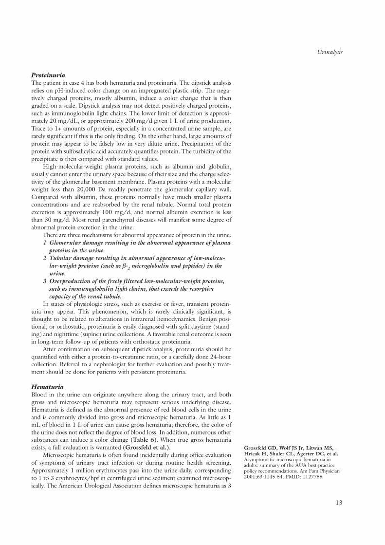
ProteinuriaThe patient in case 4 has both hematuria and proteinuria. The dipstick analysisrelies on pH-induced color change on an impregnated plastic strip. The nega-tively charged proteins, mostly albumin, induce a color change that is thengraded on a scale. Dipstick analysis may not detect positively charged proteins,such as immunoglobulin light chains. The lower limit of detection is approxi-mately 20 mg/dL, or approximately 200 mg/d given 1 L of urine production.Trace to 1+ amounts of protein, especially in a concentrated urine sample, arerarely significant if this is the only finding. On the other hand, large amounts ofprotein may appear to be falsely low in very dilute urine. Precipitation of theprotein with sulfosalicylic acid accurately quantifies protein. The turbidity of theprecipitate is then compared with standard values.
High-molecular-weight plasma proteins, such as albumin and globulin,usually cannot enter the urinary space because of their size and the charge selec-tivity of the glomerular basement membrane. Plasma proteins with a molecularweight less than 20,000 Da readily penetrate the glomerular capillary wall.Compared with albumin, these proteins normally have much smaller plasmaconcentrations and are reabsorbed by the renal tubule. Normal total proteinexcretion is approximately 100 mg/d, and normal albumin excretion is lessthan 30 mg/d. Most renal parenchymal diseases will manifest some degree ofabnormal protein excretion in the urine.
There are three mechanisms for abnormal appearance of protein in the urine.1 Glomerular damage resulting in the abnormal appearance of plasma
proteins in the urine.2 Tubular damage resulting in abnormal appearance of low-molecu-
lar-weight proteins (such as β-2 microglobulin and peptides) in theurine.
3 Overproduction of the freely filtered low-molecular-weight proteins,such as immunoglobulin light chains, that exceeds the resorptivecapacity of the renal tubule.
In states of physiologic stress, such as exercise or fever, transient protein-uria may appear. This phenomenon, which is rarely clinically significant, isthought to be related to alterations in intrarenal hemodynamics. Benign posi-tional, or orthostatic, proteinuria is easily diagnosed with split daytime (stand-ing) and nighttime (supine) urine collections. A favorable renal outcome is seenin long-term follow-up of patients with orthostatic proteinuria.
After confirmation on subsequent dipstick analysis, proteinuria should bequantified with either a protein-to-creatinine ratio, or a carefully done 24-hourcollection. Referral to a nephrologist for further evaluation and possibly treat-ment should be done for patients with persistent proteinuria.
HematuriaBlood in the urine can originate anywhere along the urinary tract, and bothgross and microscopic hematuria may represent serious underlying disease.Hematuria is defined as the abnormal presence of red blood cells in the urineand is commonly divided into gross and microscopic hematuria. As little as 1mL of blood in 1 L of urine can cause gross hematuria; therefore, the color ofthe urine does not reflect the degree of blood loss. In addition, numerous othersubstances can induce a color change (Table 6). When true gross hematuriaexists, a full evaluation is warranted (Grossfeld et al.).
Microscopic hematuria is often found incidentally during office evaluationof symptoms of urinary tract infection or during routine health screening.Approximately 1 million erythrocytes pass into the urine daily, correspondingto 1 to 3 erythrocytes/hpf in centrifuged urine sediment examined microscop-ically. The American Urological Association defines microscopic hematuria as 3
13
Grossfeld GD, Wolf JS Jr, Litwan MS,Hricak H, Shuler CL, Agerter DC, et al.Asymptomatic microscopic hematuria inadults: summary of the AUA best practicepolicy recommendations. Am Fam Physician2001;63:1145-54. PMID: 1127755

Sokolosky MC. Hematuria. Emerg MedClin North Am 2001;19:621-32.PMID: 11554278
Urinalysis
14
erythrocytes/hpf on microscopic examination of the centrifuged urine speci-men, in two of three freshly voided, clean-catch, midstream urine samples.Repeated testing is done because hematuria is intermittent in some diseases.
The reported prevalence of asymptomatic hematuria in adults varies widely.Population-based studies have shown prevalence of less than 1% to 16%. Thisrange is attributed to differences in patient demographic characteristics, dura-tion of follow-up, definition of hematuria, diagnostic technique, and number ofscreening tests per patient. Patients at high risk for urologic disease, such as eld-erly men, have a higher prevalence of hematuria.
The pathophysiology of hematuria depends on the site in the urinary tractfrom which blood loss occurs. Hematuria in which blood originates from thenephron is termed glomerular hematuria. Erythrocytes can enter the urinaryspace from the glomerulus or, rarely, from the renal tubule. Disruption of thefiltration barrier in the glomerulus may result from inherited or acquired abnor-malities in the structure and integrity of the glomerular capillary wall. Theseerythrocytes can be trapped in Tamm–Horsfall mucoprotein and will be mani-fest in the urine by erythrocyte casts. Casts in the urine indicate clinically sig-nificant disease at the glomerular level. However, in disease of the nephron,casts may be absent, and isolated red blood cells may be the only finding. Thepresence of proteinuria supports a glomerular source of blood loss.
Hematuria without proteinuria or casts is termed isolated hematuria.Although a few glomerular diseases may produce isolated hematuria, this find-ing is more consistent with extraglomerular bleeding. Disruptions of the uroep-ithelium such as irritation, inflammation, or invasion, can result in normal-appearing erythrocytes in the urine. Cancer, renal stones, trauma, infection, andmedications may cause these disruptions. Nonglomerular renal causes of bloodloss, such as tumors of the kidney, renal cysts, infarction, and arteriovenous mal-formation, can also cause blood loss into the urinary space.
The dipstick urinalysis records a reaction between hydrogen peroxide andchromogen that is catalyzed by hemoglobin. This reaction results in a greencolor change of the chromogen that is visible on the dipstick. The sensitivity ofthe dipstick to detect hematuria at a concentration of more than 3 erythro-cytes/hpf is more than 90% (Sokolosky).
Many factors can produce false-positive and false-negative results on dip-stick analysis. Vitamin C ingestion, urine pH less than 5.1, or prolonged expo-sure of the dipstick to air before testing can cause false-negative results.Contamination of the urine with menstrual blood, myoglobinuria, and bacter-ial peroxidases can produce false-positive findings. For these reasons, all positiveresults on dipstick analysis and all negative results with a high index of suspicionshould be sent for microscopy. Samples sent for microscopy should be evaluatedwithin 1 hour, because casts will begin to disintegrate and erythrocytes maylyse. Cellular elements may be preserved for a few more hours by refrigerationof the sample.
On microscopy, dysmorphic erythrocytes (which are distorted in bothshape and size) and casts are consistent with a glomerular source of bleeding.Best seen with phase-contrast microscopy by trained personnel, these findings,especially in conjunction with significant proteinuria, should prompt referral toa nephrologist for evaluation for glomerular disease and consideration of a kid-ney biopsy. Erythrocytes from a nonglomerular source more closely resembleperipheral blood on microscopy, with isomorphic erythrocytes and absence of casts.
The current American Urological Association Best Practice PolicyRecommendations for evaluation of isolated hematuria are based on presenceof risk factors for significant urologic disease. Low-risk patients should undergoeither urine cytology examination or cystoscopy along with upper-tract imaging
TABLE 6 Substances That MayCause Red Pigmenturia
Endogenous Sources
Bilirubin
Myoglobin
Hemoglobin
Porphyrins
Foods
Rhubarb
Blackberries
Blueberries
Paprika
Beets
Fava beans
Artificial food colorings
Drugs
Rifampin
Nitrofurantoin
Sulfonamides
Metronidazole
Phenytoin
Prochlorperazine
Phenolphthalein
Quinine
Chloroquine
Phenazopyridine
Levodopa
Methyldopa
Doxorubicin
Desferoxamine

Imaging
(computed tomography or intravenous pyelography). Patients with suspiciousfindings on cytology should then be referred for cystoscopy. Patients with sig-nificant risk factors such as smoking history, occupational exposure to benzenesor aromatic amines, age greater than 40 years, history of urologic disorder ordisease, irritative voiding symptoms, history of analgesic abuse, history of pelvicirradiation should undergo a complete evaluation including upper-tract imag-ing, urine cytology and cystoscopy.
Leukocytes and Other Formed ElementsThe finding of more than 1 leukocyte/hpf in the urine can be consideredabnormal; however, fewer than 3 to 5 leukocytes/hpf is usually consideredacceptable. Leukocytes may originate from any point along the urogenital tract,and, like hematuria, leukocyturia has a broad differential diagnosis. The findingof other formed elements in the urine, casts, and proteinuria implicate aglomerular or renal source. Most leukocytes in the urine are polymorpho-nuclear; these cells contain esterases that can be detected on chemical dipstickanalysis of urine. The finding of leukocytes in the urine usually implies infection;however, glomerular or tubulointerstitial inflammation and allergic reaction cancause leukocyturia.
Lipids and fat in the urine are almost always seen in association with heavyproteinuria or the nephrotic syndrome. They may appear as free lipid droplets,in round oval fat bodies, or in fatty casts. Fat is best seen under polarized light,where the cholesterol esters appear as refractile objects shaped like Maltese crosses.
Casts are cylindrical aggregates of Tamm–Horsfall mucoprotein that aresecreted by the distal nephron, which trap the intraluminal contents and appearin the urine. Erythrocytes, leukocytes, and debris may appear in cast form, andall implicate different pathologic mechanisms. Erythrocyte casts most oftenimply glomerular disease; leukocyte casts connote inflammation or infection ofthe renal parenchyma, and granular casts result from degenerating cellular ele-ments and protein precipitates. Other casts, such as hyaline, waxy, and fattycasts, may also be seen.
ImagingPlain radiography of the abdomen can provide rudimentary information of thekidneys and will reveal calcifications in the pelvis, but this technique alone is notuseful for assessment of kidney disease. Intravenous administration of contrastagent combined with serial radiography (intravenous urography or pyelog-raphy) can be useful in assessment of nonglomerular bleeding and nephrolithia-sis but may have significant side effects, such as allergic reaction and azotemia.Diabetic patients and those with mild kidney disease may be especially prone toazotemia secondary to iodinated contrast agents. The routine use of plainradiography and intravenous urography to assess kidney disease has beenreplaced by such techniques as ultrasonography and computed tomography.
UltrasonographyUltrasonography of the kidney, ureters, and bladder has become the initial testof choice in many kidney and urologic diseases because of the relative ease,availability, and safety at all levels of kidney function. Ultrasonography reliablydetects hydronephrosis with a sensitivity greater than 90%. Both radiopaqueand radiolucent renal stones larger than 5 mm may be seen. Echogenicity of therenal cortex is used as a surrogate marker for kidney fibrosis and, along withkidney length, can be used to monitor disease progression. Simple cysts are eas-
15

Kidney Biopsy
16
ily distinguished from solid renal tumors. Solid tumors smaller than 3 cm arebest seen with other methods, as described below. Duplex sonography has beenused in renal artery stenosis to diagnose and predict outcome after revascular-ization (Radermacher et al.).
Computed TomographyComputed tomography is now the test of choice for renal lithiasis and masses.Helical computed tomography is especially helpful in imaging the retroperi-toneal area and can have a spatial resolution up to 0.5 mm. Nonenhanced com-puted tomography is used to detect kidney stones and calcification; enhancedcontrast scans using iodinated contrast agents are helpful in delineating masses,abscesses, and tumors of the kidneys, renal pelvis, and adrenal glands. The needto use intravenous contrast material for most scans limits the utility of com-puted tomography for patients with advanced kidney disease or those at highrisk for contrast-induced kidney failure.
Magnetic Resonance ImagingMagnetic resonance imaging exposes atoms in the body to a powerful externalmagnetic field and pulses them at a specific radiofrequency. Magnetic resonanceimaging yields high-resolution images of the kidney parenchyma and perirenaltissues. Tumors smaller than 3 cm that are difficult to visualize with ultra-sonography are easily seen on magnetic resonance imaging. Use of the intra-venous, noniodinated contrast agent gadolinium in magnetic resonance angiog-raphy has greatly enhanced the ability of magnetic resonance imaging to ana-lyze the vasculature of the kidney and diagnose renal artery stenosis. Three-dimensional gadolinium-enhanced magnetic resonance angiography is betterthan digital subtraction angiography in detecting ostial lesions, but visibility ofthe distal renal artery and hilum is worse (Schoenberg et al.). As for duplexultrasonography of the renal arteries, interobserver variability exists with use ofmagnetic resonance angiography.
Use of arteriography for diagnosis of renal artery stenosis is usually reservedfor cases in which the index of suspicion is higher an intervention is required.
Radionuclide ScanningRadionuclide scanning is primarily used to assess renal perfusion and is espe-cially useful for detecting substantial differences in perfusion between the kid-neys. This technique easily detects severe reduction in or absence of renal per-fusion, especially when involvement is unilateral. Renography has a sensitivity of75% for detection of hemodynamically significant renal artery stenosis. The sen-sitivity increases to 92%, and specificity to 95% by repeating renography afteradministration of oral captopril. Comparatively, using intra-arterial angiographyas the standard, magnetic resonance angiography has a sensitivity and specificityof 100% and 96% respectively (Postma et al.) and duplex ultrasonography hasa sensitivity and specificity of 98% and 98%, respectively (Olin et al.).
Kidney BiopsyA kidney biopsy is obtained to establish a diagnosis, aid in prognosis, or tailortherapy. Biopsy provides information on the glomeruli, tubulointerstitium, andvasculature. The level of fibrosis in the interstitium and vasculature correlatesbetter than the glomerular histopathology with renal function.
Common indications for kidney biopsy are evaluation of primary nephroticsyndrome in adults or corticosteroid-unresponsive nephrotic syndrome in chil-dren, acute or rapidly progressive glomerulonephritis, and an elevated serum
Radermacher J, Chavan A, Bleck J,Vitzthum A, Stoess B, Gebel MJ, et al.Use of Doppler ultrasound to predict theoutcome of therapy for renal-artery stenosis.N Engl J Med 2001;344:410-7.PMID: 11172177
Schoenberg SO, Knopp MV, Londy F,Krishnan S, Zuna I, Lang N, et al.Morphologic and functional magnetic reso-nance imaging of renal artery stenosis: amultireader tricenter study. J Am SocNephrol 2002;13:158-69. PMID: 11752033Postma CT, Joosten FB, Rosenbusch G,Thien T. Magnetic resonance angiographyhas a high reliability in the detection of renalartery stenosis. Am J Hypertens1997;10:957-63. PMID 9324099Olin JW, Peidemonte MR, Young JR,DeAnna S, Grubb M, Childs MB. Theutility of duplex ultrasound scanning of therenal arteries for diagnosing significant renalartery stenosis. Ann Intern Med.1995;122:833-8. PMID 7741367

Glomerular Anatomy and Its Relation to Glomerular Disease
creatinine concentration in kidney transplant recipients. Kidney biopsy may alsoprove useful in the evaluation of common diseases that do not display typicalfeatures, such as diabetic patients with kidney disease but no evidence of othermicrovascular disease and in those with acute renal failure of no clear origin.Kidney biopsies are usually not performed in advanced chronic kidney diseasebecause the risk for complications outweighs the benefit of the test.
Biopsies may be performed percutaneously with local anesthesia, by anopen surgical technique, or laparoscopically. The laparoscopic approach offersdirect visualization of the kidney and controlled hemostasis (Gimenez et al.).This technique requires general anesthesia but may benefit high-risk patientswho require a kidney biopsy, since the rate of complications is low and the pro-cedure has a high success rate.
The most common complications of percutaneous kidney biopsy are self-limited microscopic hematuria, which occurs in most patients, and benign peri-nephric hematomas. Gross hematuria occurs in less than 10% of patients and isalso self-limited in most instances. Complications such as arteriovenous fistulas,aneurysm, requirement of blood transfusion, and infection are much more rare.Mortality following a kidney biopsy is approximately 0.12%.
Glomerular Diseases
Primary glomerular disorders occur in the absence of known systemic diseases.Secondary glomerular disorders occur as a consequence of systemic disease.Secondary glomerular disorders may complicate metabolic or immune dis-orders, infections, neoplasms, hereditary factors, or other processes.
Glomerular Anatomy and Its Relation to Glomerular Disease
• What is the structure of the glomerulus?• How does glomerular structural integrity relate to glomerular function?
The glomerulus consists of four regions: arterioles, the glomerular capillary wall (endothelium, glomerular basement membrane, and podocyte), themesangium, and Bowman’s space and capsule (Figure 2). Abnormalities ofthese structures result in failure of function, which manifests as proteinuria,hematuria, or declining glomerular filtration rate. Glomeruli are bound byafferent and efferent arterioles, thus permitting fine regulation of intra-glomerular pressure by modulation of these two structures. Angiotensin-converting enzyme inhibitor therapy makes use of this unique anatomy by prefer-entially dilating the efferent arteriole over the afferent arteriole, therebydecreasing intraglomerular pressure.
Ultrafiltration occurs across the glomerular capillary wall. Endothelial cellsline the capillary lumen, create a nonthrombogenic surface, and act as a porousfilter with spaces or fenestrae to permit filtration. The glomerular basementmembrane has historically been viewed as the seat of glomerular permselectiv-ity, but this role was recently challenged. Podocytes attach to the outer surfaceof the glomerular basement membrane predominantly by integrins, a family ofproteins that act as adhesion molecules. Podocytes are connected to each otherby a slit membrane that covers the glomerular basement membrane, which iscomposed, at least in part, of a secreted podocyte protein called nephrin.Mutations in nephrin cause massive proteinuria in newborns (the congenitalFinnish nephrotic syndrome). This discovery led to the idea that the podocyteslit membrane, rather than the glomerular basement membrane, may be the
17
Gimenez LF, Micali S, Chen RN, MooreRG, Kavoussi LR, Scheel PJ Jr.Laparoscopic renal biopsy. Kidney Int.1998;54:525-9. PMID: 9690219

Glomerular Anatomy and Its Relation to Glomerular Disease
18
seat of glomerular permselectivity. Podocytes may be injured by stretch accom-panying glomerular hypertrophy, such as in minimal change disease and focalsclerosis; by metabolic factors, such as hyperglycemia; by complement activa-tion, such as in lupus nephritis or membranous nephropathy; by chemical tox-ins or medications; and by direct infection, such as in HIV-associated nephropa-thy. Injury to podocytes may thus induce proteinuria.
The mesangium is composed of cells and matrix. Mesangial expansion maybe due to hypercellularity, such as that seen in immune disorders (for example,systemic lupus erythematosus or IgA nephropathy), matrix expansion (diabeticnephropathy), or infiltration by abnormal proteins (amyloidosis). Mesangialhypercellularity is often associated with both hematuria and some degree ofproteinuria. Mesangial matrix expansion may reduce glomerular filtration rateby occluding the capillary lumina.
Bowman’s space is the glomerular region bounded by podocytes and slitmembrane and the parietal epithelial cells. It is usually empty apart from theseintrinsic glomerular cells and ultrafiltrate. In inflammatory glomerular disor-ders, this space may fill with infiltrating mononuclear cells, activated and divid-ing glomerular epithelial cells, and the fibrous products of these cells. This phenomenon is called a crescent. Crescentic glomerulonephritis is a nephrologicemergency because rapid collapse of the glomerular tuft induces loss ofglomerular filtration, often irreversibly.
Glomerular disorders are diagnosed on the basis of their clinical presenta-tion and their histomorphologic appearance. Sometimes a diagnosis is stronglysuspected by the history, physical examination, and serologic studies (includingcomplement levels, antinuclear and anti-DNA antibodies, antinuclear cytoplas-mic antibody [ANCA] levels, and HIV and hepatitis status) alone. This is usu-ally the case for diabetic nephropathy and for the idiopathic nephrotic syn-drome of childhood. Renal biopsy is rarely performed for asymptomatic urinaryabnormalities, such as proteinuria less than 1 g/d, or for microscopic hematuria
F I G U R E 2 .The glomerulus Schematic representation of a glomerulus indicat-ing the urinary space, capillary lumen, theglomerular basement membrane and cells which are intrinsic to the glomerulus: mesangialcell and matrix, the fenestrated endothelial cellsand epithelial cells. Immune-complex deposits aredepicted in the mesangial, subendothelial, andsubepithelial areas.
(U.S. = urinary space; C.L. = capillary lumen; Mes. =mesangial; GBM = glomerular basement membrane.)

Clinical Syndromes of Glomerular Disease
in the absence of renal insufficiency or a systemic disorder. However, renalbiopsy is indicated in patients with heavy proteinuria, dysmorphic hematuria,erythrocyte casts, and proteinuria or hematuria in the presence of low glomer-ular filtration rate when the underlying cause is uncertain.
Clinical Syndromes of Glomerular Disease
Case 5A 30-year-old man in otherwise good health presents withweight gain of 4.5 to 6.5 kg (10 to 14 lb); periorbital and facialedema in the morning, with resolution during the day; andankle edema toward evening, which has worsened in severityover 3 months. He reports that “his urine looks like beer.”There is no family history of proteinuria or renal disease.
Physical examination is unremarkable apart from mildhypertension (blood pressure, 140/95 mm Hg) and modestankle and pretibial pitting edema. Blood urea nitrogen, creati-nine concentration, and glucose level are normal; the serumalbumin level is low; and the serum cholesterol level is high.Urinalysis shows 4+ proteinuria on dipstick, and microscopicanalysis is negative apart from oval fat bodies. A 24-hour urinecollection shows 10 g proteinuria, and a random urinary pro-tein-to-creatinine ratio is 7. Serologic studies that are normal or negative included hepatitis B and C and HIV serologies,urine and serum immunofixation, C3 and C4 levels, and a test for syphilis.
The Nephrotic SyndromeThis patient has the nephrotic syndrome, characterized by proteinuria of 3 to3.5 g/d or greater, hypoalbuminemia, hyperlipidemia, lipiduria, and edema.Table 7 lists many of the causes of the nephrotic syndrome. Diagnosis and fol-low-up of nephrotic-range proteinuria by measurement of random urinary pro-tein-to-creatinine ratios instead of 24-hour urine protein and creatinine is nowa common and validated practice. Edema is the most common presenting man-ifestation of the nephrotic syndrome. It is caused by a combination of lowoncotic pressure and renal sodium retention, of which the latter is due to acti-vation of the renin–angiotensin–aldosterone system or renal resistance to natri-uretic peptides. Volume overload is an important consequence of the nephroticsyndrome, although total-body sodium and water overload may occur simulta-neously with intravascular volume depletion. Many patients with the nephroticsyndrome, however, have normal intravascular volume. Particularly in adults,volume overload associated with nephrotic syndrome may also contribute tohypertension and edema. In children, ascites and pleural and pericardial effu-sions may accompany the nephrotic syndrome. In adults, extravasation of fluidfrom the vascular compartment into the interstitium (“third-spacing”) may alsooccur but is less common, and it may suggest the presence of a disorder inwhich serosal permeability is increased, such as systemic lupus erythematosus,tuberculosis, or neoplasm.
Hyperlipidemia and increased lipoprotein are common but not universalcomplications of nephrotic syndrome. Before the advent of HMG-CoA reduc-tase inhibitors, uncontrolled hyperlipidemia contributed to accelerated athero-genesis in these patients. Hypercoagulability is another complication of thenephrotic syndrome; this state may be associated with decreased plasma levelsof protein S, protein C, and antithrombin III. Deep venous thromboses more
19
TABLE 7 Causes of the NephroticSyndrome
Primary glomerular disease
Minimal change disease
Focal and segmental glomerulosclerosis
Membranous nephropathy
Membranoproliferativeglomerulonephritis
Mesangial proliferative glomerulonephritis
Drugs
Gold- and mercury-containing medications
Captopril
NSAIDs
Penicillamine
Lithium
Secondary to systemic diseases
Diabetes
Cancer: lymphoma, breast, lung, myeloma,colon, thyroid
Infection: subacute bacterial endocarditis,post-streptococcal, hepatitis B or C, HIV,syphilis, ventriculoatrial shunts, chronicvisceral abscesses
Connective tissue disorder: systemic lupuserythematosus, rheumatoid arthritis,Sjögren’s syndrome, anticardiolipinsyndrome
Amyloidosis
Heredofamilial diseases
Alport’s syndrome
Fabry’s disease
Nail–patella syndrome
Lecithin-cholesterol acyltransferasedeficiency
Familial focal and segmentalglomerulosclerosis
Uncommon secondary causes
Ulcerative colitis
Toxemia of pregnancy
Vesicoureteral reflux
Renal artery stenosis
NSAIDs = nonsteroidal anti-inflammatory drugs.

Clinical Syndromes of Glomerular Disease
20
so than arterial thromboses occur frequently, and in patients with membranousnephropathy, renal vein thromboses are characteristic. Thromboses occur atgreater frequency in patients with a serum albumin level less than 2 g/dL.Other significant complications of the nephrotic syndrome include loss of 25-hydroxyvitamin D, which may induce secondary hyperparathyroidism; low thy-roxine levels with normal thyroid-stimulating hormone levels and euthyroidism;and susceptibility to infection as a consequence of numerous factors, includingurinary loss of complement, immunoglobulins, and zinc-binding proteins,abnormalities in leukocyte function, and use of immunosuppressive medications.
TreatmentThe treatment of patients with the nephrotic syndrome has changed greatly inrecent years because of the effectiveness of angiotensin-converting enzymeinhibitors and angiotensin receptor blockers in diminishing proteinuria andslowing renal disease progression. These agents have favorable side-effect pro-files compared with the immunosuppressive agents usually used in patients withimmune forms of glomerulonephritis. As a result, therapeutic decision makinghas become more complex, since neither specific randomized treatment efficacystudies nor evidenced-based studies clearly delineate whether immunosuppres-sive therapy or treatment with angiotensin-converting enzyme inhibitors orangiotensin receptor blockers is superior in a manner that is generalizable totreatment of individual patients. Thus, angiotensin-converting enzymeinhibitors or angiotensin receptor blocker therapy, alone or in combination withimmunosuppressive therapies, should be used as first-line treatment to dimin-ish proteinuria. Dosing of angiotensin-converting enzyme inhibitors or angio-tensin receptor blockers should be increased sequentially until proteinuria fails to decline further. The total dose used to inhibit proteinuria may exceeddosing guidelines advised for hypertension. Increasing the dose may be limitedby unacceptable increments in serum creatinine or potassium, which should becarefully monitored with each dose increment. Women treated withangiotensin-converting enzyme inhibitors or angiotensin receptor blockersshould avoid pregnancy and breast-feeding due to teratogenicity and postpar-tum acute renal failure in neonates. Combining angiotensin-converting enzymeinhibitors and angiotensin receptor blockers have theoretical advantages, butthe two should probably not be used in concert with β-blockers in patients withcongestive heart failure due to concerns regarding increased cardiovascularmorbidity and mortality. The use of angiotensin-converting enzyme inhibitorsor angiotensin receptor blockers to treat proteinuria slows the rate of loss ofrenal function.
The hyperlipidemia of nephrotic syndrome is poorly responsive to dietarytherapies alone. The HMG-CoA reductase inhibitors control nephrotic hyper-lipidemia and should be used for the duration of the nephrotic syndrome.Similarly, patients experiencing a venous or arterial thrombosis should betreated with anticoagulants for the duration of the nephrotic syndrome. InEurope, it is common to anticoagulate prophylactically patients with a serumalbumin less than 2 g/dL; in the United States, use of this practice varies.Children with the nephrotic syndrome are sometimes given vitamin D to pre-vent secondary hyperparathyroidism. For adult patients prescribed corticoste-roids, consideration should be given to the use of bone-sparing therapy thatincludes vitamin D and calcium. Before immunosuppressive therapy is started,isoniazid therapy or vaccinations may be appropriate on an individualized basis.
The patient described in case 5 has idiopathic nephrotic syndrome, proba-bly due to idiopathic membranous glomerulopathy. Common serologic testsperformed to identify secondary causes of the nephrotic syndrome before con-
Clinical Syndromes of Glomerular Disease
sideration of renal biopsy are listed in the case description. These tests shouldbe used selectively to address specific diagnostic concerns on the basis of a care-ful history and physical examination. The following primary glomerulopathiescan present as the nephrotic syndrome.
Minimal Change DiseaseMinimal change disease is thus named because there are scant light microscopicglomerular abnormalities and few or no immunoreactants on immunofluores-cence microscopy. Ultrastructural analysis demonstrates almost universal efface-ment of the podocyte foot process. The pathogenesis is unclear, but a circu-lating permeability factor has been implicated. Rare familial cases have beenreported. Secondary causes of minimal change disease include hypersensitivityinduced by use of nonsteroidal anti-inflammatory drugs, usually along withacute tubulointerstitial nephritis; lithium treatment; non-Hodgkin’s lymphoma;and occasionally, leukemia. Minimal change disease may occur more frequentlyin atopic persons. In its idiopathic form, minimal change disease is the mostcommon cause of the nephrotic syndrome in children, with a male-to-femalepredominance of 2 to 1. Occurring at a rate of 12 to 18 million persons yearly,its incidence peaks at 4 years of age. In adults, minimal change disease has nosex-based predilection and occurs at a rate of about 2 million persons yearly,with a peak in incidence at approximately 65 years of age.
The predominant clinical manifestation is edema, which may be massiveand occur rapidly over just a few days. Ascites and pleural and pericardial effu-sions are more common in children than adults. Hypertension occurs in aminority of patients but is three times more prevalent in adults than children.Hematuria is uncommon. Azotemia, if it occurs, is often related to hypovolemiaand may be precipitated by overaggressive diuresis. Conversely, acute renal fail-ure may also occur spontaneously in the setting of massive edema and mayresolve with diuresis. Laboratory tests to assess secondary causes are usuallynegative or normal. However, the sedimentation rate may be elevated, particu-larly if marked hypoalbuminemia is present.
The most corticosteroid-sensitive of all glomerular diseases, completeremission is achieved in at least 90% of children within 2 to 3 months of initia-tion of prednisone and at least 75% of adults after 4 months. Unfortunately,relapse is common. Re-treatment with steroids is often sufficient to reinduceremission, but for patients with frequent relapse and those with steroid depend-ence, the addition of oral alkylators, such as cyclophosphamide or chlorambu-cil, may reinduce and maintain remission while minimizing the total steroiddose administered. Cyclosporine has been used in small studies, but nephro-toxicity and more frequent relapse are concerns. Experience with mycophenolatemofetil is so limited that guidelines for its use are not yet available. Salt restric-tion and diuretics should be used as palliative therapy, but aggressive diuresisshould be avoided. Renal failure is uncommon, occurring in less than 2% ofpatients, and may represent conversion to focal segmental glomerulosclerosis.
Focal and Segmental GlomerulosclerosisHistopathologically, focal and segmental glomerulosclerosis is defined by seg-ments of sclerosis in only a portion (segmental) of some glomeruli (focal).Podocytes in focal and segmental glomerulosclerosis show diffuse foot processeffacement, a feature that probably contributes to the nephrotic-range protein-uria characteristic of the disorder. Numerous histologic variants occur, the mostimportant of which is the collapsing lesion. This lesion is characteristic of HIV-associated nephropathy but is also occasionally seen in the absence of HIV, andit is the least corticosteroid-sensitive glomerular disease. Focal and segmental
21

Clinical Syndromes of Glomerular Disease
22
glomerulosclerosis is best thought of as a common histologic picture that canresult from numerous pathogenetic etiologies. In patients with idiopathic focalsegmental glomerulosclerosis, a circulating permeability factor has been pro-posed to play a pathogenetic role. This factor may be present but not yet iden-tified in as many as one third of patients. Diseases that promote glomerulargrowth or cause loss of nephrons predispose to focal segmental glomerulo-sclerosis, presumably by inducing intraglomerular hypertension. These includesuch diverse entities as morbid obesity, subtotal nephrectomy, unilateral renaldysgenesis, chronic ureterovesical reflux, sickle-cell anemia, and congenitalcyanotic heart disease. Focal and segmental glomerulosclerosis may also com-plicate the progressive forms of almost any primary glomerulopathy. Heroin usehas been associated with focal and segmental glomerulosclerosis, but this asso-ciation has waned in the past decade. Familial forms of focal and segmentalglomerulosclerosis have been identified, and mutations in the genes for thepodocyte proteins α-actinin 4 and podocin induce autosomal dominant andrecessive forms, respectively.
The collapsing form of focal and segmental glomerulosclerosis is mostnotably associated with HIV, but infection with parvovirus has also been impli-cated, as has use of high-dose pamidronate. The collapsing form can also occuridiopathically.
Focal and segmental glomerulosclerosis is the most frequent form of idio-pathic nephrotic syndrome in African-American persons. It presents most oftenas the nephrotic syndrome but may also present as persistent non–nephrotic-range proteinuria. Microscopic hematuria may accompany the proteinuria.Hypertension is common, and accelerated or malignant hypertension may bethe presenting manifestation. Serologic tests to assess secondary causes are usuallynegative or normal in patients with idiopathic focal segmental glomerulosclerosis.
Treatment is controversial. Some patients may be adequately managed withangiotensin-converting enzyme inhibitors or angiotensin receptor blockersalone, particularly if proteinuria can be controlled to less than 1 g/d, althoughoutcome comparisons of this treatment strategy with immunosuppressive regi-mens have not been studied. Although focal and segmental glomerulosclerosiswas once thought to be steroid resistant, about 20% to 40% of adult patientsrespond to steroid therapy, particularly high-dose oral prednisone given for atleast 3 to 6 months. In nonresponders, cyclosporine and, recently, mycophe-nolate mofetil have been used with some success. The rate of progression torenal failure varies, and more rapid progression is associated with heavy pro-teinuria that is refractory to therapy. Spontaneous remission is rare, althoughweight loss reportedly prompted remission in a few patients with morbid obe-sity. Recent reports suggest that highly active antiretroviral therapy for recent-onset HIV may limit progression and reverse some of the histologic damageobserved in patients with HIV-associated collapsing focal segmental glomerulo-sclerosis. Idiopathic focal and segmental glomerulosclerosis may recur in renalallografts within days or months of transplantation, particularly in patients withcirculating permeability factor. In these patients, plasmapheresis may decreaseproteinuria and improve allograft function.
Membranous NephropathyIdiopathic membranous nephropathy is one of the most frequent causes of thenephrotic syndrome in adults, accounting for 30% to 50% of cases, dependingon the subgroup studied. A broad range of systemic disorders may predisposeto secondary membranous nephropathy. The nephrotic syndrome is the pre-senting manifestation in about 95% of patients with membranous nephropathy.Microscopic hematuria is present in 15% to 30% of patients. The incidence ofdeep venous thrombosis, especially renal vein thrombosis, is more common in

Clinical Syndromes of Glomerular Disease
membranous nephropathy than in other forms of the nephrotic syndrome.Pulmonary emboli may occur and may in rare cases be the presenting manifestation.
The diagnosis is confirmed by the finding of subepithelial immune com-plexes along the glomerular basement membrane on renal biopsy. Certain histologic changes on renal biopsy help to distinguish idiopathic membranousnephropathy from a secondary form. Laboratory tests to assess secondarycauses are usually negative or normal in idiopathic membranous nephropathy.The course of patients with idiopathic membranous nephropathy varies widelyand has been summarized by the “rule of thirds”: One third of patients progressto end-stage renal disease over 10 years of follow-up, one third remain pro-teinuric with stable or slowly declining renal function, and one third experiencespontaneous remission.
Factors associated with an increased risk for progression include a highserum creatinine concentration at diagnosis, hypertension, tubulointerstitialfibrosis on renal biopsy, heavy proteinuria (>10 g/d), and male sex. The courseis also affected by patient age (children rarely progress to end-stage renal dis-ease), ethnicity (disease may be more benign in Japan, Southeast Asia, and partsof Europe than in the United States), and genetic factors (persons with certainhaplotypes have a worse prognosis). Thus, the choice of therapy should beguided by estimates of risk of progressive renal disease and risks associated withthe severity of the nephrotic syndrome.
Treatment of membranous nephropathy remains controversial. Forpatients at low risk for progression, angiotensin-converting enzyme inhibitorsor angiotensin receptor blocker therapy can decrease proteinuria, and in sodoing, diminish the rate of progression of renal disease and ameliorate thenephrotic syndrome. The dose of angiotensin-converting enzyme inhibitor orangiotensin receptor blocker should be incrementally increased until the pro-teinuria fails to decrease further or the patient develops an unacceptable sideeffect. For patients at high risk for progression or those with severe nephroticcomplications, immunosuppressive therapy is advocated. Therapy with pred-nisone alone achieves remission in only a minority of patients and is no longergenerally recommended. A meta-analysis suggested that immunosuppressivetherapy with corticosteroids and either chlorambucil or cyclophosphamide ismore efficacious than supportive therapy in achieving partial and completeremission (Imperiale et al.). A randomized prospective study showed thatcyclosporine was also efficacious in maintaining renal function and diminishingproteinuria (Cattran et al.). However, the durability of these remissions afterdiscontinuation of cyclosporine therapy and the long-term consequences of thistherapy remain a concern. Mycophenolate mofetil has been used with someanecdote success. Membranous nephropathy occasionally recurs and can occurde novo in the transplanted kidney.
Membranoproliferative GlomerulonephritisThere are two major histologic types of membranoproliferative glomer-ulonephritis, reflecting different pathogenetic mechanisms. Membrano-proliferative glomerulonephritis type I is characterized by subendothelial andmesangial deposits and capillary-wall mesangial interposition. Most adults whopresent with membranoproliferative glomerulonephritis type I have underlyinghepatitis C, although some cases are idiopathic (Johnson et al.). A membra-noproliferative glomerulonephritis type I histologic pattern may also be seen inadults with underlying chronic infectious, neoplastic, inflammatory disorders,or, rarely, partial lipodystrophy.
Membranoproliferative glomerulonephritis type II is characterized by dep-osition of electron-dense material in the capillary wall. Membranoproliferative
23
Imperiale TF, Goldfarb S, Berns JS. Arecytotoxic agents beneficial in idiopathicmembranous nephropathy? A meta-analysisof the controlled trials. J Am Soc Nephrol.1995;5:1553-8. PMID: 7756587Cattran DC, Greenwood C, Ritchie S,Bernstein K, Churchill DN, Clark WF, et al. A controlled trial of cyclosporine inpatients with progressive membranousnephropathy. Canadian GlomerulonephritisStudy Group. Kidney Int. 1995;47:1130-5.PMID: 7783410Johnson RJ, Gretch DR, Yamabe H, Hart J, Bacchi CE, Hartwell P, et al.Membranoproliferative glomerulonephritisassociated with hepatitis C virus infection. N Engl J Med. 1993;328:465-70.PMID: 7678440

Secondary Causes of Glomerular Diseases
24
glomerulonephritis most commonly presents as the nephrotic syndrome withmicroscopic or, occasionally, gross hematuria. However, it may also be asymp-tomatic with microhematuria or non–nephrotic-range proteinuria, and it infre-quently presents as an acute nephritic syndrome. Hepatitis C–associated mem-branoproliferative glomerulonephritis is characterized by frequent cryoglobu-linemia, sometimes causing arteritis, low levels of C3 or C4, low levels ofrheumatoid factor, and skin leukocytoclastic vasculitis. Serum levels of amino-transferases are frequently but not always elevated. A low C3 level is found in approximately 70% of patients with type I disease and 90% of patients withtype II disease.
Therapy is controversial. Interferon-α with ribavirin is recommended totreat the underlying hepatitis C but cannot be used in patients with a glomeru-lar filtration rate less than 50%. In addition, interferon-α may exacerbate cryo-globulinemic vasculitis. Relapse of hepatitis C is frequent on discontinuation oftherapy. Experience with pegylated interferon is limited. Corticosteroids are rel-atively contraindicated because they may exacerbate hepatitis C, but they maybe indicated, along with plasmapheresis, in patients with cryoglobulinemic vasculitis before beginning interferon therapy. For membranoproliferativeglomerulonephritis associated with other infections, neoplasms, or inflamma-tory disorders, treatment of the associated systemic disorder is indicated. Foridiopathic membranoproliferative glomerulonephritis, steroids are often used,particularly in children; however, their efficacy is a matter of controversy.Membranoproliferative glomerulonephritis often recurs in renal allograft recipients.
Secondary Causes of Glomerular DiseasesTable 7 lists secondary glomerular disorders that may present as the nephroticsyndrome.
AmyloidosisAmyloidosis is a heterogeneous group of disorders characterized by depositionof extracellular fibrillar materials of varying types (AA, AL, transthyretin, gel-solin, lysozyme, fibrinogen A-α, and β2-microglobulin) in tissues and organs.Renal involvement is frequent with AA and AL amyloidosis and less frequent ornot seen at all with some of the other forms. In the United States, amyloidosisinvolving the kidney is associated with chronic inflammatory conditions (AA)or is due to increased secretion of a monoclonal lymphoid product (AL). Indeveloping countries, AA amyloidosis is most commonly associated withchronic infections, such as tuberculosis. In the Middle East, AA amyloidosis isone of the more common causes of end-stage renal disease in patients withfamilial Mediterranean fever. The AL form, most commonly seen in patientswith systemic idiopathic amyloidosis or multiple myeloma, reflects overproduc-tion and deposition of immunoglobulin or immunoglobulin-like light chains,most commonly λ, produced by a clone of B lymphocytes (Falk et al.).
Patients with amyloidosis may present with proteinuria with or withoutmicroscopic hematuria, often with orthostatic hypotension. Diagnosis is madeby identification of the paraprotein in serum by protein electrophoresis orimmunofixation; in glomeruli, the tubulointerstitium, and arterioles on renalbiopsy; or in other tissues, including fat pad aspirates and gingival or rectalbiopsy samples. With Congo red, AL amyloid stains with apple-green birefrin-gence. Ultrastructurally, it appears as 8- to 10-nm fibrils. Progressive renal failureis common, and patients with end-stage renal disease have shortened survival.
Treatment of AL amyloidosis is controversial. Melphalan and prednisone,and more recently vincristine, doxorubicin, and dexamethasone, may extend
Falk RH, Comenzo RL, Skinner M. Thesystemic amyloidoses. N Engl J Med.1997;337:898-909. PMID: 9302305
K E Y P O I N T S
• The nephrotic syndrome is characterizedby proteinuria of 3 to 3.5 g/d or greater,hypoalbuminemia, hyperlipidemia,lipiduria, and edema.
• Diagnosis and follow-up of nephrotic-range proteinuria by measurement ofrandom urinary protein-to-creatinineratios is a common and validated practice.
• 3-Hydroxy-3-methylglutaryl coenzyme A(HMG-CoA) reductase inhibitors controlnephrotic hyperlipidemia and should beused for the duration of the nephroticsyndrome.
• Diseases that promote glomerulargrowth or cause loss of nephrons pre-dispose to focal segmental glomeru-losclerosis, presumably by inducingintraglomerular hypertension; theseinclude such diverse entities as morbidobesity, subtotal nephrectomy, unilateralrenal dysgenesis, chronic ureterovesicalreflux, sickle-cell anemia, and congenitalcyanotic heart disease.
• Focal segmental glomerulosclerosis isthe most frequent form of the idio-pathic nephrotic syndrome in African-American persons.
• About 20% to 40% of adult patientswith focal segmental glomerulosclerosisrespond to corticosteroid therapy, par-ticularly high-dose oral prednisonegiven for at least 3 to 6 months. In non-responders, cyclosporine and mycophe-nolate mofetil have been used withsome success.
• Deep venous thrombosis, especiallyrenal vein thrombosis, is more commonin membranous nephropathy than inother forms of the nephrotic syndrome.
• About one third of patients with idio-pathic membranous nephropathyprogress to end-stage renal diseaseover 10 years of follow-up, one thirdremain proteinuric with stable or slowlydeclining renal function, and one thirdexperience spontaneous remission.
• Interferon-α with ribavirin is recom-mended to treat the underlying hepati-tis C in membranoproliferative glomeru-lonephritis but cannot be used inpatients with a glomerular filtrationrate less than 50%.

Secondary Causes of Glomerular Diseases
survival in some patients. Colchicine has also been used, particularly in patientswith familial Mediterranean fever. Treatment of AA amyloidosis should bedirected toward the underlying inflammatory or infectious process when possible.
HIV-Associated NephropathyThe predominant glomerular lesion in patients with HIV is collapsing focal seg-mental glomerulosclerosis, usually referred to as HIV-associated nephropathy(Winston et al.). It is distinguished from idiopathic focal segmental glomer-ulosclerosis histologically by the presence of numerous prominent abnormal-appearing podocytes crowding the urinary space, microcystic tubular changes,and tubulointerstitial nephritis, and clinically by the propensity for more rapidprogression to end-stage renal disease. Although it is usually seen in patientswith long-term disease, HIV-associated nephropathy has also been reported inpatients with acute infection. In addition, a diverse spectrum of other histologicforms of glomerular disease have been reported in patients with HIV infection,including IgA nephropathy; non-IgA mesangial proliferative glomerulonephri-tis; membranous glomerulopathy; thrombotic microangiopathies; and theglomerular lesions associated with lymphomas and hepatitis B and C, which areoccasionally comorbid conditions.
Diabetic NephropathyDiabetic nephropathy is characterized by mesangial expansion, thickening ofthe glomerular basement membrane, effacement of the foot process, protein-uria, and resultant tubulointerstitial fibrosis. Recent work indicates that diabeticnephropathy is due in large part to structural injury induced by hyperglycemia,intraglomerular hypertension, cytokine and growth factor elaboration, and oxi-dant stress. The likelihood of developing diabetic nephropathy appears to bemodulated by environmental and genetic factors. Patients with diabeticnephropathy are usually known to have diabetes for at least 5 to 10 years beforethe onset of proteinuria and have concomitant diabetic vascular complications,including retinopathy and cardiovascular, cerebrovascular, and peripheral arte-rial disease. Overt diabetic nephropathy is preceded by a period of microalbu-minuria (albumin excretion of 30 to 300 mg/d). However, among patientswith long-term type 1 diabetes (>10 years), those with microalbuminuriaalready have significant histologic glomerular damage. Microalbuminuria is alsoassociated with increased risk for cardiovascular morbidity and mortality. Inmany patients with microalbuminuria who do not first die of cardiovascular dis-ease, renal disease will progress to overt proteinuria (>300 mg/d albuminuriaor >500 mg/d proteinuria), followed by progressive azotemia.
Glycemic control delays or prevents progression from normoalbuminuriato microalbuminuria to overt nephropathy. Ideally, the hemoglobin A1C
shouldbe maintained at approximately 7%. Specific strategies to diminish microalbu-minuria and proteinuria by blocking the renin–angiotensin system and decreas-ing blood pressure to less than 125/75 mm Hg (which usually requires treat-ment with three or four drugs) slow progression from one diagnostic categoryto the next (Diabetes Control and Complications Trial Research Group).Use of angiotensin-converting enzyme inhibitors or angiotensin receptorblockers slows the rate of loss of glomerular filtration rate (Lewis et al., 2001;Lewis et al., 1993; Brenner et al.). Diabetic nephropathy may recur or occurde novo in the transplanted kidney.
25
Winston JA, Bruggeman LA, Ross MD,Jacobson J, Ross L, D’Agati VD, et al.Nephropathy and establishment of a renalreservoir of HIV type 1 during primaryinfection. N Engl J Med. 2001;344:1979-84. PMID: 11430327The effect of intensive treatment of diabeteson the development and progression of long-term complications in insulin-dependent dia-betes mellitus. The Diabetes Control andComplications Trial Research Group. N EnglJ Med. 1993;329:977-85. PMID: 8366922Lewis EJ, Hunsicker LG, Clarke WR, Berl T, Pohl MA, Lewis JB, et al.Renoprotective effect of the angiotensin-receptor antagonist irbesartan in patientswith nephropathy due to type 2 diabetes. N Engl J Med. 2001;345:851-60.PMID: 11565517Lewis EJ, Hunsicker LG, Bain RP, RohdeRD. The effect of angiotensin-converting-enzyme inhibition on diabetic nephropathy.The Collaborative Study Group. N Engl JMed. 1993;329:1456-62. PMID: 8413456Brenner BM, Cooper ME, de Zeeuw D,Keane WF, Mitch WE, Parving HH, et al.Effects of losartan on renal and cardiovascu-lar outcomes in patients with type 2 diabetesand nephropathy. N Engl J Med.2001;345:861-9. PMID: 11565518
K E Y P O I N T S
• Melphalan and prednisone, and morerecently vincristine, doxorubicin, anddexamethasone, may extend survival insome patients with renal amyloidosis.
• The predominant glomerular lesion inpatients with HIV is collapsing focalsegmental glomerulosclerosis, usuallyreferred to as HIV-associated neph-ropathy.
• In diabetes mellitus, glycemic controldelays or prevents progression fromnormoalbuminuria to microalbuminuriato overt nephropathy.
• Specific strategies to diminish micro-albuminuria and proteinuria by block-ing the renin–angiotensin system anddecreasing blood pressure to less than125/75 mm Hg (which usually requirestreatment with three or four drugs)slow progression from one diagnosticcategory to the next.

Acute Glomerulonephritis
26
Acute Glomerulonephritis
Case 6A 25-year-old Japanese-American man presents with a sorethroat and brown urine. Physical examination is unremarkableexcept for blood pressure of 140/95 mm Hg and pharyngealerythema without exudate. He does not have skin rash, arthri-tis, or guaiac-positive stool. Throat culture demonstrates onlynormal flora. Serum electrolytes and complete blood count arenormal. The serum creatinine concentration is elevated (1.6mg/dL). Urinalysis reveals 1+ to 2+ protein, 4+ heme, and noleukocytes or nitrites. Microscopic analysis reveals 20 dysmor-phic erythrocytes/hpf on an unspun specimen. Serologic studiesthat are normal or negative include complement measurement,hepatitis and HIV serologies, serum IgA levels, antinuclear anti-bodies, anti–double-stranded DNA, anti-streptozyme panel,serologic test for syphilis, antineutrophil cytoplasmic antibody,and anti-glomerular basement membrane antibody. Renalbiopsy shows mesangial hypercellularity and mesangial immunecomplexes composed predominantly of IgA and C3 but alsosome IgG.
This patient has acute glomerulonephritis, which, when fully expressed, is char-acterized by abrupt-onset hematuria, sometimes with erythrocyte casts, hyper-tension, and decreased glomerular filtration rate and often with oliguria.Proteinuria is common, but non–nephrotic-range proteinuria is characteristic.Erythrocytes in the urine because of glomerular injury can sometimes be dis-tinguished from hematuria from a lower urinary tract source by their dys-morphic shape (many acanthocytes), which is best seen on phase contrastmicroscopy, and by their small volume when measured by an automatic analyzer(mean corpuscular volume <72 fL or urine-to-blood erythrocyte ratio of meancorpuscular volume <1). Acute glomerulonephritis is often reversible.
IgA Nephropathy (Berger’s Disease)The patient in case 6 most likely has IgA nephropathy, the most common causeof idiopathic glomerulonephritis. He presents with synpharyngitic hematuria,that is, hematuria precipitated or exacerbated by pharyngitis. This characteristicdistinguishes IgA nephropathy from post-streptococcal glomerulonephritis, inwhich the hematuria is delayed by 2 to 3 weeks; during that time, an immuno-logic response evolves in the host. IgA nephropathy is especially prevalent inAsia and in the Mediterranean regions of Europe. In the United States, under-diagnosis is likely because definitive diagnosis is made by renal biopsy, a practicethat is discouraged in patients with asymptomatic microhematuria with or with-out non–nephrotic-range proteinuria and normal renal function. The definingrenal pathology involves IgA and C3 in mesangial deposits, often with co-deposition of lesser amounts of IgG or IgM. The characteristic light microscopicfinding is mesangial proliferation, although minimal proliferation to superim-posed crescents can be seen. The pathogenesis and antigen are unknown, butthe antibody appears to originate in the mucosal secretory system. Secondaryforms exist and include associations with alcoholic cirrhosis, gluten enteropathy,and HLA-B27 arthritides. Up to 30% of cases may be familial, and putative sus-ceptibility genes have been identified. Henoch–Schönlein purpura, a systemicvasculitis with mesangial and extrarenal vascular IgA deposits that presents with

Acute Glomerulonephritis
nephritis, purpura, and gastrointestinal bleeding as its classical manifestations,may be pathogenetically related to IgA nephropathy (Rai et al.).
The clinical presentation of IgA nephropathy ranges from asymptomaticmicroscopic hematuria with or without proteinuria (<15% of patients arenephrotic) to acute glomerulonephritis (5% to 10%) with episodic gross hema-turia, and occasionally rapidly progressive glomerulonephritis. Serum comple-ment levels are normal. Apart from urinary findings, hypertension may be theonly clinical manifestation of IgA nephropathy. Half of patients have increasedserum IgA levels, and levels do not correlate with disease activity. As in case 6,results of other serologic tests are normal or negative. Although IgA nephropa-thy was originally described as a benign form of renal disease, it now appearsthat as many as 30% to 50% of patients may reach end-stage renal disease over20 years of follow-up. Older age at onset, heavy proteinuria, hypertension, andcrescents or segmental sclerosis on biopsy are risk factors for a poor prognosis.
Treatment is controversial. Most patients who have suspected IgAnephropathy but normal renal function and non–nephrotic-range proteinuriashould be observed. In patients with minimal mesangial proliferation andnephrotic-range proteinuria, corticosteroid-induced remissions of proteinuriaare common (Ballardie and Roberts). In patients with more classical mesan-gial proliferation, hematuria, and non–nephrotic-range proteinuria, studiesevaluating the efficacy of combinations of steroids, cytotoxic agents, warfarin,and dipyridamole differ in their results. These agents are used much more fre-quently in countries in which the prevalence of IgA nephropathy is the highest.Fish oil therapy has been reported to slow progression of renal failure.Mycophenolate mofetil is reported to be useful in anecdotal case reports, and alarge-scale trial is under way. In patients with Henoch–Schönlein purpura, renalinvolvement is often not severe and usually remits spontaneously. However, inpatients with crescentic glomerulonephritis, immunosuppressive therapy maybe helpful. Deposits of IgA recur in the transplanted kidney but have not beenfunctionally significant except in a few patients with Henoch–Schönlein purpura.
Poststreptococcal Glomerulonephritis and Other Bacterial InfectionsPoststreptococcal glomerulonephritis is an immune complex disorder associ-ated with infection by nephritogenic strains of group A or, sometimes, group Cstreptococci. Acute glomerulonephritis may follow pharyngitis or impetigo,usually by 1 to 3 weeks. It is principally a disease of children. The classic clinical presentation of poststreptococcal glomerulonephritis is one of acuteglomerulonephritis and consists of dark or smoky-colored urine and edema,often with hypertension and sometimes with oliguria. More subtle clinical man-ifestations, such as asymptomatic hematuria and non–nephrotic-range protein-uria, may also occur. Classic laboratory manifestations include antibodies tostreptococcal antigens (antistreptolysin O, antihyaluronidase, antistreptokinase,and anti-DNase B) and hypocomplementemia.
Treatment is directed at the underlying infection if it is still present at thetime of diagnosis of glomerulonephritis, although it will not alter the course ofglomerulonephritis. Supportive therapy for hypertension and volume overloadis important to avoid congestive heart failure and encephalopathy. Immuno-suppressive therapy is rarely indicated, except perhaps when the classic sub-epithelial hump–mesangial hypercellularity lesion is complicated by crescenticglomerulonephritis. The glomerulonephritis resolves spontaneously over weeksto months in both children and adults, but a propensity for persistent proteinuriaand slowly progressive renal insufficiency may be more common in older persons.
27
Rai A, Nast C, Adler S. Henoch-Schönleinpurpura nephritis. J Am Soc Nephrol.1999;10:2637-44. PMID: 10589705
Ballardie FW, Roberts IS. Controlledprospective trial of prednisolone and cytotox-ics in progressive IgA nephropathy. J Am SocNephrol. 2002;13:142-8. PMID: 11752031

Acute Glomerulonephritis
28
Other infectious processes associated with glomerulonephritis include sub-acute bacterial endocarditis, ventriculoatrial shunt nephritis, chronic visceralabscesses, malaria, syphilis, hepatitis B and C infection, and HIV infection.
Lupus NephritisLupus nephritis is an immune complex–mediated complication of systemiclupus erythematosus that presents in various histologic patterns described in aWorld Health Organization (WHO) classification. The histologic classificationis useful because it delineates lesions with different prognoses and, therefore,different treatments. Patients in classes I (normal) and II (mesangial prolifera-tive glomerulonephritis) have good prognoses and minimal or no clinical pre-senting features. Patients in classes III (focal proliferative glomerulonephritis),IV (diffuse proliferative glomerulonephritis), and V (membranous lupus) pre-sent similarly, usually with nephrotic-range proteinuria and hematuria andsometimes with rapidly declining renal function. The latter occurs most com-monly in patients with diffuse proliferative glomerulonephritis. The prognosisof patients with diffuse proliferative glomerulonephritis is the most serious, andmost clinical trials in systemic lupus erythematosus nephritis are directedtowards its treatment. Approximately 60% of patients with systemic lupus erythematosus nephritis who undergo renal biopsy have diffuse proliferativeglomerulonephritis. Some biopsy results in patients with systemic lupus erythe-matosus are inadequately described by the WHO classification system, includ-ing those with thrombotic microangiopathic changes and those with character-istics of more than one WHO class. At least 30% of patients move from one classto another because of progression or response to therapy.
Patients with systemic lupus erythematosus nephritis may present with anyof the classic symptoms of the systemic disease, including arthralgias, non-deforming arthritis, malar or discoid rash, photosensitivity, oral ulcers, alopecia,myalgias, serositis, cerebritis, and myocarditis, although nephritis may some-times be the sole initial presenting manifestation. Serum complement levels are usually low because of classic complement pathway activation. In addition,baseline superimposed congenital C4 deficiencies associated with systemic lupuserythematosus may be present. The renal presentation is generallynon–nephrotic-range or nephrotic-range proteinuria, usually with hematuria,often with some pyuria and, occasionally, erythrocyte casts; the latter findingusually indicates the presence of crescents. Although not reliably distinguishablewithout renal biopsy, patients with diffuse proliferative glomerulonephritis tendto have more hypertension and proteinuria, a more active urine sediment,higher titers of antineutrophil antibody and anti-double-stranded DNA anti-body, and more profound hypocomplementemia than do patients in otherWHO classes. The most aggressive therapy is directed toward the treatment ofdiffuse proliferative glomerulonephritis, since rates of renal failure are still ashigh as 25% over 5 to 10 years of follow-up. The prognosis in patients withmembranous lupus is similar to that in patients with idiopathic membranousnephropathy.
In general, the more sclerosis on biopsy, the less likely the patient will ben-efit from drug therapy. The more proliferative the lesion, the greater the likeli-hood of clinical response. Treatment for diffuse proliferative glomerulonephri-tis usually consists of prednisone and intravenous cyclophosphamide, althoughazathioprine may be substituted for cyclophosphamide during pregnancy andwhen patients wish to maintain ovarian function (Austin et al.). Initial studiesdemonstrating benefit from mycophenolate mofetil are emerging, both forinduction and for maintenance of remission (Chan et al.). Optimal duration ofthe treatment course is controversial, but therapy for 1 year after achievement
Austin HA 3rd, Klippel JH, Balow JE, leRiche NG, Steinberg AD, Plotz PH, et al.Therapy of lupus nephritis. Controlled trialof prednisone and cytotoxic drugs. N Engl JMed. 1986;314:614-9. PMID: 3511372Chan TM, Li FK, Tang CS, Wong RW,Fang GX, Ji YL, et al. Efficacy of myco-phenolate mofetil in patients with diffuseproliferative lupus nephritis. Hong Kong-Guangzhou Nephrology Study Group. N Engl J Med. 2000;343:1156-62.PMID: 11036121
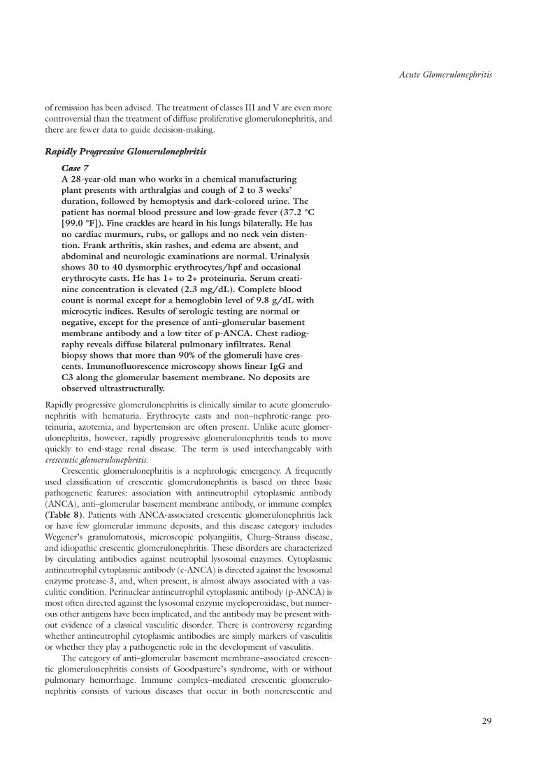
Acute Glomerulonephritis
of remission has been advised. The treatment of classes III and V are even morecontroversial than the treatment of diffuse proliferative glomerulonephritis, andthere are fewer data to guide decision-making.
Rapidly Progressive Glomerulonephritis
Case 7A 28-year-old man who works in a chemical manufacturingplant presents with arthralgias and cough of 2 to 3 weeks’duration, followed by hemoptysis and dark-colored urine. Thepatient has normal blood pressure and low-grade fever (37.2 °C[99.0 °F]). Fine crackles are heard in his lungs bilaterally. He hasno cardiac murmurs, rubs, or gallops and no neck vein disten-tion. Frank arthritis, skin rashes, and edema are absent, andabdominal and neurologic examinations are normal. Urinalysisshows 30 to 40 dysmorphic erythrocytes/hpf and occasionalerythrocyte casts. He has 1+ to 2+ proteinuria. Serum creati-nine concentration is elevated (2.3 mg/dL). Complete bloodcount is normal except for a hemoglobin level of 9.8 g/dL withmicrocytic indices. Results of serologic testing are normal ornegative, except for the presence of anti–glomerular basementmembrane antibody and a low titer of p-ANCA. Chest radiog-raphy reveals diffuse bilateral pulmonary infiltrates. Renalbiopsy shows that more than 90% of the glomeruli have cres-cents. Immunofluorescence microscopy shows linear IgG andC3 along the glomerular basement membrane. No deposits areobserved ultrastructurally.
Rapidly progressive glomerulonephritis is clinically similar to acute glomerulo-nephritis with hematuria. Erythrocyte casts and non–nephrotic-range pro-teinuria, azotemia, and hypertension are often present. Unlike acute glomer-ulonephritis, however, rapidly progressive glomerulonephritis tends to movequickly to end-stage renal disease. The term is used interchangeably with crescentic glomerulonephritis.
Crescentic glomerulonephritis is a nephrologic emergency. A frequentlyused classification of crescentic glomerulonephritis is based on three basicpathogenetic features: association with antineutrophil cytoplasmic antibody(ANCA), anti–glomerular basement membrane antibody, or immune complex(Table 8). Patients with ANCA-associated crescentic glomerulonephritis lackor have few glomerular immune deposits, and this disease category includesWegener’s granulomatosis, microscopic polyangiitis, Churg–Strauss disease,and idiopathic crescentic glomerulonephritis. These disorders are characterizedby circulating antibodies against neutrophil lysosomal enzymes. Cytoplasmicantineutrophil cytoplasmic antibody (c-ANCA) is directed against the lysosomalenzyme protease-3, and, when present, is almost always associated with a vas-culitic condition. Perinuclear antineutrophil cytoplasmic antibody (p-ANCA) ismost often directed against the lysosomal enzyme myeloperoxidase, but numer-ous other antigens have been implicated, and the antibody may be present with-out evidence of a classical vasculitic disorder. There is controversy regardingwhether antineutrophil cytoplasmic antibodies are simply markers of vasculitisor whether they play a pathogenetic role in the development of vasculitis.
The category of anti–glomerular basement membrane–associated crescen-tic glomerulonephritis consists of Goodpasture’s syndrome, with or withoutpulmonary hemorrhage. Immune complex–mediated crescentic glomerulo-nephritis consists of various diseases that occur in both noncrescentic and
29

Acute Glomerulonephritis
30
crescentic forms, including systemic lupus erythematosus, cryoglobulinemia,poststreptococcal (and other infection-related) glomerulonephritis, andHenoch–Schönlein purpura.
Goodpasture’s SyndromeThe patient in case 7 has Goodpasture’s syndrome, as suggested by the clinicalpresentation of a pulmonary-renal syndrome consisting of hemoptysis, pul-monary infiltrates, and hematuria with erythrocyte casts. The diagnosis is con-firmed by the presence of circulating anti–glomerular basement membrane anti-bodies in the blood and the characteristic linear immunofluorescent pattern ofdeposition of antibodies (and complement) along the glomerular basementmembrane on renal biopsy. Approximately 30% of patients with Goodpasture’ssyndrome also manifest p-ANCA. The pathogenesis is unknown but reflectsdevelopment of antibodies to an epitope on the noncollagenous domain of theα chain of type IV collagen present in normal glomerular basement membraneand alveolar basement membrane (Kalluri et al.). Exposure to volatile hydro-carbons, cigarette smoke, or respiratory viral infections have been posited tounmask the antigen in alveoli and induce antibody in genetically susceptiblepersons. Goodpasture’s syndrome occurs in all age groups and has only a min-imal male predominance. Pulmonary symptoms (cough, dyspnea, crackles, andhemoptysis) precede or are coincident with renal symptoms in more than 70%of patients. Substantial alveolar bleeding can occur without frank hemoptysis.Anemia (with characteristics of iron deficiency) may be out of proportion to thedegree of azotemia, presumably owing to sequestration of blood in the lungs.Azotemia occurs in 50% to 70% of cases at initial presentation. Arthritis orarthralgias are common. Hypertension is uncommon (<20% of patients) untilrenal failure is advanced. Urinalysis shows hematuria, erythrocyte casts, andnon–nephrotic-range proteinuria.
Results of serologic testing are usually negative or normal except for thepresence of anti–glomerular basement membrane antibody. Antibody titer doesnot correlate with severity of illness. In nonoliguric patients with less functionalimpairment, combination treatment with corticosteroids, plasmapheresis, andcyclophosphamide can successfully induce remission. Most untreated patientsprogress to end-stage renal disease, and oliguric patients with a serum creati-nine concentration greater than 6 mg/dL are less likely to recover renal func-tion even if treated. Smoking or exposure to volatile hydrocarbons may precip-itate relapse of pulmonary hemorrhage. Recurrence in renal allografts is uncom-
Kalluri R, Wilson CB, Weber M, GunwarS, Chonko AM, Neilson EG, et al.Identification of the alpha 3 chain of type IVcollagen as the common autoantigen inantibasement membrane disease andGoodpasture syndrome. J Am Soc Nephrol.1995;6:1178-85. PMID: 8589284
TABLE 8 Causes of Rapidly Progressive Glomerulonephritis
ANCA-associated
Wegener’s granulomatosis
Microscopic polyarteritis nodosa
Churg–Strauss disease
Idiopathic crescentic glomerulonephritis
Anti-GBM mediated
Goodpasture’s syndrome
Idiopathic anti-GBM disease
Immune-complex mediated
Lupus nephritis
Infection-associated (poststreptococcal, atrioventricular shunts, chronic visceralabscesses, subacute bacterial endocarditis)
Cryoglobulinemia
Henoch–Schönlein purpura
ANCA = antineutrophil cytoplasmic antibody; GBM = glomerular basement membrane.
K E Y P O I N T S
• Goodpasture’s syndrome is suggestedby the clinical presentation of a pul-monary–renal syndrome consisting ofhemoptysis, pulmonary infiltrates, andhematuria with red blood cell casts.
• The diagnosis of Goodpasture’s syn-drome is confirmed by circulatinganti–glomerular basement membraneantibodies in the blood and the charac-teristic linear immunofluorescent pat-tern of deposition of antibodies (andcomplement) along the glomerularbasement membrane on renal biopsy.

Acute Glomerulonephritis
mon unless patients undergo transplantation while they still have high titers ofanti–glomerular basement membrane antibody.
Wegener’s GranulomatosisWegener’s granulomatosis is a granulomatous vasculitis of medium-sized tosmall arterioles and venules. Classically, the diagnosis is made by the histologicfindings of necrotizing vasculitis and granulomas. Cytoplasmic antineutrophilcytoplasmic antibody is present in approximately 80% of patients withWegener’s granulomatosis and aids in diagnosis. Some evidence suggests that c-ANCA may also be important in pathogenesis. Presenting symptoms mostoften involve the upper respiratory tract and include rhinorrhea, sinusitis,nasopharyngeal mucosal ulceration, cough, shortness of breath, and hemopty-sis. Transient infiltrates or nodular densities may be seen on chest radiography.Renal involvement is characterized by proteinuria, dysmorphic hematuria, andoccasionally erythrocytes. Approximately 10% of patients have azotemia onpresentation. Serum complement levels are normal. Renal biopsy usually showscrescentic glomerulonephritis without glomerular immune deposits in patientswho present with an acute or rapidly progressive nephritic picture.Tubulointerstitial granulomas may occasionally be seen. Additional symptomsinclude fever, weight loss, malaise, arthralgias, nondeforming arthritis,mononeuritis multiplex, skin papules, vesicles, purpura, and leukocytoclasticvasculitis. A finding of c-ANCA is specific and sensitive in establishing in thediagnosis; however, some patients have p-ANCA, and up to 10% of patientsmay test negative for ANCA. Antineutrophil cytoplasmic antibody titers may beuseful in following response to therapy and predicting relapse. The mortalityrate at 2 years is more than 80% to 90% for patients with untreated nephritis.Cyclophosphamide and prednisone induce remission in 80% to 90% of treatedpatients, but there is a significant incidence (5% to 6%) of bladder cancer in thedecade after prolonged treatment with daily oral cyclophosphamide.Intravenous cyclophosphamide, mycophenolate mofetil, and azathioprine areoccasionally used in place of oral cyclophosphamide to minimize toxicity,although it remains to be proven that the latter two agents are as effective ininducing or maintaining remission as are regimens using cyclophosphamide.Relapse is frequent, and multiple courses of treatment may be necessary.
Other forms of systemic vasculitis that cause crescentic glomerulonephritis,including microscopic polyangiitis nodosa and Churg–Strauss disease, aretreated with protocols similar to that of Wegener’s granulomatosis. Patientswith this disease are more likely to test positive for perinuclear and cytoplasmicantineutrophil cytoplasmic antibody.
Tubulointerstitial Diseases
Case 8A 65-year-old woman presents with chronic fatigue that hasworsened over the past 3 to 4 months. In the 2 previousmonths, she noted polydipsia and polyuria and woke up once ortwice each night to urinate. On the day of presentation, sheexperienced severe back pain. She has no fever, sweats, or chills.Medical history is unremarkable, and she is taking no medica-tions except for oral calcium carbonate, 1 g/d, to preventosteoporosis.
Physical examination is remarkable for normal vital signs.She appears pale. There is point tenderness over the 10th tho-
31
K E Y P O I N T S
• Berger’s disease, or IgA nephropathy, isthe most common cause of idiopathicglomerulonephritis.
• The clinical presentation of IgAnephropathy ranges from asymptomaticmicroscopic hematuria with or withoutproteinuria to acute glomerulonephritiswith episodic gross hematuria and,occasionally, rapidly progressiveglomerulonephritis.
• Up to 30% to 50% of patients with IgAnephropathy may reach end-stage renaldisease over 20 years of follow-up.Older age at onset, heavy proteinuria,hypertension, and crescents or segmen-tal sclerosis on biopsy are risk factorsfor a poor prognosis.
• Most patients who have IgA nephrop-athy but normal renal function andnon–nephrotic-range proteinuria shouldbe observed.
• In lupus nephritis, the most aggressivetherapy is directed toward the treat-ment of diffuse proliferative glomerulo-nephritis (World Health Organizationclass IV), since rates of renal failure arestill as high as 25% over 5 to 10 yearsof follow-up. The prognosis in patientswith membranous lupus is similar tothat in patients with idiopathic mem-branous nephropathy.
• In rapidly progressive glomerulonephri-tis (also known as crescentic glomerulo-nephritis), red blood cell casts,non–nephrotic-range proteinuria,azotemia, and hypertension are oftenpresent.
• Goodpasture’s syndrome is suggestedby the clinical presentation of a pul-monary–renal syndrome consisting ofhemoptysis, pulmonary infiltrates, andhermaturia with red blood cell casts.
• The diagnosis of Goodpasture’s syn-drome is confirmed by circulatinganti–glomerular basement membraneantibodies in the blood and the charac-teristic linear immunofluorescent pat-tern of deposition of antibodies (andcomplement) along the glomerularbasement membrane on renal biopsy.
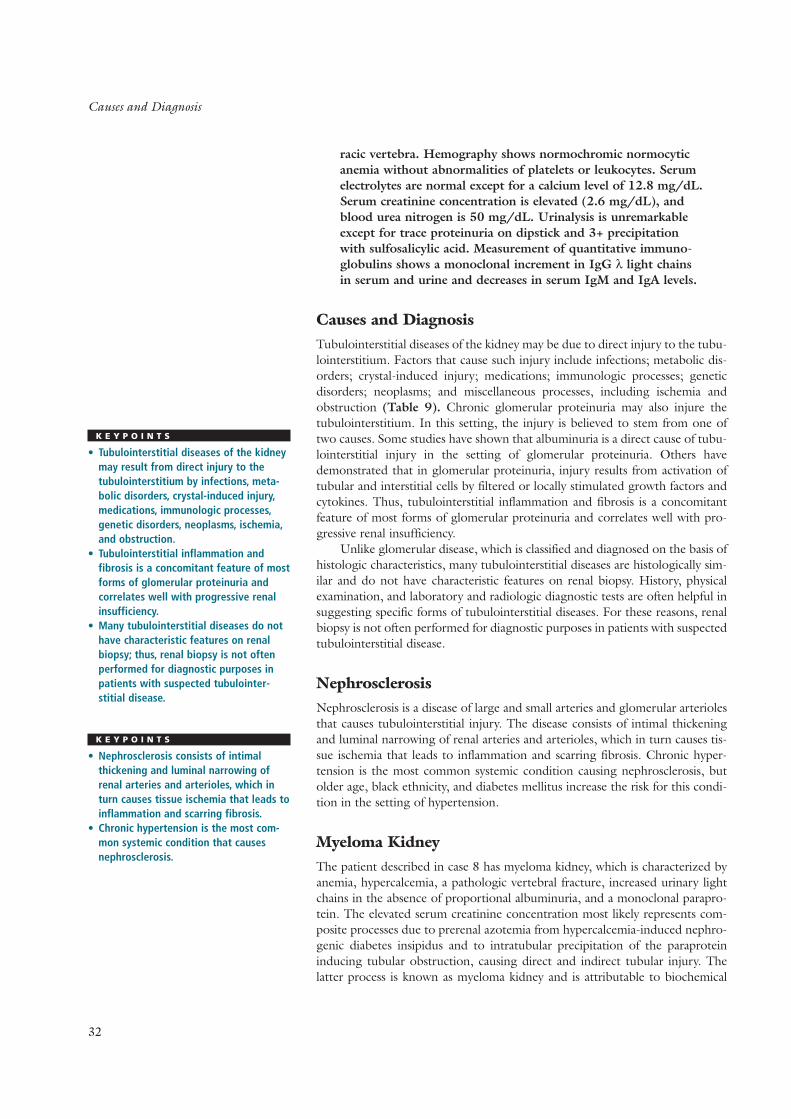
Causes and Diagnosis
32
racic vertebra. Hemography shows normochromic normocyticanemia without abnormalities of platelets or leukocytes. Serumelectrolytes are normal except for a calcium level of 12.8 mg/dL.Serum creatinine concentration is elevated (2.6 mg/dL), andblood urea nitrogen is 50 mg/dL. Urinalysis is unremarkableexcept for trace proteinuria on dipstick and 3+ precipitationwith sulfosalicylic acid. Measurement of quantitative immuno-globulins shows a monoclonal increment in IgG λ light chainsin serum and urine and decreases in serum IgM and IgA levels.
Causes and DiagnosisTubulointerstitial diseases of the kidney may be due to direct injury to the tubu-lointerstitium. Factors that cause such injury include infections; metabolic dis-orders; crystal-induced injury; medications; immunologic processes; geneticdisorders; neoplasms; and miscellaneous processes, including ischemia andobstruction (Table 9). Chronic glomerular proteinuria may also injure thetubulointerstitium. In this setting, the injury is believed to stem from one oftwo causes. Some studies have shown that albuminuria is a direct cause of tubu-lointerstitial injury in the setting of glomerular proteinuria. Others havedemonstrated that in glomerular proteinuria, injury results from activation oftubular and interstitial cells by filtered or locally stimulated growth factors andcytokines. Thus, tubulointerstitial inflammation and fibrosis is a concomitantfeature of most forms of glomerular proteinuria and correlates well with pro-gressive renal insufficiency.
Unlike glomerular disease, which is classified and diagnosed on the basis ofhistologic characteristics, many tubulointerstitial diseases are histologically sim-ilar and do not have characteristic features on renal biopsy. History, physicalexamination, and laboratory and radiologic diagnostic tests are often helpful insuggesting specific forms of tubulointerstitial diseases. For these reasons, renalbiopsy is not often performed for diagnostic purposes in patients with suspectedtubulointerstitial disease.
NephrosclerosisNephrosclerosis is a disease of large and small arteries and glomerular arteriolesthat causes tubulointerstitial injury. The disease consists of intimal thickeningand luminal narrowing of renal arteries and arterioles, which in turn causes tis-sue ischemia that leads to inflammation and scarring fibrosis. Chronic hyper-tension is the most common systemic condition causing nephrosclerosis, butolder age, black ethnicity, and diabetes mellitus increase the risk for this condi-tion in the setting of hypertension.
Myeloma KidneyThe patient described in case 8 has myeloma kidney, which is characterized byanemia, hypercalcemia, a pathologic vertebral fracture, increased urinary lightchains in the absence of proportional albuminuria, and a monoclonal parapro-tein. The elevated serum creatinine concentration most likely represents com-posite processes due to prerenal azotemia from hypercalcemia-induced nephro-genic diabetes insipidus and to intratubular precipitation of the paraproteininducing tubular obstruction, causing direct and indirect tubular injury. Thelatter process is known as myeloma kidney and is attributable to biochemical
K E Y P O I N T S
• Tubulointerstitial diseases of the kidneymay result from direct injury to thetubulointerstitium by infections, meta-bolic disorders, crystal-induced injury,medications, immunologic processes,genetic disorders, neoplasms, ischemia,and obstruction.
• Tubulointerstitial inflammation andfibrosis is a concomitant feature of mostforms of glomerular proteinuria and correlates well with progressive renalinsufficiency.
• Many tubulointerstitial diseases do nothave characteristic features on renalbiopsy; thus, renal biopsy is not oftenperformed for diagnostic purposes inpatients with suspected tubulointer-stitial disease.
K E Y P O I N T S
• Nephrosclerosis consists of intimalthickening and luminal narrowing ofrenal arteries and arterioles, which inturn causes tissue ischemia that leads toinflammation and scarring fibrosis.
• Chronic hypertension is the most com-mon systemic condition that causesnephrosclerosis.

Analgesic Nephropathy
characteristics of the light chain and not of the host. Paraproteins with λ lightchains are most likely to induce myeloma kidney, whereas paraproteins with κlight chains are more likely to deposit in glomeruli and induce the nephroticsyndrome. Occasionally, plasma-cell infiltration may occur. Renal amyloidosismay also complicate the lesion and is associated with λ light chains.
Myeloma kidney is best prevented by maintenance of hydration to inhibitsupersaturation and precipitation of the paraprotein and by therapy to minimizethe amount of circulating paraprotein. Chemotherapy is both preventive andtherapeutic. In cases of progressive azotemia despite adequate chemotherapy,plasmapheresis has been advocated, although this practice is not universallyaccepted. Renal involvement in patients with multiple myeloma is associatedwith shortened survival.
Analgesic NephropathyAnalgesic nephropathy is a chronic tubulointerstitial disease characterized byprogressive loss of renal function in a patient with a history of ingestion of atleast 2 kg of analgesics over time, without any evidence for another identifiablecause of tubulointerstitial disease (De Broe and Elseviers). The disorder isdiagnosed by exclusion. Analgesic nephropathy usually occurs when a mixtureof analgesics has been used, such as phenacetin, acetaminophen, caffeine, andcodeine, but it may also occur when a single agent is used long term. The roleof nonsteroidal anti-inflammatory drugs in the development of analgesicnephropathy remains controversial, but many believe that these drugs may becontributors. Whether the newer cyclooxygenase-2 inhibitors have similaractions is not currently known. An irregular contour of the kidney on computedtomography helps to confirm clinical suspicion. The presentation of analgesicnephropathy is often nonspecific, with renal insufficiency as the presentingmanifestation in a patient with a bland urinary sediment and non-nephroticproteinuria, but patients may also present with the hematuria and colicky painof papillary necrosis.
Treatment consists predominantly of discontinuation of treatment withanalgesics, which, if done early in the course, may stabilize renal function.However, discontinuation is often difficult to achieve because patients may beusing the drugs to relieve the pain of chronic headache or degenerative jointdisease. The long-term consequences of analgesic nephropathy include end-stage renal disease and transitional-cell carcinoma of the urinary tract; the latteroccurs particularly in patients who used phenacetin. Patients with persistentnondysmorphic hematuria and long-term use of analgesics should be evaluatedfor neoplasm. Analgesic abuse may also contribute to a worse renal outcome inpatients with other independent renal diseases. A Chinese herb that contains aplant nephrotoxin called aristolochic acid, taken to relieve pain, has also beenimplicated as a cause of chronic interstitial nephritis. Its use produces a charac-teristic histopathologic profile that progresses to end-stage renal disease.
Genetic Disorders and Renal Disease
Genetic disorders may cause kidney disease either directly by causing geneticallymediated renal functional or morphologic abnormalities (for example, autoso-mal dominant polycystic kidney disease) or through genetically induced sys-temic disturbances in which the kidney is involved secondarily (such as Fabry’sdisease or primary hyperoxaluria). About 50 mendelian, or single-gene, renaldisorders are currently known (George and Neilson) (Table 10). The most
33
De Broe ME, Elseviers MM. Analgesicnephropathy. N Engl J Med. 1998;338:446-52. PMID: 9459649
K E Y P O I N T S
• Analgesic nephropathy is a chronictubulointerstitial disease characterizedby progressive loss of renal function ina patient with a history of ingestion ofat least 2 kg of analgesics over time,with no other identifiable cause oftubulointerstitial disease.
TABLE 9 Major Causes of Acuteand Chronic TubulointerstitialDiseases
Genetic
Polycystic kidney disease
Medullary cystic disease
Infectious
Pyelonephritis
Emphysematous pyelonephritis
Tuberculosis
Metabolic
Hypercalcemia
Hyperuricemia
Hypokalemia
Oxalosis
Cystinosis
Immunologic
Sjögren’s syndrome
Drug hypersensitivity
Renal transplant rejection
Toxic
Analgesics
Chinese herbs
Lithium
Amphotericin
Cisplatin
Cyclosporine
Radiation
Anatomic
Obstruction
Reflux
Miscellaneous
Atheroembolic disease
George A Jr, Neilson E. Genetics of kidneydisease. Am J Kidney Dis. 2000;35(4 Suppl1:S160-S169. PMID: 10766015

Genetic Disorders That Cause Direct Renal Effects
34
common are polycystic kidney disease, medullary sponge kidney disease,Alport’s syndrome, and cystinuria. These disorders are transmitted in families asautosomal dominant, autosomal recessive, or X-linked traits. The genetics ofeven single-gene disorders is complex because several disorders that appear tobe phenotypically the same (for example, autosomal dominant polycystic kidney disease, Bartter’s syndrome, Alport’s syndrome, and cystinuria) can becaused by two or three distinct genotypes. A complete and continuouslyupdated list of these single-gene disorders, most of which are rare, can be foundat http://www.ncbi.nim.nih.gov/Omim (Sessa et al.).
In contrast, a wide variety of common disorders affecting the kidney resultfrom complex interactions between genetic predisposition and environmentalfactors. Such genetically complex traits, also called polygenic disorders, arise fromsubtle variations in multiple genes that interact with diverse environmental fac-tors. In these disorders, the clinical expression of disease varies according to theinteraction between genetic predisposition and key environmental or host fac-tors. Examples include hypertension and salt intake, or diabetic predispositionand obesity.
Genetic Disorders That Cause Direct Renal EffectsPolycystic kidney disease is characterized by multiple epithelial-lined renal cystsscattered throughout the cortex and medulla of both kidneys. The disease hastwo major forms. Autosomal dominant polycystic kidney disease is typically dis-covered in patients 30 to 50 years of age, whereas autosomal recessive poly-cystic kidney disease is usually expressed at birth and causes death during theneonatal period (Igarashi and Somlo). Autosomal recessive polycystic kidneydisease is caused by a mutation on chromosome 16 and is rare (1 in 40,000 live births).
Autosomal dominant polycystic kidney disease is the fourth leading causeof renal failure. It is caused by an abnormal gene on the short arm of chromo-some 16 in 95% of cases. An abnormal gene on chromosome 4 accounts formost of the remainder of cases. Recent evidence implicates a third gene in somecases of this disease. Autosomal dominant polycystic kidney disease occurs at afrequency of about 1 in 1000 persons. It affects all racial and ethnic groups.Early clinical manifestations include back and flank pain, hematuria, renal
Sessa A, Conte F, Meroni M, Battini G.Hereditary kidney diseases. Contrib Nephrol.1997;122:1-217. PMID: 9399028
Igarashi P, Somlo S. Genetics and patho-genesis of polycystic kidney disease. J AmSoc Nephrol. 2002;13:2384-98.PMID: 12191984
TABLE 10 Some Renal Disorders Caused by Single-Gene Abnormalities
Disease Mode of Inheritance Gene Locus Frequency
Polycystic kidney disease Autosomal dominant (common) Chromosome 16 and 4 1:1000
Autosomal recessive (rare) 1:40,000
X-linked (80%)
Alport’s syndrome Autosomal recessive (10%) Xq22
2q35-37
Benign familial hematuria Autosomal recessive (carrier) 2q and 13q
Nephrogenic diabetes insipidus Autosomal recessive
Bartter’s syndrome Autosomal recessive Not known 1:2 Million
Gitelman’s syndrome Autosomal recessive Chromosome 16 Not known
Cystinosis Autosomal recessive Chromosome 2p 1:7000
Fabry’s disease X-linked X-q21,22 1:40,000
Hyperoxaluria, type I, II, II Autosomal recessive 2q36-37

Genetic Disorders That Cause Direct Renal Effects
stones, hypertension, and urinary tract infections (Fick and Gabow).Approximately 50% of patients develop renal insufficiency before 70 years ofage, and renal function declines linearly over several years. Autosomal dominantpolycystic kidney disease is associated with cerebral aneurysms (especially ifthere is a family history of aneurysm), hepatic cysts (40% to 60% of patients),mitral and aortic valve prolapse, and colonic diverticular disease. As many as25% of patients with autosomal dominant polycystic kidney disease do not havea family history of the disease. In some of these patients, the disease is due to anew mutation. In other patients, family members may have died of other causes,so that disease in the patient is detected only at a later age, when symptoms orsigns of renal failure are first observed.
Medullary sponge kidney, which does not cause renal failure, is associatedwith hematuria, hypercalciuria (50% of patients), nephrocalcinosis, calciumstone disease, and hemihypertrophy. Familial occurrence accounts for some, butnot most, cases. The diagnosis is made by intravenous pyelography showingsmall cystic outpouchings of the renal papillary ducts.
Alport’s syndrome is an X-linked disorder in 80% of patients and an auto-somal recessive disorder in 11% of patients. Affected men develop hematuria,proteinuria, and renal failure in the second or third decade of life. The abnor-mal gene is on Xq22 in the X-linked disorder and on 2q in the autosomal reces-sive disorder. The genetic abnormality causes a disorder of type IV and V col-lagen and results in abnormalities of the lens and glomerular basement mem-brane and in deafness. Affected heterozygous women have hematuria, but renalfailure is uncommon.
Benign familial hematuria is a disorder of collagen synthesis that causesmicroscopic or gross hematuria and an abnormally thin glomerular basementmembrane. Genetic studies have suggested that this disorder represents a car-rier state of the autosomal recessive Alport’s syndrome. Unlike Alport’s syn-drome, benign familial hematuria usually does not result in renal failure. Benignfamilial hematuria usually presents in childhood; a family history of hematuriais suggestive of the condition.
Nephrogenic diabetes insipidus, Bartter’s syndrome, Gitelman’s syndrome,and Liddle’s syndrome are genetically mediated disorders of renal tubular func-tion. Tubular unresponsiveness to antidiuretic hormone in nephrogenic dia-betes insipidus causes urinary concentrating defects, polyuria, and thirst.Abnormal chloride transporters in the ascending loop of Henle (Bartter’s syn-drome) and the distal tubule (Gitelman’s syndrome) result in hypokalemia,hypochloremic metabolic alkalosis, renal potassium wasting, and normotension.Bartter’s syndrome causes growth retardation. Nephrogenic diabetes insipidusis usually discovered in infancy or early childhood, whereas Bartter’s syndromeis usually diagnosed in childhood or the early teen years.
Nephronophthisis and medullary cystic disease complex are related condi-tions characterized by multiple cysts located in the corticomedullary junctionand medulla. The cysts arise from the distal and collecting tubules. The diseaseproduces tubular atrophy, interstitial inflammation and scarring, and renal fail-ure. Familial nephronophthisis is a recessive disorder that causes renal failurebefore 20 years of age. Medullary cystic disease, an autosomal dominant condi-tion, produces renal failure in early adulthood. In these disorders, the initialclinical presentation consists of polyuria, polydipsia, and nocturia (due to a renalconcentrating disorder) and renal salt wasting, all of which stem from the tubu-lar injury produced by the cystic and scarring process. Azotemia and end-stagerenal failure follow.
Cystinuria is covered in the section on nephrolithiasis.
35
Fick GM, Gabow PA. Hereditary andacquired cystic disease of the kidney. KidneyInt. 1994;46:951-64. PMID: 7861721
K E Y P O I N T S
• Autosomal dominant polycystic kidneydisease is typically found in patients 30 years of age or older who have afamily history of the disorder.
• Autosomal dominant polycystic kidneydisease is the fourth most commoncause of renal failure and is character-ized by multiple cysts distributed amongboth kidneys.
• Benign familial hematuria is character-ized by unexplained microhematuria orgross hematuria and erythrocyte casts.It does not usually result in renal failure.
• Benign familial hematuria usually pre-sents in childhood; a family history ofhematuria is suggestive.
• Nephrogenic diabetes insipidus is usually discovered in infancy or earlychildhood. It is characterized by polyuriaand thirst.
• Bartter’s syndrome is usually diagnosedin childhood or the early teen years. Thesyndrome causes growth retardationand hypokalemia, but patients are normotensive.

Genetic Disorders That Cause Systemic Abnormalities Affecting The Kidney
36
Genetic Disorders That Cause Systemic AbnormalitiesAffecting The KidneyFabry’s disease is due to a deficiency of α-galactosidase-A enzyme caused by agene abnormality on the long arm of the X chromosome (q21 and q22). Menare more often affected than women. The enzyme deficiency causes an accu-mulation of neutral glycophospholipid in endothelial, epithelial, and smooth-muscle cells throughout the body. Marked accumulation of glycophospholipidsin the glomerular and tubular cells occurs. Clinical features include proteinuria,azotemia, renal failure, cutaneous angiokeratomas, painful paresthesias of thehands, and premature coronary disease. Electron microscopy of the kidneyreveals the characteristic inclusion bodies in the cytoplasm, with concentriclamellation and zebra or onion-skin appearance. The same structures are foundon electron microscopic examination of spun urine sediment.
Primary hyperoxaluria results from an inborn error of metabolism in whichglyoxalate cannot be converted to glycine because of a deficiency of glyoxalateaminotransferase or glyoxalate reductase in the liver. There are three geneticforms. Type I and II disease have similar clinical presentations. Typically, thepatient develops nephrolithiasis and nephrocalcinosis before 20 years of age,and end-stage renal failure occurs in 50% of patients by 15 years of age. Renalbiopsy demonstrates marked calcium oxalate deposition, but oxalate depositionis found in many other tissues as well. Type I disorder is caused by an abnormalgene on chromosome 2q36–37 and is the most common. Type III disorder iscaused by excessive intestinal reabsorption of oxalate in the absence of othergastrointestinal disease.
Genetic Factors in Diabetic NephropathyFour lines of evidence support a genetic susceptibility for diabetic nephropathy.First, familial clustering of nephropathy is observed among Pima Indians, inwhom the incidence of proteinuria in diabetic offspring is 14% if neither parenthad proteinuria, 23% if one diabetic parent had proteinuria, and 46% if bothparents had diabetes and proteinuria. Second, family history of hypertensionand nephropathy are strongly associated. Third, renal lesions in diabetic siblingsare similar. Finally, polymorphisms of angiotensin-converting enzyme gene andcollagen gene are present.
Fluid and Electrolytes
Figure 3 shows the normal distribution of water and electrolyte solutes andtheir contributions to intracellular fluid, extracellular fluid, and plasma osmo-lality (the concentration of solutes in the body water, expressed as mosmol/kg).The concentration of solute in the intracellular fluid is the same as that in theextracellular fluid, resulting in equivalent osmolality in both spaces. The solutesresponsible for the osmolality in the intracellular fluid and extracellular fluid,however, differ substantially. In the intracellular fluid, potassium and organicphosphate esters are the predominant osmoles. In the extracellular fluid,sodium salts account for most of the osmoles. Nonsodium solutes, such as ureaand glucose, have effects on the plasma osmolality equal to their molar concentrations: for example, Posm = 2 × Na + blood urea nitrogen/2.8 + glucose/18.
High levels of glucose and urea have varying effects on water distributionand the serum sodium concentration. Urea readily distributes across cell mem-
K E Y P O I N T S
• Fabry’s disease is characterized by acroparesthesias, cutaneous angio-keratomas, proteinuria, and azotemia.
• Primary hyperoxaluria causes calciumoxalate nephrolithiasis in childhood.

Hyponatremia
38
tive extracellular fluid volume is greatly reduced. When significant volumedepletion and hypo-osmolality occur simultaneously, the volume stimulus over-rides the inhibitory effect of hypo-osmolality and secretion of antidiuretic hor-mone increases.
HyponatremiaHyponatremia, defined as a plasma sodium concentration less than 136 meq/L,occurs in as many as 4% of hospitalized patients (Adrogue et al.). With theexception of the rare patient with psychogenic polydipsia, whose water intakeexceeds the water-excreting capacity of kidney, hyponatremia usually resultsfrom the inability to appropriately excrete dilute urine.
Hyponatremia can be associated with a high, normal, or low plasma osmo-lality. Hyponatremia with normal or high osmolality occurs most often inpatients with hyperglycemia or those receiving mannitol infusion. In thesecases, the hyponatremia occurs as water moves from the cellular to the extra-cellular space through the osmotic effect of glucose or mannitol. Most patientswith hyponatremia have low plasma osmolality with excess body water relativeto body sodium. Usually this state is due to the nonosmotic stimulation ofantidiuretic hormone, and these patients excrete concentrated urine (that is,their renal water excretion is decreased).
Evaluation of the extracellular fluid status is the first step in determiningthe cause of hypoosmolal hyponatremia (Table 11). Biochemical findings helpto further confirm the diagnosis. For example, very low levels of blood ureanitrogen and uric acid in a euvolemic patient suggest the syndrome of inappro-
Adrogue HJ, Madias NE. Hyponatremia.N Engl J Med. 2000;342:1581-9.PMID: 10824078
TABLE 11 Evaluation of Hyponatremia
Cause Extracellular Fluid Volume Status
Volume Depletion* Euvolemia† Volume Excess‡
Clinical Gastrointestinal fluid loss SIADH Congestive heart failure
Adrenal insufficiency Hypothyroidism Cirrhosis
Renal sodium loss Cortisol deficiency or Nephrotic syndromepanhypopituitarism
Adrenal
Renal salt wasting
From Nonrenal From Renal Biochemical§ Losses Losses
Serum sodium|| 120 meq/L 120 meq/L 120 meq/L 120 meq/L
Plasma osmolality 250 mosmol/kg 250 mosmol/kg 250 mosmol/kg 250 mosmol/kg
Urine sodium <10 meq/L >20 meq/L Equals dietary intake <10 meq/L
Urine osmolality >600–800 mosmol/kg 600–800 mosmol/kg Inappropriately elevated (>100 mosmol/kg) >300–400 mosmol/kg
Plasma ADH Elevated Elevated Elevated Elevated
BUN Elevated Elevated <10 mg/dL Elevated
Serum uric acid Elevated Elevated <4 mg/dL Elevated
*Volume depletion is defined as orthostasis, decreased skin turgor, and hypotension.
†Euvolemia is defined as no clinically evident volume abnormalities.
‡Volume excess is defined as edema.
§It is assumed that diuretics are not being used.
||Serum sodium level of 120 meq/L has been arbitrarily assumed in each category.
ADH = antidiuretic hormone; BUN = blood urea nitrogen; SIADH = syndrome of inappropriate secretion of antidiuretic hormone.

Hyponatremia
priate antidiuretic hormone. An elevated blood urea nitrogen level and lowurine sodium excretion suggest that true volume depletion is present or that thepatient has nephrotic syndrome, cirrhosis, or congestive heart failure (condi-tions that cause decreased effective circulatory volume). The clinical history andfindings on physical examination allow differentiation among these possibilities.
Proper treatment of hyponatremia should follow from the above assess-ment. Volume-depleted patients should be treated with normal saline to expandthe extracellular fluid volume and thus inhibit the release of antidiuretic hor-mone that had been triggered by hypovolemia. Patients with extracellular fluidvolume expansion (such as those with heart failure) require treatment of theunderlying disorder and restriction of water and salt intake. Water restriction(intake of less than 600 to 800 mL/d) needs to be imposed only when theserum sodium concentration is less than 125 meq/L. Such patients may alsobenefit from treatment with loop diuretics because these diuretics favor excre-tion of more water than sodium, thus increasing water excretion.
In hyponatremic patients with euvolemia, the pathophysiologic cause isincreased production or release of antidiuretic hormone on a nonosmolar andnonvolume basis. Cortisol and thyroid deficiencies may cause euvolemichyponatremia due to increased release of antidiuretic hormone.
As many as one third of hospitalized patients with hyponatremia have thesyndrome of inappropriate antidiuretic hormone secretion. The causes for thissyndrome include central nervous system disorders; such medications as fluox-etine and thiazide diuretics; and neoplasms, especially small-cell carcinoma(Table 12).
When the serum sodium concentration decreases to less than 110 meq/L,coma and death may result. Severe symptomatic hyponatremia occurs particu-larly postoperatively in women of child-bearing age. Treatment usually requiresinfusions of hypertonic saline with or without furosemide. The quantity ofsodium chloride required to increase the plasma sodium is calculated as follows:
meq Na required = total body water × desired increase in plasma Na
meq Na required = 0.6 (or 0.5 in women) × body weight in kg × desired increase in plasma Na
39
TABLE 12 Some Causes of the Syndrome of Inappropriate AntidiureticHormone Secretion
Increased hypothalamic production of antidiuretic hormone
Neuropsychiatric disorders
Central nervous system infections
Central nervous system malignancies
Psychosis
Drugs: cyclophosphamide, vincristine, haloperidol, fluoxetine
Pulmonary disease: pneumonia, acute respiratory failure
Surgery
Severe nausea
Idiopathic cause
Ectopic production of antidiuretic hormone
Carcinoma: small-cell lung, bronchogenic, neuroblastoma
Potentiation of antidiuretic hormone effect in the kidney
Chlorpropamide, carbamazepine, intravenous cyclophosphamide, tolbutamide

Hypernatremia
40
Although the sodium circulates mainly in the extracellular fluid, the aboveformula uses total body water as the space of distribution for sodium becausewhen sodium is infused, water shifts from the intracellular fluid to the extra-cellular fluid. The rate of correction of symptomatic hyponatremia should beguided by the rate at which the hyponatremia has developed. In acute sympto-matic hyponatremia (usually defined as hyponatremia of less than 48 hours’duration), the rate of correction should not exceed 8 to 12 meq/L in the first24 hours. Increasing the serum sodium concentration too rapidly can result ina severe neurologic injury known as central pontine myelinosis. In one recentstudy, no patient experienced a severe neurologic complication if the serumsodium concentration was corrected at a rate less than 0.55 meq/h to a con-centration of 120 meq/L, or by less than 12 meq during the first 12 hours or18 meq by 48 hours (Sterns et al.).
In chronic asymptomatic hyponatremia, correction is best accomplishedslowly by water restriction. Recently, a vasopressin V2 receptor antagonist wasused clinically to successfully treat hyponatremic patients with cirrhosis and thesyndrome of inappropriate antidiuretic hormone (Decaux). This agent mayprove to be an effective therapy.
HypernatremiaHypernatremia is defined as an increase in the serum sodium concentration togreater than 145 meq/L (Adrogue et al.). It is rarely caused by excessive inges-tion of sodium; rather, it is due to loss of hypotonic fluids from the body with
K E Y P O I N T S
• Although ethanol, methanol, and ethyl-ene glycol are not included in the for-mula for calculating the plasma osmo-lality, they do cause an increase inmeasured osmolality.
• A difference of greater than 10mosmol/kg between the measured andcalculated osmolality is considered anelevated osmolal gap and suggests thepresence of one of these osmoles. Themost common cause of an elevatedosmolol gap is ethanol intoxication.
• Hyponatremia with normal or highosmolality occurs most often in patientswith hyperglycemia or those receivingmannitol infusion. Most patients withhyponatremia have low plasma osmolal-ity with excess body water relative tobody sodium; usually this state is due tothe nonosmotic stimulation of antidi-uretic hormone.
• In hyponatremic patients with euv-olemia, the pathophysiologic cause isincreased production or release ofantidiuretic hormone on a nonosmolarand nonvolume basis.
• Up to one third of hospitalized patientswith hyponatremia have the syndromeof inappropriate antidiuretic hormone,which can be caused by central nervoussystem disorders; by medications, suchas fluoxetine and thiazide diuretics; andneoplasms, especially small-cell carcinoma.
• In acute symptomatic hyponatremia, therate of correction should not exceed 8to 12 meq/L in the first 24 hours;increasing the serum sodium concentra-tion too rapidly can result in centralpontine myelinosis. In chronic asympto-matic hyponatremia, correction is bestaccomplished slowly by water restriction.
Sterns RH, Cappuccio JD, Silver SM,Cohen EP. Neurologic sequelae after treat-ment of severe hyponatremia: a multicenterperspective. J Am Soc Nephrol.1994;4:1522-30. PMID: 8025225Decaux G. Difference in solute excretionduring correction of hyponatremic patientswith cirrhosis or syndrome of inappropriatesecretion of antidiuretic hormone by oralvasopressin V2 receptor antagonist VPA-985.J Lab Clin Med. 2001;138:18-21.PMID: 11433224
Adrogue HJ, Madias NE. Hypernatremia.N Engl J Med. 2000;342:1493-9.PMID: 10816188
TABLE 13 Causes of Hypernatremia
Increased water loss
Insensible
Burns
Fever/heat
Mechanical ventilation/hyperventilation
Gastrointestinal loss
Vomiting/nasogastric tube suction
Diarrhea
Renal loss
Central diabetes insipidus
Nephrogenic diabetes insipidus
Osmotic diuresis
Reduced water intake
Hypothalamic dysfunction
Reduced thirst
Essential hypernatremia
Inability to drink water
Comatose
Infant not given adequate water
Hypertonic infusions
Saline/sodium bicarbonate
Water shifts out of extracellular fluid compartment
Seizure/extreme exercise (water shifts into muscle cells)
Gastrointestinal bleeding with intraluminal protein catabolism (water shifts into lumenof intestine)

Potassium Metabolism
inadequate water replacement (Table 13). A typical patient with the latter con-dition is an elderly person with dementia and fever who resides in a nursinghome, has increased insensible water losses, and is unable or too confused todrink adequately. Hypernatremia (hypertonicity) stimulates central nervous sys-tem receptors to produce and release antidiuretic hormone and stimulatesthirst. Even if release of antidiuretic hormone or its renal action is defective,thirst-driven drinking will prevent the patient from becoming severely hyper-natremic. Therefore, severe hypernatremia usually indicates a defective thirst stim-ulus or an inability to drink water, as well as some defect in renal concentratingfunction. Symptoms of hypernatremia are weakness, lethargy, seizures, andcoma; the condition may cause death. Hypernatremia due to inadequate waterintake is associated with concentrated urine and elevated plasma levels of anti-diuretic hormone. Hypernatremia due to inadequate production or release ofantidiuretic hormone (central diabetes insipidus) is associated with inadequatelyconcentrated urine and low or undetectable plasma levels of antidiuretic hor-mone. Treatment requires water replacement and administration of exogenousantidiuretic hormone. Hypernatremia due to renal unresponsiveness to antidi-uretic hormone is associated with normal or high plasma levels of antidiuretichormone and inappropriately dilute urine. This disorder may be congenital oracquired. Acquired nephrogenic diabetes insipidus may be caused by drugs(such as lithium and foscarnet), hypokalemia, hypercalcemia, sickle-cell diseaseand trait, and amyloidosis. Treatment requires adequate water replacement.Treatment with exogenous antidiuretic hormone is of no benefit.
Since hypernatremia is usually due to a deficit of body water, the waterdeficit should be calculated to estimate the quantity of hypotonic fluid thatshould be administered. Only about 50% of the calculated deficit should bereplaced in the first 24 hours.
Potassium MetabolismThe serum potassium concentration is tightly regulated because of its importantrole in transmembrane potential difference of cells. Derangements in serumpotassium are manifested by disorders in muscle, cardiac, and neurologic cells.More that 98% of body potassium is intracellular (cellular K = 150 meq/L)resulting in a ratio of cellular to extracellular fluid potassium of 35:1. Smallchanges in the potassium concentration of extracellular fluid substantially alterthis ratio and thereby affect vital functions.
The average potassium intake is 50 to 100 meq/d. Potassium is eliminatedmostly by renal excretion via the distal nephron. Renal excretion of potassiumis regulated by aldosterone, urine flow rate, distal tubular delivery of sodium,acid–base status, and intracellular potassium stores. The intracellular potassiumbalance is maintained by insulin, catecholamines, acid–base status, extracellularfluid osmolality, and cell integrity.
HypokalemiaHypokalemia (serum potassium level <3.5 meq/L) can result from potassiumloss or shifts but rarely from inadequate intake alone (Table 14). The mostcommon causes of hypokalemia are gastrointestinal (vomiting and diarrhea)and renal (use of diuretics). Rare causes of hypokalemia include primary aldo-steronism, Bartter’s syndrome, Gitelman’s syndrome, and periodic paralysis.Hypokalemia may cause ileus, muscle cramps, rhabdomyolysis, and cardiacarrhythmias. Electrocardiographic findings include U waves and flat or invertedT waves. Hypokalemia decreases insulin secretion, and chronic hypokalemia hasbeen associated with formation of renal cysts.
41
K E Y P O I N T S
• Acquired nephrogenic diabetes insipidusmay be caused by drugs (such as lithiumand foscarnet), hypokalemia, hypercal-cemia, sickle-cell disease and trait,and amyloidosis. Treatment requiresadequate water replacement.
K E Y P O I N T S
• Hypomagnesemia should be suspectedin hypokalemic patients because theseintracellular ions are often lost together.
• Potassium-sparing diuretics, thetrimethoprim component of trimetho-prim–sulfamethoxazole, and pentami-dine can cause hyperkalemia becausethey inhibit the potassium secretorychannels in the distal nephron.
• Among the most common reasons forhyperkalemia is the syndrome ofhyporeninemic hypoaldosteronism, char-acterized by decreased angiotensin IIproduction (owing to diminished reninrelease) and an intra-adrenal defect,both of which contribute to decreasedaldosterone secretion. Excessive dietarypotassium intake is rarely a cause ofhyperkalemia unless renal potassiumexcretion is simultaneously decreased.
• In acute hyperkalemia, treatment is tar-geted toward antagonism of cardiactoxicity (by administration of calciumgluconate), shifting of potassium intocells (by infusion of insulin and glucoseand aerosol administration of β-adre-nergic agonists), and removal of potas-sium (by administration of cationexchange resins or hemodialysis).Chronic hyperkalemia is treated byrestriction of dietary potassium, avoid-ance of drugs that impair potassiumexcretion, and use of kaliuretic diuret-ics, such as hydrochlorothiazide orfurosemide.

Potassium Metabolism
42
Hypokalemia can often be treated by administration of oral potassium salts,but in severe hypokalemia, intravenous potassium chloride should be given.The potassium concentration of the intravenous fluid should not exceed 40meq/L, and the infusion rate should not exceed 20 to 40 meq/h. Total potas-sium deficits are difficult to predict, but a serum potassium level of 3.0 meq/Lis equivalent to a 200- to 400-meq potassium deficit and a level of 2.0 meq/Lis equivalent to a 400- to 800-meq deficit.
Hypomagnesemia should be suspected in hypokalemic patients becausethese intracellular ions are often lost together. Furthermore, hypokalemia is dif-ficult to correct in the presence of hypomagnesemia because the hypomagne-semia causes renal potassium wasting through a mechanism not yet elucidated.
HyperkalemiaExcessive dietary potassium intake is rarely a cause of hyperkalemia unless renalpotassium excretion is simultaneously decreased (Table 15). Potassium may
TABLE 14 Differential Diagnosis of Hypokalemia
High Urinary Potassium* Low Urinary Potassium†
Metabolic acidosis Gastrointestinal loss
Renal tubular acidosis Diarrhea
Distal Laxative abuse
Proximal Internal shifts
Drugs Periodic paralysis
Acetazolamide Insulin
Metabolic alkalosis β2-Agonists
See section on Acid–Base Disorders Alkalosis
Dietary deficiency
*Greater than 20 meq/24 h or 20 meq/L.
†Less than 20 meq/L or 20 meq/L.
TABLE 15 Causes of Hyperkalemia
Increased intake
Shift from intracellular to extracellular fluid compartment
Ex vivo: pseudohyperkalemia
Metabolic acidosis (especially hyperchloremia)
β-Adrenergic blockers
Insulin deficiency or resistance
Hyperosmolality or hyperglycemia
Rhabdomyolysis
Hyperkalemic periodic paralysis
Arginine hydrochloride infusion
Succinylcholine
Digoxin overdose
Reduced renal excretion
Hyporenin hypoaldosteronism
Renal insufficiency or failure
Hypoaldosteronism
Aldosterone resistance (inherited, acquired, drug related)
Type IV renal tubular acidosis
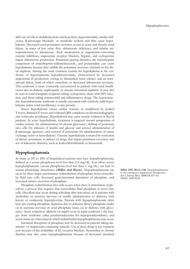
Hypophosphatemia
shift out of cells in rhabdomyolysis and hemolysis, hyperosmolality, insulin defi-ciency, β-adrenergic blockade, or metabolic acidosis and thus cause hyper-kalemia. Decreased renal potassium excretion occurs in acute and chronic renalfailure, in states of low urine flow, aldosterone deficiency, and tubular un-responsiveness to aldosterone. Such medications as angiotensin-convertingenzyme inhibitors, angiotensin receptor blockers, heparin, and cyclosporineimpair aldosterone production. Potassium-sparing diuretics, the trimethoprimcomponent of trimethoprim–sulfamethoxazole, and pentamidine can causehyperkalemia because they inhibit the potassium secretory channels in the dis-tal nephron. Among the most common reasons for hyperkalemia is the syn-drome of hyporeninemic hypoaldosteronism, characterized by decreasedangiotensin II production (owing to diminished renin release) and an intra-adrenal defect, both of which contribute to decreased aldosterone secretion.This syndrome is most commonly encountered in patients with renal insuffi-ciency due to diabetic nephropathy or chronic interstitial nephritis. It may alsobe seen in renal transplant recipients taking cyclosporine, those with HIV infec-tion, and those taking nonsteroidal anti-inflammatory drugs. The hyporenine-mic hypoaldosterone syndrome is usually associated with relatively mild hyper-kalemia unless renal insufficiency is also present.
Severe hyperkalemia causes cardiac toxicity, as manifested by peaked T waves, flattened P waves, and widened QRS complexes on electrocardiography,and ventricular arrythmias. Hyperkalemia may cause muscle weakness or flaccidparalysis. In acute hyperkalemia, treatment is targeted toward antagonism ofcardiac toxicity (by administration of calcium gluconate), shifting of potassiuminto cells (by infusion of insulin and glucose and aerosol administration of β-adrenergic agonists), and removal of potassium (by administration of cationexchange resins or hemodialysis). Chronic hyperkalemia is treated by restrictionof dietary potassium; avoidance of drugs that impair potassium excretion, anduse of kaleuretic diuretics, such as hydrochlorothiazide or furosemide.
HypophosphatemiaAs many as 5% to 10% of hospitalized patients may have hypophosphatemia,defined as a serum phosphorus level less than 2.5 mg/dL. Less often, severehypophosphatemia (serum phosphorus level less than 1 mg/dL) can lead toserious physiologic disturbance (Miller and Slovis). Hypophosphatemia canoccur by three major mechanisms: redistribution of phosphate from extracellu-lar fluid into cells, decreased gastrointestinal absorption of phosphate, andincreased urinary excretion of phosphate.
Phosphate redistribution into cells occurs when there is stimulation of gly-colysis, a process that requires that extracellular fluid phosphate to move intocells. Glycolysis may occur during refeeding after starvation (as in patients withalcoholism or anorexia nervosa) or insulin administration to diabetics withketotic or nonketotic hyperglycemia. Patients with hypophosphatemia oftenhave pre-existing phosphate depletion due to deficient dietary phosphate intake(as in anorexia nervosa) or renal phosphate losses (as in diabetes with glyco-suria). Acute respiratory alkalosis (as might occur in sepsis syndrome), the hun-gry bone syndrome (after parathyroidectomy for hyperparathyroidism), andsevere burns are other states in which redistribution hypophosphatemia may occur.
Intestinal absorption of phosphate may be decreased in patients taking alu-minum- or magnesium-containing antacids. Use of these drugs is less commonnow because of the availability of H2-receptor blockers. Steatorrhea or chronicdiarrhea may also cause hypophosphatemia because of decreased intestinal
43
Miller DW, Slovis CM. Hypophosphatemiain the emergency department therapeutics.Am J Emerg Med. 2000;18:457-61.PMID: 10919539

Hypomagnesemia
44
absorption and renal phosphate loss caused by concomitant vitamin D deficiency.
Renal phosphate wasting occurs with hyperparathyroidism, vitamin D defi-ciency, vitamin D–resistant rickets, oncogenic osteomalacia, and disorders asso-ciated with Fanconi’s syndrome.
Elevated urinary excretion of phosphorus differentiates renal phosphatewasting from hypophosphatemia due to ionic shifts or intestinal malabsorption.
Symptoms and signs of hypophosphatemia include muscle weakness,hematologic abnormalities (such as leukocyte dysfunction, hemolytic anemia,or platelet disorders), and metabolic encephalopathy. Treatment may be givenorally with skimmed milk or phosphasoda (by enema) three to four times daily.Rarely, intravenous administration of phosphate may be required in severehypophosphatemia. When given intravenously, the concentration of plasma cal-cium, phosphorus, and creatinine must be monitored to prevent hyperphos-phatemia, metastatic calcification, and acute renal failure.
HypomagnesemiaHypomagnesemia is most often diagnosed in hospitalized patients, especiallythose in the intensive care unit; the incidence in these settings is 12% and as many as 60%, respectively (Agus). Hypomagnesemia is caused primarily bygastrointestinal or renal losses. Shifting of magnesium from the extracellular tointracellular space is a less common cause.
Continued loss of gastrointestinal secretions without replacement can alsoresult in hypomagnesemia. More often, severe diarrhea, steatorrhea, malab-sorption, and intestinal bypass can produce clinically significant hypomagne-semia. Pancreatitis can be associated with hypomagnesemia that is due in partto saponification of calcium and magnesium in necrotic fat.
Renal magnesium wasting occurs when reabsorption of renal sodium isinhibited at tubular sites where magnesium is also reabsorbed. Loop diureticsand thiazide diuretics can have this effect. Of note, potassium-sparing diureticsenhance magnesium transport and decrease magnesium excretion. Extracellularfluid volume expansion in primary hyperaldosteronism causes hypomagnesemiabecause of reduction in passive reabsorption of magnesium. Hypercalcemia maycause hypomagnesemia because the increased filtered load of calcium reachingthe loop of Henle results in increased calcium reabsorption in the loop, an eventthat reduces loop reabsorption of magnesium. Cisplatin, aminoglycosides,amphotericin B, and cyclosporine are examples of drugs that cause renal mag-nesium wasting. Renal tubular magnesium wasting is also seen in obstructiveuropathy; renal transplantation; and Bartter’s syndrome, Gitelman’s syndrome,and other rare familial disorders.
Hypomagnesemia due to shifts occur in the hungry bone syndrome afterparathyroidectomy, osteoblastic metastases, acute respiratory alkalosis, orinsulin therapy. Since hypomagnesemia is known to suppress parathyroid hor-mone release and inhibit its effect on its receptor, hypocalcemia often accom-panies severe hypomagnesemia. Hypomagnesemia must be corrected beforehypocalcemia can be successfully treated.
The symptoms and signs of hypomagnesemia include lethargy, anorexia,nausea, neuromuscular disorders (Chvostek’s and Trousseau’s signs), and car-diac arrhythmias, even in the absence of hypocalcemia. The causes of hypo-magnesemia are frequently apparent from the history; however, patients in theintensive care unit may have multiple factors, including poor nutrition, hypoal-buminemia, and administration of aminoglycosides or diuretics. If no cause isapparent, gastrointestinal and renal losses can be differentiated by measuring
K E Y P O I N T S
• Hypophosphatemia can occur by threemajor mechanisms: redistribution ofphosphate from extracellular fluid intocells, decreased gastrointestinal absorp-tion of phosphate, and increased urinaryexcretion of phosphate.
Agus ZS. Hypomagnesemia. J Am SocNephrol. 1999;10:1616-22.PMID: 10405219

Approach to Acid–Base Problem Solving
the 24-hour urinary magnesium excretion. In a patient with hypomagnesemiaand normal renal magnesium handling, the 24-hour urinary magnesium excre-tion will be less than 30 mg and the fractional excretion of magnesium will beless than 2%. In those with renal magnesium wasting, the fractional excretionwill exceed 4% to 8% (Elisaf et al.).
Treatment of hypomagnesemia requires correction of the underlying causewhenever possible. Oral administration of slow-release magnesium oxide maybe sufficient to treat mild hypomagnesemia and patients whose illnesses neces-sitate continued use of diuretics. In severe hypomagnesemia (plasma magne-sium level <1.0 mg/dL), administration of intravenous magnesium sulfate at arate of 50 meq every 8 to 24 hours may be required to keep the plasma mag-nesium level above 1.0 mg/dL. The plasma magnesium concentration controlsthe rate of magnesium reabsorption in the loop of Henle, the major site of mag-nesium reabsorption. Sudden large increases in plasma magnesium concentra-tion (such as after administration of an intravenous bolus of magnesium) willsuppress tubular reabsorption of magnesium and make attempts at correctionof hypomagnesemia less effective.
Acid–Base Disorders
The concentration of free hydrogen ions in the serum is maintained around 40nmol, which corresponds to a pH of 7.4. Any acid or base added to the systemis buffered, which maintains the concentration of free hydrogen concentrationwithin narrow limits. The primary buffer system is the bicarbonate–PCO2 sys-tem. The following law of mass action describes the relationship among freehydrogen, PCO2, and bicarbonate (Rose and Post):
[H+ ] = 24 × PCO2/ HCO3
This relationship demonstrates that a primary increase in arterial PCO2(respiratory acidosis) or decrease in serum bicarbonate (metabolic acidosis) willresult in an increased hydrogen ion concentration and a low pH. A primarydecrease in arterial Pco2 (respiratory alkalosis) or increase in serum plasmabicarbonate (metabolic alkalosis) results in a lower hydrogen ion concentrationand an elevated pH. The rate of alveolar ventilation regulates the Pco2, and thekidney regulates the bicarbonate concentration (Gennari et al.).
Bicarbonate can be considered the base and PCO2 the acid. Although arte-rial Pco2 is not technically an acid, it acts as such in the body by combining withwater to form H2CO3
CO2 + H2O ↔ H2CO3– ↔ H+ + HCO3
Approach to Acid–Base Problem SolvingA systemic approach to acid–base problem solving involves asking four questions:
• What is the primary disturbance?• Is compensation appropriate?• What is the anion gap?• Does the change in the anion gap equal the change in the serum bicarbonate concen-
tration (a value called the delta–delta)?
Case 9A 47-year-old man with a 3-day history of severe diarrhea pres-ents to the emergency department because of weakness, dysp-nea, and dizziness. On physical examination, his supine blood
45
Elisaf M, Panteli K, Theodorou J,Siamopoulos KC. Fractional excretion ofmagnesium in normal subjects and inpatients with hypomagnesemia. Magnes Res.1997;10:315-20. PMID: 9513927
Rose BD, Post TW. Clinical Physiology ofAcid-Base and Electrolyte Disorders. 5th ed.St. Louis: McGraw Hill; 2001:535-49.Gennari FG, Cohen JJ, Kassirer JP.Measurement of acid-base status. In: CohenJJ, Kassirer JP, eds. Acid/Base. Boston:Little, Brown; 1982.
K E Y P O I N T S
• Hypomagnesemia is caused primarily bygastrointestinal or renal losses.
• Hypomagnesemia must be correctedbefore hypocalcemia can be successfullytreated.
• Treatment of hypomagnesemia requirescorrection of the underlying causewhenever possible. Oral administrationof slow-release magnesium oxide maybe sufficient to treat mild hypomagne-semia and patients whose illnessesrequire continued use of diuretics. Insevere hypomagnesemia (plasma mag-nesium level <1.0 mg/dL), administra-tion of intravenous magnesium sulfateat a rate of 50 meq every 8 to 24 hoursmay be required to keep the plasmamagnesium level above 1.0 mg/dL.
• Since hypomagnesemia is known to sup-press parathyroid hormone release andinhibit the effect of the hormone on itsreceptor, hypocalcemia often accompa-nies severe hypomagnesemia.

Approach to Acid–Base Problem Solving
46
pressure is 100/70 mm Hg, and his supine pulse rate is110/min. When the patient sits up, his systolic blood pressuredecreases to 80 mm Hg and his heart rate increases to130/min. Laboratory studies show blood urea nitrogen, 30 mg/dL; serum creatinine, 1.7 mg/dL; serum sodium, 130 meq/L; serum potassium, 3.2 meq/L; serum chloride, 100 meq/L; serum bicarbonate, 10 meq/L. Arterial blood gas studies on room air reveal pH 7.24, Pco2 23 mm Hg,Po2105 mm Hg, and bicarbonate concentration 9 meq/L.
The pH indicates the primary disturbance (Table 16). A low pH, as in case 9,means that an acidosis is present. A low HCO3 concentration further narrowsthe diagnosis to primary metabolic acidosis; this is the disorder present in case9. A high Pco2 indicates a primary respiratory acidosis. In contrast, a high pHindicates an alkalosis. A high bicarbonate concentration means that a metabolicalkalosis is present, whereas a low Pco2 indicates respiratory alkalosis.
The compensatory response to a primary disturbance is predictable (Table16) and brings the pH toward normal. Compensation may be appropriate evenif the pH is abnormal. When assessing for appropriate compensation, theexpected change in Pco2 is best used in primary metabolic problems and theexpected change in bicarbonate in primary respiratory problems. Assessment ofcompensation can help in the detection of mixed respiratory and metabolicacid-base disturbances. In case 9, increased alveolar ventilation would beexpected to decrease the Pco2 to between 19.5 and 23.5 mm Hg [Pco2 = 0.5(9)+ 8 ± 2] (Table 16). Because the Pco2 is 23 mm Hg and therefore within thepredicted range, respiratory compensation in this patient is appropriate.
The anion gap should be calculated regardless of the primary disturbance.The sum of all anions and all cations in serum (as measured in meq/L) must beequal. On the basis of sodium, chloride, and bicarbonate measurements,healthy persons have an anion gap of 8 to 12 meq/L. The anion gap is calcu-lated as follows:
Anion gap = [Na+] − ([Cl–] + [HCO3])
TABLE 16 Acid–Base Disorders and Compensatory Responses
ArterialBlood Time for
Disorder H+ pH HCO3 PCO2 Adaptive Response Adaptation
Metabolic acidosis ↑ ↓ ↓↓ ↓ ∆ PCO2= (1.5) HCO3 + 8 12 to 24 h
∆ PCO2= HCO3+ 15
Metabolic alkalosis ↓ ↑ ↑↑ ↑ Pco2 increases 0.7 mm Hg for 24 to 36 hevery 1.0-meq/L increase in HCO3
Respiratoryacidosis
Acute ↑ ↓ ↑ ↑↑ 1-meq/L increase in HCO3 for Minutes to hoursevery10-mm Hg increase in PCO2
Chronic ↑ ↓ ↑ ↑↑ 3.5-meq/L increase in HCO3 for Daysevery 10-mm Hg increase in PCO2
Respiratoryalkalosis
Acute ↓ ↑ ↓ ↓↓ 2-meq/L reduction in HCO3 for Minutes to hoursevery 10-mm Hg decrease in PCO2
Chronic ↓ ↑ ↓ ↓↓ 4-meq/L decrease in HCO3 for Daysevery10-mm Hg decrease in PCO2
Double arrows indicate the primary disturbance.

Delta–Delta
Negative charges on proteins account for the missing unmeasured anions.The charges on cations not included in the calculation, such as potassium andmagnesium, are balanced by the unmeasured anions, such as phosphates andsulfates. The presence of either a low level of albumin (an anion) or an unmea-sured cationic light chain may result in a low anion gap. When the primary dis-turbance is a metabolic acidosis, the anion gap helps narrow the diagnostic pos-sibilities to an anion gap acidosis or a non–anion gap acidosis (Ishihara andSzerlip).
If the primary disturbance is a condition other than metabolic acidosis, calculation of the anion gap helps to reveal a “hidden” anion gap metabolic acidosis. In case 9, the anion gap is 20 (130 − [100 + 10] = 20), indicating ananion gap metabolic acidosis.
Delta–DeltaThe ratio between the change in anion gap and the change in plasma HCO3concentration (∆ anion gap –∆ bicarbonate concentration) in an uncomplicatedanion gap metabolic acidosis is usually 1 to 2. The patient in case 9 has an aniongap of 20. If a normal anion gap is assumed to be 12, the change in the aniongap is 8. The change in the serum bicarbonate concentration is 14 (24 – 10).The change in the anion gap (8) divided by the change in the bicarbonate (14)yields a ratio of less than 1. The metabolic acidosis is more severe than wouldbe expected from the isolated presence of an acid with its unmeasured anion.This finding suggests the presence of a concurrent non–anion gap metabolicacidosis. A ratio between the change in the anion gap and the change in plasmabicarbonate concentration that exceeds 2 suggests that the decrease in this con-centration is less than expected because of a concurrent metabolic alkalosis.
Metabolic AcidosisMetabolic acidosis is discussed here in the context of non–anion gap metabolicacidosis, anion gap acidosis, lactic acidosis, and ketoacidosis.
Non–Anion Gap Metabolic AcidosisWhen metabolic acidosis reduces the bicarbonate concentration and the aniongap remains normal, hyperchloremic metabolic acidosis is present; that is, thechloride concentration is high relative to the sodium concentration.Hyperchloremic metabolic acidosis develops in one of two ways: Fluids con-taining high concentrations of sodium bicarbonate or potential sodium bicar-bonate are lost from the extracellular fluid, or hydrogen chloride or potentialhydrogen chloride is added to the extracellular fluid. Either condition will causean increase in chloride concentration and decrease in bicarbonate concentra-tion. The ensuing hyperchloremic metabolic acidosis will not change the aniongap, because the reduction in the bicarbonate concentration is offset by theincrease in chloride.
Table 17 lists the causes of non–anion gap metabolic acidosis, of which themost common is diarrhea. Diarrhea leads to loss of sodium bicarbonate, as theintestinal fluid below the stomach is relatively alkaline. In case 9, diarrheaaccounts for the non–anion gap portion of the metabolic acidosis. When gastro-intestinal epithelium is exposed to urine, as in ureterosigmoidostomy or ilealloop bladders, it will absorb chloride from and secrete bicarbonate and potas-sium into the urine. This process results in hypokalemic hyperchloremic meta-bolic acidosis. Administration of sodium bicarbonate and sodium chloride cor-
47
Ishihara, K, Szerlip HM. Anion gap acido-sis. Semin Nephrol. 1998;18:83-97.PMID: 9459291
K E Y P O I N T S
• Determination of the plasma pH revealsthe primary acid–base disturbance.
• The expected change in bicarbonateconcentration or PCO2 compared withthe actual change indicates whethercompensation is appropriate.
• The anion gap should be calculatedregardless of the primary disturbance.
K E Y P O I N T S
• In anion gap metabolic acidosis, theratio between the change in anion gap and the change in bicarbonate concentration should be 1 to 2.

Metabolic Acidosis
48
rects the metabolic acidosis and volume depletion produced by diarrhea orureterosigmoidostomy.
All types of renal tubular acidosis cause hyperchloremic metabolic acidosis.Proximal renal tubular acidosis is caused by a reduced capacity of the kidney toreabsorb sodium bicarbonate, causing the serum bicarbonate concentration to decrease to 14 to 20 meq/L. If the serum bicarbonate concentration isincreased above the abnormally low renal tubule threshold for bicarbonatereabsorption (for example, when exogenous sodium bicarbonate is adminis-tered), sodium bicarbonate is excreted in the urine. However, the urine isappropriately acidified when the serum bicarbonate concentration decreasesbelow this threshold level and a steady state is present. Urinary potassium wast-ing and resultant hypokalemia often accompany proximal renal tubular acidosisand may worsen with sodium bicarbonate therapy.
Distal renal tubular acidosis results from an inability of the renal tubules togenerate or maintain a normal pH gradient (normal minimal urinary pH is lessthan 5.5). This inability to excrete the 50 to 100 meq of hydrogen ions gener-ated during metabolism of the usual western diet results in a severe (<10meq/L) decrease in serum bicarbonate concentration.
Patients with distal renal tubular acidosis excrete inappropriately alkalineurine. Distal renal tubular acidosis frequently leads to medullary calcifications
TABLE 17 Causes of Hyperchloremic (Normal Anion Gap) Metabolic Acidosis
Gastrointestinal loss of HCO3
Diarrhea
Ureterosigmoidostomy
Renal HCO3 loss
Proximal renal tubular acidosis
Isolated—sporadic, familial
Fanconi’s syndrome—with phosphaturia, glucosuria, uricosuria, aminoaciduria
Familial, cystinosis, tyrosinemia, multiple myeloma, Wilson’s disease, ifosfamide,osteopetrosis
Carbonic anhydrase inhibitors
Ileal bladder
Reduced renal H+ secretion
Distal renal tubular acidosis
Familial, hypercalcemic-hypercalciuric states, Sjögren’s syndrome, autoimmunediseases, amphotericin B, renal transplant
Type 4 renal tubular acidosis
Hyporeninemic-hypoaldosterone—diabetes mellitus, tubulointerstitial diseases,NSAIDs
Defective mineralocorticoid synthesis or secretion—long-term heparin therapy,Addison’s disease, congenital adrenal defects
Inadequate renal response to mineralocorticoids—sickle-cell disease, systemiclupus erythematosus, potassium-sparing diuretics, “chloride shunts”
Early uremia
HCl/HCl precursor ingestion/infusion
HCl
NH4Cl
Arginine HCl
Other
Status post chronic hyperventilation
Recovery from diabetic ketoacidosis
Toluene inhalation
NSAIDs = nonsteroidal anti-inflammatory drugs.

Metabolic Acidosis
and calcium kidney stones (due to hypercalciuria and deficient excretion of uri-nary citrate). The presence of hyperchloremic metabolic acidosis and an alkalineurinary pH suggests the diagnosis of renal tubular acidosis. However, urinarytract infections can also alkalinize the urine, because certain bacteria will metab-olize urea to ammonium and carbon dioxide. Distal renal tubular acidosis cangenerally be treated with 60 to 100 meq of sodium bicarbonate daily. Sodiumbicarbonate simultaneously corrects the acidosis, ameliorates renal potassiumwasting, and increases urinary citrate levels. Increased urinary citrate excretionprotects against renal papillary calcification and kidney stone formation.
Type 4 renal tubular acidosis is a hyperkalemic hyperchloremic metabolicacidosis that is usually due to hypoaldosteronism or an inadequate renal tubu-lar response to aldosterone. This state leads to a reduction in urinary excretionof potassium and a resultant hyperkalemia, which interferes with renal produc-tion of NH4
+. This condition, along with inhibition of renal hydrogen ionexcretion caused by aldosterone deficiency, leads to development of metabolicacidosis. Some patients with type 4 renal tubular acidosis require administrationof exogenous mineralocorticoids, others respond well to diuretics, and still oth-ers require treatment with exogenous sodium bicarbonate. The underlyingpathology can sometimes also be corrected (for example, obstructive uropathy).
Anion Gap Metabolic AcidosisAnion gap metabolic acidosis results when hydrogen ions accumulate with ananion other than chloride. The accompanying unmeasured anion elevates theanion gap. Table 18 lists the causes of an anion gap metabolic acidosis, alongwith the unmeasured anion.
Lactic AcidosisIn the process of gluconeogenesis, lactic acid is generated from metabolism ofpyruvate. Lactic acid is transiently buffered by the bicarbonate buffer systemand is then converted back to pyruvate, primarily in the liver. This processrequires functional mitochondria and normal oxidative metabolism and resultsin the regeneration of the buffered bicarbonate. A decrease in bicarbonate con-centration and resultant gap metabolic acidosis occur when lactic acid accumu-lates, as seen most commonly in states of tissue hypoperfusion (Adrogue andMadias). In case 9, the anion gap portion of the metabolic acidosis is most
49
TABLE 18 Causes of High-Anion-Gap Metabolic Acidosis
Condition Unmeasured Anion
Lactic acidosis Lactate
Ketoacidosis
Ethanol β-Hydroxybutyrate
Starvation β-Hydroxybutyrate
Diabetes β-Hydroxybutyrate, acetoacetate
Uremia Sulfates
Phosphate
Urate
Hippurate
Methanol ingestion Formate
Ethylene glycol ingestion Glycolate
Oxalate
Salicylate poisoning Salicylate
Ketones
Lactate
Adrogue HJ, Madias NE. Medicalprogress: management of life-threateningacid-base disorders. First of two parts. N Engl J Med. 1998;338:26-34.PMID: 9414329

Metabolic Alkalosis
50
likely due to lactic acidosis from tissue hypoperfusion. Drug-induced mito-chondrial dysfunction, as seen with nucleoside therapy in treatment of AIDS,can lead to lactic acidosis in the absence of obvious tissue hypoxia. Grand malseizures, caused by an increased metabolic rate, result in a lactic acidosis thatquickly reverses.
Use of bicarbonate to treat lactic acidosis caused by tissue hypoxia remainscontroversial. Correction of the underlying disorder allows the accumulated lac-tate to be regenerated back to bicarbonate. Consequently, a true bicarbonatedeficit may not exist. If the bicarbonate concentration is very low and the pHis less than 7.1, administration of sodium bicarbonate may be helpful (Adrogueand Madias).
Ethylene glycol is present in antifreeze. When ingested, it is metabolizedby alcohol dehydrogenase to glyoxylate and oxalic acid. The oxalate derivedfrom ethylene glycol precipitates with calcium and is deposited in the brain,lungs, peripheral nerves, and kidneys. Renal failure frequently occurs. A plasmaosmolal gap and abundant urinary calcium oxalate crystals are important diag-nostic clues suggesting ethylene glycol poisoning.
Methanol, commonly referred to as wood alcohol, is present in many com-mercially available solvents. Ingested methanol is converted by alcohol dehy-drogenase to formaldehyde and formic acid, which can cause blindness, coma,and death. Methanol ingestion will also cause an osmolar gap. Treatment ofboth methanol and ethylene glycol poisoning requires inhibition of alcoholdehydrogenase. This inhibition will block conversion of the ingested compoundto organic acids and other toxic metabolites. Inhibition is accomplished byadministration of ethanol or fomepizole. Hemodialysis effectively removes theingested poison and its toxic metabolites and simultaneously corrects the meta-bolic acidosis and electrolyte abnormalities.
An osmolal gap is present when the measured plasma osmolality exceedsthe calculated plasma osmolality by 10 mosmol/kg. Plasma osmolality is calcu-lated as follows:
plasma osm = 2 × [Na+] + [glucose level]/18 + [blood urea nitrogen level]/2.8
KetoacidosisWhen glucose is in short supply or cannot be utilized, the liver converts freefatty acids into ketones to be used as an alternative energy source. Decreasedinsulin activity and increased glucagon activity lead to formation of acetoaceticacid and β-hydroxybutyric acid. The presence of these ketoacids decreases theserum bicarbonate concentration and increases the anion gap.
As in lactic acidosis, alkali therapy in ketoacidosis is controversial.Enhancement of glucose utilization will allow regeneration of bicarbonate fromketoacid anions and correction of the acidosis. Occasionally, excretion of theketoacid anions in the urine will limit the amount of bicarbonate that can begenerated through therapy with glucose or insulin. In this case, the patient willhave a normal anion gap and may benefit from exogenous alkali therapy.
Metabolic Alkalosis
Case 10A 36-year-old woman presents to the emergency departmentbecause of generalized weakness. She takes no prescribed med-ications or any other drugs. On physical examination, her bloodpressure is 105/75 mm Hg. Laboratory studies show bloodurea nitrogen, 40 mg/dL; serum creatinine, 1.9 mg/dL; serum
Adrogue, HJ, Madias NE. Medicalprogress: management of life-threateningacid-base disorders. Second of two parts. NEngl J Med. 1998;338:107-11.PMID: 9420343

Metabolic Alkalosis
sodium, 130 meq/L; serum potassium, 3.0 meq/L; serumchloride, 85 meq/L; serum bicarbonate, 35 meq/L. Arterialblood gas studies on room air reveal pH 7.49 and Pco2 48 mmHg. The urinary sodium concentration is 50 meq/L, potassiumconcentration is 30 meq/L, and chloride concentration is 2meq/L (Table 19).
A primary increase in the bicarbonate concentration can result from loss ofhydrogen chloride or, less commonly, addition of bicarbonate. Once generated,the metabolic alkalosis is corrected through urinary excretion of the excessbicarbonate. Alkalosis is maintained only when renal bicarbonate excretion islimited owing to a reduction in renal function or stimulation of renal tubulebicarbonate reabsorption. Increased reabsorption is caused by extracellular fluidvolume contraction, chloride depletion, hypokalemia, or elevated mineralo-corticoid activity.
The most common causes of metabolic alkalosis are vomiting, nasogastricsuction, and diuretic therapy. In these cases, which are classified as chlorideresponsive, administration of sodium chloride reverses the alkalosis by expand-ing the intravascular volume (Table 20).
51
TABLE 19 Acid–Base Problem Solving: Case 10
Arterial blood gases: pH 7.49, PCO2 48 mm Hg, HCO335 meq/L
Electrolytes: Na 130 meq/L, K 3.0 meq/L, Cl 85 meq/L, CO2 35 meq/L
What is the primary disturbance? The pH is > 7.4; thus, an alkalosis is present.Because the HCO3 concentration is high, the primary disturbance is a metabolicalkalosis.
Is compensation appropriate? Given the 11-meq/L increase in the serum HCO3concentration, the PCO2 is expected to increase by approximately 8 mm Hg and beclose to 48 mm Hg, (see table 17). Compensation is therefore appropriate.
What is the anion gap? 130 – (85 + 30) = 15.
Could a “hidden” gap metabolic acidosis be present? This is not likely. A small (1–5meq/L) increase in the anion gap is common in metabolic alkalosis. This increase isthought to be multifactorial but mostly associated with both changes in the amountof protein and the amount of negative charges on proteins that result from thealkalosis.
What is the delta–delta? This question is useful only in primary metabolic acidosis.
TABLE 20 Differential Diagnosis of Metabolic Alkalosis
Low Urinary [CI] (<20 meq/L) High Urinary [CI] Chloride Responsive (>20 meq/L) Chloride Unresponsive
Diuretics (Remote) Diuretics (Recent)
Vomiting/nasogastric tube suction High blood pressure
Status post chronic hypercarbia Primary hyperaldosteronism
Cushing’s disease
Ectopic ACTH production
Exogenous mineralocorticoid production
Mineralocorticoid-like substances
Liddle’s syndrome
Low blood pressure
Bartter’s syndrome
Gitelman’s syndrome
Severe potassium depletion
ACTH = adenocorticotropic hormone.

Metabolic Alkalosis
52
The key pathophysiologic mechanisms responsible for the developmentand maintenance of gastric alkalosis are as follows. The stomach secretes hydro-gen chloride and sodium chloride, which are lost in vomitus. Because no hydro-gen chloride enters the duodenum, the pancreas does not excrete bicarbonate.Because hydrogen chloride is lost but bicarbonate is not lost concomitantly, theplasma bicarbonate concentration increases. This process represents the gener-ation phase of metabolic alkalosis.
Extracellular fluid contraction and chloride depletion simultaneouslydevelop. The renal filtered load of bicarbonate increases, and some sodiumbicarbonate is excreted, resulting in the unexpected finding of a normal urinarysodium level despite extracellular fluid volume contraction. The bicarbonaturiapartially corrects the alkalosis. With additional extracellular fluid volume con-traction and chloride depletion, no further sodium bicarbonate is lost in theurine and the alkalosis is maintained. The reabsorption of sodium bicarbonatein the distal tubule is associated with potassium secretion, resulting inhypokalemia that helps to maintain alkalosis. Saline expansion of the extracellu-lar fluid and correction of the hypokalemia will reverse all of the factors thatdrive bicarbonate reabsorption and rapidly correct the alkalosis.
Another frequent cause of metabolic alkalosis is administration of thiazideand loop diuretics. These diuretics increase renal excretion of sodium chlorideand water and thereby contract the extracellular fluid and activate therenin–angiotensin–aldosterone axis. Persistent distal delivery of sodium chlo-ride in the presence of aldosterone results in urinary loss of potassium andhydrogen (Figure 5). This process generates hypokalemia and metabolic alka-losis. The combination of hypokalemia and extracellular fluid contraction main-tain the metabolic alkalosis. Thus, the kidney is the site of both bicarbonategeneration and maintenance in patients with diuretic-induced metabolic alkalosis.
In diuretic-induced metabolic alkalosis, soon after ingestion or infusion ofa diuretic, the urinary chloride concentration increases to greater than 20meq/L as a result of the diuretic effect. The urinary chloride concentration willthen decrease to less than 20 meq/L when the effect of the diuretic wanes. Thisalkalosis is classified as a volume-sensitive, or low urinary chloride concentra-tion, metabolic alkalosis even though the measured urinary chloride concentra-tion may be low or high. The very low urinary chloride concentration in case10 suggests vomiting or remote diuretic ingestion. It also suggests that sodiumchloride volume expansion will correct the alkalosis.
Less commonly, a generated metabolic alkalosis is maintained in theabsence of volume depletion. Patients with metabolic alkalosis and a high uri-nary chloride level (>20 meq/L) have maintenance mechanisms related to per-sistent mineralocorticoid effect in the absence of extracellular fluid contractionor hypokalemia. In general, infusion of sodium chloride does not correct meta-bolic alkaloses in patients with high urinary levels of chloride. Consequently,these disorders are also called chloride-unresponsive, or chloride-resistant,metabolic alkaloses. Examples are primary hyperaldosteronism and Cushing’ssyndrome.
F I G U R E 5 .Diuretic-induced metabolic alkalosis.Loop diuretics wil reduce Na and CI reabsorptionin the thick ascending limb of Henle and thiazidediuretics reduce Na and CI reabsorption at moredistal sites (the diluting segment). Both diureticscause increased delivery of Na and CI to the col-lecting tubules where Na reabsorption increases H and K secretion.
K E Y P O I N T S
• Metabolic alkalosis is often caused byupper gastrointestinal loss of hydrogenchloride or by renal loss of hydrogenchloride with diuretic therapy.
• Metabolic alkalosis is maintained byextracellular fluid volume contraction,chloride depletion, hypokalemia, or ele-vated mineral corticoid activity.
Na+ Na+ Na+
H+ K+
NaCl

Respiratory Acidosis
Respiratory AcidosisRespiratory acidosis is due to a primary increase in arterial Pco2, which accu-mulates when ventilation is inadequate. Hypoventilation can result from disor-ders or medications that affect the central nervous system respiratory center,respiratory muscles, and chest wall; obstruction of the airway; orventilation–perfusion mismatch. Table 21 shows the most common causes ofrespiratory acidosis. Acute respiratory acidosis involves a small increase in bicar-bonate concentration as a result of cellular buffering. In chronic respiratory acidosis, the increase in bicarbonate concentration is larger because renal gen-eration of bicarbonate is stimulated. Table 16 shows the expected level of compensation.
Respiratory AlkalosisHyperventilation reduces the arterial Pco2, which increases the pH. Table 22shows the causes of respiratory alkalosis. The expected compensatory responsesfor acute and chronic respiratory alkalosis are shown in Table 16.
Mixed Acid–Base Disorders
Case 11A 38-year-old man with diabetes presents with 4-day history ofpersistent vomiting. His body temperature is 39 °C (102 °F),pulse rate 88/min, and blood pressure 98/56 mm Hg.Laboratory studies show serum sodium, 138 meq/L; serumpotassium, 3.0 meq/L; serum chloride, 80 meq/L; serumbicarbonate, 34 meq/L; serum glucose, 510 mg/dL; room airarterial blood gas, pH 7.5; Pco2, 42 mm Hg; Po2, 80 mm Hg.
Often, more than one acid–base disturbance is present simultaneously.Diagnosis of mixed disturbances requires calculation of renal and respiratorycompensation. Calculation of the anion gap and the ratio of the change in aniongap to change in bicarbonate concentration (“delta–delta”) may also be helpful(Table 23).
Mixed metabolic and respiratory acidosis frequently occurs during car-diopulmonary arrest. If a patient with metabolic acidosis has an inappropriatelylow arterial Pco2, respiratory alkalosis may coexist. This mixed disorder willtend to normalize the pH. Sepsis often causes this particular mixed disorder,because endotoxin directly stimulates the respiratory center while hypotensionleads to lactic acidosis. Salicylate poisoning also causes mixed metabolic acido-
53
TABLE 21 Causes of RespiratoryAcidosis
Central nervous system depression
Sedatives
Central nervous system lesions
Neuromuscular disorders
Myopathies
Neuropathies
Thoracic cage restriction
Kyphoscoliosis
Scleroderma
Impaired lung motion
Pleural effusion
Pneumothorax
Acute obstructive pulmonary disease
Aspiration
Tumor
Bronchospasm
Chronic obstructive pulmonary disease
Miscellaneous
Ventilator malfunction
Cardiopulmonary resuscitation
TABLE 22 Causes of RespiratoryAlkalosis
Anxiety
Central nervous system disorders
Stroke
Tumor
Infection
Hormones
Progesterone
Catecholamines
Drugs
Salicylates
Analeptics
Sepsis and endotoxemia
Hyperthyroidism
Hypoxia
Pregnancy
Cirrhosis
Pulmonary edema
Lung diseases
Pulmonary emboli
Restrictive lung disorders
Pneumonia
Ventilator-induced
TABLE 23 Acid–Base Problem Solving: Case 11
Arterial blood gases: pH 7.5, PCO2 42 mm Hg, PO2 80 mm Hg, HCO3 34
Serum electrolytes: Na 138 meq/L, K 3.0 meq/L, Cl 80 meq/L, HCO3 34 meq/L, glucose510 mg/dL
What is the primary disturbance? Metabolic alkalosis, generated by loss of HCl fromthe stomach and maintained by the intravascular volume depletion.
Is compensation appropriate? No. For the 10-meq increase in HCO3, the expected PCO2would be 47 mm Hg. Thus, there is a concomitant respiratory alkalosis, perhaps due toinfection.
What is the anion gap? 24. Although the anion gap may be slightly elevated in ametabolic acidosis, the extent of this anion gap suggests the presence of an anion gapmetabolic acidosis, perhaps due to diabetic ketoacidosis or lactic acidosis from tissueunderperfusion.

Prerenal Azotemia
54
sis and respiratory alkalosis, because toxic salicylate levels directly stimulate res-piration and simultaneously uncouple cellular oxidative metabolism and gener-ate an anion gap acidosis.
One of the most challenging diagnostic problems is a patient with an ele-vated Pco2. The difficulty lies in the different compensatory responses in acuteand chronic respiratory acidosis. For example, in a patient with a Pco2 of 60 mmHg, the expected compensatory response in acute respiratory acidosis would bean increase in bicarbonate concentration to 26 meq/L. The expected responsein chronic respiratory acidosis is an increase in bicarbonate concentration to 32meq/L. Intermediate bicarbonate values, such as 29 meq/L, may mean that ametabolic alkalosis is present with an acute respiratory acidosis or that a metabolicacidosis is complicating a chronic respiratory acidosis. In this case, history andphysical examination allow the clinician to distinguish between these possibilities.
Acute Renal Failure
Acute renal failure is defined as a sudden decrease in the glomerular filtrationrate. The diagnosis is usually associated with an increased blood urea nitrogenconcentration (azotemia) and increased serum creatinine concentration. Acuterenal failure can be diagnosed with certainty when the patient’s previous renalfunction is known and a decline is documented.
Before intrinsic renal disease is diagnosed, disorders of extracellular fluidvolume (prerenal azotemia) and obstructive causes of renal insufficiency(postrenal azotemia) must be excluded (Figure 6). The evaluation of patientswith acute renal failure should include a history of recent medications and pro-cedures and examination of hemodynamic and volume status, the bladder andthe prostate, urinalysis, and urinary diagnostic indices (Gastaldello et al.).Renal ultrasonography is useful in detecting urinary tract obstruction or chronicrenal insufficiency by estimating the size of the kidneys. Renal biopsy may bevaluable in the few patients in whom the diagnosis remains unclear.
Acute renal failure occurs in as many as 7% of patients in tertiary care cen-ters and in up to one third of patients in intensive care units. It rarely occurs inoutpatients. Prerenal azotemia, however, is the most common cause of acuterenal failure in both outpatients and hospitalized patients.
Prerenal Azotemia• When do angiotensin-converting enzyme inhibitors or angiotensin II receptor blockers
cause acute renal failure?• What is the hemodynamic cause of acute renal failure in such patients?
Prerenal azotemia due to decreased renal perfusion is found in patients withvolume depletion and those with volume overload. The most frequent cause ofprerenal azotemia is extracellular fluid volume depletion. Volume depletioncauses stimulation of the sympathetic nervous system and the renin–angiotensin–aldosterone system. This results in increased renal tubular reab-sorption of sodium in the proximal and distal portions of the nephron andincreased release of antidiuretic hormone, which results in increased water reab-sorption. Therefore, the urine of patients with prerenal azotemia is character-ized by low volume, low urinary sodium concentration, increased urinary crea-tinine concentration, and high osmolality. Under microscopic examination, theurine is generally normal.
The use of nonsteroidal anti-inflammatory drugs, including cyclooxyge-nase-2 inhibitors, may be associated with functional reversible decrements in
Gastaldello K, Melot C, Kahn RJ,Vanherweghem JL, Vincent JL, Tielemans C.Comparison of cellulose diacetate and polysulfone membranes in the outcome ofacute renal failure. A prospective randomizedstudy. Nephrol Dial Transplant.2000;15:224-30. PMID: 10648669

Prerenal Azotemia
renal blood flow and glomerular filtration rate secondary to inhibition of renalafferent arteriolar vasodilatation. This is particularly evident in patients withpreexisting renal insufficiency or concurrent volume depletion. Hypercalcemiamay cause renal vasoconstriction and a decreased glomerular filtration rate butis also associated with renal tubular defects that may cause polyuria, worseningof volume depletion, and increase in serum calcium concentration. This canlead to a vicious circle of volume depletion and increasing hypercalcemia in theabsence of intrinsic renal disease.
Patients who have acute renal failure have increased morbidity and a 10- to15-fold increase in mortality compared with patients who do not have acuterenal failure. Patients with prerenal azotemia generally have a favorable prog-nosis. The mortality rate for patients with prerenal azotemia requiring nephrol-ogy consultation or treatment in an intensive care unit may be higher than inpatients treated in other settings, probably because of the severity of the under-lying presenting condition.
55
F I G U R E 6 .Approach to the Patient with Prerenal Azotemia.
Increased blood urea nitrogen and creatinine concentration
Determine if new or old
Rule out prerenal azotemia(history, physical examination,urinalysis, and urinary indices)
Chronic renal failure
Yes
Treat underlying causeNo
Rule out obstructive uropathy(history, physical examination, urinalysisradiologic studies)
No
Examination of urine
Nephritic syndromeInterstitialnephritis
Yes Treat
Muddy brown castsIncreased urinarysodium concentrationDecreased urinarycreatinine concentrationIncreased fractionalexcretion of sodium
Increased urinary proteinconcentrationHematuriaErythrocyte casts
Acute tubular necrosis
Crystalluria
Drug nephrotoxicity
PyuriaLeukocyte castsEosinophiluria
Dipstick hematuriawithout erythrocytes
Rhabdomyolysis

Prerenal Azotemia
56
Determination of the fractional excretion of sodium (FENa) may be usefulin patients with oliguria. The FENa represents the percentage of the total filteredsodium that is ultimately excreted in the final urine. The FENa can be deter-mined from a spot urine specimen, with simultaneous determination of plasmasodium and creatinine concentrations:
Where: Una = urines
FeNa = UNa/PNa × 100%________Ucr/Pcr
The FENa is characteristically less than 1% in patients with prerenalazotemia. In patients with acute renal failure, a FENa greater than 1% usuallyindicates an intrinsic renal cause of azotemia, but a high value is never normalin patients with oliguria. Diuretic use and osmotic diuresis due to glycosuria inpatients with diabetes mellitus may be associated with natriuresis, volume deple-tion, and subsequent prerenal azotemia. Urinary sodium concentration andFENa may be high in such patients and therefore may be unreliable indicators ofthe volume-depleted state during diuresis. Determination of the FENa is mostuseful in patients with oliguria and acute renal failure.
Case 12A 54-year-old woman is admitted to the hospital because ofincreasing edema and dyspnea. She has a history of congestiveheart failure due to ischemic heart disease. She was treated withdigoxin and hydrochlorothiazide, but her symptoms worsened.
On physical examination, her pulse rate is 118/min andregular, and blood pressure is 106/85 mm Hg. Neck veins aredistended 6 cm at 45 degrees, and hepatojugular reflux is pres-ent. Bilateral crackles are heard, and an S3 gallop is auscultated.She has bilateral lower extremity edema.
On admission, laboratory values are as follows: blood ureanitrogen, 72 mg/dL; serum creatinine, 1.3 mg/dL; sodium,128 meq/L; potassium, 3.2 meq/L; chloride, 102 meq/L; andbicarbonate, 22 meq/L. Urinalysis shows a specific gravity of1.020, trace protein, trace ketones, and no glucose. Microscopicexamination is normal. Urinary sodium concentration is 8meq/L, and urine osmolality is 530 mosm/kg H2O. A 24-hoururine volume is 310 mL on the first hospital day.
Patients with decreased renal blood flow despite clinical volume overload (suchas those with congestive heart failure, other cardiovascular diseases, or ascites)have renal responses similar to the responses of patients with volume depletion.Findings on urinalysis and microscopic examination are also similar to those inpatients with volume depletion. Therapy consists of normalizing the extracellu-lar fluid volume and treating the underlying disease with diuretics, if possible.The FENa may still be a useful measurement in patients with acute renal failurewho are receiving diuretics, since a low value substantiates the diagnosis of pre-renal azotemia and suggests resistance to diuretic action. A high FENa alonecannot distinguish an intrinsic renal cause of azotemia from prerenal azotemiatreated with diuretics.
Patients with decreased renal blood flow secondary to various forms ofrenal vascular disease (such as severe unilateral or bilateral renal artery stenosis,renal artery thrombosis, and renal embolic disease) may have decreased renalperfusion. The use of angiotensin-converting enzyme inhibitors andangiotensin receptor blockers may also be associated with functional reversible

Postrenal Azotemia (Urinary Tract Obstruction)
decrements in renal blood flow and glomerular filtration rate, particularly inpatients with preexisting renal insufficiency, bilateral renal vascular disease, orconcurrent volume depletion. Angiotensin-converting enzyme inhibitors andangiotensin receptor blockers decrease resistance of the postglomerular efferentarterioles and decrease net glomerular filtration pressure. This causes adecreased glomerular filtration rate in patients with renal insufficiency or renalartery stenosis. A net decrease in glomerular filtration pressure results, especiallyin patients with preexisting volume depletion or renal insufficiency.
Initial treatment of prerenal azotemia consists of optimizing volume status.The most appropriate management of patients with congestive heart failure orascites is treatment of the underlying disease, use of inotropic drugs to improvecardiac output, and normalization of extracellular fluid volume (frequently withdiuretics) to improve cardiac output and renal perfusion. In patients in whomdrug effects are suspected to cause the prerenal azotemia, treatment with thedrug in question should be discontinued and the clinical course should beobserved. In patients treated with angiotensin-converting enzyme inhibitors,the development of acute renal failure should prompt investigation of thepatency of the renal vasculature. Patients with hypercalcemia should be treatedwith volume repletion and a loop diuretic, such as furosemide, to maximize uri-nary calcium excretion.
Postrenal Azotemia (Urinary Tract Obstruction)Obstruction of the urinary outflow tract may also cause acute renal failure. Tocause azotemia, obstruction must involve the outflow tracts of two normal kid-neys or of one kidney if bilateral renal dysfunction was present previously. Thediagnosis of acute or chronic urinary tract obstruction should be consideredstrongly in patients who have had abdominal or pelvic surgery, gynecologic orprostate gland neoplasms, or radiation therapy. Chronic urinary tract obstruc-tion is often asymptomatic. The presence of anuria suggests complete obstruc-tion, whereas urinary frequency, polyuria, oliguria, and nocturia may oftenaccompany partial urinary tract obstruction. Urine indices and sodium concen-tration are not reliable findings in patients with urinary tract obstruction. Theratio of blood urea nitrogen to serum creatinine may be elevated as a result ofdecreased tubular flow and increased tubular reabsorption of urea.
Hydronephrosis on ultrasonography is sensitive and specific in confirmingthe diagnosis of urinary tract obstruction. If periureteral metatastic disease orretroperitoneal fibrosis encases the ureters or if the results of ultrasonographyare equivocal, computed tomography or magnetic resonance imaging may pro-vide better diagnostic discrimination. Antegrade or retrograde intravenous pyel-ography or percutaneous nephrostomy is only rarely used to diagnose urinarytract obstruction.
Polyuria may occur as a physiologic response after correction of urinarytract obstruction, or postobstructive diuresis may occur because of sodium andwater retention and abnormal renal tubular handling of sodium and water.
Intrinsic Acute Renal Failure• Does the clinical setting provide clues to the diagnosis and prognosis in patients with
intrinsic acute renal failure?• What therapeutic strategies may be used to limit the development of contrast-
mediated nephropathy?• How does acute interstitial nephritis differ from nephrotoxic renal disease?
57
K E Y P O I N T S
• In hospitalized patients, prerenalazotemia is associated with increasedmortality if it complicates a diseasewith substantial morbidity.
• Nonsteroidal anti-inflammatory drugsmay worsen renal function in patientswith volume depletion or volume over-load and prerenal azotemia.
• Determination of the fractional excre-tion of sodium is most useful in patientswith oliguria and acute renal failure.
• A fractional excretion of sodium greaterthan 1% is never physiologic in patientswith oliguria.
• The acute renal failure associated withadministration of angiotensin-convert-ing enzyme inhibitors or angiotensinreceptor blockers is a result of disor-dered hemodynamics that initiates atype of prerenal azotemia secondary todecreased renal perfusion. Azotemia isfrequently reversible after discontin-uation of the drug.
• Patients with acute renal failure whoare receiving angiotensin-convertingenzyme inhibitors or angiotensin recep-tor blockers and who have an evalua-tion suggesting prerenal azotemiashould be screened for a solitary kidneyor renal vascular disease.

Intrinsic Acute Renal Failure
58
Case 13A 52-year-old man with crescendo angina is transferred to theintensive care unit because of hypotension after coronary arterybypass grafting. He has had hypertension for 16 years andhypercholesterolemia for 12 years, both of which are well con-trolled by various medications.
On physical examination, his pulse rate is 110/min andregular and blood pressure is 78/54 mm Hg. Temperature is39 °C (102.2 °F). His chest is clear, and no murmurs or gallopsare heard. His abdomen is not tender. Trace bilateral lowerextremity edema is present. Blood urea nitrogen concentrationis 29 mg/dL, and serum creatinine concentration is 1.4mg/dL. Urinalysis shows a specific gravity of 1.018, trace pro-tein, and no glucose or ketones. On microscopic examination,the urine is normal.
The patient is treated with nafcillin and gentamicin. On thesecond day in the intensive care unit, his blood pressure is140/70 mm Hg and he is afebrile. The 24-hour urine output is 245 mL. Blood urea nitrogen is 72 mg/dL, and serum creatinine is 3.0 mg/dL. Urinalysis reveals a specific gravity of 1.009. Microscopic examination shows granular casts and debris.
Acute tubular necrosis, secondary to renal ischemia and/or nephrotoxins, iscommon in patients in intensive care units and is the most common cause ofacute renal failure due to intrinsic renal disease in hospitalized patients(Albright). The decreased glomerular filtration rate has been ascribed to vaso-constriction, intratubular obstruction secondary to swollen necrotic cells, back-leak of glomerular filtrate through disrupted proximal tubules, and decreasedglomerular permeability.
Most patients with acute tubular necrosis have multiple associated condi-tions, such as hypotension, sepsis, or administration of nephrotoxic agents.Renal insufficiency may be insidious or sudden. Patients with acute tubularnecrosis typically have an initial oliguric phase that varies in degree and dura-tion. It may be so brief as to be clinically inapparent, or it may continue for aslong as 10 to 14 days. Occasionally, oliguria is irreversible, and chronic renalfailure or end-stage renal disease results. More often, the oliguric phase is fol-lowed by a diuretic phase, which is sometimes characterized by large increasesin urine flow. The diuresis is a consequence of the increasing glomerular filtra-tion rate coupled with the inability of regenerating tubules to reabsorb sodiumand water normally. Fluid and electrolyte disorders, such as volume depletion,hyponatremia, hypernatremia, hypokalemia, hyperkalemia, or hypomagnesemia,may result. If recovery occurs, the long-term prognosis is good, but abnormalrenal function is often present and may persist for years.
The urine in patients with acute tubular necrosis is typically abnormal, withpigmented tubular epithelial cell casts and debris. The urinary sodium concen-tration and FENa are usually high, whereas the urine osmolality is not increasedcompared with the plasma osmolality. The latter reflects the inability of thetubules to reabsorb sodium and water. The urinary creatinine concentration istypically low, reflecting both the decrease in glomerular filtration rate and reab-sorption of tubular fluid. In patients with nonoliguric acute renal failure, uri-nary diagnostic indices are unreliable and should not be used.
After establishing the diagnosis of acute renal failure related to intrinsicrenal disease, the clinician must attempt to minimize renal parenchymal injury,prevent symptoms of uremia, ensure metabolic balance, and promote recovery
Albright RC Jr. Acute renal failure: a practi-cal update. Mayo Clin Proc. 2001;76:67-74.PMID: 1115541

Intrinsic Acute Renal Failure
of renal function. Optimization of the patient’s clinical volume status is imper-ative. Sodium and water restriction may be necessary, especially in patients witholiguria and acute tubular necrosis. Protein restriction, administration of essen-tial amino acids, and maintenance of carbohydrate intake may limit catabolismbut maintain nitrogen balance. Potassium and phosphorus intake should berestricted. Hyperkalemia and acidemia should be identified and treated.Medications that can reduce the glomerular filtration rate or interfere withpotassium disposition and magnesium-containing antacids should be discontin-ued. Dosages of these medications should be adjusted according to the patient’sdegree of renal insufficiency.
Hemodialysis, peritoneal dialysis, continuous arteriovenous hemofiltration,and continuous venovenous hemofiltration may be necessary to treat complica-tions of acute renal failure. Each of these therapies has specific advantages anddisadvantages. Some indications for emergent dialysis in patients with acuterenal failure are hyperkalemia, acidemia, and hypoxemia from volume overloadthat is refractory to treatment. Hemodialysis provides rapid systemic treatmentof hyperkalemia and volume overload, but the procedure is limited by thepatient’s mean arterial pressure and cardiovascular status. Hemodialysis alsorequires systemic anticoagulation and access to the circulation through rela-tively large blood vessels. Peritoneal dialysis is easily initiated, is useful in thehemodynamically unstable patient, and does not require systemic anticoagula-tion. It may be less useful than hemodialysis in the emergent treatment ofhyperkalemia or acidemia but provides good long-term control of fluid balance.However, peritoneal dialysis is relatively contraindicated in patients who haverecently undergone abdominal surgery.
Continuous arteriovenous hemofiltration and continuous venovenoushemofiltration are useful in hemodynamically unstable patients with hypoten-sion. Both of these techniques provide excellent fluid control and allow admin-istration of large amounts of fluid, particularly for total parenteral nutrition.However, these continuous therapies are relatively inefficient in treatingpatients with uremia and its complications. Clearance of uremic toxins may beincreased by adding a dialytic component (continuous arteriovenous hemo-diafiltration or continuous venovenous hemodiafiltration). Although continuousfiltration procedures have been advocated to clear vasoactive substances such ascytokines in patients with sepsis and multiple organ system failure, a well-designed, prospective, controlled clinical trial of continuous hemodiafiltrationcompared with intermittent hemodialysis for patients with acute renal failure inthe intensive care unit did not demonstrate a survival advantage with the noveltherapy (Mehta et al.).
The mortality rate in patients with acute renal failure has been relativelyunchanged over the past four decades and is as high as 80% in studies of patientsin intensive care units. These findings may reflect the growth of an aging pop-ulation with preexisting chronic illnesses. Hospitalized surgical patients orpatients in intensive care units who develop acute renal failure and patients witholiguria have consistently poorer survival than do outpatients, hospitalizedmedical patients, patients treated in less intensive care settings, and patients withnonoliguric acute renal failure. As many as 15% of patients undergoing cardiacsurgery may develop acute renal failure, with a 13% mortality rate. Higher mor-tality rates occur in elderly patients and in patients with more severe oliguricacute renal failure, preexisting chronic disease, respiratory failure requiringmechanical ventilation, multiple organ system failure, jaundice, hepatic failure,neoplasms, hypotension, and coma. Mortality due to acute renal failure isrelated to the severity of the underlying illness and the patient’s previous health
59
Mehta RL, McDonald B, Gabbai FB, PahlM, Pascual MT, Farkas A, et al. A random-ized clinical trial of continuous versus intermittent dialysis for acute renal failure.Collaborative Group for Treatment of acuterenal failure in the ICU. Kidney Int.2001;60:1154-63. PMID: 11532112

Intrinsic Acute Renal Failure
60
status, as measured by several scoring systems. Some surviving patients willrequire long-term renal replacement therapy.
Conservative management of the complications of acute renal failure is eas-ier in nonoliguric patients because of their high urine output and their less fre-quent need for dialysis. Whether the type of membrane used for dialysis ofpatients with acute renal failure is independently associated with outcome iscontroversial. So-called “biocompatible” dialysis membranes are not associatedwith complement activation, whereas bioincompatible membranes often resultin activation of complement and can be associated with an anaphylactic-likeresponse. Prospective studies of patients with acute renal failure who weretreated with biocompatible dialysis membranes compared with bioincompatiblemembranes have had varying results. The reasons for these disparities areunknown, but neutrophil activation by dialysis membranes may be a moreimportant determinant of deleterious effects than is activation of the comple-ment cascade. At present, the relative merits of the use of different membranesin patients with acute renal failure have not been conclusively determined.
There are few data to guide the provision, delivery, and timing of fluid andsolute removal by intermittent and continuous techniques. One study showedthat increased ultrafiltration volume, typically associated with increased clear-ance of uremic toxins, was associated with improved survival in patients withacute renal failure treated with continuous venovenous hemofiltration (Roncoet al.). Another study showed greater survival in patients with acute renal fail-ure treated with daily intermittent hemodialysis compared with patients treatedwith hemodialysis on alternate days (Schiffl et al.). The generalizability of thisfinding is, however, uncertain.
In patients with established acute renal failure, the therapeutic effects ofvasodilators (including calcium channel blockers), atrial natriuretic peptide, andgrowth factors, are also inconclusive. No study has provided convincing evi-dence that administration of dopamine enhances clinically significant outcomesin patients with acute renal failure (Lassnigg et al.; Lamiere and Vanholder).A recent study in patients with acute renal failure failed to confirm results fromanimal models concerning the benefits of supplementation with thyroid hor-mone (Acker et al.). The use of these agents therefore cannot be recom-mended.
NephrotoxicityThe overall incidence of contrast-mediated nephropathy is low but may bemarkedly increased in high-risk patients with diabetes mellitus or renal insuffi-ciency. Contrast-mediated nephropathy typically results in acute renal failure 24to 48 hours after exposure. The serum creatinine concentration peaks at 3 to 5days. Contrast-mediated nephropathy comprises a range of disorders, frominconsequential enzymuria or a slight transient decrease in the glomerular fil-tration rate to severe irreversible oliguric acute renal failure. Renal function usu-ally returns to baseline in 10 to 14 days. However, renal impairment may beprolonged and may require temporary or, rarely, permanent dialysis. Renal dys-function is usually not associated with oliguria. Urinalysis typically shows onlycasts and is nondiagnostic. Unlike most other causes of acute renal failure dueto intrinsic renal disease, the urine osmolality in patients with contrast-mediatednephropathy is usually high and the FENa is often low. Contrast-mediatednephropathy generally is associated with low morbidity and mortality.
Prevention of contrast-mediated nephropathy is best accomplished byavoiding administration of contrast agents in high-risk patients and by choos-ing alternative diagnostic procedures, such as radionuclide or magnetic reso-nance imaging scans and ultrasonography, if possible. In high-risk patients, lim-
Ronco C, Bellomo R, Homel P,Brendolan A, Dan M, Piccinni P, et al.Effects of different doses in continuousveno-venous haemofiltration on outcomes ofacute renal failure: a prospective randomisedtrial. Lancet. 2000;356:26-30.PMID: 10892761Schiffl A, Lange S, Fischer R. Dailyhemodialysis and the outcome of acute renalfailure. N Engl J Med. 2002;346:305-10.PMID: 11821506Lassnigg A, Donner E, Grubhofer G,Presterl E, Druml W, Hiesmayr M. Lackof renoprotective effects of dopamine andfurosemide during cardiac surgery. J Am Soc Nephrol. 2000;11:97-104.PMID: 10616845Lamiere N, Vanholder R. Pathophysiologicfeatures and prevention of human and exper-imental acute tubular necrosis. J Am SocNephrol. 2001;12 Suppl 17:S20-S32.Acker CG, Singh AR, Flick RP,Bernardini J, Greenberg A, Johnson JP. A trial of thyroxine in acute renal failure.Kidney Int. 2000; 57:293-8.PMID: 10620211
K E Y P O I N T S
• Acute tubular necrosis is a commoncause of acute renal failure in patientsin the intensive care unit.
• Urinary indices are most sensitive indiagnosing acute tubular necrosis inpatients with oliguria.
• What is the prognosis for hospitalizedpatients with acute renal failure due toacute tubular necrosis?
• What therapy provides the best outcomein hospitalized patients with acute renalfailure due to acute tubular necrosis?
• Continuous renal replacement therapiesare now used almost as often as peri-toneal dialysis and hemodialysis inpatients with acute renal failure whoare treated in intensive care units.
• Pathways to decreasing the incidence ofcontrast-mediated nephropathy inpatients with renal insufficiency includeperforming procedures in carefullyselected patients using the minimumamount of contrast agent; ensuringoptimal fluid balance; discontinuingconcurrent administration of nephro-toxic medications, if possible; ensuringvolume repletion by administration ofsaline solutions; and administeringacetylcysteine.
• Common risk factors for nephrotoxicityinclude advanced age, volume deple-tion, underlying renal insufficiency, pro-longed drug therapy, and administrationof more than one nephrotoxic agent.

Intrinsic Acute Renal Failure
iting the dose of the contrast agent and providing volume expansion with 5%dextrose and half-normal saline decrease the incidence of nephropathy.Administration of acetylcysteine in a randomized controlled trial of patientswith renal insufficiency undergoing computed tomographic procedures dra-matically decreased the proportion of patients who experienced worsening renalinsufficiency after the administration of a nonionic low-osmolality contrastagent (Tepel et al.). These findings, however, have only been variably repro-duced (Durham et al.; Shyu et al.; Briguori et al.). Differences between out-comes of treatment with acetylcysteine in studies of contrast nephropathy maydepend on the population assessed, the severity of comorbid illness, the doseand type of contrast, the outcome measures, or the procedures used. In a largerandomized controlled study, administration of normal saline significantlyreduced the incidence of contrast media–associated nephropathy comparedwith infusion of 5% dextrose and half-normal saline (Mueller et al.). Althoughother approaches to decreasing contrast-mediated nephropathy have been stud-ied (for example, administration of calcium channel blockers and theophylline),their role has not been rigorously defined, nor has their superiority to adminis-tration of half-normal saline been shown. In a recent study, the use of dialysisto remove contrast agents in patients with renal insufficiency did not improveoutcomes.
Drug-Induced NephrotoxicityNephrotoxic drugs, often antibiotics, are typically eliminated by the kidneys andtend to accumulate when the glomerular filtration rate is decreased.Nephrotoxicity due to a drug alone is often insidious and occurs more com-monly in settings outside the intensive care unit. Seven percent to 29% of casesof hospital-acquired acute renal failure are related to the use of aminoglycosideantibiotics. The risk of nephrotoxicity due to aminoglycoside antibioticsincreases with patient age, duration of therapy, and presence of volume deple-tion or hypotension and preexisting renal disease.
Aminoglycoside antibiotic nephrotoxicity may be associated with renalpotassium and magnesium wasting, resulting in development of hypokalemia,hypomagnesemia, and hypocalcemia. Polyuria and nephrogenic diabetesinsipidus may result. A decreased glomerular filtration rate may not becomeclinically apparent until 1 to 2 weeks after initiation of therapy. Urinary sodiumconcentration and FENa are typically high. In patients with multiple risk factors,the time course for developing acute renal failure may be shorter.
Because the therapeutic-to-toxic ratio for aminoglycoside antibiotics is low,monitoring of serum peak and trough drug levels is advised. When administer-ing gentamicin and tobramycin, avoidance of peak and trough concentrationsof greater than 10 µg/mL and 2 µg/mL, respectively, is associated with lowerrates of nephrotoxicity. Maintenance of volume status and repletion of electro-lytes are important for patients receiving aminoglycoside antibiotics. If acuterenal failure develops, the drug should be discontinued if clinically feasible.
Irreversible nephrotoxicity due to amphotericin B is related to the totalcumulative dose administered and rarely develops unless the total dose exceeds2 g (Deray). Concomitant use of cyclosporine may increase the risk for renaldisease. Amphotericin B reacts with cell membrane sterols, causing renal vaso-constriction and structural renal arteriolar and tubular damage. Elderly and volume-depleted patients are at greatest risk. Volume repletion and use of lipid-complexed preparations may reduce toxicity. Acute renal failure is usually non-oliguric and is slowly progressive but causes little proteinuria. Tubular disorders,such as distal renal tubular acidosis and abnormal water and cation reabsorp-
61
Tepel M, van der Giet M, Scwartzfeld C,Laufer U, Liermann D, Zidek W.Prevention of radiocontrast agent inducedreductions in renal function by acetylcys-teine. N Engl J Med. 2000;343:180-4.PMID: 10900277Durham JD, Caputo C, Dokko J,Zaharakis T, Pahlavan M, et al. A random-ized controlled trial of N-acetylcysteine toprevent contrast nephropathy in cardiacangiography. Kidney Int. 2002;62:2202-7.PMID: 12427146Shyu KG, Cheng JJ, Kuan P.Acetylcysteine protects against acute renaldamage in patients with abnormal renal func-tion undergoing a coronary procedure. J Am Coll Cardiol. 2002;40:1383-8.PMID: 12392825Briguori C, Manganelli F, Scarpato P, Elia PP, Golia B, Riviezzo G, et al.Acetylcysteine and contrast agent-associatednephropathy. J Am Coll Cardiol2002;40:298-303. PMID: 12106935Mueller C, Buerkle G, Buettner HJ,Petersen J, Perruchoud AP, Eriksson U,et al. Prevention of contrast media-associ-ated nephropathy: randomized comparisonof 2 hydration regimens in 1620 patientsundergoing coronary angioplasty. ArchIntern Med. 2002;162:329-36.PMID: 11822926
Deray G. Amphotericin B nephrotoxicity. JAntimicrob Chemother. 2002;749 Suppl1:37-41. PMID: 11801579

Intrinsic Acute Renal Failure
62
tion, may be noted. Newer antifungal preparations may be less toxic thanamphotericin B.
Pentamidine and vancomycin may also cause nephrotoxicity. Long-termuse of high-dose lithium to treat bipolar disorder may result in prerenalazotemia accompanied by tubular disease. Chinese herbs and other alternativetherapeutics may cause acute renal failure in outpatients. Clinicians should beaware of the nephrotoxic effects of these preparations.
Case 14A 35-year-old man has HIV infection that was diagnosed 2 yearsago. His serum creatinine concentration at that time was 0.6mg/dL. Initially, he received highly active antiretroviral therapywith zidovudine, lamivudine, and abacavir. His regimen wasswitched to stavudine, delavirdine, and ritonavir 2 months ago.
Two months later, he presents for a routine visit. Physicalexamination reveals blood pressure of 110/70 mm Hg and aregular heart rate of 100/min without orthostatic changes. The chest is clear, with no cardiac murmur or gallop, and the abdomen is normal. He has moderate bilateral lowerextremity edema.
Laboratory values are as follows: sodium, 136 meq/L;potassium, 5.2 meq/L; chloride, 99 meq/L; bicarbonate, 22meq/L; creatinine, 2.7 mg/dL; blood urea nitrogen, 37mg/dL. Urinalysis reveals a specific gravity of 1.030, no hema-turia, trace proteinuria, trace ketonuria, and no glucosuria.Urinary microscopic examination shows tubular cell casts butno erythrocyte casts.
HIV InfectionPatients with HIV infection develop acute renal failure for the same reasons aspatients without HIV infection. Protease inhibitors have been associated withdevelopment of acute renal failure. An acute reversible decrease in renal func-tion may occur shortly after initiation of therapy with ritonavir. Indinavir is alsoassociated with the development of acute renal failure. Indinavir is excreted bythe kidneys, and indinavir crystals form in the urine, especially if the urine isconcentrated or of low volume. The crystals may precipitate in renal tissue andlead to acute renal failure. In addition, patients treated with indinavir may havesuch symptoms as dysuria, colic, and back pain, since indinavir kidney stonescan be associated with urinary tract obstruction and postrenal azotemia. Suchabnormalities as hematuria or crystalluria provide clues to the diagnosis. Inmany patients, therapy with indinavir can be restarted after volume repletion.Crystalluria and intrarenal obstruction may also be the cause of acute renal fail-ure in patients treated with sulfadiazine and acyclovir. Recently, rhabdomyoly-sis and thrombotic microangiopathic renal disease have been noted as relativelyfrequent causes of acute renal failure in HIV-infected patients (Weiner et al.).
Although infections and immunologic diseases may cause acute interstitialnephritis, this disorder is most frequently associated with medications.Penicillins, quinolones, nonsteroidal anti-inflammatory drugs, diuretics, cimeti-dine, phenytoin, phenobarbital, allopurinol, cephalosporins, interferon-α, andother drugs have been implicated in the development of acute interstitialnephritis (Woywodt et al.). The diagnosis is suggested by the presence ofnonoliguric acute renal failure, especially in association with signs of systemichypersensitivity, such as fever, rash, and eosinophilia. Sterile pyuria and micro-scopic hematuria are common findings, and non–nephrotic-range proteinuriamay be present. Eosinophiluria is a supportive finding and often differentiates
Weiner NJ, Goodman JW, Kimmel PL.The HIV-associated renal diseases: currentinsight into pathogenesis and treatment.Kidney Int. 2003;63:1618-31.PMID: 12675837.Woywodt A, Schwarz A, Mengel M,Haller H, Zeidler H, Kohler L.Nephrotoxicity of selective COX-2inhibitors. J Rheumatol. 2001;28:2133-5.PMID: 11550988

Acute Renal Failure in Patients with Cancer
acute interstitial nephritis from acute tubular necrosis, nephrotoxicity, andpyelonephritis. Eosinophiluria may be demonstrated by Wright’s stain or themore specific Hansel’s stain, although the finding is nonspecific. Eosinophiluriamay also occur in patients with acute prostatitis, rapidly progressive glomerulo-nephritis, and cholesterol emboli. Renal histologic specimens demonstrate anacute interstitial infiltrate composed of mononuclear cells and, less often, ofeosinophils. Therapy involves discontinuation of the causative agent or treat-ment of the underlying disease. Although glucocorticoids are often used totreat acute interstitial nephritis, no data from prospective clinical trials are avail-able on the efficacy of this therapy. Time to recovery varies, and dialysis may benecessary.
Acute Renal Failure in Patients with Cancer• What are the most common nephrotoxic drugs given to patients who are being
treated for cancer?• What therapeutic strategies may be used to limit the development of acute renal
failure in patients with cancer?
In patients with cancer, the usual causes of acute renal failure should be con-sidered, in addition to unique causes related to the particular type of cancer andits treatment (Kapoor and Chan). Urinary tract obstruction should always beconsidered in patients with cancer, especially those with lymphoma or pelvicorgan neoplasms. Acute oliguric urate nephropathy, a consequence ofintratubular deposition of urate crystals, occurs most often in patients with lym-phoproliferative and hematologic disorders during chemotherapy. Acute renalfailure due to tumor cell lysis is accompanied by the release of other ions fromneoplastic cells, resulting in hyperphosphatemia, hypocalcemia, and hyper-kalemia. Uric acid crystalluria and a high ratio of urinary uric acid to urine cre-atinine (greater than 1.0) are found. Preventive therapy includes administrationof allopurinol before chemotherapy is started, volume repletion, and alkaliniza-tion of the urine with sodium bicarbonate in an attempt to maintain urine pHabove 6.5. Treatment of acute renal failure and other metabolic derangementsis associated with a good prognosis.
Hemolytic–uremic syndrome may occur after bone marrow transplanta-tion. Its clinical presentation is often that of the nephritic syndrome (hyperten-sion, renal insufficiency, proteinuria, and hematuria, often with erythrocytecasts) that occurs 4 to 12 months after bone marrow transplantation. The diag-nosis of hemolytic–uremic syndrome should be suspected in patients withmicroangiopathic hemolytic anemia, thrombocytopenia, and central nervoussystem dysfunction. Therapy is supportive, and some patients develop end-stagerenal disease.
The clinical syndrome of radiation nephropathy includes hypertension,renal insufficiency, renal vascular damage, proteinuria, and anemia. The syn-drome develops in patients who received radiation exposure to the kidneys ofgreater than 23 Gy and often occurs months after completion of the radiationtherapy.
Cisplatin (formerly called cis-platinum) nephrotoxicity may present aspolyuria without azotemia during the first 2 days after drug administration.Nephrogenic diabetes insipidus and a decreased glomerular filtration rate mayoccur 72 to 96 hours after administration of cisplatin. Recovery usually occurswithin 2 to 4 weeks, but renal abnormalities may persist. Cisplatin nephrotoxi-city is dose-dependent and cumulative.
Methotrexate causes both acute tubular necrosis and renal failure becauseof crystal precipitation in the renal tubules. Antineoplastic antibiotics, such as
63
Kapoor M, Chan GZ. Malignancy andrenal disease. Crit Care Clin. 2001;17:571-98. PMID: 11525049

Other Causes of Acute Renal Failure
64
plicamycin, may cause acute tubular necrosis. Dose-related mitomycin C toxic-ity is usually associated with the hemolytic–uremic syndrome.
Biological response modifiers are increasingly used to treat various neo-plastic diseases. Interleukin-2 causes a capillary leak–like syndrome, associatedwith edema, ascites, and oliguria. Patients have a low FENa, consistent with pre-renal azotemia. Renal function usually returns to normal within 30 days aftercessation of therapy. Interstitial nephritis, minimal change nephropathy, andimmune complex nephropathy have been reported in patients receiving inter-feron-α, and acute renal failure has occurred in a few patients. Interferon-γsometimes causes acute renal failure (focal segmental glomerulosclerosis andacute tubular necrosis) and proteinuria. Immunoglobulin therapy has beenassociated with a spectrum of renal disorders including acute renal failure, butthe abnormalities are usually reversible. Advanced age, volume depletion, andrenal insufficiency are risk factors for this complication.
In addition to drugs given to treat opportunistic infections in immuno-compromised hosts, immunosuppressive medications, such as tacrolimus andcyclosporine, can result in the development of renal insufficiency. Acute renalfailure occurs frequently, is usually dose dependent, and is manifested by vaso-constriction, which is generally reversible if the dose is decreased or the drug isdiscontinued. Rarely, these drugs may cause a thrombotic microangiopathicacute renal failure.
Other Causes of Acute Renal Failure• What are the most frequent causes of acute renal failure in patients with alcoholic
liver disease?
Case 15A 61-year-old man is admitted to the hospital because ofincreasing abdominal girth and edema. He has a 30-year-history of alcohol use and was told 1 year ago that he had liver disease. He has received furosemide for the past 3 monthsto treat the edema.
On physical examination, his pulse rate is 112/min andregular, and blood pressure is 96/70 mm Hg. The chest isclear, and cardiac examination is normal. The abdomen is dis-tended and tense, and a fluid wave can be elicited. No reboundor rigidity is present. He has bilateral lower extremity edema.
On admission, laboratory values are as follows: serum crea-tinine, 1.2 mg/dL; serum sodium, 122 meq/L; serum potas-sium, 3.1 meq/L; serum chloride, 102 meq/L; serum bicar-bonate, 20 meq/L; and blood urea nitrogen, 42 mg/dL.Urinalysis shows a specific gravity of 1.020, trace protein, traceketones, and no glucose. Microscopic examination is normal.Urinary sodium concentration is 6 meq/L, and urine osmo-lality is 670 mosm/kg H2O.
Patients with alcoholic liver disease are at risk for several types of acute renal fail-ure. The most common cause is prerenal azotemia in patients with cirrhosis andascites. Patients with end-stage liver disease may also develop acute tubularnecrosis during periods of hemodynamic instability or sepsis, or secondary touse of nephrotoxic drugs or rhabdomyolysis. The hepatorenal syndrome is aform of progressive decreased renal perfusion that occurs in patients withadvanced liver disease in whom no other causes sufficiently explain the severityand persistence of the renal dysfunction (such as drug toxicity, infection, or
K E Y P O I N T S
• Urinary tract obstruction should alwaysbe considered in patients with cancer. Itis often associated with lymphoma, gastro-intestinal neoplasms, and tumors of the genitourinary system.

Other Causes of Acute Renal Failure
other underlying systemic illness) (Arroyo et al.). The hepatorenal syndromeis thought to be a physiologic renal response to systemic complications of theliver disease. The mortality rate for patients with the hepatorenal syndrome isextremely high. Spontaneous recovery occurs rarely, and typically only if thehepatic disease improves. The severity of the liver disease is probably the mostimportant determinant of survival. Liver transplantation may successfullyreverse the hepatorenal syndrome in these high-risk patients.
Although the hepatorenal syndrome usually occurs in patients with cirrho-sis, it may develop in patients with fulminant hepatitis or hepatic cancer.Patients usually have ascites, portal hypertension, jaundice, hypoalbuminemia,and some degree of hypotension. The hepatorenal syndrome frequently occursin conjunction with diuresis, paracentesis, surgical procedures, gastrointestinalbleeding, or infection.
To diagnose the hepatorenal syndrome, volume status, usually measured bycentral venous pressure and/or cardiac hemodynamic parameters, must be ade-quate. The urinary sodium concentration is usually less than 10 meq/L, and theFENa is generally less than 1%. A high urinary sodium concentration in theabsence of diuretic administration strongly suggests another diagnosis.Azotemia and oliguria are progressive. The pathogenesis of the hepatorenalsyndrome is unknown, but abnormalities of leukotriene and endothelin metab-olism, shunting of renal plasma flow from cortical to medullary segments, andintense renal vasoconstriction in the presence of systemic and splanchnic vasodi-latation have all been implicated. Renal histology is usually normal, which sug-gests that structural renal disease is not the cause of the hepatorenal syndrome.
Treatment consists of identifying and discontinuing possible inciting agentsand maintaining central and renal hemodynamic variables, often by using albu-min infusions to enhance oncotic pressure.
Acute renal failure due to rhabdomyolysis is associated with high serumlevels of creatine kinase and creatinine, hyperuricemia, hyperkalemia, andhyperphosphatemia. This form of acute renal failure may be precipitated bymuscle trauma, strenuous exercise, influenza, potassium or phosphorus deple-tion, drug overdose, or alcohol use. Such drugs as 3-hydroxy-3-methylglutarylcoenzyme A (HMG-CoA) inhibitors may also cause acute renal failure due torhabdomyolysis, especially in patients with inborn errors of muscle metabolism(Omar et al.). Reports of acute renal failure associated with rhabdomyolysisafter cocaine use have described associated hepatic failure and disseminatedintravascular coagulation. The serum creatinine level may be markedly increasedor rise by more than 2 mg/dL per day as a result of muscle injury and decreasedrenal excretion. Urinalysis shows dipstick-positive heme in the absence of ery-throcytes on microscopic examination, reflecting myoglobinuria. Pigmentedcasts are also seen. Other microscopic findings are the same as those of acutetubular necrosis. Volume repletion, administration of mannitol, and adminis-tration of bicarbonate to alkalinize the urine have been recommended, eventhough studies showing that these therapeutic measures are effective after theonset of renal injury are lacking. Administration of furosemide early in thecourse of acute renal failure has been suggested in patients with oliguria. Theultimate prognosis is good, although dialysis may be necessary, especially ifsevere hyperkalemia is present.
Patients with atherosclerotic heart disease and peripheral vascular diseasemay be at risk for several types of renal disease (Alcazar and Rodicio). Themost common type is prerenal azotemia in patients with congestive heart fail-ure, although patients with heart disease may develop acute tubular necrosisduring periods of hemodynamic instability or sepsis, after receiving nephrotoxicdrugs, or after administration of contrast agents during coronary angiography
65
Arroyo V, Guevara M, Gines P.Hepatorenal syndrome in cirrhosis: patho-genesis and treatment. Gastroenterology.2002;122:1658-76. PMID: 12016430
Omar MA, Wilson JP, Cox TS.Rhabdomyolyis and HMG-CoA reductaseinhibitors. Ann Pharmacother.2001;35:1096-107. PMID: 11573861
Alcazar JM, Rodicio JL. Ischemicnephropathy: clinical characteristics andtreatment. Am J Kidney Dis. 2000;36:883-93. PMID: 11054344

Other Causes of Acute Renal Failure
66
or peripheral arteriography. In a large retrospective study, approximately 3% ofpatients experienced acute renal failure after percutaneous coronary interven-tion (Rihal et al.). The presence of diabetes and increased serum creatinineconcentration were risk factors. Atheroembolic renal disease occurs in patientswith atherosclerosis, especially after angiography, angioplasty, vascular surgery,treatment with intra-aortic balloon pumps, anticoagulation, or thrombolysis.Renal atheroemboli rarely occur spontaneously. The clinical findings are a resultof cholesterol crystals or debris from atheromatous plaques obstructing smallrenal vessels and causing local inflammation, ischemia, hypertension, and pro-gressive renal insufficiency. Other organ system dysfunction, such as cerebralischemia, ocular abnormalities, and intestinal vascular insufficiency, may occurconcomitantly and are clues to the diagnosis. Refractile plaques in retinal arter-ies (Hollenhorst plaques), livedo reticularis, petechial lesions, and cyanosis ofthe lower extremity digits may be noted. Leukocytosis, eosinophilia, eosinophil-uria, hypocomplementemia, and an increased erythrocyte sedimentation ratemay also be present but are not diagnostic. The diagnosis may be confirmed bybiopsy of muscle, skin, or kidney that shows the typical biconcave clefts in smallvessels. The course of renal insufficiency varies but frequently progresses overdays to months. No treatment has been shown to be beneficial. Therapy con-sists of treatment of hypertension, discontinuation of anticoagulation, andpreparation for dialysis if necessary. The prognosis for recovery of renal func-tion and patient survival is poor.
Chronic Kidney Disease
The National Kidney Foundation launched the Kidney Disease OutcomesQuality Initiative (K/DOQI) to evaluate, classify, and stratify patients withchronic kidney disease, which is defined by the presence of kidney damage ordecreased kidney function (glomerular filtration rate <60 mL/min) for 3months or more, irrespective of diagnosis (NKF-K/DOQI Clinical PracticeGuidelines).
Table 24 summarizes the overall clinical action plan for the different stagesof chronic kidney disease. The disease is staged according to the glomerular fil-tration rate, which may be calculated by using either the Cockcroft–Gault orModification of Diet in Renal Disease formula. The National Kidney
Rihal CS, Textor SC, Grill DE, BergerPB, Ting HH, Best PJ, et al. Incidenceand prognostic importance of acute renalfailure after percutaneous coronary interven-tion. Circulation 2002; 105:2259-64.PMID: 12010907
NKF–K/DOQI Clinical Practice Guidelinesfor chronic kidney disease: evaluation, classi-fication, and stratification. Kidney DiseaseOutcome Quality Initiative. Am J KidneyDis. 2002;39(2 Suppl 2):S1–246.PMID: 11904577
TABLE 24 Chronic Kidney Disease: A Clinical Action Plan
Stage Description GFR (mL/min) Action
At increased risk ≥ 90, with CKD risk factors Screening, CKD risk reduction
1 Kidney damage with normal ≥ 90 Diagnosis and treatment, treatment or increased GFR of comorbid conditions, slowing of
progression, cardiovascular riskreduction
2 Kidney damage with 60–89 Estimation of progressionmildly decreased GFR
3 Moderate decreased GFR 30–59 Evaluation and treatment ofcomplications
4 Severely decreased GFR 15–29 Preparation for kidney replacementtherapy
5 Kidney failure <15, or dialysis Replacement (if uremia present)
CKD = chronic kidney disease; GFR = glomerular filtration rate.
Adapted from: K/DOQI Clinical Practice Guidelines on Chronic Kidney Disease: evaluation, classification, and stratification. K/DOQI Clinical Practice Guidelines on Chronic Kidney DiseaseWork Group. Part I. Executive Summary. Am J Kidney Dis. 2002;39(2 Suppl 1):S17-S31.

Management Issues
68
Management Issues• What is the most important intervention known to slow the progression of diabetic
nephropathy?
Recommendations from a National Institutes of Health conference on opti-mization of care for chronic kidney disease patients advocate nephrology consultation for women with a serum creatinine concentration greater than 1.5 mg/dL and men with a serum creatinine concentration greater than 2.0 mg/dL. Benefits of referral include confirmation of the diagnosis, identificationof reversible causes of kidney dysfunction, initiation of therapy likely to slow dis-ease progression, and timely preparation of the patient for renal replacementtherapy. Early nephrology referral has also been shown to reduce initial treat-ment costs.
Progression of Kidney DiseaseKidney function declines progressively in approximately 85% of patients withkidney damage once the serum creatinine concentration increases to greaterthan 1.5 to 2.0 mg/dL. After the correct diagnosis is established, initial effortsshould be directed at evaluating and correcting potential reversible conditionsthat may be superimposed on the underlying disease process (Table 26). Incase 16, the clinical presentation is consistent with diabetic nephropathy; treat-ment with nonsteroidal anti-inflammatory drugs was discontinued becausethese agents are potential nephrotoxins. In many cases, the subsequent declinein kidney function appears to evolve from a series of secondary events or meta-bolic changes unrelated to the original insult. Appropriate management of thesederangements may slow the rate of continued renal injury. The most importantindependent predictors of accelerated kidney dysfunction are poorly controlledhypertension and the level of urinary protein excretion.
In most kidney diseases, the decrease in glomerular filtration rate is linear.Chronic kidney disease may progress to end-stage renal disease because of sec-ondary factors unrelated to the original disease. Such factors include systemicand intraglomerular hypertension; hyperlipidemia; metabolic acidosis; precipi-tation of calcium phosphate in the renal interstitium; interstitial fibrosis;enhanced production of growth factors, such as transforming growth factor-αand platelet-derived growth factor; and activation of the renin–angiotensin system.
Control of systemic hypertension is the most important maneuver to slowthe progression of established nephropathy. Adherence to a renal diet (proteinrestriction) is less efficacious. In diabetic patients, optimal glycemic control willreduce the initial development of microalbuminuria or overt nephropathy.
HypertensionHypertension usually accompanies progressive kidney disease. Systemic hyper-tension may increase intraglomerular capillary pressure and capillary wall stress,induce endothelial cell damage, increase glomerular permeability, and activatethe renin–angiotensin system and other local growth factors, resulting inglomerular hypertrophy and glomerulosclerosis. Angiotensin-convertingenzyme inhibitors appear to be especially effective in reducing the rate of pro-gression of diabetic and nondiabetic renal disease. This effect is most pro-nounced in patients with urinary protein excretion rates greater than 3 g/d(Jafar et al.).
Beneficial effects of angiotensin-converting enzyme inhibitors do not resultsolely from their actions on systemic pressure. For equivalent reductions inmean arterial pressure, angiotensin-converting enzyme inhibitors retard pro-gression of renal disease more effectively than do calcium channel blockers in
K E Y P O I N T S
• Early referral to a nephrologist forpatients with azotemia (serum creati-nine concentration >2 mg/dL in men and>1.5 mg/dL in women) is recommendedin an attempt to reduce the substantialmorbidity and mortality of patients withrenal disease.
• Adequate control of systemic hyperten-sion is the most important interventionto slow the progression of renal insuffi-ciency. Target blood pressure should be less than 130/85 mm Hg in allpatients with renal disease and lessthan 125/75 mm Hg in patients withproteinuria greater than 1 g/d.Angiotensin-converting enzymeinhibitors appear to have a renoprotective effect.
• Therapy with erythropoietin is effica-cious in correcting the anemia ofchronic kidney disease in patients withpre-end-stage renal disease and thosewith end-stage renal disease.
• The three types of renal bone diseaseare osteitis fibrosa, in which boneturnover is increased owing to hyper-parathyroidism; osteomalacia, in whichbone turnover is decreased and osteoidlevels are increased secondary to alu-minum deposition in bone; and adynamic bone disease.
• Management strategies to prevent renalosteodystrophy include dietary phos-phorus restriction, oral calcium-contain-ing phosphorus binders, and calcitriolsupplements to increase serum calciumand suppress parathyroid hormonesecretion.
Jafar TH, Schmid CH, Landa M, GiatrasI, Toto R, Remuzzi G, et al. Angiotensin-converting enzyme inhibitors and progres-sion of nondiabetic renal disease. A meta-analysis of patient-level data. Ann InternMed. 2001;135:73-87. PMID: 11453706

Management Issues
hypertensive nephrosclerosis (Agodoa et al.). In hypertensive or normotensivepatients with type 1 diabetes, angiotensin-converting enzyme inhibitors reducethe frequency of progression from microalbuminuria to overt diabeticnephropathy.
Patients with type 2 diabetes also appear to benefit from treatment withangiotensin-converting enzyme inhibitors, but recent studies document a similar renoprotective effect of angiotensin receptor blockers in this group(Parving et al.; Lewis et al.; Brenner et al.). Nondihydropyridine calciumchannel blockers (verapamil or diltiazem) have also been shown to retard pro-gression of kidney disease in patients with type 2 diabetes mellitus.
Target blood pressure should be less than 130/85 mm Hg for all patientswith kidney disease and less than 125/75 mm Hg for patients with urinary pro-tein excretion greater than 1 g/24 h. Optimal glycemic control in diabetes willalso reduce the development of microalbuminuria or overt nephropathy.
Dietary ProteinThe benefit of dietary protein restriction in chronic kidney disease remains con-troversial (Pedrini et al.). Hypoalbuminemia in patients beginning dialysis is astrong predictor of early death. Thus, any component of this abnormality thatis due to inadequate protein intake should be corrected. Dietary protein intakeof 0.6 to 1 g/kg daily appears to be safe, as it is well tolerated and does not leadto malnutrition, unless severe nephrotic syndrome or advanced chronic kidneydisease exists. The favorable effect of protein restriction in experimental animalmodels and studies in diabetic patients was not confirmed in two large con-trolled trials of nondiabetic patients in Italy and the United States. A reasonableclinical recommendation for patients with chronic kidney disease would includerigorous blood pressure control and moderate protein intake. As chronic kid-ney disease progresses, the typical renal diet should contain 2 g/d restriction ofpotassium and 2 g/d restriction of sodium with usual fluid intake.
AnemiaA normochromic, normocytic anemia may accompany progressive chronic kid-ney disease. In most cases, the anemia is due to a deficiency of erythropoietin.The interstitial cells of the normal kidney are the primary sites of erythropoietinsynthesis in response to decreased renal tissue oxygenation. The anemia ofchronic kidney disease is attributable to reduced erythropoietin production dueto a reduction in functioning renal mass. Other causes of anemia, such as gas-trointestinal bleeding, iron or folate deficiency, and hemolysis, should beexcluded. Use of recombinant erythropoietin has nearly eliminated anemia as amajor cause of morbidity in patients with end-stage renal disease or pre–end-stage renal disease.
The optimal target hemoglobin values remain to be determined in patientswith pre–end-stage renal disease. However, target hemoglobin values of 11 to12 g/dL may improve energy and physical function and reduce left ventricularmass. A large survey of patients with end-stage renal disease showed that admin-istration of erythropoietin before initiation of dialysis provided a survival bene-fit during the first 19 months of dialysis therapy (Fink et al.). Reductions inmortality remain to be demonstrated in large, randomized clinical controlled trials.
Erythropoietin therapy may worsen hypertension in approximately 30% ofpatients. Headaches and flu-like symptoms occur less frequently. No evidenceindicates that erythropoietin therapy accelerates progression of kidney disease,as long as blood pressure is adequately controlled.
Patients with chronic kidney disease who have hemoglobin levels less than10 g/dL are candidates for recombinant erythropoietin therapy, once iron defi-
69
Agodoa LY, Appel L, Bakris GL, Beck G,Bourgoignie J, Briggs JP, et al. Effect oframipril vs amlodipine on renal outcomes inhypertensive nephrosclerosis: a randomizedcontrolled trial. JAMA. 2001;285:2719-28.PMID: 11386927Parving HH, Lehnert H, Brochner-Mortensen J, Gomis R, Andersen S,Arner P. The effect of irbesartan on thedevelopment of diabetic nephropathy inpatients with type 2 diabetes. N Engl J Med.2001;345:870-8. PMID: 11565519Lewis EJ, Hunsicker LG, Clarke WR, Berl T, Pohl MA, Lewis JB, et al.Renoprotective effect of the angiotensin-receptor antagonist irbesartan in patientswith nephropathy due to type 2 diabetes. N Engl J Med. 2001;345:851-60.PMID: 11565517Brenner BM, Cooper ME, de Zeeuw D,Keane WF, Mitch WE, Parving HH, et al.Effects of losartan on renal and cardiovascu-lar outcomes in patients with type 2 diabetesand nephropathy. N Engl J Med.2001;345:861-9. PMID: 11565518Pedrini MT, Levey AS, Lau J, ChalmersTC, Wang PH. The effect of dietary proteinrestriction on the progression of diabetic andnondiabetic renal diseases: a meta-analysis.Ann Intern Med. 1996;124:627-32.PMID: 8607590
TABLE 26 Potentially ReversibleCauses of Worsening Renal Failure
Hypotension/renal hypoperfusion
Volume depletion
Uncontrolled hypertension
Congestive heart failure
Nephrotoxic drugs or radiocontrast agents
Urinary tract obstruction
Hypercalcemia
Sepsis
Urosepsis or obstruction
Fink J, Blahut S, Reddy M, Light P. Useof erythropoietin before the initiation of dial-ysis and its impact on mortality. Am J KidneyDis. 2001;37:348–55. PMID: 11157377

Medical Management of the Uremic State
70
ciency has been excluded. For adult patients with severe anemia or anemia-dependent angina, 75 to 125 U/kg weekly, should be administered subcuta-neously in divided doses. Low iron stores, inflammation or chronic infection,hemoglobinopathies, bone marrow fibrosis, aluminum toxicity, and vitamin B12or folate deficiency may limit response.
Hyperparathyroidism and Renal OsteodystrophySecondary hyperparathyroidism is almost universal in patients with chronic kid-ney disease. Phosphate retention occurs soon after the glomerular filtration ratedecreases to less than 60 to 80 mL/min and plays a central role in stimulatingthe increase in parathyroid hormone synthesis. Phosphate retention is nowthought to promote parathyroid hormone release by a direct effect of hypocal-cemia, decreased formation and effect of calcitriol (1,25-dihydroxy vitamin D),or a direct effect of hyperphosphatemia on parathyroid hormone gene expres-sion. Since a calcium-sensing receptor, with its attendant messenger RNA andprotein, has been identified in the parathyroid gland, it now appears that thisreceptor may directly sense hypocalcemia. The suppression of renal calcitriolsynthesis may increase parathyroid hormone by decreasing the serum calciumlevel or by removing the inhibitory effect of calcitriol on the parathyroid gland.Independent of the mechanism, this secondary hyperparathyroid response is a“trade-off” in which serum levels of calcium and phosphorus normalize at the costof a persistently elevated parathyroid hormone level and potential bone disease.
To mitigate these changes, dietary phosphorus restriction should be initi-ated if the glomerular filtration rate decreases to less than 60 mL/min. Oral calcium-containing phosphate binders (calcium acetate or calcium carbonate,taken with meals) should be administered as necessary to normalize serum lev-els of calcium and phosphorus. New calcium-free phosphate binders are cur-rently available, but the role of these agents remains to be determined inchronic kidney disease. Calcitriol can be administered to suppress parathyroidhormone and retard the development of renal bone disease. Calcitriol may beuseful in patients with persistent hypocalcemia or severe hyperparathyroidism aslong as hypercalcemia and elevation of the serum calcium–phosphorus productare avoided. The goal of therapy is to maintain parathyroid hormone levels attwo to three times the normal values. In assessing parathyroid hormone activ-ity, intact parathyroid hormone (or N-terminal parathyroid hormone molecule)should be measured because inactive C-terminal parathyroid hormone mole-cules accumulate in patients with kidney failure.
The three types of renal bone disease are osteitis fibrosa, in which boneturnover is increased because of secondary hyperparathyroidism; osteomalacia,which is characterized by low bone turnover and increased osteoid levels sec-ondary to aluminum deposition in bone; and adynamic bone disease, whichmay be more prevalent in elderly persons, diabetic patients, persons treated withaluminum hydroxide, and those receiving continuous ambulatory peritonealdialysis. The pathophysiology of adynamic bone disease may relate to excessivesuppression of parathyroid hormone by calcitriol therapy. Patients with renalosteodystrophy may present with bone pain or fractures. Radiographic signs ofosteitis fibrosa include subperiosteal bone resorption of phalanges, distal clavicles,and skull. Osteopenia and pseudofractures are more suggestive of osteomalacia.
Medical Management of the Uremic StateKidney function continues to decline with time in most patients with chronickidney disease. Inadequate sodium excretion leads to hypertension and edema,necessitating dietary sodium restriction and therapy with diuretics. Hyperkalemia

Medical Management of the Uremic State
may be induced by use of angiotensin-converting enzyme inhibitors and saltsubstitutes and becomes more likely as the glomerular filtration rate decreasesto less than 20 mL/min. Dietary potassium should be restricted, and othersources of potassium should be discontinued. Correction of metabolic acidosismay improve muscle strength and lessen the effects of secondary hyperparathy-roidism on bone. Medications should be reviewed, and doses should beadjusted as necessary to compensate for reduced renal metabolism and removal.Hypermagnesemia is a potential problem for patients using magnesium-containing antacids and cathartics.
Uremia is the clinical state in which patients with advanced renal failuredevelop signs and symptoms related to azotemia. Uremic symptoms are pre-sumed to be caused by poorly identified “uremic toxins,” which accumulate asa result of inadequate removal by the kidney. These symptoms include anorexia,nausea, vomiting, pruritus, impaired cognitive functions, fatigue, sleep distur-bances, sensory and motor neuropathy, asterixis, pericarditis, impaired myocar-dial contractility, and seizures. The improvement in symptoms with effectivedialysis supports the hypothesis that retained toxins are responsible. Table 27lists absolute and relative indications for and contraindications to renal replace-ment therapy.
Uremia is accompanied by several endocrine disorders. Levels of total thy-roxine, triiodothyronine, and free triiodothyronine are decreased, whereas lev-els of thyroid-stimulating hormone and free thyroxine levels are usually normal.Plasma levels of prolactin and growth hormone are elevated. Peripheral resist-ance to insulin is present, yet renal insulin clearance is impaired, allowing manydiabetic patients to reduce or discontinue insulin use. Gonadal dysfunctionoccurs in both men and women, resulting in testicular atrophy, amenorrhea,sexual dysfunction, and infertility. Luteinizing hormone levels are elevated inmen and women. Growth retardation frequently occurs in children with end-stage renal disease.
71
TABLE 27 Indications and Contraindications for Renal Replacement Therapy in End-Stage Renal Disease
Absolute indications
Hyperkalemia
Respiratory or congestive heart failure
Refractory metabolic acidosis
Pericarditis
Relative indications
Glomerular filtration rate < 10 mL/min (< 15 mL/min in a patient with diabetes mellitus)
Serum creatinine > 8 mg/dL (> 6 mg/dL in a patient with diabetes mellitus)
Uremic symptoms
Relative contraindications
Severe, irreversible dementia
Debilitating chronic disease

Treatment of End-Stage Renal Disease
72
Treatment of End-Stage Renal Disease
Case 17The patient in case 16 has a healthy brother who has volun-teered to be a kidney donor. The patient requests your guidanceregarding the merits of renal transplantation and dialysis.
Patients requiring renal replacement therapy can undergo dialysis (peritoneal orhemodialysis) or renal transplantation. Without replacement therapy, thesepatients will die. The primary internist plays a key role in initiating the treat-ment process and in counseling patients who decline dialysis. Early referral to anephrologist is essential for adequate patient education and preparation for var-ious treatment options.
Dialysis versus Renal TransplantationAll patients should be counseled about their options for dialysis or transplanta-tion and receive information comparing survival and quality of life with bothmethods. Cadaveric kidney transplantation has been shown to have a survivaladvantage over long-term dialysis in suitable patients matched for age and renaldisease. Transplantation also offers superior quality of life and is less expensivethan long-term dialysis. However, many patients are not medically suitable forsurgery and long-term immunosuppression (Table 28). Currently, 5-year sur-vival rates with long-term dialysis are 30% to 40% in patients without diabetesand 20% in diabetic patients. In comparison, 5-year survival rates are 88% aftertransplantation. The current shortage of donor organs in the United Statesseverely limits patient access to transplantation, despite its superior results andreduced long-term costs. Patients are encouraged to find their own donors,both to avoid prolonged waiting and because excellent results are achieved withtransplantation of kidneys from living related or unrelated donors.
Dialysis TechniquesHome hemodialysis, in-center hemodialysis, and home peritoneal dialysis(chronic ambulatory or cycler peritoneal dialysis) are the treatments available tomost U.S. patients with end-stage renal disease. Almost 85% of patients receivein-center hemodialysis; despite better survival statistics, only 1% of patientsreceive home hemodialysis. High-efficiency (cellulose-type membranes withlarge surface area) and high-flux (noncellulose membranes with larger poresize) hemodialysis membranes have been proposed to decrease symptoms asso-ciated with dialysis. Long-term use of high-flux membranes has been proposedto reduce morbidity in several reports. Peritoneal dialysis, which is used in about15% of patients, has patient survival equivalent to in-center hemodialysis inpatients who do not have diabetes mellitus. Data from the U.S. Renal DataSystem show a slight increase in mortality in diabetic and elderly patientstreated with chronic ambulatory peritoneal dialysis. However, this finding is notuniversal, and peritoneal dialysis is frequently offered to independent diabeticpatients and elderly patients with hemodynamic instability.
Maintenance of access to dialysis is a major challenge with both hemodial-ysis and peritoneal dialysis. For peritoneal dialysis, placement of a peritonealcatheter should optimally occur 2 to 4 weeks before therapy is started.Peritonitis caused by gram-positive organisms may be a common problem. Forhemodialysis, an arteriovenous fistula or a prosthetic vascular graft should beplaced weeks to months before initiation of dialysis. Cuffed double-lumen sub-cutaneous catheters have been used, but they have a higher failure rate frominfection and thrombosis. In addition, they tend to deliver a lower dose of dialysis.
K E Y P O I N T S
• For patients undergoing dialysis, overallmortality rates among those withoutdiabetes approximate 22% at 1 yearand 60% to 70% at 5 years. Rates areworse among diabetic patients.
• After renal transplantation, patient survival rates of 95% at 1 year and 88%at 5 years are expected. Continuingcadaveric graft survival rates of 85% at 1 year and 70% at 5 years can be expected with use of newer immunosuppressant agents.
TABLE 28 Exclusion Criteria forRenal Transplantation
Active infection* (including HIV)
Recent active cancer
Dementia
Significant cardiopulmonary or hepaticdisease
Chronic debilitated state
Habitual substance abuse ornoncompliance
* Hepatitis C is controversial.

Treatment of End-Stage Renal Disease
Medical Problems in Patients Undergoing DialysisCardiovascular disease, infection, and withdrawal from dialysis are the primarycauses of death in patients undergoing dialysis. Cardiovascular disease accountsfor almost 50% of deaths in patients on dialysis, and coronary artery disease isresponsible for 30%. Patients undergoing dialysis have many cardiovascular riskfactors, including hypertension, hyperlipidemia, and hyperhomocystinemia, andmany have diabetes as an underlying disorder. Dialysis-induced hypotension,increased oxidant stress, and an increased calcium intake may also contribute toatherosclerotic risk. Cardiomyopathy and left ventricular hypertrophy may alsooccur. The 1-year mortality rate after an acute myocardial infarction is morethan 50%. Coronary angioplasty is less effective in patients undergoing dialysisbecause rates of restenosis are 70% to 80% at 6 months. Thus, the invasive treat-ment in these patients should be coronary artery bypass grafting. The long-term outcomes of percutaneous transluminal angioplasty with stent placementremain to be determined.
Infection accounts for 15% to 20% of all deaths. Infection of the vascularaccess may occur, with few local findings. Common organisms predominate,and infection of the vascular access site is a frequent problem.
Although 1-year survival rates have improved (approximately 80%), theyremain lower than survival rates for patients undergoing dialysis in countriesother than the United States. Multiple explanations have been proposed, butstrong emphasis has been placed on insufficient doses of dialysis. Dialysis dosesare now monitored frequently and adjusted accordingly.
Kidney TransplantationKidney transplantation is considered successful if the glomerular filtration rateafter transplantation is greater than 50 mL/min. Advantages of successful trans-plantation include better survival and restoration of normal energy levels, hemato-crit, and endocrine function, which allow a return to an unrestricted lifestyle.Kidney transplantation stabilizes or improves autonomic neuropathy, retin-opathy, and gastroenteropathy in diabetic patients in whom these conditionsworsened on dialysis. A lack of adequate numbers of donor organs, surgical risk,complications, and the cost of immunosuppression are the major barriers towider use of transplantation.
ContraindicationsTable 28 shows the most commonly agreed-on criteria used to exclude patientsfrom consideration for renal transplantation. Chronological age is no longerconsidered an exclusion criterion per se if the patient has few comorbid conditions.
Patient and Graft SurvivalPatient survival after kidney transplantation is significantly influenced by thesource of the organ. Recipients of a living-related kidney have survival rates of97% at 1 year and 90% at 5 years. Recipients of a living-unrelated kidney havesurvival rates of 96% at 1 year and 84% at 5 years. Recipients of a cadaveric kid-ney have survival rates of 93% at 1 year and 81% at 5 years. Diabetic patientsand patients older than 60 years of age have 1-year survival rates of approxi-mately 90% but 5-year survival rates of 45% to 70%. Cardiovascular disease isthe most frequent cause of death in adults. Acute myocardial infarctionaccounts for one third of cardiovascular deaths. Infection accounts for 18% ofdeaths, and 10% of deaths are from cancer.
Kidney graft survival is categorized as short term (<1 year after transplan-tation) or long term (≥1 year). Short-term cadaveric transplant survival exceeds85% in many centers. Although short-term survival is influenced by many fac-
73

Treatment of End-Stage Renal Disease
74
tors (for example, source of the kidney, HLA mismatch and panel-reactive anti-body status, and dialysis history), acute rejection is the primary cause of short-term renal allograft loss. Long-term survival is measured in half-lives (timebeyond the first post-transplantation year in which 50% of grafts are no longerfunctioning) and is also influenced by numerous factors (such as HLA match-ing, episodes of acute rejection, hypertension, and recurrent or new glomeru-lar disease). Use of newer immunosuppressive agents has significantly improvedshort-term graft survival. Long-term survival has improved slightly. For trans-plants performed in the United States between 1988 and 1996, the half-life was 18.8 years for cadaveric allografts and 21.6 years for one-haplotype–mismatched living-related grafts (Hariharan et al.).
Special Problems in Renal Transplant RecipientsThe calcineurin inhibitors cyclosporine and tacrolimus, which inhibit inter-leukin-2 production, are the cornerstones of immunosuppressive therapy. Bothdrugs may produce hypertension, hyperkalemia, and nephrotoxicity. Unlikecyclosporine, tacrolimus does not cause hirsutism or gum hypertrophy but itcan induce seizures, encephalopathy, diarrhea, and glucose intolerance.Nephrotoxicity limits the use of calcineurin inhibitors as sole agents. Thus, mostimmunosuppressive regimens add steroids or an inhibitor of purine metabo-lism, such as mycophenolate mofetil or azathioprine. Mycophenolate mofetilacts more selectively on lymphocytes than does azathioprine, but it inducesdose–related diarrhea and leukopenia and may be associated with a higher fre-quency of viral infections. In contrast to azathioprine, mycophenolate mofetildoes not prolong the half-life of allopurinol. Antimicrobial prophylaxis for com-mon opportunistic infections, such as cytomegalovirus; Epstein–Barr virus;Pneumocystis, Candida, and Aspergillus species; and mycobacteria, is now rou-tinely administered to transplant recipients. Cardiovascular disease, especiallymyocardial infarction, is the most common cause of morbidity and mortality.Cutaneous and lymphoid neoplasms occur at a significantly higher rate in trans-plant recipients than in the general population. Aseptic necrosis of the hips andknees and cataracts related to use of high–dose steroids were formerly a signif-icant problem but are now decreasing in frequency.
Nephrolithiasis
Nephrolithiasis affects 1% to 5% of the population. Men have twice the risk ofwomen (Pak). Table 29 shows the composition, frequency, and causes ofnephrolithiasis.
The clinical presentation of patients with nephrolithiasis usually includesmoderate to severe colicky flank pain with radiation into the lower abdomen orperineal area, urinary symptoms of urgency or frequency, and microscopic orgross hematuria. Some patients present with silent ureteral obstruction, unex-plained persistent urinary infection, or painless hematuria. The diagnosis maybe confirmed either by renal ultrasonography, intravenous pyelography, or spi-ral computed tomography. Stone analysis is rarely indicated with first stone pas-sage, because most stones are calcium containing and will have a typical appear-ance on radiography. When kidney–ureter–bladder imaging or intravenouspyelography fails to demonstrate a stone in the course of the urinary tract in apatient with typical renal colic, the clinician should suspect radiolucent stonedisease (uric acid); calcium stones less than 1 to 3 mm in diameter; or nonstonecauses, such as obstruction by blood clots or tumors.
Hariharan S, Johnson CP, Bresnahan BA,Taranto SE, McIntosh MJ, Stablein D.Improved graft survival after renal transplan-tation in the United States, 1988 to 1996. N Engl J Med. 2000;342:605-12.PMID: 10699159
Pak CY. Kidney stones. Lancet.1998;351:1797-801. PMID: 9635968

Calcium Stone Disease
Nephrolithiasis results from abnormal urinary concentration and/or com-position of stone-forming salts that is metabolic or dietary in origin. Metabolicdisorders include idiopathic hypercalciuria, hyperparathyroidism, hereditaryhyperoxaluria, and cystinosis. Dietary risk factors include low dietary intake ofcalcium; high dietary intake of sodium, purine (uric acid), animal protein(which causes increased urinary calcium excretion), and oxalate; and enterichyperoxaluria associated with inflammatory bowel disease or short-bowel syndromes.
Calcium Stone DiseaseCalcium stone disease occurs most often in the third to fifth decade of life. Mostpatients with calcium stones have hypercalciuria (defined as 24-hour urinary cal-cium excretion greater than 300 mg in men, greater than 250 mg in women,or greater than 4 mg/kg in men or women). The hypercalciuria may be associ-ated with hyperparathyroidism or sarcoidosis, with or without hypercalcemia.More often, the hypercalciuria occurs in the setting of a normal calcium level andin the absence of systemic diseases; in this case, it is called idiopathic hypercalciuria.
Most patients with hypercalciuria have excessive gastrointestinal absorptionof calcium. In many of these cases, the level of 1,25 vitamin D is elevated, andthe serum phosphorus level is slightly low; the mechanism for these derange-ments is not known. Because these patients also have inappropriate calciuriawhile consuming a calcium-restricted diet, such a diet is not advised.Hypercalciuria is worsened by high dietary intake of sodium, high intake of ani-mal protein, and use of loop diuretics; it is reduced by use of distally acting thi-azide diuretics and amiloride. In patients with recurrent hypercalciuric stones,treatment should consist of high fluid intake, dietary sodium restriction, andthiazide diuretics. Dietary calcium restriction is not advised because negativecalcium balance may occur and because a low-calcium diet increases gastro-intestinal absorption of oxalate and oxaluria. This increase in urinary oxalate cansubstantially increase supersaturation of the urine by calcium oxalate, which inturn increases the rate of stone formation.
75
TABLE 29 Nephrolithiasis: Composition, Frequency, and Causes
Composition Frequency Cause
Calcium oxalate or phosphate 75% Hypercalciuria
High dietary sodium and protein
Hypercalcemia
Idiopathic
Low urine volume
Chronic dehydration, hot climate
Hyperuricosuria
Hyperoxaluria
Low dietary calcium
Low urinary citrate level
Chronic metabolic acidosis
Renal tubular acidosis
Inflammatory bowel disease
Uric acid 10%–15% Low urinary pH
Chronic metabolic acidosis
Hyperuricosuria
Struvite 10%–15% Urine infection (urease-splittingbacteria)
Cystine <1% Cystinuria (single-gene defect)

Struvite (Infection) Stone Disease
76
Several studies have shown that a higher dietary intake of calcium is asso-ciated with fewer calcium stone events in both men and women (Curhan et al.,1993; Curhan et al., 1997). Furthermore, a recent study in 120 Italianpatients with hypercalciuric calcium oxalate stones demonstrated that a dietconsisting of a normal amount of calcium but low in sodium and animal pro-tein was associated with reduced frequency of calcium stones compared with alow-calcium diet (Borghi et al.). In this study, both diets were associated witha reduction in the urinary calcium level; however, urinary oxalate excretionincreased in the group receiving a low-calcium diet and decreased in the groupreceiving a high-calcium diet. The reduction in urinary oxalate excretion inpatients consuming a normal-calcium diet is attributed to intestinal binding ofdietary oxalate by dietary calcium, thus lessening the amount of free oxalateavailable for absorption. Although both groups had reduced calcium oxalatesaturation of the urine, participants who consumed the normal-calcium diet hada greater reduction. Compared with patients receiving a low-calcium diet, thosereceiving the normal-calcium, low-sodium, low-protein diet had a 50% reduc-tion in risk for stone formation at 5 years.
Other risk factors for calcium stones include chronic low urine output;hyperoxaluria, which is seen in patients with inflammatory bowel disease, thosewho consume large amounts of leafy green vegetables, or, rarely, those with arecessively inherited disorder of oxalate metabolism; hyperuricosuria; and lowurine citrate level, which occur most often in patients with inflammatory boweldisease and renal tubular acidosis. Renal stones that occur in the distal form ofrenal tubular acidosis are frequently composed of calcium phosphate, present asmultiple stones on radiography (nephrocalcinosis), and develop in the presenceof persistently alkaline urine (pH >5.5) despite metabolic acidosis.
Struvite (Infection) Stone DiseaseStruvite (infection) stones, composed of magnesium ammonium phosphate,occur only in the presence of urine that is chronically infected with urease-pro-ducing bacteria. These organisms split urea and cause persistently alkaline urine.Struvite stones, which are often branched (“staghorn-shaped”), occur morecommonly in women than men. Treatment consists of eradication of infectionwith antibiotics and removal of the bacteria-laden stones.
Uric Acid Stone DiseaseUric acid stones occur especially in patients with unusually low urine pH andhyperuricosuria. In some patients, this very low urinary pH is due to a defect inrenal ammonia secretion that results in less buffering of secreted hydrogen ion.Urate stones are radiolucent but are visualized by ultrasonography and com-puted tomography. Since the solubility of uric acid is greatly increased whenurine pH is raised, treatment should consist of alkalinization of urine to a pHgreater than 6.5 by administration of oral sodium bicarbonate or citrate solu-tion and hydration. In patients with hyperuricosuria, allopurinol can be used.
Cystine Stone DiseaseCystine stone disease occurs in patients who have inherited an autosomallyrecessive gastrointestinal and renal tubular transport disorder of four aminoacids: cystine, ornithine, arginine, and lysine. Of these, cystine is the most insol-uble in normally acid urine and thus precipitates into stones. Onset occurs at a
Curhan GC, Willett WC, Rimm EB,Stampfer MJ. A prospective study of dietarycalcium and other nutrients and the risk ofsymptomatic kidney stones. N Engl J Med.1993;328:833-8. PMID: 8441427Curhan GC, Willett WC, Speizer FE,Speigelman D, Stampfer MJ. Comparisonof dietary calcium with supplemental calciumand other nutrients as factors affecting therisk for kidney stones in women. Ann InternMed. 1997;126:497-504. PMID: 9092314Borghi L, Schianchi T, Meschi T, Guerra A,Allegri F, Maggiore U, et al. Comparisonof two diets for the prevention of recurrentstones in idiopathic hypercalciuria. N Engl JMed. 2002;346:77-84. PMID: 11784873
K E Y P O I N T S
• Hypercalciuria is the most common riskfactor for calcium nephrolithiasis.
• Hypercalciuria is significantly worsenedby a high-sodium diet.
• Treatment for hypercalciuric stone form-ers includes low dietary sodium, highfluid intake, normal calcium intake, and,in some patients, hydrochlorothiazidetherapy.
K E Y P O I N T S
• Struvite, or infection, stones form in thepresence of active urinary infection.Thus, a urine examination showing noleukocytes or bacteria rules out thistype of stone disease.
K E Y P O I N T S
• Uric acid stones can frequently be dis-solved by alkalinization of the urine byadministering potassium citrate orsodium bicarbonate orally so that theurine pH is above pH 6.5.

Normal Renal Function
younger age than does calcium disease, and stones are radioopaque. Treatmentconsists of hydration, alkalinization of the urine to a pH greater than 6.5, andadministration of D-penicillamine or α-mercaptopropionyl glycine to convertthe cystine to a more soluble cysteine-drug disulfide complex. Captopril hasbeen shown to have similar effects as D-penicillamine.
Work-up and Management of NephrolithiasisTable 30 shows the work-up and management of nephrolithiasis. Most renalstones are composed of calcium, are smaller 5 mm, and will readily pass with-out instrumentation. Evaluation for the first stone event in an adult should belimited to a routine chemistry panel; urinalysis; stone analysis, if possible; andintravenous pyelography or renal ultrasonography to detect multiple stones oranatomic abnormalities of the urinary tract. Treatment of a first uncomplicatedcalcium stone is hydration and observation. However, nephrolithiasis recurs inmany patients: Additional stones form in 35% of patients at 2 years and 52% at10 years. Work-up for recurrent or complicated stones includes questioningabout family history, use of over-the-counter vitamins, chronic dehydration,diarrheal disorders, sarcoidosis, and conditions associated with renal tubular aci-dosis (e.g., Sjögren’s syndrome). Intravenous pyelography; a chemistry profilefor creatinine, calcium, uric acid, electrolytes (to detect renal tubular acidosis),and serum parathyroid hormone (especially in hypercalcemia); urine culture;and 24-hour urine collection for sodium, calcium, oxalate, urate, and citrateshould be done. Nephrocalcinosis on radiography suggests hyperparathy-roidism, medullary sponge kidney, or renal tubular acidosis. Hypercalcemiadeveloping after treatment with a thiazide for hypercalciuria suggests latenthyperparathyroidism. Onset of stone disease in patients younger than 20 yearsof age suggests cystinuria or renal tubular acidosis, and a family history of renalstones is more common in idiopathic hypercalciuria and cystinuria.
Basic treatment regardless of the type of stone includes high fluid intake,relief of persistent obstruction, and treatment of infection. In idiopathic calciumstone disease, hypercalciuria is managed by dietary sodium restriction (but notcalcium restriction), thiazide diuretics, or amiloride or a combination of thesemeasures. Hyperoxaluria may respond to dietary oxalate restriction. Treatmentof hyperuricosuria with a low-purine diet or allopurinol reduces recurrent cal-cium stones. Treatment of idiopathic hypocitraturia with oral citrate is not ofproven benefit.
Renal Function and Disease in Pregnancy
Pregnancy is accompanied by physiologic changes that can adversely affect renalfunction and that may exacerbate renal disease. The management of new-onsetand preexisting hypertension in pregnancy is a major issue.
Normal Renal Function• What is the significance of electrolyte abnormalities and increased urinary protein
excretion in pregnant women?• What is the significance of and clinical approach to asymptomatic bacteriuria in
pregnant women?
In normal pregnancy, blood pressure decreases soon after conception andreaches a nadir at about 20 weeks. This decrease in blood pressure results froman imbalance between peripheral vasodilatation, which is associated with
77
K E Y P O I N T S
• The differential diagnosis for multiple,bilateral calcium nephrolithiasis(nephrocalcinosis) includes primaryhyperparathyroidism, distal renal tubu-lar acidosis, medullary sponge kidney,and primary hyperoxaluria.
TABLE 30 Work-up ofNephrolithiasis
Medical history: diarrhea, urinary tractinfection, gout
Family history: cystinuria, oxalosis
Urine pH
Urine culture (if indicated)
Stone analysis
Serum calcium, phosphorus, parathyroidhormone, electrolytes, creatinine, and uric acid
24-hour urine calcium, sodium, oxalate,citrate, urate, creatinine, and (whenindicated) cystine
Renal imaging: ultrasonography,intravenous pyelography, spiral computed tomography

Hypertension during Pregnancy
78
increased synthesis of vasodilatory prostacyclin (prostaglandin I2), and vaso-constriction, which is mediated by increased thromboxane synthesis. Cardiacoutput, blood and plasma volume, and sodium retention increase during preg-nancy. Edema commonly occurs. Renal size increases, and ureteral dilatationdevelops. Renal plasma flow increases 50% to 70% above normal during the firsttrimester and remains elevated during the third trimester. The glomerular fil-tration rate increases at the end of the first month of pregnancy, reaches valuesgreater than 150% of normal, and remains elevated until term. These hemo-dynamic events cause a low-normal blood urea nitrogen level (mean, 7.5 to 10.0mg/dL) and serum creatinine concentration (mean, 0.5 to 0.8 mg/dL) and alow uric acid level. Therefore, serum creatinine concentrations and blood pres-sures at the upper limits of normal are abnormal in pregnant patients. Urinaryprotein excretion rarely exceeds 200 mg/24 h, but glucosuria in the absence ofhyperglycemia is common.
Asymptomatic bacteriuria develops in approximately 5% of pregnantwomen (Maclean). Although clinical trials have been inconclusive, bacteriuriashould be treated, since it may be associated with premature labor and deliveryand infants who are small for gestational age. Bacteriuria may progress topyelonephritis in up to one quarter to one third of patients. Pyelonephritis isassociated with an increased risk of intrauterine death and premature labor.Cephalosporins and amoxicillin have been used in pregnant patients, butquinolones are to be avoided. Women with asymptomatic bacteriuria shouldhave regular urine cultures after completion of antibiotic therapy, whereaswomen with pyelonephritis should receive long-term suppressive antibiotictherapy.
Hypertension during Pregnancy• What is the differential diagnosis of and the complications associated with hyper-
tension during pregnancy?
Hypertension in pregnancy is defined as blood pressure of 140/90 mm Hg orgreater (National High Blood Pressure Education Program WorkingGroup on High Blood Pressure in Pregnancy). Systolic or diastolic bloodpressure less than 140/90 mm Hg has not been associated with meaningfuladverse clinical outcomes. Hypertension affects up to 10% of pregnant womenin different populations. The differential diagnosis includes 1) chronic hyper-tension (preexisting essential or secondary hypertension), which may be maskedby the vasodilatation of pregnancy; 2) gestational hypertension; 3) preeclamp-sia, a multisystem disease unique to pregnancy that involves both hypertensionand renal disease and is manifested by proteinuria and renal insufficiency (see below); and 4) preeclampsia superimposed on chronic hypertension.
Hypertension that was present before pregnancy or that developed before20 weeks of gestation must be differentiated from hypertension complicatingpregnancy after the 20th week (usually preeclampsia) because of the differentprognosis and treatment of preeclampsia compared with other hypertensive dis-orders of pregnancy. The presence of hypertensive retinopathy and electrocar-diographic changes or the absence of nephrotic-range proteinuria may be help-ful in confirming the diagnosis of chronic hypertension, while signs of systemicillness suggest preeclampsia.
Chronic HypertensionChronic hypertension is hypertension that is present before the onset of preg-nancy or that is diagnosed before the 20th week of gestation. It occurs in 1% to5% of pregnant patients and is more common in older, obese, and black
Maclean AB. Urinary tract infection in preg-nancy. Int J Antimicrob Agents.2001;17:273-6. PMID: 11295407Report of the National High Blood PressureEducation Program Working Group on HighBlood Pressure in Pregnancy. Am J ObstetGynecol. 2000;183:S1-S22.PMID: 10920346
K E Y P O I N T S
• Normal levels of blood urea nitrogenand serum creatinine may be associatedwith significant renal dysfunction inpregnant patients.
• Urinary protein excretion rarely exceeds200 mg/24 h during pregnancy.
• Asymptomatic bacteriuria is common inpregnant patients and should be treated.
K E Y P O I N T S
• Hypertension present before pregnancyor developing before 20 weeks of ges-tation must be differentiated fromhypertension complicating pregnancyafter the 20th week because of the dif-ferent prognosis and treatment ofpreeclampsia.
• Preexisting hypertension increases therisk of preeclampsia and maternal andinfant morbidity.
• Severe hypertension should be treatedbecause therapy may improve maternaland fetal outcomes. However, treatmentof hypertension in pregnant patientshas not reduced the incidence ofpreeclampsia or premature delivery.
• Methyldopa is the drug that has beenused the longest to treat hypertensionin pregnant patients.
• Treatment with drugs that interfere withthe action of angiotensin should be dis-continued in pregnant patients.

Hypertension during Pregnancy
women. Blood pressure greater than 120/75 mm Hg is associated withincreased fetal and maternal morbidity and mortality. Chronic hypertensionincreases the risk of preeclampsia, abruptio placentae, fetal growth retardation,and fetal death. Patients with chronic hypertension have a 25% to 27% risk forsuperimposed preeclampsia, but the frequency of preeclampsia is unrelated tothe presence of proteinuria during the first trimester or to subsequent treatmentwith low-dose aspirin.
Treatment of chronic hypertension in pregnant women must be individu-alized. However, relevant studies to guide therapy are few, are often under-powered, and have yielded contradictory data (Umans and Lindheimer).There is little evidence from rigorously controlled prospective studies that drugtreatment of hypertension (blood pressure >140 to 179/90 to 109 mm Hg) inpregnant patients with normal renal function and no proteinuria improvesneonatal outcomes. However, antihypertensive therapy may prevent progres-sion of hypertension to more severe levels. This is important because the sever-ity of hypertension is associated with preterm delivery and small-for-gestational-age infants. Preexisting essential hypertension should be treated because treat-ment may improve several aspects of maternal and fetal outcomes; however,such treatment has not been shown to reduce the incidence of preeclampsia orpremature delivery.
Bed rest often is effective in decreasing diastolic blood pressure to 90 to100 mm Hg. Sodium restriction is controversial but should be considered if itwas useful during a patient’s prior pregnancy. Clinicians have the most experi-ence with methyldopa as an antihypertensive agent for the treatment of hyper-tension in pregnancy. Labetalol; hydralazine; β-blockers (other than atenolol;see below); and, more recently, calcium channel blockers have also been usedto treat women with chronic hypertension during pregnancy. Therapy withangiotensin-converting enzyme inhibitors and angiotensin receptor blockersshould be discontinued or not used because fetal complications occur whenthese medication are taken during the second and third trimesters. The use ofatenolol has been associated with fetal growth restriction. The use of diureticsin the treatment of pregnant patients with chronic hypertension is controver-sial, although such treatment is often continued in patients with chronic hyper-tension who were receiving these agents before the beginning of pregnancy.
Some studies have reported that calcium supplementation during preg-nancy has beneficial effects on blood pressure and reduces the risk forpreeclampsia, but these findings may be limited to a subset of patients with lowcalcium intake. Calcium supplementation has no effect on the frequency ofpreterm or cesarean delivery, intrauterine growth retardation, or intrauterine orperinatal death. The use of calcium supplements is still controversial. Womenwith chronic hypertension should be monitored frequently after the 20th weekfor signs of preeclampsia.
Gestational HypertensionGestational hypertension is blood pressure greater than 140/90 mm Hg in theabsence of proteinuria in a woman who was normotensive before 20 weeks ofpregnancy. Gestational hypertension, also known as transient hypertension ofpregnancy, is not associated with signs of preeclampsia and resolves after deliv-ery. It is more common in multiparous or overweight patients and in womenwith a family history of hypertension. It is sometimes difficult to differentiategestational hypertension from chronic hypertension until the postpartum period,when gestational hypertension resolves whereas chronic hypertension persists.
79
Umans JG, Lindheimer MD.Antihypertensive therapy in pregnancy. Curr Hypertens Rep. 2001;3:392-9.PMID: 11551373

Hypertension during Pregnancy
80
Preeclampsia and EclampsiaPreeclampsia is a multisystem disease unique to pregnancy (Walker). It is char-acterized by both hypertension and renal disease and is manifested by protein-uria and renal insufficiency. Preeclampsia is occasionally accompanied by abnor-malities of coagulation, but hematuria is unusual. The occurrence of convul-sions in patients with preeclampsia defines eclampsia. Preeclampsia usuallyoccurs after the 20th week of gestation, most commonly in women who arepregnant for the first time. When preeclampsia occurs in the first trimester, itstrongly suggests that the patient has a hydatidiform mole. The pathogenesis ofthe disease is not fully understood, but it may be related to abnormal cytotro-phoblast invasion of spiral arterioles, decreased uteroplacental hypoperfusion,an imbalance between increased synthesis of thromboxane and decreased pro-duction of prostaglandin I2, increased oxidative stress, disordered endothelinmetabolism, or endothelial cell dysfunction. The signs and symptoms are theresult of widespread effects on endothelial cells. Both maternal and paternal fac-tors may be associated with susceptibility to preeclampsia (Esplin et al.).
Diagnosis of preeclampsia is difficult in patients with preexisting renal dis-ease because of the similarity of signs and symptoms. Reduced clearance of cre-atinine and uric acid is common and results in elevated serum levels of creati-nine and uric acid. Weight gain and edema formation may be related to changesin capillary permeability as well as to sodium retention. Serum uric acid levelsgreater than 5.5 mg/dL are usually associated with preeclampsia. The level cor-relates directly with the severity of clinical and pathologic disease and inverselywith fetal survival. Patients with preeclampsia may have abnormalities associatedwith the development of vascular thrombosis, such as activated protein C resist-ance, antiphospholipid antibodies, protein S deficiency, and increased levels ofhomocysteine. The renal lesion endotheliosis consists of swelling of glomerularendothelial cells, accompanied by glomerular deposition of fibrinogen and infil-tration of lipid-laden macrophages. These changes resolve soon after delivery(Buchbinder et al).
Patients with preeclampsia should have frequent monitoring of plateletcount, liver enzymes, renal function and urinary protein excretion. Therapyconsists of bed rest, antihypertensive agents, seizure prophylaxis as necessary,and, ultimately, delivery. Although antihypertensive treatment does notimprove perinatal outcomes, it should be instituted when diastolic blood pres-sure exceeds 100 to 110 mm Hg if delivery is not desirable. Hydralazine,labetalol, and methyldopa have been used, as have calcium channel blockers, β-blockers (except atenolol), and clonidine. Hydralazine, labetalol, nifedipine,and, rarely, sodium nitroprusside have been used to treat acute severe hyper-tension in preeclampsia. Magnesium sulfate, a vasodilator that increasesprostaglandin I2 levels, is both an effective prophylactic and anticonvulsant inpatients with preeclampsia, compared with both phenytoin and nimodipine asprophylactic agents (Dekker and Sibai; Belfort et al.). Fetal outcomes are alsobetter in mothers treated with magnesium sulfate than in mothers treated withother anticonvulsants, for reasons that remain unclear. Magnesium supplemen-tation has been associated with respiratory paralysis and maternal death.Synergism between magnesium and calcium channel blockers, such as nifedip-ine, can cause severe hypotension, which should be avoided because of the riskof increasing uteroplacental ischemia. Management of magnesium supplemen-tation may be problematic in pregnant patients with renal disease. Such patientsneed frequent assessment of neurologic status and serum magnesium concen-tration. Fetal monitoring is an important part of the management of patientswith preeclampsia. If blood pressure cannot be controlled or if hyperuricemia,proteinuria, or increasing renal insufficiency develops, delivery should be con-sidered even if the fetus is less than 32 weeks of age. If eclampsia or the HELLPsyndrome (hemolysis, elevated liver enzymes, and low platelet count) develops,immediate delivery is indicated.
Walker JJ. Pre-eclampsia. Lancet.2001;356:1260-5. PMID: 11072961Esplin MS, Fausett MB, Fraser A, Kerber R,Mineau G, Carrillo J, et al. Paternal andmaternal components of the predispositionto preeclampsia. N Engl J Med.2001;344:867-72. PMID: 11259719
Buchbinder A, Sibai BM, Caritis S,Macpherson C, Hauth J, Lindheimer MD,et al. Adverse perinatal outcomes are signifi-cantly higher in severe gestational hyperten-sion than in mild preeclampsia. Am J ObstetGynecol. 2002;18666-71. PMID: 11810087Dekker G, Sibai B. Primary, secondary andtertiary prevention of preeclampsia. Lancet.2001;357:209-15. PMID: 11213110Belfort MA, Anthony J, Saade GR, AllenJC Jr. A comparison of magnesium sulfateand nimodipine for the prevention ofpreeclampsia. N Engl J Med. 2003;348:304-11. PMID: 12540643

Chronic Renal Insufficiency in Pregnant Patients
In patients at high risk for preeclampsia (those with hypertension, diabetesmellitus, preexisting renal disease, multiple pregnancies, poor previous obstet-ric history, or family or personal history of preeclampsia), preventive treatmentwith aspirin, 60 mg/d, may be effective, perhaps by reversing disorderedprostaglandin metabolism. Aspirin therapy was reported to decrease the inci-dence of preeclampsia in healthy women who had never been pregnant before,but it was associated with a higher incidence of abruptio placentae. The find-ings of a benefit associated with aspirin in other populations, including nulli-parous women, have not been confirmed. Such therapy in patients without riskfactors for preeclampsia is usually not recommended. Administration of theantioxidant vitamins C and E may decrease the incidence of preeclampsia inhigh-risk patients, but further studies are necessary to confirm the findings.
The frequency of abruptio placentae and the incidence of preterm delivery,neonatal complications (including rate of admission to neonatal intensive careunits), neonatal intraventricular hemorrhage, and perinatal death are higher inwomen with chronic hypertension with superimposed preeclampsia than inwomen who do not develop this complication.
Chronic Renal Insufficiency in Pregnant Patients• What is the effect of chronic renal disease on pregnancy?
Proteinuria greater than 300 mg/24 h during the first trimester in pregnantwomen with chronic hypertension is associated with a higher incidence of deliv-ery of infants at less than 35 weeks of gestation, birth weights that are low forgestational age, neonatal intraventricular hemorrhage, and more frequentadmission to neonatal intensive care units. Proteinuria may be an indication ofunderlying renal disease as a cause of the chronic hypertension and is a risk fac-tor for adverse outcomes regardless of whether hypertension is controlled.Survival of infants of women with chronic renal insufficiency ranges from 70%to 100%, although prematurity and intrauterine growth retardation are common.
Pregnant women with preexisting nephropathy develop increased protein-uria and hypertension. Pregnant women with mild renal insufficiency may notdevelop impaired renal function as frequently do those with a lower glomer-ular filtration rate, but few prospective data are available to substantiate thisclaim. Women with a serum creatinine concentration greater than 1.4 mg/dLin the first trimester have decreased fertility, and almost 50% may lose renalfunction during pregnancy or during the postpartum period. Women withmore advanced renal disease, with a serum creatinine concentration greaterthan 2.0 mg/dL in the first trimester, have a higher risk of pregnancy-associ-ated progressive renal insufficiency. Similar outcomes occur in women with dia-betic nephropathy. Flares of systemic lupus erythematosus may occur duringpregnancy and the postpartum period and increase the risk for renal failure.Prednisone and azathioprine are used to treat these flares, but cyclophos-phamide is teratogenic and must be avoided.
Most women with end-stage renal disease who undergo dialysis are infer-tile, although pregnancy may occur in such patients. Infant survival is improv-ing, but only about 50% of women maintain pregnancy to term. Many infantsare premature or small for gestational age. Two percent of women of child-bearing age with a functioning renal transplant may conceive. Transplant recip-ients with a serum creatinine concentration less than 2.0 mg/dL may haveinfants who are small for gestational age. Intensive dialysis has been used in anattempt to maximize the chances of successful delivery in pregnant women withrenal insufficiency or end-stage renal disease, but no controlled studies are avail-able to guide therapy.
81
K E Y P O I N T S
• Proteinuria in pregnant patients withchronic hypertension is a risk factor foradverse outcomes, regardless ofwhether hypertension is controlled.
• Women with a serum creatinine concen-tration greater than 1.4 mg/dL in thefirst trimester have decreased fertilityand may experience increased renalinsufficiency during pregnancy or duringthe postpartum period.
• Women with a serum creatinine concen-tration greater 2.0 mg/dL in the firsttrimester have an increased risk ofpregnancy-associated progressive renalinsufficiency.

Acute Renal Failure in Pregnant Patients
82
Acute Renal Failure in Pregnant PatientsPrerenal azotemia and urinary tract obstruction should be considered in allpregnant women with acute renal failure. Although urinary tract obstruction israre, it may be difficult to diagnose because of the physiologic hydronephrosisof pregnancy. Acute renal failure in pregnancy is associated with abruptio pla-centae, septic abortion, severe preeclampsia, amniotic fluid embolism, andretained fetus. Intrinsic acute renal failure in pregnancy is usually due to acutetubular necrosis or acute cortical necrosis. Anuria and hematuria, or the per-sistence of oliguria or anuria for more than 1 week, suggest cortical necrosis.The prognosis for recovery of renal function in patients with acute corticalnecrosis is poor.
Postpartum acute renal failure is a rare but serious complication that canoccur several days to 10 weeks after delivery. Findings include hypertension,renal insufficiency, and microangiopathic hemolytic anemia. The syndrome isrelated to the thrombotic microangiopathies, thrombotic thrombocytopenicpurpura, and the hemolytic–uremic syndrome. A peripheral blood smear thatshows signs of microangiopathic hemolytic anemia in the setting of thrombo-cytopenia and acute renal failure after delivery is diagnostic. Patients have beentreated with plasma exchange, but renal failure often persists in survivors.
K E Y P O I N T S
• All patients with a decreased glomer-ular filtration rate are at risk forpreeclampsia and worsening renal func-tion and should seek preconceptioncounseling.
• Most women with end-stage renal dis-ease who require dialysis are infertile.Infertility is usually reversible after successful transplantation.


![[Proliferative lupus nephritis treatment: practice survey in nephrology and internal medicine in France]](https://static.fdokumen.com/doc/165x107/6336db7d20d9c9602f0b0be8/proliferative-lupus-nephritis-treatment-practice-survey-in-nephrology-and-internal.jpg)