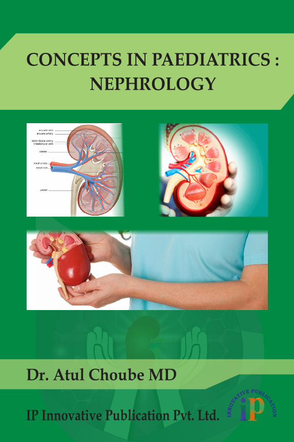
CONCEPTS IN PAEDIATRICS : NEPHROLOGY - Innovative ...
103
IP Innovative Publication Pvt. Ltd. Dr. Atul Choube MD CONCEPTS IN PAEDIATRICS : NEPHROLOGY
-
Upload
khangminh22 -
Category
Documents
-
view
1 -
download
0
Transcript of CONCEPTS IN PAEDIATRICS : NEPHROLOGY - Innovative ...
IP Innovative Publication Pvt. Ltd.
Dr. Atul Choube MD
CONCEPTS IN PAEDIATRICS :
NEPHROLOGY

CONCEPTS IN PAEDIATRICS : NEPHROLOGY
IP Innovative Publication Pvt. Ltd.


CONCEPTS IN PAEDIATRICS : NEPHROLOGY
IP Innovative Publication Pvt. Ltd.
Dr. Atul Choube
IP Innovative Publication Pvt. Ltd.
Published By:IP Innovative Publication Pvt. Ltd.First Floor, RZ- 1/4- A, Vijay Enclave, Main Dabri-Palam Road, New Delhi-110045, India.Ph: +91-11-25052216 / 25051061. Mob. : +91-8826859373, 8826373757E-mail: [email protected], [email protected]: www.innovativepublication.com
CONCEPTS IN PAEDIATRICS : NEPHROLOGY
ISBN : 978-93-88022-09-5
Edition : First, 2018
Price (INR) : Rs. 695/- (USD) : $ 35
Copyright © 2018, IP Innovative Publication Pvt. Ltd.All rights reserved.
No part of this book may be reproduced in any form, by Photostat, microfilm, xerography, or any other means, or incorporated into any information retrieval system, electronic or mechanical, without the written permission of the copyright owner.
Inquiries for bulk sales may be solicited at: [email protected]
This book has been published in good faith that the contents provided by the contributor’s contained herein are original, and is intended for educational purposes only. While every effort is made to ensure accuracy of information, the publisher and the editors specifically disclaim any damage, liability, or lose incurred, directly or indirectly, from the use or application of any of the contents of this work. If not specifically stated, all figures and tables are courtesy of the editors. Where appropriate, the readers should consult with a specialist or contact the manufacturer of the drug or device.
Printed at: New Delhi

DEDICATION
This book is dedicated to my parents, teachers, and my students of past, present and future.

vii
ABOUT THE BOOK
Concepts in Paediatrics : Nephrology is a synopsis of diseases of kidneys and urinary tract. Its concise and compact format makes it easy to be carried to the clinics by UG and PG students studying Paediatric Medicine and also practitioners of Paediatrics and Medicine. The book covers in its chapters all aspects of Nephrology starting from development, structure and function to various diseases of kidneys and urinary system. Special emphasis is given to congenital renal disorders which are so prevalent in children. Chapter on urinary tract infection describes in detail the diagnosis and treatment. A significant addition is chapter on renal transplant for end stage renal diseases. The book is also useful for Paediatricians aspiring for superspeciality courses in Nephrology and Urology.

ix
PREFACE
Diseases of the kidneys and urinary system are common occurrences in paediatric population and comprise the bulk of cases in hospitals and clinics. A proper understanding of their pathogenesis, diagnosis and therapy is therefore necessary for both students and clinicians and also for interviews for employment and positions. It is this aim the book has endeavored to attain in an easy to follow but concise and compact format. It is a hope that the book will be a pocket guide to both students and clinicians alike.I thank the publishers and editorial staff for all the help and encouragement.

xi
ACKNOWLEDGEMENT
I gratefully acknowledge the inspiration given by Er. Anugrih Choube B.E. for initiation and completion of the book. Thank you my dear son and God bless you.

ContentsAbout The Book ........................................................................................ viiPreface ........................................................................................................ ixAcknowledgement ...................................................................................... xi
1. Renal Anatomy ............................................................................................ 12. Nephron ........................................................................................................ 33. Renal Development ..................................................................................... 64. Renal Physiology ......................................................................................... 85. Renal Function Tests ................................................................................. 116. Renal Anomalies ........................................................................................ 187. Renal Tubular Defects ............................................................................... 208. Urinary Tract Infections ........................................................................... 249. Nephritis ..................................................................................................... 35
10. Haematuria ................................................................................................. 5011. Nephrotic Syndrome ................................................................................. 5212. Haemolytic Uraemic Syndrome .............................................................. 6413. Renal Hypertension .................................................................................. 6714. Renal Vein Thrombosis ............................................................................ 6815. Nephroblastoma (Wilm’s Tumor) ........................................................... 7016. Acute Renal Insufficiency ......................................................................... 7317. Chronic Renal Insufficiency ..................................................................... 80

1
Chapter 1RENAL ANATOMY
Kidneys, ureters and bladder are retroperitoneal structures. Kidneys are at level of 1st to 4th lumber vertebrae at or slightly above level of umbilicus. They can be usually palpated in neonate. Each kidney has 8-12 pyramidal shaped lobes.
External surface of fetal kidney is lobulated which gradually disappear with age. Each lobe has 2 zones, cortex and medulla. In cortex glomeruli and proximal and distal convoluted tubules are located. In medulla vasa recta, descending and ascending limb of loop of Henle and collecting ducts are located. Many collecting ducts fuse to form duct of Bellini which opens into minor calyx. Minor calyces are subdivisions of superior and inferior major calyces that unite to form renal pelvis from which urine drains into ureter and is transported by active peristalsis to bladder.
BLOOD SUPPLY Each kidney receives 10% of cardiac output. Renal artery arises from aorta.
Interlobar artery pass between lobes to renal pelvis. At junction of cortex and medulla interlobar arteries divide to form arcuate arteries which pass between cortex and medulla parallel to surface of kidneys. From these arteries interlobular arteries enter cortex and run perpendicular to kidney surface. Interlobular arteries branch to form afferent arterioles each of which supplies one glomerulus. Some interlobular arteries pass directly to superficial cortex to provide much of its blood supply.
About 50 micron before afferent arteriole enters glomerulus muscle cells of media assume appearance of secretory cells. They contain granular deposits of renin. These cells situated at vascular pole of glomerulus constitute zuxta glomerular apparatus. Just beyond point at which arteriole enters Bowman capsule it subdivides into several branches which in turn branch into network of glomerular capillary loops. These reunite to form efferent arterioles which emerges at vascular pole where renal tubule returning to cortex makes tangential contact with afferent arteriole of its own glomerulus. Tubular epithelial cells become narrower here. This portion of tubule is known as macula densa.
CONCEPTS IN PAEDIATRICS : NEPHROLOGY
2
Nephrons at junction of cortex and medulla are called juxta medullary nephrons. In them diameter of efferent arterioles is slightly larger than diameter of afferent arteriole. Reverse is true of arterioles of cortical nephrons. Efferent arterioles of outer and mid cortical nephrons divide into anastomosing network of capillaries that surround proximal and distal convoluted tubules and cortical portion of loop of Henle and collecting duct. For subcapsular or outer cortical nephrons these peritubular capillaries form efferent arteriole of associated glomerulus. For nephrons that are deeper in cortex there is free communication with peritubular capillary network from efferent arterioles of other nephrons. Walls of peritubular capillaries are very thin and are in very close proximity to basement membrane surrounding each tubule. Cortical capillaries merge to form interlobular veins.
Efferent arterioles of inner cortical and zuxta medullary nephrons provide peritubular capillary network for proximal and distal convoluted tubules and loops of Henle and collecting ducts in that area.
Efferent arterioles of zuxta medullary nephrons also supply vasa recta which are recurrent arterial loops that parallel loops of Henle as they descend through medulla to papilla. Vasa recta turn upward at bend of loop to zuxta medullary region to enter interlobular or arcuate vein. Vasa recta function as counter current exchangers in process of urine concentration.
Cortex receives about 75% of total renal blood flow. About 20% goes to zuxta medullary cortex and outer medulla. Blood flow through inner medulla being much slower facilitates maintenance of high solute concentration in this region that is essential to concentration of urine.Factors Altering Distribution of Renal Blood Flow:
Saline overload or administration of diuretic increases blood flow and glomerular filtration rate in outer cortical nephrons.
In congestive heart failure, shock or dehydration inner cortical and zuxta medullary areas are preferentially perfused. This regulatory function involves autonomic nervous system, humoral factors such as anti diuretic hormone and angiotensin and prostaglandins.
q
3
Chapter 2NEPHRON
Nephron is functioning unit in formation of urine. There are one million nephrons in each kidney. Loops of zuxta medullary nephrons extend deep into medulla. They have different role in regulation of salt and water excretion.
GLOMERULUSIt is 150-200 micron in diameter and is filtering apparatus of nephron. There
is intricate, spherical, convoluted, capillary network arising from afferent arteriole after it enters Bowman capsule. Walls of capillaries of this network form membrane across which process of filtration occurs.
Walls of glomerular capillaries have 3 layers. Endothelium is thin and attenuated and is traversed by multiple fenestrae with glycoprotein surface coat. Glomerular basement membrane is uninterrupted, highly convoluted membrane about 1200 A° thick, having central electron dense layer (lamina densa) and two electron lucent layers, lamina rara interna which is subendothelial in location and lamina rara externa which is subepithelial. Epithelium consists of large cells with extensive cytoplasmic projections. These sub-divide into foot processes which interdigitate with one another and are in direct contact with glomerular basement membrane. Between foot processes are filtration slits which are 240 A° in diameter. Covering epithelial cells and filling spaces between foot processes are glycoprotein cell coat upto 800 A° in thickness which is negatively charged. Fixed negative charge conferred by endothelial and epithelial cell coats helps in traversing of charged macromolecules across glomerular capillary wall. They restrict access of negatively charged molecules to urinary space and facilitate transit of those molecules which are positively charged.
Mesengial cells lie deep in central region of glomerulus and are separated from capillary lumina by overlying endothelial cells. Mesengial cells and intercellular material between them called matrix constitute mesangium.
Macromolecules which circulate through glomerular capillaries may enter mesangium through interface between endothelial and mesangial cells and migrate via intercellular channels towards zuxta glomerular region. Macromolecules may also be
CONCEPTS IN PAEDIATRICS : NEPHROLOGY
4
phagocytosed by mesangial cells or by infiltrating phagocytes. Mesangium thus acts as component of reticuloendothelial system in glomerular circulation. Mesangial region is site of injury in diseases affecting glomeruli, responding sometimes in nonspecific ways (as with cellular and matrix proliferation) at other times with pathognomonic changes (eg intercapillary nodule formation in diabetic glomerulosclerosis).
BOWMAN CAPSULEThis sorrounds glomerulus. Its basement membrane is continuous with
basement membrane of proximal convoluted tubule and is lined on inner aspect with parietal epithelial cells. Tubular portion of nephron begins at orifice in capsule situated opposite vascular pole.
TUBULES Various portions are proximal convoluted tubule, loop of Henle, distal
tubule and collecting duct. Tubular basement membrane provides uninterrupted framework for tubular epithelium.
Proximal convoluted tubule is situated in cortex. Its epithelium is cuboidal and one cell deep. Spherical nuclei are situated at basal surface of cell. Spaces between cells are channels through which solutes and water reabsorbed from lumen by cells pass to peritubular capillaries. Luminal brush border increases reabsorptive surface of cells. There is tight junction between cells in their luminal aspect. This is impermeable to solutes or water but back diffusion of reabsorbed solutes and water into tubular lumen occurs via these intercellular junctions. There are numerous mitochondria in cells of proximal convoluted tubule. Peritubular capillaries are next to basement membrane. Proximal convoluted tubule cells transport or reabsorb large quantity of water and solute from tubular lumen. They also participate in process of tubular secretion by which substances synthesized within cell or derived from circulation are added to luminal fluid.
Loop of Henle is continuation of proximal convoluted tubule. Nephrons with glomeruli situated in outer two third of cortex have short or absent loops. Those with glomeruli in inner third have longer loops which extend towards tip of papilla.
After descending into medulla loop turns back to ascend to cortex where it becomes distal tubule. Epithelium of descending limb is flat and squamous and tubular diameter is less than that of proximal convoluted tubule. This portion is called thin segment of loop. Luminal surface of cells of thin segment have short widely spaced microvilli and their cytoplasm has infrequent mitochondria.
Ascending limb has thicker epithelium and nuclei are situated near luminal surface. Numerous rod shaped mitochondria occupy basal half of these cells. Short microvilli arise from luminal surface. Cleft like infolding of basal plasma membrane of cells bring it into intimate contact with mitochondria.
NEPHRON
5
Distal tubule has initial portion, pars recta which continues in straight course toward glomerulus. As distal tubule passes its glomerulus of origin it makes contact with afferent arteriole. This part of tubule is macula densa. Thereafter distal tubule becomes convoluted. Cells are cuboidal and have dense coarsely granular cytoplasm containing numerous mitochondria. Cell nucleus is apical. Luminal surface of cells has numerous short microvilli.
Collecting duct is formed by junction of 2 or more terminal segments of distal convoluted tubule and receives additional branches in its course to medullary papilla. It has cuboidal epithelium. This joins duct of Bellini through which urine from collecting ducts is discharged into minor calyx at papillary tip.
INTERSTITIUMInterstitial space and number of cells increase as papilla is approached. Space
is filled with flocculent material of low electron density.Type 1 interstitial cells are most numerous. They resemble fibroblasts. They
contain many lipid bodies and abundant granulated endoplasmic reticulum. Lipid droplets contain renal PGE2 and PGF2 alpha precursors.
Type 2 cells have some characteristics of mononuclear cells and may have phagocytic activity.
Type 3 pericyte is found adjacent to vasa recta.Renal medulla is site for prostaglandin synthesis.
NERVE SUPPLYNerve fibres course along blood vessels. Some fibres innervate juxta glomerular
efferent arterioles that give rise to vasa recta. Nerves play role in regulation of renal blood flow and glomerular filtration rate. Stimulation of renal sympathetic nerves cause reduction in cortical renal blood flow and leads to reduction in urinary excretion of sodium. Sympathetic blockers cause renal vasodilatation and mild natriuresis.
q
6
Chapter 3RENAL DEVELOPMENT
Kidney derives from metanephros from embryonic mesoderm. During 5th week of embryonic life ureteric diverticulum develops as outgrowth of mesonephric duct from point near to cloaca. It grows headwards into nephrogenic cord and becomes surrounded by mesodermal tissue which will give rise to metanephros. Primitive nephrons develop in close proximity to it.
Ureteric bud devides and subdivides while growing towards periphery of metanephros. First nephrons to be formed are those deeply situated in kidney. As each branch of bud is surrounded by nephrogenic tissue fetal kidney assumes lobulated appearance.
New nephrons continue to form upto 36 weeks of gestation only. Thereafter increase of nephron mass is by increase of tubular length and glomerular size.
Fetal sclerosis occurs in youngest glomeruli when their tubules do not communicate with ureteric bud as its growth ceases in later stages of fetal life. Other glomeruli show features of immaturity varying from unvascularized clumps of epithelial cells to occasional cuboidal epithelium and limited lobulation of tufts. Tubular immaturity is even more profound than that of glomeruli for ratio of glomerular surface area to tubular volume is much greater than in later childhood.
Ureteric bud by process of subdivision followed by coalescence gives rise to renal pelvis, calyces and collecting ducts. During this process oldest, deepest nephrons are lost. Occasionally glomeruli in aberrant positions beneath pelvic mucosa or within arterial walls will survive.
Urinary bladder is formed from ventral and cephalic portion of cloaca after this has been separated from rectum by urorectal septum. Into this are incorporated caudal ends of mesonephric tubes and ureteric buds. These form bladder and urethra.
Anatomical evidence of renal immaturity at birth is mirrored physiologically by glomerular filtration rate, PAH clearance, renal bicarbonate and glucose reabsorption which are markedly diminished.
Normally less urea is produced by rapidly growing baby. More dietary nitrogen is deposited as protein in tissues. Anabolism, growth and renal clearance
RENAL DEVELOPMENT
7
are responsible for maintenance of normal blood urea levels in small infants. Excessive dietary protein, growth retardation and tissue catabolism (eg in acute infections) can lead to urea synthesis in excess of renal excretory capacity. Thus raised blood urea in infant while abnormal does not indicate necessarily impairment of renal function.
Diminished urea excretion is responsible for poor concentrating ability of newborn kidney.
Fetus excretes large amount of sodium and water before birth. At birth newborn is deprived of limitless transplacental supply of water and sodium and must suddenly conserve both of these. This is achieved within few days after birth.
q
8
Chapter 4RENAL PHYSIOLOGY
GLOMERULAR FILTRATIONThis depends upon higher functional pressure in afferent arterioles. Filtration
barrier is formed by endothelium with slit pores, basement membrane and epithelium with its interdigitating podocytes.
Filtration of macromolecules depends upon their size, shape and electrical charge. Electrostatic hindrance is provided by fixed negatively charged component of glomerular capillary wall. Podocytes have surface coating of sialoproteins which are negatively charged.
Normally about 20% of plasma appears as filtrate which contains all diffusible and ultrafiltrable substances present in plasma.
TUBULAR REABSORPTIONProximal convoluted tubules reabsorb 80% of glomerular filtrate. Glucose,
amino acids and proteins are almost completely reabsorbed. Potassium is completely reabsorbed in proximal convoluted tubule and secreted in distal tubule in exchange for sodium. Chloride reabsorption is passive.
Bulk of energy expenditure by kidney is related to sodium reabsorption in which sodium potassium activated ATP is involved. Isotonic reabsorption of water coincides with sodium reabsorption and depends upon raised oncotic pressure of plasma proteins in peritubular capillaries. This arises as result of haemoconcentration following filtration in glomeruli.
Bicarbonate reabsorption occurs actively and depends upon bicarbonate stimulated ATPase at brush border.
Tubules preferentially reabsorb certain essential amino acids. Renal aminoaciduria may be due to specific defect in carriers or enzyme system or may be part of generalized disorder of tubular function.
Phosphates are partially reabsorbed in proximal convoluted tubule. Remaining phosphate is filtered in distal convoluted tubule by H+ ion secreted there and help in H+ ion excretion.

RENAL PHYSIOLOGY
9
CONCENTRATION AND DILUTION
Countercurrent Multiplier System of Urinary Concentration:There is active transport of sodium chloride out of ascending limb of loop of
Henle into hypertonic medulla. Fluid first follows down descending limb which has high water permeability but low permeability to sodium. Osmolality of contents rises from 300 mOsmol at entry to 1500 mOsmol at tip of loop. Thin ascending limb is relatively impermeable to water but reabsorbs sodium chloride. Collecting duct is permeable to urea which is then trapped at higher concentration in inner medula. It contributes to medullary hypertonicity.
Dilution of urine occurs in ascending limb of loop of Henle and early distal tubule. This is result of sodium reabsorption without concomitant water diffusion. There is no net addition of water to tubular fluid. When anti diuretic hormone levels are low nephron will be impermeable to water and hypotonic fluid passes through collecting tubule to be excreted as dilute urine. When ADH level is high then distal and collecting tubular walls become permeable to water. In distal tubule isotonicity with plasma is reached and in collecting tubule as they pass through highly hypertonic papillary interstitium water diffuses out of lumen down osmotic gradient into papilla as result tubular fluid becomes concentrated to same osmolality as papillary tip.Causes of impaired diluting ability:
1. Decreased glomerular filtration rate:i. Chronic renal disease.
2. Endocrinal:i. Hypothyroidism.ii. Hypopituitarism.iii. Hypoadrenalism.
3. Syndrome of inappropriate anti diuretic hormone secretion:i. Secondary to malignancy.
4. Dilutional disturbance: (Sodium retaining states)
i. Cirrhosis of liver.ii. Congestive heart failure.iii. Nephrotic syndrome.
HYDROGEN ION EXCRETION Bicarbonates are buffers in body.
H + HCo3 = H2Co3 = Co2 + H2O.Kidney is instrumental in conserving bicarbonate ions and excreting H ions.Bicarbonate is reabsorbed as result of H ion secretion into tubular lumen.
CONCEPTS IN PAEDIATRICS : NEPHROLOGY
10
Resulting carbonic acid is dehydrated by carbonic anhydrase to carbon di-oxide which diffuses into tubular cell. There it is reconstituted into bicarbonate.
PROSTAGLANDINS AND RENAL BLOOD FLOWProstaglandins are synthesized in walls of blood vessels and also in
renomedullary interstitium cells.Prostaglandins are vasodilators whose releases in hypertension offsets action
of angiotensin.PGE2 is major product of interstitial cells of kidney. It opposes action of ADH
on collecting ducts. Kallikrein and bradykinin action is through PGE2.PGE causes vasodilatation of arterioles of inner cortex. Its infusion leads
to increase in renal blood flow and osmotic diuresis accompanied by increased sodium and potassium excretion.
Prostacyclin produced by vascular endothelium also offsets action of pressure hormones and prevents deposition of platelets and products of coagulation.
q
11
Chapter 5RENAL FUNCTION TESTS
Functions of kidney are following:A. Excretion of:
1. Waste products of metabolism eg urea, uric acid, creatinine.2. Toxic substances eg phenol, glucuronides, sulphates.3. Drugs eg salicylates.
B. Reabsorption of Substances Vital to Body eg Glucose, Amino Acids.
C. Maintenance of Constancy of Mileu Interior by:1. Regulation of water balance.2. Regulation of electrolyte balance.3. Regulation of acid base balance.
D. Erythropoiesis:1. Production of erythropoietin.
E. Vitamin D Metabolism:1. Formation of 1,25 OH dehydroxy cholecalciferol.
F. Regulation of Blood Pressure:1. Renin angiotensin aldosteron.2. Prostaglandin production.
ASSESSMENT OF RENAL FUNCTION
Aim:Tests are performed to know:
1. Whether glomeruli, tubules or renal blood flow is dysfunctioning.2. Are there any extra renal factors involved in production and maintenance
of renal disease?3. What is state of renal reserve?

CONCEPTS IN PAEDIATRICS : NEPHROLOGY
12
Classification of Renal Function Tests:
A. Tests of Glomerular Function:1. Glomerular filtration rate or creatinine clearance.2. Proteinuria.3. Cell count and type of cast.
B. Tests of Tubular Function:1. Specific gravity of urine.2. Osmolality of urine.3. Concentration, dilution tests.4. Urinary acidification.5. Urinary electrolyte estimation.6. Amino acids in urine.7. Types of cells and casts.
C. Test of Renal Blood Flow:1. Para amino hippurate clearance.
D. Biochemical Examination:1. Urea.2. Uric acid.3. Creatinine.4. Blood glucose.5. Ketones.6. Serum calcium.
E. Tests of Renal Structural Integrity:1. Intravenous urogram.2. Renal scan.3. Renogram.4. Renal angiogram.5. Renal ultrasonogram
F. Renal Biopsy:1. For histopathologic confirmation of deranged renal function.
CREATININE CLEARANCE
Definition:Amount of plasma cleared of creatinine in unit time.
Method of measuring glomerular filtration rate by creatinine clearance:24 hours urinary collection is done. Urinary creatinine is measured.
RENAL FUNCTION TESTS
13
Plasma creatinine is measured.CC = UV/P.U = Urinary creatinine.V = Urine flow per minute.P = Plasma creatinine.
Falacy:In very high serum creatinine levels some amount of creatinine may get
secreted by tubules.Roughly GFR is calculated by formula:
In male: 120/Serum creatinine.In female: 100/Serum creatinine.Serum creatinine starts rising only when GFR falls by 50%.
PROTEINURIAProtein excretion by healthy kidneys:
In preterm < 50 mg/24 hour. Children < 10 year < 100 mg/24 hour. Children 10-18 year upto 300 mg/24 hour.
Incidence of proteinuria estimated by Rudolph in 1967 is 6.3%.Persistent proteinuria is abnormal.
Causes of abnormal proteinuria:A. Elevated Concentration of Proteins in Plasma:
1. Eg multiple plasma infusions in coagulation disorders.B. Addition of Proteins to Tubular Fluid:
1. Tamm Horsefall uromucoid.2. Hypokalemic nephropathy.
C. Inadequate Tubular Reabsorption of Filtered Proteins:1. Wilson disease.2. Fanconi syndrome.3. Renal tubular acidosis.4. Galactosemia.
D. Increased Glomerular Permeability:1. Selective in minimal lesion glomerulonephritis.2. Nonselective in other glomerulonephritis.
Methods of detecting proteinuria:1. Heat coagulation (Richard Bright).2. Chemical coagulation:
i. 3% sulphosalicylic acid.ii. Ethalon phosphotungistic acid.
CONCEPTS IN PAEDIATRICS : NEPHROLOGY
14
Fallacies of Turbidometric Tests:Presence of urates and phosphates, X ray contrast media, peicillin,
tolbutamide metabolites.1. Dipstick method.2. Electrophoretic examination of urine:
i. Glomerular proteinuria: Albumin, transferrin, gamma globulin, alpha 2-glycoproteins.
ii. Tubular proteinuria: Alpha 2 globulin, beta globulin, lysozymes.3. Transferrin and immunoglobulin G clearance:
i. Selective proteinuria: Ratio 0.2.
TESTS OF RENAL CONCENTRATION
1. Specific Gravity:Measurement of urinary specific gravity and osmolality after fluid deprivation:
All fluids are withheld for 12 hours following which urinary specific gravity should rise to over 1020.Indications:
1. Fanconi syndrome.2. Wilson disease.3. Pyelonephritis early stage.Test should be done cautiously in diabetes insipidus. If weight loss is more
than 3% test is terminated.2. Pitressin 0.5 units per kg intramuscular at 6 pm. Measure specific gravity after 10 hours.3. D deaminovasopressin 10 micro gm in infants. 20 micro gm in children given intranasal. Effect in 2 hours.
1. Age 7-40 days: Urinary concentration 600-1100 mOsm/kg.2. Age 2 months to 3 years: Urinary concentration 416-463 m osm/kg.3. Age 3 years to 16 years: Urinary concentration 870-309 m osm/kg.
TESTS OF URINARY ACIDIFICATIONAmmonium chloride is administered 0.1 gm per kg. In next 4-8 hours urinary
pH is measured hourly. Normal < 5.31. Preterm 1-3 weeks age: 5.962. Term 1-3 weeks age: 53. 1-12 months age: < 54. 3-15 years age: < 5.5
Test:Give oral water 60 ml per square meter body surface area per hour. Take
control urine specimen. Give ammonium chloride 75-150 m eq per square meter
RENAL FUNCTION TESTS
15
body surface area. Value of this test is limited in routine clinical evaluation of renal function.
Test is useful in Fanconi syndrome and renal tubular acidosis where there is defect in secretion of H+ ions by renal tubules.
MEASUREMENT OF RENAL PLASMA FLOW
1. Para Amino Hippurate Clearance: (Wolf modification of Fick formula)
RPF = (UX – VX). V/AX – VXUX = urinary concentration.VX = concentration in renal vein.AX = arterial concentration.V = urinary flow rate.Method: Priming dose of PAH 8 mg per kg. Maximum rate of transport of
PAH is 80 mg per minute per 1.73 square meter surface area of body equivalent to plasma concentration of 10-14 mg per dl. Amount of PAH reduces in proportion of renal insufficiency.2. I131 Hippuran. Single injection. Dose 40 mci per square meter body surface area.3. Diffusible gas indicator for measuring renal plasma flow. An inert gas is introduced into kidney. RPF is determined by rate at which gas is washed out of kidney.
TESTS OF RENAL STRUCTURAL INTEGRITY
1. Intravenous Urogram:In newborn due to low glomerular filtration rate associated with immaturity of
renal function there is poor or no visualization of upper tracts.There are two phases of nephrogram:
1. Vascular phase is useful in diagnosis of vascular, renal or intra abdominal tumors.
2. Tubular phase is useful in differential diagnosis of renal neoplasm and cysts.
Total body opacification: Following high dose of contrast medium liver and spleen are also visualized. Best seen in infants and children. Useful in diagnosis of subclinical ascites and solid intra abdominal tumor from cyst.2. Radionuclide Renogram:
Aim: To assess individual function of abnormal kidney in presence of normal or near normal contralateral kidney.
1. Reno vascular hypertension.
CONCEPTS IN PAEDIATRICS : NEPHROLOGY
16
2. Hydronephrosis.3. Atrophic pyelonephritis.4. Congenital hypoplastic kidney.5. Following ureteric reimplantation.Method: Usual water intake. Child kept prone. Oral lugol solution 1 hour
prior. Intravenous Hippuran 131. Dose 2 mu ci per pound. Exposure at 3-5 minutes interval for 30 minutes.
Analysis: First phase. Initial rapid rise in count rate. Reflects radioactivity in renal vessels and parenchyma.
Second phase. Less rapidly rising count rate. Radioactivity in renal tubules.Third phase. Excretory renogram. Rapid decline in count rate.With onset of renal functional impairment second phase rises less steeply.
3. Renal Ultrasonogram:Noninvasive safe procedure. No discomfort. No patient preparation.
Diagnostic method of choice in patients with contrast medium hypersensitivity.Indications:
1. Congenital anomalies:i. Renal agenesis.ii. Horseshoe kidney.iii. Crossed ectopic kidney.iv. Ureteric duplication.v. Congenital urachal anomalies.
2. Space occupying lesions:i. Cystic kidney.ii. Neoplastic mass.iii. Inflammatory mass.iv. Obstructive uropathy.
3. Before renal biopsy to outline kidney accurately.4. Renal trauma:
i. Perirenal and perivesical haematoma and urinoma.ii. Laceration of kidney.
5. Renal transplant:i. Diagnosis of acute rejection by cortical edema.ii. Haematoma, abscess.iii. Diagnostic accuracy 95%.
4. Renal Biopsy:Indications:
1. Persistent haematuria or proteinuria.2. Atypical severe acute glomerulonephritis.
RENAL FUNCTION TESTS
17
3. Steroid resistant nephrotic syndrome.4. Hereditary nephropathies.5. Acute or chronic renal insufficiency of uncertain cause.6. Renal hypertension.7. Evaluation of renal involvement in systemic diseases eg systemic lupus
erythematosus, polyarteritis nodosa, Henoch Schonlein purpura, diabetes mellitus, secondary amyloidosis, haemolytic uraemic syndrome.
8. Evaluation of renal allograft.Contraindications:
1. Major:i. Bleeding diathesis.ii. Solitary kidney.iii. Anticoagulant therapy.iv. Intra renal tumor.
2. Minor:i. Hydronephrosis.ii. Perinephric abscess.iii. Acute intrarenal infection.iv. Severe anaemia.v. Small contracted end stage kidney.
q
18
Chapter 6RENAL ANOMALIES
RENAL AGENESISBilateral renal agenesis is incompatible with prolonged extra uterine life,
occurs twice as often in males as in females and is present in between 0.2-0.4% autopsies performed in infants stillborn or dying soon after birth.
Potter facies with large low set ears deficient in cartilage, wide set eyes, beaked nose and micrognathos is seen at birth.
Other congenital anomalies particularly bilateral pulmonary hypoplasia are often associated with renal agenesis.
Embryologically condition results from lack of ureteric budding from mesonephric duct.
For diagnosis of bilateral renal agenesis intravenous urogram can be undertaken in anuric neonate with progressive uraemia.
Ultrasonography, nephrography and computerized tomography are useful.Unilateral renal agenesis occurs in 0.1% of neonates. More common in
males. Associated anomalies of external ear on ipsilateral side, agenesis or atresia of ureter of corresponding kidney and congenital abnormalities in ipsilateral leg. Contralateral kidney is hypertrophied in compensation. This may be more easily accidentally damaged because of its larger size.
Unilateral renal agenesis is result of lack of formation of ureteric bud or its inability to stimulate differentiation of nephrogenic mesoderm.
Possibility of unilateral renal agenesis should always be excluded before contemplating nephrectomy or percutaneous kidney biopsy and before deciding that kidney is non functioning eg in suspected renal vein thrombosis.
Ultrasonography helps detect nonfunctioning kidney.
POLYCYSTIC KIDNEYUsually bilateral in contradistinction to unilateral multi cystic kidney disease.Infantile form presents at birth with bilateral renal masses and is rapidly
lethal. It may present in late infancy. There may be siblings with same condition but no affliction of earlier generation.
RENAL ANOMALIES
19
Pathologically cysts are fusiform in shape affecting cortex and medulla and radially oriented with kidney retaining its general uniform shape.
On urography there is delay in appearance of dye. Nephrogram may show streaky pattern and delay of hours or days in loss of radio opaque material.
Lack of communication between nephrons and collecting ducts with dilatation of nephrons is seen. Hypertrophy and cystic degeneration of interstitial portions of collecting ducts occurs. These children die during first month of life but may survive for years with varying degree of chronic renal insufficiency.
Adult type may appear even in neonates although it characteristically gives rise to symptoms of renal insufficiency in middle age.
Inheritance is autosomal dominant.Kidneys become distorted by roughly circular and randomly distributed cysts.
Radiologically seen as ‘spider kidney’.Both types of polycystic kidneys may be associated with cysts of liver
and pancreas.In some patients renal cystic disease is associated with hepatic fibrosis. These
juvenile patients present with features of portal hypertension such as ascites, splenomegaly and haemetemesis.
Familial bilateral cystic dysplasia of kidney rarely affects liver. Radiologically in these cases blunted renal calyces are seen.
Progressively increasing renal insufficiency is to be anticipated in all types of polycystic kidney disease. There are episodes of haematuria and recurrent urinary tract infection.
Kidney may be so large as to obstruct delivery of baby.Treatment is symptomatic to correct fluid and electrolyte disturbance induced
by renal failure and systemic hypertension. Surgical relief of portal hypertension by porta caval shunt has been found useful.
Renal transplantation is definitive treatment.q

20
Chapter 7RENAL TUBULAR DEFECTS
FANCONI SYNDROME This is due to generalized defect of renal tubular function in proximal tubules.
In consequence glucose, amino acid, uric acid, phosphate, sodium, potassium, bicarbonate and protein all have enhanced clearance and appear in increased quantities in urine.Etiology and Pathogenesis:
Renal tubular damage may be produced by heavy metals such as lead, cadmium and uranium.
Syndrome may occur in Wilson disease, hereditary fructose intolerance, galactosemia, glycogenosis, tyrosinaemia and following use of outdated and deteriorated tetracyclins.Fanconi syndrome is divided into two types:
1. More severe is associated with cystine storage in many tissues. It is inherited as autosomal recessive gene, presents in infancy and early childhood and progresses to produce glomerular damage and renal insufficiency. It is called cystinosis or Lignac Fanconi syndrome.
2. Second type usually presents in adult life. Autosomal recessive inheritance is described in some families. It is not associated with cystinosis and progresses to renal insufficiency much slowly.
Swan neck deformity of renal tubules are seen.Clinical Features:
Disease presents in first few years of life, affecting males and females equally. Onset is with anorexia, vomiting, inability to thrive, polydipsia and polyuria. There are episodes of intense vomiting and fever leading to dehydration.
Examination reveals growth retardation and dehydration. There are deep rapid respirations associated with acidosis. Muscle hypotonia is present. Severe hypokalemia causes generalized muscular weakness and paralytic ileus.
RENAL TUBULAR DEFECTS
21
Rickets is obvious with frontal bossing, craniotabes, swelling of metaphyses at wrists, knees, ankles and costochondral junctions. Bowing of tibia, fibula, femur may occur.
Cystine crystals may be identified by slit lamp examination of cornea. They may lead to photophobia.
Progressive advancing glomerular damage due to cystinosis leads to renal insufficiency. Then symptoms of chronic renal insufficiency supplant those due to tubular dysfunction. Hypertension, bleeding diathesis and neurological complications may occur. Anorexia, growth retardation, polydipsia and polyuria remain prominent symptoms.Diagnosis:
Cystine storage may be detected by slit lamp examination of cornea or examination of peripheral leucocytes, bone marrow or renal biopsy specimen.
Routine urine testing shows presence of glycosuria and proteinuria. Proteinuria is tubular comprising of post albumin, alpha 2 and beta globulins.
Amino acid chromatography reveals generalized increase in amino acid excretion. Cystine, alanine and valine are especially prominent. Blood levels of amino acids are normal or slightly increased.
Hypokalaemia, hyponatraemia and hypochloraemia are present due to abnormal increased urinary loss. This contributes to dehydration.
Potassium loss is due to lack of proximal reabsorption and increased tubular secretion in effort to reabsorb more sodium from tubular fluid.
Diminution of capacity to concentrate urine is present and is due to combined effect of reduction of sodium transport by loop of Henle and of hypokalaemia.
Metabolic acidosis is due to lack of proximal tubular bicarbonate reabsorption. Distal tubular secretion of H ions is unimpaired.
Occasionally alkalosis may occur due to renal salt wastage.Rickets is demonstrable radiologically. Serum calcium levels are normal but
inorganic phosphorus levels are low and alkaline phosphatase very high. Boney lesions are due to decreased tubular phosphate reabsorption and poor hydroxylation of 25-hydroxy cholecalciferol by kidney.
Uric acid clearance is increased. Hypercalciuria is found only when sodium excretion is also excessive. Progressive deterioration in glomerular filtration rate and renal blood flow are found.
Intravenous urogram shows poor dye concentration.Percutaneous renal biopsy reveals cystine crystals.
Treatment:Tubular dysfunction can not be corrected but secondary effects can be
improved. Replacement of water and electrolytes is required. 5-10 meq per kg per
CONCEPTS IN PAEDIATRICS : NEPHROLOGY
22
day are required of sodium and potassium. Addition of sodium bi-carbonate and potassium chloride to intravenous infusion may be necessary to control acidosis.
Thiazide diuretics which increase salt depletion and increase proximal tubular bicarbonate reabsorption are used to correct acidosis.
Vitamin D in doses of 25000-50000 iu per day are given. Addition of oral phosphate supplement improves rachitic changes with smaller dose of vitamin D.
Penicillamine and diet low in cystine are given.
LOWE SYNDROME (Oculo Cerebro Renal Syndrome)
Lowe and his coworkers in 1952 described 3 boys with syndrome of mental retardation, organic aciduria, cataract and glaucoma. It is X linked recessive disease of males transmitted by female carriers who are normal or have early onset of cataract. Abnormality of Kreb cycle and particularly of ornithine arginine metabolism is postulated.Clinical Features:
Boys present from age 3 months onward with typical head with large ears, prominent forehead, flattened nasal bridge and prominent scalp veins in pale skin. Cataract as rule is shown in early cases by slit lamp examination.
Inconstant features are obesity, deafness, cryptorchidism and eczema.Intermittent pyrexia and growth retardation is usual. Inability to thrive,
osteoporosis and rickets occur.Mental deficiency is severe with hypotonia, lax joints, hypermobility of joints
and absent or greatly diminished tendon reflexes.Eyes are often rolled wildly in pseudonystagmus. Finger pressure on eyeballs
produce visual hallucinations.EEG may show fast 24 cycles per second general activity.Buphthalmos and congenital glaucoma may be present.Proteinuria occurs with complicated tubular dysfunction.Distal renal tubular acidosis is always present.There is hyperphosphaturia with hypophosphataemia, normocalcaemia and
elevated levels of alkaline phosphatase.Defective tubular ammonium production and defective tubular bicarbonate
absorption occurs.Hyperaminoaciduria especially lysinuria and tyrosinuria is seen.During febrile episodes hypernatraemia and dehydration may occur.Renal biopsy reveals tubular damage and dilatation with relatively
normal glomeruli.
RENAL TUBULAR DEFECTS
23
Treatment:Good nutrition and hygiene.Surgery for cataract and glaucoma. Sodium bicarbonate for metabolic
acidosis. Calciferol for rickets.During infancy there are recurrent infections and renal insufficiency.
RENAL GLYCOSURIANormally, filtered glucose is almost completely reabsorbed in proximal tubule.
But at high blood glucose levels, usually in excess of 180 mg/dl, glucose begins to appear in increasing quantities in urine. Renal threshold for glucose is exceeded.
When renal threshold is reduced glycosuria occurs at lower blood sugar levels than normal ie renal glycosuria.
Small amount of glucose, upto 5 mg/dl is found in normal urine. Its absence is used as indication of presence of glucose consuming bacteria in urine.There are two types:
1. Threshold for glucose and maximal rate of tubular reabsorption are uniformly diminished throughout nephron. Such situation is found in various forms of Fanconi syndrome whether idiopathic, related to cystine storage or secondary to Wilson disease or heavy metal toxicity. It may also occur as isolated tubular defect and is inherited in dominant fashion.
2. Renal glycosuria due to heterogeneity between different nephrons or because of abnormal enzyme system with reduced affinity for glucose. Glucosuria appears at low levels of blood glucose from minority of affected nephrons. This type produces no clinical disturbance, is of uncertain inheritance and unassociated with other evidence of renal tubular dysfunction.
Importance of this condition lies in distinguishing it from diabetes mellitus by measurement of blood glucose or performance of glucose tolerance test in conjunction with observation of urinary glucose excretion.
To ensure that urinary reducing substance is indeed glucose and not fructose, galactose or homogentisic acid glucose oxidase method is specific but chromatography and osazone synthesis may also help.
Intravenous glucose tolerance test curve is normal but oral glucose tolerance test may show flat curve reminiscent of malabsorption and indication that perhaps as in Hartnup disease and cystinuria transport defect is shared by jejunum.
No therapy is needed. Urinary loss of glucose is less than 30 gms per day per 1.73 square meter and therefore insignificant in terms of nutritional requirements.
q

24
Chapter 8URINARY TRACT INFECTIONS
These conditions have one common feature of significant bacteriuria.
CLASSIFICATION
A. Diseases with Bacteriuria:1. Non obstructive.2. Obstructive.3. Neurogenic bladder.
B. Localization of Infection:1. Upper UTI.2. Lower UTI.3. Superficial (cystitis).4. Deep (pyelonephritis).
C. Duration of Infection:1. Acute UTI.2. Chronic UTI.
DEFINITIONS
1. Cystitis:Infection limited to urinary bladder. Symptoms vary from severe pain on
micturition, burning, frequency and dull pain over bladder area. Patients are usually afebrile.
Diagnosis of infection limited to bladder can not be accepted unless following minimal criteria are fulfilled:
1. Patient is afebrile.2. Normal ESR.3. Normal renal concentration capacity.4. Antibodies against infecting organism.5. Limitation of infection to bladder can be demonstrated by bladder wash
and ureteral catheterization.
URINARY TRACT INFECTIONS
25
2. Acute Pyelonephritis:Acute bacterial infection of renal parenchyma. Patient is febrile. Symptoms
are toxic fever, chills, severe malaise, dysuria, urgency, frequency and loin pain.Diagnosis is made from history and presence of bacteriuria and pyuria together
with transitory lowering of renal concentration capacity, transitory increase of antibodies titre or demonstration of ureteral bacteriuria with bladder wash method.3. Chronic Pyelonephritis:
Clinical condition characterized by continuous excretion of bacteriuria or frequent relapses of infection.
Radiologically there is characteristic progressive renal scarring.Histologically there are characteristic lesions of renal parenchyma.
4. Cystourethral Syndrome:Vague term used for patients with classical symptoms of cystitis but lacking
demonstrable bacteriuria. Pyuria is often present. Some show inflammation on endoscopy. Condition is more common in small girls complaining of dysuria.
DIAGNOSTICS Demonstration of significant bacteriuria is sole valid criterion for presence
of UTI.A. Demonstration of Bacteriuria:Kass’s suggestion for definition of significant bacteriuria:
1. Less than 10 raise to power 4 bacteria per ml of urine means probably contamination.
2. 10 raise to power 4 to 10 raise to power 5 bacteria per ml of urine suggests new culture.
3. More than 10 raise to power 5 bacteria per ml of urine indicates probably infection.
4. If urine is obtained by bladder puncture than 10 raise to power 3 bacteria per ml of urine is significant.
1. Collection of Urine:1. Clean catch: Midstream urine should be collected. Preputial folds of small
boys may contain large number of bacteria.2. Bladder puncture: Mainly used in newborn or during first few days.
Indications:i. Persistent bacteriuria of doubtful significance.ii. Seriously ill patient in whom accurate diagnosis is essential.iii. Obstruction of bladder outflow tract or urethra.
CONCEPTS IN PAEDIATRICS : NEPHROLOGY
26
3. Catheterisation: This is used when bladder puncture is not convenient for example in children over 1 year. Risk of introducing infection for children with previous UTI is 50%.
2. Transportation of Urine:Bacterial multiplication starts rapidly in vitro. At 0-4°C bacterial count will
remain unchanged for 24-48 hours.3. Influence of Antibiotics Present in Urine:
Therapy should be discontinued for sufficiently long time before culture to permit complete excretion of antimicrobials.
Lack of demonstration of significant bacteriuria in overt infection of urinary tract may be due to complete obstruction of unilaterally infected kidney or presence of antibiotics in urine.B. Demonstration of Secondary Phenomenon of Inflammation:
1. WBC Count:Appearance of increased number of white cells in urine is secondary to
inflammation, bacterial or non bacterial. Pyuria thus never proves bacterial UTI nor does normal WBC count exclude UTI.Pyuria is of diagnostic help in:
1. Making tentative diagnosis in acutely ill patients before results of culture are available.
2. Support of diagnosis of UTI in patients with asymptomatic bacteriuria.3. Suggesting renal involvement by appearance of white cells casts.It is more common to find normal WBC count in recurrent infection than first
one. First specimen of urine in morning should be examined.Quantitative evaluation of urinary white cells is made by counting cells of
freshly voided uncentrifuged urine in counting chamber.Voided specimen from boys should contain less than 10 WBC per cu mm. In
girls 50-100 WBC per cu mm are normal.Methylene blue 0.5% stain helps differentiate between WBC and
epithelial cells.Proteinuria is of little diagnostic help in UTI.Haematuria is common especially in neonatal infection and in older boys.
2. Determination of Antibodies Titre:Agglutinating and haemagglutinating antibodies to ‘O’ antigen of infecting
E. Coli can be demonstrated in serum of patients with pyelonephritis but not from patients with superficial infection eg cystitis.
URINARY TRACT INFECTIONS
27
This characteristic is of help in knowing localization of infection which is of special interest in patients with asymptomatic bacteriuria or with symptoms suggesting cystitis where renal involvement is not infrequent although symptomalology is sparse. Antibodies are highly specific and thus can be used in diagnosis to verify that isolated bacteria are cause of infection. This may be clinically useful in patients with moderately high bacterial count (10 raise to power 4 to 10 raise to power 5 per ml of urine) and in all scientific work on UTI.3. Renal Function Tests in UTI:
Renal concentration capacity: In acute infections of kidney there occurs transitory decrease of capacity of kidneys to concentrate urine. This may be used diagnostically to localize infection. In uncomplicated cases capacity is restored to normal in 6-8 weeks. If concentration capacity is not restored possibility of obstruction to urine flow, renal scarring or persistent infection should be considered.
Glomerular function: Increased blood urea is uncommon in uncomplicated acute UTI. If present it suggests either obstruction of urine flow with bilateral hydronephrosis or marked parenchymal reduction.
During newborn period blood urea increase may occur even in absence of obstruction. Lowering of renal blood flow during and after UTI may occur.4. Radiology:Aims of intravenous urogram and micturiting cystourethrogram are:
1. To detect factors predisposing to or encouraging infection consisting of congenital or acquired obstruction organic or functional of urinary flow, calculus and gross vesicoureteral reflux.
2. To detect and outline narrowing of renal tissue and calyceal dilatation which may be early sign of progressive renal scarring.
3. To check rate of growth of kidney which may be valuable aid in assessing effect of treatment.
Apparent First Infection:Immediate and full radiologic investigation is mandatory in following
situations:1. When mass is seen or palpated after micturition in area over symphysis
suggesting obstructed bladder emptying.2. When mass is palpated in upper abdomen suggesting hydronephrosis.3. When increased blood urea or serum creatinine has been found.4. When increased blood pressure is recorded.5. When infection does not clear inspite of adequate antibiotics.6. When there is persistently low concentration capacity or persistently high
antibodies titre.
CONCEPTS IN PAEDIATRICS : NEPHROLOGY
28
7. All boys with UTI.8. All girls below 3-4 years of age.
Recurrent Infection:Patients of all age and gender should have intravenous urogram and micturiting
cystourethrogram. They can be repeated in order to monitor renal growth and calyceal appearance at intervals of six months during first year of life, at intervals of 12 months during second year of life and later on at intervals of 2-3 years.5. Other Diagnostic Studies:
1. Cystoscopy.2. Haemodynamic studies of act of micturition.3. Isotope renography.4. Ultrasonography.
ETIOLOGYEnteric bacteria cause majority of UTI in patients who have no complicating
disorder of urinary tract (calculus, obstruction and neurogenic bladder).E. Coli group ‘O’ (1, 2, 4, 6, 7, 18, 75) which dominate faecal flora are also
subgroup which invade urinary tract.First few months after sulphonamide course further UTI is often sulphonamide
resistant. This is due to antibiotic induced change in intestinal flora. This then determines bacteriology of reinfection.
In patients with complications such as calculus, obstruction or neurogenic bladder, E. Coli infection is common but other gram negative bacteria such as proteus mirabilis, pseudomonas aeruginosa, alkaligenus faecalis, klebsiella aerogenes, staphyllococcus albus, staphyllococcus aureus and enterococci are found. These bacteria have complicated resistance pattern. These bacteria dominate urethral flora.PATHOGENESIS
There are two routes for entry of bacteria in urinary tract. Ascending and haematogenous. Most infections are ascending but those in newborns are blood born.1. Haematogenous Route:
Bacterimia in newborns may be caused by different manipulations eg pharyngeal suction, tracheal tubes, umbilical catheterization etc. or may have started prenatally.2. Ascending Infection:
Close proximity of urinary tract to anal area with its heavy colonization by gram negative organisms presupposes highly efficient defense mechanism to prevent ascending infection.
URINARY TRACT INFECTIONS
29
Clearing of bacteria entering urinary tract are related to two factors. Ability to complete emptying of urinary system and bactericidal element present in bladder.
In patient with posterior urethral valve, ureterocele, bladder diverticulum, neurogenic bladder or calculus, emptying mechanism is at fault.
Young girls with UTI without malformations have more or less continuous bacteriuria. Recurrent infection in them is new infection (reinfection), not recrudescence of old one (relapse). Bacterial colonization of periurethral area and vagina precedes bladder infection. It is also seen that urogenital cells from UTI prone females bind E. Coli better than cells from controls. This suggests defective host resistance.
Vesicoureteric reflux has been especially incriminated as factor facilitating infection in these patients. There are two phases in initiation of ascending infection. Invasion of bladder and spread of bacteria from bladder to renal tissue.Factors influencing acquisition of infection:
1. Gender: There is male preponderance during first weeks of life which later converts to female one.
2. Age: Onset of UTI has peak incidence during first year of life.3. Cooling may provoke urgency.4. Acute respiratory infection precedes UTI in 10-15% cases.5. Dark complexion girls are more prone for UTI than white ones.
Pathogenesis of Symptoms:There is tendency for symptoms to become less and less dramatic with
increasing number of attacks.Several symptoms of UTI (high fever, chills, malaise, leucocytosis) are caused
by lipopolysaccharides (endotoxin) liberated from bacteria. Repeated exposures to endotoxin seems to induce relative tolerance and fever and leucocytosis are not elicited with same ease. This asymptomatic UTI may be expression of such tolerance phenomenon. This tolerance is only relative since with high dose of endotoxin tolerance may be overcome and generalized symptoms develop.
CLINICAL FEATURES
In Neonate Asymptomatic bacteriuria is found in 1-3% of newborns, mainly in males.
In some it persists if untreated and eventually cause overt findings and symptoms after 1-2 months or more.
Classical symptoms of neonatal UTI are sluggishness, feeding difficulty, irritability, tenderness upon touching, poor weight gain with or without fever.
In 75% of neonates falling ill during first 10 days of life there is early drop in weight exceeding 10% of birth weight (primary weight loss) or fall in
CONCEPTS IN PAEDIATRICS : NEPHROLOGY
30
weight of 50 grams or more occuring at days 5-10 usually after preceding weight gain (secondary weight loss). Abnormally slow weight gain may be noted after successful treatment.
Overt neurological symptoms such as generalized convulsions, marked hypotonicity, irritability, respiratory inadequacy, absent or hardly elicitable primitive reflexes are seen in 25% of patients.
Pleocytosis of CSF without meningitis is found in several patients with and without cerebral symptoms.
Laboratory investigations reveal bacterimia in 50% cases. Blood urea increased in 20% cases. Some have marked oliguria and transient increase in renal size.In Infants and Toddler
Acute infections especially first ones present with high remitting fever. Meningismus, irritability, abnormal sensitivity to touching skin are often seen. Abdominal pain, vomiting and pallor or grey skin color is common. Symptoms of UTI are lacking but typically smelly odors of napkin may attract mother’s attention. Few patients may have microscopic haematuria. Inability to thrive, anorexia, vomiting, poor weight gain, sluggishness, diffuse abdominal discomfort are seen in long standing infections.
Lack of more dramatic symptoms may be due to tolerance to endotoxin.In Adolescent Girls
Symptoms become more moderate with increasing age. High fever may appear with rigors. Urinary symptoms may become more frequent from age 6-7 years but may be completely lacking upto puberty. Abdominal discomfort is common. Localization of pain to loin is phenomenon appearing late. Tenderness over loin is noted. Moderate character of symptoms may be due to fact that infections in this age group are recurrences to large extent with possible endotoxin tolerance.In Adolescent Boys
There are few general symptoms. Fever is seen in only 50% cases. It is moderate. Haematuria may be present with dysuria and urgency. Proteus and atypical bacteria are frequent causative organisms. Renal concentration capacity is lowered.Infections Complicated by Obstruction
When occurring with bilateral hydronephrosis, infections usually start during first few months of life.
With other obstructions onset of infections may be at any age.On examination enlarged bladder or mass in loin may be found. Hypertension,
dehydration, electrolyte disturbances including acidosis are seen.
URINARY TRACT INFECTIONS
31
Straining at micturition, poor urinary stream or dribbling after micturition may characterize urethral obstruction.
SUMMARY OF CLINICAL FEATURES1. Febrile urinary tract infection is always pyelonephritis but patients of
pyelonephritis may be afebrile and asymptomatic.2. More infections patients have had previously or closer recurrence follows
earlier infection less severe are general symptoms. This may be because of acquired relative tolerance to effect of endotoxin.
3. Diagnosis of cystitis can not be made only on basis of symptomatology.4. Such bacteria as enterococci, proteus, pseudomonas and staphyllococci
often cause less symptoms than infections caused by E. Coli.5. Increased blood urea and blood pressure in patients above age of one
month means existence of bilateral hydronephrosis or advanced renal parenchymal reduction.
6. In children with symptoms of urgency, burning, dysuria or diurnal enuresis of secondary onset urine may often be sterile inspite of pyuria. This suggests inflammation of urinary or genital tract or urogenital tuberculosis.
ASYMPTOMATIC BACTERIURIA In healthy girls aged 4-16 years prevalence of bacteriuria is 0.7-2.5%. There is no
definitive increase with age. In males prevalence is very low beyond neonatal period.Clinical Features:
One third of patients have past history with symptoms referrable to urinary tract. Pyuria is absent. Reflux is seen in 25-30%. Focal renal scar occurs in 10-25%.Bacteriology:
E. Coli isolates from patients with symptomatic pyelonephritis are different than those from asymptomatic bacteriuria. Some of differences may be due to selection of polysaccharide deficient mutant as adaptation of infection to local immune response of host. After elimination of these mutants by therapy urinary tract may be invaded by intact bacteria from faecal reservoir which may explain why therapy in asymptomatic bacteriuria sometimes is followed by symptomatic recurrences.Diagnosis:
C reactive protein may be more reliable than wash out tests, elevation of ESR, reduction in concentration capacity and antibodies titre determination.Therapy:
7-10 day course of antibiotic temporarily eliminates infection in 90% cases but only 20-25% remain uninfected one two two years later.
CONCEPTS IN PAEDIATRICS : NEPHROLOGY
32
Spontaneous cases persisting for one year appear in 10% cases.It seems reasonable to treat and follow patients with symptoms, with foul
smelling urine, with scars and with gross reflux.Course:
Asymptomatic bacteriuria is consequence of earlier symptomatic infections. It is better to screen infants and small children with fever for urinary tract infections.
MANAGEMENTFebrile UTI in child is inflammatory, probably abscess forming process in
renal parenchyma. Consequently every infection must be regarded as potential threat to future health of child.
Aim of therapy is to prevent progressive renal disease. This implies responsibility for several years for each patient with definite infection.
Predisposing factor such as congenital or acquired obstruction or stone should be searched for. Inspect organs, abdomen and bladder area and palpate thoroughly. Raised bladder, mass in loin, hypertension, high blood urea, electrolyte disturbances including acidosis should evoke suspicion of complication. Response to therapy should be judged. If fever does not settle or sediments do not clear in 4-5 days after institution of therapy it is likely that either bacteria are resistant to antibiotic or that there is complication. Same is true if concentration capacity does not return to normal after 6 weeks. Such patients should have intravenous urogram or voiding cysto urethro gram as soon as possible.
If urethral obstruction is suspected on initial examination, patient should have drainage by urethral catheter until thorough evaluation can be performed.
If radiological examination shows marked dilatation of upper urinary tract, catheter should be left in place until free urinary flow is established surgically. Direct drainage of unilateral obstructed pelvis may be necessary.
ANTIBIOTIC THERAPY Current approach is to seek eradication of infection by course of antibiotic for
10-14 days and follow this by prophylactic antibiotic therapy in smaller dose given over longer interval.
Chronic parenchymal infections require bactericidal antibiotic in high doses over long time by intravenous route.
Urinary concentration of antibiotic is more important than blood concentration.Nitrofurantoin when given in doses less than 3 mg per kg per day is well
tolerated. Pulmonary fibrosis is rare. When GFR falls below 50% of normal nitrofurantoin becomes ineffective.
Cotrimoxazole dose 5-7 mg trimethoprim and 25-35 mg sulphamethoxazole per kg twice daily for 12 days.

URINARY TRACT INFECTIONS
33
When patient is vomiting or seriously ill cephalosporin third generation (ceftriaxone or cefotaxim) 50-75 mg per kg per day intravenous is given.
Urine culture should be sent after 3 days to check for possible return of bacteriuria. Urine culture should be repeated after 3 weeks.
Sulphonamides reach high concentration in gut which exerts selective effect on fecal flora favoring colonization by sulphonamide resistant strains. Since gut flora in most instances determines bacteriology of urinary tract infections, these can be anticipated as likely to be resistant. New bacteria may be resistant to not only sulphonamides but also other antibiotics. Bacteria transfer resistance to new bacteria. This is known as transmissible resistance and is accomplished by plasmids, extra chromosomal DNA, carrying genetic code for different properties including drug resistance (R factor). Bacteria carrying resistance of transmissible type are more viable than those carrying resistance of traditional mutation. They also carry virulence factor.
Problem of resistant urinary tract infections should be seen in light of influence of therapy on intestinal flora. Use of more and more potent antibiotic may induce more profound changes in intestinal flora and so more and more complicated resistance patterns of multiple resistance types of bacteria causing recurrent infections. Best protection against urinary re infection would be offered by preparation combining good antibacterial effect on urinary tract with no propensity for selecting resistant intestinal strains. Cotrimoxazole seems to fullfill these requirements.
VESICOURETERIC REFLUXVUR is pathological phenomenon except during first few weeks of life. It may be
congenital or acquired and in some instances possibly genetically determined. There are grades depending upon whether reflux takes place during rest or only on voiding.
VUR has two effects: It may facilitate recurrent infection by preventing complete emptying of bladder and predispose to renal scarring by effectively transporting bacteria from bladder upto kidney tissue.
When gross VUR is associated with intra renal reflux and infection it is predictive of future renal damage in area with IRR.Anatomical Basis for IRR:
Collecting ducts open with slit like orifices on cone shaped single papillae but with circular ones in fused papillae. With increasing pressure in renal pelvis slit like orifices tend to be occluded while gaping orifices of fused papillae remain open and urine is forced into renal parenchyma. Since fused papillae with gaping orifices are not always present gross VUR is not invariably associated with renal damage.
CONCEPTS IN PAEDIATRICS : NEPHROLOGY
34
Since distribution of fused papillae is most common in upper pole followed by lower and least common in mid zone of kidney, scarring is also seen in these areas only.
Renal scarring has multifactorial background. IRR being one important factor. Age of onset of infection, delay of therapy and quality of follow up are other factors.
Some E. Coli strains regularly cause ureteritis leading to dysfunction with increased intra ureteral pressure and massive pyelorenal backflow with renal infection. This functional obstruction is as effective as gross VUR with IRR in causing renal damage. E. Coli which cause ureteral dysfunction belong to group with binding to P blood group associated antigen.
There is tendency for reflux to disappear spontaneously. Hence 2-3 years prophylactic antibiotic therapy (nitrofurantoin 1-2 mg per kg per day) is given before considering surgery.
PROGNOSIS Infections complicated by obstruction: Nature of obstruction, success of
surgical intervention and time lapse between onset of infection and establishment of adequate drainage determine degree of permanent damage to kidney.
Girls without obstruction falling ill after newborn period: They are liable to get recurrences, usually reinfections. Tendency for repeated infections may persist for years. Long term follow up is required since recurrences are asymptomatic.
Clinically severe complications: Most severe manifestations of UTI are uraemia, papillary necrosis, calculus, anaemia, hypertension. They are due to renal scarring.
Neonatal infection without obstruction: These tend to part of generalized bacterimia. Early recurrences during month following initial infection appear in 25% cases. Recurrence after 1 year is rare. Frequency of renal scarring after neonatal UTI is 5%.
q
35
Chapter 9NEPHRITIS
Term originally applied to diffuse, bilateral, non bacterial inflammatory parenchymal disease of kidney has been extended to cover focal and segmental disease.
NEPHRITIC SYNDROMEClinical syndrome in which haematuria, proteinuria, casturia and leucocyturia
is usually present. Systemic hypertension, oliguria, oligosaluria, hypervolaemia, hypocomplementaemia or encephalopathy may or may not be present.
NEPHROTIC SYNDROMEClinical syndrome in which gross proteinuria, always selective but to
variable degree, is always present with mirror image hypoproteinaemia, edema, ascites and hyperlipaemia are usually present. Systemic hypertension, azotemia, erythrocytouria and hypocomplementaemia are sometimes present. Oliguria and oligosaluria may be extreme.
Many children with nephritis suffer from immune complex deposition with few resulting from antibodies to glomerular basement membrane and others related to immunologic deficiency. Immune complex diseases are characterized by circulating immune complex in plasma and antibodies antigen complex may be eluted. Usually antigen is not identified specifically and glomerular immunofluroscence reveals granular pattern. Anti GBM disease is rare in children but occurs in some rapidly progressive crescentic nephritis and Goodpasture syndrome. Antibodies are found in plasma. GBM is attached to antibodies. Subsequently C3 complement is deposited and polymorphs deposition follows.
MORPHOLOGICAL CLASSIFICATION
A. Minimal Change:On light microscopy glomeruli are normal or at most show minimal, focal,
mesangial hypercellularity or increased amount of fibrillar basement membrane like material which forms mesangial matrix. Occasional glomeruli may appear

CONCEPTS IN PAEDIATRICS : NEPHROLOGY
36
to be completely sclerosed without accompanying tubular atrophy and probably represent involutional process which occurs during early childhood.
Minimal change is seen in more than 80% of nephrotic children and often with symptomless proteinuria when immunofluroscence is negative and only significant change on electron microscopy is fusion of glomerular epithelial cell foot process, reversible change which resolves following disappearance of proteinuria.
Similar light microscopy changes are seen in most cases of benign recurrent haematuria and also in early stages of Alport syndrome.B. Focal Segmental Glomerulosclerosis:
Early lesion, focal one sparing some glomeruli, consists of sclerosis, with or without hyaline deposits, involving individual segments of glomerular tufts which become locally adherent to Bowman capsule. Sclerosis spreads progressively so that in its fully developed form disease is characterized by presence of partially or totally sclerosed glomeruli as well as normal ones. Unaffected glomeruli show minimal change or mild mesangial hypercellularity. Tubular atrophy develops with less glomerular change than in other forms of glomerulonephritis.
Lesion first develops in zuxtamedullary glomeruli spreading outward through cortex. Affected glomeruli show presence of IgM and C3 with small amounts of IgG on immunofluroscence.
Well developed glomerular lesions observed in Alport syndrome are similar to focal glomerular sclerosis, although interstitial foam cells appear early. However immunofluroscence is negative while electron microscopy shows irregular thickening of basement membrane with characteristic basket weave deformity of lamina densa.C. Membranous Nephropathy:
Capillary walls show diffuse thickening. Cellular proliferation being minimal or absent. Electron microscopy shows that discrete electron dense deposits are embeded in outer side of basement membrane which reacts by extending finger like processes between them. Immunohistology shows coarse granular capillary wall deposits of IgG and C3 with diffuse distribution. Disease is rare in childhood but carries better prognosis than adulthood.D. Proliferative Glomerulonephritis:
Diffuse proliferative exudate glomerulonephritis: There is diffuse proliferation of mesangial and endothelial cells accompanied by infiltration of tufts with polymorphonuclear leucocytes. All glomeruli are uniformly affected. Capillary walls are normal on light microscopy. Electron microscopy demonstrates subepithelial deposits (humps). This appearance is characteristic of post streptococcal nephritis and has good prognosis.
NEPHRITIS
37
Pure mesangial proliferation: Diffuse proliferation is confined to mesangial cells while capillary basement membranes are normal so that capillary patency is maintained. This is typically seen in resolving post streptococcal glomerulonephritis. This form also occurs in some cases of nephrotic syndrome, benign recurrent haematuria and anaphylactoid nephritis. Serial biopsy suggests spontaneous healing in majority of cases but when associated with nephrotic syndrome it may herald development of focal glomerulosclerosis.
Glomerulonephritis with crescents: This type of lesion may be seen in anaphylactoid purpura, SLE, PAN and Goodpasture syndrome. It may also occur as illness presenting as mixed nephritic nephrotic syndrome.
Dominant feature is epithelial crescent because aggregation of proliferating epithelial cells binding glomerular tuft to capsule assumes this shape in section. Rarely mesangial proliferation is absent and tufts are collapsed showing linear capillary wall deposits of IgG. In childhood there is more often mesangial proliferation and immunohistology reveals granular capillary wall deposits of IgG and C3 or mesangial IgA in case of Henoch Schonlein nephritis. Crescents themselves may contain fibrin.
Crescents undergo fibrous organization. Encircling crescents involving more than 80% of glomeruli usually denote rapidly progressive course, whereas occasional small crescents seen typically in focal proliferative glomerulonephritis will merely leave few scars. Minority of patients will have slowly progressive glomerulonephritis.
Focal proliferative glomerulonephritis: Mesangial proliferation is focally distributed throughout kidney and also affects individual glomeruli segmentally. Overlying epithelial cells proliferate to form small crescents which heal with local scar formation. Lesion is typical of anaphylactoid nephritis but also occurs in nephrotic syndrome and mild lupus nephritis.
Membranoproliferative glomerulonephritis: Lesion is characterized by diffuse capillary wall thickening in addition to variable and extreme mesangial proliferation.
In type 1 deposits seen on electron microscopy occupy subendothelial position and are discrete. They are associated with mesangial interposition in which mesangial cells and matrix extend in wedge like manner between capillary basement membrane and lining endothelium causing double contour seen on light microscopy.
In type 2 electron microscopy reveals continuous replacement of lamina densa with extremely dense homogeneous material, so called dense intramembranous deposits. This variety is sometimes associated with partial lipodystrophy.
Coarse granular capillary wall deposits of C3 and other complement components and immunoglobulins are found. Disease most often occurs primarily presenting clinically with mixed nephritic and nephrotic features and is frequently
CONCEPTS IN PAEDIATRICS : NEPHROLOGY
38
associated with hypocomplementaemia. Similar lesion occurs with infected ventriculo atrial shunt.
RENAL BIOPSY
A. Differential Diagnosis of Various Forms of Glomerulonephritis Which May Present Clinically in Similar Manner:
Nephrotic syndrome: 80% of children with nephrotic syndrome have minimal changes on biopsy. This is dominant lesion in preschool child. Hypertension and haematuria are transient and minimal and proteinuria highly selective. Other two lesions found are focal segmental glomerulosclerosis and membranoproliferative glomerulonephritis.
Acute nephritic syndrome: Diagnosis of acute post streptococcal nephritis is supported by finding diffuse proliferative exudative glomerulonephritis. Low ASO titre and persistent abnormal C3 pattern will suggest different underlying cause. Heavy proteinuria is generally transient in post streptococcal glomerulonephritis while blood pressure, renal functions and serum protein levels rapidly return to normal. Persistence of any of these abnormalities beyond one month from onset would suggest need for renal biopsy.
Asymptomatic proteinuria: Isolated excessive proteinuria can only be regarded as of pathological significance when it has been shown to persist for several weeks and effect of posture has been eliminated. Associated microscopic haematuria is indication for immediate renal biopsy.
Recurrent haematuria: Most children in this category are suffering from benign recurrent haematuria in which biopsy changes are minor and nonprogressive consisting mainly of segmental or diffuse mesangial thickening and sometimes hypercellularity with or without IgA deposit. Familial nephropathy presents with recurrent haematuria. Mesangial IgA deposits are absent. Characteristic changes on electron microscopy in capillary basement membrane consisting of localized but often widespread splitting and layering of lamina densa with corresponding widening of whole membrane.B. Assessment of Severity:
In anaphylactoid nephritis glomerular lesion is always proliferative but varies in severity from minimal, focal and segmental mesangial hypercellularity which is capable of complete healing to diffuse proliferative glomerulonephritis with extensive crescents involving more than 80% of glomeruli which is associated with rapid downhill course.
Lesion of SLE can also take form of either focal or diffuse proliferative glomerulonephritis often with excessive crescents in later. In addition there is membranous type which carries worse prognosis than that of primary membranous
NEPHRITIS
39
nephropathy in childhood. EM may reveal extensive deposits both within and on either surface of basement membrane when light microscopy shows little evidence of abnormality of capillary wall. This would indicate very active disease with prospect of deterioration calling for intensive therapy.C. Monitoring Progress:
Serial biopsy study is of value in children with persistent disease in whome measurements of GFR remain normal. GFR may show little or no decline until both kidneys are already irreversibly damaged. Comparison of two biopsy specimens taken at interval of 2-5 years may afford useful information regarding progression of lesion and long term prospect of reaching maturity before requiring dialysis or renal transplant.D. Research:
It is possible to explore ‘normality’ in form of patients with relapsing nephrotic syndrome in whom biopsy has been performed prior to considering cyclophosphamide therapy but while in steroids induced remission.
Much of research based upon renal biopsy has utilized material obtained primarily for clinical purposes.ACUTE NEPHRITIS
Upto 85% cases occur in relation to streptococcal infection usually of throat but occasionally of skin or elsewhere. Lancefield group A beta haemolytic streptococcus concerned is most likely type 12 but other types such as 4, 25 or 49 are occasionally involved.
It is misnomer to call it post streptococcal because continuing streptococcal illness is infact present in considerable proportion of cases unless antibiotic has been given. Elevation of ASO titre to 200 Todd units per ml is most reliable test.
ASO titre is normal at first in 15% cases particularly when there is skin primary but may become positive if repeated late in illness.
Anti streptokinase and anti hyaluronidase titres may be abnormal when ASO titre is not.
In skin cases anti streptococcal deoxyribonuclease B test is more frequently positive. C3 nephritic factor may provide useful information.
Not all cases of acute nephritis are streptococcal in origin. Precipitating cause can sometimes be viral infection such as varicella, mononucleosis, echovirus or hepatitis B.
Although most cases of acute nephritis are sporadic, local epidemics of streptococcal skin infection or respiratory infection may produce local epidemics.
Microscopic haematuria can occur during any streptococcal infection and is not necessarily followed by acute nephritis. Type specific antibodies appear one to two months after streptococcal infection.
CONCEPTS IN PAEDIATRICS : NEPHROLOGY
40
Pathogenesis:Acute nephritis is immunological consequence of extraneous infection usually
by Lancefield group A type 12-beta haemolytic streptococci.It is immune complex disease due to cross reaction of antibodies produced in
response to exogenous antigen with endogenous basement membrane. Antibodies active against renal tissue are found in circulation. Hypocomplementaemia occurs. Immunofluroscence demonstration of IgG and C3 as granular capillary wall deposits is possible. Plasma C3 level is low.
Acute streptococcal nephritis is diffuse inflammatory condition usually bilaterally symmetrical and affecting glomeruli, tubules and interstitial tissue. Lesions are proliferative and exudative to varying degree.Clinical Features:
Presentation usually results from observed haematuria following upper respiratory infection. Swollen, tender cervical glands may have been noted.
Atypical presentation may be with myocardial involvement, acute left heart failure, acute hypertension, headache, hypertensive encephalopathy or acute anuria. Fever and abdominal pain are complained of but not dysuria or frequency.
Acute nephritis is commonest between 5-10 years of age but may occur even in 6 month old baby and older children. Males are more commonly affected than females. In first attack there is likely to be delay of 7-14 days between onset of respiratory infection and recognition of acute nephritis. Time interval is shorter if second attack arises precipitated by subsequent streptococcal infection.
Presenting features are haematuria, casturia, oliguria, edema and systemic hypertension. Occasionally children present with hypertension and pulmonary edema before haematuria has begun. Apparent anaemia may reflect hypervolaemia.
Swollen cervical glands or follicular tonsillitis may be present. Periorbital edema is common. Pitting edema may be generalized with or without ascites. ESR is raised. C3 level decreased. Oliguria, oligosaluria and gross haematuria are usual. Most erythrocytes in urine are lysed giving brown, smoky appearance. Specific gravity exceeds 1020 and osmolality 500 mos per litre. Casturia is marked with cellular cast common. Gross haematuria persists for 1-14 days but microscopic haematuria may persist for several months. Proteinuria varies in amount and may occasionally be so gross as to cause hypoalbuminaemia leading to nephrotic syndrome either initially or later in disease. When present initially this is called as nephrotic nephritis.There are two phases:
Initial phase is period of oliguria with hypervolaemia, azotaemia, hypertension and pulmonary congestion which gives way to diuresis and natriuresis usually within one to two weeks.
NEPHRITIS
41
Oliguric phase is product of glomerulo tubular imbalance with less filtration and adequate reabsorption. Systemic hypertension is usually present. Acute nephritis associated with skin lesions has higher incidence of edema and hypertension. Hypertensive encephalopathy occurs in 10% of such cases and is typified by convulsions, vomiting, aphasia and hemiparesis. Headache may be warning sign, reinforced by confusion and disorientation prior to seizures.
Acute hypertension may produce left ventricular embarrassment with pulmonary edema, basal crepitations and raised JVP with peripheral edema.
During period of sodium retention, whether or not hypertension is present, there tends to be hypervolaemia with increased cardiac size and gross increase in pulmonary vascular pattern.
Electrocardiographic changes reflect electrolyte imbalance sometimes in association with depression of T wave.
Blood urea ranges from normal to grossly elevated values if anuria persists. Renal plasma flow is normal. GFR reduced. Tubular function is less affected than glomerular.
Haemoglobin may be low, partly due to hypervolaemia. Anaemia is thus normocytic normochromic and improved by diuresis. Neutrophil count may be raised or normal. ESR is raised unless congestive heart failure is present. C3 fraction of complement may be lowered.
Renal biopsy is unjustified in straightforward case of glomerulonephritis. Indications are renal insufficiency, secondary nephrotic syndrome and persisting low levels of plasma C3. When disease exists for more than one month biopsy may assist prognostication or correct wrong diagnosis but rarely influences prognosis.DIFFERENTIAL DIAGNOSIS
A. Acute Pyelonephritis:This may present with haematuria, frequency, dysuria, rigors, scalding on
micturition and pyuria. Fever may be present. Stained urine sediments reveal bacteria and culture of urine high bacterial count.B. Anaphylactoid Nephritis:
There is history of purpuric rash with or without polyarthritis, abdominal colic and malena. Plasma C3 is not lowered.C. Recurrent Haematuria:
There are no signs of hypertension and hypervolaemia. ASO titre and blood urea values are normal.D. Nephrotic Syndrome:
This may present with haematuria, oliguria and hypertension. Erythrocyturia is unlysed and transient. Gross selective proteinuria, edema with hypovolaemia are
CONCEPTS IN PAEDIATRICS : NEPHROLOGY
42
distinctive. Biopsy is required to identify certain histologic patterns of nephritis such as membranoproliferative type which may present with nephrotic syndrome, nephritic syndrome or both.E. Other Diseases:
• Gross hypertension.• Paroxysmal haemoglobinuria.• SLE.• Schistosomiasis.• Familial nephritis.• Goodpasteur syndrome.• Haemolytic uraemic syndrome.• Calculus.• Periarteritis nodosa.• Trauma.• Haemorrhagic cystitis.• Rhabdomyosarcoma.• Nephroblastoma.
MANAGEMENT
A. Antimicrobial Therapy:Penicillin should be given for 2 weeks. ASO titre is estimated and test repeated
in 2 weeks. If patient is sensitive to penicillin then oxytetracyclin is given.B. Dietary Restriction:
During period of oliguria average patient should be given one litre of fluid per day. One easy regime is 500 ml of milk and 500 ml water daily supplemented by carbohydrates ad lib. This amounts to 17 gm of proteins daily. Sodium intake is reduced to 20-50 mmol per day. When diuresis occurs then sodium, water and proteins content of diet should be increased.C. Bed Rest:
This is not beneficial to patient in absence of gross hypertension. Proteinuria and haematuria may be increased using orthrograde position. This is usually transient.D. Treatment of Complications:
When oliguria is severe and urine output is less than 400 ml per day fluid intake should be restricted to 300-500 ml plus equivalent of total volume of urine passed. Sodium intake should be restricted to 10-20 mmol per day and protein intake 15-20 gm per day. Frusemide can be given 1-2 mg per kg when acute left ventricular failure occurs. Peritoneal dialysis is indicated by climbing serum phosphorus more than 3.5 mmol per litre. Acute pulmonary edema is absolute indication.

NEPHRITIS
43
Hypertensive encephalopathy: Diastolic blood pressure more than 100 mm Hg is indication to start phenobarbitone.
If blood pressure continues to rise intravenous frusemide 2-5 mg per kg may be given.
Diazoxide intravenous 2-10 mg per kg is drug of choice. It lasts for 12-24 hours.Reserpine 0.05 mg per kg intramuscular reinforced by hydralazine 0.2 mg per
kg intramuscular 12 hourly.If hypertension persists treatment by aldomet may be given, initially 3 mg per kg
and repeated 8 hourly. If seizures begin then midazolam intravenous 0.1-0.2 mg per kg per dose may be given until fit stops, followed by intramuscular phenobarbitone.
Digoxin is given if congestive heart failure ensues.Peritoneal dialysis may be life saving when oliguria is profound.
PROGNOSIS Death may occur due to hypervolaemia and pulmonary edema. Sometimes
nephrotic process continues and secondary nephrotic syndrome may arise after weeks or months. This progressively downhill course may last for months or years. Eventually, condition either resolves spontaneously or goes on to renal insufficiency with metabolic acidosis, systemic hypertension and azotaemia.
With development of chronic glomerulonephritis, clinical features change. Urine contains variable but small quantity of protein and volume tends to be large. Casturia is rule and casts tend to be granular or waxy. There is occasional microscopic haematuria and excessive white blood cells excretion. Urinary osmolality on water deprivation is diminished to below 300 mOsmol and blood urea rises progressively to above 150 mg per dl. Metabolic acidosis occurs. Renal osteodystrophy may be present. Occasionally sodium or potassium losing state may arise. Anaemia of normochromic normocytic type may occur partly due to increased haemolysis and partly to decreased synthesis. Nonstreptococcal Nephritis:
1. Subacute bacterial endocarditis.2. Shunt nephritis.3. Septicaemia.4. Syphilis.5. Hepatitis B.
RAPIDLY PROGRESSIVE GLOMERULONEPHRITISDefined as producing renal insufficiency within 6 months of onset. Many
cases follow post streptococcal nephritis. Others are due to SLE, anaphylactoid nephritis or diffuse proliferative nephrotic syndrome, focal glomerulosclerosis or membranoproliferative lesion associated with hypocomplementaemia.
CONCEPTS IN PAEDIATRICS : NEPHROLOGY
44
Pathologically it is diffuse proliferative glomerulonephritis with high percentage of glomeruli showing epithelial crescents. Antiglomerular basement membrane antibodies may be associated.
Treatment with anticoagulants, fibrinolytics, cyclophosphamide and prednisolone have been tried.
Renal transplantation is life saving.
GOODPASTEUR SYNDROME Rare glomerulonephritis associated with diffuse pulmonary hemorrhage.
In addition to nephritis child has dyspnea, cough and haemoptysis. Pulmonary infiltrates are seen on X ray chest. Most cases progress to death in uraemia or from pulmonary hemorrhage. This is form of anti glomerular basement membrane antibodies disease with linear capillary wall deposits of IgG and C3. Pulmonary alveoli may be antigenically similar and therefore vulnerable to this reaction. Anaemia due to iron sequestration in lungs and haemoptysis may occur.
This clinical picture is not specific and may be produced by polyarteritis nodosa, anaphylactoid purpura, pulmonary haemosiderosis and Wegner granulomatosis.
Most promising treatment is repeated plasmapheresis.
ANAPHYLACTOID NEPHRITIS (Henoch Schonlein nephritis)
Nephritis that occurs in course of severe anaphylactoid purpura presents insidiously with purpura, proteinuria, microscopic haematuria and later gross haematuria and casturia with or without nitrogen retention and systemic hypertension. Overt renal symptoms occur in 40% of children with anaphylactoid purpura. Boys are affected more commonly than girls. Histological abnormalities are likely to be detected in 70% of cases on renal biopsy.
Urinary manifestations are indistinguishable from acute post streptococcal nephritis and only preceding, concomitant or subsequent signs of anaphylactoid purpura make clinical differentiation possible. Such signs are specifically distributed purpura, polyarthritis and gastrointestinal manifestations. Majority have mild signs such as transient proteinuria lasting for few days, beginning several days after rash and arthritis. Haematuria may complicate picture as may gross proteinuria (more than 1 gm per day). Such children are likely to progress to secondary nephrotic syndrome.
Child with mild proteinuria is likely to recover completely. Haematuria may persist for many months and then disappear. Daily loss of more than 2 grams of protein per day in urine indicates underlying diffuse proliferative lesion and rapid downhill progression often with nephrotic phase.
NEPHRITIS
45
Signs of beta haemolytic infection should be sought by throat swab, ASO titre.If haematuria persists for 1 month, proteinuria exceeds 1 gm per day, malignant
hypertension arises or renal insufficiency sets in then renal biopsy is indicated.Range of histology is varied from pure mesangial proliferation through focal
proliferative glomerulonephritis to less common extracapillary glomerulonephritis.Prognosis is not good in extracapillary glomerulonephritis if crescents
are present in more than 50% of glomeruli. Deposits of immunoglobulin IgG, complement and fibrinogen can be found in most glomeruli irrespective of histology present.Treatment:
Streptococcal infection may or may not be present. Benzyl penicillin therapy should follow immediate culture of throat or other specific nidus of infection.
Bed rest is indicated for early active anaphylactoid purpura since this diminishes cropping of purpura and reduces proteinuria.
If gross albuminuria leads to secondary nephrotic syndrome then this should be dealt with diet and diuretic therapy. This would necessitate low sodium intake (25-50 mmol per day) high protein intake (2.5 gm per kg per day) and frusemide (1-2 mg per kg per day).
Occurance of acute severe glomerular dysfunction would occasion use of diet low in sodium (25-50 mmol per day) low protein (0.5 gm per kg per day) and restricted fluid intake (500-1000 ml per day or urinary volume plus 25 ml per kg per day).
Severe hypertension should be treated with restricted sodium intake, intravenous methyldopa or hydralazine.
Acute renal insufficiency may call for peritoneal dialysis.In rapidly progressive glomerulonephritis combined use of corticosteroids,
cytotoxic drugs (cyclophosphamide and azathioprine), anticoagulants (heparin, aspirin) is tried.Prognosis:
Gross proteinuria exceeding 1-2 gm per day is bad prognostic sign indicating diffuse proliferative glomerulonephritis.
Chronic renal insufficiency requires renal transplant.
MEMBRANOPROLIFERATIVE GLOMERULONEPHRITISPatients with nephritis and persistently low levels of C3 may present with
proteinuria, haematuria, acute nephritic or nephrotic picture.Onset is usually in childhood and commonest above 6 years. ASO titre is
normal. Disease may affect several members of family.
CONCEPTS IN PAEDIATRICS : NEPHROLOGY
46
Biopsy reveals cellular proliferation with sclerosis of mesangium. Capillary walls are irregularly thickened. Electron microscopy reveals two types: split basement membrane and those with dense deposits in basement membrane.
C3 is always deposited in capillary loops. Immunoglobulin may also be deposited in granular membrane.
Disease is associated with activation of alternative pathway as opposed to classical complement pathway. Siblings may be affected. There is also link with partial lipodystrophy and possibly with SLE and excessive vulnerability to infection. Condition may occur in mixed connective tissue disease.
Onset may be insidious or dramatic, varying from gradual increase of proteinuria or haematuria to rapid onset of acute hypertension or nephrotic syndrome, always with persistently low C3 and sometimes C4. Disease usually progresses with diminishing renal function.
Prognosis is bad with rapid downhill course.When chronic renal insufficiency sets in long term dialysis or renal transplant
may be considered.FOCAL GLOMERULONEPHRITIS
Clinically there are recurrent minor episodes of haematuria. Pathologically variety of differing lesions which tend to be localized to small areas of kidney or to individual glomeruli such as focal embolic nephritis in bacterimia.
This term is applied to histological pattern in which only some glomeruli are affected in number of conditions such as nephrotic syndrome, proteinuria, haematuria, anaphylactoid nephritis and benign recurrent haematuria.
Classification by renal biopsy is focal proliferation, minimal change with focal glomerular fibrosis and minimal change with focal or segmental hyalinization.
Prognostically those with focal proliferation tend to do well and those with focal glomerular fibrosis and focal and segmental hyalinization tend to do badly.
In most patients hypertension, proteinuria, casturia and haematuria resolve over period of months.
Group of patients with arthritis, transient skin rash, colicky abdominal pain and anaemia in association with focal lesions has been described.HEREDITARY NEPHRITIC SYNDROME
Group of inherited form of renal diseases which may or may not be accompanied by progressive deafness, ocular defects, platelet defects, hyperlipidaemia, hyperprolinaemia and hyperprolinuria.
Hereditary nephritic syndrome was first described by Guthrei in 1902. Term Alport syndrome is used when proteinuria, haematuria and deafness coexist.
Pattern of inheritance varies. Commonly autosomal dominant. Sometimes partial X linked dominance with male to male transmission from occasional crossing over of homologous portions of X and Y chromosomes.

NEPHRITIS
47
Clinical Presentation:Onset is insidious with mild proteinuria, casturia and erythrocyturia which
remains undetected until coincident illness or screening programme leads to detection. Occasionally there is abrupt onset related to upper respiratory tract infection or to urinary tract infection.
Disease progresses relentlessly with exacerbations resulting in recurrent haematuria. Progress is much faster in males.
Deafness should be detected by audiogram.Urine of parents, siblings, parental siblings and grandparents should be tested
for proteinuria and haematuria and for hyperprolinuria in patient.Eye changes are detectable in 10% of affected children. Myopia, cataract,
spherophakia, lenticonus, retinitis pigmentosa or nystagmus.Platelet abnormalities such as thrombocytopenia and giant platelets have
been described.Metabolic disorders include hyperprolinaemia, hyperprolinuria,
hydroxyprolinuria and glycinuria.Plasma complement levels are normal.ASO titre is normal.
Differential Diagnosis:Other causes of proteinuria and haematuria:
1. Benign recurrent haematuria.2. Streptococcal nephritis.3. Orthrostatic proteinuria.4. Nephroblastoma.5. Rhabdomyosarcoma of bladder.6. Urolithiasis.7. Urinary tract infection.8. Bleeding diathesis.9. Haemoglobinuria.Normal complement fraction and ASO titre help rule out glomerulonephritis
and membranoproliferative glomerulonephritis.Intravenous urogram rules out tumors and calculi.Urine should be cultured. Orthrostatic proteinuria can be tested for.Renal biopsy reveals focal glomerular lesions including thickening of
glomerular basement membrane and focal mesangial proliferation. Focal lesion progress to hyalinization, crescent formation and glomerulosclerosis. Periglomerular fibrosis develops. Interstitial and tubular changes occur including interstitial fibrosis. Tubules containing RBC and foam cells derived from tubular epithelium.
CONCEPTS IN PAEDIATRICS : NEPHROLOGY
48
Prognosis:Outlook for males is bad. Hearing deficit may progress to be bilateral.
Progression of kidney disease is inexorable in males. Many die before reaching 4th decade. Females have better prognosis and normal life span.Treatment:
Palliative nonspecific therapy of chronic renal insufficiency, long term haemodialysis, renal transplant.
Genetic counselling should be given.
MEMBRANOUS NEPHROPATHYIt complicates malaria and syphilis. Diagnosis is based on renal biopsy
of child aged more than 3 years presenting with nephrotic syndrome and unexplained proteinuria.
Link between this disease and Australia antigen has been shown.Associated crescentic glomerulo nephritis may coexist. Strong association
between idiopathic membranous nephropathy and HLA DRW 3 has been reported.Histologic examination shows thickening of glomerular basement membrane
with deposits of immune complexes. Granular capillary wall deposits of IgG and C3 are seen followed by glomerulosclerosis.
Some children deteriorate in few years. Others over decades. 30-40% improve spontaneously and may recover.
Nephrotic patient will clear of edema when given steroids but proteinuria returns to normal. Prolonged low sodium diet with diuretics may be required. As chronic renal insufficiency ensues symptomatic treatment for it is required.
LUPUS NEPHRITIS Proteinuria is rule with erythrocyturia, haematuria and casturia. Heavy
proteinuria may preced nephrotic syndrome and systemic hypertension. Azotemia may occur.
Overt signs of renal disease are present in majority of children with drug induced SLE but minority of those with idiopathic SLE. It is immune complex disease with diminution of complement in plasma. Anti DNA antibodies are involved.
Histology varies: focal segmental proliferative glomerulonephritis, diffuse proliferative glomerulonephritis and membranous nephritis with diffuse thickening of capillary wall.
Tubular changes include thickening of basement membrane, hyaline droplets and tubular atrophy.
Electron microscopy reveals electron opaque deposits of antigen antibodies complexes and hyaline thrombi.

NEPHRITIS
49
Treatment:Steroids are given in tapering doses for 6 weeks. Azathioprin 2-3 mg daily
for 2 months has been used. Low sodium diet and hypotensive drugs are required to control hypertension. If chronic renal insufficiency develops haemodialysis and renal transplant may be required.
SHUNT NEPHRITIS Use of ventricular jugular shunt in treatment of hydrocephalus has been
followed by increased number of low grade infections usually due to coagulase negative staphyllococci.
Staphyllococcal septicemia may result in fever, haemolytic anaemia, proteinuria, haematuria and nephrotic syndrome.
Histology reveals membranoproliferative glomerulonephritis, deposits of staphyllococcal antigen, IgG and C3 in glomeruli.
It is immune complex disease with low C3 in plasma.Recovery occurs if shunt is removed.
SICKLE CELL NEPHROPATHYHaematuria, nephrotic syndrome or defects of tubular concentration may
arise. This type of nephrotic syndrome is resistant to steroids.Renal anomalies result from ischaemia of renal papillae. Enlarged glomeruli
are found in kidneys of older children.Haematuria may be due to stasis, hypoxia and sickling relative to papillae.
q
50
Chapter 10HAEMATURIA
Defined as more than 10 erythrocytes per cu mm of urine. 3.4% children have some haematuria in 1 out of 4 specimens.
Monosymptomatic haematuria due to glomerular disease is linked with selective glomerular proteinuria whereas haematuria of urinary tract origin is accompanied by complete pattern of serum proteins.
It is important to make sure that discoloration of urine is due to blood and is not pigmentary due to ingestion of beet root, carotene or excretion of alcaptone, urate, tyrosine, haemoglobin, drugs or dyes.
It is also desirable to exclude local hemorrhage due to meatal ulceration or foreign body damaging distal urethra in males and menstruation in females.
CAUSES1. Urinary tract infection: Bacteriuria and pyuria are usually present.
Frequency and dysuria coexist.2. Nephritic syndrome: There is lysis of red cells (smokey urine). Cellular
casts and leucocytes in urine. Oliguria is common. May be transient as in streptococcal nephritis and anaphylactoid nephritis or continuing in membranoproliferative glomerulonephritis and Gutherie syndrome.
3. Nephrotic syndrome: About 20% of all nephrotic syndrome may present with erythrocyturia which is gross in 10%. Red cells are usually intact as compared to lysis which occurs in acute nephritis.
4. Schistosomiasis.5. Trauma, accidental or deliberate as in battered baby syndrome or on
percutaneous renal biopsy.6. Neoplasia: nephroblastoma or rhabdomyosarcoma of bladder.7. Tuberculosis: haematuria occurs with sterile pyuria.8. Bleeding disorders: coagulation and platelet diseases.9. Drugs: streptomycin, cyclophosphamide and cytotoxics.10. Benign recurrent haematuria: First described by Scheidmandel in 1913 as
renal epistaxis. Biopsy shows focal segmental proliferation accompanied
HAEMATURIA
51
by basement membrane deposits. On immunofluroscent study C3, IgA, IgG granular mesangial deposits are seen. Prognosis is good in most children. Existence of familial nephritis should be excluded. Audiogram may help to exclude Alport syndrome.
11. Renal vein thrombosis.12. Developmental disorders such as polycystic kidney.13. Hydronephrosis.14. Renal calculus.15. Physical stress.16. Response to viral infection.17. Berger disease: There is accumulation of IgA, IgG and C3 in mesangial
region. Infection is causal. There are recurrent episodes of haematuria following mild upper respiratory infections.
POSTURAL PROTEINURIAProteinuria is absent or slight or within normal limits in lying position but
becomes greater in upright position.It occurs in 2-5% of normal adolescents and disappears as they grow older.Number of other causes of proteinuria such as resolving nephrotic syndrome
may show aggravation of proteinuria by assumption of vertical posture ie have postural component.Mechanism:
1. Increased penetration of plasma proteins through glomerular capillary wall.2. Reduced reabsorption of proteins by renal tubules when upright posture
is assumed.Alterations in blood vessel tone or venous pressure produced by standing
contributes to loss of proteins through tubules.Diagnosis is by exclusion of other causes of proteinuria. If there is no
demonstrable abnormality such as haematuria, elevation of ASO titre, hypertension, constant proteinuria in horizontal position then parents should be reassured.
Urinary tract infections should be excluded in all patients.q
52
Chapter 11NEPHROTIC SYNDROME
DEFINITION Clinical syndrome in which gross proteinuria, always selective but to
variable degree is present with mirror image hypoproteinaemia, edema, ascites and hyperlipaemia is present. Hypertension, azotemia, hypocomplementaemia and excessive erythrocyturia is sometimes present.
CLASSIFICATION
A. Congenital Nephrotic Syndrome:Group of diseases determined prior to birth usually familial but sometimes
inherited which present with edema in early weeks of life.B. Idiopathic Nephrotic Syndrome:
Group of diseases whose origin is unknown and in which any of wide range of histologic changes may be present.C. Secondary Nephrotic Syndrome:Group of diseases in which underlying primary condition is known:
1. Collagen nephrosis:i. Anaphylactoid purpura.ii. SLE.iii. PAN.
2. Quartan malaria nephrosis.3. Post nephritic nephrosis.4. Membranous nephropathy.5. Membranoproliferative.6. Renal venous thrombosis.7. Toxic nephrosis:
i. Drugs: Troxidon.ii. Metals: Lead and mercury.iii. Envenomation.
NEPHROTIC SYNDROME
53
8. Allergic nephrosis:i. Bee stings.ii. Pollen inhalation.iii. Immunization.
9. Rare causes:i. Amyloidosis.ii. Sickle cell disease.iii. Diffuse sarcomatosis.
CONGENITAL NEPHROTIC SYNDROME (Finnish type)
On electron microscopy fusion of foot processes is seen. Immature or avascular glomeruli are seen in cortex and at later stage proliferative glomerular destruction occurs. Interstitial cells contain monocytes and plasma cells and tubular dilatation of proximal tubules is seen. Mesangial cell proliferation may occur.
Presentation is with edema and gross proteinuria in first month of life. Placental weight is usually more than 25% of newborn’s weight. Postural deformities of hands and feet together with wide cranial sutures are present. Mirror image hypoproteinaemia with hypogammaglobulinaemia are seen.
Death occurs due to fulminant bacterial infection within weeks or months of life.
Infant is grossly edematous. Ascites may cause herniation. They are hypotonic, lie in opisthotonus and are non responsive to steroids, cytotoxic and dietary treatment. Diuretics may palliate anasarca. Death occurs from renal insufficiency.
Detection of affected fetus in utero is possible by detecting excess alpha feto protein in amniotic fluid. Abortion is recommended if test proves positive.
With renal transplant after bilateral nephrectomy in neonate and after correction of renal loss of albumin and gamma globulin it is possible to maintain life by haemo or peritoneal dialysis. Renal transplant can be carried out within first year of life.
IDIOPATHIC NEPHROTIC SYNDROME Glomerular insult may be due to variety of immunologic insult to glomeruli.
80-90% of children with INS have minimal changes in histology.Antigen antibody reaction to virus infection like herpes simplex acquired
in infancy and early childhood remains latent and capable of being realighted throughout life. Demonstration of cell mediated immunity in INS is complementary evidence. Demonstration of IgE in glomeruli is reported. Cow milk antibodies are seen in significant proportion of children.
CONCEPTS IN PAEDIATRICS : NEPHROLOGY
54
Clinical Features:Relapses occur in winter clusters.Maximum age incidence between 1-5 years. Peak around second birthday.
Onset is insidious with periorbital edema. History of preceding respiratory infection is obtained in only one third of cases but such infection preceds high proportion of relapses. Such infection is usually coryzal in type.
Untreated, scrotum may become grossly distended and can rupture if skin becomes ulcerated.
Increased intra abdominal pressure everts umbilicus and displaces heart and liver upwards.
Gross periorbital edema obstructs vision, predisposes to infection and conjunctivitis and may cause grave eyes damage.
Pleural transudates are commonly present.Complete shedding of edema may occur spontaneously or follow acute infections.Polyuria of diuresis is accompanied by dilution of proteinuria and this may
be mistaken for reduction of proteinuria. This type of diuresis after stress was rationale for deliberately giving measles or malaria to children with INS.
Spontaneous improvement is inevitable in considerable proportion. Spontaneous recovery can occur after many years of proteinuria and proteinuria can persist for years without obvious deterioration of renal functions.Complications:
Increased tendency to thrombosis with changes in blood coagulation, fibrinolysis and blood chemistry. These include elevation in factor 5, factor 8, fibrinogen, plasminogen and alpha 2 macro globulin with antiplasmin activity and lowered alpha 1 anti trypsin.
Tubular dysfunction may occur in children with INS and include proximal and distal hyperchloraemic acidosis, renal glycosuria, hyperkaluria, hyperaminoaciduria and difficulty in homeostasis.
Urinary tract infection is common complication in INS.INS is always disease in which majority of cases tend to recover completely,
unless dying of acute infection or toxic complications of drug therapy.Diagnosis:
1. Soft gravitational edema.2. Selective proteinuria.3. Hyperlipaemia.4. Mirror image hypoproteinaemia.
NEPHROTIC SYNDROME
55
Differential Diagnosis:1. Congenital and secondary nephrotic syndrome.2. Other causes of proteinuria.3. Oligoalbuminic edema as in kwarshiorkor, coeliac disease. 4. Protein losing enteropathy.5. Angioneurotic edema.6. Nephritic syndrome.
General Management:Child should be supervised from local center preferably within easy reach
of home and where schooling can be preserved and coordinated between hospital and home.
Finance may be of importance and traveling cost, drug and hospital costs may be prohibitive to individual in developing country.
Psychological problems arise. Parents may feel it easier to accept steroids resistant case progressing towards death rather than intermittent ballooning of recurring edema in frequentely relapsing steroids sensitive child.
Steroids obesity and cyclophosphamide baldness are predictable and parents should be warned. They respect prescience which cement good doctor – parents – child relationship.
During period of gross edema and ascites patient is lethargic and content to rest in bed.
Grossly distended scrotum should be supported and foot of bed raised considerably to permit drainage.
Gross ascites causing respiratory distress may mandate paracentesis abdominis but it rarely required with effective diuresis and salt poor albumin availability.
When gross periorbital edema prohibits vision head should be elevated and eyelids will drain gravitationally. This will tend to increase edema of genitalia.
As edema lessens child should be allowed up and about.Increased tendency to thromboembolism exists especially if dehydration
occurs following diuresis specially for patients on steroids therapy.When chronic renal failure arises it must be treated on standard lines.
ROLE OF RENAL BIOPSY Rarely of benefit to individual patients and difficult to justify for typical INS
without azotemia, hypertension or erythrocyturia, particularly if there is highly selective proteinuria and normal C3 level.
Best test of steroids response is steroids therapy not biopsy.Biopsy is best reserved for frequent relapser, children aged 8 years or more,
atypical cases and in relation to scientific research. Children who do not respond to 6 weeks steroids therapy should be biopsied.
CONCEPTS IN PAEDIATRICS : NEPHROLOGY
56
Biopsy result may help contraindicate long term intensive steroids therapy for small proportion of children with persistent hypocomplementaemia and membranoproliferative histology membranous histology or any other proliferative lesions.
DIET Water intake is restricted to 1000 ml daily. When gross diuresis is provoked
water intake be increased promptly to avoid risk of thrombosis.High protein low sodium diet is given until patient is free from edema or
proteinuria or until uraemia ensues. Satisfactory diet is achieved by cooking all vegetables without salt, adding no salt at table and by reducing intake of milk. Diet should contain 75-100 gms of proteins daily. This helps to compensate for proteinuria, conserves tissue protein and lessens emaciation. When steroids are introduced calorie restraint reduces steroids obesity. Where specific protein sensitivity is suggested or antibodies to protein is shown diet free of specific protein is reported to produce amelioration.
Gross proteinuria and sodium retention may last for days, weeks or many years but eventually renal insufficiency and uraemia relentlessly begin. At this stage large intake of water is essential since child with isosthenuria is drinking to live. Protein content of diet should be reduced to 25 gms daily.
ANTIMICROBIAL THERAPY
A. Prophylactic Antibiotics:Organism concerned are streptococci, pneumococci sensitive to penicillin.
Antibiotics resistance and superinfection with resistant organisms may occur.When patient on steroids therapy is exposed to infection by virus such as
measles it is desirable to give gamma globulin to help mitigate likely severe attack of disease.
Specific immunoglobulins against zoster or varicella are useful adjuvant to treatment for children receiving immunosuppressant drugs and accidentally exposed.B. Therapeutic Antibiotics:
This depends upon sensitivity tests on organism. In majority organism is pneumococci and penicillin is highly effective. Low grade cellulitis is typical in these cases. Pneumococcal peritonitis is common. Rarely fulminant gram negative sepsis with death from bacterimic shock occurs. Intravenous cephalosporins are given in such cases.
Pyelonephritis occurs commonly in steroids resistant children with INS. It should be eradicated by antibiotics before beginning steroids or cytotoxic therapy.
NEPHROTIC SYNDROME
57
Prophylactic cover should be maintained with cotrimoxazole and nitrofurantoin. When steroids resistance is noted always exclude urinary tract infection.
DIURETIC THERAPY Object of diuretic therapy is to increase output of sodium and water. Best
diuretic treatment for most INS is response to steroid therapy.For congenital, secondary or steroid resistant cases diuretics may be indicated
as follows:A. Prior to Steroid Therapy:
1. With gross anasarca when percutaneous biopsy is desired or patient distressed.2. If susceptible patient has been in contact with chicken pox and measles.3. If infection such as urinary tract infection is present.4. If clinical, biochemical, haematological or histological data contraindicate
steroid therapy.B. Following Steroid Therapy:
1. When disease is resistant to steroids therapy.2. When gross steroids effects have been produced such as hirsutism,
obesity, hypertension, dwarfism and osteoporosis.3. When steroids toxicity such as fits, diabetes or papilledema have
been produced.There are 3 groups of diuretics:A. Plasma Expanders:
Salt poor albumin intravenous 0.5 gm per kg. Effect is transient because albuminuria increases and situation reverts back to hypoalbuminic state. Dangers are transmission of infection.B. Thiazide Diuretics:
Chlorthiazide and hydrochlorthiazide which is 10 times more potent. They cause tubular loss of potassium.
Chlorthiazide is given 250 mg 6 hourly. Hydrochlorthiazide 25 mg 6 hourly.Benzothiadiazines produce diuresis by interfering with tubular reabsorption
of sodium ions. Gastrointestinal symptoms, skin eruptions and thrombocytopenia occurs. Hyperglycaemia and hyperuricaemia may occur.C. Frusemide:
Natriuretic effect when given orally is complete in 4 hours. After intravenous injection diuresis begins in 1-2 minutes and completed in 2 hours. Drug is partly metabolized in vivo and is largely eliminated by combination of tubular secretion
CONCEPTS IN PAEDIATRICS : NEPHROLOGY
58
and glomerular filtration. There is consistent change in glomerular filtration rate after frusemide. Oral dose 20-80 mg. Intravenous 10-40 mg. Intravenous dose useful in pulmonary edema and cerebral edema. This may be combined with intravenous albumin in resistant nephrotic edema or with spironolactone or triamterene therapy.D. Spironolactone:
Aldosteron antagonist reducing sodium reabsorption from distal renal tubule. It has structure similar to aldosteron and acts as antagonist at target site.
With low sodium intake spironolactone leads to natriuresis with potassium conservation. Dose 25 mg 6 hourly orally. Drug is used as complementary to thiazide or frusemide which ensure that adequate quantity of sodium is presented to distal tubule. In presence of aldosteron antagonism exchange of sodium for potassium or hydrogen ions is reduced and consequently excretion of sodium takes place.
Combination of thiazide with spironolactone can be continued indefinitely in symptomatic treatment of edema.Complications of Diuretic Therapy:
Hyperkaluria and hypokalaemia should be anticipated and prevented. Clinical signs of fatigue and weakness are not reliable. ECG monitoring is useful. Supplemental potassium 25 mmol daily should be given.
CORTICOSTEROID THERAPY There are two types of treatment:1. Continuous high dose with tapering dosage at end.Schedule of oral prednisolone:
1. Day 1-10: 60 mg daily.2. 11-20: 40 mg daily.3. 21-30: 20 mg daily.4. 31-40: 10 mg daily.ACTH may be injected intramuscular on 3 occasions during last 10 days of
prednisolon treatment to reawaken adrenal glucocorticogenesis although this will not awake pituitary activity.Contraindications to Steroids:
1. Persistent hypocomplementaemia. (Biopsy advised to exclude membranoproliferative histology).
2. Haematuria, systemic hypertension, azotaemia (biopsy advised)3. Exposure to measles or varicella.4. Bacterial infections eg pyelonephritis. (Both above are temporary
contraindications).
NEPHROTIC SYNDROME
59
5. Peptic ulcer.6. Lack of adequate supervision.95% cases respond within 8 weeks. 35-40% cases do not relapse.Steroid dose is not by weight because edema negates accurate use of height:
weight nomograms.2. Intermittent corticosteroid therapy every 48 hours or 3 days in every 7. Initial corticosteroid therapy will divide patients into those who respond and those who do not.
Response is defined as less than 100 mg proteinuria per day. Transient return of slight proteinuria at end of intensive steroids therapy may occur. Transient proteinuria may occur later when upper respiratory infection has occurred intercurrently. Such proteinuria is not indication for restarting steroids and patient should be left for 2-3 weeks during which period proteinuria will disappear. When proteinuria persists for 2-3 weeks or if edema recurrs then second fully tapered course of prednisolone is given to deal with underlying immunologic upset.
Patient who relapses and responds completely to retreatment with prednisolon does not require biopsy. Such children are almost always minimal change.
If relapse occurs during intermittent therapy or two relapses within 6 months of onset then patient is likely to become frequent relapser and possibility of cyclophosphamide therapy should be considered.
Child who is non responsive to prednisolon and does not have urinary tract infection should be subjected to biopsy. Many will be minimal change and will recover within one year either with or without steroids therapy if effective diuretic, dietetic and antimicrobial cover is given.Dangers of steroids therapy:
1. General:i. Cushingoid obesity with buffalo hump.ii. Increased appetite.iii. Altered physiological and mental state.iv. Euphoria.
2. Cutaneous manifestations:i. Poor wound healing.ii. Cutaneous striae which may lead to spontaneous weeping of skin.iii. Hirsutism of trunk, face, pubic and moustache area.
3. Gastrointestinal:i. Abdominal distension simulating obstruction due to faecal impaction.ii. Peptic ulcer.iii. Pancreatitis.iv. Hepatomegaly.
CONCEPTS IN PAEDIATRICS : NEPHROLOGY
60
4. Diabetes mellitus.5. Immunological:
i. Decreased resistance to infection and flaring of latent tuberculosis.6. Skeletal:
i. Delayed growth.ii. Osteoporosis.iii. Compression fracture.iv. Reduced bicortical/transverse bone ratio (osteoporotic index).
7. Cardiovascular:i. Systemic hypertension.ii. Acute left ventricular failure.iii. Thromboembolism.
8. Haematological:i. Leucocytosis.ii. Thrombocytopenia.
9. Neurological:i. Convulsions due to hypertension and hypokalaemia.ii. Papillaedema.iii. Pseudotumor cerebri.
10. Cataracts.Growth retardation is followed by period of catch up growth but catch up
may not occur if therapy is prolonged for more than one year. Mechanism involves somatomedin and blockade of RNA synthesis.
Biocortical thickness is expressed as percentage of total transverse diameter of 3 bones such as metacarpal, femur and spinal body.
CYTOTOXIC THERAPY Cyclophosphamide is effective in treatment of steroids sensitive patients who
relapse frequently. Proteinuria should first be abolished by corticosteroid therapy and when diuresis ensures free flow of urine then cyclophosphamide is started whilst patient continues to receive 10-20 mg prednisolon daily. Cyclophosphamide is given in dose of 2.5-3 mg per kg per day orally with high fluid intake for 8 weeks. With concomitant corticosteroid therapy leucocyte count is rarely depressed below 4000/cu mm. If polymorphs fall below 1000/cu mm or total leucocyte count below 2500/cu mm stop therapy temporarily and resume when leucocyte count rises. Taper steroids after stopping cyclophosphamide and then stop steroids altogether.
Long remission occurs in most frequent relapsers and after 5 years or more 50% remain well. Period free of steroids therapy is beneficial to physique and growth of patient.
NEPHROTIC SYNDROME
61
Cyclophosphamide is rarely effective in treatment of steroids resistant patients except late steroids responders with minimal change histology.
Severe toxic effects may occur. Partial and reversible baldness is common. Depression of resistance makes patients very vulnerable to virus infection especially measles and varicella, bacterial sepsis and moniliasis. Danger is more when cytotoxic therapy is combined with steroids.
Toxic effects are mutagenic, carcinogenic and sterilizing. Gross chromosomal changes can be produced. If such mutations occur and were to persist in gonadal clone they could prove frightening complication if cyclophosphamide is used in children who survive to reproduce. Sterility may be temporary or permanent and relative or absolute.Guidelines for cyclophosphamide therapy:
1. Explain potential dangers to parents and take informed consent.2. Use cyclophosphamide only for steroids toxic, steroids sensitive,
frequently relapsing cases of idiopathic nephrotic syndrome.3. Provoke diuresis with steroids therapy prior to giving cyclophosphamide
therapy and ensure adequate urine flow round the clock and thus reduce risk of haemorrhagic cystitis.
4. Continue low dose steroids therapy (10-20 mg per day) during cyclophosphamide therapy to reduce risk of agranulocytosis.
5. Give 8 weeks treatment of cyclophosphamide in dose of 2.5 mg per kg per day orally.
6. Taper and stop steroids after cyclophosphamide therapy stops.7. Protect child against virus infection.
PROGNOSISWith modern treatment and no cytotoxic drugs about 70% will be well, 20%
will have proteinuria and other complications and 10% will be dead after 5 years.Prognosis is worse in those patients who have membranous lesions, chronic
hypocomplementaemia and membranoproliferative lesions or focal glomerulosclerosis.Initial haematuria, azotaemia, systemic hypertension or onset after age 7
years are not encouraging.Most ominous sign is no response to steroid therapy.When previously steroids responsive relapsing patient subsequently does not
respond to steroids, course of cytotoxic drugs produce longer remission in steroids sensitive relapsers.
Long term prognosis is good. Most children cease relapsing by early adulthood.
CONCEPTS IN PAEDIATRICS : NEPHROLOGY
62
SECONDARY NEPHROTIC SYNDROME
A. Collagen Nephrosis:Anaphylactoid nephritis may have nephritic period. SLE and PAN also have
nephrotic stage. Treatment is symptomatic.B. Quartan Malaria Nephrosis:
Caused by P. Malariae. Peak age incidence is 8 years. Presentation is with classical features of nephrotic syndrome ie hypoproteinaemia, gross proteinuria, edema and ascites. Concomitant infestation with tape worm and round worm may be present. In idiopathic nephrotic syndrome proteinuria is always highly selective. In malaria nephrosis it is not. In QMN level of plasma C3, IgA, IgG and IgM tend to be low. Therapeutic trial with steroids is given. If good response is obtained (as in 15% of Nigerian children then INS is likely cause. If no response or poor response is obtained then QMN is probable cause.
Biopsy in early stage reveal involvement of few glomeruli in segmental manner. At later stage segmental sclerosis appears in mesangium sometimes with epithelial crescent. Tubular atrophy follows. Immunofluroscent study shows deposits of IgM, IgG, C3 and plasmodium malariae antigen on glomerular basement membrane indicating this to be immune complex disease. Electron microscopy reveals thickened and duplicated basement membrane.
Clinical presentation varies with duration of disease. Hyperlipaemia and hypercholesterolaemia are less marked. Excessive erythrocyturia is more commonly present than in INS.
Prognosis is poor and is worse progressively as disease becomes longer established. Eradication of Plasmodium malariae infestation produces no amelioration. Most cases are resistant to steroids except in early stage.
Treatment with azathioprine and cyclophosphamide have been tried. Optimum approach is eradication of mosquitoes.
Schistosomiasis may have similar effect.C. Post Nephritic Nephrosis:
Nephrotic syndrome is commoner following anaphylactoid nephritis. It is more usual to have mixed picture of nephritic and nephrotic syndrome which has poor prognosis.
Treatment is symptomatic. During period of edema dietetic, diuretic and antimicrobial therapy is required. In later stages hypertension and progressive renal insufficiency require treatment.D. Renal Vein Thrombosis:
Nephrotic syndrome is rare in course of this disease in children. Treatment is of primary condition with symptomatic treatment of nephrotic state.

NEPHROTIC SYNDROME
63
E. Toxic Nephrosis:Basic approach is to prevent it if possible by removing mercury and prevention
of snake bite. Recovery occurs on removal of offending agent.F. Allergic Nephrosis:
Nephrosis following insect stings and immunization is unusual. When reaction occurs to immunization procedure which mandates subsequent reinoculation possibility of renal complications must be remembered. Children with INS in remission should not be allowed to have subsequent immunization.
In pollen nephrosis IgE deposits are seen in glomeruli. Similar deposits are seen in patients with focal glomerulosclerosis.
q
64
Chapter 12HAEMOLYTIC URAEMIC SYNDROME
Gasser in 1955 applied this term to clinical syndrome characterized by acute haemolysis, acute renal failure and systemic hypertension. Microangiopathy, thrombocytopenia and circulating burr cells are usual. Hyperbilirubinaemia and intravascular coagulation are possible. Haematuria, proteinuria and casturia is present in child who is oliguric. Occurance is maximum in first 3 years of life and in countries with poor hygiene and tends to occur mainly in older children as conditions improve.
ETIOLOGYHypersensitivity to viral or bacterial infection enteral in site resulting in
Schwartzman reaction to endotoxin is most likely mechanism.Viruses include arbovirus, myxovirus, coxsackie virus, adenovirus and
cytomegalovirus. Bacterias such as E. Coli, schigella, salmonella, streptococcus, anaerobic claustridia and rickettsia.
There is underlying abnormality of prostaglandin synthesis whereby there is lack of release of prostacyclin, natural inhibitor of intravascular platelet aggregation. Such defect could increase liability to platelet vascular interaction after viraemia or other infection. Such defect might be inherited.
Prostaglandins and prostacyclins have role in regulating intrarenal blood flow, antihypertensive activity and sodium excretion.PATHOGENESIS
1. Viral infection with direct effect on renal endothelium.2. Thrombotic microangiopathy.3. Schwartzman phenomenon.4. Antigen antibody complement reaction.Viral infection damages endothelium and activate complement and kinin and
thereby evoke coagulation.Microangiopathic haemolytic anaemia may be associated with
microthrombi formation.Serial coagulation studies indicate consumption of factor 5 and fibrinogen
with raised fibrin degeneration products indicating consumption coagulopathy.

HAEMOLYTIC URAEMIC SYNDROME
65
HISTOPATHOLOGY Renal manifestations are most dramatic. Microangiopathy is rule with
capillary and arteriolar lesion dominating. In addition to fibrin thrombi there is thickening of walls of these vessels and flocculant deposits between endothelial walls and basement membrane. Platelet aggregations occur with thrombotic patches of various size resulting. Cellular proliferation is rare. Histology is variable from focal glomerulonephritis to cortical necrosis. Immunofluroscence shows fibrinogen in capillaries and walls of arterioles. Electron microscopy reveals widening of subendothelial area by fluffy deposits relating to thickening of capillary walls in glomeruli. Glomerular endothelial walls are swollen and mesangial cells may be hyperplastic. Smudged foot processes of epithelium, electron dense deposits and virus like particles have been described.CLINICAL FEATURES
Commonest history is that of mild gastroenteritis which may have lasted from one day to two weeks. Occasionally respiratory infection precedes this disorder or chronic renal disease exists prior to acute exacerbation.
Abdominal pain, gastrointestinal and nasal bleeding is common. Sudden pallor, haematuria and acute renal shut down occurs.
Edema or purpura may also be presenting sign. Jaundice occasionally occurs.Anaemia is usually severe and accompanied by neutrophil leucocytosis.
Thrombocytopenia varies from slight to marked. Erythrocytes have characteristic contracted, distorted and fragmented appearance known as “burr cells”. There are fragmentocytes, helmet cells and microspherocytes. Reticulocytosis is present and may be accompanied by erythroblastaemia. Coomb and Heinz body tests are negative. Serum bilirubin elevated. Blood urea reaches 300 mg per dl or more very rapidly. Hypertension is always present.
Improvement in urine output, rise in platelet count and fall in FDP herald recovery.
DIFFERENTIAL DIAGNOSIS:1. Thrombotic thrombocytopenic purpura: Characterized by haemolytic
anaemia, neurological signs, purpura and pyrexia with 25% mortality.2. Acute renal failure complication on chronic renal failure: Plasma C3
is normal.3. Microangiopathic haemolytic anaemia: Anaemia found in association
with thrombotic thrombocytopenic purpura, acute glomerulonephritis, haemolytic uraemic syndrome and renal cortical necrosis.
TREATMENT1. Elimination of precipitating agents.
CONCEPTS IN PAEDIATRICS : NEPHROLOGY
66
2. Treatment of severe haemolytic anaemia, renal insufficiency and systemic hypertension.
3. Treatment of primary condition includes specific antimicrobial therapy and symptomatic treatment of complications.
For correction of anaemia small packed red cells transfusion are given to maintain haemoglobin around 8-10 gm per 100 ml.
Use of fresh platelets obtained by plasmapheresis should be considered if thrombocytopenia is profound.
In mild uraemia fluid intake should be restricted to urinary output plus 200-500 ml daily with low sodium (less than 25 mmol daily). Dietary intake of protein should be restricted to 0.5-1 gm per kg per day with energy intake mainly as carbohydrates.
In severe uraemia intravenous 5% glucose infusion should be given.Acute uraemia may require treatment with peritoneal or haemodialysis.Intravenous heparin 25 units per kg per hour is employed to minimize
intravascular thrombosis.For treatment of hypertension low sodium diet and water restriction helps.
Antihypertensives are given.Prophylactic antibiotic is advised during acute stage and while patient is
on dialysis.Plasmapheresis and prostacyclin infusions have been used.
PROGNOSIS Mortality is 10-30%. Children less than 2 years do better. Relapses may occur
even after 1 year of good health. Long term dialysis is required in some children.q
67
Chapter 13RENAL HYPERTENSION
Hypertension of renal origin accounts for at least 80% of all hypertension in children.
CAUSES1. Nephritic and nephrotic syndrome.2. Chronic pyelonephritis and scarred kidney.3. Collagen disease.4. Haemolytic uraemic syndrome.5. Nephroblastoma.6. Goldblatt kidney.7. Renal artery anomaly.8. Chronic renal failure.9. Post renal transplantation.
GOLDBLATT KIDNEYUnilateral, contracted, ischaemic or infarcted kidney associated with systemic
hypertension, hyperreninaemia and increasing plasma angiotensin levels.Removal of affected kidney is often accompanied by persistent hypertension. This
suggests that when certain stage has been reached irreversible changes in contralateral kidney are producing hypertension via renin – sodium – aldosteronic axis.
Diagnosis is based on radiological demonstration of small, scarred kidney associated with otherwise unexplained systemic hypertension. It is important to show that adequate renal function is present. Differential renal function tests are useful. Bilateral catheterisation of renal veins and comparison of renin content of samples from each of these and serum from elsewhere yields valuable data.
Medical treatment consists of antihypertensives and low sodium diet.Bilateral nephrectomy and long term haemodialysis or transplantation may
be considered.q
68
Chapter 14RENAL VEIN THROMBOSIS
This condition is found most often in infants but 7% are aged more than 1 year at onset. 31% are detected in first week of life and 44% by end of first month. Total 75% cases in neonatal period. Males are affected twice as common as females.
Several cases begin during fetal life. Asphyxia, birth trauma and prematurity are causal factors.
Use of high solute milk to feed babies causes hyperosmolality and renal vein thrombosis.
RVT always begins in small tributaries and spreads to greater vessels such as arcuate veins. Condition may be grave and symmetrical or minor and focal with unilateral distribution. Rarely it spreads to thrombosis of renal vein and thrombosis of inferior vena cava and adrenal veins. Renal venous thrombosis is preferred term.
Presentation is typically in neonate who is anuric or has oliguria and haematuria. Gross metabolic acidosis may be mistaken for respiratory infection. History of loose stools is common. One or both kidney is enlarged or palpable in about 60% cases. High blood urea is constantly present. Thrombocytopenia is rule and FDP found in excess in plasma together with burr cells and low level of factor 5.
Older child may develop condition in association with other illness or trauma, particularly angiocardiography in cyanosed child. Precipitating factor in angiocardiography may be when large volume of hypertonic contrast medium is used. Radiological proof of nonfunctioning kidney is useful. Ultrasonographic definition is possible.
Contrary to adult life it is very rare for renal venous thrombosis in child to be associated with production of nephritic syndrome. Occurance of thrombosis is increased in patients with idiopathic nephrotic syndrome. Renal vein thrombosis may complicate this condition in child.
TREATMENT Bilateral complete renal venous thrombosis giving rise to uraemia requires
urgent dialysis and later renal transplantation.
RENAL VEIN THROMBOSIS
69
When one kidney is grossly affected other is likely to be affected if only to minor degree. In such cases heparin therapy is indicated in dosage of 100 units per kg stat and thereafter 25 units per kg per hour by intravenous infusion to maintain capillary clotting time marginally prolonged. Following unilateral renal venous thrombosis affected kidney may act as Goldblatt kidney and produce severe hypertension damaging sole remaining healthy kidney. Such nonfunctioning kidney should be removed when infant is 6-8 months old.
q
70
Chapter 15NEPHROBLASTOMA
(WILM’S TUMOR)
It occurs in 1 per 10,000 live births and is probably present from birth. Recognition is in first 5 years of life with peak incidence at 2nd and 3rd year.
Tumor may arise in utero and be detected at birth or shortly afterwards but rare after 5 years.
Link between nephroblastoma and aniridia and other developmental malformation such as duplication has been reported.
Incidence of nephroblastoma is much higher in siblings of affected twins.Tumor is larger than kidney at time of diagnosis and may be very large.
Diagnosis of small tumor is by accident or due to investigation of hypertension which is due to renin production by tumor or kidney. Tumor is largely extrarenal when superficial but when deep seated compresses and displaces renal tissue.
Nephroblastoma is bilateral in 10% cases. This could be secondary spread or multifocal origin.
Nephroblastoma consists of embryonic renal tissue with epithelial or mesenchymal elements. Epithelial element varies in differentiation upto glomerular and tubular production and mesenchymal elements may be primitive or differentiated to muscle, osteoid tissue or cartilage. Local spread occasionally takes place. Blood spread is common. Secondary deposits may be found in lungs, liver and vertebrae.
Nephroblastoma must be distinguished from neuroblastoma and hamartomas.
CLINICAL FEATURES Predominant signs are abdominal mass, abdominal distension and abdominal
pain. Haematuria is uncommon. Systemic hypertension is present. Pyrexia and occasionally secondary mass detected on X ray chest are presenting features. Polycythaemia may exist due to excessive erythropoietin. High serum and urine levels of mucopolysaccharide isolated as hyaluronic acid are often obtained from child with disseminated nephroblastoma.
NEPHROBLASTOMA(WILM’S TUMOR)
71
DIAGNOSIS Child with abdominal pain, abdominal distension, haematuria and abdominal
lump should be investigated with intravenous urogram.Differentiation from other forms of malignant tumors and cysts is required. X
ray chest and bones are indicated for evidence of secondary spread.Plain X ray abdomen shows calcification around edge of nephroblastoma
in 15% cases. Calcification is more commonly due to neuroblastoma and in that tumor urogram usually shows displacement of kidney outward and downwards. Occasionally contralateral kidney is also displaced.
Teratoma may be indistinguishable unless containing specific tissue such as teeth which can be shown radiologically.
Other conditions are hydronephrosis, renal cysts, renal venous thrombosis and pyelonephritis.
Tumor cells are seen in urine in few patients only.Renal biopsy carries risk of spread of malignancy and hemorrhage.Ultrasound is helpful in distinguishing between intrarenal cysts and tumors.
TREATMENT
A. Surgery:Transperitoneal approach and early tying of renal vessels is advised.
B. Chemotherapy:Actinomycin 15 microgram per kg for 5 days with first dose on day of
operation. Leucocyte and platelet count should be monitored. Repeated courses can be given. Cyclophosphamide and vincristin are also used.C. Radiotherapy:
Initial treatment prior to surgery will shrink tumor. Radiation nephritis may be produced.
Infant aged less than 1 year with operable lesion and with no evidence of secondary spread requires nephrectomy, added chemotherapy with optional radiotherapy.
Child aged more than 1 year should have nephrectomy, actinomycin D and radiotherapy to metastasis.
If infant or child has inoperable lesion chemotherapy and radiotherapy followed by nephrectomy later if possible is advised.
When patient has bilateral nephroblastoma nephrectomy on worse side with radiotherapy to smaller tumor is possible but seldom cures. Chemotherapy with actinomycin D or vincristin or both is helpful.

CONCEPTS IN PAEDIATRICS : NEPHROLOGY
72
PROGNOSISYounger child has better prognosis. Localized tumor has much better
prognosis. Bilateral tumors have poor prognosis.q

73
Chapter 16ACUTE RENAL INSUFFICIENCY
DEFINITIONRapid onset of biochemical disorders resulting from inability of renal function
to cope with regulatory functions upon which homeostasis, nitrogen excretion, fluid and acid base balance depend. Consequentely oliguria (less than 250 ml per square meter per day) or anuria, azotemia and metabolic acidosis are dominant features together with other signs of precipitating cause such as shock, pallor and bleeding diathesis. Occasionally urine output is not reduced but concentration capacity is greatly diminished called as high output failure.A. Prerenal:
This is due to hypovolaemia and poor renal plasma flow. Urine is of high osmolality and high specific gravity with oliguria potentially reversible by correcting inadequate haemoperfusion. Underlying parenchymal abnormality may coexist.B. Intrinsic Renal:
Established renal parenchymal damage is reflected in oliguria with low osmolality of urine and uraemia, accumulation of metabolites, metabolic acidosis, hyperkalaemia and disturbed homeostasis.C. Post Renal:
Occurs most commonly as complication of obstructive uropathy often due to spina bifida but also occurs in posterior urethral valve or bilateral pelvi ureteric strictures. It has been noted also in drug crystalluria.
ACUTE RENAL FAILURE IN NEWBORN Renal insufficiency in neonate is masked by placental action. Gross renal
abnormality may be present varying from agenesis of kidney through severe dysplasia to gross hydronephrosis due to posterior urethral obstruction.
Renal venous thrombosis may occur prenatally.With division of umbilical cord placental renal function ceases and
uraemia begins.
CONCEPTS IN PAEDIATRICS : NEPHROLOGY
74
Agenesis of kidneys may be suspected during delivery when oligohydramnios and amnion nodosa is noted and Potter face may be present.
Intravenous urogram followed by diagnostic nephrosonography confirms absence of kidneys and not merely lack of function.
Routine examination of newborn includes palpation of bladder which if enlarged may indicate posterior urethral obstruction.
Common acquired lesions which produce ARF in newborn are renal venous thrombosis and gram negative septicaemia.
Prerenal azotaemia is common in neonates who become dehydrated particularly when this is due to lack of adequate fluid intake. Hypertonic dehydration with azotemia is common complication in newborn and may lead to renal venous thrombosis.
Recognition of ARF in napkin period is difficult. Alert nurse will spot unusually dry successive napkins. Alternatively tachypnea of metabolic acidosis draws attention.Treatment:
If condition is mild then oral hydration will overcome prerenal azotemia with oliguria secondary to dehydration.
Lesser degrees of ARF should be given low sodium, low potassium and low protein milk feeds with contents of which no higher than breast milk.
If in doubt as to pathogenesis mannitol 10-20% solution should be given 0.5-1 gm per kg body weight. Increase in urinary volume indicates continuing renal function and need for increased fluid intake.
If oliguria is marked and serious disease is suspected then 1-3 ml per kg per hour of 0.25% saline with 5% dextrose is started.
If fluid overload is present 5-10 mg per kg of frusemide repeated 4 hourly and intake output recorded. Nursing neonate nude on weighing platform may be useful.
Hyperkalemia (7 mmol per L) may be corrected by administering cation exchange resin in calcium phase rectally or orally in dose of 1 gm per kg. Hyperkalaemia may be temporarily lowered by administering insulin and glucose intravenous. If other indications are present best method for correcting hyperkalaemia is by peritoneal dialysis.
Severe acidosis (pH less than 7.1) requires intravenous sodium bi carbonate which will incidentally increase hypernatraemia but is corrected by peritoneal dialysis.
Plasma phosphate level rising above 9.3 mg per 100 ml is indication for peritoneal dialysis since plasma calcium level should be kept above 8 mg% to prevent convulsions.
If fits are noted 5-10 ml of 10% calcium gluconate solution intravenous will control this.
Coliform septicaemia should be treated by appropriate antibiotic after culture of blood and urine.
ACUTE RENAL INSUFFICIENCY
75
Bilateral renal agenesis is lethal but work in Finland on newborn make it seem possible that effective dialysis followed by renal transplant could be life saving.
ARF IN INFANTS AND CHILDREN In older child recognition of oliguria is easier. Urinary volume may drop
less than 300 ml per meter square per day. Signs of underlying disease such as proteinuria, casturia, haematuria or dysuria may be reported.CAUSES
• Haemolytic uraemic syndrome.• Cortical necrosis.• Glomerulonephritis.• Nephrotic syndrome.• Renal venous thrombosis.• Acute tubular necrosis.• Postoperative.• Urolithiasis.• Hyperuricaemic nephropathy.• Obstructive uropathy.• Nephroblastoma.• Systemic lupus erythematosus.• Anaphylactoid nephritis.• Goodpasteur syndrome.• Rapidly progressive glomerulonephritis.Commonest cause is dehydration.
MANAGEMENT
A. General Measures:1. Good nursing care.2. Careful mouth toilet.3. Barrier nursing.4. Accurate assessment of weight, blood pressure, fluid balance and electrolytes.5. Blood urea nitrogen and acid base state.Diet should control azotemia yet be able to prevent catabolism.Treatment of primary cause be it hypertonic dehydration, shock, poisoning,
acute nephritis or haemolytic uraemic syndrome is essential.Early peritoneal dialysis is advised.Bladder catheterisation should be avoided.
B. Definition of Cause:Primary cause may be obvious or obscure and prerenal, renal or postrenal.
If primary cause is recognised appropriate specific therapy is applied together

CONCEPTS IN PAEDIATRICS : NEPHROLOGY
76
with suitably modified nonspecific treatment. If actual cause is obscure then broad division into prerenal, renal and postrenal ARF may be made and appropriate nonspecific symptomatic therapy started.
Apart from usual diagnostic tests for renal disease, diagnostic radiology and ultrasound have much to offer and may indicate renal presence, size, location and morphology. Infusion urography may identify poorly functioning kidney and abnormalities in generalized and local function such as cortical necrosis or renal venous thrombosis. Assessment of size and morphology of functioning or nonfunctioning kidney and of renal pelvis may be obtained by pulsed ultrasound which will also show subcapsular perirenal haematoma or external blood from ruptured kidney.C. Treatment of Specific Problems:1. Hypovolaemia and hypotension are treated by correction of shock by restoration of circulation and hydration.
Insufficient renal perfusion due to haemodyanamic factors, reduced plasma volume, electrolyte disturbance and dehydration is usually readily reversible.
10-20 ml per kg of warm plasma should be given rapidly. If plasma is not available then same volume of physiological saline. When urination ensues glucose saline may be given 6-10 ml per kg per hour (can be dictated by plasma sodium level). If adequate urination does not occur than parenchymal damage must be suspected and fluid intake reduced to insensible loss (30 ml per kg per day) plus fluid loss assessed in urine, vomit, stools and sweat (20 ml per kg per hour).
In protein losing states such as nephrotic syndrome and burns, scalds, intravenous infusion of plasma or albumin in dose of 10-20 ml per kg may prove life saving.2. Infection:
ARF may have been precipitated by septicaemia, acute pyelonephritis or other acute infections. Blood and other appropriate cultures should be sent to laboratory and vigorous antimicrobial chemotherapy begun on best guess principle intravenous.3. Systemic Hypertension:
Diastolic blood pressure more than 105 mm Hg requires immediate therapy with hydrallazine 0.15 mg per kg per dose intravenous and may be repeated. Alternatively diazoxide 3-5 mg per kg intravenous or methyldopa 5-10 mg per kg intravenous may be used.4. Anaemia:
This may be marked in haemolytic uraemic syndrome or following hemorrhage. Anaemia may be masked by dehydration and hypovolaemia. Correction is with small amount of packed cell transfusion slowly.

ACUTE RENAL INSUFFICIENCY
77
5. Left Ventricular Failure:Digoxin should be used cautiously when there is hyperkalaemia. Correction
of water and sodium overload by peritoneal dialysis is preferable.6. Convulsions:
These may be due to uraemia, hyperphosphataemic hypocalcimia, water intoxication, hypernatraemia, hyponatraemia and systemic hypertension. Intravenous midazolam should be given slowly till convulsions cease. This should be followed by intramuscular phenobarbitone to obtain continuous anticonvulsant control until cause is defined and dealt with appropriately.7. Overhydration:
This occurs when fluid intake is continued in face of oliguria. It may be associated with electrolyte imbalance. Cessation of fluid intake, attempts to produce diuresis by frusemide may work but peritoneal dialysis is usually indicated. Hyponatraemia due to excessive sodium loss following treatment of obstructive ARF should be avoided.8. Hypernatraemia:
Sodium overload may exist with hyperosmolar hypernatraemic dehydration with normal plasma and extracellular levels when concomitant sodium overload is present and even with hyponatraemia when gross overhydration exists.
Edema, ascites and increased heart rate and respiratory rate may be present along with pulmonary congestion. Peritoneal dialysis is required.9. Hyperkalaemia and Hypokalaemia:
ECG shows peaked T waves, widened QRS segment, ST elevation and arrhythmias which can lead to cardiac arrest.
Treatment is with cation exchange resin in calcium phase, glucose and insulin infusion, calcium gluconate and peritoneal dialysis. Hyperkalaemia complicates various types of ARF including traumatic, burns, general anesthesia and use of aldosteron antagonists.
Hypokalaemia (serum potassium less than 3.5 mmol per L) may result from overtreatment with cation exchange resin or peritoneal dialysis with low potassium in dialysate. It may be corrected by peritoneal dialysis by adding potassium chloride 3.5 mmol per L to dialysate fluid prophylactically or 7 mmol per L therapeutically until plasma potassium level rises to 5 mmol per L.10. Hypocalcimia:
This is secondary to hyperphosphataemia and may produce convulsions. Plasma phosphate level more than 9.3 mg per 100 ml or calcium level less than 8 mg per dl may be indication for peritoneal dialysis.
CONCEPTS IN PAEDIATRICS : NEPHROLOGY
78
11. Metabolic Acidosis:pH less than 7.1 calls for immediate intravenous dose of 5-20 mmol of
8.4% sodium bi-carbonate followed by peritoneal dialysis. Sodium bi-carbonate increases hypernatraemia.12. Intravascular Coagulation:
Renal venous thrombosis and haemolytic uraemic syndrome are characterized by thrombocytopenia and increased FDP. Heparin is indicated in RVT.
In rapidly proliferating glomerulonephritis combination of anticoagulant with immunosuppresant has been tried (heparin with steroids).
Repeated plasmapheresis is of benefit in Goodpasteur syndrome.13. Acute on Chronic ARF:
Child with anaemia and less than 3rd centile for height raises question of undetected CRF underlying acute incident. Large palpable cystic kidneys, renal rickets, bruising and pruritus may be found.
Precipitating factor may be infection or hypertension. Infection may be renal or extrarenal producing exaggerated metabolism and perhaps pyrexia.
Immediate appropriate antibiotic therapy is indicated with high fluid intake.Other precipitating factors are reduced fluid intake (due to stomatitis) or
increased fluid loss (due to enteral infection). These conditions must be treated.14. Maintaining Nutrition:
Requirement is 400 kcal and 300 ml fluid per meter square body surface area per day to prevent protein and fat catabolism.
Use of central venous line permits 25% or 50% glucose to be delivered to blood. 2.4 kcal per ml of fluid low in sodium and protein is given between dialysis until diuretic phase.15. Peritoneal Dialysis:Indications:
1. Uncontrolled hyperkalaemia (serum potassium more than 7.1 mmol per L).2. Convulsions.3. Left ventricle failure.4. Progressive uraemia.5. Uncontrolled hypertension.6. Water and sodium overload.7. Uncontrolled hyponatraemia (serum sodium less than 125 mmol per L) or
hypernatraemia (serum sodium more than 160 mmol per L).Peritoneal dialysis should begin early and not regarded as desperate last in
moribund water logged patient.
ACUTE RENAL INSUFFICIENCY
79
DEATH, TRANSPLANT OR LONG TERM DIALYSIS When ARF is irreversible and lethal there are two choices:
1. Permitting dignified death.2. Attempted survival.
When attempted survival is justifiable again there are two choices:1. Renal transplantation.2. Long term dialysis.Parents of child with irreversible ARF are greatly influenced by attitudes of
medical, nursing and auxiliary personnel.Few parents wish their offspring to suffer physical and psychological trauma
of long term dialysis including chronic invalidism, dwarfing, psychiatric upsets, social and economic disasters and likely family problems.
Transplant is justified for children of 5 years or more if medical, social and parental aspects are right.
Multifactorial ethical, clinical, financial, social and psychological pressures result in differing answers.
Most parents are willing to accept dignified death for loved child rather than continued but low quality existence for few extra months or year followed by death.
q
80
Chapter 17CHRONIC RENAL INSUFFICIENCY
Chronic renal failure is clinical state in which deteriorating renal function occurs due to progressive reduction in number of functioning nephrons. Consequently essential renal functions such as excretion and concentration of nitrogenous products is impaired causing uraemia. Sodium, ammonia, hydrogen excretion and production of erythropoietin may be disturbed and metabolism of vitamin D3 impaired. Total body magnesium rises and growth retardation occurs with renal osteodystrophy.CRF has 3 stages:Stage 1:
Renal function more than 20% of normal. (GFR more than 20 ml per minute per 1.73 meter square). Symptoms mainly polyuria or polydipsia with or without enuresis. May be reversible or capable of arrest. Treatment of hypertension, anaemia, urinary tract infection, obstructive uropathy, renal osteodystrophy and metabolic acidosis may or may not be required.Stage 2:
Renal function 5-20% of normal. (GFR 5-20 ml per minute per 1.73 meter square). Anorexia, nausea, vomiting now complicate polydipsia and polyuria with uncompensated metabolic acidosis increasing and anaemia. Protein intake must now be restricted.Stage 3:
Renal function less than 5% of normal. (GFR less than 5 ml per minute per 1.73 meter square). Profound anaemia, oliguria, edema, hyperkalaemia and overhydration may occur. Life ceases to be supportable without artificial relief of uraemia. Decision for dialysis and renal transplantation is required.
CAUSES OF CRF1. Glomerular nephropathy.2. Hereditary nephropathy.3. Renal hypoplasia.
CHRONIC RENAL INSUFFICIENCY
81
4. Urinary tract malformations.5. Vascular nephropathy.
DIAGNOSISWhen CRF is end product of known progressive renal disease such as
idiopathic nephrotic syndrome, chronic pyelonephritis, reflux nephropathy, renal hypoplasia, obstructive uropathy, nephronothisis or cystinosis diagnosis is simple. Some children present with symptoms such as polyuria, polydipsia and return of enuresis, profound unexplained anaemia, dwarfism, growth retardation with or without renal rickets (renal osteodystrophy). Vague symptoms such as anorexia, dyspepsia and delayed puberty are common.
Some cases are detected on routine urine screening at school or clinic.Urine examination shows low specific gravity (osmolality) of fixed type
(isosthenuria). Volume is increased. There may not be proteinuria and hyaline or granular casturia. Other abnormalities depend upon primary cause of illness and complications such as systemic hypertension.
Plasma biochemistry reveals nitrogen retention with high levels of blood urea (more than 100 mg%), creatinine (more than 2 mg per dl), metabolic acidosis and variety of electrolyte disturbances such as hyponatraemia (serum sodium less than 125 m eq/L) and hyperkalaemia (serum potassium more than 7 m eq/L).
Uraemia term is applied to ill patient with above derangements of function usually accompanied by muddy pallor, sunken eyes and wasting as death draws nearer. Rarely uraemic pericarditis may occur and terminally haemorrhagic tendency may require attention.
MANAGEMENT During period of assessment and diagnosis and until death, dialysis or renal
transplant supportive measures and symptomatic treatment are required.A. Diet:
No dietary restriction in stage 1 unless systemic hypertension mandates low sodium intake (25-50 mmols or 575-1150 mg daily).
In stage 2 and 3 dietary restrictions are applied to reduce excessive nitrogen intake, ensure adequate energy, minerals, vitamin intake, maintain homeostasis and acid base balance.1. Protein Intake:
This is determined by two factors: age of patient and GFR.In stage 2 CRF intake for young infant would be 1.25-1.5 gm per kg per day
and 10 year old child 0.75 gm per kg per day. This is intended to provide nitrogen essential for growth and anabolism and reduce gluconeogenesis. Part of daily
CONCEPTS IN PAEDIATRICS : NEPHROLOGY
82
nitrogen intake may take part of essential amino acid mixtures. Sulphated amino acids are principal source of H ions, reduction in specific protein intake is helpful.
In stage 3 CRF, diet of essential amino acids only with energy provided by carbohydrates and fat may be used but may not be palatable to child. When child reaches this terminal stage humanity should be allowed to triumph over science and child’s whims be indulged during his remaining days desirably spent at home.2. Energy Intake:
Preferably more than 100 kcal per kg per day should be given. This is difficult on low protein diet. Glucose, sugar, jam, honey, bread should be used freely.3. Water and Electrolytes:
So long as urinary volume is adequate intake is dictated by thirst of patient who indeed is drinking to live by maintaining high output of isothenic urine. When urine volume decreases (around GFR 10%) intake of water must be reduced to equal output plus 300-500 ml daily.
Normally sodium intake should be 1 mmol per kg per day but in hypertension or edema 0.5 mmol per kg per day is enough while sodium losers with interstitial disease require 5 mmol per kg per day. One must take account of sodium in medicaments such as sodium bi carbonate, intravenous infusions and sodium salts of penicillin.
Foods high in potassium should be eliminated from diet as GFR falls to 10 ml per minute per 1.73 square meter but below this specific daily intake must be maintained at about 1.5 m eq per kg per day.B. Hypocalcaemia and Hyperphosphataemia:
Oral aluminum hydroxide 500 mg 6 hourly will reduce phosphate absorption.Vitamin D 3 is used for renal rickets.
C. Renal Osteodystrophy:Biochemically there is hyperphosphataemia, hypocalcimia and elevated
serum alkaline phosphatase with increased level of serum parathyroid hormone.Radiologically there are signs of secondary hyperparathyroidism with loss of
lamina dura, bony erosions of metacarpals and generalized osteoporosis with or without radiological signs of rickets.
Initial treatment is with aludrox 500 mg daily which causes lowering of serum phosphorus with resultant elevation of serum calcium. Dietary calcium content should be 1 gm per day.
Vitamin D3 in dose of 400 iu daily is given orally.Child may present with vague limb pains but more often with waddling gait
due to associated myopathy of muscles of pelvic girdle.
CHRONIC RENAL INSUFFICIENCY
83
D. Metabolic Acidosis:This is most marked in predominantly medullary disease such as nephronopthisis.Control of proteins especially sulphated amino acids is required. Sodium bi
carbonate 0.5-2 mmol per kg per day orally in divided doses may be given. Risks are sodium overload and hypocalcimic tetany.E. Gastrointestinal Symptoms:
Nausea and vomiting may be improved by administration of chlorpromazine 2-3 mg per kg per day. Intractable hiccough is similarly treated.F. Dwarfism:(Height less than 3rd centile)Cause of growth retardation is multifactorial:
1. Poor nutrition.2. Anaemia.3. Metabolic acidosis.4. Chronic sepsis.5. Uraemia.6. Renal osteodystrophy.One etiological factor may be energy deficiency, therefore diet must provide
more than 100 kcal per kg per day.G. Blood Transfusion:
Packed cell transfusion raises PCV which may reduce renal plasma flow and may produce acute renal failure. This becomes emergency.
Erythropoietin has part to play in this condition since this is related to kidney function.H. Hypertension:
Therapy consists of diuretics and hypotensive agents. Chlorthiazide and methyl dopa may be used.I. General Supportive Measures:
Urinary tract infection is common. Intensive antimicrobial therapy is indicated.In end stage CRF environment of child should be more pleasant.
J. Peritoneal Dialysis:Soft canula can be left permanently in abdomen and continuous ambulatory
peritoneal dialysis can be carried out in CRF.It is of use in very young infant or preschool child. Or to tide patient over gap
in available vessel access. Risk is peritonitis.
CONCEPTS IN PAEDIATRICS : NEPHROLOGY
84
K. Haemodialysis:Treatment is required for 12-18 hours per week at home or hospital. In
addition to physical complications of poor growth and nutrition, delayed puberty and anaemia, psychosocial complications may arise in patient and parents. For every child so treated family’s integrity is placed at risk and time for parental care of other siblings is lessened.
Child on regular dialysis therapy is supervised by team led by pediatric nephrologist but including surgeon, child psychiatrist, dietician, teacher, play therapist, social worker, technician, physiotherapist and pediatric nurse. Access to biochemical laboratory employing micromethods is also required.
Benefits of regular dialysis therapy are mixed with dangers of psychological problem, lack of growth and development, continual hospital admissions, pain, misery and inability to lead normal life or eat normal diet.Diet during regular dialysis therapy should consist of:
1. Water 400-1500 per day.2. Protein 1-2 gm per kg per day.3. Sodium 25-150 mmol per day.4. Calories 75-100 kcal per kg per day.Patient must be weighed daily to monitor state of hydration as well as lean
body mass.Regular dialysis therapy is acceptable in young child only if subsequent renal
transplant is possible, child mentally normal with stable home background and aged at least 5-10 years.L. Renal Transplant:
Extraordinary success is obtained in transplanting healthy kidney from identical twin. Transplantation of healthy kidney from living or dead donor whether related or not is practical proposition provided tissue groups are compatible. Transplant holds out hope of living with normal diet and activity in child with terminal CRF.
Cadaveric donor is acceptable because requesting kidney from living parent amounts to moral blackmail since refusal is socially impossible.
Role of pediatrician is to assess whether or not child should be sent for regular dialysis therapy and transplant and to protect kidneys of parents and other potential donors from recklessness of overenthusiastic surgeons.
Average duration of transplant is 2-3 years.qqq

IP Innovative Publication Pvt. Ltd.RZ- 1/4-A, Vijay Enclave, Palam Dabri Road,New Delhi- 110045.Website: www.innovativepublication.com
ABOUT THE AUTHOR
Dr. Atul Choube MD, is an alumnus of Armed Forces
Medical College. He has vast experience in teaching
undergraduate and postgraduate Paediatrics and is an
examiner in many medical colleges. He has written
research articles in National and International journals
and authored books on many medical subjects.
Currently he is Professor and Head of the department
of Paediatrics in Rajshree Medical Research Institute
and Medical College, Bareilly, India.

Dr. Atul Choube MDCONCEPTS IN PAEDIATRICS : NEPHROLOGY