Integrated Systematic Nephrology - Hong Kong University Press
45
IntegratedSystematic Nephrology Fourth Edition Edited by Desmond Yap, Tak Mao Chan, and Man Kam Chan HONG KONG UNIVERSITY PRESS COPYRIGHT MATERIAL
-
Upload
khangminh22 -
Category
Documents
-
view
0 -
download
0
Transcript of Integrated Systematic Nephrology - Hong Kong University Press
Integrated Systematic Nephrology
Fourth Edition
Edited by Desmond Yap, Tak Mao Chan, and Man Kam Chan
HONG
KON
G UN
IVER
SITY
PRE
SS C
OPYR
IGHT
MAT
ERIA
L

Hong Kong University PressThe University of Hong KongPokfulam RoadHong Konghttps://hkupress.hku.hk
© 2022 Hong Kong University Press
ISBN 978-988-8528-70-7 (Paperback)
All rights reserved. No portion of this publication may be reproduced or transmitted in any form or by any means, electronic or mechanical, including photocopying, recording, or any information storage or retrieval system, without prior permission in writing from the publisher.
British Library Cataloguing-in-Publication DataA catalogue record for this book is available from the British Library.
10 9 8 7 6 5 4 3 2 1
Printed and bound by Sunshine (Caimei) Printing Co., Ltd. in Hong Kong, China
HONG
KON
G UN
IVER
SITY
PRE
SS C
OPYR
IGHT
MAT
ERIA
L

Preface to the Fourth Edition vii
Preface to the Third Edition viii
Preface to the Second Edition ix
Preface to the First Edition x
List of Contributors xi
1. Normal Structure and Function of the Kidneys 1Benjamin So, Desmond Yap, and Sing Leung Lui
2. Symptoms and Signs of Kidney Diseases and Investigative Procedures 13Kit Ming Lee, Desmond Yap, and Pearl Pai
3. Water Metabolism, Hyponatraemia, and Hypernatraemia 23Gary Chan
4. Electrolyte and Acid–Base Disorders 31Lorraine Kwan, Yuk Lun Cheng, and Wai Kei Lo
5. Acute Kidney Injury 45Gary Chan and Sydney Tang
6. Urinary Tract Infection and Urogenital Tuberculosis 51Zi Chan and Sunny Wong
7. Tubulointerstitial Diseases and Kidney Fibrosis 55Sydney Tang and Susan Yung
8. Glomerulonephritis 59Desmond Yap, Sydney Tang, Kar Neng Lai, Alex Tang, Kwok Wah Chan, and Tak Mao Chan
9. Systemic Diseases and the Kidneys 71Desmond Yap, Gary Chan, Sydney Tang, Harry Gill, Chik Cheung Chow, Siu Ka Mak, Alex Tang, Gavin Chan, and Tak Mao Chan
10. Hypertension and the Kidneys 97Gary Chan and Bernard Cheung
11. Pregnancy and Kidney Diseases 103Gary Chan and Noel Shek
Contents
HONG
KON
G UN
IVER
SITY
PRE
SS C
OPYR
IGHT
MAT
ERIA
L

vi Contents
12. Common Urological Problems 109James Tsu
13. Hereditary Kidney Diseases and Paediatric Nephrology 115Lorraine Kwan, Stella Chim, Alison Ma, Eugene Chan, and Wai Ming Lai
14. Epidemiology of Chronic Kidney Disease: Global, Regional, and Local Perspectives 123Kai Ming Chow, Chao Li, Xue-wang Li, Xue-mei Li, Chi Bon Leung, and Philip Kam Tao Li
15. Chronic Kidney Disease: Clinical Manifestations and Management 129Benjamin So, Desmond Yap, and Sing Leung Lui
16. Peritoneal Dialysis: Principles and Practice 141Terence Yip, Ping Nam Wong, Wai Ling Chu, and Cheuk Chun Szeto
17. Haemodialysis and Haemodiafiltration: Principles and Practice 151Maggie Ming Yee Mok, Hon Lok Tang, Chiu Cheuk Wong, Samuel Ka Shun Fung, and May Ki Lam
18. Kidney Transplantation: Principles and Practice 161Maggie Ma, Simon C. Y. Cheung, Cindy Choy, Gavin Chan, Janette Kwok, and Tak Mao Chan
19. Drugs, Toxins, and the Kidneys 179Becky Ma, Davina Lie, Shing Yeung, Siu Fai Cheung, Patrick Siu Chung Leung, Sik Hon Tsui, and Desmond Yap
20. Critical Care Nephrology 191Hoi Ping Shum, Desmond Yap, and Wing Wa Yan
21. Imaging in Nephrology and Interventional Nephrology 197Ferdinand Chu, Victor Lee, and Kelvin Choi
22. Geriatrics, Palliative Nephrology, and Renal Rehabilitation 205Yat Fung Shea, Kwok Ying Chan, Terence Yip, and Maggie Ming Yee Mok
Index 209
HONG
KON
G UN
IVER
SITY
PRE
SS C
OPYR
IGHT
MAT
ERIA
L

fourth edition, with Associate Professor Dr Desmond Yap as the lead editor, has been extensively revised based on the latest knowledge and evidence. New chapters or sections such as critical care nephrology, new modalities of haemodialysis, advances in kidney transplantation across immunological barriers, geri-atric nephrology, palliative or supportive care, and renal rehabilitation have been added.
As in previous editions, this book aims to bridge the gap between two types of textbooks on the subject of nephrology—at one end, the sizeable multi-author textbooks with exhaustive bibliogra-phies, and at the other, handbooks or point-form notes on selected topics. Our book is positioned as one that is manageable in depth and breadth, encompassing the entire scope of nephrology. Clinical pertinence and significance is the overarch-ing emphasis throughout, and a practical approach has been adopted in all chapters so that the contents will prove useful to a frontline clinician. It is at a level appropriate to the clinical practice of a junior renal physician and can serve as a quick nephrology ref-erence for a general practitioner. It is also intended for students, physician trainees, and clinicians with a keen interest in renal medicine.
This book is a concerted effort of all contribu-tors and our publisher. The authors include not only nephrologists in various hospitals, but also special-ists in different disciplines such as urology, radiology, pathology, histocompatibility, paediatrics, palliative care, obstetrics, critical care medicine, emergency medicine, haematology, clinical pharmacology, basic science, and renal nursing. We would like to thank all the authors for their support and important contributions.
Desmond Yap and Tak Mao ChanMay 2021
Nephrology is often perceived as a complex specialty. The clinical practice of nephrology is intricately related to many medical disciplines, as the kidneys are often important targets of injury in various conditions. In addition, renal insufficiency not only causes complications in various organ systems, but also impacts the choice and dosing of treatments for diseases affecting other organs. The causes and mechanisms of kidney diseases are often complex and multifactorial. These attributes have rendered nephrology a challenging subject for medical undergraduates and young clinicians alike. The first and second editions of this book, written by Professor M. K. Chan, then Chief of Nephrology at the Department of Medicine of the University of Hong Kong, was an immediate success when published in 1986 and 1989 respectively as the work provided a much-welcomed comprehensive, yet succinct and systematic coverage of topics in nephrology relevant to the level required of medical students and physi-cian trainees. The popularity of this book continued with its extensively revised and updated third edition (2006), edited by Chief of Nephrology Professor T. M. Chan with contributions from the nephrology com-munity in Hong Kong.
The field of nephrology has witnessed many advances in knowledge and therapeutics in the fifteen years since the last edition of this book. There are new nomenclature and classification systems for acute kidney injury, chronic kidney disease, and renal parenchymal diseases. Progress in basic science and translational research has increased understanding of the molecular and mechanistic pathways of kidney diseases and expedited the clinical application of new knowledge, resulting in the advent of new treat-ments. The incidence and prevalence rates of various kidney diseases have also changed because of evolving demographics and socio-economic devel-opments. In response to these changes, the current
Preface to the Fourth Edition
HONG
KON
G UN
IVER
SITY
PRE
SS C
OPYR
IGHT
MAT
ERIA
L

Editors
Desmond YAP, Clinical Associate Professor, Honorary Consultant, Department of Medicine, Queen Mary Hospital, the University of Hong Kong
Tak Mao CHAN, Chair Professor and Chief of Nephrology, Yu Chiu Kwong Professor in Medicine, Honorary Consultant, Department of Medicine, Queen Mary Hospital, the University of Hong Kong
Man Kam CHAN, author and editor of the first edition
Chapter Authors(In alphabetic order)
Eugene CHAN, Associate Consultant, Department of Paediatrics and Adolescent Medicine, Hong Kong Children’s Hospital
Gary CHAN, Associate Consultant, Department of Medicine, Queen Mary Hospital, the University of Hong Kong
Gavin CHAN, Consultant and Division Head (Anatomical Pathology), Department of Pathology, Queen Mary Hospital, the University of Hong Kong
Kwok Wah CHAN, Clinical Associate Professor, Department of Pathology, Queen Mary Hospital, the University of Hong Kong
Kwok Ying CHAN, Consultant and Chief of Service, Palliative Medicine Unit, Grantham Hospital, Hong Kong
Zi CHAN, Associate Consultant, Department of Medicine and Geriatrics, United Christian Hospital, Hong Kong
Yuk Lun CHENG, Consultant and Chief of Nephrology, Department of Medicine and Intensive Care Unit, Alice Ho Miu Ling Nethersole Hospital, Hong Kong
Bernard CHEUNG, Clinical Professor, Department of Medicine, Queen Mary Hospital, the University of Hong Kong
Simon C. Y. CHEUNG, Consultant, Department of Medicine, Queen Elizabeth Hospital, Hong Kong
Siu Fai CHEUNG, Consultant and Chief of Service (Medicine), Department of Medicine and Geriatrics, Yan Chai Hospital, Hong Kong
Stella CHIM, Consultant, Department of Paediatrics and Adolescent Medicine, Queen Mary Hospital, the University of Hong Kong
Contributors
HONG
KON
G UN
IVER
SITY
PRE
SS C
OPYR
IGHT
MAT
ERIA
L

xii Contributors
Kelvin CHOI, Associate Consultant, Department of Radiology, Queen Mary Hospital, the University of Hong Kong
Chik Cheung CHOW, Associate Consultant, Department of Medicine, Pamela Youde Nethersole Eastern Hospital, Hong Kong
Kai Ming CHOW, Consultant and Chief of Service (Medicine), Department of Medicine and Therapeutics, Prince of Wales Hospital, the Chinese University of Hong Kong
Cindy CHOY, Consultant, Department of Medicine, Queen Mary Hospital, the University of Hong Kong
Ferdinand CHU, Consultant, Department of Radiology, Queen Mary Hospital, the University of Hong Kong
Wai Ling CHU, Renal Nurse Specialist, Department of Medicine, Tung Wah Hospital, Hong Kong
Samuel Ka Shun FUNG, Consultant and Division Chief (Nephrology), Department of Medicine and Geriatrics, Princess Margaret Hospital, Hong Kong
Harry GILL, Clinical Assistant Professor, Department of Medicine, Queen Mary Hospital, the University of Hong Kong
Lorraine KWAN, Associate Consultant, Department of Medicine, Queen Mary Hospital, the University of Hong Kong
Janette KWOK, Consultant and Division Head (Transplantation and Immunogenetics), Department of Pathology, Queen Mary Hospital, the University of Hong Kong
Kar Neng LAI, Honorary Professor, Department of Medicine, Queen Mary Hospital, the University of Hong Kong
Wai Ming LAI, Consultant, Department of Paediatrics and Adolescent Medicine, Hong Kong Children’s Hospital
May Ki LAM, Nursing Consultant, Department of Medicine, Queen Mary Hospital, the University of Hong Kong
Kit Ming LEE, Resident, Department of Medicine, Queen Mary Hospital, the University of Hong Kong
Victor LEE, Associate Consultant, Department of Radiology, Queen Mary Hospital, the University of Hong Kong
Chi Bon LEUNG, Consultant, Department of Medicine and Therapeutics, Prince of Wales Hospital, the Chinese University of Hong Kong
Patrick Siu Chung LEUNG, Clinical Assistant Professor of Emergency Medicine Practice, Emergency Medicine Unit, Li Ka Shing Faculty of Medicine, the University of Hong Kong
Chao LI, Attending Physician, Division of Nephrology, Peking Union Medical College Hospital, Chinese Academy of Medical Sciences and Peking Union Medical College
Philip Kam Tao LI, Consultant and Honorary Professor, Department of Medicine and Therapeutics, Prince of Wales Hospital, the Chinese University of Hong Kong
Xue-mei LI, Professor, Division of Nephrology, Peking Union Medical College Hospital, Chinese Academy of Medical Sciences and Peking Union Medical College
Xue-wang LI, Professor, Division of Nephrology, Peking Union Medical College Hospital, Chinese Academy of Medical Sciences and Peking Union Medical College
Davina LIE, Resident Specialist, Department of Medicine, Queen Mary Hospital, the University of Hong Kong
Wai Kei LO, Honorary Professor, Department of Medicine, Queen Mary Hospital, the University of Hong Kong
HONG
KON
G UN
IVER
SITY
PRE
SS C
OPYR
IGHT
MAT
ERIA
L

Contributors xiii
Sing Leung LUI, Consultant and Chief of Service (Medicine), Honorary Professor, Department of Medicine, Tung Wah Hospital, Hong Kong
Alison MA, Consultant and Service Head (Nephrology), Department of Paediatrics and Adolescent Medicine, Hong Kong Children’s Hospital
Becky MA, Resident Specialist, Department of Medicine, Queen Mary Hospital, the University of Hong Kong
Maggie MA, Consultant and Co-director of Combined Renal Replacement Services, Department of Medicine, Queen Mary Hospital, the University of Hong Kong
Siu Ka MAK, Head of Nephrology, Department of Medicine and Geriatrics, Kwong Wah Hospital, Hong Kong
Maggie Ming Yee MOK, Associate Consultant, Department of Medicine, Tung Wah Hospital, Hong Kong
Pearl PAI, Consultant and Chief of Service (Medicine), Honorary Professor, Department of Medicine, HKU-Shenzhen Hospital, Shenzhen, China
Yat Fung SHEA, Associate Consultant, Department of Medicine, Queen Mary Hospital, the University of Hong Kong
Noel SHEK, Consultant and Chief of Obstetrics, Department of Obstetrics and Gynaecology, Queen Mary Hospital, the University of Hong Kong
Hoi Ping SHUM, Consultant and Chief of Service (Adult Intensive Care Unit), Adult Intensive Care Unit, Pamela Youde Nethersole Eastern Hospital, Hong Kong
Benjamin SO, Resident, Department of Medicine, Queen Mary Hospital, the University of Hong Kong
Cheuk Chun SZETO, Professor and Chief of Nephrology, Honorary Consultant, Department of Medicine and Therapeutics, Prince of Wales Hospital, the Chinese University of Hong Kong
Alex TANG, Clinical Assistant Professor and Honorary Associate Consultant, Department of Pathology, Queen Mary Hospital, the University of Hong Kong
Hon Lok TANG, Consultant, Department of Medicine and Geriatrics, Princess Margaret Hospital, Hong Kong
Sydney TANG, Chair Professor, Yu Professor in Nephrology, Honorary Consultant, Department of Medicine, Queen Mary Hospital, the University of Hong Kong
James TSU, Consultant and Chief of Urology, Director of Combined Renal Replacement Services, Department of Surgery, Queen Mary Hospital, the University of Hong Kong
Sik Hon TSUI, Chief of Service and Honorary Clinical Associate Professor, Department of Accident and Emergency, Queen Mary Hospital, the University of Hong Kong
Chiu Cheuk WONG, Consultant, Department of Surgery, Queen Mary Hospital, the University of Hong Kong
Ping Nam WONG, Consultant, Department of Medicine and Geriatrics, Kwong Wah Hospital, Hong Kong
Sunny WONG, Consultant, Department of Medicine and Geriatrics, United Christian Hospital, Hong Kong
Wing Wa YAN, Consultant, Adult Intensive Care Unit, Pamela Youde Nethersole Eastern Hospital, Hong Kong
Shing YEUNG, Consultant, Department of Medicine, Tseung Kwan O Hospital, Hong Kong
Terence YIP, Consultant, Honorary Clinical Associate Professor, Department of Medicine, Tung Wah Hospital, Hong Kong
Susan YUNG, Associate Professor, Department of Medicine, the University of Hong Kong
HONG
KON
G UN
IVER
SITY
PRE
SS C
OPYR
IGHT
MAT
ERIA
L

Introduction
The kidneys are vital organs responsible for body homeostasis. Key functions of the kidneys include the excretion of waste products, control of blood pressure and fluid status, regulation of electrolyte and acid–base balance, and production of hormones. This chapter provides an overview of the structure and functions of the kidneys, which is a prerequisite to understanding the pathophysiology and treat-ments of renal disorders.
The Nephron
Each kidney contains about a million nephrons, which serve as the functional units of the kidney. Fluid is filtered from the blood via the glomeru-lar capillary tuft, into the surrounding Bowman’s capsule, and subsequently flows through the proximal convoluted tubule, the loop of Henle, and the distal convoluted tubule, before emptying into the collecting duct. Collecting ducts from different nephrons eventually drain into the duct of Bellini, which opens into a renal calyx.
Tubular cells are differentiated to facilitate their functions of reabsorption and secretion. Epithelial cells of the proximal tubule are cuboidal in appearance, with a brush border and numerous mitochondria, as necessitated by their high meta-bolic activity. Distal renal tubular cells are relatively more flattened than their proximal counterparts.
The juxtaglomerular apparatus is situated between the afferent arteriole and the distal segment of the ascending loop of Henle and is made up of juxtaglomerular cells (also known as granular cells), the macula densa, and pericytes (Lacis cells) (Figure
1
Normal Structure and Function of the Kidneys
Benjamin So, Desmond Yap, and Sing Leung Lui
1.1). It is an important component of tubuloglomeru-lar feedback (see below).
There are two types of nephrons, namely the cortical and juxtamedullary nephrons. The latter have longer loops of Henle that penetrate deep into the medulla, which have important roles in the osmoregulation of the medullary interstitium.
Renal Blood Supply
The kidneys receive 20% of the total cardiac output. The renal artery progressively branches to form the interlobar arteries, the arcuate arteries, and the interlobular arteries. The interlobular arteries give rise to the afferent arterioles as they course through the cortex, branching into tufts of glomerular capillaries to form the glomeruli. The glomerular capillaries then coalesce to form the efferent arte-riole. The blood supply to the renal tubules is then derived from post-glomerular efferent arterioles. Corticomedullary efferent arterioles give rise to vasa recta, which penetrate deep into the renal medulla adjacent to the loops of Henle.
Glomerular Filtration
The glomerular capillary membrane is much more permeable to water and solutes than capillary membranes elsewhere, while remaining largely impermeable to proteins. A small driving pressure is thus sufficient to effect ultrafiltration. This filtration pressure is a function of the hydrostatic and colloid osmotic pressures across the glomerular membrane. The single nephron glomerular filtration rate is given as a product of the filtration pressure (Puf), the
HONG
KON
G UN
IVER
SITY
PRE
SS C
OPYR
IGHT
MAT
ERIA
L

2 Normal Structure and Function of the Kidneys
surface area of the glomerular capillaries (S), and the hydraulic permeability coefficient of the glomerular capillaries (k). Disease states that affect any of these factors can thus alter the glomerular filtration rate (GFR).
Autoregulation of Renal Blood Supply and GFR
Renal blood flow is maintained at a relatively constant level within a wide range of perfusion pressures, via autoregulatory mechanisms. A myogenic mecha-nism has been proposed: blood vessels respond to increased wall tension by contraction of vascular smooth muscle, thus preventing excessive stretch of the vessel. In addition, the juxtaglomerular appara-tus senses volume delivery to the distal tubule and mediates a neurohormonal response to regulate renal blood flow.
Stimulation of the sympathetic nervous system, such as by endogenous catecholamine release from
the adrenal medulla, causes vasoconstriction and reduces renal blood flow. At low doses, dopamine preferentially stimulates dopaminergic receptors leading to renal vasodilation. Angiotensin II is syn-thesized in the systemic circulation as well as in the kidney; in the kidney, it preferentially leads to vaso-constriction of the efferent arteriole. Autacoids may also alter renal blood flow: locally secreted endothe-lin is a renal vasoconstrictor whereas nitric oxide, prostaglandins, and bradykinin tend to dilate renal vessels and increase renal blood flow. Drugs that affect these neurohormonal pathways (e.g. inhibi-tors of prostaglandin synthesis such as non-steroidal anti-inflammatory drugs) can therefore affect renal haemodynamics and cause renal vasoconstriction.
GFR is further regulated by a system of tubuloglomerular feedback, orchestrated by the juxtaglomerular apparatus. A decrease in the delivery of sodium chloride to the macula densa cells leads firstly to afferent arteriolar vasodilation, thereby increasing GFR; and secondly to renin release from juxtaglomerular cells. Increased angiotensin
Figure 1.1: Schematic diagram showing the structures of the juxtaglomerular apparatus and the glomerulus
HONG
KON
G UN
IVER
SITY
PRE
SS C
OPYR
IGHT
MAT
ERIA
L

Benjamin So, Desmond Yap, and Sing Leung Lui 3
II metabolized from this pathway constricts the efferent arterioles to maintain GFR.
Unless the drop in blood pressure is too precipi-tous or prolonged, GFR is maintained at a relatively constant level. However, GFR autoregulation is often impaired in kidney disease, causing significant fluc-tuations in GFR when the blood pressure changes. Thus, patients with chronic kidney disease are more susceptible to acute haemodynamic changes.
Structure and Permselectivity of the Glomerular Capillaries
Glomerular capillary haemodynamics enable filtra-tion of large volumes of plasma while effectively retaining proteins and cellular elements within the circulation. The filtration barrier comprises 3 layers—an inner fenestrated capillary endothelium, the glomerular basement membrane (GBM), and an outer layer of podocytes with interdigitating foot processes separated by slit diaphragms. The movement of larger molecules across the filtra-tion barrier is determined by their size, shape, and charge.
The GBM consists of an inner dense zone known as the lamina densa, and inner and outer lucent zones known respectively as the laminae rara interna and externa. It is an amorphous structure of 300–350 nm thickness, composed of both collagenous and non-collagen glycoproteins including type IV collagen, laminin, entactin, fibronectin, and heparan sulphate. Type IV collagen is assembled into a lattice-like network, forming a scaffold to which other matrix components adhere, and providing tensile strength and support to the GBM. Laminin is another major component of the GBM, and is believed to play an important role in cell differentiation and adhesion as well as providing structural integrity. It logically follows that antibodies directed against these GBM components are usually nephritogenic. For example, antibodies directed against the non-collagenous NC1 domain of the a3 chain of type IV collagen cause anti-GBM disease resulting in rapidly progressive glomerulonephritis.
Two important heparan sulphate proteoglycans, perlecan and agrin, have been identified in the GBM. Glycosaminoglycan chains are covalently attached to the core protein structure of these proteoglycans, conferring a net negative electrochemical charge. Damage to these structures is associated with effacement of the podocyte foot processes,
resulting in loss of glomerular permselectivity and thus proteinuria.
Renal Tubular Functions and Mechanisms of Regulation
The formation of urine by ultrafiltration of plasma is highly efficient for the elimination of waste products. However, the renal tubules are essential for determining the final composition of urine through coordinated reabsorption and secretion of water and solutes (Figure 1.2). This is important for maintaining fluid balance, as well as electrolyte and acid–base homeostasis. Disorders of the proximal tubules can give rise to wasting of potassium (K+), bicarbonate (HCO3
-), phosphate (PO43-), urate, glucose, and amino
acids; whereas dysfunction of the distal tubules is often associated with abnormalities in reabsorption/excretion of K+ and failure to acidify urine. The loop of Henle and the collecting ducts are key sites for regulating urinary dilution and concentration.
The following sections will highlight specific renal tubular functions responsible for handling several key electrolytes, which are often regulated by a cross-talk between glomerular and tubular func-tions (i.e. glomerulo-tubular balance).
Renal Handling of Sodium
Sodium (Na+) is the most abundant cation in the extracellular fluid. It is extensively reabsorbed by renal tubules after it is filtered by the glomerulus, such that less than 1% of the filtered load is actually excreted in the urine. The bulk of Na+ reabsorption occurs in the proximal tubules by indirect active transport. A concentration gradient is created as Na+ is actively transported out of proximal tubular cells into the surrounding intercellular space by the Na+-K+-ATPase in the basolateral membranes. Na+ therefore passes down the concentration gradient from the tubular lumen into the proximal tubular cells, accelerated by cotransport with glucose and amino acids. Some Na+ also enters proximal tubular cells with chloride (Cl-), in exchange for hydrogen ion (H+). Water reabsorp-tion follows Na+ in the proximal tubules; due to the development of an osmotic gradient created with increased Na+ reabsorption, water is transported across the basolateral cell membrane and tight junctions into the interstitium, and eventually into peritubular capillaries, mediated by Starling forces. Due to the active transport of Na+ from the lumen,
HONG
KON
G UN
IVER
SITY
PRE
SS C
OPYR
IGHT
MAT
ERIA
L

4 Normal Structure and Function of the Kidneys
Figure 1.2: Schematic diagram showing the key sites of water and solute regulation in the kidney
Physical and hormonal factors affecting sodium handling
Physical forces determine the extent of Na+ and water reabsorption especially at the level of the proximal tubules. This is mainly a function of the hydrostatic and colloid osmotic forces in the peri-tubular capillaries and the renal interstitium. In the case of dehydration, the hydrostatic pressure in the peritubular capillaries is relatively low while the colloid osmotic pressure in the capillaries is high due to haemoconcentration, favouring reabsorption of water and Na+ from the renal interstitium.
The reabsorption of Na+ is highly influenced by severe important hormones:
1. Aldosterone stimulates the Na+-K+-ATPase pump on the basolateral membrane of the principal cells of the cortical collecting tubule to increase Na+ reabsorption while increasing K+ secretion.
the transepithelial membrane potential gradient is negative in the proximal tubules.
In the thick ascending limb of the loop of Henle, Na+ reabsorption follows the active Cl- transport out of the tubular lumen via the Na+/K+/Cl- cotransporter 2 (NKCC2). The transepithelial potential difference in this segment of the nephron is positive in the lumen.
In the distal convoluted tubules and the collecting tubules, Na+ reabsorption is by active transport. The epithelium in this segment of the nephron, unlike in the proximal tubule, is only poorly permeable to Na+. Na+ reabsorption here is stimulated by aldosterone, and is coupled with the tubular secretion of K+ and H+ ions. The net transepithelial potential difference is normally orientated negatively in the lumen. Although the Na+ reabsorption capacity of the distal convoluted tubule and collecting duct is low (about 10% of the filtered load), final adjustments in the Na+ concentration in the urine are made in this part of the nephron.
HONG
KON
G UN
IVER
SITY
PRE
SS C
OPYR
IGHT
MAT
ERIA
L

Benjamin So, Desmond Yap, and Sing Leung Lui 5
Dilution and Concentration of Urine
The spatial configuration of the hairpin loop of Henle and the vasa recta creates a counter-current multiplier mechanism useful for the dilution and concentration of urine (Figure 1.3). The descending loop of Henle (A) is highly permeable to water with high concentrations of aquaporin-1 channels but less permeable to Na+ and Cl-. As such, the tubular fluid at the descending loop of Henle progressively becomes more concentrated due to water moving into the interstitium by osmosis. To reduce the buildup of hydrostatic pressure in the interstitium (B), the vasa recta transport accumulated fluid away to maintain the concentration of solutes in the medullary interstitium. The thin and thick ascending limbs of the loop of Henle are virtually impermeable to water but allow reabsorption of Na+ and Cl-. In the thick ascending limb of the loop of Henle (C), Na+, Cl- and other ions move into the interstitium by active transport and also through paracellular pathways. These mechanisms facilitate the generation of high osmolarity in the interstitium but very dilute tubular fluid within the loop of Henle.
Increased K+ concentrations and angiotensin II levels stimulate aldosterone secretion.
2. Angiotensin II increases in cases of low effec-tive circulating volume, such as hypotension or dehydration. It retains Na+ through stimulation of aldosterone activity, and also directly promotes Na reabsorption in the proximal tubules, the loop of Henle, the distal tubules and the collecting tubules. In addition, it leads to constriction of the efferent arterioles, thereby reducing peritubular capillary hydrostatic pressure, and favouring reabsorption of Na+ and water.
3. Atrial natriuretic peptide (ANP) is released into the circulation in volume-expanded states to regulate blood volume. In the kidney, the ANP prohormone is further modified by the addition of four amino acids to form urodilatin. ANP reduces Na+ and water reabsorption in the renal tubules especially in the collecting ducts, and also inhibits the renin-angiotensin-aldosterone system. The family of natriuretic peptides is gaining recognition for their impacts on the car-diovascular system as well.
Figure 1.3: Schematic diagram showing the mechanisms for urine dilution and concentration in the kidney
HONG
KON
G UN
IVER
SITY
PRE
SS C
OPYR
IGHT
MAT
ERIA
L

6 Normal Structure and Function of the Kidneys
into the medullary interstitium down a concentra-tion gradient facilitated by specific urea receptors, contributing to about 40–50% of the osmolarity of the medullary interstitium. Some of this seques-trated urea eventually diffuses back into the tubular fluid at the thin loop of Henle as described above, recirculating through the latter parts of the tubular system before excretion. The importance of urea in urinary concentration is underscored by the fact that subjects that take high-protein diets can concentrate their urine better, due to the higher concentra-tion of nitrogenous waste products they produce. Conversely, urine concentration is impaired in mal-nourished individuals.
Renal Handling of Potassium
K+ is a predominantly intracellular cation, with only 2% of the body’s K+ store being in the extracellular space. As even small changes in the blood K+ level can affect cellular function and may precipitate cardiac arrhythmias, homeostasis and regulation of K+ must be efficient and responsive to changes in the blood K+ level.
Filtration of K+ at the glomerulus is relatively constant. 65% of the filtered load of K+ is reabsorbed in the proximal tubules and another 30% in the loop of Henle via the NKCC2 (coupled with Na+ and Cl- as described above) as well as paracellular pathways. Fine-tuning of K+ excretion is primarily mediated by secretion at the level of the distal and cortical collect-ing tubules.
Na+-K+-ATPase pumps in the basolateral mem-branes of principal cells in the late distal and cortical collecting tubules are stimulated by high extracel-lular concentrations of K+, leading to the transfer of K+ from the interstitium into the intracellular space in exchange for Na+. K+ then diffuses rapidly into the renal tubules down an electrochemical gradient. Under conditions of K+ depletion, however, K+ is reabsorbed via H+/K+-ATPase transporters located on the apical side of intercalated cells in the collecting duct.
Factors affecting potassium secretion and reabsorption in distal and collecting tubules
1. Increased mineralocorticoid activity, mostly by secretion of aldosterone from the adrenal cortex in response to hyperkalaemia, stimulates K+ secretion through direct action on the Na+/
Urinary concentration is achieved by the actions of antidiuretic hormone (ADH), also known as vaso-pressin, at the collecting duct (D). The collecting duct is usually impermeable to water. ADH is released from the posterior pituitary in response to various osmotic and non-osmotic stimuli, and binds to V2 vasopressin receptors at the basolateral membrane, leading to water reabsorption via apical aquaporin-2 channels. As the collecting duct passes through the hypertonic medullary interstitium, water can be reabsorbed efficiently. Thus, the urine concentration and the amount of free water loss are modified by ADH action based on blood osmolality as well as other variables. Deficiency of ADH or resistance to the actions of ADH results in diabetes insipidus, with the production of excessively dilute urine; conversely, excessive ADH, such as in syndrome of inappropriate antidiuretic hormone, results in impaired free water excretion.
The maintenance of a hyperosmotic renal med-ullary interstitium is of paramount importance in facilitating water reabsorption in the collecting ducts and urine concentration. There are two key mechanisms for maintaining a hyperosmotic renal medullary interstitium.
The first mechanism is known as the counter-current multiplier. Ions are actively pumped into the medullary interstitium in the thick ascending limb of the loop of Henle. As the descending limb of the loop of Henle remains permeable to water, the osmolarities in the descending limb and med-ullary interstitium equilibrate through osmosis of water. However, this results in concentrated tubular fluid then reaching the ascending limb of the loop of Henle, where solutes are pumped into the inter-stitium. Na+ and Cl- continue to flow into the loop of Henle from the proximal tubule, thus perpetuating this process. The net effect is that the osmolarity in the medullary interstitium is significantly higher at 1,200–1,400 mOsm/L compared to the surrounding tubules.
The second mechanism is urea recirculation. Along the renal tubules, urea becomes progressively more concentrated as it is not as permeable as water and is thus reabsorbed to a lesser degree. Some urea is also passively secreted into the tubular fluid in the thin loop of Henle from the medullary interstitium. The thick ascending limb of the loop of Henle, distal tubules, and cortical collecting tubules are relatively impermeable to urea. Especially in the presence of high ADH levels, water is reabsorbed rapidly in the collecting duct, resulting in a very high urea concentration in the tubules. The urea then diffuses
HONG
KON
G UN
IVER
SITY
PRE
SS C
OPYR
IGHT
MAT
ERIA
L

Benjamin So, Desmond Yap, and Sing Leung Lui 7
transient receptor potential cation channel subfamily V member 5 (TRPV5) in distal tubular cells is required for Ca2+ reabsorption as this takes place against the electrochemical gradient (which is lumen-negative in this segment).
Factors affecting calcium reabsorption and excretion
1. Parathyroid hormone (PTH) is the most important hormone governing Ca2+ reabsorption. Except in subjects who have an autonomously function-ing parathyroid adenoma, the parathyroid gland detects Ca2+ levels via CaSR and adjusts PTH secretion accordingly. In the renal tubules, PTH promotes Ca2+ reabsorption through effects on TRPV5 activity as well as intracellular Ca2+ trans-porter proteins.
2. Volume status affects Na+ delivery to the renal tubules and by extension, the extent of Na+ reab-sorption. As passive Ca2+ reabsorption mirrors that of Na+ and water, an expansion of extracel-lular volume reduces Ca2+ reabsorption.
3. Blood Ca2+ levels directly affect renal Ca2+ excre-tion. Increases in serum Ca2+ level increases the filtered load, though such increase in filtered load is partially offset by hypercalcaemia-induced renal vasoconstriction and reduced GFR. Furthermore, hypercalcaemia also reduces Ca2+ reabsorption by suppressing PTH actions and also via PTH-independent effects such as activa-tion of CaSR in the loop of Henle.
4. Acidosis increases renal Ca2+ excretion. This occurs by increasing the filtered load of Ca2+ as acidosis increases the ionized fraction of Ca2+, and also mobilizes additional Ca2+ from the bone as H+ ion is buffered in the bone. Additionally, acidosis also directly inhibits Ca2+ reabsorption by reducing the channel conductance of TRPV5 in the renal tubules.
5. Calcitriol (1, 25-(OH)2-vitamin D) increases renal Ca2+ excretion through several mechanisms: (a) through increasing Ca2+ levels and thus the filtered Ca2+ load; (b) through suppression of PTH activity; and (c) through direct effects on the distal tubules. The vitamin D receptor is expressed in multiple sites of distal tubular cells, and affects Ca2+ transport through the apical and basolateral membranes as well as intracellular transport.
6. Calcitonin does not appear to have direct effects on renal reabsorption of Ca2+, but instead inhibit calcitriol synthesis and promote calcitriol degra-dation in the proximal renal tubular cells.
K+-ATPase pump, and also stimulates activity and expression of H+/K+-ATPases.
2. Increased distal tubular flow rate due to volume expansion, diuretic treatment, or a high Na+ diet can stimulate K+ secretion as the secreted potassium is rapidly flushed away. Thus a steep electrochemical gradient is maintained, promot-ing K+ secretion.
3. Acid–base disturbances affect the movement of K+ in different cell types and also K+ secretion in the kidneys. In acute acidosis, increased H+ ion concentration inhibits the Na+/K+ ATPase pump in the basolateral membrane of tubular cells throughout the nephron, reducing K+ secretion in the kidneys. In chronic acidosis, proximal tubular NaCl and water reabsorption is inhibited, which increases Na+ delivery to the distal tubules and overrides the initial effect of acidosis on K+ secretion.
4. During pregnancy, high circulating levels of pro-gesterone favour the reabsorption of K+ via H+/K+-ATPase transporters to meet the increased K+ requirements of pregnancy.
Renal Handling of Calcium
Most of the body’s calcium (Ca2+) is stored in the bone. However, serum Ca2+ levels affect myocardial and neuromuscular functions and a fine balance between gastrointestinal absorption and renal excre-tion is typically maintained. Just under half of serum Ca2+ is not filtered, as it is protein-bound; thus total ultrafiltratable Ca2+ equals the sum of ionized Ca2+ and complexed Ca2+ (i.e. Ca2+ bound to phosphate or citrate). Up to 98–99% of the filtered Ca2+ load is eventually reabsorbed.
Around 55–60% of Ca2+ is reabsorbed at the proximal tubules, 20–25% in the loop of Henle, and the remaining at the distal tubules. Much of the reabsorption of Ca2+ in the proximal tubules is via paracellular pathways and parallels reabsorption of Na+ and water, with a smaller percentage attribut-able to active transport. In the thick ascending loop of Henle, a lumen-positive transepithelial potential difference drives paracellular reabsorption of Ca2+, a process that is regulated by the calcium-sensing receptor (CaSR) in response to changes in blood Ca2+ levels; active transport also occurs in this segment. While only around 10–15% of Ca2+ reabsorption takes place at the terminal nephron, it is here that fine-tuning of the final Ca2+ content of urine takes place. Active transport mediated by a receptor called
HONG
KON
G UN
IVER
SITY
PRE
SS C
OPYR
IGHT
MAT
ERIA
L

8 Normal Structure and Function of the Kidneys
24-hydroxylase to break down calcitriol. As PO43-
levels increase in chronic kidney disease, FGF23 levels increase dramatically but often fail to com-pensate for the excessive retention of PO4
3-. In patients with normal renal function, autonomous secretion of FGF23 in oncogenic osteomalacia results in phosphate wasting.
3. Calcitriol exhibits complex effects on renal PO4
3- handling: on the one hand, it enhances PO4
3- reabsorption at the proximal tubules, while on the other hand, it also increases blood Ca2+ levels and suppresses PTH actions.
4. Metabolic acidosis inhibits PO43- transporter
activity in the proximal tubules to increase the PO4
3- available to buffer against acidaemia.5. Insulin promotes PO4
3- reabsorption at the proximal tubule in phosphate-depleted states through a stimulatory effect on transporters.
6. Very high glucose levels with glycosuria inhibit PO4
3- reabsorption due to the codependence of both systems on the generation of an electro-chemical Na+ gradient.
7. Other neurohormonal pathways can also have an impact on renal PO4
3- handling. For example, glucocorticoids, oestrogen and dopamine all induce phosphaturia by reducing the number of Na+-PO4
3- cotransporters available at the proximal tubule. The role of oestrogen is underscored by the finding that healthy post-menopausal women, who have lower levels of oestrogen, show lower urinary PO4
3- clearance compared to pre-menopausal women. Conversely, older men have higher levels of oestrogen compared to younger men and may have increased urinary PO4
3- clearance.8. Thyroid hormone can increase urinary PO4
3- reabsorption by upregulating transcription and expression of PO4
3- transporters.
Acidification of Urine
The maintenance of body pH within the physiologi-cal range is vital for cellular functions and therefore is tightly regulated. Alterations in body pH are cor-rected acutely by the body’s buffer systems, namely, the bicarbonate (HCO3
-) and PO43- buffer systems,
then later by respiratory and renal compensations. The kidney’s response is relatively slow (occurs within several days) but is by far the most powerful mechanism to regulate acid–base balance. Excretion of acid generated during the body’s metabolism is also effected through the kidney.
7. Diuretics affect renal Ca2+ excretion in different ways. Loop diuretics bind to the NKCC2 trans-porter in the loop of Henle, which decreases Na+ reabsorption, and hence inhibits Ca2+ reab-sorption and promotes calciuresis. Conversely, thiazide diuretics bind to the thiazide-sensitive Na+/Cl- cotransporter, thereby reducing intracel-lular Na+ concentrations in distal tubular cells. This triggers increased expression of the basolat-eral Na+/Ca2+-exchanger to replete intracellular Na+ stores, which results in hypocalciuria.
Renal Handling of Phosphate
Most of the body’s stores of phosphorus are in the bones, with less than 1% present as serum phos-phate (PO4
3-). However, maintenance of PO43- within
a relatively normal range is important for various cellular processes and is a result of the interplay of gastrointestinal absorption, exchange with bone, and renal excretion.
In the nephron, about 85% of the filtered PO43-
load is reabsorbed at the proximal tubules. Three Na+-PO4
3- cotransporters have been identified in the apical brush border of the proximal tubules. The remaining 15% of reabsorption appears to take place in the distal tubules, but the exact transporters remain unknown. Hormonal or dietary factors that affect PO4
3- reabsorption appear to do so primarily by changing the quantity of transporters expressed. For example, a high PO4
3- diet reduces the transporters available, thereby reducing renal PO4
3- reabsorption, and vice versa for a low PO4
3- diet.
Factors affecting phosphate reabsorption
1. Like with Ca+, PTH is the most important hormonal influence on PO4
3- handling. In subjects with normal renal function, it promotes phosphaturia by reducing the number of Na+/PO4
3- cotrans-porters available for reabsorption.
2. Fibroblast growth factor 23 (FGF23) is gaining recognition as an important hormone in bone turnover and PO4
3- metabolism. It is synthesized by osteoblasts in response to serum PO4
3- levels. FGF23 functions at the proximal tubules in the presence of a locally produced cofactor known as Klotho to reduce the expression of PO4
3- transporters and promote phosphaturia. FGF23 also inhibits PTH synthesis. In addition, FGF23 also downregulates 1α-hydroxylase and thus reduces calcitriol synthesis, while upregulating
HONG
KON
G UN
IVER
SITY
PRE
SS C
OPYR
IGHT
MAT
ERIA
L
Benjamin So, Desmond Yap, and Sing Leung Lui 9
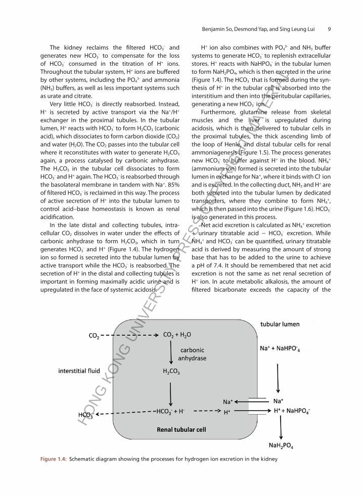
The kidney reclaims the filtered HCO3- and
generates new HCO3- to compensate for the loss
of HCO3- consumed in the titration of H+ ions.
Throughout the tubular system, H+ ions are buffered by other systems, including the PO4
3- and ammonia (NH3) buffers, as well as less important systems such as urate and citrate.
Very little HCO3- is directly reabsorbed. Instead,
H+ is secreted by active transport via the Na+/H+ exchanger in the proximal tubules. In the tubular lumen, H+ reacts with HCO3
- to form H2CO3 (carbonic acid), which dissociates to form carbon dioxide (CO2) and water (H2O). The CO2 passes into the tubular cell where it reconstitutes with water to generate H2CO3 again, a process catalysed by carbonic anhydrase. The H2CO3 in the tubular cell dissociates to form HCO3
- and H+ again. The HCO3- is reabsorbed through
the basolateral membrane in tandem with Na+. 85% of filtered HCO3
- is reclaimed in this way. The process of active secretion of H+ into the tubular lumen to control acid–base homeostasis is known as renal acidification.
In the late distal and collecting tubules, intra-cellular CO2 dissolves in water under the effects of carbonic anhydrase to form H2CO3, which in turn generates HCO3
- and H+ (Figure 1.4). The hydrogen ion so formed is secreted into the tubular lumen by active transport while the HCO3
- is reabsorbed. The secretion of H+ in the distal and collecting tubules is important in forming maximally acidic urine and is upregulated in the face of systemic acidosis.
H+ ion also combines with PO43- and NH3 buffer
systems to generate HCO3- to replenish extracellular
stores. H+ reacts with NaHPO4- in the tubular lumen
to form NaH2PO4, which is then excreted in the urine (Figure 1.4). The HCO3
- that is formed during the syn-thesis of H+ in the tubular cell is absorbed into the interstitium and then into the peritubular capillaries, generating a new HCO3
- ion.Furthermore, glutamine release from skeletal
muscles and the liver is upregulated during acidosis, which is then delivered to tubular cells in the proximal tubules, the thick ascending limb of the loop of Henle, and distal tubular cells for renal ammoniagenesis (Figure 1.5). The process generates new HCO3
- to buffer against H+ in the blood. NH4+
(ammonium ion) formed is secreted into the tubular lumen in exchange for Na+, where it binds with Cl- ion and is excreted. In the collecting duct, NH3 and H+ are both secreted into the tubular lumen by dedicated transporters, where they combine to form NH4
+, which is then passed into the urine (Figure 1.6). HCO3
- is also generated in this process.
Net acid excretion is calculated as NH4+ excretion
+ urinary titratable acid − HCO3- excretion. While
NH4+ and HCO3
- can be quantified, urinary titratable acid is derived by measuring the amount of strong base that has to be added to the urine to achieve a pH of 7.4. It should be remembered that net acid excretion is not the same as net renal secretion of H+ ion. In acute metabolic alkalosis, the amount of filtered bicarbonate exceeds the capacity of the
Figure 1.4: Schematic diagram showing the processes for hydrogen ion excretion in the kidney
HONG
KON
G UN
IVER
SITY
PRE
SS C
OPYR
IGHT
MAT
ERIA
L

10 Normal Structure and Function of the Kidneys
Figure 1.5: Schematic diagram showing the process of ammoniagenesis and secretion of ammonium ion in proximal renal tubular cells
Figure 1.6: Schematic diagram showing the process for ammonium excretion in the collecting ducts
HONG
KON
G UN
IVER
SITY
PRE
SS C
OPYR
IGHT
MAT
ERIA
L

Benjamin So, Desmond Yap, and Sing Leung Lui 11
proximal tubular reabsorption and titration by distal tubular secretion of H+, resulting in bicarbonaturia and a high urine pH. Net renal secretion of H+ ion is positive to titrate against the large HCO3
- load, but net acid excretion is negative.
Factors affecting hydrogen ion secretion in the proximal tubules
1. Carbonic anhydrase activity: inhibition of carbonic anhydrase activity limits the conver-sion of CO2 and H2O to H2CO3, a critical step in H+ secretion and HCO3
- reabsorption; thus carbonic anhydrase inhibitors such as acetazolamide may cause metabolic acidosis.
2. K+ store: chronic K+ depletion leads to intracel-lular acidosis, which increases H+ secretion.
3. Glutamine metabolism: acute acidosis alters the intra-renal metabolism of glutamine, such that renal ammoniagenesis is increased several-fold to cope with the acute load of acid.
4. pCO2 in peritubular blood: elevated pCO2, includ-ing that due to respiratory acidosis, increases the availability of intracellular CO2 that can be converted to H2CO3, which then forms HCO3
- and H+ to be secreted into the tubular lumen.
5. Extracellular fluid volume: as Na+ is exchanged for H+ in the proximal tubules, enhanced Na+ reabsorption in face of a low effective circulating volume is associated with increased H+ secretion and HCO3
- reabsorption, and thus metabolic alkalosis.
6. Hormonal influences: adrenergic agonists and angiotensin II can stimulate HCO3
- reabsorption/H+ secretion in acute acidosis, while PTH inhibits HCO3
- reabsorption/H+ secretion. In chronic acidosis, endothelin-1 is upregulated leading to enhanced Na+/H+ exchange and thus increased H+ secretion; and glucocorticoids may also play a role in promoting HCO3
- reabsorption.
Factors affecting hydrogen ion secretion in the distal nephron
1. Intracellular or extracellular acidosis: the mecha-nisms affecting H+ ion secretion is similar to that in proximal renal tubules.
2. Transepithelial potential difference: the apical H+-ATPase responsible for H+ secretion is very sensitive to electrochemical gradients. Thus, increased lumen-negative voltage, such as that caused by increased sodium reabsorption or the presence of non-resorbable anions, will promote H+ secretion.
3. Hormonal influences: mineralocorticoids such as aldosterone stimulate H+ secretion in the distal nephron. The role of angiotensin II and endothe-lin-1 in distal renal tubular H+ secretion has also been reported.
HONG
KON
G UN
IVER
SITY
PRE
SS C
OPYR
IGHT
MAT
ERIA
L

Introduction
Glomerulonephritis (GN) is an important cause of various forms of renal dysfunction including nephritic syndrome, nephrotic syndrome, and chronic kidney disease (CKD). This chapter provides an overview of the clinical characteristics, pathophysiology, and management of important primary GNs.
Immunoglobulin A Nephropathy
Clinical characteristics
In 1968, the French pathologists Jean Berger and Nicole Hinglais first described 25 patients with recurrent haematuria and mesangial IgA deposits that surmounted IgG deposits. The eponym ‘Berger’s Disease’ was introduced in 1973, and by 1975 the defining features of this condition became apparent and the term IgA nephropathy (IgAN) was commonly used thereafter.
IgAN is still the most common primary glomeru-lonephritis worldwide. The prevalence of IgAN also differs across populations and geographic locations. IgAN is most common in East Asia, less prevalent in Europe, and infrequent in Africa. In Hong Kong, IgAN accounts for approximately 30% of all primary glomerular diseases. Within Europe, IgAN is most prevalent in the northern countries. The prevalence and genetic risk increase for native populations as the distance from Africa increases northwards and eastwards. Prevalence rates are much lower in the United Kingdom, Canada, and the United States. In North America, the disease is twice as common in males as in females, whereas in Asia the gender ratio is roughly equal. The spectrum of presentation is
8
Glomerulonephritis
Desmond Yap, Sydney Tang, Kar Neng Lai, Alex Tang, Kwok Wah Chan, and Tak Mao Chan
highly variable and ranges from asymptomatic urine abnormality such as microscopic haematuria, to episodic gross haematuria, to CKD with proteinuria and hypertension.
The incidence of IgAN is highest in the second and third decades of life. The first episode of macro-scopic haematuria generally occurs between 15 and 30 years of age. Not infrequently, patients may first present with macroscopic haematuria complicating mucosal infection (respiratory or gastrointestinal), and the former is often described as ‘synpharyngitic macrohaematuria’. Macrohaematuria occurs shortly (within 12–72 hours) following the pharyngitic episode and is sometimes accompanied by loin pain. The urine colour is red or brown, but seldom contains clots. Asymptomatic microscopic haematuria is a more common presentation than macrohaematuria, especially in Asian populations, and is often detected with health screening. Urine microscopy reveals dys-morphic red blood cells and red cell casts.
Proteinuria, when present, tends to fluctuate within a narrow range for most patients. Proteinuria is usually not heavy and < 30% have proteinuria exceeding 1 g/day. A transient increase of proteinuria occurs with gross haematuria complicating mucosal infection or urinary tract infection. In a small group of patients, proteinuria reaches the nephrotic range (> 3.5 g/24 hours) and kidney histology typically shows ultrastructural features of minimal change disease but with mesangial IgA deposits. This is referred to as ‘an overlapping syndrome of IgAN and minimal change disease (MCD)’. This entity occurs more frequently in children and clinically resembles MCD, responding well to corticosteroids.
Acute kidney injury (AKI) is an uncommon pres-entation and the pathology is frequently associated
HONG
KON
G UN
IVER
SITY
PRE
SS C
OPYR
IGHT
MAT
ERIA
L

60 Glomerulonephritis
with extensive crescent formation. Around 15% of patients have significant renal impairment (CKD stage 3 or higher) at first presentation. Occasionally, IgAN may present with advanced CKD that requires prompt initiation of renal replacement therapy.
A subset of patients, particularly children, manifest a vasculitic form of illness. IgA vasculitis (IgAV), formerly Henoch-Schönlein purpura, is a form of vasculitis marked by IgA deposition within the blood vessels of affected tissues. IgAV commonly affects the small blood vessels of the skin, joints, intestines, and kidneys, leading to a tetrad of palpable purpura mostly in the lower extremities without platelet or coagulation disorder, arthralgia, abdominal pain, and kidney disease. Rarely, it can affect the lungs and central nervous system. It is the most common form of vasculitis in children. When IgAV occurs in children younger than 16 years, it is often self-limiting. Adults may have more severe and relapsing disease. Kidney involvement in IgAV is his-topathologically indistinguishable from that seen in the kidney-limited disease of IgAN.
The prognosis of IgAN is variable. Some patients have only a single haematuric episode, others have repeated exacerbations. Overall, IgAN pursues a slow but relentless clinical course with consequent kidney failure in 30–40% of patients within 20–30 years after clinical presentation. The percentage of patients who will go into renal failure is roughly the same as the duration of the disease in years from the time of diagnosis. Overall, however, there is a wide range of interindividual variability in the disease course, and specific factors that affect progression to end-stage kidney disease (ESKD) are poorly understood. Even among patients with apparently good prognostic markers, such as normal renal function, blood pressure, and minimal proteinuria on presentation, up to one-third could develop significant proteinu-ria and CKD upon prolonged follow-up in Chinese cohorts, whereas only 4% of similar low-risk patients progressed over a 15-year period in a Spanish cohort, highlighting a genetic difference in disease progression.
As such, predicting clinical outcomes for IgAN remains an imprecise process. There are clinico-pathological features that are generally, but not universally, accepted as indicating a less favourable prognosis in patients with preserved renal function at diagnosis (Table 8.1).
Pathogenesis and histopathological features
Accumulating evidence suggests a strong heritable component to IgAN. This includes numerous reports of familial aggregation of IgAN from the 1980s, and more recently the observation that with increasing distance from Africa, there is increasing genetic pre-disposition to IgAN, with significant west-to-east and south-to-north risk gradients. Genome-side associa-tion studies have been performed in Caucasian and Chinese populations, revealing different risk alleles of IgAN, including those involved in adaptive and innate immunity, glycosylation of IgA1, the renin-angiotensin system, and the human leukocyte antigen (HLA) molecules HLA-DQ and HLA-DR.
The primary defect of IgAN seems to lie in the structure of the IgA molecule, rather than in the kidney. Human IgA may be monomeric (mIgA) or polymeric (pIgA, in which two or four IgA mol-ecules are joined by the bridging protein J chain that is essential for pIgA assembly). There are two subclasses, IgA1 and IgA2, whose functional distinc-tions are not well understood. The mucosal immune system produces both pIgA1 and pIgA2, which reach mucosal surfaces as secretory IgA (pIgA + secretory component) by transepithelial transport. Serum IgA is mostly marrow-derived monomeric IgA1.
A working hypothesis is that patients with IgAN have inherited defects in B cells producing
Table 8.1: Commonly accepted indicators of a worse prognosis in IgA nephropathy
Demographic Male sex Older age at diagnosis Obesity
Clinical No history of macroscopic haematuria Persistent microscopic haematuria Persistent hypertension
Biochemical Proteinuria persistently > 1 g/day Hyperuricaemia
Histological (light microscopy): Mesangial hypercellularity Focal segmental glomerular sclerosis Endocapillary cellular proliferation Capillaritis Interstitial fibrosis/tubular atrophy Crescents Thrombotic microangiopathy Loss of podocytes
HONG
KON
G UN
IVER
SITY
PRE
SS C
OPYR
IGHT
MAT
ERIA
L

62 Glomerulonephritis
Due to the high variability of LM findings, the ‘Oxford Classification of IgA Nephropathy’, first developed in 2009, showed that four glomerular and parenchymal parameters possess reproducible and independent predictive values on renal outcomes: mesangial hypercellularity (M), endocapillary pro-liferation (E), segmental glomerulosclerosis (S), and tubular atrophy/interstitial fibrosis (T). The histologic classification was further refined to MEST-C scores with the incorporation of C (crescents) lesions for crescentic IgAN. Nowadays, kidney histology reports across the world use this classification.
Management
There has been no approved specific therapy for IgAN and treatment is largely symptomatic, aiming at control of blood pressure to < 125/75 mmHg, pro-teinuria, and preservation of renal function. Lifestyle measures, including a low-salt diet, weight reduction, smoking cessation, and avoidance of nephrotoxins, are important initial approaches.
Treatment is based on the use of angiotensin-converting enzyme inhibitor or angiotensin receptor blocker for patients with proteinuria with or without elevated blood pressure control. The addition of aliskiren (a direct renin inhibitor) on top of losartan has a further antiproteinuric effect, though the devel-opment of hyperkalaemia limits this combination in patients with moderate CKD. The addition of high-dose corticosteroids to supportive care in selected patients with risk factors for CKD progression has been shown to confer at best marginal benefits, but was associated with significant treatment-related adverse events. There is little convincing evidence for additional benefits from cytotoxic or other immunomodulatory agents, except for CYC in cres-centic IgAN. Mycophenolate mofetil (MMF) has been reported to be efficacious only in Chinese patients, and could be considered for steroid-sparing for patients in whom high-dose corticosteroids are to be commenced. Tonsillectomy, with or without cor-ticosteroids, was associated with improved kidney survival and/or haematuria/proteinuria in Japanese studies.
For patients who progress to kidney failure, transplantation offers the best potential for full rehabilitation. After transplantation, mesangial IgA deposition has been shown to recur in 20–60% of grafts. Recurrent IgAN is associated with progressive loss of allograft function in about 10%.
Minimal Change Disease
Clinical characteristics
MCD is an important cause of nephrotic syndrome. MCD is the most common cause of nephrotic syndrome in children, accounting for over 90% of all cases. It is also responsible for 10–25% of nephrotic syndrome in adults. MCD patients usually present with bilateral lower limb or even generalized oedema (e.g. peri-orbital swelling, scrotal swelling, or anasarca in severe cases). Proteinuria in MCD is typi-cally heavy and rapid-onset, patients generally do not show overt renal dysfunction. Nonetheless, acute kidney injury can occur in some older adults with MCD. Due to heavy proteinuria, most MCD patients have hypogammaglobulinaemia (especially IgG) and severe dyslipidaemia. MCD shows association with viral illness (e.g. upper respiratory infections), drugs (e.g. non-steroidal anti-inflammatory drugs) or haematological disorders (e.g. non-Hodgkin’s lymphoma) (Table 8.2). Relapses are common in adult patients with MCD. Prognosis of MCD is gener-ally favourable, with low risk of progression to CKD or ESKD.
Table 8.2: Common clinical conditions that are associated with minimal change disease
Examples
Infections • Upper respiratory infection, syphilis, HIV
Drugs • NSAIDs, lithium, bisphosphonates
Malignancy • Non-Hodgkin’s lymphoma, leukaemia
HIV, human immunodeficiency virus; NSAIDs, nonsteroidal anti-inflammatory drugs
Pathogenesis and histopathological features
The pathogenesis of MCD remains obscure and is postulated to be related to aberrant T-cell responses. LM typically shows normal glomerular morphology (Figure 8.4), which is accompanied by a negative IF staining. The pathognomonic EM features include extensive effacement of foot processes, vacuolation, and the appearance of microvilli in the podocytes (Figure 8.5).
Management
Since MCD is the most common cause of nephrotic syndrome in children, empirical high-dose corti-costeroids (60 mg/m2/day) can be initiated without the need for kidney biopsy. Kidney biopsy, however,
HONG
KON
G UN
IVER
SITY
PRE
SS C
OPYR
IGHT
MAT
ERIA
L

Desmond Yap, Sydney Tang, Kar Neng Lai, Alex Tang, Kwok Wah Chan, and Tak Mao Chan 63
should be considered in children with clinical features atypical of MCD (Table 8.3). Children tend to show better and more rapid response to corticosteroids compared with adults. In this context, many children with MCD can be tapered off from corticosteroids after approximately six months. For children with steroid-dependent or frequently relapsing MCD, sec-ond-line treatments such as alkylating agents (e.g. cyclophosphamide [CYC] or chlorambucil), calcineu-rin inhibitors (CNI), or levamisole can be considered. Growth retardation remains an important concern in children receiving high doses or repeated courses of corticosteroids. Alkylating agents should also be used with caution in children due to their long-term toxicities. Side eff ects of levamisole include neutro-penia, skin rashes, and hepatotoxicity.
Table 8.3: Clinical features atypical of minimal change disease in children
• Age of onset < 1 year old or > 12 years old• Steroid resistance or subsequent failure to respond to
corticosteroids in steroid-sensitive nephrotic syndrome• Family history of nephrotic syndrome• Presence of other extra-renal manifestations (e.g. skin
rash, joint pain)• Presence of features suggestive of nephritic syndrome
(e.g. active urine sediments, hypertension, renal insuffi ciency)
Compared with children, adults have a much wider spectrum of diseases that can cause nephrotic syndrome and therefore a kidney biopsy is required to confi rm a diagnosis of MCD. In general, the dosage of corticosteroids used for adults is lower than that for children. Corticosteroids are commenced at 0.8–1 mg/kg/day (up to 80 mg/day) and are slowly tapered over six months. Appropriate prophylaxis for pneumocystis jiroveci (e.g. cotrimoxazole) and hepatitis B virus (HBV) (e.g. antiviral in those at risk of HBV reactivation), and gastroprotective agents (e.g. proton pump inhibitors) should be initiated in patients who receive high doses of corticosteroids. In patients who show poor response to corticoster-oids, a repeat kidney biopsy should be performed to exclude focal segmental glomerulosclerosis. Up to 50% of adult MCD patients relapse after being completely weaned off corticosteroids. Adult MCD patients with a partial response or relapse can be managed with a course of CYC or prolonged CNI treatment. CYC has the advantage of conferring more sustained remission in steroid-dependent or frequently relapsing MCD, but long-term treatment-related side eff ects such as ovarian failure and
Figure 8.4: Minimal change disease. The glomerulus is unremarkable by light microscopy (H&E, ×400, Dr A. H. N. Tang).
Figure 8.5: Minimal change disease. Extensive foot process effacement (arrows) and microvillous transfor-mation of podocytes are present (transmission electron microscopy, ×5000, Dr G. S. W. Chan).
HONG
KON
G UN
IVER
SITY
PRE
SS C
OPYR
IGHT
MAT
ERIA
L

64 Glomerulonephritis
Pathogenesis and histopathological features
FSGS can be classified as primary or secondary, depending on the underlying aetiology (Table 8.4). The pathogenesis of primary (idiopathic) FSGS remains unclear, though some evidence suggests that it might be related to a circulating factor (CF). Proposed candidates for this CF include the serum urine-type plasminogen activator receptor, cardio-trophin-like cytokine factor 1, apoA1b (an isoform of ApoA1), and anti-CD40 antibodies. Secondary FSGS can be related to genetic defects, infections, drugs, or maladaptive structural–functional responses. Typical LM findings are segmental solidification involving any portion of a glomerular tuft (Figure 8.6). The glomerular capillaries are obliterated by matrix substances, often accompanied by hyalinosis, endocapillary foam cells, and wrinkling of the glo-merular basement membrane. IF is usually negative or with limited IgM and C3 staining in sclerotic areas. EM shows wrinkling or retraction of the glomerular basement membrane and extensive podocyte foot process effacement, with no electron-dense deposits. FSGS has different histological variants (Table 8.5) and each is associated with its distinct clinical behaviour and prognosis. The collapsing variant is highly resistant to immunosuppressive treatments and is associated with rapid progression to ESKD. The tip-lesion variant is more responsive to immunosuppressive treatments and has relatively low risk of ESKD. Similarly, the cellular variant is also responsive to immunosuppression and shows inter-mediate outcomes compared to the tip-lesion and collapsing variants. The perihilar variant is often seen in FSGS patients due to reduced nephron mass, and is frequently associated with glomerulomegaly. While classic FSGS (also known as ‘FSGS not otherwise specified’) is the most common histological variant, its prognostic significance remains undefined.
increased risk of malignancy remain important concerns. Cyclosporin A (CYA) or tacrolimus (TAC) are both viable choices of CNI in MCD. Chronic CNI nephrotoxicity and the considerable rates of relapse upon drug withdrawal are potential problems of CNI treatment. Careful monitoring of CNI exposure and renal function can help lower the risk of nephrotoxic-ity of long-term CNI administration. There is also data to suggest that mycophenolate may be used for ster-oid-sparing and reducing disease relapse. Emerging evidence also shows the efficacy of anti-CD20 treatment in patients with refractory or frequently relapsing disease. Hepatitis B viral status must be checked prior to anti-CD20 administration and the appropriate prophylaxis given as stated above.
Focal Segmental Glomerulosclerosis
Clinical characteristics
Focal segmental glomerulosclerosis (FSGS) is an important cause of nephrotic syndrome and renal failure in children and adults. Histological features of FSGS can be detected in 30–40% of kidney biopsies performed in patients with proteinuria or nephrotic syndrome. The majority of children with FSGS present with nephrotic-range proteinuria, while adult FSGS patients may show either nephrotic- or subne-phrotic-range proteinuria at the onset of disease. Hypertension is observed in 30–50% of patients with FSGS. Up to 20–30% of patients with FSGS also show evidence of renal impairment at presentation, and about 25–75% have microscopic haematuria at the time of diagnosis. FSGS is associated with a significantly higher risk of ESKD compared with other primary nephrotic glomerular diseases, and the risk of progression to ESKD is influenced by the severity of renal disease at presentation, ethnicity, histological variants, and response to treatments. Only a small proportion of patients have spontane-ous remission while the majority of primary FSGS patients will experience a progressive increase in proteinuria and renal function decline. Up to half of the children and adult FSGS patients who do not respond well to treatment will develop ESKD after five years of diagnosis. FSGS can recur after kidney transplantation, with recurrence rates of around 30% after the first kidney transplantation and 85%–100% after the second kidney transplantation. Recurrent FSGS manifests as nephrotic syndrome early after transplantation (usually within one month) and is associated with rapid allograft loss.
Table 8.4: Primary and secondary causes of FSGS
Primary (idiopathic) FSGS
• Postulated to be related to circulating factor
Secondary FSGS
• Genetic causes (e.g. APOL1)• Maladaptive responses (e.g. reduced nephron mass such
as low birth weight, single kidney, reflux disease, morbid obesity, cyanotic heart disease)
• Infection (e.g. HIV, parvovirus B19)• Drugs (e.g. pamidronate, lithium)
FSGS, focal segmental glomerulosclerosis; HIV, human immunodeficiency virus
HONG
KON
G UN
IVER
SITY
PRE
SS C
OPYR
IGHT
MAT
ERIA
L

Desmond Yap, Sydney Tang, Kar Neng Lai, Alex Tang, Kwok Wah Chan, and Tak Mao Chan 65
Management
Patients with primary FSGS and subnephrotic-range proteinuria should be treated with renin-angioten-sin-aldosterone systems (RAAS) blocking agents. Immunosuppressive therapies are indicated in primary FSGS patients with nephrotic syndrome. The mainstay of treatment is high dosage of oral corticosteroid (1 mg/kg/day) for an initial period of 4–8 weeks, with subsequent tapering of the dosage. The cumulative remission rates range between 40 and 60%. Alkylating agents (chlorambucil or CYC) can be considered in steroid-resistant FSGS. There is evidence to show that the use of corticosteroids combined with CNI is superior to corticosteroids alone in preserving renal function. Corticosteroids and MMF have also been reported to be effective in the treatment of FSGS. Plasma exchange is indicated in patients with recurrent FSGS after transplantation. There are also anecdotal reports on the efficacy of biologics (e.g. anti-CD20) in refractory FSGS and recurrent FSGS, but data from prospective rand-omized clinical trials are still lacking.
Membranous Nephropathy
Clinical characteristics
Membranous nephropathy (MN) is a common cause of nephrotic syndrome in adults, and can be detected in up to 30% of kidney biopsies from adult patients who present with proteinuria or nephrotic syndrome. MN is relatively uncommon in children, accounting for 2–12% of cases that present with nephrotic syndrome. MN is characterized by the insidious onset of nephrotic- or subnephrotic-range proteinuria. About 80% of patients show overt nephrotic syndrome at the time of presentation. MN patients occasionally show microscopic haematuria and exhibit variable degrees of renal impairment. MN patients can also suffer from complications of nephrotic syndrome such as thromboembolic events or abnormal lipid profiles. Other systemic features may be present in patients with secondary MN due to autoimmune or neoplastic disorders. Approximately 25% of MN patients have spontane-ous complete remission, but this can take quite a long time—several months or even years. Another 25% of patients have persistent proteinuria without loss of renal function. Of patients with nephrotic syndrome who are left untreated, up to 50% will have a progressive decline in renal function and eventually develop ESKD. Roughly a quarter of MN
Figure 8.6: Focal segmental glomerulosclerosis (FSGS). There is segmental sclerosis of the glomerulus (arrow) with hyalinosis and adhesion to the Bowman’s capsule (PASD, ×400, Dr A. H. N. Tang).
Table 8.5: Clinical behaviour and prognosis of different histological variants of focal segmental glomerulosclerosis
Histological variants Clinical behaviour and prognosis
Classic FSGS (NOS) variant
• The most common form of FSGS• The prognostic significance
remains undefined
Tip lesion variant • More responsive to immunosuppressive treatments
• Relatively lower risk of progression to ESKD
Cellular variant • Responsive to immunosuppressive treatments
• Intermediate renal prognosis between tip lesion and collapsing variants
Perihilar variant • Often seen in FSGS due to reduction in nephron mass
Collapsing variant • Association with HIV infection• Highly resistant to
immunosuppressive treatments• Rapid and high rates of
progression to ESKD
ESKD, end-stage kidney disease; FSGS, focal segmental glo-merulosclerosis; HIV, human immunodeficiency virus; NOS, not otherwise specifiedHO
NG K
ONG
UNIV
ERSI
TY P
RESS
COP
YRIG
HT M
ATER
IAL
68 Glomerulonephritis
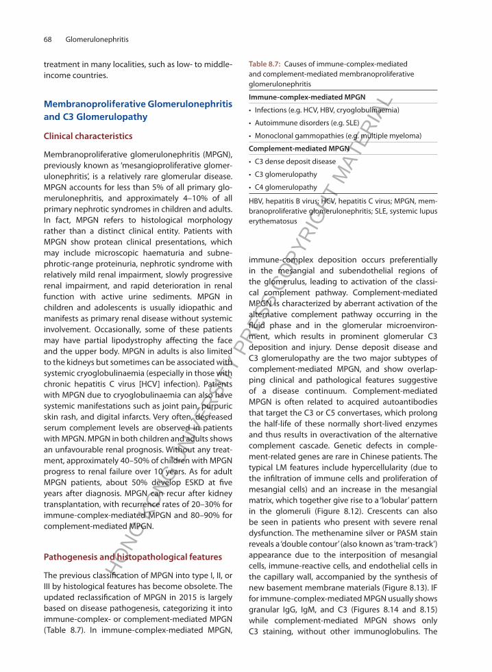
immune-complex deposition occurs preferentially in the mesangial and subendothelial regions of the glomerulus, leading to activation of the classi-cal complement pathway. Complement-mediated MPGN is characterized by aberrant activation of the alternative complement pathway occurring in the fluid phase and in the glomerular microenviron-ment, which results in prominent glomerular C3 deposition and injury. Dense deposit disease and C3 glomerulopathy are the two major subtypes of complement-mediated MPGN, and show overlap-ping clinical and pathological features suggestive of a disease continuum. Complement-mediated MPGN is often related to acquired autoantibodies that target the C3 or C5 convertases, which prolong the half-life of these normally short-lived enzymes and thus results in overactivation of the alternative complement cascade. Genetic defects in comple-ment-related genes are rare in Chinese patients. The typical LM features include hypercellularity (due to the infiltration of immune cells and proliferation of mesangial cells) and an increase in the mesangial matrix, which together give rise to a ‘lobular’ pattern in the glomeruli (Figure 8.12). Crescents can also be seen in patients who present with severe renal dysfunction. The methenamine silver or PASM stain reveals a ‘double contour’ (also known as ‘tram-track’) appearance due to the interposition of mesangial cells, immune-reactive cells, and endothelial cells in the capillary wall, accompanied by the synthesis of new basement membrane materials (Figure 8.13). IF for immune-complex-mediated MPGN usually shows granular IgG, IgM, and C3 (Figures 8.14 and 8.15) while complement-mediated MPGN shows only C3 staining, without other immunoglobulins. The
treatment in many localities, such as low- to middle-income countries.
Membranoproliferative Glomerulonephritis and C3 Glomerulopathy
Clinical characteristics
Membranoproliferative glomerulonephritis (MPGN), previously known as ‘mesangioproliferative glomer-ulonephritis’, is a relatively rare glomerular disease. MPGN accounts for less than 5% of all primary glo-merulonephritis, and approximately 4–10% of all primary nephrotic syndromes in children and adults. In fact, MPGN refers to histological morphology rather than a distinct clinical entity. Patients with MPGN show protean clinical presentations, which may include microscopic haematuria and subne-phrotic-range proteinuria, nephrotic syndrome with relatively mild renal impairment, slowly progressive renal impairment, and rapid deterioration in renal function with active urine sediments. MPGN in children and adolescents is usually idiopathic and manifests as primary renal disease without systemic involvement. Occasionally, some of these patients may have partial lipodystrophy affecting the face and the upper body. MPGN in adults is also limited to the kidneys but sometimes can be associated with systemic cryoglobulinaemia (especially in those with chronic hepatitis C virus [HCV] infection). Patients with MPGN due to cryoglobulinaemia can also have systemic manifestations such as joint pain, purpuric skin rash, and digital infarcts. Very often, decreased serum complement levels are observed in patients with MPGN. MPGN in both children and adults shows an unfavourable renal prognosis. Without any treat-ment, approximately 40–50% of children with MPGN progress to renal failure over 10 years. As for adult MPGN patients, about 50% develop ESKD at five years after diagnosis. MPGN can recur after kidney transplantation, with recurrence rates of 20–30% for immune-complex-mediated MPGN and 80–90% for complement-mediated MPGN.
Pathogenesis and histopathological features
The previous classification of MPGN into type I, II, or III by histological features has become obsolete. The updated reclassification of MPGN in 2015 is largely based on disease pathogenesis, categorizing it into immune-complex- or complement-mediated MPGN (Table 8.7). In immune-complex-mediated MPGN,
Table 8.7: Causes of immune-complex-mediated and complement-mediated membranoproliferative glomerulonephritis
Immune-complex-mediated MPGN
• Infections (e.g. HCV, HBV, cryoglobulinaemia)
• Autoimmune disorders (e.g. SLE)
• Monoclonal gammopathies (e.g. multiple myeloma)
Complement-mediated MPGN
• C3 dense deposit disease
• C3 glomerulopathy
• C4 glomerulopathy
HBV, hepatitis B virus; HCV, hepatitis C virus; MPGN, mem-branoproliferative glomerulonephritis; SLE, systemic lupus erythematosus
HONG
KON
G UN
IVER
SITY
PRE
SS C
OPYR
IGHT
MAT
ERIA
L

70 Glomerulonephritis
classical EM findings of immune-complex-mediated MPGN are electron-dense deposits detected pre-dominantly in the subendothelial regions (Figure 8.16). Complement-mediated MPGN (e.g. dense deposit disease) show highly electron-dense bands of homogenous substances occupying the glomeru-lar basement membrane.
Management
Patients with a histological diagnosis of MPGN should be examined for underlying causes, such as chronic viral hepatitis infection, autoimmune diseases, or monoclonal gammopathies. More detailed investigation of the complement cascade should be considered in very young patients, those with significant family histories, or histological features suggestive of complement-mediated MPGN. Patients with chronic hepatitis B- or C-associated MPGN should receive antiviral therapies, while those with autoimmune disorders or monoclonal gammopathies should be managed accordingly. Patients with HCV-associated cryoglobulinaemia and MPGN may also benefit from anti-CD20 treat-ment. All MPGN patients should have optimal blood pressure and proteinuria control by RAAS-blocking agents. Children or adult idiopathic MPGN patients with nephrotic syndrome and progressive decline in renal function can be treated with corticosteroids and CYC or MMF for not more than six months. Pulse corticosteroids can be used when patients show rapidly deteriorating renal function or crescentic formations in the kidney biopsy. The efficacy of ecu-lizumab (anti-C5b monoclonal antibody) has been reported, but there is insufficient data overall to recommend these biologics as first-line treatments in complement-mediated MPGN.
HONG
KON
G UN
IVER
SITY
PRE
SS C
OPYR
IGHT
MAT
ERIA
L

Introduction
Chronic kidney disease (CKD) is associated with various systemic complications and increased patient mortality. This chapter provides an overview of the causes and natural history of CKD and also highlights the pathophysiology and management of some common and important CKD complications.
Definitions of Chronic Kidney Disease
The Kidney Disease: Improving Global Outcomes (KDIGO) guidelines define CKD as abnormalities of kidney structure or function, present for over three months, with implications for health. CKD is distin-guished from acute kidney injury (AKI) primarily by its chronicity. CKD often has different aetiologies, requires different interventions, and is associated with a distinct set of short- and long-term clinical outcomes.
Evidence of kidney damage can be subtle, espe-cially at early stages. For the purposes of diagnosis of CKD, such evidence can manifest as either:
– abnormalities in various markers of kidney damage, such as but not limited to: albu-minuria, microscopic haematuria and other abnormalities in the urinary sediment, struc-tural anomalies on imaging (such as evidence of polycystic kidneys), aberrant findings on histology, or evidence of electrolyte or acid–base disorders suggestive of renal tubular disorders;
– persistently decreased glomerular filtration rate (GFR) to less than 60 mL/min/1.73 m2.
15
Chronic Kidney Disease: Clinical Manifestations and Management
Benjamin So, Desmond Yap, and Sing Leung Lui
The kidney has a myriad of functions includ-ing excretory, endocrine, and metabolic functions. Though measurement of the GFR primarily reflects excretory function, a decline in other kidney func-tions often parallels the decrease in GFR. Systemic complications of CKD, as well as all-cause mortality and other adverse clinical outcomes, also increase progressively with deteriorating GFR. Accordingly, CKD is classified into five stages based on the esti-mated GFR (eGFR) (Table 15.1).
Causes of Chronic Kidney Disease
While CKD is associated with a distinct constellation of clinical sequelae, it is not sufficient as a diagnosis in and of itself. Identification of the underlying cause is important for both prognostication and manage-ment of CKD. In developed countries, the leading causes of CKD in adults nowadays are diabetes mellitus (DM) and hypertension (HT) (see also
Table 15.1: Classification of chronic kidney disease
Stage GFR (mL/min/1.73 m2) Description
1 ≥ 90 Kidney damage with normal or ↑GFR
2 60–89 Kidney damage with mild ↓GFR
3a 45–59 Moderate ↓GFR
3b 30–44
4 15–29 Severe ↓GFR
5 <15 (or requiring dialysis)
Kidney failure
GFR, glomerular filtration rate
HONG
KON
G UN
IVER
SITY
PRE
SS C
OPYR
IGHT
MAT
ERIA
L

130 Chronic Kidney Disease: Clinical Manifestations and Management
Chapter 14). While this is also the case in Hong Kong, there is also a significant disease burden of chronic glomerulonephritis (GN), such as IgA nephropathy, in East Asia. CKD due to systemic autoimmune diseases (e.g. systemic lupus erythematosus), obstructive uropathy, and cystic kidney disease are also impor-tant aetiologies of CKD. In children, unlike in the adult population, congenital anomalies such as renal dysplasia or agenesis, and urological abnormalities such as vesicoureteric reflux are the major causes of CKD (see Chapter 13).
Evaluation of Chronic Kidney Disease
A high index of suspicion is required to identify patients with CKD, especially at earlier stages when renal abnormalities are often clinically silent. There is no evidence to suggest that screening for CKD in otherwise healthy populations alters outcomes meaningfully, but targeted screening for groups at high risk appears to be more worthwhile. In this context, patients with conditions such as DM, HT, rheumatological disease, urological problems, or those requiring chronic use of potentially nephro-toxic drugs such as calcineurin inhibitor (CNI) or non-steroidal anti-inflammatory drugs may benefit from regular screening for the development of CKD.
The current guidelines recommend that CKD be classified according to the cause, GFR, and albumi-nuria category (CGA). This serves as a useful schema for the evaluation of CKD. When a patient is found to have a low calculated GFR, it is important to follow up by observing the evolution of renal function over time to establish chronicity, and to identify a likely cause. Comprehensive evaluation of history, physical examination, blood tests, examination of the urinary sediment, and radiologic imaging can reveal the cause in many cases of CKD, with renal biopsies reserved for selected cases for definitive diagnosis or prognostication. Objective markers of kidney damage, especially albuminuria, should be assiduously sought for, as this not only affects diagnosis and management but also has prognostic significance.
Natural History and Evolution of Chronic Kidney Disease
CKD tends to be progressive over time, irrespective of the underlying cause. Certain kidney diseases such as diabetic nephropathy, may show a more accelerated disease course during the later stages of CKD and
progress rapidly to end-stage kidney disease (ESKD). Glomerulosclerosis, tubular atrophy, and tubuloint-erstitial fibrosis inevitably ensue in patients with established CKD. These pathological changes are due to a series of secondary maladaptive changes in intra-renal haemodynamics, as well as inflammatory and metabolic factors. Complications of CKD, which will be elaborated in subsequent sections, develop in tandem with this process.
As nephrons become scarred or senescent with acute or chronic injury, the remaining nephrons compensate by increasing blood flow and glomeru-lar hyperfiltration to maintain the same GFR. The elevated intraglomerular pressure increases wall stress and endothelial injury, causing chronic glo-merular damage with time. This triggers a complex neurohormonal response, including activation of the renin-angiotensin-aldosterone system (RAAS) and various profibrotic pathways, which contrib-ute to progressive tubulointerstitial fibrosis (see also Chapter 7). Modification of several of these pathways have been shown to attenuate renal injury and fibrosis in animal models, but aside from RAAS blockers, few treatments have been effective in human subjects.
Complications of Chronic Kidney Disease
Complications of CKD can develop in different organ systems as renal function declines. However, signifi-cant symptoms generally do not occur until GFR falls to below 15 mL/min/1.73 m2, when patients begin to develop clinically overt features of the uraemic syndrome. Many patients present for the first time in late-stage disease, when the time window for inter-vention has elapsed and complications have already become established.
Cardiovascular complications
Cardiovascular disease is the leading cause of death at all stages of CKD. Individuals diagnosed with CKD have a ten- to twentyfold increased risk for cardio-vascular death compared to age- and sex-matched controls. CKD is also associated with increased risk for adverse outcomes for patients diagnosed with different cardiovascular conditions, including acute coronary syndrome, valvular disorders, and conges-tive heart failure. Many patients with CKD may have comorbid conditions such as DM and HT, which further aggravates the risk of cardiovascular disease.
HONG
KON
G UN
IVER
SITY
PRE
SS C
OPYR
IGHT
MAT
ERIA
L

Benjamin So, Desmond Yap, and Sing Leung Lui 131
Hypertension and fluid overload
The single most common cardiovascular diagnosis in CKD is HT. HT is both a cause and effect in CKD. Uncontrolled essential or secondary HT can lead to both albuminuria and reduction in GFR with time. Conversely, up to 85% of patients with CKD, regardless of the underlying cause, may have HT. The pathogenesis of HT in CKD is multifactorial; sodium and water retention, and aberrations in neu-rohormonal pathways all play significant roles in the disease process (see also Chapter 10). Of note, the RAAS and sympathetic nervous systems are often active in CKD and serve as logical targets for blood pressure control. The corollary is that the numerous systemic complications of HT, including stroke and coronary artery disease, are also more prevalent in the CKD population. The patient with CKD is less able to cope with an acute sodium load and is prone to developing fluid overload. However, in the absence of other disorders, such as heart failure, liver cir-rhosis, or nephrotic syndrome, significant oedema is uncommon until the patient reaches stage 4 or 5 CKD. In advanced CKD, gross fluid overload can result in peripheral and pulmonary oedema, pleural effu-sions, and occasionally ascites.
Arterial diseases
Coronary artery disease, cerebrovascular disease, and peripheral vascular disease are all more preva-lent in the CKD population. Arterial disease develops via atherosclerosis as well as arteriosclerosis. The former is characterized by deposition of lipid-laden plaques in the arterial intima, especially in the carotid bifurcation, coronary arteries, renal arteries, femoral arteries, and infrarenal aorta, causing pro-gressive arterial occlusion. The latter, arteriosclerosis, is predominantly driven by medial degeneration with distortion of the elastic lamellae and second-ary fibrosis, calcification, and hypertrophy of the media and intima, especially in the aorta and central arteries.
The process of atherosclerosis is enhanced in CKD, accelerating exponentially with decreasing GFR. Part of this is attributable to the significant burden of concomitant cardiovascular risk factors including DM, HT, and dyslipidaemia in patients with CKD. Additionally, altered bone mineral metabolism in CKD further compounds the progression of ather-osclerosis as well as arteriosclerosis. Arteriosclerosis tends to be far more common in advanced CKD than in the general population and portends a more
sinister prognosis than the ‘normal’ arteriosclerosis that occurs with ageing.
Diseases of the myocardium
Left ventricular hypertrophy (LVH) is frequently present in CKD patients. Contributing factors of LVH in CKD patients include chronic volume overload, anaemia, systemic HT, aortic valve calcification, and activation of multiple neurohormonal and fibrotic pathways. LVH leads to diastolic dysfunction and is aggravated by coexisting ischaemic and valvular heart disease. These pathophysiological changes collectively cause a syndrome known as uraemic cardiomyopathy, which is associated with pulmo-nary oedema and cardiac arrhythmias. Patients with CKD often tolerate treatments for heart failure more poorly, show a higher propensity for cardiac decompensation, and require renal replacement therapy (RRT) at an earlier stage due to prominent fluid overload symptoms.
Diseases of the heart valves and pericardium
Calcification of the aortic valve and mitral valve par-allels the process of vascular calcification throughout the whole body. Pericarditis with pericardial effusion occurs in a small percentage of patients with ESKD, often when RRT is not initiated in a timely manner. Pericardiocentesis yields a sterile, exudative pericar-dial fluid with lymphocytic infiltrate. Many patients may be relatively asymptomatic until they present with clinical manifestations of cardiac tamponade. Concomitant bleeding tendency may lead to life-threatening haemopericardium in these patients.
Arrhythmias
Atrial fibrillation (AF) affects up to 20% of pre-dialysis CKD patients. Incident AF is associated with an accel-erated decline in renal function due to clinically silent renal thromboembolism. Anticoagulation for AF may be complicated with an increased risk of haemorrhage in patients with impaired renal function. Sudden cardiac death is common in the CKD population, especially in the dialysis popula-tion. The incidence of sudden cardiac death within the first year of initiating haemodialysis (HD) is around 5–7%. Loop recorder studies show that most episodes are due to bradyarrhythmias and asystole rather than ventricular tachyarrhythmias. As most arrhythmias in patients with advanced CKD tend not to be shockable rhythms, the installation of implanted cardioverter/defibrillators in this high-risk population remains controversial.
HONG
KON
G UN
IVER
SITY
PRE
SS C
OPYR
IGHT
MAT
ERIA
L

132 Chronic Kidney Disease: Clinical Manifestations and Management
Haematologic complications
Anaemia
Anaemia is common in CKD, affecting 20–40% of patients with stage 3 CKD and over 70% of stage 5 patients. Multiple factors contribute to anaemia in CKD patients. Conditions that result in reduced oxygen delivery to peripheral tissues (e.g. anaemia or chronic hypoxaemia) promote stabilization of hypoxia-inducible factor (HIF), which in turn enhances the production of erythropoietin (EPO). The kidney is normally responsible for the production of 90% of EPO in the body. Therefore, decreased EPO production is the key mechanism for renal anaemia and the frequency and severity of anaemia correlate with the reduction in nephron mass. Iron deficiency is another important cause of anaemia in patients with CKD. It is estimated that up to 50% of patients with stage 3 to 5 CKD have iron deficiency, based on the gold standard of bone marrow examination. Iron deficiency in CKD may be either absolute or relative. Absolute iron deficiency occurs in the setting of chronic blood loss (e.g. gastrointestinal bleeding, or blood loss during HD), while relative (functional) iron deficiency is usually due to a supply-demand mismatch in iron stores. Functional iron deficiency is often triggered by the use of erythropoietin-stimulat-ing agents (ESA), as the available store of iron cannot be mobilized fast enough to cope with the acceler-ated rate of red blood cell production. Furthermore, elevated hepcidin (a crucial regulator of iron homeo-stasis) in CKD patients can also lead to functional iron deficiency, and thus renal anaemia and resistance to ESA. Vitamin B12 and folic acid deficiencies can occur in ESKD patients as a result of malnutrition and excessive removal by dialysis. Secondary/tertiary hyperparathyroidism exerts negative effects on EPO production and integrity of red blood cells, and in severe cases may cause secondary myelofibrosis and ESA resistance. Microcytic hypochromic anaemia due to aluminium overload is less commonly seen nowadays due to more restricted use of aluminium-containing phosphate binders.
Bleeding tendency
Increased risk of haemorrhage is observed in CKD patients, especially in those with more severe renal impairment. While patients with severe uraemia often have prolonged bleeding time, the results of other quantitative haematological parameters (e.g. platelet count and coagulation profiles) are generally unremarkable. The increased bleeding tendency in
uraemic patients is thought to be related to platelet dysfunction as result of dysfunctional glycoprotein IIb/IIIa (a platelet membrane glycoprotein that normally interacts with von Willebrand factor and fibrinogen), leading to impaired platelet aggregation and adhesion. A low haematocrit may also contrib-ute to bleeding tendency in CKD.
Electrolyte and acid–base disturbances
Given the pivotal role of the kidney in the regula-tion of electrolyte and acid–base status in the body (see Chapter 1), it logically follows that electrolyte and acid–base disturbances ensue in patients with CKD and ESKD. Salient electrolyte and acid–base abnormalities in CKD and ESKD patients include hyperkalaemia, metabolic acidosis, hypocalcaemia, and hyperphosphataemia.
The incidence of hyperkalaemia increases with reducing GFR, owing to impaired regulation by the distal nephron and concomitant metabolic acidosis. Other precipitating factors for hyperkalaemia in CKD patients include the use of RAAS blockers, potas-sium-sparing diuretics, and CNI. Hyperkalaemia due to hyporeninaemic hypoaldosteronism (type 4 renal tubular acidosis) is common among DM patients, even at earlier stages of CKD.
Metabolic acidosis is also frequently observed in CKD patients as they have compromised capacity for bicarbonate conservation and generation through ammoniagenesis. In earlier stages of CKD, this is compensated for by buffer systems in the extracel-lular fluid, tissues, and bone. Acidaemia usually develops in later stages of CKD when these systems become overwhelmed progressively. The anion gap may remain normal in CKD until late stages, when anions such as phosphate, sulphate, urate, and hip-purate accumulate. Metabolic acidosis can promote protein catabolism and muscle breakdown, and may accelerate CKD progression by induction of tubu-lointerstitial fibrosis.
Mineral and bone disorder
Changes in bone mineral metabolism in CKD
As GFR declines, regulation of calcium and phospho-rus becomes increasingly disturbed as a result of a complex interplay between the various factors that affect bone mineral homeostasis (Figure 15.1). These hormonal factors include vitamin D compounds (such as vitamin D2 and D3, and their 25-hydroxylated
HONG
KON
G UN
IVER
SITY
PRE
SS C
OPYR
IGHT
MAT
ERIA
L

Benjamin So, Desmond Yap, and Sing Leung Lui 133
onwards, when the clinical behaviour and fracture risk of patients begin to diverge from the healthy general population. The skeletal abnormalities in CKD patients can show distinct or mixed pathologies (mixed uraemic osteodystrophy), which have poten-tial implications for management. Bone biopsy is the gold standard for diagnosing skeletal disease in CKD, but is rarely done in practice due to its invasiveness. Instead, the type of bone disease in CKD patients can often be inferred from various laboratory param-eters, including alkaline phosphatase, PTH, vitamin D, and aluminium levels. Some specific tests such as deferoxamine (DFO) test may aid the diagnosis of aluminium-related bone diseases in CKD patients.
Classification of bone histology in renal osteo-dystrophy now follows a ‘turnover, mineralization, volume’ (TMV) system as defined by the National Kidney Foundation. Mechanistically, bone turnover refers to the rate of bone remodelling due to osteoclast-mediated bone resorption and osteo-blast-mediated bone formation, with the generation of an osteoid, which is an extracellular matrix that needs to be further mineralized. Mineralization is the process by which calcium phosphate crystals are deposited into the matrix; this is governed by osteoblasts but can be limited by other factors like vitamin D deficiency or skeletal resistance to PTH. Finally, bone volume represents the end-result of bone resorption, formation, and mineralization. Impaired bone volume is associated with increased bone fragility and susceptibility for fractures.
Osteitis fibrosa cystica is one of the most classical forms of renal osteodystrophy. This is usually attrib-utable to the skeletal effects of elevated PTH, and is characterized by high bone turnover, with increased osteoclastic activity and excess bone resorption, as well as increased osteoblastic activity leading to more rapid formation of osteoid. A bone biopsy may show marrow fibrosis, woven osteoid with disor-dered collagen fibres, and increased osteoclasts and osteoblasts. Clinically, patients may suffer from an escalated risk of bone fracture and tendon rupture, as well as anaemia due to secondary myelofibrosis.
Adynamic bone disease (ABD) may occur in CKD patients and is characterized by low bone turnover together with abnormal mineralization. Osteoclast and osteoblast activity are both depressed, and there is also a consequent defect in mineralization. The amount of osteoid is not increased, as osteoblas-tic activity is low. The most common cause of ABD is excessive suppression of PTH, often related to the use of high doses of vitamin D analogues and cal-cimimetics. Other important causes of ABD include
and 1,25-hydroxylated compounds), parathyroid hormone (PTH) and fibroblast growth factor 23 (FGF23) (see also Chapter 1).
The kidney’s ability to excrete a phosphate load decreases as renal function deteriorates, resulting in phosphate retention. Hyperphosphataemia is a key driving event leading to elevated PTH and FGF23 levels. Increased FGF23 diminishes 1α-hydroxylation of 25-hydroxyvitamin D (calcidiol) to 1,25-hydroxy-vitamin D in renal tubules, resulting in reduced intestinal calcium absorption and hypocalcaemia which further promotes PTH secretion. Secondary hyperparathyroidism in CKD is characterized by excessive PTH production and hyperplasia of para-thyroid glands in response to hypocalcaemia. As it is a compensatory mechanism to hypocalcaemia and hyperphosphataemia, it does not usually result in hypercalcaemia. With persistent severe secondary hyperparathyroidism, the parathyroid glands may become autonomous (i.e. tertiary hyperparathyroidism) and patients often show both hypercalcaemia and hyperphosphataemia. The skeletal and systemic manifestations of altered mineral metabolism in CKD were previously general-ized as ‘renal osteodystrophy’, but the term ‘chronic kidney disease–mineral and bone disorder’ (CKD-MBD) is now preferred. ‘Renal osteodystrophy’ now largely refers to the skeletal pathologies diagnosed from a bone biopsy.
Bone disease in chronic kidney disease
In general, a diagnosis of CKD-MBD is made starting from later stages of CKD, from at least stage 3
Figure 15.1: A schematic diagram showing the patho-genesis of chronic kidney disease-mineral bone disease (CKD-MBD)
HONG
KON
G UN
IVER
SITY
PRE
SS C
OPYR
IGHT
MAT
ERIA
L

Introduction
Successful kidney transplantation offers better survival and quality of life than dialysis in patients with end-stage kidney disease (ESKD). This chapter will provide an overview of transplant immunology, evaluation of renal transplant recipients and donors, use of immunosuppression, and the complications and management of kidney transplantation.
Immunology of Organ Transplantation
The immune system primarily serves to defend against infections related to various pathogens. In the context of organ transplantation, immune responses can lead to rejection of the transplanted organ. Despite the use of various effective immuno-suppressive therapies, acute and chronic rejection remain major causes of kidney allograft loss. Human leukocyte antigens (HLA), expressed on the surface of the donor cells, are the principal molecules that trigger or mediate an immune response during organ transplant rejection.
Human leukocyte antigen system
The human leucocyte antigen (HLA), which cor-responds to the major histocompatibility complex (MHC) in human, is located on chromosome 6p21.3. It plays a pivotal role in enabling the immune system to distinguish between ‘self’ and ‘non-self’. The clas-sical HLA Class I antigens include HLA-A, HLA-B, and HLA-C while classical HLA Class II includes HLA-DR, HLA-DQ, and HLA-DP. HLAs are largely responsible for adaptive immunity and govern the rejection of
18
Kidney Transplantation: Principles and Practice
Maggie Ma, Simon C. Y. Cheung, Cindy Choy, Gavin Chan, Janette Kwok, and Tak Mao Chan
allogeneic transplantation. HLA Class I proteins are expressed in most cell types and they are heterodi-mers that consist of α and β2-microglobulin chains (Figure 18.1). They load intracellular antigens (self or foreign antigens, e.g. viral peptides) in the endo-plasmic reticulum and subsequently translocate to the cell surface. HLA Class II proteins are mainly expressed on professional antigen-presenting cells (APC) such as B cells and dendritic cells. They are also heterodimers that consist of α and β chains and are responsible for presenting extracellular antigens (Figure 18.1).
Allo-recognition
Allo-recognition is the process by which T lym-phocytes recognize the foreign antigens on the transplanted organ. T cells recognize the presented antigens via T-cell receptors. T-cell allo-recognition can be direct or indirect. The former recognizes the unprocessed allogeneic HLA molecule presented by donor APC, while the latter recognizes the pro-cessed allo-peptide presented by recipient APC in the context of self (recipient) HLA (Figure 18.2). CD8+ cytotoxic T cells recognize the antigens presented by HLA Class I and exert cytotoxicity to the cells. On the contrary, CD4+ helper T cells recognize antigens presented by HLA Class II and provide help to B cells to produce antibodies.
T-lymphocyte activation
Activation of T lymphocytes is the prelude to rejection, and involves three important signalling
HONG
KON
G UN
IVER
SITY
PRE
SS C
OPYR
IGHT
MAT
ERIA
L

Maggie Ma, Simon C. Y. Cheung, Cindy Choy, Gavin Chan, Janette Kwok, and Tak Mao Chan 163
pathways. The binding of allo-peptide/HLA mol-ecules on APC to the TRC/CD3 complex on T-cell surfaces constitutes the first signal. This is followed by the synthesis of the second messengers, which leads to an increase in intracellular calcium level and activation of calcium-dependent phosphatase which is crucial to cytokine gene induction. Calcineurin is a calcium-calmodulin complex-dependent phos-phatase that plays a key role in T-cell activation. The second signal is the non-antigen specific costimula-tory signal of engagement of the B7 on APCs with the CD28 on T cells. These signals lead to the induc-tion of cytokine genes, most notably the production of IL-2 and increased expression of IL-2 receptors. The binding of these cytokines to their respective receptors provides the third signal, leading to the progression of T-cell activation, cell proliferation, and clonal expansion.
Role of B lymphocytes
Activated CD4+ T helper cells interact with B cells to trigger humoral immune responses, i.e. production of specific antibodies targeting exposed antigens. It is increasingly recognized that alloantibodies can be major effectors of both acute and chronic immune-mediated injury in the graft kidney. Alloantibodies are mostly directed against HLAs but can also target other kinds of antigens (e.g. endothelial or epithelial antigens).
Evaluation of Kidney Transplant Recipients and Donors
Kidney transplant recipients
Early kidney transplantation is associated with favour-able patient survival and allograft outcomes. ESKD patients can undergo ‘pre-emptive’ kidney transplan-tation before initiation of maintenance dialysis if a donor is available. Various medical comorbidities can increase peri-operative risks or jeopardize patient or allograft survival after kidney transplantation, and some may even preclude patients from renal trans-plantation (Table 18.1).
Kidney donors: Deceased and live donors
Deceased donors can be classified as heart-beating donors who meet the criteria of brain death (donation after brain death, DBD) or as non-heart beating donors (donation after cardiac death, DCD).
Certification of brain death should be performed by a neurologist or intensivist not associated with the transplant team. Organ shortage has led to the increasing use of marginal donors (expanded criteria donors, ECDs) to maximize the opportunities for transplantation. It is important to ensure that all deceased donors have reasonable kidney function and without any uncontrolled sepsis. Utmost pre-cautions should be taken to avoid transmission of infections such as hepatitis B virus (HBV), hepatitis C virus (HCV), or human immunodeficiency virus (HIV) from the donor to the recipient. Nowadays, it is feasible to transplant a kidney from an HBsAg-positive donor to a recipient who is HBsAg-positive or HBsAg-negative but with protective levels of anti-HBs antibody. Prior to the advent of effec-tive antiviral therapies, kidneys from HCV-positive donors were allocated to HCV-positive recipients. The arrival of highly effective direct-acting antiviral agents (DAA) has changed the management of HCV infection, and treatment of HCV in patients with CKD can be done before or after kidney transplantation. Also, there is accumulating data to show that with antiviral treatment it is safe and feasible to transplant kidneys from HCV-positive donors to HCV-negative recipients. There is also emerging data supporting kidney transplantation in selected CKD patients who have HIV infections.
Live kidney donation is a unique medical situa-tion, in which the patient (donor) receives no direct medical benefit from an operation that is associ-ated with risks. Therefore, proper counselling and informed consent are obligatory, with the donor fully aware of the potential short- and long-term implications of donor nephrectomy. The process has to be free from coercion. Involvement of unrelated donors or non-first-degree relatives usually requires approval from the local human organ transplant board. The medical evaluation of live donor also needs to ensure that:
1. The potential donor is in good health and fit for surgery.
Table 18.1: Contraindications to kidney transplantation
• Active infection• Active malignancy• Any other medical illness with short life expectancy• Poorly controlled psychosis• Medical non-compliance or active substance abuse
HONG
KON
G UN
IVER
SITY
PRE
SS C
OPYR
IGHT
MAT
ERIA
L
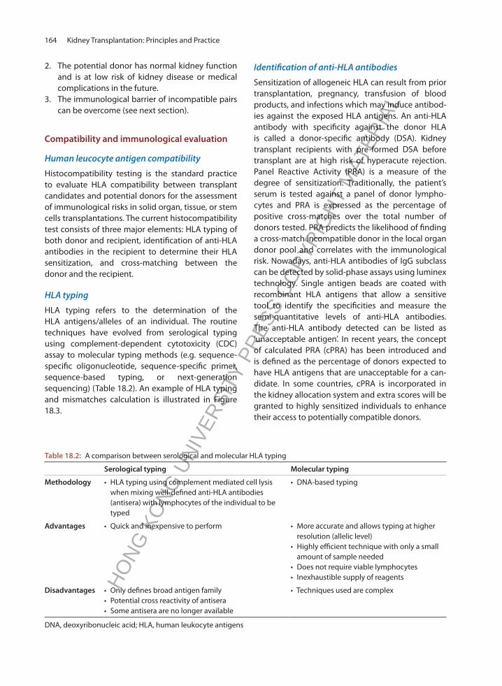
164 Kidney Transplantation: Principles and Practice
2. The potential donor has normal kidney function and is at low risk of kidney disease or medical complications in the future.
3. The immunological barrier of incompatible pairs can be overcome (see next section).
Compatibility and immunological evaluation
Human leucocyte antigen compatibility
Histocompatibility testing is the standard practice to evaluate HLA compatibility between transplant candidates and potential donors for the assessment of immunological risks in solid organ, tissue, or stem cells transplantations. The current histocompatibility test consists of three major elements: HLA typing of both donor and recipient, identification of anti-HLA antibodies in the recipient to determine their HLA sensitization, and cross-matching between the donor and the recipient.
HLA typing
HLA typing refers to the determination of the HLA antigens/alleles of an individual. The routine techniques have evolved from serological typing using complement-dependent cytotoxicity (CDC) assay to molecular typing methods (e.g. sequence-specific oligonucleotide, sequence-specific primer, sequence-based typing, or next-generation sequencing) (Table 18.2). An example of HLA typing and mismatches calculation is illustrated in Figure 18.3.
Identification of anti-HLA antibodies
Sensitization of allogeneic HLA can result from prior transplantation, pregnancy, transfusion of blood products, and infections which may induce antibod-ies against the exposed HLA antigens. An anti-HLA antibody with specificity against the donor HLA is called a donor-specific antibody (DSA). Kidney transplant recipients with pre-formed DSA before transplant are at high risk of hyperacute rejection. Panel Reactive Activity (PRA) is a measure of the degree of sensitization. Traditionally, the patient’s serum is tested against a panel of donor lympho-cytes and PRA is expressed as the percentage of positive cross-matches over the total number of donors tested. PRA predicts the likelihood of finding a cross-match incompatible donor in the local organ donor pool and correlates with the immunological risk. Nowadays, anti-HLA antibodies of IgG subclass can be detected by solid-phase assays using luminex technology. Single antigen beads are coated with recombinant HLA antigens that allow a sensitive tool to identify the specificities and measure the semi-quantitative levels of anti-HLA antibodies. The anti-HLA antibody detected can be listed as ‘unacceptable antigen’. In recent years, the concept of calculated PRA (cPRA) has been introduced and is defined as the percentage of donors expected to have HLA antigens that are unacceptable for a can-didate. In some countries, cPRA is incorporated in the kidney allocation system and extra scores will be granted to highly sensitized individuals to enhance their access to potentially compatible donors.
Table 18.2: A comparison between serological and molecular HLA typing
Serological typing Molecular typing
Methodology • HLA typing using complement mediated cell lysis when mixing well-defined anti-HLA antibodies (antisera) with lymphocytes of the individual to be typed
• DNA-based typing
Advantages • Quick and inexpensive to perform • More accurate and allows typing at higher resolution (allelic level)
• Highly efficient technique with only a small amount of sample needed
• Does not require viable lymphocytes• Inexhaustible supply of reagents
Disadvantages • Only defines broad antigen family• Potential cross reactivity of antisera• Some antisera are no longer available
• Techniques used are complex
DNA, deoxyribonucleic acid; HLA, human leukocyte antigens
HONG
KON
G UN
IVER
SITY
PRE
SS C
OPYR
IGHT
MAT
ERIA
L

Maggie Ma, Simon C. Y. Cheung, Cindy Choy, Gavin Chan, Janette Kwok, and Tak Mao Chan 165
incompatible renal transplantation can also confer a survival benefit in ESKD patients who do not have compatible live donors as they might otherwise face long waiting times for deceased donor kidneys.
Strategies to overcome incompatibility
Paired kidney exchange programme
Kidney paired donation refers to the matching of a potential kidney transplant recipient who has a willing but incompatible donor to another incompat-ible pair (Figure 18.4). Such an approach gives both recipients the chance to receive a compatible kidney. However, potential recipients who have blood group O or broad sensitization may have difficulty in finding a matched kidney.
Desensitization
This process involves the removal of antibodies against blood group antigens or HLA by plasma-pheresis or immunoadsorption to achieve safe target titres, and depletion of B lymphocytes with anti-CD20
HLA cross-match
Physical cross-match is performed to demonstrate the response of DSA using the live donor’s cells and the recipient’s serum. A conventional CDC cross-match assay detects the presence of cytotoxic DSA which is a contraindication for transplantation. Flow cytometric cross-match assay is highly sensitive to detect low levels of DSA bound to the donor’s T cells and B cells. Rejection risk assessment based on the combination of anti-HLA antibodies identification and cross-match results is shown in Table 18.3.
Blood group compatibility
Transplantation in subjects with incompatible blood groups is generally avoided due to the risk of hypera-cute rejection mediated by pre-formed anti-A or anti-B antibodies against the blood group antigens. Nevertheless, blood group incompatible kidney transplantation is technically possible nowadays with desensitization prior to transplantation and can achieve comparable patient and graft survival to blood group compatible transplants. Blood group
Figure 18.3: An example of human leukocyte antigen (HLA) typing in kidney transplant. Mismatched antigen(s) refer to the HLA antigen(s) that is/are found in the prospective donor but not in the recipient.
Table 18.3: Interpretation of histocompatibility test results
Scenario Solid phase antibody results
XM results Risk of Rejection
Absent of DSA Negative Negative Low
Anti-HLA antibodies positive but not against donor’s HLA typing
Positive Negative Low
On anti-CD20 treatment Negative B-cell FCXM negative(after pronase treatment)
Low
Presence of DSA Positive Positive High
Presence of non-HLA antibodies and absent of DSA
Positive/Negative Positive Uncertain
DSA, donor-specific antibody; FCXM, flow cytometric cross-match; HLA, human leukocyte antigen; XM, cross-match
HONG
KON
G UN
IVER
SITY
PRE
SS C
OPYR
IGHT
MAT
ERIA
L

Introduction
Critical care nephrology is a new discipline formally established in 1998 worldwide. It is a multidisciplinary branch of medicine that deals with issues of critical care medicine and nephrology. Acute kidney injury (AKI) is a centre of attention in the arena of critical care nephrology and internists, critical care physi-cians, surgeons, and nephrologists all participate in the management of this important and serious condition. Moreover, chronic dialysis patients who become critically ill may also require renal support in the intensive care unit (ICU). This chapter will high-light the spectrum of renal problems in critically ill patients and the application of renal replacement therapy (RRT) in these settings.
Disease Spectrum in Critical Care Nephrology
AKI is a common condition in the critical care setting, and the reported incidence ranges from 22% to 57% depending on the definition used. The development of AKI in critically ill patients is associated with sub-stantial morbidities, mortality, and treatment costs. Furthermore, patients with prolonged or improperly managed AKI may also progress to chronic kidney disease (CKD) or even end-stage kidney disease (ESKD). Common conditions that are associated with AKI in critical care settings include severe infection and septic shock, cardiogenic shock, liver failure, intra-abdominal hypertension, malignancy, recovery from major operation (especially cardiac and vascular surgery), trauma, burns, and exposure to iodine-based contrast or other nephrotoxic agents.
20
Critical Care Nephrology
Hoi Ping Shum, Desmond Yap, and Wing Wa Yan
The pathogenesis of AKI from the above-mentioned conditions are highly complex and contributed by macro- and micro-circulatory disturbance, the surge of inflammatory mediators and reactive oxygen species, activation of the coagulation cascade, gly-cocalyx degradation, and renal venous congestion. Once AKI occurs in a critically ill patient, the cause of AKI should be identified as quickly as possible to tailor a set of actions to prevent further progression of AKI and avoid irreversible kidney damage. Another commonly encountered clinical scenario in critical care nephrology is when patients on chronic hae-modialysis (HD) or peritoneal dialysis (PD) become critically ill and require ICU care. In this context, their usual modality and regimen of dialysis may be significantly modified to suit their clinical conditions and metabolic demands.
Renal Replacement Therapy in the Critical Care Setting
About 10% of AKI patients require RRT to compensate for the loss of renal function, manage acid–base and electrolyte disturbances, remove excessive fluid gain, and allow time for renal recovery. RRT in critically ill patients can be provided continuously or intermit-tently depending on patients’ needs. Continuous renal replacement therapy (CRRT) is a slow and con-tinuous process that removes fluid and uremic toxins from the patients. CRRT is the predominant form of RRT in the critical care settings due to its accurate volume control, steady acid–base and electrolyte correction, and good maintenance of blood pressure during the treatment. It is frequently prescribed in critically ill patients with hemodynamic instability
HONG
KON
G UN
IVER
SITY
PRE
SS C
OPYR
IGHT
MAT
ERIA
L
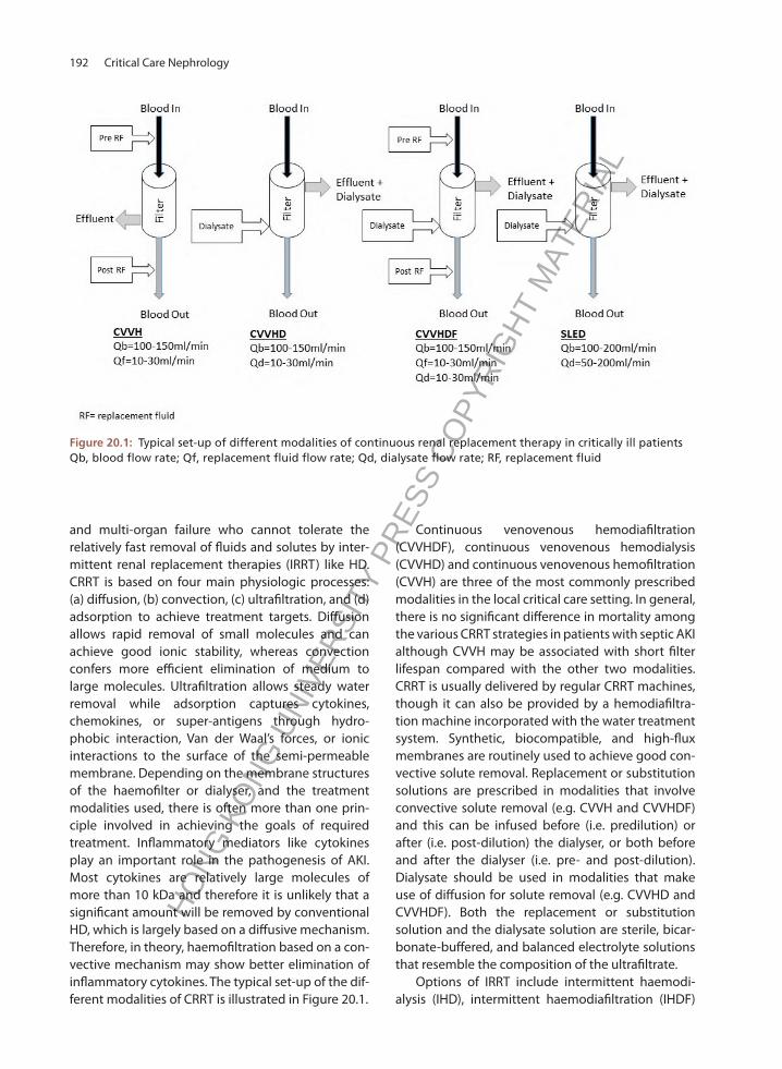
192 Critical Care Nephrology
and multi-organ failure who cannot tolerate the relatively fast removal of fluids and solutes by inter-mittent renal replacement therapies (IRRT) like HD. CRRT is based on four main physiologic processes: (a) diffusion, (b) convection, (c) ultrafiltration, and (d) adsorption to achieve treatment targets. Diffusion allows rapid removal of small molecules and can achieve good ionic stability, whereas convection confers more efficient elimination of medium to large molecules. Ultrafiltration allows steady water removal while adsorption captures cytokines, chemokines, or super-antigens through hydro-phobic interaction, Van der Waal’s forces, or ionic interactions to the surface of the semi-permeable membrane. Depending on the membrane structures of the haemofilter or dialyser, and the treatment modalities used, there is often more than one prin-ciple involved in achieving the goals of required treatment. Inflammatory mediators like cytokines play an important role in the pathogenesis of AKI. Most cytokines are relatively large molecules of more than 10 kDa and therefore it is unlikely that a significant amount will be removed by conventional HD, which is largely based on a diffusive mechanism. Therefore, in theory, haemofiltration based on a con-vective mechanism may show better elimination of inflammatory cytokines. The typical set-up of the dif-ferent modalities of CRRT is illustrated in Figure 20.1.
Continuous venovenous hemodiafiltration (CVVHDF), continuous venovenous hemodialysis (CVVHD) and continuous venovenous hemofiltration (CVVH) are three of the most commonly prescribed modalities in the local critical care setting. In general, there is no significant difference in mortality among the various CRRT strategies in patients with septic AKI although CVVH may be associated with short filter lifespan compared with the other two modalities. CRRT is usually delivered by regular CRRT machines, though it can also be provided by a hemodiafiltra-tion machine incorporated with the water treatment system. Synthetic, biocompatible, and high-flux membranes are routinely used to achieve good con-vective solute removal. Replacement or substitution solutions are prescribed in modalities that involve convective solute removal (e.g. CVVH and CVVHDF) and this can be infused before (i.e. predilution) or after (i.e. post-dilution) the dialyser, or both before and after the dialyser (i.e. pre- and post-dilution). Dialysate should be used in modalities that make use of diffusion for solute removal (e.g. CVVHD and CVVHDF). Both the replacement or substitution solution and the dialysate solution are sterile, bicar-bonate-buffered, and balanced electrolyte solutions that resemble the composition of the ultrafiltrate.
Options of IRRT include intermittent haemodi-alysis (IHD), intermittent haemodiafiltration (IHDF)
Figure 20.1: Typical set-up of different modalities of continuous renal replacement therapy in critically ill patientsQb, blood flow rate; Qf, replacement fluid flow rate; Qd, dialysate flow rate; RF, replacement fluid
HONG
KON
G UN
IVER
SITY
PRE
SS C
OPYR
IGHT
MAT
ERIA
L

Hoi Ping Shum, Desmond Yap, and Wing Wa Yan 193
and sustained low-efficiency dialysis (SLED). In general, IRRT should be considered in situations requiring rapid correction of electrolytes or urgent fluid removal, or in acute poisoning. While there is no proven survival benefit of CRRT over IRRT, CRRT appears to confer advantages to hemodynamic stability, control of fluid balance, renal recovery, and dialysis dependence. Furthermore, CRRT is also associated with a smaller increase in intracranial pressure compared with IRRT and thus more prefer-able in patients with liver failure, acute brain injury, or conditions that create a predisposition to cerebral oedema. The current guidelines recommend that CRRT and IRRT be used as complementary therapies in critically ill patients with AKI, and the choice of modality should take into consideration the patient’s condition (e.g. diagnosis, hemodynamic status etc), availability of nursing staff, equipment, and other relevant resources (Table 20.1). In this context, patients with haemodynamic instability generally receive CRRT and are often transferred to IRRT once they are free from vasopressor use.
Acute PD is often an overlooked dialysis modality for critically ill patients with AKI. It is technically easy to perform and is associated with low cost and good haemodynamic stability. PD is a safe and viable option for dialysis support in AKI patients, especially for neonates, children, and those with a PD catheter
in-situ. The current data suggest that acute PD is comparable to HD as regards the incidence of renal recovery and mortality in critically ill patients with AKI. The slow rate of solute removal in acute PD incurs a lower risk of disequilibrium syndrome but is also a drawback in patients with high physiological and metabolic stresses. Acute PD is contraindicated for those with recent abdominal or cardiothoracic surgery, severe respiratory failure, extreme volume overload, and very poor glycaemic control.
The right timing to commence RRT in critically ill patients with AKI remains debatable due to a lack of consensus on the parameters to define timing and threshold for initiation of RRT. Starting RRT before a patient develops absolute indications (e.g. fluid overload resistant to diuretics therapy, severe metabolic acidosis with pH < 7.15, hyperkalaemia > 6.5 mmol/L, or symptomatic uraemia) is a common practice in ICUs. However, commencing RRT too early may subject patients who could have naturally recovered kidney function to unnecessary risks of complications. Although current evidence suggests that early initiation of RRT in critically ill patients with AKI does not improve mortality when compared with standard or late initiation, early RRT is associated with a significant reduction of the length of hospi-talization. The appropriate timing of initiating RRT should be individualized and take into consideration
Table 20.1: A comparison of the different modalities of renal replacement therapy in critically ill patients
Parameters IHD SLED CRRT
Principle Diffusion Diffusion CVVH: ConvectionCVVHD: DiffusionCVVHDF: Both
Blood flow (ml/min) 200–300 100–200 100–150
Dialysate flow (ml/min) > = 500 50–200 CVVHD: 10–30CVVHDF: 10–30
Treatment duration (hours) 3–5 6–12 >24
Hemodynamic stability Unstable Marginally stable Stable
Fluid removal Rapid Intermediate Very slow and gentle
Efficiency High Medium Low
Anticoagulation LMWH/UFH/ anticoagulation free
LMWH/UFH RCA/LMWH/UFH/ pre-dilution
Nursing workload Low Intermediate High
Nurse requirement Renal nurse Renal nurse ICU nurse
Treatment cost Low Low High
CRRT, continuous renal replacement therapy; CVVH, continuous venovenous haemofiltration; CVVHD, continuous veno-venous haemodialysis; CVVHDF, continuous venovenous haemodiafiltration; ICU, Intensive care unit; IHD, intermittent haemodialysis; LMWH, low molecular weight heparin; RCA, regional citrate anticoagulation; SLED, sustained low-efficiency dialysis; UFH, unfractionated heparin
HONG
KON
G UN
IVER
SITY
PRE
SS C
OPYR
IGHT
MAT
ERIA
L

194 Critical Care Nephrology
patients’ comorbidities, current diagnoses, the severity of illness, extra-renal organ dysfunction, fluid burden, as well as laboratory and physiological parameters.
The adequacy of IHD is usually assessed by the removal of urea (a surrogate of low molecular weight products of metabolism) and is often reflected by the urea reduction ratio (URR) or the fractional clearance of urea (Kt/Vurea, where K is the instantaneous clear-ance, t is the treatment time, and V is the volume of distribution of urea). For CVVH, the clearance of small molecules like urea is equal to the ultrafiltra-tion rate as the sieving coefficient should be 1, which indicates free passage with complete equilibra-tion. During CVVHD, the dialysis flow rate is much slower (10–30 ml/min) than the blood flow rate (100–150 ml/min), and hence urea in the dialysate will equilibrate with that in the plasma and its clear-ance will be approximated by the dialysate flow rate. Thus, in the absence of infusion with predilutional replacement fluid, the treatment dose can be quanti-fied based on effluent flow rates, which is equal to the sum of the ultrafiltrate and dialysate flow. The current evidence suggests performing CRRT at a minimum delivered dose of an effluent flow rate of 20–25 ml/kg/hr. Importantly, one should appreciate that the prescribed dose is not necessarily equal to the actual delivered dose due to circuit clotting, machine alarms, or other logistical issues like inter-ruptions for investigative or surgical procedures.
Anticoagulation in CRRT
Activation of platelets and the coagulation cascade occurs with the exposure of blood to the haemofil-ter/dialyser membrane. Protein coating and clogging together with clot formation compromise the hae-mofilter performance, and therefore anticoagulation is necessary to prevent clotting of the extracorporeal circuit. The appropriate choice of anticoagulation in CRRT is key to maintaining circuit patency and minimizing bleeding complications in high-risk pop-ulations. Adequate anticoagulation can effectively prevent clot formation over the membrane surface, which translates into longer treatment duration and less discrepancy between the prescribed and deliv-ered treatment doses. Moreover, less circuit clotting is associated with reduced blood transfusion require-ment, less consumption of nursing manpower for circuit change, and lower treatment costs. An ideal anticoagulation strategy should be regional, inex-pensive and easy to implement, simple to monitor
the anticoagulation effect, readily reversible, and with minimal side effects.
Unfractionated heparin (UFH) and low molecu-lar weight heparin (LMWH) are commonly used systemic anticoagulants for CRRT and IRRT (Table 20.2). They are inexpensive, readily available, and have an extensive history of clinical usage. The CRRT circuit lifespan using UFH or LMWH as anticoagulant ranges from 26 to 52 hours. LMWH shows less protein binding than UFH, and so its anticoagulation effect is more predictable and may also confer better circuit patency. The bleeding risk and chance of heparin-induced thrombocytopenia of LMWH are also lower than that of UFH. Both UFH and LMWH may induce variable degrees of systemic anticoagulation, which limit their use in patients with bleeding tendency or postoperative conditions.
Regional citrate anticoagulation (RCA) represents an emerging way of anticoagulation in patients receiving CRRT. Citrate chelates ionized calcium, which is crucial for the activation of various clotting factors and thrombin formation. An ionized calcium concentration of 0.25–0.35 mmol/L is essential for achieving the anticoagulation effect in an extracor-poreal circuit. The majority of citrate is removed by either filtration or dialysis during CRRT. Calcium is replaced intravenously before the blood is returned to the patient to compensate for the extracorporeal loss and to normalize the systemic calcium levels. Compared with heparin-based anticoagulation, RCA is associated with lower bleeding risk and longer haemofilter lifespan (27–72 hours), but with similar overall mortality. However, RCA is technically more demanding than a heparin-based regimen. Conditions associated with reduced metabolism of citrates, such as chronic liver disease, ischaemic hepatitis, hypoxemia, and impaired muscle perfu-sion, are not uncommon in the ICU setting. Excessive citrate accumulation leads to low ionized calcium levels and acidosis, thereby causing hypotension secondary to decreased myocardial contractility and vascular hypotonia. Other metabolic disturbances of citrate accumulation include hypomagnesaemia and hypernatremia.
Anticoagulation-free RRT can be considered for patients with high bleeding risk or those with con-traindications for citrate use. In this context, higher blood flow rates and predilution with replacement fluids can be used to improve circuit lifespan. Despite these measures, the circuit lifespan is still often shorter compared with UFH or LMWH circuits.
HONG
KON
G UN
IVER
SITY
PRE
SS C
OPYR
IGHT
MAT
ERIA
L

Hoi Ping Shum, Desmond Yap, and Wing Wa Yan 195
Table 20.2: Different anticoagulation strategies for renal replacement therapy in critically ill patients
Methods Pros Cons
Anticoagulant-free/saline flush
• Easy implementation• Safest in those with bleeding risks
• Short haemofilter lifespan and more blood loss due to circuit clotting
• Labour intensive for repriming of circuit
Pre-dilution • Easy implementation• Can be used in those with bleeding risk and
minimal monitoring is needed
• Short haemofilter lifespan• Compromised treatment efficacy due to
pre-dilution
UFH • Effective anticoagulation• Anticoagulation effect is titratable• Extensive clinical experience
• Causes systemic anticoagulation and thus increased risk of bleeding
• Less predictable PK than LWMH• Risk of HIT
LMWH • Easy implementation• Effective anticoagulation• Extensive clinical experience• Lower risk of HIT than UFH
• Causes systemic anticoagulation and thus increased risk of bleeding
• Anticoagulation effect is not readily reversible
RCA • Longer haemofilter lifespan and lower risk of bleeding compared with UFH or LMWH
• Risk of citrate toxicity and metabolic/electrolyte disturbance
• Complicated protocol and higher manpower demand
Direct thrombin inhibitors/ heparinoids
• Can be considered in patients who develop HIT
• Limited local experience• High cost
HIT, heparin-induced thrombocytopenia; LMWH, low molecular weight heparin; PK, pharmacokinetics; RCA, regional citrate anticoagulation; UFH, unfractionated heparin
HONG
KON
G UN
IVER
SITY
PRE
SS C
OPYR
IGHT
MAT
ERIA
L
A disintegrin and metalloprotease with thrombospondin 1 repeats member 13 (ADAMST13), 91
acid-base, 31acidification, urine, 8, 18acidosis, metabolic, 40acidosis, renal tubular, 41acidosis, respiratory, 38acquired cystic kidney disease, 113acute cellular rejection, 171acute interstitial nephritis, 56acute kidney injury (AKI), 45acute pyelonephritis, 14, 51acute renal failure, 181acute tubular necrosis (ATN), 46adynamic bone disease (ABD), 133air embolism, 157aldosterone, 4, 31alkaline diuresis, 187alkalosis, metabolic, 39alkalosis, respiratory, 38Alport syndrome, 116aluminium bone disease, 133aluminium toxicity, 158aminoglycosides, 42, 184amyloidosis, 90amyloidosis, dialysis-related, 157angiography, 99, 197, 198, 199, 200, 203angiomyolipoma, 117angiotensin ii receptor blocker (ARB), 32, 62, 73, 101, 135,
181angiotensin-converting enzyme inhibitors (ACEI), 32, 62,
73, 82, 101, 135, 181anion gap (AG), 40anion gap, blood, 40, 43anion gap, urine, 18, 43antibody-mediated rejection (AMR), 172anticoagulation, 153, 194antidiuretic hormone (ADH), 6, 23, 25, 27anti-glomerular basement membrane disease (anti-GBM
disease), 78anti-neutrophil cytoplasmic antibody (ANCA), 79anti-phospholipase A2 receptor antibody (Anti-PLA2R), 66
Index
anti-thrombospondin type 1 domain-containing 7A (anti-THSD7A), 66
anti-thymocyte globulin (ATG), 167arrhythmia, 131, 157arteriovenous fistula (AVF), 121, 136, 151, 201arteriovenous graft (AVG), 151, 201asymptomatic bacteriuria, 52automated peritoneal dialysis (APD), 144autoregulation, 2azathioprine (AZA), 76, 77, 79, 81, 82, 107, 108, 168azotaemia, 112, 179
Bartter syndrome, 118belatacept, 169belimumab, 78benign prostatic hyperplasia (BPH), 109beta-2-microglobulin, 141, 152, 157biocompatibility, 152BK virus, 21, 174, 175bleeding tendency, 87, 132blood group compatibility, 165
C3 glomerulopathy, 68calcimimetics, 35, 137calcineurin inhibitors (CNI), 64, 65, 67, 76, 107, 108, 167,
184calciphylaxis, 134calcium, 7, 33carbonic anhydrase, 9, 37cardiovascular disease, 98, 130, 176cast nephropathy, 88, 89chloride, 16chronic allograft nephropathy, 172chronic glomerulonephritis, 56chronic interstitial nephritis (CIN), 55chronic kidney disease (CKD), 13, 14, 18, 50, 57, 60, 73, 74,
98, 101, 104, 107, 115, 123, 129chyle, 14, 171circulating factor (CF), 64cirrhosis, 48, 84cisplatin, 35, 184, 185citrate, 35, 194, 195
HONG
KON
G UN
IVER
SITY
PRE
SS C
OPYR
IGHT
MAT
ERIA
L

210 Index
clearance, 154clearance, creatinine, 16clearance, free water, 6, 23collapsing glomerulopathy, 64, 86collecting duct, 1complements, 19, 21, 68, 74computed tomography, 20, 198continuous ambulatory peritoneal dialysis (CAPD), 143continuous cyclic peritoneal dialysis (CCPD), 144continuous erythropoietin receptor activators (CERA), 138continuous renal replacement therapy (CRRT), 159, 187,
191contrast induced nephropathy, 197corticosteroids, 35, 57, 62, 65, 67, 70, 76, 79, 81, 83, 84, 88,
167crescentic glomerulonephritis, 19cryoglobulinaemia, 82cyclophosphamide (CYC), 62, 63, 65, 67, 70, 76, 79, 81, 83cyclosporin A (CYA), 64, 67, 167cystinosis, 119cystinuria, 120cystitis, 51, 52cytomegalovirus (CMV), 166, 174
deceased donor kidney transplantation, 163desmopressin, 26, 29, 138diabetes insipidus (DI), central, 27, 29diabetes insipidus (DI), nephrogenic, 27, 29diabetes mellitus (DM), 71, 72, 73, 74diabetic kidney disease (DKD), 71, 72, 73, 74dialysis disequilibrium syndrome, 156dialyzer reactions, 156diethyl-triamino-penta-acetic acid (DTPA), 20dilution and concentration, urine, 5dimercaptosuccinic acid (DMSA), 20disability-adjusted life-years (DALYs), 123, 124, 126distal renal tubule, 1diuretics, 182donor, kidney, 163, 201Doppler, 19, 198dyslipidaemia, 62
eculizumab, 95emphysematous pyelonephritis, 174encapsulating peritoneal sclerosis (EPS), 148endothelin, 11erectile dysfunction, 111erythrocytosis, 177erythropoiesis-stimulating agents (ESA), 99, 132erythropoietin (EPO), 55, 132everolimus, 169exit site infection, 147expanded haemodialysis, 159extra-corporeal removal, toxins, 186extra-corporeal shock wave lithotripsy (ESWL), 113
Fabry disease, 117Fanconi syndrome, 36, 55, 89
focal segmental glomerulosclerosis (FSGS), 64fractional excretion, 16, 18, 36, 42fungal infection, 176
gastroesophageal reflux disease (GERD), 149geriatric nephrology, 205Gitelman syndrome, 33, 39, 119glomerular filtration rate (GFR), 2, 16glomerulonephritis (GN), 59glomerulus, 3, 47glucagon like peptide-1 (GLP-1), 73Goodpasture’s disease, 78graft renal artery thrombosis, 170graft renal vein thrombosis, 170, 203growth hormone, 119, 121
haematuria, 13haemodiafiltration (HDF), 151, 158haemodialyser, 152haemodialysis (HD), 151haemofiltration, 158haemolysis, elevated liver enzymes and low platelet count
(HELLP) syndrome, 106haemolytic uraemic syndrome (HUS), 64haemoperfusion, 159, 187Hantavirus, 87Henoch-Schönlein purpura (HSP), 60hepatitis B virus (HBV), 19, 84, 86hepatitis C virus (HCV), 19, 84, 86hepatorenal syndrome (HRS), 84hernia, 148high cut-off (HCO) membrane, 159histocompatibility, 161, 164human immunodeficiency virus (HIV), 86human leukocyte antigen (HLA), 161hungry bone syndrome, 137hydrothorax, 149hyperaldosteronism, 99hypercalcaemia, 34hypercalciuria, 36hyperkalaemia, 31hyperlipidaemia, 25, 167hypernatraemia, 27hyperoxaluria, 120hyperparathyroidism, secondary, 132, 133hyperparathyroidism, tertiary, 34, 133hyperphosphataemia, 36, 133, 137hypertension, 97hyperuricaemia, 56, 95, 168hypocalcaemia, 35, 133, 136hypokalaemia, 32hypomagnesaemia, 32, 119hyponatraemia, 24hypophosphataemia, 36hypotension, 155hypouricaemia, 89
IL-2 receptor antagonist, 166
HONG
KON
G UN
IVER
SITY
PRE
SS C
OPYR
IGHT
MAT
ERIA
L

211
immune checkpoint inhibitors (ICPI), 187immunoglobulin A nephropathy (IgAN), 59immunoglobulin G subclass 4 disease (IgG4 disease), 83immunosuppression, 76, 166incontinence, 13, 14infection-related glomerulonephritis (IRGN), 85intermittent peritoneal dialysis (IPD), 144intra-renal, acute kidney injury, 47iron therapy, 138, 206
ketamine, 188ketoacidosis, 41kidney biopsy, 20Kimmelstiel-Wilson nodule, 72
lactic acidosis, 40leptospirosis, 86leukocyte esterase, 14Liddle syndrome, 100light chain deposition disease (LCDD), 88, 89lithium, 187live donor kidney transplantation, 163, 201lupus nephritis, 74lymphocele, 171, 203, 204
magnetic resonance imaging (MRI), 20, 197major histocompatibility complex (MHC), 161malnutrition, 134, 138mammalian target of rapamycin (mTOR), 169membranoproliferative glomerulonephritis (MPGN), 68membranous nephropathy (MN), idiopathic, 65membranous nephropathy (MN), secondary, 66mercaptoacetyltriglycine (MAG3), 20mercury, 188mineral bone disorder, 122, 132, 136minimal change disease (MCD), 62, 118monoclonal gammopathy of renal significance (MGRS),
19, 87M-type phospholipase A2 receptor, 66multiple myeloma, 87muscle cramps, 156mycobacterium, 53, 56, 145, 175mycophenolate mofetil (MMF), 61, 65, 70, 76, 84, 108, 167,
168mycophenolic acid (MPA), 108, 167, 168
natriuretic peptides, 5nephritic syndrome, 59nephrocalcinosis, 120nephron, 1nephrotic syndrome, 59, 118neurogenic bladder 48, 51, 53nitric oxide, 2, 72nocturia, 13nocturnal intermittent peritoneal dialysis (NIPD), 144
Obinutuzumab, 77obstructive uropathy (ON), 107, 109, 126
oliguria, 13, 49osmolality, plasma, 24, 25osmolality, urine, 24, 25, 27osteitis fibrosa cystica, 133osteomalacia, 134
paediatric nephrology, 115palliative nephrology, 205parathyroid hormone (PTH), 7, 18, 34, 35, 36, 133, 137parathyroidectomy, 137pauci-immune, 19, 80percutaneous nephrolithotomy (PCNL), 113peritoneal dialysis (PD), 141peritoneal dialysis solution, 143peritoneal equilibration test (PET), 142peritonitis, 145pharmacokinetics, 179plasma exchange, 79, 81pleuro-peritoneal communication, 149, 199pneumocystis carinii, 175pneumocystis jiroveci, 175polyarteritis nodosa (PAN), 81polycystic kidney disease, 115polyoma virus, 174, 175polyuria, 13positron emission tomography, 20post-renal, acute kidney injury, 47post-streptococcal glomerulonephritis, 85post-transplant lymphoproliferative disorders (PTLD), 167,
176post-transplant malignancy, 176pre-eclampsia, 47, 104, 105, 106, 107pregnancy, 103pre-renal, acute kidney injury, 45prostate specific antigen (PSA), 110protein-energy wasting syndrome (PEWS), 134, 150proteinuria, 13proximal renal tubule, 11pseudohyponatraemia, 25pyelonephritis, 52
radio-isotope scan, 20rapamycin, 169rapidly progressive glomerulonephritis (RPGN), 78rasburicase, 95regional citrate anticoagulation (RCA), 153, 194rejection, 171renal artery stenosis (RAS), 19, 99, 170, 197, 198renal biopsy, 20renal calculi, 112renal cell carcinoma, 113renal colic, 14renal osteodystrophy, 133, 137renal rehabilitation, 207renal tubular acidosis (RTA), distal, 41, 42renal tubular acidosis (RTA), proximal, 41, 42renin-angiotensin-aldosterone system (RAAS), 5, 65, 130renovascular hypertension, 99
HONG
KON
G UN
IVER
SITY
PRE
SS C
OPYR
IGHT
MAT
ERIA
L

212 Index
reset osmostat, 27reverse osmosis, 152rhabdomyolysis, 96rheumatoid arthritis, 83rickets, 120
salicylate, 41scleroderma renal crisis (SRC), 82scleroderma, 82serum urine-type plasminogen activator receptor (suPAR),
64shiga-toxin, 94sirolimus, 169Sjogren syndrome, 83skeletal survey, 20sodium-glucose co-transporter 2 (SGLT-2), 73syndrome of inappropriate antidiuretic hormone (SIADH),
24, 26systemic lupus erythematosus (SLE), 74
tacrolimus (TAC), 64, 67, 76, 77, 167, 168thrombotic microangiopathy (TMA), 91, 95, 168thrombotic thrombocytopenic purpura (TTP), 91toxicology, 186traditional Chinese medicine (TCM), 188transplantation, kidney, 161transtubular potassium gradient (TTKG), 32tuberculosis, 53, 145, 175tuberous sclerosis, 117tubulointerstitial diseases, 55tubulointerstitial fibrosis, 57tubulointerstitium, 55tumour lysis syndrome, 95tunnel tract infection, 147, 199tunnelled cuffed catheter, 151, 201
ultrafiltration (UF), 141, 142, 147, 149, 151, 153, 155, 156, 199
ultrasonography, 19, 99, 197, 201, 203uraemia, 14, 40uraemic pericarditis, 131urea kinetic modelling, 154uric acid, 15urinalysis, 15urinary retention, 13urinary tract infection, 51urolithiasis, 111
valganciclovir, 174, 182vascular access, 151vasculitis, 79, 89vesicoureteric reflux, 51, 130viral hepatitis, 86, 174vitamin D, 7, 33, 34, 35, 36, 120, 133vitamin D analogue, 137
water deprivation test, 18Wegener’s granulomatosis, 79
Zopiclone, 188
HONG
KON
G UN
IVER
SITY
PRE
SS C
OPYR
IGHT
MAT
ERIA
L