Kidney News - February 2022 - American Society of Nephrology
28
February 2022 | Vol. 14, Number 2 > Continued on page 5 Early Dialysis Improves Survival—but Is the Tradeoff Worth It? By Timothy O’Brien When a Kidney Transplant Fails, Retransplantation May Offer Better Survival over Dialysis A lthough kidney transplantation is the opti- mal therapy after kidney failure for prolong- ing patient survival and improving quality of life, kidneys transplanted from deceased do- nors often do not function longer than 10 to 15 years. erefore, many recipients must eventually receive a sec- ond transplant or undergo dialysis, with considerations such as the scarcity of donor organs and the immuno- logical sensitization of transplant recipients factoring into decisions related to these options. Because direct comparison of transplantation versus di- alysis continuation through a randomized controlled trial is not feasible due to ethical, biological, and logistic rea- sons, investigators recently conducted a retrospective study that analyzed data pertaining to 2346 adults with a failed first kidney transplant who were waitlisted for a second kidney transplant in Austria during 1980–2019 (1). In the CJASN study, patients who received a second kidney transplant soon after a failed first transplant had a longer average survival time compared with those who underwent dialy- sis while remaining on the transplant waitlist. Rainer Oberbauer, MD, of the Medical Uni- versity of Vienna, in Austria, is senior author of the study. At a 10-year follow-up point, the overall mortality was 41%, and patients who under- went retransplantation lived for an average of 5.8 months longer than those who underwent dialysis. e difference in survival time with retransplantation was low- er in patients who had a longer wait time after their first transplant failed, however. Patients who underwent re- transplantation lived for an average of 8.0 months longer with a waiting time of less than 1 year but for 0.1 month with a waiting time of 8 years. ere was no statistically significant survival difference in individuals with a waiting time of more than 3 years after first graft loss. is decreased survival advantage was mainly a conse- quence of improved relative survival over time in patients who remained on dialysis awaiting transplantation, per- haps reflecting a biological selection of long-term survi- vors, the authors note. Also, there was a higher survival benefit with second kidney transplants in recent years compared with earlier years, indicating advances in current transplant practices. Furthermore, kidney transplant recipients with living do- nors also appeared to have higher survival rates than those with deceased donors. “Our data showed that a second transplantation is By Tracy Hampton Continued on page 5 > F or patients with advanced chronic kidney disease (CKD), early dialysis initiation—at an estimated glomerular filtration rate (eGFR) of 15–16 mL/ min/1.73 m 2 —leads to modest reductions in mor- tality and cardiovascular events, reports a study in e BMJ (1). “However, to reach the maximum survival benefit, pa- tients would need to start dialysis up to 4 years earlier,” com- ments lead author Edouard Fu, PhD, a research fellow at the Division of Pharmacoepidemiology and Pharmacoeconom- ics at Brigham and Women’s Hospital and Harvard Medical School, Boston, Massachusetts. e conclusions are consistent with the sole previous randomized trial of dialysis initiation times—and support current guideline recommendations on dialysis initiation. Fu and colleagues write: “Our findings provide novel evi- dence on the optimal timing of dialysis initiation and show that even with maximum eGFR separations, the range of plausible effects is likely to be small.” Inside ASN President’s Update The art of nephrology Onco-nephrology A kidney transplant recipient, nephrologist, and fellow discuss how onco-nephrology can improve patient care. Fellows First SGLT2 inhibitors in IgA nephropathy A kidney emoji The heart, brain, and lung have them. Why not the kidney? International medical graduates Hiring IMGs on a J-1 visa
-
Upload
khangminh22 -
Category
Documents
-
view
0 -
download
0
Transcript of Kidney News - February 2022 - American Society of Nephrology

February 2022 | Vol. 14, Number 2
>Continued on page 5
Early Dialysis Improves Survival—but Is the Tradeoff Worth It?By Timothy O’Brien
When a Kidney Transplant Fails, Retransplantation May Offer Better Survival over Dialysis
Although kidney transplantation is the opti-mal therapy after kidney failure for prolong-ing patient survival and improving quality of life, kidneys transplanted from deceased do-
nors often do not function longer than 10 to 15 years. Therefore, many recipients must eventually receive a sec-ond transplant or undergo dialysis, with considerations
such as the scarcity of donor organs and the immuno-logical sensitization of transplant recipients factoring into decisions related to these options.
Because direct comparison of transplantation versus di-alysis continuation through a randomized controlled trial is not feasible due to ethical, biological, and logistic rea-sons, investigators recently conducted a retrospective study
that analyzed data pertaining to 2346 adults with a failed first kidney transplant who were waitlisted for a second kidney transplant in Austria during 1980–2019 (1).
In the CJASN study, patients who received a second kidney transplant soon after a failed first transplant had a longer average survival time compared with those who underwent dialy-sis while remaining on the transplant waitlist. Rainer Oberbauer, MD, of the Medical Uni-versity of Vienna, in Austria, is senior author of the study.
At a 10-year follow-up point, the overall mortality was 41%, and patients who under-went retransplantation lived for an average of 5.8
months longer than those who underwent dialysis. The difference in survival time with retransplantation was low-er in patients who had a longer wait time after their first transplant failed, however. Patients who underwent re-transplantation lived for an average of 8.0 months longer with a waiting time of less than 1 year but for 0.1 month with a waiting time of 8 years. There was no statistically significant survival difference in individuals with a waiting time of more than 3 years after first graft loss.
This decreased survival advantage was mainly a conse-quence of improved relative survival over time in patients who remained on dialysis awaiting transplantation, per-haps reflecting a biological selection of long-term survi-vors, the authors note.
Also, there was a higher survival benefit with second kidney transplants in recent years compared with earlier years, indicating advances in current transplant practices. Furthermore, kidney transplant recipients with living do-nors also appeared to have higher survival rates than those with deceased donors.
“Our data showed that a second transplantation is
By Tracy Hampton
Continued on page 5 >
For patients with advanced chronic kidney disease (CKD), early dialysis initiation—at an estimated glomerular filtration rate (eGFR) of 15–16 mL/min/1.73 m2—leads to modest reductions in mor-
tality and cardiovascular events, reports a study in The BMJ (1).
“However, to reach the maximum survival benefit, pa-tients would need to start dialysis up to 4 years earlier,” com-ments lead author Edouard Fu, PhD, a research fellow at the Division of Pharmacoepidemiology and Pharmacoeconom-
ics at Brigham and Women’s Hospital and Harvard Medical School, Boston, Massachusetts.
The conclusions are consistent with the sole previous randomized trial of dialysis initiation times—and support current guideline recommendations on dialysis initiation. Fu and colleagues write: “Our findings provide novel evi-dence on the optimal timing of dialysis initiation and show that even with maximum eGFR separations, the range of plausible effects is likely to be small.”
InsideASN President’s Update The art of nephrology
Onco-nephrology A kidney transplant recipient, nephrologist, and fellow discuss how onco-nephrology can improve patient care.
Fellows First SGLT2 inhibitors in IgA nephropathy
A kidney emojiThe heart, brain, and lung have them. Why not the kidney?
International medical graduatesHiring IMGs on a J-1 visa
11644142 HCP Post OPDP Journal Ad KING Size M3FRDate:Client:Product:Client Code:WF Issue #Releasing as:Final Size:Finishing:Gutter:Colors:
Producer:AD:AE:QC:Production:Digital Artist:FR Spellcheck:
10-29-2021 11:07 AMBAYERFINERENONEPP-KER-US-0016-110165182PDFx1A21" x 14"Trim0.5" on each page4/c process
Andrew SlaftaRodrigo PanucciKetaki DatarNoneDavid ZinkLaRosa, Vincent (NYC-FCB)None
Job info
Team
Special Instructions
Aspira (Bold, Regular, Italic)Fonts Images
Inks
PREPARED BY
Additional Information
Additional Comments for Sizing
Webcargo zipped PDF X1A to David Zink and Joe Lee.Please include PI page in file.
Magazine Ad
Bleed: 22" x 15" Cyan, Magenta, Yellow, Black
BAYE_A074682_4C.tif (CMYK; 300 ppi; 100%; 99.4MB), NEW_KERE_US_RM_Prom_DosAdm_CMYK_FC_Pos.ai (66.83%; 81KB), Corp-Lo-go_BG_Bayer-Cross_Basic_print_CMYK.ai (26.01%; 1.2MB), qr-code (2).ai (10.94%; 99KB)
Scale: 1" = 1"
BleedTrim/FlatLive/Safety
22" w x 15" h 22" w x 15" h21" w x 14" h 21" w x 14" h19" w x 13.25" h 19" w x 13.25" h
Path: PrePress:Bayer:Finerenone:11644142:PP-KER-US-0016-1_HCP_Post_OPDP_Journal_Ad_KING_Size_M3FR.indd
PDFX1A _
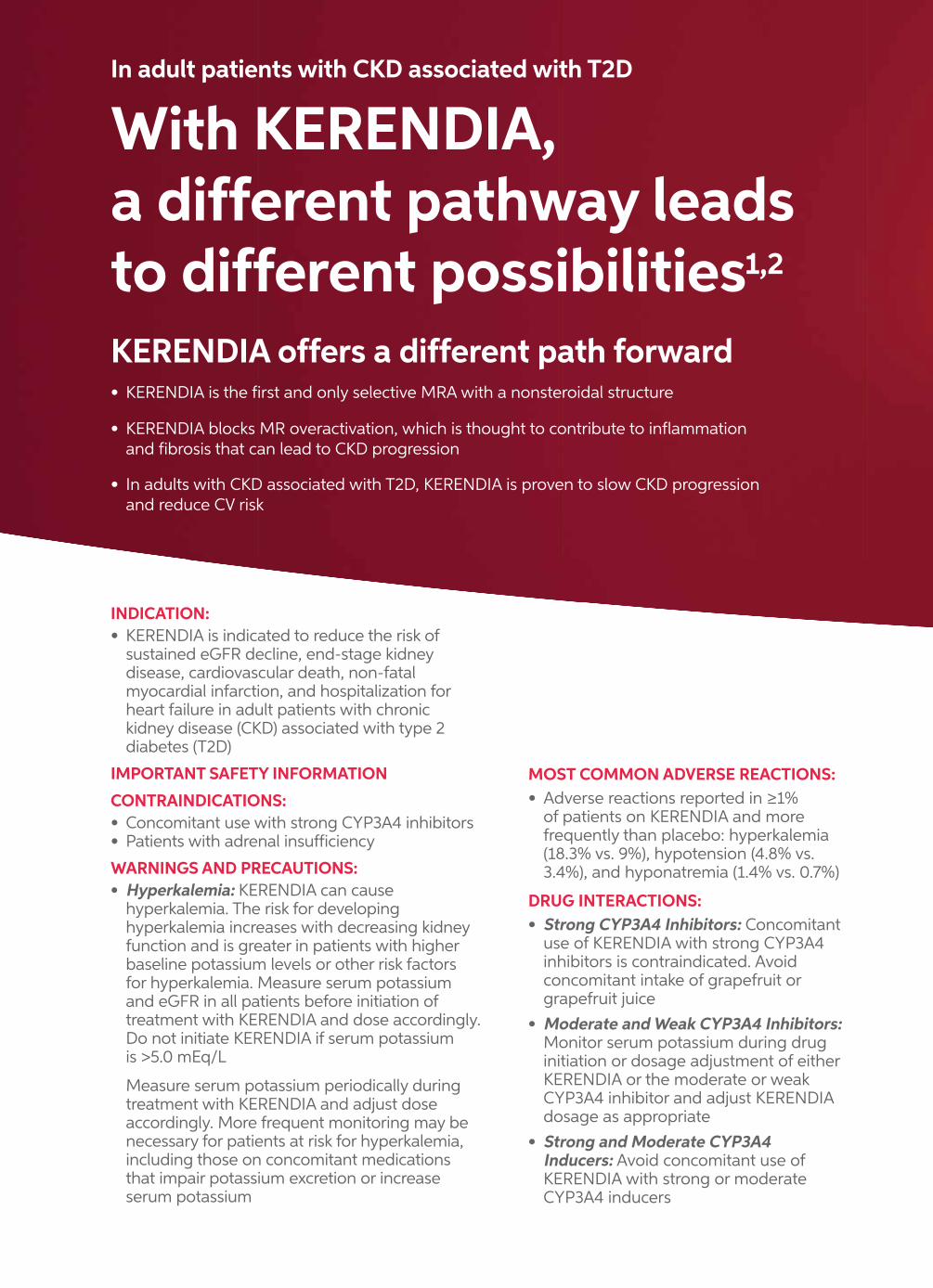
With KERENDIA, a di� erent pathway leads to di� erent possibilities1,2
In adult patients with CKD associated with T2D
KERENDIA o� ers a di� erent path forward• KERENDIA is the � rst and only selective MRA with a nonsteroidal structure
• KERENDIA blocks MR overactivation, which is thought to contribute to in� ammationand � brosis that can lead to CKD progression
• In adults with CKD associated with T2D, KERENDIA is proven to slow CKD progression and reduce CV risk
MOST COMMON ADVERSE REACTIONS:• Adverse reactions reported in ≥1%
of patients on KERENDIA and more frequently than placebo: hyperkalemia (18.3% vs. 9%), hypotension (4.8% vs. 3.4%), and hyponatremia (1.4% vs. 0.7%)
DRUG INTERACTIONS:• Strong CYP3A4 Inhibitors: Concomitant
use of KERENDIA with strong CYP3A4 inhibitors is contraindicated. Avoid concomitant intake of grapefruit or grapefruit juice
• Moderate and Weak CYP3A4 Inhibitors:Monitor serum potassium during drug initiation or dosage adjustment of either KERENDIA or the moderate or weak CYP3A4 inhibitor and adjust KERENDIA dosage as appropriate
• Strong and Moderate CYP3A4Inducers: Avoid concomitant use of KERENDIA with strong or moderate CYP3A4 inducers
USE IN SPECIFIC POPULATIONS• Lactation: Avoid breastfeeding during
treatment with KERENDIA and for 1 day after treatment
• Hepatic Impairment: Avoid use of KERENDIA in patients with severe hepatic impairment (Child Pugh C) and consider additional serum potassium monitoring with moderate hepatic impairment(Child Pugh B)
Please read the Brief Summary of the KERENDIA Prescribing Information on the following page.
CKD=chronic kidney disease; CV=cardiovascular; MR=mineralocorticoid receptor; MRA=mineralocorticoid receptor antagonist; T2D=type 2 diabetes.
References: 1. KERENDIA (� nerenone) [prescribinginformation]. Whippany, NJ: Bayer HealthCare Pharmaceuticals, Inc; July 2021. 2. Bakris GL,et al; FIDELIO-DKD Investigators. N Engl J Med. 2020;383(23):2219-2229.
Learn more about KERENDIA and the FIDELIO-DKD trial
© 2021 Bayer. All rights reserved. BAYER, the Bayer Cross, and KERENDIA are registered trademarks of Bayer.
All other trademarks are property of their respective owners. PP-KER-US-0016-1 10/21
INDICATION:• KERENDIA is indicated to reduce the risk of
sustained eGFR decline, end-stage kidney disease, cardiovascular death, non-fatal myocardial infarction, and hospitalization for heart failure in adult patients with chronic kidney disease (CKD) associated with type 2 diabetes (T2D)
IMPORTANT SAFETY INFORMATION
CONTRAINDICATIONS: • Concomitant use with strong CYP3A4 inhibitors • Patients with adrenal insuª ciency
WARNINGS AND PRECAUTIONS:• Hyperkalemia: KERENDIA can cause
hyperkalemia. The risk for developing hyperkalemia increases with decreasing kidney function and is greater in patients with higher baseline potassium levels or other risk factors for hyperkalemia. Measure serum potassium and eGFR in all patients before initiation of treatment with KERENDIA and dose accordingly. Do not initiate KERENDIA if serum potassiumis >5.0 mEq/L
Measure serum potassium periodically duringtreatment with KERENDIA and adjust dose accordingly. More frequent monitoring may be necessary for patients at risk for hyperkalemia, including those on concomitant medications that impair potassium excretion or increase serum potassium
S:19"
S:13.25"
T:21"
T:14"
B:22"
B:15"
F:10.5"
FS:9"
F:10.5"
FS:9"
PP-KER-US-0016-1_HCP_Post_OPDP_Journal_Ad_KING_Size_M3FR.indd 1PP-KER-US-0016-1_HCP_Post_OPDP_Journal_Ad_KING_Size_M3FR.indd 1 10/29/21 11:08 AM10/29/21 11:08 AM

11644142 HCP Post OPDP Journal Ad KING Size M3FRDate:Client:Product:Client Code:WF Issue #Releasing as:Final Size:Finishing:Gutter:Colors:
Producer:AD:AE:QC:Production:Digital Artist:FR Spellcheck:
10-29-2021 11:07 AMBAYERFINERENONEPP-KER-US-0016-110165182PDFx1A21" x 14"Trim0.5" on each page4/c process
Andrew SlaftaRodrigo PanucciKetaki DatarNoneDavid ZinkLaRosa, Vincent (NYC-FCB)None
Job info
Team
Special Instructions
Aspira (Bold, Regular, Italic)Fonts Images
Inks
PREPARED BY
Additional Information
Additional Comments for Sizing
Webcargo zipped PDF X1A to David Zink and Joe Lee.Please include PI page in file.
Magazine Ad
Bleed: 22" x 15" Cyan, Magenta, Yellow, Black
BAYE_A074682_4C.tif (CMYK; 300 ppi; 100%; 99.4MB), NEW_KERE_US_RM_Prom_DosAdm_CMYK_FC_Pos.ai (66.83%; 81KB), Corp-Lo-go_BG_Bayer-Cross_Basic_print_CMYK.ai (26.01%; 1.2MB), qr-code (2).ai (10.94%; 99KB)
Scale: 1" = 1"
BleedTrim/FlatLive/Safety
22" w x 15" h 22" w x 15" h21" w x 14" h 21" w x 14" h19" w x 13.25" h 19" w x 13.25" h
Path: PrePress:Bayer:Finerenone:11644142:PP-KER-US-0016-1_HCP_Post_OPDP_Journal_Ad_KING_Size_M3FR.indd
PDFX1A _
With KERENDIA, a di� erent pathway leads to di� erent possibilities1,2
In adult patients with CKD associated with T2D
KERENDIA o� ers a di� erent path forward• KERENDIA is the � rst and only selective MRA with a nonsteroidal structure
• KERENDIA blocks MR overactivation, which is thought to contribute to in� ammationand � brosis that can lead to CKD progression
• In adults with CKD associated with T2D, KERENDIA is proven to slow CKD progression and reduce CV risk
MOST COMMON ADVERSE REACTIONS:• Adverse reactions reported in ≥1%
of patients on KERENDIA and more frequently than placebo: hyperkalemia (18.3% vs. 9%), hypotension (4.8% vs. 3.4%), and hyponatremia (1.4% vs. 0.7%)
DRUG INTERACTIONS:• Strong CYP3A4 Inhibitors: Concomitant
use of KERENDIA with strong CYP3A4 inhibitors is contraindicated. Avoid concomitant intake of grapefruit or grapefruit juice
• Moderate and Weak CYP3A4 Inhibitors:Monitor serum potassium during drug initiation or dosage adjustment of either KERENDIA or the moderate or weak CYP3A4 inhibitor and adjust KERENDIA dosage as appropriate
• Strong and Moderate CYP3A4Inducers: Avoid concomitant use of KERENDIA with strong or moderate CYP3A4 inducers
USE IN SPECIFIC POPULATIONS• Lactation: Avoid breastfeeding during
treatment with KERENDIA and for 1 day after treatment
• Hepatic Impairment: Avoid use of KERENDIA in patients with severe hepatic impairment (Child Pugh C) and consider additional serum potassium monitoring with moderate hepatic impairment(Child Pugh B)
Please read the Brief Summary of the KERENDIA Prescribing Information on the following page.
CKD=chronic kidney disease; CV=cardiovascular; MR=mineralocorticoid receptor; MRA=mineralocorticoid receptor antagonist; T2D=type 2 diabetes.
References: 1. KERENDIA (� nerenone) [prescribinginformation]. Whippany, NJ: Bayer HealthCare Pharmaceuticals, Inc; July 2021. 2. Bakris GL,et al; FIDELIO-DKD Investigators. N Engl J Med. 2020;383(23):2219-2229.
Learn more about KERENDIA and the FIDELIO-DKD trial
© 2021 Bayer. All rights reserved. BAYER, the Bayer Cross, and KERENDIA are registered trademarks of Bayer.
All other trademarks are property of their respective owners. PP-KER-US-0016-1 10/21
INDICATION:• KERENDIA is indicated to reduce the risk of
sustained eGFR decline, end-stage kidney disease, cardiovascular death, non-fatal myocardial infarction, and hospitalization for heart failure in adult patients with chronic kidney disease (CKD) associated with type 2 diabetes (T2D)
IMPORTANT SAFETY INFORMATION
CONTRAINDICATIONS: • Concomitant use with strong CYP3A4 inhibitors • Patients with adrenal insuª ciency
WARNINGS AND PRECAUTIONS:• Hyperkalemia: KERENDIA can cause
hyperkalemia. The risk for developing hyperkalemia increases with decreasing kidney function and is greater in patients with higher baseline potassium levels or other risk factors for hyperkalemia. Measure serum potassium and eGFR in all patients before initiation of treatment with KERENDIA and dose accordingly. Do not initiate KERENDIA if serum potassiumis >5.0 mEq/L
Measure serum potassium periodically duringtreatment with KERENDIA and adjust dose accordingly. More frequent monitoring may be necessary for patients at risk for hyperkalemia, including those on concomitant medications that impair potassium excretion or increase serum potassium
S:19"S:13.25"
T:21"T:14"
B:22"B:15"
F:10.5"
FS:9"
F:10.5"
FS:9"
PP-KER-US-0016-1_HCP_Post_OPDP_Journal_Ad_KING_Size_M3FR.indd 1PP-KER-US-0016-1_HCP_Post_OPDP_Journal_Ad_KING_Size_M3FR.indd 1 10/29/21 11:08 AM10/29/21 11:08 AM

KERENDIA (finerenone) tablets, for oral useInitial U.S. Approval: 2021
BRIEF SUMMARY OF PRESCRIBING INFORMATIONCONSULT PACKAGE INSERT FOR FULL PRESCRIBING INFORMATION
1 INDICATIONS AND USAGEKerendia® is indicated to reduce the risk of sustained eGFR decline, end-stage kidney disease, cardiovascular death, non-fatal myocardial infarction, and hospitalization for heart failure in adult patients with chronic kidney disease (CKD) associated with type 2 diabetes (T2D).
4 CONTRAINDICATIONSKerendia is contraindicated in patients: • Who are receiving concomitant treatment with strong CYP3A4 inhibitors [see Drug
Interactions (7.1)]. • With adrenal insufficiency.
5 WARNINGS AND PRECAUTIONS5.1 HyperkalemiaKerendia can cause hyperkalemia [(see Adverse Reactions (6.1)]. The risk for developing hyperkalemia increases with decreasing kidney function and is greater in patients with higher baseline potassium levels or other risk factors for hyperkalemia. Measure serum potassium and eGFR in all patients before initiation of treatment with Kerendia and dose accordingly [see Dosage and Administration (2.1)]. Do not initiate Kerendia if serum potassium is > 5.0 mEq/L. Measure serum potassium periodically during treatment with Kerendia and adjust dose accordingly [see Dosage and Administration (2.3)]. More frequent monitoring may be necessary for patients at risk for hyperkalemia, including those on concomitant medications that impair potassium excretion or increase serum potassium [see Drug Interactions (7.1), 7.2)].
6 ADVERSE REACTIONSThe following serious adverse reactions are discussed elsewhere in the labeling:• Hyperkalemia [see Warnings and Precautions (5.1)]
6.1 Clinical Trials ExperienceBecause clinical trials are conducted under widely varying conditions, adverse reaction rates observed in the clinical trials of a drug cannot be directly compared to rates in the clinical trials of another drug and may not reflect the rates observed in practice.The safety of Kerendia was evaluated in the randomized, double-blind, placebo-controlled, multicenter pivotal phase 3 study FIDELIO-DKD. In this study, 2827 patients received Kerendia (10 or 20 mg once daily) and 2831 received placebo. For patients in the Kerendia group, the mean duration of treatment was 2.2 years.Overall, serious adverse reactions occurred in 32% of patients receiving Kerendia and in 34% of patients receiving placebo. Permanent discontinuation due to adverse reactions occurred in 7% of patients receiving Kerendia and in 6% of patients receiving placebo. Hyperkalemia led to permanent discontinuation of treatment in 2.3% of patients receiving Kerendia versus 0.9% of patients receiving placebo.The most frequently reported (≥ 10%) adverse reaction was hyperkalemia [see Warnings and Precautions (5.1)]. Hospitalization due to hyperkalemia for the Kerendia group was 1.4% versus 0.3% in the placebo group.
Table 3 shows adverse reactions in FIDELIO-DKD that occurred more commonly on Kerendia than on placebo, and in at least 1% of patients treated with Kerendia.
Table 3: Adverse reactions reported in ≥ 1% of patients on Kerendia and more frequently than placebo in the phase 3 study FIDELIO-DKD
Adverse reactions KerendiaN = 2827
n (%)
PlaceboN = 2831
n (%)Hyperkalemia 516 (18.3) 255 (9.0)Hypotension 135 (4.8) 96 (3.4)Hyponatremia 40 (1.4) 19 (0.7)
Laboratory TestInitiation of Kerendia may cause an initial small decrease in estimated GFR that occurs within the first 4 weeks of starting therapy, and then stabilizes. In a study that included patients with chronic kidney disease associated with type 2 diabetes, this decrease was reversible after treatment discontinuation.
7 DRUG INTERACTIONS7.1 CYP3A4 Inhibitors and InducersStrong CYP3A4 InhibitorsKerendia is a CYP3A4 substrate. Concomitant use with a strong CYP3A4 inhibitor increases finerenone exposure [see Clinical Pharmacology (12.3)], which may increase the risk of Kerendia adverse reactions. Concomitant use of Kerendia with strong CYP3A4 inhibitors is contraindicated [see Contraindications (4)]. Avoid concomitant intake of grapefruit or grapefruit juice.
Moderate and Weak CYP3A4 InhibitorsKerendia is a CYP3A4 substrate. Concomitant use with a moderate or weak CYP3A4 inhibitor increases finerenone exposure [see Clinical Pharmacology (12.3)], which may increase the risk of Kerendia adverse reactions. Monitor serum potassium during drug initiation or dosage adjustment of either Kerendia or the moderate or weak CYP3A4 inhibitor, and adjust Kerendia dosage as appropriate [see Dosing and Administration (2.3) and Drug Interaction (7.2)].
Strong and Moderate CYP3A4 InducersKerendia is a CYP3A4 substrate. Concomitant use of Kerendia with a strong or moderate CYP3A4 inducer decreases finerenone exposure [see Clinical Pharmacology (12.3)], which may reduce the efficacy of Kerendia. Avoid concomitant use of Kerendia with strong or moderate CYP3A4 inducers.
7.2 Drugs That Affect Serum PotassiumMore frequent serum potassium monitoring is warranted in patients receiving concomitant therapy with drugs or supplements that increase serum potassium [see Dosage and Administration (2.3) and Warnings and Precautions (5.1)].
8 USE IN SPECIFIC POPULATIONS8.1 PregnancyRisk SummaryThere are no available data on Kerendia use in pregnancy to evaluate for a drug-associated risk of major birth defects, miscarriage or adverse maternal or fetal outcomes. Animal studies have shown developmental toxicity at exposures about 4 times those expected in humans. (see Data). The clinical significance of these findings is unclear. The estimated background risk of major birth defects and miscarriage for the indicated population is unknown. All pregnancies have a background risk of birth defect, loss or other adverse outcomes. In the U.S. general population, the estimated background risk of major birth defects and miscarriage in clinically recognized pregnancies is 2 to 4% and 15 to 20%, respectively.
DataAnimal Data In the embryo-fetal toxicity study in rats, finerenone resulted in reduced placental weights and signs of fetal toxicity, including reduced fetal weights and retarded ossification at the maternal toxic dose of 10 mg/kg/day corresponding to an AUCunbound of 19 times that in humans. At 30 mg/kg/day, the incidence of visceral and skeletal variations was increased (slight edema, shortened umbilical cord, slightly enlarged fontanelle) and one fetus showed complex malformations including a rare malformation (double aortic arch) at an AUCunbound of about 25 times that in humans. The doses free of any findings (low dose in rats, high dose in rabbits) provide safety margins of 10 to 13 times for the AUCunbound expected in humans. When rats were exposed during pregnancy and lactation in the pre- and postnatal developmental toxicity study, increased pup mortality and other adverse effects (lower pup weight, delayed pinna unfolding) were observed at about 4 times the AUCunbound expected in humans. In addition, the offspring showed slightly increased locomotor activity, but no other neurobehavioral changes starting at about 4 times the AUCunbound expected in humans. The dose free of findings provides a safety margin of about 2 times for the AUCunbound expected in humans.
8.2 LactationRisk SummaryThere are no data on the presence of finerenone or its metabolite in human milk, the effects on the breastfed infant or the effects of the drug on milk production. In a pre- and postnatal developmental toxicity study in rats, increased pup mortality and lower pup weight were observed at about 4 times the AUCunbound expected in humans. These findings suggest that finerenone is present in rat milk [see Use in Specific Populations (8.1) and Data]. When a drug is present in animal milk, it is likely that the drug will be present in human milk. Because of the potential risk to breastfed infants from exposure to KERENDIA, avoid breastfeeding during treatment and for 1 day after treatment.
8.4 Pediatric UseThe safety and efficacy of Kerendia have not been established in patients below 18 years of age.
8.5 Geriatric UseOf the 2827 patients who received Kerendia in the FIDELIO-DKD study, 58% of patients were 65 years and older, and 15% were 75 years and older. No overall differences in safety or efficacy were observed between these patients and younger patients. No dose adjustment is required.
8.6 Hepatic ImpairmentAvoid use of Kerendia in patients with severe hepatic impairment (Child Pugh C). No dosage adjustment is recommended in patients with mild or moderate hepatic impairment (Child Pugh A or B).Consider additional serum potassium monitoring in patients with moderate hepatic impairment (Child Pugh B) [see Dosing and Administration (2.3) and Clinical Pharmacology (12.3)].
10 OVERDOSAGEIn the event of suspected overdose, immediately interrupt Kerendia treatment. The most likely manifestation of overdose is hyperkalemia. If hyperkalemia develops, standard treatment should be initiated. Finerenone is unlikely to be efficiently removed by hemodialysis given its fraction bound to plasma proteins of about 90%.
13 NONCLINICAL TOXICOLOGY13.1 Carcinogenesis, Mutagenesis, Impairment of FertilityFinerenone was non-genotoxic in an in vitro bacterial reverse mutation (Ames) assay, the in vitro chromosomal aberration assay in cultured Chinese hamster V79 cells, or the in vivo micronucleus assay in mice.In 2-year carcinogenicity studies, finerenone did not show a statistically significant increase in tumor response in Wistar rats or in CD1 mice. In male mice, Leydig cell adenoma was numerically increased at a dose representing 26 times the AUCunbound in humans and is not considered clinically relevant. Finerenone did not impair fertility in male rats but impaired fertility in female rats at 20 times AUC to the maximum human exposure.
17 PATIENT COUNSELING INFORMATIONAdvise patients of the need for periodic monitoring of serum potassium levels. Advise patients receiving Kerendia to consult with their physician before using potassium supplements or salt substitutes containing potassium [see Warnings and Precautions (5.1)]. Advise patients to avoid strong or moderate CYP3A4 inducers and to find alternative medicinal products with no or weak potential to induce CYP3A4 [see Drug Interactions (7.1)].Avoid concomitant intake of grapefruit or grapefruit juice as it is expected to increase the plasma concentration of finerenone [see Drug Interactions (7.1)].Advise women that breastfeeding is not recommended at the time of treatment with KERENDIA and for 1 day after treatment [see Use in Specific Populations (8.2)].
© 2021, Bayer HealthCare Pharmaceuticals Inc., All rights reserved.
Manufactured for:Bayer HealthCare Pharmaceuticals Inc.Whippany, NJ 07981
Manufactured in Germany 6711200BS1A
S:9"
S:13"
T:10.5"
T:14"
B:11"
B:15"
PP-KER-US-0016-1_HCP_Post_OPDP_Journal_Ad_KING_Size_M3FR.indd 2PP-KER-US-0016-1_HCP_Post_OPDP_Journal_Ad_KING_Size_M3FR.indd 2 10/29/21 11:08 AM10/29/21 11:08 AM

advantageous regarding gained life years; however, the difference to non-transplanted patients decreases with time on the waiting list,” Oberbauer said. “Nevertheless, patients might have a higher quality of life when trans-planted and therefore should get a second transplant if a suitable donor organ is available.” Oberbauer stressed that patients with a failed first kidney transplant should
be waitlisted immediately if they are fit to undergo a sec-ond transplantation.
An accompanying editorial notes that second kidney transplant candidates comprise a sizable portion of wait-ing-list populations—for example, 11.8% in the United States and 27.5% in Austria (2). The editorial’s authors state that if the study’s results are reproduced in addition-al countries, efforts should be made to decrease time on the waiting list for second kidney transplant candidates through measures such as expedited workup and enlist-ment of patients with failing first kidney transplants before they require dialysis.
References
1. Kainz A, et al. Waiting time for second kidney trans-plantation and mortality. Clin J Am Soc Nephrol [pub-lished online ahead of print December 29, 2021]. doi: 10.2215/CJN.07620621; doi: 10.2215/CJN.07620621; https://cjasn.asnjournals.org/content/early/2021/12/23/CJN.07620621
2. Fallahzadeh MK, Birdwell KA. Waitlist mortality for sec-ond kidney transplants. Clin J Am Soc Nephrol [published online ahead of print December 29, 2021]. doi: 10.2215/CJN.15021121; https://cjasn.asnjournals.org/content/early/2021/12/23/CJN.15021121
When a Kidney Transplant FailsContinued from cover
Early Dialysis Improves SurvivalContinued from cover
Analysis “explicitly mimics” clinical trial of dialysis initiation times The sole randomized trial regarding this issue—the Initiat-ing Dialysis Early and Late (IDEAL) study, published in The New England Journal of Medicine in 2010 (2)—found that planned, early initiation of dialysis did not improve survival or other outcomes. However, IDEAL compared only two strategies, which achieved eGFR separation of just 1.8 (9.0 vs. 7.2) mL/min/1.73 m2. “That a kidney function outside this range exists at which starting dialysis is associated with better outcomes therefore remains possible, and uncertainty on this question among providers persists,” the researchers write.
Many previous observational studies have explored the optimal GFR to initiate dialysis, if any such threshold exists. In contrast to the trial, most of these observational studies found a strong survival advantage for late dialysis initiation. Why did the observational studies and IDEAL trial give such discordant results? In a close reading of the observational studies, Fu and colleagues found that virtually all had design errors leading to three types of bias, on top of residual con-founding: immortal time bias, lead time bias, and collider stratification bias. (Fu explains this in detail in a recent Twit-ter thread (3).)
“These biases occur if investigators do not properly emu-late the design of a clinical trial, in which the start of fol-low-up always aligns with the assignment of the treatment strategies,” Fu comments. “Fortunately, all three biases are self-inflicted and can be prevented by aligning start of follow-up and assignment of strategies.” The researchers used novel analytical methods—incorporating data cloning, censoring, and weighting—to “explicitly mimic” a multi-arm clinical trial comparing various dialysis initiation strategies.
The analysis included data on 10,290 patients with grades 4 to 5 CKD receiving routine nephrologist care be-tween 2007 and 2017, drawn from the National Swedish
Renal Registry. Median age was 73 years, 36% of patients were women, and 42% had diabetes. At baseline, 69% of patients had an eGFR between 15 and 20 mL/min/1.73 m2, with a median of 16.8 mL/min/1.73 m2. During follow-up, 3822 patients initiated dialysis. At a median follow-up of 3 years, 40.4% had died, and 23.8% had experienced a major cardiovascular event.
In their main analysis, Fu and colleagues compared out-comes for 15 dialysis initiation strategies based on eGFR values ranging from 4 to 19 mL/min/1.73 m2. In addition, in a secondary analysis, the authors investigated the same treatment strategies as the IDEAL study, to benchmark their results against the trial findings: early dialysis initiation was defined as an eGFR of 10–14 mL/min/1.73 m2 and late initiation as 5–7 mL/min/1.73 m2. The researchers also de-fined an “intermediate initiation” arm with an eGFR range of 7–10 mL/min/1.73 m2, representing the mean achieved eGFR in patients assigned to early initiation in IDEAL.
Five-year all-cause mortality and major adverse cardio-vascular events (MACE; comprising cardiovascular death, nonfatal myocardial infarction, and nonfatal stroke) were compared between groups. The eGFR reference range was 6–7 mL/min/1.73 m2, the range at which most patients in Sweden start dialysis.
For all-cause mortality, outcomes were best for patients receiving very early dialysis, initiated at an eGFR of 15–16 mL/min/1.73 m2. In this category, 5-year absolute risk of death from any cause was 48.7% (95% confidence interval 43.9%–53.4%) compared with 53.8% in the reference range of 6–7 mL/min/1.73 m2. Absolute risk differences ranged from a 0.8% decrease at an eGFR of 5–6 mL/min/1.73 m2 to a 5.1% increase at 15–16 mL/min/1.73 m2. Associated hazard ratios were 1.01 and 0.89, respectively. Early initia-tion reduced mortality across patient subgroups defined by age, sex, diabetes, eGFR, and ischemic heart disease.
“Compared with starting at an eGFR between 6 and 7 mL/min/1.73 m2, we estimated that patients initiating at an eGFR between 15 and 16 mL/min/1.73 m2 would live on average 1.6 months longer over a 5-year follow-up period,” Fu stated.
However, to attain those extra weeks of survival, patients would need to start dialysis much earlier: 4 years earlier, on
average. “For many patients, the modest survival benefit may not outweigh this increased time on dialysis,” the researchers write.
Absolute risk of MACE was lowest for patients initiat-ing dialysis at an eGFR between 17–18 and 11–12 mL/min/1.73 m2, with progressively higher risks at later initia-tion. Compared with the reference range, absolute risk differ-ences ranged from an increase of 1.5% to a decrease of 3.3%, with hazard ratios of 1.04 to 0.91, respectively. For earlier initiation at an eGFR between 15 and 16 mL/min/1.73 m2, absolute MACE risk was 2.9% (0.2%–5.5%) lower with a hazard ratio of 0.94 (0.91–0.98).
In a supporting analysis, following the GFR cutoffs used in the IDEAL study, early initiation at an eGFR of 10–14 mL/min/1.73 m2 was associated with a 3.3% (1.3%–5.3%) reduction in 5-year mortality and a 3.6% (1.0%–6.0%) re-duction in MACE: hazard ratio 0.96 for both. Those results were “congruent” with the IDEAL findings, the researchers note, which found a hazard ratio of 1.04.
Rather than supporting a strategy of early initiation, Fu and colleagues believe that the modest survival benefit may not outweigh the substantially longer period spent on di-alysis. The investigators conclude: “[T]hese data provide no support for any strategy other than starting dialysis on the basis of symptoms and patients’ preferences, which is wide-spread clinical practice, recommended by guidelines, and a [patient-centered] approach.”
References
1. Fu EL, et al. Timing of dialysis initiation to reduce mor-tality and cardiovascular events in advanced chronic kidney disease: Nationwide cohort study. BMJ 2021; 375:e066306. doi: 10.1136/bmj-2021-066306
2. Cooper BA, et al. A randomized, controlled trial of ear-ly versus late initiation of dialysis. N Engl J Med 2010; 363:609–619. doi: 10.1056/NEJMoa1000552
3. Fu EL. @FuEdouard: “Why did observational studies find increased survival for late dialysis initiation, whereas the IDEAL RCT found no difference?” Twitter, 6:30 p.m., Nov. 30, 2021. https://twitter.com/FuEdouard/sta-tus/1465840754820059146
Are you a fellow and have a tip or idea you’d like to share with your fellow peers and the broader kidney community?
Send your idea to the ASN Kidney News Fellows First column at [email protected]
February 2022 | ASN Kidney News | 5
KidneyNewsEDITORIAL STAFFEditor-in-Chief: Kenar D. Jhaveri, MD, FASN, Donald and Barbara Zucker School of Medicine at Hofstra/Northwell, Hempstead, NYExecutive Editor: Dawn McCoyDesign: Lisa Cain
EDITORIAL BOARDRay Bignall, MD, The Ohio State College of Medicine, Columbia, OHSamira Farouk, MD, FASN, Icahn School of Medicine at Mt. Sinai, NYKatie Kwon, MD, FASN, Lake Michigan Nephrology, St. Joseph, MIHajeong Lee, MD, PhD, Seoul National University Hospital, South KoreaEdgar V. Lerma, MD, FASN, University of Illinois, Chicago/Associates in Nephrology SC, Chicago, ILEugene Lin, MD, FASN, University of Southern California – Los Angeles, CAJia H. Ng, MD, Donald and Barbara Zucker School of Medicine at Hofstra/Northwell, Hempstead, NYMaria Jose Soler Romeo, MD, PhD, University Hospital, Vall d’Hebron, Barcelona, SpainMatthew Sparks, MD, FASN, Duke University, Durham, NCMayuri Trivedi, MBBS, DM, Lokmanya Tilak Municipal General Hospital General Hospital,
Mumbai, IndiaAnitha Vijayan, MD, FASN, Washington University in St. Louis, MOFellows First: Sam Kant, MD, Johns Hopkins University School of Medicine; Matthew R. Sinclair, MD,
Duke University; Tiffany Truong, DO, University of Southern California, Los Angeles
ADVERTISING SALESThe Walchli Tauber Group2225 Old Emmorton Road, Suite 201, Bel Air, MD 21015443-252-0571 Mobile 214-704-4628 Phone [email protected]
CLASSIFIED ADVERTISING443-512-8899 *106 [email protected]
ASN COUNCILPresident: Susan E. Quaggin, MD, FASNPresident-Elect: Michelle A. Josephson, MD, FASNPast President: Anupam Agarwal, MD, FASNSecretary: Deidra C. Crews, MD, MS, FASNTreasurer: Keisha L. Gibson, MD, MPH, FASNCouncilors: Linda F. Fried, MD, MPH, FASN, Crystal A. Gadegbeku, MD, FASN,Patrick H. Nachman, MD, FASN, Prabir Roy-Chaudhury MD, PhD, FASN
Executive Vice President: Tod IbrahimActing Director of Publishing: Phillip Kokemueller
ASN Kidney News is published by the American Society of Nephrology 1401 H Street, NW, Suite 900, Washington, DC 20005. Phone: 202-640-4660
www.asn-online.orgASN Kidney News is the authoritative source for analysis of trends in medicine, industry, and policy affecting all practitioners in nephrology. The statements and opinions expressed in ASN Kidney News are solely those of the authors and not of the American Society of Nephrology (ASN) or the editorial policy of the editors. The appearance of advertisements in ASN Kidney News is not a warranty, endorsement, or approval of the products or services advertised or of their effectiveness, quality, or safety. The American Society of Nephrology disclaims responsibility for any injury to persons or property resulting from any ideas or products referred to in the articles or advertisements. It is the policy of Kidney News to publish relevant disclosures of authors.
The American Society of Nephrology is organized and operated exclusively for scientific and educational purposes, including enhancing the field of nephrology by advancing the scientific knowledge and clinical practice of that discipline through stimulation of basic and clinical investigation, providing access to new knowledge through the publication of journals and the holding of scientific meetings, advocating for the development of national health policies to improve the quality of care for renal patients, cooperating with other national and international societies and organizations involved in the field of nephrology, and using other means as directed by the Council of the Society.
Postmaster: Please send address changes to ASN Kidney News, c/o Customer Service, American Society of Nephrology 1401 H Street, NW, Suite 900, Washington, DC 20005.
Publications mail agreement No. 40624074. Return undeliverable Canadian addresses to PO Box 503, RPO West Beaver Creek, Richmond Hill ON L4B 4R6
ASN Kidney News (ISSN print 1943-8044 and online 1943-8052) is an official publication of the American Society of Nephrology, 1401 H Street, NW, Suite 900, Washington DC 20005, and is published monthly 11 times a year except November. Periodicals postage paid at Washington, DC and at additional mailing offices. Subscription rates: $12 per year. To order, please email [email protected]. Subscription prices subject to change. Annual ASN membership dues include $12 for ASN Kidney News subscription.
Copyright © 2021 All rights reserved
«WINNER OF 3 DESIGN AWARDS «
ASN gratefully acknowledges the Society’s Diamond and
Platinum Corporate Supporters for their contributions in 2021.
DIAMOND LEVEL
PLATINUM LEVEL
CORPORATE SUPPORTERS
20
21
Otsuka Group of Companies

February 2022 | ASN Kidney News | 7
AK 98 streamlines staff workflow and dialysis treatment monitoring. It’s also designed to be an optimal platform for both chronic dialysis care and
the hospital environment.
Baxter’s smallest and lightest hemodialysis machine combines the advantages of easy portability with reduced hands-on time and maintenance, and allows for greater flexibility and utilization of staff and resources.
AK 98, Baxter’s most VERSATILE and RELIABLE hemodialysis machine.
Built to provide operational and cost efficiencies.
AK 98 DIALYSIS MACHINE
Visit hemodialysis.baxter.com/AK98 or talk to your Baxter representative for more information.The Baxter AK 98 dialysis machine is intended to be used for intermittent hemodialysis and/or isolated ultrafiltration treatments of patients with chronic or acute renal failure or fluid overload upon prescription
by a physician. The AK 98 dialysis machine is indicated to be used on patients with a body weight of 25 kg or more. The AK 98 dialysis machine is intended to be used by trained operators when prescribed by a physician, in a chronic care dialysis or hospital care environment. The Baxter AK 98 dialysis machine is not intended for Selfcare or Home use.
Rx Only. For safe and proper use of this product, please refer to the Operator’s Manual.Baxter and AK 98 are trademarks of Baxter International Inc. or its subsidiaries. US-RC4-210051 v1.0 01/2022
Emoji are text-embedded picto-grams used to communicate and provide context in written elec-tronic messages. Billions of emoji
are sent worldwide every day (1). There cur-rently exist anatomical heart ( ), brain ( ), and lung ( ) emoji but no kidney emoji. Chronic kidney disease (CKD) affects 1 in every 10 people (2), yet kidney health literacy is limited in the general population (3) and even in those with CKD (4). The introduction of a kidney emoji would help jumpstart a global conversation about kid-ney health in the general population. Here are the steps needed to transform this idea into reality (5).
Emoji are regulated by the Unicode Con-sortium, which standardizes all characters used in electronic communication across technological platforms (5). Proposals for new emoji are reviewed annually by Unicode and must follow strict formatting guide-lines that include a proposed design for the emoji, expected usage level, and justification for why the emoji is needed (6). Proposals are strengthened by including community support and by rallying endorsements from relevant professional societies. ASN and sev-eral other major nephrology societies have written letters supporting the creation of a kidney emoji (7). The American Association of Kidney Patients (AAKP), which launched the Decade of the Kidney in 2019, span-ning from 2020 to 2030, fully backs the effort. Paul T. Conway, Chair of Policy and Global Affairs for AAKP stated, “In the past 10 years, American kidney patient consum-ers have shown great skill at impacting health policy and innovation through social media activism and direct engagement with govern-ment officials. A kidney emoji will immedi-ately scale the global impact of kidney patient voices, raise broader public awareness of the disease and encourage the United Nations, the World Health Organization and other bodies to sharpen their focus on the growing crisis posed by kidney disease and failure.”
We propose an emoji depicting both right and left kidneys, ureters, renal arteries, and renal veins (Figure 1) and seek community feedback ([email protected]) on this provisional design from individuals living with kidney disease and from kidney health professionals. If approved, the kidney emoji will be a standardized and familiar icon available for widespread use in professional and interpersonal communication (5).
Caitlyn Vlasschaert is with the Department of Medicine, Queen’s University, Kingston, ON, Can-
Figure 1. Proposed kidney emoji design
Kidney Emoji: A Rallying CallBy Caitlyn Vlasschaert, Jade M. Teakell, Harish Seethapathy, Shuhan He, and Edgar V. Lerma
ada. Jade M. Teakell is with the Division of Renal Diseases and Hypertension, UTHealth McGovern Medical School, Houston, TX. Harish Seethapathy is with the Division of Nephrology, Department of Medicine, Massachusetts General Hospital, Boston. Shuhan He is with the Center for Innovation in Digital HealthCare, Lab of Computer Science, Massachusetts General Hospital, Boston. Edgar V. Lerma is with the Department of Medicine, Sec-tion of Nephrology, University of Illinois at Chicago.
All authors were involved in writing this per-spective and report no conflicts of interest.
References1. Burge J. 5 Billion emojis sent daily on
Messenger. Emojipedia, July 17, 2017. https://blog.emojipedia.org/5-billion-emojis-sent-daily-on-messenger/
2. GBD Chronic Kidney Disease Collabo-ration. Global, regional, and national burden of chronic kidney disease, 1990-2017: A systematic analysis for the Global Burden of Disease Study 2017. Lan-cet 2020; 395:709–733. doi: 10.1016/S0140-6736(20)30045-3
3. Gheewala PA, et al. Public knowledge of chronic kidney disease evaluated using a validated questionnaire: A cross-sectional study. BMC Public Health 2018;18:371. doi: 10.1186/s12889-018-5301-4
4. Molnar AO, et al. Perceived and objective
kidney disease knowledge in patients with advanced CKD followed in a multidisci-plinary CKD clinic. Can J Kidney Health Dis 2020; 7:2054358120903156. doi: 10.1177/2054358120903156
5. Lai D, et al. Emoji for the medical com-munity—challenges and opportuni-ties. JAMA 2021; 326:795–796. doi: 10.1001/jama.2021.8409
6. Guidelines for submitting Unicode® emoji proposals. Unicode Inc. https://unicode.org/emoji/proposals.html
7. The kidney emoji. Medical Emoji. htt-ps://medicalemoji.org/kidney

8 | ASN Kidney News | February 2022
ASN President’s Update: The Art of NephrologyBy Susan E. Quaggin
Built on innovation, nephrology is a specialty of many firsts: from developing organ replacement therapies to advocating successfully for govern-ment support of lifesaving dialysis to removing
race from a commonly used clinical algorithm. If asked in 2019, I would have declared nephrology the epitome of vi-sionary leadership: determined to solve the most complex medical and social justice issues globally and inspired by a passion for patients.
Today, I view things somewhat differently. During the past 2 years, our specialty has demonstrated some of the most effective crisis leadership in medicine. When this col-umn publishes, we may be past the worst of the surge caused by the Omicron variant. Yet, as I write today, we are in the midst of local, regional, national, and global emergencies, facing critical shortages of dialysis staff, resources, and sup-plies, as well as exhaustion across the entire spectrum of our workforce and most of all, an overwhelming shared concern for our patients who are among the most vulnerable.
Different than the previous COVID-19 surges, we—the kidney community—are acutely aware of the excess burden of loss of life of patients with kidney diseases and kidney failure, who at least one media outlet called “the pandemic’s perfect victims” (1); of the blunted immune responses of our transplant and dialysis populations to vaccines (2); and of the shared experience of working the frontlines at a time when supplies—particularly personal protective equipment (PPE)—were non-existent or scarce, when there were no vaccines, and when rationing treatments was required.
We have witnessed firsthand the major impact of the acute infection and long-term complications from COV-ID-19 on kidney health (2). We also understand the in-creased kidney disease burden that the world will surely face in the coming years.
Approaching the third year of this pandemic, we face unprecedented numbers of infections throughout the world and increased infections in our own colleagues and family members. And, just as we did in early 2020 and 2021, we are demonstrating exemplary, effective, and resolute rapid-response leadership.
It is this flexibility—meeting the needs of our patients wherever and however we can—that is so remarkable. We continue to work tirelessly in the face of critical clinical de-mand, and yet, despite the ever-increasing needs, we still innovate, speak up, and lead for health and social justice, as well as continue to develop new therapies to slow and ultimately cure kidney diseases (3).
Our short-term responses to this crisis are helping in-form the long-term transformation of our specialty. This is the art of nephrology.
Since the first reported SARS-CoV-2 case in November 2019 (4), many positive clinical trials have occurred in our field (3), bringing new hope for the more than 1.2 billion people worldwide with kidney diseases and diabetes (5, 6). There have also been several treatment approvals by the US Food and Drug and Administration (FDA) for orphan (7) and common (8) kidney diseases, reports of major ad-vances to make xenotransplantation a reality (9), and tangi-
ble changes that demonstrate our commitment to include health justice in each and every activity we pursue. For ex-ample, I am thrilled that ASN has offered loan mitigation to six nephrology fellows (scheduled to start July 1, 2022) who identify as underrepresented in medicine.
Previously, I felt that visionary leadership was all that was needed to continue to transform our specialty and to ac-celerate our wins. Today, I am struck that, in fact, we are a specialty in perfect balance, demonstrating both visionary and responsive leadership, as well as successfully and rapidly adapting in the face of adversity, guided by a core princi-ple: patients first, always. Throughout this crisis, we have responded to life-threatening, often unexpected, challenges that have sustained and even improved care.
All pandemics end. Although there will be far too many deaths, lifelong illnesses, and battle scars, I am confident that our specialty will emerge different and stronger.
As we contemplate a post-pandemic world, we must capitalize on our experiences of bringing the art of neph-rology to bear even in a crisis. We must use what we have learned to truly transform our specialty for the better.
Our community is building toward a new future in at least two ways:
1 Nephrology is defined by kidney health, not kidney failure.
When millions of Americans with kidney diseases (10)—end-stage and chronic kidney disease—were at much higher risk of severe infection from SARS-CoV-2, our community raised the alarm, advocating for improved safety measures, PPE, and vaccines in dialysis units; promoting telehealth and increased access to home therapies; and reporting, in real time, the increased risk of death and serious outcomes for patients with kidney diseases.
The kidney community’s rapid action saved lives and raised awareness of the burden of kidney diseases through-out the world. Now is the time to pivot and bring our shared vision of dramatically reducing the burden of kidney diseases by ensuring all patients who need powerful new therapies—such as the “flozins” and non-steroidal miner-alocorticoid antagonists—receive them.
How can you help?We must end crash-starts on dialysis. In the United
States, one-third of all Americans are at risk for kidney diseases (10), and 90% of people with kidney diseases are unaware they are affected with the disease (10). The US Pre-ventive Services Task Force recommendations, published in 2012 (the same year ASN and FDA established the Kidney Health Initiative), do not recommend screening, citing in-adequate evidence that early intervention of chronic kidney disease is beneficial (11). Because these decade-old recom-mendations have “sunsetted,” we require new ones, and we must raise awareness that the 33% of Americans at risk for kidney diseases (12) deserve to know.
ASN, patient organizations (such as the National Kid-ney Foundation and the American Association of Kidney Patients), and other stakeholders worldwide are working on this issue, and we need your voice to amplify these requests. Overwhelming evidence exists that these powerful therapies can prevent kidney diseases, kidney failure, and death. We must identify people at risk for kidney diseases, so they re-ceive the benefit of these new therapies.
Learn how to prescribe these new treatments, partici-pate in webinars or use resources (such as the ASN Dia-betic Kidney Disease education module), empower your patients and become their health allies to demand access to these lifesaving interventions, partner with your colleagues in primary care and related specialties (such as cardiology and endocrinology) to amplify your excitement for these advances, promote interdisciplinary clinical teams and new training programs (such as nephro-cardiology), and bring the excitement of these new therapies to trainees, informing them that we are a specialty “on the move” with the power to change the course of kidney diseases. Demand access to these therapies for all patients who need them, and help change payer restrictions and policies that discriminate and cause harm to the people who need these therapies most.
Science and medicine are leading us out of the pandem-ic, and they are changing our specialty. Nephrology is no longer overshadowed by kidney failure but defined by kid-ney health. We cannot stop until everyone is aware: kidney diseases matter, nephrology matters, the 850 million people with kidney diseases matter (13).
2 Nephrology is committed to health care justice and access for all.
The disproportionate impact of COVID-19 on communi-ties of color and disadvantaged populations throughout the world, including in the United States, demands that society acknowledge the horrific truth of health and social injustice. Having long raised awareness about the disproportionate burden of kidney diseases, leaders in the kidney community are addressing the pervasive and negative impact of systemic racism and the impact of social determinants of health on kidney disease prevalence and outcomes.
The publicized condemnation of race in clinical algo-rithms and media coverage of our community’s patient-centered approach to remove race from the kidney function estimating formula (i.e., estimated glomerular filtration rate [eGFR]) provide us a new visibility and leadership. Even as we continue to battle the pandemic, we must build on our rapid response and unwavering commitment by demand-ing more on behalf of our patients, so that they are able to access and receive the best care. It is unacceptable that Black Americans are three times more likely to have kidney failure (10). It is time to intervene and eliminate these disparities.
As a result of our efforts in this arena, kidney issues have been discussed and broadly disseminated by The New York Times, ProPublica, and “Grey’s Anatomy,” to name but a few. We must continue to lead this charge by demanding that politicians, policymakers, the media, health care insti-tutions, and industry honor their commitments to support justice, equity, diversity, and inclusion. They must now turn their expressed intentions into actions.
Begin by demanding accountability each and every day—from ourselves, our colleagues, and others. Why does the National Institutes of Health spend $18 per patient for kidney research compared with $305 per patient for can-cer (14)? Advocate for change. At academic institutions, demand changes to student admission and tenure criteria, or serve on tenure or admission committees. Get involved with community outreach programs, amplify the efforts of others on social media or through sponsorship, or provide aid or donate to local charities to grow communities of op-portunity, as David R. Williams, PhD, MPH, urged in his State-of-the-Art Lecture during ASN Kidney Week 2021.
In my address at last year’s Kidney Week, I asked you to “remember who we are”—we are remarkable! The kidney community has come together, collaborating and reacting at record speed to the global crisis, advocating successfully for our patients and for needed resources, and continuing to innovate. Let us continue to leverage these advances and build on what we’ve learned during the past 2 years.
Responding successfully to major crises and being vision-ary at our core: these efforts represent the art of nephrology.
Susan E. Quaggin, MD, FASN, is with the Division of Neph-rology and Hypertension, Northwestern University Feinberg School of Medicine, Chicago, IL, and is ASN President.
References1. Eldeib D. They were the pandemic’s perfect victims.
ProPublica. December 28, 2021. https://www.pro-publica.org/article/they-were-the-pandemics-perfect-victims
2. National Kidney Foundation. COVID-19 vaccine and treatments for people with kidney disease. 2022. Ac-cessed January 14, 2022. https://www.kidney.org/coro-navirus/vaccines-kidney-disease
3. National Institute of Diabetes and Digestive and Kid-ney Diseases. Clinical trials for chronic kidney disease. Accessed January 14, 2022. https://www.niddk.nih.gov/health-information/kidney-disease/chronic-kid-ney-disease-ckd/clinical-trials

February 2022 | ASN Kidney News | 9
K IDNEY HEALTH
WE’RE UNITED
Join the kidney health professionals who are embracing four priorities to move us closer to our goal of a world without kidney diseases.
Help make a difference by visiting www.4kidneyhealth.org
UNITED TO MAKE A DIFFERENCE.
4. Bryner J. 1st Known case of coronavirus traced back to November in China. Live Science. March 14, 2020. https://www.livescience.com/first-case-coronavirus-found.html
5. Lv J-C, Zhang L-X. Prevalence and disease burden of chronic kidney disease. Adv Exp Med Biol 2019; 1165:3−15. doi: 10.1007/978-981-13-8871-2_1
6. Standl E. Global statistics on diabetes. European Soci-ety of Cardiology. April 1, 2019. https://www.escardio.org/Education/Diabetes-and-CVD/Recommended-Reading/global-statistics-on-diabetes
7. Genetic and Rare Diseases Information Center. List of FDA orphan drugs. Accessed January 14, 2022. https://rarediseases.info.nih.gov/diseases/fda-orphan-drugs
8. U.S. Food and Drug Administration. FDA approves treatment for chronic kidney disease. April 30, 2021.
https://www.fda.gov/news-events/press-announce-ments/fda-approves-treatment-chronic-kidney-disease
9. Lu T, et al. Xenotransplantation: Current status in pre-clinical research. Front Immunol 2020; 10:3060. doi: 10.3389/fimmu.2019.03060
10. National Institute of Diabetes and Digestive and Kid-ney Diseases. Kidney disease statistics for the United States. Accessed January 14, 2022. https://www.niddk.nih.gov/health-information/health-statistics/ kidney disease
11. U.S. Preventive Services Task Force. Final recom-mendation statement. Chronic kidney disease: Screening. August 15, 2012. https://www.uspreven-tiveservicestaskforce.org/uspstf/document/Recom-mendationStatementFinal/chronic-kidney-disease-ckd screening#bootstrap-panel--2
12. National Kidney Foundation. Race, ethnicity, & kid-ney disease. 2002. Accessed January 14, 2022. https://www.kidney.org/atoz/content/minorities-KD
13. Jager K. A single number for advocacy and commu-nication—worldwide more than 850 million indi-viduals have kidney diseases. Nephrol Dial Transpl 34; 11: 1803–1805. https://academic.oup.com/ndt/arti-cle/34/11/1803/5574389
14. Drew L. Funding campaign to ensure progress in the fight against kidney disease. Advocacy in Ac-tion Blog, National Kidney Foundation. March 18, 2021. https://nkfadvocacy.blog/#:~:text=But%20despite%20having%20a%20larger%20patient%20population%2C%20NIH,death%20and%20 disability%20from%20kidney%20disease%20 increased%2065%25

11595920 July Neph Ad - King-Size Comp (BS UPDATE) M7FRDate:Client:Product:Client Code:WF Issue #Releasing as:Final Size:Finishing:Gutter:Colors:
Producer:AD:AE:QC:Production:Digital Artist:FR Spellcheck:
7-20-2021 2:33 PMHORIZON THERAPEUTICSHORIZON -KRYSTEXXAP-KRY-017748434994PDFx1A21.5"w x 14.5"hMagazine0.625" each side4C
Meg KirkJamie GardnerLir DimansteinNAEddie ColonJacobson, Neil (NYC-SRX)NA
Job info
Team
Special Instructions
Helvetica Neue LT Std (45 Light, 75 Bold, 46 Light Italic)
Fonts Images
Inks
PREPARED BY
Additional Information
Additional Comments for Sizing
Please provide a high resolution PDF/X-1a to the below for release to pub:Lir Dimanstein - [email protected] Datar - Ketaki.
Safety: 9.5"w x 13"h
Trim: 10.75"w x 14.5"h Cyan, Magenta, Yellow, Black
KXX_Gradient_Fragment_4C.ai (397.27%; 103KB), KXX_Logo_Pos_4C.ai (48.16%; 81KB), Horizon_Logo_4C_M01.eps (15.91%; 1.8MB), HORI_A061025_4C.tif (CMYK; 1234 ppi; 24.31%; 64.2MB), HORI_A061024_4C.tif (CMYK; 1234 ppi; 24.31%; 63.7MB), Shape Explore_V6b_ma-genta_RE08252017_UV.ai (66.73%; 6.6MB)
Scale: 1" = 1"
BleedTrim/FlatLive/Safety
11.25" w x 15" h 11.25" w x 15" h10.75" w x 14.5" h 10.75" w x 14.5" h8.25" w x 13" h 8.25" w x 13" h
Path: PrePress:Horizon:Krystexxa:11595920:P-KRY-01774_July_Neph_Ad_KING_Size_Comp_M7FR.indd
PDFX1A _
KRYSTEXXA and the HORIZON logo are trademarks owned by or licensed to Horizon. © 2021 Horizon Therapeutics plc P-KRY-01774-2 07/21
References: 1. KRYSTEXXA (pegloticase) [prescribing information] Horizon. 2. McDonagh EM, et al. Pharmacogenet Genomics. 2014;24:464-476. 3. Terkeltaub R, et al. Arthritis Res Ther. 2006;8(suppl 1):S4.
Please see Brief Summary of Prescribing Information, including Boxed Warning, for KRYSTEXXA on the following page.
Only 10% of uric acid � ltered through the kidney is excreted3
vs Nearly all of allantoin � ltered through the kidney is excreted2,3
Artist’s renditions.
INDICATION AND IMPORTANT SAFETY INFORMATION
INDICATIONS AND USAGE
KRYSTEXXA® (pegloticase) is indicated for the treatment of chronic gout in adult patients who have failed to normalize serum uric acid and whose signs and symptoms are inadequately controlled with xanthine oxidase inhibitors at the maximum medically appropriate dose or for whom these drugs are contraindicated.
Important Limitations of Use: KRYSTEXXA is not recommended for the treatment of asymptomatic hyperuricemia.
IMPORTANT SAFETY INFORMATION
WARNING: ANAPHYLAXIS AND INFUSION REACTIONS
Anaphylaxis and infusion reactions have been reported to occur during and after administration of KRYSTEXXA. Anaphylaxis may occur with any infusion, including a � rst infusion, and generally manifests within 2 hours of the infusion. However, delayed-type hypersensitivity reactions have also been reported. KRYSTEXXA should be administered in healthcare settings and by healthcare providers prepared to manage anaphylaxis and infusion reactions. Patients should be premedicated with antihistamines and corticosteroids. Patients should be closely monitored for an appropriate period of time for anaphylaxis after administration of KRYSTEXXA. Monitor serum uric acid levels prior to infusions and consider discontinuing treatment if levels increase to above 6 mg/dL, particularly when 2 consecutive levels above 6 mg/dL are observed.
The risk of anaphylaxis and infusion reactions is higher in patients who have lost therapeutic response.
Concomitant use of KRYSTEXXA and oral urate-lowering agents may blunt the rise of sUA levels. Patients should discontinue oral urate-lowering agents and not institute therapy with oral urate-lowering agents while taking KRYSTEXXA.
In the event of anaphylaxis or infusion reaction, the infusion should be slowed, or stopped and restarted at a slower rate.
Inform patients of the symptoms and signs of anaphylaxis, and instruct them to seek immediate medical care should anaphylaxis occur after discharge from the healthcare setting.
CONTRAINDICATIONS: G6PD DEFICIENCY ASSOCIATED HEMOLYSIS AND METHEMOGLOBINEMIA
Screen patients for G6PD de� ciency prior to starting KRYSTEXXA. Hemolysis and methemoglobinemia have been reported with KRYSTEXXA in patients with G6PD de� ciency. Do not administer KRYSTEXXA to these patients.
GOUT FLARES
An increase in gout � ares is frequently observed upon initiation of anti-hyperuricemic therapy, including treatment with KRYSTEXXA. If a gout � are occurs during treatment, KRYSTEXXA need not be discontinued. Gout � are prophylaxis with a non-steroidal anti-in� ammatory drug (NSAID) or colchicine is recommended starting at least 1 week before initiation of KRYSTEXXA therapy and lasting at least 6 months, unless medically contraindicated or not tolerated.
CONGESTIVE HEART FAILURE
KRYSTEXXA has not been studied in patients with congestive heart failure, but some patients in the clinical trials experienced exacerbation. Exercise caution when using KRYSTEXXA in patients who have congestive heart failure and monitor patients closely following infusion.
ADVERSE REACTIONS
The most commonly reported adverse reactions in clinical trials with KRYSTEXXA are gout � ares, infusion reactions, nausea, contusion or ecchymosis, nasopharyngitis, constipation, chest pain, anaphylaxis and vomiting.
RENAL EXCRETION OF ALLANTOIN IS UP TO 10 TIMES MORE EFFICIENT THAN EXCRETION OF URIC ACID2
KRYSTEXXA (PEGLOTICASE) IS A RECOMBINANT URICASE ENZYME THAT CONVERTS URATE INTO ALLANTOIN1
TO LEARN MORE, VISIT KRYSTEXXAHCP.COM
S:19"
S:13"
T:21.5"
T:14.5"
B:22"
B:15"
F:10.75"
FS:9"
F:10.75"
FS:9"
P-KRY-01774_July_Neph_Ad_KING_Size_Comp_M7FR.indd 1-2P-KRY-01774_July_Neph_Ad_KING_Size_Comp_M7FR.indd 1-2 7/20/21 2:33 PM7/20/21 2:33 PM

11595920 July Neph Ad - King-Size Comp (BS UPDATE) M7FRDate:Client:Product:Client Code:WF Issue #Releasing as:Final Size:Finishing:Gutter:Colors:
Producer:AD:AE:QC:Production:Digital Artist:FR Spellcheck:
7-20-2021 2:33 PMHORIZON THERAPEUTICSHORIZON -KRYSTEXXAP-KRY-017748434994PDFx1A21.5"w x 14.5"hMagazine0.625" each side4C
Meg KirkJamie GardnerLir DimansteinNAEddie ColonJacobson, Neil (NYC-SRX)NA
Job info
Team
Special Instructions
Helvetica Neue LT Std (45 Light, 75 Bold, 46 Light Italic)
Fonts Images
Inks
PREPARED BY
Additional Information
Additional Comments for Sizing
Please provide a high resolution PDF/X-1a to the below for release to pub:Lir Dimanstein - [email protected] Datar - Ketaki.
Safety: 9.5"w x 13"h
Trim: 10.75"w x 14.5"h Cyan, Magenta, Yellow, Black
KXX_Gradient_Fragment_4C.ai (397.27%; 103KB), KXX_Logo_Pos_4C.ai (48.16%; 81KB), Horizon_Logo_4C_M01.eps (15.91%; 1.8MB), HORI_A061025_4C.tif (CMYK; 1234 ppi; 24.31%; 64.2MB), HORI_A061024_4C.tif (CMYK; 1234 ppi; 24.31%; 63.7MB), Shape Explore_V6b_ma-genta_RE08252017_UV.ai (66.73%; 6.6MB)
Scale: 1" = 1"
BleedTrim/FlatLive/Safety
11.25" w x 15" h 11.25" w x 15" h10.75" w x 14.5" h 10.75" w x 14.5" h8.25" w x 13" h 8.25" w x 13" h
Path: PrePress:Horizon:Krystexxa:11595920:P-KRY-01774_July_Neph_Ad_KING_Size_Comp_M7FR.indd
PDFX1A _
KRYSTEXXA and the HORIZON logo are trademarks owned by or licensed to Horizon. © 2021 Horizon Therapeutics plc P-KRY-01774-2 07/21
References: 1. KRYSTEXXA (pegloticase) [prescribing information] Horizon. 2. McDonagh EM, et al. Pharmacogenet Genomics. 2014;24:464-476. 3. Terkeltaub R, et al. Arthritis Res Ther. 2006;8(suppl 1):S4.
Please see Brief Summary of Prescribing Information, including Boxed Warning, for KRYSTEXXA on the following page.
Only 10% of uric acid � ltered through the kidney is excreted3
vs Nearly all of allantoin � ltered through the kidney is excreted2,3
Artist’s renditions.
INDICATION AND IMPORTANT SAFETY INFORMATION
INDICATIONS AND USAGE
KRYSTEXXA® (pegloticase) is indicated for the treatment of chronic gout in adult patients who have failed to normalize serum uric acid and whose signs and symptoms are inadequately controlled with xanthine oxidase inhibitors at the maximum medically appropriate dose or for whom these drugs are contraindicated.
Important Limitations of Use: KRYSTEXXA is not recommended for the treatment of asymptomatic hyperuricemia.
IMPORTANT SAFETY INFORMATION
WARNING: ANAPHYLAXIS AND INFUSION REACTIONS
Anaphylaxis and infusion reactions have been reported to occur during and after administration of KRYSTEXXA. Anaphylaxis may occur with any infusion, including a � rst infusion, and generally manifests within 2 hours of the infusion. However, delayed-type hypersensitivity reactions have also been reported. KRYSTEXXA should be administered in healthcare settings and by healthcare providers prepared to manage anaphylaxis and infusion reactions. Patients should be premedicated with antihistamines and corticosteroids. Patients should be closely monitored for an appropriate period of time for anaphylaxis after administration of KRYSTEXXA. Monitor serum uric acid levels prior to infusions and consider discontinuing treatment if levels increase to above 6 mg/dL, particularly when 2 consecutive levels above 6 mg/dL are observed.
The risk of anaphylaxis and infusion reactions is higher in patients who have lost therapeutic response.
Concomitant use of KRYSTEXXA and oral urate-lowering agents may blunt the rise of sUA levels. Patients should discontinue oral urate-lowering agents and not institute therapy with oral urate-lowering agents while taking KRYSTEXXA.
In the event of anaphylaxis or infusion reaction, the infusion should be slowed, or stopped and restarted at a slower rate.
Inform patients of the symptoms and signs of anaphylaxis, and instruct them to seek immediate medical care should anaphylaxis occur after discharge from the healthcare setting.
CONTRAINDICATIONS: G6PD DEFICIENCY ASSOCIATED HEMOLYSIS AND METHEMOGLOBINEMIA
Screen patients for G6PD de� ciency prior to starting KRYSTEXXA. Hemolysis and methemoglobinemia have been reported with KRYSTEXXA in patients with G6PD de� ciency. Do not administer KRYSTEXXA to these patients.
GOUT FLARES
An increase in gout � ares is frequently observed upon initiation of anti-hyperuricemic therapy, including treatment with KRYSTEXXA. If a gout � are occurs during treatment, KRYSTEXXA need not be discontinued. Gout � are prophylaxis with a non-steroidal anti-in� ammatory drug (NSAID) or colchicine is recommended starting at least 1 week before initiation of KRYSTEXXA therapy and lasting at least 6 months, unless medically contraindicated or not tolerated.
CONGESTIVE HEART FAILURE
KRYSTEXXA has not been studied in patients with congestive heart failure, but some patients in the clinical trials experienced exacerbation. Exercise caution when using KRYSTEXXA in patients who have congestive heart failure and monitor patients closely following infusion.
ADVERSE REACTIONS
The most commonly reported adverse reactions in clinical trials with KRYSTEXXA are gout � ares, infusion reactions, nausea, contusion or ecchymosis, nasopharyngitis, constipation, chest pain, anaphylaxis and vomiting.
RENAL EXCRETION OF ALLANTOIN IS UP TO 10 TIMES MORE EFFICIENT THAN EXCRETION OF URIC ACID2
KRYSTEXXA (PEGLOTICASE) IS A RECOMBINANT URICASE ENZYME THAT CONVERTS URATE INTO ALLANTOIN1
TO LEARN MORE, VISIT KRYSTEXXAHCP.COM
S:19"
S:13"T:21.5"
T:14.5"B:22"
B:15"
F:10.75"
FS:9"
F:10.75"
FS:9"
P-KRY-01774_July_Neph_Ad_KING_Size_Comp_M7FR.indd 1-2P-KRY-01774_July_Neph_Ad_KING_Size_Comp_M7FR.indd 1-2 7/20/21 2:33 PM7/20/21 2:33 PM

Table 1. Adverse Reactions Occurring in 5% or More of Patients Treated with KRYSTEXXA Compared to Placebo
a If the same subject in a given group had more than one occurrence in the same preferred term event category, the subject was counted only once.
b Most did not occur on the day of infusion and could be related to other factors (e.g., concomitant medications relevant to contusion or ecchymosis, insulin dependent diabetes mellitus).
Immunogenicity Anti-pegloticase antibodies developed in 92% of patients treated with KRYSTEXXA every 2 weeks, and 28% for placebo. Anti-PEG antibodies were also detected in 42% of patients treated with KRYSTEXXA. High anti-pegloticase antibody titer was associated with a failure to maintain pegloticase-induced normalization of uric acid. The impact of anti-PEG antibodies on patients’ responses to other PEG-containing therapeutics is unknown.
There was a higher incidence of infusion reactions in patients with high anti-pegloticase antibody titer: 53% (16 of 30) in the KRYSTEXXA every 2 weeks group compared to 6% in patients who had undetectable or low antibody titers.
As with all therapeutic proteins, there is a potential for immunogenicity. The observed incidence of antibody positivity in an assay is highly dependent on several factors including assay sensitivity and specificity and assay methodology, sample handling, timing of sample collection, concomitant medications, and underlying disease. For these reasons, the comparison of the incidence of antibodies to pegloticase with the incidence of antibodies to other products may be misleading.
Postmarketing Experience General disorders and administration site conditions: asthenia, malaise, peripheral swelling have been identified during postapproval use of KRYSTEXXA. Because these reactions are reported voluntarily from a population of uncertain size, it is not always possible to reliably estimate their frequency or establish a causal relationship.
USE IN SPECIFIC POPULATIONSPregnancy Risk SummaryThere are no adequate and well-controlled studies of KRYSTEXXA in pregnant women. Based on animal reproduction studies, no structural abnormalities were observed when pegloticase was administered by subcutaneous injection to pregnant rats and rabbits during the period of organogenesis at doses up to 50 and 75 times, respectively, the maximum recommended human dose (MRHD). Decreases in mean fetal and pup body weights were observed at approximately 50 and 75 times the MRHD, respectively.
All pregnancies have a background risk of birth defect, loss or other adverse outcomes. In the US general population, the estimated background risk of major birth defects and miscarriage in clinical recognized pregnancies is 2% to 4% and 15% to 20%, respectively.
Data Animal Data In 2 separate embryo-fetal developmental studies, pregnant rats and rabbits received pegloticase during the period of organogenesis at doses up to approximately 50 and 75 times the maximum recommended human dose (MRHD), respectively (on a mg/m2 basis at maternal doses up to 40 and 30 mg/kg twice weekly, in rats and rabbits, respectively). No evidence of structural abnormalities was observed in rats or rabbits. However, decreases in mean fetal and pup body weights were observed at approximately 50 and 75 times the MRHD in rats and rabbits, respectively (on a mg/m2 basis at maternal doses up to 40 and 30 mg/kg every other day, in rats and rabbits, respectively). No effects on mean fetal body weights were observed at approximately 10 and 25 times the MRHD in rats and rabbits, respectively (on a mg/m2 basis at maternal doses up to 10 mg/kg twice weekly in both species).
Lactation Risk Summary It is not known whether this drug is excreted in human milk. Therefore, KRYSTEXXA should not be used when breastfeeding unless the clear benefit to the mother can overcome the unknown risk to the newborn/infant.
Pediatric Use The safety and effectiveness of KRYSTEXXA in pediatric patients less than 18 years of age have not been established.
Geriatric Use Of the total number of patients treated with KRYSTEXXA 8 mg every 2 weeks in the controlled studies, 34% (29 of 85) were 65 years of age and older and 12% (10 of 85) were 75 years of age and older. No overall differences in safety or effectiveness were observed between older and younger patients, but greater sensitivity of some older individuals cannot be ruled out. No dose adjustment is needed for patients 65 years of age and older.
Renal Impairment No dose adjustment is required for patients with renal impairment. A total of 32% (27 of 85) of patients treated with KRYSTEXXA 8 mg every 2 weeks had a creatinine clearance of ≤62.5 mL/min. No overall differences in efficacy were observed.
OVERDOSAGE No reports of overdosage with KRYSTEXXA have been reported. The maximum dose that has been administered as a single intravenous dose is 12 mg as uricase protein. Patients suspected of receiving an overdose should be monitored, and general supportive measures should be initiated as no specific antidote has been identified.
PATIENT COUNSELING INFORMATION Advise the patient to read the FDA-approved patient labeling (Medication Guide).
General Information Provide and instruct patients to read the accompanying Medication Guide before starting treatment and before each subsequent treatment.
Anaphylaxis and Infusion Reactions • Anaphylaxis and infusion reactions can occur at any
infusion while on therapy. Counsel patients on the importance of adhering to any prescribed medications to help prevent or lessen the severity of these reactions.
• Educate patients on the signs and symptoms of anaphylaxis, including wheezing, peri-oral or lingual edema, hemodynamic instability, and rash or urticaria.
• Educate patients on the most common signs and symptoms of an infusion reaction, including urticaria (skin rash), erythema (redness of the skin), dyspnea (difficulty breathing), flushing, chest discomfort, chest pain, and rash.
• Advise patients to seek medical care immediately if they experience any symptoms of an allergic reaction during or at any time after the infusion of KRYSTEXXA.
• Advise patients to discontinue any oral urate-lowering agents before starting on KRYSTEXXA and not to take any oral urate-lowering agents while on KRYSTEXXA.
Glucose-6-phosphate dehydrogenase (G6PD) Deficiency Inform patients not to take KRYSTEXXA if they have a condition known as G6PD deficiency. Explain to patients that G6PD deficiency is more frequently found in individuals of African, Mediterranean, or Southern Asian ancestry and that they may be tested to determine if they have G6PD deficiency, unless already known.
Gout Flares Explain to patients that gout flares may initially increase when starting treatment with KRYSTEXXA, and that medications to help reduce flares may need to be taken regularly for the first few months after KRYSTEXXA is started. Advise patients that they should not stop KRYSTEXXA therapy if they have a flare.
Manufactured by: Horizon Therapeutics Ireland DAC Dublin, Ireland US License Number 2022
Distributed by: Horizon Therapeutics Deerfield, IL 60015
KRYSTEXXA and the HORIZON logo are trademarks owned by or licensed by Horizon.© 2021 Horizon Therapeutics plc L-KRY-00019 03/21
Adverse Reaction (Preferred Term)
KRYSTEXXA8 mg every 2 weeks (N=85)
Na (%)
Placebo(N=43)N (%)
Gout flare 65 (77%) 35 (81%)
Infusion reaction 22 (26%) 2 (5%)
Nausea 10 (12%) 1 (2%)
Contusionb or Ecchymosisb
9 (11%) 2 (5%)
Nasopharyngitis 6 (7%) 1 (2%)
Constipation 5 (6%) 2 (5%)
Chest Pain 5 (6%) 1 (2%)
Anaphylaxis 4 (5%) 0 (0%)
Vomiting 4 (5%) 1 (2%)
(pegloticase injection), for intravenous infusion
Brief Summary - Please see the KRYSTEXXA package insert for Full Prescribing Information.
WARNING: ANAPHYLAXIS and INFUSION REACTIONS; G6PD DEFICIENCY ASSOCIATED HEMOLYSIS and METHEMOGLOBINEMIA • Anaphylaxis and infusion reactions have been
reported to occur during and after administration of KRYSTEXXA.
• Anaphylaxis may occur with any infusion, including a first infusion, and generally manifests within 2 hours of the infusion. However, delayed-type hypersensitivity reactions have also been reported.
• KRYSTEXXA should be administered in healthcare settings and by healthcare providers prepared to manage anaphylaxis and infusion reactions.
• Patients should be pre-medicated with antihistamines and corticosteroids.
• Patients should be closely monitored for an appropriate period of time for anaphylaxis after administration of KRYSTEXXA.
• Monitor serum uric acid levels prior to infusions and consider discontinuing treatment if levels increase to above 6 mg/dL, particularly when 2 consecutive levels above 6 mg/dL are observed.
• Screen patients at risk for G6PD deficiency prior to starting KRYSTEXXA. Hemolysis and methemoglobinemia have been reported with KRYSTEXXA in patients with G6PD deficiency. Do not administer KRYSTEXXA to patients with G6PD deficiency.
INDICATIONS AND USAGEKRYSTEXXA® (pegloticase) is a PEGylated uric acid specific enzyme indicated for the treatment of chronic gout in adult patients refractory to conventional therapy.
Gout refractory to conventional therapy occurs in patients who have failed to normalize serum uric acid and whose signs and symptoms are inadequately controlled with xanthine oxidase inhibitors at the maximum medically appropriate dose or for whom these drugs are contraindicated.
Important Limitations of Use:KRYSTEXXA is not recommended for the treatment of asymptomatic hyperuricemia.
CONTRAINDICATIONSGlucose-6-phosphate dehydrogenase (G6PD) deficiency
WARNINGS AND PRECAUTIONSAnaphylaxis During pre-marketing clinical trials, anaphylaxis was reported with a frequency of 6.5% (8/123) of patients treated with KRYSTEXXA every 2 weeks and 4.8% (6/126) for the every 4-week dosing regimen. There were no cases of anaphylaxis in patients receiving placebo. Anaphylaxis generally occurred within 2 hours after treatment. Diagnostic criteria of anaphylaxis were skin or mucosal tissue involvement, and, either airway compromise, and/or reduced blood pressure with or without associated symptoms, and a temporal relationship to KRYSTEXXA or placebo injection with no other identifiable cause. Manifestations included wheezing, peri-oral or lingual edema, or hemodynamic instability, with or without rash or urticaria. Cases occurred in patients being pre-treated with one or more doses of an oral antihistamine, an intravenous corticosteroid and/or acetaminophen. This pre-treatment may have blunted or obscured symptoms or signs of anaphylaxis and therefore the reported frequency may be an underestimate.
KRYSTEXXA should be administered in a healthcare setting by healthcare providers prepared to manage anaphylaxis. Patients should be pre-treated with antihistamines and corticosteroids. Anaphylaxis may occur with any
infusion, including a first infusion, and generally manifests within 2 hours of the infusion. However, delayed type hypersensitivity reactions have also been reported. Patients should be closely monitored for an appropriate period of time for anaphylaxis after administration of KRYSTEXXA. Patients should be informed of the symptoms and signs of anaphylaxis and instructed to seek immediate medical care should anaphylaxis occur after discharge from the healthcare setting.
The risk of anaphylaxis is higher in patients whose uric acid level increases to above 6 mg/dL, particularly when 2 consecutive levels above 6 mg/dL are observed. Monitor serum uric acid levels prior to infusions and consider discontinuing treatment if levels increase to above 6 mg/dL. Because of the possibility that concomitant use of oral urate-lowering therapy and KRYSTEXXA may potentially blunt the rise of serum uric acid levels, it is recommended that before starting KRYSTEXXA patients discontinue oral urate-lowering medications and not institute therapy with oral urate-lowering agents while taking KRYSTEXXA.
Infusion ReactionsDuring pre-marketing controlled clinical trials, infusion reactions were reported in 26% of patients treated with KRYSTEXXA 8 mg every 2 weeks, and 41% of patients treated with KRYSTEXXA 8 mg every 4 weeks, compared to 5% of patients treated with placebo. These infusion reactions occurred in patients being pre-treated with an oral antihistamine, intravenous corticosteroid and/or acetaminophen. This pre-treatment may have blunted or obscured symptoms or signs of infusion reactions and therefore the reported frequency may be an underestimate.
Manifestations of these reactions included urticaria (frequency of 10.6%), dyspnea (frequency of 7.1%), chest discomfort (frequency of 9.5%), chest pain (frequency of 9.5%), erythema (frequency of 9.5%), and pruritus (frequency of 9.5%). These manifestations overlap with the symptoms of anaphylaxis, but in a given patient did not occur together to satisfy the clinical criteria for diagnosing anaphylaxis. Infusion reactions are thought to result from release of various mediators, such as cytokines. Infusion reactions occurred at any time during a course of treatment with approximately 3% occurring with the first infusion, and approximately 91% occurred during the time of infusion.
KRYSTEXXA should be administered in a healthcare setting by healthcare providers prepared to manage infusion reactions. Patients should be pre-treated with antihistamines and corticosteroids. KRYSTEXXA should be infused slowly over no less than 120 minutes. In the event of an infusion reaction, the infusion should be slowed, or stopped and restarted at a slower rate.
The risk of infusion reaction is higher in patients whose uric acid level increases to above 6 mg/dL, particularly when 2 consecutive levels above 6 mg/dL are observed. Monitor serum uric acid levels prior to infusions and consider discontinuing treatment if levels increase to above 6 mg/dL. Because of the possibility that concomitant use of oral urate-lowering therapy and KRYSTEXXA may potentially blunt the rise of serum uric acid levels, it is recommended that before starting KRYSTEXXA patients discontinue oral urate-lowering medications and not institute therapy with oral urate-lowering agents while taking KRYSTEXXA.
G6PD Deficiency Associated Hemolysis and Methemoglobinemia Life threatening hemolytic reactions and methemoglobinemia have been reported with KRYSTEXXA in patients with glucose-6-phosphate dehydrogenase (G6PD) deficiency. Because of the risk of hemolysis and methemoglobinemia, do not administer KRYSTEXXA to patients with G6PD deficiency [see Contraindications]. Screen patients at risk for G6PD deficiency prior to starting KRYSTEXXA. For example, patients of African, Mediterranean (including Southern European and Middle Eastern), and Southern Asian ancestry are at increased risk for G6PD deficiency.
Gout FlaresDuring the controlled treatment period with KRYSTEXXA or placebo, the frequencies of gout flares were high in all treatment groups, but more so with KRYSTEXXA treatment during the first 3 months of treatment, and decreased in the subsequent 3 months of treatment. The percentages of patients with any flare for the first 3 months were 74%, 81%, and 51%, for KRYSTEXXA 8 mg every 2 weeks, KRYSTEXXA 8 mg every 4 weeks, and placebo, respectively. The percentages of patients with any flare for the subsequent 3 months were 41%, 57%, and 67%, for KRYSTEXXA 8 mg every 2 weeks, KRYSTEXXA 8 mg every 4 weeks, and placebo, respectively. Patients received gout flare prophylaxis with colchicine and/or nonsteroidal anti-inflammatory drugs (NSAIDs) starting at least one week before receiving KRYSTEXXA.
Gout flares may occur after initiation of KRYSTEXXA. An increase in gout flares is frequently observed upon initiation of anti-hyperuricemic therapy, due to changing serum uric acid levels resulting in mobilization of urate from tissue deposits. Gout flare prophylaxis with a non-steroidal anti-inflammatory drug (NSAID) or colchicine is recommended starting at least 1 week before initiation of KRYSTEXXA therapy and lasting at least 6 months, unless medically contraindicated or not tolerated. KRYSTEXXA does not need to be discontinued because of a gout flare. The gout flare should be managed concurrently as appropriate for the individual patient.
Congestive Heart Failure KRYSTEXXA has not been formally studied in patients with congestive heart failure, but some patients in the clinical trials experienced exacerbation. Two cases of congestive heart failure exacerbation occurred during the trials in patients receiving treatment with KRYSTEXXA 8 mg every 2 weeks. No cases were reported in placebo-treated patients. Four subjects had exacerbations of pre-existing congestive heart failure while receiving KRYSTEXXA 8 mg every 2 weeks during the open-label extension study. Exercise caution when using KRYSTEXXA in patients who have congestive heart failure and monitor patients closely following infusion.
Re-treatment with KRYSTEXXA No controlled trial data are available on the safety and efficacy of re-treatment with KRYSTEXXA after stopping treatment for longer than 4 weeks. Due to the immunogenicity of KRYSTEXXA, patients receiving re-treatment may be at increased risk of anaphylaxis and infusion reactions. Therefore, patients receiving re-treatment after a drug-free interval should be monitored carefully.
ADVERSE REACTIONSThe following serious adverse reactions are discussed in greater detail in other sections of the label: • Anaphylaxis [see Warnings and Precautions] • Infusion Reactions [see Warnings and Precautions] • G6PD Deficiency Associated Hemolysis and
Methemoglobinemia [see Warnings and Precautions] • Gout Flares [see Warnings and Precautions] • Congestive Heart Failure [see Warnings and Precautions]
Clinical Trials ExperienceThe data described below reflect exposure to KRYSTEXXA in patients with chronic gout refractory to conventional therapy in two replicate randomized, placebo-controlled, double-blind 6-month clinical trials: 85 patients were treated with KRYSTEXXA 8 mg every 2 weeks; 84 patients were treated with KRYSTEXXA 8 mg every 4 weeks; and 43 patients were treated with placebo.
Because clinical studies are conducted under widely varying and controlled conditions, adverse reaction rates observed in clinical studies of a drug cannot be directly compared to rates in the clinical studies of another drug, and may not predict the rates observed in a broader patient population in clinical practice.
The most common adverse reactions that occurred in ≥5% of patients treated with KRYSTEXXA 8 mg every 2 weeks are provided in Table 1.
S:19"
S:13"
T:21.5"
T:14.5"
B:22"
B:15"
F:10.75"
FS:9"
F:10.75"
FS:9"
P-KRY-01774_July_Neph_Ad_KING_Size_Comp_M7FR.indd 3-4P-KRY-01774_July_Neph_Ad_KING_Size_Comp_M7FR.indd 3-4 7/20/21 2:33 PM7/20/21 2:33 PM
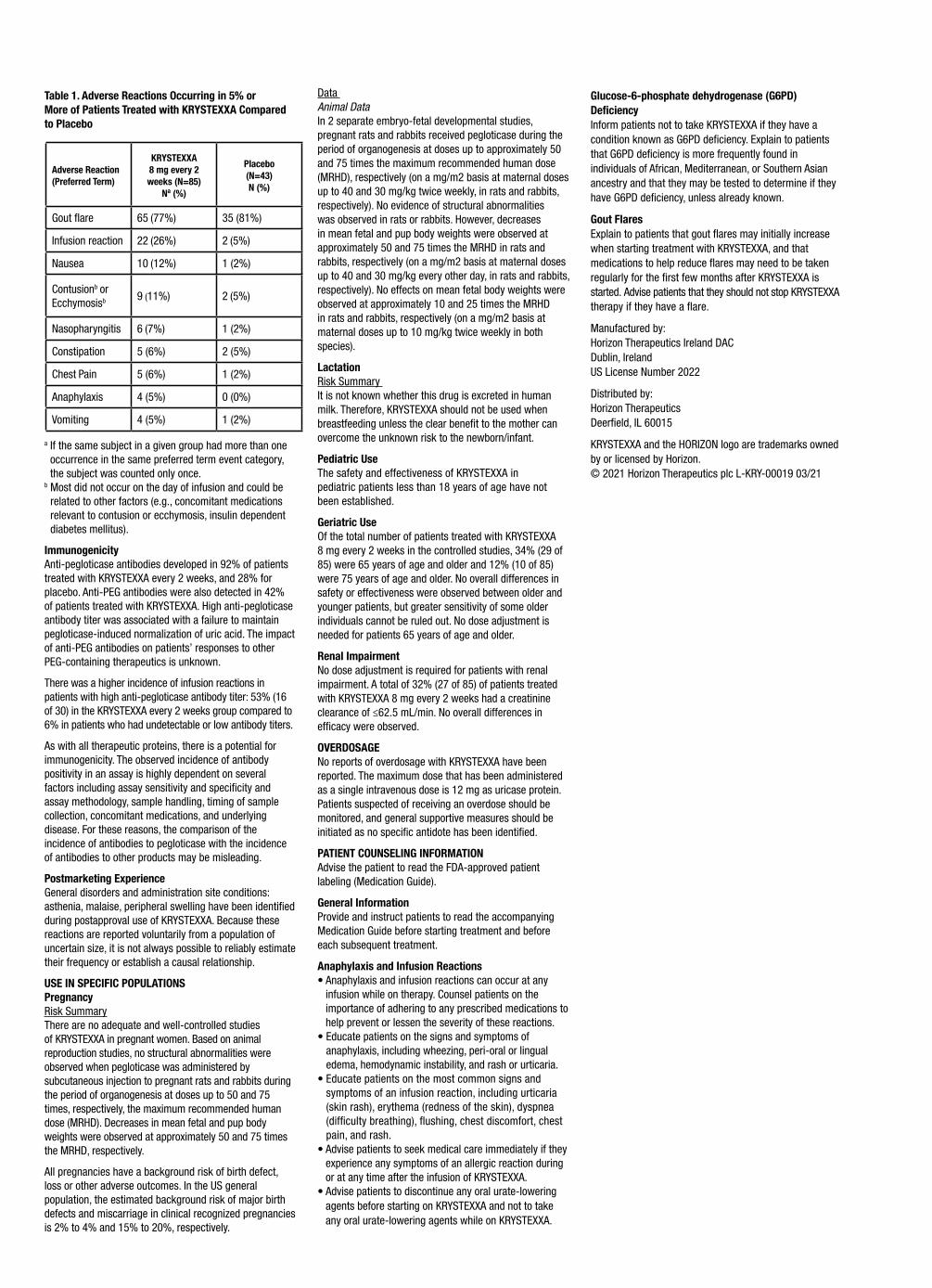
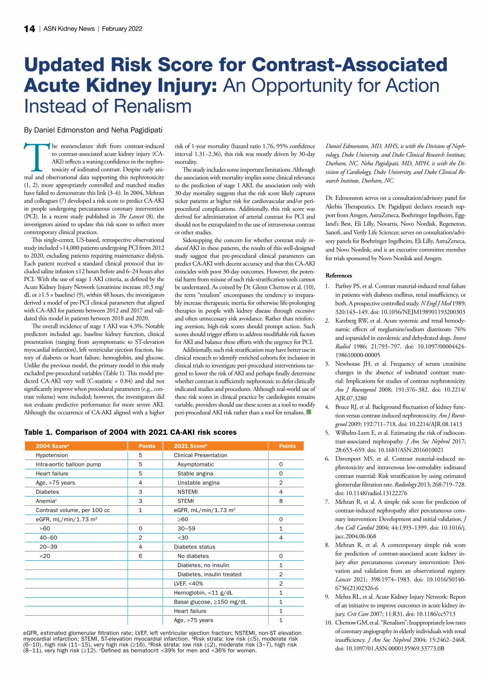
14 | ASN Kidney News | February 2022
T he nomenclature shift from contrast-induced to contrast-associated acute kidney injury (CA-AKI) reflects a waning confidence in the nephro-toxicity of iodinated contrast. Despite early ani-
mal and observational data supporting this nephrotoxicity (1, 2), more appropriately controlled and matched studies have failed to demonstrate this link (3−6). In 2004, Mehran and colleagues (7) developed a risk score to predict CA-AKI in people undergoing percutaneous coronary intervention (PCI). In a recent study published in The Lancet (8), the investigators aimed to update this risk score to reflect more contemporary clinical practices.
This single-center, US-based, retrospective observational study included >14,000 patients undergoing PCI from 2012 to 2020, excluding patients requiring maintenance dialysis. Each patient received a standard clinical protocol that in-cluded saline infusion ≤12 hours before and 6−24 hours after PCI. With the use of stage 1 AKI criteria, as defined by the Acute Kidney Injury Network (creatinine increase ≥0.3 mg/dL or ≥1.5 × baseline) (9), within 48 hours, the investigators derived a model of pre-PCI clinical parameters that aligned with CA-AKI for patients between 2012 and 2017 and vali-dated this model in patients between 2018 and 2020.
The overall incidence of stage 1 AKI was 4.3%. Notable predictors included age, baseline kidney function, clinical presentation (ranging from asymptomatic to ST-elevation myocardial infarction), left ventricular ejection fraction, his-tory of diabetes or heart failure, hemoglobin, and glucose. Unlike the previous model, the primary model in this study excluded pre-procedural variables (Table 1). This model pre-dicted CA-AKI very well (C-statistic = 0.84) and did not significantly improve when procedural parameters (e.g., con-trast volume) were included; however, the investigators did not evaluate predictive performance for more severe AKI. Although the occurrence of CA-AKI aligned with a higher
risk of 1-year mortality (hazard ratio 1.76, 95% confidence interval 1.31−2.36), this risk was mostly driven by 30-day mortality.
The study includes some important limitations. Although the association with mortality implies some clinical relevance to the prediction of stage 1 AKI, the association only with 30-day mortality suggests that the risk score likely captures sicker patients at higher risk for cardiovascular and/or peri-procedural complications. Additionally, this risk score was derived for administration of arterial contrast for PCI and should not be extrapolated to the use of intravenous contrast or other studies.
Sidestepping the concern for whether contrast truly in-duced AKI in these patients, the results of this well-designed study suggest that pre-procedural clinical parameters can predict CA-AKI with decent accuracy and that this CA-AKI coincides with poor 30-day outcomes. However, the poten-tial harm from misuse of such risk-stratification tools cannot be understated. As coined by Dr. Glenn Chertow et al. (10), the term “renalism” encompasses the tendency to irrepara-bly increase therapeutic inertia for otherwise life-prolonging therapies in people with kidney disease through excessive and often unnecessary risk avoidance. Rather than reinforc-ing aversion, high-risk scores should prompt action. Such scores should trigger efforts to address modifiable risk factors for AKI and balance these efforts with the urgency for PCI.
Additionally, such risk stratification may have better use in clinical research to identify enriched cohorts for inclusion in clinical trials to investigate peri-procedural interventions tar-geted to lower the risk of AKI and perhaps finally determine whether contrast is sufficiently nephrotoxic to defer clinically indicated studies and procedures. Although real-world use of these risk scores in clinical practice by cardiologists remains variable, providers should use these scores as a tool to modify peri-procedural AKI risk rather than a tool for renalism.
Daniel Edmonston, MD, MHS, is with the Division of Neph-rology, Duke University, and Duke Clinical Research Institute, Durham, NC. Neha Pagidipati, MD, MPH, is with the Di-vision of Cardiology, Duke University, and Duke Clinical Re-search Institute, Durham, NC.
Dr. Edmonston serves on a consultation/advisory panel for Akebia Therapeutics. Dr. Pagidipati declares research sup-port from Amgen, AstraZeneca, Boehringer Ingelheim, Egg-land’s Best, Eli Lilly, Novartis, Novo Nordisk, Regeneron, Sanofi, and Verily Life Sciences; serves on consultation/advi-sory panels for Boehringer Ingelheim, Eli Lilly, AstraZeneca, and Novo Nordisk; and is an executive committee member for trials sponsored by Novo Nordisk and Amgen.
References
1. Parfrey PS, et al. Contrast material-induced renal failure in patients with diabetes mellitus, renal insufficiency, or both. A prospective controlled study. N Engl J Med 1989; 320:143−149. doi: 10.1056/NEJM198901193200303
2. Katzberg RW, et al. Acute systemic and renal hemody-namic effects of meglumine/sodium diatrizoate 76% and iopamidol in euvolemic and dehydrated dogs. Invest Radiol 1986; 21:793−797. doi: 10.1097/00004424-198610000-00005
3. Newhouse JH, et al. Frequency of serum creatinine changes in the absence of iodinated contrast mate-rial: Implications for studies of contrast nephrotoxicity. Am J Roentgenol 2008; 191:376−382. doi: 10.2214/AJR.07.3280
4. Bruce RJ, et al. Background fluctuation of kidney func-tion versus contrast-induced nephrotoxicity. Am J Roent-genol 2009; 192:711−718. doi: 10.2214/AJR.08.1413
5. Wilhelm-Leen E, et al. Estimating the risk of radiocon-trast-associated nephropathy. J Am Soc Nephrol 2017; 28:653−659. doi: 10.1681/ASN.2016010021
6. Davenport MS, et al. Contrast material-induced ne-phrotoxicity and intravenous low-osmolality iodinated contrast material: Risk stratification by using estimated glomerular filtration rate. Radiology 2013; 268:719−728. doi: 10.1148/radiol.13122276
7. Mehran R, et al. A simple risk score for prediction of contrast-induced nephropathy after percutaneous coro-nary intervention: Development and initial validation. J Am Coll Cardiol 2004; 44:1393−1399. doi: 10.1016/j.jacc.2004.06.068
8. Mehran R, et al. A contemporary simple risk score for prediction of contrast-associated acute kidney in-jury after percutaneous coronary intervention: Deri-vation and validation from an observational registry. Lancet 2021; 398:1974−1983. doi: 10.1016/S0140-6736(21)02326-6
9. Mehta RL, et al. Acute Kidney Injury Network: Report of an initiative to improve outcomes in acute kidney in-jury. Crit Care 2007; 11:R31. doi: 10.1186/cc5713
10. Chertow GM, et al. “Renalism”: Inappropriately low rates of coronary angiography in elderly individuals with renal insufficiency. J Am Soc Nephrol 2004; 15:2462−2468. doi: 10.1097/01.ASN.0000135969.33773.0B
Table 1. Comparison of 2004 with 2021 CA-AKI risk scores
2004 Scorea Points 2021 Scoreb Points
Hypotension 5 Clinical Presentation
Intra-aortic balloon pump 5 Asymptomatic 0
Heart failure 5 Stable angina 0
Age, >75 years 4 Unstable angina 2
Diabetes 3 NSTEMI 4
Anemiac 3 STEMI 8
Contrast volume, per 100 cc 1 eGFR, mL/min/1.73 m2
eGFR, mL/min/1.73 m2 ≥60 0
>60 0 30−59 1
40−60 2 <30 4
20−39 4 Diabetes status
<20 6 No diabetes 0
Diabetes, no insulin 1
Diabetes, insulin treated 2
LVEF, <40% 2
Hemoglobin, <11 g/dL 1
Basal glucose, ≥150 mg/dL 1
Heart failure 1
Age, >75 years 1
Updated Risk Score for Contrast-Associated Acute Kidney Injury: An Opportunity for Action Instead of RenalismBy Daniel Edmonston and Neha Pagidipati
eGFR, estimated glomerular filtration rate; LVEF, left ventricular ejection fraction; NSTEMI, non-ST elevation myocardial infarction; STEMI, ST-elevation myocardial infarction. aRisk strata: low risk (≤5), moderate risk (6−10), high risk (11−15), very high risk (≥16). bRisk strata: low risk (≤2), moderate risk (3−7), high risk (8−11), very high risk (≥12). cDefined as hematocrit <39% for men and <36% for women.

February 2022 | ASN Kidney News | 15
“There's something special about chlorthalidone.” – Rajiv Agarwal, MD, as heard on “Freely Filtered”
The nephrology community was abuzz at ASN Kidney Week 2021 as Rajiv Agarwal presented the results of the Chlorthalidone in Chronic Kidney Disease (CLICK) trial, with simultane-
ous publication in The New England Journal of Medicine (1).In an attempt to refute the dogma that thiazide-like diu-
retics lose effectiveness at low estimated glomerular filtration rate (eGFR) (2), the CLICK trial enrolled 160 patients with stage 4 chronic kidney disease (CKD; eGFR 15 to <30 mL/min/1.73 m2) and uncontrolled hypertension—defined as a mean 24-hour ambulatory blood pressure monitoring (ABPM) of 130 mm Hg or higher (systolic BP [SBP]) or 80 mm Hg or higher (diastolic BP [DBP])—in a double-blind, randomized, placebo-controlled trial of chlorthalidone ver-sus placebo. The primary outcome was a change in 24-hour ABPM from baseline to 12 weeks.
Of the 160 subjects in the trial, the average age was in the mid-60s, 40% were Black race, and about three-quarters were male, with a similar number of subjects with diabetes mellitus. Subjects were taking an average of 3.4 antihyper-tensives (60% were on a loop diuretic), and the mean eGFR was 23.2 mL/min/1.73 m2.
With a starting dose of 12.5 mg once daily, the study dose was doubled every 4 weeks, up to a maximum dose of 50 mg, if the patient had a SBP or DBP ≥135 mm Hg or ≥85 mm Hg, respectively.
After only 4 weeks, with a mean dose of 11.5 mg daily, patients in the chlorthalidone group (n = 81) had a clinic SBP reduction of 9.2 mm Hg (vs. a rise of 2.7 mm Hg in the placebo group [n = 79]), and by 12 weeks, the mean dose was 23.1 mg, resulting in a decrease in SBP of 12.6 mm Hg (vs. a rise of 2.4 mm Hg in the placebo group). As seen in Figure 1, at 12 weeks, patients receiving chlorthalidone had a decrease in 24-hour ABPM—the primary outcome—of SBP 11 mm Hg and DBP 4.9 mm Hg (vs. 0.5 mm Hg and 1 mm Hg, respectively, in placebo). Notably, most of the antihypertensive effect occurred early (within 4 weeks) and at the starting dose (12.5 mg daily).
Also notable was the decrease in urinary albumin-to-creatinine ratio by 12 weeks, which was 52% in the chlo-rthalidone group versus 4% in the placebo group. This was a significant decrease that persisted even 2 weeks after the trial concluded. This finding confirms prior studies that indicate an antiproteinuric effect of thiazide(-like) diuretics as part of an antihypertensive regimen (3, 4).
Although the results of CLICK are practice changing and demonstrate the clear antihypertensive and antiproteinuric efficacy of chlorthalidone in advanced CKD, caution and discretion must be used when initiating this therapy. Sub-
jects receiving chlorthalidone were more likely to have an increase in serum creatinine, hypokalemia, hyponatremia, hypomagnesemia, hyperglycemia, hyperuricemia, and diz-ziness. The risk of a significant rise in creatinine was much higher for patients already on a loop diuretic, and in current practice, many of these patients may also be on a sodium glucose co-transporter 2 inhibitor, which has a mild diuretic effect. These side effects may be particularly notable for older adults.
CLICK extends the findings of other studies that have shown the BP and cardiovascular benefits of thiazide-like diuretics (i.e., chlorthalidone and indapamide) into a high-risk cohort of advanced CKD patients (5−7). Given the long half-life and potency, consideration of a low dose (e.g., 12.5 mg) and less frequent dosing (every other day or thrice weekly) may mitigate some concerns or side effects. Al-though CLICK did not establish an outcome benefit—such as a reduction in mortality or major adverse events—given its impressive BP-lowering results, it is time that treatment of hypertension in advanced CKD clicked with chlortha-lidone.
Jamie S. Hirsch, MD, is with the Division of Kidney Diseases and Hypertension, Department of Medicine, Donald and Bar-bara Zucker School of Medicine at Hofstra/Northwell, Great Neck, NY. Michael Turk, DO, is a PGY-2 Internal Medicine resident at Allegheny General Hospital, Pittsburgh, PA.
The author reports no conflicts of interest.
References
1. Agarwal R, et al. Chlorthalidone for hypertension in advanced chronic kidney disease. N Engl J Med 2021; 385:2507−2519. doi: 10.1056/NEJMoa2110730
2. Chertow GM, et al. “Renalism”: Inappropriately low rates of coronary angiography in elderly individuals with renal insufficiency. J Am Soc Nephrol 2004; 15:2462−2468. doi: 10.1097/01.ASN.0000135969.33773.0B
3. Vogt L, et al. Effects of dietary sodium and hydrochlo-rothiazide on the antiproteinuric efficacy of losartan. J Am Soc Nephrol 2008; 19:999−1007. doi: 10.1681/ASN.2007060693
4. Trujillo H, et al. The forgotten antiproteinuric proper-ties of diuretics. Am J Nephrol 2021; 52:435−449. doi: 10.1159/000517020
5. Kostis JB, et al. Association between chlorthalidone treatment of systolic hypertension and long-term sur-vival. JAMA 2011; 306:2588−2593. doi: 10.1001/jama.2011.1821
6. Olde Engberink RHG, et al. Effects of thiazide-type and thiazide-like diuretics on cardiovascular events and mor-tality. Hypertension 2015; 65:1033−1040. doi: 10.1161/HYPERTENSIONAHA.114.05122
7. Liang W, et al. Comparison of thiazide-like diuretics ver-sus thiazide-type diuretics: A meta-analysis. J Cell Mol Med 2017; 21:2634−2642. doi: 10.1111/jcmm.13205
“CLICK”ing with Chlorthalidone: Rediscovering an Old Drug for Advanced CKDBy Jamie S. Hirsch
Boosters for Transplant Patients: Some Reach Protective Antibody LevelsA third dose of the COVID-19 vaccine achieves protective antibody levels in nearly 40% of kidney transplant recipi-ents without a previous immune response, reports a study in JAMA Internal Medicine (1).
The single-center, single-blind randomized trial includ-ed 201 kidney transplant recipients who did not develop SARS-CoV-2 spike protein antibodies after two doses of the mRNA vaccine. Patients were assigned to either a heterolo-gous vaccination strategy using an Ad26COVS1 viral vector vaccine or a homologous strategy with a third dose of an
mRNA vaccine, either BNT162b2 or mRNA-1273. The main endpoint was seroconversion to detectable levels of SARS-CoV-2 spike protein antibodies within 4 weeks after the third dose.
Within 4 weeks after the third dose, 39% of patients developed protective levels of spike protein antibody. Se-roconversion rates were similar for the two strategies: 35% with mRNA vaccines and 42% with the vector vaccine. On interferon-γ release assays, just 17 patients had positive T-cell responses after the third dose.
Less than one-half of kidney transplant recipients develop protective levels of antibodies to the SARS-CoV-2 spike pro-tein after the standard two-dose course of the mRNA vaccine [Reindl-Schwaighofer R, et al. Comparison of SARS-CoV-2 antibody response 4 weeks after homologous vs heterologous third vaccine dose in kidney transplant recipients: A rand-omized clinical trial. JAMA Intern Med, published online ahead of print December 20, 2021. doi: 10.1001/jamain-ternmed.2021.7372; https://jamanetwork.com/journals/jamainternalmedicine/fullarticle/2787200].
@michaelturk6Agarwal et al., NEJM Nov. 2021PMID: 34739197
Conclusion: Among patients with advanced CKD and poorly controlled HTN, chlorthalidone therapy improved BP control at 12 weeks. RCT, randomized controlled trial; CI, confidence interval; sCr, serum creatinine; HTN, hypertension.
Cohort Methods Outcomes
Double-blind RCT
CKD4, and BP > 130/80 mm Hg on ≥1 drug
Chlorthalidonen = 81
−11.0 mm Hg −52% 59%
Primary OutcomeΔ blood pressure
Secondary OutcomeΔ urine albumin/Cr
25% ↑ in sCr on loop diuretic
Placebon = 79
−0.5 mm Hg −4% 13%
−10.5(95% CI −14.6 to −6.4)
P < 0.001
−50(95% CI −60 to −37)
P < 0.001
n = 16077% male, 58% White
Academic health systemin Indianapolis, IN, USA
12-w
eek
follo
w-u
p
OR 9.2(95% CI 3.0 to 31.3)
Avg. doseat 12 wk = 23.1 mg
12.5 mg start dose; doubled q4 weeks;
max of 50 mg
CLICK: Does chlorthalidone improve blood pressure in patients with chronic kidney disease and hypertension?
Figure 1.

16 | ASN Kidney News | February 2022
A s a specialty, nephrology is heavily dependent on international medical graduates (IMGs). According to the Association of American Medical Colleges (AAMC) (1), 65% of
nephrology fellows in 2019 were IMGs, the highest of any major internal medicine specialty. This has now led to more than one-half of the active workforce being gradu-ates of international medical schools (51%). Although the exact numbers of IMGs on a visa and the proportion of J-1s and H1-Bs are unknown, it is well recognized by local and national leaders that providing viable and satisfying solutions for entry into the workforce for a nephrology fellow on a visa has never been more crucial. According to a 2019 ASN survey (2), nearly 50% of IMG respondents reported having difficulty finding a satisfactory position, with major barriers being location of practice, adequate compensation, and visa requirements. These unfortunate numbers reflect the arduous path that a fully trained grad-uate on a J-1 visa embarks on while trying to commence a career in the United States.
Upon graduation, a J-1 trainee is mandated to go back to his or her home country for 2 years or obtain a waiver to stay in the United States; this can be obtained by work-ing in an underserved area or filing a claim for persecu-tion or hardship (3). In the current immigration climate, it is vital that fellowship program directors and division chiefs understand and have utmost clarity in the hiring process, so they can provide their trainees with direct and practical guidance, thereby lessening the trainees’ anxiety and distress. If you are a program director, division chief, or mentor, the following information is what you need to know (Figure 1).
STEP 1: Know your hospital or practice location.Enter your hospital address on the Health Resources & Services Administration’s website (https://data.hrsa.gov/tools/shortage-area/by-address), or search Health Profes-sional Shortage Area (HPSA) by address to find this tool.
Your entering the address provides two pieces of in-formation: 1) whether the location lies in a medically underserved area (MUA) or HPSA (noted as “MUA/P” [MUA or Population] and “Primary Care HPSA”; den-
tal and mental health HPSAs do not qualify) and 2) the HPSA score (1−25) for the location; the higher the score, the higher the priority.
Only one of the above (MUA or HPSA) is required for the location to be eligible for a J-1 waiver, and the county must be highlighted as green on the map. However, if the county in which the practice is located appears red, then it is not a MUA or HPSA location. This doesn’t mean the end of the road, however, as there are two options to ex-plore.
1 Is there a different practice location for the division or practice that can serve as the primary practice location for the new hire? These could be outpatient practices, smaller network hospitals, dialysis units, interventional suites, or a combination of the above. Check if these locations are green on the map.
2 FLEX spots: Most states allow eligibility if the practice in a “red” location can show that the new hire will take care of underserved patients from a “green” location. However, note that FLEX applicants are the lowest pri-ority for most states.
STEP 2: Know your state.Although it is impossible to keep up with requirements of every state, it is simple for employers to understand their home state. The easiest and most efficient way to accom-plish this is to have a conversation with an immigration lawyer or the liaison at the international office. It is im-portant to know the chances of approval for a particular practice and state. Your spending months on paperwork in a state where chances of approval for a particular specialty or location are low would not be prudent.
There are a few government agencies that sponsor visa waivers. Some, such as the Appalachian Regional Com-mission (ARC) or US Department of Health and Human Services (HHS), do not accept specialties like nephrol-ogy, and others, such as through the Veterans Affairs (VA) administration, are extremely hard to get or have limited spots. However, if the employer is located in eligible parts of 8 states (MO, IL, KY, TN, AR, LA, MS, and AL), the Delta Regional Authority (DRA) may be a potential spon-
sor. But, by far, the most common pathway is the Con-rad 30, a federal program that allows each state 30 such positions. Therein lies the problem, however: each state, regardless of size, population, or need, gets 30 positions. Texas and New York get 30, as do North Dakota and Wyoming.
Approval processThe following are some issues that play into approval chances:• Preference for primary care specialties (internal/family
medicine, Ob-Gyn, pediatrics, psychiatry): some states (e.g., NJ, NY, CA, etc.) only accept or allot most of their spots for primary care.
• Has the state recently granted FLEX waivers? If yes, how many? The maximum is 10 per state, but most states accept far fewer due to demand and ability to fill with non-FLEX (HPSA/MUA) applications.
• Allocation system: states use different methods to pick applicants, such as:o first-come, first-served basis (e.g., CO, AR, ID, etc.)o primary care first followed by priority ranking of
specialists by HPSA score or other rules (most com-mon)
o lottery system (e.g., FL, CT)o locally trained applicants or applicants with lan-
guage skills preferred in some stateso limits per employer in some states; thus, employers
asked to prioritize order of applications
The second part of approval is paperwork, which in-cludes the following:• Trainees cannot apply to more than one state at a time.
So it is important to understand state application dead-lines (typically September/October) and approval/re-jection deadlines in primary states and also in backup states. Backups are typically in states with late deadlines or those that never fill and accept rolling applications throughout the year.
• Paperwork may appear daunting, but requirements are straightforward. Quality legal representation is vital and typically costs around $5000−$10,000. Paperwork is a mere hindrance as long as planned in advance:o Some states require full medical licenses to be eligi-
ble to apply.o Most states require displaying recruitment efforts
for 6 months.o Some states require letters of support from commu-
nity physicians or organizations.o Some states have specific contract clauses, such as no
non-competes.
Once an application is picked by the state’s Depart-ment of Health, the subsequent progress through the US Department of State (DOS) and US Citizenship and Im-migration Services (USCIS) is uncomplicated. An H1-B visa is granted at the end of the process, and the employee is bound to the state and the practice for 3 years, a time during which a green card cannot be obtained. Some states (e.g., AL and GA) have heavy liquidation clauses of $250,000 if an employee decides to terminate his or her contract. Also, a job switch is near impossible during the 3 years and requires the employee to demonstrate extenuat-ing circumstances and even then, only allows a switch to another location within the same state that falls under the HPSA/MUA designation.
Under such trying circumstances, it is essential for
Hiring an International Medical Graduate on a J-1 Visa WaiverBy Harish Seethapathy
STEP 2: Know your state
Other factors to consider:State deadlines
Legal fees ($5,000-$10,000)
Paperwork/licensing
Preference for primary care specialties
Chances of FLEX approval
Allocation system:ü First come, first servedü HPSA scoreü Lotteryü Employer limits
Practice location not eligible but patients from HPSA/MUA
Hospital, outpatient units, dialysis units, interventional suites
HPSA/MUA FLEX (lower priority)
STEP 1: Know your practice location*
Seethapathy H. Hiring an international medical graduate (IMG) on a J1 visa waiver. ASN Kidney News 2022 Visual Abstract by Paolo Nikolai So, MD @nikkonephro
30 spotsper state Hiring IMGs on a J-1 Visa Waiver
HPSA: Health Professional Shortage AreaMUA: Medically Underserved Area
*https://data.hrsa.gov
Figure 1.

February 2022 | ASN Kidney News | 17
our leaders to understand the granularities of the process. Failure to find solutions comes at a great cost, with many specialty-trained physicians opting to work in hospital medicine or primary care when they are unable to find a reasonable waiver position or when their application is re-jected. Regardless of whether trainees are hired as faculty at their teaching hospital or at private community practices, leadership input will be valuable to trainees in helping to find positions that provide career paths in nephrology that are desirable, fulfilling, and sustainable in the long term.
Harish Seethapathy, MBBS, is an Assistant Physician with Massachusetts General Hospital and an instructor of medicine with Harvard Medical School, Boston, MA.
The author reports no conflicts of interest.
References
1. Association of American Medical Colleges (AAMC). Physician Specialty Data Report. Accessed November 11, 2021. https://www.aamc.org/data-reports/work-
force/report/physician-specialty-data-report2. Sozio SM, et al. 2019 Nephrology Fellow Survey Re-
sults and Insights. ASN Alliance for Kidney Health, 2019. Accessed November 11, 2021. https://www.asn-online.org/education/training/workforce/Neph-rology_Fellow_Survey_Report_2019.pdf
3. Neyra JA, et al. International medical graduates in nephrology: A guide for trainees and programs. Adv Chronic Kidney Dis 2020; 27:297−304.e1. doi: 10.1053/j.ackd.2020.05.003
Onco-nephrology is evolving as an impor-tant subspecialty in transplant care. Britta-ny Schreiber (BS), a renal fellow, interviews Kevin Fowler (KF), a kidney transplant recipi-ent, and Naoka Murakami (NM), a transplant nephrologist.
BS: Why are you interested in onco-nephrology?
KF: I received a preemptive kidney transplant in 2004, and due to chronic immunosuppression, I have had sev-eral episodes of cancer. Fortunately, all of the episodes were successfully resolved, but the pathway to treatment success has not always been clear. For example, when I was diagnosed with prostate cancer, I had to navigate a landscape where I felt alone. I received conflicting medi-cal opinions on the best treatment option. Eventually, my radiation oncologist recommended that I watch and wait. I was not satisfied with this direction, and I asked my radiation oncologist to take my case to a multidisci-plinary tumor board for discussion. After reviewing my case, the tumor board unanimously recommended treat-ment over watchful waiting.
The next challenge was determining the best treat-ment option for me. Eventually, a former colleague and friend, who was a transplant nephrologist, guided me to the best treatment option. This was accomplished by utilizing data derived from the Israel Penn International Transplant Tumor Registry. If I had been aware of on-co-nephrologists, who are skilled at managing cases like mine, I am confident that my process would have gone a lot smoother with reduced anxiety.
BS: What is transplant onco-nephrology, and what is the future of the field?
NM: Transplant onco-nephrology is a field that bridges the care between transplant nephrology and oncology. There are two focus areas (Figure 1):
1 pre-transplant evaluation for patients with a cancer history (e.g., multiple myeloma and other plasma cell dyscrasias) and
2 posttransplant cancer prevention and care for trans-plant recipients receiving cancer therapies (e.g., im-munotherapy and cell therapies).
As the population ages, and cancer therapies continue to improve, cancer history is becoming more common in transplant candidates. Although the American Society of Transplantation provides waitlist guidelines for those with a history of cancer (1, 2), challenges remain to pro-vide transplant opportunities for certain patients. Kidney
transplant recipients are also at a higher risk of cancer. Although the transplant field has made progress in im-proving cardiovascular outcomes (3), better cancer care remains a great unmet need with much opportunity for improvement. Kevin’s experience highlights the lack of data in this field and is a call to action for us to further research, raise awareness, and educate our colleagues and trainees on the risks and challenges of cancer in our pa-tients.
In the future, transplant onco-nephrologists can help increase access to transplant and improve strategies for posttransplant cancer prevention, diagnosis, and treat-ment, with the ultimate goal of reducing the cancer bur-den.
Brittany Schreiber, MD, and Naoka Murakami, MD, PhD, FASN, are affiliated with Brigham and Women’s Hospital, Boston, MA. Kevin Fowler is Principal, The Voice of the Patient, Inc., St. Louis, MO.
Drs. Schreiber and Murakami report no conflicts of in-terest. Kevin Fowler works with the following compa-nies: Akebia, Bayer, eGenesis, Gilead, Hansa Biopharma, Otsuka, Palladio Biosciences, Responsum for CKD, Ta-laris, and Travere Therapeutics.
References
1. Al-Adra DP, et al. Pretransplant solid organ malig-nancy and organ transplant candidacy: A consensus expert opinion statement. Am J Transplant 2021; 21:460−474. doi: 10.1111/ajt.16318
2. Al-Adra DP, et al. Preexisting melanoma and hema-tological malignancies, prognosis, and timing to solid organ transplantation: A consensus expert opinion statement. Am J Transplant 2021; 21:475−483. doi: 10.1111/ajt.16324
3. Saran R, et al. US Renal Data System 2018 Annual Data Report: Epidemiology of kidney disease in the United States. Am J Kidney Dis 2019; 73(3 Suppl 1):A7−A8. doi: 10.1053/j.ajkd.2019.01.001
Onco-nephrology in Transplant Care: Patient’s Voice and Call for Awareness and Action By Brittany Schreiber, Kevin Fowler, and Naoka Murakami
CCaanncceerr pprreevveennttiioonn//aawwaarreenneessss
- Risk-dependent managementof immunosuppression
- Recommendations on cancerscreening
- Patient education- Collaboration with oncologyteam
NNaavviiggaattiinngg ccaanncceerr ttrreeaattmmeenntt
- Knowledge of novel cancer therapies
- Immunotherapies- Cell therapy e.g., CAR-T, virus-specific celltherapy (BKV and EBV)
- Collaboration with oncology team
OOnnccoo--nneepphhrroollooggyy iinn kkiiddnneeyy ttrraannssppllaannttaattiioonn Key areas of onco-nephrology in transplant care
PPrree--ttrraannssppllaanntt PPoossttttrraannssppllaanntt
AAcccceessss ttoo ttrraannssppllaanntt
- History of plasma celldyscrasia, HSCT ( e.g., multiple myeloma,amyloidosis, MGRS)- Collaboration withoncology team on riskassessment ofrecurrence ofmalignancyposttransplant
KKeeyy FFaaccttss 44--55%% T ransplant candidates have history of cancer
33--110000xx H igher risk of cancer posttransplant
22..77xx H igher cancer-related death posttransplant
C ancer is the 33rrdd ((1166%%)) leading cause of deathposttransplant
Figure created by BioRender
Figure 1.
This figure highlights the important areas of focus and key facts about cancer in patients during pre- and posttransplant periods. HSCT, hematopoietic stem cell transplant; MGRS, monoclonal gammopathy with renal significance; CAR-T, chimeric antigen receptor-T cell; BKV, BK virus; EBV, Epstein-Barr virus.

18 | ASN Kidney News | February 2022
Nephrology is a relatively young specialty. It emerged in the second half of the 20th centu-ry, with the rise of kidney biopsy, dialysis, and transplantation. Although kidneys have been
studied since antiquity, stones and obstruction were a dom-inant focus. Urology books from 1739 mention the only treatment of anuria as bladder catheterization (1). So, how did the study of glomerular diseases rise as a cornerstone of our work? Let’s look at some of the important milestones in the birth of our understanding of glomerulonephritis.
2000 BC: Where does urine come from, the bladder?As early as 2000 BC, Egyptian priest physicians proclaimed that urine was formed by “the power that is inherent in the particles about the region of the bladder,” a kind of purifica-tion process. A number of prescriptions for the treatment of hematuria—frequency, retention, and infection—have been described in ancient Egyptian medical papyri. This shows that kidney diseases were widely prevalent since an-cient times (2).
460−375 BC: Examination of urine holds important clues.Hippocrates was born to a family of physicians on the Greek island of Kos (3). Some of his teachings were pre-served in the library of Alexandria. It is said that the library was burned, and whatever texts survived then became known as the “Hippocratic Corpus.” Although it is con-sidered a single corpus of Hippocratic medicine, it varies in content, age, style, and methods practiced. Hence, the au-thorship is not exactly known. Let’s revisit some of the origi-nal descriptions of the kidney from Hippocrates: Dropsy is an old term derived from “hydrops,” used to describe swell-
ing of the soft tissues due to accumulation of water. Hippo-crates, in one of his travel texts on “Air, Water, and Places,” wrote about quartan malaria being associated with dropsy. Watson and Clark have recently deciphered that quartan malaria causes nephrotic syndrome (4). In his work “About Inner Sufferings,” Hippocrates uses the term “nephritis” to describe hematuria, strangury (vesical tenesmus), and ol-iguria. He attributed it to infection and overconsumption of starch—probably a description of present-day post-in-fectious glomerulonephritis. He emphasized a lot on urine examination and stated that the presence of frothy urine was suggestive of chronic disease (proteinuria) (3).
1st−2nd centuries AD: Urine comes from the kidneys.A famous physician in the Roman empire was a Greek named Galen. He showed, for the first time, that urine is formed by the kidneys and not the bladder. He conducted an animal experiment where he tied both the ureters and demonstrated that the ureters enlarged and were almost ready to burst. Upon releasing the tied ureters, urine was seen gushing into the bladder. Galen emphasized keen clin-ical observation of the patient and documented the same (case reports and case series). This gave birth to and encour-aged the idea of medical research (5).
8th−14th centuries AD: Examining urineExamination of the urine persisted throughout this period, with contrasting views on its legitimacy. The famous and greatest physician of Islam, Isaac Ebreus (880−940 AD) wrote in his book Guida Medicorum (Guide for Physicians) about the importance of urine examination and fixed the rules of “uroscopy.” Thus, uroscopy became an important and fundamental diagnostic tool in Salerno, Italy, at a fa-
mous medical school at that time. In contrast, Roman and Greek physicians considered examination of pulse as more important than urine. In the dawn of the Enlightenment, however, many looked down on the technique of urine ex-amination as unscientific divination (6).
17th century: The glorious glomerulus discoveredThe work of three Italians—Marcello Malpighi (1628−1694), Lorenzo Bellini (1643−1704), and Giovan Battista Morgagni (1682−1771)—defined “glomerulus” in medical history.
Malpighi, an Italian anatomist referred to as the “father of physiology and embryology,” lent his name to the Mal-pighian corpuscles (renal corpuscle consists of glomerulus and Bowman’s capsule) and the Malpighian pyramids of the kidney (cone-shaped tissue in the cortex of the kidney) (7). At the young age of 20, physician and anatomist Bellini had already begun research on the kidneys and described the papillary ducts, known to us as the “ducts of Bellini” (7).
18th century: Proteinuria definedThe first scientific demonstration of proteinuria can be at-tributed to Domenico Cotugno (1736−1822), an Italian physician to the king of Naples. In 1765, Cotugno noticed that upon heating the urine of a soldier with dropsy, it turned white like a coagulated egg (“ovi albuminis persimi-lem”) (8).
19th century: Putting it all together: Kidney diseaseThe work of Dr. Richard Bright, Guys Hospital, London (1789−1858), was a milestone in the development of nephrology. He compiled the observations of kidney dis-eases thus far and inferred that there was an association among dropsy, coagulable urine, and kidney disease. His book, Reports of Medical Cases, explained the consequenc-es of kidney diseases. He combined clinical history with proteinuria and postmortem examination of the kidneys. Upon postmortem examination of the kidneys, he classified them into three categories: soft kidneys with yellow mot-tling (nephrotic syndrome); granulated with white, opaque, interstitial deposits (acute glomerulonephritis); and small, rough, and hard contracted kidneys (end stage). Due to his extensive scientific work and research in the field of kidney diseases, he was called the “father of nephrology,” and any type of kidney disease was called “Bright’s disease” (9, 10).
Bright’s disease was a common term used for all kidney diseases in Europe and the United States for over a cen-tury. Several physicians were working on the pathogenesis of Bright’s disease. Whereas some physicians hypothesized that the disease developed in several sequential steps, others sought to classify it into acute and chronic Bright’s disease. Acute Bright’s disease (acute glomerulonephritis) caused the kidneys to swell and increased the chances of mortality, whereas chronic Bright’s disease (chronic glomerulonephri-tis) caused the kidneys to shrink, and death was due to a different cause (7).
Pierre-François Olive Rayer (1793−1867), a French academic physician at the Charité Hospital in Paris, and the doctor to King Louis Philippe and Emperor Napoleon III, published the first textbook on renal disease: Traité des maladies des reins (1839−1841). Rayer described in detail “albuminous nephritis” (the equivalent of Bright’s disease) and for the first time, differentiated it from “suppurative ne-phritis” (due to ascending infection from the urinary tract or bloodborne) (11).
Urine microscopy dates back to 1630, and renowned microscopists Robert Hooke and Herman Boerhaave both examined urine. The development of the achromatic lens by Charles-Louis Chevalier (1804–1859, France) signifi-cantly advanced microscopic studies of urine and autopsied renal tissue. Among others, Rudolph Virchow (1821–1902, Germany) introduced the concept of “parenchymatous ne-phritis,” and Friedrich G. J. Henle (1809–1885, Prague)
The Birth of Understanding GlomerulonephritisBy Mythri Shankar
Hippocrates. Engraving based on drawing by Peter Paul Rubens. National Library of Medicine, Bethesda, MD (CC by 4.0).
Portrait of Richard Bright from Thomas Joseph Pettigrew, Medical Portrait Gallery, vol. 2 (1838).
Glomerular Disease Corner

February 2022 | ASN Kidney News | 19
studied tubulointerstitial disease in patients with albuminu-ria. James Tyson (1841–1919, North America) correlated urine examination findings of epithelial cells and red blood cells with severe glomerular inflammation, causing rupture of the capillary basement membrane (12, 13).
During the same time, William Bowman (1816–1892, United Kingdom) and Carl Ludwig (1816–1895, Germa-ny) contributed significantly to the growing understanding of renal physiology. The theory of urine formation—glo-merular filtration, tubular secretion, and reabsorption—was explained for the first time. “Bowman’s capsule” of the nephron is named after Bowman (12, 13).
19th century: Glomerulonephritis definedEdwin T. Klebs (1834−1913, Germany) coined the term “glomerulonephritis” in an 1870 handbook of anatomical pathology, essentially describing a case of mesangial prolif-eration (12). The term nephritis was still widely used by other physicians to describe inflammation of the glomeru-lus.
20th century: Kidney pathology emerged to begin to define distinct entities. Three centuries after Malpighi first observed the structure of the glomerulus, percutaneous kidney biopsy, immu-nofluorescence, and electron microscopy were developed almost simultaneously.
In 1944, a needle biopsy of the kidney was performed for the first time on 13 patients by Nils Alwall from Lund, Sweden. Unfortunately, one patient died due to a com-plication of the kidney biopsy. After this episode, Alwall stopped performing kidney biopsies and did not publish his work until 1952. Poul Iversen and Claus Brun from Copenhagen continued Alwall’s efforts and published a case series of kidney biopsies in 1951 (14, 15).
Jean Hamburger (1909−1992), a French physician-surgeon, set up a renal unit (Necker Hospital, Paris) from the humble ruins of World War II. He kick-started the renal unit with minimal infrastructure and very few sup-porting staff. He was interested in the clinicopathological studies and morphological varieties of glomerulonephritis. Soon, a new era of kidney research began with Hamburg-er. In 1960, he proposed “nephrology” (meaning the study of kidneys in Greek) as a specialty to evaluate and treat patients with kidney disease. He founded the International Society of Nephrology (ISN) and convened the first suc-cessful International Congress of Nephrology in Geneva (September 1960) (16).
We have come a long way since the early description of nephritis. These tremendous advances have been possible as a result of the collaboration of scientists and research-ers; the dissemination of information through journals and national and international conferences; and most importantly, learning from our past experiences. Review-ing history shows that a particular field can advance only with scientific contributions, new technical developments, and economic support. Recent insights into pathobiology and genetics of glomerular diseases have paved the way for future research and development of therapeutic strategies (15).
Mythri Shankar is Assistant Professor, Department of Neph-rology, Institute of Nephro-urology, Bengaluru, India, and a GlomCon Education Committee Member (2021−2022) and GlomCon Fellow (2020−2021).
The author reports no conflicts of interest.
References1. Al-Attar BA. History of development of nephrology.
Saudi J Kidney Dis Transpl 1996; 7:373−377. https://www.sjkdt.org/article.asp?issn=1319-2442;year=1996;volume=7;issue=4;spage=373;epage=377;aulast=Al-Attar
2. Salem ME, Eknoyan G. The kidney in ancient Egyp-tian medicine: Where does it stand? Am J Nephrol 1999; 19:140–147. doi: 10.1159/000013440
3. Eknoyan G. Origins of nephrology: Hippocrates, the father of clinical nephrology. Am J Nephrol 1988; 8:498−507. doi: 10.1159/000167669
4. Hendrickse RG, Adeniyi A. Quartan malarial nephrotic syndrome in children. Kidney Int 1979; 16:64−74. doi: 10.1038/ki.1979.103
5. Retsas S. Galen’s “errors.” Lancet 2010; 376:686. doi: 10.1016/S0140-6736(10)61337-2; https://www.thelancet.com/journals/lancet/article/PIIS0140-6736(10)61337-2/fulltext
6. Dal Canton A, Castellano M. Theory of urine forma-tion and uroscopic diagnosis in the Medical School of Salerno. Kidney Int 1988; 34:273−277. doi: 10.1038/ki.1988.176
7. Stratta P, et al. The concept of ‘glomerulonephritis.’ The fascinating history of evolution and emergence of a specialist’s nosology focus on Italy and Torino. Am J Nephrol 1999; 19:83–91. doi: 10.1159/000013431
8. Fogazzi GB, et al. The Urinary Sediment: An Integrated View. 1993; Milano, Masson, p 1.
9. Ritz E, et al. French and German nephrologists in the mid-19th century. Am J Nephrol 1989; 9:167–172. doi: 10.1159/000167958
10. Peitzman SJ. From dropsy to Bright’s disease to end-stage
renal disease. Milbank Q 1989; 67 (Suppl 1):16−32. https://www.milbank.org/quarterly/articles/from- dropsy-to-brights-disease-to-end-stage-renal-disease/
11. Richet G. From Bright’s disease to modern nephrol-ogy: Pierre Rayer’s innovative method of clinical inves-tigation. Kidney Int 1991; 39:787−792. doi: 10.1038/ki.1991.96
12. Weening JJ, Jennette JC. Historical milestones in re-nal pathology. Virchows Arch 2012; 461:3−11. doi: 10.1007/s00428-012-1254-7
13. Eknoyan G. On the etymology of nephritis: A his-torical appraisal of its origins. J Am Soc Nephrol 2020; 31:1170−1173. doi: 10.1681/ASN.2019050510
14. Iversen P, Brun C. Aspiration biopsy of the kidney. Am J Med 1951; 11:324−330. https://www.amjmed.com/article/0002-9343(51)90169-6/fulltext
15. Jhaveri KD, Fishbane S. Glomerular diseases: Entering a new era. Clin J Am Soc Nephrol 2014; 9:598−599. doi: 10.2215/CJN.06110613
16. Richet G. Jean Hamburger 1909–1992. Kidney Int 1992; 42:810−812. doi: 10.1038/ki.1992.351

Methotrexate (MTX) has been used for treat-ment of connective tissue disorders, includ-ing rheumatoid arthritis. In much higher doses, MTX is used for various hematologic
and oncologic disorders (1). Renal elimination accounts for 70%−90% of the clearance of MTX (2). High-dose intra-venous MTX has the potential for causing kidney injury by crystal precipitation within the renal tubules (3, 4). In addition, oral MTX can potentially accumulate in patients with reduced kidney function and lead to toxic effects, such as myelosuppression and hepatotoxicity (5). However, al-though MTX is contraindicated in patients with a creatinine (Cr) clearance of <30 mL/min, a renal safety profile of low-dose MTX (LD-MTX) is not well established.
The Cardiovascular Inflammation Reduction Trial (CIRT) was a randomized, double-blind, multi-site, place-bo-controlled trial of LD-MTX used for prevention of car-diovascular events due to atherosclerotic disease. In a recent paper published by Sparks et al. (6), the authors performed a secondary analysis using data from CIRT to assess the im-pact of LD-MTX on the estimated glomerular filtration rate (eGFR) and renal adverse events.
In this study by Sparks et al. (6), 4786 subjects were included, with 2391 randomized to the LD-MTX group and 2395 to the placebo arm. eGFR was calculated using the Chronic Kidney Disease Epidemiology Collaboration (CKD-EPI) equation (7). The primary outcome was change in eGFR (∆eGFR) from baseline. The two groups were simi-lar in age, sex, and race representation; stage of CKD (stages 1−3); as well as proportion of patients with diabetes mellitus and hypertension. Median follow-up was 23 months, and median drug dose was 16 mg/week. The least mean squares ∆eGFR from baseline through the follow-up period was statistically significant and favored LD-MTX in the modi-fied intention-to-treat analysis (0.93 mL/min/1.73 m2 [95% CI, 0.45–1.40, p = 0.001]), intention-to-treat analysis (0.94
mL/min/1.73 m2 [95% CI 0.46−1.41, p = 0.001]), and after removing the race coefficient from the CKD-EPI equation (0.93 mL/min/1.73 m2 [95% CI 0.46−1.40, p = 0.001]).
The eGFR slope difference between LD-MTX and pla-cebo was noted to be 1.06 mL/min/1.73 m2 per year (95% CI 0.60−1.51, p < 0.001). At the end of the study, there was a higher percentage of patients in the placebo group with CKD stage 3 overall (placebo: 10.4% vs. LD-MTX: 7.5%, p = 0.004) and CKD stage 3B or worse (placebo: 1.7% vs. LD-MTX: 0.9%, p = 0.029). Renal adverse events were classified (based on change in serum Cr [sCr]) as mild (sCr 1.5−2 × baseline), moderate (sCr 2.0−3 × baseline), or severe (sCr ≥ 3 × baseline). Most of the events were mild (138 in the LD-MTX group vs. 184 in the placebo group), with severe events being noted in two in the LD-MTX arm and four in the placebo arm.
The study was well designed and allowed for a robust sta-tistical analysis to support the results. The data suggest that LD-MTX is safe and well tolerated from a renal standpoint in patients with normal kidney function and in patients with mild to moderate renal impairment (stages 1−3 CKD). In addition, the study found a significant trend toward pres-ervation of eGFR with use of LD-MTX compared with placebo. The study raises an intriguing question regarding the potential mechanism behind the observed renoprotec-tive effect of LD-MTX: Could LD-MTX affect a variety of biologic pathways to slow down the decline of eGFR? There may be a possible role for suppression of inducible nitric oxide synthase (iNOS) activity by MTX; iNOS activity has been reported to be increased in various kidney diseases. Ex-perimental studies have demonstrated a significant decrease in NO production in mouse models of collagen-induced ar-thritis with the use of MTX, which was associated with a de-crease in kidney dysfunction and parenchymal damage (8).
CIRT excluded individuals with systemic rheumatologi-cal diseases. Hence, an important question from the study is
whether the safety and adverse-events data can be extrapo-lated to subjects with rheumatological diseases. As eGFR drops, there may be an increased risk of hematological ad-verse events—in particular, leucopenia—with LD-MTX. This has been shown in a recent study by Lee et al. (5), who investigated the systemic toxicity of MTX in patients with rheumatoid arthritis and renal impairment (classified as pre-viously developed vs. newly developed; the latter group in-cluded subjects with renal dysfunction developing after more than 3 months of exposure to MTX). The most common systemic adverse event was leucopenia in the newly devel-oped group versus the previously developed group (n = 10, 15.2% vs. n = 2, 3.7%, p = 0.038).
Although renal safety was the primary focus of the study by Sparks et al. (6), overall safety of MTX with a decline in eGFR may be a limiting factor for safe use of LD-MTX in this patient population. And, proteinuria is a strong prog-nostic marker of kidney dysfunction and renal survival. Be-cause proteinuria was not studied as a clinical factor in this study, the impact of LD-MTX on proteinuria is not known. Prospective, well-designed future studies are needed to vali-date the results from this study over a longer duration of fol-low-up and to examine the impact of MTX on proteinuria with CKD.
Bhavna Bhasin-Chhabra, MD, is with the Division of Neph-rology, Medical College of Wisconsin, Milwaukee. Juan Carlos Q. Velez, MD, is with the Department of Nephrology, Ochsner Clinic Foundation, New Orleans, LA, and Ochsner Clinical School, The University of Queensland, Brisbane, Australia.
The authors report no conflicts of interest. Dr. Velez has served as a consultant and an advisor for Mallinckrodt Phar-maceuticals, a consultant for Bayer and Travere Therapeu-tics, and a speaker for Otsuka Pharmaceutical. None of the products manufactured by these companies are discussed in this manuscript.
References
1. Khan ZA, et al. Methotrexate: A detailed review on drug delivery and clinical aspects. Expert Opin Drug Deliv 2012; 9:151−169. doi: 10.1517/17425247.2012.642362
2. Widemann BC, Adamson PC. Understanding and managing methotrexate nephrotoxicity. Oncologist 2006; 11:694−703. doi: 10.1634/theoncologist.11-6-694
3. Mallipattu SK, Ross MJ. Methotrexate in the urine. Kid-ney Int 2011; 80:226. doi: 10.1038/ki.2011.97
4. Howard SC, et al. Preventing and managing tox-icities of high-dose methotrexate. Oncologist 2016; 21:1471−1482. doi: 10.1634/theoncologist.2015-0164
5. Lee JS, et al. Methotrexate-related toxicity in patients with rheumatoid arthritis and renal dysfunction. Rheu-matol Int 2020; 40:765−770. doi: 10.1007/s00296-020-04547-y
6. Sparks JA, et al. Effect of low-dose methotrexate on eGFR and kidney adverse events: A randomized clini-cal trial. J Am Soc Nephrol 2021; 32:3197−3207. doi: 10.1681/ASN.2021050598
7. Levey AS, et al. A new equation to estimate glomerular filtration rate. Ann Intern Med 2009; 150:604−612. doi: 10.7326/0003-4819-150-9-200905050-00006
8. Budancamanak M, et al. Protective effects of thymoqui-none and methotrexate on the renal injury in collagen-induced arthritis. Arch Toxicol 2006; 80:768−776. doi: 10.1007/s00204-006-0094-0
20 | ASN Kidney News | February 2022
Impact of Low-Dose Methotrexate on Estimated Glomerular Filtration RateBy Bhavna Bhasin-Chhabra and Juan Carlos Q. Velez
Figure 1.

In a 670 patient study funded by the International Society of Nephrology, the South Africa Medical Research Council and the University of Witwatersrand, Johannesburg, South Africa, the Nova Point of Care StatSensor Creatinine/eGFR meter was more accurate than the central laboratory IDMS-traceable Jaffe methodology in estimating GFR when both methods were compared to MEASURED GFR (iohexol).1
• StatSensor measurements showed less proportional and constant error than respective IDMS Jaffe measurements when compared to iohexol measured GFR (mGFR).1
• StatSensor showed better accuracy than the IDMS Jaffe methodology at identifying patients with mGFR’s <90 mL/min/1.73 m2.1
• Of particular interest in the study, StatSensor showed better accuracy than the laboratory Jaffe methodology in the 60-89 mL min/1.73 m2 range, where individuals with early disease may benefit from renal protective measures.1
Point of Care Creatinine/eGFR Method is More Accurate than Laboratory Method:
Large Medical Center Study
novabiomedical.com
Nova Biomedical StatSensor Creatinine Meter
1. George J et al. Evaluating chronic kidney disease in rural South Africa: comparing estimated glomerular filtration rate using point of care to iohexol measured GFR. CCLM 2021.
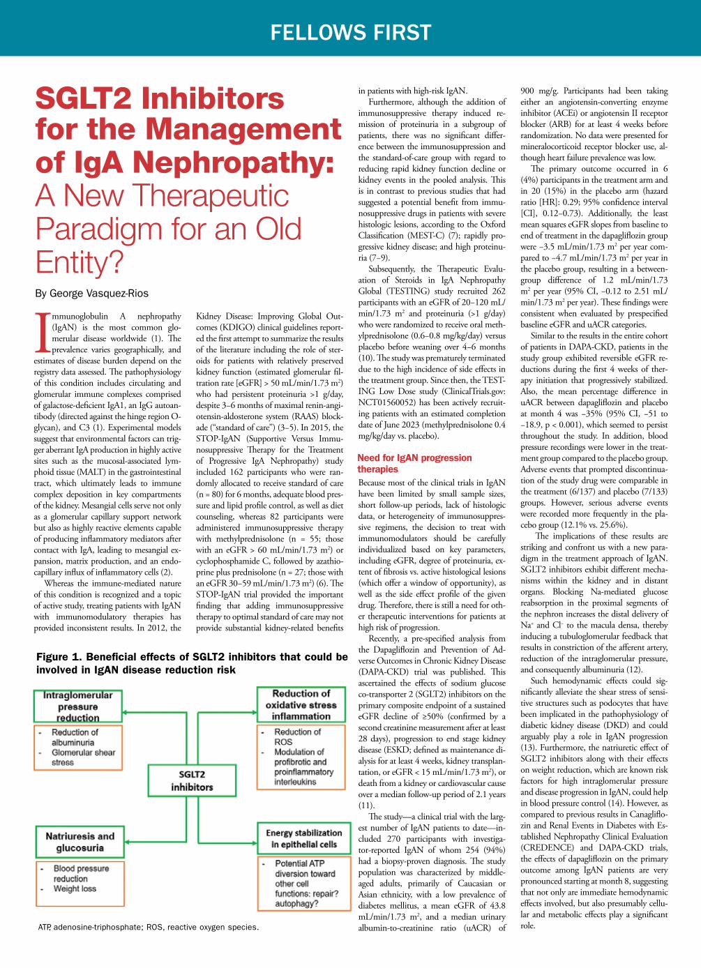
22 | ASN Kidney News | February 2022
Immunoglobulin A nephropathy (IgAN) is the most common glo-merular disease worldwide (1). The prevalence varies geographically, and
estimates of disease burden depend on the registry data assessed. The pathophysiology of this condition includes circulating and glomerular immune complexes comprised of galactose-deficient IgA1, an IgG autoan-tibody (directed against the hinge region O-glycan), and C3 (1). Experimental models suggest that environmental factors can trig-ger aberrant IgA production in highly active sites such as the mucosal-associated lym-phoid tissue (MALT) in the gastrointestinal tract, which ultimately leads to immune complex deposition in key compartments of the kidney. Mesangial cells serve not only as a glomerular capillary support network but also as highly reactive elements capable of producing inflammatory mediators after contact with IgA, leading to mesangial ex-pansion, matrix production, and an endo-capillary influx of inflammatory cells (2).
Whereas the immune-mediated nature of this condition is recognized and a topic of active study, treating patients with IgAN with immunomodulatory therapies has provided inconsistent results. In 2012, the
Kidney Disease: Improving Global Out-comes (KDIGO) clinical guidelines report-ed the first attempt to summarize the results of the literature including the role of ster-oids for patients with relatively preserved kidney function (estimated glomerular fil-tration rate [eGFR] > 50 mL/min/1.73 m2) who had persistent proteinuria >1 g/day, despite 3−6 months of maximal renin-angi-otensin-aldosterone system (RAAS) block-ade (“standard of care”) (3−5). In 2015, the STOP-IgAN (Supportive Versus Immu-nosuppressive Therapy for the Treatment of Progressive IgA Nephropathy) study included 162 participants who were ran-domly allocated to receive standard of care (n = 80) for 6 months, adequate blood pres-sure and lipid profile control, as well as diet counseling, whereas 82 participants were administered immunosuppressive therapy with methylprednisolone (n = 55; those with an eGFR > 60 mL/min/1.73 m2) or cyclophosphamide C, followed by azathio-prine plus prednisolone (n = 27; those with an eGFR 30−59 mL/min/1.73 m2) (6). The STOP-IgAN trial provided the important finding that adding immunosuppressive therapy to optimal standard of care may not provide substantial kidney-related benefits
in patients with high-risk IgAN.Furthermore, although the addition of
immunosuppressive therapy induced re-mission of proteinuria in a subgroup of patients, there was no significant differ-ence between the immunosuppression and the standard-of-care group with regard to reducing rapid kidney function decline or kidney events in the pooled analysis. This is in contrast to previous studies that had suggested a potential benefit from immu-nosuppressive drugs in patients with severe histologic lesions, according to the Oxford Classification (MEST-C) (7); rapidly pro-gressive kidney disease; and high proteinu-ria (7−9).
Subsequently, the Therapeutic Evalu-ation of Steroids in IgA Nephropathy Global (TESTING) study recruited 262 participants with an eGFR of 20−120 mL/min/1.73 m2 and proteinuria (>1 g/day) who were randomized to receive oral meth-ylprednisolone (0.6−0.8 mg/kg/day) versus placebo before weaning over 4−6 months (10). The study was prematurely terminated due to the high incidence of side effects in the treatment group. Since then, the TEST-ING Low Dose study (ClinicalTrials.gov: NCT01560052) has been actively recruit-ing patients with an estimated completion date of June 2023 (methylprednisolone 0.4 mg/kg/day vs. placebo).
Need for IgAN progression therapiesBecause most of the clinical trials in IgAN have been limited by small sample sizes, short follow-up periods, lack of histologic data, or heterogeneity of immunosuppres-sive regimens, the decision to treat with immunomodulators should be carefully individualized based on key parameters, including eGFR, degree of proteinuria, ex-tent of fibrosis vs. active histological lesions (which offer a window of opportunity), as well as the side effect profile of the given drug. Therefore, there is still a need for oth-er therapeutic interventions for patients at high risk of progression.
Recently, a pre-specified analysis from the Dapagliflozin and Prevention of Ad-verse Outcomes in Chronic Kidney Disease (DAPA-CKD) trial was published. This ascertained the effects of sodium glucose co-transporter 2 (SGLT2) inhibitors on the primary composite endpoint of a sustained eGFR decline of ≥50% (confirmed by a second creatinine measurement after at least 28 days), progression to end stage kidney disease (ESKD; defined as maintenance di-alysis for at least 4 weeks, kidney transplan-tation, or eGFR < 15 mL/min/1.73 m2), or death from a kidney or cardiovascular cause over a median follow-up period of 2.1 years (11).
The study—a clinical trial with the larg-est number of IgAN patients to date—in-cluded 270 participants with investiga-tor-reported IgAN of whom 254 (94%) had a biopsy-proven diagnosis. The study population was characterized by middle-aged adults, primarily of Caucasian or Asian ethnicity, with a low prevalence of diabetes mellitus, a mean eGFR of 43.8 mL/min/1.73 m2, and a median urinary albumin-to-creatinine ratio (uACR) of
900 mg/g. Participants had been taking either an angiotensin-converting enzyme inhibitor (ACEi) or angiotensin II receptor blocker (ARB) for at least 4 weeks before randomization. No data were presented for mineralocorticoid receptor blocker use, al-though heart failure prevalence was low.
The primary outcome occurred in 6 (4%) participants in the treatment arm and in 20 (15%) in the placebo arm (hazard ratio [HR]: 0.29; 95% confidence interval [CI], 0.12−0.73). Additionally, the least mean squares eGFR slopes from baseline to end of treatment in the dapagliflozin group were −3.5 mL/min/1.73 m2 per year com-pared to −4.7 mL/min/1.73 m2 per year in the placebo group, resulting in a between-group difference of 1.2 mL/min/1.73 m2 per year (95% CI, −0.12 to 2.51 mL/min/1.73 m2 per year). These findings were consistent when evaluated by prespecified baseline eGFR and uACR categories.
Similar to the results in the entire cohort of patients in DAPA-CKD, patients in the study group exhibited reversible eGFR re-ductions during the first 4 weeks of ther-apy initiation that progressively stabilized. Also, the mean percentage difference in uACR between dapagliflozin and placebo at month 4 was −35% (95% CI, −51 to −18.9, p < 0.001), which seemed to persist throughout the study. In addition, blood pressure recordings were lower in the treat-ment group compared to the placebo group. Adverse events that prompted discontinua-tion of the study drug were comparable in the treatment (6/137) and placebo (7/133) groups. However, serious adverse events were recorded more frequently in the pla-cebo group (12.1% vs. 25.6%).
The implications of these results are striking and confront us with a new para-digm in the treatment approach of IgAN. SGLT2 inhibitors exhibit different mecha-nisms within the kidney and in distant organs. Blocking Na-mediated glucose reabsorption in the proximal segments of the nephron increases the distal delivery of Na+ and Cl− to the macula densa, thereby inducing a tubuloglomerular feedback that results in constriction of the afferent artery, reduction of the intraglomerular pressure, and consequently albuminuria (12).
Such hemodynamic effects could sig-nificantly alleviate the shear stress of sensi-tive structures such as podocytes that have been implicated in the pathophysiology of diabetic kidney disease (DKD) and could arguably play a role in IgAN progression (13). Furthermore, the natriuretic effect of SGLT2 inhibitors along with their effects on weight reduction, which are known risk factors for high intraglomerular pressure and disease progression in IgAN, could help in blood pressure control (14). However, as compared to previous results in Canagliflo-zin and Renal Events in Diabetes with Es-tablished Nephropathy Clinical Evaluation (CREDENCE) and DAPA-CKD trials, the effects of dapagliflozin on the primary outcome among IgAN patients are very pronounced starting at month 8, suggesting that not only are immediate hemodynamic effects involved, but also presumably cellu-lar and metabolic effects play a significant role.
FELLOWS FIRST
SGLT2 Inhibitors for the Management of IgA Nephropathy: A New Therapeutic Paradigm for an Old Entity?By George Vasquez-Rios
Figure 1. Beneficial effects of SGLT2 inhibitors that could be involved in IgAN disease reduction risk
ATP, adenosine-triphosphate; ROS, reactive oxygen species.

Pleiotropic effects of SGLT2 inhibitors include modulation of inflammatory and profibrotic mediators and regulation of toxic intracellular compounds (i.e., advanced gly-cation end products), among others, as dem-onstrated in models of type 2 diabetes melli-tus (15). However, the role of these factors in the pathogenesis of IgAN is less certain. It is accepted that currently employed immuno-suppressive strategies lack conclusive efficacy data, as there is a high-risk toxicity profile. Interestingly, it is possible that addressing the non-immune component of IgAN could represent a safe and effective strategy for kidney preservation while the risks and benefits of immunosuppressive therapies are discussed (Figure 1). This could be particu-larly important in patients without evidence of active and severe histological features, as defined by the MEST-C score (not available in DAPA-CKD). However, further studies are needed.
The pre-specified analysis of DAPA-CKD had some limitations that are worth men-tioning. This study included patients who were on a “stable” dose of RAAS blockers 4 weeks prior to enrollment without detail if the given therapy was maximized. Therefore, it is unclear how much improvement could have been elicited by adding SGLT2 inhibi-tors to the medical regimen in patients with maximal standard therapy (14). Further-more, it is difficult to ascertain how much of the benefits seen in the treatment group could be attributed to weight loss and blood pressure reduction, which are attainable with other less expensive and evidence-supported medications.
Certainly, recommendations on the RAAS blockade in IgAN are supported by small studies that have shown reduction in proteinuria and less kidney function dete-rioration. Nonetheless, none of the ACEi and/or ARB trials have shown the significant biomarker stabilization and outcome im-provements demonstrated by dapagliflozin (16−18). Moreover, the study population in this pre-specified analysis seems to exhibit a high risk for kidney disease progression. When observing the cumulative incidence of the primary endpoint among the patients in the placebo group by month 32, approxi-mately 24% had experienced a combination of sustained eGFR reduction ≥50%, progres-sion toward ESKD, or death from a kidney or cardiovascular cause, which suggests that there was a high rate of rapid progressors in the latter group.
Therefore, the effectiveness of dapagli-flozin as a co-adjuvant therapy in high-risk patients should be carefully examined. Im-portant areas of uncertainty include the safety of using SGLT2 inhibitors in patients with IgAN treated with immunosuppression (excluded in DAPA-CKD) and whether this class could be similarly beneficial among patients with lower levels of albuminuria. Although the results remained consistent when stratified by eGFR (≥45 or <45 mL/min/1.43 m2) and uACR (>1000 or <1000 mg/g per day), the effects of SGLT2 inhibi-tors should be carefully ascertained in both rapid and slow progressors. Moreover, it is necessary that future studies include a more heterogeneous population such as patients of African ancestry, who are documented to
have increased risk for kidney progression and who have been largely underrepresented in IgAN studies (3, 19, 20).
Despite the aforementioned caveats, SGLT2 inhibitors continue to serve as an attractive therapeutic option for a vast num-ber of patients with kidney disease. Clinical trials such as DAPA-CKD are changing the way we understand kidney disease and “rais-ing the bar” for other candidate therapies in this field. The investigators deserve recogni-tion for designing DAPA-CKD as the first event-driven trial of an SGLT2 inhibitor that included patients with CKD due to a broad range of etiologies such as IgAN. SGLT2 in-hibitors could be considered as an “add-on” therapy when stabilization of clinical param-eters is still needed despite optimal standard of care. Alternatively, they could be used in patients who are intolerant to RAAS block-ers.
Future studies should evaluate the ef-fects of SGLT2 inhibitors in a larger popu-lation of patients whose standard therapy is optimal to uncover their true potential and to evaluate their safety profile when immu-nosuppressive therapy is concomitantly ad-ministered. Finally, although The Study of Heart and Kidney Protection with Empagli-flozin (EMPA-KIDNEY; ClinicalTrials.gov: NCT03594110) trial should reveal the effi-cacy and tolerability of SGLT2 inhibitors in patients with non-diabetic CKD, dedicated IgAN trials are very much needed to contin-ue advancing our knowledge of this condi-tion and individualized interventions.
George Vasquez-Rios, MD, is with the Division of Nephrology, Department of Internal Medi-cine, Icahn School of Medicine at Mount Sinai, New York, NY.
Acknowledgment: The author would like to express his gratitude to Drs. Miriam Chung, Kirk N. Campbell, Joseph A. Vassalotti, and Jaime Uribarri at the Icahn School of Medicine at Mount Sinai for their valuable feedback at the time of preparing this manu-script.
The author reports no conflicts of interest.
References1. Rodrigues JC, et al. IgA nephropathy.
Clin J Am Soc Nephrol 2017; 12:677−686. doi: 10.2215/CJN.07420716
2. Coppo R. Corticosteroids in IgA ne-phropathy: Lessons from recent studies. J Am Soc Nephrol 2017; 28:25−33. doi: 10.1681/ASN.2016060647
3. Radhakrishnan J, Cattran DC. The KDIGO practice guideline on glomeru-lonephritis: Reading between the (guide)lines—application to the individual pa-tient. Kidney Int 2012; 82:840−856. doi: 10.1038/ki.2012.280
4. Pozzi C, et al. Corticosteroids in IgA nephropathy: A randomised controlled trial. Lancet 1999; 353:883−887. doi: 10.1016/s0140-6736(98)03563-6
5. Manno C, et al. Randomized controlled clinical trial of corticosteroids plus ACE-inhibitors with long-term follow-up in proteinuric IgA nephropathy. Nephrol Dial Transplant 2009; 24:3694−3701 [published correction appears in Nephrol Dial Transplant 2010; 25:1363−1364]. doi: 10.1093/ndt/gfp356
6. Rauen T, et al. Intensive supportive care plus immunosuppression in IgA nephropathy. N Engl J Med 2015; 373:2225−2236. doi: 10.1056/NEJ-Moa1415463
7. Barbour SJ, et al. The MEST score provides earlier risk prediction in IgA nephropathy. Kidney Int 2016; 89:167−175. doi: 10.1038/ki.2015.322
8. Ballardie FW, Roberts ISD. Controlled prospective trial of prednisolone and cy-totoxics in progressive IgA nephropathy. J Am Soc Nephrol 2002; 13:142−148. doi: 10.1681/ASN.V131142
9. Vecchio M, et al. Immunosuppres-sive agents for treating IgA nephropa-thy. Cochrane Database Syst Rev 2015; CD00396. doi: 10.1002/14651858.CD003965.pub2
10. Lv J, et al. Effect of oral methylpredni-solone on clinical outcomes in patients with IgA nephropathy: The TEST-ING randomized clinical trial. JAMA 2017; 318:432−442. doi: 10.1001/jama.2017.9362
11. Wheeler DC, et al. A pre-specified analysis of the DAPA-CKD trial dem-onstrates the effects of dapagliflozin on major adverse kidney events in pa-tients with IgA nephropathy. Kidney Int
2021; 100:215−224. doi: 10.1016/j.kint.2021.03.033
12. Sen T, Heerspink HJL. A kidney per-spective on the mechanism of action of sodium glucose co-transporter 2 inhibi-tors. Cell Metab 2021; 33:732−739. doi: 10.1016/j.cmet.2021.02.016
13. Cravedi P, Remuzzi G. Pathophysiology of proteinuria and its value as an out-come measure in chronic kidney disease. Br J Clin Pharmacol 2013; 76:516−523. doi: 10.1111/bcp.12104
14. Barratt J, Floege J. SGLT-2 inhibition in IgA nephropathy: The new standard of care? Kidney Int 2021; 100:24−26. doi: 10.1016/j.kint.2021.04.002
15. Vasquez-Rios G, Nadkarni GN. SGLT2 inhibitors: Emerging roles in the protec-tion against cardiovascular and kidney disease among diabetic patients. Int J Nephrol Renovasc Dis 2020; 13:281−296. doi: 10.2147/IJNRD.S268811
16. Coppo R, et al. IgACE: A placebo-controlled, randomized trial of angi-otensin-converting enzyme inhibitors in children and young people with IgA nephropathy and moderate proteinuria. J Am Soc Nephrol 2007; 18:1880−1888. doi: 10.1681/ASN.2006040347
17. Li PK-T, et al. Hong Kong study using valsartan in IgA nephropathy (HKVIN): A double-blind, randomized, place-bo-controlled study. Am J Kidney Dis 2006; 47:751−760. doi: 10.1053/j.ajkd.2006.01.017
18. Praga M, et al. Treatment of IgA ne-phropathy with ACE inhibitors: A randomized and controlled trial. J Am Soc Nephrol 2003; 14:1578−1583. doi: 10.1097/01.asn.0000068460.37369.dc
19. Papeta N, et al. APOL1 variants in-crease risk for FSGS and HIVAN but not IgA nephropathy. J Am Soc Nephrol 2011; 22:1991−1996. doi: 10.1681/ASN.2011040434
20. Sehic AM, et al. Increased recogni-tion of IgA nephropathy in African-American children. Pediatr Nephrol 1997; 11:435−437. doi: 10.1007/s004670050311
FELLOWS FIRST February 2022 | ASN Kidney News | 23

Rapid Drop in COVID-19 Antibodies for Dialysis Patients
Similar Long-Term Outcomes with DCD versus DBD Kidneys
Belzutifan Shows Activity against Renal Cancers in VHL Disease
Starting SGLT2 Treatment Lowers AKI Risk in Type 2 DiabetesA nationwide data analysis confirms that SARS-CoV-2 anti-
body responses to vaccination decrease rapidly in dialysis pa-tients, leaving them at risk of breakthrough infection, reports Annals of Internal Medicine.
The researchers analyzed real-world data on 4791 pa-tients receiving care in a large US network of dialysis facili-ties. Residual plasma from routine monthly tests performed at a central laboratory was used to measure qualitative and quantitative antibodies to the SARS-CoV-2 receptor-binding domain (RBD). The analysis began in January 2021, before COVID-19 vaccines were widely available. By mid-Septem-ber 2021, 2563 patients were fully vaccinated.
Trends in antibody levels were assessed, including the pos-sible association between antibody titers and the risk of break-through COVID-19 infection. Among vaccinated patients, the estimated rate of undetectable RBD responses increased from 6.6% at 14 to 30 days after vaccination to 20.2% at 5 to 6 months. Median index values decreased from 91.9 to 8.4, respectively.
During follow-up, clinically documented COVID-19 oc-curred in 2% of fully vaccinated dialysis patients, compared with 3% of partially vaccinated (one dose) and 11% of un-vaccinated patients. In a nested case-control analysis, each breakthrough case was matched to five controls for age, sex, and vaccination month, with adjustment for diabetes status and region.
The analysis included 56 patients with breakthrough in-fections, with samples collected a median of 21 days before diagnosis. Waning of the antibody response was significantly associated with the risk of breakthrough infection. Compared with an index RBD range of 23 or higher (reflecting an an-tibody level of 506 binding antibody units per milliliter), rate ratios for breakthrough infection were 11.6 at pre-break-through RBD values of less than 10 and 6.0 at values between 10 and 23. Peak antibody responses were higher for patients with evidence of previous SARS-CoV-2 infection, although this difference lessened during follow-up.
SARS-CoV-2 antibody levels after vaccination are “strong-ly associated” with the risk of breakthrough COVID-19 in-fection in dialysis patients, the study concludes. The findings have implications for efforts to define a “persisting antibody” threshold for protection against COVID-19, which may be especially important for high-risk or immunocompromised patients. The researchers note that 40% of dialysis patients with breakthrough infections were hospitalized [Anand S, et al. SARS-CoV-2 vaccine antibody response and break-through infection in patients receiving dialysis. Ann Intern Med, published online ahead of print December 14, 2021. doi: 10.7326/M21-4176; https://www.acpjournals.org/doi/10.7326/M21-4176].
Even after decades of follow-up, kidneys transplanted after circulatory determination of death (DCD) show similar outcomes to kidneys donated after brain death (DBD), re-ports a study in Nephrology Dialysis Transplantation.
Of 1133 kidney transplants performed between 1985 and 2000 at the authors’ Swiss medical center, 122 used DCD grafts. The DCD kidney recipients—74 men and 48 women, median age 46 years—were matched one to one for sex, age, and transplant year to patients receiving DBD grafts during the same period. Outcomes were assessed through 2020.
At 35 years’ follow-up, median graft survival was almost identical between groups: 24.5 years for DCD recipients versus 23 years for DBD recipients. Delayed graft function was more common in DCD recipients—47 patients—compared with 23 patients after DBD transplants. How-ever, there were no long-term differences in graft or patient survival.
Among patients with more than 20 years of graft surviv-al, measures of graft function were similar between groups. The slope of change in glomerular filtration rate was −0.6
mL/min/year in the DCD group and −0.3 mL/min/year in the DBD group. Creatinine levels were 133 versus 119 μmol, and proteinuria was 370 versus 240 mg per 24 hours, respectively.
Kidneys donated after cardiovascular death are an im-portant source of organs for transplantation. Studies have reported similar outcomes for DCD and DBD kidneys up to 10 years, but there are few data on longer-term outcomes.
This 35-year follow-up study shows similar graft survival and excellent function with DCD versus DBD kidneys. Good outcomes are achieved despite the higher rate of de-layed graft function in DCD organs. The researchers con-clude: “[O]ur results indicate that criteria for selecting grafts for deceased kidney transplantation should not be based on the type of organ donation” [Müller A, et al. Long-term out-comes of transplant kidneys donated after circulatory death. Nephrol Dial Transplant, published online ahead of print December 17, 2021. doi: 10.1093/ndt/gfab358; https://academic.oup.com/ndt/advance-article/doi/10.1093/ndt/gfab358/6468756].
Renal cell and non-renal cell carcinomas associated with von Hippel−Lindau (VHL) disease show evidence of response to the hypoxia-inducible factor inhibitor belzutifan, reports a study in The New England Journal of Medicine.
The phase 2, open-label trial included 61 adults with VHL disease, with diagnosis based on the presence of germ- line VHL alterations and at least one renal cell carcinoma measuring at least 10 mm. All patients were treated with bel-zutifan, a novel oral hypoxia-inducible factor 2α (HIF-2α) inhibitor, at a dose of 120 mg/day. Complete or partial ob-jective responses were assessed by an independent radiology review committee, following standard criteria. Responses of non-renal cell cancers, which included pancreatic lesions in
all patients, were also analyzed, along with safety outcomes.The patients were 32 men and 29 women, median age
41 years. All but 2 had undergone previous surgery or abla-tive procedures, with a median of 4 procedures per patient. Median follow-up was 21.8 months.
The objective response rate in patients with renal cell carcinoma was 49%. All of these were partial responses; an-other 49% of patients had a best response of stable disease. Among evaluable patients with partial responses, the median linear growth rate was 4.1 mm per year before belzutifan versus −5.6 mm per year on treatment. Responses were also observed for non-renal cancers: 47 of 61 for pancreatic can-cers (77%) and 15 of 50 for central nervous system heman-
For older adults with type 2 diabetes, treatment with a so-dium glucose cotransporter-2 inhibitor (SGLT2i) is associ-ated with a lower risk of acute kidney injury (AKI), com-pared with other antidiabetic medications, according to a pre-proof paper in the American Journal of Kidney Diseases.
The population-based cohort study used Medicare fee-for-service data on more than 417,000 patients, aged 66 years or older, with type 2 diabetes. All enrolled patients had a newly filled prescription for an SGLT2i, a dipeptidyl peptidase 4 inhibitor (DPP-4i), or a glucagon-like peptide-1 receptor agonist (GLP-1RA) from 2013 through 2017. New SGLT2i users were propensity score matched to new DPP-4i or GLP-1RA users. Cox proportional hazards anal-yses were performed for the primary outcome of hospitaliza-tion for AKI, as either the primary or secondary discharge diagnosis.
Analyses included approximately 68,000 matched pairs for comparison of patients initiating SGLT2i versus DPP-4i treatment and 71,000 pairs for comparison of SGLT2i versus GLP-1RA. Mean patient age was 72 years. In both comparisons, about 65% of patients starting SGLT2i treat-ment received canagliflozin. About three-fourths of patients were prescribed metformin, whereas nearly one-third were prescribed insulin.
In both comparisons, SGLT2i treatment was associ-ated with a lower rate of AKI hospitalization. Incidence rate per 1000 patient-years was 19.6 with SGLT2i versus 27.8 with DPP-4i; hazard ratio (HR) 0.71. For SGLT2i versus GLP-1RA, incidence rates were 21.7 versus 27.1 per 1000
patient-years; HR 0.81. Secondary outcomes also favored SGLT2i treatment,
including a reduced risk of AKI hospitalization requiring dialysis: HR 0.39 versus DPP-4i and 0.56 versus GLP-1RA. The results were also consistent across a range of sensitivity analyses. The data also confirmed the known associations of SGLT2i treatment with an increased risk of diabetic ketoaci-dosis and a reduced risk of hospitalization for heart failure.
SGLT2i treatments have been shown to have a wide range of benefits for patients with type 2 diabetes, including decreased rates of kidney disease progression and death from renal or cardiovascular causes. However, these medications are also associated with an acute reduction in glomerular filtration rate, raising concerns for a potential increase in the risk of AKI. Based on postmarketing data, the US Food and Drug Administration has issued warnings that SGLT2i treatment might cause AKI.
The new population-based study may alleviate those concerns. The authors find that the risk of AKI hospitaliza-tion is lower in older patients with type 2 diabetes who initi-ate SGLT2i treatment compared with DPP-4i or GLP-1RA treatment. Although acknowledging the limitations of the analysis, the researchers conclude: “Our results add to the available evidence on the safety profile of SGLT2i in older adults” [Zhuo M, et al. SGLT2 inhibitors and the risk of acute kidney injury in older adults with type 2 diabetes. Am J Kidney Dis, published online ahead of print November 8, 2021. doi: 10.1053/j.ajkd.2021.09.015; https://www.ajkd.org/article/S0272-6386(21)00953-7/fulltext].
Findings

Dapagliflozin Reduces Risk of Abrupt Kidney Function Declines in CKDFor high-risk patients with chronic kidney disease (CKD) and substantial albuminuria, dapagliflozin reduces the in-cidence of abrupt declines in kidney function, according to an analysis of randomized trial data in Kidney International.
The study was a prespecified analysis of the Dapagli-flozin and Prevention of Adverse Outcomes in Chronic Kidney Disease (DAPA-CKD) trial. Enrolled patients had CKD with an estimated glomerular filtration rate of 25 to 75 mL/min/1.73 m2 and a urinary albumin-to-creatinine ratio of 200 to 5000 mg/g. The analysis included matched groups of 2152 patients assigned to the sodium glucose cotransporter-2 (SGLT2) inhibitor dapagliflozin 10 mg/day or placebo.
The main outcome of interest was an abrupt decline in kidney function, defined as doubling of serum creatinine
over two subsequent study visits. Investigator reports of seri-ous adverse events related to acute kidney injury (AKI) were examined as well.
Median follow-up was 2.4 years. Doubling of serum cre-atinine occurred in 2.9% of patients assigned to dapaglifloz-in versus 4.2% in the placebo group; hazard ratio 0.68. The reduction in abrupt declines in kidney function remained significant after accounting for competing risk of mortality and across baseline subgroups. Rates of AKI-related serious adverse events were similar between groups: 2.5% with da-pagliflozin and 3.2% with placebo.
The main results of the DAPA-CKD trial showed that dapagliflozin reduced the risks of kidney failure and heart failure hospitalization, whereas it prolonged survival in patients with CKD. It is important to understand how
SGLT2 inhibitors affect the risk of AKI in patients with CKD and albuminuria.
The DAPA-CKD data show that dapagliflozin reduces the risk of abrupt declines in kidney function in this popu-lation. “These data support the favorable benefit-risk profile of dapagliflozin, and endorse the revised [KDIGO] clini-cal practice guidelines recommending the use of SGLT2 inhibitors in patients with CKD,” the researchers write. A dedicated trial will be needed to evaluate dapagliflozin as a therapeutic option to prevent AKI [Heerspink HJL, et al. A pre-specified analysis of the Dapagliflozin and Prevention of Adverse Outcomes in Chronic Kidney Disease (DA-PA-CKD) randomized controlled trial on the incidence of abrupt declines in kidney function. Kidney Int 2022; 101:174-184. doi: 10.1016/j.kint.2021.09.005].
February 2022 | ASN Kidney News | 25
gioblastomas (30%).Adverse events included anemia in 90% of patients and
fatigue in 66%. Treatment was discontinued in 7 patients, voluntarily in 4 of them.
Patients with VHL disease are at high risk of renal cell carcinoma as a result of VHL gene inactivation and constitu-
tive activation of HIF-2α. Some effective form of systemic therapy could be of benefit by controlling tumor growth and reducing the burden of surgery.
The study demonstrates activity of HIF-2α inhibition with belzutifan against renal cell and non-renal cell carcino-mas associated with VHL disease. Side effects are common
but generally low grade. Although acknowledging the study’s limitations, the authors point out that randomized trials are unlikely due to the lack of other non-surgical treatments for VHL disease [Jonasch E, et al. Belzutifan for renal cell car-cinoma in von Hippel–Lindau disease. N Engl J Med 2021; 385:2036–2046. doi: 10.1056/NEJMoa2103425].
A Call to Action for Physicians: Become Informed and Empowered, and Begin to Heal ThyselfBy Stephen J. Thomas
Can you recall a more trying time for phy-sicians? Burnout and moral injury among physicians were on the uptick prior to the COVID-19 pandemic. Doctor shortages,
the burden of exorbitant student loans, longer work hours, worsening administrative requirements, and dys-functional and disparate electronic medical records were all taking a professional, and often personal, toll. Now, as we enter the third year of the pandemic, many physi-cians are thoroughly exhausted and deflated, and they are digging deep to find their resilience.
How did we arrive here? Clearly, the answer is com-plex and multi-factorial, and not all of the challenges facing physicians were foreseeable or controllable—al-though many were and remain so. (Perhaps this is why doctors are adding “learned helplessness” to the list of problems afflicting our community.) Thus, instead of asking ourselves “How did we arrive here?” the more im-portant question may be “Why do we stay here?”
The answer begins with the understanding—or lack of understanding—of our individual and collective val-ues. What value does a physician bring to his or her pa-tients, profession, colleagues, and employer? What value does a specialty bring to a health care system and the elusive goal of providing high-quality, affordable, and well-coordinated medical care? Finally, how does physi-cian value translate into worth, as reflected in compensa-tion, workload, call schedule, benefits, and other terms of employment?
Nephrologists care for some of the sickest and most complex patients. They work across multiple specialties
to treat underlying medical problems, trying to halt ad-vancing kidney disease. They intensely manage the ap-proximately 780,000 Americans who ultimately develop end stage kidney disease. They direct dialysis units serv-ing over half a million people and manage over a quarter of a million people living with a kidney transplant. They make themselves available day and night, addressing medical emergencies. Many in academia attempt to bal-ance their clinical activities with teaching and research responsibilities. Consequently, who determines what these contributions are worth?
Thereupon enters the resource-based relative value scale (RBRVS): the physician payment system used by the Centers for Medicare & Medicaid Services (CMS) and other payers. The RBRVS system established relative value units (RVUs), which determine physicians’ com-pensation for their services and the resources required to provide them. The RVS is determined by the RVS Update Committee (RUC). The RUC is a group of 32 physicians and other health care professionals who ad-vise CMS on how to value various medical services. The advice of the RUC is nearly always accepted by CMS, yet nephrology is not currently represented on the com-mittee.
Whether you are an employed nephrologist or in your own private practice, it is unrealistic to think a Current Procedural Terminology (CPT) code, and associated RVU, accurately reflects your value. Even if an RVU was cable of capturing the complexity and effort associated with a single type of patient interaction, it cannot capture the interaction’s downstream value. Almost every patient interaction results in blood work, imaging studies, renal biopsies, or interventional radiology or surgical consulta-tions for placement or creation of dialysis catheters, fistu-las, or grafts. How is this value captured?
US medicine and the industries that orbit it have a physician valuation problem, and the trickle-down ef-fects are impacting the quality of care we deliver to our patients.
How do we fix it? At Phairify, we believe solving the physician value dilemma will be a long journey, and it is our mission to show you the correct azimuth. First, we engage physicians to help them understand the problem and to appreciate how inaction promulgates it. Second, we are encouraging doctors to come together around a common purpose to create change. Third, we believe physicians can, and should, assert control over generating
accurate, timely, and specialty-specific value information. Fourth, we offer a physician-first alternative to the cur-rent employer-oriented and directed marketplace.
Phairify has innovated a digital platform designed to inform physicians of their value and empower them to build their best careers. On Phairify’s platform, physi-cians anonymously and collectively share value infor-mation. The information is timely, multi-dimensional, and filterable, enabling doctors to understand how their current employment situation compares with peers. Em-powered with this information, physicians then use the platform to anonymously explore the job marketplace and direct a fair balance between their value and worth.
It is time for physicians to decide if they will follow Albert Einstein’s famous witticism, which suggests that by changing nothing, we can still hope for a different out-come. Or will we accept a call to action, define our own value, and begin to level our professional playing field?
Stephen J. Thomas, MD, is an infectious diseases physician-scientist who treats adults at SUNY Upstate Medical Uni-versity in Syracuse, NY. He chairs a basic science depart-ment and directs a global health and translational sciences institute. Along with his partners, Dr. Thomas co-founded Phairify.
Dr. Thomas’ entrepreneurial activities include CEO and co-founder of Cormac Life Sciences, which services a disabled veteran-owned small business and provides biomedical R&D consulting services, and co-founder and Director of Strategy for Phairify, a SaaS startup and digital human resources platform. His consulting arrangements (2021−2022) include the following: Pfiz-er—vaccine advisory board, compensated for time; Sa-nofi Pasteur—vaccine advisory board, compensated for time; Merck—chair, dengue vaccine scientific advisory committee, compensated for time; Takeda—dengue vaccine case adjudication committee and dengue vac-cine consultant, compensated for time; Clover Biophar-ma—COVID-19 vaccine case adjudication committee, compensated for time; Icosavax—COVID-19 and Zika vaccines data safety monitoring board, compensated for time; Moderna—chair, Zika vaccine data safety moni-toring board, compensated for time; PrimeVax—scien-tific advisory board member, compensated for time with equity; and Island Pharmaceuticals—scientific advisory board member, no compensation to date.

Take Control of Your Career with Phairify
ASN is partnering with Phairify to provide members with new career management tools. Get complimentary access to Phairify’s exclusive nephrology-specific compensation and productivity data that can level the negotiation playing field for physicians.
Learn more at www.asn-online.org/phairify or scan the QR code with your mobile device.
February 2022 | ASN Kidney News | 27
Index to AdvertisersAmerican College of Rheumatology . . . . . . . . Page 19
Baxter . . . . . . . . . . . . . . . . . . . . . . . . . . . . . . . . . Page 7
Bayer . . . . . . . . . . . . . . . . . . . . . . . . . . . . . . . Pages 2-4
Horizon . . . . . . . . . . . . . . . . . . . . . . . . . . . .Pages 10-13
Natera . . . . . . . . . . . . . . . . . . . . . . . . . . . . . Back Cover
Nova Biomedical . . . . . . . . . . . . . . . . . . . . . . . Page 21
I wish to start/renew a FREE* subscription to Kidney News
7-digit number label (Required for change of name/address only)
Name
Address
City State Zip
Telephone Fax
Email Address
Signature Date
Title/Position Physician Researcher RN, CNN, NM, LPN, APN, PA Dialysis Center Director Administration Clinic Manager/Coordinator Social Work Other
Specialty Area General Nephrology Transplantation Dialysis Laboratory Other
Institution Hospital <100 beds Hospital 100-250 beds Hospital 251-500 beds Hospital > 500 beds Dialysis Center Clinical Lab Other
Please Circle Degree: MD MD/PhD DO PhD MBA RN MS BS Other_________
Free Subscriber Service Request Card
Return the completed form to: Bob Henkel, 1401 H Street NW, #900, Washington, DC 20005 or Fax: 202-403-3615 or Email: [email protected]
KidneyNews
Nephrologist Cambridge, Somerville and Everett, MA
Cambridge Health Alliance, an award-winning public healthcare system, is recruiting a Nephrologist to join our Department of Medicine. CHA provides innovative care through an established network of outpatient clinics, two full service hospitals and urgent care services. CHA is a teaching affiliate of both Harvard Medical School and Tufts University School of Medicine.
● Full time nephrologist (will consider part-time) to provide outpatient care in our Medical Specialties clinics at Everett Hospital with rotations at Cambridge Hospital
● Incoming physician will provide nephrology consult and call coverage as part of CHA’s inpatient services
● This position will include resident and medical student teaching ● Incoming physician should possess excellent clinical/communication skills
Qualified candidates will demonstrate commitment to serving CHA’s socioeconomically diverse, multicultural patient population. Incoming physicians will provide excellent patient care as part of a collaborative, multidisciplinary team. Previous experience in an academic safety net system is a plus.
CHA offers competitive compensation and benefits packages commensurate with experience including guaranteed base salary, health and dental, generous paid time off, CME time and dollars, and more!
Qualified candidates can submit their CV via email at [email protected].
In keeping with federal, state and local laws, Cambridge Health Alliance (CHA) policy forbids employees and associates to discriminate against anyone based on race, religion, color, gender, age, marital status, national origin, sexual orientation, relationship identity or relationship structure, gender identity or expression, veteran status, disability or any other characteristic protected by law.
NephrologistCambridge, Somerville and Everett, MA
Cambridge Health Alliance, an award-winning public healthcare system, is recruiting a Nephrologist to join our Department of Medicine. CHA provides innovative care through an established network of outpatient clinics, two full service hospitals and urgent care services. CHA is a teaching affiliate of both Harvard Medical School and Tufts University School of Medicine.
• Full time nephrologist (will consider part-time) to provide outpatient care in our Medical Specialties clinics at Everett Hospital with rotations at Cambridge Hospital • Incoming physician will provide nephrology consult and call coverage as part of CHA’s inpatient services • This position will include resident and medical student teaching • Incoming physician should possess excellent clinical/communication skills
Qualified candidates will demonstrate commitment to serving CHA’s socioeconomically diverse, multicultural patient population. Incoming physicians will provide excellent patient care as part of a collaborative, multidisciplinary team. Previous experience in an academic safety net system is a plus.
CHA offers competitive compensation and benefits packages commensurate with experience including guaranteed base salary, health and dental, generous paid time off, CME time and dollars, and more!
Qualified candidates can submit their CV via email at: [email protected]
In keeping with federal, state and local laws, Cambridge Health Alliance (CHA) policy forbids employees and associates to discriminate against anyone based on race, religion, color, gender, age, marital status, national origin, sexual orientation, relationship identity or relationship structure, gender identity or expression, veteran status, disability or any other characteristic protected by law.
The National Kidney Foundation of Illinois offers grant funding for fellows, medical students, young investigators, and innovators participating in research projects that will increase the understanding of kidney and transplant-related diseases and/or improve the management and treatment thereof. Applications are due soon! Visit nkfi.org/research for more details.
PRINT ADVERTISING
THE EFFECTIVE WAY TO:
GROW YOUR WORKFORCE INVEST IN YOUR FUTURE WITH FELLOWSHIPS
FURTHER YOUR EDUCATION WITH CME COURSES PROMOTE AN UPCOMING CONFERENCE
THESE OPPORTUNITIES & MORE WHEN YOU CONTACT:
ANNE [email protected]
(864) 616-7797
References:1- Halloran, et al. Manuscript in submission, 2022
2- Akalin, et al. Clinical Validation of an Immune Quiescence Gene Expression Signature in Kidney Transplantation. Kidney360 September 2021, 10.34067/KID.0005062021; DOI: https://doi.org/10.34067/KID.0005062021
3- Park S, et al. Combining Blood Gene Expression and Cell Free DNA to Diagnose Subclinical Rejection in Kidney Transplant Recipients. Clin J Am Soc Nephrol. 2021 Oct;16(10):1539-1551. doi: 10.2215/CJN.05530421. PMID: 34620649; PMCID: PMC8499014.
*Expected Medicare pricing based on previously approved DNA and RNA transplant assessment tests.
Prospera has been developed and its performance characteristics determined by the CLIA-certi� ed laboratory performing the test. The test has not been cleared or approved by the US Food and Drug Administration (FDA). CAP accredited, IOS 13485 certi� ed, and CLIA certi� ed. © 2022 Natera, Inc. All Rights Reserved. PRO_AD_ProspQuant_20220119_NAT-9100006
13011 McCallen Pass, Building A Suite 100 | Austin, TX 78753 | natera.com
As seen in the Trifecta Study, ProsperaTM with Quanti� cation delivered an AUC of 0.91 to differentiate rejection from quiescence and 0.89 in rejection from nonrejection.
Costly multimodal approaches may be priced at $6,000 per time point.*
Prospera with Quantifi cation delivers similar clinical performance at half the cost
Learn more atnatera.com/prospera-with-quanti� cation
NAT-20276 Prospera Quant ASN Full Tabloid Ad_FINAL_v2.indd 1NAT-20276 Prospera Quant ASN Full Tabloid Ad_FINAL_v2.indd 1 1/24/22 10:16 AM1/24/22 10:16 AM