TRYPSINIZATION OF MONKEY-KIDNEY TISSUE
20
Bull. Org. mond. Sante 1956, 14, 147-166 Bull. Wld Hlth Org. TRYPSINIZATION OF MONKEY-KIDNEY TISSUE: AN AUTOMATIC METHOD FOR THE PREPARATION OF CELL SUSPENSIONS* CATHERINE RAPPAPORT, Ph.D. Research Assistant, Section of Preventive Medicine, Yale University School of Medicine, ** New Haven, Conn., USA Manuscript received in Dezember 1955 SYNOPSIS A study of some of the factors which influenced the rate and amount of cells released from tissue fragments during trypsinization led to a revision of the method described by Youngner for monkey kidney. The revision includes the use of a glass mixing-chamber and magnetic stirrer in place of the Waring blendor. Simpler to use, the revised method has been found to yield, consistently, about 7x 10⁷ cells per g of kidney tissue, or from two to three times more than that obtained by the earlier method. The revised method may be done either manually or auto- matically. A simple glass apparatus which automatically regulates the continuous addition of trypsin and removal of cell suspension during trypsinization has been developed. It operates reliably over a threefold volume range and a varying flow rate. The yield of cells per gram of tissue treated in the automatic trypsinizer is about 30 % greater than when the change of fluids is done manually. Introduction The dissolution of tissue fragments into cell suspensions by trypsin was first demonstrated in 1914 by Rous & Jones.5 Dulbecco & Vogt I reintro- duced the method for the preparation of chick-embryo and monkey-kidney cell suspensions. Their work demonstrated the fundamental importance of this technique for studies of animal virology. Youngner 8 recently revised the trypsinization procedure with attention directed to the production of large quantities of monkey-kidney cell suspensions. The Youngner modification of the Dulbecco-Vogt procedure has been widely used. The method, however, requires the full attention of a tech- nician, and thus the scope of experimental studies has been limited by the time required in preparing cell suspensions. * This work was aided by a grant from the National Foundation for Infantile Paralysis. ** WHO Regional Poliomyelitis Centre for the Americas 463 - 147-
-
Upload
khangminh22 -
Category
Documents
-
view
0 -
download
0
Transcript of TRYPSINIZATION OF MONKEY-KIDNEY TISSUE

Bull. Org. mond. Sante 1956, 14, 147-166Bull. Wld Hlth Org.
TRYPSINIZATION OF MONKEY-KIDNEY TISSUE:AN AUTOMATIC METHOD FOR THE PREPARATION
OF CELL SUSPENSIONS*
CATHERINE RAPPAPORT, Ph.D.Research Assistant,
Section of Preventive Medicine,Yale University School of Medicine, **
New Haven, Conn., USA
Manuscript received in Dezember 1955
SYNOPSIS
A study of some of the factors which influenced the rate andamount of cells released from tissue fragments during trypsinizationled to a revision of the method described by Youngner for monkeykidney. The revision includes the use of a glass mixing-chamberand magnetic stirrer in place of the Waring blendor. Simpler touse, the revised method has been found to yield, consistently, about7x 107 cells per g of kidney tissue, or from two to three timesmore than that obtained by the earlier method.
The revised method may be done either manually or auto-matically. A simple glass apparatus which automatically regulatesthe continuous addition of trypsin and removal of cell suspensionduring trypsinization has been developed. It operates reliablyover a threefold volume range and a varying flow rate. The yieldof cells per gram of tissue treated in the automatic trypsinizer isabout 30% greater than when the change of fluids is done manually.
Introduction
The dissolution of tissue fragments into cell suspensions by trypsin wasfirst demonstrated in 1914 by Rous & Jones.5 Dulbecco & Vogt I reintro-duced the method for the preparation of chick-embryo and monkey-kidneycell suspensions. Their work demonstrated the fundamental importance ofthis technique for studies of animal virology. Youngner 8 recently revisedthe trypsinization procedure with attention directed to the production oflarge quantities of monkey-kidney cell suspensions.
The Youngner modification of the Dulbecco-Vogt procedure has beenwidely used. The method, however, requires the full attention of a tech-nician, and thus the scope of experimental studies has been limited by thetime required in preparing cell suspensions.
* This work was aided by a grant from the National Foundation for Infantile Paralysis.** WHO Regional Poliomyelitis Centre for the Americas
463 - 147-

C. RAPPAPORT
This paper describes an automatic method for trypsinization. A flaskhas been designed by which trypsin solution can be added continuouslyand cell suspension removed automatically as it is formed during the mech-anical stirring of tissue. Other modifications of the Youngner method havebeen made, based on a study of some of the factors affecting the rate ofcell release and number of cells obtained during trypsinization. The revisedmethod yields from two to three times more cells than the earlier method.
Materials and Methods
Solutions: trypsin solution (after Dulbecco & Vogt l)-0.25 % NutritionalBiochemical Co. " 1:300" trypsin in phosphate buffered saline made asfollows: NaCl, 8.0 g per 1; KCL, 0.2 g per I; Na2HPO4, 1.15 g per 1;MgCl2, 6H20, 0.1 g per 1; CaCl2, 0.14 g per 1.
Growth medium: 0.5 % lactalbumin enzymatic hydrolysate and 2% calfserum in Hanks' balanced salt solution (after Melnick & Riordan 3).
Cell counts: One part of cell suspension was stained with two parts ofa stain (after Sanford et al.6) of 0.1 % crystal violet in 0.1 M citric acid.The stained suspension was mixed thoroughly and counted, using a haemo-cytometer. All the cells falling in the four large squares of the haemocyto-meter, as used in a white cell count, were counted and averaged. The cellcount per ml equals the average count per square x 1O 000 (haemocyto-meter volume)x3 (dilution factor). Only cells showing both nuclei andcytoplasm were counted. These have been called " whole " cells. The wholecell count approaches the number of viable cells, although it is certainlynot to be assumed that every " whole " cell is viable. The quality of thesuspensions was also judged by the absence of clumps and tubules. How-ever, our best suspensions always contained some clumps. The followingconvention was used in counting clumps: clumps in which the individualnuclei were surrounded by a large volume of cytoplasm were treated asaggregations of single cells and every visible cell was counted. When theclump showed the nuclei close together with little visible cytoplasm, thewhole clump was counted as only one cell. This method requires someevaluation by the counter. It has been found, however, that counts obtainedby a given worker on the same preparation, even after an interval of severaldays, are within 5 % of each other. The total error in the method is due tovariation in sampling and in charging the haemocytometer. For thesestudies, three aliquots of the suspensions to be counted were taken andstained. The counts on each aliquot were made in duplicate (and thehaemocytometer re-charged) and averaged. The counts were found tohave a mean arithmetic variation of ±15 %.
148
TRYPSINIZATION OF MONKEY-KIDNEY TISSUE
Growth tubes: The success of each trypsinization procedure was eva-luated by the yield of whole cells and by the quality of outgrowth. The cellsuspension was diluted into the growth medium to a concentration of 300 000cells per ml and 0.5 ml seeded into 16 x 150 mm test-tubes and incubatedin stationary racks at 370C. This concentration was used because it hadbeen found in previous work by the present author that growth is optimalwhen 125 000-200 000 cells are added per tube. Thus, even if there hadbeen an error of 25 % in the cell counts the cells were seeded in the range ofoptimum concentration. Cell suspension was considered satisfactory ifclear confluent sheets were obtained in 4-5 days.
Monkeys: Rhesus monkeys weighing from 5 to 7 pounds (about 2.2 kgto 3.2 kg) were used.
Experimental
Several factors which might influence the yield of cells obtained duringtrypsinization were investigated. The two most important were found to bethe time of incubation with respect to time of stirring and the centrifuga-tion speeds used to harvest the cells from the trypsinization fluid.
For these experiments, two sets of kidneys from healthy rhesus monkeyswere removed under aseptic conditions. A kidney of each set was pairedwith a kidney of the other set. One of the paired sets served as control andwas trypsinized according to the Youngner method. The other set wastrypsinized by a modification of his method. The total number of wholecells obtained and the quality of outgrowth were compared for the twopreparations.
In order to follow the changes which were made, a resume ofthe Youngner method is given. Kidney cortex and medulla are mincedinto pieces 2 mm-4 mm in size and stirred in a trypsin solution in a Waringblendor. After 10 minutes of stirring, the fluid, now containing single cells,is decanted. Fresh trypsin solution is added to the residual tissue and thestirring is repeated. Each period of stirring and decanting is called a " run ".Runs are repeated until the tissue is " exhausted ", i.e., until no more pinktissue remains. The fluids from the combined runs are collected by centri-fugation at 1000 revolutions per minute (r.p.m.) for five minutes.
Effect of incubation of tissue with trypsin
The effect of preliminary incubation of the minced tissue in trypsinwithout stirring was determined. The number of cells in each run duringtrypsinization by the Youngner method was determined and compared withthe number obtained when tissue was allowed to incubate for 45 minutesin trypsin before stirring in the Waring blendor. The results are shown inTable I.
149

C. RAPPAPORT
TABLE I. EFFECT OF INCUBATION WITH TRYPSIN ON RATE OF RELEASE OFCELLS FROM MONKEY-KIDNEY TISSUE DURING TRYPSINIZATION *
* Tissue stirred in Waring blendor with 100-mI aliquots of trypsin solutions** Number of " whole " cells per ml x 100
It is seen that when tissue is trypsinized the successive runs becomericher in cells. After about four runs the rate of release of cells is maximal.Under the conditions of trypsinization, the four runs require about anhour. After the fourth or fifth run, the number of cells released into thetrypsin fluid declines.
The rate of release of cells is different if the tissue is allowed to incu-bate in trypsin before the runs are begun. After incubation, the numberof cells in the first run is equivalent to the number of cells after four runs
Controls Pre-incubated with trypsin for1 hour at 3700
cells recovered **cells recovered *run-no. x107 run no. xlO17
Experiment 1
1 <0.3 1 6.0
2 1.8 2 6.3
3 4.2 3 4.0
4 6.3 4 2.6
5 3.3 5 1.0
6 2.1
7 <0.3
Yield: total cells 18x 10' Yield: total cells 19.1 x 10⁷
weight of tissue 4.3 g weight of tissue 2.8 g
cells/g 4.2x 10' cells/g 7.1 x 10⁷
Experiment 2
1 <0.3 1 18.7
2 0.9 2 11.0
3 7.5 3 9.6
4 9.6 4 3.6
5 4.5 5 3.0
6 3.8 6 1.0
Yield: total cells 27.7x10' Yield: total cells 46.9x10'
weight of tissue 4.9 g weight of tissue 4.0 g
cells/g 5.3x 10' cells/g 12.0x 10⁷
150

TRYPSINIZATION OF MONKEY-KIDNEY TISSUE 151
without incubation. The total number of cells obtained after four runswith incubation is almost twice that obtained in four runs withoutincubation.
The experiment was modified by carrying two sets of tissue throughseveral trypsinizations without preliminary incubation. When the numberof cells started to decline after five runs, one set was incubated with trypsinfor 20 minutes and the other was trypsinized with successive 10-minuteruns as before. A comparison of the number of cells obtained (Table II)
TABLE II. EFFECT OF INCUBATION DURING TRYPSINIZATION ON RELEASEOF CELLS*
Run No. Total cells recovered x 108
1 <0.03
2 0.18
3 1.45
4 1.80
5 0.90
Control (without incubation) Incubated for 20 minutes at 340-370C
6 0.38 0.61
7 0.13 0.50
8 0.03 0.10
Re-incubated for 20 minutes at 340.370C
9 0.045 0.240
10 0.030 0.11
11 0.015 0.03
Total cellsfrom runsnos. 6-11 0.63 1.59
* 7.0 g of monkey-kidney tissue trypsinized with 100-mi aliquots of trypsin solution for 5 runs.Tissue divided into two equal portions and trypsinization continued with and without incubation.
shows that after the 20 minutes of incubation, one run was equivalentto two or three runs without incubation.
The favourable effect of incubation may be due to the fact that trypsinonly activates intracellular proteases which cause the dissolution of thetissue mass. Two observations may be mentioned in this connexion:(1) cells are released after incubation in the absence of trypsin and sub-sequent stirring in a trypsin-free salt solution, although the rate is too slowto be practical; and (2) although cutting the tissue finely would be expectedto facilitate trypsinization, since a greater surface would be exposed to

C. RAPPAPORT
trypsin, it has been found that this actually decreases the yield of cells.A favourable effect of cutting the tissue finely observed at the beginningof these studies proved to be due to the fact that a longer time was takenin the preparation of the tissue. After incubation, large fragments (1 cm)of kidney disintegrate more quickly and with a greater yield of free cellsthan smaller pieces.
Regardless of the mechanism of the breakdown of tissue into cellsuspensions, advantage may be taken of the fact that the trypsinizationrequires a certain amount of time. The number of runs, and hence thenumber of manipulations and the volume of fluid which must subsequentlybe centrifuged to obtain a given number of cells, may be greatly reducedby allowing for this time.
It has been shown that an increase in time of incubation with respectto time of stirring increases the number of cells obtained by trypsinization.This may be due not only to the time required for the action of trypsin,but also, since the cells are very fragile as will be shown below, to thedecrease in the amount of mechanical agitation.
Centrifugation
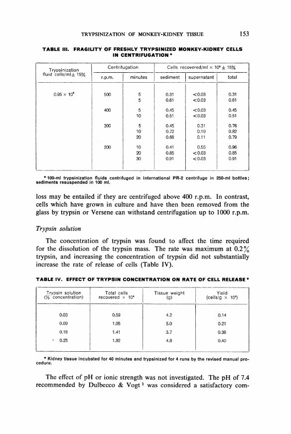
Freshly prepared suspensions of trypsinized cells were found to bevery fragile and were destroyed by manipulations such as simple pipettingor centrifugation at forces which are without effect on most cells. Theeffect of centrifugation at different speeds on the number of cells harvestedfrom trypsinization fluids is slown in Table III. For these studies anInternational Centrifuge, model PR-2, was used. This centrifuge doesnot set up resonance at low speeds. Many centrifuges do so in the range200-300 r.p.m. and the resonance may be sufficient completely to disruptsedimented pellets of monkey-kidney cells.
It is seen in Table III that at speeds above 400 r.p.m. there is a signi-ficant loss in the number of cells. In some cases the total number of cellsin both the supernate and the sediment after centrifugation at these speedswas less than 500% of the original number. In these counts only cells show-ing both nuclei and attached cytoplasm were counted. The number offree nuclei in the supernatant after centrifugation was noticeably higherthan before, although accurate counts were not made. The number ofcells recovered after centrifugation at 300 r.p.m. approached, within theerrors of counting, the number before centrifugation. The number actuallysedimented, however, increased with length of time of centrifugation,but 30 minutes of centrifugation at 200 r.p.m. (11 xg) were sufficientto sediment all the cells into a firm pellet. The pellet is firm enough topermit removal of the supernatant fluid by aspiration.
The cells released by trypsin proved unexpectedly fragile, and evenafter standing several days in the cold in the growth medium substantial
152
TRYPSINIZATION OF MONKEY-KIDNEY TISSUE
TABLE III. FRAGILITY OF FRESHLY TRYPSINIZED MONKEY-KIDNEY CELLSIN CENTRIFUGATION *
Trypsinization Centrifugation Cells recovered/ml x 106 ± 15%fluid cells/ml± 15% r.p.m. minutes sediment supernatant total
0.95 x 10⁶ 500 5 0.31 <0.03 0.315 0.61 <0.03 0.61
400 5 0.45 <0.03 0.4510 0.51 < 0.03 0.51
300 5 0.45 0.31 0.7610 0.72 0.10 0.8220 0.68 0.11 0.79
200 10 0.41 0.55 0.9620 0.85 <0.03 0.8530 0.91 <0.03 0.91
* 100-mi trypsinization fluids centrifugedsediments resuspended in 100 ml.
in International PR-2 centrifuge in 250-mi bottles;
loss may be entailed if they are centrifuged above 400 r.p.m. In contrast,cells which have grown in culture and have then been removed from theglass by trypsin or Versene can withstand centrifugation up to 1000 r.p.m.
Trypsin solution
The concentration of trypsin was found to affect the time requiredfor the dissolution of the trypsin mass. The rate was maximum at 0.2%trypsin, and increasing the concentration of trypsin did not substantiallyincrease the rate of release of cells (Table IV).
TABLE IV. EFFECT OF TRYPSIN CONCENTRATION ON RATE OF CELL RELEASE *
Trypsin solution 1 Total cells Tissue weight Yield(% concentration) recovered x 108 (g) (cells/g x 108)
0.03 0.59 4.2 0.14
0.09 1.05 5.0 0.21
0.18 1.41 3.7 0.38- 0.25 1.92 4.8 0.40
* Kidney tissue incubated for 40 minutes and trypsinized for 4 runs by the revised manual pro-cedure.
The effect of pH or ionic strength was not investigated. The pH of 7.4recommended by Dulbecco & Vogt 1 was considered a satisfactory com-
153
C. RAPPAPORT
promise between the pH optimal for trypsin (pH 8.2) and that which isphysiological for cells. The high phosphate concentration in the trypsinsolution has been found to be useful not so much because of bufferingaction but because it prevents irreversible clumping of the cells.
The effect which incubation was found to have on trypsinizationsuggests that intracellular factors may determine the rate of release of cells.If this were the case, the rate would be determined by the concentrationat which tissue extracts were maintained during trypsinization, i.e., by therate of change of trypsin fluid above the tissue mass. It was not possibleto investigate this when fluids were added and decanted manually. Usingthe automatic trypsinizer, where a constant volume in the mixing chambermay be maintained with a continuous change of fluid, it has been foundthat the number of cells present in the mixing chamber determines the rateof release of cells from the tissue mass.
Efforts to substitute Versene for trypsin were unsuccessful. Versene hasbeen used successfully for preparation of liver cells by several workers,2'7and also for the removal of kidney cells from tissue-culture monolayers(M. Vogt-personal communication). The release of cells from the freshkidney tissue was found to be too slow to be practical. Further, the cellsobtained from the Versene-treated tissue had to be harvested and re-suspended before they would grow.
Revised Trypsinization Procedure
The observations presented indicate that successful trypsinizationdepends on maintaining a proper relationship between time of incubationand time of mechanical stirring and observing precautions made necessaryby the extreme fragility of the cells. With these requirements in mind, itwas possible to simplify the trypsinization procedure.
The first and most desirable item was replacing the Waring blendorwith a mixing chamber which would be easier to handle. An Erlenmeyerflask was modified so that a magnetic stirrer could be used in place of aWaring blendor. Using this flask, kidneys were trypsinized with systematicmodification of the Youngner method. Each modification was judged by:(1) the number of whole cells, and (2) the ease of operation.
The revised procedure to be given has been in routine use for over ayear in the Yale laboratory. It may be done by adding and decantingfluids manually or the fluids may be changed mechanically by use of theautomatic trypsinizer. The revised procedure yields two to three timesmore cells per gram of tissue trypsinized than the earlier method. A step-by-step procedure, with manual changes of fluids, will be given first. Thepreparation of the tissue and the harvesting of the cells, steps 1-3 andsteps 8-10, are the same for both the manual and automatic methods.
154

TRYPSINIZATION OF MONKEY-KIDNEY TISSUE 155
A description of the automatic trypsinizing flask and notes on its use willthen be given.
Trypsinization flask a
A few simple modifications of an ordinary Erlenmeyer flask willpermit one to use a magnetic stirrer instead of the Waring blendor. Thesides of a 500-ml, or 250-ml, Erlenmeyer flask have been indented at rightangles to the bottom surface in four places. They should extend to abouttwo-thirds of the height of the flask and cut into the bottom surface about2.5 cm. This gives the bottom of the flask the shape of a Maltese cross.This modification permits good mixing with cavitation at certain criticalvolumes. Maximum mixing over a wider range of volumes may be obtainedif the flask is further modified by accentuating the curvature between thebottom and the sides of the flask. This is done by compressing the glass inthis region inwards and upwards.
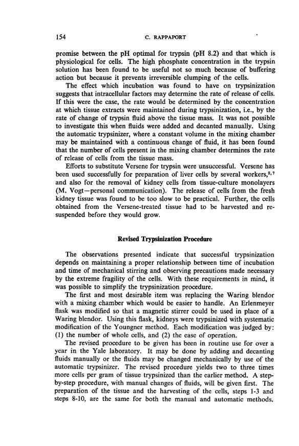
During trypsinization it is necessary to add and decant fluids many timesand therefore it is convenient to have an inlet for trypsin from a reservoirand a side-arm for decanting fluids incorporated into the flask. The side-arm for decanting should be placed so that the tissue tends to be trapped inthe bottom when the flask is tilted for decanting. The efficiency of trappingmay be increased by enlarging the flask just under the side-arm. This isparticularly useful when large volumes of tissue are trypsinized. Theflask assembled for use is shown in Fig. 1.
A 500-ml flask modified in this manner is large enough to trypsinize aninitial load of 80 g of tissue. If less than 30 g of tissue are to be trypsinized,a 250-ml flask should be used, thus reducing the amount of trypsin solutionrequired.
The volume of trypsin required during stirring is determined by thesize and shape of the flask and not by the amount of tissue. In the 500-mlflask, maximum stirring efficiency is obtained at a total volume (includingmagnet and tissue) of about 150 ml. This is determined for any given flaskand tissue load by the volume which allows rapid rotation of fluid andtissue fragments up and around the sides of the flask with slight cavitationand no foaming. The flask should be marked at this level and trypsin addedfrom a reservoir to this mark for each run.
Trypsinization of monkey kidneys
Step 1. Kidneys are removed from freshly killed and exsanguinatedmonkeys and are placed in a sterile Petri dish. They are decapsulated andcut in half. This is best done by holding the kidney firmly with forceps and
a The trypsinization flask and the automatic trypsinizer (with magnets) described in this paper areavailable at the Macalaster-Bicknell Co., New Haven, Conn., USA.
11

156 C. RAPPAPORT
making a cut through the kidney horizontal to the flat surface with uterinescissors, exposing the central pelvis. The pelvis is removed by cuttingaround its outer edge, lifting it with forceps to free it from the underlyingtissue. Any adhering connective tissue is also discarded.
FIG. 1. SPECIAL TRYPSINIZATION FLASK USED WITH MAGNETIC STIRRER,ASSEMBLED FOR TRYPSINIZATION BY MANUAL METHOD

TRYPSINIZATION OF MONKEY-KIDNEY TISSUE
Step 2. The tissue is cut into pieces at least 1 cm x 1 cm. It is importantto cut through the tissue because strands of connective tissue may interferewith proper mixing.
Step 3. The cut tissue is transferred to the trypsinization flask andcovered with trypsin pre-warmed to 370C.
Steps 1-3, the preparation of one kidney, should take about 7-10 minutes.The process is repeated for each of the remaining kidneys. After eachkidney is prepared it is added to the others, covered with trypsin, andincubated. Thus, 6-8 kidneys may be conveniently prepared in the hourrequired before stirring starts.
Step 4. One hour after trypsin was added to the first kidney the incuba-tion fluid is decanted into a sterile flask which is kept at 40C. Fresh pre-warmed trypsin is added to flask to a level equal to a total volume ofabout 150 ml.
Step 5. Tissue and trypsin are stirred with a magnetic stirrer for7 minutes. The speed of stirring should be sufficient to allow rapid mixingwith cavitation and no foaming.
Step 6. After 7 minutes of stirring, the magnetic stirrer is turned off,the tissue allowed to settle, and the fluid decanted into the receptaclecontaining the incubation fluid.
The remaining tissue is washed clean of free but adhering cells byadding 50-100 ml of trypsin solution, swirling by hand and decanting.If the tissue has been disintegrated significantly during a run or wash, thedecanted fluids when held to the light will be seen to be a fine suspensionof tissue fragments and cells. After the third or fourth run, it may benecessary to wash the tissue 2-4 times before the washings are clear.
This step, decanting, washing, and re-filling, should be done withouthaste and may take as much as 5-10 minutes.
Steps 5 and 6 (mixing with trypsin, and decanting and washing) arerepeated 6-8 times or until there is a noticeable decrease in the rate ofdissolution of tissue as indicated by a clearing of the fluids.
Step 7. After 6-8 runs, when the fluids have noticeably cleared, thetissue is removed to a small beaker. Considerable connective tissue hasusually become visible during trypsinization at this point and may interferewith mixing. The connective tissue is cut, and if necessary the remainingtissue should be cut into pieces of 0.5 cm in size. The tissue-is covered withtrypsin and incubated at 370C for 20 minutes.
Repeat steps 5 and 6 after the second incubation until tissue is exhausted.The number of runs required to exhaust tissue varies with the mass oftissue. A total of 8 runs is usually sufficient for 4 kidneys and 10-13 for8 kidneys.
Step 8. The combined fluids from all the runs are centrifuged at200 r.p.m. (11 xg) for 30 minutes.
157

158 C. RAPPAPORT
Step 9. The supernatant fluid is removed from the packed cells by avacuum aspirator. The fluid can be removed to within 1-2 ml withoutremoving cells. This residual fluid has not been found to affect the qualityor viability of the suspension in any way. There should be approximately3 ml of packed cells for each kidney trypsinized.
Step 10. The packed cells are resuspended in about 100 ml of the growthmedium, pre-warmed to 370C. If cold solution is added, the cells mayclump badly. The suspension is filtered through 2 layers of sterile cheesecloth. The gauze filter is washed free of adhering cells with about 100 mlof nutrient solution.
Step 11. The number of whole cells, i.e., those showing both nucleiand attached cytoplasm, are counted in a haemocytometer.
Step 12. The suspension is diluted in the nutrient medium to give300 000 cells per ml, and 0.5 ml of this suspension is seeded into16 mm x 150 mm tubes. After 4-5 days of incubation, a confluent sheetof cells is present.
Step 13. If the suspension is to be kept several days before use,4 itshould be stored at 40C at a concentration of not more than 600 000 cellsper ml. The stored suspension should be centrifuged after the first10-24 hours and resuspended in fresh growth medium for the remainderof the storage period. The suspension is again centrifuged and againresuspended in fresh medium just before use. This procedure is necessaryfor maximum survival and growth, because it has been found by the authorthat freshly trypsinized kidney-cell suspensions liberate a heat-labile toxinwhich kills slowly at 40C and may destroy up to 900% of the cells in a fewhours under growing conditions at 370C.
The preparation and trypsinization of 8 kidneys takes about 4-5 hours.An average yield is between 0.6 x 10⁸ and 0.8 x 10⁸ cells per g of "dressed"tissue.
Continuous automatic trypsinizing flask a
A flask permitting the continuous and automatic addition of trypsinand withdrawal of cells is shown in Fig. 2. The main problem in designinga suitable flask was the position of the outflow valve. The vigorous stirringduring trypsinization required to sheer the cells from the tissue mass keepsthe tissue in suspension and tends to force tissue into an outflow valve.This difficulty could be overcome if the valve were arranged so that thetissue could settle before the fluid flowed out. However, a system such asthis could probably be operated at only one volume with maximum effi-ciency. It was considered advantageous to have a flask that could operateat several different volumes.
a We are indebted to Mr. Frank Lynsky of Macalaster-Bicknell Co. for advice and work involved inthe development of this flask.

TRYPSINIZATION OF MONKEY-KIDNEY TISSUE
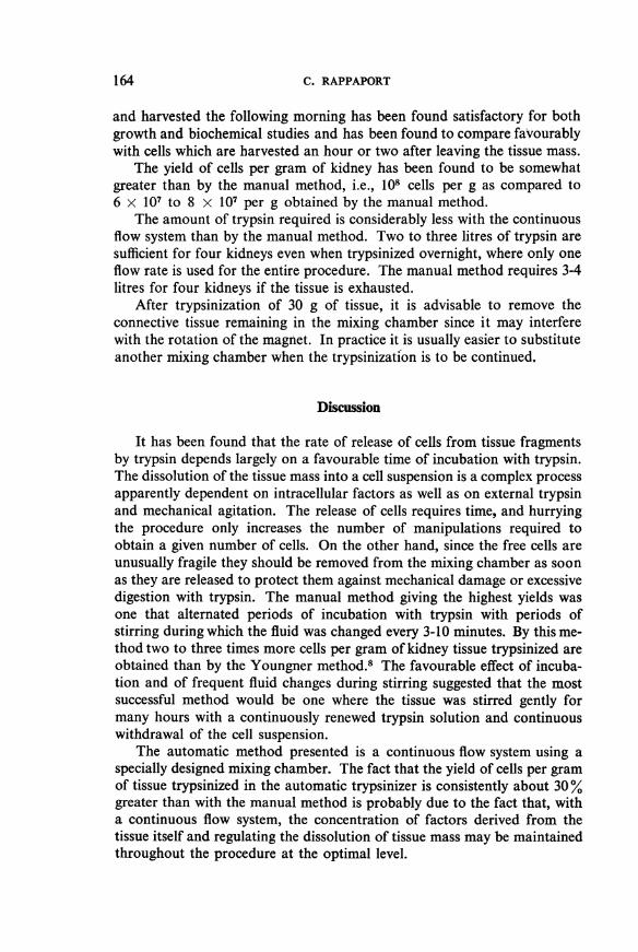
FIG. 2. AUTOMATIC TRYPSINIZER
F
The diagram shows the side and bottom of a 500-mi Kjeldahl flask modified for use as a mixingchamber in the continuous trypsinization procedure. A = Ground-glass bacteriological joint.B = Inlet tube for trypsin from reservoir. C = Air valve. D = Flat glass disc, 2 inches (50 mm) indiameter, with 6 bored holes, 1.5 mm in diameter. E = Sloped outlet drain. F = Indentations andmodifications at sides and bottom of flask to increase mixing efficiency. G = Plastic-coveredmagnet, 11/. x 'I% inches (about 41 mm x 9.5 mm).
The apparatus shown in Fig. 2 consists of a glass mixing-chamber closedby a ground-glass joint containing two openings. Through one, trypsincan be admitted from reservoir. The other is a valve which can admit airwhen desirable. In the bottom centre of the flask holes have been drilledso that fluid can drain from the mixing chamber into a receiving jar.
The flask takes advantage of the fact that a magnetic stirrer can mixliquids in a chamber placed at some distance from the surface of the stirrer.Thus, when tissue fragments are stirred in the mixing chamber, the motionof the magnet sweeps the fragments from the drain and permits only thecell suspension to pass through into the receiving jar.
The mixing chamber and magnetic stirrer should be arranged so thattrypsin is admitted by siphon action through a gum-rubber lead from areservoir. The flow of trypsin is thus virtually independent of the amountof trypsin in the reservoir and can be controlled simply by a pinch clampon a gum-rubber lead into the mixing chamber. After equilibration is
159

C. RAPPAPORT
established, the flow of trypsin into the mixing chamber regulates theoutflow of liquid through the drain. The system can never overflow ordrain dry.
It is important to realize that the action of the magnet is indispensablefor the flow of liquid through the system. The drain valve must never beopened unless the magnet is in motion. If it is opened and the magnetis not turning, the tissue will settle and clog the pores. In this case, thecells are not recovered as soon as they are released and they may be lostby excessive digestion with trypsin.
The size of the magnet is important. It should be heavy enough sothat it is not deflected by the tissue and yet not so heavy that it cannot beturned smoothly by the magnetic stirrer. A magnet 15/8 X /8 inches (about41 mm x 9.5 mm), weighing at least 10 g, has been found satisfactory forloads of tissue up to 30 g and the usual size of stirrer.
The bottom of the flask has been designed so that the magnet tendsto recentre itself if deflected by tissue or by the usual changes in the linevoltage. It may be worth while mentioning the points which were consideredin developing the flask. First, the bottom is blown so that the inside surfaceis flat. Secondly, the flat portion of the bottom is only ¼4 inch (about6 mm) greater than the length of the magnet. Thus, if the magnet is displacedduring stirring it will necessarily return to the centre. Thirdly, the indenta-tions and modifications on the side of the flask were selected from severaltrial flasks because they gave efficient mixing over a wide range of volumesand particularly because the flow of liquid during mixing was itself acentring force.
The self-regulating capacity of the flask is not sufficient to withstandmarked changes in the rate of stirring due to large fluctuations in linevoltage. If the apparatus is to be left unattended on a voltage sourcewhich may change suddenly, it is advisable to use a constant voltage trans-former. The smallest constant voltage transformer, capacity 15 amperes,commercially available is more than adequate for this purpose. Operatedat proper speeds and on a steady line the trypsinizer has proved veryreliable and has been left trypsinizing for more than 24 hours unattended.
Operation of flask. The flask and rubber tubing leading from thereservoir and to the receiving vessel are sterilized by autoclaving. The airoutlet should have a short rubber tube attached, which is plugged withcotton before sterilization. Pure gum rubber is recommended for allconnexions, because some of the synthetic rubbers or plastics may bedistorted enough by autoclaving to allow air to leak in. A plastic-coveredmagnet 1 5/8 X 3/8 inches (about 41 mm x 9.5 mm) is sterilized by boiling justbefore use.
Kidney fragments of not less than 1 cm x 1 cm are prepared as alreadydescribed. It is advisable to cut the kidneys in a beaker or Petri dish,
160

TRYPSINIZATION OF MONKEY-KIDNEY TISSUE 161
because if the tissue is minced in a test-tube the cutting is uneven andsome very small pieces are produced.
The tissue and magnet are placed in the flask, of which the outlet drainhas been closed by a pinch clamp; 100 ml-150 ml of trypsin solution, pre-warmed to 370C are also added. The trypsin reservoir is placed so thattrypsin can be admitted into the mixing chamber by siphon action througha drop of at least 1 foot (30 cm). The reservoir should be kept at 370C.
FIG. 3. AUTOMATIC TRYPSINIZER, ASSEMBLED
IWm

C. RAPPAPORT
This may be done by placing a microburner under the reservoir or bysetting up the entire apparatus in a " walk-in " incubator. The outletdrain should feed into a suitable receptacle, placed at least 11/2 feet (about45 cm) lower than the mixing chamber. If it is desired to tap the trypsinizerfor cell suspension throughout the day, it will be found convenient to usea bell-cap on the outlet tube for the receiving flask. The bell-cap can bechanged easily from one receptacle to another with a minimum chanceof contamination. If the cells are to be kept several hours before harvesting,the receptacle should be kept in an ice-water bath. The apparatus assembledfor use is shown in Fig. 3.
The magnetic stirrer is started and flask centred so that the magnetoperates smoothly in the centre of the flask. This may be adjusted by thesound of the magnet rotating, as well as by the appearance. When properlycentred, the rotation of the magnet is almost noiseless.
The tissue is incubated with gentle stirring for 30-45 minutes. Afterincubation, the speed of stirring is increased until there is rapid swirlingof tissue up and around the sides of the mixing chamber. There shouldbe slight cavitation but no foaming.
At this time, with the air valve closed, the drain valve is opened slowly.Trypsin will flow from the mixing chamber into the receiving jar andprime the siphoning of trypsin from the reservoir. When trypsin startsflowing into the mixing chamber, the inlet valve should be closed graduallyuntil, with the drain valve completely open, the rate of flow through theflask is regulated by the pinch clamp on the inlet from the trypsin reservoir.
During trypsinization, small pieces of tissue may escape through thedrain into the receiving jar. If the tissue has been cut as directed and ifthe speed of the magnet is properly adjusted, the tissue actually lost inthis way is negligible. However, if the flow of fluid is regulated by a pinchclamp on the drain valve instead of on the trypsin inlet as indicated, theconstriction will tend to trap any tissue escaping and may block the outflowof fluid.
Volumes and loads. The flask is marked on the outside at three differentlevels, indicating a total volume (from the mark to the pinch clamp on theoutlet drain) of 60 ml, 100 ml, and 150 ml. These levels have been foundsatisfactory for trypsinization of 13, 30, and 50 g of tissue respectively.The constant volume in the flask, or " working volume " at which it isdesirable to run a trypsinization, depends on the liquid volume which willpermit adequate stirring and smooth rotation of the magnet in the flask,and not on the amount of tissue. Thus, the working volume may be changedif the amount of tissue is changed or if tissue of a different density is used.
To change the volume in the mixing chamber, the air outlet is opened.If the volume is to be decreased, the trypsin inlet is closed and fluid isallowed to drain out until it is at the desired level. The air valve is then
162

TRYPSINIZATION OF MONKEY-KIDNEY TISSUE
closed and the flow of trypsin regulated. To increase the volume in themixing chamber, the drain valve is closed, the air valve is opened andtrypsin is allowed to run in until it is at a level equivalent to about 30 mlmore than the desired volume. On closing the air outlet, and opening thedrain valve, the volume will adjust itself at the desired level with the previousrate of trypsin inflow.
Rate offlow. The rate of flow into the mixing chamber is regulated sothat the fluid which is drained off contains from 5 x 10⁵ to 10 x 105cells per ml. At this concentration, the fluid is turbid and when held tothe light will be seen to be a suspension of material. The regulation may bemade satisfactorily without counting, by adjusting the rate so that the outflowis always turbid but fast enough to prevent the cells from settling out alongthe drain tube. Increasing the rate of flow results in an extravagant use oftrypsin and seems to decrease the number of cells obtained (see Table V).
When the flask is at working volume of 60 ml (including magnet andtissue) with loads up to 15 g, a rate of flow of 1-2 drops per second has beenfound to be satisfactory for the main portion of the trypsinization. This isa " clear-out " time of less than 20 minutes. For loads of tissue of 30 g anda working volume of 100 ml, a rate of 3-4 drops per second is sufficient.
TABLE V. EFFECT OF FLOW RATE DURING AUTOMATIC TRYPSINIZATION ONRATE OF CELL RELEASE *
Tissue Flow rate Cell count x 106 in outflow at Total ml of Yieldweight (drops/ trypsin used (cells/g)(g) second) 1 hour 2 hours 3 hours
8.0 1.2 0.51 1.1 0.83 610 0.6x 10'
7.8 3.4 0.05 0.08 0.11 1750 0.2x10⁸
* Paired kidney tissue incubated for 40 minutes and trypsinized for 3 hours in automatic tryp-sinizers at 100-ml working volumes.
Towards the end of trypsinization-or at the very beginning, when therelease of cells is slow-the rate may be considerably reduced with a signi-ficant saving in trypsin and in the volume which must subsequently becentrifuged. If the apparatus is to be left unattended for the entire trypsiniza-tion, however, the rates suggested are adequate for the whole procedure.
Performance. The kidneys are usually dressed and placed in the flaskwith trypsin at 9.30 in the morning. After 30-45 minutes' incubation thecontinuous flow system is started. Viable cell suspensions are available anhour later and are harvested and used throughout the day. Fresh kidneytissue may be added 1-2 hours later and again after 4-5 hours, and trypsiniza-tion may be continued for 24 hours. Kidney tissue trypsinized overnight
163
C. RAPPAPORT
and harvested the following morning has been found satisfactory for bothgrowth and biochemical studies and has been found to compare favourablywith cells which are harvested an hour or two after leaving the tissue mass.
The yield of cells per gram of kidney has been found to be somewhatgreater than by the manual method, i.e., 108 cells per g as compared to6 x 107 to 8 x 10⁷ per g obtained by the manual method.
The amount of trypsin required is considerably less with the continuousflow system than by the manual method. Two to three litres of trypsin aresufficient for four kidneys even when trypsinized overnight, where only oneflow rate is used for the entire procedure. The manual method requires 3-4litres for four kidneys if the tissue is exhausted.
After trypsinization of 30 g of tissue, it is advisable to remove theconnective tissue remaining in the mixing chamber since it may interferewith the rotation of the magnet. In practice it is usually easier to substituteanother mixing chamber when the trypsinization is to be continued.
Discussion
It has been found that the rate of release of cells from tissue fragmentsby trypsin depends largely on a favourable time of incubation with trypsin.The dissolution of the tissue mass into a cell suspension is a complex processapparently dependent on intracellular factors as well as on external trypsinand mechanical agitation. The release of cells requires time, and hurryingthe procedure only increases the number of manipulations required toobtain a given number of cells. On the other hand, since the free cells areunusually fragile they should be removed from the mixing chamber as soonas they are released to protect them against mechanical damage or excessivedigestion with trypsin. The manual method giving the highest yields wasone that alternated periods of incubation with trypsin with periods ofstirring during which the fluid was changed every 3-10 minutes. By this me-thod two to three times more cells per gram of kidney tissue trypsinized areobtained than by the Youngner method.8 The favourable effect of incuba-tion and of frequent fluid changes during stirring suggested that the mostsuccessful method would be one where the tissue was stirred gently formany hours with a continuously renewed trypsin solution and continuouswithdrawal of the cell suspension.
The automatic method presented is a continuous flow system using aspecially designed mixing chamber. The fact that the yield of cells per gramof tissue trypsinized in the automatic trypsinizer is consistently about 30%greater than with the manual method is probably due to the fact that, witha continuous flow system, the concentration of factors derived from thetissue itself and regulating the dissolution of tissue mass may be maintainedthroughout the procedure at the optimal level.
164

TRYPSINIZATION OF MONKEY-KIDNEY TISSUE
The automatic trypsinizing apparatus was developed because it was feltthat the time and attention required for preparation of cell suspensions,although not a serious factor for certain kinds of studies, prohibited inves-tigations requiring small amounts of fresh material frequently. With thisin mind, it was considered that simplicity, inexpensiveness, and ease ofhandling were important factors in the choice of design. An apparatusoperating under a wider variety of conditions than the one described hereand with complete automatic control could certainly be developed, but itwould be more complicated and more expensive.
The automatic trypsinizer which has been developed is a simple glassflask to be used with a magnetic stirrer. The flask is inexpensive, may besterilized by autoclaving, and is easily operated. It can trypsinize efficientlya constant load of 30 g of tissue. It operates over a threefold volume rangeand a varying flow rate.
Two simple precautions should be observed when the apparatus is tobe left for many hours unattended. First, the voltage supply should berelatively constant as indicated by a constant rate of rotation of the magnet,or a constant voltage transformer should be used. Secondly, each set-upshould be checked for leaks, indicated by a discrepancy between the rateof inflow of trypsin and the rate of outflow. If the system is not closed, itwill eventually overflow or drain dry. When these two precautions areobserved, the apparatus will work reliably for many hours without adjust-ment.
RItSUMI2
La methode de resolution de fragments tissulaires en suspensions cellulaires paraction de la trypsine s'est revelee tr6s feconde en virologie. Introduite en 1914 par Rous etJones, reprise par Dulbecco et Vogt, cette methode a et6 recemment adaptee par YoungnerA la production de suspensions de cellules d'embryon de poulet et de rein de singe. Tellequ'elle etait pratiquee jusqu'a maintenant, cette methode avait l'inconvenient de necessiterune surveillance constante et de se preter mal aux recherches demandant frequemment depetites quantites de materiel frais.
L'auteur decrit une modification qui rend cette methode automatique. Grace a unechambre de melange specialement conque A cet effet et rempla9ant le Waring blendor,l'appareil assure I'afflux constant de trypsine et l'e1imination automatique des cellulesmises en suspension par l'agitateur magnetique. Cet appareil qui fonctionne a troisvolumes diff6rents et avec une vitesse d'ecoulement reglable, peut traiter de fa9onconstante 30 g de tissu. II donne un rendement en cellules deux A trois fois superieur acelui des autres methodes. Moyennant certaines precautions - voltage constant assurantla rotation reguliere de l'agitateur et suppression des causes de fuites et de pertes dansl'appareil - le systeme fonctionne sans surveillance pendant plusieurs heures.
Les diverses etapes de la methode et l'utilisation de l'appareil sont decrites en d6tail.L'effet de certains facteurs sur le rendement en cellules y est discute sur la base dedonnees experimentales; ce sont en particulier la duree de la trypsinisation, la centrifu-gation, la concentration de la trypsine, la relation entre la duree de l'incubation et cellede I'agitation.
165

166 C. RAPPAPORT
REFERENCES
1. Dulbecco, R. & Vogt, M. (1954) J. exp. Med. 99, 1672. Mascona, A. (1952) J. exp. Cell Res. 3, 5353. Melnick, J. L. & Riordan, J. T. (1952) Proc. Soc. exp. Biol. (N.Y.), 81, 2084. Melnick, J. L. et al. (1955) Proc. Soc. exp. Biol. (N. Y.), 88, 6765. Rous, P. & Jones, F. S. (1916) J. exp. Med. 23, 5646. Sanford, K. K. et al. (1951) J. nat. Cancer Inst. 11, 7737. St. George, S., Friedman, M. & Byers, S. 0. (1954) Science, 120, 4638. Youngner, J. S. (1954) Proc. Soc. exp. Biol. (N. Y.), 85, 202