Lymphoid Tissue - KGMU
59
Lymphoid Tissue Dr Punita manik Professor Department of Anatomy K G’s Medical University U P Lucknow
-
Upload
khangminh22 -
Category
Documents
-
view
2 -
download
0
Transcript of Lymphoid Tissue - KGMU
Lymphoid TissueDr Punita manik
ProfessorDepartment of AnatomyK G’s Medical University
U PLucknow

Classification
• Diffuse Lymphoid tissue: diffuse arrangement of lymphocytes and plasma cells deep to epithelium in Lamina propria of digestive, respiratory and urogenital tracts forming an immunological barrier against invasion of microorganisms.
• Dense Lymphoid tissue: presence of lymphocytes in the form of nodules

Classification
Dense Lymphoid Tissue: Nodules are found either in association with mucous membranes of viscera or as discrete encapsulated organs.
2 Types
1. MALT(Mucosa associated Lymphoid Tissue; Nonencapsulated)
2. Discrete Lymphoid organs (Encapsulated)

• MALT:
Solitary Nodules
Aggregated Nodules(Peyer’s patches)
Lymphoid nodules in vermiform appendix
Waldeyer’s ring at the entrance of pharynx
• Discrete Lymphoid Organs:
Thymus
Lymph Node
Spleen
Tonsil

Lymph Node
• Connective tissue framework
Capsule
Trabecula
Reticular Stroma (fibres)

Components of Lymph Node
• Parenchyma
1.Cortex
2.Paracortex
(Inner Cortex)
3.Medulla

Lymph node

Lymph Node Reticulum
• Reticular stroma-
reticular fibres and phagocytic reticular cells
• Gives structural support to the lymphoid cells

Lymph Node Reticulum
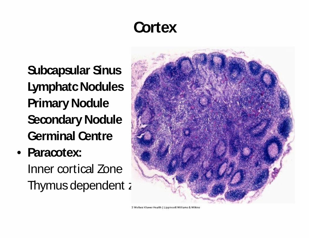
Cortex
Subcapsular SinusLymphatc NodulesPrimary NoduleSecondary NoduleGerminal Centre
• Paracotex:Inner cortical ZoneThymus dependent zone

Lymph Node
• Medulla:
• Medullary Cords -Darkly stained, branching anastomosing – B Lymphocytes, plasma cells& macrophages
• Medullary Sinuses-Lightly stained

Lymph Node

Lymph Node

Lymph Node

Thymus• Central Lymphoid organ• Dual Origin- Lymphoctes- Mesoderm• Reticular epithelial cells- Endoderm• Formed only by T lymphocytes• No B lymphocytes• Divided into lobules of lymphoid tissue( No
lymphatic nodule)• Has hassall’s corpuscles• Produces thymic hormones• Fully dev at birth, involutes at puberty.
Thymus
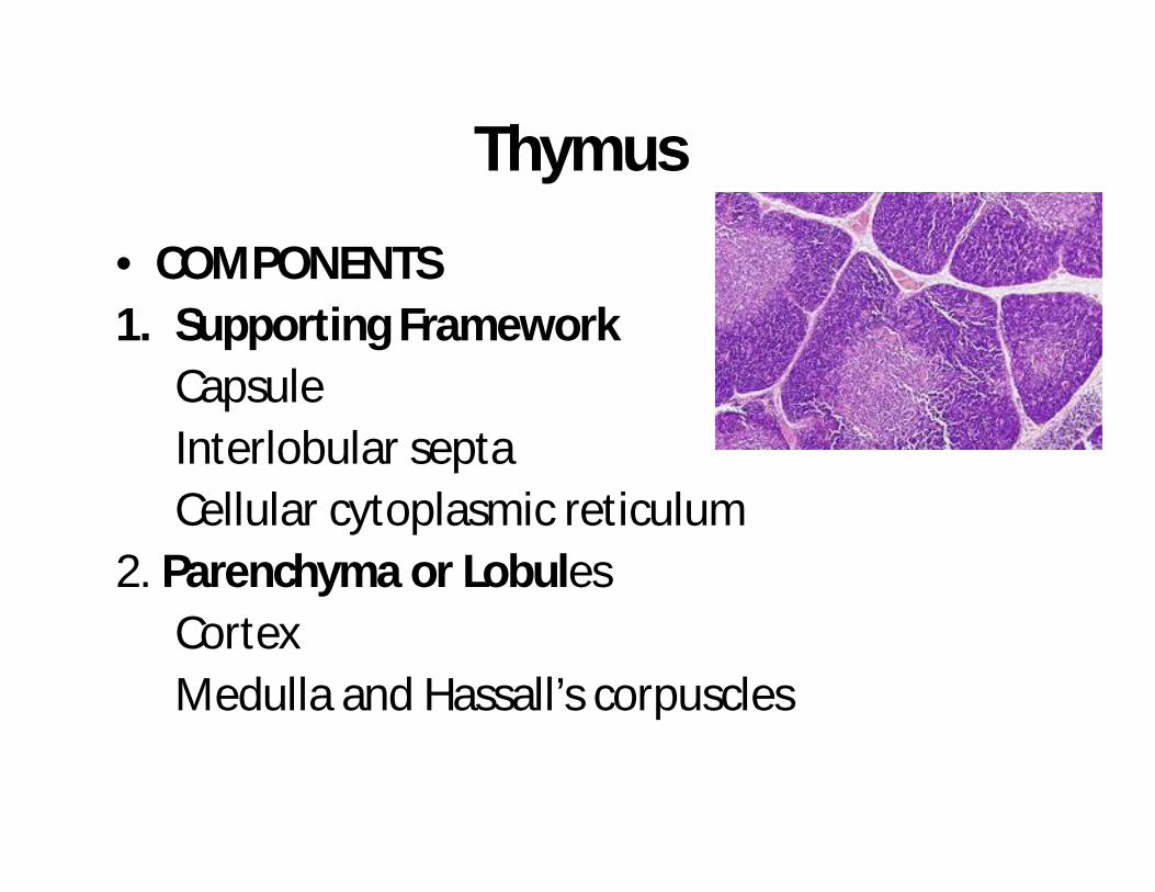
• COMPONENTS1. Supporting Framework
CapsuleInterlobular septaCellular cytoplasmic reticulum
2. Parenchyma or LobulesCortexMedulla and Hassall’s corpuscles

Thymus
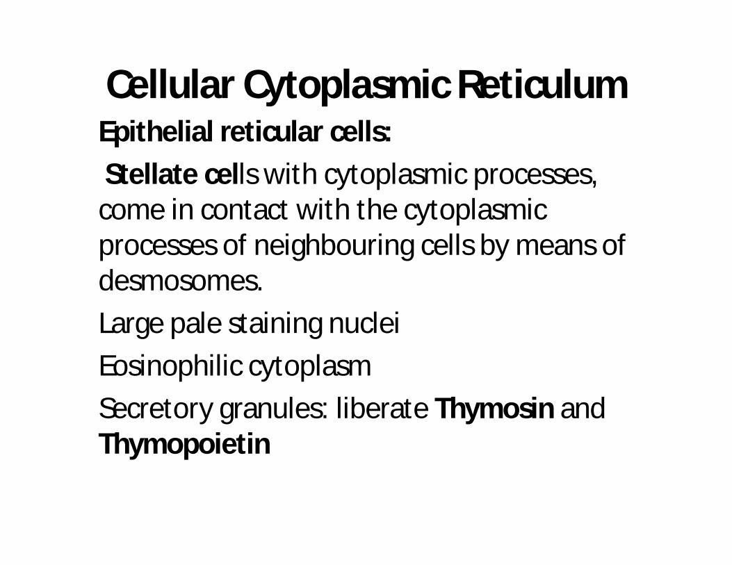
Cellular Cytoplasmic ReticulumEpithelial reticular cells:
Stellate cells with cytoplasmic processes, come in contact with the cytoplasmicprocesses of neighbouring cells by means of desmosomes.
Large pale staining nuclei
Eosinophilic cytoplasm
Secretory granules: liberate Thymosin and Thymopoietin

Cellular Cytoplasmic Reticulum
• Gives support to the lymphocytes of thymiclobules
• This reticulum is different from the reticulum of other lymphoid organs where it is formed by the reticular fibres.

ParenchymaCortex:
Darkly stained
Densely packed lymphocytes-Thymocytes
Outer cortex-Large cells-Lymphoblasts
Lymphoblasts divide by mitosis, push cells towards deeper part of cortex
Blood Thymic Barrier: between thick basement membrane of capillaries and processes of epithelial reticular cells

ParenchymaMedullaLightly stained because lymphocytes areless densely packed.Characteristic Feature: presence of Hassall’s CorpusclesHassall’s Corpuscles: Round lamellated acidophilic bodies(30-100mm), central homogeneous hyaline material surrounded by concentric layers of flattened epithelial cells. These cells are filled with keratin filaments

Thymus

Thymus

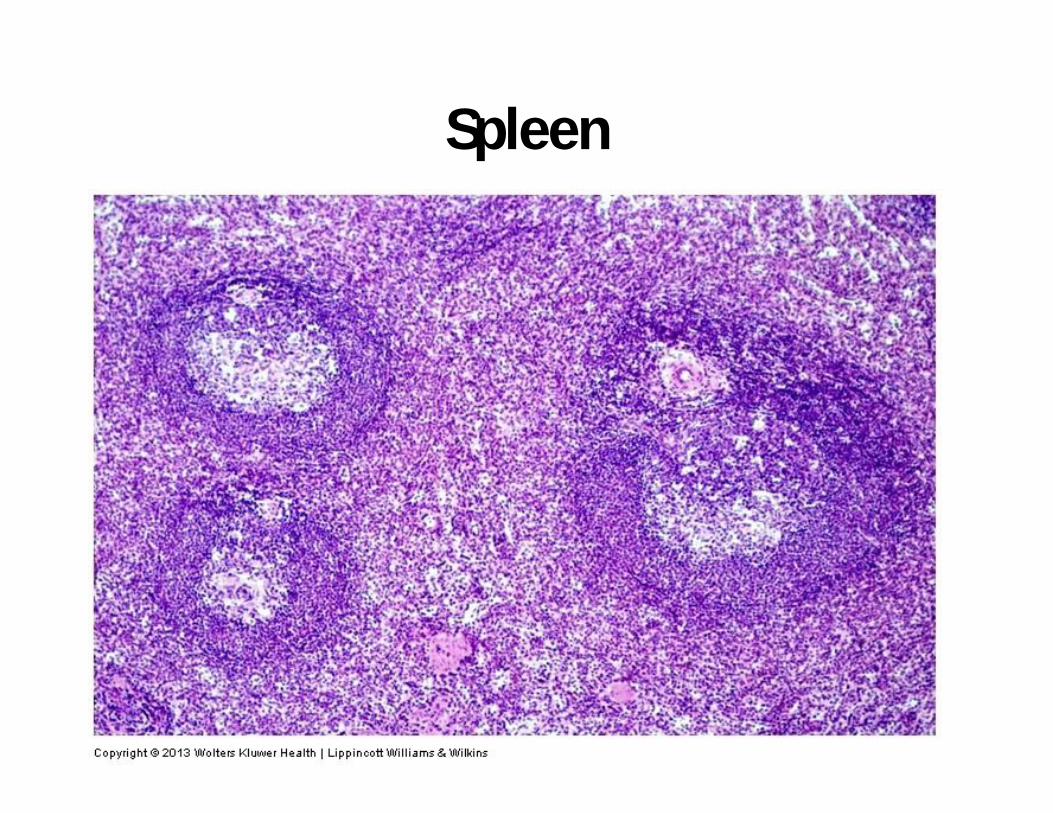
Spleen• Largest Lymphoid organ
• Blood forming organ( Haemopoesis) in foetallife
• Blood destroying organ in post natal life (Graveyard of RBC)
• Filteration of blood from antigens, microorganisms, and aged platelets and abnormal and aged RBCs.

Spleen
Structure of Spleen
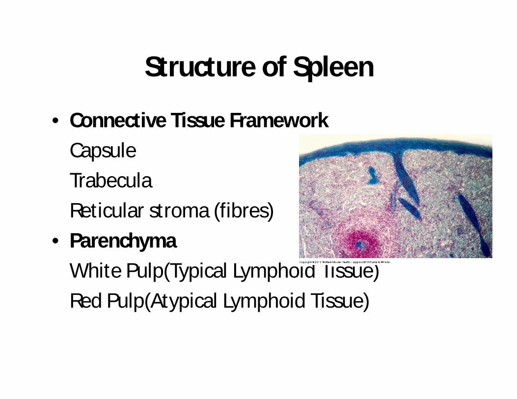
• Connective Tissue Framework
Capsule
Trabecula
Reticular stroma (fibres)
• Parenchyma
White Pulp(Typical Lymphoid Tissue)
Red Pulp(Atypical Lymphoid Tissue)
Spleen
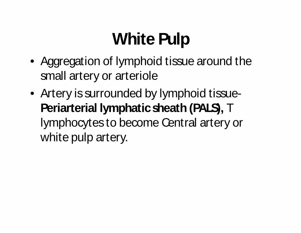
White Pulp• Aggregation of lymphoid tissue around the
small artery or arteriole
• Artery is surrounded by lymphoid tissue-Periarterial lymphatic sheath (PALS), T lymphocytes to become Central artery or white pulp artery.

• Large collections of B lymphocyes around PALS forming lymphatic nodule with germinal centres (white pulp)
• In these nodules artery occupies an eccentric position, those known as central artery

White Pulp• The lymphatic nodules(white pulp) are
surrounded by immunologically active zone containing macrophages, few T lymphocytes and blood sinuses. This zone is known as Marginal zone between white and red pulp.
• The central artery enters the red pulp where it divides to form Straight Penicillararterioles.
• Some Penicillar arterioles may show thickening of the wall due to aggregation of macrophages, reticular cells and lymphoid cells know as Ellipsoids

Spleen circulation

Spleen circulation

Red Pulp
• Modified Lymphoid tissue• Heavily infiltrated with all cells of circulating
blood• Gives dark red color to the tissue when fresh• Composed of irregular anastomosing Splenic
cords of Billroth and Splenic venous sinuses between the cords.
• Splenic cord is a spongy network of reticular fibres infilterated with reticular cells,lymphocytes, macrophages, plasma cells and all elements of circulating blood

Red Pulp• Splenic venous sinuses are lined by highly
elongated , spindle shaped endothelial cells on a discontinuous basement membrane.
• The structure of these sinuses can be compared to tall wooden barrels with both ends open and the endothelial cells being represented by wooden staves, hence known as Stave cells.
• Externally, the sinuses are encircled by reticular fibres in a transverse direction like the steel bands holding together the staves of the wooden barrel.

Red Pulp
• Since the gap between the endothelial cells of the splenic sinuses is 2-3micron m in diameter, only the flexible cells are able to pass easily to and from the cords and sinuses.
• A reduction in the flexibility of erythrocytes after 120 days, signals for their destruction.

Theories of Splenic CirculationTo explain the mode of termination of the arterial capillaries of the Penicilli
• Closed Circulation Theory:
Blood passes directly from the arterial capillaries in to the splenic venous sinuses of the red pulp i.e. the vascular system is closed or continuous.

Theories of Splenic CirculationOpen Circulation Theory:
• Blood passes fro the arterial capillaries of penicilli into cords of Bilroth and from there into the sinuses through the spaces between endothelial cells.
• The nonflexible old erythrocytes are retained in the cords and are engulfed by macrophages.

Theories of Splenic Circulation
Another compromised Theory
Splenic circulation is closed in contracted spleen and open in distended spleen.

Clinical
• Removal of spleen: splenectomy can be done if required. Does not have any adverse effect
• Spleen may enlarge secondary to malaria and leukemia

Spleen
• Phagocytosis of old RBCs-Macrophages present in spleen remove iron from the haemoglobin of aged RBCs which is reused for synthesis of haemoglobin in bone-marrow


Tonsillar crypt


Identifying Features1. Has numerous crypts lined
by stratified squamousepithelium
2. Lamina propria contains lymphatic nodules.
3. Mucous glands in the deeper part , ducts opening in the bottom of the crypt

Tonsil

Tonsil


Tonsil


Clinical
• In tonsilitis, the mouth of the crypts may appear purulent due to infection and pus formation.

MCQ
Crypts of palatine tonsils are lined by
1. stratified squamous non keratinized epithelium
2. stratified squamous keratinized epithelium
3. Simple squamous epithelium
4. Simple cuboidal epithelium

MCQ
Section of a lymph node can be identified by the presence of
1.Thick trabeculae
2. White pulp
3.Interlobular septum
4.Subcapsular sinus

MCQ
White pulp of spleen can be identified from the lymphatic nodule of lymph node by the presence of
1. Germinal centre
2. Lymphocytes
3. Eccentric arteriole
4. corona

MCQ
Hassall’s corpuscles are seen in
1. Spleen
2. Lymph node
3. Tonsil
4. Thymus
MCQ
Splenic sinuses are lined by
1. Fenestrated endothelium
2. Discontinuous endothelium
3. Continuous endothelium
4. Columnar epithelium

MCQ
Lymphatic nodules are present in all EXCEPT
1. Spleen
2. Thymus
3. Tonsil
4. Lymph Node