Elastin-Coated Biodegradable Photopolymer Scaffolds for Tissue Engineering Applications
Upload
independentCategory
view
0download
0
Biofunctionalized Nano-fibrous Polymer Scaffolds for Tissue
EngineeringUniversity of Washington
Materials Science and EngineeringTravis Kassab
Keywords
tissue engineering, nanofibers, biopolymers, scaffolds, ECM analogues, synthetic polymers, chitosan, gelatin, collagen, laminin, starch, biofunctionalization, biomaterials, nanotechnology, regenerative medicine, in vitro, cell incubation
Abstract This review previews recent research conducted on various nanofiber polymer scaffolds for tissue engineering. Opti-mizing the medium upon which cells will most successfully attach to and proliferate on constitutes a popular area of discussion within the tissue engineering community. Scaffolds must mimic the extracellular matrix (ECM) of the native cell in order for successful tissue regeneration. This requires the scaffold to possess similar mechanical and chemical properties to that of the native ECM (6). Many times synthetic and natural polymers are combined together in order to hybridize the desirable properties from each. Biopolymers can biofunctionalize synthetic polymers with their bio-logical signalling mechanisms, creating a hybrid scaffold material more conducive to cell attachment and proliferation (18). This review considers research on biopolymers such as chitosan, collagen, gelatin, laminin, and starch. These biopolymers were used to biofunctionalize various synthetic polymers. Each study examines scaffold manufacturing techniques, incubation and imaging procedures, and resulting cell growth. This review briefly describes the procedure taken by the experimenters in order to obtain the material under review. The second category describes the outcome of incubating cells on these tested materials. The last category gives an overview of the type of characterization tech-niques and equipment used by the researchers in order to determine experimental outcomes such as cell viability, proliferation, morphology, and histology.

Table of Contents
1. Introduction 1.1 Scaffold Processing
1.2 Biofunctionalization
2. Scaffold Biomaterials 2.1 Chitosan
2.2 Collagen
2.3 Gelatin
2.4 Laminin
2.5 Starch
3. Concluding Statements

1. Introduction Tissue engineering describes the interdisciplinary process of tissues grown in vitro for in vivo applications. Cells grow onto a extracellular matrix (ECM) analogue, or scaffold. This review focuses on different polymers used as scaffolding material for tissue regeneration. The field of tissue engineering encompasses the regeneration of muscle, nerve, bone, and heart tissues. The generation of these tissues helps problems like donor scarcity, immune system rejection, and pathogen transfer (4).
Regenerating cells occurs by seeding said host cells onto a scaffold made from polymer biomaterials. Many different kinds of scaffolds exist due to the fact that it must closely mimic the host cell’s ECM (6). For example, consid-er the difference in growing environments between bone cells and nervous cells. Bone tissue ECM’s require scaffolds with rigid mechanical properties that imitate surrounding bone tissue; however, nervous tissue ECM’s require the op-posite for their scaffolds. The ideal scaffold promotes host cell growth, proliferation, and differentiation while main-taining an appropriate 3D orientation. They must degrade at the same rate as the host tissues regenerate. Scaffolds must not adversely affect the body, such as a biomaterial that causes inflammatory responses (1).
1.1 Scaffold Processing Nanofibers elicit desirable physical properties in regards to scaffold design for cell growth. Cell proliferation requires a highly interconnected pore network while still maintaining structural integrity to bear stresses and loading from the body. Nanofibers can provide this environment along with a high surface-to-volume ratio, which further promotes cell proliferation (3). Several different methods exist for creating these nano-fibrous scaffolds. Self-assem-bly describes the spontaneous organization of a material based off of pre-arranged covalent bonds. This technique has been used in the past but requires a complicated lab setup. The thermodynamic separation of polymers, or phase separation, may also produce these nano-fibrous matrices. A limited number of polymers and solvents may undergo this technique. Electrospinning constitutes the most practical and scalable method for nano-fiber scaffold creation (4). An electric field between a grounded collector and a positively charged capillary creates a jet from a pol-ymer solution previously inserted into the capillary. The collector rotates to distribute the incoming polymer nanofib-ers. This rotation can collect fibers at specific or random, non-woven orientations depending on its angular velocity. Figure 1 depicts this process. Also, adjusting solution concentration and viscosity will alter fiber diameter (3).
Figure 1. Schematic representation of electrospinning process (2)
1.2 Biofunctionalization Polymers have always been good candidate materials for tissue scaffolds, especially after the establishment of electrospinning (1). This attention stems from their unique properties such as biodegradability, biocompatibility, high surface-to-volume ratio, and mechanical properties. These properties depend on a polymer’s composition and constituent macro-molecule arrangement (13). Current research attempts to navigate the vast spectrum of polymers in an effort to optimize scaffold material for bone tissue. This research includes both natural and synthetic polymers. Both have their advantages and disadvantages. For instance, natural polymers (such as various proteins or polysac-charides) are more biocompatible than their synthetic counterparts due to specialized receptor sites that encourage cell attachment. On the other hand, the reproducibility and consistent mechanical properties characteristic of synthet-ic polymers allow for more control over material features when compared to natural polymers (12). These two types of polymers can be combined together in order to garner the advantages of both. This process is known as biofunc-tionalization (18). Figure 2 depicts a blended polymer scaffold used for bone tissue regeneration.

These blended nanofibers have improved control over cell behaviour due to the mechanical properties from synthetic polymers and the biocompatibility from natural polymers. There exist two processes to accomplish this blending. Surface functionalization describes the first process. The surface of the polymers are coated with bioactive materials to encourage cell attachment. This occurs by incubating polymer meshes in solubilized biomolecules or chemically bonding biomolecules to exposed polymer functional groups. Bulk biomolecule incorporation describes the second blending process. This occurs during the nanofiber fabrication stage. Two polymer solutions can be direct-ly mixed together (when dissolved by the same solvent) or simultaneously spun through co-axial electrospinning. The modified nanotopography and chemical nature characteristic of these blended scaffolds better mimic the host cell’s ECM which promotes increased cell attachment, proliferation, and migration (25).
2. Scaffold Biomaterials
2.1 Chitosan Chitosan is a naturally occurring polysaccharide derived from crustacean shells, such as crab or shrimp. Re-search conducted by Bhattarai et al. blended chitosan and poly(ethylene glycol) (PEO) together (4). Acetic acid (.5M) was used to dissolve varying ratios of chitosan and PEO solutions ranging from 60/40 wt% to 90/10 wt% (chitosan/PEO). These solutions were electrospun to create a nano-fibrous scaffold for chondrocyte generation. Fiber diame-ters ranged from 80 - 180 nm. PEO was used to lessen the viscosity of the chitosan solution, which allowed for the fabrication of thinner fibers. Obtaining the optimal nanofiber structure involved balancing the chitosan to PEO ratio. The 90/10 wt% solution maintained its fiber composition in water, whereas high concentrations of PEO allowed for scaffold swelling and diminished structural integrity. After the culturing of chondrocytes and osteoblasts on these scaffolds for five days, cell viability was measured with fluorescent microscopy and live-dead assays. Here, calcein intrudes live cells with intact membranes. The microscopy technique causes this calcein to fluoresce allowing for the tracking of living cells. This revealed good cell adherence and viability. The specimen were coated with Au/Pb and observed under a scanning electron microscope where characteristic chondrocyte morphology was observed (4).
Another study conducted by Bhardwaj & Kundu, blended solutions of chitosan and silk fibroin (a naturally occurring protein polymer) in acetic acid (3, 14). Again, silk fibroin (SF) to chitosan weight percent ratios were varied (2:1, 1:1, 1:2, 1:3). The researchers used liquid displacement to measure porosity- a crucial factor for the promotion of cell migration in nanofiber scaffolds (4). The scaffolds produced by the solutions with weight percent ratios of 2:1 and 1:1 produced the highest porosity results, both lying about 90%. X-ray diffraction was used to confirm scaffold composition. Diffraction peaks confirmed the presence of chitosan and determined the existence of β-sheet silk crystals. Mechanical properties depended on mass ratios. Scaffolds produced from 2:1 and 1:1 solutions exhibited the most desirable effects on cell growth. Chitosan improved cell adhesion and proliferation from the electrostatic attrac-tion between its positively charged amino groups and the negatively charged cell membranes (3).
Jing et al. treated nano-fibrous, electrospun poly(propylene carbonate) (PPC) scaffolds with a chitosan solution previously dissolved in acetic acid (14). Different concentrations of chitosan solutions were used in PPC treatment. After incubating 3T3 fibroblasts on the scaffolds, the cells were collected and centrifuged in order for cell proliferation analysis using a cytometer. Also, a Nikon Eclipse Ti microscope paired with Image J software was used for cell count analysis. These characterization techniques found that .05 mg/ml concentration of chitosan solution yield-ed the best results in regards to cell attachment and proliferation. Scaffold morphology observation, using SEM imag-ing, confirmed that the chitosan nanofibers improved the entire structural affinity of the scaffold. X-ray photoelectron
Figure 2. SEM imaging of blended chitosan/PEO nano-fibrous scaffolds (5)
spectroscopy was used to analyze the surface chemistry of the scaffolds. This confirmed the existence of an outer layer of chitosan on the previously spun PPC scaffold. PPC/chitosan scaffolds had improved cell attachment when compared to PPC control groups due to chitosan’s hydrophilic amino groups. This increased hydrophilicity caused a stretched out cell morphology, indicating better cell adhesion than cells on PPC scaffolds. These cell morphologies were observed through transmission electron microscope and SEM technologies (14).
2.2 Collagen Scaffolds Collagen is the most abundant protein found in natural cell ECM’s. Its integrin receptors play a key role in structural and adhesive cell functions (23). Its biocompatibility and biodegradability features make collagen a prime candidate for nanofiber tissue engineering scaffolds; however, its mechanical properties do not meet surrounding ECM requirements. Combining this biopolymer with synthetic polymers produced scaffolds that overcame the short-comings of pure collagen. Zhang et al. freeze dried Type I collagen to electrospun poly-L-lactic (PLA) nano-fibrous mats (25). Bone cells were seeded onto collagen and collagen/PLA scaffolds in order to compare collagen’s biofunc-tionalizing effect on the reparation of osteochondral defects. A larger amount of calcium nodules formed on cells seeded on the collagen/PLA scaffolds than on the control scaffolds. Von Kassa staining indicated that the function-alized scaffold maintained better osteochondral cell differentiation throughout the incubation period. This scaffold also performed better in vivo, in regards to the degree of cartilage repair across bone cell defects. As expected, the compressive modulus of the collagen/PLA scaffold was significantly higher than that of the pure collagen scaffold (25).
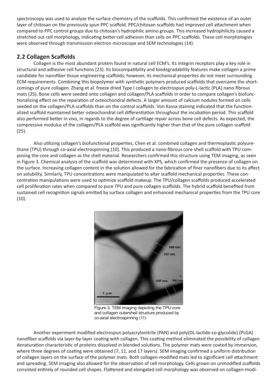
Also utilizing collagen’s biofunctional properties, Chen et al. combined collagen and thermoplastic polyure-thane (TPU) through co-axial electrospinning (10). This produced a nano-fibrous core-shell scaffold with TPU com-posing the core and collagen as the shell material. Researchers confirmed this structure using TEM imaging, as seen in Figure 3. Chemical analysis of the scaffold was determined with XPS, which confirmed the presence of collagen on the surface. Increasing collagen content in the solution allowed for the fabrication of finer nanofibers due to its affect on solubility. Similarly, TPU concentrations were manipulated to alter scaffold mechanical properties. These con-centration manipulations were used to optimize scaffold makeup. The TPU/collagen scaffolds produced accelerated cell proliferation rates when compared to pure TPU and pure collagen scaffolds. The hybrid scaffold benefited from sustained cell recognition signals emitted by surface collagen and enhanced mechanical properties from the TPU core (10).
Figure 3. TEM imaging depicting the TPU core and collagen outershell structure produced by co-axial electrospinning (11)
Another experiment modified electrospun polyacrylonitrile (PAN) and poly(DL-lactide-co-glycolide) (PLGA) nanofiber scaffolds via layer-by-layer coating with collagen. This coating method eliminated the possibility of collagen denaturation characteristic of proteins dissolved in blended solutions. The polymer mats were coated by immersion, where three degrees of coating were obtained (7, 11, and 17 layers). SEM imaging confirmed a uniform distribution of collagen layers on the surface of the polymer mats. Both collagen-modified mats led to significant cell attachment and spreading. SEM imaging also allowed for the observation of cell morphology. Cells grown on unmodified scaffolds consisted entirely of rounded cell shapes. Flattened and elongated cell morphology was observed on collagen-modi-

fied scaffolds. This indicates the advantageous biofunctional effects elicited by collagen, since the modified scaffolds encouraged cell spreading and phenotypic expression more than the control scaffolds (23).
2.3 Gelatin Scaffolds Gelatin is a natural biopolymer endowed with integrin binding sites, making it a suitable candidate material for tissue engineering scaffolds. Gelatin has received a lot of attention because of its control over cell adhesion and maintained differentiation. Combining it with synthetic polymers can improve the cell adhesion and differentiation of the entire scaffold because of these integrin sites. Recent research conducted by Xue et al. examined the func-tionalization of polycaprolactone (PCL) by blending with gelatin biopolymers (24). A solution of 1:1 mass ratio was electrospun into nano-fibrous scaffolds. An anti-infective feature was also looked at by the addition of metronidozole (MNA). Varying amounts of (MNA) were added to these polymer solutions due to its selective activity against bacte-ria. SEM imaging showed randomly oriented, uniform nanofiber structures with porosity values ranging from 60% - 80%, allowing for sufficient nutrient transport. PCL’s inherent hydrophobic properties were improved by the addition of gelatin due to its amine and carboxyl functional groups. This increase in hydrophilicity surely improved the degra-dation rate of the scaffold. The hybrid scaffold successfully encapsulated MNA, allowing for controlled drug delivery. In vivo experimentation proved this when MNA loaded scaffolds (above 5% MNA) produced less of an inflammatory response when compared to the bodily response from non-treated scaffolds. It was observed that MNA started to hinder cell growth at around 40% composition. The ideal anti-infective properties and cell growth occurred in scaf-folds composed of 30% MNA (24).
Synthetic polymers usually possess sufficient mechanical properties for tissue engineering scaffolds, but lack cell recognition signals and hydrophilicity features that promote cell seeding. Chen & Su sought to improve poly-lactic acid’s (PLLA) functionality with gelatin (8). This study preferred surface modification over direct blending since it caus-es less alteration in nanofiber structure when compared to the ladder. The researchers took the study a step further by modifying gelatin’s carboxyl groups in order to make it cationic. In theory this positively charged protein would experience an increased amount of interaction with negatively charged cell membranes. Observation of the scaffold surface was conducted using a confocal laser scanning microscope (CLSM) by first staining the protein. This con-firmed a uniform distribution of cationic gelatin on the scaffold’s surface. During and after cell incubation, TEM and SEM imaging were used to observe cell interaction on the protein surface and overall morphology. The amount of living and dead cells were observed with an MTS assay where living cells fluoresced green and dead cells fluoresced red. Figure 4 depicts the results of this MTS assay through the 14 day incubation period. Gelatin/PLLA scaffolds were observed to have superior cell activity and vitality compared to normal PLLA scaffolds. Also, cells experienced de-dif-ferentiation on PLLA scaffolds, but gelatin/PLLA scaffolds promoted the proper phenotypic expression expected of healthy chondrocytes. Deeper cell penetration was observed on the gelatin-modified scaffolds and SEM showed thick ECM forming over the newly formed chondrocyte cells. This supports the significant role that gelatin was hypothe-sized to play in biofunctionalization (8).
Figure 4. Results of the MTS assay used to determine cell vitality over the 14 day incubation period (9)
2.4 Laminin Scaffolds Laminin is a glycoprotein that occurs naturally in nerve cells and plays a key role in cellular migration and alignment. It also promotes neurite function and stimulates cell mitosis, making it an intriguing candidate material

for studies that examine the biofunctionalization of polymer scaffolds for cell regeneration. One such study, con-ducted by Kijenska et al., created polyprolactone (PLCL) and laminin hybrid scaffolds (14). They created two types of scaffolds- one created by blending solutions and the other created from co-axial electrospinning. The electrospinning produced core-shell scaffolds with laminin as the outer shell material. The research team seeded neurolemmocytes (or Schwann cells) into the scaffolds. Schwann cells are reparative cells grown around damaged nerves. SEM imaging determined that the nanofibers were fabricated in the 300 nm range. Attenuated Total Reflection Transform Infared (ATR-FTIR) spectroscopic analysis was used to determine the surface chemistry of the scaffolds. Results from this instrument suggested an insignificant amount of laminin on the surface of the scaffolds resulting from the blending technique. MTS assays indicated a lower degree of cell viability in blended scaffolds when compared to normal PLCL scaffolds. The same analysis of the core-shell scaffolds showed a 78% increase in cell proliferation compared to the blended scaffolds. This suggests that the encapsulation of laminin in the core-shell scaffolds allowed for a controlled release of bioactive agents produced from the laminin itself. Dissolving laminin in the organic solvent (required when blending the two solutions) may have denatured it and rendered its biofunctionalizing agents useless. Nevertheless, cells interacted well on both blended and core-shell scaffolds, seen by the formation of typical, spindle shape mor-phology with tripolar extensions observed through SEM imaging (Figure 5) (15).
Figure 5. SEM micrographs depicting cell morphology on a) TCP, b) PLCL, c) PLCL/Lam (Blend), d) PLCL/Lam (CS). (16)
Characterized by its good biocompatibility and sufficient mechanical properties, the biomaterial community considers PLLA an applicable material for tissue engineering. Like many other synthetic polymers, PLLA lacks biologi-cal signalling abilities and hydrophilic properties needed for ideal cell growth. Koh et al. covalently bonded, physically absorbed, and blended laminin with PLLA nano-fibrous mats (17). SEM imaging determined the fiber diameters of these scaffolds ranging from 100 - 500 nm. XPS imaging showed that significantly more lamina existed on the surface of the blended scaffolds. Regardless, all treated scaffolds had better cell viability compared to the unmodified scaf-folds. SEM imaging also showed the most neurite outgrowth occurred on the blended scaffolds (17).
2.5 Starch Scaffolds Starch is a natural polymer composed of two carbohydrates, amylose and amylopectin (20). Santos et al. combined starch and PCL in a 30:70 wt% ratio to form an SPLC through a fiber blending process (21). These scaffolds elicited good porosity values, averaging about 75%. Endothelial cells were seeded onto the scaffolds and tested for vi-ability and proliferation at 3 days and 7 days using a CLSM after calcein-AM staining. CLSM analysis showed sustained cell viability, shown by the living cells fluorescing green. At 7 days the cells covered much of the scaffold surface and had even penetrated deeper into mesh. Cells continued to grow between day 3 and day 7 known from DNA quantifi-cation measurements showing increased cell numbers. The scaffolds, with incubating cells, were sputter coated with gold and viewed under an SEM. These images depicted healthy cell morphology. The study, extended the experiment by coating some of the scaffolds with fibronectin- a protein responsible for cell adhesion regulation. These scaffolds produced the best cell proliferation when observed through CLSM and staining technology. CLSM also showed the presence of PECAM-1 and VE-cadherin in immunofluorescent micrographs. Figure 6 displays the imaging of these ad-hesion regulators on the surface of SPCL scaffolds coated with fibronectin. The maintenance of PECAM-1 and VE-cad-herin indicated positive interactions between cells and the matrix (21).

Figure 6. CLSM imaging of PECAM-1 fluorescing green on SPCL scaf-folds (22)
Another study examining starch used SPCL scaffolds for cartilage tissue repair. Researchers incubated bovine articular chondrocytes onto these scaffolds. After sputter coating with gold, cell morphologies and degrees of pro-liferation were examined with an SEM. These images showed that the chondrocytes had extensively colonized the surface of the SPCL scaffold and had even penetrated deeply into the pores. Cell adhesion was homogeneously dis-tributed and uniform in nature. The cell adhesion showed no difference between the surfaces of fibers and contact points between two fibers. This indicates that the 3D environment produced by the scaffolds favoured the formation of native ECM. Blue metachromatic assays were used to determine the proteoglycans present throughout the matrix. The researchers used optical microscopes to view the results of these assays. They found regions of cells highlighted in purple informing that a native ECM, containing proteoglycans, was formed around and over the seeded cells. The cell proliferation was compared between SPCL and normal PGA scaffolds. The PGA scaffolds produced large regions of cell depletion, whereas SPCL scaffolds were covered in full growth sites (20).
3. Conclusion Electrospinning nanofibers allows for the fabrication of nanotopographic, 3D scaffolds that are advantageous for cell growth. This constitutes the most practical, reproducible method for obtaining these structures (12). Polymers have received a lot of attention for tissue engineering candidate materials. Synthetic polymers excel due to their con-sistent mechanical properties. Their structural function remains intact in aqueous environments where most biopoly-mers fail. Introducing biopolymers adds biosignalling properties that encourage cell attachment and proliferation (9). All of the reviewed studies suggest that the two types of polymers mutually benefit one another when combined to produce a superior scaffold environment.
Chitosan’s affect on chondrocyte regeneration was studied with PEO scaffolds. This experiment sought to determine the most effective concentration ratio of chitosan to PEO. The 90/10 wt% concentration displayed an ideal balance between structural integrity and cell viability. Higher concentrations of PEO started to compromise the scaffold’s structure (4). Chitosan and silk fibroin mixtures were also examined. Higher concentrations of silk fibroin yielded scaffolds with the highest compressive moduli. Even relatively small concentrations of chitosan increased the biocompatibility of the scaffold (3). The addition of chitosan decreased the hydrophobic nature of PPC scaffolds. After ten days of incubation, healthier cell morphologies were observed on PPC/CS scaffolds (14).
The affect of collagen on PLA scaffolds used for bone tissue defect reparation was studied. By itself, collagen lacked the proper mechanical properties required for bone cell growth. PLA compensated for this mechanical short-coming and collagen was shown to increase the formation of calcium nodules. The compressive modulus of cells regenerated on hybrid scaffolds was greater than that of cells grown on pure scaffolds (25). Another study combined TPU and collagen to produce core-shell scaffolds. TPU provided a structurally sound core while surface collagen pro-vided cell recognition signals that increased cell attachment (10). Similar scaffold structures were obtained using PAN and PLGA. Collagen successfully biofunctionalized these synthetic polymers as well, supported by the flattened and elongated cell morphologies that resulted (23).
Scaffolds were produced with PCL’s desirable in vivo degradation properties and gelatin’s integrin binding sites. Anti-bacterial medicine was also introduced, yielding scaffolds that promoted increased cell proliferation and minimal inflammatory responses (24). Gelatin was also seen to decrease the hydrophobic nature of PLLA scaffolds. This led to increased cell activity on Gelatin/PLLA scaffolds when compared to control scaffolds. The hybrid scaffolds also maintained cell phenotypic expression throughout the entire incubation period (8).

Laminin and PLCL composed other biofunctionalized scaffolds, which were particularly useful for the regener-ation of nerve cells. The core-shell structured scaffolds yielded the best cell proliferation due to the encapsulated bio-signalling factors produced by laminin (14). Laminin combined with PLLA also yielded higher cell viabilities compared to unmodified PLLA scaffolds. These blended scaffolds produced the most neurite outgrowth indicating increased levels of biocompatibility due to the laminin (17).
Studies combined starch and PCL, producing scaffolds for improved endothelial cell regeneration. Cell cul-tures migrated more successfully on these scaffolds. These cells also had the most healthy morphologies. PECAM-1’s existence on the scaffold’s surface also contributed to improved cell proliferation (21).

Literature Cited
1. Barnes C P, Sell S A, Boland E D, Simpson D G, Bowlin G L, Nanofiber technology: Designing the next generation of
tissue engineering scaffolds, Advanced Drug Delivery Reviews, Volume 59, Issue 14, 10 December 2007,
Pages 1413-1433, ISSN 0169-409X
2. Barnes C P, Sell S A, Boland E D, Simpson D G, Bowlin G L. [Figure of electrospinning setup]. Virginia Common
Wealth University. Nanofiber technology: Designing the next generation of tissue engineering scaffolds.
Richmond, VA. Advanced Drug Delivery Reviews 59 (2007).
3. Bhardwaj N, Kundu SC, Silk fibroin protein and chitosan polyelectrolyte complex porous scaffolds for tissue engi
neering applications, Carbohydrate Polymers, Volume 85, Issue 2, 6 May 2011, Pages 325-333, ISSN 0144-
8617
4. Bhattarai N, Edmondson D, Veiseh O, Matsen F A, Zhang M, Electrospun chitosan-based nanofibers and their
cellular compatibility, Biomaterials, Volume 26, Issue 31, November 2005, Pages 6176-6184, ISSN 0142-9612.
5. Bhattarai N, Edmondson D, Veiseh O, Matsen F A, Zhang M. [SEM imaging of chitosan/PEO nanofibers]. University
of Washington, Seattle. Electrospun chitosan-based nanofibers and their cellular compatibility. Seattle, WA.
Biomaterials 26 (2005).
6. Burg K J, Porter S, Kellam J F, Biomaterial developments for bone tissue engineering, Biomaterials, Volume 21, Issue
23, 1 December 2000, Pages 2347-2359, ISSN 0142-9612.
7. Cai Q, Wan Y, Bei J, Wang S, Synthesis and characterization of biodegradable polylactide-grafted dextran and its
application as compatilizer, Biomaterials, Volume 24, Issue 20, September 2003, Pages 3555-3562, ISSN 0142-
9612
8. Chen J P, Su C H, Surface modification of electrospun PLLA nanofibers by plasma treatment and cationized gelatin
immobilization for cartilage tissue engineering, Acta Biomaterialia, Volume 7, Issue 1, January 2011, Pages
234-243, ISSN 1742-7061
9. Chen J P, Su C H. [MTS assay imaging]. Chang Gung University. Surface modification of electrospun PLLA nanofibers
by plasma treatment and cationized gelatin immobilization for cartilage tissue engineering. Kwei-San, Taiwan.
Acta Biomaterialia 7 (2011).
10. Chen R, Huang C, Ke Q, He C, Wang H, Mo X, Preparation and characterization of coaxial electrospun
thermoplastic polyurethane/collagen compound nanofibers for tissue engineering applications, Colloids and
Surfaces B: Biointerfaces, Volume 79, Issue 2, 1 September 2010, Pages 315-325, ISSN 0927-7765.

11. Chen R, Huang C, Ke Q, He C, Wang H, Mo X. [TEM image of collagen/TPU]. Donghua University. Preparation and
characterization of coaxial electrospun thermoplastic polyurethane/collagen compound nanofibers for tissue
engineering applications. Shanghai, China. Colloids and Surfaces B: Biointerfaces (2010).
12. Dhandayuthapani B, Yoshida Y, Maekawa T, and Kumar D S, “Polymeric Scaffolds in Tissue Engineering
Application: A Review,” International Journal of Polymer Science, vol. 2011, Article ID 290602, 19 pages,
2011, doi:10.1155/2011/290602.
13. Hutmacher D W, Scaffolds in tissue engineering bone and cartilage, Biomaterials, Volume 21, Issue 24, 15
December 2000, Pages 2529-2543, ISSN 0142-9612.
14. Jing X, Mi H-Y, Peng J, Peng X-H, Turng L-S, Electrospun aligned poly(propylene carbonate) microfibers with
chitosan nanofibers as tissue engineering scaffolds, Carbohydrate Polymers, Available online 20 October
2014, ISSN 0144-8617
15. Kijeńska E, Prabhakaran M P, Swieszkowski W, Kurzydlowski K J, Ramakrishna S, Interaction of Schwann cells with
laminin encapsulated PLCL core–shell nanofibers for nerve tissue engineering, European Polymer Journal,
Volume 50, January 2014, Pages 30-38, ISSN 0014-3057.
16. Kijeńska E, Prabhakaran M P, Swieszkowski W, Kurzydlowski K J, Ramakrishna S. [SEM micrographs of Schwann
morphologies]. University of Singapore & Warsaw University of Technology. Interaction of Schwann cells with
laminin encapsulated PLCL core–shell nanofibers for nerve tissue engineering. Warsaw, Germany. European
Polymer Journal 50 (2014).
17. Koh H S, Yong T, Chan C K, Ramakrishna S, Enhancement of neurite outgrowth using nano-structured scaffolds
coupled with laminin, Biomaterials, Volume 29, Issue 26, September 2008, Pages 3574-3582, ISSN 0142-
9612.
18. Mano J F, Sousa R A, Boesel L F, Neves N M, Reis R U, Bioinert, biodegradable and injectable polymeric matrix
composites for hard tissue replacement: state of the art and recent developments, Composites Science and
Technology, Volume 64, Issue 6, May 2004, Pages 789-817, ISSN 0266-3538
19. Neal R A, McClugage S G III, Link M C, Sefcik L S, Ogle R C, and Botchwey E A. Tissue Engineering Part C: Methods.
March 2009, 15(1): 11-21. doi:10.1089/ten.tec.2007.0366.
20. Olivia J T, Crawford A, Mundy J M, Moreira A R, Gomes M E, Hatton P V, Reis RL, A cartilage tissue engineering
approach combining starch-polycaprolactone fibre mesh scaffolds with bovine articular chondrocytes,
Journal of Materials Science: Materials in Medicine, Volume 2, 2007, Pages 295-302, ISSN 0957-4530.

21. Santos M I, Fuchs S, Gomes M E, Unger R E, Reis R L, Kirkpatrick C J, Response of micro- and macrovascular
endothelial cells to starch-based fiber meshes for bone tissue engineering, Biomaterials, Volume 28, Issue 2,
January 2007, Pages 240-248, ISSN 0142-9612, http://dx.doi.org/10.1016/j.biomaterials.2006.08.006.
22. Santos M I, Fuchs S, Gomes M E, Unger R E, Reis R L, Kirkpatrick C J. [CLSM imaging of PECAM-1]. University of
Minho & Johannes Gutenberg University Mainz. Response of micro- and macrovascular endothelial cells to
starch-based fiber meshes for bone tissue engineering. Braga, Portugal. Biomaterials 28 (2007).
23. Truong Y B, Glattauer V, Briggs K L, Zappe S, Ramshaw J A M, Collagen-based layer-by-layer coating on electrospun
polymer scaffolds, Biomaterials, Volume 33, Issue 36, December 2012, Pages 9198-9204, ISSN 0142-9612.
24. Xue J, He M, Liu H, Niu Y, Crawford A, Coates P D, Chen D, Shi R, Zhang L, Drug loaded homogeneous electrospun
PCL/gelatin hybrid nanofiber structures for anti-infective tissue regeneration membranes, Biomaterials,
Volume 35, Issue 34, November 2014, Pages 9395-9405, ISSN 0142-9612.
25. Zhang S, Chen L, Jiang Y, Cai Y, Xu G, Tong T, Zhang W, Wang L, Ji J, Shi P, Ouyang H W, Bi-layer collagen/
microporous electrospun nanofiber scaffold improves the osteochondral regeneration, Acta Biomaterialia,
Volume 9, Issue 7, July 2013, Pages 7236-7247, ISSN 1742-7061.
Copyright © 2022 FDOKUMEN