Aligned PLGA/HA nanofibrous nanocomposite scaffolds for bone tissue engineering
Upload
independentCategory
view
5download
0
Alginates in Tissue Engineering 77
77
From: Methods in Molecular Biology, vol. 238: Biopolymer Methods in Tissue EngineeringEdited by: A. P. Hollander and P. V. Hatton © Humana Press Inc., Totowa, NJ
7
Alginates in Tissue Engineering
Marcy Wong
1. IntroductionAlginate is a polysaccharide derived from brown seaweed, which has the
unique property of being able to form a gel in the presence of certain divalentcations (e.g., calcium, strontium, or barium). Alginate has been of commercialinterest in the food industry since the 1930s, when its properties as an emulsi-fier, thickener, and stabilizer were recognized. Alginate has also long beenused for biomedical purposes, particularly in the manufacture of surgical dress-ings for exuding wounds (1). However, the explosive increase in medical appli-cations for alginate, began with the recognition of its use as a scaffold for theencapsulation and immunoprotection of transplanted cells. The encapsulationof non-autologous islet cells in alginate for the treatment of diabetes is basedon the concept that nutrients can diffuse in and insulin out of the alginate con-struct without triggering an immune response (2–3). Similarly, alginate hasbeen used to immunoprotect recombinant cells delivering tumor-suppressingagents (4–5) and growth hormone (6). Stable cultures in alginate beads havebeen achieved with a number of cell types including chondrocytes, bone-marrow stromal cells, islets, myoblasts, fibroblasts, Schwann cells, kidneycells, epithelial cells, and hepatocytes. For orthopedic purposes, encapsulatedbone-marrow stromal cells and chondrocytes have been proposed for the heal-ing bone and cartilage defects (7–9). As a bulking agent, alginate has gainedattention as a space-filling material for treating pediatric urinary reflex (10–12).
The methods in this article describe the encapsulation of chondrocytes iso-lated from articular cartilage in standard alginate. The methods can be general-ized to other cell types and other means of gelation. The use of alginate forencapsulation of chondrocytes has become popular, not for its immuno-

78 Wong
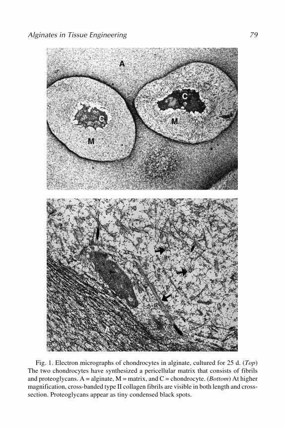
protective properties, but because the chondrocytes maintain their phenotypeand three-dimensional (3D)-morphology in the gel and do not undergo dedif-ferentiation even after long culture periods. As seen in Fig. 1, chondrocytescultured in alginate synthesize a pericellar matrix, which is biochemically andstructurally similar, although not identical, to that found in native cartilage.
1.1. Properties of Alginate
Alginate is a copolymer of D-mannuronic acid (M) and L-guluronic acid (G).The gelation of alginate occurs as blocks of guluronic acid bind to other Gblocks via divalent cations, usually calcium ions (Fig. 2). The mechanical prop-erties of alginate are therefore highly dependent on the G content, the length ofthe G blocks, and the molecular weight of the molecules. Commercially avail-able alginates vary greatly in their composition, and those derived from stemsof Laminaria hyperborea have a guluronic-to-mannuronic ratio 3× higher thanalginate derived from Durvillea potarum (13). Preparation of the hydrogels canalso affect the properties of the gel significantly. The alginate concentration,viscosity, method of sterilization, and the rate of gelation can all be controlledto achieve the desired properties (14–17). Larger structures with more uniformgelation characteristics can be achieved though the use of CaCO3-D-glucono-δ-lactone (GDL) and CaCO3-GDL-CaSO4 during gelation. These substancessignificantly slow the rates of gelation because of the lower solubility of CaCO3
compared to CaCl2 (17). The use of strontium and barium ions in place of calciumions can also produce a stronger and more stable gel (18–19). The modification ofthe chemical structure of alginate in combination with other biopolymers can fur-ther extend the properties of this versatile material (20–22). The biological successof alginate-based transplants will also depend on the heavy metal, endotoxin, andprotein content of the material, and the trace amounts of bacteria, yeast, and moldpresent. Important efforts to standardize alginate properties are currently beingundertaken by the American Society for Testing and Materials (ASTM) in issuingguidelines for tissue-engineered medical products (TEMPS) (13).
2. Materials2.1. Isolation of Cells
1. Articular cartilage (see Note 1).2. Dulbecco's modified Eagle's medium (DMEM) with glutamax-1 (with 25 mM
Hepes, without sodium pyruvate, with pyridoxine) to which the following addi-tives are added:
a. 10% heat-inactivated fetal calf serum (FCS).b. 0.35 mM proline.c. 50 μg/mL gentamycin.
3. Sterile phosphate-buffered saline (PBS) with 50 μg/mL gentamycin.
Alginates in Tissue Engineering 79
Fig. 1. Electron micrographs of chondrocytes in alginate, cultured for 25 d. (Top)The two chondrocytes have synthesized a pericellular matrix that consists of fibrilsand proteoglycans. A = alginate, M = matrix, and C = chondrocyte. (Bottom) At highermagnification, cross-banded type II collagen fibrils are visible in both length and cross-section. Proteoglycans appear as tiny condensed black spots.

80 Wong
4. 1% Pronase solution in DMEM media at 37°C.5. 0.14% Collagenase solution in DMEM media at 37°C.6. 0.1% Solution of trypan blue.7. Hemocytometer.8. Nylon meshes, 120-μm and 20-μm hole size from Millipore (autoclaved).9. Stainless steel strainer, e.g., for tea (autoclaved).
2.2. Alginate Bead Culture
1. Alginate (Pronova LVG) with >60% guluronic acid from FMC BioPolymer AS(Drammen, Norway) (see Note 2). The alginate solution is prepared 2–3 d beforeuse. For a 2% solution, 0.45 g NaCl, and 1.0 g alginate are placed in a 50-mL tubeand volume is brought up to 50 mL with distilled water (see Note 3).
2. 22-gauge needle and syringe.3. Sterile solution of 50 mM CaCl2/0.9% NaCl in 200-mL beaker.4. DMEM with glutamax-1 (with 25 mM Hepes, without sodium pyruvate, with
pyridoxine) to which the following additives are added:
a. 10% heat-inactivated FCS.b. 0.35 mM proline.c. 50 μg/mL gentamycin = 0.5 mL 1000X stock.d. 20 μg/mL ascorbate = from 250X stock (made fresh).
Fig. 2. A schematic of the structure of alginate based on the egg-box model (26).The gray circles represent calcium ions, which primarily bind to guluronic acid blocksto polymerize the gel. Guluronic-rich alginates (left) have a stronger, more open struc-ture than mannuronic-rich alginates (right).

Alginates in Tissue Engineering 81
e. 1 mM cysteine = from 100 mM stock (made fresh).f. 1 mM pyruvate = from 100 mM stock (made fresh).
2.3. Alginate Cylinder Culture
1. Alginate, Pronova LVG with >60% guluronic acid from FMC BioPolymer AS(see Note 2). The alginate solution is prepared 2–3 d before use. For a 2% solu-tion, 0.45 g NaCl, and 1.0 g alginate are placed in a 50-mL tube and volume isbrought up to 50 mL with distilled water (see Note 3).
2. Stainless-steel casting mold (see Note 4).3. Durapore membranes, 5-μm pore size (Millipore).3. Sterile dermal punches (3- or 5-mm diameter) (Stiefel Laboratories).4. Nylon meshes (Millipore) lining 6-well culture plates.5. Size 23-g, 1.25, Nr. 14 needles, and 2-mL syringe (sterile).6. 200-mL beakers of 70% ethanol, sterile PBS, and sterile solution of 50 mM CaCl2/
0.9% NaCl.7. 0.9% NaCl solution (sterile).8. DMEM with glutamax-1 (with 25 mM Hepes, without sodium pyruvate, with
pyridoxine) to which the following additives are added:
a. 10% heat-inactivated FCS.b. 0.35 mM proline.c. 50 μg/mL gentamycin.d. 20 μg/mL ascorbate = from 250X stock (made fresh).e. 1 mM cysteine = from100 mM stock (made fresh).f. 1 mM pyruvate = from 100 mM stock (made fresh).
3. Methods3.1. Isolation of Chondrocytes from Articular Cartilage
1. Remove cartilage from the underlying bone with a sterile scalpel (see Note 5).2. Wash pieces of cartilage in PBS supplemented with gentamycin 3 times × 5 min.3. Transfer tissue to complete temperature- and CO2-equilibrated culture medium
until start of digest.4. Digest tissue in 1% Pronase for 1 h at 37°C. The Pronase is removed and the
tissue is washed once with PBS.5. Digest tissue in 0.14% collagenase solution for at least 17 h at 37°C.6. On the morning of the following day, the collagenase solution together with the
digested tissue is transferred to two 50-mL tubes and the volume of each isbrought up to 45 mL with PBS. Tubes are centrifuged for 5 min at 1000g and thefluid is decanted. Cells are resuspended in 45 mL of PBS and centrifuged for5 min at 1000g, and the fluid is decanted. The cells are then resuspended in20 mL of PBS.
7. Filter cell suspension through a 120-μm nylon mesh held in place by a metalstrainer.
8. Filter cell suspension through a 20-μm nylon mesh. The volumes of the two tubes

82 Wong
are combined and brought up to 45 mL with PBS.9. Centrifuge at 700g for 5 min. The fluid is decanted and cells are suspended in
40 mL of PBS.10. Using a 50-μL aliquot of cell suspension combined with 50 μL of 0.2% trypan
blue solution, the total number of cells and the percentage of viability is mea-sured with a hemocytometer.
11. Centrifuge the cell suspension at 500g for 5 min and decant the solution. Filter-sterilized alginate is added to the cell pellet to attain the desired cell density of 4million cells/mL (see Note 6). The alginate/cell solution is mixed gently until aneven dispersion of cells is attained. At this point, cylinders or beads can be madefrom the alginate solution.
3.2. Alginate Bead Culture
The alginate solution is drawn up into a 22-gauge needle, and the solution isextruded slowly into the CaCl2/NaCl solution. The beads form spontaneously.Allow gelation to proceed for 15 min, and wash beads in 0.9% NaCl for 5 minto remove remaining CaCl2. Transfer beads to temperature- and CO2-equilibratedDMEM media + 10% FCS.
3.3. Alginate Cylinder CultureThe casting mold consists of a metal ring and its upper and lower cover. The
22-mm dia × 2-mm high volume in the ring defines the size of the alginate diskthat is cast. The top and bottom of the metal ring are spanned by a Duraporemembrane and supported by a wire mesh (Fig. 3).
1. Soak all metal pieces and filters in 70% ethanol and assemble the mold in a ster-ile way (see Note 4).
2. Soak mold in beaker containing 70% ethanol, tapping it to remove air bubbles.3. Rinse mold in sterile PBS for 3 min and transfer to sterile Petri dish.4. Inject 1.5 mL of alginate/cell suspension into the mold via a 23-gauge needle.5. Carefully set entire mold into a beaker of sterile 50 mM CaCl2/0.9% NaCl for
30 min to polymerize the alginate.6. Rinse mold briefly in a beaker of PBS and disassemble in a sterile way.7. Using the sterile dermal punch, cut up to twelve 6-mm alginate cylinders from
the disk. The 6-mm cylinders are washed in 0.9% NaCl for 5 min to removeremaining CaCl2.
8. Transfer cylinders to temperature- and CO2-equilibrated DMEM media + 10%FCS. The cylinders are cultured in 6-well plates that have been lined with nylonmesh to prevent samples from adhering to the tissue-culture plastic.
3.4. Alginate Construct Characterization
Immunohistochemistry can be performed on alginate beads or cylinders us-ing standard protocols. Fix alginate pieces in 2% glutaraldehyde buffered with

Alginates in Tissue Engineering 83
0.1 M sodium cacodylate, 10 mM CaCl2 for 3 h followed by incubation in 20%sucrose for 24 h. 10 mM CaCl2 is added to the reagents to prevent destabiliza-tion of the alginate.
4. Notes1. Chondrocytes can survive for several days after sacrifice of the animal if the
tissue is stored at 4°C and the joint is not opened until just before cell isolation.The joint should be opened with a sterile scalpel. With a fresh scapula, tissue isremoved in small slices and placed into sterile PBS.
2. A sterile version of this alginate is available from FMC Biopolymers. Also availableare medium-viscosity alginates and alginates with high mannuronic content. Algi-nate purchased from Kelco Company and Sigma are not purified. They are derivedfrom Macrocystis pyrifera, and usually have weaker mechanical properties becauseof the lower guluronic acid content and a lower mean guluronic acid block length.
3. The alginate solution should be made fresh with each encapsulation, as the poly-mer chains can degrade. The alginate will take up to 3 d to dissolve. Prior to use,
Fig. 3. The stainless-steel casting mold for the gelation of alginate disks, shownassembled (upper left) and disassembled with the ring (upper right) and top and bot-tom cover (bottom left and right).

84 Wong
the alginate is sterilized by passing through a 2 μ syringe filter. Sterilization byautoclave should be avoided.
4. An alternative to our mold has been proposed by Hedbom et al. (23). They cutholes from a silicon rubber pad. The bottom of this mold was a nylon filter soakedin calcium chloride. Gelation occurred as the alginate contacted the calcium chlo-ride, and after 2 min, the entire cast was placed in calcium chloride. Gelation inmolds using slower gelation rates is also recommended.
5. Articular cartilage is stratified into superficial, transitional, and upper and lowerradial zones. The cell morphology and biosynthetic activity vary significantlyamong the zones (24–25), and a digest from the full thickness of cartilage willcontain populations of chondrocytes from each layer. It is possible to remove theupper half of cartilage first and perform separate digestions.
6. Many researchers have had more success producing cartilage-like constructs byincreasing the initial chondrocyte cell density to up to 50 × 106 cells/mL (9).
AcknowledgmentsThis work was supported by the Swiss National Science Foundation and
the M. E. Müller Institute (Director: Prof. E. B. Hunziker). The technicalexpertise of Veronique Gaschen, Mark Siegrist, and Xuanhui Wang is grate-fully acknowledged.
References1. Thomas, S. (2000) Alginate dressings in surgery and wound management—Part
1. J. Wound Care 9, 56–60.2. Lim, F. and Sun, A. M. (1980) Microencapsulated islets as bioartificial endocrine
pancreas. Science 210, 908–910.3. Soon-Shiong, P., Heintz, R. E., Merideth, N., Yao, Q. X., Yao, Z., Zheng, T., et al.
(1994) Insulin independence in a type 1 diabetic patient after encapsulated islettransplantation. Lancet 343, 950–951.
4. Cirone, P., Bourgeois, J. M., Austin, R. C., and Chang, P. L. (2002) A novelapproach to tumor suppression with microencapsulated recombinant cells. Hum.Gene Ther. 13, 1157–1166.
5. Joki, T., Machluf, M., Atala, A., Zhu, J., Seyfried, N. T., Dunn, I. F., et al. (2001)Continuous release of endostatin from microencapsulated engineered cells fortumor therapy. Nat. Biotechnol. 19, 35–39.
6. Chang, P. L. (1997) Microcapsules as bio-organs for somatic gene therapy. Ann.NY Acad. Sci. 831, 461–473.
7. Shang, Q., Wang, Z., Liu, W., Shi, Y., Cui, L., and Cao, Y. (2001) Tissue-engineered bone repair of sheep cranial defects with autologous bone marrowstromal cells. J. Craniofac. Surg. 12, 586–593.
8. Fragonas, E., Valente, M., Pozzi-Mucelli, M., Toffanin, R., Rizzo, R., Silvestri,F., et al. (2000) Articular cartilage repair in rabbits by using suspensions of allo-genic chondrocytes in alginate. Biomaterials 21, 795–801.

Alginates in Tissue Engineering 85
9. Chang, S. C., Rowley, J. A., Tobias, G., Genes, N. G., Roy, A. K., Mooney, D. J.,et al. (2001) Injection molding of chondrocyte/alginate constructs in the shape offacial implants. J, Biomed, Mater, Res, 55, 503–511.
10. Loebsack, A., Greene, K., Wyatt, S., Culberson, C., Austin, C., Beiler, R., et al.(2001) In vivo characterization of a porous hydrogel material for use as a tissuebulking agent. J. Biomed. Mater. Res. 57, 575–581.
11. Diamond, D. and Caldamone, A. (1999) Endoscopic correction of vesicoureteral refluxin children using autologous chondrocytes: preliminary results. J. Urol. 162, 1185–1188.
12. Atala, A., Kim, W., Paige, K. T., Vacanti, C. A., and Retik, A. B. (1994) Endo-scopic treatment of vesicoureteral reflux with a chondrocyte-alginate suspension.J. Urol. 152, 641–643; discussion 644.
13. Dornish, M., Kaplan, D., and Skaugrud, O. (2001) Standards and guidelines forbiopolymers in tissue-engineered medical products: ASTM alginate and chitosanstandard guides. American Society for Testing and Materials. Ann. NY Acad. Sci.944, 388–397.
14. Wong, M., Siegrist, M., Wang, X., and Hunziker, E. (2001) Development ofmechanically stable alginate/chondrocyte constructs: effects of guluronic acidcontent and matrix synthesis. J. Orthop. Res. 19, 493–499.
15. Draget, K., Myhre, S., Skjak-Break, G., and Ostgaard, K. (1988) Regeneration,cultivation and differentiation of plant protoplasts immobilized in Ca-alginatebeads. J. Plant Physiol. 132, 552–556.
16. Martinsen, A., Skjak-Braek, G., and Smidsrod, O. (1989) Alginate as immobiliza-tion material: 1. Correlation between chemical and physical properties of alginatebeads. Biotechnol. Bioeng. 33, 79–89.
17. Kuo, C.K. and Ma, P.X. (2001) Ionically crosslinked alginate hydrogels as scaf-folds for tissue engineering: part 1. Structure, gelation rate and mechanical prop-erties. Biomaterials 22, 511–521.
18. Wideroe, H. and Danielsen, S. (2001) Evaluation of the use of Sr2+ in alginateimmobilization of cells. Naturwissenschaften 88, 224–228.
19. Peirone, M., Ross, C. J., Hortelano, G., Brash, J..L., and Chang, P. L. (1998)Encapsulation of various recombinant mammalian cell types in different alginatemicrocapsules. J. Biomed. Mater. Res. 42, 587–596.
20. Caterson, E. J., Li, W. J., Nesti, L. J., Albert, T., Danielson, K., and Tuan, R. S.(2002) Polymer/alginate amalgam for cartilage-tissue engineering. Ann. NY Acad.Sci. 961, 134–138.
21. Lee, K.Y., Alsberg, E., and Mooney, D. J. (2001) Degradable and injectablepoly(aldehyde guluronate) hydrogels for bone tissue engineering. J. Biomed.Mater. Res. 56, 228–233.
22. Halberstadt, C., Austin, C., Rowley, J., Culberson, C., Loebsack, A., Wyatt, S., etal. (2002) A hydrogel material for plastic and reconstructive applications injectedinto the subcutaneous space of a sheep. Tissue Eng. 8, 309–319.
23. Hedbom, E., Ettinger, L., and Hauselmann, H. J. (2001) Culture of articularchondrocytes in alginate gel—a means to generate cartilage-like implantable tis-sue. Osteoarthritis Cartilage 9, S123–130.

86 Wong
24. Aydelotte, M. B., Greenhill, R. R., and Kuettner, K. E. (1988) Differences betweensub-populations of cultured bovine articular chondrocytes. II. Proteoglycanmetabolism. Connect. Tissue Res. 18, 223–234.
25. Aydelotte, M. B. and Kuettner, K. E. (1988) Differences between sub-populationsof cultured bovine articular chondrocytes. I. Morphology and cartilage matrix pro-duction. Connect. Tissue Res. 18, 205–222.
26. Grant, G., Morris, E., Rees, D., Smith, P., and Thom, D. (1973) Biological inter-actions between polysaccharides and divalent cations. FEBS Lett. 32, 195–198.
Copyright © 2022 FDOKUMEN