Novel porous aortic elastin and collagen scaffolds for tissue engineering
11
Biomaterials 25 (2004) 5227–5237 Novel porous aortic elastin and collagen scaffolds for tissue engineering Qijin Lu, Kavitha Ganesan, Dan T. Simionescu, Narendra R. Vyavahare* Department of Bioengineering, 501-1 Rhodes Research Center, Clemson University, Clemson, SC 29634, USA Received 9 June 2003; accepted 7 December 2003 Abstract Decellularized vascular matrices are used as scaffolds in cardiovascular tissue engineering because they retain their natural biological composition and three-dimensional (3-D) architecture suitable for cell adhesion and proliferation. However, cell infiltration and subsequent repopulation of these scaffolds was shown to be unsatisfactory due to their dense collagen and elastic fiber networks. In an attempt to create more porous structures for cell repopulation, we selectively removed matrix components from decellularized porcine aorta to obtain two types of scaffolds, namely elastin and collagen scaffolds. Histology and scanning electron microscopy examination of the two scaffolds revealed a well-oriented porous decellularized structure that maintained natural architecture of the aorta. Quantitative DNA analysis confirmed that both scaffolds were completely decellularized. Stress– strain analysis demonstrated adequate mechanical properties for both elastin and collagen scaffolds. In vitro enzyme digestion of the scaffolds suggested that they were highly biodegradable. Furthermore, the biodegradability of collagen scaffolds could be controlled by crosslinking with carbodiimides. Cell culture studies showed that fibroblasts adhered to and proliferated on the scaffold surfaces with excellent cell viability. Fibroblasts infiltrated about 120 mm into elastin scaffolds and about 40 mm into collagen scaffolds after 4 weeks of rotary cell culture. These results indicated that our novel aortic elastin and collagen matrices have the potential to serve as scaffolds for cardiovascular tissue engineering. r 2003 Elsevier Ltd. All rights reserved. Keywords: Scaffold; Collagen; Elastin; Tissue engineering; Biodegradation 1. Introduction Tissue engineering offers the potential to create functional and viable tissue constructs for patients requiring organ replacement. One potential approach for creating tissue constructs is to isolate cells from the patient, expand the cell population, culture cells in vitro on a scaffold, and then implant the resulted tissue engineered construct back into the patient. In this approach, a three-dimensional (3-D) scaffold is neces- sary to serve as a template to guide cell growth and tissue development. Formation of the new tissue is greatly influenced by the chemical composition, porosity and 3-D structure of the scaffold [1]. Several require- ments have been identified to be crucial to tissue engineered scaffolds: (1) biocompatibility, to prevent unwanted host tissue responses to the implant; (2) appropriate surface chemistry to promote cell attach- ment and proliferation; (3) interconnected pores with proper size to favor cell infiltration and vascularization; (4) controlled biodegradability to facilitate the forma- tion of new tissue; (5) adequate mechanical properties to maintain the structure and function immediately after implantation and during remodeling of the implants [2]. Currently, biodegradable synthetic polymers such as polyglycolic acid and polylactic acid have been used as scaffold materials in tissue engineering. However, polymeric scaffolds may not interact with cells in a desired manner since their surface chemistry does not promote adequate cell adhesion [3] and may induce toxic and inflammatory reactions [4]. In addition, it is difficult to construct a 3-D synthetic polymer scaffold that resembles the structure of natural tissues. An alternative to synthetic polymers is the use of natural materials. Scaffolds composed of purified ARTICLE IN PRESS *Corresponding author. Tel.: +1-864-656-5559; fax: +1-864-656- 4466. E-mail addresses: [email protected] (D.T. Simionescu), [email protected] (N.R. Vyavahare). 0142-9612/$ - see front matter r 2003 Elsevier Ltd. All rights reserved. doi:10.1016/j.biomaterials.2003.12.019
Transcript of Novel porous aortic elastin and collagen scaffolds for tissue engineering

Biomaterials 25 (2004) 5227–5237
ARTICLE IN PRESS
*Correspondin
4466.
E-mail addres
narenv@clemson
0142-9612/$ - see
doi:10.1016/j.bio
Novel porous aortic elastin and collagen scaffoldsfor tissue engineering
Qijin Lu, Kavitha Ganesan, Dan T. Simionescu, Narendra R. Vyavahare*
Department of Bioengineering, 501-1 Rhodes Research Center, Clemson University, Clemson, SC 29634, USA
Received 9 June 2003; accepted 7 December 2003
Abstract
Decellularized vascular matrices are used as scaffolds in cardiovascular tissue engineering because they retain their natural
biological composition and three-dimensional (3-D) architecture suitable for cell adhesion and proliferation. However, cell
infiltration and subsequent repopulation of these scaffolds was shown to be unsatisfactory due to their dense collagen and elastic
fiber networks. In an attempt to create more porous structures for cell repopulation, we selectively removed matrix components
from decellularized porcine aorta to obtain two types of scaffolds, namely elastin and collagen scaffolds. Histology and scanning
electron microscopy examination of the two scaffolds revealed a well-oriented porous decellularized structure that maintained
natural architecture of the aorta. Quantitative DNA analysis confirmed that both scaffolds were completely decellularized. Stress–
strain analysis demonstrated adequate mechanical properties for both elastin and collagen scaffolds. In vitro enzyme digestion of the
scaffolds suggested that they were highly biodegradable. Furthermore, the biodegradability of collagen scaffolds could be controlled
by crosslinking with carbodiimides. Cell culture studies showed that fibroblasts adhered to and proliferated on the scaffold surfaces
with excellent cell viability. Fibroblasts infiltrated about 120 mm into elastin scaffolds and about 40 mm into collagen scaffolds after 4
weeks of rotary cell culture. These results indicated that our novel aortic elastin and collagen matrices have the potential to serve as
scaffolds for cardiovascular tissue engineering.
r 2003 Elsevier Ltd. All rights reserved.
Keywords: Scaffold; Collagen; Elastin; Tissue engineering; Biodegradation
1. Introduction
Tissue engineering offers the potential to createfunctional and viable tissue constructs for patientsrequiring organ replacement. One potential approachfor creating tissue constructs is to isolate cells from thepatient, expand the cell population, culture cells in vitroon a scaffold, and then implant the resulted tissueengineered construct back into the patient. In thisapproach, a three-dimensional (3-D) scaffold is neces-sary to serve as a template to guide cell growth andtissue development. Formation of the new tissue isgreatly influenced by the chemical composition, porosityand 3-D structure of the scaffold [1]. Several require-ments have been identified to be crucial to tissue
g author. Tel.: +1-864-656-5559; fax: +1-864-656-
ses: [email protected] (D.T. Simionescu),
.edu (N.R. Vyavahare).
front matter r 2003 Elsevier Ltd. All rights reserved.
materials.2003.12.019
engineered scaffolds: (1) biocompatibility, to preventunwanted host tissue responses to the implant; (2)appropriate surface chemistry to promote cell attach-ment and proliferation; (3) interconnected pores withproper size to favor cell infiltration and vascularization;(4) controlled biodegradability to facilitate the forma-tion of new tissue; (5) adequate mechanical properties tomaintain the structure and function immediately afterimplantation and during remodeling of the implants [2].Currently, biodegradable synthetic polymers such aspolyglycolic acid and polylactic acid have been used asscaffold materials in tissue engineering. However,polymeric scaffolds may not interact with cells in adesired manner since their surface chemistry does notpromote adequate cell adhesion [3] and may induce toxicand inflammatory reactions [4]. In addition, it is difficultto construct a 3-D synthetic polymer scaffold thatresembles the structure of natural tissues.
An alternative to synthetic polymers is the use ofnatural materials. Scaffolds composed of purified

ARTICLE IN PRESSQ. Lu et al. / Biomaterials 25 (2004) 5227–52375228
extracellular matrix (ECM) proteins have been usedextensively in tissue engineering. The majority of thesestudies have focused on the fabrication of collagenscaffolds, combined with various procedures for theformation of pores followed by seeding with cells [5–7].Even though the biochemical composition of scaffoldsresembles those of natural tissues, they display verypoor mechanical properties [6,8,9]. This may be due tothe fact that reconstituted ECM molecules are notsufficiently structured and lack proper orientation. Toexploit the 3-D ECM networks of natural tissues,processed vascular tissues have been employed for tissueengineering. This involved removal of cells followed bymoderately successful attempts to repopulate the decel-lularized tissues with cells in vitro [10]. Sheep studiesalso showed that decellularized porcine aorta exhibited alow potential for repopulation in vivo [11]. These resultsmay be due to the dense structure of the aorta and thelack of sufficient porosity. The aorta is made ofconcentric layers of elastin sheets interspersed with acollagen fiber network and populated by smooth musclecells [12]. It is apparent that the aorta, even afterdecellularization, lacks the required porosity necessaryfor cell infiltration.
In the current study we selectively removed one of thetwo major structural components (elastin or collagen)from decellularized aorta for the purpose of creatinginterconnected pores with adequate sizes for cellinfiltration and repopulation. Two types of scaffoldsderived from porcine aorta, namely aortic elastinscaffolds and aortic collagen scaffolds were preparedand characterized. Their usefulness for the developmentof cell-populated constructs was investigated in vitro byculturing fibroblasts on the two scaffolds.
2. Materials and methods
2.1. Tissue harvesting
Porcine hearts were harvested at a local slaughter-house, rinsed in cold saline and transported to thelaboratory on ice. Ascending porcine aorta (a 2–3 cmsupravalvular segment) was dissected, cleaned of ad-herent tissues and fat and rinsed in cold sterile saline.Tissues were subsequently processed for the preparationof aortic elastin and collagen scaffolds, as describedbelow.
2.2. Preparation of aortic elastin scaffolds
Elastin scaffolds were prepared by cyanogen bromide(CNBr) treatment to remove cells, collagen and otherECM components except elastic fibers [13]. Briefly, freshaortic samples were cut into 4� 8 mm2 pieces (ordumbbell shape for mechanical tests as described
below), treated with 50 mg/ml CNBr in 70% formicacid (8 ml/cm2) for 19 h at 20�C with gentle stirring,followed by 1 h at 60�C and boiling for 5 min toinactivate CNBr. Properties of elastin scaffolds wereevaluated by histology, scanning electron microscopy,stress–strain analysis and in vitro biodegradability, asdescribed below. For cell culture studies, samples werefurther washed in sterile saline, sterile Dulbecco’sphosphate buffered saline and preincubated overnightin cell culture medium, using aseptic techniques.
2.3. Preparation of aortic collagen scaffolds
Aortic collagen scaffolds were obtained by decellular-ization followed by carbodiimide crosslinking and finalenzymatic removal of elastic fibers, as described below.For in vitro degradation studies, a control groupwithout carbodiimide crosslinking was also used forcomparison.
(a)
Decellularization: Porcine aorta was decellularizedusing sodium dodecyl sulfate (SDS) extraction [14]followed by trypsin digestion [15]. Aortic sampleswere cut into 10 mm� 10 mm pieces (or dumbbellshape for mechanical properties tests) and incubatedin 1% SDS (in 10 mm Tris buffer, pH 8) for 4 dayswith gentle shaking at room temperature using 4 ml/cm2. Tissue samples were extensively rinsed with10 mm Tris buffer pH 8, and treated with 0.05%trypsin (ICN Biomedicals Inc., Aurora, OH) in10 mm Tris buffer, 0.1% ethylenediamine tetraaceticacid, pH 8, for 18 h at 37�C using 4 ml/cm2.(b)
Crosslinking: SDS/trypsin-decellularized aortic sam-ples were incubated in 20 mm 1-ethyl-3-(3-dimethy-laminopropyl) carbodiimide-HCl (EDC) and 10 mmN-hydroxysuccinimide (NHS) (Pierce, Rockford,IL) in 50 mm Hepes buffer, pH 6.5, for 72 h atroom temperature using 4 ml per 1 cm2 of aorta.After crosslinking, samples were rinsed in 100 mm
Na2HPO4 to quench and neutralize residual EDC[16].
(c)
Elastin removal: Decellularized, crosslinked porcineaorta samples were further treated with elastase toremove elastic fibers leaving behind a 3-D collagenscaffold. Ultra-pure elastase (Elastin ProductsCompany Inc., Owensville, MO) was dissolved ata concentration of 20 U/ml in 100 mm Tris buffer,1 mm CaCl2, 0.02% NaN3, pH 7.8, using 4 ml per1 cm2 of aorta. Samples were incubated for 4 days at37�C with gentle shaking and thoroughly rinsedwith sterile water. Aortic collagen scaffolds for cellculture studies were sterilized for 30 min in 70%ethanol and washed with sterile phosphate buffersaline (PBS) supplemented with 1% penicillin–streptomycin followed by preincubation in cellculture medium overnight before cell seeding.
ARTICLE IN PRESSQ. Lu et al. / Biomaterials 25 (2004) 5227–5237 5229
2.4. Characterization of scaffolds
Fresh aorta and scaffolds were fixed in neutralbuffered 10% formalin, embedded in paraffin andsections obtained from the center of specimens werestained with Hematoxylin and Eosin (H&E), Masson’strichrome, Gomori’s one-step staining and Picro-siriusred staining for histological assessment of cells, collagenand elastin. Scaffolds were also analyzed by scanningelectron microscopy, stress–strain analysis and in vitrobiodegradability assays, as described below.
(a)
Fig.
CNB
trich
Scanning electron microscopy (SEM): SEM wasperformed on cross sections of scaffolds to investi-gate morphology and porosity. Samples for SEMwere fixed in Karnovsky’s fixative (2.5% glutar-aldehyde and 2% formaldehyde in 100 mm ca-codylic acid buffer, pH 7.4) and dehydrated in aseries of graded (50–100%) ethanol. Samples werestored in 100% ethanol prior to critical drying inCO2, and then were gold coated before analysis on aHitachi S-4700 field emission SEM. Pore size wasmeasured on SEM photographs (n=6) using SpotAdvanced Software (Diagnostic Instruments Inc.,Sterling Heights, MI).
(b)
DNA quantification and SDS-PAGE analysis: Freshaorta, elastin scaffolds and collagen scaffolds werepulverized in liquid nitrogen and lyophilized. DNAwas isolated from 10–20 mg dry samples using the1. Preparation and characterization of elastin scaffolds. Representative histolo
r extraction of cells and collagen. A and B, H&E stain (cell nuclei are dark an
rome, in which collagen fibrils stain blue and elastic fibers red. Intimal surfa
Wizard Genomic DNA Purification Kit (Promega,Madison, WI) and quantified with Hoechst 33258using a calibration curve with 0–500 ng/ml DNA.Values are expressed as ng DNA/mg dry sample(n=3). For sodium dodecyl sulfate polyacrylamidegel electrophoresis (SDS-PAGE) analysis, drysamples (1–2 mg) were incubated for 5 days at4�C in extraction buffer (50 mm Tris, 0.1% SDS,pH 7.8, 100 ul/mg dry) and centrifuged at12,000 rpm for 10 min at 4�C. Supernatant sampleswere applied to a reducing 4–15% SDS-PAGEgradient gel (BioRad) and gels were silver stainedusing a commercial kit (SilverSnap II kit, Pierce,Rockford, IL).
(c)
Mechanical properties: Elastin and collagen scaf-folds were evaluated by tensile stress–strain analysisand were compared with properties of fresh aorta.For this purpose, fresh aorta was cut with a die toobtain dumbbell-shape specimens that were 40 mmin length and 5 mm in gauge width (long axisparallel to circumferential direction of the aorta).The specimens (n=4 from each group) were thensubjected to various extraction sequences as de-scribed above for elastin and collagen scaffoldpreparation, maintaining the same ratio of treat-ment solution volume to sample surface area.Tensile tests were performed at room temperatureon a Vitrodyne V-1000 mechanical tester (LifecoInc., Burlington, VT) equipped with a 10 lb loadgical aspects of fresh porcine aorta before (A, C) and after (B, D)
d extracellular matrix is pink). C and D are stained with Masson’s
ce is on bottom. Original magnification, � 200.

ARTICLE IN PRESS
Fig. 2. Scanning electron microscopy of elastin scaffolds showing the
3-D network of elastic fibers. Overall morphology (top, original
magnification, � 250, bar represents 100mm) and representative details
(bottom, original magnification, � 1000, bar represents 20 mm).
Q. Lu et al. / Biomaterials 25 (2004) 5227–52375230
cell. The specimens were extended until rupture at arate of 12 mm/min.
(d)
In vitro biodegradability: For elastin scaffolds, 5–10 mg lyophilized samples (n=3) were incubated in1 ml of 0.1 U/ml ultra-pure elastase (dissolved in100 mm Tris, 1 mm CaCl2, 0.02% NaN3, pH 8) for atotal of 12 days on a shaker at 37�C. At day 1, 2, 4,8 and 12, samples were retrieved, rinsed withdistilled water and lyophilized. The percentage ofmass remaining after digestion was calculated fromthe dry weight before and after enzyme digestion.For collagen scaffolds (both uncrosslinked andEDC crosslinked), 5–10 mg lyophilized samples(n=3) were incubated in 1 ml of 100 U/ml type Icollagenase (Sigma, St. Louis, MO) dissolved in50 mm Tris, 10 mm CaCl2, 0.02% NaN3, pH 8, on ashaker at 37�C. Uncrosslinked samples were re-trieved at 12 h, 1, 2 and 4 days after incubation.EDC crosslinked samples were retrieved at 1, 2, 4,11 and 25 days after incubation. The percentage ofmass remaining after digestion was calculated fromthe dry weight before and after enzyme digestion.
2.5. Cell culture on elastin and collagen scaffolds
A 3T3 mouse fibroblast cell line (ATCC, Manassas,VA) was used in this study. Cell seeding on scaffoldswas performed by suspending freshly trypsinized2� 106 fibroblasts in 2.5 ml of cell culture medium (seebelow) with 5 scaffolds in each tube. Cell-seededscaffolds were cultured under rotary conditions in50 ml polypropylene centrifuge tubes (fitted with0.2 mm filters) placed on a tissue culture rotator (Cel-Gro, Lab-line, Melrose Park, IL) with a rotating speedof 6–8 rpm in a humidified incubator at 37oC and 10%CO2. The cell culture medium used was Dulbecco’smodified Eagle’s medium (HyClone, Logan, UT)supplemented with 10% fetal bovine serum (HyClone,Logan, UT) and 1% penicillin–streptomycin (GibcoBRL, Gaithersburg, MD). Culture medium was chan-ged twice weekly. Cultured samples were collected foranalysis after 7–28 days. Live/dead assay using calceinAM and ethidium homodimer fluorescent staining(Molecular Probes, Eugene, OR) was performedperiodically to evaluate cell viability and proliferationon scaffold surfaces. Cell-seeded scaffolds were fixedin 10% neutral buffered formalin and embedded inparaffin. Cross sections were stained with H&E toevaluate cell infiltration through the scaffolds.
2.6. Statistical data analysis
Results are expressed as mean 7 standard error of themean (SEM). Statistical analyses of the data wereperformed using a two-tailed unpaired Student’s t-testand probability values (p) for significance were calcu-lated; po0.05 were considered significant.
3. Results
3.1. Characterization of elastin scaffolds
Freshly harvested aorta stained with H&E andMasson’s trichrome staining showed characteristicdense structure of smooth muscle cells surrounded bycollagen and elastic fibers (Fig. 1A and C). After CNBrtreatment, cells and collagen could no longer be detectedby H&E and Masson’s trichrome staining (Fig. 1B andD), indicating that CNBr treatment effectively removed
ARTICLE IN PRESS
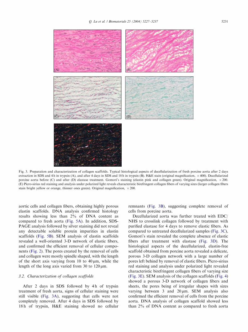
Fig. 3. Preparation and characterization of collagen scaffolds. Typical histological aspects of decellularization of fresh porcine aorta after 2 days
extraction in SDS and 4 h in trypsin (A), and after 4 days in SDS and 18 h in trypsin (B). H&E stain (original magnification, � 400). Decellularized
porcine aorta before (C) and after (D) elastase treatment. Gomori’s staining (elastin pink and collagen green). Original magnification, � 200.
(E) Picro-sirius red staining and analysis under polarized light reveals characteristic birefringent collagen fibers of varying sizes (larger collagen fibers
stain bright yellow or orange, thinner ones green). Original magnification, � 200.
Q. Lu et al. / Biomaterials 25 (2004) 5227–5237 5231
aortic cells and collagen fibers, obtaining highly porouselastin scaffolds. DNA analysis confirmed histologyresults showing less than 2% of DNA content ascompared to fresh aorta (Fig. 5A). In addition, SDS-PAGE analysis followed by silver staining did not revealany detectable soluble protein impurities in elastinscaffolds (Fig. 5B). SEM analysis of elastin scaffoldsrevealed a well-oriented 3-D network of elastic fibers,and confirmed the efficient removal of cellular compo-nents (Fig. 2). The pores created by the removal of cellsand collagen were mostly spindle shaped, with the lengthof the short axis varying from 10 to 40 mm, while thelength of the long axis varied from 30 to 120 mm.
3.2. Characterization of collagen scaffolds
After 2 days in SDS followed by 4 h of trypsintreatment of fresh aorta, signs of cellular staining werestill visible (Fig. 3A), suggesting that cells were notcompletely removed. After 4 days in SDS followed by18 h of trypsin, H&E staining showed no cellular
remnants (Fig. 3B), suggesting complete removal ofcells from porcine aorta.
Decellularized aorta was further treated with EDC/NHS to crosslink collagen followed by treatment withpurified elastase for 4 days to remove elastic fibers. Ascompared to untreated decellularized samples (Fig. 3C),Gomori’s stain revealed the complete absence of elasticfibers after treatment with elastase (Fig. 3D). Thehistological aspects of the decellularized, elastin-freescaffold obtained from porcine aorta revealed a delicate,porous 3-D collagen network with a large number ofpores left behind by removal of elastic fibers. Picro-siriusred staining and analysis under polarized light revealedcharacteristic birefringent collagen fibers of varying size(Fig. 3E). SEM analysis of the collagen scaffolds (Fig. 4)showed a porous 3-D network of collagen fibers andsheets, the pores being of irregular shapes with sizesvarying between 3 and 20 mm. SEM analysis alsoconfirmed the efficient removal of cells from the porcineaorta. DNA analysis of collagen scaffold showed lessthan 2% of DNA content as compared to fresh aorta

ARTICLE IN PRESS
Fig. 4. Representative scanning electron microscopy of collagen
scaffolds depicting an intricate fibrous network. General morphology
(top, original magnification, � 500, bar represents 50 mm) and details
(bottom, original magnification, � 2000, bar represents 10 mm).
Fig. 5. Purity analysis of scaffolds by DNA assay (A) showing more
than 98% reduction of DNA content in both elastin and collagen
scaffolds (as compared to fresh aorta). Comparative SDS-PAGE
analysis (B) of extracts obtained from fresh aorta, elastin scaffolds and
collagen scaffolds showing absence of soluble contaminating proteins
in the two scaffolds. Protein standards are shown in lane 1 (Std.) with
corresponding molecular weights (10–250 kDa).
Q. Lu et al. / Biomaterials 25 (2004) 5227–52375232
(Fig. 5A). In addition, SDS-PAGE analysis of collagenscaffold followed by silver staining did not reveal anydetectable soluble protein impurities (Fig. 5B).
3.3. Mechanical properties of scaffolds
Typical stress–strain curves for elastin scaffolds andcollagen scaffolds are graphically represented in Fig. 6.The rupture tensile strength of the elastin scaffoldshowed values of 26979 kPa, which was about 5 timeslower than that of fresh aorta (1132785 kPa). However,the distensibility (strain at rupture) of the elastinscaffolds was similar to that of fresh aorta(1.0870.02 mm/mm vs. 1.1970.03 mm/mm). Theseresults indicated that removal of the collagen networkreduced tensile strength of scaffolds but maintained
their distensibility. Compared to elastin scaffolds andfresh aorta, aortic collagen scaffolds exhibited lowertensile strength (167726 kPa) and lower distensibility(0.2270.01 mm/mm).
3.4. In vitro biodegradability of scaffolds
The weight percentage of elastin scaffold remainingafter elastase digestion versus incubation time is plottedin Fig. 7. Elastin degraded gradually with time, reaching50% degradation in less than 4 days and completedegradation after 12 days, indicating that the aorticelastin scaffolds are completely biodegradable.
For collagen scaffolds, non-crosslinked collagensamples were almost completely digested (0.970.5%mass remaining) after collagenase treatment for 2 days(Fig. 7), while EDC/NHS crosslinked collagen samplesexhibited a significant resistance to biodegradation(78.472.2% mass remaining after 2 days). Increasingincubation time to 25 days did not significantly induce

ARTICLE IN PRESS
Fig. 7. In vitro enzyme degradation of elastin scaffolds (top) and
collagen scaffolds (bottom). Values are expressed as percentage (%)
mass remaining after exposure to enzyme.
Fig. 6. Stress–strain analysis revealing characteristic mechanical
properties of the elastin and the collagen scaffolds.
Fig. 8. Viability assays. Typical aspects of the Live/Dead assay (live
cells green and dead cells red, arrows) showing excellent cell viability of
fibroblasts cultured on elastin scaffolds (top) and collagen scaffolds
(bottom). Original magnification, � 200.
Q. Lu et al. / Biomaterials 25 (2004) 5227–5237 5233
further degradation (74.671.3%, p=0.2) of crosslinkedcollagen samples (data not shown). These resultsindicate that the rate of aortic collagen scaffolddegradation can be controlled via EDC crosslinkingchemistry.
3.5. Cell culture studies
Repopulation of aortic elastin scaffolds: Fibroblastsseeded on elastin scaffolds were cultured for up to 28days under rotary conditions. Live/dead assays demon-strated that within 3 days, fibroblasts were able toadhere and proliferate on both sides of the surfaces ofelastin scaffolds with excellent cell viability (Fig. 8). Asevidenced by H&E staining, the fibroblasts were foundto infiltrate the intramural layers of the scaffolds within7 days in rotary culture. The depth of cell infiltration

ARTICLE IN PRESS
Fig. 9. Histological aspects of fibroblast infiltration through elastin
scaffolds after 7 to 28 days in culture. H&E stain (original
magnification � 200). Bottom picture is shown at a larger magnifica-
tion to highlight fibroblasts alignment along elastin fibers. H&E stain
(original magnification � 400).
Fig. 10. Infiltration of fibroblasts through collagen scaffolds after 7–28
days in culture. H&E stain (original magnification � 200).
Q. Lu et al. / Biomaterials 25 (2004) 5227–52375234
increased with culture time, reaching about 120 mm after28 days (Fig. 9). Most interestingly, cells grew in anorganized fashion and aligned themselves along thelength of the elastic fibers apparently establishing closecontacts with them (Fig. 9). These results indicate thatCNBr extracted aorta is an excellent substrate for celladhesion and proliferation.
Repopulation of aortic collagen scaffolds: Fibroblastsseeded onto crosslinked aortic collagen scaffolds andcultured under rotary conditions demonstrated excellentsurface attachment and high cell viability as shown byLive/dead assay (Fig. 8). H&E staining results (Fig. 10)showed that the fibroblasts infiltrated into the collagenscaffolds to a depth of about 20 mm after 7 days, andthat the infiltration was increased to about 40 mm after28 days.
4. Discussion
In the present study we showed that decellularizedscaffolds composed of pure matrix components could beobtained from aortic tissue without disruption of thenatural configuration of the aorta. These scaffoldsprovide excellent support for cell proliferation and

ARTICLE IN PRESSQ. Lu et al. / Biomaterials 25 (2004) 5227–5237 5235
could be used in cardiovascular tissue engineeringapplications.
Decellularized porcine matrix has been investigated asa scaffold for cardiovascular tissue engineering, espe-cially in applications for heart valves and vasculargrafts. Decellularization processes involve removingcells with detergents [14], enzyme digestion [17],hypotonic breakdown of cell membranes or a combina-tion of these treatments [18–20]. Using these ap-proaches, both of the two main ECM components,namely collagen and elastin, were preserved. However, ithas been shown that cell migration into these decel-lularized aortic scaffolds was inadequate, both in vitro[17] and in vivo [21], probably due to the very tightECM organization specific to the aortic matrix. Toaddress this problem, we selectively removed matrixcomponents from decellularized aorta to create moreporous scaffolds capable of facilitating cell infiltrationand repopulation.
Elastin scaffolds were obtained by treatment of freshaorta with CNBr in formic acid, a treatment that isknown to cleave proteins at methionine residues (aminoacid ubiquitous in all proteins, including collagen, butabsent in elastin) [22]. In our present study, histologicalanalysis demonstrated that CNBr treatment completelyremoved cells and collagen from the porcine aorta,resulting in elastin scaffolds consisting of concentriclayers of fibers that resembled the original configurationof native aortic elastic lamellae. Moreover, the absenceof protein impurities detectable by SDS-PAGE andsilver staining points out to the purity of scaffolds. Thecomplete removal of cells was also supported by DNAassays, which showed that scaffold preparation stepsremoved more than 98% of DNA compared to freshaorta. To our knowledge, this is a first report showingthe use of CNBr to decellularize aorta for tissueengineering applications. Residual cell remnants withinthe aortic wall were reported after decellularization withhypotonic/hypertonic solutions followed by multipleenzyme digestion [19]. The complete removal ofxenogeneic cell remnants is important for decellularizedscaffolds because they have been implicated in initiationof implant calcification and immune responses towardsimplanted prostheses [23].
Collagen scaffolds were prepared from fresh porcineaorta by a sequence of three extraction steps, namelySDS/trypsin decellularization, carbodiimide crosslink-ing and finally enzyme-mediated elastin removal.Although it has been reported that 1% SDS was ableto decellularize porcine heart valve cusps in 24 h [14],our data showed that decellularization of aortic tissueswas incomplete even after 2 days of 1% SDS treatment(Fig. 3). These differences may occur because of highertissue thickness and density of the aorta compared tocusp tissue. In our experience, successful decellulariza-tion of porcine aorta was attained after 4 days
extraction in SDS followed by 18 h in trypsin (Fig. 3).Decellularized aortic tissues were further crosslinkedwith EDC/NHS in order to stabilize the tissue beforebeing subjected to elastase treatment. Unlike standardglutaraldehyde crosslinking, which may induce toxicreactions and calcification after implantation, EDCcrosslinking does not leave behind harmful residualchemicals and thus EDC crosslinked tissues are lesstoxic and more resistant to calcification in vivo [16].Moreover, the degree of collagen crosslinking (whichtranslates into the extent of biodegradability) can becontrolled by varying the EDC concentration [6] makingEDC-crosslinked collagen scaffolds excellent candidatesfor tissue engineering.
The last step in collagen scaffold preparation was toremove elastin by elastase digestion. Ultra-pure porcinepancreatic elastase was used to selectively remove elastinbut preserve collagen fibers. Gomori’s staining (Fig. 3)demonstrated that elastin was completely removed after4 days of treatment in elastase, obtaining a porousscaffold consisting of collagen fibers. To our knowledge,this is a first report showing removal of elastin fibersfrom decellularized, EDC-crosslinked aortic tissue foruse in tissue engineering.
SEM analysis of elastin scaffolds (Fig. 2) revealed ahighly porous structure consisting of a 3-D network ofelastic fibers, reminiscent of the native aortic structure.This natural architecture may facilitate fiber-orientedcell infiltration and repopulation, as seen in naturaltissues. Pores created by the removal of cells andcollagen were mostly spindle shaped, with sizes varyingfrom 10 to 120 mm. This is in contrast with polymerscaffolds where natural tissue architecture is difficult toachieve. SEM analysis of collagen scaffolds showed aporous structure composed of thick collagen fibers andsheets (Fig. 4); the pores were irregular in shape, withsizes varying from 3 to 20 mm. Teebken et al. [17]reported pore sizes of between 1 and 10 mm indecellularized porcine aortic scaffolds, in which bothelastin and collagen were preserved. Our resultsdemonstrated that selective removal of elastin orcollagen resulted in acellular scaffolds with pore sizesgreater than those reported for acellular aortic tissues.
To characterize the mechanical properties of elastinand collagen scaffolds, stress–strain analysis was per-formed. Our results indicated that removal of thecollagen network reduced the tensile strength butmaintained the distensibility of porcine aorta. On theother hand, removal of elastin resulted in scaffolds withlower tensile strength and lower distensibility than thoseof fresh aorta. Lower tensile strength values of collagenscaffolds as compared to fresh aorta may indicatepartial degradation of collagen due to trypsin treatment.However, the tensile strength values for both scaffoldsderived form aorta were at least eight-fold higher thanthe values (B20 kPa) reported for scaffolds fabricated
ARTICLE IN PRESSQ. Lu et al. / Biomaterials 25 (2004) 5227–52375236
from reconstituted collagen and cultured with smoothmuscle cells under static conditions [24], pointing out thepractical advantages of the proposed methods forscaffold preparation. These results also suggested thatthe combined properties of collagen and elastin fibersare responsible for biomechanical functions of aorta.
We hypothesize that after seeding the scaffolds withfibroblasts or smooth muscle cells and culturing in abioreactor which provides suitable strain on the scaf-folds, the cells will deposit their own collagen and elastinin a direction corresponding to the applied strain, thusincreasing the mechanical properties of the tissueengineered constructs.
In vitro biodegradation tests showed that elastinscaffolds could be digested completely by low levels ofelastase after 12 days at 37�C (Fig. 6). This indicatedthat CNBr treatment did not denature elastin andrender it nonbiodegradable, although this in vitroexperiment could not predict the degradation kineticsof elastin scaffolds when implanted in vivo. Our grouphas shown that elastin has a tendency to calcify whenimplanted subdermally in young rats [25] limiting theapplicability of elastin scaffolds in young recipients.However, experimental studies have shown that calcifi-cation is much reduced when such implants areperformed in mature animals [26]. Furthermore, wehave shown that pretreatment of elastin with aluminumions completely inhibits elastin-mediated calcification[27] and thus, this potential limitation may be circum-vented.
Noncrosslinked collagen scaffolds exhibited a highrate of biodegradation in vitro, while EDC-crosslinkedsamples were about 80% resistant to collagenasethroughout the length of the study. These resultsindicate that unfixed scaffolds may degrade rapidlyafter implantation, while EDC-crosslinked collagen maydegrade significantly slower. As an added advantage,crosslinking of collagen with EDC would reduceimmunogenicity of collagen [16]. These results pointout to the possibility of regulating biodegradability ofaortic collagen scaffolds through alterations of fixationparameters, as has been shown earlier by Park et al. [6]with reconstituted collagen matrices crosslinked withdifferent concentrations of EDC.
In order to evaluate cell adhesion, proliferation andinfiltration properties, 3T3 mouse fibroblasts wereseeded on elastin and collagen scaffolds and culturedon a tissue rotator for 4 weeks. Live/dead assay (Fig. 8)showed that fibroblasts were able to adhere andproliferate on the surfaces of elastin and collagenscaffolds with excellent viability. This suggested thatthese scaffolds were noncytotoxic and were inductive forcell growth, even though toxic chemicals such as CNBror EDC were used during scaffold preparation. Afterinitial attachment and proliferation on the scaffoldsurfaces, fibroblasts were found to infiltrate the intra-
mural layers of both elastin and collagen scaffolds asshown by H&E staining (Figs. 9 and 10). The depth ofcell infiltration increased with culture time; for elastinscaffolds, the infiltration depth reached about 120 mmafter 28 days, while collagen scaffolds were infiltrated toabout 40 mm after 28 days. The significant cell infiltra-tion depth found in elastin scaffolds probably resultedfrom its higher porosity, since it has been reported thatgreater porosity enhances transmural cell migration andproliferation [28]. The slower infiltration into collagenscaffolds may also be explained by the slower degrad-ability of the crosslinked scaffolds. Most importantly,cellular growth in elastin scaffolds was directed by thenaturally aligned architecture of the elastic fibers. Suchcell-seeded scaffolds may eventually develop into newmatrices with defined configurations required for manycardiovascular applications.
5. Conclusions and perspectives
We demonstrated that elastin and collagen scaffoldsderived from porcine aorta maintain the naturalarchitecture of porcine aorta and fulfill many of therequired properties for use in tissue engineering. Bothscaffolds are manageable and exhibit adequate mechan-ical characteristics, porosity, biodegradability and lackof cytotoxicity. Furthermore, their surfaces and compo-sitions efficiently promote cell adhesion, proliferationand infiltration into the deep layers of these tissueconstructs. Due to differences in composition of the twoscaffolds, the applications in tissue engineering may bedistinct; elastin scaffolds are more extensible, havelarger pores and may degrade slower in vivo and maybe used for replacement of tissues that need to maintainlong-term distensibility. Uncrosslinked collagen scaf-folds are more delicate, less extensible and more rapidlybiodegradable and may be more suitable for guidedtissue regeneration. An additional benefit of collagenscaffolds is the possibility to induce a variable degree ofchemical crosslinking allowing for controlled degrada-tion kinetics. Current work is in progress to cultureprimary fibroblasts, myofibroblasts and endothelial cellson aortic elastin and collagen scaffolds in mechanicallyconditioned bioreactors that stimulate the synthesis ofnew ECM and enhance the mechanical properties of thenovel tissue engineered constructs.
Acknowledgements
The authors thank Linda Jenkins for assistance withhistology and Swadeep Pillarisetti for the SEM picturesof elastin scaffolds. This work was supported in part byNIH grant (#HL61652) and a Scientist DevelopmentGrant from the American Heart Association (to NRV).

ARTICLE IN PRESSQ. Lu et al. / Biomaterials 25 (2004) 5227–5237 5237
References
[1] Chapekar MS. Tissue engineering: challenges and opportunities.
J Biomed Mater Res 2000;53(6):617–20.
[2] Yang S, Leong KF, Du Z, Chua CK. The design of scaffolds for
use in tissue engineering. Part I. Traditional factors. Tissue Eng
2001;7(6):679–89.
[3] Langer R. Tissue engineering: a new field and its challenges.
Pharm Res 1997;14(7):840–1.
[4] Ye Q, Zund G, Jockenhoevel S, Hoerstrup SP, Schoeberlein A,
Grunenfelder J, Turina M. Tissue engineering in cardiovascular
surgery: new approach to develop completely human autologous
tissue. Eur J Cardiothorac Surg 2000;17(4):449–54.
[5] Park SN, Lee HJ, Lee KH, Suh H. Biological characterization of
EDC-crosslinked collagen-hyaluronic acid matrix in dermal tissue
restoration. Biomaterials 2003;24(9):1631–41.
[6] Park SN, Park JC, Kim HO, Song MJ, Suh H. Characterization
of porous collagen/hyaluronic acid scaffold modified by 1-ethyl-3-
(3-dimethylaminopropyl)carbodiimide cross-linking. Biomaterials
2002;23(4):1205–12.
[7] Itoh H, Aso Y, Furuse M, Noishiki Y, Miyata T. A honeycomb
collagen carrier for cell culture as a tissue engineering scaffold.
Artificial Organs 2001;25(3):213–7.
[8] Feng Z, Yamato M, Akutsu T, Nakamura T, Okano T, Umezu
M. Investigation on the mechanical properties of contracted
collagen gels as a scaffold for tissue engineering. Artificial Organs
2003;27(1):84–91.
[9] Pei M, Solchaga LA, Seidel J, Zeng L, Vunjak-Novakovic G,
Caplan AI, Freed LE. Bioreactors mediate the effectiveness of
tissue engineering scaffolds. FASEB J 2002;16(12):1691–4.
[10] Elkins RC, Dawson PE, Goldstein S, Walsh SP, Black KS.
Decellularized human valve allografts. Ann Thorac Surg
2001;71(5 Suppl):S428–32.
[11] Goldstein S, Clarke DR, Walsh SP, Black KS, O’Brien MF.
Transpecies heart valve transplant: advanced studies of a
bioengineered xeno-autograft. Ann Thorac Surg 2000;70(6):
1962–9.
[12] Jaques A, Serafini-Fracassini A. Morphogenesis of the elastic
fiber: an immunoelectronmicroscopy investigation. J Ultrastruct
Res 1985;92(3):201–10.
[13] Rasmussen BL, Bruenger E, Sandberg LB. A new method for
purification of mature elastin. Anal Biochem 1975;64(1):255–9.
[14] Booth C, Korossis SA, Wilcox HE, Watterson KG, Kearney JN,
Fisher J, Ingham E. Tissue engineering of cardiac valve prostheses
I: development and histological characterization of an acellular
porcine scaffold. J Heart Valve Dis 2002;11(4):457–62.
[15] Steinhoff G, Stock U, Karim N, Mertsching H, Timke A,
Meliss RR, Pethig K, Haverich A, Bader A. Tissue engineering
of pulmonary heart valves on allogenic acellular matrix conduits:
in vivo restoration of valve tissue. Circulation 2000;102(19 Suppl 3):
III50–5.
[16] van Wachem PB, Plantinga JA, Wissink MJ, Beernink R, Poot
AA, Engbers GH, Beugeling T, van Aken WG, Feijen J, van Luyn
MJ. In vivo biocompatibility of carbodiimide-crosslinked collagen
matrices: Effects of crosslink density, heparin immobilization, and
bFGF loading. J Biomed Mater Res 2001;55(3):368–78.
[17] Teebken OE, Bader A, Steinhoff G, Haverich A. Tissue
engineering of vascular grafts: human cell seeding of decellu-
larised porcine matrix. Eur J Vasc Endovasc Surg 2000;19(4):
381–6.
[18] Zeltinger J, Sherwood JK, Graham DA, Mueller R, Griffith LG.
Effect of pore size and void fraction on cellular adhesion,
proliferation, and matrix deposition. Tissue Eng 2001;7(5):
557–72.
[19] Zeltinger J, Landeen LK, Alexander HG, Kidd ID, Sibanda B.
Development and characterization of tissue-engineered aortic
valves. Tissue Eng 2001;7(1):9–22.
[20] Conklin BS, Richter ER, Kreutziger KL, Zhong DS, Chen C.
Development and evaluation of a novel decellularized vascular
xenograft. Med Eng Phys 2002;24(3):173–83.
[21] Walles T, Herden T, Haverich A, Mertsching H. Influence of
scaffold thickness and scaffold composition on bioartificial graft
survival. Biomaterials 2003;24(7):1233–9.
[22] Daamen WF, Hafmans T, Veerkamp JH, Van Kuppevelt TH.
Comparison of five procedures for the purification of insoluble
elastin. Biomaterials 2001;22(14):1997–2005.
[23] Schmidt CE, Baier JM. Acellular vascular tissues: natural
biomaterials for tissue repair and tissue engineering. Biomaterials
2000;21(22):2215–31.
[24] Nerem RM, Seliktar D. Vascular tissue engineering. Annu Rev
Biomed Eng 2001;3:225–43.
[25] Vyavahare N, Ogle M, Schoen FJ, Levy RJ. Elastin calcification
and its prevention with aluminum chloride pretreatment. Am J
Pathol 1999;155(3):973–82.
[26] Mako WJ, Vesely I. In vivo and in vitro models of calcification
in porcine aortic valve cusps. J Heart Valve Dis 1997;6(3):
316–23.
[27] Bailey M, Xiao H, Ogle M, Vyavahare N. Aluminum chloride
pretreatment of elastin inhibits elastolysis by matrix metallopro-
teinases and leads to inhibition of elastin-oriented calcification.
Am J Pathol 2001;159(6):1981–6.
[28] Nakayama Y, Nishi S, Ishibashi-Ueda H, Matsuda T. Surface
microarchitectural design in biomedical applications: in vivo
analysis of tissue ingrowth in excimer laser-directed micropored
scaffold for cardiovascular tissue engineering. J Biomed Mater
Res 2000;51(3):520–8.