ABSORB Biodegradable Stents Versus Second-Generation Metal Stents
Upload
independentCategory
view
1download
0
Fax +41 61 306 12 34E-Mail [email protected]
Review
Cells Tissues Organs 084 DOI: 10.1159/000XXXXXX
Fibrin: A Natural Biodegradable Scaffold in Vascular Tissue Engineering
Faisal M. Shaikh a, b Anthony Callaghan b Eamon G. Kavanagh a, b
Paul E. Burke a, b Pierce A. Grace a, b Tim M. McGloughlin b
a Department of Surgery, Mid-Western Regional Hospital, and b Centre for Applied Biomedical Engineering Research, University of Limerick, Limerick , Ireland
Introduction
Cardiovascular disease (CVD) represents a growing health and socioeconomic burden in most countries around the world [Murray and Lopez, 1997; World Health Report, 2002]. Each year, CVD causes over 4.35 million deaths in Europe and over 1.9 million deaths in the Eu-ropean Union [Petersen et al., 2005]. The total estimated annual cost due to CVD is EUR 169 billion, with health-care accounting for 62% of costs [Leal et al., 2006]. De-
Key Words
Tissue engineering � Vascular grafts � Natural scaffolds � Biodegradable scaffolds � Fibrin gel
Abstract
Arterial occlusive disease remains a major health issue in the developed world and a rapidly growing problem in the de-veloping world. Although a growing number of patients are now being effectively treated with minimally invasive tech-niques, there remains a tremendous pressure on the vascu-lar community to develop a synthetic small-diameter vascu-lar graft with improved long-term patency rates. The field of tissue engineering offers an exciting alternative in the search for living organ replacement structures. Several methodolo-gies have emerged for constructing blood vessel replace-ments with biological functionality. Common strategies include cell-seeded biodegradable synthetic scaffolds, cell self-assembly, cell-seeded gels and xenogeneic acellular materials. A wide range of materials are being investigated as potential scaffolds for vascular tissue engineering appli-cations. Some are commercialised and others are still in de-velopment. Recently, researchers have studied the role of fibrin gel as a three-dimensional scaffold in vascular tissue engineering. This overview describes the properties of fibrin gel in vascular tissue engineering and highlights some re-cent progress and difficulties encountered in the develop-ment of cell fibrin scaffold technology.
Copyright © 2008 S. Karger AG, Basel
Accepted after revision: January 7, 2008 Published online: $ $ $
Dr. Tim M. McGloughlin Centre for Applied Biomedical Engineering Research University of Limerick IE– $ $ $ Limerick (Ireland) Tel. +353 61 202 217, Fax +353 61 202 944, E-Mail [email protected]
© 2008 S. Karger AG, Basel1422–6405/08/0000–0000$24.50/0
Accessible online at:www.karger.com/cto
Abbreviations used in the paper
ACA �-aminocaproic acidBM-TEV bone marrow tissue-engineered vesselsCVD cardiovascular diseaseEC endothelial cellECGF endothelial cell growth factorECM extracellular matrixEPCs endothelial progenitor cellsePTFE expanded polytetrafluoroethylenehTERT human telomerase reverse transcriptaseMFB myofibroblastsNE norepinephrinePGA polyglycolic acidPTFE polytetrafluoroethyleneSMCs smooth muscle cellsSNAP sodium nitroprusside derivativeTEVs tissue-engineered vessels
CTO084.indd 1CTO084.indd 1 29.04.2008 15:36:1129.04.2008 15:36:11

Shaikh /Callaghan /Kavanagh /Burke /Grace /McGloughlin
Cells Tissues Organs 0842
spite recent advances in preventive medicine and inter-ventional radiology, arterial bypass remains an impor-tant therapeutic option in the management of CVD. Nevertheless, arterial reconstructive surgery is not with-out significant constraints and complications. Harvest-ing native vessels may produce donor site complications such as infection and tissue loss. In addition, there may be a limited supply of harvested vessels because of disease or previous utilisation. Synthetic materials such as Da-cron and polytetrafluoroethylene (PTFE) may partly ad-dress these problems. Whereas synthetic grafts have been widely accepted in large-diameter vessel reconstruction, their utility as small vascular conduits has been limited by high thrombosis rates [Abbott et al., 1987; Burkel, 1988; Teebken and Haverich, 2002]. A biologically de-rived tissue-engineered vessel (TEV) may provide a solu-tion to all these problems. Tissue-engineered vascular graft offers the possibility of designing a true biological substitute material with patient-specific properties. Tis-sue engineering is defined as ‘an interdisciplinary field that applies the principles of engineering and life scienc-es towards the development of biological substitutes that restore, maintain, or improve tissue function’ [Nerem, 1991; Langer and Vacanti, 1993]. Several methodologies have emerged for constructing the blood vessel replace-ments with biological functionality. The most common strategies include cell-seeded gels, cell self-assembly, cell-seeded biodegradable synthetic scaffolds and xenogeneic acellular materials.
Cell-Seeded Gels In 1986, Weinberg and Bell [1986] were the first to at-
tempt a completely biological TEV using animal collagen gels and cultured bovine endothelial cells (ECs), smooth muscle cells (SMCs) and fibroblasts. In the same year, van Buul-Wortelboer et al. [1986] developed a vascular wall with ECs and SMCs grown on a collagen lattice. How-ever, owing to the mechanical weakness of collagen gel, such grafts were not suitable for surgical implantation.
Cell Self-Assembly L’Heureux et al. [1998] have described a different ap-
proach to constructing TEVs by taking advantage of the natural ability of cells to produce their own extracellular matrix (ECM). Briefly, human SMCs and fibroblasts were cultured to form a sheet with associated ECM. These sheets were rolled over a mandrel to form a vascular wall media derived mainly from SMCs, coated by an adventi-tia derived from skin fibroblasts. After maturation, the inner tube was seeded with ECs. However, when implant-
ed in canine models, these engineered vessels result in a patency rate of only 50% after 1 week of implantation.
In a most recent study with a similar approach, human TEVs were constructed using adult human fibroblast alone and then xenografted in an immunosuppressed ca-nine model for short-term and in immunosuppressed rat and primate models for long-term evaluations [L’Heureux et al., 2006]. The TEVs were antithrombogenic and me-chanically stable for up to 8 months in vivo. A smooth muscle-specific � -actin-positive cell population devel-oped within the TEVs suggests the contribution of re-modelling process to the regeneration of vascular media. However, the longer time required to produce these TEVs (approximately 28 weeks) would limit their clinical ap-plication in emergency settings.
Cell Scaffold Technology The commonest method employed in tissue engineer-
ing of vascular constructs is based on cell scaffold tech-nology [Langer and Vacanti, 1993; Nerem and Seliktar, 2001]. In this approach, human cells are seeded onto a suitable bioabsorbable matrix or scaffold. The cells not only multiply, but also release growth factors and matrix proteins, resulting in the formation of complete human tissue [Whitaker et al., 2001; Khademhosseini et al., 2006]. Scaffolds play an important part in controlling the behaviour of cells [Rosenberg, 1962; Grinnell, 1978]. By providing a three-dimensional synthetic template, scaf-folds offer a temporary skeleton, providing a defined ar-chitecture to guide cellular adhesion, tissue growth and devolvement [Kakisis et al., 2005]. Thus, in addition to cells, scaffolds form a most important component in tis-sue engineering.
Several materials are being investigated as potential scaffolds for vascular tissue engineering applications. These can be broadly classified into categories of syn-thetic or naturally derived substances. Unfortunately, none of these meet the characteristics of an ideal scaffold. Common scaffold materials ranging from biological ma-terials such as decellularised xenogenic or allogenic ma-trices [Schmidt and Baier, 2000; Schaner et al., 2004], fi-brin and collagen [Itoh et al., 2001; Berglund et al., 2003] to synthetic polymers such as polyglycolic acid (PGA), polylactic acid, polyhydroxybutrate as well as co-poly-mers of poly- L -lactic acid and polyglycolic acid [Niklason and Langer, 1997; Stock and Vacanti, 2001] meet some but not all of the requirements for an ideal scaffold mate-rial.
Synthetic polymers are relatively biocompatible and involve biomaterials already in use in surgical practice.
CTO084.indd 2CTO084.indd 2 29.04.2008 15:36:2529.04.2008 15:36:25

Fibrin Gel in Vascular Tissue Engineering
Cells Tissues Organs 084 3
Most synthetic polymer scaffolds used in vascular tissue engineering are biodegradable, one of the most desirable material properties for tissue engineering. The major ad-vantage of synthetic scaffolds is that during manufactur-ing, their architectural and mechanical properties can be precisely controlled [Isenberg et al., 2006a, b]. These in-clude molecular weight, shape, porosity, microstructure, tensile strength and degradation time. This facilitates mass production of uniform scaffold constructs. More-over, there is no risk of pathogen transmission.
Among the several synthetic biodegradable polymers used for the construction of engineered vessels, PGA is the most commonly studied scaffold. Its highly porous structure allows nutrient diffusion and subsequent neo-vascularisation [Kakisis et al., 2005]. Moreover, it is eas-ily handled and fabricated into different shapes. As an approach to develop small-diameter vascular conduits, Niklason et al. [1999, 2001] have used a PGA scaffold and a biomimetic perfusion system. Bovine aortic SMCs were seeded on tubular PGA scaffolds and cultured for 8 weeks under conditions of pulsatile pressure before seeding with ECs. The grafts showed 100% patency 4 weeks after implantation into porcine saphenous arteries. They found that the endothelialisation was present only in pockets of the graft surface with persistent PGA remnants. This fail-ure of endothelialisation may be due to cytotoxic degra-dation products of PGA or lack of cell signalling support. Moreover, contractile response of these grafts was only 5% of normal rabbit aorta, but they had burst pressure in excess of 2,000 mm Hg. However, attempts to translate this approach to adult human cells initially failed [Nikla-son, 1999]. McKee et al. [2003] postulated that the pri-mary difficulty with engineering tissues in this model was limited life span of differentiated donated cells. They therefore induced ectopic expression of the human telo-merase reverse transcriptase (hTERT) subunit in adult human smooth muscle to extend the cells’ life span. How-ever, despite the success of hTERT infection of SMC for engineering blood vessels from adult human cells, the strength of human vessels is still too weak to withstand chronic physiological stresses. Moreover, although no ev-idence of transformation has been detected in the hTERT-infected SMC [Poh et al., 2005] so far, there are still con-cerns about oncogenicity in the long term.
However, in spite of these benefits, implantation of synthetic scaffolds has so far yielded modest results. Ma-jor drawbacks of biodegradable polymers include their inherent stiffness before implantation, lack of natural ad-hesive proteins and specific ligands, and the difficulty encountered in the process of creating balance between
the matrix degradation time and the reconstruction of a new matrix [Cebotari et al., 2002]. Premature degrada-tion leading to rupture, aneurysmal dilatation and inti-mal hyperplasia are other major limitations restricting the use of biodegradable polymer constructs in vascular applications [Yow et al., 2006].
Natural collagens are major structural proteins in the ECM of the arterial wall accounting for 20–50% of the dry weight of the arterial wall [Bartos and Ledvina, 1979]. This has led researchers to investigate the use of natural collagen as a potential scaffold in vascular tissue engi-neering [Weinberg and Bell, 1986; L’Heureux et al., 1993]. Unfortunately, collagen in gel form does not possess high mechanical strength, resulting in constructs that are too weak for implantation. To resolve this problem, research-ers have investigated collagen matrices supported with synthetic materials [Weinberg and Bell, 1986; Hirai and Matsuda, 1996]. However, the higher cost, variability in cross-linking density and fibre size, unpredictable enzy-matic degradation, possible side effects and mineralisa-tion are its major limitations [Lee et al., 2001]. Xenogenic acellular scaffolds are further natural alternative materi-als and have been used successfully in numerous tissue engineering applications [Badylak, 2004]. Although they have shown promising results in animal models [Lantz et al., 1993], major concerns remain regarding their use in humans, including unknown transfer of animal-related infections, xenogeneic rejection patterns and their asym-metric dimensions [Patience et al., 1997; Courtman et al., 2001].
Fibrin Gel
Scaffold material is of vital importance in tissue engi-neering and the search for an ideal matrix material for vascular tissue-engineered constructs continues. This re-view examines the role of fibrin gel in applications of scaffold utilisation. Fibrin gel, which is a naturally occur-ring biodegradable matrix, has shown many promising results in vascular tissue engineering applications. The following outlines the current applications of fibrin.
Fibrin Gel as a Biological Scaffold Fibrin gel is a naturally occurring biodegradable ma-
trix that simulates the final stages of the haemostatic co-agulation cascade, forming a structured fibrin clot simi-lar to a physiological clot. The role of fibrin as a biological scaffold has long been studied especially in the process of wound healing and tumour growth [Clark et al., 1982;
CTO084.indd 3CTO084.indd 3 29.04.2008 15:36:2629.04.2008 15:36:26
Shaikh /Callaghan /Kavanagh /Burke /Grace /McGloughlin
Cells Tissues Organs 0844
Dvorak et al., 1987; Amrani et al., 2001]. The importance of fibrin in tissue repair has been acknowledged both clinically and experimentally [Gray et al., 1993; Jackson, 2001]. Fibrin has been studied as a biological scaffold in various tissue engineering applications ( table 1 ). Fibrin gel has many inherent properties that make it an ideal natural biological scaffold for vascular tissue engineering applications ( table 2 ).
Fibrin-based biomaterials are biocompatible and bio-degradable and have high affinity to various biological surfaces [Bensaid et al., 2003]. Being a naturally occur-ring physiological scaffold, it supports angiogenesis and tissue repair [Amrani et al., 2001]. In addition, fibrin nat-
urally contains sites for cellular binding, and has been shown to have excellent cell seeding effects and good tis-sue development [Ye et al., 2000]. Moreover, modification and functionalisation of fibrin matrices has been used to provide controlled release of genes [Trentin et al., 2006] and growth factors [Schmoekel et al., 2005]. Further-more, fibrin gels can be produced from the patients’ own blood and used as an autologous scaffold for the seeded cell without the potential risk of a foreign body reaction [Ye at al., 2000]. Because of its established effects on vas-cular cells, fibrin is of particular interest as a scaffold in vascular tissue engineering [Rowe et al., 2007].
Table 1. Desirable features of ideal scaffold comparable to fibrin gel scaffolds
Ideal characteristic Fibrin characteristics Reference
BiocompatibleNon-toxicNon-allergicNon-inflammatory
Fibrin-based biomaterials have high affinity to variousbiological surfaces and are already in clinical use
Bensaid et al. [2003]Ye et al. [2000]Wozniak et al. [2003]
BiodegradableControlled and adjustablebiodegradation allowing sufficienttime for new vessel formation
Biodegradation can be easily controlled with the use of cross-linking of fibres or using inhibitors of fibrinolysis
Mol et al. [2005]Wozniak et al. [2003]
Autologous natureNon-immunogenicNo foreign body reaction
Fibrin gels are autologously harvested from the patient’s own blood, thus limiting immune or foreign body reactions
Ye et al. [2000]Aper et al. [2007]Jockenhoevel et al. [2001a]
Biological structure Fibrin is a provisional matrix in the normal wound healing; its structural and biochemical properties make it apromising candidate as a scaffold in tissue engineering
Rowe et al. [2006]
Three-dimensional supportiveAssisting cellular growthPermitting easy diffusion ofnutrition and oxygen
Fibrin gels offer excellent cellular growth and tissuedevelopment in a three-dimensional matrix structure;however, there have been concerns regarding the diffusion limits in the gels
Ye et al. [2000]Aper et al. [2003]
Easily processable Manufactured in less timeReproducible
Fibrin gels can be easily produced from patients’ own blood and are completely reproducible
Aper et al. [2007]
EconomicalAffordable to all
Being natural and autologous scaffolds, the economical cost would be acceptable to all
–
Considerable mechanical strength Structurally too weak to withstand physiological dynamic environments
Jockenhoevel et al. [2001b]
Variable in shape and size Possible to construct fibrin scaffolds to fill any desired size, shape or geometry and can be used as an injectable scaffold
Jockenhoevel et al. [2001a]
Tuneable chemical, physical andmechanical properties
The compliance and polymerisation rate of fibrin canbe tightly controlled by varying ionic strength and theconcentration of fibrinogen and thrombin
Rowe et al. [2006]Eyrich et al. [2007]Kjaergard et al. [1994]
CTO084.indd 4CTO084.indd 4 29.04.2008 15:36:2629.04.2008 15:36:26

Fibrin Gel in Vascular Tissue Engineering
Cells Tissues Organs 084 5
Preparation/Formation of Fibrin Gel Fibrin gel is composed of 2 essential components, fi-
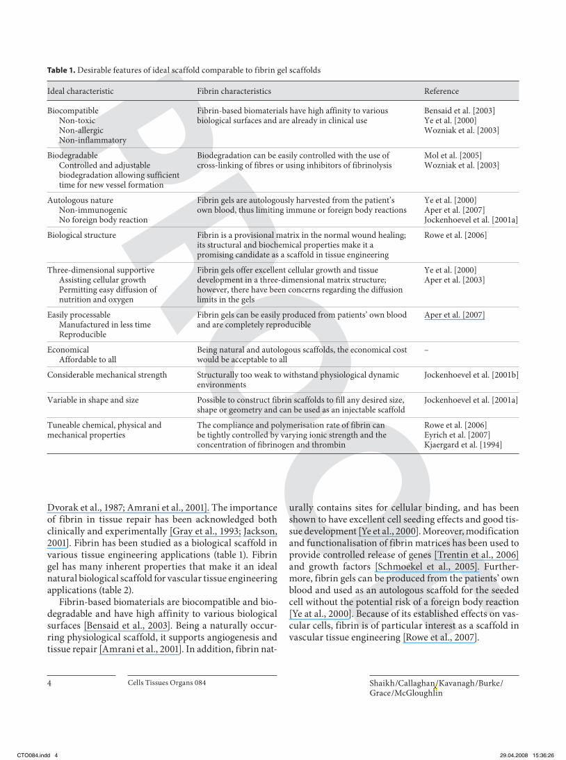
brinogen and thrombin. The fibrinogen (soluble plasma glycoprotein synthesised by the liver) fraction contains a highly concentrated solution of human fibrinogen. When mixed with thrombin, fibrinogen is converted into fibrin monomers [Hantgan et al., 1994; Veklich et al., 1998]. The thrombin fraction contains a highly concentrated human thrombin and calcium chloride solution. The calcium ions serve as key cofactors in the enzymatic conversion of fibrinogen into fibrin. Fibrinogen is a heterodimer glyco-protein made up of 3 pairs of identical chains ( � , � and � ). The thrombin is activated from its inactive form by factor XI, in the presence of factor V. During thrombus formation, the activated thrombin cleaves the 2 small peptides (fibrinopeptides A and B) from the amino-ter-minal ends of the � - and � -chains of the fibrinogen to form fibrin monomers ( fig. 1 ) [Laki, 1951; Lorand and Middlebrook, 1952]. These undergo end-to-end poly-merisation to form long fibrin strands, which then pre-cipitate to form a fibrin clot. Thrombin also activates fac-tor XIII (fibrin-stabilising factor) in the presence of cal-cium ions inducing covalent bond formation between the � -chains of the fibrin monomers. Polymers then form be-
tween the fibrin � -chains, imparting tensile strength and further stabilising the fibrin clot.
Three-Dimensional Matrix A critical step in tissue engineering application is the
development of a three-dimensional structural scaffold, mimicking the ECM which is able to promote and guide actively the newly formed cells and tissue. As pointed out earlier, commonly used materials for maintaining three-dimensional structures in tissue engineering are either synthetic polymers like polyglycolic acid or natural gels like collagen. However, such materials are expensive and some are potentially immunogenic, have low cell adhe-sion, show toxic degradation, and inflammatory and for-eign body reactions. To address these issues, Ye et al. [2000] investigated the use of fibrin gel as an alternative three-dimensional biodegradable scaffold in cardiovas-cular tissue engineering. Human myofibroblasts (MFBs) harvested from the ascending aorta were suspended and seeded in fibrin gel solution into 6-well plates for 4 weeks and supplemented with different concentrations of apro-tinin to control degradation of the gel. The study showed homogenous cell growth and confluent collagen produc-tion in fibrin gel structures. In addition, no toxic degra-dation or inflammatory reactions were detected in the fibrin gels. Furthermore, fibrin gel-MFB structures dis-solved within 2 days in medium without aprotinin, but medium supplemented with higher concentration of aprotinin retained the three-dimensional structure and had higher collagen content and better tissue develop-ment.
Natural and Controlled Biodegradation of Fibrin Rapid and complete integration into the host tissue is
one of the key requirements for the clinical success of scaffolds in tissue engineering applications. Fibrin gels offer the advantage of a completely biodegradable poly-mer. In the normal biological environment, fibrin strands are eventually degraded through enzymatic activation of plasminogen, which becomes the proteolytic enzyme plasmin. This response is initiated when plasminogen ac-tivators, tissue-type plasminogen activator or urokinase, convert the inactive zymogen plasminogen to the active form plasmin. Plasmin is responsible for the degradation and solubilisation of the insoluble fibrin meshwork in wounds and haemostatic plugs [Mosesson, 2005]. Be-cause of this rapid degradation and intrinsic instability of fibrin gels due to this fibrinolysis, it is seen as a major dif-ficulty in using fibrin gel as scaffold material for tissue engineering applications, especially in fields where shape-
Table 2. Fibrin gel scaffolds in various tissue engineering appli-cations
Organ Reference
Cornea Alaminos et al. [2006]
Cartilage Eyrich et al. [2007]Mesa et al. [2006]Park et al. [2005]
Cardiac muscle and valves Birla et al. [2005]van Lieshout et al. [2006]
Skin wound healing Cox et al. [2004]Bannasch et al. [2000]Cohen et al. [2001]Currie et al. [2001]
Bone Tholpady et al. [1999]Yamada et al. [2003]
Liver Bruns et al. [ 2005]
Urethra and urinary bladder Farhat et al. [2006]Bach et al. [2001]
Nerve Willerth et al. [2006]Pittier et al. [2005]
CTO084.indd 5CTO084.indd 5 29.04.2008 15:36:2629.04.2008 15:36:26

Shaikh /Callaghan /Kavanagh /Burke /Grace /McGloughlin
Cells Tissues Organs 0846
specific scaffolds are required [Eyrich et al, 2007]. How-ever, it is possible to control fibrin degradation with the use of cross-linking or by enzyme inhibitors [Wozniak, 2003]. Thus, fibrin scaffolds offer an additional benefit of more controlled degradation and adaptation according to the tissue developmental needs [Ye et al., 2000]. Resis-tance to fibrin degradation is achieved through cross-linking of the fibres or the presence of the inhibitors of fibrinolysis [Grassl et al., 2002]. Proteolytic inhibitors, such as aprotinin, and fibrinolytic inhibitors, such as tranexamic acid or � -aminocaproic acid (ACA), have been studied to slow the degradation of fibrin gels [Woz-niak, 2003].
Various studies have examined the effects of inhibi-tors on cell proliferation, collagen production and tissue formation. Beneficial effects have been reported [Ye et al., 2000] as well as adverse results [Grassl et al., 2002] and no effects at all [Grassl et al., 2003]. Fibrin gel studies us-ing MFBs and aprotinin have reported an increase in col-lagen formation and better tissue development with in-crease inhibitor [Ye et al., 2000]. Grassl et al. [2003], in an effort to optimise the culture conditions of neonatal SMCs entrapped in tubular fibrin gels, found that vary-ing the concentration of ACA did not affect the collagen production, but that lower concentration resulted in com-promised physical integrity of the gels. In contrast, a more recent study by Mol et al. [2005] found that the use of ACA inhibits ECM formation, although it did not hamper human venous MFB proliferation. These find-ings were based on histological evaluation of tissue con-structs after culturing specimens for up to 6 weeks. They hardly identified any collagen fibres in specimens with ACA. A possible explanation for the inhibitory effects of
ACA is that as the fibrin degradation inhibitor ACA is a lysine analogue, it might act as competitive residue pre-venting cross-linking of collagen molecules, explaining the absence of collagen fibres.
Enhanced Endothelial Attachment and Shear Stress Resistance ECs play a crucial role in vascular biology. In addition
to their function as a selective permeability barrier, ECs are involved in the regulation of thrombosis, thromboly-sis, platelet adherence, modulation of vascular tone and blood flow, regulation of immune and inflammatory re-sponse, and mechanotransduction [Sumpio et al., 2002]. Moreover, EC loss from vascular injury is responsible for the local activation of the pathophysiological cascade leading to the development of intimal hyperplasia [Ver-rier and Boyle, 1996; De Meyer and Herman, 1997]. A confluent, adherent, non-thrombogenic endothelial layer is therefore a key requirement for a successfully engi-neered vessel [Mitchell and Niklason, 2003]. Earlier at-tempts at developing tissue-engineered vascular grafts involved seeding ECs onto the surface of synthetic grafts to provide the antithrombogenic blood-contacting sur-face. EC seeding has been attempted either by primary means, as the bypass procedure is underway (single-stage seeding), or by seeding the graft prior to its surgical im-plantation (2-stage seeding) [Rashid et al., 2004]. Herring
et al. [1978] were the first to introduce successful single-stage seeding of ECs onto synthetic vascular grafts. Since then, many researchers have examined various adhesive coatings on synthetic materials with the goal of enhanc-ing EC coverage and improving long-term performance of cell-seeded grafts. Collagen, laminin, fibronectin and
N
A AC
C
C
C
C
C
N
N
N N
Thrombin
N
Fibrinopeptide A Fibrinopeptide B Disulphide bond Fig. 1. Diagram showing the structure of fibrinogen and thrombin cleavage site (ar-rows).
CTO084.indd 6CTO084.indd 6 29.04.2008 15:36:2629.04.2008 15:36:26

Fibrin Gel in Vascular Tissue Engineering
Cells Tissues Organs 084 7
fibrin glue coatings have all been tested on Dacron and PTFE vascular grafts [Xiao and Shi, 2004]. Schrenk et al. [1987] first described the effects of fibrin glue coating on expanded PTFE (ePTFE) prostheses. They found that the pre-coating of ePTFE grafts with fibrin glue enhanced EC attachment and a more equal distribution of cells over the graft surface compared to those pre-treated with whole blood. Since then, many researchers have evaluat-ed the use of fibrin gel either in vivo or in vitro as a po-tential biological scaffold for vascular tissue engineering applications.
In vivo, ECs are subjected to complex mechanical en-vironments composed of shear stress, pressure and cir-cumferential stretch [Ziegler et al., 1998]. For successful clinical application of the EC seeding, seeded cells should resist such in vivo mechanical forces. The major limita-tion of EC seeding in synthetic vascular grafts is that not only is the number of available autologous cells limited, but also are the efficiency of harvest and the degree ofadherence minimal. More than 70% of seeded ECs are washed away during restoration of the blood flow [Rosen-man et al., 1985]. EC detachment from the surface, which essentially occurs during the first 20 min after restoration of the blood flow, is greater at increasing levels of shear stress [Thompson et al., 1994]. Various groups have stud-ied the role of adhesive proteins such as fibronectin, col-lagen, gelatine, whole ECM and fibrinogen in enhancing the EC attachment. It has been shown that fibrin is the most non-thrombogenic adhesive protein with respect to platelet consumption and platelet activation compared to whole ECM or gelatine [Santhosh Kumar and Krishnan, 2001]. Williams et al. [1985] studied in vitro adherence of adult human ECs to both Dacron and Teflon. Their re-sults showed that EC adherence dramatically increases when the grafts are coated with ECM, plasma or fibronec-tin. Unfortunately, fibronectin coating also increases the adhesion of unwanted platelets further increasing the risk of thrombus formation [Seeger and Klingman, 1985; Poot et al., 1989]. Studying the effects of fibrin, gelatine and EC growth factor (ECGF) enmeshed with fibrin, coating to support attachment and proleferation of ECs onto poly-meric materials, Santhosh Kumar and Krishan [2001] found that EC monolayer on gelatine was lost from the surface by 40%, while that on fibrin matrix was oriented towards the direction of flow without significant cell loss. There was no difference in the effects of shear between monolayers grown on fibrin and fibrin with ECGF. Also, ECs grown on fibrin with ECGF composite do not elicit any thrombogenic reaction when platelet-rich plasma was incubated with the monolayer.
Zilla et al. [1989] investigated shear stress resistance of cultured human endothelium on 6-mm PTFE vascular grafts in vitro by pre-coating the grafts with fibrin glue. Seven grafts were lined with adult human saphenous vein ECs and 11 with human umbilical vein ECs. Grafts were then cultivated for 9 days before they were exposed to pulsatile shear stress for 48 h. After 24 h of perfusion, a cell loss of 23% in adult human saphenous vein ECs and of 42% in human umbilical vein EC-lined grafts was en-countered. In spite of a further cell loss during the follow-ing 24 h of perfusion, most of the graft surface was still covered by ECs. The authors concluded that fibrin glue was a suitable substrate for the formation of a shear stress-resistant EC monolayer on PTFE vascular grafts.
In an attempt to improve the resistance of seeded ECs to desquamation due to shear stress, Gosselin et al. [1996] evaluated the effects of coating ePTFE grafts with fibrin glue and fibronectin on the retention of ECs exposed to pulsatile flow ex vivo . Their study concluded that the re-tention of ECs seeded onto ePTFE grafts coated with fi-brin glue containing fibroblast growth factor 1 and hepa-rin was significantly higher than that on fibronectin-coated grafts after 1 h of perfusion. Because of these inherent physiological properties, fibrin became the sub-strate of choice for clinical trials and was approved for clinical use in Europe [Zilla et al., 1989].
The clinical trials utilising fibrin for endothelialisa-tion have so far shown convincing results [Zilla et al., 1994; Deutsch et al., 1999]. In a recent study, the authors describe a 7-year experience of clinical in vitro autolo-gous endothelialisation of 153 fibrin pre-coated infrain-guinal ePTFE implanted as femoropopliteal bypass grafts [Meinhart et al., 2001]. Patients assigned to in vitro au-tologous endothelialisation have 4–5 cm of vein removed under local anaesthesia. Cephalic vein was used as acell source in most cases. Before EC lining, 6- or 7-mm ePTFE grafts 70 cm long were pre-coated with fibrin glue. Grafts were then filled with EC suspension in culture me-dium (7 8 1.5 ! 10 5 cells/cm 2 ). Endothelialisation was achieved with a seeding device rotating at 6 revolutions per hour for 3 h. The grafts were further cultivated for 8.7 8 2.3 days before implantation. In the course of 7 years, 136 patients received 153 successfully endothelialised ePTFE grafts. The observation period for 6-mm grafts was 7 years, that for 7-mm grafts was 4 years. Patency as-sessment was based on duplex sonography and angiogra-phy. The results revealed a primary patency of 62.8% after 7 years for all infrainguinal bypasses. Endothelialised grafts implanted in above-knee position had a 6-year pa-tency of 60%. The patency rate for below-knee grafts was
CTO084.indd 7CTO084.indd 7 29.04.2008 15:36:2729.04.2008 15:36:27

Shaikh /Callaghan /Kavanagh /Burke /Grace /McGloughlin
Cells Tissues Organs 0848
70.8%. Primary patency was 83.7% for the 40 grafts with an inner diameter of 7 mm after 4 years, whereas it was 60% for 6-mm grafts. These data provide promising evi-dence that fibrin-coated endothelialisation improves the patency of synthetic vascular grafts.
In a more recent study, Isenberg et al. [2006a, b] exam-ined the response of endothelialised fibrin gel to physio-logically relevant shear stresses as a final stage in the in vitro development of vascular tissue. The objective of the study was to quantify EC retention upon exposure to physiological flow, which remains a major problem with all small-diameter grafts. While the fibrin scaffolds used in this study possessed mechanical properties that were variable and probably inadequate for implantation, they were still able to demonstrate the central point of the hy-pothesis, namely, that it is possible to generate endotheli-alised fibrin-based scaffolds with high surface coverage and maintain this endothelialised layer in the presence of physiologically relevant shear stresses. This study thus provides the basis for further investigations into the role of the endothelial fibrin scaffold’s development and its importance in modulating the thrombogenicity and im-munogenicity of the constructs in vivo .
Uniform and Efficient Cell Carrier Cell distribution and propagation on the scaffold plays
a crucial role in determining the progression of tissue for-mation. It is known that an initially high homogenous cell distribution is associated with high ECM production [Kim et al., 1998; Mol et al., 2005]. However, it is a sig-nificant challenge to distribute a high cell density effi-ciently and uniformly throughout the scaffold volume [Martin et al., 2004]. Fibrin gels provide many advan-tages that may benefit vascular tissue engineering. Fibrin gel as a cell carrier provides uniform cell distribution, as cells freely migrate and proliferate on top of and within a fibrin gel matrix. It also offers a fast and efficient seeding method, involving only 1 seeding step, rendering a com-pact structure. Furthermore, it also prevents the loss of soluble matrix components into the surrounding medi-um, resulting in a more mature ECM.
Fibrin Gel and Angiogenesis In the normal wound healing process the fibrin clot
not only limits further blood loss, but in addition pro-vides a number of factors that stimulate the formation of new blood vessels. Focusing on this fundamental prop-erty of fibrin and its impact on normal cellular binding, migration and infiltration, researchers have studied in vitro cell systems involving the fibrin matrix and macro-
phages, fibroblasts and ECs [Ciano et al., 1986; Brown et al., 1993; Collen et al., 2003]. Studies have shown that cell-matrix interactions determine the successful matrix in-vasion by the invading cells. There is evidence that EC invasion in fibrin clot is affected by EC fibrinolysis by both plasmin and metalloproteinase systems [Collen et al., 2003]. In their study, Collen et al. [2003] showed that when human ECs were seeded on top of fibrin collagen matrix and stimulated with basic fibroblast growth factor and tumour necrosis factor, both the plasmin and metal-loproteinase contributed to the invasion by ECs. This was partly inhibited by aprotinin and partly by metallopro-teinase inhibitor BB94. In addition to matrix-remodel-ling proteases and cellular matrix receptors, the structure of the matrix is an important determinant in this invad-ing process. The structure of the matrix not only provides the necessary binding sites for cellular integrins, but also determines the rate and extent of the proteolytic degrada-tion by the invading cells [van Hinsbergh et al., 2001].
Delivery Vehicle for Growth Factors Bioactive signalling molecules called growth factors
play a significant role in the cellular growth, proliferation and differentiation in the ECM in vivo [Badylak, 2005]. In order to achieve a successful tissue development, the scaffolds must not only incorporate the growth factors but also release them in a timely and controlled fashion [Whitaker et al., 2001]. In addition to serving as a provi-sional matrix for tissue engineering, fibrin gel can offer a promising approach for the delivery of growth factors. Fibrin gel allows incorporation of growth factors, bioac-tive peptides and proteins and thus can also function as a kind of delivery system for added biologically active substances, such as vascular endothelial growth factor and basic fibroblast growth factor, which can modulate the differentiation of the seeded cells [Jeon et al., 2005; Bhang et al., 2007].
Shape Molding/Injectable Scaffold In cell scaffold technology, the scaffold can be used
either as a pre-formed, three-dimensional porous struc-ture or as an injectable scaffold. In the latter, a mixture of bioactive molecules and solidifiable precursors are in-jected into the defect to form a three-dimensional struc-ture in situ. As the fibrin gel components are in suspen-sion or solution before solidification in vivo, a more ho-mogeneous distribution of cells within the matrices is easily obtained, and it is possible to construct scaffolds to fill any desired form, shape, geometry or architecture. Hence, fibrin gel as an injectable scaffold is a promising
CTO084.indd 8CTO084.indd 8 29.04.2008 15:36:2729.04.2008 15:36:27

Fibrin Gel in Vascular Tissue Engineering
Cells Tissues Organs 084 9
matrix for tissue regeneration, especially for engineering vascular tissues such as valve conduits and vessels with complex side branches.
Studies have been undertaken to assess fibrin gel scaf-folds for tissue-engineered heart valves. Jockenhoevel et al. [2001b] were the first to evaluate fibrin gel as an inject-able scaffold for production of three-dimensional valve conduits. An adjustable aluminium mould consisting of ‘aortic’ and ‘ventricular’ stamps was constructed and used to cast three-dimensional aortic valve fibrin gel structure seeded with MFBs. Although the cell distribu-tion and collagen synthesis were consistent and the re-sulting tissue was suturable, the newly formed matrix had little mechanical stiffness for immediate implantation. Shrinkage of the gel structure also presents a problem [Jockenhoevel et al., 2001a].
Mechanical Weakness The most significant drawback of fibrin gels is that
they are structurally too weak to withstand physiological dynamic environments in vivo [Jockenhoevel et al., 2001b]. To overcome this limitation, various researchers have combined synthetic or natural polymers with fibrin gel. The concept of hybrid scaffolds combining the ad-vantages of natural and synthetic materials is very attrac-tive and seems effective. The skeleton of synthetic poly-mers defines the gross shape and size of the engineered tissue and supports the forming tissue during the initial stages, whereas the fibrin gel facilitates cell invasion and growth.
In a vascular graft model, Aper et al. [2004] described a composite matrix utilising fibrin gel reinforced with a biodegradable polymer, polyglactin, co-cultured with ECs and MFBs and grown in a bioreactor. A structured co-culture of ECs and MFBs was obtained, mimicking the formation of a vascular wall with preserved viability utilising a fibrin preparation. Fluorescence staining re-vealed maintained viability of MFBs to a maximum depth of 519 8 27 � m on day 14 after seeding. Beyond this depth, large numbers of dead cells were found in fluores-cence live/dead assay. Insufficient supply with oxygen and nutrients seems to limit further migration and spreading into the fibrin gel. Other studies have reported three-dimensional extent of seeded fibroblasts in fibrin gel down to a depth of 3 mm [Ye et al., 2000; Jockenhoe-vel et al., 2001a, b]. The mean wall thickness of human coronary arteries ranges between 390 and 1,300 � m, with an outside diameter of 3.9–5.7 mm [Gow and Hadfield, 1979]. Aper et al. [2004] assumed that the maximum thickness of a multilayer of viable MFBs achieved in a fi-
brin preparation would allow for construction of small-diameter vessels.
Cummings et al. [2004] studied collagen, fibrin gel and combined collagen and fibrin gel at equal concentra-tion to form hybrid scaffolds, embedded with rat aortic SMCs in tubular constructs. Morphological and me-chanical properties and the effect of cyclic mechanical strain were evaluated after 6 days in culture. Each matrix type exhibited a characteristic stress-strain profile. Pure collagen had the highest linear modulus at both 2 and4 mg/ml (191 and 242 kPa, respectively) and pure fibrin had the lowest modulus at both concentrations (28 and 19 kPa, respectively), while collagen-fibrin mixtures had intermediate moduli of 153 and 116 kPa, respectively. The ultimate tensile stress was strongly dependent on the degree of gel compaction. Collagen-fibrin mixtures at2 mg/ml total protein content exhibited the highest val-ues (49.7 kPa). This was significantly greater than the ul-timate tensile stress of pure collagen (36.1 kPa) and pure fibrin (15.6 kPa) at the same total protein concentration (p ! 0.0001). Application of cyclic mechanical strain to collagen-fibrin mixture constructs caused a significant increase in gel compaction and a decrease in cell prolif-eration. The linear modulus, ultimate tensile stress and toughness of the constructs were all augmented by me-chanical strain.
Adaptive Vascular Reactivity The functional blood vessel substitute must not only
possess sufficient mechanical strength, but must be able to adapt to changing blood flow conditions, that is, it must exhibit vasoactivity. To achieve this requires hav-ing, as part of the construct, vascular SMCs, which are contractile in nature and oriented in a circumferential direction. Only if an engineered blood vessel substitute possesses these functional characteristics, it may be said that the functionality exhibited by a native vessel is being mimicked [Nerem, 1991].
Swartz et al. [2005] have studied the vasoreactive prop-erties of fibrin-based TEVs. The authors use ovine vascu-lar SMCs and ECs to engineer small-diameter blood ves-sels. Briefly, SMC containing thrombin, fibrinogen and CaCl 2 solution (1.5 ml/tube) was poured into a mold(3-ml syringe barrel) surrounding a 4.0-mm outer diam-eter silastic tube and gelled. After 30 min of incubation, the mold was removed and the fibrin tube was placed in a 50-ml conical tube containing culture medium supple-mented with ascorbic acid. The constructs were incubat-ed for 48 h before addition of aprotinin.
CTO084.indd 9CTO084.indd 9 29.04.2008 15:36:2729.04.2008 15:36:27

Shaikh /Callaghan /Kavanagh /Burke /Grace /McGloughlin
Cells Tissues Organs 08410
After 2 weeks in culture, the constructs, which were to be implanted, were placed into 15-ml conical tubes con-taining ECs in culture medium. These tubes were capped and placed on a rotating device for 2 h at 6 rpm to facili-tate seeding of ECs on the outer surface of the cylindrical constructs. The constructs were then returned to the in-cubator for 3 or 10 days, at which time they were implant-ed. Immediately before implantation, the outer surface of each TEV was pulled inward using forceps to place the ECs in the luminal side of the vessel.
The TEVs attained considerable mechanical strength and vasoreactivity after only 2 weeks in culture. Most im-portantly, fibrin-based TEVs were strong enough to with-stand implantation into the jugular veins of 12-week-old lambs and remained patent for 15 weeks. When explant-ed, TEVs exhibited remarkable remodelling with consid-erable production of collagen and elastin and significant-ly increased mechanical strength and vascular reactivity. The ability of the TEVs to contract or relax was measured after exposure of annular segments of each construct to various vasoactive substances. When exposed to KCl, a non-receptor-mediated vasoconstrictor, TEVs contract-ed with a maximal force of 60 g/g (no aprotinin), which was significantly higher than the contractile force of a na-tive jugular vein (5.9 g/g). Receptor-mediated contraction was also examined using a thromboxane A 2 mimeticU-46619 and norepinephrine (NE). TEVs demonstrated significant contraction in response to both U-46619 and NE, albeit to a lesser extent than KCl, exerting maximal contractions of 21 and 9 g/g, respectively. Relaxation of the constructs was measured by using sodium nitroprus-side derivative (SNAP) that acts as a nitric oxide donor. Nitric oxide induces relaxation through the soluble gua-nylyl cyclase pathway, which ultimately activates protein kinase G. The TEV constructs were first contracted with NE, and the SNAP response was measured as a percent-age of the maximal NE-induced contraction. At a con-centration of 10 –7 M , SNAP caused a partial relaxation, whereas 10 –6 M SNAP caused complete relaxation, indi-cating that the soluble guanylyl cyclase pathway was highly functional in TEV constructs.
Adjustable Morphological and Biochemical Properties The adjustable morphological and biochemical prop-
erties are another desirable and favourable feature of an ideal scaffold. The structural and biochemical properties of fibrin gel can be modified and accustomed according to the developmental needs of the tissue-engineered con-structs. Variation in fibrin gel components such as fibrin-ogen concentration, thrombin concentration, calcium
concentration and ionic strength can generate gels with different structural and mechanical properties as well as stability [Rowe et al., 2006; Eyrich et al., 2007; Kjaergard et al., 1994].
An increase in fibrinogen concentration causes a de-crease in average fibre bundle thickness and an increase in the number of fibre bundles. Also, a decrease in Ca ++ concentration causes a decrease in fibre bundle diameter and an increase in the total number of fibre bundles [Her-bert et al., 1998]. With regard to variation in thrombin, decreased concentration of thrombin results in both in-creased gel compaction as well as thickening of the fibrin microfibres leading to increased mechanical strength [Row et al., 2006]. Similarly, according to Eyrich et al. [2007], only fibrin gels with fibrinogen concentration of 25 mg/ml or higher, a Ca ++ concentration of 20 m M and a pH between 6.8 and 9 were transparent and stable. In contrast, with concentrations out of these ranges, turbid gels were obtained that shrank and completely dissolved within a few weeks.
Accessibility/Completely Autologous Scaffold One of the main goals in vascular tissue engineering
is to create off-the-shelf, immediately available, com-pletely autologous small-calibre vessel substitutes. The recent discovery of vascular endothelial stem cells or en-dothelial progenitor cells (EPCs) has offered the advan-tage of providing purified autologous cells even in vascu-lar patients. The EPCs can be derived easily from patients’ own blood or bone marrow. The increase in circulation EPCs in response to stimuli of vascular injury, hypoxia and ischemia offers an excellent opportunity for easy harvesting either from venesection or non-depleting self-renewing source (blood) for vascular tissue engineering grafts [Sales et al., 2005; Yow et al., 2006]. For a fully bio-compatible small-diameter vascular graft with good long-term patency and functionality, apart from the functional autologous endothelium, a completely autolo-gous scaffold with sufficient biomechanical properties is the other key requirement.
Recently, Aper et al. [2007] have described the devel-opment of a vascular conduit from a completely autolo-gous scaffold and EPCs separated from the peripheral blood of a single donor. Fibrin segments were generated and seeded with EPCs separated from 100 ml of porcine peripheral blood. Fibrin gel tubes, 12–15 cm long and lined with endothelial-like cells, were successfully gener-ated. The seeded tubes showed a remarkable elasticity and burst strength of up to 90 mm Hg. Stable fibrin gel tubes completely lined with an endothelium-like mono-
CTO084.indd 10CTO084.indd 10 29.04.2008 15:36:2729.04.2008 15:36:27

Fibrin Gel in Vascular Tissue Engineering
Cells Tissues Organs 084 11
layer from fibrin and EPCs, both isolated from the same volume of blood, were successfully generated.
In a most recent study, Liu et al. [2007] have developed a novel method for isolating SMCs from ovine bone mar-row mononuclear cells using a tissue-specific promoter and fluorescence-activated cell sorting. Compared to vascular SMC, bone marrow-derived smooth muscle progenitor cells exhibited similar morphology, showed high proliferation potential and expressed early, interme-diate and late markers of vascular SMCs. Bone marrow-derived smooth muscle progenitor cells were embedded in fibrin hydrogels, which were then polymerised around 4-mm diameter mandrels to engineer cylindrical TEV, denoted as bone marrow TEV (BM-TEV). The engineered blood vessels exhibited vascular reactivity in response to KCl and NE and mechanical properties that were compa-rable to those of TEV from vascular SMCs. ECs were also isolated from ovine bone marrow mononuclear cells and seeded in the lumen of BM-TEV that were subsequently implanted as interpositional grafts into the jugular veins of lambs. At 5 weeks after implantation, explanted BM-TEV displayed a confluent endothelial monolayer, cir-cumferential alignment of smooth cells in close proxim-ity to the lumen and remarkable matrix remodelling. Specifically, BM-TEV showed high levels of collagen and fibrillar elastin very similar to native veins. These results raise the hope that functional autologous vessel substi-tutes might one day be engineered from the patient’s own tissues.
Conclusion
This review has outlined the advantages and limita-tions of a naturally occurring biodegradable matrix fi-brin gel. The three-dimensional matrix has tremendous potential in shape moulding, allowing complex shapes to be created. Uniform cell seeding attachment and cell shear detachment resistance are exceptional. The scaf-fold possesses outstanding cell seeding capabilities in ad-dition to being an excellent growth factor carrier. More-over, the controlled biodegradation and collagen synthe-sis make it a perfect scaffold for vascular tissue engineering grafts. Furthermore, fibrin gel’s autologous nature avoids the possibility of negative immune response after implantation. Fibrin gel has inherent potential, out-performing other matrix materials in many respects, with the exception of mechanical strength. Mechanical strength is still under investigation, with promising re-sults being found by Swartz et al. [2005]. Current tech-niques utilising hybrid scaffold technology [Zilla et al., 1989] may be one of the possible routes to increased me-chanical strength and integrity of fibrin gel scaffolds. In conclusion, fibrin gel has obvious promise for developing complex three-dimensional tissue-engineered organs.
References
Abbott, W.M., J. Megerman, J.E. Hasson, G. L’Italien, D.F. Warnock (1987) Effect of com-pliance mismatch on vascular graft patency. J Vasc Surg 5: 376–382.
Amrani, D.L., J.P. Diorio, Y. Delmotte (2001) Wound healing: role of commercial fibrin sealants. Ann NY Acad Sci 936: 566–579.
Alaminos, M., M. Del Carmen Sanchez-Queve-do, J.I. Munoz-Avila, D. Serrano, S. Me-dialdea, I. Carreras, A. Campos (2006)Construction of a complete rabbit cornea substitute using a fibrin-agarose scaffold. In-vest Ophthalmol Vis Sci 47: 3311–3317.
Aper, T., A. Schmidt, M. Duchrow, H.P. Bruch (2007) Autologous blood vessels engineered from peripheral blood sample. Eur J Vasc En-dovasc Surg 33: 33–39.
Aper, T., O.E. Teebken, G. Steinhoff, A. Haverich (2004) Use of a fibrin preparation in the en-gineering of a vascular graft model. Eur J Vasc Endovasc Surg 28: 296–302.
Bach, A.D., H. Bannasch, T.J. Galla, K.M. Bittner, G.B. Stark (2001) Fibrin glue as matrix for cultured autologous urothelial cells in ure-thral reconstruction. Tissue Eng 7: 45–53.
Badylak, S.F. (2004) Xenogeneic extracellular matrix as a scaffold for tissue reconstruction. Transpl Immunol 12: 367–377.
Badylak, S.F. (2005) Regenerative medicine and developmental biology: the role of the extra-cellular matrix. Anat Rec B New Anat 287: 36–41.
Bannasch, H., R.E. Horch, E. Tanczos, G.B. Stark (2000) Treatment of chronic wounds with cultured autologous keratinocytes as sus-pension in fibrin glue. Zentralbl Chir 125(supp 1): 79–81.
Bartos, F., M. Ledvina (1979) Collagen, elastin and desmosines in three layers of bovine aor-tas of different ages. Exp Gerontol 14: 21–26.
Bensaid, W., J.T. Triffitt, C. Blanchat, K. Oudina, L. Sedel, H. Petite (2003) A biodegradable fi-brin scaffold for mesenchymal stem cell transplantation. Biomaterials 24: 2497–2502.
Berglund, J.D., M.M. Mohseni, R.M. Nerem, A. Sambanis (2003) A biological hybrid model for collagen-based tissue engineered vascu-lar constructs. Biomaterials 24: 1241–1254.
Bhang, S.H., O. Jeon, C.Y. Choi, Y.H. Kwon, B.S. Kim (2007) Controlled release of nerve growth factor from fibrin gel. J Biomed Ma-ter Res A 80: 998–1002.
Birla, R.K., G.H. Borschel, R.G. Dennis, D.L. Brown (2005) Myocardial engineering in vivo: formation and characterization ofcontractile, vascularized three-dimensional cardiac tissue. Tissue Eng 11: 803–813.
Brown, L.F., N. Lanir, J. McDonagh, K. Tognazzi, A.M. Dvorak, H.F. Dvorak (1993) Fibroblast migration in fibrin gel matrices. Am J Pathol 142: 273–283.
CTO084.indd 11CTO084.indd 11 29.04.2008 15:36:2729.04.2008 15:36:27

Shaikh /Callaghan /Kavanagh /Burke /Grace /McGloughlin
Cells Tissues Organs 08412
Bruns, H., U. Kneser, S. Holzhüter, B. Roth, J. Kluth, P.M. Kaufmann, D. Kluth, H.C. Fiegel (2005) Injectable liver: a novel approach us-ing fibrin gel as a matrix for culture and in-trahepatic transplantation of hepatocytes. Tissue Eng 11: 1718–1726.
Burkel, W.E. (1988) The challenge of small diam-eter vascular grafts. Med Prog Technol 14: 165–175.
Cebotari, S., H. Mertsching, K. Kallenbach, S. Kostin, O. Repin, A. Batrinac, C. Kleczka, A. Ciubotaru, A. Haverich (2002) Construction of autologous human heart valves based on an acellular allograft matrix. Circulation 106: I63–I68.
Ciano, P.S., R.B. Colvin, A.M. Dvorak, J. Mc-Donagh, H.F. Dvorak (1986) Macrophage migration in fibrin gel matrices. Lab Invest 54: 62–70.
Clark, R.A., J.M. Lanigan, P. DellaPelle, E. Man-seau, H.F. Dvorak, R.B. Colvin (1982) Fibro-nectin and fibrin provide a provisional ma-trix for epidermal cell migration during wound reepithelialization. J Invest Dermatol 79: 264–269.
Cohen, M., A. Bahoric, H.M. Clarke (2001) Aero-solization of epidermal cells with fibrin glue for the epithelialization of porcine wounds with unfavorable topography. Plast Reconstr Surg 107: 1208–1215.
Collen, A., R. Hanemaaijer, F. Lupu, P.H. Quax, N. van Lent, J. Grimbergen, E. Peters, P. Koolwijk, V.W. van Hinsbergh (2003) Mem-brane-type matrix metalloproteinase-medi-ated angiogenesis in a fibrin-collagen ma-trix. Blood 101: 1810–1817.
Courtman, D.W., B.F. Errett, G.J. Wilson (2001) The role of crosslinking in modification of the immune response elicited against xeno-genic vascular acellular matrices. J Biomed Mater Res 55: 576–586.
Cox, S., M. Cole, B. Tawil (2004) Behavior ofhuman dermal fibroblasts in three-dimen-sional fibrin clots: dependence on fibrinogen and thrombin concentration. Tissue Eng 10: 942–954.
Cummings, C.L., D. Gawlitta, R.M. Nerem, J.P. Stegemann (2004) Properties of engineered vascular constructs made from collagen, fi-brin, and collagen-fibrin mixtures. Biomate-rials 25: 3699–3706.
Currie, L.J., J.R. Sharpe, R. Martin (2001) The use of fibrin glue in skin grafts and tissue-engineered skin replacements: a review. Plast Reconstr Surg 108: 1713–1726.
De Meyer, G.R., A.G. Herman (1997) Vascular endothelial dysfunction. Prog Cardiovasc Dis 39: 325–342.
Deutsch, M., J. Meinhart, T. Fischlein, P. Preiss, P. Zilla (1999) Clinical autologous in vitro endothelialization of infrainguinal ePTFE grafts in 100 patients: a 9-year experience. Surgery 126: 847–855.
Dvorak, H.F., V.S. Harvey, P. Estrella, L.F. Brown, J. McDonagh, A.M. Dvorak (1987) Fibrin containing gels induce angiogenesis: impli-cations for tumor stroma generation and wound healing. Lab Invest 57: 673–686.
Eyrich, D., F. Brandl, B. Appel, H. Wiese, G.Maier, M. Wenzel, R. Staudenmaier, A.Goepferich, T. Blunk (2007) Long-term sta-ble fibrin gels for cartilage engineering. Bio-materials 28: 55–65.
Farhat, W.A., J. Chen, C. Sherman, L. Cart-wright, A. Bahoric, H. Yeger (2006) Impact of fibrin glue and urinary bladder cell spray-ing on the in-vivo acellular matrix cellular-ization: a porcine pilot study. Can J Urol 13: 3000–3008.
Gosselin, C., D.A. Vorp, V. Warty, D.A. Severyn, E.K. Dick, H.S. Borovetz, H.P. Greisler (1996) ePTFE coating with fibrin glue, FGF-1, and heparin: effect on retention of seeded endo-thelial cells. J Surg Res 60: 327–332.
Gow, B.S., C.D. Hadfield (1979) The elasticity of canine and human coronary arteries with reference to postmortem changes. Circ Res 45: 588–594.
Grassl, E.D., T.R. Oegema, R.T. Tranquillo (2002) Fibrin as an alternative biopolymer to type-I collagen for the fabrication of a media equivalent. J Biomed Mater Res 60: 607–612.
Grassl, E.D., T.R. Oegema, R.T. Tranquillo (2003) A fibrin-based arterial media equiva-lent. J Biomed Mater Res A 66: 550–561.
Gray, A.J., J.E. Bishop, J.T. Reeves, G.J. Laurent (1993) A � and B � chains of fibrinogen stim-ulate proliferation of human fibroblasts. J Cell Sci 104: 409–413.
Grinnell, F. (1978) Cellular adhesiveness and ex-tracellular substrata. Int Rev Cytol 53: 65–144.
Hantgan, R.R., C.W. Francis, V.J. Marder (1994) Fibrinogen Structure and Physiology, ed 3. Philadelphia, Lippincott.
Herbert, $ $ $ (1998) $ $ $ . Herring, M., A. Gardner, J. Glover (1978) A sin-
gle-staged technique for seeding vascular grafts with autogenous endothelium. Sur-gery 84: 498–504.
Hirai, J., T. Matsuda (1996) Venous reconstruc-tion using hybrid vascular tissue composed of vascular cells and collagen: tissue regen-eration process. Cell Transplant 5: 93–105.
Isenberg, B.C., C. Williams, R.T. Tranquillo (2006a) Small-diameter artificial arteries engineered in vitro. Circ Res 98: 25–35.
Isenberg, B.C., C. Williams, R.T. Tranquillo (2006b) Endothelialization and flow condi-tioning of fibrin-based media-equivalents. Ann Biomed Eng 34: 971–985.
Itoh, H., Y. Aso, M. Furuse, Y. Noishiki, T. Mi-yata (2001) A honeycomb collagen carrier for cell culture as a tissue engineering scaffold. Artif Organs 25: 213–217.
Jackson, M.R. (2001) Fibrin sealants in surgical practice: an overview. Am J Surg 182: 1S–7S.
Jeon, O., S.H. Ryu, J.H. Chung, B.S. Kim (2005) Control of basic fibroblast growth factor re-lease from fibrin gel with heparin and con-centrations of fibrinogen and thrombin. J Control Release 105: 249–259.
Jockenhoevel, S., K. Chalabi, J.S. Sachweh, H.V. Groesdonk, L. Demircan, M. Grossmann, G. Zund, B.J. Messmer (2001a) Tissue engineer-ing: complete autologous valve conduit–a new moulding technique. Thorac Cardio-vasc Surg 49: 287–290.
Jockenhoevel, S., G. Zund, S.P. Hoerstrup, K. Chalabi, J.S. Sachweh, L. Demircan, B.J. Messmer, M. Turina (2001b) Fibrin gel – ad-vantages of a new scaffold in cardiovascular tissue engineering. Eur J Cardiothorac Surg 19: 424–430.
Kakisis, J.D., C.D. Liapis, C. Breuer, B.E. Sumpio (2005) Artificial blood vessel: the Holy Grail of peripheral vascular surgery. J Vasc Surg 41: 349–354.
Khademhosseini, A., R. Langer, J. Borenstein, J.P. Vacanti (2006) Microscale technologies for tissue engineering and biology. Proc Natl Acad Sci USA 103: 2480–2487.
Kim, B.S., A.J. Putnam, T.J. Kulik, D.J. Mooney (1998) Optimizing seeding and culture methods to engineer smooth muscle tissue on biodegradable polymer matrices. Bio-technol Bioeng 57: 46–54.
Kjaergard, H.K., U.S. Weis-Fogh (1994) Impor-tant factors influencing the strength of au-tologous fibrin glue; the fibrin concentration and reaction time – comparison of strength with commercial fibrin glue. Eur Surg Res 26: 273–276.
Laki, K. (1951) The action of thrombin on fibrin-ogen. Science 114: 435–436.
Langer, R., J.P. Vacanti (1993) Tissue engineer-ing. Science 260: 920–926.
Lantz, G.C., S.F. Badylak, M.C. Hiles, A.C. Cof-fey, L.A. Geddes, K. Kokini, G.E. Sandusky, R.J. Morff (1993) Small intestinal submucosa as a vascular graft: a review. J Invest Surg 6: 297–310.
Leal, J., R. Luengo-Fernandez, A. Gray, S. Peter-sen, M. Rayner (2006) Economic burden of cardiovascular diseases in the enlarged Eu-ropean Union. Eur Heart J 27: 1610–1619.
Lee, C.H., A. Singla, Y. Lee (2001) Biomedical ap-plications of collagen. Int J Pharm 221: 1–22.
L’Heureux, N., S. Paquet, R. Labbe, L. Germain, F.A. Auger (1998) A completely biological tissue-engineered human blood vessel. FASEB J 12: 47–56.
L’Heureux, N., Dusserre N., Konig G., Victor B., Keire P., Wight T.N., Chronos N.A., Kyles A.E., C.R. Gregory, G. Hoyt, R.C. Robbins, T.N. McAllister (2006) Human tissue-engi-neered blood vessels for adult arterial revas-cularization. Nat Med 12: 361–365.
L’Heureux, N., L. Germain, R. Labbe, F.A. Auger (1993) In vitro construction of a human blood vessel from cultured vascular cells: a morphologic study. J Vasc Surg 17: 499–509.
CTO084.indd 12CTO084.indd 12 29.04.2008 15:36:2829.04.2008 15:36:28

Fibrin Gel in Vascular Tissue Engineering
Cells Tissues Organs 084 13
Liu, J.Y., D.D. Swartz, H.F. Peng, S.F. Gugino, J.A. Russell, S.T. Andreadis (2007) Function-al tissue-engineered blood vessels from bone marrow progenitor cells. Cardiovasc Res 75: 618–628.
Lorand, L., W.R. Middlebrook (1952) The action of thrombin on fibrinogen. Biochem J 52: 196–199.
Martin, I., D. Wendt, M. Heberer (2004) The role of bioreactors in tissue engineering. Trends Biotechnol 22: 80–86.
McKee, J.A., S.S. Banik, M.J. Boyer, N.M. Ha-mad, J.H. Lawson, L.E. Niklason, C.M. Counter (2003) Human arteries engineered in vitro. EMBO Rep 4: 633–638.
Meinhart, J.G., M. Deutsch, T. Fischlein, N. Howanietz, A. Froschl, P. Zilla (2001) Clini-cal autologous in vitro endothelialization of 153 infrainguinal ePTFE grafts. Ann Thorac Surg 71: S327–S331.
Mesa, J.M., V. Zaporojan, C. Weinand, T.S. John-son, L. Bonassar, M.A. Randolph, M.J. Yaremchuk, P.E. Butler (2006) Tissue engi-neering cartilage with aged articular chon-drocytes in vivo. Plast Reconstr Surg 118: 41–49.
Mitchell, S.L., L.E. Niklason (2003) Require-ments for growing tissue-engineered vascu-lar grafts. Cardiovasc Pathol 12: 59–64.
Mol, A., M.I. van Lieshout, C.G. Dam-de Veen, S. Neuenschwander, S.P. Hoerstrup, F.P. Baaijens, C.V. Bouten (2005) Fibrin as a cell carrier in cardiovascular tissue engineering applications. Biomaterials 26: 3113–3121.
Mosesson, M.W. (2005) Fibrinogen and fibrin structure and functions. J Thromb Haemost 3: 1894–1904.
Murray, C.J., A.D. Lopez (1997) Global mortali-ty, disability, and the contribution of risk fac-tors: Global Burden of Disease Study. Lancet 349: 1436–1442.
Nerem, R.M. (1991) Cellular engineering. Ann Biomed Eng 19: 529–545.
Nerem, R.M. (2000) Tissue engineering a blood vessel substitute: the role of biomechanics. Yonsei Med J 41: 735–739.
Nerem, R.M., D. Seliktar (2001) Vascular tissue engineering. Annu Rev Biomed Eng 3: 225–243.
Niklason, L.E., R.S. Langer (1997) Advances in tissue engineering of blood vessels and other tissues. Transpl Immunol 5: 303–306.
Niklason, L.E., J. Gao, W.M. Abbott, K.K. Hir-schi, S. Houser, R. Marini, R. Langer (1999) Functional arteries grown in vitro. Science 284: 489–493.
Niklason, L.E., W. Abbott, J. Gao, B. Klagges, K.K. Hirschi, K. Ulubayram, N. Conroy, R. Jones, A. Vasanawala, S. Sanzgiri, R. Langer (2001) Morphologic and mechanical charac-teristics of engineered bovine arteries. J Vasc Surg 33: 628–638.
Park, S.H., S.R. Park, S.I. Chung, K.S. Pai, B.H. Min (2005) Tissue-engineered cartilage us-ing fibrin/hyaluronan composite gel and its in vivo implantation. Artif Organs 29: 838–845.
Patience, C., Y. Takeuchi, R.A. Weiss (1997) In-fection of human cells by an endogenous ret-rovirus of pigs. Nat Med 3: 282–286.
Petersen, S., V. Peto, M. Rayner, J. Leal, R.Luengo-Fernandez, A. Gray (2005) Europe-an Cardiovascular Disease Statistics. Lon-don, British Heart Foundation.
Pittier, R., F. Sauthier, J.A. Hubbell, H. Hall (2005) Neurite extension and in vitro my-elination within three-dimensional modi-fied fibrin matrices. J Neurobiol 63: 1–14.
Poh, M., M. Boyer, A. Solan, S.L. Dahl, D. Pe-drotty, S.S. Banik, J.A. McKee, R.Y. Klinger, C.M. Counter, L.E. Niklason (2005) Blood vessels engineered from human cells. Lancet 365: 2122–2124.
Poot, A.A., W.G. Van Aken, A. Dekker, T. Beugeling, J. Feijen, A. Bantjes, J.A. Van Mourik (1989) Cell seeding for bioartificial blood vessels. Hybrid Art Org 177: 285–292.
Rashid, S.T., H.J. Salacinski, B.J. Fuller, G. Ham-ilton, A.M. Seifalian (2004) Engineering of bypass conduits to improve patency. Cell Prolif 37: 351–366.
Rosenberg, M.D. (1962) Long-range interactions between cell and substratum. Proc Natl Acad Sci USA 48: 1342–1349.
Rosenman, J.E., R.F. Kempczinski, W.H. Pearce, E.B. Silberstein (1985) Kinetics of endothe-lial cell seeding. J Vasc Surg 2: 778–784.
Rowe, S.L., S. Lee, J.P. Stegemann (2007) Influ-ence of thrombin concentration on the me-chanical and morphological properties of cell-seeded fibrin hydrogels. Acta Biomater 3: 59–67.
Sales, K.M., H.J. Salacinski, N. Alobaid, M. Mikhail, V. Balakrishnan, A.M. Seifalian (2005) Advancing vascular tissue engineer-ing: the role of stem cell technology. Trends Biotechnol 23: 461–467.
Santhosh Kumar, T.R., L.K. Krishnan (2001) En-dothelial cell growth factor (ECGF) en-meshed with fibrin matrix enhances prolif-eration of EC in vitro. Biomaterials 22: 2769–2776.
Schaner, P.J., N.D. Martin, T.N. Tulenko, I.M. Shapiro, N.A. Tarola, R.F. Leichter, R.A. Carabasi, P.J. Dimuzio (2004) Decellularized vein as a potential scaffold for vascular tissue engineering. J Vasc Surg 40: 146–153.
Schmidt, C.E., J.M. Baier (2000) Acellular vascu-lar tissues: natural biomaterials for tissue re-pair and tissue engineering. Biomaterials 21: 2215–2231.
Schmoekel, H.G., F.E. Weber, J.C. Schense, K.W. Gratz, P. Schawalder, J.A. Hubbell (2005) Bone repair with a form of BMP-2 engi-neered for incorporation into fibrin cellingrowth matrices. Biotechnol Bioeng 89: 253–262.
Schrenk, P., G.S. Kobinia, P. Brucke, G. Syre, A. Edtstadler (1987) Fibrin glue coating of e-PTFE prostheses enhances seeding of hu-man endothelial cells. Thorac Cardiovasc Surg 35: 6–10.
Seeger, J.M., N. Klingman (1985) Improved en-dothelial cell seeding with cultured cells and fibronectin-coated grafts. J Surg Res 38: 641–647.
Stock, U.A., J.P. Vacanti (2001) Tissue engineer-ing: current state and prospects. Annu Rev Med 52: 443–451.
Sumpio, B.E., J.T. Riley, A. Dardik (2002) Cells in focus: endothelial cell. Int J Biochem Cell Biol 34: 1508–1512.
Swartz, D.D., J.A. Russell, S.T. Andreadis (2005) Engineering of fibrin-based functional and implantable small-diameter blood vessels. Am J Physiol Heart Circ Physiol 288: H1451–H1460.
Teebken, O.E., A. Haverich (2002) Tissue engi-neering of small diameter vascular grafts. Eur J Vasc Endovasc Surg 23: 475–485.
Tholpady, S.S., R. Schlosser, W. Spotnitz, R.C. Ogle, W.H. Lindsey (1999) Repair of an osse-ous facial critical-size defect using augment-ed fibrin sealant. Laryngoscope 109: 1585–1588.
Thompson, M.M., J.S. Budd, S.L. Eady, R.F. James, P.R. Bell (1994) Effect of pulsatile shear stress on endothelial attachment to na-tive vascular surfaces. Br J Surg 81: 1121–1127.
Trentin, D., H. Hall, S. Wechsler, J.A. Hubbell (2006) Peptide-matrix-mediated gene trans-fer of an oxygen-insensitive hypoxia-induc-ible factor-1 � variant for local induction of angiogenesis. Proc Natl Acad Sci USA 103: 2506–2511.
Van Buul-Wortelboer, M.F., H.J. Brinkman, K.P. Dingemans, P.G. de Groot, W.G. van Aken, J.A. van Mourik (1986) Reconstitution ofthe vascular wall in vitro: a novel model to study interactions between endothelial and smooth muscle cells. Exp Cell Res 162: 151–158.
Van Hinsbergh, V.W., A. Collen, P. Koolwijk (2001) Role of fibrin matrix in angiogenesis. Ann NY Acad Sci 936: 426–437.
Van Lieshout, M., G. Peters, M. Rutten, F. Baai-jens (2006) A knitted, fibrin-covered poly-caprolactone scaffold for tissue engineering of the aortic valve. Tissue Eng 12: 481–487.
Veklich, Y., C.W. Francis, J. White, J.W. Weisel (1998) Structural studies of fibrinolysis by electron microscopy. Blood 92: 4721–4729.
Verrier, E.D., E.M. Boyle Jr. (1996) Endothelial cell injury in cardiovascular surgery an over-view. Ann Thorac Surg 64: S2–S8.
Weinberg, C.B., E. Bell (1986) A blood vessel model constructed from collagen and cul-tured vascular cells. Science 231: 397–400.
Whitaker, M.J., R.A. Quirk, S.M. Howdle, K.M. Shakesheff (2001) Growth factor release from tissue engineering scaffolds. J Pharm Pharmacol 53: 1427–1437.
Willerth, S.M., K.J. Arendas, D.I. Gottlieb, S.E. Sakiyama-Elbert (2006) Optimization of fi-brin scaffolds for differentiation of murine embryonic stem cells into neural lineage cells. Biomaterials 27: 5990–6003.
CTO084.indd 13CTO084.indd 13 29.04.2008 15:36:2829.04.2008 15:36:28

Shaikh /Callaghan /Kavanagh /Burke /Grace /McGloughlin
Cells Tissues Organs 08414
Williams, S.K., B.E. Jarrell, L. Friend, J.S. Ra-domski, R.A. Carabasi, E. Koolpe, S.N. Muel-ler, S.C. Thornton, T. Marinucci, E. Levine (1985) Adult human endothelial cell compat-ibility with prosthetic graft material. J Surg Res 38: 618–629.
World Health Report (2002) Reducing Risks, Promoting Healthy Life. Geneva, World Health Organization.
Wozniak, G. (2003) Fibrin sealants in support-ing surgical techniques: the importance of individual components. Cardiovasc Surg 11(suppl 1): 17–21.
Xiao, L., D. Shi (2004) Role of precoating inartificial vessel endothelialization. Chin J Traumatol 7: 312–316.
Yamada, Y., J.S. Boo, R. Ozawa, T. Nagasaka, Y. Okazaki, K. Hata, M. Ueda (2003) Bone re-generation following injection of mesenchy-mal stem cells and fibrin glue with a biode-gradable scaffold. J Craniomaxillofac Surg 31: 27–33.
Ye, Q., G. Zund, P. Benedikt, S. Jockenhoevel, S.P. Hoerstrup, S. Sakyama, J.A. Hubbell, M. Turina (2000) Fibrin gel as a three dimen-sional matrix in cardiovascular tissue engi-neering. Eur J Cardiothorac Surg 17: 587–591.
Yow, K.H., J. Ingram, S.A. Korossis, E. Ingham, S. Homer-Vanniasinkam (2006) Tissue engi-neering of vascular conduits. Br J Surg 93: 652–661.
Ziegler, T., K. Bouzourene, V.J. Harrison, H.R. Brunner, D. Hayoz (1998) Influence of oscil-latory and unidirectional f low environments on the expression of endothelin and nitric oxide synthase in cultured endothelial cells. Arterioscler Thromb Vasc Biol 18: 686–692.
Zilla, P., M. Deutsch, J. Meinhart, R. Puschmann, T. Eberl, E. Minar, R. Dudczak, H. Lugmaier, P. Schmidt, I. Noszian, et al. (1994) Clinical in vitro endothelialization of femoropopli-teal bypass grafts: an actuarial follow-up over three years. J Vasc Surg 19: 540–548.
Zilla, P., R. Fasol, P. Preiss, M. Kadletz, M. Deutsch, H. Schima, S. Tsangaris, P. Gros-curth (1989) Use of fibrin glue as a substrate for in vitro endothelialization of PTFE vas-cular grafts. Surgery 105: 515–522.
CTO084.indd 14CTO084.indd 14 29.04.2008 15:36:2829.04.2008 15:36:28
Copyright © 2022 FDOKUMEN