Electrospun nanofibrous structure: A novel scaffold for tissue engineering
9
Electrospun nanofibrous structure: A novel scaffold for tissue engineering Wan-Ju Li, 1,4 * Cato T. Laurencin, 2 Edward J. Caterson, 4 Rocky S. Tuan, 4, * Frank K. Ko 1,3 1 School of Biomedical Engineering, Science and Health Systems, Drexel University, Philadelphia, Pennsylvania 19104 2 Department of Chemical Engineering, Drexel University, Philadelphia, Pennsylvania 19104 3 Department of Materials Engineering, Drexel University, 31 st and Market Street, Philadelphia, Pennsylvania 19104 4 Department of Orthopaedic Surgery, Curtis 501, Thomas Jefferson University, Philadelphia, Pennsylvania 19107 Received 26 July 2001; revised 21 November 2001; accepted 5 December 2001 Abstract: The architecture of an engineered tissue substi- tute plays an important role in modulating tissue growth. A novel poly(D,L-lactide-co-glycolide) (PLGA) structure with a unique architecture produced by an electrospinning process has been developed for tissue-engineering applications. Electrospinning is a process whereby ultra-fine fibers are formed in a high-voltage electrostatic field. The electrospun structure, composed of PLGA fibers ranging from 500 to 800 nm in diameter, features a morphologic similarity to the extracellular matrix (ECM) of natural tissue, which is char- acterized by a wide range of pore diameter distribution, high porosity, and effective mechanical properties. Such a structure meets the essential design criteria of an ideal en- gineered scaffold. The favorable cell–matrix interaction within the cellular construct supports the active biocompat- ibility of the structure. The electrospun nanofibrous struc- ture is capable of supporting cell attachment and prolifera- tion. Cells seeded on this structure tend to maintain pheno- typic shape and guided growth according to nanofiber orientation. This novel biodegradable scaffold has potential applications for tissue engineering based upon its unique architecture, which acts to support and guide cell growth. © 2002 Wiley Periodicals, Inc. J Biomed Mater Res 60: 613–621, 2002 Key words: tissue engineering; electrospinning; PLGA; scaf- fold; mesenchymal stem cell INTRODUCTION Tissue engineering for some time has being recog- nized as a promising alternative to donor tissues, which often are in short supply. The promise is that biologic function lost in host tissues will be able to be restored and maintained by tissue engineering. 1 Al- though various structures of engineered tissue scaf- folds have been developed for tissue replacement, the goal of producing a clinically useful tissue scaffold still is far from being realized. An ideal tissue-engineered scaffold should be me- chanically stable and capable of functioning biologi- cally in the implant site. 2 Mechanical stability is de- pendent primarily on the selection of the biomaterial, the architectural design of the scaffold, and the cell– material interactions. Biologic functioning is regulated by biologic signals from growth factors, extracellular matrix (ECM), and the surrounding cells. 3 ECM mol- ecules surround cells to provide mechanical support and regulate cell activities. 4 The ultimate goal of the scaffold design is the pro- duction of an ideal structure that can replace the natu- ral ECM until host cells can repopulate and resynthe- size a new natural matrix. To achieve this goal, the scaffold material must be selected carefully, and the scaffold architecture must be designed to insure that the seeded cells are biocompatible with the engineered scaffold. Biocompatibility can be classified into surface and structural biocompatibility, which are determined by the selection of the material and the architecture of the scaffold, respectively. 5 Surface biocompatibility is as- sociated with the surface chemistry of the material. The chemical characteristics of a material surface will mediate the adsorption of biologic molecules that regulate cell activities, such as adhesion and migra- tion. 6 The surface chemistry of a tissue-engineered scaf- fold is dependent upon the type of the material, rang- *Present address: Cartilage Biology and Orthopaedics Branch, National Institute of Arthritis, and Musculoskeletal and Skin Diseases, National Institutes of Health, Bethesda, MD 20892 Correspondence to: F. K. Ko at Department of Materials Engineering; e-mail: [email protected] © 2002 Wiley Periodicals, Inc.
Transcript of Electrospun nanofibrous structure: A novel scaffold for tissue engineering
Electrospun nanofibrous structure: A novel scaffold fortissue engineering
Wan-Ju Li,1,4* Cato T. Laurencin,2 Edward J. Caterson,4 Rocky S. Tuan,4,* Frank K. Ko1,3
1School of Biomedical Engineering, Science and Health Systems, Drexel University, Philadelphia, Pennsylvania 191042Department of Chemical Engineering, Drexel University, Philadelphia, Pennsylvania 191043Department of Materials Engineering, Drexel University, 31st and Market Street, Philadelphia, Pennsylvania 191044Department of Orthopaedic Surgery, Curtis 501, Thomas Jefferson University, Philadelphia, Pennsylvania 19107
Received 26 July 2001; revised 21 November 2001; accepted 5 December 2001
Abstract: The architecture of an engineered tissue substi-tute plays an important role in modulating tissue growth. Anovel poly(D,L-lactide-co-glycolide) (PLGA) structure with aunique architecture produced by an electrospinning processhas been developed for tissue-engineering applications.Electrospinning is a process whereby ultra-fine fibers areformed in a high-voltage electrostatic field. The electrospunstructure, composed of PLGA fibers ranging from 500 to 800nm in diameter, features a morphologic similarity to theextracellular matrix (ECM) of natural tissue, which is char-acterized by a wide range of pore diameter distribution,high porosity, and effective mechanical properties. Such astructure meets the essential design criteria of an ideal en-
gineered scaffold. The favorable cell–matrix interactionwithin the cellular construct supports the active biocompat-ibility of the structure. The electrospun nanofibrous struc-ture is capable of supporting cell attachment and prolifera-tion. Cells seeded on this structure tend to maintain pheno-typic shape and guided growth according to nanofiberorientation. This novel biodegradable scaffold has potentialapplications for tissue engineering based upon its uniquearchitecture, which acts to support and guide cell growth. ©2002 Wiley Periodicals, Inc. J Biomed Mater Res 60: 613–621,2002
Key words: tissue engineering; electrospinning; PLGA; scaf-fold; mesenchymal stem cell
INTRODUCTION
Tissue engineering for some time has being recog-nized as a promising alternative to donor tissues,which often are in short supply. The promise is thatbiologic function lost in host tissues will be able to berestored and maintained by tissue engineering.1 Al-though various structures of engineered tissue scaf-folds have been developed for tissue replacement, thegoal of producing a clinically useful tissue scaffoldstill is far from being realized.
An ideal tissue-engineered scaffold should be me-chanically stable and capable of functioning biologi-cally in the implant site.2 Mechanical stability is de-pendent primarily on the selection of the biomaterial,the architectural design of the scaffold, and the cell–
material interactions. Biologic functioning is regulatedby biologic signals from growth factors, extracellularmatrix (ECM), and the surrounding cells.3 ECM mol-ecules surround cells to provide mechanical supportand regulate cell activities.4
The ultimate goal of the scaffold design is the pro-duction of an ideal structure that can replace the natu-ral ECM until host cells can repopulate and resynthe-size a new natural matrix. To achieve this goal, thescaffold material must be selected carefully, and thescaffold architecture must be designed to insure thatthe seeded cells are biocompatible with the engineeredscaffold.
Biocompatibility can be classified into surface andstructural biocompatibility, which are determined bythe selection of the material and the architecture of thescaffold, respectively.5 Surface biocompatibility is as-sociated with the surface chemistry of the material.The chemical characteristics of a material surface willmediate the adsorption of biologic molecules thatregulate cell activities, such as adhesion and migra-tion.6
The surface chemistry of a tissue-engineered scaf-fold is dependent upon the type of the material, rang-
*Present address: Cartilage Biology and OrthopaedicsBranch, National Institute of Arthritis, and Musculoskeletaland Skin Diseases, National Institutes of Health, Bethesda,MD 20892
Correspondence to: F. K. Ko at Department of MaterialsEngineering; e-mail: [email protected]
© 2002 Wiley Periodicals, Inc.

ing from natural biopolymers to synthetic polymers.The most commonly used natural biopolymers in-clude demineralized bone matrix, agarose, collagen,hyaluronan, basement membrane, and alginate.7–12
Synthetic polymers that are used include degradablepolyesters, such as polyglycolic acid (PGA), polylacticacid (PLA), and their copolymers, poly (D,L-lactide-co-glycolide) (PLGA).13 These biodegradable polymershave a long history of clinical use and currently areused in various tissue engineering applications.14,15
Structural biocompatibility is affected by the physi-cal morphology of a scaffold, primarily by its archi-tecture and the dimensions of its building compo-nents. The dimensions of the building components ofa scaffold are important factors in regulating cell ac-tivities.16,17 Cell behavior is known to be regulated bythe physical properties of an engineered scaffold, suchas the architecture and topography. Previous studieshave shown that cell proliferation is influenced by thearchitectural scale of the structure and that adhesion isaffected by the topography of the material.18–20
In connective tissue, ECM is composed of two mainclasses of macromolecules, ground substances (pro-teoglycans) and fibrous proteins (collagens),21 that to-gether form a composite-like structure. Collagens em-bedded in proteoglycans maintain structural and me-chanical stability. The collagen fibrous structure isorganized in a three-dimensional fiber network com-posed of collagen fibers that are formed hierarchicallyby nanometer-scale multi-fibrils.22 Therefore, ideally,the dimensions of the building blocks of a tissue-engineered scaffold should be on the same scale withthose of natural ECM.
A novel scaffold, the electrospun nanofibrous struc-ture, is introduced in this study. This structure, pro-duced by an electrospinning process, is a nonwoven,three-dimensional, porous, and nano-scale fiber-basedmatrix. The electrospinning process was first intro-duced in the early 1930s and has been continuouslyinvestigated to date.23–31 This process is capable ofproducing ultra-fine fibers by electrically charging asuspended droplet of polymer melt or solution.25,26
In this technique, polymer solution is drawn fromthe capillary, forming a suspended droplet at the tip ofthe capillary by force of gravity or mechanical pump-ing combined with electrostatic charge. The polymerjet is initiated when the electrostatic charge overcomesthe surface tension of the droplet. Nanofibers areformed by the narrowing of the ejected jet stream as itundergoes increasing surface charge density due toevaporation of the solvent.32 The nanofibrous struc-ture produced by the electrospinning process has ahigh surface area-to-volume ratio, providing moresubstrate for cell attachment (and therefore a highercell density per unit of space) compared to other struc-tures.
The electrospinning process has been used in vari-
ous applications.27,28 To explore the applications ofthis structure in tissue engineering, the properties ofthe structure, including porosity and mechanicalproperties, were characterized and cell activitieswithin the nano-scale fiber-based scaffolds were in-vestigated. Two cell types—fibroblasts and bone-marrow-derived mesenchymal stem cells (MSCs)—were applied on the scaffolds in this study. Fibroblastsare widely distributed in skin and tendons, and bone-marrow-derived mesenchymal stem cells are pluripo-tent and capable of differentiating into different con-nective tissue cell types, such as the chondrocyte, os-teoblast, adipocyte, and myoblast.33 These two celltypes were used in this study because replacements ofskin and cartilage tissue are the possible candidatesfor future applications of this scaffold.
MATERIALS AND METHODS
Electrospun nanofibrous structures
Electrospun nanofibrous structures were fabricated by theelectrospinning process using the apparatus schematicallyshown in Figure 1. A polymer solution was prepared bydissolving 1 g of copolymer poly(D,L-lactide-co-glycolide)[85:15; PLGA] (Purac, Lincolnshire, IL) in 20 mL of organicsolvent mixture composed of (1:1) tetrahydrofuran (THF;Fisher, Pittsburgh, PA) and dimethylformamide (DMF;Sigma, St. Louis, MO) and mixing it well by vortexing themixture overnight.
For the process of electrospinning, polymer solution wasplaced in a 20-mL glass syringe fitted with an 18-G needle.The syringe was fixed at the support at a 45-degree angledown-tilted from horizontal. Eighteen kilovolts were pro-vided by the high voltage power supply (Gamma High Volt-
Figure 1. Scheme of the electrospinning apparatus: (A)glass syringe containing polymer solution; (B) nanofiber jet;(C) copper collecting plate; and (D) power supply. See Ma-terials and Methods for additional details.
614 LI ET AL.
age Research, Ormond Beach, FL) and employed at a dis-tance of 20 cm between the copper collecting plate (cathode)and the needle tip (anode). The polymer solution was drawnfrom the syringe, forming a pendant drop at the tip of theneedle by the combining force of gravity and electrostaticcharge. A positive-charged jet ejected from the drop splayedto the negative-charged target. A nanofibrous structure wasformed on the collecting plate and then carefully removedfor subsequent use.
Physical property characterization
Porosity
Pore diameter distribution, total pore volume, total porearea, and porosity of the structure were measured by theAutoPore III mercury porosimeter (Micromeritics Instru-ment Co., Norcross, GA). Sample reparation and proceduresfor measurement were conducted following the instructionsprovided by the manufacturer. Briefly, the 1-mm thick elec-trospun structure was cut into 2 × 5-cm rectangular shapesand weighed. A sample was placed in the cup of the pen-etrometer (£s/n-14, 3 Bulb, 0.412 Stem, Powder), which wasclosed by tightening the cap. The penetrometer, along witha sample, was sent into the pressure chamber of the poros-imeter for measurement of pore properties.
Tensile property
The 1-mm thick nanofibrous structure was cut into 1 ×6-cm rectangular shapes, and tensile properties were char-acterized by the Kawabata Evaluation System (KES-G1, KatoTech Co., Japan). The method of tensile determination fol-lowed standard mechanical testing methods for fabric ma-terials. The 1 × 6-cm × 1-mm specimen was verticallymounted on two 1 × 1-cm mechanical gripping units of thetensile tester at their ends, leaving a 4-cm gauge length formechanical loading. Load–deformation data were recordedat a deforming speed of 0.05 cm/s, and the stress-straincurve of the nanofibrous structure was constructed from theload-deformation curve.
Scanning electron microscopy
Electrospun nanofibrous structures were sputter coatedwith gold (Denton Desk-1 Sputter Coater), and their mor-phologies before and after cell seeding were observed byscanning electron microscopy (SEM, Amray 3000) at an ac-celerating voltage of 10 or 20 kV.
Cell proliferation assay
Electrospun nanofibrous structures were sterilized by UVlight for 6 h per side. Utilizing a protocol approved by the
Thomas Jefferson University Institutional Review Board,human bone-marrow-derived mesenchymal stem cells(hMSCs) were isolated from healthy donors with the pa-tients’ consent.34 A 5–6-mL aliquot of bone marrow washarvested from the iliac crest and spun at 600× g for 6 minto sediment the red blood cells. The upper layer of the mar-row suspension was added to 150-cm2 tissue culture flasks(Corning Glass Works, Corning, NY) containing Dulbecco’smodified Eagle’s medium (DMEM, BioWhittaker, Walkers-ville, MD), 10% fetal bovine serum (FBS, Premium Select,Atlanta Biologicals, Inc., Atlanta, GA), and antibiotics (50mg/mL of streptomycin, 50 IU of penicillin/mL; Cellgro,Herndon, VA).
The medium was replaced every three days and cultureswere maintained in a tissue culture incubator at 37°C with5% CO2. After two passages, cells reached confluence andwere removed by trypsin treatment, counted, and seeded on1-mm thick electrospun nanofibrous scaffolds (1 cm2) at adensity of 25,000 cells/cm2. The cellular constructs weremaintained in an incubator at 37°C, and the cell culture me-dium was changed every 3 days. After 1, 3, 5, 7, and 10 days,the cellular constructs were harvested, washed with PBS toremove non-adherent cells, then exposed to esterified dye,28, 78-bis-(2-carboxyethyl) -5-(and-6)-carboxyfluorescein,acetoxymethyl ester (BCECF-AM; Molecular Probes, Eu-gene, OR) for 30 min. The intensity of fluorescent dyeyielded during the cell-BCECF-AM incorporation was pro-portional to cell number.
Cell numbers at different time points were indicated in-directly by the relative fluorescence units (RFU) obtainedfrom readings from a SPECTRAFLUOR Plus fluorometer(Tecan, Research Triangle Park, NC). The RFU readings ofthree known cell numbers, 50,000, 100,000, and 200,000, wereused to create a standard curve to convert the RFU readingsto cell numbers.
Cell-matrix interaction
BALB/c C7 mouse fibroblast cells were obtained fromAmerican Type Culture Collection (ATCC; Arlington, VA).The cells were plated in monolayer in 75-cm2 tissue cultureflasks (Corning Glass Works, Corning, NY) and cultured toconfluence in cell culture medium consisting of Dulbecco’smodified Eagle’s medium (DMEM; BioWhittaker, Walkers-ville, MD), 10% fetal bovine serum (FBS, Premium Select,Atlanta Biologicals, Inc., Atlanta, GA), and antibiotics (50mg/mL of streptomycin, 50 IU of penicillin/mL; Cellgro,Herndon, VA).
The medium was replaced every 3 days and cultures weremaintained in a tissue culture incubator at 37°C with 5%CO2. A population of 50,000 BALB/c C7 fibroblast cells wasseeded on a scaffold and grown in DMEM medium with10% FBS for 7 days. Cellular constructs were harvested atdays 1, 3, and 7, fixed with 4% glutaraldehyde for 1 h atroom temperature, dehydrated through a series of gradedalcohol solutions, and then air-dried overnight. Dry cellularconstructs were sputter coated and observed by SEM at anaccelerating voltage of 20 kV.
615ELECTROSPUN NANOFIBROUS SCAFFOLD

Statistical analysis
Values were expressed as means ± standard deviations.Statistical differences were determined by Student’s two-tailed t test.
RESULTS
Morphology of electrospun nanofibrous structures
SEM micrographs of electrospun nanofibrous struc-tures revealed that the structure was composed of ran-domly oriented fibers the diameters of which rangedfrom 500 to 800 nm (Fig. 2). Three-dimensional poresformed between fibers were interconnected and dis-tributed throughout the structure.
Porosimetry of electrospun nanofibrous structures
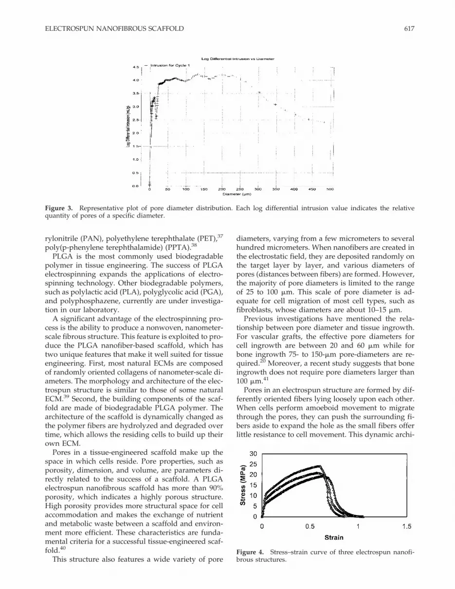
Assessment of structural pore properties was deter-mined with the use of a mercury porosimeter. Theporosity of electrospun nanofibrous structure was91.63%, indicating it was a highly porous structure.The total pore volume was 9.69 mL/g, and the totalpore area was 23.54 m2/g. A representative plot ofpore diameter distribution, illustrated in Figure 3, in-dicates that pore diameters ranged broadly from 2 to465 mm.
Tensile properties of electrospunnanofibrous structures
Mechanical properties, such as tensile modulus, ul-timate tensile stress, and ultimate strain, were evalu-
ated. A representative stress–strain curve was con-structed from the load–deformation curve and is illus-trated in Figure 4. The tensile modulus of the structurewas 323.15 MPa. The ultimate tensile stress of thestructure was 22.67 MPa, and the ultimate strain was95.8%. Since the composed fibers were randomly ori-ented within the nanofibrous structures, mechanicalanisotropy is expected on the X-Y plane of the struc-ture.
Cell proliferation assay
A cell proliferation assay was used to indicate thenumber of living cells in a scaffold. Cell number wasconverted from relative fluorescence units (RFU) bycalibrating a set of control experiments with a knowncell number. Following the seeding of 25,000 cells/cm2
on the nanofibrous scaffold, the cell number increasedwith time, as illustrated in Figure 5, and reached aplateau after day 7, showing a fivefold increase in cellpopulation during the 10-day culture period.
Cell–matrix interaction
Cell morphology and the interaction between cellsand nanofibers were studied in vitro for 7 days.BALB/c C7 mouse fibroblasts adhered and spread onthe surface of the PLGA fiber network (Fig. 6) and hadstarted to migrate through the pores and to grow un-der layers of the fiber network at day 3 [Fig. 7(A)].These fibroblasts interacted and integrated well withthe surrounding fibers [Fig. 7(B)]. SEM micrographsshowed that the development of cell growth wasguided by the fiber architecture. Cells grew in the di-rection of fiber orientation, forming a three-dimensional and multicellular network according tothe architecture of the nanofibrous structure [Fig.7(C,D)].
DISCUSSION
The electrospinning process provides a promisingmeans for creating a tissue-engineered scaffold. Sincethe electrostatic spinning technique was first patentedin 1934,35 many of its applications have been studiedin different engineering areas. In this investigation, thenanofiber-based scaffold produced by the electrospin-ning process is introduced for the application of tissueengineering. To date, many polymers have been elec-trospun, including polyethylene oxide (PEO),32
acrylic,23 nylon,36 polyethylene glycol (PEG), polyac-
Figure 2. SEM micrograph (original magnification ×1500)of the electrospun PLGA nanofibrous structure composed ofrandomly oriented ultra-fine fibers. Bar, 10 mm.
616 LI ET AL.
rylonitrile (PAN), polyethylene terephthalate (PET),37
poly(p-phenylene terephthalamide) (PPTA).38
PLGA is the most commonly used biodegradablepolymer in tissue engineering. The success of PLGAelectrospinning expands the applications of electro-spinning technology. Other biodegradable polymers,such as polylactic acid (PLA), polyglycolic acid (PGA),and polyphosphazene, currently are under investiga-tion in our laboratory.
A significant advantage of the electrospinning pro-cess is the ability to produce a nonwoven, nanometer-scale fibrous structure. This feature is exploited to pro-duce the PLGA nanofiber-based scaffold, which hastwo unique features that make it well suited for tissueengineering. First, most natural ECMs are composedof randomly oriented collagens of nanometer-scale di-ameters. The morphology and architecture of the elec-trospun structure is similar to those of some naturalECM.39 Second, the building components of the scaf-fold are made of biodegradable PLGA polymer. Thearchitecture of the scaffold is dynamically changed asthe polymer fibers are hydrolyzed and degraded overtime, which allows the residing cells to build up theirown ECM.
Pores in a tissue-engineered scaffold make up thespace in which cells reside. Pore properties, such asporosity, dimension, and volume, are parameters di-rectly related to the success of a scaffold. A PLGAelectrospun nanofibrous scaffold has more than 90%porosity, which indicates a highly porous structure.High porosity provides more structural space for cellaccommodation and makes the exchange of nutrientand metabolic waste between a scaffold and environ-ment more efficient. These characteristics are funda-mental criteria for a successful tissue-engineered scaf-fold.40
This structure also features a wide variety of pore
diameters, varying from a few micrometers to severalhundred micrometers. When nanofibers are created inthe electrostatic field, they are deposited randomly onthe target layer by layer, and various diameters ofpores (distances between fibers) are formed. However,the majority of pore diameters is limited to the rangeof 25 to 100 mm. This scale of pore diameter is ad-equate for cell migration of most cell types, such asfibroblasts, whose diameters are about 10–15 mm.
Previous investigations have mentioned the rela-tionship between pore diameter and tissue ingrowth.For vascular grafts, the effective pore diameters forcell ingrowth are between 20 and 60 mm while forbone ingrowth 75- to 150-mm pore-diameters are re-quired.20 Moreover, a recent study suggests that boneingrowth does not require pore diameters larger than100 mm.41
Pores in an electrospun structure are formed by dif-ferently oriented fibers lying loosely upon each other.When cells perform amoeboid movement to migratethrough the pores, they can push the surrounding fi-bers aside to expand the hole as the small fibers offerlittle resistance to cell movement. This dynamic archi-
Figure 3. Representative plot of pore diameter distribution. Each log differential intrusion value indicates the relativequantity of pores of a specific diameter.
Figure 4. Stress–strain curve of three electrospun nanofi-brous structures.
617ELECTROSPUN NANOFIBROUS SCAFFOLD

tecture provides cells with an opportunity to opti-mally adjust the pore diameter and grow into the scaf-fold even though some pores are relatively small.Therefore, some pores with smaller diameters in thisstructure may not hinder cell migration. Such a hy-pothesis of dynamic cell–scaffold interaction needs tobe further investigated.
The mechanical property of a scaffold is anotherimportant aspect of its design. The purpose of a scaf-fold is not only for providing a surface for cell resi-dence but also for maintaining mechanical stability atthe defect site of the host.42 A well-designed tissue-engineered scaffold has to meet two mechanical re-quirements to be effective. The scaffold must retainstructural integrity and stability when a physicianhandles and implants it into the defect site of the host.And after surgery, the structure at the implant sitemust provide sufficient biomechanical support duringthe process of tissue regeneration and structure deg-radation.
Some biopolymers, such as agarose and alginate,have been shown to promote cell growth and differ-entiation, but their weak mechanical properties limit
clinical use. On the other hand, the synthetic polymer-based scaffolds, such as those composed of PLGApolymer, have more effective mechanical properties.The mechanical properties of an engineered scaffoldare dominated by intrinsic factors, such as the chem-istry of the material, and by extrinsic factors, such asthe dimension or architectural arrangement of thebuilding blocks.
An electrospun nanofibrous structure is composedof randomly oriented nanometer-scale PLGA fibers.Its mechanical properties are determined by PLGAmolecular chains and the structural arrangement ofthe nanofibers. Previous study has shown that theelectrospun nanofibrous structure has better mechani-cal properties than do structures composed of largerdiameter fibers.27 In addition, Amornsakchai et al.found that the strength of fiber decreases as the fiberdimensions increase.43 This novel electrospun nanofi-brous structure will be stronger than the existing fiber-based structures composed of approximately 20 mm indiameter fibers.
Compared with the results of experiments con-ducted by Kempson and Edwards,44,45 the mechanicalproperties of the structure are comparable to those ofhuman cartilage and skin. The comparison of tensilemodulus, ultimate tensile stress, and ultimate strainamong electrospun structures, cartilage, and skin isshown in Table I. Based on the morphology and me-chanics of the scaffold, it is suggested that the electro-spun nanofibrous structure would be suitable as a skinor cartilage substitute.
In the cell proliferation assessment, the electrospunnanofibrous structure was found to promote cellgrowth. This novel structure provides a high level ofsurface area for cells to attach to due to its three-dimensional feature and its high surface area-to-volume ratio. It is important that a tissue-engineeredscaffold be loaded with the proper cell density with-out compromising the size of the scaffold. After 7 days
Figure 5. Proliferation of human bone-marrow-derivedmesenchymal stem cells (hMSCs) seeded on electrospunnanofibrous structures. Quadruplicate samples at each timepoint were measured, and the cell number at day 10 had afivefold increase compared with that at day 1 (p < 0.005).
Figure 6. SEM micrographs (original magnification ×500) of BALB/c C7 mouse fibroblasts on an electrospun nanofibrousstructure at (A) 3 days of culture and (B) 7 days of culture. Bar, 10 mm.
618 LI ET AL.
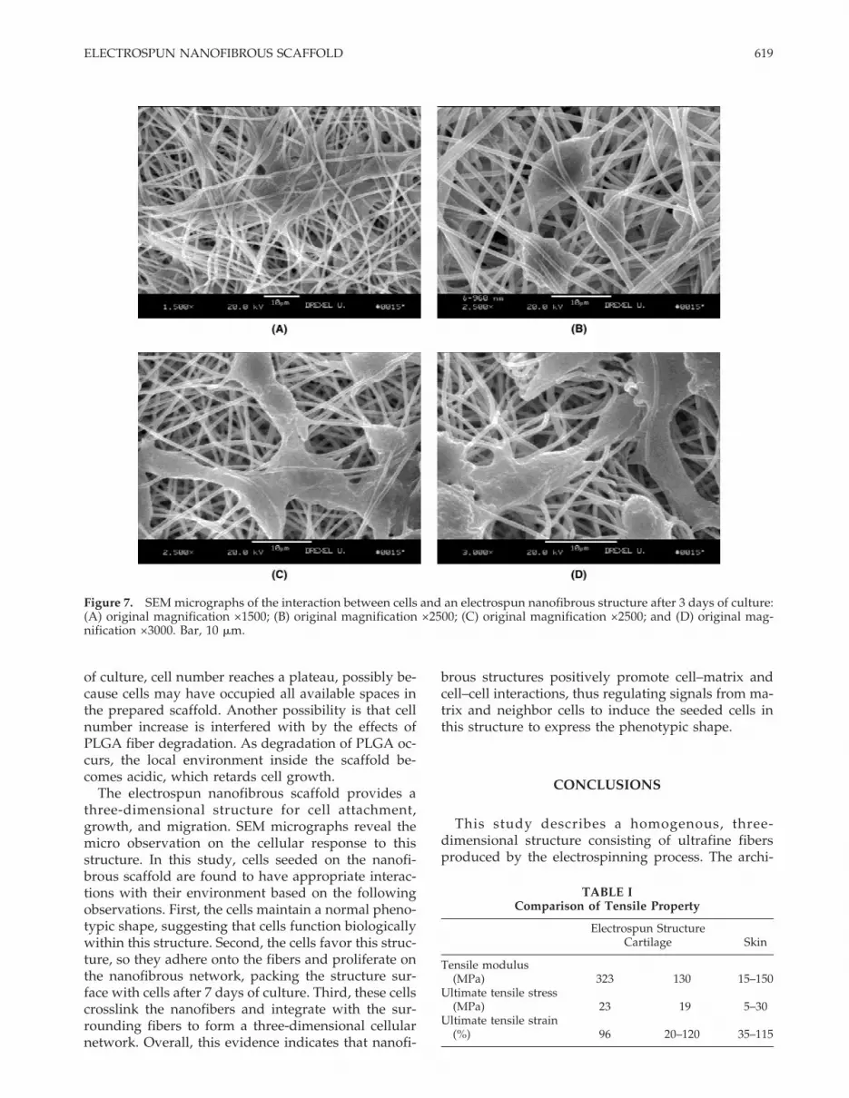
of culture, cell number reaches a plateau, possibly be-cause cells may have occupied all available spaces inthe prepared scaffold. Another possibility is that cellnumber increase is interfered with by the effects ofPLGA fiber degradation. As degradation of PLGA oc-curs, the local environment inside the scaffold be-comes acidic, which retards cell growth.
The electrospun nanofibrous scaffold provides athree-dimensional structure for cell attachment,growth, and migration. SEM micrographs reveal themicro observation on the cellular response to thisstructure. In this study, cells seeded on the nanofi-brous scaffold are found to have appropriate interac-tions with their environment based on the followingobservations. First, the cells maintain a normal pheno-typic shape, suggesting that cells function biologicallywithin this structure. Second, the cells favor this struc-ture, so they adhere onto the fibers and proliferate onthe nanofibrous network, packing the structure sur-face with cells after 7 days of culture. Third, these cellscrosslink the nanofibers and integrate with the sur-rounding fibers to form a three-dimensional cellularnetwork. Overall, this evidence indicates that nanofi-
brous structures positively promote cell–matrix andcell–cell interactions, thus regulating signals from ma-trix and neighbor cells to induce the seeded cells inthis structure to express the phenotypic shape.
CONCLUSIONS
This study describes a homogenous, three-dimensional structure consisting of ultrafine fibersproduced by the electrospinning process. The archi-
TABLE IComparison of Tensile Property
Electrospun StructureCartilage Skin
Tensile modulus(MPa) 323 130 15–150
Ultimate tensile stress(MPa) 23 19 5–30
Ultimate tensile strain(%) 96 20–120 35–115
Figure 7. SEM micrographs of the interaction between cells and an electrospun nanofibrous structure after 3 days of culture:(A) original magnification ×1500; (B) original magnification ×2500; (C) original magnification ×2500; and (D) original mag-nification ×3000. Bar, 10 mm.
619ELECTROSPUN NANOFIBROUS SCAFFOLD

tecture of the structure is similar to that of naturalextracellular matrix, indicating that the electrospunnanofibrous scaffold is suitable as a tissue substitute.
These nanofiber-based scaffolds are characterizedby a wide range of pore size distribution, high poros-ity, and high surface area-to-volume ratio, which arefavorable parameters for cell attachment, growth, andproliferation. The structures provide effective me-chanical properties suitable for soft tissue, such as skinor cartilage. This study provides a basis for futureoptimization of electrospun nanofibrous scaffold fortissue-engineering applications. Applying this struc-ture as a substitute for a specific tissue, such as carti-lage, will be a focus of our future investigations.
The authors thank John Mckelvie for assistance in prepa-ration of the electrospinning apparatus and Dr. Keith G.Danielson for reviewing the manuscript.
References
1. Langer R, Vacanti JP. Tissue engineering. Science 1993;260:920–926.
2. Thomson RC, Yaszemski MJ, Mikos AG. Polymer scaffold pro-cessing. In: Lanza RP, Langer R, Chick WL, editors. Principlesof tissue engineering. Austin, TX: R.G. Landes; 1997. p 263–271.
3. Reddi AH. Morphogenesis and tissue engineering of bone andcartilage: Inductive signals, stem cells, and biomimetic bioma-terials. Tissue Eng 2000;6:351–359.
4. Alberts B, Bray D, Lewis J, Raff M, Roberts K, Watson JD.Molecular biology of the cell. New York: Garland; 1994. p 971–995.
5. Wintermantel E, Mayer J, Blum J, Eckert KL, Luscher P,Mathey M. Tissue engineering scaffolds using superstructures.Biomaterials 1996;17:83–91.
6. Boyan BD, Hummert TW, Dean DD, Schwartz Z. Role of ma-terial surfaces in regulating bone and cartilage cell response.Biomaterials 1996;17:137–146.
7. Dahlberg L, Kreicbergs A. Demineralized allogenic bone ma-trix for cartilage repair. J Orthop Res 1991;9:11–19.
8. Watt FM, Dudhia J. Prolonged expression of differentiatedphenotype by chondrocytes cultured at low density on a com-posite substrate of collagen and agarose that restricts cellspreading. Differentiation 1988;38:140–147.
9. Ponticiello MS, Schinagl RM, Kadiyala S, Barry FP. Gelatin-based resorbable sponge as a carrier matrix for human mesen-chymal stem cells in cartilage regeneration therapy. J BiomedMater Res 2000;52:246–255.
10. Allemann F, Mizuno S, Eid K, Yates KE, Zaleske D, GlowackiJ. Effects of hyaluronan on engineered articular cartilage extra-cellular matrix gene expression in 3-dimensional collagen scaf-folds. J Biomed Mater Res 2001;55:13–19.
11. Bradham DM, Horton WE. In vivo cartilage formation fromgrowth factor modulated articular chondrocytes. Clin Orthop1998;352:239–249.
12. Bonaventure J, Kadhom N, Cohen-Solal L, Ng KH, Bour-guignon J, Lasselin C. Reexpression of cartilage-specific genesby dedifferentiated human articular chondrocytes cultured inalginate beads. Exp Cell Res 1994;212:97–104.
13. Agrawal CM, Ray RB. Biodegradable polymeric scaffolds formusculoskeletal tissue engineering. J Biomed Mater Res 2001;55:141–150.
14. Behravesh E, Yasko AW, Engel PS, Mikos AG. Synthetic bio-degradable polymers for orthopaedic applications. Clin Or-thop 1999;367S:S118–S125.
15. Athanasiou KA, Niederauer GG, Agrawal CC. Sterilization,toxicity, biocompatibility, and clinical applications of polylac-tic acid/polyglycolic acid copolymer. Biomaterials 1996;17:93–102.
16. Sanders JE, Stiles CE, Hayes CL. Tissue response to single-polymer fibers of varying diameters: Evaluation of fibrous en-capsulation and macrophage density. J Biomed Mater Res2000;52:231–237.
17. Wan H, Williams RL, Doherty PJ, Williams DF. A study of cellbehaviour on the surfaces of multifilament materials. J MaterSci: Mater Med 1997;8:45–51.
18. Flemming RG, Murphy CJ, Abrams GA, Goodman SL, NealeyPF. Effects of synthetic micro- and nano-structured surfaces oncell behavior. Biomaterials 1999;20:573–588.
19. Green AM, Jansen JA, van der Waerden JPCM, von Recum AF.Fibroblast response to microtextured silicone surfaces: Textureorientation into or out of the surface. J Biomed Mater Res 1994;28:647–653.
20. von Recum AF, Shannon CE, Cannon CE, Long KJ, van KootenTG. Surface roughness, porosity, and texture as modifiers ofcellular adhesion. Tissue Eng 1996;2:241–253.
21. Tan W, Krishnaraj R, Desai TA. Evaluation of nanostructuredcomposite collagen–chitosan matrices for tissue engineering.Tissue Eng 2001;7:203–210.
22. Kadler KE, Holmes DF, Trotter JA, Chapman JA. Collagen fi-bril formation. Biochem J 1996;316:1–11.
23. Baumgarten PK. Electrostatic spinning of acrylic microfibers. JColloid Interface Sci 1971;36:71–79.
24. Doshi J, Reneker DH. Electrospinning process and applicationof electrospun fibers. J Electrostatics 1995;35:151–160.
25. Reneker DH, Chun I. Nanometre diameter fibres of polymer,produced by electrospinning. Nanotechnology 1996;7:216–223.
26. Fong H, Chun I, Reneker DH. Beaded nanofibers formed dur-ing electrospinning. Polymer 1999;40:4585–4592.
27. Kim J, Reneker DH. Mechanical properties of composites usingultrafine electrospun fibers. Polym Composites 1999;20:124–131.
28. Chun I, Reneker DH, Fong H, Fang X. Carbon nanofibers frompolyacrylonitrile and mesophase pitch. J Adv Mater1999;31:36–41.
29. Ko FK, Laurencin CT, Borden MD, Reneker DH. The dynamicsof cell–fiber architecture interaction. Proc Ann Mtg BiomaterRes Soc 1998;Apr: 11.
30. Ko FK, Borden MD, Laurencin CT. The role of fiber architec-ture in biocomposites: The tissue engineering approach. Proc13th Intl Conf Composite Mater 2001;Jul: Abstract No. 1428.
31. Fertala A, Han WB, Ko FK. Mapping critical sites in collagen IIfor rational design of gene-engineered proteins for cell-supporting materials. J Biomed Mater Res 2001;57:48–58.
32. Jaeger R, Bergshoef MM, Batlle CM, Schonherr H, Vancso GJ.Electrospinning of ultra-thin polymer fibers. Macromol Symp1998;127:141–150.
33. Caplan AI. Mesenchymal stem cells. J Orthop Res 1991;9:641–650.
34. Caterson EJ, Nesti LJ, Li WJ, Danielson KG, Albert TJ, VaccaroAR, Tuan RS. Three-dimensional cartilage formation by bonemarrow-derived cells seeded in polylactide/alginate amalgam.J Biomed Mater Res. Forthcoming.
35. Formhals A. Process and apparatus for preparing artificialthreads. US patent no. 1,975,504; 1934.
36. Gibson PW, Schreuder-Gibson HL, Rivin D. Electrospun fibermats: Transport properties. AIChE J 1999;45:190–195.
37. Warner SB, Buer A, Ugbolue SC, Rutledge GC, Shin MY. A
620 LI ET AL.
fundamental investigation of the formation and properties ofelectrospun fibers. Natl Textile Center Ann Rep 1998;Nov:83–90.
38. Srinivasan G, Reneker DH. Structure and morphology of smalldiameter electrospun aramid fibers. Polym Int 1995;36:195–201.
39. Nishido T, Yasumoto K, Otori T, Desaki J. The network struc-ture of corneal fibroblasts in the rat as revealed by scanningelectron microscopy. Invest Ophthalmol Vis Sci 1988;29:1887–1990.
40. Ma PX, Choi JW. Biodegradable polymer scaffolds with well-defined interconnected spherical pore network. Tissue Eng2001;7:23–33.
41. Itala AI, Ylanen HO, Ekholm C, Karlsson KH, Aro HT. Porediameter of more than 100 mm is not requisite for bone in-
growth in rabbits. J Biomed Mater Res, Appl Biomater 2001;58:679–683.
42. Hutmacher DW. Scaffold in tissue engineering bone and car-tilage. Biomaterials 2000;20:2529–2543.
43. Amornsakchai T, Cansfield DLM, Jawad SA, Pollard G, WardIM. The relation between filament diameter and fracturestrength for ultra-high-modulus polyethylene fibres. J MaterSci 1993;28:1689–1698.
44. Kempson GE, Muir H, Pollard C, Tuke M. The tensile proper-ties of the cartilage of human femoral condyles related to thecontent of collagen and glycosaminoglycans. Biochim BiophysActa 1973;297:456–472.
45. Edwards C, Marks R. Evaluation of biomechanical propertiesof human skin. Clin Dermatol 1995;13:375–380.
621ELECTROSPUN NANOFIBROUS SCAFFOLD