Fabrication and characterisation of drug-loaded electrospun ...
9
Fabrication and characterisation of drug-loaded electrospun polymeric nanofibers for controlled release in hernia repair Ivan J. Hall Barrientos a,b , Eleonora Paladino b,c,d , Sarah Brozio b , Melissa K. Passarelli d , Susan Moug e , Richard A. Black a , Clive G. Wilson b , Dimitrios A. Lamprou b,f , * a Biomedical Engineering, University of Strathclyde, Glasgow, United Kingdom b Strathclyde Institute of Pharmacy and Biomedical Sciences (SIPBS), University of Strathclyde, 161 Cathedral Street, Glasgow, G4 0RE, United Kingdom c EPSRC Centre for Innovative Manufacturing in Continuous Manufacturing and Crystallisation (CMAC), University of Strathclyde, Technology and Innovation Centre, 99 George Street, G1 1RD Glasgow, United Kingdom d National Physical Laboratory (NPL), Hampton Road, Teddington, Middlesex, TW11 0LW, United Kingdom e National Health Service (NHS), Royal Alexandra Hospital, Paisley, PA2 9PN, United Kingdom f Medway School of Pharmacy, University of Kent, Medway Campus, Anson Building, Central Avenue, Chatham Maritime, Chatham, Kent, ME4 4TB, United Kingdom A R T I C L E I N F O Article history: Received 24 October 2016 Received in revised form 8 December 2016 Accepted 10 December 2016 Available online 14 December 2016 Keywords: Electrospinning Scaffolds Hernia Drug release Physicochemical characterisation A B S T R A C T The chemical distribution and mechanical effects of drug compounds in loaded electrospun scaffolds, a potential material for hernia repair mesh, were characterised and the efficacy of the material was evaluated. Polycaprolactone electrospun fibres were loaded with either the antibacterial agent, irgasan, or the broad-spectrum antibiotic, levofloxacin. The samples were subsequently characterised by rheological studies, scanning electron microscopy (SEM), atomic force microscopy (AFM), contact angle goniometry (CAG), in vitro drug release studies, antibacterial studies and time-of-flight secondary ion mass spectrometry (ToF-SIMS). Increased linear viscoelastic regions observed in the rheometry studies suggest that both irgasan and levofloxacin alter the internal structure of the native polymeric matrix. In vitro drug release studies from the loaded polymeric matrix showed significant differences in release rates for the two drug compounds under investigation. Irgasan showed sustained release, most likely driven by molecular diffusion through the scaffold. Conversely, levofloxacin exhibited a burst release profile indicative of phase separation at the edge of the fibres. Two scaffold types successfully inhibited bacterial growth when tested with strains of E. coli and S. aureus. Electrospinning drug-loaded polyester fibres is an alternative, feasible and effective method for fabricating non-woven fibrous meshes for controlled release in hernia repair. © 2016 Elsevier B.V. All rights reserved. 1. Introduction Hernia repair, one of the most common general surgeries performed, is complicated by bacterial infections and implant rejection (Earle, 2010). Commercially available mesh devices currently employed in hernia repairs contain brained or knitted fibres. The mechanical properties of the mesh and the biocom- patibility of the material are critical to the healing process. Tissue incorporation, a key factor in the success of the graft device is dependent on the material type, density, compliance and electrical properties of the mesh (Procter et al., 2009). Graft failure motivates research into new fabrication methods for incorporating bio- materials and drug encapsulation in novel mesh matrices, such as hot-melt extrusion (Li et al., 2013), electrospinning (Toncheva et al., 2011), 3D printing (Holländer et al., 2016) and high-speed rotary spinning (Sebe et al., 2013). Electrospinning is the most popular and preferred technique for nanofiber fabrication due to its simplicity, cost-effectiveness, flexibility, and ability to spin a broad range of polymers (Zamani et al., 2013). The method allows for the simple and direct functionalization of fibres with drug compounds and is compatible with solvents such as chloroform and dimethyl sulfoxide. In addition, the process of electrospinning with the use of solvents such as chloroform, dimethyl sulfoxide etc., allows functionalisa- tion of the scaffolds through the inclusion of drugs in the polymer- solvent solution without the need for a complicated preparation * Corresponding author at: Medway School of Pharmacy, University of Kent, Medway Campus, Anson Building, Central Avenue, Chatham Maritime, Chatham, Kent, ME4 4TB, United Kingdom. E-mail address: [email protected] (D.A. Lamprou). http://dx.doi.org/10.1016/j.ijpharm.2016.12.022 0378-5173/© 2016 Elsevier B.V. All rights reserved. International Journal of Pharmaceutics 517 (2017) 329–337 Contents lists available at ScienceDirect International Journal of Pharmaceutics journal homepage: www.elsev ier.com/locate /ijpharm
-
Upload
khangminh22 -
Category
Documents
-
view
4 -
download
0
Transcript of Fabrication and characterisation of drug-loaded electrospun ...

International Journal of Pharmaceutics 517 (2017) 329–337
Fabrication and characterisation of drug-loaded electrospun polymericnanofibers for controlled release in hernia repair
Ivan J. Hall Barrientosa,b, Eleonora Paladinob,c,d, Sarah Broziob, Melissa K. Passarellid,Susan Mouge, Richard A. Blacka, Clive G. Wilsonb, Dimitrios A. Lamproub,f,*aBiomedical Engineering, University of Strathclyde, Glasgow, United Kingdomb Strathclyde Institute of Pharmacy and Biomedical Sciences (SIPBS), University of Strathclyde, 161 Cathedral Street, Glasgow, G4 0RE, United Kingdomc EPSRC Centre for Innovative Manufacturing in Continuous Manufacturing and Crystallisation (CMAC), University of Strathclyde, Technology and InnovationCentre, 99 George Street, G1 1RD Glasgow, United KingdomdNational Physical Laboratory (NPL), Hampton Road, Teddington, Middlesex, TW11 0LW, United KingdomeNational Health Service (NHS), Royal Alexandra Hospital, Paisley, PA2 9PN, United KingdomfMedway School of Pharmacy, University of Kent, Medway Campus, Anson Building, Central Avenue, Chatham Maritime, Chatham, Kent, ME4 4TB, UnitedKingdom
A R T I C L E I N F O
Article history:Received 24 October 2016Received in revised form 8 December 2016Accepted 10 December 2016Available online 14 December 2016
Keywords:ElectrospinningScaffoldsHerniaDrug releasePhysicochemical characterisation
A B S T R A C T
The chemical distribution and mechanical effects of drug compounds in loaded electrospun scaffolds, apotential material for hernia repair mesh, were characterised and the efficacy of the material wasevaluated. Polycaprolactone electrospun fibres were loaded with either the antibacterial agent, irgasan,or the broad-spectrum antibiotic, levofloxacin. The samples were subsequently characterised byrheological studies, scanning electron microscopy (SEM), atomic force microscopy (AFM), contact anglegoniometry (CAG), in vitro drug release studies, antibacterial studies and time-of-flight secondary ionmass spectrometry (ToF-SIMS). Increased linear viscoelastic regions observed in the rheometry studiessuggest that both irgasan and levofloxacin alter the internal structure of the native polymeric matrix. Invitro drug release studies from the loaded polymeric matrix showed significant differences in releaserates for the two drug compounds under investigation. Irgasan showed sustained release, most likelydriven by molecular diffusion through the scaffold. Conversely, levofloxacin exhibited a burst releaseprofile indicative of phase separation at the edge of the fibres. Two scaffold types successfully inhibitedbacterial growth when tested with strains of E. coli and S. aureus. Electrospinning drug-loaded polyesterfibres is an alternative, feasible and effective method for fabricating non-woven fibrous meshes forcontrolled release in hernia repair.
© 2016 Elsevier B.V. All rights reserved.
Contents lists available at ScienceDirect
International Journal of Pharmaceutics
journal homepage: www.elsev ier .com/locate / i jpharm
1. Introduction
Hernia repair, one of the most common general surgeriesperformed, is complicated by bacterial infections and implantrejection (Earle, 2010). Commercially available mesh devicescurrently employed in hernia repairs contain brained or knittedfibres. The mechanical properties of the mesh and the biocom-patibility of the material are critical to the healing process. Tissueincorporation, a key factor in the success of the graft device isdependent on the material type, density, compliance and electrical
* Corresponding author at: Medway School of Pharmacy, University of Kent,Medway Campus, Anson Building, Central Avenue, Chatham Maritime, Chatham,Kent, ME4 4TB, United Kingdom.
E-mail address: [email protected] (D.A. Lamprou).
http://dx.doi.org/10.1016/j.ijpharm.2016.12.0220378-5173/© 2016 Elsevier B.V. All rights reserved.
properties of the mesh (Procter et al., 2009). Graft failure motivatesresearch into new fabrication methods for incorporating bio-materials and drug encapsulation in novel mesh matrices, such ashot-melt extrusion (Li et al., 2013), electrospinning (Tonchevaet al., 2011), 3D printing (Holländer et al., 2016) and high-speedrotary spinning (Sebe et al., 2013).
Electrospinning is the most popular and preferred technique fornanofiber fabrication due to its simplicity, cost-effectiveness,flexibility, and ability to spin a broad range of polymers (Zamaniet al., 2013). The method allows for the simple and directfunctionalization of fibres with drug compounds and is compatiblewith solvents such as chloroform and dimethyl sulfoxide. Inaddition, the process of electrospinning with the use of solventssuch as chloroform, dimethyl sulfoxide etc., allows functionalisa-tion of the scaffolds through the inclusion of drugs in the polymer-solvent solution without the need for a complicated preparation

330 I.J. Hall Barrientos et al. / International Journal of Pharmaceutics 517 (2017) 329–337
process (He et al., 2015). Electrospinning has previously beenapplied to the fabrication of triclosan/cyclodextrin inclusioncomplexes (Celebioglu et al., 2014), the construction of scaffoldswith perlecan domain IV peptides (Hartman et al., 2011),manufacture of biocatalytic protein membranes (Kabay et al.,2016), and encapsulation of levofloxacin in mesoporous silicananoparticles (Jalvandi et al., 2015). Given the broad applications ofelectrospinning, there has been previous research specificallyfocused on the development of electrospun polymeric materialsfor hernia repair mesh devices. Electrospinning produces scaffoldscontaining micro-fibres and this is an advantageous feature notobserved in braided mesh commercial devices � these microfibersalso introduce mechanical anisotropy and provide topographicfeatures to guide cell alignment (Goldstein and Thayer, 2016).However, electrospun fibres typically incorporate the use oforganic solvents and for applications such as hernia repair ortissue engineering, the toxicity of organic solvents used could behighly critical – avoiding organic solvents is of outmost importancefor applications in medicine and pharmacy (Agarwal and Greiner,2011; Bubel et al., 2014).
The purpose of this study is to examine the physicochemicalproperties, bacteria response, and drug loading of electrospunscaffolds. The polymer chosen for this study is polycaprolactone(PCL); a biodegradable polyester commonly used in biomedicalapplications for controlled release and targeted drug delivery(Bhavsar and Amiji, 2008). PCL, a biodegradable aliphatic polyester(Azimi et al., 2016), is an obvious candidate for drug deliverysystems due to its high biocompatibility and ease of degradation inthe human body (Bikiaris et al., 2007). Drug loading of structuresthat mechanically resemble interfacial tissue and which allowsshort or long-term release of suitable bioactives may be utilisablein hernia-repair meshes. PCL was chosen in this research as it has ahigh permeability to a variety of drug molecules (e.g. gentamycin,chitosan) and low toxicity (Murthy, 1997). The matrix was loadedand electrospun with two drugs, irgasan (an antibacterial agentused commonly in soaps, detergents and surgical cleaning agents)or levofloxacin (a broad-spectrum antibiotic used commonly totreat gastrointestinal infections). The mechanical characteristics,morphology, surface hydrophobicity, drug efficacy and chemicaldistribution were characterised with an array of analyticaltechniques. The results from this study should help to buildplatform to aid future work with various fabrication methods, suchas extrusion and shaping using 3D printing.
2. Materials & methods
2.1. Materials
Polycaprolactone (PCL) with a mean molecular weight of 80 kD,Irgasan (variation of Triclosan, >97%), Levofloxacin (>98%), and allthe solvents used for the electrospinning were obtained fromSigma Aldrich. The solvents consisting of chloroform (anhydrous,containing amylenes as stabilizers, >99%) and N,N-dimethylfor-mamide (DMF, anhydrous 99.8%).
2.2. Preparation of PCL solutions
Different solutions with a polymer concentration of 12% (w/w)were prepared to be used within the electrospinning method – thisparticular concentration was used due to its possessed sutureretention and tensile strengths appropriate for hernia repair, asspecified for similar electrospun scaffolds described by Ebersoleet al. (2012). Various PCL formulations were constructed of a totalweight of 25 g per solution, which allowed for PCL (12% w/w) and a9:1 (w/w) ratio of chloroform (CLF) to N,N-dimethylformamide(DMF). For the unloaded polymer solution, 3 g of PCL was dissolved
in 22 g of CLF:DMF (9:1) which was initially mixed through 30 minin a centrifuge, a further 30 min in a sonicator (Elma S30Elmasonic) and a final 1 h with a magnetic stirrer. This processwas vital to ensure that the solution was fully homogeneous. Thesolution was left overnight, and a further 30 min of sonicationapplied the following morning in order to confirm the homogene-ity of the solution. For the irgasan-loaded solutions, the samemethod was applied, except the solution contained 1% (w/w)irgasan. The concentration of the levofloxacin-loaded solutionswas 0.5% (w/w), providing sufficient sensitivity in the release cellfor accurate UV analysis. All the preparations turned to clearsolutions. These observations were interpreted to determine thatthe solutions had successfully homogenised. The solutions werethen subsequently used in the electrospinning process and forrheological analysis.
2.3. Electrospinning of PCL solutions
The PCL test specimens were fabricated for each polymericsolution, using a custom in-house electrospinning apparatus,which consisted of a syringe pump (Harvard Apparatus PHD 2000infusion, US) and two 30 kV high-voltage power supplies (Alpha IIIseries, Brandenburg, UK). The polymer solution was loaded intoglass syringe and fed through tubing with a metal needle tipattached at the end. The needle was clamped into place, to allow ahigh-voltage supply to run through it, which allowed an electricfield to be created between the needle and the target plate. Thesyringe was clamped to a pump, which determined the specificinjection flow rate of the polymeric solutions. For each of the threesolutions (e.g. unloaded, irgasan-loaded, and levofloxacin-loaded),3 varying flow rates of 0.5, 1 and 1.5 ml h�1 were applied acrossvarying voltages of 2 kV–5 kV (needle) and 10 kV–18 kV (targetplate). The variation in flow rate and applied voltages was tocorrect any problems that occurred during fabrication, i.e. ‘spitting’of solution at the target plate, or any potential beading (which wasexamined through SEM). The fabrication of this solution waselectrospun onto the target that was covered with aluminium foil,in order for the final material to be removed and used for furthercharacterisation. The final yield of electrospun PCL resulted in thin,flexible sheets of material.
2.4. Rheological studies
A Thermo Scientific HAAKE MARS II rheometer with a P35 TiLcone and plate was used to measure the rheological andmechanical behaviour of the different unloaded and loadedpolymeric solutions. The objective of this experiment was toexamine the viscoelastic properties of the PCL solution, specificallyto determine whether the irgasan or levofloxacin is having aneffect on the mechanical properties of the polymer. The methodused was taken and modified from the rheological studyundertaken by Bubel et al. (2014). In briefly, an oscillatingamplitude sweep between 0.1 Pa–1000 Pa at a frequency of 1 Hzwas used to determine the linear viscoelastic region (LVER) of thesamples. Once the LVER is determined from the amplitude sweep, adownwards oscillating frequency sweep from 10 Hz–0.1 Hz with ashear stress (Pa) within the LVER was then used in order to helpunderstand the nature of the solutions concerning strength andstability. The experiments were repeated 4 times per solution, andfor each experiment, each data point (20 data points per method)was optimised to repeat each measurement 5 times.
2.5. Scanning electron microscopy (SEM)
The morphology and diameter of individual fibres spun fromPCL solution were determined from scanning electron micrographs

I.J. Hall Barrientos et al. / International Journal of Pharmaceutics 517 (2017) 329–337 331
of each sample (TM-1000, Hitachi, UK, Ltd.). The samples weremounted on an aluminium plate with conductive tape. Images offibres were taken at various locations of each electrospun PCLscaffold in order to determine the overall uniformity of fibres. Priorto imaging, the samples were sputter coated with gold for 30 susing a Leica EM ACE200 vacuum coater, the process beingrepeated four times in order to increase the conductivity of thesamples.
2.6. Atomic force microscopy (AFM)
Further morphological analysis was undertaken through atomicforce microscopy. A Multimode 8 microscope (Bruker, USA), withScanasyst-Air probes (Bruker, USA) was used in Peak ForceQuantitative Nano Mechanics (QNM) mode, as described byLamprou et al. (2013). The imaging of the fibres was performedunder ambient conditions, with a silicon cantilever probe. The tipradius of the probe and the spring constant were calculated to be inthe regions of 0.964 nm (18� tip half angle) and 0.4935 N/m,respectively. The scan sizes ranged from 200 nm to 25 mm, at a scanrate of 0.977 Hz with 256-sample resolution. The RoughnessAverage (Ra) values were determined by entering surface scanningdata, and digital levelling algorithm values were determined usingNanoscope Analysis software V1.40 (Bruker USA). AFM imageswere collected from two different samples and at random spotsurface sampling.
2.7. Contact angle goniometry (CAG)
To monitor changes in wettability of the scaffolds, sessile dropcontact angle for distilled water was measured by contact anglegoniometry, using a contact angle goniometer (Kruss G30,Germany) as described by Lamprou et al. (2010).
2.8. In vitro drug release studies
The drug releases of the irgasan/levofloxacin loaded PCLscaffolds were measured in order to determine the release profileof the drugs. Samples of PCL-IRG were immersed in phosphatebuffered saline (PBS) containing 0.5% sodium dodecyl sulphate(SDS) at 37 �C, and samples of PCL-LEVO were immersed in PBSonly at 37 �C. This release study was based on the method cited byDuan et al. (2013). The solutions were agitated using a shaker at arate of 80 rev/min. The UV absorbance of both drugs wasmeasured: irgasan at 280 nm (Piccoli et al., 2002), and levofloxacinat 292 nm (Maleque et al., 2012) respectively. Measurements weretaken at intervals at 15 min, 30 min, 1 h, 2 h, 4 h, 8 h, 24 h and everyday after the 24 h mark for up to 7 days. At each point, 4 ml ofsolution was taken from the vial and replaced with fresh in order tosatisfy the perfect-sink conditions and keeping the volume of thesolution constant.
2.9. Antibacterial studies
The antibacterial efficacy of the drug loaded electrospunscaffolds were tested against Escherichia coli (E. coli) 8739 andStaphylococcus aureus (S. aureus) 29213. S. aureus is Gram positive,E. coli is Gram negative and both bacteria are common causes ofnosocomial infections. Both irgasan and levofloxacin should haveantibacterial effects. For this study, an agar diffusion method wasused. Luria-Bertani (LB) agar was prepared from a formulation of5 g tryptone, 2.5 g yeast extract, 5 g NaCl in 475 ml of deionizedwater. The LB agar was autoclaved and poured into 20 ml plates.The E. coli and S. aureus were grown overnight in 5 ml of LB Broth,with both bacteria inoculated from a single colony. 150 ml of theE. coli and S. aureus cultures were spread onto six different plates of
LB agar. Three plates consisted of spread E. coli, including a scaffoldfree plate, which acted as a control – the other 2 plates, weredivided into 4 sections, with 1 section containing an unloaded PCLscaffold, and the other 3 containing PCL-irgasan andPCL-levofloxacin scaffolds. This procedure was repeated for threeplates of spread S. aureus. The plates were incubated for 24 h, andsubsequently examined. Diameters of the zones of growthinhibition were measured, and these data compared across thedrugs and bacterial strains. This method was based on the methoddescribed by Davachi et al. (2016).
2.10. Time of flight secondary ion mass spectrometry (ToF-SIMS)
ToF-SIMS data was acquired using a ToF-SIMS V massspectrometer (ION-TOF GmbH, Münster, Germany) based at theWolfson Foundation Pharmaceutical Surfaces Laboratory at theUniversity of Strathclyde. The instrument is equipped with abismuth liquid metal ion gun (LMIG), an argon gas cluster ion beam(GCIB) and a gridless reflectron time-of-flight mass analyzer.
Three different acquisition modes, detailed below, were used toanalyse the fibres: high mass resolution spectroscopy, depthprofiling, high lateral resolution imaging. Owing to the insulativenature of the materials, a low-energy electron beam (21 V) wasused to compensate for charging.
2.10.1. High mass resolution spectroscopyFor an optimal mass resolution, the primary ion beam (Bi3+
primary ions) was pulsed at 10 kHz frequency with a pulse width of17.0 ns. The primary ion gun energy was set at 30 kV and the pulsedtarget current was approximately 0.63 pA. Data was collected bothin the positive and in the negative secondary ion polarities, in threereplicates; each acquisition was made from different areas of thesamples used in this study. The analysed area and the acquisitiontime, for each repetition, were respectively 100 mm � 100 mm and120 s, delivering a primary ion dose density (PIDD) of approxi-mately 4.6 � 1012 (primary ions/cm2). Reference spectra for pureLevofloxacin and Irgasan compounds were acquired in positive andnegative ion mode from 0 to 400 Da.
2.10.2. High lateral resolution imagingThe LMIG was operated using the imaging mode, with high
lateral resolution, and Bi3++ was selected as primary ion beam. Theprimary ion gun energy was 30 kV and the pulsed target currentwas approximately 0.048 pA. High lateral resolution ion imageswere collected over a surface area of 100 mm � 100 mm, using apulsed analysis beam (pulse width = 100 ns). The resolution was256 � 256 pixels per image (pixel width was circa 0.4 mm). Eachimage was obtained with a final ion dose of 6.5 �1012 primary ions/cm2 or less. The dose was kept below the static limit of 1013
primary ions/cm2 to minimize surface damages during theanalysis. The images were processed with the ION-TOF SurfaceLab6.6 software (Münster, Germany).
2.10.3. 3D imagingThe LMIG and the GCIB were employed in a dual-beam
configuration to collect the depth profile and the 3D image data.The LMIG was operated in pulsed mode to investigate the lateraldistribution of chemical species, while the Argon source wasoperated in DC mode to remove multiple layers of material fromthe sample surface between the analytical cycles. For the depthprofiling analysis, the dual beam experiment used a 30 kV Bi3++
primary ion beam for analysis and a 10 kV Ar1500+ beam forsputtering. The pulsed current of the Bi3++ primary ion beam was0.048 pA and the DC current of the cluster Ar1500+ was 10.22 nA,with a 500 s analysis time and 4 s sputtering time. The raster areasof the pulsed analysis beams and the DC sputter were 100 mm

Fig.1. a: Amplitude sweep viscous modulus (G’) data for PCL, PCL-IRG and PCL-LEVO solutions; b: Amplitude sweep viscous modulus (G”) data for PCL, PCL-IRG and PCL-LEVOsolutions; c: Amplitude sweep shear viscosity (h) data for PCL, PCL-IRG and PCL-LEVO solutions.
332 I.J. Hall Barrientos et al. / International Journal of Pharmaceutics 517 (2017) 329–337
� 100 mm and 300 mm � 300 mm, respectively. The resolution was256 � 256 pixels per image (pixel width of about 0,4 mm). Data wascollected in the negative secondary ion mode.
In the course of each acquisition, mass spectral information ateach image pixel was collected in the m/z range of 0–917 m/z.
2.11. Statistical analysis
All experiments were performed in triplicate with calculation ofmeans and standard deviations. Two-way analysis of variance(ANOVA) was used for multiple comparisons along with Tukey’smultiple comparing tests, followed by T-test to access statisticalsignificance for paired comparisons. Significance was acknowl-edged for p values lower than 0.05.
3. Results and discussion
3.1. Rheological studies
For each polymeric solution, multiple amplitude sweeps wereused in order to correctly identify the linear viscoelastic region(LVR). This was repeated to detect any major variations in the LVR,and for a more accurate shear stress to be used in the frequencysweeps. For each of the samples, elastic modulus (G’), viscousmodulus (G”) and shear viscosity (h) was calculated andsubsequently analysed.
It can be seen in Fig. 1 that for all three solutions, the viscositymodulus (from 30 Pa to 80 Pa) is considerably greater than theelastic modulus (0.5 Pa to 6 Pa) which implies that the solutionsexhibit significantly less elastic properties. As observed in Fig. 1c,both polymer-drug-loaded solutions of irgasan and levofloxacinshow differences in the shear viscosity (h). The amplitude sweepdemonstrated that these drugs caused a reduction in all three ofthese parameters – this may be caused by the possible transitionfrom semi-dilute to dilute regime, where there are less polymer
Fig. 2. a: Frequency sweep elastic modulus (G’) data for PCL, PCL-IRG and PCL-LEVO solutsolutions; c: Frequency sweep shear viscosity (h) data for PCL, PCL-IRG and PCL-LEVO
chain entanglements (Dias et al., 2013). It is also worth noting thatthe LVR for the drug-loaded solutions was extended; the unloadedPCL solution had a short LVR of between 50 Pa to 100 Pa (shearstress), which then resulted in shear thinning at high shearstresses. These long LVRs are indicative of well-dispersed, stablepolymer-drug systems. This behaviour of Newtonian to shearthinning has been previously observed in other studies; it can beattributed to the formation of physical bonding between the drugand the polymer, which causes an increase in the solution viscosity(Sadrearhami et al., 2015).
The frequency sweep data shown in Fig. 2 are indicative of howthe drug dispersed in the matrix affected the overall structure.Again, it was observed that loading the polymer solution withdrugs had an effect, with measured viscosity in all three samplesappearing to be frequency dependent. According to data in both G’and G” graphs, G” was shown to be the dominating effect(G’ ranging from seven to 30 Pa, and G” ranging from 150 to 175 Pa).Long regions of viscoelasticity normally imply that there is acertain degree of stability within the polymer matrix; however, thefrequency sweep implies otherwise. It appears that G’ and G” areboth frequency dependent, which implies that the system has littleinternal network and is easily disturbed (Bubel et al., 2014).
3.2. Fibre morphology
Fig. 3 shows SEM images of the various unloaded and drug-loaded PCL scaffolds. Smooth morphology can be observed in all 3different scaffolds and at a 12% concentration of polymer, there isno significant beading or any visible signs of either API outside ofthe fibres. The major differences across the three different scaffoldsare the fibre size � the addition of irgasan reduced the averagefibre diameter to 1.623 �1.9 mm. These fibres appear to berelatively consistent in size compared to other various PCL-fibrestudies,1.1 �6.6 mm, 2.7 � 2.0 mm and 1.83 � 0.050 mm (Celebiogluet al., 2014; Detta et al., 2010; Del Valle et al., 2011). The
ions; b: Frequency sweep viscous modulus (G”) data for PCL, PCL-IRG and PCL-LEVO solutions.

Fig. 3. SEM images of PCL (a), PCL-IRG (b) and PCL-LEVO (c) electrospun fibres.
I.J. Hall Barrientos et al. / International Journal of Pharmaceutics 517 (2017) 329–337 333
morphology of the levofloxacin-loaded fibres appeared to differfrom the unloaded and irgasan loaded fibres: whilst there appearsto be a smooth morphology, the fibres appear more densely packedwith a greater ‘curvature’ of the fibres. These fibres are also greaterin diameter in comparison with the PCL-IRG scaffold, with anaverage fibre diameter or 2.865 � 3.0 mm. The PCL-LEVO fibresappear to be much larger in diameter compared with studies byJalvandi et al. (Jalvandi et al., 2015) (600–800 nm), Puppi et al.(Puppi et al., 2011) (219.2 � 55.1 nm) and Park et al. (Park et al.,2012) (232 � 20.4 nm). This variation in fibre diameter couldpossibly be attributed to the higher voltage applied to the targetplate during the electrospinning process – for PCL and PCL-IRGsolutions, the voltage applied varied between 10 and 12 kVwhereas the PCL-LEVO solution was � 18 kV. There is a criticalvalue of applied voltage, and the increase in the diameter with anincrease in the applied voltage are attributed to the decrease in thesize of the Taylor cone and increase in the jet velocity for the sameflow rate (Haider et al., 2015).
Considering the morphology of the fibres at a greater detail andimage resolution, the AFM characterisation showed a significantdifference between the irgasan-loaded and levofloxacin-loadedfibres. Fig. 4a shows the smooth morphology of the PCL-IRGscaffold at a 400 nm scale, and it can be clearly seen that thereappears to be no signs of API on the surface of the polymer. Thissuggests that the irgasan is integrated into the polymeric matrix. Incontrast, it was found using AFM that within certain areas of thePCL-LEVO scaffold, there appeared to be regions with crystallineAPI sitting at the surface (Fig. 4b).
Fig. 4. a: AFM image of PCL-IRG fibres;
3.3. Surface characterisation
The CAG results for the irgasan-loaded fibres indicated anincrease in the hydrophobicity of the scaffold in comparison to theunloaded PCL scaffold � the water drop took 45 min to absorb fullyinto the PCL-IRG scaffold, and this slow nature of absorptionpotentially indicates that the irgasan may release in a sustainedmechanism. This is most likely due to the hydrophobic nature ofirgasan combined within the polymeric matrix of PCL, which alsohas a certain degree of hydrophobicity. The CAG results for thelevofloxacin-loaded scaffolds were inconclusive given that hydro-philic nature of levofloxacin- the water droplet applied wasabsorbed almost immediately; therefore, no data could beobtained. However, this does support the hypothesis that theremay be an amount of levofloxacin sitting at the surface of thesample � the quick absorbance of the water droplet may be thelevofloxacin uptake.
3.4. Drug efficacy of electrospun scaffolds
The release of irgasan (Fig. 5) from the PCL-irgasan scaffoldappeared to exhibit sustained release behaviour of the encapsu-lated drug. The final cumulative drug release was found to be at50%; although more irgasan will be released beyond 200 h(equilibrium had not been observed at the 200 h). The behaviourof the PCL-levofloxacin scaffold was entirely different to theirgasan-loaded scaffold. It exhibited burst release behaviour andthe antibiotic was almost entirely lost from the matrix within the
b: AFM image of PCL-LEVO fibres.
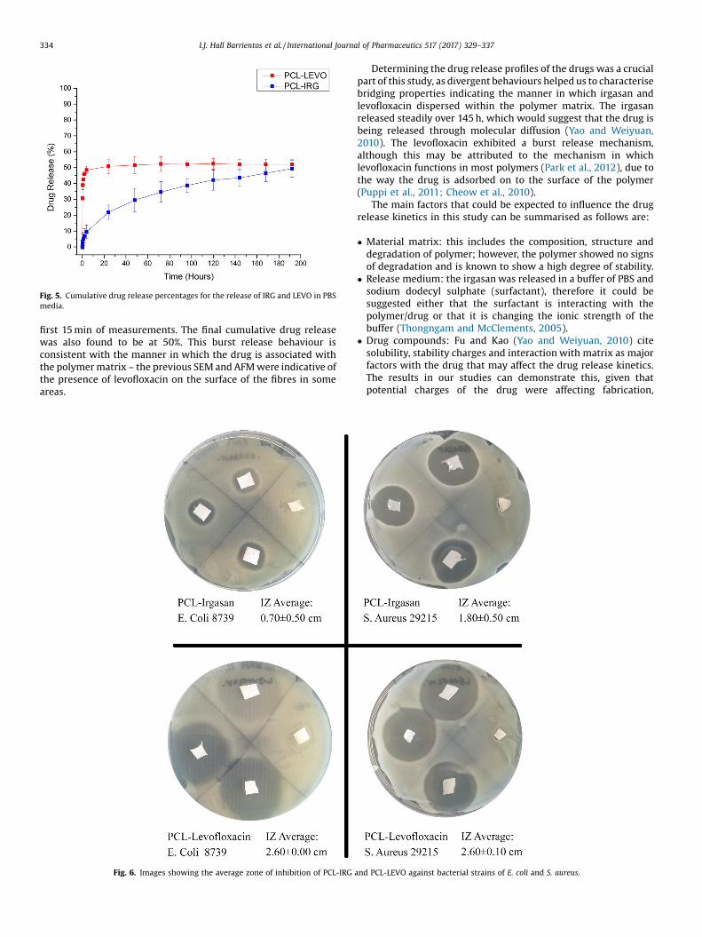
Fig. 5. Cumulative drug release percentages for the release of IRG and LEVO in PBSmedia.
334 I.J. Hall Barrientos et al. / International Journal of Pharmaceutics 517 (2017) 329–337
first 15 min of measurements. The final cumulative drug releasewas also found to be at 50%. This burst release behaviour isconsistent with the manner in which the drug is associated withthe polymer matrix – the previous SEM and AFM were indicative ofthe presence of levofloxacin on the surface of the fibres in someareas.
Fig. 6. Images showing the average zone of inhibition of PCL-IRG
Determining the drug release profiles of the drugs was a crucialpart of this study, as divergent behaviours helped us to characterisebridging properties indicating the manner in which irgasan andlevofloxacin dispersed within the polymer matrix. The irgasanreleased steadily over 145 h, which would suggest that the drug isbeing released through molecular diffusion (Yao and Weiyuan,2010). The levofloxacin exhibited a burst release mechanism,although this may be attributed to the mechanism in whichlevofloxacin functions in most polymers (Park et al., 2012), due tothe way the drug is adsorbed on to the surface of the polymer(Puppi et al., 2011; Cheow et al., 2010).
The main factors that could be expected to influence the drugrelease kinetics in this study can be summarised as follows are:
� Material matrix: this includes the composition, structure anddegradation of polymer; however, the polymer showed no signsof degradation and is known to show a high degree of stability.
� Release medium: the irgasan was released in a buffer of PBS andsodium dodecyl sulphate (surfactant), therefore it could besuggested either that the surfactant is interacting with thepolymer/drug or that it is changing the ionic strength of thebuffer (Thongngam and McClements, 2005).
� Drug compounds: Fu and Kao (Yao and Weiyuan, 2010) citesolubility, stability charges and interaction with matrix as majorfactors with the drug that may affect the drug release kinetics.The results in our studies can demonstrate this, given thatpotential charges of the drug were affecting fabrication,
and PCL-LEVO against bacterial strains of E. coli and S. aureus.

I.J. Hall Barrientos et al. / International Journal of Pharmaceutics 517 (2017) 329–337 335
therefore it can be assumed that the charges of irgasan andlevofloxacin may be affecting the drug release kinetics.
3.5. In-Vitro antibacterial activity
The antibacterial efficacy (Fig. 6) of both irgasan andlevofloxacin-loaded scaffolds were tested against strains of E. coliand S. aureus, with the efficacy specifically determined by visualzones of inhibition on the agar plate. The PCL-irgasan scaffoldshowed signs of some activity, albeit weak, against E. coli with anaverage inhibition zone diameter of 0.7 � 0.5 cm. However, theirgasan-loaded scaffold was particularly successful inhibiting thegrowth of S. aureus with an average inhibition zone diameter of1.8 � 0.5 cm. There was a higher-level efficacy observed within thePCL-levofloxacin cultures of both E. coli and S. aureus. Both strainsof bacteria were inhibited on the agar plate with an average
Fig. 7. a/b: The images above are acquired with a high lateral resolution mode, which enabthe API (colour overlay images C and D); c: Overlay of [C6H9O2]- (PCL) in grey and [C12H6Cand [C18H19FN3O4]- (Levofloxacin) in yellow.
diameter of no growth of 2.6 cm. The antibacterial studies haveshown that there is a high efficacy of bacteria inhibition in bothirgasan and levofloxacin-loaded scaffolds across E. coli andS. aureus bacteria. The levofloxacin-loaded scaffolds demonstratedlarger values of inhibition zones, for both bacteria � this should bethe case, given that levofloxacin is a broad-spectrum antibiotic,active against both gram positive and gram negative. The irgasan-loaded scaffold showed stronger inhibition to the S. aureusbacteria; however, this should not be viewed as a negative result.S. aureus is a gram-positive bacterium that is commonly found onthe skin, therefore is a major cause of nosocomial wound infection(Sisirak et al., 2010). The hydrophobic natures of irgasan and PCL,and potential stronger interactions between drug and polymer arelikely to aid the sustained release from the fibres � this sustainedrelease can be observed in the previous in vitro drug release study,and observed in the reduced inhibition of E. Coli (Celebioglu et al.,2014).
les to easily visualise the nanofibers (total ion image A and B) and the distribution ofl3O2]- (Irgasan) in yellow; d: Overlay of [C6H9O2]- (PCL) in grey and of [C17H19FN3O2]-

Fig. 8. a: The 2D (left) and 3D images show the distribution of [C6H9O2]- (PCL) in grey and [C12H6Cl3O2]- (Irgasan) in yellow. The analysed volume is 100 mm � 100 mm on theX-Y axes, and �3 mm on the Z axis: (A) viewed from the top and (B) inclined in order to aid 3D visualization. 8b: The 2D (left) and 3D images show the distribution of [C6H9O2]-
(PCL) in grey and of [C17H19FN3O2]- and [C18H19FN3O4]- (Levofloxacin) in yellow. The analysed volume is 100 mm � 100 mm on the X-Y axes,and �3 mm on the Z axis: (A)viewed from the top and (B) inclined in order to aid 3D visualization.
336 I.J. Hall Barrientos et al. / International Journal of Pharmaceutics 517 (2017) 329–337
3.6. ToF-SIMS analysis
Imaging and 3D imaging techniques showed a difference in thedistribution of the active pharmaceutical ingredients (APIs)between Irgasan-loaded and Levofloxacin-loaded fibres. PCL isidentified by the ion at m/z 113 ([C6H9O2]- [M-H]-), Levofloxacin bythe ions at m/z 360 ([C18H19FN3O4]- [M-H]-) and m/z 316([C17H19FN3O2]-), and Irgasan by the ions at m/z 287([C12H6
35Cl3O2]- [M-H]-), m/z 289 ([C12H635Cl237Cl1O2]-) and m/z
291 ([C12H635Cl137Cl2O2]-). The total ion images and the overlays of
single ion images for the characteristic peaks of PCL (grey) and thetwo drugs (yellow) are reported in Fig. 7. The ion images show ahomogeneous distribution of Irgasan, throughout the electrospunfibres, whilst the Levofloxacin appears to be concentrated inseveral small areas. This was confirmed by 3D imaging, whereIrgasan characteristic peaks appeared to be homogeneouslydistributed in the volume (Fig. 8a). Conversely, Levofloxacin hadan intense signal localized to small areas and mainly on the surface(Fig. 8b).
4. Conclusions
The purpose of this study was to fabricate drug-loaded fibresthat may potentially be used within a hernia repair context. Thegood understanding of the relationship between the solutionviscosity and the spinning parameters is essential if the techniqueis to be effective, hence the need to characterise the effect of drugloading on the rheological behaviour of the spinning solutions. Itwas observed that the addition of both irgasan and levofloxacinhad a direct influence on the rheological behaviour of thesolutions; a reduction in elastic modulus, viscous modulus, and
shear viscosity occurred, which may cause a reduction in polymerchain entanglements. However, this explanation may not be theonly viable one � rheological behaviour of drug-loaded solutionshas been widely researched, although further characterisation intothe molecular interactions between drug and polymer may givefurther insight into why the solution behaviour changes signifi-cantly. Atomic force microscopy indicated that crystals, probably oflevofloxacin were present on the surface of the polymer fibres, andthis was crucial in explaining the behaviour of the drug during invivo release studies and antibacterial activity profile. The presenceof levofloxacin at the surface of the polymer was confirmedthrough contact angle goniometry (immediate absorbance of thewater droplet showed the hydrophilic nature of levofloxacin inaction), in vitro release studies (the drug demonstrated a burstrelease behaviour), antibacterial studies (an increased averageinhibition zone repelled both bacteria types immediately) and ToF-SIMS. In the ToF-SIMS study, the molecular weight of levofloxacinwas shown at various areas across the fibres and the 3D imaging ofthe matrix indicated there was a certain degree of drugencapsulation. This study has contrasted the incorporation oftwo different drugs within an electrospun fibre, and shown thatthrough bridging chemical, mechanical and biological studies,their behaviours can be fully interpreted. The next stages of thisresearch are to now assess whether these constructs are usefulwithin any clinical scenario, and in particular, within the treatmentof hernia repair.
Acknowledgements
The authors would like to thank the UK Engineering & PhysicalSciences Research Council (EPSRC) Doctoral Training Centre in

I.J. Hall Barrientos et al. / International Journal of Pharmaceutics 517 (2017) 329–337 337
Medical Devices, University of Strathclyde (EPSRC Grant Ref.EP/F50036X/1) for the studentship awarded to IHB. The authorswould also like to thank the EPSRC Centre in ContinuousManufacturing and Crystallisation (CMAC) for access to specialisedinstruments.
References
Agarwal, S., Greiner, A., 2011. On the way to clean and safe electrospinning-greenelectrospinning: emulsion and suspension electrospinning. Polym. Adv.Technol. 22 (3), 372–378.
Azimi, B., Nourpanah, P., Rabiee, M., Arbab, S., 2016. Poly (lactide �co- Glycolide)Fiber: An Overview. .
Bhavsar, M.D., Amiji, M.M., 2008. Development of novel biodegradable polymericnanoparticles-in-microsphere formulation for local plasmid DNA delivery inthe gastrointestinal tract. AAPS Pharm. Sci. Tech. 9 (1), 288–294.
Bikiaris, D.N., Papageorgiou, G.Z., Achilias, D.S., Pavlidou, E., Stergiou, A., 2007.Miscibility and enzymatic degradation studies of poly(e-caprolactone)/poly(propylene succinate) blends. Eur. Polym. J. 43 (6), 2491–2503.
Bubel, K., Grunenberg, D., Vasilyev, G., Zussman, E., Agarwal, S., Greiner, A., 2014.Solvent-Free aqueous dispersions of block copolyesters for electrospinning ofbiodegradable nonwoven mats for biomedical applications. Macromol. Mater.Eng. 1445–1454.
Celebioglu, A., Umu, O.C.O., Tekinay, T., Uyar, T., 2014. Antibacterial electrospunnanofibers from triclosan/cyclodextrin inclusion complexes. Colloids Surf. BBiointerfaces 116, 612–619.
Cheow, W.S., Chang, M.W., Hadinoto, K., 2010. Antibacterial efficacy of inhalablelevofloxacin-loaded polymeric nanoparticles against E. coli biofilm cells: theeffect of antibiotic release profile. Pharm. Res. 27 (8), 1597–1609.
Davachi, S.M., Kaffashi, B., Zamanian, A., Torabinejad, B., Ziaeirad, Z., 2016.Investigating composite systems based on poly l-lactide and poly l-lactide/triclosan nanoparticles for tissue engineering and medical applications. Mater.Sci. Eng. C 58, 294–309.
Del Valle, L.J., Camps, R., Díaz, A., Franco, L., Rodríguez-Galán, A., Puiggalí, J., 2011.Electrospinning of polylactide and polycaprolactone mixtures for preparation ofmaterials with tunable drug release properties. J. Polym. Res. 18 (6), 1903–1917.
Detta, N., Brown, T.D., Edin, F.K., Albrecht, K., Chiellini, F., Chiellini, E., Dalton, P.D.,Hutmacher, D.W., 2010. Melt electrospinning of polycaprolactone and its blendswith poly(ethylene glycol). Polym. Int. 59 (11), 1558–1562.
Dias, J.R., Antunes, F.E., Ba’rtolo, P.J., 2013. Influence of the rheological behaviour inelectrospun PCL nano fibres production for tissue engineering application.Chem. Eng. Trans. 32 (2011), 1015–1020.
Duan, K., Xiao, D., Weng, J., 2013. Triclosan-loaded PLGA Microspheres-porousTitanium Composite Coating. , pp. 1–6.
Earle, D.B., 2010. Biomaterials in Hernia Repair. (Online]. Available: http://laparoscopy.blogs.com/prevention_management_3/2010/08/biomaterials-in-hernia-repair.html. [Accessed: 15-Sep-2015).
Ebersole, G.C., Buettmann, E.G., MacEwan, M.R., Tang, M.E., Frisella, M.M., Matthews,B.D., Deeken, C.R., 2012. Development of novel electrospun absorbablepolycaprolactone (PCL) scaffolds for hernia repair applications. Surg. Endosc.Other Interv. Tech. 26 (10), 2717–2728.
Goldstein, A.S., Thayer, P.S., 2016. Fabrication of complex biomaterial scaffolds forsoft tissue engineering by electrospinning. In: Grumezescu, A. (Ed.),Nanobiomaterials in Soft Tissue Engineering. William Andrew, Amsterdam, pp.299–330.
Haider, A., Haider, S., Kang, I.K., 2015. A comprehensive review summarizing theeffect of electrospinning parameters and potential applications of nanofibers inbiomedical and biotechnology. Arab. J. Chem..
Hartman, O., Zhang, C., Adams, E.L., Farach-carson, M.C., Petrelli, J., Chase, B.D.,Rabolt, J.F., 2011. Biofunctionalization of electrospun PCL-based scaffolds withperlecan domain IV peptide to create a 3-D pharmacokinetic cancer model.Biomaterials 31 (21), 5700–5718.
He, M., Xue, J., Geng, H., Gu, H., Chen, D., Shi, R., Zhang, L., 2015. Fibrous guided tissueregeneration membrane loaded with anti-inflammatory agent prepared by
coaxial electrospinning for the purpose of controlled release. Appl. Surf. Sci. 335,121–129.
Holländer, J., Genina, N., Jukarainen, H., Khajeheian, M., Rosling, A., Mäkilä, E.,Sandler, N., 2016. Three-Dimensional printed PCL-Based implantableprototypes of medical devices for controlled drug delivery. J. Pharm. 105 Jenny,Natalja Genina, Harri Jukarainen, Mohammad Khajeheian, Ari Rosl. ErmeiMäkilä, Niklas Sandler. Three-Dimensional Print. PCL-Based Implant. PrototypesMed. Devices Control. Drug Deliv. J..
Jalvandi, J., White, M., Truong, Y.B., Gao, Y., Padhye, R., Kyratzis, I.L., 2015. Release andantimicrobial activity of levofloxacin from composite mats of poly(e-caprolactone) and mesoporous silica nanoparticles fabricated by core-shellelectrospinning. J. Mater. Sci. 50 (24), 7967–7974.
Kabay, G., Kaleli, G., Sultanova, Z., Ölmez, T.T., Şeker, U.Ö.Ş., Mutlu, M., 2016.Biocatalytic protein membranes fabricated by electrospinning. React. Funct.Polym. 103, 26–32.
Lamprou, D.A., Smith, J.R., Nevell, T.G., Barbu, E., Willis, C.R., Tsibouklis, J., 2010. Self-assembled structures of alkanethiols on gold-coated cantilever tips andsubstrates for atomic force microscopy: molecular organisation and conditionsfor reproducible deposition. Appl. Surf. Sci. 256 (6), 1961–1968.
Lamprou, D.A., Venkatpurwar, V., Kumar, M.N.V.R., 2013. Atomic force microscopyimages label-Free, drug encapsulated nanoparticles In vivo and detectsdifference in tissue mechanical properties of treated and untreated: a tip fornanotoxicology. PLoS One 8 (5), 8–12.
Li, D., Guo, G., Fan, R., Liang, J., Deng, X., Luo, F., Qian, Z., 2013. PLA/F68/Dexamethasone implants prepared by hot-melt extrusion for controlled releaseof anti-inflammatory drug to implantable medical devices: i. Preparation,characterization and hydrolytic degradation study. Int. J. Pharm. 441 (1–2), 365–372.
Maleque, M., Hasan, M.R., Hossen, F., Safi, S., 2012. Development and validation of asimple UV spectrophotometric method for the determination of levofloxacinboth in bulk and marketed dosage formulations. J. Pharm. Anal. 2 (6), 454–457.
Murthy, R.S.R., 1997. In: Jain, N.K. (Ed.), Biodegradable Polymers. CBS Publisher, NewDehli, pp. 27–51.
Park, H., Yoo, H., Hwang, T., Park, T.J., Paik, D.H., Choi, S.W., Kim, J.H., 2012.Fabrication of levofloxacin-loaded nanofibrous scaffolds using coaxialelectrospinning. J. Pharm. Investig. 42 (2), 89–93.
Piccoli, A., Fiori, J., Andrisano, V., Orioli, M., 2002. Determination of triclosan inpersonal health care products by liquid chromatography (HPLC). Farmaco 57 (5),369–372.
Procter, L., Falco, E.E., Fisher, J.P., Roth, J.S., 2009. Abdominal wall hernias &biomaterials, In: Gefen, A. (Ed.), Bioengeering Research of Chronic Wounds. 1sted. Springer Verlag, Berlin, pp. 425–447.
Puppi, D., Dinucci, D., Bartoli, C., Mota, C., Migone, C., Dini, F., Barsotti, G., Carlucci, F.,Chiellini, F., 2011. Development of 3D wet-spun polymeric scaffolds loaded withantimicrobial agents for bone engineering. J. Bioact. Compat. Polym. 26, 478–492.
Sadrearhami, Z., Morshed, M., Varshosaz, J., 2015. Production and evaluation ofpolyblend of agar and polyacrylonitrile nanofibers for in vitro release ofmethotrexate in cancer therapy. Fibers Polym. 16 (2), 254–262.
Sebe, I., Szabo, B., Nagy, Z.K., Szabo, D., Zsidai, L., Kocsis, B., Zelko, R., 2013. “Polymerstructure and antimicrobial activity of polyvinylpyrrolidone-based iodinenanofibres prepared with high-speed rotary spinning technique’’. Int. J. Pharm.458 (1), 99–103.
Sisirak, M., Zvizdic, A., Hukic, M., 2010. Methicillin-resistant Staphylococcus aureus(MRSA) as a cause of nosocomial wound infections. Bosn. J. Basic Med. Sci.10 (1),32–37.
Thongngam, M., McClements, D.J., 2005. Influence of pH, ionic strength, andtemperature on self-association and interactions of sodium dodecyl sulfate inthe absence and presence of chitosan. Langmuir 21 (1), 79–86.
Toncheva, A., Paneva, D., Manolova, N., Rashkov, I., 2011. Electrospun poly(L-lactide)membranes containing a single drug or multiple drug system for antimicrobialwound dressings. Macromol. Res. 19 (12), 1310–1319.
Yao, F., Weiyuan, J.K., 2010. Drug release kinetics and transport mechanisms of non-degradable and degradable polymeric delivery systems. Expert Opin. DrugDeliv. 7 (4), 429–444.
Zamani, M., Prabhakaran, M.P., Ramakrishna, S., 2013. Advances in drug delivery viaelectrospun and electrosprayed nanomaterials. Int. J. Nanomed. 8, 2997–3017.