Dry Formulation of Virus-Like Particles in Electrospun ... - MDPI
13
Article Dry Formulation of Virus-Like Particles in Electrospun Nanofibers Sasheen Dowlath 1 , Katrin Campbell 2 , Farah Al-Barwani 1 , Vivienne L. Young 1 , Sarah L. Young 2,3 , Greg F. Walker 4 and Vernon K. Ward 1, * Citation: Dowlath, S.; Campbell, K.; Al-Barwani, F.; Young, V.L.; Young, S.L.; Walker, G.F.; Ward, V.K. Dry Formulation of Virus-Like Particles in Electrospun Nanofibers. Vaccines 2021, 9, 213. https://doi.org/ 10.3390/vaccines9030213 Academic Editor: Esther Blanco and Juan Bárcena Received: 22 January 2021 Accepted: 25 February 2021 Published: 3 March 2021 Publisher’s Note: MDPI stays neutral with regard to jurisdictional claims in published maps and institutional affil- iations. Copyright: © 2021 by the authors. Licensee MDPI, Basel, Switzerland. This article is an open access article distributed under the terms and conditions of the Creative Commons Attribution (CC BY) license (https:// creativecommons.org/licenses/by/ 4.0/). 1 Department of Microbiology & Immunology, School of Biomedical Sciences, University of Otago, P.O. Box 56, 720 Cumberland St, Dunedin 9054, New Zealand; [email protected] (S.D.); [email protected] (F.A.-B.); [email protected] (V.L.Y.) 2 Department of Pathology, Dunedin School of Medicine, University of Otago, P.O. Box 56, 720 Cumberland St, Dunedin 9054, New Zealand; [email protected] (K.C.); [email protected] (S.L.Y.) 3 Faculty of Medicine and Health, School of Medical Sciences, The University of Sydney, Sydney, NSW 2006, Australia 4 School of Pharmacy, University of Otago, P.O. Box 56, 720 Cumberland St, Dunedin 9054, New Zealand; [email protected] * Correspondence: [email protected]; Tel.: +64-3-4799028 Abstract: Biologics can be combined with liquid polymer materials and electrospun to produce a dry nanofibrous scaffold. Unlike spray-drying and freeze-drying, electrospinning minimizes the physiological stress on sensitive materials, and nanofiber mat properties such as hydrophobicity, solubility, and melting temperature can be tuned based on the polymer composition. In this study, we explored the dry formulation of a virus-like particle (VLP) vaccine by electrospinning VLP derived from rabbit hemorrhagic disease virus modified to carry the MHC-I gp100 tumor-associated antigen epitope. VLP were added to a polyvinylpyrrolidone (PVP) solution (15% w/v) followed by electrospinning at 24 kV. Formation of a nanofibrous mat was confirmed by scanning electron microscopy, and the presence of VLP was confirmed by transmission electron microscopy and Western blot. VLP from the nanofibers induced T-cell activation and interferon- (IFN-) γ production in vitro. To confirm in vivo cytotoxicity, Pmel mice treated by injection with gp100 VLP from nanofibers induced a gp100 specific immune response, lysing approximately 65% of gp100-pulsed target cells, comparable to mice vaccinated with gp100 VLP in PBS. VLP from nanofibers also induced an antibody response. This work shows that electrospinning can be used to dry-formulate VLP, preserving both humoral and cell-mediated immunity. Keywords: electrospinning; nanofiber; dry-formulation; virus-like particle; vaccine 1. Introduction Virus-like particles (VLP) have been extensively explored as vaccine material, with current VLP vaccines comprising liquid suspensions. VLP are structures derived from the shell components of a wild-type virus and are therefore classed as subunit vaccines. VLP are devoid of a viral genome and cannot infect or replicate within the host [1,2]. Due to the antigenic and structural similarities, VLP have the ability to induce immunity against the parental virus and are currently utilized in this capacity in licensed vaccines against human papillomavirus (HPV) [3], hepatitis B virus (HBV) [4], and hepatitis E virus (HEV) [5,6]. VLP can also be modified to carry unrelated antigens such as components of other pathogens or cancer epitopes for therapeutic vaccines, thus making them a versatile and safe particulate vaccine delivery platform [7–10]. Like many vaccines, liquid formulations of VLP vaccines must be correctly stored and strictly maintained between 2–8 ◦ C to preserve vaccine potency, adding both cost and complexity to the supply chain [11–13]. A broken cold chain could result in wasted Vaccines 2021, 9, 213. https://doi.org/10.3390/vaccines9030213 https://www.mdpi.com/journal/vaccines
-
Upload
khangminh22 -
Category
Documents
-
view
1 -
download
0
Transcript of Dry Formulation of Virus-Like Particles in Electrospun ... - MDPI
Article
Dry Formulation of Virus-Like Particles inElectrospun Nanofibers
Sasheen Dowlath 1, Katrin Campbell 2 , Farah Al-Barwani 1, Vivienne L. Young 1 , Sarah L. Young 2,3,Greg F. Walker 4 and Vernon K. Ward 1,*
�����������������
Citation: Dowlath, S.; Campbell, K.;
Al-Barwani, F.; Young, V.L.; Young,
S.L.; Walker, G.F.; Ward, V.K. Dry
Formulation of Virus-Like Particles in
Electrospun Nanofibers. Vaccines
2021, 9, 213. https://doi.org/
10.3390/vaccines9030213
Academic Editor: Esther Blanco and
Juan Bárcena
Received: 22 January 2021
Accepted: 25 February 2021
Published: 3 March 2021
Publisher’s Note: MDPI stays neutral
with regard to jurisdictional claims in
published maps and institutional affil-
iations.
Copyright: © 2021 by the authors.
Licensee MDPI, Basel, Switzerland.
This article is an open access article
distributed under the terms and
conditions of the Creative Commons
Attribution (CC BY) license (https://
creativecommons.org/licenses/by/
4.0/).
1 Department of Microbiology & Immunology, School of Biomedical Sciences, University of Otago,P.O. Box 56, 720 Cumberland St, Dunedin 9054, New Zealand; [email protected] (S.D.);[email protected] (F.A.-B.); [email protected] (V.L.Y.)
2 Department of Pathology, Dunedin School of Medicine, University of Otago, P.O. Box 56, 720 Cumberland St,Dunedin 9054, New Zealand; [email protected] (K.C.); [email protected] (S.L.Y.)
3 Faculty of Medicine and Health, School of Medical Sciences, The University of Sydney,Sydney, NSW 2006, Australia
4 School of Pharmacy, University of Otago, P.O. Box 56, 720 Cumberland St, Dunedin 9054, New Zealand;[email protected]
* Correspondence: [email protected]; Tel.: +64-3-4799028
Abstract: Biologics can be combined with liquid polymer materials and electrospun to produce adry nanofibrous scaffold. Unlike spray-drying and freeze-drying, electrospinning minimizes thephysiological stress on sensitive materials, and nanofiber mat properties such as hydrophobicity,solubility, and melting temperature can be tuned based on the polymer composition. In this study,we explored the dry formulation of a virus-like particle (VLP) vaccine by electrospinning VLPderived from rabbit hemorrhagic disease virus modified to carry the MHC-I gp100 tumor-associatedantigen epitope. VLP were added to a polyvinylpyrrolidone (PVP) solution (15% w/v) followedby electrospinning at 24 kV. Formation of a nanofibrous mat was confirmed by scanning electronmicroscopy, and the presence of VLP was confirmed by transmission electron microscopy and Westernblot. VLP from the nanofibers induced T-cell activation and interferon- (IFN-) γ production in vitro.To confirm in vivo cytotoxicity, Pmel mice treated by injection with gp100 VLP from nanofibersinduced a gp100 specific immune response, lysing approximately 65% of gp100-pulsed target cells,comparable to mice vaccinated with gp100 VLP in PBS. VLP from nanofibers also induced an antibodyresponse. This work shows that electrospinning can be used to dry-formulate VLP, preserving bothhumoral and cell-mediated immunity.
Keywords: electrospinning; nanofiber; dry-formulation; virus-like particle; vaccine
1. Introduction
Virus-like particles (VLP) have been extensively explored as vaccine material, withcurrent VLP vaccines comprising liquid suspensions. VLP are structures derived fromthe shell components of a wild-type virus and are therefore classed as subunit vaccines.VLP are devoid of a viral genome and cannot infect or replicate within the host [1,2].Due to the antigenic and structural similarities, VLP have the ability to induce immunityagainst the parental virus and are currently utilized in this capacity in licensed vaccinesagainst human papillomavirus (HPV) [3], hepatitis B virus (HBV) [4], and hepatitis E virus(HEV) [5,6]. VLP can also be modified to carry unrelated antigens such as components ofother pathogens or cancer epitopes for therapeutic vaccines, thus making them a versatileand safe particulate vaccine delivery platform [7–10].
Like many vaccines, liquid formulations of VLP vaccines must be correctly storedand strictly maintained between 2–8 ◦C to preserve vaccine potency, adding both costand complexity to the supply chain [11–13]. A broken cold chain could result in wasted
Vaccines 2021, 9, 213. https://doi.org/10.3390/vaccines9030213 https://www.mdpi.com/journal/vaccines
Vaccines 2021, 9, 213 2 of 13
vaccine stock or the administration of ineffective vaccines. This problem is often com-pounded in countries where cold chain infrastructures may be suboptimal [14]. Vaccinesuspensions can include a variety of compounds to improve stability including salts, pHbuffers, cryoprotectants, and thermoprotectants. [13]. Despite advancement in formulation,refrigeration, and storage technology, inadequate temperature control remains the primarycause of compromised vaccine stocks [11,12,14]. One possible approach to minimize thereliance on cold-chain handling is to prepare a dry formulation of the vaccine material,which can be reconstituted into a liquid suspension prior to administration. Dry formula-tions involve the removal of water from the sample, reducing the motility of the sampleand minimizing degradation pathways facilitated by water. [15]. Dry formulations areless prone to temperature-induced degradation, leading to reduced reliance on the coldchain, fewer wasted vaccine stocks, and an increase in cost-effectiveness and shelf life [16].Another key advantage is that dry formulated material can be packaged into capsules,nasal sprays, inhalers, and transdermal patches, enabling flexibility with administrationroutes and target sites [16].
Freeze-drying has historically been a gold standard for dry formulation of biologicsand pharmaceuticals; however, freeze-drying was shown to result in undesirable aggre-gation and attenuated immunogenicity of VLP [17]. Freeze-drying is also a long, costly,and energy consuming process, which requires expensive equipment [15]. Some successhas been garnered by freeze-drying the leaves of a plant-based expression system con-taining the VLP, which may be used in oral vaccination applications such as capsules [18].Spray-drying is another technology which has showed promise. A liquid suspensioncontaining vaccine material and excipients are nebulized into an aerosol, before beingsprayed into a heated gaseous medium for dehydration into a powder [16]. Sheer stressgenerated during the nebulization process may result in the damage of protein-basedvaccine components, while the dehydration stress may also cause undesirable aggregationand attenuation [19–21]. Saboo et al. reported successful spray drying of HPV VLPsinto a dry powder, although this was only possible using a multi-component excipientsystem due to stresses exhibited during the process [22]. The research paradigm is shiftingtowards eliminating the cold chain altogether, with innovations in vaccine formulationsand storage technology whereby the vaccine material is maintained to the highest integritywith exposure to the least amount of stress [23–25].
Electrospinning is a one-step technique that involves the rapid drying of a polymersolution into a fibrous sheet by applying an electrostatic field to the solution [26]. Theelectrostatic field facilitates the formation of a charged nanofiber jet, which is attractedto a grounded surface. During the time of flight, the solvent rapidly evaporates, leavingbehind a continuous solid nanofiber filament, which is then randomly deposited on thegrounded collection surface and over time, a nanofiber sheet is formed [27]. Electrospin-ning offers several advantages compared to conventional dry formulation methods such asfreeze-drying or spray drying. The solvent is removed rapidly from the liquid formulation,typically in nanoseconds, as opposed to several days with freeze-drying [15,25,28]. Temper-ature and humidity control is often recommended but not essential and electrospinning canbe carried out at ambient temperature and atmospheric pressure. The formulation is notsubjected to extreme temperatures or pressure changes associated with freeze-drying andspray drying, allowing for a gentler drying process for sensitive biologics and proteins [16].Nanofibers can be comprised of singular polymers, or mixture of polymers depending onthe application. Properties such as high surface area to volume ratio, solubility, hydropho-bicity, individual fiber size, and rate of dissolution can be altered by adjusting the polymermixture [29]. Nanofiber technology has been employed in controlled release drug delivery,tissue regeneration, air and water filtration, and wound healing applications [26,29–33].Scalability has also been improved in recent years with advancements in multi-needle andneedle-less systems.
PVP was selected as the polymer for our nanofiber formulation due to its high-watersolubility and is commonly used as a binding agent and drug stabilizer in oral tablets and
Vaccines 2021, 9, 213 3 of 13
capsules. PVP has previously been used as the basis for many electrospinning studies,including wound healing, the stabilization of drugs and bacteriophages, and other slow-release applications [34–40].
This study aimed to dry-formulate an active vaccine component in a nanofiber matwhile maintaining immunogenicity. To demonstrate this, we used rabbit hemorrhagic dis-ease virus (RHDV) VLPs as an antigen carrier that was modified to express an MHC-classI-restricted tumor antigen. The VP60 gene of RHDV was modified to fuse two copies of thehuman gp100 MHC-I epitope to the N-terminus, placing copies of the epitope internally inthe assembled VLP [8]. The gp100 antigen was shown to elicit a strong cell-mediated im-mune response in tumor trials and was established as a murine melanoma model [8,41–43].Previous studies also demonstrated that RHDV VLPs carrying heterologous antigens astherapeutic cancer vaccines, such as gp100, were able to induce a strong, enhanced immuneresponse [8,41,44,45].
In this study, we successfully electrospun VLP into a PVP-based nanofiber and demon-strated that intact particles can be recovered and visualized by transmission electronmicroscopy (TEM) upon nanofiber solubilization. Furthermore, we showed that the hu-moral and cell-mediated immune responses were conserved when particles were stored ina nanofiber. Importantly, we demonstrated that both the cellular and humoral immuneresponse generated by the dry-formulated VLP was comparable to freshly prepared VLP,both in vitro and in vivo.
2. Materials and Methods2.1. Expression and Purification of VLP
VLP were expressed in Sf 21 insect cells using a baculovirus expression system aspreviously described [46,47]. DNA encoding two copies of the gp100 epitope (KVPRN-QDWLALLKVPRNQDWL) with an immunoproteasome cleavage sequence (ALL) wasinserted at the N-terminus of the RHDV VP60 gene (Figure 1A) by PCR extension andused to generate a recombinant baculovirus by homologous recombination (gp100.2LVLP). Expression and purification of VLP were carried out as previously described [47].Briefly, suspension cultures of Sf21 insect cells were infected with recombinant baculovirusexpressing the VP60 protein +/− the gp100 epitopes at a multiplicity of infection of 1 andincubated at 27 ◦C at 125 rpm for 3 days. The cells were lysed with 0.5% Triton X-100and the VLP purified using differential centrifugation, followed by a centrifugation on a1.2 and 1.4 g mL CsCl step gradient at 100,000 g for 18 h. The VLP band was harvestedand dialyzed into 0.1 M phosphate buffer pH 7.4 containing 0.15 M NaCl (PBS). A controlVLP without the gp100 epitope motif was also expressed (RHDV VLP). Expression andpurification of VLP was confirmed by SDS-PAGE and the presence of the gp100 epitopewas confirmed by mass spectrometry at the Otago Centre for Protein Research. Particleassembly was confirmed by TEM at the Otago Centre for Electron Microscopy.
2.2. Electrospinning
Polyvinylpyrrolidone (PVP K-90, Mw 360,000, Sigma-Aldrich, St. Louis, MO, USA)solutions were prepared in Milli-Q water with stirring overnight at room temperature. VLP(5 mg) which had been dialyzed against PBS was added to a 30% w/v PVP solution at a1:1 ratio and mixed by magnet stirring for 20 min. The solution was drawn into a 5-mLsyringe (Becton Dickinson, Franklin, NJ, USA) with blunted needle (18 G) and attachedto an infusion pump (Fusion Touch 100, Chemyx Inc., Stafford, TX, USA) to deliver thesolution at a constant flow rate of 0.1 mL h−1. The live terminal of a high voltage powersupply was attached to the needle and was set to generate an electrical potential of 24 kV.A grounded metal wheel collector was placed 15 cm from the needle tip and rotated atapproximately 10 rpm. A strip of aluminum foil was placed around the circumferenceof the wheel, to which nanofiber was deposited. Nanofiber mats were stored at roomtemperature on the aluminum foil in a zip-locked bag containing silica gel. Nanofiberswere stored for up to seven days prior to biological assays.
Vaccines 2021, 9, 213 4 of 13Vaccines 2021, 9, x 4 of 13
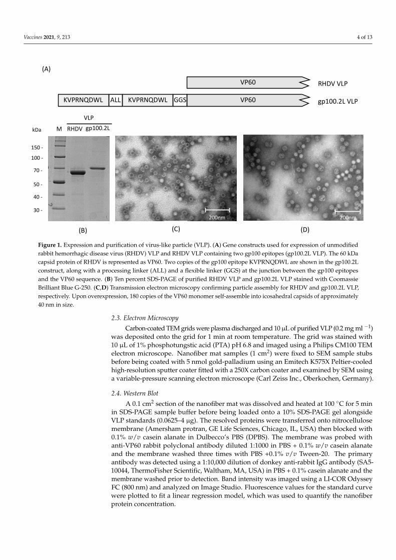
Figure 1. Expression and purification of virus-like particle (VLP). (A) Gene constructs used for expression of unmodified rabbit hemorrhagic disease virus (RHDV) VLP and RHDV VLP containing two gp100 epitopes (gp100.2L VLP). The 60 kDa capsid protein of RHDV is represented as VP60. Two copies of the gp100 epitope KVPRNQDWL are shown in the gp100.2L construct, along with a processing linker (ALL) and a flexible linker (GGS) at the junction between the gp100 epitopes and the VP60 sequence. (B) Ten percent SDS-PAGE of purified RHDV VLP and gp100.2L VLP stained with Coo-massie Brilliant Blue G-250. (C,D) Transmission electron microscopy confirming particle assembly for RHDV and gp100.2L VLP, respectively. Upon overexpression, 180 copies of the VP60 monomer self-assemble into icosahedral capsids of approximately 40 nm in size.
2.2. Electrospinning Polyvinylpyrrolidone (PVP K-90, Mw 360,000, Sigma-Aldrich, St. Louis, MO, USA)
solutions were prepared in Milli-Q water with stirring overnight at room temperature. VLP (5 mg) which had been dialyzed against PBS was added to a 30% w/v PVP solution at a 1:1 ratio and mixed by magnet stirring for 20 min. The solution was drawn into a 5-mL syringe (Becton Dickinson, Franklin, NJ, USA) with blunted needle (18 G) and at-tached to an infusion pump (Fusion Touch 100, Chemyx Inc., Stafford, TX, USA) to deliver the solution at a constant flow rate of 0.1 mL h−1. The live terminal of a high voltage power supply was attached to the needle and was set to generate an electrical potential of 24 kV. A grounded metal wheel collector was placed 15 cm from the needle tip and rotated at approximately 10 rpm. A strip of aluminum foil was placed around the circumference of the wheel, to which nanofiber was deposited. Nanofiber mats were stored at room tem-perature on the aluminum foil in a zip-locked bag containing silica gel. Nanofibers were stored for up to seven days prior to biological assays.
2.3. Electron Microscopy Carbon-coated TEM grids were plasma discharged and 10 µL of purified VLP (0.2
mg ml −1) was deposited onto the grid for 1 min at room temperature. The grid was stained with 10 µL of 1% phosphotungstic acid (PTA) pH 6.8 and imaged using a Philips CM100 TEM electron microscope. Nanofiber mat samples (1 cm2) were fixed to SEM sample stubs before being coated with 5 nmol gold-palladium using an Emitech K575X Peltier-cooled
(D)
200nm
(C)200nm
150 -
100 -
70 -
50 -
40 -
MkDa
30 -
(B)
RHDV gp100.2LVLP
KVPRNQDWL KVPRNQDWL VP60
VP60
ALL GGS
RHDV VLP
gp100.2L VLP
(A)
Figure 1. Expression and purification of virus-like particle (VLP). (A) Gene constructs used for expression of unmodifiedrabbit hemorrhagic disease virus (RHDV) VLP and RHDV VLP containing two gp100 epitopes (gp100.2L VLP). The 60 kDacapsid protein of RHDV is represented as VP60. Two copies of the gp100 epitope KVPRNQDWL are shown in the gp100.2Lconstruct, along with a processing linker (ALL) and a flexible linker (GGS) at the junction between the gp100 epitopesand the VP60 sequence. (B) Ten percent SDS-PAGE of purified RHDV VLP and gp100.2L VLP stained with CoomassieBrilliant Blue G-250. (C,D) Transmission electron microscopy confirming particle assembly for RHDV and gp100.2L VLP,respectively. Upon overexpression, 180 copies of the VP60 monomer self-assemble into icosahedral capsids of approximately40 nm in size.
2.3. Electron Microscopy
Carbon-coated TEM grids were plasma discharged and 10µL of purified VLP (0.2 mg ml −1)was deposited onto the grid for 1 min at room temperature. The grid was stained with10 µL of 1% phosphotungstic acid (PTA) pH 6.8 and imaged using a Philips CM100 TEMelectron microscope. Nanofiber mat samples (1 cm2) were fixed to SEM sample stubsbefore being coated with 5 nmol gold-palladium using an Emitech K575X Peltier-cooledhigh-resolution sputter coater fitted with a 250X carbon coater and examined by SEM usinga variable-pressure scanning electron microscope (Carl Zeiss Inc., Oberkochen, Germany).
2.4. Western Blot
A 0.1 cm2 section of the nanofiber mat was dissolved and heated at 100 ◦C for 5 minin SDS-PAGE sample buffer before being loaded onto a 10% SDS-PAGE gel alongsideVLP standards (0.0625–4 µg). The resolved proteins were transferred onto nitrocellulosemembrane (Amersham protran, GE Life Sciences, Chicago, IL, USA) then blocked with0.1% w/v casein alanate in Dulbecco’s PBS (DPBS). The membrane was probed withanti-VP60 rabbit polyclonal antibody diluted 1:1000 in PBS + 0.1% w/v casein alanateand the membrane washed three times with PBS +0.1% v/v Tween-20. The primaryantibody was detected using a 1:10,000 dilution of donkey anti-rabbit IgG antibody (SA5-10044, ThermoFisher Scientific, Waltham, MA, USA) in PBS + 0.1% casein alanate and themembrane washed prior to detection. Band intensity was imaged using a LI-COR OdysseyFC (800 nm) and analyzed on Image Studio. Fluorescence values for the standard curvewere plotted to fit a linear regression model, which was used to quantify the nanofiberprotein concentration.
Vaccines 2021, 9, 213 5 of 13
2.5. Animals and Ethics
Female C57BL/6 mice were obtained from the Hercus Taieri Research Unit, Universityof Otago, Dunedin, New Zealand. Female pmel mice, expressing T-cell receptors specificto the MHC-I restricted gp10025–33 epitope, were obtained from the Young lab breedingstock, Department of Pathology, University of Otago, Dunedin New Zealand. Experimentswere conducted in accordance with an ethical permit granted by the University of OtagoAnimal Ethics Committee (AEC D103/17). Animals were euthanized by carbon dioxide orcardiac puncture.
2.6. T-Cell Proliferation and Interferon-γ Production Assay
Bone marrow-derived dendritic cells (BMDCs) were extracted from femurs and tibiaeof C57BL/6 mice as previously described [48], adjusted to a concentration of 5 × 10⁴ cellsml−1 and pulsed with either PBS, PVP, blank nanofiber, 1 µg RHDV VLP or gp100.2L VLP,or gp10025–33 peptide (0.38 µg·mL−1, the molar equivalent of 1 µg of gp100.2L VLP). Asection of nanofibers containing 1 µg of RHDV VLP or gp100.2L VLP was added directlyonto the cells. T-cells from pmel mice were prepared according to Kramer et al. [41]and sorted for CD8 expression using anti-CD8 AutoMACS MicroBeads (Miltenyi Biotech,Bergisch Gladbach, Germany). CD8+ T cells (1 × 10⁸ cells·mL−1) were diluted 1:1 with20 µM carboxyfluorescein succinimidyl ester (CFSE) (Invitrogen) and added to the pulsedBMDCs at a ratio of 10:1, and then incubated for 72 h at 37 ◦C, 10% CO2. Cells were treatedwith 50 µL of a 1:1000 dilution of LIVE/DEAD™ Fixable Near-IR Dead Cell Stain (LifeTechnologies™, Eugene, OR, USA) followed by treatment with CD16/CD32 Fc blockingantibody, and then stained with CD3-PE-CF594 (clone 145-2C11) and CD8α-APC (clone53-6.7) (Life Technologies™, Eugene, OR, USA). Fluorescence was measured using a Galliosflow cytometer (Beckman Coulter, Brea, CA, USA) with a three-laser (405, 488, and 633 nm),ten-color configuration and analyzed using Kaluza software (Beckman Coulter, Brea, CA,USA). Interferon-γ (IFN-γ) levels were measured by enzyme-linked immunosorbent assay(ELISA) as previously described [41].
2.7. In Vivo Cytotoxicity
Five groups of six mice were vaccinated with either PBS, 100 µg of gp100.2L VLP,100 µg of RHDV VLP from a dissolved nanofiber sample, 100 µg equivalent of gp100.2LVLP from a dissolved nanofiber sample, and the molar equivalent of free gp100 con-tained in 100 µg of VLP (100 µL at 19.6 µg·mL−1). Each vaccination contained 25 µg ofCpG oligonucleotide as an adjuvant (CpG 1826, GeneWorks, SA, Australia) added to thetreatment prior to vaccination. For nanofiber samples, this was post-dissolution in PBS.Treatments were administered subcutaneously to the left flank using a 29-G needle. Micewere boosted 21 days later with an additional dose of their respective treatment. Mice wereinjected intravenously with target (pulsed with gp100 peptide) and non-target splenocyteson day 28 derived from donor C57BL/6 mice. The red blood cells were removed fromthe splenocyte preparation by lysis with ammonium chloride followed by centrifugation.Remaining cells were divided into 2 groups; unpulsed (untreated) cells and pulsed cells,treated with 10 mM of synthetic gp10025–33 peptide (KVPRNQDWL) and incubated for2 h at 37 ◦C, 5% CO2. Each group was differentially stained, with the untreated groupreceiving 5 µM CFSE (CFSELO) and the treated group receiving 50 µM CFSE (CFSEHI).Cells from each group were mixed in a 1:1 ratio and 1 × 10⁸ cells were injected into thetail vein of each mouse. Mice were sacrificed 40 h post injection and the splenocytes wereharvested and stained with LIVE/DEAD™ Fixable Near-IR Dead Cell Stain. Fluorescencewas measured on a Gallios flow cytometer and analyzed using Kaluza software.
2.8. Serum Collection and ELISA
Mice from the in vivo cytotoxicity assay were bled on day 30 by cardiac puncture,serum was collected and stored at −80 ◦C. Each well of a 96-well plate was coated with2 µg of VP60 followed by incubation at 4 ◦C overnight and then washed in PBS + 0.05%
Vaccines 2021, 9, 213 6 of 13
Tween-20 six times (wash buffer). The plate was blocked by adding 100 µL of 20 mg mL−1
BSA in PBS (blocking buffer) per well and incubated for an hour at 37 ◦C and then washedsix times. Serum was serially diluted ten-fold (10−1–10−6) in blocking buffer and 100 µladded to each well. Plates were incubated for 2 h at 37 ◦C and then washed six times inwash buffer. Anti-mouse IgG-HRP (Sigma-Aldrich, Cat. no. A9044) was diluted 1:60,000 inblocking buffer before addition of 100 µl to each well and incubated for 1 h at 37 ◦C. Wellswere washed six times before addition of 100 µL 3,3′,5,5′-tetramethylbenzidine (TMB)substrate to each well. The reaction was stopped after 2 min by adding 100 µL of 1 M HClsolution. Plates were read at 450 nm using a Multiskan FC Microplate spectrophotometer(ThermoFisher Scientific).
2.9. Statistical Analysis
Statistical analysis was carried out using GraphPad Prism version 7.04 (GraphPadSoftware, La Jolla, CA, USA). Analyses of the in vivo data consisted of a Kruskal Wallis non-parametric test with a Dunn’s post-hoc test. Advice on statistical analysis was provided byAndrew Gray, Biostatistical Unit, Division of Health Sciences, University of Otago.
3. Results3.1. VLP Expression and Purification
VLP were expressed using the commercial baculovirus expression system, flashBACUltra (Oxford Expression Technologies, Oxford, UK). VLP were purified by differentialcentrifugation and a CsCl gradient before assessment by SDS-PAGE (Figure 1B). Proteinidentity was confirmed by Western blot and the presence of the correct gp100 sequencewas confirmed by mass spectrometry analysis (data not shown). Overexpression of theVP60 capsid protein led to self-assembly into icosahedral particles of approximately 40 nmin size, which was confirmed by TEM (Figure 1C,D, respectively). Size and shape of theexpressed VLP were consistent with previous work [46].
3.2. Incorporation of VLP into Nanofibers
Electrospinning of VLP in a 15% (w/v) PVP solution in PBS was performed successfullyat 24 kV and a flow rate of 0.1 mL h−1. While PVP-water solutions can be electrospunat much higher flow rates, PBS was a necessary addition to the formulation in order topreserve the structural integrity of the VLP. SEM imaging (Figure 2A) of an electrospunnanofiber mat showed heterogenous nanofiber structures including tubular strands andflat ribbon-like sheets with diameters ranging from 100 nm to 5 µm. A comparison betweenthe blank, unloaded nanofibers (Figure 2B,C) and the RHDV VLP-loaded nanofibers(Figure 2D,E) revealed particles on the surface of the VLP-loaded nanofiber, consistent withthe presence of RHDV VLP. The observed particles were arranged singularly or clusteredand the diameter of the particles was consistent with sputter-coated VLP. To confirm theidentity of the observed structures, a VLP-loaded nanofiber mat was dissolved in PBS andthen loaded onto a 1.2 and 1.4 g·cm−3 CsCl step gradient as utilized for VLP purification.Material recovered from the CsCl interface revealed that intact VLP particles were present,as visualized by TEM (Figure 2F,G). SDS-PAGE confirmed the protein to be 60 kDa insize, consistent with the size of VP60. To confirm that the isolated particles were of RHDVVP60 origin and simultaneously assess nanofiber loading uniformity, 0.1 cm2 samples weredissolved and analyzed by Western blotting (Figure 2H). Results confirm that the 60 kDaprotein was VP60 and that loading was relatively consistent across the mat. These resultsindicate that VLP can be electrospun into a dry nanofibrous mat and that intact VLP can berecovered upon dissolution of the mat.
3.3. T-Cell Proliferation and IFN-γ Production Assays
An initial in vitro test was performed to confirm activity before analysis in vivo. TheVLP-loaded nanofibers were tested for the ability to induce a cell-mediated immuneresponse by measuring T-cell proliferation as well as IFN-γ production. BMDCs from

Vaccines 2021, 9, 213 7 of 13
C57BL/6 mice were pulsed with treatments, followed by a co-culture with T cells frompmel mice, which were genotyped to express a T-cell receptor specific to the gp10025–33 pep-tide. RHDV VLP-loaded nanofiber (negative control) and gp100.2L VLP-loaded nanofibersamples were placed directly into the cell culture. Other controls included VLP (no epitope)in PBS, gp100.2L VLP in PBS, and gp100 peptide as a positive control. T-cell proliferationdata (Figure 3A) was standardized against the gp100 peptide response. The gp100.2LVLP-loaded nanofiber induced a very strong response at or near 100%, similar to othergroups containing gp100. In contrast, RHDV VLP-loaded nanofiber without the gp100epitope induced limited proliferation. Similarly, gp100.2L VLP-loaded nanofiber inducedan IFN-γ response of 111 ng ml−1 (Figure 3B), comparable to that of the gp100 peptide(116 ng mL−1).
Vaccines 2021, 9, x 7 of 13
at much higher flow rates, PBS was a necessary addition to the formulation in order to preserve the structural integrity of the VLP. SEM imaging (Figure 2A) of an electrospun nanofiber mat showed heterogenous nanofiber structures including tubular strands and flat ribbon-like sheets with diameters ranging from 100 nm to 5 µm. A comparison be-tween the blank, unloaded nanofibers (Figure 2B,C) and the RHDV VLP-loaded nano-fibers (Figure 2D,E) revealed particles on the surface of the VLP-loaded nanofiber, con-sistent with the presence of RHDV VLP. The observed particles were arranged singularly or clustered and the diameter of the particles was consistent with sputter-coated VLP. To confirm the identity of the observed structures, a VLP-loaded nanofiber mat was dis-solved in PBS and then loaded onto a 1.2 and 1.4 g·cm−3 CsCl step gradient as utilized for VLP purification. Material recovered from the CsCl interface revealed that intact VLP par-ticles were present, as visualized by TEM (Figure 2F,G). SDS-PAGE confirmed the protein to be 60 kDa in size, consistent with the size of VP60. To confirm that the isolated particles were of RHDV VP60 origin and simultaneously assess nanofiber loading uniformity, 0.1 cm2 samples were dissolved and analyzed by Western blotting (Figure 2H). Results con-firm that the 60 kDa protein was VP60 and that loading was relatively consistent across the mat. These results indicate that VLP can be electrospun into a dry nanofibrous mat and that intact VLP can be recovered upon dissolution of the mat.
Figure 2. Incorporation and analysis of VLP in electrospun nanofibers. (A) Macrostructure of a nanofiber mat produced by electrospinning a 15% w/v polyvinylpyrrolidone (PVP) solution. (B,C) SEM of blank nanofiber microstructures. (D,E)
(H)
1µm
(B)
40nm
(G)
(F)
40nm
(E)
100nm
(D)
1µm
(C)
100nm
(A)
1 2 3 4 5 6
1007055
35
kDa
Figure 2. Incorporation and analysis of VLP in electrospun nanofibers. (A) Macrostructure of a nanofiber mat producedby electrospinning a 15% w/v polyvinylpyrrolidone (PVP) solution. (B,C) SEM of blank nanofiber microstructures. (D,E)SEM of RHDV VLP-loaded nanofiber microstructures. (F,G) RHDV-loaded nanofibers were dissolved and re-purified ona CsCl gradient, recovered material viewed by TEM. (H) Nanofiber loading uniformity. Lanes 1–6 represent randomlyselected samples cut from the nanofiber mat and dissolved. An amount equivalent of 0.1 cm2 nanofiber was run in eachlane and VP60 levels compared by fluorescent western blot using an anti-RHDV VP60 antibody. The original western blotand relatively band intensities are shown in Supplementary Figure S1.
Vaccines 2021, 9, 213 8 of 13
Vaccines 2021, 9, x 8 of 13
SEM of RHDV VLP-loaded nanofiber microstructures. (F,G) RHDV-loaded nanofibers were dissolved and re-purified on a CsCl gradient, recovered material viewed by TEM. (H) Nanofiber loading uniformity. Lanes 1–6 represent randomly selected samples cut from the nanofiber mat and dissolved. An amount equivalent of 0.1 cm2 nanofiber was run in each lane and VP60 levels compared by fluorescent western blot using an anti-RHDV VP60 antibody. The original western blot and relatively band intensities are shown in Supplementary Figure S1.
3.3. T-Cell Proliferation and IFN-γ Production Assays An initial in vitro test was performed to confirm activity before analysis in vivo. The
VLP-loaded nanofibers were tested for the ability to induce a cell-mediated immune re-sponse by measuring T-cell proliferation as well as IFN-γ production. BMDCs from C57BL/6 mice were pulsed with treatments, followed by a co-culture with T cells from pmel mice, which were genotyped to express a T-cell receptor specific to the gp10025–33 peptide. RHDV VLP-loaded nanofiber (negative control) and gp100.2L VLP-loaded nan-ofiber samples were placed directly into the cell culture. Other controls included VLP (no epitope) in PBS, gp100.2L VLP in PBS, and gp100 peptide as a positive control. T-cell pro-liferation data (Figure 3A) was standardized against the gp100 peptide response. The gp100.2L VLP-loaded nanofiber induced a very strong response at or near 100%, similar to other groups containing gp100. In contrast, RHDV VLP-loaded nanofiber without the gp100 epitope induced limited proliferation. Similarly, gp100.2L VLP-loaded nanofiber induced an IFN-γ response of 111 ng ml−1 (Figure 3B), comparable to that of the gp100 peptide (116 ng ml−1).
Figure 3. In vitro T-cell proliferation and cytokine response towards VLP-loaded nanofibers. BMDCs Cells were pulsed with 1 µg of VLP in PBS, 1 µg of VLP from a nanofiber, or gp100 peptide at a molar equivalent to gp100 in 1 µg of VLP. T-cells were co-cultured with pulsed bone marrow-derived dendritic cells (BMDCs) for 72 h. (A) T cell proliferation deter-mined by flow cytometry and (B) IFN-γ production results determined by ELISA of the supernatant. Each bar represents the mean percentage of T-cell proliferation (±SEM). The data represents 3 independent experiments. Statistical significance was determined by a one-way ANOVA with Dunnett’s multiple comparisons test. **** p < 0 0001. Statistical significance displayed is the comparison of RHDV VLP nanofiber against negative control groups. The gating strategy used to generate this data is shown in Supplementary Figure S2.
3.4. In Vivo Cytotoxicity and Antibody Response Following demonstration that T-cell activation and IFN-γ production was upregu-
lated by VLP released from nanofibers, the activity of VLP-loaded nanofiber was assessed in vivo for the induction of target specific cytotoxicity towards gp100, as well as a specific
Figure 3. In vitro T-cell proliferation and cytokine response towards VLP-loaded nanofibers. BMDCs Cells were pulsedwith 1 µg of VLP in PBS, 1 µg of VLP from a nanofiber, or gp100 peptide at a molar equivalent to gp100 in 1 µg of VLP. T-cellswere co-cultured with pulsed bone marrow-derived dendritic cells (BMDCs) for 72 h. (A) T cell proliferation determinedby flow cytometry and (B) IFN-γ production results determined by ELISA of the supernatant. Each bar represents themean percentage of T-cell proliferation (±SEM). The data represents 3 independent experiments. Statistical significancewas determined by a one-way ANOVA with Dunnett’s multiple comparisons test. **** p < 0 0001. Statistical significancedisplayed is the comparison of RHDV VLP nanofiber against negative control groups. The gating strategy used to generatethis data is shown in Supplementary Figure S2.
3.4. In Vivo Cytotoxicity and Antibody Response
Following demonstration that T-cell activation and IFN-γ production was upregulatedby VLP released from nanofibers, the activity of VLP-loaded nanofiber was assessed in vivofor the induction of target specific cytotoxicity towards gp100, as well as a specific antibodyresponse toward the VLP. Mice were vaccinated subcutaneously with treatments followedby a boosting vaccination 21 days later (Figure 4A). On day 28, mice were challenged withunpulsed control splenocytes and gp100-pulsed target splenocytes. Mice were culled 40 hlater and assessed for specific killing of the target cells by flow cytometry. Results fromFigure 4B,C show that nanofibers loaded with gp100.2L VLP induced specific lysis of gp100-pulsed BMDCs (approximately 65%) in a similar capacity to that of gp100.2L VLP deliveredin PBS (approximately 60%). While the results for the nanofiber gp100 VLP treatmentproduced a broader range of response than gp100 VLP without nanofiber, there was nostatistical difference between these two VLP treatment groups carrying the gp100 epitope.PBS only and RHDV VLP-loaded nanofiber containing no epitope induced no specific lysis(Figure 4B,C). To assess ability of the released VLP to stimulate an antibody response, anenzyme-linked immunosorbent assay (ELISA) was performed using the collected serumas the source of the primary probing antibody against purified VLP (Figure 4D). Resultsindicate that all treatments that contained VLP induced an antibody response. RHDV VLPnanofiber and gp100.2L VLP nanofiber producing comparable antibody titers to that of thegp100.2L VLP delivered in PBS.
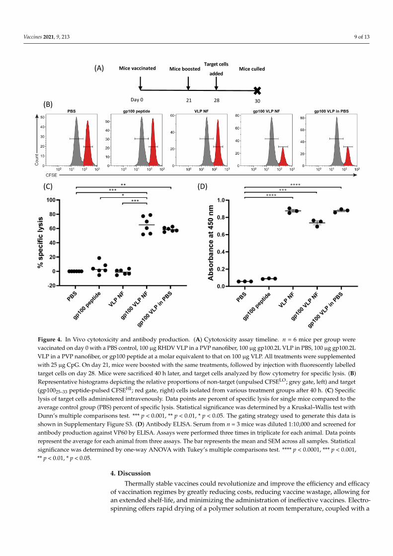
Vaccines 2021, 9, 213 9 of 13
Vaccines 2021, 9, x 9 of 13
antibody response toward the VLP. Mice were vaccinated subcutaneously with treat-ments followed by a boosting vaccination 21 days later (Figure 4A). On day 28, mice were challenged with unpulsed control splenocytes and gp100-pulsed target splenocytes. Mice were culled 40 h later and assessed for specific killing of the target cells by flow cytometry. Results from Figure 4B,C show that nanofibers loaded with gp100.2L VLP induced spe-cific lysis of gp100-pulsed BMDCs (approximately 65%) in a similar capacity to that of gp100.2L VLP delivered in PBS (approximately 60%). While the results for the nanofiber gp100 VLP treatment produced a broader range of response than gp100 VLP without nan-ofiber, there was no statistical difference between these two VLP treatment groups carry-ing the gp100 epitope. PBS only and RHDV VLP-loaded nanofiber containing no epitope induced no specific lysis (Figure 4B,C). To assess ability of the released VLP to stimulate an antibody response, an enzyme-linked immunosorbent assay (ELISA) was performed using the collected serum as the source of the primary probing antibody against purified VLP (Figure 4D). Results indicate that all treatments that contained VLP induced an anti-body response. RHDV VLP nanofiber and gp100.2L VLP nanofiber producing comparable antibody titers to that of the gp100.2L VLP delivered in PBS.
Figure 4. In Vivo cytotoxicity and antibody production. (A) Cytotoxicity assay timeline. n = 6 mice per group were vac-cinated on day 0 with a PBS control, 100 µg RHDV VLP in a PVP nanofiber, 100 µg gp100.2L VLP in PBS, 100 µg gp100.2L VLP in a PVP nanofiber, or gp100 peptide at a molar equivalent to that on 100 µg VLP. All treatments were supplemented with 25 µg CpG. On day 21, mice were boosted with the same treatments, followed by injection with fluorescently labelled
Day 0 21 28 30
Mice vaccinated Mice boostedTarget cells
addedMice culled(A)
(C) (D)
(B)
Figure 4. In Vivo cytotoxicity and antibody production. (A) Cytotoxicity assay timeline. n = 6 mice per group werevaccinated on day 0 with a PBS control, 100 µg RHDV VLP in a PVP nanofiber, 100 µg gp100.2L VLP in PBS, 100 µg gp100.2LVLP in a PVP nanofiber, or gp100 peptide at a molar equivalent to that on 100 µg VLP. All treatments were supplementedwith 25 µg CpG. On day 21, mice were boosted with the same treatments, followed by injection with fluorescently labelledtarget cells on day 28. Mice were sacrificed 40 h later, and target cells analyzed by flow cytometry for specific lysis. (B)Representative histograms depicting the relative proportions of non-target (unpulsed CFSELO; grey gate, left) and target(gp10025–33 peptide-pulsed CFSEHI; red gate, right) cells isolated from various treatment groups after 40 h. (C) Specificlysis of target cells administered intravenously. Data points are percent of specific lysis for single mice compared to theaverage control group (PBS) percent of specific lysis. Statistical significance was determined by a Kruskal–Wallis test withDunn’s multiple comparisons test. *** p < 0.001, ** p < 0.01, * p < 0.05. The gating strategy used to generate this data isshown in Supplementary Figure S3. (D) Antibody ELISA. Serum from n = 3 mice was diluted 1:10,000 and screened forantibody production against VP60 by ELISA. Assays were performed three times in triplicate for each animal. Data pointsrepresent the average for each animal from three assays. The bar represents the mean and SEM across all samples. Statisticalsignificance was determined by one-way ANOVA with Tukey’s multiple comparisons test. **** p < 0.0001, *** p < 0.001,** p < 0.01, * p < 0.05.
4. Discussion
Thermally stable vaccines could revolutionize and improve the efficiency and efficacyof vaccination regimes by greatly reducing costs, reducing vaccine wastage, allowing foran extended shelf-life, and minimizing the administration of ineffective vaccines. Electro-spinning offers rapid drying of a polymer solution at room temperature, coupled with a
Vaccines 2021, 9, 213 10 of 13
cheaper running cost and a faster material preparation time when compared to other vac-cine drying technologies, such as freeze-drying or spray drying [16]. In this study, we havedemonstrated that electrospinning is an effective method to produce a dry formulation ofa particulate vaccine. More importantly, results of the in vivo experiments demonstratethat VLP formulated in the nanofibers were able to stimulate a cell-mediated and humoralimmune response without loss of functionality when compared to VLP that had not beenincorporated into nanofibers.
VLP were successfully electrospun in a PVP solution into a nanofiber mat. Inspectionby SEM confirmed that VLP-like structures were visible on the surface of the nanofibers,indicating a successful drying of VLP. Western blot analysis of the dissolved gp100.2L VLPnanofiber confirmed that the material was of RHDV VP60 origin and that the materialwas evenly distributed throughout the sheet. Repurification of a dissolved nanofiber on aCsCl gradient followed by TEM analysis confirmed that intact particles were present andthat structures were intact. These results indicated that VLP can be easily incorporatedand recovered from a nanofiber while remaining structurally intact. For the nanofiber tobe an effective dry formulation of a vaccine, it is of utmost importance to also maintainimmunogenicity.
As a proof of concept and further evidence to suggest the VLP was intact and ableto elicit an immune response, VLP-loaded nanofibers were subjected to both in vitro andin vivo immune assays. The gp100 epitope was previously shown to establish a strongimmunotherapeutic response in murine melanoma models, which can be further enhancedby delivery on a modified VLP, thus, providing a means of evaluating the immunogenicityof the dry formulated VLP [41]. Results in vitro indicated that the gp100 peptide waseffectively cleaved and processed from the VLP particles by DCs, followed by cross-presentation in the context of MHC-I in order to stimulate a T cell proliferation and IFN-γresponse. Results of the in vivo cytotoxicity assay demonstrated a significant response inthe group vaccinated with the gp100.2L VLP nanofiber, similar to that of mice vaccinatedwith gp100.2L VLP delivered in PBS. Contrastingly, the gp10025–33 free peptide showedvery little cytotoxic stimulation. It is important to note that longer, more complex peptidespresented on a VLP require uptake and endosomal processing by antigen presentingcells, leading to more efficient cross-presentation on MHC-I when compared to shorter,free peptides such as gp10025–33 alone [8,41,49]. The comparable nature of the cytotoxicresponses seen in the gp100.2L VLP nanofiber and gp100.2L VLP in PBS groups furthersuggest that the nanofiber VLP are intact and are processed by cross-presentation, as hasbeen shown for free VLP [50].
Serum from the mice in the in vivo cytotoxicity work was harvested and tested forantibody production against VP60. All groups containing VLP, including in nanofibers,showed an upregulated antibody titer when compared to PBS and gp100 peptide groups.The similar antibody responses to VLP derived from the electrospun nanofiber and VLP thatwas not electrospun confirms that VP60 remains immunologically relevant in the nanofiber.
This work demonstrates an early proof of concept to show that VLP can be electro-spun into a dry formulation nanofiber, and can be reconstituted prior to delivery whilemaintaining immunogenicity. The tuneability of the nanofiber enables the physical andchemical properties to be altered by adjusting the polymer composition. Due to the highwater-solubility of PVP, reconstitution involved immersion in an aqueous solution, PBSin this case. Polymer choices or blends other than the PVP used in this study could beexplored for fine tuning solubility, permeability, hydrophobicity and humidity stability, orto offer slow-release characteristics to enhance the immune response through the use ofnon-aqueous polymers. Other stabilizing materials such as trehalose can also be incorpo-rated into the polymer solution [40]. Nanofibers allow for transportation and storage inthe dry form up until the need for administration, or can be compressed into alternativedelivery forms, such as oral capsules or patches which can be placed directly onto mucosalsites. Several doses could also be electrospun into a single, large, multidose sheet. A

Vaccines 2021, 9, 213 11 of 13
nanofiber formulation has the potential to also be delivered as a dry patch, to be hydratedby the tissue.
Several factors, such as polymer concentration, electrostatic field strength, fluid viscos-ity, surfactants, distance to the collection surface, ionic strength, and flow rate can impacton the efficiency and quality of the final nanofiber [51,52]. Extending the research into theeffects of electrospinning parameters on our PVP blend could improve electrospinningefficiency and protein yield.
5. Conclusions
This work demonstrates that electrospinning offers an attractive method for the dryformulation of particulate vaccines with complex biological structures without loss offunctionality. VLP were successfully electospun into a nanofibrous mat and recoveredupon reconstitution of the mat. By using VLP as an antigenic carrier stored in the nanofibers,we were able to confirm that both the humoral and cell-mediated immune response wasmaintained when VLP were released from the nanofiber mat and used as a vaccine. Itshould be noted that VLP was used as an exemplar antigen, and that this technique has thepotential to be applied to a range of vaccine types and antigens.
Supplementary Materials: The following are available online at https://www.mdpi.com/2076-393X/9/3/213/s1, Figure S1: Nanofibre loading uniformity. Lanes 1–7 represent randomly selectedsamples cut from the nanofibre mat. Lane 1, 0.05 cm2 of nanofibre. Lanes 2–7, 0.1 cm2 of nanofibreper lane. VP60 levels were compared by fluorescent western blot using an anti- RHDV VP60 antibodyand near IR fluorescent secondary antibody. Image acquisition and relative band intensities wereobtained using a LiCor Odyssey FC instrument and Image Studio software. Figure S2: Gatingstrategy for T cell proliferation analysis; Figure S3: Gating strategy for the in vivo cytotoxicity.
Author Contributions: Conceptualization, S.D., V.L.Y., V.K.W., and G.F.W.; methodology, S.D., K.C.,V.K.W., and V.L.Y.; formal analysis, S.D. and K.C.; investigation, S.D. and K.C.; resources, F.A.-B.,V.K.W., G.F.W., and S.L.Y.; data curation, S.D.; writing—original draft preparation, S.D.; writing—review and editing, S.D., K.C., G.F.W., V.K.W., and V.L.Y.; visualization, S.D.; supervision, V.K.W.,G.F.W., and V.L.Y. All authors have read and agreed to the published version of the manuscript.
Funding: This research received no external funding.
Institutional Review Board Statement: Experiments were conducted in accordance with ethicalpermit granted by the University of Otago Animal Ethics Committee (AEC D103/17).
Informed Consent Statement: Not Applicable.
Data Availability Statement: The data presented in this study are available in the manuscript andassociated supplementary materials.
Acknowledgments: The authors wish to thank Nicholas Shields for production of Figure 4B.
Conflicts of Interest: The authors declare no conflict of interest.
References1. Grgacic, E.V.; Anderson, D.A. Virus-like particles: Passport to immune recognition. Methods 2006, 40, 60–65. [CrossRef]2. Cimica, V.; Galarza, J.M. Adjuvant formulations for virus-like particle (VLP) based vaccines. J. Clin. Immunol. 2017, 183, 99–108.
[CrossRef] [PubMed]3. Godi, A.; Bissett, S.L.; Miller, E.; Beddows, S. Relationship between Humoral Immune Responses against HPV16, HPV18, HPV31
and HPV45 in 12–15 Year Old Girls Receiving Cervarix® or Gardasil® Vaccine. PLoS ONE 2015, 10, e0140926.4. Pol, S.; Driss, F.; Michel, M.-L.; Nalpas, B.; Berthelot, P.; Brechot, C. Specific vaccine therapy in chronic hepatitis B infection. Lancet
1994, 344, 342. [CrossRef]5. Wu, X.; Chen, P.; Lin, H.; Hao, X.; Liang, Z. Hepatitis E virus: Current epidemiology and vaccine. Hum. Vaccines Immunother.
2016, 12, 2603–2610. [CrossRef]6. Zhang, X.; Wei, M.; Sun, G.; Wang, X.; Li, M.; Lin, Z.; Li, Z.; Li, Y.; Fang, M.; Zhang, J.; et al. Real-time stability of a
hepatitis E vaccine (Hecolin(R)) demonstrated with potency assays and multifaceted physicochemical methods. Vaccine 2016, 34,5871–5877. [CrossRef]
Vaccines 2021, 9, 213 12 of 13
7. Win, S.J.; McMillan, D.G.; Errington-Mais, F.; Ward, V.K.; Young, S.L.; Baird, M.A.; Melcher, A.A. Enhancing the immunogenicityof tumour lysate-loaded dendritic cell vaccines by conjugation to virus-like particles. Br. J. Cancer 2012, 106, 92–98. [CrossRef]
8. Al-Barwani, F.; Donaldson, B.; Pelham, S.J.; Young, S.L.; Ward, V.K. Antigen delivery by virus-like particles for immunotherapeuticvaccination. Ther. Deliv. 2014, 5, 1223–1240. [CrossRef]
9. Cubas, R.; Zhang, S.; Kwon, S.; Sevick-Muraca, E.M.; Li, M.; Chen, C.; Yao, Q. Virus-like particle (VLP) Lymphatic Trafficking andImmune Response Generation after Immunization by Different Routes. J. Immunother. 2009, 32, 118–128. [CrossRef]
10. Zhang, S.; Cubas, R.; Li, M.; Chen, C.; Yao, Q. Virus-Like Particle Vaccine Activates Conventional B2 Cells and Promotes B CellDifferentiation to IgG2a Producing Plasma Cells. Mol. Immunol. 2009, 46, 1988–2001. [CrossRef]
11. Matthias, D.M.; Robertson, J.; Garrison, M.M.; Newland, S.; Nelson, C. Freezing temperatures in the vaccine cold chain: Asystematic literature review. Vaccine 2007, 25, 3980–3986. [CrossRef] [PubMed]
12. Purssell, E. Reviewing the importance of the cold chain in the distribution of vaccines. Br. J. Community Nurs. 2015, 20, 481–486.[CrossRef] [PubMed]
13. Donaldson, B.; Lateef, Z.; Walker, G.F.; Young, S.L.; Ward, V.K. Virus-like particle vaccines: Immunology and formulation forclinical translation. Expert Rev. Vaccines 2018, 17, 833–849. [CrossRef] [PubMed]
14. Wirkas, T.; Toikilik, S.; Miller, N.; Morgan, C.; Clements, C.J. A vaccine cold chain freezing study in PNG highlights technologyneeds for hot climate countries. Vaccine 2007, 25, 691–697. [CrossRef]
15. Maltesen, M.J.; van de Weert, M. Drying methods for protein pharmaceuticals. Drug Discov. Today Technol. 2008, 5,e81–e88. [CrossRef]
16. Kanojia, G.; Have Rt Soema, P.C.; Frijlink, H.; Amorij, J.-P.; Kersten, G. Developments in the formulation and delivery of spraydried vaccines. Hum. Vaccines Immunother. 2017, 13, 2364–2378. [CrossRef]
17. Lang, R.; Winter, G.; Vogt, L.; Zurcher, A.; Dorigo, B.; Schimmele, B. Rational design of a stable, freeze-dried virus-like particle-based vaccine formulation. Drug Dev. Ind. Pharm. 2009, 35, 83–97. [CrossRef]
18. Czyz, M.; Pniewski, T. Thermostability of Freeze-Dried Plant-Made VLP-Based Vaccines. In Sustainable Drying Technologies;Olvera, J.d.R., Ed.; IntechOpen: London, UK, 2016.
19. LeClair, D.A.; Cranston, E.D.; Xing, Z.; Thompson, M.R. Optimization of Spray Drying Conditions for Yield, Particle Size andBiological Activity of Thermally Stable Viral Vectors. Pharm. Res. 2016, 33, 2763–2776. [CrossRef]
20. Mumenthaler, M.; Hsu, C.C.; Pearlman, R. Feasibility study on spray-drying protein pharmaceuticals: Recombinant humangrowth hormone and tissue-type plasminogen activator. Pharm. Res. 1994, 11, 12–20. [CrossRef]
21. Webb, S.D.; Golledge, S.L.; Cleland, J.L.; Carpenter, J.F.; Randolph, T.W. Surface adsorption of recombinant human interferon-gamma in lyophilized and spray-lyophilized formulations. J. Pharm. Sci. 2002, 91, 1474–1487. [CrossRef]
22. Saboo, S.; Tumban, E.; Peabody, J.; Wafula, D.; Peabody, D.S.; Chackerian, B.; Muttil, P. Optimized Formulation of a ThermostableSpray-Dried Virus-Like Particle Vaccine against Human Papillomavirus. Mol. Pharm. 2016, 13, 1646–1655. [CrossRef]
23. Chen, X.; Fernando, G.J.; Crichton, M.L.; Flaim, C.; Yukiko, S.R.; Fairmaid, E.J.; Corbett, H.J.; Primiero, C.A.; Ansaldo, A.B.; Frazer,I.H.; et al. Improving the reach of vaccines to low-resource regions, with a needle-free vaccine delivery device and long-termthermostabilization. J. Control. Release 2011, 152, 349–355. [CrossRef]
24. Leung, V.; Mapletoft, J.; Zhang, A.; Lee, A.; Vahedi, F.; Chew, M.; Szewczyk, A.; Jahanshahi-Anbuhi, S.; Ang, J.; Cowbrough, B.;et al. Thermal Stabilization of Viral Vaccines in Low-Cost Sugar Films. Sci. Rep. 2019, 9, 7631. [CrossRef] [PubMed]
25. Lovalenti, P.M.; Anderl, J.; Yee, L.; Nguyen, V.; Ghavami, B.; Ohtake, S.; Saxena, A.; Voss, T.; Truong-Le, V. Stabilization of LiveAttenuated Influenza Vaccines by Freeze Drying, Spray Drying, and Foam Drying. Pharm. Res. 2016, 33, 1144–1160. [CrossRef]
26. Ramakrishna, S. An Introduction to Electrospinning and Nanofibers; World Scientific Publishing Co: Singapore, 2005.27. Ramakrishna, S.; Fujihara, K.; Teo, W.-E.; Yong, T.; Ma, Z.; Ramaseshan, R. Electrospun nanofibers: Solving global issues. Mater.
Today 2006, 9, 40–50. [CrossRef]28. Rieger, K.; Birch, N.; Schiffman, J. Designing electrospun nanofiber mats to promote wound healing—A review. J. Mater. Chem. B
2013, 1, 4531–4541. [CrossRef] [PubMed]29. Agrahari, V.; Agrahari, V.; Meng, J.; Mitra, A.K. Chapter 9—Electrospun Nanofibers in Drug Delivery: Fabrication, Advances,
and Biomedical Applications. In Emerging Nanotechnologies for Diagnostics, Drug Delivery and Medical Devices; Elsevier: Boston,MA, USA, 2017; pp. 189–215.
30. Hu, X.; Liu, S.; Zhou, G.; Huang, Y.; Xie, Z.; Jing, X. Electrospinning of polymeric nanofibers for drug delivery applications. J.Control. Release 2014, 185, 12–21. [CrossRef]
31. Xue, J.; Wu, T.; Dai, Y.; Xia, Y. Electrospinning and Electrospun Nanofibers: Methods, Materials, and Applications. Chem. Rev.2019, 119, 5298–5415. [CrossRef]
32. Tipduangta, P.; Belton, P.; Fábián, L.; Wang, L.Y.; Tang, H.; Eddleston, M.; Qi, S. Electrospun Polymer Blend Nanofibers forTunable Drug Delivery: The Role of Transformative Phase Separation on Controlling the Release Rate. Mol. Pharm. 2016, 13,25–39. [CrossRef]
33. Babitha, S.; Rachita, L.; Karthikeyan, K.; Shoba, E.; Janani, I.; Poornima, B.; Sai, K.P. Electrospun protein nanofibers in healthcare:A review. Int. J. Pharm. 2017, 523, 52–90. [CrossRef]
34. Kadajji, V.G.; Betageri, G.V. Water Soluble Polymers for Pharmaceutical Applications. Polymers 2011, 3, 1972–2009. [CrossRef]35. Dai, M.; Jin, S.; Nugen, S.R. Water-soluble electrospun nanofibers as a method for on-chip reagent storage. Biosensors 2012, 2,
388–395. [CrossRef] [PubMed]
Vaccines 2021, 9, 213 13 of 13
36. Gökmese, F.; Uslu, I.; Aytimur, A. Preparation and Characterization of PVA/PVP Nanofibers as Promising Materials for WoundDressing. Polym. Plast. Technol. Eng. 2013, 52, 1259–1265. [CrossRef]
37. Kamble, R.N.; Gaikwad, S.; Maske, A.; Patil, S.S. Fabrication of electrospun nanofibres of BCS II drug for enhanced dissolutionand permeation across skin. J. Adv. Res. 2016, 7, 483–489. [CrossRef]
38. Sriyanti, I.; Edikresnha, D.; Rahma, A.; Munir, M.M.; Rachmawati, H.; Khairurrijal, K. Mangosteen pericarp extract embedded inelectrospun PVP nanofiber mats: Physicochemical properties and release mechanism of α-mangostin. Int. J. Nanomed. 2018, 13,4927–4941. [CrossRef] [PubMed]
39. Wang, Y.; Zhao, X.; Tian, Y.; Wang, Y.; Jan, A.K.; Chen, Y. Facile Electrospinning Synthesis of Carbonized Polyvinylpyrrolidone(PVP)/g-C3 N4 Hybrid Films for Photoelectrochemical Applications. Chem. Eur. J. 2017, 23, 419–426. [CrossRef] [PubMed]
40. Dai, M.; Senecal, A.; Nugen, S.R. Electrospun water-soluble polymer nanofibers for the dehydration and storage of sensitivereagents. Nanotechnology 2014, 25, 225101. [CrossRef]
41. Kramer, K.; Al-Barwani, F.; Baird, M.A.; Young, V.L.; Larsen, D.S.; Ward, V.K.; Young, S.L. Functionalisation of Virus-Like ParticlesEnhances Antitumour Immune Responses. J. Immunol. Res. 2019, 2019, 5364632. [CrossRef]
42. Liu, G.; Ying, H.; Zeng, G.; Wheeler, C.J.; Black, K.L.; Yu, J.S. HER-2, gp100, and MAGE-1 are expressed in human glioblastomaand recognized by cytotoxic T cells. Cancer Res. 2004, 64, 4980–4986. [CrossRef]
43. Patel, P.M.; Ottensmeier, C.H.; Mulatero, C.; Lorigan, P.; Plummer, R.; Pandha, H.; Elsheikh, S.; Hadjimichael, E.; Villasanti, N.;Adams, S.E.; et al. Targeting gp100 and TRP-2 with a DNA vaccine: Incorporating T cell epitopes with a human IgG1 antibodyinduces potent T cell responses that are associated with favourable clinical outcome in a phase I/II trial. Oncoimmunology 2018, 7,e1433516. [CrossRef]
44. Donaldson, B.; Al-Barwani, F.; Pelham, S.J.; Young, K.; Ward, V.K.; Young, S.L. Multi-target chimaeric VLP as a therapeuticvaccine in a model of colorectal cancer. J. Immunotherap. Cancer 2017, 5, 69. [CrossRef]
45. Peacey, M.; Wilson, S.; Perret, R.; Ronchese, F.; Ward, V.K.; Young, V.; Young, S.L.; Baird, M.A. Virus-like particles from rabbithemorrhagic disease virus can induce an anti-tumor response. Vaccine 2008, 26, 5334–5337. [CrossRef] [PubMed]
46. Peacey, M.; Wilson, S.; Baird, M.A.; Ward, V.K. Versatile RHDV virus-like particles: Incorporation of antigens by geneticmodification and chemical conjugation. Biotechnol. Bioeng. 2007, 98, 968–977. [CrossRef] [PubMed]
47. McKee, S.J.; Young, V.L.; Clow, F.; Hayman, C.M.; Baird, M.A.; Hermans, I.F.; Young, S.L.; Ward, V.K. Virus-like particles andalpha-galactosylceramide form a self-adjuvanting composite particle that elicits anti-tumor responses. J. Control. Release 2012, 159,338–345. [CrossRef]
48. Inaba, K.; Inaba, M.; Romani, N.; Aya, H.; Deguchi, M.; Ikehara, S.; Muramatsu, S.; Steinman, R.M. Generation of large numbersof dendritic cells from mouse bone marrow cultures supplemented with granulocyte/macrophage colony-stimulating factor. J.Exp. Med. 1992, 176, 1693–1702. [CrossRef] [PubMed]
49. Faure, F.; Mantegazza, A.; Sadaka, C.; Sedlik, C.; Jotereau, F.; Amigorena, S. Long-lasting cross-presentation of tumor antigen inhuman DC. Eur. J. Immunol. 2009, 39, 380–390. [CrossRef] [PubMed]
50. Win, S.J.; Ward, V.K.; Dunbar, P.R.; Young, S.L.; Baird, M.A. Cross-presentation of epitopes on virus-like particles via the MHC Ireceptor recycling pathway. Immunol. Cell Biol. 2011, 89, 681–688. [CrossRef]
51. Abutaleb, A.; Lolla, D.; Aljuhani, A.; Shin, H.U.; Rajala, J.W.; Chase, G.G. Effects of Surfactants on the Morphology and Propertiesof Electrospun Polyetherimide Fibers. Fibers 2017, 5, 33. [CrossRef]
52. Haider, A.; Haider, S.; Kang, I.-K. A comprehensive review summarizing the effect of electrospinning parameters and potentialapplications of nanofibers in biomedical and biotechnology. Arab. J. Chem. 2018, 11, 1165–1188. [CrossRef]