Squalene Loaded Nanoparticles Effectively Protect Hepatic ...
22
Citation: Bidooki, S.H.; Alejo, T.; Sánchez-Marco, J.; Martínez-Beamonte, R.; Abuobeid, R.; Burillo, J.C.; Lasheras, R.; Sebastian, V.; Rodríguez-Yoldi, M.J.; Arruebo, M.; et al. Squalene Loaded Nanoparticles Effectively Protect Hepatic AML12 Cell Lines against Oxidative and Endoplasmic Reticulum Stress in a TXNDC5-Dependent Way. Antioxidants 2022, 11, 581. https:// doi.org/10.3390/antiox11030581 Academic Editor: Catalina Alarcòn de-la-Lastra Received: 28 February 2022 Accepted: 16 March 2022 Published: 18 March 2022 Publisher’s Note: MDPI stays neutral with regard to jurisdictional claims in published maps and institutional affil- iations. Copyright: © 2022 by the authors. Licensee MDPI, Basel, Switzerland. This article is an open access article distributed under the terms and conditions of the Creative Commons Attribution (CC BY) license (https:// creativecommons.org/licenses/by/ 4.0/). antioxidants Article Squalene Loaded Nanoparticles Effectively Protect Hepatic AML12 Cell Lines against Oxidative and Endoplasmic Reticulum Stress in a TXNDC5-Dependent Way Seyed Hesamoddin Bidooki 1 , Teresa Alejo 2,3 , Javier Sánchez-Marco 1 , Roberto Martínez-Beamonte 1,4,5 , Roubi Abuobeid 1 , Juan Carlos Burillo 6 , Roberto Lasheras 6 , Victor Sebastian 2,3,7 , María J. Rodríguez-Yoldi 4,5,8 , Manuel Arruebo 2,3,7 and Jesús Osada 1,4,8, * 1 Departamento de Bioquímica y Biología Molecular y Celular, Facultad de Veterinaria, Instituto de Investigación Sanitaria de Aragón-Universidad de Zaragoza, E-50013 Zaragoza, Spain; [email protected] (S.H.B.); [email protected] (J.S.-M.); [email protected] (R.M.-B.); [email protected] (R.A.) 2 Departamento de Ingeniería Química y Tecnologías del Medio Ambiente, Universidad de Zaragoza, E-50018 Zaragoza, Spain; [email protected] (T.A.); [email protected] (V.S.); [email protected] (M.A.) 3 Instituto de Nanociencia y Materiales de Aragón (INMA), CSIC-Universidad de Zaragoza, E-50009 Zaragoza, Spain 4 Instituto Agroalimentario de Aragón, CITA-Universidad de Zaragoza, E-50013 Zaragoza, Spain; [email protected] 5 Centro de Investigación Biomédica en Red de Fisiopatología de la Obesidad y Nutrición (CIBEROBN), Instituto de Salud Carlos III, E-28029 Madrid, Spain 6 Laboratorio Agroambiental, Servicio de Seguridad Agroalimentaria de la Dirección General de Alimentación y Fomento Agroalimentario, Gobierno de Aragón, E-50059 Zaragoza, Spain; [email protected] (J.C.B.); [email protected] (R.L.) 7 Centro de Investigación Biomédica en Red de Bioingeniería, Biomateriales y Nanomedicina (CIBER-BBN), Instituto de Salud Carlos III, E-28029 Madrid, Spain 8 Departamento de Farmacología, Fisiología, Medicina Legal y Forense, Facultad de Veterinaria, Instituto de Investigación Sanitaria de Aragón-Universidad de Zaragoza, E-50013 Zaragoza, Spain * Correspondence: [email protected]; Tel.: +34-976-761-644; Fax: +34-976-761-612 Abstract: Virgin olive oil, the main source of fat in the Mediterranean diet, contains a substantial amount of squalene which possesses natural antioxidant properties. Due to its highly hydrophobic nature, its bioavailability is reduced. In order to increase its delivery and potentiate its actions, squalene has been loaded into PLGA nanoparticles (NPs). The characterization of the resulting nanoparticles was assessed by electron microscopy, dynamic light scattering, zeta potential and high- performance liquid chromatography. Reactive oxygen species (ROS) generation and cell viability assays were carried out in AML12 (alpha mouse liver cell line) and a TXNDC5-deficient AML12 cell line (KO), which was generated by CRISPR/cas9 technology. According to the results, squalene was successfully encapsulated in PLGA NPs, and had rapid and efficient cellular uptake at 30 μM squalene concentration. Squalene reduced ROS in AML12, whereas ROS levels increased in KO cells and improved cell viability in both when subjected to oxidative stress by significant induction of Gpx4. Squalene enhanced cell viability in ER-induced stress by decreasing Ern1 or Eif2ak3 expressions. In conclusion, TXNDC5 shows a crucial role in regulating ER-induced stress through different signaling pathways, and squalene protects mouse hepatocytes from oxidative and endoplasmic reticulum stresses by several molecular mechanisms depending on TXNDC5. Keywords: olive oil; liver; squalene; PLGA; oxidative stress; endoplasmic reticulum stress; TXNDC5; Gpx4; Ern1; Eif2ak3 1. Introduction The Mediterranean diet, also known as the Med diet, has been associated with a variety of health benefits on cardiovascular diseases, including a reduction in the frequency of Antioxidants 2022, 11, 581. https://doi.org/10.3390/antiox11030581 https://www.mdpi.com/journal/antioxidants
-
Upload
khangminh22 -
Category
Documents
-
view
1 -
download
0
Transcript of Squalene Loaded Nanoparticles Effectively Protect Hepatic ...
�����������������
Citation: Bidooki, S.H.; Alejo, T.;
Sánchez-Marco, J.; Martínez-Beamonte,
R.; Abuobeid, R.; Burillo, J.C.; Lasheras,
R.; Sebastian, V.; Rodríguez-Yoldi, M.J.;
Arruebo, M.; et al. Squalene Loaded
Nanoparticles Effectively Protect
Hepatic AML12 Cell Lines against
Oxidative and Endoplasmic
Reticulum Stress in a
TXNDC5-Dependent Way.
Antioxidants 2022, 11, 581. https://
doi.org/10.3390/antiox11030581
Academic Editor: Catalina Alarcòn
de-la-Lastra
Received: 28 February 2022
Accepted: 16 March 2022
Published: 18 March 2022
Publisher’s Note: MDPI stays neutral
with regard to jurisdictional claims in
published maps and institutional affil-
iations.
Copyright: © 2022 by the authors.
Licensee MDPI, Basel, Switzerland.
This article is an open access article
distributed under the terms and
conditions of the Creative Commons
Attribution (CC BY) license (https://
creativecommons.org/licenses/by/
4.0/).
antioxidants
Article
Squalene Loaded Nanoparticles Effectively Protect HepaticAML12 Cell Lines against Oxidative and EndoplasmicReticulum Stress in a TXNDC5-Dependent WaySeyed Hesamoddin Bidooki 1 , Teresa Alejo 2,3, Javier Sánchez-Marco 1, Roberto Martínez-Beamonte 1,4,5 ,Roubi Abuobeid 1, Juan Carlos Burillo 6, Roberto Lasheras 6, Victor Sebastian 2,3,7 ,María J. Rodríguez-Yoldi 4,5,8 , Manuel Arruebo 2,3,7 and Jesús Osada 1,4,8,*
1 Departamento de Bioquímica y Biología Molecular y Celular, Facultad de Veterinaria, Instituto deInvestigación Sanitaria de Aragón-Universidad de Zaragoza, E-50013 Zaragoza, Spain;[email protected] (S.H.B.); [email protected] (J.S.-M.); [email protected] (R.M.-B.);[email protected] (R.A.)
2 Departamento de Ingeniería Química y Tecnologías del Medio Ambiente, Universidad de Zaragoza,E-50018 Zaragoza, Spain; [email protected] (T.A.); [email protected] (V.S.); [email protected] (M.A.)
3 Instituto de Nanociencia y Materiales de Aragón (INMA), CSIC-Universidad de Zaragoza,E-50009 Zaragoza, Spain
4 Instituto Agroalimentario de Aragón, CITA-Universidad de Zaragoza, E-50013 Zaragoza, Spain;[email protected]
5 Centro de Investigación Biomédica en Red de Fisiopatología de la Obesidad y Nutrición (CIBEROBN),Instituto de Salud Carlos III, E-28029 Madrid, Spain
6 Laboratorio Agroambiental, Servicio de Seguridad Agroalimentaria de la Dirección General de Alimentacióny Fomento Agroalimentario, Gobierno de Aragón, E-50059 Zaragoza, Spain; [email protected] (J.C.B.);[email protected] (R.L.)
7 Centro de Investigación Biomédica en Red de Bioingeniería, Biomateriales y Nanomedicina (CIBER-BBN),Instituto de Salud Carlos III, E-28029 Madrid, Spain
8 Departamento de Farmacología, Fisiología, Medicina Legal y Forense, Facultad de Veterinaria, Instituto deInvestigación Sanitaria de Aragón-Universidad de Zaragoza, E-50013 Zaragoza, Spain
* Correspondence: [email protected]; Tel.: +34-976-761-644; Fax: +34-976-761-612
Abstract: Virgin olive oil, the main source of fat in the Mediterranean diet, contains a substantialamount of squalene which possesses natural antioxidant properties. Due to its highly hydrophobicnature, its bioavailability is reduced. In order to increase its delivery and potentiate its actions,squalene has been loaded into PLGA nanoparticles (NPs). The characterization of the resultingnanoparticles was assessed by electron microscopy, dynamic light scattering, zeta potential and high-performance liquid chromatography. Reactive oxygen species (ROS) generation and cell viabilityassays were carried out in AML12 (alpha mouse liver cell line) and a TXNDC5-deficient AML12cell line (KO), which was generated by CRISPR/cas9 technology. According to the results, squalenewas successfully encapsulated in PLGA NPs, and had rapid and efficient cellular uptake at 30 µMsqualene concentration. Squalene reduced ROS in AML12, whereas ROS levels increased in KO cellsand improved cell viability in both when subjected to oxidative stress by significant induction of Gpx4.Squalene enhanced cell viability in ER-induced stress by decreasing Ern1 or Eif2ak3 expressions. Inconclusion, TXNDC5 shows a crucial role in regulating ER-induced stress through different signalingpathways, and squalene protects mouse hepatocytes from oxidative and endoplasmic reticulumstresses by several molecular mechanisms depending on TXNDC5.
Keywords: olive oil; liver; squalene; PLGA; oxidative stress; endoplasmic reticulum stress; TXNDC5;Gpx4; Ern1; Eif2ak3
1. Introduction
The Mediterranean diet, also known as the Med diet, has been associated with a varietyof health benefits on cardiovascular diseases, including a reduction in the frequency of
Antioxidants 2022, 11, 581. https://doi.org/10.3390/antiox11030581 https://www.mdpi.com/journal/antioxidants
Antioxidants 2022, 11, 581 2 of 22
cardiovascular events as well as their risk factors, including obesity and metabolic disorderssuch as diabetes, hypertension and dyslipidemia [1]. This diet pattern consists of varyingplant-based food sources such as fruits, vegetables, olive oil and nuts [2]. Virgin olive oil, themain source of fat in this diet, has been linked to a reduced risk of general and cause-specificmortality [3]. It is composed of an oily matrix of triglycerides containing monounsaturatedfatty acids and a minor fraction dubbed unsaponifiable [4,5]. The biological effects of thelatter compounds have recently been the focus of attention [6].
Squalene accounts for almost 90% of the hydrocarbons present in the unsaponifiablefraction of virgin olive oil [7]. Squalene is a terpenoid-like natural lipid with an isoprenoidstructure that is used as an intermediary in the biosynthesis of phytosterols and terpenesin plants and cholesterol in animals [8]. Squalene has a number of demonstrated thera-peutic features, including being a natural antioxidant, lowering blood cholesterol levelsand having tumor-protective properties [9]. For instance, squalene inhibited aberranthyperproliferation in a non-tumorigenic mammary epithelial cell line in vitro, accordingto Katdare et al. [10]. Squalene suppressed cell proliferation in an invasive MDA-MB-231breast cancer cell line by triggering apoptosis and DNA damage [11]. Murakoshi et al.found that topically applied squalene significantly reduced mice skin tumors [12].
Several compounds encapsulated in nanoparticles for anti-cancer therapy and otherdisorders have been extensively studied with the goal of protecting sensitive chemicals fromdegradation, increasing their solubility and therefore favoring their bioavailability [13,14].Nanoparticles have also been used for targeting and crossing biological barriers, lessen-ing irritation or facilitating the bioavailability of different drugs while minimizing sideeffects [15,16]. As a result, a wide range of naturally derived and biodegradable particles,such as chitosan, poly (lactic-co-glycolic acid) (PLGA) and protein-based particles havebeen developed [17]. PLGA is one of the most effectively used biodegradable polymersdue to the fact that its hydrolysis results in endogenous metabolite monomers, lactic acidand glycolic acid, which can easily be degraded in the body by the Krebs cycle. Therefore,because the body can adequately metabolize these two monomers, the use of PLGA fordrug delivery or tissue engineering applications has a low risk of systemic toxicity [18].Many routes have been reported to be involved in PLGA nanoparticle cellular uptake andpenetration into the cytoplasm [19,20].
Protein synthesis, processing, and folding; intracellular transport and calcium sig-naling; drug detoxification; and lipid metabolism are all performed in the endoplasmicreticulum (ER), which is a multifunctional organelle [21]. ER homeostasis alterations arecaused by high levels of free fatty acids, calcium depletion or insulin resistance and leadto an accumulation of misfolded proteins, which triggers the unfolded protein response(UPR) [22–24]. Grp78 releases Ern1, Eif2ak3 and Atf6 as a result of the presence of unfoldedproteins in the ER [25,26]. Squalene reduces hepatic fat content and induces the expressionof proteins involved in the lipidic metabolism [27]. Our hypothesis is that the restoring ofthose critical proteins by administering bioavailable squalene may result in reduced ERstress.
The imbalance between the excessive generation of cellular reactive oxygen species(ROS) [26] and the reduced ability of live organisms to counteract ROS through antioxidantsystem response is commonly referred to as oxidative stress [28–30]. Research from ani-mal models suggests that oxidative stress plays a role in steatohepatitis [31]. Squalene’sfunction has been studied in a variety of cell lines and appears to be linked to quenchingoxidative stress. In mouse peritoneal macrophages and human promyelocytic leukemiacell lines (HL-60), respectively, squalene reduced the intracellular ROS content caused bylipopolysaccharide incubation and also suppressed hydrogen peroxide-induced proteincarbonylation [32,33]. Squalene reduced intracellular ROS levels, inhibited H2O2-inducedoxidative injury and protected human mammary epithelial cells (MCF10A) against ox-idative DNA damage [34]. The liver is the most commonly affected organ by oxidativestress, owing to its constant exposure to oxidative stimuli and its high mitochondrial
Antioxidants 2022, 11, 581 3 of 22
activity [30,35]; dietary squalene administration could reduce oxidative stress in severalmice models as well [36].
Thioredoxin domain-containing 5 (TXNDC5) protects hepatic cells from stress-inducedapoptosis [37,38]. TXNDC5 is situated in the ER and, as a member of the protein disulfideisomerase (PDI) family, is implicated in protein modification and folding [39]. Duringhypoxic situations, TXNDC5 is abundantly expressed in the liver and endothelial cells andperforms vital roles in anti-oxidative harm, anti-anoxia-induced apoptosis and cellularproliferation [40,41]. Hence, this present study describes the protection function of PLGA-based squalene nanoparticles on oxidative and ER stress in mouse hepatocytes. To addressthese issues and acquire a better understanding of the mechanisms involved in the putativerole of squalene, the function of TXNDC5 and the main ER molecular mechanisms in stresscircumstances were explored.
2. Materials and Methods2.1. Preparation of PLGA-Based Squalene-Loaded Nanoparticles
Squalene–PLGA polymeric nanoparticles were synthesized by the single-emulsion sol-vent evaporation technique [42,43] using Resomer® RG 503H poly(D,L-lactide-co-glycolide)(PLGA-COOH, Mw 24–38 kDa) (Sigma-Aldrich; Merck Millipore, Darmstadt, Germany),Pluronic F68 (Panreac Química S.L.U; Barcelona, Spain) and ethyl acetate 99.6% ACS(Sigma-Aldrich, Merck Millipore, Darmstadt, Germany) in the presence of 100, 75, 50, 25 µLof squalene (2.05 M, ≥98%, liquid) (Sigma-Aldrich, Merck Millipore, Darmstadt, Germany).Briefly, PLGA (50 mg) and Pluronic (150 mg) were dissolved in ethyl acetate (5 mL). Differ-ent concentrations of squalene were added to the solution together with 10 mL of Milli-Qwater and sonicated (Branson Digital Sonifier 450, Danbury, CT, USA) in an ice bath for25 s and at 40% amplitude using a probe of 0.13 inches in diameter. Then, the organicsolvent was evaporated under sterile conditions for 3 h with stirring at 600 rpm. Finally,the nanoparticles were collected by centrifugation (Thermo Fisher Scientific, Waltham, MA,USA) at 12,350× g and then at 15,000× g for 15 min at 10 ◦C and dispersed in fresh PBS forthe subsequent cellular experiments.
2.2. Physicochemical Characterization of the Nanoparticles
A scanning electron microscope (SEM, FEG INSPECT-F50, Eindhoven, Netherlands)was used to determine the morphology of the resulting nanoparticles. For sample prepara-tion, a drop of the NP dispersion (10 µL, 1 mg/mL) was placed on a glass slide, fixed withcarbon tape to a holder, air-dried overnight and sputtered with a very thin, fine-grainedPalladium coating to facilitate electron conduction (Leica EM ACE200, Wetzlar, Germany).In order to determine the resulting particle size, particles were also analyzed using transmis-sion electron microscopy (TEM) (Tecnai T20, FEI Company, Hillsboro, OR, USA, operatingat 200 kV). This microscope is equipped with a thermionic gun (LaB6) and a SuperTwin®
objective lens that allows a 0.24 nm spatial resolution. The CCD camera selected to takethe TEM Images was a Veleta CCD 2k × 2k, for fast acquisition and a wide field of view.Squalene–PLGA NPs were negatively stained using phosphotungstic acid dissolved inMilli-Q water (30 mg/mL). TEM samples were prepared on Forward Cu-200 mesh TEMgrids by depositing 100 µL (1 mg/mL) of NP dispersion onto the grid, then squalene–PLGANPs were stained with a phosphotungstic acid solution, washed with water to remove saltsin excess and finally dried overnight. For each sample, four different areas of the grid wereexamined to obtain representative results, obtaining at least 15 images per sample. At least150 nanoparticles were measured from TEM images using ImageJ software version 3.5 toplot the particle size histogram and determine mean diameter and standard deviation. ABrookhaven 90 Plus (Holtsville, NY, USA) (90◦ scattering angle, 25 ◦C) was used to assessthe hydrodynamic particle size and zeta potential of the nanoparticles in water at neutralpH. When suspending 20 µL of NPs in 3 mL of distilled water for size measurements bydynamic light scattering (DLS), a good attenuator value (7–9) was procured. The averageof five 180 s measurements yielded the mean hydrodynamic diameter for each prepara-
Antioxidants 2022, 11, 581 4 of 22
tion. PLGA-squalene NPs (70 µL) were dispersed in 2 mL of 1 mM KCl before filling themeasurement cell for zeta potential measurements at neutral pH and 25 ◦C. The average offive independent measurements in automated mode, followed by the application of theSmoluchowski equation, yielded the mean zeta potential for each preparation. The findingswere standardized using blank PLGA nanoparticles without squalene as a reference.
2.3. PLGA Encapsulation Efficiency Experiment
In a 5% dextrose solution, PLGA NPs with different initial squalene contents (100,75, 50, 25 µL of 2.05 M of squalene) were formulated and then washed twice with waterusing an ultrafiltration device (Amicon, molecular weight cut-off, 100,000 Da). Squalenewas extracted from the NPs using ethanol and quantified by high-performance liquidchromatography (HPLC). The HPLC Waters Alliance 1695 (Waters, Milford, MA, USA)was used, which was retrofitted with a Waters DAD 2996 photodiode array detector, aHewlett-Packard computer running Waters Empower 3 software and a Waters autosamplerwith a 50 µL loop. Synchronous spectra detection wavelengths ranging from 200 to 600 nmwere recorded for all peaks. A non-gradient mobile phase of acetonitrile and methanol(50:50, v/v) was adopted at a constant flow rate of 1 mL/min on a Waters XSelect LC-18column (2.1 mm by 150 mm, 3.5 µm). The squalene peak was calculated quantitatively bycomparing it to a standard curve at a wavelength of 216 nm. The absorbance of the organicsolvents in the selected wavelength of 216 nm was subtracted by the autozero.
2.4. Squalene Extraction
Following cell harvest and squalene being loaded into PLGA NPs, squalene wasextracted and analyzed by gas chromatography and mass spectrometry (GC/MS), aspreviously described [44].
2.5. AML12 Cell Culture
The mouse hepatocyte cell line (AML12) was obtained from the ATCC collection(Manassas, VA, USA) and cultured in a 6-well plate (in duplicate) at 37 ◦C in a humidi-fied atmosphere of 5% CO2 in Dulbecco’s modified Eagle’s minimum essential medium(DMEM; Thermo Fisher Scientific, Waltham, MA, USA): F-12-Ham’s medium (GE Health-care Life Science, South Logan, UT, USA) at a 1:1 ratio supplemented with 10% fetalbovine serum (Thermo Fisher Scientific, Waltham, MA, USA), 1:500 insulin-transferrin-selenium (Corning, Bedford, MA, USA), 40 ng/mL dexamethasone (Sigma-Aldrich; MerckMillipore, Darmstadt, Germany), 1% nonessential amino acids (Thermo Fisher Scientific,Waltham, MA, USA), 1% amphotericin B (1000 mg/mL; Thermo Fisher Scientific, Waltham,MA, USA), 1% penicillin (1000 U/mL; Thermo Fisher Scientific) and 1% streptomycin(1000 mg/mL; Thermo Fisher Scientific, Waltham, MA, USA). This medium was removedafter the AML12 cells reached 90–100% confluence, and the cells were washed once withPBS before being given the medium free of fetal bovine serum and amphotericin B. ForRNA isolation and cDNA synthesis, performed as described below, cells were treated with12.5 nM of thapsigargin (Sigma-Aldrich, Merck Millipore, Darmstadt, Germany) or 25 mMof H2O2 (Sigma-Aldrich, Merck Millipore, Darmstadt, Germany) for 24 h and 30 min,respectively, after 72 h of exposure to 30 µM of squalene loaded PLGA nanoparticles.
2.6. Characterization of Cell Morphology in Presence of PLGA-Squalene Nanoparticles
AML12 cells (2000 cells per well) were cultured in a 24-well plate (in duplicate). Cellswere incubated for 72 h in the presence of 150, 60, 30 and 15 µM squalene loaded in PLGAnanoparticles and non-loaded PLGA nanoparticles as control. After washing 2 times withPBS, cells were mixed with 4% formaldehyde (Panreac Química S.L.U., Barcelona, Catalonia,Spain) and dissolved in PBS for 30 min at room temperature. Cells were washed with 60%isopropanol (Panreac Química S.L.U., Barcelona, Catalonia, Spain) and PBS, respectively,afterward, stained with 1% Nile Red (Thermo Fisher Scientific, Waltham, MA, USA) anddissolved in PBS for 15 min in the dark. After PBS wash, an epifluorescence microscope
Antioxidants 2022, 11, 581 5 of 22
(Floid Cell Imaging System; Thermo Fisher Scientific, Waltham, MA, USA) with excitationand emission wavelengths of 552/636 nm was utilized for detecting the influence of severalPLGA NPs concentrations on the AMl12 cell line.
2.7. Generation of a Stable TXNDC5 Knockout AML12 Cell Line
The AML12 cell line was grown as erstwhile explained to create stable clones withoutTXNDC5. The culture medium was withdrawn after one week of development, and thecells were washed twice with PBS before being transfected with TXNDC5/ERp46 HDR andTXNDC5 CRISPR/Cas9 KO plasmids (Santa Cruz Biotechnology, Dallas, TX, USA) usinglipofectamine 2000 (Thermo Fisher Scientific, Waltham, MA, USA). TXNDC5 CRISPR/Cas9KO plasmid possesses gRNA sequence; 5′-TTATCAAGTTCTTCGCTCCG-3′ to generatea double-stranded break (DSB) specifically in the fifth exon of Txndc5. To provide theselection of constant knockout (KO) AML12 cells, the TXNDC5/ERp46 HDR recombinedthe Txndc5 gene containing a puromycin resistance gene. Puromycin-resistant AML12 KOcells were selected after several rounds of puromycin incubations. TXNDC5 absence wasconfirmed by Western blot (Supplementary Figure S1).
2.8. RNA Extraction
Total cellular RNA was extracted according to the manufacturer’s instructions by usinga Quick-RNATM MiniPrep kit (Zymo Research, CA, USA). RNA was quantified based onthe absorbance ratio at 260/280 nm wavelength using a Nanodrop 2000c Spectrophotometer(Thermo Fisher Scientific, Waltham, MA, USA). The integrity of the 28S and 18S ribosomalRNAs was confirmed by electrophoresis on a 1% agarose gel followed by ethidium bromidestaining, and the 28S/18S ratio was larger than 2.
2.9. Quantitative Real-Time PCR (RT-qPCR)
To achieve equivalent efficiencies, the reverse transcriptase quantitative PCR tests ofthese transcripts were optimized in terms of primer and input cDNA concentrations. ThePrimeScript RT reagent kit (TaKaRa Biotechnology, Kusatsu, Shiga, Japan) was used toreverse transcribe 500 ng of extracted total RNA into the supplementary deoxyribonucleicacid in the presence of random and oligo (dT) primers, following the manufacturer’sinstructions. Primer Express (Applied Biosystems, Foster City, CA, USA) was used todesign the primers for each gene, as mentioned in Supplementary Table S1, which werethen validated for gene specificity and amplification of cDNA rather than genomic DNAusing BLAST analysis (NCBI); eventually, the primers were selected based on the primerefficiency. On a Step One Plus Real-Time PCR System (Applied Biosystem, Foster City,CA, USA), quantitative real-time PCR was performed according to the manufacturer’sguidelines (SYBR Green PCR Master Mix, Applied Biosystems, Foster City, CA, USA). Eachgene’s transcript expression level was estimated using the comparative 2−∆∆CT method,normalized to the endogenous control genes Ppib and Tbp and expressed as a relative ratioto the mean values of control samples.
2.10. Western Blot
The Bradford reagent (Bio-Rad, Hercules, CA, USA) was used to assess the total proteinconcentration in AML12 WT and KO cells after they were lysed. Then, 10 µg of proteinswere separated on a 10% sodium dodecyl sulfate-polyacrylamide gel electrophoresis andtransferred to polyvinylidene difluoride filter membranes (Bio-Rad, Hercules, CA, USA).The membrane was blocked for 1 h at room temperature using a PBS buffer containing5% BSA. After blocking, the membrane was incubated at 4 ◦C overnight with a primaryrabbit polyclonal antibody against mouse TXNDC5 (1:1000, Proteintech, Manchester, UK)and mouse monoclonal anti-β-ACTIN (1:1000, Sigma, St Louis, MO, USA). The membranewas washed three times with a PBS buffer containing 0.1% Tween 20 and incubated for 1 hat room temperature with conjugated goat anti-rabbit IgG (H&L) DyLight 800 secondaryantibody (1: 60,000, Thermo-scientific, Waltham, MA, USA) and goat anti-mouse IgG (H&L)
Antioxidants 2022, 11, 581 6 of 22
DyLight 680 secondary antibody (1: 30,000, Thermo-scientific, Waltham, MA, USA). Blotwas visualized by Odyssey®Clx (LI-COR, Bad Homburg, Germany).
2.11. Cell Viability Assay
Cell viability was determined using 3-(4 5-dimethylthiazol-2-yl)-2 5-diphenyltetrazoliumbromide assay (MTT; Sigma-Aldrich, Merck Millipore, Darmstadt, Germany). Cells wereseeded on a 96-well plate at 5000 cells/well and exposed for 24 h to 18 nM of thapsigargin(Sigma-Aldrich, Merck Millipore, Darmstadt, Germany) dissolved in 0.1% DMSO for ERstress and 30 min of 20, 25 and 30 mM of H2O2 (Sigma-Aldrich, Merck Millipore, Darmstadt,Germany) for oxidative stress in presence of 30 µM of nanoencapsulated squalene for 72 h,thereupon 1 mg/mL of MTT was added to the culture medium. Following 3 h incubation,cell growth medium was replaced by DMSO and absorbance measurements were assessedwith a 96-well plate reader at 570 nm.
2.12. Reactive Oxygen Species Assay
AML12 cells (5000 cells per well) were seeded in a 96-well plate and cultured for72 h at 37 ◦C. The cells were treated for 72 h with PLGA-squalene NPs or control NPsdiluted in the medium free of fetal bovine serum and amphotericin B at a concentrationof 30 µM; afterward, 10 µL of 2.0 mg/mL 2,7-dichlorofluorescein diacetate (DCFH-DA;Sigma-Aldrich, Merck Millipore, Darmstadt, Germany) dissolved in fresh PBS were addedto the cells. After 3 h, the medium was removed, and cells were incubated with a mediumcontaining hydrogen peroxide (H2O2; final concentration, 25 mM; Sigma-Aldrich, MerckMillipore, Darmstadt, Germany). After 3 h, the presence of ROS was assessed by measuringthe conversion of DCFH-DA into fluorescent dichlorofluorescein (DCF) at excitation andemission wavelengths of 485 and 520 nm in a microplate reader (FLUOstar®, Omega, BMGLabtech, Ortenberg, Germany), respectively.
2.13. Statistical Analysis
Statistical analyses were carried out using the GraphPad Prism 8 for Windows (Graph-Pad, S. Diego, CA, USA). Statistical significance was defined as a p-value of less than 0.05.The Mann–Whitney U test was used to conduct the statistical analysis. The Shapiro–Wilktest was used to determine the normal distribution of data and Bartlett’s or Levene’s testswere used to determine the homology of variance among groups. A 2-tailed Student’s t-testand two-way ANOVA with Dunnett’s multiple comparisons test were used to investigateparameters that matched both criteria. The means and standard deviations of the resultsare shown.
3. Results3.1. Synthesis and Physicochemical Characterization of Squalene Loaded PLGA Nanoparticles
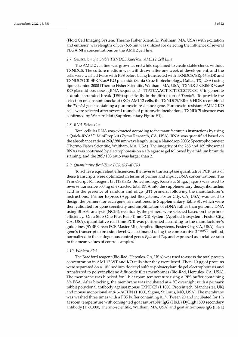
To examine the synthesis and physicochemical characterization of the nanoparticlesprepared using the single emulsion approach, the squalene encapsulation ability of PLGAwas characterized by HPLC (Table 1). The maximum squalene loading in PLGA-basednanoparticles was found when using 50 µL initial squalene (2.05 M, ≥98%, liquid) volumethat reached 8122 ± 735 µM of squalene loading. This represented 77.8 ± 5.1% encap-sulation efficiency, and the highest ratio of squalene referred to PLGA of the ones tested.The encapsulation efficiency reported in the literature for squalene loaded nanoparticleswas variable depending on the nanoparticle type and the synthetic method ranging from26 to 82% [45–47]. These nanoparticles were selected to carry out the experimental work.Nanoparticles were characterized by scanning electron microscopy (SEM) to determinetheir morphology and size (Figure 1). PLGA NPs devoid of squalene were spherical inshape, in contrast to NPs loaded with squalene that showed a larger size an oval shape(Figure 1). Transmission electron microscopy (TEM) analysis confirmed a general trend to-ward size increase when squalene was loaded in PLGA (Figure 1). Particle size histogramsdetermined from TEM images revealed that the mean size of PLGA nanoparticles increased
Antioxidants 2022, 11, 581 7 of 22
from 90.5 ± 13.3 to 167.9 ± 31.3 when squalene was encapsulated (Figure 1). Then, itseems that squalene affects both the micelle shape and size during the emulsification pro-cess. This observation is in agreement with previous results where the presence of lipidssuch as cholesterol can increase the PLGA size [48]. The hydrodynamic diameter of thenanoparticles in water at neutral pH, estimated by dynamic light scattering (DLS), was259 ± 107 nm for squalene loaded PLGA NPs and their dispersion was higher than that ofempty NPs (Table 2). The analysis on the particle size between TEM and DLS demonstratedthe same trend, confirming the size enlargement when squalene is encapsulated. Thenanoparticle mean diameters from TEM image-based measurements were smaller than theones obtained from hydrodynamic size measurement. However, these differences can beexplained by the technique differences—in TEM, the nanoparticles are dried, and the sizeshould be smaller after shrinking [49–52]. The presence of squalene also had a clear impacton the zeta potential values. In this sense, the negative values (−46.8 ± 0.6 mV) of PLGAnanoparticles without squalene significantly decreased in PLGA-based squalene nanoparti-cles (−36.2 ± 0.3 mV) at neutral pH. The negative charge of PLGA NPs without squalenecould be attributed to the presence of ionized carboxyl groups in the acid terminated PLGA(PLGA-COOH), whereas the less negative charge of PLGA-based squalene nanoparticlesmight be due to the presence of squalene on the surface of the nanoparticles, as describedpreviously for other nanoparticles [53].
Antioxidants 2022, 11, x FOR PEER REVIEW 8 of 23
Figure 1. Electron microscopy analysis, SEM, TEM and particle size histograms of PLGA nanopar-ticles with and without squalene.
Table 2. Physicochemical characterization of PLGA nanoparticles.
Polymer Diameter (nm) Dispersion Zeta Potential (mV) PLGA NPs with squalene 259 ± 107 0.173 −36.2 ± 0.3
PLGA NPs without squalene 129 ± 37 0.083 −46.8 ± 0.6
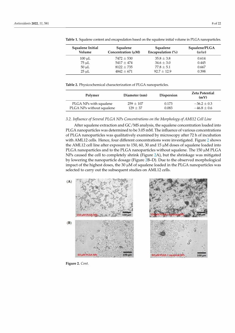
3.2. Influence of Several PLGA NPs Concentrations on the Morphology of AMl12 Cell Line After squalene extraction and GC/MS analysis, the squalene concentration loaded
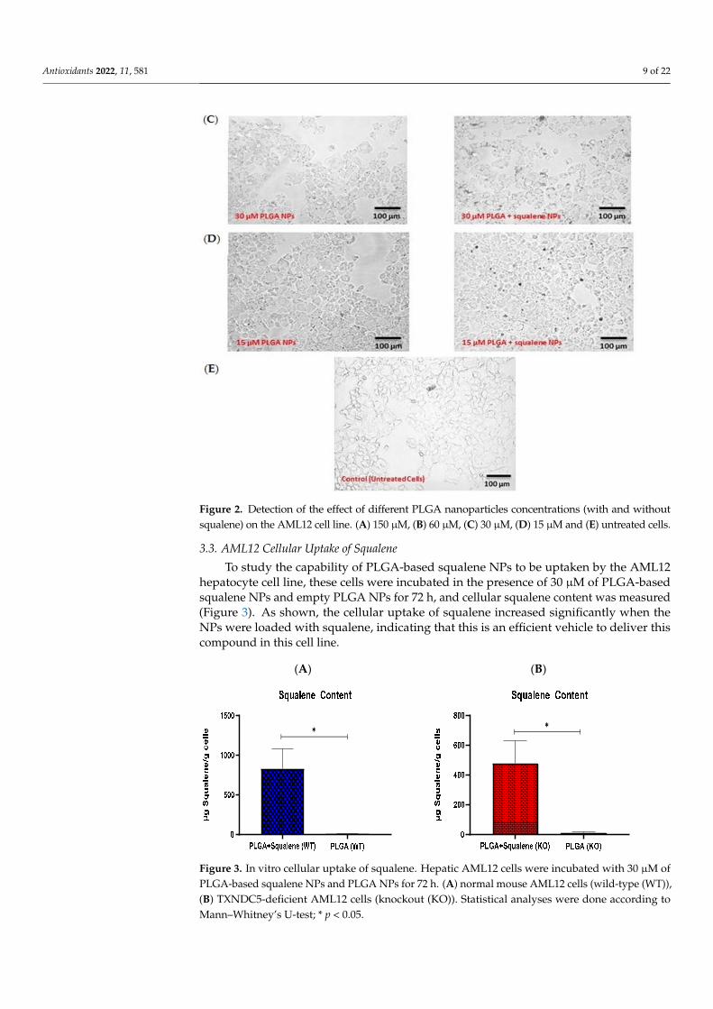
into PLGA nanoparticles was determined to be 3.05 mM. The influence of various concen-trations of PLGA nanoparticles was qualitatively examined by microscopy after 72 h of incubation with AML12 cells. Hence, four different concentrations were investigated. Fig-ure 2 shows the AML12 cell line after exposure to 150, 60, 30 and 15 µM doses of squalene loaded into PLGA nanoparticles and to the PLGA nanoparticles without squalene. The 150 µM PLGA NPs caused the cell to completely shrink (Figure 2A), but the shrinkage
Figure 1. Electron microscopy analysis, SEM, TEM and particle size histograms of PLGA nanoparti-cles with and without squalene.
Antioxidants 2022, 11, 581 8 of 22
Table 1. Squalene content and encapsulation based on the squalene initial volume in PLGA nanoparticles.
Squalene InitialVolume
SqualeneConcentration (µM)
SqualeneEncapsulation (%)
Squalene/PLGA(w/w)
100 µL 7472 ± 530 35.8 ± 3.8 0.61475 µL 5417 ± 474 34.6 ± 3.0 0.44550 µL 8122 ± 735 77.8 ± 5.1 0.66725 µL 4842 ± 671 92.7 ± 12.9 0.398
Table 2. Physicochemical characterization of PLGA nanoparticles.
Polymer Diameter (nm) Dispersion Zeta Potential(mV)
PLGA NPs with squalene 259 ± 107 0.173 −36.2 ± 0.3PLGA NPs without squalene 129 ± 37 0.083 −46.8 ± 0.6
3.2. Influence of Several PLGA NPs Concentrations on the Morphology of AMl12 Cell Line
After squalene extraction and GC/MS analysis, the squalene concentration loaded intoPLGA nanoparticles was determined to be 3.05 mM. The influence of various concentrationsof PLGA nanoparticles was qualitatively examined by microscopy after 72 h of incubationwith AML12 cells. Hence, four different concentrations were investigated. Figure 2 showsthe AML12 cell line after exposure to 150, 60, 30 and 15 µM doses of squalene loaded intoPLGA nanoparticles and to the PLGA nanoparticles without squalene. The 150 µM PLGANPs caused the cell to completely shrink (Figure 2A), but the shrinkage was mitigatedby lowering the nanoparticle dosage (Figure 2B–D). Due to the observed morphologicalimpact of the highest doses, the 30 µM of squalene loaded in the PLGA nanoparticles wasselected to carry out the subsequent studies on AML12 cells.
Antioxidants 2022, 11, x FOR PEER REVIEW 9 of 23
was mitigated by lowering the nanoparticle dosage (Figure 2B–D). Due to the observed morphological impact of the highest doses, the 30 µM of squalene loaded in the PLGA nanoparticles was selected to carry out the subsequent studies on AML12 cells.
Figure 2. Detection of the effect of different PLGA nanoparticles concentrations (with and without squalene) on the AML12 cell line. (A) 150 µM, (B) 60 µM, (C) 30 µM, (D) 15 µM and (E) untreated cells.
Figure 2. Cont.
Antioxidants 2022, 11, 581 9 of 22
Antioxidants 2022, 11, x FOR PEER REVIEW 9 of 23
was mitigated by lowering the nanoparticle dosage (Figure 2B–D). Due to the observed morphological impact of the highest doses, the 30 µM of squalene loaded in the PLGA nanoparticles was selected to carry out the subsequent studies on AML12 cells.
Figure 2. Detection of the effect of different PLGA nanoparticles concentrations (with and without squalene) on the AML12 cell line. (A) 150 µM, (B) 60 µM, (C) 30 µM, (D) 15 µM and (E) untreated cells.
Figure 2. Detection of the effect of different PLGA nanoparticles concentrations (with and withoutsqualene) on the AML12 cell line. (A) 150 µM, (B) 60 µM, (C) 30 µM, (D) 15 µM and (E) untreated cells.
3.3. AML12 Cellular Uptake of Squalene
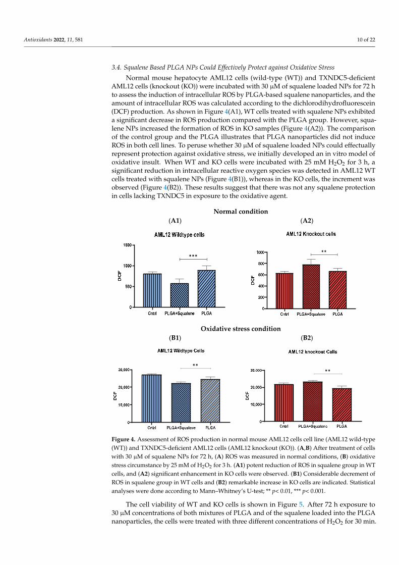
To study the capability of PLGA-based squalene NPs to be uptaken by the AML12hepatocyte cell line, these cells were incubated in the presence of 30 µM of PLGA-basedsqualene NPs and empty PLGA NPs for 72 h, and cellular squalene content was measured(Figure 3). As shown, the cellular uptake of squalene increased significantly when theNPs were loaded with squalene, indicating that this is an efficient vehicle to deliver thiscompound in this cell line.
Antioxidants 2022, 11, x FOR PEER REVIEW 10 of 23
3.3. AML12 Cellular Uptake of Squalene To study the capability of PLGA-based squalene NPs to be uptaken by the AML12
hepatocyte cell line, these cells were incubated in the presence of 30 µM of PLGA-based squalene NPs and empty PLGA NPs for 72 h, and cellular squalene content was measured (Figure 3). As shown, the cellular uptake of squalene increased significantly when the NPs were loaded with squalene, indicating that this is an efficient vehicle to deliver this com-pound in this cell line.
(A) (B)
✱
✱
Figure 3. In vitro cellular uptake of squalene. Hepatic AML12 cells were incubated with 30 µM of PLGA-based squalene NPs and PLGA NPs for 72 h. (A) normal mouse AML12 cells (wild-type (WT)), (B) TXNDC5-deficient AML12 cells (knockout (KO)). Statistical analyses were done accord-ing to Mann–Whitney’s U-test; * p < 0.05.
3.4. Squalene Based PLGA NPs Could Effectively Protect against Oxidative Stress Normal mouse hepatocyte AML12 cells (wild-type (WT)) and TXNDC5-deficient
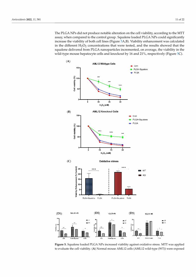
AML12 cells (knockout (KO)) were incubated with 30 µM of squalene loaded NPs for 72 h to assess the induction of intracellular ROS by PLGA-based squalene nanoparticles, and the amount of intracellular ROS was calculated according to the dichlorodihydrofluores-cein (DCF) production. As shown in Figure 4(A1), WT cells treated with squalene NPs exhibited a significant decrease in ROS production compared with the PLGA group. How-ever, squalene NPs increased the formation of ROS in KO samples (Figure 4(A2)). The comparison of the control group and the PLGA illustrates that PLGA nanoparticles did not induce ROS in both cell lines. To peruse whether 30 µM of squalene loaded NPs could effectually represent protection against oxidative stress, we initially developed an in vitro model of oxidative insult. When WT and KO cells were incubated with 25 mM H2O2 for 3 h, a significant reduction in intracellular reactive oxygen species was detected in AML12 WT cells treated with squalene NPs (Figure 4(B1)), whereas in the KO cells, the increment was observed (Figure 4(B2)). These results suggest that there was not any squalene pro-tection in cells lacking TXNDC5 in exposure to the oxidative agent.
Figure 3. In vitro cellular uptake of squalene. Hepatic AML12 cells were incubated with 30 µM ofPLGA-based squalene NPs and PLGA NPs for 72 h. (A) normal mouse AML12 cells (wild-type (WT)),(B) TXNDC5-deficient AML12 cells (knockout (KO)). Statistical analyses were done according toMann–Whitney’s U-test; * p < 0.05.
Antioxidants 2022, 11, 581 10 of 22
3.4. Squalene Based PLGA NPs Could Effectively Protect against Oxidative Stress
Normal mouse hepatocyte AML12 cells (wild-type (WT)) and TXNDC5-deficientAML12 cells (knockout (KO)) were incubated with 30 µM of squalene loaded NPs for 72 hto assess the induction of intracellular ROS by PLGA-based squalene nanoparticles, and theamount of intracellular ROS was calculated according to the dichlorodihydrofluorescein(DCF) production. As shown in Figure 4(A1), WT cells treated with squalene NPs exhibiteda significant decrease in ROS production compared with the PLGA group. However, squa-lene NPs increased the formation of ROS in KO samples (Figure 4(A2)). The comparisonof the control group and the PLGA illustrates that PLGA nanoparticles did not induceROS in both cell lines. To peruse whether 30 µM of squalene loaded NPs could effectuallyrepresent protection against oxidative stress, we initially developed an in vitro model ofoxidative insult. When WT and KO cells were incubated with 25 mM H2O2 for 3 h, asignificant reduction in intracellular reactive oxygen species was detected in AML12 WTcells treated with squalene NPs (Figure 4(B1)), whereas in the KO cells, the increment wasobserved (Figure 4(B2)). These results suggest that there was not any squalene protectionin cells lacking TXNDC5 in exposure to the oxidative agent.
Antioxidants 2022, 11, x FOR PEER REVIEW 11 of 23
Normal condition (A1) (A2)
✱✱✱
✱✱
Oxidative stress condition
(B1) (B2)
✱✱
✱✱
Figure 4. Assessment of ROS production in normal mouse AML12 cells cell line (AML12 wild-type (WT)) and TXNDC5-deficient AML12 cells (AML12 knockout (KO)). (A,B) After treatment of cells with 30 µM of squalene NPs for 72 h, (A) ROS was measured in normal conditions, (B) oxidative stress circumstance by 25 mM of H2O2 for 3 h. (A1) potent reduction of ROS in squalene group in WT cells, and (A2) significant enhancement in KO cells were observed. (B1) Considerable decrement of ROS in squalene group in WT cells and (B2) remarkable increase in KO cells are indicated. Statis-tical analyses were done according to Mann–Whitney’s U-test; ** p< 0.01, *** p< 0.001.
The cell viability of WT and KO cells is shown in Figure 5. After 72 h exposure to 30 µM concentrations of both mixtures of PLGA and of the squalene loaded into the PLGA nanoparticles, the cells were treated with three different concentrations of H2O2 for 30 min. The PLGA NPs did not produce notable alteration on the cell viability, according to the MTT assay, when compared to the control group. Squalene loaded PLGA NPs could sig-nificantly increase the viability of both cell lines (Figure 5A,B). Viability enhancement was calculated in the different H2O2 concentrations that were tested, and the results showed that the squalene delivered from PLGA nanoparticles incremented, on average, the via-bility in the wild-type mouse hepatocyte cells and knockout by 16 and 21%, respectively (Figure 5C).
Figure 4. Assessment of ROS production in normal mouse AML12 cells cell line (AML12 wild-type(WT)) and TXNDC5-deficient AML12 cells (AML12 knockout (KO)). (A,B) After treatment of cellswith 30 µM of squalene NPs for 72 h, (A) ROS was measured in normal conditions, (B) oxidativestress circumstance by 25 mM of H2O2 for 3 h. (A1) potent reduction of ROS in squalene group in WTcells, and (A2) significant enhancement in KO cells were observed. (B1) Considerable decrement ofROS in squalene group in WT cells and (B2) remarkable increase in KO cells are indicated. Statisticalanalyses were done according to Mann–Whitney’s U-test; ** p< 0.01, *** p< 0.001.
The cell viability of WT and KO cells is shown in Figure 5. After 72 h exposure to30 µM concentrations of both mixtures of PLGA and of the squalene loaded into the PLGAnanoparticles, the cells were treated with three different concentrations of H2O2 for 30 min.
Antioxidants 2022, 11, 581 11 of 22
The PLGA NPs did not produce notable alteration on the cell viability, according to the MTTassay, when compared to the control group. Squalene loaded PLGA NPs could significantlyincrease the viability of both cell lines (Figure 5A,B). Viability enhancement was calculatedin the different H2O2 concentrations that were tested, and the results showed that thesqualene delivered from PLGA nanoparticles incremented, on average, the viability in thewild-type mouse hepatocyte cells and knockout by 16 and 21%, respectively (Figure 5C).
Antioxidants 2022, 11, x FOR PEER REVIEW 12 of 23
Figure 5. Squalene loaded PLGA NPs increased viability against oxidative stress. MTT was ap-plied to evaluate the cell viability. (A) Normal mouse AML12 cells (AML12 wild-type (WT)) were exposed to 30 µM PLGA based squalene NPs for 72 h had a significant increase in viability in pres-ence of 20, 25 and 30 mM of H2O2, and (B) TXNDC5-deficient mouse hepatocyte cells (AML12 knockout (KO)) displayed a similar increment in viability. (C) Statistically, a significant difference of 16% and 21% was observed on average in viability enhancement of WT and KO cell lines, re-spectively, related to respective control in 20, 25 and 30 mM of H2O2. (D) TXNDC5 deletion can drastically lower the viability of the mouse hepatocyte in the absence of squalene at all concentra-tions tested; however, when the cells were treated with squalene, the viability of both cell lines
Figure 5. Squalene loaded PLGA NPs increased viability against oxidative stress. MTT was appliedto evaluate the cell viability. (A) Normal mouse AML12 cells (AML12 wild-type (WT)) were exposed
Antioxidants 2022, 11, 581 12 of 22
to 30 µM PLGA based squalene NPs for 72 h had a significant increase in viability in presence of20, 25 and 30 mM of H2O2, and (B) TXNDC5-deficient mouse hepatocyte cells (AML12 knockout(KO)) displayed a similar increment in viability. (C) Statistically, a significant difference of 16% and21% was observed on average in viability enhancement of WT and KO cell lines, respectively, relatedto respective control in 20, 25 and 30 mM of H2O2. (D) TXNDC5 deletion can drastically lower theviability of the mouse hepatocyte in the absence of squalene at all concentrations tested; however,when the cells were treated with squalene, the viability of both cell lines increased. Although (D3)there were no significant differences between WT and KO cells at 30 mM H2O2, (D1,D2) a statisticaldifference was seen in samples treated with squalene loaded in PLGA nanoparticles in presenceof 20 and 25 mM H2O2. Statistical analysis was carried out according to two-way ANOVA andMann–Whitney’s U-test for pairwise comparisons; * p < 0.05, ** p < 0.01, *** p < 0.001, **** p < 0.0001.
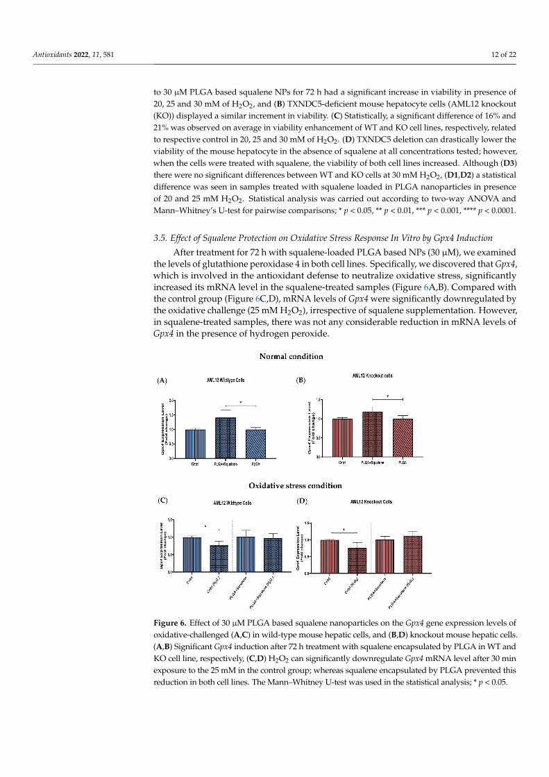
3.5. Effect of Squalene Protection on Oxidative Stress Response In Vitro by Gpx4 Induction
After treatment for 72 h with squalene-loaded PLGA based NPs (30 µM), we examinedthe levels of glutathione peroxidase 4 in both cell lines. Specifically, we discovered that Gpx4,which is involved in the antioxidant defense to neutralize oxidative stress, significantlyincreased its mRNA level in the squalene-treated samples (Figure 6A,B). Compared withthe control group (Figure 6C,D), mRNA levels of Gpx4 were significantly downregulated bythe oxidative challenge (25 mM H2O2), irrespective of squalene supplementation. However,in squalene-treated samples, there was not any considerable reduction in mRNA levels ofGpx4 in the presence of hydrogen peroxide.
Antioxidants 2022, 11, x FOR PEER REVIEW 13 of 23
increased. Although (D3) there were no significant differences between WT and KO cells at 30 mM H2O2, (D1,D2) a statistical difference was seen in samples treated with squalene loaded in PLGA nanoparticles in presence of 20 and 25 mM H2O2. Statistical analysis was carried out according to two-way ANOVA and Mann–Whitney’s U-test for pairwise comparisons; * p < 0.05, ** p < 0.01, *** p < 0.001, **** p < 0.0001.
3.5. Effect of Squalene Protection on Oxidative Stress Response In Vitro by Gpx4 Induction After treatment for 72 h with squalene-loaded PLGA based NPs (30 µM), we exam-
ined the levels of glutathione peroxidase 4 in both cell lines. Specifically, we discovered that Gpx4, which is involved in the antioxidant defense to neutralize oxidative stress, sig-nificantly increased its mRNA level in the squalene-treated samples (Figure 6A,B). Com-pared with the control group (Figure 6C,D), mRNA levels of Gpx4 were significantly downregulated by the oxidative challenge (25 mM H2O2), irrespective of squalene supple-mentation. However, in squalene-treated samples, there was not any considerable reduc-tion in mRNA levels of Gpx4 in the presence of hydrogen peroxide.
Figure 6. Effect of 30 µM PLGA based squalene nanoparticles on the Gpx4 gene expression levels of oxidative-challenged (A,C) in wild-type mouse hepatic cells, and (B,D) knockout mouse hepatic cells. (A,B) Significant Gpx4 induction after 72 h treatment with squalene encapsulated by PLGA in WT and KO cell line, respectively, (C,D) H2O2 can significantly downregulate Gpx4 mRNA level after 30 min exposure to the 25 mM in the control group; whereas squalene encapsulated by PLGA prevented this reduction in both cell lines. The Mann–Whitney U-test was used in the statistical analysis; * p < 0.05.
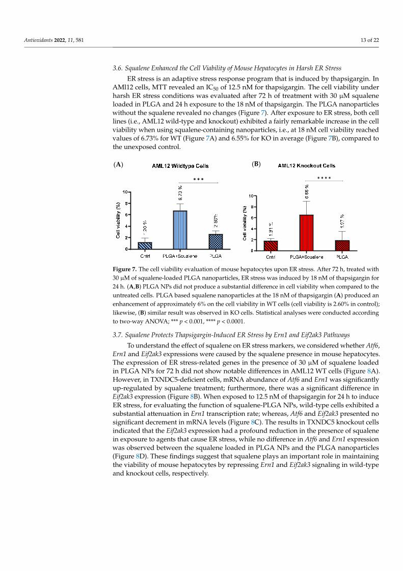
3.6. Squalene Enhanced the Cell Viability of Mouse Hepatocytes in Harsh ER Stress ER stress is an adaptive stress response program that is induced by thapsigargin. In
AMl12 cells, MTT revealed an IC50 of 12.5 nM for thapsigargin. The cell viability under harsh ER stress conditions was evaluated after 72 h of treatment with 30 µM squalene loaded in PLGA and 24 h exposure to the 18 nM of thapsigargin. The PLGA nanoparticles without the squalene revealed no changes (Figure 7). After exposure to ER stress, both cell
Figure 6. Effect of 30 µM PLGA based squalene nanoparticles on the Gpx4 gene expression levels ofoxidative-challenged (A,C) in wild-type mouse hepatic cells, and (B,D) knockout mouse hepatic cells.(A,B) Significant Gpx4 induction after 72 h treatment with squalene encapsulated by PLGA in WT andKO cell line, respectively, (C,D) H2O2 can significantly downregulate Gpx4 mRNA level after 30 minexposure to the 25 mM in the control group; whereas squalene encapsulated by PLGA prevented thisreduction in both cell lines. The Mann–Whitney U-test was used in the statistical analysis; * p < 0.05.
Antioxidants 2022, 11, 581 13 of 22
3.6. Squalene Enhanced the Cell Viability of Mouse Hepatocytes in Harsh ER Stress
ER stress is an adaptive stress response program that is induced by thapsigargin. InAMl12 cells, MTT revealed an IC50 of 12.5 nM for thapsigargin. The cell viability underharsh ER stress conditions was evaluated after 72 h of treatment with 30 µM squaleneloaded in PLGA and 24 h exposure to the 18 nM of thapsigargin. The PLGA nanoparticleswithout the squalene revealed no changes (Figure 7). After exposure to ER stress, both celllines (i.e., AML12 wild-type and knockout) exhibited a fairly remarkable increase in the cellviability when using squalene-containing nanoparticles, i.e., at 18 nM cell viability reachedvalues of 6.73% for WT (Figure 7A) and 6.55% for KO in average (Figure 7B), compared tothe unexposed control.
Antioxidants 2022, 11, x FOR PEER REVIEW 14 of 23
lines (i.e., AML12 wild-type and knockout) exhibited a fairly remarkable increase in the cell viability when using squalene-containing nanoparticles, i.e., at 18 nM cell viability reached values of 6.73% for WT (Figure 7A) and 6.55% for KO in average (Figure 7B), compared to the unexposed control.
✱ ✱ ✱ ✱ ✱ ✱ ✱
Figure 7. The cell viability evaluation of mouse hepatocytes upon ER stress. After 72 h, treated with 30 µM of squalene-loaded PLGA nanoparticles, ER stress was induced by 18 nM of thapsigargin for 24 h. (A,B) PLGA NPs did not produce a substantial difference in cell viability when compared to the untreated cells. PLGA based squalene nanoparticles at the 18 nM of thapsigargin (A) produced an enhancement of approximately 6% on the cell viability in WT cells (cell viability is 2.60% in con-trol); likewise, (B) similar result was observed in KO cells. Statistical analyses were conducted ac-cording to two-way ANOVA; *** p < 0.001, **** p < 0.0001.
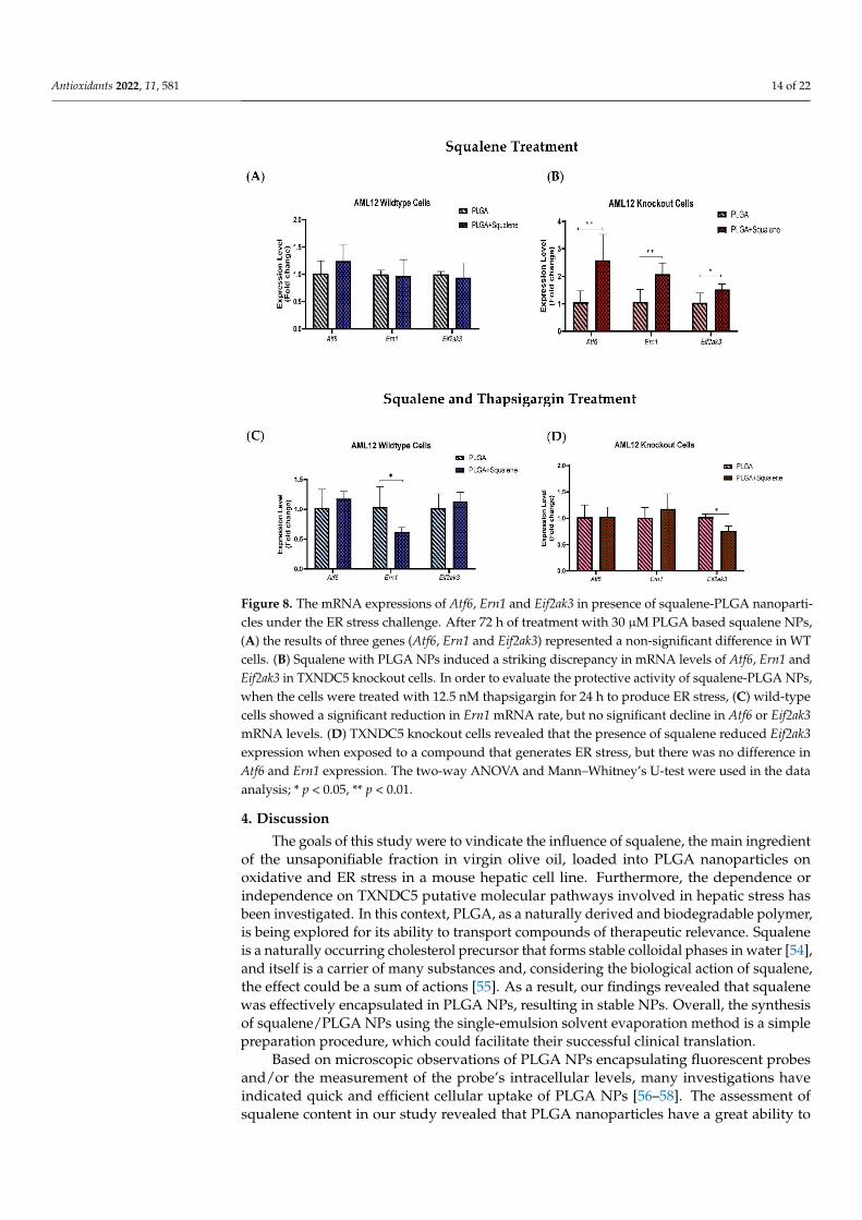
3.7. Squalene Protects Thapsigargin-Induced ER Stress by Ern1 and Eif2ak3 Pathways To understand the effect of squalene on ER stress markers, we considered whether
Atf6, Ern1 and Eif2ak3 expressions were caused by the squalene presence in mouse hepato-cytes. The expression of ER stress-related genes in the presence of 30 µM of squalene loaded in PLGA NPs for 72 h did not show notable differences in AML12 WT cells (Figure 8A). However, in TXNDC5-deficient cells, mRNA abundance of Atf6 and Ern1 was signif-icantly up-regulated by squalene treatment; furthermore, there was a significant differ-ence in Eif2ak3 expression (Figure 8B). When exposed to 12.5 nM of thapsigargin for 24 h to induce ER stress, for evaluating the function of squalene-PLGA NPs, wild-type cells exhibited a substantial attenuation in Ern1 transcription rate; whereas, Atf6 and Eif2ak3 presented no significant decrement in mRNA levels (Figure 8C). The results in TXNDC5 knockout cells indicated that the Eif2ak3 expression had a profound reduction in the pres-ence of squalene in exposure to agents that cause ER stress, while no difference in Atf6 and Ern1 expression was observed between the squalene loaded in PLGA NPs and the PLGA nanoparticles (Figure 8D). These findings suggest that squalene plays an important role in maintaining the viability of mouse hepatocytes by repressing Ern1 and Eif2ak3 sig-naling in wild-type and knockout cells, respectively.
(A) (B)
Figure 7. The cell viability evaluation of mouse hepatocytes upon ER stress. After 72 h, treated with30 µM of squalene-loaded PLGA nanoparticles, ER stress was induced by 18 nM of thapsigargin for24 h. (A,B) PLGA NPs did not produce a substantial difference in cell viability when compared to theuntreated cells. PLGA based squalene nanoparticles at the 18 nM of thapsigargin (A) produced anenhancement of approximately 6% on the cell viability in WT cells (cell viability is 2.60% in control);likewise, (B) similar result was observed in KO cells. Statistical analyses were conducted accordingto two-way ANOVA; *** p < 0.001, **** p < 0.0001.
3.7. Squalene Protects Thapsigargin-Induced ER Stress by Ern1 and Eif2ak3 Pathways
To understand the effect of squalene on ER stress markers, we considered whether Atf6,Ern1 and Eif2ak3 expressions were caused by the squalene presence in mouse hepatocytes.The expression of ER stress-related genes in the presence of 30 µM of squalene loadedin PLGA NPs for 72 h did not show notable differences in AML12 WT cells (Figure 8A).However, in TXNDC5-deficient cells, mRNA abundance of Atf6 and Ern1 was significantlyup-regulated by squalene treatment; furthermore, there was a significant difference inEif2ak3 expression (Figure 8B). When exposed to 12.5 nM of thapsigargin for 24 h to induceER stress, for evaluating the function of squalene-PLGA NPs, wild-type cells exhibited asubstantial attenuation in Ern1 transcription rate; whereas, Atf6 and Eif2ak3 presented nosignificant decrement in mRNA levels (Figure 8C). The results in TXNDC5 knockout cellsindicated that the Eif2ak3 expression had a profound reduction in the presence of squalenein exposure to agents that cause ER stress, while no difference in Atf6 and Ern1 expressionwas observed between the squalene loaded in PLGA NPs and the PLGA nanoparticles(Figure 8D). These findings suggest that squalene plays an important role in maintainingthe viability of mouse hepatocytes by repressing Ern1 and Eif2ak3 signaling in wild-typeand knockout cells, respectively.
Antioxidants 2022, 11, 581 14 of 22Antioxidants 2022, 11, x FOR PEER REVIEW 15 of 23
Figure 8. The mRNA expressions of Atf6, Ern1 and Eif2ak3 in presence of squalene-PLGA nanopar-ticles under the ER stress challenge. After 72 h of treatment with 30 µM PLGA based squalene NPs, (A) the results of three genes (Atf6, Ern1 and Eif2ak3) represented a non-significant difference in WT cells. (B) Squalene with PLGA NPs induced a striking discrepancy in mRNA levels of Atf6, Ern1 and Eif2ak3 in TXNDC5 knockout cells. In order to evaluate the protective activity of squalene-PLGA NPs, when the cells were treated with 12.5 nM thapsigargin for 24 h to produce ER stress, (C) wild-type cells showed a significant reduction in Ern1 mRNA rate, but no significant decline in Atf6 or Eif2ak3 mRNA levels. (D) TXNDC5 knockout cells revealed that the presence of squalene reduced Eif2ak3 expression when exposed to a compound that generates ER stress, but there was no differ-ence in Atf6 and Ern1 expression. The two-way ANOVA and Mann–Whitney’s U-test were used in the data analysis; * p < 0.05, ** p < 0.01.
4. Discussion The goals of this study were to vindicate the influence of squalene, the main ingredi-
ent of the unsaponifiable fraction in virgin olive oil, loaded into PLGA nanoparticles on oxidative and ER stress in a mouse hepatic cell line. Furthermore, the dependence or in-dependence on TXNDC5 putative molecular pathways involved in hepatic stress has been investigated. In this context, PLGA, as a naturally derived and biodegradable polymer, is being explored for its ability to transport compounds of therapeutic relevance. Squalene is a naturally occurring cholesterol precursor that forms stable colloidal phases in water [54], and itself is a carrier of many substances and, considering the biological action of squalene, the effect could be a sum of actions [55]. As a result, our findings revealed that squalene was effectively encapsulated in PLGA NPs, resulting in stable NPs. Overall, the synthesis of squalene/PLGA NPs using the single-emulsion solvent evaporation method is a simple preparation procedure, which could facilitate their successful clinical transla-tion.
Based on microscopic observations of PLGA NPs encapsulating fluorescent probes and/or the measurement of the probe’s intracellular levels, many investigations have in-dicated quick and efficient cellular uptake of PLGA NPs [56–58]. The assessment of squa-lene content in our study revealed that PLGA nanoparticles have a great ability to transfer
Figure 8. The mRNA expressions of Atf6, Ern1 and Eif2ak3 in presence of squalene-PLGA nanoparti-cles under the ER stress challenge. After 72 h of treatment with 30 µM PLGA based squalene NPs,(A) the results of three genes (Atf6, Ern1 and Eif2ak3) represented a non-significant difference in WTcells. (B) Squalene with PLGA NPs induced a striking discrepancy in mRNA levels of Atf6, Ern1 andEif2ak3 in TXNDC5 knockout cells. In order to evaluate the protective activity of squalene-PLGA NPs,when the cells were treated with 12.5 nM thapsigargin for 24 h to produce ER stress, (C) wild-typecells showed a significant reduction in Ern1 mRNA rate, but no significant decline in Atf6 or Eif2ak3mRNA levels. (D) TXNDC5 knockout cells revealed that the presence of squalene reduced Eif2ak3expression when exposed to a compound that generates ER stress, but there was no difference inAtf6 and Ern1 expression. The two-way ANOVA and Mann–Whitney’s U-test were used in the dataanalysis; * p < 0.05, ** p < 0.01.
4. Discussion
The goals of this study were to vindicate the influence of squalene, the main ingredientof the unsaponifiable fraction in virgin olive oil, loaded into PLGA nanoparticles onoxidative and ER stress in a mouse hepatic cell line. Furthermore, the dependence orindependence on TXNDC5 putative molecular pathways involved in hepatic stress hasbeen investigated. In this context, PLGA, as a naturally derived and biodegradable polymer,is being explored for its ability to transport compounds of therapeutic relevance. Squaleneis a naturally occurring cholesterol precursor that forms stable colloidal phases in water [54],and itself is a carrier of many substances and, considering the biological action of squalene,the effect could be a sum of actions [55]. As a result, our findings revealed that squalenewas effectively encapsulated in PLGA NPs, resulting in stable NPs. Overall, the synthesisof squalene/PLGA NPs using the single-emulsion solvent evaporation method is a simplepreparation procedure, which could facilitate their successful clinical translation.
Based on microscopic observations of PLGA NPs encapsulating fluorescent probesand/or the measurement of the probe’s intracellular levels, many investigations haveindicated quick and efficient cellular uptake of PLGA NPs [56–58]. The assessment ofsqualene content in our study revealed that PLGA nanoparticles have a great ability to
Antioxidants 2022, 11, 581 15 of 22
transfer squalene as a nanocarrier in mouse hepatocytes (Figure 3). As described in differentstudies, PLGA nanoparticles are effective nanocarriers for the encapsulation and delivery ofvarious anti-cancer agents such as oleanolic (OA) and ursolic (UA) acids in three differentcell lines, i.e., HepG2 (human hepatoma cell line), Caco-2 (human epithelial colorectaladenocarcinoma cell line) and Y-79 (human retinoblastoma cell line) [16]; also, for theencapsulation of several pharmaceuticals including haloperidol, estradiol, etc. [20]. Forinstance, the PLGA-based curcumin NPs have displayed entrapment efficiency in the rangeof 77 to 85% [59].
Intracellular ROS production is necessary for regular cellular activities and phys-iological processes, but when its production exceeds the intrinsic antioxidant capacity,oxidative stress occurs, causing severe damage to cellular macromolecules [60]. Squalene’santioxidant properties are intimately linked to the unique and stable triterpene structurethat allows it to effectively scavenge harmful free radicals [61]. The protective effects ofsqualene versus oxidative destruction have been formerly documented in rodents [6,62–65].Squalene can protect murine macrophages [32], Chinese hamster pulmonary fibroblasts(V79 cells) [66], human monocytes [32] and mammary epithelial cells (MCF10A) fromhydrogen peroxide-induced damage in cell culture assays by directly scavenging ROS in adose-dependent manner [34] and our finding in the present study reveal that squalene canreduce the ROS induction in mouse hepatic cells. Next, we investigated whether TXNDC5deletion contributes to squalene protection against oxidative stress. ROS influences ERhomeostasis and protein folding directly or indirectly, causing ER stress and possibly celldeath in the case of extreme ER stress [67,68]. TXNDC5 appears to be involved in theformation of ROS and ER stress, according to growing data [69,70]. Inhibiting TXNDC5expression via knockdown has previously been shown to induce ROS and ER stress inpancreatic cancer cells [69]; however, increasing TXNDC5 expression in lipid endothelialcells effectively reduces ROS production and protects cells [70]. The present study demon-strates that, in AML12 cells, as reflected in Figure 4A, an inverse and statistically significantdifference was found between ROS content and TXNDC5 in exposure to squalene so that,in absence of TXNDC5, ROS was increased, while the ROS was decreased in WT samples.When cells were challenged to an oxidative stimulus (Figure 4B), a significant decreaseof ROS in the squalene treated group in WT cells was observed, being the opposite inTXNDC5-KO cells. This finding reinforces previous research that found that oxidativestress-induced TXNDC5 was involved in proper protein folding via its disulfide isomeraseactivity [71]. Overall, these results evidence that squalene can alter ROS production inoxidative stress, in dependence of TXNDC5 (Figure 9). In the evaluation of the viability ofcells in different concentrations of H2O2 based on the TXNDC5 elimination, in all concen-trations that we tested, in the absence of squalene, TXNDC5 deletion significantly reducedthe viability of mouse hepatocytes (Figure 5D). However, when the cells were treated withsqualene, the viability of both cell lines was increased using 20 and 25 mM concentrationsof H2O2 (Figure 5(D1,D2)). With these results in consideration, TXNDC5 can increase thesqualene efficiency in AML12 cell viability and act as an oxidative stress-induced survivalfactor that regulates ROS/ER stress signaling, allowing AML12 cells to remain viable underoxidative stress, but it is not the only factor, and squalene could bypass it.
Antioxidants 2022, 11, 581 16 of 22
Antioxidants 2022, 11, x FOR PEER REVIEW 17 of 23
and urine bladder and normalize the alteration of Gpx4 in the heart and hemolysate of red blood cells [62,63]. Our results also evidence the induction of Gpx4 in the presence of squa-lene in WT and TXNDC5-deficient cells. When both kinds of cells were exposed to H2O2, squalene incubation rescued the decreased Gpx4 expression with independence of TXNDC5 (Figure 6). This mechanism could partly explain that TXNDC5-deficient mouse hepatocyte cells could survive against H2O2-induced oxidative stress by the therapeutic action of squalene (Figure 9).
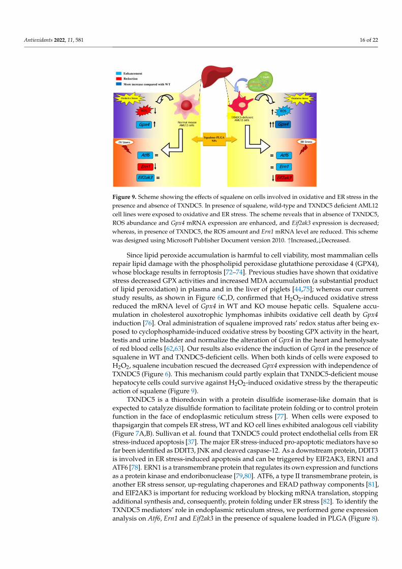
Figure 9. Scheme showing the effects of squalene on cells involved in oxidative and ER stress in the presence and absence of TXNDC5. In presence of squalene, wild-type and TXNDC5 deficient AML12 cell lines were exposed to oxidative and ER stress. The scheme reveals that in absence of TXNDC5, ROS abundance and Gpx4 mRNA expression are enhanced, and Eif2ak3 expression is de-creased; whereas, in presence of TXNDC5, the ROS amount and Ern1 mRNA level are reduced. This scheme was designed using Microsoft Publisher Document version 2010. ↑Increased,↓Decreased
TXNDC5 is a thioredoxin with a protein disulfide isomerase-like domain that is expected to catalyze disulfide formation to facilitate protein folding or to control protein function in the face of endoplasmic reticulum stress [77]. When cells were exposed to thapsigargin that compels ER stress, WT and KO cell lines exhibited analogous cell viability (Figure 7A,B). Sullivan et al. found that TXNDC5 could protect endothelial cells from ER stress-induced apoptosis [37]. The major ER stress-induced pro-apoptotic mediators have so far been identified as DDIT3, JNK and cleaved caspase-12. As a downstream protein, DDIT3 is involved in ER stress-induced apoptosis and can be triggered by EIF2AK3, ERN1 and ATF6 [78]. ERN1 is a transmembrane protein that regulates its own expression and functions as a protein kinase and endoribonuclease [79,80]. ATF6, a type II transmembrane protein, is another ER stress sensor, up-regulating chaperones and ERAD pathway components [81], and EIF2AK3 is important for reducing workload by blocking mRNA translation, stopping additional synthesis and, consequently, protein folding under ER stress [82]. To identify the TXNDC5 mediators’ role in endoplasmic reticulum stress, we performed gene expression analysis on Atf6, Ern1 and Eif2ak3 in the presence of squalene loaded in PLGA (Figure 8). In the context of ER stress-induced effects, Chawsheen et al. found that the knockdown of TXNDC5 in human lung cancer cells accelerates the unfolded proteins and induces ER stress that increases the expression of Eif2ak3, Ern1 and Atf6 [83].
Accordingly, our results demonstrated that the downregulation of TXNDC5 increased the mRNA level of ER stress markers in mouse hepatic cells (Supplementary Figure S2). In the presence of induced ER stress, squalene decreased Ern1 expression in WT cells in comparison to KO (Figure 8C). However, the opposite result was observed regarding Eif2ak3 expression that decreased in TXNDC5 knockout cells, and no significant change was seen in Atf6 (Figure 8C,D). Multiple experiments showed that knockout of
Figure 9. Scheme showing the effects of squalene on cells involved in oxidative and ER stress in thepresence and absence of TXNDC5. In presence of squalene, wild-type and TXNDC5 deficient AML12cell lines were exposed to oxidative and ER stress. The scheme reveals that in absence of TXNDC5,ROS abundance and Gpx4 mRNA expression are enhanced, and Eif2ak3 expression is decreased;whereas, in presence of TXNDC5, the ROS amount and Ern1 mRNA level are reduced. This schemewas designed using Microsoft Publisher Document version 2010. ↑Increased,↓Decreased.
Since lipid peroxide accumulation is harmful to cell viability, most mammalian cellsrepair lipid damage with the phospholipid peroxidase glutathione peroxidase 4 (GPX4),whose blockage results in ferroptosis [72–74]. Previous studies have shown that oxidativestress decreased GPX activities and increased MDA accumulation (a substantial productof lipid peroxidation) in plasma and in the liver of piglets [44,75]; whereas our currentstudy results, as shown in Figure 6C,D, confirmed that H2O2-induced oxidative stressreduced the mRNA level of Gpx4 in WT and KO mouse hepatic cells. Squalene accu-mulation in cholesterol auxotrophic lymphomas inhibits oxidative cell death by Gpx4induction [76]. Oral administration of squalene improved rats’ redox status after being ex-posed to cyclophosphamide-induced oxidative stress by boosting GPX activity in the heart,testis and urine bladder and normalize the alteration of Gpx4 in the heart and hemolysateof red blood cells [62,63]. Our results also evidence the induction of Gpx4 in the presence ofsqualene in WT and TXNDC5-deficient cells. When both kinds of cells were exposed toH2O2, squalene incubation rescued the decreased Gpx4 expression with independence ofTXNDC5 (Figure 6). This mechanism could partly explain that TXNDC5-deficient mousehepatocyte cells could survive against H2O2-induced oxidative stress by the therapeuticaction of squalene (Figure 9).
TXNDC5 is a thioredoxin with a protein disulfide isomerase-like domain that isexpected to catalyze disulfide formation to facilitate protein folding or to control proteinfunction in the face of endoplasmic reticulum stress [77]. When cells were exposed tothapsigargin that compels ER stress, WT and KO cell lines exhibited analogous cell viability(Figure 7A,B). Sullivan et al. found that TXNDC5 could protect endothelial cells from ERstress-induced apoptosis [37]. The major ER stress-induced pro-apoptotic mediators have sofar been identified as DDIT3, JNK and cleaved caspase-12. As a downstream protein, DDIT3is involved in ER stress-induced apoptosis and can be triggered by EIF2AK3, ERN1 andATF6 [78]. ERN1 is a transmembrane protein that regulates its own expression and functionsas a protein kinase and endoribonuclease [79,80]. ATF6, a type II transmembrane protein, isanother ER stress sensor, up-regulating chaperones and ERAD pathway components [81],and EIF2AK3 is important for reducing workload by blocking mRNA translation, stoppingadditional synthesis and, consequently, protein folding under ER stress [82]. To identify theTXNDC5 mediators’ role in endoplasmic reticulum stress, we performed gene expressionanalysis on Atf6, Ern1 and Eif2ak3 in the presence of squalene loaded in PLGA (Figure 8).
Antioxidants 2022, 11, 581 17 of 22
In the context of ER stress-induced effects, Chawsheen et al. found that the knockdownof TXNDC5 in human lung cancer cells accelerates the unfolded proteins and induces ERstress that increases the expression of Eif2ak3, Ern1 and Atf6 [83].
Accordingly, our results demonstrated that the downregulation of TXNDC5 increasedthe mRNA level of ER stress markers in mouse hepatic cells (Supplementary Figure S2).In the presence of induced ER stress, squalene decreased Ern1 expression in WT cellsin comparison to KO (Figure 8C). However, the opposite result was observed regardingEif2ak3 expression that decreased in TXNDC5 knockout cells, and no significant change wasseen in Atf6 (Figure 8C,D). Multiple experiments showed that knockout of ATF6 preventedthe upregulation of Txndc5 mRNA; TXNDC5 is located downstream of the ATF6 in cardiac,kidney fibroblasts and stelae cells [84–86]. Consistent with these results, we found thatTXNDC5 may be posited downstream of the ATF6 and upstream of EIF2AK3 and ERN1 inhepatic cells. Based on this hypothesis, we suggest that TXNDC5 may regulate ER activitythrough distinct signaling pathways in stressful circumstances; moreover, squalene couldreduce cell mortality by decreasing Ern1 or Eif2ak3 expression as ER stress markers inhepatic cells depending on TXNDC5.
5. Conclusions
The current study shows that squalene was successfully encapsulated in PLGA NPs,yielding stable nanoparticles with rapid and efficient cellular uptake. Squalene-basedPLGA NPs effectively reduced ROS levels in normal mouse hepatocytes, whereas ROSwas increased in TXNDC5-deficient AML12 cells. The cell viability of WT and KO cellsunder oxidative stress conditions was increased in the presence of squalene by Gpx4induction. Squalene also enhanced the cell viability of mouse hepatocytes in thapsigargin-induced ER stress by repressing Ern1 or Eif2ak3 expression in wild-type or knockoutcells, respectively. Thus, TXNDC5 represented a crucial role in regulating ER activitythrough different signaling pathways in stressful circumstances. Likewise, squalene-loadedPLGA-NPs protect mouse hepatocytes from oxidative and endoplasmic reticulum stress byvariable mechanisms depending on TXNDC5 presence. While this study was successful,there were some limitations in the PLGA encapsulation efficiency of squalene, which wasvariable in the amount of squalene in each batch of nanoparticle synthesized. Additionally,there was a limitation in PLGA-squalene NP cell treatment, so the cells underwent osmoticstress when exposed to high doses of PLGA-squalene NPs. Hence, high doses of squalenemay not be useful in these experiments.
Supplementary Materials: The following supporting information can be downloaded at: https://www.mdpi.com/article/10.3390/antiox11030581/s1, Figure S1: Characterization of AML12 celllines; Figure S2: The mRNA expressions of Atf6, Ern1 and Eif2ak3 in WT and KO cells; Table S1:Sequences of real-time PCR primers according to MIQE guidelines.
Author Contributions: Conceptualization, S.H.B., T.A., J.S.-M., R.M.-B., R.A., J.C.B., R.L., M.J.R.-Y.,M.A. and J.O.; methodology, S.H.B., T.A., V.S., J.S.-M., R.M.-B., R.A., J.C.B. and R.L.; software, S.H.B.,T.A., J.S.-M., J.C.B. and R.L.; validation, S.H.B., T.A., J.S.-M., J.C.B. and R.L.; formal analysis, S.H.B.,T.A., V.S., J.S.-M., R.M.-B., R.A., J.C.B. and R.L.; investigation, S.H.B., T.A., J.S.-M., R.M.-B., R.A., J.C.B.and R.L.; resources, M.J.R.-Y., M.A. and J.O.; data curation, S.H.B., R.A. and J.O.; writing—originaldraft preparation, S.H.B.; writing—review and editing, S.H.B., T.A., V.S., J.S.-M., R.M.-B., R.A., J.C.B.,R.L., M.J.R.-Y., M.A. and J.O.; visualization, S.H.B.; supervision, T.A., R.M.-B., M.J.R.-Y., M.A. andJ.O.; project administration, M.J.R.-Y., M.A. and J.O.; funding acquisition, M.J.R.-Y., M.A. and J.O. Allauthors have read and agreed to the published version of the manuscript.
Funding: This research was supported by grants (CIBEROBN, CB06/03/1012, 1 January 2008)from CIBER Fisiopatología de la Obesidad y Nutrición as initiative of FEDER-ISCIII, Ministerio deCiencia e Innovación-Fondo Europeo de Desarrollo Regional (PID2019-104915RB-I00, 1 June 2020)and Fondo Social Europeo-Gobierno de Aragón (B16_20R, 26 March 2020). S.H.B. was recipient of ajoint fellowship from the Universities of Zaragoza and Pau and J.S.-M. was recipient of a FundaciónCuenca Villoro fellowship.
Antioxidants 2022, 11, 581 18 of 22
Institutional Review Board Statement: Not applicable.
Informed Consent Statement: Not applicable.
Data Availability Statement: Data is contained within the article and supplementary material.
Acknowledgments: We thank Cristina Barranquero and Tania Herrero-Continente for their helpin maintaining the lab. We thank Isabel Ortiz de Solórzano for her help and assistance during thenanoparticle synthesis. V.S. acknowledges the use of the National Facility ELECMI ICTS, node“Laboratorio de Microscopias Avanzadas” at Universidad de Zaragoza.
Conflicts of Interest: The authors declare no conflict of interest.
Abbreviations
PLGA Poly lactic-co-glycolic acidNPs NanoparticlesROS Reactive oxygen speciesAML12 Alpha mouse liver cell lineWT Wild-typeKO Knock-outCRISPR Clustered regularly interspaced short palindromic repeatsMDA-MB-231 M.D. Anderson-metastatic breast 231ER Endoplasmic reticulumUPR Unfolded protein responseHL-60 Human leukemic cell line 60PDI Protein disulfide isomeraseSEM Scanning electron microscopeTEM Transmission electron microscopyDLS Dynamic light scatteringHPLC High performance liquid chromatographyMTT 3-(4 5-dimethylthiazol- 2-yl)-2 5-diphenyltetrazolium bromideDCFH-DA 2,7-dichlorofluorescein diacetateDCF DichlorofluoresceinPpib Peptidylprolyl isomerase BTbp TATA box binding proteinGpx4 Glutathione peroxidase 4Eif2ak3 Eukaryotic translation initiation factor 2 alpha kinase 3Atf6 Activating transcription factor 6Ern1 Endoplasmic reticulum (ER) to nucleus signaling 1Txndc5 Thioredoxin domain containing 5
References1. Guasch-Ferré, M.; Willett, W. The Mediterranean diet and health: A comprehensive overview. J. Intern. Med. 2021, 290, 549–566.
[CrossRef]2. Shannon, O.M.; Ashor, A.W.; Scialo, F.; Saretzki, G.; Martin-Ruiz, C.; Lara, J.; Matu, J.; Griffiths, A.; Robinson, N.; Lillà, L.
Mediterranean diet and the hallmarks of ageing. Eur. J. Clin. Nutr. 2021, 75, 1–17. [CrossRef]3. Guasch-Ferré, M.; Li, Y.; Willett, W.C.; Sun, Q.; Sampson, L.; Salas-Salvadó, J.; Martínez-González, M.A.; Stampfer, M.J.; Hu, F.B.
Consumption of olive oil and risk of total and cause-specific mortality among US adults. J. Am. Coll. Cardiol. 2022, 79, 101–112.[CrossRef]
4. Martínez-Beamonte, R.; Sánchez-Marco, J.; Felices, M.J.; Barranquero, C.; Gascón, S.; Arnal, C.; Burillo, J.C.; Lasheras, R.; Busto, R.;Lasunción, M.A. Dietary squalene modifies plasma lipoproteins and hepatic cholesterol metabolism in rabbits. Food Funct. 2021,12, 8141–8153. [CrossRef]
5. Hu, F.B. The Mediterranean diet and mortality-olive oil and beyond. N. Engl. J. Med. 2003, 348, 2595–2596. [CrossRef]6. Gabás-Rivera, C.; Barranquero, C.; Martínez-Beamonte, R.; Navarro, M.A.; Surra, J.C.; Osada, J. Dietary squalene increases high
density lipoprotein-cholesterol and paraoxonase 1 and decreases oxidative stress in mice. PLoS ONE 2014, 9, e104224. [CrossRef][PubMed]
7. Martakos, I.; Kostakis, M.; Dasenaki, M.; Pentogennis, M.; Thomaidis, N. Simultaneous determination of pigments, tocopherols,and squalene in Greek olive oils: A study of the influence of cultivation and oil-production parameters. Foods 2020, 9, 31.[CrossRef]
Antioxidants 2022, 11, 581 19 of 22
8. Lou-Bonafonte, J.M.; Martínez-Beamonte, R.; Sanclemente, T.; Surra, J.C.; Herrera-Marcos, L.V.; Sanchez-Marco, J.; Arnal, C.;Osada, J. Current insights into the biological action of squalene. Mol. Nutr. Food Res. 2018, 62, 1800136. [CrossRef] [PubMed]
9. Gaforio, J.J.; Sánchez-Quesada, C.; López-Biedma, A.; del Carmen Ramírez-Tortose, M.; Warleta, F. Molecular Aspects of Squaleneand Implications for Olive Oil and the Mediterranean Diet. In The Mediterranean Diet; Elsevier: Amsterdam, The Netherlands,2015; pp. 281–290.
10. Katdare, M.; Singhal, H.; Newmark, H.; Osborne, M.P.; Telang, N.T. Prevention of mammary preneoplastic transformation bynaturally-occurring tumor inhibitors. Cancer Lett. 1997, 111, 141–147. [CrossRef]
11. Sánchez-Quesada, C.; Gutiérrez-Santiago, F.; Rodríguez-García, C.; Gaforio, J.J. Synergistic Effect of Squalene and Hydroxytyrosolon Highly Invasive MDA-MB-231 Breast Cancer Cells. Nutrients 2022, 14, 255. [CrossRef]
12. Murakoshi, M.; Nishino, H.; Tokuda, H.; Iwashima, A.; Okuzumi, J.; Kitano, H.; Iwasaki, R. Inhibition by squalene of thetumor-promoting activity of 12-O-Tetradecanoylphorbol-13-acetate in mouse-skin carcinogenesis. Int. J. Cancer 1992, 52, 950–952.[CrossRef] [PubMed]
13. Carbone, C.; Martins-Gomes, C.; Caddeo, C.; Silva, A.; Musumeci, T.; Pignatello, R.; Puglisi, G.; Souto, E. Mediterranean essentialoils as precious matrix components and active ingredients of lipid nanoparticles. Int. J. Pharm. 2018, 548, 217–226. [CrossRef][PubMed]
14. Santos, I.S.; Ponte, B.M.; Boonme, P.; Silva, A.M.; Souto, E.B. Nanoencapsulation of polyphenols for protective effect againstcolon–rectal cancer. Biotechnol. Adv. 2013, 31, 514–523. [CrossRef]
15. Alvarado, H.L.; Abrego, G.; Garduno-Ramirez, M.L.; Clares, B.; García, M.L.; Calpena, A.C. Development and validation of a high-performance liquid chromatography method for the quantification of ursolic/oleanic acids mixture isolated from Plumeria obtusa.J. Chromatogr. B 2015, 983, 111–116. [CrossRef]
16. Silva, A.M.; Alvarado, H.L.; Abrego, G.; Martins-Gomes, C.; Garduño-Ramirez, M.L.; García, M.L.; Calpena, A.C.; Souto, E.B.In vitro cytotoxicity of oleanolic/ursolic acids-loaded in PLGA nanoparticles in different cell lines. Pharmaceutics 2019, 11, 362.[CrossRef]
17. Jiang, H.; Sheng, Y.; Ngai, T. Pickering emulsions: Versatility of colloidal particles and recent applications. Curr. Opin. ColloidInterface Sci. 2020, 49, 1–15. [CrossRef]
18. Kumari, A.; Yadav, S.K.; Yadav, S.C. Biodegradable polymeric nanoparticles based drug delivery systems. Colloids Surf. BBiointerfaces 2010, 75, 1–18. [CrossRef]
19. Vasir, J.K.; Labhasetwar, V. Biodegradable nanoparticles for cytosolic delivery of therapeutics. Adv. Drug Deliv. Rev. 2007, 59,718–728. [CrossRef]
20. Danhier, F.; Ansorena, E.; Silva, J.M.; Coco, R.; Le Breton, A.; Préat, V. PLGA-based nanoparticles: An overview of biomedicalapplications. J. Control. Release 2012, 161, 505–522. [CrossRef]
21. Hotamisligil, G.S. Endoplasmic reticulum stress and atherosclerosis. Nat. Med. 2010, 16, 396–399. [CrossRef]22. Cnop, M.; Foufelle, F.; Velloso, L.A. Endoplasmic reticulum stress, obesity and diabetes. Trends Mol. Med. 2012, 18, 59–68.
[CrossRef] [PubMed]23. Fu, S.; Yang, L.; Li, P.; Hofmann, O.; Dicker, L.; Hide, W.; Lin, X.; Watkins, S.M.; Ivanov, A.R.; Hotamisligil, G.S. Aberrant
lipid metabolism disrupts calcium homeostasis causing liver endoplasmic reticulum stress in obesity. Nature 2011, 473, 528–531.[CrossRef] [PubMed]
24. Fu, S.; Watkins, S.M.; Hotamisligil, G.S. The role of endoplasmic reticulum in hepatic lipid homeostasis and stress signaling. CellMetab. 2012, 15, 623–634. [CrossRef]
25. Xu, C.; Bailly-Maitre, B.; Reed, J.C. Endoplasmic reticulum stress: Cell life and death decisions. J. Clin. Investig. 2005, 115,2656–2664. [CrossRef]
26. Oliván, S.; Martínez-Beamonte, R.; Calvo, A.C.; Surra, J.C.; Manzano, R.; Arnal, C.; Osta, R.; Osada, J. Extra virgin olive oil intakedelays the development of amyotrophic lateral sclerosis associated with reduced reticulum stress and autophagy in muscle ofSOD1G93A mice. J. Nutr. Biochem. 2014, 25, 885–892. [CrossRef] [PubMed]
27. Ramírez-Torres, A.; Barceló-Batllori, S.; Martínez-Beamonte, R.; Navarro, M.A.; Surra, J.C.; Arnal, C.; Guillén, N.; Acín, S.; Osada, J.Proteomics and gene expression analyses of squalene-supplemented mice identify microsomal thioredoxin domain-containingprotein 5 changes associated with hepatic steatosis. J. Proteom. 2012, 77, 27–39. [CrossRef] [PubMed]
28. Persson, T.; Popescu, B.O.; Cedazo-Minguez, A. Oxidative stress in Alzheimer’s disease: Why did antioxidant therapy fail?Oxidative Med. Cell. Longev. 2014, 2014, 427318. [CrossRef]
29. Pisoschi, A.M.; Pop, A. The role of antioxidants in the chemistry of oxidative stress: A review. Eur. J. Med. Chem. 2015, 97, 55–74.[CrossRef]
30. Chen, Y.; Gu, Y.; Zhao, H.; Zhou, Y. Dietary squalene supplementation alleviates diquat-induced oxidative stress and liver damageof broiler chickens. Poult. Sci. 2021, 100, 100919. [CrossRef]
31. Albano, E.; Mottaran, E.; Occhino, G.; Reale, E.; Vidali, M. role of oxidative stress in the progression of non-alcoholic steatosis.Aliment. Pharmacol. Ther. 2005, 22, 71–73. [CrossRef]
32. Cárdeno, A.; Aparicio-Soto, M.; Montserrat-de la Paz, S.; Bermudez, B.; Muriana, F.J.G.; Alarcón-de-la-Lastra, C. Squalene targetspro-and anti-inflammatory mediators and pathways to modulate over-activation of neutrophils, monocytes and macrophages.J. Func. Food 2015, 14, 779–790. [CrossRef]
Antioxidants 2022, 11, 581 20 of 22
33. Yoshimura, T.; Harashima, M.; Kurogi, K.; Suiko, M.; Liu, M.-C.; Sakakibara, Y. A novel procedure for the assessment of theantioxidant capacity of food components. Anal. Biochem. 2016, 507, 7–12. [CrossRef] [PubMed]
34. Warleta, F.; Campos, M.; Allouche, Y.; Sánchez-Quesada, C.; Ruiz-Mora, J.; Beltrán, G.; Gaforio, J.J. Squalene protects againstoxidative DNA damage in MCF10A human mammary epithelial cells but not in MCF7 and MDA-MB-231 human breast cancercells. Food Chem. Toxicol. 2010, 48, 1092–1100. [CrossRef] [PubMed]
35. Starkov, A.A. The role of mitochondria in reactive oxygen species metabolism and signaling. Ann. N. Y. Acad. Sci. 2008, 1147, 37.[CrossRef] [PubMed]
36. Gabás-Rivera, C.; Jurado-Ruiz, E.; Sánchez-Ortiz, A.; Romanos, E.; Martínez-Beamonte, R.; Navarro, M.A.; Surra, J.C.; Arnal, C.;Rodríguez-Yoldi, M.J.; Andrés-Lacueva, C. Dietary Squalene Induces Cytochromes Cyp2b10 and Cyp2c55 Independently of Sex,Dose, and Diet in Several Mouse Models. Mol. Nutr. Food Res. 2020, 64, 2000354. [CrossRef]
37. Sullivan, D.C.; Huminiecki, L.; Moore, J.W.; Boyle, J.J.; Poulsom, R.; Creamer, D.; Barker, J.; Bicknell, R. EndoPDI, a novelprotein-disulfide isomerase-like protein that is preferentially expressed in endothelial cells acts as a stress survival factor. J. Biol.Chem. 2003, 278, 47079–47088. [CrossRef]
38. Zhang, L.; Hou, Y.; Li, N.; Wu, K.; Zhai, J. The influence of TXNDC5 gene on gastric cancer cell. J. Cancer Res. Clin. Oncol. 2010,136, 1497–1505. [CrossRef]
39. Horna-Terrón, E.; Pradilla-Dieste, A.; Sánchez-de-Diego, C.; Osada, J. TXNDC5, a newly discovered disulfide isomerase with akey role in cell physiology and pathology. Int. J. Mol. Sci. 2014, 15, 23501–23518. [CrossRef]
40. Edman, J.C.; Ellis, L.; Blacher, R.W.; Roth, R.A.; Rutter, W.J. Sequence of protein disulphide isomerase and implications of itsrelationship to thioredoxin. Nature 1985, 317, 267–270. [CrossRef]
41. Freedman, R.B.; Hirst, T.R.; Tuite, M.F. Protein disulphide isomerase: Building bridges in protein folding. Trends Biochem. Sci.1994, 19, 331–336. [CrossRef]
42. Lacoma, A.; Usón, L.; Mendoza, G.; Sebastián, V.; Garcia-Garcia, E.; Muriel-Moreno, B.; Domínguez, J.; Arruebo, M.; Prat, C.Novel intracellular antibiotic delivery system against Staphylococcus aureus: Cloxacillin-loaded poly (d,l-lactide-co-glycolide) acidnanoparticles. Nanomedicine 2020, 15, 1189–1203. [CrossRef] [PubMed]
43. Andreu, V.; Larrea, A.; Rodriguez-Fernandez, P.; Alfaro, S.; Gracia, B.; Lucía, A.; Usón, L.; Gomez, A.-C.; Mendoza, G.; Lacoma,A. Matryoshka-type gastro-resistant microparticles for the oral treatment of Mycobacterium tuberculosis. Nanomedicine 2019, 14,707–726. [CrossRef] [PubMed]
44. Martínez-Beamonte, R.; Alda, O.; Sanclemente, T.; Felices, M.J.; Escusol, S.; Arnal, C.; Herrera-Marcos, L.V.; Gascón, S.; Surra, J.C.;Osada, J. Hepatic subcellular distribution of squalene changes according to the experimental setting. J. Physiol. Biochem. 2018, 74,531–538. [CrossRef] [PubMed]
45. Huang, Z.; Wang, H.; Gao, C.; Shen, H.; Fa, X.e. Drug loaded gold nano-particulates for therapeutics of myocardial infarction inrat model. J. Biomater. Tissue Eng. 2018, 8, 197–205. [CrossRef]
46. Kumar, L.R.; Chatterjee, N.; Tejpal, C.; Vishnu, K.; Anas, K.; Asha, K.; Anandan, R.; Mathew, S. Evaluation of chitosan as awall material for microencapsulation of squalene by spray drying: Characterization and oxidative stability studies. Int. J. Biol.Macromol. 2017, 104, 1986–1995. [CrossRef]
47. Jiao, L.; Liu, Z.; Zhang, Y.; Feng, Z.; Gu, P.; Huang, Y.; Liu, J.; Wu, Y.; Wang, D. Lentinan PLGA-stabilized pickering emulsion forthe enhanced vaccination. Int. J. Pharm. 2022, 611, 121348. [CrossRef]
48. Hu, Y.; Hoerle, R.; Ehrich, M.; Zhang, C. Engineering the lipid layer of lipid–PLGA hybrid nanoparticles for enhanced in vitrocellular uptake and improved stability. Acta Biomater. 2015, 28, 149–159. [CrossRef]
49. Alejo, T.; Uson, L.; Landa, G.; Prieto, M.; Yus Argón, C.; Garcia-Salinas, S.; de Miguel, R.; Rodríguez-Largo, A.; Irusta, S.; Sebastian,V. Nanogels with High Loading of Anesthetic Nanocrystals for Extended Duration of Sciatic Nerve Block. ACS Appl. Mater.Interfaces 2021, 13, 17220–17235. [CrossRef]
50. Garms, B.C.; Poli, H.; Baggley, D.; Han, F.Y.; Whittaker, A.K.; Anitha, A.; Grøndahl, L. Evaluating the effect of synthesis, isolation,and characterisation variables on reported particle size and dispersity of drug loaded PLGA nanoparticles. Mater. Adv. 2021, 2,5657–5671. [CrossRef]
51. Wu, D.; Xu, Z.; Li, Z.; Yuan, W.; Wang, H.-Q.; Xie, X. Reduction and temperature dually-triggered size-shrinkage and drug releaseof micelles for synergistic photothermal-chemotherapy of cancer. Eur. Polym. J. 2021, 154, 110535. [CrossRef]
52. Raval, N.; Maheshwari, R.; Kalyane, D.; Youngren-Ortiz, S.R.; Chougule, M.B.; Tekade, R.K. Importance of PhysicochemicalCharacterization of Nanoparticles in Pharmaceutical Product Development. In Basic Fundamentals of Drug Delivery; Elsevier:Amsterdam, The Netherlands, 2019; pp. 369–400.
53. Alejo, T.; Andreu, V.; Mendoza, G.; Sebastian, V.; Arruebo, M. Controlled release of bupivacaine using hybrid thermoresponsivenanoparticles activated via photothermal heating. J. Colloid Interface Sci. 2018, 523, 234–244. [CrossRef] [PubMed]
54. Desmaële, D.; Gref, R.; Couvreur, P. Squalenoylation: A generic platform for nanoparticular drug delivery. J. Control. Release 2012,161, 609–618. [CrossRef]
55. Arias, J.L.; Reddy, L.H.; Othman, M.; Gillet, B.; Desmaele, D.; Zouhiri, F.; Dosio, F.; Gref, R.; Couvreur, P. Squalene basednanocomposites: A new platform for the design of multifunctional pharmaceutical theragnostics. ACS Nano 2011, 5, 1513–1521.[CrossRef] [PubMed]
56. Panyam, J.; Sahoo, S.K.; Prabha, S.; Bargar, T.; Labhasetwar, V. Fluorescence and electron microscopy probes for cellular andtissue uptake of poly (D,L-lactide-co-glycolide) nanoparticles. Int. J. Pharm. 2003, 262, 1–11. [CrossRef]
Antioxidants 2022, 11, 581 21 of 22
57. Sahoo, S.K.; Panyam, J.; Prabha, S.; Labhasetwar, V. Residual polyvinyl alcohol associated with poly (D,L-lactide-co-glycolide)nanoparticles affects their physical properties and cellular uptake. J. Control. Release 2002, 82, 105–114. [CrossRef]
58. Panyam, J.; Zhou, W.Z.; Prabha, S.; Sahoo, S.K.; Labhasetwar, V. Rapid endo-lysosomal escape of poly (DL-lactide-coglycolide)nanoparticles: Implications for drug and gene delivery. FASEB J. 2002, 16, 1217–1226. [CrossRef]
59. Rakotoarisoa, M.; Angelov, B.; Garamus, V.M.; Angelova, A. Curcumin-and fish oil-loaded spongosome and cubosome nanoparti-cles with neuroprotective potential against H2O2-induced oxidative stress in differentiated human SH-SY5Y cells. ACS Omega2019, 4, 3061–3073. [CrossRef]
60. Nita, M.; Grzybowski, A. The role of the reactive oxygen species and oxidative stress in the pathomechanism of the age-related oculardiseases and other pathologies of the anterior and posterior eye segments in adults. Oxidative Med. Cell. Longev. 2016, 2016, 3164734.[CrossRef]
61. Kohno, Y.; Egawa, Y.; Itoh, S.; Nagaoka, S.-i.; Takahashi, M.; Mukai, K. Kinetic study of quenching reaction of singlet oxygen andscavenging reaction of free radical by squalene in n-butanol. Biochim. Et Biophys. Acta (BBA)-Lipids Lipid Metab. 1995, 1256, 52–56.[CrossRef]
62. Motawi, T.M.; Sadik, N.A.; Refaat, A. Cytoprotective effects of DL-alpha-lipoic acid or squalene on cyclophosphamide-inducedoxidative injury: An experimental study on rat myocardium, testicles and urinary bladder. Food Chem. Toxicol. 2010, 48, 2326–2336.[CrossRef]
63. Senthilkumar, S.; Yogeeta, S.K.; Subashini, R.; Devaki, T. Attenuation of cyclophosphamide induced toxicity by squalene inexperimental rats. Chem. Biol. Interact. 2006, 160, 252–260. [CrossRef]
64. Farvin, K.S.; Anandan, R.; Kumar, S.H.S.; Shiny, K.; Sankar, T.; Thankappan, T. Effect of squalene on tissue defense system inisoproterenol-induced myocardial infarction in rats. Pharmacol. Res. 2004, 50, 231–236.
65. Farvin, K.S.; Kumar, S.H.S.; Anandan, R.; Mathew, S.; Sankar, T.; Nair, P.V. Supplementation of squalene attenuates experimentallyinduced myocardial infarction in rats. Food Chem. 2007, 105, 1390–1395. [CrossRef]
66. Skorkowska-Telichowska, K.; Hasiewicz-Derkacz, K.; Gebarowski, T.; Kulma, A.; Moreira, H.; Kostyn, K.; Gebczak, K.; Szyjka, A.;Wojtasik, W.; Gasiorowski, K. Emulsions made of oils from seeds of GM flax protect V79 cells against oxidative stress. OxidativeMed. Cell. Longev. 2016, 2016, 7510759. [CrossRef]
67. Clarke, R.; Cook, K.L.; Hu, R.; Facey, C.O.; Tavassoly, I.; Schwartz, J.L.; Baumann, W.T.; Tyson, J.J.; Xuan, J.; Wang, Y. Endoplasmicreticulum stress, the unfolded protein response, autophagy, and the integrated regulation of breast cancer cell fate. Cancer Res.2012, 72, 1321–1331. [CrossRef]
68. Wang, W.-A.; Groenendyk, J.; Michalak, M. Endoplasmic reticulum stress associated responses in cancer. Biochim. Et Biophys. ActaMol. Cell Res. 2014, 1843, 2143–2149. [CrossRef]
69. Lee, S.-O.; Jin, U.-H.; Kang, J.H.; Kim, S.B.; Guthrie, A.S.; Sreevalsan, S.; Lee, J.-S.; Safe, S. The orphan nuclear receptor NR4A1(Nur77) regulates oxidative and endoplasmic reticulum stress in pancreatic cancer cells. Mol. Cancer Res. 2014, 12, 527–538.[CrossRef]
70. Gu, M.-X.; Fu, Y.; Sun, X.-L.; Ding, Y.-Z.; Li, C.-H.; Pang, W.; Pan, S.; Zhu, Y. Proteomic analysis of endothelial lipid rafts reveals anovel role of statins in antioxidation. J. Proteome Res. 2012, 11, 2365–2373. [CrossRef]
71. Kojima, R.; Okumura, M.; Masui, S.; Kanemura, S.; Inoue, M.; Saiki, M.; Yamaguchi, H.; Hikima, T.; Suzuki, M.; Akiyama, S.Radically different thioredoxin domain arrangement of ERp46, an efficient disulfide bond introducer of the mammalian PDIfamily. Structure 2014, 22, 431–443. [CrossRef]
72. Dixon, S.J.; Lemberg, K.M.; Lamprecht, M.R.; Skouta, R.; Zaitsev, E.M.; Gleason, C.E.; Patel, D.N.; Bauer, A.J.; Cantley, A.M.; Yang,W.S. Ferroptosis: An iron-dependent form of nonapoptotic cell death. Cell 2012, 149, 1060–1072. [CrossRef]
73. Angeli, J.P.F.; Schneider, M.; Proneth, B.; Tyurina, Y.Y.; Tyurin, V.A.; Hammond, V.J.; Herbach, N.; Aichler, M.; Walch, A.;Eggenhofer, E. Inactivation of the ferroptosis regulator Gpx4 triggers acute renal failure in mice. Nat. Cell Biol. 2014, 16, 1180–1191.[CrossRef] [PubMed]
74. Yang, W.S.; SriRamaratnam, R.; Welsch, M.E.; Shimada, K.; Skouta, R.; Viswanathan, V.S.; Cheah, J.H.; Clemons, P.A.; Shamji, A.F.;Clish, C.B. Regulation of ferroptotic cancer cell death by GPX4. Cell 2014, 156, 317–331. [CrossRef] [PubMed]
75. Lv, M.; Yu, B.; Mao, X.; Zheng, P.; He, J.; Chen, D. Responses of growth performance and tryptophan metabolism to oxidativestress induced by diquat in weaned pigs. Animal 2012, 6, 928–934. [CrossRef] [PubMed]
76. Garcia-Bermudez, J.; Baudrier, L.; Bayraktar, E.C.; Shen, Y.; La, K.; Guarecuco, R.; Yucel, B.; Fiore, D.; Tavora, B.; Freinkman, E.Squalene accumulation in cholesterol auxotrophic lymphomas prevents oxidative cell death. Nature 2019, 567, 118–122. [CrossRef]
77. Jessop, C.E.; Watkins, R.H.; Simmons, J.J.; Tasab, M.; Bulleid, N.J. Protein disulphide isomerase family members show distinctsubstrate specificity: P5 is targeted to BiP client proteins. J. Cell Sci. 2009, 122, 4287–4295. [CrossRef]
78. Oyadomari, S.; Mori, M. Roles of CHOP/GADD153 in endoplasmic reticulum stress. Cell Death Differ. 2004, 11, 381–389.[CrossRef]
79. Shamu, C.E.; Walter, P. Oligomerization and phosphorylation of the Ire1p kinase during intracellular signaling from theendoplasmic reticulum to the nucleus. EMBO J. 1996, 15, 3028–3039. [CrossRef]
80. Hassler, J.; Cao, S.S.; Kaufman, R.J. IRE1, a double-edged sword in pre-miRNA slicing and cell death. Dev. Cell 2012, 23, 921–923.[CrossRef]
81. Schuck, S.; Prinz, W.A.; Thorn, K.S.; Voss, C.; Walter, P. Membrane expansion alleviates endoplasmic reticulum stress indepen-dently of the unfolded protein response. J. Cell Biol. 2009, 187, 525–536. [CrossRef]
Antioxidants 2022, 11, 581 22 of 22
82. Cullinan, S.B.; Diehl, J.A. PERK-dependent activation of Nrf2 contributes to redox homeostasis and cell survival followingendoplasmic reticulum stress. J. Biol. Chem. 2004, 279, 20108–20117. [CrossRef]
83. Chawsheen, H.A.; Jiang, H.; Ying, Q.; Ding, N.; Thapa, P.; Wei, Q. The redox regulator sulfiredoxin forms a complex withthioredoxin domain–containing 5 protein in response to ER stress in lung cancer cells. J. Biol. Chem. 2019, 294, 8991–9006.[CrossRef]
84. Shih, Y.-C.; Chen, C.-L.; Zhang, Y.; Mellor, R.L.; Kanter, E.M.; Fang, Y.; Wang, H.-C.; Hung, C.-T.; Nong, J.-Y.; Chen, H.-J.Endoplasmic reticulum protein TXNDC5 augments myocardial fibrosis by facilitating extracellular matrix protein folding andredox-sensitive cardiac fibroblast activation. Circ. Res. 2018, 122, 1052–1068. [CrossRef]
85. Chen, Y.-T.; Jhao, P.-Y.; Hung, C.-T.; Wu, Y.-F.; Lin, S.-J.; Chiang, W.-C.; Lin, S.-L.; Yang, K.-C. Endoplasmic reticulum proteinTXNDC5 promotes renal fibrosis by enforcing TGF-β signaling in kidney fibroblasts. J. Clin. Investig. 2021, 131, e143645.[CrossRef] [PubMed]
86. Hung, C.-T.; Su, T.-H.; Chen, Y.-T.; Wu, Y.-F.; Chen, Y.-T.; Lin, S.-J.; Lin, S.-L.; Yang, K.-C. Targeting ER protein TXNDC5 in hepaticstellate cell mitigates liver fibrosis by repressing non-canonical TGFβ signalling. Gut 2021, 2021, 325065. [CrossRef] [PubMed]