Using Zimbardo's Experiment video documentary to effectively ...
Upload
khangminh22Category
view
2download
0
Vol. 71, No. 3 March 2005
JCDA
Canada’s Peer-Reviewed Dental Journal• www.cda-adc.ca/jcda •
Journal of the Canadian Dental Association
Can Dental Burs BeSterilized Effectively
for Reuse?
DistractionOsteogenesis andDental Implant
Therapy
IatrogenicParesthesias of theThird Division of
the Trigeminal Nerve
Sporadic Burkitt’sLymphoma of
the Jaws
Clinical Showcase:Removing a Bur fromthe Maxillary Sinus
Glass pin by Dr. Christopher and Dianne Robinson
PM
4006
4661
R
0996
1
Special Oral and Maxillofacial Surgery Issuein conjunction with the Canadian Association of Oral and Maxillofacial Surgeons

A trip to the dentist can feel this goodNow you can offer your patient a revolutionary treatment that uses the power of ozone. In just 40 seconds, and often without anaesthetic ordrilling, Healozone gently and effectively destroys99.9% of cavity-causing bacteria.*
While eliminating the need to remove healthy toothstructure, the HealOzone system promotes the remineralization and natural healing process.
As a result, the need for future treatments, such asroot canal, can be significantly reduced.
With pain-free treatment you willovercome major obstacles to repeatvisits by your patients - needle pho-bia and drill anxiety.
HealOzone, the patient-friendly, healthy alternative.
SciCan, 1440 Don Mills Road, Toronto, Ontario M3B 3P9 Phone (416) 445-1600 Fax (416) 445-2727
* Dr Julian Holmes, BA SocSci, BDS ”Clinical Reversal of Primary Occlusal Fissure Carious Lesions (POFCLs) Using Ozone in General Dental Practice”
HealOzone is a trademark of CurOzone USA, Inc., used by SciCan under licence.
March 2005, Vol. 71, No. 3 139Journal of the Canadian Dental Association
Editorial consultantsDr. Catalena Birek
Dr. Gary A. Clark
Dr. Jeff Coil
Dr. Pierre C. Desautels
Dr. Terry Donovan
Dr. Robert Dorion
Dr. Robert V. Elia
Dr. Joel B. Epstein
Dr. Kenneth E. Glover
Dr. Daniel Haas
Dr. Felicity Hardwick
Dr. Robert J. Hawkins
Dr. Aleksandra Jokovic
Dr. Asbjørn Jokstad
Dr. Richard Komorowski
Dr. Ernest W. Lam
Dr. James L. Leake
Dr. William H. Liebenberg
Dr. Kevin E. Lung
Dr. Debora C. Matthews
Dr. David S. Precious
Dr. Richard B. Price
Dr. N. Dorin Ruse
Dr. George K.B. Sándor
Dr. Benoit Soucy
Dr. Gordon W. Thompson
Dr. Robert S. Turnbull
Dr. David W. Tyler
Dr. Peter T. Williams
JCDAJournal of the Canadian Dental Association
CDA Board of DirectorsPresidentDr. Alfred DeanSydney, Nova Scotia
President-ElectDr. Jack CottrellPort Perry, Ontario
Vice-PresidentDr. Wayne HalstromVancouver, British Columbia
Dr. Michael ConnollyCharlottetown, Prince Edward Island
Dr. Craig FedorowichHamiota, Manitoba
Dr. Don FriedlanderOttawa, Ontario
Dr. Gordon JohnsonNorth Battleford, Saskatchewan
Dr. Robert MacGregorKentville, Nova Scotia
Dr. Jack ScottEdmonton, Alberta
Dr. Robert SextonCorner Brook, Newfoundland and Labrador
Dr. Darryl SmithValleyview, Alberta
Dr. Deborah StymiestFredericton, New Brunswick
Mission statementCDA is the authoritative national voice of dentistry, dedicated to therepresentation and advancement of the profession, nationally andinternationally, and to the achievement of optimal oral health.
CDA Executive DirectorGeorge Weber
Editor-In-ChiefDr. John P. O’Keefe
Writer/EditorSean McNamara
Assistant EditorNatalie Blais
Coordinator, French TranslationNathalie Upton
Coordinator, PublicationsRachel Galipeau
Writer, Electronic MediaMelany Hall
Manager, Design & ProductionBarry Sabourin
Graphic DesignerJanet Cadeau-Simpson
Associate EditorsDr. Michael J. Casas
Dr. Anne CharbonneauDr. Mary E. McNally
Dr. Sebastian SabaAll statements of opinion and supposed factare published on the authority of the authorwho submits them and do not necessarilyexpress the views of the Canadian DentalAssociation. The editor reserves the right toedit all copy submitted to the Journal. Publica-tion of an advertisement does not necessarilyimply that the Canadian Dental Associationagrees with or supports the claims therein.
The Journal of the Canadian Dental Associa-tion is published in both official languages —except scientific articles which arepublished in the language in which they arereceived. Readers may request the Journal inthe language of their choice.
The Journal of the Canadian DentalAssociation is published 11 times per year(July-August combined) by the CanadianDental Association. Copyright 1982 by theCanadian Dental Association. PublicationsMail Agreement No. 40064661. RegistrationNo. 09961. Return undeliverable Canadianaddresses to: Canadian Dental Association at1815 Alta Vista Drive, Ottawa, ON K1G 3Y6.Postage paid at Ottawa, Ont. Subscriptionsare for 11 issues, conforming with the calen-dar year. All 2005 subscriptions are payablein advance in Canadian funds. In Canada —$81 ($75.70 + GST, #R106845209); UnitedStates — $110; all other — $136. Notice ofchange of address should be received beforethe 10th of the month to become effective thefollowing month. Member: American Associat-ion of Dental Editors and Canadian CirculationsAudit Board • Call CDA for information andassistance toll-free (Canada) at: 1-800-267-6354• Outside Canada: (613) 523-1770 • CDAFax: (613) 523-7736 • CDA E-mail: [email protected] • Web site: www.cda-adc.ca
ISSN 0709 8936Printed in Canada

Esthetics were rated excellent to very good in
97% of restorations.*
FiltekTM Supreme Universal Restorative.(It’s good to be king.)
*THE DENTAL ADVISOR, Vol. 21, No. 5, June 20043M, ESPE and Filtek are trademarks of 3M or 3M ESPE A.G.Used under license in Canada. 0501-MG-21485© 3M, 2005

March 2005, Vol. 71, No. 3 141Journal of the Canadian Dental Association
D E P A R T M E N T S
Guest Editorial . . . . . . . . . . . . . . . 143
President’s Column . . . . . . . . . . . 145
Letters . . . . . . . . . . . . . . . . . . . . . . . . 147
News. . . . . . . . . . . . . . . . . . . . . . . . . . 151
About the CAOMS. . . . . . . . . . . . 156
Point of Care . . . . . . . . . . . . . . . . . 193
Clinical Showcase . . . . . . . . . . . . . 200
CDSPI Reports . . . . . . . . . . . . . . . 203
New Products . . . . . . . . . . . . . . . . . 205
Classified Ads . . . . . . . . . . . . . . . . . 207
Advertisers’ Index . . . . . . . . . . . . . 214
CONTENTSJournal of the Canadian Dental Association
All matters pertaining to the Journal shouldbe directed to: Editor-in-chief, Journal of theCanadian Dental Association, 1815 Alta VistaDrive, Ottawa, ON, K1G 3Y6. E-mail:[email protected].
• Toll-free: 1-800-267-6354 •• Tel.: (613) 523-1770 •• Fax: (613) 523-7736 •
All matters pertaining to classified advertisingshould be directed to: Ms. Beverley Kirk-patrick c/o Canadian Medical Association,1867 Alta Vista Dr., Ottawa, ON K1G 3Y6
• Toll-free: 1-800-663-7336 , ext. 2127 •• Tel.: (613) 731-9331•• Fax: (613) 565-7488 •
All matters pertaining to display advertisingshould be directed to: Mr. Peter Greenhough
c/o Keith Communications Inc.,104-1599 Hurontario St., Mississauga, ON L5G 4S1
• Toll-free: 1-800-661-5004 •• Tel.: (905) 278-6700 •• Fax: (905) 278-4850 •
Publication of an advertisement does notnecessarily imply that the Canadian Dental Association
agrees with or supports the claims therein.
C L I N I C A L P R A C T I C E
Management of a Patient with an Accessory Maxilla andCongenital Facial Fistula . . . . . . . . . . . . . . . . . . . . . . . . . . . . . . . . . . . . 161Vesa T. Kainulainen, DDS, EHL, PhDGeorge K.B. Sándor, MD, DDS, PhD, FRCD(C), FRCSC, FACSDouglas W. Stoneman, DDS, FRCD(C)
Sporadic Burkitt’s Lymphoma of the Jaws: The Essentials ofPrompt Life-saving Referral and Management . . . . . . . . . . . . . . 165
Ahmed Jan, DDS Kashyap Vora, BDS, FDS RCS (Eng) George K.B. Sándor, MD, DDS, PhD, FRCD(C), FRCSC, FACS
Mandibular Distraction Osteogenesis for Endosseous Dental Implants. . . . . . . . . . . . . . . . . . . . . . . . . . . . . . . . . 171
David A. Walker, DDS, MS, FRCD(C)
A P P L I E D R E S E A R C H
Resterilization of Instruments Used in a Hospital-based Oral and Maxillofacial Surgery Clinic . . . . . . . . . . . . . . . . . . . . . . . 179Nicholas J.V. Hogg, MSc, DDSArchibald D. Morrison, DDS, MSc, FRCD(C)
Iatrogenic Paresthesia in the Third Division of the Trigeminal Nerve: 12 Years of Clinical Experience . . . . . . . . . . . 185
René Caissie, DMD, MSc Jacques Goulet, DMD, FRCD(C)Michel Fortin, DMD, PhD, FRCD(C)Domenic Morielli, BSc, DDS
Please see our advertisement opposite the Editorial page.
An independent review* has concluded that oscillating-rotating technology, pioneered by Oral-B, is the most
effective at reducing plaque and gingivitis.*For more information, and to read the published abstract, visit the Cochrane Collaboration website at
www.update-software.com/toothbrush.
“We acknowledge the financial supportof the Government of Canada throughthe Publications Assistance Program
towards our mailing costs.”

The best oral care your patients can get between appointments.
> Oscillating-rotating technologyconfirmed most effective by The Cochrane Review1
> One-TouchTM Speed Controlallows patients to change speeds seamlessly to bestmeet their needs
> 2-Minute Professional Timersignals every 30 seconds toencourage quadrant-by-quadrant brushing
©2004 Oral-B Laboratories 1 Heanue M et al. Manual versus powered toothbrushing for oral health (Cochrane Review). In: The Cochrane Library, Issue 1, 2003, Oxford:Update Software 2 Sicilia A et al. A systematic review of powered vs manual toothbrushes in periodontal cause-related therapy. J Clin Periodontol 2002; 29(Suppl 3):39-54.3 van der Weijden, GA et. al. Powered toothbrushing compared to a professional polish. J Dent Res 2001; 80 (Spec Iss): 743 Abstr.1734.
For the best possible cleaning between appointments,
your patients should use the new Oral-B Professional
Care 8000. It features our oscillating-rotating action,
the only independently validated technology proven
superior to other forms of brushing – including
manual and sonic – in reducing plaque and gingivitis.1,2
Plus, with the new One-Touch Speed Control and
optional cleaning attachments, your patients can adapt
their oral care routine as desired. The new Professional
Care 8000. The highest standard in oral care.
THE NEW ORAL-B PROFESSIONALCARE 8000
CUSTOMIZED CLEANING ATTACHMENTS
TongueFreshener
InterdentalInterdentalCleaner
PowerPolisher
DualAction
For more information, call our friendly CustomerService Representatives at 1 800 268-5217 or faxus at (905) 712-5544/3, or visit www.oralb.com

March 2005, Vol. 71, No. 3 143Journal of the Canadian Dental Association
tive surgical techniques for treatingmandibular fractures described byEdward Ellis (1993). Unquestionably,peer-reviewed publications representone of the most effective means ofcommunicating pertinent and timelyinformation to dental specialists andgeneralists alike.
In the past 2 decades, majorchanges have taken place in generaland specialized dentistry. Much of theearly oral surgery literature dealt withbasic exodontia, removal of wisdomteeth, maxillofacial trauma, infec-tions, cysts and tumours. Morerecently, there has been a shift inemphasis to orthognathic and recon-structive surgery, temporomandibularjoint surgery, implants, distractionosteogenesis, endoscopic proceduresand esthetic surgery. These changes insurgical focus demonstrate how ourspecialty uses basic informationgleaned from publications to developand implement new surgical proce-dures. This principle is illustrated inthis edition of JCDA by Dr. DavidWalker, who describes a case involvingbilateral intraoral distraction osteoge-nesis, and by Drs. Friedlich andRittenberg, who report on a case inwhich a bur fragment was retrievedfrom the maxillary sinus of a patientusing an endoscopic technique. Bothpapers demonstrate how the authors’experience became the source of inter-esting and useful information, worthyof being shared with colleagues.
In Canada, we are fortunate tohave internationally recognized oraland maxillofacial surgeons such as Dr. David Precious at DalhousieUniversity and Drs. Simon Weinbergand George Sándor at the Universityof Toronto, all of whom are regularcontributors to the dental literature.However, our dental specialists andgeneralists should not have to dependsolely upon our academic institutionsas the primary source of contributors
Guest Editorial
Dr. Bruce R. Pynn
FROM PRACTICE TOPUBLICATION
Bruce R. Pynn, MSc, DDS, FRCD(C) Thunder Bay, OntarioDr. Pynn is the CAOMS liaison to JCDA
Textbooks have historicallyserved as a major professionalreference and information
source. However, by the publicationdate much of the informationcontained in a textbook may nolonger be current. The unfortunatereality is that new and importantinformation can take years before it isfinally published in this format.
Indeed, if it were not for the exis-tence of peer-reviewed professionaljournals, many of our innovativetechniques might have taken an inor-dinate amount of time to becomeincorporated into our armamentar-ium. Prime examples of this includethe pioneering work of Robert Hall(1959), who reported on the benefi-cial effect of the high-speed turbineunit for bone removal in a variety oforal and maxillofacial surgical proce-dures; the research of William Bell(1975), whose anatomic studiesformed the biological basis for ourmodern advanced orthognathicsurgical techniques; and the innova-
to the professional literature. Withmore than 18,000 practising dentistsand dental specialists throughout thecountry, there must be an abundanceof interesting material from which wecould all benefit. This informationcan be published and there are peoplewilling to help practitioners bringthis worthwhile material through topublication.
I vividly recall the trepidation thatI experienced when preparing myfirst article in consideration for publi-cation. Years later, I still feel anxiouseach time I submit an article, but myanxieties are tempered with a sense ofaccomplishment and fulfillment.These feelings compel me to continueto submit articles.
There is no more noble pursuitthan the sharing of knowledgeamongst professional colleagues. Inthis regard, I would encourage practi-tioners engaged in private practice toshare their expertise, knowledge andinteresting cases, so that we may alllearn from each other. Dr. DanielLaskin, editor emeritus of the Journalof Oral and Maxillofacial Surgery,once stated that “through such shar-ing of knowledge everyone benefits,because it leads to closer cooperationbetween specialties and the dentalcommunity at large, ultimately lead-ing to better patient care.” Teachablemoments happen every day in ourpractices. Take advantage of thesemoments by writing them down,researching and refining your ideasand submitting a paper to a journal.By doing so, you will not only help toenlighten your colleagues, but moreimportantly, your efforts may eventu-ally translate into improved patientcare.

Most toothpastes offer no protection against plaque after brushing – let alone after eating and drinking,when teeth become more vulnerable to bacterial attack. But Colgate Total* is different.Its unique formula protects against plaque for 12 hours, even after eating and drinking.1,2
Only Colgate Total provides clinically proven1 protection to help fight all of the following in one toothpaste:
Gingivitis (reduced 28%-88%)1 Calculus (reduced up to 55%)1
Plaque (reduced 11%-59%)1 Bad breath (reduced by 24%)2
12 hour plaque protection worth recommending
1.Volpe AR, et al. J Clin Dent. 1996; 7 (suppl): S1-S14. 2. Data on file, Colgate-PalmoliveCompany. 3. Ayad f, et al. Clinical efficacy of a new tooth whitening dentifrice. J Clin Dent.2002; 13:82-85. 4. Singh S, et al.The clinical efficacy of a new tooth whitening dentifriceformulation: A six-month study in adults. J Clin Dent. 2002; 13:86-90.**Clinically proven whitening applies only to Colgate Total* Whitening toothpaste.†Colgate-Palmolive independent research study on file.
*TM Reg’d Colgate-Palmolive Canada Inc.
Plus it offers effective caries prevention1 and clinically proven whitening.3,4**
Colgate. The choice of today’s dentists and hygienists.†

March 2005, Vol. 71, No. 3 145Journal of the Canadian Dental Association
President’s Column
It can be difficult to see yourself asothers see you. In my experience,many dentists have a distorted
idea of how they are viewed by thegeneral public. Moreover, the dentalprofession as a whole has troubleseeing itself through the public’s eyes.
One way to determine the public’sperception of dentistry is to ask. CDAdid just that in a national telephonesurvey of over 1,800 Canadians conduc-ted at the end of 2004. For the mostpart, the results are quite encouragingand should be considered a source ofpride for the profession.
Overwhelmingly, patients seedentists as skilled and professional andhaving their best interests in mind.Survey participants said they trust theadvice their dentist gives them andthey feel dentists provide reliable infor-mation about their oral health. In fact,when ranked against other professions,such as lawyers, physicians, pharma-
cists and accountants, dentists wererated the highest in terms of level ofprofessionalism.
These results point to the fact thatdentists are doing a good job of maintaining the public’s confidenceabout their role in delivering good oral health care. This is especiallysignificant when you consider thatalmost two-thirds of those surveyedsaid that their dentist is their mainsource of oral health care and treat-ment information.
When asked about levels of service,dentistry also performed very well.Almost 90% of participants respondedpositively to questions related to officelocation, hours of operation, beingable to communicate in the languageof their choice and being able to seethe dentist of their choice.
The public appears to be hearingour profession’s messages about theimportance of good oral health. Thenumber of people reporting goodhygiene habits is on the rise, as is thenumber reporting a dental visit at leastonce a year. Dental phobias seem to bedecreasing, with more patients expres-sing how benign a dental visit hasbecome relative to many years ago.
The survey revealed that communi-cation with patients may be an area forimprovement. When asked, very fewpatients had been consulted on generalhealth issues. Two-thirds of respon-dents said that their dentist did notdiscuss a link between oral health andother conditions such as diabetes,heart disease or stroke. Similarly,results showed that dentists do notappear to talk to their patients aboutthe symptoms of oral cancer. However,patients reported that they are veryinterested in receiving information inthe form of brochures and they enjoyreading this material when it is madeavailable.
Respondents were not shy aboutsaying that they believe that govern-
ment has a role to play in oral health.Almost 90% suggested that thegovernment should play a larger rolein raising awareness of oral healthamong Canadians. The federal govern-ment has moved one step closer tofulfilling this role by appointing aChief Dental Officer position atHealth Canada. Dr. Peter Cooney willassume this new role and part of hismandate includes promoting improve-ments in the oral health status ofCanadians. It was also interesting tonote that 80% of those asked expres-sed a desire to see the national healthcare system expanded to include somelevel of dental care.
I’ve just summarized a lot of statis-tics but what do they all mean? CDAmust plan future public educationstrategies and develop materials for ourmembers by continually monitoringthe attitudes and needs of our patients.This information helps identify areaswhere greater efforts can be directed toimprove relationships with patients.
One such area that could benefitfrom increased examination is theseniors population in Canada. InFebruary, I attended the first everSeniors Oral Health Forum — ameeting between CDA and theprovincial dental associations — wherewe began the process of identifying thekey issues and steps for action toaddress this looming health care crisis.
The dental profession needs tolearn more about the specific needsof seniors. Surveys and statistics gathe-rings are ways to engage Canadiansof all ages in a dialogue about oralhealth. I believe that opportunities forexchanges between patients and theprofession inevitably lead to opportu-nities for improvements in the deliveryof oral health care.
LISTENINGTO THEPUBLIC
Dr. Alfred Dean
Alfred Dean, [email protected]

Synergizing Access And Comfort.
©2005 A-dec Inc. All rights reserved.
A-DEC 500.TM
With the new A-dec 500 chair, access and comfort have
finally met their match. As one of the most thoroughly
researched chairs ever to hit the market, A-dec 500
offers features specifically designed to optimize these
competing elements. Like an ultra-thin backrest that
allows you more leg room under the chair, yet is flexible
and contoured to provide the patient with comfortable
support. A thin, gliding headrest that automatically
moves with the patient. And synchronized motion
between seat and backrest for one of the smoothest
rides ever created in a dental chair. So with A-dec 500,
you can now have a chair that truly bridges the gap
between access and comfort—and ultimately creates
more synergy between the dental team and patient.
For more information on A-dec 500, contact your local
authorized A-dec dealer, visit www.a-dec.com,
or call 1-800-547-1883 today.
Anatomically shaped
contours and a thin,
flexible design combine
to optimize both access
and comfort.

March 2005, Vol. 71, No. 3 147Journal of the Canadian Dental Association
LettersEditor’s Comment
The Journal welcomes letters fromreaders about topics that are relevantto the dental profession. The viewsexpressed are those of the author and donot necessarily reflect the opinions orofficial policies of the Canadian DentalAssociation. Letters should ideally be nolonger than 300 words. If what youwant to say can’t fit into 300 words,please consider writing a piece for ourDebate section.
Gilbert Medical DentalSupplies Trading as Excel-Dent
One of our Canadian customersrecently sent us a copy of an article1
published in JCDA concerning theunsavoury business practices of GilbertMedical Dental Supply (“Gilbert’s”).
We are especially concerned thatGilbert’s seems to have carried out manyof these practices in the name of Excel-Dent, a name that bears an uncannyresemblance to the name of ourcompany, Excel Dental Supplies Ltd.
Excel Dental Supplies is incorpora-ted in Hong Kong and manufacturesgutta-percha and absorbent paperpoints in China for the endodonticmarket. We have been doing businessfor over 15 years and have customers inmost major markets, including Canada.A sizeable portion of our business isprivate label business, but we do sellproducts under the “Excel” label.
We believe that the similarity innames between our company and Excel-Dent has created uncertainty in theminds of our current and potentialcustomers and has resulted in harm toour reputation. We realize this is a matterthat should be resolved in a court of law,but legal fees being what they are andbecause we would need to file “out ofjurisdiction,” it seems that the costsmight exceed any potential benefit wewould be able to realize.
We would simply like CDA to knowthat Canadian dentists were not the
only parties harmed by Gilbert’s busi-ness practices.
Ronald B. SternDirectorExcel Dental Supplies Ltd.Chai Wan, Hong Kong
Reference1. Rogue dental supply company declaresbankruptcy [News]. J Can Dent Assoc 2004;70(9):592.
QDSA’s Withdrawal from CDADr. Chantal Charest, the president
of the Quebec Dental SurgeonsAssociation (QDSA), did her best toexplain the reasons for her organization’swithdrawal from CDA,1 but I remainunconvinced. On careful analysis, thejustifications she gave certainly do notmake a lot of sense, nor do they presenta convincing case for the “s” word —separation.
Her contention that a professionalorganization must understand andrespond appropriately to the concernsand views of its members, making itsentire resources available in the process,is correct. And that is precisely whatCDA sets out to do for its entiremembership, including Quebecdentists, through their elected CDArepresentatives. If, as a democraticallyconceived and structured national body,CDA is able to satisfy the needs ofdentists in all other provinces, there isno reason to presume it could notadequately represent the interests ofQuebec practitioners. That assumptionis only valid, of course, in the absence ofsome other, unstated motive, such ascovert nationalism. While Quebecdentists continue to emphasize thedifferences that they claim constitute aseparate national identity, the rest of usare intent on forging the necessary cohe-sion among all dentists that we regard asthe only effective tool for dealing with(often recalcitrant) governments.
No matter how complex and diffe-rent a dental administrative structuremight be within a given province, thedentists of that province merely need tofunnel their input, via some structured
pyramid system, to their CDA represen-tatives in order to be heard. If it worksfor other provinces, it can be made towork for Quebec, providing a reasona-ble attitude of accommodation prevailsand that no hidden agenda exists.
Dr. Charest’s assertion that Quebec’srepresentatives in CDA would not“have the right to represent their homeprovincial association and must exclusi-vely serve the greater interests of CDA”is balderdash. Under any system,provincial representatives would onlyhave to refer back to their constituentsto obtain guidance to negotiate as dulyelected board members.
Unity, an essential goal in a countryas diverse as Canada, can only be achie-ved if we keep the following truism inmind: differences divide, similaritiesunite. There is no implication of boringuniformity in this adage, but merely the recognition that exploiting the natu-ral attractive forces among people overthe divisive ones is more constructive; it endorses the logic of unity overdisunity.
Dr. Donald F. MulcahyEdmonton, Alberta
Reference1. QDSA’s withdrawal from CDA. [Letter]J Can Dent Assoc 2004; 70(10):663.
Is Dentistry a Profession?I am astounded at Dr. Welie’s
concluding remarks in the final articleof his series on professionalism,1 wherehe suggests that because dentists attendseminars on how to build a successfulbusiness or perform cosmetic procedu-res, this somehow reflects a desire ondentistry’s part to relinquish its status asa profession.
When was the last time any profes-sion pursued the goal of earning lessincome? Am I the only one who findsthat a health care system run by for-profit physicians who essentially earntheir income from tax dollars is a littlebit insane? All I ever hear from thedoctors I rub shoulders with is how theywant to earn more money. Most of the
physicians in my city are living lifestylesthat seem built on consumption. All the dental procedures done in one yearwould be dwarfed by the medicalprofession’s “cosmetic interventions.”
Every day I deal with dental patientswith active disease who, even in this era,have to be convinced that it would be agood idea to repair their decay. Rightafter they turn down treatment becausethey aren’t in pain yet, they rush to theirBotox and laser hair removal appoint-ments.
Dr. Kim W. ScottMedicine Hat, Alberta
Reference1. Welie JV. Is dentistry a profession? Part 3.Future challenges. J Can Dent Assoc 2004;70(10):675–8.
Response from the AuthorI am quite pleased with Dr. Scott’s
response because he (unwittingly?)supports my concerns. I never claimedthat medicine, unlike dentistry, is agenuine profession and not at risk. IfDr. Scott’s description of the physiciansin his town is correct, and if thatdescription were to apply to all physi-cians (as he seems to suggest), it wouldmerely show that the medical professionis not or is no longer a profession as Ihave defined that term. But I am far lesspessimistic than Dr. Scott. I am quitecertain that many physicians, and like-wise many dentists, seek to be genuineprofessionals rather than successfulbusinesspersons. Granted, it may notalways be easy to reach that goal: financial gain is always a temptation andthere are many systemic barriers to thisaspiration. But for many health careproviders, it nevertheless remains a goal worth striving for. Unfortunately,Dr. Scott’s stated practice philosophyshows that not all health care providersare so inclined, which is why I deemedit urgent to sketch a view of professio-nalism that is admittedly idealistic andaspirational. But isn’t that what ethicistsare supposed to do?
Dr. Jos WelieCreighton University Medical CenterOmaha, Nebraska
Reference1. Welie JV. Is dentistry a profession? Part 3.Future challenges. J Can Dent Assoc 2004;70(10):675–8.
Who Should RepresentDentists?
Dr. Richard Busse’s letter1 in theNovember issue of JCDA painted anidyllic picture of organized dentistry inBritish Columbia. According to Dr.Busse, the separation of membershipfunctions (undertaken by theAssociation) and licensing/regulatoryroles (performed by the College) hasbeen complete and has successfully alle-viated all scepticism and fear amongstthe membership.
In reality, this separation is far frombeing complete or satisfactory. Indeed,there is a growing number of dentists inB.C. who believe that the separation ishardly more than window dressing.
Dr. Busse also states that the fundingmodel used in B.C., which is manda-tory through the licence fee, is the sameas in the other provinces. However, hefails to mention that this is not the casein Canada’s 2 largest provinces, Ontarioand Quebec, which represent approxi-mately 64% of Canada’s dentist popula-tion. Dentists residing in those provin-ces can freely decide if they want tobelong to their provincial or nationalorganization.
Dr. Busse gives 2 examples of howthe public’s interest is being served by alldentists belonging to their memberassociation: access to continuing educa-tion and to professional counselling.The fact of the matter is that dentists areoverwhelmed by continuing educationopportunities and hardly need the helpof the Association in that regard. Interms of counselling, an increasingnumber of dentists turn to the freecounselling services offered throughCDSPI because they are concerned withconfidentiality issues.
I am glad that Dr. Busse did notmention as a “members’ benefit” ourseriously flawed and much contested feeguide, which, for more and more practi-tioners, represents a hindrance torunning a practice in a fiscally prudentmanner. It is an additional annoyance to
many members that the Associationspends a huge amount of moneyannually for the services of an out-of-province accountant/statistician to“develop” the fee guide and create areport of dubious practical value.
Who do I believe should representdentists? An independent organization(such as an Association) whose budgetcomes from voluntary membership fees,whose offices are separate from all otherdental or government organizations andagencies, whose employees are notinvolved in the activities of other dentalorganizations, and whose sole purposeand interest is the representation andsupport of its members. An organiza-tion that would stand behind memberswho are having disciplinary and regula-tory problems. An organization thatwould represent its members againstorganizations such as SOCAN, which,as I see it, is attempting to exploit themembers and to discriminate againstour profession.
Dr. Emil SztopaPort Coquitlam, B.C.
Reference1. Busse R. Who should represent dentists?[Letter] J Can Dent Assoc 2004; 70(10):663–4.
Dr. Busse1 gives a very concise andinteresting historical account of theevolution and development of organi-zed dentistry in British Columbia. Asyou may already know, the findings ofB.C.’s Seaton Commission werecongruent with those of Ontario’sWoods-Gordon Report of the 1960s.On the basis of this report, the govern-ment forced dentists in Ontario to sepa-rate the Royal College of DentalSurgeons of Ontario, the licensing bodyfor dentistry, from the Ontario DentalAssociation (ODA), the voluntary asso-ciation of dentists.
Since graduating in 1954, I havebeen a member of CDA, ODA, theNiagara Peninsula Dental Association(my regional component society), andthe St. Catharines Dental Society. Ijoined FDI when I became aware of thisorganization and realized its relevanceand usefulness to me. I got full valueand great benefits for all dues paid.
Journal of the Canadian Dental Association148 March 2005, Vol. 71, No. 3
L e t t e r s
However, I also appreciate that not alldentists agreed with my assessment ofmembership benefits, and I stronglybelieve that they had the right to notjoin, whatever their reasons. I stillbelieve that no one should be forced topay dues to a society, union or any asso-ciation if they choose not to join.
As a dentist, I had to constantlyproduce quality care and please myclients to retain them. I had to prove tomy clients that I was competent andthat my services were beneficial to themand worth the cost. Likewise, ourvoluntary dental associations mustprove their relevance, worth and benefitto their clients, namely the dentists intheir jurisdiction.
Democracy and free market systemsare not always the most efficient nor thecheapest way of accomplishing certainobjectives. Our Bill of Rights gives usthe freedom of choice.
I see no justification for compulsorymembership and dues to all the levels oforganized dentistry because it “elimina-tes the necessity of costly membershipdrives and the possibility of dentistsbenefiting from services without payingfor them.”
If the number of dentists is small andthey unanimously agree to “compul-sory” membership for any reason, thenthat would be acceptable. Otherwise, it’snot a voluntary association but an unpa-latable tyranny of the majority.
Dr. Ivan HrabowskySt. Catharines, Ontario
Reference1. Busse R. Who should represent dentists?[Letter] J Can Dent Assoc 2004; 70(10)663–4.
CDA and Global NetworkingI have written a number of debate
articles for JCDA recently, mostly ondental ethics and communications, andhave received many positive responses,not only from dentists across Canada,but from dentists around the world.From the e-mails I have received fromBrazil, Mexico, Spain, India, Pakistanand Australia, I have discovered that ourprofessional issues in Canada are ofinterest to many others in diverse cultu-res and distant lands. I want to thank
JCDA for giving me the opportunity tocarry on this dialogue with like-mindeddentists around the world. The CDAjournal has established itself as a signifi-cant player in the global knowledgenetwork. This is a testament to thequality and unique nature of JCDA, andis something we should all take pride in.
Dr. Barry SchwartzSchool of DentistryUniversity of Western OntarioLondon, Ontario
Mandibular Third MolarAutotransplantation
My reaction to the article by Drs.Mendes and Rocha1 on mandibularthird molar autotransplantation is: “Plusça change, plus c’est la même chose.”
I would like to refer you to the text-book Dentistry for the adolescent,2 writ-ten by Castaldi and Brass. Both theseauthors, as I’m sure you know, weredistinguished Canadian academics andclinicians.
It was my privilege to contributeChapter 21, “Dental reconstructionwith transplants,” to this fine text. Mycontribution included 4-year radiogra-phic follow-up of case histories dating asfar back as 1966. The sequential radio-graphs provided evidence of ongoingpulpal vitality, maturation and growthof root structure, as well as acceleratedmaturation of coronal pulp chambers.
I have been a member of CDAduring my entire practice life. Icommend you for the excellent evolu-tion and improvement in our scientificjournal, particularly under your watch.However, the peer-reviewed article byDrs. Mendes and Rocha seems to be alittle “old hat” to me, given that Iconducted presentations on the proce-dure at provincial and national meetingsas far back as 1968.
It was unfortunate that in theirhistorical review for a Canadian publi-cation, the authors did not find thedetailed description and clinical guideli-nes for the procedure in a Canadiantext.
Dr. Walter H. SusselChilliwack, British Columbia
References1. Mendes RA, Rocha G. Mandibular molarautotransplantation — literature review withclinical cases. J Can Dent Assoc 2004;70(11):761–6.2. Castaldi CR, Brass GA. Dentistry for theadolescent. Philadelphia: W.B. Saunders; 1980.
March 2005, Vol. 71, No. 3 149Journal of the Canadian Dental Association
L e t t e r s
Continuing DentalEducation
CDA maintains a currentlisting of continuing dentaleducation courses to helpdentists stay informed aboutvarious learning opportunitiesoffered to them in Canada andabroad. To view the completecalendar of CDE events, visitCDA’s Web site at www.cda-adc.ca.

March 2005, Vol. 71, No. 3 151Journal of the Canadian Dental Association
NewsNational Oral Health Month
In April, CDA will conduct itsannual National Oral Health Monthcampaign. This year’s campaign aimsto reinforce the importance of goodoral health in relation to overallhealth and the role of the dentist asprimary oral health care provider.The Oral Health — Good for Lifecampaign will be broadly dissemina-ted using many communication vehi-cles, including a supplement in theNational Post and Le Journal deMontréal. Downloadable patienteducation fact sheets and materialsare available on the CDA Web site,along with more details of the 2005National Oral Health Monthcampaign. C
FDI and IADR PublishReport on the Future Deliveryof Oral Health Care
The FDI World DentalFederation recently published areport entitled “Cutting edgeresearch that will impact future oral
health care” in its February 2005edition of the International DentalJournal. Developed in collaborationwith the International Association forDental Research (IADR), the reportcondenses the findings of IADR’s 21special research groups with respectto scientific developments in eachgroup’s particular field of study.
The new report is targetedtowards the general practitioner andsummarizes the research that willimpact the future delivery of oralhealth care. Results of the findingshave been presented at 2 FDI–IADRscience transfer seminars, held incollaboration with IADR’s annualconvention in 2003 and 2004.
For direct access to the report, see the March JCDA bookmarks.Further information on the report can be obtained by writing toProfessor Asbjørn Jokstad, FDI’sscientific affairs manager, at [email protected]. C
Global Tobacco TreatyOfficially Enacted
The Framework Convention onTobacco Control (FCTC) will offi-cially become international law inMarch 2005. This internationaltobacco treaty has a primary objective“to protect present and future genera-tions from the devastating health,social, environmental and economicconsequences of tobacco consump-tion and exposure to tobacco smoke.”
The treaty required the ratificationof 40 countries to become internatio-nal law and this number was achievedin December 2004. Commenting onthe ratification, Dr. J.T. Barnard,executive director of the FDI WorldDental Federation, said: “Dentistsneed to play an active role in smokingcessation with their patients anddental associations need to becomeeffective public health advocates.”
The FCTC has been negotiatedunder the auspices of the WorldHealth Organization (WHO) withFDI participating in the negotiationand lobbying process from the beginning.
For more information on theFCTC, see the March JCDA book-marks C
Cone-beam CT Unit a Firstfor UBC Dentistry
The University of British Columbia(UBC) plays host to the first cone-beam CT (CBCT) unit to be instal-led in a dental school in Canada. Dr. Elaine Orpe, a clinical assistantprofessor at UBC who has relocatedher private practice to the university,is the owner of the iCAT unit fromImaging Sciences International.
Dr. David MacDonald (PubMed:MacDonald-Jankowski), associateprofessor and chair of the oral andmaxillofacial radiology division atUBC, explains the main features ofthe CBCT unit. “Formerly, the
COVER ARTISTSThis month’s cover art comes from
Dr. Christopher Robinson and his wifeDianne of Edmonton, Alberta. Thecouple has been married for 33 yearsand during that time they have engagedin various artistic pursuits individuallyand collectively.
The artwork on the cover depicts anoriginal composition of a kiln-firedfused dichroic glass pin. This material gains its vibrant colours from an aero-space coating that is applied to colourless glass in a vacuum chamber. Glassart has interested the couple for over 25 years. Originally drawn to flat,stained glass construction they are now more focused on hot glass.
Dr. Robinson is an oral and maxillofacial surgeon who is a past presidentof the Canadian Association of Oral and Maxillofacial Surgeons (CAOMS)and now serves as its executive director. He was a founder and first chair ofCDA’s Committee on Specialist Affairs. C
Cover photo by Dr. Robinson. Photo of artists by Dr. Glen Zenith of Edmonton.

relied on both clinical examinationand conventional radiology to assessand diagnose lesions affecting the jawbones,” says Dr. MacDonald.“Unfortunately, the radiograph gene-rally reveals only a coarse image of thelesion. This is partly due to the lack ofsensitivity to display small changes inthe bone and partly due to the super-imposition of all structures within the3D volume of bone, displayed only asa 2D image. This is particularly sowith regards to the panoramic radio-graph,” explains Dr. MacDonald.
He also notes that while spiral CThas assisted to some extent, its spatialresolution, or the ability to separatelyidentify 2 minute points, was stillinadequate. Dr. MacDonald believesCBCT overcomes this previous short-coming. “Spiral CT uses a planargeometry and 2D reconstruction,whereas CBCT perform non-planargeometry and a 3D reconstruction,”Dr. MacDonald continues. “AsCBCT interrogates a much smallervolume of tissue, it is also called‘micro CT.’ The advantage of CBCTis the superior spatial resolution oftissues with high contrast, like mine-ralized tissue such as teeth and bone.It also imparts a lower radiation dosethan spiral CT.”
While there are currently otherCBCT units (iCAT, Newtom andMercuRay) used in specialist privatepractice, the iCAT is the only unitwith Canadian wheelchair access. C
Junior Researcher Wins BestManuscript Prize
In March, the American DentalEducation Association (ADEA)presented its ‘Best Manuscript of2004’ by a junior researcher in the“Critical Issues in Dental Education”category to Sonya Smithers ofBedford, Nova Scotia. Ms. Smitherswas lead author on the article “WhatPredicts Performance in CanadianDental Schools?”, which appeared inthe June 2004 edition of the Journal ofDental Education.
The winning manuscript was basedon a study examining the validity ofboth cognitive and non-cognitivefactors used for selection to Canadiandental schools. The authors looked atwhether the addition of a personalitymeasure would increase the validity ofpredicting performance beyond thatachieved by an interview and theDental Aptitude Test. It was the firstof 2 pilot studies leading to a currentmulticentre study in Canadian dentalschools on admission criteria and itsassessment.
Data for the study were collected as part of Ms. Smithers’ master’sthesis project at St. Mary’s University,where she is also a part-time facultymember. Contributing authors onthe article were Dr. Vic Catano, chairof the department of psychology atSt. Mary’s University, and Dr. DonCunningham, assistant dean of thefaculty of dentistry at DalhousieUniversity. C
CIHR Issues a Request forApplications
In December 2004, CIHR’sInstitute of Health Services and PolicyResearch (IHSPR), in collaborationwith the Institute of AboriginalPeoples’ Health (IAPH), the Instituteof Population and Public Health(IPPH) and the Knowledge TranslationBranch, launched a Request forApplications (RFA) entitled ScopingReviews and Research Syntheses: PriorityHealth Services and System Issues.
According to the CIHR Web site,the purpose of this RFA is “to generaterelevant evidence to inform importantdecisions that will be taken by healthcare and public health policy makersand managers in Canada over the next few years.”
The registration deadline for theRFA is May 1, 2005, and the fullapplication is due on June 1, 2005.More details can be found on CIHR’s Web site at: www.cihr-irsc.gc.ca/e/25651.html. C
A P P O I N T M E N T S
NDEB Names New President
Dr. Craig Meyers of Prince Albert,Saskatchewan, is the new president ofthe National Dental Examining Boardof Canada (NDEB).
Dr. Meyers has held several posi-tions on NDEB, including chair ofthe Board’s Examinations, By-Laws,Appeals and Finance Committees. Hehas also been on the NDEB executivesince 1996. A 1980 graduate of theUniversity of Saskatchewan, Dr. Meyerspractises general dentistry in PrinceAlbert. He is a former president of theCollege of Dental Surgeons ofSaskatchewan. He is a fellow of theAmerican College of Dentists and theAcademy of Dentistry International.Dr. Meyers will serve a 2-year term asNDEB president. C
Winnipeg Specialist NamedMDA President
Dr. Lee McFadden of Winnipeghas been elected president of the
Journal of the Canadian Dental Association152 March 2005, Vol. 71, No. 3
N e w s
Drs. Elaine Orpe and David MacDonaldshown with the iCAT cone-beam CT unit.
Dr. Craig Meyers
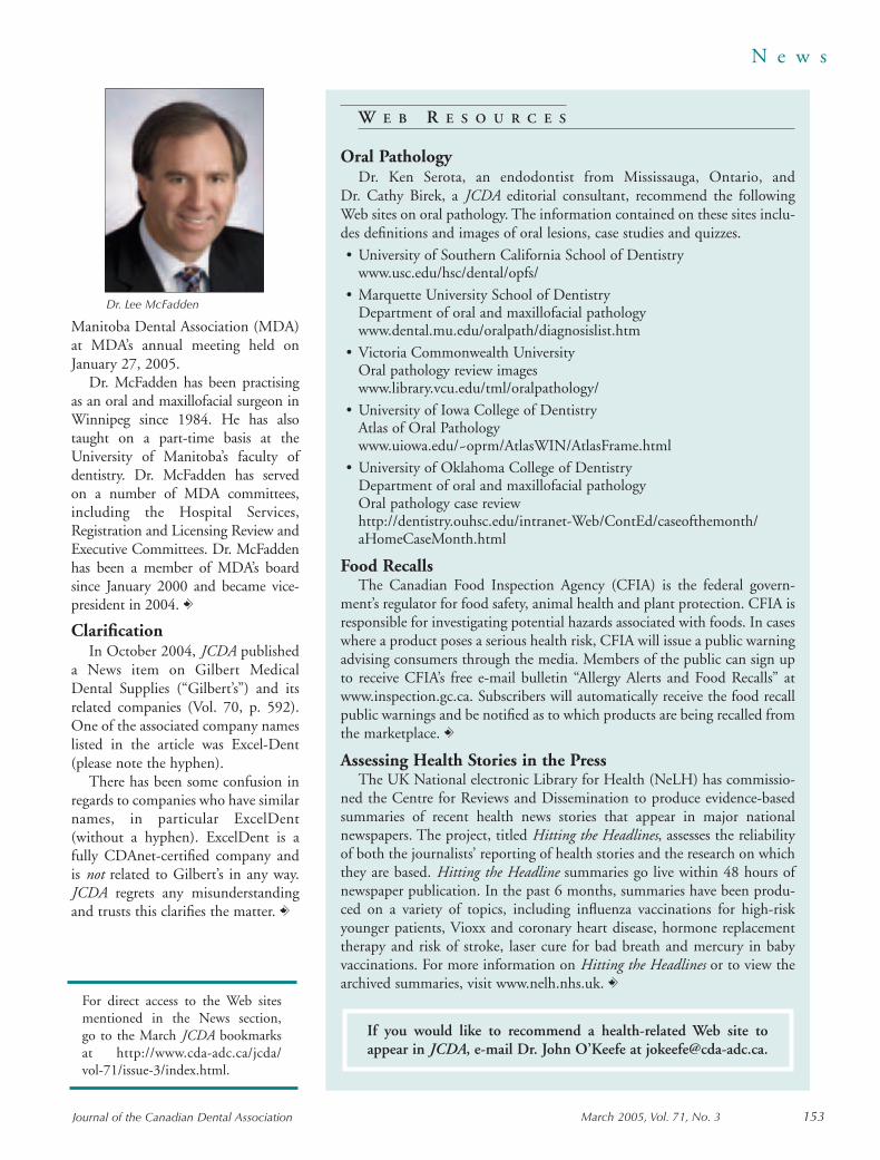
Manitoba Dental Association (MDA)at MDA’s annual meeting held onJanuary 27, 2005.
Dr. McFadden has been practisingas an oral and maxillofacial surgeon inWinnipeg since 1984. He has alsotaught on a part-time basis at theUniversity of Manitoba’s faculty ofdentistry. Dr. McFadden has served on a number of MDA committees,including the Hospital Services,Registration and Licensing Review andExecutive Committees. Dr. McFaddenhas been a member of MDA’s boardsince January 2000 and became vice-president in 2004. C
ClarificationIn October 2004, JCDA published
a News item on Gilbert MedicalDental Supplies (“Gilbert’s”) and itsrelated companies (Vol. 70, p. 592).One of the associated company nameslisted in the article was Excel-Dent(please note the hyphen).
There has been some confusion inregards to companies who have similarnames, in particular ExcelDent(without a hyphen). ExcelDent is afully CDAnet-certified company andis not related to Gilbert’s in any way.JCDA regrets any misunderstandingand trusts this clarifies the matter. C
March 2005, Vol. 71, No. 3 153Journal of the Canadian Dental Association
N e w s
For direct access to the Web sitesmentioned in the News section, go to the March JCDA bookmarksat http://www.cda-adc.ca/jcda/vol-71/issue-3/index.html.
Dr. Lee McFadden
W E B R E S O U R C E S
Oral PathologyDr. Ken Serota, an endodontist from Mississauga, Ontario, and
Dr. Cathy Birek, a JCDA editorial consultant, recommend the followingWeb sites on oral pathology. The information contained on these sites inclu-des definitions and images of oral lesions, case studies and quizzes.
• University of Southern California School of Dentistrywww.usc.edu/hsc/dental/opfs/
• Marquette University School of DentistryDepartment of oral and maxillofacial pathologywww.dental.mu.edu/oralpath/diagnosislist.htm
• Victoria Commonwealth UniversityOral pathology review imageswww.library.vcu.edu/tml/oralpathology/
• University of Iowa College of Dentistry Atlas of Oral Pathologywww.uiowa.edu/~oprm/AtlasWIN/AtlasFrame.html
• University of Oklahoma College of DentistryDepartment of oral and maxillofacial pathologyOral pathology case review http://dentistry.ouhsc.edu/intranet-Web/ContEd/caseofthemonth/aHomeCaseMonth.html
Food RecallsThe Canadian Food Inspection Agency (CFIA) is the federal govern-
ment’s regulator for food safety, animal health and plant protection. CFIA isresponsible for investigating potential hazards associated with foods. In caseswhere a product poses a serious health risk, CFIA will issue a public warningadvising consumers through the media. Members of the public can sign upto receive CFIA’s free e-mail bulletin “Allergy Alerts and Food Recalls” atwww.inspection.gc.ca. Subscribers will automatically receive the food recallpublic warnings and be notified as to which products are being recalled fromthe marketplace. C
Assessing Health Stories in the PressThe UK National electronic Library for Health (NeLH) has commissio-
ned the Centre for Reviews and Dissemination to produce evidence-basedsummaries of recent health news stories that appear in major nationalnewspapers. The project, titled Hitting the Headlines, assesses the reliabilityof both the journalists’ reporting of health stories and the research on whichthey are based. Hitting the Headline summaries go live within 48 hours ofnewspaper publication. In the past 6 months, summaries have been produ-ced on a variety of topics, including influenza vaccinations for high-riskyounger patients, Vioxx and coronary heart disease, hormone replacementtherapy and risk of stroke, laser cure for bad breath and mercury in baby vaccinations. For more information on Hitting the Headlines or to view thearchived summaries, visit www.nelh.nhs.uk. C
If you would like to recommend a health-related Web site toappear in JCDA, e-mail Dr. John O’Keefe at [email protected].
Journal of the Canadian Dental Association154 March 2005, Vol. 71, No. 3
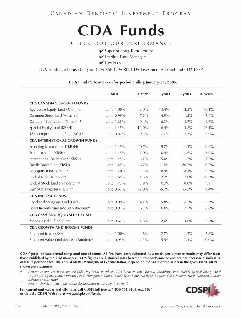
CDA FundsC H E C K O U T O U R P E R F O R M A N C E
✔ Superior Long-Term Returns✔ Leading Fund Managers✔ Low Fees
CDA Funds can be used in your CDA RSP, CDA RIF, CDA Investment Account and CDA RESP.
CDA figures indicate annual compound rate of return. All fees have been deducted. As a result, performance results may differ fromthose published by the fund managers. CDA figures are historical rates based on past performance and are not necessarily indicativeof future performance. The annual MERs (Management Expense Ratios) depend on the value of the assets in the given funds. MERsshown are maximum.
† Returns shown are those for the following funds in which CDA funds invest: 1Trimark Canadian Fund, 2KBSH Special Equity Fund,3KBSH US Equity Fund, 4Trimark Fund, 5Templeton Global Stock Trust Fund, 6McLean Budden Fixed Income Fund, 7McLean BuddenBalanced Value Fund.
†† Returns shown are the total returns for the index tracked by these funds.
For current unit values and GIC rates call CDSPI toll-free at 1-800-561-9401, ext. 5024or visit the CDSPI Web site at www.cdspi.com/funds.
C A N A D I A N D E N T I S T S ’ I N V E S T M E N T P R O G R A M
CDA Fund Performance (for period ending January 31, 2005)
MER 1 year 3 years 5 years 10 years
CDA CANADIAN GROWTH FUNDS
Aggressive Equity fund (Altamira) up to 1.00% 2.0% 13.4% 8.3% 10.1%
Common Stock fund (Altamira) up to 0.99% 7.2% 4.9% 1.2% 7.8%
Canadian Equity fund (Trimark)†1 up to 1.65% 9.4% 6.3% 8.7% 9.6%
Special Equity fund (KBSH)†2 up to 1.45% 12.0% 6.4% -4.8% 16.1%
TSX Composite Index fund (BGI)†† up to 0.67% 9.2% 7.7% 2.7% 9.9%
CDA INTERNATIONAL GROWTH FUNDS
Emerging Markets fund (KBSH) up to 1.45% 0.7% 9.7% 1.1% 0.9%
European fund (KBSH) up to 1.45% -7.0% -10.4% -11.6% 3.9%
International Equity fund (KBSH) up to 1.45% -6.1% -5.0% -11.7% 3.6%
Pacific Basin fund (KBSH) up to 1.45% -6.7% -1.0% -20.5% 0.7%
US Equity fund (KBSH)†3 up to 1.20% -3.5% -8.9% -8.1% 9.5%
Global fund (Trimark)†4 up to 1.65% 1.6% 2.7% 7.0% 10.3%
Global Stock fund (Templeton)†5 up to 1.77% 3.9% 0.7% 0.0% n/a
S&P 500 Index fund (BGI)†† up to 0.67% -2.0% -5.7% -5.5% 9.4%
CDA INCOME FUNDS
Bond and Mortgage fund (Fiera) up to 0.99% 4.1% 5.8% 6.7% 7.3%
Fixed Income fund (McLean Budden)†6 up to 0.97% 6.3% 6.6% 7.7% 8.6%
CDA CASH AND EQUIVALENT FUND
Money Market fund (Fiera) up to 0.67% 1.6% 2.0% 3.0% 3.8%
CDA GROWTH AND INCOME FUNDS
Balanced fund (KBSH) up to 1.00% 4.6% 2.7% 1.2% 7.8%
Balanced Value fund (McLean Budden)†7 up to 0.95% 7.2% 5.5% 7.1% 10.0%


Journal of the Canadian Dental Association156 March 2005, Vol. 71, No. 3
The Canadian Association of Oraland Maxillofacial Surgeons (CAOMS)welcomes the opportunity to engage inthis collaborative effort with JCDA. A special thank you goes out toDr. John O’Keefe and the editorial staffof JCDA, Dr. Bruce Pynn, CAOMSliaison to JCDA, and all of the contrib-utors who devoted time and effort tothe development of this special edition.
Oral and maxillofacial surgery hasevolved as a specialty over a rather shortperiod of time. In Canada, Dr. GeorgeBeers of Montreal is recognized as thefirst dentist to specialize in oral surgeryin the late 1800s. Oral and maxillofa-cial surgery is considered to be the firstdental specialty. It has been said thatthe First and Second World Wars werethe catalyst behind its rapid develop-ment. Dr. Fulton Risdon, who wasassigned to the Maxillofacial Centre forthe Canadian Forces at Sidcup duringWorld War I, returned to Toronto as a pioneer specialist in plastic and oralsurgery. He was later appointed professor of oral surgery at theUniversity of Toronto.
During the war, the number ofwounded needing facial bone recon-struction was overwhelming. Patientsfell under the care of dentists, whowere the recognized jaw specialists ofthe time. These dentists were the true
pioneers of the specialty. They learnedin the field and developed surgicalprocedures in response to extreme situ-ations. Today, new advances are fuelledby research and education. Oral andmaxillofacial surgery retains its histori-cal ties to dentistry; this synergyensures that our specialty continues toevolve so we can meet the growingneeds of our patients.
CAOMS was founded in 1953 toestablish a national forum to discusssurgical problems, to oversee therapidly changing format of graduateeducation and to expedite progress inoral and maxillofacial surgery. Theseefforts were founded on the fraternal-ism that allowed our members todevelop friendships with colleaguesfrom across Canada and to growpersonally and professionally. CAOMSand its members remain committed to
the continued advancement of thespecialty through close professionalconnections. The Association holds anannual scientific meeting that high-lights cutting-edge research throughabstract presentations. These educa-tional meetings are usually open to alldentists in Canada and the format ofthe event allows for a broad dissemina-tion of information.
The members of CAOMS remaincommitted to providing timely and
appropriate access to care for thepatients in their community, with the necessary support of all of ourdental colleagues. The Association’sredesigned Web site (www.caoms.com)provides access to a Canada-widedirectory of surgeons and housesexcellent information for patientswishing to learn more about theirsurgical treatment options related todental implants, orthognathic surgery,facial trauma, removal of wisdomteeth, cleft palate repair, temporo-mandibular joint problems and thedelivery of anesthesia.
CAOMS is very fortunate to have asits executive director Dr. ChristopherRobinson, whose diligent work inrepresenting the specialty of oral andmaxillofacial surgery and the profes-sion of dentistry equitably in thenational arena is unprecedented. He embodies the zeal that has charac-terized our past 44 presidents andconstitutes a tremendous role modelfor our current executive, which iscomposed of Dr. Walter Dobrovolsky,immediate past president, Dr. LeeMcFadden, president-elect, Dr. ArchieMorrison, treasurer, and Dr. Pierre-Éric Landry, secretary. Our executive ismade up of representatives from eachof our component regional associa-tions. It is a pleasure to work with thisteam of highly skilled and dedicatedvolunteers. With the continuedsupport of CDA, we look forward toforging the strong interdisciplinarybonds that are required for all of us todo what we do best... care!
Dr. Joseph J. FriedlichPresident, CAOMS
About CAOMS
Dr. Joseph J. Friedlich
President’s Message
�The members of CAOMSremain committed
to providing timely and appropriate
access to care for the patients in
their community, with the necessary support
of all of our dental colleagues.
�
The Foundation forContinuing Education andResearch (CAOMS)
Canadian research in oral andmaxillofacial surgery is greatlysupported by the Foundation forContinuing Education and Research(CAOMS). Members of CAOMSfounded this arm’s-length, non-profitorganization in 1988. The objective ofthe Foundation is to contribute to thewelfare of the public by the advance-ment of the specialty of oral andmaxillofacial surgery through contin-uing education and the diffusion ofknowledge. The Foundation is theonly national philanthropic organiza-tion with a mission that is dedicatedto the financial support of researchand education in the specialty of oraland maxillofacial surgery.
The Foundation’s initial mandatewas to provide for comprehensiveliterature reviews on various subjectsin the form of “risks and benefits.”The Foundation has published “risksand benefits” reviews for impactedthird molar surgery, orthognathicsurgery and surgery related to internalderangements of the temporo-mandibular joint.
Currently, the Foundation is evolv-ing into an organization that, in addi-tion to internal projects, now directlysupports specific research endeavoursthrough funding and guidance. Theseprojects are undertaken in both theacademic and private practice settings.An innovative study is now underwayto examine the nature of the relation-ships between general dental practi-tioners and oral and maxillofacialsurgeons. This study should help oraland maxillofacial surgeons betterrelate to and support their colleaguesin general dental practice.
Various grants, ranging in amountsfrom $2,000 to $10,000, have beenawarded to many researchers in ouracademic institutions across Canada.Past projects have made valuablecontributions to the fields of anesthe-siology, distraction osteogenesis and
surgery for cleft lip and palate. Mostrecently, the Dr. Ron Warren awards were presented to: Dr. Daisy Chemalyfrom the University of Manitoba (forresearch in the field of oral cancer),Dr. Albert Hadad from the Universityof Toronto (bone substitutes),Dr. Brett Habijanac from McGillUniversity (maxillofacial trauma), Dr. Nicholas Hogg from DalhousieUniversity (bacteriology/infectioncontrol) and Dr. Annie-ClaudeValcourt from Laval University(temporomandibular dysfunction).
The vital and practical benefits thatthis research provides for both ourpatients and the profession includeimproved quality of care, scientificallyvalidated and evidence-based care, theestablishment of new and innovativetechniques and direct scientificsupport for the expanding scope ofpractice of the dental profession.These benefits can only be realizedthrough the generous support of ourcorporate partners, colleagues andpatients. Voluntary donations to theFoundation can be made throughCAOMS, 174 Colonnade Road, Unit25, Ottawa, ON K2E 7J5.
The Foundation is tirelesslyadministered by Dr. William L.Frydman, chair, Dr. Ken Bentley,secretary/treasurer, and the Board ofTrustees, composed of Dr. RichardBell, Dr. Ben Davis, Dr. GeorgeSándor and Dr. Dany Morais.
Canadian Residency Programsin Oral and MaxillofacialSurgery
The efforts of CAOMS and theFoundation are easily recognized inthe 5 residency programs in oral andmaxillofacial surgery in Canada.These university-based programs havedeveloped international reputationsand attract high calibre candidatesfrom Canada and around the world.
Dalhousie UniversityThe oral and maxillofacial surgery
specialty program at Dalhousie is a 6-year program that includes a master’sdegree in oral and maxillofacial surgeryand a medical degree. One resident isaccepted per year in addition to onefellow. The fellowship position hasrecently been formalized and is of oneyear’s duration. Dalhousie faculty areall fellowship-trained; areas of subspe-cialty training include orthognathicsurgery, trauma, preprosthetic recon-structive and implant surgery, cleft lipand palate surgery and head and neckcancer surgery. Research is ongoing inthe following areas of interest: obstruc-tive sleep apnea, cleft lip and palate,sterilization of instruments, prepros-thetic surgery, temporomandibulardisorders, orthognathic surgery andpathology.
Department of Oral and MaxillofacialSciences
Dalhousie University Faculty of Graduate Studies 5981 University Avenue Halifax, NSB3H 3J5
www.registrar.dal.ca/calendar/gr/ORAL.htm#1
Laval UniversityThe oral and maxillofacial surgery
graduate training program at LavalUniversity/Hôpital de l’Enfant-Jésusis a 5-year residency leading to amaster of science degree and diplomaqualification. Ten regular residents are currently engaged in training. An additional position is held for acandidate with a special contract whois required to return to practice in aremote underserviced area followinggraduation. The program attractsinternational interest, with regularrotation of residents from France andSwitzerland who wish to expand their French-language education. Aformal fellowship in orthognathic,trauma and reconstructive surgerywill be offered in the next 2 to 3 years.
March 2005, Vol. 71, No. 3 157Journal of the Canadian Dental Association
A b o u t C A O M S
�

Currently, leading-edge researchprojects are in progress by residentswishing to obtain doctoral qualifica-tion in osseous distraction and neuralregeneration. The research in osseousdistraction is being undertaken inDr. Antonio Nanci’s laboratory at theUniversity of Montreal, while theresearch in neural regeneration istaking place at Dr. François Auger’sLaboratory of Experimental TissueEngineering (LOEX) in Quebec City.
Department of Oral and MaxillofacialSurgery
Laval University Faculty of Dentistry2435 Pavillon Jean-Charles-BonenfantQuebec City, QCG1K 7P4
www.fmd.ulaval.ca/index.html
McGill UniversityThe McGill University graduate
training program in oral and maxillo-facial surgery is a fully accredited, 4-year program leading to a diplomain oral and maxillofacial surgery and amaster of science degree. Two residentpositions are available each year. Oneis a fully funded position open tograduates of North American dentalschools. The second position is opento non-North American graduateswho have funding from their homecountry and have made a commit-ment to return to their country andwork within the health care system.The major research initiatives of thisprogram are bone physiology, bonehealing and bone regeneration (incollaboration with the McGill Boneand Periodontal Research Centre) andosseointegrated implants. Funding for these initiatives is obtained from3 principal sources. Alumni providegenerous support through theKenneth C. Bentley alumni fund andthe Fund for Oral and MaxillofacialSurgery Research and ContinuingEducation (FORCE), which has beensuccessful in generating funds fromindustry sources. The McGill program
is also very grateful to funding organi-zations such as the CAOMSFoundation and the Order of Dentistsof Quebec.
Division of Oral and MaxillofacialSurgery
McGill University 1650 Cedar Avenue Montreal, QCH3G 1A4
www.mcgill.ca/dentistry/graduate/
University of TorontoThe graduate program in oral and
maxillofacial surgery and anesthesia atthe University of Toronto is a 4-yearprogram with a compulsory master’sdegree based on a research project.Residents may choose to enroll in adoctoral program instead of the master’sprogram. There are 8 funded residencypositions, with 2 students in each year,and up to 2 international fellowshipsper year, one in pediatric oral andmaxillofacial surgery and one in reconstructive oral and maxillofacialsurgery. The graduate program is newlyhoused at Mount Sinai Hospital, wheredentistry is a protected program. Thefaculty of dentistry is affiliated with theprogram, as are the Hospital for SickChildren, the Bloorview MacMillanChildren’s Centre, and Sunnybrookand Women’s Health Centre. Residentsgain clinical exposure in all areas of oraland maxillofacial surgery. A new rota-tion to a cleft lip and palate unit at theUniversity of Oulu in Finland has beenestablished. This initiative has receivedgenerous funding and support from the Ontario Society of Oral andMaxillofacial Surgeons. There are also anumber of community-based practices in oral and maxillofacialsurgery that graduate residents maychoose to visit during their electiverotations. Research in the graduateprogram focuses primarily on boneregeneration, hyperbaric oxygen therapy, laser surgery, treatment ofcongenital malformations and surgicalorthodontics.
Department of Oral and MaxillofacialSurgery
University of Toronto Faculty of Dentistry 124 Edward StreetToronto, ONM5G 1G6
www.utoronto.ca/dentistry/academic/graduate/graduateprograms.html
University of ManitobaThe oral and maxillofacial surgery
program at the University ofManitoba is of 4 years’ duration andleads to a master’s degree in oral andmaxillofacial surgery. Five residentsare currently enrolled in the program.Generally, one new resident isaccepted each year, with the possibil-ity of additional resident positions.On-service rotations provide residentswith broad exposure to both adult andpediatric oral and maxillofacialsurgery. The residents are also sched-uled in off-service rotations in internalmedicine, adult and pediatric anesthe-sia, surgical intensive care, emergencyroom medicine, otolaryngology andsurgical oncology. Interaction andcooperation between the residents inoral and maxillofacial surgery andthose in the graduate orthodonticprogram ensures a diversity of experi-ence. Present research includes studiesin oncology, trauma, implants andorthognathic surgery.
Division of Oral and MaxillofacialSurgery
University of Manitoba Faculty of Dentistry 790 Bannatyne Avenue Winnipeg, MBR3E 0W2
www.umanitoba.ca/faculties/dentistry/gradPrograms/grad_OMS.html
Journal of the Canadian Dental Association158 March 2005, Vol. 71, No. 3
A b o u t C A O M S
March 2005, Vol. 71, No. 3 159Journal of the Canadian Dental Association
A b o u t C A O M S
�
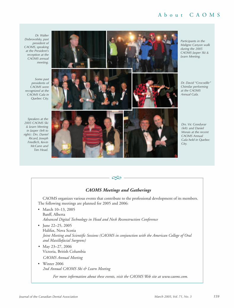
Some pastpresidents of
CAOMS wererecognized at the
CAOMS Gala inQuebec City.
Participants in theMaligne Canyon walkduring the 2005CAOMS Jasper Ski &Learn Meeting.
Dr. David “Crocodile”Chimilar performing at the CAOMSAnnual Gala.
Dr. WalterDobrovolsky, past
president ofCAOMS, speakingat the President’sreception at theCAOMS annual
meeting.
Speakers at the 2005 CAOMS Ski& Learn Meetingin Jasper (left to
right): Drs. DanielRicard, Joseph
Friedlich, KevinMcCann and
Tim Head.
Drs. Vic Goodyear(left) and DanielMorais at the recentCAOMS AnnualGala held in QuebecCity.
CAOMS Meetings and Gatherings
CAOMS organizes various events that contribute to the professional development of its members.The following meetings are planned for 2005 and 2006:
• March 10–13, 2005Banff, AlbertaAdvanced Digital Technology in Head and Neck Reconstruction Conference
• June 22–25, 2005Halifax, Nova ScotiaJoint Meeting and Scientific Sessions (CAOMS in conjunction with the American College of Oraland Maxillofacial Surgeons)
• May 23–27, 2006 Victoria, British Columbia
CAOMS Annual Meeting
• Winter 2006 2nd Annual CAOMS Ski & Learn Meeting
For more information about these events, visit the CAOMS Web site at www.caoms.com.

with over 100 products,trust the GUM® brand tomeet all of your needs.
HEALTHY GUMS. HEALTHY LIFE.™
®
™™ ™
The Science of a Toothbrush
TechniqueTechnique®
Built to brush at 45º
The Technique® handle effortlesly places bristles at the correct 45º angle towards the gumline to reinforce the Bass technique.
The Dome Trim® design with raised center row bristles reaches below the gumline.
Proper brushing requires patient educationand the proper toothbrush
Provide your patients the solution to effective cleaning.
For over 80 years, GUM® has focused on designing innovative products that meet "the special needs" of dental professionals and their patients.
1-800-265-8353www.jbutler.com
© 2004 Sunstar Butler C04157
NEW &IMPROVED
GRIP!

March 2005, Vol. 71, No. 3 161Journal of the Canadian Dental Association
C L I N I C A L P R A C T I C E
Developmental aberrations may result in the formation of supernumerary or extra structures.Polydactily or extra fingers or toes and supernu-
merary teeth are examples of such developmental duplica-tions. Accessory or supplementary jaws are duplicatedportions of jaws or entire jaws, with or without teeth.1 Inthe case of the maxilla, such accessory jaws are called disto-mus.2,3 Distomus formation is extremely rare.2 Accessoryjaws may be associated with the formation of an accessoryrudimentary or vestigial mouth.1 Reports indicate thatdistomus has been observed more commonly in associationwith lateral facial clefts.2 Congenital facial clefts, whichhave been classified by Tessier,4 may occur where embry-ologic processes fuse. The most common facial clefts arecleft lip and palate; the prevalence of cleft lip with or with-out cleft palate in the largest study to date is 1.2 per 1,000live births.5 The incidence of lateral facial clefts in variousseries ranges from 0.3% to 0.67% of all facial clefts.5,6
Facial clefts associated with duplication of various oralstructures have also been reported.7–11 Stoneman1 andDeGurse and others2 have reported cases of a congenitalfacial fistula with an accessory maxilla and teeth.
This article reports the management, over 9 years, of apatient with an accessory maxilla with supernumerary teethand a congenital facial fistula, who required treatment dueto increasingly bothersome symptoms.
Case ReportAn 8-year-old boy was referred for assessment of a fistula
that occasionally drained milk-like fluid from an openingon the left nasolabial groove of the face. The parents statedthat this drainage had been present since birth. The patient had been previously diagnosed as having an incom-plete facial cleft. Serial panoramic radiographs and acomputed tomography (CT) scan revealed the progressivedevelopment of first 2, then 3 tooth-like structures in theleft cheek in a cavity with a fluid-filled lumen. A diagnosisof odontoma was dismissed, on the basis of the appearanceand position of the lesion. Rather the lesion was thought toresemble an accessory maxilla. The past medical history wasotherwise unremarkable.
The patient was followed regularly with semi-annualvisits over the next 7 years. A creamy-white milk-like fluidwas initially expressible by palpation of a dimple of the leftcheek in the nasolabial groove (Fig. 1). The volume of thedrainage progressively increased over the years of follow-up,such that each time the patient smiled, he expressed thefluid spontaneously onto his cheek. This steadily increasingspontaneous drainage became distressing to the patient, tothe point of being socially unacceptable. Bacterial cultureand sensitivity testing of the facial discharge revealednormal skin flora with a scant growth of coagulase-negativeStaphylococcus species. Surgery had been delayed at therequest of the boy’s parents. At the age of 15 years, the
Management of a Patient with an AccessoryMaxilla and Congenital Facial Fistula
• Vesa T. Kainulainen, DDS, EHL, PhD •• George K.B. Sándor, MD, DDS, PhD, FRCD(C), FRCSC, FACS •
• Douglas W. Stoneman, DDS, FRCD(C) •
A b s t r a c tAlthough accessory jaws are a rare occurrence, the presence of such accessory tissue may cause some bothersomesymptoms. This case report helps identify these unusual developmental lesions so that dentists can refer suchpatients for definitive care and management.
MeSH Key Words: child; dental fistula/radiography; maxilla/abnormalities; tooth supernumerary/radiography
© J Can Dent Assoc 2005; 71(3):161–3This article has been peer reviewed.

Journal of the Canadian Dental Association162 March 2005, Vol. 71, No. 3
Kainulainen, Sándor, Stoneman
patient had a fistula on the left cheekapproximately 3 cm lateral to the leftoral commissure. It was secretingspontaneously or when the left cheekwas rubbed.
Radiographic examination revealeda 4-cm–wide round cavity with3 supernumerary teeth in the leftmaxillary sinus area (Fig. 2). Inpanoramic radiographs taken 6 yearsearlier, there were 2 premolar-likesupernumerary teeth. A CT scanshowed a cavity with a sharp delin-eation from the maxillary bone andits zygomatic process. The lesionextended from the orbital floor to thezygomatic buttress area. It did notextend into the inferior part of theleft maxillary sinus or into the maxil-lary alveolar process. Otherwise thewhole sinus was full of accessorytissue. The teeth within the lesionwere attached to the bony walls of thecavity (Fig. 3a). This bony cavity wasfilled with fluid and connected to acavity in the soft tissues of the leftcheek (Fig. 3b).
Surgery was performed undergeneral anesthesia. A plastic catheterwas threaded into the fistula andmethylene blue dye was injected intothe catheter to help delineate theaccessory structures connected to the fistula (Fig. 4). A vestibular incision was used to expose the entireanterior maxilla. Bone over the softtissue capsule was removed (Fig. 5). The fistula and the soft tissuessurrounding the blue-stained struc-tures in the left cheek were removedin 1 piece. The teeth and the bonycavity, which contained a brownishliquid, were removed in a separatepiece. Care was taken not to damagethe parotid duct and branches of thefacial nerve. The specimens were sent for histopathologic evaluation(Fig. 6).
Histologic sections revealed bone, developing teeth anda stratified squamous epithelium lining the lumen of thebony and soft-tissue cavities. Postoperative healing wasuneventful and neither the lesion nor the fistula has shownany clinical or radiographic signs of recurrence in over3 years of follow-up (Fig. 7).
DiscussionThis case report presents the long-term management
and closely supervised follow-up of a congenital facialfistula with an accessory maxilla and teeth. The diagnosis ofincomplete facial cleft had been suggested when the patientwas 6 years of age. Because the only complaint was a fistula
Figure 5: The anterior wall of the left maxillais exposed and removed. A separate bonywall is removed to gain access to the bonycavity of the lesion.
Figure 4: A catheter is threaded through thecutaneous opening of the fistula into thelumen of the lesion in the left cheek.
Figure 3a: Computed tomography image(coronal view) showing the lesionoccupying most of the left maxillary sinus.
Figure 1: Drainage of a milk-like fluid fromthe left nasolabial groove.
Figure 2: Preoperative panoramicradiograph showing 3 supernumerary teethdeveloping in a bony cavity in the leftmaxillary sinus.
Figure 3b: Computed tomography image(axial view) showing the lumen of the soft-tissue lesion located lateral to the anteriormaxillary wall.

March 2005, Vol. 71, No. 3 163Journal of the Canadian Dental Association
Management of a Patient with an Accessory Maxilla and Congenital Facial Fistula
References1. Stoneman DW. Congenital facial fistulawith formation of accessory bone and teeth.Report of a case. Oral Surg Oral Med OralPathol 1978; 45(1):150–4.2. DeGurse K, Chung H, Pharoah M. Facialdimple with accessory bone and teeth.Dentomaxillofac Radiol 1995; 24(2):135–8.3. Worth HM. Principles and practice of oralradiologic interpretation. Chicago (IL): YearBook Medical Publishers; 1963. p 114–5.4. Tessier P. Anatomical classification of facial,cranio-facial and latero-facial clefts.J Maxillofac Surg 1976; 4(2):69–92.5. Cooper ME, Stone RA, Liu Y, Hu DN,Melnick M, Marazita ML. Descriptiveepidemiology of nonsyndromic cleft lip withor without cleft palate in Shanghai, China,
from 1980 to 1989. Cleft Palate Craniofac J 2000; 37(3):274–80.6. Fogh-Andersen P. Rare clefts of the face. Acta Chir Scand 1965;129:275–81.7. Pitanguy I, Franco T. Nonoperated facial fissures in adults.Plast Reconstr Surg 1967; 39(6):569–77.8. Smylski PT. Accessory jaw bones; a report of a case. J Oral Surg AnesthHosp Dent Serv 1952; 10(1):70–4.9. Chowdhury SR, Roy A. Duplication of the upper lip and maxilla.Br J Plast Surg 1991; 44(6):468–9.10. Avery JK, Hayward JR. Case report: duplication of oral structureswith cleft palate. Cleft Palate J 1969; 6:506–15.11. Ball IA. Klippel-Feil syndrome associated with accessory jaws(distomus). Br Dent J 1986; 161(1):20–3.
that was occasionally draining, the parents opted for long-term observation. The drainage progressively worseneduntil the patient found it intolerable and requested removalof the lesion. Serial assessments were conducted to ensurethat the patient was not lost to follow-up and that therelated structures near the lesion were growing anddeveloping normally.
The development of an accessory jaw is very rare. Thelesion may occur as a mass of bone containing teeth or as acomplete jaw.1,2,7–11 Stoneman1 and DeGurse and others2
have suggested that some facial fistulas are vestiges of facialclefts. In this case, in addition to the accessory jaw tissue, afistula connected the skin to a separate lumen or rudimentarymouth in the cheek. The management of such lesions mustbe guided by the symptoms and their potential to interferewith the development of surrounding structures. The timingof their removal depends on these 2 factors and the progres-sive development of bothersome symptoms. Dentists cancontribute to the management of such lesions by identifyingthem and referring the patient for definitive management. C
Dr. Kainulainen is a former fellow in oral and maxillo-facial surgery, The Hospital for Sick Children andUniversity of Toronto, Toronto, Ontario, and iscurrently an assistant professor in oral and maxillofacialsurgery, University of Oulu, Oulu, Finland.
Dr Sándor is an associate professor and director, graduate program in oral and maxillofacial surgery andanesthesia, University of Toronto, and the coordinator oforal and maxillofacial surgery at The Hospital for SickChildren and Bloorview MacMillan Children’s Centre,Toronto, Ontario, and docent in oral and maxillofacialsurgery, University of Oulu, Oulu, Finland.
Dr. Stoneman is deceased; he was professor emeritus inoral radiology at the University of Toronto, and consul-tant oral radiologist at The Hospital for Sick Children,Toronto, Ontario.
Correspondence to: Dr. George K.B. Sándor, The Hospital of SickChildren, S-527, 555 University Ave., Toronto ON M5G 1X8.E-mail: [email protected].
The authors have no declared financial interests.
Figure 7: After 3 years of follow-up, apanoramic radiograph shows a normallypneumatized left maxillary sinus with nosigns of recurrence.
Figure 6: Teeth with the excised bony andsoft-tissue specimens.

Master Teams• Didier Dietschi / Roberto Spreafico• Mauro Fradeani / Giancarlo Barducci• David A. Garber / Pinhas Adar• Ueli Grunder / Hans-Peter Spielmann• John C. Kois / Steven McGowan• Edward A. McLaren / Shane White• Konrad H. Meyenberg / Walter M. Gebhard• Avishai Sadan / Markus B. Blatz• Henry Salama / Maurice A. Salama• Frank M. Spear / Vincent G. Kokich• Alan V. Sulikowski / Aki Yoshida• Thomas F. Trinkner / Matt Roberts
Master Clinicians/Ceramists• Dario Adolfi• Nitzan Bichacho• Daniel Edelhoff• Ronald E. Goldstein• Kenneth S. Hebel• Pascal Magne• John W. McLean• Lloyd L. Miller• Claude Sieber• Robert R. Winter
For complete program and registration materials, contact:Quintessence Publishing Co, Inc
551 Kimberly Drive, Carol Stream, IL 60188-1881Phone (630) 682-3223 • Fax (630) 682-3288
E-mail [email protected] • Website www.quintpub.com
Topics• Extensive Rehabilitation• Choice of Ceramic Systems• Interdisciplinary Implant
and Esthetic Treatment• Dentist-Technician
Teamwork
Sponsored by
The International QuintessencePublishing Group
Louisiana State University
March 2005, Vol. 71, No. 3 165Journal of the Canadian Dental Association
C L I N I C A L P R A C T I C E
Burkitt’s lymphoma is a malignant tumour of B-celllymphocyte origin and is classified as a non-Hodgkin’s lymphoma (NHL). Three clinical variants
are recognized: African (endemic), American (sporadic) andHIV associated.1 Sporadic Burkitt’s lymphoma is a raremalignancy among western populations. In most series, theannual incidence is 2–3 cases per million.2
During the early 20th century, Sir Albert Cook, while inEast Africa, described a common malignancy among youngAfrican children that predominantly affected the jaws andoccasionally various abdominal organs. Half a century later,a surgeon, Denis Burkitt, who was working in Kampala,Uganda, noticed the same lesions on the faces of youngAfrican children, with occasional intestinal involvement. In 1958, Burkitt published his findings, calling the lesion“a sarcoma involving the jaws.”3 Epstein and others firstdemonstrated a herpes virus in a biopsy sample derivedfrom Burkitt’s lymphoma in 1964. The virus was laterdesignated Epstein-Barr virus (EBV) and was considered apotential etiologic factor of Burkitt’s lymphoma.4
Burkitt’s lymphoma is probably the fastest growingmalignant neoplasm to affect humans. It can double in sizein 24 hours with 80% of its cells undergoing mitosis at anypoint.5 The symptoms are rapidly progressive. Multifocalextranodal involvement is common to all subtypes ofBurkitt’s lymphoma.1
Burkitt’s lymphoma is thought to account for 40% of allchildhood NHL.2 Its incidence is greatly dependent ongeographic location; in equatorial Africa, it accounts for50% to 75% of all malignancies in children.6 SporadicBurkitt’s lymphoma is not as common. Its annual incidenceis 2–3 cases per million. Burkitt’s lymphoma accounts for40% of HIV-associated NHL.2
Endemic Burkitt’s lymphoma is usually diagnosedbetween the ages of 5 and 7 years and involves the jaws aswell as other facial bones in 60% to 80% of cases. Lesscommonly, it involves the abdomen and bone marrow.Endemic Burkitt’s lymphoma is virtually always associatedwith EBV, as positive titres are found in over 90% of cases.7
In contrast, sporadic Burkitt’s lymphoma is associatedwith slightly older children (average age 12 years). The
Sporadic Burkitt’s Lymphoma of the Jaws:The Essentials of Prompt Life-saving
Referral and Management• Ahmed Jan, DDS •
• Kashyap Vora, BDS, FDS RCS (Eng) •• George K.B. Sándor, MD, DDS, PhD, FRCD(C), FRCSC, FACS •
A b s t r a c tBurkitt’s lymphoma is an undifferentiated non-Hodgkin’s B-cell lymphoma. Three clinical subtypes are recognized:African (endemic), American (sporadic) and HIV associated. Sporadic Burkitt’s lymphoma is a rare malignancyamong western populations. This report describes a case of sporadic Burkitt’s lymphoma of the jaws with an alarmingly rapid spread associated with acute renal failure. This type of rapid progression bespeaks the need forprompt recognition and life-saving referral by the dental practitioner. The clinical features of Burkitt’s lymphomainvolving the jaws include severely hypermobile, ectopically displaced and supra-erupted teeth. The purpose of thiscase report and review of the literature is to illustrate the clinical and histopathologic features of Burkitt’s lymphomato help clinicians recognize such cases readily and facilitate prompt and potentially life-saving referral.
MeSH Key Words: Burkitt lymphoma/diagnosis; case report; mandibular neoplasms/pathology
© J Can Dent Assoc 2005; 71(3):165–8This article has been peer reviewed.

Journal of the Canadian Dental Association166 March 2005, Vol. 71, No. 3
Jan, Vora, Sándor
abdomen is the most common site of presentation. About25% of sporadic Burkitt’s lymphoma cases involve the headand neck, most commonly in the form of cervicallymphadenopathy. Maxillofacial bony involvement occursin fewer than 30% of cases. EBV-positive titres are found inonly 20% of cases.7
Adult Burkitt’s lymphoma patients are almost alwaysafflicted with AIDS. Burkitt’s lymphoma accounts for 40% of HIV-associated NHL cases.2 Like sporadic Burkitt’slymphoma, the HIV-associated variant often involves theabdomen and it involves the bone marrow in 30% of cases.Unlike endemic Burkitt’s lymphoma, EBV titres areelevated in only 30% of cases of adult HIV-associatedBurkitt’s lymphoma.2
Case ReportA 13-year-old white girl was referred to the oral and
maxillofacial surgery service of The Hospital for SickChildren in Toronto with the chief complaint of spontaneousintermittent pain in her lower back teeth and inability tochew food comfortably. A month earlier, she had complainedto her parents of progressively loosening teeth. The painhad been present for 2 weeks. She was referred by hergeneral dentist to a periodontist, who immediately referredthe patient for oral and maxillofacial surgical assessment.The past medical history revealed a diagnosis of microcytichypochromic anemia and infectious mononucleosis 3 weeksbefore presentation. Results of a complete blood count andserology were consistent with these 2 diagnoses.
Physical examination revealed a lethargic young womanwith obvious pallor. Bilateral palpable mobile submandibu-lar lymph nodes were found in her head and neck. Themandibular and maxillary divisions of the trigeminal andfacial nerves were intact. Generalized teeth mobility rangedfrom 1+ in upper and lower anterior teeth to 3+ in lowersecond premolar and second molars. This was noted despitethe absence of any visible plaque on the teeth surfaces.Severe buccal expansion of the alveolus next to the lower
posterior teeth was noted. Both lower second molars weresupra-erupted and in premature occlusal contact with theupper first molars causing a massive open bite. The leftlower second molar was more supra-erupted than the right.The gingiva was pale, pink and swollen with spontaneousareas of bleeding.
An orthopantomogram and intraoral periapical radi-ographs were taken. Severe alveolar bone loss around thelower second premolars as well as the lower second molarswas noted (Figs. 1 and 2). These teeth were floating. Thedeveloping lower third molars were displaced coronally andmedially relative to their crypts, indicating possible infiltra-tion of these structures (Fig. 1).
Based on the clinical examination and past medical anddental history, a decision was made to extract the lower leftsecond molar and perform an incisional biopsy of the softtissue in the tooth socket.
HistopathologyThe histopathology was consistent with diagnosis of
a high-grade B-cell NHL with features of Burkitt’slymphoma. Routine hematoxylin- and eosin-stainedsections revealed fibrofatty tissue diffusely infiltrated withsheets of a monomorphic population of lymphoid cells.Numerous scattered phagocytic cells all around the sectionimparted a “starry-sky appearance” (Fig. 3). Immunohisto-chemical staining was positive for CD20, CD22 andCD79, which are specific for B-cell lymphocytes. CD10was focally positive, which is consistent with the earlyphases of B-cell proliferation. MIB1 staining was positive in more than 95% of cells, revealing increased nuclearDNA synthesis.
Definitive ReferralThe patient was immediately referred to the hematol-
ogy–oncology department for assessment and definitivecare. Further laboratory results revealed a secondary diagnosis of acute renal failure necessitating emergency life-saving dialysis. The effects of the patient’s renal failure
Figure 1: Orthopantomogram showing thedisplaced teeth germs of the lower thirdmolars coronally and medially relative totheir bony crypts. Severe alveolar boneresorption is visible around the lowersecond molars.
Figure 2: Periapical radiograph showingsevere alveolar bone resorption around thelower second premolars.
Figure 3: Diffuse sheets of monomorphiclymphoid cells with scattered macrophagesproducing a “starry sky appearance.”

March 2005, Vol. 71, No. 3 167Journal of the Canadian Dental Association
Sporadic Burkitt’s Lymphoma of the Jaws
were corrected by the dialysis, she responded well to subse-quent chemotherapy and is currently in remission.
DiscussionThe differential diagnosis of hypermobile teeth with
absent local factors includes: generalized juvenile periodon-titis, HIV periodontitis, Langerhan’s cell histiocytosis,Papillon-Lefèvre syndrome, hypophosphatasia, cyclicneutropenia, vitamin-D resistant rickets, acrodynia,leukemia and lymphoma. Head and neck involvementoccurs in only 25% of sporadic Burkitt’s lymphoma cases and most commonly in the form of cervicallymphadenopathy,2 which can make the diagnosis quitechallenging in contrast to endemic Burkitt’s lymphoma.
The clinical findings may vary according to the anatomical site of involvement and the timing of presenta-tion. The range may be from no signs and symptoms toairway obstruction, intestinal obstruction and biliaryobstruction. Inferior alveolar nerve parasthesia has alsobeen reported to be the only presenting sign of sporadicBurkitt’s lymphoma.8 Signs and symptoms of oral Burkitt’slymphoma, including mobile teeth, toothache, oral masses,gingival enlargement, pain, jaw expansion, swelling andsensory disturbances, have been recorded by some workerswith pain being the most common presenting symptom.7
In this case, pain was also the predominant presentingsymptom and generalized mobility of teeth with theabsence of any obvious causative factor was the most strik-ing sign. The unusual aspect of this case was the severity ofdisease, exemplified by the rapidity of the development ofacute renal failure, which left untreated would have resultedin a terminal outcome.
Although EBV is strongly considered as a potential etio-logic factor of Burkitt’s lymphoma, its precise role is notwell understood, especially in sporadic Burkitt’s lymphoma.EBV is an enveloped herpes virus that contains double-strand linear DNA of 170 to 175 kb in the nucleocapsid.9
After entering the oropharynx and adjacent structures, thisvirus preferentially infects B-cells via the C3d complementreceptor, CD21. Primary infection during early childhoodis mostly asymptomatic, whereas infection during adoles-cence results in acute infectious mononucleosis in 30% to50% of cases.10 Immunodeficiencies may allow viral reactivation and the excessive proliferation of EBV-infectedB-cells, which may lead to the development of EBV posi-tive B-lymphoproliferative diseases or lymphomas.11
Approximately 90% of African Burkitt’s lymphomatumours contain EBV DNA, whereas only 20% of sporadicBurkitt’s lymphomas are EBV associated. However,disrupted and aberrant expressions of the viral genome haverecently been discovered in the United States in cases ofsporadic Burkitt’s lymphoma that were diagnosed as EBVnegative in standard screening. This suggests that the viralgenome itself may be dispensable at some stage of the
tumour’s development. There may be greater involvementof EBV in sporadic Burkitt’s lymphoma than previouslydocumented, suggesting that EBV DNA may, in someinstances, be lost after initiation of the neoplasm. Althoughscreening tests may be insufficient to determine the EBV status of all neoplasms,12 the data suggest thatEBV may play a prominent role in the pathogenesis of Burkitt’s lymphoma.13
The overexpression of c-myc oncogene and the func-tional loss of wild type p53 gene are considered as possibleetiologic factors as well. Immunodeficient patients, particu-larly those with AIDS, and cardiac or renal transplantpatients are more susceptible to developing NHL.14
Burkitt’s lymphoma may be the first manifestation of AIDS in many cases.15
Investigations for suspected Burkitt’s lymphoma shouldinclude an incisional biopsy to establish a definitive diag-nosis. In the maxillofacial region, plain radiography isusually sufficient but may be augmented by computedtomography and magnetic resonance imaging. In mostinstitutions, lumbar puncture to examine the cerebrospinalfluid is a routine measure to look for malignant cells in thecentral nervous system.2
Burkitt’s lymphoma is one of the first human malignan-cies shown to be curable by chemotherapy alone. A combi-nation of cyclophosphamide, doxyrubicin, vincristine andprednisone is one example of a drug therapy. Radiotherapyis reserved for overt central nervous system disease that isresistant to chemotherapy and is reported to be useful incertain emergencies, such as airway obstruction.7 Bonemarrow transplantation may be necessary after completionof chemotherapy cycles. The surgical management ofBurkitt’s lymphoma is limited to biopsy.7
The prognosis of Burkitt’s lymphoma depends on theextent of the disease, the patient’s age and the timing ofdiagnosis.3 It is excellent in children, where it approaches100% disease-free survival in early stages and 75% to85% of patients survive free of disease in later life.7 Adultsare less fortunate, with a survival rate of 50% to 75%.7
Adults are almost always HIV-positive patients and may dieof other causes.16
ConclusionThe role of the dentist in the early diagnosis and prompt
referral of patients with Burkitt’s lymphoma cannot beoveremphasized. Life-threatening complications occursuddenly and unexpectedly in the development of Burkitt’slymphoma. These complications include airway obstruc-tion, abdominal obstruction and acute renal failure, as inthis case report.
Dentists must be suspicious when faced with a childpatient presenting with unexplained hypermobility of teeth,supra-eruption of permanent teeth and severe alveolar boneresorption around these teeth, where the local etiologic

Journal of the Canadian Dental Association168 March 2005, Vol. 71, No. 3
Jan, Vora, Sándor
factors of periodontal disease are not present. The unex-plained displacement of developing teeth from their cryptsis another worrisome sign. Unexplained paresthesia of themandibular branch of the trigeminal nerve is also ominous.When any of these symptoms or signs exists, the dentistmust have a high index of suspicion and act promptly.Failure to refer such a patient in a timely manner mayrapidly lead to disastrous consequences. C
Dr. Jan is an intern in oral and maxillofacial surgery,University of Toronto, Toronto, Ontario.
Dr. Vora is a fellow in oral and maxillofacial surgery,The Hospital for Sick Children and University ofToronto, Toronto, Ontario.
Dr Sándor is an associate professor and director, graduate program in oral and maxillofacial surgery andanesthesia, University of Toronto, and coordinator of oral and maxillofacial surgery at The Hospital for SickChildren and Bloorview MacMillan Children’s Centre,Toronto, Ontario, and docent in oral and maxillofacialsurgery, University of Oulu, Oulu, Finland.
Correspondence to: Dr. George K.B. Sándor, The Hospital of SickChildren, S-527, 555 University Ave, Toronto ON M5G 1X8.E-mail: [email protected].
The authors have no declared financial interests.
References1. Neville BW, Damm DD, Allen CM, Bouquot JE. Oral and maxillo-facial pathology. 2nd ed. Philadelphia: W.B. Saunders; 2002. p. 608.
2. Banthia V, Jen A, Kacker A. Sporadic Burkitt’s lymphoma of the headand neck in the pediatric population. Int J Pediat Otorhinolaryngol 2003;67(1):59–65.3. Burkitt D. A sarcoma involving the jaws of African children.Br J Surg1958; 46(197):218–23.4. Epstein MA, Achong BG, Barr YM. Virus particles in culturedlymphoblasts from Burkitt’s lymphoma. Lancet 1964; 15:702–3.5. Durmus E, Oz G, Guler N, Avunduk M, Caliskan U, Blanchaert RHJr. Intraosseous mandibular lesion. J Oral Maxillofac Surg 2003;61(2):246–9.6. Shapira J, Peylan-Ramu N. Burkitt’s lymphoma. Oral Oncol 1998;34(1):15–23.7. Ardekian L, Rachmiel A, Rosen D, Abu-el-Naaj I, Peled M, Laufer D.Burkitt’s lymphoma of the oral cavity in Israel. J Craniomaxillofac Surg1999; 27(5):294–7.8. Landesberg R, Yee H, Datikashvili M, Ahmed AN. Unilateralmandibular lip anesthesia as the sole presenting symptom of Burkitt’slymphoma: case report and review of literature. J Oral Maxillofac Surg2001; 59(3):322–6.9. Bornkamm GW, Hammerschmidt W. Molecular virology of Epstein-Barr virus. Philos Trans R Soc London B Biol Sci 2001; 356(1408):437–59.10. Cohen JI. Epstein-Barr virus infection. New Engl J Med 2000;343(7):481–92.11. Ohga S, Nomura A, Takada H, Hara T. Immunological aspects ofEpstein-Barr virus infection. Crit Rev Oncol Hematol 2002;44(3):203–15.12. Razzouk BI, Srinivas S, Sample CE, Singh V, Sixbey JW. Epstein-Barrvirus DNA recombination and loss in sporadic Burkitt’s lymphoma.J Infect Dis 1996; 173(3):529–35.13. Sandlund JT, Downing JR, Crist WM. Non-Hodgkin’s lymphoma inchildhood. N Engl J Med 1996; 334(19):1238–48.14. Tsui SH, Wong MH, Lam WY. Burkitt’s lymphoma presenting asmandibular swelling — report of a case and review of publications.Br J Oral Maxillofac Surg 2000; 38(1):8–11.15. Carbone A. Emerging pathways in the development of AIDS-relatedlymphomas. Lancet Oncol 2003; 4(1):22–9.16. Carbone A. AIDS-related non-Hodgkin’s lymphomas: from pathology and molecular pathogenesis to treatment. Hum Pathol 2002;33(4):392–404.
Classified AdsGuaranteed access to Canada’s largest audience of dentists
JCDAC
lass
ified
Ad
s
Get Triple ExposureWhen you advertise in JCDA’s Classified Ads section, you benefit
from triple exposure. Classified Ads are published in PDF format on theJCDA and CDA Web sites before the paper version is even printed.
This means that the lead-time for getting your advertisementseen by readers is reduced significantly. As well, there is addedbenefit when e-mail and Web site links are included in your ad,since they are turned into live links. This allows readers to respondto your ad immediately.
JCDA Classified Ads are the most effective way to reach alldentists and dental students in Canada.
For more information on placing Classified Ads in JCDA, contact Beverley Kirkpatrick at the Canadian Medical Association,1867 Alta Vista Drive, Ottawa, ON K1G 3Y6; tel.: (613) 731-9331or 1-800 663-7336, ext. 2127; e-mail: [email protected]. C
Because CDSPI knows dentists...
05-46 01/05 CDA
Dr. Murray Dundass spends more time enjoying his cattle farm andless time pouring over his investment statements
”
’ve been investing successfully
through the Canadian
Dentists’ Investment Program
since I graduated in 1973.
As a result, my wife Trish and
I are enjoying the type of
lifestyle we dreamed of. We
particularly like dealing with
one personal investment
advisor who understands
our financial situation.
Dr. Murray Dundass
1-800-561-9401 www.cdspi.com
1-877-293-9455 www.proguideline.com
*Restrictions may apply to advisory services in certain jurisdictions.
The Canadian Dentists' Investment Program is sponsored by the CDA.
Is your investment provider responsive to your financial needs?
CDSPI administers quality insurance and investment plans tailored
exclusively for dental professionals and their families and offers expert,
personalized advice through its affiliate, Professional Guide Line Inc.
Call today to have a Personal Investment Advisor assigned to you.*
“

Call us toll free at 1-800-533-6825 in the U.S., 1-800-263-8182 in Canada. www.ivoclarvivadent.us.com © 2004 Ivoclar Vivadent, Inc. Bluephase is a registered trademark of Ivoclar Vivadent, Inc. * Products currently manufactured by Ivoclar Vivadent, Inc.
100% CUSTOMER SATISFACTION G U A R A N T E E D !
• Powerful output of 1,100mW/cm2 ensures fast, thoroughcuring of all dental materials*.
• First of its kind click & cure™
feature offers the option toconvert the bluephase from a cord-free to a corded unit,providing continuous curing on demand.
• Multiple power and timer settings are easily read and available at your finger tips.
• State-of-the-art lithium ion battery provides 60 minutes of continuous curing, with no lazy battery effect and minimal recharging time.
• High-quality design features an ergonomic, balanced handpiece, smooth lines and a sturdy base unit that includes a radiometer.
Feel the differencequality makes.
bluephase®
LED CURING L IGHT
Introducing the bluephase® LED Curing Light
Corded or cordless! Simply removethe corded power supply from thebase, insert directly into the handpiece and...”Click & Cure”!
For more information, log ontowww.getbluephase.com

March 2005, Vol. 71, No. 3 171Journal of the Canadian Dental Association
C L I N I C A L P R A C T I C E
Dental practitioners frequently encounter partiallyedentulous patients. Replacement of the lostdentition by dental implant-supported restoration
offers many advantages over fixed bridgework or removablepartial dentures.
Some patients present with insufficient bone for conven-tional implant-supported restoration. Traditional treatmentapproaches have included augmentation of the alveolarridge with autologous, homologous or xenogenic bone.Alloplastic materials and various other osteoinductive orosteoconductive biomaterials have also been used for ridgeaugmentation.1 Autologous bone grafting has many advan-tages over other techniques but is not without risks andpotential complications, including wound dehiscence,infection, partial or total loss of the bone graft, and donorsite morbidity.2 The difficulties that can be encounteredwith vertical ridge augmentation by means of bone graftingtechniques are frequently limited to soft-tissue tolerancesand bone graft resorption.3
Alveolar distraction osteogenesis is a surgical techniquethat encourages creation of new bone and soft tissuethrough incremental lengthening of osseous segments.4 Thetechnique is relatively uncomplicated and avoids the needfor bone grafting. The secondary effect of stretching andcreating new soft tissues, a technique called distractionhistogenesis, is particularly helpful in vertical ridgeaugmentation. The following case illustrates successful
alveolar distraction osteogenesis to allow dental implant-supported restoration.
Case Report A 52-year-old woman reported that she did not like her
lower removal partial denture (RPD) and rarely wore it. Themandibular posterior teeth had been extracted 36 yearsbefore, and one removable mandibular partial denture hadbeen made 15 years ago. The patient related that the RPDmoved under function, which resulted in food trapping, andshe never felt comfortable eating while wearing the denture.
The patient had undergone uterine surgery forendometriosis 1 year before the current presentation, andshe had undergone cosmetic facial surgery 10 years before,without complications. Her medications included hormonereplacement therapy and calcium supplements.
Clinical examination confirmed that the posteriormandibular alveolar ridges were thin bilaterally, and therewas adequate maxillary structure for her complete upperdenture (Figs. 1a, 1b). The patient had a Class II occlusion,with moderate mandibular retrognathia, and there were nosignificant findings on examination of the temporo-mandibular joint. Panoramic radiography confirmed amoderate bilateral saddle defect of the posterior mandible(Fig. 2). Tomography revealed a bony width of 3 to 4 mmin the crestal region bilaterally in the posterior mandible(Fig. 3).
Mandibular Distraction Osteogenesis forEndosseous Dental Implants
• David A. Walker, DDS, MS, FRCD(C) •
A b s t r a c tPatients with complete or partial edentulism who have insufficient bone for endosseous dental implant treatmentpresent a challenge for the dental practitioner. Alveolar distraction osteogenesis is a technique for creating bone andsoft tissue, without the need for bone grafting and its potential complications. In this article, alveolar distractionosteogenesis is compared with traditional bone grafting techniques. A case is presented to illustrate successful bilateral mandibular vertical distraction osteogenesis with creation of adequate bone volume for endosseousimplant-supported dental restoration.
MeSH Key Words: alveolar ridge augmentation/methods; dental implantation, endosseous; osteogenesis, distraction/methods
© J Can Dent Assoc 2005; 71(3):171–5This article has been peer reviewed.

Journal of the Canadian Dental Association172 March 2005, Vol. 71, No. 3
Walker
On the basis of the clinical and radiographic evaluation,the patient consented to bilateral mandibular alveolardistraction osteogenesis.
Surgical ProcedureThe patient received intravenous sedation and local
anesthesia. A vestibular incision was made in the rightposterior mandible 5 mm inferior to the junction of theattached gingiva and alveolar mucosa; the incision extendedfrom the retromolar region to the bicuspid region. A subperiosteal dissection was developed inferiorly to allowidentification of the mental foramen and the mentalneurovascular bundle with minimal dissection on the superior aspect of the alveolus. The alveolar distractiondevice (KLS Martin Track Plus, Jacksonville, Fla.) wascontoured to fit the bony anatomy, and the outline ofthe osteotomy for the transport disk was marked with a#701 bur. A vertical vector of distraction close to the sagittal plane was selected to avoid lingual distraction of thetransport segment. Holes were drilled for placement ofmonocortical and bicortical 1.5-mm screws, to stabilize thedevice, and the device was subsequently removed. Thetrapezoidal osteotomy of the transport segment was care-fully completed with the #701 bur, reciprocating saw, andspatula osteotome. Care was taken to protect the vascularpedicle and to leave soft-tissue attachments to the transportsegment except in the areas of the flaring vertical osteotomycuts. The vascularity of the transport segment is predomi-nantly from the lingual periosteum, the mucosa and the
mylohyoid muscle. The distraction osteogenesis device wasre-applied, stabilized with the monocortical and bicortical1.5-mm screws, and tested for movement of the transportbone segment. Mucosal closure was accomplished withinterrupted 4.0 Vicryl horizontal mattress sutures withrunning 4.0 gut oversew (Ethicon, Johnson & Johnson,Somerville, N.J.) The same procedure was performed forthe left mandible.
The patient was given postoperative instructions tomaintain a liquid or pureed diet for 1 month and toprogress to a soft diet after that time. Acetaminophen withcodeine was prescribed for analgesia, and oral clindamycin150 mg 4 times daily was maintained for 10 days.Chlorhexidine gluconate 0.12% mouth rinse 15 mL twicedaily was used for 2 weeks postoperatively.
The latency (waiting) period, for initial healing, was5 days, and the distraction rate was 0.33 mm, 3 times a day(for a total of 1 mm/day), which resulted in total deviceactivation of 8.33 mm over 8 days (Figs. 4 and 5). Duringthe follow-up period, portions of the superior arm of thedistraction device became exposed bilaterally, and 2 loosescrews were removed, the first at 3 months and the secondat 4 months after the completion of distraction (Fig. 6).The patient experienced mild bilateral paresthesia, whichcompletely resolved by 2 months after distraction. Fourmonths after insertion of the distraction devices, they wereremoved under intravenous sedation and local anesthetic.The distraction regenerate was well ossified and stable. Four
Figure 5: Panoramic radiograph at thecompletion of 8 mm of vertical distraction.
Figure 3: Tomographic image of the left andright posterior mandible demonstrating3- to 4-mm width of the crestal region of thealveolus.
Figure 4: Panoramic radiograph obtainedimmediately after placement of the alveolardistraction devices.
Figure 2: Panoramic radiograph showingmoderate saddle defect.
Figure 1a: Clinical evaluation showingknife-edged right mandibular ridge.
Figure 1b: Clinical evaluation showingknife-edged left mandibular ridge.

March 2005, Vol. 71, No. 3 173Journal of the Canadian Dental Association
Mandibular Distraction Osteogenesis for Endosseous Dental Implants
cylindrical threaded endosseous implants (TiUnite, NobelBiocare, Goteborg, Sweden) were placed through the transport segment and the distraction regenerate (Figs. 7aand 7b). The following implants were placed: rightmandible, 11.5 mm × 3.75 mm fixture and 8.5 mm ×5 mm fixture; left mandible, 11.5 mm × 3.75 mm fixtureand 10 mm × 5 mm fixture. The implant treatment wasperformed in 2 stages, with abutment connection occurring6 months after placement of the implant fixtures. Clinicaland radiographic examination revealed that the implantswere osseointegrated at the time of stage II placement oftransmucosal healing abutments (Figs. 8, 9a, 9b). The fixedprosthodontic rehabilitation was carried out 2 months afterplacement of the abutment, and the fixtures were loadedover a follow-up period of 8 months (Figs. 10a, 10b, 10c).
DiscussionDistraction osteogenesis techniques were pioneered in
modern times by a Russian orthopedic surgeon, Dr. GavrielIlizarov.5,6 Distraction osteogenesis involves an osteotomyor cut through the bony segment, a latency or waitingperiod to allow resolution of inflammation and initial heal-ing, a distraction or callus manipulation period and a bonyconsolidation period.
Maxillofacial distraction techniques have led to success-ful lengthening of the mandible and maxilla. The proce-dures have been particularly helpful in patients with cranio-facial syndromes, cleft maxilla or tumour defects of themaxillofacial region.7–10 Alveolar distraction osteogenesis isthe latest application of this exciting technique, and successhas been widely documented.11–13 In addition, develop-
Figure 10c: Final restoration, occlusal view.Figure 10a: Final restoration, right mandible(prosthodontist Dr. G. Zarb).
Figure 10b: Final restoration, left mandible.
Figure 9b: Placement of healing abutmentsin the left mandible
Figure 8: Panoramic radiograph 6 monthsafter placement of the implant, at the timeof abutment placement; the distractionregenerate has matured well.
Figure 9a: Placement of healing abutmentsin the right mandible
Figure 6: Alveolar distraction device with a portion of the bone plate exposed,2 months after the distraction procedure.
Figure 7a: Placement of the rightmandibular implant through the distractionregenerate 4 months after distraction (thedistraction device has been removed).
Figure 7b: Placement of left mandibularimplant through distraction regenerate.

Journal of the Canadian Dental Association174 March 2005, Vol. 71, No. 3
Walker
ment of miniaturized distraction devices has made distrac-tion osteogenesis of small bone segments feasible.
Alveolar distraction osteogenesis offers many advantagesover traditional bone grafting techniques. An increase inalveolar bone height and concomitant increase in vestibularalveolar mucosa is a result of gradual bone distraction.Onlay bone grafting techniques can present difficulties,mainly because of the acute soft-tissue stretch required tocover block or particulate bone grafts. This factor becomesmore complex when a scarred tissue bed is present. Wounddehiscence is a potentially serious complication resulting inexposure of the nonvital bone graft to oral microflora andpotential infection. When such infection occurs, the resultcan be partial or total loss of the graft, which necessitatesretreatment.
Donor site complications of the hip (ilium)2 and tibia14
have been reported, including infection, peritonitis, persis-tent pain, hip or tibia fracture, permanent paresthesias andpermanent gait disturbance. Potential morbidity of cranialor rib donor sites include scalp hematoma, intracranialhemorrhage and brain injury for the former and pneu-mothorax and persistent chest wall pain for the latter.15,16
Potential complications associated with intraoral donorsites include pain, bleeding, infection, and temporary orpermanent paresthesia or dysesthesia of the teeth, gingiva,lip and chin.3,17 Although such donor site complicationscan be serious, they are uncommon. Nonetheless, alveolar distraction osteogenesis avoids the inherent risks,complications and donor site morbidity associated withbone grafting.
A variety of intraosseous and extraosseous devices areavailable for alveolar distraction osteogenesis.18 Theextraosseous device used in the case presented here allowedgood stability of both the device and the transport bonesegment during distraction and consolidation. It also allowedintraoperative adjustments to the vector of distraction. Theheight of the transport osseous segment was 5 mm and itslength was 2 cm bilaterally. Adequate osseous volume isnecessary for stabilization of the device and successfulcreation of the distraction regenerate. The location of thetrapezoidal osteotomy was based on the position of the infe-rior alveolar nerve, the location and vector of the device, andthe thickness of the alveolar bone.
A 5-day latency period was allowed, and distraction wasstarted on the fifth postoperative day. The latency period isimportant for resolution of inflammation from the initialsurgical procedure. It also allows cellular induction anddifferentiation of fibroblasts, formation of collagen andsubsequent induction of osteoblasts during the early stagesof new bone formation.19 The distraction rate for thispatient was 1 mm/day, performed in 3 activations. Thegreater the frequency of activation, the more favourable the
distraction regenerate.6 Patient cooperation is important toachieve successful activation of the distraction device.
Adequate consolidation time is required for maturationof the distraction regenerate so that it can support dentalimplant placement. Various consolidation times have beenreported, but 3 to 4 months is typically adequate.4 Furtherremodelling of the distraction regenerate occurs during theimplant healing period. In this case, the distraction deviceswere easily removed at the same time as the titaniumimplants were placed. The endosseous implants were placedin a 2-stage technique, similar to that used with conven-tional bone grafting techniques. Excellent primary implantstability was achieved at all implant fixture sites.
The patient had mild to moderate requirements for anal-gesia over the first 5 days and experienced no pain duringactivation. There was no evidence of infection around thedistraction device during the 4-month consolidationperiod, even though a portion of the stabilization platebecame exposed and 2 screws loosened. The distractionregenerate has neovascularity, which appears to be moreresistant to infection20 than is the case with bone grafting.The loosening of 2 screws at 3 and 4 months after distrac-tion appeared to have no clinical impact, as the distractionregenerate was mature enough by then to support the placement of titanium implants.
Continued bony maturation during the consolidationperiod was evident on the panoramic radiographs. Theimplants remained stable under functional loading duringthe 8-month follow-up period. At the time of writing, thepatient was contemplating a fixed maxillary implant-supported prosthesis. Because there was insufficient bonestock in the maxilla for distraction osteogenesis, autologousbone grafting was an option.
ConclusionsAlveolar distraction osteogenesis can be used to augment
deficient bony ridges to allow subsequent endosseousimplants. The procedure is associated with minimal morbid-ity and avoids the need for bone grafting and potential donorsite morbidity. This report has documented the creation ofadequate height and volume of bone for placement of anendosseous implant-supported dental restoration. C
Acknowledgements: I would like to thank Dr. George Zarb forprosthodontic restoration of this patient’s occlusion, and I would liketo thank Goretti Sozinho for typing the manuscript.
Dr. Walker is an assistant professor and staff oral andmaxillofacial surgeon at the implant prosthodontic unit,faculty of dentistry, University of Toronto, Toronto,Ontario; staff oral and maxillofacial surgeon, TheHospital for Sick Children, Toronto; and a Diplomate ofthe American Board of Oral and Maxillofacial Surgery.
Correspondence to: Dr. David A. Walker, 419–170 St. George St.,Toronto, ON M5R 2M8. E-mail: [email protected].
The author has no declared financial interests in any company manufacturing the types of products mentioned in this article.

March 2005, Vol. 71, No. 3 175Journal of the Canadian Dental Association
Mandibular Distraction Osteogenesis for Endosseous Dental Implants
References1. Levin L, Barber D, Betts N, MacAfee K, Feinberg S, Fonseca R. Boneinduction and the biology of grafting. In: Fonseca R, Davis W, editors.Reconstructive preprosthetic oral and maxillofacial surgery. 2nd ed.Philadelphia: W. B. Saunders; 1995. p. 41–72.2. Cricchio G, Lundgren S. Donor site morbidity in two differentapproaches to anterior iliac crest bone harvesting. Clin Implant Dent RelatRes 2003; 5(3):161–9.3. Clavero J, Lundgren S. Ramus or chin grafts for maxillary sinus inlayand local onlay augmentation: comparison of donor site morbidity andcomplications. Clin Implant Dent Relat Res 2003; 5(3):154–60.4. Clarizio L. Vertical alveolar distraction versus bone grafting for implantcases. In: Jensen O, editor. Alveolar distraction osteogenesis, Chicago:Quintessence Publishing; 2002. p. 59–68.5. Ilizarov GA. The tension stress effect on the genesis and growth oftissues. Part I. The influence of stability fixation and soft tissue reservation. Clin Orthop 1989; (238):249–81.6. Ilizarov GA. The tension stress effect on the genesis and growthof tissues. Part II. The influence of rate and frequency of distraction.Clin Orthop 1989; (239):263–85.7. Chin M, Toth BA. Distraction osteogenesis in maxillofacial surgeryusing internal devices: review of five cases. J Oral Maxillofac Surg 1996;54(1):45–53.8. McCarthy JG, Schreiber J, Karp N, Thorne CH, Grayson BH.Lengthening of the human mandible by gradual distraction. Plast ReconstrSurg 1992; 89(1):1–8.9. Molina F, Ortiz Monasterio F. Mandibular elongation and remodelingby distraction: a farewell to major osteotomies. Plast Reconst Surg 1995;96(4):825–40.10. Walker DA. Management of severe mandibular retrognathia in theadult patient using distraction osteogenesis. J Oral Maxillofac Surg 2002;60(11):1341–6.
11. Hidding J, Lazar F, Zoller J. The Cologne concept on vertical distrac-tion osteogenesis. In: Arnaud E, Diner P, editors. 3rd InternationalCongress on Cranial and Facial Bone Distraction Processes (June 14–16,2001, Paris, France). Bologna (Italy): Monduzzi Editore; 2001. p. 65–72.12. Chin M. Alveolar distraction osteogenesis with endosseous devices in175 cases. In: Arnaud E, Diner P, editors. 3rd International Congress onCranial and Facial Bone Distraction Processes (June 14–16, 2001, Paris,France). Bologna (Italy): Monduzzi Editore; 2001. p. 73–80.13. Gaggl A, Schultes G, Karcher H. Vertical alveolar ridge distractionwith prosthetic treatable distractors: a clinical investigation.Int J Oral Maxillofac Implants 2000; 15(5):701–10.14. Marchena JM, Block MS, Stover JD. Tibial bone harvestingunder intravenous sedation: morbidity and patient experiences.J Oral Maxillofac Surg 2002; 60(10):1151–4.15. Sawin PD, Traynelis VC, Menezes AH. A comparative analysis offusion rates and donor-site morbidity for autogeneic rib and iliac crestbone grafts in posterior cervical fusions. J Neurosurg 1998; 88(2):255–65.16. Bolger WE, McLaughlin K. Cranial bone grafts in cerebrospinal fluidleak and encephalocele repair: a preliminary report. Am J Rhinol 2003;17(3):153–8.17. Nkenke E, Radspiel-Troger M, Wiltfang J, Shultze-Mosgau S,Winkler G, Newkam FW. Morbidity of harvesting retromolar bonegrafts: a prospective study. Clin Oral Implants Res 2002; 13(5):514–21.18. Stucki-McCormick, Moses J, Robinson R, Laster Z, Mommaerts M,Jensen O. Alveolar distraction devices. In: Jensen O, editor. Alveolardistraction osteogenesis. Chicago: Quintessence Publishing; 2002.p. 41–58.19. Samchukov M, Cope J, Cherkashin A. Biological basis of new boneformation under the influence of tension stress. In: Samchukov M, CopeJ, Cherkashin A, editors. Craniofacial distraction osteogenesis. St. Louis(MO): Mosby; 2001. p. 21–36.20. Walker D. Buried bidirectional telescopic mandibular distraction. In:Samchukov M, Cope J, Cherkashin A, editors. Craniofacial distractionosteogenesis. St. Louis (MO): Mosby; 2001. p. 313–22.

Don’t be afraid of the water.
Why let costly adjustments, retakes and remakes take a bite out of your profit? Get accurate impressions on the first take with ImpregumTM PentaTM Soft Impression Material from 3M ESPE!
• Better initial hydrophilicity than any VPS. Displaces moisture right from the start of the mix – when it counts – for void-free impressions!
• Less-rigid Soft polyether is easier to remove. Excellent for use with dual-arch trays and has a fresh mint flavour.
• Preferred over VPS for excellent detail reproduction in a wet environment.1
• Results in precise-fitting restorations without distortion. Unique polyether isultra-forgiving and reduces stress for you and your assistant.
To order, contact an authorized 3M ESPE distributor.
ImpregumTM
PentaTM SoftPolyetherImpression Material
©3M
200
4.A
ll rig
hts
rese
rved
.3M
, E
SP
E,
Impr
egum
and
Pen
ta a
re t
rade
mar
ks o
f 3M
or
3M E
SP
E A
G.U
sed
unde
rlic
ense
in C
anad
a.A
quas
il U
ltra
and
EX
AFA
ST
are
not
trad
emar
ks o
f 3M
ES
PE
.041
2-C
P-2
1352
The Shark Fin Test proves it! The taller the fin the better the flow!
Aquasil Ultra LV Exafast NDS InjectionAquasil Ultra XLV Impregum Soft LB
Results shown areat the end of theworking timerecommended bythe manufacturer.**
Impregum Penta Soft polyether flows better into the toughest spots yet does not slump — capturingmore detail than any VPS! In fact it offers the best flow behaviour during the entire working time.
**Photo documentation by 3M ESPE. 1Data on file.

Aquasil Ultra LV Fast Set (VPS)
Exafast NDS Injection (VPS)
Aquasil Ultra XLV Fast Set (VPS)
Impregum Soft LB (Polyether)
Time
Cont
act A
ngle
120.0
100.0
80.0
60.0
40.0
20.00.0 2.0 4.0 6.0 8.0 10.0
Studies show clinical advantages in using innovative“soft” polyether vs. vinyl polysiloxane.
IMPRESSION MATERIALS UPDATE
[1] Klettke Th., Kuppermann B., Führer C., Richter B.,“Hydrophilicity of Precision Impression Materials DuringWorking Time,” CED/IADR, Istanbul, 2004, submitted for publication.
[2] Photo documentation by 3M ESPE. Data on file.
For nearly 40 years, dentists have relied on polyether impressionmaterials for ultra-precise restorations.The downside of traditional polyetherwas some patients found the flavorunpleasant – and it was difficult to remove.
But the innovative “soft” polyethertechnology from 3M ESPE successfullyresolved these issues. Sporting a freshmint flavour, the less-rigid ImpregumTM
PentaTM Soft Impression Material ismuch easier to remove, yet maintainsproper rigidity for a wide range ofapplications, including dual-archtray techniques.
What’s more, the improved softformula stays true to the hallmark of polyether: capturing the finestdetail even in moist conditions.
Widely accepted laboratory methodsprove the soft polyether impressionmaterials in the Impregum family (3MESPE) have a characteristic profilethat is highly desirable in clinical andlaboratory use.
The molds are separated and samples are measuredusing a caliper accurate to 0.01mm. The taller the fin height, the better the flow.
Soft polyether offers better initial hydrophilicity than VPS – providing void-freeimpressions in wet conditions.
Polyether is hydrophilic bynature of its chemical makeup.From the time it is mixed, until itsets, moisture in the mouth will notinterfere with achieving accurate,void-free impressions.
VPS materials are intrinsicallyhydrophobic (water repellent) so they have to be made hydrophilicby adding surfactants. When a surfactant comes into contact with moisture, it has to“migrate”to the surface. This prevents thehydrophilicity from fully developingduring working and setting times –which can result in voids and inaccurate impressions.
The snap-set behaviour – which istypical for polyether –ensures the materialwill not start settingbefore the workingtime ends, and when it does set, it does so immediately.
Soft polyether flows better than VPS – capturing more detail for precise-fitting restorations.
ConclusionThe soft polyether impression
materials have an ideal character-istic profile that will result inaccurate impressions and feweradjustments, remakes and retakesfor the dental practice.
For more product information or testing details, please call 1-800-265-1840 ext. 6229 or visitwww.3mespe.com/impregumsoft.
Shark Fin Test Method
Approx. 10ml of impressionmaterial is injected to fill the receptacle of the SharkFin device. The mixing tip isburied into the receptacle to avoid trapping air.
A fixed mold and 147gweight are placed over the receptacle. The pin is released allowing theweight to sink slowly intothe material. (The 147gweight accurately reflectsthe pressure applied during clinical placement in the mouth.)
The material is allowed to set.
4
3
2
1
©3M 2004. All rights reserved. 3M, ESPE, Impregum and Penta are trademarks of 3M or 3M ESPE AG. Used under license in Canada.
0412-CP-21353
Contact Angle Measurement is a method frequently used to determine hydrophilicity.1 The test results belowshow why polyether is indispensible to dentists who value getting void-free impressions the first time.
Gap Flow Behavior of Light Body Materials documented by Shark Fin Test
Reduced weight,pressure appliedafter 25 seconds
Reduced weight,pressure appliedat the end of theworking timeFi
n Le
ngth
in m
m
25.0
20.0
15.0
10.0
5.0
0.0EXAFAST
NDS InjectionAquasil
Ultra XLV Fast SetAquasil
Ultra LV Fast SetImpregum
Soft Light Body
Snap Set
Working Time Setting Time
Viscosity
Time
The “snap-set behaviour”of soft polyether is ultimately forgiving.
Polyether
VPS
the flow properties are analyzedhas to be considered.
For this study, two test serieswere carried out for each material:Pressure was applied 25 secondsafter mixing begins; and also at the end of the working time asindicated by the manufacturer.
Polyether impression materialsexhibit significantly better flowproperties, at the beginning as well as at the end of the workingtime. The flow properties of the polyether materials remainalmost constant throughout the entire working time.
Polyether also offers special flowproperties so can flow into criticalareas with very low pressure exerted(especially important in the case of a deep sulcus or undercut areas, orwhen using techniques such as thedual arch technique).
An established method for analyzing flow properties is the SharkFin Test developed by impressioningexperts at 3M ESPE.
The results of a study involvingleading light body impression materials are illustrated in the graph (below).2
For a study to be clinically relevant, the point in time when

with over 100 products, trust the GUM® brand to
meet all of your needs.
Special needs require special solutions...
For over 80 years, GUM® has focused on designing innovative products that meet "the special needs" of dental professionals.
Proxabrush® Snap Ons and Proxabrush® double ended
handles have set the standard for interdental cleaning.
HEALTHY GUMS. HEALTHY LIFE.™
For in-home use or on the go,patients can easily remove plaquefrom furcations, wide interproximal space,bridges, implants and orthodontic appliances.
®
™™ ™
The Science Of Interdental Brushes
Proxabrush® Trav-Ler
Proxabrush® Handles
Plus, antibacterial* protected bristles that can significantly reduce bacterial contamination.
Antibacterial Coated Non Antibacterial Coated* Bristles have a patented Chlorhexidine antibacterial coating for continuous bristle protection up to two weeks. Bacterial growth that may affect the bristles is inhibited. The Chlorhexidine on the bristles does not protect you against disease. As always, rinse your brush. © 2004 Sunstar Butler C04156
Go-Betweens™ Cleaners

March 2005, Vol. 71, No. 3 179Journal of the Canadian Dental Association
A P P L I E D R E S E A R C H
Sterilization of instruments ensures that they are free of“all microbial life including microbial spores whichare the most difficult of micro-organisms to kill.”1
If the sterilization process is effective in killing bacterialspores, it will also be effective against mycobacteria and allviruses, including herpes simplex virus, hepatitis and HIV.1
Resterilization is “the repeated application of a terminalprocess designed to remove or destroy all viable forms ofmicrobial life, including bacterial spores, to an acceptablesterility assurance level.”2 Resterilization of instrumentsused on one patient for reuse on another has been commonpractice in dentistry and oral and maxillofacial surgery.Some instruments used in oral and maxillofacial and ortho-pedic procedures, such as bone drills and saws, are Class I
instruments as defined by the United States’ Food and DrugAdministration and can be reused if sterility can be guaran-teed.3 However, there is now evidence that the sterilizationprocess is complex and that if strict adherence to an effec-tive protocol is not followed, contamination of instrumentsmay result.
In the past decade, single-use devices (SUDs) have been promoted in many dental and medical practices as astrategy to prevent the transmission of blood- and tissue-borne pathogens from patient to patient. This practice hasalso been influenced by high-profile legal cases that havebrought the issue of SUDs to the attention of the mediaand the public.4 For example, in Toronto in 2002, a caseinvolving a patient who contracted the hepatitis B virus via
Resterilization of Instruments Used in a Hospital-based Oral and Maxillofacial
Surgery Clinic• Nicholas J.V. Hogg, MSc, DDS •
• Archibald D. Morrison, DDS, MSc, FRCD(C) •
A b s t r a c t Objective: The transmission of pathogens from one patient to another via contaminated devices has been a high-
profile issue in infection control. Although single-use devices have been promoted as a preventative strategy,resterilization of instruments has been a common practice in dentistry. The purpose of this study was to investigate the rate of bacterial contamination of instruments resterilized for use in oral and maxillofacialprocedures in a hospital-based clinic.
Methods: The experiment was a prospective randomized controlled study. The test group consisted of burs that hadbeen used in surgical procedures. These burs were grossly debrided before being cleaned and gas sterilizedin the central sterilizing department of the hospital. The burs were transferred in a sterile fashion into a culturemedium selected to grow oral bacteria. The control group comprised new unused instruments treated in anidentical fashion before culturing. All burs were incubated and monitored daily for 72 h.
Results: The rate of bacterial contamination in the test groups was significantly higher than in the control group (p < 0.05).
Conclusions: Reuse of instruments can be cost-effective if the safety of patients can be assured; however, there isincreasing evidence that the sterilization process may not be completely effective. Consideration should begiven to the classification of certain types of dental burs as single-use devices if sterilization cannot be guaranteed.
MeSH Key Words: cross infection/prevention & control; dental instruments; disinfection methods; equipment reuse
© J Can Dent Assoc 2005; 71(3):179–82This article has been peer reviewed.

Journal of the Canadian Dental Association180 March 2005, Vol. 71, No. 3
Hogg, Morrison
contaminated electroencephalogram electrodes resulted in a $27.5-million settlement against the neurologist andhospital.4 SUDs are convenient and their use has becomewidespread in hospitals around the world. However, the useof disposable instruments does not come without a signifi-cant cost to the health care system as well as environmentalconcerns.5
Currently, numerous articles address the transmission ofblood- and tissue-borne pathogens from one patient toanother via contaminated devices.6–8 Many studies look atthe bacterial and viral contamination of dental and medicalinstrumentation and the safety of sterilizing and reusingthese instruments.9,10
There have also been concerns over the possible trans-mission of prions by contaminated surgical instruments.6
The contact of endodontic files with the peripheralbranches of the trigeminal nerve may present a risk of transmission of Creutzfeldt-Jakob Disease (CJD), althoughthere is no evidence of transmission of CJD in dentistry.6,11
Although SUDs have been promoted as a strategy toprevent cross-infection of patients, resterilization of previously used instruments is still common as cost is asignificant factor in the decision to reuse instruments indentistry and oral and maxillofacial surgery.12 The practiceof reprocessing used instruments is becoming more andmore prevalent with the overall goal of saving money and decreasing environmental pollution.2 Supporters ofresterilization believe that the labelling of some devices as SUDs by manufacturers is done so that they can increaseprofits and avoid liability with regard to cross-infection of patients on whom their instruments are used.
Modern dental and medical equipment can be intricateand contain small lumens, as in endoscopic equipment, and therefore requires more rigorous procedures to ensure sterilization. Some instruments cannot be consis-tently and reliably sterilized; because of the risk of cross-contamination with these instruments, disposable devicesbecame established in the health care industry. There is stillmuch debate regarding the reuse of instruments in bothdentistry and medicine.4,6–8
The purpose of this study was to investigate the rate ofbacterial contamination of instruments resterilized for usein oral and maxillofacial procedures in a hospital-basedclinic.
Materials and MethodsThe test group consisted of 2 types of bone burs:
#8 round burs and #701 fissure burs that had been used ina hospital-based clinic during surgical procedures requiringbone removal or re-contouring or sectioning of teeth(Fig. 1). The staff who worked in the clinic processed theburs initially; they grossly debrided the 2 types of bursbefore sending the instruments to the central sterilizingdepartment (CSD) of the hospital. In the CSD, the burswere unpackaged and placed in an ultrasonic cleaner for3 minutes to remove gross organic and microbial contami-nation. Following this, they were run through a washer–decontaminator station that flushed them with waterheated to 98°C. The burs were then processed in a dryingstation and packed in paper and plastic peel-back packagesbefore entering the gas sterilization cycle.
Gas vapour sterilization involved a gas mixture consist-ing of 10% ethylene oxide and 90% CO2. The burs weresubjected to a 1-h conditioning cycle, 3-h sterilization(55°C) cycle, 20-minute exhaust cycle, and a 12-h aerationcycle. The gas vapour sterilization process was monitoredusing physical, chemical and biological indicators. Oncompletion of the procedure, the burs were transferred insterile fashion into test tubes containing a culture mediumselected to grow oral bacteria (Todd-Hewitt broth). Thecontrol group comprised new unused instruments treatedin an identical fashion before culturing. All samples(n = 160) were then placed in an incubator maintained at37°C (Fig. 2) to mimic body temperature. The burs wereexamined daily over 72 h to check for evidence of bacterialgrowth. Chi-squared tests were used to test for significantdifferences between the 2 groups and subgroups.
Figure 1: The 2 types of burs tested werethe #701 fissure bur and the #8 round bur.
Figure 2: The instruments were placed intotest tubes and stored vertically in racks in a37°C incubator.
Test group: #701 fissure burs
Growth
No growth
Test group: #8 round burs
Growth
No growth
Control group: #701 fissure burs
Growth
No growth
Control group: #8 round burs
Growth
No growth
45%
55%
Figure 3: Bacterial contamination afterculture in the 2 types of burs in the testgroup compared with the control groupafter 72 h of observation.

March 2005, Vol. 71, No. 3 181Journal of the Canadian Dental Association
Resterilization of Instruments Used in a Hospital-based Oral and Maxillofacial Surgery Clinic
ResultsIn the test group, 100% of the #701 fissure burs and
45% of the #8 round burs showed evidence of bacterialgrowth after 72 h of observation (Fig. 3). No instrumentsin the control group showed any evidence of bacterialgrowth after 72 h (Fig. 3). The bacterial growth on thedental burs was examined. The colony structure and Gramstaining were consistent with the growth of streptococcusspecies. Chi-squared tests showed significant differencesbetween the groups (p < 0.05) (Table 1).
DiscussionThis study showed that the sterilization technique used
in the hospital clinic and CSD was not effective in cleaningsome of the instruments used in oral and maxillofacialsurgical procedures. Surprisingly high rates of bacterialcontamination were noted with both types of bone burs. All of the #701 fissure burs showed evidence of bacterialcontamination after 72 h of observation.
Other studies have also shown that reuse of instrumentsis common and that cleaning of these instruments may notalways be effective. For example, Lowe, Burke and others12
conducted a survey of general dentists in Scotland andfound that 93% of those who answered the survey reusedmatrix bands on multiple patients in their practices.Although 99% of respondents used a steam autoclave tosterilize instruments, they used a variety of presterilizationcleaning methods, ranging from a pre-soak only to a combination pre-soak, ultrasonic cleaning and hand scrubbing. The importance of pre-cleaning instrumentsbefore steam autoclaving has been well reviewed.13
In a subsequent study, Lowe, Bagg and others14 lookedat blood contamination of matrix bands; they collectedused matrix bands and matrix band retainers from generaldentists in the community. The instruments had been ster-ilized according to the regular protocol within each office,which included steam autoclaving after pre-cleaning. Theyfound that 34% of hand scrubbed and 4% of ultrasonicallycleaned matrix bands had evidence of blood contamination,and blood was detected on 32% of hand scrubbed and3% of ultrasonically cleaned matrix band retainers. Theseresults show the benefit of ultrasonic cleaning before steamautoclaving as confirmed by other studies.15 The results arealso similar to the present study in that they showed a highrate of contamination. The data confirm that there was a
failure of the sterilization process, and Lowe, Bagg andothers14 agree with the possibility of using disposablesystems to eliminate risks, although cost may be a deterrentto the widespread acceptance of this practice.
Endodontic files are another type of instrument that iscommonly reused. In a survey of general dentists in theUnited Kingdom, Bagg and others11 found that 88% ofpractitioners reused endodontic files. Smith and others6
compared used endodontic files that had been collectedfrom general dental practices with files from a dental hospital, and found that 76% of the former were visiblycontaminated when inspected under a dissecting micro-scope, as opposed to 14% of those from the dental hospital.These authors also concluded that the cleaning methodsused were insufficient to remove the organic material on theendodontic files. They suggested that a cost–benefit analysis would be helpful in determining whether these fileswould be suitable for designation as single use.
The clinical applicability of studies that look at the riskof cross contamination as a result of using contaminatedinstruments depends on the amount of the pathogen trans-ferred, the infectivity of the pathogen and host resistance.16
The ultimate outcome depends on the long-term course ofthe disease caused by the pathogen. Attention has beenfocused on bacterial infection, but as the oral cavity is acontaminated environment to begin with, the clinicalapplicability of the research is difficult to elucidate. Therehas been public concern over handpiece and waterline contamination issues as these topics were widely covered inthe media. There have also been ethical studies looking atthe issues of reuse and reprocessing and whether the patientis at risk from these practices.17 Resterilization is a contro-versial issue that has yet to be resolved.
This study revealed a high rate of bacterial contamina-tion of rotary instruments despite pre-cleaning and gas sterilization in a hospital-based sterilization department.Other studies have shown that pre-cleaning and steriliza-tion in dental offices may not be as effective at renderinginstruments free from contamination as is commonlythought. Cost–benefit analysis may show that for someinstruments it may be more cost-effective to use them onceand discard them rather than attempt a cleaning and sterilization process that may not be effective.
Table 1 Statistical analysis
Groups compared X2 value DF p value
Test group (n = 80) vs. control group (n = 80) 87.870 1 p < 0.0001Fissure burs, test group (n = 40) vs. round burs, test group (n = 40) 27.649 1 p < 0.0001Fissure burs: test group (n = 40) vs. control group (n = 40) 76.050 1 p < 0.0001Round burs: test group (n = 40) vs. control group (n = 40) 20.717 1 p < 0.0001
DF = degrees of freedom.

Journal of the Canadian Dental Association182 March 2005, Vol. 71, No. 3
Hogg, Morrison
ConclusionsSterilizing instruments is a labour-intensive process that
requires careful attention to detail. Reuse of rotary instru-ments can be a cost-effective measure in the practice of oraland maxillofacial surgery if the safety of patients can beassured. Yet there seems to be increasing evidence that thesterilization process may not be completely effective due tohuman, mechanical or microbial factors. Considerationshould be given to the classification of rotary instruments as SUDs if sterilization cannot be guaranteed. C
Acknowledgement: Source of funding for this research: the John P.Laba Memorial Research Fund.
Dr. Hogg is chief resident, department of oral andmaxillofacial sciences, faculty of dentistry, DalhousieUniversity, Halifax, Nova Scotia.
Dr. Morrison is director of graduate training in oral and maxillofacial surgery, department of oral andmaxillofacial sciences, faculty of dentistry, DalhousieUniversity, Halifax, Nova Scotia.
Correspondence to: Dr. Archibald D. Morrison, Department of Oral and Maxillofacial Sciences, Faculty of Dentistry, DalhousieUniversity, Halifax, NS B3H 3J5. E-mail: [email protected].
The authors have no declared financial interests.
References1. Woods R. Sterilisation: Part 1. Instrument preparation. FDI World1996; 5(2):7–10.2. Dunn D. Reprocessing single-use devices — the ethical dilemma.AORN J 2002; 75(5):989–99.3. Dunn D. Reprocessing single-use devices — regulatory roles.AORN J 2002; 76(1):100–6, 108–12.4. Mackay B. No ban on reuse of single-use medical devices imminent.CMAJ 2002; 166(7):943.5. Dunn D. Reprocessing single-use devices — the equipment connec-tion. AORN J 2002; 75(6):1143–58.6. Smith A, Dickson M, Aitken J, Bagg J. Contaminated dental instruments. J Hosp Infect 2002; 51(3):233–5.7. Linsuwanont P, Parashos P, Messer HH. Cleaning of rotary nickel-titanium endodontic instruments. Int Endod J 2004; 37(1):19–28.8. Heeg P, Roth K, Reichel R, Cogdill P, Bond WW. Decontaminatedsingle-use devices: an oxymoron that may be placing patients at risk forcross-contamination. Infect Control Hosp Epidemiol 2001; 22(9):542–9.
9. Lewis DL, Arens M. Resistance of microorganisms to disinfection indental and medical devices. Nat Med 1995; 1(9):956–8.10. Lewis DL, Arens M, Appleton SS, Nakashima K, Ryu J, Boe RK, and others. Cross-contamination potential with dental equipment.Lancet 1992; 340(8830):1252–4.11. Bagg J, Sweeney CP, Roy KM, Sharp T, Smith A. Cross infectioncontrol measures and the treatment of patients at risk of CreutzfeldtJakob disease in UK general dental practice. Br Dent J 2001;191(2):87–90.12. Lowe AH, Burke FJ, McHugh S, Bagg J. A survey of the use of matrix bands and their decontamination in general dental practice.Br Dent J 2002; 192(1):40–2.13. Woods R, Amerena V, David P, Fan PL, Heydt H, Marianos D.Sterilisation: Part 2. Heat and chemical sterilisation. FDI World 1996;5(3):13–6.14. Lowe AH, Bagg J, Burke FJ, MacKenzie D, McHugh S. A study ofblood contamination of Siqveland matrix bands. Br Dent J 2002;192(1):43–5.15. Rutala WA, Gergen MF, Jones JF, Weber DJ. Levels of microbialcontamination on surgical instruments. Am J Infect Control 1998;26(2):143–5.16. Kohn WG, Collins AS, Cleveland JL, Harte JA, Eklund KJ, Malvitz DM. Centers for Disease Control and Prevention. Guidelines forinfection control in dental health care settings – 2003. MMWR RecommRep 2003; 52(RR-17):1–67.17. Ball CK, Schafer EM, Thorne D. Reusing disposables: same old story — more characters added. Insight 1996; 21(3):77–84.
Got an opinion? Discuss this article in the CDA Members’Forum at www.cda-adc.ca/forum. Not sure how to log in?It’s as easy as…
1. Go to the Web address provided above2. Type in your password3. Choose a topic and start “chatting”.
Don’t know your password? Forgot your password?
Online instructions are provided to help you retrieve thatinformation. Or contact CDA at 1-800-267-6354, between8 a.m. and 4 p.m. EST, e-mail: [email protected].


March 2005, Vol. 71, No. 3 185Journal of the Canadian Dental Association
A P P L I E D R E S E A R C H
Changes in feeling in the orofacial region mayinterfere with speaking, chewing and social interactions.1 Even apparently minor changes can
significantly affect a patient’s quality of life.2 Trauma to aperipheral nerve may result in a deficiency ranging fromtotal loss of sensation (anesthesia) to a mild decrease in feel-ing (mild hypoesthesia). These sensory deficits may beeither temporary or permanent. Some patients may alsoexperience dysesthesia, which is characterized by abnor-mally painful sensations. Such pain may be caused by a
neuroma located at the site of the trauma, changes in theautonomic nervous system (sympathetically mediated pain)or alterations in the central nervous system (central neuropathic pain). Allodynia is a type of dysesthesia characterized by a painful response to normally nonpainfulstimuli, such as light touching or shaving. Hyperalgesia isan exaggeration of the pain response to stimuli, whereashyperpathia is an exaggerated response to pain that persistseven after the stimulus has been removed.3 The pathophys-iology of these neuropathies is complex, and treatment
Iatrogenic Paresthesia in the Third Division of the Trigeminal Nerve:
12 Years of Clinical Experience• René Caissie, DMD, MSc •
• Jacques Goulet, DMD, FRCD(C) •• Michel Fortin, DMD, PhD, FRCD(C) •
• Domenic Morielli, BSc, DDS •
A b s t r a c tBackground: Iatrogenic paresthesia in the third division of the trigeminal nerve remains a complex clinical problem
with major medicolegal implications. However, most lawsuits can be prevented through better planning ofprocedures and by obtaining informed consent. The purpose of this article is to present the authors’ clinicalexperience over the past 12 years, to review the principles of prevention and management of trigeminal paresthesia and to highlight the resulting medicolegal implications.
Methods: The files of all 165 patients referred to the oral and maxillofacial surgery department for evaluation ofiatrogenic paresthesia in the third division of the trigeminal nerve were reviewed. The characteristics of thesubgroup of patients who had taken an attending dentist to court were compared with those of the otherpatients.
Results: Surgical extraction of impacted molars was the main cause of paresthesia in 109 (66%) of the 165 subjects.The alveolar nerve was affected in 89 (54%) subjects, the lingual nerve in 67 (41%) subjects, and bothnerves were affected in 9 (5%) subjects. There were more female than male patients (ratio 2.2:1). Lawsuitswere initiated in 33 (20%) of the cases; patients who initiated lawsuits were younger, were more likely tohave experienced anesthesia and were more likely to need microsurgery (all p < 0.001). Poor surgical planning and lack of informed consent were the most common errors on the part of the dentists.
Conclusions: An accurate evaluation of surgical indications and risk, good surgical technique, preoperativeinformed consent and sufficient postoperative follow-up should help to reduce the frequency of neurosensorydeficits after dental treatment and attendant lawsuits.
MeSH key words: molar, third/surgery; postoperative complications; sensation; trigeminal nerve/injuries
© J Can Dent Assoc 2005; 71(3):185–90This article has been peer reviewed.

results are often disappointing.4 The presence of anesthesia,dysesthesia or spontaneous pain also indicates poorprospects for recovery without surgical intervention.Overall, 25% of patients with iatrogenic paresthesia sufferpermanent effects.5
The risk of iatrogenic paresthesia of the third division ofthe trigeminal nerve depends on the procedure performed,the technique used and the surgeon’s experience. Iatrogenicparesthesia remains a complex clinical problem with majormedicolegal implications. The purpose of this article isto present the authors’ clinical experience over the past 12years, to review the principles of prevention and manage-ment of trigeminal paresthesia and to highlight the result-ing medicolegal implications.
MethodsThe files of all patients referred to the authors’ oral and
maxillofacial surgery department between 1990 and 2001for evaluation of iatrogenic paresthesia in the third divisionof the trigeminal nerve were reviewed. All patients had beenseen and treated by the same surgeon. The data extractedfrom the files included age, sex, description of the trauma,lesion location, initial diagnosis, need for microsurgery andtype of surgery performed. The sample was divided into2 subgroups according to whether the patient had initiateda lawsuit against the dentist who had performed the surgery.These subgroups were compared by the Student t-test todetermine whether any of the independent variables wereassociated with initiation of lawsuits.
Results The cohort consisted of 165 patients. The most
common cause of paresthesia in the third division of thetrigeminal nerve was extraction of impacted third molars(109 patients), followed by trauma due to injection(19 patients) (Table 1). In 135 patients, the lesion waslocated at the level of a single nerve, the lower alveolar nervein 82 (61%) of these patients and the lingual nerve in53 (39%). Twenty-one patients had bilateral pain in thelower alveolar or the lingual nerve, and 9 patients had painin both of these nerves on the same side. One hundred andfourteen (69%) of the subjects were female (ratio of womento men 2.2:1). During the initial evaluation, most patientspresented with hypoesthesia (103 [62%]) or anesthesia(17 [10%]). Dysesthesia was seen in 36 (22%) of the cases,and the proportion of female subjects was significantlyhigher in this subgroup (p = 0.007). Thirty-three patients(20%) underwent microsurgery for ablation of a neuroma,reanastomosis or neural decompression.
Legal proceedings were initiated by 33 (20%) of the165 patients. Patients who initiated lawsuits were younger,were more likely to have experienced anesthesia and weremore likely to have needed microsurgery (Table 2). Theaverage amount granted in the legal proceedings was$17,956, which was 42% of the average amount requested,$43,047 (Table 3). The highest amount awarded was$35,347 and the lowest $5,167. Most disputes were settledamicably or out of court. In general, higher amounts weregranted to patients who had recourse to a lawyer’s services.
Journal of the Canadian Dental Association186 March 2005, Vol. 71, No. 3
Caissie, Goulet, Fortin, Morielli
Table 1 Causes of paresthesia in the third division of the trigeminal nerve
Nerve affected; no. (and %) of patients
Alveolar nerve Lingual nerve Both TotalCause of injury (n = 89) (n = 67) (n = 9) (n = 165)
Exodontics 50 (56) 52 (78) 7 (78) 109 (66)Injection 5 (6) 14 (21) 0 19 (12)Osteotomy 15 (17) 0 2 (22) 17 (10)Implant 8 (9) 1 (1) 0 9 (5)Endodontics 5 (6) 0 0 5 (3)Accident 4 (4) 0 0 4 (2)Parodontics 2 (2) 0 0 2 (1)
Table 2 Factors influencing probability of lawsuits
No. (and %) of patients
Lawsuit No lawsuit TotalFactor (n = 33) (n = 132) (n = 165) p value
Mean age (years) 32.8 36.1 35.4 < 0.001Sex ratio (F/M) 21/12 (1.75) 93/39 (2.38) 114/51 (2.2) 0.46Anesthesia 9 (27) 9 (7) 18 (11) < 0.001Dysesthesia 5 (15) 31 (23) 36 (22) 0.35Microsurgery required 17 (52) 16 (12) 33 (20) < 0.001Lingual nerve affected 14 (42) 62 (47) 76 (46) 0.68

March 2005, Vol. 71, No. 3 187Journal of the Canadian Dental Association
Iatrogenic Paresthesia in the Third Division of the Trigeminal Nerve
DiscussionThe reported incidence of paresthesia after extraction of
impacted third molars varies between 0.2%6 and 22%7 forthe lingual nerve and between 0.4%8 and 7%9 for the loweralveolar nerve. These variations can be explained by differ-ences in procedures and technique, in particular with regardto clinical evaluation and diagnostic criteria, as well asdifferences in the surgeon’s experience. The risk of pares-thesia depends on the clinical situation. It may be almostnonexistent under the best conditions (young patient,incompletely formed roots, mandibular canal not in closeproximity) but could exceed 50% in other circumstances(elderly patient, unfavourable position of the tooth, prox-imity of the mandibular canal). A good clinical evaluationcan be used to inform the patient about the potential risksof surgery. Written informed consent, after the patient hasreceived a complete description of these risks, must beobtained in all cases of nontherapeutic surgical removal ofthe molars (i.e., preventive treatment). Consent is alsostrongly recommended in cases of therapeutic surgicalextraction (treatment of pathology) for semi-impacted orimpacted third molars.
Among patients with iatrogenic paresthesia in the thirddivision of the trigeminal nerve, 75% regain normal sensi-tivity without further treatment.5 In most cases, completerecovery occurs 6 to 8 weeks after the trauma, although itmay take up to 24 months. If paresthesia is not completelyresolved within about 2 months, the probability of a perma-nent deficit increases significantly; it is unlikely thatcomplete resolution will occur if the deficit is still presentafter 9 months.10 The prognosis of spontaneous recovery isbetter for the lower alveolar nerve than for the lingualnerve.8
In the preoperative evaluation for nontherapeutic extrac-tion of impacted third molars, the surgeon must decidewhether the risks of surgery exceed the expected benefits.This study and the authors’ clinical experience have shownthat, too often, the risk of surgery had been poorly evalu-ated and, in certain cases, the treatment is not even indi-cated. The surgeon must be familiar with the indications forremoval of impacted third molars (Table 4). He or she mustalso be familiar with the radiologic signs indicating theproximity of the lower alveolar nerve. The presence of a radiolucent band at the apex of the third molar, loss of continuity of the upper or lower bony cortex in the lower alveolar canal and shrinking or deviation of this canalare all reliable signs indicating closer proximity of the lower alveolar nerve to the root of the wisdom tooth(Figs. 1 to 3).11
Management
Perioperative PrecautionsIf the patient feels a sensation like an electric shock when
the needle is inserted, the needle should be withdrawn by afew millimetres before the solution is administered. If theposition of the extracted tooth allows the lower alveolarnerve to be seen at the bottom of the alveolus, manysurgeons prefer to place a piece of absorbable gelatinin the alveolus (e.g., Gelfoam, Pharmacia & UpjohnCo, Kalamazoo, Mich.) before closing. However, the
Table 3 Settlements for lawsuits
Average amount ($)
Lawsuit No. Requested Awarded
Amicable settlement without legal proceedings or lawyer 6 19,478 12,838Amicable settlement without legal proceedings but with lawyer 8 40,437 19,997Legal action and unfavourable judgement to the patient 2 60,868 28,733Legal action and favourable judgement to the patient 1 39,000 0Legal action, settled out of court 6 59,878 17,062Legal action, not settled 5 138,689 NADenial of responsibility with lawyer, without any action taken 3 30,031 NADiscontinuation of suit before court case 1 38,000 NAPreventive notice without any action taken 1 NA NAOverall 33 17,956 43,047
NA = not applicable
Table 4 Indications and contraindications forextraction of lower third molars
Indications
Prevention and treatment of infectionPrevention and treatment of dental and periodontal pathologyPrevention and treatment of cysts and odotongenic tumoursOrthodontic considerations (facilitate alignment, prevent relapse)
Contraindications
Advanced age (> 30 years)Very young age (< 12 years)Imminent damage to adjacent structuresPossibility that tooth might erupt or serve as an abutmentOrthodontic considerations (tooth needed as an anchor
or for the alignment of teeth)Patient refusal to accept risks associated with extraction

Journal of the Canadian Dental Association188 March 2005, Vol. 71, No. 3
Caissie, Goulet, Fortin, Morielli
effectiveness of this measure, which aims to minimize fibro-sis and thus prevent paresthesia, has not been established.
Clinical EvaluationPatients with postoperative paresthesia must be treated
promptly and should be seen as soon as possible for clinicalevaluation. The clinical evaluation should comprise thefollowing elements:
1. Map the affected area by pencil outline on a drawing ora photograph of the patient (Fig. 4).
2. Determine the sensations felt by the patient when acotton swab is lightly applied to the affected area (Fig. 5).
3. Determine the ability of the patient to detect the direc-tion of a sweeping motion (in an area of about 1 cm),applied with a resin applicator or the tip of a rolled-uptissue (Fig. 6).
4. Describe the patient’s sensations when a 27-gauge hardneedle is applied in the affected region with sufficientpressure to indent the skin without penetrating it (Fig. 7).
The presence of dysesthesia or spontaneous pain mustbe noted. Each test must be conducted at 3 sites: the lowerlip, the lip–chin fold and the chin. Any patient with pares-thesia should receive corticosteroids to minimize theinflammatory response.12 Empiric treatment with pred-nisone (50 mg once daily) for 7 days is often used in theauthors’ oral surgery department. This medication must bestarted as soon as possible, ideally the day after surgery.
Figure 7: The flat tip of a 27-gauge needle isused to evaluate the patient’s perception ofpainful stimuli.
Figure 6: A soft hairbrush can be used toassess the patient’s ability to detectdirection of movement.
Figure 4: Mapping of the affected area candelimit the scope of the problem and canbe used to follow its development.
Figure 5: A swab may be used to evaluatethe patient’s sensations in the affected area.Only the point of the swab should comeinto contact with the subject’s tissues, so asto obtain a reliable reading.
Figure 1: A 34-year-old patient whounderwent extraction of tooth 48.Anesthesia in the area of the lip and chinwas noted upon initial examination. Neuraldecompression with debridement revealedthat the nerve had been partially cut, andthere was intense scarring at the affectedsite. Legal proceedings were initiatedagainst the dentist. Note the presence of aradiolucent band at the apex of the thirdmolar.
Figure 2: A 32-year-old patient whounderwent extraction of tooth 38. After thesurgery, the patient presented with severedysfunctional hypoesthesia as well ashyperalgesic neuralgia and eventuallylaunched a lawsuit. Note the classic signs ofvery close proximity between the loweralveolar nerve and the apex of the thirdmolar, i.e., loss of continuity of the bonycortex of the lower alveolar canal, as well asits shrinking and deviation at the apex.
Figure 3: A 32-year-old patient who wasevaluated for hypoesthesia of the lip andchin and the vestibular mucosa afterextraction of tooth 38. There is majordeviation of the lower alveolar canal,indicating close proximity between thecanal and the tooth.
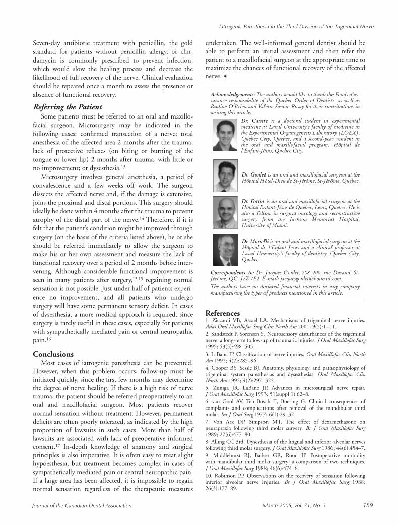
Seven-day antibiotic treatment with penicillin, the goldstandard for patients without penicillin allergy, or clin-damycin is commonly prescribed to prevent infection,which would slow the healing process and decrease the likelihood of full recovery of the nerve. Clinical evaluationshould be repeated once a month to assess the presence orabsence of functional recovery.
Referring the PatientSome patients must be referred to an oral and maxillo-
facial surgeon. Microsurgery may be indicated in thefollowing cases: confirmed transection of a nerve; totalanesthesia of the affected area 2 months after the trauma;lack of protective reflexes (on biting or burning of thetongue or lower lip) 2 months after trauma, with little orno improvement; or dysesthesia.13
Microsurgery involves general anesthesia, a period ofconvalescence and a few weeks off work. The surgeondissects the affected nerve and, if the damage is extensive,joins the proximal and distal portions. This surgery shouldideally be done within 4 months after the trauma to preventatrophy of the distal part of the nerve.14 Therefore, if it isfelt that the patient’s condition might be improved throughsurgery (on the basis of the criteria listed above), he or sheshould be referred immediately to allow the surgeon tomake his or her own assessment and measure the lack offunctional recovery over a period of 2 months before inter-vening. Although considerable functional improvement isseen in many patients after surgery,13,15 regaining normalsensation is not possible. Just under half of patients experi-ence no improvement, and all patients who undergosurgery will have some permanent sensory deficit. In casesof dysesthesia, a more medical approach is required, sincesurgery is rarely useful in these cases, especially for patientswith sympathetically mediated pain or central neuropathicpain.16
ConclusionsMost cases of iatrogenic paresthesia can be prevented.
However, when this problem occurs, follow-up must beinitiated quickly, since the first few months may determinethe degree of nerve healing. If there is a high risk of nervetrauma, the patient should be referred preoperatively to anoral and maxillofacial surgeon. Most patients recovernormal sensation without treatment. However, permanentdeficits are often poorly tolerated, as indicated by the highproportion of lawsuits in such cases. More than half oflawsuits are associated with lack of preoperative informedconsent.17 In-depth knowledge of anatomy and surgicalprinciples is also imperative. It is often easy to treat slighthypoesthesia, but treatment becomes complex in cases ofsympathetically mediated pain or central neuropathic pain.If a large area has been affected, it is impossible to regainnormal sensation regardless of the therapeutic measures
undertaken. The well-informed general dentist should beable to perform an initial assessment and then refer thepatient to a maxillofacial surgeon at the appropriate time tomaximize the chances of functional recovery of the affectednerve. C
Acknowledgements: The authors would like to thank the Fonds d’as-surance responsabilité of the Quebec Order of Dentists, as well asPauline O’Brien and Valérie Savoie-Rosay for their contributions inwriting this article.
Dr. Caissie is a doctoral student in experimental medicine at Laval University’s faculty of medicine inthe Experimental Organogenesis Laboratory (LOEX),Quebec City, Quebec, and a second-year resident inthe oral and maxillofacial program, Hôpital del’Enfant-Jésus, Quebec City.
Dr. Goulet is an oral and maxillofacial surgeon at theHôpital Hôtel-Dieu de St-Jérôme, St-Jérôme, Quebec.
Dr. Fortin is an oral and maxillofacial surgeon at theHôpital Enfant-Jésus de Québec, Lévis, Quebec. He isalso a Fellow in surgical oncology and reconstructivesurgery from the Jackson Memorial Hospital,University of Miami.
Dr. Morielli is an oral and maxillofacial surgeon at theHôpital de l’Enfant-Jésus and a clinical professor atLaval University’s faculty of dentistry, Quebec City,Quebec.
Correspondence to: Dr. Jacques Goulet, 208–200, rue Durand, St-Jérôme, QC J7Z 7E2. E-mail: [email protected].
The authors have no declared financial interests in any company manufacturing the types of products mentioned in this article.
References1. Ziccardi VB, Assael LA. Mechanisms of trigeminal nerve injuries.Atlas Oral Maxillofac Surg Clin North Am 2001; 9(2):1–11.2. Sandstedt P, Sorensen S. Neurosensory disturbances of the trigeminalnerve: a long-term follow-up of traumatic injuries. J Oral Maxillofac Surg1995; 53(5):498–505.3. LaBanc JP. Classification of nerve injuries. Oral Maxillofac Clin NorthAm 1992; 4(2):285–96.4. Cooper BY, Sessle BJ. Anatomy, physiology, and pathophysiology oftrigeminal system paresthesias and dysesthesias. Oral Maxillofac ClinNorth Am 1992; 4(2):297–322.5. Zuniga JR, LaBanc JP. Advances in microsurgical nerve repair.J Oral Maxillofac Surg 1993; 51(suppl 1):62–8.6. van Gool AV, Ten Bosch JJ, Boering G. Clinical consequences ofcomplaints and complications after removal of the mandibular thirdmolar. Int J Oral Surg 1977; 6(1):29–37.7. Von Arx DP, Simpson MT. The effect of dexamethasone onneurapraxia following third molar surgery. Br J Oral Maxillofac Surg1989; 27(6):477–80.8. Alling CC 3rd. Dysesthesia of the lingual and inferior alveolar nervesfollowing third molar surgery. J Oral Maxillofac Surg 1986; 44(6):454–7.9. Middlehurst RJ, Barker GR, Rood JP. Postoperative morbidity with mandibular third molar surgery: a comparison of two techniques.J Oral Maxillofac Surg 1988; 46(6):474–6.10. Robinson PP. Observations on the recovery of sensation followinginferior alveolar nerve injuries. Br J Oral Maxillofac Surg 1988;26(3):177–89.
March 2005, Vol. 71, No. 3 189Journal of the Canadian Dental Association
Iatrogenic Paresthesia in the Third Division of the Trigeminal Nerve
11. Tammisalo T, Happonen RP, Tammisalo EH. Stereographic assess-ment of mandibular canal in relation to the roots of impacted lower thirdmolar using multiprojection narrow beam radiography. Int J OralMaxillofac Surg 1992; 21(2):85–9.12. Gregg JM. Nonsurgical management of traumatic trigeminal neural-gias and sensory neuropathies. Oral Maxillofac Clin North Am 1992;4(2):375–92.13. Pogrel MA. The results of microneurosurgery of the inferior andlingual nerve. J Oral Maxillofac Surg 2002; 60(5):485–9.14. Ruggiero S.Terminal nerve injury and repair. N Y State Dent J 1996;62(8):36–40.15. Gregg JM. Studies of traumatic neuralgia in the maxillofacial region:symptom complexes and response to microsurgery. J Oral Maxillofac Surg1990; 48(2):135–40.16. Gregg JM. Studies of traumatic neuralgias in the maxillofacial region:surgical pathology and neural mechanisms. J Oral Maxillofac Surg 1990;48(3):228–37.17. Lydiatt DD. Litigation and the lingual nerve. J Oral Maxillofac Surg2003; 61(2):197–200.
Journal of the Canadian Dental Association190 March 2005, Vol. 71, No. 3
Caissie, Goulet, Fortin, Morielli
Dentists & Oral HealthProfessionals
Paul DioGuardi Q.C.Tax Lawyer39 Years Experience Formerly Tax Counsel RevenueCanada (CRA)and Departmentof Justice
V-00
7-11
04
Can’t see us in person?Visit our secure, encrypted site:www.taxamnesty.ca
TO ORDER OUR BOOK: “Tax Amnesty –Avoiding the Tax Trap”Visit: www.ontaxpublications.com
— The Tax Amnesty Lawyers —Ottawa: 613-237-2222 Toronto: 416-657-4408
Vancouver: 604-678-8559 Toll-free: 1-866-758-9030
www.effectivetaxsolutions.com
Get a Tax Pardon!Avoid criminal prosecution and penalties up to 250% of the amount you owe. In many cases we can reduce the tax and interest otherwise payable.
DioGuardi & Company, LLP
Anonymity & Confidentiality assured by special legal privilegewith the tax authorities. Your name is only released following
our negotiations and upon legal agreement.
UNLIKE US, YOUR ACCOUNTANT/FINANCIAL PLANNER/ADVISOR CAN BE FORCED TO REVEAL
YOUR IDENTITY AND TESTIFY AGAINST YOU!
LET US REVIEW YOUR TAX PLANNINGYou could be inadvertently committing tax evasion through improper tax and financial planning. Protect yourpractice, family, home and assets!
AVOID BANKRUPTCY!Unlike us, bankruptcy trustees represent your creditors(the tax collector). Because they are partially paid ona results oriented basis, the more tax you pay the moretrustees earn. We have no such conflict of interestand offer a one stop, multidisciplinary approach (taxlawyers, tax accountants and financial planners) tohelp solve your problem.
Our goal, through leveraged negotiationswith the CRA, or if needed, a court applica-
tion, is to reduce your tax liability.
Large, unpayable incometax bill ($175,000+)

May 26 - 29, 2005
Presented by the Alberta Dental Association and College
JasperDental
Congress
Beautiful Jasper, Alberta in the heart of the Rocky Mountainswill be the host venue for this annual conference – four daysof professional development sessions and social activitieswith a distinctively western Canadian flare! The Congressfeatures a world-class program of speakers targeted at theentire dental community: dentists, dental hygienists, dentalassistants and dental specialists.
Enjoy the Great Canadian Lodge experience while you stay at the host hotel, The Fairmont Jasper Park Lodge, knownworldwide for its rustic elegance and natural beauty. Attendscientific sessions and technology fair activities at the JasperActivity Centre.
Mountains of experience await! The congress schedule includes:
Internationally renowned speakers including:
Dr. Cal Torneck• Endodontic Radiology• Classification &
Management of Periapical disease ofEndodontic Origin
Dr. Douglas Dederich• Periodontal Esthetic
Enhancements• Use of Lasers in Dentistry• Current Concepts in
Periodontics
Dr. J. Manhart• Nanoceramic Composites
Dr. Antonio Mancuso • Dental Aesthetic
Assessment
Dr Barbeau• Hand Asepsis and Surface
Disinfection
Dr. Ken Zakariasen • The Great Dental Team –
Getting it Right!• A Systems Approach to
Practice Efficiency andEffectiveness
Ms Sherry Froelich • Stress Busters
Dr David Pfaff • Peak Performance
Ms Susan Isaac • Women's Wellness –
Gender Biology Researchand Perimenopause
Barb Allen & John Webster• Determining Your Personal
Investment Profile – toChoose InvestmentProducts Right for You
• Technology Fair with over 90 exhibitors
• Thursday’s Golf Tournament at the award winning Fairmont Jasper Park Lodge Golf Course
Social Events• The Best of Alberta Welcome Reception• An Evening in Black and White with The Oceanairs• Saturday Finale: Monster Mash featuring Shakin' Not
Stirred, one of Edmonton's hottest bands!
Additional activities for delegates, spouses, partners and family include:
For more information, a complete speakerschedule and the list of hotels where roomshave been blocked and their rates, visit theADA&C's website at www.abda.ab.ca or contactthe ADA&C offices at (780) 432-1012. To avoid disappointment, register early!
• War Canoe Races on Lac Beauvert at the Jasper Park Lodge
• Fun Run/Walk• Youth Day Camp• Cooking Demonstration
• White Water Rafting• Golf Clinic• Pilates• Hiking• Tennis Tournament
The 2005 Jasper Dental Congress is presented in conjunction with the Alberta DentalAssociation and College, Alberta Dental Assistants Association and the Alberta Societyof Dental Specialists
� Dual-curing for more flexibility and efficiency
� Highly radiopaque
� One-step ceramic bond
� High adhesion to ceramic, metal, composite and dentin/enamel
� Clinically proven for years
� Excellent wettability for bulk fillings
� Now available in both universal and transparent shades
New QuickMix syringe
� Fast direct application
� Perfectly mixed, no air bubbles
� Easy and precise dosing
� No gun required
� Economy tips: up to 80% less waste
Universal, dual-cured resin cement. Also ideally suited to use as a bulk-fill posterior restorative.
creative in researchCall toll-free 1-888-658-2584
Get on the fast track …
… with QuickMix syringe11 Hanover Square · 19th Floor · New York · NY 10005 · www.voco.com · [email protected]
Authorized dealers:
Patterson DentalHenry Schein ArconaAsh Temple/ServidentSinclair Dental Co. Ltd.Alpha Dental · Clift’s Dental
Now in QuickMix syringe Provicol QMeugenol-free temporary luting cement
• No mess, no dried out, cracked tubes
• Excellent adhesion to Structur 2 SC and other temporary materials
• Easy clean-up after removal
• Calcium hydroxide - less post-operative sensitivity

March 2005, Vol. 71, No. 3 193Journal of the Canadian Dental Association
Point of CareThe Point of Care section answers everyday clinical questions by providing practical information thataims to be useful at the point of patient care. The responses reflect the opinions of the contributors and do not purport to set forth standards of care or clinical practice guidelines. This month’s responses were provided by speakers at the Annual Spring Meeting of the OntarioDental Association, to be held May 5–7, 2005, in Toronto, Ontario.
Anesthetic-related allergies, even mild ones, constituteless than 1% of medical emergencies in the dental office.Nonetheless, it is important to understand whichsubstances within a local anesthetic solution are possibleallergens and to determine the source of a patient’s allergicreaction.
Local anesthetics are classified as esters or amides. Today,all injectable local anesthetics used in dentistry are amides(Box 1). A wide variety of esters and amides are available astopical anesthetics, for example, benzocaine (an ester),tetracaine (an ester) and lidocaine (an amide).
If a patient is allergic to an ester-based anesthetic, theallergen is not the anesthetic itself but a breakdown product, p-aminobenzoic acid (PABA), which is generatedon metabolization of any ester. Therefore, if a patient isallergic to one ester-type local anesthetic, he or she will beallergic to all such anesthetics. The same is not true foramides, which break down into a variety of metabolites;thus, allergy to one amide should not preclude the use ofanother, unless testing reveals an unusual situation ofmultiple amide allergies. In fact, allergy to any amide-typelocal anesthetic is extremely rare, although some cases havebeen documented.1 If a patient demonstrates an allergy tothe contents of an amide-based anesthetic cartridge, thelikely culprit is the preservative for the vasoconstrictor,sodium metabisulphite. Such patients might reportsulphite allergy in their medical history. They are typicallysensitive to any products that contain sulphite preservatives(Box 2). This type of allergy is not the same as a sulphaallergy, which is an allergy to a class of antibiotics calledsulphonamides. People with sulpha allergies do not demon-strate cross-sensitivity to sulphites. Therefore, if a patientis allergic to sulphite preservatives but not to the local anesthetic itself, he or she can safely tolerate a solution withno vasoconstrictor. The incidence of sulphite allergy ishigher among allergy-prone asthmatic patients than amongnonasthmatic patients.
At one time a bacteriostatic agent called methyl-paraben was available in dental anesthetic cartridges.
Question 1 A patient of mine has reported that she is “allergic to freezing.” What are the causes of local anesthetic-related allergies?
Box 1 Injectable local anesthetics available in Canada
Drug Available as plain solution
Articaine NoBupivacaine NoLidocaine NoMepivacaine YesPrilocaine Yes
Box 2 Foods that contain sulphite preservatives
Salads served in salad barsDried fruitsAlcoholic drinksPotato chipsDeli meatsPicklesCheeseLemon and lime juices, some other fruit juicesGelatinMuffin mixCanned and dried soupsCanned fishCider and vinegar
Methylparaben is also metabolized to PABA, so it was apotential allergen. This product is needed only in multidosevials and is no longer available in dental cartridges.
When there is a question as to the cause of an allergy, thepatient should be sent to an allergist. The dentist shouldrequest testing for a few different anesthetics and for thepreservative sodium metabisulphite. It is also a good idea to give the patient a variety of anesthetic samples to take to the allergist, including a solution that does not containvasoconstrictor.
There is also the possibility of allergy to the latex withinthe cartridge. Latex particles can enter the cartridge afterthe needle perforates the diaphragm or via the plunger (insome cartridges). However, it is unlikely that latex within
Journal of the Canadian Dental Association194 March 2005, Vol. 71, No. 3
P o i n t o f C a r e
the cartridge can induce latex hypersensitivity, as there areno published reports of an allergic response to the latexcomponent of a dental cartridge.2 In any case, companiesare now moving toward use of latex-free components.
If a patient reports an allergy to a local anesthetic, it isof paramount importance to determine the events that ledto the report. Sensitivity to epinephrine or an intravascularinjection commonly leads to misinterpretation of the reaction as an allergy. Affected patients may experiencesymptoms such as palpitations, sweating, nausea, and afeeling of faintness, and some might call this an allergicreaction. Another clinical situation that can be mistakenfor an allergic reaction is an overdose of local anesthetic. Inthis situation, the patient may demonstrate a range of signsand symptoms, including a feeling of discomfort, tingling,metallic taste, confusion, talkativeness, hypertension andincreased pulse. In more extreme overdose situations,
seizures and coma may occur. Allergies do not typicallypresent in this way. Patients must be counselled on thedifferences between allergic reactions and the symptoms ofoverdose and epinephrine-induced reactions. C
Dr. David Isen maintains a private practice in Toronto,Ontario, where he treats patients who require advancedanesthetic care. He has lectured extensively on topicsrelated to local anesthesia, medical emergencies and nitrous oxide sedation. E-mail: [email protected].
Dr. Isen’s session at the ODA meeting, titled “Advanced local anesthe-sia - what you need to know,” will be presented on Thursday, May 5.
References1. MacColl S, Young ER. An allergic reaction following injection of localanesthetic: a case report. J Can Dent Assoc 1989; 55(12):981–4.2. Shojaei AR, Haas, DA, Local anesthetic cartridges and latex allergy: a literature review. J Can Dent Assoc 2002; 68(10):622–6.
BackgroundThe Canadian Cancer Society estimates that 145,500
Canadians will develop cancer during 2004 and that68,300 cancer-related deaths will occur. The managementof many malignancies includes the use of chemotherapeuticdrugs. As these treatments have become more intensive andtherapeutically successful, the complications haveincreased. The mouth is a frequent site of such side effects.In chemotherapy, most complications are the result ofimmunosuppression, myelosuppression and direct cyto-toxic effects on oral tissues (Figs. 1 to 3). Oral complica-tions in chemotherapy patients are usually acute andsubside shortly after the chemotherapeutic drugs leave thesystem. Such side effects include mucositis, infections,hemorrhage, xerostomia and neurotoxicity. Mucositis, themost common acute oral complication of chemotherapy,typically appears 5 to 7 days after the start of treatment (itmay appear as early as 3 days after initiation of cancer ther-apy). Unfortunately, most chemotherapeutic drugs affectnormal tissue as well as the neoplastic cells and tissues. It isthis lack of specificity in the majority of current therapiesthat contributes to the wide range of oral complications.
It is important to determine the cytotoxic, immunosup-pressive and myelosuppressive nature of a chemotherapeu-tic regime. A cytotoxic drug will induce mucositis, whereasan immunosuppressive drug will allow microorganisms toflourish, putting the patient at high risk for periodontal
and odontogenic abscesses as well as viral and fungal infec-tions. A myelosuppressive drug complicates treatment byreducing platelet counts, making emergency surgery androutine oral hygiene dangerous.
Most chemotherapy regimens for cancer comprise acombination of drugs. It is therefore not unusual for apatient to experience both mucositis and immunosuppres-sion. This type of regimen may leave the patient extremelysusceptible to opportunistic infections.
Dental Management of Cancer PatientsThe most logical time to perform dental treatment for a
cancer patient is before the patient’s cancer therapy begins.Most of the cancer patients who are treated in a dentaloncology clinic, however, are seen on emergency referralfrom the oncologist or the dentist and are undergoingactive chemotherapy. Most dental emergencies duringactive chemotherapy could be avoided through aprechemotherapy intraoral examination and a thoroughperiodontal cleaning and appropriate mouth care. Thus, itis unfortunate that few oncologists recommend to theirpatients that they seek treatment from the family dentistbefore chemotherapy commences.
During cancer chemotherapy, dental treatment shouldbe undertaken only on an emergency basis. Such treatmentmay include periodontal cleaning, if the patient’s hygienehas been neglected and he or she has active periodontaldisease. Therefore, such emergency treatment involves any
Question 2 What should I know about treating dental patients who are undergoing chemotherapy andwhen is it the best time for dental treatment?

March 2005, Vol. 71, No. 3 195Journal of the Canadian Dental Association
P o i n t o f C a r e
Figure 1: Secondary bacterial and fungalinfections caused by chemotherapy-induced mucositis.
Figure 2: Candida albicans infection causedby the xerostomic and immunosuppresvieeffects of chemotherapy agents.
Figure 3: Herpes simplex virus on the dorsumof the tongue and C. albicans on thecommisure of the lips caused by theimmunosuppressive effects of chemotherapy.
dental treatment required to remove a source or potentialsource of infection. The practice guidelines listed below areappropriate and safe for any general practice dentist treat-ing a patient who is undergoing cancer therapy. As for anysituation, the dentist’s judgement should be based on his or her own comfort zone.
1. Dental treatment should be undertaken only afterconsultation with the patient’s oncologist or a dentaloncologist (if there is one in your area), to coordinatethe dental treatment with the patient’s optimal hemato-logical status.• White blood cell count must be greater than
1.0 × 10⁹/L. • Platelet count must be greater than 40 × 109/L and
the international normalized ratio must be normal.• Antibiotic prophylaxis is required when the absolute
neutrophil count is less than 2.0 × 10⁹/L.• Patients with indwelling catheters (also called central
venous catheters or Hickman lines) require prophy-lactic antibiotic coverage.
2. The optimal time to perform dental treatment is justbefore a cycle of chemotherapy begins, to maximize thetime before the patient’s condition reaches a nadir.
3. At any time, symptomatic teeth with pulpal involve-ment can be opened, debrided and closed with a temporary restoration.
4. Decay can be excavated and sedative fillings placedanytime during chemotherapy treatment.
5. Generally, extractions are contraindicated except inextreme emergencies (i.e., when an infected tooth maybe the source of systemic infection).
6. A nonflavoured, nonalcohol 0.12% chlorhexidine rinseshould be prescribed for use 4 times daily and up toevery 2 hours in the event of oral mucositis. Becausethere are no clinical practice guidelines for the treatmentof chemotherapy-induced mucositis, chlorhexidine isused at our cancer centre to treat this condition.
7. Petroleum products should be avoided in the treatmentof dry, cracked lips; instead, lanolin should be recommended. C
Dr. Deborah Saunders is head of the dental oncologyprogram at the Northeastern Ontario Regional CancerCentre in Sudbury, Ontario. E-mail: [email protected].
Dr. Saunders’ session at the ODA meeting, titled “Dental care for thecancer patient,” will be presented on Friday, May 6.
Further ReadingMassler CF Jr. Preventing and treating the oral complications of cancer
therapy. Gen Dent 2000; 48(6):652–5.National Institutes of Health consensus development conference
statement: oral complications of cancer therapies: diagnosis, preventionand treatment. J Am Dent Assoc 1989; 119(1):179–83.
Ord RA, Blanchaert RH Jr, editors. Oral cancer: the dentist’s role in diagnosis, management, rehabilitation and prevention. QuintessencePublishing Co.; 2000.
U.S. Department of Health and Human Services. Oral complications ofcancer therapy: what the oncology team can do. NIH Publication No. 99-4360, Bethesda, MD; June 2002. Available from: URL: http://www.nohic.nidcr.nih.gov/campaign/onc_fact.htm.

Journal of the Canadian Dental Association196 March 2005, Vol. 71, No. 3
P o i n t o f C a r e
BackgroundThe complexity of the oral and dental
flora has prevented clear elucidation ofspecific causative agents in most forms ofodontogenic infections. Anaerobic bacte-ria, which are part of the normal oral anddental flora, represent at least 350morphological and biochemically distinctbacterial groups.1
Most odontogenic infections resultinitially from the formation of dentalplaque and continue to develop in areas oftissue damage or trauma. Dental caries,periodontal disease, pericoronitis andpostsurgical wounds are common factorsin odontogenic infections. Once patho-genic bacteria become established, theycan cause a wide variety of local anddisseminated complications. The most common infectionsinclude dentoalveolar infections, gingival infections, andperiodontitis. These can be categorized as localized infec-tions (such as acute periodontal abscesses), spreading infec-tions (such as early cellulitis and infections with deep spaceinvolvement) and life-threatening infections (such asnecrotizing fasciitis and Ludwig’s angina).2
Odontogenic infections are generally caused by mixedaerobic and anaerobic polymicrobial bacteria from thesame families of oral microorganisms (obligate anaerobesand gram-positive aerobes). The microorganisms recoveredfrom infections generally reflect the host’s indigenous oralflora. Therefore the choice of antibiotic to treat odontogenicinfections must be made according to the polymicrobialnature of such infections and local resistance patterns.
In all instances of odontogenic infections it is essentialthat the airway be assessed and secured if necessary, as theinitial life-saving manoeuvre. If there is an abscess to bedrained or necrotic tissue requiring removal or debridement,this must also be done.2,3 Attention can then turn to antibi-otic choices. One common mistake is the tendency tounderdose the antibiotic, which is assumed to be one of thecauses of antibiotic treatment failures.4 Practitioners mustbe aware of the appropriate pediatric and adult dosages ofantibiotics that are useful in odontogenic infections4
(Box 1). Moreover, the course of the infection must bemonitored continuously both clinically and by followingthe results of culture and sensitivity testing. Widespreadresistance of pathogens is another important cause ofantibiotic treatment failures. General practitioners must beprepared for early referral of such cases to a specialist if the
infection is not responding to treatment, especially if thereare any airway or neurological concerns.
Choice of AntibioticsThe gold standard first-choice antibiotic has historically
been penicillin for patients not allergic to this drug.Phenoxymethyl-penicillin, or penicillin V, can be used totreat the vast majority of odontogenic infections. It is moreresistant to gastric acids than its predecessor, penicillin G,and it is very well tolerated orally. However, resistant bacte-rial species, particularly those that elaborate β-lactamase,have made the treatment of odontogenic infections morecomplex and difficult. Amoxicillin has a spectrum similarto penicillin, and its effectiveness against Haemophilusinfluenzae can be useful. Amoxicillin can also be effectiveagainst bacterial species that produce β-lactamase ifcombined with clavulanic acid. Diarrhea may be one majorside effect of the amoxicillin–clavulanate combination.Cephalosporins offer no major advantages over the peni-cillins and are much more expensive.
Although penicillin is still a good first choice today, itsspectrum of activity may need to be augmented. One possibility is metronidazole, a bacteriostatic agent that ishighly active against most anaerobes but which has poorcoverage of aerobic species. Metronidazole should never beused on its own to treat an acute odontogenic infection.The use of metronidazole may not be entirely benign, andside effects may occur; for example, metronidazole maycause an Antabuse-type reaction if combined with alcohol,and peripheral neuropathies have been reported.
Question 3 What are the choices of antibiotics for the treatment of acute odontogenic infections?
Box 1 Antibiotic choices and dosages for dental practitioners treating acute odontogenic infections
Antimicrobial drug Adult dosage Pediatric dosage
Penicillin V 600 mg 4 times a day 30–50 mg/kg per dayin 4 divided doses
Amoxicillin 500 mg 3 times a day 20–50 mg/kg per dayin 3 divided doses
Amoxicillin–clavulanate 500/125 mg 3 times a day 40 /10 mg/kg per dayin 3 divided doses
Metronidazole 500 mg 3 times a day 15–30 mg/kg per dayin 3 divided doses
Clindamycin 150–300 mg 4 times a day 10–30 mg/kg per dayin 3 divided doses
Ciprofloxacin 250–750 mg twice a day 40 mg/kg per dayin 2 divided doses
March 2005, Vol. 71, No. 3 197Journal of the Canadian Dental Association
P o i n t o f C a r e
Erythromycin and tetracycline have limited applicationin dentistry. Erythromycin is a bactericidal antibiotic witha poor performance record in odontogenic infections.There are serious compliance issues because of the intensenausea and vomiting that this drug can cause. When givenintravenously, erythromycin tends to be extremely irritat-ing to the veins. The usefulness of tetracycline has beendiminished by widespread resistance.
Clindamycin is the drug of choice for patients with ahistory of penicillin allergy. Clindamycin has the advantageof reliable coverage against gram-positive aerobic andanaerobic bacteria, with the possibility of attaining highintra-bony levels with both intraoral and intravenousadministration. The biggest disadvantage of clindamycinis its association with pseudomembranous colitis. The2 groups at greatest risk appear to be elderly patients andpatients who have recently had long-term hospital stays andare therefore at risk for nosocomial (hospital-acquired)infections. The incidence of clindamycin resistance alsoseems to be increasing.
Ciprofloxacin may be one other antibiotic to consider.Like the other fluoroquinolones, this unique fluoroquinoloneantibiotic has potent gram-negative activity inhibiting DNA gyrase and topoisomerase IV.5 Ciprofloxacin is also
effective against gram-positive organisms and may be usedtogether with clindamycin. C
Dr. George Sándor is an associate professor and director, graduate program in oral and maxillofacialsurgery and anesthesia, University of Toronto, and is thecoordinator of oral and maxillofacial surgery at TheHospital for Sick Children and Bloorview MacMillanChildren’s Centre, Toronto, Ontario, and docent in
oral and maxillofacial surgery, University of Oulu, Oulu, Finland. E-mail: [email protected].
Dr. Sándor’s sessions at the ODA meeting, titled “Keeping generalpractitioners out of trouble when performing dento-alveolar surgery”and “The ever changing face of odontogenic infections,” will bepresented on Friday, May 6.
References1. Sandor GK, Low DE, Judd PL, Davidson RJ. Antimicrobial treatmentoptions in the management of odontogenic infections. J Can Dent Assoc1998; 64(7):508–14.2. Fenton CC, Kertesz T, Baker G, Sandor GK. Necrotizing fasciitis of the face: a rare but dangerous complication of dental infection.J Can Dent Assoc 2004; 70(9):611–5.3. Judd PL, Sandor GK. Management of odontogenic orofacial infectionin the young child. Ont Dent 1997; 74(8):39–43, 45.4. Barron RP, Freilich MM, Sandor GKB. Extraction timing in paediatricodontogenic infections. Ont Dent 2001; 78(8):15–8.5. Drlica K, Zhao X. DNA gyrase, topoisomerase IV, and the4-quinolones. Microbiol Mol Biol Rev 1997; 61(3):377–92.
Like all drugs, sedatives produce a range of effectsdepending on patient factors. In most patients, mild anxiolysis or light sedation results, but at the other end ofthe spectrum (in rare cases) a near catatonic state can occur.Sedatives can cause a patient to become unresponsive,which can lead to hypoventilation or an inability to main-tain the airway. Luckily, standard benzodiazepines, alcoholsand hypnotics have a wide therapeutic margin and aregenerally safe. So safe, in fact, that current Royal College of Dental Surgeons of Ontario guidelines do not requirepulse oximetry or other mechanical monitors when thesedrugs are given as a single agent by mouth. However, if apractitioner chooses a mechanical monitor during single-agent sedation, a pulse oximeter is an excellent first choice.
What Does a Pulse Oximeter Do?Pulse oximeters measure the level of oxygen in the blood.
Oximetry is generally accurate, non-invasive and sensitive tochanges in hypoventilation. The oximeter, which is attachedto the patient’s finger using a small clip, shines a lightthrough the nail bed and measures the ratio of oxygenated todeoxygenated hemoglobin (Fig. 1). This measurement iscalculated continuously and averaged over 5–60 seconds,
depending on the machine settings. This means that changesin the amount of oxygen in the blood will be seen on theoximeter as soon as the value is calculated.
When a patient loses consciousness to the point that heor she is unable to maintain the airway, breathing stops.The level of carbon dioxide in the blood rises, creating theurge to breathe that one normally feels when holding one’sbreath. As minutes tick by, oxygen dissociates from hememolecules in the red blood cells and is used by the body. At first, the amount of oxygen in the blood drops slowly(Fig. 2), but as hemoglobin releases oxygen, oxygen’s affin-ity for the heme molecule decreases and it is released evermore quickly. To the clinician, this is important because thedrop in a patient’s oxygen saturation from 100% to 90% isrelatively slow (usually minutes), but the drop from about85% to 10% is very rapid (usually seconds). At the pointwhere this acceleration begins, the patient will appear blue(cyanotic) and the problem becomes quickly apparent. Ifoxygen saturation remains low for more than 4–6 minutes,death will result. The bottom line is that oxygen saturationbelow 85%, in a sedated patient, must be treated immedi-ately to prevent dire consequences.
Question 4 I use oral sedatives in my office for children and anxious patients. Should I use pulse oximetry?

Journal of the Canadian Dental Association198 March 2005, Vol. 71, No. 3
P o i n t o f C a r e
Figure 1: Oxygen saturation monitor withfinger clip (the patient is breathing room airand oxygen saturation is 98%).
Figure 2: The oxygen dissociation curveshowing steep drop of 85% on the verticalaxis.
Figure 3: If a patient has an oxygensaturation of 37% (critically low), ventilateusing a bag-valve mask and 100% oxygen.
Practical Use of an OximeterThe good news is that the body has a certain store of
oxygen, so the drop in oxygen can be measured and theprocess reversed before the results become permanent. If apatient is on an oximeter and oxygen saturation beginsto fall, basic steps can usually reverse the problem(assuming it is related to excessive sedation).
1. If the patient is conscious, tell him or her to take somedeep breaths.
2. If the patient is conscious, give supplemental oxygenwith a mask.
3. If the patient is unconscious, try to rouse him or her,then open the airway.
4. If the airway is patent, use a bag-valve mask (BVM) withsupplemental oxygen.
5. If the patient is unconscious but breathing, “assist”the breaths with the BVM until oxygen saturation is96% or more; if the patient is not breathing, give1 breath every 5 seconds until that saturation level isreached (Fig. 3).
6. Consider a reversal agent (flumazenil for benzodi-azepines or naloxone for narcotics) to wake the patient.
Like all pieces of equipment, the pulse oximeter can giveinaccurate readings. The most common reasons for a falselow value are movement, cold fingers (causing inadequateperipheral circulation for accurate measurements) and nailpolish. If your office conducts sedation, consider a pulseoximeter and take the following steps:
• Carefully read the manual to learn over how long aperiod the values are averaged, how to maintain theequipment and how long the battery will last withoutpower.
• Ensure that support equipment (supplemental oxygen) isreadily available, familiar to all staff and well maintained.
• Run office emergency drills to practise dealing withsome of the situations listed above.
• Ensure that the protocols you use and the drugs you giveare in accordance with the provincial guidelines.
A pulse oximeter is an inexpensive, easy-to-use piece ofequipment to monitor patients accurately during any levelof sedation. Using it can increase the margin of safety withsedated patients and provide an extra measure of comfort tothe staff providing care. C
Dr. Ian Furst is an oral and maxillofacial surgeon withthe Coronation Dental Specialty Group and staffsurgeon at Cambridge Memorial Hospital, Cambridge,Ontario. He is president of the Ontario Dental Societyof Anaesthesiology. E-mail: [email protected].
Dr. Furst’s sessions at the ODA meeting, titled “Anesthesia and anesthet-ics in a general dental practice,” will be presented on Thursday, May 5.
Further ReadingFearnley SJ. Pulse oximetry. Update in Anaesthesia 1995; Issue 5. Available
from: URL: www.nda.ox.ac.uk/wfsa/html/u05/u05_003.htm.Guidelines: use of sedation and general anaesthesia in dental practice.
Toronto: Royal College of Dental Surgeons of Ontario; January 2005.Available from: URL: www.rcdso.org/pdf/guidelines/Guidelines_sedation_01_05.pdf.
Hata T, Nickel EJ, Hindman B, Morgan D. Procedural sedation resourcecenter: guidelines, education, and testing for procedural sedation andanalgesia. Pulse oximetry. Iowa City: Virtual Hospital, University ofIowa. Available from: URL: www.vh.org/adult/provider/anesthesia/ProceduralSedation/PulseOximetry.html.
Hill E, Stoneham MD. Practical applications of pulse oximetry. Updatein Anaesthesia 2000; Issue 11. Available from: URL: www.nda.ox.ac.uk/wfsa/html/u11/u1104_01.htm.
The use of pulse oximetry during conscious sedation. Council onScientific Affairs, American Medical Association. JAMA 1993;270(12):1463–8.
Web site on pulse oximetry. Available from: URL: www.pulseox.info/index.htm.


Journal of the Canadian Dental Association200 March 2005, Vol. 71, No. 3
Clinical Showcase
Traditionally, foreign bodies displaced into the maxillarysinus are retrieved either through the entrance wound (suchas an extraction site) or through a secondary site. TheCaldwell-Luc approach was the gold standard for access tothe maxillary sinus for treatment of various problems,including retrieval of foreign bodies, until the developmentof functional endoscopic sinus surgery (FESS). Both tech-niques have limitations and potential complications. Thiscase presentation demonstrates the use of a modifiedCaldwell-Luc approach for retrieval of a foreign body fromthe maxillary sinus in a case where retrieval would not havebeen possible with the FESS technique because of the largesize of the foreign body. The technique employed in thiscase takes advantage of lessons learned from minimallyinvasive surgery and FESS.
Case HistoryA 54-year-old man was seen for an emergency consulta-
tion after removal of teeth 26 and 27. The patient’s historyindicated that the high-speed surgical bur used for removalof the teeth had become dislodged somewhere within thepatient’s oral cavity. The patient had well controlled hyper-tension and bipolar disorder.
Clinical examination identified active epistaxis andbleeding from a patent oroantral fistula. The findings onneurological examination were unremarkable. The patienthad no visual disturbance, and he was hemodynamicallystable.
Patient ManagementPrimary treatment included local measures to achieve
hemostasis and an attempt to locate the dislodged bur. Thebur could not be visualized clinically at that time, and thewound was closed primarily to achieve hemostasis and closethe oroantral fistula. Panoramic and plain film radiographyindicated that the surgical bur was within the maxillarysinus (Fig. 1). The patient was admitted to the oral andmaxillofacial surgery service for further investigations,including computed tomography. This imaging confirmedthat the surgical bur lay within the sphenoidal recess of themaxillary antrum (Figs. 2 and 3).
The surgical plan was to encourage closure of theoroantral fistula and then to retrieve the foreign body by anendoscopically assisted Caldwell-Luc procedure. It was felt
that the endoscope would allow for direct visualization ofthe surgical bur and would facilitate controlled removal.The patient was given antibiotics and topical and systemicdecongestants and was discharged. After approximately3 weeks of healing, the oroantral fistula had closed, and anappointment was scheduled for retrieval of the foreignbody.
With the patient under general anesthesia, standardsurgical technique was used to create a small osteotomy inthe lateral antral wall superior to the root apices of thepremolar teeth. The position of the infraorbital nerve wasidentified, and the nerve was protected. The size of theopening was restricted but sufficient to allow passage of a4.0-mm endoscope (Karl Storz Endoscopy AmericaIncorporated, Culver City, Calif.) and a probe. The aperture of the ostium was approximately 1.25 cm in diam-eter. The antrum was thoroughly examined through theendoscope (Fig. 4). The surgical bur was easily identified; itwas embedded in the medial–superior recess (Fig. 5), ashad been predicted by the radiographic assessment. A surgi-cal grasper was inserted, and the bur was gently removed(Figs. 6 and 7). Associated inflammatory tissue wasdebrided. The wound was irrigated copiously and closedprimarily. The patient was maintained on a course of post-operative antibiotics and decongestants for 10 days follow-ing surgery.
OutcomeThe patient’s immediate and long-term recovery was
uneventful. There was minimal surgical edema. The integrityof the maxillary division of the trigeminal nerve waspreserved; the patient had no complaints and testing showedno signs of nerve injury. There were no postoperativeantrum-related complaints; the wounds healed completely,and there was no residual oroantral fistula.
Lessons to be LearnedSpecial care must be taken to ensure that adjacent hard
and soft tissues are protected at all times from iatrogenicinjury. This case demonstrates the potential for a surgicalinstrument to become embedded in contiguous structures.In this case, a surgical bur lodged just millimetres from theorbital contents within the ethmoidal recess of the maxil-lary antrum. It is the authors’ opinion that the surgical bur
Endoscopically Assisted Caldwell-Luc Procedure for Removal of a Foreign Body from the Maxillary SinusJoseph Friedlich, DDS, FRCD(C), Dip ABOMSBrian N. Rittenberg, BA, DDS, MSc, FRCD(C)
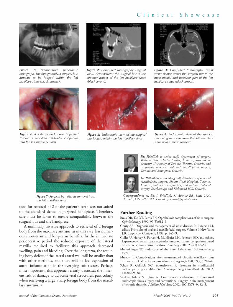
March 2005, Vol. 71, No. 3 201Journal of the Canadian Dental Association
C l i n i c a l S h o w c a s e
Figure 6: Endoscopic view of the surgicalbur being removed from the left maxillarysinus with a micro rongeur.
Figure 4: A 4.0-mm endoscope is passedthrough a modified Caldwell-Luc openinginto the left maxillary sinus.
Figure 5: Endoscopic view of the surgicalbur lodged within the left maxillary sinus.
used for removal of 2 of the patient’s teeth was not suitedto the standard dental high-speed handpiece. Therefore,care must be taken to ensure compatibility between thesurgical bur and the handpiece.
A minimally invasive approach to retrieval of a foreignbody from the maxillary antrum, as in this case, has numer-ous short-term and long-term benefits. In the immediateperioperative period the reduced exposure of the lateralmaxilla required to facilitate this approach decreasedswelling, pain and bleeding. Over the long term, the result-ing bony defect of the lateral antral wall will be smaller thanwith other methods, and there will be less expression ofantral inflammation in the overlying soft tissues. Perhapsmost important, this approach clearly decreases the inher-ent risk of damage to adjacent vital structures, particularlywhen retrieving a large, sharp foreign body from the maxil-lary antrum. C
Dr. Friedlich is active staff, department of surgery,William Osler Health Centre, Ontario, associate indentistry, University of Toronto, Toronto, Ontario, andin private practice, oral and maxillofacial surgery,Toronto and Brampton, Ontario.
Dr. Rittenberg is attending staff, department of oral andmaxillofacial surgery, Mount Sinai Hospital, Toronto,Ontario, and in private practice, oral and maxillofacialsurgery, Scarborough and Richmond Hill, Ontario.
Correspondence to: Dr. J. Friedlich, 55 Avenue Rd., Suite 2100,Toronto, ON M5P 3E5. E-mail: [email protected].
Further ReadingBuus DR, Tse DT, Farris BK. Ophthalmic complications of sinus surgery.
Ophthalmology 1990; 97(5):612–9.Gonty AA. Diagnosis and management of sinus disease. In: Peterson LJ,
editor. Principles of oral and maxillofacial surgery. Volume I. New York:J.B. Lippincott Company; 1992. p. 245–9.
Guller U, Hervey S, Purves H, Muhlbaier LH, Peterson ED, and others.Laparoscopic versus open appendectomy: outcomes comparison basedon a large administrative database. Ann Surg 2004; 239(1):43–52.
Messerklinger W. Endoscopy of the nose. Urban and Schwarzenberg;1978.
Murray JP. Complications after treatment of chronic maxillary sinusdisease with Caldwell-Luc procedure. Laryngoscope 1983; 93(3):282–4.
Schon R, Gellrich NC, Schmelzeisen R. Frontiers in maxillofacialendoscopic surgery. Atlas Oral Maxillofac Surg Clin North Am 2003;11(2):209–38.
Venkatachalam VP, Jain A. Comparative evaluation of functionalendoscopic sinus surgery and conventional surgery in the managementof chronic sinusitis. J Indian Med Assoc 2002; 100(2):78–9, 82–3.
Figure 3: Computed tomography (axialview) demonstrates the surgical bur in themost medial and posterior part of the leftmaxillary sinus (black arrow).
Figure 1: Preoperative panoramicradiograph. The foreign body, a surgical bur,appears to be lodged within the leftmaxillary sinus (black arrows).
Figure 2: Computed tomography (sagittalview) demonstrates the surgical bur in thesuperior aspect of the left maxillary sinus(black arrow).
Figure 7: Surgical bur after its removal fromthe left maxillary sinus.
�
�
�
�
Take a look at the Canadian Dentists’ Investment Program. You’ll see that it offers what you need to build and
manage wealth for every stage in your life:
• Exceptional RRSP, RRIF, RESP, non-registered and self-directed plans
• An individual pension plan with higher contribution limits and greater tax savings than RRSPs
• No-load CDA funds with management fees among the lowest you’ll find anywhere
• No-cost portfolio rebalancing and retirement savings progress reports
• Easy to read statements and online account access
• Free, expert advice† from certified financial planners
Don’t miss out on a good thing. Call today to have a Personal Investment Advisor from Professional Guide LineInc. — A CDSPI Affiliate assigned to you and discuss your personalized strategy for investing.†
1-877-293-9455, extension 5023† Restrictions may apply to advisory services in certain jurisdictions.
05-58 01/05
Find out what you’re missingFind out what you’re missing
Th e Ca n a d i a n D e n t i s t s’ I nve s t m e n t P r o g ra m

March 2005, Vol. 71, No. 3 203Journal of the Canadian Dental Association
CDSPI ReportsARE YOU
READY TOBECOME
DISABLED?
By Susan Roberts
Your chances are like the flip of a coin: As surprising as thismay sound, actuarial statistics
reveal the disturbing reality thatdentists stand about a 50-50 chance*of becoming disabled for an extendedperiod at some point during theircareers.
Given the relatively high probabil-ity of experiencing a disabling illnessor injury, it’s prudent for all dentiststo prepare for the prospect. In doingso, consider the following suggestions.
Ensure Your DisabilityInsurance Fully Protects You
The amount of disability insuranceyou’re eligible to purchase depends on your income. That’s why it’s crucialto carefully review your coverage eachyear. If your income significantlyincreases and you don’t alter yourcoverage to reflect that change, youcould suffer needless financial hard-ships in the event of a disability.
Additionally, the Canadian Dentists’Insurance Program’s Long TermDisability (LTD) Insurance planoffers options that can enhance yourdisability insurance protection. Forexample, the Future InsuranceGuarantee (FIG) Option allows youto increase your coverage at specificpoints in your life without having tosubmit medical information. Call tolearn more about FIG and otheroptions to enhance your coverage.
If you’re responsible for paying allor a portion of your practice costs,you should also consider obtaining
the Program’s Office OverheadExpense (OOE) Insurance plan —which covers certain office-relatedcosts in order to keep your practicegoing when a disability prevents youfrom practising. It’s important toconsider obtaining this coverage evenif you share office expenses with apartner, since the terms of yourprofessional agreement could legallybind you to pay for your share of over-head costs — even during a disability.
To ensure you’re properly protectedby your disability insurance coverages,speak to your personal insurance advi-sor at Professional Guide Line Inc. fora no-cost review.
Establish an Emergency Fund Before disability insurance claim
payments begin, you must first satisfyan elimination period. During thisperiod — beginning with the onset ofa disability — benefits are notpayable. Depending on the elimina-tion period you chose when youpurchased your Program LTD cover-age, it could be as long as 120 daysbefore payments begin. Therefore,consider establishing an emergencyfund that would sufficiently coveryour living expenses until your bene-fits start coming in.
When deciding upon how much toput aside in your emergency fund,bear in mind that a disability couldintroduce new expenses into yourbudget — such as the cost of expen-sive medication.
Grant Power of AttorneyIf a disability left you incapaci-
tated, who would be authorized tomake decisions about your personalfinancial affairs and how your practicewould operate in your absence? Youcan prepare for this contingency bygranting power of attorney to some-one you trust. Contact your lawyer foradvice.
You may also wish to spell out in adocument how your practice shouldbe operated in your absence. Forexample, it could include the name ofa colleague who would be willing totreat your patients until a locum isfound, and who would be authorizedto pay expenses on your behalf. Oncedrafted, consider discussing yourdisability contingency plan with yourfamily and staff.
Know What to Expect WhenFiling a Disability InsuranceClaim
Here is an overview of the initialsteps involved should you need to filea claim under your Insurance ProgramLTD and/or OOE coverage:
First, you’ll need to contactCDSPI’s Claim Support Centre —which serves as a liaison betweenclaimants and the insurer (the insureris the sole adjudicator of your claim).The Claim Support Centre represen-tative will take some preliminaryinformation about your claim, informthe insurer about your intent to file aclaim, and then send you a Claim Kitthat includes forms you’ll need tocomplete for the insurer.
The Claim Kit will include a formthat will need to be completed by yourphysician, as well as forms pertaining toyour finances to determine your benefitamount. You’ll also need to submitsupporting documents with thesefinancial forms, which may include:complete copies of your personal taxreturn and Notice of Assessment docu-ments for your 2 most recent taxationyears, copies of financial statements,partnership agreements, corporate taxinformation and financial agreementswith management companies andlocums. In the case of an OOE claim,you will also be required to submit abreakdown of your office expenses inthe 3-month period prior to when yourdisability commenced.

Journal of the Canadian Dental Association204 March 2005, Vol. 71, No. 3
C D S P I R e p o r t s
(During the claim process, you’llalso be in contact with the insurer’sclaims consultant, who may requestadditional financial and medicalinformation.)
If you become disabled, you willneed to supply many different types offinancial documents to the insurer(within 90 days of the date your disabil-ity commenced) at a time when youmay not find it easy to assemble thesepapers. Since a disability could leaveyou incapacitated, consider whethersomeone acting on your behalf couldeasily procure these documents — andperhaps create a filing system wherethey could be easily found.
Takes Steps to Reduce YourChances of Becoming Disabled
Not all types of disabilities
common to dentists are preventable,but many others are. That’s whyCDSPI’s Claim Support Centrecreated an informative booklet calledLong Term Disability and You —Reducing the Risk. It contains informa-tion specific to dentists about ways to lessen the likelihood of suffering a disability. To download a free copy,click on “Loss Prevention” at www.cdspi.com/claims. C
Susan Roberts is the servicesupervisor for the CanadianDentists’ Insurance Program.
For no-cost insurance planning advice,**speak to your personal insurance advisor atProfessional Guide Line Inc. — A CDSPIAffiliate. Dial 1-877-293-9455 (toll-free) or416-296-9455, extension 5002.
The Canadian Dentists’ Insurance Program’sLong Term Disability and Office OverheadExpense Insurance plans are underwritten byThe Manufacturers Life Insurance Company(Manulife Financial).
*Source: Commissioner’s IDA MorbidityTable, Society of Actuaries.
**Restrictions may apply to advisory servicesin certain jurisdictions. Residents of Quebecand PEI, please call CDSPI at 1-800-561-9401, extension 5000, for insurance planinformation.
Informationprovided byCanadian DentalService Plans Inc.
Dental offices have contacted the Canadian Dental Association (CDA) expressing their dissatisfaction with a dental supply company named Canada Wide DentalSupplies. A sales representative for this company repeatedly telephones dental offices to solicit business, despite explicit requests to stop calling. Several officesdescribe the calls as “harassing” and are frustrated with the actions of this sales representative.
CDA reminds readers to exercise caution by not disclosing credit card details to companies with whom they are unfamiliar.
Take ActionOffices can advise such harassing callers to place them on a “do not call” list. Legitimate dental supply companies should respect such a request and discontinuecontact by telephone.
The Canadian Marketing Association offers its Do Not Contact service free of charge (www.cmaconsumersense.org/marketing_lists.cfm). This service removes yourname and telephone number from telemarketing call lists and helps reduce the number of marketing offers received by mail, telephone and fax.
Bell Canada offers privacy services on most of its business or residential lines for a monthly fee (www3.bell.ca/ecare/PrsCSrvMgAc_Phone.page). These servicesinclude a Call Screening option, which can block up to 12 incoming telephone numbers. A Call Privacy option forces callers from “private” or “unknown” numbersto enter their phone number or a 10-digit number before your office phone will ring.
Bell Canada also suggests compiling a list of the harassing phone calls, noting the date, time and phone number of the calls. Bell will then register and distributethis information to participating phone service providers across the country.
Stay InformedCDA first notified its members about Canada Wide Dental Supplies in a CDAlert distributed by e-mail on January 27, 2005. A complete archive of these electronicbulletins can be found on the members’ side of the CDA Web site (www.cda-adc.ca/english/members/cda_members/member_news/cdalert/default.asp).
If you would like to receive future CDAlerts, contact a CDA membership services representative at 1-800-267-6354 or e-mail [email protected].
Caution to readers
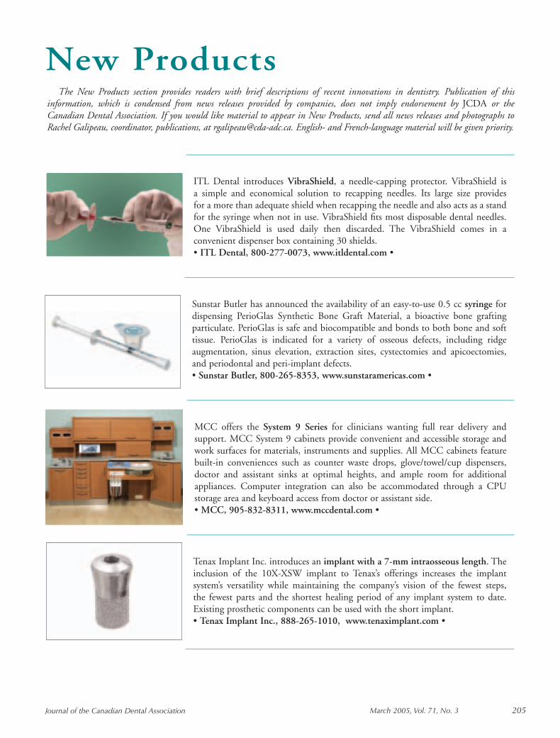
March 2005, Vol. 71, No. 3 205Journal of the Canadian Dental Association
New ProductsThe New Products section provides readers with brief descriptions of recent innovations in dentistry. Publication of this
information, which is condensed from news releases provided by companies, does not imply endorsement by JCDA or the Canadian Dental Association. If you would like material to appear in New Products, send all news releases and photographs toRachel Galipeau, coordinator, publications, at [email protected]. English- and French-language material will be given priority.
ITL Dental introduces VibraShield, a needle-capping protector. VibraShield is a simple and economical solution to recapping needles. Its large size provides for a more than adequate shield when recapping the needle and also acts as a standfor the syringe when not in use. VibraShield fits most disposable dental needles.One VibraShield is used daily then discarded. The VibraShield comes in a convenient dispenser box containing 30 shields.• ITL Dental, 800-277-0073, www.itldental.com •
Sunstar Butler has announced the availability of an easy-to-use 0.5 cc syringe fordispensing PerioGlas Synthetic Bone Graft Material, a bioactive bone graftingparticulate. PerioGlas is safe and biocompatible and bonds to both bone and softtissue. PerioGlas is indicated for a variety of osseous defects, including ridgeaugmentation, sinus elevation, extraction sites, cystectomies and apicoectomies,and periodontal and peri-implant defects. • Sunstar Butler, 800-265-8353, www.sunstaramericas.com •
MCC offers the System 9 Series for clinicians wanting full rear delivery andsupport. MCC System 9 cabinets provide convenient and accessible storage andwork surfaces for materials, instruments and supplies. All MCC cabinets featurebuilt-in conveniences such as counter waste drops, glove/towel/cup dispensers,doctor and assistant sinks at optimal heights, and ample room for additional appliances. Computer integration can also be accommodated through a CPU storage area and keyboard access from doctor or assistant side.• MCC, 905-832-8311, www.mccdental.com •
Tenax Implant Inc. introduces an implant with a 7-mm intraosseous length. Theinclusion of the 10X-XSW implant to Tenax’s offerings increases the implantsystem’s versatility while maintaining the company’s vision of the fewest steps, the fewest parts and the shortest healing period of any implant system to date.Existing prosthetic components can be used with the short implant.• Tenax Implant Inc., 888-265-1010, www.tenaximplant.com •


Classified AdsGuaranteed access to Canada’s largest audience of dentists
Cla
ssifi
ed
Ad
s
To place your ad, contact:
Beverley Kirkpatrick or Deborah Rodd
c/o Canadian Medical Association1867 Alta Vista Dr. Ottawa, ON K1G 3Y6Tel.: 800 663-7336 or
(613) 731-9331, ext 2127or 2314
Fax: (613) 565-7488E-mail: [email protected]
Placement of ads by telephone not accepted.
Deadline Dates
Issue Closing DateApril March 10May April 8
Send all box number replies to:
Box ... JCDA1867 Alta Vista Dr.Ottawa, ON K1G 3Y6
The names and addresses of advertis-ers using box numbers are held instrict confidence.
Display Advertising Rates
1 page 1,795 1⁄3 page 6502⁄3 page 1,275 1⁄4 page 5651⁄2 page 955 1⁄6 page 4451⁄8 page 305
Regular Classified Rates
$95 for the first 50 words or fewer,each additional word 85¢. Reply boxnumbers $20 (first insertion only).
Special Display (2 1⁄8˝ x 2 1⁄8˝) $225.
All advertisements must be prepaid.
10% discount to CDA members.
O F F I C E S &P R A C T I C E S
ALBERTA - Lethbridge: A city of66,000 servicing a surrounding popula-tion of about 150,000, Lethbridge is oneof the fastest growing cities in Alberta.Recreation, sunshine, location and qual-ity of life are its appeal. One of Leth-bridge’s premier dental offices is now forsale in the second busiest mall in the city.This vibrant office offers both exclusivityand exposure. Go to www.drchuck.ca andwww.lethbridgepages.com D1683
ALBERTA/BRITISH COLUMBIA -Wanted: A solo practice to purchase. E-mail [email protected], tel. (403)969-8786. D1691
ALBERTA - Edmonton: Location well-suited for periodontist, availablebeside established prosthodontist. Ex-ceptionally located and attractive prop-erty offers 1,600 ft. of space for lease inbrand new building. Prime exposure onstreet level in Edmonton, Alberta. Call(780) 420-1604 to enquire about thisopportunity. D1622
ALBERTA - Rimbey: Opportunity topurchase practice or associate with tran-sition to ownership in this familyoriented, solo, rural practice. New gradu-ates welcome. Owner willing to assistwith transition. Excellent long-term staff. Practice grossing $580,000/year on4 days/week. Good leaseholds. Low overhead. One hour south of Edmonton.Priced reasonably. Contact: Anne,tel. (403) 843-2173. D1569
ALBERTA - Edmonton: Well-estab-lished, solo dental practice for sale, fewblocks away from West Edmonton Mall;3 operatories, very bright and beautifuloffice; 1200 active and pleasant patients.
The present dentist is moving out of Edmonton, will help in transition. Tel.(780) 987-9198. D1495
BRITISH COLUMBIA - Kelowna:Prosthodontic practice for sale. Attractive,high-income (7-figure billings), modern,3-operatory office. Computerized office,digital x-ray, new panoramic machine. All facets of prosthodontics - fixed/removable, implants, cosmetic proce-dures. Hygienist on staff. Very desirablearea. Independently appraised. Completetransition available (owner willing to stayon as an associate). Well-established referral base. E-mail [email protected]. D1651
BRITISH COLUMBIA - Courtenay:Comox Valley, Vancouver Island. Arapidly growing community with recre-ational and educational advantages for the whole family - skiing atMt. Washington, freshwater and saltwater fishing, hiking, biking, swimming,beachwalking, etc. Three excellent highschools, excellent community college.Well-established, quality care generalpractice. Preventive based, high-endrestorative; 1,400 active charts. Twohygiene operatories and 4 restorativeoperatories in 2,200 sq. ft. Building andequipment 11years old. Three differenthealth care offices in building. Grossmid $500,000 on 180 days. Tel. (250)338-6080 (private). D1656
BRITISH COLUMBIA - Surrey:Long-time, well-established practice,approximately 2,200 active patients andapproximately 5,500 on file. Great newpatient flow. Seven operatories all fur-nished and plumbed. Grossing approxi-mately $750,000 a year with 1 dentist.Well-trained, long-term and highlymotivated staff. Owner leaving to special-ize. Please e-mail Dr. R. Standerwick,[email protected]. D1655
Journal of the Canadian Dental Association March 2005, Vol. 71, No. 3 207

Cla
ssifi
ed
Ad
s
BRITISH COLUMBIA - Kitimat:Well-established general practice for sale.Hygienist-supported recall and perio pro-gram, in a great town with a solid long-term industrial base. All kinds of outdoorand indoor recreation available minutesfrom your doorstep. No traffic jams andgood income on 4-day week. Owner relo-cating for family reasons. Tel. (604) 576-1176 for more information. D1423
BRITISH COLUMBIA - Victoria: Ex-ceptional family practice for sale in busyplaza location. Well-established practicewith long lease. Gross a million +. Sixoperatories with modern equipment,computerized intraoral camera. Highnew-patient flow, approximately 2400active patients. Exceptional opportunityfor a progressive, professional. Principalwill stay for transitional period ifrequired. Please contact: Bob, fax (250)475-3216 or e-mail [email protected].
D1537
NOVA SCOTIA - Halifax: Exceptionalopportunity in downtown Halifax. Modern well-established dental practicefor sale. Prime location with greatpatients and staff, high new patient flow.Excellent leaseholds and high-tech equip-ment. Professional practice valuationavailable upon request. Gross $700,000 +per year, on 4 days per week. Owner isrelocating out of province. Contact: Peter, tel. (902) 497-6700 or check ourWeb site www.scotiadental.com. D1634
SASKATCHEWAN - Prince Albert: Forsale, 39-year-old, well-established, well-managed practice with 3,600 + activepatients and excellent hygiene program inplace. Completely computerized officewith 5 operatories and oral camera.
Grossing just under $900,000 working170 days/year. Situated in Prince Albertwith low cost of living, reasonable indoorand outdoor amenities including 30minutes away from cottage country.Dentist motivated to sell, staff poised tofacilitate quick successful transition.Enquiries to: Box 2194, Prince Albert,SK S6V 6Z1; tel. (306) 764-8145 or fax(306) 922-9240. D1627
VERMONT, US - Brandon: Dentalpractice for sale. Small-town life at itsbest… Yankee magazine picked Brandonas one of the top 2 towns in NewEngland. Close to skiing and lakes. Anexcellent financial opportunity for anydentist interested in owning an activegeneral practice. Long-term, dedicatedstaff and patients. Please contact: SusanHayden, 5 Carver St., Brandon, VT05733; tel. (802) 247-3473. D1688
UNITED STATES: Practice ownershipopportunities! Affordable Care Inc.(ACI) offers a unique business modelthat allows dentists to own a dental practice with no personal financial in-vestment. ACI develops general dentistrypractices for dentists who choose toaffiliate. ACI manages the traditional“headache” areas of business operations,marketing, payroll, human resources andmore. You’re free to focus on what you do best... delivering high-quality patientcare! ACI provides practice managementservices to over 110 affiliated dental practices in over half the U.S. Thesegeneral practices, each owned by an indi-vidual dentist, focus on full and partialdentures, extractions, relines, repairs and a niche market, providing SendaxMDI implants. Each practice enjoys theservice of a dedicated on-site dental labo-ratory, enabling same day service in mostcases. We are currently identifyingdentists who have interest in affiliatedownership of a practice modeled afterthis growing, progressive, highly success-ful practice model with a 30-year history.Learn more about how you can capturethis opportunity earning more than youever thought possible while buildingequity into your practice for retirement.All with no financial investment fromyou! I urge you to call us today for detailsof these outstanding opportunities.Available locations include: Minneapolis,
Rochester and Duluth, Minnesota;Milwaukee, La Crosse and Wausau,Wisconsin; Cleveland, Ohio; Utica, NewYork, Las Vegas and Reno, Nevada;Phoenix and Tucson, Arizona; ChicoRedding and Fresno, California; Eugeneand Medford, Oregon; Spokane,Washington; Traverse City, Michigan;Hartford, Connecticut; Wichita andTopeka, Kansas and Tallahassee, Florida.For more information, please call Alan Wallace at (800) 313-3863,ext. 2234, fax (252) 527-7384 or [email protected]. D1681
P O S I T I O N S
A V A I L A B L E
ALBERTA - Edmonton: Part-time/full-time associate required to join afamily-oriented practice. Applicantsmust possess team spirit, pay attentionto detail and be committed to providingprofessional quality care and service.Please fax resume to (780) 414-6045,attention: Business Manager. D1678
ALBERTA - Westlock: Implant intern-ship. We are seeking a compassionate in-dividual committed to continuing educa-tion for an implant internship. Recent ornew graduates welcomed for a successful,modern surgical and teaching facilitywith an outstanding team specializing incosmetic and implant dentistry. Join usand learn about the exciting field ofimplant dentistry while perfecting youroverall dental skills. Rural lifestyle with arelaxed atmosphere offers many rewardsand outdoor pursuits. Only 45 minutesnorth of Edmonton. If you would like tobecome a part of our dynamic team,please reply to e-mail [email protected] or fax your CV or re-sume to (780) 349-2626, attention: Kim. Feel free to visit our Web sitewww.albertadentalimplants.com D1692
ALBERTA - Calgary: Full-time associaterequired for general dental practice insouth Calgary. Opportunity for futurebuy-in is available to the right candidate.This is a progressive, well-establisheddental office employing the latest tech-nology and techniques with an excellent,well-trained team. Please fax your resumeto (403) 271-9180. D1658
208 March 2005, Vol. 71, No. 3 Journal of the Canadian Dental Association
D1636

Cla
ssifi
ed
Ad
s
ALBERTA - Northwest Calgary: Asso-ciate, June 1, 2005. Excellent opportunityfor dynamic, experienced family dentist.Full-time position with 2 evenings perweek and every second Saturday. Largeexisting practice with 30-40 newpatients/month. Fax (403) 259-2622 ore-mail [email protected]. D1632
ALBERTA - Milk River: Dentist re-quired to establish practice in MilkRiver. Modern, renovated facility isavailable for lease and prepared for im-mediate dental practice occupation.Large space can accommodate practicesranging from small scale to state of theart. Dental services are in high demandfor clientele from southern Alberta andnorthern Montana. For information onthis exciting opportunity, please contactthe Town of Milk River, tel. (403) 647-3773, fax: (403) 647-3772, [email protected]. D1635
ALBERTA - Calgary: Associate re-quired, full time, for office in trendyhigh-traffic location beside a Starbucks.Special opportunity. E-mail [email protected], tel. (403) 262-1581.
D1644
ALBERTA - Edmonton: Associateneeded for busy, 4-5 day/week position.Excellent opportunity with great staff in a well-established office. E-mail [email protected], fax (780) 989- 9649,tel. (780) 477-6649. D1652
ALBERTA - Lloydminster: Associateposition available Feb. 1, 2005, in verybusy 2-dentist practice. Averaging 80new patients a month. Willing to trans-fer many of the current patient base(6,000 patients) to new dentist. Excel-lent staff, excellent patients. Call Craig,(780) 875-4222. D1613
ALBERTA - Slave Lake: Full-time associate required for a busy practice.Well-established office with six operato-ries. Excellent opportunity for new graduates or experienced dentist. Pleasecontact: Jose Antony, Office Manager,tel. (780) 849-4477 or fax resume to(780) 849-6332. D1621
ALBERTA - Cold Lake: Our well-established family practice is currently
looking for an associate to join ourfriendly team of professionals. We arelooking for a compassionate, motivateddentist with excellent communicationskills to assume existing patients as theowner gears down for retirement. There is an opportunity for the right individualto buy all or part of this outstanding practice. We have a successful acceleratedperio-hygiene department complete withintraoral cameras. You will enjoy provid-ing all aspects of general dentistry in abusy, progressive, computerized familyoffice. If you are looking for a rewardingand satisfying career with a team commit-ted to providing exceptional dentalhealth, please give us a call! We are lo-cated just 3 hours northeast of Edmon-ton. You can enjoy a quality lifestyle inthis beautiful lakeside city. For furtherinformation, contact: Kelly Avery, tel.(780) 594-5984 or fax your resume to(780) 594-5965. D1581
ALBERTA - High Prairie: Caring, en-thusiastic and hard-working associateneeded immediately for a very busy gen-eral practice. Emphasis on oral surgery
and implants. Be as busy as you want tobe and enjoy the outdoors. Only 4 hoursfrom Edmonton. Suits new graduate orexperienced practitioner. Locums on aregular basis welcome. Fax resume to(780) 523-4434. D1555
ALBERTA - Rural: Help! Associate re-quired as full-time partner is leaving prac-tice to pursue non-dental interests. Step inand assume full patient load. Young, ener-getic staff. Relaxed atmosphere. Family-oriented practice. New graduates welcomeand buy-in for the right individual. Only 2 hours from Edmonton. Full orpart-time applicants welcome. Contact:Neil, tel. (780) 484-5868. D1487
BRITISH COLUMBIA - Vancouver:Opportunity for a certified periodontistto associate or purchase a well-estab-lished, progressive periodontal practicein downtown Vancouver and the North Shore. We emphasize quality peri-odontal treatment and dental implants.Our office is almost brand new and in anideal location. If interested please faxresume to (604) 913-1610. D1679
Journal of the Canadian Dental Association March 2005, Vol. 71, No. 3 209
D15
98
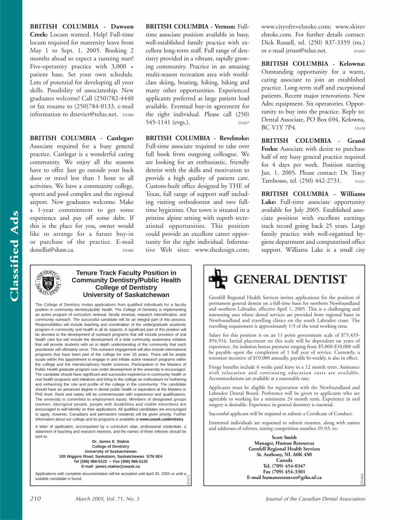
BRITISH COLUMBIA - DawsonCreek: Locum wanted. Help! Full-timelocum required for maternity leave fromMay 1 to Sept. 1, 2005. Booking 2months ahead so expect a running start!Five-operatory practice with 3,000 +patient base. Set your own schedule.Lots of potential for developing all yourskills. Possibility of associateship. Newgraduates welcome! Call (250)782-4440or fax resume to (250)784-0133, e-mailinformation to [email protected]. D1680
BRITISH COLUMBIA - Castlegar:Associate required for a busy general practice. Castlegar is a wonderful caringcommunity. We enjoy all the seasons have to offer. Just go outside your backdoor or travel less than 1 hour to all activities. We have a community college,sports and pool complex and the regionalairport. New graduates welcome. Make a 1-year commitment to get some experience and pay off some debt. If this is the place for you, owner would like to arrange for a future buy-inor purchase of the practice. E-mail [email protected]. D1686
BRITISH COLUMBIA - Vernon: Full-time associate position available in busy,well-established family practice with ex-cellent long-term staff. Full range of den-tistry provided in a vibrant, rapidly grow-ing community. Practice in an amazingmulti-season recreation area with world-class skiing, boating, hiking, biking andmany other opportunities. Experiencedapplicants preferred as large patient loadavailable. Eventual buy-in agreement forthe right individual. Please call (250)545-1141 (evgs.). D1657
BRITISH COLUMBIA - Revelstoke:Full-time associate required to take overfull book from outgoing colleague. Weare looking for an enthusiastic, friendlydentist with the skills and motivation toprovide a high quality of patient care.Custom-built office designed by THE ofTexas, full range of support staff includ-ing visiting orthodontist and two full-time hygienists. Our town is situated in apristine alpine setting with superb recre-ational opportunities. This position could provide an excellent career oppor-tunity for the right individual. Informa-tive Web sites: www.thedesign.com;
www.cityofrevelstoke.com; www.skirevelstoke.com. For further details contact:Dick Russell, tel. (250) 837-3359 (res.) or e-mail [email protected]. D1663
BRITISH COLUMBIA - Kelowna:Outstanding opportunity for a warm,caring associate to join an establishedpractice. Long-term staff and exceptionalpatients. Recent major renovations. NewAdec equipment. Six operatories. Oppor-tunity to buy into the practice. Reply to:Dental Associate, PO Box 694, Kelowna,BC V1Y 7P4. D1628
BRITISH COLUMBIA - GrandForks: Associate with desire to purchasehalf of my busy general practice requiredfor 4 days per week. Position starting Jan. 1, 2005. Please contact: Dr. TracyTambosso, tel. (250) 442-2731. D1631
BRITISH COLUMBIA - WilliamsLake: Full-time associate opportunityavailable for July 2005. Established asso-ciate position with excellent earnings track record going back 25 years. Largefamily practice with well-organized hy-giene department and computerized officesupport. Williams Lake is a small city
Cla
ssifi
ed
Ad
s
210 March 2005, Vol. 71, No. 3 Journal of the Canadian Dental Association
Tenure Track Faculty Position inCommunity Dentistry/Public Health
College of DentistryUniversity of Saskatchewan
The College of Dentistry invites applications from qualified individuals for a facultyposition in community dentistry/public health. The College of Dentistry is implementingan active program of curriculum renewal, faculty renewal, research intensification, andcommunity outreach. The successful candidate will be an integral part of this process.Responsibilities will include teaching and coordination of the undergraduate academicprogram in community oral health in all its aspects. A significant part of this position willbe devoted to the development of outreach programs that will include provision of oralhealth care but will include the development of a total community awareness initiativethat will provide students with an in depth understanding of the community that eachpractitioner will ultimately serve. This outreach engagement will also include internationalprograms that have been part of the college for over 25 years. There will be ample scope within this appointment to engage in and initiate active research programs withinthe college and the interdisciplinary health sciences. Participation in the Masters ofPublic Health graduate program now under development at the university is encouraged.The candidate should have significant and successful experience in community health ororal health programs and initiatives and bring to the college an enthusiasm for furtheringand enhancing the role and profile of the college in the community. The candidate should have an advanced degree in dental public health or equivalent at the Masters orPhD level. Rank and salary will be commensurate with experience and qualifications.The university is committed to employment equity. Members of designated groups(women, Aboriginal people, people with disabilities and visible minorities) areencouraged to self-identify on their applications. All qualified candidates are encouragedto apply; however, Canadians and permanent residents will be given priority. Furtherinformation about our college and its programs is available at www.usask.ca/dentistry .
A letter of application, accompanied by a curriculum vitae, professional credentials, a statement of teaching and research interests, and the names of three referees should besent to:
Dr. James E. StakiwCollege of Dentistry
University of Saskatchewan105 Wiggins Road, Saskatoon, Saskatchewan S7N 5E4
Tel (306) 966-5122 • Fax (306) 966-5132E-mail [email protected]
Applications with complete documentation will be accepted until April 30, 2005 or until asuitable candidate is found.
D16
71
GENERAL DENTISTGrenfell Regional Health Services invites applications for the position ofpermanent general dentist on a full-time basis for northern Newfoundlandand southern Labrador, effective April 1, 2005. This is a challenging andinteresting area where dental services are provided from regional bases inNewfoundland and travelling clinics on the south Labrador coast. Thetravelling requirement is approximately 1/3 of the total working time.
Salary for this position is on an 11 point government scale of $75,433-$94,916. Initial placement on this scale will be dependent on years ofexperience. An isolation bonus payment ranging from $5,000-$10,000 willbe payable upon the completion of 1 full year of service. Currently, aretention incentive of $10,000 annually, payable bi-weekly, is also in effect.
Fringe benefits include 6 weeks paid leave in a 12 month term. Assistancewith relocation and continuing education costs are available.Accommodations are available at a reasonable rate.
Applicants must be eligible for registration with the Newfoundland andLabrador Dental Board. Preference will be given to applicants who areagreeable to working for a minimum 24 month term. Experience in oralsurgery is desirable. Experience in general dentistry is essential.
Successful applicant will be required to submit a Certificate of Conduct.
Interested individuals are requested to submit resumes, along with namesand addresses of referees, stating competition number, 05.03, to:
Scott SmithManager, Human Resources
Grenfell Regional Health ServicesSt. Anthony, NL A0K 4S0
CanadaTel. (709) 454-0347Fax (709) 454-3301
E-mail [email protected]
D16
64
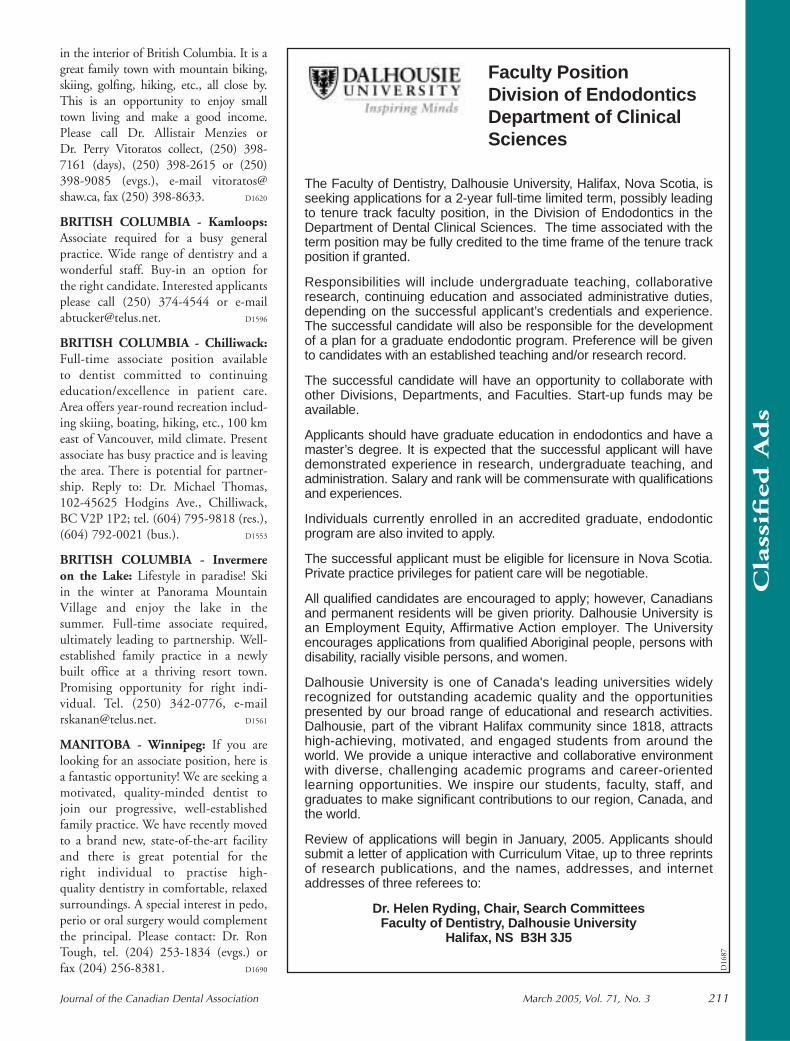
Cla
ssifi
ed
Ad
s
in the interior of British Columbia. It is agreat family town with mountain biking,skiing, golfing, hiking, etc., all close by.This is an opportunity to enjoy smalltown living and make a good income.Please call Dr. Allistair Menzies or Dr. Perry Vitoratos collect, (250) 398-7161 (days), (250) 398-2615 or (250)398-9085 (evgs.), e-mail [email protected], fax (250) 398-8633. D1620
BRITISH COLUMBIA - Kamloops:Associate required for a busy generalpractice. Wide range of dentistry and awonderful staff. Buy-in an option for the right candidate. Interested applicantsplease call (250) 374-4544 or [email protected]. D1596
BRITISH COLUMBIA - Chilliwack:Full-time associate position availableto dentist committed to continuingeducation/excellence in patient care.Area offers year-round recreation includ-ing skiing, boating, hiking, etc., 100 kmeast of Vancouver, mild climate. Presentassociate has busy practice and is leavingthe area. There is potential for partner-ship. Reply to: Dr. Michael Thomas,102-45625 Hodgins Ave., Chilliwack,BC V2P 1P2; tel. (604) 795-9818 (res.),(604) 792-0021 (bus.). D1553
BRITISH COLUMBIA - Invermereon the Lake: Lifestyle in paradise! Ski in the winter at Panorama MountainVillage and enjoy the lake in the summer. Full-time associate required,ultimately leading to partnership. Well-established family practice in a newlybuilt office at a thriving resort town.Promising opportunity for right indi-vidual. Tel. (250) 342-0776, [email protected]. D1561
MANITOBA - Winnipeg: If you arelooking for an associate position, here isa fantastic opportunity! We are seeking amotivated, quality-minded dentist tojoin our progressive, well-establishedfamily practice. We have recently movedto a brand new, state-of-the-art facilityand there is great potential for the right individual to practise high-quality dentistry in comfortable, relaxedsurroundings. A special interest in pedo,perio or oral surgery would complementthe principal. Please contact: Dr. RonTough, tel. (204) 253-1834 (evgs.) orfax (204) 256-8381. D1690
Journal of the Canadian Dental Association March 2005, Vol. 71, No. 3 211
Faculty PositionDivision of EndodonticsDepartment of ClinicalSciences
The Faculty of Dentistry, Dalhousie University, Halifax, Nova Scotia, isseeking applications for a 2-year full-time limited term, possibly leadingto tenure track faculty position, in the Division of Endodontics in theDepartment of Dental Clinical Sciences. The time associated with theterm position may be fully credited to the time frame of the tenure trackposition if granted.
Responsibilities will include undergraduate teaching, collaborativeresearch, continuing education and associated administrative duties,depending on the successful applicant’s credentials and experience.The successful candidate will also be responsible for the developmentof a plan for a graduate endodontic program. Preference will be givento candidates with an established teaching and/or research record.
The successful candidate will have an opportunity to collaborate withother Divisions, Departments, and Faculties. Start-up funds may beavailable.
Applicants should have graduate education in endodontics and have amaster’s degree. It is expected that the successful applicant will havedemonstrated experience in research, undergraduate teaching, andadministration. Salary and rank will be commensurate with qualificationsand experiences.
Individuals currently enrolled in an accredited graduate, endodonticprogram are also invited to apply.
The successful applicant must be eligible for licensure in Nova Scotia.Private practice privileges for patient care will be negotiable.
All qualified candidates are encouraged to apply; however, Canadiansand permanent residents will be given priority. Dalhousie University isan Employment Equity, Affirmative Action employer. The Universityencourages applications from qualified Aboriginal people, persons withdisability, racially visible persons, and women.
Dalhousie University is one of Canada's leading universities widelyrecognized for outstanding academic quality and the opportunitiespresented by our broad range of educational and research activities.Dalhousie, part of the vibrant Halifax community since 1818, attractshigh-achieving, motivated, and engaged students from around theworld. We provide a unique interactive and collaborative environmentwith diverse, challenging academic programs and career-orientedlearning opportunities. We inspire our students, faculty, staff, andgraduates to make significant contributions to our region, Canada, andthe world.
Review of applications will begin in January, 2005. Applicants shouldsubmit a letter of application with Curriculum Vitae, up to three reprintsof research publications, and the names, addresses, and internetaddresses of three referees to:
Dr. Helen Ryding, Chair, Search CommitteesFaculty of Dentistry, Dalhousie University
Halifax, NS B3H 3J5
D16
87

MANITOBA - The Pas: Looking to bemore busy or for a change? Come workfor us in a busy general practice clinicwhere you can be as busy or relaxed asyou want. We pay a high guaranteedcommission on top of your regular per-centage. Your accommodation and travel are reimbursed as well. Work fulltime or in terms with extended holi-days. Flexibility, high net income andbest of all no hassles. Call for details,(204) 623-1494 or fax resume to (204)623-6162. D1647
NORTHWEST TERRITORIES - In-uvik: Replacement for 3 weeks. Lookingfor a dentist from Mar. 21 to Apr. 8,2005. Other dates also available. Sched-ule already booked, excellent remunera-tion, accommodations included. Comeexperience the far North. Please contact:Nancy or Lynda , tel. (867) 777-3008 ore-mail [email protected].
D1650
NORTHWEST TERRITORIES - Yel-lowknife: Associate needed to join anestablished, very busy, modern dentalclinic (6 dentists) in a thriving commu-nity - the diamond capital of NorthAmerica. The clinic offers all modernequipment including intraoral cameras,abrasion units, etc., with an excellentand friendly support staff, providingvery high-quality dentistry, with theemphasis on quality rather than quan-tity. This is an excellent opportunity foranyone wishing to enjoy a wonderfullifestyle whilst practising dentistry at itsbest. Please send resume to: Administra-tion, PO Box 1118, Yellowknife, NTX1A 2N8; tel. (867) 873-6940, fax(867) 873-6941. D1159
NUNAVUT - Iqaluit: Associate posi-tion(s) available for immediate start. Es-tablished clinic offers generous packageand full appointment book to associates.All round clinical skills are your ticket toa wide range of recreational activities! Notravel required and housing available inCanada’s newest and fastest growing capi-tal city. Please apply to: Administration,PO Box 1118, Yellowknife, NT X1A2N8; or tel. (867)873-6940, fax (867)873-6941. D1497
ONTARIO - Brampton: Part-time andfull-time dental associates required forBrampton office. Please fax resume to
(905) 791-0644 indicating which daysavailable. D1693
ONTARIO/QUEBEC: Looking forbilingual associate for 5 mature and busypractices, south-west Quebec and/orCornwall, Hawkesbury, Ontario area.Full schedule (crown/bridge, endodon-tics, etc.). Stability, flexibility and respectassured. Possible sale. Seeing is worth be-lieving. Luc, tel. (450) 370-7765. D1674
ONTARIO - Cambridge: Part-time associate required for 2-3 days in a 9-year-old, fast-growing, family-oriented,multilingual general dental practice.Please fax resume to (519) 622-3608, e-mail [email protected]. D1682
ONTARIO - Northwestern: Associate-ship available immediately. Full-time as-sociateship with option to purchase.Busy family practice located in North-western Ontario, conveniently locatedon Trans-Canada Highway. Twenty-eight-year-old, well-established clinic,2 full-time hygienists, 4 operatories.Excellent opportunity for someone whoenjoys the outdoors and a low-stressenjoyable work situation with flexiblehours and vacation time. Reply to: CDA Classified Box # 2846. D1684
ONTARIO - Niagara Region: Full-time dental associate position availablefor a family-oriented, well-establishedpractice. Please fax your resume to (905)871-3977. D1689
ONTARIO - Lake Huron: We are looking for a full-time associate to joinour practice. If you are interested inbeing fully scheduled, want to work withan experienced staff and think youmight enjoy life in a beautiful resorttown, we have the position you are looking for. Our modern office is fullyequipped with 6 operatories, intraoralcameras and computers. We haverecently replaced our charts, re-vampedour hygiene program and routinelyattend continuing education courses.Buy-in option would be available for the right candidate. This would be anexcellent investment opportunity for anew graduate. Please call MichaelWalden at (519) 396-2641. D1654
ONTARIO - Ottawa east: Associate-ship full time. Very busy practice. Buy-in,
cost sharing options available. Frenchspeaking an asset. Call Jocelyne, (613) 748-8266 D1668
ONTARIO - Amherstburg/WindsorArea: Full-time dentist wanted to associ-ate in our busy, well-established dentalpractice in Amherstburg, Ontario. Ouroffice is just a 20-minute drive fromWindsor. Presently retain over 6,000active charts and growing. Looking for akind, active, professional and highlymotivated individual. Offering up to50% compensation. Excellent opportu-nity for any associate willing to committhemselves to long-term relationship and future growth in this wonderfulcommunity. Tel. (519) 980-4073. D1639
ONTARIO - West of Toronto: Full-time associate position in a well-estab-lished practice, replacing associate whois returning to school for graduate stud-ies. Your schedule will be booked fromday 1 and you will have the opportunityto be exposed to all aspects of dentistrysuch as cosmetics, implants and muchmore as we have a team of specialistsworking alongside of us! If you are ateam player and are looking for a posi-tive working environment, fax to(905)846-5593. D1641
ONTARIO - Brockville and Morris-burg: Experienced associate required for1 of 2 well-established, busy practices.Enjoy a small-town atmosphere and thescenic beauty of the 1000 Islands regionwith easy access to large city centres.Only 30 minutes to Kingston and 60 minutes to Ottawa. For more information contact: Dr. GeorgeChristodoulou, Altima Dental Canada,tel. (416) 785-1828, ext. 201, e-mail [email protected]. D1269
QUEBEC - Abitibi: Rouyn-Noranda.Our family practice offers you a uniqueopportunity to join our team for a re-placement during a maternity leavestarting mid-April for a period of sixmonths. Very busy practice, booked overtwo months in advance, full-time posi-tion guaranteed thereafter. Excellentwork schedule; 4 days/week Monday toThursday, no weekends. Very nice area,with lakes and forests nearby for the na-ture lover. For more information, pleasecontact Aline at (819) 762-1972. D1659
Cla
ssifi
ed
Ad
s
212 March 2005, Vol. 71, No. 3 Journal of the Canadian Dental Association

Cla
ssifi
ed
Ad
s
Journal of the Canadian Dental Association March 2005, Vol. 71, No. 3 213
Selling a practice?Leasing an office?
Buying equipment?Renting your vacation property?
To place your CDA Classified ad contact:
Beverley Kirkpatrick or Deborah Rodd
Toll-free at (800) 663-7336 ext. 2127/2314
Fax (613) 565-7488E-mail [email protected]
Advertisements are also published online
(www.cda-adc.ca/jcda) at no additional charge.
PROSTHODONTICSMcGill University
The Faculty of Dentistry, McGill University, invites applications fortenure-track and clinical-track positions in prosthodontics at the levelof Assistant or Associate Professor. Candidates must have completed anundergraduate degree in dentistry and specialty training inprosthodontics. Rank and salary will be commensurate with educationand experience.
Responsibilities will include undergraduate and graduate teaching andadministration. A working knowledge of French will be advantageous.Intramural private practice facilities are available.
The additional requirements for the tenure track include a PhD orequivalent degree and the ability to conduct independent research in arelevant field. An Ambrose Clinical Professorship may be awarded toan outstanding clinical-track candidate.
Applications, including a curriculum vitae, a statement of research andteaching interests, and the names, postal and e-mail addresses of threereferees, should be sent to the following address by April 15, 2005.
Dr. Jeffrey Myers Chair, Search Committee
Faculty of Dentistry, McGill University1650 Cedar Avenue, Room A3.132Montreal, QC, Canada H3G 1A4
E-mail [email protected] (514) 934-8352
McGill University is committed to equity in employment. All qualifiedcandidates are encouraged to apply; however, Canadians and permanentresidents will be given priority.
D16
76
Tenure Track Faculty Position inOral/Maxillofacial Surgery
College of DentistryUniversity of Saskatchewan
The College of Dentistry invites applications from qualified individuals for afaculty position in oral/maxillofacial surgery. The college is implementing anactive program of curriculum renewal, faculty renewal, and research in-tensification. This position is a key part of that process. Start date is July 1, 2005or when a suitable candidate is found. Responsibilities and duties includedidactic and clinical instruction of undergraduate students in oral/maxillofacialsurgery, an ongoing research commitment, and college administration. Theapplicant must have relevant postgraduate and clinical qualifications at theFRCD(C) or equivalent level and research experience. Possession of anadvanced degree is desirable. The MD degree would be an asset. The idealcandidate will be from a CDA/ADA-approved oral/maxillofacial surgery residencyprogram with significant experience in dentoalveolar and orthognathic surgery,TMD, anesthesia, implantology, pathology and trauma management. On-siteprivate practice privileges are available. The Royal University Hospital andCollege of Medicine are adjacent to the College of Dentistry and the successfulcandidate is expected to become involved with programs and initiatives in theseinstitutions. Rank and salary will be commensurate with experience andqualifications. The university is committed to employment equity. Members ofdesignated groups (women, Aboriginal people, people with disabilities and visibleminorities) are encouraged to self-identify on their applications. All qualifiedcandidates are encouraged to apply; however, Canadians and permanentresidents will be given priority. Further information about our college and itsprograms is available at www.usask.ca/dentistry .
A letter of application, accompanied by a curriculum vitae, professional creden-tials, a statement of teaching and research interests, and the names of threereferees should be sent to:
Dr. James E. StakiwCollege of Dentistry
University of Saskatchewan105 Wiggins Road, Saskatoon, Saskatchewan S7N 5E4
Tel (306) 966-5122 • Fax (306) 966-5132E-mail [email protected]
Applications with complete documentation will be accepted until April 30, 2005 oruntil a suitable candidate is found.
D16
72
Tenure Track Faculty Position inPediatric Dentistry
College of DentistryUniversity of Saskatchewan
The College of Dentistry invites applications from qualified individuals fora faculty position in pediatric dentistry. The College of Dentistry is imple-menting an active program of curriculum renewal, faculty renewal, andresearch intensification. The successful candidate will be an integral partof this process. Applicants will have postgraduate training in pediatricdentistry at the Master’s or PhD level with relevant clinical qualifications(FRCD(C) preferred), and the successful candidate will have researchexperience. Responsibilities will include didactic and clinical instruction ofundergraduate students, research programs and administration. Pediatricgeneral anesthetic services are currently available. On-site privatepractice privileges are available. The Royal University Hospital andCollege of Medicine are adjacent to the College of Dentistry and thesuccessful candidate is expected to become involved with programs andinitiatives in these institutions. Rank and salary will be commensuratewith experience and qualifications. The university is committed toemployment equity. Members of designated groups (women, Aboriginalpeople, people with disabilities and visible minorities) are encouraged to self-identify on their applications. All qualified candidates are encour-aged to apply; however, Canadians and permanent residents will be given priority. Further information about our college and its programs isavailable at www.usask.ca/dentistry .
A letter of application, accompanied by a curriculum vitae, professionalcredentials, a statement of teaching and research interests, and thenames of three referees should be sent to:
Dr. James E. StakiwCollege of Dentistry
University of Saskatchewan105 Wiggins Road, Saskatoon, Saskatchewan S7N 5E4
Tel (306) 966-5122 • Fax (306) 966-5132E-mail [email protected]
Applications with complete documentation will be accepted until April 15,2005 or until a suitable candidate is found.
D16
73

Cla
ssifi
ed
Ad
sQUEBEC - Montreal: Oral and max-illofacial surgery associate for well-estab-lished, solo, bilingual practice. Progressionto buy-in and role reversal anticipated forthe right individual who is skilled,personable and who considers patientcare a priority. Confidential reply to:CDA Classified Box # 2845. D1669
QUEBEC - Eastern Townships:Windsor, near Sherbrooke. We are givingan associate the opportunity to becomepart of a mature and fully competentteam. Pleasant and motivating workatmosphere. Please fax resume to (819)845-7854. Tel. Dr. Jacques Vaillancourt,(819) 845-3080. D1371
SASKATCHEWAN - Regina: Full-time associateship available in a busy,established orthodontic practice. Excel-lent, oriented, energetic staff. Buy-inoption. Please call (306) 586-3222 inconfidence. D1665
YUKON - Whitehorse: Full-time den-tist required for busy 5-operatory prac-tice. Great staff and friendly environ-ment. Come and enjoy the greatwonders of the North. Starting date isnegotiable. Reply to: Pine Dental, 51105th Ave., Whitehorse, YT Y1A 1L4; tel.(867) 668-2273, fax (867) 668-5121.
D1677
YUKON TERRITORY - Whitehorse:Come for the beauty - mountains, lakesand rivers. Or come for the opportunity to practise dentistry where you are appre-ciated and well compensated. Have alook at our Web site www.klondike-dental.com. Tel. (867) 668-4618, fax(867) 667-4944. D1422
TEXAS - Dallas: Growing dental com-pany in and around Dallas is seeking full-time associates. Must be licensed orqualified to be licensed in Texas. Highestcompensation package in the state; earn$200,000 - $400,000. Company to han-dle all immigration matters. Please call(630) 788-7167. D1513
VERMONT, US - Burlington: Beauti-ful Burlington, Vermont, is calling you!Established practice is seeking a dentistfor a Monday through Friday work week. Full-time compensation includes:competitive salary, bonus potential and afull benefits package (including a 401Kplan with matching funds). Part-timecandidates will be considered. Positionsalso available in other states. For a posi-tion with a future call Brian Whitley,(800) 313-3863, ext. 2290 or [email protected]. D1604
VERMONT, US: Dentists and oral sur-geons. Opportunities for general dentistsin Rutland, Montpelier and Lake Cham-plain areas. Openings available for em-ployment, private practice and practiceacquisitions. Enjoy the splendor of theGreen Mountains and Lake Champlain,all part of the unbeatable Vermontlifestyle. Contact: Lynn Harris, tel. (800) 288-1730, fax (518) 266-9289, e-mail [email protected].
D1538
214 March 2005, Vol. 71, No. 3 Journal of the Canadian Dental Association
HEAD NORTHA new clinic, in a new building, in a
thriving northern city. Six state-of-the-artoperatories, digital radiology, booking
4 months in advance. What more could we ask for...
AN ASSOCIATE, DAMMIT!
murraya DENTAL
4069 4th ave. whitehorse, yukon Y1A 1H1
Tel. (867) 633-4401 • Fax (867) 633-4402
D16
11
A D V E R T I S E R S ’I N D E X
3M ESPE . . . . . . . . .140, 176-7
A-Dec International . . . . . . .146
Biolase . . . . . . . . . . . . . . . . .155
CDA RSP . . . . . . . . . . . . . .154
CDSPI . . . . . . . . . . . . .169, 202
Colgate-Palmolive Canada Inc. . . . . . . . . . . . . .144
DioGuardi and Company, LLP . . . . . . . . . . . . . . . . . . .190
FDI Annual World Dental Congress . . . . . . . . . .206
Imtec Corporation . . . . . . . .215
Ivoclar . . . . . . . . . . . . . . . . .170
Jasper Dental Congress . . . . .191
Oral-B Laboratories . . .141, 142
Procter & Gamble . . . . . . . .150
Quintessence Publishing . . . .164
SciCan . . . . . . . . . . . . . . . . .138
Straumann Canada Ltd. . . . .183
Sultan Chemists . . . . . . . . . .184
Sunstar Butler . . . .160, 178, 216
Vacalon Inc. . . . . . . . . . . . . .175
Vident . . . . . . . . . . . . . . . . .199
VOCO GmbH . . . . . . . . . . .192

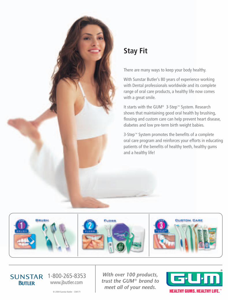
With over 100 products,trust the GUM® brand tomeet all of your needs.
HEALTHY GUMS. HEALTHY LIFE.™
®
1-800-265-8353www.jbutler.com
© 2004 Sunstar Butler C04171
™ ™ ™
Stay Fit
There are many ways to keep your body healthy.
With Sunstar Butler’s 80 years of experience working with Dental professionals worldwide and its completerange of oral care products, a healthy life now comes with a great smile.
It starts with the GUM® 3-Step™ System. Researchshows that maintaining good oral health by brushing, flossing and custom care can help prevent heart disease,diabetes and low pre-term birth weight babies.
3-Step™ System promotes the benefits of a completeoral care program and reinforces your efforts in educatingpatients of the benefits of healthy teeth, healthy gums and a healthy life!
Copyright © 2022 FDOKUMEN