Kerala Dental Journal
56
-
Upload
khangminh22 -
Category
Documents
-
view
1 -
download
0
Transcript of Kerala Dental Journal


C o n t e n t s
Kerala Dental Journal
Vol. 32 | No. 1 | Jan. 2009
KDJ ISSN No. 0972-396X, Vol.32, No. 1, January 2009 5
President’s Message 6
Editorial 7
DCI President’s Message 9
Comparative study of colony counts of different species of oral
streptococci in saliva of dentulous, edentulous and in those
wearing partial and complete dentures 11
Rajani Mary Kurien, Ganesh Shenoy Panchmal, Vijaya Hegde, Ronald Roche
Graphoanalysis : an aid in patient evaluation 14Anupama M.S., Sadhvi K.V., Hari B., Chandrasekharan Nair K.,Jaykar Shetty, Vishwanath G
A comparative study on the marginal fit of multi unitsingle piece castings using over refractory technique andconventional technique- A pilot study 20
Sushma Y, Chandrika L, Srividya S; Chandrasekharan Nair K;Jayakar Shetty; Vishwanath G
Peripheral osteomas of jaws- A study of 6 cases 23Akhilanand Chaurasia, Dr. Anita Balan
The C shaped canal- an anatomical variant 27Saumyakanta Mohanty, Jolly Mary Varughese, N.O. Varghese
Prevention of periodontal diseases 31Riyas Seinullabdeen, Tintu Sara Chandy, Seba Abraham,Prakash P., Ambili R. Nisha K.J.
Diagnose the following cases 34Akhilanand chaurasia, Anita Balan
Hemisection – A better option in furcation management 35Preeja C, Presanthila Janam, Haeigin Tom Varghese, T. Sreelal, K.Harshakumar
Eagle’s Syndrome 38Arun George, Devi Gopakumar, Bobby John, S Sunil
Herpes zoster infection of the maxillary nerve 40Ajay G. Nayak, Laxmikanth ChatraPrashanth Shenai K., Prasanna Kumar
Scleroderma 43Gayathri Krishnan, Vishnu Mohan
Management of fractured edentulous mandible with gunning splint 45Harshakumar, Anitha Gopinathan Ajith Kumar P.K.
Osteogenesis imperfecta type IV 47Roopashri Rajesh Kashyap, Gopakumar R., Gogineni Subhas Babu, Sreejan C.K.
Dental Jewellery 50Shibu Thomas Mathew
Quiz 52Rani Mol, Anita Balan
Secretary’s Message and Association Reports 53

KDJ - Vol.32, No. 1, January 20096
Dr. K.N. Pratap Kumar
Dear Colleagues,
I am delighted to have this opportunity toextend my personal BEST WISHES to all ofyou. I sincerely hope you all enjoyed the 41st
State Conference at Nedumbassery as it wasa wonderful opportunity for IDA membersState wide to toast their collectiveachievements in 2008, with memories oftriumph joy and friendship blended together.
To be committed and to perform better, theorganization necessarily has to have the setup of simple and transparent workingconditions. Let us have the synergy in servingtogether with the motto “WE” rather than“YOU’ or I. Synergy helps the organizationto maximize the combined efforts ofindividuals working in different teams.
Motivation is the driving force in our lives. Itcomes from a desire to succeed. Withoutsuccess there is little pride in life, no enjoymentor excitement at work and at home. The mostpowerful motivation comes from within ourbelief system. To move in to action, we needto believe in what we do and acceptresponsibility for our life. When we acceptresponsibility for our behaviour and action,our attitude towards life becomes positive. Webecome more productive, both personally andprofessionally. Life becomes more meaningfuland fulfilling.
Enthusiasm and motivation go hand in hand.Enthusiasm inspires confidence, raises morale,build loyalty and is priceless. You can feelenthusiasm by the way a person talks, walksor shakes hand, a habit that one can acquireand practice.
We often talk about attracting new membersand retaining current ones but rarely discussdeveloping quality members with character,commitment, conviction, courtesy andcourage. Yes, we certainly need to devote ourattention to building membership but let usalso nurture the members already in the fold.
More and more rules, regulations, and moneyextracting methodologies are introduced everyyear. In such circumstances this organizationwill be the one which will protect the interest
of all the dental practitioners. Your activeinvolvement and firm unity under the bannerof IDA is mandatory in the coming years tofight for our causes.
Let us
Be Professional in our approach andimplementation,
Observe the code of ethics anddiscipline of our association,
Maintain and enhance the relationshipand convert it in to everlasting,
Maintain an effective communicationthereby resulting in total transparency
Resolve issues internally if any.
Life is not just party and pleasure; it is alsopain and despair. Unthinkable things happen.Sometimes every thing turns upside down.Bad things happen to good people. It takesboth rain and sunshine to create a rainbow.Our lives are no different. There is happinessand sorrow. There is the good and the bad,dark spots and bright spots. When we canhandle adversity well, it only strengthens us.
Oral Cancer Detection and AwarenessProgramme, Smoke Free Institutions,Voluntary Blood Donors Forum, PhotoHealth Card, Swanthanam [Spend a day withhandicapped (physically or mentally) orphansgeriatrics] are some of the programmes ofmajor importance this IDA year.
I request all the branch members to take partin IDA activities and give life to IDA. Theworld around us has changed a lot and weneed to change too. If we are willing tochange our thinking we can change our lifeand we can make our tomorrow better andbrighter. Future belongs to those who grab itand take advantage of every opportunity andthus we create our own destiny.
LET US LEARN TO EXCEL IN LIFE AND
IN PROFESSION
With regards and love
Yours in IDA
President’s Message

KDJ - Vol.32, No. 1, January 2009 7
Editorial
Dr. K. Nandakumar
N e e d o f t h e h o u r
The last quarter century was the golden period of Indian dentistry if
the demand for the related professional course is considered as the
parameter. The number of dental colleges, increased by twenty times.
Colleges belonged both to the public and private sectors. While the
government colleges put a solid foundation to the academics, modern
developments were ushered in vast proportions because of the liberal
finances available to the private colleges. Both government and private
colleges had a complementary growth which has put the profession
into a developmental track. Last two years have witnessed a lack of
demand for the professional courses of dentistry. In Kerala it is not
very evident but the neighboring states like Karnataka and Tamilnadu
are facing it really, with many seats remaining unfilled. Can dental
professionals remain complacent after observing this down trend?
India needs more dentists so that our rural population will gain access
to dental health care amenities. Youngsters have kept dentistry in the
low priority list which is not a good sign. Indian Dental Association
has the a responsibility to promote our profession amongst the
youngsters who are in the plus two classes. We have to market our
profession highlighting its scope and relevance in the broad picture
of health care system. The nobility and success of dentistry as a
profession should be brought to the notice of the youngsters so that
they will get attracted to it. We have to conduct promotional lectures
using effective visuals in the schools and in the public forum where
many parents do attend. I feel we have to give serious thought over
this and take effective steps. It is the need of the hour.
I take this opportunity to thank each and every member for reposing
confidence in me once again by electing to the post of Editor of
Kerala Dental Journal. On behalf of the members of IDA Kerala
State branch, I have great pleasure to place on record our greatest
appreciation to the former editor Dr.Santhosh Sreedhar who set a
standard to our Journal which has won national award thrice in a
series.
KDJ - Vol.32, No. 1, January 20098
What ‘IDA’ means to youDescription:
� Founded in 1946, IDA is a premier non-profit, professional organization of dental
professionals in the country numbering over 40000.
� Being an exclusive body of dentists in India, it effectively harnesses its vast resources
aimed at attaining professional excellence in their day to day clinical and research
activities and in making India the hub of oral healthcare destination of the world
Vision/Mission:
� make IDA the national, authoritative and independent, voice of the dental professionals;
� vigorously promote, safeguard, defend and protect the interests and rights of members
in their commitment to providing optimal oral healthcare of high professional standards
� promote, preserve and uphold the highest ethical values and principles of dental practice
� contribute to evolving the Oral Health Policy in the country
� contribute to improving the quality of dental services and enhance the image of its
members
� undertake education, communication, research and other support activities
� update dental professionals on clinical and technological advances of global significance
� partnering other organizations in the promotion of oral and general health in the country
� promoting professional advancement of members and their commitment to dental
excellence
Principal Activities:
� Implements programmes and projects aimed at improving oral health in the country;
� undertakes education and research activities and serves as incubators for dental research
development;
� organizes accredited continuing dental education programmes for professional
advancement and improving quality of dental service;
� provides an excellent forum for exchange of clinical and scientific articles through Its
publications;
� participates in conference and workshops aimed at improving oral healthcare;
� implements projects and programmes for the enrichment of the fraternity, students of
dentistry and the general public;
� focuses on prevention and interception of dental and oral diseases
� organizes early detection and treatment of oral cancer lesions through SPOT Centres
and TII Centres being established under Oral Cancer Foundation
Member Benefits:
� provision of Legal/Ethical Advice
� Dental Shield Insurance
� Retirement Benefits
� Advocacy and Negotiations
� Representation in various fields
Certification/Product Endorsement
3 awards certificates on being trained in oral cancer detection/tobacco intervention
initiative
3 grants IDA Seal of Approval for healthcare products to ensure their safety
From the Desk of Hon Secretary GeneralIndian Dental Association, Head Office
Ashok Dhoble
Hon. Secretary General

KDJ - Vol.32, No. 1, January 2009 9
Brig (Dr.) Anil Kohli
Dental profession has an undeniable role in the health care system of our
country. When we aim at the well being of the people, improvement of
dental health becomes increasingly relevant because teeth are vulnerable to
the most rampant diseases viz. dental caries and the diseases affecting the
periodontium. The enormous nature of the dental diseases highlights the
importance of prevention. But preventive measures alone will not be the
only realistic solution in the context of India because of the volume of
population. Hence we are settling to the fact that twenty functioning teeth are
to be considered as the essential dentition. Perhaps this practical realization is
possible only with teeth and not with any other organ in the human body.
Successful dental health maintenance is possible only when we keep curative
as well as preventive measures at equilibrium with a realistic expectation of
the number of existing teeth.
Dental council of India has always been aiming at strengthening the building
blocks of our profession- education and clinical practice. Availability of
competent professionals is ensured by the council by adhering to high standards
of educational institutions both in infrastructure and qualified teaching
professionals. Post graduate, undergraduate and diploma courses are offered
in recognized institutions all over the country. Both urban and rural areas of
our country will have availability of qualified dental professionals very shortly.
The council also would like to bring advanced treatments like dental implants
within the reach of the common man. That is the reason for making our
curriculum more dynamic by introducing the five year BDS programme and
by reorienting the MDS regulation. Making professional advancement
programmes mandatory for renewal of council registration will make far
reaching changes to our profession. Research should also be given a new
direction in India and quality publications will provide ample opportunities
for the young dental professionals of our country to publish their work without
much delay. I hope Kerala Dental Journal would be a torch bearer of our
profession. Jai Hind.
Brig (Dr.) Anil Kohli

KDJ - Vol.32, No. 1, January 200910
EDITOR
Dr. K. Nandakumar
ASST. EDITOR
Dr. R.M. Baiju
BUSINESS MANAGER
Dr. Mathew Jose
EDITORIAL CONSULTANTS
Dr. Santhosh Sreedhar
Dr. K. Chandrasekharan Nair
Dr. K. George Varghese
Dr. Ipe Varghese
Dr. Oommen Aju Jacob
Dr. Thomas Manjooran
Dr. N.O. Varghese
Dr. Sobha Kuriakose
Dr. T. Sreelal
Dr. Siby Xavier
EX-OFFICIO MEMBERS
Dr. K.N. Pratap Kumar
Dr. Antony Thomas
Dr. C.K. Ashokan
Dr. Samuel K. Ninan
EDITORIAL BOARD
Dr. Anita Balan
Dr. Sreela Jayakumar
Dr. Twinkle S. Prasad
Dr. K.S. Ravindran Nair
Dr. Sooraj
Dr. Ajith Kumar
Dr. V.T. Beena
Dr. Bindu J. Nair
Dr. Hari
Dr. Bindu R. Nayar
Dr. Arun Sadasivan
Dr. Anil Mathew
Dr. P.A. Murukan
Dr. Pradeep Dethan
Dr. Eldo Koshy
Dr. Sheela Sreedharan
Dr. M.S. Suchitra
Dr. V.P. Kannan
Dr. Vinod Krishnan
Dr. Benoy Kurian
Dr. Joseph Issac
Dr. V.G. Sam Joseph
Dr. V.I. Paul
Dr. Gibi Paul
Dr. Manju Renjith
Dr. Jayakrishnan
EDITORIAL OFFICE
Neelambikam, Attukal, ManacaudTrivandrum, Kerala - 695 009
Phone: 0471-2459235Mobile: 09447066100
e-mail: [email protected]: www.idakerala.org
OFFICE BEARERS OFIDA KERALA STATE
PRESIDENTDr. K.N. Pratap Kumar
IMM. PAST PRESIDENTDr. C.K. Ashokan
PRESIDENT ELECTDr. Samuel K. Ninan
VICE PRESIDENTSDr. Jaibin GeorgeDr. Sony Thomas
Dr. Santhosh Sreedhar
HON. SECRETARYDr. Antony Thomas
JOINT SECRETARYDr. C.C. Joseph
ASST. SECRETARYDr. S.Narayanan
TREASURERDr. K.S. Ravindran Nair
EDITORDr. K. Nandakumar
CDE CONVENORDr. O.V. Sanal
CDH CONVENORDr. G. Anil
Dental profession is greatly relatedto fast cutting devices. Present daydentist cannot think of daily practicewithout the use of air turbines. It isinteresting to note that ancient worldalso has used drilling devices withingenious designs. The coverpicture shows one of the ancientdrills which was supposed to beused in ancient India or Pakistan.Excavated skulls showedevidences of drilling which probablymight have been undertaken by thisdrill. The sophisticated modernairotor is shown to highlight theevolutionary process.
Kera la Den ta l Jou rna lVol. 32 | No. 1 | Jan. 2009
Edited byDr. K. Nandakumar
Hon. Editor
Published By:Dr. Antony Thomas
Hon Secretary
For Indian Dental AssociationKerala State Branch
Production :Suman Graphics
For Private Criculation only
Winner of the Best State BranchJournal Award at the
National Level2005-06, 2006-07 & 2007-08

KDJ - Vol.32, No. 1, January 2009 11
Introduction
Numerous studies have shown that mutansstreptococci in saliva can be used as an index of thedegree of colonization on teeth. The study of microorganisms of the genus Streptococci is of great clinicalinterest due to their pathogenic potential.(1) They causea wide variety of diseases which include dental cariesand also serious systemic diseases like bacterialendocarditis, rheumatic fever, peurpural fever andvarious pyogenic infections.
The warm and moist condition in the oral cavity,combined with its variety of sites suited for prospectivebacterial colonization offers the oral streptococci anoptimal environment for their growth. (2) Thecomposition of oral micro flora at different surfaceswithin the mouth is based on physical and biologicalproperties like presence of receptors for microbialadhesion, the redox potential of the site and provisionof essential nutrients.(3) Saliva bathes both hard and softtissues of the oral cavity and maintains the ecologicbalance in the mouth.(4)
Comparative study of colony counts of different species of
oral streptococci in saliva of dentulous, edentulous and in
those wearing partial and complete dentures
* Rajani Mary Kurien ** Ganesh Shenoy Panchmal *** Vijaya Hegde *** Ronald Roche
Abstract
Objectives: To study and compare the number of colony forming units of Streptococcus mutans,Streptococcus sanguis, Streptococcus salivarius, Streptococcus mitis and Streptococcus milleri indentulous, edentulous and in those wearing partial and complete dentures by using semi quantitativeculture method of saliva samples with calibrated standard loop. Materials: Sterile specimen collectionbottles, Mitis salivarius agar plates, Standard loop, Candle jar, Incubator, Colony counter.Methodology: Study population consisted of 100 subjects with 25 in each group, from the agegroup of 40-80 years who were attending the departments of Community Dentistry andProsthodontics at Yenepoya Dental College, Mangalore. Unstimulated saliva samples were collectedfrom patients and inoculated on to Mitis salivarius agar plates using calibrated standard loop. Theplates were then incubated anaerobically at 370C for 24 hours and left at room temperature forfurther 24 hours. Using a colony counter the number of colonies of each species was counted.Result: Streptococcus mutans and Streptococcus mitis predominates in dentulous group,Streptococcus sanguis in complete denture group, Streptococcus salivarius in edentulous group andStreptococcus milleri in removable partial denture group.
Microbes that were formerly associated only withoral diseases have been shown to be increasinglypathogenic in general. Almost 50% of the oral microflora is constituted by oral streptococci. Bacteremia mayoccur after dental treatment; but also after vigorous toothbrushing especially in patients with periodontitis.(4) Thusfor many micro organisms the oral cavity acts as animportant pathway into the human body.
Methodology
Study population consisted of 100 subjects with 25in each group of age group 40-80 years who attendedthe departments of Community Dentistry andProsthodontics at Yenepoya Dental College, Mangalore.Period of study was from September to November,2007. Informed consent was obtained prior to the study.
Criteria for inclusion in the study(1) Edentulousness without dentures for past three
months for edentulous group.(2) Minimum 20 teeth for dentulous group.(3) Partial dentures in either maxillary or mandibular
arches for partial denture group.
Research

KDJ - Vol.32, No. 1, January 200912
Fig. 4 Distribution of Streptococcusmutans in each group
Fig. 5 Distribution of Streptococcussanguis in each group
Fig. 6 Distribution of Streptococcussalivarius in each group
(4) Wearing complete dentures for the past 3 monthsfor complete denture group.
(5) Absence of active dental caries.
(6) No history of antibiotics or steroidal therapy inpast 3 months.
(7) No history of diabetes.
1-2 ml of unstimulated saliva was collected in sterilebottles at least 2 hours after ingestion of food orbeverage. Samples were inoculated on to Mitis-Salivarius Agar plates by impregnating 0.001 ml of salivausing calibrated standard loop. The agar plates wereincubated at 370C under anaerobic conditions for 24hours and left at room temperature for further 24 hoursfor better appreciation of colony characteristics of oralstreptococci. Using a colony counter, the number ofcolonies of different species of streptococci producedby 1 µl of saliva was counted based on colonymorphology. (Figures 1-3)
Results
The highest counts of Streptococcus mutans (Fig. 4)was obtained from saliva samples of dentulous groupfollowed by slightly lower counts in the complete denturegroup. The levels of S. mutans in the edentulous groupwere very low.
The distribution of Streptococcus sanguis (Fig. 5)was highest in the complete denture group, followed indecreasing order by dentulous, removable partial denture
and edentulous groups. Levels of S. sanguis were similarto that of S. mutans except for slight predominance inthe complete denture group.
The pattern of distribution of Streptococcussalivarius (Fig. 6) was contradictory to those of S. mutansand S. sanguis. Significantly higher levels of S. salivariuswere seen in the edentulous group, in comparison tothe other groups. Least counts were observed in thedentulous group. The complete and partial denturegroups showed values in between the extremes.
Highest salivary counts of Streptococcus mitis (Fig.7)were shown by the dentulous group followed indecreasing order by complete denture, edentulous andremovable partial denture groups. On contrary to allthe above Streptococcal species, Streptococcus milleri(Fig. 8) gave the highest counts in removable partialdenture group followed by the dentulous and completedenture groups which showed similar counts. However,S. milleri levels in the edentulous group were significantlylow.
Discussion
The pattern of distribution of S. mutans is consistentwith the studies by Fitzgerald et al, 1983, which provedthat the prevalence patterns of S. mutans in the salivaof naturally dentate individuals is similar to that of fulldenture wearers; the edentulous individuals withoutdentures had no detectable mutans streptococci in theirsaliva. S. mutans is considered by most experts to be
Fig. 1, 2, 3 Series of Streptococcus Species
Rajani Mary Kurien

KDJ - Vol.32, No. 1, January 2009 13
the prime etiologic agent involved in human dental caries.Thus the elderly and especially those wearing denturescan harbor high levels of potentially cariogenicorganisms and could therefore continue to remain atrisk of caries and could act as vectors for thetransmission of these bacteria to young children in closefamily situations.(5)
Distribution patterns of Streptococcus sanguis closelyagrees with the results of Loeshe. et al. Streptococcussanguis levels increased both in the presence of denturesand with increased number of teeth.(6) Predominancein complete denture group is in accordance with thefact that S. sanguis prefers hard surfaces, but is capableof colonizing mucosal surfaces also. Previous studieshave shown that S. sanguis represents one half of theoral streptococci involved in bacterial endocarditis.(2)
Very high levels of Streptococcus salivarius inedentulous mouths prove that the organism has a clearcut predilection for mucosal surfaces. This also supportsthe fact that the sterile mouth of newborn infant is firstcolonized by S. salivarius. They have been isolated fromthe mouth of infants 18 hours after birth, and continuesto predominate on tongue and mucosa with age.(1)
Streptococcus mitis comprises a major percentageof microorganisms in plaque.(2) It is well known thatplaque easily accumulates in mouths with dentures andteeth. This explains the increased counts of S. mitis inthe dentulous and complete denture groups. Howeverslightly elevated counts in the edentulous group couldnot be explained.
The study results show significantly high counts ofS. milleri in the removable partial denture group.Previous studies have shown that S. milleri is associatedwith oral and systemic pyogenic infections.(2) This couldprobably explain the result, as most of the RPD patientsin the study had poor periodontal conditions.
Conclusions
Each species of oral streptococci is unique in itspreference of surfaces or sites within the oral cavity.From the study, it can be concluded that Streptococcusmutans and Streptococcus mitis predominate indentulous group, Streptococcus sanguis in completedenture group, Streptococcus salivarius in edentulousgroup and Streptococcus milleri in removable partialdenture group. The presence of Streptococcus mutansand Streptococcus sanguis has an antagonistic effect onStreptococcus salivarius. Presence of dentures andincrease in the number of teeth gives increased countsof Streptococcus mutans and Streptococcus sanguis anddecreased counts of Streptococcus salivarius.
References
1. Amoroso Patricia, Fernando A, Gagliardi MO. Prevalence of
Streptococcus of saliva of children and adolescents. Braz J Oral
Sci.2003; 2(4):164-168
2. Mcghee JR, Michalek SM, Cassell GH. Oral Streptococci with
emphasis on Streptococcus mutans, Dental Microbiology,1st
Edition, Harper and Row, January 1982, 679-689.
3. Marsh PD, Percival RS, Challacombe SJ. The influence of denture-
wearing and age on the oral microflora. J of Dent Res. 1992; 71,
1374-1381.
4. Narhi TO, Ainamo A, Muerman JH. Mutans streptococci and
lactobacilli in the elderly. Scand J Dent Res. 1994;102; 97-102.
5. Fitzgerald DB, Fitzgerald RJ, Adams BO, Morhart RE.
Prevalence, distribution of serotypes and carcinogenic potential
in hamsters of mutans streptococci from elderly individuals.
Infect Immun. 41;691-697.
6. Loesche WJ, Schork A, Terpenning MS, Chen YM, Soll J. Factors
which influence levels of selected organisms in saliva of older individuals.
J Clin Microbiol. 1995;33(10)2550-2557.
* PG Student, ** Professor and Head, *** Associate Professor,***PG StudentDepartment of Preventive and Community Dentistry, YenepoyaDental College, Deralakatte, Mangalore - 575 018
Fig. 8 Distribution of Streptococcus milleri in each group
Oral streptococci in saliva
Fig. 7 Distribution of Streptococcus mitis in each group

KDJ - Vol.32, No. 1, January 200914
Assessment of personality of a patient is made bykeen observation and communication, but very oftenthis may not be factual since the attitude and expectationsare camouflaged during the first few meetings.Successful treatment of completely edentulous patientis often complicated by the unrevealed attitudes andexpectations. Graphoanalysis can reveal a person’sdominant psychological behavior. Graphoanalysis is ascientific system of identifying and assessing thecharacter and personality of an individual through astudy of handwriting. The techniques used are basedon a well-defined, standardized method of identifyingstrokes, slants, and traits in the writing and relating themto personality.
Slant indicates the writer’s emotional response toexternal forces. A right slant (////) indicates a strongresponse to emotional situations. Such persons are caring,warm and outgoing and their heart rules the mind. Avertical slant (llll) indicates that the emotions are on checkie. mind rules the heart. A left slant (\\\\) indicatesconcealing of emotions and the personality is observedas cold and indifferent. Slant is evaluated by aninstrument called emotional gauge which containsdifferent slants which are related to a base line. Thereare five different slants, each having a different meaning.Different slants are shown in Fig.1. AB / BC slantsindicate less emotion. They are reserved, self orientedand decisions are made on judgment. CD slant indicatesoptimistic and philosophical attitude. DE slant issuggestive of people, who thrive on their mood changes.They are very expressive and outwardly display theiremotions. Because of their mood changes they may facehealth problems quite frequently.
Traits
• Optimistic base line : An upward base line of thesentences shows this trait
• Philosophical mind : Open loops in the letters ‘l’ and‘h’ shows this trait
• High self esteem : The high horizontal bar on theletter ‘t’ shows this trait
Graphoanalysis : an aid in patient evaluation
Research
* Anupama M.S., * Sadhvi K.V., * Hari B., ** Chandrasekharan Nair K., ** Jaykar Shetty, *** Vishwanath G
Abstract
Objectives: The objectives of the study were to find out the personality trait of the individualsusing interview method, find out the personality trait through graphoanalysis and to compare andfind out the congruence of the traits revealed from interview and graphoanalysis. Methodology:Ten edentulous individuals were selected and whose personality was evaluated both by interviewmethod and using a handwriting sample. Later the evaluations were matched to find out the congruence.Results: Out of the ten patients, the personality trait revealed both in hand writing and interviewmethods matched in eight. Conclusions: Grapho analysis can reveal the hidden personality traitswithout subjecting the patients to elaborate interviews and this has great prognostic potential.
Fig. 1 Emotional gauge

KDJ - Vol.32, No. 1, January 2009 15
• Physical frustration : Incomplete loop formation inletters ‘y’ and ‘g ‘ shows this trait
• Close minded: Prominent appearing letter ‘k’ showsthis trait
• Defiance : Closed loop in letter ‘e’ shows this trait
• Stubborn: Pre stroke in letter ‘t’ shows this trait.
• Resentment : Pre stroke in letter ‘t’, which jabs thebase line sentences shows this trait.
• Physical frustration: Incomplete looping in the letters‘g’ and ‘y’ shows this trait
• Low self esteem : The horizontal bar of ‘t’ placedat a lower level shows this trait
Baseline
When a sentence is written on a paper, the base lineslope can elicit the following characteristics
Handwriting which does not vary, which goes acrossthe sheet in a straight base line, shows a person who isnot quickly upset by every little thought expressed byanother person. The mind functions carefully and moodsare held up.
Optimism is expressed in uphill handwriting. Thewriter is not easily discouraged, maintains a hopefulattitude and is ambitious to go ahead.
The handwriting which runs downhill showspessimism, and lack of enthusiasm to new ideas. It thedownhill slant is not too marked, the pessimism mayjust be a mix of caution and critical nature and can bemanaged.
If the baseline of the handwriting changes with eachword or each line, that person’s moods blow hot andcold according to time place and companions. It is noteasy for such people to get adjusted to any work orcareful routine.
Abnormal baselines
(a) Descends excessively: Depression, fatigue
(b) Ascends excessively: Possible faking orpretending Optimism
(c) Vary: Doubts or questions about decisions
Graphoanalysis : an aid in patient evaluation

KDJ - Vol.32, No. 1, January 200916
(d) Is irregular (erratic): Emotional instability,confusion
(e) Convex: Moody
(f) Concave: Moody, depression, or Sadness
The three zones of handwriting :
There are three zones to examine in handwriting.These zones reflect imagination and desires. The upperzone reveals intellectual thought, abstract thinking,daydreaming, psychic abilities, and imagination. Theupper zone indicates philosophical imagination. Themiddle zone deals with the day to day aspects of life,like home, family, social concerns. The middle zonepoints to our approach to daily life. The lower zoneemphasizes physical and material drives such as physicalabilities, appetite, and the desire for material wealth. Thelower zone reveals activities essential to survival.
A larger loop in the letter l signifies creativity andopen mind, thinner loop of this letter signifies a quickthinker. The “o” is in the mid zone which concerns dailyactivities. If the “o” was written thin (not round butskinny), the writer would be narrow minded. If mid
zone letters are very fat the writer worries too much.Lower loop letters concern desires for material wealth,appetite, and physical drives. This works the same asupper loops— the fatter the loop, the more the desire.The thinner the loop, the less the desire. Lower loopletters ( y g p f) indicate desires in different areas. The“y” represents money. The “g” (gregarious), socializing.
Tall narrow loop is an indication of cautious thinkerwho curbs enthusiasm, regards new ideas withskepticism. Tall wide loops shows writer has highaspirations. If the lower loop is too small to the rest ofthe handwriting it signifies a person whose mind oftenwavers between independence and caution.
Strokes of letter ‘t’
When a short blunt stroke which points downwardsis used, it reveals a mind which is very critical and has atendency to cling to own opinions.
The long crossing stroke is written by the personwho likes to assert his own personality and is enthusiasticin his ideas and activities.
The ‘t’s with hooks and knots are signs of a personwho is hesitant about an idea until it is ripe or until hehas seen its prognosis.
In view of the evident capability of graphoanalysisto explore the personality, the present study wasdesigned.
Objectives : the objectives of the study were to
a) find out the personality trait of the individualsusing interview method
b) find out the personality trait throughgraphoanalysis
c) to compare and find out the congruence of thetraits revealed from interview and that revealedfrom graphoanalysis.
Methodology
Ten patients requiring denture were randomlyselected by a group of three evaluators. After initialinterview and observations, patients were groupedaccording to House classification.
F PhilosophicalF ExactingF HystericalF Indifferent
The observations of the evaluators were not revealedat this stage.
Handwriting sample of each patient was obtainedand we got it analyzed by the handwriting analyst. Basedon the strokes, slants and loops in the handwriting(letters), the dominant emotional make up of eachpatient was identified.
The personality traits matching graphoanalysis traitsare given in table below
Anupama M.S.

KDJ - Vol.32, No. 1, January 2009 17
Philosophical • Anticipate need for treatment Optimistic baseline
• Willing to rely on dentist for treatment.
• Follows advice of dentist Philosophical mind
High self esteem
Slant : CD (Fig. 1)
Exacting • Poor health Physical frustration
• Need treatment but un willing to
accommodate suggestions
Close minded
• Doubt’s the dentists ability
Defiance
Slant : AB / BC (Fig.1)
Personality( House)
Characteristics Graphoanalyst’s traits
Graphoanalysis : an aid in patient evaluation

KDJ - Vol.32, No. 1, January 200918
Personality( House)
Characteristics Graphoanalyst’s traits
Hysterical • Neglectful of oral health Stubborn
• Unwilling to try and adapt to new dentures Resentment
• Even if they use it is less often. Physical frustration
• Resentful of treatment Slant : DE (Fig. 1)
Indifferent • Do not care about self image Low self esteem
• Not motivated to enjoy Fear of ridicule
• Manage to survive without dentures Slant ; AB (Fig. 1)
Anupama M.S.

KDJ - Vol.32, No. 1, January 2009 19
Results
Sl. No. Samples Evaluator’s report Graphoanalyst’s report
1 Exacting Exacting
2 Exacting Exacting
3 Philosophical Indifferent
4 Hysterical Hysterical
5 Indifferent Indifferent
6 Hysterical Exacting
7 Indifferent Indifferent
8 Hysterical Hysterical
9 Philosophical Philosophical
10 Indifferent Indifferent
patient’s emotional set up affect the outcome of thetreatment and can help the clinician to be guarded againstan exacting or hysterical type of patient.
Reference
Sharma B E: Teach yourself graphology, 1st Ed:, Lotus Press,Delhi, 2006.
* P.G. student ** Professor *** Associate ProfessorAECS Maaruti College of Dental Sciences, Bangalore
Out of the 10 patients whose handwriting werestudied, in eight of the handwritings the observationdone by the evaluators was the same as that done bythe graphoanalyst.
Conclusion
Graphoanalysis silently and efficiently explores thepersonality of the patient, and can be used successfullyon patients to decipher their personality by studying theirhandwriting.
Clinical implications
Use of graphoanalysis can help us predict how the
Graphoanalysis : an aid in patient evaluation

KDJ - Vol.32, No. 1, January 200920
Multiple unit long span castings have become anecessity in the recent times because of theoverwhelming popularity of implant treatment. Castingan extensive framework consisting of more than threeunits connecting teeth or implants can be a challengebecause it involves greater possibilities of distortion dueto shrinkage of the alloy and defects attributable tolaboratory processes. Errors may occur during theprocess of fabricating patterns, material manipulationor casting. Soldering is traditionally used for uniting longspan fixed partial denture components to obtain anacceptable fit. Removal of the wax pattern from thefinal cast prior to investing, as followed in conventionalcasting technique, is considered to be the primary causeof distortion manifested by poor marginal fit. In anattempt to reduce the discrepancy of marginal fit, waxpattern can be made directly over a refractory castobtained by duplicating the final cast and be investedwith the cast (over refractory casting technique).Comparative evaluation of both the techniques is rarelydocumented in the Indian context.
Hence the present study is designed to evaluate themarginal discrepancies of single piece casting FPDframework fabricated with over refractory castingtechnique and the conventional casting technique. Thisis expected to generate data useful for laboratoryprocessing of multiple unit castings necessitated byimplant therapy.
In an attempt to improve accuracy of fixed partialdentures Fusayama T et al1 have compared varioussoldering techniques and one piece cast techniques. Fewererrors were produced with the fewer soldering spots
and the accuracy of one piece cast fixed partial denturesmade by the improved thermal expansion techniquewas the greatest. The soldered fixed partial dentureswere generally under sized in the mesio distal dimensionand the one piece cast partial dentures were generallyslightly over sized.
In order to evaluate the accuracy of multiple unitcastings for fixed denture prosthesis Bruce R W2 hasmade a study on castings using an expandable die. Hetested different sizes and methods of spruing the oneunit castings. A series of one unit castings was made fora metal fixed partial denture die. Different sizes andmethods of spruing the one unit castings were tested. Itwas found that, with proper fabrication and spruing ofpatterns, one unit fixed partial prostheses castings canbe made accurately.
Hinman R W et al3 has made a study to know thevariables that affect the accuracy of one piece FPDcastings. The most consistent predictable results in fitwere obtained with the bench setting technique withwax running bars. Investment mold expansion andpattern distortion affect the accuracy of multi unitcastings but pattern distortion had the greater influence.The bench-set technique and wax sprue system withmodification for reducing expansion to obtain a lessoversize casting should produce consistently accuratedental castings.
Accuracy of one-piece casting of fixed partialdentures consisting of three, four, and five units wascompared by Schiffleger B E et al4 fit of the castingsimproved approximately 50% after sectioning, whichindicates that the castings were distorted. The distortion
A comparative study on the marginal fit of multi unit single
piece castings using over refractory technique and
conventional techniqueSushma Y*, Chandrika L*, Srividya S* ; Chandrasekharan Nair K** ; Jayakar Shetty***; Vishwanath G****
Abstract:
Objectives: To evaluate the marginal discrepancies of single piece casting FPD frameworkfabricated with over refractory casting technique and the conventional casting technique. Methodology:A metal model consisting of three full crown preparations was created to simulate a five unit fixedpartial denture situation. The metal model was duplicated in dental stone and in phosphate bondedrefractory investment material using poly vinyl silixane impression. The stone cast was used for theconventional casting technique and the refractory cast was used for the over refractory technique. Inconventional technique was pattern was invested where as in over refractory cast technique waxpattern was invested along with refractory cast. Casings were seated on the metal die and the marginalgap was measured under microscope. Results: In conventional technique the marginal gap measuredwas 49.9646 µm whereas in over refractory cast technique the gap was 24.6602 µm
Research

KDJ - Vol.32, No. 1, January 2009 21
was least for the three unit FPDs and greatest for thefive unit FPDs.. The distortion was a three dimensionalphenomenon, with the greatest discrepancy on the mesiogingival surface of the anterior retainer and on thedistolingual surface of the posterior retainer. Thelingual-facial diameter of the castings at the gingival axialline angle was significantly larger than the dies in mostcases. The discrepancy in the fit of casts was primarilydue to warpage.
Correa G O et al5. evaluated the marginal fit of onepiece FPD frame work castings obtained by two castingtechniques: conventional technique (CT) and overrefractory technique (ORT). Castings were made fromcommercially pure titanium, Titanium aluminiumvanadium and Nickel Chromium alloys. For bothtechniques the marginal discrepancies were significantlydifferent for the alloys evaluated, presenting thefollowing order from greatest to least marginaldiscrepancy; CP Ti, Ni Cr alloy and Ti 6Al – 4V. Theover refractory technique for multiunit FPD frameworks demonstrated significantly lower marginaldiscrepancies than the conventional technique.
ObjectiveA pilot study was done to compare the marginal fit
of multi unit single piece castings using over refractorytechnique and conventional technique.
MethodologyA metal model consisting of three full crown
preparations was created to simulate a five unit fixedpartial denture situation. The crown preparation wasgiven 1.5 mm wide shoulder and the axial surfaces wereformed at 16 degree total occlusal convergence. Thecervico-occlusal height was 5 mm. The metal modelwas duplicated in dental stone and in phosphate bondedrefractory investment material using poly vinyl silixaneimpression. The stone cast was used for the conventionalcasting technique and the refractory cast was used for
the over refractory technique. (Fig 1,2)
Conventional Technique: Working cast made ofdental stone was immersed in water for three minutesand a layer of die spacer was used to cover occlusaland axial regions. 1 mm thick wax pattern was madeusing an electrical dipping unit. The three wax patternswere connected with 4 mm thick wax cylinders and5mm thick wax cylinder was used as major sprue. Thewaxed framework was then invested with phosphatebonded investment material. Ni-Cr castings were madein induction casting machine. The frame work wasdivested using air borne particle. Sprues are cut and airborne particle abrasion was repeated externally andinternally. (Fig.3, 4)
Over refractory Technique: The working cast wasmade of phosphate bonded refractory material(wirovest) and was dry heated at 70 degree C for 40minutes and then immersed for 10 seconds in liquidsurface hardener and then heated for an additional 10minutes. Wax patterns were made similar to that inconventional method but the cast with the spruedpattern was invested and castings were obtained. (Fig.5,6).
Marginal Fit Measurement: The marginaldiscrepancies were measured as the linear distancebetween the marginal edge of the coping and thecervical edge of the metal cast preparation. Marginalgap measurements were recorded at 6 places for eachcoping, to the nearest 0.5µm using a stereo microscopeat 10x magnification in conventional technique: (Fig 7,8,9) and in over refractory technique (Fig 10, 11,12)
The 18 marginal gap measurements are averagedfor each cast frame work considered as singleexperimental unit for statistical analysis.
ResultsThe mean values of the marginal gap measured in
conventional technique and over refractory techniqueare given in table I and table II
Fig.1 Metal master cast Fig.2 Polyvinyl siloxane impression Fig 3 Wax pattern on stone cast
Fig 4 Casting on metal model Fig 5 Wax pattern on refractory cast Fig 6 Casting on metal model
Marginal fit of castings

KDJ - Vol.32, No. 1, January 200922
Table I :Marginal gap measured in conventional technique: (µm)
Observa 1st coping 2nd coping 3rd copingtions (µm) (µm) (µm)
1 33.03269 44.56736 49.89194
2 48.59357 62.48441 48.32972
3 37.39047 63.16287 53.59866
4 55.08888 68.09864 57.58558
5 39.28061 65.61221 51.98378
6 27.54444 53.83670 39.28061
Meanvalue 40.155 59.52 50.11
Standarddeviation 10.1231 8.8156 6.1977
Average of Mean values = 49.96462 (µm) ± 8.3788
Table II Marginal gap measured in over refractory technique: (µm)
Observa 1st coping 2nd coping 3rd copingtions (µm) (µm) (µm)
1 33.28976 31.03689 14.88763
2 22.23581 32.64329 31.03689
3 19.58598 29.19705 18.69523
4 19.67882 33.03269 24.77452
5 11.67882 28.90360 20.64543
6 26.11463 17.54444 28.90366
Meanvalue 22.097 28.726 23.157
Standarddeviation 7.2405 5.7360 6.2021
Average of Mean values= 24.66029(µm) ± 6.39286
Fig 7: 1st coping Fig 8: 2nd coping Fig 9: 3rd coping
Fig 10: 1st coping Fig 11: 2nd coping Fig 12: 3rd coping
Sushma Y
Table III Comparison of mean marginal gap measurement
(µm)
Technique Mean value with standard deviation
Conventional
technique 49.96462 ± 8.3788
Over refractorytechnique 24.66029 ) ± 6.39286
Calculated student t test is : t=8.08 (at 34 degrees of freedom)
Tabulated t value is 2.04 (p= 0.01). The calculated tvalue exceeds this, so the difference between the meansis very highly significant. Clearly, castings obtained fromoverrefractory technique shows better marginal fit than,castings obtained from conventional technique
Conclusion: Castings obtained with over refractorytechnique showed better marginal fit.
References:1 Fusayama T, Wakumoto S, Hosoda S; Accuracy of Fixed Partial
Dentures made by Various Soldering Techniques and One PieceCasting; 1964 J Prosthet Dent; 14 (2) 334 – 342
2 Bruce R W; Evaluation of multiple unit castings for FixedPartial Dentures; 1964 J Prosthet Dent; 14 (5) 939-943
3 Hinman R W, Tesk J A, Parry E E et al; Improving the CastingAccuracy of Fixed Partial Dentures; 1985 J Prosthet Dent; 53(4) 466-471
4 Schiffleger B E, Ziebert G J, Dhuru V B, et al; Comparison ofAccuracy of Multiunit One Piece Castings; 1985 J ProsthetDent; 54 (6) 770-776
5 Correa G O, Henriques G E P, Mesquita M F et al; OverRefractory Casting Technique as an Alternative to One PieceMultiunit Fixed Partial Denture Frame Works; 2006 J ProsthetDent; 95 (3) 243-249
*PG student **Professor, Head of the Department*** Professor ****Associate Professor
Department of Prosthodontics, AECS Maaruthi Dental College,Bangalore.

KDJ - Vol.32, No. 1, January 2009 23
Peripheral osteomas of jaws - A study of six cases* Akhilanand Chaurasia, ** Anita Balan
Introduction
Osteoma is a benign osteogenic tumor arising fromproliferation of cancellous or compact bone increasingin size by continuous bone formation.1,2 It is a usuallyslow growing, asymptomatic solitary lesion which mainlyaffects young adults.3,4 The pathogenesis of Peripheralosteoma is unclear some investigators consider it is atrue neoplasm while others classify it as developmentalanomaly.3 Osteomas are thought to occur reactively aftertrauma or as a result of muscle traction on periosteum.5
Osteomas are essentially restricted to craniofacial skeletonand are rarely if ever diagnosed in other bones.6,7
Osteomas are found mainly in craniofacial bones. Theyoccur most frequently in paranasal sinuses8 followed byexternal ear canal, orbital wall, temporal bone andpterygoid processes and rarely in mandible.5 Mandibularosteomas originate mainly from lingual aspect of bodyof mandible and lower border in region of angle beingthe most common site.2,3 The incidence is similar betweengenders and can present across all age group.3 Howeverin a recent serial study of 35 new cases of peripheralosteomas of the oral and maxillofacial region maleswere twice as commonly affected as females with ageof presentation ranging from 14 to 58 years with a meanage of 29.4 years. Three different type of osteomashave been described in literature, Central type ofosteomas are characterized by their origin from theendosteum, Peripheral type osteomas originate fromperiosteum and Extraskeletal osteomas generallydevelop within muscle.9,10
Case report
The data collected on the basis of age at diagnosis,sex, location, size, presenting complain, duration of
Abstract
The purpose of this article is to present 6 new cases of peripheral osteoma and evaluate diagnosiswith review of literature. The 6 patients who reported to outpatient section of Department of OralMedicine and Radiology, Government Dental College, Trivandrum having diagnosis of peripheralosteomas are included in the study. Demographic data, location, presenting symptoms, radiographicfindings were analysed. The criteria for diagnosis of peripheral osteomas was radiological andhistopathological features. 6 patients ranging in age from 10 to 45 years of age with mean age of23.5 years. 83% lesions were located in mandible and 17% were in maxilla. Peripheral osteomas arerare tumors of jaws. Whenever bony swellings will be encountered in jaws of young and adult onesOsteoma should be included in differential diagnosis.
lesion, radiographic features, soft tissue involvementbased on CT, Histopathological diagnosis, recurrenceand complication postoperatively are tabulated asfollows. (Table - 1)
Results
The 6 patients with diagnosed cases of peripheralosteoma were evaluated statistically on the basis ofabove mentioned criterias. Of the total 6 patients, 3were male and 3 were female ranging in age from 10 to45 years of age with a mean age of 23.5 years. 83% ofperipheral osteomas were found in mandible while 17%lesions were in maxilla. On the basis of histologicalfeatures 83% osteomas were cancellous type and 17%were compact type. The size of osteomas in theregreatest diameter ranged from 1 cm to 2.6 cm with amean of 1.75 cm. The duration of lesions were knownin all cases and ranging from 1 year to 14 year with amean of 4.1 years. The presenting symptoms in thiscase series included facial asymmetry and local sensitivity.The typical radiographic seen on occlusal radiograph,panoramic radiograph and CT with 3D reconstructionin our patients were that of well circumscribedradiopaque mass attached to affected bone. No softtissue involvement is noted in any of 6 cases. The surgicalresection was treatment of choice and the histologicalpicture in our cases were both compact and cancelloustype with cancellous variety predominance. There wasno recorded complication as result of surgical resection.The post-surgical follow up for 3 month had been doneby clinical and radiographic evaluation of each patientbut no recurrence or complication noted post-operatively.
Case Series

KDJ - Vol.32, No. 1, January 200924
Discussion
Osteomas of jaw bones are rare tumors. Theselesions appears as unilateral, pedunculated or seesilemushroom like masses, well marginated and varying indiameter from 10 to 40 mm. Although osteomas aregenerally asymptomatic, osteomas of mandible maycause facial assymetry.3 Osteoma of condyle may causea slow, progressive shift in patient’s occlusion withdeviation of the midline of the chin toward unaffectedside. This results in facial asymmetry and TMJdysfunction. The most common clinical manifestationinvolving condyle are malocclusion and facialasymmetry.4 Osteomas can arise from surface of bone(Periosteal osteoma) as a polypoidal or sessile mass Ormay be located within medullary bone (Endostealosteoma).6 According to meta-analysis of osteomas ofmandible, 63 cases were reported in English literaturefrom 1927 to 2003 and 30.5% of these osteomas arosefrom posterior body, 28.5% from the condyle, 14.2%from the angle region, 11.1% from ascending ramus,7.9% from coronoid process, 6.3% from anterior bodyand 1.5% from sigmoid notch.11 In a study by Sayan etal, 35 new cases of peripheral osteomas of oral and
maxillofacial region were reported. Of these cases 8occurred in mandible and 5 in maxilla and most ofthem appeared as unilateral, mushroom like masses.3
The first case of peripheral osteoma involving condylarprocess was reported by Ivy in 1927.12 Since then only13 cases of osteoma arising in condylar process havebeen reported in literature.4,13 Accordingly osteomas canbe classified into 2 types. First type of osteomas arethose that proliferate and cause replacement of condyleby osteoma and second type of osteomas are thosethat form a pedunculated or osseous mass on thecondyle or neck of mandible.4 However a combinationof above 2 types has been reported by Chong-HuatSiar et al in a case report in which condyle having bilobedstructure, one lobe presenting as a pedunculated massand other lobe seen as replacement of condyle. Theyclassified it as a third type of osteoma in classificationof osteomas occurring in condylar process.14 Thoughexact etiology and pathogenesis of peripheral osteomais still unclear, Traumatic, Congenital, Inflamatory andendocrine causes have been considered as possibleetiologic factors.1 However regarding pathogenesis acombination of trauma and muscle traction which mayinitiate an osteogenic reaction has been suggested as
Patient Age/sex Location Size(cm) Presenting Duration Radiographic Soft tissue Histopathologic Complications
no Symptoms of lesion feature involvement diagnosis
based on CT
1. 45/F Neck of 1.5 Facial 14 years Radiopaque -No- Cancellous -No-mandibular asymmetry osteoma
condyle
2. 19/F Maxilla 2 Swelling 3 years Radiopaque -No- Compact -No-buccal to causing local osteoma
14.15 sensitivityregion
3. 10/M Mandibular 1.5 Facial 2 years Radiopaque -No- Cancellous -No-angle disfugrement osteoma
buccal aspect
4. 15/M Mandible 1 Local 1 year Radiopaque -No- Cancellous -No-Lower sensitivity osteoma
border in and facialmental asymmetryregion
5. 17/M Mandible 2.5 Facial 1.5 years Radiopaque No Cancellous -No-Lower asymmetry osteomaborder
buccally inbody region
6. 35/F Mandibular 2 Local 3 years Radiopaque No Cancellous -No-angle region sensitivity osteoma
buccally and facialassymetry
Akhilanand Chaurasia

KDJ - Vol.32, No. 1, January 2009 25
underlying mechanism.3 However we have noinformation as to the possible cause in our case.Histologically Osteomas are composed of mature bonetissue with dense lamellaes and organized haversianchannel. Although they contain osteoblasts, fibroblastsand giant cells in intertrabecular stroma. Haemopoiticcells are rarely observed and osteomas present variableosteoblastic and osteoclastic activity.15
Histologically an osteoma consists of either normalappearing dense mass of lamellar bone with minimalmarrow tissue (Compact type osteoma) or of trabeculaeof mature lamellar bone with intervening fatty or fibrous
marrow (Cancellous osteoma).1
Differential diagnosis of osteomas includeOsteosarcoma, Osteoblastic metastasis, Paget’disease,Osteoid osteoma and Monostotic fibrous dysplasia. Theradiological margin of these lesions are less evident thanthat of osteomas.16 Imaging of Peripheral osteomas canbe achieved by different imaging modalities includingpanoramic radiograph, Reverse towne’s view or Waterview or CT scan. The use of CT scanning with 3Dconstruction makes it possible to achieve better resolutionand more precise localization.17 RadiographicallyOsteomas are seen as circumscribed, oval, radiopaque,
Fig 1 Mandibular Occlusal radiograph showingosteoma of lower border of mandible radiopaque
mass adjacent to left symphysis menti region
Fig 2 Panoramic radiograph showing Osteoma ofcondylar neck as a radiopaque mass of 1.5x1.5cm superimposing on neck of left side condyle.
Fig 3- Reversetowne’s view
showing osteoma ofcondylar neck as a
radiopaque massattached to outer
cortical plate.
Fig 4 Axial CT scan showing hyperdenseradiopaque mass attached to outer cortex of
mandible in condyle region.
Fig 5 3-D CT Reconstruction picture showingextent and size of osteoma involving lower border
in area adjacent to symphysis menti region
Fig 6 Histological picture of Cancellous type ofosteoma showing trabeculae of mature lamellar
bone with intervening fatty marrow.
Peripheral osteomas of jaws - A study of six cases
KDJ - Vol.32, No. 1, January 200926
well demarcated masses similar in density to normalbone attached to cortex by a broad base or a pedicle.3
In our case a well demarcated, hyerdense and largepedunculated mass located on lateral surface of condylarneck was demonstrated by CT and 3D reconstruction.These findings were highly suggestive of a peripheralosteoma of condyle of mandible. Bone scan is notroutinely performed for diagnosis of osteomas butwhen used it will be able to disclose the physiologicalactivity of peripheral osteomas enabling to determinewhether it is long standing, mature lesion with no furthergrowth or relatively young lesion that is activelygrowing.18 Smaller endosteal osteomas are difficult todifferentiate from foci of condensing osteitis or focalchronic sclerosing osteomyelitis or idiopathicosteosclerosis.6 Removal of peripheral osteoma is notnecessary. Osteomas causing pain, facial asymmetry,malocclusion and actively growing lesions are indicatedfor surgical excisions whereas for small, asymptomaticlesions periodic observation is needed. Recurrence aftersurgical excision is extremely rare.3 To date there is onlyone reported case of recurrence of a peripheralosteoma of mandible following surgical excision.19
Further there are no reports of malignant transformationof peripheral osteoma in literature.19
References1. Lucas RB: Pathology of tumors of Oral tissues; Churchill,
Livingstone Edinburgh, 191-195, 1984
2. Schneider LC, Dolinsky HB,Grojesk IE; Solitary peripheralosteoma of jaws: Report of a case and review of literature; Joral surg 38, 452-455, 1980
3. Sayan NB, Ucok C, Karasu HA, Gunhan O:Peripheral osteomaof Oral and maxillofacial region: A study of 35 new caseses; JOral maxillofacial surgery 60, 1299-1301, 2000.
4. Kondoh T, Seto K, Kobayashi K: Osteoma of mandibularcondyle; Report of a case with review of literature,Joralmaxillofacial surg 56, 972-979, 1998.
5. Kaplan I, Calderon S, Buchner A: Peripheral osteoma of
mandible : A study of 10 new cases and analysis of literature-J Oral maxillofacial surg 52, 467-70, 1994.
6. Neville BW, Damm DD, Allen CM, Bouquot JE: Oral andmaxillofacial pathology: WB saunders,Philadelphia,472-473,1995.
7. Odell EW, Morgan P: Biopsy Pathology of oral lesions:Chapman Halland Medical, London, 266, 1998.
8. Zaccardi VB, Smith JA: osteoma of maxillary antrum- OralSurg, Oral Med, Oral Pathol 80:378-379, 1995.
9. Egezi JA, Sciubba J(ed 2), PA Saunders, Philadelphia 407,1993.
10. Junquera-Gutierrez L, Lopez Arranz JS, Albertos-Castr JM,De Vincente-Rodriguez JC; Odontogenic tumors; classification,clinical features, diagnosis and treatment: MED Oral 2:94-101, 1997.
11. JohannACBR,Freitas JB, Guiar MCF, Arau NS, Mesquita RA:Peripheral osteoma of the mandible; case report and reviewof literature-J Craniomaxillofac Surg 33:276-281, 2005.
12. Ivy RH: Benign bony enlargement of condyloid process ofmandible; Ann Surg 85, 27-30, 1927.
13. Warner BF, Luna MA, Robert Newland T; Temporomandibularjoint neoplasm and pseudotumors; Adv Anat Pathol 7, 365-381, 2000.
14. Chong Huat Siar, Ajura abdul Jalil, Saravanan Ram and Kok-Hanng ; Osteoma of condyle as cause of limited mouth opening:A case repot; J Oral Science 46:1:51-53, 2004.
15. Denia A, Perez F, Canalis RR et al: Extraradicular osteomas oftemporal bone: Arch Otolaryngol 105:706-709, 1979.
16. Esterm SA, Vessely MB, Oro JJ: Osteoma of internal auditarycanal: Otolaryngol Head Neck Surg 108, 293-297, 1993.
17. Bodner L, Bar-Ziv J, Kaffe I; CT of Jaw Lesions, J Comp AssistTomong 18:22-6, 1994.
18. Yitzhak Woldenberg, Michael Nash,Lipa bodner: Osteoma ofmaxillofacial region: Med Oral Patol Cir Bucal 10 Suppl12:61:39-42, 2005.
19. Bosshardt L, Gordon RC,Westerberg M, Morgan A: RecurrentPeripheral osteoma of mandible: Report of a case, J Oral Surg29, 446-450, 1971.
* PG student, **Professor & HOD,Department of Oral medicine & Radiology,
Govt. Dental college, Trivandrum-Kerala
Akhilanand Chaurasia
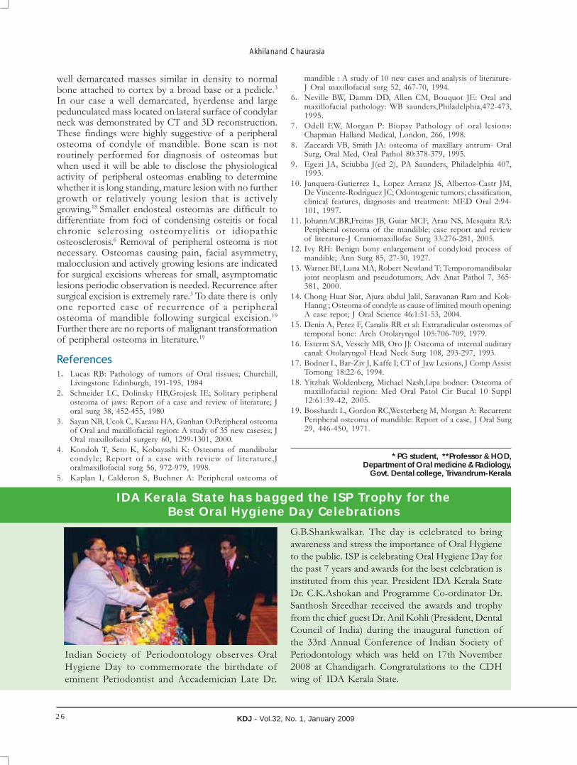
Indian Society of Periodontology observes OralHygiene Day to commemorate the birthdate ofeminent Periodontist and Accademician Late Dr.
IDA Kerala State has bagged the ISP Trophy for theBest Oral Hygiene Day Celebrations
G.B.Shankwalkar. The day is celebrated to bringawareness and stress the importance of Oral Hygieneto the public. ISP is celebrating Oral Hygiene Day forthe past 7 years and awards for the best celebration isinstituted from this year. President IDA Kerala StateDr. C.K.Ashokan and Programme Co-ordinator Dr.Santhosh Sreedhar received the awards and trophyfrom the chief guest Dr. Anil Kohli (President, DentalCouncil of India) during the inaugural function ofthe 33rd Annual Conference of Indian Society ofPeriodontology which was held on 17th November2008 at Chandigarh. Congratulations to the CDHwing of IDA Kerala State.

KDJ - Vol.32, No. 1, January 2009 27
IntroductionVariations of root canal morphology are always been
a constant challenge for diagnosis & successfulendodontic therapy, so a thorough knowledge of mostcommon anatomic characteristics & their possiblevariation is essential. One of the most importantanatomic variation is the C configuration of the canalsystem. The C shaped canal was first described by Cookeand Cox.1979, most commonly found in mandibularsecond molar, a ribbon shaped canal that includes themesiobuccal and distal canals and sometimes themesiolingual canal. Sometimes instead of having severaldiscrete orifices, the pulp chamber of C shaped canal isa single ribbon shaped orifice with a 180 arc, which inmandibular molars starts at the mesiolingual line angleand sweeps around the buccal to the end at the distalaspect of the pulp chamber.
The complexity of the C shaped canal prevents thesecanals from being cleaned, shaped and obturatedeffectively during the root canal therapy. Very little dentinseparates the external surface from the C shaped canalsystem, there by increasing the possibility of strippingperforation during endodontic or restorative treatment.So a proper identification of this abnormal root patternwill help in successful management.
Etiology
Failure of the Hertwig’s epithelial root sheath to fuseon the lingual or buccal root surface is the main causeof C shaped roots, which always contain a C shapedcanal. The C shaped root is also formed by coalescencebecause of the deposition of the cementum with time.
Anatomical features
The C shaped canal appears when fusion of eitherthe buccal or lingual aspect of the mesial and distal root
Review
The C shaped canal- an anatomical variant* Saumyakanta Mohanty, ** Jolly Mary Varughese, *** N.O. Varghese
Abstract
Dental anomalies are the formation defects due to genetic disturbances during the morphogenesisor histogenesis of tooth. Root anatomy is highly complex & un-predictable. Recognition of unusualvariations in the canal configuration is critical. The C configuration, which is an important anatomicvariation, presents a thin fin connecting the mesial and distal individual canal which makes the canalcross-section C shaped and presents much difficulty in its through cleaning and shaping and subsequentobturation. Recognition of a C shaped canal configuration before treatment will surely facilitate ofmore effective management. Because of the importance of its true diagnosis and treatment, acomprehensive review of its normal morphology and its different varieties are discussed in thisarticle.
occurs. This fusion remains irregular, and the two rootsstays connected by inter radicular ribbon of dentin. Thefloor of the pulp chamber is deep and has an unusualanatomic appearance. Two or three canals may be foundin the C shaped groove or the C shape may becontinuous through out the root length.
Classification
1) Melton’s classification
Based on the cross sectional shape
Category 1-continuous C shaped canal running fromthe pulp chamber to the apex defines a C shaped outlinewithout any separation. (Figure 1)
Category 2- the semicolon ( ; )shaped orifice in whichdentine separates a main C shaped canal from one mesialdistinct canal. (Figure 2)
Category 3- refers to those with two or more discreteand separate canals.( Figure 3)
2) Fans classifications
Category 1 (C1) - the shape was an interrupted Cwith no separation or division.
Category 2 (C2) - the canal shape resembled asemicolon resulting from a discontinuation of the Coutline, but either angle alpha or beta angle should beno less than 60 degree. (Figure 4)
Category 3- 2 or 3 separate canals and both angle,alpha and beta were less than 60 degree. (Figure 5)
3) Fans classification ( Radiographic classification)
Type 1- conical or square root with a vague,radiolucent longitudinal line separating the root into distaland mesial parts. There was a mesial and distal canalthat merged into one before exiting at the apicalforamen.

KDJ - Vol.32, No. 1, January 200928
Type 2 - conical or square root with a vague,radiolucent longitudinal line separating the root into distaland mesial parts. There was a mesial and distal canaland the two canals appeared to continue on their ownpathway to the apex.
Type 3 – conical or square root with a vague,radiolucent longitudinal line separating the root into distaland mesial parts. There was a mesial and distal canal,one canal curved to and superimposed on thisradiolucent line when running towards the apex, andthe other canal appeared to continue on its own pathwayto the apex. (Figure 6)
4) Classification based on DSR images and microCT reconstruction
Digital subtraction radiography (DSR), whicheliminates the identical image regions in a series ofradiographs obtained in the same exposures positionand at different time intervals. Radiographic contrastmedium is often used in clinic to change the radio opacityof some anatomic structures before the DSR. MicroCT scans have been applied not only to evaluate canalshapes or cross-section of teeth.
All the DSR images and buccolingual reconstructedcanal images based on micro CT scanning were classified
Type1-(Merging type) - canal images merged intoone major canal before exiting from the apical foramen.
Type2 (Symmetrical type) –There were separatemesial and distal canal appeared to be symmetrical intheir size and continued on their own pathway to theapex.
Type3 (Asymmetrical type)-There were separatemesial and distal canals and mesial and the distal canals
appeared to be symmetrical in their size and continuedon their own pathway to the apex, the distal canal seemsmuch wider than the mesial canal. (Figure 7)
Diagnosis
All the teeth that qualified as having a C shaped canalsystem had to exhibit all the following 3 features;
1) Fused roots.2) A longitudinal groove on the lingual and buccal
surface of the root. 3) At least one cross-sectional of the canal belongs
to the C1, C2 or C3 configuration.
Radiographic diagnoses
A preoperative radiograph and additionalradiographs from 20 degree mesial or distal projectionmay be the only noninvasive means clinically to provideclues about the canal morphology. In fact mostradiographs revealed radicular fusion or proximity, alarge distal canal, a narrow mesial canal and a blurredimage of a third canal in between. (Figure 8)
Radiographic interpretation is overall effective whenbased on film combinations e.g. preoperative andworking length radiographs) or preoperative and finalradiographs and or all 3 radiographs. The radiographstaken while negotiating the canals may reveal twocharacteristics for such canal configuration-instrumentstend to converge at the apex (Figure 9) and/or mayexit at the furcation. (Figure 10).
The latter sometimes resembles a perforation of thefurcation. Micro CT has been used not only to assesscross-section of teeth, but also for three dimensionalreconstructions of canals at high resolution.
Fig. 1 Fig. 2 Fig. 3 Fig. 4
Saumyakanta Mohanty
Fig. 5 Fig. 6 Fig. 7 Fig. 8

KDJ - Vol.32, No. 1, January 2009 29
Clinical diagnosis
1) Anatomical outline of the floor of the pulpchamber
2) Persistence of hemorrhage3) Pain when separate canal orifice were found
Fused roots and C shaped roots may present withnarrow root grooves that predispose to localizedperiodontal disease, which may be the first diagnosticindication of such anatomic variation. When a deepgroove is present on the lingual and buccal surface of aroot a C shaped canal is expected. Under operatingmicroscopic view one can not assume such a shapecontinues through out its length.
Management
Importance of preoperative radiograph
Preoperative radiography shows limited information.Apically tapering roots and roots that appear to becontinuous or square at the apex can be suspected as Cshaped canal configuration. Some C shaped canals aredifficult to interpret because of the thickness of thebony trabeculae.
The C shaped must be suspected when the rootsare fused or close to each other. It should be noted thatbilateral occurrence is possible, so review of dentalhistory is important.
Access cavity preparation
Initial canal system recognition occurs afterachievement of routine endodontic access and removalof tissue from the pulp chamber. Fiber optic Tranillumination can enhance the variant canal anatomyidentification. Placing the fiber optic tip under the rubberdam on the buccal surface illuminates the pulp chamber.The canal system appears as a dark line in an illuminatedarea. Deep orifice preparation and careful probing withsmall files will help in assessing the types of C shapedcanal. (Figure 11 & Figure 12)
Cleaning and shaping
In all categories mesiobuccal and distal canal areprepared normally. Isthmus should not be preparedlarger than 25 size files, otherwise strip perforation canoccur. Extravagant use of small sized file and 5.25%sodium hypochlorite is the key to through debridementin the narrow canal isthmus. An increased volume ofirrigant and deeper penetration of with small instrumentsusing sonic and ultrasonic may allow for cleansability infan shaped areas of the C shaped canal. Aggressiveinstrumentation may sometimes cause perforation.
The ribbon canal space is frequently eccentric to thelingual side of the C shaped radicular dentin. Ananticurvature filing method in the coronal third of thecanal is needed to prevent perforation.
The C shaped canal- an anatomical variant
Fig. 9 Fig. 10 Fig. 11
Fig. 12 Fig. 13 Fig. 14

KDJ - Vol.32, No. 1, January 200930
Obturation
The mesiolingual canal and the distal canal can beprepared and obturated as standard canals. Applicationof thermoplastisized gutta percha is more appropriatetechnique of obturation. Gutta-percha can be thermoplasticized with a heated spreader in an open flame,electric spreader or injectible (Obtura) systems. Properplacement of the sealer with ultrasonic endodontic filesis critical. Endotech 2 can also be used with a zap andtap maneuver; i.e., preheating the Endotech plugger for4-5 sec before insertion (zap) and then moving the hotinstrument in and out in short continuous strokes (taps)10 to 15 times. The plugger was removed while stillhot, followed by a cold spreader with insertion ofadditional accessory points. Touch n heat system canalso be used. (Figure 13 & Figure 14)
Endodontic surgery
The intracanal communications or fins visualized onserial sections reinforce the difficulty the clinician wouldencounter after apicectomy with retropreparation andeventual retrofilling.
If endodontic surgical intervention is indicated fora molar with a C shaped root canal anatomy, strongconsideration should be given to extraction, retrofillingand intensional replantation.
Restoration and prognosis
If post placement for a crown core is desired useof only distal canal (if present independently) shouldbe considered. When sound principle of biomechanicalpreparation and obturation and restoration are followed,the long term prognosis of C shaped canal root retentionequals that of other molars.
References
1) Jerome CE. C-shaped root canal systems: diagnosis,treatment,
and restoration. Gen Dent 1994; 42:424-7.
2) Manning SA. Root canal anatomy of mandibular second molars.
Part II. C-shaped canals. Int Endod J 1990; 23:40-5.
Saumyakanta Mohanty
3) Cooke HG 3rd, Cox FL. C-shaped canal configurations in
mandibular molars. J Am Dent Assoc 1979; 99:836-9.
4) Barril I, Cochet JY, Ricci C. Treatment of a canal with a “C”
configuration. Rev Fr Endod 1989; 8:47-58.
5) Fan B, Cheung GS, Fan M, Gutmann JL, Bian Z. C-shaped
canal system in mandibular second molars: Part I-Anatomical
features. J Endod 2004; 30:899-903.
6) Fan B, Cheung GS, Fan M, Gutmann JL,Fan W. C-shaped canal
system in mandibular second molars: Part II-Radiographic
features. J Endod 2004; 30:904-8.
7) Barnett F. Mandibular molar with C-shaped canal. Endod
Dent Traumatol 1986; 2:79-81.
8) Haddad GY, Nehme WB, Ounsi HF. Diagnosis, classification,
and frequency of C-Shaped canals in mandibular second molars
in the Lebanese population. J Endod 1999; 25:268-71.
9) Bolger WL,Schindler WG. A mandibular first molar with a C-
shaped root configuration. J Endod 1988; 14:515-9.
10) Chai WL,Thong YL. Cross-sectional morphology and minimum
canal wall widths in C-shaped roots of mandibular molars. J
Endod 2004; 30:509-12.
11) Fan W, Fan B, Gutmann JL, Cheung GSP. Identification of C-
shaped canal systems in mandibular second molars. Part I:
Radiographic and anatomic features revealed by intraradicular
contrast medium. J Endod 2007; 33:806-10.
12) Plotino G, Grande NM, Pecci R, Bedini R, Pameijer CH, Somma
F. Three-dimensional imaging using micocompound tomography
for studying tooth macromorphology. J Am Dent Assoc 2006;
137:1555-61.
13) Newton CW, McDonald S. A C-shaped canal configuration in
a maxillary first molar. J Endod 1984; 10:397-9.
14) Sutalo J, Simeon P, Tarle Z, et al. “C”-shaped canal configuration
of mandibular second permanent molar. Coll Antropol 1998;
22:179-86.
15) Liewehr FR, Kulild JC, Primack PD. Obturation of a C-shaped
canal using an improved method of warm lateral condensation.
J Endod 1993; 19:474-7.
*P.G. Student, **Professor & Head, ***Principal & Professor,Department of Conservative Dentistry & EndodonticsGovernment Dental College, Thiruvananthapuram.
In appreciation ofResearch in thefield of DentistryDr. K. Nandakumar hasbeen awarded Fellowshipby Indian Society ofDental Research (ISDR)during the function held atChennai. Dr. AmmbumaniRamdas, Minister forhealth, Govt. of Indiahanding over thefellowship certificate.
Dr. V. IpeVa r g h e s e ,elected asPresident ofthe IndianAssociation
of Oral and MaxillofacialPathologists at the annualmeeting of the associationheld at Kolkata in 29thDecember 2008. He is thePrincipal of Govt. DentalCollege, Calicut, Kerala.
Elected

KDJ - Vol.32, No. 1, January 2009 31
Introduction
In our country periodontal disease is the largest causefor tooth loss. A disease of such high prevalence hasoften eluded importance due to the lack of generalawareness that the disease is preventable and themisconception of tooth loss being an age-associateddisease.
Plaque and plaque retentive factors are the majorcauses for periodontal diseases. Preventive measuresinvolves aids that facilitate it’s removal and decreases itsformation.
Methods for prevention of periodontal
diseases
� Mechanical aids - Mechanical plaque controlmethods include tooth brushing and inter dental cleaningusing oral hygiene aids and professional prophylaxis.Mechanical plaque control is the most dependable formof plaque control.
� Chemical aids - Chemical plaque control is onlyused as an adjunct to mechanical means to increase theirefficiency and not a substitute.
Mechanical aids
1. Toothbrush
It is the principal method employed in oral hygieneand is the most widely used oral hygiene aid. The mainaim of a toothbrush is to remove plaque withoutdamaging the soft tissues. The selection of a propertoothbrush is confusion for many. It is not the brandof the toothbrush that matters but its size, shape of thehead, type of bristle and frequency of replacement. Formost patients a short-headed toothbrush with round-ended soft to medium nylon bristles is recommended.A soft-bristled brush is more effective in removing
Prevention of periodontal diseases
* Riyas Seinullabdeen ** Tintu Sara Chandy, *** Seba Abraham, **** Prakash P., ****Ambili R. ***** Nisha K.J.
plaque as it causes less harm to soft and hard tissuesthan a brush with hard bristles because soft bristles aremore flexible and thus can reach subgingival andproximal areas. A brush with a smaller head can be easilymoved around all the surfaces of the tooth. Round-ended bristles are recommended because they cause lesssoft tissue trauma than course-cut bristles. A toothbrushis to be replaced as soon as the bristles begin to fray,which is usually around 3 months. If all the bristles areflattened after 1 week, brushing is probably too vigorous.If the bristles are still straight after 6 months, eitherbrushing is done very gently or the brush has not beenused everyday.
Brushing technique
The ideal tooth brushing technique is the one thatremoves plaque, food debris and stain, and stimulatesthe gingival tissues with the least time and effort, anddoes not damage oral tissues. Since these criteria cannotbe met with any specific tooth brushing technique, it isnecessary to evaluate each individual condition in orderto select the proper brushing method.
The method most commonly suggested is theModified Bass technique. Here the bristles are placed atthe gingival margin establishing a 45º angle to the longaxis of the teeth and moved in a vibratory, back-and-forth motion while simultaneously moving it coronallyalong the tooth surface. Strokes are repeated around 20times, 3 teeth at a time. On the lingual aspect of anteriorteeth the brush is inserted vertically and the heel of thebrush is pressed into the gingival sulcus and proximalsurface at a 45º angle. The bristles are then activated.Occlusal surface are cleaned by pressing the bristles firmlyagainst the pit and fissure and then activating the bristle.Patients finish the procedure by brushing their tongueto help remove bacteria.
The chosen tooth brushing method should be
Abstract
Periodontal disease is the most common cause for tooth loss in our country. The high prevalenceof this disease prevails in our society due to lack of knowledge and improper oral hygiene practices.The fact that this disease is preventable is often overlooked. The dentist as well as the patient has anactive role to play in the prevention of this disease. The dentist has an important role in motivating,educating and instructing the patient about proper oral hygiene practices. This article is a review ofthe different means by which periodontal disease can be prevented or can be curtailed from recurringafter treatment.
Review

KDJ - Vol.32, No. 1, January 200932
demonstrated for the patient and the patient has topractice it in the office until he or she develops areasonable efficiency in using it. Clinical assessment ofa patient’s toothbrush and tooth brushing technique isalso to be done. This will help the clinician to detect anysign of abrasion or gingival recession that may haveresulted from improper brushing method.
Powered toothbrush
They are a valuable alternative to manual brushes.These brushes mimic the action manual tooth brushesand also make the brushing faster and efficient. Thehead of the tooth brushes oscillate in a side to sidemotion or in a rotary motion. The frequency of theoscillations is around 40Hz in an ordinary poweredtooth brush.
Advantages
� Increases patient motivation resulting in betterpatient compliance.
� Increased accessibility in interproximal and lingualtooth surface.
� No specific brushing technique required.
� Uses less force than manual tooth brushes.
Use of powered toothbrush is not superior to usingmanual toothbrushes in removing plaque and there isno evidence that powered toothbrush will cause moreharm to the soft tissues than manual toothbrushes.
2. Interdental cleansing aids
The interdental areas are usually not accessible bytoothbrush. Plaque removal from these areas is facilitatedby using interdental cleansing aids. The interdentalcleansing aid chosen will depend on the type of gingivalembrasure.
Dental floss
These type of inter dental cleaning aids are indicatedto remove plaque from inter proximal surface with TypeI gingival embrasures.
Methods of use of dental floss1. Spool method – 18 inch length of floss is taken
and wound around the middle finger of both handsleaving about one inch of floss which is held betweenthe thumbs and forefingers. Using a gentle and sawingmotion, the floss is guided between the teeth. When thefloss reaches the gumline, it is then curved into a C-shape against one tooth and is gently slid between thegum and tooth until resistance is felt. While holding thefloss tightly against the tooth, the side of the tooth isscraped gently six times. Without removing the floss,the proximal side of the other tooth is scraped too.This procedure is repeated on the remaining teeth.
2. Loop method - The ends of a 12-inch piece offloss is tied to form a loop or a circle. All fingers exceptthe thumbs are placed within the loop, so that fingersor thumbs tips used to place floss between teeth will be1 inch apart. The flossing procedure is the same as withthe spool method.
Incorrect flossing can traumatize both hard and softtissues, and may also result in inadequate plaque removal.Laceration of inter-dental papilla and cervical wear onproximal root surfaces are signs of improper flossing.
3. Gingival massage
Massaging the gingiva produces epithelial thickening,increased keratinization and increased mitotic activity inthe epithelium & connective tissue making the gingivamore resistant to inflammation. Tooth brushes,interdental cleaning devices and rubber tip stimulatorcan be used.
Figure 1 - Modified BassBrushing technique
Figure 2 - Type 1 No gingivalrecession – Dental Floss
Figure 3 - Type 2Moderatepapillary
recession –Interdental Brush
Figure 4 - Type 3Complete loss of
papillae – Unituftedbrush
Riyas Seinullabdeen

KDJ - Vol.32, No. 1, January 2009 33
4. Oral irrigation devices
These devices clean nonadherent bacteria and debrisfrom the oral cavity more effectively than dotoothbrushes and mouth rinses, especially frominaccessible areas around the orthodontic appliances andfixed prosthesis. It helps to remove subgingival plaqueand is helpful in delivering antimicrobial agents intoperiodontal pockets. These devices are of 2 types:-
Supragingival irrigation devices
They work by directing a high pressure, steady orpulsating stream of water through a nozzle to toothsurfaces. The device has a built-in pump to generate thepressure.
Subgingival irrigation devices
They disrupt and detoxify subgingival plaque bydelivering antimicrobial agents into periodontal pocket.It can be performed both in dental office or by thepatient at home
Irrigation tip is inserted atleast 3 mm into theperiodontal pocket
Chemical plaque control aids
1.Chlorhexidine
Chlorhexidine is a cationic bisbiguanide withpronounced antiseptic properties. It is effective againstgram positive, gram negative bacteria and yeast. It hasmainly 2 actions:-
Anti plaque action - Due to its property of sustainedavailability – substantivity it forms a reservoir ofchlorhexidine, slowly dissolving from all oral surfaces,resulting in the bacteriostatic mileu in the oral cavity.
Anti bacterial action - It is bacteriostatic at lowconcentrations and bactericidal at high concentrations
Disadvantages
� Extrinsic staining of teeth
� Painful, desquamative lesions on the oral mucosamay be associated with burning sensation
� Impaired taste sensation
� Rarely, parotid swelling – due to mechanicalobstruction of parotid duct
Essential oil mouthrinses
Essential oil mouth rinses contain thymol, eucalyptol,menthol and methyl salicylate.
A fixed combination of essential oils kills germs thatcan cause plaque & gingivitis
Mechanism of Action
Essential oils disrupt the bacterial cell wall to killplaque and gingivitis causing organisms nonselectively.
Frequency of plaque removal
Cleaning once a day with a toothbrush and toothpaste
along with flossing is sufficient if it is performedmeticulously. Emphasis is not on the frequency ofbrushing but on the efficiency of complete plaqueremoval. Poor performance of plaque removal can beimproved by brushing twice a day and flossing. Cleaning3 or 4 times per day does not appear to further improveperiodontal conditions.
Conclusion
� The major cause for periodontal disease is plaqueand plaque retentive factors.
� Plaque control measures have 2 purposes – tominimize gingival inflammation and to preventthe recurrence of progression of periodontaldisease in a treated mouth.
� Plaque control is mainly achieved by mechanicalmeans and chemical aids act as adjuvant that helpsto enhance the efficacy of mechanical aids.
� Tooth brushing and flossing once a day isadequate if done efficiently.
� The proper oral hygiene method for each patienthas to be evaluated by the dentist and the patientis to be trained until he or she is proficient.Improper oral hygiene techniques can cause moreharm than good.
References
1. Essentials of preventive and community dentistry – SobenPeter
(2nd edition)
2. Canadian Dental Hygineist Association Position Paper on Tooth
Brushing – Joanna Asadoorian
3. What do patients really need to know about toothbrushes and
dental floss? Khalid M. Al- Siaf, Saudi Dental Journal
4. Carranza’s Clinical Periodontology 10th Edition – Newman, Takei,
Carranza]
*IV Year Student **Junior resident, *** Prof. & Head of theDepartment, **** Reader, ****Reader, *****Senior LecturerDepartment of Periodontology, PMS College of Dental Science& Research, Golden Hills, Venkode.P.O,Vattappara,Trivandrum.
Prevention of periodontal diseases
Former Professor of Prosthodonticsof Calicut and Trivandrum DentalColleges and Rajas Dental College,Nagercoil expired on 30th January2009. After retirement Prof. Devi
took sanyasa at the Santhigiri Ashram, Pothencode,Trivandrum. As a sanyasin she was known as JananiDharmishta Jnana Tapaswini. Prof. Devi was abeloved teacher adored both by her undergraduateand postgraduate students.
Obituary
Dr. N. Sulochana Devi

KDJ - Vol.32, No. 1, January 200934
* Akhilanand Chaurasia, ** Anita Balan
Diagnose the following case
Diagnose
Fig. 1 Fig. 2 Fig. 3
Fig. 4
A 38 year old male patient (Fig1) reported withpresenting complaint of a swelling in right side of facesince 2 years. Previously the swelling was small but sincelast 6 months it was slowly increasing in size. On extra-oral examination a swelling of 2x1 cm was noted inPremolar region in right side (Fig 2). It was soft to firmin consistency, nonfluctuant, noncompressible havingwell defined borders. There was no associated pain. Onintra-oral examination mild obliteration of buccal sulcuswas noted in right premolar region (Fig 3). Panoramic
* Postgraduate Student, ** Professor & HOD,Department of Oral Medicine and Radiology, GovernmentDental College, Trivandrum, Kerala - 695 011
Fig. 5
radiograph of patient showed an enlarged mentalforamen (Fig 4,5). While doing excision of swelling itwas found to attached to mental nerve through it deepsurface.
a. What this lesion could be?
b. What is differential diagnosis?
Answers
a.Lipoma associated with right mental nerve.b.Differential diagnosis include-
1. Fibroma 2. Neurofibroma
From clinical and radiographic features it was thought to beneurofibroma but while doing biopsy multiple yellowish fatglobules were seen and histopathology was also compatiblewith diagnosis of lipoma of buccal mucosa. Very few caseshad been reported in literature in which lipoma of buccal mucosawas found to be associated with mental nerve.

KDJ - Vol.32, No. 1, January 2009 35
Introduction
Modern advances in dentistry as well as the increasedawareness among patients have lead to the developmentof many treatment modalities which providesopportunity for patients to maintain a functionaldentition for life time. Periodontal disease that extendsinto the furcation areas can pose significant difficultyduring treatment, as can extensive caries or root fracturesthat involve the furcation areas. Treating any of theseproblems is particularly difficult with regard to theinterproximal furcation areas; as the disease process andsubsequent treatment could affect the periodontalattachment apparatus of the adjacent teeth. Rootamputation procedures can be used when attachmentloss, caries, other endodontic problems or a fractureinvolves furcation area of a molar.
Root amputation procedures are a logical way toeliminate a weak, diseased root to allow the stronger tosurvive, whereas if retained together, they wouldcollectively fail. The treatment may involve amultidisciplinary approach so that the teeth are retainedin whole or in part. There are various root amputationprocedures and it includes root resection, hemisection,and bicuspidization or root separation. Root amputationprocedure on mandibular molars is usually referred toas hemisection. Hemisection denotes removal orseparation of root with its accompanying crown portion.Selected root removal allows improved access for homecare and plaque control with resultant bone formationand reduced pocket depth.
This article reviews hemisection of a mandibularmolar and the concomitant prosthetic management asa treatment option for furcation involvement ofmandibular molars.
Hemisection – A better option in
furcation management
Preeja.C*, Presanthila Janam**, Haeigin Tom Varghese***, T. Sreelal****, K.Harshakumar*****
Indications
Periodontal Indications:
1. Severe vertical bone loss involving only one rootof multi-rooted teeth.
2. Through and through furcation destruction.3. Unfavourable proximity of roots of adjacent
teeth, preventing adequate hygiene maintenancein proximal areas.
4. Severe root exposure due to dehiscence.
Endodontic and Restorative Indications:
1. Prosthetic failure of abutments within a splint:
If a single or multirooted tooth is periodontallyinvolved within a fixed bridge, instead of removing theentire bridge, if the remaining abutment support issufficient, the root of the involved tooth is extracted.
2. Endodontic failure:
Hemisection is useful in cases in which there isperforation through the floor of the pulp chamber, orpulp canal of one of the roots of an endodonticallyinvolved tooth which cannot be instrumented.
3. Vertical fracture of one root:
The prognosis of vertical fracture is hopeless. Ifvertical fracture traverses one root while the other rootsare unaffected, the offending root may be amputed.
4. Severe destructive process:
This may occur as a result of furcation or sub gingivalcaries, traumatic injury, and large root perforation duringendodontic therapy.
Contraindications:
a. Inoperable canals in root to be retained.b. Fused roots making separation impossible.c. Strong adjacent teeth available for bridge
abutments as alternatives to hemisection.
Case Report
Fig. 1, 2, 3, 4 Clinical, radiographic and surgical management stages

KDJ - Vol.32, No. 1, January 200936
Case report
A 48 year old female reported to Dept. ofPeriodontics, Govt. Dental College, Trivandrumcomplaining of a long standing swelling and pusdischarge in relation to lower right back tooth for thepast one year. On clinical examination chronicperiodontal abscess was present in relation to buccalaspect of 46 (Fig.1). On periodontal probing deepperiodontal pocket of 7mm was present in relation tomesial root of the tooth. Patient also gave a history ofendodontic treatment of 46 due to caries 2 years back.Patient did not give any history of pain or associatedsymptoms.
On radiographic examination, an inappropriatelyfilled root canal with filling material perforating throughthe furcation area was seen. Severe bone loss was evidentsurrounding the mesial root and involving the furcationarea of 46. The bony support of distal root wascompletely intact (Fig. 2). So the best treatment optionfor this case was removal of the diseased portion ofthe tooth by hemisection.
Under local anesthesia, full thickness mucoperiostealflap was reflected after giving a crevicular incision fromdistal aspect of second premolar to mesial aspect ofsecond molar. Upon reflection of the flap, the bonydefect along the mesial root became quite evident. Allchronic inflammatory tissue was removed to exposethe bone. The vertical cut method was used to resectthe crown. A long shank tapered fissure carbide bur ona highspeed handpiece is placed along the long axis ofthe tooth in the area of the buccal furcation and a cut ismade (Fig. 3). This cut is channeled toward the centerof the tooth. Similarly a cut is made in the lingualfurcation which is also directed towards the centre. Thesecuts are then joined so that the mesial and distal halvesare completely separated (Fig. 4). A fine probe waspassed through the cut to ensure complete separationof the two halves of the tooth. Once the separationwas complete the mesial root along with crown portionwas extracted with a small extraction forceps (Fig 5 &6). The empty socket was thoroughly curetted andirrigated adequately with sterile saline to remove bonychips and amalgam debris.
The furcation area was trimmed to ensure that nospicules or filling material were present to cause furtherperiodontal irritation. Scaling and root planing of theroot surfaces was done. The extraction site was irrigated,
debrided and the flap was then repositioned and suturedwith 3/0 black silk sutures (Fig. 7 & 8).
At the end of first week sutures were removed. After3 months the prosthetic rehabilitation was done. Thetreatment plan for the prosthodontic management phasewas to fabricate a fixed partial denture using 47, distalhalf of 46, 45 and 43 as abutments to replace the mesialhalf of 46 and the 44. The idea was to develop anocclusion with canine protected articulation, decreasedvertical overlap and flattened posterior cusps. Thisocclusal pattern will protect the weakened posteriorabutment against excessive loading during excursivemovements and during mastication. The occlusal surfacearea of the prosthesis over the distal root of 46 waskept as minimum as possible to decrease the stressesthat may act on it. Due to aesthetic reason, metal withceramic facing was the restoration of choice except for47 which received a full metal restoration to preservemaximum tooth structure in the non appearance zone.Modified ridge lap was the pontic design used infabricating the restoration. (Fig 9, 10 & 11).
DiscussionRoot amputation is a useful alternative procedure
for saving those multirooted teeth which have beenindicated for extraction. But success of root amputationprocedures depends, to a large extent, on proper caseselection. It is important to consider the following factorsbefore deciding to undertake any of the resectionprocedures.
� Advanced bone loss around one root withacceptable level of bone around the remainingroots.
� Angulation and position of the tooth in the arch:A molar that is buccally, lingually, mesially ordistally titled, cannot be resected.
� Divergence of the roots : Teeth with divergentroots are easier to resect; closely approximatedor fused roots are poor candidates.
� Length and curvature of roots : Long and straightroots are more favourable for resection thanshort, conical roots.
� Feasibility of endodontics and restorativedentistry in the root/roots to be retained.
� Patient’s oral hygiene status, caries index, andmedical status should be considered.
Preeja C.
Fig. 5, 6, 7, 8 Clinical, radiographic and surgical management stages

KDJ - Vol.32, No. 1, January 2009 37
In the present case report on hemisection, mesialroot along with crown portion is removed which hasmany advantages than removing the distal root. Themesial root has an hour-glass-shaped cross section whichmay be difficult for plaque control and restorativeprocedure. Also, root canals are closer to external rootsurface which can complicate the subsequent restorativetreatment. But the distal root has an oval cross sectionwith only one, wide root canal. Distal root iscomparatively large, providing a greater mass of dentinto resist fracture. Further, when the resected mandibularmolar is a terminal abutment for a bridge, the retentionof the distal root will result in a longer dental arch thanwould be the case had the mesial root been retained.
The removal of a root alters the distribution ofocclusal forces on the remaining roots. Therefore, it iswise to evaluate the occlusion of teeth from which rootshave been resected and, if necessary, adjust the occlusion.Centric holds should be maintained and eccentric forcesshould be eliminated from the area over the root thatwas removed.
Teeth that have been resected can be used as abutmentteeth for fixed partial dentures, splints, or vertical supportfor cantilever fixed partial dentures. The retention of astrategic tooth by root resection may preclude the needfor a removable partial denture. However their loadbearing ability will be lessened due to diminishedattachment area as a result of periodontal attachmentloss. The mesial root of mandibular first molar provides37% of the attachment surface area and the distalprovides 32%. If the furcation is uncovered, 31% ofthe attachment area which is provided by the root trunkhas been lost. The mesiofacial, distofacial and palatalroots of a maxillary first molar furnish 25%, 19%, and24% of the attachment area respectively. The root trunksupplies 32% of the attachment for the tooth. Removalof a corresponding root on a second molar willprobably result in a similar percentage loss of support.The total root surface area of the first and second molarsdiffers by only 0.5%to1.2%.
In case of mandibular hemisection, saving the mesialsegment would be desirable if the molar in questionwere the last tooth in the arch and the opposing teethdid not extend very far distal to the mandibular firstmolar. The distal root could be used as an abutmentfor a short span fixed partial denture replacing the mesial
root. Occasionally the one root may be used as the distalabutment for a long span fixed partial denture, replacingan entire molar. But this must be viewed as high riskprosthesis as the remaining single root will usually haveslightly less than one third of the alveolar support ofthe intact tooth with normal bone anchorage.
ConclusionThe prognosis for hemisection is good provided
that case selection has been correct, the procedure hasbeen performed adequately, and the restoration is ofan acceptable design relative to the occlusal andperiodontal needs of the patient. Root amputationtherapies are used to preserve as much tooth structureas possible rather than sacrificing the whole tooth.
Investigations of root resected or hemisected teethhave shown that such teeth can function successfully forlong periods. The keys to long term success are thoroughdiagnosis, selection of patients with good oral hygiene,and careful surgical and restorative management.
References
1. Newman, Takei, Klokkevold, Carranza. Carranza’s Clinical
Periodontology, Tenthth edition.
2. Jan Lindhe. Clinical Periodontology and Implant Dentistry,
Fourth edition.
3. Erpenstein H. A 3-year study of hemisectioned molars. J Clin
Periodontol 1983; 10(1): 1-10.
4. Buhler H. Evaluation of root-resected teeth: results after 10
years. J Periodontol 1983; 59:805-10.
5. T Hempton and C Leone. A review of root resective therapy as
a treatment option for maxillary molars. J Am Dent Assoc
1997; 128; 449-455.
6. Kim Y. Furcation involvements: therapeutic considerations.
Compend Contin Educ Dent. 1998 Dec; 19(12):1236-40.
7. Herbert T. Shillingburg, Jr, Sumiya Hobo, Lowell D. Whitsett,
Richard Jacobi, Susan E. Brackett. Fundamentals of fixed
prosthodontics, third edition.
8. Reinhardt RA, Sivers JE: Management of class III furcally
involved abutments for fixed prosthodontic restoration. J
Prosthet Dent 1988;13:126-135.
9. Stephen F. Rosenstiel, Martin S Land, Junhei Fujimoto.
Contemporary Fixed Prosthodontics, fourth edition.
*PG student, **Professor and Head, Dept: of Periodontics,***PG student, **** Prof: and Head ***** Professor
Dept: of Prosthodontics, Govt. Dental College, Trivandrum
Hemisection – A better option in furcation management
Fig. 9, 10, 11 Furcation of prosthesis

KDJ - Vol.32, No. 1, January 200938
Eagle’s Syndrome* Arun George , ** Devi Gopakumar , *** Bobby John , *** S Sunil
Abstract
Eagle syndrome is a constellation of symptoms and signs resulting from abnormal stylohyoidchain ossification. The styloid chain consists of the styloid process, the stylohyoid ligament, and thelesser cornu of the hyoid bone. Eagle Syndrome is a rare entity which is not commonly suspected inclinical practice, and only a small percentage of the 4% of population believed to have an elongatedstyloid process and a calcified stylohyoid ligament manifest the symptoms. The signs and symptomsmentioned in various sources for Eagle’s syndrome includes Pain on turning head, Pain on extendingtongue, Voice changes, Cough, Sinusitis, Dizziness, Bloodshot eyes, Throat pain, Facial pain, Foreignbody sensation in throat, Dysphagia, Disturbed sense of taste, Asymptomatic, Headache, Sensationof excessive salivation, Throat discomfort, Vertigo, Swallowing difficulty, Pain on opening mouth.The diagnosis is often first made radiologically and once made this prompts clinical confirmation bypalpation of the tonsillar fossa.
Introduction
Eagle syndrome derives it name from Watt WeemsEagle an Amercian Otolaryngologist. He first publishedhis data in 1937 with a series of 200 patients presentingwith symptoms related to a calcified stylohyoid ligamentor elongated styloid process. These symptoms includea pharyngeal foreign body sensation, dysphagia, painon head rotation, otalgia, dizziness and headaches. Thesymptoms are attributed to impingement of theglossopharyngeal nerve as it runs close to the styloidprocess and calcified ligament. An elongated or calcifiedstylohyoid ligament does not necessarily indicate Eaglesyndrome. The diagnosis is made on the basis of historyand exacerbation of pain on palpation of the tonsillarfossa. Another test of diagnostic, and also therapeuticbenefit, is that of injecting local anaesthetic into thetonsillar fossa, which should result in diminishedsymptoms
Case Report
A 42 year old female presented to our college withhistory of pain on the right side of neck and dysphagiaof 2 year duration. She had consulted Physician & ENTspecialist before, and was referred to Dept of Oral &Maxillofacial Surgery of our college. She presented withunilateral pain on the right side occasionally on yawningand swallowing. Her general examination did not revealany relevant findings. Cervical lymphnodes were notpalpable. Right cervical area below styloid process wastender to palpate. Intraoral examination revealed nosignificant dental pathology. An OPG was taken, and adiagnosis of calcified and elongated stylohyoid ligamentwas made.
Discussion
Eagle syndrome is a group of symptoms causedby calcification of the stylohyoid ligament and anabnormally long styloid process. The styloid process isa bone at the base of the skull which is attached tomuscles and ligaments connected to the throat andtongue. The styloid process and ligament areembryological remnants of the second branchial archand persists as a structure running from the base of theskull to the lesser horn of the hyoid, passing betweenthe internal and external carotid arteries. In 1937, WattWeems Eagle was the first to present two cases ofpharyngeal discomfort associated with elongated styloidprocess Eagle originally described two distinctsyndromes: the classic styloid and carotid arterysyndromes. Mineralisation of the stylohyoid ligament isa well recognised radiographic finding and an incidenceof 18.2% has been reported on panoramic radiographs.The majority of patients are asymptomatic. Eagle’ssyndrome now describes a syndrome in which there iselongation of the stylohyoid process with associatedsymptoms which may include pain in the throat,dysphagia, dysphasia, otalgia, sensation of a foreign bodyin the throat and facial pain which may be vague and illdefined or an acute neuralgic type of pain radiating tothe ear or along the mandible.
Eagle Syndrome can be unilateral or bilateral. Innormal adults, the styloid process is approximately25mm long, and its tip is located between the externaland internal carotid arteries, just lateral to the tonsillarfossa. The mineralized section of the styloid process orstylohoid ligament is rigid and more subjected to fracturesecondary to traumatic injury.
The actual cause of the elongation is a poorly
Case Report

KDJ - Vol.32, No. 1, January 2009 39
understood process. Several theories have beenproposed: 1) congenital elongation of the styloidprocess due to persistence of a cartilaginous analog ofthe stylohyal (one of the embryologic precursors ofthe styloid), 2) calcification of the stylohyoid ligamentby an unknown process, and 3) growth of osseous tissueat the insertion of the stylohyoid ligament
The pathophysiological mechanism of symptoms isdebated as well. Theories include the following:
1) traumatic fracture of the styloid process causingproliferation of granulation tissue, which places pressureon the surrounding structures 2) compression ofadjacent nerves, the glossopharyngeal, lower branch ofthe trigeminal, or chorda tympani; 3) degenerative andinflammatory changes in the tendonous portion of thestylohyoid insertion, called insertion tendonitis; 4)irritation of the pharyngeal mucosa by directcompression or post-tonsillectomy scarring (involvescranial nerves V, VII, IX, and X); and 5) impingementof the carotid vessels, producing irritation of thesympathetic nerves in the arterial sheath
The signs and symptoms mentioned in varioussources for Eagle’s syndrome includes
� Pain on turning head, Pain on extending tongue,Voice changes, Cough, Sinusitis, Dizziness, Bloodshoteyes, Throat pain, Facial pain, Foreign body sensationin throat, Dysphagia, Disturbed sense of taste,Asymptomatic, Headache, Sensation of excessivesalivation, Throat discomfort, Vertigo, Swallowingdifficulty, Pain on opening mouth
Treatment of Eagle syndrome can be both surgicaland nonsurgical. Nonsurgical treatments involvesassurance, nonsteroidal anti-inflammatory medications,and steroid injections. Conventional medical treatmentsmay help alleviate the symptoms of Eagle syndrome,but they do not address the core of the problem. But,Eagle’s syndrome can be successfully treated by surgery.Several transoral and extraoral-cervical ways tostyloidectomy have been described. Transoral resectionof the styloid process is perfectly easy to perform, itcan be done with local anesthesia, it involves no extensivefascial dissection. Transoral resection causes no outsidescars; also, the length of both the operation and the
recovery period is short. The risks of the transoralapproach, which are low, involve the chance of a deepcervical infection and the possibility of a neurovascularinjury during an attempt to leave as little remnant of thestyloid process as possible.
Conclusion
Eagle’s syndrome develops due to an elongation ordeformation of the styloid process and ossification”of the stylohyoid ligament. The development of thispathology is influenced by cervical osteochondrosis,frequent tonsillitis, tonsillectomy and purulent facial andcervical inflammations.
The clinical presentation of Eagle’s syndromedepends on the localization of disturbance. The analysisof clinical symptoms allows not only to diagnose thesyndrome, but also to foresee a possible disturbanceof the styloid process or the stylohyoid ligaments.Radiological analysis is the basic method for thelocalization of the stylohyoid complex disturbance andthe diagnostics of its nature.
Best results of treatment are achieved when a surgicalmethod of treatment is applied.
References1. Ryan D. Murtagh, Jamie T. Caracciolo, and Gaspar Fernandez.
CT Findings Associated with Eagle Syndrome. AJNR Am JNeuroradiol 22:1401–1402, August 2001
2. James G. Lorman and J. Roy Biggs. The Eagle Syndrome.AJR140:881-882, May 1983
3. Luis Balbuena, Jr., David Hayes, Sylvester G. Ramirez, andRobert Johnson. Eagle’s Syndrome (Elongated Styloid Process).Southern Medical Journal. 1997; March.
4. Nickel J, Sonnenburg M, Scheufler O, Andresen R. [Eaglesyndrome: diagnostic imaging and therapy]. Rontgenpraxis. 2003;55:108-13.
5. Balasubramanian S. The ossification of the stylohyoid ligamentand its relation to facial pain. Br Dent J 1964; 116:108–111
6. Chase DC, Zarmen A, Bigelow WC, McCoy JM. Eagle’ssyndrome: a comparison of intraoral versus extraoral surgicalapproaches. Oral Surg Oral Med Oral Path 1986; 62:625–629
7. Eagle WW. Elongated styloid process: symptoms and treatment.Arch Otolaryngol 1958; 64:172–176.
* Senior Lecturer, ** Lecturer, *** Assistant Professor,***Assistant Professor, Oral & Maxillofacial Pathology,
Mar Baselios Dental College, Kothamangalam
Eagle’s Syndrome

KDJ - Vol.32, No. 1, January 200940
Introduction
Infections have afflicted mankind since timesimmemorial. Viruses were found to be the causativeagents of self limiting infections. However, thoughmostly self limiting, the morbidity caused by theseinfections is no less a concern to the dental clinician facedwith a patient suffering from them. Herpes zoster is asecondary infection caused by Herpesvirus varicellae,commonly referred to as the varicella-zoster virus(VZV). It has been estimated that upto half of all peoplewho live upto the age of 85 years may be affected byherpes zoster.1
The presenting signs and symptoms may vary at timesand in the prodromal stage of this disease, odontalgiamay be the only symptom. This may prove to be quitea diagnostic dilemma for the unwary clinician. Wepresent a case of herpes zoster infection involving themaxillary division of the right trigeminal nerve. Thecharacteristic features of this case should help the busypractitioner to firmly etch in mind the appearance ofthe condition and the treatment that needs to beaccorded to such cases.
Case report
A 64 year-old female patient reported to our institutewith a complaint of pain in the right upper jaw on eatingfood since 4 days. The pain was severe, continuous andradiated to the right cheek region. It started suddenlywhen she had woken up in the morning 4 days prior toreporting to us. There had been intense burningsensation on the right side of the face since the previousevening that was at the time of examination. The patienthad fever for the preceding 5 days. She also had difficultyin eating and drinking.
The patient’s past medical and family histories wereunremarkable. She had undergone uneventful extraction
Herpes zoster infection of the maxillary nerve
* Ajay G. Nayak, ** Laxmikanth Chatra,*** Prashanth Shenai K., *Prasanna Kumar
Abstract
Herpes zoster is a viral infection caused by Herpesvirus varicellae. It can cause severe morbidity ifearly diagnosis and treatment of the condition is not provided. A thorough knowledge of thiscondition will prevent unnecessary and delayed treatment for the patient. A case is presented of anelderly lady who presented with the classical signs and symptoms of herpes zoster. The conditionwas then treated with a newer antiviral drug (valacyclovir) which resulted in complete resolution ofthe condition. The presence of herpes zoster infections as a part of the spectrum of manifestationof HIV infection only highlights the urgent need for every dental clinician to be able to diagnose andeffectively manage this condition.
of all her teeth 12 years ago due to mobility and sincethen had been wearing complete dentures. She did notreport any oral habits related to tobacco or areca nut.
The patient on physical examination, appearedemaciated and had pyrexia of 101oF. The patient alsoexhibited pallor of the palpebral conjunctiva. Her bloodpressure was within normal limits for her age and gender– 142/ 96 mm of Hg. Extra oral examination revealederythema and vesicular lesions in the middle third ofthe face on the right side that ended abruptly in themidline (fig 1). The vesicles were present as clusterslateral to and below the right ala of the nose extendingupto the right angle of the mouth. Crusting was noticedon a ruptured vesicle immediately lateral to the right alaof the nose. Crusted lesions were also present on thevermilion border of the right side of the upper lipending in the midline (fig 2). The left side of the faceupto the midline did not show any abnormality. Thearea was tender on palpation and produced stingingsensation. The vesicles could be ruptured by applicationof pressure. Nikolsky’s sign was negative. Rightsubmandibular lymphadenopathy was detected.
Intraorally, curdled-appearing, multiple, clusters ofvesicles and erosions on an erythematous base werepresent on the palatal mucosa on the right side (fig 3).These lesions also stopped sharply at the midline withoutany extension to the opposite side. On palpation, themucosa was tender and severe stinging sensation wasfelt by the patient, from the midline upto the rightmaxillary vestibule. The vesicles could be deroofed withsome light pressure, however Nikolsky’s sign was absent.The scrapping produced a raw bleeding base. The othersoft tissues did not exhibit any abnormality. All themaxillary and mandibular teeth were clinically absent.
Based on the history reported by the patient and thefindings of the clinical examination a diagnosis of Herpes
Case Report

KDJ - Vol.32, No. 1, January 2009 41
zoster infection of the maxillary division of the righttrigeminal nerve was arrived at.
The patient’s emaciated and dehydrated conditionprompted hospital admission and institution of IV fluidsimmediately – Dextrose Normal Saline (DNS) – 100ml/ hour infusion. Investigations such as completehemogram and anti HIV ELISA were performed. Theblood picture showed lymphocytosis, a decreasedhemoglobin level of 8.3 gm % (normal: 12-16 gm %)and an ESR of 39 mm/hour (normal: 0-15 mm/hour).The Anti HIV ELISA was negative, and since the historywas not suggestive it was not repeated.
The specific pharmacotherapy instituted consistedof the following:
� Systemic Antiviral therapy – Valacyclovir, 1000mg – 1 t.i.d. x 7 days
� Topical Antiviral therapy – 5% Acyclovir cream– t.i.d. x 7 days
� Anti-inflammatory therapy – Diclofenac, 50 mg– 1 b.i.d x 5 days
When reviewed after 2 days of therapy, a newcutaneous bulla lateral to the right ala of the nose wasnoted (fig 4). There was crusting of the cutaneous lesionswith hemorrhagic ulcers on the vermilion border ofthe lip (fig 5). The erythematous skin surface appearedto be reducing in intensity as compared to that atpresentation.
On the fourth and fifth days the lesions showedchanges consistent with healing. When the patient wasreviewed after completion of the antiviral therapy (onthe 8th day), the lesions, both cutaneous and oral, hadhealed completely. The patient was then discharged fromthe hospital and recalled for review after a week
The patient was then reviewed after a week of thecompletion of the antiviral therapy. The patient reportedno problems in swallowing and reported consumingregular solid and fluid diet without any burning sensationof the oral mucosa. Intraorally, no abnormalities of thesoft tissues were noted.
The patient has been asymptomatic for the past threeyears and is on continuous follow-up.
Discussion
The varicella zoster virus (VZV) is responsible fortwo common infectious diseases, chicken pox (varicella)and shingles (zoster). Herpes zoster infection is causedby subsequent reactivation of latent varicella zoster virusfrom dorsal root or cranial nerve ganglia, present sinceprimary infection with varicella (chicken pox).2 Thisreactivation may occur even decades later. The mostfavored ganglia are those of the T3 – L2 segments, butin the head and neck region the virus favors thetrigeminal ganglion to remain dormant. The commonestcause of viral reactivation is decline in cell mediatedimmunity related to age. After the primary infection thevirus is probably often reactivated, but competent cellmediated immunity prevents clinical disease.1 It is acontagious condition and spreads via direct contact withan infected person or through inhalation of air-bornerespiratory secretions. Children in contact with a zosterpatient can develop varicella and hence zoster patientsmay need to be isolated during the course of the diseasefrom those who have not suffered from varicella. Thelesions remain contagious from 2 days before the rashappears until all the lesions are crusted with no drainage.
Herpes zoster infections progresses through 3 stages:prodromal, active (acute) and the chronic stages.3
However each patient may not develop all the threestages. Some patients do not develop the classic vesiculareruptions and may manifest only dermatomal pain,causing difficulty in diagnosis, called as zoster sine herpete.4
The patients presenting in the prodromal stage describetheir condition as a burning, tingling, itching, boring,pricking or knife-like sensation on the skin. These sensorychanges are thought to be a result of the degenerationof nerve fibrils from viral infection activity.3 The activephase is the emergence of the rash accompanied bymalaise, low grade fever and at times nausea. The rashgoes through maturation stages of an erythematous
Fig. 1 Erythema and vesicles ending abruptly in themidline of the right middle third of the face
Fig. 2 Crusted lesions on the lip seen only on the right halfof the upper lip
Herpes zoster infection of the maxillary nerve

KDJ - Vol.32, No. 1, January 200942
papule followed by vesicles that eventually form pustules.These pustules undergo crusting which then fall off inapproximately 2 weeks, leaving behind erythematousmacular areas. The oral mucosa also manifests similarlesions. The chronic stage is termed as the Post herpeticneuralgia (PHN), which is defined as pain lasting beyondthe period of healing of the active lesions. This may lastfor 1-3 months and sometimes even for years or decades.
The differential diagnoses that need to be consideredare acute vesiculobullous conditions herpes simplexinfections and erythema multiforme. However thecharacteristic involvement of a region supplied by aspecific nerve with an abrupt stop of the lesions at themidline, as seen in the case presented is very characteristicof herpes zoster. Herpes simplex infections are usuallyrecurrent and erythema multiforme may havegeneralized manifestations. However when herpes zosteroccurs as a component of the AIDS manifestation, itmay also be generalized and prove to be fatal if nottreated aggressively.
The mainstay of contemporary zoster managementis the use of antiviral drugs like acyclovir, valacyclovirand famciclovir.1,2 Acyclovir is used in the dose of 800mg, 5 times daily for atleast 7 days. Valacyclovir isconverted to the active form of acyclovir, but patientcompliance for this drug is better due to the dosageschedule of 1000 mg, t.i.d for 7 days. Famciclovir isused as 500 mg, t.i.d. for 7 days. Most cases respond totherapy if it is initiated within 72 hours of thedevelopment of the lesions. In the case described thevesicles went through stages of desquamation and finallyhealing. However the institution of pharmacotherapywith antiviral agents aided considerably in the healingand pain management. It has been found that antiviraltherapy definitely reduces the viral load and viralshedding in the vesicles and hastens healing. Additionalpain management can be performed using analgesicsand anti inflammatory drugs like paracetamol, ibuprofen,etc, though these drugs are not helpful in treating postherpetic neuralgia.
A live attenuated vaccine against varicella is nowcommonly available. Because the vaccine virus may beless likely to establish latency and reactivate, it has beenthought that vaccination may ultimately reduce the
incidence of zoster.5 Vaccination of older adults whohave had varicella may also prevent or attenuate zoster.
In conclusion, it can be safely stated that the earlydiagnosis and prompt management of herpes zosterinfections can go a long way in reducing the discomfortof the patient. The emergence of these infectious statesin the AIDS spectrum must make the clinician morevigilant to look for any underlying causes and accordinglyproceed with the management strategies. A rigid follow-up schedule is also an essential requisite to avoid longterm morbidities.
References
1. Johnson RW, Dworkin RH. Treatment of herpes zoster andpostherpetic neuralgia. BMJ 2003; 326:748-50
2. Gnann JW, Whitley RJ. Herpes zoster. N Engl J Med 2002;347:340-6
3. Strommen GL, Pucino F, Tight RR, Beck CL. Human infectionwith H. Zoster: etiology, pathophysiology, diagnosis, clinicalcourse and treatment. Pharmacotherapy 1988; 8:52-68
4. Barratt AP, Katelaris CH, Morris JG, Schifter M. Zoster sineherpete of the trigeminal nerve. Oral Surg Oral Med OralPathol 1993; 75:173-5
5. Levin MJ. Use of varicella vaccines to prevent herpes zoster inolder individuals. Arch Virol 2001; suppl 17:151-160
* Assistant Professor ** Senior Professor & Head *** ProfessorDepartment of Oral Medicine & Radiology, Yenepoya DentalCollege & Hospital, Nithyanand Nagar Post, Deralakatte,Mangalore.
Fig. 3 Curdledappearingvesicles anderosions on thelips and palatalmucosa stoppingsharply at themidline withoutextension to theopposite side
Fig. 4 Crustedulcers on a
fadingerythematous
base with newbullae lateral to
the ala of thenose as seen 2
days afterantiviral therapy
Ajay G. Nayak
Fig. 5 Hemorrhagic confluent ulcers on the lips on the3rd day of antiviral therapy

KDJ - Vol.32, No. 1, January 2009 43
Scleroderma
Introduction
Scleroderma or systemic sclerosis is a systemicconnective tissue disease, characterized by vasomotordisturbances, fibrosis, subsequent atrophy of the skin,subcutaneous tissue, muscles and internal organs (eg:alimentary tract, lungs, heart, kidney, CNS) withassociated immunological disturbances. Progressivesystemic sclerosis is characterized by ultimate indurationof the skin and fixation of the epidermis to the deepersubcutaneous tissues. Greatest incidence is between 30and 50 years of age. The tongue, soft palate and larynxare the intra-oral structures commonly involved.Limitation of mouth opening is seen in many cases.Reduced opening of the mouth and fixation of the jaware a result of involvement of the peri-temporomandibular joint tissues. Bone resorption ofthe angle of the mandible, usually bilaterally, has beenreported. Rarely, there has been partial or completeresorption of condyles and/or coronoid process ofthe mandible.
* Gayathri Krishnan, ** Vishnu Mohan
Case report
A 35 year old male patient came to the Departmentof Oral and Maxillofacial Surgery of Azeezia collegeof Dental Sciences and Research with the complaint ofdifficulty in opening and closing the mouth. On historytaking, we came to know that the patient earlier had asubluxation of the TMJ and was referred to anotherhospital. The surgeons had tried to push back thecondylar head back into the glenoid fossa which resultedin fracture of the angle of the mandible. Radiographicexamination shows that the fracture of the angle of themandible was as a result of a cystic lesion in the regionof the angle. ( Pathological fracture)
Surgical Procedure
Sub-mandibular or Risdon’s incision was done toaccess the angle of the mandible. The skin andsubcutaneous tissue were incised with a scalpel downto the level of the platysma and the skin wasundermined. The platysma was then sharply divided
Pre-operative photograph Pre-operative radiographs
Abstract
Scleroderma is a systemic connective tissue disease characterized by vasomotor disturbances,fibrosis, subsequent atrophy of the skin, subcutaneous tissue, muscles and internal organs with associatedimmunological disturbances. Limitation of mouth opening and fixation of the jaw as a result ofinvolvement of peri-temporomandibular joint tissues have been reported. Bone resorption of theangle of the mandible has been reported in some cases. In this case report, the patient is a 35 year oldmale who came to our college with difficulty in opening and closing the mouth. The patient earlierhad a subluxation of the TMJ where the surgeons (in another hospital) had tried to push back thecondylar head back into the glenoid fossa. This resulted in fracture of the mandibular angle.Radiographic examination revealed that the fracture was as a result of a cystic lesion in the region ofthe angle. The cystic lesion was curetted and the fracture was reduced and fixed with Titanium platesand screws. In a scleroderma patient, there is chance for resorption of the angle of the mandible. Sowhen there is subluxation, it should be reduced only under skeletal muscle relaxants to avoid mandibularangle fracture.
Case Report
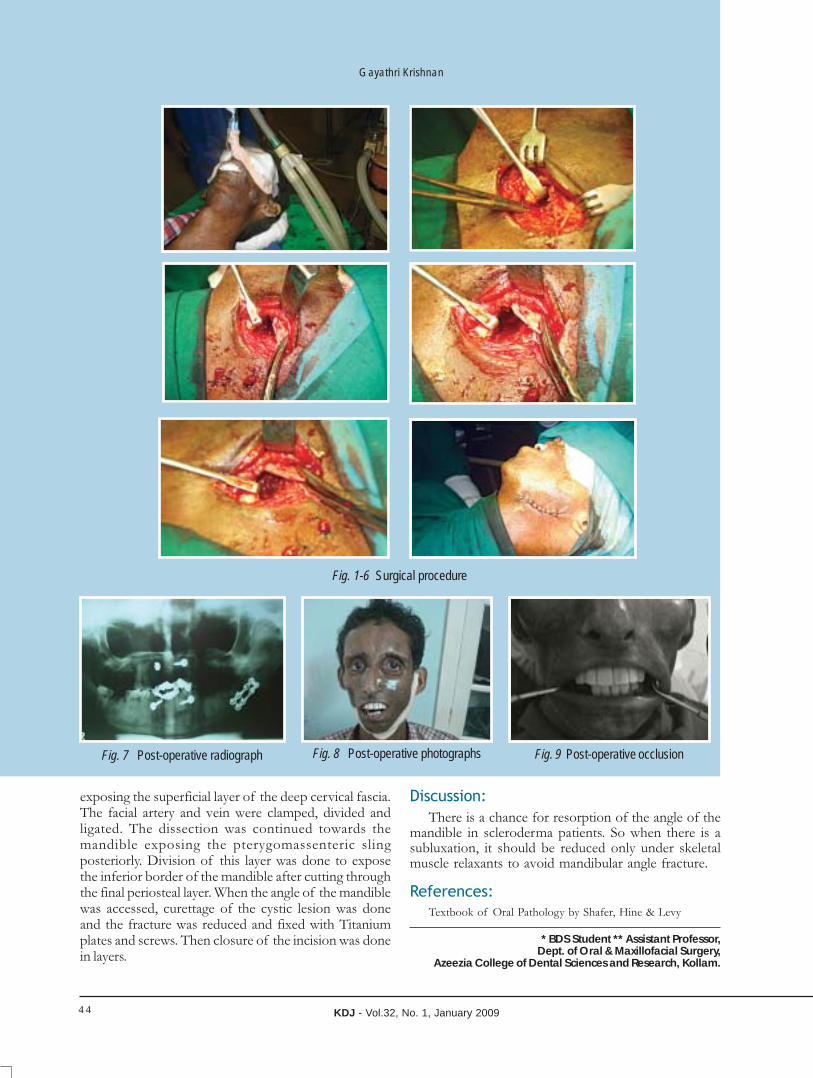
KDJ - Vol.32, No. 1, January 200944
Fig. 1-6 Surgical procedure
Fig. 7 Post-operative radiograph
Gayathri Krishnan
exposing the superficial layer of the deep cervical fascia.The facial artery and vein were clamped, divided andligated. The dissection was continued towards themandible exposing the pterygomassenteric slingposteriorly. Division of this layer was done to exposethe inferior border of the mandible after cutting throughthe final periosteal layer. When the angle of the mandiblewas accessed, curettage of the cystic lesion was doneand the fracture was reduced and fixed with Titaniumplates and screws. Then closure of the incision was donein layers.
Discussion:
There is a chance for resorption of the angle of themandible in scleroderma patients. So when there is asubluxation, it should be reduced only under skeletalmuscle relaxants to avoid mandibular angle fracture.
References:
Textbook of Oral Pathology by Shafer, Hine & Levy
* BDS Student ** Assistant Professor,Dept. of Oral & Maxillofacial Surgery,
Azeezia College of Dental Sciences and Research, Kollam.
Fig. 8 Post-operative photographs Fig. 9 Post-operative occlusion

KDJ - Vol.32, No. 1, January 2009 45
RTA one week prior to the day of reporting.
Medical history revealed myocardial infarction fourmouths back, contraindicating plate fixation under GA.Moreover the patient was suffering from DiabetesMellitus and Hypertension. She has been undermedication for the past 10 years.
On examination the patient was asymptomatic witha minimally displaced fracture extending upto the baseof the mandible. TMJ was palpable, mouth openingand mandibular movements were within normal limits.Intra oral examination revealed healing sockets in thelower anterior region from where the teeth wereextracted. The mucosa was intact over the fracturedsegments on the body of the mandible. Panoramicradiograph was taken to confirm the diagnosis. (Fig. 1).
After examination, preliminary impression of themaxillary and mandibular arch were made with alginateimpression material. Special trays were fabricated ondiagnostic casts for final impression and bordermoulding was accomplished with low fusing impressioncompound. Final impression was made and master castfabricated on permanent denture base.
The patient was recalled on the next day for jawrelation procedure. The vertical dimension and thecentric relation were registered and transferred to anarticulator.
In order to prevent any kind of movement betweenthe maxillary and mandibular components of the splinta locking mechanism was incorporated. Theanteroposterior movement was prevented by preparinga vertical rectangular keyway having a dimension of3cm x 5cm on the maxillary occlusal rims and a
Introduction
Management of fractured edentulous mandible hasalways been a challenging task for dental practitioner.Such mandibles usually have reduced cross sectionaldimension with limited contact areas of the fracturedends. In addition the poorly vascularized dense andsclerotic bone exhibits low regenerative potential.Severely atrophic edentulous mandible, especially thosehaving a radiographic height or 10 mm or less, are poorcandidates for plate fixation under General Anesthesia.In addition the older patient who may be malnourishedand in poor general health may preclude prolonged GA.Many patients are females with osteoporotic bonesmaking screw fixation difficult and unreliable.
In the above circumstances Gunning splints couldbe the only option for the stabilization of the fracturedmandible. The component of the splint that is fitted tothe maxillary arch is fitted by means of per alveolarwiring. Additional were may be passed throughpyriform rim or around the zygomatic arch. In themandibular arch, the component of the splint is fixedby circummandibular wiring. Screws can also be usedto fix the splints. The jaws are immobilized for 4-6weeks.
Case Report
A 65 year old female patient was referred to theDepartment of Prosthodontics, Govt. Dental College,Trivandrum from the Department of Oral &Maxillofacial Surgery for the fabrication of a splint toimmobilize the fractured segments of bone. She had afracture on the (Rt) body of the mandible, following
Management of fractured edentulous
mandible with gunning splint* Harshakumar, ** Anitha Gopinathan ** Ajith Kumar P.K.
Abstract
Maxillofacial surgical procedures and prosthodontic interventions are most often complimentary.This becomes particularly significant in the management of fractured edentulous jaws. An exclusivesurgical management by means of plate fixation may not be feasible always especially because ofunderlying diseases the patient is suffering from. In such cases the oral surgeon, after assessing theexact nature and extent of fracture should communicate with the prosthodontist regarding the typeof splint that would help in the management of fracture.
A completely edentulous elderly female patient reported with fractured mandible was managedby a combined prosthetic – oral surgical intervention.
Case Report

KDJ - Vol.32, No. 1, January 200946
corresponding key was fabricated on the mandibularocclusal rims that exactly fits into the keyway. Alongitudinal ‘step-like’ preparation on the occlusal rimswas incorporated so as to prevent any lateral movement.(Fig. 2, 3).
Stainless steel wire hooks were attached on eachquadrant of wax occlusal rim on the buccal aspectapproximately 5 mm above the occlusal plane. Thesehooks were used for passing ligature wire duringimmobilization.
A rectangular window was made on the anteriorsegment (4 cm x 4 cm) in dimension from canine tocanine region for feeding purpose. The wax rim wasfinished, polished and processed in acrylic.
The patient was recalled on the third day for theinsertion of gunning splint. The extensions, centricrelation position, key-key way relationship were evaluated.The splint was finished and polished and fixed to thejaws in the Department of Oral & Maxillofacial Surgery.(Fig. 4,5 & 6).
Discussion
Gunning splints are indicated for the reduction,fixation and immobilization of unilateral and bilateralfractures of the edentulous mandible, where thefractures lying proximal to these areas can be controlledby intermaxillary fixation. These splints provide a formof indirect control on the bone fragments, transmittedthrough the mucoperiosteum.
Gunning splints are contra indicated in unfavourablydisplaced fractures lying outside the denture bearingareas, in projectile injuries involving grossly comminuted
soft tissue and bone loss, and in severe posteriordisplacement of fractures of the anterior part of themandible. Extreme atrophy of the maxilla or mandiblealso complicates this technique.
Gunning splints can also be constructed from
a. The patient’s existing dentures suitably modified.
b. Impressions made from patient’s mouth.
c. Models cast from the fitting surface of the patient’sdentures.
d. Prefabricated Gunning type splints.
e. Disposable, edentulous impression trays withouttheir handles.
Conclusion
Gunning splints offer control and support for thefractured segments of mandible. The easiness offabrication and fixation of Gunning splint make themacceptable for practitioner as well as a compromisedpatient.
Reference
1. Peter Ward Booth – Surgical management of mandibular
atrophic fractures; maxillofacial surgery; 2nd Edition, Churchill
& Livingstone, 1999; 1:62.
2. J.L1. Williams – Mandibular fractures; Rowe and Williams
Maxillofacial injuries; 2nd Edition; Churchill & Livingstone,
1994, 1:283.
* Professor, Department of Prosthodontics, Govt. Dental College,Trivandrum** House Surgeon, ** House Surgeon,Govt. Dental College, Trivandrum
Harshakumar
Fig. 1 Panoramic view showing fracturemandible before extraction
Fig. 6 After insertion
Fig. 2 Finished splint
Fig. 4 Tissue surface of the splint
Fig.3 Cases surfaces of the splint
Fig. 5 Intra oral view

KDJ - Vol.32, No. 1, January 2009 47
Osteogenesis imperfecta type IV
* Roopashri Rajesh Kashyap, ** Gopakumar R., *** Gogineni Subhas Babu, *** Sreejan C.K.
generalized attrition, generalized spacing & antero-posterior cross bite (Fig.3). Dental examination of kidsrevealed opalescent teeth (fig4).
On carrying out further radiological investigations(Fig.5-6), Intra oral periapical radiograph showednormal thickness and density of enamel, completeobliteration of pulp chamber, thin slender roots andinterdental bone loss. Panoramic radiograph showedthat the intact teeth had bulbous crowns and markedcervical constriction & obliteration of pulp chamberof all teeth. Lateral cephalogram revealed hypoplasticmaxilla, midfacial hypoplasia & prognathic mandible.Chest radiograph showed healing fractures with callusformation (patient was not aware of fractures) &dislocated left shoulder joint with maldeveloped scapulaand superiorly placed glenoid articulating surface.
Ground section of extracted tooth showed absenceof pulp space. Outer most layer of coronal dentinshowed tubular structure; however radicular dentin andinner layers of coronal dentin showed altered tubularstructure. Enamel and cementum appeared to benormal. Features were compatible with clinical diagnosisof dentinogenesis imperfecta (Fig.7, Fig.8).The patientwas diagnosed to be suffering from OI type IV B.
Abstract
Osteogenesis imperfecta is a heritable systemic disorder of the connective tissue.
Dentinogenesis imperfecta, which is sometimes an accompanying symptom of OI, belongs to agroup of genetically conditioned dentin dysplasias and is characterized clinically by an opalescentamber appearance of the dentin. A case of DI associated with OI with a positive family history ispresented in this paper. The purpose of this paper is to present the dental and skeletal characteristicsof OI associated with DI and an insight into the genetic perspectives. Patients with OI and opalescentteeth should be evaluated as soon as the deciduous teeth erupt; immediate dental treatment and oralhygiene instructions can be of great help in reducing the need for extensive dental care.
Introduction
Osteogenesis imperfecta (OI), is a clinically andgenetically heterogeneous group of autosomal dominantinherited disorders characterized by bone fragility andfractures.1 The clinical features commonly observed inpatients with OI include abnormal bone formation,growth deficiency, bone fragility, blue sclerae, hearingloss, skin thinness, joint laxity, hypermobility anddentinogenesis imperfecta(DI). This disease causes eithera decrease in collagen synthesis or the production ofstructurally defective collagen. Hence all tissues rich intype I collagen may be affected.2Diagnosis of mild OIis challenging due to its variable phenotypic expressionand inconstant course. This article presents a case ofmoderately involved OI associated with DI with a briefreview of clinical characteristics of OI.
Case report
A 32yrs old lady reported to Department of OralMedicine and Radiology with the complaint ofdiscoloured teeth. History revealed discoloureddeciduous predecessors. Full coverage crown placementwas done for upper and lower anteriors 10yrs back.(Few crowns dislodged 2yrs back). She also reportedthat she was diagnosed to have less dense bones whenshe was admitted to hospital 2yrs back for treatmentof shoulder and hip dislocation. Also reported aboutthe discoloured teeth of her both children.
She had short stature (141cm), barrel shaped chest,deformed shoulder, hyperextensible joints, absence ofbody hairs & brachycephallic skull. Facial featuresincluded triangular face, broad forehead, hypertelorism,white sclera, absence of malar eminence, prognathicmandible (Fig.1, Fig.2). Intra oral examination revealedmultiple yellowish to brown coloured teeth with
Case Report
Normal male
Affected male
Normal female
Affected female
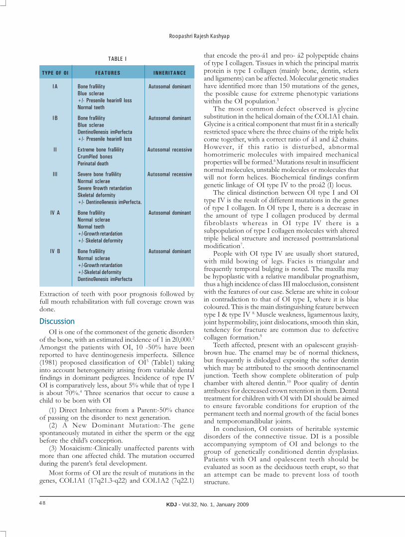
KDJ - Vol.32, No. 1, January 200948
Extraction of teeth with poor prognosis followed byfull mouth rehabilitation with full coverage crown wasdone.
Discussion
OI is one of the commonest of the genetic disordersof the bone, with an estimated incidence of 1 in 20,000.2
Amongst the patients with OI, 10 -50% have beenreported to have dentinogenesis imperfecta. Sillence(1981) proposed classification of OI5 (Table1) takinginto account heterogeneity arising from variable dentalfindings in dominant pedigrees. Incidence of type IVOI is comparatively less, about 5% while that of type Iis about 70%.4 Three scenarios that occur to cause achild to be born with OI
(1) Direct Inheritance from a Parent:-50% chanceof passing on the disorder to next generation.
(2) A New Dominant Mutation:-The genespontaneously mutated in either the sperm or the eggbefore the child’s conception.
(3) Mosaicism:-Clinically unaffected parents withmore than one affected child. The mutation occurredduring the parent’s fetal development.
Most forms of OI are the result of mutations in thegenes, COL1A1 (17q21.3-q22) and COL1A2 (7q22.1)
TABLE I
TYPE OF OI FEATURES INHERITANCE
IA Bone fragility Autosomal dominant
Blue sclerae
+/- Presenile hearing loss
Normal teeth
I B Bone fragility Autosomal dominant
Blue sclerae
Dentinogenesis imperfecta
+/- Presenile hearing loss
II Extreme bone fragility Autosomal recessive
Crumpled bones
Perinatal death
III Severe bone fragility Autosomal recessive
Normal sclerae
Severe growth retardation
Skeletal deformity
+/- Dentinogenesis imperfecta.
IV A Bone fragility Autosomal dominant
Normal sclerae
Normal teeth
+/-Growth retardation
+/- Skeletal deformity
IV B Bone fragility Autosomal dominant
Normal sclerae
+/-Growth retardation
+/-Skeletal deformity
Dentinogenesis imperfecta
that encode the pro-á1 and pro- á2 polypeptide chainsof type I collagen. Tissues in which the principal matrixprotein is type I collagen (mainly bone, dentin, scleraand ligaments) can be affected. Molecular genetic studieshave identified more than 150 mutations of the genes,the possible cause for extreme phenotypic variationswithin the OI population.3
The most common defect observed is glycinesubstitution in the helical domain of the COL1A1 chain.Glycine is a critical component that must fit in a stericallyrestricted space where the three chains of the triple helixcome together, with a correct ratio of á1 and á2 chains.However, if this ratio is disturbed, abnormalhomotrimeric molecules with impaired mechanicalproperties will be formed.6 Mutations result in insufficientnormal molecules, unstable molecules or molecules thatwill not form helices. Biochemical findings confirmgenetic linkage of OI type IV to the proá2 (I) locus.
The clinical distinction between OI type I and OItype IV is the result of different mutations in the genesof type I collagen. In OI type I, there is a decrease inthe amount of type I collagen produced by dermalfibroblasts whereas in OI type IV there is asubpopulation of type I collagen molecules with alteredtriple helical structure and increased posttranslationalmodification7.
People with OI type IV are usually short statured,with mild bowing of legs. Facies is triangular andfrequently temporal bulging is noted. The maxilla maybe hypoplastic with a relative mandibular prognathism,thus a high incidence of class III malocclusion, consistentwith the features of our case. Sclerae are white in colourin contradiction to that of OI type I, where it is bluecoloured. This is the main distinguishing feature betweentype I & type IV 8. Muscle weakness, ligamentous laxity,joint hypermobility, joint dislocations, smooth thin skin,tendency for fracture are common due to defectivecollagen formation.9
Teeth affected, present with an opalescent grayish-brown hue. The enamel may be of normal thickness,but frequently is dislodged exposing the softer dentinwhich may be attributed to the smooth dentinoenameljunction. Teeth show complete obliteration of pulpchamber with altered dentin.10 Poor quality of dentinattributes for decreased crown retention in them. Dentaltreatment for children with OI with DI should be aimedto ensure favorable conditions for eruption of thepermanent teeth and normal growth of the facial bonesand temporomandibular joints.
In conclusion, OI consists of heritable systemicdisorders of the connective tissue. DI is a possibleaccompanying symptom of OI and belongs to thegroup of genetically conditioned dentin dysplasias.Patients with OI and opalescent teeth should beevaluated as soon as the deciduous teeth erupt, so thatan attempt can be made to prevent loss of toothstructure.
Roopashri Rajesh Kashyap

KDJ - Vol.32, No. 1, January 2009 49
References
1. Huber MA, Antonio S, Osteogenesis imperfecta, Oral SurgOral Med Oral PatholOral Radiol Endod 2007;103:314-20.
2. Chevrel G, Osteogenesis Imperfecta, Orphanet encyclopedia,
June 2004.
3. Tsai CL, Lin YT, Lin YT. Dentinogenesis Imperfecta Associatedwith Osteogenesis Imperfecta: Report of Two Cases ChangGung Med J 2003; 26:138-43.
4. Waltimo J, Harri AO, Lukinmaa PL. Mild Forms OfDentinogenesis Imperfecta In Association With OsteogenesisImperfecta As Characterized By Light And TransmissionElectron Microscopy, J Oral Pathol Med 1996;25:256-264.
5. Sillence DO. Osteogenesis Imperfecta: An Expanding Panoramaof Variants,Clin Orthop Rel Res 1981;159:11-25.
6. Marjomäki V, Suuriniemi M, Genetics of Children’s BoneGrowth, University of Jyväskylä, Department of Biologicaland Environmental Science, Finland.
7. Wenstrup RJ, Tsipouras P, Byers PH. Osteogenesis ImperfectaType IV Biochemical Confirmation of Genetic Linkage to theproa2(1) Gene of Type I Collagen, J Clin Invest1986;78:1449-1454.
8. Paterson CR, McAllion S, Miller R. Osteogenesis Imperfectawith Dominant Inheritance And Normal Sclerae. J Bone JtSurg 1983; 65B:35-39.
9. Gorlin RJ, Cohen MM, Levin LS. Syndromes of the Head andNeck, 3rd edition, Oxford University press, pp155-166.
10. Neville BW, Damm DD, Allen CM, Bouquot JE. Oral andMaxillofacial Pathology, 2nd edition, Elsevier publications, pp,534-535
* Postgraduate Student, ** Principal, Mahathma Gandhi DentalCollege & Hospital, Jaipur.*** Professor & Head, ***Postgraduate Student, Dept. of OralMedicine And Radiology, A.B.Shetty Memorial Institute of DentalSciences, Mangalore - 575 018.
Fig.1 Short stature
Fig.2 White sclera Fig.3 Generalized discoloured teeth with antero-posteriorcross bite
Fig.4 Opalescent teeth of daughter (7yrs old) Fig.5 Panoramic radiograph shows marked cervicalconstriction & obliteration of pulp chamber
Fig.7 Scanner view of ground section oftooth shows obliteration of pulpal space.
Fig.8 Photomicrograph of decalcifiedsection shows irregular dentin tubules.
Osteogenesis imperfecta type IV
Fig.6 Chest radiograph showing fractured ribs,dislocated left shoulder joint with maldeveloped scapula
and superiorly placed glenoid articulating surface.

KDJ - Vol.32, No. 1, January 200950
Introduction
“Beauty lies in the eye of the beholder”. Teethjewellery is a brilliant way to add sparkles to your smileand stand out of the crowd. Tooth jewellery is the latestthing in cosmetic dentistry - and it’s popular with peopleof all ages. These tiny little designs are great fun, andbecause there’s no drilling involved, they won’t harmyour teeth. Dental jewel is a special decoration, forexample diamond or another stone, which can beattached to your teeth without consequences
Are you a suitable candidate for application ofa dental jewel?
If you desire to endow your smile with a jeweland the teeth to which the jewel should be attached arehealthy, you can be a suitable candidate for this treatment.
It is necessary to consider your expectations andrealize that the dental jewel will be a new, significantpart of your look. Be prepared that people can stareat you, most of them will appreciate the beauty of yourdecorated smile, but some of them can consider it tooextravagant.
Dental skyce
Skyce are clear and sapphire –white or blue crystals… high-quality tooth jewels. Skyce can improve theappearance of your teeth far and away and emphasizewhiteness of your smile.
Dental Jewellery
* Shibu Thomas Mathew
Abstract
Dentists of today are finding that expertise and more people who consult them are no longerreally patients. They are individuals who desire perfectly aligned sparkling white teeth, and who mayeven ask for tooth jewellery. Patients want a sparkling smile, dentists want teeth to be healthy. Dentistsare committed to maintaining the health of teeth at all costs. The dental jewellery does not need to bepre-treated and is bonded onto the tooth in the same way as an orthodontic bracket. The purposeof this article is to create awareness about tooth jewellery and its implications.
Sticking of Skyce is absolutely harmless procedurefor your teeth. It can be sticked on a surface of yourtooth without defection of an integrity of enamel whatmeans without any preparation. It can be fixed on aplace of an injured enamel, pigmented spot, mixtureof a photopolymeric fillings and so on. The procedureis absolutely painless and takes only 10-20 minutes. As ithas no sharp edges, it does not hurt your lips. If youwants to take it off in the future, it can be done veryquickly, without pain and a disturbance of integrity ofyour tooth. Taking care of Skyce is nothing else than aregular brushing. It is your chance to combine betweenbeauty and health in one harmless process!
Placement
1. Apply rubber dam2. Carefully clean the contact surface using rotary
brushes and cups and a polishing agent.3. Etch the area with 37% phosphoric acid on the
contact surface for 60 seconds,and carefully sprayand clean the surface
4. Apply bonding agent and light cure5. Place the bonding material on the contact surface
and place the skyce on to the tooth, remove excessfrom the area, and then cure.
Information

KDJ - Vol.32, No. 1, January 2009 51
Dental crystals
These are applied same as for skyce.
Dazzlers, Twinkles
Twinkles are specially designed to be bonded to thetooth. Our patented backside similar to an orthodonticbracket makes Twinkles stay on as long as you choose.Twinkles are pure gold and precious stones likediamonds, sapphires and rubies. Dental crystals are glassmounted on a thin foil of aluminum to create theattractive spark. The bonding is not as durable but caneasily last for more than 6 months.
Is it possible to remove the jewel?
The dental jewel is attached to the tooth with asubstance, which does not damage the enamel, it istherefore possible to remove it or change it withoutconsequences.
How long does the dental jewel last?
The dental jewel can last a relatively long time, it ishowever limited by the stress put on the decorated tooth
and dental care. If the jewel gets loose unexpectedly, itis possible to reattach it without consequences.
Conclusion
Apart from a temporary sensitivity of the teeth,triggered by the acid, nothing wrong can happen duringthis treatment. But the opposite is true in case ofinsufficient oral hygiene. Every piece of dental jewellerycreates an ideal niche for caries bacteria. No dentist, nomatter how perfect, can prevent that. If you don’t brushyour teeth carefully enough, your entire “Dazzling” toothcan fall victim to caries.
References
Piercing and tooth jewelry — an ethical dilemma. SADJ 2001
Dec; 56(12):574.
Piercing and tooth jewellery. SADJ 2002 May; 57(5):165.
Lip and tongue piercing: experiences and views of general dental
practitioners in South lancashire. Prim Dent Care 2004 Jul; 11(3):
92-6
A few piercing thoughts. J Am Dent Assoc 1998 Nov; 129(11):
1519-20
Hasegawa TK, Matthews M, Hanna MJ; Ethical dilemma. What
would you do? [Case Reports, Ky Dent J 1998 May-Jun; 50(3):
26-8, 38.
* Assistant Professor, A. B. Shetty Memorial Institute of DentalScience, Mangalore
Dental Jewellery

KDJ - Vol.32, No. 1, January 200952
Quiz
Q 1. A young female patientpresented with the facialfeatures, in the photograph,which was of recent onset. Thecharacteristic feature depictedin this picture isa) Marcus Gunn phenomenonb) Mobius syndromec) Bell’s signd) Inverted Marcus
Gunn phenomenon
size. The surface wasnodular, the consistencywas firm. Routiner a d i o g r a p h i cexamination reveled noabnormalities. Theprobable diagnosis is:
a) Squamous cell carcinomab) Periapical cystc) Pleomorphic adenomad) Lipoma
Q 2. What is the conditiondepicted in the radiograph?a) Globulomaxillary cystb) Palatal Abscessc) # Maxilla with the missingfractured segmentd) Cleft Palate
Q 3. Routine radiographicexamination revealed aradiopaque lesioninvolving the apex of 34.What is the inference?
a) Hypercementosisb) Fixer splashc) Condensing osteitisd) Cementoma
a) Ameloblastomab) O d o n o t g e n i c
Keratocystc) Dentigerous cystd) Residual cyst
Q 4. Radiograph of a 38 year old male patient presentedwith pain of 48 revealed a unilocular radiolucencyin the ramus region of the mandible. The most likelydiagnosis is:
Q 5. A reddish discolorationof the right buccalmucosa was noted in a67 year old patient of3 year duration . Shewas asymptomatic butnoticed an increase insize of the lesionrecently. What is thecondition?
a) Haemangiomab) Erythroplakiac) Squamous cell
carcinomad) Purpuric macule
Q 6. A 54 year old female patient presented to the OPDwith a painless swelling of the palate of 6 monthsduration. The swelling increased slowly to the present
Q 7. The radiographic feature depicted in this regionindicated?
a) Fractureb) Due to movement unsharpnessc) Developmental anomalyd) Periapical pathology
Q 8. The artifact in thisradiograph is:
a) Scratchesb) Developer splashc) Stainingd) Static electricity
birth. This condition iscalled:
a) Microdontiab) Natal teethc) Neo Natal teethd) Eruption Sequestrum
Q 9. A 28 day old child was brought to the OPD with thecomplaint of mobile lower front teeth present form
Q 10. What is the radiolucencyassociated with the centralincisor in this photographof a radiograph?a) External resorptionb) Internal resoptionc) Developer splashd) Lateral periodontal cyst
ANSWERS : 1. c; 2. d; 3.c; 4.c; 5.b; 6.c; 7.b; 8.d; 9.b; 10.a
Rani Mol*, Anita Balan**
* Postgraduate Student, ** Professor & HOD,Department of Oral Medicine and Radiology, GovernmentDental College, Trivandrum, Kerala - 695 011

KDJ - Vol.32, No. 1, January 2009 53
Secretary’s Report andAssociation News
Installation of new office bearers for the year 2008-09
Installation meeting was held on 23-11-08 at HotelCochin Durbar Nedumbassery.1st State Executive Committee Meeting
Held on 23-11-08 at Hotel Cochin DurbarNedumbasseryDRS Registration
The State office sent a representation to the Hon’bleMinister for Health and Family Welfare, Govt. of Keralaregarding the registration of Dental Clinics in Directorate ofRadiation Safety. The State President and CDH convenorhas met the minister and the minister has assured to takefurther actions regarding this after seeking reference fromthe concerned department.Presidents’ and Secretaries’ Seminar-TUNE 2009
Held on 11-01-09 at Hotel Arcadia Kottayam. Theseminar registration commenced at 9-30 am. The inauguralproceedings commenced with welcome speech by DrPraveen J Thayil president IDA Central Kerala KottayamBranch. He welcomed the dignitaries and office bearers.The chief guest Mr Mohammed Hanish IAS (Director PublicRelations, Govt.of Kerala) inaugurated the seminar bylighting the traditional lamp and enlightened the crowd witha glistering speech. State President Dr Pratap Kumar, Guestof Honour Kerala Dental Council President Dr MathewJoseph, President Elect Dr Samuel K Ninan, Vicepresidents Dr. Jaibeen George, Dr. Sony Thomas and DrSanthosh Sreedhar spoke on the occasion. State SecretaryDr Antony Thomas delivered the vote of thanks and theinaugural session was adjourned for the training session.
The opening of the training session started with thepresidential priorities by Dr Pratap Kumar.
“Learn to Excel” training session was conducted by MrBenny Kurian (JCI International Trainer).
The next to speak was Dr.Antony Thomas Hon Secretary.He presented the Calendar of Activities of IDA Kerala State
for the year 2008-09.The interactive session was very fruitfuland the members were able to clarify their apprehensionsregarding the constitution amendments.
The presentation on IDA HOPE by HOPE Secretary DrNizaro Siyo created an awareness of the same.
The post lunch session commenced with the trainingin Public Speaking and Quality leadership by Prof;TomyCherian(JCI international Trainer).
State CDE Convenor. Dr OV Sanal and State CDHConvenor Dr Anil G outlined about the their projects andguide lines respectively. Website chairman DrMadhavankutty detailed about the renovated IDA web site.Conference Secretary Dr Oommen George appealed allthe branch officials for their active involvement in theforthcoming Kerala State Dental Conference at Calicut. Honeditor Dr K Nandakumar spoke about the plans of the neweditorial team. The final session “Relax”, stressmanagement through Yoga and Meditation was enthralledby Mr EMG Nair(Chairman International Health and PeaceFoundation). A total number of 97 members attended. Theevent was hosted by IDA Central Kerala Kottayam Branch.The chief Co-ordinators of the programme were Dr EapenThomas and Dr Anukesh.P.The programme was wellorganized by the host branch and congratulations and
S e c r e t a r y ’ s M e s s a g eMy Dear Fellow Members,
Wishing you all a Happy and Prosperous New Year-2009.
First of all let me express my sincere gratitude to each and every one of you for the confidence shownto us during the last IDA year. As the Hon State secretary I have entered the final lap of my office andI am extending my gratitude from the bottom of my heart to all of you for the confidence shown onme. This year we can expect new mantras from our versatile President Dr. K.N. Pratap Kumar. We are
very much fortunate to have the most trusted and dedicated member Dr. K. Nandakumar who have shown his mettleto various capacities as our New Editor.This year we were able to start our activities with a well organized office bearers workshop on 11th Jan at Kottayam. Weare pipelining a few innovative programmes and hopefully we will be able to full fill them all successfully. Far sighting thevision for a good finanancial foundation to IDA Kerala State, this year State is directly hosting the Kerala State DentalConference on 20,21&22nd Nov 09 at Calicut. I appeal to all of you to register at the earliest so that the COC canapproch the conference with great confidence. All the members are requested to contact the local branch secretariesfor the application forms for 42nd Kerala State Dental Conference, IDA HOPE, IDA IMAGE, DRS etc..Once again expecting your whole hearted co-operation, let me conclude.
DR ANTONY THOMASHon State Secretary
REPORT OF ACTIVITIES

KDJ - Vol.32, No. 1, January 200954
appreciation to the Co-ordinators for making thisprogramme a grand success.New Year Celebration
President Dr Pratap Kumar cut the New Year cakeduring the inaugural ceremony of Presidents’andSecretaries’ Seminar and shared the happiness of theoccasion with the office bearers.IDA Slogan
The slogan of the year “Learn to Excel in Life and inProfession “ was released by chief guest Mr MohammedHanish IAS.State level Advocacy workshop on Tobacco Control Laws
State president Dr KN Pratap Kumar attended the oneday workshop organized by RCC Trivandrum on January15th -09.IDA HOPE
The bereaved family of Dr Raju K.S of Attingal Branchwas the first beneficiary of the new scheme. The StatePresident handed over a cheque for Rs 2,65000/-to thefamily of late Dr Raju KS on 18-01-09.President’s Visit
President Dr Pratap Kumar visited the followingbranches in connection with the installation ceremony andfamily get together.
26-10-08——————Coastal Malabar30-11-08——————North Malabar06-12-08——————Malanadu07-12-08——————Thiruvalla14-12-08——————Malappuram20-12-08——————Kodungallur21-12-08——————Central Kerala Kottayam
1. Dr. K.G Nair Trophy for the Best Local Branch I D A CentralKerala Kottayam
2. First Runner up - Best Local Branch I D A Malabar3. Second Runner up - Best Local Branch I D A North Malabar4. Dr. Samuel. K. Ninan Award for Best Local Branch President
Dr. Sherry. M. Joseph5. Runner up - Best Local Branch President Dr. Joseph CC6. Best Secretary - Local Branch Dr. Anukesh. P7. Runner up - Best Local Branch Secretary Dr. Anil Thunoli8. Best CDH Activties IDA Central Kerala Kottayam9. Runner up - Best CDH Activties IDA North Malabar10. Best - Scientific Activities IDA Central Kerala Kottayam11. Runner up - Best Scientific Activities IDA North Malabar12. Best Journal / Bulletin IDA Central Kerala Kottayam13. Membership Growth % up to 31st Oct. IDA Trivandrum14. IDA HOPE Award IDA Malabar15. Website Award IDA Central Kerala Kottayam16. Student Activity Award IDA Malappuram17. Dr. K.L. Baby Award for Best Scientific Paper in
Kerala Dental JournalDr Sreeja. J (P.G Student Dept. of Conservative Dentistry& Endodontics Govt. Dental college Trivandrum)
18. Other KDJ Awards for Best scientific Papers(I) Under Graduate Student Category: Dr. Miss. Sreekala P. C(II) Private Practitioner Category: Dr.Sha Menon. KC(III) Academician Category: Dr. Anand Induchoodan
Special Appreciation Awards:-1. Tele film Agnisakshiyaayi...- I D A Malabar2. Free Dental Clinic Project- I D A Coastal Malabar Branch3. Tribal Project - Trident- I D A Chalakkudy
4. Student Activities - I D A Malanadu5. Smile 2008- I D A Malappuaram6. I D A- HOPE Membership Growth Award- I D A Malabar Branch
Presidential Awards:-1. Dr. Viswanath. V2. Dr. Pratap Kumar. KN3. Dr. Anil Mathew4. Dr. Venugopal5. Adv. Shyam Padman6. Dr. Antony ThomasBest Performance Awards:-1. Dr. CK Ashokan (President IDA Kerala)2. Dr. Oommen George(IPP & I D A HOPE Ad-hoc committee chairman)3. Dr. V. Viswanath Chairman (I D A HOPE & Legal Cell)4. Dr. Pratap Kumar. KN.
(For SRRADHA-Hapatitis immunization programme)5. Dr. Samuel. K. Ninan (Ist Vice President)6. Dr. Habeeb Mohammad
(2nd Vice President & Chairman I D A IMAGE)7. Dr.Shibu Rajagopal (3rd Vice President)8. Dr. Santhosh Sreedhar
(Co-ordinator Oral Hygiene Day, Editor of KDJ)9. Dr. Mohammad Sameer. PT (CDH- Convener)10. Dr. Sanal. OV (CDE- Convener)11. Dr. Nizaro Siyo (I D A -HOPE Secretary)12. Dr. Joseph. CC (Co-ordinator No Tobacco Day)13. Dr. Ravindran Nair KS
(Co-ordinator Presidents’ & Secretaries’ Seminar)14. Dr. Narayanan S. (Asst. Secretary)
WINNERS OF I D A KERALA STATE AWARDS - 2008
Secretary’s Report and Association News
21-12-08——————Kollam27-12-08——————Kunnamkulam28-12-08——————Mavelikara04-01-08—————---Tellicherry03-01-09—————-—Wayanad04-01-08——————Malabar18-01-09——————Attingal25-01-09—————-—Greenvalley
Secretary’s VisitHon Secretary Dr Antony Thomas visited the following
branches in connection with the installation ceremony andfamily get together.
07-12-08———————Thiruvalla14-12-08———————Malappuram20-12-08———————Kodungallur28-12-08———————Mavelikkara27-12-08——————--Nedumbasserry21-12-08——————--Central Kerala Kottayam21-12-08——————--Kollam03-01-09———————Wayanad04-01-09—————-—-Malabar04-01-09—————-—-Tellicherry

KDJ - Vol.32, No. 1, January 2009 55
15. Dr. Baby. K. Antony (Chairman - Awards Committee)16. Dr. Ranjith. CK (Chairman - Students Committee )17. Dr. Raveendranath.M (Chairman - Screening & Scruitnizing)18. Dr. Madhavankutty. B (Chairman - Website )19. Dr. Lalappan. K. Joseph ( Co- ordinator - Dentists Day)20. Dr. Sandeep Rajagopal (Co-ordinator - No Tobacco Day)21. Dr. Sudheer. KT (Chairman Sports Committee)22. Dr. Siby. T. Chenankara (For IDA Kochi Journal BIDA)Trophy for Student Conference-08Ist Amrita College of Dental SciencesIst Runner up Mar Baselious Dental College Kothamangalam2nd Runner up Pushpagiri College of Dental SciencesStudent Award for the Year -2007-08(For the Student of Calicut Dental College)Top Mark Award (I BDS) Sulakshana. KTop Mark Award (II BDS) Aby AbrahamTop Mark Award (III BDS) Poornima Chandran. KRTop Mark Award (IV BDS) Miss. Neethu. LTop Mark Award (Final BDS) Ranjith. RDr. Jacob Zachariah Memorial Award for the Top Mark Scorer inOral Pathology Anu KII-Best Out Going Student AwardDr. K.K Pathrose Award - Vincia Paul
1. Dr. NS Rajeevan Memorial Award for Top Mark Scorer in OralSurgery Vincia Paul2. Dr Abu Mathew Memorial Award for the Best Student inProsthodontics Jezanta D’ Costa3.Dr. Robin Titus Memorial Award for Top Mark Scorer inOrthodontics Neethu. LPriya Saju Manavalan Endowement Award - For the first rankholder in the final BDS of all the universities in Kerala - Tintu SaraChandy - P M S Dental College (Kerala University)
University Top Mark Awards - 2008Top Mark Award Final BDS- Tintu Sara Chandy (Kerala University)Top Mark Award IInd BDS - Gayathri Krishnan (Kerala University)Top Mark Award Ist BDS - Litu Mary Thamby (M G University)Top Mark Award Ist BDS - Shamna Ibrahimkutty (M G University)Top Mark Award IInd BDS - Anju Treasa Andrews (M G University)Top Mark Award IIIrd BDS - Beenu. V (M G University)Top Mark Award Final BDS - Smitha Jose (M G University)Top Mark Award Ist BDS - Anjana Ramanathan (Amrita Vishwa Vidya
Peetham University)Top Mark Award IInd BDS -Minu Mathew (A V V P U)Top Mark Award IIIrd BDS -Dhanya. S. Kumar (A V V P U)Top Mark Award Final BDS -Mahitha Mohan (A V V P U)
1st CDH Programme was conducted on the 4th ofDecember, 2008 at Garden Valley English Medium School,Kuttipala at 1:30pm. 250 students attended theprogramme. Dr. Mohammed Sameer.P.T, President of IDAMalappuram Branch, conducted classes on Oral Hygienemethods and dental caries. Dr. Sameer.K.P, CDHConvenor, conducted a class on Tobacco and Oral cancer.The classes were well appreciated. Oral hygiene kits weredistributed to the students. Mr. Hamza Master was thestudent co-ordinator of the programme.2nd CDE Programme was conducted by the eminentBristish faculty, Dr. Sursh.V.Nair, on 14th of December, 2008at 2pm at Hotel Food ‘n’ Fun, Kottakkal. He spoke on“Contemporary Endodontics and General Dentistry: A UKPerspective”. 30 members attended the seminar. Thepresident of IDA Malappuram, Dr. Sameer.P.T, and the Hon.Secretary of IDA Malappuram, Dr. RajeshRaveendranathan, the IPP, Dr. Suresh.P.N and the CDEConvenor, Dr. Sasikumar.T.P were present. Certificatesof attendance were distributed among the registeredpresent members. It was a 2 hour session with a very
IDA MALAPPURAM BRANCH
advanced powerpoint presentation.The seminar was veryuseful for the participants especially the deeper insightsinto bone grafting, newer obturation techniques, rootresections and prosthetic appliances.Installation General Body Meeting was held at Hotel Food‘n’ Fun, Kottakkal on 14th of December, 2008 at 11am.The meeting was honoured by the presence of the ChiefGuest Dr.K.T.Jaleel M.L.A. His speech on NationalIntegration and Secularism was inspiring. Dr.Pratapkumar.K, President Kerala State IDA, and Dr.Anthony Thomas, Hon. Secretary of Kerala State IDA, wereamong the dignitaries. The function was first presided byDr. Suresh.P.N, the outgoing president of IDAMalappuram, and after the installation ceremony, by Dr.Sameer.P.T, the incoming president of IDA Malappuram.The new executive committee members of IDAMalappuram were installed during the function. Dr. RajeshRaveendranthan, Hon. Secretary of IDA Malappuram,delivered the vote of thanks. The entertainmentprogrammes consisted of a sumptuous lunch, gamesfor kids and a “Kolkkali” programme.
1ST CDE Programme was conducted on 16th of November,2009 at Hotel Grand, Valanchery by Dr. Vishwanathan at12pm. The subject was “How to Be a Good StandingDentist”. It was a short lecture and was attended by 58members.
DR. SHINY FRANCIS MEMORIAL ESSAY CPMPETITION
As part of the Indian Dental Association’s Kerala State Anti-Tobacco Day celebration, MIDA has conceptualized Dr. ShinyFrancis Memorial Trophy as a memoir to the late Dr. ShinyFrancis, who had been an active member of MIDA. The trophywill be presented to the best Essay written by a femaledentist/student of the Kerala dental fraternity. The subject is“Tobacco Related Oral Lesions”. The essay should bepresented as a Word document printed on an A4 size paperin not more than 1500 words. Entries should reach theHonorary Secretary, Dr. Rajesh Raveendranathan’s, officeon or before the 1st of May, 2009. The address is as follows:
Dr. Rajesh Raveendranathan, Indra Dental and TMJ-CareCentre, Manjeri Road, Uphill, Malappuram - 676 509, Kerala
For further details please contact the President,the Secretary or the CDE convenor of IDA, Malappuram
Branch
Secretary’s Report and Association News

KDJ - Vol.32, No. 1, January 200956
IDA KOCHI BRANCH
AGM and Installation Ceremony
IDA Kochi Branch had its Annual General Body Meetingand Installation Ceremony on 9th November 2008 atWhyte Fort Hotel, Maradu, Cochin. During the AGM, Dr.Rajesh Kottooran presented the Annual report and Dr.Anajana G presented the audited accounts, both of whichwere appreciated by the general body. Dr. Jose Juliangave his presidential address and thanked the officebearers and all the members for their kind support duringhis term.
The new address and telephone number of I.D.A KochiBranch is as follows:
Dr. Vinod Mathew BDS DSS; Hon. Secretary IDA KochiKattukaran’s Multi-Speciality Dental Clinic
Varappan’s Building, Banerji Road, Cochin 682 018Tel: 0484 2355597; Mob: 94470 55598
E-mail: [email protected]
Please be kind enough to make furthercorrespondences to this address.
IDA MembershipsThe new Calendar year started with the campaign to getmaximum membership renewals before December 15th
2008. IDA Kochi is proud to have renewed membershipsof over 150 members within this short period. In addition,ten new members have joined the association.
Executive Committee MeetingsIDA Kochi had its first executive committee meeting
under Dr. V. A Afzal on the 11th of December 2008 at HotelGokulam Park Inn, Kaloor.
Membership renewal efforts, sponsorships formeetings, Planning for the Christmas and New Yearprogram to be held on the 24th of January 2009, HOPE,IMAGE, DRS details etc were addressed by the secretaryDr. Vinod Mathew. Secretary thanked all the memberswho took part in the NOH program.
Later, Dr. Anjana.G briefed the ECM with the proposedbudget. Efforts of Dr. Siby Chennankkara for getting thebest Magazine award for Kochi IDA Journal – BIDA wasappreciated and applauded.
Activities for the period from 10th Nov 2008 to 10th Jan2009
1. 41st Kerala State Dental Conference: 21st, 22nd, and23rd Nov, 2008
Venue: Cochin Durbar Nedumbassery.
2. Executive meeting: 2nd December 2008
Venue: G B Palace Ankamaly.
3. Combined Executive meeting: At Ezhatumughamon 7th Dec 2008.
4. Installation and Christmas/ New Year Celebrations:
The Ninth Installation ceremony of the branch washeld at Hotel Saj Earth- Nedumbassery on 27th Dec 2008.
Dr Samuel K Ninan (President Elect IDA Kerala State)was the chief guest and Dr Antony Thomas (Hon Sec IDAKerala State), the guest of honour.
Dr Vinu PR was sworn in and installed as thePresident and Dr (Major) Jude John, the Hon sec. DrVinu then installed his team of executive members andalso welcomed 7 new members who joined the branchon the auspicious day.
Dr Samuel K Ninan congratulated the branch for a
IDA NEDUMBASSERY BRANCH
wonderful conference it put up. Dr Antony Thomasencouraged the members under the leadership of DrVinu, to bring more laurels to the branch.
After the official function was over it was time for merrymaking and joy. Santa Claus distributed gifts to allchildren present. It was followed by a musicalextravaganza and games for adults and children.
5. Ist Executive for the year 2009 on 9th Jan 2009 atPeriyar Club Aluva.
Secretary’s Report and Association News

KDJ - Vol.32, No. 1, January 2009 57
INSTALLATION CEREMONY 2008-09
The installation ceremony of IDA Malanadu Branchwas on 06.12.08 at Brooke Side Club, Kolenchery. Mas-ter of Ceremony invited dignitaries to the dias. Dr. JaymonK. Alias collared the president and the meeting was callto order at 8.30 p.m. by the president Dr. Sankar Vinod.After the prayer song by Miss. Anjana Alias. Dr. Giju Georgewelcomed the gathering. Secretary Dr. Jaymon K. Aliaspresented the annual report for the year 2007-08 andwas duly passed. In his presidential address Dr. Sankarvinod thanked each and every member for their supportand co-operation during the last year. Then Dr. Siju V.Jose introduced the incoming president. Dr. K.N. PratapKumar installed the new president Dr. Jose Paul and in
IDA MALANADU BRANCH
his acceptance speech he mentioned about his dreamsfor the coming IDA year. Dr. Jose Paul installed the OfficeBearers for the year 2008 – 09. Dr. Babu John introducedour chief guest Dr. K. N. Pratap Kumar – IDA Kerala StatePresident and in his address he offered all the supportfrom the state branch for the coming year. Dr. Benny Au-gustine introduced the guest of honour Prof. Dr. GeorgePaul and the new projects for the year was inauguratedby him. Dr. Jaibin George first vice president IDA KeralaState felicitated the new team. 18 new members wereinducted to our IDA Malanadu Family. Invited guest andother club members felicitated the new team. Dr. SankarVinod, Dr. P.I. Kochukunju and Dr. Reeja Baby were thewinners of the package tour to Ooty Sponsored by For-tune Retreats, Cochin. Dr. Byju Paul Kurian proposedthe vote of thanks. After the National Anthem the meetingwas adjourned for variety entertainments, fellowship anddinner.
Ist EXECUTIVE MEETINGThe First Executive meeting was on 23.12.08 at Paray
Hotel Muvattupuzha hosted by Dr. Jose Paul, Dr. JaymonK. Alias and Dr. Giju George. After the discussion themeeting is adjourned for fellowship and dinner.
Secretary’s Report and Association News
Report of activities November-December 2008
1. Dental Health Check up camp in association with IMA& the Diabetes Control Society.
A dental check up camp was organised by idamavelikara branch in association with indian medicalassociation and the diabetes control society, at st. Mary’scathedral auditorium on 6th November, 2008.
More than 300 patients were examined and free healthcare samples were distributed to the needy patients.2. 1st Executive and General Body meeting on 6th
December at Rotary Club of Mavelikara.The first executive meeting and general body meeting
of IDA Mavelikara was held on 6th December at the rotaryclub of mavelikara. The agenda was to decide about theinstallation ceremony and about the future activities of thebranch. The official date of installation was decided as28th December. The branch congragulated Dr. Anil. G forbeing selected as state CDH chairman.the members ob-served a moment of silent prayer for the demise ofMrs.Omana Ninan, mother of Dr. Sonia Susan and alsothe victims of Mumbai blasts.3. Installation ceremony- December 28th at HotelBhagavath Gardens, Chengannur.
The installation of Dr. Renjith.R as the new presidentof the branch was held on the 28th of December at hotelbhagavath gardens, Chengannur, with the state presi-dent Dr. K.N. Prathap Kumar as the chief guest, Sri. SajiCheriyan, Sports Council president as the guest of honour.
The other dignitaries on the diaz were, Dr. AnthonyThomas, state secretary, Adv. Anil Panachooran (Poet&Lyricist), Dr. Samuel.K. Ninan, president elect ida keralastate and Dr. Anil. G state CDH chairman.
The meeting was followed by a nostalgic musicalevening, dinner and fellowship.
IDA MAVELIKARA BRANCH
Dental check up
DENTIST DAY CELEBRATIONSIDA Kerala State (Hosted By IDA Mavelikara Branch)
Date: March 6, 2009 - Venue: Cooperative Bank Auditorium Mavelikara.Celebrity Guest: Sri Jagathy Sreekumar
All IDA Members Are Invited To Attend The Programme.
Programme Schedule1. Public Function- Importance Of Oral Hygiene Prevention Of OralCancer, 2. Students Cultural Programme, 3. Inauguration Of IDA MajorProjects, 4. Awards Ceremony
Dr. Prathap Kumar K.N Dr. Anthony ThomasPresident IDA Kerala State Hon. Secretary IDA Kerala State
Dr. Anil.G, CDH Convenor, IDA Kerala State

KDJ - Vol.32, No. 1, January 200958
IDA THRISSUR BRANCH
Installation Ceremony 2008-2009The office bearer of IDA Thrissur has taken charge on 13-
12.08, under the leadership of President Dr. Anil Das and Hon.Secretary Dr. Ajmel Habeeb.
Activities in December 20081. Combined executive meeting of Office bearers of 2008 &
2009 held on 29-11-08 at Aquatics. 15 Members attend themeeting.
2. First general body and installation of office bearers of 2008-2009 held at Hotel Joys Palace on 13-12-08. Dr. Antony J. Maliakaldelivered pledge to all office bearers. This was followed by CDEprogramme - Distraction Osteogenesis by Dr. Vinay V. Kumar. 63members attended the meeting.
Secretary’s Report and Association News
Government Dental College (GDC), Thiruvananthapuram isone of the premier Institutions in India, dedicated to DentalEducation. It was startedin the year 1959 andpresently offers bothundergraduate andpostgraduate courses.GDC produced severaloutstanding professionalswho made the countr yproud in the last 50 years.A good number ofstudents who havegraduated from thisInstitution are today occupying prestigious positions in theprofession and in the administration. Many of the students fromthis college have made name abroad, both in profession and inother walks of life.
GDC attracts a large number of patients from all over thestate because of the high professional standards and providesexcellent clinical material for training and research. The collegehas an excellent teaching faculty of senior experienced and youngenthusiastic academicians. Institution is marching ahead with adetermination to achieve greater professional standards.
Highlights of Golden Jubilee CelebrationsInaugural Function : 8th Feb 2009
Family Get together : 11th and 12th JulyCDE Programmes : 2nd Saturdays /
2nd SundayValedictory Function : 20th December ‘09
Marching towards Greater Heights.....
Govt. Dental College, Medical College Campus,Thiruvananthapuram - 695 011. Tel: 0471-2444092, www.gdctvm.net
Golden Jubilee Celebrations of
GOVT. DENTAL COLLEGETHIRUVANANTHAPURAM
(1959-2009)

KDJ - Vol.32, No. 1, January 200964
GUIDELINES
Manuscripts: Articles should be type written on one sideof A4 size (21x28cm) White paper in double spacing witha sufficient margin. One Original and two high qualityxerox copies should be submitted. The author’s name isto be written only on the original copy and not on the twoxerox copies. In addition to the printed version, a CDcontaining the article file also should be submittedcompulsorily. Use a clear and concise reporting style.KDJ reserves the right to edit manuscript, toaccommodate space and style requirements. Authors areadvised to retain a copy for the reference.
Title Page: Title page should include the title of thearticle and the name, degrees, positions, professionalaffiliations of each author. The corresponding authors,telephone, e-mail address, fax and complete mailing addressmust be given.
Abstract: An abstract of the article not exceeding 200words should be included with abbreviated title for thepage head use. Abstract should state the objectives,methodology, results and conclusions.
Tables: Tables should be self explanatory, numbered inroman numbers, according to the order in the text andtype on separate sheets of paper. Number and legendshould be typed on top of the table.
Illustrations: Illustrations should be clearly numberedand legends should be typed on a separate sheet of paper,while each figure should be referred to the text. Goodblack and white glossy photographs or drawings drawn inblack Indian ink on drawing paper should be provided.Colour photographs will be published as peravailability of funds. It will incur printing cost.Otherwise the cost of printing will be at the expenseof authors. Photographs of X-rays should be sent andnot the original X-rays. Prints should be clear and glossy.On the back of each print in the upper right corner, writelightly the figure number and author’s name; indicate topof the photograph with an arrow of word Top’ Slides
and X-ray photographs should be identified similarly.
Reference: Reference should be selective and keyed innumerical order to the text in Vancouver Style. Type themdouble spaced on a separate sheet of paper. Journalreferences must include author’s names, article title, journalname, volume number, page number and year. Bookreference must include author’s or editor’s names, chaptertitle, book title, edition number, publisher, year and pagenumbers.
Copy right: Submission of manuscripts implies that thework described has and not been published before (exceptin the form of on abstract or as part of published lectures,review or thesis) and it is not under consideration forpublication else where, and if accepted, it will not bepublished else where in the same form, in either the sameor another language without the comment of copyrightholders. The copyright covers the exclusive rights ofreproduction and distribution, photographic reprints, videocassettes and such other similar things. The views/opinionsexpressed by the authors are their own. The journal bearsno responsibility what so ever.
The editors and publishers can accept no legalresponsibility for any errors, omissions or opinionsexpressed by authors. The publisher makes no warranty,for expression implied with respect to the materialcontained therein. The journal is edited and publishedunder the directions of the editorial board who reservethe right to reject any material without giving explanations.All communications should be addressed to the Editor.No responsibility will be taken for undelivered issues dueto circumstances beyond the control of the publishers.
Books for review: Books and monographs will bereviewed based on their relevance to KDJ readers. Booksshould be sent to the Editor and will become property ofKDJ.
Return of articles: Unaccepted articles will be returnedto the authors only if sufficient postage is enclosed withthe manuscripts.
All correspondence may please be sent to the following address :Dr. K. Nandakumar, Hony. Editor, Kerala Dental Journal
Neelambikam, Attukal, Manacaud, Trivandrum, Kerala - 695 009Phone: 0471-2459235 / Cell: 09447066100 / e-mail: [email protected]
Kerala Dental JournalINFORMATION FOR AUTHORS
Subscription Rates: Free distribution for all the members of the Indian Dental Association Kerala State.
Inland Subscription: Rs. 400 per issue, Rs. 1500 for 1 Year, Rs. 2500 for 2 years, Rs. 3500 for 3 years.
KDJ - Vol.32, No. 1, January 200964