A Tribute to Our Friend - Journal of Dental Hygiene
68
Source: Journal of Dental Hygiene, Vol. 79, No. 1, Winter 2005 Copyright by the American Dental Hygienists' Association A Tribute to Our Friend MA Gaston Mary Alice Gaston is emeritus professor of dental hygiene at the University of Tennessee Health Science Center, a past president of the American Dental Hygienists' Association, and current editor of the Journal of Dental Hygiene. Keywords: administration, professional association, ADHA publications The American Dental Hygienists' Association (ADHA) central office staff is made up of highly competent individuals who work together for the benefit of our profession. Sometimes it seems as though people take positions with ADHA and stay only long enough to gain experience and build a reputation before moving on to better paying positions somewhere else. This can't be prevented and is an expected part of doing business in today's world. Fortunately for ADHA and for our profession, such people bring their energy and creativity to their positions and are serious about doing a good job. With rare exception, they readily accept appropriate responsibility for the function and successful operation of their particular division. In general, they are not satisfied with maintaining the status quo, and they look for innovative ways to improve our processes. Since becoming editor of the Journal of Dental Hygiene, I have worked with several young staff editors in the Division of Communications and have most certainly regretted losing each one of them. The roles and work assignments of this essential second tier of ADHA staff are determined by the division directors, or senior staff, who are responsible to the executive director for managing specific components of the day-to-day business of the association, and for implementing the decisions of the Board of Trustees (BOT) and House of Delegates. Whenever a senior ADHA staff member or division director leaves, there is an immediate and broad-ranging effect on other ADHA staff and member volunteers. Because I have been actively involved in ADHA business for many years, I, like some of you, understand that some turnover of division directors is unavoidable and is to be expected. Even so, I was unprepared for the departure of Rosetta Gervasi, director of the Division of Communications. Actually, I nearly lost my breath when, in early December, Ms. Gervasi notified the division that she would be leaving ADHA at the end of December 2004. I couldn't immediately visualize what it would be like to work without her wise and capable guidance. At this time, I'm still adjusting to the reality that we will simply carry on. - 1 -
-
Upload
khangminh22 -
Category
Documents
-
view
1 -
download
0
Transcript of A Tribute to Our Friend - Journal of Dental Hygiene
Source: Journal of Dental Hygiene, Vol. 79, No. 1, Winter 2005
Copyright by the American Dental Hygienists' Association
A Tribute to Our Friend
MA Gaston
Mary Alice Gaston is emeritus professor of dental hygiene at the University of Tennessee Health Science Center, a past president ofthe American Dental Hygienists' Association, and current editor of the Journal of Dental Hygiene.
Keywords: administration, professional association, ADHA publications
The American Dental Hygienists' Association (ADHA) central office staff is made up of highly competent individualswho work together for the benefit of our profession. Sometimes it seems as though people take positions with ADHA andstay only long enough to gain experience and build a reputation before moving on to better paying positions somewhereelse. This can't be prevented and is an expected part of doing business in today's world.
Fortunately for ADHA and for our profession, such people bring their energy and creativity to their positions and areserious about doing a good job. With rare exception, they readily accept appropriate responsibility for the function andsuccessful operation of their particular division. In general, they are not satisfied with maintaining the status quo, and theylook for innovative ways to improve our processes. Since becoming editor of the Journal of Dental Hygiene, I have workedwith several young staff editors in the Division of Communications and have most certainly regretted losing each one ofthem.
The roles and work assignments of this essential second tier of ADHA staff are determined by the division directors, orsenior staff, who are responsible to the executive director for managing specific components of the day-to-day businessof the association, and for implementing the decisions of the Board of Trustees (BOT) and House of Delegates. Whenevera senior ADHA staff member or division director leaves, there is an immediate and broad-ranging effect on other ADHAstaff and member volunteers.
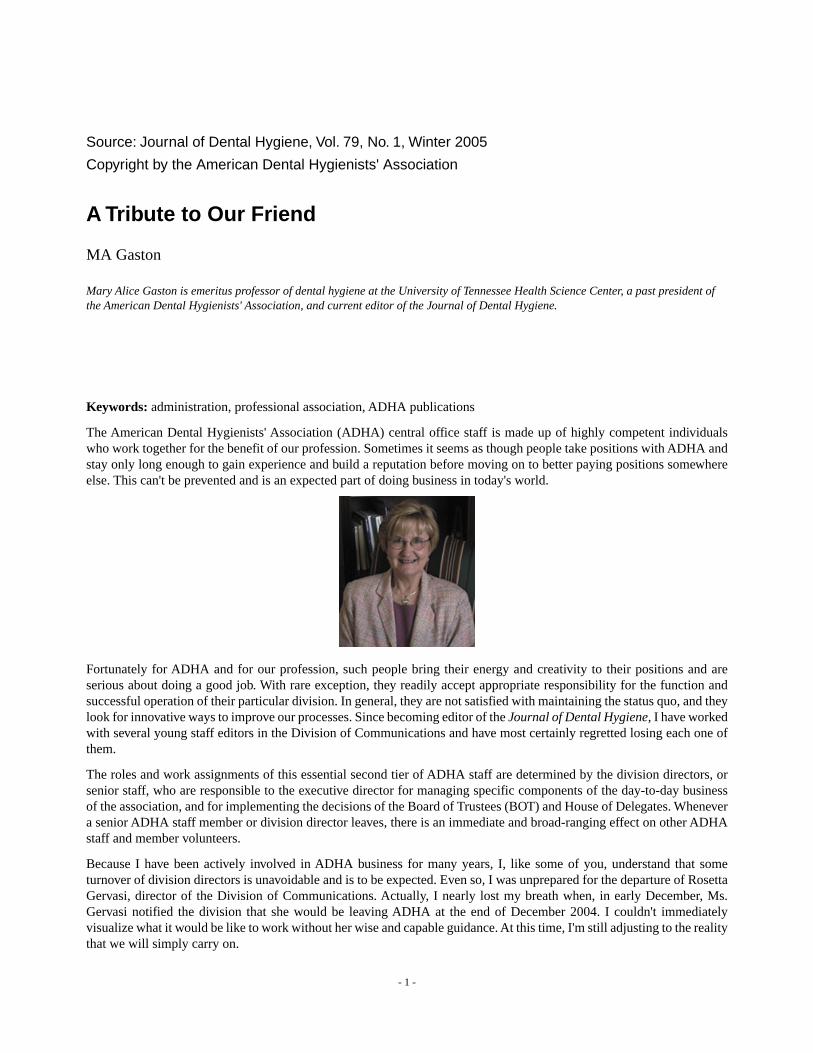
Because I have been actively involved in ADHA business for many years, I, like some of you, understand that someturnover of division directors is unavoidable and is to be expected. Even so, I was unprepared for the departure of RosettaGervasi, director of the Division of Communications. Actually, I nearly lost my breath when, in early December, Ms.Gervasi notified the division that she would be leaving ADHA at the end of December 2004. I couldn't immediatelyvisualize what it would be like to work without her wise and capable guidance. At this time, I'm still adjusting to the realitythat we will simply carry on.
- 1 -
To compensate for their loss, ADHA communications division staff joined hands and are conducting business and meetingtheir obligations, as others have done when other division directors have moved on. Now that Rosetta Gervasi is no longerwith us, I believe it is appropriate to pay tribute to her for the significant contributions she made to ADHA and to dentalhygiene.
Rosetta left her position at the American Dental Assistants' Association in the spring of 1988 to join ADHA as directorof the Division of Communications. For the past 17 years, she has influenced every aspect of the association's business. Ihad the unique opportunity of working with her as an ADHA officer and president and, more recently, as the editor of theJournal for the past seven years. I have admired Rosetta for her great talent, keen intuition, and extraordinary politicalsensitivity. She knew when to press ahead with an idea and when to stand in place, both extraordinary skills that workwell at ADHA. Let me review only a few of her accomplishments, so that you can view Rosetta through the eyes of thosewho know her well and recognize her great talent.
Creating and refining Access magazine for ADHA was perhaps Rosetta's greatest achievement. For many years, ADHAmembers expressed their desire for a timely news publication that would give them current clinical practice information,plus news about issues concerning the profession throughout the country. With Rosetta's lead, ADHA members got theirwish. Access, first appearing in a tabloid format in March 1987, was converted to the newsmagazine format with Rosetta'sguidance in August 1988. Access has received public recognition and awards for journalistic excellence several times overthe past 17 years, and it is a benefit cherished by ADHA members.
Rosetta's editorials in Access touched all of us because they were remarkably insightful and provocative. You see, Rosettawas often the point person in our profession's skirmishes with organized dentistry. She protected our officers by writingbiting editorials and raising difficult questions that dental hygienists found it hard to openly explore because of possiblethreats to their jobs. Rosetta always took the position that she was a dental hygiene outsider just stating her views, questioningcurrent practices, and lobbying for change in the oral health care system. Dental hygienists everywhere applauded eachtime one of her inspiring editorials appeared in print.
As for the Journal, Rosetta supported it throughout her tenure at ADHA. She recognized the Journal's unique status asthe dental hygiene profession's premiere peer-reviewed scientific publication. I can't speak for previous Journal editors,but I can say that, without fail, Rosetta respected my position and always deferred to my judgment concerning Journalcontent. We enjoyed a collegial relationship in which we each respected the position of the other. Rosetta took care of thebusiness of the Journal and provided staff support, permitting me to do my job unrestrained by resource concerns.
Early in her tenure at ADHA, Rosetta recognized that the Journal publication process would run more smoothly if theeditor position became a staff position, reporting to the communications division director rather than directly to the BOT.The BOT approved this administrative change, and it has worked well for the past 15 years. Perhaps it has worked wellbecause Rosetta was sincerely respectful of the position of the dental hygienist editor of the Journal. She often remindedme that I was the Journal content expert, especially when we were trying to resolve a politically charged issue, while atthe same time protecting the peer-review process. I can never forget her favorite phrase: "It's your call, Mary Alice." Igrew to appreciate Rosetta's strong commitment to dental hygiene's professional goals, and commend her for her willingnessto support ADHA programs designed to promote public awareness of dental hygienists.
Under Rosetta's leadership, the materials ADHA produces each year for National Dental Hygiene Month evolved intovisually pleasing depictions of dental hygienists' contribution to the nation's oral health. Rosetta supported all ADHAinitiatives to bring greater visibility to dental hygienists and completed all special projects that were assigned to her division.These outstanding projects included implementing the toll-free consumer hotline, creating information packets for use incombating preceptorship training of dental hygienists, developing and implementing the first ADHA Web site in 1993,producing several important position papers, including those on managed care, dental prophylaxis, and tooth polishing,and producing a consumer brochure explaining the link between oral and systemic health and diseases.
I am quite sure many of you could add examples to this short list of Rosetta's contributions. I want to leave you withthoughts of two accomplishments that will always set Rosetta apart from the ordinary in my mind. First, Rosetta is a loverof art and expressed that love when she was put in charge of the most recent updating and redecorating of the ChicagoADHA office on Michigan Avenue, a task far above her usual division director duties. Finally, Rosetta sometimes helpedofficers draft important speeches to be delivered in settings not always friendly to the position of dental hygiene. She did
Journal of Dental Hygiene, Vol. 79, No. 1, Winter 2005
Copyright by the American Dental Hygienists' Association
- 2 -
so for me in 1990 when ADHA was invited by the American Dental Association to speak during a meeting to discussdental workforce issues.
The night before the meeting, I didn't sleep much because I wrote and rewrote and practiced my speech over and over.Although I made numerous changes to Rosetta's draft, I kept the phrase that defined the day for the dental hygienistspresent in the very large conference room packed with dentists. There was a part of the speech in which we wanted tomake it clear that ADHA would always adamantly oppose preceptorship training of dental hygienists, and that it was nota suitable alternative for increasing the dental hygiene workforce. I took Rosetta's wording and declared to all that theconcept of preceptorship training for dental hygienists was a "dinosaur" that had outlived its usefulness. The dentalhygienists who were there cheered loudly in support of the dinosaur statement, and they still laugh whenever we recallthat speech and that day. Of course, the opposing majority group was highly offended.
I still view that afternoon as one of the funniest events of my presidency and, to this day, attribute the success of my speechto Rosetta Gervasi's marvelous wit and extraordinary grasp of the English language. Yes, I owe her much, and so does thedental hygiene profession. I wish our friend Rosetta the very best in this career change and in whatever she does in thefuture. She will be missed.
Journal of Dental Hygiene, Vol. 79, No. 1, Winter 2005
Copyright by the American Dental Hygienists' Association
- 3 -
Source: Journal of Dental Hygiene, Vol. 79, No. 1, Winter 2005
Copyright by the American Dental Hygienists' Association
Upfront
Kristen Romanowski
Testing saliva to detect oral cancer
Scientists funded by the National Institute of Dental and Craniofacial Research (NIDCR) recently reported early successin using saliva to diagnose oral cancer. With 91% accuracy, researchers were able to use saliva samples to distinguishbetween healthy people and those diagnosed with oral squamous cell carcinoma, the sixth most common cancer in theUnited States.
According to the study published in the Dec. 15, 2004 issue of Clinical Cancer Research, researchers found high levelsof four cancer-related molecules in the saliva of 32 patients who had been diagnosed with oral cancer but not treated.These four molecules, which are messenger RNA, serve as chemical records that an individual gene has been expressed.
To see if the messenger RNA patterns in saliva could serve to distinguish cancer patients from healthy subjects, theresearchers screened saliva samples without knowing whether a healthy person or a cancer patient provided the sample.In nine out of 10 samples, researchers identified the saliva from cancer patients. In a larger study planned for the nearfuture, the researchers hope to use saliva to distinguish between various stages of oral cancer and to improve their accuracyrate.
There are currently no biochemical or genetic diagnostic tests commercially available for oral cancer, senior study authorDavid Wong, DMD, DMSc, said in a NICDR press release. The initial results of this early study, he said, highlight thepotential clinical value of saliva as a diagnostic biofluid, and one easier to obtain than blood. "If correct, a salivary testwould be quick, painless, and most likely less expensive than current diagnostic tests," Wong said. -KR
Study suggests new treatment for oral mucositis
Cancer patients who suffer the pain and complications of oral mucositis may find relief in a drug called palifermin(recombinant human keratinocyte growth factor), according to clinical trial results published in the Dec. 16 issue of TheNew England Journal of Medicine.
Oral mucositis, a common side effect of chemotherapy and radiation, can cause severe ulceration of the lining of the mouth,interrupting cancer treatments and increasing the risk for infection. Talking, eating, drinking, and routine oral hygienemay also become very painful or even impossible. Patients with the most severe oral mucositis, classified by the WorldHealth Organization (WHO) as grades 3 and 4, cannot swallow solid foods.
Although oral health care professionals can recommend ways to manage oral mucositis and help prevent infection-carefuloral hygiene with adapted toothbrushes, frequent hydration with baking soda and water mouthrinses, the use of topicalanesthetics and saliva substitutes, and changes in diet, for example-no approved treatment yet exists to reduce or preventthe condition.
- 1 -

Amgen, the drug manufacturer that funded the trial, applied in June to the U.S Food and Drug Administration for approvalof palifermin. The drug works by protecting keratinocytes, cells that stimulate the growth of epithelial cells in the mucosallining, from the damage caused by chemotherapy and radiation, thus protecting the mucosal lining.
In the study, palifermin significantly reduced the incidence and severity of oral mucositis in patients undergoing intensivetherapy for hematologic cancers like non-Hodgkin's lymphoma, leukemia, and multiple myeloma. For three days prior tohigh-dose chemotherapy and total body irradiation, half of the 212 patients in the study received palifermin and halfreceived a placebo. Following bone marrow transplantations, patients continued to take the placebo or palifermin foranother three days. The two groups were directly compared and evaluated for 28 days.
While 98% of patients taking placebo experienced severe (WHO grade 3 and 4) oral mucositis, only 63% of patients takingpalifermin were similarly affected and, on average, for three fewer days. Compared with 62% of the placebo group, only20% of the palifermin group experienced the worst form, grade 4. Furthermore, the palifermin group experienced 60%less mouth and throat soreness, took lower doses of painkillers over a shorter period, and had higher white blood cellcounts than patients taking the placebo. The most common side effect of the drug was a mild skin rash.
On a related note, the National Cancer Institute (NCI) is seeking pediatric participants for a two-year clinical trial to testthe ability of a homeopathic preparation to treat chemotherapy-induced oral mucositis. Traumeel S, a mouthrinse containingminerals and extracts from 12 plants, was shown in an earlier study to reduce mucositis in young patients undergoing stemcell transplantation. Researchers now plan to test the efficacy of this treatment in 180 patients aged 3 to 25. For moreinformation, please visit http://cancer.gov/clinicaltrials/COG-ACCL0331. -KR
Plaque a culprit in hospital-induced pneumonia
A new study emphasizes the importance of access to oral health services and daily oral hygiene for elderly residents oflong-term care facilities. Results published in the November 2004 issue of the journal Chest suggest that dental plaquecan be a reservoir for the respiratory pathogens that cause hospital-acquired pneumonia (HAP) in institutionalized, criticallyill elderly patients. The researchers state that their study is the first to confirm the link between bacterial colonization ofdental plaque and lower respiratory infection in institutionalized patients using molecular genotyping.
In the prospective study, researchers assessed the oral health status of 49 nursing home residents who were critically illand required mechanical ventilation. Upon admission to the intensive care unit, each patient was given a plaque indexscore, and bacterial cultures were taken from tooth surfaces and the buccal mucosa. Fourteen of the patients developedHAP after an average of 11.6 days on a ventilator. In patients who developed pneumonia, respiratory pathogens in oralsamples were compared to those in lower respiratory tract samples. Researchers found that S aureus and Gram-negativeenteric bacilli accounted for the majority of the respiratory pathogens found in dental plaque.
For institutionalized elders, poor oral hygiene, decreased activity levels, and medication-induced xerostomia can createfavorable conditions for the pathogens that cause lower respiratory tract infection. The authors of this study call for furtherstudies to investigate the relationship between oral disease and respiratory illness, and they advocate active programs toimprove the oral health status of nursing home residents. -KR
Dental x-rays could be first step in osteoporosis screening
Panoramic dental x-rays can help identify postmenopausal women at risk for skeletal osteoporosis, a new study suggests.In a study published in the December 2004 issue of the American Journal of Roentgenology, researchers looked at thepanoramic dental x-rays of 316 postmenopausal women and analyzed their cortical shapes and jaw widths. Women witheroded cortical shapes were identified as needing further BMD testing.
Questionnaires are widely used as the first step in determining which women need to have further BMD testing. In thisstudy, however, dental x-rays were just as sensitive as or more sensitive than questionnaires were in identifying these
Journal of Dental Hygiene, Vol. 79, No. 1, Winter 2005
Copyright by the American Dental Hygienists' Association
- 2 -

women. However, dental x-rays are not as specific as questionnaires and have the potential for false positives and negatives,study author Akira Taguchi, DDS, PhD, said in an American Roentgenology and Ray Society press release.
But, "because dental panoramic x-rays are taken for the diagnosis of conditions affecting the teeth and jaws in clinicalpractice worldwide," Taguchi said, "the dentist could also look at the mandibular cortical shape and width and refer theappropriate women for further BMD testing." -KR
Journal of Dental Hygiene, Vol. 79, No. 1, Winter 2005
Copyright by the American Dental Hygienists' Association
- 3 -
Source: Journal of Dental Hygiene, Vol. 79, No. 1, Winter 2005
Copyright by the American Dental Hygienists' Association
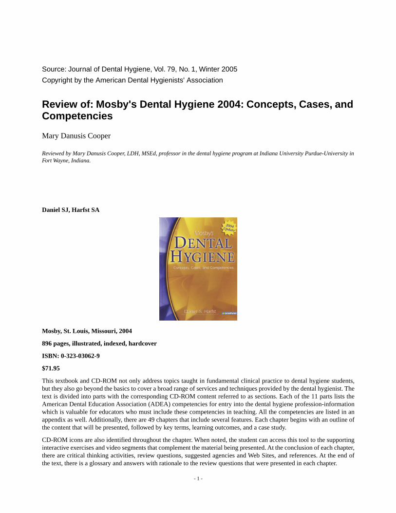
Review of: Mosby's Dental Hygiene 2004: Concepts, Cases, andCompetencies
Mary Danusis Cooper
Reviewed by Mary Danusis Cooper, LDH, MSEd, professor in the dental hygiene program at Indiana University Purdue-University inFort Wayne, Indiana.
Daniel SJ, Harfst SA
Mosby, St. Louis, Missouri, 2004
896 pages, illustrated, indexed, hardcover
ISBN: 0-323-03062-9
$71.95
This textbook and CD-ROM not only address topics taught in fundamental clinical practice to dental hygiene students,but they also go beyond the basics to cover a broad range of services and techniques provided by the dental hygienist. Thetext is divided into parts with the corresponding CD-ROM content referred to as sections. Each of the 11 parts lists theAmerican Dental Education Association (ADEA) competencies for entry into the dental hygiene profession-informationwhich is valuable for educators who must include these competencies in teaching. All the competencies are listed in anappendix as well. Additionally, there are 49 chapters that include several features. Each chapter begins with an outline ofthe content that will be presented, followed by key terms, learning outcomes, and a case study.
CD-ROM icons are also identified throughout the chapter. When noted, the student can access this tool to the supportinginteractive exercises and video segments that complement the material being presented. At the conclusion of each chapter,there are critical thinking activities, review questions, suggested agencies and Web Sites, and references. At the end ofthe text, there is a glossary and answers with rationale to the review questions that were presented in each chapter.
- 1 -
Educators often evaluate materials from a different perspective: How is the material presented? Is the material up to date?And, is the material presented so that students understand and gain the knowledge required to not only pass the dentalhygiene National Board Examination, but to also continue in clinical practice after graduation? Several chapters cover thematerial thoroughly and review all aspects of the topics. Especially outstanding are the chapters on infection control,ergonomics, and instrument sharpening. In addition, the material is enhanced by the use of color photographs, as well asdetailed tables and illustrations. In this updated edition, appendices have been added to strengthen the material presented.Appendix topics include the Health Insurance Portability and Accountability Act (HIPPA), a caries risk assessment form,and glove types and indications, to name a few.
However, there are minor shortcomings. After teaching dental hygiene for several years, this reviewer was surprised tofind hoes and chisels in the instrumentation and sharpening chapters. With the availability of many ergonomically effectivehand-scaling and power-assisted instruments, these instruments are not as popular today. The porte polisher is anotherexample of a device that is not used today because of the use of the engine-driven polisher and the popularity of theAmerican Dental Hygienists' Association's (ADHA) position on selective polishing. For instructional purposes in a dentalhygiene program, the instrumentation chapter would need to be supplemented with additional photographs on technique-the"how-to's." This chapter lacks laboratory application. In addition, although the seating positions are addressed in the chapteron ergonomics, the student will need to go outside the instrumentation chapter to access what is needed.
As dental health professionals, our emphasis is on educating the patient on disease prevention. One important area ofemphasis is the use of fluorides for patients who have moderate to high risk for caries development. The fluoride chaptercould offer more detail on fluoride delivery systems in the office, as well as self-applied topical products. Again, the"how-to's" could be emphasized more, so that the student can appropriately learn this information to educate the patienton not only the best product to use, but also on how to use the product most effectively. Overall, this textbook is a greatlearning tool for students and dental hygiene educators. In addition, this textbook would be a valuable reference for thosein private practice.
Journal of Dental Hygiene, Vol. 79, No. 1, Winter 2005
Copyright by the American Dental Hygienists' Association
- 2 -
Source: Journal of Dental Hygiene, Vol. 79, No. 1, Winter 2005
Copyright by the American Dental Hygienists' Association
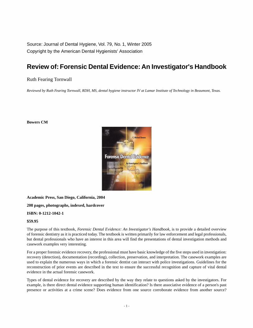
Review of: Forensic Dental Evidence: An Investigator's Handbook
Ruth Fearing Tornwall
Reviewed by Ruth Fearing Tornwall, RDH, MS, dental hygiene instructor IV at Lamar Institute of Technology in Beaumont, Texas.
Bowers CM
Academic Press, San Diego, California, 2004
208 pages, photographs, indexed, hardcover
ISBN: 0-1212-1042-1
$59.95
The purpose of this textbook, Forensic Dental Evidence: An Investigator's Handbook, is to provide a detailed overviewof forensic dentistry as it is practiced today. The textbook is written primarily for law enforcement and legal professionals,but dental professionals who have an interest in this area will find the presentations of dental investigation methods andcasework examples very interesting.
For a proper forensic evidence recovery, the professional must have basic knowledge of the five steps used in investigation:recovery (detection), documentation (recording), collection, preservation, and interpretation. The casework examples areused to explain the numerous ways in which a forensic dentist can interact with police investigations. Guidelines for thereconstruction of prior events are described in the text to ensure the successful recognition and capture of vital dentalevidence in the actual forensic casework.
Types of dental evidence for recovery are described by the way they relate to questions asked by the investigators. Forexample, is there direct dental evidence supporting human identification? Is there associative evidence of a person's pastpresence or activities at a crime scene? Does evidence from one source corroborate evidence from another source?
- 1 -
Suggestions and guidelines are described to increase the certainty of successfully recognizing and capturing vital dentalevidence in actual forensic casework.
Each chapter covers a specific area. Chapter 1 describes a qualified forensic dentist and advises readers to use aboard-certified forensic dentist. The author then goes on to describe what dentists would do in this role and recommendsthat law enforcement and legal professionals develop a working relationship with them. Other information provided inthis chapter includes the language of dental identification, human tooth morphology, teeth numbering systems, the dentalinvestigator's role in forensic cases, and courtroom uses of dental evidence. Chapter 2 examines case studies of death andabuse investigations. The author details the investigative steps in dental identification: the preliminary examination anddetailed search of the crime scene, the collection stage, the antemortem and postmortem dental profiling, and the comparisonof the dental profiles. Chapter 3 describes the recognition, recovery, and analysis of bite mark evidence. The authordescribes the protocols for evidence collection, including photographic documentation and the impression process. Thischapter also includes information on the recovery of salivary DNA from bitten objects and skin. Chapter 4 looks at howto blend evidence from multiple expert opinions on DNA and bite marks.
Chapter 5 examines the physical characteristics of child, elder, and spousal abuse and neglect, emphasizing the dentalaspects of the investigation. As dental hygienists in most states are legally mandated, as well as ethically charged, to reportcases of suspected abuse to the appropriate authorities, this chapter provides for them a useful comprehensive review ofdental evidence of abuse and neglect. The chapter also provides a glossary of abuse investigation terms.
Chapter 6 discusses the dental forensic framework surrounding mass fatality incidents. Chapter 7 outlines the methodsused in digital imaging in human identification and gives the reader an overview on this subject. Chapter 8 discusses legalissues in the introduction of evidence and the description of findings to the court on forensic odontology. Chapter 9describes the use of photography, which is commonly the only means by which a forensic dentist can evaluate bite marksand abuse cases. The chapter focuses on proper documentation of dental evidence and problems to avoid.
Overall, this book provides the reader with the concepts and protocols vital to a successful outcome of a criminal investigationcontaining dental evidence. The author suggests that these methods need to be practiced and the protocols maintained inorder to be available and successful under the actual casework conditions.
Journal of Dental Hygiene, Vol. 79, No. 1, Winter 2005
Copyright by the American Dental Hygienists' Association
- 2 -
Source: Journal of Dental Hygiene, Vol. 79, No. 1, Winter 2005
Copyright by the American Dental Hygienists' Association
Review of: Dentistry for the Child and Adolescent
Christine Nathe
Reviewed by Christine Nathe, RDH, MS, associate professor and graduate program director, Division of Dental Hygiene, at theUniversity of New Mexico in Albuquerque.
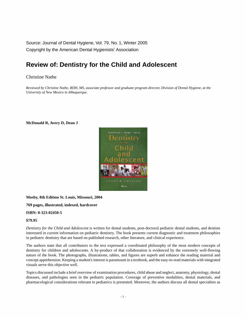
McDonald R, Avery D, Dean J
Mosby, 8th Edition St. Louis, Missouri, 2004
769 pages, illustrated, indexed, hardcover
ISBN: 0-323-02450-5
$79.95
Dentistry for the Child and Adolescent is written for dental students, post-doctoral pediatric dental students, and dentistsinterested in current information on pediatric dentistry. The book presents current diagnostic and treatment philosophiesin pediatric dentistry that are based on published research, other literature, and clinical experience.
The authors state that all contributors to the text expressed a coordinated philosophy of the most modern concepts ofdentistry for children and adolescents. A by-product of that collaboration is evidenced by the extremely well-flowingnature of the book. The photographs, illustrations, tables, and figures are superb and enhance the reading material andconcept apprehension. Keeping a student's interest is paramount in a textbook, and the easy-to-read materials with integratedvisuals serve this objective well.
Topics discussed include a brief overview of examination procedures, child abuse and neglect, anatomy, physiology, dentaldiseases, and pathologies seen in the pediatric population. Coverage of preventive modalities, dental materials, andpharmacological considerations relevant to pediatrics is presented. Moreover, the authors discuss all dental specialties as
- 1 -
they relate to pediatrics and hospital dentistry, including special care populations. A brief overview of dental public healthis also included.
One missing link is a chapter on the role of dental hygienists in pediatric dentistry settings. Unfortunately, in many states,dental hygienists are frequently not employed in pediatric settings. Rather, a somewhat superficial version of preventivecare is provided by dental assistants as a substitute for dental hygiene care. By expanding the textbook to include roles fordental hygienists, more potential pediatric dentists could be informed about the value of a dental hygienist, both in assessmentand therapeutic roles and, importantly, in the education and management of patients. This information would help increasethe level of preventive dental hygiene science practiced in a population that could be influenced by this well-educatedpractitioner. The dental hygienist would have the opportunity to provide a lifelong influence on the value placed onpreventive oral health care.
Interestingly, in the chapter on patient management-a section detailing production and fee collection philosophies-theauthors state that dental hygiene production usually includes the oral examination. And, although there is mention thatsome case can be made for the dentist to be compensated on percentages of the examination, more typically these areconsidered dental hygiene production. Understandably, as a dental hygienist, this seems extremely logical to me.
This comprehensive book on pediatric dentistry is well written and fun to read. This textbook was obviously written fordental students, but it is recommended as an additional resource to faculty who teach pediatric dental hygiene. Moreover,it could be recommended as supplemental reading for dental hygiene students and as a reference for dental hygiene providerspracticing in a pediatric setting.
Journal of Dental Hygiene, Vol. 79, No. 1, Winter 2005
Copyright by the American Dental Hygienists' Association
- 2 -
Source: Journal of Dental Hygiene, Vol. 79, No. 1, Winter 2005
Copyright by the American Dental Hygienists' Association
Review of: Oral and Maxillofacial Medicine
Sandra L Boucher-Bessent
Reviewed by Sandra L. Boucher-Bessent, RDH, BS, public health dental hygienist and dental program manager at Cabarrus HealthAlliance in Kannapolis, North Carolina, and adjunct faculty, Department of Dental Ecology, at the University of North Carolina inChapel Hill.
Scully C
Wright Publishing Co., 2004
556 pages, illustrated, indexed, paperback
ISBN: 0-723-61074-6
$64.95
Oral and Maxillofacial Medicine provides comprehensive coverage of diseases relevant to oral and peri-oral structures,including detailed oral manifestations of systemic diseases. Senior dental and dental hygiene students, as well as surgery,pathology, and other trainees and practitioners of oral medicine, will find author Crispian Scully's book a valuable resourcefor both board review and practical application.
Scully provides basic information so that non-medical and non-dental readers may also use the book with ease. Scullyexplains in his introduction that the book highlights more frequent and serious conditions and guides the reader throughdidactic and problem-oriented approaches. This book should be considered an adjunct to a student or professional oralmedicine library, and not the primary resource for oral pathology subject matter. The author makes references to anddirects the reader to several other texts he has authored or other pharmacopoeias.
- 1 -
The content is divided into five sections, each of which is arranged in an easy-to-follow format. The first section is a reviewof the fundamental principles of a comprehensive and thorough medical history and examination. The first two chaptersare so basic that a reader with a non-dental or non-medical background could easily grasp the material. For example,whenever Scully uses medical terminology, he follows the word with an explanation or description of the word inparentheses. Subsequent chapters in Section I become more detailed and provide information and guidelines for investigativeprotocol and principles of management to assist the practitioner to confirm or rule out diagnoses and prognoses. Eachchapter in Section I begins with a thought-provoking, patient-centered quote that captures the essence of Scully's message.Scully reinforces the appropriateness of the phrases by using them in context throughout the book.
Each section is color-coded for easy access. Just as chapters in Section I begin with quotes, chapters in Section II beginwith a bulleted list of "Key Points." This format facilitates the reader's navigation through the "Common Complaints"section, which discusses the more common symptoms and signs found in oral medicine. Discussion of each condition ispresented in an outline format that introduces the disease and discusses its causes, clinical features, oral complications,diagnosis, management, and references for further reading. Scully provides color plates for many of the oral conditionsthroughout this chapter and the next. He also organizes written material in table form for easier comprehension and referral.Scully uses algorithmic charts for the diagnostician to use when assessing symptoms and determining definitive diagnoses.Scully provides many examples of patient information sheets that help the patient understand the diagnostician's explanations,as well as Web sites specific to many of the conditions.
Section III describes in detail the most common and important conditions seen in oral medicine. This section also approacheseach condition in outline form, including a brief description of the disease; incidence; one's predisposition by age, sex,and geographic location; etiology and pathogenesis; clinical features; and diagnosis and management. Section IV includesa very comprehensive, alphabetical list of other conditions relevant to oral medicine. It includes some color plates and abrief synopsis of each condition. For some conditions, the author includes information on the treatment and managementof the illness, while oral implications and manifestations may be described for others. Section V is a discussion of importantaspects of HIV infection and iatrogenic diseases and is similar in format to the previous three chapters.
Finally, Scully provides two appendices. Appendix 1 tabulates other relevant oral manifestations of systemic disorders.It includes several tables that specifically detail oral conditions associated with human body systems and that will benefitclinicians and diagnosticians. Appendix 2 outlines the medicines used to treat patients with oral diseases.
The book does not contain biographic information on the author, but does list his impressive credentials and affiliationson the title page, from which the reader may deduce that the author is located in England. The reader should bear this factin mind when encountering words that are spelled differently than those found in American texts. Examples of spellingdifferences are "foetus" for fetus, "anaemia" for anemia, "minimise" for minimize, and "enquire" for inquire. The differentspelling of well-known words can be distracting to the reader at first, but after mentally processing the various spellingchanges, the reader will eventually begin to not notice them.
Journal of Dental Hygiene, Vol. 79, No. 1, Winter 2005
Copyright by the American Dental Hygienists' Association
- 2 -
Source: Journal of Dental Hygiene, Vol. 79, No. 1, Winter 2005
Copyright by the American Dental Hygienists' Association
The Perceived Likelihood of Dental Hygienists to Report AbuseBefore and After a Training Program
Marji Harmer-Beem
Marji Harmer-Beem, RDH, MS, is an assistant professor and advanced clinical course director at the University of New England inPortland, Maine. She is also a PANDA (Prevent Abuse and Neglect through Dental Awareness) trainer in Maine.
Purpose. The rise of abuse, mandatory reporting, and penalties for not reporting abuse make this study significant fororal health care personnel. The purpose of this research was to determine survey results pertaining to the likelihoodof dental hygienists reporting abuse before and after a training program, in order to influence and encourage similartraining programs in other locations and to impact dental hygiene curricula.
Methods. Exempt status was obtained from the University of New England Institutional Review Board for the Protectionof Human Subjects. A convenience sample was taken of registered dental hygienists who attended a training programand who volunteered to complete a 10-item statement form. A three-category ordinal Likert-type scale was employed.The statement form was filled out before and after a tested training program for the recognition and reporting of abuse(violence) and neglect. The terms family violence, child abuse, and elder abuse were defined as umbrella terms toencompass all abuse, except where explicit. This study explores two research questions: Do dental hygienists perceivethe likelihood to make a report if confronted with suspected abuse, and would training make a difference in the perceivedlikelihood to report? The 10 statements were grouped into three sets for analysis: 1) training and experience in reporting,2) knowledge of responsibilities, signs, symptoms, and interviewing, and 3) likelihood of making a report. Data wereanalyzed using descriptive statistics to explain the population's knowledge characteristics and the likelihood of reportingabuse.
Results. Twenty-six surveys were administered and 25 surveys were returned for a 96% response rate (n=25). Surveyresults supported training to increase compliance with mandatory reporting. Of the subgroup having experience withreporting (n=5; 20%), over half (n=3) knew all aspects of abuse. The entire group knew more about child abuse thanelder abuse. Prior to training, 40% definitely knew that they would likely report abuse, 40% somewhat knew that theywould likely report it, and 20% didn't know or said it would be unlikely that they would report. Only 5% stated that theydefinitely knew how to make a report before the training. After training, 100% reported that they would be likely tomake a report, an overall increase of 60%. In the pre-survey, 60% said they did not know how to make a report, comparedto 96% indicating in the post-survey that they knew how to make a report after training.
Conclusion. Evidence from the dental hygienists attending a continuing education program supports that trainingincreases the self-perceived likelihood to report abuse. This study also acknowledged areas for investigation of curricularaugmentation, such as providing more information on elder abuse and presenting a guide for filing a report of abuseto the appropriate agencies. It is imperative for educators to include adequate information in dental and dental hygienecurricula for training in reporting abuse. It is also incumbent upon dental hygiene clinicians to identify their owneducational needs and to seek out appropriate continuing education. These identified outcomes are an importantreinforcement to providing adequate instruction in dental hygiene curricula.
- 1 -

Keywords: dental hygiene curriculum, family violence, child abuse, elder abuse, reporting abuse, recognizing abuse,continuing education
Introduction
Every year, nearly a half million reports of abuse and neglect are filed on behalf of older Americans and vulnerable adults.1
In 1999, an estimated 3.2 million children were reported as suspected victims of child abuse or neglect.2 Estimates rangefrom one million 3 to three million 4 annual incidents of violence against a current or former spouse, boyfriend or girlfriend.
Across populations, abuse has been identified as a serious public health problem in Western society.5, 6 Abuse and family
violence have been recognized as national problems in the United States, across all socioeconomic levels.5,6,7 Familyviolence has been accepted as an umbrella term to encompass child abuse, elder abuse, intimate partner abuse, and abuse
of disabled or vulnerable persons.5, 7 Physical abuse, emotional abuse, sexual abuse, intentional neglect, and economicabuse serve to control and demean the victim, and different forms of violence serve to keep the abused victim isolated,
vulnerable, and helpless.5 Table I defines various categories of abuse.8 Although the various forms of abuse differ slightly,
the dynamics of differential power between the perpetrator and the victim are nearly identical.5
Table I . Definition of Abuse
In all 50 states, dentists and dental hygienists are mandated reporters of observed cases of child abuse, elder abuse, and
abuse of vulnerable adults, as well as instances of active, passive, and self-neglect within the family.1, 6, 9 Few states,California being one, require health care providers to report observed instances of domestic violence or intimate partner
Journal of Dental Hygiene, Vol. 79, No. 1, Winter 2005
Copyright by the American Dental Hygienists' Association
- 2 -
abuse.10 Dental hygienists should be aware of any abuse reporting legal requirements in the states in which they practice.Abuse, neglect, and exploitation have clinical significance for dental hygienists. Dental hygienists should be alert to
suspicious injuries to patients' heads and necks, along with bruises in different stages of healing.6 In general, the laws areclear: clinicians are ethically and legally bound to report signs and symptoms of suspected abuse to the state protective
services., 10, 11, 12 Penalties for violating states' mandatory reporting laws vary.12 Many professionals are uncomfortabletalking about these problems that affect society, and they may not know how to act upon suspicions. It is incumbent upondental hygiene clinicians to seek out information concerning their own knowledge deficits on family violence and for
educators to ensure curricula to prepare dental hygienists to meet their responsibilities.7, 8, 11, 13
This study explores two research questions: Do dental hygienists perceive the likelihood to make a report if confrontedwith suspected abuse, and would training make a difference in the perceived likelihood to report? This report describesone cohort's responses concerning the recognition and reporting of abuse before and after a training program.
Review of the Literature
The literature shows a paucity addressing the topic of abuse curriculum and what clinicians know about abuse. A NationalLibrary of Medicine search of the key words curriculum, elder abuse, child abuse, and family violence yielded only 17articles from the last 10 years in the dental and dental hygiene literature. The literature shows that there is a need for dentalhygiene educators to examine the family violence curriculum for deficiencies and also suggests that clinicians examine
self-deficits relating to abuse.7, 11, 13, 14, 15, 16
Superficial awareness of abuse, tendency to avoid involvement, and lack of knowing professional responsibilities have
been discussed in the literature.13, 17, 18 Gutmann and Solomon examined family violence curricula of 173 dental hygieneprograms in the United States. Using a survey to measure curriculum content, knowledge, and attitudes in the area offamily violence, researchers found that child abuse was taught in most (70.5%) of the programs, while elder abuse (54.9%),
intimate partner abuse (46.8%), and abuse of individuals with disabilities (46.2%) was taught in fewer programs.7 Jessee
reported on the extent of child abuse content in dental curricula in North American dental schools (n=54).15 Jessee foundthat physical abuse and dental neglect appeared in the curriclum 100% of the time, whereas emotional abuse/neglect,
sexual abuse, physical neglect, medical care neglect, and failure to thrive were reported less frequently.15 Needleman,MacGregor, and Lynch reported that a statewide child abuse and neglect educational program increased most respondents'
awareness and knowledge of child abuse and neglect and made them more likely to detect and report such cases.14
Furthermore, the top three factors indicated as barriers to reporting suspected cases included lack of an adequate history
(60.1%), self-perceived lack of knowledge about abuse and neglect (11.5%), and lack of knowledge of reporting (12.0%).14
Pediatric dentists reported having seen suspected cases of child abuse and neglect most frequently.14, 17 Adair et al., in astudy with a stratified randomized design of respondents (n=185) who were general dentists, reported that a minority of
dentists believed that they were required to report neglect (7.3%).19
Welbury, Hobson, Stephenson, and Jepson reported that a computer-assisted learning program increased knowledge of
oro-facial signs of physical child abuse from 10% before the training to 95% after the training.20 Kilpatrick, Scott, andRobinson identify dentists in New South Wales, Australia, as being aware of abuse and its different types, but having
considerable lack of knowledge about child protection protocols.17 The researchers suggest that dentists may be reluctant
to report because of the lack of knowledge on how to make a report.17
Murphree, Campbell, Gutmann, Plichta, Nunn, McCann, and Gibson ask, "How well prepared are Texas dental hygienists
to recognize and report elderly abuse?"11 This cross-sectional random study suggested that Texas dental hygienists are not
prepared to recognize and report elder abuse following graduation from a dental hygiene program.11 They also state thatdental hygienists are misinformed and unknowledgeable about the laws pertaining to elder abuse in Texas. Forty-eight
Journal of Dental Hygiene, Vol. 79, No. 1, Winter 2005
Copyright by the American Dental Hygienists' Association
- 3 -
percent reported no official training for the recognition of elder abuse, whereas 27% indicated knowledge from journal
articles, and 24.3% had formal training through school or continuing education. Murphree et al.11, as well as other researchers7, 8, 13, also suggest that increased education levels for practitioners may be effective in creating a greater awareness ofabuse.
Methods and Materials
Subjects
University of New England institutional review board approval for exempt status was obtained for the study. The subjectsof this study were registered dental hygienists who attended a continuing education (CE) training program for the recognitionand reporting of abuse. A convenience sample was taken of all registered dental hygienists (n=26) who volunteered tocomplete a before-and-after 10-item statement form with a three-category ordinal Likert-type scale. No demographicinformation was asked to protect anonymity and participant confidentiality. Unanswered surveys were excluded.
Instrument
An anonymous 10-item statement form with a three-category Likert-type ordinal scale was developed to ascertain thelikelihood of dental hygienists recognizing abuse and neglect, and their likelihood to report abuse before and after a trainingprogram (Figure 1). The three-category scale used "definitely know," "somewhat know," and "don't know" for the participantto self-rate statements concerning abuse. The investigator asked: Would the training program make a perceived difference?The central hypothesis of the study was that, indeed, training would influence the dental hygienists' perceived likelihoodto act. Contrarily, the null hypothesis states that there would be no difference between the pre-surveys and post-surveys.The statement forms were color-coded, labeled "pre-survey" and "post-survey," and sequentially numbered for identificationand comparison of the pre- and post-surveys. The statement form was pilot tested on a small group of dental hygieneeducators before it was implemented.
Journal of Dental Hygiene, Vol. 79, No. 1, Winter 2005
Copyright by the American Dental Hygienists' Association
- 4 -
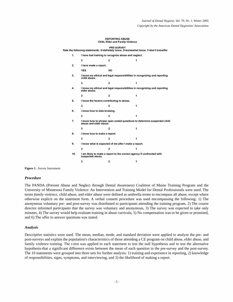
Figure 1 . Survey Instrument
Procedure
The PANDA (Prevent Abuse and Neglect through Dental Awareness) Coalition of Maine Training Program and theUniversity of Minnesota Family Violence: An Intervention and Training Model for Dental Professionals were used. Theterms family violence, child abuse, and elder abuse were defined as umbrella terms to encompass all abuse, except whereotherwise explicit on the statement form. A verbal consent procedure was used encompassing the following: 1) Theanonymous voluntary pre- and post-survey was distributed to participants attending the training program, 2) The coursedirector informed participants that the survey was voluntary and anonymous, 3) The survey was expected to take onlyminutes, 4) The survey would help evaluate training in abuse curricula, 5) No compensation was to be given or promised,and 6) The offer to answer questions was stated.
Analysis
Descriptive statistics were used. The mean, median, mode, and standard deviation were applied to analyze the pre- andpost-surveys and explain the population's characteristics of those attending a CE program on child abuse, elder abuse, andfamily violence training. The t-test was applied to each statement to test the null hypothesis and to test the alternativehypothesis-that a significant difference exists between the mean of each question in the pre-survey and the post-survey.The 10 statements were grouped into three sets for further analysis: 1) training and experience in reporting, 2) knowledgeof responsibilities, signs, symptoms, and interviewing, and 3) the likelihood of making a report.
Journal of Dental Hygiene, Vol. 79, No. 1, Winter 2005
Copyright by the American Dental Hygienists' Association
- 5 -
Results
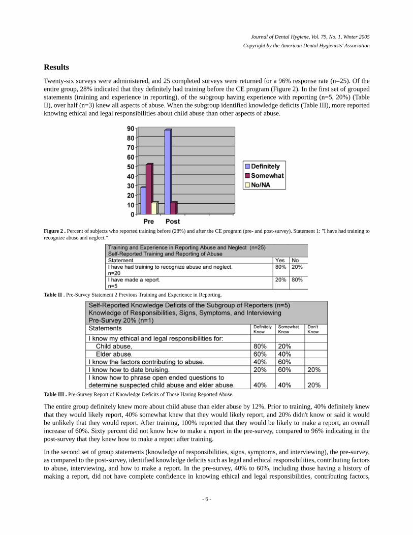
Twenty-six surveys were administered, and 25 completed surveys were returned for a 96% response rate (n=25). Of theentire group, 28% indicated that they definitely had training before the CE program (Figure 2). In the first set of groupedstatements (training and experience in reporting), of the subgroup having experience with reporting (n=5, 20%) (TableII), over half (n=3) knew all aspects of abuse. When the subgroup identified knowledge deficits (Table III), more reportedknowing ethical and legal responsibilities about child abuse than other aspects of abuse.
Figure 2 . Percent of subjects who reported training before (28%) and after the CE program (pre- and post-survey). Statement 1: "I have had training torecognize abuse and neglect."
Table II . Pre-Survey Statement 2 Previous Training and Experience in Reporting.
Table III . Pre-Survey Report of Knowledge Deficits of Those Having Reported Abuse.
The entire group definitely knew more about child abuse than elder abuse by 12%. Prior to training, 40% definitely knewthat they would likely report, 40% somewhat knew that they would likely report, and 20% didn't know or said it wouldbe unlikely that they would report. After training, 100% reported that they would be likely to make a report, an overallincrease of 60%. Sixty percent did not know how to make a report in the pre-survey, compared to 96% indicating in thepost-survey that they knew how to make a report after training.
In the second set of group statements (knowledge of responsibilities, signs, symptoms, and interviewing), the pre-survey,as compared to the post-survey, identified knowledge deficits such as legal and ethical responsibilities, contributing factorsto abuse, interviewing, and how to make a report. In the pre-survey, 40% to 60%, including those having a history ofmaking a report, did not have complete confidence in knowing ethical and legal responsibilities, contributing factors,
Journal of Dental Hygiene, Vol. 79, No. 1, Winter 2005
Copyright by the American Dental Hygienists' Association
- 6 -
dating bruising, questioning techniques, making a report, and expectations after reporting. After the training program,these knowledge levels increased to 80% and above (Table IV).
Table IV . Post-Survey Report of Knowledge Increase of the Entire Group.
In response to the third set of statements, the perceived likelihood of making a report of abuse increased dramatically aftertraining from 40% to 100%. Knowledge of what is expected of the reporter after making a report increased from 8% to84% after training.
The simple comparison of the mean scores showed an increase in the post-survey, leading to the conclusion of practicalsignificance. However, when the two-tail t-test on all statements, excluding statement 2, was done to compare the meanscores of each statement on the pre- and post-survey, the difference was statistically significant to the p=0.05 level onstatements 1, 5, 6, 8, and 10. Looking at the overall average scores, they increased. Also, an increase in the standarddeviation, an increase in the sum of scores, and median score increases were seen (Table V).
Table V . Comparison of Descriptive Statistics and t-Test of Statistical Significance
In response to statement 1, "I have had training to recognize abuse and neglect," the perception of having had trainingincreased from 28% to 88% in the post-survey, an increase of 60%. Not realizing 100% could mean that, for 12%, the CEprogram did not fulfill all expectations (Figure 2). Only 20% stated they had no training. It can be assumed by selecting"somewhat" that the dental hygienist received some training and that the dental hygienist knew about abuse but did notfeel confident with all aspects.
Statement 2, "I have made a report," was a stand-alone statement with no follow-up to ascertain who had experience inmaking a report. Twenty percent (n=5) reported that they had given an account to authorities (Figure 3).
Journal of Dental Hygiene, Vol. 79, No. 1, Winter 2005
Copyright by the American Dental Hygienists' Association
- 7 -

Figure 3 . Percent of subjects having made a report of abuse prior to the CE program. Statement 2: "I have made a report."
The results of statement 3, "I know my ethical and legal responsibilities in recognizing and reporting child abuse," indicatedthat, prior to the training, only 32% definitely understood their ethical and legal responsibilities to report (Figure 4). Instatement 4, "I know my ethical and legal responsibilities in recognizing and reporting elder abuse," fewer (20%) definitelyunderstood their responsibility to report in the pre-survey. In statement 4 after the training, "definite knowledge" of theethical and legal responsibilities in reporting elder abuse increased by 72% (Figure 5).
Figure 4 . Statement 3: "I know my ethical and legal responsibilities in recognizing and reporting child abuse." After the training 100% of the subjectsreported definitely knowing legal and ethical responsibilities.
Figure 5 . Statement 4: "I know my ethical and legal responsibilities in recognizing and reporting elder abuse." The percent of knowledge rose 72%following the CE program.
Journal of Dental Hygiene, Vol. 79, No. 1, Winter 2005
Copyright by the American Dental Hygienists' Association
- 8 -

Concerning statement 5, "I know the factors contributing to abuse," training increased the overall perceived knowledgeof the contributing factors of abuse from 20% before the CE program to 92%, an increase of 72% after training (Figure6).
Figure 6 . Statement 5: "I know the factors contributing to abuse." Subjects reported a knowledge increase by 72%.
After the training, regarding statement 6 "I know how to date bruising," there was an 80% increase in the perceivedknowledge for dating bruises. By far, this was an area about which this population knew the least. Sixty percent in thepre-survey did not know how to date patient bruising (Figure 7).
Figure 7 . Statement 6: "I know how to date bruising." This figure demonstrates the greatest percentage of knowledge deficits are in the skill of datingof bruising.
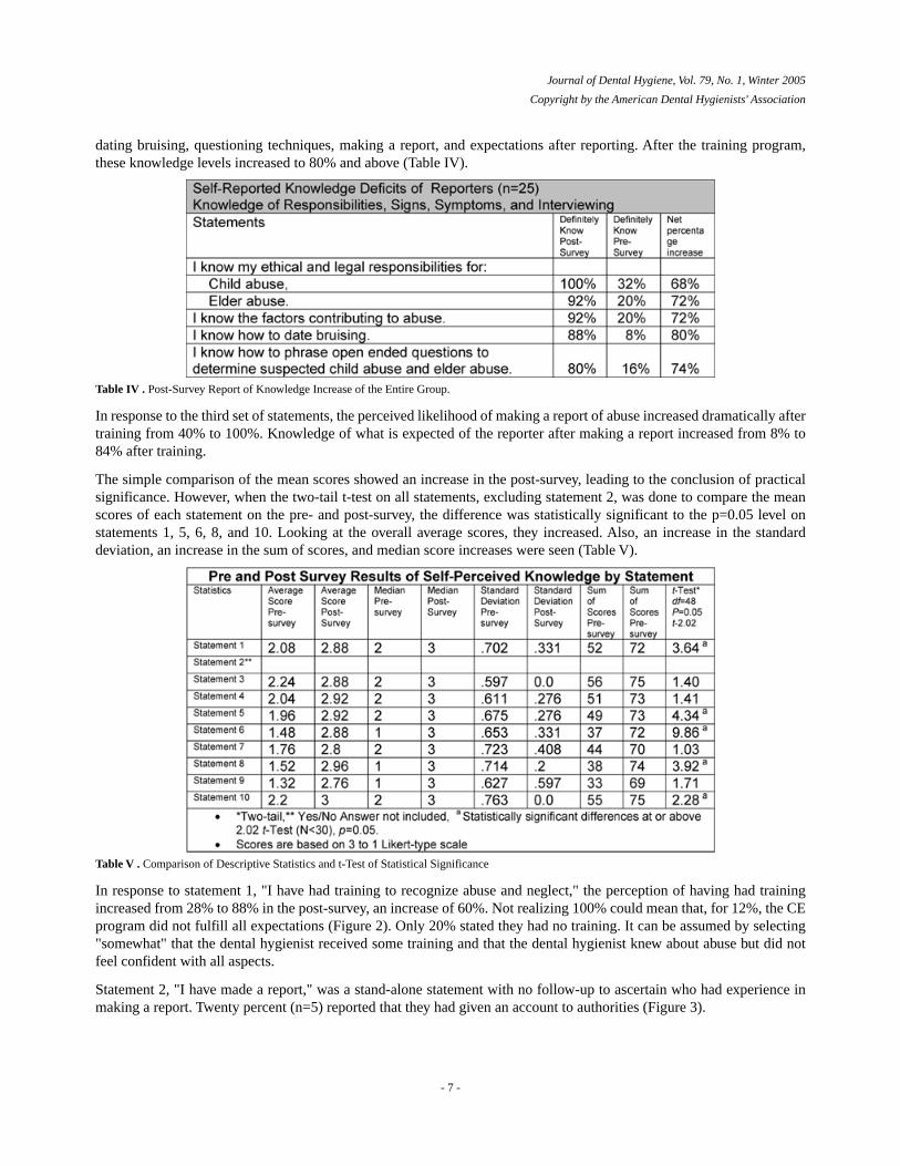
Interviewing skills was another perceived knowledge deficit. In statement 7, "I know how to phrase open-ended questionsto determine suspected child and elder abuse," knowledge was increased by training, from 16% in the pre-survey to 80%in the post survey, an overall increase of 64% (Figure 8).
Journal of Dental Hygiene, Vol. 79, No. 1, Winter 2005
Copyright by the American Dental Hygienists' Association
- 9 -
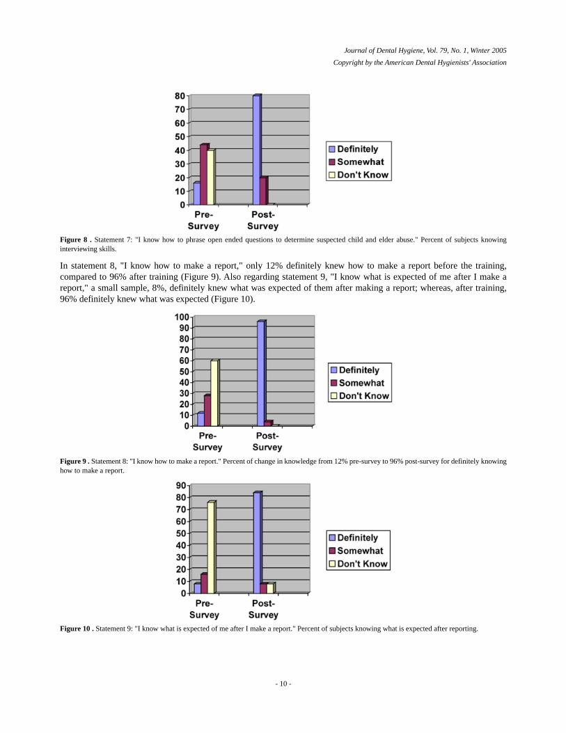
Figure 8 . Statement 7: "I know how to phrase open ended questions to determine suspected child and elder abuse." Percent of subjects knowinginterviewing skills.
In statement 8, "I know how to make a report," only 12% definitely knew how to make a report before the training,compared to 96% after training (Figure 9). Also regarding statement 9, "I know what is expected of me after I make areport," a small sample, 8%, definitely knew what was expected of them after making a report; whereas, after training,96% definitely knew what was expected (Figure 10).
Figure 9 . Statement 8: "I know how to make a report." Percent of change in knowledge from 12% pre-survey to 96% post-survey for definitely knowinghow to make a report.
Figure 10 . Statement 9: "I know what is expected of me after I make a report." Percent of subjects knowing what is expected after reporting.
Journal of Dental Hygiene, Vol. 79, No. 1, Winter 2005
Copyright by the American Dental Hygienists' Association
- 10 -
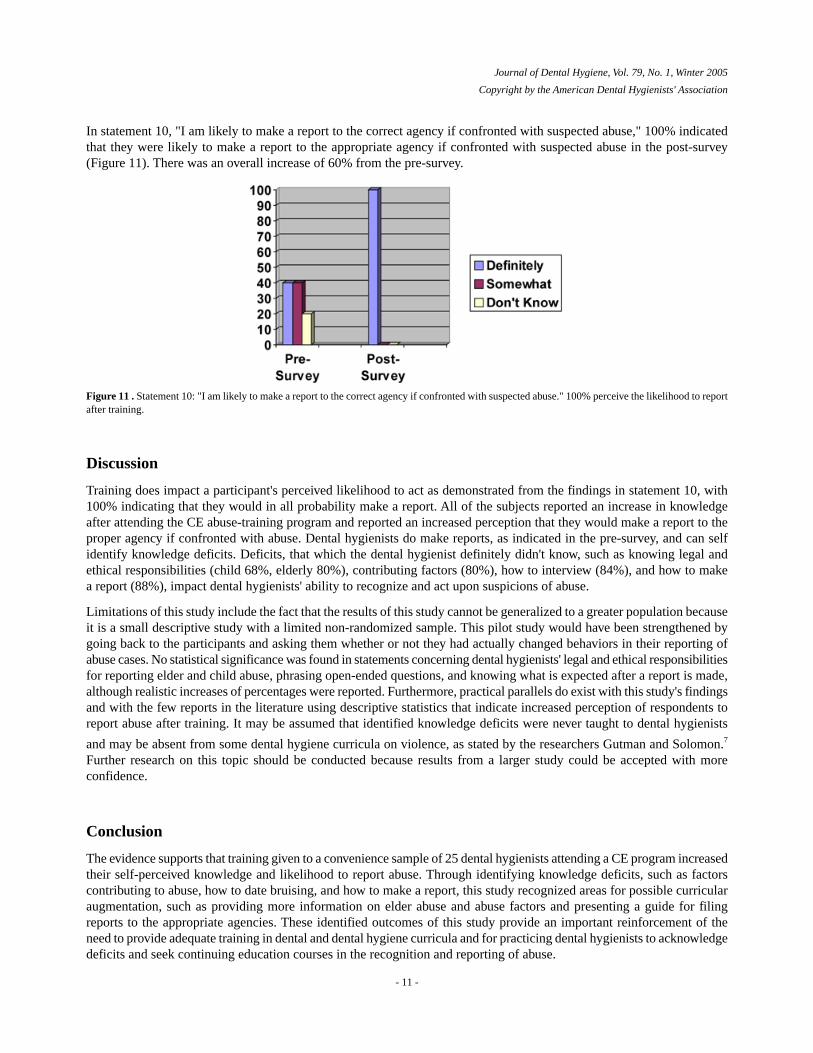
In statement 10, "I am likely to make a report to the correct agency if confronted with suspected abuse," 100% indicatedthat they were likely to make a report to the appropriate agency if confronted with suspected abuse in the post-survey(Figure 11). There was an overall increase of 60% from the pre-survey.
Figure 11 . Statement 10: "I am likely to make a report to the correct agency if confronted with suspected abuse." 100% perceive the likelihood to reportafter training.
Discussion
Training does impact a participant's perceived likelihood to act as demonstrated from the findings in statement 10, with100% indicating that they would in all probability make a report. All of the subjects reported an increase in knowledgeafter attending the CE abuse-training program and reported an increased perception that they would make a report to theproper agency if confronted with abuse. Dental hygienists do make reports, as indicated in the pre-survey, and can selfidentify knowledge deficits. Deficits, that which the dental hygienist definitely didn't know, such as knowing legal andethical responsibilities (child 68%, elderly 80%), contributing factors (80%), how to interview (84%), and how to makea report (88%), impact dental hygienists' ability to recognize and act upon suspicions of abuse.
Limitations of this study include the fact that the results of this study cannot be generalized to a greater population becauseit is a small descriptive study with a limited non-randomized sample. This pilot study would have been strengthened bygoing back to the participants and asking them whether or not they had actually changed behaviors in their reporting ofabuse cases. No statistical significance was found in statements concerning dental hygienists' legal and ethical responsibilitiesfor reporting elder and child abuse, phrasing open-ended questions, and knowing what is expected after a report is made,although realistic increases of percentages were reported. Furthermore, practical parallels do exist with this study's findingsand with the few reports in the literature using descriptive statistics that indicate increased perception of respondents toreport abuse after training. It may be assumed that identified knowledge deficits were never taught to dental hygienists
and may be absent from some dental hygiene curricula on violence, as stated by the researchers Gutman and Solomon.7
Further research on this topic should be conducted because results from a larger study could be accepted with moreconfidence.
Conclusion
The evidence supports that training given to a convenience sample of 25 dental hygienists attending a CE program increasedtheir self-perceived knowledge and likelihood to report abuse. Through identifying knowledge deficits, such as factorscontributing to abuse, how to date bruising, and how to make a report, this study recognized areas for possible curricularaugmentation, such as providing more information on elder abuse and abuse factors and presenting a guide for filingreports to the appropriate agencies. These identified outcomes of this study provide an important reinforcement of theneed to provide adequate training in dental and dental hygiene curricula and for practicing dental hygienists to acknowledgedeficits and seek continuing education courses in the recognition and reporting of abuse.
Journal of Dental Hygiene, Vol. 79, No. 1, Winter 2005
Copyright by the American Dental Hygienists' Association
- 11 -
Acknowledgements
Notes
Correspondence to: Marji Harmer-Beem [email protected]
References1. Teaster PB. A response to the abuse of vulnerable adults: the 2000 survey of state adult protective services. Washington, DC: The
National Center on Elder Abuse. Available at http://www.elderabusecenter.org/pdf/research/apsreport030703.pdf [cited 2004 Jun3].
2. US Department of Health and Human Services. Child maltreatment 1999: reports from the states to the national child abuse andneglect data system. Washington, DC: US Government Printing Office; 2001. Available athttp://www.acf.dhhs.gov/programs/cb/publications/cm99/ [cited 2004 Jun 7].
3. US Department of Justice. Violence by intimates: analysis of data on crimes by current or former spouses, boyfriends, andgirlfriends. 1998Mar. Available at http://www.ojp.usdoj.gov/bjs/pub/pdf/vi.pdf [cited 2004 Jun 7].
4. Collins KS, Schoen C, Joseph S, et.al. Health concerns across a woman's lifespan: the Commonwealth Fund 1998 survey ofw o m e n ' s h e a l t h . N e w Yo r k , N Y: T h e C o m m o n w e a l t h F u n d . 1 9 9 9 M a y.http://www.cmwf.org/publications/publications_show.htm?doc_id=221554 [cited 2004 Jun 7].
5. Short S, Tiedemann JC, Rose DE. Family violence: an intervention model for dental professionals. Northwest Dent.1997Sep-Oct;76(5):31-5.
6. Senn DR, McDowell JD, Alder ME. Dentistry's role in the recognition and reporting of domestic violence, abuse, and neglect.Dent Clin North Am. 2001Apr;45(2):343-63, ix.
7. Gutmann ME, Solomon ES. Family violence content in dental hygiene curricula: a national survey. J Dent Educ. 2002Sep;66(9):999-1005.
8. Harmer-Beem MJ. Recognizing elder abuse: oral health clinicians' roles and responsibilities. Contemporary Oral Hygiene.2004;4(5): 14-17.
9. Mouden LD, Smeadstad B. Reporting child abuse and neglect: the dental hygienist's role. Dentalhygienistnews. 1996;8(4): 14-16.10. Hyman A. Mandatory reporting of domestic violence by health care providers: a policy paper. San Francisco, CA: Family Violence
Prevention Fund; 1997Nov3.11. Murphree KR, Campbell PR, Gutmann ME, Plichta SB, Nunn ME, McCann AL, Gibson G. How well prepared are Texas dental
hygienists to recognize and report elderly abuse?. J Dent Educ. 2002 Nov;66(11): 1274-80.12. Sfikas PM. Reporting abuse and neglect. J Am Dent Assoc. 1999Dec;130(12): 1797-9.13. Ramos-Gomez F, Rothman D, Blain S. Knowledge and attitudes among California dental care providers regarding child abuse
and neglect. J Am Dent Assoc. 1998Mar;129(3): 340-8.14. Needleman HL, MacGregor SS, Lynch LM. Effectiveness of a statewide child abuse and neglect educational program for dental
professionals. Pediatr Dent. 1995 Jan-Feb;17(1): 41-45.15. Jessee SA. Child abuse and neglect curricula in North American dental schools. J Dent Educ. 1995Aug;59(8): 841-3.16. Jessee SA, Martin RE. Child abuse and neglect: assessment of dental students' attitudes and knowledge. ASDC J Dent Child.
1998Jan-Feb;65(1): 21-24.17. Kilpatrick NM, Scott J, Robinson S. Child protection: a survey of experience and knowledge within the dental profession of New
South Wales, Australia. Int J Paediatr Dent. 1999Sep;9(3): 153-9.18. Gibson-Howell JC. Domestic violence identification and referral. J Dent Hyg. 1996Mar-Apr;70(2): 74-9.19. Adair SM, Yasrebi S, Wray AI, Hanes CM, Sams DR, Russell CM. Demographic, educational, and experiential factors associated
with dentists' decisions to report hypothetical cases of child maltreatment. Pediatr Dent. 1997Nov-Dec;19(8): 466-9.20. Welbury RR, Hobson RS, Stephenson JJ, Jepson NJ. Evaluation of a computer-assisted learning programme on the oro-facial
signs of child physical abuse (non-accidental injury) by general dental practitioners. Br Dent J. 2001Jun;190(12): 668-70.
Journal of Dental Hygiene, Vol. 79, No. 1, Winter 2005
Copyright by the American Dental Hygienists' Association
- 12 -

Source: Journal of Dental Hygiene, Vol. 79, No. 1, Winter 2005
Copyright by the American Dental Hygienists' Association
Disciplinary Actions Associated with the Administration of LocalAnesthetics Against Dentists and Dental Hygienists
JoAnn C Scofield, Marylou E Gutmann, Janice P DeWald and Patricia R Campbell
JoAnn C. Scofield, RDH, MS, is an assistant professor; Marylou E. Gutmann, RDH, MA, is a professor and graduate program director;Janice P. DeWald, DDS, MS, is a professor, director, and chair; and Patricia R. Campbell, RDH, MS, is an associate professor andclinic coordinator; all are at the Caruth School of Dental Hygiene, Baylor College of Dentistry, at The Texas A & M University SystemHealth Science Center in Dallas, Texas.
Purpose. Research studies have demonstrated the need for and the ability of dental hygienists to provide local anestheticsfor pain control and reduction of patient anxiety. Although two-thirds of state dental practice laws allow these servicesto be performed by dental hygienists, controversy exists between organized dentistry and dental hygiene regarding theadministration of local anesthetics by dental hygienists. Some dentists believe the quality of care would be compromisedand patient safety jeopardized because dental hygienists do not have adequate background knowledge to preventcomplications and recognize emergencies caused by anesthetics. The purpose of this study was to collect quantitativedata addressing safety when dental hygienists administer local anesthetics.
Results. Eighteen responses were received, for a response rate of 69%. These data showed, over a 10-year period, noreports of disciplinary actions against dental hygienists for the administration of local anesthetics.
Conclusion. This study affirmed public safety, which should be helpful to states considering statutes to allow theadministration of local anesthetics by dental hygienists. Results suggest that properly educated dental hygienists in thestates surveyed have administered local anesthetics to patients without harm.
Keywords: dental hygienist, local anesthetic, scope of practice, safety
Introduction
The need for pain control in dentistry, as well as the safety and effectiveness of local anesthetics, has been well documented.1-5
Controversy between organized dentistry and organized dental hygiene exists in some localities regarding the administrationof local anesthetics by dental hygienists. A minority of state dental boards believe that the quality of patient care wouldbe compromised and the safety of patients would be jeopardized because dental hygienists do not have sufficient backgroundknowledge to prevent complications and recognize emergencies caused by anesthetic complications. Many research studies,however, have demonstrated the need for and the ability of dental hygienists to safely provide local anesthetics for pain
control and reduction of patient anxiety.2, 3, 5-8 As of July 2003, 33 state statutes allow dental hygienists to administer localanesthetics (Figure 1).
- 1 -
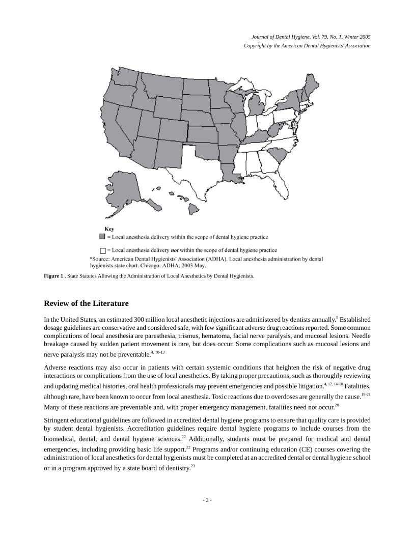
Figure 1 . State Statutes Allowing the Administration of Local Anesthetics by Dental Hygienists.
Review of the Literature
In the United States, an estimated 300 million local anesthetic injections are administered by dentists annually.9 Establisheddosage guidelines are conservative and considered safe, with few significant adverse drug reactions reported. Some commoncomplications of local anesthesia are paresthesia, trismus, hematoma, facial nerve paralysis, and mucosal lesions. Needlebreakage caused by sudden patient movement is rare, but does occur. Some complications such as mucosal lesions and
nerve paralysis may not be preventable.4, 10-13
Adverse reactions may also occur in patients with certain systemic conditions that heighten the risk of negative druginteractions or complications from the use of local anesthetics. By taking proper precautions, such as thoroughly reviewing
and updating medical histories, oral health professionals may prevent emergencies and possible litigation.4, 12, 14-18 Fatalities,
although rare, have been known to occur from local anesthesia. Toxic reactions due to overdoses are generally the cause.19-21
Many of these reactions are preventable and, with proper emergency management, fatalities need not occur.20
Stringent educational guidelines are followed in accredited dental hygiene programs to ensure that quality care is providedby student dental hygienists. Accreditation guidelines require dental hygiene programs to include courses from the
biomedical, dental, and dental hygiene sciences.22 Additionally, students must be prepared for medical and dental
emergencies, including providing basic life support.22 Programs and/or continuing education (CE) courses covering theadministration of local anesthetics for dental hygienists must be completed at an accredited dental or dental hygiene school
or in a program approved by a state board of dentistry.23
Journal of Dental Hygiene, Vol. 79, No. 1, Winter 2005
Copyright by the American Dental Hygienists' Association
- 2 -
Administration of Local Anesthetics By Dental Hygienists
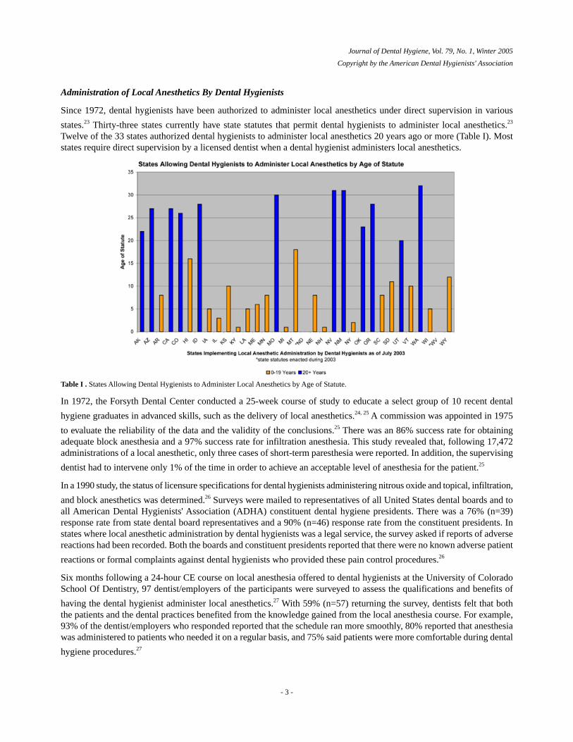
Since 1972, dental hygienists have been authorized to administer local anesthetics under direct supervision in various
states.23 Thirty-three states currently have state statutes that permit dental hygienists to administer local anesthetics.23
Twelve of the 33 states authorized dental hygienists to administer local anesthetics 20 years ago or more (Table I). Moststates require direct supervision by a licensed dentist when a dental hygienist administers local anesthetics.
Table I . States Allowing Dental Hygienists to Administer Local Anesthetics by Age of Statute.
In 1972, the Forsyth Dental Center conducted a 25-week course of study to educate a select group of 10 recent dental
hygiene graduates in advanced skills, such as the delivery of local anesthetics.24, 25 A commission was appointed in 1975
to evaluate the reliability of the data and the validity of the conclusions.25 There was an 86% success rate for obtainingadequate block anesthesia and a 97% success rate for infiltration anesthesia. This study revealed that, following 17,472administrations of a local anesthetic, only three cases of short-term paresthesia were reported. In addition, the supervising
dentist had to intervene only 1% of the time in order to achieve an acceptable level of anesthesia for the patient.25
In a 1990 study, the status of licensure specifications for dental hygienists administering nitrous oxide and topical, infiltration,
and block anesthetics was determined.26 Surveys were mailed to representatives of all United States dental boards and toall American Dental Hygienists' Association (ADHA) constituent dental hygiene presidents. There was a 76% (n=39)response rate from state dental board representatives and a 90% (n=46) response rate from the constituent presidents. Instates where local anesthetic administration by dental hygienists was a legal service, the survey asked if reports of adversereactions had been recorded. Both the boards and constituent presidents reported that there were no known adverse patient
reactions or formal complaints against dental hygienists who provided these pain control procedures.26
Six months following a 24-hour CE course on local anesthesia offered to dental hygienists at the University of ColoradoSchool Of Dentistry, 97 dentist/employers of the participants were surveyed to assess the qualifications and benefits of
having the dental hygienist administer local anesthetics.27 With 59% (n=57) returning the survey, dentists felt that boththe patients and the dental practices benefited from the knowledge gained from the local anesthesia course. For example,93% of the dentist/employers who responded reported that the schedule ran more smoothly, 80% reported that anesthesiawas administered to patients who needed it on a regular basis, and 75% said patients were more comfortable during dental
hygiene procedures.27
Journal of Dental Hygiene, Vol. 79, No. 1, Winter 2005
Copyright by the American Dental Hygienists' Association
- 3 -
A 1997 study was conducted following a Minnesota CE course on the administration of local anesthetics by dentalhygienists.28 Participants were asked to report frequency of use, impact on practice, methods used, and complicationsthat may have occurred. There was a 78% (n=273) response rate, with 88% reporting no complications from theadministration of local anesthetics. Complications that were reported included hematomas (16%), heart palpitations (12%),paresthesia (2%), and allergic reaction (1%). The results of this study indicated that the respondents felt that, when they
used local anesthetic, they provided more comfortable treatment for their patients (77%) with few complications.28
Quality Assurance
Each state legislature in the United States has established laws that regulate the practice of dentistry and dental hygiene.29-31
State legislatures grant state boards of dentistry, administrative bodies whose primary duty is to protect the citizens ofindividual states, the authority to determine the rules and regulations governing the practice of dentistry. To ensure publicprotection, state legislatures impose laws based on the recommendations of the boards of dentistry that identify minimalstandards of care that practitioners must provide. State legislatures and consumers rely on boards of dentistry as regulatory
bodies to enforce the state practice acts that govern the dental profession.30
Although state practice acts differ in each state, there are common elements among licensing jurisdictions. Board members,whose eligibility varies from state to state, are qualified by their educational credentials and experience to serve on theboard. The mission of state boards is to evaluate the quality of care provided by licensed practitioners to ensure protectionfor the public from practitioners who are negligent in the provision of care or who act in an improper manner. All statepractice acts, except in Alabama, require graduation from an accredited school of dental hygiene and successful completion
of both a written and clinical competency examination for dental hygiene licensure.29-31
State dental boards usually review complaints against dental practitioners before civil litigation is initiated, and they are
empowered with legal authority to conduct disciplinary hearings.29, 31 Infractions of the practice act, such as negligence,incompetence, fraud, and practicing beyond the scope of a dental or dental hygiene license, can lead to disciplinary actiontaken against both the dental hygienist and the dentist/employer. Disciplinary actions taken against dentists and dental
hygienists for violations of the state practice act may include license suspension, license revocation, or civil fines.29, 31
The complaint review process against dental practitioners by patients is similar in most states.31 In general, to initiate acomplaint, a patient must submit the allegations in writing to the state dental board. The complaint must include the nameof the dentist, the approximate dates of treatment, and a narrative detailing the alleged negligence or impropriety. Apractitioner in the same field as the accused is usually chosen to serve as a consultant and review the complaint. Theidentity of the reviewing consultant remains anonymous; however, if an examination of the patient is required, the anonymityof the reviewer may be impossible. Upon completion of the inquiry regarding the complaint of the practitioner by a patient,the consultant forwards a factual report and an opinion regarding the merits of the complaint to the members of the state
board.31
Once the report is received, the state board initiates an administrative review process to determine if the standards of careand practice of dentistry were violated. If the board members conclude that the practitioner violated the accepted standardsof care and practice of dentistry, the severity of the infraction is determined. The violation is then classified as "slight,""moderate," or "substantial." A slight violation results in little or no damage to the patient. For example, a provider mayfail to obtain an informed consent prior to extracting a tooth for which there is no treatment alternative. A moderateviolation includes such situations as the failure to diagnose, treat, or refer to a specialist. For example, a practitioner mayfail to diagnose periodontal disease that is developing, resulting in a decline of the patient's periodontal health. A substantial
violation occurs when considerable harm is done to the patient, such as extracting the wrong tooth or teeth.31
Upon completion of the administrative review process, the enforcement division of the state board of dentistry has theauthority to suspend or revoke a license to practice dentistry or dental hygiene if it concludes that a substantial violationhas occurred. The practitioner has the right to reject or appeal the board resolution and request a review by an administrativelaw judge or a criminal court judge. Once a license has been revoked, a practitioner cannot have the license reinstated. In
order to practice dentistry or dental hygiene again, the practitioner must reapply for licensure.29, 31
Journal of Dental Hygiene, Vol. 79, No. 1, Winter 2005
Copyright by the American Dental Hygienists' Association
- 4 -
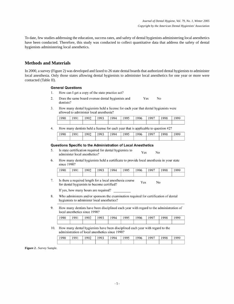
To date, few studies addressing the education, success rates, and safety of dental hygienists administering local anestheticshave been conducted. Therefore, this study was conducted to collect quantitative data that address the safety of dentalhygienists administering local anesthetics.
Methods and Materials
In 2000, a survey (Figure 2) was developed and faxed to 26 state dental boards that authorized dental hygienists to administerlocal anesthesia. Only those states allowing dental hygienists to administer local anesthetics for one year or more werecontacted (Table II).
Figure 2 . Survey Sample.
Journal of Dental Hygiene, Vol. 79, No. 1, Winter 2005
Copyright by the American Dental Hygienists' Association
- 5 -
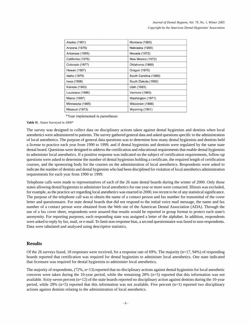
Table II . States Surveyed in 2000*
The survey was designed to collect data on disciplinary actions taken against dental hygienists and dentists when localanesthetics were administered to patients. The survey gathered general data and asked questions specific to the administrationof local anesthetics. The purpose of general data questions was to determine how many dental hygienists and dentists helda license to practice each year from 1990 to 1999, and if dental hygienists and dentists were regulated by the same statedental board. Questions were designed to address the certification and educational requirements that enable dental hygieniststo administer local anesthetics. If a positive response was indicated on the subject of certification requirements, follow-upquestions were asked to determine the number of dental hygienists holding a certificate, the required length of certificationcourses, and the sponsoring body for the courses on the administration of local anesthetics. Respondents were asked toindicate the number of dentists and dental hygienists who had been disciplined for violation of local anesthetics administrationrequirements for each year from 1990 to 1999.
Telephone calls were made to representatives of each of the 26 state dental boards during the winter of 2000. Only thosestates allowing dental hygienists to administer local anesthetics for one year or more were contacted. Illinois was excluded,for example, as the practice act regarding local anesthetics was enacted in 2000, too recent to be of any statistical significance.The purpose of the telephone call was to obtain the name of a contact person and fax number for transmittal of the coverletter and questionnaire. For state dental boards that did not respond to the initial voice mail message, the name and faxnumber of a contact person were obtained from the Web site of the American Dental Association (ADA). Through theuse of a fax cover sheet, respondents were assured that results would be reported in group format to protect each state'sanonymity. For reporting purposes, each responding state was assigned a letter of the alphabet. In addition, respondentswere asked to reply by fax, mail, or e-mail. To limit non-response bias, a second questionnaire was faxed to non-respondents.Data were tabulated and analyzed using descriptive statistics.
Results
Of the 26 surveys faxed, 18 responses were received, for a response rate of 69%. The majority (n=17, 94%) of respondingboards reported that certification was required for dental hygienists to administer local anesthetics. One state indicatedthat licensure was required for dental hygienists to administer local anesthetics.
The majority of respondents, (72%, n=13) reported that no disciplinary actions against dental hygienists for local anestheticconcerns were taken during the 10-year period, while the remaining 28% (n=5) reported that this information was notavailable. Sixty-seven percent (n=12) of the state boards reported no disciplinary action against dentists during the 10-yearperiod, while 28% (n=5) reported that this information was not available. Five percent (n=1) reported two disciplinaryactions against dentists relating to the administration of local anesthetics.
Journal of Dental Hygiene, Vol. 79, No. 1, Winter 2005
Copyright by the American Dental Hygienists' Association
- 6 -
Sixty-seven percent (n=12) reported a required minimum length of time for a local anesthesia course for dental hygienists,ranging from 14 to 72 hours. The remaining 33% (n=6) answered "no" when asked if there was a required length for alocal anesthesia course. Thirty-nine percent (n=7) were required to pass a dental board sponsored examination, while theremaining 61% (n=11) had to successfully complete a dental board-approved course.
Discussion
There has been much discussion regarding the administration of local anesthetics by dental hygienists. Negative claimsrelating to patient safety have been circulated, yet there appears to be little scientific research to substantiate a lack ofsafety when local anesthetics are administered by dental hygienists. The lack of disciplinary actions against dental hygienists(0=local anesthetic), as determined by state dental board disciplinary actions, suggests that dental hygienists can be taughtto safely administer local anesthetics without harm to patients. Research on administration of local anesthetics by dentalhygienists dates back to the 1972 "Forsyth Experiment" that included the successful teaching and administration of local
anesthetics by dental hygienists.24, 25 A study by Sisty-LePeau in 1990 also found that dental hygienists could safely
administer local anesthetics.26 The results of the present study found negligible disciplinary actions against dentists andnone against dental hygienists regarding the administration of local anesthetics. In addition, this study corroborates
information from the National Practitioner Data Bank (NPDB)32 and data from the American Association of DentalExaminers that document a low rate of adverse incidents involving both dentists and dental hygienists who administer
local anesthetics.33
Due to the lack of information on this topic in refereed journals, this study attempted to collect information directly fromthe state licensing or disciplinary boards. The response rate from the state boards, however, was lower than anticipated.Of the 18 states that did respond, 12 provided incomplete responses about the number of licensed dentists and dentalhygienists. The lack of data was indicated by leaving the response area blank on the return survey or indicating that theinformation was not available.
Fifty percent of the responding states had statutes allowing dental hygienists to administer local anesthetics for over 10years, with five states allowing this practice for more than 25 years. This length of time should be adequate to collect andreview data on disciplinary actions that could have documented occurrences of unsafe or harmful practices by dentalhygienists who administer local anesthetics. In addition, the number of state statutes allowing local anesthetic administrationby dental hygienists has more than doubled since 1990, with six additional state statutes legalizing the practice since thisstudy began. These states are Illinois (2000), Kentucky (2002), Michigan (2002), New Hampshire (2002), North Dakota(2003), and West Virginia (2003).23 This legalization may not have occurred had any significant disciplinary actionsagainst dental hygienists been documented.
Considering that an estimated 300 million local anesthetic injections are administered annually,9 the rate of disciplinary
actions is very low. Similar to research conducted by Sisty-LePeau in 1990, this research encountered comparable problems.26
For example, there was no consistency in record-keeping among states, which made the accurate collection of data difficult.Some state licensing boards reported that all dental hygienists could administer local anesthetics, while others indicatedan unknown, limited number.
Although this study did not determine the actual cause of disciplinary actions, a review of malpractice litigation foundthat most complications could have been prevented if a thorough health history had been evaluated and/or if the proper
dosage of medication had been administered, thus avoiding a toxic reaction.19-21 Future investigation may include a studyof how thoroughly health histories are reviewed in dental practices, as well as how frequently a patient's vital signs areobtained before administering local anesthetics.
Investigating malpractice insurance rates among states may be another area of investigation that may support the findingsof this study. Some questions that may be asked include whether malpractice rates have increased for dentists and dentalhygienists in states that allow the administration of local anesthetics, and how these rates in states where dental hygienistsmay administer local anesthetics compare to rates in states where they may not. Dentists should be included in the study,
Journal of Dental Hygiene, Vol. 79, No. 1, Winter 2005
Copyright by the American Dental Hygienists' Association
- 7 -
as a patient may bring litigation against a dentist as well as a dental hygienist because the dentist is directly responsiblefor care provided by the dental hygienist.
Conclusion
Debate still exists between organized dentistry and dental hygiene over the administration of local anesthetics by dentalhygienists. Current evidence, however, indicates that dental hygienists have demonstrated successful and safe administrationof local anesthetics to dental patients.
Acknowledgements
Notes
Correspondence to: JoAnn C. Scofield [email protected]
References1. Gillis MG. Local anesthesia delivery by dental hygienists: an essential component of contemporary practice. Journal of Practical
Hygiene. 2000;9(6): 13-22.2. De Jongh A, Stouthard ME. Anxiety about dental hygienist treatment. Community Dent Oral Epidemiol. 1993;21(2): 91-5.3. Sisty-LePeau N, Nielsen-Thompson N, Lutjen D. Use, need and desire for pain control procedures by Iowa hygienists. J Dent
Hyg. 1992;66(3): 137-46.4. Malamed SF, Sykes P, Kubota Y, Matsuura H, Lipp M. Local anesthesia: a review. Anesth Pain Control Dent. 1992;1(1): 11-24.5. Pihlstrom BL, Hargreaves KM, Bouwsma OJ, Myers WR, Goodale MB, Doyle MJ. Pain after periodontal scaling and root planing.
J Am Dent Assoc. 1999;130(6): 801-6.6. DeMartino BK. Anxiety: dental overview. Dent Clin North Am. 1987;31(1): 11-6.7. Gruythuysen RJ, Vertin AA. Dental hygienists: independent practice and pain control services. Community Dent Oral Epidemiol.
1994;22(3): 198-200.8. DeAngelis S, Goral V. Utilization of local anesthesia by Arkansas dental hygienists, and dentists' delegation/satisfaction relative
to this function. J Dent Hyg. 2000;74(3): 196-204.9. Malamed SF. Handbook of local anesthesia. 4thed. St. Louis, MO: Mosby; 1997. 259.10. Haas DA, Lennon D. A 21-year retrospective study of reports of paresthesia following local anesthetic administration. J Can Dent
Assoc. 1995;61(4): 319-20,23-6,29-30.11. Haas DA. Localized complications from local anesthesia. J Calif Dent Assoc. 1998;26(9): 677-82.12. Moore PA. Adverse drug interactions in dental practice: interactions associated with local anesthetics, sedatives and anxiolytics
[Part IV of a series]. J Am Dent Assoc. 1999;130(4): 541-54.13. Pogrel MA, Thamby S. Permanent nerve involvement resulting from inferior alveolar nerve blocks. J Am Dent Assoc. 2000July;131:
901-7.14. Niwa H, Hirota Y, Shibutani T, Matsuura H. Systemic emergencies and their management in dentistry: complications independent
of underlying disease. Anesth Prog. 1995;43(1): 29-35.15. Meechan JG, Skelly AM. Problems complicating dental treatment with local anaesthesia or sedation: prevention and management.
Dent Update. 1997;24(7): 278-83.16. Warshafsky T. Plaintiffs' view of malpractice litigation. Dent Clin North Am. 1982;26(2): 333-9.17. . Patient assessment and preventive measures for medical emergencies in the dental office. Dent Clin North Am. 1999;43(3):
383-400.18. Daublander M, Muller R, Lipp MD. The incidence of complications associated with local anesthesia in dentistry. Anesth Prog.
1997;44(4): 132-41.19. Robinson EM. Death in the dental chair. J Forensic Sci. 1989;34(2): 377-80.20. Malamed SF. Morbidity, mortality and local anaesthesia. Prim Dent Care. 1999;6(1): 11-5.
Journal of Dental Hygiene, Vol. 79, No. 1, Winter 2005
Copyright by the American Dental Hygienists' Association
- 8 -
21. Macdonald G. A selected review of lawsuits brought against dentists: their relevance to dental hygiene practice. J Dent Hyg.198660;8(5): 358-61.
22. American Dental Association (ADA). Accreditation standards for dental hygiene education programs. Chicago, IL; 1998.23. American Dental Hygienists' Association (ADHA). Local anesthesia administration by dental hygienists state chart. Chicago, IL;
2003.24. Lobene RR, Berman K, Chaisson LB, Karelas HA, Nolan LF. The Forsyth experiment in training of advanced skills hygienists.
J Dent Educ. 1974;28(7): 369-79.25. Doerr RE, Bertonazzi LP, Mercer JF, Kahan R, Lotzkar S, Phillips R, et.al. The report of the commission to evaluate the "Final
report, A study of new duties for dental hygienists" conducted at Forsyth Dental Center in Boston, Massachusetts. J Mass DentSoc. 1976;25(4): 201-216.
26. Sisty-LePeau N, Boyer EM, Lutjen D. Dental hygiene licensure specifications on pain control procedures. J Dent Hyg. 1990;64(4):179-85.
27. Cross-Poline GN, Passon JC, Tilliss TS, Stach DJ. Effectiveness of a continuing education course in local anesthesia for dentalhygienists. J Dent Hyg. 1992;66(3): firstpage-lastpage.
28. Anderson JM. Use of local anesthesia by dental hygienists who completed a Minnesota CE course. J Dent Hyg. 2002;76(1): 35-46.29. Morris WO. The Dentist's Legal Advisor. St. Louis, MO: Mosby; 1995. 377- 90; 294- 305; 49- 56;30. Guarino KS. Licensure and certification of dentists and accreditation of dental schools. J Dent Educ. 1995;59(1): 205-36.31. Eckstein EC. Complaint review procedures of state licensing boards. Gen Dent. 1997;45(3): 294-7.32. Oshel R R Associate Director of the Division of Quality Assurance for Research and Disputes, US Department of Health and
Human Services . Personal Communication [Email]. 1999Feb.33. American Association of Dental Examiners (AADE). Clearinghouse for Board Actions. Chicago: AADE. City, IL:
Publishernameifany_like Government printing office; 2000. Available at http://www.aadexam.org/Clearinghouse/Clearinghouse.htm[cited 2004 Aug 20].
Journal of Dental Hygiene, Vol. 79, No. 1, Winter 2005
Copyright by the American Dental Hygienists' Association
- 9 -
Source: Journal of Dental Hygiene, Vol. 79, No. 1, Winter 2005
Copyright by the American Dental Hygienists' Association
Effects of Ultrasonic Scaling and Hand-Activated Scaling onTactile Sensitivity in Dental Hygiene Students
Danielle L Ryan, Michele Darby, Deborah Bauman, Susan L Tolle and Dayanand Naik
Danielle L. Ryan, BSDH, MS, is an assistant professor at the University of Nebraska Medical Center College of Dentistry in Lincoln,Nebraska. Michele Darby, BSDH, MS, is an eminent scholar and graduate program director; Susan L. Tolle, BSDH, MS, is a professor;and Deborah Bauman, BSDH, MS, is an associate professor; all are at the Gene W. Hirschfeld School of Dental Hygiene, Old DominionUniversity, in Norfolk, Virginia. Dayanand Naik, PhD, is a professor in the Department of Mathematics and Statistics at Old DominionUniversity.
Purpose. This study was conducted to determine if tactile sensitivity varies in dental hygiene students who use theultrasonic scaler, as compared to those who scale with hand-activated instruments.
Methods. A two-group, randomized subjects, pretest-posttest design was carried out mid-semester for five weeks on 40first-year dental hygiene students who met the inclusion criteria of this study and who agreed to participate. A conveniencesample of 40 consenting, first-year dental hygiene students were randomly assigned to one of two groups (experimentalor control). After establishing a baseline tactile sensitivity score with the Vibratory Sensory Analyzer (VSA), experimentalgroup subjects used the ultrasonic scaler to remove 4cc's of artificial calculus from a typodont in a controlled, simulatedclinical setting for 45 minutes, while each control subject manually scaled 4cc's of artificial calculus on a typodont ina controlled, simulated situation for 45 minutes. Immediately following exposure to either the ultrasonic scaler orhand-activated scaling instruments, tactile sensitivity scores were obtained using the VSA. Analysis of variance withone repeated measures factor was used to determine between group and within group differences on the pretest andposttest tactile sensitivity scores.
Results. Results revealed that tactile sensitivity increased after a 45-minute scaling session with the ultrasonic scaler.Pretest to posttest changes in tactile sensitivity for the ultrasonic scaling group exhibited a much larger threshold ascompared to those in the hand-activated scaling group, supporting a gain in students' level of sensitivity with stimulus(vibration). Tactile sensitivity decreased in those who used hand-activated scaling instruments. The thumb, index, andmiddle fingers of students in both groups showed similarities in tactile sensitivity, with the index finger being the mostsensitive.
Conclusion. Tactile sensitivity decreases with hand-activated scaling and increases with ultrasonic scaling over a45-minute period. Short-term vibration exposure from the ultrasonic scaler is insufficient to negatively affect tactilesensitivity.
Keywords: tactile sensitivity, clinical dental hygiene education, repetitive strain injury
- 1 -
Introduction
Tactile sensitivity is the ability to distinguish relative degrees of tooth surface roughness or smoothness through the sense
of touch and proprioception.1 A person's tactile sensitivity may be impaired because of musculoskeletal and nerve disorders
associated with cumulative trauma, repetitive tasks, and high frequency vibrations.2 The purpose of this research was todetermine if ultrasonic scaler and hand-activated scaler usage affects tactile sensitivity of the thumb, index, and middlefingers of dental hygiene students. In doing so, ultrasonic scaling was compared to hand-activated scaling in a simulated,controlled clinical setting. The specific research questions were:
What short-term effect does the type of scaling (ultrasonic versus hand-activated scaling) have on the overall tactilesensitivity of aspiring dental hygienists?
Does tactile sensitivity change after scaling (ultrasonic versus hand-activated scaling) for 45 minutes?
Is there a difference in the tactile sensitivity of the thumb, index, and middle fingers of dental hygiene students?
The use of ultrasonic scalers, with their high frequency vibrations and noise, may be a factor affecting tactile sensitivityin dental hygienists.
Review of the Literature
Noise Effects on Tactile sensitivity
Ultrasonic scaling devices produce noise that may be greater than the Environmental Protection Agency's recommended
maximum of 70 decibels within 24 hours.3 In dentistry, ultrasonic frequencies range from 20,000 to 50,000 vibrations per
second with a 68 to 75 average decibel range.4,5 The higher the vibration per second, the greater the calculus removal
efficiency.3,4 Ultrasonic scalers emit high frequency vibrations, which have the potential to cause occupational hearing
loss.6 In a 1995 study, levels of annoyance and discomfort from high frequency noise produced by ultrasonic cleaners
revealed high ratings at all levels of exposure, ranging from 72 to 96 decibels.7 Researchers concluded that even the lowest
level of noise (70 decibels) produced by the ultrasonic cleaner should be avoided.7 Noise exposure from ultrasonic
instruments commonly produce hearing loss after long periods of time and can temporarily alter patients' hearing.8
Proprioceptive dysfunction can often be caused by noise exposure, which, in turn, can affect other sensory organs, including
the sense of touch.9 Two common causes of proprioceptive dysfunction are vestibular dysfunction and peripheral neuropathy.One example of vestibular dyfunction is the involuntary movement of the eyeball (nystagmus), caused by an ear disturbance.Peripheral neuropathies, common in dental practitioners, can cause difficulties in proprioception as well. Persons withperipheral neuropathies have little or no tactile sensitivity and are prone to self-injury because of their inability to feel
stimuli.10,11 If this occurs in a dental hygienist, the ability to evaluate a client after therapeutic scaling would be impaired.
Like neuropathy of the hands in diabetic patients, peripheral neuropathy follows a distal to proximal pattern, affecting
strength and balance, where large myelinated nerves house sensory and motor components.12 The disturbance of thesesensory and motor components may have different effects on objective sensory performance tests, depending on the level
of dysfunction.12 Tinnitus can often occur, causing one to lose the ability to feel because of the loud ringing in the ears.
When an individual feels something, the sensation is transmitted by nerve fibers that, in turn, tell the brain that there issomething there, and it is felt. If a sensory system (e.g., hearing) is disturbed, the ability to use other sensory organs, suchas tactile sensitivity, may be impaired. This impairment may be temporary or long-term, depending on the individual and
other relative factors.8,9
Journal of Dental Hygiene, Vol. 79, No. 1, Winter 2005
Copyright by the American Dental Hygienists' Association
- 2 -
Factors Affecting Tactile Sensitivity
Tactile sensitivity is reliant on several structures in the hand, including the median nerve, encapsulated nerve endings, and
specialized capsules of connective tissues.13 The median nerve runs through and innervates the thumb, index, middle, andmedian aspect of the ring finger. The median nerve houses large nerve fibers, such as A-beta fibers, which are characterized
by vibratory, proprioceptive, and tactile discriminatory sensation.14 According to Vinik et al., "tactile discriminatorysensation is mediated primarily via the large, but thinly myelinated, fast-conducting sensory afferents (A-beta fibers)innervating skin and underlying soft tissues. Due to difficulties in quantitatively detecting specific sensory deficits, little
definitive data exists addressing the issue of nerve fiber involvement."14
Even more specific than peripheral neuropathies are sensory neuropathies, which can arise from dysfunctions inproprioception, noise, or vibration. Microscopic mechanoreceptors involved in sensation lie within the Pacinian corpuscle,which is most sensitive to skin displacements. The Pacinian corpuscle is a rapidly adapting receptor that lies on a nerve
ending and consists of a multilayered connective tissue sheath that is approximately 1mm in diameter and 3mm in length.13
Its purpose is to aid in vibration detection.
The best frequency range for optimal sensitivity of the fingertip is within 100 to 200 Hertz (Hz).11 Once outside of thisrange, the sensitivity of the Pacinian corpuscle-and, hence, tactile sensitivity-decreases.11 The frequency range for mostultrasonic scalers is 20,000 to 50,000 Hz; therefore, tactile sensitivity may be affected with vibration within the ultrasonic
Hz range.15
Akesson et al. measured 30 dental hygienists, 30 dentists, 30 dental technicians, and 30 nurses on tactile sensitivity,strength, motor performance, sensorineural symptoms and signs, and vascular symptoms.15 The researchers concludedthat a decrease in strength was rather severe in the health professionals studied, and that impairment in tactile sensitivityand performance, though not as severe, was notable. According to the researchers, dentists experienced more peripheralneuropathy than dental hygienists because the dentists' hands were exposed to vibration for longer durations from usinghigh- and low-speed handpieces. Interestingly, there was an increased vibrotactile perception threshold at low and highfrequencies and decreased hand strength, leading the researchers to conclude that grip forces were lower among those
groups exposed to vibration.15 The researchers noticed a relationship between impaired vibrotactile sense and decreased
muscle strength.15 Hjortsberg et al. also reported reduced tactile sensitivity with exposure to higher frequencies (noise)
above 1,000 Hz.10 Therefore, with vibration exposure, tactile sensitivity may be one of the first physiological componentsto be affected.
In a study by Flodmark and Lundborg, male workers who had been exposed to vibration in industry comprised theexperimental group; those subjected to heavy manual work, but without vibration exposure, made up the case-control
group.16 Results revealed that a decrease in vibrotactile sense as measured by the Sensitivity Index (SI) may be one of the
first changes found following exposure to vibration.16 The researchers suggested that not only vibration, but also manual
work, may decrease vibrotactile sense.16 This may have implications for dental hygienists who use hand-activated instrumentsfor scaling. Flodmark and Lundborg's results indicated that the ranges of 0.50 to 0.64 were critical because an SI score
greater than 0.50 suggests a rise in sensorinueral symptoms and reduced vibrotactile sense.16 Although not studied in dentalhygienists using hand instruments, decreases in vibrotactile sense are early signs of compression neuropathies. Flodmarkand Lundborg further suggest that there may be correlations between the grip forces exerted while performing hand
instrumentation and compression neuropathies.16
Akesson, Balogh, and Skerfving studied ultrasonic scaler exposure time to measure the relationship between amount of
daily vibration exposure and sensitivity.17 The average time exposed, according to the time registration device, was
approximately 12 minutes a day.17 Despite the limited reported exposure time (12 minutes) to high frequency vibrations
from ultrasonic scalers, dental hygienists experienced pathological SI scores greater than 0.80.17 Akesson et al. found
dentists to have a decrease in vibrotactile perception with increased exposure to vibration.15 This decrease in perception,
Journal of Dental Hygiene, Vol. 79, No. 1, Winter 2005
Copyright by the American Dental Hygienists' Association
- 3 -
which was noted in the dominant hand and less pronounced in the non-dominant hand, was possibly attributed to the firmgrasp or grip strength required in some dental procedures. This same pattern of decreased perception, also noted by dental
hygienists, is attributed to using low-speed handpieces and ultrasonic scaling devices.15
More dentists were affected than dental hygienists, primarily due to grip force, increased exposure time, and use of high-and low-speed handpieces that run at frequency levels most likely to cause impairments. Grip forces tended to be loweramong those exposed to vibration because they did not have to exert as much energy. The amount of energy transmittedto the dental hygienist may be lower if the instrument or device is being held loosely; however, workers exposed to vibrationoften experience a decrease in muscular force. This impaired muscle function throughout the grip of the hand may be due
to injury to the muscle or nerve tissue, or a combination of both, because of vibration in and of itself.15 Impairments ingrip strength have also been found to occur among those in which low frequency vibrations (<50 Hz) are transmitted tothe hand and forearm, as opposed to higher vibrations that are absorbed by the hand.
Vibration-induced muscle injury also has been documented on laboratory rats.18 Following several days of vibration
exposure at a frequency of 80 Hz, muscle fiber degradation and changes were noted in plantar muscle sections.18 Necking
et al. found irregular muscle fiber profiles in the major portion of tissue cross sections from all vibrated legs.18 About 70%of the cross sections showed necrotic fibers or fibers undergoing necrosis. This has implications for oral health professionalswho frequently use ultrasonic or sonic scaling devices that emit vibrations that operate within high and low frequencylevels.
Dental hygiene practice demands that dental hygienists maintain pinch grasps on narrow sized instruments and use repetitive
motions that require applied force for scaling and root planing.19 According to Gerwatowski, McFall, and Stach, dentalhygienists report that latex gloves reduce tactile sensitivity and could cause a tighter grasp or pinch in order to feel calculus
and other irregularities.20 Gerwatowski et al. recommend the use of sonic and ultrasonic scalers because they require less
grip and wrist motion of the dental hygienist.20 The large diameter handle design on mechanized instruments encourages
a more open grasp, therefore decreasing the amount of pinching.20 Researchers found that the amount of grasp force appliedto instruments caused altered sensations as noted by 159 (out of 260) dental hygienists who responded to a survey on upper
extremity pain and dysfunction.21 Stentz el al. underscored the need for better ergonomic instrument designs for
practitioners.21
Neuropathies induced by vibration include Raynaud's phenomenon, characterized by fingers that become white, blanched,and very cold. Raynaud's phenomenon occurs in less than 15% of the population, and 1% to 3% may actually worsen over
time. A secondary form of Raynaud's, called vibration syndrome, is most often related to vibrating handpieces.22 Thisdamage can occur from continued exposure to vibration, even following short-term use, coupled with time. Signs andsymptoms such as tingling, numbness, and blanching may progress, leading to irreversible damage of the fingers.
According to a study conducted by the National Institute for Occupational Safety and Health (NIOSH), of 385 shipyardworkers exposed to vibrating hand tools and having symptoms of Raynaud's phenomenon, 47% had advanced stages of
vibration syndrome, while 19% had earlier stages of vibration syndrome.22 These findings suggest that practitioners exposedto vibration from ultrasonic scaling devices might develop Raynaud's phenomenon or vibration syndrome regardless ofexposure time.
Methods
Procedures, Materials, and Data Collection Instrument
The protocol was reviewed and approved by the Old Dominion University Institutional Review Board for the Protectionof Human Subjects. A pre-screening questionnaire was filled out by first-year, entry-level dental hygiene students todetermine who met the inclusion/exclusion criteria. If subjects had a current and/or past history of some form of dominant
Journal of Dental Hygiene, Vol. 79, No. 1, Winter 2005
Copyright by the American Dental Hygienists' Association
- 4 -
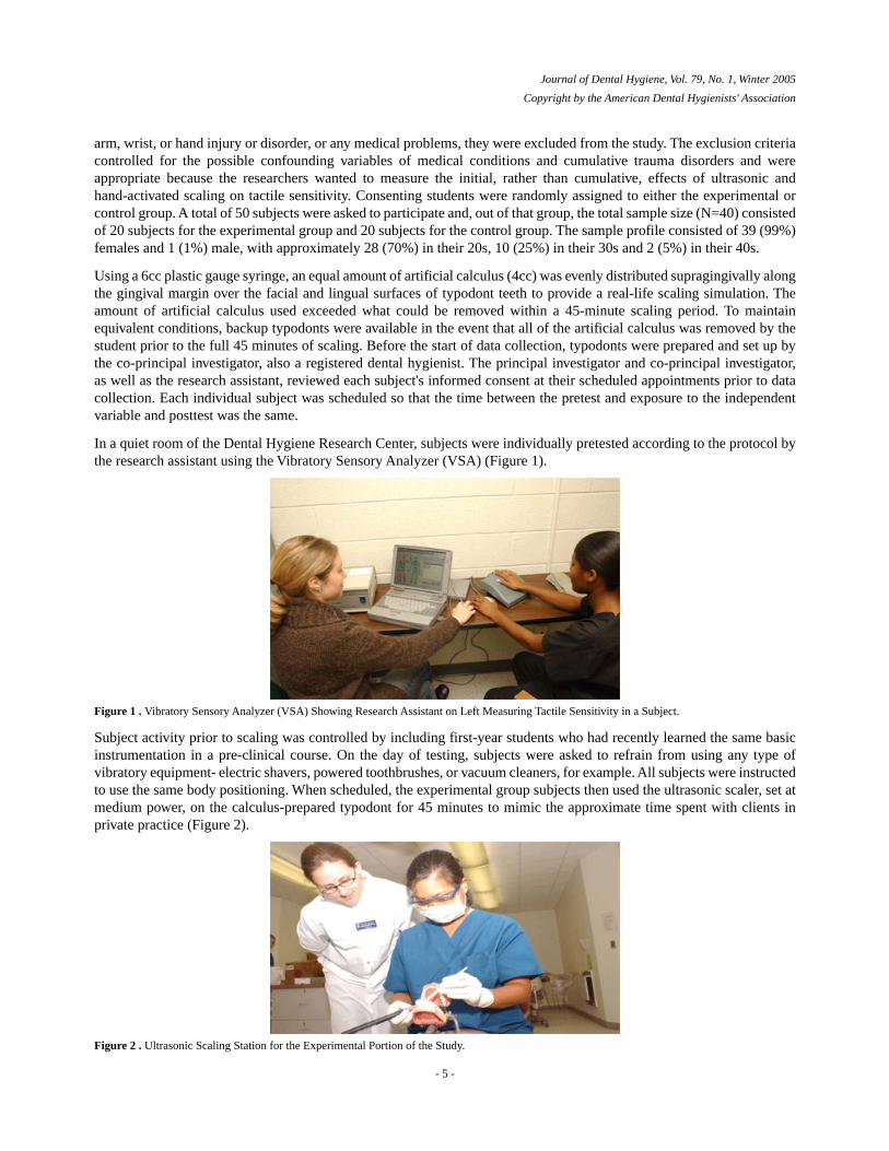
arm, wrist, or hand injury or disorder, or any medical problems, they were excluded from the study. The exclusion criteriacontrolled for the possible confounding variables of medical conditions and cumulative trauma disorders and wereappropriate because the researchers wanted to measure the initial, rather than cumulative, effects of ultrasonic andhand-activated scaling on tactile sensitivity. Consenting students were randomly assigned to either the experimental orcontrol group. A total of 50 subjects were asked to participate and, out of that group, the total sample size (N=40) consistedof 20 subjects for the experimental group and 20 subjects for the control group. The sample profile consisted of 39 (99%)females and 1 (1%) male, with approximately 28 (70%) in their 20s, 10 (25%) in their 30s and 2 (5%) in their 40s.
Using a 6cc plastic gauge syringe, an equal amount of artificial calculus (4cc) was evenly distributed supragingivally alongthe gingival margin over the facial and lingual surfaces of typodont teeth to provide a real-life scaling simulation. Theamount of artificial calculus used exceeded what could be removed within a 45-minute scaling period. To maintainequivalent conditions, backup typodonts were available in the event that all of the artificial calculus was removed by thestudent prior to the full 45 minutes of scaling. Before the start of data collection, typodonts were prepared and set up bythe co-principal investigator, also a registered dental hygienist. The principal investigator and co-principal investigator,as well as the research assistant, reviewed each subject's informed consent at their scheduled appointments prior to datacollection. Each individual subject was scheduled so that the time between the pretest and exposure to the independentvariable and posttest was the same.
In a quiet room of the Dental Hygiene Research Center, subjects were individually pretested according to the protocol bythe research assistant using the Vibratory Sensory Analyzer (VSA) (Figure 1).
Figure 1 . Vibratory Sensory Analyzer (VSA) Showing Research Assistant on Left Measuring Tactile Sensitivity in a Subject.
Subject activity prior to scaling was controlled by including first-year students who had recently learned the same basicinstrumentation in a pre-clinical course. On the day of testing, subjects were asked to refrain from using any type ofvibratory equipment- electric shavers, powered toothbrushes, or vacuum cleaners, for example. All subjects were instructedto use the same body positioning. When scheduled, the experimental group subjects then used the ultrasonic scaler, set atmedium power, on the calculus-prepared typodont for 45 minutes to mimic the approximate time spent with clients inprivate practice (Figure 2).
Figure 2 . Ultrasonic Scaling Station for the Experimental Portion of the Study.
Journal of Dental Hygiene, Vol. 79, No. 1, Winter 2005
Copyright by the American Dental Hygienists' Association
- 5 -

Once 45 minutes had expired, the co-principal investigator, who was responsible for the experimental portion of the study,advised subjects to stop using the ultrasonic scaler or hand instruments. Then, the research assistant, who was blind to thegroup status of the subjects, conducted post-testing with the VSA.
The time of day for scaling and measurement was balanced between both experimental and control groups, therebycontrolling the variable of time. A maximum of eight subjects were tested in a week, for a total of five weeks to completethe data collection portion of the study. To ensure optimal functioning and minimal variability, two pre-used, but calibrated,ultrasonic scaling units made by Dentsply, and 20 new standard P-10 Cavitron ultrasonic tips were used for the ultrasonicscaling portion of the experiment. For similar reasons, 20 new Barnhardt Universal 5/6 curets and 20 new anterior sicklesmanufactured by Hu-Friedy Manufacturing Company were used during the hand-activated scaling portion of the experiment.Each scaling trial was accurately monitored with a standard timer. Approximately 12 typodonts (Columbia Dentoform),including mounts, simulated the positioning of clients during dental hygiene care. The number of typodonts used enabledthe co-principal investigator to prepare the artificial calculus on each typodont for use over a two- to three-day period.Once used in a trial, typodonts were cleaned by the co-principal investigator and prepared for the next group of scheduledsubjects.
The data collection instrument was the VSA, manufactured by Medoc Advanced Medical Systems in Minneapolis,Minnesota. The VSA consists of a microcomputer device with a vibratory button as the stimulator (Figure 3).
Figure 3 . Vibratory Unit with Mouse.
The VSA measures the soft tissue of the pulp of the thumb, index, and middle fingers, focusing on the large nerve fibers.23
The VSA is a clinically valid method of rapid screening, early detection, and longitudinal evaluation of persons at risk of
sensory dysfunction; it can record over 30,000 tests and does automated comparisons to age-matched normative data.23
The VSA has proven to be valid and reliable through repeated testing in a number of different clinical trials.12,23 Several
major universities across the United States have utilized the VSA and its components.12 For example, it is currently beingused by the Eastern Virginia Medical School Strelitz Diabetes Institute in Norfolk, Virginia, to measure sensory dysfunctionin patients with diabetes.
The manufacturer of the VSA calibrates the instrument at the production facility and has developed a device to test for
appropriate calibration of the instrument on site.12 In cooperation with the Strelitz Diabetes Institute at Eastern VirginiaMedical School in Norfolk, Virginia, a trained VSA technician oriented the principal investigator, co-principle investigator,and research assistant, so that they developed proficiency using the VSA. Approximately six hours were spent learninghow to operate the VSA.
The actual vibratory test started when the stimuli in the vibratory button either increased or decreased in intensity and thesubject felt no sensation at all. The research assistant then started the test. Eleven trials were taken, with the first trial (apractice trial) omitted. The purpose of the practice trial was to orient the subject to what was to be expected; therefore, the
Journal of Dental Hygiene, Vol. 79, No. 1, Winter 2005
Copyright by the American Dental Hygienists' Association
- 6 -
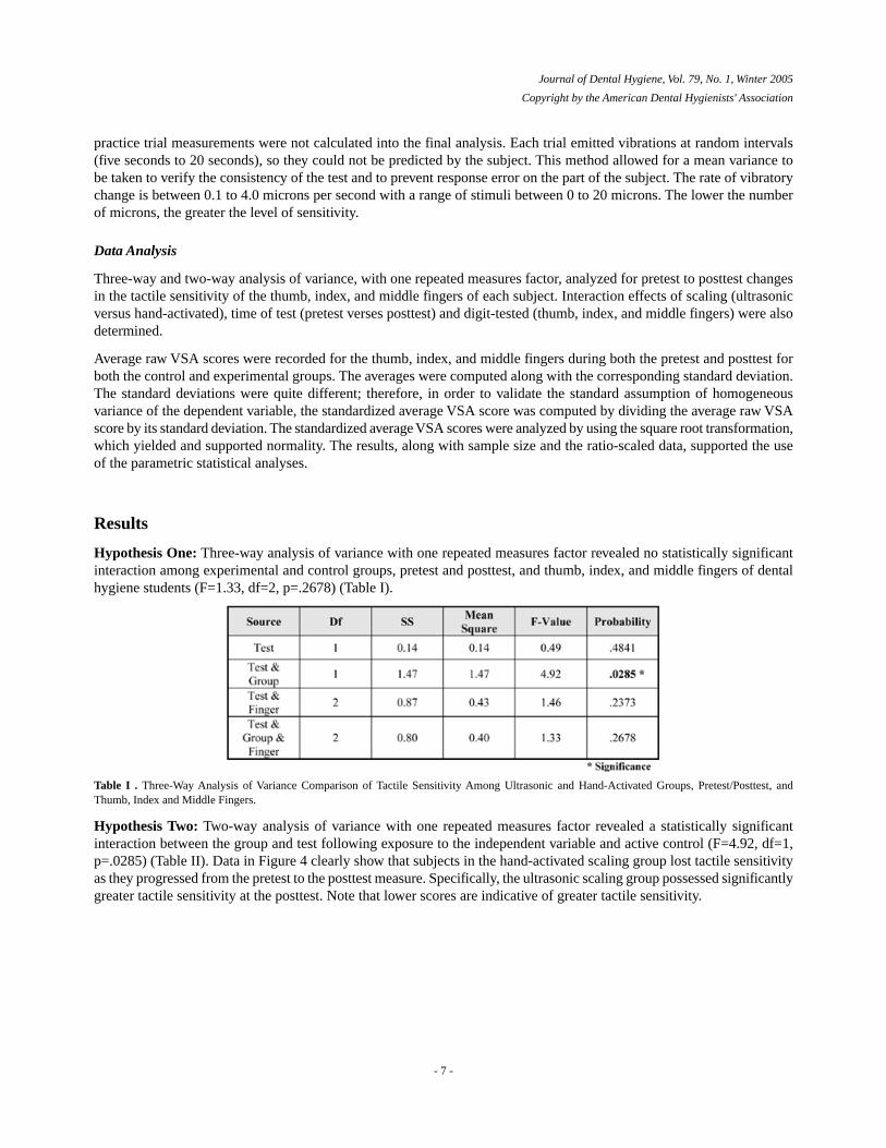
practice trial measurements were not calculated into the final analysis. Each trial emitted vibrations at random intervals(five seconds to 20 seconds), so they could not be predicted by the subject. This method allowed for a mean variance tobe taken to verify the consistency of the test and to prevent response error on the part of the subject. The rate of vibratorychange is between 0.1 to 4.0 microns per second with a range of stimuli between 0 to 20 microns. The lower the numberof microns, the greater the level of sensitivity.
Data Analysis
Three-way and two-way analysis of variance, with one repeated measures factor, analyzed for pretest to posttest changesin the tactile sensitivity of the thumb, index, and middle fingers of each subject. Interaction effects of scaling (ultrasonicversus hand-activated), time of test (pretest verses posttest) and digit-tested (thumb, index, and middle fingers) were alsodetermined.
Average raw VSA scores were recorded for the thumb, index, and middle fingers during both the pretest and posttest forboth the control and experimental groups. The averages were computed along with the corresponding standard deviation.The standard deviations were quite different; therefore, in order to validate the standard assumption of homogeneousvariance of the dependent variable, the standardized average VSA score was computed by dividing the average raw VSAscore by its standard deviation. The standardized average VSA scores were analyzed by using the square root transformation,which yielded and supported normality. The results, along with sample size and the ratio-scaled data, supported the useof the parametric statistical analyses.
Results
Hypothesis One: Three-way analysis of variance with one repeated measures factor revealed no statistically significantinteraction among experimental and control groups, pretest and posttest, and thumb, index, and middle fingers of dentalhygiene students (F=1.33, df=2, p=.2678) (Table I).
Table I . Three-Way Analysis of Variance Comparison of Tactile Sensitivity Among Ultrasonic and Hand-Activated Groups, Pretest/Posttest, andThumb, Index and Middle Fingers.
Hypothesis Two: Two-way analysis of variance with one repeated measures factor revealed a statistically significantinteraction between the group and test following exposure to the independent variable and active control (F=4.92, df=1,p=.0285) (Table II). Data in Figure 4 clearly show that subjects in the hand-activated scaling group lost tactile sensitivityas they progressed from the pretest to the posttest measure. Specifically, the ultrasonic scaling group possessed significantlygreater tactile sensitivity at the posttest. Note that lower scores are indicative of greater tactile sensitivity.
Journal of Dental Hygiene, Vol. 79, No. 1, Winter 2005
Copyright by the American Dental Hygienists' Association
- 7 -
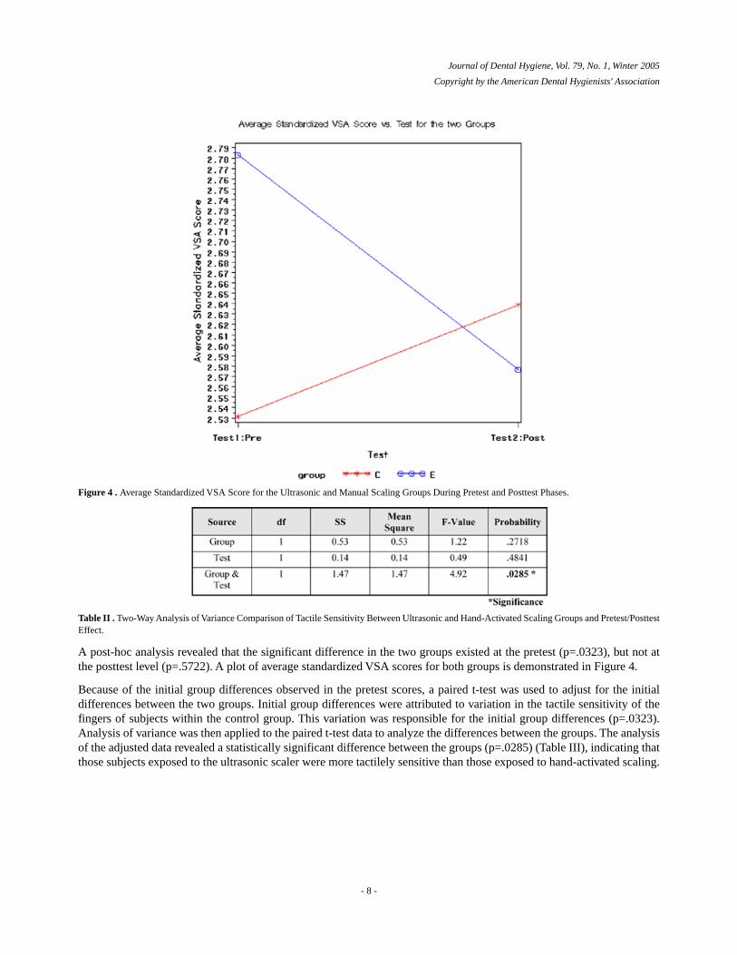
Figure 4 . Average Standardized VSA Score for the Ultrasonic and Manual Scaling Groups During Pretest and Posttest Phases.
Table II . Two-Way Analysis of Variance Comparison of Tactile Sensitivity Between Ultrasonic and Hand-Activated Scaling Groups and Pretest/PosttestEffect.
A post-hoc analysis revealed that the significant difference in the two groups existed at the pretest (p=.0323), but not atthe posttest level (p=.5722). A plot of average standardized VSA scores for both groups is demonstrated in Figure 4.
Because of the initial group differences observed in the pretest scores, a paired t-test was used to adjust for the initialdifferences between the two groups. Initial group differences were attributed to variation in the tactile sensitivity of thefingers of subjects within the control group. This variation was responsible for the initial group differences (p=.0323).Analysis of variance was then applied to the paired t-test data to analyze the differences between the groups. The analysisof the adjusted data revealed a statistically significant difference between the groups (p=.0285) (Table III), indicating thatthose subjects exposed to the ultrasonic scaler were more tactilely sensitive than those exposed to hand-activated scaling.
Journal of Dental Hygiene, Vol. 79, No. 1, Winter 2005
Copyright by the American Dental Hygienists' Association
- 8 -
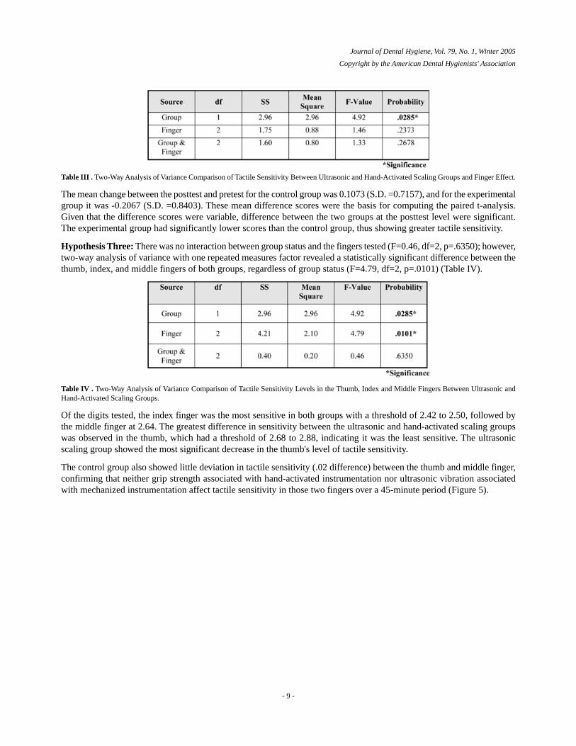
Table III . Two-Way Analysis of Variance Comparison of Tactile Sensitivity Between Ultrasonic and Hand-Activated Scaling Groups and Finger Effect.
The mean change between the posttest and pretest for the control group was 0.1073 (S.D. =0.7157), and for the experimentalgroup it was -0.2067 (S.D. =0.8403). These mean difference scores were the basis for computing the paired t-analysis.Given that the difference scores were variable, difference between the two groups at the posttest level were significant.The experimental group had significantly lower scores than the control group, thus showing greater tactile sensitivity.
Hypothesis Three: There was no interaction between group status and the fingers tested (F=0.46, df=2, p=.6350); however,two-way analysis of variance with one repeated measures factor revealed a statistically significant difference between thethumb, index, and middle fingers of both groups, regardless of group status (F=4.79, df=2, p=.0101) (Table IV).
Table IV . Two-Way Analysis of Variance Comparison of Tactile Sensitivity Levels in the Thumb, Index and Middle Fingers Between Ultrasonic andHand-Activated Scaling Groups.
Of the digits tested, the index finger was the most sensitive in both groups with a threshold of 2.42 to 2.50, followed bythe middle finger at 2.64. The greatest difference in sensitivity between the ultrasonic and hand-activated scaling groupswas observed in the thumb, which had a threshold of 2.68 to 2.88, indicating it was the least sensitive. The ultrasonicscaling group showed the most significant decrease in the thumb's level of tactile sensitivity.
The control group also showed little deviation in tactile sensitivity (.02 difference) between the thumb and middle finger,confirming that neither grip strength associated with hand-activated instrumentation nor ultrasonic vibration associatedwith mechanized instrumentation affect tactile sensitivity in those two fingers over a 45-minute period (Figure 5).
Journal of Dental Hygiene, Vol. 79, No. 1, Winter 2005
Copyright by the American Dental Hygienists' Association
- 9 -
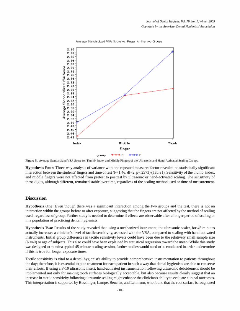
Figure 5 . Average Standardized VSA Score for Thumb, Index and Middle Fingers of the Ultrasonic and Hand-Activated Scaling Groups.
Hypothesis Four: Three-way analysis of variance with one repeated measures factor revealed no statistically significantinteraction between the students' fingers and time of test (F=1.46, df=2, p=.2373) (Table I). Sensitivity of the thumb, index,and middle fingers were not affected from pretest to posttest by ultrasonic or hand-activated scaling. The sensitivity ofthese digits, although different, remained stable over time, regardless of the scaling method used or time of measurement.
Discussion
Hypothesis One: Even though there was a significant interaction among the two groups and the test, there is not aninteraction within the groups before or after exposure, suggesting that the fingers are not affected by the method of scalingused, regardless of group. Further study is needed to determine if effects are observable after a longer period of scaling orin a population of practicing dental hygienists.
Hypothesis Two: Results of the study revealed that using a mechanized instrument, the ultrasonic scaler, for 45 minutesactually increases a clinician's level of tactile sensitivity, as tested with the VSA, compared to scaling with hand-activatedinstruments. Initial group differences in tactile sensitivity levels could have been due to the relatively small sample size(N=40) or age of subjects. This also could have been explained by statistical regression toward the mean. While this studywas designed to mimic a typical 45-minute scaling session, further studies would need to be conducted in order to determineif this is true for longer exposure times.
Tactile sensitivity is vital to a dental hygienist's ability to provide comprehensive instrumentation to patients throughoutthe day; therefore, it is essential to plan treatment for each patient in such a way that dental hygienists are able to conservetheir efforts. If using a P-10 ultrasonic insert, hand-activated instrumentation following ultrasonic debridement should beimplemented not only for making tooth surfaces biologically acceptable, but also because results clearly suggest that anincrease in tactile sensitivity following ultrasonic scaling might enhance the clinician's ability to evaluate clinical outcomes.This interpretation is supported by Busslinger, Lampe, Beuchat, and Lehmann, who found that the root surface is roughened
Journal of Dental Hygiene, Vol. 79, No. 1, Winter 2005
Copyright by the American Dental Hygienists' Association
- 10 -
following ultrasonic instrumentation, thereby suggesting the need for hand-activated instrumentation following ultrasonic
scaling.24 A combination of ultrasonic and hand-activated scaling should be used in order to reduce the likelihood ofdecreasing tactile sensitivity following a routine 45-minute scaling session. This recommended protocol has implicationsfor all types of practice in which nonsurgical periodontal therapy is performed.
Hypothesis Three: Statistical analysis revealed a significant difference among the thumb, index, and middle fingers ofthe ultrasonic and hand-activated scaling groups. In the simulated situation, the index finger appears to be the most tactilelysensitive finger, with the thumb being the least sensitive. These findings, although observed in the laboratory, may refute
Nield-Gerig's belief that the middle finger is the most tactilely sensitive.25 Perhaps the index finger's position in the modifiedpen grasp, on top of the instrument handle, maximizes the opportunity to sense changes picked up by the instrument.Pinching (to squeeze between the thumb and a finger) and gripping (to maintain a secure grasp), common forces involvedin grasping a hand-activated instrument during working strokes, might be related to the thumb and middle finger's decreasedtactile sensitivity, as compared to the index finger.
Findings of this study and that of Michalak-Turcotte and Atwood-Saunders support the recommendation that dental
hygienists vary the use of ultrasonic scalers with hand-activated scaling.26 A combined use of hand-activated scaling andultrasonic scaling is recommended because there is minimal to no pinch force with the ultrasonic scaler, only repetitivemotions, as compared to hand-activated scaling. Pinching and gripping could reduce tactile sensitivity if the nerve endingsbecome pinched or isolated, further explaining why tactile sensitivity decreased in subjects in the hand-activated group.These findings conflict with those of Hjortsberg et al. and Flodmark and Lundborg, who both found decreases in workers'
tactile sensitivity associated with vibration exposure.10,16 Duration of exposure and variability between the simulatedlaboratory and clinical situations might explain these conflicting outcomes. While this study was conducted using 30,000Hz units in a simulated clinical setting, subjects in Akesson et al. showed impaired vibrotactile sense and decreased muscle
strength at both low and high frequencies associated with dental hand pieces.15 Furthermore, in the dental hygienists studied
by Akesson et al., the impaired tactile sense was greater in their dominant hand.15 Because first-year, entry-level dentalhygiene students were used, pinching and gripping of instruments could be greater than that found in experienced dentalhygienists who have developed muscle strength and hand coordination. This study should be replicated in a population ofexperienced dental hygiene practitioners under normal clinical practice conditions to determine if hand-arm muscle strengthaffects tactile sensitivity.
Hypothesis Four: Neither the thumb, index, nor middle fingers were affected following exposure to the ultrasonic ormanual scaling instruments. This could be due to the fact that tactile sensitivity is a relatively stable variable over time,or that the 45 minutes devoted to scaling was inadequate to alter the tactile sensitivity. Because this study focused on initialeffects in a simulated setting, follow-up studies need to be conducted in order to determine long-term effects under normalclinical practice conditions.
Conclusions
Based on the results of this simulated clinical investigation using the VSA, the following conclusions are made:
Tactile sensitivity is affected differentially by mechanized and hand-activated scaling over the short term. Dental hygienestudents who use the ultrasonic scaler for 45 minutes are likely to experience increased tactile sensitivity. Dental hygienestudents who use hand-activated instruments for 45 minutes are likely to experience decreased tactile sensitivity.
Greater tactile sensitivity is experienced in the index finger than in the thumb and middle finger, regardless of whether amechanized or hand-activated scaling instrument is used.
Ultrasonic scalers enhance tactile sensitivity in first-year, entry-level dental hygiene students in a simulated clinical setting.With an increase in the use of mechanized instrumentation in nonsurgical periodontal therapy, more research should beconducted to determine if the ultrasonic scaler causes an increase in tactile sensitivity over time and, if so, at what rate.Findings in this study do not support changes in clinical instrumentation protocols at this time, but emphasize the needfor more research in order to better understand tactile sensitivity in oral healthcare professionals.
Journal of Dental Hygiene, Vol. 79, No. 1, Winter 2005
Copyright by the American Dental Hygienists' Association
- 11 -
Acknowledgements
This investigation was supported in part by the American Dental Hygienists' Association Institute for Oral Health andby the Gene W. Hirschfeld School of Dental Hygiene at Old Dominion University. The authors would also like toacknowledge Kevin Stansberry, BS, a research associate at the Strelitz Diabetes Institute, Eastern Virginia MedicalSchool, in Norfolk, Virginia, for his contribution.
Notes
Correspondence to: Danielle Ryan [email protected]
References1. Tsutsui P. Periodontal instruments and instrumentation theory. In: Darby ML, Walsh MM. , editors. Dental hygiene theory and
practice. 2nded. Philadelphia, PA: WB Saunders Co.; 2003. 385- 431.2. Setcos JC, Mahyuddin. Noise levels encountered in dental, clinical and laboratory practice. Int J Prosthodont. 1998;11: 150-157.3. Merrell HB, Claggatt K. Noise pollution and hearing loss in the dental office. Dental Assist. journaltitle. 1992;61(3): 6-9.4. Hodges KO. Concepts in nonsurgical periodontal therapy: ultrasonic periodontal debridement. Albany, NY: Delmar; 1998. 320-
344.5. Stevens M. An earful: Is someone listening to the din of occupational noise exposure in dentistry?. RDH. 1999;19(9): 34-85.6. Wilson JD, Darby ML, Tolle SL, Sever SC. Effects of occupational ultrasonic noise exposure on hearing of dental hygienists: a
pilot study. J Dent Hyg. 2002;76(5): 262-269.7. Holmberg K, Ulflandstrom U, Nordstrom B. Annoyance and discomfort during exposure to high-frequency noise from an ultrasonic
washer. Percept Mot Skills. 1995;81: 819-827-lastpage.8. Moller P, Grevstad A, Kristoffersen T. Ultrasonic scaling of maxillary teeth causing tinnitus and temporary hearing shifts. J Clin
Periodontol. 1976;3: 123-127.9. McCance KL, Huether SE. Pathophysiology: the biologic basis for disease in adults and children. 3rded. St. Louis, MO: Mosby;
1998.10. Hjortsberg U, Rosen I, Orbaek P, Lundborg G, Balogh I. Finger receptor dysfunction in dental technicians exposed to high-frequency
vibration. Scand J Work Environ Health. 1989;15: 339-344.11. Lundstrom R, Lindmark A. Effects of local vibration on tactile perception in the hands of dentists. Journal of Low Frequency
Noise and Vibration. 1982;1: 1-11.12. Resnick HE, Stansberry KB, Harris TB, Tirivedi M, Smith KY, Morgan P, Vinik AI. Diabetes, peripheral neuropathy, and old
age disability. Muscle Nerve. 2002;25(1): 43-50.13. Hunter JM, Schneider LH, Macking EJ. Tendon and nerve surgery in the hand. 3rded. St. Louis, MO: Mosby; 1997.14. Vinik AI, Suwanwalaikorn S, Stansberry KB, Holland MT, McNitt PM, Lawrence EC. Quantitative measurement of cutaneous
perception in diabetic neuropathy. Muscle Nerve. 1995;18: 574-584.15. Akesson I, Lundborg G, Hortsmann V, Skerfving S. Neuropathy in female dental personnel exposed to high frequency vibrations.
J Occup Environ Med. 1995;52: 116-123.16. Flodmark BT, Lundborg G. Vibrotactile sense and hand symptoms in blue collar workers in a manufacturing industry. J Occup
Med. 1997;54: 880-887.17. Akesson I, Balogh I, Skerfving S. Self-reported and measured time of vibration exposure of ultrasonic scaling in dental hygienists.
Journal of Applied Ergonomics. 2001;32: 47-51.18. Necking LE, Dahlin LB, Friden J, Lundborg G, Lundstrom R, Thornell LE. Vibration-induced muscle injury: An experimental
model and preliminary findings. J Hand Surg [Br. 1992;17: 270-274.19. Michalak-Turcotte C. Controlling dental hygiene work-related musculoskeletal disorders: the ergonomic process. J Dent Hyg.
2000;74(74): 41-48.20. Gerwatowski LJ, McFall DB, Stach DJ. Carpal tunnel syndrome: Risk factors and preventive strategies for the dental hygienist.
J Dent Hyg. 1992;66(2): 89-94.21. Stentz TL, Riley MW, Harn SD, Sposato RC, Stockstill JW, Harn JA. Upper extremity altered sensations in dental hygienists.
Int J Ind Ergon. 1994;13: 107-112.22. Current intelligence bulletin 38: Vibration syndrome [Internet]. c1983 cited 2001 Mar 8 Cincinnati (OH) Center for Disease
Control Available from: http://www.cdc.gov/niosh/83110_38.html.
Journal of Dental Hygiene, Vol. 79, No. 1, Winter 2005
Copyright by the American Dental Hygienists' Association
- 12 -

23. Medoc Ltd Neurosensory analyzer model TSA-II and vibratory sensory analyzer VSA-3000 user and service guide. 5thed.Minneapolis, MN: Compass Medical Technologies; 2002.
24. Busslinger A, Lampe K, Beuchat M, Lehmann B. A comparative in vitro study of a magnetostrictive and a piezoelectric ultrasonicscaling instrument. J Periodontol. 2001;28: 642-649.
25. Nield-Gehrig JS. Fundamentals of periodontal instrumentation. 4thed. Philadelphia, PA: Lippincott Williams & Wilkins; 2000.26. Michalak-Turcotte C, Atwood-Saunders M. Ergonomic strategies for the dental hygienist (Pt 1). Journal of Practical Hygiene.
2000;9(3): 39-42.
Journal of Dental Hygiene, Vol. 79, No. 1, Winter 2005
Copyright by the American Dental Hygienists' Association
- 13 -
Source: Journal of Dental Hygiene, Vol. 79, No. 1, Winter 2005
Copyright by the American Dental Hygienists' Association
An Assessment of Printed Oral Cancer Materials from LocalHealth Departments in Illinois
Sandra J Maurizio, Sherri M Lukes and Ronda DeMattei
Sandra J. Maurizio, RDH, PhD, is an associate professor, Sherri M. Lukes, RDH, MS, is an assistant professor, and Ronda DeMattei,RDH, MSEd, is an assistant professor; all are at Southern Illinois University in Carbondale, Illinois.
Local health departments can be resources for the public and health professionals to access educational materials ona variety of topics. Though the dental office is ideal, it should not be the only venue for obtaining oral cancer educationalmaterials. As part of a cancer prevention and early detection project, this pilot study solicited printed educationalmaterials concerning oral cancer from all local health departments (LHDs) in Illinois.
Sixty of the 94 health departments responded, for a response rate of 63.8%. Only 32 had printed oral cancer educationalmaterials for public and professional distribution, and most focused on tobacco. Forty-eight different samples werereceived. At least one risk factor was identified in all materials. The most common was tobacco usage, present in 100%of the materials. Twenty-nine (61%) identified tobacco use as the only risk factor. Alcohol consumption, the secondmost frequently identified risk factor, was mentioned in only 12 (26%) of the materials. Few materials comprehensivelycovered areas such as signs and symptoms, risk factors, and the importance of examinations for early detection. Onlyfive materials (11%) acknowledged that early lesions are often asymptomatic.
Local health departments are in an influential position to disseminate educational materials to the public and healthprofessionals. Dental hygienists have the knowledge and expertise required to identify quality materials. They shouldbe proactive in assisting LHDs with acquisition of appropriate printed materials and partner with them to advocate forprevention and early detection of oral cancer. In collaboration, the two can ensure that all LHDs have current andcomprehensive printed materials about oral cancer to distribute to the public and other health professionals.
Keywords: oral cancer, printed educational materials, public, local health departments
Introduction and Review of the Literature
Dental hygienists must advocate for oral cancer awareness in their communities. As oral health care providers, they sharea responsibility to ensure that the public and other health professionals have access to current and comprehensive printededucational materials about oral cancer. Materials disseminated from a variety of places, including the dental office, mustoutline risk factors, signs and symptoms, and the need for routine oral cancer examinations for early detection. Localhealth departments (LHDs) in Illinois serve as resources for printed materials about a multitude of health issues. Dentalhygienists can play an important role in ensuring that LHDs have appropriate printed materials concerning oral cancer fordistribution to the public and health professionals.
- 1 -
The American Cancer Society estimates that more than 28,000 Americans will be diagnosed with oral cancer in 2004, and
7,200 will die as a result of the disease.1 Recent Surveillance, Epidemiology and End Results (SEER) data indicate thereare 9.7 new cases per 100,000 in the United States per year, with little progress in the advancement of early diagnosis
during the last decades.2,3,4 The five-year survival rate for oral cancer is only 17% for advanced stage disease and 81% for
early localized disease.2 The five-year relative survival rate is 56%.1 Because of these statistics, Healthy People 2010 and
the Surgeon General's oral health report of 2000 both stress the need for earlier detection and diagnosis.5,6
Major risk factors associated with oral cancer are tobacco usage and excessive alcohol consumption.3,7 The synergistic
effect of tobacco and alcohol is particularly implicated in neoplasm development.3,4,7,8 Current smokers are six to 28 timesmore likely to develop oral cancer, and the combined use of tobacco and alcohol accounts for 75% to 90% of oral cancer
cases in the United States.6 Long-term users of snuff (chewing tobacco) may face a 50-fold risk for oral and pharyngeal
cancer development.3 Other risk factors associated with oral cancer include sun exposure for lip cancer, increased age,
male gender, various viruses, lack of fruits and vegetables in the diet, and genetic predisposition.3 Males are twice as likelyas females to develop oral cancer. Age is strongly associated with oral cancer, with 95% of the oral cancers occurring in
individuals 40 years of age or older.3
The most common signs and symptoms of oral cancer are a white or red patch that does not go away, a sore that bleedseasily and fails to heal, and a lump or thickening of tissue. Signs such as difficulty chewing and/or swallowing or difficulty
moving the tongue are usually those of advanced disease.1 Because clinicians focus on patient-reported symptoms, rather
than observed signs, and early oral cancer lesions are painless, diagnosis is often delayed.3,9
Several studies indicate that dentists and physicians do not routinely perform adequate oral cancer examinations and are
remiss in diagnosis of and referral for early oral cancers.4,9-18 Yellowitz and Goodman found that 82% of physicians and
17% of dentists did not perform an oral cancer examination on most of their patients.18 Prout et al. found that 77% of
patients with advanced stage oral cancer were under a physician's care within three to 24 months of diagnosis.19 Another
study found that 94% of patients with advanced stage oral cancers were seen by a physician within one year of diagnosis.20,21
A national study of dental hygienists found that 66% of dental hygienists reported providing screenings 100% of the timefor adults over age 40 at their initial appointment. Only 25% reported routinely palpating lymph nodes all of the time, and
half did not palpate lymph nodes at all.22 Data from the 1998 National Health Interview Survey (NHIS) Cancer ControlSupplement found that fewer than 10% of adults aged 18 or older reported having had an oral cancer examination within
the last three years.23,24
It is well documented that the public lacks adequate knowledge about oral cancer risk factors, signs and symptoms, and
methods to facilitate early detection.6,7,10,11,16 The public, as well as other non-dental health professionals, must be informedof the need for oral cancer examinations as part of routine preventive care and of the risk factors and signs and symptomsassociated with the disease. Horowitz and Nourjah expressed the need for implementation of vigorous oral cancer education
for the public.10
In 1998, Chung and colleagues examined educational materials that focused on oral and pharyngeal cancers and were
available to American adults.25 They surveyed national and state organizations or agencies and requested copies of oralcancer materials they produced. Brochures, videotapes, posters, fact sheets, and leaflets were collected. Only 59 itemsincluded oral cancer in the content, and only 20 focused specifically on oral cancer. They called for a variety of newmaterials, particularly ones aimed at persons with low reading ability, and a brief list of steps for performing oral cancer
examinations, similar to brochures currently available for breast examinations.25
Canto and colleagues examined the coverage and quality of oral cancer information in the popular press. They discoverednot only a lack of coverage, but inaccuracies in the content as well. More than 50% of the articles did not mention warning
Journal of Dental Hygiene, Vol. 79, No. 1, Winter 2005
Copyright by the American Dental Hygienists' Association
- 2 -
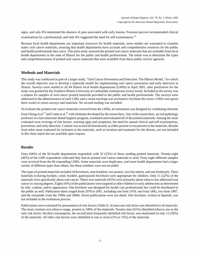
signs, and only 6% mentioned the absence of pain associated with early lesions. Fourteen percent recommended clinical
examinations by a professional, and only 8% suggested the need for self examinations.26
Because local health departments are important resources for health materials, more studies are warranted to examinestates' oral cancer materials, ensuring that health departments have accurate and comprehensive resources for the publicand health professionals they serve. This pilot study assessed the printed oral cancer materials that are available from localhealth departments in the state of Illinois for the public and health professionals. The intent was to determine the typesand comprehensiveness of printed oral cancer materials that were available from these public service agencies.
Methods and Materials
This study was conducted as part of a larger study, "Oral Cancer Prevention and Detection: The Illinois Model," for whichthe overall objective was to develop a statewide model for implementing oral cancer prevention and early detection inIllinois. Surveys were mailed to all 94 Illinois local health departments (LHDs) in April 2001, after permission for thestudy was granted by the Southern Illinois University at Carbondale institutional review board. Included in the survey wasa request for samples of oral cancer printed materials provided to the public and health professionals. The surveys wereaddressed to the administrators of each LHD, and a return envelope was enclosed to facilitate the return. LHDs were giventhree weeks to return surveys and materials. No second mailing was included.
To evaluate the printed oral cancer materials received from the LHDs, an instrument was designed by combining elements
from Chung et al.25 and Canto et al.26 with elements developed by the researchers. One of the researchers, an oral pathologyprofessor in a baccalaureate dental hygiene program, examined and evaluated all of the printed materials. Among the areasevaluated were coverage of risk factors, warning signs and symptoms, the need for annual clinical and self-examinations,prevention, and early detection. Content was scored dichotomously as either present or not present in the materials. Resultsfrom other areas evaluated for inclusion in the materials, such as location and treatment for the disease, are not includedin this short report but are available upon request.
Results
Sixty (64%) of the 94 health departments responded, with 32 (53%) of those sending printed materials. Twenty-eight(46%) of the LHD respondents indicated they had no printed oral cancer materials to send. Forty-eight different sampleswere received from the 60 responding LHDs. Some materials were duplicates, and some health departments had a largervariety of different types than others, but those numbers were not recorded.
The types of printed materials included 34 brochures, nine booklets, two posters, two fact sheets, and one bookmark. Threematerials (coloring booklet, comic booklet, game/puzzle brochure) were appropriate for children. Only 11 (22%) of thematerials were specifically about oral cancer. Thirty-two materials (65%) were primarily about tobacco but addressed oralcancer in varying degrees. Eight (16%) of the publications were targeted at older children or early adolescents as determinedby title, content, and/or appearance. One brochure was designed for health care professionals but could be distributed tothe public as well. Publication dates ranged from 1978 to 2001, including one from 1978, one from 1982, two from 1987,and the remainder from the 1990s and 2000s. Seven publications were not dated. One brochure, written in Spanish, wasnot included in the evaluation process.
Publications were evaluated for presentation of risk factors (Table I). At least one risk factor was identified in all materials.The most common was tobacco usage, present in 100% of the materials. Twenty-nine (61%) identified tobacco use as theonly risk factor. Alcohol consumption, the second most frequently identified risk factor, was mentioned in only 12 (26%)of the materials. All other risk factors were identified in one to seven (2% to 15%) of the materials.
Journal of Dental Hygiene, Vol. 79, No. 1, Winter 2005
Copyright by the American Dental Hygienists' Association
- 3 -
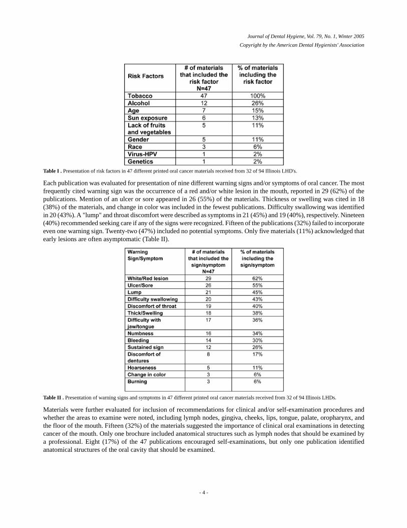
Table I . Presentation of risk factors in 47 different printed oral cancer materials received from 32 of 94 Illinois LHD's.
Each publication was evaluated for presentation of nine different warning signs and/or symptoms of oral cancer. The mostfrequently cited warning sign was the occurrence of a red and/or white lesion in the mouth, reported in 29 (62%) of thepublications. Mention of an ulcer or sore appeared in 26 (55%) of the materials. Thickness or swelling was cited in 18(38%) of the materials, and change in color was included in the fewest publications. Difficulty swallowing was identifiedin 20 (43%). A "lump" and throat discomfort were described as symptoms in 21 (45%) and 19 (40%), respectively. Nineteen(40%) recommended seeking care if any of the signs were recognized. Fifteen of the publications (32%) failed to incorporateeven one warning sign. Twenty-two (47%) included no potential symptoms. Only five materials (11%) acknowledged thatearly lesions are often asymptomatic (Table II).
Table II . Presentation of warning signs and symptoms in 47 different printed oral cancer materials received from 32 of 94 Illinois LHDs.
Materials were further evaluated for inclusion of recommendations for clinical and/or self-examination procedures andwhether the areas to examine were noted, including lymph nodes, gingiva, cheeks, lips, tongue, palate, oropharynx, andthe floor of the mouth. Fifteen (32%) of the materials suggested the importance of clinical oral examinations in detectingcancer of the mouth. Only one brochure included anatomical structures such as lymph nodes that should be examined bya professional. Eight (17%) of the 47 publications encouraged self-examinations, but only one publication identifiedanatomical structures of the oral cavity that should be examined.
Journal of Dental Hygiene, Vol. 79, No. 1, Winter 2005
Copyright by the American Dental Hygienists' Association
- 4 -
Discussion
It appears that many LHDs in Illinois lack printed materials that adequately present pertinent facts relevant to oral cancer.Because Americans are generally uninformed about risk factors, signs and symptoms, and the need for examinations, itis disconcerting that 28 (46%) of the 60 health departments which responded had no printed oral cancer materials to
distribute.4,9-14 All LHDs should have an array of current printed oral cancer materials available for distribution to thepublic and health professionals promoting oral cancer awareness and education.
Several limitations of this pilot study should be noted and resolved in subsequent studies. The survey instrument did notprovide an operational definition of printed oral cancer educational materials; thus, it failed to make an explicit distinctionbetween tobacco materials and those addressing oral cancer. This interpretation was left up to the respondent. As a result,some departments may have failed to submit materials designed primarily for tobacco education, even though oral cancerinformation was included in the materials' content.
Due to deadlines beyond the researchers' control, the study was to be completed in a six-week time period. Therefore, nosecond mailing was sent to non-respondent LHDs. The time limitation was also a reason for the evaluation of materialsby only one researcher; therefore, inter-rater reliability was not established. Having additional evaluators assess the materialswould have strengthened the reliability of obtained data. However, the materials were examined as to whether specificelements were included or missing. The degree to which the specific elements were covered was not assessed.
A variety of free materials are available through the American Cancer Society and the National Oral Health InformationClearinghouse. Many Illinois local health departments distributed the same materials, and most were designed foranti-tobacco education. Caution must be exercised, however, to avoid sending the message that oral cancer is associatedonly with tobacco use. Most tobacco materials did not provide comprehensive coverage of oral cancer nor information oncritical issues such as the need for exams to facilitate early detection.
Aside from tobacco, there was little mention of risk factors in the printed materials. Most materials primarily dealt withtobacco and alcohol and failed to discuss other risk factors. Increasing age, for example, is an important risk factor thatshould be identified in the materials. The public should be better informed about risk factors other than tobacco use thatare associated with oral cancer.
Collected materials inadequately presented warning signs and symptoms. With the current emphasis on early detection,oral cancer materials should stress the absence of symptoms and painlessness associated with early lesions. Many of thematerials listed signs and symptoms like difficulty swallowing, which is germane to advanced disease rather than earlystage lesions. The American Cancer Society Facts and Figures lists difficulty swallowing as a symptom of advanced
disease, which usually bears a poor prognosis.1 To aid in early diagnosis, oral cancer materials should stress the lack ofsigns and symptoms, as well as those associated with early oral cancer lesions.
Clinical and self-examinations are necessary, especially for those at high risk, to diagnose oral cancer in its earliest stage.High-risk patients include tobacco users, alcohol abusers, and those over the age of 40. Most of the printed materials did
not address the need for self-examinations. Izquierdo and Rozier9 discuss the tendency for clinicians to rely onpatient-reported symptoms. While not all experts agree on the utility of self-exams, many encourage them in conjunctionwith regular clinical exams. Although the efficacy of self-exams is not documented in the literature, it seems rational toperform them, particularly for high-risk individuals, when considering the asymptomatic nature of early oral lesions. Ifprinted materials address the painlessness of early oral cancers and the reduced morbidity with early detection, perhapspatients would recognize the need for clinical and self-examinations.
Healthy People 2010 5 and the Surgeon General's oral health report6 both clearly address the importance of examinations
for early detection, but the literature reveals a lack of routine oral cancer examinations by dentists and physicians alike.7,10,11,16
Silverman views performing oral cancer exams on all adult patients, as well as educating them about the need for such
examinations, as a challenge for dental professionals, but one with great merit.4 As with self- exams, if the need for clinicalexaminations were better explained in printed oral cancer materials, patients could become proactive and request oralcancer examinations from their dentists or physicians.
Journal of Dental Hygiene, Vol. 79, No. 1, Winter 2005
Copyright by the American Dental Hygienists' Association
- 5 -
Local health departments should have up-to-date printed materials for distribution. Publication dates of the collectedmaterials ranged from 1978 to 2001. Although most of the information remains accurate, some content in dated publicationsmay not be current. For example, it is accurate that difficulty swallowing is a symptom of oral cancer, but this symptom
is now recognized as one of advanced disease.1
To avoid the suggestion of negligence on the part of LHDs, one must recognize the vast array of health issues that confrontthese agencies. Faced with issues such as HIV infection, teen pregnancy, and the threat of bioterrorism, just to mention afew, it is not surprising that adequate supplies of printed materials about oral cancer may not be a top priority for theagencies. As oral health specialists, dental hygienists should assume a proactive role in partnering with LHDs to provideoral cancer materials to the public and other professionals. Volunteering to serve as oral health consultants is one way tocontribute expertise and promote oral health awareness. When current and comprehensive materials are acquired by dentalhygienists, a portion could be supplied to the local health department. It could be an ongoing project for local dentalhygiene components to periodically examine LHD educational materials to ensure that quality oral health materials areavailable for distribution to the public. Dental hygienists could also assist in the development of educational materials,ensuring appropriate information is included in the content. It is the dental hygienist's professional responsibility to promoteawareness of oral cancer and to monitor printed materials for public and professional distribution-not only in the dentaloffice, but anywhere she/he can provide expertise concerning the disease.
Additional research into materials and their availability should be conducted. The examination of printed materials in allstates could serve as a catalyst for ensuring that disseminated materials contain the most current, comprehensive, anduseful information regarding oral cancer for the entire country. Further examination of local health departments couldprovide more insight into the reasons for the lack of printed materials about oral cancer. In this study, perhaps the healthdepartments that sent materials were those with dental clinics in their facilities. All health departments should have materialsavailable because clients may visit for immunizations or other services and pick up displayed oral cancer materials. Otheroral health information could be displayed as well, encouraging patients to utilize oral health services, either at the healthdepartment or in the private sector.
Conclusions/Recommendations
The availability of oral cancer printed materials from LHDs in Illinois appears to be inadequate. Additionally, comprehensiveeducational materials must be developed for the distinct purpose of educating people about oral cancer, beyond justmentioning it in anti-tobacco materials. Risk factors, signs and symptoms, and the need for clinical and self-examinationsmust be stressed. Oral cancer has the highest mortality rate of any oral disease, but with early detection, morbidity andmortality rates improve significantly.
As oral health specialists, dental hygienists should be proactive and help agencies acquire comprehensive printed materialsfor public and professional distribution, promoting awareness and facilitating early detection of this life-threatening disease.Local dental hygiene components could develop ongoing projects in which members periodically examine printed materialsat LHDs to ensure that comprehensive, accurate materials are available to the public. Suggestions for this type of projectare included in Table III. Dental hygienists could offer to assist with all oral health materials, not just those concerningoral cancer, and collaborate with the agency for the overall oral health of its clients.
Journal of Dental Hygiene, Vol. 79, No. 1, Winter 2005
Copyright by the American Dental Hygienists' Association
- 6 -
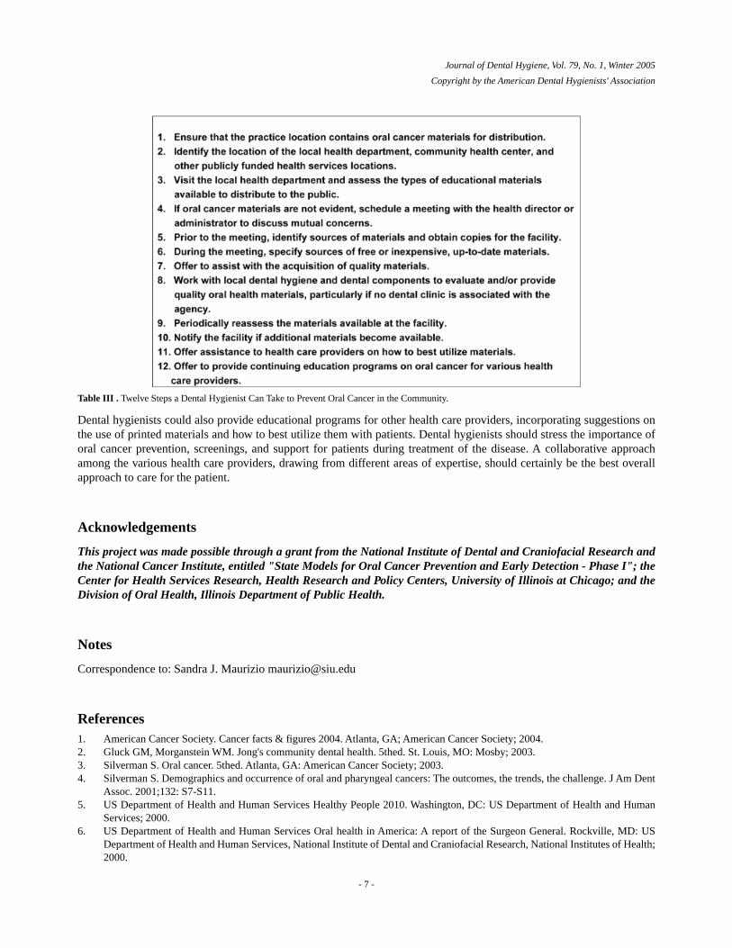
Table III . Twelve Steps a Dental Hygienist Can Take to Prevent Oral Cancer in the Community.
Dental hygienists could also provide educational programs for other health care providers, incorporating suggestions onthe use of printed materials and how to best utilize them with patients. Dental hygienists should stress the importance oforal cancer prevention, screenings, and support for patients during treatment of the disease. A collaborative approachamong the various health care providers, drawing from different areas of expertise, should certainly be the best overallapproach to care for the patient.
Acknowledgements
This project was made possible through a grant from the National Institute of Dental and Craniofacial Research andthe National Cancer Institute, entitled "State Models for Oral Cancer Prevention and Early Detection - Phase I"; theCenter for Health Services Research, Health Research and Policy Centers, University of Illinois at Chicago; and theDivision of Oral Health, Illinois Department of Public Health.
Notes
Correspondence to: Sandra J. Maurizio [email protected]
References1. American Cancer Society. Cancer facts & figures 2004. Atlanta, GA; American Cancer Society; 2004.2. Gluck GM, Morganstein WM. Jong's community dental health. 5thed. St. Louis, MO: Mosby; 2003.3. Silverman S. Oral cancer. 5thed. Atlanta, GA: American Cancer Society; 2003.4. Silverman S. Demographics and occurrence of oral and pharyngeal cancers: The outcomes, the trends, the challenge. J Am Dent
Assoc. 2001;132: S7-S11.5. US Department of Health and Human Services Healthy People 2010. Washington, DC: US Department of Health and Human
Services; 2000.6. US Department of Health and Human Services Oral health in America: A report of the Surgeon General. Rockville, MD: US
Department of Health and Human Services, National Institute of Dental and Craniofacial Research, National Institutes of Health;2000.
Journal of Dental Hygiene, Vol. 79, No. 1, Winter 2005
Copyright by the American Dental Hygienists' Association
- 7 -
7. Hay JL, Ostroff JS, Cruz GD, LeGeros RZ, Kenigsberg H, Franklin DM. Oral cancer risk perception among participants in anoral cancer screening program. Cancer Epidemiol Biomarkers Prev. 2002;11: 155-158.
8. Sciubba JJ. Oral cancer and its detection: History taking and the diagnostic phase of management. J Am Dent Assoc. 2001;132:S12-S18.
9. Izquierdo JN, Rozier RG. Oral and pharyngeal cancer research, prevention, and control: Quo vademus?. J Public Health Dent.1996;56: 305-5.
10. Horowitz AM, Nourjah PM. Factors associated with having oral cancer examinations among US adults 40 years of age or older.J Public Health Dent. 1996;56: 331-5.
11. Horowitz AM, Drury TF, Goodman HS, Yellowitz JA. Oral pharyngeal cancer prevention and early detection: Dentists' opinionsand practices. J Am Dent Assoc. 2000;131: 453-462.
12. Yellowitz JA, Horowitz AM, Drury TF, Goodman HS. Survey of U.S. dentists' knowledge and opinions about oral pharyngealcancer. J Am Dent Assoc. 2000;131: 653-661.
13. Horowitz AM, Siriphant P, Sheikh A, Child WL. Perspectives of Maryland dentists on oral cancers. J Am Dent Assoc. 2001;132:S5-S6.
14. Schnetler JFC. Oral cancer diagnosis and delays in referral. Br J Oral Maxillofac Surg. 1992;30: 200-213.15. Maguire BT, Roberts EE. Dentists' examination of the oral mucosa to detect oral cancer [Abstract] . J Public Health Dent. 1994;54:
115.16. Alfano MC, Horowitz AM. Professional and community efforts to prevent morbidity and mortality from oral cancer. J Am Dent
Assoc. 2001;132: S24-S29.17. Canto MT, Horowitz AM, Child WL. Views of oral cancer prevention and early detection: Maryland physicians. Oral Oncology.
2002;38: 373-377.18. Yellowitz JA, Goodman HS. Assessing physicians' and dentists' oral cancer knowledge, opinions and practices. J Am Dent Assoc.
1995;126: 53-56.19. Prout MN, Heeren TC, Barber CE, et.al. Use of health services before diagnosis of head and neck cancer among Boston residents.
Am J Prev Med. 1990;6: 77-83.20. Elwood JM, Gallagher RP. Factors influencing early diagnosis of cancer of the oral cavity. Can Med Assoc J. 1985;133: 651-656.21. Barker GJ, Williams KB, McCunniff MD, Barker BF. Effectiveness of an oral and pharyngeal cancer awareness program for
health professionals. J Cancer Ed. 2001;16(1): 18-23.22. Forrest JL, Horowitz AM, Shmuely Y. Dental hygienists' knowledge, opinions, and practices related to oral and pharyngeal cancer
risk assessment. J Dent Hyg. 2001;75(4): 271-281.23. Centers for Disease Control and Prevention, National Center for Health Statistics National Health Interview Survey. 1998. [as
cited in Healthy People 2010. Washington (DC): US Department of Health and Human Services; 2000.]24. Horowitz AM, Goodman HS, Yellowitz JA, Nourjah PA. The need for health promotion in oral cancer prevention and early
detection. J Public Health Dent. 1996;56(6): 319-330.25. Chung V, Horowitz AM, Canto MT, Siriphant P. Oral cancer educational materials for the general public: 1998. J Public Health
Dent. 2000;60: 49-52.26. Canto MT, Kawaguchi Y, Horowitz AM. Coverage and quality of oral cancer information in the popular press: 1987-98. J Public
Health Dent. 1998;58: 241-7.
Journal of Dental Hygiene, Vol. 79, No. 1, Winter 2005
Copyright by the American Dental Hygienists' Association
- 8 -
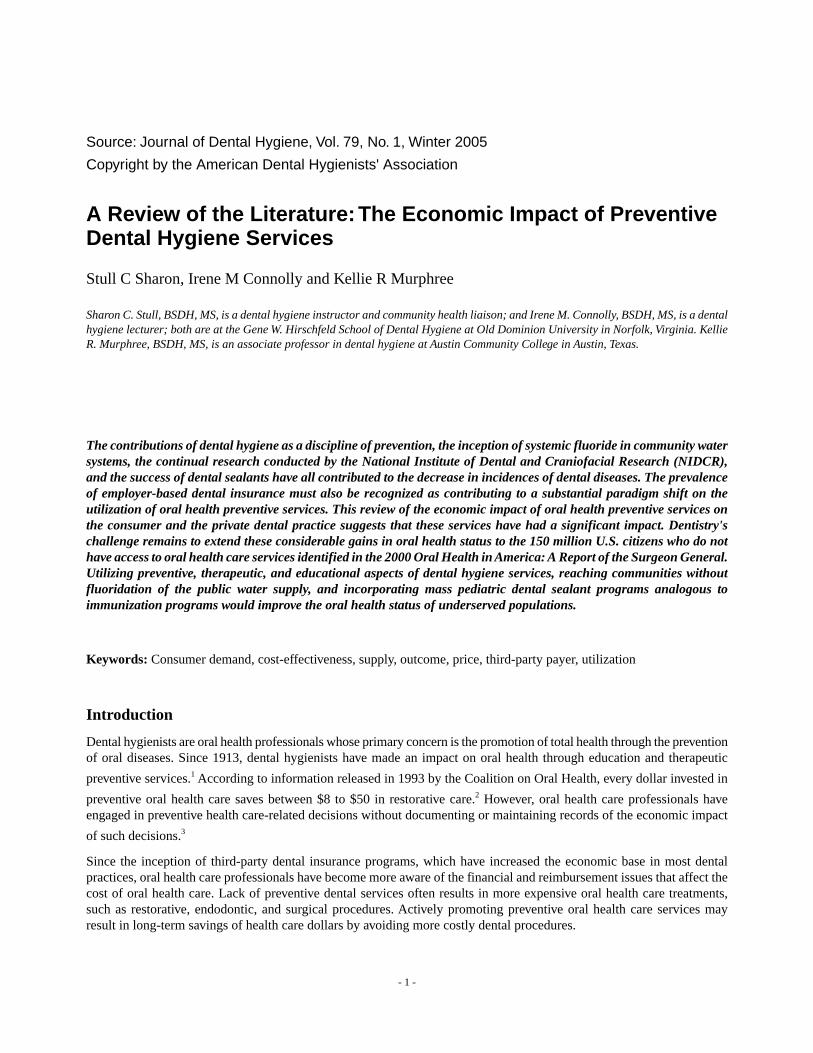
Source: Journal of Dental Hygiene, Vol. 79, No. 1, Winter 2005
Copyright by the American Dental Hygienists' Association
A Review of the Literature:The Economic Impact of PreventiveDental Hygiene Services
Stull C Sharon, Irene M Connolly and Kellie R Murphree
Sharon C. Stull, BSDH, MS, is a dental hygiene instructor and community health liaison; and Irene M. Connolly, BSDH, MS, is a dentalhygiene lecturer; both are at the Gene W. Hirschfeld School of Dental Hygiene at Old Dominion University in Norfolk, Virginia. KellieR. Murphree, BSDH, MS, is an associate professor in dental hygiene at Austin Community College in Austin, Texas.
The contributions of dental hygiene as a discipline of prevention, the inception of systemic fluoride in community watersystems, the continual research conducted by the National Institute of Dental and Craniofacial Research (NIDCR),and the success of dental sealants have all contributed to the decrease in incidences of dental diseases. The prevalenceof employer-based dental insurance must also be recognized as contributing to a substantial paradigm shift on theutilization of oral health preventive services. This review of the economic impact of oral health preventive services onthe consumer and the private dental practice suggests that these services have had a significant impact. Dentistry'schallenge remains to extend these considerable gains in oral health status to the 150 million U.S. citizens who do nothave access to oral health care services identified in the 2000 Oral Health in America: A Report of the Surgeon General.Utilizing preventive, therapeutic, and educational aspects of dental hygiene services, reaching communities withoutfluoridation of the public water supply, and incorporating mass pediatric dental sealant programs analogous toimmunization programs would improve the oral health status of underserved populations.
Keywords: Consumer demand, cost-effectiveness, supply, outcome, price, third-party payer, utilization
Introduction
Dental hygienists are oral health professionals whose primary concern is the promotion of total health through the preventionof oral diseases. Since 1913, dental hygienists have made an impact on oral health through education and therapeutic
preventive services.1 According to information released in 1993 by the Coalition on Oral Health, every dollar invested in
preventive oral health care saves between $8 to $50 in restorative care.2 However, oral health care professionals haveengaged in preventive health care-related decisions without documenting or maintaining records of the economic impact
of such decisions.3
Since the inception of third-party dental insurance programs, which have increased the economic base in most dentalpractices, oral health care professionals have become more aware of the financial and reimbursement issues that affect thecost of oral health care. Lack of preventive dental services often results in more expensive oral health care treatments,such as restorative, endodontic, and surgical procedures. Actively promoting preventive oral health care services mayresult in long-term savings of health care dollars by avoiding more costly dental procedures.
- 1 -
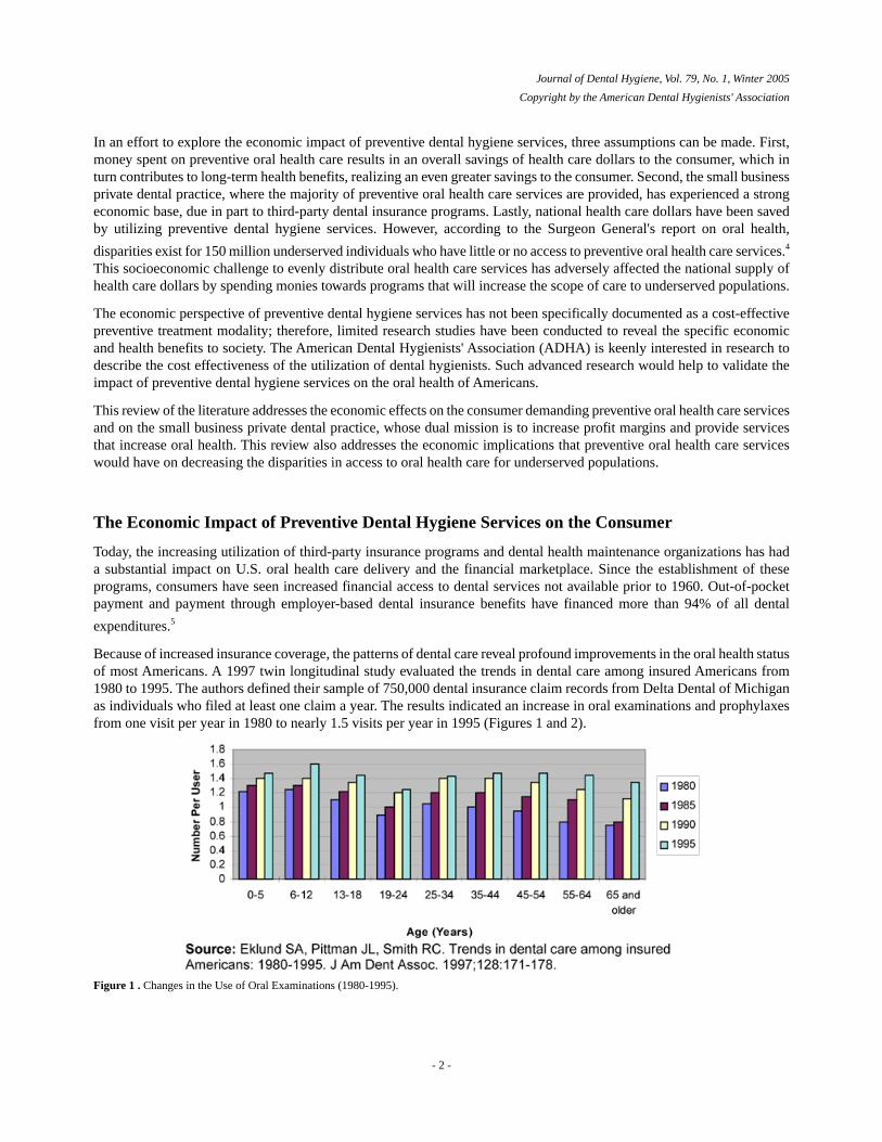
In an effort to explore the economic impact of preventive dental hygiene services, three assumptions can be made. First,money spent on preventive oral health care results in an overall savings of health care dollars to the consumer, which inturn contributes to long-term health benefits, realizing an even greater savings to the consumer. Second, the small businessprivate dental practice, where the majority of preventive oral health care services are provided, has experienced a strongeconomic base, due in part to third-party dental insurance programs. Lastly, national health care dollars have been savedby utilizing preventive dental hygiene services. However, according to the Surgeon General's report on oral health,
disparities exist for 150 million underserved individuals who have little or no access to preventive oral health care services.4
This socioeconomic challenge to evenly distribute oral health care services has adversely affected the national supply ofhealth care dollars by spending monies towards programs that will increase the scope of care to underserved populations.
The economic perspective of preventive dental hygiene services has not been specifically documented as a cost-effectivepreventive treatment modality; therefore, limited research studies have been conducted to reveal the specific economicand health benefits to society. The American Dental Hygienists' Association (ADHA) is keenly interested in research todescribe the cost effectiveness of the utilization of dental hygienists. Such advanced research would help to validate theimpact of preventive dental hygiene services on the oral health of Americans.
This review of the literature addresses the economic effects on the consumer demanding preventive oral health care servicesand on the small business private dental practice, whose dual mission is to increase profit margins and provide servicesthat increase oral health. This review also addresses the economic implications that preventive oral health care serviceswould have on decreasing the disparities in access to oral health care for underserved populations.
The Economic Impact of Preventive Dental Hygiene Services on the Consumer
Today, the increasing utilization of third-party insurance programs and dental health maintenance organizations has hada substantial impact on U.S. oral health care delivery and the financial marketplace. Since the establishment of theseprograms, consumers have seen increased financial access to dental services not available prior to 1960. Out-of-pocketpayment and payment through employer-based dental insurance benefits have financed more than 94% of all dental
expenditures.5
Because of increased insurance coverage, the patterns of dental care reveal profound improvements in the oral health statusof most Americans. A 1997 twin longitudinal study evaluated the trends in dental care among insured Americans from1980 to 1995. The authors defined their sample of 750,000 dental insurance claim records from Delta Dental of Michiganas individuals who filed at least one claim a year. The results indicated an increase in oral examinations and prophylaxesfrom one visit per year in 1980 to nearly 1.5 visits per year in 1995 (Figures 1 and 2).
Figure 1 . Changes in the Use of Oral Examinations (1980-1995).
Journal of Dental Hygiene, Vol. 79, No. 1, Winter 2005
Copyright by the American Dental Hygienists' Association
- 2 -
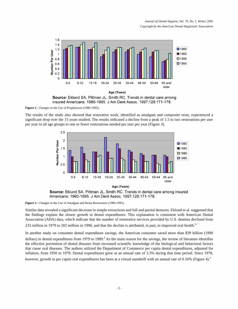
Figure 2 . Changes in the Use of Prophylaxes (1980-1995).
The results of the study also showed that restorative work, identified as amalgam and composite resin, experienced asignificant drop over the 15 years studied. The results indicated a decline from a peak of 1.5 to two restorations per userper year in all age groups to one or fewer restorations needed per user per year (Figure 3).
Figure 3 . Changes in the Use of Amalgam and Resin Restoration (1980-1995).
Similar data revealed a significant decrease in simple extractions and full and partial dentures. Eklund et al. suggested thatthe findings explain the slower growth in dental expenditures. This explanation is consistent with American DentalAssociation (ADA) data, which indicate that the number of restorative services provided by U.S. dentists declined from
233 million in 1979 to 202 million in 1990, and that the decline is attributed, in part, to improved oral health.6, 7
In another study on consumer dental expenditure savings, the American consumer saved more than $39 billion (1990
dollars) in dental expenditures from 1979 to 1989.8 As the main reason for the savings, the review of literature identifiesthe effective prevention of dental diseases from increased scientific knowledge of the biological and behavioral factorsthat cause oral diseases. The authors utilized the Department of Commerce per capita dental expenditures, adjusted forinflation, from 1950 to 1978. Dental expenditures grew at an annual rate of 3.3% during that time period. Since 1978,
however, growth in per capita real expenditures has been at a virtual standstill with an annual rate of 0.16% (Figure 4).9
Journal of Dental Hygiene, Vol. 79, No. 1, Winter 2005
Copyright by the American Dental Hygienists' Association
- 3 -
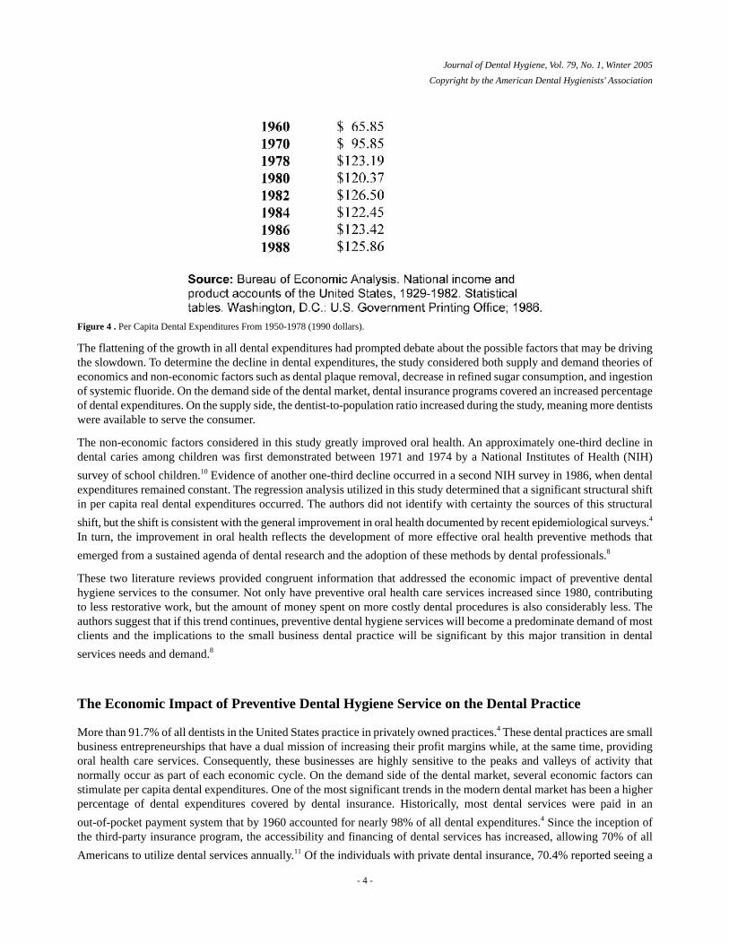
Figure 4 . Per Capita Dental Expenditures From 1950-1978 (1990 dollars).
The flattening of the growth in all dental expenditures had prompted debate about the possible factors that may be drivingthe slowdown. To determine the decline in dental expenditures, the study considered both supply and demand theories ofeconomics and non-economic factors such as dental plaque removal, decrease in refined sugar consumption, and ingestionof systemic fluoride. On the demand side of the dental market, dental insurance programs covered an increased percentageof dental expenditures. On the supply side, the dentist-to-population ratio increased during the study, meaning more dentistswere available to serve the consumer.
The non-economic factors considered in this study greatly improved oral health. An approximately one-third decline indental caries among children was first demonstrated between 1971 and 1974 by a National Institutes of Health (NIH)
survey of school children.10 Evidence of another one-third decline occurred in a second NIH survey in 1986, when dentalexpenditures remained constant. The regression analysis utilized in this study determined that a significant structural shiftin per capita real dental expenditures occurred. The authors did not identify with certainty the sources of this structural
shift, but the shift is consistent with the general improvement in oral health documented by recent epidemiological surveys.4
In turn, the improvement in oral health reflects the development of more effective oral health preventive methods that
emerged from a sustained agenda of dental research and the adoption of these methods by dental professionals.8
These two literature reviews provided congruent information that addressed the economic impact of preventive dentalhygiene services to the consumer. Not only have preventive oral health care services increased since 1980, contributingto less restorative work, but the amount of money spent on more costly dental procedures is also considerably less. Theauthors suggest that if this trend continues, preventive dental hygiene services will become a predominate demand of mostclients and the implications to the small business dental practice will be significant by this major transition in dental
services needs and demand.8
The Economic Impact of Preventive Dental Hygiene Service on the Dental Practice
More than 91.7% of all dentists in the United States practice in privately owned practices.4 These dental practices are smallbusiness entrepreneurships that have a dual mission of increasing their profit margins while, at the same time, providingoral health care services. Consequently, these businesses are highly sensitive to the peaks and valleys of activity thatnormally occur as part of each economic cycle. On the demand side of the dental market, several economic factors canstimulate per capita dental expenditures. One of the most significant trends in the modern dental market has been a higherpercentage of dental expenditures covered by dental insurance. Historically, most dental services were paid in an
out-of-pocket payment system that by 1960 accounted for nearly 98% of all dental expenditures.4 Since the inception ofthe third-party insurance program, the accessibility and financing of dental services has increased, allowing 70% of all
Americans to utilize dental services annually.11 Of the individuals with private dental insurance, 70.4% reported seeing a
Journal of Dental Hygiene, Vol. 79, No. 1, Winter 2005
Copyright by the American Dental Hygienists' Association
- 4 -
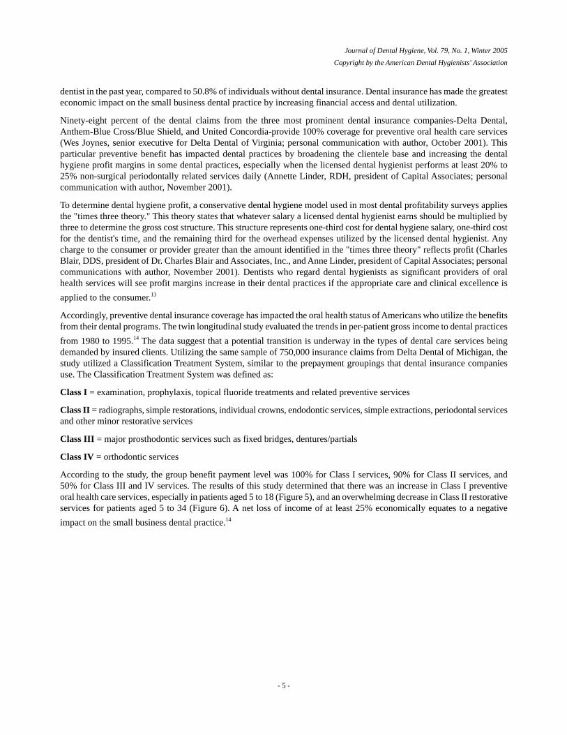
dentist in the past year, compared to 50.8% of individuals without dental insurance. Dental insurance has made the greatesteconomic impact on the small business dental practice by increasing financial access and dental utilization.
Ninety-eight percent of the dental claims from the three most prominent dental insurance companies-Delta Dental,Anthem-Blue Cross/Blue Shield, and United Concordia-provide 100% coverage for preventive oral health care services(Wes Joynes, senior executive for Delta Dental of Virginia; personal communication with author, October 2001). Thisparticular preventive benefit has impacted dental practices by broadening the clientele base and increasing the dentalhygiene profit margins in some dental practices, especially when the licensed dental hygienist performs at least 20% to25% non-surgical periodontally related services daily (Annette Linder, RDH, president of Capital Associates; personalcommunication with author, November 2001).
To determine dental hygiene profit, a conservative dental hygiene model used in most dental profitability surveys appliesthe "times three theory." This theory states that whatever salary a licensed dental hygienist earns should be multiplied bythree to determine the gross cost structure. This structure represents one-third cost for dental hygiene salary, one-third costfor the dentist's time, and the remaining third for the overhead expenses utilized by the licensed dental hygienist. Anycharge to the consumer or provider greater than the amount identified in the "times three theory" reflects profit (CharlesBlair, DDS, president of Dr. Charles Blair and Associates, Inc., and Anne Linder, president of Capital Associates; personalcommunications with author, November 2001). Dentists who regard dental hygienists as significant providers of oralhealth services will see profit margins increase in their dental practices if the appropriate care and clinical excellence is
applied to the consumer.13
Accordingly, preventive dental insurance coverage has impacted the oral health status of Americans who utilize the benefitsfrom their dental programs. The twin longitudinal study evaluated the trends in per-patient gross income to dental practices
from 1980 to 1995.14 The data suggest that a potential transition is underway in the types of dental care services beingdemanded by insured clients. Utilizing the same sample of 750,000 insurance claims from Delta Dental of Michigan, thestudy utilized a Classification Treatment System, similar to the prepayment groupings that dental insurance companiesuse. The Classification Treatment System was defined as:
Class I = examination, prophylaxis, topical fluoride treatments and related preventive services
Class II = radiographs, simple restorations, individual crowns, endodontic services, simple extractions, periodontal servicesand other minor restorative services
Class III = major prosthodontic services such as fixed bridges, dentures/partials
Class IV = orthodontic services
According to the study, the group benefit payment level was 100% for Class I services, 90% for Class II services, and50% for Class III and IV services. The results of this study determined that there was an increase in Class I preventiveoral health care services, especially in patients aged 5 to 18 (Figure 5), and an overwhelming decrease in Class II restorativeservices for patients aged 5 to 34 (Figure 6). A net loss of income of at least 25% economically equates to a negative
impact on the small business dental practice.14
Journal of Dental Hygiene, Vol. 79, No. 1, Winter 2005
Copyright by the American Dental Hygienists' Association
- 5 -
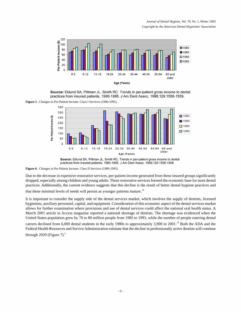
Figure 5 . Changes in Per-Patient Income: Class I Services (1980-1995).
Figure 6 . Changes in Per-Patient Income: Class II Services (1980-1995).
Due to the decrease in expensive restorative services, per-patient income generated from these insured groups significantlydropped, especially among children and young adults. These restorative services formed the economic base for most dentalpractices. Additionally, the current evidence suggests that this decline is the result of better dental hygiene practices and
that these minimal levels of needs will persist as younger patients mature.14
It is important to consider the supply side of the dental services market, which involves the supply of dentists, licensedhygienists, auxiliary personnel, capital, and equipment. Consideration of this economic aspect of the dental services marketallows for further examination where provisions and use of dental services could affect the national oral health status. AMarch 2001 article in Access magazine reported a national shortage of dentists. The shortage was evidenced when theUnited States population grew by 70 to 80 million people from 1985 to 1993, while the number of people entering dental
careers declined from 6,000 dental students in the early 1980s to approximately 3,900 in 2001.13 Both the ADA and theFederal Health Resources and Service Administration estimate that the decline in professionally active dentists will continue
through 2020 (Figure 7).5
Journal of Dental Hygiene, Vol. 79, No. 1, Winter 2005
Copyright by the American Dental Hygienists' Association
- 6 -
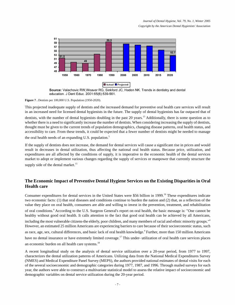
Figure 7 . Dentists per 100,000 U.S. Population (1950-2020).
This projected inadequate supply of dentists and the increased demand for preventive oral health care services will resultin an increased need for licensed dental hygienists in the future. The supply of dental hygienists has far outpaced that of
dentists, with the number of dental hygienists doubling in the past 20 years.13 Additionally, there is some question as towhether there is a need to significantly increase the number of dentists. When considering increasing the supply of dentists,thought must be given to the current trends of population demographics, changing disease patterns, oral health status, andaccessibility to care. From these trends, it could be expected that a fewer number of dentists might be needed to manage
the oral health needs of an expanding U.S. population.5
If the supply of dentists does not increase, the demand for dental services will cause a significant rise in prices and wouldresult in decreases in dental utilization, thus affecting the national oral health status. Because price, utilization, andexpenditures are all affected by the conditions of supply, it is imperative to the economic health of the dental servicesmarket to adopt or implement various changes regarding the supply of services or manpower that currently structure the
supply side of the dental market.15
The Economic Impact of Preventive Dental Hygiene Services on the Existing Disparities in OralHealth care
Consumer expenditures for dental services in the United States were $56 billion in 1999.16 These expenditures indicatetwo economic facts: (1) that oral diseases and conditions continue to burden the nation and (2) that, as a reflection of thevalue they place on oral health, consumers are able and willing to invest in the prevention, treatment, and rehabilitation
of oral conditions.4 According to the U.S. Surgeon General's report on oral health, the basic message is: "One cannot behealthy without good oral health. It calls attention to the fact that good oral health can be achieved by all Americans,
including the most vulnerable citizens-the elderly, poor children, and many members of racial and ethnic minority groups."4
However, an estimated 25 million Americans are experiencing barriers to care because of their socioeconomic status, such
as race, age, sex, cultural differences, and basic lack of oral health knowledge.4 Further, more than 150 million Americans
have no dental insurance or have extremely limited coverage.17 This under- utilization of oral health care services places
an economic burden on all health care systems.18
A recent longitudinal study on the analysis of dental service utilization over a 20-year period, from 1977 to 1997,characterizes the dental utilization patterns of Americans. Utilizing data from the National Medical Expenditures Survey(NMES) and Medical Expenditure Panel Survey (MEPS), the authors provided national estimates of dental visits for eachof the several socioeconomic and demographic categories during 1977, 1987, and 1996. Through mailed surveys for eachyear, the authors were able to construct a multivariate statistical model to assess the relative impact of socioeconomic anddemographic variables on dental service utilization during the 20-year period.
Journal of Dental Hygiene, Vol. 79, No. 1, Winter 2005
Copyright by the American Dental Hygienists' Association
- 7 -
The results indicated no significant difference in the rate of utilization during this 20-year period. Those that could accessdental services continued to do so, while the lower socioeconomic populations continued to be barred. However, by 1996a significant narrowing in the utilization rate gap was observed between whites and nonwhites. The data also showed thatthe gap in utilization rates between lower and higher income individuals widened during the 20-year period, which iscongruent with employment status. People who were employed at any time during the study year were more likely to seea dentist. The authors further suggested that policy efforts to reduce the gaps in socioeconomic and demographic disparitiesappear to have been unsuccessful. In fact, the utilization rates widened between these income groups during the studyperiod. As for the overall trend in the general population, the study found decreased dental visits per person. In conclusion,people who use dental services are more likely to have fewer visits per year on a per capita basis than they did in 1977.This may be due to fewer services needed or requested by clients. Such speculation is consistent with other studies ondental service utilization, which report an increased proportion of dental services for routine diagnostic and preventive
services only.7, 19
Although there have been gains in oral health status for the population as a whole, gains have not been evenly distributedacross sub-populations. Of the 130,836 professionally active dentists in the United States in 2000, 91.7% were in private
practice.4 Yet, an estimated 25 million individuals reside in areas lacking adequate dental care services, as defined by
Health Professional Shortage Area criteria.20 Access to private dental offices, which increases utilization, is one of the
barriers that underserved populations face, along with the high cost of dental care.12
The most effective method of defining the oral health needs of a population is a community-based approach that uses
principles of public health.21 One oral health policy study states that "oral health services must be integrated into the general
health continuum."21 Another study describes a three-tiered methodology of a prevention continuum for cost-effectivepublic health. Primary prevention is the most cost-effective level, fostering health education, disease prevention, and healthpromotion strategies (i.e. water fluoridation, dental sealants, and prophylaxis). Secondary prevention eliminates or reducesdisease in its early stages. While this stage of prevention is effective, it is also more expensive, requires more technology,and is not as efficient as primary prevention. Tertiary prevention requires extensive rehabilitation and surgical procedures,
both of which are more expensive services and require more providers training.20, 22
Of the dollars spent on health care in the United States, fewer than 5% target prevention.23 Therefore, utilizing the conceptsof Leavell, primary prevention is the most cost-effective means to reach underserved populations while producing a positiveeconomic impact on both government and individuals. Further, primary prevention strategies involve licensed dental
hygienists and do not require a dentist to perform such duties.21 Yet, current public health programs utilize less than 5%of the dental care budget that supports federal, state, and local dental public health programs, and many such programs
have experienced serious cutbacks in recent years.24
A study published by Partnership for Prevention established that statewide oral health tracking systems are effective meansto establish data about community oral health status and disease risks. Such systems would be needed to plan effectiveoral health programs for high-risk populations. For example, the North Carolina Dental Health Program utilized a trackingsystem that identified oral health needs by using public health dental hygienists to screen students in kindergarten and
fifth grade to determine and document their oral health status.25 One national health objective is for all states and the
District of Columbia to have oral health tracking systems in place by 2010, according to Healthy People 2010.25, 26
Further, the Center for Policy Alternatives documented 15 states that introduced legislation to improve access to oral
health. Of the various legislative bills, many involve licensed dental hygienists.17 The economic trend is to spend oralhealth care dollars in the most efficient way possible by spending funds on programs where the change in oral health status
will be greatest per dollar spent.15 Dental hygienists should be recognized as the major resource for attaining cost-effective
oral health policy goals.18
Journal of Dental Hygiene, Vol. 79, No. 1, Winter 2005
Copyright by the American Dental Hygienists' Association
- 8 -
Discussion
Historically, limited attention has been given to the economic impact of dental hygiene preventive services, resulting inpublic health policy that is unplanned, with little focus on disease prevention and oral health promotion. The data that wasobtainable revealed that where there is an increase in preventive dental services utilization, a significant decrease inrestorative needs exists.
The economic impact of preventive dental hygiene oral health care services on the consumer has yet to be measuredcompletely. Considering the increase in accessibility, mainly because of the impact of employer-based dental insurance,the consumer has been able to seek dental services that historically were not utilized or even valued. Dental insurance is
a major determinant of dental utilization.4 More Americans now benefit from the multitude of strategies involved inadministering oral health disease prevention services. Dental hygienists, in the majority of private practices, administerthese preventive oral health care services under the auspices of a consumer advocate, educator, and clinician.
Economically, Americans spend more for dental services than they did in the past. These expenditures indicate an increasein accessibility, and thus utilization, and they show a willingness to invest in dental care and signify the increased valuebeing placed on oral health. More Americans are dentate, free of dental diseases, and need only routine biannual oralprophylaxis rather than the more expensive restorative-based services. This marks a complete transition of dental needsover a 40-year span. With improved oral health and positive health outcomes, one cannot evaluate the true economicimpact of preventive oral health care services because of the diversity of variables affecting oral health. For these reasons,the billions saved in dental expenditures through preventive oral health care services and dental research is likely tounderestimate the full value of these activities according to the review of literature.
As small business entrepreneurs, dentists are interested in increasing their profit margins while providing oral health careservices. However, the consumer seeks less costly preventive dental care primarily because insurance benefits providetotal coverage for those services while covering only a percentage of the more expensive dental services. Dental insurancehas not only impacted the consumer through accessibility and financial means, but has also economically impacted thedental practice in two distinct relationships. First, dental insurance has increased client population, which has increasedutilization of dental services, which, in turn, has increased profit margins for the dental practice. Secondly, this increasein dental utilization has resulted in decreased demand for restorative and/or invasive dental services, thus reducing dentalexpenditures. Within this literature review, more dental expenditures are going for preventive oral health care servicesrather than for the more expensive and invasive procedures, causing the decrease in per capita income to the dental practice.
This transition from a historically restorative-based practice has resulted in a significant improvement in the oral healthstatus of clients. This positive oral health outcome trend has created more of a realignment of dental services, rather thanthe expected negative economic impact on the dental practice seen in the review of literature. Consumers with moredisposable income are seeking and demanding alternative dental services, such as cosmetic dentistry, implant technology,and adult orthodontics, as a means to continue to improve their oral and total health. Therefore, dentistry as a business haswitnessed significant profitability gains on a cumulative basis, due in part to aesthetic dental services (Charles Blair, DDS,president of Dr. Charles Blair and Associates, Inc.; personal communication with author, November 2001).
The other side to this economic analysis concerns the existing disparities in the oral health status of the underserved
populations, as noted in Oral Health in America: A Report of the Surgeon General.4 While collaboration between dentistsand dental hygienists continues to provide comprehensive oral health care to those who can afford and access care, dentistry'sethical challenge in the new millennium is to address these recognized disparities in the underserved populations whoseunmet dental needs are enormous. Public dental health programs have not been successful thus far; therefore, dentalprofessionals need to make the difference. Are dentists and licensed dental hygienists willing to break from their privatepractices to reach the underserved? Are dentists willing to address supervision laws allowing dental hygienists to practicein underserved areas without supervision? Is the profession of dental hygiene ready to address this issue? Are dentistswilling to accept more Medicaid clients? Are dentists willing to offer more Donated Dental Services. to indigent populations?These are the concerns and decisions that face dentistry today and in the future.
Genuine improvements in oral health spending and policies are only possible with a paradigm shift from curing diseaseto disease prevention and promotion of oral health. Dental hygiene, as an oral health profession, promotes total health
Journal of Dental Hygiene, Vol. 79, No. 1, Winter 2005
Copyright by the American Dental Hygienists' Association
- 9 -
through the prevention of oral diseases. However, the limitations for increasing the scope of practice for dental hygieniststo reach the low socioeconomic populations are numerous. Laws for practice restrictions of licensed dental hygienistssignificantly prevent the increase of access and utilization of diagnostic and therapeutic preventive oral health care services
outside the private practice.21 Presently, 47 states in the United States have, in various approaches, moderated direct
supervision laws for dental hygienists.27 The increase in the role of dental hygienists positioned in public health environmentspromotes renewed emphasis on setting and attaining oral health policy goals that address disparity issues for underservedpopulations. Alfred Fones, the founder of dental hygiene, indicated that the profession is distinct and should be positionedas such in dental public health. In fact, Fones believed that dental hygienists could provide education and dental hygiene
treatment outside the dental office, with particular focus on mass pediatric prevention.1
Conclusion
The economic success that consumers and small business dental practices have witnessed with preventive dental hygieneservices currently continues to improve the oral health status of Americans. With the existing disparities, however,underserved populations have yet to experience the same success and results. If the business of dentistry, as the review ofliterature states, is experiencing significant gains in practice profitability on a cumulative basis, would it not be imperativethat the profession of dentistry ethically reach the underserved populations? The impact would be equal to the strikingresults witnessed by those already served. Although the data and specific statistics are broad-based, this report may be theimpetus for more research towards attaining documented results.
Acknowledgements
Notes
Correspondence to: Sharon Stull [email protected]
References1. Fones AC. Mouth hygiene. 4thed. Philadelphia (PA): Leas & Febiger; 1934. 248.2. American Dental Hygienists' Association (ADHA) [Internet]. Access to care position paper, 2001. City (IL): ADHA; c2004.
Available at http://www.adha.org/profissues/access to_care.htm.3. Chang CF, Price SA, Pfoutz SA. Economics and nursing: critical professional issues. Philadelphia (PA): FA Davis Company;
2005.4. US Department of Health and Human Services Oral health in America: a report of the Surgeon General . Rockville (MD): US
Department of Health and Human Services. National Institute of Dental and Craniofacial Research, National Institutes of Health;2000.
5. Valachovic RW, Weaver RG, Sinkford JC, Haden NK. Trends in dentistry and dental education . J Dent Educ. 2001;65(6):539-561.
6. Nash KD, Dentley JE. Is restorative dentistry on its way out?. J Am Dent Assoc. 1991;122(9): 79-80.7. Eklund SA, Pittman JL, Smith RC. Trends in dental care among insured Americans: 1980-1995. J Am Dent Assoc. 1997;128:
171-178.8. Brown LJ, Beazoglou T, Heffley D. Estimated savings in U.S. dental expenditures, 1979-1989. Public Health Rep. 1994;109(2):
195-205.9. Bureau of Economic Analysis . National income and product accounts of the United States, 1929-1982. Statistical tables .
Washington (DC): U.S. Government Printing Office; 1986.10. Brunelle JA. Oral health of United States children: the national survey of dental caries in US school children: 1986-1987 National
and Regional Findings. Bethesda (MD): National Institutes of Health; 1989.11. U.S Department of Health and Human Services Public Health Service, National Center for Health Statistics Plan and operation
of the Third National Health and Nutrition Examination Survey (NHANES III), 1988-1994. Vital Health Statistics. 19941(32).12. Bloom B, Gift HC, Jack SS. Dental services and oral health; United States: 1989. Vital Health Statistics. 1992;10(183): 1-95.
Journal of Dental Hygiene, Vol. 79, No. 1, Winter 2005
Copyright by the American Dental Hygienists' Association
- 10 -
13. Smith C. Is dentistry coming up short?. Access. 2001;15(3): 22-7,63.14. Eklund SA, Pittman JL, Smith RC. Trends in per-patient gross income to dental practices from insured patients, 1980-1995. J Am
Dent Assoc. 1998;129: 1556-9.15. Feldstein P. Healthcare economics. 5thed. Albany (NY): Delmar; 1999. 206.16. Centers for Medicare and Medicaid Services [Internet]. Health Care Financing Review Medicare and Medicaid Statistical
Supplement, 2001. Baltimore (MD): Centers for Medicare and Medicaid Services; c2004. Available athttp://www.cms.hhs.gov/review/supp/.
17. Center for Policy Alternatives [Internet]. Oral health policy brief. Washington (DC): Center for Policy Alternatives; c2004.Available at http://www.cfpa.org/issues/issue.cfm?issue=OralHealth.xml.
18. Axelsson P. An introduction to risk prediction and preventive dentistry. Carol Stream (IL): Quintessence; 1999.19. Manski RJ, Moeller JF, Maas WR. Dental services: an analysis of utilization over 20 years. J Am Dent Assoc. 2001;132: 655-676.20. U.S. Department of Health and Human Services Response to General Accounting Office April 2000 report Oral health: dental
disease is a chronic problem among low-income populations [letter dated March 31, 2000]. Washington (DC): U.S. GeneralAccounting Office ; 2000.
21. Warren R. Oral health for all: policy for available, accessible, and acceptable care. Washington (DC): Center for Policy Alternatives;1999Sep.
22. Leavell H, Clark E. Preventive medicine for the doctor in his communities. 3rded. New York (NY): McGraw Hill; 1965.23. Warren R. Assuring oral health in the midst of disease and disability. Proceedings of the 6th World Conference on Preventive
Dentistry ; 1997 Mar 30-31; Cape Town, South Africa.24. Harris NO, Garcia-Godoy F. Primary preventive dentistry. Stamford (CT): Appleton & Lange; 1999. 426.25. Partnership for Prevention (PFP) [Internet]. Priorities in prevention: Oral health common and preventable ailments . Washington
(DC): PFP; 2001May. Available at http://www.prevent.org/priorities/PinP_0501_Oralhealth.pdf.26. U.S. Department of Health and Human Services Healthy people 2010. 2nded. Washington (DC): U.S. Government Printing Office;
2000.27. ADHA, Division Of Government Affairs. General supervision statistics. Chicago (IL): ADHA; 2003.
Journal of Dental Hygiene, Vol. 79, No. 1, Winter 2005
Copyright by the American Dental Hygienists' Association
- 11 -
Source: Journal of Dental Hygiene, Vol. 79, No. 1, Winter 2005
Copyright by the American Dental Hygienists' Association
Notices
ADHA-sponsored awards
Deadlines are approaching for the following ADHA-sponsored awards. For more information and to download applications,please visit www.adha.org.
Wrigley/ADHA Tobacco Intervention Award
March 15, 2005 deadline
Linda DeVore Leadership Scholars Program
March 31, 2005 deadline
2005 Student Presentations and Awards Program
March 31, 2005 deadline
2005 SADHA Community Service Award
April 15, 2005 deadline
Academy of Laser Dentistry
The Academy of Laser Dentistry (ALD) is now accepting registration for an introductory hands-on course in lasers at theALD 12th Annual Conference and Exhibition in New Orleans, from April 6-9, 2005. The course will be held during apre-conference session on April 5, 2005, and will provide a comprehensive overview of the clinical applications of lasersin contemporary dental practice. Advanced level courses are also available. For more information and to apply, pleasevisit www.source2005.org/certification.htm
- 1 -