The Godfather of Left-Schmittianism? Otto Kirchheimer and ...
Upload
khangminh22Category
view
0download
0
Recent Advances on Natural Products, covering all
the latest and outstanding developments in the area
Tribute to Prof. Otto R. Gottlieb
XXVII Annual Meeting on Micromolecular Evolution,
Systematics and Ecology
Reflections on the Current Status of
Chemosystematics
November 4th to 7th, 2007, Hotel Colina Verde,
São Pedro, São Paulo State, Brazil.
SPONSORED BY
Natural Product Division of the
Brazilian Chemical Society – PN-SBQ
SUPPORTED BY
FAPESP, CNPq, CAPES


Dr. M. FÁTIMA DAS G. F. DA SILVA
Universidade Federal de São Carlos Secretary-General
LOCAL ORGANIZING COMMITTEE
Dr. Dirceu Martins - Universidade Federal da Bahia Dr. Dulce H. S. Silva - Universidade Estadual Paulista Dr. Janete H. Yariwake - Universidade de São Paulo Dr. João H. G. Lago - Universidade Presbiteriana Mackenzie Dr. M. da Conceição F. de Oliveira - Universidade Federal do Ceará Dr. Maria H. Sarragioto - Universidade Estadual de Maringá
Dr. Mario G. de Carvalho - Universidade Federal Rural do Rio de Janeiro Dr. Massuo J. Kato - Universidade de São Paulo Dr. Norberto P. Lopes – Universidade de São Paulo Dr. Paulete Romoff - Universidade Presbiteriana Mackenzie Dr. Roberto G. S. Berlinck - Universidade de São Paulo Dr. Sérgio M. Nunomura - Instituto Nacional de Pesquisas da Amazônia
Dr. VANDERLAN DA S. BOLZANI Universidade Estadual Paulista, Brazil Scientific Committee Coordinator
MEMBERS
Dr. Alan Crozier - University of Glasgow, Scotland Dr. Ângelo da C. Pinto - Universidade Federal do Rio de Janeiro, Brazil Dr. Blas Lotina Hennsen - Universidad Nac. Autonoma de México, Mexico Dr. Frederico G. Cruz - Universidade Federal da Bahia, Brazil Dr. András Guttman - University of Innsbruck, Austria Dr. Jon Clardy - Harvard University, USA Dr. José M. Barbosa Filho - Universidade Federal da Paraíba, Brazil Dr. Lauro E. S. Barata - Universidade de Campinas, Brazil Dr. Leslie Gunatilaka - University of Arizona, USA
Dr. Michel Rohmer - Université Louis Pasteur, France Dr. Nidia F. Roque - Universidade Federal da Bahia, Brazil Dr. Norman G. Lewis - Washington State University, USA Dr. Paulo C. Vieira - Universidade Federal de S. Carlos, Brazil Dr. Peter Houghton – King’s College, England Dr. Robert Verpoorte - Universiteit Leiden, The Netherlands Dr. Susana A. Zacchino - Universidad Nac. de Rosario, Argentina Dr. Thomas Efferth - Cancer Research Center, Germany
ORGANIZING COMMITTEE
HONOR CHAIR
PROF. DR. OTTO R. GOTTLIEB
CHAIR PROF. DR. RAIMUNDO BRAZ FILHO Universidade Estadual do Norte Fluminense


WELCOME FROM THE ORGANIZING COMMITTEE
On behalf of the Organizing Committee, we are glad to welcome all participants in the 1st Brazilian Conference on Natural Products (1st BCNP) and the XXVII Annual Meeting on Micromolecular Evolution, Systematics and Ecology (RESEM), which have been held from November 4th to 7th , 2007 at Hotel Colina Verde in São Pedro, São Paulo State, Brazil.
The 1st BCNP will be organized by the Natural Product (PN) Division of the Brazilian Chemical Society (SBQ) at intervals of two years. The first one is a Tribute to Prof. Otto R. Gottlieb for his outstanding contributions to Natural Product. Prof. Gottlieb was born in Brno, Czechoslovakia, on August 31st, 1920, and immigrated to Brazil in 1939. Fortunately, he opted for his Brazilian nationality. Professor Gottlieb ranks as one of the most influential professionals in Organic Chemistry and the Chemistry of Natural Products in Brazil. Having developed an intense interest in the molecular diversity of the rich Brazilian flora at an early stage of his career, he went on to pioneer the introduction of Phytochemistry as a major discipline in Brazil, and he has settled several graduate courses and research programs in many Brazilian institutions. He has supervised over 120 graduate students, many of whom now hold influential positions in Brazilian universities and research organizations. He pioneered the study of the Brazilian plant biodiversity searching for chemotaxonomic markers. The establishment of an interdisciplinary program of chemobiology pursuing the rationalization of evolution, systematics and ecology of plants as a guide to the search for plant-derived bioactive substances, represented a further facet of his broad phytochemical interests. Gottlieb at an early stage of his activities on chemobiology initiated the present series of Annual Meetings on Micromolecular Evolution, Systematics and Ecology (RESEM), which now has been happening for 27 years. He continues to sponsor RESEM and now the Natural Product Division of the Brazilian Chemical Society became an important instrument for maintaining it at intervals of two years into the international BCNP.
The XXVII RESEM program is focused on “Reflections on the Current Status of Chemosystematics”. A most distinguished roll of speakers will dedicate special attention on the future directions for research on chemosystematics in Brazil.
The purpose of the 1st BCNP was to bring together renowned specialists, researchers and students from all over the world, and from all area of the Natural Products sciences, to meet, discuss, and exchange their views and experiences on topics related to “Recent Advances on Natural Products, covering all the latest and outstanding developments in the area”
The Plenary and Short Lectures have been arranged to foster studies on: Biodiversity & Natural Products; Biological and Pharmacological Activity of Natural Products; Manufacturing and Quality Control of Herbal Drugs and Essential Oils; Biosynthesis and Molecular Biology of Natural Products; Recent Advances on Isolation and Structure Elucidation of Secondary Matabolites.
The Organizing Committee has labored to provide a truly prominent slate of lecturers and excellent posters, world-class accommodations, and hospitality second to none. All they have endeavored to maximize the interaction of all the participants and speakers by having a single venue and no parallel sessions. We are happy to announce that the program has attracted over 300 participants from 5 countries.
We would like to thank CNPq, CAPES and FAPESP for their willingness to underwrite the financial obligations of this symposium, to the students who became involved in the organization of this Meeting, particularly to Márcio Santos Soares by intense dedication in the administrative activities during the several preparative phases of this event. We are also very grateful to all members of the Organizing Committee for what they have done to make this Meeting the success that we hope it will be.
Finally, we hope to all of you a good stay in Brazil, and a successful and enjoyable Meeting. Prof. Dr. Raimundo Braz Filho Prof. Dr. M. Fátima das G. F. da Silva Chair, Organizing Committee Secretary General, Organizing Committee

TABLE OF CONTENTS
RESEM – Plenary Lecture RPL-1
RESEM – Short Lectures RSL-1- RSL-5
RESEM – Oral Session ROS-1- ROS- 3
RESEM – Poster Session RPS-1- RPS-17
BCNP – Plenary Lecture BPL-1- BPL-9
BCNP – Short Lectures BSL-1- BSL-17
BCNP – Oral Session
Session A BOSA-1- BOSA-8
Session B BOSB-1- BOSB-5
Session C BOSC-1- BOSC-5
Session D BOSD-1- BOSD-5
BCNP – Poster Session BPS-1 - BPS-253

PLENARY LECTURE ABSTRACT

RPL-1
PHYLOGENY AND CHEMOGRAPHY Anders Backlund1*
1Div. of Pharmacognosy, Dept. of Medicinal Chemistry, Uppsala Biomedical Center,
Uppsala University; *presenting author
Natural compounds are evolutionary selected and pre-validated by Nature, displaying a unique chemical diversity and a corresponding diversity of biological activities. These features make them highly interesting for studies of chemical biology, and in the pharmaceutical industry for development of new leads. Of utmost importance, for a rational discovery and exploration of new biologically active compounds, are two aspects. One of these would be the identification and charting of the biologically relevant chemical space, the other a similar charting of the corresponding evolutionary space (e.g. Bohlin et al., 2007 pro parte). The first key to this is the coverage of the natural products’ chemical space. For this purpose we introduced ChemGPS-NP (Larsson et al., 2005; 2007). This new tool is tuned for handling the chemical diversity encountered in natural products research, in contrast to previous tools focused on the much more restricted drug-like chemical space. The aim is to provide a framework for making compound classification and comparison more efficient and stringent, to identify volumes of chemical space related to particular biological activities, and to track changes in chemical properties due to, for example, evolutionary traits and modifications in biosynthesis. Physical-chemical properties not directly discernible from structural data can be discovered, making selection more efficient and increasing the probability of hit generation when screening natural compounds and analogues. The second key would consequently be to explore evolutionary space, or essentially to elucidate robust phylogenies for the relevant groups of organisms under study. From this basis, reflecting the evolutionary history and hence biosynthesis development, further conclusions can be drawn (e.g. el-Seedi et al., 2005, Backlund et al., 2006). From these initial attempts, brief discussions on the intersection of chemical and evolutionary space of iridoids, betalains, and sesquiterpenes will be expanded. Backlund, A., Gottfries, J., Bohlin, L., Larsson, J. Chemography and Phylogeny - navigating chemical and evolutionary space. Planta Medica 72, p 972. – 2006. Bohlin, L., Göransson, U., Backlund, A. Modern pharmacognosy: Connecting biology and chemistry. Pure and Applied Chemistry 79, pp 763-774. – 2007. el-Seedi, H., Larsson, S., and Backlund, A. Chemosystematic value och cyclopeptide alkaloids from Heisteria nitida (Olacaceae). Biochemical Systematics and Ecology 33, pp 831-39. – 2005. Larsson, J., Gottfries, J., Bohlin, L., and Backlund, A. Expanding the ChemGPS chemical space with natural products. Journal of Natural Products 68, pp 985-991. – 2005.


SHORT LECTURES ABSTRACTS


RSL-1
MICROMOLECULAR CHARACTERS IN PLANT SYSTEMATICS: PAS T AND PRESENT
Antonio Salatino
Universidade de São Paulo, Instituto de Biociências, Departamento de Botânica. Caixa
Postal 11461, CEP 05422-970, São Paulo, SP, Brasil. [email protected]
Secondary metabolites played an important role along de period 1960-1980 as systematic characters for delimitation of angiosperm groups above family level and for the establishment of relationships among them. They were also highly relevant for the establishment of kinships among genera, species and infraspecific categories. Chemical characters were object of much debate among taxonomists, because chemical evidences often conflicted with comparative morphology. Nonetheless, the systematic relevance of several classes of secondary metabolites (tyrosine derived alkaloids, betalaines, glycosinolates, iridoids, sesquiterpene lactones, polyacetylenes) gained recognition by proponents of the main angiosperm systems of classification along the second half of the last century, although with much disagreement as to the relative importance of chemical evidence over morphology. For example, while iridoids distribution indicated affinities among a broad range of dicotyledon families, some authors preferred to recognize as taxonomically coherent only a restricted group of iridoid bearing families, while assigning a condition of convergence to the occurrence of iridoids in other taxa. The advent of molecular (macromolecular) systematics, starting in the early 80’s, led to a decline of the prestige of micromolecular characters as systematic evidence. For phylogenetic purposes, molecular evidence has turned out the most reliable tool, all other types of evidence playing a subsidiary role. Like most phenotypic characters, secondary metabolites provide limited numbers of characters for phylogenetic reconstructions, as opposed to the numerous molecular characters that can be sampled from several nuclear, plastidial and mitochondrial DNA regions. In addition, many phenotypic characters (the chemical ones included) are subject to environmental influences, which pose much caution as to their use in systematics and phylogeny. In most cases of disagreements formerly observed and derived from incongruence between chemical and morphological evidences, angiosperm molecular phylogenies have agreed with kinships suggested by micromolecule distributions. Chemical systematics remains a highly important shortcut for prospecting economically important chemicals, in particular for medicinal purposes. Quite often molecular phylogenies are used for the establishment of evolutionary trends of phenotypic characters. Hence, chemical evolution is currently determined upon a topology based on molecular evidences, rather than being an independent basis for the establishment of phylogenies. On the other hand, chemical characters are taxonomically useful towards three main objectives: 1) establishing affinities among taxa, for which no molecular evidence is available; 2) adding phenotypic sinapomorphies to branches corresponding to important clades in molecular phylogenies; 3) by means of combination with molecular markers, bringing additional support to phylogenies, especially in cases of low tree resolution or low clade support. CNPq

RSL-2
POTENCIAL DE ASTERACEAE DO BRASIL: QUÍMICA, BIOLOGI A E INFORMÁTICA
Fernando B. Da Costa
Universidade de São Paulo, Faculdade de Ciências Farmacêuticas de Ribeirão Preto,
Laboratório de Farmacognosia, Av. do Café s/no., 14040-903, Ribeirão Preto-SP, Brasil
Além de constituintes de óleos essenciais, os terpenóides e os compostos fenólicos são as classes de metabólitos secundários mais investigadas de Asteraceae brasileiras. Dentre os terpenóides, destacam-se os sesqui, di e triterpenos, enquanto que os flavonóides são os mais comuns dentre os polifenóis. O interesse nestes grupos substâncias é devido à elevada diversidade estrutural que apresentam, à sua importância para estudos quimiotaxonômicos e ao seu potencial biológico. Salienta-se que no Brasil várias Asteraceae são utilizadas como plantas medicinais, seja em produtos comerciais ou em preparados tradicionais.
Nos últimos anos, foram investigadas quimicamente várias espécies de gêneros pertencentes a grupos brasileiros e sul americanos. Foram isoladas e identificadas lactonas sesquiterpênicas, diterpenóides derivados do caurano e pimarano, além de flavonóides. O perfil químico de tricomas glandulares de algumas espécies foi estabelecido. Merecem destaque os estudos filogenético e etnofarmacológico de algumas espécies de cerrado.
Com base nos resultados destes estudos, constatou-se a elevada quimiodiversidade das espécies investigadas, tendo sido identificadas várias substâncias novas. As relações entre os perfis químicos e a classificação taxonômica de algumas espécies foi avaliada, sendo possível efetuar interessantes discussões. Através de estudos multidisciplinares, o potencial biológico de extratos brutos e de diversas substâncias foi revelado, em especial de terpenóides. Por exemplo, descobriu-se lactonas sesquiterpênicas com potente ação citotóxica, leishmanicida e antiinflamatória e diterpenos com pronunciado efeito antiespasmódico. Foram encontrados extratos brutos com atividade antiinflamatória e com efeito fagoinibidor.
Estudos computacionais realizados com diversas moléculas de Asteraceae levaram à elaboração de modelos direcionados para a coleta racional de espécies vegetais e para a análise quimiotaxonômica de tribos e de subtribos, para a atribuição automática de deslocamentos químicos de RMN 1H e predição de tempos de retenção para cromatografia líquida de fase reversa. Com base na quimiotaxonomia e na utilização de bancos de dados de estruturas, pretende-se estudar o metabolismo secundário e efetuar triagem virtual de moléculas frente a alvos biológicos.
Conclui-se que a investigação das Asteraceae brasileiras é relevante para a área de química de produtos naturais do país e uma fonte promissora de moléculas de interesse, seja para a química, biologia ou informática. Logo, estimula-se a formação de grupos de pesquisa multidisciplinares para diversificar a pesquisa das potencialidades de espécies e de moléculas desta família.
(CAPES, FAPESP, CNPq, Alexander von Humboldt Stiftung)

RSL-3
FORESTS OR ETHANOL: W HAT ARE THE CHOICES IN A SCENARIO OF CLIMATIC CHANGE?
Marcos Silveira Buckeridge
Department of Botany, Institute of Biosciences, University of São Paulo [email protected]
Plants grown under elevated [CO2] exhibit increase of their biomass due to a higher rate of photosynthesis. Although a number of results have been published with temperate tree species, relatively little is known about the physiological responses of Neotropical trees. We have recently demonstrated that seedlings of Neotropical legume trees indeed grow faster, accumulating more carbon in their bodies. Furthermore, the level of incorporation of carbon for each species was proportional to their respective life span, indicating that the rates of carbon sequestration is higher in fast growing species and lower in slow-growing species. In other words, ecological succession is probably the more efficient way to maintain constant carbon sequestration. We also performed experiments with sugarcane (a C4 plant) growing them under elevated [CO2]. We found that as photosynthesis is stimulated, near 60% more biomass is produced in elevated CO2 with slightly higher sugar concentration. The reason for obtaining higher photosynthesis is likely to be related to an increase in the electron transport genes in sugarcane leaves. This poses a dilemma: shall we plant sugarcane everywhere or regenerate/preserve forests? Before deciding to go ahead with either of the decisions, one has to bear in mind that the amount of carbon in the entire Brazilian sugarcane crop is about 8 million tons, whereas in the forests only in South America the stocks are of about 70 billion tons. Thus, regeneration or preservation alone are quantitatively much more efficient in avoiding carbon emissions to the atmosphere. It is important to remember that we have already burnt out 3 billion tons of carbon of the Amazon, which means that we could not recover the carbon lost even if we doubled sugarcane production and continue to produce it for 100 years. On the other hand, ethanol produced from sugarcane has some potential to mitigate carbon emissions. Probably the best solution would be the “game-theory” one, which is to increase cane biotechnology at the maximum possible, therefore maximizing profit, but at the same time contemplate forest preservation and regeneration, even if this means having forests strategically planted in sugarcane fields, which could limit production to some extent. This would probably turn a product that could be called Brazilian Environmentally Friendly Ethanol (BREFE) more competitive in the world market. Increasing the production of BREFE is probably one of the best ways to go for a country who flirts with the idea of being one of the environmental superpowers in the future. MCT, FAPESP, CNPq.

RSL-4
OVERVIEW OF GLOBAL ENVIRONMENTAL CHANGE
Carlos A. Nobre
Chair of IGBP SC
Instituto Nacional de Pesquisas Espaciais, Ministério da Ciência e Tecnologia, Centro de Previsão de Tempo e Estudos Climáticos. INPE - RODOVIA PRESIDENTE DUTRA KM 39
e-mail: nobre cptecinpe.br This overview will cover some of the key developments of GEC science over the last 5 years since the Amsterdam Conference in 2001. It will focus mostly on GEC at the regional scale, that is, how changes occurring at hot spots of the Planet can influence the global environment and vice-versa. The influence of the rapid rates of land use change in Amazonia on regional climate and on global biogeochemical and the impact of GEC on the sustainability of that region will be used as illustrations of scientific advancements in regional GEC science. Examples of GEC for Africa and Asia will also be presented. The overview will also dwell on the emergence of applications of Earth System Science, which challenge the scientific community and the GEC Programmes to translate dense and reliable information on global and regional environmental changes into actions toward a sustainable future for the Planet which can make a difference in the debate on the .environmental, social and economic aspects of sustainability This is particularly important for developing countries in order to resolve the apparent dilemma of sustainable management of the environment in the face of urgently needed economic growth. Special attention will be given to the requirements of Earth observatons from space for the development of Earth System Science.

RSL-5
CHEMISTRY OF NATURAL PRODUCTS: FROM SCHEELE TO GOTT LIEB
Nídia Franca Roque (Bahia Federal University)
The known organic compounds before the XVIII century were from natural origin with the exception of ether and acetic acid, both obtained from ethanol. Scheele (1842-1886) was the first chemist to develop a methodology to extract natural compounds from live organisms, mostly plants. He succeeded in obtaining acids from plants and animals, by an acid/base extraction. He also obtained sugars and glycerin. Lowitz e Kirchhoff isolated glucose and fructose and observed that they were different from sucrose. Lavoisier, working in the oxidation of sugars, found lactic acid as a product of this reaction. He also found that the relation between oxygen and hydrogen in sugars is the same that in water. In the beginning of XIX century, in France, natural product chemistry had a remarkable advance. Fixed oils, alkaloids, colorful compounds were isolated and analyzed regarding the percentage of carbon and hydrogen. In 1817, after the achievement of crystallized morphine by Seturner, the alkaloid class was recognized. It was at that time that sugars, amino acids, terpenes, flavonoids and other aromatic compounds were discovered. In 1835 thirty-five alkaloids were known. The use of alkaloids and other natural compounds in medicine as well as pigments made chemistry vary attractive. Chevreul, working with coloring material from plants isolated brasilein from Caesalpinia echimata Lam (Pau Brasil) and indigo from Indigosfera tinctoria. At that time about one hundred essential oils had been used for different purposes
In the middle of XIX century, chemistry began to move to German. After the chemical structural ideas of Kekule, van´t Hoff and other chemists the structural determination of natural products became one of the main fields in chemistry research. The way to achieve this goal was by synthesis and degradation of natural products. The work of Bredt with camphor, Willstäter with tropanic alkaloids and Wallach with monoterpenes were remarkable, but there is no doubt that Fischer was the one who developed the greatest work in this area. He elucidated the structure of the hexoses, isolated and elucidated the structure of 12 natural purins, receiving the 1902 Nobel Prize. This prize was given to 12 natural products chemists during the first half of XX century; among them, Ruzicka received the Prize in 1922 for his extensive work with terpenes and other natural products. In my opinion, the work of Semmler dealing with the structure of santalene, a tricycle sesquiterpene, in the beginning of 1900.was one of the most important in the field.
In the middle of XX century, biosyntheses was one of the important challenges of natural product chemistry. At that time, structural determination had the aid of NMR and mass spectroscopy measurements. Names such as Djerassi and Nakanish, both still alive, make great contribution in the field.
Professor Otto R.Gottlieb started with the study of essential oils when he perceived the importance of the knew techniques (NMR, MS) in the structural determination of natural products. He decided to search that knowledge and when came back to Brazil in 1964 he began to teach modern natural products chemistry all over the country. By giving lectures he taught hundreds of students about the organic chemistry of natural products. In the beginning, Gottlieb phytochemistry research was mainly in the structural determination of compounds from Amazon plants. Then, the study about the systematic, evolution and ecology of plants became more important to him. He published around 700 papers and oriented more than one hundred graduate students. He won more than 40 awards and was indicated by the Brazilian scientific society to compete to the Nobel Prize in 1999.


ORAL SESSION ABSTRACTS


ROS-1
SELF ORGANIZING MAPS AS TOOL FOR TAXONOMIC CLASSIFI CATIONS AT LOWER HIERARCHICAL LEVELS
Vicente P. Emerenciano1, Marcus T. Scotti1, Marcelo J. P. Ferreira1, Mauro V. Correia1,
Sandra A. V. Alvarenga2, Gilberto V. Rodrigues3
1 Instituto de Química, Universidade de São Paulo, PO Box 26077, 05513-970, Brazil 2Faculdade de Engenharia de Guaratinguetá, UNESP, 12516-410, Guaratinguetá, Brazil
3Departamento de Química, ICEx, Universidade Federal de Minas Gerais, Brazil The use of chemical data as auxiliary data in the classification of vegetable taxa has been employed since some decades. In these works several methods and techniques were utilized in order to use the chemical data as variables for the classification of the most varied vegetable groups. The objective of this work is to show a novel application of Self-Organizing Maps (SOM) as an alternative and efficient technique which, making use of chemical data, can also be utilized in classification of botanical taxa, i.e. chemosystematics. The SOM has cluster analysis characteristics similar to those of more classical unsupervised algorithms. One of the most important advantages of a Kohonen network1 is visualize relationships of objects, into 2-dimensional space, preserving the essential topological features of data. Another advantage, as others artificial neural networks procedures, is associated to the fact that SOM can “learn” and connect information which is extremely important in chemistry where the relationship between cause and effect has not known yet. In contrast to the traditional statistical methods, SOM are not only restricted to linear correlations or linear spaces. Consequently, they can be applied in problems involving classification, prediction and visualization of chemical data. As opposed to the multilayer neural networks, the SOM has relatively little been used in chemistry and mainly little been used in the chemistry of natural products.
The botanical group selected for this study was the Asteraceae family which constitutes a group of plants spread widely across the world, comprising about 23000 species. According to Bremer2 the family is divided into four subfamilies and 17 tribes. Over 5000 species of the family had already been chemically studied, from which ca. 7000 micromolecular compounds had been isolated at that time. In this study, the number of occurrences of each chemical class at the hierarquical level analyzed was utilized, comprising monoterpenes, sesquiterpenes, sesquiterpene lactones, diterpenes, triterpenes, flavonoids, coumarins, polyacetylenes and benzofurans isolated from 650 genera grouped according Bremer2. The choice of such metabolites is owing to a vast record of them in this family. The presence of the most representative 144 skeletal types were extracted from a database containing 38000 occurrences of compounds isolated from Asteraceae. These skeletal types were inserted in the Kohonen network as input data. The SOM was able to separate the four subfamilies of Asteraceae with a performance of 86%. Among the most studied tribes, pertaining to the Asteroideae subfamily, the unsupervisioned network classifies four tribes with high degree of recognition (80% and 70% for training and test series, respectively). Through the use of SOM phylogenetic relationships among the subfamilies and tribes of Asteraceae could be established. Thus, the method can be applied as a powerful and complementary tool in plant classification. References 1. T. Kohonen, Self-Organizing Maps, 2nd ed., Springer Verlag: Heidelberg, 1997. 2. K. Bremer, Asteraceae - Cladistics and classification, Timber Press: Oregon, 1994.

ROS-2
21
H
H
H
HO
H
HHO
H
H
H
H
HHO
3 4
CHEMOSYSTEMATICS SIGNIFICANCE OF TRITERPENES IN OLAC ACEAE
Lorena Mayara de Carvalho Cursino (IC), Cecília Verônica Nunez (PQ). E-mail: [email protected]
Coordenação de Pesquisas em Produtos Naturais, Instituto Nacional de Pesquisas da Amazônia, Av. André Araújo, 2936, Aleixo, Manaus, Amazonas. CEP 69060-001.
The Amazonian rain forest holds the highest diversity of plant in the world and among these plants Minquartia guianensis Aubl. (Olacaceae) can be found in Acre, Amazonas, Roraima, Pará and Amapá States in Brazil, where its trunk is widely used as a light post on account of all the types of bad whether it can withstand. Popularly it is known as acariquara, acariquara-roxa, acari among others1. A chemical study with the stem bark of M. guianensis collected in Ecuador yielded the minquartynoic acid, a cytotoxic polyacetylene2 and it also showed moderate in vitro activity against Plasmodium falciparum and Leishmania major3. Another chemical study with barks of M. guianensis from Equador showed the presence of the triterpenes eritrodiol and betulin and also the acetylene minquartynoic acid and the lichexanthone4. The present study was performed with leaves of M. guianensis which were extracted with dichloromethane, methanol and water three times each using ultrasound for 20 minutes. The dichloromethanic extract fractionation was made using several chromatographic techniques which allowed isolating the triterpenes: squalene (1), lupen-3-ona (2), taraxerol (3) and lupeol (4). The chemical structure identification was made based on spectral 1H and 13C Nuclear Magnetic Resonance (NMR) data and as compared to literature. In the literature available so far, triterpenes were only previously reported in two species belonging to Olacaceae family: Heisteria nitida (lupeol) and Minquartia guianensis (eritrodiol – a β-amirin derivative and betulin – a lupeol derivative) both collected in Equador, in the Amazonian rain forest. Our work corroborates that triterpenes could be used as chemical and geographic markers of these genus.
Acknowledgements: FAPEAM and PPBio/MCT. Refs. 1. J.L.C. Camargo and I.D.K. Ferraz, Informativo Técnico Rede de Sementes da Amazônia, 10 (2005). 2. R.J. Marles, N.R. Farnsworth, D.A. Neill, Journal of Natural Products, 52, 261 (1989). 3. H.B. Rasmussen, et al. Journal of Natural Products, 63, 1295 (2000); 4. H.R. El-Seedi, A.C. Hazell and K.B.G. Torssell, Phytochemistry 35, 5, 1297 (1994);

ROS-3
INHIBITION OF TOPOISOMERASE I BY PHYSALINS FROM Physalis angulata L. IN VITRO
Cavalcanti, B.C.1, Sombra, C.M.L.1, Bezerra, D.P.1, Veras, M.L.2., Pessoa, O.D.L.2, Moraes, M.O.1., Costa-Lotufo, L.V.1., Pessoa, C.1* 1Department of Physiology and Pharmacology, Health Sciences Center, Federal University of Ceará, Fortaleza, Brazil; 2Department of Chemistry Organic and Inorganic, Federal University of Ceará, Fortaleza, Brazil.
Crude extracts of Physalis angulata L. (Solanaceae) and several isolated
secondary metabolites exhibits a wide variety of biological activities such as antimicrobial, antiinflamatory, trypanocidal, and cytotoxic. The cytotoxicity of this plant has been attributed to the physalins, compounds characterized as withasteroids, a series of C28-steroidal lactones structurally based on the ergostane skeleton which are commonly produced by Solanaceae plants (Cárdenas et al., 1994). There are some evidences that physalins might be able to affect DNA synthesis (Magalhães et al., 2006), so these compounds might be a suitable candidate to cause inhibition of topoisomerase. To test whether cytotoxic properties were related to topoisomerase inhibition, this protein was evaluated in a cell-free system. Purified human topoisomerase I was incubated with physalins B, D and F (5 or 10 µg/mL) in the presence of supercoiled plasmid DNA, and the products were subjected to electrophoresis to separate closed and open circular DNA. In this case, the relaxation of DNA was inhibited in both tested concentrations for only physalins B and D. Further studies in the structure-activity relationship of physalins and their derivatives are still in progress. Support: CNPq, FUNCAP and Institute Claude Bernard.
Refs. 1. J. Cárdenas et al., Progress in chemistry of organic natural products. Springer- Verlag, vol 63 (1994). 2. H.I.F. Magalhães et al., Letters in Drug Design & Discovery, 3, 625 (2006)


POSTER SESSION ABSTRACTS


RPS-1
PRINCIPAL COMPONENTS ANALYSIS FOR DETERMINATION OF THE SEASONAL VARIATION IN THE VOLATILE OIL COMPOSITION FROM
MYRCIA MACROCARPA DC (MYRTACEAE)
Amanda de Souza1,2
, Marcus Tullius Scotti3, Maria Cláudia Marx Young2, Vicente de Paulo
Emerenciano3, Paulo Roberto H. Moreno3 1. Programa de Pós-graduação em Botânica, Instituto de Biociências, USP, São Paulo, SP, Brasil; 2. Instituto de Botânica, Seção de Fisiologia e Bioquímica de Plantas, São Paulo, SP, Brasil; 3. Instituto de Química, USP, São Paulo, SP, Brasil.
The Atlantic forest from São Paulo is characterized by a great diversity of Myrtaceae species. These species are considered as a source for essential oils with recognized pharmacological activities such as eugenol, main component from clove oil (Syzygium aromaticum (L.) Merr. & L.M.Perry). The production and the composition of essential oils may be affected by several factors such as phenology, climate, geographic localization, genetic variability and endogenous rhythms. However, studies relating the volatile oil variation in Myrtaceae with these factors are scarce. In this way, we proposed an analytical method to study the seasonal variation of the volatile oil using as model Myrcia macrocarpa DC which is native from the Atlantic Rain forest. Materials and Methods The leaves of three specimens were collected at the State Park Fontes do Ipiranga (São Paulo, SP) in the period of January 2005 to January 2006. The essential oils were obtained by hydro-distillation in a Clevenger-type apparatus for 4 h and further analyzed by gas chromatography coupled with mass spectrometer (GC/MS). The raw ion-chromatogram data, more precisely, abundance of different fragments (m/z) in function of the retention time for 21 samples, were exported in ASCII file format. The data sets were inserted into MATLAB 6.5 program to generate contour plots of the chromatograms. Mass fragments (m/z) ranges most frequently found in the different samples were selected, after visual analysis of the contour plots, submitted to “area normalization” and tested to Principal Components Analysis (PCA) using the Uncrambler v. 9.5 program. Results and Discussion The mass fragments (m/z) ranges selected and submitted to PCA were: 55, 67, 77-79, 80-84, 91-97, 105-110, 120-123, 133-135, 139, 147-160, 133-135, 139, 147-150, 160-163, 204-207. The first principal component explains to 83% of whole variance and the second only 9%, totalizing 91%. Analyzing the scorer plot, it can be noticed that there were no significant differences among individuals collected in the same month and year. However, a significant difference was verified among samples of different months and for same samples collected in the same month of different years. Conclusion PCA was an effective tool to verify seasonal and individual variations using only raw CG/MS data of the volatile oils from M. macrocarpa DC. Refs. S. Wold et al., Chemometrics and Intelligent Laboratory Systems, 2, 37 (1987)

RPS-2
USE OF BACKPROPAGATION ARTIFICIAL NEURAL NETWORKS TO PREDICT THE OCCURRENCES OF CHEMICAL CLASSES IN ASTERACEAE
Vicente P. Emerenciano1, Marcelo J. P. Ferreira1, Marcus T. Scotti1, Mauro V. Correia1, Sandra
A. V. Alvarenga2, Gilberto V. Rodrigues3
1 Instituto de Química, Universidade de São Paulo, PO Box 26077, 05513-970, Brazil
2Faculdade de Engenharia de Guaratinguetá, UNESP, 12516-410, Guaratinguetá, Brazil 3Departamento de Química, ICEx, Universidade Federal de Minas Gerais, Brazil
The Asteraceae family comprises about 23000 species, whose economical and medicinal importance has been widely described. Botanically, the family has been divided into subfamilies and tribes by several authors and, chemically this group has been extremely studied from which an enormous variety of chemical classes have been isolated: terpenoids, coumarins, flavonoids, polyacetylenes and benzofurans. Until the 90’s, about 7000 species of the family already had some type of chemical study. The chemical constituents isolated from this species (38000 occurrences) were stored in our database and the number of species a priori is considered representative for an approach by artificial neural netwoks (ANN)1. The research on phytochemistry and the search for new biologically active substances from plants has been a constant scientific activity for several decades. Usually the factors that direct this search type are ethnobotanical information coming from the popular use of plants or obtained from chemotaxonomic studies. In spite of a great number of studied species, the information on them stored so far still is scarce, due to the phytochemical study of each species is rather incomplete. Certain genera have many interesting species and are extremely studied, what generates plenty of data. A typical example in our database is the genus Artemisia, whose number of studied species/existent species is 250/390, that is 64%. However, for other genera also present in our database, this ratio may be very small, 8/120 for the genus Kaonosphyllon taken as an example, that is, only 7%. Techniques of multivariate statistics have been used to approach the existence of correlations among occurrences of chemical classes in the family but the results of this methodology still are insufficient to determine species to be analyzed accurately. As the data are somewhat imprecise, an approach using ANN may be more precise for the analysis of this type of phytochemical information. The aim of this work is to predict the presence or absence and the occurrence number of the chemical classes of natural products in genera of the Asteraceae through ANN. A backpropagation neural network was training with nine variables represeting the chemical classes of compounds (inputs data) in 400 genera. The outputs were heuristic variables scaled from 0 to 1 and representing the rare, frequent or very frequent probability to found a specific chemical class in genera of family. The ANN was able to predict the presence or absence, as well as the heuristic degree of probability of all chemical classes with 84.3%. The external set containing 100 genera was submitted to the ANN analysis. From this procedure, the ANN was able to predict with 79.1% the presence of chemical class and classify correctly the heuristic probability of occurrence. The search for potentially active compounds has been the objective of several research groups all around the world. A single heuristic information coming from the researcher that knows the database of the Asteraceae has shown itself here quite interesting as a new approach to be introduced in ANN. The results showed here demonstrated this application and also encourage us to continue the research based on data from specific families of plants using this methodology to indicate promising plants that should be studied. References
1. J. Gasteiger, E. Zupan, Angew. Chem. Int. Ed. Engl. 32, 503 (1993).

RPS-3
CHEMOTAXONOMIC RELATIONSHIPS IN CELASTRACEAE
Ana Valéria de Mello Cruz1, Marcelo J. Pena Ferreira2,3, Marcus T. Scotti2,Maria Auxiliadora C. Kaplan1, Vicente P. Emerenciano2
1 Núcleo de Pesquisas de Produtos Naturais, UFRJ, 21941-590, Rio de Janeiro, Brazil 2Instituto de Química, Universidade de São Paulo, Caixa Postal 26077, 05513-970, São Paulo 3Departamento de Ciências da Saúde, Centro Universitário Nove de Julho, São Paulo, Brazil
Celastraceae sensu lato, including Hippocrateaceae genera, comprises a group of woody
lianas, shrubs and trees with a pantropical distribution. Some species are used for medicinal purposes, as ornamentals plants and used socially as a stimulant. Various authors have considered Hippocrateaceae and Celastraceae as two distinct families but other taxonomists have been argued for the unified family. The production of micromolecules in Celastraceae sensu lato has been characterized mainly by the occurrence of terpenoids, especially dihydroagarofuran sesquiterpene polyol ester, alkaloids, friedelane triterpenoids and some unusual substances as dimmers and trimmers of sesquiterpenes, diterpenes and triterpenes. The most known biogenetic feature is the production of quinonemethides, a kind of nortriterpenoid. In order to deal with a great number of compounds and botanical informations in a chemotaxonomic study, the use of multivariate statistical methods as principal component analyses (PCA) has been implemented. This powerful tool enables to reduce the dimensionality of the data set without a significant loss of information. In this paper the PCA was employed in a chemical database containing 664 occurrences of triterpenes and used to investigate some intergeneric relationships in Celastraceae.
This study involved the tribes Cassineae, Celastreae, Euonymeae, Lophopetaleae, Hippocrateeae and Salaciae since they have a more representative chemical profile (96% of the triterpene occurrences). The triterpene’s occurrences of Celastraceae s.l. allowed making a model to explain the genera relationships. Celastraceae phylogeny inferred from 26 S nuclear ribosomal DNA genes1 was used as a base to pointed out correlations with the chemical data. In this analysis two main groups of genera could be recognized. The first group was chemically characterized by no production of the 26(14→15)-abeo-24-D:A-friedo-nor-oleanane and low production of the lupane skeleton. The second group showed the production of all celastraceous skeletal types of triterpenes.
Comparison of PCA and cladistic analysis showed Cassine s.s distinct from Elaeodendron. Such distinction has also been supported by differences in pollen, bark and wood anatomy2. Cassine s.s. produces quinonamethide triterpenoids derived from the 24,29–D:A–friedo–dinor–oleanane while Elaeodendron does not. Maytenus s.l. (including Gymnosporia and Tricerma) is not resolved as a natural group1. Our results support the distinction of Gymnosporia from Maytenus once the later produces high diversity of substances and skeleton types while Gymnosporia appear to have a tenuous concentration in the oleanane skeleton production. PCA analysis for tribes shows that all celastraceous tribes are very similar on the chemical point of view. This fact corroborates with the idea of a unified family including Hippocrateaceae genera within Celastraceae1. References 1. M. P. Simmons et al., Molecular Phylogenetics and Evolution, 19, 353 (2001). 2. R. H. Archer et al., South African Journal of Botany, 63, 146 (1997).

RPS-4
CHEMOSYSTEMATICS OF ASTERACEAE TRIBES USING PRINCIP AL COMPONENT ANALYSIS
Vicente P. Emerenciano1, Marcus T. Scotti1, Marcelo J. P. Ferreira1, Mauro V. Correia1,
Sandra A. V. Alvarenga2, Gilberto V. Rodrigues3
1 Instituto de Química, Universidade de São Paulo, PO Box 26077, 05513-970, Brazil 2Faculdade de Engenharia de Guaratinguetá, UNESP, 12516-410, Guaratinguetá, Brazil
3Departamento de Química, ICEx, Universidade Federal de Minas Gerais, Brazil
The Asteraceae family has been intensely studied by researchers of the diverse areas, such as chemists, pharmaceutics, botanicals, etc due its large chemical and morphologic diversity. Several classifications systems intend to cluster the genera in tribe according to morphological, macromolecular and micromolecular criteria. From the point of view of the tribes, since Reading’s Symposium, several authors attribute different interpretations for groups which classification is still not clear, for example the tribes Mutiseae, Helenieae, Heliantheae and others1. The use of chemical data as auxiliary data in the classification of Asteraceae taxa has been employed in the last decades. In these papers several methods and techniques were used in order to utilize the chemical data as variables for the classification of the most varied sufamilies, tribes and subtribes of the family. The aim of this paper is to use chemical data related to isolated secondary metabolites of Asteraceae, which are available in our database containing all chemical information about this family, to compare them with the Wagenitz and Bremer’s classification employing principal component analysis (PCA). The power of PCA analysis for discriminate groups in phenetic studies were already published. The principal advantage of this method is the possibility of to work with several variables when they are highly correlated. This methodology avoids the redundance of the multivariate analysis and reduces the dimensionality of the data without of information prejudice. In some very advanced taxons the diversity of chemical classes and / or skeletal types makes impossible the inspection of the data in graphics without the dimensionality reduction. Thus, the PCA was applied to Asteraceae database containing 38000 occurrences of compounds isolated from their species. The chemical classes used in this study comprising monoterpenes, sesquiterpenes, sesquiterpene lactones, diterpenes, triterpenes, flavonoids, coumarins, polyacetylenes and benzofurans. The results were analyzed according to the Bremer and Wagnetiz classification and show a great similarity with both classification systems. The separation of the Cichorioideae and Asteroideae subfamilies could be evidencied from chemical data once the production of secondary metabolites is less expressive in the Cichorioideae. This observation is in agreement with the botanical classification that rankes this group as the more basal in Asteraceae. The very small number of isolated compounds of Barnadesieae tribe was the responsable for its position as an outlier in the PCA analysis. Therefore, the primitive position within the family based on DNA data appears to be confirmed by the chemical data. In the other hand, the separation of the cluster formed by the Inuleae s.l. is not well confirmed by the chemical data. The use of PCA in the taxonomic studies could be compared with the two types of classification obtained to Asteraceae family and some correlations between the chemical and botanical data were obtained. However, some doubts remain about the evolution of this family and we considered that the introduction of the one or more chemical descriptors could be useful to improve the discussion about the evolution of the groups within the family.
References 1. K. Bremer, Asteraceae - Cladistics and classification, Timber Press: Oregon, 1994.

RPS-5
NEOLIGNANS OF Talauma ovata AND THEIR CHEMOTAXONOMIC SIGNIFICANCE
Maria Élida Alves Stefanello, Letícia Ferrari Lemos Barros and Andersson Barison
Departamento de Química – Universidade Federal do Paraná
Magnolia and Talauma are the most important genera in Magnoliaceae. Several phytochemical studies have been carried on Magnolia while Talauma has received little attention. The present work describes the results of phytochemical study of leaves of T. ovata. Chromatographic fractionation of the dichloromethane extract yielded three known benzofuranoid neolignans. The compounds were identified by NMR 1D and 2D as well as comparison with reported data as acuminatin, licarin A and 7-(4-hydroxy-3-methoxyphenyl)-1’-trans-propenal-3’-methoxy-8-methyldihydrobenzofuran. The last one has been previously obtained only by synthesis1. Neolignans of this type are been reported for the first time in Talauma, however are common in Magnolia. These results support the proposal of reducing Talauma to a sub-genus of Magnolia. Refs. 1. K. Kuroda et al., Organic Geochemistry, 37, 665 (2006)

RPS-6
CHEMOTAXONOMIC STUDY OF Eugenia AND Myrcia (MYRTACEAE)
Maria Élida Alves Stefanello and Iara Messerschmidt Departamento de Química – Universidade Federal do Paraná
The taxonomic classification in Myrtaceae is difficult and chemical data could be useful to investigate the delimitation of botanical groups. With this aim the composition of essential oils of Brazilian Eugenia and Myrcia was analyzed statistically. Chemical data was getting through review in the literatura. These data was analyzed by the Principal Component Analysis (PCA) and Cluster Analysis. Twelve variables, corresponding to monoterpenes, acyclic sesquiterpenes and ten cyclic skeletons of sesquiterpenes were used. In Eugenia, the first component was positively determined by the skeletons germacrane, bicyclogermacrane and elemane and the second component by content of monoterpenes and skeleton caryophyllane. The PCA did not result in a clear grouping of species. In Myrcia, the first component was determined by the skeletons humulane and bisabolane and the second component by eudesmane and aromadendrane. The PCA resulted in two near groups, showing that Myrcia is a more homogenous group than Eugenia. The PCA of Myrcia and Eugenia together showed Myrcia as a group immerse in Eugenia.

RPS-7
EVOLUTIONARY TENDENCIES IN SWARTZIA GENUS: BIOGENETIC PROPOSAL FOR SWARTZIARBOREOLS
Celira Caparica Santos1 , Ana Maria G. A. Tozzi2, Eva G. Magalhães1and
Aderbal Farias Magalhães1
1 Depto de Produtos Naturais, Inst. de Química, 13084-862, [email protected], UNICAMP, 2 Depto de Botânica, Inst. de Biologia, 13.083-971, [email protected], UNICAMP, Campinas-SP.
This work gives continuation to the chemiosystematic study of plants from the Swartzia genus regarding evolutionary tendencies [1], by taking into account the disparity and the diversity of specific metabolites isolated and identified in them [2].
The biogenetic substitution of the compounds originated from the shikimate route by the compounds originated from the acetate route can be taken as an evolutionary tendency since the later ones are found in more recent angiosperms [2].
This evolution can be observed in the plants of Swartzia genus since S. polyphylla, S. ulei, S. leiocalycina and S. laevicarpa produce isoflavonoids and S. arborescens and S. langsdorffii produce abieta-8,11,13-triene diterpenoid derivatives named swartziarboreols [3].
In order to explain the biodiversity of isolated swartziarboreols, it is reported the proposal of a biogenetic pathway based in that reported for ferruginol [4], including a pair of epimers (1, 1’, 2 and 2’), which were recently detected [5].
O
O
CH3O
OCH3
A B
C
1, 1' = (A/B cis, A/B trans)2, 2 ' = ∆ 15,16; (A/B cis, A/B trans)
15
16
References:
[1] A. F. Magalhães, et al., Eclet. Quím. , 31 (2), 13 (2006) . [2] O. R. Gottlieb, et al., 1996. Editora UFRJ, Rio de Janeiro. “Biodiversidade: Um Enfoque Químico-Biológico”. [3] A. F. Magalhães, et al., J. Nat. Prod. 68 (8), 1290 (2005). [4] Y. Tomita, et al., J.Chem. Soc., Chem. Commun., 108 (1989). . [5] C. C. Santos, et al., Phytoch. Analysis, (2007).

RPS-8
CHEMICAL COMPOSITION, ANTIOXIDANT AND ANTIMICROBIAL ACTIVITY OF A SAMPLE OF BRAZILIAN RED PROPOLIS FROM MACEIÓ ( AL)
Adne Abbud Righi1, Thiago Renée Alves1, Giuseppina Negri2, Antonio Salatino1 1Universidade de São Paulo, Instituto de biociências, Departamento de Botanica, C. Postal 11461, 05422-970, São Paulo, SP, Brazil. 2CEBRID - Departamento de Psicobiologia, UNIFESP, R. Botucatu, 862 – Ed. Ciências Biomédicas – 1º andar, CEP 04023-062, São Paulo, SP, Brazil.
Propolis is a complex resinous substance used by bees to seal their hives. It is gathered from plants modified by the bees to produce the finished material. A range of medicinal properties have been attributed to propolis, antimicrobial, anti-hepatotoxic, anti-inflammatory, immunostimulant, anti-oxidative, anti-cancer properties, among others. Propolis may consist of flavonoid aglycones, phenolic acids and their esters, phenolic aldehydes, sesquiterpenes, quinones, coumarins, steroids, amino acids, sugars, proteins and inorganic compounds. The exact composition of propolis varies according to its variety and geographical source. The most often commercialized propolis type in Brazil is green propolis, which has plant material collected by bees from Baccharis dracucculifolia. A new type of propolis from Maceió (AL), namely red propolis, has been shown to derive from Dalbergia ecastophyllum and to have composition distinct from other known propolis types. The purpose of this research was to determine the total contents of phenolic compounds and total flavonoids and identify individual constituents of a sample of Maceió red propolis, and to evaluate its antimicrobial and anti-oxidative activities. Total phenols were determined by the Folin–Ciocalteu method and total flavonoids were determined by the AlCl3 method. Identification of constituents was based on HPLC/ESIMS and GC/EIMS analyses. Antimicrobial activities determined using the macrodilution method in broth with the folowing microorganisms Pseudomonas aeruginosa, Baccillus subtilis, Candida albicans, Salmonella typhimurium, Klebsiella pneumoniae, Enterococcus faecalis, Escherichia coli, Proteus mirabilis and Streptococcus pyogenes. Antioxidant activities were evaluated by DPPH and β-caroten/linoleic acid methods. Methanolic extracts exhibited 24,13% of total phenols and 3,55% of total flavonoids. Mains constituents identified were the isoflavonoids 2’-hydroxy-4’,7-dimethoxyisoflavan, 7,2’-dihydroxy-4’-methoxyisoflavan (vestitol) and 2’,4’,7-trihydroxyisoflavanone. Other constituents found were the phenylpropanoids trans-anethol, methyleugenol, elimicin, methoxyeugenol and cis-asarone, and the triterpenic alcohols α- and β-amyrins and lupeol. Methanolic extracts (2,0 mg/mL) exhibited 84,5% of antioxidant activity relative rutin by the β-caroten/linoleic acid method, while 25 µg/mL methanolic extracts had 39,1% of antioxidant activity relative rutin by the DPPH method. Methanolic extracts inhibited growth of all microorganisms tested. FAPESP, CNPq

RPS-9
Coumarins from Euxylophora paraensis and their Chemosystematics Significance
Marsele Machado Isidoro (PG), Sebastião da Cruz Silva (PG), Maria Fátima das Graças Fernandes da Silva, João Batista Fernandes, Paulo Cezar Vieira,
Departamento de Química, Universidade Federal de São Carlos, Caixa Postal 676, 13565-
905 São Carlos, SP, Brazil; [email protected]
Euxylophora paraensis Hub. (Rutaceae) is a South American tree which is sometimes used in the United States as 'Brazilian Satinwood' for the manufacture of fine furniture [1]. It is known to contain a series of rare indolopyridoquinazoline alkaloids related to 1-hydroxyrutaecarpine, 2-quinolone N-methylflindersine, 4-quinolones spectabiline, furoquinoline skimmianine and two 4-hydroxy-2,2,6-trimethyl-3,4,5,6-tetrahydro-2H-pyrano[3,2-c]quinolin-5-one and 4-(2,3-dihydroxy-3-methy1butoxy)-1-methylquinolin-2(1H)-one, dimeric quinolinone alkaloids, as (6aα,7α,14aα)-6,6,9,16-tetramethyl-7-(2-methylprop-l-enyl)-6,6a,7,9,14a,l6-hexahydro-H,15H -quino[3",4":5',6']pyrano [2',3':4,5] pyrano-[3,2-c]quinoline-8,15-dione [2].
Our taxonomic interest in the Rutaceae, stimulated an investigation of the leaves and stem of E. paraensis. Stem afforded N-methylflindersine, limonoid limonin and two coumarins marmesin (1) and 8-methoxymarmesin (2) [3]. The extracts from leaves afforded three coumarins pimpinellin (3), 8-methoxypsoralen (4), lanatin (5) and the furoquinolin alkaloid γ-fagarin. Coumarin 5 appears to be new as natural product, it was found at literature as synthetic derivative [x]. This is the first record of the five latter coumarins from Euxylophora. The results provide firm support for including this genus in the Rutaceae.
O O O
O
OO
OCH3
OO O
OCH3
CH3O
O
OOHO
O
OMe
OOHO
O
1 2
34 5
1. DANIELI, B; PALMISANO, G; RAINOLDI, G; RUSSO. Hidroxy Rutaecarpine from Euxilophora Paraensis. Phitochemistry (Elsevier) (1974), 13(8), 1603-6. 2. JURD, LEONARD; WONG, ROSALIND Y. New quinolinone Alkaloids from the heartwood of Euxilophora Paraensis. Australian Journal of Chemistry (1981), 34(8), 1625-32.
3. MARINHO, P. S. B. Estudo Químico das Espécies Khaya ivorensis (MELIACEAE) e Euxilophora paraensis (RUTACEAE). São Carlos, Programa de Pós-Graduação em Química – UFSCar, Tese de Doutorado, 2007, 200p. 4. BANERJEE, K; GUPTA, D; ATAL, C. K; Coumarins of Heracleum thomsoni and Claisen rearrangement of lanatin. Phitochemistry (Elsevier) (1980), 19(6), 1256-8.

RPS-10
ANTHRANILATE ALKALOIDS, COUMARIN AND LIMONOIDS FROM NYCTICALANTHUS SPECIOSUS AND THEIR CHEMOSYSTEMATICS
SIGNIFICANCE
Suzana Thiemy Farias1 (IC), Karine Valadares Guimarães Reche2* (PG), , Maria Fátima das G. F. da Silva2 (PQ), João B. Fernandes2 (PQ), Paulo C. Vieira2 (PQ).
1Centro Universitário Paulista (UNICEP), São Carlos-SP, 2 Universidade Federal de São Carlos, Departamento de Química, Laboratório de Produtos Naturais, Rod. Washington Luiz
Km 235, São Carlos-S.P. *[email protected]; [email protected]
Nycticalanthus speciosus Ducke is a tree found only in north of Brazil, Manaus,
Amazonas [1]. No previous phytochemical work has been reported on Nycticalanthus. Recently, we have described the isolation and identification of six known alkaloids dictamine, γ-fagarine, skimianine, 1-methyl-4-methoxyquinolin-2-one, N-methylflindersin and rutaecarpin, the new alkaloid 8,8a-seco-8-carboxy-14-N-methylrutaecarpin, the coumarin seselin, and three new limonoids methyl 2-hydroxy-epiisoobacunoate (1), methyl 2,21-dihydroxy-23-oxoepiisoobacunoate (2) and 7-acetoxy-21-hydroxy-23-oxotecleanine (3) [2]. Thus, our taxonomic interest in the Rutaceae stimulated an investigation of other plant organ of N. speciosus. The extracts from the stem bark afforded the limonoids 1, methyl 2-acetoxy-21-hydroxy-23-oxoepiisoobacunoate (4), two alkaloids 4-methoxyquinolin-2-one (5), dictamine (6) and the coumarin 5-methoxyseselin (7).
Seção 1.01 The results provide firm support for including the genus Nycticalanthu in the Rutaceae.
1. J. R. Pirani, Rodriguésia, 56(86), 189-204 (2005). 2. Alan Bezerra Ribeiro. Fitoquímica do enxerto de Citrus sinensis sobre C. limonia e da espécie Nycticalanthus speciosus (Rutaceae) e biossíntese de cumarinas preniladas. Tese de doutorado. UFSCar. São Carlos 2006. p.257.
O
O
H
AcO
H3CO O
HO
O
O
O
O
OH
4
5
N
OCH3
CH3
O
6
N O
OCH3
7
OO O
OCH31
O
O
O
OH
O
OH3CO
HO
H
O
3
O
OCCH3
O
O
OHO
O
O
H
O
O
H3CO
O
O
O
O
HO O
H
H
OH
O
2

RPS-11
CHEMOSYSTEMATIC OF CUSPARIA, ANGOSTURA, CONCHOCARPUS AND GALIPEA (RUTACEAE)
Barbara Sayuri Bellete, M. Fátima das G. F. da Silva, João B. Fernandes and Paulo C. Vieira. Departamento de Química, Universidade Federal de São Carlos, CP 676, 13565-905 São Carlos - SP, Brazil
Saint-Hilaire (1823, 1824, 1825) cited Cusparia, Angostura, and Conchocarpus
among the synonyms of Galipea Aubl., and described several new species in Galipea and two in Ticorea Aubl. Those he described in Galipea belong in Conchocarpus, and those he described in Ticorea belong in Galipea. His erroneous circumscriptions of these genera were perpetuated by de Candolle (1824), Jussieu (1825), and Bentham & Hooker (1862). Engler, in his treatments of the Rutaceae in Martius’s “Flora Brasiliensis” (1874) and in “Die Natürlichen Pflanzenfamilien” (1986, 1931), defined these genera somewhat more clearly. He limited Ticorea to species with five fertile stamens by transferring to Galipea the two species (with two fertile stamens) described by Saint-Hilaire. He limited Galipea to species with connate carpels by excluding from it the apocarpus species, i.e., the species described by Saint-Hilaire (which, as noted above, belong in Conchocarpus) and the type of Angostura, which he called Cusparia trifoliate (Willd.) Engl. He recognized these as one genus, Cusparia, still confounding Angostura and Conchocarpus. He also failed to realize that his Galipea bracteata (Nees & Mart.) Engl. was congeneric with his C. trifoliate. These errors were rectified by J. A. Kallunki & J. R. Pirani (1998) recently. Angostura Roem. & Schult. as understood by Engler was defined more narrowly by J. A. Kallunki & J. R. Pirani. The species excluded from Angostura were recognized as species of Conchocarpus J. C. Mikan. Three new species of Angostura (A. alipes Kallunki, A. quinquefolia Kallunki, A. simplex Kallunki) were described, and three new combinations in this genus were made [A. bracteata (Nees ξ Mart.) Kallunki, A. granulosa (Kallunki) Kallunki, A. longiflora (K. Krause) Kallunki]. They recognized seven species of Angostura, the six latter and its type A. trifoliata (Willd.) T. S. Elias. The major source of both 2-alkyl and 2-arylquinolines are these genera. The co-occurrence of different structural types of quinoline and acridone alkaloids suggests affinity between these genera and supported J. A. Kallunki & J. R. Pirani taxonomic conclusions (1998).
J. A. Kallunki & J. R. Pirani, Synopses of Angostura Roem. & Schult. and Conchocarpus J. C. Mikan (Rutaceae). Kew Bulletin Vol. 53 (2), (1998).

RPS-12
TRITERPENES AND STEROLS PROFILE IN HEXANE EXTRACTS FROM Casearia SPECIES USING GAS CHROMATOGRAPHY
Gerardo Magela Vieira Júnior, Luana Caleffi and Alberto José Cavalheiro
UNESP-São Paulo State University, Chemistry Institute, NuBBE- Núcleo de Bioensaios,
Biossíntese e Ecofisiologia de Produtos Naturais, 14800-900, Araraquara, SP, Brazil.
The increasing interest in the use of medicinal herbs requires consistent and fast methods for the identification of the phytochemical constituents and for the quality control of phytotherapics. In the case of non-polar extracts, the biological and pharmacological activities were often believed to be due to a number of triterpenoid and steroid compounds, such as α-and β-amyrins, lupeol, betulin and betulinic acid1,2. As part of our ongoing research on bioactive compounds from Brazilian plants for the treatment of tropical diseases, we have investigated species of Casearia, a genus belonging to the Flacourtiaceae family, with approximately 1300 known species3. In this work, we have employed gas chromatography (GC-FID) to investigate the hexane extracts composition of four Casearia species (C. gossypiosperma, C. obliquoa, C. decandra, and C. rupestris). The analysis were carried out using the Crevelin et al4 modified method, which is rapid and simple, and does not require pre-derivatization of the crude botanical extract. The extracts (10 mg – leaves and twig) were dissolved in chloroform (3 mL) and submitted to solid phase extraction on silica gel (200 mg), eluted with chloroform (10 mL). The fraction obtained was dried, dissolved again in chloroform (1mg/mL) and analyzed by GC-FID in duplicate. All the hexane extracts were analyzed by GC-FID on a Varian model CP-3800 gas chromatograph, with temperature of injector and of flame ionization detector adjusted to 260 °C and 290 °C, respectively. The injected volume was 2.0 µL. SPB-50 (cross-linked 50% phenyl-methyl-silicone, 30 m × 0.25 mm × 0.25 µm) capillary column was employed and the column temperature was 280 °C (isotherm). Triterpenes and sterols were identified by comparison of the relative retention (RR) of the samples with the RR of the standard sterols and triterpenes. Cholesterol was used as the internal standard. The results show that the steroid β-sitosterol is present in all the analyzed extracts. Stigmasterol and campesterol were detected only in the leaves and twigs extracts of C. gossypiosperma. The triterpenes α-amyrin and β-amyrin were only detected in C. gossypiosperma (twigs) and C. rupestris (leaves), respectively, while the lupeol acetate was detected in twigs of C. gossypiosperma. These results clearly demonstrate that the chemical profiles of the four Casearia species concerning sterols are very similar, just the species C. gossypiosperma and C. rupestris presented small differences in the steroidal and triterpenic composition. In summary, we have used a simple, rapid, and relatively cheap method to identify triterpenes and sterols from hexane extracts of Casearia species using GC-FID. Refs. 1. F. A. Oliveira et al., Pharm. Biochem. Behav., 78,: 719 (2004). 2. J. Patoca, Journal of Applied Biomedicine, 1, 7 (2003). 3. H. O. Sleumer,. Flacourtiaceae. Flora Neotropica, 22,. 1-499, (1980). 4. E. J. Crevelin et al., Biomed. Chromatogr., 20, 827 (2006).

RPS-13
Chemical Characterization of Azadirachta indica grafted on Melia azedarach and Analyses of Azadirachtin A by HPLC-MS-MS (SRM)
Moacir Rossi Forim (PG), Vivian Estevam Cornélio (IC), M. Fátima das G. F. da Silva(PG),
Edson Rodrigues-Filho (PG), João B. Fernandes (PG), Paulo C. Vieira (PG) Departamento de Química, Universidade Federal de São Carlos, CP 676, 13565-905 São Carlos - SP, Brazil
The Neem tree is well adapted to hot, dry climates where shade temperatures often reach
50 degrees Celsius and annual rainfall ranges from 400 to 1,200 millimeters. It has successfully been introduced in northern, northeastern and western regions of Brazil. Chinaberry tree is native to Asia and was widely introduced in Brazil as an ornamental plant. These trees have a better potential to survive low temperatures than Neem trees. The latter has been grafted to stems of M. azedarach and the tree has been adapted well in cool climates. These grafts growing in southern regions of Brazil are part of breeding and selection program of Agronomic Institute of Paraná (Brazil) aimed at development vigorous Neem trees to be established in more cool regions. This is the first time that the graft of A. indica on M. azedarach rootstock has been investigated. Thus, roots, stems, leaves and seeds of this graft were examined in order to determine if secondary metabolites present in the rootstock could be translocated into the A. indica. Extraction of Azadirachtin A from all parts of the Neem tree has been described in the inaccessible Japanese patent literature (Kanaki, 2007), but it has been frequently reported to be present at highest concentration in the mature seeds (Akhila et al, 1999). However, the concentrations of azadirachtin A may vary due to fluctuating contents in the natural sources, or susceptibility of the compounds to environmental influences as heat, light, etc., or when the tree budded on a second one as Melia azedarach as a rootstock. High-performance liquid chromatography coupled with tanden mass spectrometry (HPLC-MS/MS) was used to develop a rapid and sensitive method for detecting azadirachtin A on all aerial parts of this graft. As expected, azadirachtin A was nearly in equal proportion in fresh juvenile, matured and fully matured seed kernels, dry fully matured seed kernels. It was present in smaller proportion in fresh immature fruits, in general the content of azadirachtin A increase with growth of the fruit. On the other hand, peak area obtained in the SRM chromatogram of dry seed coat cannot be directly compared, since large differences (13.0<<20.0 mg.mL-1) in concentration make comparisons invalid. The analysis of dry stem without bark indicated a notable concentration of azadiractin A, but it appeared with a significant decrease in dry stem bark and flowers. The limit of detection of azadirachtin A was 25 ng.mL-1. It means that the proposed method should have the ability to detect azadirachtin A if it had been present in leaves. The absence of azadirachtin A in leaves could be attributed to the influence of graft on the biosyntheses of this limonoid in this organ. Leaves of A. indica have been the subject of a number of investigations that have yielded many limonoids, but seldom have been cited the presence of azadirachtin A. Method for extracting azadirachtin A from leaves has been described in the inaccessible Japanese patent literature (Kanaki, 2007), by alkaline ionized water with a stable pH for a long period. Thus, it is premature to draw any conclusions about the role of graft on the absence of azadirachtin A, until the leaves of A. indica on M. azedarach can be evaluated by Kanaki method. References M.R. Forim, Estudo fitoquímico do Enxerto de Azadirachta indica sobre Melia azedarach: quantificação de substâncias inseticidas (2006)

RPS-14
BIOACTIVE COMPOUNDS FROM THE LEAVES AND STEMS OF MYRSINE RUBRA M. F. FREITAS & KINOSHITA (MYRSINACEAE).
Hildegardo S. Françaa, Arthur Luiz Corrêab, Rodrigo A. S. Cruzb, Gisele da S. Botasb, Meriane P. Carvalhoa, Leandro Rochab.
aNúcleo de Pesquisas de Produtos Naturais, Universidade Federal do Rio de Janeiro, Brasil. bFaculdade de Farmácia, Universidade Federal Fluminense, Brasil. The family Myrsinaceae consists on nearly 1000 species of trees and shrubs spread in 33 genera and is characterized by the presence of benzoquinones, anthraquinones, flavonol glycosides and a number of triterpenoids based on oleanane and/or ursane skeleton 1. In Brazil, the genus Myrsine (family Myrsinaceae) is represented by at least 34 species and is widely used in traditional medicine as anthelmintics and antibacterials.2 Myrsine rubra M.F. Freitas & L.S. Kinoshita is a Brazilian native high sized tree, distributed in Atlantic Coast from Espirito Santo to Paraná state, occurring in sand dunes (restinga) 3. A chemical and antibacterial investigation of the AcOEt extract of the leaves and stems of M. rubra has led to the isolation of two active flavonol glycosides 1 and 2 new within this specie.
Both extracts showed broad spectrum antibacterial activity against five ATCC standard bacterial strains Staphylococcus aureus (ATCC 25923), S. aureus (ATCC 29213), S. epidermidis (ATCC 12223), Enterococcus faecalis (ATCC 29212) and Escherichia coli (ATCC 25922) determined by the disk diffusion method.4 These data indicate that in addition to the potential employment of many Myrsine species as anthelmintic, M. rubra can be considered a potential source of promising antibacterial agents for treatment of intestinal or respiratory tract infections in man. References 1. A.H. Januaro et al., Phytochemistry, 4, 1251 (1992) 2. L.O.M. Arot et al., Phytochemistry, 45, 1107 (1996) 3. M.F. Freitas; L.S. Kinoshita, Rodriguésia, 56, 67 (2005) 4. NCCLS. Performance Standards for Antimicrobial Disk Susceptibility Tests; Approved Standard—8th Edition (2003)

RPS-15
CHEMICAL COMPOSITION AND BIOLOGICAL ACTIVITIES OF T HE ESSENTIAL OIL FROM OCOTEA NOTATA (NEES) MEZ. (LAURACEAE).
Rafael Garretta, Rodrigo A. S. Cruza, Leandro S. Pinheirob, Marcelo S. Guerrac, Leandro Rochaa.
aFaculdade de Farmácia, Universidade Federal Fluminense, Niterói, RJ, Brasil. bNúcleo de Pesquisas de Produtos Naturais, Universidade Federal do Rio de Janeiro, Ilha do
Fundão, RJ, Brasil. cFaculdade de Formação de Professores, Universidade do Estado do Rio de Janeiro, São
Gonçalo, RJ, Brasil Plants of the genus Ocotea (Lauraceae), commonly known in Brazil as “cinnamons” (canelas), are traditionally used in folk medicine for treating several diseases which manifest themselves by symptoms such as pain, neuralgia, dyspepsia and anorexia.1 Many Ocotea species have caught the attention of phytochemists and pharmacologists for the presence of unusual lignans, neolignans and alkaloids, and some of them are reputed traditional remedies or possess a proven pharmacotherapic action.2
Due to the importance of this genus, we report in this work the chemical study and biological activity evaluation of the essential oil from Ocotea notata (Nees) Mez, which is distributed over the Brazilian Coast from Sergipe to Paraná State, mainly occurring in sand dunes regions (restinga). The leaves were hydrodistilled according to Brazilian Pharmacopoeia methods, and 1,0 mL of an yellow-green oil was obtained. Essential oil yield was determined on a volume to dry weight basis as 0.12%. GC-MS analysis of the oil led to the identification of 12 constituents (83,08% of the total) by their quantitative data and Kovat’s indices. The major constituents were mainly sesquiterpenes (54,69%): germacrene A (22,67%) and β-caryophyllene (22,88%) were the most abundant. The concentration of monoterpenes was 28,39%, with important amounts of α-pinene (8,65%); β-pinene (6,65%) and terpinolene (5,5%). In vitro general toxicity of the essential oil, obtained by means of Brine Shrimp Lethality Test (BSL), was also evaluated. The oil exerted a relatively high toxic activity with LC50 2.37 µg/mL. Antibacterial activity of the essential oil was also checked by employing the standard disks diffusion technique against gram positive (Enterococcus foecalis ATCC 29212, Staphylococcus aureus ATCC 25923, S. aureus ATCC 29213, S. epidermidis ATCC 12223) and gram negative strain (Escherichia coli ATCC 25922).3
The essential oil of O. notata leaves proved to possess interesting properties, emerging from both its chemical composition and from the evaluation of its in vitro biological activities. Moreover, the general toxicity exerted by the oil and its specific action against gram positive bacteria (E. foecalis, S. aureus, S. epidermidis), may suggest its use as a functional fragrance. References 1. L.C.S.L. Morais, J.M. Barbosa-Filho, R.N. Almeida, Journal of Ethnopharmacology, 62, 57 (1998) 2. A. Guerrini, et al, Journal of agricultural and Food Chemitry, 54, 7778 (2006) 3. NCCLS. Performance Standards for Antimicrobial Disk Susceptibility Tests; Approved Standard—8th Edition (2003)

RPS-16
STUDY OF CHEMOECOLOGICAL RELATION BETWEEN VEGETAL S PECIES AND Leontopithecus rosalia (CALLITRICHIDAE)
Luciana de Mello Vieira1, Selma Ribeiro de Paiva2, Alan Patrick Heringer3, Maria Raquel Figueiredo3, Alphonse Kelecom2
1Graduação em Ciências Biológicas, Universidade Santa Úrsula 2Depto de Biologia Geral, Instituto de Biologia, Universidade Federal Fluminense
3Laboratório de Química de Produtos Naturais, Far-manguinhos, Fiocruz With the richest primate fauna of the world, Brazil shelters 17 genera and 70 species of monkeys and tamarins, of which approximately 50% are endemic. Although Amazônia concentrates the highest diversity of primates, some of the most threatened species are found in the Atlantic Forest, exactly for being endemic of an almost destroyed ecosystem, with all its remainders already degraded. The golden-lion-tamarin Leontopithecus rosalia, endemic of the State of Rio de Janeiro, became the symbol of a well succeeded program of reproduction in captivity, as a result of the national and international effort for its survival. Usually, the food selection by primates is related with its palatability or digestibility, caloric or nutritional value, or the degree of toxicity. Thus, L. rosalia prefers leaves with high protein value and low levels of fibers and secondary metabolites (digestive inhibitors and toxins). The present study aimed to analyze some aspects of the chemoecological relation between L. rosalia and vegetable species. A careful bibliographical survey of the plant species used in the feeding of the golden-lion-tamarin was carried out, and was followed by field observations on the behavior of groups of golden-lion-tamarins in relation to the vegetal species inside of the Conservation Unit REBIO-Poço das Antas (RJ). The vegetal species were harvested from Fazenda do Estreito, inside the limits of the EPA Bacia do Rio São João (RJ). Leaves of Cecropia hololeuca, Henriettea saldanhaei, Miconia cinnamomifolia and Miconia latecrenata and fruits of C. hololeuca and Euterpe edulis were collected, extracted with MeOH:H2O (1:1) and submitted to total phenol analysis by the method of Folin-Denis, using tannic acid as standard. The results of the total phenol analyses revealed a high concentration in leaves of M. cinnamomifolia, C. hololeuca and M. latecrenata. In turn, a low total phenols concentration was observed in fruits of C. hololeuca and a greater one in pulp and rinds of E. edulis. It was also observed that the tamarins eat the pulp of E. edulis fruits, discarding the rind and, moreover, that they consume the fruits still green. The relation between the plants chemistry and the alimentary habit of L. rosalia seems to indicate that these primates probably possess physiological mechanisms able to diminish the activity of plant secondary metabolites.

RPS-17
THE RESEARCHERS OF INSTITUTO DE QUÍMICA AGRÍCOLA AS DISSEMINATORS OF NATURAL PRODUCTS RESEARCH IN BRASI L
Carla M. C. dos Santos (IC), Mônica M. Elias (IC), João M. M. Filho (PQ), Nadja P. dos
Santos (PQ)
Instituto de Química, Universidade Federal do Rio de Janeiro, CT, Bloco A, Cidade Universitária Ilha do Fundão, 21949-590 - Rio de Janeiro – RJ
The Instituto de Química Agrícola (IQA), so named in 1934, replaced The Instituto de
Química, created, in 1918, by Mário Saraiva (1885-1950), a physician from Bahia1. In 1962, the IQA was closed down, and its most important researchers, among which were, Walter B. Mors, Benjamin Gilbert, and Otto Gottlieb, were dispersed throughout the country, creating new research institutes, most of them in the area of Natural Products Chemistry.
The Instituto de Química focused on agricultural, industrial and livestock research, and on contributions to the teaching of chemistry. In 1934, when it was renamed as Instituto de Química Agrícola, its attributions were modified2.
Walter B. Mors joined the IQA in 1947 to research into plants toxic to livestock. At the same time, Otto Gottblieb was invited to join the IQA group of researchers; one of his activities was to extract the essential oil of pau-rosa (Brazilian rosewood)3. In 1958, invited by Walter Mors, Benjamin Gilbert came to Brazil to work with infrared spectroscopy.
After the IQA was closed down, in 1962, Walter B. Mors and Benjamin Gilbert helped to create the Natural Products Research Center at the Pharmacy School in the then called Universidade do Brasil, today, The Núcleo de Pesquisas de Produtos Naturais of The Universidade Federal do Rio de Janeiro (NPPN/UFRJ). Although retired in 1991, Walter B. Mors, is still heads the research at NPPN.
After working at many Brazilian institutions, Benjamin Gilbert became in 1987 a researcher of Fundação Oswaldo Cruz (FIOCRUZ), where he is still active.
Otto Gottlieb, after a long peregrination for almost the entire territory of Brazil, structuring research groups in natural products, finally stopped at The Instituto de Química of The Universidade de São Paulo (USP), in which he stayed till his compulsory retirement4. He was the Brazilian researcher who got closer to winning a Nobel Prize. Since 2003, he has been a Visiting Professor at The Universidade Federal Fluminense, UFF5.
The IQA was essential to the implantation and structuring of the research groups in natural products now in existence in Brazil. After its extinction, its ideas were disseminated throughout the national territory, and these scientists we’ve mentioned deserve an important place in the history of Brazilian Science. REFS. 1. Rheinboldt, H. Em. As Ciências no Brasil v.2;Azevedo F., org.; ed. UFRJ, 98 (1994). 2. www.dichistoriasaude.coc.fiocruz.br , acessada em agosto de 2007. 3. Faria, L. R. de, História, Ciências, Saúde-Manguinhos, III (3), 53 (1997). 4. Pinto, A. C.; Silva, D. H. S.; Bolzani, V. S.; Lopes, N. P. e Epifanio, R. A.; Quim. Nova,
25, 1, 45 (2002). 5. Dos Santos, N. P.; Pinto, A. C.; Alencastro, R. B.; Quim. Nova, 29, 3, 621-626 (2006).


PLENARY LECTURES ABSTRACTS


BPL-1
EVOLUTION AND CURRENT STATUS OF THE PHYTOCHEMISTRY IN BRAZIL
Vanderlan da Silva Bolzani
São Paulo State University, Institute of Chemistry, NuBBE – Nucleus of Bioassays, Biosynthesis, and Ecophysiology of Natural Products, Araraquara, SP, Brazil.
Brazil, known worldwide as the country of soccer and samba, also has a fascinating
history on the Chemistry of Natural Products, since the colonization times, when several European scientists came to our country aiming to discover, and to study plants of our huge biodiversity. From this ancient time, there are several important ethnographies, among these, Historia Naturalis Brasilae, written by W. Piso and G. Marcgraf in 1648, and Systema Materiae Medica e Vegetabilis Brasiliensis from Carl Friedrich Philip Martius, 1843 are considered the most important documents, which described the potential of our biodiversity, further scientifically demonstrated in the several studies published by Pelletier and Caventou, with plants of Brazilian flora containing alkaloids. Nevertheless, modern Phytochemistry’s in Brazil had its acme during the last century due to the expertise of several researchers, who contributed for the advance on Natural Product Chemistry in Brazil. Obviously, three researchers established the basis for the development and consolidation of this area. In Sao Paulo, Prof. Gottlieb studying plants from Amazonian Region, especially Lauraceae and Myristicaceae gave deep contribution on structure elucidation and biosynthesis of lignans and neolignans, and so far established important connections between secondary metabolites and plant phylogeny of various taxonomic groups. In Rio de Janeiro, Profs. Walter B. Mors and Benjamin Gilbert dedicated their efforts to secondary metabolites of medicinal plants, increasing the chemical profile of several Brazilian plant species.
In 1996, the number of lectures given at the 20th IUPAC Symposium on the Chemistry of Natural Products held in Chicago demonstrated that the main focus of developed countries was dedicated to macromolecules. Maybe inspired in the first world’s trend, the major Brazilian drug discovery was not derived from a micromolecule from plant, but was resulted from biochemical research accomplished by Prof. Sergio Ferreira. It involved bradykinin and a potentiator derivative, which after exploited by Squibb resulted in the development of captopril, a drug used until nowadays in the treatment of high blood pressure.
Recently, the ripening of researchers who work on Natural Products, and the new trends of this area have contributed for the establishment of new groups, which subject of research are focused on the search for bioactive micro- and macromolecules, chemical ecology, marine derivative compounds, associated microorganisms, biosynthesis, and metabolomics. These new trends also require, of course, a multidisciplinary approach involving botanists, chemists, biologists, pharmacists, and pharmacologists, which is an important initiative to study our huge biodiversity.
Recently, the Biota Program, supported by FAPESP was considered a valuable tool to map and catalogue the biodiversity of the main biomes of the State, including flora, fauna, microorganisms, and insects aiming their preservation and sustainable uses. This new conception of program toward biodiversity would fit nicely into the expertise of natural product chemists: For example, molecular biology techniques would be important tools not only for biotechnology, but also in the characterization of medicinal plants. Proteomics and metabolomics open exciting opportunities for the knowledge of tropical and equatorial plant species, and for studying the effects of medicinal plants. Taking in mind these considerations, we can give tremendous contribution for the future on Natural Products Chemistry in Brazil.

BPL-2
Exploring the Chemistry of Biotic Interactions: The Brazilian Challenge and Opportunity
Jerrold Meinwald
Cornell University
Ithaca, NY 14853-1301 USA
The discipline of organic chemistry, with its objective of understanding the chemistry of living organisms, had its origins two centuries ago. During the 19th century, the important theoretical concepts of molecular structure and stereochemistry were developed, and great progress in recognizing the relationship between structure and reactivity was made, along with the development of synthetic methods. In the 20th century, these areas continued to make enormous progress, and many reaction mechanisms were elucidated. In addition, separation techniques improved dramatically, and physical methods of structure determination became incredibly powerful. It has long been recognized that certain compounds, such as the alpha amino acids, sugars, fats, and nucleic acid bases (“primary metabolites”) are common to just about all organisms, while others, such as alkaloids, antibiotics, terpenes, etc. (“secondary metabolites”) occur much more sporadically, perhaps only in a single species. In the early days of studying secondary metabolites, or “natural products,” the emphasis was on characterizing the structures of compounds of particular interest, which often meant compounds with important biological activity. Interest in devising laboratory syntheses and in understanding biosynthetic mechanisms quickly followed. During the last half century, chemists have become more aware of the value of studying the roles of secondary metabolites in the lives of the organisms that produce them, and these studies have given rise to the discipline of chemical ecology. In this lecture, selected examples of elucidating the chemistry underlying signaling mechanisms among insects, spiders, snakes, and plants will be presented. Interestingly, the majority of currently used pharmaceutical compounds trace their origins to such signaling molecules, which have always been central to the drug discovery process. Newly improved strategies for the characterization of naturally-occurring small molecules hold the promise of rapid progress in the discovery of new drugs and new drug targets. Because of the current worldwide high rate of species extinction, it is urgent for chemists and biologists to devote increasing attention to the chemical riches provided to us by the world’s biodiversity. Nowhere in the world is this opportunity, as well as the challenge of taking full advantage of it, greater than in Brazil!

BPL-3
Bioprospecting: exploring our only renewable natural resource
Robert Verpoorte
Department of Pharmacognosy, Section Metabolomics, Institute of Biology Leiden, PO Box 9502, 2300RA Leiden, The Netherlands, Email: [email protected]
Biodiversity is our only renewable natural resource. Long time ago our ancestors found all kind of uses for plants, microorganisms, etc. But as we are so used to our daily life, most people do not realize that it is based on these natural resources. We all know that milk comes from a factory! But in fact we use plants and other organisms for the production of e.g. food, medicines, dyes, flavors, fragrances, agrochemicals, clothing, paper, as well as for construction, fuel and many other applications. Much of the economic activities in the world are based on this. So we should be very grateful with the heritage of our ancestors. Still there is a lot of this traditional knowledge that has not yet been really explored, e.g. there is an estimated number of medicinal plants of 40,000-70,000 species, most of which have never been studied in detail. Using this knowledge in combination with our powerful scientific tools there are many possibilities for developing novel products and concepts from biodiversity. Biodiversity as source for developing novel medicines will be discussed in some more detail.
BPL-4
Pharmacogenomics of Artesunate in Cancer
Thomas Effertha
aGerman Cancer Research Center, Pharmaceutical Biology (C015), Im Neuenheimer Feld
280, 69120 Heidelberg, Germany; Phone: 49-6221-423426; Email: [email protected] Secondary metabolites from plants serve as defence against herbivores, microbes, viruses, or competing plants. Many medicinal plants have pharmacological activities and may, thus, be a source for novel treatment strategies. We have systematically analyzed medicinal plants used in traditional Chinese medicine during the past decade and focused our interest on Artemisia annua L. (qinhao, sweet wormwood). We found that the active principle of Artemisia annua L., artemisinin, exerts not only anti-malarial activity but also profound cytotoxicity against tumor cells. The inhibitory activity of artemisinin and its derivatives towards cancer cells is in the nano- to micromolar range. Candidate genes that may contribute to the sensitivity and resistance of tumor cells to artemisinins were identified by pharmacogenomic and molecular pharmacological approaches. Target validation was performed using cell lines transfected with candidate genes or corresponding knockout cells. These genes are from classes with different biological function; for example, regulation of proliferation (BUB3, cyclins, CDC25A), angiogenesis (vascular endothelial growth factor and its receptor, matrix metalloproteinase-9, angiostatin, thrombospondin-1) or apoptosis (BCL-2, BAX). Artesunate triggers apoptosis both by p53-dependent and -independent pathways. Anti-oxidant stress genes (thioredoxin, catalase, -glutamyl-cysteine synthetase, glutathione S-transferases) as well as the epidermal growth factor receptor confer resistance to artesunate. Cell lines over-expressing genes that confer resistance to established anti-tumor drugs (MDR1, MRP1, BCRP, dihydrofolate reductase, ribonucleotide reductase) were not cross-resistant to artesunate, indicating that artesunate is not involved in multidrug resistance. The anticancer activity of artesunate has also been shown in human xenograft tumors in mice. First encouraging experiences in the clinical treatment of patients suffering from uveal melanoma suggest larger clinical trials with artesunate for cancer treatment in the near future. References: 1. Efferth T. et al. (1996) Arzneim-Forsch/Drug Res 46:196-200. 2. Efferth T. et al. (2001) Int J Oncol 18:767-773. 3. Efferth T. et al. (2002) J Mol Med 80:233-242. 4. Efferth T. et al. (2002) Biochem Pharmacol 64:617-623. 5. Efferth T. et al. (2003) Mol Pharmacol 64:382-394. 6. Efferth T. et al. (2004) Free Rad Biol Med 37:998-1009. 7. Efferth T. et al. (2004) Biochem Pharmacol 67:1689-1799. 8. Dell’Eva R. et al. (2004) Biochem Pharmacol 68:2359-2366 9. Efferth T. (2005) Drug Res Updates 8:85-97. 10. Efferth T. (2006) Curr Drug Targets 7:407- 421.

BPL-5
Active compounds from plants used traditionally to treat cancer
Peter Houghton
Professor in Pharmacognosy, Pharmaceutical Sciences Research Division, King’s College London, 150 Stamford St., LONDON SE1 9NH, UK
Drugs based on natural products from plants and fungi comprise a high percentage of chemotherapeutic agents in clinical use. In addition, many plants are considered to give protection against the development of cancer because of their antimutagenic, antioxidant and estrogenic properties. Over the last 15 years our laboratory has studied plants from a variety of cultures which have the reputation of being useful in cancer. Bioassays using crude extracts have been used to test for cytotoxic effects and for activities that might upregulate the mammalian cell’s own defence mechanisms. If activity is established, bioassay-guided fractionation is carried out to isolate active compounds, whose structure is then determined using advanced spectroscopic methods. The Sulforhodamine B assay for cytotoxicity has been our standard technique and a range of cancer and non-cancer cell lines has been used in order to determine specificity. Tests for antioxidant and estrogen receptor binding effects have also been used. Work on upregulation of glutathione-S-transferase has been investigated for some plants from Malaysia and species of Alpinia have shown both cytotoxic and GST-upregulation effects, thus they display a dual activity which might explain traditional usage. As well as testing activities of the crude extracts, the possibility of teir containing ‘pro-drugs’ has been investigated by comparing cytotoxicities pre- and post-incubation with enzyme systems analogous to those occurring within the body for plants used traditionally against cancer in Thailand and China. Recent studies on the mode of action of active compounds from Nigerian and Chinese species will be discussed. References Jackson, S.J., Houghton, P.J., Retsas, S. and Photiou, A. (2000) In vitro cytotoxicity of
norviburtinal and isopinnatal from Kigelia pinnata against cancer cell lines. Planta Med. 66 758-761.
Houghton, P.J. (2002) The sausage tree (Kigelia pinnata) : ethnobotany and recent scientific work. S. Afr. J. Bot. 68 14-20.
Itharat, A., Plubrukarn, A., Kongsaeree, P., Tam Bui, Keawpradub, N. and Houghton, P.J. (2003) Dioscorealides and Dioscoreanone, Novel Cytotoxic Naphthofuranoxepins, and 1,4-Phenanthraquinone from Dioscorea membranacea Pierre. Organic Letters 5 2879 – 2882.
Itharat, A., Houghton, P.J., Eno-Amooquaye, E., Burke, P.J., Sampson, J.H. and Raman, A (2004) In vitro cytotoxic activity of Thai medicinal plants used traditionally to treat cancer. J. Ethnopharmacology 90 33-38.
Lee, C.C.and Houghton, P.J.(2005) Cytotoxicity of plants from Malaysia and Thailand used traditionally to treat cancer. J. Ethnopharmacology 100: 237-243.
Houghton, P.J., Howes, M.-J., Lee, C.C. and Steventon, G. (2007) Uses and abuses of in vitro tests in ethnopharmacology: Visualising an elephant. J. Ethnopharmacology 110: 391-400.
Fang, R., Houghton, P.J., Luo, C and Hylands, P.J. (2007) Isolation and structure determination of triterpenes from Iris tectorum. Phytochemistry 68:1242-1247
Plant Secondary Metabolites: Their Role in the Human Diet

BPL-6
Alan Crozier University of Glasgow
There is increasing evidence that plant secondary metabolites such as flavonoids and phenolic compounds have a key role in the reduced incidences of cardiovascular disease and cancer that are associated with the long-term consumption of a fruit and vegetable-rich diet. Originally these effects were thought to be due the antioxidant properties of these compounds but it is becoming evident that the diverse protective effects are not due solely to in vivo increases in antioxidant levels and that other mechanisms are also involved. Qualitative and quantitative variations in the amounts of flavonoids and phenolic compounds in different fruits and vegetables as well as beverages such as red wine, fruit juices, tea and coffee, will be discussed. The bioavailability of these compounds is an important issue with some components being absorbed in the small intestine and others in the large intestine where they are also subjected to catabolism by colonic bacteria. This topic will be discussed with reference to feeding studies with flavonols (onions and tomato juice), anthocyanins (strawberries) and green tea (flavan-3-ols) with healthy human subjects and volunteers with an ileostomy.
BPL-7
The future of natural products research
Jon Clardy
Department of Biological Chemistry and Molecular Pharmacology Harvard Medical School
Boston, MA USA In the past, natural products made many important contributions to chemistry, biology, and medicine. Today those contributions are either forgotten or dismissed as historical relics of a bygone era, and the role of natural products is diminished. What can be done to incorporate natural products into scientific discovery, especially into modern drug discovery, in a time when high-throughput screening and genome sequencing are the major discovery paradigms? There are a number of answers, some requiring relatively minor technical changes and some requiring much deeper changes. Since modern drug discovery is dominated by high-throughput screening, natural products natural products simply have to be compatible with this technology. Approaches such as partial purification in advance of screening must be adopted. An example of how natural products can be adopted for use in a novel high-throughput screen called the small molecule microarray will serve as an example. The emphasis on genome sequencing argues that natural products should make use of sequencing information and more generally DNA-based discovery methods to find new natural products. It also suggests that researchers focus on more genetically tractable organisms that can be cultured – and genetically manipulated – in the laboratory. Two examples will be used to illustrate this part of the lecture: 1) The first begins with the observation that sequenced bacterial (and fungal) genomes show many biosynthetic gene clusters for which no corresponding small molecule is known. In the case of the very well studied producer of ivermectin, Streptomyces avermitilis, we can associate small molecules with approximately 10% of the identified clusters, meaning that 90% of the natural products made by S. avermitilis remain to be discovered. Members of this 90% are called cryptic metabolites, and bacillaene and other molecules will illustrate approaches to studying them systematically. 2) One way to try to break the repression that prevents cryptic metabolites from being studies is heterologous expression of cryptic (or simply unknown) genes in a host organism. Pantocin A will show how one small molecule discovered by heterologous expression was studied, and large scale heterologous expression studies both on Pantoea agglomerans and Photorhabdus luminescens will illustrate how new molecules can be discovered. Finally, there is the old approach of using chemical ecology to discover new molecules, but with the new twist that it should focus only genetically tractable organisms. Two examples of this approach will be given: the first involves a developmental switch in the model organism Caenorhabditis elegans, and the second involves the interaction of the Southern Pine Beetle, two of its symbionts, and the parasite of one of the symbionts.

BPL-8
Biosynthesis of Lignins and Lignans: A Tribute to Otto R. Gottlieb
Norman G. Lewis Institute of Biological Chemistry, Washington State University,
Pullman WA, 99643-6340, USA; Tel.: 509.335.2682; Fax: 509.335.8206; Email: [email protected]
This contribution reflects upon the influence of Professor Otto on our work, on science
in Brazil and throughout the world. He has left a remarkable legacy. In this lecture, I emphasize the importance of the evolution and diversity of
biochemical pathways in vascular plants. These have led to formation of a remarkable array of plant polyphenols, including allyl/propenyl phenols, lignans, structural lignins, (iso)flavonoids and, etc. that are present in the 350,000 or so extant species – all areas that Prof. Otto gave leadership to from a scientific standpoint. It is those phenolics that were largely responsible for the successful colonization of plants to land. Our world thus depends on their quite wonderful (biochemical) world. I discuss the progress in various selected areas, and the exciting progress that has been made.
Allylphenol/propenyl phenols: These include monomeric, highly valued, flavor and fragrance molecules such as chavicol and eugenol. Their popular use by the western civilization began with imports along the spice route several centuries ago. Yet their biosynthetic pathways were only delineated last year with discovery of chavicol/eugenol synthases (CS/ES). These proteins belong to the same family as pinoresinol-lariciresinol reductases, isoflavone reductases and phenylcoumaran benzylic ether reductases which we had characterized earlier. Their discoveries are described, together with the proposed catalytic mechanism, and the future potential of these phenols.
Lignans: In an analogous manner, lignans have a long, remarkable, history of medicinal usage, in protecting against various diseases, as well as in plant defenses. For example, podophyllotoxin (an antiviral) can be converted into etoposide, tenoposide and Etophos®, which are widely used in cancer treatment. Others such as tetramethyl nordihydroguaiaretic acid show excellent promise in treatment of recalcitrant head and neck malignancies in NIH-supported trials.
The progress made in defining the biochemical processes in lignan formation are described: this includes mechanistic aspects involved in dirigent-mediated stereoselective coupling of E-coniferyl alcohol to afford (+)-pinoresinol, as indicated by site-directed mutagenesis, assays, and X-ray structure. In addition, the basis for enantiospecificity of pinoresinol-lariciresinol reductases which can lead to various health-promoting and/or medicinal lignans is discussed, together with the current knowledge of how the creosote bush lignans (nordihydroguaiaretic acid) and related lignans are formed.
Cell wall biosynthetic lignins: Currently a rather controversial topic involves that of lignin macromolecular assembly and configuration. These biopolymers are very highly conserved, in terms of strong evolutionary pressure to form them from monolignols (as well as partially from monolignol esters in grasses.) The trials and tribulations that the lignin field has undergone for nearly five decades are described, this resulting in large part because no experiments had ever been designed to distinguish between a presumed random versus that of non-random assembly. Detailed discussion is given of the monolignol forming biochemical machinery, and modulation of its biosynthesis (e.g. mutants/down-regulated plant lines and the phenotypes so obtained). In particular, attention is given to beginning to resolve the severe limitations that currently exist in macromolecular lignin analyses. Discussed are trends obtained consistent with formation of a small but finite number of lignin macromolecular structures that are monolignol-derived.

BPL-9
Advances on the Assignment of Absolute Configuration by 1H-NMR
Prof. Ricardo Riguera
Departamento de Química Orgánica, Facultad de Química, Universidad de Santiago de Compostela. 15782 Santiago de Compostela, Spain, [email protected]
Abstract: The assignment of the absolute configuration by NMR is particularly useful in cases where the amount of sample is limited, no monocrystals are available or a fast and inexpensive method is needed. In its classical approach, the substrate is separately derivatized with the two enantiomers of a chiral auxiliary reagent and the absolute configuration is obtained by comparison of the 1H-NMR spectra of the two resulting diastereomers (1). Alternatively, two spectra of only one derivative taken a two different temperatures or before/after addition of a chelating agent can be employed (2). Also, modifications that reduce or eliminate the manipulation of the sample, making the assignment an almost automatic procedure, have been described (1).
H
HX
L1
L2
(R)-Aux.
∆δRS L1 ∆δRS L2
(?) (S)-Aux.
1H NMR
1H NMR
L1
L1
L2
L2
1) Derivatization
H
HX
L1
L2
2) Comparison of NMR spectra
3) Calculation of ∆δRS signs
∆δRSL2 >0
∆δRS L1<0
4) Assignment of abs. configuration
(?)-(R)-Aux.
(?)-(S)-Aux.
In this presentation, I will show first the foundations of the procedure and its application to a variety of monofunctional chiral compounds (alcohols, amines carboxylic acids, cyanohydrins…). Next, application of this methodology to the assignment of some polyfunctional compounds such as diols, aminoalcohols and triols (3) will be presented and illustrated with their actual 1H-NMR spectra. References: 1) Seco, J. M.; Quiñoá E.; Riguera R. Chem. Rev. 2004, 104, 17-117. 2) (a) Latypov, Sh.K.; Seco, J.M.; Quiñoá, E.; Riguera, R. J. Am. Chem. Soc. 1998, 120, 877-882; (b) García, R.; Seco, J. M.; Vázquez, S. A.; Quiñoá, E.; Riguera, R. J. Org. Chem. 2006, 71, 1119-1130. 3) (a) Freire F., Seco, J.M., Quiñoá E., and Riguera R., Chem. Eur. J. , 2005, 11, 5509-5522; (b) IBID Org. Letters. , 2005, 7, 4855-4858; (c) Leiro V., Quiñoá E. and Riguera R. Chem. Commun. 2005, 5554-5556; (d) Lallana E., Freire F., Seco, J.M., Quiñoá E., and Riguera R., Org. Lett. 2006, 4449-4452; (e) Freire F., Calderon F., Seco, J.M., Quiñoá E., and Riguera R. J. Org. Chem. 2007, 72, 2297-2301.


SHORT LECTURES
ABSTRACTS


BSL-1
Bioactive Metabolites from the Symbiotic Dinoflagellates
Daisuke Uemura
Department of Chemistry, Graduate School of Science, and Institute for Advanced Research,
Nagoya University, Furo-cho, Chikusa, Nagoya 464-8602, Japan E-mail: [email protected]
The discovery of a new chemical substance frequently triggers a breakthrough in basic science.1 Various natural products with extraordinary structures and significant biological activities have been isolated and characterized from marine organisms. These bioactive compounds are promising candidates for drugs or biological probes for physiological studies. It is important to search continually for unexpected and unforeseen compounds from nature. To overcome the difficulty of targeted pursuit for such bioactive key compounds, careful observation in the field is extremely important. Recently, we isolated two bioactive compounds from the symbiotic marine dinoflagellate Symbiodinium sp., and named symbioimine2 and symbiodinolide.3 Symbioimine significantly inhibited the differentiation of RAW264 cells into osteoclasts. Thus, it is an antiresorptive drug candidate for the prevention and treatment of osteoporosis in postmenopausal women. Symbiodinolide, a novel 62-membered polyol macrolide, exhibited a potent voltage-dependent N-type Ca2+ channel- opening activity at 7 nM. The isolation, structure, biological activities, and synthetic studies of these two compounds will be described. References 1. Uemura, D. Chem. Rec. 2006, 6, 235. 2. Kita, M.; Kondo, M.; Koyama, T.; Yamada, K.; Matsumoto, T.; Lee, K.-H.; Woo, J.-T.;
Uemura, D. J. Am. Chem. Soc. 2004, 126, 4794. 3. Kita, M.; Ohishi, N.; Konishi, K.; Kondo, M.; Koyama, T.; Kitamura, M.; Yamada, K.;
Uemura, D. Tetrahedron, in press.
BSL-2
Trichome secretions and the ecological chemistry of the leaf surface in Solanaceae
Horacio Heinzen*, M.V.Cesio, C. Dutra & P. Moyna
Pharmacognosy & Natural Products Faculty of Chemistry, UdelaR Gral. Flores 2124, 11800 Mdeo, Uruguay
Solanaceae is an economically important botanical family either as source of foods or pharmacological active principles. A wide array of bioactive secondary metabolites have been isolated from solanaceae species, like alkaloids, whitanolides, sesquiterpenes, sugar esters and many other chemical classes of natural products. Despite their usefulness for different human activities, the great majority of these compounds are components of either the preformed or the induced defensive system of the plant. To understand properly their role within this system, it must be kept in mind that, from an ecological point of view, interactions between plants and other organisms are essentially physico-chemical in nature. The border line for these interactions is at the surface of the leaf. An insect is either attracted or repelled towards a plant, following complex and specific physicochemicals cues, a neighbouring plant can be inhibited through the release of allelochemicals from the surface of the plant and many parasitic micro organisms like fungi and bacteria must germinate there prior invasion of the vegetal. A very interesting way of combining these defensive and communication tools often found in a great number of solanaceae species is through epidermal hairs. Almost twenty different types of hairs have been described for solanaceae and one third of them are glandular. Trichomes are glandular hairs where the plant stores bioactive compounds and freed them into the environment due to an external stimulus. Sugar esters are wide spectrum bioactive natural products secreted be type IV trichomes in Nicotiana, Datura and Solanum genera, which have found an application as bioinsecticides in the US market. Of the different bioactivities described for these compounds the allelopathic, anti-insect, antibacterial and antifungal ones can be highlighted. Structurally, they are poliesters of common sugars like sucrose and glucose and short chain ramified fatty acids. Looking for new germoplasm sources of these compounds, we broaden the study of the trichome bearing solanaceae, looking for new sugaresters structures and selective bio-activities. SEM investigation of the leaf surface in some native Solanaceae species allowed the recognition of Type IV trichomes and the chemical investigations confirmed the presence of acylsugars. Normal sugaresters will be reported for the first time in inflorescence trichomes of Nicotiana glauca, a glabrous native shrub where the presence of such compounds is ecologically relevant and in Salpichroa, a Solanaceae genus where sugar esters will be reported for the first time. New type of structures of acyl sugars from weeds that are non tuberous Solanum species will be presented. These new structures are glycosides of different sugar (arabinose, arabinoxylans, rhamnoglucosil) molecules and medium to long chain R-β-hydroxiacids, n-esterified with lauric, miristic, palmitic and stearic acids. The biological activity of these compounds have been tested in antiinsect, allelopathic, antifungal and antibacterial bioassays and the results obtained will be discussed in terms of the ecological significance of these findings. These compounds have shown a very broad spectrum in the different bioassays. Acyl sugars from non tuberous Solanum species showed a level of bioactivity which in some cases is similar to that of commercial agrochemicals.

BSL-3
BIOLOGICALLY ACTIVE SECONDARY METABOLITES FROM “CAA TINGA” THE CARACTERISTIC FLORA OF THE NORTHEAST OF BRAZIL
Edilberto R. Silveira
Departamento de Química Orgânica e Inorgânica, Universidade Federal do Ceará,
Caixa postal 12.200, 60021-940, Fortaleza – Ceará. [email protected] In Brazil, five different native vegetation types can be found: forest (floresta amazônica and floresta atlântica), savannah (cerrado), steppe (caatinga), praire (campos), and mangroves and sandunes (dunas e manguezais). From them, the “caatinga” (in the native language “clear forest”), characteristic of the northeastern Brazil, is the most neglected and unexplored relative to its importance as a source of plants with chemical constituents showing potential use. The Ceará State, one of the poorest Brazilian states, in the middle part of the region inside the “caatinga dominium”, besides its wonderful beaches and sand dunes is privileged with at least two other vegetation niches of atlantic forest and savannah, thus becoming a prolific source of plants with potential use. Its people, either peasant or urban, make use of several plants with ethnobotanic value making them excellent subjects for research purposes and we have, as part of our research endeavor, studied several native plants with reputed popular medicinal uses. In the “caatinga”, where just a dry and a rainy season occur, is incredible high the incidence of plants presenting pleasant smell what makes it a source of economically exploitable essential oil producer plants such as Lippia sidoides, Ocimum gratissimum and several others from the Euphorbiaceae, Lamiaceae, Mirtaceae and Verbenaceae, for instance. Myracrodruon urundeuva (Anacardiaceae), a well disperse tree through the caatinga, and popularly used as anti-inflammatory, has shown at least five chemotypes based on the major volatile component of its essential oil: -ocimene and 3-carene, as the major ones, followed by limonene, -pinene and myrcene. Related to the non-volatile compounds we have focused on the Asteraceae (Egletes viscosa), Boraginaceae (Cordia piauiensis), Leguminosae (Amburana cearensis, Copaifera langsdorfii and Harpalyce brasiliana), and the Solanaceae (Acnistus arborescens and Physalis angulata). Through a multidisciplinary program (Chemistry, Botany, Agronomy and Pharmacology) we have studied those plant species to characterize a vast class of secondary metabolites such as flavonoids (particularly the pterocarpans), steroids (including the whitasteroids), phenylpropanoids, quinone and hydroquinone terpenoids, mono, sesqui, di and triterpenoids, and saponins. The pharmacological activities range from antimicrobial to anti-inflammatory, muscle relaxant, anti-ophidian and, particularly, cytotoxic. For Egletes viscosa and Amburana cearensis seed propagation experiments were successfully performed making them available for further studies. The project has received grants from the funding Brazilian governmental agencies:
CNPq, PRONEX, CAPES, FINEP, FUNCAP and BNB

BSL-4
Bioactive constituents from plants native to the “cerrado” and “pantanal” of Mato Grosso do Sul, Brazil
Dr. Fernanda R. Garcez
Department of Chemistry, Federal University of Mato Grosso do Sul, Campo Grande, MS, Brazil
The state of Mato Grosso do Sul, located in the central-western region of Brazil, is home of three major biomes, namely “cerrado”, “pantanal” and tropical forest. The “cerrado” landscape, which is characterized by extensive savanna-like formations, with different vegetation types, including grassland with scattered shrubs and trees with peculiar leathery leaves and twisted trunks covered by a thick bark, consists of 61% of the state vegetation. The “pantanal”, the world’s largest wetland area, which was recognized by UNESCO as one of the most exuberant and diversified natural Earth resources, is found in Brazil in only two states: Mato Grosso and Mato Grosso do Sul, the latter comprising 2/3 of the total area of the Brazilian “pantanal”, which is 150,355 km2. The vast range of biodiversity of the two ecosystems has led to an increasing interest in the chemical investigation of plants endemic to these regions, as rich sources of biologically active compounds.
One of the studies developed by our research group in the Federal University of Mato Grosso do Sul, Campo Grande, MS, Brazil, is the search for potential anticancer constituents in plants collected in the “cerrado” and “pantanal” of Mato Grosso do Sul, by evaluating the mutagenicity, antimutagenicity and cytotoxicity of their crude extracts and isolated compounds. The methods employed for assessing these bioactivities comprise the following assays: the micronucleus test (evaluation of micronuclei frequency in the peripheral blood of fish), analysis of DNA strand breaks using the comet assay (single-cell gel electrophoresis) on mammalian cells, the wing somatic mutation and recombination test (SMART) in Drosophila melanogaster and cytotoxicity against human cancer cell lines (MTT colorimetric assay). In this lecture, some of the significant results obtained by our group in this study will be presented.

BSL-5
Bioactivity-guided chemical studies of natural products isolated from plants that interact and inhibit photosynthesis.
Blas Lotina-Hennsen
Universidad Nacional Autónoma de Mexico In the photosynthetic machinery of chloroplasts occur the energetic metabolism of plants. This is a preferred target for allelochemicals that inhibit PSII at different level of the electron transport redox chain of the chloroplast. Interruption of the electron transport in the electron transport chain of PSII or PSI by natural product can be detected by polarography or by transient of chlorophyll a fluorescence induction curve (Kautsky curve), known as JIP test. Secondary metabolites that transform the regular OJIP sequence into an OJ curve have a similar site of interaction and inhibition of the phenylureas and triazines herbicides, or the natural product sorgoleone, which act as inhibitors of the D1 protein of photosynthetic electron transport chain located at the reducing side of PSII, that displaces QB. Secondary metabolites as tricolorin A isolated from Ipomoea tricolor (Achnine et al., Physiol Plant., 1999) and trachyloban-19-oic acid, isolated from Iostephane heterophylla (Asteraceae) (Hernández-T. et al.,Pestic Biochem Physiol 2003) behaved like DCMU herbicide. Other natural products affect the J – I phase of fluorescence in a similar way as Tris does, which is an indication that the site of interaction and inhibition is the water-splitting enzyme of chloroplasts. Odoratol is a natural protolimonoid, isolated from Cedrela odorata (Meliaceae). Comparative analysis of odoratol and some of its derivatives indicate that the diol moiety at position 23 and 24 of the side chain of this compound is an important structural requirement for its inhibitory activity (Achnine et al., J. Agric Food Chem 1998). Annonaceous acetogenins as squamocin, bullatacin and motrillin, bis-tetrahydrofuran isolated from Annona purpurea (Annonaceae) inhibited ATP synthesis and uncoupled electron transport, indicating that act as Hill reaction inhibitors. Their target site was located at the oxygen-evolving complex (OEC). These acetogenins also present uncoupler activity as they enhanced light-activated Mg2+-ATPase, and basal electron flow (Chàvez et al., Physiol Plant 2001). Bioactivity-guided fractionation and isolation. - The air-dried leaves of Croton ciliatoglanduliferus were ground and extracted at room temperature with n-hexane. The solvent was evaporated in vacuo to give a crude extract that inhibited ATP synthesis. The I50 value was 42 ppm. To know the compound responsible for this inhibitory activity, the n-hexane extract was fractionated by column chromatography then by thin-layer chromatography: two compounds were isolated and characterized as the flavonoids retusin (5-hydroxy-3,7,3¢,4¢-tetramethoxyflavone) (1) and pachypodol (5,4¢-dihydroxy-3,7,3¢-trimethoxyflavone) (2). Both compounds were separated by repeated runs in preparative thin-layer chromatography (hexane-ethyl acetate 80:20 v/v), and their structures were confirmed by comparison of their physical (melting points) and spectroscopical properties (UV, 1HNMR, 13CNMR, HMQC, HMBC, and NOESY data). Compound 2 was the most active on ATP synthesis inhibition. The I50 value was 51 µM. 2 Behaves as a Hill reaction inhibitor. It inhibited the uncoupled electron flow on photosystem II partial reaction from water to dichlorophenol indophenol (DCPIP) and from water to sodium silicomolybdate. However, the uncoupled partial reaction from diphenylcarbazide to DCPIP and the uncoupled photosystem I from DCPIPred to MV were not inhibited by 2. Therefore, pachypodol inhibits the water-splitting enzyme activity (González-V. et al., J. Agric Food Chem 2006). Bioactivity-guided fractionation of the dried stems of Croton ciliatoglanduliferus led to the isolation of labdanes as labdane 8α,15 diol (1) and its acetyl derivative (2). Both labdanes show inhibitory activity of ATP synthesis and electron flow. The site of inhibition on thylakoids electron transport chain was located for 1 at the oxygen-evolving complex (OEC), and for 2 at the oxygen-evolving complex (OEC) and at P680 in PSII and at the span of P700 to FX in PSI. This is the first time that we found an allelochemical that inhibits PSI, the labdane 8α,15 diol (Morales-F. et al., Photosynth Res, 2007).

BSL-6
THE ACTIVATION OF PLANT RESISTANCE AND THE RELATION SHIP BETWEEN PLANT PHYTOPHAGOUS INSECTS AND PARASITOIDS. João Gomes da Costa1, Ervino Bleicher2, Janser Nobre Oliveira2 and Antônio Euzébio Goulart Sant’Ana1 1Instituto de Química e Biotecnologia – Universidade Federal de Alagoas 57.072-970 Maceió Al, 2Departamento de Fitotécnia Universidade Federal do Ceará – 60356-001 Fortaleza CE. Plant resistance to insect relies on the ability of individual cells to perceive herbivores, to transmit this information systematically, and them to trigger defenses that impair herbivore performance. Plants have evolved a variety of mechanisms to withstand the damage and stresses caused by pathogens and herbivorous animals, as well as by many abiotic factors. One such mechanism involves the emission of volatile compounds, either constitutively or as a result of biotic infestation or physical damage, which can affect pathogen development and the behaviour of insect herbivores searching for a food source. Plant volatiles that are induced on damage to repel insect attack also can act as an indirect plant defense mechanism by attracting other insects that prey on or parasite the herbivores. Such compounds may act as signals between plants, whereby defense mechanisms are induced in undamaged plants in response to volatiles produced by neighbouring infested plant. We will discuss the role of volatiles produced by plant defense mechanism during herbibory act by indirect effect by attracting predators and parasitoids of pest insect.
BSL-7
Natural products and analogues with antifungal properties
Susana A. Zacchino Pharmacognosy, Faculty of Biochemical and Pharmaceutical Sciences, National University of Rosario, Suipacha 531-(2000)-Rosario, Argentina; e-mail: [email protected]
The in vitro antifungal properties and studies of mode of action of natural compounds and synthetic analogues are reported here. Bioautography and dilution assays were used as a first order screen to determine the Minima Inhibitory and Fungicidal Concentrations (MIC and MFC) following the guidelines of NCCLS (now CLSI). Then, higher order tests such as the determination of MIC80 and MIC50
% inhibition at different concentrations, time to-kill studies against standardized as well as clinical isolates of fungal species, and others, allowed us to have a wide view on the antifungal properties of the compounds under study. Targeted assays including the detection of fungal cell wall inhibitors, the binding to ergosterol, the observation of curling, shortening and other type of hypha malformations were performed to the most active compounds. The synthesis and evaluation of series of analogues of active compounds allowed us to perform SAR studies and to design better structures for further development. Extracts from fruits of Phytolacca tetramera (P.t.), aerial parts of Polygonum ferrugineum (P.f.), Polygonum acuminatum (P.a.) and Zuccagnia punctata (Z.p.), showed the best activities against a panel of standardized opportunistic and pathogenic fungi for human beings. The bioassay-guided fractionation led to the isolation of Phytolaccoside B from P.t.,1 three chalcones and for the first time an homoisoflavanone from P.f.,2 polygodial from P.a,3 flavonoids, chalcones and a cynnamic ester from Z.p.4 In addition, the isolation and characterization of antifungal soybean infecting fungi allowed us to have a panel of phytopathogenic fungi against which new compounds from Zuccagnia punctata were detected. 5 Synthetic series of chalcones,6 homoallylamines,7-9 and cyclic imides,10 analogues of those isolated from natural sources, were tested and SAR studies were performed with them. A thorough study on the stability of cyclic imides into the growth media allowed us to determine the structures responsible for the activity.11
Acknowledgements: ANPCyT PICT R 260, RIBIOFAR (CYTED), UNR.
References: 1-Escalante et al., J. Ethnopharm. 82, 29, 2002; 2-López et al., Phytochem., 67, 2152, 2006; 3-Derita et al., Biochem. Syst. Ecol. 2007, in press; 4-Svetaz et al., Planta Med. in press, 2007; 4Svetaz et al., 5-J. Agric. Food Chem. 52, 3297, 2004; 6-López et al., Bioorg. Med. Chem. 9, 1999, 2001; 7-Urbina et al., Bioorg.Med. Chem. 8, 691, 2000. 8- Suvire et al., Bioorg. Med. Chem. 14,1851, 2006; 9-Vargas et al., Bioorg. Med. Chem. 11, 1531, 2003; 10-López et al., Arzneim. Forsch. (Drug. Res.) 55, 123, 2005; 11-Sortino et al., manuscript to be submitted to Bioorg. Med. Chem. 2007.

BSL-8
Medicinal Plants as Source of New Phytomedicine
João Batista Calixto
Department of Pharmacology UFSC, Florianópolis –SC, Brazil
Natural products, especially those derived from higher plants and microorganisms; have over the centuries, contributed greatly for the development of modern therapeutic drugs. Some important classes of drugs currently used in the therapeutic, namely, anti-cancer, analgesics; antibacterials were developed directly or indirectly from natural sources. Natural products can be used in several ways to develop new drugs: a) they are used as source of direct therapeutic agents, (both as pure drugs and phytomedicines);b) they serve as raw material for elaboration of complex semi-synthetic drugs; c) they are used as prototypes for design of lead and complexes molecules and d) they can be used as taxonomic markers for discovery of new drugs. Currently, about one-third of the best-selling drugs in the word are natural products or their derivatives.
In the second part of my presentation, I will discuss some recent pre-clinical studies carried out with the Brazilian medicinal plant Cordia verbenacea. Since 1998, our own group and several other Brazilian research groups have studied this plant with the aim of developing a phytomedicine with scientific proof of safety, efficacy and quality according to the Brazilian guidelines established by ANVISA. The essential oil obtained from the leaf of C. verbenacea was studied phytochemically every month for a year and the active markers - the sesqueterpenes α humulene and trans-caryophyllene - were analyzed both phytochemically and pharmacologically to investigate their seasonal fluctuation. Preclinical topical and systemic anti-inflammatory and antinociceptive studies were carried out with the oil and active compounds from C. verbenacea in several pharmacological in-vivo models of pain and inflammation. The oil and active compounds α-humulene and trans caryophyllene revealed a strong and long-lasting anti-inflammatory and antinoceptive actions with potency and efficacy comparable with the anti-inflammatory drugs currently available on the market. Their mechanisms of action are quite complex and are related mainly with their ability to suppress the expression of the key enzymes involved in the inflammatory processes such as, COX2 and iNOS and also the synthesis and expression of the pro-inflammatory cytokine, namely TNFα and IL1-β, due to their ability to interfere with the activation of nuclear factor ©§B. Preclinical acute and repeated toxicological studies carried out on rodents and dogs revealed that the essential oil of C. verbenaces standardized as 0.5% in α humulene was well tolerated, presenting no evidence of important side effects. After several multicenter clinical studies (phase I, II and III) the topical anti-inflammatory product Acheflan® produced from C. verbenacea was approved by the Brazilian FDA-like agency NVISA and is currently in the market to treat trauma, tendinitis and myofascial pain.

BSL-9
New Directions in Natural Product Research
A. Leslie Gunatilaka
Southwest Center for Natural Products Research The University of Arizona, U.S.A.
Natural products constitute small molecules produced by plants, microorganisms, marine invertebrates, and insects. Many natural products have evolved for the purpose of enhancing the survival of the producing organism and therefore should have some biological and/or ecological relevance. Over the past few decades natural products research has undergone a number of significant changes and a variety of natural products have been isolated, characterized, and synthesized, providing treatments for many otherwise incurable human and animal diseases, and agrochemicals for improved food production. Growing body of evidence suggests that plant-associated microorganisms, especially endophytic and rhizosphere microorganisms, represent a huge and largely untapped resource of natural products with chemical structures that have been optimized by evolution for biological and/or ecological relevance (Gunatilaka, 2006). Contrary to common assumptions concerning limited biodiversity in arid regions, we have encountered a rich and diverse range of microorganisms living in association with plant communities of the U.S. Southwestern desert. In our search for novel small molecule bioactive agents from this niche, we have constructed a microbial library consisting of numerous endophytic and rhizosphere fungal strains. Extracts derived from cultures of some selected strains have been screened in assays for inhibition of cancer cell proliferation and migration (Zhan et al., 2007), and heat shock modulation (Turbyville et al., 2006). Organisms producing metabolites active in these assays were identified, cultured on large-scale and the derived extracts have been subjected to bioactivity-guided fractionation to obtain a variety of natural products with diverse structures, and potential applications in agriculture and cancer chemotherapeutics. In this presentation our approach to search for bioactive and/or novel natural products from a hitherto under-explored niche and manipulation of some plant-associated and other microorganisms to obtain “unnatural” natural products will be illustrated.
Financial support for this work was generously provided by the U.S. National
Institutes of Health through the National Cancer Institute (Grant R01 CA90265), and the Arizona Biomedical Research Commission.
References
Gunatilaka, A.A.L. (2006) J. Nat. Prod. 69: 509−506. Turbyville, T.J., Wijeratne, E.M.K., Liu, M.X., Burns, A.M., Seliga, C.J., Luevano, L.A.;
David, C.L., Faeth, S.H.; Whitesell, L., Gunatilaka, A.A.L. (2006) J. Nat. Prod. 69:178−184.
J. Zhan, A.M. Burns, M.X. Liu, S.H. Faith and A.A.L. Gunatilaka (2007) J. Nat. Prod. 70: 227−232.

BSL-10
Natural products: from laboratory to man
Joan Houghton
Royal Free and University College Medical School, London and Cranfield University, UK A survey by the National Cancer Institute (J. Nat. Prod., 66, 1022 (2003)) showed that 61% of the 877 small-molecule new chemical entities introduced as drugs worldwide during 1981–2002 could be traced to or were inspired by natural products. These include natural products (6%), natural product derivatives (27%), synthetic compounds with natural-product-derived pharmacophores (5%), and synthetic compounds designed on the basis of knowledge gained from a natural product (that is, a natural product mimic; 23%).
The influence of natural products is therefore significant even in therapeutic areas for which they might not seem relevant, such as cholesterol management, diabetes, arthritis, and depression. The increasing importance of countries such as India and China, where the healthcare systems have for centuries and continue today to rely on their traditional plant-based medicines has helped to foster a growing interest in the West in traditional medicines and their relevance in treating many of the diseases that afflict the world today. Scientists and companies are now therefore using some of these traditional medicines and their modern counterparts as a 'discovery engine' for developing new licensed pharmaceutical drugs. This contrasts with last century where the advent of synthetic chemistry and the discovery and use of New Chemical Entities (NCEs) in drug development dominated the pharmaceutical industry and resulted in fewer and fewer drugs being developed from plant sources in the West.
In recent years the FDA, the EMEA and the MHRA in the UK have reviewed the regulatory frameworks governing the development and use of drugs based on natural products. The FDA Guidance states, "because of the unique nature of botanicals, the FDA finds it appropriate to apply regulatory policies that differ from those applied to synthetic, semi-synthetic, or otherwise highly purified or chemically modified drugs (including antibiotics)". Under these guidelines, botanical drugs can be developed more quickly and more cheaply than conventional synthetic NCE pharmaceuticals. In the UK it is now possible to register herbal medicines that have an established traditional use. In order to do so they need to meet required standards of safety and quality. The alternative is to obtain a product licence which requires evaluation of safety, quality and efficacy (or effectiveness). As there are so many products on the market that need to be registered, current products will not have to comply with the legislation until April 2011.
Thus there is increasing recognition that natural products for medicinal use need to be registered. Randomised controlled clinical trials are the gold standard for evaluating safety and efficacy. Design of such studies is key to getting appropriate data. Clinical trials for natural products should differ somewhat from those of pharmaceutical trials using NCEs, as most of these products have already shown a history of use but many of the basic principles are the same. These issues will be discussed in the presentation.

BSL-11
Sensory implications of nutraceutical compounds in foods
John R Piggott
University of Strathclyde Glasgow Scotland
The texture and particularly the flavour of foods are major determinants of food preferences. Where they have a choice, few consumers are willing to substantially compromise such sensory qualities for other benefits. Reduced energy “diet” products have formed a well-established market sector, but in general there has been resistance to the adoption of diets with reduced levels of salt, fat and sugar. Some products have been successful, but only when product performance substantially matches that of the traditional product. Where functional ingredients or nutraceuticals are incorporated into foods it may be necessary to make other changes to the formulation, or to develop different products, in order to overcome such resistance. There is some evidence that male and female consumers perceive “healthiness” in different ways, and that European and North American female consumers may be more willing to select foods perceived as healthy.

BSL-12
A breakthrough in isoprenoid biochemistry: the discovery of the methylerythritol phosphate pathway for the formation of isoprene units in bacteria and plants
Michel Rohmer
Université Louis Pasteur / CNRS, Institut de Chimie UMR 7177 4 rue Blaise Pascal, 67070 Strasbourg Cedex, France
[email protected] Incorporation of 13C labelled precursors (acetate and glucose isotopomers) into the bacterial triterpenoids of the hopane series and isoprenoids from plant plastids disclosed a novel biosynthetic pathway towards isoprene units, corresponding to an alternative to the classical mevalonate pathway. Complete elucidation required molecular biology techniques, including gene identification and characterization of the enzymes. Starting from pyruvate 1 and glyceraldehyde phosphate 2, isopentenyl diphosphate 9 and dimethylallyl diphosphate 10, the universal isoprenoid precursors are synthesized via deoxyxylulose phosphate 4, four methylerythritol derivatives 5-7 and 2-methylbutene-1,4-diol diphosphate 8. Incorporation of deuterium labelled precursors pointed out unprecedented features and threw light on the stereochemical course of the last two reactions catalysed by Fe/S cluster enzymes. This pathway occurs in most eubacteria, in the Plasmodium spp. responsible for malaria and in the chloroplasts of all higher plants, where it responsible for the formation of hemi-, mono- and diterpenes, as well as the carotenoids and the prenyl chain of plastoquinone. The most striking aspects of discovery and elucidation of this long overlooked biosynthetic route will be discussed.
ychB
gcpE
O
CO2
NADPH NADP+
dxr
H
O
O
OH
PO
O
OH
ATP
OHO OH
OH
P
O
OHO
ADP
CTP
dxs
ygbP
CO2
OHO P
O
OO
O
OHPO
OO O
HO OH
N
N NH2
PHOO
O
O
OO
OH
OH
PO
O
OH
O PO
OO P
OOH
OOH
O
HO OH
OH
PO
O
O
PO
O
O O
HO OH
N
N NH2O
lytB
ygbB
CMP OH
O
O
OH
PP
OOO
OO
O P
O
OO
P
O
OHO
PPi
+
O PO
O
O PO
OH
O
isoprenoids
1
9
8
76
54
32
10
M. Rohmer, Pure Appl. Chem. 79, 739-751 (2007), and references cited therein.

BSL-13
Biosynthesis of bioactive compounds from Piperaceae
Maysa Furlan Instituto de Química, Universidade Estadual Paulista “Júlio de Mesquita Filho” - UNESP,
Araraquara, São Paulo, Brazil. Phytochemical and biological investigation carried out on Piperaceae species have led to the identification of 4-nerolidylcatecol (antioxidant), amides (isobutil, piperidine, and piperidone) (insecticide, antifungal, molluscide)1-4, chromenes and benzoic acid derivatives (antifungal, antitumoral)5,6, tetrahydrofuran lignans (trypanocidal)7. Based on such potential regarding bioprospecting, the investigation of their biosynthesis and regulations are of paramount importance. A number of enzymes involved in the biosynthesis of terpenes (IPP synthase and isomerases, prenyltransferases), phenylpropanoids, amides and meroterpenes (PAL, SCoA ligases, PKS III and amide synthases) are involved in the formation of a number of bioactive compounds in Piper tuberculatum, P. aduncum, P. arboreum, P. crassinervium, Piper gaudichaudianum and Potomorphe umbellata (Piperaceae)8-12. The biosynthetic investigation of specific compounds and associated pathways have been carried out by means of feeding experiments with labeled precursors which have identified the major enzymes and their intracellular localization (microsome, cytosol, plastid and membrane)8-12. The purification, analysis of proteins have been carried out by chromatographic steps, 2D-PAGE and MALDI-ToF, respectively. References 1. Alécio, A. C., Bolzani, V. Silva, Young, M. C. M., Kato, M. J. and Furlan, M. Journal of Natural Products 61, 637-639, 1998. 2. Navickiene, H. M. D., Alécio, A. C., Kato, M. J., Cavalheiro, A J., Bolzani, V. da S., Young, M. C. M and Furlan, M. Phytochemistry, 55, 621-626, 2000. 3. Silva, R. V., Navickiene, H. M. D., Kato, M. J .,Bolzani, V. da S., Young, M. C. M and Furlan, M. Phytochemistry, 59, 521-527, 2002. 4. Navickiene, H. M. D., Miranda, J. E., Bortoli, S. A., Bolzani, Kato, M. J., V. S., and Furlan, M. Pest Management Science 63, 399-403, 2007 5. Baldoqui, D. C., Kato, M. J., Cavalheiro, A. J., Bolzani, V. da S., Young, M. C. M. and Furlan, M. Phytochemistry 51, 899-902, 1999. 6. Lago, J. G. L., Ramos, C. S., Casanova, D. C. C., Morandim, A. de A., Bergamo, D. C. B., Cavalheiro, A. J., Bolzani, V. da S., Furlan, M., Guimarães, E. F., Young, M. M. C. and Kato, M. J. Journal of Natural Products 67, 1783-1788, 2004. 7. Felippe, L. G., Baldoqui, D. C., Kato, M. J., Bolzani, V. da S., Guimarães, E. F., Cicarelli, R. M. B. and Furlan, M. Phytochemistry (in press). 8. Bergamo, D. C. B., Kato, M. J., Bolzani, V. S. and Furlan, M. Journal of the Brazilian Chemical Society, 16, 1406-1409, 2005. 9. Morandim, A. A., Bergamo, D. C. B., Kato, M. J., Cavalheiro, A. J., Bolzani, V. S. and Furlan, M. Phytochemical Analysis, 16, 282-286, 2005. 10. Kato, M. J. and Furlan, M. Pure and Applied Chemistry, 79(4), 529-538, 2007. 11. Lopes, A A, Bergamo, D. C. B., López, S. N., Kato, M. J., Bolzani, V. S. and Furlan, M. Phytochemistry, 68(5), 2053-2058, 2007. 12. Leite, A A., Lopes, A A., Kato, M. J., Bolzani, V. S. and Furlan, M. Journal of the Brazilian Chemical Society (in press).

BSL-14
Mimicry and Deception used in chemical communication
Anita J Marsaioli
Chemistry Institute /UNICAMP, CP 6154 Campinas 13083-941, Brazil
The channels of communication among living organisms can be summarized as tactile, chemical, visual and acoustic. Insects seem to rely more heavily on chemical signals than on any other form of communication however more recent research reveals that even unicellular organisms like bacteria do depend on chemical communication to survive. These signals are named as semiochemicals or infochemicals and serve as a form of “languague” that mediate interactions between living organisms and are divided into interspecific (pheromones) and intraspecific signals (allochemicals). Surviving means being able to fight, eat and reproduce, therefore sex attractants, trail marking and defense substances do have an enormous impact on all living organisms and these are extensively employed either “correctly” or mimicking other species pheromones in order to get a survival advantage. Well documented examples are the Orchidaceae mimicking bee virgin queen sexual pheromones, fatty acids, thus pollination is rather effective in these orchid species that offer little rewards1. Fatty acids are also the communication basis of the first-instar larvae of the blister beetle Meloe franciscanis which mimick female sexual pheromones of the solitary bee Habropoda pallida2 thus ensuring the beetles transportation and survival. Among the unicellular organisms homoserine lactones are used as chemical communication and these are fatty acid derivatives. Consequently the role of chemical substances in communication will be central focus of this presentation.
1 Flach A, Marsaioli AJ, Singer RB, et al. JOURNAL OF CHEMICAL ECOLOGY (2006) 59-70 Singer RB, Flach A, Koehler S, et al. ANNALS OF BOTANY (2004) 755-762 2 Saul-Gershenz, L.S. and Millar J.G. PNAS (2006) 38,1439-14044.

BSL-15
THE USE OF COUNTERCURRENT CHROMATOGRAPHY IN THE SEP ARATION OF NATURAL PRODUCTS
Gilda G. Leitão1, Fernanda das Neves Costa1, Thiago Bereta Brum1, Pierre André de Souza2,
Lisandra Ferreira de Abreu1 1Núcleo de Pesquisas de Produtos Naturais, Universidade Federal do Rio de Janeiro, Centro
de Ciências da Saúde, Bloco H, Ilha do Fundão, 21.941-590, Rio de Janeiro, RJ, Brazil. 2 Instituto de Química, UFRJ, Bl. A, CT, Ilha do Fundão, Rio de Janeiro, RJ, Brazil. The separation of bioactive secondary metabolites from crude plant extracts has always been a challenge to natural products researchers and CCC offers many advantages compared to traditional phytochemical techniques of purification, especially upon those where chromatography with solid supports is used.1 The main advantage of CCC is that it is a form of liquid-liquid chromatography, which does not use a solid support; there can be no loss of compounds or bioactivity due to interactions between the solid phase and the target compounds. Here we show some examples of the purification of plant-derived compounds using isocratic and gradient elution modes in CCC. The first example shows the isolation of dammarane triterpenos from the dichloromethane extract of leaves of Cabralea canjerana (Meliaceae), with anti-micobacterial activity. Eichlerianic acid was isolated as the major compound in this extract with solvent system hexane-ethyl acetate-methanol-water 1:1.5:2.5:1 v:v:v:v, aqueous phase as mobile. The second example shows the isolation of simple phenolic acids (protocathecuric and vanillic acids) and rhamnosyl derivatives of quercetin from the ethyl acetate extract of leaves of Bathysa australis (Rubiaceae). The solvent system was composed of hexane-ethyl acetate-methanol-water 1:2:0.8:1, organic phase as mobile. The phenolic acids and the flavonoids are excellent anti-oxidant compounds. Two examples of separation by gradient elution are the isolation of phenylpropanoids and iridoid glycosides from Stachytarpheta cayennensis2 (Verbenaceae) and flavonoid glycosides from Siparuna guianensis1 (Siparunaceae). In the case of Stachytarpheta, the ethyl acetate extract of the roots was submitted to a four-step gradient elution with the solvent system ethyl acetate-butanol-water 1:x:1, x = 0.05; 0.2; 0,5 and 1. The gradual raise of BuOH in the organic mobile phase led to the isolation of three phenyl propanoids (martynoside, isoverbascoside and verbascoside) and three iridoids, one of which ipolamiide. For the ethyl acetate extract of S. guianensis, a two-step gradient composed of hexane-ethyl acetate-methanol-water 0.6:4:x:1, x = 0.05 and 0.7 was used, followed by isocratic elution, leading to the isolation of quercetin-3-O-rutinoside and quercetin-7-O-rutinoside. All separations were done with PC INC. equipment, 80ml coil, at a flow rate of 2ml/min. and 850rpm. The separations lasted about 3-4h (except for the gradient elutions) and consumed not more than 2l of solvent. The samples varied from 200mg to 1g. These examples show the versatility of the technique.
Refs.
1. G.G. Leitão et al., J. Liq. Chromatog & Rel. Technol., 28 2041 (2005) 2. G.G. Leitão et al., J. Liq. Chromatog & Rel. Technol., 28, 2053 (2005)

BSL-16
Recent Advances in Separation Sciences and its Role on Natural Products Research
Fernando M. Lanças
University of São Paulo Institute of Chemistry at São Carlos 13560-970 São Carlos (SP) – Brasil
Natural Products encompass a very broad research area which includes several complementary activities and expertise. Due to its large complexity, this area nowadays has been subdivided in several niches according to the final interest, each one covering different aspects of the broader area. Independently of the selected sub-area, the Separation Sciences has being playing a very important role on the development of this important scientific area, being widely used for extraction, isolation, separation and identification of most Natural Products constituents in either analytical, preparative and industrial scales. In this presentation, recent advances in the area of Separation Sciences and either its actual or potential applications to the Natural Products area will be presented and its major advantages and limitations discussed. Emphasis will be done on:
• Modern Sample Preparation Techniques (in special those environmentally friend such as SPME, SBSE,HS-SME, SFE on both analytical and Prep scale ) according to a “Green Chemistry” approach;
• High Resolution and small scale chromatographic techniques (HRGC, c-LC, UHP-LC,
c-SFC, High Temperature GC, Temperature Programmed-LC, Multidimensional and Comprehensive GCxCG and LCxLC, and so on);
• Hyphenated Techniques involving separation and identification techniques
(LC/LC/MS/MS), SPME/GC/MS, SBSE/LC/MS, among others.
In each case either an actual or a potential application in the area of Natural Products will be shown and discussed, emphasizing the “Green Chemistry” approach (significant reduction in the use of solvents; avoiding the use of organic solvents; dramatic reduction in waste production; use of reduced sample amounts).

BSL-17
Proteomics and biomarker discovery
András Guttman
Institut für Analytische Chemie und Radiochemi
Identification of quantitative and/or qualitative protein expression differences and characterization of specific proteomes (e.g., cancer cell proteome) will advance clinical diagnostics and drug discovery. Up and down-regulation and the appearance or disappearance of hundreds and sometimes even thousands of proteins can be encountered in cellular systems subjected to changes in physiological conditions, such as cancer. This lecture aims to discuss novel proteomics tools with the main emphasis on mapping diseased cell proteomes and discover new surrogate biological markers (biomarkers). Orthogonal and distinctly unrelated separation techniques such as multidimensional electrophoresis and chromatographic methods provide excellent resolving power, while their miniaturization enables rapid analysis times. Advanced bioinformatics tools are used for quantification and comparison of individual proteins and up and down-regulation domains between samples, e.g., cancer cells vs. normal cells, anticancer drug treated cells vs. non-treated cells, etc. Sophisticated data mining approaches (bioinformatics) are applied to find biomarkers of clinical interest.


ORAL SESSION A:
Biodiversity & Natural Products


BOSA-1
A Novel NMDA Receptor Antagonist Isolated from the Venom of the Social Wasp Polybia paulista
Saidemberg, D. M.1, Salamoni, S. D.2, Cesar-Tognoli L. M. M.1, Mendes, M. A.3, Tormena,
C. F.4, Rittner, R.4, da Silva-Filho, L. C.1, da Costa, J. C.2, Palma, M. S.1 ([email protected] )
1Dept. Biology, CEIS, Lab. Structural Biology & Zoochemistry, IBRC-UNESP, Rio Claro, SP,, 2Neurosciences Laboratory, IBR - PUCRS, Porto Alegre, RS,3Institute of Chemistry,
CEPEMA-USP, São Paulo, SP, 4Institute of Chemistry UNICAMP, Campinas, SP – Brazil.
Keywords: insect venom, NMDA antagonists, anticonvulsivants, social wasps. The venomous insects use low molecular mass neurotoxins to cause injuries to mammal’s nervous systems; however, some times these toxins may present a neuroprotective action under specific physiological conditions. Wasps have been bioprospected for the search of such types of neuroprotective agents. The venom of the social wasp Polybia paulista was extracted and fractionated under reversed phase HPLC, resulting in the separation of 13 different fractions, which were screened for anticonvulsivant effect in male Wistar rats, by using in vitro preparation of brain slices in an electrophysiology system. The fraction presenting neuroprotective effect was submitted to structural elucidation by using NMR analysis combining 1-D (1H and 13C) and 2-D (gCOSY, gHSBC and gHMBC contour map), in addition of mass spectrometry (ESI-MS, MS/MS, HRMS). In the present work, we found out a novel compound which was named as histaminyl-carboxy pyranose. This novel compound blocks strongly the glutamatergic NMDA-dependent receptors. The synthesis and further studies of this molecule can lead to new potential antiepilepitic drugs in the future. Financial Support: FAPESP/ BIOprospecTA (FAPESP) / CNPq

BOSA-2
Inflammation-inducing Substance from Marine Animals
Kazuhiko Nakamura Department of Chemistry, Graduate School of Science, Nagoya University
Furo-cho Chikusa Nagoya 464-8602, Japan [email protected]
The amphinomid polychaetes are popularly known as “fire worms” or “bristle worms”, and they are included among marine dangerous animals. They are frequently found at a sand-muddy beach or shallow water in the Temperate and Sub-tropical zones. They possess large satae capable of penetrating human skin, causing allergic reactions. The search of inflammation-inducing substances has been studied1); however, the bioactive substances are not yet elucidated. In this study, we attempted to isolate the active substance guided by a direct induction of inflammation to a mouse footpad. Specimens of fire worms Erythoe complanata were collected on a tidal wetland of Awase beach of Okinawa island, Japan. And, we found the methanolic extract of the animals exhibited an inflammation-inducing activity to a mouse footpad by subcutaneous administration. The water-soluble bioactive substance, eurythotoxin, was purified by repeating chromatography (polystyrene beads and silica gel), and its structure was characterized by a trimethyl ammonium group and an olefinic carbon chain. The detailed structure elucidation and biological properties of this compound will be discussed. Erythoe complanata structure of eurythotoxin body length: 7 cm Ref. 1. Suadicani, S. O.; Freitas, J. C.; Sawaya, M. I. Comp. Biochem. Physiol. 1993, 104C, 327-
332.
NH
O
+NMe3
OHAcO-

BOSA-3
NEW ALKALOIDS OF THE SEA SPONGE Aplysina fistularis
Narlize S. Liraa, Ricardo C. Montesa, José G. de Sena Filhoa, Micheline de A. Limaa, Daniel Esdras de A. Uchoab, Emídio V. L. da Cunhaa, José M. Barbosa Filhoa, Raimundo Braz Filhoc, Celidarque da Silva Diasa*.
a Universidade Federal da Paraíba, Laboratório de Tecnologia Farmacêutica-Caixa Postal 5009, 58051-970 - João Pessoa-PB, Brasil; bUniversidade Federal do Ceará-CENAUREM, Caixa Postal12200,60021.940-Fortaleza-CE; cUniversidade Estadual do Norte Fluminense Darcy Ribeiro, Av. Alberto Lamêgo, 2000, parque Califórnia, 28013-600-Campos dos Goytacazes, RJ. *[email protected]
The specie Aplysina fistularis is a Verongida sea sponge of the family Aplysinidae, presents as a tubular form, concise and elastic, yellow and frequently found in the Brazilian cost. This genre is known for their isoxazolines alkaloids with residues of bromade tirosines presenting anti-tumor, antimicrobio and toxic pharmacological activities witch display a large range of main reasons to study this specie. Aiming to obtain more information about this sponge studies were carried out towards its chemical structure. The sponge was collected in João Pessoa-PB with geographic reference the localization: 07°02`S 34°34`W. The material was identified by he zoologist Ulisses Pinheiro from the Laboratório de Invertebrados Marinhos da Universidade Estadual do Sudoeste da Bahia – UESB, and then dried out (45o C) and powdered in minced until 1.7 Kg, submitted to macerated with ethanol 95% obtaining an ethanolic extract (EE). The phytochemical screening revealed alkaloids to witch the research was directed resulting in quaternaries alkaloids FAQ (2,276 Kg). The FAQ was submitted to chromatographic analysis in Sephadex LH-20 in ethylic acetate and methanol 1:1, obtaining 21 fractions organized by their Rf´s after CCDA analysis under ultraviolet light. He sample 1-3 yielded he following yellow amorphous solid and identified though 1D and 2D NMR (500 MHz) in methanol-d4 as being 2-(3-methoxy-4-(4-methoxy-5-(trimethylammonium) ethyl) furan-3-yl) furan-2-yl)-N,N,N- trimethylethanaminium. This study showed that Aplysina fistularis holds a quaternary alkaloid still not reported in the literature and has a high importance in the specie. Keywords: Aplysinidae, Aplysina fistularis, Quaternaries Alkaloids
OMe3N
+
MeO
O
OMe
N+Me3
7
6
1 5
43
2
1'
6'
7'2'
3'4'
5'
Figure 1. 2-(3-methoxy-4-(3’-methoxy-2’-(trimethylammonium) ethyl) furan-2’-yl) furan-2-yl)-N,N,N- trimethylethanaminium. Refs
1. THOMS, C. et al. J. Nat. Prod. 32 (1), 97-123 (2006). 2. EBEL, R. et al. J. Chem. Ecol. 23 (5), 1451-1462 (1997).

BOSA-4
NEW FLAVONOIDS FROM THE ROOTS OF TEPHROSIA TOXICARIA PERS.
César Cornélio Andrei1, Terezinha de Jesus Faria1, Leonardo Panazzolo1, Fabrício Amaro Aguiar de Oliveira1, Jurandir Pereira Pinto1, Raimundo Braz Filho2 and Isabel Craveiro
Moreira3. 1LPMBA, Departamento de Química, Universidade Estadual de Londrina-UEL. 2LCQUI-CCT, Universidade do Norte Fluminense (UENF), Campos dos Goytacazes-RJ. 3COALM, Universidade Tecnológica Federal do Paraná (UTFPr), Londrina-Pr. Flavonoids are a widespread class of natural compounds showing wide range of biological activities. One of their main reported activities is the capacity to act as antioxidant by scavenging free radicals. The genus Tephrosia has been known as important source of flavonoid compounds1 but there are very few papers involving chemical studies of this plant2. This work describes the chemical study of hexane and ethyl acetate extracts by chromatographic techniques such as column (CC) , preparative thin layer (PTLC), HPLC and GC-MS), that resulted in the isolation of flavones, flavanones, chalcones, rotenoids, pterocapanes and biflavonoids. Furthermore, three new compounds, two biflavonoids and one chalcone, have been isolated. Their structures were unambiguously determinated by NMR and MS spectra. The biflavonoid, named toxicarine B, was isolated by CC and PTLC on the hexane extract of the plant. The structure of this compound presented two equivalent flavane units linked by C-4/4a and C-5/5a, forming an extra ring. This purpose is in agreement with 1H and 13CNMR and NOESY spectra. Another biflavonoid toxicarine A, isolated from both hexane and ethyl acetate extracts, was identified by NMR and MS spectra. This compound is constituted by one unit of 5,7-dimethoxyflavane and one unit of the flavanone glabranine. The third compound, toxicariachalcone, presented the substituted groups at ring A with one hydroxyl, two methoxy and one prenyl groups. This pattern of oxigenation at C-5, C-7 and C-9 and C-prenilation at C-8 was observated in all of the flavonoids isolated from this plant. The chalcone structure was attributed by NMR and MS spectra. The bioactivity of the extracts of this plant had been tested and showed, preliminarily, mortality against Artemia salina, antifungal properties in a TLC bioautography using Cladosporium herbarum and antioxidant activities evaluated by hydrogen-donating ability (DPPH• assay) and inhibition of lipid peroxidation.
O
O
O
Me
O
OO
Me HH
H
H
H
HH H
HH H
1''a
2''a
3''a
4''a
5''a
5''
4''
3''2''
1''
1a
2a
3a
4a
5a
6a7a
8a
9a
10a
1'a
2'a
3'a
4'a
5'a
6'a
6'
5'4'
3'
2'1'
109
8
76
5
43
2
1
NOE
O
OH
OMe
MeO
1
2
3
4
5
67
8
9
1'
2'
3'
4'
5'6'
1''2''
3''
4''5''5'''''4'''''3'''''
2'''''
1'''''
6'''5'''
4'''
3'''2'''
1'''
10''
9''8''7''
6''
5''
4''3''
2''
1''
5''''
4''''
3''''
2''''
1''''
6'
5'
4'
3'
2'
1'
10
9
8
76
5
4
3
2
1 O
OMeMeO
H
O
HO
HO
O
H
NOE
toxicarine B toxicarine A toxicariachalcone
1. M. E. S. Kassem et al., Nat. Prod. Comm. 1, 953 (2006). 2. D. S. Jang et al., Journal of Natural Products, 66, 1166 (2003).

BOSA-5
CHARACTERIZATION AND DETECTION OF MAJOR MICROMOLECU LAR CONSTITUENTS FROM SPECIES OF CERRADO AND ATLANTIC F OREST
USING HPLC/HRMS/DAD COMBINED WITH IN SILICO TECHNIQUES
Castro-Gamboa, I., Pauletti, P. M., Magio, J. da S., Queiroz, M. M. F., Matos, A. T. S., Ropero, D. R., Silva, D. H. S., Bolzani, V. da S.
Núcleo de Bioensaios, Biossíntese e Ecofisiologia de Produtos Naturais – NuBBE, Universidade Estadual Paulista – UNESP – Instituto de Química, Departamento de Química
Orgânica, Rua Prof. Francisco Degni s/n – 14.800-900 – Araraquara – São Paulo - Brasil
The Brazilian biodiversity is a rich source to search for biological active compounds and its preservation is an important goal both for the intrinsic value of this enormous biological resource as well as for its potential as new sources of pharmaceuticals, cosmetics, and agrochemicals. Recently we are incorporating the development of dereplication methodologies using hyphenated chromatographic techniques such as HPLC/HRMS/DAD to accelerate the screening of crude extracts from plant species that exhibited prior in vitro antioxidant and antimalarial properties. Quite recently we are combining in silico techniques, using well-known established data-bases, to construct a micromolecular profile of the crude matrixes helping on the selection of valuable plant species. In this work we will discuss how the combination of those techniques was useful for the study of endophytic fungi and plant extracts of species from Cerrado and Atlantic Forest, specially the case of Strychnos brasiliensis (Loganiaceae) a genus bearing as chemomarkers monoterpene indole alkaloids. The crude extract from S. brasiliensis (1,0 mg) was initially analyzed by HPLC/HRMS/DAD. The resulting TIC (Total Ion Chromatogram) was analyzed, processed and interpreted, generating for each major extracted peak, a high resolution molecular formulae. Combining this data with a major natural product database, we were able to detect some indolic monoterpene alkaloids such as brucine and icajinine, previously reported in other Strychnos species. The detection of quinolinic alkaloids such as cusparine, chimanine and mainly quinine, called our attention since S. brasilinesis is commonly confused with S. pseudoquina, a nearby species popularly used against malaria. Some triterpenes such as fridelin, β-amirin, β-sitosterol and stigmasterol were also generated for the detected mass fragments. The co-occurrence of friedelin and β-amyrin which showed an identical high resolution mass (m/z: 426.38617) was solved after an MS-MS analysis of this ion. A fragment at m/z 208.40 originated by a retro Diels-Alder rearrangement only possible for β-amyrin confirmed the presence of this triterpene in the series.

BOSA-6
ISOLATION AND ANTIRADICAL EVALUATION OF CINNAMIC AC ID DERIVATIVES FROM BACCHARIS REGNELLI (ASTERACEAE)
Camila R. Eckert1,2, Helena Maria R. de Oliveira1, Renato da C. Ramos1,
Oriana A. Fávero1, João Henrique G. Lago1, Paulete Romoff1, Wilhelm J. Baader2 1Centro de Ciências e Humanidades, Universidade Presbiteriana Mackenzie
2Instituto de Química, Universidade de São Paulo Baccharis genus is widespread in tropical areas of South America, and comprises about 500 species, of which 120 occur in Brazil1. Many of them are used in folk medicine to treat gastrointestinal and liver disorders, diabetes, anti-inflammatory processes etc. The main compounds identified in this genus are diterpenoids, triterpenoids and flavonoids. In the present work we report the isolation of two cinnamic acid derivatives from Baccharis regnellii leaves collected in Campos do Jordão, São Paulo. Moreover, the extracts and pure substances were submitted to the luminol/hemin/hydrogen peroxide chemiluminescence assay to evaluate their antiradical activity2. This technique allows the determination of the number of radicals scavenged (n) of pure compounds as well as complex mixtures, in relation to Trolox®. The active MeOH extract was partitioned with hexane, CH2Cl2, EtOAc and n-butanol. The EtOAc phase (2.3 g) showed anti-radicalar potential and was subjected to chromatographic separation in Sephadex LH-20 (MeOH) to afford 14 groups. After purifications steps based on silica gel chromatography (step gradient of CHCl3 and MeOH), compound 1 (R = CH3, 25.9 mg) was obtained from group 8 (300 mg) and compound 2 (R = H, 9.8 mg) from group 9 (130 mg). The 1H NMR spectra (pyridine-d5) of these substances showed signals at δ 6.94 – 7.35 or δ 6.88 – 7.62, characteristic for aromatic hydrogens and at δ 8.03 (1H, d, J = 15.8 Hz) and δ 6.76/6.68 (1H, d, J = 15.8 Hz) attributed to trans olefinic hydrogens. The sugar moiety was identified as glucosil by the signals at δ 5.85/5.84 (1H, d, J = 6.3 Hz) and δ 4.37/4.34 (m). The presence of three methoxy groups in 1 was proposed by the singlets appearing at δ 3.77 (6H, s) and δ 3.80 (3H, s), whereas substance 2 possess only two methoxy groups as indicated by the signal at δ 3.72 (6H, s). Analysis of the 13C NMR, HMQC and HMBC spectra allowed the characterization of the new compound 1 as 4’-O-β-D-glucopyranosyl-3’,5’-dimethoxybenzyl-ferulate and the known compound 2 as 4’-O-β-D-glucopyranosyl-3’,5’-dimethoxybenzyl-caffeate, previously isolated from B. articulata3. Compound 2 presented a higher antiradical capacity (n = 0.7) than 1 (n = 0.4), but both were less active than Trolox (n = 2).
OOH
OH
OH
OH
O
O
OR
OH O
OMe
OMe
1 R = CH3
2 R = H
1. L.G. Verdi et al., Quim. Nova, 28, 85 (2005). 2. E.L. Bastos et al., J. Agric. Food Chem., 51, 7481 (2003). 3. S.Q. Oliveira et al., Free Rad. Res., 37, 555 (2003).

BOSA-7
CYCLIC PEPTIDES FROM BRAZILIAN PLANTS: ISOLATION, CHARACTERIZATION AND BIOACTIVITY OF PEPTIDES FROM JATROPHA
SPECIES (EUPHORBIACEAE)
Altei, W. F. 1, Saito, M. S.1, Picchi, D. G.1, Cilli. E. M.2, Pauletti, P.1, Silva, D. H. S.1, Castro-Gamboa, I.1, Giannini, M. J.3, Verli, H.4, Bolzani, V. S.1
1Universidade Estadual Paulista – UNESP, Instituto de Química, Araraquara, SP, 2Faculdade de Ciências Farmacêuticas, Araraquara, SP, 3Instituto de Botânica, Secretaria
do Meio Ambiente, São Paulo, SP, 4Departamento de Produção de Matéria Prima, Faculdade de Farmácia, UFRGS.
Cyclic peptides are widely accumulated in animals and microorganisms, and it is relatively rare in higher plants, being distributed mainly in Euphorbiaceae, Rubiaceae and Violaceae. This class of peptide has recently received special attention due to its large range of biological activity. Also these compounds have often been used as models for studies of structural features of proteins, which conformational determination of such cyclic peptides is an important step to exhibit local interactions which could initiate the folding of native proteins process1. Here, we report the isolation, purification, and characterization of the cyclic peptides accumulated in three Jatropha species occurring in the Northeast of Brazil. The latex of three species: Jatropha curcas L., J. multifida L. and J. gossypifolia L. were partitioned with ethyl acetate and fractionated on Sephadex G15. The crude peptidic fractions obtained from each plant species were analysed by 18C reverse phase HPLC to reveal several peaks. Each peak was resolved into pure peptides, the novel jatrophidin I (1) and the pohlianin A (2) (J. curcas); labaditin (3) and biobollein (4) (J. multifida) and cyclogossine A (5) and cyclogossine B (6) (J. gossypifolia) by multi-step preparative HPLC. The structure of all cyclic peptides, including the new one were elucidated by a combination of chemical degradation, amino acid analysis, mass spectrometry and two-dimensional 1H and 13C NMR spectroscopy. The NMR studies in conjunction with molecular modeling using distance geometry calculations revealed that the new peptide 1 exists as two conformers of a cyclic structure. The isolates were also examined against four fungi strains; however 1 and 2 showed week effect against these strains. Compound 2, 5 and 6 was also submitted to in vitro heme interaction assay, wich showed antimalarial activity. These results can be useful to map the peptidic composition of other Brazilian plant species, which represent another potential source of bioactive compounds. (FAPESP, CNPq, CAPES).
(1) Reference 1. Gibs, A. C.; Kondejewki. L. H.; Gronwald, W.; Nip, A. M.; Hodges, R. S.; Sykes, B. D.; Wishart, D. S. Nature structural biology, 1998, 5, 284-288.
O
N
N
NO
NO
NH
NO
O
N
HN
O
HO
N H
N
O
H
O
H
H
H
H
H
H
H
H
H
HH
Gly
Trp
LeuAsn
Leu
Leu
Gly
Pro

BOSA-8
ANTI-INSECTAN ACTIVITY OF URUGUAYAN PLANTS.
Lucía Castillo 1,2, Azucena González-Coloma 2, Andrés González 1, Martina Díaz 1, María Bassagoda 3, Eduardo Alonso 3 y Carmen Rossini 1*.
1Laboratorio de Ecología Química, Departamento de Química Orgánica, Facultad de Química, Universidad de la República. Gral. Flores 2124 CP 11800 Montevideo, Uruguay. 2 Laboratorio de Ecología Química, Centro de Estudios Medioambientales del Consejo Superior de Investigaciones Científicas. Serrano 115 DPDO, Madrid, España. 3 Cátedra de Botánica, Departamento de Química Orgánica, Facultad de Química, Universidad de la República. Gral. Flores 2124 CP 11800 Montevideo, Uruguay. The anti-insectan activity of ethanolic extracts of different plant parts was evaluated for nine species belonging to five families: Bignoniaceae: Clytostoma callistegioides, Dolichandra cynanchoides, Macfadyena unguis-cati; Sapindaceae: Dodonaea viscosa, Allophylus aedulis, Serjania meridionalis; Lamiaceae: Salvia procurrens; Solanaceae: Lycium cestroides; and Phytolaccaceae: Phytolacca dioica. Aphid settling inhibition assays were done with a feeding generalist, Myzus persicae, and a specialist that feeds on grasses, Rhopalosiphum padi (Hemiptera: Aphididae). Antifeedant assays were performed with adults of the specialist Epilachna paenulata (Coleoptera: Coccinellidae) and larvae of the generalist Spodoptera littoralis (Lepidoptera: Noctuidae). Settling inhibition was calculated as percent % SI = 1 - (% of aphids on treated surfaces / % of aphids on a control surface) / 100.1 Feeding reduction was expressed as % FR = [1 - (treatment consumption/ control consumption)] / 100.2 The results showed that only the twig extracts from A. aedulis (Sapindaceae) showed a significant settling inhibition effect against M. persicae (% SI = 77.6 ± 1.8), although it was inactive against R. padi (% SI = 24.5 ± 7.0). Several leaf extracts showed significant feeding reduction against E. paenulata: the three Bignoniaceae species [C. callistegioides, (% FR = 100 ± 0), D. cynanchoides (% FR = 85 ± 8), M. unguis-cati (% FR = 82 ± 8)], the three Sapindaceae (D. viscosa , % FR = 91 ± 4; A. aedulis % FR = 90 ± 6 and S. meridionalis % FR = 93 ± 6) and L. cestroides (Solanaceae, % FR = 100 ± 0). However, none of these extracts were active against S. littoralis. The relatively negative results in the aphid settling assay may indicate that these plant species are not-resistant against aphids, or that aphid settling inhibition activity may be found in lipophilic compounds located at the leaf surface. The results in the feeding reduction assays may be explained by the different feeding strategies of the two herbivores tested. As a generalist, S. littoralis is able to ingest a variety of plant foods, and is therefore expected to exhibit greater resistance toward plant chemical defenses. This trait, however, would not be expected for specialists such as E. paenulata, which has only been selected to withstand secondary metabolites from its food plants (Cucurbitaceae). 1. C. Gutiérrez et al., J. Chem. Ecol., 23, 1641 (2006) 2. A. González-Coloma et al., J. Agric. Food Chem., 44, 312 (1996)

ORAL SESSION B: Biological and Pharmacological
Activity of Natural Products


BOSB-1
Evaluation of Glyceraldehyde-3-Phosphate Dehydrogenase Natural Product Inhibitors by Isothermal Titration Calorimetry
Helton José Wiggers1, Juliana Cheleski1, Renato F. Freitas1, Adriano Deffini Andricopulo2, Carlos Alberto Montanari1*
1 Grupo de Química Medicinal de Produtos Naturais, NEQUIMED-PN. Departamento de
Química e Física Molecular, Instituto de Química de São Carlos, Universidade de São Paulo, Av. Trabalhador Sancarlense, 400, 13560-970, São Carlos, SP, Brazil. Tel: +55 16 3373-
9986; Fax: +55 16 3373-9985 Email: [email protected]
2 Laboratório de Química Medicinal e Computacional, Centro de Biotecnologia Molecular Estrutural, Instituto de Física de São Carlos, Universidade de São Paulo, Av. Trabalhador
Sancarlense 400, 13560-970, São Carlos, SP, Brazil.
Abstract
Isothermal titration calorimetry (ITC) has been employed in kinetic parameters determination of enzymes belonging to every enzyme classification. The general principle is to measure the heat-flow, which is proportional to the rate of the enzymatic catalysis. In this work, we report virtual and docking screenings followed by structure-activity relationships (SAR) that were successfully employed in the search for new Trypanosoma cruzi glyceraldehyde-3-phosphate dehydrogenase (GAPDH) inhibitors. Calorimetric assay for T. cruzi GAPDH (EC 1.2.1.12) was used to determine the interactions between selected natural products and the target enzyme. Some exhibited lower IC50 values against T. cruzi GAPDH as compared to early-published GAPDH natural product inhibitors. Michaelis-Menten kinetic parameters were obtained from a pseudo-first-order method yielding for catalysed G3P conversion to 1,3-DPG kcat = 88.3 ± 1.7, KM = 30.7 ± 1.1 µM and kcat/KM = 2.88 ± 0.05 x 10⁶ M-1 s-1. This assay allows the search and development of new compounds for Chagas’ disease chemotherapy. It is also envisaged that ITC has a great potential for new applications in the field of affinity kinetics. (CNPq, FAPESP)

BOSB-2
NATURAL PRODUCTS AS INHIBITIORS OF CATHEPSIN V: ID ENTIFICATION, BIOLOGICAL ACTIVITY AND MOLECULAR MODELING
Richele P. Severino1; Rafael V.C. Guido2; Paulo Cezar Vieira1; João Batista Fernandes1; M.
Fátima das G. F. da Silva1; Adriano D. Andricopulo2; Dieter Brömme3. 1Laboratory of Natural Products, Departament of Chemistry, Federal University of São
Carlos, São Carlos, SP, Brazil; 2Institute of Physics of São Carlos, São Paulo State University, São Carlos, SP, Brazil; 3University of British Columbia, Vancouver, Canada.
Key words: cathepsin V; natural products; docking. Introduction: Coronary arterial disease is one of the major causes of human death. In this context, atherosclerosis, which is characterized by a thickening and loss of elasticity of the arterial wall, is the leading cause of cardiac illness. Cathepsins are papain-like cysteine proteases with elastolytic activities that have been identified in macrophages present in plaque areas of diseased blood vessels. Nearly 60% of the total elastolytic activity of macrophages is related to cysteine proteases with cathepsins V, K, and S contributing equally. However, cathepsin V (cath-V) is the isoform that exhibits the most potent elastase activity. Therefore, cath-V enzyme is a potential molecular target for the treatment of atherosclerosis.1,2 In the present work, a series of 170 compounds isolated from Brazilian plants were tested and some of them identified as potent reversible inhibitors of cath-V. Moreover, aiming at gaining further insights into the inhibitors’ binding mode, a molecular modeling study was carried out. Experimental Procedures: The biological activity of the isolated compounds was assessed for their ability to inhibit cath-V by fluorescence assays using the fluorogenic substrate 7-amino-4-methyl coumarin (MCA) in black 96-well microplate. The IC50 values (concentration required for 50% inhibition of cath-V) of each inhibitor were calculated by non linear curve-fitting regression analysis of the experimental data. Subsequently, kinetics studies were performed to evaluate the inhibition mechanism, the Ki (inhibitory constant) values of the inhibitors and the selectivity toward cath-V. Molecular modeling tools were applied in order to investigate the inhibitors binding mode within receptor. Accordingly, the docking programs FlexX and GOLD were employed to sampling the inhibitors conformers within the cath-V binding site and a structure-based pharmacophore model was used for analyzing the proposed binding mode. Conclusions: The kinetic studies revealed that this novel identified series of natural products are potent inhibitors of cath-V. Furthermore, the assays also showed significant differences on the selective activity towards cathepsins K, L and S. Finally, the molecular modeling studies suggested a suitable binding mode that is in good agreement with the inhibitors’ mechanism of action and can be used for designing selective compounds with improved affininity toward cath-V enzyme. Refs: 1. Y. Yasuda et al.; J. Biol. Chem., 259, 36761 (2004). 2. J.R. Somoza et al.; Biochemistry, 39, 12543 (2000).

BOSB-3
The Cytotoxic, Trypanocidal and Antileishmanial Activity of Alkaloids Isolated from Duguetia furfuracea - Annonaceae
Denise Brentan da Silva1(PG), Gardênia C. G. Militão2(PG), Letícia V. Costa-Lotufo2(PQ), Cláudia Pessoa2(PQ), Manuel O. de Moraes2(PQ), Sérgio Albuquerque1(PQ), João Máximo
de Siqueira3(PQ), Walmir Silva Garcez3(PQ)
1- FCFRP/USP, Av. do Café s/n, CEP 14040-903, E-mail: [email protected]; 2- Lab. de Oncologia Exp., DFF, UFCE; 3- UFMS, Campo Grande/MS.
Duguetia furfuracea (Annonaceae) is a shrub distributed throughout the Brazilian state of Mato Grosso do Sul. It is known as “araticum-seco” and in folk medicine its powdered seeds are mixed with water for use against pediculosis, whereas an infusion of the twigs and leaves are used to treat rheumatism1.
From alkaloid extract of the subterranean stem bark of Duguetia furfuracea were obtained five alkaloids. These compounds were evaluated in vitro against Trypanosoma cruzi, Leishmania amazonensis and three human cancer cell lines (Colon-HCT-8, glioblastoma-SF925 and breast-MDAMB435 human cancer cells).The new alkaloid, duguetine β-N-oxide, was found to have potent antileishmanial (Table 1) and cytotoxic (Table 2) activities. This substance and duguetine were much potent against the human cell lines, although they are aporphinic alkaloids and have N-methyl substituent making their structure very non-planar and would seem to make them poor candidates for DNA binding by intercalation2. The oxoaporphine alkaloid, dicentrinone, had not considerable cytotoxic activity, although of its very planar, but it showed the highest antileishmanial activity. Only duguetine and dicentrinone were active against trypomastigote forms and presented the lowest IC50 values.
Therefore, the results obtained demonstrate that D. furfuracea contain potential
compounds, mainly duguetine, duguetine β-N-oxide and dicentrinone, for use as agents against Trypanosoma cruzi, Leishmania amazonensis and the human cell lines. Refs. 1. V. E. G. Rodrigues et al., Ciência Agrotec., 25, 102 (2001). 2. S. H. Woo et al., Biochemical Pharmacology, 57, 1141 (1999).
Table 2 – Cytotoxic activity of compounds isolated from D. furfuracea for Tumor Cell Lines
Cell line (% cytotoxic) HCT-8 (IC 50) SF295 (IC50) MDAMB35 (IC 50) Duguetine 91.1 ± 0.8 (0.5) 86.0 ± 1.1 (0.5) 98.1 ± 0.1 (4.4) Duguetine β-N-oxide 92.0 ± 1.0 (0.7) 87.1 ± 0.7 (0.6) 84.5 ± 1.6 (2.7) Dicentrinone 68.0 ± 0.7 50.3 ± 1.5 37.5 ± 4.0 N-methyltetrahydropalmatine 57.6 ± 0.7 39.8 ± 0.7 31.1 ± 0.8 N-methylglaucine 45.2 ± 2.1 25.1 ± 2.3 15.1 ± 6.4
Colon (HCT-8); glioblastoma (SF925) and breast (MDAMB435) human cancer cells. Doxorubicin was used as positive control. All compounds were tested concentration of 25 µg/mL and only active compounds were calculate IC50 (µg/mL) values and 95% confidence interval obtained by nonlinear regression
Table 1 – Trypanocidal and antileishmanial activity of compounds isolated from D. furfuracea Trypanocidala Antileishmanialb
IC50 (µg/mL) IC50 (µg/mL) Duguetine 9.32 4.32 Duguetine β-N-oxide 30.79 0.11 Dicentrinone 18.83 0.01 N-methyltetrahydropalmatine 9072 17.03 N-methylglaucine 4957 4.88
Data are presented as IC50 values and 95% confidence interval obtained by nonlinear regression; aPositive Control: gentian violet (IC50 31 µg/mL); bPositive Control: Anfotericine B (ED 304 µg/mL)
N
O
OCH3
O
OH
H
H
OCH3
H3CO
-+
Duguetine β-N-oxide

BOSB-4
ANTITUMOR ACTIVITY OF PIPERLONGUMININE, AN ALKALOID /AMIDE FROM Piper divaricatum, IN MICE BEARING SARCOMA 180 TUMOR
Daniel P. Bezerra1; Cláudia Pessoa1; Manoel O. Moraes1; Nylane M.N. Alencar1; Rodney O. Mesquita1; Michael W. Lima1; Ana Paula N.N. Alves2; Otília D.L. Pessoa3, João H. Chaves3,
Edilberto R. Silveira3; Letícia V. Costa-Lotufo1 1Department of Physiology and Pharmacology, UFC – Brazil; 2Department of Clinical
Odontology, UFC – Brazil; 3Department of Organic and Inorganic Chemistry, UFC – Brazil.
Many authors have already emphasized that phytochemicals from spices, such as peppers, have biological applications [1, 2, 3, 4]. Piperlonguminine is a known alkaloid amide from peppers, including Piper divaricatum. The aim of this study was to investigate the in vitro and in vivo antitumor effect of piperlonguminine in experimental models. Hematological, biochemical, and histopathological analyses were performed in order to evaluate the toxicological aspects. The cytotoxicity of piperlonguminine was tested against HL-60 (leukaemias), MDA-MB-435 (breast), SF-295 (brain), and HCT-8 (colon) human cancer cell lines using MTT assay. For the in vivo antitumor activity, Sarcoma 180 cells were injected (2×10⁶cells/animal/s.c.) in mice left hind limbs. One day after, the animals were treated intraperitoneally with piperlonguminine (25 or 50 mg/kg/day) for 7 days. Negative control was treated with the vehicle (4% DMSO) used for diluting the tested substance. The 5-Fluorouracil was used as positive control. On day 8, the mice were sacrificed and tumors and organs were excised, weighed and submitted to histopathology analysis. Piperlonguminine did not show significant in vitro cytotoxic effect at the experimental exposure levels, but showed in vivo antitumor effect. The inhibition rates were 38.71 % and 40.68 %, after 7 days of treatment, at the doses of 25 mg/kg and 50 mg/kg, respectively. As described in previous studies, one important structural requirement to direct cytotoxic effects in tumor cells is the presence of two α,ß-unsaturated carbonyl moieties as observed for piplartine [1, 2, 3, 4]. On the other hand, when we considered in vivo antitumor activity, the presence of the methylenedioxyphenyl and the alicyclic amide group are important structural requirements to be considered [2, 4]. The histopathological analysis suggests that the liver and the kidney were only weakly affected by piperlonguminine treatment. Neither the enzymatic activity of transaminases (aspartate aminotransferase-AST and alanine aminotransferase-ALT) nor urea levels were significantly altered. In the hematological analysis, all parameters analyzed remained constant after piperlonguminine treatment. In conclusion, these data reinforce the anticancer potential of spices components. Supported by: CNPq, CAPES, BNB, FUNCAP, FINEP, Claude Bernard Institute. Key words: Piper divaricatum, Piperlonguminine, Antitumor activity, Toxicity Refs. 1. D.P. Bezerra et al., Z. Naturforsch, 60, 539 (2005) 2. D.P. Bezerra et al., Braz. J. Med. Biol. Res, 39, 801 (2006) 3. D.P. Bezerra et al., Toxicol. in Vitro, 21, 1 (2007) 4. D.P. Bezerra et al., J. Appl. Toxicol (in press).

BOSB-5
CONFORMATIONAL STUDY OF PSYCHOLLATINE, A GLYCOSIDE MONOTERPENE INDOLE ALKALOID FROM Psychotria umbellata Vell.
Carolina S. Passos1, Vitor A. Kerber2, Hugo Verli1, Arthur G. Fett-Neto3, José Angelo
Zuanazzi1, Amélia T. Henriques1* (*[email protected]) 1Programa de Pós-Graduação em Ciências Farmacêuticas, Universidade Federal do Rio Grande do Sul, Av. Ipiranga 2752, 90610-000, Porto Alegre, Brazil; 2Departamento de
Farmácia, UFPR, Av. Presidente Lothario Meisner, 632, 80210-70, Brazil; 3Laboratório de Fisiologia Vegetal, Centro de Biotecnologia, UFRGS, Av. Bento Gonçalves 9500, 91500-970,
Brazil.
The chemical study of Psychotria umbellata Vell. (Rubiaceae) leaves led to isolation of psychollatine (1), a glycoside monoterpene indole alkaloid1 that exhibits an interesting psychopharmacological profile, including anxiolytic, antidepressive and amnesic effects in mice models2. Molecular modeling has been used as an auxiliary tool to evaluate the conformations adopted by new molecules, including alkaloids, together with coupling constants obtained by NMR analysis and correlations observed in 2D NMR techniques3. In the present work, a conformational study of psychollatine (1) was carried out, in order to elucidate the results obtained by NMR techniques and to establish the conformational features of 1. The molecular structure of compound 1 was submitted to semiempirical calculations using RM1 Hamiltonian of the MOPAC 7.0 program and all obtained conformers were minimized until the achievement of gradient norm values below 0.001. Theoretical JH,H coupling constants were calculated employing the Karplus equation (parameterized by Hasnoot-Altona) and compared with the experimental values. From the conformational analyses by RM1, four minimun energy conformations were evaluated for psychollatine (1), being two half-chair due to the inversion of the dihydropiran ring (ring E) of the iridoid moiety (defined as conformational substates Ea e Eb) and two half-chairs on ring C from the tetrahydro-β-carboline system (defined as conformational substates Cc e Cd). These conformations were named 1EaCc, 1EaCd, 1EbCc, 1EbCc. The theoretical coupling constants calculated by RM1 for the conformations 1EaCc, 1EaCd presented good agreement with the experimental data. However, for the conformers 1EbCc, 1EbCc, a significant difference between theoretical and experimental data was observed for the coupling constant between H-20 and H-21, suggesting that in the analytical conditions the substate Ea of the dihydropiran ring could be predominant. For the ring C, the theoretical JH,H coupling constants calculated for all conformers evaluated presented values closely related to experimental data. Thus, a detailed analysis of 1D and 2D NMR spectral data together with the conformational study could reinforces the hypothesis of a dihedral angle near 180° between H-20 and H-21. Moreover, the combined results of spectroscopic and molecular modeling data suggests that psychollatine (1) is a structure formed by the combination of a geniposide derivative and triptamine.
O
H
H H
H
COOCH3
H
GlyO
NHNH
H3
6
9
11
21 20
15
14
DE
CB
A
1
O
H
H H
H
COOCH3
H
GlyO
NHNH
H3
6
9
11
21 20
15
14
DE
CB
A
1
Refs. 1. V. A. Kerber, Ph. D. Thesis, 1999. 2. F.L. Both et al., J Nat Prod, 68, 374 (2005) 3. C. L. Cardoso et al., J Nat Prod 66, 1017 (2003)


ORAL SESSION C: Manufacturing and Quality Control of Herbal Drugs and Essential Oils


BOSC-1
Chemometrics Treatment of Two Varieties of Transgenic Sugarcane Leaves by HPLC Fingerprinting
Tatiana O. Liraa, Renata Colomboa, Juliano S. Ribeiro b, Márcia M.C. Ferreira b, Janete H.
Yariwakea
aInstituto de Quíımica de São Carlos, Universidade de São Paulo, 780, 13560-970 São Carlos, SP, Brasil b Instituto de Quíımica, Universidade Estadual de Campinas (UNICAMP),
13083-970 Campinas, SP, Brasil
The aim of this study was to evaluate the differences in the HPLC chromatographic profile between two varieties of transgenic sugarcane (“Bowman-Birk” and “Kunitz”, furnished by Prof. Marcio de Castro Silva-ESALQ/USP) and, consequently, to identify the secondary metabolites which are responsible for these differences. For this purpose, two pattern recognition methods, partial least squares discriminant analysis (PLS-DA) and principal component analysis (PCA) were performed [1-2]. High-performance liquid chromatography (HPLC) method with photo-diode array (DAD) detection, previously described [3], was used in order to obtain the HPLC fingerprints of flavonoids of sugarcane leaves for chemometrics analysis. Preprocessing (baseline correction and autoscaling) and pretreatments (correlation optimized warping- COW) of fingerprints chromatograms were required to reduce drifts in the retention times as well as alignment correction in the chromatographic data, due to its peak shape and area [4]. PLS-DA (RMSECV = 0,0662, RCV = 0,991 using two latent variables) successfully classified the leaves of two sugarcane varieties. PC1 versus PC2 scores plot (describing 84,2% of total variance) effectively distinguished the Kunitz and Bowman-Birk samples groups. Through PLS-DA it was possible to decrease the number of variables from 2701 to 70 and thus improve the PCA results. From the loadings (PC1 and PC2), it could be conclude that mainly 7 peaks were able to discriminate the two sets of transgenic sugarcane leaves (tR1 = 6,74, tR2 = 11,8, tR3 = 17,8, t R4= 19,9, t R5= 23,2, t R6 = 27,2, t R7 = 45,1 min). Some of the compounds corresponding to those peaks were elucidated by comparison with previous LC-MS data [6-7]. These results demonstrated that our approach is capable of identify the metabolites which are significant for the discrimination of these two transgenic sugarcane leaves.
Acknowledgments: CAPES, CNPq, FAPESP
Refs. 1. S. Wold et al., Chemometrics and Intelligent Laboratory Systems, 2, 37 (1987) 2. L. Xie et al., Analytical Chimica Acta, 584, 379 (2007) 3. R. Colombo et al., Journal of Chromatography A, 1103, 118 (2006) 4. N.P.V. Nielsen et al., Journal of Chromatography A, 805, 17 (1998) 5. R. Colombo et al., Phytochemical Analysis, 17, 337 (2006) 6. R. Colombo et al., Journal of Chromatography A, 1082, 51 (2005)

BOSC-2
CLASSIFICATION OF Aristolochia SPECIES ON GC-MS AND CHEMOMETRIC ANALYSES OF ESSENTIAL OILS Gisele B. Messiano 1, Carla S. Francisco 1, Lucia M. X. Lopes 1, Aristeu G. Tininis 1, José E. de Oliveira 1, Lindolpho Capellari Jr. 3 1 UNESP, Instituto de Química, Araraquara, 14801-970, SP, Brazil 2 USP, ESALQ, Piracicaba, 13418-900, SP, Brazil Brazilian Aristolochia genus (Aristolochiaceae) consists of approximately 100 species 1. In traditional medicine, these plants are known as “one thousand men”, and have been mainly used as abortifacients, stomachics, antiophidians, antiasthmatics, expectorants, and, recently, in slimming therapies 1.Despite the significant number of Brazilian Aristolochia species, the volatile compounds they contain are known for only a few species. The goals of the present study were to investigate the nature of essential oils from roots of 10 Aristolochia species, and correlate their oil compositions to morphological groups for the identification of these species. The variation in the chemical constitutions of the essential oils was examined by taking into account the various species and duration of extraction2. The essential oils from the roots of each individual were obtained by hydrodistillation in a modified Clevenger apparatus and were analyzed by GC-MS. More than 50 essential oils were obtained from the roots of the plants under diverse durations of extraction (0.5, 2, and 4 h). A total of 75 compounds were identified in the analyzed oils by comparison of their mass spectra with those reported in the literature. The highest concentration of monoterpene hydrocarbons was observed in the oils of A. arcuata, A. galeata, A. gigantea, A. malmeana, and A. melastoma, whereas the oil of A. pubescens showed a predominance of oxygenated monoterpenes. That of A. esperanzae showed a predominance of sesquiterpenes hydrocarbons, whereas those of A. chamissonis, A. elegans, and A. lagesiana showed a predominance of oxygenated sesquiterpenes. The principal component analysis (PCA) was individually applied on datasets of normalized chromatograms from roots oils. These analyses showed that 2 h of hydrodistillation is the best duration when the oils are used for discriminate species. Analysis of the PCs (PC1 and PC3) enabled classification of the species into four morphological groups. A group consisted of A. arcuata, A. chamissonis, A. lagesiana, A. melastoma, and A. pubescens. A. gigantea and A. elegans were separate in two different groups. A. esperanzae, A. galeata, and A. malmeana may be in a fourth group that was more similar to the first group. These results are consistent to presented by Hoehne (1942) 3. The GC-MS and exploratory multivariate analyses of the chemical constituents of the essential oils from roots of 10 Aristolochia species, which were obtained after 2 h of hydrodistillation, are good tools for helping to identify and also classify Aristolochia species into morphological groups. These analyses could aid in the identification of further specimen belonging to Aristolochia genus, including those available in the popular markets. Refs. 1. L.M.X. Lopes et al., Research Advances in Phytochemistry, 2, 19 (2001) 2. C.S. Francisco et al., Phytochemistry, (in press) (2007) 3. F.C. Hoehne, Flora Brasílica: Aristolochiaceae, 15t.2 (1942)

BOSC-3
TAXONOMIC DIFFERENTIATION BETWEEN PHYLLANTHUS SPECIES IN THE INQUIRY OF THE AUTHENTICITY OF COMMERCIAL SAMPLES O F
“QUEBRA-PEDRA”
M. S. Santos1; A. G. Ferreira1; M. M. C.Ferreira2; G. M. Figueira3 1. DQ - Universidade Federal de São Carlos
2. IQM - Universidade Estadual de Campinas; 3. CPQBA - Universidade Estadual de Campinas Phyllanthus niruri and P. tenellus species, knew in the Brazilian folk as “quebra-pedra”, are described in the fourth edition of the Brazilian Pharmacopeia and its use is mainly related to treatment of patients with urolithiasis1. One of the problems faced in the market of this herbal drug is that most of the times, different species of Phyllanthus end up consumed without any criterion. In this work are described a methodology for taxonomic differentiation of five different Phyllanthus species (P. niruri, P. tenellus, P. carolininesis, P. amarus e P. urinaria), all of then cultivated in a controlled conditions in the herbarium of CPQBA-UNICAMP, as well as, four commercial samples founded in the local commercial market. The technique used for this proposed was 1H High Resolution Magic Angle Spinning (HR-MAS) NMR, which gotten useful information directly from vegetable material without any pré-treatment or extraction process, allied to chemometric analysis from the data set obtained. All 1H HR-MAS NMR measurements were carried through on a Bruker Avance DRX 400 instrument (operating at 400.23 MHz for Hydrogen) equipped with a 4mm HR-MAS probe head and zirconium rotor. A few drops of D2O were added in the samples for field homogeneity adjustment and the spectra are referenced using TMSP-2,2,3,3-D4 (sodium-3-trimethylsilylpropionate) like an internal reference. The spectra were collected using 5 kHz spinning speed without temperature regulation using the Carr–Purcell–Meiboom–Gill2 (CPMG) spin-echo pulse sequence. Water suppression was included in the CPMG sequence. Data analyses on generated data matrices from 1H HR-MAS NMR were performed by the Pirouette® software (v.2.02, Infometrix, USA) and Principal Component Analysis (PCA) method were used for data analysis. From the PCA scores plot it is possible to distinguish all the species P. niruri, P. tenellus, P. carolininesis, P. amarus e P. urinaria and also to observe that only one of commercial samples which is closed with the P. niruri, one of the species recommended for the Brazilian Pharmacopeia and used for the treatment of urolithiasis. An other comercial sample is closed with the P. urinaria. However other two samples are not related with the genero Phyllanthus, most probably, because this samples including other vegetal species. It is important to mention that without the use of chemometrics tools will be impossible to distinguish the 1H NMR spectra and certainly to classify the commercial samples in on of the pattern species.
Refs. 1. A. M. Freitas et al, British Journal of Urology, 89(9), 829 (2002) 2. T.D.M. Claridge. High-Resolution NMR Techniques in Organic Chemistry. v.19. Pergamon Press. Oxford. 1999. p.31-37.

BOSC-4
STUDY OF THE CHEMOTYPES OF LIPPIA GRAVEOLENS HBK FROM GUATEMALA USING GC-MS AND MULTIVARIATE ANALYSIS
J.F.Pérez Sabinoa,c*, M.Mérida Reyesb, A.J.Ribeiro da Silvac
a Escuela de Química, Universidad de San Carlos, Ciudad de Guatemala, Guatemala. bEscuela de Biología, Universidad de San Carlos, Ciudad de Guatemala, Guatemala cNúcleo
de Pesquisas de Produtos Naturais, UFRJ, Rio de Janeiro, Brasil. GC-MS, PCA and HCA were applied to evaluate the variability of the essential oils of Lippia graveolens HBK (Family: Verbenaceae). L. graveoens, a shrub to 2 m tall known as Mexican oregano in Mexico and Central America, grows wild in arid regions in the east of Guatemala. The leaves of L. graveolens, are used for treatment of colds, bronchitis, asthma and as seasoning for food preparations [1]. The essential oil of L. graveolens has showed activity against Gram-positive and Gram-negative bacteria and fungi [2]. L. graveolens has showed essential oil yields higher than 3 % (w/dw) by hydrodistillation. Previous works have described three chemotypes (thymol, carvacrol and mixed chemotypes) for L. graveolens. Fischer et al. [3], found significant differences in yield and composition in the essential oil from experimental cultivation of L. graveolens from five different populations. They found yields by hydrodistillation up to 3.6 % (w/dw) and thymol (6.8-80.6%), carvacrol (1.1-44.2%), β-caryophyllene (2.8-8.7%) and p-cymene (2.7-6.9%) as major components. The aim of the study was to apply the Principal Component Analysis (PCA) and Hierarchical Cluster Analysis (HCA) multivariate techniques to differentiate the chemotypes of L. graveolens populations based in their essential oils composition. Aerial parts of 14 individuals of L. graveolens HBK were collected in January 2006, from seven populations located in three eastern departments of Guatemala. Essential oils were extracted by hydrodistillation for 3h using a Clevenger type apparatus and analyzed by Gas Chromatography/Mass spectrometry (GC-MS) using a HP5 (5% phenylmethylsilicone) column (25m x 0.2 mm, 0.25 µm film thickness). Temperature program was 60°-240°C (7min) at 3°C.min-1. The compounds were identified by their mass spectra, external standards as reference and retention indices correlated with those published by specialized literature. Multivariate Analysis was performed with the Program XLSTAT version 2007. There was coincidence between the results obtained by PCA and HCA with the major compounds in the essential oils. Thus, eight essential oil samples from populations of the provinces of Zacapa and El Progreso were classified in the first component corresponding to thymol chemotype, with thymol in 45.7-78.7%. Three samples classified in the second component corresponded to carvacrol chemotype, with carvacrol in 43.8.47.4% (populations from the province of Chiquimula) and three corresponded to the mixed chemotype (populations from the provinces of Zacapa and El Progreso). 1. M.E. Pascual et al., J. of Ethnopharm. 76:201 (2001) 2. L.R. Salgueiro et al., Planta Med. 69:80 (2003) 3. U. Fischer et al. Essential oils: basic and applied research. Proceedings of the 27ª International Symposium on Essential Oils, Vienna, Austria, 1996 IL. USA. Allured pp. 266-269 (1997)

BOSC-5
INFLUENCE OF SOLAR RADIATION IN PHENOLIC COMPOUNDS PRODUCTION IN MIKANIA GLOMERATA.
Carlos A. Carollo1, Janaina M. de Souza2, Leonardo Gobbo Neto1, Norberto P. Lopes1, Carlos
A. Martinez2, Dionéia C. R. de Oliveira1. 1- FCFRP - University of São Paulo - Brazil - [email protected], 2- FFCLRP - University
of São Paulo - Brazil
In the last years, with the increase in ozone hole, plants are more susceptible to the effects of solar radiation. Once several authors mention the influence of solar radiation in the composition of secondary metabolites1, this work seeks to evaluate the variation in composition of phenolic compounds (phenylpropanoids and flavonoids) of M. glomerata grown under four levels of radiation: full sun, 25%, 50% and 75% of radiation interference. The analysis of secondary metabolites was made by HPLC-DAD-MS. The majority compounds were isolated in HPLC in preparative scale and identified through their UV spectra and 1H, 13C, DEPT, HMBC and HMQC NMR data, as the chlorogenic, fertaric and 3,5-dicaffeoyl quinic acids and the mikanin-3-O-sulfate flavonoid. To the experiments 30mg of the dry and powdered leaves were used, as methodology previously described2. The analyses were accomplished in HPLC-DAD, equipped with two monolithic (OnyxTM 100 x 4,6 mm - C-18 Phenomenex) columns in line and a pre-column of the same material. As mobile phase was used H2O:acetic acid 1% (phase A) and MeCN:acetic acid 1% (phase B), 3% of B during 5 min., 3-20% B (5-30 min.), 20-35% B (30-35 min.), 35% B (35-36 min.), 35-100% B (36-41 min.), and more 4 min. to return to the initial conditions and re-equilibrate the column, flow rates were 3ml/min. Quantification and detection limits were evaluated. The quantified compound exhibited coefficients of linear regression higher than 0,999 and a wide linearity strip. The recovery of the method was in interval of 85 to 93% for phenylpropanoid derivatives and around 100% for the flavonoid, with a variation coefficient smaller than 5% calculated at different fortification levels. The analyses showed that phenylpropanoid derivatives exhibited an increase in concentration of up to 20 times with the increase of light incidence, while the flavonoid did not suffer great influence. On the attempt to relate these results with the location of these compounds in the leaf, photomicrographies were obtained in a laser scanning confocal microscopy (TCS SP2 - Leica Microsystems), as described methodology3, where the presence of glandular trichome was observed in the abaxial surface of the leaves. After revelation with NP-PEG, flavonoid presence was visualized inside the trichome, while phenylpropanoid derivatives were distributed in all leaf. With these results we can justify the increase of only a class of substances, once compounds stored in glandular trichome of Asteraceae usually do not suffer increase after the adult phase of the leaves4. Refs.
1. T. M. de la Rosa et al., New Phytologist , 150, 121 (2001). 2. C.A. Carollo et al., 30a RASBQ, PN-191, (2007). 3. P. Hutzler et al., J. Exp. Bot., 49, 953, (1998). 4. J. C. Göpfert et al., Plant Biol., 7, 148, (2005).


ORAL SESSION D: Biosynthesis and Molecular Biology
of Natural Products

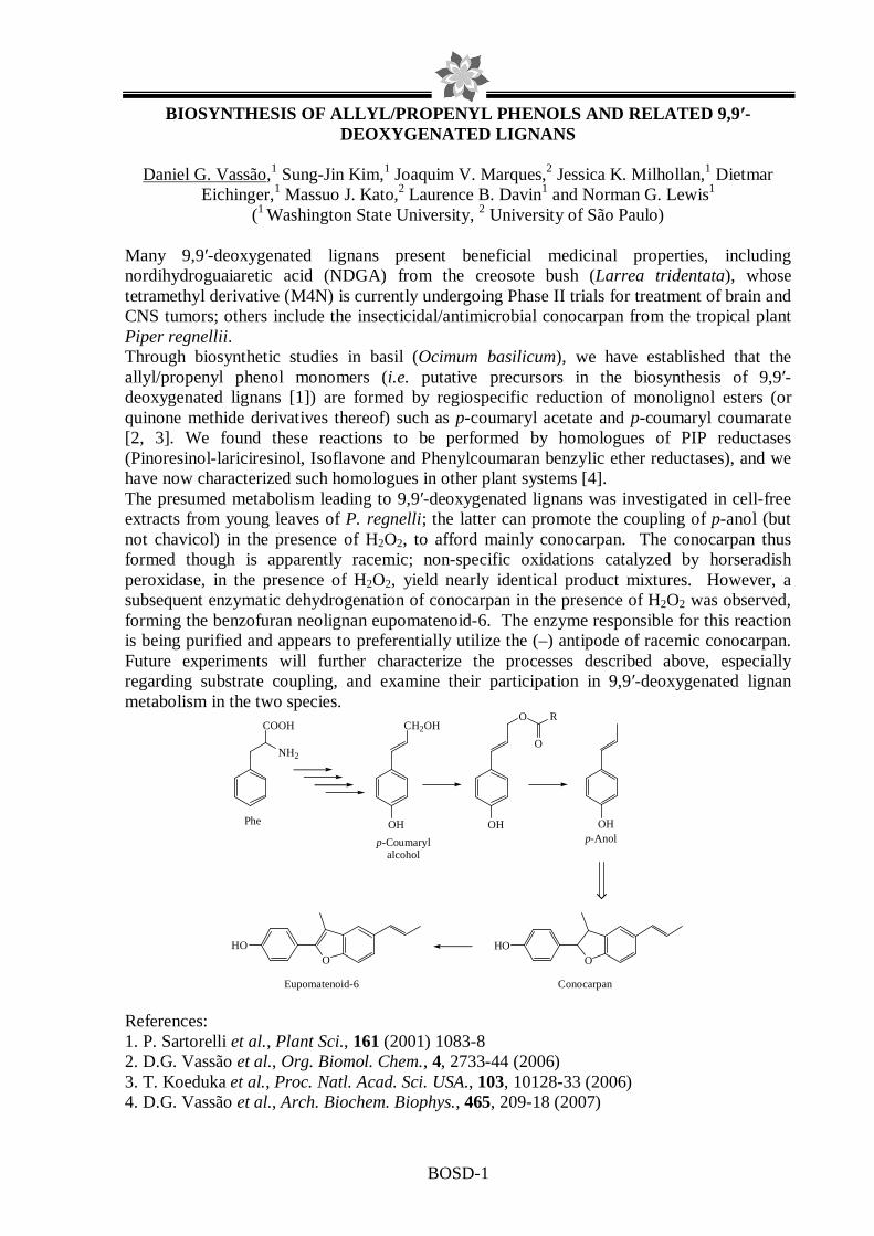
BOSD-1
BIOSYNTHESIS OF ALLYL/PROPENYL PHENOLS AND RELATED 9,9′-DEOXYGENATED LIGNANS
Daniel G. Vassão,1 Sung-Jin Kim,1 Joaquim V. Marques,2 Jessica K. Milhollan,1 Dietmar
Eichinger,1 Massuo J. Kato,2 Laurence B. Davin1 and Norman G. Lewis1 (1 Washington State University, 2 University of São Paulo)
Many 9,9′-deoxygenated lignans present beneficial medicinal properties, including nordihydroguaiaretic acid (NDGA) from the creosote bush (Larrea tridentata), whose tetramethyl derivative (M4N) is currently undergoing Phase II trials for treatment of brain and CNS tumors; others include the insecticidal/antimicrobial conocarpan from the tropical plant Piper regnellii. Through biosynthetic studies in basil (Ocimum basilicum), we have established that the allyl/propenyl phenol monomers (i.e. putative precursors in the biosynthesis of 9,9′-deoxygenated lignans [1]) are formed by regiospecific reduction of monolignol esters (or quinone methide derivatives thereof) such as p-coumaryl acetate and p-coumaryl coumarate [2, 3]. We found these reactions to be performed by homologues of PIP reductases (Pinoresinol-lariciresinol, Isoflavone and Phenylcoumaran benzylic ether reductases), and we have now characterized such homologues in other plant systems [4]. The presumed metabolism leading to 9,9′-deoxygenated lignans was investigated in cell-free extracts from young leaves of P. regnelli; the latter can promote the coupling of p-anol (but not chavicol) in the presence of H2O2, to afford mainly conocarpan. The conocarpan thus formed though is apparently racemic; non-specific oxidations catalyzed by horseradish peroxidase, in the presence of H2O2, yield nearly identical product mixtures. However, a subsequent enzymatic dehydrogenation of conocarpan in the presence of H2O2 was observed, forming the benzofuran neolignan eupomatenoid-6. The enzyme responsible for this reaction is being purified and appears to preferentially utilize the (–) antipode of racemic conocarpan. Future experiments will further characterize the processes described above, especially regarding substrate coupling, and examine their participation in 9,9′-deoxygenated lignan metabolism in the two species.
OHp-Anol
Conocarpan
OHO
OHO
Eupomatenoid-6
COOH
Phe
NH2
CH2OH
OH
p-Coumarylalcohol
OH
O R
O
References: 1. P. Sartorelli et al., Plant Sci., 161 (2001) 1083-8 2. D.G. Vassão et al., Org. Biomol. Chem., 4, 2733-44 (2006) 3. T. Koeduka et al., Proc. Natl. Acad. Sci. USA., 103, 10128-33 (2006) 4. D.G. Vassão et al., Arch. Biochem. Biophys., 465, 209-18 (2007)

BOSD-2
SECONDARY METABOLITES FROM THE FUNGUS LENTINUS STRIGUELLUS
B. A. Barros-Filho1, M. C. F. de Oliveira*1, J. Mafezoli2, M. N. G. Pessoa3 and H. C. B. de Oliveira4
1-Departamento de Química Orgânica e Inorgânica, Laboratório de Biotecnologia e Síntese Orgânica-LABS, Universidade Federal do Ceará, Fortaleza-CE, Brazil, [email protected]. 2-Curso de Ciências Farmacêuticas, Universidade de Fortaleza, Fortaleza-CE, Brazil. 3-Departamento de Fitotecnia, Universidade Federal do Ceará, Fortaleza-CE, Brazil
4-Ministério do Meio Ambiente, Secretaria de Política para o Desenvolvimento Sustentável, Brasília-DF, Brazil.
Lentinus species (basidiomycete) are important sources of secondary metabolites and have provided some biologically active compounds, such as the antibiotic cortinellin and antimicrobial sesquiterpene hirsutane1. This work reports the first chemical investigation of L. striguellus. This fungus was cultivated in 14 L of peptone-yeast extract medium for 28 days under static condition. Mycelium was separated from the liquid medium, dried until constant weigh and subjected to extraction with hexane, hexane/EtOAc 50%, EtOAc and MeOH. Column chromatography of Hex/AcOEt 50% extract (388.1 mg) allowed the isolation of the indol alkaloid echinuline2 (1, 16.0 mg) and anthraquinone physcion3 (2, 5.0 mg). L. striguellus was also grown in a shaker (150 rpm) using the same procedure previously described. Partitition of the liquid medium (7 L) with AcOEt afforded 1.4 g of extract, which was chromatographed and provided 2,2-dimethyl-6-methoxy-4-chromanone (3, 35.0 mg), 2,2-dimethyl-3-hydroxy-6-methoxychroman (4, 12.8 mg) and 2,2-dimethyl-3,4-dihydroxy-6-methoxychroman4 (5, 7.5 mg). Structures were elucidated by MS and 1H and 13C NMR analysis. Compounds 3 and 4 were previously isolated from L. Conatus1 and all other substances are being described for the first time in Lentinus genus.
Refs. 1. V. Rukachaisirikul et al., Journal of Natural Products, 68, 1674, (2005). 2. S.K. Talapatra et al., Journal of the Indian Chemical Society, 78, 773, (2001). 3. X. Chu et al., Journal of Chromatography A, 1097, 33, (2005). 4. Q. Wang et al., Tetrahedron: Asymmetry, 15, 29 (2004).
N
N
N
O
O
H
H
H
1
O
O
OHOH
CH3O
2
O
OH
OHCH3O
5
O
O
CH3O
3O
OH
CH3O
4

BOSD-3
PARTIAL PURIFICATION AND CHARACTERIZATION OF EUPOMA TENOI-6 SYNTHASE FROM PIPER REGNELLII
Joaquim V. Marques1, Daniel G. Vassão2, Massuo J. Kato1, Laurence B. Davin2 and Norman
G. Lewis2 (1Universidade de São Paulo, 2Washington State University)
P. regnellii Miq. is popularly known as “pariparoba” and can be found in lowlands of neotropical regions. Its major secondary metabolites described so far include several phenylpropanoids and dihydrobenzofuran neolignans with antibacterial, antifungal and antichagasic activities (Benevides et al., 1999).
The biosynthesis of neolignan (+)-conocarpan requires p-hydroxypropenylbenzene as precursor which can be dimerized by enantioselective oxidative coupling (Sartoreli et al., 2001). The benzofuran neolignan eupomatenoid-6 is apparently formed by dehydrogenation of conocarpan, but the mechanism of such conversion remains to be clarified.
The enzyme that catalyzes the conversion of (+)-conocarpan in its dehydrogenated analogue, with hydrogen peroxide as co-factor, has been partially purified from the leaves of P. regnellii and characterized in terms of its pH and temperature optima, substrate specificity and kinetic parameters.
Enzymes that perform similar reactions have been described for sterols and fatty acids and the reaction mechanism usually involves NAD+ or NADP+ in some cases (Atawong et al., 2003) or NADH or NADPH and molecular oxygen in others (Taton and Rahier, 1996 Behrouzian and Buist, 2003), but none has been described to perform dehydrogenation with hydrogen peroxide as co-factor and thus the mechanism of this reaction still needs to be investigated.
Refs: Atawong, A., Hasegawa, M., and Kodama, O. (2002). Bioscience, Biotechnology, and
Biochemistry 66, 566-570. Behrouzian, B., and Buist, P.H. (2003). Prostaglandins, Leukotrienes and Essential Fatty
Acids 68, 107-112. Benevides, P. J. C., Sartorelli, P. and Kato, M. J. (1999) Phytochemistry 52, 339. Sartorelli, P., Benevides, P. J. C., Ellensohn, R. E., Rocha, M. V. A., F. Moreno, P. R. H. and
Kato, M. J. (2001) Plant Science 161, 1083.

BOSD-4
BIOTRANSFORMED PRODUCTS FROM TERPENOIDS BY FUNGI
Arakawa, N.S.1; Ambrósio, S.R.2; Gobbo Neto, L.3; Carollo, C.3; Said, S.1; Da Costa, F.B.1 1FCFRP – USP, Dept. Pharmaceutical Sciences, 2UNIFRAN – Dept. Organic Chemistry,
3FCFRP – USP, Dept. Physics and Chemistry.
Microorganisms like fungi are multi-enzyme systems that undergo chemical transformations in the presence of different types of starting materials. Therefore these organisms are an efficient and cost-effective alternative to obtain interesting compounds that are difficult to be synthesized and that show lower toxicity and/or enhanced activity.
The sesquiterpene lactone budlein A (1) is the main constituent of the sunflower-like plant Viguiera robusta (Asteraceae). It has important biological effects including potent cytotoxic activity against human tumor cell lines as well as in vitro and in vivo anti-inflammatory effect1. Nevertheless, sometimes compounds of this type present toxic properties due to the α,β-unsaturated system in the lactone ring. However, the obtention of analogues of 1 by traditional approaches, i.e. derivatization, is quite complex. Thus, we decided to perform the biotransformation of 1 using the fungus Aspergillus terreus.
Budlein A was incubated on Czapeck liquid medium and after 120 h at 30 ºC and 120 rpm the culture filtrates were extracted with CHCl3. The organic layer was chromatographed over silica gel and the biotransformed products (2 and 3) were purified by HPLC. The structures were elucidated by NMR spectrometry and HRESIMS.
The biotransformation of 1 afforded two new and unusual terpenoid products which are difficult to be obtained by traditional synthetic reactions. The transformation carried out by A. terreus involved a double bond isomerization of the side chain ester at C-8 (3), the reduction of the double bond at C-2 and the complete removal of the lactone moiety followed by the formation of a new bond between C-7 and C-3 (2 and 3). The 3D structure of 2 is also shown. So far, the enzymes involved in these reactions were not identified, but they were responsible for the elimination of the putative toxic moiety of the molecule.
This is a good example of the versatility of a microbial-catalyzed biotransformation leading to unusual derivatives with potential pharmacological interest. The next step is to evaluate the biological effects of the products 2 and 3.
O
O
O
OH
O
O
O
O
OHOH
O
O
O
O
OHOH
O
O
O
2
3
7
8
1
2
3
2
3
7
8
4
5 6
9
110
4
5 6
9
10
Refs 1. D.A.R. Valério et al., Eur. J. Pharm., 562, 155 (2007)

BOSD-5
THE EFFECT OF PHENYLALANINE ADDITION ON THE BIOSYNT HESIS OF BIS-PHENYLPROPANOIDS AMIDES IN THE FUNGUS PENICILLIUM SP.
ISOLATED AS ENDOPHYTE FROM MELIA AZEDARACH.
Taícia P. Fill1, Edson Rodrigues Filho1
1. Laboratório de Bioquímica Micromolecular de Microorganismos (LaBioMMi) – UFSCar- São Carlos-SP
The associations between plants and microorganisms are being considered an important and emergent research area and many important active substances were discovered in the past few years. In this context, Penicillium sp., a fungus isolated as endophyte from Melia azedarach (Meliaceae), produces many interesting secondary metabolites such as the bis-phenylpropanoid amides, Brasiliamide A (1) and E (2). These Brasiliamides seem to cause convulsion in silkworms (Bombix mori)1, and researchers even consider these compounds as a possible new group of tremorgenic substances1. These amides also showed activity against gram-positive bacteria Bacillus subtilis in bioassays recently conducted at LaBioMMi (DQ/UFSCar). Because of their fast and easy development in artificial mediums, the microorganisms provide many manipulation possibilities to increase the production of secondary metabolites. During early experimental works, it were started studies to find the best conditions for the production of bis-phenylpropanoid amides, which are probably made through condensation of two amino acid unities. In this sense, the aim of this work was to develop and apply analytical methodologies to study the influence of L-phenylalanine on the Penicillium sp. metabolism and Brasiliamides production, and to verify the best production period, in order to increase the yield of these compounds, once these substances shows interesting parasitic and bactericide activities. The experimental procedures started with the inoculum preparation which was obtained by suspending spores from 14-day-old cultures in water to a density of (1–2) x107 spores/mL. The microorganism was cultivated at 25° in 125 mL Erlenmeyer flasks containing 30g of rice. To study the effect of the amino acid on amide production, the medium was supplemented with 25 mg of L-phenylalanine during inoculation. Secondary metabolites were extracted with organic solvents, and samples for analysis were taken at 2, 3, 5, and 10 days intervals. Qualitative analyses were made using a triple quadrupole mass spectrometer in previously optimized conditions. It was verified that the addition of L-phenylalanine in the culture medium increases the production of Brasiliamide A and the maximum production of this metabolite occurs at 28 days. An interesting fact is that the Brasiliamide production is verified in 2-days culture. In conclusion, the best medium and period of cultivation were optimized and the Brasiliamide A production maximized, indicating that L-phenylalanine could be the precursor on its biosynthesis, which will be further proved by adding labeled L-phenylalanine in the culture medium. 1 Fujita, T.; Makishima, D.;Hayashi, H. Biosci. Biotechnol. Biochem.. 2002, 66(8), 3415
N
OO
O
HNH
O
OO
N
N O
OO
H
O
(1) (2)


POSTER SESSION ABSTRACTS


BPS-1
Inhibition of ascitic Ehrlich tumor growth by intra peritoneal injection of Pfaffia paniculata butanolic residue ( Brasilian ginseng). Juliana Vieira, Patricia Matsuzaki, Márcia Kazumi Nagamine, Silvana Lima Gorniak Mitsue Haraguchi, Gokithi Akisue, Maria Lúcia Zaidan Dagli*
Laboratório de Oncologia Experimental, Departamento de Patologia, Faculdade de Medicina Veterinária e Zootecnia, Universidade de São Paulo, Av. Prof. dr. Orlando Marques de Paiva, 87 CEP 05508-900 São Paulo - SP - BRAZIL Phones 55 (011) 3091-7712 / Fax 55 (011) 3091-7829 E-mail address: [email protected] *Correspondent author Antineoplastic effects of butanolic residue of Pfaffia paniculata in Ehrlich tumor-bearing mice by oral treatment was reported previously. The aim of this study was to investigate the effects of this residue by intraperitoneal treatment. A toxicity study was performed in which doses of 12,5 mg/Kg; 25mg/Kg e 50mg/Kg of butanolic residue of P. paniculata were administered by intraperitoneal injection for seven days in Swiss mice do not bearing Ehrlich ascitic tumor. The mice did not lose weight during the treatment and any pathologic alteration was observed by histologic acessment of the colected organs. The effect of this residue on the ascitic Ehrlich tumor in Swiss male mice was then investigated. Male mice received, by intraperitoneal injection, once a day, 12,5mg/Kg; 25mg/Kg or 50mg/Kg of butanolic residue of PP or saline solution (PBS), as control, for seven days. This protocol started in the same day of tumor inoculation with 5X10⁶ cells i.p. the concentration of 50mg/Kg demonstrated a lower number of tumoral cells than control, thus the protocol was repeated with this concentration. The ascitic tumor was evaluated by the quantification of the volume of the ascitic fluid, relative number of tumor cells , number of dead tumoral cells and total number of tumor cells, morphology of tumor cells and histopathologic alteration in representative slices of the organs . A decrease in tumoral cells number per ml was observed in P. paniculata treated mice, that was followed by a numerical decrease in the total numbers of tumor cells in the ascitic fluid colected. These results may indicate the presence of tumor–cell inhibitory effects by P. paniculata residue. Refs. 1. Nakai, S.; Takagi, N.; Miichi, H.; Hayashi, S.; Nishimoto, N.; Takemoto, T.; Kizu, H. Pfaffosides and nortriterpenoid saponins from Pfaffia paniculata. Phytochemistry, v. 23, n. 8, p. 1703-5, 1984. 2. Matsuzaki P., Gokithi A., Oloris C.S., Gorniak L.S., Dagli M.L.Z..Efecct of Pfaffia paniculata(Brazilian ginseng) on the Ehrlich tumor in its ascitic form. Life Sciences(in press) 74, 573-579. 2003. 3. Haraguchi, M.; Mimaki, Y.; Motidome, Y.; Morita, H.; Takeya, K.; Itokawa, H.; Yokosuka,
A.; Sashida, Y. Steroidal saponin from the leaves of Cestrum sendtenerianum.
Phytochemistry, v. 55, p. 715-20, 2000.
BPS-2
In Vivo ANTICÂNCER ACTIVITY EVALUATION OF BY-PRODUCTS FROM THE PRODUCTION OF ARTEMISININ FROM ARTEMISIA annua L. (ASTERACEAE)
Aline Sousa1, Gabriela Marchetti 1, Rodney Rodrigues1, Ilza Sousa1, Fernanda Gaspi2 Cláudio
Navarro Filho1, João Carvalho 1, Mary Foglio1 1Centro Pluridisciplinar de Pesquisas Químicas, Biológicas e Agrícolas, P.O. Box 6171, 13083-970, Universidade Estadual de Campinas, UNICAMP – Campinas - SP, Brazil,
2 Uniararas, Av. Dr. Maximiliano, 500, 13607-339 Araras-SP
Malaria is one of the major causes of mortality in the Brazil and worldwide. At CPQBA-UNICAMP the artemisinin production from Artemisia annua L (Asteraceae) in large scale was patented. The production process generates a sesquiterpene-enriched residue. Therefore we started investigation on the biological potential of this material. The objective of this work was to evaluate the "in vivo" anticancer activity of Fraction (F2S) obtained by filter column chromatography from the residue generated in the artemisinin pilot scale production that previously demonstrated activity when tested on "in vitro” anticancer assay on nine human cell lines. The Ehrlich “in vivo” Tumor Model was selected as the standard assay. Ehrlich tumor cells were maintained in male Swiss mice through serial i.p. inoculation in 7-day intervals in an ascitis form. After preparing the cells, the total number was determined by Newbauer chamber using trypan blue dye exclusion method (Dagli, 1992), with tumor cell viability always higher than 90%. The cells were then diluted in saline (0,9%) for final inoculation density (1 x 10⁵ cells/mL). Fraction (F2S) was previously submitted to acute toxicity (LD50) study in order to determine which concentrations could give the potential therapeutic activity but not kill the animals, as described by Litchfield & Wilcoxon (1953). In the EAT assay, five groups of eight animals received 1 * 105 cells/mL, via i.p. The drug administration (via i.p.) began on the fourth day after inoculation, and the groups received the following treatments: Grupo1 - saline (vehicle) negative control, group 2 - doxorrubicina (5 mg/kg) positive control and group 3 - fraction (F2S), doses (200 mg/Kg), group 4 – fraction (F2S) dose (300 mg/Kg) and group 5 - fraction (F2S), doses (400 mg/Kg).The animals were treated again in the same doses in the 14th and 21st days after inoculation, and every day all animals were observed and scored for behavior and clinical conditions (Ullman-Cullere, 1999). When one animal got the highest punctuation, this animal was sacrificed and this day considered the time of the animal’s death. With EAT model, Fraction’s F2S antitumoral action was confirmed by the prolongation of the life-spam of treated groups. The expectation is that an active compound can act in tumor cells curing some percentage of them and/or prolonging the life spam of others. These results are very important, considering that EAT is a very aggressive tumor, having killed the controlled group after 20 days of the tumor cells’ inoculation. With 200, 300 and 400mg/Kg doses tested in this same date (20º post-treatment), all animals survived. On the 40th day post-treatment all three sample animal groups continued alive. On the 90 th day post-treatment, when assay was ended, the three sample groups treated with F2S had 50% animal survival. The doses of 200, 300 and 400mg/Kg had no adverse reactions demonstrating a potential "in vivo" anticancer activity for fraction F2S, prompting further studies in Hollow fiber models with human tumor cell lines. Dagli MLZ, et.al.. Braz J Vet Res Ani Sci 1992; 29: 97-103; Litchfield Jr JT, Wilcoxon F. J Pharm Exp Therap 1953; 108 (1): 18-25; Ullman-Cullere MH, Foltz CJ. Laboratory Animal Science 1999; 49 (3): 319-323

BPS-3
IN VITRO ANTICANCER ACTIVITY OF Calea pinnatifida (R.Br.) Less. LEAVES
Arianny do Nascimento Santos 1, Gabriela Menezes Marchetti 1, João Ernesto de Carvalho 1, Mary Ann Foglio 1
1 Centro Pluridisciplinar de Pesquisas Químicas, Biológicas e Agrícolas, P.O. Box 6171, 13083-970, Universidade Estadual de Campinas, UNICAMP – Campinas - SP, Brazil Calea pinnatifida (R.Br.) Less., Asteraceae is popularly known as aruca or erva-de-lagarto, used in folk medicine to treat amoebiasis and giardiasis. Twelve constituents of C. pinnatifida (R.Br.) Less. were identified by Kato et al. (1994) in the essential oil, extracted from aerial parts. The scope of this study was to evaluate “in vitro” antiproliferative activity of Calea pinnatifida (R.Br.) Less. crude extract and to isolate the actives fractions by activity guided fractionation. The aerial parts of Calea pinnatifida (R.Br.) Less. were collected in the experimental field of CPQBA – Unicamp. Grinded dry leaves were extracted on Soxhlet with methylene chloride. The extract was dried under vacuum, yielding crude methylene chloride extract (ED) (9,9%). The crude methylene chloride extract was purified by column filter chromatography. The fraction that presented the best antiproliferative activity in the “in vitro” assay was submitted to an acid-base extraction. Three fractions were obtained, a neutral non-polar fraction (NNP); a basic fraction (B) and an acid fraction (A) that were submitted to the anticancer “in vitro” assay. The samples were evaluated for anticancer activity in UACC62 (melanoma), MCF-7 (breast), NCI 460 (lung, non-small cells), OVCAR03 (ovarian), PC0 3 (prostate), HT-29 (colon), 786-0 (renal) and NCI-ADR (ovarian expressing phenotype multiple drugs resistance) cancer cell lines “in vitro” . A 48 h SRB cell viability assay was performed to determine growth inhibition and cytotoxic properties of the compounds. Cells were treated with at least four different concentrations levels ranging from 0.25 to 250 µm/mL with determination of total growth inhibition (TGI) parameter. The crude methylene chloride extract (ED) presented selectivity concentration-dependent to cancer cell line 786-0 (TGI: 32,05). This result is important because there is no specific treatment for renal cancer. The neutral non-polar fraction (NNP) presented selectivity concentration-dependent for cancer cell lines NCI-460 (TGI: 5,42), MCF-7 (TGI: 4,08), UACC-62 (TGI: 5,59) and 786-0 (TGI: 2,51). The basic fraction (B) presented selectivity concentration-dependent for cancer cell lines NCI-ADR (TGI: 4,47) and acid fraction (A) presented selectivity concentration-dependent for lines UACC-62 (TGI: 8,91) and 786-0 (TGI: 8,19). The clean up process of the crude methylene chloride extract permitted the identification of fractions with potential selectivity anticancer activity that will be further studied in “in vivo” experimental cancer models. Kato et al. Fitoterapia 65 (4): 377 (1994)

BPS-4
PC AND CPC ANALYSIS OF A SET OF FLAVONOIDS AND DERI VATIVES AS ANTIPROTOZOAL AGENTS
Luciana Scotti1, Kerly Fernanda Mesquita Pasqualoto2, Elizabeth Igne Ferreira1 1 – LAPEN, Department of Pharmacy, Faculty of Pharmaceutical Sciences, University
of São Paulo - USP, São Paulo, SP, 05508-900, Brazil. 2 - Laboratory of Theoretical and Applied Chemometrics, Department of Physical
Chemistry, Institute of Chemistry, The State University of Campinas, Campinas, SP, 13083-970, Brazil.
Introduction. Trypanosomiasis and leishmaniasis have reemerged over the last few decades as important threats to human health and economical development. Flavonoids have been known to exert diverse biological effects, particularly, acting as antioxidants and prophylactic agents against several diseases, including parasitic diseases. Computer-aided molecular design methods are considered a rational tool to find new drugs for the treatment of the mentioned diseases. Objective. Application of Principal Component (PC) and Consensus PC (CPC) analysis to a set of 106 flavonoid derivatives (aglycones and glycosides, and other compounds of phenolic and phenylpropanoid nature) having antiprotozoal activity against Trypanosoma brucei rhodesiense, Trypanosoma cruzi, and Leishmania donovani, and cytotoxicity for mammalian L6 cells. Methodology. Three-dimensional molecular structures from Protein Data Bank (PDB) were used as starting geometries of ligands. The flavonoids and derivatives models were minimized employing the AM1 semiempirical method (HyperChem 6.0). PCA and CPCA were applied using VolSurf+ program. Those chemometric methods were used to generate findings for elucidating the structure-activity relationships of the investigated compounds. Results. In a preliminary investigation, a classification between the 3D models and antiprotozoal and cytotocity activities, considering a dataset of 105 compounds and 114 descriptors, was searched employing PCA. Two significant principal components (PCs), explaining about 70% of the total variance of the matrix, were found by a cross-validation technique. The loadings plot indicated the variables logp c-hex, logd-10, G, R, CW1-8, W1-8, which were mixed and hydrophilic descriptors, as having greater influence in antiprotozoal activity. CPCA was performed, and PC1 and PC2 explained about 75% of original information, but 85% of the total explained variance was computed with H2Oprobe. Conclusion. Those findings can be used to understand the investigated system and establish the relevant descriptors to the biological activity and cytotoxicity. The chemometric tools used here were the beginning of a search for desingning new flavonoid agents having antiprotozoal activity. Refs. 1. D. Tasdemir et al., Antimicrobial agents and chemoterapy, 50, 4 (2006).

BPS-5
USE OF SELF-ORGANIZING MAPS OF THE FLAVONOIDS AND A NALOGUES WITH ANTIPROTOZOAL ACTIVITIES
Luciana Scotti1, Marcus Tullius Scotti2, Kerly Fernanda Mesquita Pasqualoto3, Elizabeth Igne
Ferreira1, Vicente P. Emerenciano2 1 - School of Pharmaceutical Sciences, University of São Paulo (USP), 580 Prof.
Lineu Prestes Av., Bl. 13, Conjunto das Químicas, Cidade Universitária, 05508-900, Brazil 2 – Instituto de Química, Universidade de São Paulo, Post Office box 26077, 05513-
970 São Paulo, SP, Brazil 3 - Laboratory of Theoretical and Applied Chemometrics, The State University of
Campinas, 13083-970, Post office box 6154, Campinas, SP, Brazil
Introduction Trypanosomiasis and leishmaniasis have reemerged over the last few decades as important threats to human health and economical development. Flavonoids have been known to act as antioxidants and preventive agents against several diseases. There is the need for the development of new, cheap, safe, and easy-to-administer molecules for the treatment of these infectious diseases. Objective We investigated 105 flavonoid glycosides and other phenolic compounds with antiprotozoal activities against Trypanosoma brucei rhodesiense, Trypanosoma cruzi, and Leishmania donovani and cytotoxicities for mammalian L6 cells. Methodology 106 flavonoids and derivates were submitted geometry optimization using the AM1 semiempirical quantum chemical method by the Hyperchem v. 6.0 software. The compounds were divided in approximately 50% active and 50% inactive, observing pIC50 against T. cruzi, T. brucei, L. donovani and pED50 for cytotoxity. The molecules were saved as MolFiles for computing 1664 molecular descriptors using DRAGON Professional v. 5.4. Redundant and noisy information were excluded. The 658 remaining descriptors were used for performing a Kohonen Neural Network. The Multiple Linear Regression (MLR) models by using genetic algorithms (GA) to perform variable selection using MobyDigs v. 1.0 software. Kohonen maps were performed by SOM (Self-Organizing Map) Toolbox 2.0 for Matlab 6.5, using all (658) descriptors. SOM results obtained using only the selected descriptors (about 8) by MLR using GA were compared with the previous ones. Conclusion For all descriptors, training sets with activities against T.cruzi, T. brucei, L. donovani and cytotoxicity shows a match, respectively, 72.7%, 72.5%, 77.3%, 72.9% (active group) and 89.3%, 71.4%, 73.3%, 80.% (inactive group). The test set calculated with T. cruzi, T. brucei, L. donovani and cytotoxicity showed the matches, respectively, 75.0%, 68.8%, 37.5% and 62.5%. Thus is possible to use SOM as a filter for a virtual screening. For selected descriptors, training sets shows a match, respectively, 75.8%, 87.5%, 72.7%, 82.7% (active group) and 91.1%, 89.8%, 80.0%, 81.1% (inactive group). The test set calculated with T. cruzi, T. brucei, L. donovani and cytotoxicity showed the matches, respectively, 75.0%, 68.8%, 62.5% and 56.3%. The selected variables were capable to divide the compounds mainly with activities against the T. cruzi. Refs. 1. D. Tasdemir et al., Antimicrobial agents and chemoterapy, 50, 4 (2006).

BPS-6
ANTIBACTERIAL AND ANTIMICROBIAL ACTIVITY OF EXTRACT S FROM Typha latifolia
Natália Covre de Melo1(IC), Daniel Moreto Silvestre1(IC), Ana Flávia de Oliveira Gregol2
(IC), Alessandra Cazellato de Medeiros2(IC), Marta Cristina Teixeira Duarte2(PQ), Juliano de Souza Gracioso (PQ)1, Catarina dos Santos1(PQ) * ([email protected])
1Departamento de Ciências Biológicas, Faculdade de Ciências e Letras, UNESP-Assis. 2Divisão de Microbiologia CPQBA/UNICAMP.
Typha latifolia inhabits marshes, shallow, ditches and wet wastes along river. It is a
perennial herb from a creeping rhizome and a dark brown pistil. The cattails are folk remedy for several diseases1. In connection with our study of antimicrobial activity of plants from Northern São Paulo State, it has been presented the preliminary results of antimicrobial activity from extracts of T. latifolia.
The plant material was collected from “varzea” near of Assis, São Paulo, Brazil. From dry and powdered stalk has gotten the following extracts: hexane (EHTa), chloroform (ECTa), ethyl acetate (EAcTa) and methanol (EMEOHTa). All these extracts were prepared from by dynamic maceration T. latifolia for 2h. Dried material (30g) was extracted with hexane (30 mL) and repeated for twice with 30 mL of the same solvent. After this, the extracts were filtered off and plant residue was treated with ethyl acetate. The same procedure was applied for methanol and final residue discarded. From of these extracts were carried out the antimicrobial activity by MIC against the bacteria Pseudomonas aeroginosa (ATCC 13388), Escherichia coli (ATCC 11775), Rhodococcus equi (ATCC 25729), Micrococcus luteus (ATCC 4698), Staphylococcus epidermes (ATCC 12228), Salmonella choleraesuis (ATCC 10708), Enterococcus faecium (ATCC 5079), Bacillus subtilis (ATCC 6051), Staphylococcus aureus (ATCC 6538), Enterococcus hirae (ATCC 10541) and against the yeast Candida albicans (ATCC 10231). EHTa showed activity to Rhodococcus equi (0,25 mg/mL), EAcTa showed inhibition (0,50 mg/mL) to Bacillus subtilis (0,50 mg/mL) and Candida Albicans (0,50 mg/mL).
All the rest the microorganisms tested presented no activity (> 1,0 mg/mL). 2 Refs. 1Hartwell, J.L., Kinsey, A.C., and Rollins, R.C. 1958. Edible wild plants of eastern North America. Rev. Ed. Harper & Bros., New York. 2Duarte, M.C.T.et al, Journal of Ethnopharmacology,97, 2, 305 (2004).

BPS-7
ANTIMICROBIAL ACTIVITY OF EXTRACTS OBTAINED FROM TH E WASTE RESIDUE OF Agave sisalana
Daniel Moreto Silvestre1 (IC), Patrícia de Cássia Salgado 1(IC), Ana Flávia de Oliveira
Gregol2 (IC), Alessandra Cazellato de Medeiros2(IC), Marta Cristina Teixeira Duarte2(PQ), Pedro Oliva Neto (PQ), Catarina Santos1(PQ) * ([email protected])
1Departamento de Ciências Biológicas, Faculdade de Ciências e Letras, UNESP-Assis. 2Divisão de Microbiologia CPQBA/UNICAMP.
Agave sisalana is a plant that has an attractive commercial value because of its applications like rope industry, tapestry, drugs, animal food, etc1. By the other side, the waste aqueous residue, rich in steroidal saponins, has been discarded. The aim of this research was to study the antimicrobial activity of MIC (Minimal Inhibitory Concentration) from the extracts obtained from this residue. Firstly, it was done a reduction of ten times of the water volume of the waste residue by heating, followed by evaporation of residual water at r.t. From this crude material, it was done a ressuspension in water and it prepared the following extracts by liquid-liquid extraction: butanolic (EBAS) and ethyl acetate (EAAS). Some crystals was formed in the EBAS extract, (called EBASc), which were separated and used on antimicrobial activity testing without any further purifying treatment. From the same crude and evaporated material it was prepared the hidroalcoholic extracts: ethanol:water 70:30 (EAqAS70) and ethanol:water 50:50 (EAqAS50). Second, the waste residue was diluted with ethanol in a proportion residue:ethanol 50:50 v/v. There was a formation of a precipitate, which was filtered and discarded. The liquid portion evaporated (r.t.) and the extract obtained was called EVAS extract. The antimicrobial activity of these extracts was tested against the yeast Candida albicans (ATCC 10231) and following the bacteria: Pseudomonas aeroginosa (ATCC 13388), Escherichia coli (ATCC 11775), Rhodococcus equi (ATCC 25729), Micrococcus luteus (ATCC 4698), Staphylococcus epidermes (ATCC 12228), Salmonella choleraesuis (ATCC 10708), Enterococcus faecium (CCT 5079), Bacillus subtilis (ATCC 6051), Staphylococcus aureus (ATCC 6538), Enterococcus hirae (ATCC 10541). EBASc showed strong activity against Candida albicans (0,3mg/ml) and Rhodococcus equi (0,075 mg/ml), moderate inhibition against Enterococcus hirae (0,7 mg/ml) and no activity to others microorganisms used. EBAS showed stronger activity than EBASc against to the Candida albicans (0,15 mg/ml) and Rhodococcus equi (0,05 mg/ml), but a discrete inhibition against Enterococcus hirae (0,9 mg/ml), Staphylococcus aureus (1.0 mg/ml) and no activity to others microorganisms. EAqAS70 showed only one strong activity against Rhodococcus equi (0,3 mg/ml). All the rest of tested extracts showed no activity. It was considered “no activity” when the microorganisms tested presented inhibition > 1,0 mg/mL2. As expected, the antimicrobial activities were concentrated in saponin fraction. Refs. 1. Lincoln, R. A./ Strupinski, K. and Walker International Journal Pharm, 34, 384 (1996). 2. Duarte, M.C.T.et al, Journal of Ethnopharmacology,97, 2, 305 (2004).

BPS-8
ANTIMICROBIAL ACTIVITY OF EXTRACTS FROM Pyrostegia venusta
Patrícia Bittencourt da Silva 1(IC), Ana Flávia de Oliveira Gregol2 (IC), Alessandra Cazellato
de Medeiros2(IC), Marta Cristina Teixeira Duarte2(PQ), Catarina dos Santos1(PQ) *
([email protected]) 1Departamento de Ciências Biológicas, Faculdade de Ciências e Letras, UNESP-Assis.
2Divisão de Microbiologia CPQBA/UNICAMP.
Pyrostegia venusta is a climber with delicate sterns of the family Bignoniaceae that is represented by more than 100 genera and about 800 species. In this plant are found flavonoids1,2, rutin2 and phenolics3 compounds. In connection with our study of antimicrobial activity of plants from Northern São Paulo State, we are now reporting our preliminary results concerning of Minimal Inhibitory Concentration (MIC) of hexanic, ethyl acetate and metanolic extracts from P.venusta.
Leaves of P. venusta were collected in an area of "cerrado" of Assis, São Paulo, Brazil. Dried leaves (30g) were macerated with 30 mL of hexane with agitation for 2h. Subsequently, the extract was filtered and plant residue has re-extracted with two portions of 30mL hexane each and evaporated. This extracted was the hexanic extract (EHPV). The same preparation was used with ethyl acetate (EAPV), methanol (EMPV) and chloroform (ECPV).
The antimicrobial activity of EHPV and EAPV was tested against the yeast Candida albicans (ATCC 10231) and following bacteria: Pseudomonas aeroginosa (ATCC 13388), Escherichia coli (ATCC 11775), Rhodococcus equi (ATCC 25729), Micrococcus luteus (ATCC 4698), Staphylococcus epidermes (ATCC 12228), Salmonella choleraesuis (ATCC 10708), Enterococcus faecium (CCT 5079), Bacillus subtilis (ATCC 6051), Staphylococcus aureus (ATCC 6538), Enterococcus hirae (ATCC 10541). EAPV showed strong activity against Candida albicans (0,3mg/ml), discrete inhibition against Staphylococcus aureus (1,0 mg/ml) and no activity to others microorganisms used. EHPV showed moderate inhibition against Staphylococcus aureus (0,9mg/ml), good inhibition against Enterococcus hirae (0,5mg/ml) and no activity to others microorganisms used. It was considered “no activity” when the microorganisms tested presented inhibition > 1,0 mg/mL. 4 These results indicate EAPV and EHPV extracts, obtained from Pyrostegia venusta, have the better potential for an antimicrobial activity.
(a) Refs 1.Ferreira, D. T. et al. Quím. Nova,.23, 1, 42, (2000). 2.Blatt, C.T.T.; Dos Santos, M.D. e Salatino, A. Plants Systematic and Evolution., 210, 289 (1998). 3. Dos Santos, M.D.; Blatt, C.T.T.A. Rev. Bras. Bot. ,21, 2, 135, (1998). 4.Duarte, M.C.T.et al, Journal of Ethnopharmacology,97, 2, 305 (2004).

BPS-9
SELF-ORGANIZING MAPS TO PREDICT THE ANTI-VIRAL ACTI VITY OF SESQUITERPENE LACTONES IN THE SUBGENOMIC HCV REPLIC ON SYSTEM
Michelle Rossini1, Marcus Tullius Scotti1, Marcelo J. P. Ferreira1, Ricardo Stefani2, Vicente
P. Emerenciano1 1- Instituto de Química, Universidade de São Paulo, 05513-970, SP, Brazil 2 - Instituto de Ciências Exatas e Biológicas, Universidade Federal de Ouro Preto
Introduction Some sesquiterpene lactones (SLs) are the active compounds of a great number of traditionally medicinal plants, mainly from the Asteraceae family, and possess considerable anti-microbiologic activity. Studies in vitro have shown the anti-viral activity in the subgenomic Hepatitis C virus (HCV) replicon system; proving that screening of natural products is a viable and fast way for identifying novel molecular diversity as potential drug leads.1 In this study we investigated a set of 19 different molecules, represented by 5 skeletons of SLs (11 germacranolides, 4 eudesmanolides, 1 guaianolide and 1 pseudoguaianolide) and 2 additional lactones, in what it says respect of their anti HCV properties. Materials and Methods These molecules were classified as actives (12 molecules with EC50 < 10µM) and as inactives (7 molecules with EC50 > 10µM), and were drawn into SISTEMAT X and had their activity data inserted. After that, they were codified in 3D using .mol (mdl) files. Molecular modelling computations were performed on Hyperchem 7.0 for Windows. The molecules were subjected to geometry optimization and conformational analysis. Semi-empirical quantum chemical method used was AM1 and the root mean square gradient value of 0.001 kcal/mol as termination condition. The molecules were saved as MolFiles for computing various molecular descriptors using DRAGON Professional v. 5.4. These descriptors were used for performing a Kohonen Neural Network. All descriptors provided by DRAGON were calculated, totalizing 1664 descriptors. For each block of descriptors, the constant variables were excluded, as well as those that presented only a different value of the series. Kohonen maps were trained using Matlab 6.5 and SOM (Self-Organizing Map) Toolbox 2.0. We used MobyDigs program for the calculation of Multiple Linear Regression (MLR) models by using genetic algorithms, with pEC50 = –log EC50 (EC50 provided by the anti-viral activity of the 12 molecules with specific activity in the subgenomic HCV replicon system) as dependent variable, and all DRAGON descriptors as independent variables. The selected descriptors were reanalyzed at SOM Toolbox. Results and Conclusions The 3D-MoRSE descriptor showed the best results, with 100% of match for the inactive compounds, 83.3% for the active ones and 89.5% for the entire block. The less satisfactory values were shown by Randic descriptor block that reached 57.1% of correctly prediction for inactive molecules, 100% for active ones and 84.2% for the total test. After the MLR analysis, the descriptors (MOR11u, G1s and HATS3v) were selected by the most significant equation (n =12, r2=0.94, Qcv
2= 0.88, s=0.05, F=-39.6). The SOM obtained for these three descriptors showed best results (85.7% of match for the inactive compounds, 91,7% for the active ones and 89.5% for all). SOM is a possible tool to be used as a filter for a virtual screening for SLs. Reference 1. D. Hwang et al., Bioorganic and Medicinal Chemistry, 14, 83 (2006).

BPS-10
ANTI-INFLAMMATORY AND ANALGESIC ACTIVITIES OF CRUDE EXTRACT AND ISOLATED COMPOUNDS FROM Nectandra megapotamica (LAURACEAE)
Ademar A. da Silva Filho1, Eveline S. Costa1, Karen C. S. Rezende1, Murilo de J. Fukui1, Wilson R. Cunha1, Márcio L. A. e Silva1, N. P. Dhammika Nanayakkara2, Shabana I. Khan2, Jairo K. Bastos3
1Grupo de Pesquisas em Produtos Naturais, Núcleo de Pesquisa em Ciências Exatas e Tecnológicas, Universidade de Franca, Franca, SP, Brazil; 2National Center for Natural Products Research, Research Institute of Pharmaceutical Sciences, School of Pharmacy, University of Mississippi, Mississippi, USA; 3Faculdade de Ciências Farmacêuticas de Ribeirão Preto, Universidade de São Paulo, Ribeirão Preto, SP, Brazil.
The bark of Nectandra megapotamica (Spreng) Chodat et Hassler (Lauraceae) is used in Brazilian folk medicine as an anti-rheumatic and to relieve pain1. The chromatographic fractionation of the crude hydroalcoholic extract of the barks of N. megapotamica (ENM ) afforded the isolation of α-asarone (1) and eleven lignans, including veraguensin (2) and galgravin (3). Regarding the analgesic and anti-inflammatory activities, the oral administration of ENM (500mg/kg) produced a significant inhibition of the constrictions induced by acetic acid by 82.2%, while α-asarone (1), veraguensin (2), and galgravin (3), at the dose of 20mg/kg (p.o.) showed 60.5%, 71.3%, and 70.6% of inhibition, respectively. In the carrageenan-induced paw edema, veraguensin (2) and galgravin (3), at the dose of 20mg/kg (p.o.), showed a significant anti-edematous effect of 41.2% and 71.4%, respectively. The compound 1 and ENM were inactive in this assay at the tested doses. In the hot plate test, the oral administration of α-asarone (20 mg/kg) and ENM (300 mg/kg) produced a significant analgesic effect, while 2 and 3 were inactive. In the in vitro COX-2 and NFkB assays, all tested samples were inactive. The results taken together suggested that 2 and 3 possessed peripheral analgesic activity, while the analgesic properties of ENM may be due mainly to the presence of α-asarone2.
H3CO
H3COO
OCH3
OCH3H3CO
H3COO
OCH3
OCH3H3CO
H3CO
1 2 3
Refs
1. Da Silva Filho, A.A. et al., Journal of Natural Products 67, 42 (2004a). 2. Da Silva Filho, A.A. et al., Journal of Pharmacy and Pharmacology 56, 1179
(2004b).

BPS-11
POTENTIAL HYPOGLYCEMIC EFFECTS OF LEANDRA LACUNOSA EXTRACT IN ALLOXAN-INDUCED DIABETIC RATS
Glenda Marqueti Arantes; Rodrigo Lucarini, Antonio E. Miller Crotti;
Ademar Alves da Silva Filho, Márcio L. Andrade e Silva, Wilson Roberto Cunha.
Universidade de Franca – Av. Dr. Armando Salles de Oliveira, 201 – Franca –SP 14404-600
Diabetes mellitus is a major endocrine disorder affecting nearly 10% of the population all over the world. Despite of the introduction of hypoglycemic agents, diabetes and related complications continue to be a serious medical problem. Medicinal plants have been used as an alternative treatment of diabetes. Leandra lacunosa (Melastomataceae) has been used in folkloric Brazilian medicine for treatment of this disease. The aim of the present work was to evaluate the antidiabetic activity of the hydroalcoholic extract of aerial parts of L. lacunosa in the alloxan-induced diabetic rats. The extract of the plant were obtained by maceration with ethanol/water (8:2 v/v). Diabetes was induced in rats by i.v. administration of 150 mg/kg of alloxan monohydrate. After 2 weeks, animals with level of blood glucose higher than 200 mg/dL were selected. In this experiment were used 3 groups of 5 rats each: group I (negative control - treated with saline), group II (positive control – chlorpropamide 40 mg/kg) and group III (diabetic rats treated with of hydroalcoholic extract 500 mg/kg). Blood glucose level was measured just prior to and 1, 2, and 4 h after drug administration. Treatment with hydroalcoholic extract of L. lacunosa indicates a significant reduction in blood glucose levels at 4h samples (Table 1). The present study confirms the folkloric use of this specie for treatment of diabetes. Further pharmacological and biochemical investigations are underway to elucidate the mechanism of these hypoglycemic effects. [Financial support: FAPESP] Table 1: Effect of hydroalcoholic extract of L. lacunosa on blood glucose level in
Alloxan-induced diabetic rats
Samples
0 h 1 h 2 h 4 h
hydroalcoholic extract a 319.4 ± 12.9 255,8 ± 45.4 (19.9%)
240.0 ± 46.6 (24.6%)
166.6 ± 31.5 (47.8%)
Positive controlb
252.4 ± 21.8 154.4 ± 21.0
(38.8%) 140.4 ± 35.4
(44.4%) 116.4 ± 49.3
(53.9 %) Negative controlc
218.8 ± 25.0 249.8 ± 22.0 293.6 ± 37.7 246.6 ± 22.0
ª 500 mg/kg b chlorpropamide 40 mg/kg c Saline. The values (mg/dL) are expressed as means ± S.E.M. (n=5) determined at different time (h) after treatment. The percentage of reduction is shown in brackets.
BPS-12
ANTICANCER PROPERTIES OF FURANODITERPENES FROM Pterodon pubescens Benth.
Karina Alves Silva 1, Humberto Moreira Spindola1,2, Carina Denny 1,2, Ana Lúcia Tasca Gois Ruiz †, Ana Paula Silva1, Iolanda Marques Timoteo 1, Ilza Maria de Oliveira Sousa 1, Rodney
Alexandre Ferreira Rodrigues 1, Jorge Yoshio Tamashiro3, João Ernesto de Carvalho 1,2, Mary Ann Foglio 1,2*
1 Centro Pluridisciplinar de Pesquisas Químicas, Biológicas e Agrícolas, P.O. Box 6171, 13083-970, Universidade Estadual de Campinas, UNICAMP – Campinas - SP, Brazil 2 Departamento de Farmacologia, Anestesiologia e Terapêutica, Faculdade de Odontologia de Piracicaba, P.O. Box 52, 13414-903, UNICAMP – Piracicaba - SP, Brazil, 3 Departamento de Botânica, P.O. Box 6109, 13084-971, Universidade Estadual de Campinas, UNICAMP – Campinas - SP, Brazil This paper reports the bioassay guided isolation and structure elucidation of compounds isolated from Pterodon pubescens Benth. methylene chloride-soluble fraction evaluated for anticancer activity in UACC62 (melanoma), MCF-7 (breast), NCI 460 (lung, non-small cells), OVCAR03 (ovarian), PC0 3 (prostate), HT-29 (colon), 786-0 (renal) and NCI-ADR (ovarian expressing phenotype multiple drugs resistance) cancer cell lines in vitro. A 48 h SRB cell viability assay was performed to determine growth inhibition and cytotoxic properties of the compounds. Cells were treated with at least four different concentrations levels ranging from 0.25 to 250 µm/mL with determination of total growth inhibition (TGI) parameter. Compounds, 7 β – diacetoxyvouacapane (1), 6 α – 7 β – diacetoxyvouacapane (2), methyl 6 α– 7 β – dihydroxyvouacapan–17 β-oate (4) were identified based on comparison of experimental 1H and 13C – NMR with reported spectral by Fascio et. al (1976), Campos et. al. (1994) and Ortalo-Magne et.al. (2005). Novel Compound 3 was deduced as having an elemental formula C22H32O4, by HREI-MS (observed M+. = 360.23556), which indicated seven insaturation sites. Infrared absorptions at 3449 (OH), 1713 (C=O) cm-1 provided evidences for hydroxyl and carbonyl functionalities. When acetylated with excess acetic anydride/pyridine, this compound showed identical 1H and 13C – NMR spectral data to compound 6 α–7 β– diacetoxyvouacapane 2 (HREI-MS 402,2630), suggesting that the difference in compound 3 was an acetyl group either in C-6 or C-7. Compound 3 1H– NMR spectra data lacked a signal at δ 5.24 pm due to H-7α and the appearance of a signal at δ 3.4 pm characteristic of hydroxyl group. Therefore these data suggested compound 3 to have the same relative configuration to 6 α – 7 β – diacetoxyvouacapane (2) with the hydroxy group attached β to C-7 whereas the acetyl group positioned α at C-6. Novel compound 3 (TGI 4.48 µg/mL) proved to be potent for ovarian cancer cells that express multidrug resistance phenotype (NCI-ADR) (TGI 4.48 µg/mL). A.M. Campos, et al Phytochemistry, 36 (2): 403-406, (1994); M.Fascio, et al Phytochemistry 15: 201-203, (1976); A.Ortalo-Magne, et al Phytochemistry, 66, 2316-2323, (2005)

BPS-13
EVALUATION OF THE ACTIVITY ON INTESTINAL MOTILITY O F Endopleura uchi (HUMIRIACEAE) AQUEOUS EXTRACT
HUBINGER, S.Z.a; POLITI, F.A.S.c; SALGADO; H.R.N.d; PIETRO, R.C.L.R.d; MOREIRA, R.R.D.b
a Pós-Graduação no Departamento de Princípios Ativos Naturais e Toxicologia; b Docente do Departamento de Princípios Ativos Naturais e Toxicologia; c Pós-Graduação no Departamento de Fármacos e Medicamentos; d Docente do Departamento de Fármacos e Medicamentos, Faculdade de Ciências Farmacêuticas, Unesp, Araraquara-SP, Brazil Email: [email protected]
Plants have long been an invaluable source of medication in human communities
worldwide. The Brazilian flora is vast and varied and each of its biomas is rich in species, many with medicinal attributes. The Endopleura uchi (Huber) Cuatrec. is a member of the family Humiriaceae and mollis is common in the Brazilian states of Pará and Amazonas, in the north of Brazil. According Sant’ana et al. (2000) and Ferreira et al. (2005), the stem of this plant has two major compounds: bergenine and 8,10 – dimetoxiberginine. The E. uchi is largely used by the population to combat miomas, although there are just a few biological studies with its extract. This study objectified evaluates the pharmacology activities of this plant. In the first part of this study, the extract of the stem of E. uchi was obtained by decoction with water. This extract was dried and used to the studies of acute oral toxicology (doses of 500, 1000 and 2000 mg/kg), according the OECD Guideline (2002). The extract was also used in the pharmacological study of intestinal motility (Wong et al., 1981) in dose of 200 mg/kg. The result showed that E. uchi aqueous extract offers a wide margin of safety and cannot be considered toxic. In the study of intestinal motility effect, the group treated with plant extract, the motility was decreased by 10,8% in the dose of 200 mg/kg of extract when compared with the control group, that received just water. It can be conclude that the extract of E. uchi presents an inhibition effect on the intestinal motility.
Refs.
Ferreira, A.G. et al. In: 57a Reunião anual da SBPC, Fortaleza-CE. (2005) OECD. Guideline 423: Acute Oral Toxicity – Acute Toxic Class Method. (2002)
Sant’ana, A.E.G. et al. In: XVI Simpósio de Plantas Medicinais do Brasil, Recife-PE. Livro de resumos. p. 229. (2000)
Wong, C.L. et al. European Journal of Pharmacology, v.73, p. 11-19. (1981)

BPS-14
SACAVANGER CAPACITY OF Dimorphandra mollis (LEGUMINOSAE) FRUIT EXTRACT
HUBINGER, S.Z.a; VELLOSA, J.C.R.c; BUNETTI, I.L.d; OLIVEIRA, O.M.M.F.d; MOREIRA, R.R.D.b
a Pós-Graduação no Departamento de Princípios Ativos Naturais e Toxicologia; b Docente do Departamento de Princípios Ativos Naturais e Toxicologia; c a Pós-Graduação no Departamento de Análises Clínicas; d Docente do Departamento de Análises Clínicas, Faculdade de Ciências Farmacêuticas, Unesp, Araraquara-SP, Brazil Email: [email protected]
Dimorphandra mollis is a Brazilian plant largely used in folk medicine with
antoxidant potential for containing flavonids. Its fruit pericarp is used in Brazil to treat ulcers, to reduce inflammation and to heal wounds (Lorenzi & Matos, 2002). From the fruits it can be extract flavonóides, mainly rutin and quercetin (Sousa et al., 1991). The study was performed on fruits from a Dimorphandra mollis tree growing in the Araraquara district of São Paulo State. The dried and comminuted plant material was extracted in ethanol. We have evaluated the crude ethanolic extract of D. mollis fruits as a potential antioxidant source using an assay based on the bleaching of the radical monocation 2,2’-azinobis (3-ethylbenzothiazoline-6-sulfonic acid) (ABTS+) and by DPPH scavenging capacity (Oliveira et al, 2007). Cysteine was used as a positive antioxidant control evaluated at different concentrations. Crude ethanolic extract showed marked action over DPPH (IC50= 79,5 ± 3,5 µg/ml) and radical (IC50= 3,45 ± 0,071 µg/ml). Both extracts and cysteine (DPPH: IC50= 21 ± 1,4 µg/ml; ABTS•+: IC50= 1,3 ± 0,14 µg/ml) were able to scavenge radicals at low levels. From the results, we can conclude that the crude ethanolic extract from Dimorphandra mollis fruits has a powerful antioxidant potential.
Refs.
Lorenzi, H. & Matos, F.J.A. Plantas Medicinais no Brasil nativas e exóticas. Nova Odessa: Ed. Instituto Plantarum (2002). Oliveira, O.M.M.F. et al. Fitoterapia, v.78, p.263 (2007).
Sousa, M.P. et al. Constituintes químicos ativos de plantas medicinais brasileiras. Fortaleza: Universidade Federal do Ceará (1991).

BPS-15
13C NMR SPECTRAL DATA AND MOLECULAR DESCRIPTORS TO PR EDICT THE ANTIOXIDANT ACTIVITY OF FLAVONOIDS
Marcus Tullius Scotti, Eric Muramatsu, Michelle Rossini, Marcelo J. P. Ferreira, Vicente P. Emerenciano Instituto de Química, Universidade de São Paulo, PO Box 26077, 05513-970,
SP, Brazil
Introduction Tissue damage due to oxidative stress is directly linked to development of many, if not
all, human morbidity factors and chronic diseases. In this context, the search for dietary natural occurring molecules with antioxidant activity, such flavonoids, became essential. The DPPH system is a stable radical-generating procedure that simulates the biological free radical producing mechanism and because of this, the system can be used to measure the biological antioxidant activities of electron accepting compounds like flavonoids. The 13C NMR, as molecular descriptors, can be used to describe the molecular structure. These data give rich information about the molecular structure and they are sensitive to detect differences on the molecule. Objective
In this study we investigated a set of 41 flavonoids (23 flavones and 18 flavonols) analyzing their structures and biological antioxidant activity1. Molecular information given by the 13C NMR data were used to predict the antioxidant activity of the flavonoids, sorting them in active or inactive compounds from the ANNs (artificial neural networks). Materials and Methods
13C NMR data were used to perform a Kohonen self-organizing map study, analyzing the influence and weight that each carbon has in the activity. Thus, the compounds were classified as active if they exhibited IC50<200µM and inactive if they exhibited values of IC50>200µM. The 13C NMR chemical shifts of flavonoids were used as the input data and the separation between active and inactive compounds were obtained. The unsupervised training were performed using the SOM Toolbox version 2.0 for Matlab version 6.5 computing environment by MathWorks, Inc. Results and Conclusion
The results from Kohonen map show a highly significant match with high percentage for both groups (80%). Analyzing the groups separately the major mach occurred among the active compounds (84%). This could be explained by the common chemical environment exhibited by the active molecules once these compounds have a pattern of substitution that influences the electromagnetic surroundings in the same way. The importance of these carbons was confirmed in the 13C NMR analysis. Through the 13C NMR chemical shifts the ANN achieve a significant result, sorting the active and inactive molecules with minimal errors (20%). This supports the idea that 13C NMR data can be used to perform classification of flavonoids taking into account the biological activity, making clear that radical scavenging activity is highly correlated with the chemical environment. Reference 1. Y. Yokozawa et al., Biochemical Pharmacology, 56, 213 (1998).

BPS-16
QUANTITATIVE RELATIONSHIP BETWEEN OXIDATION OF DITE RPENES AND 3D MOLECULAR DESCRIPTORS IN ASTERACEAE FAMILY
Marcus Tullius Scotti1, Marcelo J. P. Ferreira1, Ricardo Stefani2, Vicente P. Emerenciano1
1 - Instituto de Química, Universidade de São Paulo, PO Box 26077, 05513-970, SP, Brazil 2 - Instituto de Ciências Exatas e Biológicas, Universidade Federal de Ouro Preto
The seminal contribution of Gottlieb and co-workers to chemotaxonomy resulted in several postulates about the evolution of secondary metabolites in plants1. One of them suggests that “The evolution of micromolecules proceeds by oxidation. The relatively highly oxidized compounds characterize new chemical lines”. Recently, it was suggested that the oxidative pathways in plants occur parallel to protective mechanisms against oxidative degradation2. The same skeletal types, of a chemical class, can be found in different tribes, subtribes or even genera. In such cases, a method to distinguish one tribe from the rest is through comparison of sets of shared skeletons or substitutional diversification of such skeletons. The base of this procedure is: different broken metabolic are characterized by sets of compound, and their structures 3D should be used in the differentiation process. Objectives In this study we verify and quantify the existent relationship of the oxidation number (NOX) of diterpenes, in the 13 tribes (Anthemideae, Arctoteae, Astereae, Calenduleae, Cardueae, Eupatorieae, Gnaphalieae, Helenieae, Heliantheae, Inuleae, Mutisieae, Senecioneae, Vernonieae) of the Asteraceae family, with the RDF descriptors from 3D structures. Material and Methods The coordinates 3D of 2150 diterpenes were generated through the program SISTEMAT X, starting from 2D representation. Molecular modeling computations were performed on SPARTAN for Windows v. 4.0 software. The molecules were subjected to geometry optimization and conformational analysis. The semi-empirical quantum chemical method used was AM1. Respective botanical occurrences for each compound and its oxidation number were extracted of the program SISTEMAT X. Molecules with their energies minimized were saved as MDL MolFiles for computing RDF descriptors using the program DRAGON Professional v. 5.4. The program BuildQSAR was used to select the variables for the generation of a linear model (MLR - multiple linear regression) with up to 4 variables. Results and Conclusions The most statistically regression equation, largest squared cross-validation correlation coefficient (Qcv
2), is: NOX = +1,168(±0,277) RDF070u -2,754(±0,749) RDF095u +6,776(±1,995) RDF135u -2,083(±0,701) RDF150u -35,588(±2,998)
(n=013; r2=0,931; s=0,670; F=27,266; Qcv2=0,856; SPRESS=0,972) Although there is a high correlation between the NOx and the RDF descriptors, the tribes was not distributed by the evolution order proposed by Bremer. Tribes considered less "developed", in other words, basal tribes of the Bremer’s diagram, as Arctoteae present high value of the average of oxidation number of the present diterpenes, while tribes more "developed", as Anthemideae, present low values. Refs. 1. O. R. Gottlieb, Phytochemistry, 28, 2545 (1989). 2. O. R. Gottlieb, M. A. C. Kaplan, Natural Products Letters, 2, 171 (1993).

BPS-17
USE OF SELF-ORGANIZING MAPS AND 13C NMR SPECTRAL DATA TO PREDICT ALDOSE REDUCTASE ACTIVITY OF FLAVONOIDS
Marcus Tullius Scotti, Hamilton Mitsugo Ishiki, Eric Muramatsu, Mauro Vicentini Correia,
Marcelo J. P. Ferreira, Vicente P. Emerenciano Instituto de Química, Universidade de São Paulo, PO Box 26077, 05513-970, SP, Brazil
The aldose reductase (AR) enzyme is responsible to reduce glucose to sorbitol using as a
cofactor the nicotinamide-adenine dinucleotide phosphate (NADPH). Simultaneously, another enzyme, sorbitol dehydrogenase, oxidizes sorbitol to fructose. However, in diabetes conditions, glucose level in this pathway is increased and sorbitol is produced faster than oxidized to fructose. The accumulation of sorbitol in lens leads to cataract formation. Flavonoids are phenolic compounds isolated from a wide range of vascular plants, with over 8000 individual compounds known. They possess a high variety of biological activities. Structure-activity relationships of flavonoids are important due to the facility of the analysis once they are natural active substances that show small plane molecules with few functional groups. These compounds contain a basic structure constituted of 15 carbon atoms arranged in three rings (C6-C3-C6) and showed the molecular structural requirements for a remarkable AR inhibition effect. To describe the molecular structure the 13C NMR can be used. These data give rich information about the molecular structure and they are sensitive to detect small differences on the molecule. The most used ANN architecture for pattern recognition is the Kohonen network also named of Self-Organizing Map (SOM). A SOM can map multivariate data onto a two dimensional grid, grouping similar patterns near each other. In this study, we correlate 13C NMR data with a set of 71 flavonoids with AR inhibition effect, obtained from literature1. For some structures, the 13C NMR data were obtained using ACDlabs software. Since flavonoids are plane aromatic compounds, the aditivity model can be applied to predict 13C NMR data.
The unsupervised training was performed using the SOM Toolbox version 2.0 for Matlab version 6.5 computing environment by MathWorks, Inc. The correlation between the 13C NMR data to the biological activity was performed using SOM. For that, the compounds were classified as active if they exhibited pIC50=log1/IC50>5.69µM and inactive if they exhibited values of pIC50<5.35µM. The compounds were first divided into two subsets: one training set composed of 54 molecules and one external test set composed of 17 compounds (around 20% of the whole set). The test set contains representative samples of trained group and includes the range of activity values of the training group.
The training set shows a highly significant match (85%) for active and inactive groups. Analyzing the groups separately the major match occurred among the active compounds (89%). The test group confirms the analysis success showing the highly significant match with high percentage for both groups (88%). A remarkable 100% match for the active group and a good match of 75% for the inactive compounds support the validation of the study. This difference was observed once the active compounds exhibited a common chemical environment i.e., a same pattern of substitution, thus these groups influence the electromagnetic surroundings in the same way. Refs.
1. H. Matsuda et al., Chem. Pharm. Bull., 50, 788 (2002).

BPS-18
PREDICITION OF ANTI-INFLAMMATORY ACTIVITY OF SESQUI TERPENE LACTONES USING SELF-ORGANIZING MAPS AND 13C NMR DATA
Marcelo J. Pena Ferreira, Marcus T. Scotti, Vicente P. Emerenciano
Instituto de Química, Universidade de São Paulo, Caixa Postal 26077, 05513-970, São Paulo, SP, Brazil
Sesquiterpene lactones are a large group of natural products from which more than 4000
structures are known mostly isolated from the Asteraceae family. These compounds are described as the active substances of various medicinal plants used in traditional medicine and possess a wide variety of biological and pharmacological activities, such as antimicrobial, cytotoxic and anti-inflammatory activities. There are strong indications that the activities are mediated by a general mechanism chemically dependent of the presence of α,β-unsaturated carbonyl structures, such as α-methylene-γ-lactones or α,β-unsaturated cyclopentenones or conjugated esters. These functional groups react with nucleophiles, especially sulfhydryl group of cysteine, by a Michael-type addition.
Although the number of sesquiterpene lactone skeletons is limited, a variety of substituents and their position and configuration isomerism, leads to an enormous number of possible structures. Complete structure determination (constitution, configuration and conformation) often represents a hard task. Thus, the 1H and 13C NMR spectroscopy play a very important role in the structural determination process as well as a valuable source of data for correlation between the structure features, NMR parameters and biological activities.
The aim of this work is to predict the anti-inflammatory activity of sesquiterpene lactones using the 13C NMR data and self-organizing maps (SOM).
To generate a SOM1 for the prediction of the anti-inflammatory activity, the data set of 58 sesquiterpene lactones containing their inhibitory concentrations (µM) in the NF-κB DNA binding assay2 and their 13C NMR data3 was employed. The NF-κB is a central mediator of the human immune system promoting the expression of over 400 target genes in response to inflammatory stimulators. To get better clustering, it was necessary to reduce the number of activity classes, which are divided in nine groups because of the experimental procedure, and to combine compounds with similar activity. Therefore, two classes of sesquiterpene lactones are generated: Class A – shows the compounds with high activities (IC100= 5 – 50µM); Class I – contains the compounds with low activities (IC100= 100 – 300µM).
The internal validation revealed that a correct prediction of NF-κB inhibitory activity was possible to 81.03%. The validation with an external data set that was not used during selection and training was carried out. External validation with 10 compounds, divided equally in the two classes, resulted in a correct prediction of 80%. The model can be used to search and develop lead structures with a strong inhibitory activity on NF-κB DNA binding; a good prediction is especially interesting for the higher activity class where our model, using only 13C MNR data, allows the correct prediction of 31 from 38 sesquiterpene lactones (81.58%) belonging to the class A containing the higher active compounds. Therefore, from these results one can be concluded that our model is a valuable tool for the prediction of the NF-κB inhibitory activity and for the screening of lead structures possessing this activity. References 1. T. Kohonen, Biological Cybernetics, 43, 59 (1982). 2. B. Siedle et al., Journal of Medicinal Chemistry, 47, 6042 (2004). 3. M. Budesinsky et al., Annual Reports on NMR Spectroscopy, 30, 231 (1995).

BPS-19
STRUCTURE AND EVALUATION OF ANTI-RADICAR POTENTIAL OF CALDENSINIC ACID FROM PIPER CALDENSE (PIPERACEAE)
Rodrigo O. Saga Kitamura1, Massuo J. Kato2, Paulete Romoff1 and
João Henrique G. Lago1 1Centro de Ciências e Humanidades, Universidade Presbiteriana Mackenzie
2Instituto de Química, Universidade de São Paulo
Piper species have also been described to contain several prenylated benzoic acids, which showed antimicrobial, moluscicidal, antifungal and anti-radicalar activities1. Based on such information, the MeOH extract from leaves of P. caldense was evaluated to its ability to scavenge 1,1-diphenyl-2-picryl-hydrazyl radical (DPPH) in vitro assay2. Thus, aiming to isolate the active compound, the crude extract (542 mg) was subjected to chromatographic separation in Sephadex LH-20 (MeOH) affording four groups (I – IV) in which the anti-radicalar potential was detected in two of them (II and III). The 1H NMR spectrum of group II (147 mg) showed a 1,3,4,5-tetrassubstituted aromatic ring due the signals at δ 7.34 (d, J = 2.0 Hz) and 7.42 (d, J = 2.0 Hz) and a prenylated chain due the four singlets at δ 1.59, 1.62, 1.67 and 1.73, assigned to methyl groups and broad multiplets at δ 5.11 – 5.17 and at δ 2.07 – 2.27, assigned to hydrogens linked to sp2 and sp3 carbons, respectively. The triplet at δ 6.78 (J = 7.4 Hz, 1H), suggested the presence of and carboxyl group in the side chain. The 13C NMR spectra (BBD and DEPT 135°) showed 27 signals, two of them assigned to carboxyl groups, six to aromatic ring and 19 to side chain carbons. Analysis of DQ-COSY, HMQC, HMBC and NOESY spectra allowed the characterization of active compound as 3-(11’-carboxyl-3’,7’,15’-trimethyl-hexadeca-2’E,6’E,10’E,14’E-tetraenyl)-4,5-dihydroxy-benzoic acid (1), named caldensinic acid. In order to evaluate and compare the anti-radicalar activities, 1 was acetylated to 1a and both compounds were subjected to the DPPH-scavenging assay, using Trolox® as positive control. Compound 1 had the highest hydrogen-donating capacity in comparison to 1a, but it was twice less active than Trolox®. Thus, this evaluation suggested that the presence of catechol unit is important to anti-radicalar activity of these prenylated benzoic acid derivatives, which have been found in several Piper species1.
OR
RO
COOH
COOH
1 caldensinic acid R = H1a diacetoxy calcensinic acid R = Ac
1. L.F. Yamaguchi et al., Phytochemistry, 67, 1838 (2006). 2. A. Kumaran et al., Food Chem., 100, 356 (2007).

BPS-20
POLAR COMPOUNDS ISOLATED FROM Maytenus truncata Reissek (Celastraceae)
Gloria Del Carmen Meléndez Salazar1,2, Roqueline R. S. Miranda1, Lucienir P. Duarte1, Sidney A. Vieira Filho,1,3 and Gracia D. Fátima Silva1
1 – Núcleo de Estudo de Plantas Medicinais (NEPLAM), Depto. Química, ICEx, UFMG. Av.
Antônio Carlos, 6627. CEP 31270-901, Belo Horizonte, MG, Brazil. 2 – Depto. de Química e Exatas, Universidade Estadual do Sudoeste da Bahia.
Rua José Moreira Sobrinho, s/n, CEP 45200-000, Jequié, Bahia, Brazil. 3 – DEFAR, Escola de Farmácia, Universidade Federal de Ouro Preto.
Rua Costa Sena, 171, CEP 35000-400, Ouro Preto, Minas Gerais, Brazil.
Maytenus truncata [“Caminha de anjo” (Angel’s little bed) or “Arvore de Natal” (Christmas tree)] is generally used in traditional medicine of the peoples who live around “Rio das Contas” region, Jequié, Bahia, Brazil, in the form of infusion or decoct for treatment of gastrointestinal tract diseases, hemorrhages and “womans’ diseases”. For these reasons there are great interest to know the chemical structure of polar compounds of this plant. From M. truncata dried fragmented leaves or branches using phytochemical processes, were possible to obtain the following polar contituents: sulphate inorganic salt (1), the polyol dulcitol or galactitol (2), the polyphenols pro-anthocyanidin (3) and 4’-O-methylepigallocatechin (4), 3β-stigmast-5-enyl β-D-glucopyranoside of (5) and the alkaloid maitein (6). These compounds were identified by spectrometric methods (IV, 1H and 13C NMR) and thin layer chromatography using in this case comparison with authentic samples. The results obtained are not contrary to popular use of M. truncata decoct, once polyphenols compounds, as the flavonoids 3 and 4, present antioxidant activity and protector factor1.
N
PhO
O
OAc
OAc
OAc
OAc
Me
OH MeO
O
OMe
Me
O
AcOH
H
H
H
OO
OH
HOHO
OH
3
6
18
1920
21 22
23
2526
28
29
27
24
1'2'
3'
4'
6'
5'
O
O
HO
OH
HO
OH
OH
OH
OH
OH
OCH3
OHOHO
OH
OH
OH
OH
OCH3
CH2OH
OHH
HHO
HHO
OHH
CH2OH
23
4
5 6
H H
References 1. F.G.Gonzalez and L.C. Di Stasi, Phytomedicine, 9, 125, (2002)

BPS-21
ANALGESIC AND ANTI-INFLAMMATORY ACTIVITIES OF Baccharis dracunculifolia (ASTERACEAE)
Ademar A. da Silva Filho1, Sérgio F. de Andrade2, Murilo de J. Fukui1, Dimas de O. Resende1, Márcio L. Andrade e Silva1, Wilson R. Cunha1, Shabana I. Khan3, N. P. Dhammika Nanayakkara3, Jairo K. Bastos4. 1Grupo de Pesquisas em Produtos Naturais, Universidade de Franca, Franca, SP, Brazil; 2Núcleo de Investigações Químico-Farmacêuticas (NIQFAR), Universidade do Vale do Itajaí – UNIVALI, Itajaí, Santa Catarina, Brazil; 3National Center for Natural Products Research, University of Mississippi, Oxford, MS, USA; 4Faculdade de Ciências Farmacêuticas de Ribeirão Preto, Universidade de São Paulo, Ribeirão Preto, SP, Brazil. Baccharis dracunculifolia D.C. (Asteraceae) is the most important plant source of the Brazilian green propolis, which is popularly known for its antimicrobial activity1,2. B. dracunculifolia is used in folk medicine as anti-inflammatory3. The aim of this work was to evaluate the analgesic and anti-inflammatory activities of the leaves hydroalcoholic extract of B. dracunculifolia (BdE). The crude extract (BdE), at different doses, was evaluated by using the acetic acid-induced writhing in mice, paw edema induced by both carrageenan and histamine in rats. In addition, the in vitro effect of the BdE on the activity of cyclooxygenase-2 (COX-2) enzyme was also evaluated. The results showed that BdE reduced the number of abdominal contortions by 45.4 % (at 200 mg/kg, p.o.) and 76.2 % (at 500 mg/kg, p.o.) in the acetic acid-induced writhing in mice. Regarding the anti-inflammatory assay, BdE displayed a significant anti-inflammatory activity in the paw oedema assay in rats induced by carrageenan (48.9% of edema inhibition at 400 mg/kg, p.o.), but it did not provide a significant reduction (p > 0.05 Student “t” test) in the paw edema assay induced by histamine in comparison with the positive control (Hydroxizine, at 10 mg/kg, p.o.). In the in vitro COX-2 assay, the COX-2 enzyme activity was inhibited by BdE, which showed an IC50 value of 45 µg/mL. The results obtained in this work confirmed the traditional indications of B. dracunculifolia as anti-inflammatory. Also, both the reduction in the paw edema induced by carrageenan and the acetic acid-induced writhes result from the anti-inflammatory activity of BdE, which may be related to the inhibition of the COX-2 enzyme activity. Refs
3. Leitão, D.P.S. et al., Biol Pharm Bull, 27 (11), 1834-1839 (2004). 4. Da Silva Filho, A.A. et al., Z Naturforsch C, (2007) in press. 5. Da Silva Filho, A.A. et al., J Pharm Pharmacol 56 (9), 1195-1199 (2004).

BPS-22
ANTILEISHMANIAL AND ANTIMALARIAL ACTIVITIES OF TETRAHYDROFURAN LIGNANS FROM Nectandra megapotamica (LAURACEAE)
Ademar A. da Silva Filho1, Wilson R. Cunha1, Márcio L. A. e Silva1, N.P. Dhammika Nanayakkara2, Jairo K. Bastos3
1Grupo de Pesquisas em Produtos Naturais, Núcleo de Pesquisa em Ciências Exatas e Tecnológicas, Universidade de Franca, Franca, SP, Brazil; 2National Center for Natural Products Research, Research Institute of Pharmaceutical Sciences, School of Pharmacy, University of Mississippi, Mississippi, USA; 3Faculdade de Ciências Farmacêuticas de Ribeirão Preto, Universidade de São Paulo, Ribeirão Preto, SP, Brazil.
The bark of Nectandra megapotamica (Spreng) Chodat et Hassler (Lauraceae) is used in Brazilian folk medicine as an anti-rheumatic and to relieve pain1,2. Seven tetrahydrofuran lignans, isolated from the crude hydroalcoholic extract of the barks of N. megapotamica (Lauraceae), were evaluated for their in vitro antileishmanial and antimalarial activities. Among the evaluated compounds, machilin-G (1a) and veraguensin (2a) showed the highest antileishmanial activities, displaying, for both compounds, an IC50 value of 18 µg/mL and IC90 value of 36 µg/mL, while galgravin (1b), nectandrin-A (1c), nectandrin-B (1d), calopeptin (2b), and ganshisandrine (3) were inactive against Leishmania donovani. In the antimalarial assay against Plasmodium falciparum, it was observed that calopeptin (2b) displayed moderate activity, with IC50 values of 3800 ng/mL (against D6 clone) and 3900 ng/mL (against W2 clone), while the lignans 1a-1d, 2a, and 3 were inactive. In order to compare the effect on the parasites with the toxicity to mammalian cells, the cytotoxic activity of the isolated compounds were evaluated against the VERO cells, showing that all evaluated tetrahydrofuran lignans exhibited no cytotoxicity in the maximum dose tested.
Refs
1. A.A. da Silva Filho et al., Journal of Natural Products 67, 42 (2004). 2. A.A. da Silva Filho et al., Journal of Pharmacy and Pharmacology 56, 1179 (2004).
O
R
OCH3
OCH3
R1 2
R R1
a OCH3 OCH3 b OCH2O
O
R
R2
OCH3
R1
7 7`
8 8`
1
R R1 R2
a OCH2O OCH3
b OCH3 OCH3 OCH3 c OCH3 OH OCH3 d OCH3 OH OH
O
H3CO
OCH3
OCH3
H3CO 3

BPS-23
PHYTOCHEMICAL STUDY AND COMPARATIVE EVALUATION OF T HE IN VITRO ANTIMICROBIAL ACTIVITIY OF Baccharis dracunculifolia
(ASTERACEAE) AND BRAZILIAN GREEN PROPOLIS Ademar A. da Silva Filho1, Dimas de O. Resende1, Murilo de J. Fukui1, Niege A. J. C. Furtado1, Márcio L. A. e Silva1, Wilson R. Cunha1, João P. B. de Sousa2, Luís E. Gregório2, N. P. Dhammika Nanayakkara3, Jairo K. Bastos2. 1Grupo de Pesquisas em Produtos Naturais, Núcleo de Pesquisa em Ciências Exatas e Tecnológicas, Universidade de Franca, Franca, SP, Brazil; 2Faculdade de Ciências Farmacêuticas de Ribeirão Preto, Universidade de São Paulo, Ribeirão Preto, SP, Brazil; 3National Center for Natural Products Research, University of Mississippi, Oxford, MS, USA. Baccharis dracunculifolia D.C. (Asteraceae) is the most important plant source of the Brazilian green propolis (BGP), which is popularly known for its antimicrobial activity1. B. dracunculifolia and BGP display similar anticariogenic2, anti-ulcer3, and immunomodulatory4 activities. However, it is still unknown whether B. dracunculifolia displays the same antimicrobial activities reported for BGP. The aim of this work was to evaluate the in vitro antimicrobial activity of B. dracunculifolia and some of its isolated compounds. The results showed that the leaves extract of B. dracunculifolia (BdE) presents antimicrobial activity, mainly against Candida krusei (IC50: 65 µg mL-1) and Cryptococcus neoformans (IC50: 40 µg mL-1). In comparison to the BdE, green propolis hydroalcoholic extract (GPE) showed better antimicrobial activity, especially against Candida krusei (IC50: 9 µg mL-1). The chromatographic fractionation of the BdE was carried out, affording the isolation of several compounds, including ursolic acid (1), 2α-hydroxy-ursolic acid (2), isosakuranetin (3), aromadendrin-4’-methylether (4), baccharin (5), viscidone (6), and hautriwaic acid lactone (7). Compounds 1 (IC50: 5 µg mL-1) and 2 (IC50: 3 µg mL-1) showed antibacterial activity against methicillin-resistant Staphylococcus aureus, while compound 3 was active against C. neoformans (IC50: 15 µg mL-1). All evaluated samples exhibited no cytotoxicity against the VERO cells in the maximum dose tested. The results showed that the BdE, similar to the GPE, displays in vitro antimicrobial activity, which may be related to the effect of several compounds present in the crude extracts3.
Refs
6. A.A. da Silva Filho et al., J Pharm Pharmacol 56 (9), 1195-1199 (2004). 7. D.P.S. Leitão et al., Biol Pharm Bull, 27 (11), 1834-1839 (2004). 8. M. Lemos et al., J Pharm Pharmacol 59 (9), 603-608 (2007). 9. F. Missima et al., J Pharm Pharmacol 59, 463-468 (2007). 10. A.A. da Silva Filho et al., Z Naturforsch C, (2007) in press.
COOH
O
O
5
COOHR
OH 1: R = H 2: R = OH
OOH
OCH3
OH OR
3: R = H 4: R = OH
O
OH
O
OH
6
O
OO
7

BPS-24
CYTOTOXIC ALKALOIDS FROM THE LEAVES OF OCOTEA ACUTIFOLIA Fernanda R. Garcez1, Walmir S. Garcez1, Ana Francisca G. da Silva1, Gabriela Linck1, Maria de
Fátima C. Matos2, Evelyn Cristina S. Santos2, Patrícia M. Oguma2
1Department of Chemistry, Federal University of Mato Grosso do Sul, Campo Grande, MS, 79070-900, Brazil
2Department of Pharmacy and Biochemistry, Federal University of Mato Grosso do Sul, Campo Grande, MS, Brazil
Several aporphine alkaloids possess significant in vitro cytotoxic properties against a number of human cancer cell lines and this class of secondary metabolites is of wide occurrence in members of the family Lauraceae.1 In a continuation of our search for potential antitumor agents from plants endemic to Mato Grosso do Sul, Brazil,2 the composition of the alkaloid extract of the leaves from Ocotea acutifolia (Nees) Mez. (Lauraceae) was studied. This work led to the isolation of two new, ocoteine N-oxide (1) and nor-ocoxylonine (2) and eight known aporphinoid alkaloids, ocoxylonine (3), ocoteine (4), O-methylcassyfiline (5), dicentrine (6), nor-dicentrine (7), leucoxine (8), isodomesticine (9) and neolitsine (10). Compound 10 is reported for the first time in the genus Ocotea. Air-dried and powdered leaves (500 g) of O. acutifolia were extracted at room temperature with EtOH. After concentration in vacuo, the EtOH extract was treated with 10% acetic acid and then partitioned with EtOAc to yield the EtOAc layer and the acid layer. The latter was adjusted to pH 8 with NH4OH and extracted with EtOAc to yield the crude alkaloid extract. This was repeatedly subjected to column and flash chromatography on silica gel, gel filtration on Sephadex LH-20 and reversed phase HPLC separations to afford alkaloids 1-10. The structural elucidation of these isolates was mainly based on 1D- and 2D-NMR spectroscopic techniques. Alkaloids 1, 2, 4, 8, and a mixture of 4 and 6 and of 5 and 7 were tested for their in vitro cytotoxic properties against the Hep2 (larynx carcinoma) human cancer cell line (Table 1). Ocoteine (4) and a mixture of 4 and dicentrine (6) displayed significant cytotoxic activity; compounds 1, 8 and a mixture of 5 and 7 exhibited weak cytotoxicity, while 2 was inactive in this assay.
Table 1- Cytotoxicity of compounds 1, 2, 4, 8, and a mixture of 4 and 6 and of 5 and 7 against Hep2 cell line
Compound IC50/(µmL-1) 1 32.75 2 > 50 4 8.14 8 37.23 mixture of 4 and 6 10.08 mixture of 5 and 7 43.78 Cisplatin (positive control) 1.64
1. C. Stevigny et al., Curr. Med. Chem.-Anti-Cancer Agents, 5, 173 (2005) 2. F. R. Garcez et al., J. Braz. Chem. Soc. 17, 1223 (2006); F. R. Garcez et al., Planta Med., 71, 923 (2005); F.
R. Garcez et al., Planta Med. 65, 775 (1999)
N
O
O
CH3O
OCH3
CH3
OCH3
1
23
3a4
5
6a
7
7a
8
9
10
11a
11b11c
11
O-
+
1
N
O
O
OO
1
23
3a4
5
6a
7
7a
8
9
10
11a
11b11c
11
CH3
10
1
2
10
11
N
O
O
CH3O
OCH3
OH
33a
4
5
6a
7
7a
8
9
11a
11b11c
CH3
8
1
23
3a4
5
6a
7
7a
8
9
10
11a
11b11c
11
N
OO
CH3O
HO
CH3
9
N
O
O
CH3O
OCH3
OH
OCH3
1
23
3a4
5
6a
7
7a
8
9
10
11a
11b11c
11
2 R=H3 R=CH3
R
N
O
O
CH3O
OCH3
OCH3
1
23
3a4
5
6a
7
7a
8
9
10
11a
11b11c
11
4 R= CH35 R= H
R
N
O
O
CH3O
OCH3
1
23
3a4
5
6a
7
7a
8
9
10
11a
11b11c
11
6 R= CH37 R= H
R

BPS-25
GASTROPROTECTION OF Plinia edulis (VELL.) SOBRAL (MYRTACEAE)
Tati Ishikawa1, Lucas Bergamo Navarro2, Raquel Donatini1, Edna Tomiko Myiake Kato1, Massayoshi Yoshida2,3
1 Departamento de Farmácia, Faculdade de Ciências Farmacêuticas, Universidade de São Paulo, C.P. 66083, CEP 05508 -000, São Paulo-SP, Brazil.
2 Departamento de Química Fundamental, Instituto de Química, Universidade de São Paulo, C.P. 26077, CEP 05599 -970, São Paulo-SP, Brazil.
3 Coordenação de Central Analítica, Centro de Biotecnologia da Amazônia, Av. Gov. Danilo de Matos Areosa, 690, CEP 69075-351, Manaus-AM, Brazil.
Introduction: Plinia edulis, medicinal plant known as “cambucá”, is native in Brazilian Atlantic Rain Forest. However, no reports are available on the relationship between the activity of its extract and gastric ulceration. The objective of this work was to investigate the gastroprotective effect of the aqueous ethanol leaf extract and its fractions at the HCl/ethanol induced ulcers model. In addition, the gastroprotective effect of ursolic acid was accessed. Materials and Methods: An oral dose of 100 mg/kg body wt. was initially used to establish a general profile of the antiulcerogenic activity of the extract and its fractions at HCl/ethanol model in rats. Lansoprazole at 30 mg/kg was used as the reference drug. Ursolic acid was administered at 50 mg/kg. The stomachs were excised and the ulceration area (mm2) determined. The extract was submitted to partition with organic solvents of increasing polarity and bio-guided chromatographic procedures and spectrometric analyses were performed in order to obtain purified fractions for detection of the plant metabolites involved in the gastroprotective activity. Results and Discussion: The extract showed extremely significant antiulcer activity at the used doses being more active than lansoprazole. Among the fractions obtained by partition, the hexane fraction was the most effective, but has shown lower activity than the crude extract. β-amyrin, β-sitosterol, ursolic acid, maslinic acid and corosolic acid were identified in the hexane fraction. Although others triterpenes are well known as gastroprotectors, ursolic acid reduced the lesion area, but has not shown significant activity at this model. These results suggest that the extremely high activity observed in the aqueous ethanol extract of leaves of P. edulis is probably due to synergism among chemical constituents and the triterpenes are partially involved in the gastroprotective mechanisms. Acknowledgements: To CNPq, CAPES and FAPEAM for fellowships and to CNPq, CAPES, FAPESP and FAPEAM for financial support.
BPS-26
SCREENING OF ACTIVE SUBSTANCES FROM Serjania erecta RADLK AS MEMORY STIMULANT
Moraes, L. T.1; Broggini, L. S. C. 1; Couto, L. B. 1; Pereira, P. S. 1; França,S.C. 1
1. Universidade de Ribeirão Preto, Av. Costábile Romano, 2.201 Ribeirânia - Ribeirão Preto - SP - Brasil, CEP 14096-900 [email protected] Plants such as Ginko biloba, Ptychopetalum olacoides, Pseudocalymma elegans, Salvia officinalis, Physostigma venenosum, Galanthus nivalis L., Huperzia serrata and Leucojum vernum L. have been evaluated in neurological disturbances and the Alzheimer disease. The neotropical class Serjania Mill. have about 203 species known at the moment, being the most numerous of this tribe, in which it is find the Sapindaceaes family, which is rich in saponins, an active compound of adaptogen plants, such as Paullinia cupana and Cardiospermum halicacabum, with known depurative, tonic-stimulant and immunomodulator actions. Serjania erecta Radlk, popularly known as “retrato-de-téiu” and “cinco-folhas”, belongs to this family. It is endemic to sensu stricto cerrado, and it is an arbustive plant (Silva, 1998). The leaves are used as menstrual regulator, healing and antiseptic. According to Guarim Neto and his collaborators (2000), from the leaves it is prepared some teas that help in the ulcers treatment, and the root is used for the high blood pressure treatment. The objective of this project is to find out a potential drug that could prevent or delay neuronal degenerations, with an improvement of the cognitive and comportamental losses, mainly in the Alzheimer disease, that reaches an incidence of more than one million people in Brazil, through the pharmacological characterization in vivo and in vitro of the vegetal extract of Serjania erecta Radlk and its purified fractions, with an approach to the following aspects: purification, isolation and chemistry characterization, pharmacological study directed to the enzymatic properties (AChE and BuChE) and biochemistry (lipoperoxidation assay) of fractions and purified substances, pharmacological and comportamental study through the Passive Avoidance and the Elevated Plus Maze methods, induzed by scopolamine, and by the fractions and purified substances. The preliminary results obtained with the aqueous extract of Serjania erecta were: presence of pharmacologically active substances: steroidal saponins, glucosilated flavonoides and condensed tannins, and the comportamental assay with rodents showed cognitive improvement (memory and attention), with antioxidant and enzymatic protection, favorable results in the hematological and biochemistry analyses, suggesting adaptogen action, without toxicity, that, therefore, stimulates the continuity of the research about Serjania erecta Radlk, evaluating the physiological actions directly in the central nervous system, action mechanism, elucidation of the chemistry structures present in the active compounds, and a second pre-clinic phase in non-rodents. Refs. 1. GUARIM NETO, G.; SANTANA, S.R.; SILVA, J.V.B. Notas etnobotânicas de espécies de Sapindaceae Jussieu. Acta Bot Bras. 14 (3):327-334, 2000. 2. SILVA, S.R. Plantas do cerrado utilizadas na região do Grande Sertão Veredas. Brasília: FUNATURA, 1998.Support: UNAERP and CAPES.

BPS-27
IN VITRO ANTICANCER ACTIVITY OF EXTRACTS OF PLANT SPECIES F ROM SÃO PAULO STATE
Santos AN 1, Silva KA 1, Silva BP 1, Alves A 1, Ming LC 2, Carvalho JE 1, Redher VLG 1,
Alves MJQF 2, Barros LCP 2, Marques MOM 3, Torres RB 3, Yoshida M 5, Silva SMP 4, Foglio MA 1
1 Centro Pluridisciplinar de Pesquisas Químicas, Biológicas e Agrícolas, P.O. Box 6171, 13083-970, Universidade Estadual de Campinas, UNICAMP – Campinas – SP, Brazil 2 Universidade Estadual Paulista “Júlio Mesquita Filho”, UNESP – Botucatu – SP, Brazil 3 Instituto Agronômico de Campinas, IAC – Campinas – SP, Brazil 4 Pólo Regional do Vale do Paraíba/APTA/SAA – Pindamonhangaba – SP, Brazil 5 Universidade de São Paulo, USP – São Paulo – SP, Brazil Ethnofarmacology has contributed for the development of many important plant-derived drugs. The study of naturals products represents an important source of knowledge in the discovery of new anticancer drugs and infectious diseases. About 67% of efective drugs used in chemotherapy are derived from naturals products. This study is part of the Bioprospecta (Biota-Fapesp) program that has as major aim the identification and characterization of São Paulo state biodiversity, and definition of mechanisms for conservation and sustainable use. The scope of this study was to evaluate “in vitro” antiproliferative activity of crude ethanol extracts of 25 plant species collected among the Piperaceae, Apocynaceae, Verbenaceae, Melastomataceae, Asteraceae, Maranteceae, Commelinaceae, Myrtaceae, Malpighiaceae, Solanaceae, Ericaceae, Fabaceae, Apoaceae families from São Paulo state. The plant species were collected in Serra da Mantiqueira – SP, by Dr. Ming and identified by Dra. Torres. Grinded dry plant species were extracted on Soxhlet with ethanol. The extracts obtained were cleaned with active charcoal for chlorophyll removal, dried under vacuum, freeze dried and submitted to anticancer “in vitro” assay. The samples were evaluated for anticancer activity in UACC62 (melanoma), MCF-7 (breast), NCI 460 (lung, non-small cells), OVCAR03 (ovarian), PC0 3 (prostate), HT-29 (colon), 786-0 (renal) and NCI-ADR (ovarian expressing phenotype multiple drugs resistance) cancer cell lines. A 48 h SRB cell viability assay was performed to determine growth inhibition and cytotoxic properties of the crude extracts. Cells were treated with at least four different concentrations levels ranging from 0.25 to 250 µm/mL with determination of growth inhibition of 50% of the cells (GI50); total growth inhibition (TGI) and concentration need to kill 50% of the cells (LC50) parameters. Among the extracts under study we observed a concetration-dependent selectivity for cell line 786-0 for samples from Apoaceae, Solanaceae, Myrtaceae, Commelinaceae, Asteraceae, Verbenaceae, Apocynaceae and Piperaceae families, compared to the positive control (doxorrubicina). Whereas plant extracts from Ericaceae, Malpighiaceae and Melastomataceae demonstrated a concetration-dependent selectivity for NCI-ADR cell lines. Cragg, et al. J. Nat. Prod. 69 (3): 488-498, 2006; K. Hostettmann et al. Princípios ativos de plantas superiores. 1a. Edição. Editora EdUFSCar; São Carlos, 2003.

BPS-28
IN VITRO ANTIPROLIFERATIVE ACTIVITY OF CAMPOMANESIA guazumifolia (CAMBESS.) O. BERG FRACTIONS
Juliana Nonaka IC, Rodney Rodrigues PQ, Ilza Sousa TC, Ana Ruiz PQ, Ana Possenti PQ, João
Carvalho PQ, Mary Foglio PQ Centro Pluridisciplinar de Pesquisas Químicas Biológicas e Agrícolas, Universidade
Estadual de Campinas
Key words: Campomanesia guazumifolia, antiproliferative activity
Campomanesia guazumifolia (Cambess.) O. Berg is popularly known as Sete-Capotes, pertaining to the Myrtaceae family. In the south of Brazil the population uses this plant to treat gastric disorders. Markman et al (2004) studied the antiulcerogenic activity of Campomanesia xanthocarpa. Fernandes et al (2003) and Limberger et al (2001) studied the essential oil composition of some Campomanesia species. This study reports the bioassay guided fractionation of crude Campomanesia dichloromethane and ethanol soluble fractions evaluated for in vitro anticancer activity in UACC62 (melanoma), MCF-7 (breast), NCI 460 (lung, non-small cells), OVCAR03 (ovarian), PC0 3 (prostate), HT-29 (colon), 786-0 (renal) and NCI-ADR (ovarian expressing phenotype multiple drugs resistance) cancer cell lines. A 48 h SRB cell viability assay was performed to determine growth inhibition and cytotoxic properties of the fractions. Cells were treated with at least four different concentrations levels ranging from 0.25 to 250 µm/mL with determination of total growth inhibition expressed by three concentration-dependent parameter; concentration that produces 50% growth inhibition (GI50), concentration which totally inhibits cell growth (TGI) and concentration that produces 50% lethal cell killing (LC50). The aerial parts of the Campomanesia guazumifolia (Cambess.) O. Berg were collected in CPQBA-Unicamp experimental field on March 2007. Leaves in natura were grinded with dry ice, and extracted with dichloromethane. After filtration, the solvent was evaporated under vacuum 40ºC, supplying the diclorometanic crude extract (DRE). The plant residue was retaken in ethanol, extracted with producing the ethanolic crude extract (ERE). This extract (ERE) presented antiproliferative activity superior to the dichloromethane crude extract (DRE). Therefore ERE was further extracted with variation of pH producing four fractions denominated: fraction A (acid fractions), fr B (basic fractions), fr NA (neutral apolar fraction) and fr NP (neutral polar fraction). Fr NA showed selectivity in a concentration-dependent way for HT-29 (colon) cell line with the most potent GI50, TGI, and LC50 values. Fraction B showed inhibition in a concentration-dependent way for MCF-7 (breast), NCI 460 (lung), UACC62 (melanome) and OVCAR-3 (ovary) cell lines, whereas fraction NP inhibited all cell lines, and fraction A was inactive.
Markman, B. E. O, Bacchi, E. M, Kato, E. T. M. Antiulcerogenic Effects of Campomanesia xanthocarpa. Journal Of Ethnopharmacology, v. 94, n. 1, p. 55-57, 2004; Fernandes J. B. F, Vargas V. M. F. Mutagenic and antimutagenic potential of the medicinal plants M. Laevigata and C. xanthocarpa. Phytotherapy Research, v. 17, n. 3, p. 269-273, 2003; Limberger, R. P, Apel, M, Sobral, M. et al. Aromatic Plants From Brazil - Chemical Composition Of Essencial Oils From Some Campomanesia Species (Myrtaceae). Journal of Essential Oil Research, v. 13, n. 2, p. 113-115, Mar-Apr 2001.
CNPQ, FAPESP

BPS-29
RADICAL SCAVENGING AND ANTIOXIDANT ACTIVITIES OF FL AVONOLS FROM LEAVES OF Tournefortia bicolor S.W. (BORAGINACEAE)
Thiago B. Correia da Silva (PQ)1,2, Vivian Karoline T. Souza (IC)1, Ana Paula F. da Silva (PQ)1, Patrícia E. S. Oliveira (PG)1, and Lucia M. Conserva (PQ)1* [email protected]
1Inst. de Química e Biotecnologia - Universidade Federal de Alagoas, 57072-970, Maceió-AL, Brazil 2 Universidade Federal de Alagoas, Pólo Viçosa - Campi Arapiraca, 57700-000, Viçosa-AL, Brazil
Antioxidant activity of plant extract is often associated with the presence of polyphenolic
compounds, which have an important role in stabilizing lipid oxidation and have inhibitory effects on mutagenesis and carcinogenesis in human when is daily ingested from a diet rich in fruits and vegetables1,2. It is well known that this activity is due to their capacity to be donors of hydrogen atoms or electrons and to capture the free radicals. These reactive species are formed by the normal metabolism of aerobics cells starting from the oxygen molecular reduction. When these radicals had an excessive formation induce oxidative damage to biomolecules, which eventually causes numerous neurodegenerative disease3. This work describes the total phenolic (TP) content by Folin-Ciocalteu4, DPPH free radical scavenging (FRS)5 and total antioxidant (TAA) activities by FTC method6 of extracts and isolated flavonols from leaves of Tournefortia bicolor. The air-dried leaves were extracted at room temp. with acetone and EtOH 90%. These extracts were suspended in MeOH-H2O (3:2), extracted successively with C6H14, CHCl3 and EtOAc and were evaluated for their TP content, FRS and TAA activities. The EtOAc fractions from both crude extracts showed a high phenolic content (acetone: 365.18 mg/g D.W and EtOH: 691.78 mg/g D.W). Thus, these fractions were evaluated for FRS and TAA activities, where EtOAc fraction (EtOH extract) showed a significant FRS (IC50 30.0±3.2 µg/mL) and TAA (85.44% at 100 µg/mL) activities. This fraction was filtered on silica gel column and the most active CH2Cl2-EtOAc 1:1 sub-fraction [TP content > 1000 mg/g D.W, FRS (IC50 12.8±5.6 µg/mL) and TAA inhibition (85.17% at 25 µg/mL)], after successive chromatographic fractionations (silica gel and Sephadex LH-20) afforded rutin (1, 25 mg) and tiliroside (2, 15 mg). On the other hand, EtOAc fraction (acetone extract) with a good FRS (101.0±3.5 µg/mL) and TAA (78.37% at 50 µg/mL) activities, after the same experimental procedure afforded quercetin (3, 25 mg). These flavonols (1-3) were also evaluated and showed a significant FRS and a good TAA inhibition at 25 µg/mL when compared with standard used (AA, α-TC and BHT) (Table 1). These results suggest that this species contain phenolic compounds that may act as antioxidant.
Table 1. DPPH free radical scavenge and antioxidant activities of isolated flavonols (1-3).
Seção 1.02 Isolated Substances and
Standards
DPPH FRS* IC50 (µg/mL)
TAA Inhibition ( %) at 25 µµµµg/mL
Rutin (1) 21.82 ± 3.3 84.01 Tiliroside (2) 50.4 ± 4.2 80.26 Quercetin (3) 7.67± 3.6 90.0 Ascorbic acid (AA) 37.37 ± 3.18 91.0 α-Tocopherol (α-TC) 92.51 ± 3.17 91.0 BHT 73.78 ± 13.73 -
* The average values of three assays (n = 3) are presented as mean±S.D. a at 25 µg/mL.
(i) References: 1 G.C. Yen et al., J. Agric. Food Chem. 41, 67-70 (1993); 2 D-J. Huang et al., Bot. Bull. Acad. Sin. 46, 99-106 (2005); 3 L-A. Tziveleka et al., Biorg. Med.Chem. 10, 935 (2002); 4 Y.-Y. Soong & P.J. Barlow, Food Chemistry 88, 411-417 (2004); 5 C. Sánchez-Moreno et al., J. Sci. Food Agric. 76, 270-276 (1998); 6 I. Gulçin et al., Phytomedicine 13, 343-351 (2006).
Support: CNPq, FAPEAL, IMSEAR and BNB-RENORBIO.

BPS-30
RADICAL SCAVENGING AND ANTIOXIDANT ACTIVITIES OF PH ENOLIC COMPOUNDS AND EXTRACTS FROM FRUITS OF Triplaris americana L.
Patrícia Emanuella S. Oliveira (PG)1, Thiago B. C. da Silva (PQ)1,2, Ana Paula F. da Silva (PQ)1 and Lucia M. Conserva (PQ)1* [email protected]
1 Inst. de Química e Biotecnologia, Universidade Federal de Alagoas, 57072-970, Maceió/AL, Brazil 2 Universidade Federal de Alagoas, Pólo Viçosa - Campi Arapiraca, 57700-000, Viçosa-AL, Brazil
Damage from free radicals has been implicated in the some degenerative disorders, cancer, heart disease, diabetes, and atherosclerosis1. Antioxidants can protect the human body these radicals and retard the process of many chronic diseases. In recent years, several methods such as DPPH and ferric thiocyanate (FTC), have been used to determine the anti-radical and antioxidant capacities of the biologic samples, plant extracts and isolated compounds2. Thus, this work describes the total phenolics (TP) content, free radical scavenging (FRS) and total antioxidant activities (TAA) by Folin-Ciocalteu3, DPPH4 and FTC methods5, respectively, of isolated compounds and crude extracts from fruits of Triplaris americana L. The fresh fruits were extracted at room temp. with C6H14 and MeOH. The MeOH extract was suspended in MeOH-H2O (3:2) and extracted successively with C6H14, CHCl3 and EtOAc. The obtained fractions were evaluated and the EtOAc and MeOH-H2O extracts showed significant TP content, FRS and TAA activities (Table 1), which the relationship between these activities for many of crude extracts was positive and significant (p<0.05). These results suggest that this species contain a variety of phenolic compounds that may act as antioxidant. So, these fractions were filtrated on silica gel column with solvent of different polarities and the most active EtOAc sub-fractions (C and E), after successive chromatographic fractionations and TLC preparative, afforded 1 (13 mg) and 2 (24 mg) from C and 1 (14 mg) and 3 (7 mg) from E. These compounds were also evaluated and showed significant FRS and good antioxidant activities (at 25 µg/mL) when compared with standards used (AC, α-TC and BHT) (Table 1). Compound 2 that has an arabinofuranosyl unity at C-3 was lightly more active as antioxidant than 3. This fact can probably be associated to the degree purity this last compound. Table 1. Total phenolics content, free radical scavenging and total antioxidant activities of crude
extracts and isolated substances (1-3).
Crude extracts/Isolated compounds/ Standards used
TP Content * (mg/g D.W GAE)
DPPH* IC50 (µg/mL)
TAA Inhibition ( %)* at 25 µµµµg/mL
EtOAc 997.3 ± 3.55 52.58 ± 3.13 88.3
CH2Cl2 (A) NT > 200 NT CH2Cl2-EtOAc 1:1 (B) >1000 24.32 ± 3.04 82.3
EtOAc (C) >1000 37.93 ± 6.12 93.0
EtOAc-MeOH 1:1 (D) 1000 ± 33.45 51.92 ± 1.83 94.0
MeOH-H2O → EtOAc (E) 601.8 ± 15.02 71.99 ± 7.19 90.8
Gallic acid (1) 14.10 ± 6.10 90.53 3-O-α-Arabinofuranosylquercetin (2) 30.64 ± 1.75 90.71 Quercetin (3) 27.98 ± 5.46 82.58 Ascorbic acid (AC) 37.37 ± 3.18 91.0 α-Tocopherol (α-TC) 92.51 ± 3.17 91.0 BHT 73.78 ± 13.73 NT NT = Not tested. * The average values of three assays (n = 3) are presented as mean ± S.D. References: 1 M-Y Shon et al., J. Food Comp. Analysis 20, 113-118 (2007); 2 R.L. Prior & C. Guohua, Proc. Soc. Exp. Biol. Med. 220, 255-261 (1999); 3 Y.-Y. Soong & P.J. Barlow Food Chem. 88, 411-417 (2004); 4 C. Sánchez-Moreno et al., J. Sci. Food Agric. 76, 270-276 (1998); 5 I. Gulcin et al., Phytomedicine 13, 343-351 (2006). Support: CNPq, FAPEAL, IMSEAR and BNB-RENORBIO.

BPS-31
Statistical design to development of Lychnophora ericoides leaves extract with high vicenin-2 content by chemical and biological monitoring. Diniz, A.1,2, Jesus Costa, R.3, Jordão, B.Q.3, Yamashita, F.4, Lopes, N.P.2 1 Depto Ciências Farmacêuticas, UEL; 2 Depto Física e Química, FCFRP, USP; 3 Depto Biologia Geral, UEL; 4 Depto Ciência e Tecnologia de Alimentos, UEL. Introduction. The vicenin-2, antinflammatory flavone, was presented in L. ericoides leaves, therefore sesquiterpenes lactones can attribute toxic properties for the extracta. Objective. The aim was develop a standard liquid extract from L. ericoides leaves with high vicenin-2 contening using a factorial design and genotoxic monitoring. Methodology. Extract development. The factorial design 23 was used to development of L. ericoides leaves extract with high vicenin-2 content. The variables studied were: extraction time; plant: solvent ratio and alcoholic content. The vicenin-2 levels were analyzed using validated HPLC method. The statistical analyses of experimental design were generated with Statistica 5.0®. Genotoxicity monitoring by Comet Assay. Around 0.5x10⁵ HTC cells (rat hepatoma cells (Rattus norvegicus)), were incubated in culture tubes containing culture medium. Cells were treated with ethanolic extract and their alcoholic vehicle. MMS (alkylating agent) was used as positive control and PBS as negative control. The cells were prepared for the comet assay according to the protocolb and they were then viewed with a fluorescence microscopec. The data was carried out using ANOVA for all the results of each test and Dunnet’s test to compare the number of cells with DNA damage in the comet assay, with the mean values in PBS or MMS controls (p < 0.05). Results. The best level of vicenin-2 was found to ethanol 20°GL (EET 20). Although the vicenin-2 chemical monitoring had showed the best concentration in EET 20, the folk medicine extraction fabrication practices ethanol with major alcoholic graduation (around 80° GL) (EET 80). Because this fact ours studies included the genotoxicity monitoring of both extracts (EET 20 and EET 80). The damaging score show that the solvents ethanol 80 °GL and 20 °GL alone do not induce hepatomes DNA damage. The EET 20 presented lower damaging score than negative control (score = 8) in both concentration tested 25 µL (score = 5.3) and 50 µL (score = 7.7). This result suggests a genoprotective effect for EET 20. However, the EET 80 presented damaging score near than positive control, so the EET 80 damaging score was 26.7 for 25 µL concentration and 140.7 for 50 µl. The positive control score was 201. These data show the high genotoxic effect to extract prepared with ethanol 80 °GL. Conclusion. The EET 20 had the best extractive efficiency for vicenin-2 and the extract with high vicenin-2 level showed no genotoxicity effect. The combined of chemical and biological results can be more effective to obtain better results using few experiments. Financial support. Fapesp and Fundação Araucária.
References a Gobbo Neto, et al. Planta medica, 71, 3 (2005). b Speit, G.and Hartmann, A. Methods in molecular biology: DNA repair protocols – eukaryotic systems. 203 (1999). c Kobayashi, et al. MMS Communication 3, 103 (1995).

BPS-32
CYTOTOXIC ACTIVITY OF COMPOUNDS ISOLATED FROM ENDOP HYTIC FUNGI OF Smallanthus sonchifolius (ASTERACEAE)
Margareth B. C. Gallo1; Andrew S. Nunes2; Bruno C. Cavalcanti2; Manoel O. Moraes2; Letícia V. Costa-Lotufo2; Cláudia Pessoa 2; Jairo K. Bastos1; Mônica T. Pupo1 1Faculdade de Ciências Farmacêuticas de Ribeirão Preto, Universidade de São Paulo. E-mail: [email protected] 2Departamento de Fisiologia e Farmacologia, Faculdade de Medicina, Universidade Federal do Ceará. Yacon (S. sonchifolius) originally cultivated in the Andean highlands, was introduced in Brazil in 1989. Its tubers are reported to contain a high concentration of oligofructans, polyssacharides not metabolized by human beings thus employed as diet food. Besides, leaves are sources of kaurene-type diterpenoids presenting antifungal activity.1 Endophytes are microorganisms regarded as mutualists when living inside plants without causing diseases and are innovative fonts of bioactive secondary metabolites. From the leaves and roots, subsequent to surface-sterilizing procedures, was isolated Papulospora immersa (PI) and from the stem Phoma betae Frank (PB). They were cultivated on rice medium at 30o C for 20 and 60 days, respectively. Both media were extracted with ethanol and partitioned in ethyl acetate yielding the correspondent fractions (EAF). These fractions were effective in the cytotoxic bioassay, that was assessed using the MTT assay at a single sample concentration (100 µg/mL for extracts) in four human tumor cell lines, HCT-8 (colon), SF295 (glioblastoma), LH-60 (promyelocitic leukemia) and MDA-MB435 (breast), for 72 h. Doxorubicin was used as positive control. PI-EAF produced ergost-4-en-3-one (1), ergost-4,6,8(14),22-tetraen-3-one (2) and an additional substance not yet characterized (3), whereas compounds 1 and dimethylterephtalate (4) were isolated from PB-EAF. Compounds 1, 3 and 4 displayed an IC50 > 25 µg/mL in all biossayed tumor cells whilst substance 2 was primarily active against colon and leukemia cell lines (Table1). Until now, from both PI and PB was isolated compound 2 whose cytotoxic effect was considered moderate. So it has not been viewed as the mainly active compound responsible for the initial extract activities and a more detailed chromatographic screening of the extracts is been carried on in order to isolate further cytotoxic compounds. In addition, PB is a notable species for its ability of biosynthesizing aphidicolan-16β-ol synthase, a key enzyme in the biosynthesis of an unusual tetracyclic diterpene and important anticancer agent named aphidicolin.2 Moreover chemical study of PI is pioneer and can contribute to the knowledge of the chemical profile of the genus. Table 1. IC50 (µg/mL) of ergost-4,6,8(14),22-tetraen-3-one
Compound HCT-8 SF295 LH-60 MDA-MB435 2 6.2 17.0 5.3 14.1
References 1. A. Inoue et al., Phytochemistry, 39, 845 (1995). 2. Yao, Q., Biosynthetic Studies of Fungal Diterpenes Antibiotics. Master’s Dissertation. Oregon State University. 203p. 2007.

BPS-33
LARVICIDAL ACTIVITY OF GRANDISIN AGAINST Aedes aegypti L.
Marise Maleck de Oliveira Cabral 1, 2, Jeronimo Augusto Alencar1, Anthony Érico Guimarães1, Massuo Jorge Kato3
1Laboratório de Diptera/Instituto Oswaldo Cruz, IOC/FIOCRUZ, Rio de Janeiro, RJ, Brazil. 2Departamento de Ciências Biológicas/CECETEN, Universidade Severino Sombra/USS, Vassouras, RJ, Brazil.
3Laboratório de Química de Produtos Naturais, Departamento de Química Fundamental, Universidade de São Paulo/USP, São Paulo, SP, Brazil.
INTRODUCTION: Dengue is a disease caused by an arborivus transmitted by Aedes aegypti (Diptera: Culicidae) and with predominantly occurrence in tropical and subtropical countries. Since no effective vaccine is available, the strategy for disease prevention has been addressed to vector control using natural insectides from plants. The aim of this study was to evaluate the larvicidal activity of lignan grandisin isolated from Piper solmsianum (Piperaceae). EXPERIMENTAL: P. solmsianum was collected in Picinguaba, Ubatuba, SP and the identification of the botanical material has been reported previously (Martins et al., 2000). The purification of the lignan grandisin from aerial parts has been carried out as described (Martins et al., 2003). Ae. aegypti eggs were collected in Seropédica and Nova Iguaçu, RJ, from ovitraps methods (Alencar et al., 2004) and has been maintained in the Laboratory of Diptera/IOC/RJ. The grandisin solution (30µL/mL) was applied on Petri dish with dechlorinated water (30mL) at final concentrations of 1µg/mL-1, 10µg/mL-1 and 100µg/mL-1 containing fish food suspension (1g/larvae). The L1 larvae (25 larvae/groups) of Ae. aegypti in triplicate were evaluated for grandisin effect and the controls groups included acetone solution (without grandisin) and untreated solution. The mosquitoes were maintained at 27±1°C and 70±10% RH during all the experiments and the toxicity and growth development of Ae. aegypti were evaluated during 15 days. RESULTS AND CONCLUSION: Ae. aegypti treated with grandisin solutions showed viabilility of 32% (1 µg/m-1) and 12% (10 µg/mL-1) in the larval stage, and only 4% (1 µg/mL-1) and 8% (10 µg/mL-1) of emergence have been observed. The grandisin treatment showed 100% larvae toxicity at 100 µg/mL-1 and a LD50 of 2.37 µg/mL-1. The activity of the lignans from Piper species suggests their potential in developing new useful types of mosquito’s control. (FAPERJ; FAPESP; CAPES; IOC). REFERENCES 1. J. Alencar et al. , Entomol. Vect., 11, 369 (2004). 2. R.C.C. Martins et al., Phytochemistry, 55, 843 (2000). 3. R.C.C. Martins et al., Phytochemistry, 64, 667 (2003).

BPS-34
CHROMOBACTERIUM CELL LINES AS SOURCE OF NEW ANTICANCER CHEMOTHERAPEUTICS AGENTS COLLECTED AT AMAZON REGION
Bárbara Pereira da Silva1,2, Iolanda Timoteo1, Claudia Beatriz Afonso de Menezes1,3, Fabiana Fantinatti Garboggini1,3, Ana Lucia Ruiz1, João Ernesto de Carvalho1, Mary Ann Foglio1,3
1 Centro Pluridisciplinar de Pesquisas Químicas Biológicas e Agrícolas, Universidade Estadual de Campinas, 2 IB/Unesp Botucatu, 3 ICB-USP/São Paulo
With more than 10 million new cases yearly, cancer has become one of the most devastating diseases worldwide. Despite all the known treatments, the results are still unsatisfactory and cancer has become a leading cause of death. In 2005, cancer was responsible for 13% of all deaths worldwide, showing the need for development of new anti-cancer drugs 1.
Microorganisms have become an important source of natural products for research and of great interest to pharmaceutical industries. Many known antibiotics, such as, Penicillin are secondary metabolites originated from microorganisms 2. Violacein, a purple pigment extracted from Chromobacterium violaceum, has been reported to have antibiotic, trypanocide and antiviral properties. Several studies have shown that violacein is also capable of inducing apoptosis in a variety of cancer cell lines 3, 4. The aim of this work was to study Chromobacterium sp. strains crude extracts to identify compounds with growth-inhibitory or toxic effects on particular human tumor cells. The extracts were prepared by soxhlet using chloroform, ethyl ether and ethanol sequently. The antitumor activities were evaluated on eight different human tumor cell lines: lung (NCI460), colon (HT29), breast (MCF7), prostate (PCO3), ovarian (OVCAR03), melanoma (UACC62), renal (7860) and ovarian expressing phenotype multiple drugs resistance (NCIADR). The results showed that the crude extracts inhibited cancer cell line growth in a concentration-dependent way. The parameters GI50
(50%growth inhibition), TGI (total growth inhibition) and LC50 (50% lethal concentration) values were determined. A mean graph according to Holbeck5 for CBMAI 305 and CBMAI 310 strains of Chromobacterium sp. crude chloroform extracts corroborated the selectivity for NCI.ADR cell lines in a concentration - dependent way, whereas the positive control, doxorubicin, was unable to inhibit this cell line’s growth. Chromobacterium sp. 305 and 310 strains presented another interesting characteristic: phylogenetic analysis of RNA ribosomal 16S gene show that CBMAI 305 strain grouped with Chromobacterium violaceum type strain, however, CBMAI 310 show phylogenetically related distant of the two described species type, C. violaceum and C. suttsugae, suggesting another species for the genus. Therefore the results reported herein indicate that Chromobacterium extracts produce potent anticancer metabolites that after further phytochemical & pharmacological studies might lead to interesting new pharmacophores capable of providing new anticancer chemotherapeutics agents. 1. Stewart, B.W., World Cancer Report (2003) 2. Diggins, F.W.E., British Journal of Biomedical Science, 56 (2), 83-93 (1999) 3. Carvalho, D.D et al, Toxicology in Vitro, 20, 1514-1521 (2006) 4. Kodach, L.L. et al, Carcinogenesis, 27 (3), 508-516 (2006) 5 Holbeck, S.L., European Journal of Cancer, 40, 785-793 (2004)

BPS-35
SEARCH FOR INSECTICIDAL COMPOUNDS – STUDY OF Trichilia SPECIES (MELIACEAE)
Liliane Nebo1*(PG), Andreia Pereira Matos1 (PQ), João Batista Fernandes1 (PQ),
Paulo Cezar Vieira1(PQ), Maria Fátima das Graças Fernandes da Silva1(PQ)
1 Laboratório de Produtos Naturais, Departamento de Química, UFSCar, CP 676, 13565-905 – São Carlos –SP
* [email protected] The family Meliaceae is a source of numerous secondary metabolites that exhibit significant insecticidal activity. Screening programs of Meliaceae have identified the genus Trichilia as a source for potential development. This work describes the results of the search for insecticidal compounds from the Meliaceae family. The samples assayed against fall armyworm Spodoptera frugiperda were the hexane, methanol and hydromethanol extracts of twigs, leaves, seeds, aril and exocarp of T. catigua; twigs, leaves, fruits, seeds, aril and exocarp of T. claussenii and twigs, leaves and fruits of T. elegans. The hexane and methanol extracts of leaves, hexane of twigs and methanol of fruits from T. claussenii showed to possess the higher insecticidal activity against S. frugiperda. Nevertheless, the hexane and methanol extracts of fruits from T. elegans showed the highest rate of larval mortality (100 %). The hydromethanol extract of T. elegans showed moderate insecticidal activity and also showed growth inhibition and antifeedant activities. The active extracts were fractionated leading to the iolation of 20 compounds, including limonoids, triterpenes, coumarins, steroids and one sesquiterpene. From the hexane extract of leaves from T. claussenii were isolated the steroids β-sitosterol, stigmasterol, campesterol and sitostenone; the triterpenoids α-amyrin, β-amyrin, lupeol, lupenone, in addition to the cycloartane triterpene 24-methylen-26-hydroxycicloartan-3-on and sesquiterpene criptomeridiol. From the methanol extract of leaves of T. claussenii were isolated the steroids 24-methylen-3β,4β,22α-trihydroxy-colesterol and 3-β-O-β-D-glucopyranosilsitosterol. From the methanol extract of fruits from T. elegans were isolated the coumarins 6,7-dimethoxycoumarin, 6-methoxy-7-hydroxycoumarin e 7-hydroxycoumarin. The same coumarins and the steroids were also isolated from the hexane extract of fruits from T. elegans. The limonoids cedrelone and 11β-methoxycedrelone were isolated from the hexane extract of aril from T. catigua. Limonoids methylangolensate together with the photogedunin epimeric mixture were isolated from the methanol extract of aril from T. catigua. This is the first report of the isolation of limonoids from T. catigua.
References 1. D.A.Wheeler et al., Entomologia experimentalis et Applicata, 98, 9-16 (2001) 2. L. G. Batista-Pereira et al., Pest Manag. Science, 58:503-507 (2002). 3. A. P. Matos et al., Bioassay, 1:X;1-6, (2006)
BPS-36
ANTINOCICEPTIVE EFFECT OF CRUDE METANOLIC EXTRACT OF Condalia buxifolia.
Graciela Maldaner1, Jurandir de Moura Júnior2, Bruna Carolina Garmatz2, Ademir Farias
Morel1, Eliane Maria Zanchet2 Department of Chemistry1 and Department of Physiology and Pharmacology2.
Universidade Federal de Santa Maria, Brazil. [email protected]
Condalia buxifolia is a tree that grows in the south of Brazil, Uruguay and Argentine, where it is used by the local people as antitermic and in the treatment of dysenteries. The aim of the present work was to evaluate the potential antinociceptive activity of the crude metanolic extract (CME) of Condalia buxifolia. The antinociceptive activity was investigated in male mice, weight 20-25g, n=7-8, in the experimental models of acetic acid-induced abdominal constriction and hot-plate. The results showed that in the acetic acid-induced abdominal constriction the doses of 100 and 1000 mg/kg of the CME given orally significantly inhibited the constriction in 42,8% and 43%, respectively, comparing to the control group. When the CME of Condalia buxifolia was given by intraperitoneal route, the doses of 10, 30 and 100 mg/kg inhibited the constrictions in 67,8%, 58,6% and 68%, respectively, comparing to the control group. The hot-plate test demonstrated that the CME of Condalia buxifolia given orally significantly increased the average time of latency for the withdrawal of the hind paw, showing the peak of the antinociceptive activity in the fourth hour after administration. The tested doses of 100, 300 and 1000 mg/kg respectively increased the latency for the withdrawal of the hind paw in 103, 104 and 73%, comparing to the initial measure. When given by intraperitoneal route (10, 30 and 100 mg/kg), only the dose of 100 mg/kg significantly changed the latency response in the hot-plate.There was no significant difference when compared to the group morphine (positive control). The attained results describe for the first time the antinociceptive properties of Condalia buxifolia in the acetic acid-induced abdominal constrictions and hot-plate tests, representing a scientific contribution for the rational use of medicinal plants.

BPS-37
EVALUATION OF THE ANTI-INFLAMMATORY ACTIVITY OF Endopleura uchi (HUMIRIACEAE) AQUEOUS EXTRACT
POLITI, F.A.S.a; HUBINGER, S.Z.a; LONGO, M.C.c; MOREIRA, R.R.D.c; PIETRO,
R.C.L.R.b a Pós-Graduação no Departamento de Fármacos e Medicamentos; b Docente do Departamento
de Fármacos e Medicamentos; c Docente do Departamento de Princípios Ativos Naturais e Toxicologia , Faculdade de Ciências Farmacêuticas, Unesp, Araraquara-SP, Brazil
Email: [email protected]
Plants have long been an available source of medicines in human communities worldwide. The Brazilian flora is vast, varied and each one of its biomas are rich in species, most of them with medicinal attributes. The Endopleura uchi (Huber) Cuatrec. is a member of the family Humiriaceae and is common in the Brazilian states of Pará and Amazonas, in the north of Brazil. According Sant’ana et al. (2000) and Ferreira et al. (2005), the stem of this plant has two major compounds: bergenin and 8,10 – dimethoxibergenin. Endopleura uchi is largely used by the people to combat miomas, although there are just a few biological studies with its extract. The aim of this study was to evaluate the pharmacologic activities of this plant. In the first part of this study, the extract of the bark of E. uchi was obtained by decoction with water. This extract was dried and used in the pharmacological study of anti-inflammatory activity with rat paw edema model (Winter et. al, 1962) in a dose of 100 and 200 mg/Kg. The result showed that in the rat paw model, the inflammation response was significantly reduced in the oral treatment of animals with plant extract with a maximum decrease in paw volume by 18,7% in the dose of 100 mg/Kg and 26,9% with the dose of 200 mg/Kg. It can be concluded that the extract of E. uchi has anti-inflammatory activity.
Refs.
1. A.G. Ferreira et al. In: 57a Reunião anual da SBPC, Fortaleza-CE. (2005) 2. A.E.G. Sant’ana et al. In: XVI Simpósio de Plantas Medicinais do Brasil, Recife-
PE. Livro de resumos. p. 229. (2000) 3. C.A. Winter et al. Proc. Soc. Exp. Biol. Med., 111: 544-547. (1962)

BPS-38
PRELIMINARY STUDIES OF ANTIMICROBIAL ACTIVITY OF Endopleura uchi (Huber) Cuatrec. (HUMIRIACEAE) BARKS
POLITI, F.A.S.a; MOREIRA, R.R.D.c; SOUZA, T.M.a; PIETRO, R.C.L.R.b
a Pós-Graduação no Departamento de Fármacos e Medicamentos; b Docente do Departamento de Fármacos e Medicamentos; c Docente do Departamento de Princípios Ativos Naturais e
Toxicologia , Faculdade de Ciências Farmacêuticas, Unesp, Araraquara-SP, Brazil Email: [email protected]
The great diversity of plant species availables in Brazil raise interest from pharmaceutical industries and some research groups directionated at development of new phytoterapic drugs. Endopleura uchi (Huber) Cuatrec. belongs to Humiriaceae family and is common in the Brazilian states of Pará and Amazonas, in the north of Brazil. The stem of this plant has two major compounds, according to Sant’ana et al. (2000): bergenin and 8,10 – dimethoxiberginin.The barks are largely used by the population to combat miomas, although there are just a few biological studies with its extract. This study aimed evaluates the antimicrobial activity of dry aqueous extract and the dry ethanolic extract 50% in front of Staphylococcus aureus (ATCC 25923), Escherichia coli (ATCC 25922), Bacillus subtilis (ATCC 9372), Proteus mirabilis (ATCC 25933), Pseudomonas aeruginosa (CC), Shigella somnei (CC), Enterococcus faecalis (CC). The aqueous extract was obtnained by infusion at 10% concentration and posterior lyophlilization. The ethanolic extract 50% was obtained by turboextraction and concentraded under reduced pressure in rotatory evaporator until dryness. The extracts were ressupended in dimethylsulfoxide (DMSO) to obtain the stock solution 1:1 (v/v). One colony of each strain was inoculated in brain and heart infusion and incubated at 37 ºC during 24 h. After the inocula were prepared by adjusting the turbidity of the suspension to 0,5 McFarland standard with saline solution 0,9%. To Petri dish were added Muller-Hinton Agar, in which 150µL of bacterial suspension in adequate concentration were added. Petri dish prepared and homogeneized, after solidification were used to evaluate antimicrobial activity through templates technique. Each well received 50µL of ampicillin solution (50µg/mL); 100µL of each E. uchi extract and 50µL of DMSO as negative control. The plates were incubated at 37 ºC for 24 h. Bacterial growth inhibition was determined by the inhibition haloes around the wells. Our preliminary results showed no inhibition of bacterial growth for all strains tested with this extract concentration. However, the antimicrobial activity must be best studied using different extract concentration and other techniques to explore this biological activity.

BPS-39
EFFECT OF AQUEOUS EXTRACT OF Agave sisalana ON GROWTH OF Lactobacillus fermentum and Saccharomyces cerevisiae.
Fabíola Aliaga de Lima (IC), Catarina dos Santos(PQ) and Pedro de Oliva-Neto (PQ).1
1Departamento de Ciências Biológicas, Laboratório de Microbiologia e Bioquímica da Faculdade de Ciências e Letras, UNESP-Assis. ([email protected])
The Agave sisalana (sisal) presents fungicidal substances that inhibits Saccharomyces cerevisiae and other fungus as Cryptococcus neoformans and Crebrothecium ashbyi 1. Saccharomyces cerevisiae is the responsible microorganism in the process of the industrial alcoholic fermentation. It is presented as deteriorant in the production of soft drinks 2. Although the Lactobacillus fermentum is an important contaminant in the process of the industrial alcoholic fermentation. This work aimed at evaluation the capacity of growth inhibition Agave sisalana extracts in five different concentrations (from 3750 ppm to 234,37 ppm) against two distinct lineages of Saccharomyces cerevisiae (CCT 4370 and FCLA M26), and Lactobacillus fermentum (CCT 1396 and CCT 0559). The Minimal Inhibitory Concentration (MIC) was determined by the method of macrodilution 3 with following adaptations: 6 mL of medium MRS (Man Rogosa Sharpe) (Difco), in pH 6,0, for L. fermentum and formulated medium 2% of sucrose, in pH 5,0 for S. cerevisiae. Both sterilized 121°C per 15 minutes. Inoculate was standardized in accordance with the Macfarland 0,5 in aseptic conditions. The cultures have been incubated 30°C. The cellular growth of the bacteria and the yeast was measured in spectrophotometer at 600nm. The MIC was carried through in triplicate and it was done a mean of percentage of inhibition results. It had an inhibition against S. cerevisiae (CCT 4370) in 94% on the concentration of 468,75 ppm. For the FCLA M26 lineage there was an inhibition of 100% on the 937,5 ppm. However, for both the lineage of L. fermentum, there was not inhibition in the growth (MIC >3750 ppm). The gotten results are excellent and suggest that new assays must be carried through objectifying its application in the microbiology control in the nutritious industry. Refs. 1. Ujikawa, K.; Purchio, A, Ciência e Cultura, 41, 12, 1218-24 (1989). 2.Rocha, C. D.; Schmidt, H. J.; Monteiro, C. Odebrecht, E. Visão Acadêmica, 5 , 2, 95-100, (2004). 3. Jones, R. P.; Pamment, N.; Greenfield, P. F, Process Biochemistry ,16, 42-49 (1981).
BPS-40
PASSIFLORA EDULIS FORMA FLAVICARPA DEGENER (MARACUJÁ) INHIBIT INFLAMMATORY RESPONSE IN A MURINE MODEL
Zucolotto, S.M.; Goulart, S.; Fröde, T.S; Schenkel, E.P., Centro de Ciências da Saúde, Pós-Graduação em Farmácia, Universidade Federal de Santa Catarina, Campus Universitário, Trindade, 88040-970 Florianópolis, SC, Brazil. Introduction: In Brazil there are several native plants of the genus Passiflora, know as maracujás. Some of these species are cultivated because their fruits are used in the juice preparation. Additionally, the tea of their leaves are used in the popular medicine as sedative, tranquilizer, against cutaneous inflammatory diseases, and intermittent fever. Objectives: The aim of this study was to evaluate the anti-inflammatory activity of fractions and compounds isolated from leaves of Passiflora edulis forma flavicarpa Degener (P. edulis) in the mouse model of pleurisy induced by carrageenan. Material and methods: The leaves of P. edulis were collected in October of 2003 in Antônio Carlos, SC, Brazil (FLOR 33886). The powdered leaves were extracted under infusion for 10 min and partionated with ethyl acetate (EtOAc) and n-butanol (BuOH). The BuOH fraction (BF) was chromatographed on a flash column, in silica gel, resulting in four sub-fractions named A-D. Further, from the C sub-fraction (CSF), three compounds were isolated by chromatographic procedures. In the in vivo study non-fasted adult Swiss mice of both sexes (18–22 g) were used throughout the experiments. The mouse model of pleurisy was induced by carrageenan (Cg, 1%) 1. Different groups of animals were pretreated (0.5 h, i.p.) with BF, A-D sub-fractions or isolated compounds 1, 2 and 3. The total and differential leukocytes and myeloperoxidase (MPO) levels in the pleural fluid were evaluated 4 h after Cg injection. Statistical differences between groups were determined by analysis of variance (ANOVA) and complemented with either Dunnett´s or Student t tests. Values of P < 0.05 were considered as indicative of significance. Results: BuOH fraction (50 and 100 mg/kg) inhibited leukocytes from 31±4.0 to 57±5.4 (P < 0.05), neutrophils from 38±4.8 to 67±6.0 (P < 0.05) and MPO levels from 63±44 to 62±37 (P < 0.01). Amongst the A-D sub-fractions, the CSF showed best result. This fraction at doses of 50 mg/Kg was capable to inhibit leukocytes by 50±71, neutrophils by 72±0.60 (P < 0.01) and MPO levels by 60±36 (P < 0.05). Compounds 1 and 2 (25 mg/kg), isolated from CSF also inhibit leukocytes by 57±10 and by 43±8.6 (P < 0.01), and neutrophils by 53±13 and by 41±8.0 (P < 0.01), respectively. Only the compound 1 decreased MPO levels by 43±27 (P < 0.05). Compound 3 did not change the studied inflammatory parameters. Compound 1, 2 and 3 were identified as flavonoids on the basis of UV and are now being elucidated by NMR spectroscopy (results not shown). Conclusion: The P. edulis leaves showed promising results as anti-inflammatory agent by inhibiting activated leukocytes due to neutrophils to the site of inflammation by carrageenan, in mice. The present study provides evidences that flavonoids, can be, in part, responsible for this activity. Refs. 1 T.S.F. Saleh et al., British Journal of Pharmacology, 118, 811, 1996.

BPS-41
EVALUATION OF THE ANTIOXIDIZING ACTIVITY OF ISOLATE D XIMENIA
AMERICANA L. EXTRACTS AND COMPOUNDS
Valdiléia T. Uchôa1, Adonias A. Carvalho2, Cleyton Marcos de M. Sousa2, Mariana H. Chaves2 e Antônio E. G. Sant’Ana1 (PQ).
1Universidade Federal de Alagoas-CCEN; Departamento de Química; Maceió-Al
2 Universidade Federal de Piauí, Departamento de Química, Teresina-PI Irreversible oxidative damage to biomolecules caused by free radicals is one of the factors associated to the development of several degenerative diseases such as atherosclerosis, Parkinson’s disease, Alzheimer’s disease, cardiovascular diseases, cancer, diabetes, etc.1 To evaluate the antioxidizing activity of vegetal extracts and their pure compounds, the assay with radical DPPH is frequently used as it is a comparatively easy and fast method1,2. In this work, the antioxidizing activity of ethanol extracts of the bark, stem wood, and leaves of Ximenia americana was evaluated. The hexane, chloroform, ethyl acetate, and hydromethanol fractions obtained by liquid-liquid partition of extracts and of pure compounds derived from the ethyl acetate fractions of leaves and bark were submitted to quantitative antioxidant assays. Activity was evaluated according to the methodology described in literature2 with small modifications and the consumption of free radical DPPH in samples was monitored by measuring the decrease in absorbance of solutions with different concentrations in a UV-Vis spectrophotometer at 516 nm wavelength with rutin and gallic acid as positive controls. The intensity of the capacity of the samples to arrest free radicals was quantitatively evaluated by the percent inhibition value. At 100 µg/mL, EtOH extracts of stem bark, leaves, and stem wood presented a percent inhibition of radical DPPH of 94.75% ± 0.27, 57.27% ± 4.20, and 42.83% ± 2.43, respectively. Therefore, stem bark presented antioxidizing activity comparable to that of the positive control, gallic acid, (94.84% ± 0.44), and larger than that of rutin (89.25% ± 0.25). The hexane fractions of the three EtOH extracts presented low percent inhibition of radical DPPH, showing it to be poor in antioxidizing compounds. At 100 µg/mL, ethyl acetate extracts of stem bark, leaves, and stem wood reduced DPPH by 93.49% ± 2.85, 92.33% ± 3.12, and 46.65% ± 2.83, respectively. The ethyl acetate stem wood fraction was more active at concentrations of 250 µg/mL (82.34% ± 5.39), and 200 µg/mL (72.87% ± 6.94). Flavonoids epi-cathequin, from stem bark, and quercetin, from leaves, were isolated from the ethyl acetate fractions. At 50 µg/mL, the isolated compounds, quercetin and epi-catequin and the positive controls, gallic acid and rutin, presented DPPH percent inhibition of 93.84% ± 0.13, 62.62% ± 2.5, and 83.59% ± 3.77, and 64.34% ± 3.73, respectively. The IC50 values of extracts, fractions, and compounds were calculated and the results show that the ethyl acetate fractions of X. americana stem bark (29.62 ± 3.00) and leaves (33.03 ± 2.66) are the most active. The high antioxidizing activity of these fractions is justified by the presence of epi-catequin (34.64 ± 3.00 µg/mL) and quercetin (16.40 ± 1.30 µg/mL). 1. V. I. Lodyato et al., Biorganic & Medicinal Chemistry Letters 14, 4253 (2004). 2. C. Soler-Rivas et al., Phytochemical Analysis 11, 1 (2000).
BPS-42
EVALUATION OF THE ANTIMALARIAL ACTIVITY OF CECROPIA
PACHYSTACHYA TRÉC (CECROPIACEAE) EXTRACT
Valdiléia T. Uchoa,1 Anderson A. Andrade,2 Antoniana U. Krettli2 & Antônio E. G. Sant’Ana1.
1Universidade Federal de Alagoas- Instituto de Química e Biotecnologia; Maceió-AL
2 Instituto René Rachou, FIOCRUZ, Belo Horizonte, MG Malaria remains one of the major public health problems in most tropical countries. According to the WHO, about 300-500 million new cases are recorded every year. In Brazil, it was recorded 600 thousand cases in 2005. There are around 152 thousand people living in high-risk areas concentrated in the Legal Amazon - a 5.1 million square km administrative unit that encompasses the Brazilian states of Acre, Amazonas, Roraima, Amapá, Pará, Rondônia, and portions of the states of Maranhão, Tocantins and Mato Grosso)1. In search of new antimalarial drugs based on ethnopharmacological study1, we singled species Cecropia pachystachya for study. We evaluated its schizonticide activity in blood and carried out its chemical study. Rats infected with P. berghei were treated orally with different doses in four consecutive days. In vitro tests for P. falciparum were carried out by the tritiated hypoxanthine incorporation method according to methodology described in literature2. Amounts of 125, 250, and 500 mg/kg of the root raw ethanolic extract reduced parasitemia by 48 to 66% on days 5 and 7 after inoculation in relation to untreated control rats. In vitro tests with 50 and 25 µg/ml doses inhibited P. falciparum parasitemia by up to 90%. The liquid-liquid partition of this extract resulted in hexane, chloroform, ethyl acetate, and hydromethanolic fractions, which were also submitted to antimalarial tests. Only the hexane fraction was active, reducing parasitemia by 42% in vivo at 500 mg/kg and by 60% in vitro at 50µg/ml. After silica gel filtration, the seven subfractions obtained from this fraction were submitted to bioassay and four reduced parasitemia by 32 to 62% in vivo at 500 mg/kg. Subfractions MRP1.2 (3.45g) and MRP1.7 (3.92g) were submitted to silica gel column chromatography and two compounds were obtained, CPMR-1, inodorous white flakes identified as β-sitosterol (steroid), and CPMR-6, an inodorous amorphous white solid identified as tormentic acid (triterpenes). Both were submitted to antimalarial tests. In in vitro tests, tormentic acid was active with IC50 of 12.14 µg/ml, while β-sitosterol was inactive In in vivo tests, tormentic acid and β-sitosterol were partially active at 5 mg/ml. Higher concentrations must be tested. In summary, it is observed a higher activity for tormentic acid in in vitro tests than in in vivo tests. A new molecule extracted from a medicinal plant confirmed the importance of this approach in the search for new antimalarial drugs3. 1. www.funasa.br (in 27/07/2007) 2. M.G.L. Brandão, Ciência Cultura 37,1152 (1985) 3. L.H.Carvalho & A. U. Krettli. Memória Instituto Oswaldo Cruz 86, 181 (1991)

BPS-43
Anti-bacterial activity of Serjania lethalis (Sapindaceae) Edjane V. Pires; Daniel de M. Silva; Milena D. Lima; Maria Amélia L. Santos; José Leandro
S. Duarte; Antônio E.G. Sant’Ana Universidade Federal de Alagoas - Instituto de Química e Biotecnologia 57.072-970, Maceió–
AL Serjania lethalis A.St.- Hil., a woody climbing vine known colloquially as Timbó, is
popularly used for fishing as an attractant. The crude ethanol extract obtained from the trunk of the plant was subject to a liquid-liquid partition after solubilization in methanol:water (2:3; 1.5L) with the following solvents: n-hexane (5x400 mL), chloroform (5x400 mL) and ethyl acetate (5x400mL). After having solvent removed at low pressure by rotavapor, the fractions were subject to bioassay. The ethyl acetate extract was shown to have more inhibitory activity on two resistant strains of Staphylococcus aureus. The results show that the stem and leaf extracts of Serjania lethalis may be sources of antibiotic activity against susceptible and resistant Staphylococcus aureus (strainATCC25922). The minimum inhibitory concentration (MIC) values for the fractions derived were less than 100 mg/ml. Dialysis was the method of purification selected, using a synthetic membrane in which was placed 2g of fraction (ACOEt), then immersed in 300mL of deionized water. The fractionate water was changed every 24 hours, obtaining a total of six fractions which were then freeze dried. Fraction 1 (originating from the dialysis) was qualitatively analyzed by Thin Layer Chromatography (TLC) with the mobile phase Pyridine:Butanol:Water (100:35:25) and cerium (IV) sulfate as the developer, which revealed a single spot Rf=0.5. HPLC was used to analyze the efficiency of separation, with a silica column of 0.46x15 cm with the mobile phase dichloromethane:hexane (60:20, v/v). The chromatogram revealed several bands, with at least two of good resolution (Fig. 1). The results obtained suggest that compounds 1 and 2 are heterosides.
Figure 1 - Chromatographic profile of fraction 1 (dialysis) and isolation of compound 1. Ref.: 1- Balbach, A., 1963a. A Flora Nacional na Medicina Doméstica II, 23rd ed. A Verdade Presente, São Paulo.
0
60
120
180
240
300
0,0 0,5 1 ,0 1 ,5 2 ,0 2 ,5
C om p ound 1
Tem po (m in )
mA
BS
0
20
40
60
80
100
120
140
160
0,0 0,5 1,0 1,5 2,0 2,5
Compound 2
Compound 1
Tempo(min)
mA
BS

BPS-44
CYTOTOXIC ACTIVITY OF ETHANOLIC EXTRACTS OF Lippia sidoides CHAM. OBTAINED BY DIFFERENT EXTRACTIVE METHODS IN Artemia salina LEACH
Viviane de Oliveira Garcia (PG) (1 ); Simone Cristina Benovit (IC) (1, 2); Luiz Gustavo Brenner Reetz(PG)(1); Rosmari Hörner(PQ)(1); Ana Maria Soares Pereira(PQ)(3); Berta
Maria Heinzmann(PQ)(1, 2)
(1) Programa de Pós-Graduação em Ciências Farmacêuticas, Centro de Ciências da Saúde, Universidade Federal de Santa Maria, Santa Maria, RS, Brasil.
(2) Programa de Pós-Graduação em Farmacologia, Centro de Ciências da Saúde Universidade Federal de Santa Maria, Santa Maria, RS, Brasil.
(3) Universidade Ribeirão Preto, Ribeirão Preto, SP, Brasil. BACKGROUND: Lippia sidoides Cham. (Verbenaceae) is a Brazilian plant, which is popularly known as “alecrim-pimenta”. Its aerial parts are used in the traditional medicine as an anti-infective and antiseptic agent (Matos, 1994). Previous studies have reported that the essential oil of this species shows different values of systemic toxicity and little cutaneous toxicity (Fontenelle, et al. 2007, Albuquerque, 2006, Couto, et al, 2000, Mendonça, et al,1990). However there is no information about the comparative toxicity in vivo of crude extracts obtained from its aerial parts. OBJECTIVE: The aim of this study was to compare the cytotoxity of two different ethanolic extracts obtained from the leaves of L. sidoides by soxhlet extraction and maceration. METODOLOGY: Lippia sidoides Cham. was cultivated in Ribeirão Preto, SP; the leaves were collected in March 2007. The dried plant material was powdered (600 µm) and extracted with EtOH in a Soxhlet apparatus for about 29-34 h. For the ethanolic macerate, dried leaves were extracted at room temperature in 95% EtOH for 21 days (three times, 7 days) under frequent shaking. The extraction experiments were made in triplicate. The dried ethanolic extracts were screened for toxicity with larvae of A. salina. The test was performed in triplicate in wells, with extract concentrations of 4, 40 and 400 µg/mL. RESULTS AND CONCLUSION: The soxhlet extract showed a LC 50 of 50,91 µg/mL and for the macerate, the LC 50 was 45,39 µg/mL. According to Costa et al. 2001, the ethanolic extract of L. sidoides contains constituents with cytotoxic potencial against tumoral cells. In this work the cytotoxicity of the macerate and the soxhlet extract does not differ statistically. These results suggest that the fraction responsible for the cytotoxicity does not contain unstable or thermolable substances. Refs: 1. F. J. A. Matos, Farmácias Vivas, (1994). 2. G. B. L. Couto, et al., Revista do Conselho Regional de Odontologia de Pernambuco, 3, 83 (2000). 3. V. L. M. Mendonça, et al., Aerosol e Cosméticos, 12, 10 (1990). 4. R. O. S. Fontenelle, et al., Journal of Antimicrobial Chemotherapy , 59, 934 (2007). 5. J. F. C. Albuquerque, In Simpósio de Plantas Medicinais do Brasil, 19 (2006). 6. S. M. Costa, Journal of Natural Products, 64, 792 (2001). 7. B. N. Meyer, Planta médica, 45, 31 (1982).
BPS-45
IN VITRO ANTIOXIDANT ACTIVITY OF Senecio crassiflorus (Poir.) DC. var. crassiflorus
CLARISSA GIESEL HELDWEIN1; ROMAIANA PICADA PEREIRA1; BERTA MARIA HEINZMANN1,2,3 ; JOÃO BATISTA TEIXEIRA da ROCHA4;
1-Curso de graduação em Farmácia, Universidade Federal de Santa Maria, Santa Maria, RS, Brazil. 2-Programa de Pós-graduação em Ciências Farmacêuticas / Programa de Pós-Graduação em Farmacologia, Centro de Ciências da Saúde, Universidade Federal de Santa Maria, Santa Maria, RS, Brazil. 3-Departamento de Farmácia Industrial 4-Departamento de Química, Programa de Pós-Graduação em Bioquímica Toxicológica, Universidade Federal de Santa Maria, RS, Brazil. BACKGROUND: In recent years, much attention has been directed to the involvement of free radicals in pathogenesis of many diseases. At the same time, a great scientific advance has been verified involving the chemical and pharmacological analysis of medicinal plants in order to identify new compounds with therapeutical properties as an important source of natural biological active products1.Senecio species are rich in secondary metabolites which exhibit various pharmacological activities, such as antioxidant2, antibacterial, antiinflammatory, cytotoxic, antimalaric, antimycotic and antihelmintic activities3. S. crassiflorus var. crassiflorus (Asteraceae) is an herb, native of the Brazilian south coast, known as margarida-das-dunas4 among others. The antibacterial activity of its essential oils has been evidenced recently5. OBJECTIVE: Evaluation the antioxidant properties of the ethyl acetate fraction, obtained by the sequential fractionation of the ethanolic macerate from the fresh aerial parts of Senecio crassiflorus var. crassiflorus. METODOLOGY: The fractions were obtained by liquid-liquid extraction of the ethanolic extract with hexane, dichloromethane, ethyl acetate and buthanol. Thin Layer Chromatography (TLC) of the fractions was performed in order to detect the presence of constituents with potential antioxidant activity. Lipid peroxidation in Rat’s brain was induced by Fe2+ and the influences of the ethyl acetate fraction on the thiobarbituric acid reactive species (TBARS) levels were measured. The tested concentrations ranged from 2µg/mL up to 160µg/mL and three samples of each concentration were assayed. The results were analyzed by ANOVA. RESULTS: TLC analysis indicates the presence of phenolic compounds in the ethyl acetate fraction. This fraction caused a significant decrease (p<0,001) in the iron-induced TBARS production at all tested concentrations. CONCLUSION: The ethyl acetate fraction of S. crassiflorus var. crassiflorus exhibits a great antioxidant activity. The active constituents are phenolic compounds, probably flavonoids . This group of substances is often found in herbal ethyl acetate fractions. As polyphenoles, flavonoids have the ideal structure to bind free radicals and thus act as scavengers and might be responsible for the antioxidant properties of the tested plant material. Further studies are necessary to determine the possible utilization of S. crassiflorus extracts for the treatment of human diseases related to oxidative stress. FINANCIAL SUPPORT: CNPq. KEY-WORDS: Senecio crassiflorus var. crassiflorus; antioxidant activity; TBARS Refs. 1. V. C. Filho et al., Quím. Nova., São Paulo, 21, 1 (1998). 2. P. M. BORK et al., FEBS Lett., 402, 1, 85-90 (1997). 3. L. N. Francrscato; Tese (Mestrado em Ciências Farmacêuticas – Controle e avaliação de insumos e produtos farmacêuticos), Universidade Federal de Santa Maria, RS, Brasil, 154 (2007). 4. C. V. Cordazzo; U. Seeliger, FURG., Rio grande, 275 (1988). 5. A. L. Murari et al., Congresso Brasileiro de Patologia Clínica e Medicina Laboratorial., Curitiba, (2006b).

BPS-46
TOTAL PHENOLIC AND FLAVONOID CONTENT AND ANTIOXIDAN T ACTIVITIES OF BRAZILIAN PROPOLIS SAMPLES
Caroline Cristina Fernandes da Silva¹ & Maria Luiza Faria Salatino²
¹São Paulo University, Biociences Institute, Bothany Department, Graduate in Bothany, São Paulo, SP, Brazil. ([email protected])
² São Paulo University, Biociences Institute, Bothany Department, Phytochemistry and molecular Biology Laboratory, São Paulo, SP, Brazil.
Propolis is a resinous substance constituted mainly by beeswax and plant material. Its
colour and texture are highly variable, and its chemical composition also differs according to the hive geographic location. Bees use propolis to seal opens in the hive, to cover the internal surface in order to keep the internal temperature at 35ºC and to maintain an antiseptic environment, among another purposes. Propolis pharmacological properties are known since remote times. Among well-known properties, emphasis has been given to antiinflamatory, antimicrobian, citotoxic and antioxidant activities. Propolis samples from different places have distinct compounds and properties, therefore chemical studies are important, since pharmacologically active new substances might be uncovered. The present study aims to analyze the antioxidant properties of six Brazilian green propolis samples and test a possible relationship among phenolic and flavonoid contents and antioxidant activities. Analyses of antioxidant activity followed two methods: reduction of the free radical DPPH (2,2-diphenyl-1-picrylhidrazyl) and β-carotene discoloring method. Total phenolic compounds were determined by the Folin-Ciocaulteau and total content of flavones and flavonols (flavonoid classes with higher antioxidant properties) by the AlCl3 methods. Possible relatioships among antioxidant activities and phenolic and flavonoid contents were tested by the minimum square method, assuming as significant values of R2 > 0.5. With DPPH method, antioxidant activities of 30 µg/mL methanolic extracts ranged from 14.6 to 27.2%. Results of β-carotene discoloring method with 1 mg/mL methanolic extracts ranged from 71.2 to 84.1%. Total phenols ranged from 10.9% and 16.5% and total flavonoid contents ranged from 2.2% to 4.3%.Statistical tests showed that a significant relationship exists between antioxidant activity towards DPPH and flavonoid content (R2 = 0.5939 ), but not towards β-carotene/linoleic acid (R2 = 0.357). Total phenolic contents have no correlation with either antioxidant method (R2 = 0.396and 0.012). Since propolis fractions with lower polarity than methanolic extracts (hexan, chloroform, ethyl acetate) also exhibited antioxidant activities, compounds other then phenylpropanoids and flavonoids are likely to act as antioxidants, mainly by the β-carotene discoloring method. This would account for the lack of correlation above mentioned. (FAPESP, CNPq)
Refs. 1. R.G. Woisky & A. Salatino, J Apicultural Res., 37, 99–105 (1998). 2. W. Brand-Williams et al., Lebensm.-Wiss. u.-Technol, 28, 25-30 (1995).
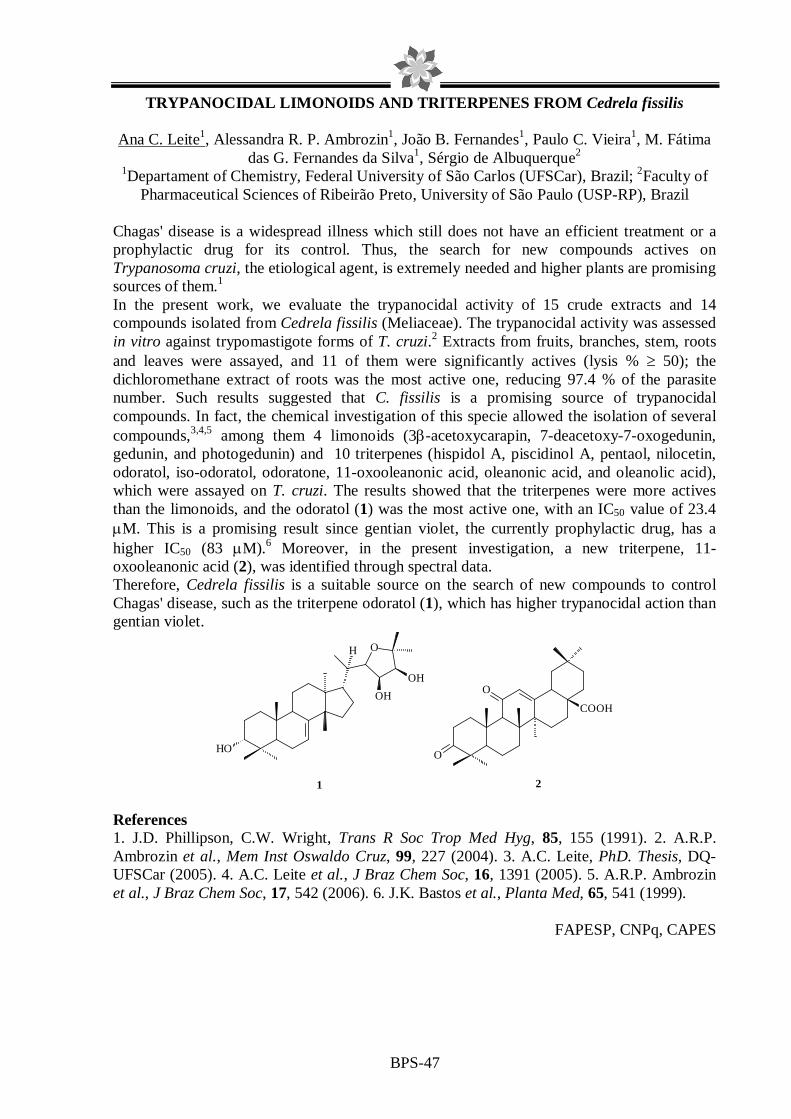
BPS-47
TRYPANOCIDAL LIMONOIDS AND TRITERPENES FROM Cedrela fissilis
Ana C. Leite1, Alessandra R. P. Ambrozin1, João B. Fernandes1, Paulo C. Vieira1, M. Fátima das G. Fernandes da Silva1, Sérgio de Albuquerque2
1Departament of Chemistry, Federal University of São Carlos (UFSCar), Brazil; 2Faculty of Pharmaceutical Sciences of Ribeirão Preto, University of São Paulo (USP-RP), Brazil
Chagas' disease is a widespread illness which still does not have an efficient treatment or a prophylactic drug for its control. Thus, the search for new compounds actives on Trypanosoma cruzi, the etiological agent, is extremely needed and higher plants are promising sources of them.1 In the present work, we evaluate the trypanocidal activity of 15 crude extracts and 14 compounds isolated from Cedrela fissilis (Meliaceae). The trypanocidal activity was assessed in vitro against trypomastigote forms of T. cruzi.2 Extracts from fruits, branches, stem, roots and leaves were assayed, and 11 of them were significantly actives (lysis % ≥ 50); the dichloromethane extract of roots was the most active one, reducing 97.4 % of the parasite number. Such results suggested that C. fissilis is a promising source of trypanocidal compounds. In fact, the chemical investigation of this specie allowed the isolation of several compounds,3,4,5 among them 4 limonoids (3β-acetoxycarapin, 7-deacetoxy-7-oxogedunin, gedunin, and photogedunin) and 10 triterpenes (hispidol A, piscidinol A, pentaol, nilocetin, odoratol, iso-odoratol, odoratone, 11-oxooleanonic acid, oleanonic acid, and oleanolic acid), which were assayed on T. cruzi. The results showed that the triterpenes were more actives than the limonoids, and the odoratol (1) was the most active one, with an IC50 value of 23.4 µM. This is a promising result since gentian violet, the currently prophylactic drug, has a higher IC50 (83 µM).6 Moreover, in the present investigation, a new triterpene, 11-oxooleanonic acid (2), was identified through spectral data. Therefore, Cedrela fissilis is a suitable source on the search of new compounds to control Chagas' disease, such as the triterpene odoratol (1), which has higher trypanocidal action than gentian violet.
O
HO
OH
OH
H
1
O
O
COOH
2 References 1. J.D. Phillipson, C.W. Wright, Trans R Soc Trop Med Hyg, 85, 155 (1991). 2. A.R.P. Ambrozin et al., Mem Inst Oswaldo Cruz, 99, 227 (2004). 3. A.C. Leite, PhD. Thesis, DQ-UFSCar (2005). 4. A.C. Leite et al., J Braz Chem Soc, 16, 1391 (2005). 5. A.R.P. Ambrozin et al., J Braz Chem Soc, 17, 542 (2006). 6. J.K. Bastos et al., Planta Med, 65, 541 (1999).
FAPESP, CNPq, CAPES

BPS-48
Evaluation of mutagenic activity of amentoflavone and methanol extract of Byrsonima crassa measured by mutation reverse assay”
aCARDOSO, C.R.P.; bSANNOMIYA, M.; bVILEGAS,W.; aVARANDA, E.A.
aDepartment of Biological Sciences, Faculty of Pharmaceutical Sciences of Araraquara,UNESP-São Paulo State University- Rodovia Araraquara-Jaú Km 1, 14801-902, Araraquara, SP, Brazil b Chemical Institute of Araraquara, UNESP-São Paulo State University, Rua Professor Francisco Degni, s/n,CP355, 14801-970, Araraquara, SP, Brazil
Byrsonima crassa Niedenzu is a plant popularly used for the treatment of gastric dysfunctions and pertaining to the Brazilian central vegetation. The methanol extract, obtained by maceration, contains catechin, tannins, terpenes and flavonoids; both mutagenic potential and antioxidant properties have been ascribed to flavonoids. The mutagenicity of some flavonoids is believed to be associated with the formation of reactive oxygen species and seems to depend on the number and position of hydroxyl groups. In the present study the mutagenic activity of the methanol, chloroform and 80% aqueous methanol extracts, as well as ethyl acetate and aqueous sub-fractions of this medicinal plant were evaluated by Salmonella typhimurium assay, using strains TA100, TA98, TA102 and TA97a. The reverse mutation assay is used to evaluation of different mutagenic mechanisms. These assays are performed with and without S9 mix. The results showed mutagenic activity of the methanolic extract in the TA98 strain without S9. The acetate fraction showed strong signs of mutagenicity without S9, suggesting that in this enriched fraction were concentrated the compounds that induced mutagenic activity. The aqueous fraction showed no mutagenic activity. The TLC and HSCCC analyses of the acetate fraction with some standard compounds permitted the isolation of the quercetin-3-O-β-D-galactopyranoside, quercetin-3-O-α-L-arabinopyranoside, amentoflavone, methyl gallate and (+)-catechin, of which only the amentoflavone exhibited positive mutagenicity to TA98 (+S9, -S9). Financial support: FAPESP-BIOTA

BPS-49
ANTIOXIDANT POTENTIAL OF METHANOLIC ROOT EXTRACT OF PYLOCARPUS SPICATUS IN A CELL OXIDATIVE STRESS INDUCING MODEL
Oliveira, D.M.1; Costa,S.L.1; Costa,M.F.D.1; Velozo,E.S.2; El-Bachá, R.S.1; Junior, J.R.N. 2
1 Laboratório de Neuroquímica e Biologia Celular (LabNq), UFBA; 2 Laboratório de Pesquisa em Matéria Médica (LAPEMM), UFBA
There is a great biodiversity in Brazil that was not completely studied, yet. Therefore, it
is important to know the biological (and posssible pharmacological) effects of plant extracts, like antioxidant activity, in different biological systems. In this context, we searched the antioxidant properties of methanolic root extract of Pylocarpus spicatus, a plant found in the Northeast of Brazil, using cell oxidative stress promoted by catechol (1,2-dihydroxybenzene)[1] [2] as an experimental model.
The extract was donated by LAPEMM and dissolved in DMSO (to obtain a final concentration of this diluent in the medium of 0.5 %). Catechol was dissolved in HCl (0,01M). The cells used in tests were of the GL-15 line (human glioblastoma); they were cultivated and treated during 48 hours with increasing concentrations of both catechol and the extract to determine the IC50 for each one. After that, cells in culture were pretreated with increasing concentrations of extract (below toxic concentrations) and catechol at 1 mM to demonstrate protective effect. The cell viability, measured by MTT method [3] was compared between the groups.
The catechol presented an IC50 of 912 µM (Fig.1). The IC50 could not be calculated for the extract because it did not kill 50% of cells even at the maximal concentration (3000 µg/mL) (Fig.2). The methanolic root extract of Pylocarpus spicatus presented protective activity at three tested concentrations (6 µg/mL, 30 µg/mL e 100 µg/mL) against the effects os catechol at 1mM during 64h (Fig.3).
0 1 2 3 40
25
50
75
100
Log [catechol] µµµµM
Cel
l Via
bilit
y (%
)
Sol 20 30 6010
0200 300 60
0100
020
0030
000
50
100
150
*
[extract] µµµµg/mL
Cel
l Via
bilit
y (%
)
sol
cat 3 6 10 20 30 60 10
020
030
060
00
50
100
**
**
[extract] µµµµg/mL + Catechol 1mM
Cel
l Via
bilit
y (%
)
Figure 1 Figure 2 Figure 3
The development of antioxidant therapies could give support to treatments of several diseases. Moreover, the use of natural products to promote health is an important perspective. Thus, these are relevant data to know relevant activities of Pylocarpus spicatus.
Refs. 1. Barreto G. E. S. et al., Acta Cir. Bras., 20, 40-45 (2005). 2. Pereira M. R. G. et al., J. Bras. Patol. Clínica, 40, 281-286 (2004). 3. Mosmann T., J. Immunol. Methods, 65, 55-63 (1983).

BPS-50
ACTION OF Pothomorphe umbellata AND Struthanthus sp EXTRACTS TO A CLINICAL ISOLATE Trichophyton rubrum
MARSIGLIA-PIMENTA,D.O.M1, FUSCO-ALMEIDA, A. M.1,PIETRO, R. C. L.2
1 Curso de Ciências Farmacêuticas UNAERP, Universidade de Ribeirão Preto, Ribeirão Preto, São Paulo, Brasil; 2 Departamento de Fármacos e Medicamentos, Faculdade de Ciências
Farmacêuticas de Araraquara, UNESP - Universidade Estadual de São Paulo, Araraquara, São Paulo, Brasil
Pothomorphe umbellata is a characteristic plant of the Brazilian flora, known popularly as “caapeba” or “pariparoba” that presents anti-inflammatory, analgesic, anti-PAF and antioxidant actions. Struthantus sp is a hemi-parasite Brazilian plant of which there are few biological studies. In two past decades the incidence of infections caused by dermatophytes and other fungi has increased, due to the inefficiency of some drugs to these agents. Works in literature indicate pharmacological action for several plant species, including its use in the popular medicine. The objective of this work was to evaluate the antifungal activity of Pothomorphe umbellata and Struthantus sp extracts to a clinical isolate of Trichophyton rubrum with resistance antifungal history. The tests were performed according the NCCLS M38-P (NCCLS, 2002), using plates of microtitulation for the determination of minimal inhibitory concentration (MIC). Aerial parts of plants were dried and converted to powder and submitted to extraction in ambient temperature during approximately 10 days. Ethanolic and methanolic extracts, obtained by maceration with ethanol 70% and methanol, respectively, were lyophilized. The extracts were dissolved in DMSO to obtain stock solution concentration of 50mg/mL. The growth inhibition was observed after incubation of 7 days at 28°C to determine MIC. The ethanolic and methanolic extracts were tested and presented good antifungal action in vitro, with MIC values of 312.5 and 156.25 ug/mL respectively. Our results suggest that the extracts may be explored to the discovery of substances that posses the antifungal activity. Refs. 1. National Committe for Clinical Laboratory Standards, 2002. NCCLS document M38-A.

BPS-51
ANTIHERPES ACTIVITY OF THE RED SEAWEED CHONDROPHYCUS FLAGELLIFERUS (RHODOMELACEAE, CERAMIALES)
Lhullier, C. 1,a ; Silva, I.T.1,b ; Simões, C.M.O.1,b ; Falkenberg, M.B.1,a ; Schenkel, E.P.1,a
aLaboratório de Produtos Naturais; bLaboratório de Virologia Aplicada; 1Depto de Ciências Farmacêuticas, CCS; Universidade de Santa Catarina, Florianópolis, SC, 88040-900.
Introduction: The red alga Chondrophycus flagelliferus belongs to the Laurencia complex1, and its preliminary chromatographic analysis showed the presence of steroids, sesquiterpenes and glycolipids. Marine glycolipids, as well as those from terrestrial organisms, are amphiphylic compounds which are currently classified into two groups: glycoglycerolipids (GGLs) and glycosphingolipids (GSLs). These substances have demonstrated immunostimulant, antitumor and antiviral activities2. Herpes Simplex Virus types 1 and 2 (HSV-1 and 2) infect a great number of people all over the world and their resistance to the available treatment makes the herpetic infections a serious problem of public health. Objective: The aim of the present study was to evaluate the potential anti-HSV-1 and anti-HSV-2 activities the aqueous extract of the red seaweed Chondrophycus flagelliferus. Methodology: This alga was collected at Bombinhas Beach (Santa Catarina, Brazil) and extracted with ethanol 70%. This extract was partitioned with ethyl acetate, yielding an organic and an aqueous extracts. The aqueous extract cytotoxicity was evaluated by MTT assay on VERO and GMK AH1 cells. The anti-HSV-l (strains 29R and KOS) and anti-HSV-2 (strain 333) activities were evaluated by plaque number reduction assay on VERO and GMK AH1 cells, respectively. Results: The tested extract showed no cytotoxic effects on these cells, at the maximum evaluated concentration of 0.5 mg/mL, and it significantly inhibited the replication of the tested viruses: HSV-2 strain 333 (77 %), HSV-1 strain 29R (92%) and HSV-1 strain KOS (97%). Conclusions: The presence of glycolipids in the aqueous extract of Chondrophycus flagelliferus could explain the detected activity, and additional studies will be carried out with the isolated substances from this extract to confirm this hypothesis. Refs. 1. M.T. Fujii et al., Phycologia, 45, 432 (2006). 2. E. Fattorusso et al., Fortschritte der Chemie Organischer Naturstoffe, 72, 215 (1997). Financial support: CNPq, FAPESC, CAPES

BPS-52
ALKYL PHENOLS, BIFLAVONOIDS, DIPHENYL ETHER AND A N EW DEPSIDE FROM SCHINOPSIS BRASILIENSIS ENGL.
Manuela P. Cardoso1,3, Juceni P. David2 and Jorge M. David1
1Instituto de Química, 2Faculdade de Farmácia, Universidade Federal da Bahia, 40170-280, Salavador (BA), Brazil. 3Departamento de Química, Universidade Estadual de Feira de
Santana, Feira de Santana (BA), Brazil.
Species of the Anacardiaceae family has spread around the world and some of them have economical and commercial importance. The presence of alkyl and alkenyl phenols, cathechols, resorcionols, hydroquinones is the cause of the contact dermatitis that characterize the poison species of this family. Schinopsis brasiliensis Engl. is popularly known in Brazil as “baraúna”. It is a large tree well known in the Brazilian northeastern and it is employed by the local population in the cattle verminosis and, the wood is also employed in constructions. For this reason, today it is considerate by the brazilian authorities as an endangered species. This work describes the chemical study of the methanol extract of wood of S. brasiliensis The CHCl3 and EtOAc fractions obtained from the partition of MeOH extract were submitted to different chromatographic procedures over silica gel and Sephadex LH-20. These procedures permitted to isolate vanillin, vanillic acid, 2,4-dihydroxybenzaldeid, methyl 2,4-dihydroxybenzoate, 3,4,5-trimethoxybenzil alcohol, methyl gallate, gallic acid, siringaresinol, β-sitosterol, estigmast-4-en-3-one, methyl 3,5-dicloro-6-(6-hydroxy-4-methoxy-3-methoxycarbonil-2-methyl-phenoxy)-2-hydroxy-4-methyl-benzoate (1), luxenchalcone (2), besides the new compounds methyl 2,4-dihydroxy-3,6-dimethyl-benzoate, methyl 2-hydroxy-3,6-dimethyl-4-(3,4-dimethoxy-benzoyloxy)-benzoate (3), 4’-methoxy-7-hydroxy-flavanone-(3→3'')-4'''-methoxy-3''',7''-dihydroxy-flavanone (4) and a mixture of seven methyl n-alkyl-2-hydroxy-4-methoxybenzoate. All the structures were elucidated by empiric data calculation and different techniques of NMR and MS of the pure compounds or their acetyl derivatives. Compound 1 is an unusual plant compound which was previously isolated from Byrsonima microphylla (Malpighiaceae)1.The biflavonoid (2) was only obtained from Luxemburgia octandra (Ochnaceae)2.
(CNPq, FAPESB, CAPES, PRONEX).
OOH
HO
OH
O
O
HO OH
O
Cl
CH3
Cl
OHO
CH3
OCH3O
CH3O
H
OHCH3O
CO2Me
OH
CH3
H3C
O
C O
OCH3
OCH3
O
OO
O
OCH3
HO
CH3O
OH
OH
1 2 3 4
Refs. 1. J. H. C. Rocha et al., Bioscience, Biotechnology, and Biochemistry, 70, 2759 (2006) 2. M. G. de Carvalho et al., Journal of the Brazilian Chemical Society, 15, 146 (2004)
BPS-53
ANTIMICROBIAL ACTIVITIES OF THE EXTRACTS OF FUNGAL SPECIES: Xylaria spp, Crinipellis perniciosa, Trichoderma stromaticum AND Phytophthora palmivora. Vanessa R. Guedes (PG)*, Regina M. G. dos Santos (PQ), Dirceu Martins (PQ), Frederico G. Cruz (PQ) GESNAT, Departamento de Química Orgânica, Instituto de Química, Universidade Federal da Bahia, Campus de Ondina, Salvador-Bahia-40170-290 *[email protected] The purpose of this work was screened for antimicrobial activities the extracts of five fungi species, namely, Crinipellis perniciosa, Trichoderma stromaticum, Phytophthora palmivora and two unidentified species Xylaria sp1 and Xylaria sp2. The fungi were inoculated in solid (rice) and liquid (malt, PD and Czapek) media for period of 20 to 40 days. The solid medium of Xylaria sp1, Xylaria sp2 and C. perniciosa, were extracted with methanol. The liquid medium were extracted with methanol and methanol/dichloromethane (mycelium) and the broth with ethyl acetate at pH=5 and pH=9. The extract of the solid media obtained for Xylaria sp1 was sequentially extracted with hexane, chloroform and ethyl acetate at pH=5 and pH=9. The methanol and methanol/dichloromethane extracts of the mycelium were sequentially extracted with chloroform and ethyl acetate at pH=5 and pH=9. The broth of T. stromaticum and P. palmivora species were extracted with ethyl acetate and the solid medium were divided in two portions, the first was extracted with methanol and the other with acetone. In total 38 extracts were screened. Antimicrobial tests were carried out using the technique of Minimal Inhibitory Concentration (MIC) against different strains of Gram-positive bacteria (Bacillus subtilis, Staphylococcus aureus, Streptococcus mutans and Micrococcus luteus), Gram-negative bacteria (Salmonella typhimurium, Pseudomonas aeruginosa and Escherichia coli) and fungi (Candida albicans, Aspergillus niger, Cladosporium cladosporioides and Crinipellis perniciosa). Most of the extracts of the species tested were active against Gram-positive bacteria strain. The species, T. stromaticum, was found to be the most active with values of the MIC equal, or next to the inhibitory concentration of the control (chloranfenicol). From Gram-negative bacteria strain, Salmonella typhimurium was most susceptible to extracts assayed and P. palmivora extracts were the most active. For the E. coli bacteria, only the T. stromaticum specie was active and for P. aeruginosa the C. perniciosa and T. stromaticum species were actives. Concerning the fungi, the T. stromaticum specie was found the most active against C. cladosporioides strain and only one specie of the genus Xylaria was active against A. niger. The results indicate the potential of these species to produces drugs to the treatment of microbial infections. The active extracts will be fractioned seeking the isolation actives metabolites.
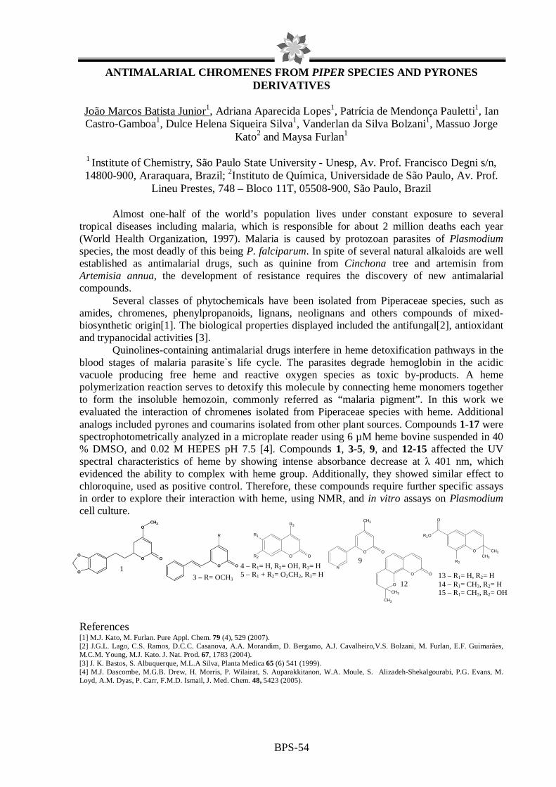
BPS-54
ANTIMALARIAL CHROMENES FROM PIPER SPECIES AND PYRONES DERIVATIVES
João Marcos Batista Junior1, Adriana Aparecida Lopes1, Patrícia de Mendonça Pauletti1, Ian Castro-Gamboa1, Dulce Helena Siqueira Silva1, Vanderlan da Silva Bolzani1, Massuo Jorge
Kato2 and Maysa Furlan1
1 Institute of Chemistry, São Paulo State University - Unesp, Av. Prof. Francisco Degni s/n, 14800-900, Araraquara, Brazil; 2Instituto de Química, Universidade de São Paulo, Av. Prof.
Lineu Prestes, 748 – Bloco 11T, 05508-900, São Paulo, Brazil
Almost one-half of the world’s population lives under constant exposure to several tropical diseases including malaria, which is responsible for about 2 million deaths each year (World Health Organization, 1997). Malaria is caused by protozoan parasites of Plasmodium species, the most deadly of this being P. falciparum. In spite of several natural alkaloids are well established as antimalarial drugs, such as quinine from Cinchona tree and artemisin from Artemisia annua, the development of resistance requires the discovery of new antimalarial compounds.
Several classes of phytochemicals have been isolated from Piperaceae species, such as amides, chromenes, phenylpropanoids, lignans, neolignans and others compounds of mixed-biosynthetic origin[1]. The biological properties displayed included the antifungal[2], antioxidant and trypanocidal activities [3].
Quinolines-containing antimalarial drugs interfere in heme detoxification pathways in the blood stages of malaria parasite`s life cycle. The parasites degrade hemoglobin in the acidic vacuole producing free heme and reactive oxygen species as toxic by-products. A heme polymerization reaction serves to detoxify this molecule by connecting heme monomers together to form the insoluble hemozoin, commonly referred as “malaria pigment”. In this work we evaluated the interaction of chromenes isolated from Piperaceae species with heme. Additional analogs included pyrones and coumarins isolated from other plant sources. Compounds 1-17 were spectrophotometrically analyzed in a microplate reader using 6 µM heme bovine suspended in 40 % DMSO, and 0.02 M HEPES pH 7.5 [4]. Compounds 1, 3-5, 9, and 12-15 affected the UV spectral characteristics of heme by showing intense absorbance decrease at λ 401 nm, which evidenced the ability to complex with heme group. Additionally, they showed similar effect to chloroquine, used as positive control. Therefore, these compounds require further specific assays in order to explore their interaction with heme, using NMR, and in vitro assays on Plasmodium cell culture. References [1] M.J. Kato, M. Furlan. Pure Appl. Chem. 79 (4), 529 (2007). [2] J.G.L. Lago, C.S. Ramos, D.C.C. Casanova, A.A. Morandim, D. Bergamo, A.J. Cavalheiro,V.S. Bolzani, M. Furlan, E.F. Guimarães, M.C.M. Young, M.J. Kato. J. Nat. Prod. 67, 1783 (2004). [3] J. K. Bastos, S. Albuquerque, M.L.A Silva, Planta Medica 65 (6) 541 (1999). [4] M.J. Dascombe, M.G.B. Drew, H. Morris, P. Wilairat, S. Auparakkitanon, W.A. Moule, S. Alizadeh-Shekalgourabi, P.G. Evans, M. Loyd, A.M. Dyas, P. Carr, F.M.D. Ismail, J. Med. Chem. 48, 5423 (2005).
O OO
O
OCH3
1 O O
R
3 – R= OCH3
O O
R3
R2
R1
4 – R1= H, R2= OH, R3= H 5 – R1 + R2= O2CH2, R3= H
O
O
CH3
CH3
O
12
9 N
O O
CH3
O CH3CH3
R2
O
R1O
13 – R1= H, R2= H 14 – R1= CH3, R2= H 15 – R1= CH3, R2= OH

BPS-55
Antinociceptive and toxicological activities of Zeyheria montana (Bignoniaceae) ethanolic extract.
GUENKA, L. C.; GOMES, R. C.; MELO, V. L.; KITANISHI, C.R.R.; BELEBONI, R. O.;
COUTO, L. B. Unidade de Biotecnologia-UNAERP.
Pain and inflammatory processes, particularly those chronic ones, are often linked to several diseases and to continuous public health problems. In recent decades, the search for new analgesic drugs has been motivated by actual drugs costs or effectiveness distortions. In this work, we evaluated the antinociceptive potential and some few toxicological properties of the ethanolic extract from Z. montana leaves (75; 150; 300 mg/Kg, ip). Acetic acid-induced writhes in mice were significantly inhibited by extract doses of 75 mg/Kg (67.27%), 150 mg/Kg (49.38%) and 300 mg/Kg (82.87%). Besides peripheral analgesic activity, Z. montana extract induced, apparently, central antinociceptive effect at the dose of 300 mg/Kg (ip) in rats. In this case, the latency time until the appearance of the writhing/stretching response of the rats was significantly increased by 46.5%. Some few sign of toxicity, including the ulcerogenic ones, was absent to mice treated with those extract on an extensive range of doses. The LD50 was > 2000 mg/Kg. These results suggest that the Z. montana extract exhibits both central and peripheral antinociceptive activities with little toxic effects. Finally, the search for new compounds from different biological sources, chemical natures or different mode of actions could be an attractive alternative to development of new drugs more effective or cheaper than the actual ones. Keywords: Antinociception; Plant extract; Z. Montana; Hargreaves test; acetic acid-induced writhes. Financial support: UNAERP

BPS-56
Evaluation of antinociceptive and toxicological activities of Serjania erecta (Sapindaceae) leaves extract
GUENKA, L. C.; GOMES, R. C.; MELO, V. L.; KITANISHI, C.R.R.; BELEBONI, R. O.;
COUTO, L. B. Unidade de Biotecnologia-UNAERP.
Antinociceptive and toxicological activities of the ethanolic extract from S. erecta leaves were studied. Antinociceptive evaluation was carried out against chemical and thermal stimuli in mice and rats, respectively, and toxicological studies (LD50), including the ulcerogenic test, were conduced in mice. Acetic acid-induced writhes were significantly inhibited by extract doses of 75 mg/Kg, (60.40%), 150 mg/Kg (64.37%) and 300 mg/Kg, ip (61.77%). Apparently, no central antinociception was observed for all extract doses tested. Some a few signs of toxicity, including the ulcerogenic effect, were not observed to rats treated with those extract on an extensive range of doses. The LD50 was > 2,000 mg/Kg. These results suggest that the S. erecta extract exhibits a peripheral antinociceptive activity with little toxic effects. Finally, the search for new compounds from different biological sources, chemical natures or different mode of actions could be an attractive alternative to development of new drugs more effective or cheaper than the actual ones. Keywords: Antinociception; Plant extract; S. erecta; Hargreaves test; acetic acid-induced writhes. Financial support: UNAERP.

BPS-57
Nematocidal effects of medicinal plants against Pratylenchus zeae and Pratylenchus jaehni.
Slomp, L.; Guenka, L.C.; Pereira, P.S.; França, S.C.; Zingaretti, S.M.; Beleboni, R.O.
Unidade de Biotecnologia-Universidade de Ribeirão Preto. Synthetic nematicides have been broadly used in the past decades for the control of plant parasitic nematodes. Despite their relevant effectiveness, these compounds can cause great damage to the environment, and to the human or the animal health. The growing searches for new nematicides, particularly of natural ones, may conduce to higher safety and efficiency to the nematode control. In this work it was evaluated the in vitro nematocidal effect of ethanolic extracts obtained from selected plant species: Tithonia diversifolia, Ecilpta alba and Mikania glomerata (Asteraceae); Tabernaemontana catharinensis and Mandevilla velutina (Apocynaceae); Casearia sylvestris (Salicaceae); Zeyheria montana (Bignoniaceae); Lippia alba (Verbenaceae); Croton antisyphiliticus (Euphorbiaceae) and Serjania erecta (Sapindaceae) against the plant parasitic nematodes Pratylenchus zeae (Nematoda: Pratylenchidae) and Pratylenchus jaehni (Nematoda: Pratylenchidae). Moreover, a preliminary phytochemical characterization of these plant extracts was performed in order to associate these data to those observed in nematocidal assays. Our results indicated a significant nematocidal activity of the analyzed extracts, in special those demonstrated by E. alba (DL50 (ppm) = 304.08; 55.32 – P. zeae and DL50 (ppm) = >1000; 212.82 – P. jaehni; 12 and 24 h, respectively), T. catharinensis (DL50 (ppm) = 215.26; 60.04 – P. zeae and DL50 (ppm) = 825.44; 376.60 – P. jaehni; 12 and 24 h, respectively), C. sylvestris (DL50 (ppm) = 198.05; 56.94 – P. zeae and DL50 (ppm) = 747.98; 322.98 – P. jaehni; 12 and 24 h, respectively), Z. montana (DL50 (ppm) = 166.43; 34.08 – P. zeae and DL50 (ppm) = >1000; 427.34 – P. jaehni; 12 and 24 h, respectively) and S. erecta (DL50 (ppm) = 178.74; 74.12 – P. zeae e DL50 (ppm) = 689.24; 249.50 – P. jaehni; 12 and 24 h, respectively). Thus, these data show that the evaluated plants presented significant nematocidal effects, which are of high economic or environmental interest and useful towards the growth of agricultural activities worldwise. Keywords: Plant extracts; Plant parasitic nematodes; Pratylenchus jaehni; Pratylenchus zeae. Financial support: Capes and UNAERP.

BPS-58
OCIMUM GRATISSIMUM L.: A RICH SOURCE OF URSOLIC ACID AND EUGENOL
Terezinha de Jesus Faria, Dayana Lacerda Custódio, Juliana Feijó de Souza Daniel, Fernando
César de Macedo Júnior, Milton Faccione Departamento de Química, Universidade Estadual de Londrina
Ocimum gratissimum, originating in the Orient, is widespread throughout tropical countries including Brazil, where it is popularly known as "alfavacão, alfavaca and alfavaca-cravo". This species is used in traditional medicines in South America and Africa for a variety of therapeutic purposes which includes its use in treating bacterial infections, diarrhoea, diabetes1. Recently we have examined the antifungal activity of the essential oil obtained by steam-distillation (1.1% w/w) of the aerial parts of O. gratissimum. The results revealed that the essential oil inhibited the growth of Botryosphaeria rhodina, Rhizoctonia sp., two strains of Alternaria sp., Aspergilllus niger, and Penicillium chrysogenum. The compound that showed antifungal activity was isolated and identified as eugenol by TLC bioautography technique. GC/MS analysis showed that eugenol was the main constituent of the essential oil studied2. In continuation of the studies on O. gratissimum we have examined the aerial parts of the plant. Differently of the essential oil, there are few studies reported on the macerated extract of this species. Powdered aerial parts were continuously extracted with hexane-dichloromethane-ethanol mixture (1:3:1) at room temperature. The obtained extract (25.0g) was chromatographed on silica gel column eluted with hexane, dichloromethane and ethanol to yield aliphatic hydrocarbon mixture (C31, C33, C34, C35), eugenol, caryophyllene oxide, a mixture of stigmasterol/sitosterol and large quantity of ursolic acid (3.5g). Ursolic acid has attracted considerable interest because of its range of biological activities such as cytotoxic, anti-mutagenic, antiviral, anti-invasive, trypanocidal and other activities that are of interest to modern pharmacology3,4. This is the first report of ursolic acid in Ocimum gratissimum species.
1. J. C. et al., Fitoterapia, 71, 444 (2000). 2. T. J. de Faria et al., Brazilian Archives of Biology and Technology, 49, 867 (2006). 3. C. Mello et al., Journal of Molecular Strutucture, 799, 141 (2006). 4. M. E. Lacikova et al., Chromatographia, 63, 117 (2006).
HO
COOH
Ursolic acid

BPS-59
ANTIMICROBIAL ACTIVITY OF C-METHYLATED FLAVONOIDS ISOLATED FROM LEAVES OF Myrcia hiemalis (MYRTACEAE)
Silva, P.D.1, Neta, L. C. S. 1, Costa, G. A. N. 1, Martins, D.1, Vannier-Santos, M. A.2 , Menezes, D. 2, Alves, E. S. S. 2, Brandão, T. C. S. C. 2, and Cruz, F.G.1.
1 GESNAT, Departamento de Química Orgânica, Instituto de Química, Universidade Federal da Bahia, Salvador-BA-Brazil; 2 Laboratório de Biomorfologia Antiparasitária e Centro de
Microscopia Eletrônica da FIOCRUZ-BA-Brazil.
The species of the Myrtaceae family practically occur in the whole world. Some species of this family are used in Brazil in the treatment of diabetics, rheumatism, as astringents, anti-inflammatory and in the treatment of chronic ulcers, hemorrhages, fever, cystitis and urethritis. The plants of this family are characterized by the production of tannins, flavonoids, mono-, sesqui- and triterpenes, derivatives of the fluoroglucinol, chromenes and stilbenes. The aim of this work is the phytochemistry study of the non-volatile extracts of Myrcia hiemalis leaves, as well as the establishment of its biological properties. The leaves were harvested in the sand dunes of Salvador, Bahia, northeastern region of Brazil in 2005. The dried leaves were ground and extracted with ethanol. Them, the extract was partitioned with hexane, dichloromethane and ethyl acetate. Antimicrobial tests were carried out with the extract using the technique of Minimal Inhibitory Concentration (MIC) with the following microorganisms: Bacillus subtilis, Staphylococcus aureus, Micrococcus luteus, Streptococcus mutans, Escherichia coli, Pseudomonas aeruginosa, Salmonella, choleraesuis, Aspergillus niger, Cladosporium cladosporioides, Candida albicans and Crinipellis perniciosa. The hexane and ethyl acetate extracts were inactive. The dichloromethane extract presented activity against Gram-positive bacteria as well as the fungus C. perniciosa. Seven components had been isolated and identified from the dichloromethane extract: myricitrin, daucosterol, 2',4'-dihydroxy-3',5'-dimethyl-6'-methoxychalcone, 2',6'-dihydroxy-3'-methyl-4'-methoxychalcone, 2',3’,4'-trihydroxy-5'-methyl-6'-methoxychalcone, 7-hydroxy-6,8-dimethyl-5-methoxyflavanone and 5,7-dihydroxy-6,8-dimethylflavanone. The structures of the substances were determined using 1H and 13C NMR techniques. The isolated substances were assaied against above microorganisms and the 2',4'-dihydroxy-3',5'-dimethyl-6'-methoxychalcone presented antimicrobial activity against M. luteus and the substance 7-hydroxy-6,8-dimethyl-5-methoxyflavanone acted against B. subtilis and M.luteus. Anti-Trypanosoma cruzi and anti-Leishmania amazonensis tests were done with isolated substances using a technique developed in the FIOCRUZ-BA. The 2',4'-dihydroxy-3',5'-dimethyl-6'-methoxychalcone was active against both parasites (anti-T. cruzi: IC50 13,12 µM; anti-L. amazonensis: 100% inhibition in 50 µM) and 2',6'-dihydroxy-3'-methyl-4'-methoxychalcone presented activity against T. cruzi. (IC50 35,95 µM). Supported by CNPq, FAPESB, FINEP and CAPES.

BPS-60
INSECTICIDAL ACTIVITY OF Trichilia sp. (MELIACEAE) EXTRACTS AGAINST Atta sexdens rubropilosa
Cristiane de Melo Cazal*1(PG), João B. Fernandes1(PQ), Odair C. Bueno2 (PQ), Maria Fátima G. F. da Silva1(PQ), Paulo C. Vieira1(PQ). 1Universidade Federal de São Carlos, Departamento de Química, Laboratório de Produtos Naturais, Rod. Washington Luiz Km 235, São Carlos-S.P. 2Universidade Estadual Paulista, Centro de Estudos de Insetos Sociais, Rio Claro-SP. [email protected]
The family Meliaceae includes many plants that are sources of valuable timber and many of them have wide ranging of uses in ethnomedicine. Numerous secondary metabolites that exhibit significant insecticidal activity were already found in Trichilia genus1. The leaf-cutting ants of the genera Atta and Acromyrmex use mostly fresh plant fragments to raise their symbiotic fungi2 and are the cause of considerable economic damage, due to defoliation that they cause3. Control of this pest is still problematic, presenting only temporary effects and is sometimes, harmful to the environment, to man and other animals. Consequently, an extensive search for alternate methods to control these insects has been made in an attempt of substitute traditional agrochemicals with high specificity and, therefore, causing less damage to the environment. In this context, this work evaluated the toxic effect of dichloromethane and methanol extracts of leaves, stem, bark, branches and thin branches of Trichilia sp against leaf-cutting ants Atta sexdens rubropilosa as source for less toxic compounds that could be used as a soft control method. The extracts were incorporate in artificial diet for the ingestion toxicity test for the ants. For each treatment were used 50 ants distributed in 10 Petri plates, lined with filter paper. The Petri plates were maintained in B.O.D, at 24 °C and R. U.H. above 70 %. The ants mortality were accomplished daily, during 25 days and the obtained data of the bioassay were analyzed through the comparison of the survival curves by the test "log rank" (Prism 3.0). The dichloromethane and methanol extracts of bark, branches, thin branches and methanol extracts leaves and stem of Trichilia sp showed high activity against leaf-cutting ants Atta sexdens rubropilosa, while leaves and stem dichloromethane extracts showed lower rates of activities. The dichloromethane extract of stem was fractionated with the objective of testing the pure compounds and led to isolation of steroids, triterpene, acid cinamic derived and coumarin characteristics of this genus. These compounds were identified by comparison of their NMR, MS, UV and IR data.
1- A. P. Matos et al., Bioassay, 1:X;1-6, (2006). 2. Hölldobler, B.; Wilson, E.O.; The Ants (1990). 3. Della-Lucia, T. M. C.; Fowler, H.G. In As Formigas Cortadeiras;1 (1993)
FAPESP, CNPq, CAPES

BPS-61
Plinia cauliflora LEAVES AGAINST Candida albicans
Tatiana M. de Souza1*, Juliana A. Severi2, Wagner Vilegas2, Rosemeire C.L.R. Pietro1
1- Faculdade de Ciências Farmacêuticas – UNESP, Araraquara, SP 2- Instituto de Química – UNESP, Araraquara, SP
In the last few years, resistance of Candida albicans to antifungal drugs has become a fact of concern. So, plant extracts and compounds activity evaluation has been focused in order to obtain new agents against Candida. The objective of this work was to determine the activity of Plinia cauliflora leaves against C. albicans. The leaves extract was obtained with 70% ethanol by percolation and dried under reduced pressure. Liquid-liquid partition of the extract (15.0 g) was done with 500 mL of water and 500 mL of ethylacetate and after, with 500 mL of n-butanol. Crude extract and the fractions of ethylacetate (EtOAcFr), butanol (BuOHFr) and water (WFr) were totally dried and 25 mg/mL of each one were separately dissolved in 1 mL of DMSO with subsequent dilution in RPMI-1640 medium (1:2; v/v). Determination of Minimum Inhibitory Concentration (MIC) was done according NCCLS M27-A2 at 96-wells plates. Final samples concentration was 1.56 mg/mL and final C. albicans (ATCC 64548) inoculum was 2.5-5.0x10³ cells/mL. Controls of 32 µg/mL of amphotericin B, DMSO:RPMI (1:2; v/v), medium and growth were also done. It was proceeded serial dilutions in the plates that were incubated for 48 h at 25 °C. After that time, subcultures of the plates were done in agar-Sabouraud plates and incubated for 48 h at 25 °C to determine Minimum Fungicidal Concentration (MFC). MIC measurement was done by addition of 2% TTC. The test was done in duplicate. WFr showed the best values of MIC and MFC. BuOHFr showed the smaller value of MIC but MFC was higher than start concentration. EtOAcFr had intermediated MIC values and MFC was higher than start concentration. Crude extract showed the highest MIC value. Based on those results, it was possible to observe better activities against C. albicans for WFr and BuOHFr, therefore, it is necessary to continue the studies of this promissory extract by evaluation of its constituents. Support: PADC-UNESP Refs. 1. M.C.T. Duarte et al., Journal of Ethnopharmacology, 97, 305-311 (2005). 2. NCCLS, Norma M27-A2 (2002).

BPS-62
Pharmacological and Phytochemical characterization of Tabernaemontana catharinensis (Apocynaceae) stem bark extract.
R.C., Gomesa; Guenka, L.C.a; A., Coutinho-Netoa; V.L., Meloa; V.C., Fernandesa; G.,
Dagravaa; W.S., Santosb; P.S., Pereiraa; L.B., Coutoa; R.O., Belebonia a- Unidade de Biotecnologia, Universidade de Ribeirão Preto, São Paulo, Brasil.
b- Departamento de Biologia, Faculdade de Ciências e Letras de Ribeirão Preto, Universidade de São Paulo, São Paulo, Brasil.
Analgesic, anti-inflammatory and toxic activities, as well as the phytochemical profile of the ethanolic extract from T. catharinensis (Apocynaceae) stem bark were studied in this work. Analgesic evaluation was carried out against chemical and thermal stimuli. Anti-inflammatory activity was investigated on carrageenin-induced edema in rats and toxicological studies (LD50) were conducted in mice. Phytochemical analyses were performed according standardized methodology. Acetic acid-induced writhes were significantly inhibited by extract doses of 37.5 mg/Kg (40.97%), 75 mg/Kg (77.70%) and 150 mg/Kg (88.98%). Besides peripheral analgesic activity, the T. catharinensis extract induced central analgesia at all doses tested, particularly noticed 60 and 90 min following administration. The extract significantly reduced edema formation by 30.35% (37.5 mg/Kg), 34.46% (75 mg/Kg) and 56.42% (150 mg/Kg) when assessed 180 min following carrageenan intraplantar injection, demonstrating anti-inflammatory action. The calculated LD50 value was 2200 mg/Kg on toxicological assay. Phytochemical analyses of ethanolic extract from T. catharinensis stem bark showed the presence of alkaloids and terpenoids, which might be responsible for the observed pharmacological activities. Keywords: Anti-inflammatory; Antinociceptive; Plant extract; Tabernaemontana catharinensis. Financial support: UNAERP.

BPS-63
NEUROPROTECTIVE EFFECTS OF COUMARINS PRESENT IN LEA VES OF ZANTOXYLUM TINGOASSUIBA
Oliveira, D.M.1; Costa,S.L.1; Costa,M.F.D.1; Velozo, E.S.2; El-Bachá, R.S.1; Hohlemwerger,S.V.A.2, E.S.2
1 Laboratório de Neuroquímica e Biologia Celular (LabNq), UFBA; 2 Laboratório de Pesquisa em Matéria Médica (LAPEMM), UFBA
Oxidative processes are an important factor in the pathogenesis of several disorders,
including neurodegenerative diseases like Parkinson’s Disease (PD) and Alzheimer’s disease (AD) [1]. Therefore, the search for new compounds with antioxidant activity is currently the focus of many studies. Recent works have demonstrated that the toxicity of 1,2-dihydroxybenzene (catechol), a metabolite of benzene, involves inhibition of the respiratory chain [2] and superoxide and reactive quinones generation, promoting oxidative stress in human glioblastoma GL-15 cells [3]. In this work, we used the cytotoxicity of catechol to GL-15 cells as an experimental model to demonstrate the neuroprotective effects of coumarins obtained from Zantoxylum tingoassuiba. Approximately 1.2 liters of the water used in the hydro distillation, after cooled, was extracted, for 3 times, with 250 mL of the chloroform and to proceed with ethyl acetate. The solvents were evaporate under reduce pressure. This material, after chromatography on column, afforded the furanocumarins xathotoxin and isopipinelin. Catechol and coumarins were dissolved in HCl (0.01 M) and DMSO (1 %), respectively. Cells were cultured and treated for 48 hours with increasing concentrations of each substance in order to determine the concentration that kills 50 % of cell (IC50). Cell viability was measured using MTT method. Data were fitted to nonlinear regression plots to determine the IC50 for catechol and coumarins (Fig.1 a-b), that was 912 µM and 73 µg/mL respectivily. After that, the cells in culture were pretreated with increasing concentrations of coumarins (below toxic concentrations) with 1 mM catechol. The viability was compared with a negative control for cytotoxicity (cells treated with 0.5 % DMSO alone) and a positive control (cells treated with 1 mM catechol and 0.5 % DMSO). Catechol killed 61 ± 3,6 % of cells, differing significantly of the negative control group (p<0.001). A partial but significant protective effect of coumarins was observed when the cells were treated whith concentrations in the range of 1µg/mL - 3µg/mL (Fig.2).
Fig.1 a b Fig.2
0 1 2 3 40
25
50
75
100
Log [Catechol] ( µµµµM)
Cel
l Via
bilit
y (%
)
0 1 2 30
50
100
Log [coumarins] ( µµµµg/mL)
Cel
l Via
bilit
y (%
)
Cel
lSol Cat 1 2 3 6 10 20 30 60 10
00
50
100
* * *
[coumarins] ( µµµµg/mL)
Cel
l Via
bilit
y (%
)
The development of potential neuroprotective therapies for neurodegenerative diseases
can be based on understanding the protective effects of compounds present in natural products. Thus, these data are important to know relevant pharmacological activities of Zantoxylum tingoassuiba.
Refs. 1. Davies K. J., Biochem. Soc. Symp., 61, 1-31 (1995) 2. Barreto G. E. S. et al., Acta Cir. Bras., 20, 40-45 (2005) 3. Pereira M. R. G. et al., J. Bras. Patol. Clínica, 40, 281-286 (2004)

BPS-64
Functional Characterization of an Acidic Phospholipase A2 Isolated from Bothrops pirajai Snake Venom with Hypotensive and Antiplatelet Properties
Sabrina S. Teixeira, Franco M. Nunes da Silva, Norival A. Santos-Filho, Clayton Z. Oliveira,
Silvana Marcussi, Aristides Q. Rueda, Andreimar M. Soares. Departamento de Análises Clínicas, Toxicológicas e Bromatológicas, Faculdade de Ciências
Farmacêuticas de Ribeirão Preto, FCFRP, Universidade de São Paulo, USP, Ribeirão Preto-SP.
The phospholipases A2 (PLA2, E.C.3.1.1.4) are multifunctional enzymes, with specific Ca+2-dependent catalytic activity on phospholipids, hydrolyzing the 3-sn-phospholipids 2-acyl ester bond and releasing fatty acids and lysophospholipids. The PLA2s are widely distributed in nature and instigate medical-scientific interest due to its involvement in many human inflammatory diseases and in envenomation by bees and snakes. The present work aimed at the partial functional characterization of an acidic phospholipase A2, Asp49 (BpirPLA2), isolated from Bothrops pirajai venom and having its N-terminal sequence determined. Its catalytic activity in the induction of pharmacological effects (hypotensive and antiplatelet action) was evaluated after chemical modification of His48 residue with 4-bromophenacyl bromide (BPB). BpirPLA2, an acidic phospholipase A2 previously isolated from Bothrops pirajai snake venom, caused a hypotensive response in the rat and inhibited platelet aggregation in vitro. Catalytic, antiplatelet and other activities were abolished by chemical modification with BPB, which is known to covalently bind to His48 of the catalytic site. N-terminal sequence was elucidated (SLWQFGKMINYVM-GESGVLQYLSYGCYC GLGGQGQPTDATDRCCFVHDCC). Although not myotoxic, BpirPLA2 was catalytically active. These data confirm that myotoxicity does not necessarily correlate with catalytic activity in similar native PLA2s and that either of these two activities may exist alone. Besides being a molecular model of relevant catalytic activity, BpirPLA2 is also promising as a hypotensive agent and inhibitor of platelet aggregation, as these pharmacological effects are potentially interesting for the elaboration of therapeutic agents in clinical-medicine. References: 1. Soares, A.M.; Fontes, M.R.M.; Giglio, J.R., Current Organic Chemistry, 8 (15), 1-14 (2004).
2. Stábeli, R.G., et al., Comparative Biochemistry and Physiology, 142, 371–381 (2006).
Financial support: FAPESP and CNPq.

BPS-65
ANTIBACTERIAL ACTIVITY OF CRUDE HYDROALCOHOLIC EXTRACT AND FRACTIONS FROM PFAFFIA GLOMERATA (AMARANTHACEAE) ROOTS
AGAINST ORAL PATHOGENS
Thais N. C. Bianco1, Márcio L. A. e Silva1, Ademar A. da Silva Filho1, Rosangela da Silva1, Wilson R. Cunha1, Ana C. Pereira1, Vanessa A. de Souza1, Rodrigo Lucarini 2, Lílian B. Montanari2, Carlos H. G. Martins2, Vanessa de Andrade Royo1,3, Jairo K. Bastos3.
1 Grupo de Pesquisas em Produtos Naturais, Núcleo de Pesquisa em Ciências Exatas e Tecnológicas, Universidade de Franca, Franca SP, Brazil; 2 LAPeMA, Núcleo de Pesquisa em Ciências Exatas e Tecnológicas, Universidade de Franca, Franca SP, Brazil; 3 Faculdade de Ciências Farmacêuticas de Ribeirão Preto, Universidade de São Paulo, Ribeirão Preto SP, Brazil. *E-mail: [email protected]. The roots of several Pfaffia species (Amaranthaceae) are used in the Brazilian folk medicine as antitumoral, tonic, aphrodisiac, and for the treatment of diabetes and rheumatism1. Because of their popular use and form, the roots of Pfaffia species are popularly known as “Brazilian ginseng”, similar to the Asian ginseng (Panax sp, Araliaceae)1. According to literature data, the hydroalcoholic extract of Pfaffia glomerata roots displays anti-inflammatory1 and antileishmanial2 activities. The aim of this work was to evaluate the in vitro antibacterial activity of the crude hydroalcoholic extract of Pfaffia glomerata roots (RhE) and its organic fractions against several oral pathogens. Air-dried and powdered roots were exhaustively extracted with EtOH: H2O (95:5 v/v) at room temperature. The filtered extract was concentrated under vacuum to afford the RhE, which was dissolved in MeOH: H2O (7:3 v/v) solution, followed by partition with hexane, DCM, EtOAc, and n-BuOH in sequence. The results showed that the crude extract (RhE) displayed antibacterial activity mainly against Streptococcus mitis (ATCC 49456) (MIC: 60 µg mL-1), Streptococcus sanguinis (ATCC 10556) (MIC: 70 µg mL-1), and Streptococcus salivaris (ATCC 25975) (MIC: 90 µg mL-1). The hexane (RH) and ethyl acetate (RE) fractions were active mainly against Streptococcus sobrinus (ATCC 33478), showing MIC values of 60 µg mL-1 and 170 µg mL-1, respectively, while the dichloromethane fraction (RD) was moderately active against Enterococcus faecalis (ATCC 4082) (MIC: 350 µg mL-1). The best antibacterial results were obtained to the aqueous fraction (RA), which was active mainly against Streptococcus sobrinus (ATCC 33478) (MIC: < 50 µg mL-1), Streptococcus mitis (ATCC 49456) (MIC: < 50 µg mL-1), and Streptococcus mutans (ATCC 25175) (MIC: < 50 µg mL-1), while the buthanol fraction (RB) was inactive against most of the tested pathogens. This study encourages further investigations of the chemical constituents which are responsible for the antibacterial activity against oral pathogens of the hydroalcoholic extract of Pfaffia glomerata roots and its active fractions.
Refs:
1. Neto et al., J. Ethnopharmacol, 96, 87-91 (2005).
2. Neto et al., Phytomed, 11, 662-665 (2004).

BPS-66
ALLELOPATHIC EVALUATION OF Gomphrena globosa, Tabernaemontana catharinensis AND Tithonia diversifolia IN THE GERMINATION OF WEEDS
Oliveira, P. V. A.1; Bregagnoli, M.2 ; França, S. de C. 1; Pereira, P. S.1
(1) Universidade de Ribeirão Preto-UNAERP, Ribeirão Preto, São Paulo; (2) Escola Agrotécnica Federal de Muzambinho – EAFMz, Muzambinho, Minas Gerais.
[email protected] With the increase in productivity in the last 40 years old, the agriculture became dependent of chemical, insecticides, fungicides, herbicides and fertilizers. Since the men recognize the toxicity of some synthetic herbicides, the use of vegetal extracts and compounds of vegetal origin increases and is studied. These studies are related to allelopathy, which is all beneficial or harmful interferences among plants, including microorganisms, provoked by the release of chemical compounds elaborated by them, through alive or dead tissues. Among these substances there are the chemical composites present in the plants, resulting from the primary and secondary metabolisms. In the first group, there are the indispensable substances to the plants, formed through the photosynthesis, which is divided in: carbohydrates, proteins, lipids and nucleic acid. In the second group, there are the substances formed in the sequence, from the metabolism energy of the primary compounds that have the mainly function of vegetable protection. Nowadays, 10.000 secondary products are known, and are divided into groups according to the structural class, in: phenolic acid, flavonoids, terpenoids, alkaloids and so on. The allelopathy is very common in vegetables, and the most known ones are: Allium cepa (garlic), Coffea arabica (coffee), Glycine max (soybean), Helianthus annuus (sunflower), Nicotina tabacum (tobacco), Zea mays (corn) and others. Due to this fact, in the past years, the University of Ribeirão Preto has been interested in evaluating the allelopathic potential of the purified fractions and isolated substances from vegetal species, described as allelopathic, looking for results to produce a biotechnology product. The aim of this work is evaluating the allelopathic potential of Gomphrena globosa, Tabernaemontana catharinensis and Tithonia diversifolia, using Bidens pilosa and Brachiaria brizantha as weed plant. For the germination tests of the weed plants, acrylic boxes (7.5 x 7.5 x 7.0 cm) were used, and phenolic triturated foam was the substrate, with doses of 0%, 1 %, 2 % and 4 % of vegetal extracts being tested in each box separately. The experiments were kept to 23 ºC, 80 % of relative humidity and photoperiod of 13 h/day. The datas of germination percentage, germination velocity, dry weight and the survival of them were the collected in inspections on the 7th, 14th and 21st days, after the sowing. The results showed that the three plants used as allelopathic in the study presented inhibition activity and delay in the development of the weeds, in special the Tithonia diversifolia against Brachiaria brizantha and the Gomphrena globosa against Bidens pilosa. LORENZI, H. Manual de Identificação e de Controle de Plantas Daninhas. 5ª edição. São Paulo: Instituto Plantarum, 1997 LOVETT, J. V. Allelopathic and self-defence in plants. Australian Weeds, 1982. Support: UNAERP / CAPES

BPS-67
SCREENING FOR ALKALOIDS AND BIOLOGICAL EVALUATION O F SENNA/CASSIA SPECIES.
Pivatto M.1, Garcia F.1, Danuello A. C.1, Serrano M. A. R.1, Lopes N. P.2, Young M. C. M.3,
Furlan M.1, Bolzani V. S.1 1Núcleo de Bioensaios, Biossíntese e Ecofisiologia de Produtos Naturais, Instituto de Química, Universidade
Estadual Paulista, CP 355, 14801-970, Araraquara-SP, Brasil. 2Departamento de Física e Química, Faculdade de Ciências Farmacêuticas de Ribeirão Preto, Universidade de
São Paulo, Av. Café s/n, 14040-903, Ribeirão Preto-SP, Brasil. 3Seção de Fisiologia e Bioquímica de Plantas, Instituto de Botânica, CP 4005, 01061-970, São Paulo-SP, Brasil.
As part of our on-going research on plants from Brazilian plant species, we select six species of Senna/Cassia sp., which have been reported to accumulate bioactive piperidine alkaloids, in different plant organs.1-3 Our preliminary studies showed high concentration of the piperidine alkaloids in flowers of S. spectabilis, and this evidence may be used for further study on the ecological role of piperidine alkaloids for the taxon Fabaceae, since this class of secondary metabolite is rare in plants.4 Aiming to select the species that accumulate these metabolites, the extracts obtained from the flowers of S. multijuga, S. macranthera, S. velutina, S. spectabilis, C. fistula and C. leptophylla were evaluated by tandem mass spectrometry, using direct injection in order to have a metabolomic profile of all species analyzed. Additionally, a bioautographic enzyme assay on TLC plates was carried out for evaluation of these extracts as potential acetylcholinesterase (AChE) inhibitiors.5 Among the evaluated species only S. spectabilis and S. multijuga showed inhibition of AChE in the bioautography screening assay, and the metabolites responsible for the activity was identified as piperidine and pyridine alkaloids respectively, by tandem mass spectrometry (Figure 1). The tandem mass methodology allied with biological screening was fast and efficient on the dereplication of the known bioactive alkaloids as well as for map the metabolomic profile from the six species evaluated.
Figure 1. (a) piperidine alkaloid identified in S. spectabilis, and (b) pyridine identified in S. multijuga. Refs. 1 Bolzani, V. S.; Gunatilaka, A. A. L.; Kingston, D. G. I. Tetrahedron, 51, 5929 (1995). 2 Viegas Junior, C. et al., J. Nat. Prod., 67, 908 (2004). 3 Viegas Junior, C. et al., Bioorg. Med. Chem., 13, 4184 (2005). 4 Pivatto, M. et al., J. Braz. Chem. Soc., 16, 1431 (2005). 5 Marston, A.; Kissling, J.; Hostettmann, K. Phytochem. Anal., 13, 51 (2002).
NH3C
HO
CH3
R
nNH
R2O
CH3
O
n
R1
a) b)

BPS-68
ANTIOXIDANT CAPACITY OF SOME MORACEAE LATTICE SAMPL ES
Ana Lúcia T. G. Ruiz 1, Cristiana Madjarof 1, Sirlene V. Tinti 1, José Eduardo L. S. Ribeiro2, Aparecida D. de Faria2, Mary Ann Foglio3, João Ernesto de Carvalho1.
1. Divisão de Farmacologia e Toxicologia, CPQBA/UNICAMP, SP; 2. Departamento de Botânica, INPA, AM; 3. Divisão de Fitoquímica, CPQBA/UNICAMP, SP. Reactive oxygen species, whether produced endogenously or derived from external sources, can result in severe damage to DNA, protein, and lipids. The importance of oxidative damage to the pathogenesis of many diseases as well as to degenerative processes of aging has becoming increasingly apparent over the past few years (1). Many natural products had been tested as antioxidants in order to confirm their preventive action. In this study, the antioxidant capacity of Ficus cremersii C.C.Berg and Perebea mollis (Planch. & Endl.) Hubber ssp. mollis lattices (Moraceae) were evaluated for the first time.
Lattice samples (± 500 mL) collected in Ducke Reservation (AM, Brazil) had been mixed into ethanol 98% (2). After filtration and complete ethanol elimination, filtrated fractions were lyophilized. DPPH assay and Folin-Ciocalteu (FC) assay were performed as described in literature (3, 4). EC50, time needed to reach EC50 (TEC50) and antiradical efficiency (AE) were determined and FC results were expressed as mggallic acid/gsample. In cell culture, human fibroblasts (104 cell/mL, 100µL/well) were seeding in a 96-well plate 24h before treatment with successively sample dilutions (100µL/ well, 0.25 to 250µg/mL), in triplicate. Twenty-four hours later, medium was removed and oxidative stress was induced by H2O2 (0.5 mM in DMEM/FBS 5%, 100µL/ well) during 90 min. Cells were allowed to recover in fresh medium for 48h after stress before MTT assay. Trolox was used as antioxidant control. In DPPH assay, trolox (EC50 = 2.7 ± 0.2 µg/mL, AE = 3.7 ± 0.1) were more active than F. cremersii filtrated extract (51.3 ± 1.4 µg/mL, 0.195 ± 0.005) and P. mollis filtrated extract (93.5 ± 4.8 µg/mL, 0.107 ± 0.005). In the order hand, the Folin-Ciocalteu experiment presented P. mollis filtrated extract (620.3 ± 13.0 mggallic acid/gsample) more active than Trolox (116.6 ± 36.4 mggallic acid/gsample) and F. cremersii filtrated extract (73.4 ± 7.1 mggallic
acid/gsample). In the oxidative stress model using human fibroblasts, at 250µg/mL, trolox reduced in 51.2 ± 4.2% and P. mollis filtrated extract in 34.5 ± 2.7 % the oxidative damage while F. cremersii filtrated extract showed none protective action. In conclusion, FC assay results were better correlated with those obtained in cell culture. This evaluation demonstrated that P. mollis filtrated extract is more promissory as antioxidant than F. cremersii filtrated extract. Refs. 1. J.L. Martindale, et al., Journal of Cellular Physiology 192,1 (2002) 2. S. Rubnov et al., J Nat Prod 64, 993 (2001). 3. A. Jiménez-Escrig, et al., J Sci Food Agric 80, 1686 (2000) 4. R.L. Prior, et al., J Agric Food Chem 53, 4290 (2005) 5. A. Mocali et al., Experimental Cell Research 216, 388 (1995)

BPS-69
Screening of sesamine analogues as inhibitors of human and T. cruz glyceraldehyde-3-phosphate dehydrogenase enzymes
Marcela C. de Moraes1, Bruna R. Amaral1, Aderson Zottis2, Adriano D. Andricopulo2,
Arlene G. Corrêa1, Glaucius Oliva2, Carmen L. Cardoso3, Quezia B. Cass1. 1Departamento de Química, Universidade Federal de São Carlos
2Centro de Biotecnologia Molecular Estrutural, Instituto de Física de São Carlos, Universidade de São Paulo
3Departamento de Química, Faculdade de Filosofia, Ciências e Letras de Ribeirão Preto, Universidade de São Paulo. The development of screening methods to identify new biologically active compounds in complex mixtures is a challenging task. One promising approach is the use of immobilized enzymes onto solid support creating immobilized enzyme reactors (IMERs), which are a helpful tool to select specific inhibitors bye the use of rapid on-line screening of natural and combinatorial library, and also for the direct determination of drug-receptors binding interactions. The enzyme glyceraldehyde-3-phosphate dehydrogenase plays an important role in the cycle life of the T. cruzi. Structural differences between the human and T. cruzi GAPDH might offer the possibility of developing a drug to treat Chagas’s disease by the selection a specific inhibitor of T. cruzi GAPDH.1,2 The human GAPDH displays a number of diverse activities unrelated to its glycolytic function, including its role in membrane fusion, nuclear RNA export, DNA replication and DNA repair. Moreover, it is reported that GAPDH might be involved in apoptosis, age-related neurodegenerative disease, prostate cancer and viral pathogenesis.3
This work reports the use of IMERs based on the human and T. cruzi GAPDH enzymes for the screening of GAPDH inhibitors for theses two enzymes. Thus, the inhibitory activities of a collection with twelve compounds of sesamine analogues were analyzed with the free human GAPDH enzyme in solution and in the IMER format. The same collection was screened using the IMER of T. cruzi GAPDH enzyme.The assays with both IMERs identified a reversible active compound selective to the human enzyme. The assay with free human GAPDH indicated that the activity of the selective compound is time dependent.
O
O
O
OH
H
O
O
Fig 1. Sesamine structure. Refs. 1. Cardoso, C.L.; Lima, V.V.; Zottis, A.; Oliva, G.; Andricopulo, A.D.; Wainer, I.W.; Cass, Q.B., Journal of Chromatography A, 1120, 151 (2006). 2. Cardoso, C.L.; Moraes, M.C.; Guido, R.V.C.; Oliva, G.; Andricopulo, A.D.; Wainer, I.W.; Cass, Q.B., Analyst, submitted. 3. Sirover, M.A., Biochimica et Biophisica Acta, 1432, 159 (1999).

BPS-70
FLOW CITOMETRY ANALYSIS OF HL-60 CELLS TREATED WITH WITHAPHYSALINS EXTRACTED FROM Acnistus arborescens Danilo Damasceno Rochaa, Ana Isabel Vitorino Maiab, Maria Leopoldina Verasb, Cláudia Pessoaa, Manoel Odorico de Moraesa, Otília Deusdênia Loiola Pessoab, Edilberto Rocha Silveirab, Letícia Veras Costa-Lotufoa a Departamento de Fisiologia e Farmacologia, Universidade Federal do Ceará, Brasil. b Departamento de Química Orgânica e Inorgânica, Universidade Federal do Ceará, Brasil. Plants of the family Solanaceae are known to produce a series of C28-steroidal lactones, structurally based on the ergostane skeleton, designated as withasteroids. Among this class of compounds, the withaphysalins, isolated from Acnistus arborescens, showed an antiproliferative activity against various tumor cell lines1. As part of the study on the mechanisms involved in the cytotoxic effects of withaphysalins F and O, the cell viability (membrane integrity), cell cycle distribution, DNA fragmentation and the mitochondria transmembrane potential of HL-60 cells (human leukemia) treated with these compounds, were analysed using flow citometry. HL-60 cells (0.3 x 10⁶ cells/mL) were treated with withaphysalins O and F at concentrations of 1, 2.5 and 5µg/mL. The vehicle (DMSO) and doxorubicin (0.3µg/mL) were used as negative and positive controls, respectively. After 24 hours of incubation, aliquots of 50 µL were added to 100 µL of propidium iodide (50µg/mL) to analyze the membrane integrity, and added to 100 µL of propidium iodide containing citrate (0.1%) and Triton X-100 (0.1%) to analyze the cell cycle distribution and DNA fragmentation. To analyze the mitochondria transmembrane potential, cells were incubated with rhodamine 123 (1µg/mL). In all experiments, cell fluorescence (5000 events) was determined by flow citometry. Withaphysalins O and F, only at the highest concentration tested (5µg/mL), reduced the number of viable cells to 60 and 40 % respectively, when the negative control showed 96% of viable cells. In the cell cycle analysis, both withaphysalins led to a cell cycle arrest at G2/M, at the concentration of 2.5µg/mL. Cells treated with withaphysalin F also showed a significant increase in DNA fragmentation (4.8, 11.9 and 13.2% for 1, 2.5 and 5µg/mL respectively) comparing to the negative control (2.1%). Withaphysalin O caused 10.2 and 13.4% of DNA fragmentation at concentrations of 2.5 and 5µg/mL respectively. Results of the mitochondria transmembrane potential shows that depolarization varies in accordance to the concentration tested (1, 2.5 and 5µg/mL) with 4.7, 17.5 and 9.1% for withaphysalin O and 7.6, 16.6 and 5.6% for withaphysalin F. Withaphysalins O and F, caused a cell cycle arrest at G2/M, leading cells to apoptosis. Key words: withaphysalin, flow citometry, HL-60, Acnistus arborescens Refs. 1. D.D. Rocha et al., Life Sciences, 79, 1692-01 (2006)

BPS-71
STACHYTARPHETA CAYENNENSIS (RICH.) VAHL. -DERIVED GLYCOSYLATED PHENYLPROPANOIDS WITH SYNERGISTIC ANTIBACTERIAL AND ANTIOXIDANT ACTIVITIES
PIERRE ANDRÉ DE SOUZA1, GILDA GUIMARÃES LEITÃO2, NANCI C. DE LUCAS1, LISLIE C. BENCHETRIT3
1Departamento de Química Orgânica, Instituto de Química, UFRJ; [email protected] 2Núcleo de Pesquisa de Produtos Naturais, CCS, UFRJ; [email protected], 3Departamento de Microbiologia Médica, Instituto de Microbiologia Dr. Paulo de Góes, CCS, UFRJ.
S. cayennesis (Rich.) Vahl, Verbenaceae, is commonly known in Brasil as “Gervão- roxo”, “Gerbão”, “Verbena”, etc. The roots and leaves of this plant are used in Brazilian folk medicine as diuretics, analgesics and to treat sore skin, wounds, bronchial problems and rheumatism.1 In this work the crude ethanolic extract and the partitioned fractions obtained from hexane, dichloromethane, ethyl acetate (as well as the isolated substances of this fraction, glycosylateds phenylpropanoids: verbascoside, isoverbascoside, martinoside) and butanol from roots of S. cayennensis were assayed for their potential antioxidant activity against DPPH (2,2-diphenyl-1-picryl-hydrazyl-hydrate) free-radical and several Gram-positive and Gram-negative bacteria by using the agar diffusion method.2,3 The greatest antioxidant activity was exhibited by the ethyl acetate fraction (IC50 = 18,0 µg/ml). Antibacterial activity was demonstrated against Gram-positive bacteria, especially Streptococcus pyogenes, in the EtOAc fraction. The estimation of minimal inhibitory concentrations (MIC) from this fraction was MIC = 0,080 mg/ml. The compounds isolated from the EtOAc fraction exhibited very strong antioxidant properties and synergistic antibacterial activity against S. pyogenes.4,5,6
Refs.
1. Leitao.G.G, De Souza.P.A et al., Journal of Liquid Chromatography ε Related Technologies, 28, 2053 (2005)
2. Smânia.A et al., Journal of Ethopharmacology, 45, 177 (1995) 3. Namjo.F et al., Free Radic.Biol.Med, 21, 895 (1996) 4. Hamill, A.F et al., Journal of Ethopharmacology, 87, 15 (2003) 5. Tomassini,I.C et al.,Fitoterapia, 64, 3 (1993) 6. Holetz,B.F et al.,Men.Inst.Oswaldo Cruz,RJ, 97, 1027 (2002)

BPS-72
STUDY OF ANTIFUNGAL ACTION IN VITRO OF METHANOLIC EXTRACT OF Pothomorphe umbellata TO STRAINS OF Trichophyton rubrum
RODRIGUES, E.R.1,2; NOGUEIRA, N.G.P.1; LEITE, F.S.3; FACHIN, A.L.1; MARTINEZ-ROSSI, N.M. 4 ; JANUARIO, A.H.1; FUSCO-ALMEIDA, A. M.3; PIETRO, R.C.L.R.5 1 Unidade de Biotecnologia - UNAERP, Universidade de Ribeirão Preto, Ribeirão Preto, São Paulo; 2 Centro Universitário da Fundação Educacional Guaxupé - UNIFEG, Guaxupé, Minas Gerais; 3 Curso de Ciências Farmacêuticas - UNAERP, Universidade de Ribeirão Preto, Ribeirão Preto, São Paulo; 4 Departamento de Genética -FMRP-USP; 5 Faculdade de Ciências Farmacêuticas - UNESP, Universidade Estadual de São Paulo, Araraquara, São Paulo, Brasil.
Pothomorphe umbellata, species of the flora Brazilian, belonging the family Piperaceae, is a shrub erect, perennial, very ramified, with articulate stems, with 1.0 to 2.5 m of height. Known popularly as caapeba, pariparoba, malvaísco, capeva, caapeba-verdadeira, it can be found from the South of the Brazil until Amazonas, mainly in the states of São Paulo, Minas Gerais, Espírito Santo and Bahia. It is used in the popular medicine as anesthetic, diuretic, anti-spasmodic agent, against gastrointestinal illnesses and asthma, for inflammatory disorders and as antimalaric. The root decoction is indicated for diseases of the liver and vesicle. It presents action anti-PAF and antioxidant in vitro. Of their leaves they were isolated antioxidant substances, as the 4-nerolidylcathecol, steroids as sitosterol, besides essential oil, mucilage, phenolic substances and pigments. Trichophyton rubrum is a dermatophyte fungi, filamentous, well adapted the human being that causes infections in the skin, hair and nail. In last two decades the incidence of infections caused by dermatophytes and other fungi has been increasing considerably, besides observation of the inefficiency of the drugs to these agents, needing, for this reason, the search for new drugs. The objective of this work was to evaluate the antifungal activity of the methanolic extract of P. umbellata to the strains of T. rubrum: H6, clinical isolated from patient with dermatophytosis and ∆TruMDR2 obtained from the H6 strain, after disruption of TruMDR2 gene, involved in the multiple resistance to drugs. The tests were made according to adaptation of the norm NCCLS M38-P, using plates of microtitration of 96 wells for the determination of the minimum inhibitory concentration (MIC) for visual reading after incubation of 7 days to 28°C. A sample of each well was transferred for Petri plates containing Sabouraud agar for determination of the minimum fungicidal concentration (MFC). The tests for the extract methanolic presented action antifungal in vitro, obtaining values of CIM of 625 ug/mL and CFM of 1250 ug/mL for the strain H6 while for strain ∆TruMDR2 the values of CIM and CFM were of 1250 ug/mL. This difference can be due to the fact of the strain ∆TruMDR2 to express a gene of multiple resistance that was disrupted. It was demonstrated antifungal action of the extract that can be due to several substances contained in this. Our preliminary results suggest that the antifungal activity should be more explored to characterize fractions or compounds that possess this property. Support: UNAERP

BPS-73
DNA DAMAGE INDUCED BY WITHAPHYSALINS O, M AND N ON MYELOID LEUKEMIA CELLS AND PERIPHERAL BLOOD MONONUCLEAR CEL LS
Danilo Damasceno Rochaa, Carla Maria Lima Sombraa, Bruno Coêlho Cavalcantia, Ana Isabel Vitorino Maiab, Maria Leopoldina Verasb, Cláudia Pessoaa, Manoel Odorico de Moraesa, Otília Deusdênia Loiola Pessoab, Edilberto Rocha Silveirab, Letícia Veras Costa-Lotufoa a Departamento de Fisiologia e Farmacologia, Universidade Federal do Ceará, Brasil. b Departamento de Química Orgânica e Inorgânica, Universidade Federal do Ceará, Brasil. Withaphysalins O, M and N are C28 - steroidal lactones isolated from Acnistus arborescens. These three compounds possess a selective cytoxocity against leukemia cell lines when compared to peripheral blood mononuclear cells (PBMC)1. The aim of this study was to evaluate the genotoxic (DNA damage) effect of withaphysalins O, M and N on human leukemia cell line (HL-60) and PBMC using the alkaline comet assay2. PBMC were obtained from the peripheral blood of healthy volunteers after centrifugation on a Ficoll gradient. PBMC and HL-60 cells were treated for 24 hours with 0.45, 0.9 and 1.8µg/mL, 4.85, 9.7 and 19.4µg/mL, 2.3, 4.6 and 9.2µg/mL (values corresponding to 1/2IC50, IC50 and 2IC50 in HL-60 cell line), respectively to withaphysalins O, M and N. The vehicle (DMSO) and doxorubicin (0.3 µg/mL) were used as negative and positive controls, respectively. After treatment, slides were stained with ethidium bromide and cells were scored visually into five classes, according to tail size (from undamaged-0, to maximaly damaged-4), and a value (Damage Index, DI) was assigned to each sample according to its class. Relatively to the negative control, the tested concentrations induced DNA damage in a dose-dependent manner, increasing DNA damage indexes and frequencies. Comparatively, HL-60 demonstrated to be more sensitive to DNA damage than PBMC, as withaphysalins O and N were more genotoxic against HL-60 in all three concentrations tested. For HL-60, 0.9µg/mL of withaphysalin O was able to present a DNA damage index (DI) of 135.00 ± 6.08, while N only achieved approximately the same DI (134.66 ± 12.42) with a 10-fold increase in concentration (9.2µg/mL). For all treatments in HL-60 and PBMC, O, M and N caused less DNA damage than the positive control doxorubicin (0.3µg/mL), except M at 19.4µg/mL in HL-60, whose DI was 255.00 ± 14.73 (DIpositive control: 205.66 ± 14.97). In conclusion, withaphysalins O, M and N display a genotoxic activity in HL-60 cell line and PBMC. Key words: withaphysalin, comet assay, HL-60, Acnistus arborescens Refs. 1. D.D. Rocha et al., Life Sciences, 79, 1692-01 (2006) 2. N.P. Singh et al., Experimental Cell Research 75, 184-91 (1988)

BPS-74
MIMOSINE ASSOCIATED WITH CYCLOPHOSPHAMIDE IS EFFECTIVENESS TO REDUCE ERLICH TUMOR GROWTH
Dipe, V.V.1, Gotardo, A. T.1, Blind, L.Z., Tikuma, M.1; Haraguchi, M.2, Górniak, S. L.1 1School of Veterinary Medicine and Animal Sciences - University of São Paulo. 2 Biological Institute of São Paulo Introduction: Mimosine (MI) is an amino acid derived from plants from Leguminosae family, as that from generous Mimosa and Leucaena. These sorts of plants cause natural intoxication on livestock, mainly on dry season. One of the mechanisms of toxicity of this compound is by interfering on protein and DNA synthesis. Recently, it has been demonstrated that MI can inhibit the cell cycle, specifically on G1 fase, consequently it appears to interrupt the renovation of cells from esophagus and intestine. The MI also causes the occurrence of alopecia in intoxicated animals. In addition, it has been reported that MI inhibited pancreatic tumor development in vivo, and in vitro studies showed that this amino acid can repressed uterine cancer growth. However to the best of our knowledge, no studies are found in the literature showing the effects of this amino acid on growth of the Ehrlich tumor cell. Aim: To evaluate the effects of MI on growth of ascitic Ehrlich tumor cells, as well as the effect of the association of this amino acid with a classic chemotherapeutic drug, the cyclophosphamide (CY). Methodology: The dose of MI employed in the present study was 30.0mg/kg and 12.5mg/kg of CY. Sixty Swiss mice were at random divided into 6 groups (n=10) that received, during 20 days, the follow treatments by ip injection: PBS (control group – Co), Ehrlich cells (E), Ehrlich cells plus MI (EM), Ehrlich cells plus CY (EC), Ehrlich cells plus MI and CY (EMC) and finally, MI alone (M). In the last day of treatment all animals were killed to perform the ascitic cells collection. The aspect of the ascitic fluid was evaluated and volume was measured. Total tumor cell numbers, as well as, dead and live Ehrlich tumor cells were counted. It was employed the Tukey statistical test to analyze significant differences among groups. Results: In relation to body weight it was interestingly to verify that MI when administered alone did not promote toxicity on mice. The statistical analyses employed here revealed that animals from EMC group showed a significant reduction on body weight when compared to those mice from E and EC groups. Corroborate with this data, the results obtained from ascitic volume measurement in which it was noticed that mice from EMC group showed a diminished ascitic volume when compared to E group; however, EC group showed the same results when compared to animal that received only the tumor. When total cell number was evaluated EMC group revealed a reduction on cell number when compared to all groups that received the tumor and the different treatments. Discussion: CY is one of the most important alkylating drugs employed to treat cancer which reacts to DNA interrupting, in this manner, the mitosis process. Many studies reported that MI toxic mechanism of action could be related to its iron chelator property. It is well known that iron deficiency alter folate metabolism in mammals and can interfere on tumor cell growth. The results here obtained clearly shown that the association of CY and MI is effectiveness to avoid ascitic tumor development. It could be hypothesize that MI shows a synergic effect when administered with CY, probably due to its antimetabolic property. More studies have been conducting in our lab to better evaluate the chemotherapeutic activity of MI. Conclusion: MI can promote synergic effect on cancer therapy when administered in association with alkylating drugs.

BPS-75
BIOLOGICAL ACTIVITY OF THE ROOTS FROM TEPHROSIA TOXICARIA PERS.
César Cornélio Andrei1, Daniela Rezkallah Iwasso1, Maria Amélia Fiorini1, Leonardo Panazzolo1, Renata Micheli Matinez3, Gabriela Amanda Nogueira Zonta3, Marcela Maria Baracat3, Maria Inês Rezende2, Juliana Feijó de Souza Daniel1, Rúbia Casagrande3, Isabel
Craveiro Moreira4. 1LPMBA, Departamento de Química, Universidade Estadual de Londrina (UEL), Londrina-Pr. 2Departamento de Bioquímica e Biotecnologia, UEL, Londrina-Pr. 3Departamento de Ciências Farmacêuticas, UEL, Londrina-Pr. 4COALM, Universidade Tecnológica Federal do Paraná (UTFPr), Londrina-Pr.
Species of the genus Tephrosia are known as a source of rotenoids and flavonoids. Previous reports described the antibacterial, hepatoprotective and potential cancer chemopreventive effects.1,2 In this study, bioassays of toxicity against Artemia salina (Leach)3, antifungal4 and antioxidant activities5,6 of the ethyl acetate extract (hexanic - TTAH and methanolic - TTAM fraction) of the roots of Tephrosia toxicana were examined. The bioassays were performed as described elsewhere.3-6 For DPPH• assay, 100mM acetate buffer pH 5.5, ethanol, 250µM DPPH• and TTAH and TTAM in different concentrations were added, and then the absorbance was measured after 10min at 517nm. For lipid peroxidation assay, reaction mixture (65 mM KCl and 10 mM Tris-HCl, pH 7.4), TTAH and TTAM in different concentrations, mitochondria (1mg of protein), 50µM (NH4)2Fe(SO4)2 and 2mM sodium citrate were added, and kept for 30 min at 37 C. MDA formation was estimated using the thiobarbituric acid method and the absorbance was measured at 535 nm. The LC50 value obtained through mortality of the A. salina was 124 µg/mL for the TTAH and total mortality was verified in the 5 mg/mL for the TTAM. The antifungal activity was examined by TLC bioautography using Cladosporium herbarum. Four antifungal spots were observed at Rf 0.33, 0.4, 0.46 and 0.73 for the TTAM and Rf 0.56, 0.86, 0.9 for the TTAH. Standard isolated C-prenylflavonoids such as 7-methylglabranine and tephrowatsin B are being tested to verify which flavonoid is responsible for the antifungal activity. The TTAH and TTAM presented concentration-dependent effect in scavenging DPPH• radical and lipid peroxidation. The IC50 value for hydrogen-donating ability of TTAH and TTAM were 1.43 and 0.29mg/mL, respectively. The maximum percentage of inhibition of malondialdehyde (97%) was obtained using 160µg/mL of TTAH. The results showed that the extracts of the T. toxicaria presented great pharmacological potential.
1. M. Kare et al., J. Soc. Ouest. Chim. 11, 41 (2006). 2. A. Jain et al., Indian J. Pharm. Sci. 68, 740 (2006). 3. B. N. Meyer et al., Planta Med. 45, 31 (1982). 4. A. L. Homans et al., J. Chromatogr. 51, 327 (1970). 5. R. Casagrande et al., Int. J. Pharm., 328, 183 (2007). 6. R. Casagrande et al., AAPS PharmSciTech, 7, E1 (2006).
BPS-76
IN VITRO ANTIPROLIFERATIVE ACTIVITY OF Arctium lappa L. ROOT EXTRACTS
Fabrícia Predes1, Mary Ann Foglio, João Ernesto de Carvalho, Ana Lúcia Tasca Gois Ruiz,
Heidi Dolder1 1Departamento Biologia Celular, P.O. Box 6109, 13083-863, Universidade Estadual de Campinas, UNICAMP – Campinas - SP, Brazil, 2Centro Pluridisciplinar de Pesquisas
Químicas, Biológicas e Agrícolas, P.O. Box 6171, 13083-970, Universidade Estadual de Campinas, UNICAMP – Campinas - SP, Brazil
Arctium lappa L. (Asteraceae) also known as “burdock” is a traditional Chinese medicine (1). The root has long been cultivated as a popular vegetable for dietary use and folk medicine (2, 3). Several studies have reported that the root possesses various pharmaceutical activities including antibacterial, desmutagenic, antioxidant, anti-inflammatory activity, and hepatoprotective efficacy (3). Ming et al reported anti-cancer activity in prostate cancer cell lines for this plant species (1). Recently, researches on natural products have increased and resulted in the discovery of more efficient drugs for cancer treatment. The aim of this study was to evaluate the in vitro antiproliferative activity of eight different extracts from A. lappa. The roots were extracted sequentially with dichloromethane, ethanol and water in soxhlet apparatus and at room temperature with agitation, also extracting sequently with dichloromethane, ethanol and water. The direct extraction with 70 % hydro alcoholic solution under reflux and aqueous crude extract by soxhlet apparatus were also prepared. The extracts were concentrated under vacuum and were further freeze-dried to yield the crude extracts. The in vitro antiproliferative assay was performed using nine human cancer cell lines: leukemia (K-562), prostate (PC0-3), kidney (786-0), ovary (OVCAR-03), melanoma (UACC-62), colon (HT-29), lung (NCI-460), breast (MCF-7) and multi-drug resistant ovarian cells (NCI-ADR) which response was determined by the Sulforhodamine B assay (SRB). Cells were treated with at least four different concentrations levels ranging from 0.25 - 250 µm/ml to determine growth inhibition and cytotoxic properties. The results showed that burdock root extracts did not have antiproliferative activity. According to Zheng (4), phytochemicals with antioxidant capacities, such as Arctium lappa L., are associated with lower incidence and lower mortality rates of cancer in humans, although they are not cytotoxic to cancer cell lines.
Refs. 1. D.S. Ming et al., Pharmaceutical Biology, 42, 44 (2004) 2. S. Lin et al., Journal of Biomedical Science, 9, 401 (2002) 3. F. Chen et al., Food Chemistry, 86, 479 (2004) 4. W. Zheng et al., Journal of Agricultural and Food Chemistry, 49, 5165 (2001)

BPS-77
IN VIVO ANTICANCER POTENTIAL OF A CUCURBITANE TRITERPENE F ROM Cayponia racemosa (CUCURBITACEAE)
Ivana Nogueira Fernandes Dantas1; Paulo Michel Ferreira1; Ana Paula Negreiros Nunes Alves1; Davina Camelo Chaves2; Francisco José Queiroz Monte2; Manoel Odorico de
Moraes1; Cláudia Pessoa1; Letícia Veras Costa-Lotufo1 1. Department of Physiology and Pharmacology, Federal University of Ceará, Brazil
2. Department of Organic and Inorganic Chemistry, Federal University of Ceará, Brazil Introduction: Historically, natural products have served as the most significant source of new leads for pharmaceutical development and plants have been the most important source of human medicines. Cayaponia racemosa is a plant commonly found in northeastern Brazil known as “guardião”. It had been shown that some cucurbitacins from its fruits have cytotoxic activity against tumor cell lines. Deacetylpicracin, cucurbitacin P and 2,3,16,20(R),25-pentahydroxy-22-oxocucurbita-5-en presented IC50 values ranging from 0.64 a 4.97µg/mL against leukemia, colon, breast, and melanoma cell lines (1). Nevertheless, those compounds have never been investigated concerning their in vivo effects. Thus, in the present study we evaluated the in vivo antitumor effects of the cucurbitacin 2,3,16,20(R),25-pentahydroxy-22-oxocucurbita-5-en obtained from C. racemosa. Experimental: Anticancer effects of the isolated cucurbitacin were evaluated in Sarcoma 180 tumor. On day 0, mice received injections s.c. with 2 x 10⁶ cells/animal in mice left hind limbs. Animals were treated with the cucurbitacin (25 mg/kg) i.p. for 8 days. Negative control group was treated with the vehicle (4% DMSO) used for diluting the tested compound. The 5-fluorouracil (25 mg/kg/day) was used as positive control. On day 9, the mice were sacrificed and tumors and organs (liver, spleen and kidneys) were excised, weighed and submitted to histopathology analysis in order to assess toxicity. Results and Discussion: The in vivo effects of cucurbitacin (25 mg/Kg) were examined in the Sarcoma 180 tumor model. 8-day treatment led to 47.7% tumor suppression by day 9. Histopathological analysis showed no hepatotoxicity. Kidneys were only weakly affected by the cucurbitacin treatment. On the other hand, spleen weights increased after treatment (x 0,8350g) compared to control group (x 0,6580g) (p<0.01). In conclusion, the cucurbitacin 2,3,16,20(R),25-pentahydroxy-22-oxocucurbita-5-en from the fruits of C. racemosa showed a moderate in vivo anticancer effect in Sarcoma 180 model. The enlargement of the spleen of treated animals could suggest an immunomodulatory property. Financial Support: CNPq, CAPES, BNB, FINEP, InCB Refs. 1. I.N.F. Dantas et al., Zeitschrift für Naturforschung. C, A Journal of Biosciences, 61, 643 (2006)

BPS-78
SEARCH FOR INHIBITORS OF CATHEPSINS FROM BRAZILIAN PLANTS
Emerson F. Marques1; Richele P. Severino1; Paulo Cezar Vieira1; João Batista Fernandes1; M. Fátima das G. F. da Silva1; Dieter Brömme2;
1Universidade Federal de São Carlos, São Carlos, SP, Brazil, 2University of British Columbia, Vancouver, Canada.
Key words: natural products; Zanthoxylum; cathepsins K, V, L and S. Introduction: Mammalian lysosomal cysteine proteases, the cathepsins, are generally regarded as enzymes that randomly degrade proteins in lysosomes. However, this concept has been reviewed, and it was demonstrated that some of the human cathepsins are also involved in selective and controlled processes and have specific functions associated with their restricted tissue localization.1 Human cysteine proteases are implicated in the pathogenesis of several diseases including rheumatoid arthritis, osteoporosis, tumor metastasis, periodontal disease, asthma and autoimmune diseases.2 Therefore, potent and selective inhibitors of the cysteine proteases are expected to be good candidates for therapeutic agents.3 We have been interested in finding natural products as inhibitors of cathepsins K, V, L and S. A small library of natural products isolated from Brazilian plants and species of genus Zanthoxylum were tested as inhibitors using a high-throughput fluorogenic substrate assay. Experimental Procedure: Assays of cathepsins (K, V, L and S) were determined spectrofluorometrically using the fluorogenic substrate Z-FR-MCA. The compounds and extracts used for HTS were diluted in DMSO. The enzymes were preactivated for 5 min in a buffer 100 mM sodium acetate pH 5.5, 5 mM EDTA, 2.5 mM DTE. Before the addition of substrate, all compounds were preincubated 5 min with the active enzyme, directly in the black microplate (96 well). After that, the substrate Z-FR-MCA (in DMSO) was added and the hydrolysis was followed continuously by increase of fluorescence at λem 460 nm and λex 355 nm (emission and excitation wavelengths for MCA). The positive control was E-64, an irreversible inhibitor for cysteine peptidase. Enzyme inhibition data were calculated using the program SigmaPlot 9.0. Conclusions: Through inhibition assays of cathepsins we found that some compounds can potently inhibit cathepsin V and L. We could also observe different selectivities for the inhibition of cathepsins L. From all tested compounds, terpenes, cumarins, alkaloids and flavonoids are the more promissing ones. The extracts of Zanthoxylum species showed to be active against different cathepsins. Reference: 1. M.F.M. Alves et al.; Biochem. J., 373, 981 (2003). 2. D. Brömme & J. Kaleta. Curr. Pharm. Des., 8, 1639 (2002). 3. A. Fujishima et al.; FEBS Lett., 407, 47 (1997).

BPS-79
IN VITRO ANTIBACTERIAL ACTIVITY OF Senecio crassiflorus (Poir.) DC. VAR. crassiflorus
Anelise Levay Murari (PG)1; Fernanda Hernades De Carvalho (IC) 2; Fabiane Rigatti (IC) 2;
Rosmari Hörner (PQ) 3; Berta Maria Heinzmann (PQ)1,4
1- Programa de Pós-graduação em Ciências Farmacêuticas, Centro de Ciências da Saúde, Universidade Federal de Santa Maria, Santa Maria, RS, Brazil. 2- Curso de Graduação em Farmácia, Universidade Federal de Santa Maria, Santa Maria, RS, Brazil. 3-Departamento de Análises Clínicas e Toxicológicas 4- Departamento de Farmácia Industrial BACKGROUND: Several Senecio species (Asteraceae) have been used used in folk medicine for the treatment of infections1. Senecio crassiflorus (Poir.) DC. var. crassiflorus, commonly known as “margarida-das-dunas” among others, is native from the south coast of Brazil2 and is known for containing pyrrolizidine alkaloids3 and sesquiterpenes.4, 5
OBJECTIVE: Evaluation of the antibacterial activity of the CH2Cl2 extracts of the fresh leaves and stems of Senecio crassiflorus var. crassiflorus. METODOLOGY: The plant was collected in Capão da Canoa, RS, Brazil, in April 2006 and identified by Prof. Dr. Nelson Matzembacher (UFRGS). Voucher specimen nº SMDB 10132 is preserved in the Herbarium of the Department of Biology, UFSM. Leaves (59g) and stems (12g) of S. crassiflorus var. crassiflorus were extracted by maceration with CH2Cl2. The extracts were concentrated in vacuum at 25ºC. The evaluation of the antimicrobial activity of the extracts was accomplished through the broth microdilution method based on M7-A6/CLSI/NCCLS6 document for anaerobic bacteria. The tested concentrations varied between 0.016 and 8.2 mg/mL, and the strains assayed were Staphylococcus aureus ATCC 25922, Escherichia coli ATCC 25922, Pseudomonas aeruginosa ATCC 27853, Bacillus cereus ATCC 14579, and the clinical isolates B. cereus and S. aureus ß-hemolytic. CONCLUSION: Both extracts were tested for bactericidal effect. The CH2Cl2 extract of the stems showed good inhibitory activity against Staphylococcus aureus ATCC 25923 (MIC of 0.064 mg/mL and MBC of 0.512 mg/mL) and B. cereus ATCC 14579 (MIC and MBC of 0.128 mg/mL). The leaves extract exhibited a weaker activity than the stems extract and showed activity against S. aureus ATCC 25923 (MIC of 0.512 mg/mL and MBC of 2.050 mg/mL) and B. cereus ATCC 14579 (MIC of 2.050 mg/mL and MBC greater than 4.100 mg/mL). Only the stems extract showed activity against Gram-negative strains. KEY-WORDS: Senecio crassiflorus var. crassiflorus; antibacterial activity; CH2Cl2 extract. Refs. 1. A. A. BOLZAN et al., Acta Farm. Bonaerense, 26, (2007), in press. 2. N. I. MATZENBACHER Tese (Doutorado em Botânica – O Complexo “Senecionoide” (Asteraceae-Senecioneae) no Rio Grande do Sul Brasil), Universidade Federal do Rio Grande do Sul, Porto Alegre, RS, Brasil, 274 (1998). 3. J. R. Trigo et al., Biochem. Syst. Ecol. 31, 1011 (2003). 4. E. A. Jares, et al., High Resolution Chromatography, 12, 565 (1989). 5. A. L. Murari, et al. Resumos da 58ª Reunião Anual da Sociedade Brasileira para o Progresso da Ciência (2006). 6. CLSI/NCCLS; Approved Standard – Sixth Edition. NCCLS document M7-A6. NCCLS: Wayne (2003)

BPS-80
IN VIVO AND IN VITRO ANTIBIOTIC ACTIVITIES FROM ENDOPHYTIC FUNGI
Momesso, L. S.1*; Guimarães, D. O.1*; Moy, T. I.2; Ausubel, F. M.2; Pupo, M. T.1
1 Departamento de Ciências Farmacêuticas, Faculdade de Ciências Farmacêuticas de Ribeirão Preto, USP, Brazil; 2Department of Molecular Biology, Massachusetts General Hospital, and
Department of Genetics, Harvard Medical School, Boston, MA, USA
There is a continuing need for new antibiotics, mainly due to the increasing number of resistant microbes. Microbial natural products have been the most prolific source of antibiotics suggesting that efforts to discover new natural lead compounds are likely to be fruitful1. We have focused our bioprospecting program based on three main approaches, in order to increase the chances of success in the discovery of new antibiotics: i) selecting endophytic microorganisms to be studied, because they are untapped sources of biodiversity and live in a complex web of interactions with the host plant and other microbes; ii) using a recently new in vivo developed assay with the nematode Caenorhabditis elegans infected with Enterococcus faecalis, in order to get in vivo active compounds and to find new mechanisms of action, and iii) co-culturing endophytic fungi, since the competition for nutrients might induce the production of bioactive compounds. The endophytes were isolated from the Brazilian Asteraceae host plants, and cultured in liquid (Czapek, malt extract) and solid (rice) media. The in vivo antibiotic activity is measured by the rescue of worms from the E. faecalis infection2. A total of 114 endophytic extracts have been screened in the C. elegans assay resulting in five active extracts (4.4%), an excellent hit rate compared to the previously screened ChemBridge Library of compounds (0.3%) and NCI collection of plant and marine extracts (0.8%)2. The best results were found for Guignardia mangiferae (VA-15, malt extract), Alternaria sp. (VR-2, Czapek and rice cultures), Diaporthe phaseolorum (VR-4, rice culture), and Glomerella sp (VA-28, rice culture), which were able to rescue the worms (50%) from the E. faecalis infection at different concentrations up to 100 µg/mL in 168 hours. Mixed cultures have been carried out on malt extract liquid medium with VR-2 and VR-8, VR-2 and VR-10, VR-8 and VR-10, and the three fungi were also cultured alone as an experiment control. All the obtained extracts were in vitro screened against Candida albicans using the serial dilution method in 96-well plates. Most active extracts were obtained from single cultures of Alternaria sp. (VR-2, MIC 100 µg/mL) and Chaetomium globosum (VR-10, MIC 10 µg/mL). We have not observed better antifungal activity for mixed cultures. HPLC analyses of the extracts showed that when mixed together, the endophytic fungi completely change their metabolite production. Our results have shown endophytes from Asteraceae as good sources of antibiotics, and chemical investigation of the active extracts is in progress in our laboratory. Refs. 1. J. Clardy, M. A. Fishbach & C. Walsh. Nature Biotechnol. 2006, 24, 1541. 2. T. I. Moy, A. R Ball., Z. Anklesaria , G. Casadei, K. Lewis, F. M. Ausubel. Proc. Nat. Acad. Sci. USA 2006, 103, 10414. * Both authors contributed equally.

BPS-81
MECHANISM OF ACTION OF SELECTED NATURAL PRODUCTS ON SPINACH CHLOROPLASTS PHOTOSYNTHESIS
Thiago A. M. Veiga1 (PG)*, Márcio S. Soares1 (PG), João B. Fernandes1 (PQ), Paulo C.
Vieira1 (PQ), Maria Fátima das G. F. da Silva1 (PQ), Beatriz K. Díaz2 (PQ) e Blas L. Hennsen2 (PQ). [email protected]
1Departamento de Química, Universidade Federal de São Carlos (UFSCar), 13565-905, São
Carlos-S.P., Brasil. 2Departamento de Bioquímica, Facultad de Química, Universidad Nacional Autónoma de México (UNAM), Ciudad Universitaria, 04510, México D. F.,
México.
Inhibitors may block photosynthesis at different sites along the electron transport chain [1]. Electron acceptors may divert electron flow at the level of different redox enzymes. In isolated chloroplasts, studied commercial herbicides inhibit the reducing side of photosystem II (PSII) or accept electrons at the reducing side of photosystem I (PSI). Continuing with our studies [2-4] of natural products as inhibitors of photosynthesis on spinach chloroplasts, we found that ocotilone (1), anacardic acid (2) and 1,3,5-trihydroxy-2,8-di-(3-methyl-butenyl-2)-10-methyl-9-acridone (3) have different mechanisms of action on photosynthesis process.
The mechanisms of action have been characterized through several reactions: Measurement of ATP Synthesis (Hill’s reaction), Measurement of Non-cyclic Electron Transport Rate, Uncoupled photosystem II (PSII) and photosystem I (PSI) electron flow determination (including partial reactions) and studies of chlorophyll a [3-4]. The table 1 shows typical results from experiments measuring the effects of purified compounds 1 to 3 on the rates of ATP synthesis by freshly lysed spinach chloroplasts with MV as electron acceptor. The I50 values for (1), (2) and (3) were 16.0, 38.0 and 24.0 µM, respectively.
The compounds studied in this work behaved as a Hill reaction inhibitors. Compounds (1) and (3) interact at PSII of spinach chloroplasts because act as inhibitors of donator side of PSII. Compound (2) interacts at PSII and PSI of spinach chloroplasts. Oxygen evolution measurement indicates that the site of interaction of compound (2) is at level of QA to QB. Chl a fluorescence measurements confirmed that electron transport is blocked at the QB site. Table 1. Effect of 1 to 3 on the ATP synthesis, PSII (H 2O to DCPIP) and PSI (TMQH2 to MV ).
Acknowledgment. The authors gratefully acknowledge financial support from grants DGAPA-UNAM, IN 205806; CNPQ; FAPESP and the author T.A.M.V. thanks to CAPES for the scholarship support. 1. Izawa, S. In Encyclopedia of Plant Physiology New Series.
Springer-Verlag: Berlin, 1977, vol. 5, chap. 16, pp 266-282. 2. Veiga, T.A.M. et al., 30ª. Reunião Anual da SBQ. Livro de Resumos, 2007. 3. Veiga, T.A.M. et al., J. Agric. Food Chem., 55, 4217, 2007. 4. Veiga, T.A.M. et al., Arc. Biochem. Biophys. 465, 38, 2007.
I50 (µµµµM) Compound ATP PSII PSI
(1) 16.0 93.0 N.E. (2) 38.0 26.0 81.0 (3) 24.0 23.0 N.E.
O
O
HO
(1)
OH
C15H31
OH O
(2)
N
O
OH CH3
OH
OH
(3)

BPS-82
MUTAGENIC ACTIVITY FROM Indigofera suffruticosa MILLER (FABACEAE) AND CYTOTOXIC ACTIVITY FROM INDIGO-GLU Cássia Regina Primila Cardosob, Tamara Regina Calvoa Adriana Candido da Silva Mouraa, Wagner Vilegasa, Eliana Aparecida Varandab
aUNESP - São Paulo State University, Instituto de Química, Dep. de Química Orgânica, Araraquara, SP, Brazil. bUNESP - São Paulo State University, Faculdade de Ciências Farmacêuticas, Dep. de Ciências Biológicas, CEP 14801-902, Araraquara, SP, Brazil. Indigofera suffruticosa (Fabaceae, IS), known in Brazil as anil and jiquelite, is used for the treatment of some diseases, and also as antimicrobial, purgative, sedative and antiulcer. The ethnobotanic information, integrated with the pharmacologic and chemistry studies, is the starting point of our research concerning the discovered of phytoterapeutics from the Brazilian flora. To be used as a phytomedicines, the evaluation of its adverse effects is essential. Therefore, we evaluated the mutagenic activity of the MeOH extract from IS as well as a flavonoid and alkaloid fraction obtained from this extract. The MeOH extract have 3 new substances: quercetin 3-O-[4’’’- O-α-L-rhamnopyranosyl-β-D-xylopyranosyl(1→2)β-D-galactopyranoside], indigo 3-O-β-glucopyranoside and 6’-metoxi-2,5,6-trihydroxiisoindirubin, and quercetin 3-O-[β-D-apiofuranosyl(1→6)β-D-glucopyranoside], quercetin 3-O-[α-L-rhamnopyranosyl(1→6)β-D-glucopyranoside], quercetin 3-O-[β-D-glucopyranosyl(1→2)β-D-galactopyranoside], quercetin 7-O-β-D-glucopyranoside, indican, indigo, indirubin, allantoin, gallic acid, pinitol and sitosteryl 3-O-β-D-glucopyranoside. Assays were performed by the Ames test, using Salmonella typhimurium, TA100, TA98, TA102 and TA97a strains, with and without S9 mix. The cytotoxic assays with indigo, alkaloid isolated from IS, were performed by colorimetric sulforodamina-B method, using human mammary tumor cells (MCF7 line), in specific experimental conditions. Only the MeOH extract exhibit mutagenic activity in the TA98 (-S9) strain. Flavonoid fraction presented signs of mutagenic activity for the strain TA98 (-S9 and +S9). The values of the mutagenic index varied from 1.1 to 1.9, with a significant dose response effect (p≤0.01). Alkaloid fraction presented weak signs of mutagenic activity only for the highest dose used (1.5 µg/plate), with MI 1.7 and p≤0.05. The cytotoxic assays showed interesting indigo-glu activity, with reduction of 30-40% of cell viability and the IC50% 84, 4µg/mL. These results are important for a better understanding of the biologic assays of phytomedicines extracted from the Brazilian biodiversity and take information about these new substance, the alkaloid indigo-glu. Nota: Biota-Fapesp, CNPq.

BPS-83
COMPARATIVE CHROMATOGRAPHIC PROFILE OF THE FLAVONOI DS C-GLYCOSIDES FROM PLANTS USED IN THE FOLK MEDICINE
Silvana Maria Zucolotto; Andressa Córneo Gazola; Geison Modesti Costa; Flávio Henrique Reginatto; Eloir Paulo Schenkel
Centro de Ciências da Saúde, Pós-Graduação em Farmácia, Universidade Federal de Santa Catarina, Campus Universitário, Trindade, 88040-970, Florianópolis, SC, Brazil. Some reports correlate the pharmacologic properties of several medicinal plants with their C-glycosyl flavonoids profile. The aim of this study was to compare the C-glycosyl flavonoids chromatographic profiles by TLC and HPLC from three plants that had been largely used in popular medicine in Brazil: Passiflora edulis form flavicarpa Degener, Cecropia catharinensis Cuatrecasas e Wilbrandea ebracteata Cogn. P. edulis, known as “maracujá” is used in the folk medicine mainly as sedative; C. catharinensis, traditional named as “embaúba”, is used for the asthma treatment, cough and as anti-inflammatory while W. ebracteata, know as “taiuiá” is traditionally used as laxative, skin affections and also as anti-inflammatory. Herein, the n-BuOH fraction from the crude aqueous extract from the P. edulis and C. catharinensis leaves and W. ebracteata roots were compared by TLC, using silica F254 as adsorvent and ethyl acetate:formic acid:water (80:10:10,v/v/v) as mobile phase. The HPLC analysis were performed with a C18 column, a gradient of acetonitrile:acetic acid 1% as the mobile phase (1.0 mL/min) and UV detector (330 nm). The flavonoids orientin, isoorientin, vitexin, isovitexin, vicenin-2, spinosin and swertisin were used as reference substances. The chromatographic analysis by TLC showed that n-BuOH fractions from P. edulis, C. catharinensis and W. ebracteata are rich in C-glycosyl flavonoids, according to the intensity of the fluorescence observed after revelation with NP Reagent. Additionally, the comparison with the C-glycosides standards by TLC and HPLC suggest the presence of orientin, isoorientin, vitexin, isovitexin and vicenin-2 in all fractions with exception of C. catharinensis, where vitexin is absence. Furthermore, spinosin and swertisin were verified only in n-BuOH fraction from W. ebracteata.

BPS-84
INSECTICIDAL ACTIVITY OF Trichilia sp. (MELIACEAE) EXTRACTS AGAINST
Atta sexdens rubropilosa Cristiane de Melo Cazal*1(PG), João B. Fernandes1(PQ), Odair C. Bueno2 (PQ), Maria Fátima G. F. da Silva1(PQ), Paulo C. Vieira1(PQ). 1Universidade Federal de São Carlos, Departamento de Química, Laboratório de Produtos Naturais, Rod. Washington Luiz Km 235, São Carlos-S.P. 2 Universidade Estadual Paulista, Centro de Estudos de Insetos Sociais, Rio Claro-SP. [email protected]
The family Meliaceae includes many plants that are sources of valuable timber and many of them have wide ranging of uses in ethnomedicine. Numerous secondary metabolites that exhibit significant insecticidal activity were already found in Trichilia genus1. The leaf-cutting ants of the genera Atta and Acromyrmex use mostly fresh plant fragments to raise their symbiotic fungi2 and are the cause of considerable economic damage, due to defoliation that they cause3. Control of this pest is still problematic, presenting only temporary effects and is sometimes, harmful to the environment, to man and other animals. Consequently, an extensive search for alternate methods to control these insects has been made in an attempt of substitute traditional agrochemicals with high specificity and, therefore, causing less damage to the environment. In this context, this work evaluated the toxic effect of dichloromethane and methanol extracts of leaves, stem, bark, branches and thin branches of Trichilia sp against leaf-cutting ants Atta sexdens rubropilosa as source for less toxic compounds that could be used as a soft control method. The extracts were incorporate in artificial diet for the ingestion toxicity test for the ants. For each treatment were used 50 ants distributed in 10 Petri plates, lined with filter paper. The Petri plates were maintained in B.O.D, at 24 °C and R. U.H. above 70 %. The ants mortality were accomplished daily, during 25 days and the obtained data of the bioassay were analyzed through the comparison of the survival curves by the test "log rank" (Prism 3.0). The dichloromethane and methanol extracts of bark, branches, thin branches and methanol extracts leaves and stem of Trichilia sp showed high activity against leaf-cutting ants Atta sexdens rubropilosa, while leaves and stem dichloromethane extracts showed lower rates of activities. The dichloromethane extract of stem was fractionated with the objective of testing the pure compounds and led to isolation of steroids, triterpene, acid cinamic derived and coumarin characteristics of this genus. These compounds were identified by comparison of their NMR, MS, UV and IR data.
1- A. P. Matos et al., Bioassay, 1:X;1-6, (2006). 2. Hölldobler, B.; Wilson, E.O.; The Ants (1990). 3. Della-Lucia, T. M. C.; Fowler, H.G. In As Formigas Cortadeiras;1 (1993)
FAPESP, CNPq, CAPES

BPS-85
BIOACTIVE COMPOUNDS PRODUCED BY THE ENDOPHYTIC FUNG US AM-04, ISOLATED FROM LEAVES OF A libertia macrophylla (RUBIACEAE).
Camila M. de Oliveira1 (PG); Mariana C. Cafêu1 (PG); Henrique C. Trevisan1 (PQ); Geraldo
H. Silva2 (PQ); Maria Cláudia M. Young3 (PQ); Vanderlan S. Bolzani1 (PQ); Ângela R. Araújo1 (PQ).
1Instituto de Química, UNESP, Araraquara, SP; 2Universidade Federal de Sergipe, Aracajú, SE; 3Instituto Botânico, São Paulo, SP.
The term “endophyte” is an all-encompassing topographical term which includes all organisms that, during a variable period of their life, symptomlessly colonise the living internal tissues of their hosts1. The colonization and propagation of endophytes may in some ways offer significant benefits to their host plants by producing a plethora of substances that provide protection and survival value to the plants.These facts indicate that endophytes play an important role in ecological community2. This work aimed the chemical study and mainly the evaluation of the biological potential of the compounds isolated from the endophytic fungus AM-04, associated with the plant species of Cerrado A. macrohylla. The fungus was cultivated using two different culture media: PDB (potato dextrose broth, 5.0L) and maize (810.0 g). The crude AcOEt extract obtained in maize (10.0 g) was fractionated by CC using silica gel C18 and eluted with a H2O-ACN gradient (90:10-100% ACN) affording 10 fractions (A-J). The fraction B (0,024g) afforded the compound 1 and the fraction D (0,033g) afforded the compound 2. The crude AcOEt extract (0.501 mg) obtained in PDB was fractionated by CC using silica gel C18 and eluted with a H2O-ACN gradient (90:10-100% ACN) affording 5 fractions (A-E). The fraction B (0,040g) was further purified using reversed-phase prep. HPLC [λ = 254 nm, 12.0 ml/min, H2O-ACN (90:10, 40 min)] and supplied the substance 4 (12.4 mg, Rt = 3 min). The fraction C (0.020g) presented crystalline aspect and after recrystallization with MeOH led to the isolation 3 (0.021g). The compounds 1-4 were evaluate for antifungal activity against the phytopathogenic fungi Cladosporium cladosporioides and C. sphaerospermum as well as for anticholinesterase inhibition using Ellman’s reagent. The chemical study of AM-04 allowed the isolation of the known compounds 2,4,6-trihydroxytoluene (1)3, orcinol (2)3, cyclo(L)-Pro-(L)-Val (3) and uracil (4). Their structures were stablished by spectrometric methods (1H, 13C, 1D and 2D NMR and MS-ESI) and comparison with the literature. The compounds 1 - 4 presented moderate activity at 100 µg against the assayed fungi and weak acetylcholinesterase activity at 60µg. To our knowledge this is the first report of the antifungal and anticholinesterase activity for 1 - 4. The results evidence the endophytic fungi as an important source of bioactive compounds and suggest that they exert a role of defense of the plant species, against possible phytopathogens. Refs. 1. Hua Wei Zhang et al., Nat. Prod. Rep., 18, 448 (2001). 2. Xiao-Xia Gao et al., Microbiology Letters, 249, 255 (2005).
3. Oliveira, C.M; et al., 29th SBQ (2006). Financial support FAPESP and CNPq

BPS-86
ANTIFUNGAL ACTIVITY OF THREE EUGENIA SPECIES
Michele Debiasi Alberton Magina1, Susana Johann2, Maria Aparecida de Resende2, Moacir Geraldo Pizzolatti3, Inês Maria da Costa Brighente3.
1- Department of Pharmacy, Universidade Regional de Blumenau, Blumenau – SC, Rua Antônio da Veiga, 140, CEP 89.010-971, Brasil. 2- Department of Microbiology,
Universidade Federal de Minas Gerais, B. Horizonte, Brasil. 3 - Department of Chemistry, Universidade Federal de Santa Catarina, Campus Trindade, Florianópolis-SC, CEP 88040-
900, Brasil.
In humans, fungal infections range from superficial to deeply invasive or disseminated, and have increased dramatically in recent years. Therefore, a search for new antifungal drugs is extremely necessary1. Eugenia brasiliensis Lam., Eugenia beaurepaireana (Kiaerskou) Legrand and Eugenia umbelliflora Berg are trees that grows in the coastal Brazilian forests and is commonly know as “grumixama”, “ingabaú” and “baguaçu”, respectively. Previous studies reported antifungal activity of species of Eugenia genus.2 The aim of this study was to test the antifungal activity of these species. The aerial parts of plants (leaves and stems) were collected in Santo Amaro da Imperatriz and Florianópolis (SC), in August 2004 and identified by Prof. Dr. Daniel Falkenberg (Botany Department, UFSC). The air-dried plant material was powdered and extracted separately by maceration with ethanol to yield the crude ethanol extract. The crude ethanol extract was re-suspended in aqueous ethanol solution (70 %), and partitioned with solvents of increasing polarity, to give hexane, dichloromethane, ethyl acetate, butanol and aqueous fractions. Extracts and fractions were submitted of antifungal assays against Candida albicans (ATCC 18804), C. glabrata (ATCC 2001), C. krusei (ATCC 200298), C. parapsilosis (ATCC 22019), C. tropicalis (ATCC 22019), Cryptococcus neoformans (ATCC 32608). The antifungal activity was determined by the microbroth dilution assay following the guidelines of NCCLS for yeasts M-27A3 in 96-well microplates. MICs values were determined in RPMI 1640 (Sigma) buffered to a pH 7.0 with MOPS (Sigma). Extracts solutions in DMSO (100 µL) were tested in several concentrations, from 1000 to 7.8 µg.mL-1. Inocula and extracts were incubated at 35 oC for 48 h for Candida and for 72 h for C. neoformans. Tests were performed in triplicate. The same tests were performed simultaneously for growth control (RPMI + yeast) and sterility control (RPMI + extract). Amphotericin B (Sigma) was used as reference compound with concentrations ranging from 25 to 0.03 µg.mL-1. The MIC was calculated as the highest dilution showing complete inhibition of tested strain. Extracts with MIC values below 1000 µg.mL-1, were considered active4, and below 100 µg/ml as very potent5. Of 35 samples tested, 27 showed some activity against strains tested. The most potent sample was crude extract of stems of E. brasiliensis, that presented MIC value of 7.8 µg.mL-1. The results of this screening investigation confirm the great antifungal potential of Eugenia species. The phytochemical characterization of the extracts and the identification of responsible bioactive compounds are necessary. 1M.C.S. Cruz, et al. Journal of Ethnopharmacology 111, 409 (2007).
2 Souza, G.C. et al. Journal of Ethnopharmacology 90, 135 (2004). 3NCCLS. Reference method for broth dilution antifungal susceptibility testing of yeasts. Approved standard M27-A2 – P. (2002). 4 Muschietti, L. et al. Journal of Ethnopharmacology 102, 233 (2005). 5 Rios, J.L.; Recio, M.C. Journal of Ethnopharmacology 100, 80 (2005).

BPS-87
ANTIBACTERIAL ACTIVITY OF SUBSTITUTED METHYLENE DIO XI LIGNANS OBTAINED FROM TOTAL SYNTHESIS
Vanessa A. de Souza1(PG), Thais N. C. Bianco1(IC), Ana C. Pereira1(PG), Vanessa de A. Royo1,2(PG), Niege A. J. C. Furtado1(PQ), Jairo K. Bastos2(PQ), Ademar A. da Silva Filho1(PQ), Wilson R. Cunha1(PQ), Rosangela da Silva1(PQ), Marcio L. A. e Silva1(PQ). 1Grupo de Pesquisa em Produtos Naturais, Universidade de Franca, Franca SP, Brazil; 2Faculdade de Ciências Farmacêuticas de Ribeirão Preto, Universidade de São Paulo, Ribeirão Preto SP, Brazil.
Lignans, one class of the oldest known natural products, have attracted much interest over the years on account of their broad range of biological activities. This growing interest in lignans is due to their application in cancer chemotherapy and their several pharmacological activities.1 The aim of this work was to evaluate the structure-antibacterial activity relationship of the lignans against oral pathogens. Starting from the Stobbe condensation reaction between piperonal and methylssuccinate, 7-hidroxiinoquin (1) was obtained, being used as the start material for the synthesis of other ten lignans 2-11. All synthesized compounds were evaluated (MIC. 50 a 400 µg/mL) against the oral pathogens causers of the caries. The compound 10 displayed the best activity against Lactobacillus casei (MIC: 80 µg/mL), while compound 9 showed activity against Streptococcus sanguinis (MIC: 100 µg/mL). Since the structural difference between compound 9 and 10 is the presence of the hydroxyl group at C-8 in the compound 10, this polar group may be related to the higher antibacterial activity of the compound 10 against L. casei. The antibacterial results showed that compounds 4-8 were inactive in the band of tested concentration, against all evaluated microorganisms. The compounds 2 and 3 were active against most of the evaluated microorganisms, with exception to the activity of compound 2 against Enterococcus faecalis (MIC> 400 µg/mL). The compound 11 was active against S. sobrinus (MIC 400 µg/mL), while the racemic compound 1 (1a+1b) was active against S. mutans (MIC 300 µg/mL). It is important to point out that the cis isomer 6b showed better antibacterial activity against S. mutans (MIC: 190 µg/mL), and S. mitis (MIC 300 µg/mL). Analyzing the chemical structures of all evaluated compounds and their antibacterial activity, it was possible to suggest that introduction of oxygen at C-7 and C-8, as well as any other structural modifications how ciclization may affect the antibacterial activity of 7-hidroxiinoquinin (1).
OO
O
OO
(7)
O
OO
O
(8)
OO
O
O
O
OO
OH
OH
(3)
(11)
O
O
O
O
OHOH
OO
O
OO
R1OR2
(1): R1=OH; R2=H(5): R1=O; R2=H(6): R1=OCOOCH3; R2=H(9): R1=H; R2=H(10): R1=H; R2=OH
OO
O
OO
R1
(2): R1=O; R2=H(4): R1=OH
Refs. 1. Q. Wang, Tetraedron, 62, 6107-6112 (2006)

BPS-88
TRYPANOCIDAL ACTIVITY OF COUMARINS AND STYRYL-2-PYR ONES FROM POLYGALA SABULOSA A W. BENNETT (POLYGALACEAE)
Moacir G. Pizzolatti1*, Anildo Cunha Jr1, Beatriz G. Mendes1, Cristian Soldi1, Adolfo H.
Koga3, Iriane Eger-Mangrich2, Edmundo C. Grisard3, Mário Steindel3
1Departamento de Química, Laboratório de Química de Produos Naturais, Lab 307, 3Departamento de Microbiologia e Parasitologia, Universidade Federal de Santa Catarina,
88040-900 Florianópolis, SC, Brazil 2 Núcleo de Investigações Químico-Farmacêuticas, Universidade do Vale do Itajaí, 88302-202
Itajaí, SC, Brazil
Bioactivity of fractions and compounds obtained from Polygala sabulosa against Trypanosoma cruzi epimastigote, blood trypomastigote and amastigote forms were evaluated in vitro. Dichloromethane and ethyl acetate fractions showed a strong trypanocidal activity on epimastigotes (IC50 ≤ 10.4 µg mL-1). Chromatographic analysis by TLC of these fractions confirmed the presence of previously described compounds (dihydrostyryl-2-pyrones, styryl-2-pyrones and 7-prenyloxy-6-methoxycoumarin)(1, 2). The dichloromethane fraction was fractioned by silica gel column chromatography to afford the compound α-spinasterol and the ethyl acetate fraction obtained apigenin, quercetin and a quercetin-3-O-glucoside, being the first description for the Polygala genus. 4-methoxy-6-(11,12-methylenedioxy-14-methoxydihydrostyryl)-2-pyrone, 4-methoxy-6-(11,12-dimethoxystyryl)-2-pyrone, 7-prenyloxy-6-methoxycoumarin and quercetin-3-O-glucoside showed a weak activity against blood trypomastigotes (IC50 ≤ 1008.6 µg mL-1). The coumarin was the compound most active against both epimastigote and trypomastigote forms, IC50 10.5 and 88.2 µg mL-1, respectively. The hemolytic activity and cell toxicity of each active compound was also assessed. Furthermore, 4-methoxy-6-(11,12-methylenedioxy-14-methoxydihydrostyryl)-2-pyrone and 7-prenyloxy-6-methoxycoumarin reduced 4 times the T. cruzi infection rate for Vero cells at 100 and 50 µg mL-1, respectively. These results show for the first time active compounds against T. cruzi in P. sabulosa.
1- R1-R2= OCH2O; R3=R4- H, 2- R1-R2= OCH2O; R3=OCH3; R4- H, 3- R1-R2= OCH2O; R3=R4= OCH3; 4- R1-R2= OCH2O; R3=R4- H, ∆7-8= double bond 5- R1-R2= OCH2O; R3=OCH3; R4- H, ∆7-8= double bond 6- R1-R2= OCH2O; R3=R4= OCH3, ∆
7-8= double bond 7- R1=R2= OCH3; R3=R4- H, ∆7-8= double bond
Refs. 1. M.G. Pizzolatti et al, . Phytochemistry 55, 819-822 (2000). 2. M. G. Pizzolatti et al. Biochem. Syst. Ecol. 32, 603-606 (2004)

BPS-89
MEASUREMENTS OF THE INHIBITORY EFFECT OF FRACTIONS FROM Terminalia catappa LEAVES IN THE ACTIVITY OF FISH
ACETYLCHOLINESTERASE
Margarida Maria dos Santos Humberto (PQ), Sônia Salgueiro Machado (PQ), Antônio Euzébio Goulart Sant’Ana (PQ), Roseane de Melo Alves (PG), Dannielle de Lima Costa (IC), Rosário de Fátima de Almeida Rocha (PQ).
Instituto de Química e Biotecnologia. Universidade Federal de Alagoas – UFAL. Campus A. C. Simões, Cidade Universitária. 57072-970 - Maceió - AL
Several plants present inhibitory effect in acetyl cholinesterase (AChE)1. Aiming to diminish the cholinergic deficit found in Alzheimer disease, it has been increasing the number of research on the development of new anticholinergic drugs from plant extracts2. Plants of the genus Terminalia (Combretaceae) are known as a rich source of secondary metabolites, such as pentacyclic triterpenes and their glycoside derivatives, flavonoids, tannins and other aromatic compounds, some of them with antibacterial, antifungal, anticancer and hepatoprotective activities3. The Malaysian tree Terminalia catappa L. (Combretaceae), known popular ‘chapéu de sol’ in Brazil, have been used on folk medicine for dermatitis and for antipyretic and homeostatic purposes4. Recently, the fallen leaves of this plant have been used for preventing hepatoma and treating hepatitis in India, the Philippines and some other countries. Previous studies showed that the water extract of the leaves exert antioxidant, hepatoprotective and anti-inflammatory activities and could prevent carcinogenesis5. Crude extract from T. catappa, collected in Maceio-Brazil, was prepared using leaves and then it was extracted with ethanol and concentrated to dryness under reduced pressure. This material was partitioned with n-hexane (C6H14), chloroform (CHCl3), ethyl acetate (AcOEt), n-butanol (BuOH) and water (H2O). It was measured the inhibitory effect of CHCl3, AcOEt, BuOH and H2O fractions on activity of acetylcholinesterase-AChE from fish Magil cephialus brain. Stock solutions of these fractions were prepared in water containing 5% DMSO to give a final concentration of 3 mg/mL. Acetylcholinesterase activity was measured in spectrophotometrically at λ= 412 nm6. Concentrations of 5.10-3 to 4.10-1 mg/mL of the CHCl3
fraction were used for the determination of IC50. Although all the fractions tested presented inhibitory effect on the enzyme activity, they shown different degree of inhibition that was evaluated by the percentage of the drop on the AChE activity value, CHCl3 (57,32%), AcOEt (21,99%), BuOH (15,24%) e H2O (19,41%). In order to identify the isolated compound(s) responsible for anticholinergic activity in the CHCl3 fraction further analysis have bean made. Keywords: Terminalia catappa; Combretaceae, Acethylcholinesterase inhibitors Acknowledgments: FAPEAL and CNPq-PIBIC.
Refs. 1. P.K. Mukherjee et al., Phytomedicine, 14, 284-300 (2007) 2. M.T.S. Trevisan et al., Química Nova, 29, 415-418 (2006) 3. F.R. Garcez et al., Journal of the Brazilian Chemical Society, 14, 461-465 (2003) 4. G.L. Cruz, Dicionário das plantas úteis do Brasil p. 48, 2ª ed. Rio de Janeiro, Civilização
Brasileira (1982) 5. Y.M Fan et al, Fitoterapia, 75, 253-260 (2004) 6. Ellman, G.L.. Biochemical Pharmacology 7, 88–95. (1961)

BPS-90
PRODUCTION OF GLUCOSE ESTER FROM Bacilus Subitilis FOR INSECTICIDAL APLICATION
Suzan Ialy Gomes de Medeiros, Marta Costa, Carlos R. O. Souto. UFRN – Universidade Federal do Rio Grande do Norte, DQ, CCET, Av. Senador Salgado Filho,
3000. Lagoa Nova- Natal- RN – Brasil, CEP: 59078-970. Sugar esters (biosurfactants) are amphiphilic compounds naturally synthesized by
microorganisms and enzymes which can reduce surface and interfacial tensions. They are important biotechnology products for industrial applications due to their specific modes of action, low toxicity, relative ease of preparation and applicability. They can be used in the agrochemicals, foods and beverages, cosmetics and pharmaceuticals industries. For agricultural applications, biosurfactants, in many instances enhance the effects of the microbial biocontrol agent. They have mechanisms action include facilitation of penetration or infection by the control agent, or its products or coformulated components into the cells/tissues of the target organism1,2. Surfactants may also have a direct antagonistic effect on organisms. For example, soil organisms may have the potential to degrade an added chemical control agent, such as an insecticide, and surfactants may be exploited to inhibit insecticide degraders. Surfactants can employ several mechanisms in rumen biology, when used as growth enhancers. The insecticidal activity of many biological systems appears to be enhanced by use of surfactants. For example, sugar esters are believed to be responsible for observed entrapment or repulsion of insect predators on wild potato species3. Therefore, new researches have been showed that the esters of sugar are new class of "natural" insecticides that should be developed, particularly, for the control of the insect which has developed resistance to commercial insecticides4. In this work, we describe the synthesis of long-chain sugar ester derivated of the castor oil/D-glucose and the its investigation for insecticidal application in fruit fly, due to caused serious damages for the exporting of native fruits of the Brazilian northeast. The castor oil was hydrolyzed and purified for obtaining of the ricinoleic acid. The D-glucose was dissolved in dimethylformamide (DMF), on following was added ricinoleic acid. The reaction was initialized by addition of 40 mg.mL-1 of the catalyst (alkaline proteinase from Bacillus subtilis), and the reaction was accomplished in an incubator under constant agitation and temperature in the range at 30-60 oC for 3-7 days, which were terminated with the removal of the catalyst and evaporating the DMF until to obtain a final mixture constituted of two layers. The first layer was constituted of the unreacted ricinoleic acid and the second layer with D-Glucose and glucose ester that was soluble in acetone for its purification.
FTIR, 1H and 13C NMR measurements were determined with objective to evaluate the chemistry changes possible occurred and its structural characterization. Insecticidal test of the product was realized thought the bioassays accomplished with Ceratitis Capitata adult (fruit fly). They were trapped with assay tube, refrigerated for 3 minutes at 0ºC, sprayed with various concentrations (0,1-10%) of the sugar esters and after 2 hours the mortality percent was determined. Thus, FTIR measurements were used to characterization of the formation of products through absorption in 1730 cm-1 related carbonyl group typically of ester. This product was tested on bioassay with fruit fly, showing toxicological efficiency with 1% of concentration and obtaining high mortality rate (80%) using 10% sugar esters solution.
The authors acknowledged the Biovet JSC (Bulgaria) and Novozyme for gift of enzymes and ANP/PRH-30 for financial support. Refs. 1. C. Jazzar, E. A.Hammad Bull. Insectol. 56, 269-75 (2003). 2. P. I. Kim, et al., J Appl Microbiol. 97, 942-499 (2004). 3. R. R. King, et al., Carbohydr. Res. 173, 235-241 (1988). 4. A. Singh, et al., Biotechnology Advances, 25, 99-121 (2007).

BPS-91
POTENTIAL ANTIMALARIAL AND ANTIOXIDANT ACTIVITIES O F GALLIC ACID AND ITS SEMI-SYNTHETIC ESTERS
Maicon S. Petrônio*, Luis O. Regasini, Patrícia M. Pauletti, Thaís G. Passalacqua, Ian Castro-
Gamboa, Vanderlan da S. Bolzani, Dulce H. S. Silva *[email protected]
NuBBE- Núcleo de Bioensaios, Biossíntese e Ecofisiologia de Produtos Naturais, Instituto de Química, São Paulo State University, UNESP, Rua Professor Francisco Degni, Quitandinha,
Zip code: 14801-970, Araraquara, SP, Brazil.
For more than a decade a bioassay-guided approach is used in our laboratory to search for bioactive agents of plant origin. Besides the isolation of various active novel compounds, this methodology has also led to the discovery of new interesting properties of known plant constituents. Gallic acid, a widespread natural compound, was found to possess several pharmacological activities, such as scavenging activity towards DPPH, anticarcinogenic, antimutagenic, anti-inflammatory, and recently antimalarial activity1-3. In this study, gallic acid was used as model compound for antimalarial analogues design, and a series of eight compounds (1-9) have been prepared and evaluated for their ability to interact with heme (FP) using a spectrophotometric assay. Compounds 1 and 9 induced spectral changes in the UV spectrum of FP showing an absorbance decrease at λ 405 nm, which evidenced their ability to complex with FP. Compounds 1 and 9 exhibited activities by 25% and 50% of the standard chloroquine. Additionally, these compounds were screened for antioxidant activity, using DPPH and ABTS spectrophotometric tests, evidencing high scavenging activity of compounds 1-8 relative to the antioxidant standard rutin. In contrast, compound 9 displayed weak scavenging activity, due to the lack of free hydroxy groups. The results obtained for both antimalarial and antioxidant activities are of interest, since Plasmodium-infected erythrocytes are under constant oxidative stress induced by exogenous and endogenous reactive oxidant species (RONS, byproducts of hemoglobin digestion, etc). This leads to various free-radical-induced noxious effects to the erythrocyte and suggests that compounds exhibiting both potential antiplasmodial and antioxidant activities might be interesting as leads in the search for new antimalarial drugs4,5.
1. L.C. Woodsom et al., Molecular Pharmacology, 24, 471 (1983) 2. B.H. Kroes et al., Planta Medica, 58, 499 (1992) 3. H.M. Subeki et al., Journal of Natural Products, 68, 537 (2005) 4. A.P. Simões et al., Archives of Biochemsitry and Biophysical, 298, 651 (1992) 5. G. Giribaldi et al., British Journal of Haematology, 113, 492 (2001)
(1) R= H (2) R= methyl (3) R= ethyl (4) R= n-propyl (5) R= n-butyl (6) R= n-pentyl (7) R= isobutyl (8) R= isopentyl
(9) R’= acetoyl
O
O
OH
OH
OH
R
O
OH
R´
R´
R´

BPS-92
EVALUATION OF ANTIOXIDANT ACTIVITY WITH Eclipta alba
Joice A. Leoncini; Sarazete I. V. Pereira; Suzelei C. França; Paulo S. Pereira. Unaerp. University of Ribeirão Preto- Departament of Biotechnology
Eclipta alba, popularly known as “Erva de botão”, is rich in flavonoids, with antiophydic, antitumoural and hepatotoxic actions. The aim of the work was to analyse and prove the antioxidant activity from Eclipta alba. Antioxidant is a group of heterogenic compounds formed by vitamins, minerals, natural pigments and other vegetal compounds and even enzimes, that avoid the harmful effect of free radicals. It is believed that the antioxidant compounds help in the prevention of cronic diseases such as cancer, heart diseases, cerebral spills and others. Phytochemical studies show it is found in Eclipta alba, mainly flavonoids and terpens. The ethanolic extract (56.2g) was dissolved in methanol:water (85:15) and submitted to successive partition with hexane and ethyl acetate, resulting in three fractions: FH (21.3g), FAc (3.0g) and Faq (30.4g). These fractions were submitted to assays at concentrations 10, 2.5, 1.0, 0.5, 0.1, 0.05, and 0.01mg/ml, in MeOH. The antioxidant activity was evaluated using the colorimetric method with DPPH (free radicals). The test evaluates the antioxidant potential of the substance in the reduction of DPPH and consequent down of absorbance of this compound in 517 nm, analyzed by spectrophometer, and it was used as control a rutin solution (1.0mg/ml). The colorimetric assay with DPPH gives a percentual variation of absorbance of the DPPH solution to each tested sample. The results were compared among themselves and with the reference control through the shown statistics parameters. The fractions showed antioxidant activity when compared with the rutin that showed 93% of activity. The acetate fraction (2.5mg) showed 87% and the aqueous (2.5mg), 75% of activity. The acetate fraction (1mg and 0.1 mg) showed 81% and 34% respectively, while the aqueous fraction, in the same concentrations, showed 54% and 7%. Since the fractions are not purified yet, we cannot state which substances are antioxidant. Refs. 1. A. Çakir et al., Turkish Journal of Chemistry, 30, 483 (2006) 2. L. S. A. Souza et al., Plantas Daninhas, 21, 249 (2003)

BPS-93
4- R1=H, R2=O-Gli 6- R1=H, R2=OH 7- R1=OH, R2=OH 8- R1=H, R2 =O-Gli-ram 9- R1=OH, R2=O-Gli-ram 10- R1=OH, R2=O-Gli
5- R7= O-Gli 11- R7= O-Gli-Gli
STUDY OF INTERACTION OF POTENTIAL ANTIMALARIAL GUAN IDINE ALKALOIDS AND FLAVONOIDS FROM PTEROGYNE NITENS (LEGUMINOSAE) WITH HEME USING SPECTROPHOTOMETRY.
Daniara C. Fernandes (IC); Luis O. Regasini (PG); Patrícia M. Pauletti (PQ); Ian Castro-Gamboa (PQ); Vanderlan da S. Bolzani (PQ); Dulce H. S. Silva (PQ)1
1NuBBE – Núcleo de Bioensaios, Biossíntese e Ecofisologia de Produtos Naturais - IQ - UNESP– Araraquara, SP.
In this work we evaluated the bioactivity of the leaves ethanolic extract of Pterogyne nitens in order to discovering potentially antimalarial lead compounds. The crude extract and fractions, obtained by liquid-liquid partition with hexane, EtOAc, MeOH/water, and n-BuOH, have been assayed in their capacity of inhibit in vitro β-hematin formation. The crude extract and the EtOAc fraction showed the highest activity in the hematin polymerization assay in the range of 59.6 ± 6, and 92.4 ± 1 % at 2.5 mg/mL, respectively. This assay may provide information about compounds that are able to interfere in heme (FP) detoxification pathways, which is crucial to the malaria parasite, and may be considered as potential targets for new antimalarial drug discovery1. The Plasmodium infects erythrocytes, ingesting and degrading hemoglobin, a process which liberates an enormous quantity of toxic heme2. A heme polymerization reaction serves to detoxify this molecule by connecting heme monomers together to form the insoluble hemozoin or malaria pigment, which was found to be identical to β-hematin, a synthetic polymer of heme linked by the ferric iron and the porphyrin carboxylate side chain3. Continuous detoxification of heme is necessary for uninterrupted growth and proliferation of the parasite, since heme causes oxidative damage to cell membranes and other biomolecules1. The phytochemical study of the EtOAc fraction afforded compounds 1–11, which have been evaluated for interaction with heme by spectrophotometry in a microplate reader using 6 µM heme bovine suspended in 40 % DMSO, and 0.02 M HEPES pH 7.5. Compounds 1, 3, 6, 7 and 9 induced spectral changes in the UV spectrum of FP showing absorbance decrease at λ 405 nm, which evidenced their ability to complex with FP. Guanidine alkaloids 1 and 3 activities showed moderate interaction with FP, by 50% of the antimalarial standard compound chloroquine. On the other hand, flavonoids 6, 7, and 9 showed a very small influence in the FP absorbance, by ca. 20% of chloroquine evidencing the need of further specific studies such the ones using malaria parasite cell culture with the isolated alkaloids and flavonoids from P. nitens.
1. P.J. Rosenthal J., Exp. Biol., 206, 3735 (2003) 2. P.L. Olliaro, D.E Goldberg, Parasitol. Today, 11, 294 (1995) 3. B.L. Tekwani, L.A. Walker, Comb. Chem. High Throughput Screen., 8, 63 (2005).
1- R1=R3= isoprenyl, R2=R4=H 2- R1=R2=H, R3=R4=isoprenyl 3- R1=R3=geronyl, R2=R4=H
NH
N
N R2
R1
R4
R3
O
O
OH
O
OH
R6
R5
O
O
OH
OH
OHOH
R7

BPS-94
CORDIAQUINONE J, A QUINONE FROM Cordia leucocephala, INDUCES INTERRUPTION OF THE CELL CYCLE PROGRESSION TRIGGERI NG
APOPTOSIS IN HL-60 CELLS
Daniel P. Bezerraa; José Delano B. Marinho-Filhoa; Claudia Pessoaa; Jaécio C. Dinizb; Francisco A. Vianab; Otília D.L. Pessoac; Edilberto R. Silveirac; Manoel O. Moraesa; Letícia
V. Costa-Lotufoa aDepartment of Physiology and Pharmacology, UFC – Brazil; bDepartment of Chemistry,
UERN – Brazil; cDepartment of Organic and Inorganic Chemistry, UFC – Brazil.
Cordia leucocephala Moric, popularly known as “Maria-preta”, is a native plant from caatinga used in folk medicine to treat several diseases. The plant is used in infuse or decoct against rheumatism, indigestion, and general tonic [1]. The aim of this study was to investigate the cytotoxic effects of the cordiaquinone J, a cordiaquinone from C. leucocephala. Cordiaquinone J was isolated from the extract obtained from the roots of C. leucocephala and identified based 13C and 1H on NMR spectra. The cytotoxicity of cordiaquinone J was tested against HL-60 (leukemia), MDA-MB-435 (melanoma), SF-295 (brain), and HCT-8 (colon) human cancer cell lines by MTT assay. The inhibition of proliferation was also determined by trypan blue dye exclusion test and DNA synthesis, based on the reduction of BrdU incorporation, using HL-60 as model. To further investigate the mechanisms involved in the cytotoxic activity, the effect of cordiaquinone J on membrane integrity, cell cycle distribution, and DNA fragmentation were evaluated by flow cytometry. The differential morphology with hematoxylin-eosin and acridine orange/ethidium bromide (AO/EB) staining of treated cells were also analysed. Cordiaquinone J displayed potent cytotoxicity against all cancer cell lines tested, showing IC50 values in the range of 0.89 to 2.13 µg/mL in HL-60 and SF-295, respectively. At all concentrations tested (0.25, 0.5, and 1.0 µg/mL), cordiaquinone J reduced cell viability by the trypan blue exclusion test, however it only caused significant increase in the number of non-viable cells in higher concentration. Cordiaquinone J inhibited by 55.7 %, 66.0 %, and 87.1 % the DNA synthesis at the concentrations of 0.25, 0.5, and 1.0 µg/mL, respectively. At concentrations of 0.25 µg/mL, the cells of the G0/G1 and S phase remained constant, however, there were fewer cells in the G2/M phase (12.06 % against 17.96 % of the control). At concentrations of 1.0 µg/mL, all phases were decreased. Additionally, cordiaquinone J caused significant DNA fragmentation at all concentrations tested. Cordiaquinone J induced disruption of membrane integrity at higher concentration. In morphological analysis of treated cells, an increasing number of apoptotic cells were observed. Our findings suggest that cordiaquinone J reduce tumor cell proliferation, triggering apoptosis in HL-60 cells. Supported by: CNPq, CAPES, BNB, FUNCAP, FINEP, Claude Bernard Institute. Key words: Cordiaquinone J, Cordia leucocephala, Apoptosis, HL-60.
Refs. 1. M.F. Agra, Plantas da Medicina Popular dos Cariris Velhos, Editora União. 112p. (1996)

BPS-95
ANTI-FUNGAL AND ACETYLCHOLINESTERASE INHIBITOR ACTI ON OF THE GONIOTHALAMIN AND ITS DERIVATIVES ISOLATED FROM
Cryptocarya moschata
Fernanda Rodrigues Martinelli (IC)1, Alberto José Cavalheiro (PQ)1, Karin Fabiana Bandeira de Camargo (PG)1, Marcelo Telascrea (PG)1, Murilo Assonuma (IC)1, Maria Cláudia Marx Young (PQ)2 e Maria José Soares Mendes Giannini (PQ)3 1 Núcleo de Bioensaios, Biossíntese e Ecofisiologia de Produtos Naturais (NuBBE),Instituto de Química, Universidade Estadual Paulista (UNESP), Araraquara,São Paulo 2Instituto de Botânica (IBt), São Paulo, São Paulo 3Faculdade de Ciências Farmacêuticas, Universidade Estadual Paulista (UNESP), Araraquara,São Paulo Cryptocarya moschata is an arboreal species of some 25-30m height that is widely spread in the Atlantic forests of Brazil, source of interesting polyketides know as styrylpyrones. This communication reports the evaluation of anti-fungal and acetylcholinesterase inhibitor activities of four compounds: methyl ester of the acid 3,5-dihydroxy-7-phenyl-hept-2-enoic (1) , goniothalamin (2), 4’-hydroxy- 6’-phenyl-1’,5’-hexadienyl-5,6-dihydro-2-pirone (3) and asymmetric photodimer of component 6-[(E)-styryl]-piran-2-one (4). These compounds were isolated from dichloromethanic extract of leaves, sequentially by liquid-liquid extraction, low pressure column chromatography (Silica gel) and finally preparative reverse phase (C18) HPLC. The results for anti-fungal assays showed that the compound (2) was more potent against Candida albicans, Candida krusei, Candida parapsilosis and specially against Cryptococcus neoformans with MIC of 15,6 µg/mL. Compounds (3) and (4) showed activity against Cryptococcus neofarmans, with MIC of 62,5µg/mL. For phypathogen fungal assay1, the compound (2) was more potent against Cladosporium cladosporioides and Cladosporium sphaerospermum with MIC of 1,0 µg. Compound (1) showed activity against Cladosporium cladosporioides and Cladosporium sphaerospermum with MIC of 25 and 100 µg, respectively. Compound (3) showed activity agaist Cladosporium cladosporioides and Cladosporium sphaerospermum with MIC of 5 and 25µg, respectively. Compound (4) showed activity against Cladosporium cladosporioides with MIC of 100 µg and had no activity against Cladosporium sphaerospermum. For acetylcholinesterase inhibitor assay2, the compounds (2), (3) and (4) showed inhibitor activity, being the compound (2) the most active of them. In conclusion, goniothalamin (2) was the most active of these four styrylpyrone related compounds, showing expressive anti-fungal and acetylcholinesterase inhibitor activities. Refs. 1. A. L. Homans et al., J. Chromatograms, 51, 327 (1970) 2. A. Marstson et al., Phytochemical Analysis , 13, 51 (2002)

BPS-96
ANTIMICROBIAL ACTIVITY OF EXTRACTS, FRACTIONS AND I SOLATED COMPOUND OF Lotus Corniculatus, var. São Gabriel
Juliana Bastos Dalmarco1, Beatriz Garcia Mendes1, Eduardo Monguilhott Dalmarco2,
Moacir Geraldo Pizzolatti1
1- Department of Chemistry, Labortório Químico de Produtos Naturais, UFSC, Florianópolis, Brasil, CEP: 88040-900 2 – Department of Pharmacy - Universidade Regional de Blumenau –– Blumenau, Brasil CEP: 89010-971
The species Lotus corniculatus belongs to Fabaceae family and is popularly known as
“cornichão”, from the Mediterranean and European origins1.. The species of this genus is forage legume and was introduced in Brazil as food suplement for lambs, ewes and cows due to the presence of condensed tannins. The aim of this study was to determine the antimicrobial activity of this species. The vegetable material was collected at Lages – SC at the Center of Research of EPAGRI, in November 2006. The hydroalcoholic crude extract was prepared by maceration of the whole plant for fifteen days using 1 Kg of the dried plant with ethanol 96%. The crude extract was partitioned into hexane and ethyl acetate soluble fractions. The ethyl acetate fraction was chromatographed in a column of silica gel and eluted with a mixture of hexane:ethyl acetate.in order of increased polarity. Sixteen fractions were obtained from this procedure, and the fourteenth (14th) fraction was subjected to a flash chromatograph yielding a diglycoside flavonoid identified as kaempferitrin. The compound was identified through the analysis of spectroscopies data NMR 1H, NMR13C and experiments in 2D (COSY, HMQC e HMBC) and was compared with a standard that was previously isolated2. The crude extract, fractions and isolated compound were submitted for antimicrobial activity against the strains Escherichia coli (ATCC 25923), Pseudomonas aeruginosa (ATCC 27853) and Staphylococcus aureus (ATCC 25923). Broth microdilution method was used to determine the MIC (Minimum Concentration Inhibitory)3. The value of MIC for the kaempferitrin was compared with other two glycosidic flavonoids that contain the same aglycone. Gentamicin. was used as an inhibition control. The plates were then aerobically incubated at 35o C. After incubation for 18–24 hrs, bacterial growth was evaluated by the addition of a solution of methanol (5 mg/mL) and 2,3,5 triphenyltetrazolium chloride (TTC; Vetec) which was used to detect bacterial growth by a color change to red. The best results obtained was that for the flavonoids against the strain Pseudomonas aeruginosa exhibiting the MIC 0,35mg/mL for kaempferol monoglycoside; 0,17 mg/mL for the kaempferitrin and 0,16 mg/mL for kaempferol triglycoside. 1. R. Bras. Zootec., v.33, n.6, p.1654-1661, 2004 2. Pizzolatti, M. G et al, Química Nova, Brasil, v. 26, n. 4, p. 466-469, 2003 3.CLSI / National Committee for Clinical Laboratory Standards. Performance standards for antimicrobial susceptibility testing. Fifteenth Informational Supplement M100-S15,Wayne, PA, 2005.

BPS-97
Oxidative profile of piplartine through synthetic metalloporphyrins
Estela Hanauer Schaab (PG)(1) *; Tatiana S. Minorin (PG)(2); Yassuko Iamamoto (PQ)(2);
Norberto Peporine Lopes (PQ)(1)
(1)Departamento de Física e Química, Faculdade de Ciências Farmacêuticas de Ribeirão Preto. (2) Departamento de Química, Faculdade de Filosofia, Ciências e Letras de Ribeirão Preto,
Universidade de São Paulo, Ribeirão Preto, SP, Brasil.
Piplartine is an alkaloid/amide isolated from roots and stems of Piper species. It has been reported to display significant cytotoxic activity on tumor cell lines, as well as antifungal properties and anti-platelet aggregation activity. It also possesses in vitro antimitotic activity1. There are still few studies regarding the metabolism of natural products to prove their efficacy, security and toxicity, and this could difficult their use in therapeutics. An alternative approach for these studies is the use of synthetic metalloporphyrins to mimic the in vivo reactions of the metabolism by the enzimes of cytochrome P450 (CYP 450)2. The aim of the present study was evaluate the oxidative profile of piplartine through different synthetic metalloporphyrins (MeP) in homogeneous media. The oxidation of piplartine was carried out in organic solutions, using iodosylbenzene as oxygen donor. The catalysts used were FeTPPCl, MnTPPCl, FeTFPPCl and MnTFPPCl and the solvent for the reaction medium was dicloroethane (2 mL). Reaction mixtures contained 6.0 mM piplartine, 0.3 mM metalloporphyrin and 9.0 mM PhIO3. The reaction media were analyzed by gas chromatography with mass spectrometry detector in a DB-5MS column and helium was the carrier gas. The GC-MS spectra from the reaction mixtures, in comparison with the spectra of the standart, showed that all metalloporphyrins studied lead to oxidized products, recognized through theirs fragmentation patterns. The oxidation of piplartine, with m/z 317, occurs preferentially in the lactame ring, and results on the formation of the peak with m/z 333, which corresponds to the hydroxylation of piplartine. It was also observed the presence of the peak with m/z 303, which corresponds to an oxidation resulting in a ketone in the lactame portion of the molecule, followed by a ring contraction and loss of carbonile. The comparison among the metalloporphyrins used in this study showed that the best system to obtain the oxidized products of piplartine was FeTFPPCl. These results can contribute for the study of alternative ways to obtain derivatives of natural products by mimicking the reactions of CYP450, in sufficient amounts for testing their efficacy, security and toxicity.
Refs. 1 D.P. BEZERRA et al., Toxicology in Vitro, 21, (2007). 2 J. BERNARDOU; B. MEUNIER, Advanced synthesis & catalysis, 2-3, 346, (2004). 3 A.J.B. MELO et al., Journal of molecular catalysis, 226, (2005).
Financial support: CAPES, CNPq, FAPESP Supervisor: Norberto Peporine Lopes

BPS-98
SCREENING ORGANIC HYDROPEROXIDE RESISTENCE INHIBITORS FROM Xylella fastidiosa IN THE SPECIES Lippia salviaefolia (VERBENACEAE)
Luis E. S. Netto1, Andressa Sakugawa1, Cristiano S. de Funari2, Dulce H. S. Silva2
1Institute of Biosciences, University of São Paulo, Rua do Matão, 277, Cidade Universitária, CEP 05508-900, São Paulo-SP. 2Chemical Institute, São Paulo State University, Av. Prof.
Francisco Degni, s/nº, Quitandinha, CEP 14800-900, Araraquara – SP. E-mail: [email protected]
Xylella fastidiosa is a fastidious, xylem-limited bacterium that causes a range of economically important plant diseases. Citrus variegated chlorosis (CVC), which was first recorded in Brazil in 1987, affects all commercial sweet orange varieties, leading to great losses to this country, which is the biggest exporter of citrus juice. Affected fruits are small, hardened and of no commercial value. CVC control is at present limited to removing infected shoots by pruning, the application of insecticides and the use of healthy plants for new orchards. In addition to CVC, other strains of X. fastidiosa cause a range of economically important plant diseases, affecting coffee crops and other important agriculture crops to many countries1. One of the defense mechanisms of plants against microorganism infections is the induction of an oxidative burst, which enhances the production of Reactive Oxygen Species (ROS) on the cell. To oppose the oxidative species produced in the oxidative burst and other oxidative mechanisms of the host, antioxidant systems were developed by the pathogens. Recently, a new gene related to the antioxidant defense of bacteria was isolated. The deletion of this gene enhances the cells sensibility to organic peroxides, but not to H2O2 or super oxides generators2. So, this gene was called organic hydroperoxide resistence (OHR). The present study thus investigates potential Xylella fastidiosa OHR inhibitors in Lippia salviaefolia, which is native in Cerrado of São Paulo State. The activity of OHR in test samples was measured by “Ferrous Xylenol Orange” method3, which is based in the oxidation of Fe+2 to Fe+3 by peroxides in acid media. In the presence of xylenol-orange reagent (a Fe+3 chelating agent), the formation of Fe+3/ xylenol orange complex occurs and it can be spectrophotometricaly measured at 560nm. After tannin removal, the Ethanol Extract of Lippia salviaefolia Leaves (EELLs) and its fractions, obtained from several separation procedures (Liquid-Liquid extraction - LLE, Vacuum Liquid Chromatography-VLC, and Preparative Thin Layer Chromatography-PTLC) were submitted to OHR preliminary inhibition test. The ethyl acetate fraction, obtained by LLE, was the most active, surpassing the activity observed by the crude extract. Subfractions PF2+PF3 and F5, obtained by VLC, also showed Ohr inhibitory activity and were purified by PTLC, leading to isolation of flavonoids 1 and 2, respectively. Compound 1, identified as 5,4’-dihydroxy-7-methoxyflavanone, showed strong activity whereas compound 2, identified as 7,4’-dihydroxy-5-methoxyflavanone, showed weaker Ohr inhibitory activity than the starting material. These results suggest the presence of other bioactive compounds in the tested fractions, which are now under further chemical/biological investigation. The authors would like to thank the FAPESP, CNPq and CAPES agencies for scholarships and financial support. Refs 1. A. J. G. Simpson et al. Nature 406, 151 (2000)
2. S. Mongkolsuk et al. The Journal of Bacteriology 180, 2636 (1998)
3. Z.Y.Jiang et al. Analytical Biochemistry 202, 384 (1992)

BPS-99
ANTIMICROBIAL ACTIVITY OF SYNTHETIC COUMARINS
Beatriz G. Mendes1, Moacir G. Pizzolatti1, Juliana B. Dalmarco1, Eduardo M. Dalmarco2, Ornelio Rosati3, Massimo Curini3
1Department of Chemistry, Universidade Federal de Santa Catarina, Florianópolis, Brazil;
2Department of Pharmacy, Universidade Regional de Blumenau, Blumenau, Brazil; 3Department of Chemistry and Drug Tecnology, Università degli Studi di Perugia, Perugia,
Italy.
Coumarins are an important category of secondary plant exhibiting various biological activities such as anticancer, inhibition of platelet aggregation, antiviral, antibacterial and so on (1). The antimicrobial activity of a series of twenty 4-R1-coumarins was evaluated against different species of bacteria. Coumarins were obtained by Pechmann reaction, where different substituted phenols were condensed with β-keto esters in the presence of acid catalystic generating coumarins in good yield with substituents in the phenolic nucleus and/or in the heterocyclic ring (2). The antimicrobial activities of the compounds were determined using the broth dilution technique measuring their MICs against gram-positive bacteria: Staphylococcus aureus (ATCC 25923) and S. aureus methicillin-resistant- MRSA (clinically isolated) and gram-negative bacteria: Escherichia coli (ATCC 25922) and Pseudomonas aeruginosa (ATCC 27853) (3). Gentamicin was used as an antibiotics standard in order to control the sensitivity of the test organisms. Through the antimicrobial screening, twenty tested compounds showed a broad diversity regarding growth inhibitory activity. Eleven compounds [2,6,8,9,10,12,13,15,16,17,19] among the tested ones appeared to be significantly active against all tested bacteria (MIC values 0.084−1.35 mg/mL). Eight others showed moderate activity (1.36−2.75 mg/mL) while one coumarin [1] did not show any antibacterial activity. The coumarin 6 was the most active compound against S. aureus and MRSA with MIC 0.16 and 0.082 mg/mL, respectively. The results of the assays suggest that the activity of the tested coumarins can contribute to the search for new antimicrobial agents, mainly against S. aureus, including MRSA, that shows resistance to many known antibiotics.
Refer: 1- M. Maheswara et al., Journal of Molecular Catalysis A: Chemical, 255, 49-52 (2006). 2- M. S. Manhas, et al., Tetrahedron Letters, 47, 2423-2425 (2006). 3- CLSI/National Committee for Clinical Laboratory Standards. Performance standards for antimicrobial susceptibility testing. Fifteenth Informational Supplement M100-S15,Wayne, PA (2005).
R1 (C4-3): cyclohexane 8. C7- MeO 9. C7- HO 10. C5,7- HO 11. C7,8- HO 12. C6- ethyl, C7- HO 13. C7-8- benzo 14. C7- HO, C8- Methyl
R1- CH2Cl
1. C7- MeO 2. C7- HO 3. C5,7- HO 4. C6- ethyl, C7- HO 5. C7,8- MeO 6. C7-8-benzo 7. C7- HO, C8- Methyl
R1-CH3
15-C7- MeO 16- C7- HO 17- C7,8- HO 18- C5,7- HO 19-C6- ethyl, C7- HO 20-C7,8- MeO
O
R1
OR2
46
7

BPS-100
SEARCH FOR ANTIOXIDANT COMPOUNDS IN THE SPECIES Lippia salviaefolia (VERBENACEAE)
Cristiano S. de Funari, Patricia M. Pauletti, Luciana A. Santos, Dulce H. S. Silva
Chemistry Institute, São Paulo State University, Av. Prof. Francisco Degni, s/nº, Quitandinha, CEP 14800-900, Araraquara – SP. E-mail: [email protected]
The Lippia genus comprises ca. 200 species and is the second largest in the family Verbenaceae. It is distributed in South and Central Americas and in tropical Africa (about 70-75% of the known species are widespread in Brazil)1,5. It is employed in folk medicine worldwide, mainly in the treatment of gastrointestinal and respiratory dysfunctions and hypertension, having just two toxic species registered (L. rehmani H.H.W. Pearson e L. javanica N.L. Burm.)2,4. Few pharmacological studies have been reported for Lippia species, mainly focused in antimicrobial, repellent and larvicidal effect of their essential oils, which are also the focus of chemical studies. The oxidative stress has a central role in the development of degenerative processes related to ageing and several diseases3. So, the search for natural antioxidant compounds can be a strategy for the discovery of new drug leads. The present study thus investigates antioxidant compounds in Lippia salviaefolia, which is native in Cerrado of São Paulo State. Samples obtained from each fractionation step were submitted to antioxidant tests towards β-carotene (Thin Layer Chromatography on Silica gel; eluted with EtOAc-MeOH-H20 100:13,5:10) and DPPH free radical (spectrophotometric assay), for selection of the most active subfractions and further investigation. The Ethanol Extract of Leaves (EELLs) was dissolved in MeOH-H2O 8:2 and extracted with hexane, ethyl acetate, n-butanol and water. The ethyl acetate fraction, the most active of the series, was fractionated by Vacuum Liquid Chromatography (VLC) (EF: C-18; MF: H20:MeOH 1:0; 4:1; 3:2; 1:1; 2:3; 1:4; 0:1), giving 6 fractions. Among them, “SF2+SF3” was the most active and was submitted to Size-Exclusion Chromatography (SEC) over Sephadex LH-20, eluted with MeOH. Fractions “PF2+PF3” and F5 were fractioned by Preparative Thin Layer Chromatography (PTLC). Nine out of fourteen SEC fractions showed stronger activities than fraction “SF2+SF3”, especially S17, which scavenged 50% of DPPH free radical at 3,33 µg/mL and 90% at 10 µg/mL, against 20,4 e 59% exhibited by “SF2+SF3”. Flavonoids 5,4’-dihydroxy-7-methoxyflavanone and 7,4’-dihydroxy-5-methoxyflavanone were isolated by PTLC from fractions “PF2+PF3” and F5, respectively. These results indicated that chromatographic procedures have afforded semi-purified fractions with higher radical scavenging activities than the crude extract. Additionally, it has been evidenced that the antioxidant activities toward β-carotene and DPPH free radical were related to the most polar compounds in the EELLs and in its semi purified fractions. The authors would like to thank the FAPESP, CNPq and CAPES agencies. Refs 1. L.F. Viccini et al,. Plant Systematics and Evolution, 256, 171 (2006) 2. M.E. Pascual et al,. Journal of Ethnopharmacology, 76, 201 (2001) 3. R. Masella et al., The Journal of Nutritional Biochemistry, 16, 577 (2005) 4.M.O. COSTA et al., Journal of Essential Oil Research, 17, 378 (2005) 5. W.S. Judd et al., Plant systematics. Sunderland: Sinauer Associates, Inc. (1999)

BPS-101
(1) R = H (2) R = COCH3
IN VITRO INHIBITION OF PLASMODIUM FALCIPARUM BY NEOSERGEOLIDE
AND 12-ACETYL NEOSERGEOLIDE
Ellen Cristina C. Silva1,2 ; Valter F. de Andrade-Neto3; Adrian M. Pohlit1; Márcia R. S.
Melo1,4; Rodrigo C. N. Amorim1,2; Luiz Francisco R. Silva4; Mônica R. F. Costa4; Wilson D.
Alecrim4 ; M. das Graças C. Alecrim4 & Pedro Paulo R. Vieira4 1Coordenação de Pesquisas em Produtos Naturais - CPPN, Instituto Nacional de Pesquisas da Amazônia - INPA, 69060-001, Manaus, AM, Brazil; 2Universidade Federal do Amazonas –
UFAM, 69077-000, Manaus, AM, Brazil; 3Laboratório de Biologia da Malária e Toxoplasmose, Universidade Federal do Rio Grande do Norte, UFRN, 59072-970, Natal, RN, Brazil; 4Laboratório da Gerência de Malária, Fundação de Medicina Tropical do Amazonas,
FMTAM, 69040-000, Manaus, AM, Brazil. Plants of the family Simaroubaceae are used in traditional medicine for the treatment of malaria, cancer, dysentery, and other diseases1. Quassinoids are degraded triterpenes which are found exclusively in Simaroubaceous plants and have proven antimalarial, cytotoxic and antitumor properties, among others2. In the present study, a quassinoid, neosergeolide (1) (1.14 g), was isolated from dried, ground roots and stems (6.5 kg) of the Amazonian species Picrolemma sprucei3,4 (local name: caferana). Isolation involved maceration in hexanes (to remove apolar compounds), then continuous H2O extraction, followed by continuous CHCl3 extraction of the H2O extract, and finally fractional precipitation of 1 from the CHCl3 extract. 2 was prepared (in 85 % yield) from 1 using Ac2O/pyridine. In vitro inhibition of multidrug-resistant Plasmodium falciparum K1 strain was evaluated as described previously by our group5. Quassinoid 1 exhibited significantly high inhibition of human malaria parasite (IC50 = 2.0 nM) and was more active than chloroquine and quinine5. Semi-synthetic derivative 2 presented less significant inhibition of parasite growth (IC50 = 50 µM). The in vitro sensitivity of the P. falciparum strain to the compounds tested was similar and reproducible in assays in duplicate on separate occasions. Thus, in vitro antimalarial activity of 15 and 2 is demonstrated for the first time. These compounds are potential candidates for pre-clinical tests as novel lead structures for new antimalarial prototypes.
O O
OH
OCOCH3
COOCH3
OR
OO
O
H H
HH
67
10 89
16
14
15
131112
19
18
1
5
2
3 4
3'4'
Refs. 1. I. Muhammad et al., J. Nat. Prod. 67, 772 (2004) 2. N. Fukamiya et al., Cancer Letters. 220, 37 (2005) 3. C.J. Vieira et al., Magn. Reson. Chem. 38, 805 (2000) 4. E.C.C Silva, Dissertação de Mestrado, UFAM, (2006) 5. V.F. Andrade-Neto et al., Mem. Inst. Oswaldo Cruz. 102 (3), 359 (2007)

BPS-102
ANTIFUNGAL ACTIVITY OF Serjania erecta Radlk Oliveira, P. V. A1.; Fachin, A. P.1; Rosseti, E. S.1; França, S. de C. 1; Pietro, R. C. L. R.2; Fusco-Almeida, A. M. 1,2; Pereira, P. S1.
(1)Universidade de Ribeirão Preto-UNAERP, Ribeirão Preto, São Paulo; (2) Universidade Estadual Paulista Júlio de Mesquita Filho, UNESP, Araraquara, São Paulo.
Serjania erecta Radlk, commonly known as “retrato-de-teiú” and “cinco-folhas”, is a shrubal plant of up to 2m height. Endemic in the sensu stricto cerrado, it presents hoop-bent down caule, composed leaves with oval wide foliole, apical inflorescence with white flowers, cordado and rounded fruit. Serjania erecta Radlk is popularly used as menstrual regulator, cicatrizant, antiseptic, in the treatment of ulcers, and in the combat of hipertension, presenting saponin as the main active principle. Ahead of its pharmacology activities exists interest in verifying its action in fungal infections of which the treatment its restricts to few therapeutically options and presents some collateral effect to the man, beyond inefficacy against of course resistant fungal. The objective of this study was to evaluate the antifungal activity of the aqueous extract of the high parts and respective fractions get from the column chromatograph of the study species against Candida albicans (ATCC 90028), Candida parapsilosis (ATCC 22019), Candida krusei (ATCC 6258) and Cryptococcus neoformans (ATCC90012). The analysis of the antifungal activity was determined by the minimal inhibitory concentration (MIC) through the microdilution of compounds in accordance with the CLSI (M27-A2) guidelines and the determination of the minimal fungicidal concentration (MFC) was performed in the sequence, after 24 hours. Assays using Fluconazole were made simultaneously for methodology control purposes. The results showed that the extract and the a fraction had presented activity against the tested yeasts with MIC and MFC values of 62.5 µg/mL for all four strains of yeasts, probably due to the concentration of flavonoids proven by TLC. Due to proven antifungal activity in the study, the chemical characterization of the probable components with the action must be explored to allow their use in formulations with therapeutically purposes.
Support: UNAERP

BPS-103
EVALUATION OF THE ANTIOXIDANT ACTIVITY OF SERJANIA ERECTA RADLK (SAPINDACEAE) OBTAINED OF TWO DIFFERENT HABIT ATS.
Alessandro Guedes1,2, Thalita G. dos Santos1, Mirele T. Calderari1, Moacir G. Pizzolatti2.
1-GEPNIF, Department of Pharmaceutics Sciences, University of Blumenau, Santa Catarina-BR.
2-LQPN, Department of Chemistry, University of Santa Catarina, Santa Catarina-Br. GEPNIF- Grupo de Estudos em Produtos Naturais de Interesse Farmacêutico – Universidade Regional de Blumenau.,Rua São Paulo, 2171 - Itoupava Seca 89030-000 - Blumenau – SC. Departamento de Ciências Farmacêuticas, laboratório A-504 Campus III – [email protected].
The Serjania erecta Radlk (Sapindaceae) plant, popularly known as “Cipó cinco folhas” and “retrato de teiú”, is used in the popular medicine in the region of cerrado-Brazil, for diverse indications among them anti-inflammatory, antiulcerogenic and against infectious diseases. This plant is endemic to the cerrado- region which has a focus of deforestation. This therefore called our attention to this medicinal plant and caused us to grow it in another habitat, the region of Campo Mourão- PR. The anti-inflammatory activity of this medicinal plant can be explained, among other reasons, for its antioxidant activity. This action is normally associated with the presence of phenol and flavonoids. This work evaluates the antioxidant activity of extracts, the resin and different fractions gotten from the liquid-liquid partition (with solvent of increasing polarity) of aerial parts of the S. erecta; those collected from the cerrado (CE) and those cultured in Paraná (PR). They were evaluated through the DPPH free radical scavenging activity, determination of reducing power, as well as the total phenolic and flavonoid content. The total phenolic content was estimated colorimetrically using Folin-Ciocalteu reagent. The standard curve was drawn using 1.6-50 µg/mL of pyrocathecol. Flavonoid contents were determined by colorimetric assay using AlCl3. Quercetin was used to calculate the standard curve (1.6-25 µg/mL). The dried materials (PR) and (CE) were reduced to medium powder and extracted with 70% ethanol at room temperature for 7 days and re-extracted twice at the same conditions. The extract was evaporated under reduced pressure in a rotary evaporator and dehydrated afterwards in a vacuum desiccator, over silica gel, to remove the remaining moisture from the concentrated extract. The crude extract was dissolved in an ethanol/water mixture (2:8) and filtered to separate the resin fraction which was then subjected to sequential extraction with hexane, dichlomethane, ethyl acetate, and butanol. Each fraction thus obtained, including the final aqueous fraction was evaporated to dryness, resulting the following fractions: hexane (HX), dichlomethane(DIC), ethyl acetate (EtOAc) , butanolic (BU) and aqueous. The S. erecta of the CE, had the highest content of phenolic compounds where we can salient the EtOAc and BU fractions with 16,80mg and 11,16mg of pyrocathecol for 100mg of extracts respectively. For the test for flavonoids, the fraction with the highest content was the EtOAc with 8,9mg of quercetin for 100mg of extract. It was this fraction that presented good antioxidant activity in the tests of reducing power and DPPH. Moreover, the CE extracts were found to possess the strongest antioxidant activities. In this work, we can verify significant alterations in the amount of total Phenols and flavonoids between some fractions of the sample from the CE and that of Paraná. In general, a correlation between higher amounts of antioxidants and larger amount of total phenolics and flavonoids were found. This indicates that ambient factors can interfere with the phenolic and flavonoid metabolite synthesis, as well as their antioxidant activity. Taking these into consideration, it suggests that, these extracts should be further investigated in order to determine the possible alterations in the quality of its secondary metabolites.

BPS-104
CHEMICAL CONSTITUENTES AND RADICAL SCAVENGING ACTIV ITY OF URVILLEA ULMACEA (SAPINDACEAE)
Flávia Percinoto Cardosoa, Suziane Aparecida Diasa, Silvana Maria de Oliveira Santina,
Willian Ferreira da Costaa, Maria Conceição de Souzab, Maria Helena Sarragiottoa a) Departamento de Química; b) Departamento de Biologia – Universidade Estadual de
Maringá, Avenida Colombo 5790, 87020-900 Maringá –PR, Brazil.
The present work is a part of our project on naturally occurring species in Brazilian stretch of the Upper Paraná River. The general purpose is the investigation of the biological and chemical potentiality of plants present in this area, contributing for the preservation of its biodiversity. A floristic study on the diversity of the Paraná River floodplain, area of Porto Rico (PR, Brazil), showed a high floristic heterogeneity characterized by forest formations [1]. In the survey of vascular plants, 117 families and 652 species were registered [1]. In this work, we report the results of the chemical investigation and of the radical-scavenging activity evaluation of Urvillea ulmacea Kunth (Sapindaceae), a plant present in the area of our interest. The genera Urvillea is a group of perennial climbers with 13 species distributed in tropical and subtropical areas of the American continent [2]. Phytochemical studies reported on this genus are restricted to the fatty acids and cyanolipds composition analysis for U. uniloba [3].
For isolation of the chemical constituents, the concentrated methanolic extract from aerial parts of Urvillea ulmacea was dissolved in MeOH: H2O 1:1 and successively extracted with n-hexano and EtOAc. The EtOAc fraction was subjected to repeated columns chromatography on Sephadex LH-20. The resulting sub-fractions were purified by silica gel CC or Sephadex LH-20 filtration to afford the N-methyl-5-hydroxypipecolic acid (1), epi-catechin (2), procyanidin A2 (3) and a new natural compound identified as 3-carbomethoxy-3,4-diydro-1-naphtol (4). The structures were elucidated by NMR techniques and comparison with literature data [4, 5]. The radical-scavenging activity was determined by using the DPPH assay. Our results showed a potent free radical-scavenging activity for methanolic extract, ethyl acetate fraction and procyanidin A2 (3), with IC50 values of 26.7, 16.3 and 11.5, respectively.
Refs. 1. M.C. Souza et al., (2004). Riparian vegetation of the upper Paraná river floodplain, Paraná
and Mato Grosso do Sul States, Brazil. LTER-site 6, EDUEM, PR, Brazil. 2. C.Z. Nogueira et al., American Journal of Botany, 82, 646 (1995). 3. V. Sptizer, Phytochemistry, 42, 1357 (1996). 4. L.Y. Foo et al., Phytochemistry, 54, 173 (2000). 5. G.C Kite et al., Biochemical Systematics and Ecology, 31, 279 (2003).
OHO
OH
OH
O
O
OH
HO
HO
OH
OH
OH
O
OH
OH
HO
OH
OH
OH
COOCH3N
CH3
HO
COOH
1 2 3 4

BPS-105
EFFECT OF SOME COUMARINS, FLAVONOIDS, LIMONOIDS AND ALKALOIDS ON THE GROWTH OF XYLELLA FASTIDIOSA.
Vanessa G. Pasqualotto1*(PG), Patrícia T. Baraldi1 (PG), Juciane B. L. França1 (PG), Patrícia
A. de C. Braga1 (PG), M. Fátima G. F. da Silva1(PQ), João B. Fernandes1(PQ), Paulo C. Vieira1(PQ), Alessandra A. de Souza2(PQ)
1Universidade Federal de São Carlos, C.P. 676, São Carlos-S.P, 13560.970, Brazil. 2 Centro APTA Citros Sylvio Moreira - IAC, Rod. Anhanguera, Km 158, Cordeirópolis-S.P.,
Brazil. * [email protected] Xylella fastidiosa is an important fastidious plant pathogen. This Gram-negative
bacterium is transmitted by xylem-feeding leafhoppers (Homoptera, Cicadellidae), and colonizes the xylem of plants causing diseases on several economic important crops such as citrus variegated chlorosis (CVC) in various citrus species, Pierce’s disease (PD) of grapevine and coffee leaf scorch (CLS). Rangpur lime (Citrus limonia) is the key rootstock for citriculture in Brazil and it may be grown in orchards in the presence of high disease and insect pressure and they do not show foliar symptoms of the disease. Our earlier phytochemical studies on Citrus sinenesis, grafted on Rangpur lime, showed the presence of xanthyletin, a linear pyranocoumarin, in great amount stimulating us to test some roots coumarins, flavonoids, limonoids, alkaloids, and synthetic alkaloid derivatives on growth of X. fastidiosa. The results could clarify the basis of resistance to X. fastidiosa. The 9a5c strain were obtained from petioles and stems of CVC-affected sweet orange (Citrus sinensis) plants (6 months) maintained in a protected green-house. The 9a5c strain was PCR positive for specific X. fastidiosa primers. The best results on exponential phase are summarized below:
OO O OO O O OO
OO
O OMeOO O
HO
MeO
1.0 mg mL-1
1.4 mg mL-1
1.5 mg mL-1 2.0 mg mL
-12.0 mg mL
-1
CH3O O
O
OCH3
O
OCH3
OCH3O
OOH
rhaGlyO
OCH3
OHH3CO O
OOCH3
OCH3
OCH3
OCH3
O
OH
HO
OH
OH
OH
1.0 mg mL-1
1.3 mg mL-1
1.4 mg mL-1
2.0 mg mL-1
NCH3
O
OCH3
O
OH
OCH3
OCH3 O
O
O
CH3O
CH3OO
OO
ONH
N
N
O
1.0 mg mL-1 1.5 mg mL
-11.5 mg mL
-1
1.5 mg mL-1
N
O
H
MeO
OMe
ON
O
H
N
O
H
HO N
O
H
OMe
MeO
1.0 mg mL-1 1.0 mg mL
-11.5 mg mL
-11.6 mg mL
-1
Synthetic serie If it is assumed that the MIC required for continued investigation is ≤ 1500 µg/mL, the
MICs of some compounds are meaningful. Thus, clearly these compounds need further investigation as potential bactericidal agents.
BPS-106
Screening for achetilcolinesterase inhibitors and antioxidant activity of microorganisms extracts from rhizosphera’ corn
Simone Cavalcante Silva1 (IC), Letícia Bazeia de Lima1 (IC), Luis Alberto Beraldo de Moraes1, (PQ) Flávia Mandolesi Pereira de Melo2 (PG), Itamar Soares de Melo2 (PQ),
Carmen Lúcia Cardoso1 (PQ). 1 Departamento de Química, Faculdade de Filosofia, Ciências e Letras de Ribeirão Preto,
Universidade de São Paulo. 2 Laboratório de Microbilogia Ambiental-EMPRAPA Meio Ambiente –Jaguariúna-SP
The usefulness of acetylcholinesterase (AChE) inhibitors as insecticides and as a treatment for symptoms of the early stages of Alzheimer’s disease has stimulated much research into finding natural products with this activity in recent years.1 The enzyme acetylcholinesterase (AChE) plays a pivotal role in the nervous system and rapidly hydrolyzes the active neurotransmitter acethycoline (Ach) into the inactive compounds choline and acetic acid. Amongst others, low levels of acetylcholine in the synaptic cleft are associated with Alzheimers’s disease. Achetilcolinesterase inhibitors are currently the only approved therapy for the treatment of Alzheimer’s disease, only a limited number of drugs are a commercially available. Acetylcholinesterase (AChE) from electric eel, in a free or immobilized state, can be used for the detection of insecticides. This system is convenient because of the selectivity and specificity of the inhibition of AChE by organophosphorus and carbamate insecticides.2 The fact of free radicals are implicated in a number of pathological processes including aging, inflammation, reoxygenation of ischemic tissues, atherosclerosis, and cancer have been stimulated the great interest for antioxidant compounds. Thus, natural antioxidant components, can stabilize free radicals, which are highly reactive and potentially harmful.3
The aim of this work is the screening of microbial extracts, which are an interesting source of biological compounds as selective Ache inhibitors, antioxidants, and investigate their activity as inseticides.1,4 Based on this, 250 extracts were tested using a TLC assays by achetilcolinesterase enzyme, DPPH and β-carotene. In this work we related only the screening of fifty four extracts of microorganisms from rizhosphera’ corn, which thirty showed Ache inhibition, ten showed antioxidant activity and one of them showed activity for both assays. All active extracts were characterized by chromatography and mass spectrometric techniques. However, only two extracts were selected to isolation and identification of compounds responsible to the biological activity. Subsequently, the microorganisms of each active extracts will be identified. References 1. Houghton, P.J. Ren Y., Howes M-J. Nat. Prod. Rep., 2006, 23, 181–199. 2. Mionettol, N. Jean-Louis Marty, J-L., Karube, I. Biosemors & Bioelectronics. 1994, 9, 463-470. 3. .Cuzzocrea, S.; Riley, D. P.; Caputi, A. P.; Salvemini, D. Phamacological Reviews. 2001, 53, 135-159. 4. Otoguro, K., Kuno, F., Omura, S., Pharmacol. Ther., 1997, 76, 45-54, 1997.
BPS-107
IN VITRO DETERMINATION OF THE ANTIBACTERIAL ACTIVITY OF NATURALS PRODUCTS AGAINST ORAL PATHOGEN
Vanessa G. Pasqualotto1* (PG), Cristiane M. Cazal1(PG), Jaqueline R. Batalhão1(IC),
Sebastião C. Silva1(PG), M. Fátima G. F. Silva1(PQ), João B. Fernandes1(PQ), Paulo C. Vieira1(PQ), Rodrigo Lucarini2(IC), Lilian B. Montanari2(PG), Carlos H.G.Martins2(PQ)
1Universidade Federal de São Carlos, C.P. 676, São Carlos-S.P, 13560.970, Brazil. 2 Universidade de Franca, C.P. 82, Franca-SP, 14404.600, Brazil
Natural products present a vast variety of molecules with great diversity in its structures. The interest in investigating efficient molecules with herbicidal, insecticidal, fungicidal lesser toxicity includes the potential biological as bactericidal and/or pharmacolgy. In this context, the aim of the present work was to evaluate the therapeutical properties of the secundary metabolites, ricinin, guianin and lasiodiplodin against the oral bacteria being aimed at the biological active compounds search that can be used in odontology. The antibacterial activity was carried through by the microdilution broth method, for the determination of Minimum Inibitory Concentration (MIC). Type standard selected had been Streptococcus sanguinis (ATCC 10556), S. sobrinus (ATCC 33478), S. salivarius (ATCC 25975), S. mutans (ATCC 25175), S. mitis (ATCC 49456), Enterococcus faecalis (ATCC 4082) and Lactobacillus casei (ATCC 11578). The best results had been against the S. sobrinus, L.casei, S. mitis, S. sanguinis and S. mutans for compounds guianin and lasiodiplodin (MIC=0,3mg/mL). Natural products with properties antibacterial in the care of the oral health constitute an important new compounds source.

BPS-108
ANTILEISHMANIAL CONSTITUENTS FROM THE LEAVES OF ARISTOLOCHIA CYMBIFERA
Patricia Sartorelli1, Camila S. Carvalho2, Juliana Q. Reimão3, André G. Tempone3 1Departamento de Ciências Exatas e da Terra – Escola Paulista de Química - Universidade Federal de São Paulo, UNIFESP - Diadema, R. Prof. Artur Ridel 275, CEP 09972-270, Diadema – SP – Brazil. E-mail: [email protected]. 2Centro de Estudos Químicos – UNIFIEO – Centro Universitário FIEO, Av. Franz Voegeli, 300 Bloco Branco – 4o andar. CEP 06020-190 – Osasco – SP - Brazil. 3Lab. Toxinologia Aplicada, Dept. Parasitologia, Div. Biologia Médica, Instituto Adolfo Lutz - Av. Dr. Arnaldo 351, 8º andar CEP 01246-000 São Paulo-SP-Brazil.
Medicinal plants have been the basis for the treatment of various diseases in traditional methods. In Brazil, populations in rural areas rely on traditional medicines for the treatment of many infectious diseases. Some species includes prescriptions for therapeutic purposes, like healing wounds, inflammation due to microbial or parasitic infections, skin lesions, ulcers and others1. Leishmaniasis is a protozoan tropical disease which ranges from a single cutaneous ulceration to a progressive and fatal disease2. Besides of the limited and toxic chemotherapeutic arsenal, composed mainly by pentavalent antimonials, amphotericin B and pentamidine, the relapses due to resistant parasites, demonstrate the urgent need for new drug candidates and natural products represent a rich source of new chemical entities for the development of drugs for neglected diseases. Aristolochia cymbifera, a member of the Aristolochiaceae family, has been used as abortifacients, emmenagogeous and against snake bit poisoning3 and was object of this work. We have analyzed crude extract and fractions from the leaves of Aristolochia cymbifera against the most dramatic and fatal disease form of Leishmaniasis, the visceral form (VL). Metahanolic extract from the leaves killed 100% of Leishmania (L.) chagasi promastigotes, the etiologic agent of Visceral Leishmaniasis in Brazil, at the highest concentration of 500 µg/mL. In as much as extract from the leaves showed significant antileishmanial activity with an EC50 of 89.17 µg/mL, determined by MTT assay, this was submitted to bioguided fractionation. Activity was concentrated in fractions 7 to 12 and after fractionation was possible to isolate three furofuran lignans from the fractions 7 and 8 that killed about 50 to 100% of L. chagasi promastigotes, determined by light microscopy. These substances were previously isolated from A. Cymbifera, but no antileishmanial activity was described4. New fractionation can lead to others active compounds that could be a useful tool for the development of new therapeutic agents for VL. Refs. 1. H. Lorenzi e F.J.A. Matos. Plantas Medicinais no Brasil: nativas e exóticas cultivadas. Instituto Plantarum, Nova Odessa (2002). 2. S. Singh e R. Sivakumar, J. Inf.Chemother., 10, 307 (2004). 3. G.G. Leitão et al, Phytochemistry, 31, 3277 (1992). 4. L.M.X. Lopes e V.S. Bolzani, Phytochemistry, 27, 2265 (1988).

BPS-109
LARVICIDAL ACTIVITY OF Piper tuberculatum Jacq. (Piperaceae)
Rachel Geber Corrêa1, Sergio Massayoshi Nunomura2 e Wanderli Pedro Tadei3 1. Mestrado em Biotecnologia e Recursos Naturais da Amazônia – UEA
2. Coordenação de Pesquisas em Produtos Naturais - INPA. 3. Laboratório de Malária e Dengue – INPA.
Malaria is a tropical infectious disease affecting around 400 million people with high level of morbidity, estimated in 1 million deaths annually1. Piperaceae species are well-known for their insecticidal activity. In a previous study, we have identified the efficacy of P. tuberculatum extracts against Aedes aegypti L. larvae2. With the purpose to evaluate the larvicidal activity against Anopheles nuneztovari Gabaldón (Diptera: Culicidae), one of the vectors of malaria in the brazilian Amazon; different methanolic extracts from different parts of the plant (leaf, stem and fruit), from fruits at different growth phases, as well different extracts of mature fruits (hexanic, chloroformic, methanolic and aqueous) were prepared and tested against third instar larvae of A. nuneztovari. From this initial screening, we have identified that the apolar extracts from mature fruits have much more significant activity. The crude chloroform extract has shown 50% mortality at 8.0 µg/mL. A biomonitored isolation has been carried out by using different chromatographic steps, leading to the isolation of two amides identified on the basis of their NMR spectra, as being pellitorine (1) and piplartine (2). However, pellitorine has shown moderated activity and piplartine none, even though the previous fraction that led to the isolation of pellitorine had 93.0 % efficacy at 5.0 µg/mL; indicating a possible synergism. This fraction has been purified again with RP-HPLC in isocratic mode with MeOH/H2O (65:35) collecting the pellitorine (average retention time of 21 min) in one fraction and all of remaining components in a second fraction. After concentrated, both fractions were tested, isolated and combined and it could be observed a synergistic effect between due to pellitorine (figure 1).
Refs. [1] WHO, 2005. World Malaria Report. Global Malaria Situation. [2] POHLIT et al., Acta Amazonica (34): 97-105
N
H
O
N
O O
M e O
M e O
O M e
(1 )
(2 )
Figure 1. Dose-response of different components of P. tuberculatum
0 25 50 75 1000
25
50
75
100
combined (pe ll itor ine + remaining) (b)
remain ing components (c)
pellitorine (a)
C (µµµµg/mL)
% M
orta
lity

BPS-110
ANTIOXIDANT AND ACETYLCHOLINESTERASE INHIBITORY ACT IVITY FROM STEMS OF ABUTA GRANDIFOLIA
Magno Perea Muniz1,3, Caden Souccar2, Antonio José Lapa2, Maria Lúcia Belém Pinheiro1,
Sergio Massayoshi Nunomura3, Rita de Cássia Saraiva Nunomura3.
1. Universidade Federal do Amazonas – Instituto de Ciências Exatas; 2. UNIFESP – Escola Paulista de Medicina – Laboratório de Farmacologia de Produtos Naturais; 3. Instituto Nacional de Pesquisas da Amazônia – Coordenação de Pesquisas em Produtos Naturais.
Abuta grandifolia (Mart.) Sandw is a Menispermaceae species widely spread in the Amazon, popularly known as “abuta-branca”. It is used in the traditional medicine as analgesic, for the treatment of ulcer, rheumatism and malaria1. This work describes the antioxidant activity and in vitro acetylcholinesterase (AchE) inhibitory activity of extracts, fractions and an isolated alkaloid, palmatine1, from stems of A. grandifolia. The plant material was collected 23 km from Manaus (AM), in the Reserve Adolpho Ducke and a voucher is deposited at INPA herbarium. Stems dried powdered were extracted by Soxhlet in methanol for 6 h (3 fold) and the extracts were partitioned with hexanes, chloroform and ethyl acetate and concentrated under vacuum. The antioxidant activity of the extracts and fractions were evaluated by two methods (radical scavenger capacity with DPPH2 and ferric reduction antioxidant power3). The total phenolic content was also determined by the Folin-Ciocalteu method4. The AchE inhibitory activity was determined spectrophotometrically at 412 nm using Ellman’s method5. The antioxidant activity of methanolic extract was determined as CS50 = 53.0 µg/mL (CS50 = 6.2 µg/mL of quercetin, used as positive control). The antioxidant activity was concentrated in the acetate and hidroalcoholic fractions. The inhibitory AchE activity of methanolic extract, hidroalcoholic and acetate fractions from stems were determined as IC50 = 29.5, 31.6 and 23.1 µg/mL, respectively. The alkaloid palmatine have shown good inhibitory effect 2.0 µg/mL (4.3 µM) near to the values described by Mukherjee et al (2007)6. The results indicate that the extracts and fractions of A. grandifolia have potential anti-inflammatory effect. Further investigations are being undertaken to confirm the anti-inflammatory activity in vivo.
N
OMe
OMe
MeO
MeO
I+
palmatin iodide
1.Rocha, A. I. et al., INPA-Química, 11, 3-9, (1967). 2. Choi et al. Plant Sci. 163, 1161-1168, (2002). 3. Benzie & Strain, Anal. Biochem. 239, 70-76, (1996). 4. Velioglu et al., J. Agric. Food Chem., 50, 5042-5047, (1998). 5. Ellman, G. L. et al., Biochem. Pharmacol. 7, 88-95, (1961). 6. Mukherjee et al., Phytomedicine 14, 289-300, (2007).

BPS-111
ANTIFUNGAL ACTIVITY OF EXTRACTS FROM LEAVES OF Ecli pta alba Rosseti, E. S.1; Nogueira, N. G. P. 1; Oliveira , P. V. A1; Fachin, A. P. 1; França, S. de C. 1; Pietro, R. C. L. R.2; Fusco-Almeida, A. M. 1,2 ; Pereira, P. S1. (1) Universidade de Ribeirão Preto-UNAERP, Ribeirão Preto, São Paulo; (2)Universidade Estadual Paulista Júlio de Mesquita Filho, UNESP, Araraquara, São Paulo. Eclipta alba (L) Hassk (sin. Eclipta prostrata) is an herbaceous species of annual cycle, member of the Asteraceae family, commonly known as “erva-botão”. This native species to Brazil and other tropical and subtropical regions presents white flowers, constituting a source of several combinations as flavonoids, phytosteroids and cumestanes. As application of the secondary metabolites, this plant has in evidence anti-inflammatory and anti-hepatotoxic activities, activity in the hepatic infections, artritis rheumatoid, acting also as antidote against serpent poisons. Beyond its antimicrobial activity, an interest in verifying its antifungal action also exists. The synthetic drugs present some side effects to humans, inefficacy against naturally resistant fungus and development of resistance due to selection. Fungal infections caused by Candida still remains as an emergent problem, especially among immunodeficient patients. The treatment is restricted to few therapeutic options, and so it becomes necessary to search for new active molecules of natural and human-safe sources. The objective of this study was to evaluate the antifungal activity of the ethanolic extract and respective fractions of Eclipta alba against Candida albicans (ATCC 90028), Candida parapsilosis (ATCC 22019), Candida krusei (ATCC 6258) and Cryptococcus neoformans (ATCC 90012). The analysis of the antifungal activity was determined by the minimal inhibitory concentration (MIC) of the extract and fractions through the microdilution on microplates in accordance with the CLSI (M27-A2) guidelines and the determination of the minimal fungicidal concentration (MFC) was performed in the sequence, after 24 hours. Fluconazole was used as positive control. The results showed that the ethanolic extract presented activity against the tested microorganisms, and the ethyl acetate fraction showed better results with MIC and MFC values of 125 µg/mL and 62.5 µg/mL, respectively, for all four strains of yeasts. Due to its pharmacology properties, the chemical characterization of the major active components must be carried out to allow the use of this plant species in formulations with antifungal properties. Support: UNAERP

BPS-112
ANTIFUNGAL ACTIVITY OF Tabernaemontana catharinenis
Fachin, A. P.1; Oliveira, P. V. A1.; Rosseti, E. S.1; França, S. de C. 1; Pietro, R. C. L. R2, Fusco-Almeida, A. M 1,2 ; Pereira, P. S1.
(1) Universidade de Ribeirão Preto-UNAERP, Ribeirão Preto, São Paulo; (2) Universidade Estadual Paulista Júlio de Mesquita Filho, UNESP, Araraquara, São Paulo.
Tabernaemontana catharinensis belongs to Apocynaceae family and is originary from Americas (LEEUWENBERG, 1994). It is known for the accumulation of alkaloids indolic responsible for many pharmacological activities, such as: anti-leishmanicidal, antitumoral, hipoglicemic, analgesic, cardiotonic, antimicrobial (VAN BEEK & VAN GESSEL, 1988). Ahead of its pharmacological activities the interest in verifying its antifungal action in the infections caused by yeasts, in which the treatment is restricted to few therapeutical options and presents some collateral effects to humans, beyond inefficacy against naturally resistant fungus. The objective of this study was to evaluate the antifungal activity of the aqueous extract and respective fractions of Tabernaemontana catharinensis against Candida albicans (ATCC 90028), Candida parapsilosis (ATCC 22019), Candida krusei (ATCC 6258) and Cryptococcus neoformans (ATCC 90012). The analysis of the antifungal activity was determined by the minimal inhibitory concentration (MIC) through the microdilution of the compounds in accordance with the CLSI (M27-A2) guidelines and the determination of the minimal fungicidal concentration (MFC) was performed in the sequence, after 24 hours. Assays using Fluconazole were made simultaneously for methodology control purposes. The results showed that the extract presented activity against Cryptococcus neoformans with MIC and MFC values of 125µg/ml and Candida krusei with MIC and MFC values of 250µg/ml. This activity was probably due to active substances present in the fractions, respectively, as founded by CCDC. Due to its diverse properties, the chemical characterization of the probable components must be explored to allow the use in formulations with therapeutical purposes. Support: UNAERP
BPS-113
ANTIFUNGAL ACTIVITY OF Casearia sylvestris Fusco-Almeida, A. M. 1,2; Oliveira, P. V. A1.; Nogueira, N. G. P.1; Rosseti, E. S.1; Fachin, A. P. 1; França, S. de C. 1; Pietro, R. C. L. R.2; Pereira, P. S1.
(1) Universidade de Ribeirão Preto-UNAERP, Ribeirão Preto, São Paulo; (2) Universidade Estadual Paulista Júlio de Mesquita Filho, UNESP, Araraquara, São Paulo.
[email protected] Casearia sylvestris is a shrubal species, member of Flacourtiaceae family, commonly known as “guaçatonga” and “chá-de-bugre”, among other popular denominations. It is a South America native species and presents diterpenes, flavonoids, flavones, essential oils, saponins, tannins and anthocyanosides as secondary metabolites. Casearia sylvestris is used as febrifuge, depurative, anti-diarrhea and cardiotonics, and also acts like diuretic, analgesic, aphrodisiac, anesthetic, anti-hemorrhagic, antiophidic, anti-septic, blood depurative, circulation stimulant, hemostatic, antiviral, cicatrizant, bactericidal and antifungal. These two last activities are basic in this study, therefore the existing synthetic antifungal drugs in the market present some collateral effects to the man and inefficacy. The objective of this work was to evaluate the antifungal activity of the ethanolic extract and respective fractions of Casearia sylvestris against Candida albicans (ATCC 90028), Candida parapsilosis (ATCC 22019), Candida krusei (ATCC 6258) and Cryptococcus neoformans (ATCC 90012). The analysis of the antifungal activity was determined by the minimal inhibitory concentration (MIC) through the microdilution in the microplates in accordance with the international guidelines (CLSI) and the determination of the minimal fungicidal concentration (MFC) was performed in the sequence, after 24 hours. Assays using Fluconazole were made simultaneously for methodology control purposes. The results showed that the extract and a fraction had presented activity against the tested yeasts with MIC and MFC values of 125 µg/mL for all four species, probably due to the concentration of flavonoids proven by CCDC. Due to its diverse properties, the chemical characterization of the probable components must be explored to allow the use in formulations with therapeutical purposes. Support: UNAERP

BPS-114
ANTIBACTERIAL ACTIVITY OF VOLATILE CONSTITUENTS FRO M AERIAL PARTS OF POLYGALA SABULOSA A.W BENNETT Cristian Soldi1 , Beatriz G. Mendes1, Juliana B. Dalmarco1, Eduardo M. Dalmarco2, Moacir G. Pizzolatti1
1Department of Chemistry, Universidade Federal de Santa Catarina, Florianópolis, Brazil; 2Department of Pharmacy, Universidade Federal de Blumenau, Blumenau, Brazil. Phytochemical and biological studies of species of the genus Polygala revealed the presence of a broad spectrum of secondary metabolites with promising biological activity. The presence of a bisanthraquinone in P. sabulosa, a prenyloxycoumarin, as well as the styryl- and dihydrostyryl-2-pyrones, a new class of compounds for the genus Polygala, were recently described (1,2). In a previous work, we also described the composition of essential oil from aerial parts of this specie where the major compounds were methyl salicylate (31.5%) and an unknown compound (47.0%) (3). In this work we reported the antibacterial activity and a proposal for the structure of this unknown compound (UC). The volatile oil was obtained by hydrodistillation (0.3 mL/100 g) for two hours in a modified Clevenger-type apparatus and analyzed by GC/MS. The antibacterial activity was determined using the dilution technique MIC (minimum inhibitory concentration) and MBC (minimum bactericidal concentration) against the bacteria Staphylococcus aureus (ATCC 25923), Pseudomonas aeruginosa (ATCC 27853) and Escherichia coli (ATCC 25922) (4). The oil had a strong inhibition of the growth of bacteria S. aureus (MIC = 0.15 mL.mL-1) and P. aeruginosa (MIC = 0.15 mL.mL-1) and a moderated activity against E. coli (MIC = 0.63 mL.mL-1). These results can be related with a presence in high concentration of the methyl salicylate and of the UC. This fact led us to elucidate the molecular structure of this compound (UC). In the process of the styryl- and dihydrostyryl-2-pyrones synthesis, compounds with an important anxyolitic effect reported (5), we found that the fragmentation profile on mass spectrometry of 4-methoxy-6-methyl-2-pyrone was similar to the fragmentation of UC [(m/z) = 196 (M+, 25%), 140 (23%), 125 (100%), 112 (23%), 98 (27%), 69 (53%)] present in the essential oil. Analyzing the fragments we observed that the UC had a basic skeleton of the 4-methoxy-2-pyrone, plus a side chain, with five carbons, in the C6. So, we proposed that the structure of UC is 6-(2-methylbutyl)-4-methoxy-2-pyrone. This compound is still in the process of synthesis for the confirmation of its structure and for the determination of its antibacterial activity.
Refs. 1. M.G. Pizzolatti et al., Phytochemistry, 55, 819-822 (2000). 2. M.G. Pizzolatti et al., Biochem. Syst. Ecol., 32, 603-606 (2004). 3. B.G. Mendes et al., Proceeding of VII Jornada Paulista de Plantas Medicinais (2005). 4. CLSI - Clinical and Laboratory Standards Institute. Wayne: CLSI, 2005. 5. F.S. Duarte et al., Pharmacology Biochemistry and Behavior, 86, 150-161 (2007).

BPS-115
CHEMICAL CONSTITUENTS AND ANTIEDEMATOGENIC ACTIVITY OF Peltodon radicans (Lamiaceae).
Habdel Nasser Rocha da Costa1,2, Maria Cristina dos Santos3, Antônio Flavio de Carvalho Alcântara4, Marilda Conceição Silva4, Roberta Cabral França4 and Dorila Piló-Veloso4
1-Programa Multidisciplinar de Pós-Graduação em Biotecnologia, Universidade Federal do Amazonas, Av. General Rodrigo Otávio Jordão Ramos, 3000, Campus Universitário, 69077-000, Manaus – AM, Brazil; 2-Departamento de Química, Centro de Ciências e Tecnologia,
Universidade Federal de Roraima, Av. Capitão Ene Garcez, 2413, Campus Paricarana, 69304-000, Boa Vista – RR, Brazil; 3-Departamento de Parasitologia, Laboratório de Imunologia,
Instituto de Ciências Biológicas, Universidade Federal do Amazonas, Campus Universitário, 69077-000, Manaus – AM, Brazil, 4-Departamento de Química, Instituto de Ciências Exatas,
Universidade Federal de Minas Gerais, Av. Presidente Antônio Carlos, 6627, Pampulha, 31260-901 Belo Horizonte – MG, Brazil.
Most of the snakebite incidents in the Amazon region involve the Bothrops atrox, whose venom presents the most potent edematogenic and necrotic activities in the genus.1 This work describes the studies of isolation of the chemical constituents and antiedematogenic activity of the species Peltodon radicans (Lamiaceae),2 which is used in the treatment of snakebites and scorpion stings in the region.3 The extracts presented aliphatic hydrocarbons, 3β-OH,β-amirin, 3β-OH,α-amirin, β-sitosterol, stigmasterol, ursolic acid, 2α,3β,19α-trihydroxy-urs-12-en-28-oic acid (tormentic acid), methyl 3β-hydroxy,28-methyl-ursolate, sitosterol-3-O-β-D-glucopyranoside, and stigmasterol-3-O-β-D-glucopyranoside. The flower extracts presented the largest antiedematogenic activity. This is the first report on the study of the flowers, stem, and roots of this plant.
Refs. 1- F. E. Russel et al.; J. A. M. A. 233, 341 (1975). 2- H. Lorenzi, & F. J. A Matos Plantas Medicinais no Brasil, Nativas e Exóticas. Nova Odessa: Instituto Plantarum, Rio de Janeiro (2002). 3- B. M. Ruppelt et al. Mem. Inst. Oswaldo Cruz (Rio de Janeiro), 86, 203 (1991). AKNOWLEDGMENTS Conselho Nacional de Desenvolvimento Científico e Tecnológico (CNPq), Fundação de Amparo à Pesquisa do Estado de Minas Gerais (FAPEMIG), and Fundação de Amparo à Pesquisa do Estado do Amazonas (FAPEAM).

BPS-116
EXTRACTS OBTAINED FROM THE ROOTS OF Stachytarpheta cayennensis (Rich.) Vahl PROTECTS AGAINST OXIDATIVE STRESS
Pierre André De Souza1, Bárbara R.P. Machado1, Elis C.A. Eleutherio2, Gilda G. Leitão3, Nanci C. De Lucas1. 1Departamento de Química Orgânica, Instituto de Química, UFRJ; 2Departamento de Bioquímica, Instituto de Química, UFRJ; 3Núcleo de Pesquisas de Produtos Naturais, Centro de Ciências da Saúde, UFRJ.
Oxidative stress, caused by an accumulation of reactive oxygen species (ROS), is frequently associated with aging and numerous diseases (cancer, cardiovascular and chronic diseases).Among ROS, generated as normal by-products of aerobic metabolism, are superoxide anion (O2
-), hydrogen peroxide (H2O2) and the hydroxyl radical (OH.). Superoxide dismutase, catalase and peroxidases provide a defense against the potential cytotoxicity of ROS. The purpose of this work was to evaluate the antioxidant capacity of the crude ethanolic extract obtained from roots of S. cayennesis through in vitro and in vivo methods: the capacity to inhibit the reduction of the free radical, 1,1-diphenyl-2-picrylhydrazyl (DPPH), and the ability to protect Saccharomyces cerevisiae, an eukaryotic cell model, against a lethal oxidative stress. Topical application of the macerated leaves and roots of this Brazilian plant of the Verbenaceae family is recommended in ethnomedical usages to treat sore skin wounds and other diseases. We also analyzed the antioxidant activity of two fractions obtained from the ethanolic extract (ethyl acetate and chloroformic extracts) and of a substance verbascoside isolated from the ethyl acetate fraction. All extracts and the isolated substance showed a DPPH activity similar or higher than that obtained from Ginkgo biloba, a reference plant with well documented antioxidant activity. For the in vivo assays, first exponential cells were submitted to 0.1 mg/ml of extract for 1 hour at 28°C. In the case of the isolated substance, we treated the cells with 0, 01 mg/ml. Following, cells were exposed to lethal concentrations of H2O2 or menadione (a generator of superoxide radical), for more 1 hour and then plated. All extracts were able to increase cell tolerance against both oxidants. With exception of the treatment with chloroformic extract during the menadione stress, the increase in tolerance caused by the extracts seems to be correlated to a protective effect against lipid peroxidation. The verbascoside was able to protect yeast cells against menadione. The next step of ours research will analyze the verbacosideo's antioxidant activity in H2O2 presence. Based on these results, it is possible to consider the roots of S. cayennesis as a potential source of antioxidant agents.

BPS-117
COMPOSITION, TRYPANOCIDAL AND ANTILEISHMANIAL ACTIV ITIES OF APOLAR EXTRACTS FROM BOSTRYCHIA RADICANS (RHODOMELACEAE)
Ana Lígia Leandrini de Oliveira1(PG), Denise B. da Silva1(PG), Cíntia Erbert1(PG), Otávio P. Rocha1(IC), Rafael de Felício1(IC) Sérgio Albuquerque1(PQ), Nair S. Yokoya2(PQ), Hosana
M. Debonsi1(PQ) 1-FCFRP-USP, Av. Do Café s/n, CEP: 14040-903, e-mail:
[email protected]; 2- Instituto de Botânica de São Paulo, Seção de Ficologia, São Paulo-SP
Marine environment has been investigated with the purpose of discovering new chemical structures and bioactive compounds. In this context, secondary metabolites isolated from marine red algae (Rhodophyta) has been associated to a variety of biological and pharmaceutical activities, including antitumoral, antibacterial and antifungal activities1. Red algae from Bostrychia radicans species were collected in Praia Dura, Ubatuba – São Paulo State (Brazil). The dried material was milled with liquid nitrogen and submitted to extraction by maceration with hexane, dichloromethane, and methanol. Considering the biological potential of red algae, the extracts were evaluated in vitro against Trypanosoma cruzi and Leishmania amazonensis (Table 1). The volatile fraction of dichloromethanic and hexanic extracts were analyzed by HS-SPME-GC/MS (DB-5 column). The samples (200 mg) were put into headspace vial (4 mL) coupled to the PDMS fiber and were heated to 60 ºC, with extraction time of 30 minutes. The identification of the constituents was based on comparison of the retention indices (RI) with literature date and library mass spectra. The major constituents of dichloromethanic extract were n-hexadecane (1.10%), methyl hexadecanoate (1.85%), 1-heptadecene (2.10%), 6,10,14-trimethyl-2-pentadecanone (2.76%), isopropyl myristate (3.40%) and heptadecane (55.59%); while the major constituent of hexanic extract was heptadecane (67.92%). The fatty acids and steroids composition of these extracts was determined by GC/MS (DB-Wax and DB-17 columns, respectively). Fatty acids, derivatized with sodium methoxide, were identified by co-injection with authentic standards and comparison with library mass spectra. The major compounds found in the dichloromethanic extract were methyl octadecanoate (7.42%), methyl hexadecanoate (8.21%), methyl oleate (10.30%), 1-eicosanol (15.57%), cholesterol (18.12%) and α-cholestane (19.01%); while in the hexanic extract, it was observed mainly methyl hexadecanoate (3.43%) and heptadecane (4.35%). It must be mention that there are few works about chemical composition from genus Bostrychia, which justify this chemical evaluation. Acknowledgements: CNPq, CAPES and FAPESP for financial support. Refs: 1. J.W. Blunt et al., Natural Product Reports, 24, 31 (2007)
Table 1 – Trypanocidal and antileishmanial activity of crude extracts from B. radicans Trypanocidala Antileishmanialb
IC50(µg/mL) IC50(µg/mL) Dichloromethanic extract 8.7 0.6
Hexanic extract 60.7 7.9
Methanolic extract 12.0 568.3
Data are presented as IC50 values and 95% confidence interval obtained by nonlinear regression; aPositive Control: gentian violet (IC50: 31µg/mL); bPositive Control: Anfotericine B (ED: 304µg/mL)
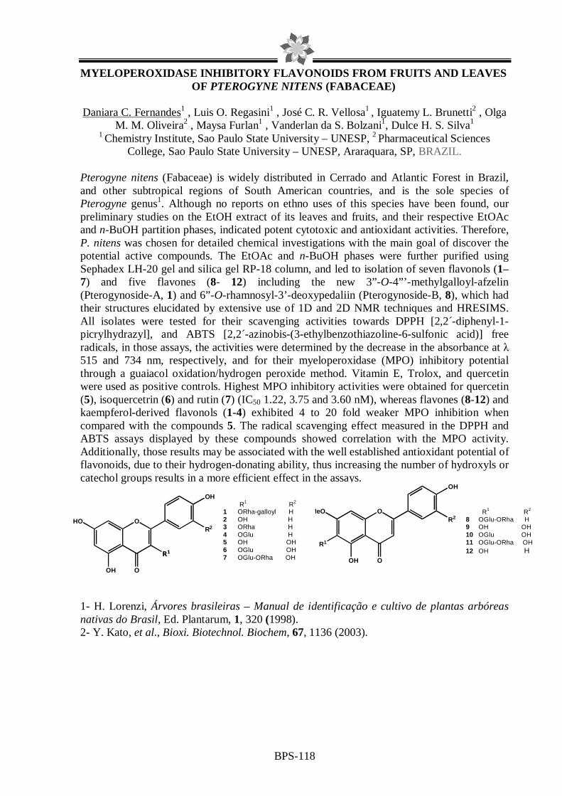
BPS-118
MYELOPEROXIDASE INHIBITORY FLAVONOIDS FROM FRUITS A ND LEAVES OF PTEROGYNE NITENS (FABACEAE)
Daniara C. Fernandes1 , Luis O. Regasini1 , José C. R. Vellosa1 , Iguatemy L. Brunetti2 , Olga
M. M. Oliveira2 , Maysa Furlan1 , Vanderlan da S. Bolzani1, Dulce H. S. Silva1 1 Chemistry Institute, Sao Paulo State University – UNESP, 2 Pharmaceutical Sciences
College, Sao Paulo State University – UNESP, Araraquara, SP, BRAZIL. Pterogyne nitens (Fabaceae) is widely distributed in Cerrado and Atlantic Forest in Brazil, and other subtropical regions of South American countries, and is the sole species of Pterogyne genus1. Although no reports on ethno uses of this species have been found, our preliminary studies on the EtOH extract of its leaves and fruits, and their respective EtOAc and n-BuOH partition phases, indicated potent cytotoxic and antioxidant activities. Therefore, P. nitens was chosen for detailed chemical investigations with the main goal of discover the potential active compounds. The EtOAc and n-BuOH phases were further purified using Sephadex LH-20 gel and silica gel RP-18 column, and led to isolation of seven flavonols (1–7) and five flavones (8- 12) including the new 3”-O-4”’-methylgalloyl-afzelin (Pterogynoside-A, 1) and 6”-O-rhamnosyl-3’-deoxypedaliin (Pterogynoside-B, 8), which had their structures elucidated by extensive use of 1D and 2D NMR techniques and HRESIMS. All isolates were tested for their scavenging activities towards DPPH [2,2´-diphenyl-1-picrylhydrazyl], and ABTS [2,2´-azinobis-(3-ethylbenzothiazoline-6-sulfonic acid)] free radicals, in those assays, the activities were determined by the decrease in the absorbance at λ 515 and 734 nm, respectively, and for their myeloperoxidase (MPO) inhibitory potential through a guaiacol oxidation/hydrogen peroxide method. Vitamin E, Trolox, and quercetin were used as positive controls. Highest MPO inhibitory activities were obtained for quercetin (5), isoquercetrin (6) and rutin (7) (IC50 1.22, 3.75 and 3.60 nM), whereas flavones (8-12) and kaempferol-derived flavonols (1-4) exhibited 4 to 20 fold weaker MPO inhibition when compared with the compounds 5. The radical scavenging effect measured in the DPPH and ABTS assays displayed by these compounds showed correlation with the MPO activity. Additionally, those results may be associated with the well established antioxidant potential of flavonoids, due to their hydrogen-donating ability, thus increasing the number of hydroxyls or catechol groups results in a more efficient effect in the assays.
1- H. Lorenzi, Árvores brasileiras – Manual de identificação e cultivo de plantas arbóreas nativas do Brasil, Ed. Plantarum, 1, 320 (1998). 2- Y. Kato, et al., Bioxi. Biotechnol. Biochem, 67, 1136 (2003).
R1 R2
8 OGlu-ORha H 9 OH OH 10 OGlu OH 11 OGlu-ORha OH 12 OH H
OHO
OH O
OH
R2
R1
OMeO
R1
OH O
OH
R2
R1 R2
1 ORha-galloyl H 2 OH H 3 ORha H 4 OGlu H 5 OH OH 6 OGlu OH 7 OGlu-ORha OH

BPS-119
INSECTICIDAL ACTIVITY OF Rauia sp. (RUTACEAE) EXTRACTS AGAINST Spodoptera frugiperda
Tatiani M. Bondancia*1(PG), Andréia P. Matos1, João B. Fernandes1(PQ), Maria Fátima G. F. da Silva1(PQ), Paulo C. Vieira1(PQ). 1Universidade Federal de São Carlos, Departamento de Química, Laboratório de Produtos Naturais, Rod. Washington Luiz Km 235, São Carlos-S.P. [email protected]
The family Rutaceae includes many plants that are sources of valuable medicines and uses in ethnomedicine. Numerous secondary metabolites that exhibit significant insecticidal activity were already found in Rutaceae genera1. The fall armyworm, Spodoptera frugiperda (J. E. Smith) (Lepidoptera: Noctuidae), is a major pest of many crops in the Americas. In Brazil, it is one of the most important pests of maize plantations, with production losses reaching 34 %. Control of this pest is still problematic and is sometimes, harmful to the environment, to man and other animals. Consequently, an extensive search for alternate methods to control these insects has been made in an attempt of substitute traditional agrochemicals with high specificity and, therefore, causing less damage to the environment. In this context, this work evaluated the toxic effect of leaves and stem hexane, dichloromethane and methanol extracts of Rauia sp against S. frugiperda. Larvae of S. frugiperda (J. E. Smith) were obtained from the Insect Bioassay Laboratory of Universidade Federal de São Carlos, Brazil, and reared on artificial diets. They were maintained in an incubation chamber with a photo phase of 12:12h L:D, 70 (± 5) % relative humidity and 25 (± 1) oC. For each treatment and control, 30 neonate larvae of S. frugiperda were used. A solution of extracts were added to ascorbic acid (1.56 g; an ingredient of the diet), after evaporation, the mixture was incorporated to the artificial diet in which bean and wheat germ are the basic ingredients at final concentrations of 1.0, 10.0, and 50.0 mg kg-1. Diet for the control was prepared similarly but without the extracts. The bioassays and statistic treatment were done as described by Kasten et al.2. For evaluation of the mortality of the larval and pupal phases, the experimental unit was constituted by the mean of five tubes with one larva each, with six replications by treatment. The leaves extracts of Rauia sp showed higher activity against S. frugiperda than stem extracts. Stem dichloromethane and methanol extracts showed lowest activity. The stem dichloromethane extract was fractionated with the objective of testing the pure compounds and to contribute to chemosystematics of Rutace and led to isolation of the coumarin rauianin and a mixture of glycosylflavonoids having rhamnosyl as one of the glycosyl unit. These classes of compounds are characteristics of this genus. Rauianin was identified by comparison of their NMR, MS, UV and IR data with literature and the flavonoids are in process of complete identification of the second sugar unit and their positions in the flavanone skeleton.
1Silva, M.F.G.F.; Gottlieb, O.R.; Ehrendorfer, F.. Plant. Syst. Evol., 161: 97-134, 1988. 2Kasten, P.J.R.; Precetti, A.A.C.M. and Parra, J.R.P., Rev. Agric. 53:68-78, 1978.
FAPESP, CNPq, CAPES

BPS-120
EFFECT OF THE ETANOLIC EXTRACT OF Endopleura uxi HUBER (HUMIRIACEAE) IN THE PRODUCTION OF HYDROGEN PEROXID E AND NITRIC OXIDE IN MURINE MACROPHAGES GRACE PRISCILA PELISSARI(1); SANDRA REGINA ANANIAS(2); RAQUEL REGINA DUARTE MOREIRA(1); IRACILDA ZEPPONE CARLOS(2). (1) Departamento de Princípios Ativos Naturais e Toxicologia, Faculdade de Ciências Farmacêuticas, Universidade Estadual Paulista “Júlio de Mesquita Filho,” Araraquara – SP. (2) Departamento de Análises Clínicas, Faculdade de Ciências Farmacêuticas, Universidade Estadual Paulista “Júlio de Mesquita Filho,” Araraquara – SP. Introduction: From a historical perspective, medicines production and the pharmacological treatment of diseases started with the use of medicinal plants. In order to adapt to environmental insults, plants produce many natural products that have antimicrobial and immunomodulating potential. Endopleura uchi it is a common tree in the Amazon region pertaining the family Humiriaceae. The tea of its rinds it is used in the popular medicine as a powerful natural composition in the combat of the renal infection, uterine inflammation, prevention of the cancer amongst others (1). Objective: The purpose of this work was to study the effects of the etanolic extract of Endopleura uxi in peritoneal macrophage cells from Swiss mice by determination of production the NO and H2O2. Material and Methods: The etanolic extract was obtain by maceration during 7 days in etanol 70% (w/w). The exudate peritoneal macrophages (PEC) were harvested from Swiss mice (6-8 weeks old, 18-25 g). For the determination of the cell viability, PEC (5x10⁶ cells/mL) were re-suspended in medium RPMI-1640 C. The suspension (100 µL) and the samples in different concentrations (100 µL) were added to each well of a 96-well tissue culture plate and incubated for 24 h. The 3-(4,5-dimethylthiazol-2-yl)-2,5-diphenyl-tetrazolium bromide (MTT) assay was performed and absorbance measured at 540 nm with a 620 nm reference filter. The nitric oxide production was determined by measuring nitrite. 50 µL of cell supernatant was removed from each well and incubated with an equal volume of Griess reagent at room temperature for 10 min; the absorbance was measured at 540 nm. For H2O2 measurement, PECs (2x10⁶ cells/ml) were suspended in a solution of phenol red complete buffer; 100 µL of this suspension was added to each wells of a tissue culture plate and added 50 µL of aqueous extract in the concentration to viability of 50%. After incubation for 1 h, the reaction was stopped with 50 µL of NaOH 5M and absorbance measured at 620 nm. Results: The cytotoxicity of the etanolic extract in vitro on peritoneal macrophages, it was IC50 = 15,20 mg/mL and IC25 = 4,00 mg/mL. In concentracion of 4,00 mg/mL the NO production was low (31,51 ± 1,32 µmlos/5.106 células, however in concentration of 15,20 mg/mL the production was greater that LPS (positive control). The H2O2 production was 483,17 ± 18,92 nmoles/5.106 (15,20 mg/mL) and 138,48 ± 3,84 nmoles/5.106 (4,00 mg/mL). These values suggest a imunoestimulante potential of this species so used by the population. Future studies will be necessary to evidence the effect of this extract in the production of other involved mediators in the immune reply. 1. MARX, F., ANDRADE, E. H. A.; ZOGHBI, M. G. B.; MAIA, J. G. S.. Eur. Food Res. Technol., v. 214, p. 331-4, 2002. FINANCIAL SUPPORT: CNPq

BPS-121
EVALUATION OF TOPICAL ANTIINFLAMMATORY AND WOUND HE ALING ACTIVITIES OF Arrabidaea chica EXTRACTS
Adriana L. Schiozer1, Laryssa K. Rauh2, Daniela A. Cabrini2, Lauro E.S. Barata1
1Natural Products R&D Laboratory, Institute of Chemistry, State University of Campinas -
UNICAMP, Caixa Postal 6154, CEP 13083-970, Campinas, SP, Brazil 2Inflammation, Pain and Fever Pharmacology Laboratory, Federal University of Paraná -
UFPR, Caixa Postal 19031, CEP 81530-900 Curitiba, PR, Brazil Arrabidaea chica Verlot, (Bignoniaceae) is known by the common names carajiru, crajiru, pariri and chica-red. It is found in Central and South America, mainly in Amazon region. The leaves are used as astringents, feminine genital disinfection, in the treatment of inflammation, skin disease and wounds, intestinal cramps, bloody dysentery, leucorrhoea, anemia and leukemia in traditional medicine1. The red pigmentation of A. chica leaves is due to the presence of 3-deoxyanthocyanidins. These rare substances differ from the known anthocyanins by the lack of a hydroxyl group and for its higher stability. The objective of this work was to evaluate topical antiinflammatory and wound healing activities of A. chica extracts and isolated compound using croton oil-induced mouse ear edema and mouse healing excision models, respectively.
Two extracts were prepared from dried, crushed A chica (type I) leaves. Maceration in ethanol-water solution, followed by filtration and evaporation yielded crude extract EtOH/AC. For the second extract, sequential maceration in water (19ºC), hexane and finally methanol/acid solution was performed. The latter fraction (MeOH/H+/AC) was concentrated deoxyanthocyanins. A characteristic red-colored deoxyanthocyanidin, carajurin2, was obtained using the following procedure: sequential maceration in water, hexane and dichloromethane, followed by filtration of the dichloromethane fraction on C18 cartridges to yield concentrated substance. Two extracts, EtOH/AC, MeOH/H+/AC, and isolated carajurin were tested. Two models were used as means to evaluate the extracts: the ear oedema and the healing incision model. The ear oedema model was induced by croton oil through topical application (0.4mg/ear) followed by the extracts application (0.01-1mg/ear). Ear thickness was measured and the oedema was expressed after inflammation induction. In the wound healing excision model, animals were submitted to dorsal incision and treated with the extracts (0.2mg/wound, twice a day) during 14 days. The healing was assessed by the rate of wound contraction. MeOH/H+/AC extract caused a dose-related inhibition of oedema (DI50=0,45 mg/ear; Imáx=60±8%) while EtOH/AC extract showed no effect. Carajurin demonstrated a slight antiedematogenic effect (Imáx=30±5%). Extracts did not alter the rate of wound contraction when compared with control group. These results suggested that the concentrated deoxyanthocyanins extract has an interesting topical aniinflammatory effect, being more effective than carajurin alone. Further studies are required to investigate other deoxyanthocyanins other than carajurin as well as the mechanism of action.
Refs. 1Mors, W. B. et al. Medicinal plants of Brazil (2000) 2Devia, B. et al. Phytochemical Analysis 13, 114-120 (2002)

BPS-122
ANTIMICROBIAL ACTIVITY OF Arrabidaea chica EXTRACTS
Adriana L. Schiozer1, Alice M. Braga1, Nathália Becker1, Luzia Lyra2, Adriana Minatel2, Angelica Schreiber2, Lauro E.S. Barata1
1 Research and Development Laboratory of Natural Products, Institute of Chemistry, 2Department of Clinical Pathology, Faculty of Medical Sciences, Caixa Postal 6154, CEP 13083-970, State University of Campinas – UNICAMP, Campinas, SP, Brazil.
Arrabidaea chica Verlot, (Bignoniaceae) is known by the common names carajiru,
crajiru, pariri and chica-red. It is found in Central and South America, mainly in Amazon region. The leaves are used as astringents, feminine genital disinfection, in the treatment of inflammation, skin disease and wounds, intestinal cramps, bloody dysentery, leucorrhoea, anemia and leukemia in traditional medicine1. The red pigmentation of A. chica leaves is due to the presence of 3-deoxyanthocyanidins. These rare substances differ from the known anthocyanins by the lack of a hydroxyl group and for its higher stability2. The objective of this work was to evaluate the antimicrobial activity of extracts of A. chica leaves.
A. chica leaves were extracted with ethanol solution at room temperature for one hour, after filtration the solution was partitioned with four solvents of increasing polarity, hexane, methylene chloride, ethyl acetate and buthanol. The ethanol extract and resulting fractions were subjected to antimicrobial susceptibility tests. The most active fraction was separated on PTLC and the resulting fractions were again submitted to antimicrobial tests. The antimicrobial tests were conducted using filamentous fungi (dermatophytes), yeasts and bacteria. The broth microdilution technique was used for the tests involving yeasts and dermatophytes. Tests were conducted using NCCLS reference method for broth dilution for yeasts and filamentous fungi. Strains of yeasts evaluated were Candida albicans ATCC 90028 and ATCC 76615, Candida krusei ATCC 6258 and Candida parapsilosis ATCC 22019. The most active fractions presented MIC (Minimum Inhibitory Concentrations) ≤100µg/mL and MFC (Minimum Fungicidal Concentrations) ≤1500µg/mL. Dermatophytes used were Trichophyton rubrum (LIF/FCM 142), T. mentagrophytes (LIF/FCM 10) among 20 others from Unicamp’s fungi hospital library. The results for dermatophytes showed MIC and MFC ≤100µg/mL for the most active fractions. To evaluate the activity against bacteria, the technique of disk diffusion was used, according to NCCLS (antimicrobial disk susceptibility tests), using strains of Staphylococcus aureus ATCC 25923 and S. hominis HC-12432-0 (both present in skin infections), the best results were obtained presented inhibition zones varying from 12 to 18 mm. These results indicate that A. chica extracts have potential as antimicrobial agents3. The observed inhibition of C. krusei is important given its resistance to fluconazole.
Refs. 1Mors, W.B. et al. Medicinal plants of Brazil (2000) 2Schiozer, A.L., Barata, L.C. Revista Fitos, in press (2007) 3Barata, L.E.S. et al., Patente de Invenção PI0600943-3. Depósito: 23/02/2006, INPI, Brasil

BPS-123
ANTIOXIDANT ACTIVITY OF THE DICHLOROMETHANE EXTRACT OF
Eugenia punicifolia (PEDRA-UME CAÁ)
Daniel Tadeu Gomes Gonzaga2 (PG), Lívia Fantini Polese1 (IC), David Rodrigues da Rocha2 (PG), Kátia G. Lima Araújo1 (PQ), Vítor F. Ferreira2 (PQ), Wilson C. Santos1 (PQ) 1 UFF – Faculdade de Farmácia – Rua Mário Viana, 523 – Santa Rosa – Niterói – RJ – 24.241-000 2 UFF – Instituto de Química – Morro do Valonguinho – Niterói – RJ – 24.020-150
Species of the genus Eugenia have been demonstrating scientific interest. We have recently shown that the water extract of E. punicifolia influences the cholinergic neurotransmission in rat diaphragma. In the present work we aimed to verify the antioxidant activity of the dichloromethane extract (DCME) of E. punicifolia, since oxidative stress is associated to several neurodegenerative diseases. Two experimental methodology were applied, namely thin layer chromatography associated to revelation with the 2, 2-diphenil-1-pycril-hydrazil (DPPH) radical, and also a test with DPPH and the extract in methanolic solution. The colorless rate of the radical in solution (decay of A517nm), was evaluated. This parameter indicates the scavenger capacity from the free radical for the compounds in the extract. Results: Through thin layer chromatography (hexane: ethyl acethate 9:1) of DCME from E. punicifolia and the revelation with vaniline:sulfuric acid, it was possible to observe 10 distinct stains, and among them 3 demonstrated a fast reaction for the revelation with DPPH, thus indicating the presence of active antioxidants substances within. In order to verify the antioxidant activity of the extract in solution, previous dissolution in methanol: dichloromethane (1:1) was required, for the low solubility in methanol, followed by mixing with methanolic solution of DPPH radical and careful observation of A517nm decay for 30 minutes. The DPPH percentage that remained in solution was calculated based on the A517nm decay. In order to perform the test the following concentrations of the extract were employed: 200, 400, 600, 800, 1.000, 1.200, 1.400 and 1.600 ppm. In order to compare the results, it was verified the antioxidant activity of terc-buthyl-hydroquinone (TBHQ), as standard solution, in the concentrations of 50 and 100 ppm. The results showed that DCME of E. punicifolia displayed antioxidant activity in DPPH radical in all concentrations employed, since a reduction of the concentration of the radical after incubation with the extract was obtained. Results were proportional to tested concentration. We observed that 800 ppm extract displayed equivalent activity to 50 ppm TBHQ; meanwhile, 1.600 ppm extract showed activity close to 100 ppm of synthetic antioxidant utilized as standard. Antioxidant activity demonstrated for DCME of E. punicifolia indicates that this fraction of the plant may be of interest in the search for new candidate molecules for possible employment in diseases that share oxidative stress as basic phenomenon. Keywords: Eugenia punicifolia, oxidative stress, DPPH, TBHQ. Grants: FAPERJ, CNPq

BPS-124
BIOGUIDED FRACTIONATION FOR CYTOTOXIC ACTIVITY FROM THE ZOANTHID Protopalythoa variabilis COLLECTED AT NORTHEASTERN
BRAZILIAN BEACHES
Paula C. Jimenez1; Diego V. Wilke1; Renata M. Araújo2; Edilberto R. Silveira2; Claudia Pessoa1; Manoel O. Moraes1; Otília D.L. Pessoa2; Letícia V. Costa-Lotufo1
1Department of Physiology and Pharmacology, UFC – Brazil; 2Department of Organic and Inorganic Chemistry, UFC – Brazil.
Marine organisms are an important source of bioactive secondary metabolites, presenting a high incidence of cytotoxic and antimicrobial compounds. Despite the recency of this field of study, the oceans have already yielded interesting natural products with confirmed or highly promising pharmacological applications and have yet awakened the industrial interest. The aim of this work was to perform a bioguided fractionation for determination of cytotoxic activity of a hydromethanolic extract (PV-HM) obtained from the zoanthid Protopalythoa variabilis collected at Paracuru and Taíba beaches (Ceará State, Brazil). The cytotoxic activity of both PV-HM and derived fractions was evaluated by the MTT assay against 4 human tumor cell lines: HL-60 (leukemia), MDA MB-435 (breast cancer), SF-295 (CNS glioblastoma) and HCT-8 (colon cancer). The PV-HM of specimens collected at Taíba beach did not show activity, however the PV-HM of Paracuru specimens was very toxic to all cell lines tested (IC50 values ranging from 0.36 to 4.24 µg/mL). The hemolytic potential was evaluated on mouse erythrocytes after 1, 2 and 4 h incubation. The PV-HM from Paracuru beach induced hemolysis only after 4 hours incubation (EC50= 68.83 µg/mL). The bioactive PV-HM was diluted in methanol and partitioned with n-hexane, dichloromethane and ethyl acetate, yieldeding 4 fractions: PV-Hex, -DCM, EtOAc and -MeOH. All fractions were cytotoxic, being the PV-Hex and -MeOH the strongest (IC50 values raging from 0.2 to 0.4 µg/mL for PV-MeOH, from 0.09 to 0.7 µg/mL for PV-Hex, from 1.4 to 2.9 µg/mL for PV-EtOAc). Only PV-MeOH induced hemolysis on mouse erythrocytes and, like PV-HM, after 4 h incubation. To further investigate the mechanisms involved in the cytotoxic activity, the effect of fractions on membrane integrity, cell cycle distribution and DNA fragmentation was evaluated on HL-60 cells using flow cytometry. Morphological alterations of treated cells were analyzed with hematoxylin-eosin staining. The results suggest that PV-Hex, -DCM and -MeOH induced apoptosis, while PV-EtOAc seemed to induce necrosis. Purification of the fraction PV-EtOAc was efficiently performed using high performance liquid chromatography, and activity of the resulting fractions was slightly improved (IC50 values ranging from 0.34 to 2.73 µg/mL). It is worthwhile to mention that several other chromatography methods used for separation (Silica gel 60 column, Sephadex-LH 20 column, TLC for PV-Hex, -MeOH and -DCM respectively) resulted in inactive fractions. Purification of others fractions are already in progress. Supported by: CNPq, CAPES, BNB, FUNCAP, FINEP, Claude Bernard Institute. Keyworks: Protopalythoa variabilis, cytotoxicity, bioguided fractionation.

BPS-125
Antifungal and acetycholinesterase inhibitors compounds of Harpagopytum procumbens (Devil’s Claw).
Tatiana Marques Ferreira Rocha1, Maria Cecília Cattai Anauate2, Elaine Cardoso Monteiro
Lopes3 and Luce Maria Brandão Torres4.
1 Graduated Student UNISA/SP 2 Postgraduate Student Departamento de Reumatologia USP/SP 3 Prodoc/CAPES Seção de Fisiologia e Bioquímica, Instituto de Botânica, São Paulo, 4Researcher Seção de Fisiologia e Bioquímica, Instituto de Botânica, São Paulo, SP.Av.Miguel Estefano 3687, 04301-012, São Paulo-SP- Brasil. [email protected]
Harpagopytum procumbens (Burch) DC. Ex Meisn. (Pedaliaceae) is a medicinal plant used by indigenous to the northwestern parts of Southern Africa. This plant is used by population to treat fevers, blood diseases, arthritis and rheumatism. In this study the dried and crude hydroethanolic extract of secondary roots, contained 1.6 to 2.0% of harpagoside was provided by Pro-formula, São Paulo, Brazil. The extract was fractionated through the normal phase silica bed using a short vacuum chromatographic method1. A stepwise gradient of increasing polarity of mobile phase (200mL) consisting of a mixture of CHCl3: CH3OH ratio 100.0, 99:01, 98:2, 96:4, 94:6, 92:8, 90:10, 88:12, 85:15 and 80:20) was employed to obtained the iridoids presents in this extracts. Iridous detection was performed with sulfuric acid/vanillin reagent by TLC and HPLC (Varian Pro Star) using reverse phase C18 (150mmx 4.6mm), isocratic system mobile phase pump A. H2O, pump B. CH3OH (1:1), flow rate 1mL/min and detection was carried out at 278nm, during 30min. The degree of purity (%) was evaluated by HPLC and characterization by hydrogen nuclear magnetic resonance (1HNMR). The extracts, fractions and pure compounds have been evaluated towards phytopathogen fungi Cladosporium cladosporioides and C. sphaerospermum using bioautography and acetylcholinesterase inhibitors were performed on TLC plates1. The cinnamic acid show an antifungic effect in this bioassay and harpogoside showed a strong inhibitory activity for the acetycholinesterase. Financial support. FAPESP.
Figure1. HPLC of harpagoside (blue) and cinnamic acid (red) Refs.
1Marston et al 2002. Phy Anal. 13: 51-54
5 1 0 1 5 2 0 2 5 3 0
M i n u t e s
- 7 4
0
1 0 0
2 0 0
3 0 0
4 0 0
5 0 0
6 0 0
7 0 0
m V o l t s
1.97
7
2.22
02.
252 2.68
6
3.56
3
3.86
1
4.96
4
6.72
6
8.47
5
10.
194
12.3
17
20.0
3920
.168
Cinnamic acid harpagoside

BPS-126
SEARCH FOR INHIBITORS OF XILELLAIN - AN Xylella fastidiosa PROTEASE
Richele P. Severino1; Vanessa G. Pasqualotto1; Paulo Cezar Vieira1; M. Fátima das G. F. da Silva1; João Batista Fernandes1; Flávio Henrique-Silva2;
1Laboratory of Natural Products, Department of Chemistry, Federal University of São Carlos, São Carlos, SP, Brazil; 2Laboratory of Molecular Biology, Department of Genetics and
Evolution, Federal University of São Carlos, São Carlos, SP, Brazil.
Key words: Xylella fastidiosa; xylellain; natural products; inhibitors. Introduction: Xylella fastidiosa is a xylem-limited, Gram-negative bacterium responsible for a large number of economically important plant diseases, such as Pierce’s disease in grapevines, citrus variegated chlorosis (CVC) in sweet oranges and leaf scorch diseases in other plants, including almond, plum, oleander, mulberry and coffee. In all cases, X. fastidiosa infects the plant xylem and impairs fruit production. CVC is considered to be potentially the most devastating citrus disease. Proteins such as proteases, cellulases and lipases may be involved in the infection process by disrupting plant tissue and allowing the spread of bacteria throughout the vascular system. The interest in cysteine proteases as chemotherapeutic targets stems from the recognition that they are critical to the life cycle or pathogenicity of a large number of parasites. Cysteine proteases from parasites play key roles in immunoevasion, enzyme activation, virulence, tissue and cellular invasion, as well as excystment, hatching and molting. Recent studies described an X. fastidiosa cysteine protease (dubbed Xylellain). This protein may be involved in X. fastidiosa pathogenicity and therefore constitutes a probable target for combating CVC and other diseases. We have been interested in finding natural products as inhibitors of xylellain1. A small library of natural products isolated from Brazilian plants was tested for their inhibitory activity using a high-throughput fluorogenic substrate assay. Some of the compounds were shown to be potent inhibitors of xylelain. Experimental Procedure: The compounds used for HTS screening were diluted in DMSO and tested in concentration of 25 µM/well. Assays were performed in 50 mM sodium acetate buffer, pH 5.0. The xylellain was preactivated in the presence of 2.5 mM DTE for 5 min at 25 oC. Before the addition of substrate, all compounds were preincubated 5 min with the active enzyme, directly in the plate (96 well; 200 µL/well). After that, substrate Z-FR-MCA (in DMSO) was added (10 µM/well) and the hydrolysis was followed continuously by increase of fluorescence at λem 460 nm and λex 355 nm (emission and excitation wavelengths for MCA). The positive control was E-64, an irreversible inhibitor for cysteine peptidase. Enzyme inhibition data were calculated using the program SigmaPlot 9.0. Conclusions: From all tested compounds it was possible to conclude that the flavonoids, cinnamic acid derivatives and alkaloids are the more promising ones followed by the coumarins. These compounds are very widely spread in many plants. Based on these observations we have already tested a library of synthetic compounds and have also found good inhibitory activity. This is the first time that natural products from plants have been tested on xylellain inhibitory activities. References: 1. V. Nogaroto et al.; FEBS Microbiol Lett, 261, 187-193 (2006).

BPS-127
LARVICIDAL ACTIVITY OF DILLAPIOL DERIVATIVES TO AEDES AEGYPTI
Ana Cristina da Silva Pinto1; Karla Lagos Nogueira2; Wanderli Pedro Tadei3; Adrian Martin Pohlit3; Francisco Célio Maia Chaves4.
1Pós-Graduação em Biotecnologia, Universidade Federal do Amazonas, Av. Gal. Rodrigo Otávio Jordão Ramos, 69077-000, Manaus-AM, e-mail: [email protected]; 2Tecnologia em Química Industrial, Centro Federal de Educação Tecnológica do Amazonas, Av. 7 de Setembro, Centro; 3Instituto Nacional de Pesquisas da Amazônia, Coordenação de Pesquisas em Produtos Naturais, Av. André Araújo 2936, 69083-000, Manaus-AM; 4Embrapa Amazônia Ocidental, Km 29 AM 010, Manaus-AM, 69011-970, Brazil. Species of the Piperaceae family are well known for their insecticidal properties1,2. Steam distillation of dry Piper aduncum leaves yields a volatile oil from which dillapiol (1) can be isolated by fractional vacuum distillation in 72 % yield (v/v). Ethers 2-6 were prepared by stirring 1 with mercury(II) acetate under nitrogen using, respectively, methanol, ethanol, propanol, butanol and octanol as solvents (0 ºC to r.t., over 2-168 h, reaction time increased with increase in alcohol chain length). After this initial reaction, the organo-mercurate intermediates were reduced by addition of alkaline sodium borohydride solution. Mercury metal was removed by filtration and chloroform was used for extraction. The chloroform phase was separated, washed with water, then saturated sodium chloride solution and was finally dried over magnesium sulfate. After removal of solvents, crude ethers were obtained. Purification was performed using flash chromatography providing 2 (41.8 mg, 78 %), 3 (37.4 mg, 62 %), 4 (149.7 mg, 77 %), 5 (166 mg, 42 %) e 6 (78.5 mg, 17 %) whose identities were confirmed by 1H and 13C-NMR. Mosquito larvae (Aedes aegypti) were bred in the laboratory under controlled temperature (27 °C) and humidity (60 %) conditions. Tests were performed according to the WHO protocol. For each treatment and control, 10 third instar larvae were transferred to 10 mL plastic cups, having a final volume of 5 mL water. The evaluation of mortality rate was performed 24 h after the beginning of the experiment, verifying the number of dead larvae. Data were evaluated through probit analysis to determine the LC50 representing the concentrations in µg/ml that caused 50 % mortality. Compounds 1, 2, 3, 4 and 5 exhibited CL50 = 36.2, 100.6, 50.3, 28.0, 19.7 µg/mL. Compound 6 exhibited no larvicidal activity. Refs. 1. C.B. Bernard et al., Journal of Chemical Ecology, 21, 801 (1995). 2. S.M. Morais et al., Biochemical Systematics and Ecology, 35, 670 (2007).
O
O
OMe
OMe O
O
OMe
OMeOR
12 - R=Me; 3 - R=Et; 4 - R=Pr; 5 - R=Bu;6 - R=Oct

BPS-128
ANXIOLITIC LIKE EFFECT OF ERYTHRINIAN ALKALOIDS FRO M ERYTHRINA VELUTINA (FABACEAE)
Maria Amélia Rodrigues Serrano1; Luciana de Avila Santos1; Paulo J. C. Nogueira2; Ricardo
L. N. de Souza2; Vanderlan S. Bolzani1
1São Paulo State University – UNESP-Instituto de Química, 14800-900, Araraquara, SP, Brazil
21São Paulo State University – UNESP – Faculdade de Ciências Farmacêuticas14800-900, Araraquara, SP, Brazil
The genus Erythrina is known in Brazilian folk medicine for the treatment of central nervous system (CNS) illnesses. Its stem barks are recommended for nervous system excitation, sleepiness, convulsions and nervous coughs[1]. Most of the scientific work on Erythrina velutina describes important pharmacological effects for crude extracts of the plant as the anti-nociceptive activity of the aqueous extract of the leaves [2] and the antibacterial activity of bark ethanolic extract [3]. E. velutina extracts have also CNS depressant activity showed through the potentiation of pentobarbital-induced sleeping time [1;4] and through the anticonvulsant action in strychinine-induced seizure model [4]. In the literature the erythrinian alkaloids were reported as responsible for the pharmacological properties on CNS. In a recent work, the phytochemical study of E. mulungu flowers afforded three erythrinian alkaloids, which were found to have anxiolitic effect in an elevated T-Maze test [5]. In continuing our work for the search for bioactive compounds, the phytochemical study led to the isolation and characterization of two known erythrinian alkaloids from stem bark of Erythrina velutina. The isolation steps were carried out on HPLC system using water/methanol as mobile phase and the identification of the structures isolates were based on NMR and ESIMS data. Preliminary evaluation of the anxiolitic activity was performed on the Ethanolic extract and alkaloidic fraction using elevated T-Maze assay. Additionally, were evaluate acute toxicity. Refs. 1. M.C. Dantas et al., Journal of Ethnopharmacology, 94, 129 (2004) 2. M. Marchioro et al., Fitoterapia, 76 (2005) 3. S. Virtuoso et al., Revista Brasileira de Farmacognosia, 15(2), 137, (2005) 4. S. M. M. Vasconcelos et al., Journal of Ethnopharmacology, 110, 271 (2007) 5. O. Flausino Jr. et al., Journal Nat. Produts, 70, 48 (2007)

BPS-129
PHYTOCHEMICAL AND BIOLOGICAL STUDY OF KIELMEYERA RUGOSA CHOISY (CLUSIACEAE)
Larissa M. Andrade1, Moacir S. Andrade1, Valéria R. S. Moraes1, Paulo Cesar L. Nogueira1, Péricles B. Alves1, Samísia M. F. Machado1, Glaucia B. Alcântara2, Antonio G. Ferreira2,
Glaucius Oliva3, Otávio H. Thiemann3. 1METABIO – Departamento de Química - Universidade Federal de Sergipe, São Cristovão-
SE, Brazil. [email protected] 2 Departamento de Química - Universidade Federal de São Carlos, São Carlos-SP, Brazil 3
Instituto de Física de São Carlos – Universidade de São Paulo, São Carlos-SP, Brazil
The genus Kielmeyera belongs to the family Clusiaceae (Guttiferae), subfamily Kielmeyeroideae, and is endemic to South America, with the vast majority of the species occurring exclusively in Brazil. Here, nearly 50 species are found, chiefly in the ‘restinga’ (sand dunes), ‘campos rupestres’ (rocky savannas) and ‘cerrado’ (savanna) vegetation south of the Amazon. Some species are commonly known in Brazil as ‘pau-santo’ and are traditionally used in Brazilian folk medicine to treat several tropical diseases, including schistosomiasis, leishmaniasis and malaria as well as fungal and bacterial infections1. Phytochemical studies with Brazilian Kielmeyera species showed mainly 4-alkyl and 4-phenyl coumarins and xanthones2. We have previously reported the isolation and structure identification of 4-phenyl and 4-n-propyl coumarins from K. rugosa fruits and leaves collected in a ‘restinga’ of Sergipe State, Brazil3. In a continuation of our study on this plant, we now report herein the isolation and structure elucidation of another 4-n-propyl coumarin (1), 4-phenyl coumarin (2) (Figure 1) from the CHCl3 extract of fruits and stem bark, respectively, along with a mixture of the two well-known triterpenes lupeol and α-amyrin isolated from the hexane extracts of leaves. For structure elucidation of these natural compounds one- and two-dimensional NMR techniques (1H, 13C, DEPT, HMBC, HSQC, and COSY) were used besides IR spectroscopy. In addition, the extracts and some fractions from the stem bark, fruits, and latex of K. rugosa were tested for their enzymatic inhibitory activities against glicossomal GAPDH from Trypanosoma cruzi and APRT from Leishmania donovani. Some Extracts and fractions displayed relevant inhibitory activity (% inhibitory activity > 80%), suggesting that the plant extracts and fractions may have tripanocidal and leishmanicidal agents.
O O
OH
HO
OH
O
2
4a
8a7
1'
6
4' 5'
1'''
3'''
2''4''
5''
(1)
OO O
OH
OR
O
4'
5'
2' '3''
5' '
3
5
1'''
2'''
7'
8'
7
R= CO2C
3C
(2)
Figure 1: 4-n-propyl and 4-phenyl coumarins isolated from K. rugosa
Refs. 1. T.M.A. Alves et al., Mem. Inst. Oswaldo Cruz, 95, 367 (2000). 2. (a) F.G. Cruz et al., Phytochemistry, 47, 1363 (1998); (b) F.G. Cruz et al., Phytochemistry, 48, 703 (1998); (c) F.G. Cruz et al., J. Braz. Chem. Soc., 12, 117 (2001). (d) F.G. Cruz et al., J. Braz. Chem. Soc., 13, 704 (2002). 3. (a) T. S. Sampaio et al., Resumos 27a RA da SBQ, PN-165 (2004); (b) M. S. Andrade et al., Resumos 28a RA da SBQ, PN-119 (2005); (b) M. S. Andrade et al., Resumos 29a RA da SBQ, PN-218 (2006); CNPq, COPES/UFS.

BPS-130
ACTIVITY OF EXTRACTS FROM Melia azedarach AGAINST Nezara viridula (HEMIPTERA: PENTATOMIDAE) - PEST OF FORAGE LEGUMES AND
SOYBEAN. DESIGN AND STANDARIZATION OF BIOASSAYS.
Luciana Baruffaldi1, Martina Díaz1, Andrés González1, Claudia Pereyra2, Horacio Silva2, Enrique Castiglioni2, Carmen Rossini1,*.
*[email protected] 1Laboratorio de Ecología Química, Departamento de Química Orgánica, Facultad de Química, UdelaR. Gral. Flores 2124 CP 11800 Montevideo, Uruguay. 2 Estación Mario Cassinoni, Facultad de Agronomía, UdelaR. Ruta 3 km 363 CP 60000 Paysandú, Uruguay. Soybean –and other legumes- production has grown during the last years in Uruguay, leading to different sanitary problems, among which the damage caused by pentatomids is an important one. Not only these pests have been difficult to control by conventional pesticides, but also they have developed resistance against conventional pesticides. Furthermore, the trend worldwide is to decrease the use of traditional pesticides, since its overuse has caused not only the development of resistance, but also the appearance of adverse effects on the environment and on non-targeted organisms. Within this framework we started a program aiming to find alternatives for controlling pentatomids of high economic incidence in Uruguay. The present work reports on the standardization of bioassays to screen for toxic activity against nymphs and adults of pentatomids, using Nezara viridula as a model. The standardization was performed exposing the insects by contact and by ingestion to a commercially available insecticide used to control them (Engeo®). Reproducible results were found only in the contact bioassay (Table 1). Using this design, extracts from different organs of Melia azedarach (Meliaceae) were tested against N. viridulans. After 24 hours of exposure, results showed good activity of extracts from fruits and twigs against nymphs, but not of the extract from leaves (Table 1). Although LD50 for fruits’ and twigs’ extracts are greater than the positive control (Engeo®); in the case of LD99, figures are comparable. Interestingly, the same extracts did not seem to affect adults when exposed for 24 hours- at least up to 1500 g/cm2 (Table 1). In the case of adults, the lethal effect is reached only after 72 hours of exposure, although with greater doses (LD99s = 850 ± 280 g/cm2, p < 0,001), indicating a greater susceptibility of immature stages than of adults. Table 1: Lethal doses [50% and 99%, mean ± standard error (µg/cm2)] after 24 hours of exposure of Engeo® and extracts from M. azedarach under the conditions of the standardized bioassay (activity by contact).
Products LD50 LD99 Nymphs
Engeo® 6,7E-03 ± 6,1E-04 (p< 0,001) 1,6E-02 ± 1,3E-03 (p< 0,001) M. azedarach
Fruits Twigs
Leaves
2,1E-02 ± 8,2E-03 (p < 0,05) 1,9E-02± 8,2E-03 (p < 0.001) > 700
5,0E-02 ± 2,2E-02 (p < 0,05) 4,3E-02 ± 3,5E-03 (p< 0.001) > 700
Adults Engeo® 3,7E-03 ± 1,2E-03 (p< 0,005) 1,1E-02 ± 3,5E-03 (p< 0,005) M. azedarach
Fruits Twigs
Leaves
>1500 >1500 >1500
>1500 >1500 >1500

BPS-131
VASORELAXANT EFFECT IN MESENTERIC ARTERY ISOLATED F ROM RAT AND TOXICITY OF 16- ENT-KAURANOL AND EXTRACTS OF Hyptis fruticosa LEAVES Maria da Paz do Nascimento Moreno1, Adriana Andrade Carvalho2, Simone Simote1 Péricles Barreto2 , Ângelo Roberto Antoniolli2 , Valéria Rêgo Alves Pereira3 , Luiza de Campos Reis3 ,Marina de Assis Souza3; M. Fátima das Graças Fernandes da Silva2 , Márcio Roberto Viana dos Santos 2 , João Batista Fernandes 1 1Departamento de Química, Universidade Federal de São Carlos, Via Washington Luiz, Km 235, C. P. 676, 13565-905, 2Departamento de Fisiologia, Universidade Federal de Sergipe, Av. Marechal Rondon, S/N, Cep 49100-000, SE, Brazil3Laboratório de Imunoparasitologia do Centro de Pesquisas Aggeu Magalhães- CPqAM/FIOCRUZ
The vasorelaxant effects induced by the leaves methanolic extract (ExMeOH01, 78,50 g,15,7%) of Hyptis fruticosa, ethyl acetate (ExAcOEt1, 29,50 g, 37,6%), dichloromethane (ExDCM1, 5,40g, 6,9%) and petroleum ether (ExEP1, 8,43g, 10,7%) fractions of this extract, and 16-ent-kauranol 1 [Rf=0,64 , Hex:AcOEt 6:4/anisaldeyde, mp 210-211 oC, 1H NMR (200 MHz, CDCl3) 0,78(3H,s,Me); 0,82 (3H,s,Me); 1,00 (3H,s,CMe) e 1,3 (3H,s,Me) e 1,5 (2H, m), 13C NMR 33,24 (C, C-4), 45,37 (C,C-8),39,35 (C, C-10), 79,31 (C, C-16), 56,23 (CH, C-5), 56,90 (CH, C-9), 49,06 (CH, C-13), 40,36 (CH2, C-1),18,61 (CH2, C-2),42,08 (CH2, C-3), 20,41 (CH2,C-6), 42,07 (CH2,, C-7, 17,97 (CH2, C-11), 26,91 (CH2, C-12), 37,70 (CH2, C-14), 58,10 (CH2, C-15), 24,44 (CH3, C-17), 33,57 (CH3, C-18), 21,54 (CH3,, C-19), 17,75 (CH3,, C-20)] were studied in isolated rings of rat superior mesenteric artery1. In intact rings (control), the extract, fractions (1-1000 µg/mL, n=6, cumulatively) and 16-ent-kauranol (0,01 – 10 µg/mL) induced concentration-dependent relaxations of tonus induced by phe 10 µM (Emax = 83±5.4%; 26±4.3%; 71±4.6%, 99±0.4% and 58±5.7%; respectively). In endothelium-denuded rings, the concentration-response curves to methanolic extract, dichloromethane, petroleum ether fractions and 16-ent-kauranol were not changed (Emax = 90±4.3%; 88±13.3%; 99±0.4%; 43±3.2% respectively). The curve to ethyl acetate fraction was increased in the final concentration (Emax = 52±6.3%). The petroleum ether fraction was more efficacious than others ones and the 16-ent-kauranol was the most potent. These results show that all products from H. fruticosa tested in this work were able of relaxation in mesenteric artery. The citotoxicity in vitro of extracts and fractions were also tested to determine the toxic concentrations for mammal cells using BALB/c mice splenocytes cultured in 96-well plates as normal cells. This method2 is efective for avaluation of specific T lymphocyte cytotoxicity. All samples are considered as non-cytotoxic (Table 1). Table 1- Results of citotoxicity
Sample Concentration
(µµµµg/ml) (a) % Inhibition (b)
ExMeOH01 25 18.53 ExEP01 10 12.14 ExDCM01 25 27.16 ExAcOEt01 5 11.74 16-ent-kauranol 50 22.85
(a) = the highest non-toxic concentration on spleen cell of BALB/c mice; (b) percentage of growth inhibition was determined comparing the percentage of the percentage of [3H]- thymidine incorporation of drug-treated wells in relation to untreated wells.
Refs. 1. B.M.; Altura et al. Am. J. Physiol. 219, 1698 (1970)
2. H. Kolb et al., Immunology, 19 (1), 556 (1998)
OH
H
H
1819
5
10
9
11
1213
14
15
16
17
76
8
1
2
3 4
20
1

BPS-132
PROLIFERATIVE EFFECT OF AQUEOUS EXTRACT OF HYPTIS FRUCTICOSA ON LIVER REGENERATION AFTER PARTIAL HEPATECTOMY IN RATS
Maria da Paz do Nascimento Moreno1, Fábio Rafael Teixeira de Santana2, Sônia Oliveira Lima2; Ricardo Fahkury2; Marcel Magalhães Alves Gama2, M. Fátima das Graças Fernandes da Silva1 , João Batista Fernandes 1 1Departamento de Química, Universidade Federal de São Carlos, Via Washington Luiz, Km 235, C. P. 676, 13565-905, 2Departamento de Medicina, Universidade Federal de Sergipe, Av. Marechal Rondon, S/N, Cep 49100-000, SE, Brazil The Hyptis fructicosa is a plant from the Lamiaceaea family and is popularly kown in Brazil as “Alecrim do Campo” or “Alecrim de Vaqueiro”. Although there are some studies about other species of the Hyptis gender, few publication about this specie are found in literature. The Liver has the property of regeneration under influence of different stimulus due to hepatocyte proliferation. Some studies shows that this proliferation process may be affected by different factors such as drugs, growing factors or phitotherapics. This present study aims to evaluate the effect of aqueous extract of Hyptis fructicosa on hepatic regeneration after partial hepatectomy in rats. In other to do this, 16 rats were divided in two groups: C (Control Group, whose rats received water daily for 4 days) and HF (Whose rats received aqueous extract of Hyptis fructicosa during 4 days using the dose of 100 mg/kg/day). On the consecutive day of this treatment, the animals of both groups underwent hepatectomy of about 67% of liver. Twenty four hours later, they were sacrificed, and the remaining mass of liver was removed and prepared to be studied through the PCNA immunohistochemical technique, using monoclonal antiboby PC-10. The glass slides were analysed through optical microscopy at high power (400 X). Nuclear labeling index for PCNA (positive nuclei/total number of counted nuclei) were determined by evaluation of at least 1,000 hepatocyte nuclei. The liver regeneration index of C group was 53.56 ± 18.91 %, while index of HF group was 21.12 ± 8.29 %. These results show that the administration of aqueous extract of Hyptis fructicosa using the dose of 100mg/kg/day increased the hepatocyte proliferation in the group HF (p=0.0027) (Figure 1). Figure 1 – Hepatic Regeneration 1. G.M.; HIGGINS et al. Arch Pathol, 12, 186 (1931).

BPS-133
ANTI- TUMOR LIGNANS FORM PODOPHYLLUM HEXANDRUM
Cláudia G Silva
Divisão de Ciências Farmacêuticas, Fundação Ezequiel Dias, Belo Horizonte, MG, Brasil Lignans have been used as lead compounds for the development of new drugs. Podophyllum hexandrum has an economic importance as a source of the aryltetralin lignan podophyllotoxin, which is used a precursor for the semi-synthesis of the anticancer drugs etoposide, teniposide and etopophos (1). The successful introduction of these drugs and the development of new derivatives have created a demand for podophyllotoxin. To date, it is commercially obtained from the rhizomes and roots of wild populations of P. hexandrum, and thus the availability of this natural product is limited (2). The search for alternative sources of podophyllotoxin and related compounds remains a key area of extensive research. The establishment of cell and organ cultures from lignan-accumulating plants opens up new approaches for the production of lignans (3). This work reports on the isolation and identification of podophyllotoxin and 4'-demethylpodophyllotoxin from the rhizomes and roots of P. hexandrum. Dried rhizomes and roots were powdered and extracted by stirring with hot ethanol. The filtered extracts were combined, evaporated to dryness and redissolved in acetone. This extract containing the lignan aglycones was separated by preparative TLC on silica gel using CHCl3/MeOH (25:1). Lignans were isolated, purified and crystallized from EtOH, according to a published method (4). The identities of the isolated compounds were confirmed by spectroscopic methods (1H NMR and MS) and supported by comparison with the literature data (4). The lignans podophyllotoxin and 4'-demethylpodophyllotoxin obtained were further used as a marker compounds in the phytochemical analysis of whole plants and tissue culture materials of species within the Berberidaceae. References 1. B Botta et al., Current Medicinal Chemistry, 8, 1363-1381 (2001) 2. A Giri and M L Narasu, Cytotechnology, 34, 17-26 (2000) 3. E Fuss, Phytochemistry Reviews, 2, 307-320 (2003) 4. D E Jackson and P M Dewick, Phytochemistry, 23, 1147-1152 (1984) Financial support: CNPq/RHAE and FUNED

BPS-134
ASSESSMENT OF FILAMENTOUS FUNGI POTENTIAL FOR BIOTRANSFORMATION OF LUPEOL
José R. M. S. Silva1; Sérgio R. Ambrósio1; Izabel C. C. Turatti2; Norberto P. Lopes2; Uir
Santana de Figueiredo3; Niege A. J. C. Furtado1 1Núcleo de Pesquisa em Ciências Exatas e Tecnológicas, Universidade de Franca
2Departamento de Física e Química da FCFRP da Universidade de São Paulo 3Instituto de Ciências Exatas e da Terra da Universidade Federal de Mato Grosso
Lupane triterpenoids are pentacyclic compounds with 30 carbon atoms, and a vast class of natural products whose structural diversity includes a wide array of functional groups1. Compounds of this class are reported to be bioactive with cytotoxic2, antitumor-promoting3, antiviral4, and anti-inflammatory5 activities. The biotransformation of lupeol was investigated by using submerged shaken liquid cultures of Glomerella cingulata and Aspergillus ochraceus. The fungi were cultivated in a pre-fermentative medium6 at 30°C and 120 rpm for 24 and 48 hours, respectively. The resulting mycelia were harvested and transferred to different fermentative media: Czapek and Koch’s K17. Lupeol was added as solution in dimethylsulfoxide and the cultures were incubated at 30°C and 120 rpm for 10 days. Samples of each culture were taken in each 24 hours period, extracted with ethyl acetate, and analyzed by GC/MS. Experiments were also run with control flasks, which contained sterile culture broths that were not inoculated and to which the substrate was added. The control flasks were incubated under the same conditions. GC/MS analyzes revealed two different peaks (Retention times of 46.583 and 48.817 minutes) in the chromatograms of the extracts from Aspergillus ochraceus cultures developed in the Koch’s K1 medium for more than 7 days. The mass spectra of these peaks displayed a series of ions similar to those observed for lupeol that indicated that the main portion of the molecule was not modified. However, both the mass spectra showed molecular ions at m/z 384, suggesting that the difference between the compounds should be small. Considering the difference between the molecular ion of lupeol at m/z 426 and the ones of other compounds at m/z 384, we suggest that successive oxidations in the isopropenyl side chain of lupeol, followed by decarboxylations, resulted at least in one of these compounds. Further investigations will be carried out in order to identify the products of biotransformation of lupeol by Aspergillus ochraceus. Refs.
1. R. Xu et al., Phytochemistry, 65, 261 (2004) 2. I. Baglin et al., Enzyme Inhib. Med. Chem., 18, 111 (2003) 3. T. Akihisa et al., J. Nat. Prod., 65, 278 (2002) 4. T. Akihisa et al., Lipids., 36, 507 (2001) 5. T. Geetha et al., J. Ethnopharmacol, 76, 77 (2001) 6. M. Jackson et al., J. Antibiot., 46, 34 (1993) 7. R. M. Atlas. Handbook of Microbiological Media. Boca Raton, CRC Press, 1995
BPS-135
GLYCOSYLATED PHENYLPROPANOID FROM STACHYTARPHETA CAYENNENSIS IMPAIRS HYDROGEN PEROXIDE FORMATION FROM MICE
BRAIN MITOCHONDRIA.
Liziene Arruda2, Pierre André de Souza4, Ana Paula S.A. Santiago1, Nanci C. de Lucas4, Gilda Guimarães Leitão3 & Antonio Galina2.
1 – Instituto de Biofísica Carlos Chagas Filho, UFRJ; 2 – Instituto de Bioquímica Médica,
UFRJ; e-mail- [email protected], 3 – Núcleo de Pesquisa de Produtos Naturais, UFRJ; e-mail- [email protected], 4 - Instituto de Química, departamento de Química Orgânica,
UFRJ; e-mail- [email protected].
S. cayennensis, Verbenaceae, is commonly known in Brazil as “Gervão-roxo”. The roots and leaves of this plant are used in Brazilian folk medicine for rheumatism, back pain and as diuretics, analgesics and tranquilizers. Plants of this family Verbenaceae are known to be rich in both phenylpropanoids and iridoids (1). This study describe the antioxidant activy of one glycosylated phenylpropanoid, called verbascoside, against endogenous reactive oxygen species (ROS) produced in mice brain mitochondria during respiration. The tests were performed using brain homogenates as mitochondria source, and demonstrate that the antioxidant activity of the verbascoside display a dose-dependent increase, without altering mitochondrial bioenergetics parameters like oxygen consumption and mitochondrial membrane potential (∆Ψm). The mitochondrial electron transport chain is widely viewed as the main site of ROS generation in the cell. The partial reduction of molecular oxygen, during oxidative phosphorylation, leads to a constant flux of superoxide and hydrogen peroxide radicals production in cells mitochondria, which may exceed cellular antioxidant defenses (2). Increasing evidence in the literature indicates that the imbalance between ROS production and detoxification is associated with several pathological conditions, such as diabetes, sepsis and neurodegenerative disorders (3-6). Thus, under a pro-oxidant mitochondrial respiration, it is expected that the oxidative damage and cell death in nervous system could be prevented by antioxidants. Refs. 1. G. G. Leitão et al., J. Liquid Chromatography & Rel. Tech., 28, 2053-2060 (2005) 2. A. Y. Andreyev et al., Biochemistry (Mosc)., 70(2), 200-214 (2005) 3. M. Brownlee, Nature, 414, 813–820 (2001) 4. G. Van den Berghe et al., N. Engl. J. Med., 345, 1359–1367 (2001) 5. J. K. Andersen, Nat. Med., 10, 18–25 (2004) 6. L.E. Meyer et al., J Biol Chemistry, 281, 37361-37371 (2006)

BPS-136
SYNTHESIS AND SAR STUDIES OF ANTIFUNGAL ANALOGUES O F GALEGINE
Regasini, L.O.1, Assonuma, M.M.1, Furlan, M.1, Silva, D.H.S.1, Barreiro, E.J.2, Zacchino, S.3, Bolzani, V.S.1
1NuBBE – Núcleo de Bioensaios, Biossíntese e Ecofisiologia de Produtos Naturais, Departamento de Química Orgânica, Instituto de Química, UNESP – Araraquara – SP - Brasil
2LASSBio – Laboratório de Avaliação e Síntese de Substâncias Bioativas, Faculdade Farmácia, UFRJ – Rio de Janeiro – RJ - Brasil
3Facultad de Ciéncias Bioquimicas y Farmacéuticas, Universidad Nacional de Rosario, Rosario - Argentina
Galega officinalis (popularly named Goat’s Rue, French Lilac, Italian Fitch or Professor-weed) has been known since the Middle Ages for relieving the symptoms of diabetes mellitus.1 Upon analysis, it turned out to contain galegine (1), a alkaloid that display several pharmacological activities such as hypoglycemic, antimicrobial, weight reduction, and inhibition of platelet aggregation.2-5 Galegine, a guanidine compound, provide the template for the synthesis of metformin and opened up interest in the synthesis of other biguanide-type antidibetic drugs.6 Using galegine as lead compound for antifungal analogue design, a series of 40 compounds (prenylguanidines, prenylthioureas, prenylureas and prenylpseudothioureas) have been synthesized and evaluated for in vitro antifungal activity against a panel of the fungi: Candida albicans, C. tropicalis, Saccharomyces cerevisiae, Cryptococcus neoformans, Aspergillus fumigatus, A. niger, A. flavus, Microsporum gypseum and Tricophyton rubrum. In conducting the structure-activity relationship (SAR), we chose to concentrate on two region of the molecule: the guanidine core and the prenyl group. In general, geranylguanidines showed more effective than isoprenyl and farnesylguanidines. The replacement bioisosteric of the guanidine core by thiourea, pseudo thiourea or urea nucleus decreased fungitoxic activity. Furthermore, the addition of the second prenyl unit on the guanidine displayed an enhancement significant of the activity. The analogs are shown to be potent inhibitors of the in vitro growth of the fungi (MIC below micromolar level) and promising as hits for further optimization. 1. L.A. Witters, J. Clin. Invest., 108, 1105 (2001) 2. A.T. Atasanov et al., Folia Medica (Plovdiv), 41, 46 (1999) 3. P. Palit et al., Journal of Pharmacy and Pharmacology, 51, 1313 (1999) 4. G. Reuter, Archiv der Pharmazie, 8, 516 (1963) 5. K. Pundarikakshudu et al., Journal of Ethnopharmacology, 77, 111 (2001) 6. W. Sneader, Drug Discovery, 22, 35, 1985.
Addition ofthe secondprenyl unit
(R1)Replacement bioisosteric
(X= O, S and SMe)
Elongation ofprenyl unit
(R2= geranyland farnesyl)
N
H
N
H
X
R2R1N
H
N
H
NH
H
1
Addition ofthe secondprenyl unit
(R1)Replacement bioisosteric
(X= O, S and SMe)
Elongation ofprenyl unit
(R2= geranyland farnesyl)
N
H
N
H
X
R2R1N
H
N
H
NH
H
1

BPS-137
BIOTRANSFORMATION OF DIHYDROBENZOFURAN NEOLIGNANS F ROM PIPER SPECIES BY CATERPILLARS OF QUADRUS U-LUCIDA
Anderson Melo Gaia, Lydia Fumiko Yamaguchi, Massuo Jorge Kato Instituto de Química, Departamento de Química Fundamental –USP
Av. Prof. Lineu Prestes, 748, São Paulo, SP, CEP 05508-000 [email protected]
In several cases plants secondary metabolites act as a barrier against herbivores due to
their toxicity. On the other hand, several detoxifying mechanisms have been used by insects in order to overcome toxic compounds and to use them as part of their own defense against predators. The biotransformation to more polar or easily extractable compound has been suggested to be one important mechanism developed by insect to cope with the toxicity. Recent studies have demonstrated the interaction between phytophagous insect and Piper species (Dyer 2004; Ramos, 2006). The sequestration of prenylated benzoic acids from P. gaudichaudianum by Naupactus bipes was shown to occur at early stage of development of larvae which is maintained until adult phase. Additionally, the demethylation of tetrahydrofuran lignans and oxidative cleavage of dihydrobenzofuran neolignans such as conocarpan from P. solmsianum and P. regnelli by caterpillars of Heraclydes brasiliensis and H. hectorides have previously been described (Ramos, 2006).
Herein we describe the interaction between caterpillars of the Lepidopterae Quadrus u-lucida feeding on the Piper solmsianum cultivated in our garden. This species contain the neolignans eupomatenoid-5 and 6 (1a and 1b), as described previously (Freixa et al., 2001).
OH
O
R
OH
O
H
O
R
Q. u-lucida
1a R=H1b R=OMe
2a R=H2b R=OMe
The chromatographic analysis by HPLC of the extracts from feces of Quadrus
caterpillar indicated a complete depletion of the neolignans during the digestion. The NMR data of feces extract displayed singlets at δ 8.30 and 8.05, assigned to aldehyde hydrogens which were associated to the deshielded hydrogens at δ 7.68 and 7.5 of the conjugated H-6’ indicated the presence of aldehydes which are compatible with the structures 2a and 2b. The role of such biotransformation reaction during the digestive process of Q. u-lucida requires further investigations. B. Freixa et al., Planta Med, 67, 873-875 (2001). C. S. Ramos, Ecologia Química de Insetos e Espécies Piperaceae, IQUSP, 87-75 (2006). L. Dyer and A. N.Palmer. Piper, a model genus for studies of evolution,chemical ecology, and, trophic interactions, Kluwer Academics, 117-139 (2004).

BPS-138
STRUCTURE AND SYNTHESIS OF CYCLIC PEPTIDES ISOLATED FROM JATROPHA GOSSYPIFOLIA L. AND J. CURCAS L .
Picchi, D. G.1, Altei, W. F. 1, Saito, M. S.1, Cilli. E. M.1, Pauletti, P.1, Silva, D. H. S.1, Castro-Gamboa, I.1, Giannini, M. J.2, Brandão, L. T.3, Young, M. C. M.3, Verli, H.4, Bolzani,
V. S.1
1Universidade Estadual Paulista – UNESP, Instituto de Química, Araraquara, SP, 2Faculdade de Ciências Farmacêuticas, Araraquara, SP, 3Instituto de Botânica, Secretaria
do Meio Ambiente, São Paulo, SP, 4Departamento de Produção de Matéria Prima, Faculdade de Farmácia, UFRGS.
Latices of Jatropha species (Euphorbiaceae) have been shown to be a rich source of bioactive cyclic peptides, and the novel jatrophidin I (1), the known cyclogossine A (2) and cyclogossine B (3), isolated from J. curcas and J. gossypifolia respectively, were synthesized using Fmoc strategy, aiming to confirm the natural peptide structure, and to have more amount of these peptides for further pharmacological evaluation. The linear precursors of peptides 1-3 were prepared using glycine in C-terminal position to prevent racemization during the cyclization process. Each precursor was synthesized using solid-phase technique based on the 9-fluorenylmethoxycarbonyl (Fmoc) amino protection employing chlorotrityl resin (cleavable with dilute TFA). After complete chain assembly, the linear peptide was cleaved from the solid support followed by precipitation of the crude material in diethyl ether. The cyclization was accomplished in DMF under high dilution conditions (1mM) with 1.5 equivalents of hexafluorophosphate (HBTU) and 10 equivalents of DIEA. The final cyclic peptide was extracted with AcOEt, purified in HPLC and analyzed by mass spectrometry and NMR experiments, comparing spectral data with the natural peptides. Additionally, computational MD calculations and analysis were performed using the GROMACS programs package version 3.3.1and the OPLS/AA force field to establish the lowest energy conformations of the new peptide 1 (Fig. 1). (FAPESP, CNPq, CAPES)
Figure 1: Superimposition of the 10 conformations of cyclic peptide 1, in two different orientations with fewest violations in H—H restraints as obtained from SA/MD.
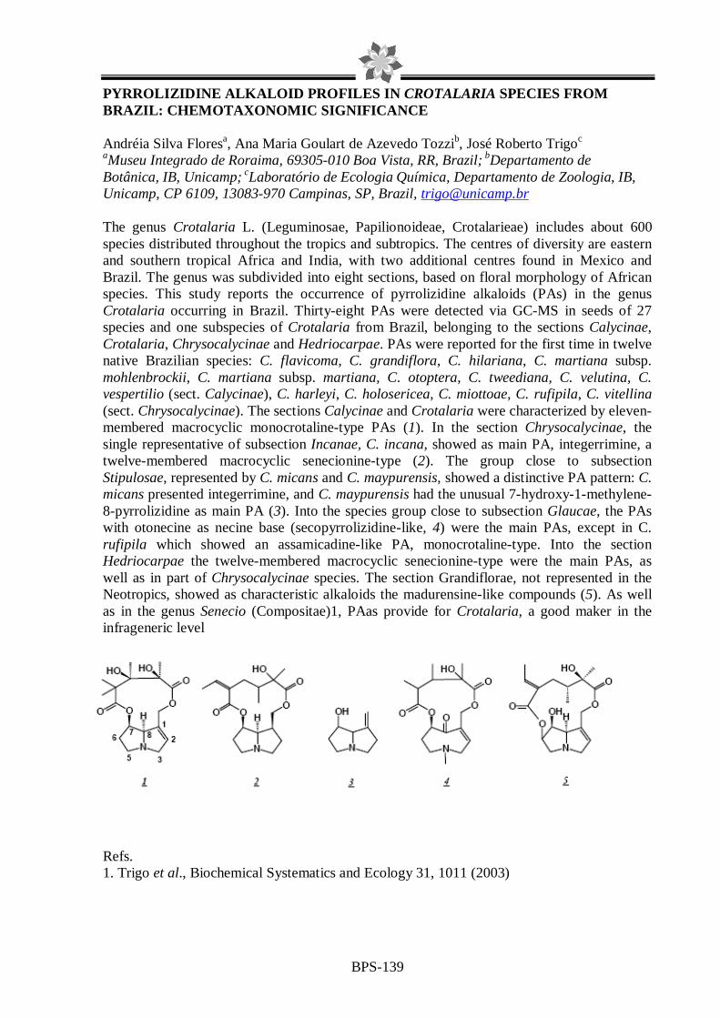
BPS-139
PYRROLIZIDINE ALKALOID PROFILES IN CROTALARIA SPECIES FROM BRAZIL: CHEMOTAXONOMIC SIGNIFICANCE Andréia Silva Floresa, Ana Maria Goulart de Azevedo Tozzib, José Roberto Trigoc aMuseu Integrado de Roraima, 69305-010 Boa Vista, RR, Brazil; bDepartamento de Botânica, IB, Unicamp; cLaboratório de Ecologia Química, Departamento de Zoologia, IB, Unicamp, CP 6109, 13083-970 Campinas, SP, Brazil, [email protected] The genus Crotalaria L. (Leguminosae, Papilionoideae, Crotalarieae) includes about 600 species distributed throughout the tropics and subtropics. The centres of diversity are eastern and southern tropical Africa and India, with two additional centres found in Mexico and Brazil. The genus was subdivided into eight sections, based on floral morphology of African species. This study reports the occurrence of pyrrolizidine alkaloids (PAs) in the genus Crotalaria occurring in Brazil. Thirty-eight PAs were detected via GC-MS in seeds of 27 species and one subspecies of Crotalaria from Brazil, belonging to the sections Calycinae, Crotalaria, Chrysocalycinae and Hedriocarpae. PAs were reported for the first time in twelve native Brazilian species: C. flavicoma, C. grandiflora, C. hilariana, C. martiana subsp. mohlenbrockii, C. martiana subsp. martiana, C. otoptera, C. tweediana, C. velutina, C. vespertilio (sect. Calycinae), C. harleyi, C. holosericea, C. miottoae, C. rufipila, C. vitellina (sect. Chrysocalycinae). The sections Calycinae and Crotalaria were characterized by eleven-membered macrocyclic monocrotaline-type PAs (1). In the section Chrysocalycinae, the single representative of subsection Incanae, C. incana, showed as main PA, integerrimine, a twelve-membered macrocyclic senecionine-type (2). The group close to subsection Stipulosae, represented by C. micans and C. maypurensis, showed a distinctive PA pattern: C. micans presented integerrimine, and C. maypurensis had the unusual 7-hydroxy-1-methylene-8-pyrrolizidine as main PA (3). Into the species group close to subsection Glaucae, the PAs with otonecine as necine base (secopyrrolizidine-like, 4) were the main PAs, except in C. rufipila which showed an assamicadine-like PA, monocrotaline-type. Into the section Hedriocarpae the twelve-membered macrocyclic senecionine-type were the main PAs, as well as in part of Chrysocalycinae species. The section Grandiflorae, not represented in the Neotropics, showed as characteristic alkaloids the madurensine-like compounds (5). As well as in the genus Senecio (Compositae)1, PAas provide for Crotalaria, a good maker in the infrageneric level
Refs. 1. Trigo et al., Biochemical Systematics and Ecology 31, 1011 (2003)

BPS-140
KAEMPFERITRIN FROM Uncaria guianensis (RUBIACEAE) AND ITS POTENTIAL AS CHEMICAL MARKER FOR THE SPECIES
Ligia M.M. Valente1, Rodolfo S. Barboza1, Sally Liechoch1, Carlos H. B. Bizarri2, M. Beatriz
S. Almeida2, Paulo J. C. Benevides2, Alvicler Magalhães2, Antonio C. Siani2 1Universidade Federal do Rio de Janeiro, Instituto de Química, Departamento de Química Orgânica, Centro de Tecnologia, Bloco A, 6oand., 21941-909, Rio de Janeiro, RJ, Brasil;
2Fundação Oswaldo Cruz, Instituto de Tecnologia em Fármacos, R. Sizenando Nabuco100, 21041-250, RJ, Brasil
The genus Uncaria (Rubiaceae) is represented in Central and South America for two species: U. tomentosa and U. guianensis, known as cat's claw. They have been used medicinally by indigenous peoples for at least two thousand years for several diseases. The species U. guianensis showed experimentally anti-inflammatory and anti-oxidant activities and U. tomentosa is noted for its immunostimulant activity, as well as cytotoxic, anti-inflammatory and antioxidant action. These bioactivities have made the species valuable plant materials and led to them being commercialized in natura or as phytopharmaceutical derivatives. Both species contain, in different proportions oxindole and indole alkaloids, triterpenoid glycosides, sterols and flavonoids that isolated or synergistically contribute for their therapeutic properties. They have some morphological differences and U. tomentosa can be further identified for their oxindole alkaloid profile and content. However, U. guianensis has a very low concentration of those alkaloids and does not have so far an efficient chemical marker.1 Until now only the polyphenolic compounds found in aqueous extracts of U. tomentosa have been reported.2 In a previous work we described the isolation for the first time in those species, from the bioactive3 EtOH extract of the leaves of U. guianensis, of the also bioactive kaempferol-3,7-O-(α)-dirhaminoside (kaempferitrin).4 Now we described the screening for kaempferitrin in the EtOH extracts from leaves and stems of U. tomentosa (Acre) and U. guianensis (Amazonas and Mato Grosso) through TLC and HPLC-DAD-MS by comparison to kaempferitrin previously isolated. The leaves and stems of both species were dried, milled and exhaustedly and sonically extracted with EtOH. The dried extracts were partitioned with hexane and MeOH/H2O 9:1 and the fractions were submitted to silica gel TLC with mobile phase systems for polyphenolic compounds and UV and NP/PEG reagent to visualize the spots. The MeOH/H2O fractions that contained polyphenolic compounds were then submitted to filtration on Sephadex LH-20 CC and the MeOH sub-fractions were analyzed by HPLC-DAD-MS in C18 reverse phase system and gradient mode using MeCN/H2O/HCOOH, pH=3. The results showed the presence of kaempferitrin only in U. guianensis with concentrations in the leaves almost five times greater than those observed in the stems (m/z 287 area). Besides, U. tomentosa had a polyphenolic profile with an overall content around four times lower than U. guianensis (TIC area). The results show the selectivity of U. guianensis to produce this bioactive flavonoid glycoside and suggest this compound as a potential chemical marker for the species. 1L.M.M. Valente, Fitos, 2, 48 (2006). 2C. Gonçalves, T. Dinis, M.T. Batista, Phytochemistry, 66, 89 (2005). 3 M.V. Carvalho, C. Penido, A.C. Siani et al., Inflammopharmacology 14, 48 (2006). 4M.B.S. Almeida, L.M.M.Valente, P.J.C. Benevides et al., X Encontro Regional de Química da Sociedade Brasileira de Química, 2005. CNPq, FAPERJ

BPS-141
EFFECT OF ESSENTIAL OIL OF Melampodium divaricatum (Rich. In Pers.) IN THE PRODUCTION OF HYDROGEN PEROXIDE AND NITRIC OXIDE IN MURINE MACROPHAGES GRACE PRISCILA PELISSARI (1); SANDRA REGINA ANANIAS(2); RAQUEL REGINA DUARTE MOREIRA(1);IRACILDA ZEPPONE CARLOS(2). (1)Departamento de Princípios Ativos Naturais e Toxicologia Faculdade de Ciências Farmacêuticas; (2)Departamento de Análises Clínicas, Faculdade de Ciências Farmacêuticas, Universidade Estadual Paulista “Júlio de Mesquita Filho,” Araraquara – SP. Introduction: Melampodium divaricatum is a member of the family Asteraceae known as false-calendula in Brazil where is used in a folk medicine, as anti-inflamatory, diuretic and against leucorrhea (1). In order to adapt to environmental insults, plants produce many natural products that have antimicrobial and immunomodulating potential (2). Macrophages release more than one hundred compounds into the extracellular environment. Among these, there are intermediate oxygen compounds, such as NO and H2O2. Objective: The purpose of this work was to study the effects of the essential oil of M. divaricatum in peritoneal macrophage cells from Swiss mice by determination of production the NO and H2O2. Material and Methods: The oil was obtained from the powdered leaves from Melampodium divaricatum, using Clevenger-type and the exudate peritoneal macrophages (PEC) were harvested from Swiss mice (6-8 weeks old, 18-25 g). For the determination of the cell viability, PEC (5x10⁶ cells/mL) was re-suspended in medium RPMI-1640 C. The suspension (100 µL) and the samples in different concentrations (100 µL) were added to each well of a 96-well tissue culture plate and incubated for 24 h. The 3-(4,5-dimethylthiazol-2-yl)-2,5-diphenyl-tetrazolium bromide (MTT) assay was performed and absorbance measured at 540 nm with a 620 nm reference filter. The nitric oxide production was determined by measuring nitrite. 50 µL of cell supernatant was removed from each well and incubated with an equal volume of Griess reagent at room temperature for 10 min; the absorbance was measured at 540 nm. For H2O2 measurement, PECs (2x10⁶ cells/ml) were re-suspended in a solution of phenol red complete buffer; 100 µL of this suspension was added to each wells of a tissue culture plate and added 50 µL of aqueous extract in the concentration to viability of 50%. After incubation for 1 h, the reaction was stopped with 50 µL of NaOH 5M and absorbance measured at 620 nm. Results: The data indicated that essential oil of M. divaricatum has a high cytotoxicity in vitro on peritoneal macrophages (IC50 = 0,075 µL/mL e IC25 = 0,040 µL/mL). In this study, the essential oil could strongly inhibit NO production and H2O2 production in macrophages stimulated by LPS and PMA respectively. The inhibition percentage ranged from 80,33 ± 4,76% (NO) and 76,22 ± 8,87% (H2O2) in the concentration 0,075 µL/mL and 40,58 ± 6,16% (NO) and 70,59 ± 1,19% (H2O2) in the concentration 0,040 µL/mL. According to this work, it is possible to suggest that essential oil obtained from M. divaricatum presents anti-inflammatory activity. 1. AGRA, M. de F.; FREITAS, P.F.; BARBOSA-FILHO, J.M..Revista Brasileira de Farmacognosia, vol. 17, n. 1, p. 114 – 121, jan./mar., (2007). 2. DIXON, R. A., Nature (London), 411, 843—847 (2001). FINANCIAL SUPPORT: CNPq

BPS-142
CHEMOSYSTEMATIC STUDY OF BURSERACEAE OLEORESINS
André Luis Rüdiger and Valdir Florêncio da Veiga Junior Dpto. de Química, Instituto de Ciências Exatas, Universidade Federal do Amazonas Av. Rodrigo Otávio Jordão Ramos, 3000, Japiim, 69077-000, Manaus, AM, Brasil.
Burseraceae family has a great importance in Brazilian folk medicine. Its oleoresin, popularly known as “Breu”, has several ethnopharmacological indications (1). Species from this family are usually hard to identify (2) and only few botanical specialists are able to identify them, but only through observation of its flowers, resulting in several taxonomic doubts. The genus Protium is the most common specie in Brazil, with chemistry and pharmacology recently reviewed (3). The present study proposes that an extensive chromatographic profile of oleoresin extracts from this family could be used to correlate chemical and taxonomic data, in a chemosystematic research. About 37 oleoresins, from Protieae (Crepidospermum, Protium and Tetragastris genus) and Canarieae Tribes (Dacryodes and Trattinnickia genus) were collected at South, Southeast, Northeast and North regions of Brazil. The majority of the oleoresins were obtained in the Amazon Region, where endemism and biodiversity of this family are the greatest in the New World. Thin layer chromatography (TLC), gas chromatography (GC-FID) and gas chromatography coupled with mass spectrometry (GC-MS) were applied to the apolar (hexane) and medium polarity (ethyl acetate) extracts. The yield from each oleoresin from the several extracts shows a clear division between some genus, and inside Protium genus. The comparative TLC showed several profiles that fit on the Burseraceae species phylogenetic distribution (4,5). Crepidospermum genus showed the greatest variation, with unusual low concentrations of the very common triterpenes α- and β-amirin, easily obtained from the Burseraceae breu. Using GC analyses we observed that the triterpene retention time region could be a fingerprint region, together to the monoterpene/sesquiterpene/triterpene yield relationship. Again, Crepidospermum showed a quite different profile, with some diterpenes that are not common in Neotropic Burseraceae species. Refs. 1. M. Pio Correa. Dicionários de plantas úteis do Brasil e das exóticas cultivadas. (1984) 2. J.E.L. Ribeiro et al. Flora da reserva Ducke: Guia de identificação das plantas vasculares de uma floresta de terra firme na Amazônia Central (1999) 3. A.L. Rüdiger et al., Pharmacognosy Reviews, 1, 93 (2007) 4. A. Weeks et al., Molecular Phylogenetics and Evolution, 35, 85 (2005) 5. P.V.A. Fine et al., Evolution, 59, 1464 (2005)

BPS-143
INFLUENCE OF THE CHIRALITY OF (R)-(-)- AND (S)-(+)- CARVONE IN THE CENTRAL NERVOUS SYSTEM: A COMPARATIVE STUDY
Damião Pergentino de Sousa* 1, Franklin Ferreira de Farias Nórega2, Reinaldo Nóbrega de
Almeida * 1Departamento de Fisiologia, Universidade Federal de Sergipe, CEP 49100-000 São
Cristóvão, Sergipe, Brazil. 2Laboratório de Tecnologia Farmacêutica, Universidade Federal da Paraíba, Caixa Postal
5009,CEP 58051-970, João Pessoa, Paraíba, Brazil Many terpenes are used therapeutically, and as flavor and fragrance materials. (R)-(-)-Carvone, the main constituent of spearmint oil, and (S)-(+)-carvone, found as major component of caraway and dill seed oils, have several applications and are used in cosmetic, food, and pharmaceutical preparations. In this study, the effect of enantiomers of carvone on the central nervous system (CNS) was evaluated in mice. The LD50 value was 484.2 mg/kg (358.9–653.2) for (S)-(+)-carvone, and 426.6 (389.0–478.6) mg/kg for (R)-(-)-carvone. Both enantiomers caused depressant effects, such as decrease in the response to the touch and ambulation, increase in sedation, palpebral ptosis, and antinociceptive effects. (S)-(+)- and (R)-(-)-carvone caused a significant decrease in ambulation. (R)-(-)-Carvone appeared to be more effective than its corresponding enantiomer at 0.5 and 2.0 h after administration. However, (S)-(+)-carvone was slightly more potent at 1 h. In potentiating pentobarbital sleeping time, (R)-(-)-carvone was more effective than (S)-(+)-carvone at 100 mg/kg, but was less potent at 200mg/kg compared to the (+)-enantiomer, indicating a sedative action. (S)-(+)-Carvone at the dose of 200 mg/kg increased significantly the latency of convulsions induced by PTZ and PIC, but (R)-(-)-carvone was not effective against these convulsions. These results suggest that (S)-(+)-carvone and (R)-(-)-carvone have depressant effect in the CNS. (S)-(+)-Carvone appears to have anticonvulsant-like activity.
O O
(R)-(-)-carvone (S)-(+)-carvone Refs. 1. D.P. De Sousa et al., Chirality, 19, 264 (2007)

BPS-144
OIL PROPOLIS EXTRACT FOR PHARMACEUTICAL PURPOSES: A N ALTERNATIVE TO ETHANOLIC EXTRACT
Lilian Buriol1, Daiane Finger1, Eduardo Morgado Schmidt1, Julio M. T. dos Santos1, Marcos Roberto da Rosa1, Sueli Pércio Quináia1, Yohandra Reyes Torres1, Alexandra Christine H. F.
Sawaya2, Marcos Nogueira Eberlin2. 1Departamento de Química, CEDETEG, Universidade Estadual do Centro-Oeste,
Guarapuava, Paraná, Brasil. 2Laboratório Thomson de Espectrometria de Massas, Instituto de Química, Unicamp, Campinas, São Paulo, Brasil
Propolis is a natural resinous product used as hydroalcoholic extract in pharmaceutical, cosmetic and hygienic products. Propolis pharmacological properties such as antibiotic, antifungal, cytotoxic, antioxidant and anti-inflammatory have been related to the phenolic composition in flavonoids and phenolic acids1. The amount of these compounds is indicated as a parameter for quality control of extracts and crude propolis2. The presence of ethanol in the formulation provokes rejection by some consumers. That’s why there has been a growing number of patents dealing with new solvents for preparing propolis extracts3. In this work we aimed to prepare edible oil propolis extracts (OEP) and compare their composition with ethanolic propolis extracts (EEP) using Electrospray Ionization Mass Spectrometry (ESI-MS) and the spectrophotometric method for qualitative and quantitative analyses, respectively. By visual inspection of the ESI-MS spectra we have concluded that both extracts have similar chemical composition. Twelve compounds have been identified by comparison of their ESI-MS spectra with those of standards: p-coumaric acid, caffeic acid, 2,2-dimethyl-2H-1-benzopyran-6-propenoic acid, 3-prenyl-4-hydroxycinnamic acid, 3,4-dihydroxy-5-prenylcinnamic acid, kaempferide, 3,5-diprenyl-4-hydroxycinnamic acid, dihydrokaempferid, 4-hydroxy-3(E)-(4-hydroxy-3-methyl-2-butenyl)-5-prenyl cinnamic acid, betuletol, 3-prenyl-4-dihydrocinnamoyloxy-cinnamic acid and dicaffeoylquinnic acid. Total phenolic substances and flavonoids were measured by spectrophotometric method. Compared with EEP, the OEP had lower yield but higher phenolic and almost similar flavonoids content, although it was necessary a longer extraction period. These results have encouraged us to create a new methodology of preparing oil propolis extracts for pharmaceutical purposes which is currently being patented. Keywords: ethanolic and oil propolis extracts, phenolic acids, flavonoids, Electrospray Ionization Mass Spectrometry, spectrophotometric method. Refs. 1. M.C. Marcucci, Apidologie, 26, 83, (1995). 2. R.G. Woisky; A. Salatino, J. Apic. Res., 37, no 2, 99, (1998). 3. A.S. Pereira et. al., Química Nova, 25, nº 2, 321, (2002).
BPS-145
ESSENTIAL OILS OF PIPER DEMERARANUM AND PIPER DUCKEI
Dominique F. Moura1, Jefferson R. de A. Silva1, Maria Lúcia B. Pinheiro1, Ana Claudia F. Amaral2
1Laboratório de Cromatografia, Departamento de Química, UFAM / Manaus – AM;
2Laboratório de Plantas Medicinais e Derivados, DPN / Farmanguinhos / FIOCRUZ - RJ. E-mail: [email protected]
The genus Piper belongs to the Piperaceae and has over 1.000 species of herbs, shrubs, small trees and hanging vines distributed in both hemispheres. The Piper species have high commercial, economical and medicinal importance, widely distributed in the tropical and subtropical regions of the world where are used in folk medicine as, for example, stomachic and carminative in digestion, diuretic and toothache. Chemical studies have shown that the genus Piper has many components including unsaturated amides, flavonoids, lignans, terpenes, steroids, phenols and alkaloids. This work presents the chemical composition from essentials oil of two Piper species, P. demeraranum (Miq.) C. Dc. and P. duckei C. Dc. that were collected in the Reserve Ducke, Manaus (Amazonas state) and the voucher specimens were deposited at herbarium of INPA (AM). The essential oils from leaves of these species were analyzed by a combination of GC/FID and GC/Mass Spectrometry using a capillary GC column HP-5 (30 cm). The essential oils were obtained from dry leaves (40g) of both plants using a Clevenger type apparatus for 3h. The oils were separated from water, dried with Na2SO4 and stored in sealed vials at low temperature before analysis. The yields of the oils were 1.8% (P. demeraranum) and 1.2% (P. duckei). The main constituents found in the oil of P. demeraranum were β-elemene (34.50 %), limonene (20.56 %), germacrene B (9.11 %), β-pinene (7.07 %). In the oil of P. duckei the main compounds found were trans-caryophyllene (29.62 %), 1H-cycloprop[e]azulene (19.20 %), germacrene-D (15.92 %). Although being the first study about the essential oils of these Piper species, the most frequently identified compounds were non-oxygenated monoterpenes and sesquiterpenes, which are similar to other essentials oils of this analyzed genus. Financial support: Fapeam; CNPq/PADCT

BPS-146
ELECTROPHYSIOLOGICAL RESPONSES OF THYRINTEINA ARNOBIA MALE AND FEMALE MOTHS TO PSIDIUM GUAJAVA ESSENTIAL OIL
Tiago Neppe,* Arlene G. Corrêa
Department of Chemistry, Federal University of São Carlos, São Carlos, SP, Brazil *[email protected]
The Brazilian eucalyptus brown looper, Thyrinteina arnobia (Lepidoptera: Geometridae), is the most harmful of the Eucalyptus pests in Brazil.1 It also feeds on native plants such as Villaresia congonha, Ilex paraguaiensis, Campomanesia spp., Eugenia spp. and Psidium guajava.2 In this way, an efficient methodology for this insect control in different cultures is necessary and becomes essential to understand the interactions mediated by semiochemicals. In this work were evaluated the electroantennographic responses (EAG) of male and female moths of T. arnobia to essential oil of white guava plants (P. guajava L.). The experiments were carried out using young (sprouts) and old leaves, collected from two different plants, which were submitted to steam distillation, using a Clevenger apparatus. For EAG, antennae from virgin male and female created in artificial diet and later transferred to feeding with plant leaves had been used. Three different oil concentrations were evaluated. It was observed, for both male and female, a maximum antenna depolarization at concentration of 10 mg/mL of either young or old leave essential oils, with no significant difference. EAG responses in both sexes were significantly higher than that for the control (hexane). These results indicate that T. arnobia adults recognize some volatile compound in the P. guajava essential oils; that is, in the antennae of both sexes there are neuron receptors (chemoreceptors), present in sensillas, which detected compounds in the essential oil. In order to confirm these results further studies will be necessary, followed by semiochemicals identification by using GC-EAD and GC-MS techniques.
1. Batista-Pereira L. G. et al., J. Braz. Chem. Soc., 17: 555, 2006. 2. Oliveira H. N. et al., Braz. Arch. Biol. Technol., 48: 801, 2005.
Acknowledgements: FAPESP, CNPq, CAPES and IFS.

BPS-147
LIPOSOMAL DELIVERY SYSTEM FOR ENCAPSULATION OF Zanthoxylum tingoassuiba ESSENTIAL OIL: PREPARATION, CHARACTERIZATION AND I N
VITRO ANTIMICROBIAL ACTIVITY
E.C.M.Cabral-Albuquerque; C.B.Detoni; S.V.A. Hohlemwerger; C.Sampaio, T.F.Barros; E.S.Velozo
LAPEMM/Faculdade de Farmácia/Universidade Federal da Bahia, Bahia, Brasil [email protected]
Essential oils are made up of many different volatile compounds. In general, essential oils have a high content of terpenes easily prone to oxidation and resinification. It seems that the antimicrobial effects are the result of many compounds acting synergistically. In addition, oils have low solubility in aqueous media, limiting their pharmaceutical applications. In order to circumvent those disadvantages, microencapsulation has been proposed as an alternative to improve stability, solubility and biological activity of essential oils (1). In recent years, liposomes have been extensively studied as a delivery system which can improve the activity and safety of many molecules. Further, liposomes are regarded as suitable carriers because they can serve as a depot system for the sustained release of an associated compound and enhance the molecules targeting to cells (2). Pursuing our interest in medicinal products obtained from Rutaceae plants, in this work we have studied the preparation and characterization of multilamelar liposomes entrapping essential oil (EO) from Zanthoxylum tingoassuiba. Essential oil from Z. tingoassuiba loaded into multilamellar liposomes was successfully produced through thin film hydration method with mean diameter of 9.4µm. The liposome-incorporated EO showed good sphericity and narrower size distribution than empty liposomes. Results of GC-MS and UV-VIS spectrophotometer revealed that EO incorporation efficiency in liposomes was approximately 50%. A qualitative analysis by Thin Layer Chromatography revealed that essential oil was successful incorporated into liposomes with no exclusion of the essential oil components. Differential scanning calorimetry (DSC) was applied to further investigate interactions of essential oil with a liposome. Changes in the liposome DSC endotherms occurring in the presence of essential oil suggest that it is situated in lipid bilayer of liposomes. The antimicrobial activity of incorporated and free EO against some ATCC bacteria, mushroom and multiresistent clinical isolates of S. aureus was evaluated. Free EO inhibited the growth of all microorganisms tested. Results obtained clearly indicate that essential oil from Z.tingoassuiba has been successful incorporated into multilamellar liposomes. Liposomes can be useful in enhancing the solubility and antimicrobial activity.
Refs. 1. D. Valenti et al., Journal of Liposome Research, 11, 73 (2001) 2.C. Sinico et al., European J. of Pharmaceutics and Biopharmaceutics, 59, 161 (2005)

BPS-148
CHEMICAL COMPOSITION OF THE ESSENTIAL OIL FROM HYPTIDENDRON CANUM (POHL EX BENTH.) HARLEY (LAMIACEAE) LEAVES AND
INFLORESCENCES
Tatiana de Sousa Fiuza1, José Realino de Paula1 , Pedro H. Ferri1, Heleno Dias Ferreira1, Leonice M. F. Tresvenzol1 , Simone Maria Teixeira de Sabóia-Morais1
1 Universidade Federal de Goiás Abstract Hyptidendron canum is a specie belonging to the savannah biome of the Lamiaceae family, which in turn has great economic standing for being the source of aromatic essential, volatile oils and ornamental plants. The aim of this study is to analyse the chemical constituents of the essential oil from leaves and inflorescences of H. canum from population samples representative of four towns in the state of Goiás - Brazil, and compare the chemical variability among them. For this purpose the essential oils from the material collected were obtained through hydrodestilation21 in a Clevenger apparatus and analysed by GC/Ms. Sesquiterpene hydrocarbons constituted the main group of compounds in all the populations. Nonetheless a few differences were noted in the quantities of the main constituents, namely of (E)-cariophyllene and amorpha-4,7(11)-diene, with the greatest contents in Bela Vista, and of bicyclogermacrene with high percentages in the Silvânia population, sabinene and β-copaen-4-α-ol with a large amount in the Silvânia population, as well as δ-3-carene and carotol with greater contents in the Hidrolândia population, suggestive of chemical polymorphism. Results obtained from Principal Component Analysis (PCA) and cluster analysis indicate great variability in the chemical composition of essential oils in the populations.

BPS-149
ANTIMICROBIAL ACTIVITY OF THE ESSENTIAL OIL OF Maytenus obtusifolia Mart. (CELASTRACEAE)
Vivianne M. de Medeirosa, Gabriela L. de A. Maiaa, José Guedes de Sena Filhoa, Josean F. Tavaresa, Damião P. de Sousaa, Egberto S. Carmoa, Ana Lívia R. Tiburtinoa, Edeltrudes de O. Limaa, José Maria Barbosa Filhoa, Marcelo Sobral da Silvaa. aUniversidade Federal da Paraíba, Laboratório de Tecnologia Farmacêutica - Caixa Postal 5009, 58051-970 - João Pessoa-PB, Brasil. *vivi_mmedeiros @yahoo.com.br Maytenus obtusifolia Mart. is a small tree found in the South America and Asia. In Brazil, where it is commonly known as “bom-nome” and “carne-de-anta”, its leaves are largely used in the folk medicine as anti-ulcer. Previous studies reported the presence of pentacyclic triterpenes from the roots extracts and flavonoids in the leaves extracts of Maytenus species. This work describes the antimicrobial activity of the essential oil of Maytenus obtusifolia. The plant was collected in May 2002, near the city of Santa Rita, State of Paraíba, Brazil, a coastal area around the Atlantic Forest. The voucher samples (Agra et Góis 3230) were deposited in the Herbarium Prof. Lauro Pires Xavier (JPB) and in the reference collection of the Laboratório de Tecnologia Farmacêutica from Universidade Federal da Paraíba, Brazil. Fresh leaves of Maytenus obtusifolia (1000 g) were cut into small pieces, and subjected to steam distillation in a Clevenger-type apparatus. For the bioassays 5 bacteria and 8 fungi were used. The essential oil was tested “in natura” (100%) and in dilutions from 32 until 2%, according to Allegrini et al. (1973). The present work was conducted in order to evaluate the antimicrobial activity using the agar well diffusion assay. The biological activity of the oil was considered positive when the media of the inhibition zone were equal or superior to 10 mm in diameter. The initial evaluation (in natura) showed inhibition growth against: S. epidermidis (ATCC 12228), S. aureus (ATCC 6538), E. coli (ATCC 18739), P. aeruginosa (ATCC 27853), L. monocytogenes (ATCC 9610), C. albicans (ATCC 90028), C. albicans (ATCC 13803), C. tropicalis (LM 37), A. flavus (LM 247) and C. guilliermondii (LM V70) affording inhibitions zones between 10-45 mm diameters. In the dilution of 32% presented inhibition growth against: S. aureus (ATCC 6538), E. coli (ATCC 18739), L. monocytogenes (ATCC 9610), C. albicans (ATCC 13803), C. tropicalis (LM 37), C. guilliermondii (LM V70), and A. flavus (LM 247). In the dilution of 16% against: S. aureus (ATCC 6538), E. coli (ATCC 18739), L. monocytogenes (ATCC 9610), C. albicans (ATCC 13803) and C. tropicalis (LM 37). The diluition of 8% presented activity against Sthaphylococcus and Candida. In summary, the study of volatile constituents showed that the oil inhibited the growth of ten of the microorganisms, when tested with the oil at the concentration of 100%. These results led to the conclusion that the essential oil tested has a weak activity against the tested microorganisms.
Refs. 1. J. Corsino et al., Phytotherapy Research, 17(8), 913-916 (2003) 2. J. Allegrini et al., Societé de Pharmacie de Montpellier, 33, 73-86 (1973)

BPS-150
THE SMELL OF Melipona quadrifasciata INFLATED VIRGIN QUEENS (HYMENOPTERA: APIDAE, MELIPONINI)
Adriana Pianaroa, Cristiano Menezesb, Vera Lúcia Imperatriz-Fonsecab, Anita Jocelyne
Marsaiolia,* aInstituto de Química, Universidade Estadual de Campinas, 13084-862, Campinas-SP, Brazil. *E-mail:
[email protected] bDepartamento de Biologia, Faculdade de Filosofia, Ciências e Letras de Ribeirão Preto, Universidade de São Paulo, 14040-901, Ribeirão Preto - SP, Brazil.
Melipona quadrifasciata is a native social stingless bee belonging to the tribe Meliponini, which occurs in tropical regions.1, 2 Mating in Meliponini generally occurs during the flight, and can be observed under laboratorial controlled conditions.3 Nevertheless, several questions concerning to virgin queens´ attractiveness to males remain to be answered. Attractive virgin queens inflate their abdomens in the colonies calling a court of workers, that allow them to survive for a while in their nests, testing their attractiveness to supersede the physogastric queen or to depart with a swarm.3 Inside their nests, they do not attract males, what occurs during their nuptial flights or in experimental arenas, with solitary gynes.3 Melipona quadrifasciata virgin queens liberate a characteristic smell after inflating their abdomens, and this smell is perceived by humans. Therefore, the aim of this work was to characterize the compounds responsible for this smell. Inflated virgin queens (2 to 3 days old) and normal virgin queens (1 day old) were submitted to dynamic headspace using porapack Q resin to trap the volatiles. The substances were eluted from the polymer with a hexane/ethyl acetate mixture and analyzed by gas chromatography-mass spectrometry. Inflated virgin queens produced a mixture of nerol and geraniol (2:3), while these substances were not observed in normal virgin queens. Biological assays aiming to discover whether these substances play any role on male attraction are currently being carried out. It was not found in literature any other report concerning the characteristic smell of inflated virgin queens.
OH
Geraniol
OH
Nerol
Refs. 1. Nogueira-Neto, P., Vida e Criação de Abelhas Indígenas Sem Ferrão, São Paulo: Editora Nogueirapis, p. 446 (1997). 2. Nascimento, V. A. et al., Genet. Mol. Biol., 23, 79 (2000). 3. van Veen, J. W. et al., Insectes Soc., 46, 361 (1999).
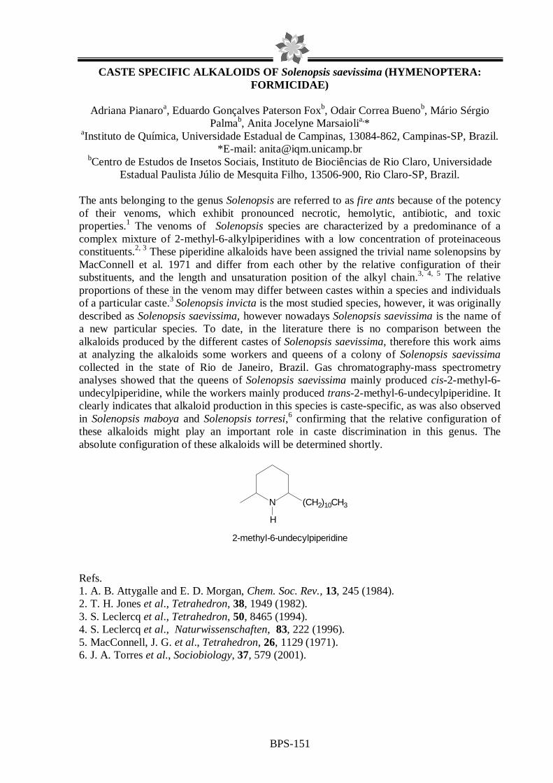
BPS-151
CASTE SPECIFIC ALKALOIDS OF Solenopsis saevissima (HYMENOPTERA: FORMICIDAE)
Adriana Pianaroa, Eduardo Gonçalves Paterson Foxb, Odair Correa Buenob, Mário Sérgio
Palmab, Anita Jocelyne Marsaiolia,* aInstituto de Química, Universidade Estadual de Campinas, 13084-862, Campinas-SP, Brazil.
*E-mail: [email protected] bCentro de Estudos de Insetos Sociais, Instituto de Biociências de Rio Claro, Universidade
Estadual Paulista Júlio de Mesquita Filho, 13506-900, Rio Claro-SP, Brazil.
The ants belonging to the genus Solenopsis are referred to as fire ants because of the potency of their venoms, which exhibit pronounced necrotic, hemolytic, antibiotic, and toxic properties.1 The venoms of Solenopsis species are characterized by a predominance of a complex mixture of 2-methyl-6-alkylpiperidines with a low concentration of proteinaceous constituents.2, 3 These piperidine alkaloids have been assigned the trivial name solenopsins by MacConnell et al. 1971 and differ from each other by the relative configuration of their substituents, and the length and unsaturation position of the alkyl chain.3, 4, 5 The relative proportions of these in the venom may differ between castes within a species and individuals of a particular caste.3 Solenopsis invicta is the most studied species, however, it was originally described as Solenopsis saevissima, however nowadays Solenopsis saevissima is the name of a new particular species. To date, in the literature there is no comparison between the alkaloids produced by the different castes of Solenopsis saevissima, therefore this work aims at analyzing the alkaloids some workers and queens of a colony of Solenopsis saevissima collected in the state of Rio de Janeiro, Brazil. Gas chromatography-mass spectrometry analyses showed that the queens of Solenopsis saevissima mainly produced cis-2-methyl-6-undecylpiperidine, while the workers mainly produced trans-2-methyl-6-undecylpiperidine. It clearly indicates that alkaloid production in this species is caste-specific, as was also observed in Solenopsis maboya and Solenopsis torresi,6 confirming that the relative configuration of these alkaloids might play an important role in caste discrimination in this genus. The absolute configuration of these alkaloids will be determined shortly.
N (CH2)10CH3
H
2-methyl-6-undecylpiperidine
Refs. 1. A. B. Attygalle and E. D. Morgan, Chem. Soc. Rev., 13, 245 (1984). 2. T. H. Jones et al., Tetrahedron, 38, 1949 (1982). 3. S. Leclercq et al., Tetrahedron, 50, 8465 (1994). 4. S. Leclercq et al., Naturwissenschaften, 83, 222 (1996). 5. MacConnell, J. G. et al., Tetrahedron, 26, 1129 (1971). 6. J. A. Torres et al., Sociobiology, 37, 579 (2001).

BPS-152
Activity of the essential oil from Coriandrum sativum L. against different oral Candida spp
Andreza F.Begnamia, Vivian F. Furlettia, Adriana S. Santosb, Marta C.T. Duarteb, Vera L.G.Rehderb
a Faculty of Dentistry of Piracicaba, UNICAMP, Brazil. bPluridisciplinar Center of Chemistry, Biological and Agricultural Researches, UNICAMP, Brazil; Different Candida spp. are commonly found in the oral microbiota and can act as opportunist producing superficial clinical manifestations, since candidoses until serious and invasive system infections. Coriandrum sativum L. (Umbelliferae/Apiaceae) known as “coentro” is a popular condiment in the Brazilian cooking and also is used as digestive, carminative, diuretic and tonic. The objective of this work was to evaluate the activity of the essential oil of C. sativum L. against different oral Candida spp: Candida albicans- CBS 562, C. krusei CBS 573, C. parapsilosis CBS 604, C. dubliniensis CBS 7987 and C. tropicalis CBS 94. The major oil constituents were also identified. The essential oil was obtained from leaves by hydrodestilation in Clevenger system. Subsequently, the oil was separated from the water phase by liquid/liquid extraction with dichloromethane and diethyl ether, and fractionated in dry column to supply six fractions for different polarities. The oil and fractions composition were determined by GC and GC–MS analyses. The minimal inhibitory concentration (MIC) from oil and fractions was determined by microdillution method (NCCLS, 2002), until concentration of 1.0 mg/mL. MIC results of the crude oil and most active fractions are shown in Table 1. The results shown that fractions 4 and 6 presented the best activities against C. albicans CBS 562, C. dubliniensis CBS 7987, and C. parapsilosis CBS 604, with MIC values between 0.007 and 0.031 mg/mL. These values are similar or lower from that commonly observed for conventional antibiotics. Table 1: Anti-Candida activity of the essential oil and fractions from Coriandrum sativum.
MIC (mg/mL) Candida spp. Crude essential oil Fraction 4 Fraction 6
C.albicans CBS 562 0.500 0.031 0.031 C.krusei CBS 573 0.250 0.063 0.063 C.parapsilosis CBS 604 0.125 0.007 0.007 C.dubliniensis CBS 7987 0.250 0.007 0.015 C.tropicalis CBS 94 * 0.063 0.063 * MIC > 1.0 mg/mL;
The major compounds identified in the crude essential oil were alcohols and aldehydes (80.76%): n-decanal (24.17%), 2-decenol (18.05%), 2-dodecenal (17.55%), 3-hexenol (Z) (10.34%), decanal (4.76%), dodecanal (3.02%) and 2-dodecenal (2.88%). The fraction 4 presented dodecanal (39.88%), 2-decenol (30.12%), n-decanol (11.41%), and 2-tetradecenol (E) (8.38%), while the constituents of the fraction 6 were: n-decanol (54.78%), 3-hexenol (Z) (16.29%) and 2-hexenol (E) (6.22%) and 2-undecenol (Z) (6.15%). The analysis revealed that fractions 4 and 6 are more enriched in alcohols than in aldehydes. The present study revealed a strong activity from C. sativum essential oil and fractions against oral Candida spp., and its potential use in the treatment of the oral candidoses.

BPS-153
Analysis of volatile components of aerial parts from Mikania pilosa Baker (Asteraceae) by HS-SPME and GC-MS.
1Luiz Elídio Gregório (PG), 2Denise Brentan da Silva (PG), 3Arnildo Pott (PQ), 2Dionéia Camilo Rodrigues de Oliveira (PQ)
1Faculdade de Filosofia, Ciências e Letras de Ribeirão Preto – USP – Av. Bandeirantes n. 3900, CEP 14040-901, E-mail: [email protected]; 2Faculdade de Ciências Farmacêuticas
de Ribeirão Preto – USP; 3Embrapa Gado de Corte Campo Grande – MS.
The genus Mikania (family Asteraceae, tribe Eupatorie, subtribe Mikaniinae) contains more than 450 species that grown in tropical regions of Africa, Asia and America, with 150 species occurring in Brazil [1]. Several Mikania species, particularly those known under the common name “guaco”, are used in Central and South American folk medicine as antiinflammatory agents, to treat respiratory tract diseases, rheumatism, influenza and against snake bite [2].
Mikania pilosa Baker is a brazilian species, which is found mainly in the states of MT, MG, SP, RJ, SC e PR [3]. However, still there is no previous report of the chemical composition of Mikania pilosa Baker. The aim of this study was to evaluate the volatile components of the aerial parts from Mikania pilosa Baker by the technologies HS-SPME and GC-MS. The plant Mikania pilosa Baker was collected in Costa Rica, MS, Brazil, in October 2004 and were identified by Dr. Roberto Lourenço Esteves (Departamento de Biologia Animal e Vegetal, Universidade Estadual do Rio de Janeiro, Brazil). The sample (400 mg) was put into headspace vial (4 mL) coupled to the PDMS fiber and was heated to 60 ºC, with extraction time of 30 min. GC-MS analysis were performed with a Shimadzu GC-2010 Chromatograph equipped with a DB-5 column (30 m x 0.25 mm, coating thickness 0.25 µm) and a GCMS-QP2010 quadrupole mass detector. Analytical conditions: injector 250 ºC; oven temperature programmed from 60 ºC to 240 ºC at 3 ºC/min; carrier gas helium at 1.33 mL/min; injection splitless mode; fiber desorption time 5 minutes. The identification of the constituents was based on comparison of the retention indices (RI) with literature date [4] and library mass spectra. Forty-eight components were identified of which spathulenol (31.4 %), δ-cadinene (11.2 %), β-selinene (6.0 %) and ledol (4.2 %) were major. Acknowledgements: CNPq, FAPESP and CAPES for financial support, Prof. Dr. Roberto Lourenço Esteves for plant identification.
Refs. 1. N. Hind, Kew Bull., 48, 245 (1993) 2. M. Krautmann et al. Journal of Natural Products, 70, 1173-1179 (2007). 3. G.M. Barroso, Arquivos do Jardim Botânico do Rio de Janeiro, 16, 244 and 320 (1958). 4. R.P. Adams, Identification Oil Components by Gas Chromatography/Mass Spectroscopy, Allured Publishing Corporation, USA, 1995.
H
H
H
H
HO
H
OH
spathulenol δ-cadinene β-selinene ledol

BPS-154
ASSESSMENT OF ESSENTIAL OIL YIELDS FROM Cymbopogon citratus (DC) STAPF, AND ANTIMICROBIAL ACTIVITY RELATED TO SEASON AND
CULTIVATION WITH Achillea millefolium
Adriana Santos ¹, Rafael Henrique Paduan¹, Zilda Cristiani Gazin², Ezilda Jacomassi²; Pérsio Sandir D’ Oliveira 3, Diógenes Aparício Garcia Cortez 4, Lúcia Elaine Ranieri Cortez 3*
1Acadêmicos do curso de Farmácia da Universidade Paranaense, 2 Universidade Paranaense, 3Curso de Farmácia do Centro de Ensino Superior de Maringá (CESUMAR) Av. Guedner, 1610, Maringá, PR, Brasil.
4Departamento de Farmácia e Farmacologia da Universidade Estadual de Maringá, 87020-900, Maringá, PR, Brasil.
The essential oil of the plant Cymbopogon citratus (DC) Stapf cultivated in the Medicinal Garden of Paranaense University in the city of Umuarama, Paraná, was obtained by steam distillation. Yield and seasonal variation were determined, and correlated with the ambient temperature and precipitation. Plants harvested in the morning of days with high temperatures and rainfall, gave better yields from plants cultivated alone and also together with Achillea millefolium L. Microbiological analysis of the essential oil of C. citratus was carried out by the broth microdilution method against the yeasts Candida albicans and C. tropicalis, and the bacteria Escherichia coli, Staphylococcus aureus and Pseudomonas aeruginosa. The essential oil from plants cultivated alone and together with A. millefolium showed moderate activity against all yeasts tested (0.63 - 1.25 mg/mL-1). The essential oil from plants cultivated alone showed high activity against Staphylococcus aureus (0.08 mg/mL-1), but no activity against the other bacteria.

BPS-155
CHARACTERIZATION AND ADULTERATION OF ROSEWOOD OIL B Y SYNTHETIC LINALOOL USING ELECTROSPRAY IONIZATION MA SS SPECTROMETRY
Rita C. Z. Souza1, Rodrigo R. Catharino2, Elaine C. Cabral2, Marcos N. Eberlin2 and Lauro E. S. Barata1.
1 Research and Development of Natural Products Laboratory 2Thomson Mass Spectrometry Laboratory, Institute of Chemistry Campinas, SP 13083-970, Brazil Institute of Chemistry – State University of Campinas (Unicamp); CP 6154, 13084-971, Campinas, SP – BRAZIL
Rosewood oil (obtained from Aniba rosaeodora) is one of the most important commercial essential oils produced in the Amazon region. A common practice is to adulterate fine rosewood oil (US$80/Kg) with synthetic linalool (US$8/Kg). Earlier studies have applied alternative techniques to characterize essential oils1. In this work, a systematic analysis with different blends of rosewood and synthetic linalool was used to establish a simple and fast methodology to trace contaminants, based on direct infusion of the samples in the electrospray ionization source of the mass spectrometer (Electrospray Ionization Mass Spectrometry ESI-MS). Five individual samples of each essential oil (wood and leaf) and synthetic linalool were analyzed. The polar compounds were extracted from each sample with methanol/water 1:1 (0.1% strong acid) solution and analyzed in positive ion mode. For the adulteration detection, leaf and wood oils were mixed with synthetic linalool. The three pure samples presented similar profiles in positive ion mode, m/z 137, 274 and 292, although in distinct proportions. The three samples could be differentiated from one another, by the presence, absence or different proportions of marker ions. For instance, the intensity ratio of m/z 137 and 153 ions in crude wood oil was 1.2:1.0, in crude leaf oil it was 3.1:1.0 and for synthetic linalool the m/z 153 ion was not detected. The intensity ratio of m/z 203 and 221 ions for crude wood oil was 1.0:2.0, for crude leaf oil it was 2.3:1.0 and in the analysis of synthetic linalool these ions were not detected. Furthermore, there were some characteristic ions found in each of the three samples. In addition to this, when synthetic linalool was mixed with crude wood oil, a change in ion intensity ratio was observed. This analytical technique proved to be very versatile and fast, and can often be applied with simple sample preparation to immediately establish chemical characterization of polar ionizable compounds. Refs.
1. Møller, J. K. S., R. R. Catharino, & M. N. Eberlin. Food Chemistry 100, 1283-1288 (2007).

BPS-156
CARACTERIZATION OF FATTY ACIDS OF BRASILIAN OIL PALM ( BUTIA CAPITATA, BUTIA YATAY AND SYAGRUS ROMANZOFFIANA) FRUITS
Rafaela Marin1, Miriam A. Apel1, Renata P. Limberger1, Maria do Carmo Raseira2, José Angelo Zuanazzi1, Amélia T. Henriques1. 1Programa de Pós-Graduação em Ciências Farmacêuticas, Universidade Federal do Rio Grande do Sul, Av. Ipiranga 2752, 90610-000, Porto Alegre, Brazil; 2Embrapa (Empresa Brasileira de Pesquisa Agropecuária, Ministério da Agricultura, Pecuária e Abastecimento), Pelotas, Rio Grande do Sul, Brazil.
Brazil, a tropical country, possesses an enormous diversity of palm fruit, most of which are excellent sources of oil. However, the potential of the Butia capitata (Mart.) (Becc.), Butia yatay (Mart.) (Becc.) and Syagrus romanzoffiana (Cham.) Glassman (Arecaceae) fruits and kernels are still little exploited. These species are native in Brazil, Uruguay and Argentina and their fruits are popularly used in beverages or as food. In this way, the proposal of this work was to analyze the fatty acids composition in kernel of the fruits in the species described; to check possible alterations in the composition of the oil from B. capitata in three different year collections (2005, 2006, 2007) and to compare three different derivatization techniques. Transesterification using three different catalysis was employed to obtainment of fatty acid methyl ester (FAME). The fruits were collected from different cities from Rio Grande do Sul (Brazil): B. yatay in São Francisco de Assis; S. romanzofiana in Porto Alegre and B. capitata in cultures of Embrapa in Pelotas. The kernel oil from palm fruits was extracted with n-hexane and suffered transesterification with: NaOH/CH3OH (1), HCl/CH3OH (2) and BF3/CH3OH (3). Thereafter, the fatty acid compositions were determined quali and quantitatively by GC-MS. Statistical analysis of the results was done using ANOVA and Tukey, with multiple range significance test (p < 0.05). The oil content ranged between 200-280 g/kg of kernel. In all the samples the levels of medium-chain saturated fatty acids (71.84- 91.01%) were higher than the levels unsaturated FAME. The qualitative composition for the FAME was quite similar for all samples analyzed; however, quantitative differences were observed among the techniques employed. Thirteen compounds were identified and the predominant fatty acids found were dodecanoic acid, octadecanoic acid, decanoic acid and 9-octadecenoic acid, ranging 25.27-48.29%; 9.21-19.55%; 8.47-20.55% and 7.4-26.6%, respectively, of the content oil, taking in account all the catalysis agents. Transesterication with BF3/CH3OH and NaOH/CH3OH provided a higher FAME unsaturated content while HCl/CH3OH transeterification provided a higher medium-chain saturated FAME content. For all the samples the dodecanoic acid was the main compound in the three different derivatization methods. Except for 2005 collection of B. capitata, no statistical difference in the predominant compounds was observed between 2006 and 2007 samples; this result can be related to the age of the samples. Therefore, considering the total content oil and the characteristics of FAME composition oil, it is possible to suggest that the oils of Butia species and Syagrus romanzzofiana can be an alternative source for industrial exploitation as well as for medical usages and specific consumption. 1. ANVISA. Métodos físico-químicos para a análise de alimentos 4 ed (2005). 2. D. Firestone Official method Ce 1b-89. In: Official methods and recommended pratices of the American oil Chemists’ Society, 4 ed (1994) 3. LR Laureles et al., Agricultural and Food Chemistry, 50, 6 (2002)

BPS-157
THE CHEMICAL ASSESSMENT OF Genipa americana L. (Rubiaceae) FRUITS ESSENTIAL OIL
Danielle A.Barbosa1; Suzana G. Leitão1; Antonio Jorge R. da Silva2, Nancy S. Barbi1
1Faculdade de Farmácia/UFRJ, 2Núcleo de Pesquisas de Produtos Naturais/UFRJ.
Genipa americana L. (Rubiaceae) is a widely distributed species in Brazil, from Amazônia to São Paulo and popularly known as “jenipapo”, “janipaba”, “jenipapeiro”, “jenipapinho” and “jenipá”. Its edible fruit is used in the folk medicine as antiasthmatic, aphrodisiac, antianemic, tonic, diuretic and useful in the spleen and liver affections and jaundice. When still unriped it supplies a bluish colored dye used by the food and cosmetic industry. Chemically, “jenipapo” is characterized by the presence of iridoids (like genipin, geniposidic acid and genamesides A-D). Some of them display anti-tumor, anti-angiogenic, anti-inflammatory and antioxidant activities. Previous works state the high amounts of carboxylic acids (butyric, 2-methylbutyric and hexanoic) found in the volatile fraction of “jenipapo” as the main responsible constituent for the characteristic fruit odour(1,2,3). Although its relevant properties, research at Chemical Abstracts don´t provide much work on “jenipapo”. The postgraduate dissertation project, in which this work is inserted, is to investigate the chemical constituents and pharmacological activities of G. americana, aiming to the improvement of the family chemical systematic understanding and the investigation of its medicinal properties. The present work deals with the analysis of the volatile fraction of the collected fruits at different times. The fresh, riped and sliced fruits of G. americana, collected in 2006 and 2007 at Nova Iguaçu (Rio de Janeiro) were submitted, separately, to steam distillation using modified Clevenger apparatus. The yellow oil was analyzed by gas chromatography-mass spectrometry for comparison of possible chemical changes during storage of fruit under refrigeration. The components´ separation was performed in a capillary column (ZB-5ms, from 60 to 240°C with increment of 3°C/min). Hydrocarbons standards of C8-C26 series were injected. The substances´ identification was done on basis of their mass spectra fragmentation patterns, with the aid of the Kováts´ indices and by comparison to the literature data. Esters and alcohols were identified as main components, as well as octanoic acid as the major compound in the oil. Refs. 1. E.S. Borges; C.M. Rezende, Journal of Essential Oil Research, 12(1), 71-74 (2000) 2. J. Pino et al., Flavour and Fragrance Journal, 20(6), 583-586 (2005) 3. A.B. Pinto et al., Flavour and Fragrance Journal, 21(3), 488-491 (2006)

BPS-158
ACARICIDE ACTIVITY OF LEAF ESSENTIAL OIL FROM Schinus terebinthifolius Raddi ON THE TWO-SPOTTED SPIDER MITE ( Tetranychus urticae).
Raquel G. Silvestre, Ilzenayde A. Neves and Claudio A. G. da Camara* Laboratório de Produtos Naturais Bioativos, Universidade Federal Rural de Pernambuco, Av.
Dom Manoel de Medeiros, s/n 52171-900, Recife-PE, Brasil, [email protected] Schinus terebinthifolius, commonly known as the Brazilian pepper tree, pertains to the family Anacardiaceae. It is a South American tree, native to Brazil, Paraguay and Argentina. In Brazil, it occurs naturally from the state of Ceará to Rio Grande do Sul. In folk medicine, the stem bark is used as a stimulant and astringent. A lotion made from the leaves and fruit is used for cleaning wounds and sores.1 Previous investigation of fruit essential oil collected in Rio Grande do Sul State, Brazil, showed antimicrobial activity and α-3-carene as a main component.2 One of the current alternatives in the control of arthropods is the search for insecticide properties in medicinal plants. This type of investigation is promising with regard to the discovery of new sources of natural insecticides.3 As part of a systematic study on the assessment of the acaricide potential of flora in the state of Pernambuco, the aim of the present study was investigate the chemical composition of the leaf essential oil in S. terebinthifolius and its fumigating action on the two-spotted spider mite (Tetranychus urticae), an important pest in tomato and bean cultivation that causes considerable harm to small agriculturalists in Pernambuco. The essential oil (EO) was obtained through hydrodistillation and submitted to GC/MS analysis. Identification of the components was performed through a comparison with a computerized databank as well as a comparison of the retention indices to those described in the literature. Retention indices were calculated by the co-injection of a homologous series of n-alkanes and the use of the Van Den Dool equation. The fumigating action4 was performed in glass receptacles (2.5 L) used as fumigation chambers (FC). Three disks (2.5cm) from jack bean leaves (Canavalia ensiformis) were placed on paper filter disks saturated with water in Petri dishes. Ten adult female mites were placed on each leaf disk. Each Petri dish, containing 30 mites, was then placed inside the FC. The EO was applied to strips of filter paper (5x2cm) attached to the lower surface of the FC lid. EO exposure was 24 hours. CL50 was calculated using the MicroProbit program. Three repetitions were performed. Thirty-three components were identified, representing 95.5% of the EO. The main components were p-cymen-7-ol (22.5%); 9-epi-(E)-cariophyllene (10.1%), carvone (7.5%) and verbenone (7.4%). The EO tested on the two-spotted spider mite proved toxic and the CL50 value was 6.84 µL/L of air. This result suggests that the essential oil of S. terebinthifolius has a fumigating potential. Further studies are needed before this essential oil can be used in the management of this pest. This was the first record of acaricide activity of the essential oil from the Brazilian pepper tree. Refs. 1. B.M. Lawrence, Perfumer & Flavorist, 9, 65 (1984). 2. I.T.S. Gehrke, et al., 30ª RASBQ, PN-148 and 149 (2007) 3. B. Park, et al., Crop Prot., 21, 249 (2002). 4. W.J.T. Pontes, J. Essent. Oil Res., 19, 379 (2007)

BPS-159
EVALUATION OF VOLATILE COMPONENTS IN VEGETABLES OF THE BRASSICACEAE FAMILY
Flávia F. Paulino1, Suzana.G. Leitão1, Nancy dos S. Barbi1, Sonia M. N. Pereira1,Antonio
Jorge R. da Silva2 1Faculdade de Farmácia/UFRJ, 2Núcleo de Pesquisas de Produtos Naturais/UFRJ
A great number of research papers concerning the antioxidant and anticarcinogenic properties of species of the Brassicaceae family are related to the presence of glucosinolates. The vegetal tissue damaged by chew, cut or processing releases mirosinase, an enzyme found in specialized cells that hydrolize these compounds in a variety of products including isothiocyanates and indols, that can be identified in the volatile fraction of parts of the vegetable. In order to provide adequate information to the consumer to the benefits of functional foods, it is necessary to know the variation on the content of bioactive components. Moreover, some works have pointed out to the variability of glucosinolates due to post-harvest manipulation, processing, storage or preparation of species of Brassicaceae. The present work is inserted in the project that aims to evaluate the variability of volatile glucosinolates and its antioxidant properties in three species of Brassicaceae: Eruca sativa L. (“salad rocket”), Brassica rapa L. (“turnip”) and Raphanus sativus L. (“radish”), purchased directly from Rio de Janeiro´s food stores via gas chromatographic /mass spectrometry profiles (GC/MS). B. rapa, with and without peel, and E. sativa, fresh and cooled by 5 days, had been submitted, separately, to the hydrodistillation in modified Clevenger apparatus. The colourless volatile fractions were analysed by GC/MS in the following conditions: ZB-5ms column; temperature ranging from 60 to 240°C, with increment of 3°C/min; injector temperature: 260°C. Standards of hydrocarbons of the series from n-8 to n-26 were injected. The components of the volatile fraction were identified after interpretation of their mass spectra and comparison with literature data and Kovats´ index. Results pointed to the presence of nitriles and isothiocianates (2-methyl-5-hexenonitrile, phenyl-(ethyl-2-isothiocyanate), buthyl-2-isothiocyanate and 4-(methylthio)-butanonitrile, among others) as major compounds in the four analysed fractions. The chromatographic pattern of samples (“turnip” with or without peel and fresh and cool “salad rocket”) displays a similar qualitative composition but also shows quantitative variations among the identified substances. Refs. J. Barillari et al. Journal of Agricultural and Food Chemistry (2005) 53: 2475-2482. K. J. Doughty et al. Phytochemistry (1996) 43(2):371-374. J. W. Fahey et al. Phytochemistry (2001) 56:5-51.

BPS-160
STINGLESS BEES´ VOLATILE COMPOUNDS
Marcela E. Zacharias1, Adriana Pianaro1, Anita Jocelyne Marsaioli1, Cristiano Menezes2, and Vera Lúcia Imperatriz-Fonseca2
1Instituto de Química, Universidade Estadual de Campinas, 13084-862, Campinas - SP, Brasil
2Departamento de Biologia, Faculdade de Filosofia, Ciências e Letras de Ribeirão Preto, Universidade de São Paulo, 14040-901, Ribeirão Preto - SP, Brasil
The social insects use different mechanisms of communication to organize the tasks and the internal and external functioning of the colonies. Bees, wasps, ants and termites have well organized and impressive colonies which fascinate chemists and biologists. This lifestyle depends on an efficient communication system to exchange information among the colony individuals. This social language can occur through visual, acoustic and chemical signals 1. The insects, in general, are rather sensitive to floral fragrances. The males Euglossini and Meliponini bees, for example, are attracted to the orchid floral fragrances that resemble the odour emitted by the queen bees of the pollinator specie, and actually collect the floral volatiles 2, 3. In this work the volatiles from cultivated stingless bees´ colonies (Meliponinae) trapped by dynamic headspace using solid phase extraction were analyzed. The analyzed Meliponinae colonies are cultivated in the Entomology Department of the College of Philosophy, Sciences and Letters of Ribeirão Preto - USP. The solvent extracts of the captured volatiles were analyzed by GC-MS. The constituents were identified by comparison between the calculated Retention Index, obtained through Van den Dool & Kratz equation, and the values from literature and their mass spectra. Major constituents of the colony of Geotrigona sp. Moure volatile were the monoterpenes α-pinene (62%) and β-pinene (33%), which are responsible for its citric aroma. The colony of Frieseomelitta varia Lepeletier has a sweet aroma and its major constituents were the sesquiterpene germacrene D. (24%) and the monoterpene α-pinene (58%). These distinct volatile constituents for species living in the same habitat may reveal that bees from different species have different visiting and foraging preferences, leading to relatively specialized plant-pollinator interactions. The constituents identified in the analyzed bees’ colonies are often found in essential oils, and have already been identified as kairomones (molecules exhaled by one species that attract or repel other species). These compounds may even have the function of protection, since it is known that some monoterpenes act, for example, as ant repellent. Refs. 1. J. Billen, Proc. Neth. Entomol. Soc. Meet., 17, 9 (2006) 2. A. Flach et al., J. Chem. Ecol., 32 (1), 59 (2006) 3. T. Eltz and K. Lunau, Chemoecology, 15, 135 (2005)

BPS-161
CHARACTERIZATION OF THE ESSENTIAL OIL AND BIOCIDAL ACTIVITY OF EXTRACTS FROM FIFTEEN MESOAMERICAN SPECIES OF PIPER
Sully M. Cruz1, Ricardo Véliz1, Ana Gómez1, Vinicio García1, Luis Álvarez1, Armando
Cáceres1,2, Isabel Gaitán1, Yelkaira Vásquez3, Miriam Apel4 and Alberto Giménez5. 1LIPRONAT-School of Chemical Sciences and Pharmacy, University of San Carlos, and 2Farmaya Laboratory, Guatemala; 3CIFLORPAN-University of Panama; 4Universidade
Federal de Rio Grande do Sul, Porto Alegre, Brasil; and, 5IIFB-Universidad Mayor de San Andrés, La Paz, Bolivia,
In Guatemala it has been described about 88 species of the genus Piper, but the native ones (about 30) have never been studied chemically or pharmacologically. Fifteen species were collected from three regions; from Suchitepéquez 4 species (Piper jacquemontianum, P. oradendrum, P. patulum and P. umbellatum); from Alta Verapaz 7 species (P. geniculatum, P. hispidum, P. obliquom, P. phytolaccifolium, P. sempervirens, P. shippianum and P. variabile) and from Izabal, 4 species (P. diandrum, P. donnell smithii, P. fallens and P. peltatum). Essential oils were obtained by hydrodestillation (yields 0.12-1.32%) and dichloromethane (yields 1.67-9.32%) and methanol (yields 2.75-14.05%) extracts by percolation followed by concentration in rotary evaporator. Activity to bacteria, yeast, fungi, protozoa and cancer cell lines were determined by micrometric methods. Five species showed some antibacterial activity at 0.5 mg/mL; four species showed antiprotozoal activity with GI50 <10 µg/mL or against cancer cell lines. Most interesting results were shown by P. jacquemontianum and P. variabile with activity against bacteria, Leishmania promastigotes, Trypanosoma epimastigotes, Plasmodium falciparum and cancer cell lines at <10 µg/mL. Constituents from the essential oil were identified by GC-MS showing that P. phytolaccifolium had the larger number of constituents (42), mainly germacrane D (18%); P. umbellatum showed 39 constituents, mainly E-nerolidol (23.4%); P. variabile showed 36 constituents, mainly camphor (28.4%); P. oradendron showed 35 constituents, manily β-pinene (30.3%); and, P. jacquemontianum showed 34 constituents mainly linalool (69.4%). Piper species showed important biocidal activities and are quite diverse in the chemical composition of its essential oils. Due to its abundance in tropical and subtropical environment, and relative easy cultivation, they could be important sources of new materials for the chemical, aromatic and pharmaceutical industry. Acknowledgement – The authors wish to thank the financial support from Dirección General de Investigación (DIGI) and Consejo Nacional de Ciencia y Tecnología (CONCYT), Guatemala, and, RIBIOFAR Network from Ibero-American Program of Science and Technology for Development (CYTED), Brasil-Spain

BPS-162
SECONDARY METABOLITES FROM Humicola grisea var. thermoidea
WILLIAN JONIS ANDRIOLI (PG)1; JOÃO ATÍLIO JORGE(PQ)2; *JAIRO KENUPP BASTOS (PQ) 1
1Departamento de Ciências Farmacêuticas, FCFRP/USP.
2Departamento de Biologia, FFCLRP/USP.
The antibiotics have been useful in our battles against infectious bacteria and fungi for over 50 years. However, many antibiotics are used commercially, or are potentially useful, in medicine for activities other than their antibiotic action. They are used as antitumor agents, immunosuppressive agents, hypocholesterolemic agents, enzyme inhibitors, antimigraine agents, and an- tiparasitic agents. A number of these products were first discovered as antibiotics which failed in their development as such, or as mycotoxins. In addition to the above alternative applications, new powerful antibiotics have been discovered and commercialized in recent years and others are in clinical testing at the moment. A few successful secondary metabolites appear to have no antibiotic activity (DEMAIN, 2003). The aim of this work was to evaluate the antimicrobial activity and applications of secondary metabolites produced by Humicola grisea var thermoidea. The production of secondary metabolites was carried out by cultivating 106 spores/g in solid medium (rice) at 40ºC for 60 days. The culture was filtered and submitted to the process of liquid-liquid partition with organic solvents. Then, the diclorometanic extract was concentrated under vacuum, and submitted to vacuum liquid chromatography technique, followed by preparative thin layer chromatography for the isolation of dimetyl tereftalate (DMT), which was elucidated by spectroscopy means (NMR). This compound was submitted to antibacterial assay to determine the minimum inhibitory concentration (MIC) against Escherichia coli, Kocuria rhizophila, Pseudomonas aeruginosa and Staphylococcus aureus. The interval of concentrations evaluated was from 50 µg/mL to 400 µg/mL. The obtained MIC values against K. rhizophila and E. coli were 350 µg/mL. The dimethyl tereftalate is used for production by biopolymer polytrimethyltereftalate (PTT), when polymrized with 1,3-propanediol. Nowadays DMT is obtained from the petroleum. Therefore, the production of this compound from fermentative means, using microorganisms would be a promising strategy.
1,3 - propanodiol (PDO) Dimethyl tereftalate (DMT) Polytrimethyltereftalate (PTT) Refs.
A. Demain; J. L. Adrio. International Microbiology, 6: 191–199.2003.
Finnancial Support: FAPESP (Grant 01/07935-6) and FCFRP/USP.
CH2
CH2
CH2 OH
OH
H
H
H
H
O
O
O
O CH3
CH3
+
OO
O OOH
n
OH
MeOH
PDO
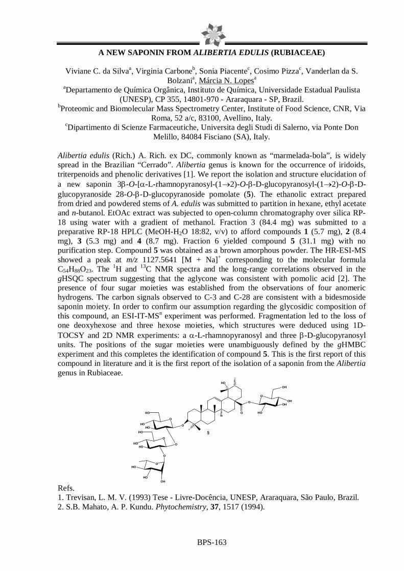
BPS-163
A NEW SAPONIN FROM ALIBERTIA EDULIS (RUBIACEAE)
Viviane C. da Silvaa, Virginia Carboneb, Sonia Piacentec, Cosimo Pizzac, Vanderlan da S. Bolzania, Márcia N. Lopesa
aDepartamento de Química Orgânica, Instituto de Química, Universidade Estadual Paulista (UNESP), CP 355, 14801-970 - Araraquara - SP, Brazil.
bProteomic and Biomolecular Mass Spectrometry Center, Institute of Food Science, CNR, Via Roma, 52 a/c, 83100, Avellino, Italy.
cDipartimento di Scienze Farmaceutiche, Universita degli Studi di Salerno, via Ponte Don Melillo, 84084 Fisciano (SA), Italy.
Alibertia edulis (Rich.) A. Rich. ex DC, commonly known as “marmelada-bola”, is widely spread in the Brazilian “Cerrado”. Alibertia genus is known for the occurrence of iridoids, triterpenoids and phenolic derivatives [1]. We report the isolation and structure elucidation of a new saponin 3β-O-[α-L-rhamnopyranosyl-(1→2)-O-β-D-glucopyranosyl-(1→2)-O-β-D-glucopyranoside 28-O-β-D-glucopyranoside pomolate (5). The ethanolic extract prepared from dried and powdered stems of A. edulis was submitted to partition in hexane, ethyl acetate and n-butanol. EtOAc extract was subjected to open-column chromatography over silica RP-18 using water with a gradient of methanol. Fraction 3 (84.4 mg) was submitted to a preparative RP-18 HPLC (MeOH-H2O 18:82, v/v) to afford compounds 1 (5.7 mg), 2 (8.4 mg), 3 (5.3 mg) and 4 (8.7 mg). Fraction 6 yielded compound 5 (31.1 mg) with no purification step. Compound 5 was obtained as a brown amorphous powder. The HR-ESI-MS showed a peak at m/z 1127.5641 [M + Na]+ corresponding to the molecular formula C54H88O23. The 1H and 13C NMR spectra and the long-range correlations observed in the gHSQC spectrum suggesting that the aglycone was consistent with pomolic acid [2]. The presence of four sugar moieties was established from the observations of four anomeric hydrogens. The carbon signals observed to C-3 and C-28 are consistent with a bidesmoside saponin moiety. In order to confirm our assumption regarding the glycosidic composition of this compound, an ESI-IT-MSn experiment was performed. Fragmentation led to the loss of one deoxyhexose and three hexose moieties, which structures were deduced using 1D-TOCSY and 2D NMR experiments: a α-L-rhamnopyranosyl and three β-D-glucopyranosyl units. The positions of the sugar moieties were unambiguously defined by the gHMBC experiment and this completes the identification of compound 5. This is the first report of this compound in literature and it is the first report of the isolation of a saponin from the Alibertia genus in Rubiaceae.
HO
O
5
O
HOHO O
HO
O
HOOH
HO
O
OHOHO
OH
HO
O
O
HOHO O
HO
Refs. 1. Trevisan, L. M. V. (1993) Tese - Livre-Docência, UNESP, Araraquara, São Paulo, Brazil. 2. S.B. Mahato, A. P. Kundu. Phytochemistry, 37, 1517 (1994).

BPS-164
LAEVICARPIN, A NEW NATURAL AMIDE FROM PIPER LAEVICARPU
Viviane P. de Freitas1, Massuo J. Kato2 and João Henrique G. Lago1 1Centro de Ciências e Humanidades, Universidade Presbiteriana Mackenzie
2Instituto de Química, Universidade de São Paulo
Chemical studies carried on Piperaceae species have revealed the occurrence of pyrones, lignoids, chromenes, terpenes, prenylated benzoic acids and amides several of which showing biological potential1. In the course of our studies on Piper species, the crude extract from leaves of P. laevicarpu was submitted to phytochemical investigation. The dried leaves (210 g) were extracted with CH2Cl2 to afford 1.2 g of crude extract. Part of this extract (800 mg) was subjected to chromatographic separation on silica gel using gradient of CH2Cl2 in MeOH to afford seven groups (I – VII). Group IV (587 mg), after purification on Sephadex LH-20 (MeOH), gave eight groups (IV-1 – IV-8). After spectrometric analysis (1H NMR and LREIMS), the flavonoids naringenin (31 mg) and sakuranetin (27 mg) were detected in the groups IV-4 (31 mg) and IV-5 (27 mg), respectively. Group IV-3 (30 mg) was composed by an amorphous powder, whose 1H NMR spectrum showed the presence of two aromatic rings due the AA`BB` system at δ 6.95 (d, J = 8.5 Hz) and 7.20 (d, J = 8.5 Hz) and the ABX system at δ 6.69 (d, J = 8.2 Hz), 6.53 (dd, J = 8.2 and 2.1) and 4.58 (d, J = 2.1 Hz). These signals, associated to two coupled triplets of methylene hydrogens at δ 2.09 (J = 5.2 Hz, 2H), 2.72 (J = 5.2 Hz, 2H), 2.83 (J = 6.2 Hz, 2H) and 3.51 (J = 6.2 Hz, 2H) were indicative of a dihydrocoumaroyltyramine derivative2. The 13C NMR spectra (BBD and DEPT 135°) showed 16 resonance lines consisting of one methoxyl, four methylenes, five methines (two equivalents), and six quaternary carbons (including a carbonyl group at δ 172.0). The molecular formula was established as C18H19NO3 by HRESIMS based on the molecular ion peak at m/z 320.1258 [M + Na]+. Finally, analysis of HMQC, HMBC and NOESY spectra indicated an ether linkage between the two aromatic rings, a rare feature among secondary compounds isolated from Piperaceae species. The structure of this amide, named as laevicarpin, is derived from dihydrocoumaroyl and tyramine and has previously been described as synthetic product3 but not as natural product.
O
N
H
O
OMe
1. J. B. Reigada et al., Tetrahedron Asymm., 18, 1054 (2007) 2. C.Y. Chen et al., Phytochemistry, 49, 1443 (1998) 3. D.L. Boger et al., J. Org. Chem., 56, 1763 (1991)

BPS-165
ISOLATION AND CHEMICAL CHARACTERIZATION OF A PHENOL IC MARKER IN ACQUEOUS EXTRACT OF PHYLLANTHUS SPECIES
Lúcia R. R. Martins, Juliana C. Barreiro, João B. Fernandes,
Edenir R. P. Filho, Quezia B. Cass Departamento de Química, Universidade Federal de São Carlos
The species belonging to the genus Phyllanthus (Euphorbiaceae) are widely
distributed throughout most tropical and subtropical countries. In Brazil, these species are popularly known as “quebra-pebra”, and used for the treatment of many diseases, such as: hepatitis, kidney stones, intestinal and urinary infections. It is reported that a great variety of secondary metabolites were isolated and identified, including flavonoids, tannins and lignans1. The characterization of the chemical profile by High Performance Liquid Chromatography (HPLC) for different extracts of Phyllanthus species can be used for differentiate them and applied in quality control/authentication of vegetable raw materials for pharmaceutical purposes2. This work reports the isolation by semi preparative HPLC from the aqueous extract of Phyllanthus niruri, a hydrolysable tannin. The structure of this substance was identified by Nuclear Magnetic Resonance experiments (1H, 13C, DEPT 135, HSQC and HMBC) and it was characterized as corilagin3 (Fig 1). This compound was analyzed by Mass Spectrometry eletronspray negative mode of ionization in ion trap analyzer. The ionization condition for the isolated substance was used as reference for the characterization of lyophilized aqueous extract samples using HPLC reversed mode of six Phyllanthus species: P. niruri, P. tenellus, P. amarus, P. caroliniensis, P. stipulatus and P. urinaria.
Fig 1. Chemical structure of corilagin. Refs.
1. J.B. Calixto et al., Medicinal Research Reviews, 4, 18 (1998) 2. Y. Z. Liang et al., Journal of Chromatography B, 812 (2004) 3. L. S. Conegero et al., Química Nova, 6, 26 (2003)
Acknowledgement To FAPESP and CNPq for the grants and financial support.

BPS-166
NEW PENTACYCLIC TRITERPENES FROM COMBRETUM LAXUM
Eder Bisoli1, Fernanda Rodrigues Garcez1, Walmir Silva Garcez1, Lidilhone Hamerski1, Caroline Tieppo1, Marilene R. Chang2 1 Department of Chemistry, Federal University of Mato Grosso do Sul, Campo Grande, MS, 79070-900, Brazil. 2 Depatament of Pharmacy and Biochemistry, Federal University of Mato Grosso do Sul, Campo Grande, MS, Brazil. Amongst the 18 genera comprising the Combretaceae, the genus Combretum is the largest with 250 species, which are mostly native to tropical and subtropical regions. Plants of this genus are known as a rich source of triterpenes and their glycosides, flavonoids, phenanthrenes, bibenzyls, stilbens, in addition to alkaloids and other aromatic compounds, some of which with anticancer, antimicrobial and hepatoprotective activities, among others. Combretum laxum Jacq., popularly known as “pombeiro branco”, is a woody shrub, which grows in the “Pantanal” of Mato Grosso do Sul, Brazil. In a previous phytochemical study on the stems of this species we obtained arjunolic (1) and asiatic (2) acids and their glucosides arjunglucoside II (3) and quadranoside IV (4), respectively.1 Further work has now led to the isolation of one new pentacyclic triterpene, 2α,3β,24-trihydroxyolean-12-ene-28-O-β-D-glucopyranoside (5) and four known ones, 2α,3β,6β-trihydroxyolean-12-ene-28-O-β-D-glucopyranoside (6), betulinic acid (7), quadranoside I (8) and bellericoside (9), of which 9 is being described for the first time in the genus Combretum. Air-dried and powdered stems of C. laxum (4375 g) were extracted at room temperature with EtOH. After concentration in vacuo, the residue obtained from the EtOH extract was subsequently partitioned between MeOH/H2O 9:1 and hexane; MeOH/H2O 1:1 and CH2Cl2; and MeOH/H2O 1:1 and n-BuOH. After a combination of column and flash chromatography on silica gel, gel filtration on Sephadex LH-20 and reversed phase CC and HPLC separations of the EtOAc (7.7 g) and n-BuOH (6.75 g) phases, triterpenes 5, 6 and 8 were isolated from the former while 3, 4 and 9 were obtained from the latter. Betulinic acid (7) was isolated from the CH2Cl2 phase. The antifungal activities of compounds 5, 6, 8, 9 and a mixture of 1 and 2 against Candida albicans, C krusei, and Cryptococcus neoformans were determined using the broth microdilution method (Table 1).2 Although showing weak or no activities against the strains tested, it is noteworthy the specificity of the mixture of 1 and 2 against Candida krusei and also of 5, 6 and 8 against C. albicans and Cryptococcus neoformans. Table 1-MIC values (µg/mL) of compounds isolated from C. laxum against Candida and Cryptococcus strains
I: inactive Reference compound: amphotericin B (MIC = 0.125 µg/mL) References
3. F. R. Garcez et al., 29th Annual Meeting of the Brazilian Chemical Society, Águas de Lindóia, SP, Brazil, 2006 (PN-052).
4. Clinical and Laboratory Standards Institute (CLSI) M27-A protocol.
Compounds Strais
1 / 2
5 6 8 9
Candida albicans ATCC 90028
I 100 100 100 100
Candida krusei ATCC 6258
100 I I I 100
Cryptococcus neoformans ATCC 32045
I
100
100
100
100
CH3
OH
R4
CH3CH3 R1
CH3
CH3
OH
R3 R2
OHCH3
CH3 CH3R1
CH3
CH3
CH3
CH2OH
OH
CH3 CH3
CH2
CH3
R1CH3 CH3
R2
CH3
OH
R3
R1 R2 R3 R4 1: COOH H CH2OH CH3 3: COOGlu H CH2OH CH3 5: COOGlu H CH3 CH2OH 6: COOGlu OH CH3 CH3 9: COOGlu H CH2OH CH2OH
R1 2: COOH 4: COOGlu
R1 R2 R3 7: COOH H H 8: COOGlu OH OH

BPS-167
NEW LABDANES FROM COPAIBA OLEORESIN
Adriano L. Romero, Paulo M. Imamura Instituto de Química, UNICAMP, C.P. 6154; CEP 13084-971, Campinas, SP, Brazil
The copaiba oleoresin is an exudates obtained from the trunk of many species of the Copaifera genus (Caesalpinoideae, Leguminosae) and is popularly used as cicatrizing, anti-inflammatory, in the treatment of bronchitis and for skin diseases, also in the cosmetic industry as fixative of the perfumes and as solvent for inks and varnishes. Although there is an extensive and recent study on the chemical composition of the oleoresin in the literature, the absolute configuration of many of then still remains unknown. In order to characterize and to determine the absolute configurations of some components, the commercial copaíba oleoresin was reinvestigated. The oleoresin was submitted to an acid-base extraction supplying two fractions: neutral fraction (81.7%) and acidic fraction (18.3%). The purification of neutral fraction by column chromatography allowed the isolation of eleven sesquiterpenes α-trans-bergamotene, γ-muurolene, β-trans-caryophyllene, β-bisabolene, caryophyllene oxide, humullene oxide, 7(11)-selinen-4-ol, torreyol, α-cadinol and two epimeric mixture of humullene dioxides [1], three dinorlabdanes 1-3 and ent-clerodane 4 [2, 3]. The absolute configuration of the dinorlabdanes 1-3 was established through the synthesis starting from known ent-3-hydroxy-copalic acid [2, 3]. The acidic fraction furnished known ent-labdane copalic acid, 3-acetoxycopalic acid, 3-hidroxycopalic acid and ent-agathic acid, ent-clerodane diterpenes hardwickiic acid, 7-acetoxyhardwickiic acid [1] and two new ent-labdanes as methyl ester after treatment with CH2N2 methyl 4-hydroperoxy-18-norcopalate (5) and methyl 3,19-dihydroxy-copalate (6). The structures of these terpenes were established on the basis of one- and two-dimensional NMR experiments and absolute configuration was established by biogenesis correlation and by comparison of the optical rotation reported in literature [4].
R
HO5HOO
COOCH3 COOCH3
OH
HO6
1 R = ketone2 R = 13(S)3 R = 13(R) 4
OH
CH2OH
H O
References 1. A.L. Romero and P.M. Imamura, XIX Simpósio de Plantas Medicinais do Brasil (2006). 2. A.L. Romero and P.M. Imamura, The 3rd Brazilian Symposium on Medicinal Chemistry (2006). 3. A.L. Romero, MSc. Thesis, Chemistry Institute, UNICAMP (2007). 4. D.D.K Manh et al., J. Nat. Prod., 46, 262 (1983).

BPS-168
NEW ALKALOID OF Psychotria stachyoides Benth.
Antônia T. Ávila Pimenta (PG), Mary Anne Sousa Lima (PQ) Edilberto Rocha Silveira (PQ)
Curso de Pós-Graduação em Química Orgânica - Departamento de Química Orgânica e Inorgânica – Centro de Ciências - Universidade Federal do Ceará
Psychotria (Rubiaceae) is one of the largest genera of flowering plants whose specimens are used in the traditional medicine against dizziness, hallucination, dementia and rubella1. A number of studies have cited Psychotria as a promising source of alkaloids with cytotoxic2, inhibitor of the human platelets3, antiprotozoal4 and analgesic5 properties. Species of this genus are widespread in Brazil, and the phytochemical works of several Southern species have revealed the presence of indole monoterpene alkaloid glucosides. As part of our investigative studies of alkaloids from Psychotria species from the northeastern Brazil flora, we have investigated Psychotria stachyoides, a native species from Guaramiranga Mountain, a Atlantic Forest spot at Ceará State. Acid-base extraction of the crude ethanol extract from the stems of P. stachyoides yielded an alkaloid fraction with a major compound. The alkaloidic fraction was submitted to preparative chromatography using the mixture of CH2Cl2/MeOH/NH4OH (90:10:0,1)as eluente to afford the pure compound 1, in addition to the coumarin scopoletin (2). Spectroscopic analysis using 1D and 2D NMR experiments including COSY, HMQC, HMBC and NOESY sequences for compound 1, revealed structural elements of a new unusual terpenoid alkaloid glucoside containing a secologanin and a tetrahydro-β-carboline moiety.
1. L. G. Joly et al., Ethnopharmacology, 20, 145 (1987) 2. A. Roth et al., Planta Medica, 5, 450 (1986) 3. A. Beretz et al., Planta Medica, 4, 300 (1985) 4. H. Achenbach et al., Phytochemistry, 38, 1537 (1995) 5. M.B. Leal et al., Internat Journ Pharmacog, 34, 267 (1987)
CNPq/CAPES/FUNCAP/PRONEX
N
NH
O
O
H
H
HH
OGliH(1)
O O
OCH3
OH
(2)

BPS-169
PHENOL COMPOUNDS FROM Alibertia rigida K. SCHUM.
João Henrique S.Luciano (PG), Mary Anne Sousa Lima (PQ), Edilberto Rocha Silveira (PQ)
Curso de Pós-Graduação em Química Orgânica, Centro de Ciências, Departamento de Química Orgânica e Inorgânica, Universidade Federal do Ceará
Species of Rubiaceae have widespread occurrence in Brazil and are well known by their economic and therapeutic importance, but the phytochemical analysis of plants belonging to the genus Alibertia have been limited to a few species. Previous reports reveals Alibertia species as a rich sources of fungitoxic iridoids, flavonoids, caffeic acid and its esters, and pentacyclic triterpenes1,2,3. In our work with A. myrciifolia, we have reported the isolation of cytotoxic flavonoids and iridoids, besides oleanane and hopane triterpenes4,5. As part of the investigative efforts to study Alibertia species from the northeastern Brazil flora, it is presented the chemical study of A. rigida. The crude ethanol extract from the leaves was partitioned with hexane, CHCl3, AcOEt and n-BuOH. The CHCl3 fraction was submitted to Si gel chromatography to afford the cafeic acid (1). Successive exclusion chromatography Sephadex LH-20 of the AcOEt fraction yielded the 1,2,4-trihydroxi-benzene (2), the flavonoids kaempferol (3) and quercetin (4), in addition to the proanthocyanidin A-6 (5). The flavonoid rutine (6) was obtained from the n-BuOH fraction after rechrystalization in MeOH. The complete proton and carbon assignments of the isolated compounds were accomplished by the use of 1D and 2D NMR experiments including COSY, HMQC, HSQC, HMBC and NOESY pulse sequences.
O
OH
HO
OH
OH
O
OH
(4)
O
OH
HO
OH
OH
O
O Gli Ram
(5)
O
OH
HO
HO OH
OH
O
HO
O
OH
OH
OH (6)
O
O
OH
HO
O
OOH
HO
OH
OH
OH
OH
OH(1)
(3)(2)
1. R.S.G. Olea et al., J Braz Chem Soc, 8, 257 (1997). 2. V.S. Bolzani et al., Phytochemistry, 30, 2089 (1991). 3. C.B. Brochini et al., Phytochemistry, 36, 1293 (1994). 4. M.C.M. Young et al., Phytochemistry, 31, 3433 (1992). 5. J.H.S. Luciano et al., Biochem Syst Ecol, 32, 1227 (2004). 6. G.C.G. Militão et al., Pharmac Biol, 43, 480 (2005). CNPq/CAPES/FUNCAP/PRONEX

BPS-170
FLAVONOIDS AND SESQUITERPENOIDS FROM Acritopappus pintoi (Asteraceae)
Carneiro, M. N. M., Roque, N. F., Souza-Neta, L C., Guedes, M. L. S.* and Martins, D. * Instituto de Biologia, UFBA; GESNAT, Instituto de Química, Universidade Federal da
Bahia, Campus Universitário de Ondina, 40170-290, Salvador, Bahia, Brazil
The Chapada Diamantina presents one of the richest ecosystems in the state of Bahia, Brazil, with many endemic and new species of plants. Acritopappus pintoi Bautista & Hind, from Asteraceae family, was collected in September 2004, at Capada Diamantina, in the municipal district of Piatã, Bahia. The importance attributed to this study came up due to few studies on this species is reported.1 Thus, from the chemical and biological perspective Acritopappus genera has been evaluated. The dichloromethane fraction of A. pintoi leaves ethanolic extract was active against gram-negative, gram-positive bacteria and fungi Micrococcus luteus and Streptococcus mutans.
The dichlromethane phase of A. pintoi leaves ethanolic extract was submitted to a silica gel chromatography separation. The first fractions eluted with CHCl3–MeOH (1%) gave after recrystallization in hexane and ethanol two sesquiterpenes, 1 and 2. The further fractions gave, after a Sephadex LH-20 column chromatography five flavonoids (3-7). The isolated compounds will be submitted to bioassays.
Compound 1 was obtained as colorless crystals, PF 224-225oC in ethanol. The 13C NMR spectrum showed twenty signals revealing an aliphatic structure. The 1H NMR confirmed the supposition and suggested the structure of a terpene compound. The number of methyl groups was incompatible with a diterpene structure, and an isoprenoyl sesquiterpene was proposed. The presence of two trissubstituted double bonds, one carbonyl, two carboxyl groups, three oxygenated carbons and two hydroxyl groups was inferred by the NMR spectra. The mass spectrum showed a signal at m/z 362, which is in agreement with the lost of a H2O molecule of C20H28O7. HSQC, HMBC, COSY and NOESY spectra were used to propose the structure of 1, a new rearranged sesquiterpene. The structure of 2 (PF 198-199oC in ethanol) was proposed comparing its spectral data with those of 1.
OHOH
O
OR
H
COOH
1
2
-CO-CH(CH3) -CH2-CH3
-CO-CH2-CH(CH3)2
R
R
O
OH
OH
OR2
OR
R1
3 4 5 6 7
R=Me; R1=H; R2=OMe R=Me; R1=H; R2=HR=Me; R1=H; R2=OHR=H; R1=OMe; R2=HR=H; R1=H; R2= O-beta-D-(6"-E-p-cumaroyglucopiranosyl)
Reference: Bohlmann, F. et al., Phytochemistry, 19, 2695 (1980)

BPS-171
BIFLAVONOIDS AND TERPENOIDS ISOLATED FROM THE LEAVE S OF OURATEA MICRODONTA Engl. (OCHNACEAE)
Mário G.de Carvalho1, Luiz Roberto M. de Albuquerque1, Luciane de S. Mendes1, Giselle M.
S. Pinheiro Guilhon2 and Silvana T. Rodrigues3
1Departamento de Química, ICE-Universidade Federal Rural do Rio de Janeiro, BR 465 KM. 07, 23890-000-Seropédica-RJ, Brazil. 2Departamento de Química, Universidade Federal do Pará, Belém-PA, Brazil, 3Departamento de Botânica, EMBRAPA-Amazônia Oriental, 66095-780, Belém/PA.
In the course of our phytochemical and pharmacological investigation of the genus Ouratea
(Ochnaceae) we reported the presence of terpenoids, isoflavonoids, flavonoid glycosides and biflavones,1,2 besides their DNA topoisomerase inhibition, citotoxic and antitumoral activities.3,4 In this study we have also reported some reviews and spectral investigation of biflavonoids and their new derivatives5,6. Continuing the phytochemical study of Ouratea species we are describing the first phytochemical study of Ouratea microdonta Engl. (Ochnaceae). The leaves and branch of this plant were collected in the Ilha de Marajó, municipality of Soure, Pará state, Brazil. Voucher specimen (No IAN-21438) is deposited at the Herbarium Embrapa Amazônica Oriental, Pará, Brazil. The dried and powdered leaves (1.0 Kg) were extracted with methanol at room temperature The solvent was removed under vacuum furnishing a residue OMFM (250.0 g). The crude methanol residue (200.0 g) was filtered on silica gel with C6H12, CHCl3, AcOEt and MeOH yielding the fractions OMFH (3.0 g), OMFC (20.6 g), OMFAc (6.5 g) and OMFM (60.0 g). These fractions were analyzed by TLC plate and fractionated on silica gel and/or sephadex LH-20 column eluting by the each adequate eluent. The subfractions that were analyzed until this time let us to identify steroids, triterpenes, a benzoic acid and three biflavonoids. From the fraction OMFH were isolated a mixture of lupeol, α- and β-amyrin (30.5 mg) and β-amyrin (50.6 mg), from OMFC were isolated a mixture of sitosterol and stigmasterol (20.0 mg), lupeol (10.0 mg) and 4-methoxy,2,5-dihydroxy-benzoic acid; from the fraction OMFAc were isolated three biflavonoids, agathisflavone (15.0 mg), 7”-methylagathisflavone 20.0 mg) and amentoflavone (12.0 mg); the agathisflavone was also isolated from the fraction OMFM. The structures of these compounds were deduced by the 1H and 13C-NMR spectra data analysis and comparison with literature.1,2 References 1. Velandia, J.R.; Carvalho, M.G. de; Braz-filho, R.; Werle A.A.; Phytochem. Anal. 13, 283 (2002) 2. Daniel, J.F.D.S.; Carvalho, M.G. de; Cardoso, R.D.S.; Agra, M. de F.; Eberlin, M. N.; J. Braz. Chem. Soc. 16, 634 (2005) 3. Grynberg, N.F.; Carvalho, M.G. de; Velandia, J.R.; Oliveira, M.C.; Moreira, I.C.; Braz-Filho, R.; Echevarria, A.; Braz. J. Med. Biol. Res. 35, 819 (2002) 4. Carvalho, M.G. de; Velandia, J.R.; de Oliveira, J.C.C.; Echevarria, A.; Braz-Filho, R.; Grynberg, N.F. Chemical Structure, Cytotoxic and Antitumour Activities of Biflavonoids from Brazilian Ouratea (Ochanceae). In Phytochemical and Pharmacologiy II of the Series “Recent Progress in Medicinal Plants”, Majumdar, D.K.; Govil, J.N.; Singh, V.K. eds., SCI Tech Publishing LLC: Texas, USA, 8, 77 (2002) 5. Suzart, L.R.; Daniel, J.F. de S.; Carvalho, M.G. de; Kaplan, M.A.C.; Quím. Nova 30|4| 984 (2006) 6. Carvalho, M. G. de; Gomes, M.S.da R.; Pereira, A.H.F.; Daniel, J. F. de S.; Schripsema, J.; Magn. Reson.
Chem. 44|1| 35 (2006). CNPq, CAPES, FAPERJ.

BPS-172
ALKALOIDS OF Hippeastrum morelianum (Lem.) - AMARYLLIDACEAE
Raquel Brandt Giordani1, Strahil Berkov2, Miriam A. Apel1, Amélia T. Henriques1, Jaume Bastida2, José Angelo S. Zuanazzi1
1Programa de Pós-Graduação em Ciências Farmacêuticas, Universidade Federal do Rio Grande do Sul, Porto Alegre, RS, Brasil.
2Departament de Productes Naturals, Facultat de Farmàcia, Universitat de Barcelona, Barcelona, Spain.
Over the past decade drug discovery from natural products, especially medicinal plants, has continued effectively to provide new drugs and drug leads against various pharmacological targets such as tumors, viruses, bacteria and fungi1. Plants of the Amaryllidaceae family are amongst the top 20 in the most widely applied medicinal plant families2. As primary constituents, more than 400 structurally diverse alkaloids have been isolated and most of them have shown significant biological activities, particularly antiviral3, antitumoral4 and psychopharmacological5,6. Hippeastrum morelianum Lem. (Amaryllidaceae) is found throughout Brazil and our interested is to investigate biologically active alkaloids synthesized by members of the Hippeastrum genus. Therefore, was obtained an ethanolic extract from fresh bulbs of H. morelianum which was pooled, dried and the residue was partitioned in light petroleum and HCl (10%). The HCl phase was washed with CH2Cl2 and the acid phase thus obtained basified with NH4OH (pH 9) and extracted first with CH2Cl2 and subsequently with n-butanol. The CH2Cl2 fraction was re-suspended and chromatographed on silica-gel using vacuum liquid chromatography (VLC) technique using an increasing polarity of solvents (200ml) pentane, ethylic ether, acetone, dichloromethane, n-butanol, ethyl acetate and, finely, methanol. Methanol fraction was chromatographed on silica-gel reverse-phase using the medium pressure liquid chromatography (MPLC) technique using an increasing gradient of methanol-water (volume rate from 0-100 to 100-0, in 8h). The fractions were purified with column chromatography as well as preparative chromatography. The potential antioxidant of the isolated alkaloids and extracts were investigated for DPPH method7. Twelve alkaloids have been isolated but only four were identified using spectroscopic methods until the moment. From VLC technique dichloromethane fraction resulted in 6 mg of pretazettine and n-butanol fraction, after purification, resulted in 110 mg of candimine. The methanolic fraction, after purification techniques, generated 40 mg of tazettine and 7 mg of haemanthamine or crinamine. The potential antioxidant was observed, at 10 mg/ml, for all fractions of acid-basic extraction while none isolated alkaloid demonstrated positive result up to 3 mg/ml. The genus Hippeastrum is one important source of Amaryllidaceae alkaloids and the extracts presented important antioxidant potential. Moreover, the occurrence of candimine alkaloid in high quantity is detached a time that none pharmacological investigation was related at the moment opening perspectives for future works. Refs. 1. N. Unver, Phytochemical Reviews, 6, 125 (2007) 2. J. Zhong, Natural Products Report, 22, 111 (2005) 3. L. Szlavik et al., Planta Medica, 70, 871 (2004) 4. J. Liu et al., FEBS Letters, 578, 245 (2004) 5. P.J. Houghton et al., Phytochemistry, 65, 2893 (2004) 6. A.F.S da Silva et al., Pharmacology Biochemistry and Behavior, 85, 148 (2006) 7. M.S Blois, Nature, 118, 1199 (1958)

BPS-173
New Steroidal Constituents of Ganoderma lucidum from Ceará-Brazil.
Aluísio M. Fonsecaa, Telma L. G. Lemosa, Maria da Conceição F. de Oliveiraa and Raimundo
Braz-Filhob aLaboratório de Biocatálise e Produtos Naturais, Departamento de Química Orgânica e Inorgânica, Universidade, Federal do Ceará, 60451-970, Fortaleza, Ceara, Brazil bSetor de Química de Produtos Naturais-LCQUI-CCT, Universidade Estadual do Norte Fluminense, Campos-RJ, Brazil Ganoderma lucidum (Ganodermataceae) is a type of fungi popularly known in the northeast of Brazil as “orelha-de-pau”, name originated for the similarity to an ear linked on trees in decomposition. From it hexane extract of G. lucidum after chromatographic procedure in silica gel and sephadex LH-20 was possible to characterize three steroids compounds with ergostane skeleton (G-1, G-2, G-3). Structures elucidation were done using spectroscopy techniques including 1 and 2D NMR. The compounds were also evaluated for their scavenging activity, using DPPH assay and showed high inhibition percentage determined as 73.5 %, 65.7% and 63.2%, respectively at 1 µg/µL, with IC50 0.021 µg/µL, 0.056 µg/µL, and 0.052 µg/µL.
Refs. 1. M.S., Shiao, M.S., Chemical Research, 3,172-180 (2003) 2. S.C. Jong, J.M. Birmingham Advances in Applied Microbiology, 37, 101(1992) 3. T.T. Chairul et al., Phytochemistry, 29, 3, 923-928 (1990) 4. M. Yoshiyuki et al., Bioorganic and Medicinal Chemistry, 7, 2047-2052 (1999) 5. J. L. Mau et al, Lebensmittel-Wissenschaft und Technologie, 38, 589-597 (2005)
G-1
O
3
2
45
6
7
89
10
1119
12
13
14 15
1617
18
2120
22
23
28
24
25
27
26
1
G-2 HO
3
2
45
6
7
89
10
1119
12
13
14 15
1617
18
2120
22
23
28
24
25
27
26
1
G-3 HO
3
2
45
6
7
89
10
1119
12
13
14 15
1617
18
2120
22
23
28
24
25
27
26
1

BPS-174
CHEMICAL CONSTITUENTS FROM ANNONA AMAZONICA (ANNONACEAE)
Cristiane L. Batista1, Regiane L. Batista1, Clahildek M. Xavier2, Emmanoel V. Costa1, Andersson Barison1, Maria Lúcia B. Pinheiro2, Izabel Cristina P. Ferreira3, Francinete Ramos
Campos1 1Departamento de Química, Universidade Federal do Paraná, Centro Politécnico, CEP 81530-
900, Curitiba, PR, Brazil, 2Departamento de Química, Universidade Federal do Amazonas, CEP 69077-000, Manaus, AM, Brazil and 3Universidade Estadual de Maringá, Maringá, PR,
Brazil. [email protected] Annona (Annonaceae) comprises about 120 species reported in South and Central America, Africa, Asia and Australia. Previous chemical and pharmacological investigations on species of this genus have indicated the presence of important bioactive compounds, exhibiting various pharmacological activities, including antiparasitic; in particular, against Leishmania sp., Plasmodium falciparum and Trypanosoma cruzi.1 Therefore, in continuation of our studies of Amazon medicinal plants and search for novel antiparasitic natural products, we have undertaken the chemical investigation of Annona amazonica R.E. Fries. This annonaceous plant is a tropical tree distributed from Panamá to South America. In Brazil this species is commonly found in the Brazilian Amazon, particularly in the states of Amazonas and Pará.2 The powdered air-dried bark of A. amazonica (1.1 Kg) was extracted successively with hexane (15.0g), CH2Cl2 (6.2g) and MeOH (40.0 g), at room temperature. The CH2Cl2 extract (6.0g) was redissolved in CH2Cl2 and submitted to acid-base extraction to yield the neutral (5.0g) and alkaloidal fractions (1.0g). The neutral fraction (4.8g) was initially subjected to silica gel column chromatography eluted with increasing concentrations of CH2Cl2 in hexane, EtOAc in CH2Cl2 and MeOH in EtOAc. The eluted fractions were evaluated and pooled by TLC analysis. Additional chromatographic separation of the fractions by preparative TLC resulted in the isolation of one sesquiterpene (caryophyllene oxide, 1); two steroids (β-sitosterol and stigmasterol, 2 and 3); one monounsaturated fatty acid (oleic acid, 4) and tree methyl esters of fatty acids (oleic, linoleic, and linolenic acids, methyl esters, 5, 6 and 7). The compounds were identified by spectroscopic methods and by comparison with literature data. This is the first report of isolation these substances in this species. The identification by GC/MS of others sesquiterpenes and methyl esters has been described in our previous work.3 The antileishmanial activity and cytotoxicity against human tumor cells will be investigated for purified compounds.
O
HH
HO
H
HHHC(CH2)4H3C CH CH2 CH CH (CH2)7 COOCH3
HC(CH2)7H3C CH (CH2) 7 COOCH3
HC(CH2)7H3C CH (CH2)7 COOH
H3C CH2 HC CH CH2 HC CH CH2 HC CH (CH2)7 COOCH3 Refs. 1. E.V. Costa et al., J. Nat. Prod., 69, 292 (2006). 2. P.J.M. Maas et al., Candollea, 49, 389 (1994). 3. C.M. Xavier et al., XVIII Simpósio de Plantas Medicinais do Brasil, AM, 393 (2004).
1
2 3: ∆22(23)
4
5
6
7
22 23

BPS-175
ALKALOIDS FROM LEAVES OF ANNONA SERICEA (ANNONACEAE) Regiane L. Batista1, Cristiane L. Batista1, Emmanoel V. Costa1, Andersson Barison1, Maria
Lúcia B. Pinheiro2, Adriane G. dos Santos2, Izabel Cristina P. Ferreira3 and Francinete Ramos Campos1
1Departamento de Química, Universidade Federal do Paraná, Centro Politécnico, CEP 81530-900, Curitiba, PR, Brazil, 2Departamento de Química, Universidade Federal do Amazonas,
CEP 69077-000, Manaus, AM, Brazil and 3Universidade Estadual de Maringá, Maringá, PR, Brazil. [email protected]
Annona (Annonaceae) comprises about 120 species reported in South and Central America, Africa, Asia and Australia. Previous chemical and pharmacological investigations on species of this genus have indicated the presence of important bioactive compounds, exhibiting various pharmacological activities, including antiparasitic, in particular against Leishmania sp., Plasmodium falciparum and Trypanosoma cruzi.1 Therefore, in a search for novel antiparasitic natural products we have studied the leaves of Annona sericea Dunal. This annonaceous plant is a tropical tree distributed in South America rain forest2 and known as “araticum do Pará”. In Brazil this species is commonly found in the Brazilian Amazon, particularly in the states of Amazonas and Pará.2 The powdered air-dried leaves of A. sericea (500g) was extracted successively with hexane (24.0g) and MeOH (35.0 g), at room temperature. The MeOH extract (33.0g) was redissolved in CH2Cl2 and subjected to acid-base extraction to yield the neutral (8.0g) and alkaloidal fractions (0.55g). The alkaloidal fraction (0.5g) was initially subjected to silica gel column chromatography, having been previously treated with a 10% NaHCO3 solution and eluted with increasing concentrations of CH2Cl2 in hexane followed by EtOAc in CH2Cl2 and MeOH in EtOAc. The eluted fractions were evaluated and pooled by TLC analysis. Additional chromatographic separation of the fractions by preparative TLC, resulted on the isolation of two oxoaporphine alkaloids, oxonuciferine (1) and oxonantenine (2), and two aporphine alkaloids, nornuciferine (3) and nornantenine (4). The compounds isolated were identified by spectroscopic methods and by comparison with literature data. The antileishmanial activity will be investigated for purified compounds. This is the first report of these alkaloids in this species. The results obtained in this study confirm that plants of this genus have strong ability to produce oxoaporphine and aporphine alkaloids, a class of compounds typically found in plants of Annonaceae. Refs. 1. E.V. Costa et al., J. Nat. Prod., 69, 292 (2006). 2. H. Rainner and J.A. Braga. Flora da Guyana Venezuelan. Vol. 2. Portland (1995).
NH
CH3O
CH3ON
CH3O
CH3O
O
1 3
NH
CH3O
CH3O
O
O42
N
CH3O
CH3O
O
O
O

BPS-176
QUATERNARY APORPHINE ALKALOIDS FROM Cryptocarya mandioccana
Karin Fabiana Bandeira de Camargo (PG), Alberto José Cavalheiro (PQ)
NuBBE – Núcleo de Bioensaios, Biossíntese e Ecofisiologia de Produtos Naturais- Depto de Química Orgânica – Instituto de Química de Araraquara – UNESP –R. Francisco Degni, s/n – Quitandinha, CEP: 14800-900, Araraquara-SP. Cryptocarya mandioccana is an arboreal species of some 25-30m height that is widely spread in the Atlantic forests of Brazil. It is recognized as an important food source for primates such as Brachyteles arachnoids E. Geoffroy, 1806 ( mono-carvoeiro or muriqui). Cryptocarya plants have components like alkaloids, flavonoids and styrylpyrones. Phytochemical studies of the bark of C. mandioccana have shown that styrylpyrones are the typical secondary metabolites present in this species1. Flavonoids glycosides, as well as styrylpyrones, have been detected in leaves and the qualitative and quantitative intra-specific variability of these secondary metabolites has also been determined2. However, a phytochemical study of other Cryptocarya genus species suggested the presence of alkaloids3. This present paper deals with the isolation and structural determination of quaternary aporphine alkaloids from the bark of this plant. The powdered dry bark of C. mandioccana was submitted to liquid-liquid extraction by sonication with hexane and methanol:acetic acid 10% (1:1 v/v). The crude extract was partitioned with chloroform to remove the lipophilic compounds. The hydroalcoolic extract was divided into two parts by solubility in chloroform:methanol (70:30). Each part was submitted to normal phase chromatography (25,0 x 1,2 cm i.d; 40-63µm) eluted using a step gradient from chloroform:methanol (70:30) with 10% of ammonium hydroxide to chloroform:methanol (63:37) with 10% of ammonium hydroxide to yield two quaternary aporphine alkaloids, the (+) menisperine and the (+) xanthoplanine. The structural determination these components were assured by UV, 1H and 13C NMR uni and bidimensional. The stereochemistry was elucidated by the optical rotation. The data were comparated with those describe in the literature3,4. In addition, the quaternary aporphine alkaloids, (+) menisperine and (+) xanthoplanine, were isolated from the C. mandioccanna for the first time. Refs. 1. A.J. Cavalheiro et al., Phytochemistry, 53, 811 (2000) 2. C.J. Nehme et al., Biochem System Ecol, 30, 613 (2002) 3. S. Lee et al., J. Natural Products, 56, 1971 (1993) 4. D.Dwuma-Badu et al., J. Natural Products, 43, 123 (1980)

BPS-177
ciscis
threothreo
threo
2112
3435
1OO
OH OH
O
O
(1)
221341
O
O
OO
OH
(CH2)nCH3
OH
OH
(2) threo/trans/threo/trans/erythro n = 4(3) threo/trans/threo/trans/threo n = 2
NEW ACETOGENINS FROM ANNONA CORNIFOLIA A. ST. -HIL.
Luciana A. R. Santos Lima, Maria Amélia D. Boaventura and Lúcia P. S. Pimenta*
Departamento de Química, Instituto de Ciências Exatas, Universidade Federal de Minas Gerais, Av. Antônio Carlos, 6627, 31270-901 Belo Horizonte - MG – Brazil
Annonaceous acetogenins are a group of secondary metabolites, until now isolated only from fourteen out of the 130 genera of Annonaceae family, that possess important biological activities as cytotoxic, antitumoral, antimicrobial, insecticide and immunosuppressive1. Annona cornifolia is a tropical shrub, typical of Minas Gerais “cerrado” whose fruit is popularly known as “araticum das caatingas”and “araticum mirim”. In our recent studies of the seeds of Annona cornifolia we have described four bis-tetrahydrofuran γ-lactones belongs to this very interesting natural products: cornifolin2, annofolin3, desacethyluvaricin and squamocin M4. We describe here the isolation and structural elucidation of a new acetogenin, folianin B (1) along with a mixture of two known compounds, glaucanisin (2) and parviflorin (3) from the ethanolic extract of A. cornifolia seeds. The phytochemical study of the ethanolic extract from A. cornifolia seeds was guided fractionation by brine shrimp test (BST). The cleaned seeds were dried at 40 oC, powdered (150.0 g) and extracted with ethanol in Soxhlet, yielding the ethanolic extract (F01, 10.0 g). This extract was successively extracted with hexane and ethylacetate. Solvent removal under reduced pressure provided hexanic (F02, 6.3 g) and ethylacetate (F03, 3.5 g) fractions. F02 was fractionated by silica gel column and reverse phase HPLC, resulting in the isolation of folianin B (9.5 mg, 1), a mixture of glaucanisin and parviflorin (34.6 mg, 2 + 3). The structure and relative or absolute stereochemistry of the acetogenins were elucidated based on 1D and 2D NMR spectroscopy techniques, including 1H NMR, 13C NMR, COSY and HMQC, and ESIMS of the parent compound. These results suggest the great potential of this plant in yielding cytotoxic, antitumoral and insecticide leader-compounds.
Refs.
1. A. Bermejo et al., Nat. Prod. Rep., 22, 269, (2005). 2. L. A. R. Santos et al., Biochem. Syst. Ecol., 34, 78, (2006). 3. L. A. R. S. Lima et al. Tetrahedron Letters, subbmitted. 4. L. A. R. Santos et al. Revista Bras. de Plantas Medicinais, 9, (2007) in press.

BPS-178
IRIDOID GLYCOSIDE FROM PTYCHOPETALUM OLACOIDES BETAHM
K.M.P. Menezes1,3, E.C. Dias1, T.C.B. Tomassini2 and A.J.R. Silva3 1INCQS/FIOCRUZ 2FARMANGUINHOS/FIOCRUZ 3NPPN/UFRJ
As part of our ongoing studies on medicinal plants, we investigated the constituents of Ptychopetalum olacoides Bentham (Olacaceae), popularly known as “Marapuama and Muirapuama”, collected in the Brazilian Amazon. Voucher specimen is preserved at the Emílio Goeldi Museum (Pará state), collection number MG 170.021. The ethanolic extract of the root bark was suspended in H2O and then partitioned with Et2O, EtOAc and BuOH. The fractions were performed on precoated plates Si gel 60 F254 and visualized by spraying reagents for alkaloids (IP), flavonoids (NP-PEG), essential oil (VS) and phenolic compound (FBS-KOH)1. The residue of the EtOAc fraction, after evaporation to dryness was purified by Flash Chromatography on Si C-18 with H2O-MeOH solvents system (9:1 to 100% MeOH). The 80% aqueous MeOH fraction provided a plumeria iridoid skeleton2. The Plumieride structure (I) was elucidated by spectroscopic methods, mainly 1D and 2D NMR3,4. This iridoid has been reported to have antidermatophytic5, antifertility6 and anticancerous activities7. Despite phytochemical investigation has been carried out, this is the first isolation of iridoid from P. olacoides.
OO
O
OH
OGlu
OCH3O
(I)
Refs. 1. H. Wagner, S. Bladt and E.M. Zgainski, Plant Drug Analysis. Springer-Verlag, 299 (1984) 2. B. Dinda, S. Debnath and Y. Harigay, Chem. Pharm. Bull., 55(2), 159 (2007) 3. C.A. Boros and F. R. Stermitz, J. Nat. Prod., 54(5), 1173 (1991) 4. C.A. Boros and F. R. Stermitz, J. Nat. Prod., 53(5), 1055 (1990) 5. T.N. Tiwari, V.B. Pandey and N.K. Dubey, Phytother. Res., 16, 393 (2002) 6. R.S. Gupta et al., Phytomedicine, 11, 169 (2004) 7. M.P. Dobhal et al., J. Org. Chem., 69, 6165 (2004) Acknowledgments: We thank Dr. Alvicler Magalhães, Farmanguinhos/Fiocruz, for 1D and 2D NMR spectroscopic measurements and FAPERJ.

BPS-179
PEPTIDES DERIVATIVES FROM TWO SPECIES OF LEGUMINOSA E
Clayton Q. Alves1, Jorge M. David1, Juceni P. David2 and Luciano da S. Lima1
1Instituto de Química, 2Faculdade de Farmácia, Universidade Federal da Bahia, 40170-280, Salvador (BA), Brazil.
Cenostigma gardnerianum Tul. and Cratylia mollis Benth. are two Leguminosae species that occur in the brazilian semi-arid region. They are popularly known as “canela-de-velho” and “camaratu”, respectively. Species of Cenostigma are employed by the local population as herbal medicine and the leaves of C. mollis is employed for cattle feeding during the drought periods in Brazilian northeastern. Previous studies with the wood and leaves of these plants indicated the presence of flavonoids and terpenoids1,2. This work describes the chemical study of the CHCl3 fractions which were obtained from partition between CHCl3/MeOH:H2O (9:1) of the MeOH extract of the leaves of C. gardenerianum and C. mollis. These fractions were submitted to CC over Si gel and eluted with mixtures of hexane and ethyl acetate of different polarities. The fractions obtained were further purified employing Sephadex LH-20 permeation and Si gel PTLC. These procedures permitted to obtain compound 1 (aurentiamide acetate) from C. mollis and 1 and 2 from C. gardenerianum. The structures of the peptides were elucidated by 1H and 13C NMR (uni and bidimensional), IR and MS. The presence of the peptide derivative aurentiamide acetate (1) in the two Leguminosae species is not a surprise because it was already found in different natural sources. It was firstly isolated in Aspergillus spp. but, it was also detected in algae and different plant species of the families Piperaceae, Euphorbiaceae and Malvaceae3. In Leguminosae family, aurentiamide acetate was only obtained from Medicago polymorpha. However, besides compound 2 was a konwn compound it can be considered an unusual plant peptide which was previously obtained only from Croton hieronymi3.
(CNPq, FAPESB, CAPES, PRONEX).
1 2 Refs. 1. L. C. Rezende et al., CD-ROM da 30ª Reunião da Sociedade Brasileira de Química, PN-159 (2007) 2. C. Q. Alves, Flavonóides antioxidantes e derivados de ácido gálico isolados de Cenostigma gardenerianum Tul. Dissertação de Mestrado, Instituto de Química, UFBA (2007) 3. C. A. N. Catalan et al., Phytochemistry, 64, 625 (2003)
NH
O
O
NHO CH3
O
NH
O
O
ONH
O

BPS-180
APORPHINE TYPE ALKALOIDS FROM BARK OF GUATTERIOPSIS FRIESIANA (ANNONACEAE)
Emmanoel V. Costa,1 Maria Lúcia B. Pinheiro,2 Franscisco A. Marques,1 Raquel M. Braga,3
Marta Cristina T. Duarte,4 and Beatriz Helena L.N. Sales Maia,1,*
1Departamento de Química, Universidade Federal do Paraná, Curitiba-PR, Brazil, 2Departamento de Química, Universidade Federal do Amazonas, Manaus-AM, Brazil,
3Instituto de Química, Universidade Estadual de Campinas, Campinas-SP, Brazil, 4Centro de Pesquisas Químicas, Biológicas e Agrícolas, Universidade Estadual de Campinas, Campinas-
SP, Brazil. * [email protected]
Plants of the family Annonaceae are known to elaborate a variety of alkaloids, some of which are reported to have interesting pharmacological properties.1 Despite the importance of the members of this family in folk medicine, the number of species that have been chemically investigated is still small.1 Guatteriopsis is a small genus, comprising about five species of small trees whose majoritary is distributed in South America.2 It has been reported only one phytochemical study of essential oils from leaves of G. blepharophylla.3 Continuing our work in a search for novel antimicrobial natural products we have studied the bark of Guatteriopsis friesiana. This plant, commonly known as “envireira”, is a tropical native small tree found in Brazil and Colombia Amazon rainforest.2 The powdered air-dried bark of G. friesiana (1.9 Kg) was successively extracted, at room temperature, with hexane and MeOH to afford 27.11g and 260.62 g, respectively. Antimicrobial activity was found for MeOH extract against Staphylococcus aureus (500 µg/mL). TLC investigations revealed the presence of alkaloids in the MeOH extract. The MeOH extract of bark (250 g) was redissolved in CHCl3 and subjected to extraction with 3% aqueous HCl. This aqueous solution had the pH adjusted to 12 with NH4OH, followed by extraction with CHCl3 to yield an alkaloid fraction (6.20 g). This fraction (6.0 g) was subjected to several chromatography columns and preparative TLC yielding four known aporphine type alkaloids: liriodenine 1 (60.0 mg), lysicamine 2 (13.6 mg), guadiscine 3 (8.8 mg) and guadiscidine 4 (7.0 mg). The isolated compounds were identified by spectroscopic methods (UV, IR, EIMS, 1H, 13C 2D NMR experiments) and comparison with literature data.4
Refs. 1. J.L. Castedo et al., Phytochemistry, 30, 2781 (1991). 2. J.E.L.S. Ribeiro et al., Flora da Reserva Ducke, Manaus: INPA, 1999. 3. J.G. Maia et al., J. Essent. Oil Res., 20, 478 (2005). 4. Guinaudeau et al., J. Nat. Prod., 761 (1983).
NO
O
R1
CH3
CH3
(1) R1-R2 = OCH2O(2) R1-R2 = OCH3
(3) R1 = OCH3
(4) R1 = OH
N
O
R2
R1
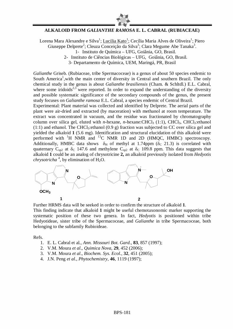
BPS-181
ALKALOID FROM GALIANTHE RAMOSA E. L. CABRAL (RUBIACEAE)
Lorena Mara Alexandre e Silva1; Lucilia Kato1; Cecília Maria Alves de Oliveira1; Piero Giuseppe Delprete2; Cleuza Conceição da Silva3; Clara Megume Abe Tanaka3.
1- Instituto de Química – UFG, Goiânia, GO, Brasil. 2- Instituto de Ciências Biológicas – UFG, Goiânia, GO, Brasil.
3- Departamento de Química, UEM, Maringá, PR, Brasil Galianthe Griseb. (Rubiaceae, tribe Spermacoceae) is a genus of about 50 species endemic to South America1,with the main center of diversity in Central and southern Brazil. The only chemical study in the genus is about Galianthe brasiliensis (Cham. & Schltdl.) E.L. Cabral, where some iridoids2,3 were reported. In order to expand the understanding of the diversity and possible systematic significance of the secondary compounds of the genus, the present study focuses on Galianthe ramosa E.L. Cabral, a species endemic of Central Brazil. Experimental: Plant material was collected and identified by Delprete. The aerial parts of the plant were air-dried and extracted (by maceration) with methanol at room temperature. The extract was concentrated in vacuum, and the residue was fractionated by chromatography column over silica gel, eluted with n-hexane, n-hexane:CHCl3 (1:1), CHCl3, CHCl3:ethanol (1:1) and ethanol. The CHCl3:ethanol (0.9 g) fraction was subjected to CC over silica gel and yielded the alkaloid 1 (5.6 mg). Identification and structural elucidation of this alkaloid were performed with 1H NMR and 13C NMR 1D and 2D (HMQC, HMBC) spectroscopy. Additionally, HMBC data shows δH of methyl at 1.74ppm (δC 21.3) is correlated with quaternary Csp2 at δC 147.6 and methylene Csp2 at δC 109.8 ppm. This data suggests that alkaloid 1 could be an analog of chrysotricine 2, an alkaloid previously isolated from Hedyotis chrysotricha 4, by elimination of H2O.
NO
N
OCH3
N
N
O
OH
1 2 Further HRMS data will be seeked in order to confirm the structure of alkaloid 1. This finding indicate that alkaloid 1 might be useful chemotaxonomic marker supporting the systematic position of these two genera. In fact, Hedyotis is positioned within tribe Hedyotideae, sister tribe of the Spermacoceae, and Galianthe in tribe Spermacoceae, both belonging to the subfamily Rubioideae. Refs.
1. E. L. Cabral et al., Ann. Missouri Bot. Gard., 83, 857 (1997); 2. V.M. Moura et al., Quimica Nova, 29, 452 (2006); 3. V.M. Moura et al., Biochem. Sys. Ecol., 32, 451 (2005); 4. J.N. Peng et al., Phytochemistry, 46, 1119 (1997);

BPS-182
IDENTIFICATION OF THE C-GLYCOSYL FLAVONOIDS IN THE BUTANOLIC FRACTION FROM THE ROOTS FROM Wilbrandia ebracteata COGN.
Andressa Córneo Gazola(1); Flávio Henrique Reginatto(1); Eloir Paulo Schenkel(1)
(1) Department of Pharmaceutical Science, Federal University of Santa Catarina, Florianópolis, Santa Catarina, Brazil Introduction: Wilbrandia ebracteata is a Brazilian plant from the Cucurbitaceae family popularly named “Taiuiá”. The Taiuiá roots are used in the folk medicine for the treatment of gastric ulcer, acute and chronic rheumatism, among other diseases. Studies with the Wilbrandia ebracteta roots showed the presence of cucurbitacins in the dichlorometane fraction and C-glycosyl flavonoids in the butanolic fraction. Objective: Identify the C-glycosyl flavonoids in the butanolic fraction from the roots of Wibrandia ebracteta. Methods: The butanolic fraction was analysed by Thin Layer Chromatography (TLC) (silica gel plates; ethyl acetate: formic acid: water – 8:1:1 and ethyl acetate: methanol: acetic acid – 11:4:0,3; natural products reagent/UV 366 nm) and High Performance Liquid Chromatography (HPLC) (phenyl column; acetonitrile: acetic acid 1% - gradient; UV-detection (330 nm). As reference compounds were used the C-glycosyl flavonoids orientin, vitexin, isovitexin, isoorientin, vitexin-2”-O-ramnoside, spinosin, 6,8-di-C-glicosil-crisin and vicenin-2. Results: The TLC and HPLC analysis indicated the presence of vitexin, isovitexin, spinosin and vicenin-2 in the butanolic fraction of the rooots. The HPLC analysis demonstrated that the major compounds of the butanolic fraction are spinosin, isovitexin and swertisin. Conclusions: The presence of vitexin, isovitexin, spinosin, vicenin-2 and swertisin was characterized in the butanolic fraction from Wilbrandia ebracteata roots by TLC and HPLC analysis. Isovitexin, spinosin and swertisin are the major compounds from this fraction.

BPS-183
A NEW ACYL-HOMOSERINE LACTONE FROM Methylobacterium mesophilicum
Armando M. Pomini1, Pedro L. R. Cruz1, Cláudia Gai2, Welington L. Araújo2,3, Anita J. Marsaioli1*
1Instituto de Química, Universidade Estadual de Campinas, P.O. Box 6154, CEP 13084-862,
Campinas, SP. 2 Departamento de Genética, ESALQ/USP, P.O. Box 83, CEP 13400-970, Piracicaba, SP. 3Núcleo Integrado de Biotecnologia, Universidade de Mogi das Cruzes, CEP
08780-911, Mogi das Cruzes, SP. *[email protected]
The chemical communication mechanism or quorum-sensing is now a widely recognized phenomenon in Gram-negative bacteria, in which the acyl-homoserine lactones (acyl-HSL) play a key role. It was demonstrated in many species that important phenotypes are under the quorum sensing control, as expression of exoenzymes, secondary metabolites and biofilm formation.1 The aim of this work is to characterize the acyl-HSL produced by the Gram-negative bacterium Methylobacterium mesophilicum, which occurs endophytically in citrus plants and seems to be associated to citrus variegated chlorosis disease.2 One of the most interesting characteristic of this microorganism is the use of methanol as the sole carbon source. Therefore, the production of acyl-homoserine lactones by this microorganism under methanotrophic conditions was investigated. Initial β-galactosidase bioassays with reporter Agrobacterium tumefaciens NTL4(pZLR4) indicated the possible presence of acyl-HSL in M. mesophilicum extracts, stimulating more accurate chemical studies.3 GC-MS and 1H NMR analyses of fractionated extracts allowed the identification of six acyl-HSLs in a complex fraction (1.1 mg) obtained from 8 L of fermentation media. Such small amount prompted us to employ chemical derivatization techniques as dimethyl-disulfide reaction, catalytic hydrogenation and synthetic methods for chemical identification. Moreover, the catalytic hydrogenation procedure allowed the absolute configuration determination of five substances in just one experiment employing gas chromatography-flame ionization detection technique with chiral column. In summa, we report herein the detection and first synthetic studies for new (1) and unusual (2). Studies concerning to characterization of compound (3) and the biological activities of these metabolites are being carried out currently. Financial support FAPESP 05/02934-4.
N
OO
OH
N
OO
OH
N
OO
OH
N
OO
OH
( )n
(1) (2)
(3)(4) n=7; (5) n=9(6) n=8*
*Absolute configuration not determined - trace amounts Refs. 1. N.A. Whitehead et al., FEMS Microbiol. Rev., 25, 365 (2001) 2. W.L. Araújo et al., Appl. Environ. Microbiol., 68, 4906 (2002) 3. A.M. Pomini et al., J. Agric. Food Chem., 55, 1200 (2007)

BPS-184
CHEMICAL CONSTITUENTS FROM Salacia elliptica LEAVES
Lucienir P. Duarte1 ([email protected]), Grasiely Faria de Sousa1, Grácia Divina de Fátima Silva1, Sidney Augusto Vieira Filho2, Salomão Bento de Vasconcelos Rodrigues1,
Roqueline Rodrigues S. de Miranda1 and Alexandre Salino3
1NEPLAM – Departamento de Química - UFMG - Av. Antônio Carlos, 6627, Pampulha, CEP 31270-901, Belo Horizonte, MG, Brazil
2 DEFAR – Escola de Farmácia - UFOP – R.Costa Sena, 171, CEP 35400-000, Ouro Preto, MG 3Departamento de Botânica – ICB – UFMG – Belo Horizonte, MG, Brazil
The genus Salacia (Celastraceae) has many species distributed in Brazil. Extracts from Salacia species has been used during thousand years in Ayurvedic medicine for oral diabetes treatment1. The inhibition of intestinal α-glucosidase activity has been demonstrated using roots extracts of S. oblonga. Therefore, it holds potential as a natural method to mitigate the blood glucose response of people with diabetes2. This work shows the study of Salacia elliptica, knows as “Bacuri”, “Saputá” and others folk names. The plant was collected in “Mata Samuel de Paula”, in Nova Lima region, Minas Gerais state. Dried and powdered leaves (394 g) were submitted to exhaustive extraction with hexane (H), ethyl acetate (EA) and methanol (M), at room temperature. After solvent withdrawn under reduced pressure, the H (7.97g), EA (8.80g) and M (36.55g) extracts were obtained. Each extract was submitted to silica gel column chromatography using appropriate eluent polarity. From H extract were isolated a mixture of long chain saturated hydrocarbons, with C29H60 (1) as a principal constituent, a mixture of pentacyclic triterpenes constituted by 3β-stearyloxy-ursan-12-ene (2) and 3β-stearyloxy-olean-12-ene (3), 1,4-trans-poliisoprene (4) (gutta-percha), friedelin (5), β-friedelinol (6) and β-sitosterol (7). From EA extract (6), canofilol (8) and β-sitosterol-3-O-β-D-glucopiranoside (9) were isolated. Dulcitol (10) was isolated from M extract. The structure of these constituents was confirmed by HRGC (comparison with standard) and spectrometric methods (IR, 1H and 13C NMR with 2D experiments).
R1
R2
O
O
R1
H
H3C
H
RO
H
CH2OH
OHH
HHO
HHO
OHH
CH2OH
R2
n
(4) 1,4-trans-poliisoprene (gutta-percha)
(5) R1 OH R2 CH3 β-friedelinol(6) R1 =O R2 CH3 friedelin(8) R1 =O, R2=CH2OH canofilol
(10) dulcitol
(2) R1 CH3 R2 H stearyloxy-ursan-12-ene(3) R1 H R2 CH3 stearyloxy-olean-12-ene
(7) R=H β-sitosterol(9) R=glucose β-sitosterol-3-O-β-D-glucopyranoside
1. B.W.Wolf et al., Food and Chemical Toxicology, 41, 867 (2003) 2. A.M. Flammang et al., Food and Chemical Toxicology, 44, 1868 (2006) Thanks FAPEMIG for financial support.

BPS-185
Compounds from bark of Maytenus salicifolia Reiss. (Celastraceae)
Roqueline Rodrigues Silva Miranda1, Lucienir Pains Duarte1, Grácia Divina de Fátima Silva1, Sidney Augusto Vieira Filho2 and Glória Del Carmen Meléndez Salazar3
1 NEPLAM –Departamento de Química – UFMG - Avenida Antônio Carlos, 6627, Pampulha, CEP
31270-901, Belo Horizonte, MG, Brazil 2 DEFAR – Escola de Farmácia – UFOP - Rua Costa Sena, 171, CEP 35400-000, Ouro Preto, MG,
Brazil 3 Departamento de Química – UESB – Rua José Moreira Sobrinho, s/n, CEP 45200-000,
Jequié, BA, Brazil
Maytenus salicifolia is encountered in forests of Minas Gerais state, Brazil. It is popularly known as “Cafezinho” and is used in medicine folk for stomach ulcer treatments (1). The decoct is topically employed in the form of bath to alleviate itches and allergic symptoms. The pharmacological activities attributed to different Celastraceae species (2) and the popular use of M. salicifolia induced its phytochemical study. A sample (1,25 g) of dried and powdered bark wood was submitted to exhaustive hot extraction with hexane, ethyl acetate and methanol. After filtration, the solvent was removed under reduced pressure and the resulted extracts were submitted to fractionation through silica gel G-60 (70-230 Mesh) column chromatography (CC), with graduating polarity of hexane, chloroform, ethyl acetate and methanol, as eluents. Sephadex LH-20 CC eluted with methanol also was used to isolate and/or purify compounds. Lupeol (I ), 4’-O-methylepigalocathechin (II ), 2-ethyl-1-(4-diethyl)-octyl phthalate (III ) and a mixture of proanthocianidins (IV ) (Fig. 1) were isolated from ethyl acetate extract. These compounds were identified by CCD comparing with authentic samples and through spectrometry [IR, 1H and 13C NMR including 2D experiments]. Until this moment, a triterpene I , the flavonoid II , the asymmetric phthalate III and a mixture of proanthocianidins IV were isolated from bark. The continuity of studies certainly will become possible the isolation of new compounds, that permits the establishment of the biosynthetic routes of different M. salicifolia constituents.
I
II
III
IV
O
O
O
O
HO
O
OHOH
HO
O
OH
HO
OH
OH
OH
OCH3
OH
HOHO
OH
OH
OH
OCH3
OH
References
2. T.S.M. Grandi, Acta Botanica Brasilica, 3, 185, (1989) 3. R. Bruning and H. Wagner, Phytochemistry, 17, 1821, (1978)

BPS-186
PHYTOCHEMICAL STUDY OF Bowdichia virgilioides Kunt. (FABACEAE) Gabriela Lemos de A. Maiaa, Kamylla Lopes Marquesa , José G. de Sena Filhoa, Ana Silvia Suassuna C. Lucio, Daysianne P. Lira, Vivianne M. de Medeirosa, Josean F. Tavaresa and José M. Barbosa Filhoa aUniversidade Federal da Paraíba, Laboratório de Tecnologia Farmacêutica-Caixa Postal 5009, 58051-970 - João Pessoa-PB, Brasil. *gabriela.lam @gmail.com Bowdichia virgilioides Kunt. (Fabaceae) is a medium size tree found in the tropical forests of South America. In Northeastern Brazil it is popularly known as “sucupira”, and its bark is used for healing of wounds, as anti-ulcer and anti-diabetic while the seeds are used in the treatment of rheumatism, arthritis, and skin diseases. The importance of this plant promoted its inclusion in the first Brazilian Pharmacopoeia. Various bioactivities, including antimalarial, hypoglycemic and inhibitor of the enzyme acetylcholinesterase, of crude extracts from this plant were reported. Previous chemical investigation resulted in the isolation of flavonoids, benzofuranoids, essential oil, triterpenoids and alkaloids. This study reports the structural identification of flavan-3-ol from the acetic phase. The plant was collected in December 2005, near the city of Santa Rita, State of Paraíba, Brazil, a coastal area around the Atlantic Forest and identified by Maria de Fátima Agra from Laboratório de Ciências Farmacêuticas in the Universidade Federal da Paraiba. This material was submitted to dry heat around 450C and powdered until 3 kg and exhaustively extracted with ethanol (95%), obtaining the ethanolic extract (EE). The EE was partionated in hexane, chloroform and ethylic acetate. The acetic phase was analyzed with Sephadex LH-20, in mixtures of chloroform and methanol 1:1, obtaining 42 fractions organized and visualized with ultraviolet light. The sample 18-19 was also submitted to successive purifications resulting in a red solid compound. The identification was based on experiments using 1D and 2D NMR (200MHz) in methanol-d4 and comparison with literature data o identify Bv-1 as 3,3`,4`,5,7- pentahidroxiflavan known as epicatechin. Through the phytochemical study of Bowdichia virgilioides Kunt, flavonoid epicatechin, already known in the family, yet isolated for the first time in genus Bowdichia. Keywords: Fabaceae, Bowdichia virgilioides, Epicatechin.
Refs. 1. J.M.Barbosa-Filho et al., Revista Brasileira de Farmacognosia 16, 258-285 (2006). 2. F.N. Melo et al., Natural Product Letters 15, 261-266 (2005).
O
OH
HO
OH
OH
OH
EpicatequinaFigure 1. Epicatechin
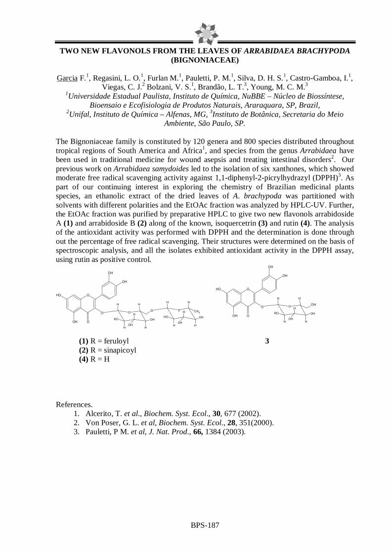
BPS-187
O
OOH
OH
OH
OH
O O
HOH
H
H
H
OHRO
H
O O
HOH
H
H
H
OHOH
H
CH3
O
OOH
OH
OH
OH
O O
HOH
H
H
H
OHRO
H
OH
TWO NEW FLAVONOLS FROM THE LEAVES OF ARRABIDAEA BRACHYPODA (BIGNONIACEAE)
Garcia F.1, Regasini, L. O.1, Furlan M.1, Pauletti, P. M.1, Silva, D. H. S.1, Castro-Gamboa, I.1,
Viegas, C. J.2 Bolzani, V. S.1, Brandão, L. T.3, Young, M. C. M.3
1Universidade Estadual Paulista, Instituto de Química, NuBBE – Núcleo de Biossíntese, Bioensaio e Ecofisiologia de Produtos Naturais, Araraquara, SP, Brazil,
2Unifal, Instituto de Química – Alfenas, MG, 3Instituto de Botânica, Secretaria do Meio Ambiente, São Paulo, SP.
The Bignoniaceae family is constituted by 120 genera and 800 species distributed throughout tropical regions of South America and Africa1, and species from the genus Arrabidaea have been used in traditional medicine for wound asepsis and treating intestinal disorders2. Our previous work on Arrabidaea samydoides led to the isolation of six xanthones, which showed moderate free radical scavenging activity against 1,1-diphenyl-2-picrylhydrazyl (DPPH)3. As part of our continuing interest in exploring the chemistry of Brazilian medicinal plants species, an ethanolic extract of the dried leaves of A. brachypoda was partitioned with solvents with different polarities and the EtOAc fraction was analyzed by HPLC-UV. Further, the EtOAc fraction was purified by preparative HPLC to give two new flavonols arrabidoside A (1) and arrabidoside B (2) along of the known, isoquercetrin (3) and rutin (4). The analysis of the antioxidant activity was performed with DPPH and the determination is done through out the percentage of free radical scavenging. Their structures were determined on the basis of spectroscopic analysis, and all the isolates exhibited antioxidant activity in the DPPH assay, using rutin as positive control.
(1) R = feruloyl 3 (2) R = sinapicoyl (4) R = H
References.
1. Alcerito, T. et al., Biochem. Syst. Ecol., 30, 677 (2002). 2. Von Poser, G. L. et al, Biochem. Syst. Ecol., 28, 351(2000). 3. Pauletti, P M. et al, J. Nat. Prod., 66, 1384 (2003).

BPS-188
FLAVONOIDS FROM Plinia cauliflora LEAVES
Tatiana M. de Souza1*, Juliana A. Severi2, Wagner Vilegas2, Rosemeire C.L.R. Pietro1
1- Faculdade de Ciências Farmacêuticas – UNESP, Araraquara, SP 2- Instituto de Química – UNESP, Araraquara, SP
Plinia cauliflora is a plant belonging to Myrtaceae family that occurs in Brazil, Argentine and Paraguay and has comestible fruits already studied. However, their leaves are still without chemical and biological evaluation. The aim of this work was to evaluate Plinia cauliflora leaves composition. The leaves extract was obtained with 70% ethanol by percolation and dried under reduced pressure. Liquid-liquid partition of the extract (15.0 g) was done with 500 mL of water and 500 mL of ethylacetate. After dried, ethylacetate fraction was dissolved in methanol and chromatographed on Sephadex LH 20 column (72.0 x 2.5 cm) using methanol as mobile phase. The fractions obtained were analyzed by thin layer chromatography and similar samples were grouped according their Rf values. Purified flavonoid fractions were dissolved in deuterated DMSO and analyzed by NMR spectrometer operating at 500 MHz. Using the comparison of their 1D and 2D NMR spectroscopic and physical data with those from previous reports in the literature, the isolated compounds were assigned to be the flavonoids quercetin-3-O-β-glucopyranoside, myricetin-3-O-β-glucopyranoside, myricetin-3-O-β-galactopyranoside and myricetin-3-O-β-allopyranoside. Flavonoids could be responsible for many biological activities, as anti-inflammatory, antimicrobial and antioxidant. The knowledge of P. cauliflora leaves composition together with biological activity studies could result in efficient therapeutic uses for people of this brazilian widely known vegetable species. Support: PADC-UNESP. Refs. 1. H. Lorenzi, Árvores brasileiras: manual de identificação e cultivo de plantas arbóreas nativas do Brasil, 3 ed., v.1 (2000). 2. P.K. Agrawal, Carbon 13 NMR of flavonóides, 1989. 3. B.J. Harborne, C.A. Willians, Phytochemistry, 55, 481-504 (2000).

BPS-189
PHYTOCHEMICAL STUDY OF PIPER MALACOPHYLLUM (PIPERACEAE)
Alberto de Oliveira1* (PG), Adalberto M. Silva1 (PG), João Henrique. G. Lago2 (PQ) and Massuo J. Kato1 (PQ). *e-mail: [email protected]
1Instituto de Química, Universidade de São Paulo, São Paulo, SP, Brazil. 2Centro de Ciências e Humanidades, Universidade Presbiteriana Mackenzie, São Paulo, SP, Brazil.
The genus Piper is a source of several classes of bioactive secondary metabolites including alkaloids, amides, flavonoids, benzoic acid derivatives, terpenes, and cyclopentanediones.1
Recently, two butenolides (1 and 2) were isolated from leaves of Piper malacophyllum in a bioactivity-guided phytochemical investigation due to the strong activity observed against Cladosporium cladosporioides and C. sphaerospermum2. However, an HPLC analysis of the crude extract showed that there are other derivatives, which were not detected in the activity-guided fractionation. Thus, in the present work we report the complete phytochemical analysis from the leaves of P. malacophyllum. Additionally, the composition of fruits, stem and roots extracts have been described by use of HPLC and HRGC/MS experiments. The dried and powdered leaves of P. malacophyllum were extracted with MeOH and the crude extract was concentrated and partitioned between MeOH/H2O and CH2Cl2 to yield the CH2Cl2 residue. An aliquot of this residue was subjected to successive chromatographic procedures to afford six compounds, which were identified by spectral analysis: 4,6-dimethoxy-5-E-phenylbutenolide (1), 4,6-dimethoxy-5-Z-phenylbutenolide (2), 1,2-methylenedioxy-4-(oct-3-E-enyl)benzene (3), shizuka-acoradienol acetate (4), shizuka-acoradienol (5) and 4-(dec-3-E-enyl)phenol (6). The dried and powdered fruits, stem and roots were individually extracted with MeOH and the crude extracts were treated in C18 sep-pak (MeOH) and analyzed by HPLC and HRGC/MS. In all extracts was verified the predominance of 6, a different profile observed to the leaves extracts, in which the butenolides 1 and 2 were the major compounds. Indeed, in the fruits and stem extracts was also detected the new compound 3. Butenolides and alkylbenzene derivatives are rarely found in the Piper genus while sesquiterpenes have been detected in several species1. However, this is the first occurrence of acorane sesquiterpenes in Piper genus being compound 4 a new natural product.
R
HO6 (R = C6H13)
1 = E2 = Z
O
MeO
O
MeOR
OO
3 (R = C4H9)
4 = acetate5 = H
OR
1. S. V. Parmar et al. Phytochemistry, 46, 597 (1997). 2. J. H. G. Lago et al. J. Braz. Chem. Soc, 16, 153 (2005).

BPS-190
A NEW TETRAHYDROFURAN LIGNAN AND OTHER METABOLITES FROM Peperomia blanda
Lidiane Gaspareto Felippe1, Debora Cristina Baldoqui1, Massuo Jorge Kato2, Vanderlan da
Silva Bolzani1, Maysa Furlan1. 1NUBBE- Núcleo de Biossíntese Bioensaio e Ecofisiologia de Produtos Naturais – Instituto
de Química da Universidade Estadual Paulista – Unesp – C. P. 355 – 14800-900 – Araraquara, SP. 2Instituto de Química – Universidade de São Paulo - USP – Prof. Lineu
Prestes 748 b 11 T – São Paulo, SP. E-mail: [email protected]
Piper and Peperomia represent the most important genera of the Piperaceae family with 2000 and 1700 species , respectively1. Species of Peperomia are well known as ornamental plants and have found application in folk medicine for the treatment of inflammation, asthma and gastric ulcers, and as analgesic and antibacterial agents2. Peperomia blanda is a perennial herb that typically grows in wet rock crevices3. To date, the only chemical informations available are on the composition of essential oil4 and on the structures of two chromenes isolated from apolar fraction of its aerial parts5. In the present work, we report on the isolation and structural elucidation of one tetrahydrofuran lignan6 (1), two secolignans (2-3), two flavones (4-5) and one polyketide (6). The EtOAc extract derived from aerial parts of P. blanda was submitted to partitions and chromatographic column to yield six compounds. The structures of the isolates were elucidated by interpretation of their spectral data, including gHMQC and gHMBC, while the relative and absolute configurations were determined from NOESY data and optical properties, respectively. The secolignans, flavones and polyketide have been isolated from other Peperomia species 7,8 but the tetrahydrofuran lignan is a new natural product apparently resulting from a mixed coupling between sinapyl alcohol and propenylphenol unities. Refs. 1 Wanke et al., Molecular Phylogenetics and Evolution, 42, 477 (2007). 2Arrigoni-Blank et al., Journal of Ethnopharmacology, 91, 215 (2004). 3Guimarães and Giordano, Rodriguésia, 55, 21 (2004). 4Santos et al., Phytochemistry, 58, 547 (2001). 5Velozo et al., Phytochemistry, 67, 492 (2006). 6Felippe, et al., Phytochemistry, 2007, in press. 7Chen et al., Heterocycles, 29, 411, 1989. 8Cheng et al., Phytochemistry, 63, 603, 2003.
O
OCH3
H3CO
H3CO
H3CO
O
O
OH
O
O
O
O
O
O
H3CO
H3CO
O
O
OCH3
H3CO
OCH3
H3CO
H3CO
H3CO
OH
O
OH
O
O
OOH
H3CO
OCH3
OCH3
O
OOH
H3CO
OCH3
OCH3
1 2 3 4
5 6

BPS-191
EVALUATION OF PHENOLIC COMPOSITION FROM SOUTH BRAZI LIAN CECROPIA SPECIES
Geison Modesti Costa; Eloir Paulo Schenkel; Flávio Henrique Reginatto
Departament of Pharmaceutical Science, Universidade Federal of Santa Catarina – SC/BR
Introduction: Cecropia catharinensis Cuatrec. (sin.: C. pachystachya Mart.) and Cecropia glaziovii Sneth. are abundant in South of Brazil, and its infusion are used in the folk medicine as cardiotonic, diuretic and for the treatment of inflammation and cases of asthma. Although they have morphological differences in their aerial parts, there are few phytochemical analysis with C. catharinensis and, as far as we are aware, there are no report comparing the chemical composition of these species. Considering that these species are used as medicinal plants, often without correct identification, the development of thin layer chromatography (TLC) and high performance liquid chromatography (HPLC) methodology may contribute significantly to the differentiation between these plants and to their control quality as raw materials. Objective: The aim of this study was to compare the phenolic composition from different extracts of C. catharinensis and C. glaziovii. Methodology: Aerial parts from both species were crushed and extracted, by aqueous infusion and aqueous decoction. The extracts obtained, after evaporated to dryness under reduced pressure, were analyzed by TLC, HPLC and UV-spectrometry (total phenolic content). In order to evaluate the flavonoid fingerprint, the aqueous infusions were successively partitioned with AcOEt and n-BuOH, and these last one were analyzed by TLC and HPLC. Results: The extracts of plants were prepared in order to obtain polar compounds, considering their popular uses. The TLC and RP-HPLC analysis for crude extracts did not shown qualitative differences in the phenolic composition between extraction methods, but allowed to difference between species. The total phenolic content higher values were verified for aqueous decoction from C. glaziovii, e for aqueous infusion from C. catharinensis. The analysis of butanolic fractions from both species aqueous infusion extracts by TLC, using AcOEt:MeOH:H2O (10:1,5:1 v/v/v) as mobile phase and RP-HPLC, with a linear gradient of ACN:H2O, suggest that majors flavonoids presents in C. glaziovii were isovitexin and isoorientin, while in C. catharinensis were predominant orientin and isoorientin. Additionally, rutin and chlorogenic acid were verified in both species. Conclusion: It is possible to differentiate C. glaziovii and C. catharinensis species by their total phenolic content and by their C-glycosil flavonoid fingerprint. Refs. 1. L.A. Mentz; E.P. Schenkel, Caderno de Farmácia, 5, 93 (1989). 2. M.M. Tanae et. al., Phytomedicine, 14, 309 (2007). 3. C.M.O. Simões, Plantas da medicina popular do Rio Grande do Sul. 5ª ed. (1998). 4. T.C. Sposito; F.A.M. Santos, American Journal of Botany, 88, 939 (2001).

BPS-192
FURTHERS COMPOUNDS FROM Helieta puberula
Mayra E. R. Silva, João B. Fernandes, Paulo C. Vieira, M. Fátima das G. Fernandes da Silva. Departament of Chemistry, Federal University of São Carlos (UFSCar), Brazil
[email protected] Great variety of secondary metabolites, such as alkaloids, flavonoids, coumarins, limonoids, etc, is Rutaceae characteristic. These classes of metabolites present biological activities of pharmacological and agronomical importance, that make relevant to study plants of this family [1]. The Helietta genus (Rutaceae) is distributed in neotropical region and plants of this genus can be obtained in México, USA, Cuba, Venezuela, Colombia, Peru, Paraguay, north of Argentine and Brazil. This genus is few studied chemically. For example, the specie H. apiculata presents activity in the central nervous system and this fact justify studying other plants of this genus. Nothing is known about other species [2]. The aim of this communication is to present furthers compounds obtained from the hydroalcoholic partition of branch ethanol extract of Helieta puberula (Rutaceae). This extract was fractioned by column chromatography with silica gel yielding coumarins: 3’-(1’,1’-dimethylalyl)-isoescopoletin, graveliferone methyl ether and alkaloids: N-methyl-4-methoxy-2-quinolone, dictamin, maculin and, in mixture, rutaecarpin and 7,8-dehydrorutaecarpin, previous isolated from H. puberula by Simote [3] and the furthers alkaloids: flindersin, N-methylflindersin, evolitrin, maculosidin, robustin and edulitin; coumarin: bergapten and Limonoid: limonin. The identification of these compounds was done by analysis of their 1D and 2D NMR (1H and 13C NMR, 1H-1H COSY45, HSQC and HMBC), MS, IR and UV data.
N
O
O
Hfffflllliiiinnnnddddeeeerrrrssssiiiinnnnaaaa22227777,,,,5555 mmmmgggg
N
O
O
CH3NNNN----mmmmeeeettttiiiillllfffflllliiiinnnnddddeeeerrrrssssiiiinnnnaaaa
4444 mmmmgggg
N O
OCH3
H
CH3O
eeeevvvvoooolllliiiittttrrrriiiinnnnaaaa111100007777 mmmmgggg
N O
OCH3
CH3O
H
CH3O
mmmmaaaaccccuuuulllloooossssiiiiddddiiiinnnnaaaa11118888 mmmmgggg
N O
OCH3
OH Heeeedddduuuulllliiiittttiiiinnnnaaaa3333,,,,2222 mmmmgggg
N O
OCH3
OCH3Hrrrroooobbbbuuuussssttttiiiinnnnaaaa0000,,,,5555 mmmmgggg
OOOO
OOOO
OOOO
OOOOOOOO
OOOO
OOOO
HHHHOOOO
HHHH
LLLLiiiimmmmoooonnnniiiinnnnaaaa55553333,,,,5555 mmmmgggg
O O O
BBBBeeeerrrrggggaaaapppptttteeeennnnoooo0000,,,,9999 mmmmgggg
[1] Waterman, P. G. & Grundon, M. F. (Eds.). New York: Academic Press, 1983, p. 31-96. [2] Goloubkova, T.D., et al.. J. Ethnopharm. 60: 141-148, 1998. [3] Simote, S. Y. Tese Doutorado – PPGQ – UFSCar, 2006.
FAPESP, CNPq, CAPES

BPS-193
COMPLETE 1H AND 13C NMR STRUCTURAL ASSIGNMENT FOR TWO NEW SESQUITERPENE LACTONES FROM Tithonia diversifolia.
Vladimir Constantino Gomes Helenoa, Sérgio Ricardo Ambrósioa,b, Mauricio Gomes Constantinob and Fernando Batista da Costac
a Núcleo de Pesquisa em Ciências Exatas e Tecnológicas da Universidade de Franca. b Depto. de Química, Faculdade de Filosofia, Ciências e Letras de Ribeirão Preto, USP. c Depto. Ciências Farmacêuticas, Faculdade de Ciências Farmacêuticas de Rib. Preto, USP.
Tithonia diversifolia is a Mexican shrub from Asteraceae family naturalized around the tropics. Its crude extracts and secondary metabolites present several biological activities. Sesquiterpene lactones (STL) were detected in glandular trichomes of the leaves and inflorescences of a Brazilian population. The dichloromethane rinse extracts of the leaves and inflorescences were investigated. In this work, we describe the total elucidation and NMR assignment of two novel STL isolated from T. diversifolia: a furanheliangolide derivative (1) and other with an unusual seco-guaianolide skeleton (2). As part of our work on the NMR complete assignment of STL,1 both structures were clarified using 1H-NMR, 13C1H-NMR, DEPT135, gCOSY, gHMQC, gHMBC and NOEdiff techniques. All NMR experiments were performed on a Bruker Avance DRX500 spectrometer. Most chemical shifts and scalar coupling constants were measured and assigned after careful analysis of the 1H-NMR spectra. 2D NMR information also contributed to the total 1H assignments. Table 1: 1H and 13C NMR data and structures of 1 and 2. (CDCl3 – 500 MHz)
Compound 1 Compound 2 Position δC δH (J in Hz) δC δH (J in Hz)
1 86.7 3.99 dd (6.5, 10.4) 43.0 2.82 ddd (7.7, 9.0, 13.9) 2a 40.5 2.56 dd (6.5, 12.6) 32.0 2.66 dd (7.7, 16.4) 2b 1.91 dd (10.4, 12.6) 2.60 dd (13.9, 16.4) 3 101.5 172.6 4 67.2 206.7 5 66.2 3.34 s 51.9 3.37 dd (9.0, 10.3) 6 75.0 5.12 d (4.8) 76.3 5.13 dd (10.0, 10.3) 7 42.0 4.32 dddd (2.5, 2.8, 4.1, 4.8) 46.5 3.22 dddd (1.8, 3.0, 3.4, 10.0) 8 71.2 5.52 ddd (4.1, 4.8, 11.1) 64.6 5.80 ddd (1.8, 2.8, 4.0) 9a 35.0 1.74 dd (11.1, 14.7) 46.0 2.54 dd (2.8, 15.2) 9b 1.66 dd (4.8, 14.7) 2.00 dd (4.0,15.2)
O
OCH3
OO
O
HO
O
O
12
3
45 6
7
8
910
1112
13
14
15
1'
2'
3'
4'
1
10 82.8 87.6 11 136.3 133.6 12 169.6 167.8 13a 123.5 6.28 d (2.8) 123.3 6.38 d (3.4) 13b 5.61 d (2.5) 5.67 d (3.0) 14 26.7 1.50 s a 23.6 1.51 s a 15 21.0 1.44 s a 33.5 2.37 s a 1’ 176.4 175.6 2’ 34.5 2.41 septet (7.0) 34.1 2.56 septet (7.0) 3’ 19.5 1.05 d (7.0) a 18.9 1.16 d (7.0) a 4’ 19.0 1.04 d (7.0) a 19.0 1.13 d (7.0) a
-OCH3 59.1 3.38 s a
O
O
O
O
O
O
O
12
3
4
56 7
8910
11
12
13
14
15
1'2'
3'
4'
2 a integrated to 3H
The gHSQC experiments were enough to assign most 13C signals, but for quaternary carbons, an extra help was provided by gHMBC. The novelty, attributed to structure 2 made the elucidation of this compound severally harder than the other, which has an known kind of structure. Only with NOE experiments data and detailed mixing of NMR information, the structures of 1 could be confirmed and of 2 could be finally assigned. Refs.
1. V.C.G Heleno et al., Magn. Reson. Chem., 42, 364-367 (2004).

BPS-194
IRIDOID AND SECOIRIDOIDS FROM GUETTARDA POHLIANA
Santin, Silvana M.O.; Oliveira, Paulo R.N., Testa, Glaucio; Souza, Maria C. Departamento de Química, Universidade Estadual de Maringá, Av. Colombo, 5790 Maringá,
PR, 87020-900 - Brazil
Guettarda pohliana Müll Arg. (Rubiaceae) is popularly know as “angélica-do-mato”. In a previous work we have reported the isolation of 3-O-β-D-quinovopyranosyl-28-O-β-glycopyranosyl-quinovic acid and its aglycone from the roots of specie [1]. Further investigation of the AcOEt root extract of G. pohliana led to the isolation of iridoid glycosides loganic acid (1), sweroside (2), the seco-iridoid secoxyloganin (3), 4,5-O-dicaffeoylquinic acid (4) and 28-O-β-glycopyranosyl-quinovic acid (5). These compounds were isolated by successive silica gel column chromatography and Sephadex LH 20 filtration, and their structures were characterized by one and bi-dimensional 1H NMR and 13C NMR spectroscopy and comparison with literature data [2, 3, 4,5] . The compounds loganic acid, sweroside and secoxyloganin were previously reported in G. platypoda [6] and their occurrence in G. pohliana may be of chemotaxonomic significance, since iridoids have been used as chemotaxonomic markers in the Rubiaceae family plants [7].
O
HO
OHOH
HOCOOH
HO
COO
R=R'=caffeoyl
HOOC
OH OH
OR'
OR
(5) (4)
O
CO2CH3CO2H
OGly
OHO
CO2H
OGlyH3C
O
OGly
O O
(1)(2) (3)
Refs. 1. S.M.O. Santin et al, 30a Reunião Anual SBQ (2007). 2. C.A. Boros and F.R.Stermitz, J. Nat. Prod., 53, 1055 (1990) 3. C.A. Boros and F.R.Stermitz, J. Nat. Prod., 54, 1173 (1991) 4. M. H. Sarragiotto et al, Química Nova, 27, 525 (2004). 5. V.S. Bolzani et al, Química Nova, 28, 601 (2005). 6. R. Aquino et al, Pharmacological Res. Comm., 20, 105 (1988) 7. H. Inouye et al, Phytochemistry, 27, 2591 (1988).

BPS-195
PHYTOCHEMICAL STUDY OF Richardia brasiliensis GOMES (RUBIACEAE)
Danielle S. Pintoa, Fábio H. Tenório de Sousaa, José G. de Sena Filhoa, Josean F. Tavaresa, Daniel Esdras de A. Uchoab, Micheline de A. Limaa, José M. Barbosa Filhoa, Celidarque da Silva Diasa and Emídio V.L. da Cunhaa. aUniversidade Federal da Paraíba, Laboratório de Tecnologia Farmacêutica-Caixa Postal 5009, 58051-970 - João Pessoa-PB, Brasil; bUniversidade Federal do Ceará-CENAUREM, Caixa Postal 12200, 60021.940-Fortaleza-CE. *[email protected] The family Rubiaceae, one of the largest among the Angiospermae comprises around 637 genera and approximately 10,700 species, mostly spread in tropical regions of Brazil. Its species are very used as eatable plants, as ornaments purposes and in the pharmaceutical industry. Richardia brasiliensis Gomes is commonly known as “poaia branca” which is native from south region of Brazil and also utilized as anti-emetic and to treat diabetes. Phytochemical analysis demonstrated the presence of alkaloids, terpenoids, steroids and phenolic compounds, including flavonoids. This class of compounds is known for their properties as anti-oxidants and anti-ulcer. This study reports the structural identification of glycoside flavonoids from the ethyl acetate phase. The botanic material was collected in Santa Rita- PB and identified by Maria de Fátima Agra from Laboratório de Tecnologia Farmacêutica of the Universidade Federal da Paraiba. This material was dryed and powdered to obtain 2.3 kg and exhaustively extracted with ethanol (95%), obtaining the ethanol extract (EE). The EE was partionated in hexane, chloroform and ethyl acetate. Chromatography methods (ethyl acetate phase) were carried out to obtain 14 fractions organized by their Rf´s using TLC. The samples 12 and 13 were joined and chromatographed over Sephadex LH-20, eluted with chloroform and methanol 1:1, obtaining 9 fractions. The sample 12-13.2 was also submitted to successive purifications resulting in a yellow solid compound. The identification was based on experiments using 1D and 2D NMR (500MHz) in methanol-d4 and comparison with literature data what led to identify Rb-1 as Isomerine-3-O-Rutinoside, already known in the family, but isolated for the first time in its genus Richardia. Keywords: Rubiaceae, Richardia brasiliensis, Isorhamnetin-3-O-Rutinoside
Figure 1. Isorhamnetin-3-O-Rutinoside
Refs. 1. H. Edeoga et al., African Journal of Biotechnology, 4 (7), 685-688 (2005) 2. S. Mongrand et al., Phytochemistry, 66, 549-559 (2005)
O
OH
OH
OH
O
OMe
O
O
O
OHOH
OH
O
OH
OH
OH CH3
78
6
2'
29
1'''
10
6''
3''
5 43
4'
3'
6'
5'1'
1''
2''
5''
2'''
5'''
3'''4'''
6'''
4''

BPS-196
A New Bidesmosidic Oleanane Saponin from Chiococca alba
Ricardo M. Borges1, Nancy dos Santos Barbi2 e A. J. R. da Silva1 1Núcleo de Pesquisas de Produtos Naturais (NPPN) 2Departamento de Analises Clinicas e
Toxicológicas/Faculdade de Farmácia Universidade Federal do Rio de Janeiro Av. Carlos Chagas Filho, 373 / Centro de Ciências da Saúde, Bloco H
Cidade Universitária - CEP: 21941-902 e-mail: [email protected]
Chiococca alba is a tropical and sub-tropical shrub spread all over the American
continent. Commonly known as “cainca”, the infusion of its roots is used in traditional medicine for the treatment of several illnesses1. Previous works on C. alba reported the isolation of lignans, coumarins, ketoalcohols2, triterpenes3, quinoline alkaloids4, a bioactive ent-kaurane and a new nor-seco-pimarane5, three new iridoids and a new seco-iridoid6, flavonoids7 and saponins8. In this report we present the isolation and structural elucidation of a new saponin from C. alba.
The ethanol extract of the roots of C. alba was evaporated and dissolved in water. After several partitions with dichloromethane and butanol, the butanol fraction was evaporated and the residue resuspended in methanol. This solution was subjected to controlled precipitation with ethyl ether. The precipitate was fractionated by mplc yielding several saponin rich fractions. Further purification was made by preparative reversed phase hplc.
The high resolution electrospray mass spectrum of the saponin displayed quasi-molecular peaks at m/z 1039.5122 ([M-H]-; negative mode) and at m/z 1063.5084 ([M+Na]+; positive mode) consistent with a molecular formula C52H80O21.
1H NMR (methanol-d5, 400MHz): 0.63; 0.85; 0.88; 0.90; 0.93; 1.06; 1.16; 1.27; 2.93; 3.17; 4.38; 5.06; 5.33; 5.46; 5.44; 5.55; 5.66; 13C NMR (methanol-d5, 100MHz): 16.0 (CH3); 16.9 (CH3); 18.3 (CH3); 18.5 (CH3); 19.4 (CH2); 23.5 (CH3); 24.3 (CH2); 25.0 (CH3); 27.1 (CH2); 28.4 (CH3); 31.3 (C); 33.6 (CH2); 33.6 (CH3); 33.6 (C); 33.8 (CH2); 35.6 (CH2); 38.2 (C); 39.7 (CH2); 40.2 (C); 41.2 (C); 43.4 (CH); 44.6 (CH2); 45.5 (C); 48.7 (CH); 57.0 (CH); 64.0 (CH2); 65.5 (CH2); 67.4 (CH); 68.9 (CH); 71.7 (CH); 72.5 (CH); 73.2 (CH); 74.4 (CH); 75.1 (CH2); 75.3 (CH); 77.7 (CH); 78.4 (CH); 79.9 (CH); 80.6 (C); 91.1 (CHOH); 94.1 (CH); 101.5 (CH); 107.0 (CH); 111.6 (CH); 124.6 (CH); 128.7 (CH); 137.5 (CH); 141.6 (C); 176.1 (C=O); Acid hydrolysis of the saponin afforded 3β-hydroxiolean-12,15-dien-28-oic acid and the monosaccharides rhamnose, arabinose and apiose (1:1:1) based on GLC analysis. The structure of the new saponin (28-O-α-D-apiofuranosyl (1→3)-α-L-rhamnopyranosyl(1→2)-α-L-arabinopyranosyl 3-O-β-D-glucopyranuronosyl-3β-hydroxyolean-12,15-dien-28-ate) is proposed based on chemical and spectral evidences (COSY, HSQC, TOCSY, TROESY and HMBC)2. 1. W.B. Mors, C.T. Rizzini and N.A. Pereira, Medicinal Plants of Brazil, Reference Publications, Inc. (2001) 2. A. El-Hafiz, B. Weniger, J.C. Quiron and N. Anton, Phytochemistry 30 (6), 2029 (1991) 3. J. Bhattacharyya and E.V.L. Cunha, Phytochemistry 31 (7), 2546 (1992) 4. N. El-Abbadi, B. Weniger, J.C. Quiron and N. Anton, Phytochemistry 31 (7), 603 (1989) 5. R. Borges-Argáez, L. Baizabal-Medina, F. May-Pat and L.M. Peñarodriguez, Journal of Natural Products, 64, 228 (2001) 6. C. A. Carbonezi, D. Martins, M.C.M., Young, M.N. Lopes, M. Furlan and V.S. Bolzani, Phytochemistry 51, 781 (1999) 7. M.N. Lopes, A.C. de Oliveira, M.C.M. Young and V.S. Bolzani, Journal of the Brazilian Chemical Society, 15, 468 (2004) 8. R.M. Borges, A.J.R. da Silva and N.S. Barbi, 29º Reunião Anual da Sociedade Brasileira de Química (2006)

BPS-197
UNUSUAL SECOLIGNANS FROM PEPEROMIA GLABELLA SPECIES
Juliana Beltrame Reigada, Camila Alexandra Rodrigues, Lydia Fumiko Yamaguchi, João Henrique Ghilardi Lago, Massuo Jorge Kato
Instituto de Química, Universidade de São Paulo - USP Av. Prof. Lineu Prestes, 748, São Paulo - SP, Brazil, CEP 05508-000
Piperaceae comprise approximately 3000 species distributed among four genera (Jaramillo et al., 2004). One of the largest genera of this basal angiosperm family, Peperomia, includes both terrestrial and epiphytic species. In Brazil, 154 herbaceous species have been described, and several of them are widely cultivated as ornamental plants. A number of traditional medicine uses for Peperomia species has been reported in South America and China. The phytochemical investigations carried out on species of Peperomia have revealed unusual secolignans such as peperomins. The biosynthetic origin of peperomins is unknown and their occurrence in the plant kingdom is quite restricted. The peperomins A - F were previously isolated from Peperomia japonica (Chen et al., 1989), P. dindigulensis (Govindachari et al., 1998), P. glabella (Monache and Compagnone, 1996) and P. pellucida (Xu et al., 2006). The biological activities described to these compounds include anticancer and anti-HIV (Zhang et al., 2007). In this study we isolated and characterized three new peperomins, denominated G, H and I, besides peperomins A, D, and E from P. glabella (Sw.) A. Dietrich and P. glabella var. nervulosa (C.DC.) Yunck, cultivated in facilities at IQ-USP. The methanol and ethyl acetate extracts of the fresh whole herbs were chromatographed on a silica gel column, and the peperomins had their structures elucidated by analysis of spectrometric data.
O
OOO
OO
O
OO
O
OMe
OMe
O
O
OMe
OMe
OMe
MeO
peperomin G peperomin H peperomin I 1. M. A. Jaramillo et al., Int. J. Plant Sci., 165, 403-416 (2004). 2. C-M. Chen et al., Heterocycles, 29, 411-414 (1989). 3. T. R. Govindachari et al., Phytochemistry, 49, 2129-2131 (1998). 4. F. D. Monache and R. S. Compagnone, Phytochemistry, 43, 1097-10968 (1996). 5. S. Xu et al., J. Nat. Prod., 69, 247-250 (2006). 6. G-L. Zhang et al., J .Nat. Prod., 70, 662-664 (2007).

BPS-198
GLYCOSIDIC IRIDOIDS FROM MOLOPANTHERA PANICULATA TURCZ (RUBIACEAE, POSOQUERIEAE).
Francielly Moreira da Silva1; Lucilia Kato1; Cecília Maria Alves de Oliveira1; Michele Paula
de Melo1, Piero Giuseppe Delprete2; Cleuza Conceição da Silva3; Clara Megume Abe Tanaka3.
1- Instituto de Química – UFG, Goiânia, GO, Brasil 2- Instituto de Ciências Biológicas – UFG, Goiânia, GO, Brasil
3- Departamento de Química, UEM, Maringá, PR, Brasil Rubiaceae is a large family of about 650 genera and more than 13,000 species, many of them being important medicinal plants1. In Brazil, the Rubiaceae are represented about 110 genera and 1700 species2. The tribe Posoquerieae was recently described by Delprete3, based on general morphology, palynology, floral morphology and phylogenetic evidence, to include Posoqueria and Molopanthera. The two genera have been traditionally positioned in the two distantly related tribes: Posoqueria in the Gardenieae, and Molopanthera in the Rondeletieae2. In order to examine the chemical relationships between the two genera, Molopanthera is examined, and preliminary results are here presented. Molopanthera is a rare, monotypic genus of trees endemic to the Atlantic forests of Brazil, mainly in the states of Bahia, Espírito Santo and Minas Gerais. Experimental: Plant material of Molopanthera paniculata Turcz. was collected and identified by Delprete. Voucher specimens are deposited at UFG Herbarium. Leaves (40 g) were powdered, air-dried and submitted to extraction with ethanol by cold percolation and the crude extract was extracted with hexane, ethyl ether, AcOEt, propanone and MeOH. The differents fractions were concentrated to dryness under reduced pressure. The ethyl ether
fraction (0,9 g) was subjected to silica gel CC eluted with CH2Cl2 and MeOH, resulting in 43 fractions. Pooled fraction (116-128, 269 mg) was purified by preparative TLC yielding pure iridoids 1 (66 mg) and 2 (6,5 mg). Identification and structural elucidation of isolated compounds were performed using spectroscopic analysis of 1H and 13C NMR 1D and 2D (COSY, HMQC, HMBC and NOESY) spectroscopy. The relative configuration was established by NOESY experiments, indicating that iridoids 1 and 2 are shanzhiside methyl ester and barlerin, respectively.
Glycosidic iridoids in Rubiaceae are rather common. However, in Posoqueria latifolia were only isolated two non-glycosidic iridoids5, and this report presents the isolation of glycosidic iridoids 1 and 2 in Molopanthera. Refs.
1. P.G. Delprete, in N. P. Smith et al. (Eds.), Flowering Plant Families of the American Tropics, 328-333 (2004)
2. P.G. Delprete, Fl. Neotr. Monogr., 77, 1-226 (1999) 3. P.G. Delprete, in Delprete, P.G., L.B. Smith & R.B. Klein. Flora Ilustrada Catarinense
Rubiáceas I, 1-344 (2004) 4. P.D.L. Chao, G.H. Svoboda,. J. Nat. Prod. 43, 571(1980)
10
O
COOCH3
OGly
OH
RO
H
H1
45
8 9
1=R=H; 2=R=COCH3

BPS-199
A NEW CHLORINATED PHENOLIC SESQUITERPENE FROM Myrcia guianensis lsley Fehlberg1 ([email protected]), Maria L. S. Guedes2
(PQ), Frederico Guaré Cruz1 (PQ) 1GESNAT, Departamento de Química Orgânica, Instituto de Química, Universidade Federal da Bahia Campus de Ondina – Salvador - Bahia - 40170-290. 2Departamento de Botânica, Instituto de Biologia, Universidade Federal da Bahia, Campus de Ondina – Salvador – Bahia – 40170-290. The genus Myrcia is one of the largest american genera of the Myrtaceae and comprises more than 300 species spread from Mexico to southern region of Brazil1. Some species of this genus have presented microbicide, antidiabetic and antitumor activities1,2. The chemical evaluation of Myrcia guianensis, harvested in the sand dunes of Salvador, Bahia state, northeastern region of Brazil, has presented relevant results which contribute to the chemical understanding of this family. Until now were isolated two new compounds: a C-methylflavone, 5,4´-dihydroxy-7,3´-dimethoxy-6,8-dimethylflavone and a chlorinated sesquiterpene 2, along with the known compounds 5,4´-dihydroxy-7-methoxy-6,8-dimethylflavone, 3,4´,5,5´,7-pentahydroxyflavone, ursolic acid, bourbonenone and 4-hydroxy-4,7-dimethyl-1-tetralone3,4. The sesquiterpenes 1 and 2 were isolated from the dichloromethane phase of the ethanolic-extracted protocol after separation by repetitive silica-gel column chromatography. Their structures were characterized by 1H NMR, 13C NMR, HMBC, HMQC, MS and by comparison with published data5. The 1H NMR spectrum of compound 1 revealed the presence of a prenyl group (δ 0,74, d, 3H and δ 1,01, d, 3H) and signals for two methyl groups (δ 2,05, 3H and δ 2,25, 3H). Besides these signals, it presented two doublets at δ 6,68 (1H) and δ 6,84 (1H) and one doublet of doublets at δ 6,88 (1H) indicating one aromatic ring trisubstituted. The 13C NMR spectrum presented 15 signals, being four of them methyls carbons (δ 20,7, δ 20,8, δ 21,2 e δ 30,0), two methylenes (δ 26,6 e δ 41,8), five methines (δ 32,9, δ 44,2, δ 115,6, δ 127,2 e δ 128,5), three quaternary carbons (δ 129,7, δ 129,8 e δ 152,0) and a carbonyl carbon at δ 210,6. The comparison to literature indicated it as the sesquiterpene sesquichamaenol. The compound 2 exhibited similar 1H and 13C NMR spectrum to that of 1. The differences in the 1H NMR spectrum of compound 2 were the presence of two singlets at δ 6,8 and δ 7,0 (such, 1H) indicating the presence of an aromatic ring tetrasubstituted. The 13C NMR spectrum confirmed the presence of an aromatic ring tetrasubstituted by the two signals of methine carbons at δ 126,6 and δ 127,8. The mass spectrum analysis of compound 2 presented, besides the molecular ion peak at m/z 268, a peak M + 2 presenting the third part of the intensity of the molecular ion, indicating a chloro atom presence. The occurrence of halogenated compounds is unusual in higher plants and there is no report of their presence in the Myrtaceae family. The chlorinated sesquiterpene isolated in this study was described for the first time.
OHO
OHO
Cl
1 2 1. R.P. Limberger et al., Química Nova, 27, 916 (2004). 2. M. Yoshikawa et al., Chemical and Pharmaceutical Bulletin, 46, 113 (1998). 3. I. Fehlberg et al.,28a RA da SBQ, (2005). 4. I. Fehlberg et al.,29a RA da SBQ, (2006). 5. N.A. Koorbanally et al., Journal of Natural Products, 65, 1349 (2002).

BPS-200
CHEMICAL STUDY OF THE ENDOPHYTIC FUNGI Xylaria sp. AND Colletotrichum crassipes, ASSOCIATED TO Casearia sylvestris.
Mariana C. Cafêu1 (PG), Camila M. de Oliveira1 (PG), Henrique C. Trevisan1 (PQ), Geraldo H. Silva2 (PG), Vanderlan S. Bolzani1 (PQ), Ludwig H. Pfenning3 (PQ), Ângela R. Araújo1
(PQ). 1NuBBE – Núcleo de Biossíntese, Bioensaios e Ecofisiologia de Produtos Naturais, Instituto
de Química – UNESP – Araraquara/SP. 2Universidade Federal de Sergipe, Aracajú/SE.
3Departamento de Fitopatologia – Universidade Federal de Lavras, Lavras/MG.
Endophytic fungi live in the internal tissues of plants for at least part of their life cycle; they can also inhabit the plant for a lifetime, being transmitted, in some cases, to future generations through the seed of the host. The endophytic fungi Xylaria sp. and Colletotrichum crassipes were isolated from healthy leaves of the plant Casearia sylvestris, popularly known as "chá de bugre" and "guaçatonga". These fungi were cultivated separately in plates containing PDA (potato dextrose agar) medium for one week. Following, they were inoculated in flasks containing PDB (potato dextrose broth) liquid medium, where they were maintained under agitation (in shaker) during 28 days, at 25ºC. Elapsed thist time, filtration was accomplished to separate the mycelium from the fermented broths. The filtered broths were submitted to liquid-liquid partition with ethyl acetate. Evaporation of the organic phase resulted in the crude extracts. The crude Xylaria sp. extract was submitted to column chromatography using normal phase silica and eluted with hexane, ethyl acetate and methanol, in increasing order of polarity. The collected 54 fractions, after analyzed by TLC were joined in 10 sub-fractions. From the sub-fractions E (10.0 mg) and I (3.0 mg) were isolated the compounds 1 (3.0 mg) and 2 (3.0 mg), respectively. The crude Colletotrichum crassipes extract was submitted to column chromatography in reverse phase C18 silica and resulted 14 fractions, which were analyzed by HPLC-DAD. The fraction 7 (12.7 mg) was submitted to preparative HPLC, resulting in the isolation of the compound 3 (2.0 mg). A fraction of the crude extract was also submitted the column chromatography using normal phase silica and resulted in the isolation of the compound 4 (2.5 mg) and 5 (3.0 mg). Analysis of the spectrometric data of the compounds 1, 2, 3, 4 and 5 allowed to identify them as Cytochalasin C, carboxylic 6-hydroxy-3-methyl-3,4-dihydroisocoumarin-5-acid, (6-methyl-3-(phenylethoxy)-1,4-dioxan-2-yl)-methanol, tyrosol and 1-hydroxy-1-phenylethyl-tyrosol, respectively. Searching the available databases resulted in no report about the compounds 3 and 5, evidencing that they are new. Concerning the compound 2, this is the first description of it as natural product. The results evidence how promising the endophytic fungi are in producing new metabolites with diverse and complex chemical skeletons. This fact justifies the urge to study chemically this niche of microorganisms, once it constitutes a source in potential for new compounds and is still little explored. Ref. 1. AZEVEDO, J. L. Ecologia microbiana, 116 (1998).
Financial support FAPESP and CNPq

BPS-201
TRITERPENES AND STEROID ISOLATED FROM LEAVES OF EUGENIA
BEAUREPAIREANA KIAERSK.
Michele Debiasi Alberton Magina1, Munique Dias2, Moacir Geraldo Pizzolatti2, Inês Maria da Costa Brighente2.
1- Department of Pharmacy, Universidade Regional de Blumenau, Blumenau – SC, Rua Antônio da Veiga, 140, CEP 89.010-971, Brasil.
2 - Laboratory of Natural Products Chemistry, Department of Chemistry, Universidade Federal de Santa Catarina, Campus Trindade, Florianópolis-SC, CEP 88040-900,
Brasil.
The Eugenia is one of the largest genera of the Myrtaceae, and comprises around 350
native species1, and some are used in folk medicine for their anti-diarrheic (E. uniflora) and antidiabetic (E. jambolana) properties2. Eugenia beaurepaireana (Kiaerskou) Legrand is a tree that grows in the Brazilian coastal forest and is commonly known as ingabaú. The popular use of this specie is recommended for the treatment of inflammatory and ulcerative diseases and used as astringent.
The aerial parts of E. beaurepaireana were collected in Santo Amaro da Imperatriz (SC), Brazil, in August 2004 and identified by Prof. Dr. Daniel de Barcellos Falkenberg (Botany Department, UFSC) - SC, Brazil. The air-dried plant material (leaves) was powdered and extracted by maceration with ethanol to yield the crude ethanol extract. The crude ethanol extract was re-suspended in aqueous ethanol solution (70 %), and filtered to give a fat fraction, called resin (15.4 g).The resin was chromatographed on silica gel column, with eluents of increasing gradient of polarity (hexane/ ethyl acetate/ethanol), to afford two white amorphous compounds, 1 (1.2 g) and 2 (17.7 mg).
Compounds 1 and 2 were analyzed by spectroscopic methods IV (KBr), NMR 1H (400 MHz, CDCl3),
13C (100 MHZ, CDCl3) and capillary gas chromatography (GC-FID, GC Shimadzu 14 B; N2, flow rate 1.0 mL min-1; column DB-1, 30 m × 0.25 mm, I.D. × 0.25 µm film thickness; T oC column: 00 °C, increasing rate of 10 °C min-1 to 300 °C; T oC injector: 300 oC, T oC detector: 290 oC; injection volume: 1.0 µL). Individual components were identified by co-injection and comparison with authentic standards and comparison of NMR literature data3. The quantitative data were obtained by electronic integration of the GC-FID peak areas.
Compound 1 showed melting point of 184.9 – 187.4 oC. The analysis of the gas chromatogram showed to be about the mixture of two substances, and its structure was confirmed as mixture of α and β-amirin (in 7,1:2,9 proportion). Compound 2 showed melting point 142.5 – 144.1 oC, characteristic of steroids. and identified as β-sitosterol. The identificadion of these compounds were given through the spectroscopic data of IV and NMR, in comparison to the data literature. References 1 Fischer, D.C.H., et al. J Essent Oil Res 17, 499 (2005). 2 Hussein, S.A.M., et al. Phytochemistry 64, 883 (2003). 3 Mahato, S.B.; Kundu, A. Phytochemistry, 37, 1517 (1994).

BPS-202
FURTHER CONSTITUENTS FROM TABEBUIA INSIGNIS
Rute Alves Pinto, Joaquim Corsino, Walmir Silva Garcez, Fernanda Rodrigues Garcez
Department of Chemistry, Federal University of Mato Grosso do Sul, Campo Grande, MS, 79070-900, Brazil
Bignoniaceae comprises 113 genera and 800 species. Species of this taxon are found throughout the tropical regions of the world and frequently occur in the Americas. The most representative members of this family are Tabebuia alba (“ipê amarelo”), T. avallanedae (“ipê roxo”) and Jacaranda brasiliana (“jacarandá”).1 The genus Tabebuia has been the subject of a number of investigations and is well known by the occurrence of naphthoquinones, furanonaphthoquinones and iridoids, among other classes of secondary metabolites2-4. We have recently reported the isolation and structure elucidation of 2(4-hydroxyphenyl)-ethyl-1-O-β-D-apiofuranosyl-(1→6)-β-glucopyranoside, 2-(4-hydroxyphenyl)-ethanol, 6-O-E-p-coumaroyl-catalpol, coumaric acid and sitosterol from the trunk bark and of 6-O-E-cafeiroyl-catalpol, kaempferol, kaempferol-3-O-β-D-glucopyranoside, ursolic and betulinic acids and sitosterol from the leaves of Tabebuia insignis.5 In continuing our investigations on the chemical constituents of the trunk bark of this species, we herein report the isolation of betulinic acid (1) and catalpol (2). Air-dried and powdered tunk bark of T. insignis (1200 g) was extracted at room temperature with ethanol. After concentration in vacuo, the ethanol extract (154.9 g) was successively subjected to column chromatographic separations on silica gel, Sephadex LH-20 and RP-18 silica gel to afford compounds 1 and 2. The structure elucidation of 1 and 2 was accomplished mainly on the basis of the interpretation of NMR spectroscopic data. The antibacterial activities of 1 and 2, as well as of the substances previously isolated from the trunk bark and leaves against Staphylococcus aureus were also evaluated in the present work, but nevertheless, all of them were devoid of any activity in this assay.
1- A. B. Joly, Introdução à taxonomia vegetal, São Paulo, EDUSP, 1991. 10th ed. 2. B.S. Park et al., J. Agric. Food Chem, 51, 295-300 (2003) 3. S. Martin., Phytochemistry, 68, 1307-1311 (2007) 4. F. Díaz., J.D. Medina., J. Nat. Prod, 59, 423-424 (1996) 5- R. A. Pinto et al., 30th Annual Meeting of the Brazilian Chemical Society, Águas de Lindóia, SP, Brazil, 2007 (PN-192).
HOH3C CH3
CH3 CH3CO2H
CH2
H3C
O
OH
O
HOOGLU
1 2

BPS-203
CUCURBITACINS OF LUFFA OPERCULATA (L.) COGN. Isoppo, S. M.; Gazola, A.C.; Farias, M.R. Departamento de Ciências Farmacêuticas, Centro de Ciências da Saúde, Universidade Federal de Santa Catarina, Campus Universitário, Trindade, 88040-970 Florianópolis, SC, Brazil. Introduction: Luffa operculata (L.) Cogn (Cucurbitaceae) is popularly known as buchinha-paulista, buchinha-do-norte and cabacinha. It occurs mainly in the North, Northeast and Southeast of Brazil. Their dry fruits are commonly used in popular medicine as nasal descongestionant, sinusitis treatment. This specie also is used improperly as abortifacient. The main compounds identificated in Luffa genus are cucurbitacins, saponins and proteins. A qualitative assay (CCD with blood agar) showed a hemolytic activity for cucurbitacin B. Objectives: The aim of this study was isolate and identificate the cucurbitacins from Luffa operculata fruits. Material and methods: The mesocarp from Luffa operculata fruits were macerate with ethanol 96% during seven days. The dry extract was ressuspend in water and successively extracted with petroleum ether and dichloromethane. The dichloromethane fraction (DF) were chromatographed on successive silica gel column with petroleum ether:ethyl acetate:isopropanol (20:5:0.5, v/v/v) as mobile phase. From DF were isolated three compounds named LO1-LO3. The compounds LO4 and LO5 where also isolated by silica gel column with methylethylketone:dichlorometane:isopropanol:water (10:2:2:5, v/v/v/v, organic phase) and dichlorometane:methanol (80:10, v/v) as mobile phase. For detection were used phosphoric vanilin, tetrazol blue and ferric cloret. LO6-LO9 were isolated through of HPLC, using column reverse silica C18 (28cm X 2,5cm), mobile phase: acetonitrila:water (20:30, v/v), detector UV (230nm) and flow: 4mL/min. The compounds were identificated on the basis of spectral data (NMR, UV and IR), comparison with authentical sample and, if necessary the compounds were submitted to acid hydrolyze with HCl 10% to identificated the sugar. Results: LO1, LO2 and LO3 were identificated as cucurbitacin B, isocucurbitacin B and cucurbitacin E, respectively. Through of CCD, IR and acid hydrolise was observed that LO4 and LO5 are glycosides. Their structures are being elucidated by NMR spectroscopy. The compounds LO6 and LO7 were identificated as cucurbitacin I and cucurbitacina D, through of comparison with authentical sample. Finally, LO8 was identificated as isocucurbitacin D using 1HNMR, 13CNMR, HMBC, COSY, ROESY and HSQC.

BPS-204
O OHO
CH3O
O
O
O
O O
HOOH
HO
OHO
O
OHOH
OH
HO
O
OH
HO
OHHO
O
Chemical constituents from Palicourea rigida (Rubiaceae)
Cleuza C. da Silvaa; Clara M.A. Tanakaa; Cecília M.A. de Oliveirab; Lucília Katob; Elisa A. da Rosaa
aDepartamento de Química - UEM; Av. Colombo- CEP 5790; 87020-900; Maringá-PR. bInstituto de Química-UFG; Campus II-Samambaia; CEP 74001-970; Goiânia-GO.
Palicourea Aubl. (Rubiaceae), besides the closely related genus Psychotria, are known as a prolific source of micromolecules such as indoles alkaloids1, pyrrolidinoindoline alkaloids2, terpenoids, benzoic acids and coumarins3 which possess a biological and chemistry diversity. Recently, the genera Palicourea and Psychotria were selected by a US NCI anticancer screening as a “hot” genera within Rubiaceae family4. In our ongoing search for new biologically active secondary metabolites from Brazilian Cerrado species, the Palicourea rigida was studied because the crude ethanolic extract showed significant cytotoxicity to human melanoma cancer cells line (SK MELL 37). The aerial parts of P. rigida were collected in Goiânia/GO, Brazil, air-dried, ground and extract with ethanol three times at room temperature. After removal the solvent in vacuo the residue was sequentially partitioned with hexane, chloroform, ethyl acetate and water. The ethyl acetate fraction was chromatographed over silica gel 60 using hexane, ethyl acetate and methanol mixtures in increasing polarity. The ethyl acetate-methanol 10% fraction was submitted to purification by Sephadex filtration with methanol and the resulting was the flavonoid (1) and the iridoid (2). Besides, the chloroform fraction was chromatographed over silica gel 60 using hexane/ethyl acetate gradient system to furnish a coumarin (3). The identification of the known compounds 1-3 involved the analysis of 1D and 2D NMR (13C NMR, 1H NMR, DEPT, 1H-1H COSY, HMQC, HMBC) spectral data and comparison with literature values.5,6,7
1 2 3 1. L.T. Dusman et al., J. Nat. Prod., 67, 1886 (2004). 2. L. Verotta et al., J. Nat. Prod., 61(3), 392 (1998). 3. H.R. El-Seedi, Rev. Latinoamer. Quim., 27/1, 13 (1999). 4. G.M. Cragg et al., J. Nat. Prod., 69, 488 (2006).
5. J.L.R. Melo et .al., Quím. Nova, 30(2), 292 (2007).
6. Y. Takeda et al., Chem. Pharm. Bull., 47(9), 1338 (1999). 7. M.G.Carvalho et al., Rev. Bras. Farmacognosia, 16(4), 497, 2006.

BPS-205
FLAVONOID DIMERS AND TETRAFLAVONOID FROM Aristolochia ridicula (Aristolochiaceae)
Marcos B. Machado, Lucia M. X. Lopes UNESP-São Paulo State University, Chemistry Institute, 14800-900, Araraquara, SP, Brazil Species belonging to the Aristolochiaceae family have been used in Brazilian traditional medicine as stomachic, anti-inflammatory, antiasthmatic, and abortifacient 1. Flavonols, dihydroflavonols and isoflavonols have been isolated from these species 1. However, the occurrence of bi- and tetraflavonoids is restricted to Aristolochia ridicula Brown 1-3. This work deals with the isolation and structural elucidation of three new flavonoids: a biflavone (1), a chalcone-flavone dimer (2), and a tetraflavonoid (3) from the leaves of A. ridicula. The natural occurrence of tetraflavonoids is rare and limited to A. ridicula, Lophila alata, Lophila lanceolata (Ochnaceae), and Cephalotaxus wilsoniana (Cephalotaxaceae) 4. Compounds 1-3 were isolated by partition and chromatographic procedures from the acetone extract, and were analyzed by HPLC-UV-MS and spectrometric methods (IR, UV, NMR, and MS). Compounds 1 and 2 were determined as biflavonoids based on the HRMS and NMR analyses. The HRMS spectra displayed quasi-molecular ions [M−H]−at m/z 567.0913 (C31H21O11) for 1 and at m/z 595.1228 (C33H24O11) for 2, and the IR spectra showed a characteristic absorption band for aromatic ketones at 1626 ± 2 cm−1. The 1H and 13C NMR spectra of both compounds suggested the presence of 1,4-disubstituted, 1,3,4-trisubstituted, tetrasubstituted, and pentasubstituted aromatic rings in the structures. Moreover, these spectra showed a signal for a methoxyl group and two signals for similar carbonyl groups for 1, whereas, three signals for methoxyl groups and two signals different for carbonyl groups were observed for 2. Furthermore, the correlations observed by gHMBC and gNOESY experiments allowed us to establish a biflavone structure for 1, which the monomer units should be linked through positions C-3 and C-6'', and a chalcone-flavone dimer for 2 with furanic aromatic ring linked through position C-6 of the flavone. The structure of 3 was also determined based on the HRMS and NMR experiments as a tetraflavonoid constituted of two flavones and two chalcones unities. The HRMS spectra of 3 displayed a quasi-molecular ion [M-H]at m/z 1192.2462 (C66H48O22), and the 1H and 13C NMR spectra suggested the presence of pairs of 1,4-disubstituted, 1,3,4-trisubstituted, tetrasubstituted, and pentasubstituted aromatic rings in the structure of 3. In addition, these spectra showed six signals for methoxyl groups and four signals for carbonyl groups. Based mainly on MS, gHMBC, and gNOESY experiments it was possible to establish a new carbon skeleton for 3.
Refs. 1. M.B. Machado, L.M.X. Lopes, Phytochemistry, 66, 669 (2005). 2. L.M.X. Lopes et al., Res. Adv. in Phytochem., 2, 19 (2001). 3. F.J.C. Carneiro et al., Phytochemistry, 55, 823 (2000). 4. D. Ferreira, et al., Flavonoids: Chemistry, Biochemistry and Applications, 17, 1101 (2005).

BPS-206
CONSTITUENTS OF Acosmium dasycarpum Uir Santana de Figueiredo1*(PG), Paulo C. Vieira2 (PQ), Maria Fátima das G. F. da Silva2 (PQ),
João B. Fernandes2 (PQ) 1Universidade Federal de Mato Grosso, Departamento de Química, Laboratório de Química
Produtos Naturais, Cuiabá – MT 2Universidade Federal de São Carlos, Departamento de Química, Laboratório de Produtos
Naturais, Rod. Washington Luiz Km 235, São Carlos – SP *[email protected]
Acosmium dasycapum is a plant characteristic from the Brazilian savannah. It is found almost exclusively in the Central and Northeastern states, especially in Bahia, Minas Gerais, São Paulo, Mato Grosso and Goiás. This plant is popularly known as “perobinha do campo, chapada, pau-paratudo, unha d’anta and genciana”. It has been used as tranquilizer, hipotensive, antineoplasic, antisiphylis, antirheumatic and in the treatment of cutaneous problems. Based on the information above we have performed a phytochemical study of the roots of A. dasycarpum. So far, we have isolated and characterized from the methylene chloride extracts the following compounds: lupeol (1) and lupenone (2), (E)-6-(4-hydroxystyryl)-4-methoxy-2H-pyran-2-one (3), bowdenol (4) and alkaloid bowdichine (5), a diaza-adamantane skeleton containing an N-acetyl enamine moiety. All these compounds have previously been isolated from other plants belonging to Acosmium and Bowdichia genera.
HO O
O
OMe
O
HO
O
OMe
O
OH
N
N
N
O
H
H
O
2
O
4 6
7
8
13
9
1112
17
18
19
21
22
101 14 16
1 2
34
5
OMe
MeO
MeO
Refs.
1. F.N. Melo et al., Nat. Prod. Lett. 15, 261-266 (2001) 2. J. M. Barbosa-Filho et al., J. Asian Nat. Prod. 6, 11-17 (2004)

BPS-207
PHYTOCHEMISTRY OF ROOT AND LEAF EXTRACTS OF Coccoloba mollis
Iuri B.de Barrosa, Queli C. Fidelisa, Gabriel F. dos Santosa, Jurandir P. Pintoa, Milton Faccionea, Marcela S.F.Tsuboya, Mario S. Montovania, Raimundo Braz-Filhob, Dalva
Trevisan Ferreiraa
a Universidade Estadual de Londrina Departamento de Química,Laboratório de Pesquisas em Moléculas Bioativas Cep: 86.051-970. b Universidade Estadual do Norte Fluminense LCQUI-CCT 28.015-620, Campos R. J. [email protected]
Coccoloba mollis [Polygonaceae] is a medicinal herb widely used in Londrina as memory modulator and energetic. No studies have been found on bibliographical research about this species. Extractives of roots and leaves were subjected to bioassay as for their cytotoxicity [MTT] and genotoxicidad [Comet assay]. The results obtained from the roots extracts in preliminary investigation were more cytotoxic than the ones obtained from the leaves extracts. This work presents chemical evaluation of the ethanolic extracts of C. mollis. Root and leaf extracts were fractioned by column chromatography using solvents of increasing polarity. These fractions where submitted to other purification techniques such as preparative thin-layer chromatography, crystallization, centrifugation and filtration. These procedures led to the isolation of compounds. Through spectroscopy methods (NMR, 1H/13C, CG-MS, IV) the compounds identified in the extracts of the roots were: antraquinones [fission, emodin], hydrocarbons and esters of long chains. In the leaves, hydrocarbons [C-16 to C-31] except C-17 were identified. The isolation of antraquinones in this plant is very important in therapeutic and medicinal chemistry. These compounds present several actions: anti-mutation antibacterial antifungal, active against Schistosoma mansoni, anti-inflammatory, laxative and anticancer. In spite of the benefits, the indiscriminated use of this plant as phytoterapic can be harmful to the health if the results of the preliminary tests of cytotoxicity and genotoxicidad are taken into consideration.
OH OHO
O
OMeCH3
OH OHO
O
OHCH3
emodin
1
2
345
6
7
89
104a10a
8a 9a1
2
345
6
7
89
104a10a
8a 9a
Fission

BPS-208
A NEW BIPHENYL FROM Clusia burle-marxii (Clusiaceae).
Paulo Roberto Ribeiro1 (IC, [email protected]), Caline Gomes Ferraz1 (PG), Maria Lenise Guedes2 (PQ) and Frederico Guaré Cruz1 (PQ).
1 GESNAT, Departamento de Química Orgânica, Instituto de Química, Universidade Federal da Bahia, Campus de Ondina, Salvador-Bahia-40170-290.
2 Departamento de Botânica, Instituto de Biologia, Universidade Federal da Bahia, Campus de Ondina, Salvador-Bahia-40170-290.
The Family Clusiaceae comprise more than 1200 species distributed in 49 genera. The species of Clusia was known as source of benzophenones, xanthones, biphenyls, steroids, terpenes, flavonoids, and tocotrienols [2]. The species Clusia burle-marxii was collected in the neighbourhood of Cachoeira do Fraga in Rio de Contas, Chapada Diamantina, BA. Phytochemical investigation of the leaves and trunk of Clusia burle-marxii (Clusiaceae) led to isolation of nine compounds. From ethyl acetate partition of methanolic extract of trunk were isolated the compounds 3-β-hydroxylup-20(29)-en-28-oic acid, 3-oxo-fridelane, 3,4-dihydroxybenzoic acid, lyoniresinol, 2, a 9,9’-dihydroxyariltetralin lignan, reported for the first time in the family [3], and a new biphenyl, 1. From ethyl acetate partition of leaves ethanolic extract were isolated the compounds 3-α-L-rhamnopyranosylquercetin [1,4], 3-α-L-rhamnopyranosylkaempferol [1,4], ∂-cis tocotrienoloic acid and 5,5-dimethyl-4-hydroxy-γ-lactone, 3. The structural elucidations were based on spectroscopy techniques: GC/MS, 1H-NMR, 13C-NMR, 1D (DEPT 135, NOEDIF), 2D (HMBC and HMQC) and comparison with literature date.
HO
OH
O
OH
OH
OH
OCH3
HO
H3CO
OH
OCH3H3CO
O
O
HO (1) (2) (3)
1. J.B. Harbone et al., The flavonoids: Advances in Research, Editora Chapman and Hall: New York, 1982.
2. C.G. Ferraz, Dissertação de Mestrado, Universidade Federal da Bahia, Brasil, 2005. 3. T. Umezawa, Phytochemistry Reviews 2: 371–390, (2003). 4. M. Lamidi et al., C.R. Chimie, 9, 1309-1313 (2006).
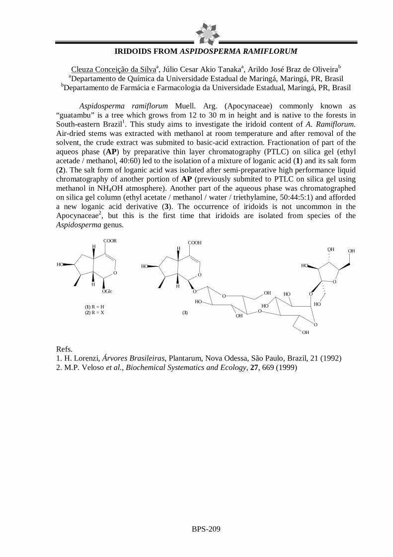
BPS-209
IRIDOIDS FROM ASPIDOSPERMA RAMIFLORUM
Cleuza Conceição da Silvaa, Júlio Cesar Akio Tanakaa, Arildo José Braz de Oliveirab aDepartamento de Química da Universidade Estadual de Maringá, Maringá, PR, Brasil
bDepartamento de Farmácia e Farmacologia da Universidade Estadual, Maringá, PR, Brasil
Aspidosperma ramiflorum Muell. Arg. (Apocynaceae) commonly known as “guatambu” is a tree which grows from 12 to 30 m in height and is native to the forests in South-eastern Brazil1. This study aims to investigate the iridoid content of A. Ramiflorum. Air-dried stems was extracted with methanol at room temperature and after removal of the solvent, the crude extract was submited to basic-acid extraction. Fractionation of part of the aqueos phase (AP) by preparative thin layer chromatography (PTLC) on silica gel (ethyl acetade / methanol, 40:60) led to the isolation of a mixture of loganic acid (1) and its salt form (2). The salt form of loganic acid was isolated after semi-preparative high performance liquid chromatography of another portion of AP (previously submited to PTLC on silica gel using methanol in NH4OH atmosphere). Another part of the aqueous phase was chromatographed on silica gel column (ethyl acetate / methanol / water / triethylamine, 50:44:5:1) and afforded a new loganic acid derivative (3). The occurrence of iridoids is not uncommon in the Apocynaceae2, but this is the first time that iridoids are isolated from species of the Aspidosperma genus.
O
O
O
H
H
HO
COOH
O
HO
OHO
OH
O
HO
OH
O
OH
HO
HO
OH
HO(1) R = H(2) R = X
O
H
H
HO
OGlc
COOR
(3)
Refs. 1. H. Lorenzi, Árvores Brasileiras, Plantarum, Nova Odessa, São Paulo, Brazil, 21 (1992) 2. M.P. Veloso et al., Biochemical Systematics and Ecology, 27, 669 (1999)

BPS-210
LARGER AMOUNT OF CABREUVIN FROM Myroxylon peruiferum. André L. F. Sarria1 (IC), João B. Fernandes1(PQ), Maria Fátima G. F. da Silva1(PQ), Paulo C. Vieira1(PQ). 1Universidade Federal de São Carlos, Departamento de Química, Laboratório de Produtos Naturais, Rod. Washington Luiz Km 235, São Carlos-S.P. [email protected] Myroxylon peruiferum (L.f.) [syn. Myroxylon balsamum (L.) Harms] (FABACEAE) has its dispersion is wide, being spread from the South of Mexico until the north of Argentina. It is a large size tree, with white flowers, winged and aromatic fruits. It is known as cabriuva and cabriuva-vermelha. Its balsam, known as balsam of Peru or Tolu or red oil is rich in vanilla. The oil was used formerly in popular medicine as expectorant for breating affections and as a sedative in case of cystitis [1]. Previous chemical study of this plant furnished monoterpenoids, sesquiterpenoids, alcohols, phenylpropanoids, isoflavones, pterocarpan, coumestans, flavanone, isoflavanones and arybenzofuran [2,3]. Cabreuvin, 3’,4’,7-trimethoxyisoflavone, isolated from wood present potent anti-Helicobacter pylori activity [4]. This communication has as objective to present the isolation of larger amount of cabeuvin from sawdust of Myroxylon peruiferum. The sawdust (5 kg) was extracted with dichloromethane yielding (36 g). This extract was fractioned in silica gel chromatography using isocratic mode yielding 1,87 g of pure cabreuvin. Cabreuvin present in its structure groups with potential to produce complex with metals ions (Ca2+, Mg2+e Zn2+) and polypyridin metals transition complex [Mn(II;III), Co(II;III), Cu(II) and Fe(II;III)] with potential to improve its activity against bacteria. These complexes are in development and they will be tested as insecticidal, fungicidal and antibacterial assays. [1] Mathias. L. et al.. J. Braz. Chem. Soc. 11: 195, 2000. [2] Oliveira, A. B. el al.. Phytochemistry 17: 593, 1978. [3] Maranduba, A. el al.. Phytochemistry 18: 815, 1979. [4] Ohsaki, A. el al.. Bioorg. Med. Chem. Letters 9: 1109, 1999.
FAPESP, CNPq, CAPES
BPS-211
FLAVONOIDS FROM THE LEAVES FROM Serjania sp Claudia Joseph Nehme(PQ)1, Marcelo Aparecido da Silva (PG)1, Danielle Cristiane Baldo (IC)1 Lourdes Campaner dos Santos (PQ)1, Wagner Vilegas (PQ)1
1UNESP-Sao Paulo State University, Instituto de Química de Araraquara
a. e-mail: [email protected]
The use of medicinal plants is an ancient practice in human populations. However, there are few scientific studies and the risks of their consumption is no known. The present work aimed chemical evaluation of the ethanol extract of the leaves from the Serjania sp. The Sapindaceae are mostly trees and shrubs, and tendril-bearing vines comprising about 140 genera and 1500 species. The Serjania genus Will has 200 species and 80 with occurrence in Brazil. Members of the genus Serjania have been employed in the folk medicine for the treatment as inflammatory and anti-ulcers. The leaves were collected at Santo Antonio do Leverger/MT State, Brazil. 400.0 g were dried, powdered and percolated at room temperature with ethanol (2 L). The solvent was evaporated under reduced pressure. The ethanol extract (3.0 g) was partionated between water, ethyl acetate and n-butanol (1:1, v/v 3 times), affording 500 mg of the ethyl acetate, 4.5 g of the n-butanol and 4.7 g of the aqueous phases. An aliquot of the ethyl acetate fraction (400 mg) was fractionated by HSCCC (High Speed Counter-Current Chromatohraphy). The fractions were analyzed by 1D and 2D NMR to afford quercetrin 1, (15 mg) kaempferol-4`-rhamnoside 2 (8 mg) and quercetin 7-methoxy-O-α-L-rhamnopyranoside-(1`→4```)-4```O-β-D-apiose 3 (10 mg). High-speed countercurrent chromatography (HSCCC) is a leading method for the fast separation of natural products from plants. It was used for the isolation of three flavonoids identified by first time in leaves from Serjania sp. The family Sapindaceae is known by the presence of saponins few stories on flavonoids and in the few works they had been told in literature on the chemistry of this genus.
FAPESP/CNPq/CAPES

BPS-212
INDOL DITERPENOID ADDUCT FROM ROOTS OF Croton rhamnifolius
Francisca Karen Souza da Silva (PG), Mary Anne Souza Lima (PQ), Raimundo Braz Filho (PQ-Visiting Professor), and Edilberto Rocha Silveira (PQ)
Curso de Pós-graduação em Química Orgânica, Departamento de Química Orgânica e Inorgânica, Centro de Ciências, Universidade Federal do Ceará
Caixa postal 12.200 – Fortaleza, Ceará – 60.021-940, e-mail [email protected] The Euphorbiaceae is a complex and diverse family with 317 genera and about 8,000 species spread all over the tropics. Croton is the second most abundant genus with 1,900 species wildly dispersed at the Antilles, South and North Americas. Croton is also a genus very common to the northeastern Brazil flora designated “Caatinga” characterized by plants well adapted to the drastic climatic conditions [just a dry (7-8 months) and a rainy (4-5 months) season occur]. Croton rhamnifolius, a shrub, sometimes a small tree, popularly known as “quebra-faca” (Port. lit.: knife breaker) due to the hardness of its wood, grows widely in the semiarid region at the border of Ceará and Pernambuco States. Entire specimens of re-sprouting stalks of C. rhamnifolius were collected at Salgueiro County-PE at the beginning of the rainy season in February, 2006. The plant material was divided in leaves, roots, trunk and stems. The leaves yielded an essential oil obtained by hydrodistillation while the other parts, after milling, where extracted with hexane followed by ethanol. TLC and 1H NMR analyses of all extracts suggested the root EtOH extract as the candidate to start the research project. Thus, it (60 g) was resuspended in MeOH and liquid-liquid partitioned (by adding water dropwise when necessary to separate the phases) with hexane, CHCl3 and EtOAc. All organics fractions were dried over anhydrous Na2SO4 and rotoevaporated while the residual hydromethanol solution was rotoevaporated and lyophilized. From the hexane fraction (3,18 g), after silica gel column chromatography, lupeol and 12-oxo-stachenone were obtained. Both normal and flash silica gel column chromatography were used to analyse the CHCl3 fraction (32,27 g) affording the triterpene alleuritolic acid, the diterpene stachenone and a novel indol diterpene adduct, designated rhamnifolin (32,1 mg), 1. All structures were elucidated by spectroscopic means, particularly 1H and 13C NMR, applying modern two-dimensional pulse sequences (COSY, HSQC, HMBC, NOESY, etc.). There is no report of indol terpenoids from plants but from endophytic fungi what brings the suspicion that rhamnifolin could indeed be a metabolite from an associated fungus, made by the incorporation of tryptophan to a kaurane type diterpenoid very common in Croton. However, rhamnifolin has shown a promising tripanocidal activity at the preliminary tests.
N
N
OHMeMe
Me
H (1)
CNPq, CAPES, FUNCAP, FINEP.

BPS-213
GUANIDINE ALKALOIDS OF MONANCHORA ARBUSCULA FROM THE COASTAL ZONE OF CEARÁ STATE, NORTHEAST OF BRAZIL
Julieta R. de Oliveira,1 Elthon G. Ferreira,2 Eduardo Hajdu,3 Letícia V. Costa-
Lotufo,2,4 Francisco Arnaldo Viana,5 Otília Deusdênia L. Pessoa,1 Edilberto R. Silveira1 1Departamento de Química Orgânica e Inorgânica - UFC, Fortaleza-CE.
2Departamento de Fisiologia e Farmacologia - UFC, Fortaleza-CE. 3Museu Nacional do Rio de Janeiro - UFRJ, Rio de Janeiro-RJ. 4Laboratório de Ciências do Mar - UFC,
Fortaleza-CE. 5Departamento de Química - UERN, Mossoró-RN.
The marine ecosystems spread over 70% of the Earth surface and is related to 95% of the biosphere. They have raised the interest of the scientists all over the world as a prolific source of new chemical entities with biological activity. Sponges are marine animals well dispersed through the oceans wild life and have been the subject of several successful research projects on biologically active secondary metabolites of marine origin. The Ceará State, Northeast of Brazil, is located just on the right corner of Brazil near to Africa and thus on a region where the marine currents change their directions. Supposing that this could affect the marine organisms life style, and their metabolism, it was decided to examine the chemical analysis of sponges from that coastal zone. Monanchora arbuscula Duch. & Mich (Crambiedae) is one of the nine sponges species recognized for the genus, characteristic of shallow waters, found in the State Park of “Pedra da Risca do Meio” under ~18 m deepness. After draining the collected material (1.215 kg) was stored in ethanol. Separated from the hydroethanol solution the animal residue was blended with a CH2Cl2/EtOH 1:1 solution and filtered. The organic solution was liquid-liquid partitioned with petrol ether followed by CH2Cl2 and EtOAc. Adsorption chromatography analysis of the petrol ether extractives (4.15 g) revealed the presence of a mixture of steroids not yet identified. From the CH2Cl2 fraction (7.34 g), upon adsorption and gel filtration chromatography analyses, mirabilin B (1; 26.0 mg) and a mixture of the epimers 8α and 8β οf 1,8a;8b,3a-didehydro-8-hydroxyptilocaulin (2 and 3; 6.0 mg) were isolated. From the EtOAc fraction (2.56 g) after a conjunction of column chromatography over silica gel and HPLC reverse phase (C8) chromatography two other guanidine alkaloids 8β-hydroxyptilocaulin (4; 34.0 mg ) and ptilocaulin (5; 24.5 mg) were obtained. All structures were characterized by spectroscopic analysis, particularly one (1H, 13C BB and 13C DEPT) and two dimensional NMR (COSY, HSQC and HMBC), and comparison to the literature data. 1, 2 and 3 have already being reported for M. unguifera (Hua et al; 2004), but not for M arbuscula. 4 and 5 have been reported previously for M. arbuscula (Tavares et al; 1995).
1
HN N
H
NH
HN NH
H
NH
RHN N
H
NH
OH1
2
33a
4
55a
67 5'
1' 3'2' 4'
8a8b 8
3: β - OH2: α - OH
5: 4:
R = H R = OH
Refs: 1. H. Hui-ming et al., Bioorg. & Med. Chem., 12, 6461 (2004) 2. R. Tavares et al., J. Nat. Prod., 58, 1139 (1995) CNPq / CAPES / FINEP / FUNCAP / PRONEX.

BPS-214
A NOVEL FLAVANONE FROM POLYGONUM SPECTABILE
DUARTE, M.G.R.1; BRANDÃO, M.2; MACEDO, J.F.2; BRAGA, F.C.3; NUNAN, E.A.3; BRANDÃO, G.C.4; BRAGA, A.B.3
1. Divisão de Ciências Farmacêuticas, Diretoria de Pesquisa e Desenvolvimento, Fundação
Ezequiel Dias, Belo Horizonte, MG 2. Departamento de Pesquisa, Empresa de Pesquisa Agropecuária de Minas Gerais, Belo
Horizonte, MG 3. Departamento de Produtos Farmacêuticos, Faculdade de Farmácia, UFMG, Belo
Horizonte, MG 4. Bolsista de Iniciação Científica, FAPEMIG
The screening of 10 weed medicinal plant extracts for in vitro antimicrobial activity showed that Polygonum spectabile was active against Staphylococcus aureus and Micrococcus luteus. Bioguided chromatographic fractionation of the ethanol extract from the aerial parts of P. spectabile resulted in an active fraction, that afforded three flavonoids designated PS1, PS2 and PS3. Spectrometric analysis allowed the characterization of PS2 as 7-hydroxy-5-methoxyflavanone (alpinetin) (1) and PS3 as 2’,4’-dihydroxy-3’,6’-dimethoxychalcone. The latter compound has already been isolated from Polygonum senegalense (2). Characterization of PS1 as 7-hydroxy-5,8-dimethoxyflavanone was accomplished by IR, UV, FAB-HRMS, 1H NMR and 13C NMR, assigned by means 2D NMR experiments (HMQC and ROESY). According to a literature review, the compound PS1 is a novel flavanone.
1. N. Qais et al., Fitoterapia, 67, 476 (1996). 2. A. Maradufu; J. H. Ouma, Phytochemistry, 17, 823-4 (1978).
O
O
HO
OCH3
OCH3
(a)

BPS-215
ISOLATION AND IDENTIFICATION OF CHEMICAL CONSTITUEN TS FROM Richardia grandiflora (Cham. & Schltdl.) Steud. (RUBIACEAE) Emídio Vasconcelos Leitão da Cunha (PQ), Maria de Fátima Vanderlei de Souza (PQ), Anna Cláudia de Andrade Tomaz (PG), Raquel Bezerra de Sá de Sousa Nogueira (IC) Laboratório de Tecnologia Farmacêutica, Universidade Federal da Paraíba, Caixa Postal 5009, 58051-970, João Pessoa, Paraíba, Brasil Brazil has a great diversity on plants that possess non-researched medicinal potential and are promising sources of therapeutic and pharmacological innovations to the most diverse areas of human health1. The Rubiaceae family is considered the biggest one of the order Gentianales2 and presents around 637 genera and 10.700 species3. The species Richardia grandiflora (Cham. & Schltdl.) Steud., known popularly as “ervanço”, “poaia” or “ipeca-mirim”, has ethnopharmacological indications to use as decoction against hemorrhoids and as vermifuge4. Aiming at contributing to the chemotaxonomic study of the family Rubiaceae and considering the absence of data in literature about the chemical constitution of the species Richardia grandiflora, the latter was submitted to a phytochemical study to isolate and identify its chemical constituents. The collected plant material (entire plant) was processed yielding the crude ethanol extract which was submitted to low-pressure chromatography giving ten fractions, being three of them submitted to column chromatography using different adsorvents. The resulting fractions were analysed through analytical thin-layer chromatography (TLC) and joined according to their RF´s. Therefore, five constituents were isolated: a mixture of the steroids sitosterol and stigmasterol, orto-benzoic acid, m-methoxy-p-hydroxy-benzoic acid and phaeophitin a, all of them isolated for the first time from the genus Richardia and identified by means of spectroscopic methods such as 1H and 13C Nuclear Magnetic Resonance, with the add of two-dimensional techniques (COSY, HSQC, HMBC and NOESY), besides comparison with literature data. Refs. 1 L.A Souza et al., Rev Bras Bot, 26, 175-184 (2003). 2 V.P. Coelho et al., Rev Bras Farmacogn, 16 (2), 170-177 (2006). 3 S. Mongrand et al., Phytochemistry, 66, 549-559 (2005) 4 M.F.Agra et al., Rev Bras Farmacogn, 17 (1), 114-140 (2007).

BPS-216
A NEW DAVANONE CHEMOTYPE OF LIPPIA INTEGRIFOLIA
C. Catalán1, M.P. de Lampasona1, C. Coronel2, D. Lorenzo2, N. Martínez2, V. Márquez2, E. Dellacassa2
1Instituto de Química Orgánica, Facultad de Bioquímica, Química y Farmacia, Universidad
Nacional de Tucumán. S. M. de Tucumán, T4000INI Argentina. 2Cátedra de Farmacognosia y Productos Naturales, Facultad de Química, Universidad de la República. Gral. Flores 2124.
11800-Montevideo, Uruguay. E-mail: [email protected]
The genus Lippia (family Verbenaceae) comprises some 200 species growing in the tropical and subtropical regions of South America, Central America and Africa. In the Americas, members of this genus can be found from Mexico to the province of Buenos Aires in Argentina. Because most of the species are aromatic, the studies on the chemistry of this genus are mostly related with the composition of the essential oils, where an outstanding feature of Lippia is the difference observed in the essential oil composition reported for the same species from different geographic origins. However, the mono- and sesquiterpenoids found in the essential oils for all the Lippia species investigated so far are quite common and widespread in the plant kingdom, the exceptions being L. integrifolia (Gris.) Hieron. which produces ketones based on the unique sesquiterpene skeletons named lippifoliane and integrifoliane. Lippia integrifolia (Griseb.) Hieronymus, commonly known as ‘incayuyo’ or ‘té del inca’, is a woody aromatic shrub native to central and northern Argentina, where infusions of the aerial parts are widely used in traditional medicine (1). L. integrifolia has been shown to be a rich source of uncommon sesquiterpenoids. The unique ketones integrifolian-1,5-dione, lippifoli-1(6)-en-5-one and closely related derivatives together with sesquitepenoids based on the rare africanane and asteriscane skeletons, constitute the bulk of its essential oil (2). In the present study, we report the results of a detailed analysis carried out on the essential oils obtained from two selected populations of L. integrifolia growing wild in northern Argentina: C653 (Santa María Departament, Catamarca Province, voucher specimen LIL 607197) and C660 (Cachi Departament, Salta Provincie, voucher specimen LIL 607276) which allowed the identification of a family of structurally related sesquiterpene compounds, and isomers, belonging to the group of the ketone davanone. These compounds and their distribution in both populations evaluated allowed proposing a new chemotype for L. integrifolia characterized by high contents of davanone and davanol isomers. Previous reports indicated davanone oil as possessing a promising aromatic profile for perfumery, novel foods and beverages (3). The notes were related to davanona and described as green, pungent, harsh, herbal, intense davana-like, fruity. The present study focuses on the comparison of sensory odour profile of L. integrifolia essential oil by GC-O, and the odour perception -as an entity- at different retention times, in order to evaluate the aromatic response (aromagram) and the behaviour of davanone and davanol isomers. Refs. 1. G. Bassols et al., Dominguezia, 13, 7 (1996) 2. A.C. Coronel et al., Flavour Fragr. J., 21, 839 (2006) 4. A. Kamath et al., Flavour Fragr. J., 16, 401 (2001)

BPS-217
Biflavonoids from Caealpinia pyramidalis Tull
Marcus Vinicius Bahia1, Juceni P. David2 and Jorge M. David3 1Instituto de Ciências Ambientais e Desenvolvimento Sustentável, Universidade Federal da Bahia, 40110-906, Barreiras (BA), 2Faculdade de Farmácia, Universidade Federal da Bahia,
3Instituto de Química, 40170-280, Salvador (BA), Brazil.
The genus Caesalpinia (Caesalpinioideae, Fabaceae), comprised of tropical or subtropical trees or shrubs, contains more than 150 species worldwide. Native Brazilian species such as Caesalpinia echinata (pau-brasil) had important economic value in the early colonial period of Brazil. Previous studies of species of this genus report remarkable biological activities for its species such as antimicrobial, antidiabetic (C. bonducella), antimalarial (C. volkensii and C. pluviosa), and antiinflammatory activities (C. sappan and C. ferrea). Caesalpinia pyramidalis Tull is an endemic tree of northeastern region and one of the predominant species in the "caatinga" vegetation. In Bahia State, it is popularly known as "catingueiro" or "pau-de-rato", and its leaves are employed in traditional medicine as diuretic, dyspeptic, digestive, and antipyretic. This work describes the chemical study of the methanol extract wood and leaves of C. pyramidalis (Leguminosae). The CHCl3 and EtOAc fractions obtained from the partition of MeOH extract were submitted to different chromatographic procedures over silica gel and Sephadex LH-20. The organic extracts of leaves of C. pyramidalis were submitted to different chromatographic procedures which permitted to isolate agathisflavone, podocarpusflavone A, loniflavone (3), glycosil sitosterol besides the mixture of apigenin and kaempferol1 and amentoflavone (1). On the other hand the methanolic extract of the wood of C. pyramidalis afforded taxifoline, gallic acid, 5,6,7,8-tetrahydroxy-4'-methoxiflavone, siringaresinol, methyl gallate, pheofitin a and 5'-hidroxyamentoflavone2 (2), the unique biflavonoid found in trunk wood of this plant. To date, C. pyramidalis is the first species in the genus to present biflavonoids. All compounds were identified by analysis of IR, UV, MS 1H and 13C NMR data, including bidimensional techniques. The biflavonoid (2) was only obtained from Bartramia ithyphylla (Bartramiaceae) e Rhytidiadelphus squarrosus (Hylocomiaceae). Refs: 1 M. V. Bahia et al., Journal of the Brazilian Chemical Society, 16, 1402 (2005) 2 M. V. Bahia, Tese de Doutorado, Universidade Federal da Bahia, 2007
O
OOH
HO
OH
O
OOH
HO
OH
2
O
OH
HO
O
OH
O
OH
HO
O
OH
OH
1
O
O OH
OHO
OOH
HO
O
OH
3

BPS-218
A NOVEL ESTER FROM HIMALAYAN CEDARWOOD OIL.
Sushilkumar A. Dubal, and S.A.Momin.
Department of Oils, Oleochemicals and surfactants, Institute of Chemical Technology, University of Mumbai.
Essential oil extracted from aromatic plants find application in flavour, fragrance industries and in aromatherapy. India with its rich bio and agro diversity is a fertile ground for the production of essential oil and their value added derivatives. However, several natural raw materials consist mainly of hydrocarbons that are not suitable as smelling substance. These hydrocarbons often possess complex carbon skeletons, which are difficult to synthesize, but offer excellent possibilities for functionalization with oxygen containing groups transformations that often lead to interesting new fragrant materials. In the present work isolation from Himalayan cedarwood oil two sesquiterpenic ketones which were found to be α-atlantone and γ-atlantone. The suceesful routes involved the preparation of novel ester molecule of atlantone & study its odour profile. Stability study of these molecules in cosmetic products was carried out.
References:
1. Anon., (1950). The wealth of India, Raw Materials, C.S.I.R., New Delhi,2, 106-7. 2. Dixit, A., and Dixit S.N., (1982). Indian perfumer, 26 (24), 216-217. 3. A.Pfau & Pl. Plattner, Helv.Chim.Acta, 1934, 17,129 4. Krishna, R. Dev.,S., and Guha,P.C., (1952).J.ind.chem.Soc.,29,72,30.
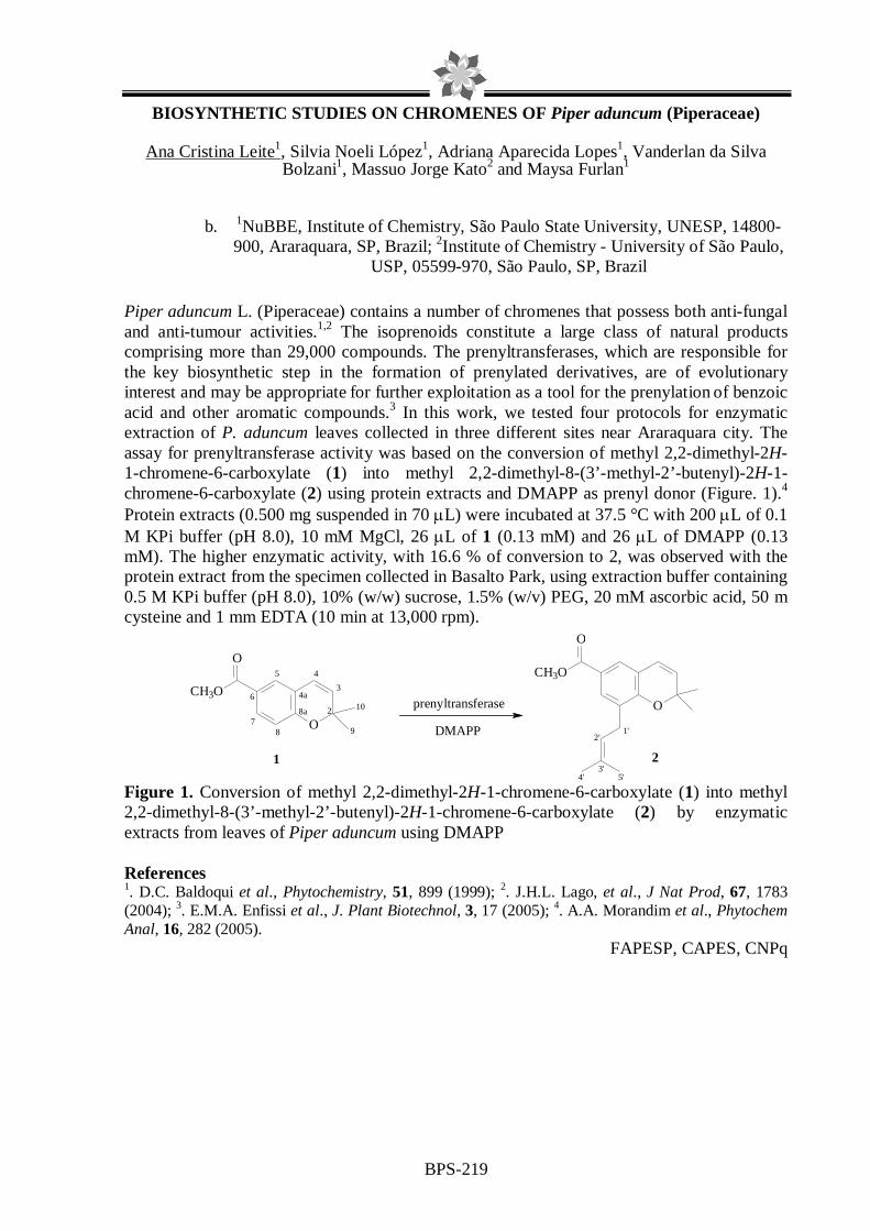
BPS-219
BIOSYNTHETIC STUDIES ON CHROMENES OF Piper aduncum (Piperaceae)
Ana Cristina Leite1, Silvia Noeli López1, Adriana Aparecida Lopes1, Vanderlan da Silva Bolzani1, Massuo Jorge Kato2 and Maysa Furlan1
b. 1NuBBE, Institute of Chemistry, São Paulo State University, UNESP, 14800-900, Araraquara, SP, Brazil; 2Institute of Chemistry - University of São Paulo,
USP, 05599-970, São Paulo, SP, Brazil
Piper aduncum L. (Piperaceae) contains a number of chromenes that possess both anti-fungal and anti-tumour activities.1,2 The isoprenoids constitute a large class of natural products comprising more than 29,000 compounds. The prenyltransferases, which are responsible for the key biosynthetic step in the formation of prenylated derivatives, are of evolutionary interest and may be appropriate for further exploitation as a tool for the prenylation of benzoic acid and other aromatic compounds.3 In this work, we tested four protocols for enzymatic extraction of P. aduncum leaves collected in three different sites near Araraquara city. The assay for prenyltransferase activity was based on the conversion of methyl 2,2-dimethyl-2H-1-chromene-6-carboxylate (1) into methyl 2,2-dimethyl-8-(3’-methyl-2’-butenyl)-2H-1-chromene-6-carboxylate (2) using protein extracts and DMAPP as prenyl donor (Figure. 1).4
Protein extracts (0.500 mg suspended in 70 µL) were incubated at 37.5 °C with 200 µL of 0.1 M KPi buffer (pH 8.0), 10 mM MgCl, 26 µL of 1 (0.13 mM) and 26 µL of DMAPP (0.13 mM). The higher enzymatic activity, with 16.6 % of conversion to 2, was observed with the protein extract from the specimen collected in Basalto Park, using extraction buffer containing 0.5 M KPi buffer (pH 8.0), 10% (w/w) sucrose, 1.5% (w/v) PEG, 20 mM ascorbic acid, 50 m cysteine and 1 mm EDTA (10 min at 13,000 rpm). Figure 1. Conversion of methyl 2,2-dimethyl-2H-1-chromene-6-carboxylate (1) into methyl 2,2-dimethyl-8-(3’-methyl-2’-butenyl)-2H-1-chromene-6-carboxylate (2) by enzymatic extracts from leaves of Piper aduncum using DMAPP References 1. D.C. Baldoqui et al., Phytochemistry, 51, 899 (1999); 2. J.H.L. Lago, et al., J Nat Prod, 67, 1783 (2004); 3. E.M.A. Enfissi et al., J. Plant Biotechnol, 3, 17 (2005); 4. A.A. Morandim et al., Phytochem Anal, 16, 282 (2005).
FAPESP, CAPES, CNPq
2'
5'4'3'
1'
CH3O
O
O
87
6
5
8a
4a3
10
CH3O
O
O
4
2
9
prenyltransferase
DMAPP
1 2

BPS-220
PURIFICATION OF 4-NEROLYDILCATECHOL SYNTHASE FROM Potomorphe umbellata LEAVES
Debora Cristina Baldoqui1, Massuo Jorge Kato2, Vanderlan da Silva Bolzani1,
Maysa Furlan1. 1NUBBE- Núcleo de Biossíntese Bioensaio e Ecofisiologia de Produtos Naturais – Instituto
de Química da Universidade Estadual Paulista – Unesp – C. P. 355 – 14800-900 – Araraquara, SP. 2Instituto de Química – Universidade de São Paulo - USP – Prof. Lineu
Prestes 748 b 11 T – São Paulo, SP. E-mail: [email protected]
The major constituent of leaves and roots of Potomorphe umbellata is the powerful antioxidant, 4-nerolidylcatechol1,2. Based on such activity, the extract obtained from roots of P. umbellata has been recently introduced in the market as active ingredient of an anti-aging lotion3. The biosynthetic pathway to the formation of 4-nerolidylcatechol in P. umbellata involves 3,4-dihydroxybenzoic acid and nerolidol as precursors as demonstrated by incubation of these compounds with cell free extracts prepared from leaves of young plants (Figure 1). Aiming to obtain this compound by green chemistry methodology, the enzyme responsible for 4-nerolidylcathecol formation was purified from leaves of Potomorphe umbellata4. The purification method was optimized as following. The crude extract was obtained from young leaves of Potomorphe umbellata using pH 8,0 TRIS-HCl buffer, in a ratio 1:3 of amount of leaves and buffer. The crude extract was submitted to heat denaturation at 70oC for 15 minutes followed by renaturation mixing the solution gently at 4oC. This solution was submitted to centrifugation and the supernatant was submitted to fractionated precipitation with ammonium sulfate. The fraction 20-40% of saturation yielded the enzyme responsible by the formation or 4-nerolidylcathecol with high purity. All processes used during the optimization to purify the enzyme of interest were accompanied by protein determination, enzymatic assay and SDS-PAGE.
Figure 1: Biosynthetic route to the formation of 4-nerolidylcathecol. Refs. 1. Kijjoa et al., Planta Medica, 39, 85 (1980). 2. Ropke et al., Photochemistry and Photobiology, 78, 416 (2005). 3. Ropke et al., International Journal of Pharmaceutics, 249, 109 (2002). 4. Baldoqui et al., “Método para obtenção do extrato enzimático de Potomorphe umbellata e uso do mesmo para obtenção de derivados de catecol”. PI 070.0238-6.
OH
OH
OH
COOH
OH
OH
enzyme +
3,4-dihydroxybenzoic acid
nerolidol 4-nerolidylcathecol

BPS-221
PEROXIDASES FROM NEEDLES OF ARAUCARIA ANGUSTIFOLIA AND THEIR CIRCADIAN RHYTHM
Lydia F. Yamaguchi and Massuo J. Kato
Instituto de Química, Departamento de Química Fundamental –USP Av. Prof. Lineu Prestes, 748 sala 1124, São Paulo, SP, CEP 05508-000
The Araucaria angustifolia is an endemic coniferae of southern and southeastern Brazil. It composes the “Mata de Araucárias” an important bioma endangered due the extensive loggings. Their leaves contain six major amentoflavone-type biflavonoids, including amentoflavone, ginkgetin and tetra-O-methylamentoflavone as determined by HPLC-ESI analyses. These compounds are reported to possess a variety of biological activities especially as antioxidants, which, is related to their capacity to suppress singlet oxygen, lipoperoxidation, and DNA oxidation promoted by several oxidant agents. An additional mode of action is related to the ability to quench transition metals involved in free radical formation (Yamaguchi e col. 2007). The biosynthesis of biflavonoids has been proposed to take place by oxidative coupling of two flavonoids units. The predominance of amentoflavone-type biflavonoid in Araucaria angustifolia indicates the specificity of peroxidase involved in its formation. The peroxidase activity was detected in the enzymatic extract from needles of Araucaria angustifolia The enzyme convert two molecules of apigenin to amentoflavone and ginkgetin. The products were identified and quantified by its UV-spectrum, LC/mass spectrometry analysis and comparison with reference compound. The circadian rhythm was characterized, as well as, the optimum conditions to enzymatic convertion, such as, pH, temperature and H2O2 concentration.
O
OH
OH
OH O
O
OH
OH
OH O
O
OH
OH
OH O
amentoflavone
apigenin
peroxidase
Yamaguchi, L. F.; Kato, M. J., Di Mascio, P. (2006) Perspectives in Biflavonoids – a review. In: Recent Progress in Medicinal Plants. Ed. Govil J.N., Stadium Press, v. 20, pp 1-54.

BPS-222
SUBSTRATE SPECIFICITY OF GERANYLTRANSFERASES IN Piper crassinervium
Silvia Noelí López1, Adriana Aparecida Lopes1, Vanderlan da Silva Bolzani1,
Massuo Jorge Kato2 and Maysa Furlan1 1NUBBE- Núcleo de Biossíntese Bioensaio e Ecofisiologia de Produtos Naturais – Instituto
de Química da Universidade Estadual Paulista – Unesp – C. P. 355 – 14800-900 – Araraquara, SP. 2Instituto de Química – Universidade de São Paulo - USP – Prof. Lineu
Prestes 748 b 11 T – São Paulo, SP. E-mail: [email protected] Piperaceae family comprises 4 genera and more than 4000 species and, among them, Piper is one of the most abundant genus with approximately 2000 species.1 Piper crassinervium species accumulates antifungal and antioxidant geranylated metabolites derived from benzoic acid and from p-hydroquinone.2 Geranyltransferases are a group of prenyltransferases that catalyze the C-geranylation on several substrates. They have been generally reported as membrane-bound enzymes and have showed strict substrate specificity for geranyldiphosphate (GPP) as the prenyl donor, although accept a variety of substrates as acceptors of GPP moieties.3,4 Substrate specificity studies carried out with P. crassinervium indicated that 3,4-dihydroxy benzoic acid was selectively geranylated by cell free extracts from its leaves.5 In this work, we report the substrate specificity studies for geranyltransferase by means of cell free and microsomal fractions from leaves and roots of P. crassinervium. Crude protein extracts were submitted to geranyltransferase activity assays with GPP, MnCl2 or MgCl2 as cofactors, and the substrates p-hydroquinone (HQ), 4-hydroxybenzoic (4-HB) and 3,4-dihydroxybenzoic (3,4-HB) acids. The microsomal fractions were obtained by ultracentrifugation as described.6.The enzymatic activity was assessed in all fractions by HPLC-UV (table 1). Differences in substrate specificity were observed, suggesting two geranyltransferases in P. crassinervium. One in soluble fraction, with affinity to 3,4-HB, and a second in the microsomal fraction with affinity to both HQ and 4-HB. SDS PAGE analysis indicated an electrophoretic band in the microsomal fraction with MW between 45-66 kDa, which is similar to that reported for geranyltransferases.6
Table 1: Summary of geranyltransferase activity in enzymatic fractions from P. crassinervium.
Leaves Roots Crude Microsomal Crude Microsomal
HQ +/-. + n.d. + 4-HB n.d. + +/- +
3,4-HB + n.d. + n.d. HQ= p-hydroquinone; 4-HB: p-hydroxybenzoic acid; 3,4-HB: 3,4-dihydroxybenzoic acid. n.d.=not detected; +=positive reaction, +/-=weak
Refs. 1S. Wanke et al., Molecular Phylogenetics and Evolution, 42, 477 (2007). 2A. P. Danelutte et al., Phytochemistry, 64, 555 (2003). 3Y. Yazaki et al., J. of Biol. Chem., 277, 6240 (2002). 4T. Kuzuyama et al., Nature, 435, 983 (2005). 5A. Lopes et al., 30 Annual Meeting Soc Bras. Química, Poster, PN-224 (2007). 6 A. Mühlenweg et al., Planta, 205, 407 (1998).

BPS-223
IDENTIFICATION AND CHARACTERIZATION OF AROGENATE DEHYDRATASES IN ARABIDOPSIS: COMPARISON TO A BACTERIAL
PREPHENATE DEHYDRATASE
Laurence B. Davin1*, Hong Yang1, Oliver R. A. Corea1,2, Man-Ho Cho1, Aldwin M. Anterola1, Frances Anne Moog-Anterola1, Susanne E. Kohalmi2,
Mark A. Bernards2, and Norman G. Lewis1 1 Institute of Biological Chemistry, Washington State University, Pullman, WA 99164-6340, USA and
2 Department of Biology, University of Western Ontario, London, Ontario, Canada, N6A 5B7 E-mail: [email protected]
While both prephenate and arogenate have been reported in plants to undergo
decarboxylative dehydration via the action of a dehydratases to afford phenylpyruvate and phenylalanine, respectively, neither enzyme(s) nor encoding gene(s) were isolated and/or identified. In this study, a data mining approach was undertaken to attempt to identify the dehydratase(s) involved in Phe formation.1 This approach suggested that there were six putative prephenate dehydratase (PDT) homologues in Arabidopsis, based on similarity to bacterial PDTs. However, earlier biochemical analyses of cell extracts detected arogenate dehydratase (ADT) rather than PDT activities. All six putative ADTs/ PDTs were cloned and expressed as Nus-tagged recombinant proteins in E. coli. Three of the resulting recombinant proteins more efficiently utilized arogenate than prephenate, with kcat/Km values of 1050, 7650 and 1560 M–1 s–1 for arogenate, vs. 38, 240 and 16 M–1 s–1 for prephenate, respectively. The remaining three, by contrast, had kcat/Km values of 1140, 490 and 620 M–1 s–1 for arogenate, with prephenate not serving as a substrate unless excess recombinant protein (>150 µg/assay) was used. For comparative purposes, a previously characterized PDT from Methanocaldococcus jannaschii was assayed under the same conditions; it was shown to have a very strong substrate preference for prephenate over arogenate. All six Arabidopsis genes, and their corresponding proteins, are thus provisionally classified as arogenate dehydratases and designated ADT1 through ADT6. 1. Journal of Biological Chemistry (2007), in press (doi 10.1074/jbc.M702662200).

BPS-224
MICROENCAPSULATION OF BRAZILIAN CERRADO’S PLANTS
Rodney Alexandre Ferreira Rodrigues1, Ilza Maria de Oliveira Sousa1, Caio de Almeida Maiochi1, Marta Cristina Teixeira Duarte1, Sirlene Valério Tinti1, Maria do Carmo Estanislau
do Amaral2, Volker Bittrich2, João Ernesto de Carvalho1, Mary Ann Foglio1
1 Chemical, Biological and Agricultural Research Center, P.O. Box 6171, 13083-970, Campinas State University, UNICAMP – Campinas - SP, Brazil, 2 Botanical Department, P.O. Box 6109, 13084-971, Campinas State University, UNICAMP – Campinas - SP, Brazil The extracts used as a microencapsulation purpose have been studied before demonstrated activity against cancer cell lines in vitro. This paper reports the microencapsulation of Brazilian’s plants cerrado, using crude extracts of Aspidosperma tomentosum Mart, Gaylussacia brasiliensis Meiss and Pterodon pubescens Benth. The extracts microencapsulated were evaluated for anticancer activity in UACC62 (melanoma), MCF-7 (breast), NCI 460 (lung, non-small cells), OVCAR03 (ovarian), PC0 3 (prostate), HT-29 (colon), 786-0 (renal) and NCI-ADR (ovarian expressing phenotype multiple drugs resistance) cancer cell lines in vitro. A 48 h SRB cell viability assay was performed to determine growth inhibition and cytotoxic properties of the compounds. Cells were treated with at least four different concentrations levels ranging from 0.25 to 250 µL/mL with determination of total growth inhibition (TGI) parameter. The extracts also were evaluated for minimum inhibitory concentration activity in Staphylococcus epidermides ATCC 12228; Pseudomonas aeruginosa ATCC 13338; Salmonella cholerasuis CCT 4296; Escherichia coli CCT 0547 and Candida albicans CCT 776. An aqueous suspension containing wall material, crude extract and tween 80 was prepared, homogenized and passed through in a mini spray dryer Büchi B-290 using 175 oC of entrance, 100 oC of exit temperature, 6-7 mL/min of liquid flow rate, 600 L/h of nitrogen pressure and 1.4 mm of nozzle diameter and also using a freezy dryer Virtis 8L. The microcapsules were obtained from acacia gum, using the proportion wall material/extract (4/1). The particle size distribution was determined by laser scattering using a Malvern Mastersizer/S MAM-5005. Microcapsules were characterized by scanning electron microscopy (SEM) in order to verify their structural characteristics. Microcapsules were mounted on a sample holder, sputter-coated with a thin layer of gold and examined with a JEOL JSM-T300 electron microscope. Both microencapsulated extract by spray dryer and freeze mainteined the same anticancer activity of the pure crude extract whereas freeze dried material didn’t produce microcapsules structures.

BPS-225
From raw material to Eucalyptus syrup: quality control of the whole process
Paula Carolina Pires Bueno and Jairo Kenupp Bastos Faculdade de Ciências Farmacêuticas de Ribeirão Preto – FCFRP/USP
Since ancient times, medicinal plants are used for the treatment of human diseases, and
nowadays plants still make an important key for primary health care. In the last years, it has been an increasing interest by the big companies in such products, stimulating the standardization and the development of reliable quality control analytical methodologies(1,2). One of the most widely used medicinal plant, Eucalyptus globulus (Myrtaceae) has been traditionally used for the treatment of respiratory diseases because of its expectorant, mucolytic and antiseptic effects(4). So, this work shows the development of syrup containing Eucalyptus globulus extract, its standardization, and the validation of the analytical methodology for its quality control, including the raw material, extracts, essencial oil and the final product. According to ANVISA(3), the average daily dosage of 1.8-cineol in formulations containing E. globulus is 14.0 to 42.5 mg. So, the syrup was made containg 10% w/w of an eucalyptus fluid extract. The method was developed and validated using a Hewlett-Packard 6890N gas chromatograph (Agilent Technologies) equipped with a split/splitless injector inlet and a flame ionization detector. An HP-5 capillary column was used for the analysis and the non linear increasing temperature gradient was: 75 °C to 200 °C, in 20 min. The identification of the main compounds of E. globulus was made using GC-MS. For the qualitative analysis, the fingerprints of 12 Eucalyptus species were obtained: E. botryoides, E. citriodora, E. elba, E. globulus, E. grandis, E. microcorys, E. paniculata, E. resinifera, E. robusta, E. saligna, E. tereticornis and E. urophylla. The chromatogram of E. globulus shows the main peak (1.8-cineol) at 5.23 minutes. The chromatograms of the other species have some significant differences and similarities in the presence and quantity of metabolites, which allows a preliminary identification of each species by this method. Besides, the selectivity and linearity achieved, the recoveries were 99.8% and 100.5% for leaves and syrup, respectively. The repeatability, determined by the relative standard deviation in six replicates at test concentration, were 1.86% and 0.92%, respectively. This result is in agreement with the Brazilian regulatory agency (the maximum value established for ANVISA is 5%). The detection limit was 4.79 µg/mL while the quantification limit was 14.51µg/mL. Finally, the influence of the main modified parameters for the robustness was determined using the Yuden test. The developed method, with internal standardization, proved to be reliable for the quantification of 1.8-cineol, not only in eucalyptus leaves, extracts and essential oils, but also in syrup formulations containing this plant extract, assuring the rastreability of the entire manufacturing process.
Refs.
1. J. B. Calixto, Brazilian Journal of Medical and Biological Research, 33, 179-189 (2000). 2. WHO, Quality Control Methods for Medicinal Plants Material, 84p., 1992. 3. J. Alonso, Tratado de Fitomedicina, 1360p., 2004. 4.ANVISA, Resolução RE n. 89 de 16/03/2004.

BPS-226
COMPARATIVE CHROMATOGRAPHIC PROFILE OF CULTIVATED Kalanchoe pinnata
Ana Claudia F. Amaral1, Luiza G. Ferreirinha1, José Luiz P. Ferreira1; Sandra A. P. M.
Fraga1; Renata B. Araújo1; Naomi K. Simas1; Ramine L. Barbosa1; Valério F. M. Amaral1
1Laboratório de Plantas Medicianis e Derivados, Departamento de Produtos Naturais/Farmanguinhos/FIOCRUZ (Rio de Janeiro).
E-mail: [email protected] Kalanchoe pinnata L. is a perennial plant belonging to the Crassulaceae family. This species is easily cultivated and widely used in folk medicine of tropical regions to treat respiratory infections, bruises, edema of legs and burnings. In Brazil this plant is known as saião, folha da fortuna, coirama branca, among others. According to literature, the main chemical constituents described for this species belong to the class of flavonoids. The objective of this work is to compare K. pinnata cultivation in relation to the edaphic characteristics of a seedbed without soil manipulation with other seedbed treated with a variable amount of calcareous for pH adjusting purposes. Additionally, the chromatographic profiles of these plants extracts were analyzed to compare the flavonoid constitution on carried out experiments. The vegetal species was cultivated in the experimental seedbeds of Farmanguinhos in Jacarepaguá campus (Rio de Janeiro). The aerial parts of plants (leaf, stem and flower) were harvested on flower blooming season. The voucher specimen was deposited at the herbarium of the Botanical Garden of Rio de Janeiro. The extracts of flowers and the mixture of leaves and stems were obtained for maceration process in ethanol 70%. The crude extracts had been compared in regard to flavonoid content using thin layer chromatography (TLC) method. The analyses were carried out on silica gel TLC plates using AcOEt:HCOOH:H2O (8.8:0.6:0.6) and AcOEt:HOAc:HCOOH:H2O (10:1.1:1.1:2.7) as solvent systems. The orange spots on TLC were visualized spraying NP/PEG reagent and under UV light (365nm). The plants treated with the calcareous addition (pH=6.8) provided a larger biomass (2.5 kg) when compared to the ones without treatment (pH=5.0) weighting less quantity of biomass (1.4 kg). Nevertheless, the flavonoid composition was similar in both treatments and has not shown qualitative differences in the analyzed extracts. More detailed experiments are necessary to support these results. However, this study under calcareous supply has not shown any relation with flavonoids variation in the K. pinnata aerial parts.

BPS-227
DETERMINATION OF TOTAL FLAVONOIDS FROM LEAVES OF CULTIVATED Cymbopogon citratus
Ana Claudia F. Amaral1; Pamela B. G. de Almeida1; Renata B. de Araújo1; José Luiz P. Ferreira1; Naomi K. Siomas1; Alexandre Xavier1, Ricardo M. Kuster2
1Laboratório de Plantas Medicinais e Derivados, DPN /Farmanguinhos /FIOCRUZ; 2NPPN/CCS/UFRJ (RJ). E-mail: [email protected]
Cymbopogon citratus (DC.) Stapf. (Poaceae) is a pleasant-tasting medicinal plant used as tea (infusion or decoction of its aerial parts) in the folk medicine. This plant is used to treat digestive disorders, inflammation, nervous disorders and fever. Several literature data have been supporting its effectiveness in most disorders, which justifies the therapeutic use of this species. Phenolic compounds are important constituents of leaves and are found in the tea presentation. Additionally, there are significant data about the antioxidative potential of these substances obtained by different methods. Considering that this potential is an important effect, mainly in inflammatory process, this study presents a comparison of the flavonoids content between the fresh and dried leaves of C. citratus that were collected (4g each) in the experimental seedbed at Jacarepaguá campus of Fiocruz (Rio de Janeiro state). The total flavonoid was analyzed according to the Brazilian Pharmacopoea procedure. The content of flavonoids in the plants leaves, calculated as quercetin, was determined by spectrophotometry at 426 nm. A Shimadzu UV1601PC spectrophotometer was used for the measurements. The fresh leaves absorbance was detected at 0.062 nm and for dried leaves at 0.048 nm. These absorbance values plotted on quercetin calibration curve gave a 1.074 and 0.886 µg/mL of quercetin content in the fresh and dried leaves. The total concentration of flavonoids in the fresh and dried plant leaves were calculated at 0.19 and 0.15%, respectively. These determinations of flavonoid content are in agreement with literature data about antioxidative potential properties of C. citratus, where these natural compounds are therapeutically related to this activity. These results can, even partially, attest the popular medicinal use of this plant. Financial support: CNPq

BPS-228
DETERMINATION OF MINERAL CONSTITUENTS IN PROPOLIS B Y FLAME ATOMIC ABSORPTION SPECTROMETRY
Daiane Finger, Lílian Buriol, Julio M. T. dos Santos, Marcos Roberto da Rosa, Sueli Pércio
Quináia, Yohandra Reyes Torres Departamento de Química, CEDETEG, Universidade Estadual do Centro-Oeste, Guarapuava,
Paraná, Brasil.
Propolis, a resin collected by bees from sprouts trees, has viscous consistency, variety of colors ranging from dark brown to red, bitter taste and complex chemical composition. As it is derived from vegetable products, propolis is subjected to environmental contaminations and could accumulate heavy metals in dangerous amounts1. Practices such as the paint used in hives and further propolis treatment may influence heavy metals content. The objective of this study was to determine inorganic constituents and the presence of metallic contaminants in samples of crude propolis from Prudentópolis City (States of Paraná-PR). A Varian AA-220 atomic absorption spectrometer (FAAS) equipped with a deuterium-arc lamp background corrector and operating with air-acetylene (Mg, K, Fe, Zn, Cu, Cd, Cr and Pb) and acetylene-oxide nitrous (Ca and Al) flame was used. All chemicals used in the experiments were of analytical-reagent grade. Deionised water (18.2 MΩ cm) obtained from a Human UP 900 System was used throughout the process. All glassware was soaked overnight in 1% v/v nitric acid and rinsed with deionized water before use. Stock solutions of all evaluated species were prepared with concentrations of 1000 mg L-1 (SpecSol, NIST-USA). Figure 1 summarizes the experimental procedure for the determination of metals in propolis. Table 1 shows the average concentration obtained for propolis samples.
Table 1. Metals in propolis (mg/g)
Figure 1: Procedure for metals determination in propolis (Prudentopolis-PR) Prudentopolis´s propolis did not show contamination by potentially toxic species and is a good source of Ca, Mg and K. The results obtained for propolis are in agreement with literature2. The present study gives information about the mineral content of propolis from Prudentopolis-PR. As heavy metals were below detection limit or in low amounts, this propolis is appropriated for the development of edible products, although other parameters for quality control must be considered as well to abide legislation. Refs. 1. R.G. Woisky; A. Salatino, J. Apic. Res., 37, no 2, 99, (1998). 2. M.C. Marcucci, Apidologie, 26, 83, (1995).

BPS-229
ISOLATION OF FLAVANONES FROM Sparattosperma leucanthum BY HIGH SPEED COUNTER-CURRENT CHROMATOGRAPHY
Fernanda das Neves Costa1, Suzana Guimarães Leitão2, Gilda Guimarães Leitão1
1Núcleo de Pesquisa em Produtos Naturais, Universidade Federal do Rio de Janeiro, Centro de Ciências da Saúde, Bloco H, Ilha do Fundão, Rio de Janeiro
2Departamento de Produtos Naturais e Alimentos, Universidade Federal do Rio de Janeiro, Centro de Ciências da Saúde, Bloco A, Ilha do Fundão, Rio de Janeiro
The family Bignoniaceae is composed of 113 genera and 800 species of trees, shrubs
and climbing shrubs. Species of this taxon are located in the tropical regions of the whole world, with frequent occurrence in the American continent1. In Brazil, plants of this family occur from the Amazon region until Rio Grande do Sul. Spattosperma leucanthum is popularly known as “Ipê branco”. Naphtoquinones, lignans, triterpenes and flavonoids have been previously isolated from species of Bignoniaceae1. The only report on phytochemical work on the genus Sparattosperma is that of the isolation of the flavanone pinocembrin-7-O-β- neohesperidoside from fruits of Sparattosperma vernicoseum2. The absence of literature data on the chemistry of S. leucanthum allied to the fact that its ethanolic extract possesses high antioxidant activity, motivated the present work. Dried and ground leaves were submitted to extraction with ethanol 96° GL. The crude extract was partitioned between water and hexane, dichloromethane, ethyl acetate and butanol, in this order. All extracts were submitted to antioxidant activity assays with the DPPH radical where the AcOEt extract presented the best results. Preliminary analysis of this extract was made by HPLC (MeOH:H2O 40:60 100:0 in 35 min), showing four major peaks which UV spectra were consistent with flavanone derivatives. The extract was then fractionated by counter-current chromatography (CCC) with the solvent system hexane:AcOEt:MeOH:H2O 4:10:4:10. About 150mg of the extract were dissolved in both phases of solvent system (5 ml) and injected in a 80ml coil of a PC Inc equipment, organic phase as mobile phase, 2ml/min, 850 rpm. Fractions of 4ml were collected. The rotation was turned off in tube 40 and the stationary phase was, then, fractionated. This procedure resulted in the isolation of the flavanone pinocembrine-7-O-β-(6’’-O-acetyl) – neohesperidoside, 1, (F 30-51). HPLC analysis showed that the isolated flavanone has RT = 16 min. Fractions 52-60 from CCC consisted of a mixture of flavanones with RT = 6 and 10 min when analyzed by HPLC. These flavanones were separated by preparative HPLC (MeOH:H2O 40:60 in 75 min). The 1H-NMR spectrum of the flavanone with RT = 10 min shows it is a 5,7-diidroxiflavanone diglycoside derivative, probably pinocembrin-7-O-β-neohesperidoside, previously isolated from the fruits of S. vernicosum. Further investigation of the structure of this compound and that of the flavanone with RT = 6 min are being carried out.
O
OH
OH O
O
HOHO
O
O
O
O
OHOHHO 1
Refs. 1. Pauletti, P.; Bolzani, V.S. ; Young, M. C. Quím. Nova, 26 (5), 641-3, 2003.
2. Kutney, J.P.; Warnock, W.D.C.; Gilbert, B. Phytochemistry, 9, 18-8, 1970.

BPS-230
COLOR STABILITY OF PIGMENTS OBTAINED FROM THE CYANOBACTERIUM NOSTOC PCC9205 AS FUNCTION OF STORAGE UNDER
DIFFERENT CONDITIONS
K.G. L. Araujo1, G. K. Silveira 1, J. C. M. Souza1, R. Deliza2, W. C. Santos1
1 Universidade Federal Fluminense, Faculdade de Farmácia, Niterói, 24.241-000, Rio de Janeiro, Brazil;2 Embrapa Agroindústria de Alimentos, Rio de Janeiro, 22570-580, Rio de
Janeiro, Brazil For edible natural pigments, solubility, coloring, stability and safety are important indicative of their applicability. The advantages of producing pigments via microbial fermentation include independence from weather conditions, colors of different shades and the need of small areas for installing bioreactors. Cyanobacteria are photosynthetic microorganisms producers of phycobiliproteins, which are water soluble blue (phycocyanin and allophycocyanin) or red (phycoerythrin) proteins. The aim of the present work was to study the color stability during the storage of an extract of the cyanobacterium Nostoc PCC9205 containing phycobiliproteins, which shows a “grape” color. Experimental: Nostoc PCC9205 was growth in BG-11 medium. The water extract from the recovered biomass was used for testing of stability as a function of: pH (4.0, 5.5 e 7.0 ), temperature (10, 22.5 e 35°C) and storage time (0, 3 and 6 months). The experimental design was multifactorial (full factorial design 23 with central point). Instrumental color analysis was done with the S & M Suga Color Computer, in the Hunter system. The color indexes measured were: L* (luminosity), a* (red index); b* (blue index) and haze (turbidity). Data were analysed using the Statistica 5.0 program. Results: For the index L* , the most notable effects were caused by the pH and storage time. The highest L* values are found with pH values of 4 and 7, and the smallest L* value with pH 5.5, for all storage times. L* values tend to decrease with the storage time, showing a darkening of the pigments solution. The index a* is the attribute related with the grade of red, and it is influenced by phycoerythrin. All the factors studied had statistically significant effect on a*. However, the highest effect was due to the pH, and the smallest was due to the temperature. The index a* tends to decrease with the storage time, demonstrating a tendency of loosing the red color. However, this alteration is more intensive in pH values near 5.5 than 4 or 7. The index b* is the attribute related with the grade of blue, and it is influenced by phycocyanin and allophycocyanin. The factors pH and time had statistically significant effect on the index b*, but temperature did not have significant effect. In pH values near 4 and 7 the loss of blue color tends to be smaller than that for pH 5.5, showing the highest color stability of the Nostoc PCC9205 extract. The index haze is related to the turbidity of the pigments solution. The mainly factor with statistically significant effect on this index was storage time. In spite of the little effect of pH on the haze, this index tends to be smaller close to the extremes pH values. For all the color parameters considered, the pH values for the higher stability were close to 4 or 7, showing that the Nostoc PCC9205 pigments are more interesting for using in acidic foods like beverages, for example, or low acidity foods, like ice cream and others.

BPS-231
COMPARISON OF ANTHOCYANIDINS CONTENT FROM Arrabidaea chica VERLOT FROM DIFFERENT BRAZILIAN REGIONS. Ilza M.O. Sousa (TC), Rodney A. F. Rodrigues (PQ), Oscar Muzilli (IC), Pedro M. Magalhães (PQ), Glyn Mara Figueira (PQ), Arianny do Nascimento Santos (IC), Amanda Alves (IC), Mary Ann Foglio (PQ) UNICAMP- CPQBA- Cx Postal 6171 CEP 13083-970 Campinas-SP [email protected] Elaine Cristina Cabral (PG), Marcos Nogueira Eberlin (PQ) UNICAMP-IQ- Cx Postal 6154Cep 13083-970 Campinas Renata M.S.Celeghini.(PQ). UNICAMP- FEA-Tecnologia de Alimentos Cx Postal 6121CEP 13083-970 Campinas-SP Key word: Arrabidaea chica, cultivo, medical plant Arrabidaea chica Verlot (Bignoniaceae) populary known as Carajuru, Puca Panga, Chica or Pariri is a liana growing in tropical America. Leaves of this plant are used as an anti-inflammatory, astringent agent, as remedy for intestinal colic, sanguine diarrhea, leucorrhoea, anemia, and leukemia in traditional medicine. South American Indians prepare a red pigment from the plants leaves for tattooing. The use of natural dyes has decreased to a large extent due to the advent of synthetic products. Recently, dyes derived from natural sources have emerged as an important alternative to synthetic ones. A literature review of Arrabidaea chica indicated that this genus is a source of anthocyanins, flavonoids and tannins. The red color has been attributed to carajirun (6,7dihydroxy-5,4’dimethoxy-flavinium) and carajurone. The scope of this study was to evaluate the red color extract’s chemical composition variation behavior from the plant species obtained throughout Brazil (North, South and West of Brazil) and introduce at CPQBA-experimental field under the same agronomical and climate conditions Leaves of Arrabidaea chica were obtained from different regions from previously established partnerships and introduced in CPQBA experimental field in January 2005. The leave material was collected, dried and grinned prior to use. The dry leaves were extracted with methanol / Citric acid 0.3% solution (3X) during periods of 1,5 hours. The solvent was filtered, evaporated under reduced vacuum and freeze dried. The crude extract was cleaned-up on SPE C-18 and further analyzed for carajurin and carajurone by HPLC- DAD (Shimatzu), Phenomenex Gemine C-18 colun (4,6 mm x 250mm i.d. 3 µm), flow rate 1 mL/min, mobile phase Methanol : water phosphoric acid pH 2,00 gradient elution. The samples color values were measured using spectral photometer Color Quest II, Hunter Lab, color CIEL*a* b*, illuminating D 65, angle 10º and calibrates TTRAN (Total Transmittance).Among the three regional samples studied (Manaus-Am; Curitiba-Pr, and Campo Grande-MS) the one from Manaus was the sample that presented the highest carajurin and carajurone ratio, with the most intense red-orange color. The anthocyanin analysis content identified by ESI-MS for the Manaus sample corroborated the above mentioned findings demonstrating higher relative abundance of carajurin (m/z 299) and carajurone (m/z 285) pigments compared to Campo Grande samples,whereas Curitiba sample did not contain those two pigments and presented yellow-green color.
Reference:
Devia,B et al. Phytochemical anal. 2002. Zorn, B,et al. Phytochemistry, 2001 Gobbo Neto, L; Lopes,N.P. Quimica Nova v. 30; 2007. Seda Ersus; Unal Yurdagel;. J.of Food Engineering,2006
BPS-232
COUMARIN AND LUPEOL QUANTIFICATION IN GC-MS IN TWO SPECIES OF Mikania spp SUBMITTED TO FOUR LEVELS OF RADIATION INTERFERENCE
Janaina Monteiro de Souza1; Carlos Alberto Martinez1; Izabel Cristina Casanova Turatti2;
Dionéia Camilo Rodrigues de Oliveira2.
1Laboratory of Plant Physiology - Department of Biology – Graduate Program in Comparative Biology, FFCLRP-University of São Paulo campus Ribeirão Preto, SP, Brazil,
([email protected]); 2Organic Chemistry Laboratory - Department of Physics and Chemistry, FCFRP-University
of São Paulo campus Ribeirão Preto, SP, Brazil. Many plant species have been utilized as traditional medicines but it is necessary to establish the scientific basis for the therapeutic actions of traditional plant medicines as these may serve as the source for the development of more effective drugs. There are lots of popular homemade medicines that recommend “Guaco” extract (Mikania glomerata and Mikania laevigata) for respiratory and other health problems. Plants of genus Mikania can withstand a tremendous range of light and shade, moisture, and temperature, making it hardy and available to spread to a variety of regions. The present work describes the GC-MS quantitative analyzes of coumarin and lupeol in two Mikania species: M. glomerata Sprengel and M. laevigata Schultz Bip, grown under four levels of radiation: full sun, 25%, 50% and 75% of radiation interference, (I0, I25, I50 and I75, respectively). It was observed an increase in the concentrations of coumarin and lupeol in both species, as the light intensity decreased. In M. glomerata, the lupeol concentrations were significantly different in the I75 treatment, when compared to the others treatments. In M. laevigata, the concentrations of coumarin were significantly different between the full sun (I0) and the treatments I25, I50 and I75, but the concentrations of lupeol did not change significantly by effects of any radiation interference treatment. Between the two species, the variations in coumarin concentrations were significant for all treatments. M. laevigata presented 350% more coumarin concentrations than M. glomerata under all levels of radiation. On the other hand, the lupeol concentrations showed an inverse pattern of variation among the species, with M. glomerata presenting concentrations 117% higher than the concentrations found in M. laevigata under all levels of radiation. Physiological parameters as photosynthetic rate, photosynthetic pigments, chlorophyll fluorescence, growth, and dry matter production have been simultaneously studied to determine the possible physiological correlations with these secondary metabolites. (Financial support: FAPESP, CAPES and CNPq)

BPS-233
EXTRACTION OF ACETOGENINS FROM SEEDS OF ANNONA MURICATA L. AND ANNONA SQUAMOSA L. BY HIGH PERFORMANCE LIQUID
CHROMATOGRAPHY
Milena D. Lima1, Daniel de M. Silva1; Edjane V. Pires1, Antônio E. G. Sant’Ana1; Quezia B. Cass2.
1Universidade Federal de Alagoas - Instituto de Química e Biotecnologia 57.072-970, Maceió – AL; 2Universidade Federal de São Carlos - Departamento de Química São Carlos 13565-
905 - SP
The acetogenins is a class of substances very difficult to separate, despite its large
spactra of biological activities. The insectidal properties of this class of compounds which is exclusive of the Annonaceae family has drive our attention to its use against the mosquito Aedes aegypti. Structurally the acetogenins are a series of C-35/C-37 natural products derived from C-32/C-34 fatty acids that are combined with a 2-propanol unit. They are usually characterized by a long aliphatic chain bearing a terminal lactone ring, with one, two or three tetrahydrofuran (THF) rings located along the hydrocarbon chain.
The methodology used to extract acetogenins from seeds of Annona muricata and Annona squamosa crude ethanolic extracts was the preparative High Performance Liquid Chromatography (HPLC) – UV detector. We isolat by HPLC seven acetogenins, two from seeds of A. muricata AMS2 (tR=8,5), AMS3 (tR=9,5) and five from seeds of A. squamosa ASS2A (tR=4,5), ASS2B (tR=5,0), ASS2C (tR=5,5), ASSX (tR=7,5) and ASS3 (tR=8,0). The substance AMS2, striking of crude extract from seeds of A. muricata was identified as Anonacin. The substance ASS3, striking of crude extract from seeds of A. squamosa was identified as Squamocin A.
0 5 10 15 20 25 30
0
50
100
150
200
250
300
mA
BS
Tempo (min)
0
50
100
150
200
mA
BS
Tempo (min)6 104 82 120
O
OO
1
2
3
4
56
7
8
9
10
11
1213
1415
16
1718
1920
21
22
2324
2526
2728
2930
3132
33
34
35
OH
OHOHOH
OH OH
OH
OO
OO
1
2
3
4
56
7
8
910
11
1213
1415
16
1718
1920
2122
2324
2526
272829
30
31
3233
34
3637
Anonacin
Squamocin A
AMS2
ASS3

BPS-234
CHEMICAL PROFILE OF THE INFUSION FROM NEEA SPECIES (NYCTAGINACEAE)
Daniel Rinaldo, Juliana Rodrigues, Clenilson M. Rodrigues, Lourdes C. dos Santos, Wagner
Vilegas Depto. de Química Orgânica, Instituto de Química - UNESP, C.P. 355, CEP 14800-900 -
Araraquara - SP, Brasil. In our search for bioactive natural products from São Paulo State, Brazil, we have examined the leaves of Neea species. These species grow wild in cerrado lands of Brazil, and they are used in folk medicine for the treatment of gastric ulcers and inflammation1. This work deals with the chemical investigation of the infusion of Neea theifera, N. schwackeana and N. pendulina using HPLC-DAD analyses. The infusions were prepared using a procedure which simulated actual brewing conditions for a cup of tea. The leaves (2 g) were steeped at 80º C for 10 min in 20 mL of water2. Samples were filtered through a 0.45 µm nylon filter and analyzed directly by HPLC. The analyses showed that N. theifera contains mainly flavonoids, whereas N. schwackeana contains phenolic acid derivatives and N. pendulina showed a poor profile of secondary metabolites, with minor amounts of flavonoids and phenolic acids. The results are important, since they allow the differentiation between these species, mainly N. schwackeana and N. pendulina, that are hardly differenciated by their morphology and some authors classify these species as synonymy. (BIOTA-FAPESP, CNPq, Capes). Refs. 1. Duke, J.; Vasquez, R.; Amazonian Ethnobotanical Dictionary, CRC Press: Boca Raton (1994). 2. J. J. Dalluge et al., Journal of Chromatography A, 793, 265 (1998).

BPS-235
DETERMINATION OF RUTIN IN LEAVES AND FRUITS OF FAVA D'ANTA (Dimorphandra mollis)
Raphael C. Cusati (IC)1, Rosana Gonçalves Rodrigues-das-Dores (PQ)4, Antônio J. Demuner (PQ)1, Ernane R. Martins2 (PQ), Fernando L. Finger (PQ)3, Vicente W. D. Casali (PQ)3. 1. Laboratório de Análise e Síntese de Agroquímicos – LASA. Departamento de Química. Universidade Federal de Viçosa. Viçosa. Minas Gerais. Brasil. 36.570-000. 2. Núcleo de Ciências Agrárias, Universidade Federal de Minas Gerais. Montes Claros. Minas Gerais. Brasil. 39.404-006. 3. Departamento de Fitotecnia, Universidade Federal de Viçosa. Viçosa. Minas Gerais. Brasil. 36.570-000. 4. Departamento de Química. Laboratório de produtos Naturais. LAPPRONA. Universidade Federal de Ouro Preto. 35400-000
Dimorphandra mollis Benth. (favela, fava d'anta - Fabaceae), Brazilian species, found in
the Cerrado, that constitutes important socio-cultural patrimony as well as economic. Its economic and pharmaceutical importance is in the fruit that has related medicinal use to the presence in pericarp and the pulp of 6 and 30% of rutin (quercetin-3-rutinoside), flavonic glicosidic, that contain, hesperidin and eriodictin, enclosed in the group of the bioflavonoids. About 50% of the world-wide production of rutin it is proceeding from the fava-d'anta; where approximately 95% of the production destines it the external market, making an annual prescription of 12 million dollar1. The objective of this study is to quantify the amount of rutin in leaves and fruits of D. mollis aiming at to the determination and identification of its chemical composition.
This form, leaves (TFO-01 the TFO-12) and fruits (TFR-01 the TFR-12) of D. mollis had been collected with 4 repetitions, in the north of Minas Gerais, may of 2006, after conditioned had been carried, selected, measured, weighed and dry until constant weight. After the drying duly had been homogenized, identified and stored in plastic packing, under the cover of the light and of the heat. The quantification of the flavonoid was made by HPLC having used itself of C18 column and mobile phase methanol/water (pH 3.0) 1:1 (0-10 min) and 7:3 (10-20 min) with flow of 1 mL/min, being monitored the 339 nm2. The determination of the concentration of the same one in the analyzed samples was made on the basis of the area of the peaks. The results had been supplied in percentage (m/m) of calculated flavonoids as rutin.
The resulted had been submitted to the analysis variance statistics and the averages to the test of Tukey 5% of probability. The amount of rutin in leaves of D. mollis varies between 10.95 and 3.22 mg/g while that in the fruits it varies between 742.12 and 415.22 mg/g. It can be verified that the rutin in leaves are minors who in the fruits, however, the plant produces great amount of leaves the year entire, while that, the fruits are produced only one time per year. In this way the total amount of rutin in leaves can compared the fruits.
The variations of concentration of the flavonoid in the fruits how much in leaves they can be attributed to the climatic conditions, of the soil, and to the mechanism of protection of the species the inhospitable conditions. Refs. 1. S.C. SANTOS et al,. Fitoterapia, 73, 292 –299 (2002). 2. R. P. SINGH et al., J. Agric. Food Chem, 50, 81-86 (2002). Acknowledge To FAPEMIG and CNPq.

BPS-236
IDENTIFICATION OF SECUNDARY METABOLITES OF MICONIA SPECIES (MELASTOMATACEAE) BY ESI/MS/MS ANALYSES
Juliana Rodrigues1, Daniel Rinaldo1, Lourdes Campaner dos Santos1, Miriam Sannomiya1, Virgínia Carbone2, Angela Mari2,3, Anunciata Napolitano3, Sonia Piacente3, Cosimo Pizza3,
Wagner Vilegas1.
1 Departamento de Química Orgânica, Instituto de Química, UNESP, C.P. 355, 14801-970, Araraquara, São Paulo, Brazil;
2 Istituto de Scienze dell´Alimentazione, CNR, Avellino, Italy; 3 Dipartimento de Scienze Farmaceutiche, Università di Salerno, Fisciano (SA), Italy.
Miconia is a genus of approximately 1000 species ocurring in tropical America and belongs to the Melastomataceae family. Many of these plants are used as medicine by people living in the Cerrado area. There are many works about flavonoids and phenolic compounds isolated from Miconia species. Polyphenols act mainly as antioxidants and radical scavengers, anticarcinogenic and as taxonomic markers in plants. The identification of these compounds by usual methods (e.g. NMR) is difficult, because of their structural complexity. ESI/MS/MS is a sensitive, rapid and convenient technique used to identify plant constituents in complex and mixed plant extracts. The use of this technique affords not only molecular weight information but also provides characteristic structural information that is useful in identifying the compounds. The aim of this study was to identify the main phenolic compounds of M. cabucu and M. rubiginosa by ESI/MS/MS. The methanolic extracts of the leaves were dissolved in methanol (1 mg/mL) and submitted directly to ESI/MS analysis, in negative mode, in a flow rate of 10 µL/min. The capillary voltage was set in -10 V and temperature was 270 ºC. Data were acquired in MS1 e MSn scanning mode. The study of the fragmentation spectra indicated the presence of quercetin, kaempferol and myricetin derivatives. The nature of interglycosidic linkage can be suggested by the evidence of the [M-132–H]–, [M–146–H]– and [M–162–H]– fragments. The nature of the quercetin, kaempferol and myricetin aglycones can be confirmed by MS-MS focused on the m/z 301, 285 and 316 [A–H]–. The presence of the diglycoside flavonoids were confirmed by deprotonated ions at m/z (595, 609, 625) [M–H]–. The nature of interglycosidic linkage of these compounds can be suggested because the evidence of the loss [M–132–H]– and [M–146–H]– (Rha→Xyll); [M–146–H]– and [M–162–H]– (Gal→Rha); [M–162–H]– and [M–162–H]– (Glu or Gal→Glu or Gal). The CID MS/MS data of these species exhibited diagnostic ions ([M–H]–- at m/z 349, 515, 555 and 793) which represents the presence of catechins derivatives (monomer, dimer and trimer) linked with one and two gallic acid unit. The presence of the gallic acid units were confirmed with loss [M–152-H]– fragments. Finally The MS-MS ion negative showed the deprotonated ion [M–H]–- at m/z 169 to corresponding gallic acid while a deprotonated ion [M–H]–- at m/z 191 represent the quinic acid. In this study it is demonstrated that use of direct injection ESI/MS/MS affords not only molecular weight information about the flavonoids, catechins and others phenolic compounds, but also provides characteristic structural information that is useful in identifying the compounds in Miconia species. (CAPES, FAPESP, CNPq)

BPS-237
ALKALOIDS ISOLATED FROM Ocotea odorifera VELL BY COUNTERCURRENT CHROMATOGRAPHY
Rodrigo Rodrigues de Oliveira1,2, Mariana Harumi Tasaka Pereira1, Cristiane Cardoso dos
Santos1, Maria Auxiliadora Coelho Kaplan3 and Maria Raquel Figueiredo1.
1Laboratório de Química de Produtos Naturais, Far-Manguinhos, FIOCRUZ, Rio de Janeiro, RJ, Brasil. 2Laboratório de Ciências Químicas, CCT, UENF, Campos dos Goytacazes, RJ, Brasil. 3Núcleo de Pesquisas de Produtos Naturais, UFRJ, Rio de Janeiro, RJ, Brasil.
The genus Ocotea belongs to the family Lauraceae that is widely distributed in the
Americas. In Brazil, species of this genus are used in folk medicine as anti-rheumatics, uterine tonics, and antisyphilitics. The chemical profile of Ocotea is characterized by the presence of neolignans, pyran-2-one and benzylisoquinoline alkaloids. Ocotea odorifera Vell., a tree of 7-8m high, is known as “canela sassafrás” or “sassafrás” in Brazil, where it is widespread in Rain Forest regions. Phytochemical analysis showed the species to contain benzylisoquinoline alkaloids. Separation of these compounds from plant extracts by classical chromatographic techniques often involves irreversible adsorption phenomena on solid stationary phase that usually cause decomposition of the sample. Countercurrent chromatography (CCC) is a separation technique in which there is no solid matrix, thus eliminating the problem of sample adsorption observed in traditional adsorption chromatography. In the last years the pH-zone-refining countercurrent chromatography has been used to separate acid and base compounds due to the advantages as: increase in sample loading capacity, high purity and high concentration of fractions. This method uses a retainer base (or acid) in the stationary phase to retain the substance in the column and an eluter acid (or base) to elute the substances according to their pKa values and hydrophobicities.
In order to separate benzylisoquinoline alkaloids from methanol extract of O. odorifera bark, pH-zone-refining countercurrent chromatography was used.
Dried and ground bark from O. odorifera was submitted to a Soxhelt extraction successively with hexane and methanol. The methanol extract was fractionated by acid-base partition in order to concentrate all alkaloids in a unique fraction (OOMCB). This fraction was submitted to pH-zone-refining countercurrent chromatography using the solvent system MBE/ AcN/ H2O (3/ 1.5/ 4) where to the mobile phase (organic phase) has been added 10 µM of TEA (eluter base) and to the stationary phase (aqueous phase) has been added 9.5 µM HCl (retainer acid).
This procedure yielded two alkaloids, norarmepavin and reticulin, that were confirmed by 1H NMR and 13C NMR (COSY, DEPT, HSQC and HMBC) analyses and phytochemical comparison with the literature records.
Refs. 1. Quinet et al., Rodriguésia, 53, 59 (2002) 2. Ito et al., Journal of Chromatography A, 753, 1-36 (1996)
CNPq; Far-Manquinhos / Fiocruz.

BPS-238
DEVELOPMENT AND VALIDATION OF MICELLAR ELECTROKINET IC
CHROMATOGRAPHY (MEKC) METHOD FOR THE QUANTITATIVE DETERMINATION OF BUTENOLIDES IN PIPER MALACOPHYLLUM
Alberto de Oliveira*, Claudinei A. Silva, Adalberto M. Silva, Marina F.M. Tavares,
Massuo J. Kato. *e-mail: [email protected] 1Instituto de Química, Universidade de São Paulo, São Paulo, SP, Brazil.
Phytochemical investigations of Piper species have led to the isolation of several classes of physiologically active compounds such as alkaloids, amides, pyrones, dihydrochalcones, flavonoids, phenylpropanoids, lignans and neolignans.1 A micellar electrokinetic chromatography (MEKC)2 method was developed and validated for the determination of two bioactive butenolides; 4,6-dimethoxy-5-E-phenylbutenolide (1) and 4,6-dimethoxy-5-Z-phenylbutenolide (2) in extract from leaves of P. malacophyllum. Both compounds had presented antifungal activity against Cladosporium cladosporioides and C. sphaerospermum.3 The analysis was performed in a 75 µm i.d. uncoated fused-silica capillary with 40.2 cm length (effective length of 30.0 cm) using a micellar system containing 20 mmol/L sodium dodecyl sulfate, 20% (v/v) acetonitrile and 10 mmol/L sodium tetraborate aqueous buffer at pH 9.2. Samples were injected hydrodynamically by applying 0.5 psi pressure during 3 s. The applied voltage was +25 kV and direct UV detection at 308 nm. For quantitative determination, coumarin (3) was used as internal standard. Under optimized conditions, the migration times for butenolides and coumarin were approximately 4.3; 4.1 and 3.1 min, respectively. Analytical curve of peak area ratios versus concentration in the range of 10.0 to 50.0 µg/mL gave a coefficient of correlation of 0.999, establishing the method linearity. The limits of detection and quantitation were 1.14 µg/mL and 3.47 µg/mL for butenolide 2 and 2.08 µg/mL and 6.31 µg/mL for butenolide 1, respectively, with 0.12% relative percentage standard deviation (%RSD) for retention factor and 1.0% RSD for peak area (n = 10). An average recovery of 100.1±1.3% at three concentration levels was obtained. Based on the performance characteristics, the proposed methodology was found suitable for the inspection of butenolides in extracts of P. malacophyllum, presenting additional advantages inherent to the CE technology such as low consumption of reagents and column endurance.
O
MeO
O
MeO
OMeO
MeO
O
O O
1 32 1. M. J. Kato, M. Furlan, Pure App Chem, 79, 529 (2007). 2. S. Terabe et al., Anal Chem, 56, 111 (1984). 3. J. H. G. Lago et al., J Brazil Chem Soc, 16, 153 (2005).

BPS-239
CHEMICAL PROFILE OF THE INFUSION FROM G. noxia (NETTO) LUNDELL
Severi, J. A.1; Santos, L. C.2; Vilegas, W.2
1- Faculdade de Ciências Farmacêuticas, UNESP, Araraquara, SP, Brasil, 2- Instituto de Química, UNESP, Araraquara, SP, Brasil. The medicinal use of plants is an ancient tradition, far older than the contemporary sciences of medicine, pharmacology and chemistry. An ethnopharmacological inventory made in the formation of the Brazilian Cerrado showed a high number of medicinal plants used to treat gastric disorders. The most popular form of use is by infusing the leaves of the plant. Based on this we selected Guapira noxia (Nyctaginaceae), which is popularly known in Brazil as “Capa-rosa” or “Maria–mole” and employed in Brazilian folk medicine against gastric disturbances. High-performance liquid chromatography (HPLC) coupled to photodiode array detection (PDA) is widely used to investigate secondary metabolites founded in infusions because it allows the recognition of a large number of compounds from plant origin. Previous work on the methanolic extract of G. noxia leaves led to isolation of di- and tri- flavonol glycosides. Leaves of G. noxia were collected at Pratânia, São Paulo State, Brazil, dried and powered. Infusions of leaves were prepared by pouring 10.0 mL of boiling water onto 1.0 g of plant material, allowing the mixture to stand for 10 min, and then filtering and lyophilizing. The sample was dissolved in water and analyzed by on-line HPLC/UV/PDA. According to their retention times, co-injection with standards and UV spectra of the peaks, the results showed the presence of eight tri- and di- flavonoid glycosides, confirming their occurrence on G. noxia tea. There are many studies related to the antiulcerogenic properties of flavonoids and their glycosides because they also may be involved in the scavenging of the reactive oxygen species on the surface of gastric mucosa, thus protecting cells from gastric injury. The presence of these compounds may probably explain the antiulcerogenic effect of the infusion of G. noxia and their use in folk medicine.

BPS-240
Biflavonoids from leaves of Cupressus lusitanica analyzed by NMR and LC-MS
Luciana S. Amaral, Edson Rodrigues Filho
1. Laboratório de Bioquímica Micromolecular de Microorganismos (LaBioMMi) – DQ –UFSCar- São Carlos-SP
Cupressus lusitanica, commonly known as cedar of Goa, Mexican cypress and Portuguese cypress, belong to the systematic family Cupressaceae. It is a tree attaining 25–30m in height growing in many countries as ornamental garden and in commercial forest plantations throughout the tropical and temperate world. The leaves of this plant are used in indigenous practice to treat catarrh and headache. As reported the main chemotaxonomic chemical components of the Cupressus genus are the biflavonoids. A wide variety of biological activity has been ascribed to these molecules, e.g. peripheral vasodilatation, hypoglycemic, antimicrobial, and antidiabetic effects, and inhibitory effects on lipid peroxidation.1 Recently, we found that some Cupressus trees growing in UFSCar campus are heavily infected by fungi. Then we started an investigation to check whether there is a parallelism between fungi infection and flavonoid biosynthesis by the host plant. We wish report now the chemical composition studies of ethanolic extract from the leaves of Cupressus lusitanica by LC-MS and NMR. Leaves of Cupressus lusitanica were collected at UFSCar, dried, pulverized and extracted with ethanol and then submitted to an acid/base partition with NH4OH/HCl. The flavoinoids were extracted with ethyl acetate and n-butanol respectively. Qualitative analyses were made using an HPLC coupled with triple quadrupole mass spectrometer in previously optimized conditions. It was verified the presence of the ions of m/z 537 and 551 in the negative mode ([M-H]-) which are described in literature as biflavonoids amentoflavona, cupressuflavona (isomers) and their methyl ether derivatives respectively. To confirm the presence of biflavonoids, the extract was further chromatographed with C18 (H2O:MeOH gradient) to furnish yellow powder which was analyzed by 1H NMR. The characteristic chemical shifts of this class of compounds are shown bellow as δ 6.46 (s) δ 7.51 (dd, 2H). Through analysis of LC-MS experiments, it could be verified the presence of biflavonoids in leaves of Cupressus lusitanica, and this was confirmed by NMR experiment.
1 A. Romani, C. Galardi; P. Pineli; N. Mulinacci; D. Heimler;Chromatographia, 2002, 56, 469-474 2 R. C. Swamy; O. Kunert; W. Schuhly; F. Bucar; D. Ferrerira;V. S. Rani; B. R. Kunar; A. V. N. A Rao; Chemistry & Biodiversity, 2006, 3(4), 405-413.
8.0 7.5 7.0 6.5
O
OH
OH
HO
O
OH
OHHO
O
O
O
OH
OH
HO
O
O
OH
HO
OH O
Amentoflavona M.M. = 538 Da
Cupressuflavona M.M.= 538 Da

BPS-241
HPLC METHOD TO STANDARDIZATION OF MARAPUAMA EXTRACT S USING VANILLIC ACID AS PHYTOCHEMICAL MARKER
Andrea Nastri de Luca Batista1, Renata Colombo1, Inara C. de Pascoli1, Helder Lopes Teles1, Geraldo Humberto Silva1, Giovanni César Coelho Bomfim1, Rosilene Cristina Rossetto Burgos1, Alberto José Cavalheiro1, Vanderlan da Silva Bolzani1, Dulce Helena Siqueira Silva1 and Maria Célia Hibari Heimberg2. 1 São Paulo State University, UNESP, IQ, 14800-900, Araraquara, SP, Brazil, 2 Centroflora Group, 18603-970, Botucatu, SP, Brazil.3
Brazil houses an enormous biological and cultural diversity, with several medicinal
plants identified by local people as being useful for CNS disorders. Ptychopetalum olacoides Bentham (Olacaceae), known as “Marapuama”, is widely consumed throughout the Amazon region [1]. Local communities use alcoholic infusions as a “nerve tonic”, aphrodisiac, appetite modulator and as an anti-tremor agent [2]. In the present study we report the isolation and structural identification of vanillic acid and two other compounds present in Ptychopetalum olacoides and the use of vanillic acid as the phytochemical marker in a validated chromatography method to standardization of commercial extracts of Ptychopetalum species.
The isolation method was made using a fluid extract fractioned in a chromatographic column packed with C18 and eluted in gradient mode with different proportion of MeOH:H2O. The final purification was made in a preparative HPLC using a phenyl-hexyl column and acetonitrile and acetic acid 0.1% in water as eluents. The structural identification of the compounds was made by NMR. For the analytical separation a HPLC-DAD method was developed and validated using a Phenomenex Luna® phenyl-hexyl column, acetic acid 0.1% in water and acetonitrile as mobile phase in a linear gradient, flow rate of 1.0 mL/min and wavelength at 260 nm.
The three compounds were isolated and identified as vanillic acid 1, protocatechuic acid 2 and theobromine 3. The vanillic acid, the majoritary compound present in plant material and in different types of commercial extracts studied was used as external standard to chromatographic method developed. This analytical method was validated presenting good response to selectivity/specificity, linearity, reproducibility, accuracy, detection and quantification limits and robustness assays.
The study concluded that vanillic acid is an efficient phytochemical marker to standardization analysis of commercialized extracts and herbal medicine sold as "marapuama" in Brazil.
Refs. 1. I. R. Siqueira et al., Pharmacology, Biochemistry and Behavior, 75, 645-650 (2003). 2. A. L. da Silva et al., Phytotherapy Research, 16, 223-226 (2002).
3
N
NN
N
O
O
H
1
3
6
24
5
9
87
10
11
O
CO2H
OH
1
35
1
CO2H
OH
OH
1
3
2
5

BPS-242
Analysis of Phyllantin, Hipophyllantin, and Niranti n in Phyllanthus amarus by GC-MS
Adriana da Silva Santos, Adilson Sartoratto and Vera Lúcia Garcia Rehder. CPQBA – UNICAMP: Centro Pluridisciplinar de Pesquisas Químicas, Biológicas e Agrícolas
da UNICAMP. Phyllanthus amarus widely used plant in treatment of diverse illnesses, specially for hepatitis B, presents lignans as main components with pharmacological interest, with chief compounds phyllantin and hipophyllantin. In vitro anticancer studies have demonstrated nirant to inhibit cell proliferation. The objective of this work was to develop an analytical methodology to quantify simultaneously the three lignans (figure 1) in the methanolic extract.
A B C
Figure 1: Structure of the lignans: Phyllantin (A), Hipophyllantin (B) and Nirantin (C).
The samples were obtained from 200 mg dried and chopped leaves, by extraction with methanol by Polytron. The analysis was performed on Agilent HP-6890/HP-5975 gas chromatography/mass selective detector, equipped with HP-5MS capillary column (30m x 0.25mm x 0.25µm). The operation temperatures were: injector: 280°C; detector: 300°C and column oven: 180°C, 5°C⋅min-1 up to 220°C, 10°C⋅min-1 up to 260°C, 3°C⋅min-1 up to 300°C (4.7 min). Chromatography grade helium (1.0 ml⋅min-1) was carrier gas. Split ratio: 20 ml⋅min-1. The standards concentrations range used for analytical curves were 4 to 40 µg⋅ml-1. The SIM mode (Single Ion Monitoring) was applied and the parameters are described in table 1.
Table 1: Parameters of quantification of the lignans by GCMS.
tR (min) Analite Ions of quantification Ions of confirmation Time Window (min) 17.822 Phyllantin 151 203 e 418 3.0 19.896 Hipophyllantin 151 45, 353 e 430 19.992 Nirantin 166 151 e 432
18.5
The yields of Phyllantin, hipophyllantin and nirantin in the methanolic extract of the P. amarus were respectively: 0.53%, 0.061% and 0.27% (m/m). The results confirmed the methods efficiency and demonstrated an easier and quicker analysis. Method validation is under study.
CH3O
OCH3
OCH3
CH3O
12
3
45
6
7
8
9
1'
8'9'7'
2'
3'
4'5
6'
' OCH3
OCH3 OCH3
OCH3'
6'
54'
3'
2'
7' '9'8
1'
9
8
7
654
32
1O
OCH3
OCH3
HC3O
O
OCH3
OCH3'
6'
54'
3'
2'
7' '9'8
1'
9
8
7
654
32
1CH3O
OCH3
OCH3
OO
10

BPS-243
COMPARISON OF TWO METHODS OF BIOACTIVE ALKALOIDS EX TRACTION FROM SENNA SPECTABILIS IRWIN & BARNEBY (FABACEAE)
Rodrigo Strohmayer Dourado1, Elaine Monteiro Cardoso-Lopes1, Maria Claudia Marx
Young1 1 – Seção de Fisiologia e Bioquímica de Plantas, Instituto de Botânica - Secretaria do Meio Ambiente – São Paulo – SP. Av. Miguel Estéfano, 3687, Água Funda, Cep: 04301-012, São Paulo, SP, Brasil
(e-mail: [email protected])
The standardization of the fractioning method directly interferes on separation,
identification, quantitative and qualitative analysis of a plant extracts. In this way, the standardization of an efficient method is essential in the chemical study of plant species. This work aims to compare two extraction methods of bioactive piperidine alkaloids. The crude ethanolic extract from leaves of Senna spectabilis were purified using two different liquid-liquid partitioning techniques. The chromatographic profiles and anticholinesterase activities were determined with alkaloidic fractions. For the first method, 3.0 g of crude ethanolic extract were re-dissolved in 30 mL methanol:water solution (8:2) (v/v). The hydromethanolic (HM) solution was partitioned with hexane, dichloromethane and ethyl acetate. The fractions were dried and the acid-base extraction was made giving alkaloidic fractions (Hex 1 - 21.4 mg, DCM 1 - 52.3 mg, EtOAc 1 - 48.7 mg and HM 1 - 25.8 mg). In the second method, 3.0 g from the crude ethanolic extract were re-dissolved in 30 mL hydrochloric acid (0.01 M) and the insoluble portion was filtered off. The acid phase was partitioned with hexane (Hex 2 - 51.5 mg) to yield a non-alkaloidic portion. The residual acid solution was basified (pH 9.0) with ammonium hydroxide and then extracted with dichloromethane (DCM 2 - 186.4 mg), ethyl acetate (EtOAc - 51.1 mg), buthanolic alcohol (But 2 - 264.9 mg) remain an aqueous fraction (Aq 2 - 315.2 mg). The resulting extracts were dried over anhydrous sodium sulfate and concentrated furnishing the crude alkaloidic fraction from leaves. To detect the anticholinesterase activity the fractions were spotted on TLC and then sprayed with acetylcholinesterase enzyme (6.66 U/mL), thoroughly dried and incubated at 37° C for 20 min (moist atmosphere). Enzyme activity was detected by spraying a solution consisting of 0.25% of 1-naphtyl acetate in ethanol (5 mL) plus 0.25% aqueous solution of Fast Blue B salt (20 mL). Potential acetylcholinesterase inhibitors appeared as clear zones on a purple colored background 1. In the first method of this work, the TLC showed two compounds with anticholinesterase activity in several fractions (Hex 1 Rf 0.48; DCM 1 Rfs 0.32 and 0.48; EtOAc 1 Rfs 0.32 and 0.48), whereas in the second method, four active compounds were detected (DCM 2 Rfs 0.12, 0.28, 0.32 and 0.48) in only one fraction. In conclusion, analyzes in gas chromatography-FID showed that the active compounds are cassine (RT = 30.62 min) present in all active fractions (Hex 1, DCM 1 EtOAc 1 and DCM 2) on Rf 0.48 and spectaline present in the fractions (DCM 1; EtOAc 1 and DCM 2) on Rf 0.32. The second method was more efficient to extract alkaloids because it yielded 22% more cassine and 65% more spectaline than the first method and maintaining all the bioactive piperidine alkaloids in one fraction. Refs. 1. Marston, A. et al. Phytochemical analisys 13 51-54 (2002)
BPS-244
TRITERPENES AND STEROLS PROFILE IN HEXANE EXTRACTS FROM Casearia SPECIES USING GAS CHROMATOGRAPHY
Gerardo Magela Vieira Júnior, Luana Caleffi and Alberto José Cavalheiro
UNESP-São Paulo State University, Chemistry Institute, NuBBE- Núcleo de Bioensaios,
Biossíntese e Ecofisiologia de Produtos Naturais, 14800-900, Araraquara, SP, Brazil.
The increasing interest in the use of medicinal herbs requires consistent and fast methods for the identification of the phytochemical constituents and for the quality control of phytotherapics. In the case of non-polar extracts, the biological and pharmacological activities were often believed to be due to a number of triterpenoid and steroid compounds, such as α-and β-amyrins, lupeol, betulin and betulinic acid1,2. As part of our ongoing research on bioactive compounds from Brazilian plants for the treatment of tropical diseases, we have investigated species of Casearia, a genus belonging to the Flacourtiaceae family, with approximately 1300 known species3. In this work, we have employed gas chromatography (GC-FID) to investigate the hexane extracts composition of four Casearia species (C. gossypiosperma, C. obliquoa, C. decandra, and C. rupestris). The analysis were carried out using the Crevelin et al4 modified method, which is rapid and simple, and does not require pre-derivatization of the crude botanical extract. The extracts (10 mg – leaves and twig) were dissolved in chloroform (3 mL) and submitted to solid phase extraction on silica gel (200 mg), eluted with chloroform (10 mL). The fraction obtained was dried, dissolved again in chloroform (1mg/mL) and analyzed by GC-FID in duplicate. All the hexane extracts were analyzed by GC-FID on a Varian model CP-3800 gas chromatograph, with temperature of injector and of flame ionization detector adjusted to 260 °C and 290 °C, respectively. The injected volume was 2.0 µL. SPB-50 (cross-linked 50% phenyl-methyl-silicone, 30 m × 0.25 mm × 0.25 µm) capillary column was employed and the column temperature was 280 °C (isotherm). Triterpenes and sterols were identified by comparison of the relative retention (RR) of the samples with the RR of the standard sterols and triterpenes. Cholesterol was used as the internal standard. The results show that the steroid β-sitosterol is present in all the analyzed extracts. Stigmasterol and campesterol were detected only in the leaves and twigs extracts of C. gossypiosperma. The triterpenes α-amyrin and β-amyrin were only detected in C. gossypiosperma (twigs) and C. rupestris (leaves), respectively, while the lupeol acetate was detected in twigs of C. gossypiosperma. These results clearly demonstrate that the chemical profiles of the four Casearia species concerning sterols are very similar, just the species C. gossypiosperma and C. rupestris presented small differences in the steroidal and triterpenic composition. In summary, we have used a simple, rapid, and relatively cheap method to identify triterpenes and sterols from hexane extracts of Casearia species using GC-FID. Refs. 1. F. A. Oliveira et al., Pharm. Biochem. Behav., 78,: 719 (2004). 2. J. Patoca, Journal of Applied Biomedicine, 1, 7 (2003). 3. H. O. Sleumer,. Flacourtiaceae. Flora Neotropica, 22,. 1-499, (1980). 4. E. J. Crevelin et al., Biomed. Chromatogr., 20, 827 (2006).

BPS-245
EXTRACTION METHODOLOGY OF Casearia sylvestris USING RESPONSE SURFACE
Giovanni César Coelho Bomfim, Gerardo Magela Vieira Júnior, André Gonzaga dos Santos,
Karin Fabiana Bandeira, Alberto José Cavalheiro UNESP – São Paulo State University
Casearia sylvestris Swartz (Flacourtiaceae) is a tree that is widely distributed within various ecosystems of South America, such as the Cerrado and the Atlantic and Amazon forests1. In the popular medicine of Brazil, the use of the plant1 is correlated with its pharmacological properties including anti-inflammatory2, anti-ophidian3 and anti-ulcer1 activities. A number of phytochemical investigations of this species4 have revealed the occurrence of tricyclic clerodane diterpenes, the casearins, which exhibited both cytotoxic and anti-fungal activities4. Considering the activity of casearins obtained from C. sylvestris, the aim of this work was to optimize the extraction condition of these compounds, by ultrasound, in order to substitute the chloride solvent (CH2Cl2) proposed by BANDEIRA (2006). The extractor solvent composition was studied using an experimental planning and the optimum composition determinate by response surface method. All solvents were satured with NH4OH as proposed by BANDEIRA (2006). After that, the response surface was used again to optimize the extraction time and the proportion between plant material/solvent (w:v). Yield and selectivity of the extraction were also available. The optimized extraction conditions was established as Hex/AcOEt/IPA (08:91:01), by one hour, using 115 mg of plant material by milliliter of solvent. Using these conditions was possible to obtain around 700 mg of crude extract with 73 % of total casearins, by gram of dried leaves. Refs. 1. A. C. Basile et al, Journal of Ethnopharmacology 1990, 30, 185 2. M. H Borges et al, Comp. Biochem. Physiol., Part B: Biochem. Mol. Biol. 2000, 127, 21. 3. B. M Ruppelt et al, Revista Brasileira de Farmácia 1990, 71, 57. 4. H. Itokawa et al, Chemical and Pharmaceutical Bulletin 1990, 38, 3384. 5. K.F. BANDEIRA et al, Phytochemical analysis, 17 (3): 168-175, MAY, 2006.

BPS-246
QUANTIFICATION OF BERGENIN IN ENDOPLEURA UCHI BARK BY HPLC
Viviane Guedes de Oliveira1, Sergio M. Nunomura2, Rita de Cássia Saraiva Nunomura2. 1. Universidade Federal do Amazonas – Instituto de Ciências Exatas; 2. Instituto Nacional de Pesquisas da Amazônia – Coordenação de Pesquisas em Produtos Naturais.
Endopleura uchi (Huber) Cuatrec. is a Humiriaceae widely spread in the Amazon known as “uxi-amarelo” or “uxi-liso”. The bark tea of E. uchi is used in the traditional medicine as anti-inflammatory 1. Hereby the isolation and quantification of bergenin in bark of E. uchi are described. The methanolic extract of bark from E. uchi was dissolved in MeOH/H2O (9:1) and partitioned in hexane, chloroform and ethyl acetate. The methanolic extract and its fractions have been previously evaluated for their antioxidant2 and the ethyl acetate and hidroalcoholic fractions have been the most active in the assays of radical scavenger of DPPH and ferric reduction antioxidant power. Then, 1.0 g of the ethyl acetate fraction has been purified in Sephadex LH-20 column eluted with methanol and afforded a fraction that has been further purified over silica flash column eluted with a CHCl3/MeOH gradient and yielded 37.0 mg of bergenin, identified by its NMR spectra. Since the anti-inflammatory activity of bergenin has previously been reported3,4, we have quantified it in aqueous extract prepared as prescribed in the traditional medicine, ie, decoction for 5 min of ca 30 g of bark in half liter. The quantification has also been determined by RP-HPLC, at 272 and 254 nm, using a LiChrospher 100 RP-18e (250 x 4; 5 µm particle size) column, in isocratic mode using 15 % of MeOH in aqueous TFA (pH 2.0) at flow of 1.0 mL/min, 50 µL injection (duplicate) and external standard method. The average retention time of bergenin has been 16 min and considered pure. Calibration curves of bergenin with five different concentrations have been constructed. All curves have shown excellent linearity and the coefficient of variance on different days were inferior to 3 %. The determined concentration of bergenin has been 3 % in bark of E. uchi. This result indicates a high concentration of bergenin in the teas and in the bark. The anti-inflammatory activity in vivo of aqueous extract has been investigated to confirm the popular use as anti-inflammatory.
O
CH2OH
OH
OH
OH
OH
MeO
O
H
H
bergenin
1. Revilla, J. Plantas da Amazônia Oportunidades Econômicas e Sustentáveis. Ed. INPA e SEBRAE, Manaus-AM, pp. 89-90, 283-284, (2002).
2. Nunomura, R. C. S. et al., 30ª. Reunião da Sociedade Brasileira de Química. São Paulo – SP, (2006)
3. Swarnalakshmi, T. et al., Current Science, 53, 917, (1984). 4. Nazir, N. et al., Journal of Ethnopharm, 112, 401-405, (2007)

BPS-247
ARRABIDAEA CHICA VERLOT RED DYE EXTRACTION BY BIOTECHNOLOGICAL TECHNIQUES
Denise Taffarello 1,2*, Oscar Muzilli2, Pedro M. Magalhães2, Glyn Mara Figueira2, Arianny dos Santos2, Amanda Alves2, Ilza M. O. Sousa2, Rodney A.F. Rodrigues2, Mary Ann Foglio1,2
([email protected]) 1Depto de Biotecnologia, ICB, USP, São Paulo-SP; 2 CPQBA, UNICAMP, Campinas-SP, Brazil Keywords: Arrabidaea chica, dye extraction, biotechnology Folk medicine is important in biotechnology, since 75% of active compounds isolated from plants come from traditional knowledge (Azevedo, 2005). Arrabidaea chica (H&B) Verlot, popularly known as Pariri, is a native tropical American vine, with pink flowers. Amazon Indians use a red pigment extracted from this specie, carajurin (6,7-dihydroxy-5,4’-dimethoxy-flavilium), as tattooing agent, adstringent and for treatment of various diseases. The use of natural dyes has decreased to a large extent due to the advent of synthetic products. Recently, dyes derived from natural sources have emerged as an important alternative to synthetic ones. A literature review of Arrabidaea chica indicated that this genus is a source of anthocyanins, flavonoids and tannins. The red color has been attributed to carajirun (6,7dihydroxy-5,4’dimethoxy-flavinium) and carajurone. Kim et al. (2005) proposed a method of enzymatic pigment production based on the introduction of hydrolytic enzymes prior to the usual extraction process, improving pigment extractable yields. Other studies undertaken by our group revealed that A. chica produces a high content of glycosylated anthocyanins. Nevertheless, the orange-red colors originate from the molecules aglycones. Therefore in accordance to Kim et. al (2005), we proposed to study the influence of the red color dye extraction with enzymatic incubation of plant material prior to extraction procedures. Initially the optimum enzyme incubation time was determined (Taffarello, 2006), establishing 2 hours treatment period as the enzyme activity condition that yielded the best carajirun and carajurone content providing desirable red color. Secondary metabolites are elaborated and accumulated by plants as a response to the environment encountered. Climate and regional factors have shown to significantly affect the production of chemical compounds. Hence we were prompted to study the seasonal anthocyanin production behavior, with enzyme treatment prior to extraction process, among seven different plant introductions from different Brazilian regions adapted at CPQBA’s experimental field under the same agronomical conditions. We evaluated the best time of year for enhancing dye extraction yields and verified if the proposed process was capable of producing a standard final product although the different plant introductions have different glycosilated anthocyanins content. Despite the final extraction yield without enzyme treatment were higher (24.28%) compared to the enzyme treated material (19.03%), the aglycones anthocyanin ratio was enhanced in the latter case as determined by HPLC- DAD (Shimatzu), Phenomenex Gemine C-18 colun (4,6 mm x 250mm i.d. 3 µm), flow rate 1 mL/min, mobile phase Methanol: water phosphoric acid pH 2.00 gradient elution. The best month for harvesting the plant species for dye extraction were March and April independently on the plant introduction processed. In conclusion, our results suggest a different approach involving enzymatic treatment for enhancement of natural dye yield production.
Refs. AZEVEDO, C. Biota Neotropica, 5, 1 (2005). KIM, D.H. et al. Journal of Agricultural and Food Chemistry. 53 (2005). TAFFARELLO, D. Anais of The 3rd Brazilian Symposium on Medicinal Chemistry (BrazMedChem) S2-158, São Pedro, Brazil (2006).

BPS-248
OPTIMIZATION OF THE SONICATION EXTRACTION METHOD OF Genipa americana L. FRUITS: A COMPARISON BETWEEN SOXHLET AND
MACERATION EXTRACTION PROCEDURES
Danielle A. Barbosa1; Angélica C. Oliveira2; Sônia M. N. Pereira1; Suzana G. Leitão1; Nancy S. Barbi1.
1Faculdade de Farmácia/UFRJ, 2Instituto de Química/UFRJ.
The search for bioactive compounds from medicinal plants plays an important role in contemporary biomedical research concerning therapeutics. The first step for bioactive substance attainment is the extraction process. Classical methods such as maceration, percolation and Soxhlet extraction require long time of extraction, high solvent amount, stirring and, in some cases, high temperatures. Many reports on effects of ultrasound-assisted (sonication) extractions have been published, and the main reported improvements prove to be enhanced efficiency and shortening of extraction time. The most important aspect for a successful ultrasonically assisted extraction is to establish appropriate values for extraction parameters relating to the biological properties of the plant material to be extracted. The objective of this work was the optimization of the ultrasound extraction of fruits from Genipa americana L. (“jenipapo”) and to compare with classical extraction techniques (Soxhlet and maceration). Approximately 10 g of dried and crushed fruits were extracted with 300 ml of methanol by maceration (6 days at room temperature), Soxhlet apparatus (refluxed by 240 min) and in ultrasonic bath (75 min at 30C). The extracts were filtered and concentrated under reduced pressure until dryness. The mass yield (%) of each extraction and the qualitative chromatographic profile were used as parameters for comparison. Optimization of sonication procedure was firstly performed using two complete factorial drawing 22 with two levels (+1 and -1) for each parameter. The proposed matrix provided 4 experiments to each procedure and the evaluated parameters were: X1 (amount of sample) - 5 and 10 g and X2 (cycles of extraction) - the effect was evaluated using 1 and 3 cycles. Extraction time and volume of solvent were investigated using 40 ml of fresh solvent in each cycle and setting time intervals of 25 min. The upper and lower values given to each variable were selected from the experience in preliminary experiments (different times were tested from 5 to 125 min and different volumes solvent ranging from 40 to 500 ml). The comparison results of extraction yields of maceration, Soxhlet and ultrasound extraction are 49,40%, 56,30% e 54,95%, respectively. Soxhlet extraction gives significantly higher values, while maceration yielded lower values. However, Soxhlet extraction requires long time of extraction and raised temperatures that can cause structure modification in thermally unstable substances present in the plant material. On extraction time, ultrasonic-assisted extraction was faster than maceration and Soxhlet. Optimization of the sonication extraction parameters of G. americana fruits indicated that the extraction time of 75 min (3 x 25 min) resulted in better mass yields having methanol as solvent. At extraction times longer than 75 min, a decrease in the mass yield was observed, which may be explained considering the possibility of decomposition of organic compounds by the effect of the sound waves. The extraction efficiency is the highest under sonication when the sample is extracted three times for 25 min using fresh solvent than when the extraction was performed once for 75 min. Comparison of these extraction methods revealed that they afford qualitatively similar extracts. The preliminary results show that the ultrasound is a simpler, faster end more effective method to extract plant material.

BPS-249
Qualitative control of chemistry markers of medicinal plants from “Cerrado”
Maria Mercedes Alves UNAERP
The Brazilian natural patrimony presents big worldwide relevance expressed by continental extension, through diversity and biologic species endemism and its genetic patrimony (according Nodari & Gerra (1999), it’s one of the countries with the biggest vegetal genetic diversity, counting with more than 55.000 of catalogued species), as well as the ecosystemic biomes variety. Among the richest savannas in the world, the Brazilian “cerrado” flora counts with 6.420 vascular species (Mendonça et al., 1998). It’s pointed in the total 6.671 native táxons, distributed in 170 families and 1.140 class (Ratter et al.,1997). As Ribeiro and Walter (1998) add, the biome “cerrado” is the second biggest in area in the country, occupy 23% of the national territory (two million Km²), being localized basically in the central plateau. The medicinal potential of “ cerrado” is very valuable for the medicines research’s for human health. The Phytoterapy viability is dependent on the correct identification of species, on the identification of chemical markers, soil and weather adequate and it extends to the dryness, pounder, storage and extracts preparation. The aim of this work is to establish of banc of dados and valid the chemistry quality control of chemical markers of 65 medicinal plants from the “cerrado” of usage in Manipulation Pharmacies. The species were collected in areas of natural occurrence in the cities São João Batista do Gloria (Serra da Canastra region), Passos, Araxá, Uberaba and Ribeirão Preto, in two seasons of the year, georeferences, making exsiccates. After the identification, the aerial parts and/or the roots systems were dried in
greenhouse at 50ºC and ground for the preparation of the hidroalcholic extracts, using 200g of the powder in 800ml of hidroalcholic solution 80% (V/V ), maceration static during 20 days. The chromatographic analysis of the extracts will be done through the classic methodologies of TLC and in Whatmann paper. The chromatography and the plates will be photographed and archived for later confection of the monograph.

BPS-250
EFFECT OF PSYCHOLLATINE, AN ALKALOID FROM Psychotria umbellata Vell. (RUBIACEAE), ON AMINO ACIDS LEVELS IN HIPPOCAMPUS A ND
PREFRONTAL CORTEX OF RATS Carolina S. Passos1, Fernanda L. Both1, Vera M. Steffen1, Vitor A. Kerber2, José Angelo
Zuanazzi1, Amélia T. Henriques1* (*[email protected]) 1Programa de Pós-Graduação em Ciências Farmacêuticas, Universidade Federal do Rio Grande do Sul, Av. Ipiranga 2752, 90610-000, Porto Alegre, Brazil; 2Departamento de
Farmácia, UFPR, Av. Presidente Lothario Meisner, 632, 80210-70, Brazil Psychollatine (1) is an indol monoterpene alkaloid, isolated from Psychotria umbellata Vell. (Rubiaceae) leaves. This compound has shown an interesting psychopharmacological profile, including mild analgesic activity against a number of algogenic stimuli1, and anxiolytic, antidepressive and amnesic effects in mice models2. These pharmacological data indicate that psychollatine (1) is able to modulate different neurotransmitter systems, including NMDA, opioid and 5-HT2A/C
receptors1,2. Moreover, in subsequent pharmacological investigations, the involvement of dopamine central receptors in the mode of action of 1 has been established3. As part of our research on this alkaloid, the present study was carried out in order to investigate the effects exerted by psychollatine (1) on amino acids neurotransmitter levels (aspartate, glutamate, glycine, glutamine and GABA) in hippocampus and prefrontal cortex of rats, brain areas implicated in the behavioral effects induced by NMDA ligands. Psychollatine (1) (7.5 mg/kg) or saline were administered i.p. in male Wistar rats (n=10). One hour after the treatment, brains were extracted and dissected out, and the hippocampus and prefrontal cortex were stored at –80 °C. The structures were extracted with percloric acid 0.1 N and the homogenates centrifuged. After that, the supernatants were derivatizated with PITC and, subsequently, analyzed by HPCL/UV at 254 nm as previously described by Steffen et al.4. The treatment with psychollatine (1) 7.5 mg/kg caused a significant decrease on the levels of Asp (10.64 ± 0.55; 8.23 ± 0.50 µmol/g, saline and psychollatine respectively) and a significant increase on the GABA levels (11.43 ± 0.43; 13.72 ± 0.53 µmol/g), both in hippocampus (P < 0.05). The levels of Glu (15.95 ± 0.58; 14.59 ± 0.64 µmol/g) and Gly (1.50 ± 0.18; 1.44 ± 0.058 µmol/g) did not show significant changes (P < 0.05) in this brain area. In prefrontal cortex, the treatment with 1 caused a significant increase on the levels of Gln (3.29 ± 0.30; 7.37 ± 0.85 µmol/g, P < 0.01). The levels of Asp (1.02 ± 0.11; 1.35 ± 0.27 µmol/g), Glu (8.91 ± 1.78; 11.92 ± 1.03 µmol/g), Gly (3.23 ± 0.95; 3.56 ± 0.55 µmol/g) and GABA (11.63 ± 2.17; 16.64 ± 1.79 µmol/g) did not show significant changes (P < 0.05). Our results indicate that animals treated with psychollatine showed an important increase on the levels of GABA (20.00 %), which is an inhibitory neurotransmitter, as well as a decrease on the aspartate levels (22.63 %), an excitatory neurotransmitter, in hippocampus. Moreover, a significant augment was observed for glutamine levels in the prefrontal cortex. These data could support the involvement of the glutamatergic system in the psychollatine’s mode of action, once aspartate acts as a NMDA agonist and glutamine is converted to glutamate in the astrocytes. However, other mechanisms could be involved in the changes on amino acids levels described by this work. This work was supported by CAPES, CNPq. Refs. 1. F.L. Both et al., Pharmacol Biol, 40, 336 (2002) 2. F.L. Both et al., J Nat Prod, 68, 374 (2005) 3. F.L. Both et al., J Nat Prod, 69, 342 (2006) 4. V. Steffen et al., Life Sci, 54, 375 (1994)

BPS-251
NEUROPSYCHOPHARMACOLOGIC EFFECTS OF THE PERICARP FR OM Passiflora edulis VARIETY flavicarpa (PASSIONFRUIT).
1Sena, L.M.; 1Nunes, I.F.; 2Zucolotto, S.M.; 2Schenkel, E.P.; 1De Lima, T.C.M. 1 Department of Pharmacology
2 Department of Pharmaceutical Sciences Universidade Federal de Santa Catarina, Florianópolis, Brazil.
Aims: Passiflora edulis variety flavicarpa (“passionfruit”) have been traditionally used to treat several conditions related to the central nervous system such as anxiety, convulsions and insomnia. Considering that the Brazilian production of this plant species is about 150 thousand ton/year and that the pericarp of the fruit represents about 65-75% of this amount, being considered a residue or industrial trash, the investigation of its central effects is a promising field due the putative economic impact, especially in developing countries such as Brazil. Therefore, this study investigated the effects of the oral treatment with the pericarp extract of P. edulis (PE) on the stress-induced hyperthermia paradigm (SIH), light-dark transition test (LDT) and ether-induced sleep test (EIS). Methods and Results: Male Swiss mice (3 months old) were treated by oral route (0,1 ml/10 g) and all experiments were accompanied by the control and positive control groups (vehicle and diazepam at the doses of 3 mg/kg in SIH, 2.5 mg/kg in LDT and 1 mg/kg in EIS). In the SIH paradigm, animals were isolated in their home cages and, 24 h later, the basal rectal temperature (TB - °C ) was measured, followed by the treatment with different doses (100, 300, 600 and 1000 mg/kg) of the pericarp extract. After 30 min, 1 and 2 h (T1, T2 and T3, respectively), the rectal temperature was measured again. In the LDT test, animals were treated (100 and 300 mg/kg) and, 1h after, they were individually placed in the light compartment of the apparatus; each animal were observed during 5 min to register the following parameters: number of transitions between the two compartments and total time (s) spent in the light compartment. Immediately after the LDT test, animals were placed in the open field apparatus to evaluate the influence of the extract on their motor activity, registering the number of rears and crossings. In the SIE paradigm, 1 h after the treatment (100 and 300 mg/kg), animals were placed in an ethyl ether saturated glass cage, registering the latency (s) and the duration (s) of the induced sleep. Results were expressed by means ± E.P.M.. The pericarp extract prevented the occurrence of SIH in T1 at the highest doses (600 mg/kg: 34.4±0.2; 1000 mg/kg: 34.2±0.4) and at all doses in T2 (100 mg/kg: 34.5±0.2; 300mg/kg: 34.6±0.3; 600mg/kg: 34.5±0.2; 1000mg/kg: 34±0.2) and T3 (100mg/kg: 34.2±0.2; 300mg/kg: 34.1±0.1; 600mg/kg: 34.2±0.2; e 1000 mg/kg: 34.2±0.1). In the LDT test, the pericarp extract increased the time spent in the light compartment at both tested doses (100 mg/kg: 113.7±17 and 300 mg/kg: 123.4±5.3) whereas the transitions between the compartments were not affected. No changes were observed in the open field parameters. In the SIH paradigm, the two tested doses reduced the latency to induce sleep (100 mg/kg: 27.3±1.2 and 300 mg/kg: 31.9±1.8) and increased its duration (100 mg/kg: 138.7±16.6 and 300 mg/kg: 144.3±13.7). Conclusions: These results suggest the existence of pronounced central effects of the pericarp extract of PE, effects represented by the protection against the HIE, by the increasing of the time spent in the light compartment (suggesting an anxiolytic-like effect) and by the reducing of the latency and the increasing of the total time of the ether-induced sleep (indicating an hypnotic effect), similar to the effects observed with the diazepam treatment. These results encourage further investigations about the neurobiologic activity and the possible underlying mechanisms of action of the extract and/or fractions of the PE pericarp on specific neurotransmitter systems. Financial support: CNPq

BPS-252
O
O
OH
OH O
OH
OH
OH O
OH
O CH3
2. fukugetin
O O
O OH
CH3
CH3
CH3
CH3
CH3
CH3
CH3 CH3
1. epiclusianone
PHYTOCHEMICAL STUDY OF RHEEDIA BRASILIENSIS AND ANTIOXIDANT ACTIVITY EVALUATION
Carla P. Coelho, Kamila R. D. de Souza, Priscilla B. M. C. Derogis, Vanessa S. Gontijo,
Jozyclécio Mégda and Marcelo H. dos Santos Laboratório de Fitoquímica e Química Medicinal, Departamento de Farmácia,
Universidade Federal de Alfenas – UNIFAL-MG. CEP 37130-000, Alfenas – MG, Brazil. Rheedia brasiliensis, a tree known popularly as bacupari, is used in folk medicine in
urinary tract diseases, arthritis and to relieve pain. Have already been reported the presence of biflavonoids, xanthones, proanthocyanins, poliprenilated benzophenones and pentacyclic triterpenes from Rheedia species.
The leaves were dried at room temperature and extracted with ethanol-water (50%), followed by liquid-liquid partition with hexane, chloroform and ethyl-acetate. The last one was chromatographed in column, and was isolated fukugetin. After this, the leaves were dried and submitted to a dichloromethane extraction and after that the extract was submitted to chromatography in column, where was isolated epiclusianone. These structures were identified by UV, IV and RMN methods. According to the literature, this is the first time that these substances are isolated from leaves of Rheedia brasiliensis.
The antioxidant activity of the extracts and isolated substances were tested by
scavenging of free radicals DPPH. and reducing power. The results are shown in the figures below: According to these methods, fukugetin and ethyl-acetate extract presented a high antioxidant activity, comparing to the standard, ascorbic acid. In the future new tests will be made to verify this activity by others mechanisms. Refs. 1. R. Braz-Filho et al., Phytochemistry, 9, 673 (1970). 2. F. Delle Monache et al., Phytochemistry, 27, 2305 (1988). 3. G. C. Yen et al., Journal of Agricultural and Food Chemistry, 67, 415 (1995).
Reducing power of the extracts and isolated substances
0
0,5
1
1,5
2
Hd ex. Ae ex. Hx ex. Cl ex. Fuk Epi AA
Extracts and substances
Abs
orba
nce
400 ppm
200 ppm
100 ppm
50 ppm
25 ppm
Scavenging of free radicals DPPH of extracts and isolated substances
0
20
40
60
80
100
120
Hd ex. Hx ex. Ea ex. Cl ex. Fuk Epi AA
Extracts and substances
Abso
rban
ce
400 ppm
200 ppm
100 ppm
50 ppm
25 ppm

BPS-253
ANTILEISHMANIAL ACTIVITY OF COUMARIN FROM Calophyllum brasiliense LEAVES AND DERIVATIVES OF THIS AGAINST Leishmania amazonensis
Mislaine Adriana Brenzan1, Celso Vataru Nakamura2, Benedito Prado Dias Filho2, Tânia Ueda-Nakamura2, Maria Claudia M. Young3, Joel Alvim Júnior4, Arlene Gonçalves Corrêa4, Diógenes Aparício Garcia Cortez5
1. Pós Graduação em Ciências Farmacêuticas; 2. Laboratório de Microbiologia Aplicada aos Produtos Naturais e Sintéticos, Departamento de Análises Clínicas, Universidade Estadual de Maringá; 3. Instituto de Botânica de São Paulo; 4. Departamento de Química, Universidade Federal de São Carlos; 5. Departamento de Farmácia e Farmacologia, Universidade Estadual de Maringá, Av. Colombo 5790, 87020-900, Maringá, PR, Brazil.
In the present study were evaluated the in vitro antileishmanial activity of (-) mammea A/BB from leaves of Calophyllum brasiliense Camb. (Clusiaceae) and derivatives of this against promastigote forms of Leishmania amazonensis. In our previous studies, Calophyllum brasiliense displayed activity against Leishmania amazonensis1 and Leishmania braziliensis2. The derivatives were obtained from (-) mammea A/BB by methoxilation, hydrogenation and reductions reactions. The compound (-) mammea A/BB showed significant activity against promastigote of L. amazonensis, with IC50 at a concentration of 3.0 µg/ml and the derivatives 1, 2, 3 and 4 showed IC50 at a concentrations of 0.37; 0.88; 6.57 and 1.02 µg/ml respectively. The derivatives 1, 2 and 4 showed greater inhibitory effect than did the (-) mammea A/BB. This study has contributed for study of structure activity of (-) mammea A/BB and it is a part of a continued search for new drugs with high activity and low side effects against diseases associated with protozoan parasites, such as leishmaniasis.
1. M.A. Brenzan et al., Parasitology Research, 101, 715-722 (2007) 2. M.A. Brenzan et al., Pharmaceutical Biology, (2008) in press
O OCH3O
H
OCH3
CH3O
O O
O
HO
H
OH
1
O O
O
R2O
H
OR1
2 3: R1= CH3; R2= CH34: R1= CH3; R2= H

BPS-254
INSECTICIDAL ACTIVITY OF NATURAL AND SYNTHETIC AMID ES ON SPODOPTERA FRUGIPERDA
Thais C. Castral, Bruna R. Amaral, Arlene G. Corrêa*
Department of Chemistry, Federal University of São Carlos, SP, Brazil *[email protected]
The fall armyworm, Spodoptera frugiperda (Smith) (Lepidoptera: Noctuidae) presents an ample distribution in the American continent, with occurrence since Mexico to South America. It is the most important pest of maize in Brazil, and can cause reduction of up to 34 % in its production.1 Usually, the control of this insect is carried out with conventional insecticides, but the abusive use of them can have negative effects. Investigation for other methods of control includes development of substances less toxic and aggressive to the environment, and more selective.
The phytochemistry of genus Piper (Piperaceae) has been widely studied, due to biological properties of amides from these plants. Recently, we reported the insecticidal activity of natural and synthetic amides against S. frugiperda.2 The most active natural piperamides were N-[3-(3’,4’-methylenedioxyphenyl)-2-(E)-propenoyl]piperidine (2), found in roots of P. piresii, and piperine (1), from P. nigrum, with LD50 of 1.07 and 41.79 µg/mg larvae, respectively. In order to screen a large number of compounds, we prepared an indexed solution phase combinatorial library of amides.3 This methodology is a very simple approach to readily identify bioactive compounds from mixture screenings. Thus, a 100 member library of amides was prepared employing 10 amines and 10 acyl chlorides. The library was fully analyzed by GC/MS and evaluated against S. frugiperda larvae in the second instar (5 day old).
The library deconvolution through cross-analysis of the results among the mixtures showed that the most active compound would be 1-[4-(trifluorometoxy)benzyl] piperidine (3), which was then prepared in its pure form and evaluated, exhibiting an LD50 of 18.29µg/ mg larvae. From these results we concluded that double bonds in the side chain and methylenedioxi group in aromatic ring may not be essential for insecticidal activity. By the other hand, piperidine in the amide moiety always showed the best results.
OO
N
O
2
O
N
OF3C
3O
O
N
O
1
1 Crocomo, W.B., Parra, J.R.P., Rev. Bras. Entomol. 1985, 29, 225. 2 Pereira, L.G.B., Castral, T. C., Silva, M.T.M., Amaral, B. R., Fernandes, J. B., Vieira, P. C., Silva, M. F. G. F., Corrêa, A. G., Z. Naturforsch. C 2006, 61, 196. 3 Smith, P.W., Lai, J. Y. Q., Whittington, A. R., Cox, B., Houston, J. G. Bioorg. Med. Chem. Lett. 1994, 4, 2821. Acknowledgments: FAPESP, CNPq, CAPES

BPS-255
SIMILAR CHEMICAL COMPOSITION OF HYDROALCOHOLIC EXTR ACTS OF ACHILLEA MILLEFOLIUM L. AND ARTEMISIA VULGARIS L.: BOTH PLANTS
KNOWN POPULARLY BY BRAND NAMES OF ANALGESIC DRUGS Júlia Movilla Pires; Giuseppina Negri; Fúlvio Rieli Mendes; Joaquim Maurício Duarte-
Almeida; Elisaldo A. Carlini Departamento de Psicobiologia. Universidade Federal de São Paulo, Brasil.
The objective of this work was to carry out phytochemical screening of hydroalcoholic
extracts of Achillea millefolium L. and Artemisia vulgaris L., both belonging to the Asteraceae family. A. millefolium, is a perennial herb that has been used for hundreds of years in folk medicine as analgesic agent in several countries and is called popularly by anador®, novalgina® or atroveran®. A. vulgaris is a perennial weed growing wild and abundantly in temperate and cold-temperature zones of the world and known popularly by anador® and cibalena®. This plant has been known not only as an edible plant but also as a folk medicine resource. In Oriental medicine, this plant has been employed as an analgesic agent and in conjunction with acupuncture therapy. The two extracts were evaluated by HPLC/DAD, which were carried out using a HP system, SERIES II 1090 and UV diode array detector working at 270 and 360 nm, equipped with a Zorbax – 5B - RP-18 (Hewlett Packard) column (4.6×250 mm, 5 µm). The solvent system for the separation of the samples was an elution gradient from methanol (B) : water (A) - 0 min. – 20%B; 10 min. – 30%B, 20 min. – 50%B; 30 min. – 70%B; 40 min. – 90%B; 45 min. – 40%B and 50 min. – 20% de B. The constituents of extracts are indicated by retention time (RT) and were characterized by UV/diode array detection (220-400 nm) that took into account their spectral characteristics. In the HPLC analyses “fingerprint”, both hydroalcoholic extracts exhibited the same phenolic compounds as principal constituents, which showed the same RT and UV/DAD spectra. The peaks at 17.6 (3.2 and 4.5%) and 26.7 (10.0 and 13.3%) for A. vulgaris and A. millefolium, respectively showed UV band at 300, 330 nm, being caffeic acid derivatives. Two caffeic acid derivatives, 3,5-dicaffeoylquinic acid and chlorogenic acid were previously found in A. millefolium1. Small quantities of apigenin and apigenin derivatives were also found in both extracts. The principal constituent at 28.3 (21.6 and 45.2%) for A. vulgaris and A. millefolium respectively, showed UV band at 260, 310,350, being a flavonoid glycoside, probably rutin. Recently, phytochemical investigations with A. millefolium led to the isolation of many flavonoid derivatives, such as, apigenin, luteolin, luteolin-7-O-β-d-glucopyranoside and rutin1. Many flavonoids were also found in Artemisia vulgaris, such as apigenin, luteolin, luteolin 7-glucoside, kaempferol 3-O-rutinoside, quercetrin and rutin2. Luteolin 7-glucoside, apigenin and rutin are common for both plants. This similar chemical profile was reflected in analgesic activity since both extracts exhibited antinociceptive peripheral effect in the same dosage3. Refs. 1. G. Innocenti et al. Phytomedicine, 14, 147 (2007). 2. S.J. Lee et al. J Agric. Food Chem., 46, 3325 (1998). 3. J.M. Pires et al. Anais da XXI Reunião Anual da FeSBE, Resumo 06.135 (2006). Financial support: AFIP and CNPq

BPS-256
PHYTOCHEMICAL CONSTITUENTS OF HYDROALCOHOLIC EXTRAC TS OF ALTERNANTHERA DENTATA, OCIMUM SELLOI, AND PLECTRANTHUS
BARBATUS, PLANTS POPULARLY KNOWN BY BRAND NAMES OF ANALGESIC DRUGS.
Júlia Movilla Pires; Giuseppina Negri; Joaquim Maurício Duarte-Almeida, Fúlvio Rieli
Mendes; Elisaldo A. Carlini. Departamento de Psicobiologia, Universidade Federal de São Paulo, Brasil.
In the last decades, many studies have investigated the analgesic effect produced by
plants. Despite of progress in the development of pain therapies, it still necessary to discover new analgesic agents with confirmed efficacy and devoid of the side effects observed with the current available analgesic drugs, mainly for the treatment of chronic pain conditions. The objective of this work was to carry out phytochemical screening of hydroalcoholic extracts of Alternanthera dentata, popularly known in Brazil as AAS®, anador® and doril®; Ocimum selloi popularly known by atroveran® and elixir paregórico® and Plectranthus barbatus called by anador®, cibalena® and melhoral®. These extracts were evaluated by HPLC/DAD, which were carried out using a HP system, SERIES II 1090 and UV diode array detector working at 270 and 360 nm, equipped with a Zorbax – 5B - RP-18 (Hewlett Packard) column (4.6×250 mm, 5 µm). The solvent system for the separation of the samples was an elution gradient from methanol (B) : water (A) - 0 min. – 20%B; 10 min. – 30%B, 20 min. – 50%B; 30 min. – 70%B; 40 min. – 90%B; 45 min. – 40%B and 50 min. – 20% de B. The constituents of extracts are indicated by retention time (RT) and were characterized by UV/diode array detection (220-400 nm) that took into account their spectral characteristics. For Alternanthera dentata, the principal constituents at 5.7 and 6.1 with UV band 220, 260 nm are gallic acid derivatives, the compound at 22.4 with UV band 310, 330 nm is a caffeic acid derivative and at 26.7 with UV band 267, 349 nm is a flavonoid. For Ocimum selloi, the principal constituent at 29.3 with UV band 310, 330 nm is a caffeic acid derivative, the compound at 28.7 with UV band 258, 290, 335 nm is a flavonoid and the compound at 25.0 with UV band 280 nm is a benzoic acid derivative. For Plectranthus barbatus, the principal constituents found at 27.6 with UV band 260, 360 nm, at 30.0 with UV band 270, 340 nm and at 35.2 with UV band 265, 340 nm are flavonoids and was also found two non-identified compounds at 37.3 with UV band 230 nm and 43.81 with UV band 280 – 450 nm. There are few data about the chemical composition of these three plants. Flavonoids were found in some species of Alternanthera1 and in Ocimum gratissimum2. P. barbatus showed the highest concentrations of flavonoids and also the best analgesic activity in pharmacological tests3. Some flavonoids exhibited analgesic activity4. The presence of alkaloid boldine, which possess analgesic activity, was detected in P. barbatus. In conclusion, the phytochemical screening showed that flavonoids could contribute for the biological activity of the plants.
Refs. 1. M.J. Salvador, D.A. Dias. Biochem Syst Ecol, 32,107 (2004). 2. R.J. Grayer et al. Phytochem Anal, 11, 257 (2000) 3. J.M. Pires, F.R. Mendes. 39º Congresso da SBFTE (2007)
4. L.F. Ibrahim et al. Chem Nat Compd, 43, 24 (2007) Financial support: AFIP and CNPq

BPS-257
Acidic Triterpenes From the Roots of Chiococca alba Ricardo M. Borges1, Nancy dos Santos Barbi2 e A. J. R. da Silva1
1Núcleo de Pesquisas de Produtos Naturais (NPPN) - UFRJ 2Departamento de Analises Clinicas e Toxicalógicas/Faculdade de Farmácia - UFRJ
Av. Carlos Chagas Filho, 373 / Centro de Ciências da Saúde, Bloco H / CEP: 21941-902 e-mail: [email protected]
Oleanane and Ursane triterpenes (including their derivatives) are widely distributed in plants. They occur in the form of free acids or as aglycones in saponins. Both oleanolic and ursolic acids are of great importance for human diet due to their wide occurrence in foods like teas, grains and other important food sources1. Owing to this importance, more than 700 research papers were published in the last decades dealing with isolation and purification, chemical modifications, pharmacology, toxicology and other medical utilizations1,2. As part of our ongoing phytochemical studies of the roots of Chiococca alba3,4 we report in this communication the characterization of several triterpenes following the application of an efficient methodology to obtain an acidic triterpene rich fraction. The characterization of two other pentacyclic triterpenes (filic-7-en-3-one and fern-7-en-3-one) from the same botanical sample, was already reported by the group4. In addition, α and β-amirin, oleanolic acid, ursolic acid and 3β-hydroxyolean-12,15-dien-28-oic acid were identified in the roots of C. alba5. Ultrasound assisted extraction of the dried and powdered roots with CH2Cl2 provided the main extract for the study. After precipitation of a polymeric resin with CH3OH, a SPE was carried out with KOH impregnated silica gel for obtaining the acid rich fraction. The SPE column was washed with hexane and CH2Cl2 and then eluted with CH3OH and CH3OH/H3PO420%. The CH3OH fraction was evaporated and the residue was methylated with diazomethane/ethyl ether. The methylated fraction was then subjected to vacuum distillation on a Kugelrohr apparatus. The bottom fraction was filtered through activated charcoal. Top and bottom fractions were analyzed by GC. The bottom fraction was analyzed by GC/MS and displayed seven distinct acid triterpenes. Quick analysis of the mass spectra obtained allowed us to classify these acid triterpenes as oleanane and ursane derivatives. Characteristics values of m/z were identified for all the given compounds. The M+ ion at m/z 470 (r.t.: 21.42 and 22.54 min.), 468 (r.t.: 20.33, 20.49 and 21.65) and 466 (r.t.: 19.54 min.) were identified as the M+ of methyl ester pentacyclic triterpene acids with increasing number of double bonds. Fragment ions at m/z 260 and 262 are very indicative of RDA (π-bonding at C12-13) at the C ring of methyl ester of pentacyclic triterpenes structures. The fragment ions at m/z 205 and 204 were identified as derived by cleavage of the C8-14 and C11-12 bonds. The fragment ions at m/z 201 and 203 were derived by the lost of 59 unities from the fragment ion at m/z 260 and 262. The fragment ions at m/z 131 and 133 were derived by the cleavage of the C18-19 and C17-22 bonds from the fragment ions at m/z 201 and 203. Other fragment ions characteristic of pentacyclic triterpene structures were identified such as m/z 245, 247, 249, 233, 231, 215, 189, 187, 161, 157, 147 and 145. In addition, the intensity of the fragment ion m/z 133 (and 131) compared to m/z 203 (and 201) or to m/z 262 (and 260) can be used to distinguish the ursolic structure from the oleanolic one. I(19.54 min.)- m/z: 466, 407, 260, 245, 231, 205, 201, 187, 157, 145 and 131. II(20.33 min.; methyl ester of 3β-hydroxyolean-12,15-dien-28-oic acid)- m/z: 468, 260, 245, 231, 207, 201, 187, 157, 145 and 131. III(20.49 min.)- m/z: 468, 408, 262, 249, 233, 203, 189, 161, 147 and 133. IV(21.42 min.; methyl ester of oleanolic acid)- m/z: 470, 411, 262, 247, 233, 220, 203, 189, 161, 147 and 133. V(21.65 min.)- m/z: 468, 408, 393, 262, 249, 203, 189, 159, 145 and 133. VI(22.54 min.; methyl ester of ursolic acid)- m/z: 470, 410, 262, 249, 233, 215, 205, 203, 189, 161, 147 and 133. VII(23.60 min.)- m/z: 452, 262, 249, 233, 215, 203, 189, 175, 161, 147 and 133. 1. David Oakenfull, Food Chemistry 7(1), 19 (1981) 2. J. Liu, Journal of Ethnopharmacology 100, 92 (2005) 3. R.M. Borges et al. X Encontro Regional da Sociedade Brasileira de Química – SBQ-Rio (2005) 4. R.M. Borges, et al. 29º Reunião Anual da Sociedade Brasileira de Química (2006) 5. M.N. Lopes, et al.Journal of the Brazilian Chemical Society, 15: 468 (2004)

BPS-258
IDENTIFICATION OF MAJOR CONSTITUENTS FROM LEAVES CR UDE EXTRACTS OF Chiococca alba AND C. brachiata (RUBIACEAE) BY
LC/DAD-UV AND LC/MS.
P. M. Pauletti (PQ), I. Castro-Gamboa (PQ), V. da S. Bolzani (PQ), and D. H. S. Silva (PQ) E-mail: [email protected]
NuBBE- Núcleo de Bioensaios, Biossíntese e Ecofisiologia de Produtos Naturais, Instituto de Química, São Paulo State University, UNESP, Rua Professor Francisco Degni, Quitandinha,
Zip code: 14801-970, Araraquara, SP, Brazil.
Chioccoca alba Hitch., known in Brazil as “Cipó Cruz”, has been reported to be a source of quinoline alkaloids, lignans, coumarins, ketoalcohols, triterpenes, diterpenes, iridoids, and seco-iridoids1-2. On the other hand, C. brachiata evidenciated flavonoids and terpenoids3. These species belong to Rubiaceae and are shrubs endemic to Americas. Roots of Chioccoca have been used in folk medicine as tonic for ganglion inflammation, diuretic, antiviral, antieodema, and as aphrodisiac4,5. Coupling of LC with spectroscopic techniques such as UV and MS provides a useful tool for rapid data collection and structure elucidation6. Thus, crude extracts from leaves of C. alba and C. brachiata were analyzed by high performance liquid chromatography (HPLC) coupled to UV photodiode array detection (LC/DAD-UV) and high resolution mass spectrometry (LC/HRMS). The LC/HRMS spectra were recorded during the same run and TIC were obtained and processed using Bruker Daltonics DataAnalysis software, with molecular formulae generated for the most abundant [M - H] - ions with a tolerance of 50 ppm. Taking into account that one mass unit was added to [M-H] - ions due to the selected detection mode, a search was performed in the site http://dnp.chemnetbase.com/ (DNP 16.1, 2007, Taylor & Francis Group) aiming to establish possible chemical structure and molecular formulae for each ion. Although some ions showed more than one possible molecular formulae, those which best fitted the observed UV data, previously knowledge on the species chemical profile, and MS/MS fragmentation patterns were selected. Eight compounds were identified in the analyzed extracts from eleven chromatographic bands, six of which were flavonoids. Additionally, as there are no reporters of flavonoids from C. alba, compounds 1 and 2 were isolated and identified by NMR data analysis as kaempferitrin (1), and lepidoside (2). Refs. 1. N.E. Abbadi et al., Planta Med., 55, 603 (1989) 2. C.A. Carbonezi et al., Phytochemistry, 51, 781 (1999) 3. M.N. Lopes et al, J. Braz. Chem. Soc., 15, 468 (2004) 4. O.A. Costa, Revista Brasileira de Farmácia, 8 124 (1978) 5. F. Dorvault, L' Officine. Vigot, Paris, 1978, p. 218. 6. J.-L. Wolfender, et al. Phytochem. Anal., 14, 290 (2003)
OR1O
OOH
OH
OR
(1) R = R1= Rha(2) R = Rha; R1=Xyl

BPS-259
COMPARATIVE STUDY OF THE TOXICITY OF DIFFERENT ETHA NOLIC EXTRACTS OF Ocimum gratissimum L.
Clarissa Giesel Heldwein(IC)(1,2); Lenise de Lima Silva (PG)(1); Luiz Gustavo Brenner Reetz(PG)(2); Rosmari Hörner(PQ)(2); Ana Maria Soares Pereira(PQ)(3); Berta Maria
Heinzmann(PQ)(1, 2) (1) Programa de Pós-Graduação em Farmacologia, Centro de Ciências da Saúde, Universidade Federal de Santa Maria, Santa Maria, RS, Brazil. (2) Programa de Pós-Graduação em Ciências Farmacêuticas, Centro de Ciências da Saúde, Universidade Federal de Santa Maria, Santa Maria, RS, Brazil. (3) Universidade Ribeirão Preto, Ribeirão Preto, SP, Brazil. Ocimum gratissimum L. (Lamiaceae) is widely distributed in tropical and warm temperature regions (1). It has been used as topical antiseptic medication (2, 3) and also as a treatment for cough, fever, conjunctivitis (4). This work aims to determine if the extraction method (maceration and soxhlet) changes the toxicity of the ethanolic extract of O. gratissimum L. by brine shrimp (Artemia salina L.) lethality test (BSL). The dried aerial parts of O. gratissimum were extracted with ethanol 95% by maceration (three times, 8 days) and by soxhlet (for about 28 h). The BSL was performed according to standard protocols (5) adapted to our laboratory conditions and the extracts were tested at concentrations of 0.4, 4, 40 µg/mL. The values obtained of LC50 were 2.24 (1.38-3.42) µg/mL and 2.58 (1.65-3.82) µg/mL for maceration and soxhlet extraction, respectively. These results suggest that the ethanolic extract of O. gratissimum displays toxicity (LC50 < 1000 µg/mL) in BSL which is not influenced by the extraction method. Refs. 1.C.V. Nakamura et al., Mem. Inst. Oswaldo Cruz, 94(5), 675-678 (1999) 2.S.M. Morais et al., Brazilian Journal of Pharmacognosy, 15(2), 169-177 (2005) 3.M.I.G. Silva et al., Brazilian Journal of Pharmacognosy, 16(4), 455-462 (2006) 4.F.D. Onajobi, J. Ethnopharmacol., 18, 03–11 (1986) 5. B.N. Meyer et al., Planta medica, 45, 31-34 (1982)

BPS-260
ISOLATION AND STRUCTURAL IDENTIFICATION OF DITERPEN ES FROM DODONAEA VISCOSA
MARASLIS, F.T.; LANG, K.L.; CARO, M.S.; SCHENKEL, E.P.
Departamento de Ciências Farmacêuticas, Universidade Federal de Santa Catarina - Campus Universitário, Trindade, CEP 88040-900 - Florianópolis, SC - Brasil.
[email protected] Dodonaea viscosa (Sapindaceae) has a wide distribution in the Brazilian coast. In Southern of Brazil it is known as “vassoura-vermelha” and the leaves are used in folk medicine for the treatment of rheumatism. The literature reports for this specie showed investigations concerning anti-inflammatory, antiulcerogenic and antimicrobial properties and the presence of secondary metabolites as diterpenes (dodonic acid, hautriwaic acid) and flavonoids (quercetin, isorhamnetin). The aim of this study was to isolate and identify diterpenes from Dodonaea viscosa leaves. The plant material was collected in November 2006, in Florianópolis, SC and was identified by the botanist Prof. Dr. Daniel Falkenberg (Department of Botany, Federal University of Santa Catarina). The air-dried leaves of Dodonaea viscosa were pulverized and macerated with EtOH at room temperature for 7 days. The extract was filtrated and evaporated under reduced pressure at temperature below 60º C, resulting in a lipophilic fraction and a hydrophilic fraction. The hydrophilic fraction was dissolved in alkaline medium (NaOH 1%) and extracted with n-butanol to afford the butanolic fraction. This fraction was chromatographed on silica gel column (CHCl3: EtOH: H2O) yielding a pure compound codified as DF2. The lipophilic fraction was submitted to a vacuum column (Petrol: EtOAc gradient) to give 10 sub-fractions. The sub-fraction 3 was submitted to successive chromatographic columns affording the compounds codified as DF3 and DF4. The NMR spectra of these three compounds allowed the proposal of partial structures of labdane diterpenes; the compound DF2 was identified as a glycosylated diterpene. The complete structures of the compounds are currently under analysis. Refs. 1. M. Pio Corrêa, Ministério da Agricultura-Instituto Brasileiro de Desenvolvimento Florestal, 3, 45 (1984) 2. N.M. Khalil et al., Fitoterapia, 77, 478 (2006) 3. H.Y. Hsu et al., Phytochemistry, 10, 2813 (1971)
BPS-261
PRELIMINARY PHYTOCHEMICAL SCREENING OF SECONDARY METABOLITES OF Polygonum acre (ANGIOSPERMAE) H.B.K
SOFIATI, F. T1.; MOREIRA, R.R.D.2; PIETRO, R.C.L.R. 3
1. Pós-graduação no Departamento de Fármacos e Medicamentos – UNESP – Araraquara 2. Departamento de Princípios Ativos Naturais e Toxicologia – FCFAR – UNESP –
Araraquara 3. Departamento de Fármacos e Medicamentos – FCFAR – UNESP – Araraquara
[email protected] Natural products have been utilized by humans since ancient times and the assistance and cure of their diseases was the first purpose for using natural products in medicine. In the last 30 years, the development of new bioassay techniques, biotechnology methods, bio-guided phytochemical studies, automated high throughput screening and high performance analytical methods, have introduced new concepts and possibilities of rational drug design and drug discovery, but the base of studies starting with a preliminary phytochemical screening, that directs all studies and techniques. Polygonum acre belongs to the family Angiospermae and is commonly found in Central and South America, mainly in Brazil and Argentina. Aerial parts are used as folk remedies for hemorrhage and infectious diseases. The leaves were dried at room temperature and powered using mortar and pestle. To phytochemical screening, the flavones were tested by Shinoda, Taubock, Pew, ferric chloride and aluminum chloride reactions. To anthraquinones were used two tests, one to glycosylated anthraquinones and other to free species. The cardiotonic glycosides were characterized by unsaturated lactone rings using Legal and Kedde reactions, and for desoxy sugars characterization, Pesez, Keller-Kiliani and Liebermann Burchard reactions. Qualitative tannin tests used were gelatin, iron salts and lead acetate reactions. For saponins was used a test to verify the formation of foam. The preliminary phytochemical screening of crude ethanol extract of P. acre showed the presence of flavones in 60% of the tests, suggesting the possible potential as antiinflammatory, antialergic, antiviral and anti-cancer. The presence of hydrolyzable tannins and condensed tannins groups, could present effects against microorganisms and hemorrhage. The presence of saponins, a polycyclic aglycone that is a choline steroid, could be indicative of use as anticholesterolemia, anti-inflammatory and antiviral agents.

BPS-262
CHEMICAL PROFILE OF Murraya paniculata STUDIED BY HPLC-UV-MS
Thaís Gimenes Greco1, Edson Rodrigues Flho1
1. Laboratório de Bioquímica Micromolecular de Microorganismos (LaBioMMi) – UFSCar-
São Carlos-SP
Murraya paniculata (Rutaceae), also popularly known in Brazil as “murta-do-campo”, has long been studied by our research group because it exhibits a differentiated behavior, compared to other plants, since it is asymptomatically infested by many microorganisms. M. paniculata coexists with the bacteria Candidatus liberibacter sp, responsible for “Greening”, a sickness present in many citriculture orchards1, without any sickness manifestation. Our early studies on this system indicate that probably the chemical contents in organs of the plant are used to control growth of microorganisms. In this sense, the aim of the present work includes investigations on the qualitative chemical profile of M. paniculata, through hyphenated techniques, such as high performance liquid chromatography coupled with ultraviolet and mass spectroscopy (HPLC-UV-MS). Figure 1 represents a flow diagram that expresses some of the studies of this project, which includes sample preparation tests - extraction of leaf compounds with different solvents and enrichment of samples using solid phase extraction technique (SPE) - and development of liquid chromatography methods, first using UV detection. The ultraviolet spectrum (Figure 2), relative to each chromatographic band, was analyzed and compared with model substances like hesperidin, rutin, naringin and others. Then, it was concluded that the fraction is enriched of flavonoids. Many of these substances show bactericidal characteristics and many other biological activities already studied2.
Figure 01 – Sample preparation flow diagram
Minutes
5 10 15 20 25 30 35 40
mA
U
0
50
100
150
mA
U
0
50
100
150
Detector A-323 nmNA2_EXTb_LIMP_2_91_18042006_metnovoNA2_EXTb_LIMP_2_91_18042006_metnovo
Overlaid Spectra
nm
200 225 250 275 300 325 350
Figure 02 – Chromatogram and UV spectrum of chromatographic bands respectives
1. www.fundecitrus.com.br (Fundecitrus), accessed in 30/08/2007; 2. SOONG, Y. Y.; BARLOW, P.J, Journal of Chromatography A, 1085 (2005) 270-277

BPS-263
MICROBIOLOGICAL QUALITY CONTROL OF THE Polygonum acre (ANGIOSPERMAE) H.B.K PREPARATIONS
SOFIATI, F. T1.; SALGADO, H.R.N. 2 PIETRO, R.C.L.R. 2
1. Pós-graduação no Departamento de Fármacos e Medicamentos – UNESP – Araraquara - SP
2. Departamento de Fármacos e Medicamentos – FCFAR – UNESP – Araraquara - SP [email protected]
There are a great number of problems associated with the microbiological contamination of topical drug products, nasal solutions and inhalation products. The significance of microorganisms in non-sterile pharmaceutical products should be evaluated in terms of the use of the product, the nature of the product, and the potential hazard to the consumer. The USP recommends that certain categories be routinely tested with specified indicator microbial contaminants1. For example plant, animal and some mineral products are tested for Salmonella sp, oral liquids for Escherichia coli, topicals for Pseudomonas aeruginosa and Staphylococcus aureus, and articles intended for rectal, urethral, or vaginal administration for yeasts and molds. The increasing consumption of natural drugs has becoming their use a Public Health issue, due to the possibility of accessing products without quality. The concern about quality is mainly due to the potential microbiological contamination of the products, by their natural origin. The situation in Brasil showed that great number of samples of the herbal drugs failed in to fulfill the pharmacopoeia parameters of acceptance and therefore, regulatory and educational measures are needed in order to guarantee the quality of these products. The objective of this work was evaluate the microbial contamination of Polygonum acre extract. The plant was collected at “Horto de Plantas Medicinais e Tóxicas da Faculdade de Ciências Farmacêuticas de Araraquara – UNESP” and the extract was obtained by percolation with ethanol 70%, dried in rotavapor and lyophilized until the total dryness. To microbiological quality control of P. acre were used the determinations of the total number of microorganisms and search of Salmonella sp, E.coli, P. aeruginosa and S. aureus. The analysis were done in agreement of Brazilian Pharmacopoeia3 and British Pharmacopoeia4. The tests of microbiological quality control showed that all the developed extracts do not present contaminations microorganisms. This fact may be resulted of good practice of collection and manipulation. Is important to point that in this species there is the presence of condensed tannins, wich could protect the plant against attack of herbivorous, microorganisms, beyond presenting enzymatic inhibition in human beings.2 Refs.
1. United States Pharmacopeial Convention. The United States Pharmacopeia. 22ª ed. Rockville: USP Pharmacopeia Convencion; 1990: 1684-5.
2. K.F. Migliato et al., Rev. bras. Farmacogn, 17, 1 (2007) 3. Farmacopéia Brasileira, 4, (1988) 4. British Pharmacopea, (2001).

BPS-264
DETERMINATION OF THE PERCENTAGE OF WATER AND TOTAL ASHES FROM LEAVES OF Cissus verticillata (L.) Nicolson & C.E.Jarvis subsp. verticillata Tatiane V. Braga1, Juliana T. Pinto2, Rosana G. R. Dôres3, Tânia T. de Oliveira4, Tanus J. Nagem3. 1. Mestranda do Curso de Pós-Graduação em Ciências Farmacêuticas (CIPHARMA), UFOP. 2. Aluna de Iniciação Cientifica, PIP/UFOP. 3. Departamento de Química, LAPPRONA , UFOP. 4. Departamento de Bioquímica, UFV.
The specie Cissus verticillata (L.) Nicolson & C.E. Jarvis subsp. verticillata
(Vitaceae) is known popularly as vegetable insulin, cipó-pucá5, cortina japonesa, uva-brava e anil trepador2 and is used under the tea`s form in the diabetes’s treatment5, as anti-inflammatory, anti-epileptic, anti-hypertension, anti-thermic, anti-rheumatic2, influenza and against respiratory infections 4.
Considering that the effectiveness and safety of vegetable species used for therapeutic depends on the quality, it is important to mind of the ideal conditions of cultivation, crop, dried, stabilization, conservation and armazenation3.
Rare are the existent information on the process of dried of C. verticillata, once, if accomplished in an inadequate way they alter the quality of the vegetable species, because the presence of excessive amount of water in vegetable drugs propitiates the development of mushrooms, bacteria, insects and it can cause hydrolyze of its chemical constituents 1.
The determination of the percentage of total ashes establishes the amount of substance no-volatile residual resultant of the incineration process1. That methodology is so fast, simple and it can be easily applied in the plant’s control quality, becoming possible to detect adulterations in order to avoid the exhibition of the consumer to the real risk of the employment of inappropriate vegetable material to the consume3.
Seeking improvement of the dried conditions, storage, dispensation and use for the population of leaves of C. verticillata, the percentage of water was calculation and total ashes through artificial dry. Thus the leaves of C. verticillata collected in the neighborhood Antônio Dias, Ouro Preto, Minas Gerais, were selected, washed, distributed in six repetitions and droughts with ventilation forced at 45ºC for period of 96 hours. In the rehearsal determination of total ashes (methodology V.4.2.4., Farm. Bras. IV, 1988), 3g of dry powder of leaves, with four repetitions, were placed in porcelain melting-pot in muffle to 450ºC, for approximately 30 minutes, even total formation of ashes.
In leaves of C. verticillata the percentage of water is of 85,9830 ± 1,68% and of ashes totals of 17,9933 ± 0,84%, attesting the effectiveness of the dried process.
Refs. 1. FARMACOPÉIA BRASILEIRA. IV ed. (1988). 2. F.L. Beltrame et al., Química Nova, 24(6), 783 (2001). 3. F.M.M. Amaral et al., Revista Brasileira de Farmacognosia, 13, 27 (2003). 4. M.D. Garcia et al., Fitoterapia 70, 71 (1999). 5. W.L.R. Barbosa et al., Revista Brasileira de Farmacognosia,12, 13 (2002).

AAUUTTHHOORRSS IINNDDEEXX
Abreu, L F. BSL-15
Albuquerque, L. R. M. BPS-171
Albuquerque, S. BOSB-3 BPS-47 BPS-117
Alcântara, A. F. C. BPS-115
Alcântara, G. B. BPS-129
Alecrim, M .G .C BPS-101
Alecrim, W. D. BPS-101
Alencar, J. A. BPS-33
Alencar, N. M. N. BOSB-4
Almeida, M .B. S. BPS-140
Almeida, P. B. G. BPS-227
Almeida, R. N. BPS-143
Alonso, E. BOSA-8
Altei, W. F. BOSA-7 BPS-138
Alvarenga, S. A. V. ROS-1 RPS-2 RPS-4
Álvarez, L. BPS-161
Alves, A. BPS-27 BPS-231 BPS-247
Alves, A. P. N.N. BOSB-4 BPS-77
Alves, C. Q. BPS-179
Alves, E. S. S. BPS-59
Alves, M. J. Q. F. BPS-27
Alves, M. M. BPS-249
Alves, P. B. BPS-129
Alves, R. M BPS-89
Alves, T. R. RPS-8
Alvim Júnior, J BPS-253
Amaral, A. C. F BPS-145 BPS-226 BPS-227
Amaral, B. R. BPS-69 BPS-254
Amaral, L. S. BPS-240
Amaral, M. C. E BPS-224
Amaral, V. F .M. BPS-226
Ambrósio, S.R. BOSD-4 BPS-134 BPS-193
Ambrozin, A. R. P. BPS-47
Amorim, R. C. N. BPS-101
Ananias, S. A. BPS-120 BPS-141
Anauat, M. C. C. BPS-125
Andrade, A. A. BPS-42
Andrade, M. S. BPS-129
Andrade, S. F. BPS-21
Andrade,L M. BPS-129
Andrei, C. C. BOSA-4 BPS-75
Andricopulo, A. D. BOSB-1 BOSB-2 BPS-69
Andrioli, W. J. BPS-162
Antoniolli, A. R. BPS-131
Apel, M. A. BPS-156 BPS-161 BPS-172
Arakawa, N.S. BOSD-4
Arantes, G. M. BPS-11
Araújo, A. R. BPS-85 BPS-200
Araújo, K. G. L. BPS-123
Araujo, K. G. L. BPS-230
Araújo, R. B. BPS-226 BPS-227
Araújo, R. M. BPS-124
Araújo, W. L. BPS-183
Arruda, L. BPS-135
Assonuma, M. M BPS-95 BPS-136
Ausubel, F. M BPS-80
Baader, W. J. BOSA-6
Backlund, A. RPL-1
Bahia, M. B. BPS-217
Baldo, D. C. BPS-211
Baldoqui, D. C. BPS-190 BPS-220
Baracat, M. M. BPS-75
Baraldi, P. T. BPS-105
Barata, L. E. S. BPS-121 BPS-122 BPS-155
Barbi, N. S. BPS-157 BPS-159 BPS-196 BPS-248 BPS-257
Barbosa, D. A. BPS-157 BPS-248
Barbosa, R. L. BPS-226
Barboza, R. S. BPS-140
Barison, A. RPS-5 BPS-174 BPS-175
Barreiro, E.J. BPS-136
Barreiro, J. C. BPS-165
Barreto, P. BPS-131
Barros L. C. P. BPS-27
Barros, I. B. BPS-207
Barros, L. F. L RPS-5
Barros, T.F. BPS-147
Barros-Filho, B. A. BOSD-2
Baruffaldi, L. BPS-130
Bassagoda, M. BOSA-8
Bastida, J. BPS-172
Bastos, J. K. BPS-32 BPL-87 BPS-10 BPS-21 BPS-22 BPS-23 BPS-65 BPS-162 BPS-225
Batalhão, J. R. BPS-107
Batista, A. N. L. BPS-241
Batista, C. L. BPS-174 BPS-175
Batista, R. L. BPS-174 BPS-175
Becker, N. BPS-122
Begnami, A. F. BPS-152
Beleboni, R. O. BPS-55 BPS-56
Beleboni, R.O. BPS-57 BPS-62
Bellete, B. S. RPS-11
Benchetrit, L. C. BPS-71
Benevides, P. J. C. BPS-140
Benovit, S. C. BPS-44
Berkov, S. BPS-172
Bezerra, D. P. BOSB-4 BPS-94 ROS-3
Bianco, T. N. C. BPL-87 BPS-65
Bisoli, E. BPS-166
Bittrich, V. BPS-224
Bizarri, C. H. B. BPS-140
Bleicher, E. BSL-6
Blind, L. Z. BPS-74

AAUUTTHHOORRSS IINNDDEEXX
Boaventura, M. A. D. BPS-177
Bolzani, V. S BSL-14 BOSA-5 BOSA-7 BPL-1 BPS-54 BPS-67 BPS-85 BPS-91 BPS-93 BPS-118 BPS-128 BPS-136 BPS-138 BPS-163 BPS-187 BPS-190 BPS-200 BPS-219 BPS-220 BPS-222 BPS-229 BPS-241 BPS-258
Bomfim, G. C. C. BPS-241 BPS-245
Bondancia, T. M. BPS-119
Borges, R. M. BPS-196 BPS-257
Botas, G. S. RPS-14
Both, F. L. BPS-250
Braga, A.B. BPS-214
Braga, A. M. BPS-122
Braga, F.C BPS-214
Braga, P. A. C. BPS-105
Braga, R. M. BPS-180
Braga, T. V. BPS-264
Brandão, G.C BPS-214
Brandão, L. T BPS-138 BPS-187
Brandão, M. BPS-214
Brandão, T. C. S. C. BPS-59
Braz-Filho, R. BOSA-3 BOSA-4 BPS-173 BPS-207 BPS-212
Bregagnoli, M. BPS-66
Brenzan, M. A. BPS-253
Brighente, I . M. C. BPS-86 BPS-201
Broggini, L. S. C BPS-26
Brömme, D. BOSB-2 BPS-78
Brum, T. B. BSL-15
Brunetti, I. L. BPS-118
Buckeridge, M. S. RSL-3
Bueno, O. C. BPS-60 BPS-84 BPS-151
Bueno, P. C. P. BPS-225
Bunetti, I.L. BPS-14
Burgos, R. C. R BPS-241
Buriol, L BPS-144 BPS-228
C.Sampaio, C. BPS-147
Cabral, E. C. BPS-155
Cabral, M M. O BPS-33
Cabral-Albuquerque, E. C. M BPS-147
Cabrini, D. A. BPS-121
Cáceres, A. BPS-161
Cafêu, M. C. BPS-85 BPS-200
Calderari, M. T. BPS-103
Caleffi, L RPS-12 BPS-244
Calixto, J. B. BSL-8
Calvo, T. R. BPS-82
Camara, C. A. G. BPS-158
Camargo, K.F. B. BPS-95 BPS-176
Campos, F, R BPS-174 BPS-175
Capellari Jr, L BOSC-2
Carbone, V. BPS-163 BPS-236
Cardoso, C. L. BPS-69 BPS-106
Cardoso, C. R. P. BPS-48 BPS-82
Cardoso, F. P. BPS-104
Cardoso, M. P. BPS-52
Cardoso-Lopes, E. M. BPS-125 BPS-243
Carlos, I. Z. BPS-120 BPS-141
Carmo, E. S. BPS-149
Carneiro, M. N. M BPS-170
Caro, M. S. BPS-260
Carollo, C BOSD-4
Carollo, C. A. BOSC-5
Carlini, E. A. BPS-255 BPS-256
Carvalho, A. A. BPS-41 BPS-131
Carvalho, C. S. BPS-108
Carvalho, J. E. BPS-3 BPS-12 BPS-27 BPS-34 BPS-68 BPS-76 BPS-224
Carvalho, J. BPS-2 BPS-28
Carvalho, M. G. BPS-171
Carvalho, M. P. RPS-14
Casagrande, R. BPS-75
Casali, V. W. D. BPS-235
Cass, Q. B. BPS-69 BPS-165 BPS-233
Castral , T. C. BPS-254
Castiglioni, E. BPS-130
Castillo, L. BOSA-8
Castro-Gamboa, I. BOSA-5 BOSA-7 BPS-138 BPS-54 BPS-91 BPS-93 BPS-187 BPS-258
Catalán, C. BPS-216
Catharino, R R. BPS-155
Cavalcanti, B. C. BPS-32 BPS-73
Cavalcanti, B. C. ROS-3
Cavalheiro, A. J. RPS-12 BPS-95 BPS-176 BPS-241 BPS-244 BPS-245
Cazal, C. M. BPS-60 BPS-84 BPS-107
Cesar-Tognoli, L. M. M BOSA-1
Cesio, M.V. BSL-2
Chang, M. R. BPS-166
Chaves, D. C. BPS-77
Chaves, F. C. M. BPS-127
Chaves, J. H. BOSB-4
Chaves, M. H. BPS-41

AAUUTTHHOORRSS IINNDDEEXX
Cheleski, J. BOSB-1
Cho, Man-Ho BPS-223
Cilli. E. M. BOSA-7 BPS-138
Clardy, J. BPL-7
Coelho, C. P. BPS-252
Colombo, R BOSC-1 BPS-241
Conserva, L. M. BPS-29 BPS-30
Constantino, M. G. BPS-193
Cornélio, V. E RPS-13
Coronel. C. BPS-216
Corrêa, A. G. BPS-69 BPS-253 BPS-146
Corrêa, A. L. RPS-14
Corrêa, R. G. BPS-109
Correia, M. V. ROS-1 RPS-2 RPS-4 BPS-17
Corsino, J. BPS-202
Cortez, D. A. G. BPS-253 BPS-154
Cortez, L .E. R. BPS-154
Costa, D. L. BPS-89
Costa, E. S. BPS-10
Costa, E. V. BPS-174 BPS-175 BPS-180
Costa, J. G. BSL-6
Costa, F. N. BSL-15 BPS-229
Costa, G. A. N. BPS-59
Costa, G. M. BPS-83 BPS-191
Costa, H. N. R. BPS-115
Costa, M BPS-90
Costa, M. R. F. BPS-101
Costa, M.F.D BPS-49 BPS-63
Costa, S.L. BPS-49 BPS-63
Costa, W. F. BPS-104
Costa-Lotufo, L. V. ROS-3 BPS-32 BOSB-3 BOSB-4 BPS-70 BPS-73 BPS-77 BPS-94 BPS-124 BPS-213
Coutinho-Neto, A. BPS-62
Couto, L. B BPS-26 BPS-55 BPS-56 BPS-62
Crotti, A. E M BPS-11
Crozier, A. BPL-6 BSL-10
Cruz, A. V. M. RPS-3
Cruz, F. G. BPS-53 BPS-59 BPS-199 BPS-208
Cruz, P. L. R. BPS-183
Cruz, R. A. S. RPS-14 RPS-15
Cruz, S. M . BPS-161
Cunha Jr, A BPS-88
Cunha, E. V. L. BOSA-3 BPS-195 BPS-215
Cunha, W. R. BPL-87 BPS-10 BPS-11 BPS-21 BPS-22 BPS-23 BPS-65
Curini, M. BPS-99
Cursino, L. M. C. ROS-2
Cusati, R. C. BPS-235
Custódio, D. L. BPS-58
D’ Oliveira, P. S. BPS-154
Da Costa, F. B. RSL-2 BOSD-4 BPS-193
da Costa, J. C. BOSA-1
da Silva, A. J. R. BOSC-4 BPS-157 BPS-159 BPS-178 BPS-196 BPS-257
da Silva, M .F.G. F. RPS-9 RPS-10 RPS-11 RPS-13 BOSB-2 BPS-35 BPS-47 BPS-60 BPS-78 BPS-81 BPS-84 BPS-105 BPS-107 BPS-119 BPS-126 BPS-131 BPS-132 BPS-192 BPS-206 BPS-210
da Silva-Filho, L. C BOSA-1
Dagli, M. L. Z BPS-1
Dagrava, G. BPS-62
Dalmarco, E. M. BPS-96 BPS-99 BPS-114
Dalmarco, J. B. BPS-96 BPS-99 BPS-114
Daniel, J. F. S. BPS-58 BPS-75
Dantas, I. N. F. BPS-77
Danuello, A. C. BPS-67
David, J. M. BPS-52 BPS-179 BPS-217
David, J. P. BPS-52 BPS-179 BPS-217
Davin, L. B. BOSD-1 BOSD-3 BPS-223
De Carvalho, F. H. BPS-79
De Lima, T.C.M. BPS-251
Debonsi, H. M. BPS-117
Deliza, R. BPS-230
Dellacassa, E. BPS-216
Delprete, P. G. BPS-181 BPS-198
Demuner, A. J. BPS-235
Denny, C. BPS-12
Derogis, P. B. M.C. BPS-252
Detoni, C. B. BPS-147
Dias, C. S. BOSA-3 BPS-195

AAUUTTHHOORRSS IINNDDEEXX
Dias, E. C. BPS-178
Dias, M. BPS-201
Dias, S. A BPS-104
Díaz, B. K. BPS-81
Díaz, M. BOSA-8 BPS-130
Diniz, A. BPS-31
Diniz, J. C. BPS-94
Dipe, V. V. BPS-74
Dolder, H. BPS-76
Donatini, R. BPS-25
Dôres, R . G. R. BPS-264
Dourado, R. S. BPS-243
Duarte, J. L. S. BPS-43
Duarte, L. P. BPS-20 BPS-184 BPS-185
Duarte, M. C.T. BPS-6 BPS-7 BPS-8 BPS-152 BPS-180 BPS-224
Duarte- Almeida, J. M. BPS-255 BPS-256
Duarte, M.G.R BPS-214
Dubal, S. A. BPS-218
Dutra, C. BSL-2
Eberlin, M. N. BPS-144 BPS-155 BPS-231
Eckert, C. R. BOSA-6
Efferth, T BPL-4
Eger-Mangrich, I. BPS-88
Eichinger, D. BOSD-1
El-Bachá, R.S. BPS-49 BPS-63
Eleutherio, E. C. A. BPS-116
Elias, M . M. RPS-17
Emerenciano, V. P. ROS-1 RPS-1 RPS-2 RPS-3 RPS-4 BPS-5 BPS-9 BPS-15 BPS-16 BPS-17 BPS-18
Erbert, C. BPS-117
Faccione, M BPS-58 BPS-207
Fachin, A. L BPS-72
Fachin, A. P. BPS-102 BPS-111 BPS-112 BPS-113
Fahkury, R. BPS-132
Falkenberg, M. B. BPS-51
Faria A. D. BPS-68
Faria, T. J. BOSA-4 BPS-58
Farias, M. R BPS-203
Farias, S. T. RPS-10
Fávero, O. A BOSA-6
Fehlberg, I BPS-199
Felício, R. BPS-117
Felippe, L. G. BPS-190
Fernandes, D. C. BPS-93 BPS-118
Fernandes, J. B. RPS-9 RPS-10 RPS-11 RPS-13 BOSB-2 BPS-35 BPS-47 BPS-60 BPS-78 BPS-81 BPS-84 BPS-105 BPS-107 BPS-119 BPS-126 BPS-131 BPS-132 BPS-165 BPS-192 BPS-206 BPS-210
Fernandes, V.C. BPS-62
Ferraz, C. G. BPS-208
Ferreira, A. G. BOSC-3 BPS-129
Ferreira, D. T. BPS-207
Ferreira, E. G. BPS-213
Ferreira, E. I. BPS-4 BPS-5
Ferreira, H. D. BPS-148
Ferreira, I. C. P. BPS-174 BPS-175
Ferreira, K. L .P BPS-226 BPS-227
Ferreira, M. J. P. ROS-1 RPS-2 RPS-3 RPS-4 BPS-9 BPS-15 BPS-16 BPS-17 BPS-18
Ferreira, M. M. C. BOSC-1 BOSC-3
Ferreira, P. M. BPS-77
Ferreira, V. F. BPS-123
Ferreirinha, L. G. BPS-226
Ferri, P. H. BPS-148
Fett-Neto, A. G. BOSB-5
Fidelis, Q. C. BPS-207
Figueira, G. M. BOSC-3 BPS-231 BPS-247
Figueiredo, M. R. RPS-16 BPS-237
Figueiredo, U. S. BPS-134 BPS-206
Filho, B. P. D. BPS-253
Filho, C. N. BPS-2
Filho, E. R. P BPS-165
Filho, J. G. S BOSA-3 BPS-149 BPS-186 BPS-195
Filho, J. M .B. BOSA-3 BPS-149 BPS-186 BPS-195
Filho, J. M. M. RPS-17
Fill, T. P. BOSD-5
Finger, D. BPS-144 BPS-228
Finger, F. L. BPS-235
Fiorini, M. A. BPS-75
Fiuza, T. S. BPS-148
Flores, A. S. BPS-139
Foglio, M. A. BPS-2 BPS-3 BPS-12 BPS-27 BPS-28 BPS-34 BPS-68 BPS-76 BPS-224 BPS-231 BPS-247
Fonseca, A. M. BPS-173

AAUUTTHHOORRSS IINNDDEEXX
Forim, M. R. RPS-13
Fox, E. G. P. BPS-151
Fraga, S. A. P. M. BPS-226
França, H. S. RPS-14
França, J. B. L. BPS-105
França, R. C. BPS-115
França, S. C. BPS-26 BPS-57 BPS-66 BPS-92 BPS-102 BPS-111 BPS-112 BPS-113
Francisco, C. S. BOSC-2
Freitas, R. F. BOSB-1
Freitas, V. P BPS-164
Fröde, T. S. BPS-40
Fukui, M. J. BPS-10 BPS-21 BPS-23
Funari, C. S. BPS-98 BPS-100
Furlan, M. BSL-134 BPS-54 BPS-67 BPS-118 BPS-136 BPS-187 BPS-190 BPS-219 BPS-220 BPS-222
Furletti, V. F. BPS-152
Furtado, N. A. J. C. BPL-87 BPS-23 BPS-134
Fusco-Almeida, A. M. BPS-50 BPS-2 BPS-102 BPS-111 BPS-112 BPS-113
Gai, C. BPS-183
Gaia, A. M. BPS-137
Gaitán, I. BPS-161
Galina, A. BPS-135
Gallo, M. B. C. BPS-32
Gama, M. M. A. BPS-132
Garboggini, F. F. BPS-34
Garcez, F. R. BSL-4, BPS-24 BPS-166, BPS-202
Garcez, W. S. BOSB-3 BPS-24 BPS-166 BPS-202
Garcia, F. BPS-67 BPS-187
García, V. BPS-161
Garcia, V. O. BPS-44
Garmatz, B. C. BPS-36
Garrett, R. RPS-15
Gaspi, F. BPS-2
Gazin, Z. C. BPS-154
Gazola, A C. BPS-83 BPS-182 BPS-203
Giannini, M J. S. M BPS-95
Giannini, M. J. BOSA-7 BPS-138
Giménez, A. BPS-161
Giordani, R. B. BPS-172
Gobbo-Neto, L. BOSD-4 BPS-231
Gokithi, Akisue BPS-1
Gomes, R.C BPS-55 BPS-56 BPS-62
Gómez, A. BPS-161
Gontijo, V. S. BPS-252
Gonzaga, D. T. G. BPS-123
González, A. BOSA-8 BPS-130
González-Coloma, A. BOSA-8
Górniak, S. L. BPS-74
Gotardo, A. T. BPS-74
Goulart, S. BPS-40
Gracioso, J. S. BPS-6
Greco, T. G BPS-262
Gregol, A. F. O. BPS-6 BPS-7 BPS-8
Gregório, L E. BPS-23 BPS-153
Grisard, E. C. BPS-88
Guedes, A. BPS-103
Guedes, M. L. BPS-208
Guedes, M. L. S. BPS-170 BPS-199
Guedes, V. R. BPS-53
GUENKA, L. C. BPS-55 BPS-56 BPS-57 BPS-62
Guerra, M. S. RPS-15
Guido, R.V.C. BOSB-2
Guilhon, G. M. S. P. BPS-171
Guimarães, A. E. BPS-33
Guimarães, D. O. BPS-80
Gunatilaka, A. L. BSL-9
Guttman, A BSL-17 Hajdu, E. BPS-213
Hamerski, L. BPS-166
Haraguchi, M. BPS-1 BPS-74
Haraguchi, S. L. G. M. BPS-1
Heimberg, M. C. H. BPS-241
Heinzen, H. BSL-2
Heinzmann, B. M. BPS-44 BPS-45 BPS-79 BPS-259
Heldwein, C. G. BPS-45 BPS259
Heleno, W. C. G. BPS-193
Henriques, A. T. BOSB-5 BPS-156 BPS-172 BPS-250
Henrique-Silva, F. BPS-126
Heringer, A. P. RPS-16
Hohlemwerger, S.V.A. BPS-63 BPS-147
Hörner, R. BPS-44 BPS-79 BPS-259
Houghton, J. BSL-10
Houghton, P. BPL-5
Hubinger, S. Z. BPS-13 BPS-14 BPS-37
Humberto, M. M. S. BPS-89
Iamamoto, Y. BPS-97
Imamura, P. M. BPS-167
Imperatriz-Fonseca, V. L. BPS-150 BPS-160
Ishikawa, T. BPS-25
Ishiki, H. M. BPS-17
Isidoro, M. M. RPS-9
Isoppo, S. M. BPS-203
AAUUTTHHOORRSS IINNDDEEXX
Jacomassi, E. BPS-154
Januario, A. H. BPS-72
Costa, R. J. BPS-31
Jimenez, P.C. BPS-124
Johann, S. BPS-86
Jordão, B.Q. BPS-31
Jorge, J. A. BPS-162
Júnior, F. C. M. BPS-58
Junior, J. M. B. BPS-54
Júnior, J. M. BPS-36
Junior, J.R.N. BPS-49
Kaplan, M A. C. RPS-3 BPS-237
Kato, E. T. M. BPS-25
Kato, L. BPS-181 BPS-198 BPS-204
Kato, M. J. BOSD-1 BOSD-3 BPS-19 BPS-33 BPS-54 BPS-137 BPS-164 BPS-189 BPS-190 BPS-197 BPS-219 BPS-220 BPS-221 BPS-222 BPS-238
Kelecom, A. RPS-16
Kerber, V. A. BOSB-5 BPS-250
Khan, S. I. BPS-10 BPS-21
Kitamura, R. O. S. BPS-19
Kitanishi, C.R.R. BPS-55 BPS-56
Koga, A. H. BPS-88
Krettli, A. U. BPS-42
Kuster, R. M. BPS-227
Lago, J. H. G. BOSA-6 BPS-19 BPS-164 BPS-189 BPS-197
Lampasona, M.P. BPS-216
Lanças, F. M. BSL-16
Lang, K. L. BPS-260
Lapa, A. L. BPS-110
Leitão, G. G. BSL-15 BPS-71 BPS-116 BPS-135 BPS-229
Leitão, S. G. BPS-157 BPS-159 BPS-229 BPS-248
Leite, A. C. BPS-47 BPS-219
Leite, F.S BPS-72
Lemos, T. L .G. BPS-173
Leoncini, J. A. BPS-92
Levay Murari, A. L. BPS-79
Lewis, N. G BPL-8 BOSD-1 BOSD-3
Lhullier, C. BPS-51
Liechoch, S BPS-140
Lima, E. O. BPS-149
Lima, F. A. BPS-39
Lima, L .B. BPS-106
Lima, L. A. L. S BPS-177
Lima, L. S. BPS-179
Lima, M. A. S. BPS-168 BPS-169 BPS-212
Lima, M. A. BOSA-3 BPS-195
Lima, M. D. BPS-43 BPS-233
Lima, M. W. BOSB-4
Lima, S .O. BPS-132
Limberger, R. P. BPS-156
Linck, G. BPS-24
Lira, D. P. BPS-186
Lira, N. S. BOSA-3
Lira, T. O. BOSC-1
Longo, M.C. BPS-37
Lopes, A. A. BPS-54 BPS-219 BPS-222
Lopes, L. M. X. BOSC-2 BPS-205
Lopes, M. N. BPS-163
Lopes, N. P. BOSC-5 BPS-31 BPS-67 BPS-97 BPS-134 BPS-181
López, S. N BPS-219 BPS-222
Lorenzo, D. BPS-216
Lotina-Hennsen, B. BSL-5 BPS-81
Lucarini, R. BPS-11 BPS-65 BPS-107
Lucas, N .C. BPS-71 BPS-116 BPS-135
Luciano, J. H. S. BPS-169
Lucio, A. S. S. C. BPS-186
Lyra, L. BPS-122
Macedo, J. F. BPS-214
Machado, B. R. P. BPS-116
Machado, M. B. BPS-205
Machado, S. M .F. BPS-129
Machado, S. S. BPS-89
Madjarof, C. BPS-68
Mafezoli, J. BOSD-2
Magalhães, A. F. RPS-7
Magalhães, A. BPS-140 BPS-178
Magalhães, E. G. RPS-7
Magalhães, P. M. BPS-231 BPS-247
Magina, M. D. A. BPS-86 BPS-201
Magio, J. S. BOSA-5
Maia, A. I. V. BPS-70 BPS-73
Maia, G. L. A. BPS-149 BPS-186
Maia, H. L. N. S. BPS-180
Maiochi, C. A. BPS-224
Maldaner, G. BPS-36
Maraslis, F. T BPS-260
Marchetti, G. M. BPS-2 BPS-3
Marcussi, S. BPS-64
Mari, A. BPS-236
Marin, R. BPS-156
Marinho-Filho, J. D. B. BPS-94

AAUUTTHHOORRSS IINNDDEEXX
Marques, E. F. BPS-78
Marques, F. A. BPS-180
Marques, J V. BOSD-1 BOSD-3
Marques, K. L. BPS-186
Marques, M. O. M. BPS-27
Márquez, V. BPS-216
MarsaioIIi, A. J. BSL-14 BPS-150 BPS-151 BPS-160 BPS-183
Marsiglia-Pimenta, D. O. M. BPS-50
Martin, E. R. BPS-235
Martinelli, F. R. BPS-95
Martinez, C. A. BOSC-5 BPS-232
Martínez, N. BPS-216
Martinez-Rossi, N.M. BPS-72
Martins, C. H. G. BPS-65 BPS-107
Martins, D. BPS-53 BPS-59 BPS-170
Martins, L. R. R. BPS-165
Matinez, R. M. BPS-75
Matos, A. P. BPS-35 BPS-119
Matos, A. T. S. BOSA-5
Matos, M. F.C. BPS-24
Matsuzaki, P BPS-1
Medeiros, A. C. BPS-6 BPS-7 BPS-8
Medeiros, S. I. G. BPS-90
Medeiros, V. M. BPS-149 BPS-186
Mégda, J. BPS-252
Meinwald, J. BPL-2
Melo, F. M. P. BPS-106
Melo, M. P. BPS-198
Melo, M.R. S. BPS-101
Melo, N. C. BPS-6
Melo, V. L. BPS-55 BPS-56 BPS-62
Melo, I. S. BPS-106
Mendes, B. G. BPS-88 BPS-96 BPS-99 BPS-114
Mendes, F. R. BPS-255 BPS-256
Mendes, L. S. BPS-171
Mendes, M. A. BOSA-1
Menezes, C. BPS-150 BPS-160
Menezes, C. B. A. BPS-34
Menezes, D. BPS-59
Menezes, K. M. P. BPS-178
Mesquita, R. O. BOSB-4
Messerschmidt, I. RPS-6
Messiano, G. B. BOSC-2
Milhollan, J. K. BOSD-1
Militão, G. C. G. BOSB-3
Minatel, A. BPS-122
Ming, L. C. BPS-27
Minorin, T. S. BPS-97
Miranda, R. R. S. BOSA-6 BPS-185
Momesso, L. S BPS-80
Momin, S.A. BPS-218
Montanari, C. A. BOSB-1
Montanari, L. B. BPS-65 BPS-107
Monte, F. J. Q. BPS-77
Montes, R. C. BOSA-3
Montovani, M. S. BPS-207
Moraes, L. A. B. BPS-106
Moraes, L. T. BPS-26
Moraes, M. C. BPS-69
Moraes, M. O. ROS-3 BPS-32 BOSB-3 BOSB-4 BPS-70 BPS-73 BPS-77 BPS-124
Moraes, V. R. S. BPS-129
Moreira, I. C. BOSA-4 BPS-75
Moreira, R.R.D. BPS-13 BPS-14 BPS-37 BPS-38 BPS-120 BPS-141 BPS-261
Morel, A. F. BPS-36
Moreno, M. P.N. BPS-131 BPS-132
Moreno, P. R. H. RPS-1
Moura, A. C. S. BPS-82
Moura, D. F. BPS-145
Moy, T. I. BPS-80
Moyna, P. BSL-2
Muniz, M. P. BPS-110
Muramatsu, E. BPS-15 BPS-17
Muzilli, O. BPS-231 BPS-247
Nagamine, M. K. BPS-1
Nagem, T. J. BPS-264
Nakamura, C. V. BPS-253
Nakamura, K. BOSA-2
Nanayakkara, N. P. D. BPS-10 BPS-21 BPS-22 BPS-23
Napolitano, A. BPS-236
Navarro, L B. BPS-25
Nebo, L. BPS-35
Negri, G. RPS-8 BPS-255 BPS-256
Nehme, C. J. BPS-211
Neppe, T. BPS-146
Neta, L. C. S. BPS-59
Neto, L. G. BOSC-5
Neto, P. O. BPS-7
Netto, L. E. S. BPS-98
Neves, I. A. BPS-158
Nobre, C. A. RSL-4
Nogueira, K. L. BPS-127
Nogueira, N. G. P. BPS-72 BPS-113 BPS-111
Nogueira, P. C. L. BPS-129
Nogueira, P. J .C. BPS-128
Nogueira, R. B. S. S. BPS-215

AAUUTTHHOORRSS IINNDDEEXX
Nonaka, J. BPS-28
Nórega, F. F. F. BPS-143
Nunan, E.A. BPS-214
Nunes, A. S. BPS-32
Nunes, I. F. BOSA-4
Nunez, C. V. ROS-2
Nunomura, R. C. S. BPS-110 BPS-246
Nunomura, S. M. BPS-109 BPS-110 BPS-246
Oguma, P. M. BPS-24
Oliva, G. BPS-69 BPS-129
Oliva-Neto, P. BPS-39
Oliver R. A. C. BPS-223
Oliveira, A. C. BPS-248
Oliveira, A. J. B. BPS-209
Oliveira, A. L. L. BPS-117
Oliveira, A. BPS-189 BPS-238
Oliveira, C. M. A. BPS-181 BPS-198 BPS-204
Oliveira, C. M. BPS-85 BPS-200
Oliveira, C. Z. BPS-64
Oliveira, D. C. R. BOSC-5 BPS-153 BPS-232
Oliveira, D.M. BPS-49 BPS-63
Oliveira, F. A. A. BOSA-4
Oliveira, H. C. B. BOSD-2
Oliveira, H. M. R. BOSA-6
Oliveira, J. E. BOSC-2
Oliveira, J. R. BPS-213
Oliveira, M. C. F. BOSD-2 BPS-173
Oliveira, O. M. M. F. BPS-14
Oliveira, O. M. M. BPS-118
Oliveira, P. E. S. BPS-29 BPS-30
Oliveira, P. V. A. BPS-66 BPS-102 BPS-111 BPS-112 BPS-113
Oliveira, P.R.N. BPS-194
Oliveira, R. R. BPS-237
Oliveira, V. G. BPS-246
Oliveira, T. T. BPS-264
Paduan, R. H. BPS-154
Paiva, S. R. RPS-16
Palma, M. S. BOSA-1 BPS-151
Panazzolo, L. BOSA-4 BPS-75
Pascoli, I. C. BPS-241
Pasqualoto, K. F. M. BPS-4 BPS-5
Pasqualotto, V. G. BPS-105 BPS-107 BPS-126
Passalacqua, T. G. BPS-91
Passos, C. S. BOSB-5
BPS-250
Paula, J. R P BPS-148
Pauletti, P. M. BOSA-5 BOSA-7 BPS-54 BPS-91 BPS-93 BPS-100 BPS-138 BPS-187 BPS-229 BPS-258
Paulino, F. F. BPS-159
Pelissari, G. P. BPS-120 BPS-141
Pereira, A. C. BPL-87 BPS-65
Pereira, A. M. S. BPS-44 BPS-259
Pereira, M. H. T. BPS-237
Pereira, P. S. BPS-26 BPS-57 BPS-62 BPS-66 BPS-92 BPS-102 BPS-111 BPS-112 BPS-113
Pereira, R. P. BPS-45
Pereira, S. M. N. BPS-159 BPS-248
Pereira, Sarazete I. V. BPS-92
Pereira, V. R. A. BPS-131
Pereyra, C. BPS-130
Pérez Sabino, J. F. BOSC-4
Pessoa, C. ROS-3, BPS-32 BOSB-3 BOSB-4 BPS-70 BPS-73 BPS-77 BPS-94 BPS-124
Pessoa, M. N. G. BOSD-2
Pessoa, O . D. L. ROS-3 BOSB-4 BPS-94 BPS-70 BPS-73 BPS-124 BPS-213
Petrônio, M. S. BPS-91
Pfenning, L. H. BPS-200
Piacente, S. BPS-163 BPS-236
Pianaro, A. BPS-150 BPS-151 BPS-160
Picchi, D. G. BOSA-7 BPS-138
Pietro, R. C. L. R. BPS-13 BPS-37 BPS-38 BPS-50 BPS-61 BPS-72 BPS-102 BPS-111 BPS-112 BPS-113 BPS-188 BPS-261 BPS-263
Piggott, J.R. BSL-11
Piló-Veloso, D. BPS-115
Pimenta, A. T. BPS-168
Pimenta, L . P. S. BPS-177
Pinheiro, L. S. RPS-15
Pinheiro, M. L B. BPS-110 BPS-145 BPS-174 BPS-175 BPS-180
Pinto, A. C. S. BPS-127
Pinto, D. S. BPS-195
Pinto, J. P. BOSA-4 BPS-207
Pinto, J. T. BPS-264
Pinto, R. A. BPS-202

AAUUTTHHOORRSS IINNDDEEXX
Pires, E. V. BPS-43 BPS-233
Pires, J. M BPS-255 BPS-256
Pivatto, M. BPS-67
Pizza, C. BPS-163 BPS-236
Pizzolatti, M. G. BPS-86 BPS-88 BPS-96 BPS-99 BPS-103 BPS-114 BPS-201
Pohlit, A. M. BPS-101 BPS-127
Polese, L .F. BPS-123
Politi, F.A. S. BPS-13 BPS-37 BPS-38
Pomini, A. M. BPS-183
Possenti, A. BPS-28
Pott, A. BPS-153
Predes, F. BPS-76
Pupo, M. T. BPS-32 BPS-80
Queiroz, M. M. F. BOSA-5
Quináia, S. P. BPS-144 BPS-228
Ramos, R. C. BOSA-6
Raseira, M. C. BPS-156
Rauh, L. K. BPS-121
Reche, K. V. G. RPS-10
Redher, V. L. G BPS-27
Reetz, L. G. B. BPS-44 BPS-259
Regasini, L. O. BPS-91 BPS-93 BPS-118 BPS-136 BPS-187
Reginatto, F. H. BPS-83 BPS-182 BPS-191
Rehder, V. L. G. BPS-152 BPS-242
Reigada, J. B. BPS-197
Reimão, J. Q. BPS-108
Reis, L.C. BPS-131
Resende, D, O. BPS-21 BPS-23
Resende, M. A. BPS-86
Reyes, M. M. BOSC-4
Rezende, K. C. S. BPS-10
Rezende, M. I. BPS-75
Ribeiro, J. E. L. S. BPS-68
Ribeiro, J. S. BOSC-1
Ribeiro, P. R. BPS-208
Rigatti, F. BPS-79
Righi, A. A. RPS-8
Riguera, R. BPL-9
Rinaldo, D. BPS-234 BPS-236
Rittner, R. BOSA-1
Rocha, D. D. BPS-70 BPS-73
Rocha, D. R. BPS-123
ROCHA, J. B. T. BPS-45
Rocha, L. RPS-14 RPS-15
Rocha, O. P. BPS-117
Rocha, R. F. A. BPS-89
Rocha, T. M. F. BPS-125
Rodrigues, C. A. BPS-197
Rodrigues, C. M. BPS-234
RODRIGUES, E. R. BPS-72
Rodrigues, G. V. ROS-1 RPS-2 RPS-4
Rodrigues, J. BPS-234 BPS-236
Rodrigues, R. BPS-2 BPS-28
Rodrigues, R. A. F. BPS-12 BPS-224 BPS-231 BPS-247
Rodrigues, S. B. V. BPS-184
Rodrigues, S. T. BPS-171
Rodrigues-das-Dores, R. G. BPS-235
Rodrigues-Filho, E. RPS-13 BOSD-5 BPS-240 BPS-262
Rohmer, M. BSL-12
Romero, A. L. BPS-167
Romoff, P. BOSA-6 BPS-19
ROPERO, D. R. BOSA-5
Roque, N. F. BPS-170
Roque, N. R. RSL-5
Rosa, E. A. BPS-204
Rosa, M .R. BPS-144 BPS-228
Rosati, O. BPS-99
Rosseti, E. S. BPS-102 BPS-111 BPS-112 BPS-113
Rossini, C. BOSA-8 BPS-130
Rossini, M. BPS-9 BPS-15
Royo, V. R. BPL-87 BPS-65
Rüdiger, A. L. BPS-142
Rueda, A. Q. BPS-64
Ruiz, A. N. BPS-34
Ruiz, A. T. G. BPS-12 BPS-68 BPS-76
Ruiz, A. BPS-28
Sabóia-Morais, S. M. T. BPS-148
Said, S. BOSD-4
Saidemberg, D. M. BOSA-1
Saito, M. S. BOSA-7 BPS-138
Sakugawa, A. BPS-98
Salamoni, S. D. BOSA-1
Salatino, A. RSL-1 RPS-8
Salatino, M. L. F. BPS-46
Salazar, G. D. C. M. BPS-20 BPS-185
Salgado, H.R.N. BPS-13 BPS-263
Salgado, P. C. BPS-7
Sannomiya, M. BPS-48 BPS-236
Sant’Ana, A. E. G. BSL-6 BPS-41 BPS-42 BPS-43 BPS-89 BPS-233
Santana, F. R. T. BPS-132
Santiago, A. P. S. A. BPS-135
Santin, S. M. O. BPS-104 BPS-194
Santos, A. G. BPS-175 BPS-245

AAUUTTHHOORRSS IINNDDEEXX
Santos, A. N. BPS-3 BPS-231 BPS-27
Santos, A. S. BPS-152 BPS-242
Santos, A. BPS-154 BPS-247
Santos, C. C. RPS-7 BPS-237
Santos, C. M. C. RPS-17
Santos, C. BPS-6 BPS-7 BPS-8 BPS-39
Santos, E. C. S. BPS-24
Santos, G. F. BPS-207
Santos, J M. T. BPS-144 BPS-228
Santos, L. A. BPS-100 BPS-128
Santos, L. C. BPS-211 BPS-236 BPS-234 BPS-239
Santos, M. A. L. BPS-43
Santos, M. C. BPS-115
Santos, M. H. BPS-252
Santos, M. R. V. BPS-131
Santos, M. S. BOSC-3
Santos, N. P. RPS-17
Santos, R. M. G. BPS-53
Santos, T. G. BPS-103
Santos, W. C. BPS-123 BPS-230
Santos, W. S. BPS-62
Santos-Filho, N. A. BPS-64
Sarragiotto, M. H BPS-104
Sarria, A. L .F. BPS-210
Sartoratto, A. BPS-242
Sartorelli, P. BPS-108
Sawaya, A.C. H. F. BPS-144
Schaab, E. H. BPS-97
Schenkel, E. P. BPS-40 BPS-51 BPS-83 BPS-182 BPS-191 BPS-251 BPS-260
Schiozer, A. L. BPS-121 BPS-122
Schmidt, E. M. BPS-144
Schreiber, A. S. BPS-122
Scotti, L. BPS-4 BPS-5
Scotti, M. T. ROS-1 RPS-1 RPS-2 RPS-3 RPS-4 BPS-5 BPS-9 BPS-15 BPS-16 BPS-17 BPS-18
Sena, L. M. BPS-251
Serrano, M. A. R BPS-67 PS-128
Severi, J. A. BPS-61 BPS-188 BPS-239
Severino, R. P. BOSB-2 BPS-78 BPS-126
Siani, A. C. BPS-140
Silva A. F. G. BPS-24
Silva, A. M. BPS-189 BPS-238
Silva, A. P . F. BPS-29 BPS-30
Silva, A. P. BPS-12
Silva, B. P BPS-27 BPS-34
Silva, C. A. BPS-238
Silva, C. C. F. BPS-46
Silva, C. C. BPS-181 BPS-198 BPS-204 BPS-209
Silva, C. G. BPS-133
Silva, D, B. BOSB-3 BPS-117 BPS-153
Silva, D. H. S BOSA-5 BOSA-7 BPS-54 BPS-91 BPS-93 BPS-98 BPS-100 BPS-118 BPS-138 BPS-136 BPS-187 BPS-241 BPS-258
Silva, D. M. BPS-43 BPS-233
Silva, E. C. C. BPS-101
Silva, F. K.S. BPS-212
Silva, F. M. N. BPS-64
Silva, F. M. BPS-198
Silva, G. D. F. BPS-20 BPS-184 BPS-185
Silva, G. H. BPS-85 BPS-200 BPS-241
Silva, H. BPS-130
Silva, I.T. BPS-51
Silva, J. R. A. BPS-145
Silva, J. R. M. S. BPS-134
Silva, K. A. BPS-12 BPS-27
Silva, L. L BPS-259
Silva, L. F. R. BPS-101
Silva, M. A. BPS-211
Silva, M. C. BPS-115
Silva, M. E. R. BPS-192
Silva, M. L. A. BPL-87 BPS-10 BPS-11 BPS-21 BPS-22 BPS-23 BPS-65
Silva, M. S. BPS-149
Silva, P. B. BPS-8
Silva, P.D. BPS-59
Silva, R. BPL-87 BPS-65
Silva, S. C. RPS-9 BPS-106 BPS-107
Silva, S. M. P. BPS-27
Silva, T. B. C. BPS-29 BPS-30
AAUUTTHHOORRSS IINNDDEEXX
Silva, V. C. BPS-163
Silva-Filho, A. A. BPL-87 BPS-10 BPS-11 BPS-21 BPS-22 BPS-23 BPS-65
Silveira, E. R. BSL-3 BOSB-4 BPS-70 BPS-73 BPS-94 BPS-124 BPS-168 BPS-169 BPS-212 BPS-213
Silveira, G. K. BPS-230
Silvestre, D. M. BPS-6 BPS-7
Silvestre, R. G. BPS-158
Simas, N. K. BPS-226 BPS-227
Simões, C.M.O. BPS-51
Simote, S. BPS-131
Siqueira, J. M. BOSB-3
Slomp, L. BPS-57
Soares, A. M. BPS-64
Soares, M. S. BPS-81
Soldi, C. BPS-88 BPS-114
Sofiati, F. T. BPS-261 BPS-263
Sombra, C. M. L. ROS-3 BPS-73
Souccar, C. BPS-110
Sousa, A. BPS-2
Sousa, C. M. M. BPS-41
Sousa, D. P. BPS-143 BPS-149
Sousa, F. H. T. BPS-195
Sousa, G. F. BPS-184
Sousa, I. M. O. BPS-12 BPS-224 BPS-231 BPS-247
Sousa, I. BPS-2 BPS-28
Sousa, J. P. B. BPS-23
Souto, C. R. O. BPS-90
Souza, J. C. M. BPS-230
Souza, A. RPS-1
Souza, A. A. BPS-105
Souza, J. M. BOSC-5 BPS-232
Souza, K. D. R. BPS-252
Souza, M .F V. BPS-215
Souza, M. A. BPS-131
Souza, M. C. BPS-104 BPS-194
Souza, P. A. BSL-15 BPS-71 BPS-116 BPS-135
Souza, R. C. Z. BPS-155
Souza, R. L. N. BPS-128
Souza, T. M. BPS-38 BPS-61 BPS-188
Souza, V. A. BPL-87 BPS-65
Souza, V. K.T BPS-29
Souza-Neta, L. C. BPS-170
Spindola, H. M. BPS-12
Stefanello, M. E. A. RPS-5 RPS-6
Stefani, R. BPS-9 BPS-16
Steffen, V. M. BPS-250
Steindel, M. BPS-88
Sung-Jin, K. BOSD-1
Tadei, W. P. BPS-109 BPS-127
Taffarello, D. BPS-247
Tamashiro, J. Y. BPS-12
Tanaka, C. M. A. BPS-181 BPS-198 BPS-204
Tanaka, J. C. A. BPS-209
Tavares, J .F. BPS-149 BPS-186 BPS-195
Tavares, M. F. M. BPS-238
Teixeira, S. S. BPS-64
Telascrea, M. BPS-95
Teles, H. L. BPS-241
Tempone, A. G. BPS-108
Testa, G. BPS-194
Thiemann, O. H. BPS-129
Tiburtino, A. L. R. BPS-149
Tieppo, C. BPS-166
Tikuma, M. BPS-74
Timoteo, I. M. BPS-12 BPS-34
Tininis, A. G. BOSC-2
Tinti, S. V. BPS-68 BPS-224
Tomassini, T. C. B. BPS-178
Tomaz, A. C. A. BPS-215
Tormena, C. F. BOSA-1
Torres, R. B. BPS-27
Torres, L .M. B. BPS-125
Torres, Y. R. BPS-144 BPS-228
Tozzi, A. M. G. A. RPS-7 BPS-139
Tresvenzo, L. M. F. BPS-148
Trevisan, H. C. BPS-85 BPS-200
Trigo, J. R. BPS-139
Tsuboy, M. S. F. BPS-207
Turatti, I. C. C. BPS-134 BPS-232
Uchoa, D. E. A. BOSA-3 BPS-195
Uchoa, V. T. BPS-41 BPS-42
Ueda-Nakamura, T. BPS-253
Uemura, D. BSL-1
Valente, L. M. M. BPS-140
Vannier-Santos, M. A. BPS-59
Varanda, E. A. BPS-48 BPS-82
Vásquez, Y. BPS-161
Vassão, D. G. BOSD-1 BOSD-3
Veiga- Junior, V. F. BPS-142
Veiga, T. A. M. BPS-81
Véliz, R. BPS-161
Vellosa, J. C. R. BPS-14 BPS-118
Velozo, E. S. BPS-49 BPS-63 BPS-147
Veras, M .L ROS-3 BPS-70 BPS-73
Verli, H. BOSB-5 BOSA-7 BPS-138
Verpoorte, R. BPL-3

AAUUTTHHOORRSS IINNDDEEXX
Viana, F. A. BPS-94 BPS-213
Viegas, C. J. BPS-187
Vieira Filho, S. A. BPS-20 BPS-184 BPS-185
Vieira -Júnior, G. M. RPS-12 BPS-244 BPS-245
Vieira, J. BPS-1
Vieira, L. M. RPS-16
Vieira, P. C. BOSB-2 RPS-9 RPS-10 RPS-11 RPS-13 BPS-35 BPS-47 BPS-60 BPS-78 BPS-81 BPS-84 BPS-105 BPS-107 BPS-119 BPS-126 BPS-192 BPS-206 BPS-210
Vieira, P. P. R. BPS-101
Vilegas, W. BPS-48 BPS-61 BPS-82 BPS-188 BPS-211 BPS-234 BPS-236 BPS-239
Wiggers, H. J. BOSB-1
Wilke, D. V. BPS-124
Xavier, A. BPS-227
Xavier, C. M. BPS-174
Yamaguchi, L.F. BPS-137 BPS-197 BPS-221
Yamashita, F. BPS-31
Yang, H. BPS-223
Yariwake, J. H. BOSC-1
Yokoya, N. S. BPS-117
Yoshida, M. BPS-27
Yoshida, M. BPS-25
Young, M.C. M. RPS-1 BSL-14 BPS-67 BPS-85 BPS-253 BPS-95 BPS-138 BPS-187 BPS-243
Zacchino, S. BPS-136
Zacchino, S. A. BSL-7
Zacharias, M. E. BPS-160
Zanchet, E. M. BPS-36
Zingaretti, S.M. BPS-57
Zonta, G. A. N. BPS-75
Zottis, A. PS-69
Zuanazzi, J. A. S. BOSB-5 BPS-156 BPS-172 BPS-250
Zucolotto, S. M. BPS-40 BPS-83 BPS-251

LLIISSTT OOFF PPAARRTTIICCIIPPAANNTTSS
ARGENTINA Susana A. Zacchino [email protected] Pharmacognosy, Faculty of Biochemical and Pharmaceutical Sciences, National University of Rosario AUSTRIA András Guttman Institutsleiter, Institut für Analytische Chemie und Radiochemi BRAZIL Ademar Alves da Silva Filho [email protected] Grupo de Pesquisa em Produtos Naturais Universidade de Franca Adne Righi [email protected] Instituto de biociências Universidade de São Paulo Adrian Martin Pohlit [email protected] Coordenação de Pesquisas em Produtos Naturais – CPPN Instituto Nacional de Pesquisas da Amazônia Adriana Pianaro [email protected] Instituto de Química Universidade Estadual de Campinas, Campinas Adriana Santos [email protected] Universidade Paranaense Alberto Oliveira [email protected] Instituto de Química Universidade de São Paulo Alberto J Cavalheiro [email protected] Chemistry Institute, São Paulo State University Alessandro Guedes [email protected] Department of Pharmaceutics Sciences University of Blumenau University of Santa Catarina
Aline Silvestre [email protected] Instituto de Química Universidade Estadual de Campinas Aluisio Fonseca [email protected] Departamento de Química Orgânica e Inorgânica Universidade, Federal do Ceará Amelia Henriques [email protected] Universidade Federal do Rio Grande do Sul Ana Carolina de Melo Miranda [email protected] Universidade Federal de Mato Grosso do Sul, UFMS Ana Claudia Amaral [email protected] DPN / Farmanguinhos / FIOCRUZ Ana Cristina Leite [email protected] NuBBE, Institute of Chemistry, São Paulo State University, UNESP, Araraquara, SP Ana Francisca Gomes da Silva [email protected] Department of Chemistry Federal University of Mato Grosso do Sul Ana Lígia Leandrini de Oliveira [email protected] FCFRP-USP Ana Lúcia Tasca Gois Ruiz [email protected] Centro Pluridisciplinar de Pesquisas Químicas, Biológicas e Agrícolas Universidade Estadual de Campinas Ana Marisa Fusco Almeida [email protected] Universidade de Ribeirão Preto Ana Paula Fachin [email protected] Universidade de Ribeirão Preto Anita J Marsaioli [email protected] Chemistry Institute /UNICAMP, CP 6154 Campinas 13083-941, Brazil
LLIISSTT OOFF PPAARRTTIICCIIPPAANNTTSS
Antônio Eduardo Miller Crotti [email protected] Universidade de Franca Anderson Gaia [email protected] Instituto de Química Universidade de São Paulo André Lúcio Franceschini Sarria [email protected] Departamento de Química Universidade Federal de São Carlos André Luis Rüdiger [email protected] Departamento de Química Universidade Federal do Amazonas Andréa Diniz [email protected] Depto Ciências Farmacêuticas, UEL Depto Física e Química, FCFRP, USP Andrea Nastri de Luca Batista [email protected] Institute of Chemistry São Paulo State University Andressa Gazola [email protected] Universidade Federal de Santa Catarina Antônio Euzébio Goulart Sant\'Ana [email protected] Brasil Instituto de Química e Biotecnologia Universidade Federal de Alagoas Antonio Gilberto Ferreira [email protected] Departamento de Química Universidade Federal de São Carlos Antonio Salatino [email protected] Universidade de São Paulo, Instituto de Biociências, Departamento de Botânica. Ari J. S. Ferreira [email protected] USP- São Paulo
Arianny Santos [email protected] Instituto de Química UNICAMP Arlene Gonçalves Corrêa [email protected] Departamento de Química Universidade Federal de São Carlos Armando Mateus Pomini [email protected] Instituto de Química Universidade Estadual de Campinas Bárbara Pereira da Silva [email protected] Centro Pluridisciplinar de Pesquisas Químicas Biológicas e Agrícolas Universidade Estadual de Campinas IB/Unesp Barbara Sayuri Bellete [email protected] Universidade Federal de São Carlos, Departamento de Química, Laboratório de Produtos Naturais, São Carlos, SP Bartholomeu Araújo Barros Filho [email protected] Departamento de Química Orgânica e Inorgânica Universidade Federal do Ceará Beatriz Helena L. de N. Sales Maia [email protected] Departamento de Química Universidade Federal do Paraná Beatriz Mendes Borba [email protected] Universidade Federal de Santa Catarina Berta Maria Heinzmann Heinzmann [email protected] Centro de Ciências da Saúde Universidade Federal de Santa Maria Bruna Amaral [email protected] Departamento de Química Universidade Federal de São Carlos Camila Martins de Oliveira [email protected] Instituto de Química UNESP
LLIISSTT OOFF PPAARRTTIICCIIPPAANNTTSS
Camila Rodrigues [email protected] Instituto de Química Universidade de São Paulo Camila Xavier [email protected] Carla Coelho [email protected] Departamento de Farmacia Universidade Federal de Alfenas Carlos Alexandre Carollo [email protected] FCFRP Universidade de São Paulo Carlos A. Nobre [email protected] Instituto Nacional de Pesquisas Espaciais, Ministério da Ciência e Tecnologia, Centro de Previsão de Tempo e Estudos Climáticos. INPE Carlos Montanari [email protected] Instituto de Química de São Carlos Universidade de São Paulo Carmen Lucia Cardoso [email protected] Faculdade de Filosofia, Ciências e Letras de Ribeirão Preto Universidade de São Paulo Carolina dos Santos Passos [email protected] Universidade Federal do Rio Grande do Sul Caroline Cristina Fernandes da Silva [email protected] Departamento de Botânica Universidade de São Paulo Cássia Regina Primila Cardoso [email protected] Departamento de Ciências Biológicas UNESP Catarina dos Santos [email protected] Departamento de Ciências Biológicas Faculdade de Ciências e Letras UNESP
Cecilia Verônica Nunez [email protected] Instituto Nacional de Pesquisas da Amazônia Celira Caparica_Santos [email protected] Departamento de Produtos Naturais Instituto de Química UNICAMP César Andrei [email protected] Departamento de Química Universidade Estadual de Londrina Cintia Lhullier [email protected] Depto de Ciências Farmacêuticas Universidade de Santa Catarina Cíntia Milagre [email protected] UNICAMP Clara Megumi Abe Tanaka [email protected] Departamento de Química UEM Clarissa Giesel Heldwein [email protected] Universidade Federal de Santa Maria Claudia Garcia [email protected] Department of Physiology and Pharmacology UFC Cláudia Gontijo Silva [email protected] Divisão de Ciências Farmacêuticas Fundação Ezequiel Dias Claudia Nehme [email protected] Instituto de Química de Araraquara Sao Paulo State University Clayton Queiroz Alves [email protected] Instituto de Química Universidade Federal da Bahia Cleuza da Silva [email protected] Departamento de Química, UEM

LLIISSTT OOFF PPAARRTTIICCIIPPAANNTTSS
Cleverson Fernando Garcia [email protected] Centro Universitário de Belo Horizonte (Uni-BH) Cristian Soldi [email protected] Departamento de Química Universidade Federal de Santa Catarina Cristiane Melo Cazal [email protected] Departamento de Química Universidade Federal de São Carlos Cristiano S. de Funari [email protected] Chemical Institut São Paulo State University Daiane Finger [email protected] Departamento de Química Universidade Estadual do Centro-Oeste Dalva Trevisan Ferreira [email protected] Departamento de Química Universidade Estadual de Londrina Damião Pergentino de Sousa [email protected] Departamento de Fisiologia Universidade Federal de Sergipe Daniara C. Fernandes [email protected] Instituto de Química UNESP Daniel P. Bezerra [email protected] Department of Physiology and Pharmacology UFC Daniel Rinaldo [email protected] Depto. de Química Orgânica Instituto de Química UNESP - Araraquara - SP Daniel Saidemberg [email protected] Dept. Biology IBRC-UNESP
Daniela Marsiglia-Pimenta [email protected] Universidade de Ribeirão Preto, Ribeirão Preto Danielle A. Barbosa [email protected] Faculdade de Farmacia UFRJ Danilo Damasceno Rocha [email protected] Departamento de Fisiologia e Farmacologia Universidade Federal do Ceará Debora Cristina Baldoqui [email protected] Instituto de Química Universidade Estadual Paulista Denise Ropero [email protected] Instituto de Química Universidade Estadual Paulista Denise Taffarello [email protected] Depto de Biotecnologia USP - São Paulo
CPQBA - UNICAMP Denise Brentan da Silva [email protected] FCFRP USP Denise Oliveira Guimaraes [email protected] FCFRP USP Diêgo Oliveira [email protected] Universidade Federal da Bahia Diógenes Aparício Garcia Cortez [email protected] Departamento de Farmácia e Farmacologia Universidade Estadual de Maringá Dorila Piló Veloso [email protected] Departamento de Química Universidade Federal de Minas Gerais

LLIISSTT OOFF PPAARRTTIICCIIPPAANNTTSS
Douglas Gatte Picchi [email protected] Instituto de Química Universidade Estadual Paulista Dulce Helena Siqueira Silva [email protected] Institute of Chemistry São Paulo State University Eder Bisoli [email protected] Department of Chemistry Federal University of Mato Grosso do Sul Edgard Antonio Ferreira [email protected] Edilberto R. Silveira [email protected] Departamento de Química Orgânica e Inorgânica, Universidade Federal do Ceará Eduardo Gonçalves Paterson Fox [email protected] Centro de Estudos de Insetos Sociais Instituto de Biociências de Rio Claro Universidade Estadual Paulista Júlio de Mesquita Filho Edvânio Ramos Rodrigues [email protected] Universidade de Ribeirão Preto Centro Universitário da Fundação Educacional Guaxupé – UNIFEG Ellen Rosseti [email protected] Universidade de Ribeirão Preto UNAERP Ellen Cristina Costa da Silva [email protected] Instituto Nacional de Pesquisas da Amazônia – INPA Universidade Federal do Amazonas – UFAM Emerson Finco Marques [email protected] Departamento de Química Universidade Federal de São Carlos Erika Izumi [email protected] Universidade Estadual de Londrina, UEL
Estela Hanauer Schaab [email protected] Departamento de Física e Química Faculdade de Ciências Farmacêuticas de Ribeirão Preto Eudes da Silva Velozo [email protected] Laboratório de Pesquisa em Matéria Médica - UFBA Fernanda das Neves Costa [email protected] Núcleo de Pesquisa em Produtos Naturais Universidade Federal do Rio de Janeiro Fernanda Garcez [email protected] Department of Chemistry Federal University of Mato Grosso do Sul Fernanda Garcia [email protected] Instituto de Química Universidade Estadual Paulista Fernanda Oliveira de Gaspi [email protected] Uniararas Fernanda Romano Suzano [email protected] Universidade de Ribeirão Preto, UNAERP Fernando Batista da Costa [email protected] Depto. Ciências Farmacêuticas Faculdade de Ciências Farmacêuticas de Rib. Preto USP Fernando Mauro Lanças [email protected] University of São Paulo Institute of Chemistry at São Carlos Filipe Sofiati [email protected] UNESP Flávia Fernandes Paulino [email protected] Faculdade de Farmácia - UFRJ Flávio Augusto Sanches Politi [email protected] Departamento de Fármacos e Medicamentos UNESP

LLIISSTT OOFF PPAARRTTIICCIIPPAANNTTSS
Flora Maraslis [email protected] Universidade Federal de Santa Catarina Francielly Moreira Silva [email protected] Instituto de Química UFG Francinete Ramos Campos [email protected] Departamento de Química Universidade Federal do Paraná Gabriela Linck [email protected] Department of Chemistry Federal University of Mato Grosso do Sul Geison Modesti Costa [email protected] Centro de Ciências da Saúde Universidade Federal de Santa Catarina Gerardo Magela Vieira Junior [email protected] Chemistry Institute São Paulo State University Gilda Guimarães Leitão [email protected] Núcleo de Pesquisas de Produtos Naturais, Universidade Federal do Rio de Janeiro Giovana Cássia de Freitas [email protected] Universidade Federal de São Carlos, UFSCar Gisele B. Messiano [email protected] Instituto de Química UNESP Gloria Del Carmen Meléndez Salazar [email protected] Núcleo de Estudo de Plantas Medicinais Depto. Química, UFMG Depto. de Química e Exatas Universidade Estadual do Sudoeste da Bahia Glyn Figueira [email protected] Instituto de Química UNICAMP
Graciela Maldaner [email protected] Department of Chemistry Universidade Federal de Santa Maria Henrique M. Romano [email protected] Klimata Centro de Estudos Ambientais, Projeto Harmonia na Terra. Florianópolis, SC Hosana Debonsi [email protected] Faculdade de Ciências Farmacêuticas de Ribeirão Preto USP Humberto Milagre [email protected] Universidade Estadual de Campinas, Instituto de Química. Inara Cristina de Pascoli [email protected] IQ São Paulo State University Ian Castro-Gamboa [email protected] Institute of Chemistry São Paulo State University Ilza Maria de Oliveira Sousa [email protected] Chemical, Biological and Agricultural Research Center Isabele Nascimento [email protected] Universidade Estadual Paulista Júlio de Mesquita Filho, Instituto de Química de Araraquara, Isley Fehlberg [email protected] Instituto de Química Universidade Federal da Bahia Jairo Kenupp Bastos [email protected] Faculdade de Ciências Farmacêuticas de Ribeirão Preto USP
LLIISSTT OOFF PPAARRTTIICCIIPPAANNTTSS
Janaina Monteiro de Souza [email protected] Department of Biology University of São Paulo Jefferson Silva [email protected] Departamento de Química UFAM João Batista Calixto [email protected] Department of Pharmacology UFSC, Florianópolis –SC, Brazil João Batista Fernandes [email protected] Departamento de Química, Universidade Federal de São Carlos João Henrique Ghilardi Lago [email protected] Instituto de Química Universidade de São Paulo João Marcos Batista Junior [email protected] Institute of Chemistry São Paulo State University Joice Leoncini [email protected] UNAERP, SP Jorge Maurício David [email protected] Instituto de Química Universidade Federal da Bahia Jose G. de Sena Filho [email protected] Laboratório de Tecnologia Farmacêutica
Universidade Federal da Paraíba José Angelo Zuanazzi [email protected] Universidade Federal do Rio Grande do Sul José Realino de Paula [email protected] Universidade Federal de Goiás José Roberto Trigo [email protected] Departamento de Zoologia Unicamp
Juan Francisco Perez Sabino [email protected] Núcleo de Pesquisas de Produtos Naturais, UFRJ Júlia Pires [email protected] Universidade Federal de São Paulo Juliana Bergamasco [email protected] Juliana Nonaka [email protected] Unicamp-CPQBA SP Juliana Rodrigues [email protected] Instituto de Química UNESP Juliana A. Severi [email protected] Faculdade de Ciências Farmacêuticas UNESP Juliana Beltrame Reigada [email protected] Instituto de Química Universidade de São Paulo Kamila R. D. de Souza [email protected] Departamento de Farmácia, Universidade Federal de Alfenas Karen Lang [email protected] Universidade Federal de Santa Catarina Karen Cristina Souza Rezende [email protected] Núcleo de Pesquisa em Ciências Exatas e Tecnológicas Universidade de Franca Karin Fabiana Bandeira de Camargo [email protected] Instituto de Química Universidade Estadual Paulista Karina Silva [email protected] Centro Pluridisciplinar de Pesquisas Químicas, Biológicas e Agrícolas

LLIISSTT OOFF PPAARRTTIICCIIPPAANNTTSS
Katia Araujo [email protected] UNESP – São Paulo State University Katia Menezes [email protected] INCQS/FIOCRUZ , NPPN/UFRJ Lauro Barata [email protected] Institute of Chemistry State University of Campinas Leandro Guenka [email protected] Unidade de Biotecnologia UNAERP Leandro Pinheiro [email protected] Núcleo de Pesquisas de Produtos Naturais Universidade Federal do Rio de Janeiro Leticia Costa-Lotufo [email protected] Department of Physiology and Pharmacology UFC Lidiane Gaspareto Felippe [email protected] Instituto de Química Universidade Estadual Paulista Ligia Maria Marino Valente [email protected] Departamento de Química Orgânica Universidade Federal do Rio de Janeiro Ligia Moreiras Sena [email protected] Department of Pharmacology Universidade Federal de Santa Catarina Lígia Tereza Moraes [email protected] Universidade de Ribeirão Preto Lilian Buriol [email protected] Departamento de Química Universidade Estadual do Centro-Oeste Guarapuava Liliane Nebo [email protected] Departamento de Química UFSCar
Liziene de Souza arruda Arruda [email protected] Instituto de Bioquímica Médica UFRJ Lucas Bergamo Navarro [email protected] Instituto de Química Universidade de São Paulo Lucia Maria Xavier Lopes [email protected] Chemistry Institute São Paulo State University Lucia Regina Rocha Martins [email protected] Departamento de Química, Universidade Federal de São Carlos Luciana Amaral [email protected] Departamento de Química UFSCar Luciana Gonzaga de Oliveira [email protected] UNICAMP Luciana Scotti [email protected] Faculty of Pharmaceutical Sciences University of São Paulo Luciano da Silva Momesso [email protected] Faculdade de Ciências Farmacêuticas de Ribeirão Preto - USP Luciano da Silva Lima [email protected] Instituto de Química Universidade Federal da Bahia Lucienir Duarte [email protected] Departamento de Química UFMG Lucilia Kato [email protected] Instituto de Química UFG

LLIISSTT OOFF PPAARRTTIICCIIPPAANNTTSS
Luis Octávio Regasini [email protected] Instituto de Química São Paulo State University Luiz Carlos Dias [email protected] UNICAMP Luiz Elídio Gregório [email protected] Faculdade de Filosofia, Ciências e Letras de Ribeirão Preto USP Luiz F. Silva Jr [email protected] Laboratório da Gerência de Malária Fundação de Medicina Tropical do Amazonas Luzinátia Ramos Soares [email protected] Lydia Fumiko Yamaguchi [email protected] Instituto de Química USP Maiara Silva Santos [email protected] Departamento de Química Universidade Federal de São Carlos Maicon S. Petronio [email protected] Instituto de Química São Paulo State University Marcela E. Zacharias [email protected] Instituto de Química Universidade Estadual de Campinas Marcela Cristina de Moraes [email protected] Departamento de Química, Universidade Federal de São Carlos Marcelo J. Pena Ferreira [email protected] Instituto de Química Universidade de São Paulo
Marcia Nasser Lopes [email protected] Instituto de Química Universidade Estadual Paulista Márcio Santos Soares [email protected] Departamento de Química Universidade Federal de São Carlos Márcio Luís Andrade e Silva [email protected] Grupo de Pesquisa em Produtos Naturais Universidade de Franca Marcos Machado [email protected] Chemistry Institute São Paulo State University Marcos Pivatto [email protected] Instituto de Química Universidade Estadual Paulista Marcos Marcal Queiroz [email protected] Instituto de Química Universidade Estadual Marcos Silveira Buckeridge Department of Botany, Institute of Biosciences, University of São Paulo Marcus Scotti [email protected] Instituto de Química Universidade de São Paulo Margareth Gallo [email protected] Faculdade de Ciências Farmacêuticas de Ribeirão Preto Universidade de São Paulo Margarida Maria dos Santos Humberto [email protected] Instituto de Química e Biotecnologia Universidade Federal de Alagoas Maria Amélia Rodrigues Serrano [email protected] Instituto de Química São Paulo State University

LLIISSTT OOFF PPAARRTTIICCIIPPAANNTTSS
Maria Auxiliadora Coelho Kaplan [email protected] Núcleo de Pesquisas de Produtos Naturais UFRJ Maria da Conceição F. de Oliveira [email protected] Departamento de Química Orgânica e Inorgânica Universidade Federal do Ceará Maria da paz Nascimento [email protected] Departamento de Química Universidade Federal de São Carlos Maria das Neves Moreira Carneiro [email protected] Instituto de Biologia Universidade Federal da Bahia Maria Élida Alves Stefanello [email protected] Departamento de Química Universidade Federal do Paraná Maria Fátima das G. F. da Silva [email protected] Departamento de Química Universidade Federal de São Carlos Maria Helena Sarragiotto [email protected] Departamento de Química Universidade Estadual de Maringá Maria Gorette Resende Duarte [email protected] Departamento de Química Universidade Federal do Paraná Maria Mercedes Alves [email protected] UNAERP Maria José de Camargo [email protected] Maria Raquel Garcia Vega [email protected] Mariana Oliveira [email protected] MARINA MEDINA [email protected]
Mario Palma [email protected] Instituto de Biociências de Rio Claro, Universidade Estadual Paulista Júlio de Mesquita Filho Mário Geraldo de Carvalho [email protected] ICE-Universidade Federal Rural do Rio de Janeiro Marise Cabral [email protected] Instituto Oswaldo Cruz Departamento de Ciências Biológicas Universidade Severino Sombra Marsele Machado Isidoro [email protected] Universidade Federal de São Carlos Marta Cristina Teixeira Duarte [email protected] Chemical, Biological and Agricultural Research Center Mauro Vicentini Correia [email protected] Mary Ann Foglio [email protected] Instituto de Química UNICAMP Mary Anne Sousa Lima [email protected] Departamento de Química Orgânica e Inorgânica Universidade Federal do Ceará Massuo Jorge Kato [email protected] Instituto de Química Universidade de São Paulo Mayra Silva [email protected] Universidade Federal de São Carlos Maysa Furlan [email protected] IQ/UNESP SP Meriane Carvalho [email protected] Universidade Federal do Rio de Janeiro

LLIISSTT OOFF PPAARRTTIICCIIPPAANNTTSS
Michele Magina [email protected] Universidade Regional de Blumenau Michelle Rossini [email protected] FCF/USP SP Milene Zezzi do Valle [email protected] Miriam Apel [email protected] Universidade Federal de Rio Grande do Sul Mitsue Haraguchi [email protected] Faculdade de Medicina Veterinária e Zootecnia Universidade de São Paulo Moacir Andrade [email protected] Universidade Federal de Sergipe Moacir Forim [email protected] Universidade Federal de São Carlos Moacir Geraldo Pizzolatti [email protected] Universidade Federal de Santa Catarina Monica Elias [email protected] Universidade Federal do Rio de Janeiro Mônica Tallarico Pupo [email protected] Faculdade de Ciências Farmacêuticas de Ribeirão Preto, Universidade de São Paulo Natalia Covre de Melo [email protected] Faculdade de Ciências e Letras, UNESP-Assis Nídia Franca Roque [email protected] Universidade Federal da Bahia Nilton Arakawa [email protected] Dept. Pharmaceutical Sciences FCFRP – USP
Niege Furtado [email protected] Núcleo de Pesquisa em Ciências Exatas e Tecnológicas Universidade de Franca Norberto Peporini Lopes [email protected] Departamento de Física e Química Faculdade de Ciências Farmacêuticas de Ribeirão Preto Patricia Sartorelli [email protected] Departamento de Ciências Exatas e da Terra Escola Paulista de Química Universidade Federal de São Paulo Patrícia Mendonça Pauletti [email protected] Chemistry Institute São Paulo State University Paula Carolina Pires Bueno [email protected] Faculdade de Ciências Farmacêuticas de Ribeirão Preto Paulete Romoff [email protected] Universidade Presbiteriana Mackenzie Paulo Cesar de Lima Nogueira [email protected] Universidade Federal de Sergipe Paulo Cezar Vieira [email protected] Departamento de Química Universidade Federal de São Carlos Paulo Roberto Ribeiro [email protected] Instituto de Química Universidade Federal da Bahia Paulo Daniel Silva [email protected] Instituto de Química Universidade Federal da Bahia Paulo Vinícius Anderson de Oliveira [email protected] Universidade de Ribeirão Preto

LLIISSTT OOFF PPAARRTTIICCIIPPAANNTTSS
Pierre Andre De Souza [email protected] Instituto de Química UFRJ Quezia Cass [email protected] Departamento de Química Universidade Federal de São Carlos Rachel Geber Corrêa [email protected] UEA Rafael Araujo [email protected] Raquel Silvestre [email protected] Universidade Federal Rural de Pernambuco Ricardo Borges [email protected] Faculdade de Farmácia Universidade Federal do Rio de Janeiro Richele Priscila Severino [email protected] Departamento de Química Universidade Federal de São Carlos Rita de Cássia Mardegan [email protected] Faculdade de Odontologia de Piracicaba (UNICAMP) Rita de Cássia Saraiva Nunomura [email protected] Instituto Nacional de Pesquisas da Amazônia Coordenação de Pesquisas em Produtos Naturais Roberto Gomes de Souza Berlinck [email protected] Instituto de Química Universidade de São Paulo Rodney Alexandre Ferreira Rodrigues [email protected] Chemical, Biological and Agricultural Research Center, UNICAMP, SP Rodrigo Dourado [email protected] Instituto de Botânica Secretaria do Meio Ambiente – São Paulo
Rosa de Belem Alves [email protected] Universidade do Estado de São Paulo Rute Alves Pinto [email protected] Department of Chemistry Federal University of Mato Grosso do Sul Sabrina Teixeira [email protected] Faculdade de Ciências Farmacêuticas de Ribeirão Preto Universidade de São Paulo Samanta Isoppo [email protected] Departamento de Ciências Farmacêuticas Universidade Federal de Santa Catarina Sebastião da Cruz Silva [email protected] Departamento de Química Universidade Federal de São Carlos Sergio M. Nunomura [email protected] Coordenação de Pesquisas em Produtos Naturais – INPA Silvana Santin [email protected] Universidade Estadual de Maringá Silvana Zucolotto [email protected] Department of Pharmaceutical Sciences Universidade Federal de Santa Catarina Silvia Noeli Lopez [email protected] Institute of Chemistry São Paulo State University Silviane Hubinger [email protected] Departamento de Princípios Ativos Naturais e Toxicologia Faculdade de Ciências Farmacêuticas, Unesp Suzana Thiemy Farias [email protected] Centro Universitário Paulista UNICEP

LLIISSTT OOFF PPAARRTTIICCIIPPAANNTTSS
Taicia Fill [email protected] Departamento de Química Universidade Federal de São Carlos Tati Ishikawa [email protected] Faculdade de Ciências Farmacêuticas Universidade de São Paulo Tatiana Lira [email protected] Instituto de Quíımica de São Carlos Universidade de São Paulo Tatiana Maria Souza [email protected] Faculdade de Ciências Farmacêuticas UNESP Tatiani Bondancia [email protected] Departamento de Química
Universidade Federal de São Carlos Terezinha Faria [email protected] Departamento de Química Universidade Estadual de Londrina Thaís Gimenes Greco [email protected] Departamento de Química
Universidade Federal de São Carlos Thais Nogueira da Costa Bianco [email protected] Grupo de Pesquisa em Produtos Naturais Universidade de Franca Therezinha Coelho Barbosa Tomassini [email protected] Farmanguinhos/Fiocruz Thiago Veiga [email protected] Departamento de Química
Universidade Federal de São Carlos Tiago Neppe [email protected] Departamento de Química
Universidade Federal de São Carlos
Thiago Silva [email protected] Inst. de Química e Biotecnologia Universidade Federal de Alagoas Uir Figueiredo [email protected] Departamento de Química Universidade Federal de Mato Grosso Valdiléia Teixeira Uchôa [email protected] Departamento de Química Universidade Federal de Alagoas Valdir F. da Veiga Junior [email protected] Departamento de Química Universidade Federal do Amazonas Vanderlan da S. Bolzani [email protected] Institute of Chemistry São Paulo State University Vanessa Guedes [email protected] Instituto de Química Universidade Federal da Bahia Vanessa Silva Gontijo [email protected] Departamento de Farmácia Universidade Federal de Alfenas Vanessa Gisele Pasqualotto [email protected] Departamento de Química Universidade Federal de São Carlos Vânius Vinícius Dipe [email protected] School of Veterinary Medicine and Animal Sciences University of São Paulo Vera Lucia Rehder [email protected] Pluridisciplinar Center of Chemistry, Biological and Agricultural Researches UNICAMP Vicente de Paulo Emerenciano [email protected] Instituto de Química Universidade de São Paulo

LLIISSTT OOFF PPAARRTTIICCIIPPAANNTTSS
Vivian Furletti [email protected] Faculty of Dentistry of Piracicaba UNICAMP Vivian Estevam Cornélio [email protected] Departamento de Química Universidade Federal de São Carlos Vladimir Constantino Gomes Heleno [email protected] Núcleo de Pesquisa em Ciências Exatas e Tecnológicas Universidade de Franca Walmir Garcez [email protected] Department of Chemistry Federal University of Mato Grosso do Sul Wanessa Altei [email protected] Instituto de Química Universidade Estadual Paulista Wilson da Costa Santos [email protected] Laboratório da Gerência de Malária Fundação de Medicina Tropical do Amazonas Wilson Roberto Cunha [email protected] Grupo de Pesquisa em Produtos Naturais Universidade de Franca FRANCE Michel Rohmer [email protected] Université Louis Pasteur / CNRS, Institut de Chimie GERMANY Thomas Efferth [email protected]
German Cancer Research Center, Pharmaceutical Biology (C015), Im Neuenheimer Feld 280, 69120 Heidelberg
GUATEMALA Sully Cruz Velasquez [email protected] LIPRONAT-School of Chemical Sciences and Pharmacy University of San Carlos JAPAN Daisuke Uemura [email protected] Department of Chemistry, Graduate School of Science, and Institute for Advanced Research, Nagoya University Kazuhiko Nakamura [email protected] Department of Chemistry Graduate School of Science Nagoya University MEXICO Blas Lotina-Hennsen [email protected] Universidad Nacional Autónoma de Mexico NETHERLANDS Robert Verpoorte [email protected] Department of Pharmacognosy, Section Metabolomics, Institute of Biology Leiden, POLAND Danuta Sobolewska [email protected] SCOTLAND Alan Crozier [email protected] University of Glasgow John R Piggott [email protected] University of Strathclyde, Glasgow

LLIISSTT OOFF PPAARRTTIICCIIPPAANNTTSS
SPAIN Ricardo Riguera [email protected] Departamento de Química Orgánica, Facultad de Química, Universidad de Santiago de Compostela SWEDEN Anders Backlund [email protected] Div. of Pharmacognosy, Dept. of Medicinal Chemistry, Uppsala Biomedical Center, Uppsala University U.K Peter Houghton [email protected] King s College, London Joan Houghton [email protected] London, UK URUGUAI Alvaro Vázquez [email protected] Facultad de Química Universidad de la República Carmen Rossini [email protected] Departamento de Química Orgánica Facultad de Química Universidad de la República Horacio Heinzen [email protected] Pharmacognosy & Natural Products Faculty of Chemistry, UdelaR Gral. Flores 2124, 11800 Mdeo, Natalia Martínez [email protected] Facultad de Química Universidad de la República
USA A. Leslie Gunatilaka [email protected]. Southwest Center for Natural Products Research, The University of Arizona Daniel Vassão [email protected] Washington State University Jerrold Meinwald [email protected] Cornell University Ithaca, NY 14853-1301 Jon Clardy [email protected] Department of Biological Chemistry and Molecular Pharmacology Harvard Medical School Boston Laurence Davin [email protected] Washington State University Norman G. Lewis [email protected] Washington State University, USA


EEXXPPOOSSII TTOORRSS
SINC DO BRASIL INSTRUMENTAÇÃO CIENTÍFICA LTDA Rua Coronel Melo de Oliveira, 562 Vila Pompéia - CEP 05011-040 - São Paulo - SP Fone (PABX): (11) 3864-1411 e-mail: [email protected]
PENSALAB EQUIPAMENTOS INDUSTRIAIS LTDA R. Dr. Homem de Melo, 644 Cj. 22 Perdizes - São Paulo - SP CEP: 05007-001 Fone: 55 11 3868-6599 Fax: 55 11 3673-8577 e-mail: [email protected]
NOVA ANALÍTICA IMPORTAÇÃO E EXPORTAÇÃO LTDA. Rua Assungui, 432 CEP: 04131-000 - São Paulo - SP Fone: (11) 2162-8080 Fax: (11) 2162-8081 e-mail: [email protected]
SHIMADZU DO BRASIL Av. Marquês de São Vicente, 1771 CEP: 01139-003 - São Paulo - SP - Brasil Tel.: (11) 2134-1688 / 3611-1688 Fax: (11) 3611-1635 / 3611-2209 email: [email protected]
Copyright © 2022 FDOKUMEN