
Untitled - IDA Kerala
49
-
Upload
khangminh22 -
Category
Documents
-
view
0 -
download
0
Transcript of Untitled - IDA Kerala

IJWDCINTERNATIONAL JOURNAL OF WOMEN’S DENTAL COUNCIL
ISSN 2348 - 1374
Editorial Board
Editor in Chief : Dr. R. Rathy
Associate Editors : Dr. Praveena.GDr. Indu.M
Dr. Annie George
Indian Advisory BoardDr. Anita Balan
Dr. Kunjamma SebastianDr. Rosamma JosephDr. Prasanthila janam
Dr. Mini JoseDr. Elizabeth Koshi
International Advisory BoardDr. Hari . S. Prasad, USA
Office bearers IDA Kerala State
President : Dr. Thomas K.C.
IDA Past president : Dr. Nizaro Siyo
President Elect : Dr. Mohammed Sameer P.T
Vice President :
: Dr. Ciju. A. Poulose
: Dr. Fazil.V. Hassan
Hon. Secretary : Dr. O.V. Sanal
Joint Secretary : Dr. Anil Kumar P.K.
Asst. Secretary : Dr. Naveed Sait
Treasurers : Dr. T.V. Rameshan
Editor : Dr. K Nandakumar
CDE Convener : Dr. Anil G
CDH Chairman : Dr. Joji George
Editorial Office: Arathy villa, Peroor south, T.K.M P.O, Kollam-691 005
Office bearers WDC Kerala State
Chair person : Dr. Anjana.G
PAST Chair person : Dr. Thaj S. Prasad
Chair person elect : Dr. Merly Joji
Vice chairperson : Dr. Sudha Santhosh
: Dr. Rani Thambi
: Dr. Susha C.N.
Secretary : Dr. Shoma Anil
Joint secretary : Dr. Elsy Bijoy
Treasurer : Dr. Jayashree Ajith
Editor : Dr. R. Rathy
Vol. 2 No. 2 July - December 2014
President’s Message
The vista of women in Dentistry has changed over the past years. My perception is that more than 80% of the class proportion is composed of women pre doctoral candidates now.
The women’s dental association in the US , Association of American Women Dentists- AAWD started as early as 1893 by Dr. Mary Stillwell Kuesel had the goal of strengthening the women dentists by trying to help one another. For over 94 years AAWD has supported women in Dentistry. Now AAWD is a national network for employment opportunities and scientific exchange.
In India the concept of a women’s Dental Council is very young and in kerala the women’s dental council started as the subcommittee of IDA Kerala state just three years back. In these three years with the help of women dentists from different branches like North Malabar, Malanadu, Ernad, mavelikkara, Kunnamkulam etc we have been successful in conducting various scientific and social awareness programs as well as this beautiful and informative e journal. Only with the whole hearted participation from the capable and enthusiastic women dentists can WDC grow to a body which can be a leading resource for advancing, connecting and enriching the lives of Women Dentists. So I exhort all the lady members to come forward and be a part of it. Wishing this journal all success and thanking Dr, Rathy R and Dr. Shoma Anil for their whole hearted effort.
Dr. Anjana G
INTERNATIONAL JOURNAL OF WOMEN’S DENTAL COUNCIL
IJWDC
Secretary's MessageDear Colleague,
Its indeed a pleasure to address you all in the second volume of IJWDC.As we begin a new IDA year,I take this opportunity to request your co operation in making this IDA year colourful.Let us prove ourselves ...I appeal to all branch officials to intimate the state office bearers Thanking you.
Yours faithfully,
Dr Shoma Anil
INTERNATIONAL JOURNAL OF WOMEN’S DENTAL COUNCIL
IJWDC
4 International Journal of Women’s Dental Council: Vol 2 | Issue 2 | 2014
INTERNATIONAL JOURNAL OF WOMEN’S DENTAL COUNCILVol. 2 No. 2 July - December 2014
CONTENTSOriginal ResearchCorrelation of stress with recurrent aphthous ulcer among dental students: a cross sectional study. 5
Review ArticleHand Hygiene In Dentistry 9Role of bcl-2 in oral carcinogenesis: a review 15Advanced diagnostic aids in periodontology 19Forensic odontology : Obligations of dentists 24
Case reportsEndodontic management of a mandibular first molar with Radix entomolaris-A case report 29Complex odontome: a case report and review of literature 34Presurgical Naso alveolar Molding: A case report 38
Short CommunicationAggressive Periodontitis: Unmasking the Villain 42
Association Branch Activities 46
International Journal of Women’s Dental Council: Vol 2 | Issue 2 | 2014 5
Correlation of stress with recurrent aphthous ulcer among dental students: a
cross sectional study.Nivia M.*Rathy Ravindran**Sherin*** Divya Raj**** Neethu*****
ABSTRACT
Recurrent aphthous stomatitis (RAS) is the most common type of ulcerative disease of the oral mucosa, and it affects approximately 20% of the general population and a high prevalence of RAS had been reported among the student population. Aim: To fi nd the prevalence of recurrent aphthous stomatitis [RAS] in dental students and whether psychological stress plays a role in the development of recurrent aphthous stomatitis. Methodology: A cross sectional study was carried out in 314 dental students of Azeezia College of Dental Science and Research. All needed informations were collected and assessment of stress of study subjects was evaluated using the questionnaire “Perceived stress scale” by Sheldon Cohen. Prevalence of RAS was calculated. Stress scores of ulcer experienced and ulcer free individuals were calculated and compared. Statistical analysis was done to fi nd out whether there is any signifi cant difference in the stress scores of ulcer experienced and ulcer free individuals with “Chi square test” using SPSS software. Results: The prevalence of apthous ulcer in the study group found to be 67.8%. Among the study group 9.6% of students were categorized as low stress group, 75.8% as moderate stress group while 14.6% as high stress group. The relation of stress with apthous ulcer was calculated using Chi square test. P value = 0.754, hence chi square test is not signifi cant. Conclusion: There is no association between stress and apthous ulcer in the study group.KEY WORDS: recurrent aphthous stomatitis, stress, dental students
ADDRESS FOR CORRESPONDENCE
Dr.Nivia.M MDS,Senior Lecturer Dept of Oral & Maxillofacial Pathology, Azeezia College of Dental Sciences & Research, Meeyannoor, Kerala State.
* Senior Lecturer, ** Professor & HOD, *** P.G. Student, **** P.G. Student, ***** Senior Lecturer, Dept of Oral & Maxillofacial Pathology, Azeezia College of Dental Science & Research, Meeyannoor, Kerala State
Introduction
The term ‘Apthous’ originates from the Greek word “aphtha” which means ulceration.1 The word apthai was fi rst mentioned by Hippocrates.2 Recurrent aphthous stomatitis (RAS) is the most frequent form of oral
ulceration with a prevalence in the general population ranging between 5%and 60%.1 A high prevalence of RAS has been reported in the student population. The etiology of RAS lesions is unknown, but several local, systemic, immunologic, genetic, allergic, nutritional, and microbial
IJWDC: Vol 2 | Issue 2 | July-December 2014Original
Research
Recurrent aphthous stomatitis (RAS) is the most common type of ulcerative disease of the oral mucosa, and it affects approximately 20% of the general population and a high prevalence of RAS had been reported among the student population. Aim: To fi nd the prevalence of recurrent aphthous stomatitis [RAS] in dental students and whether psychological stress plays a role in the development of recurrent aphthous stomatitis. Methodology: A cross sectional study was carried out in 314 dental students of Azeezia College of Dental Science and Research. All needed informations were collected and assessment of stress of study subjects was evaluated using the questionnaire “Perceived stress scale” by Sheldon Cohen. Prevalence of RAS was calculated. Stress scores of ulcer experienced and ulcer free individuals were calculated and compared. Statistical analysis was done to fi nd out whether there is any signifi cant difference in the stress scores of ulcer experienced and ulcer free individuals with “Chi square test” using SPSS software. Results: The prevalence of apthous ulcer in the study group found to be 67.8%. Among the study group 9.6% of students were categorized as low stress group, 75.8% as moderate stress group while 14.6% as high stress group. The relation of stress with apthous ulcer was calculated using Chi square test. P value = 0.754, hence chi square test is not signifi cant. Conclusion: There is no association between stress and apthous ulcer in the study group.KEY WORDS: recurrent aphthous stomatitis, stress, dental students
6 International Journal of Women’s Dental Council: Vol 2 | Issue 2 | 2014
The relation of stress with apthous ulcer was calculated using Chi square test.
Chi square value [x2] = 0.564 with two degree of freedom
P value = 0.754, hence chi square test is not significant. There is no association between stress and apthous ulcer between ulcer experienced and ulcer free individuals.
Discussion
Recurrent Aphthous Stomatitis (RAS) is defined as recurrent episodes of oral aphthous ulceration where the ulcers heal spontaneously with subsequent recurrence.6 It is characterized by recurrent bouts of solitary or multiple shallow painful ulcers, at intervals of few months to few days in patients who are otherwise well.7,8,9 RAS classified under three different clinical variants as classified by Stanley in 1972 as minor apthae, major apthae and herpetiform ulcers.10
Precise etiology is unknown. Various etiologic factors considered are microbial, immunologic, genetic, systemic, nutritional and environmental factors like stress, local trauma, drugs, food hypersensitivity etc.8,11,12 Stress has been implicated to play a role in the etiology of recurrent aphthous stomatitis, particularly in patients who have an underlying anxiety trait.14.15.16 Psychological stress act as a triggering factor for RAS and is typically observed during stressful situations.17 It has been proposed that patients with a positive family history of RAS may develop oral ulcers at an earlier age and have more severe symptoms than those with no such history.18
The present study results showed a prevalence of 67.8%. Among the study group 9.6% of students were categorized as low stress group, 75.8% as moderate stress group while 14.6% as high stress group. There is no association between stress and apthous ulcer. RAS was more frequent in upper lip. A positive family history of RAS was present for 50.7%. Examination and work deadlines were the most common reason for stress. 59.15% of the ulcer experienced individuals didn’t take any treatment.
Maheswaran T et al reported a 53% Prevalence of recurrent aphthous ulceration among the students of a dental institution in south India. His study revealed that 63% of them showed positive family history.19 Byahatti et al reported a 30% incidence of Recurrent Apthous ulcers in a group of student population in Libya. in their study 37% of the students reported stress to be trigerring factor for20 Abdulla M J et al reported a 28.2% prevalence of recurrent aphthous ulceration experience in patients
Nivia M.
factors have been proposed as causative agents.2 Stress can act as a triggering factor for RAS.4 The purpose of the study is to find whether there is any correlation of stress with aphthous ulcer. Student population is focused as they are more vulnerable to stress during academic activities like examinations.
Materials and methods
This study was conducted among the dental students of Azeezia College of Dental Sciences and Research after obtaining instituitional ethical clearance and written informed consent. Dental students of age group between 18 – 25 years who are willing to participate will be included in the study. Students with any systemic illness or ulcer caused due to trauma will be excluded from the study. The study sample includes 314 dental students of Azeezia Dental College. The study group divided into two groups, Group I – Ulcer experienced individuals, Group II – Ulcer free individuals. Students who fits into the inclusion criteria were selected by simple random sampling technique. All needed informations were collected using a preformed questionnaire and assessment of stress of study subjects was made using the “Perceived stress scale” by Sheldon Cohen. Prevalence of RAS was calculated. Stress scores of ulcer experienced and ulcer free individuals will be calculated and compared. Statistical analysis was done to find out whether there is any significant difference in the stress scores of ulcer experienced and ulcer free individuals with “Chi square test” using SPSS software. Ulcer experienced subjects were categorized into low stress, moderate stress and high stress groups depending on the stress scores obtained. PSS scores ranging from 0-13 were considered low stress, scores ranging from 14-26 were considered moderate stress and scores ranging from 27-40 were considered high stress.5
Results
The prevalence of apthous ulcer in the study group found to be 67.83%. 213 students out of 314 experienced apthous ulcer, while the remaining 101 students were free of apthous ulcer.
Factors associated with ulcer in ulcer experienced individuals are given in the Table 1.
Factors associated with stress according to Visual Analogue Scale given in Table 2.
Among the study group 9.6% of students were categorized as low stress group, 75.8% as moderate stress group while 14.6% as high stress group [Table 3].

International Journal of Women’s Dental Council: Vol 2 | Issue 2 | 2014 7
attending Piramird dental speciality in Sulaimani City. Their study showed that the apthous ulcers were more common on the lip and buccal mucosa.21 Prathibha PK et al reported a 66.9% prevalences of RAS among students in an Indian dental instituition. Common site of RAS was found to be lower lip. 44.1% showed positive family history. 50.3% of them took vitamins or topical gels as treatment. They couldn’t find any association between stress and RAS.22 This study results were similar to the results of the present study.Safadi RA et al reported 78% prevalence of recurrent aphthous ulceration in Jordanian dental patients and 63% of them didn’t took any treatment for RAS.23
Chamani G etal reported a 19.4% prevalence of recurrent aphthous stomatitis in medical, dental and pharmaceutical students of kerman medical university. Their study results showed that mental stress, use of certain food, and exam induced stress, were the most important effective factors to aggravate the aphthous ulcers.24
Conclusion
The prevalence of RAS found to 67.8% and there is no significant association between stress and apthous ulcer between ulcer experienced and ulcer free individuals.
Table:1 Factors associated with ulcer in ulcer experienced group
Experience of ulcer PercentageLast experienced ulcer Presently 6.5%
I month 32.86%6 months 38.50%1 year 23.00%
Ulcer experienced in 1 year
Once 41.31%2-3 48.83%4 or more 9.30%
Ulcer in each episode 1-2 97.6%3-6 1.8%7 or more 0.47%
How long they last 0-2 days 25.82%3-5 days 65.26%6 day or more 8.92%
Location of ulcer Upper lip 32.2%Lower lip 2.2%Upper gums 1.6%Lower gums 15%Throat 16.9%Tongue underside 3.5%Cheek .6%Tongue top 0Multiple areas 40.70%
Intensity of pain No pain 7.04%Slight 50.23%Moderate 42.25%Severe 0.47%
Treatment taken No treatment taken 59.15%Vitamin or topical gel 22.06%Home remedy 17.37%
Responded to treatment
Yes 41.78%No 5.16%Not applicable 52.58%
Reason for ulcer Fever 32.2%Skin problem 0.6%Gastric destruction 1.3%Repeated infection 27.4%Vitamin deficiency 0Spicy food 1%Sharp teeth/Cheek bite/ Tooth brush injury
27.4%
Diabetic 0Hormonal changes 1%Ortho treatment 1.3%None of the above 6.1%Multiple conditions 20.4%
Associated with stress Yes 34.27%No 64.79%
Habit of smoking Never 98.12%
Occasionally 1.41%Regularly 0.47%Previous smoker 0
Family history of ulcer Yes 50.70%No 49.77%
Specify cause of stress Exam / Work deadlines 32.5%Death of unknown 17.2%Family problems 0.3%Financial constraints 0Change of place / Food habits
2.2%
Any other 5.1%Not applicable 25.2%Multiple reasons 17.5%
Experience of ulcer Percentage
Ulcer related pain Score1 32.5%
Score 2 17.2%
Score 3 31.5%
Score 4 14.6%
Score 5 4.1%
Effect of oral ulcers on tasting, speaking and eating / chewing / swallowing
Score1 31.8%
Score 2 25.2%
Score 3 24.2%
Score 4 14.6%
Score 5 4.2%
Table: 2 – Factors associated with stress according to Visual Analogue scale
Level of stress Mild 9.6%
Moderate 75.8%
Severe 14.6%
Table: 3 – Categorisation of stress according to stress scores
Correlation of stress with recurrent aphthous ulcer among dental students: a cross sectional study.
8 International Journal of Women’s Dental Council: Vol 2 | Issue 2 | 2014
References1. Jurge S, Kuffer R, Scully C, Porter SR. Recurrent aphthous
stomatitis. Oral Di. 2006; 12:1-21.
2. Chawan M et al. Recurrent apthous stomatitis: a review; J Oral Pathol Med (2012) 41: 577–583.
3. Ship II, Brightman VJ, Laster LL. The patient with recurrent ulcers and the patient with recurrent herpes labialis: A study of two population samples. J Am Dent Assoc 75:645, 1967.
4. Scully C, Gorsky M, Nur FL (2002). Aphthous ulcerations.Dermatol Ther 15: 185–205.
5. Walli H, Ghazal H, German S, Ali M, Zuberi BF. Prevalence of stress and its relation to hair fall in female medical students. J Pioneer Med Sci 2013; 3(4):205-207.
6. Liang MW, Neoh CY. Oral aphthosis: Management gaps and recent advances. Ann Acad Med Singapore 2012;41:463-70. 8.
7. Jurge S, Kuffer R, Scully C, Porter SR. Recurrent aphthous stomatitis. Oral Dis. 2006;12:1-21.
8. Scully C, Porter S. Oral mucosal disease: Recurrent aphthous stomatitis. Br J Oral Maxillofac Surg 2008;46:198-206.
9. Stanley HR. Aphthous lesions. Oral Surg Oral Med Oral Pathol Oral Radiol Endod 1972;30:407-16.
10. Stanley HR. Aphthous lesions. Oral Surg Oral Med Oral Pathol Oral Radiol Endod 1972;30:407-16.
11. Rizzi R, Bruno S, Damacco R. Behcet’s disease. A immune related vasculitis involving vessels of all size. Int J Clin Lab Res 1997; 27: 225-32.
12. Eisenberg E. Diagnosis and treatment of recurrent apthous stomatitis. Oral maxillofac Surg Clin N Am 2013:15:111-22.
13. McCartan BE, Lamey PJ, Wallace AM. Salivary cortisol and anxiety in recurrent aphthous stomatitis. Journal of Oral Pathology & Medicine 1996; 25(7): 357 – 359.
14. McCartan BE, Lamey PJ, Wallace AM. Salivary cortisol and anxiety in recurrent aphthous stomatitis. Journal of Oral
Pathology & Medicine 1996; 25(7): 357 – 359.
15. Soto – Araya M, Rojas – Alcayaga G, Esguep A. Association between psychological diorders and the presence of Oral lichen planus, Burning mouth syndrome and Recurrent aphthous stomatitis. Med. Oral 2004; 9: 1 -7.
16. Albanidou-Farmaki E, Poulopoulos AK, Epivatianos A, Farmakis K, Karamouzis M and Antoniades D. Increased anxiety level and high salivary and serum cortisol concentrations in patients with recurrent aphthous stomatitis. Tohoku J Exp Med 2008; 214: 291 – 296.
17. Sircus W, Church R, Kelleher J. Recurrent aphthous ulceration of the mouth; a study of the natural history,aetiology and treatment. Q J Med 1957;26:235-49.
18. Ship II. Inheritance of aphthous ulcers of the mouth. J Dent Res 1965; 44: 837 – 844.
19. Maheswaran, et al.: Prevalence and family history of recurrent aphthous stomatitis. J Indian Aca Dent Specialist Res 2014;Vol. 1(2):53-55.
20. Byahatti S.M Incidence of Recurrent Apthous ulcers in a group of student population in Libya:A Questionnaire Study:Arch CranOroFac Sc 2013;1(2):26-30.
21. Mustafa Jamel Abdullah. Prevalence of recurrent aphthous ulceration experience in patients attending Piramird dental speciality in Sulaimani City. J Clin Exp Dent. 2013;5(2):e89-94.
22. Prathibha PK etal. Association of Recurrent Aphthous Ulcers With Stress Among Students In An Indian Dental Institution. NJIRM 2012; 3(3):141-147.
23. Safadi RA . Prevalence of recurrent aphthous ulceration in Jordanian dental patients. BMC Oral Health 2009; 9(31):1-5.
24. Chamani G et al. Prevalence of recurrent aphthous stomatitis and anxiety in 550 medical, dental and pharmaceutical students of kerman medical university.J Dental school 2008;26(2);1-7.
Nivia M.
International Journal of Women’s Dental Council: Vol 2 | Issue 2 | 2014 9
IJWDC: Vol 2 | Issue 2 | July-December 2014 Review Article
Hand Hygiene In Dentistry
Rusheena Balakrishnan* ABSTRACT
Hand hygiene remains the single most important measure for reducing the risk of health care associated infections. In the past 20 years, hand washing recommendations and guidelines has become increasingly complex, and a plethora of products has become available. This article aims to discuss and clarify the fundamentals of appropriate hand hygiene in dentistry.KEY WORDS: Antisepsis, Hand-rub, Lotions, Antimicrobial agents, Dental plaque, Alcohol based hand rub.
ADDRESS FOR CORRESPONDENCE
Dr. Rusheena BalakrishnanSenior LecturerDepartment Of Conservative Dentistry And EndodonticsAzeezia College of Dental Sciences and ResearchDiamond hills,Meyanoor,[email protected] 8129313037
*Senior Lecturer, Department Of Conservative Dentistry And Endodontics, Azeezia College of Dental Sciences and ResearchDiamond hills, Meyannoor, Kollam.
Introduction
Hand washing once seemed so simple. At home hands were washed before meals, after personal functions and before bed. However, no specifi c instructions were given other than-“Go wash your hands”. The children would dip their hands into water, then smear them on a towel to complete this seemingly meaningless chore, which was performed without a sense of exactly what constituted a “job well done’’ and why it was important. While hand hygiene for healthcare professionals is more involved than household hand care, similar misunderstandings exists. This may lead to inadequate or non-compliance and suggests the need for further education and clarifi cation of hand hygiene in healthcare settings.
According to WHO hand hygiene can be defi ned as “any action of hygienic hand antisepsis in order to reduce transient microbial fl ora (generally performed either by
hand rubbing with an alcohol-based formulation or hand washing with plain or antimicrobial soap and water).”8
During the past 20 years, hand-washing recommendations and guidelines have seemingly become increasingly complex. The term “hand washing” has been replaced with “hand hygiene,” and there seems to be excessive information about indications and techniques. There is also a plethora of products available. Knowing exactly what hand hygiene information is relevant, credible, or necessary for dental personnel can be confusing. The purpose of this article is to discuss and clarify the “who, where, when, what, and how” of appropriate hand hygiene in dentistry.
Who Needs to Perform Hand Hygiene?
Every year, organisms on the hands of healthcare personnel are responsible for many of the more than 2
Hand hygiene remains the single most important measure for reducing the risk of health care associated infections. In the past 20 years, hand washing recommendations and guidelines has become increasingly complex, and a plethora of products has become available. This article aims to discuss and clarify the fundamentals of appropriate hand hygiene in dentistry.KEY WORDS: Antisepsis, Hand-rub, Lotions, Antimicrobial agents, Dental plaque, Alcohol based hand rub.
10 International Journal of Women’s Dental Council: Vol 2 | Issue 2 | 2014
million documented healthcare-associated infections, the majority of which occur in in-patient settings such as hospitals and long-term care facilities1.
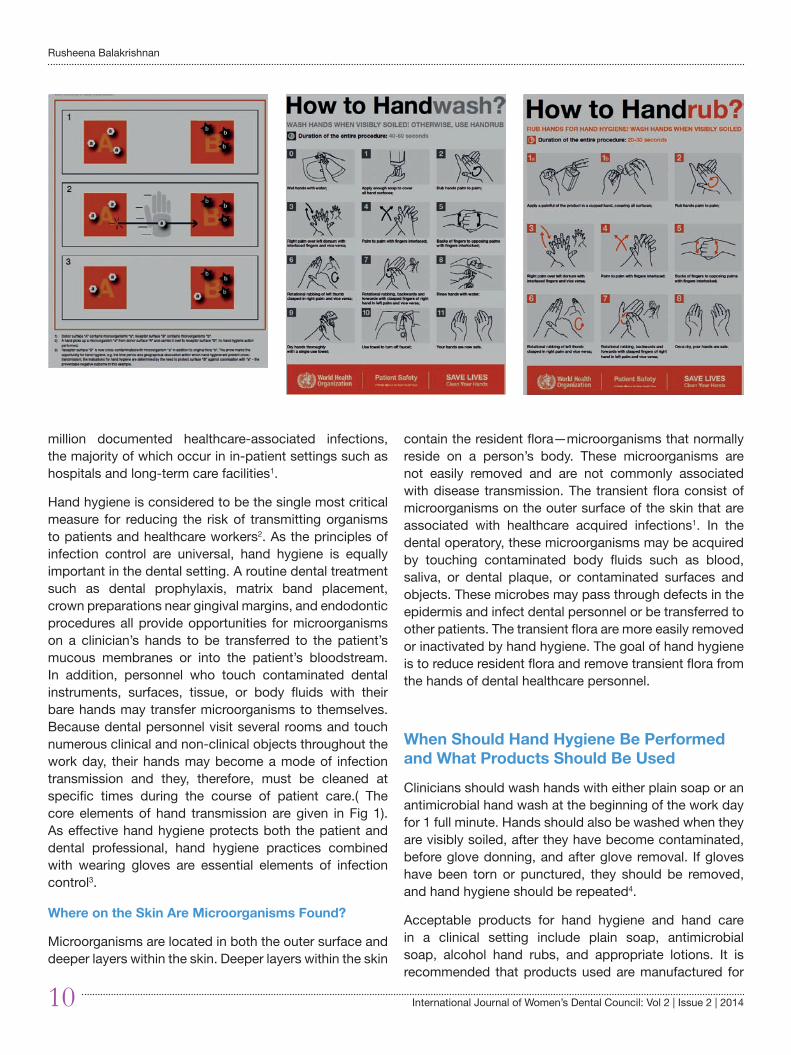
Hand hygiene is considered to be the single most critical measure for reducing the risk of transmitting organisms to patients and healthcare workers2. As the principles of infection control are universal, hand hygiene is equally important in the dental setting. A routine dental treatment such as dental prophylaxis, matrix band placement, crown preparations near gingival margins, and endodontic procedures all provide opportunities for microorganisms on a clinician’s hands to be transferred to the patient’s mucous membranes or into the patient’s bloodstream. In addition, personnel who touch contaminated dental instruments, surfaces, tissue, or body fluids with their bare hands may transfer microorganisms to themselves. Because dental personnel visit several rooms and touch numerous clinical and non-clinical objects throughout the work day, their hands may become a mode of infection transmission and they, therefore, must be cleaned at specific times during the course of patient care.( The core elements of hand transmission are given in Fig 1). As effective hand hygiene protects both the patient and dental professional, hand hygiene practices combined with wearing gloves are essential elements of infection control3.
Where on the Skin Are Microorganisms Found?
Microorganisms are located in both the outer surface and deeper layers within the skin. Deeper layers within the skin
contain the resident flora—microorganisms that normally reside on a person’s body. These microorganisms are not easily removed and are not commonly associated with disease transmission. The transient flora consist of microorganisms on the outer surface of the skin that are associated with healthcare acquired infections1. In the dental operatory, these microorganisms may be acquired by touching contaminated body fluids such as blood, saliva, or dental plaque, or contaminated surfaces and objects. These microbes may pass through defects in the epidermis and infect dental personnel or be transferred to other patients. The transient flora are more easily removed or inactivated by hand hygiene. The goal of hand hygiene is to reduce resident flora and remove transient flora from the hands of dental healthcare personnel.
When Should Hand Hygiene Be Performed and What Products Should Be Used
Clinicians should wash hands with either plain soap or an antimicrobial hand wash at the beginning of the work day for 1 full minute. Hands should also be washed when they are visibly soiled, after they have become contaminated, before glove donning, and after glove removal. If gloves have been torn or punctured, they should be removed, and hand hygiene should be repeated4.
Acceptable products for hand hygiene and hand care in a clinical setting include plain soap, antimicrobial soap, alcohol hand rubs, and appropriate lotions. It is recommended that products used are manufactured for
Rusheena Balakrishnan
International Journal of Women’s Dental Council: Vol 2 | Issue 2 | 2014 11
healthcare personnel, as these products are generally unscented, have fewer allergenic components, and are meant to be used repeatedly throughout the work day1. These products also contain emollients such as glycerin or aloe to soften hands and keep the epidermis intact.
Alcohol-based hand rubs kill microorganisms more effectively and more quickly than hand washing with soap and water5. They are also less damaging to skin, resulting in less dryness and irritation. In addition, they require less time, and since they may be placed at the point of care, they are also more accessible and may enhance compliance6. However, alcohol hand rubs do not physically remove debris from hands; thus these products should not be used if hands are visibly soiled.
Specific recommendations for hand hygiene products
Recommended products for hand hygiene are plain soap, antimicrobial soap, alcohol-based hand rubs surgical hand scrub or soap antisepsis and lotions.
Plain soap is recommended if hands are visibly soiled; before donning gloves and after glove removal; before eating; and after personal functions.
Antimicrobial soap is recommended: if hands are visibly soiled or contaminated with blood or other body fluids; before donning gloves and after glove removal; before eating; and after personal functions.
Alcohol-based hand rubs are recommended: if hands are not visibly soiled; and before donning gloves and after
glove removal.
Surgical hand scrub or soap antisepsis (either antimicrobial soap or a combination of non-antimicrobial soap, water, and alcohol-based surgical hand rub) is recommended before surgical procedures. Follow manufacturer’s instructions for quantity of product to be used.
Hand Hygiene In Dentistry
12 International Journal of Women’s Dental Council: Vol 2 | Issue 2 | 2014
Lotions The frequent use of lotions is suggested to ease the dryness resulting from frequent hand washing and to prevent dermatitis. Petroleum-based lotions can weaken latex and synthetic gloves and increase permeability and should not be used in a clinical setting. It is recommended to use products specifically manufactured for healthcare providers, as these are generally compatible with gloves and they contain fewer scents that may be offensive or allergenic to clinicians or patients7
How should hand hygiene be performed?
Although proper techniques may appear to be simply common sense, specific steps are required for hand washing in the dental setting (Figure 2)
• Wet hands completely with warm water. Extremely hot or cold water should be avoided, as temperature extremes may increase the risk of dermatitis.
• Rub hands together thoroughly for at least 15 seconds, making sure to cover all surfaces of the hands and fingers
• Rinse hands thoroughly• Dry hands thoroughly• Turn off faucets using a disposable towel to prevent
recontamination of hands8.
If using an alcohol-based hand rub, it should be applied to dry hands using the amount specified by the manufacturer 4 ( the correct hand rub procedure is described in Figure 3). Rub hands together, covering all surfaces and fingers, for at least 15 seconds until hands are dry. (If hands are dry after 10 seconds of alcohol-based hand rub usage, it is likely that too little of the product was used.) 9
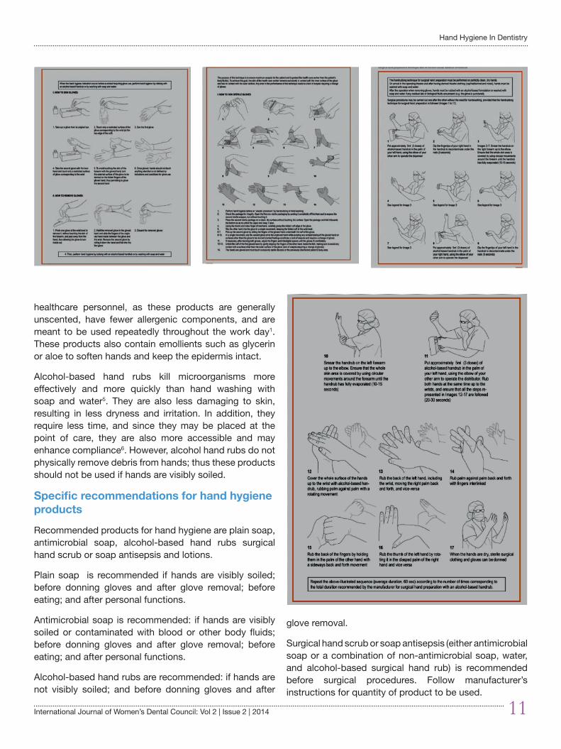
Surgical hand antisepsis is more technique-sensitive and elaborate than hand hygiene for routine dental procedures. Rings, bracelets, and all other hand and wrist jewellery must first be removed. Under running water, fingernails are cleaned to remove debris. Since bacteria on skin can multiply rapidly under gloves, it is highly recommended that surgical hand antisepsis be performed using products with “persistent activity.”Antimicrobial soaps or alcohol-based hand rubs with persistent activity prevent pathogens from surviving on hands for an extended period of time after application. Although over the-counter antimicrobial products may be purchased at virtually any store, clinicians should use products for surgical hand antisepsis (as well as products for routine dental care) that have been manufactured specifically for healthcare professionals1.
For surgical hand antisepsis using an antimicrobial soap, hands and forearms should be scrubbed together for the length of time recommended by the manufacturer of the product—typically 2 to 6 minutes. Hands and forearms should be rinsed and dried thoroughly before donning surgical gloves ( Figure 4 and figure 5 shows donning and removal of sterile gloves.)
For surgical hand antisepsis using an alcohol-based hand rub, hands and forearms should be prewashed with a non-antimicrobial soap and dried thoroughly. The hand-rub product should be applied following the manufacturer’s instructions for quantity of product to use (typically, a greater quantity than for routine hand hygiene), and hands and forearms should be allowed to dry thoroughly prior to donning sterile surgical gloves4.(Figure 6 a and Figure 6 b shows the correct technique of surgical hand preparation technique with an alcohol based hand rub formulation.)
Why Shouldn’t Jewellery or Artificial Nails Be Worn?
The effectiveness of hand hygiene can be reduced by both the presence of jewellery and artificial nails10. Long or artificial nails as well as rings make glove donning and removal more difficult and make glove tears more likely. Additionally, bacterial counts are higher on hands with long or artificial nails and nails with chipped polish11. Thus, it is recommended to keep fingernails unpolished and short with rounded, filed edges .Therefore, the wearing of hand jewellery while providing routine dental care is strongly discouraged; it is prohibited during surgical procedures.
Factors that affect the action of alcohol based hand rubs against microbes
Hand hygiene is the responsibility of the organization and all individuals involved in health care. Hand hygiene is a core element of client/patient/resident safety for the prevention of infections and the spread of antimicrobial resistance 12.
Alcohol based hand rub is the first choice for hand hygiene when hands are not visibly soiled 13,14. Alcohol based hand rub is less time consuming to use than washing with soap and water and is the most time effective protocol for routine client/patient/resident care15.
Hand hygiene is one of the most important ways to prevent the spread of infections, including the common cold and even hard to treat infections, such as methicillin resistant staphylococcus aureus (MRSA)16.
Rusheena Balakrishnan
International Journal of Women’s Dental Council: Vol 2 | Issue 2 | 2014 13
A number of factors such as type and concentration of alcohol, alcohol absorption, volume and drying time affect the action of alcohol based hand rubs against microbes.
Type of alcohol
Both isopropranol and ethanol have in vitro activity against bacteria, viruses and fungi. When tested at the same concentration, isopropranol is more efficacious than ethanol17. However ethanol has greater activity against viruses than iso propranol17,18.
Alcohol only alcohol based hand rub versus alcohol –chlorhexidine alcohol based hand rub
Although alcohols are rapidly germicidal when applied to the skin ,they have no appreciable persistent or residual activity. The addition of low concentration of chlorhexidine to an alcohol based hand rub results in greater residual activity than alcohol alone17,19 and thus improves its efficacy.
Alcohol concentration-there is a clear positive association between the extent of bacterial reduction and the concentration of alcohol contained in alcohol based hand rub products.
Furthermore, the concentration for maximum efficacy is different for iso-propranol than ethanol. For example –Alcohol based hand rub containing 60% iso-propranol is associated with similar cutaneous bactericidal activity as alcohol based hand rub that contain 77% ethanol 19.
Alcohol absorption-
The selection of an alcohol based hand rub maybe influenced by religious factors. According to some religions alcohol consumption is prohibited. Recent studies have demonstrated minimal rates of cutaneous alcohol absorption such that there should be no concern for health care workers20,21. An Australian study suggested that isopropnaol might be less likely to be absorbed than ethanol. This health care workers concerned about absorption for religious reasons may elect to use an alcohol based hand rub that contains isopropranol rather than ethanol20. An awareness of commonly held religious and cultural beliefs is vital when introducing new concepts to today’s multicultural healthcare community22.
Solution vs gel vs foam
Laboratory studies have found that alcohol based hand rub solutions are more effective than alcohol based hand rub gels that contain an equivalent concentration of alcohol23.
Alcohol based rub volume and drying time
The volume of hand rub dispensed is important . One ml of alcohol has been shown to be substabtially less effective than 3ml 24. The effective volume of alcohol based hand rubs( 2 to 3 ml;1 to 2 squirts from most alcohol based rub dispensers) generally takes 15-20 seconds to dry on hands-hence alcohol based hand rub drying time is a convenient indicator that sufficient alcohol based hand rub has been applied. It is important to follow the recommendations of the manufacturer which are usually found on the alcohol based hand rub bottle. In clinical practice often smaller volumes are used than what is recommended in the testing of alcohol based hand rubs. Unless high concentration products are used there is no significant reduction in contaminants with small volumes of alcohol based hand rubs25.
If hands are wet when alcohol based hand rub is applied-the antimicrobial efficacy of alcohol is very sensitive to dilution with water and is therefore vulnerable to inactivation, especially if only small volumes of Alcohol based rub are applied. For example if 60% isopropranol were rubbed onto wet hands in two portions of 3ml(each for one minute),the mean log bacterial reduction achieved is 3.7 as compared to 4.3 with dry hands 19. Thus it is recommended that Alcohol based hand rub be applied to dry hands.
A recent study comparing the three hand drying techniques (jet air,warm air dryer and paper towels) showed that jet air and warm air dryers resulted in increased bacterial aerosolization when drying hands than when compared to using paper towels. These results suggested that air dryers maybe unsuitable in healthcare settings as they may facilitate microbial cross –contamination via air borne dissemination to the environment or users 26.
Conclusion
The past 20 years have brought about changes in hand care recommendations and guidelines for healthcare professionals. Research and development of new products and techniques may seem to have complicated the hand hygiene process, but the fundamental principle remains: Hand hygiene is the single most important measure for reducing the risk of healthcare-associated infections. Contaminated hands continue to be a mode of infection transmission during patient care, and effective hand hygiene practices protect both patients and team members. Although many products are available for hand care, it is recommended that healthcare workers use products that are manufactured for use in healthcare
Hand Hygiene In Dentistry
14 International Journal of Women’s Dental Council: Vol 2 | Issue 2 | 2014
settings. All dental team members should be educated on the importance of proper hand hygiene, as the first critical step to infection control is definitely hand hygiene27.
References 1. Andrews N, Cuny E, Molinari JA. Antisepsis and hand hygiene. In:
Molinari JA, Harte JA, eds. Cottone’s Practical Infection Control in Dentistry.3rd ed. Philadelphia, PA: Lippincott, Williams, and Wilkins; 2010:125.
2. Kohn WG, Collins AS, Cleveland JL, et al. Guidelines for infection control in dental health-care settings—2003. MMWR Recomm Rep. 2003;52(RR-17):14.
3. Girou E, Chai SH, Oppein F, et al. Misuse of gloves: the foundation for poor compliance with hand hygiene and potential for microbial transmission? J Hosp Infect. 2004; 57(2):162-169.
4. Boyce JM, Pittet D. Guideline for Hand Hygiene in Health-Care Settings. Recommendations of the Healthcare Infection Control Practices Advisory Committee and the HICPAC/SHEA/APIC/IDSA Hand Hygiene Task Force. Society for Healthcare Epidemiology of America/Association for Professionals in Infection Control/Infectious Diseases Society of America. MMWR Recomm Rep. 2002;51(RR-16):1-45.
5. Kampf G, Kramer A. Epidemiologic background of hand hygiene and evaluation of the most important agents for scrubs and rubs. Clin Microbiol Rev. 2004; 17(4):863-893.
6. Erasmus V, Daha TJ, Brug H, et al. Systematic review of studies on compliance with hand hygiene guidelines in hospital care. Infect Control Hosp Epidemiol. 2010; 31(3):283-894.
7. Littau, CA, Thompson KM. Keep consumer hand lotions at home. American Nurse Today 2011; 6(4). Page no
h t t p : / / w w w . a m e r i c a n n u r s e t o d a y . c o m / a r t i c l e .aspx?id=7700&fid=7658 accessed July 12, 2013.
8. World Health Organization. WHO Guidelines on Hand Hygiene in Health Care. Geneva, Switzerland: World Health Organization; 2009. http://whqlibdoc.who.int/publications/2009/9789241597906_eng.pdf. Accessed July 12, 2013.
9. Molinari JA. Clinic experiences with waterless alcohol-based hand hygiene antiseptics. Compend Contin Educ Dent. 2006;27(2):84-86.
10. Hedderwick SA, McNeil SA, Lyons MJ, Kauffman CA. Pathogenic organisms associated with artificial fingernails worn by healthcare workers. Infect Control Hosp Epidemiol. 2000;21(8):505-509.
11. Gupta A, Della-Latta P, Todd B, et al. Outbreak of extended-spectrum beta-lactamase-producing Klebsiella pneumoniae in a neonatal intensive care unit linked to artificial nails. Infect Control Hosp Epidemiol.2004;25(3):210-215.
12. Best practices for hand hygiene in all health care settings,4th Edition,2014. Provisional Infectious Diseases Advisory Committee (PIDAC). Public Health Ontario Website: www.publichealthontario.ca
13. Public Health Agency of Canada. Hand Hygiene Practices in Healthcare Settings Ottawa: Centre for Communicable Diseases and Infection Control; 2012 [cited May 6,2013]. Available from:
http://www.chica.org/pdf/2013_PHAC_Hand%20Hygiene-EN.pdf.
14. Boyce JM, Pittet D. Guideline for Hand Hygiene in Health-Care Settings. Recommendations of the Healthcare Infection Control Practices Advisory Committee and the HICPAC/SHEA/APIC/IDSA Hand Hygiene Task Force. Infect Control Hosp Epidemiol. 2002 ;23(12 Suppl):S3-40.
15. Chow A, Arah OA, Chan SP, Poh BF, Krishnan P, Ng WK, et al. Alcohol handrubbing and chlorhexidine handwashing protocols for routine hospital practice: a randomized clinical trial of protocol efficacy and time effectiveness. Am J Infect Control. 2012 ;40(9):800-5.
16. Hand hygiene saves lifes, a patients guide.centre for disease control–www.cdc.gov/handhygiene(cited august 7,2014).
17. World Health Organisation. WHO Guidelines on Hand Hygiene in Health Care. In: World Alliance for Patient Safety, editor. First Global Patient Safety Challenge Clean Care is Safer Care. 1 ed. Geneva: World Health Organisation Press; 2009.
18. Kampf G, Kramer A. Epidemiologic background of hand hygiene and evaluation of the most important agents for scrubs and rubs. Clin Microbiol Rev. 2004;17(4):863-93
19. Rotter M. Hand Washing and Hand Disinfection. In: Mayall C, editor. Hospital Epidemiology and Infection Control. Philadelphia, PA: Lippincott, Williams & Wilkins; 1999. pages. 1339-55.
20. Brown TL, Gamon S, Tester P, Martin R, Hosking K, Bowkett GC, et al. Can alcohol-based hand-rub solutions cause you to lose your driver’s license? Comparative cutaneous absorption of various alcohols. Antimicrobial Agents & Chemotherapy. 2007 51(3):1107-8.
21. Kramer A, Below H, Bieber N, Kampf G, Toma CD, Huebner N-O, et al. Quantity of ethanol absorption after excessive hand disinfection using three commercially available hand rubs is minimal and below toxic levels for humans. BMC Infectious Diseases. 2007;7:117.
22. Allegranzi B, Memish ZA, Donaldson L, Pittet D. Religion and culture: potential undercurrents influencing hand hygiene promotion in health care. Am J Infect Control. 2009 ;37(1):28-34.
23. Kramer A, Rudolph P, Kampf G, Pittet D. Limited efficacy of alcohol-based hand gels. Lancet. 2002 ; 27;359(9316):1489-90.
24. Larson EL, Eke PI, Wilder MP, Laughon BE. Quantity of soap as a variable in handwashing. Infection Control: IC. 1987;8(9):371-5.
25. Kampf G. How effective are hand antiseptics for the post contamination treatment of hands when used as recommended? Am J Infect Control. 2008;36(5):356-60.
26. E.L.Best,P. Parnell, M.H.Wilcox et al.Microbial comparison of hand drying methods:the potential for contamination of the environment user and bystander. Journal of hospital infection.2014 ; 88(4): 199-206.
27. Canham L.The first step in infection control is hand hygiene.Dent assist 2011 ;80(1):42-46.PMID=21443078.
Rusheena Balakrishnan
International Journal of Women’s Dental Council: Vol 2 | Issue 2 | 2014 15
Role of BCL-2 in oral carcinogenesis: a review
Saurabh Juneja*Manjushree Juneja**N. Chaitanya Babu***
ABSTRACT
Evolution of normal oral mucosa into varying grades of dysplasia and invasive oral squamous cell carcinoma(OSCC) involves a series of compound changes at the molecular level which causes progressive loss of cell cycle control and alteration in cell maturation profi le. One of such alterations involves evasion of apoptosis as a mechanism of survival by the cancer cells. The most important member of the apoptotic pathway is the Bcl-2 gene superfamily which encodes for both pro-apototic (Bax and Bad) as well as anti-apoptotic proteins (Bcl-2 and Bcl- XL). Bcl-2 acts in harmony with other genes such as p53 in regulating the normal cell cycle progression and cell death pathway. Bcl-2 oncoprotein is one of the major proteins responsible for the cancer cells to shun apoptosis and acquire increased life span. The increased survival of these cells alters the maturation pattern of the cells and allows additional genetic mutations to accumulate responsible for progression of these cells from normal to dysplastic and further into invasive and metastatic carcinoma. Increased expression of Bcl-2 oncoprotein is seen in oral epithelial dysplasia and both early and late stages of the OSCC in varying clinical and histopathological grades and can be used as a biomarker in understanding the progression of carcinogenesis from early to late stage. It can also be utilized to prioritize the treatment needs and follow up of oral epithelial dysplasia and OSCC patients.Key Words: Apoptosis; Bcl-2; Oral cancer; Oral Squamous cell carcinoma
ADDRESS FOR CORRESPONDENCE
Dr. Saurabh Juneja, MDSSenior lecturer, Department of Oral PathologyI.T.S. Dental College, Murad Nagar, Ghaziabad, Uttar Pradesh.PIN- 201206Email ID: [email protected] no. +91 9540331189
* Senior Lecturer, Department of Oral Pathology, I.T.S. Dental College, Murad Nagar, Ghaziabad, Uttar Pradesh - 201206; ** Senior Lecturer, Department of Oral Medicine and Radiology, School of Dental Sciences, Sharda University, Greater Noida, Uttar Pradesh - 201306; *** Professor and Head, Dept of Oral Pathology, The Oxford Dental College, Bangalore, Karnataka - 560 064
Introduction
Apoptosis is a genetically programmed type of cell death which is responsible for maintenance of equilibrium in the cells multiplying and dying at the tissue level by eliminating cells which have become senescent or genetically altered.1 Apoptosis was fi rst described in liver cells by Kerr et al. in 1972.2 A balance of cell population is maintained
by equilibrium between cell lost by apoptosis and cells replenished by mitosis.3,4 The apoptotic pathway involves activation of diverse pro-apoptotic signals, which lead to a common pathway driven by a unique family of cysteine proteases (caspases) which is negatively controlled by Bcl-2 family of genes which are the primary regulators of this process of apoptosis.5
IJWDC: Vol 2 | Issue 2 | July-December 2014 Review Article
Evolution of normal oral mucosa into varying grades of dysplasia and invasive oral squamous cell carcinoma(OSCC) involves a series of compound changes at the molecular level which causes progressive loss of cell cycle control and alteration in cell maturation profi le. One of such alterations involves evasion of apoptosis as a mechanism of survival by the cancer cells. The most important member of the apoptotic pathway is the Bcl-2 gene superfamily which encodes for both pro-apototic (Bax and Bad) as well as anti-apoptotic proteins (Bcl-2 and Bcl- XL). Bcl-2 acts in harmony with other genes such as p53 in regulating the normal cell cycle progression and cell death pathway. Bcl-2 oncoprotein is one of the major proteins responsible for the cancer cells to shun apoptosis and acquire increased life span. The increased survival of these cells alters the maturation pattern of the cells and allows additional genetic mutations to accumulate responsible for progression of these cells from normal to dysplastic and further into invasive and metastatic carcinoma. Increased expression of Bcl-2 oncoprotein is seen in oral epithelial dysplasia and both early and late stages of the OSCC in varying clinical and histopathological grades and can be used as a biomarker in understanding the progression of carcinogenesis from early to late stage. It can also be utilized to prioritize the treatment needs and follow up of oral epithelial dysplasia and OSCC patients.Key Words: Apoptosis; Bcl-2; Oral cancer; Oral Squamous cell carcinoma
16 International Journal of Women’s Dental Council: Vol 2 | Issue 2 | 2014
Bcl-2 family of proteins:The Bcl-2 family of related proteins controls the apoptotic pathway through complex mechanisms. The prototype member of this family is Bcl-2 (derived from B-cell lymphoma/leukemia-2 gene) that was first discovered at the breakpoint of the t(14;18) in a follicular non-Hodgkin’s B-cell lymphoma which leads to overproduction of Bcl-2 messenger RNA and protein.6
Apoptosis is known to proceed through different pathways with a common terminal pathway, primarily regulated by Bcl-2 family of proteins.7 The Bcl-2 family of proteins consist of two groups of variety of proteins, one of which promote apoptosis (death agonists e.g.Bcl-10, Bax, Bak, Bid, Bad, Bim, Bik, and Blk) and others which oppose apoptosis (death antagonists e.g. Bcl-2, Bcl-x, Bcl-XL, Bcl-XS, Bcl-w, BAG) which differ in their activation dependent expression patterns as well as in their structure.1,8,9 The fate of a cell and its ability to survive and susceptibility to death is determined by the balance between the gene products of the Bcl-2 gene family.10 The Bcl-2 protooncogene product is a 26 kDa protein encoded in chromosome 18q21 which is a component of the nuclear envelope, endoplasmic reticulum and the outer mitochondrial membrane.11
Mechanism of Bcl-2 action:A functionally significant characteristic of Bcl-2 family of proteins is their ability to form homo- and hetero-dimers owing to various combinations of BH domains (Bcl-2 homolgy). Anti-apoptotic members are also present as integral membrane proteins found in the mitochondria, endoplasmic reticulum or nuclear membrane while few of the pro-apoptotic members localize to cytosol or cytoskeleton.8
The pathway of anti-apoptotic action of Bcl-2 is complex and still unclear. The most common pathway considered for Bcl-2 to prevent a cell from undergoing apoptosis by blocking a step which leads to the activation of caspases. Another mechanism by which Bcl-2/Bcl-xL prevents the activation of caspases is through their abilities to sequester pro-caspases.5 One of the suggested mechanisms is by altering the mitochondrial membrane permeability by altering the balance between calcium and cytochrome c levels. It also prevents the release of proapoptotic factors such as AIF (apoptosis-inducing factor) from the mitochondria into the cytoplasm. It is further supported by the presence of Bcl-2 in areas of contact between the outer and the inner mitochondrial membranes.5,6,9 Hockenberry et al. have reported Bcl-2 to possess an anti-oxidant function which has been suggested as a possible pathway to block apoptosis in cells exposed to
gamma irradiation.12
Bcl-2 and tumorigenesis:
One of the mechanisms of cancer cells to survive and sustain is through evasion and escapement of apoptosis. It is achieved by imbalance produced by either inactivation of genes favouring apoptosis or by upregulation of gene expression responsible for inhibiting apoptosis.9,13 The Bcl-2 family of genes is a key member regulating the apoptosis and modulation of cell cycle regulating proteins emphasising its primary role in controlling the processes of cell death and proliferation.10
The concept of evasion of apoptosis and progression of cancer evolved with discovery of action of Bcl-2 and its related gene products. It was established that cancer emerged when inhibition of cell death by Bcl-2 provides a favourable environment for cancer cells to survive, rather than promote proliferation as was earlier thought to be the only way for cancer cells to progress. Thus, it is now well accepted that impaired apoptosis is a crucial step in progression of tumorigenesis. Escaping the apoptosis allows the cells to persist in unfavourable environments and evolve into more aggressive derivatives. Defective apoptosis also facilitates metastasis, because the cells can ignore restraining signals from neighbours and survive detachment from the extracellular matrix. So, neoplastic progression actually reflects loss of normal apoptotic mechanisms.14,15
Bcl-2 disrupts the process of apoptosis both in the initial and final phases because this protein not only stabilizes the potential of the mitochondria membrane when forming heterodimers with bax but also inhibits the formation of oxygen-reactive species and intracellular acidification.16
Overexpression of Bcl-2 allows additional pro-carcinogenic mutations to accumulate within the cell through extended cell survival.12 It leads to immortalization of neoplastic cells and hinders removal of genetically altered cells thereby, enabling their clonal expansion as a result of defective cell death mechanism.17,18,19
Increased expression of the Bcl-2 protein can be detected in about 50% of human cancers, further emphasizing the importance of deregulating apoptosis as a fundamental step in human carcinogenesis. By promoting cell survival, Bcl-2 facilitates the permanent acquisition of mutations and malignant transformation.17,18,19 Additionally, overexpression of Bcl-2 protein in cancer cells has been linked to resistance of tumor cells to apoptosis and may have implications for their therapeutic responsiveness.20 An alteration in ratio of pro-apoptotic to anti-apoptotic
Saurabh Juneja

International Journal of Women’s Dental Council: Vol 2 | Issue 2 | 2014 17
proteins promotes tumor progression and has been cited to increased resistance to cytotoxic therapies such as chemotherapy and radiation.21
Bcl-2 in Oral carcinogenesis:Carcinogenesis of oral squamous cell carcinoma(OSCC) involves an imbalance between activity of oncogenes and lack of tumour suppression by the tumor suppressor genes leading to disparity between cell death and proliferation.22 OSCC arises as a result of multiple molecular events that develop from the combined influences of an individual’s genetic predisposition and exposure to environmental carcinogens causing accumulation of such genetic alterations and development of premalignant lesions and subsequent invasive carcinoma.21
In normal proliferating epithelium, Bcl-2 is expressed in stem cell zones such as the basal layers, where it acts to prevent the death of cells in the regenerative compartment.23 In oral cancer there are inconsistent changes in the level of the Bcl-2 family of proteins. Although expression of Bcl-2 can be seen, it is not apparent in every case and not in every area of each tumor.29,30,31 Invasive oral cancers of patients in India, who use betel quid and tobacco, have high levels of p53 and Bcl-2 in their dysplastic oral mucosa.25
Stronger expression of Bcl-2 oncoprotein has been observed in poorly differentiated OSCC as compared to well differentiated OSCC.19,27 The increased Bcl-2 expression in poorly differentiated carcinomas may reflect the loss of ability of malignant keratinocytes to differentiate terminally. Harada et al. have shown that Bcl-2 inhibits the differentiation of cultured keratinocytes in an in vitro experiment, thereby indicating the role of Bcl-2 in differentiation of these tumour cells.28 Singh et al. have shown that the cells peripherally located within infiltrating tumor nests are more intensely stained, while fully keratinized neoplastic cells show diminished or absence of Bcl-2 immunoreactivity which might be attributed to down-regulation of Bcl-2 expression concomitant with terminal cell differentiation (keratinization).29
Bcl-2 oncoprotein in histologically proven cases of dysplasia have shown a proportional increase in Bcl-2 oncoprotein corresponding to increasing grades of dysplasia.34 Increased Bcl-2 expression was observed in the cytoplasm of basal cells of dysplastic epithelium adjacent to the tumour epithelium which raises the possibility that Bcl-2 alteration may precede early invasive tumor development.19,20,30
Studies involving oral tissues have yielded contrasting results with few authors suggesting an important role of
Bcl-2 in early stages of oral tumour progression11,31,32,33 whereas others reporting an infrequent or lack of expression of Bcl-2 in oral dysplastic lesions10 and OSCC.26 These results suggest that molecular alteration in Bcl-2 may not be the only genetic change occurring in oral dysplastic and invasive tumor cells but may be one of the many mutations responsible for progression of oral epithelial tumours.
Chen et al. have proposed that post-transcriptional regulation could be a possible mechanism controlling the expression of bcl-2 (and Bax) in oral carcinomas.19 It has been reported that large C-terminal fragments with potent pro-apoptotic activity are produced through cleavage by various proteases that cleave Bcl-2 family proteins.25 Cheng et al. have suggested that there is reversal in the function of Bcl-2 oncoprotein from antiapoptotic to a proapoptotic protein due to mutations in the BH4 domain of Bcl-2 gene.34
Previous studies have also analysed the correlation between clinical, histologic, and molecular markers in OSCC with disease outcome, with differing results. Camisasca et al.35 have shown Bcl-2 expression to be an independent marker of favourable cancer specific 5-year survival whereas de Vicente et al36 and Popovic et al.37 have shown its association with a poor prognosis. This could be explained by the complexity of the process of oral carcinogenesis and variations in study settings such as variable small sample sizes or heterogeneity of the selected subjects, which frequently differ in important features, notably tumor location, treatment modality, and TNM stage.35
Conclusion: The upregulation of expression of bcl-2 makes the removal of genetically modified cells hard, favouring the accumulation of new mutations, which can result in the appearance of cells with malignant phenotype. Increased expression of Bcl-2 oncoprotein may be an early genetic change in oral carcinogenesis and may be used as a potential biomarker in predicting the biological behaviour of dysplastic lesions and invasive oral squamous cell carcinoma. However, further molecular studies emphasising analysis of the mRNA and cleaved protein products to study the transcriptional and post translational regulation of Bcl-2 in future may elucidate the enigmatic role and mechanism of Bcl-2 action in oral carcinogenesis and disease outcome.
References: 1. Coutinho-Camillo CM, Lourenço SV, Nishimoto IN, Kowalski LP,
Soares FA. Expression of Bcl-2 family proteins and association
Role of bcl-2 in oral carcinogenesis: a review
18 International Journal of Women’s Dental Council: Vol 2 | Issue 2 | 2014
with clinicopathological characteristics of oral squamous cell carcinoma. Histopathology 2010;57(2):304–316.
2. Kerr JFR, Wyllie AH, Currie AR. Apoptosis: a basic biological phenomenon with wide-ranging implications in tissue kinetics. Br J Cancer 1972;26:239-57.
3. Bloor BK, Malik FK, Odell EW, Morgan PR. Quantitative assessment of apoptosis in oral lichen planus. Oral Surg Oral Med Oral Pathol Oral Radiol Endod 1999;88(2):187-95.
4. Bunek J, Kamarajan P, Kapila YL. Anoikis mediators in oral squamous cell carcinoma. Oral Dis 2011;17(4):355-61.
5. Tsujimoto Y. Role of Bcl-2 family proteins in apoptosis: apoptosomes or mitochondria? Genes Cells 1998;3(11):697-707.
6. Polverini PJ, Nor JE. Apoptosis and predisposition to oral cancer. Crit Rev Oral Biol Med 1999;10:139–52.
7. Chao DT, Korsmeyer SJ. Bcl-2 family: Regulators of cell death. Annu Rev Immunol 1998;16:395-419.
8. Gross A, McDonnell JM, Korsmeyer SJ. BCL-2 family members and the mitochondria in apoptosis. Genes Dev 1999;13(15):1899-911.
9. Elmore S. Apoptosis: A review of programmed cell death. Toxicol Pathol 2007;35(4):495-516.
10. Loro LL, Johannessen AC, Vintermyr OK. Decreased expression of bcl-2 in moderate and severe oral epithelia dysplasias. Oral Oncol 2002;38(7):691-8.
11. Kannan K, Latha PN, Shanmugam G. Expression of bcl-2 oncoprotein in Indian oral squamous cell carcinomas. Oral Oncol 1998;34(5):373-6.
12. Hockenbery D, Oltvai Z, Yin X-M, Millman C, Korsmeyer Sl. Bcl-2 functions in an antioxidant pathway in apoptosis. Cell 1993;75:241-251
13. Loro L, Vintermyr OK, Johannessen AC. Apoptosis in normal and diseased oral tissues. Oral Dis 2005;11(5):274-87.
14. Korsmeyer SJ. Bcl-2 initiates a new category of oncogenes: regulators of cell death. Blood 1992;80(4):879-86.
15. Cory S, Adams JM. The Bcl2 family: Regulators of the cellular life-or-death switch. Nat Rev Cancer 2002;2(9):647-56.
16. Van Der Heiden MG, Thompson CB. Bcl-2 proteins: regulators of apoptosis or of mitochondrial homeostasis? Nat Cell Biol 1999;1: 209-16.
17. Vaux DL, Cory S, Adams JM. Bcl-2 genes promote haemapoietic cell survival and cooperate with C-myc to immortalize pre-B cells. Nature 1998;335:440–2.
18. Renan MJ. How many mutations are required for tumor genesis? Implications from human cancer data. Mol Carcinog 1993;7:139–46.
19. Chen Y, Kayano T, Takagi M. Dysregulated expression of bcl-2 and bax in oral carcinomas: evidence of posttranscriptional control. J Oral Pathol Med 2000;29(2):63-9.
20. Kummoona R, Mohammad Sámi S, Al-Kapptan I, Al-Muala H. Study of antiapoptotic gene of oral carcinoma by using Bcl-2 oncogene. J Oral Pathol Med 2008;37(6):345-51.
21. Choi S, Myers JN. Molecular Pathogenesis of Oral Squamous Cell Carcinoma: Implications for Therapy. J Dent Res 2008;87:14-32.
22. Zhang M, Zhang P, Zhang C, Sun J, Wang L, Li J, Tian Z, Chen W. Prognostic significance of Bcl-2 and Bax protein expression in the patients with oral squamous cell carcinoma. J Oral Pathol Med 2009;38(3):307-13.
23. Piattelli A, Rubini C, Fioroni M, Iezzi G, Santinelli A. Prevalence of p53, bcl-2, and Ki-67 immunoreactivity and of apoptosis in normal oral epithelium and in premalignant and malignant lesions of the oral cavity. J Oral Maxillofac Surg 2002;60(5):532-40.
24. Schoelch ML, Le QT, Silverman S Jr, McMillan A, Dekker NP, Fu KK, Ziober BL, Regezi JA. Apoptosis-associated proteins and the development of oral squamous cell carcinoma. Oral Oncol. 1999;35(1):77-85.
25. Ravi D, Nalinakumari KR, Rajaram RS, Nair MK, Pillai MR. Expression of programmed cell death regulatory p53 and bcl-2 proteins in Oral lesions. Cancer Lett 1996;105:139-46.
26. Loro LL, Vintermyr OK, Liavaag PG, Jonsson R, Johannessen AC. Oral squamous cell carcinoma is associated with decreased bcl-2/bax expression ratio and increased apoptosis. Hum Pathol 1999;30(9):1097-105.
27. Sulkowska M, Famulski W, Sulkowski S, Reszeć J, Koda M, Baltaziak M, Kańczuga-Koda L. Correlation between Bcl-2 protein expression and some clinicopathological features of oral squamous cell carcinoma. Pol J Pathol 2003;54(1):49-52.
28. Harada H, Mitsuyasu T, Seta Y, Maruoka Y, Toyoshima K, Yasumoto. Overexpression of bcl-2 protein inhibits terminal differentiation of oralkeratinocytes in vitro. J Oral Pathol Med 1998;27:11–7.
29. Singh BB, Chandler FW, Whitaker SB, Forbes-Nelson AE. Immunohistochemical evaluation of bcl-2 oncoprotein in oral dysplasia and carcinoma. Oral Med Oral Pathol Oral Radiol Endod 1998;85(6):692-698.
30. Yao L, Iwai M, Furuta I. Correlations of bcl-2 and p53 expression with the clinicopathological features in tongue squamous cell carcinomas. Oral Oncol 1999;35(1):56-62.
31. Tanda N, Mori S, Saito K, Ikawa K, Sakamoto S. Expression of apoptotic signaling proteins in leukoplakia and oral lichen planus: quantitative and topographical studies. J Oral Pathol Med 2000;29(8):385-93.
32. Krajewski S, Chatten J, Hanada M, Reed JC. Immunohistochemical analysis of the Bcl-2 oncoprotein in human neuroblastomas comparisons with tumour cell differentiation and N-myc protein. Lab Invest 1995;72:42-54.
33. Tsujimoto Y, Croce CM. Analysis of the structure, transcripts and protein products of bcl-2, the gene involved in human follicular lymphoma. Proceedings of National Academy of Sciences U.S.A. 1986;83:5214-18.
34. Cheng EH, Kirsch DG, Clem RJ, et al. Conversion of Bcl-2 to a Bax-like death effector by caspases. Science 1997;278:1966–8.
35. Camisasca DR, Honorato J, Bernardo V, da Silva LE, da Fonseca EC, de Faria PA, Dias FL, Lourenço Sde Q. Expression of Bcl-2 family proteins and associated clinicopathologic factors predict survival outcome in patients with oral squamous cell carcinoma. Oral Oncol 2009;45(3):225-33.
36. de Vicente JC, Olay S, Lequerica-Fernandez P, Sanchez-Mayoral LM, Junquera LM, Fresno MF. Expression of Bcl-2 but not Bax has a prognostic significance in tongue carcinoma. J Oral Pathol Med 2006;35(3):140–5.
37. Popovic B, Jekic B, Novakovic I, Lukovic LJ, Tepavcevic Z, Jurisic V, et al. Bcl-2 expression in oral squamous cell carcinoma. Ann NY Acad Sci 2007;1095(1):19–25.
Saurabh Juneja
International Journal of Women’s Dental Council: Vol 2 | Issue 2 | 2014 19
Advanced diagnostic aids in periodontology
Saurab Kishore*Teenu Abraham**Devisree**Midhulaj. A***Raju Kurien****T.P. Padmakumar***** K.Nandakumar******
ABSTRACT
Diagnosis is an important part in assess periodontal diseases. An accurate diagnosis is mandatory for proper analysis of prognosis and treatment plan. The existing modes of diagnosis is effective in evaluating the history of the disease activity. Hence there is a need of developing diagnostic aids thatcan notonly give a picture of current disease activity but also the future progresson of the disease. This review discusses a few of the recent diagnostic procedures.
ADDRESS FOR CORRESPONDENCE
Dr. Teenu AbrahamSenior Lecturer, Dept of Periodontology, Azeezia College of Dental Science and Research, Meeyanoor, Kollam - 691537 Email:[email protected]
* PG student, ** Senior Lecturer, ***Director, Senior Lecturer, **** Reader, *****Professor, ******Principal,Prof & HOD, Dept of Periodontology, Azeezia College of Dental Science and Research, Meeyanoor, Kollam
Introduction
Traditionally diagnosis of periodontitis is clinically made by measuring either the loss of connective tissue attachment to the root surface ( clinical attachment loss ) or the loss of alveolar bone with the help of a radiograph. Thus the disease can only be evaluated at one point in time, by identifying and quantifying the clinical signs present at that time. Moreover, the evaluation cannot reliably identify the sites which are with ongoing periodontal destruction. The conventional diagnostic methods will not provide any information regarding, the cause of the condition, on the patient’s susceptibility to disease. Neither they provide data about the progression nor data about the remission of disease. Also they can’t provide information about the effect of therapy on periodontal disease.
Advantages of current routine diagnostic methods
v They can be performed easily, with minimum equipment and effort and is inexpensive also.
v Most of the patients can be treated adequately during routine dental practice.
v Epidemiological surveys can be carried out swiftly and the results are usually true representation of the periodontal status of the population.
v They provide retrospective information about the disease process reasonably well.
Disadvantages of current routine diagnostic methods
v Accurate measurements may not be obtained and may mislead the clinician.
v Full mouth recording is required as the disease is site specifi c.
v Due to the infl uence of other conditions, great variation in the individual susceptibility of periodontitis occurs and the current methods fail to determine them. Hence cannot determine etiology exactly.
v There are no reliable markers for disease activity.
v Only large changes occurring can be appreciated.
v The data obtained are vulnerable to inter and intra examiner errors.
IJWDC: Vol 2 | Issue 2 | July-December 2014 Review Article
Diagnosis is an important part in assess periodontal diseases. An accurate diagnosis is mandatory for proper analysis of prognosis and treatment plan. The existing modes of diagnosis is effective in evaluating the history of the disease activity. Hence there is a need of developing diagnostic aids thatcan notonly give a picture of current disease activity but also the future progresson of the disease. This review discusses a few of the recent diagnostic procedures.
20 International Journal of Women’s Dental Council: Vol 2 | Issue 2 | 2014
v It is difficult to determine the prognosis accurately and hence to arrive at a proper treatment plan.
v There is great variability in the interpretation of findings.
v There are no reliable criteria to identify individuals/sites at risk.
A universally accepted classification system is absent1.
So the future efforts or advances in diagnosis is likely to identify the factors and conditions that place the periodontium at risk for future attachment loss and this in turn will help to focus on diagnosing patients more likely to experience disease progression.
This article will provide a brief review of advances in current diagnostic aids used in periodontics encompassing advances in clinical, radiographic, microbiologic methods.
Advances in clinical diagnosis
Periodontal diagnosis and monitoring rely upon clinical parameters to a large extent. Clinical diagnosis directly affects decisions to initiate therapy, to select methods, and to outline the topographical area of application. We can also evaluate the outcome of therapy, and attempt long-term prognosis based on clinical parameters.
Gingival Bleeding: It is associated with persistent presence of plaque on the teeth and is regarded as a sign of the associated inflammatory response. The use of gingival bleeding as an indicator of inflammation is clinically more advantageous as it is more objective. Clinicians use gingival bleeding as an indicator of gingivitis instead of using visual signs of both inflammation and bleeding. Though, its relation to the progression is unclear, some investigators suggested that gingival bleeding is also an indicator of disease activity1
Limitations: Any force greater than 0.25N may evoke bleeding in healthy sites where there is intact periodontium (Lang et al). Also, long-standing use of tobacco in heavy smokers may mask the inflammatory signs of gingivitis and periodontitis and hence BOP may be negative.
Gingival Temperature: Researchers attempted to use subgingival temperature as a measure of periodontal inflammation. PerioTemp probe (ABIO-DENT, Inc, Danvers, MA, USA) is one commercially available system used to measure subgingival temperature. It was found
that elevated mean subgingival temperature was related to subsequent attachment loss.
Periodontal Probing : it is the most widely used diagnostic tool for the clinical assessment of connective tissue destruction in periodontitis. but conventional periodontal probes have certain limitations such as, the precise location of the probe tip depends on the degree of inflammation of the underlying connective tissues. Also disparity between the measurements also depends on the probing technique, probing force, size of the probe, angle of insertion, and precision of the probe calibration. So inorder to overcome these limitations newer generations of periodontal probes have been developed. Till date, 5 generations of probes have been developed with the 5th generation using ultrasonography. It is non invasive and probing is painless. It detect, image and map the upper boundary of periodontal ligament. But needs extensive training and is expensive.
Advances in radiographic techniques
Conventional radiographs cannot depict accurately the bone morphology at buccal and lingual surfaces. Only when substantial volumes of alveolar bone has destroyed, the loss is detectable in conventional radiograph. So advanced technologies are implemented in past years, which include; Digital radiography, Subtraction radiography including Diagnostic Subtraction Radiography (DSR), Computer Assissted Densitometric Image Analysis (CADIA), Computed tomography (CT) scan, Tuned aperture computed tomography (TACT), Cone Beam CT (CBCT), Digital volume tomography (DVT), Local CT (LCT), Optical coherence tomography (OCT) .
Advances in microbiologic analysis
There are different methods for detecting bacteria in dental plaque which include bacterial culture, immunologic assays, enzymatic assays, and molecular biologic techniques that detect bacterial DNA or RNA. Among all, the gold standard against which new microbial tests can be compared is bacterial culture . Immunologic methods use antibodies that target specific bacterial antigens. When the antibodies bind their antigen, the reaction can be visualized by techniques such as direct and indirect immunofluorescent microscopic assays, flow cytometry, and enzyme-linked immunosorbent assay. Immunologic techniques enable the identification and quantification (or semiquantification) of bacteria. Several putative periodontal pathogens such as Porphyromonas gingivalis, Tannerella forsythia, and Aggregatibacter Actinomycetemcomitans possess in common a trypsin
Saurab Kishore
International Journal of Women’s Dental Council: Vol 2 | Issue 2 | 2014 21
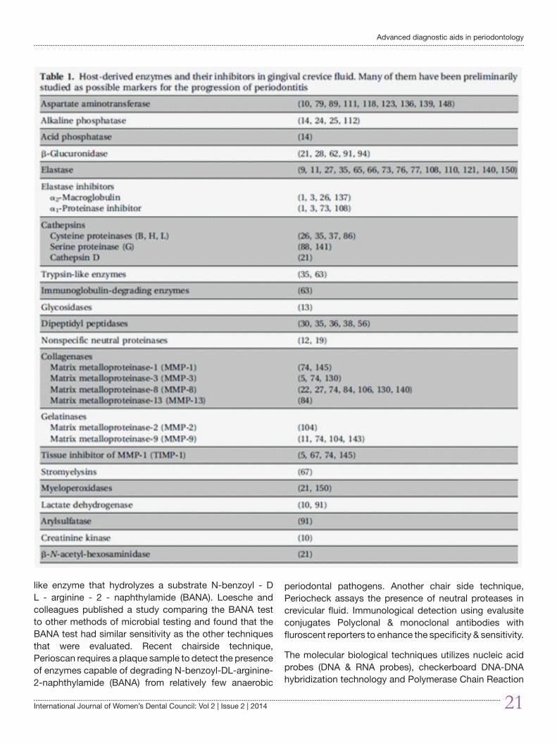
like enzyme that hydrolyzes a substrate N-benzoyl - D L - arginine - 2 - naphthylamide (BANA). Loesche and colleagues published a study comparing the BANA test to other methods of microbial testing and found that the BANA test had similar sensitivity as the other techniques that were evaluated. Recent chairside technique, Perioscan requires a plaque sample to detect the presence of enzymes capable of degrading N-benzoyl-DL-arginine-2-naphthylamide (BANA) from relatively few anaerobic
periodontal pathogens. Another chair side technique, Periocheck assays the presence of neutral proteases in crevicular fluid. Immunological detection using evalusite conjugates Polyclonal & monoclonal antibodies with fluroscent reporters to enhance the specificity & sensitivity.
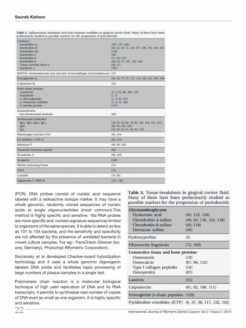
The molecular biological techniques utilizes nucleic acid probes (DNA & RNA probes), checkerboard DNA-DNA hybridization technology and Polymerase Chain Reaction
Advanced diagnostic aids in periodontology
22 International Journal of Women’s Dental Council: Vol 2 | Issue 2 | 2014
(PCR). DNA probes consist of nucleic acid sequence labeled with a radioactive isotope marker. It may have a whole genomic, randomly cloned sequences of nucleic acids or single oligonucleotides (most common).This method is highly specific and sensitive. 16s RNA probes are more specific and contain signature sequences limited to organisms of the same species. It is able to detect as few as 101 to 104 bacteria, and the sensitivity and specificity are not affected by the presence of unrelated bacteria in mixed culture samples. For eg:- ParoCheck (Greiner bio-one, Germany), Phylochip( Affymetrix Corporation) .
Socransky et al developed Checker-board hybridization technology and it uses a whole genomic digoxigenin labeled DNA probe and facilitates rapid processing of large numbers of plaque samples in a single test.
Polymerase chain reaction is a molecular biological technique of high yield replication of DNA and its RNA transcripts. It permits to synthesize vast number of copies of DNA even as small as one organism. It is highly specific and sensitive.
Saurab Kishore
International Journal of Women’s Dental Council: Vol 2 | Issue 2 | 2014 23
Advances in inflammatory and immune markers
Periodontal tissue destruction could be both bacteria mediated and host mediated. The assessment of host response involves the study of mediators, by immunologic or biochemical methods, that are recognized as part of individual’s response to the periodontal infection. These mediators are either specifically identified with the infection, such as putative pathogen, or represent a less specific reaction, such as local release of inflammatory mediators, host-derived enzymes, or tissue breakdown products.
Source of samples include saliva, gingival crevicular fluid (GCF), gingival crevicular cells, blood serum, blood cells and urine.
Conclusion
Even there are a whole range of newer diagnostic methods in all aspects, there is a lack of proven diagnostic test. There is a need for methods which is highly predictive, simple, safe and cost effective. More over, diagnostic tests
that can be used during routine dental practice should be developed.
References1 Listgarten MA,Mao R, Robinson PJ: Periodontal probing and
relationship of the probe tip to periodontal tissues,J Periodontol 47:511,1976.
2 Goodson JM, Haffajee AD, Socransky SS: The relationship between attachment level loss and alveolar bone loss, J Clin Periodontol 11: 348,1984.
3 Mol A. Imaging methods in periodontology. Periodontol 2000 2004;34:34–48.
4 Sanz M, Lau L, Herrera D, et al. Methods of detection of Actinobacillusactinomycetemcomitans, Porphyromonas gingivalis and Tannerella forsythensis in periodontal microbiology, with special emphasis on advanced molecular techniques: a review. J ClinPeriodontol 2004;31(12):1034–47.
5 Loesche WJ, Lopatin DE, Giordano J, et al. Comparison of the benzoyl-DL-arginine- naphthylamide (BANA) test, DNA probes, and immunological reagents for ability to detect anaerobic periodontal infections due to Porphyromonas gingivalis, Treponema denticola, and Bacteroides forsythus. J Clin Microbiol 1992;30(2): 427–33.
Advanced diagnostic aids in periodontology
24 International Journal of Women’s Dental Council: Vol 2 | Issue 2 | 2014
Forensic odontology : Obligations of dentists
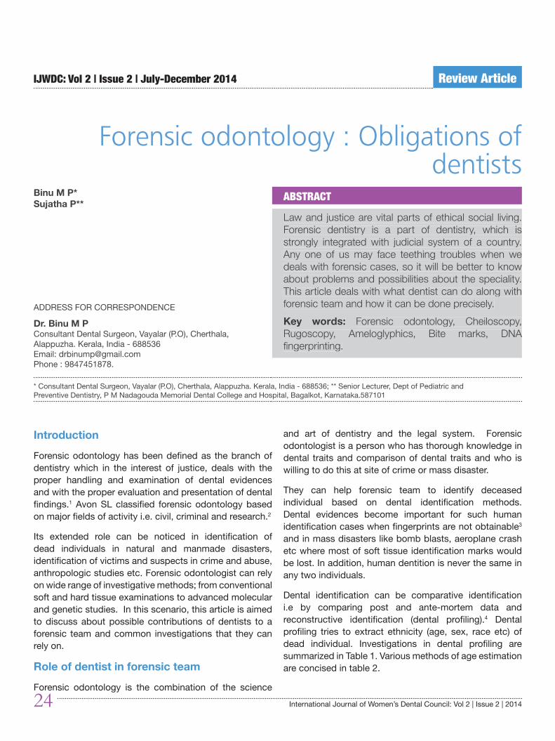
Binu M P*Sujatha P**
ABSTRACT
Law and justice are vital parts of ethical social living. Forensic dentistry is a part of dentistry, which is strongly integrated with judicial system of a country. Any one of us may face teething troubles when we deals with forensic cases, so it will be better to know about problems and possibilities about the speciality. This article deals with what dentist can do along with forensic team and how it can be done precisely. Key words: Forensic odontology, Cheiloscopy, Rugoscopy, Ameloglyphics, Bite marks, DNA fi ngerprinting.
ADDRESS FOR CORRESPONDENCE
Dr. Binu M P Consultant Dental Surgeon, Vayalar (P.O), Cherthala, Alappuzha. Kerala, India - 688536Email: [email protected] : 9847451878.
* Consultant Dental Surgeon, Vayalar (P.O), Cherthala, Alappuzha. Kerala, India - 688536; ** Senior Lecturer, Dept of Pediatric and Preventive Dentistry, P M Nadagouda Memorial Dental College and Hospital, Bagalkot, Karnataka.587101
Introduction
Forensic odontology has been defi ned as the branch of dentistry which in the interest of justice, deals with the proper handling and examination of dental evidences and with the proper evaluation and presentation of dental fi ndings.1 Avon SL classifi ed forensic odontology based on major fi elds of activity i.e. civil, criminal and research.2
Its extended role can be noticed in identifi cation of dead individuals in natural and manmade disasters, identifi cation of victims and suspects in crime and abuse, anthropologic studies etc. Forensic odontologist can rely on wide range of investigative methods; from conventional soft and hard tissue examinations to advanced molecular and genetic studies. In this scenario, this article is aimed to discuss about possible contributions of dentists to a forensic team and common investigations that they can rely on.
Role of dentist in forensic team
Forensic odontology is the combination of the science
and art of dentistry and the legal system. Forensic odontologist is a person who has thorough knowledge in dental traits and comparison of dental traits and who is willing to do this at site of crime or mass disaster.
They can help forensic team to identify deceased individual based on dental identifi cation methods. Dental evidences become important for such human identifi cation cases when fi ngerprints are not obtainable3 and in mass disasters like bomb blasts, aeroplane crash etc where most of soft tissue identifi cation marks would be lost. In addition, human dentition is never the same in any two individuals.
Dental identifi cation can be comparative identifi cation i.e by comparing post and ante-mortem data and reconstructive identifi cation (dental profi ling).4 Dental profi ling tries to extract ethnicity (age, sex, race etc) of dead individual. Investigations in dental profi ling are summarized in Table 1. Various methods of age estimation are concised in table 2.
IJWDC: Vol 2 | Issue 2 | July-December 2014 Review Article
Law and justice are vital parts of ethical social living. Forensic dentistry is a part of dentistry, which is strongly integrated with judicial system of a country. Any one of us may face teething troubles when we deals with forensic cases, so it will be better to know about problems and possibilities about the speciality. This article deals with what dentist can do along with forensic team and how it can be done precisely. Key words: Forensic odontology, Cheiloscopy, Rugoscopy, Ameloglyphics, Bite marks, DNA fi ngerprinting.
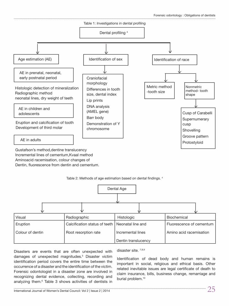
International Journal of Women’s Dental Council: Vol 2 | Issue 2 | 2014 25
Forensic odontology : Obligations of dentists
Disasters are events that are often unexpected with damages of unexpected magnitudes.5 Disaster victim identification period covers the entire time between the occurrence of a disaster and the identification of the victim. Forensic odontologist in a disaster zone are involved in recognizing dental evidence, collecting, recording and analyzing them.6 Table 3 shows activities of dentists in
disaster site. 7,8,9
Identification of dead body and human remains is important in social, religious and ethical basis. Other related inevitable issues are legal certificate of death to claim insurance, bills, business change, remarriage and burial problem.10
Table 2: Methods of age estimation based on dental findings. 4
Dental Age
Visual Radiographic Histologic Biochemical Eruption
Colour of dentin
Calcification status of teeth
Root resorption rate
Neonatal line and
Incremental lines
Dentin translucency
Fluorescence of cementum
Amino acid racemisation
Table 1: Investigations in dental profiling
Dental profiling 4
Age estimation (AE) Identification of sex Identification of race
AE in prenatal, neonatal, early postnatal period
AE in children and adolescents
Histologic detection of mineralizationRadiographic methodneonatal lines, dry weight of teeth
Eruption and calcification of toothDevelopment of third molar
AE in adults
Gustafson’s method,dentine translucencyIncremental lines of cementum,Kvaal methodAminoacid racemisation, colour changes ofDentin, fluorescence from dentin and cementum.
Craniofacial morphology Differences in tooth size, dental indexLip prints DNA analysis (AMEL gene)Barr body Demonstration of Y chromosome
Metric method -tooth size
Nonmetric method- tooth shape
Cusp of CarabelliSupernumerary cusp Shovelling Groove patternProtostyloid
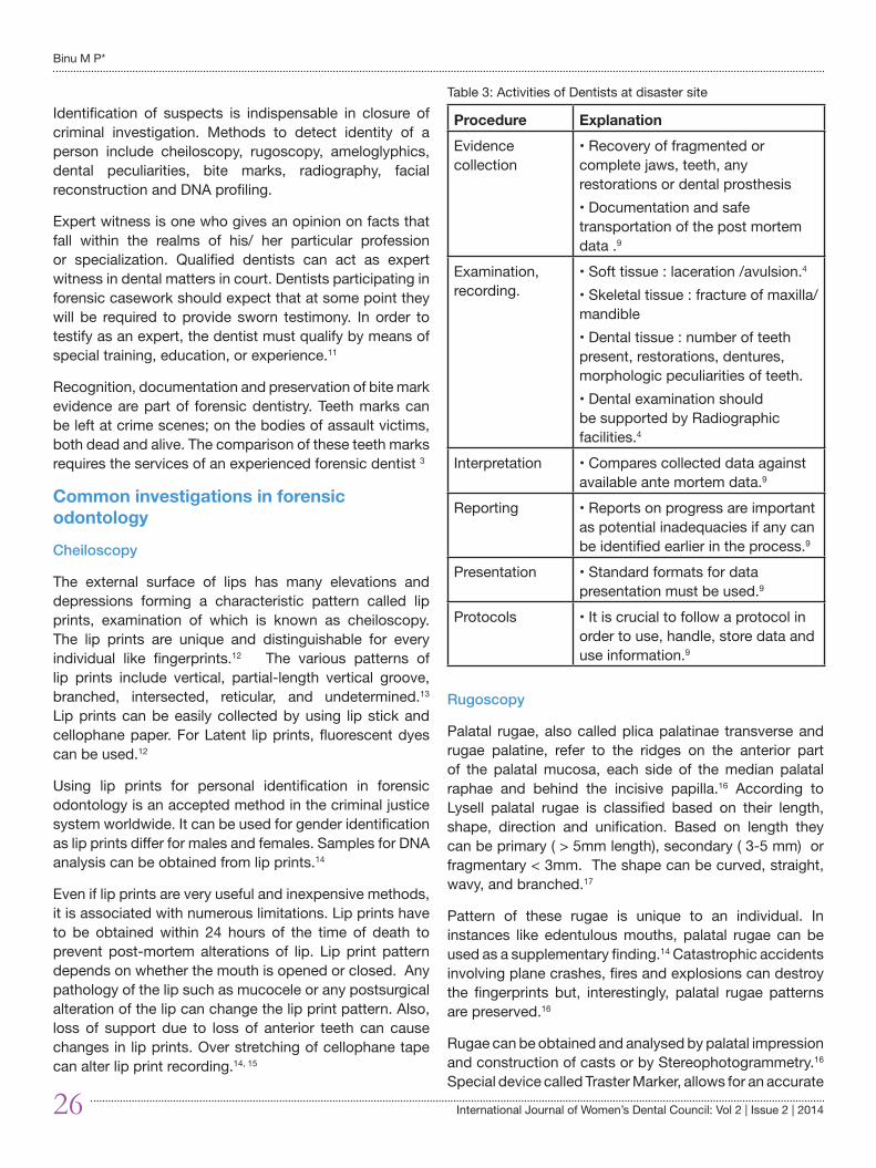
26 International Journal of Women’s Dental Council: Vol 2 | Issue 2 | 2014
Identification of suspects is indispensable in closure of criminal investigation. Methods to detect identity of a person include cheiloscopy, rugoscopy, ameloglyphics, dental peculiarities, bite marks, radiography, facial reconstruction and DNA profiling.
Expert witness is one who gives an opinion on facts that fall within the realms of his/ her particular profession or specialization. Qualified dentists can act as expert witness in dental matters in court. Dentists participating in forensic casework should expect that at some point they will be required to provide sworn testimony. In order to testify as an expert, the dentist must qualify by means of special training, education, or experience.11
Recognition, documentation and preservation of bite mark evidence are part of forensic dentistry. Teeth marks can be left at crime scenes; on the bodies of assault victims, both dead and alive. The comparison of these teeth marks requires the services of an experienced forensic dentist 3
Common investigations in forensic odontology
Cheiloscopy
The external surface of lips has many elevations and depressions forming a characteristic pattern called lip prints, examination of which is known as cheiloscopy. The lip prints are unique and distinguishable for every individual like fingerprints.12 The various patterns of lip prints include vertical, partial-length vertical groove, branched, intersected, reticular, and undetermined.13 Lip prints can be easily collected by using lip stick and cellophane paper. For Latent lip prints, fluorescent dyes can be used.12
Using lip prints for personal identification in forensic odontology is an accepted method in the criminal justice system worldwide. It can be used for gender identification as lip prints differ for males and females. Samples for DNA analysis can be obtained from lip prints.14
Even if lip prints are very useful and inexpensive methods, it is associated with numerous limitations. Lip prints have to be obtained within 24 hours of the time of death to prevent post-mortem alterations of lip. Lip print pattern depends on whether the mouth is opened or closed. Any pathology of the lip such as mucocele or any postsurgical alteration of the lip can change the lip print pattern. Also, loss of support due to loss of anterior teeth can cause changes in lip prints. Over stretching of cellophane tape can alter lip print recording.14, 15
Rugoscopy
Palatal rugae, also called plica palatinae transverse and rugae palatine, refer to the ridges on the anterior part of the palatal mucosa, each side of the median palatal raphae and behind the incisive papilla.16 According to Lysell palatal rugae is classified based on their length, shape, direction and unification. Based on length they can be primary ( > 5mm length), secondary ( 3-5 mm) or fragmentary < 3mm. The shape can be curved, straight, wavy, and branched.17
Pattern of these rugae is unique to an individual. In instances like edentulous mouths, palatal rugae can be used as a supplementary finding.14 Catastrophic accidents involving plane crashes, fires and explosions can destroy the fingerprints but, interestingly, palatal rugae patterns are preserved.16
Rugae can be obtained and analysed by palatal impression and construction of casts or by Stereophotogrammetry.16 Special device called Traster Marker, allows for an accurate
Table 3: Activities of Dentists at disaster site
Procedure Explanation Evidence collection
• Recovery of fragmented or complete jaws, teeth, any restorations or dental prosthesis• Documentation and safe transportation of the post mortem data .9
Examination, recording.
• Soft tissue : laceration /avulsion.4
• Skeletal tissue : fracture of maxilla/ mandible• Dental tissue : number of teeth present, restorations, dentures, morphologic peculiarities of teeth.• Dental examination should be supported by Radiographic facilities.4
Interpretation • Compares collected data against available ante mortem data.9
Reporting • Reports on progress are important as potential inadequacies if any can be identified earlier in the process.9
Presentation • Standard formats for data presentation must be used.9
Protocols • It is crucial to follow a protocol in order to use, handle, store data and use information.9
Binu M P*
International Journal of Women’s Dental Council: Vol 2 | Issue 2 | 2014 27
Forensic odontology : Obligations of dentists
determination of the length and position of every single palatal rugae. While the overlay print of palatal rugae in a maxillary cast is termed as calcorrugoscopy.18
Postmortem identification is not possible without the antemortem records. Complex rugae patterns can cause intra or interobserver errors. Denture wear, tooth malposition, and palatal pathology can cause alterations in rugae patterns. Rugae patterns are genetically determined, and so can be rather used in population differentiation than individual identification.14
Ameloglyphics
Enamel rods are laid down by the ameloblasts in an undulating path. This is manifested on the outer surface of enamel. These patterns on the enamel surface are called as tooth prints. Manjunath et al coined the term ameloglyphics for the study of enamel rod patterns on tooth surface.19
Each tooth print is composed of combination of eight different sub patterns, and each tooth print is unique to a particular tooth. This uniqueness of the tooth print is used as a valuable tool in forensic science for personal identification. These enamel rod end patterns can be duplicated by various methods like acetate peel technique, rubber base impression, etc.20
Advantage of the technique is that three successive tooth prints of each tooth show a similar pattern of enamel rod ends and similar distribution of minutae points. However, in spite of standardization, there can be variation, as inclusion or deletion of even a single cluster of enamel rods, could lead to variations. 19 Fractured, decayed, abraded and eroded teeth cannot be included in this method. Ameloglyphics is still in its infancy. Whether the tooth prints are the same at different depths of enamel has to be evaluated with further studies. 14
Cemental annulations and dentin translucency
Tooth cementum annulations are a reliable method for adult skeletal age estimation. The area at the junction of apical and middle third of the root is considered for this purpose.21
The alternate light and dark bands are counted with the help of image analysis software. Chronological age of the individual E = n + t [n = number of incremental lines, t = eruption age of tooth] 22
Dentinal translucency is a simple method for dental
age estimation. Translucency has been measured using callipers or by digital aids. Acharya proposed computer aided method to measure translucency on sectioned teeth. With advances in computing technology, digital evaluation of translucency can be more easily accomplished. 22
Bite marks
Bite mark is the impression produced by tooth structure alone or with associated oral structures. Bite marks can be preserved by fabrication of stone models from impressions. Bite marks are commonly associated with violent fight or sexual abuse.
Bite marks remain as controversial aspect of forensic odontology. The shape and clarity of bite marks found on the skin of the victims change in a relatively short duration. Though photographed immediately, the three-dimensional bite marks on photograph will be associated with changes in color and spatial relations. 14
Skin has the intrinsic property of distortion leading to considerable variability in the precision of representation of bite marks. Hemorrhage and edema can alter bite marks evidence. As dental features change over time, changes can occur after obtaining ante mortem records. Therefore bite marks are considered less reliable than other biometric methods. 14,23
DNA fingerprinting
Each individual has unique patterns for DNA. Therefore, DNA fingerprinting is an invaluable tool for identification of individuals. The teeth are an excellent source of genomic DNA. Polymerase Chain Reaction help to amplify and analyse collected post-mortem samples and compare it with known ante-mortem samples or parental DNA. Mitochondrial DNA is another type of material that can be used for body identification. Obtaining a DNA profile from mitochondrial DNA is higher than that with genomic DNA. Saliva is another source of DNA for forensic purposes.16
This method is highly accurate, reproducible, and unique. However, errors may develop in sample collection, processing, and interpretation. Any bacterial contamination can alter the interpretation. Too little amount of DNA can produce cause misinterpretation of results. In addition, degraded samples can produce very scant amount of high molecular weight DNA.14
Conclusion
A forensic odontologist can make valuable contributions to the forensic team. Advanced techniques like DNA finger
28 International Journal of Women’s Dental Council: Vol 2 | Issue 2 | 2014
printing have reformed the field. However, it is not only the duty of forensic odontologist to serve the society in this identification process. As we know, ante mortem records are essential for comparative method of identification. Therefore, it is the duty of all dental practitioners and institutions to maintain proper patient demographic data along with dental findings and supporting data like radiographs and photographs.
References 1. Kavitha B, Einstein A, Sivapathasundharam B. Limitations in
forensic odontology. Journal of Forensic Dental Sciences 2009 ;1 (1): 8 – 10.
2. Avon SL. Forensic odontology: the roles and responsibilities of the dentist. J Can Dent Assoc 2004; 70:453-8.
3. Bowers CM. Forensic dental Evidence: An investigator’s Handbook. 1st ed. Elsevier Academic Press California ; 2004. pp 1-32.
4. Rajendran R,Sivapathasundharam B.Shafer’s textbook of Oral Pathology.6th ed. Elsevier publications. New Delhi; 2009. pp 871 – 897.
5. Chan, T.C., et al., Information Technology and Emergency Medical Care during Disasters. Academic Emergency Medicine, 2004. 11(11): p. 1229-1236.
6. Lake, A., H. James, and J.W. Berketa, Disaster victim identification: quality management from an odontology perspective. Forensic Science, Medicine, and Pathology, 2012. 8(2): p. 157-163.
7. Stavrianos, C., et al., The role of dentistry in the management of mass disasters and bioterrorism. Acta stomatologica Croatica, 2010. 44(2): p. 110-119.
8. Pretty, I. and D. Sweet, forensic dentistry: A look at forensic dentistry–Part 1: The role of teeth in the determination of human identity. Br Dent J, 2001. 190(7): p. 359-366.
9. Rajshekar M,Tennant M. The Role of the Forensic Odontologist in Disaster Victim Identification: A Brief Review. Malaysian Journal of Forensic Sciences 5(1): 78 -85.
10. Gnanasundaram N. Tooth for truth. Journal of Forensic Dental Sciences 2010 ; 2( 2): 51- 52.
11. Senn DR,Stimson PG. Forensic Dentistry 2010. 2nd Ed. CRC Press.Boca Raton: pp 379 – 393.
12. Saraswathi TR,Mishra G, Ranganathan K Study of lip prints Journal of Forensic Dental Sciences 2009 ;1 (1): 28 – 31.
13. Suzuki K, Tsuchihashi Y. A new attempt of personal identification by means of lip print. J Indian Dent Assoc 1970;42:8-9.
14. Kavitha B, Einstein A, Sivapathasundharam B, Saraswathi TR. Limitations in forensic odontology .Journal of Forensic Dental Sciences 2009 ;1 (1): 8 – 10.
15. Sivapathasundharam B, Prakash PA, Sivakumar G. Lip prints (cheiloscopy). Indian J Dent Res 2001;12:234-7.
16. Saxena S,Sharma P, Gupta N. Experimental studies of forensic odontology to aid in the identification process. Journal of Forensic Dental Sciences 2010 ; 2 (2): 69 -76.
17. Lysell L. Plicae palatinae transversae and papilla incisive in man: A morphologic and genetic study. Acta Odont Scand 1955;13: 5-137.
18. Thomas CJ, van Wyk CW. The palatal rugae in identification. J Forensic Odontostomatol 1988;6:21-7.
19. Manjunath K, Sriram G, Saraswathi TR, Sivapathasundharam B. Enamel rod end patterns: A preliminary study using acetate peel technique and automated biometrics. J Forensic Odontol 2008;1:33-6.
20. Manjunath K, Saraswathi TR, Sriram G, Sivapathasundharam B, Porchelvam S. Reliability of automated biometrics in the analysis of enamel rod end patterns. J Forensic Dent Sci 2009:1:32-6.
21. Stein TJ, Corcoran JF. Pararadicular cementum deposition as a criterion for age estimation in human beings. Oral Surg Oral Med Oral Pathol 1994; 77:266-70.
22. Aggarwal P, Saxena S, Bansal P. Incremental lines in root cementum of human teeth; An approach to their role in age estimation using polarizing microscopy. Indian J Dent Res 2008; 19:326-30.
23. Barbanel JC, Evans JH. Bite marks in skin-mechanical factors J Forensic Sci Soc 1974; 14:235-8.
Binu M P*
International Journal of Women’s Dental Council: Vol 2 | Issue 2 | 2014 29
Endodontic management of a mandibular fi rst molar with Radix entomolaris-A case
report.Elsy P. Simon*, Swapna Honwad**
ABSTRACT
Successful root canal treatment requires thorough chemo-mechanical cleansing and shaping of the entire root canal system followed by a fl uid tight seal of the root canal with a dense inert fi lling material. The root canal is considered to be one of the most complex systems in the human body. Awareness of the morphology of each tooth & its variations can contribute to the success of endodontic treatment. The mandibular fi rst molar is known to display several anatomical variations.A major anatomical variation seen in the two rooted mandibular fi rst molar is the occurrence of an additional disto-lingual root known as Radix entomolaris. Early detection of this anatomical deviant can prevent procedural errors or post treatment complications. The purpose of this report is to present a case of Radix entomolaris that has been diagnosed and managed endodontically. KEY WORDS: Radix entomolaris, Anatomic variation, Permanent three-rooted mandibular fi rst molars, Distolingual root
ADDRESS FOR CORRESPONDENCE
Dr. Elsy P. SimonReader, Department of Conservative Dentistry and Endodontics, KMCT Dental College, Mampetta, Mukkam, Calicut-673602Mobile 9633332629; Email:[email protected]
* Reader, Department of Conservative Dentistry and Endodontics; **Senior Lecturer, Department of Oral Pathology and Microbiology, KMCT Dental College, Mampetta, Mukkam, Calicut-673602
Introduction
M .T Barrett described the pulp cavity as the most complex system in the human body. Studies on tooth morphology utilizing various techniques have established the complexity of the dental root canal.1, 4 The main goal of endodontic therapy is to heal or prevent apical periodontitis2. This is possible by ensuring complete three dimensional cleaning and shaping of the entire root canal system followed with a fl uid tight seal.1, 3 However,
complexities of the root canal anatomy present clinical challenges which calls for more diagnostic approaches and clinical skills.1Thus an in-depth knowledge of root canal anatomy and its possible variations is necessary to ensure a better prognostic outcome for endodontic therapy3, 16.
The mandibular fi rst molar typically presents with two well-defi ned roots, a mesial root characterized by a fl attened mesio-distal surface and widened bucco-lingual surface, and a distal root that is mostly straight2.The number of
IJWDC: Vol 2 | Issue 2 | July-December 2014 Case report
Successful root canal treatment requires thorough chemo-mechanical cleansing and shaping of the entire root canal system followed by a fl uid tight seal of the root canal with a dense inert fi lling material. The root canal is considered to be one of the most complex systems in the human body. Awareness of the morphology of each tooth & its variations can contribute to the success of endodontic treatment. The mandibular fi rst molar is known to display several anatomical variations.A major anatomical variation seen in the two rooted mandibular fi rst molar is the occurrence of an additional disto-lingual root known as Radix entomolaris. Early detection of this anatomical deviant can prevent procedural errors or post treatment complications. The purpose of this report is to present a case of Radix entomolaris that has been diagnosed and managed endodontically. KEY WORDS: Radix entomolaris, Anatomic variation, Permanent three-rooted mandibular fi rst molars, Distolingual root
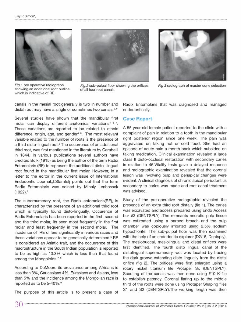
30 International Journal of Women’s Dental Council: Vol 2 | Issue 2 | 2014
canals in the mesial root generally is two in number and distal root may have a single or sometimes two canals.2, 5
Several studies have shown that the mandibular first molar can display different anatomical variations4, 6 7. These variations are reported to be related to ethnic difference, origin, age, and gender4, 6. The most relevant variable related to the number of roots is the presence of a third disto-lingual root.2 The occurrence of an additional third root, was first mentioned in the literature by Carabelli in 1844. In various publications several authors have credited Bolk (1915) as being the author of the term Radix Entomolaris (RE) to represent the additional disto- lingual root found in the mandibular first molar. However, in a letter to the editor in the current issue of International Endodontic Journal,,I.Stamfelj points out that the term Radix Entomolaris was coined by Mihaly Lenhossek (1922).7
The supernumerary root, the Radix entomolaris(RE), is characterized by the presence of an additional third root which is typically found disto-lingually. Occurence of Radix Entomolaris has been reported in the first, second and the third molar, Its seen most frequently in the first molar and least frequently in the second molar. The incidence of RE differs significantly in various races and these variations appear to be genetically determined.5 RE is considered an Asiatic trait, and the occurrence of this macrostructure in the South Indian population is reported to be as high as 13.3% which is less than that found among the Mongoloids.7, 9
According to DeMoore its prevalence among Africans is less than 3%, Caucasians 4%, Eurasians and Asians, less than 5% and the incidence among the Mongolian race is reported as to be 5-40%.9
The purpose of this article is to present a case of
Radix Entomolaris that was diagnosed and managed endodontically.
Case Report
A 55 year old female patient reported to the clinic with a complaint of pain in relation to a tooth in the mandibular right posterior region since one week. The pain was aggravated on taking hot or cold food. She had an episode of acute pain a month back which subsided on taking medication. Clinical examination revealed a large class II disto-occlusal restoration with secondary caries in relation to 46.Vitality tests gave a delayed response and radiographic examination revealed that the coronal lesion was involving pulp and periapical changes were evident. A clinical diagnosis of chronic apical periodontitis secondary to caries was made and root canal treatment was advised.
Study of the pre-operative radiographic revealed the presence of an extra third root distally (fig 1). The caries was excavated and access prepared using Endo Access bur #3 (DENTSPLY) .The remnants necrotic pulp tissue was extirpated using a barbed broach and the pulp chamber was copiously irrigated using 2.5% sodium hypochlorite. The sub-pulpal floor was then examined with the help of an endodontic explorer (DG16, Dentsply). The mesiobuccal, mesiolingual and distal orifices were first identified. The fourth disto lingual canal of the distolingual supernumerary root was located by tracing the dark groove extending disto-lingually from the distal orifice (fig 2). The orifices were first enlarged using a rotary nickel titanium file Protaper Sx (DENTSPLY). Scouting of the canals was then done using #10 K-file to establish patency. Coronal flaring up to the middle third of the roots were done using Protaper Shaping files S1 and S2 (DENTSPLY).The working length was then
Fig 3 radiograph of master cone selectionFig:2 sub-pulpal floor showing the orifices of all four root canals
Fig.1 pre operative radiograph showing an additional root outline which is indicative of RE
Elsy P. Simon*,
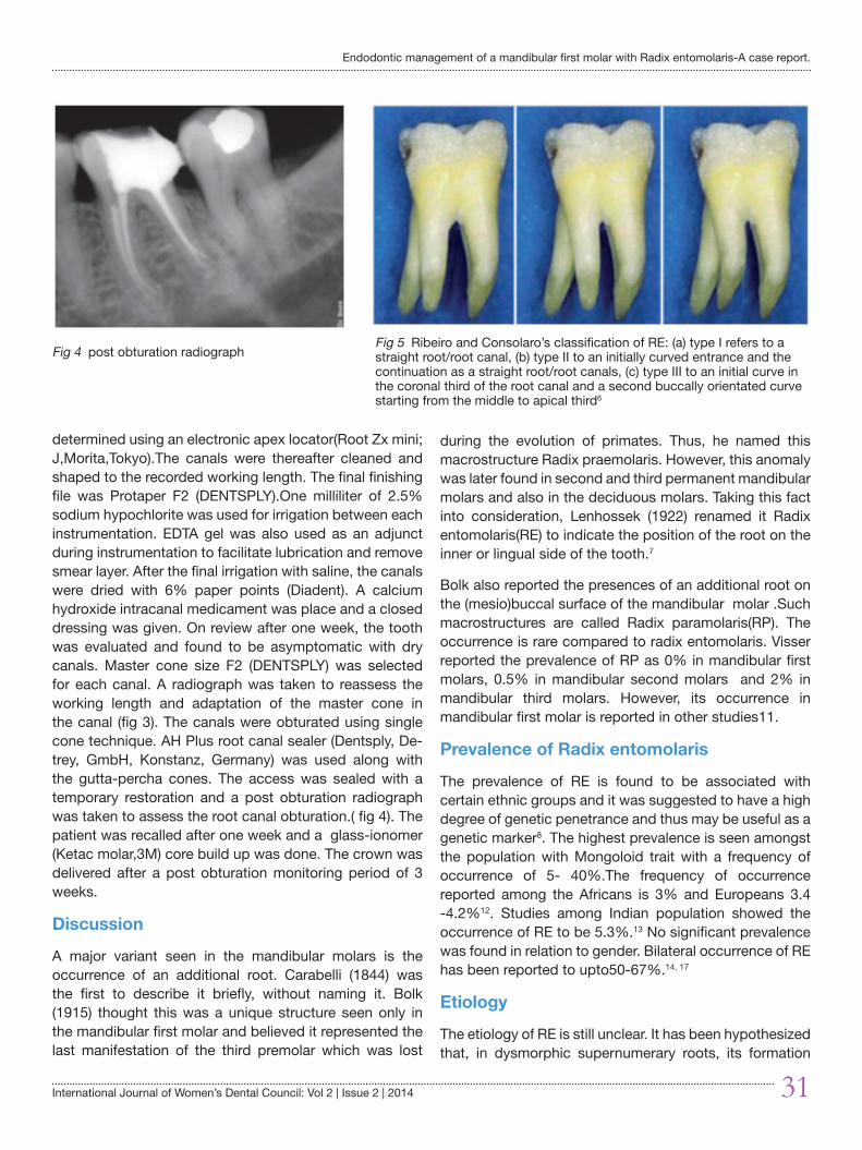
International Journal of Women’s Dental Council: Vol 2 | Issue 2 | 2014 31
Endodontic management of a mandibular first molar with Radix entomolaris-A case report.
determined using an electronic apex locator(Root Zx mini; J,Morita,Tokyo).The canals were thereafter cleaned and shaped to the recorded working length. The final finishing file was Protaper F2 (DENTSPLY).One milliliter of 2.5% sodium hypochlorite was used for irrigation between each instrumentation. EDTA gel was also used as an adjunct during instrumentation to facilitate lubrication and remove smear layer. After the final irrigation with saline, the canals were dried with 6% paper points (Diadent). A calcium hydroxide intracanal medicament was place and a closed dressing was given. On review after one week, the tooth was evaluated and found to be asymptomatic with dry canals. Master cone size F2 (DENTSPLY) was selected for each canal. A radiograph was taken to reassess the working length and adaptation of the master cone in the canal (fig 3). The canals were obturated using single cone technique. AH Plus root canal sealer (Dentsply, De-trey, GmbH, Konstanz, Germany) was used along with the gutta-percha cones. The access was sealed with a temporary restoration and a post obturation radiograph was taken to assess the root canal obturation.( fig 4). The patient was recalled after one week and a glass-ionomer (Ketac molar,3M) core build up was done. The crown was delivered after a post obturation monitoring period of 3 weeks.
Discussion
A major variant seen in the mandibular molars is the occurrence of an additional root. Carabelli (1844) was the first to describe it briefly, without naming it. Bolk (1915) thought this was a unique structure seen only in the mandibular first molar and believed it represented the last manifestation of the third premolar which was lost
during the evolution of primates. Thus, he named this macrostructure Radix praemolaris. However, this anomaly was later found in second and third permanent mandibular molars and also in the deciduous molars. Taking this fact into consideration, Lenhossek (1922) renamed it Radix entomolaris(RE) to indicate the position of the root on the inner or lingual side of the tooth.7
Bolk also reported the presences of an additional root on the (mesio)buccal surface of the mandibular molar .Such macrostructures are called Radix paramolaris(RP). The occurrence is rare compared to radix entomolaris. Visser reported the prevalence of RP as 0% in mandibular first molars, 0.5% in mandibular second molars and 2% in mandibular third molars. However, its occurrence in mandibular first molar is reported in other studies11.
Prevalence of Radix entomolaris
The prevalence of RE is found to be associated with certain ethnic groups and it was suggested to have a high degree of genetic penetrance and thus may be useful as a genetic marker8. The highest prevalence is seen amongst the population with Mongoloid trait with a frequency of occurrence of 5- 40%.The frequency of occurrence reported among the Africans is 3% and Europeans 3.4 -4.2%12. Studies among Indian population showed the occurrence of RE to be 5.3%.13 No significant prevalence was found in relation to gender. Bilateral occurrence of RE has been reported to upto50-67%.14, 17
Etiology
The etiology of RE is still unclear. It has been hypothesized that, in dysmorphic supernumerary roots, its formation
Fig 5 Ribeiro and Consolaro’s classification of RE: (a) type I refers to a straight root/root canal, (b) type II to an initially curved entrance and the continuation as a straight root/root canals, (c) type III to an initial curve in the coronal third of the root canal and a second buccally orientated curve starting from the middle to apical third6
Fig 4 post obturation radiograph
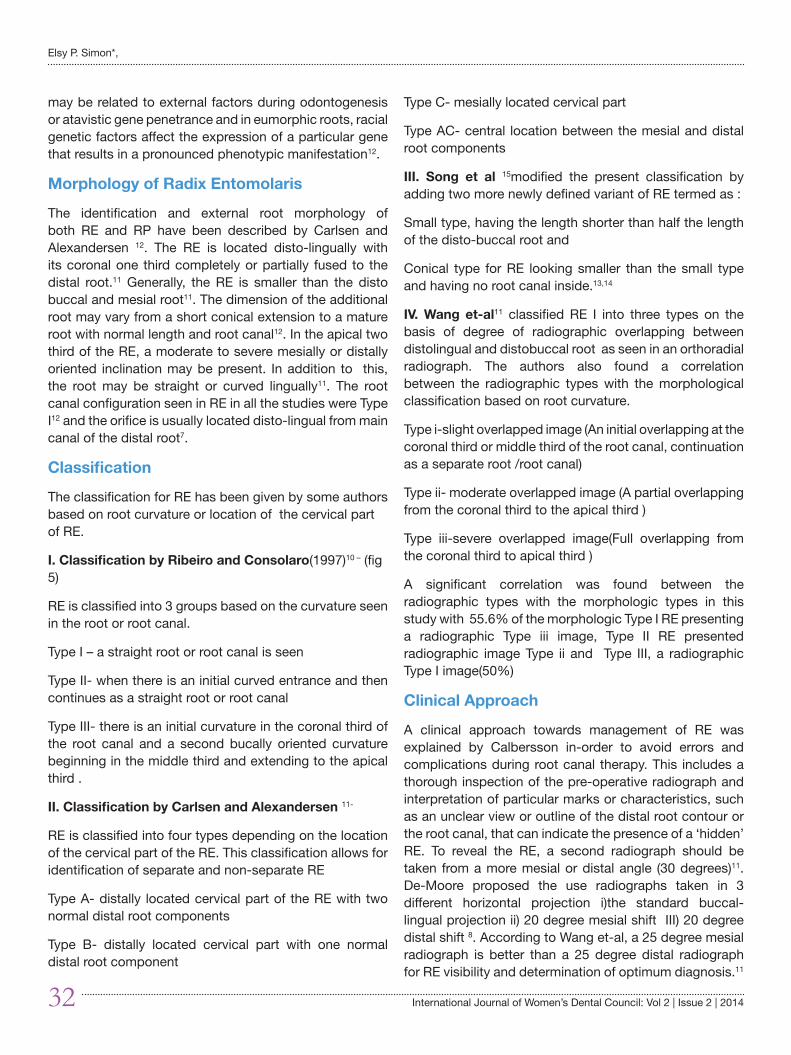
32 International Journal of Women’s Dental Council: Vol 2 | Issue 2 | 2014
may be related to external factors during odontogenesis or atavistic gene penetrance and in eumorphic roots, racial genetic factors affect the expression of a particular gene that results in a pronounced phenotypic manifestation12.
Morphology of Radix Entomolaris
The identification and external root morphology of both RE and RP have been described by Carlsen and Alexandersen 12. The RE is located disto-lingually with its coronal one third completely or partially fused to the distal root.11 Generally, the RE is smaller than the disto buccal and mesial root11. The dimension of the additional root may vary from a short conical extension to a mature root with normal length and root canal12. In the apical two third of the RE, a moderate to severe mesially or distally oriented inclination may be present. In addition to this, the root may be straight or curved lingually11. The root canal configuration seen in RE in all the studies were Type I12 and the orifice is usually located disto-lingual from main canal of the distal root7.
Classification
The classification for RE has been given by some authors based on root curvature or location of the cervical part of RE.
I. Classification by Ribeiro and Consolaro(1997)10 – (fig 5)
RE is classified into 3 groups based on the curvature seen in the root or root canal.
Type I – a straight root or root canal is seen
Type II- when there is an initial curved entrance and then continues as a straight root or root canal
Type III- there is an initial curvature in the coronal third of the root canal and a second bucally oriented curvature beginning in the middle third and extending to the apical third .
II. Classification by Carlsen and Alexandersen 11-
RE is classified into four types depending on the location of the cervical part of the RE. This classification allows for identification of separate and non-separate RE
Type A- distally located cervical part of the RE with two normal distal root components
Type B- distally located cervical part with one normal distal root component
Type C- mesially located cervical part
Type AC- central location between the mesial and distal root components
III. Song et al 15modified the present classification by adding two more newly defined variant of RE termed as :
Small type, having the length shorter than half the length of the disto-buccal root and
Conical type for RE looking smaller than the small type and having no root canal inside.13,14
IV. Wang et-al11 classified RE I into three types on the basis of degree of radiographic overlapping between distolingual and distobuccal root as seen in an orthoradial radiograph. The authors also found a correlation between the radiographic types with the morphological classification based on root curvature.
Type i-slight overlapped image (An initial overlapping at the coronal third or middle third of the root canal, continuation as a separate root /root canal)
Type ii- moderate overlapped image (A partial overlapping from the coronal third to the apical third )
Type iii-severe overlapped image(Full overlapping from the coronal third to apical third )
A significant correlation was found between the radiographic types with the morphologic types in this study with 55.6% of the morphologic Type I RE presenting a radiographic Type iii image, Type II RE presented radiographic image Type ii and Type III, a radiographic Type I image(50%)
Clinical Approach
A clinical approach towards management of RE was explained by Calbersson in-order to avoid errors and complications during root canal therapy. This includes a thorough inspection of the pre-operative radiograph and interpretation of particular marks or characteristics, such as an unclear view or outline of the distal root contour or the root canal, that can indicate the presence of a ‘hidden’ RE. To reveal the RE, a second radiograph should be taken from a more mesial or distal angle (30 degrees)11. De-Moore proposed the use radiographs taken in 3 different horizontal projection i)the standard buccal-lingual projection ii) 20 degree mesial shift III) 20 degree distal shift 8. According to Wang et-al, a 25 degree mesial radiograph is better than a 25 degree distal radiograph for RE visibility and determination of optimum diagnosis.11
Elsy P. Simon*,
International Journal of Women’s Dental Council: Vol 2 | Issue 2 | 2014 33
Clinical inspection of the tooth crown and analysis of the cervical morphology of the roots by means of periodontal probing can facilitate identification of an additional canal.11 The location of the orifice of the root canal of an RE has implications for the opening cavity. An extension of the triangular opening cavity to the (disto) lingual results in a more rectangular or trapezoidal outline form and facilitate a straight line access to the supernumerary root canal. If the RE canal entrance is not clearly visible after removal of the pulp chamber roof, a more thorough inspection of the pulp chamber floor and wall, especially in the distolingual region, is necessary. Visual aids such as a loupe,intra-oral camera or dental microscope can, in this respect, be useful. A dark line on the pulp chamber floor can indicate the precise location of the RE canal orifice.
Conclusion
Failure to recognize the presence of RE at an early stage may result in an unfavorable endodontic outcome. The advent of improved imaging techniques like radio-visiography and CBCT have helped in recognizing possible complicating factors prior to the initiation of treatment. However, the skill in diagnosing morphological variations like RE lies in the knowledge of the variant and the ability to interpret the clinical and radiographic presentations. Once the diagnosis is established the necessary clinical steps can be modified so as obtain the best possible results without compromising the integrity of the remaining crown structure.
References1. H. A. Ahmed, N. H. Abu-bakr, N. A. Yahia & Y. E. Ibrahim. Root
and canal morphology of permanent mandibular molars in a Sudanese population. Int Endod J,2007; 40: 766–771. 767
2. O.V. de Pablo, R. Estevez, M.P.Sanchez, C. Heilborn, and N. Cohenca. Root anatomy and canal configuration of the permanent mandibular first molar: a systematic review. J.Endod 2010; 36:1919-1931.
3. Peters OA. Current challenges and concepts in the preparation of root canal systems: a review. J Endod 2004;30:559–67.
4. H. R. D. Peiris, T. N. Pitakotuwage, M. Takahashi1, K. Sasaki &
E.Kanazawa. Rootcanal morphology of mandibular permanent molars at different ages.Int Endod Journal 2008; 41: 828–835.
5. Bilge Gulsum Nur, Evren Ok,et-al.Evaluation of root and canal morphology of mandibular permanent molars in a south-eastern Turkish population using cone-beam computed tomography.European Journal of Dentistry 2014; 8:154-159
6. Srinidhi V Ballullaya,Sayesh Vemuri,Pabbathi Ravi Kumar.Variable permanent mandibular first molar:Review of Literature,jcd ,vol 16,issue2,pg 99-110.2013
7. Stamfelj .I. Who coined the term radix entomolaris .Int.Endod.J2014; 47:810-11
8. R. J. G. De Moor, C. A. J. G. Deroose , F. L. G. Calberson.The radix entomolaris in mandibular first molar:an endodontic challenge.Int Endod J 2004; 37: 789–799.
9. Sujatha. I, Aamina Zoya Farook, S. M. Sharath Chandra.Prevalence of radix entomolaris in mandibular permanent first molars: a study in a South Indian population.Oral Surg, Oral Med, Oral Path, Oral Radiol and Endod 2011;112:e77-e82
10. A. R. Vivekananda Pai, Rachit Jain, Ashwini S. Colaco.Detection and endodontic management of radix entomolaris: Report of case series.Saudi Endod J 2014;4:77-82.
11. Qian Wang, Gang Yu, Xue-dong Zhou, DDS,Ove A. Peters,Qing-hua Zheng, Ding-ming Huang.Evaluation of X-Ray Projection Angulation for Successful Radix Entomolaris Diagnosis in Mandibular First Molars In Vitro. J.Endod2011;37:1064-1068
12. Srinidhi V Ballullaya, Sayesh Vemuri, Pabbati Ravi Kumar.Variable permanent mandibular first molar: Review of literature.JCD2013;16:99-110
13. Filip L. Calberson, Roeland J. De Moor,Christophe A. Deroose.The Radix Entomolaris and Paramolaris: Clinical Approach.J Endod2007;33:58-63
14. A K Garg,R.K Tewari,A kumar,S H Hashmi,N agarwal,S K Mishra. Prevelence of Three-Rooted Mandibular Permanent First Molars among the Indian Population,J.Endod 2010; 36:1302-1306.
15. Song JS,Choi HJ,Jung HS,Kim SO. The prevalence and morphologic classification of distolingual roots in the mandibular molars in a Korean population. J Endod2010;36:653-7.
16. Vertucci FJ.Root canal morphology and its relation ship to endodontic procedures.Endodo Topics2005;10:3-29.
17. Bharti R, Arya D, Saumyendra VS, Kulwinder KW, Tikku AP,Chandra A. Prevalence of radix entomolaris in an Indianpopulation. Indian J Stomatol 2011;2:165-7.
Endodontic management of a mandibular first molar with Radix entomolaris-A case report.
34 International Journal of Women’s Dental Council: Vol 2 | Issue 2 | 2014
Complex odontome: a case report and review of literature
V.R. Rekha* Joseph Edward**R. Rathy***Harish.R.K****Indu.M*****
ABSTRACT
Odontomas are the most common odontogenic tumors, which are also considered as hamartomas rather than neoplasms. They are classifi ed into complex and compound odontomas. Here we report a case of a 13 year old female patient who reported with missing mandibular left second and third molar. Radiographically a large well-defi ned radio opaque mass with a radiolucent rim and sclerotic border in relation to missing 37, 38 were seen. Histopathologic examination revealed calcifi ed areas showing enamel space, dentin, cementum like material and pulp tissue in a disorganized pattern. Based on these fi ndings, a diagnosis of complex odontome was made. Timely diagnoses of odontomas are important as they can cause delayed eruption, root resorption, pathologic fractures and formation of cystic lesions.Key words: Odontoma, Odontogenic tumor, Hamartoma, Complex Odontome, Delayed eruption
ADDRESS FOR CORRESPONDENCE
Dr. V.R. Rekha PG student ,Department of Oral & Maxillofacial Pathology,Azeezia College of Dental Sciences,Kollam, Kerala, India . Ph: 9447957642, Email: [email protected]
* PG student, ** Professor and HOD, *** Professor and HOD **** Reader, ***** Senior Lecturer, Department of Oral & Maxillofacial Pathology, Azeezia College of Dental Sciences, Kollam, Kerala, India.
Introduction
Odontomas are the most common odontogenic tumors and are composed of tissues native to teeth.1 Due to their composition and behavior, they can be regarded as hamartomas or malformations rather than true neoplasms.2 According to the World Health Organization (WHO) 2005 classifi cation odontomas are considered as mixed odontogenic tumors which are classifi ed into compound and complex odontomas.3 The enamel and dentin are laid down in an abnormal pattern because the organization of the odontogenic cells fails to reach a normal state of morphodifferentiation. Even if odontomas are characterized by their slow growth and non-aggressive behavior, they causes symptoms like delayed eruption and root resorption.4
Case report
A 13-year-old female patient reported with complaint of delay on the eruption of the lower left posterior teeth. Intraoral examination revealed missing mandibular left second and third molar (37, 38). Patient’s past medical and dental history was not signifi cant. A provisional diagnosis of impacted 37 was given. Radiographic examination revealed a large well-defi ned radio opaque mass with a radiolucent rim and sclerotic border in relation to missing 37, 38.(Fig 1). The lesion was excised conservatively and a hard tissue mass of size 2x1.5x1 cm was sent for histopathological examination. The hematoxylin and eosin (H&E) stained sections showed calcifi ed masses interspersed with fi brous tissue and extravasated RBCs. The calcifi ed areas showed enamel space, dentin,
IJWDC: Vol 2 | Issue 2 | July-December 2014 Case report
Odontomas are the most common odontogenic tumors, which are also considered as hamartomas rather than neoplasms. They are classifi ed into complex and compound odontomas. Here we report a case of a 13 year old female patient who reported with missing mandibular left second and third molar. Radiographically a large well-defi ned radio opaque mass with a radiolucent rim and sclerotic border in relation to missing 37, 38 were seen. Histopathologic examination revealed calcifi ed areas showing enamel space, dentin, cementum like material and pulp tissue in a disorganized pattern. Based on these fi ndings, a diagnosis of complex odontome was made. Timely diagnoses of odontomas are important as they can cause delayed eruption, root resorption, pathologic fractures and formation of cystic lesions.Key words: Odontoma, Odontogenic tumor, Hamartoma, Complex Odontome, Delayed eruption
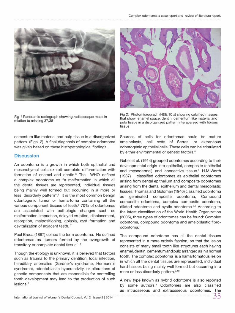
International Journal of Women’s Dental Council: Vol 2 | Issue 2 | 2014 35
cementum like material and pulp tissue in a disorganized pattern. (Figs. 2). A final diagnosis of complex odontoma was given based on these histopathological findings.
Discussion
An odontoma is a growth in which both epithelial and mesenchymal cells exhibit complete differentiation with formation of enamel and dentin.3 The WHO defined a complex odontoma as “a malformation in which all the dental tissues are represented, individual tissues being mainly well formed but occurring in a more or less disorderly pattern”.5 It is the most common benign odontogenic tumor or hamartoma containing all the various component tissues of teeth.6 70% of odontomas are associated with pathologic changes such as malformation, impaction, delayed eruption, displacement, resorption, malpositioning, aplasia, cyst formation and devitalization of adjacent teeth. 7
Paul Broca (1867) coined the term odontoma. He defined odontomas as ‘tumors formed by the overgrowth of transitory or complete dental tissue’. 6
Though the etiology is unknown, it is believed that factors such as trauma to the primary dentition, local infection, hereditary anomalies (Gardner’s syndrome, Hermann’s syndrome), odontoblastic hyperactivity, or alterations of genetic components that are responsible for controlling tooth development may lead to the production of such lesions.8
Sources of cells for odontomas could be mature ameloblasts, cell rests of Serres, or extraneous odontogenic epithelial cells. These cells can be stimulated by either environmental or genetic factors.8
Gabel et al. (1914) grouped odontomes according to their developmental origin into epithelial, composite (epithelial and mesodermal) and connective tissue.8 H.M.Worth (1937) classified odontomes as epithelial odontomes arising from dental epithelium and composite odontomes arising from the dental epithelium and dental mesoblastic tissues. Thomas and Goldman (1946) classified odontoma as geminated composite odontoma, Compound composite odontoma, complex composite odontoma, dilated odontoma and cystic odontoma.3,8 According to the latest classification of the World Health Organization (2005), three types of odontomas can be found: Complex odontoma, compound odontoma and ameloblastic fibro-odontoma.3
The compound odontome has all the dental tissues represented in a more orderly fashion, so that the lesion consists of many small tooth like structures each having enamel, dentin, cementum and pulp arranged as in a normal tooth. The complex odontoma is a hamartomatous lesion in which all the dental tissues are represented, individual hard tissues being mainly well formed but occurring in a more or less disorderly pattern.9,10
A new type known as hybrid odontome is also reported by some authors.3 Odontomes are also classified as intraosseous and extraosseous odontomes. The
Fig 1 Panoramic radiograph showing radioopaque mass in relation to missing 37,38
Fig 2: Photomicrograph (H&E,10 x) showing calcified masses that show enamel space, dentin, cementum like material and pulp tissue in a disorganized pattern interspersed with fibrous tissue
Complex odontoma: a case report and review of literature report.
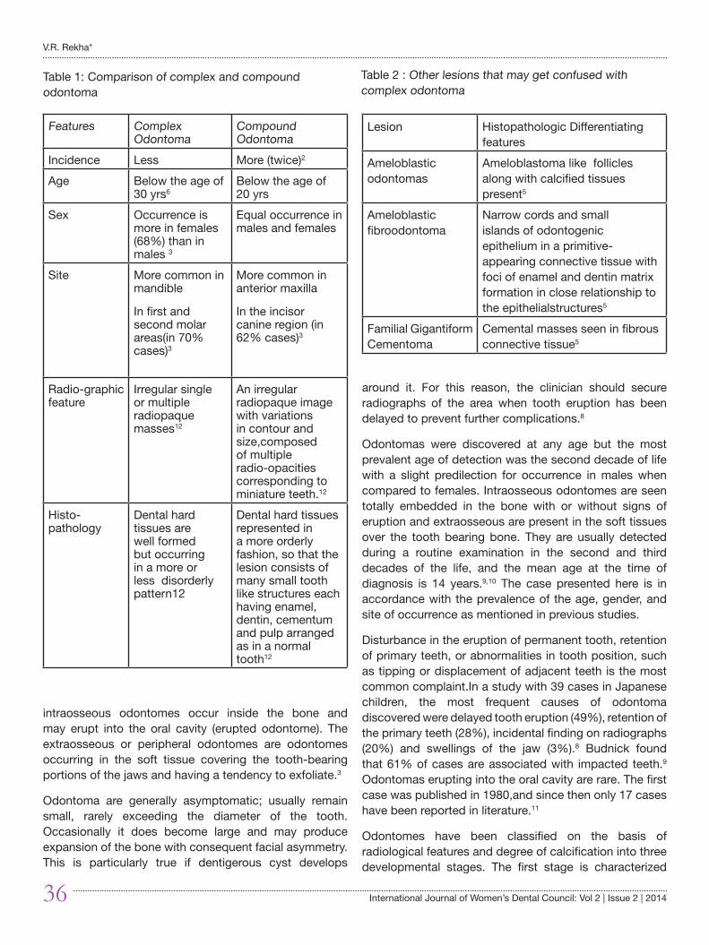
36 International Journal of Women’s Dental Council: Vol 2 | Issue 2 | 2014
intraosseous odontomes occur inside the bone and may erupt into the oral cavity (erupted odontome). The extraosseous or peripheral odontomes are odontomes occurring in the soft tissue covering the tooth-bearing portions of the jaws and having a tendency to exfoliate.3
Odontoma are generally asymptomatic; usually remain small, rarely exceeding the diameter of the tooth. Occasionally it does become large and may produce expansion of the bone with consequent facial asymmetry. This is particularly true if dentigerous cyst develops
around it. For this reason, the clinician should secure radiographs of the area when tooth eruption has been delayed to prevent further complications.8
Odontomas were discovered at any age but the most prevalent age of detection was the second decade of life with a slight predilection for occurrence in males when compared to females. Intraosseous odontomes are seen totally embedded in the bone with or without signs of eruption and extraosseous are present in the soft tissues over the tooth bearing bone. They are usually detected during a routine examination in the second and third decades of the life, and the mean age at the time of diagnosis is 14 years.9,10 The case presented here is in accordance with the prevalence of the age, gender, and site of occurrence as mentioned in previous studies.
Disturbance in the eruption of permanent tooth, retention of primary teeth, or abnormalities in tooth position, such as tipping or displacement of adjacent teeth is the most common complaint.In a study with 39 cases in Japanese children, the most frequent causes of odontoma discovered were delayed tooth eruption (49%), retention of the primary teeth (28%), incidental finding on radiographs (20%) and swellings of the jaw (3%).8 Budnick found that 61% of cases are associated with impacted teeth.9 Odontomas erupting into the oral cavity are rare. The first case was published in 1980,and since then only 17 cases have been reported in literature.11
Odontomes have been classified on the basis of radiological features and degree of calcification into three developmental stages. The first stage is characterized
Features Complex Odontoma
Compound Odontoma
Incidence Less More (twice)2
Age Below the age of 30 yrs6
Below the age of 20 yrs
Sex Occurrence is more in females (68%) than in males 3
Equal occurrence in males and females
Site More common in mandible
In first and second molar areas(in 70% cases)3
More common in anterior maxilla
In the incisor canine region (in 62% cases)3
Radio-graphic feature
Irregular single or multiple radiopaque masses12
An irregular radiopaque image with variations in contour and size,composed of multiple radio-opacities corresponding to miniature teeth.12
Histo-pathology
Dental hard tissues are well formed but occurring in a more or less disorderly pattern12
Dental hard tissues represented in a more orderly fashion, so that the lesion consists of many small tooth like structures each having enamel, dentin, cementum and pulp arranged as in a normal tooth12
Table 1: Comparison of complex and compound odontoma
Lesion Histopathologic Differentiating features
Ameloblastic odontomas
Ameloblastoma like follicles along with calcified tissues present5
Ameloblastic fibroodontoma
Narrow cords and small islands of odontogenic epithelium in a primitive-appearing connective tissue with foci of enamel and dentin matrix formation in close relationship to the epithelialstructures5
Familial Gigantiform Cementoma
Cemental masses seen in fibrous connective tissue5
Table 2 : Other lesions that may get confused with complex odontoma
V.R. Rekha*
International Journal of Women’s Dental Council: Vol 2 | Issue 2 | 2014 37
by radiolucency due to the absence of dental tissue calcification; second or intermediate stage presents partial calcification and the third or classically radiopaque stage exhibits significant calcification surrounded by a radiolucent halo.8
Histologically, this lesion consists primarily of a well delineated, roughly spherical mass of a haphazard conglomerate of mature hard dental tissues. Better-ordered, tooth-like structures can also be seen. Clear spaces and clefts that probably contain mature enamel lost in the process of decalcification are often seen. In some sections at the periphery of the mass, islands of pulp tissue in association with cords and buds of odontogenic epithelium can be found. A thin, fibrous capsule and, in some cases, a cyst wall is seen surrounding the lesion may be seen. Ghost cells are especially seen in complex odontoma.12
Differential diagnosis
The main differential diagnosis for complex odontomas is compound odontoma. Differentiating features of complex and compound odontomas are summarized in Table 1. Other lesions that may get confused with complex odontomas are listed in Table 2.
Conservative surgical enucleation is considered to be the treatment of choice in most cases of complex odontoma.
Conclusion
We present a case of complex odontome in association with missing second and third molar. Although rare, complex odontomas should be surgically excised because they are characterized by expansion of cortical plates and if left untreated can cause pathological fracture of the bone or rarely formation of cystic lesions. Prompt clinical and radiographic and histopathologic examination is essential for diagnosis of such lesions and as dentists, we should be aware of such entities.
References1. Brenda L. Nelson, Leste D.R. Thompson. Compound Odontoma.
Head and Neck Pathol2010; 4:290-291.
2. Salgado H, Mesquita P. Compound odontoma—Case report. Rev Port Estomatol Med Dent Cir Maxilofac. 2013;54(3):161–165
3. Singh V, Dhasmana S, Mohammad S, Singh N. The odontomes: Report of five cases. Natl J Maxillofac Surg 2010; 1:157–60.
4. D Cruz MA,Hegde S, Shetty UA. Large Complex Odontoma:A report of a rare entity. Sultan Qaboos University Med J, May 2013; 13( 2): E342-345,
5. Reichart AP, Philipsen HP. Odontogenic Tumors and Allied Lesions. London: Quintessence, 2004.
6. Tejasvi A, Babu B, Avinash CKA, Venkateswarlu M, Geetha P, Kinikar K, Afroze M. Case of Compound Composite Odontome: Diagnostic Criteria for Odontomas. Int J Exper Dent Sci 2012;1(1):19-22
7. Govindrajan S, Muruganandhan J, Shamsudeen S, Kumar N, Ramasamy M, Prasad S. Complex Composite Odontoma with Characteristic Histology. Case Reports in Dentistry 2013;1-5
8. Sood M, Nair PP, Suvarna P, Aggarwal K, Mishra N. A ghost in a host! – An odontome with unusual presentation.IJSS Case Reports & Reviews 2014;1(3):5-8.
9. Kodali RM, Venkat suresh B, Ramanjaneya Raju P, Vora SK. An Unusual Complex Odontoma. J. Maxillofac. Oral Surg. (July-Sept 2010) 9(3):314–317
10. G. Siva Prasad Reddy, G. V. Reddy, B. Sidhartha, K. Sriharsha,John Koshy, and Rehana Sultana. Large Complex Odontoma of Mandible in a Young Boy:A Rare and Unusual Case Report. Case Reports in Dentistry 2014;1-5
11. Ali Azhar D, Kota MZ, El Nagdy S. An Unusual Erupted Complex Composite Odontoma: A Rare Case. Case Reports in Dentistry 2014;1-5
12. Neville WB, Damm DD, Allen MC, Bouduot EJ. Oral and Maxillofacial Pathology (2nd ed). Saunders 2004; 631-32.
Complex odontoma: a case report and review of literature report.
38 International Journal of Women’s Dental Council: Vol 2 | Issue 2 | 2014
Presurgical Naso alveolar Molding: A case report
Kurien Varghese* Naveen R **Nehas H **Prasad K B **Sony Vaidyan **
ABSTRACT
Cleft of the lip, alveolus and palate (CLAP), is a complex facial deformity, which requires multiple surgeries and bone grafting to correct the defect. Active or passive Pre surgical Orthopedic Molding reduces the deformity and thus reduces number of surgeries to achieve superior post surgical esthetics. Pre surgical nasoalveolar molding (PNAM) is passive molding which align and approximate the alveolar cleft segments at the same time improve the contour of columella philtrum region and nose. A case report with bilateral cleft of lip and alveolus showing signifi cant reduction in the defect and improvement in contour of lip nose region with application of Pre surgical nasoalveolar molding (PNAM) is described.
Keywords: Cleft lip, Cleft Palate, Orthopedic Molding, Nasoalveolar Molding
ADDRESS FOR CORRESPONDENCE
Dr. Nehas HPost Graduate StudentAzeezia College of Dental Science and Research CentreDiamond Hills, MeyanoorKollam, Kerala, IndiaE-mail – [email protected] number - 9995384882
* Head of The Department, ** Post Graduate Student, Department of Prosthodontic, Azeezia college of Dental Science and Research , Kerala, India
Introduction
Cleft of the lip, alveolus and palate (CLAP) are the most common congenital malformations of the head and neck1 with incidence rate of 1.3:1000 (Combined or alone) among Indian population.2 A child born with CLAP faces a vast array of problems like feeding diffi culty, hearing loss (ear infections), missing or malformed teeth and speech defects along with psychosocial stigma infl uencing social development & rehabilitation of CLAP patients.2 Pre surgical nasoalveolar molding (PNAM) has shown promising results in solving the problems associated with CLAP to a great extent.
PNAM is a non surgical method of reshaping the gums, lips and Nostrils before cleft lip and palate surgery, thus lessening the severity of the cleft. Before introduction of concept of naso alveolar molding, repair of a large cleft required multiple surgeries between birth and 18 years of
age, putting the child at risk for psychological and social adjustment problems. With advent of PNAM, the dentist can reduce the size of the cleft and mould the alveolar and nasal tissues in the correct anatomic position. This helps to attain a better shape of the alveolus, palate and nose, and a thinner scar in the subsequent surgical intervention. Need of multiple surgical procedure is bypassed and better results are obtained, with only one or two surgeries.7-11
PNAM works on the principle of ‘negative sculpturing and Passive molding’ of the alveolus and adjacent soft tissues. In Passive molding, a custom made molding plate of acrylic is used to gently direct the growth of the alveolus to get the desired result later on. While in negative sculpturing serial modifi cations are made to the internal surfaces of the molding appliance with addition or deletion of material in certain areas to get desired shape of the alveolus, and nose.
IJWDC: Vol 2 | Issue 2 | July-December 2014 Case report
Cleft of the lip, alveolus and palate (CLAP), is a complex facial deformity, which requires multiple surgeries and bone grafting to correct the defect. Active or passive Pre surgical Orthopedic Molding reduces the deformity and thus reduces number of surgeries to achieve superior post surgical esthetics. Pre surgical nasoalveolar molding (PNAM) is passive molding which align and approximate the alveolar cleft segments at the same time improve the contour of columella philtrum region and nose. A case report with bilateral cleft of lip and alveolus showing signifi cant reduction in the defect and improvement in contour of lip nose region with application of Pre surgical nasoalveolar molding (PNAM) is described.
Keywords: Cleft lip, Cleft Palate, Orthopedic Molding, Nasoalveolar Molding
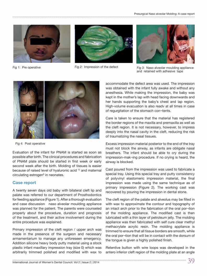
International Journal of Women’s Dental Council: Vol 2 | Issue 2 | 2014 39
Evaluation of the infant for PNAM is started as soon as possible after birth. The clinical procedures and fabrication of PNAM plate should be started in first week or early second week after the birth. Molding of tissues is easier because of raised level of hyaluronic acid 12 and maternal circulating estrogen8 in neonates.
Case report
A twenty seven days old baby with bilateral cleft lip and palate was referred to our department of Prosthodontics for feeding appliance (Figure 1). After a thorough evaluation and case discussion naso alveolar moulding appliance was planned for the patient. The parents were counseled properly about the procedure, duration and prognosis of the treatment, and their active involvement during the entire procedure was explained.
Primary impression of the cleft region / upper arch was made in the presence of the surgeon and necessary armamentarium to manage any unforeseen emergency. Addition silicone heavy body putty material using a stock plastic infant maxillary impression tray (size 0) which was arbitrarily trimmed polished and modified with wax to
accommodate the defect area was used. The impression was obtained with the infant fully awake and without any anesthesia. While making the impression, the baby was kept in the mother’s lap with head facing downwards and her hands supporting the baby’s chest and lap region. High-volume evacuation is also readv at all times in case of regurgitation of the stomach con¬tents.
Care is taken to ensure that the material has registered the border regions of the maxilla and premaxilla as well as the cleft region. It is not necessary, however, to impress deeply into the nasal cavity in the cleft, reducing the risk of traumatizing the nasal tissues.
Excess impression material posterior to the end of the tray must not block the airway, as infants are obligate nasal breathers. The infant should be able to cry during the impression-mak¬ing procedure. If no crying is heard, the airway is blocked.
Cast poured from the impression was used to fabricate a special tray. Using this special tray and putty consistency of polyvinyl elastomeric impression material, the final impression was made using the same technique as of primary impression (Figure 2). The working cast was recovered by pouring the impression in dental stone.
The cleft region of the palate and alveolus may be filled in with wax to approximate the contour and topography of an intact arch prior to the fabrication of the oral por¬tion of the molding appliance. The modified cast is then lubricated with a thin layer of petroleum jelly. The molding appliance was then fabricated with self cure clear methyl methacrylate acrylic resin. The molding appliance is trimmed to ensure that all tissue borders are smooth, while the oral por¬tion that will be in contact with the dorsum of the tongue is given a highly polished finish.
Retentive button with wire loops was developed in the antero inferior cleft region of the molding plate at an angle
Fig 1: Pre operative Fig 2: Impression of the defect Fig 3: Naso alveolar moulding appliance and retained with adhesive tape
Fig 4: Post operative
Presurgical Naso alveolar Molding: A case report
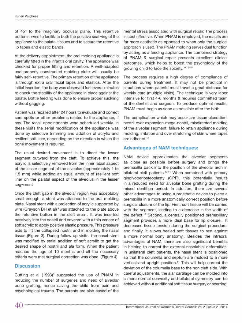
40 International Journal of Women’s Dental Council: Vol 2 | Issue 2 | 2014
of 45° to the imaginary occlusal plane. This retentive button serves to facilitate both the positive seat¬ing of the appliance to the palatal tissues and to secure the retentive lip tapes and elastic bands.
At the delivery appointment, the oral molding appliance is carefully fitted in the infant’s oral cavity. The appliance was checked for proper fitting and retention. A well-adapted and properly constructed molding plate will usually be fairly self- retentive. The primary retention of the appliance is through extra oral facial tapes and elastics. After the initial insertion, the baby was observed for several minutes to check the stability of the appliance in place against the palate. Bottle feeding was done to ensure proper suckling without gagging.
Patient was recalled after 24 hours to evaluate and correct sore spots or other problems related to the appliance, if any. The recall appointments were scheduled weekly. In these visits the serial modification of the appliance was done by selective trimming and addition of acrylic and resilient soft liner, depending on the direction in which the bone movement is required.
The usual desired movement is to direct the lesser segment outward from the cleft. To achieve this, the acrylic is selectively removed from the inner labial aspect of the lesser segment of the alveolus (approximately 1 to 1.5 mm) while adding an equal amount of resilient soft liner on the palatal aspect of the alveolus in the lesser seg¬ment
Once the cleft gap in the alveolar region was acceptably small enough, a stent was attached to the oral molding plate. Nasal stent with a projection of acrylic supported by wire (Grayson BH et al) 9 was attached to the plate above the retentive button in the cleft area . It was inserted passively into the nostril and covered with a thin veneer of soft acrylic to apply positive elastic pressure. This pressure aids to lift the collapsed nostril and in molding the nasal tissue (Figure 3). During follow up visits, the nasal stent was modified by serial addition of soft acrylic to get the desired shape of nostril and ala form. When the patient reached the age of 10 months and all the necessary criteria were met surgical correction was done. (Figure 4)
Discussion
Cutting et al (1993)6 suggested the use of PNAM in reducing the number of surgeries and need of alveolar bone grafting, hence saving the child from pain and psychological trauma. The parents are also eased of the
mental stress associated with surgical repair. The process is cost effective. When PNAM is employed, the results are far more aesthetic as opposed to when only the surgical approach is used. The PNAM molding serves dual function by acting as a feeding appliance. The combined strategy of PNAM & surgical repair presents excellent clinical outcomes, which helps to boost the psychology of the growing child to face the society. 9,13-15
The process requires a high degree of compliance of parents during treatment. It may not be practical in situations where parents must travel a great distance for weekly care (multiple visits). The technique is very labor intensive for first 4-6 months & requires committed team of the dentist and surgeon. To produce optimal results, PNAM must begin as soon as possible after the birth.
The complication which may occur are tissue ulceration, nostril over expansion-mega-nostril, misdirected molding of the alveolar segment, failure to retain appliance during molding, irritation and over stretching of skin where tapes are adhered.16
Advantages of NAM techniques:
NAM device approximates the alveolar segments as close as possible before surgery and brings the premaxilla back into the position of the alveolar arch in bilateral cleft patients.17,11 When combined with primary gingivoperiosteoplasty (GPP), this potentially results in a reduced need for alveolar bone grafting during the mixed dentition period. In addition, there are several other advantages to using a prosthetic device to place a premaxilla in a more anatomically correct position before surgical closure of the lip. First, soft tissue will be carried with the segment, leading to a decrease in the width of the defect.18 Second, a centrally positioned premaxillary segment provides a more ideal base for lip closure. It decreases tissue tension during the surgical procedure, and finally, it allows healed soft tissues to rest against a more normal bony anatomy.. Besides the intraoral advantages of NAM, there are also significant benefits in helping to correct the external nasolabial deformities. In unilateral cleft patients, the nasal stent is positioned so that the columella and septum are molded to a more vertical and upright position.11 This will help correct the deviation of the columella base to the non cleft side. With careful adjustments, the alar cartilage can be molded into a more normal convexity and bilateral symmetry can be achieved without additional soft tissue surgery or scarring.
Kurien Varghese
International Journal of Women’s Dental Council: Vol 2 | Issue 2 | 2014 41
Disadvantages of NAM
The disadvantages mentioned by various researchers are locked out segment, nostril overexpansion, irritation to skin and mucosa, exposure of primary tooth bud, obstruction of airway due to dislodgement of NAM appliance, and relapse of the molded cartilages though not entirely to some extent back to the original position.
Conclusion
The NAM technique has been significantly shown to improve the surgical outcome of CLP patients compared with other techniques of presurgical orthopedics. NAM has proved to be an effective adjunctive therapy for reducing hard and soft tissue cleft deformity before surgery. However, it is important that parents or caregivers become active members of the treatment team. Similarly, it is crucial that members of the cleft team provide the parents and caregivers adequate training, education, active support, and encouragement during NAM treatment. Lack of parent or care givers compliance and commitment results in less than ideal clinical outcomes. Despite a relative paucity of high level evidence, NAM appears to be a promising technique that deserves further study. The long term effectiveness of NAM is still to be evaluated as very few studies with long term follow up are available.
References1. Bailey BJ. Head and neck surgery- Otolaryngology. Lippincott
Raven, 1998.
2. Habel A, Sell D. Management of cleft lip and palate, Archives of diseases in childhood 1996;74:360-4.
3. McNiel C. Orthodontic procedures in the treatment of congenital cleft palate. Dent Rec 1950;70:126-32.
4. Mylin W. The pin-retained prosthesis in cleft palate orthodontics. Cleft Palate J 1968;5:219-24.
5. Latham R. Orthodontic advancement of the cleft maxillary segment : A preliminary report Cleft palate J 1980:227-33.
6. Grayson BH, Cutting CB, Wood R. Preoperative columella lengthening in bilateral cleft lip and palate. Plast Reconst Surg 1993;92:1422-3.
7. Grayson BH, Santiago PE. Pre surgical Orthopaedics for cleft lip and palate Grabb and smith Plastic Surgery, 5th edition Philadelphia: Lippincott Raven Publishers, 1997;20:237-44.
8. Grayson BH, Santiago PE. Presurgical nasoalveolar molding in infants with cleft lip and palate. Cleft palate Craniof J 1999;36:486-98.
9. Grayson BH, Cutting CB. Presurgical nasoalveolar Orthopedic moulding in primary correction of nose, lip, alveolus of infants born with unilateral and bilateral clefts. Cleft Palate Craniofac J 2001;38:193-8.
10. Grayson BH, Maull D. Nasoalveolar molding for infants born with cleft of the lip, alveolus and palate. Cleft Plastic Surgery 2004;31:149-58.
11. Ijaj A, Rafat A, Israr J. Nasoalveolar molding bilateral clefts of lip and palate infants with orthopaedic ring plate. J Pak Med Assoc 2010;60:527-31.
12. Matsuo k, Hirose T. Non surgical correction of congenital auricular deformities in early neonates, a preliminary report. Plast Reconst Surg 1984;73:38-50.
13. Santiago PE, Grayson BH, Gianoutsos MP et al. Reduced need for alveolar bone grafting by pre surgical orthopedics primary gingivoperiosteoplasty. Cleft Palate Craniofac J 1998;35:77-80.
14. Suri S, Utreja A. Management of effect of Presurgical Nasoalveolar Molding on Alveolar cleft. J Prosthet Dent 2004;92:428-33.
15. Singla S, Kaur M. Cleft palate rehabilitation. Indian J Paediatric 2008;75:703-8.
16. Bretch L, Grayson BH, Cutting CB. Nasoalveolar molding in early management of cleft lip and palate: In Clinical Method for Correction of the Unilateral Oronasal Deformity. 63-8.
17. Splengler LA, Chavarria C, Teichgraber FJ, Gatenes J, Xia JJ. Presurgical nasoalveolar moulding therapy for the treatment of bilateral cleft lip and palate: A preliminary study. Cleft Palate Craniofac J 2006;43: 321- 8.
18. Shetty V, Vyas HJ. A comparison of results using nasoalveolar moulding in cleft infants treated within 1 Month of life versus those treated after this period: Development of a new protocol. Int J OralMaxillofac Surg 2012;41:28-36.
Presurgical Naso alveolar Molding: A case report
42 International Journal of Women’s Dental Council: Vol 2 | Issue 2 | 2014
IJWDC: Vol 2 | Issue 2 | July-December 2014Short
Communication
Aggressive Periodontitis: Unmasking the Villain
Sapna Balakrishnan*
ADDRESS FOR CORRESPONDENCE
Dr. Sapna BalakrishnanReader, Dept of Periodontics,Pariyaram Dental College, Pariyaram, Mob-9496237720e-mail- sapna [email protected]
*Reader, Dept of Periodontics, Pariyaram Dental College, Pariyaram, Mob-9496237720 e-mail- sapna [email protected]
Introduction
Periodontitis is an infection that can manifest itself with a polymorph clinical presentation. Apart from the well-recognised chronic form of periodontitis formerly known as adult periodontitis, other more destructive types of periodontitis are prevalent in the society. Many-a-times the busy general practitioner misses out on the critical clinical findings and limits the treatment to a routine oral prophylaxis. Timely intervention and follow up in such cases can make drastic difference in the treatment plan for the patient as-much-as retaining a functional dentition to the option of a complete denture.
The 1999 international classification workshop reclassified the different forms of periodontitis into chronic periodontitis, aggressive periodontitis, periodontitis associated systemic diseases, and necrotizing periodontitis.
Aggressive Periodontitis
Aggressive periodontitis comprises a group of rare, often
severe, rapidly progressing forms of periodontitis often characterized by an early age of clinical manifestation and a distinctive tendency for cases to aggregate in the families2 (Lindhe).
It is also defined as a disease of the periodontium occurring in an otherwise healthy adolescent which is characterized by rapid loss of alveolar bone about more than one tooth of the permanent dentition. The amount of destruction is not commensurate with the amount of local irritants (Baers; 1971). Until recently these group of disease was defined primarily based on age of onset/diagnosis and named Early Onset Periodontitis.( fig 1,2)
Classification and clinical syndromes
Primary features (Lang et al,1999)
• Non-contributory medical history• Rapid attachment loss and bone destruction• Familial aggregation of cases
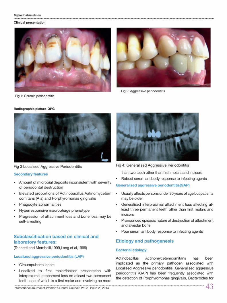
Author name
International Journal of Women’s Dental Council: Vol 2 | Issue 2 | 2014 43
Secondary features
• Amount of microbial deposits inconsistent with severity of periodontal destruction
• Elevated proportions of Actinobacillus Aatinomycetum comitans (A a) and Porphyromonas gingivalis
• Phagocyte abnormalities• Hyperresponsive macrophage phenotype• Progression of attachment loss and bone loss may be
self-arresting
Subclassification based on clinical and laboratory features:(Tonnetti and Mombelli,1999,Lang et al,1999)
Localized aggressive periodontitis (LAP)
• Circumpubertal onset• Localized to first molar/incisor presentation with
interproximal attachment loss on atleast two permanent teeth ,one of which is a first molar and involving no more
than two teeth other than first molars and incisors• Robust serum antibody response to infecting agentsGeneralized aggressive periodontitis(GAP)
• Usually affects persons under 30 years of age but patients may be older
• Generalised interproximal attachment loss affecting at-least three permanent teeth other than first molars and incisors
• Pronounced episodic nature of destruction of attachment and alveolar bone
• Poor serum antibody response to infecting agents
Etiology and pathogenesis
Bacterial etiology:
Actinobacillus Actinomycetemcomitans has been implicated as the primary pathogen associated with Localised Aggressive periodontitis. Generalised aggressive periodontitis (GAP) has been frequently associated with the detection of Porphyromonas gingivalis, Bacteroides for
Clinical presentation
Fig 1: Chronic periodontitis Fig 2: Aggressive periodontitis
Radiographic picture-OPG
Fig 3 Localised Aggressive Periodontitis Fig 4: Generalised Aggressive Periodontitis
Sapna Balakrishnan
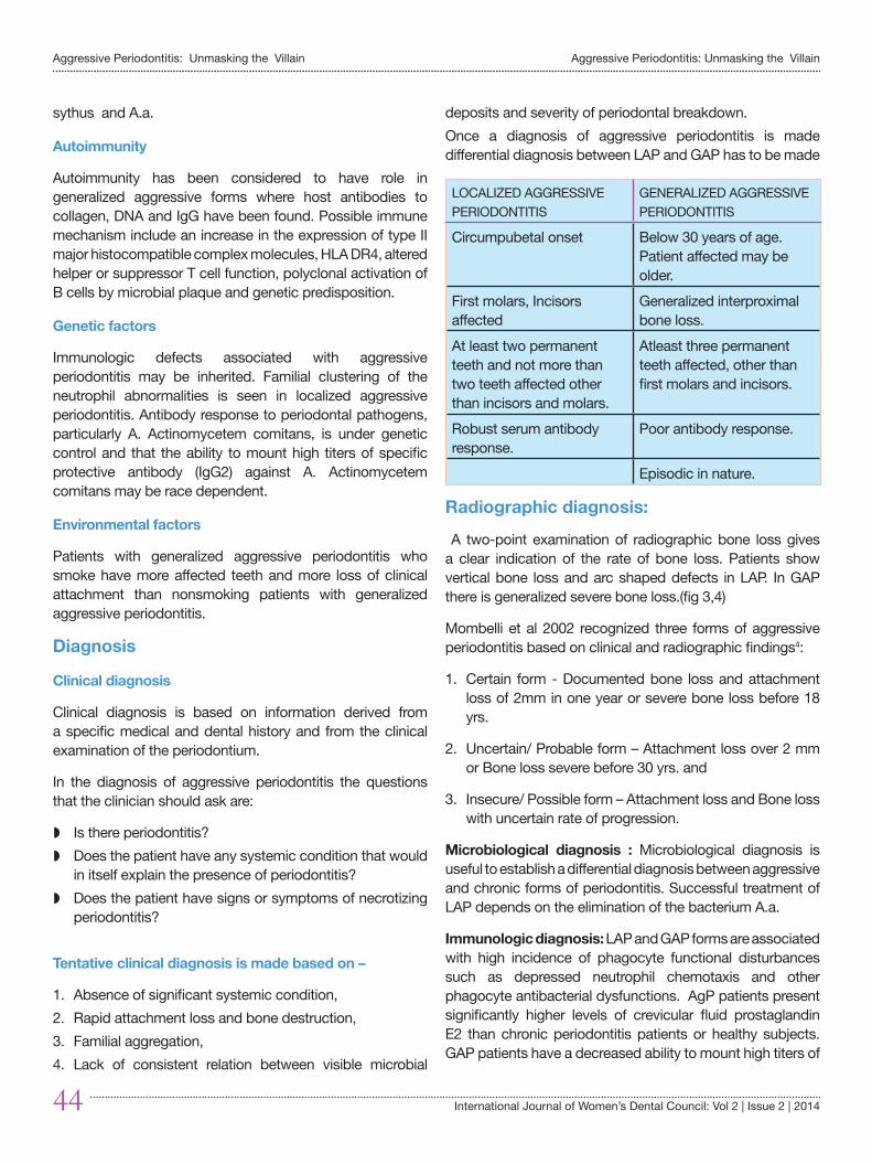
Aggressive Periodontitis: Unmasking the Villain
44 International Journal of Women’s Dental Council: Vol 2 | Issue 2 | 2014
sythus and A.a.
Autoimmunity
Autoimmunity has been considered to have role in generalized aggressive forms where host antibodies to collagen, DNA and IgG have been found. Possible immune mechanism include an increase in the expression of type II major histocompatible complex molecules, HLA DR4, altered helper or suppressor T cell function, polyclonal activation of B cells by microbial plaque and genetic predisposition.
Genetic factors
Immunologic defects associated with aggressive periodontitis may be inherited. Familial clustering of the neutrophil abnormalities is seen in localized aggressive periodontitis. Antibody response to periodontal pathogens, particularly A. Actinomycetem comitans, is under genetic control and that the ability to mount high titers of specific protective antibody (IgG2) against A. Actinomycetem comitans may be race dependent.
Environmental factors
Patients with generalized aggressive periodontitis who smoke have more affected teeth and more loss of clinical attachment than nonsmoking patients with generalized aggressive periodontitis.
Diagnosis
Clinical diagnosis
Clinical diagnosis is based on information derived from a specific medical and dental history and from the clinical examination of the periodontium.
In the diagnosis of aggressive periodontitis the questions that the clinician should ask are:
w Is there periodontitis? w Does the patient have any systemic condition that would
in itself explain the presence of periodontitis?w Does the patient have signs or symptoms of necrotizing
periodontitis?
Tentative clinical diagnosis is made based on –
1. Absence of significant systemic condition, 2. Rapid attachment loss and bone destruction, 3. Familial aggregation, 4. Lack of consistent relation between visible microbial
deposits and severity of periodontal breakdown. Once a diagnosis of aggressive periodontitis is made differential diagnosis between LAP and GAP has to be made
LOCALIZED AGGRESSIVE PERIODONTITIS
GENERALIZED AGGRESSIVE PERIODONTITIS
Circumpubetal onset Below 30 years of age. Patient affected may be older.
First molars, Incisors affected
Generalized interproximal bone loss.
At least two permanent teeth and not more than two teeth affected other than incisors and molars.
Atleast three permanent teeth affected, other than first molars and incisors.
Robust serum antibody response.
Poor antibody response.
Episodic in nature.
Radiographic diagnosis:
A two-point examination of radiographic bone loss gives a clear indication of the rate of bone loss. Patients show vertical bone loss and arc shaped defects in LAP. In GAP there is generalized severe bone loss.(fig 3,4)
Mombelli et al 2002 recognized three forms of aggressive periodontitis based on clinical and radiographic findings4:
1. Certain form - Documented bone loss and attachment loss of 2mm in one year or severe bone loss before 18 yrs.
2. Uncertain/ Probable form – Attachment loss over 2 mm or Bone loss severe before 30 yrs. and
3. Insecure/ Possible form – Attachment loss and Bone loss with uncertain rate of progression.
Microbiological diagnosis : Microbiological diagnosis is useful to establish a differential diagnosis between aggressive and chronic forms of periodontitis. Successful treatment of LAP depends on the elimination of the bacterium A.a.
Immunologic diagnosis: LAP and GAP forms are associated with high incidence of phagocyte functional disturbances such as depressed neutrophil chemotaxis and other phagocyte antibacterial dysfunctions. AgP patients present significantly higher levels of crevicular fluid prostaglandin E2 than chronic periodontitis patients or healthy subjects. GAP patients have a decreased ability to mount high titers of
Aggressive Periodontitis: Unmasking the Villain
Author name
International Journal of Women’s Dental Council: Vol 2 | Issue 2 | 2014 45
specific IgG2 antibodies to A.a.
Genetic diagnosis: Given the disproportionately high incidence of AgP in the families of affected individuals, evaluation of sibling of the proband and other family members is a requirement.
Treatment
Treatment should be initiated after completion of a careful diagnosis by specially trained periodontist. Successful treatment depends on early diagnosis, directing treatment towards suppressing or eliminating the infecting organisms and providing an environment conductive for long term maintenance.
Localized forms
The following treatments have been considered with varying degrees of success.
1. Extraction: the involved teeth, usually the first molars, are extracted and uneventful healing occurs. Transplantation of an erupting third molar in the extraction socket has also been attempted.
2. Standard periodontal therapy: Scaling and root planing, curettage, flap surgery with or with out bone grafts, root amputation, hemi sections, occlusal adjustments and strict plaque control measures. Frequent maintenance visits have been the most important aspect of treatment.
3. Antibiotic therapy: localized aggressive periodontitis has been successfully treated with SRP plus tetracycline (250mg four times daily for 14 days every 8 weeks). Carranza advocates systemic tetracycline 250mg four times daily for at least one week in conjunction with local mechanical therapy. If surgery is indicated antibiotics should be prescribed 1hr before the surgery. Doxycycline 100mg per day may also be used in conjunction with the use of chlorohexidine.
If the case is refractory then antibiotic susceptibility tests should be done for the proper choice of antibiotics. The clinician may consider a combination of amoxicillin and metronidazole similar to that suggested for refractory periodontitis.
Generalized forms
The rate of progression may be faster in these individuals and therefore the clinician should monitor the patients close with close collaboration with periodontist, general dentist,
dental hygienist and the patient’s physician. It is important to observe the over all physical status of the patient as well.
Summary and Conclusion
Aggressive periodontitis is a condition that should be recognised and managed at the earliest to limit the consequences. The general guidelines to be followed are:
1. A general medical evaluation may determine if systemic disease is present in children and young adults who exhibit severe periodontitis, particularly if aggressive periodontitis appears to be resistant to therapy. Consultation with the patient’s physician may be indicated to coordinate medical care in conjunction with periodontal therapy. Modification of environmental risk factors should be considered.
2. Initial periodontal therapy alone is often ineffective. However, in the early stages of disease, lesions may be treated with adjunctive antimicrobial therapy combined with scaling and root planing with or without surgical therapy. Microbiological identification and antibiotic sensitivity testing may be considered. In very young patients, the use of tetracyclines may be contraindicated due to the possibility of staining of teeth. Alternative antimicrobial agents or delivery systems may be considered.
3. The long-term outcome may depend upon patient compliance and delivery of periodontal maintenance at appropriate intervals, as determined by the clinician If primary teeth are affected, eruption of permanent teeth should be monitored to detect possible attachment loss.
4. Due to the potential familial nature of aggressive diseases, evaluation and counseling of family members may be indicated.
References1. Jan Lindhe, Thorkild Karring, Niklaus P Lang ‘Clinical
Periodontology and Implant Dentistry’; 3rd,4th edition
2. Fermin A. Carranza Jr, Michael G. Newman, ‘Clinical Periodontology’, Saunders, An imprint of Elsevier, Philadelphia, Pennsylvania, 1996, 8th, 9th and 10th edn
3. Wilson TG, Kornman,KS, ‘Treating Aggressive Forms of Periodontal Disease with Severe bone loss’ in ‘Fundamentals of Periodontics’ 2nd edn Quintesence Publishing Co
4. AAP Position Paper Periodontal disease in childhood and adolescents.J Periodontol 1996;67;355-366
5. Parameter on Aggressive Periodontitis, JOP , May 2001,Vol 71(supplement 5)
Sapna Balakrishnan
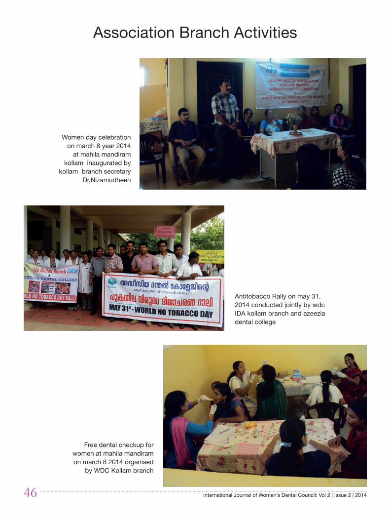
46 International Journal of Women’s Dental Council: Vol 2 | Issue 2 | 2014
Association Branch Activities
Women day celebration on march 8 year 2014
at mahila mandiram kollam inaugurated by
kollam branch secretary Dr,Nizamudheen
Antitobacco Rally on may 31, 2014 conducted jointly by wdc IDA kollam branch and azeezia dental college
Free dental checkup for women at mahila mandiram on march 8 2014 organised
by WDC Kollam branch
International Journal of Women’s Dental Council: Vol 2 | Issue 2 | 2014 47
GUIDELINES FOR AUTHORS
Manuscripts Preparation Manuscripts should be sent to be Editor, in original & two complete copies, including illustrations. The manuscript should also be sent in two fl oppies/CD and pictures be sent in original for better reproduction. The author’s name is to be written only on the original copy, and not on the Xerox copies. The manuscripts should be type written, double-spaced on one side of the paper. With at least one inch margin on all sides. All pages should be numbered sequentially.
Title Page Title page should include the title of the article and the name, degrees, position, professional affi liation of each author. The corresponding author’s fax, telephone, email address and complete mailing address must be provided.
Abstract/ Key Words Page 2 should include a maximum of 200 words abstract with abbreviated title for the page head use. The abstract should state the purpose of the study/ investigations, basic procedures, main fi ndings and the principalconclusions. Emphasize new and important aspects of the study/ observations. Only approved abbreviations are to be used.
Clinical Relevance Statement experimental investigations should include a brief statement of the clinical relevance of the paper.
Text: The text of articles should be divided into sections with the headings introduction, Material & Methods, Results and Discussions.
Indtroduction Summarise the purpose and rationale of the study. Give only pertinent references & do not extensively review the subject. Clearly state the working hypothesis.
Material & Methods To allow confi rmation of the observations, present the material & methods in suffi cient detail. Published methods should be referred to & discussed briefl y. Only if modifi cations have been made, provide details.
Results Present results in a logical sequence in the text, tables & illustrations. Do not repeat in the text all the data in the tables, summarise only important observations.
Discussions Emphasize the new & important aspects of the study and the conclusions that follow them. Do not repeat the observations to other relevant studies. Relate observations to other relevant studies & point out the implications of the fi ndings & their limitations. Avoid unqualifi ed statements & conclusions not completely supported by your data. Recommendations, when
appropriate, may be included.
Acknowledgements Acknowledge only persons who have made signifi cant contributions to the study. Authors are responsible for obtaining written permission from persons acknowledged by the name because readers may infer their endorsement of the data & conclusions.
References Authors are responsible for the accuracy of the references. The reference list should be double spaced at the end of the article in numeric sequence.
All references given must be cited in the text, numbered in order of appearance. Use the style which are based on the formats used by index Copernicus TM Journals master list. Do not include unpublished data or personal communications in the reference list. Cite such references in parenthesis in the text & include a date. Avoid using abstracts as references. Provide complete information for each reference, including names of all authors. If the references is to a part of a book, also include the title of the chapter & names of the book’s editors(s). Journal references should included author’s name, article title, abbreviated (as per standards) journal name, volume number, page number & year. References must be verifi ed by the author(s) against documents.
Illustrations All illustrations must be numbered and cited in the text in order of appearance. On the back of each illustration, place a label with the article title & fi gure number only. Do not include author names. Indicate on the top edge lightly in pencil. Do not bend, fold or use, paper clips.
Instead of original drawings, roentgenograms & other material, send sharp, glossy colour, black and white photographic prints, usually 127 by 173 mm (5 by 7 inch) but not larger than 203 by 254mm (8 by 10 inches). Letters, numbers and symbols should be clear and of suffi cient size.
For illustrations in colour, supply colour positives/ transparencies and when necessary accompanying drawing marked to indicate the region to be reproduced.
Photographs of X-rays should be sent and not the original X-rays. If photographs of persons are used, either the subjects must not be identifi able or their pictures must be photograph.
Labels Each table should be logically organized on a separate sheet & numbered consecutively. The title & footnotes should be included with the table.
GUIDELINES FOR AUTHORS
Aggressive Periodontitis: Unmasking the Villain
48 International Journal of Women’s Dental Council: Vol 2 | Issue 2 | 2014
Copy right To ensure maximum dissemination and copyright protection of material published in the journal, copyright must be explicitly transferred from the author to the Indian Dental Association. Submission of manuscripts implies that the work described has not been published before (except in the form of an abstract or as part of published lecture, review or thesis) that is not under consideration for publication elsewhere, & if accepted, it will not be published elsewhere in the same form in either the same or another language without the consent of copyright holders. The copyright covers the exclusive rights of photographic reprints, video cassettes and such similar things. Certificate signed by author/s to this effect be submitted with the manuscript. The editors & publishers accept no legal responsibility for any errors, omissions or opinions expressed by authors. The publisher makes no warranty for expressions implied with respect to the materials contained therein. The journal is edited & published under the directions of the journal committee, which reserves the right to reject any material without giving explanations.
All communication should be addressed to the Editor. Request for change of address should be referred to Hon. Secy. General. No responsibility will be taken for undelivered issue due to circumstances beyond the control of the publisher.
Letters to the Editor Letters to the Editor are encouraged to stimulate a healthy dialogue relating to the specialty.
Books for Review Books and monographs will be reviewed based on their relevance to IJWDC readers. Books should be sent to the Editor and will become the property of IJWDC.
Subscription for Hard Copy: Rs : 2000 per b/w copy
Rs : 3000 per color copy
GUIDELINES FOR AUTHORS
Edited By : Dr.R.RathyDesigned By : Suman Graphics, [email protected]
Published By : Women’s Dental Council, Kerala State Branch.