Index Copernicus ID 5385 ISSN No. 0972-396X ... - IDA Kerala
81
Index Copernicus ID 5385 ISSN No. 0972-396X Vol 39 | No. 1 January 2016 Biologic Width – Line of controller of Gingiva Interdisciplinary Periodontics Saliva: A diagnostic tool for oral cancer Impression techniques for ocular prosthesis Immediate denture – an important treatment modality Biological restorations in children Recording sublingual crescent region for improving stability and retention in mandibular dentures Rehabilitation of an edentulous geriatric patient with resorbed residual ridges and excessive interarch space using a maxillary hollow complete denture Comparison of different methods of assessing alveolar ridge dimensions prior to dental implant placement Predictive marker of residual ridge resorption-biochemical and radiographic evaluation Current concepts in eradicating enterrococcus faecalis biofilm Digital impressions – a peak into the future A case report on the management of an infrabony defect with tetracycline fibres Sweetening the burden of diabetes Evaluation of the effectiveness of photodynamic therapy using toluidine blue for microbial reduction in periodontal pockets Management of Excessive Gingival Display A cross sectional survey on the utilization of scientific journals among staffs and students of Dental Colleges in Kerala Email: [email protected] Web: www.idakerala.com SIS Index ID 833 Quarterly Publication of Indian Dental Association, Kerala State Branch
-
Upload
khangminh22 -
Category
Documents
-
view
2 -
download
0
Transcript of Index Copernicus ID 5385 ISSN No. 0972-396X ... - IDA Kerala

Index Copernicus ID 5385 ISSN No. 0972-396X
Vol 39 | No. 1January 2016
Kerala Dental Journal Vol. 39 | No. 1 | January 2016 ISSN No. 0972-396X
Biologic Width – Line of controller of Gingiva
Interdisciplinary Periodontics
Saliva: A diagnostic tool for oral cancer
Impression techniques for ocular prosthesis
Immediate denture – an important treatment modality
Biological restorations in children
Recording sublingual crescent region for improving stability and retention in mandibular dentures
Rehabilitation of an edentulous geriatric patient with resorbed residual ridges and excessive interarch space using a maxillary hollow complete denture
Comparison of different methods of assessing alveolar ridge dimensions prior to dental implant placement
Predictive marker of residual ridge resorption-biochemical and radiographic evaluation
Current concepts in eradicating enterrococcus faecalis biofi lm
Digital impressions – a peak into the future
A case report on the management of an infrabony defect with tetracycline fi bres
Sweetening the burden of diabetes
Evaluation of the effectiveness of photodynamic therapy using toluidine blue for microbial reduction in periodontal pockets
Management of Excessive Gingival Display
A cross sectional survey on the utilization of scientifi c journals among staffs and students of Dental Colleges in Kerala
Email: [email protected] Web: www.idakerala.com
SIS Index ID 833
Q u a r t e r l y P u b l i c a t i o n o f I n d i a n D e n t a l A s s o c i a t i o n , K e r a l a S t a t e B r a n c h

Kerala Dental JournalVol. 39 | No. 1 | January 2016
Edited by: Dr. K. Nandakumar, Hon. Editor • Published By: Dr. Suresh Kumar G., Hon Secretary • For IDA, Kerala State Branch • Production: Suman Graphics, [email protected]
EDITOR
Dr. K. Nandakumar
ASST. EDITOR
Dr. R.M. Baiju
BUSINESS MANAGER
Dr. Mathew Jose
EDITORIAL CONSULTANTS
Dr. Santhosh SreedharDr. K. Chandrasekharan NairDr. Ipe VargheseDr. Oommen Aju JacobDr. V.I. PaulDr. Thomas ManjooranDr. Sobha KuriakoseDr. T. SreelalDr. Siby XavierDr. M.K. Mangalam
EX-OFFICIO MEMBERS
Dr Mohammed Sameer P.T.Dr. Suresh Kumar G.Dr. Thomas KCDr. Sabu Kurian
EDITORIAL BOARD
Dr. Dibyendu Mazumder Dr. Ashok DhobleDr. Jayakar Shet tyDr. Radhakrishnan NairDr. K.T. SreelathaDr. Sunil SunnyDr. Jayaprasad K.Dr. Ajay Kumar HaridasDr. Prasanthila JanamDr. George VargheseDr. RetnakumariDr. Jolly Mary VarugheseDr. Romel JosephDr. Thomas JosephDr. Pradeep Kumar C.Dr. Vinod R.B.Dr. S. George P. JohnDr. P.G. FrancisDr. M. Sadique HussainDr. Saji P.Dr. N.O. VargheseDr. Gopinathan M.Dr. Aby Mathew T.Dr. E. Anuradha Sunil JaganDr. Manoj NairDr. P.C. SunilDr. Shamsuddin
EDITORIAL OFFICE
Neelambikam, At tukal, Manacaud Trivandrum, Kerala - 695 009
Phone: 0471-2459235 Mobile: 09447066100
e-mail: [email protected] web: www.idakerala.com
OFFICE BEARERS OF IDA KERALA STATE
PresidentDr Mohammed Sameer P.T.
imm. Past President Dr. Thomas KC
President electDr Sabu Kurian
Vice Presidents Dr. Ciju A. Poulose
Dr Eugene Varghese JosephDr Fazil V Hassan
Hon. secretary Dr Suresh Kumar G
Joint secretary Dr Binoy Stanly
asst. secretary Dr. Krisshna Kumar K S
treasurerDr. Santhoshkumar P.U.
editorDr. K. Nandakumar
cde conVenor Dr. Nirmal George Saibu
cdH cHairmanDr. Subash K Madhavan
Oral Cancer Incidence and Prevalence
The Indian ScenarioOral cancer is the most common cancer in India; as
4 in 10 of all cancers are oral cancers. Annually 130,000 people succumb to oral cancer in India which translates into approximately 14 deaths per hour. The reason for high prevalence of oral cancer in India is primarily because tobacco is consumed in the form of gutka, quid, snuff or misri. Rising tobacco use in India, where 40 per cent of the world’s smokers live has contributed to this trend. In comparison, in US oral cancer represents approximately 13% of all cancers thereby translating into 30,000 new cases every year.
Facts about oral cancer in India• Recently,atrendhasbeenobservedtowardsincreased
incidence of oral cancer among young adults. This increase in incidence is only observed in patients with tongue cancer.
• Infact,inIndia,60-80%of patientsarepresentwithadvanced disease as compared to 40% in developed countries. Early detection would not only improve the cure rate, but it would also lower the cost and morbidity associated with treatment.
• Increasingprevalenceof oralsubmucousfibrosis,especiallyin younger individuals, caused by gutka, an industrially manufactured food item has been seen.
The above facts state that, cancer cases in general, are increasing in India and it is high time that planners, social activists and government give adequate stress for prevention, early diagnosis, treatment and rehabilitation of these populations.
Index Copernicus ID 5385 ISSN No. 0972-396X
Vol 39 | No. 1January 2016
Kerala Dental Journal Vol. 39 | No. 1 | January 2016 ISSN No. 0972-396X
Biologic Width – Line of controller of Gingiva
Interdisciplinary Periodontics
Saliva: A diagnostic tool for oral cancer
Impression techniques for ocular prosthesis
Immediate denture – an important treatment modality
Biological restorations in children
Recording sublingual crescent region for improving stability and retention in mandibular dentures
Rehabilitation of an edentulous geriatric patient with resorbed residual ridges and excessive interarch space using a maxillary hollow complete denture
Comparison of different methods of assessing alveolar ridge dimensions prior to dental implant placement
Predictive marker of residual ridge resorption-biochemical and radiographic evaluation
Current concepts in eradicating enterrococcus faecalis biofi lm
Digital impressions – a peak into the future
A case report on the management of an infrabony defect with tetracycline fi bres
Sweetening the burden of diabetes
Evaluation of the effectiveness of photodynamic therapy using toluidine blue for microbial reduction in periodontal pockets
Management of Excessive Gingival Display
A cross sectional survey on the utilization of scientifi c journals among staffs and students of Dental Colleges in Kerala
Email: [email protected] Web: www.idakerala.com
SIS Index ID 833
Q u a r t e r l y P u b l i c a t i o n o f I n d i a n D e n t a l A s s o c i a t i o n , K e r a l a S t a t e B r a n c h

ContentsKerala Dental JournalVol. 39 | No. 1 | January 2016
Preisident’s Message 10Secretary’s Report 11Editorial 12Biologic Width – Line of controller of Gingiva 13 — Agee AntonyInterdisciplinary Periodontics 17 — Harikrishnan Balachandran Pillai Saliva: A diagnostic tool for oral cancer 20 — Jinisham Impression techniques for ocular prosthesis 24 — Rohit RaghavanImmediate denture – an important treatment modality 27 — Kalpana HastiBiological restorations in children 30 — Seena JohnRecording sublingual crescent region for improving stability and retention in mandibular dentures 33 — Thomas PaulRehabilitation of an edentulous geriatric patient with resorbed residual ridges and excessive interarch space using a maxillary hollow complete denture 37 — Ashwin Kumar S G.Comparison of different methods of assessing alveolar ridge dimensions prior to dental implant placement 40 — Veena T.K.Predictive marker of residual ridge resorption-biochemical and radiographic evaluation 43 — Jemcy JamesCurrent concepts in eradicating enterrococcus faecalis biofilm 47 — Ruth Hepsi BealahDigital impressions – a peak into the future 51 — Rohit Raghavan A case report on the management of an infrabony defect with tetracycline fibres 55 — Kadeeja RushinSweetening the burden of diabetes 58 — Navia George Evaluation of the effectiveness of photodynamic therapy using toluidine blue for microbial reduction in periodontal pockets 60 — Anjhana NarayananManagement of Excessive Gingival Display 65 — Jyotsna J.A cross sectional survey on the utilization of scientific journals among staffs and students of dental colleges in Kerala 68 — Civy V. Pulayath Quiz 70 — Jayanthi Association News 71
KDJ–Vol.39•No.1•January2016 9

Dear friends and colleagues,
ItiswithprideandhonorthatIwritemyfirstletteraddressingthemembers of the kerala state branch of the Indian Dental Association. This wonderful organization runs on the shoulders of ordinary members like each of you and I pray that I will be able to move forward with the support of one and all.
Over the years under the able leadership of the past presidents and secretaries IDA KSB has grown in many levels.
The social commitment of its members is tremendous and I wish to highlight this during my tenure as President. It is my dream to serve the elderly underprivileged people of our homeland by making the maximum number of complete dentures possible and delivering them to the needy.
I earnestly request all members of our branch to take up this cause and volunteer to make a complete denture for the needy person in your neighbourhood.
Ourassociation’sactivitiesencompassawiderange-social,professionaland humanitarian. Together with the Honorary Secretary Dr Suresh Kumar G and our team, we wish to highlight our activities in society and please ensure that you take an active role in the CDE, CDH and cultural programs of your branch. Together we will be take IDA to greater heights and make it more visible in the public eye. At the same time, we are also striving to create a smoother arena for the functioning and for obtaining the required governmental clearances for the same by coordinating with the government and other agencies. I assure you that I will do my very best to safeguard the interests of the profession and once again seek your blessings and support
Jai Hind and Jai IDA
Dr Mohamad Sameer P TPresident, IDA Kerala State
President’s Message
DrMohamadSameerPT
10 KDJ–Vol.39•No.1•January2016

My “GOOD WISHES” to all members of IDA Kerala State. At the onset let me thank all members of IDA Kerala State for giving me the opportunity to correlate with you at a more personal level as Hon. Secretary of this prestigious organisation. Naturally, I’m acutely aware of the profound responsibility and trust this position carries along with it.
On behalf of IDA Kerala State I request each one of you to make a concerted effort whereby more dentists can be brought under the IDA Kerala State umbrella and thus strengthen our hands to meet the challenges that would come up in our profession. Besides this also increases the fraternity feeling among ourselves with mutual trust and brotherhood.
Our doors are open to all members at all times and we will always be available to hear your grievances andfindasuitableandamicablesolutiontoyourproblemswithintheambitof reasonablelimits.Professional issues such as the problems associated with mushrooming of Dental colleges, the private dental practice scenario, the proposed Clinical Establishment legislation, the problems associated with registrations and Licences are just some of the many key issues which threatens to undermine working conditions of private clinics in this state on which we will continually remain vigilant.
I sincerely hope that IDA is able to extend its presence to the Government sector and Private colleges as well. Efforts will be undertaken to liaise with professional dental organisations in the Government sectoranditwoulddefinitelystrivetoupholdandprotecttherightsof thePrivatedentalcollegefaculties. Our aim would be to position IDA in its rightful place with regard to policy making and administrative positions thereby playing a vital role with regards to Dental profession.
Theonusof thestateofficewillbeoneffectivecommunicationswiththeLocalBranchesanditsmembers. As we grow in strength and number we have to adapt more to embrace ourselves with the technology around us while at the same time realise that this does not create a disconnect with the members at large.
IhopeduringmytenureasHonSecretary,theofficewouldbeabletocarryalongallmembersin unity and friendship and also expect that all members will extend their undivided support to implement the ideals that our predecessors have tirelessly struggled to implement over the years.
Last but not the least, I extend my appreciation to the Editorial Board of KDJ and in particular to Dr K. Nandakumar who over the years have made KDJ the best journal ever.
Dr. Suresh Kumar G.Hon. Secretary, IDA Kerala State
Secretary’s Report
Dr.SureshKumarG
KDJ–Vol.39•No.1•January2016 11
Editorial
Dr.K.Nandakumar
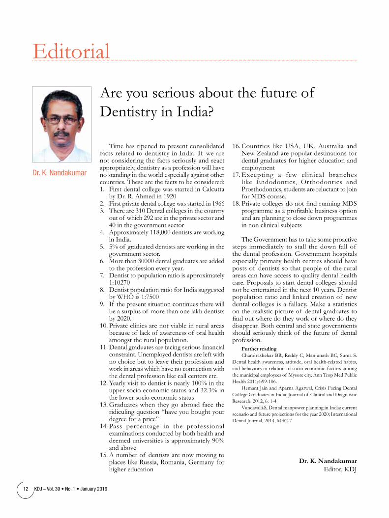
Time has ripened to present consolidated facts related to dentistry in India. If we are not considering the facts seriously and react appropriately, dentistry as a profession will have no standing in the world especially against other countries. These are the facts to be considered:1. First dental college was started in Calcutta
by Dr. R. Ahmed in 1920 2. Firstprivatedentalcollegewasstartedin19663. There are 310 Dental colleges in the country
out of which 292 are in the private sector and 40 in the government sector
4. Approximately118,000dentistsareworkingin India.
5. 5% of graduated dentists are working in the government sector.
6. Morethan30000dentalgraduatesareaddedto the profession every year.
7. Dentist to population ratio is approximately 1:10270
8. DentistpopulationratioforIndiasuggestedby WHO is 1:7500
9. If the present situation continues there will be a surplus of more than one lakh dentists by 2020.
10. Private clinics are not viable in rural areas because of lack of awareness of oral health amongst the rural population.
11.Dentalgraduatesarefacingseriousfinancialconstraint. Unemployed dentists are left with no choice but to leave their profession and work in areas which have no connection with the dental profession like call centers etc.
12. Yearly visit to dentist is nearly 100% in the upper socio economic status and 32.3% in the lower socio economic status
13. Graduates when they go abroad face the ridiculing question “have you bought your degree for a price”
14. Pass percentage in the professional examinations conducted by both health and deemed universities is approximately 90% and above
15. A number of dentists are now moving to places like Russia, Romania, Germany for higher education
16.Countries likeUSA,UK,Australia andNew Zealand are popular destinations for dental graduates for higher education and employment
17. Excepting a few clinical branches like Endodontics, Orthodontics and Prosthodontics, students are reluctant to join for MDS course.
18.PrivatecollegesdonotfindrunningMDSprogrammeasaprofitablebusinessoptionand are planning to close down programmes in non clinical subjects
The Government has to take some proactive steps immediately to stall the down fall of the dental profession. Government hospitals especially primary health centres should have posts of dentists so that people of the rural areas can have access to quality dental health care. Proposals to start dental colleges should not be entertained in the next 10 years. Dentist population ratio and linked creation of new dental colleges is a fallacy. Make a statistics on the realistic picture of dental graduates to findoutwheredotheyworkorwheredotheydisappear. Both central and state governments should seriously think of the future of dental profession.
Further readingChandrashekar BR, Reddy C, Manjunath BC, Suma S.
Dentalhealthawareness,attitude,oralhealth-relatedhabits,andbehaviorsinrelationtosocio-economicfactorsamongthe municipal employees of Mysore city. Ann Trop Med Public Health2011;4:99-106.
Hemant Jain and Aparna Agarwal, Crisis Facing Dental College Graduates in India, Journal of Clinical and Diagnostic Research.2012,6:1-4
Vundavalli.S, Dental manpower planning in India: current scenario and future projections for the year 2020; International DentalJournal,2014,64:62-7
Dr. K. Nandakumar Editor, KDJ
Are you serious about the future of Dentistry in India?
12 KDJ–Vol.39•No.1•January2016
The concept of biologic width:The concept of biologic width is
based on histologic studies by Gargiulo et al3 of the stages of passive eruption in normal cadaver Periodontium, on the dimensions and relationships of dentogingival junction in humans. The reportedfindingsweregleanedfrom30humancadaverjaws,and287teethof which 325 surfaces were examined andquantifiedtheaverageasaconstant2.04mm (the epithelial attachment is 0.97 mm, and connective tissue is 1.07 mm) withsulcusdepth0.69mm.(fig.1).In1977, Ingber et al described biologic width and credited D Walton Cohen for firstcoiningtheterm“biologicwidth”4 and suggested that a minimum of 3mm was required from the restorative margin to alveolar bone crest to permit adequate healing and restoration of tooth. Maynard and Wilson (1979) divided the periodontium into three dimensions; superficial physiologic, crevicular physiologic and subcrevicular physiologic.5• Superficial physiologic- free
gingival+attached gingival• Crevicularphysiologic-dimension
from gingival margin to junctional epithelium
• Subcrevicularphysiologic-junctionalepithelium+connective tissue attachmentIn 1994, Vacek et al on evaluation
reported that connective tissue attachment was the most consistent measurement and interproximally the biologic width is similar to that of the facial surface but not the total dentogingival complex.6
BiologicWidth–LineofcontrollerofGingiva* Agee Antony, **Subair K., ***Anil Melath
AbstractBiologic width is perceived to bestow specific numeric dimensions to a unique and dynamic biologic entity. Maintenance of biologic width is one of the key for the success or longevity of dental restorations and for the tooth. Clinicians must be aware that biologic width is indeed a reality and violation of biologic width leads to complication such as gingival inflammation and alveolar bone loss. Preserving biologic width can pave way for a longer life of restorations with healthy Periodontium. This article discusses the various methods for biologic width assessment, guidelines and various procedures for reconstruction of biologic width.
Key words: Biologic width, restorative margin, gingivectomy, apically repositioned flap, supracrestal fiberotomy.
KDJ 2016 | Vol. 39 | No. 1 | Pg 13-16
► IntroductionNatural teeth are surrounded by
gingival soft tissues that provide a biologic seal between the oral cavity and the inside of the body. This unique structure is composed of epithelium and soft connective tissue that are continuallybathedingingivalfluid1. This unique structure; biologic width is rather a great emphasis focused on perio-restorativeinterfaceinrestorativedentistry.Thebiologicwidthisdefinedas “the dimension of soft tissue, which is attached to the portion of the tooth coronal to crest of alveolar bone”.2
Evaluation: Clinical method Bone sounding Radiographic methodClinical method:
Signs of biologic width violation that are seen clinically are as follows:7
(fig2&3)• Chronic progressive gingival
inflammationaroundtherestoration• Bleedingonprobing• Localizedgingivalhyperplasia• Clinicalattachmentloss• AlveolarbonelossBone sounding
The biologic width can be assessed by probing under local anesthesia to the bone level and if this distance is less than 2mm at one or more locations, a diagnosis of biologic width violation canbeconfirmed(fig.4). Itcanbea presumptive guide for bone level assessment.8Radiographic evaluation:
Radiographic interpretation can identify interproximal violations of biologic width. A new innovative parallel profileradiographic(PRR)techniqueis used to measure both length and thickness of dentogingival unit with accuracy.9Perio-restorative interrelationship:
Categories of biologic width and margin placement guidelines to prevent violation. Kois proposed three categories of biologic width based on total dimension of attachment and the sulcus depth following bone sounding measurement namely normal crest, high crest, low crest.10,11
KDJ–Vol.39•No.1•January2016 13
KDJ – Kerala Dental Journal INfoRMAtIoN
*JuniorResident,**Reader,***Professor&HOD,Principal,DepartmentofPeriodontics;MaheInstituteofDentalSciences&Hospital,Mahe•CorrespondingAuthor:Dr.AgeeAntonyE-mail:[email protected]
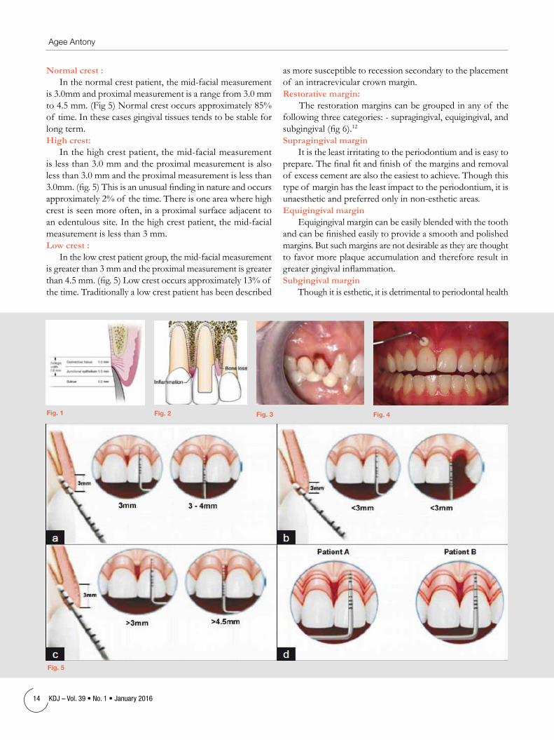
Normal crest :Inthenormalcrestpatient,themid-facialmeasurement
is 3.0mm and proximal measurement is a range from 3.0 mm to4.5mm.(Fig5)Normalcrestoccursapproximately85%of time. In these cases gingival tissues tends to be stable for long term.High crest:Inthehighcrestpatient,themid-facialmeasurement
is less than 3.0 mm and the proximal measurement is also less than 3.0 mm and the proximal measurement is less than 3.0mm.(fig.5)Thisisanunusualfindinginnatureandoccursapproximately 2% of the time. There is one area where high crest is seen more often, in a proximal surface adjacent to anedentuloussite.Inthehighcrestpatient,themid-facialmeasurement is less than 3 mm.Low crest :
Inthelowcrestpatientgroup,themid-facialmeasurementis greater than 3 mm and the proximal measurement is greater than4.5mm.(fig.5)Lowcrestoccursapproximately13%of the time. Traditionally a low crest patient has been described
as more susceptible to recession secondary to the placement of an intracrevicular crown margin.Restorative margin:
The restoration margins can be grouped in any of the followingthreecategories:-supragingival,equigingival,andsubgingival(fig6).12
Supragingival margin It is the least irritating to the periodontium and is easy to
prepare.Thefinalfitandfinishof themarginsandremovalof excess cement are also the easiest to achieve. Though this type of margin has the least impact to the periodontium, it is unaestheticandpreferredonlyinnon-estheticareas.Equigingival margin
Equigingival margin can be easily blended with the tooth andcanbefinishedeasilytoprovideasmoothandpolishedmargins. But such margins are not desirable as they are thought to favor more plaque accumulation and therefore result in greatergingivalinflammation.Subgingival margin
Though it is esthetic, it is detrimental to periodontal health
fig. 1 fig. 2 fig. 3 fig. 4
fig. 5
14 KDJ–Vol.39•No.1•January2016
Agee Antony

as it acts as a permanent irritant to the periodontium. Many studies have demonstrated qualitative and quantitative changes in subgingival microbes, increased plaque index, gingival recession and pocket depth.5 Biologic width encroachment becomes more common when planning for subgingival restorations in cases that are fractured or carious, near the alveolar crest. Also esthetics demands often require hiding of restorative margins below the gingival margins i.e., pushing them down into the sulcus, which may cause biologic width violation.
Based on the sulcus depth, the following three rules can be used to place intracrevicular margins:
1. If the sulcus probes are 1.5 mm or less, the restorative margin could be placed 0.5 mm below the gingival tissue crest
2. If the sulcus probes are more than 1.5 mm, the restorative margin can be placed in half the depth of the sulcus.
3. If the sulcus is greater than 2 mm, gingivectomy could be performed to lengthen the tooth and create a 1.5 mm sulcus. The patient can then be treated as per rule.13,14
Correction of biologic width Surgical correction • Gingivectomy • Apicallyrepositioningflapsurgery Orthodontic correction
Surgical crown lengthening:Designed to increase the clinical crown length and Surgical
correction is aimed at removing the bone away from the restorative margin.15
Indications: (1) Inadequate clinical crown for retention due to subgingival caries or tooth fracture within the cervical 1/3rd of the root in teeth with adequate periodontal attachment, (2) Placement of sub gingival restorative margins, (3) Unequal, excessive or unaesthetic gingival levels for esthetics, (4) Teeth with excessive occlusal wear or incisal wear, (5) Teeth with inadequate interocclusal space for proper restorative procedures due to supraeruption.
Contraindications: (1) Deep caries or fracture requiring excessive bone removal, (2) Post surgery creating unaesthetic outcomes, (3) Tooth with inadequate crown root ratio, (4) Non restorable teeth, (5) Tooth with increased risk of furcation involvement.External bevel gingivectomy:
It can be used only in situations with hyperplasia or pseudopocketing (> 3 mm of biologic width) and presence of adequateamountof keratinizedtissue.(fig7)16Internal bevel gingivectomy:
Reduction of excessive pocket depth and exposure of additional coronal tooth structure in the absence of a
fig. 6 fig. 7
fig. 8
KDJ–Vol.39•No.1•January2016 15
Biologic Width – Line of controller of Gingiva

sufficientzoneof attachedgingivawithorwithouttheneedforcorrectionof osseousabnormalitiesrequiresinternal-bevel gingivectomy.Apical repositioned flap surgery:
This procedure is done when there is no adequate width of attachedgingiva.(fig8)Indicatedforcrownlengtheningof multiple teeth in a quadrantor sextant of the dentition, root caries, fractures.17 Contraindication during surgical crown lengthening of a single tooth in the esthetic zone.
Apical repositioned flap surgery without osseous resection
Apical repositioned flap surgery with osseous resection
when there is a biologic width of more than 3 mm on multiple teeth
when biologic width is less than 3 mm
Orthodontic extrusion: It is indicated in cases when traditional surgical crown
lengthening will lead to unaesthetic outcomes or which could lead a negative architecture.18,19 Extrusion is performed in two ways:
• Forcederuptionwithminimalosseousresection• Forcederuptioncombinedwithfiberotomy
New advanced technique in orthodontic correction:Orthodontic extrusion associated with supracrestal
fiberotomyandrootplanning(OEFRP):Itisanewflaplesstechniqueforcrownlengthening
after orthodontic extrusion.20 The OEFRP procedure must be carried out every 2 weeks during the entire extrusive orthodontic phase.
► Conclusion:Biologic width is the sum of epithelium and connective
tissue attachment on the tooth surface. Therefore the biologic width can be understood as the body’s response to the special challenges at the site where a tooth emerges through oral mucosa. Periodontal health depends on appropriately designed restorations with correctly placed margins without violating the biologic width. Although individual variations exists in the soft tissue attachment around the teeth there is general agreement that a minimum of 3 mm should exist from the restorative margin to alveolar bone allowing for 2 mm of biologic width space and 1 mm for sulcus depth. All things are subject to interpretation,whichever interpretation prevails at a given time is a function of power and not truth.
► References:1. Dhir.S.Significance and clinical relevance of biologic width to
implant dentistry.J.Interdisciplinary dentistry.2012;2,84-91.2. Newman MG,Takei HH,Klokkevold PR,Carranza FA. Clinical
Periodontology,11th edition;Saunders-elsevier, Missouri(USA).3. Garguilo AW,Wentz FM,Orban B.Dimensions and relations of
dentogingival junction in humans.J.Periodontol 1961;32:261-74. Ingber JS,Rose LF,Coslet JG.’The biologic width’- a concept
in periodontics and restorative dentistry. Alpha Omegan 1977;70(3):62-5
5. Maynard JG, Wilson RD.Physiologic dimensions of the periodontium, significant to the restorative dentist.J.Periodontol 1979;50(4):170-4
6. Vacek JS,Gher ME,Assad DA et al. The dimensions of human dentogingival junction.Int. J.Periodontics restorative dentistry.1994;14:154-165
7. Jorgic-Srdjak,Plancak D,Maricevic T.Dragoo MR, Bosnjak A. Periodontal & prosthetic aspect of biologic width part I:Violation of biologic width.Acta Stomatol Croat 2000;34:195-7
8. Newman MG,Takei HH,Klokkevold PR,Carranza FA. Clinical Periodontology,11th edition;Saunders-elsevier, Missouri(USA) 2006, p:1050-69.
9. Galgali SR,Gontiya G.Evaluation of an innovative radiographic technique-parallel profile radiography-to determine the dimensions of the dentogingival unit. Indian J.Dent Res2011:22:237-41.
10. Kois JC. The restorative-periodontal interface:biologic parameters. Periodontal 2000.1996; 11:29-38.
11. J.Williams Robbins.Tissue management. Functional esthetics & restorative dentistry 2007;1:40-3
12. Amit parshar,Abhishek Zingade,Sheetal Sanikop et al. Biologic width:silent zone. Int. Dental J.2015:2011-15.
13. Orkin DA,Reddy J,Bradshaw D. The relationship of the position of crown margins to gingival health. J.Prosthetic dent. 1987; 57(4):421-24.
14. Freeman K,Bebermeyer R,Moretti A, Koh S.Single tooth crown lengthening by the restorative dentistry-A case report.J.Gt.Houst.Dent Soc 2000;2:14-6.
15. Jorgic-Srdjak,Plancak D,Maricevic T.Dragoo MR, Bosnjak A. Periodontal & prosthetic aspect of biologic width part II:Reconstruction of anatomy function. Acta Stomatol Croat2000;34:441-4.
16. Smukler H,Chabi M. Periodontal and dental considerations in clinical crown extension-A rational basis for treatment. Int.J.Periodont Res. Dent 1997;17:464-77.
17. Khuller N,Sharma N.Biologic width- evaluation and correction of its violation. J.Oral health comm. Dent 2009;3:20-5.
18. Luis Antonia Felippa,Sylvio Monterio,Luis Clovis,Cardosa Vierra,Elito Araujo. Re-establising biologic width with forced eruption. Quintessence Int, 2003;34:733-38.
19. Ingber JS : Forced eruption II- A method of treating non-restorable teeth, periodontal & restorative considerations. J.Periodontol 1995;47:203
20. Braga G,Bocchieri A. A new flapless technique for crown lengthening after orthodontic extrusion. Int. J.Periodontics res. Dent. 2012;32:81-90.
16 KDJ–Vol.39•No.1•January2016
Agee Antony

InterdisciplinaryPeriodontics*Harikrishnan Balachandran Pillai, **Devisree Naveen, **Teenu Abraham, ***Padmakumar T P, **** Nandakumar K.
*PGstudent,**SeniorLecturer,***Professor,****ProfessorandHOD,DepartmentofPeriodontics,AzeeziaCollegeofDentalSciencesandResearch,Meeyannoor,Kollam.CorrespondingAuthor:Dr.HarikrishnanBalachandranPillai.E-mail:[email protected].
Within modern dentistr y, periodontics and prosthodontics share an intimate and inseparable relationship in multiple aspects, including treatment plan, procedures execution, outcome achievement and maintenance. By controllinginflammationandpreparingsites for proper prosthetic prostheses, periodontists no doubt can provide a solid foundation for successful prosthetic outcomes5.
► Case reportA48yearoldfemalepatientreported
to the department of periodontics, Azeezia College of Dental Sciences and research, Kollam complaining of mobile teeth in upper left back region of the jaw. ThePatientcameforfixedreplacementof adjacent edentulous area and was referred from department of Prosthodontics for assessing the prognosis of the teeth as afixedabutment.Shehadahistoryof extractions 5 years back, prosthetic replacement of contralateral area 1 year back, and root canal treatment 3 weeks back. Patient is a known diabetic with FBS 113 mg% and is on oral hypoglycaemic drug for the past 1 year.
On intra oral examination of 23, 24,25,26,27area,themarginalgingivaappeared soft, oedematous and rolled out, periodontal pocket of 7mm was presentinrelationtomesio-buccalareaof 27, missing teeth were present in relationto24,26.Therewastraumatic
occlusionon27fromextruded38andgrade 2 mobile 27. (Fig 1, 2)
In IOPA, there was a radiolucency in the mesial surface of 27 extending fromthecemento-enameljunctiontillthe apical third of root, suggestive of vertical bone loss. (Fig 3)
The case was diagnosed to be chronic generalised periodontitis with primary occlusal trauma in 27.
The prognosis of the tooth was poor and it was ideal to undergo extraction. But since it was planned to replace the adjacent edentulous area, regenerative periodontal access therapy was planned.
The treatment plan included initial etiotropic phase inclusive of removal of plaqueandcalculus,extractionof 38andoral hygiene reinforcement. Next surgical phase followed that is periodontal access therapy in 27 region. Restorative phase followed with temporisation in the edentulous area and later permanent prosthesis placement.
► Periodontal access therapyInitially 5 ml of venous blood was
collectedfromanti-cubitalfossaandwassubjected to centrifugation for 3000 r.p.m for 10 minutes for PRF preparation.
Afteracquiringsufficientanaesthesia,crestal incision was placed with no.15
AbstractThe interdisciplinary approach has been a trend for a comprehensive dental treatment. The inter-relationship between different specialities in dentistry is very much necessary for the successful treatment outcome. In this case we have tried to restore the lost periodontal structures for teeth to be used as an abutment for prosthetic replacement and thus improve the treatment outcome.
KDJ 2016 | Vol. 39 | No. 1 | Pg 17-19
► Introduction Periodontal disease is an infectious
disease which results in destruction of periodontal structures and if untreated in time, leads to loss of teeth1. Most of the time, loss of teeth without replacement results in unwanted consequences such as drifting of teeth, food impaction and occlusal disharmony. Hence, preservation of the compromised remaining teeth which can be used as abutments for the prosthetic replacement is deemed important. In these type of cases a multidisciplinary approach involving periodontics and prosthodontics ensures restoration of the lost periodontal structures so that prosthetic restoration can be successfully placed. The importance of multidisciplinary approach in the long term prognosis of periodontal treatment is thus explained.
KDJ–Vol.39•No.1•January2016 17
KDJ – Kerala Dental Journal CASE REPoRt

fig 8: Pre-operativeandpost-operativephotographandIOPA
Pre-operative ê Post-operative ê Pre-operative ê Post-operative ê
fig 5: 1weekpost-operativeview,
fig 6: 1yearpost-operativephoto-graphafterfinalprosthesis,
fig 7: 1yearpost-operativeIOPAshowingbonefill
fig 4: A - IncisionplacedB - mucoperiostealflapreflectedandgranulationtissuecurettagedone,C - hydroxyapatitebonegraftmixedwithPRFplacedafterroot
conditioningD - GTRmembraneplacedE - suturedwith3-0resorbablesuture
fig 1: Pre-operativeviewshowingedentulousareain2ndquadrant
A
D
B
E
C
fig 3: Pre-operativeIOPAshowingintrabonydefect.fig 2: Pre-operativeOPGshowingedentulousareain2ndquadrant
fig 5 fig 6 fig 7
18 KDJ–Vol.39•No.1•January2016
Harikrishnan Balachandran Pillai
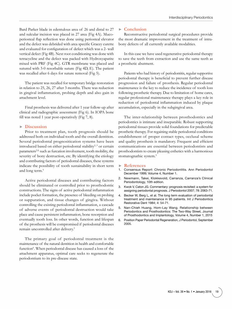
BardParkerbladeinedentulousareaof 26anddistalto27andsulcularincisionwasplacedin27area(Fig4A).Muco-periostealflapreflectionwasdoneusingperiostealelevatorandthedefectwasdebridedwithareaspecificGraceycuretteandevaluatedforconfigurationof defectwhichwasa2-wallvertical defect (Fig 4B). Next root conditioning was done with tetracycline and the defect was packed with Hydroxyapatite mixed with PRF (Fig 4C). GTR membrane was placed and suturedwith3-0resorbablesuture(Fig4D,E).Thepatientwasrecalledafter6daysforsutureremoval(Fig5).
The patient was recalled for temporary bridge restoration inrelationto25,26,27after3months.Therewasreductioningingivalinflammation,probingdepthandalsogaininattachment level.
Finalprosthesiswasdeliveredafter1yearfollow-upafterclinicalandradiographicassessment(Fig6).InIOPAbonefillwasnoted1yearpost-operatively(Fig7,8).
► Discussion Prior to treatment plan, tooth prognosis should be
addressed both on individual tooth and the overall dentition. Several periodontal prognositication systems have been introduced based on either periodontal stability2, 3 or certain parameters2,4, such as furcation involvement, tooth mobility, the severity of bony destruction, etc. By identifying the etiology and contributing factors of periodontal diseases, these systems indicate the possibility of tooth sustainability in short term and long term.5
Active periodontal diseases and contributing factors should be eliminated or controlled prior to prosthodontic constructions.Thesignsof activeperiodontalinflammationinclude pocket formation, the presence of bleeding on probing or suppuration, and tissue changes of gingiva. Without controllingtheexistingperiodontalinflammation,acascadeof adverse events of periodontal destruction would take placeandcausepersistentinflammation,boneresorptionandeventually tooth loss. In other words, function and lifespan of the prosthesis will be compromised if periodontal diseases remain uncontrolled after delivery.5
The primary goal of periodontal treatment is the maintenance of the natural dentition in health and comfortable function6. When periodontal disease has caused a loss of the attachment apparatus, optimal care seeks to regenerate the periodontiumtoitspre-diseasestate.
► Conclusion Reconstructive periodontal surgical procedures provide
themostdramaticimprovementinthetreatmentof intra-bony defects of all currently available modalities.
In this case we have used regenerative periodontal therapy to save the teeth from extraction and use the same teeth as a prosthetic abutment.
Patients who had history of periodontitis, regular supportive periodontaltherapyisbeneficialtopreventfurtherdiseaseprogression and failure of prosthesis. Regular periodontal maintenance is the key to reduce the incidence of tooth loss following prosthetic therapy. Due to limitation of home cares, regular professional maintenance therapy plays a key role in reductionof periodontalinflammationinducedbyplaqueaccumulation, especially in the subgingival area.
The inter-relationshipbetweenprosthodontics andperiodontics is intimate and inseparable. Robust supporting periodontal tissues provide solid foundations for predictable prosthetic therapy. For regaining stable periodontal conditions establishment of proper contact types, occlusal scheme andqualityprosthesisismandatory.Frequentandefficientcommunications are essential between periodontists and prosthodontists to create pleasing esthetics with a harmonious stomatognathic system.5
► References 1. Consensus Report: Chronic Periodontitis. Ann Periodontol
December 1999; Volume 4, Number 1.2. Newmann, Takei, Klokkevold, Carranza, Carranza’s Clinical
Periodontology, 10th edition.3. Kwok V, Caton JG. Commentary: prognosis revisited: a system for
assigning periodontal prognosis. J Periodontol 2007; 78: 2063-71.4. Becker W, Berg L, et al. The long term evaluation of periodontal
treatment and maintenance in 95 patients. Int J Periodontics Restorative Dent 1984; 4: 54-71.
5. Nan-Chieh Huang, Hom-Lay Wang. Relationship between Periodontics and Prosthodontics: The Two-Way Street, Journal of Prosthodontics and Implantology, Volume 4, Number 1, 2015
6. Position Paper Periodontal Regeneration, J Periodontol, September 2005.
KDJ–Vol.39•No.1•January2016 19
Interdisciplinary Periodontics

► IntroductionOral cancer is a common and lethal
malignancy and the outcome of the treatment and prognosis largely depends on early diagnosis. Oral squamous cell carcinoma is the most common among oral cancers. OSCC is the sixth most common human malignancy, with a 5-yearmortalityrateof approximately50%1whichhasnotchangedsignificantlyin more than 50 years, and a high rate of morbidity. The therapeutic modality currently offered to OSCC patients is basedontraditionalstage-predictingindices (based mostly on the TNM criteria) and on histological grading2. Unfortunately, these predictors are subjective and relatively unreliable. Thus, therehasbeenanever-growingeffortdedicated to the basic research of oral cancer. Despite the fact that the oral cavity is easily accessible, most OSCCs are not diagnosed until an advanced stage, which is believed to be the major reason for the low survival rate, and points to the urgent need for clinical diagnostic aids for early detection of OSCC. However, very few studies have examined tumor markers in the saliva of OSCC patients, though such an examinationmightbeof greatbenefitbecause of the direct contact between the oral cancer lesion and saliva.
Saliva:Adiagnostictoolfororalcancer
*Jinisham, **Nileena R. Kumar, ***Haris P.S., ****Anita Balan
AbstractOral cancer is a common and lethal malignancy. Oral squamous cell carcinoma, OSCC is sixth most common epithelial malignancies with significance morbidity and mortality. In spite of diagnostic and therapeutic advances over the decades, the disease still remains a challenge. Despite the fact that the oral cavity is easily accessible, most OSCCs are not diagnosed until an advanced stage, which is believed to be the major reason for the low survival rate, and points to the urgent need for clinical diagnostic aids for early detection of OSCC. Thus, there has been an ever-growing effort dedicated to the basic research of oral cancer, focusing on the identification of biological indicators for the diagnosis.
Salivary diagnostics is a dynamic and emerging field utilizing nanotechnology and molecular diagnostics to aid in the diagnosis. Using saliva for disease diagnostics and health surveillance is a promising approach as collecting saliva is relatively easy and non-invasive. Direct contact between saliva and the oral cancer lesion makes measurement of tumor markers in saliva an attractive alternative to serum testing. Thus, much research effort has been dedicated to investigating potential salivary biomarkers for OSCC.
Keywords: saliva; oral cancer; biomarkers
KDJ 2016 | Vol. 39 | No. 1 | Pg 20-23*PGstudent,**AssociateProfessor,***AssistantProfessor,Dept.ofOralMedicineandRadiology,Govt.DentalCollege,Kozhikode,****ProfessorandHOD,Dept.ofOralMedicineandRadiology,Govt.DentalCollege,TrivandrumCorrespondingAuthor:Dr.JinishaM.Email:[email protected]
Salivary testing, a non-invasivealternative to serum testing, can be an effective modality for diagnosis and prognosis predicting of oral cancer. Much research effort has been dedicated to investigating potential salivary biomarkers for OSCC. A systematic review of the current status of salivary diagnostics for detection of OSCC is presented
► DiscussionSaliva and its diagnostic potential
Human saliva is a clear, slightly acidic (pH=6.0-7.0)biologicalfluidcontaininga mixture of secretions from multiple salivary glands, including the parotid, submandibular, sublingual gland other minor glands beneath the oral mucosa aswellasgingivalcrevicefluid3. Saliva has long been proposed and used as a diagnostic medium as it is easily accessible, collection is noninvasive, not time consuming, inexpensive and can be used for mass screening purposes. A major drawback to using saliva as adiagnosticfluidhasbeenthenotionthat informative analytes generally are present in lower amounts than in serum. However with new techniques of detecting small quantities of salivary components including proteins and messenger Ribonucleic acid (mRNA), almost anything that can be measured in blood can be measured in saliva.
20 KDJ–Vol.39•No.1•January2016
KDJ – Kerala Dental JournalBIoMARKER

► Salivary biomarkers for oral cancer detectionSo far, more than 100 potential OSCC salivary biomarkers
havebeenreportedintheliterature.Thefirstreportof
saliva as a diagnostic tool for oral cancer detection was published in 20004.Globalprofilingof disease-associatedmolecules, such as proteins, DNA, mRNA, micro RNA, andmetabolitesisbecomingthestate-of-the-artmethodto provide promising disease biomarker candidates. The abilitytogloballyprofilethesemoleculesinsaliva,throughtranscriptomic and proteomic approaches, as well as the abilitytodetectspecificmoleculesinsalivawillgreatlyenhance the opportunities to identify reliable oral cancer biomarkers.
Mechanism for the presence of biomarker in salivaThough the exact mechanism for the presence of these
tumor markers in saliva is not known, they may be either derived from serum or can be produced locally5. Molecular markers for the diagnosis of oral cancer can be quested in 3 levels;6 changes in the cellular Deoxyribonucleic acid (DNA) which results in altered mRNA transcripts leading to altered protein levels intracellularly, on the cell surface or extracellularly. Markopoulos et al6 have summarized the molecular markers for the diagnosis of OSCC
Changes in cellular DNA Altered mRNA transcripts Altered protein markers
Allelic loss on chromosome Presenceof IL-8 Elevated levels of Defensin 1Mitochondrial DNA mutation Presenceof IL-1β Elevated CD44
P53 gene mutations Dualspecificityphosphatase1(DUSP1) H3F3A (H3histone, family 3A) ElevatedIL-6,IL-8
Promoter hypermethylation of genes Ornithine decarboxylase antizyme 1 (OAZ1) Inhibitors of apoptosis (IAP)
CyclinD1geneamplificationS100 calcium binding protein P (S100P) Spermidine/spermine N1-acetyltransferase(SAT)
Squamous cell associated antigen (SCC-Ag)
Increaseinki67markers Carcino-embryonicantigen(CEA)Micro satellite alterations of DNAPresence of HPV and EBV virus genomeCarcino-antigen(CA19-9),CA128Serum tumor marker ( CA 125)Intemediatefilamentprotein(Cyfra21-1)Tissuepolypeptidespecificantigen(TPS)Reactive nitrogen species (RNS) 8-OHdGDNAdamagemarkerLactate dehydrogenase (LDH) Immunoglobulin (IgG) s-IgAInsulin growth factor (IGF) Metalloproteinases
DNA; Deoxyribonucleic acid; mRNA; messenger ribonucleic acid; HPV; Human Papilloma Virus :EBV; Epstein Barr Virus
Functions and Clinical Utility of Saliva4
KDJ–Vol.39•No.1•January2016 21
Saliva: A diagnostic tool for oral cancer
► Alterations in host cellular DNADNA markers are universal i.e., there is not a single tumor
cell with in the lesion which does not contain its genetic material. They originate from dead cells, are detected in the early stageof tumorigenesisandareabsolutelyoncospecific;showingadirectcause-and-effectrelationshipwithtumorigenesis.However,tissuespecificityof DNAmarkersisverylow.7
Lossof heterozygosity(LOH)isdefinedaslossof genomicmaterial in one of the chromosomal pair. Studies have shown that LOH in regions that contain a known human suppressor gene is an early predictor of malignant transformation of precancerous lesion8.
Rosin et al,9 in their study found that allelic loss at 3p and9qincreasetheriskof malignanttransformationby3.8fold and the risk of further increase to 33 fold when LOH occursatchromosomes4q,8p,11q,13qand17pinadditionto the former.
p53 gene located on chromosome 17p13.1 exhibits mutationin50-70%of epithelialtumors10,11andLOHof p53allelehasbeenreportedin22%of pre-cancerand20%of oralcancer.Othergenessuchasp16,p27,p63,p73related to p53 and cell cycle have been found to be altered in varying degrees in oral cancer.10 Promoter methylation, an alternate form of gene silencing, which depends on the epigenetic factor has been described to be involved in OSCC10. The main genes to be methylated are CDKN2A, CDH1, MGMT, DAPK110, 12.
Amplificationandoverexpressionof c-MYCIN-MYChasbeenobservedin20-40%of oralcancers.Dasetal,13 have reportedamplificationof 11q13,containing1NT2,HST1andCyclinDoncogenesin30-50%of patientswithoralcancer.STAT3expressionandactivationwasfoundin82%of oralcancers related to chewing tobacco.14
Levelsof Ki67markerwereincreasedwhile8-oxoguanineDNAglycosylase,phosphorylated-Srcandmammaryserineprotease inhibitor (Maspin) were found decreased in the saliva of patients with OSCC.2 The presence of HPV and EBVvirusgenomicsequencehasbeenidentifiedaspossibleDNA markers in detecting OSCC and tumor progression15.
RNA as a biomarkerRNA has been found to be an informative marker and
salivaryRNAsignatureshavebeenidentifiedfororalcancer.StudieshavefoundsevenmRNAmolecules;(1)IL-816,17 (2)IL1β16,17 (interleukin 1)which take part in signal transduction, proliferation,inflammationandapoptosis,(3)DUSP117 (dual specificityphosphatase1)witharoleinproteinmodification,
signal transduction and oxidative stress, (4) H3F3A17 (H3 histone, family 3A) having a DNA binding activity, (5) OAZ116,17 (ornithine decarboxylase antizyme 1) taking part in polyamine biosynthesis,(6)S100P17 (S100 calcium binding protein P) with a role in protein binding and calcium ion binding and (7) SAT16,17(spermidine/spermineN1-acetyltransferase)whichtakespartinenzymeandtransferaseactivity,tobesignificantlyelevated in OSCC patients rather than in healthy controls
Protein markers Protein markers are differentiation antigens of
corresponding normal tissue and characterize a certain stage of its maturation. Salivary protein markers have shown moderate sensitivityandspecificityasprognosticmarkers6.
Asubstantialincreaseinsalivarycarbonyls(246%)isseeninOSCCpatientsandpointstothefactthatthereisasignificantfree radical attack to which the epithelial cells are exposed2. MetalloproteinasessuchasMMP-92,MMP-1118,MMP-218are significantlyalteredinOSCC.IL-6,IL-8arepostinflammatorycytokinesandareidentifiedasimportantmediatorsof cancerdevelopment and powerful activators of not only apoptosis but also anti apoptotic signaling cascade19 and hence play a roleinearlydetectionof oralpre-malignanciesandOSCC.Cytokeratinsarecytoskeletalintermediatefilamentspresentinalmost all normal and malignant epithelial cells.20 In malignant epithelial cells, the activated protease increases degradation of cytokeratinfreefilamentsintotheblood.Increasedlevelsof Cyfra21-1insalivahavebeenfoundinOSCC.21-23
Someof theothersalivarybiomarkerswhicharesignificantlyaltered in OSCC patients are inhibitors of apoptosis24 (IAP), squamous cell carcinoma associated antigen23,25(SCC-Ag),carcino-embryonicantigen23,26(CEA),carcino-antigen(CA19-9)23,25,CA12825, serum tumor marker (CA125)23, 27, tissue polypeptidespecificantigen(PPS)23,28 reactive nitrogen species (RNS)29, lactate dehydrogenase (LDH)22 and immunoglobulin G (IgG)22,s-IgA18, insulin growth factor (IGF)18 and defensin30.
Recently Oral fluid NanoSensor Test (OFNASET) technology developed by the UCLA research group, platform combinescutting-edgetechnologies,suchasself-assembledmonolayers (SAM), bionanotechnology, cyclic enzymatic amplification,andmicrofluidics,withseveralwell-establishedtechniquesincludingmicroinjectionmolding,hybridization-baseddetection,andmolecularpurification.Theintendeduse of the OFNASET is the multiplex detection of salivary biomarkers for oral cancer.31
► ConclusionThefieldof salivarydiagnosticsisabroad,complexand
crosscuttingareaof scientificresearchwithenormouspotential
22 KDJ–Vol.39•No.1•January2016
Jinisham

to impact the diagnosis of various diseases including oral cancer.Thesignificantincreaseintumormarkersinsalivaisquite encouraging. Though at present no single tumor marker can validate the presence or prognosis of disease, a panel of biomarkers would be more helpful. Recent advances and emerging technologies such as nanotechnology, proteomic and genomics make salivary diagnostics a highly sensitive tool. The present methods are not ready for immediate direct use and much needs to be done to come up with a fast, simple, cost effective clinical diagnostic system. Saliva will possibly outperform other media in the diagnosis of not only cancer but other diseases as well.
► Bibliography1. Myers JN, Elkins T, Roberts D, Byers RM. Squamous cell carcinoma
of the tongue in young adults: increasing incidence and factors that predict treatment outcomes. Otolaryngology--Head and Neck Surgery 2000; 122(1):44-51.
2. Shpitzer T, Hamzany Y, Bahar G, et al. Salivary analysis of oral cancer biomarkers. British journal of cancer 2009; 101(7):1194-98.
3. Spielmann N, Wong DT. Saliva: diagnostics and therapeutic perspectives. Oral diseases 2011; 17(4):345-54.
4. Shah FD, Begum R, Vajaria BN, et al. A review on salivary genomics and proteomics biomarkers in oral cancer. Indian Journal of Clinical Biochemistry 2011; 26(4):326-34.
5. Baum BJ. Principles of saliva secretion. Annals of the New York Academy of Sciences 1993; 694(1):17-23.
6. Markopoulos AK, Michailidou EZ, Tzimagiorgis G. Salivary markers for oral cancer detection. The open dentistry journal 2010; 4:172.
7. Lichtenstein A, Potapova G. Genetic defects as tumor markers. Molecular Biology 2003; 37(2):159-69.
8. Zhang L, Rosin M. Loss of heterozygosity: a potential tool in management of oral premalignant lesions? Journal of oral pathology & medicine 2001; 30(9):513-20.
9. Rosin MP, Cheng X, Poh C, et al. Use of allelic loss to predict malignant risk for low-grade oral epithelial dysplasia. Clinical Cancer Research 2000; 6(2):357-62.
10. Tilakaratne WM. the cancer handbook. 2nd edition ed: United States: John Wiley and Sons Ltd; 2007; 2007.
11. Boyle JO, Hakim J, Koch W, et al. The incidence of p53 mutations increases with progression of head and neck cancer. Cancer Research 1993; 53(19):4477-80.
12. Ha PK, Califano JA. Promoter methylation and inactivation of tumour-suppressor genes in oral squamous-cell carcinoma. The lancet oncology 2006; 7(1):77-82.
13. Das BR, Nagpal JK. Understanding the biology of oral cancer. Medical Science Monitor 2002; 8(11):RA258-RA67.
14. Nagpal JK, Mishra R, Das BR. Activation of Stat‐3 as one of the early events in tobacco chewing‐mediated oral carcinogenesis. Cancer 2002; 94(9):2393-400.
15. Shimakage M, Horii K, Tempaku A, et al. Association of Epstein-Barr virus with oral cancers. Human pathology 2002; 33(6):608-14.
16. Wong DT. Salivary diagnostics powered by nanotechnologies, proteomics and genomics. The Journal of the American Dental Association 2006; 137(3):313-21.
17. Zimmermann BG, Wong DT. Salivary mRNA targets for cancer diagnostics. Oral oncology 2008; 44(5):425-29.
18. Shpitzer T, Bahar G, Feinmesser R, Nagler RM. A comprehensive salivary analysis for oral cancer diagnosis. Journal of cancer research and clinical oncology 2007; 133(9):613-17.
19. Pezelj-Ribaric S, Prso IB, Abram M, et al. Salivary levels of tumor necrosis factor-α in oral lichen planus. Mediators of Inflammation 2004; 13(2):131-33.
20. Moll R, Franke WW, Schiller DL, Geiger B, Krepler R. The catalog of human cytokeratins: patterns of expression in normal epithelia, tumors and cultured cells. cell 1982; 31(1):11-24.
21. Bhatavdekar J, Patel D, Vora H, Balar D. Circulating prolactin and TPS in monitoring the clinical course of male patients with metastatic tongue cancer: a preliminary study. Anticancer research 1992; 13(1):237-40.
22. Yen T, Lin W, Kao C, Cheng K, Wang S. A study of a new tumour marker, CYFRA 21-1, in squamous cell carcinoma of the head and neck, and comparison with squamous cell carcinoma antigen. Clinical otolaryngology and allied sciences 1998; 23(1):82-86.
23. Nagler R, Bahar G, Shpitzer T, Feinmesser R. Concomitant analysis of salivary tumor markers—a new diagnostic tool for oral cancer. Clinical Cancer Research 2006; 12(13):3979-84.
24. Kurokawa H, Tsuru S, Okada M, Nakamura T, Kajiyama M. Evaluation of tumor markers in patients with squamous cell carcinoma in the oral cavity. International journal of oral and maxillofacial surgery 1993; 22(1):35-38.
25. Hoffmann J, Munz A, Krimmel M, Alfter G. Intraoperative and postoperative kinetics of serum tumor markers in patients with oral carcinoma. Journal of oral and maxillofacial surgery 1998; 56(12):1390-93.
26. Krimmel M, Hoffmann J, Krimmel C, Cornelius CP, Schwenzer N. Relevance of SCC-Ag, CEA, CA 19.9 and CA 125 for diagnosis and follow-up in oral cancer. Journal of Cranio-Maxillofacial Surgery 1998; 26(4):243-48.
27. Kurokawa H, Yamashita Y, Tokudome S, Kajiyama M. Combination assay for tumor markers in oral squamous cell carcinoma. Journal of oral and maxillofacial surgery 1997; 55(9):964-66.
28. Bahar G, Feinmesser R, Shpitzer T, Popovtzer A, Nagler RM. Salivary analysis in oral cancer patients. Cancer 2007; 109(1):54-59.
29. Nagler RM, Barak M, Peled M, et al. Early diagnosis and treatment monitoring roles of tumor markers Cyfra 21‐1 and TPS in oral squamous cell carcinoma. Cancer 1999; 85(5):1018-25.
30. Mizukawa N, Sugiyama K, Fukunaga J, et al. Defensin-1, a peptide detected in the saliva of oral squamous cell carcinoma patients. Anticancer research 1997; 18(6B):4645-49.
31. Gau V, Wong D. Oral Fluid Nanosensor Test (OFNASET) with Advanced Electrochemical‐Based Molecular Analysis Platform. Annals of the New York Academy of Sciences 2007; 1098(1):401-10.
KDJ–Vol.39•No.1•January2016 23
Saliva: A diagnostic tool for oral cancer

accurate and effective rehabilitation. Therefore, the combined efforts of the ophthalmologist, the plastic surgeon and the maxillofacial prosthodontist are essential to provide a satisfactory ocular prosthesis.3
► HistoryThefirstartificialeyesweremadeby
Egyptians. The Babylonian and Sumerian civilizationshadused‘‘art-eyes’’ instatues and mummies, fabricated from precious stone, silver or gold4. Ambroise Pare(1510-1590)–describetheuseof anartificialeyesinaneyesocket(pioneerof modernartificialeyes)-fabricatedeyes porcelain. In the beginning of the 19th century, France became the centre of artificialeyemaking.Boissoneauin1849wascreditedwithcoiningtheterm‘‘ocularist’’, He produced stock glass eyes, which were popular in Europe and America.In1853,LudwigMullerUriused a novel method (a unique method of colouring the iris) for making human glass eyes. Friedrich A. Muller is credited with developing the double wall glass prosthesis. By the end of the last century most of the ocular prosthesis was developed.IIWorldwar-Navaldentalschool(1943)-testedthefabricationof ocular prosthesis using acrylic resins.
Indications for prosthetic devicesThe various indications for
fabrication of prosthetic devices are divided into two broad categories namely congenital and acquired deformities5.
Impressiontechniquesforocularprosthesis
*Rohit Raghavan, **Shajahan P. A., ***Ritha Bos, ***Geethprasad TS
AbstractEyes are generally the first features of the face to be noted. The disfigurement caused by loss of an eye is often a psychologically damaging experience for the patient. Ocular prosthesis is an artificial replacement of the eye. To gain proper fit and intimate tissue adaptation of ocular prosthesis, an accurate impression technique is necessary. This article deals with various impression techniques used in the fabrication of acrylic resin custom ocular prostheses.
Key Words: Eye stock Prosthesis, Ocular impression
KDJ 2016 | Vol. 39 | No. 1 | Pg 24-26
*HeadofDepartment,**Professor,***PGStudent,DeptofProsthodontics,RoyalDentalCollege,Chalissery,Palakkad,KeralaCorrespondingAuthor:DrRithaBosEmail:[email protected]
A. Congenital deformities AnophthalmiaMicrophthalmia
B. Acquired deformities Phthisis bulbiPost-eviscerationPost-enucleationPost-chemicalinjuriesContracted socket following radiationPost-orbitalexenteration
Method for the fabrication of ocular prosthesis5 are 1. Examination by the ocularist2. Taking an impression3. Wax pattern for the socket4. Centration of the prosthesis5. Fabrication of iris and pupil to match
the fellow eye6. Mouldinginacrylic7. Tinting to match the scleral shade8. Packingwiththeclearacrylic9. Polishing10. Patient education about hygiene and
care of the prosthesis
Out of which the impression making is of greater importance, different technique of impression making is going to be discussed in detail.
1. The Direct Impression/External Impression
2. Impression with Stock Ocular Tray3. StockOcularTrayModifications4. Impression with Custom Ocular Tray5. Impression Using Stock Ocular
Prosthesis
► IntroductionA defects that compromise esthetics
and function, and prevents one to lead a normal life, usually leads the individual to seek treatment that will reinstate acceptable normacy. Eyes are generally thefirstfeaturestobenoted1. The loss or absence of an eye may be due to many reasons like congenital defect, trauma, tumour, a painful blind eye, sympathetic ophthalmia or the need for histological confirmationof asuspecteddiagnosis2.
Replacement of the missing eye should be done as quick as possible to promote physical and psychological healing for the patient which would help to improve the social acceptance. A multidisciplinary management and team approach are essential for an
24 KDJ–Vol.39•No.1•January2016
KDJ – Kerala Dental JournaltEChNIQuE

6. OcularProsthesisModification7. Wax Scleral Blank Technique
1 The Direct Impression/External ImpressionIn this technique impression materials like alginate is
injected directly into the enucleated socket.3 Patient is said to look straight in front as technique commences. The anatomy of the an ophthalmic socket and overlying tissues is obtained by using a rigid tray which is reinforced over the additional material applied to the external tissue. Stone model is made from the impression and a wax pattern is carved. This acts as a trial ocular prosthesis which is tried in the patient and adjusted accordingly to achieve proper tissue contours and fit.BartlettandMoore3 substantiated the necessity of an impression procedure to realize the full movement potential of a prosthesis supported by an ocular implant. By doing so voids in the socket are eliminated and possibility of debris collection is minimal. In addition, they emphasised that a wax try-inisnecessarytoevaluateproperphysiologicfunction
2 Impression with Stock Ocular TrayMost widely used impression technique. Here stock ocular
tray is used to support the impression material6,7. Allen and Webster6calleditthe“modifiedimpressionmethod.”Athinmix of ophthalmic alginate is injected through the stock ocular tray which has a hollow stem fastened in the middle. Trays are perforatedwhichaidsinflowandretentionof thealginate.Awax pattern is fabricated. This wax trial prosthesis is placed in the socket for around 10 minutes which allows the muscle accommodation.Thefitof thetrialprosthesisisevaluatedandmodifiedasneeded.
3 Stock Ocular Tray ModificationsVariationsof the“modifiedimpressionmethod”center
onthefabricationorconfigurationof astockoculartray.Maloney8, in the superior edge of customized stock trays placed 3 channels to prevent air entrapment. In this method, a raised ring around the stem which would prevent the eyelid from blocking the channels.
4 Impression with Custom Ocular TrayMiller9 explained for patients with ophthalmic sockets
which were highly irregular custom ocular trays were of greater advantage. He attached a solid suction tube to the existing prosthesis of the patient, conformer, or wax shell and invested it in an alginate mold. After the alginate sets, these were removed and replaced with clear acrylic resin. Perforations are made in the resulting tray, and a tunnel is cut into the stem and impression material can be injected through it. An impression is made using injected alginate.
5 Impression Using Stock Ocular ProsthesisStock ocular prosthesis were used as a tray to carry
impression material. In this method an appropriate stock acrylic resin ocular prosthesis was selected and then was adjusted by trimming its peripheries which was later lined with athinmixof ophthalmicalginateanddefinitiveimpressionwere obtained. Alternately, alginate can be injected directly into the socket and then reinforced by placement of the stock eye. A customized stock prosthesis were obtained from the proceeded impression. Limitations of this technique include theneedof largesupplyof artificialeyesandtheinabilityto match to the appropriate sizes and colors of the iris and pupil as required in the patient.
6 Ocular Prosthesis ModificationChalian10 suggested trimming and polishing a stock
prosthesiswillgainmoreacceptablefit.Andalsobyusingalginateorsoftwaxthestocktrayscanbemodified,whichthen can be invested and processed.
Smith11 suggested relining an existing prosthesis using animpressionwax,Korecta-WaxNo.4.Heretheexistingprosthesis is trimmed peripherally and posteriorly, and modifiedwithbaseplatewax.Whenpropercontoursareachieved,athinlayerof Korecta-WaxNo.4isadded.Thelined prosthesis is warmed, inserted which is left in place for 30 minutes and the patient is said to intermittently moves his or her eyes in all possible directions. A laboratory reline procedure is then accomplished.
7 Wax Scleral Blank TechniqueThe wax scleral blank was advocated as the starting point
in several techniques.
Benson12 created a wax pattern of half of the size of steel ball. The resultant pattern is smoothed, trial was done, and adjusted. The pattern is invested and processed with iris button attached. Chalian et al13 also followed the same.
McKinstry14 suggested “compression impression” technique in which he formed a wax pattern based on examination of thesite.Waxpatternwastriedin,modifiedasneeded,andprocessed after addition of an iris. Advantage of this method is that it accurately records and form an inferior fornix if the patient’s or the fornix is shallow or lower lid is weak.
Le Grand and Hughes15 in their “empirical/ impression” technique attached a “dummy” aluminium button to act as a handle. Then properly contoured wax pattern is used as a tray to carry alginate or support impression material. Hughes16 subsequently suggested that a syringe to be attached to the completed wax pattern so that impression material can be injected directly through it.
KDJ–Vol.39•No.1•January2016 25
Impression techniques for ocular prosthesis

Schneider duplicated the patient’s conformer to obtain awaxconformerandmodifieditinsections,usingadentalimpression wax, Iowa Wax. Various ocular movements given by patient resulted in a functional impression of the socket.
Alternately Sykes in his technique used polyvinylsiloxane materialonthetissuesurface.Themodifiedwaxpatternisthenusedtofabricatethefinalocularprosthesis.
► ConclusionThe rehabilitation of a patient who has suffered the psychologic
trauma of an ocular loss requires a prosthesis that will provide theoptimumcosmeticandfunctionalresult.Refinementinthedetails of custom ocular construction has produced a superior restoration delivered more readily. Impression making to facilitate thedesigningof anartificialeyeisacommonpracticetoday.Numerous ocular impression methods exist.
► Reference1. Doshi PJ, Aruna B. Prosthetic Management of patient with ocular
defect. J Ind Prosthodont Soc 2005; 5: 37–38.2. Raflo GT. Enucleation and evisceration. In: Tasmun W, Jaeger
E eds. Duane’s Clinical ophthalmology, Revised edn, Vol. 5. Philadelphia: Lippincott-Raven, 1995: 1–25.
3. Bartlett SO, Moore DJ. Ocular prosthesis: a physiologic system. J Prosthet Dent 1973; 29: 450–459
4. Martin O, Clodious L. The history of artificial eyes. Ann Plastic Surg 1979;3:168–70.
5. K. Raizada, D. Rani / Contact Lens & Anterior Eye 30 (2007) 152–162
6. Allen L, Webster HE: Modified impression method of artificial eye fitting. Am J Ophthalmol 1969;67:189-218
7. Allen L, Bulgarelli DM: Obtaining and understanding the alginate impressions. J Am Soc Ocularists 1988;19:4-13
8. Maloney BA: Development of impression fitting equipment: A new technique. J Am Soc Ocularists 1979;9:32-33
9. Miller BJ: Custom ocular impression trays. J Facial Somato Prosthet 1996;2:109-113
10. Chalian VA: Treating the patient with facial defects, in Laney WB (ed): Maxillofacial Prosthetics. Littleton, MA,2. PSG Publishing Co, 1979, p 286
11. Smith RM: Relining an ocular prosthesis: A case report. J Prosthodont 1995;4(3): 160-163
12. Benson P: The fitting and fabrication of a custom resin artificial eye. J Prosthet Dent 1977;38:532-538
13. Chalian VA, Drane JB, Metz HH, et al: Extraoral prosthetics, in Chalian VA, Drane JB, Standish SM (eds): Maxillofacial Prosthetics: Multidisciplinary Practice
14. Cerullo L, McKinstry RE: Ocular prostheses, in McKinstry RE (ed): Fundamentals of Facial Prosthetics. Arlington, VA, ABI Professional Publications, 1995, pp 107-109
15. Le Grand JA, Hughes MO: Empirical impression technique for artificial eye fitting. Adv Ophthalmic Plast Reconstr Surg 1990;8:118-125
26 KDJ–Vol.39•No.1•January2016
Rohit Raghavan

Theyfulfillinatolerabledegreealltherequirementsof artificialteethunderany circumstances, if we expect that the mastication, this function being more or less imperfectly performed with such pieces.” But late on in the year 1951 Sears said that “ in cases where some of the natural teeth remain in place it is wise to consider the advisability of constructing dentures before the teeth are removed”2.
Dental professionals have recognized the patients wish and need to avoid an edentulous period which resulted in the fabrication of the denture that can be placed in the patients mouth immediately following the removal of last natural teeth. The success of the immediate denture depend on the correct indication and precise execution of clinical and laboratory procedure.
► Case report 1 A 55 year old male patient was
referred to the department of Prosthodontics for oral rehabilitation. Onintra-oralexaminationteethpresentwere 12, 14, 15, 21, 22, 23, 31, 32, 33, 34, 36,37,38,42,44,46,47(FigI)
Orthopantomography was advised (Fig 2). Upper and lower alginate impressions were made; study models were fabricated and articulated. All the facial measurements were recorded.
Immediatedenture-animportanttreatmentmodality* Kalpana Hasti, **Anurag Hasti, ***Rahul Sharma, ****Aprajita Mitra
AbstractAn immediate complete denture is a restoration of the lost natural teeth and associated tissues which is inserted into the patients mouth immediately following the extraction of remaining teeth. The removal of all the remaining teeth and placement of the complete denture are important in patients’ life. The transition from dentulism to edentulism should be psychologically atraumatic as far as possible.
The cases presented here are conventional (classic) immediate denture and interim (transitional or non transitional) immediate denture which offer good esthetic and functions.
Key words: conventional immediate denture, interim immediate denture.
KDJ 2016 | Vol. 39 | No. 1 | Pg 27-29
*Reader,Dept.ofProsthodontics,JaipurDentalCollege;**Reader,***SeniorLecturer,****P.GStudent,Dept.ofProsthodontics,SchoolofDentalSciences,ShardaUniversity;CorrespondingAuthor:Dr.KalpanaHasti [email protected]
After the diagnosis of the cast, radiograph evaluation and clinical examination, a conventional immediate denture was planned. The patient was referred to the department of Oral and Maxillofacial surgery for extraction of 36,37,38and44,46,47andwasadvisedtoreportbackafter6–8weeks.Whenthepatientreportedafter6weeks,thehealing was found to be satisfactory. (Fig 3 and 4)
► Procedure Upper and lower alginate impressions
were made. A custom tray was fabricated for Camphagne’s3 impression technique. Border molding was done using green stickcompoundandfinalimpressionwas made using Zinc Oxide Eugenol and dentulousareainAlginateasapick-upimpression material.
A master cast (Fig 5), record base and occlusal rims were fabricated. Jaw relation was recorded. 14 and 34 were used as a vertical stop. The records were transferred on to the articulator, Posterior Teeth arrangement was done and upper 12 and 22 were arranged to confirmesthetics,labialfullness,lowerlip line, size and shape of teeth. Try –inwasdone.Thecastmodificationwas done by the technique given by Jerbi4. Flasking and curing was done followedbyfinishingandpolishing.Onthe day of insertion all the remaining
► Introduction Patient increasing demand for
natural appearance of the lost teeth has become a challenge to the dentist1. The placement of complete denture immediately following the removal of natural teeth is not new. Richardson (in theyear1860)emphasizedonimmediatedenture service, saying that “the value of temporary sets of teeth to the patient on the other hand is questionable.
KDJ–Vol.39•No.1•January2016 27
KDJ – Kerala Dental Journal CASE REPoRt

anterior teeth were extracted and denture insertion was done.(Fig6,7,8)
Patient was advised to wear the denture overnight and for 3 consecutive days and recall was done after 24 hours. Patient complained of ulceration in the upper canine region and in the posterior mylohyoid region and the required trimming was done. Patient was advised to continue wearing denture and called for suture removal afteraweek.Patientwasrecalledafter6monthstocheckfor the retention and stability of both the dentures and assessed for relining.
► Case report 2A 29 years old female patient was referred to department
of Prosthodontics for replacement of missing teeth. On examination,theteethpresentwere13.14,15,16,23,25,26,34,35,36,37,45,47,48.Alltheremainingteethwerefoundtobe
periodontically involved with Grade II and Grade III mobility. (Fig 9 and 10)
Orthopantography (OPG) was advised. Upper and lower Alginate impressions were made and study models were evaluated. Patient was referred to department of Periodontics for opinion. The OPG revealed severe bone loss. Patient was diagnosed with aggressive periodontitis. After diagnosis, examination and OPG evaluation an interim immediate denture was planned. The custom tray is fabricated.
Border molding is done using low fusing impression compoundandfinalimpressionwasmadewithmediumbody rubber base impression material.(Fig 11) On the master castrecordbasewasfabricatedandonlyanteriortry-inwasdone. The casts were articulated in the normal occlusion of the patient. Posterior arrangement was done and the cast modificationwasdonebythetechniquegivenbyJerbi4.
fig. 9: Preoperativefig. 8: Postoperativefig. 7: Completelyextractedmaxillaryarch
fig. 6: Completelyextractedmandibulararchfig. 5: Mastercastfig. 4: Mandibulararchpostextraction
fig. 3: Maxillaryacrhpostextractionfig. 2: DiagnosticOPGfig. 1: Preoperative
28 KDJ–Vol.39•No.1•January2016
Kalpana Hasti
Flaskingandcuringwasdonefollowedbyfinishingandpolishing. On the day of insertion all teeth were extracted and denture insertion was done. Patient was advised to wear the denture overnight and for 3 consecutive days and recall was done after 24 hours (Fig 12,13,14,15). Patient was advised to continue wearing denture and called for suture removalafteraweek.Patientwasrecalledafter6monthsto check for the retention and stability of both the dentures and assessed for relining.
► Discussion When removal of teeth becomes necessary an immediate
denture is an important treatment modality. There are many advantages of immediate dentures as it acts as a matrix which controls hemorrhage, prevents contamination and provide protective covering over the wounds.
It provides restoration of phonetics and masticatory functions and facilitates transition of the edentulous state5 It enables the patient to continue to engage in social and business activities without an embarrassing period of staying edentulous6. As in the case of conventional immediate dentures there are many advantages of CID than IID. The use of transitional partial denture to replace posterior teeth prior to the removal of the anterior teeth is highly recommended. As discussed in interim immediate complete denture case report, successful treatment of aggressive periodontitis depends a lot
in early diagnosis of the disease. When diagnosed in late stage the treatment option is extraction. A young female patient suffering from aggressive periodontitis and loss of all her teeth has a devastating effect on functionally, and psychologically so a fabrication of IID is useful method.
► Conclusion In the era of implant and immediate implant treatment,
immediate complete denture treatment should still be considered as an important treatment modality. A detailed extraoral and intraoral evaluation and correct treatment planning will lead to a successful replacement of missing structures with immediate dentures which is functionally acceptable to the patient.
► Reference 1. Puthanakar N.Y, Pappachan B, Patil A.G. Full mouth rehabilitation
by immediate denture prosthesis- A case report. Annals and Essences of Dentistry.2012;4(4):28-33.
2. Goswami R, Singh M. Immediate denture- a spatial modeling way. Guident. Sept 2012;42-44.
3. Campagna SJ. An impression technique for immediate dentures. J Prosthet Dent. 1968;20: 196-203. 3
4. Jerbi.F.C Trimming the cast in the construction of immediate denture J Prosthet Dent 16:1047-1053, 1966.
5. Wiebelt F.J. Two steps better immediate dentures. Quitessence Int. 1980;5:27-30.
6. Gilboa I, Cardash H.S. An alternative approach to immediate overdentures. J Prosthodont.2009:18;71-75.
fig. 15: Postoperativefig. 14: Processeddenturefig. 13: Postextractionmandibulararch
fig. 12: Postextractionmaxillaryarchfig. 11: Finalimpressionfig. 10: Preoperativeintraoral
KDJ–Vol.39•No.1•January2016 29
Immediate denture - an important treatment modality
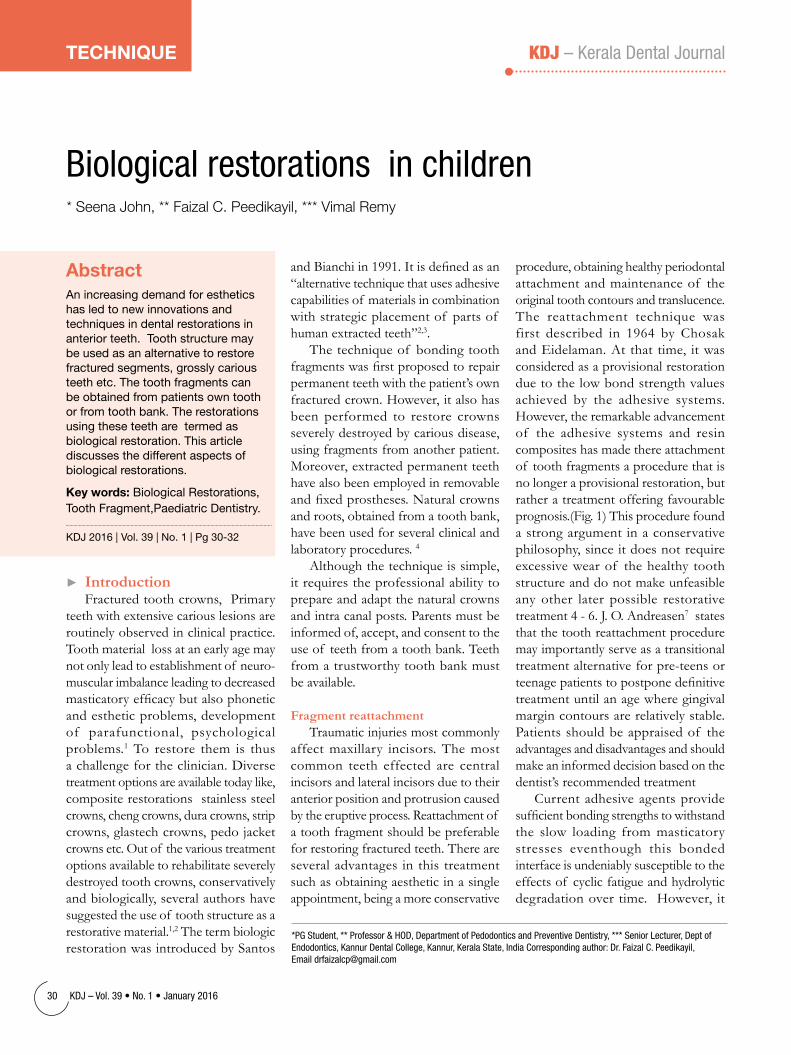
andBianchiin1991.Itisdefinedasan“alternative technique that uses adhesive capabilities of materials in combination with strategic placement of parts of human extracted teeth”2,3.
The technique of bonding tooth fragmentswasfirstproposedtorepairpermanent teeth with the patient’s own fractured crown. However, it also has been performed to restore crowns severely destroyed by carious disease, using fragments from another patient. Moreover, extracted permanent teeth have also been employed in removable andfixedprostheses.Naturalcrownsand roots, obtained from a tooth bank, have been used for several clinical and laboratory procedures. 4
Although the technique is simple, it requires the professional ability to prepare and adapt the natural crowns and intra canal posts. Parents must be informed of, accept, and consent to the use of teeth from a tooth bank. Teeth from a trustworthy tooth bank must be available.
Fragment reattachmentTraumatic injuries most commonly
affect maxillary incisors. The most common teeth effected are central incisors and lateral incisors due to their anterior position and protrusion caused by the eruptive process. Reattachment of a tooth fragment should be preferable for restoring fractured teeth. There are several advantages in this treatment such as obtaining aesthetic in a single appointment, being a more conservative
Biologicalrestorationsinchildren* Seena John, ** Faizal C. Peedikayil, *** Vimal Remy
AbstractAn increasing demand for esthetics has led to new innovations and techniques in dental restorations in anterior teeth. Tooth structure may be used as an alternative to restore fractured segments, grossly carious teeth etc. The tooth fragments can be obtained from patients own tooth or from tooth bank. The restorations using these teeth are termed as biological restoration. This article discusses the different aspects of biological restorations.
Key words: Biological Restorations, Tooth Fragment,Paediatric Dentistry.
KDJ 2016 | Vol. 39 | No. 1 | Pg 30-32
procedure, obtaining healthy periodontal attachment and maintenance of the original tooth contours and translucence. The reattachment technique was first described in 1964 by Chosakand Eidelaman. At that time, it was considered as a provisional restoration due to the low bond strength values achieved by the adhesive systems. However, the remarkable advancement of the adhesive systems and resin composites has made there attachment of tooth fragments a procedure that is no longer a provisional restoration, but rather a treatment offering favourable prognosis.(Fig. 1) This procedure found a strong argument in a conservative philosophy, since it does not require excessive wear of the healthy tooth structure and do not make unfeasible any other later possible restorative treatment4-6.J.O.Andreasen7 states that the tooth reattachment procedure may importantly serve as a transitional treatmentalternativeforpre-teensorteenagepatientstopostponedefinitivetreatment until an age where gingival margin contours are relatively stable. Patients should be appraised of the advantages and disadvantages and should make an informed decision based on the dentist’s recommended treatment
Current adhesive agents provide sufficientbondingstrengthstowithstandthe slow loading from masticatory stresses eventhough this bonded interface is undeniably susceptible to the effects of cyclic fatigue and hydrolytic degradation over time. However, it
► Introduction Fractured tooth crowns, Primary
teeth with extensive carious lesions are routinely observed in clinical practice. Tooth material loss at an early age may notonlyleadtoestablishmentof neuro-muscular imbalance leading to decreased masticatoryefficacybutalsophoneticand esthetic problems, development of parafunctional, psychological problems.1 To restore them is thus a challenge for the clinician. Diverse treatment options are available today like, composite restorations stainless steel crowns, cheng crowns, dura crowns, strip crowns, glastech crowns, pedo jacket crowns etc. Out of the various treatment options available to rehabilitate severely destroyed tooth crowns, conservatively and biologically, several authors have suggested the use of tooth structure as a restorative material.1,2 The term biologic restoration was introduced by Santos
*PGStudent,**Professor&HOD,DepartmentofPedodonticsandPreventiveDentistry,***SeniorLecturer,DeptofEndodontics,KannurDentalCollege,Kannur,KeralaState,IndiaCorrespondingauthor:Dr.FaizalC.Peedikayil, [email protected]
30 KDJ–Vol.39•No.1•January2016
KDJ – Kerala Dental JournaltEChNIQuE
appears that improvements in the luting and/or bonding systems employed and a greater knowledge of the factors influencingrestorationlongevityshouldservetoenhancethepotential for success of reattachment techniques in the future.8
Biological crownsThe use of extracted teeth as biological restoration
constitutes a viable restorative alternative for teeth with extensive coronal destruction. This technique is simple, provides excellent esthetics as well as preserves natural tooth colour compared to composite resins and stainless steel crowns, allows the preservation of sound tooth structure and has low cost. The enamel of the biologically restored tooth hasphysiologicwearandofferssuperficialsmoothnessandcervical adaptation compatible with those of surrounding teeth. Biological restorations not only mimic the missing part of the oral structures, but are also biofunctional.5,6
The length of each appointment is reduced because natural teeth are prepared previously. Clinical chair time for fragment bonding procedures is relatively short, which is a merit especially while dealing with paediatric patients Resin composite restorations do not present these advantages and can allow staining and plaque formation on their surfaces. 2,3
Disadvantages of the biological crown restoration technique includethedifficultyinobtainingteethwiththerequiredcoronal dimensions and characteristics, problems inherent to indirect restorations and matching fragment colour with tooth remnant colour. Also, having teeth from other people’s teeth in their mouth is not a pleasant idea for some patients and many of them refuse to receive this treatment However, all these factors are not contraindications of the technique2,3,6,8.
Biological restoration used teeth from the Human Tooth Bankareusedasnaturalposts&crowns.2,7 The selected tooth from the tooth bank is reshaped, roots strengthened by retro fillingwithflowablecompositesandautoclavedfor30minat121 degree centigrade and 15 lbs pressure before cementation.9
Other factors of concern while using biologic restoration is that Universal protocol of consent, storage and sterilization
should be followed in the human tooth bank. Collected samples should be scaled, polished and freed of soft tissues and periodontal remnants. Pulps have to be removed and complete biological preparation is to be done. Teeth are then storedat4degreecentigradeinHBSSwithdonoridentificationtill the time of its use. 9,10
It is important that the parents are informed that the tooth fragments used for biological restoration are previously submitted to a rigorous sterilization process that completely eliminates any risk of contamination or disease transmission to the child receiving the fragment. Presently, secure methods of sterilization and storage are available to ensure the safety of teeth or tooth fragments coming from tooth banks Several materials have been used for bonding dental fragments to cavities, e.g., adhesive systems, composite resins, glass ionomer cementsanddual-cureresincements.SincetheToothfragmentwhich was taken was large, it was concerned that optimal light-curingwouldnotbeachievedatthecavitygingivalmargin.Thus,dual-cureresin-basedcementwasusedtoenhancepolymerizationatthisregioninadditiontofillingany possible gaps existing at tooth/fragment interface with composite resin only.3,4,5
Biological restoration technique using tooth fragments has apracticalclinicalapplicabilityandisaviable,cost-effectiverestorative procedure for primary teeth with severely damaged crowns. In the present case, the use of biological restoration with natural crown resulted in clinical success as well as recovered the proper functional anatomy of the tooth. Finally the choice of restorative technique depends upon the operator preferences, esthetic demands by the parents and child’s behavior that affect the ultimate outcome of whichever restorative material chosen.
► ConclusionThe constant evolution of Restorative Odontology is
creating a new sight. In this short review represents the functional recuperation of a tooth, through the use of a endogenous dental fragment, a technique known as Biological
fig. 1: Toothfragmentreattachment
KDJ–Vol.39•No.1•January2016 31
Biological restorations in children
restoration. With highly satisfactory results obtained in many case reports, we conclude that this technique can be considered as alternative to all others and can be carried out quickly and inexpensively.
► References1. Mathur S, Rahul C, Pandit I K, Srivastava N, Gugnani N.
BiologicalRestoration of a Grossly Decayed Deciduous Mandibular Molar. Journal of Clinical and Diagnostic Research. 2012 February;6(1): 139-141.
2. Sanches K, CarvalFabrício K D, FilhPaulo N,Sada A F,WanderleyGarcia De Paula E S,Alexandra, Mussolino D Q. Biological Restorations As A Treatment Optionfor Primary Molars With Extensive Coronal destruction - Report Of Two Casesbraz Dent J (2007) 18(3): 248-252.
3. Das U K, Maiti N. Nature’s Own Alternative – Restoration With Biological Crowns. International Journal Of Pediatric Dentistry.2014april-June;3(2):144-149.
4. Saha R, Malik P. Paediatric aesthetic dentistry: a review. European Journal of Paediatric Dentistry.2012; vol. 13(1):6-12.
5. Am Botelho, Kta T, Correa-Faria P1, Lns M,Viana M. Esthetic–Functional Recovery Of Permanent Posterior Tooth Using Prima Autogenous Biologica lrestoration Journal Of Indian Society Of Pedodontics And Preventive Dentistry. 2012 Oct - Dec; 4(30):333-336.
6. Grewal N, Reeshu S.Case Report Biological restoration:An alternative Esthetic Treatment For Restoration Of Severly Multilated Primary Anterior Teeth International Journal Of ClinicalPediatric Dentistry 2008.;1(1):42-47.
7. Textbook and Color Atlas of Traumatic Injuries to the Teeth, 4th Edition, Jens O. Andreasen, FrancesM. Andreasen Lars Andersson, July 2007, Wiley-Blackwell
8 Attila IO, Cenk MHA, Serdar MT.Multidisciplinary approach to the rehabilitation of a crown –root fracture for immediate esthetics. Dent traumatol. 2006;22(1):48-52.).
9 Habiltz S, et al. Nanoindentation and storage of teeth. J Biomech 2002; 35(7):995 98.
10 Imparato JCP. Bank human teeth. Curitiba: Publi sher May, 2003.
TheLargest DentalPortalofKerala
Logonwww.idakerala.comforIDAActivities,Reports, Publicationetc...
32 KDJ–Vol.39•No.1•January2016
Seena John

minimal pressure. Appropriate retention aids the denture to resist the forces of gravity, adhesiveness of foods and the forces associated with the opening of the jaws. Among the factors of retention the most important is the accomplishment of an appropriate peripheral seal which is the positive contact of the entire perimeter of the denture base to the resilient tissues that outline the basal seat. The correct position for the mandibular denture border is easily determined in the labial, buccal, and posterior regions, but the exact placement of the denture border in the anterior lingual region has always been a factor of uncertainity.
Tilton stated that “border molding ends at a point where the border tissues seatfirmly,butlightlyagainstthedentureperiphery when the border tissues are in extreme function”1. Accurate border molding within physiologic boundaries accomplishes the most desirable border sea12,3. The purpose of this article is to illustrate how the anterior lingual seal can be established during the impression making phase by utilizing the sublingual space for a retentive, stable mandibular denture.
DefinitionsThe term “sublingual” pertains to
the region or structures located beneath the tongue.
The “sublingual crescent extension” referstothemandibulardentureflangethat covers the anterior region of the
Recordingsublingualcrescentregionforimprovingstabilityandretentioninmandibulardentures* Thomas Paul, ** Byju Paul Kurian, *** Parvathy Raj, *** Prisly Thomas,
floorof themouth.Itstartsatthecrestof the alveolar ridge and ends at the soft tissue displacement tissue at the base of the tongue not supported by bone.
The region covered by the sublingual flangeof amandibulardenturehavebeen given different terminologies namely lingual vestibule4, lower denture space5, anterior lingual region6, sublingual cells7, sublingual region8 and sublingual space9, 10.
The Glossary of Prosthodontic Terms(8thedition2005)differentiatesbetween the sublingual crescent area, sublingual fold and sublingual fossa11. Itdefinesthemasfollows:
Sublingualcrescent:Thecrescent-shapedareaontheanteriorfloorof themouth formed by the lingual wall of the mandible and the adjacent sublingual fold. It is the region of the anterior alveoilingual sulcus.
Sublingualfold:Thecrescent-shapedareaonthefloorof themouthfollowingthe inner wall of the mandible and tapering toward the molar regions. It is formed by the sublingual gland and submaxillary duct.
Sublingual fossa: A smooth depression on the lingual surface of the body of the mandible near the midline, and above the mylohyoid line and below the alveolus. This fossa accommodates part of the sublingual gland.
AnatomyThe sublingual region is bordered
AbstractAchieving retention and stability in mandibular complete denture has always been challenging to the novice and the experienced dentists throughout the world. When it comes to treating a complete denture patient we are ought to treat the particular patient and not an average patient. Various techniques and modifications available can hence be duly chosen and customized for fabrication of satisfactory dentures in compromised conditions. One such technique includes the utilization of sublingual crescent region through a slight modification of the conventional impression technique that has been described in the current article.
KDJ 2016 | Vol. 39 | No. 1 | Pg 33-36
*Professor,**ProfessorandHead,***PostGraduateStudent,DepartmentofProsthodontics,AnnoorDentalCollege,Muvattupuzha,Ernakulam-686673.CorrespondingAuthor:Dr.ParvathyRaj,E-mail:[email protected]
► IntroductionIn complete denture prosthodontics
achieving retention and stability for mandibular dentures have been an inscrutable dilemma in the bygone years. The art of impression making becomes beautiful only if it achieves the core principles underlying. Creation of such beautiful masterworks call for appropriate knowledge of the oral anatomy, knowledge of a basic and reliable technique, understanding of the materials and skill of the dentist.
Retention of a denture is the resistance to removal in a direction opposite to that of its insertion while stability is the resistance to horizontal movements. Both these factors are strongly related that without the one the other doesn’t exist and is achieved by registering exact tissue details under
KDJ–Vol.39•No.1•January2016 33
KDJ – Kerala Dental Journal PRoCEDuRE

medially by the base of the tongue and laterally by the mandible. Themuscleandmucousmembranethatformthefloorof the mouth are separated from each other by loose connective tissue containing glandular elements.
Genioglossus is the most superiorly placed muscle in the sublingual region. It is a fan shaped muscle with its group of fibersattachedanteriorlytothesuperiorgenialtubercleand runs posteriorly to be attached into and become part of the tongue6.
Theanteriorgroupof fibresareattachedjustbelowtheupper surface of the tongue at the junction of its anterior third withthemiddlethird.Theposteriorgroupof fibersprotrudesthe tongue by pulling on its posterior part and thus moving theentirestructureforward.Thefibersof theanteriorgroupare relaxed during this process, and they, together with the surrounding tissues, are moved forward passively under the influenceof pressurefromthemoreposteriorlyplacedgroupof fibers.Contractionof thesuperiorlyplacedanteriorgroupof fibersof thegenioglossusmuscledepressesthatpartof the surface of the tongue which is a short distance posterior to the tip. This produces a marked concavity of that surface ‘and tends to retract the tip of the tongue, causing it to move upward and backward. This movement occurs during the formationof thebolusandinthefirststageof swallowingandactivelyraisesthefloorof themouthtoitshighestlevel.Duringthesefunctions,thefibersof theanteriorgrouparecontractedstronglyand,beingsuperficiallyplacedinthefloorof the mouth, exert a strong pressure through the overlying mucosa against a denture base which might be obstructing its movement6.
The mylohyoid muscle is located inferiorly and serves as the floorof themouth.Thismusclearisesfromtheinnersurfaceof themandiblewhereasitsposteriorfibresoriginatefromthealveolus at the level of the third molar. The mylohyoid muscle deepens anteriorly, forming a sublingual cell composed of the genioglossus muscle arising from the upper genial tubercle.
Sublingual foldsTwo folds in the mucosa overlying the genioglossus muscle
run obliquely forward and inward at a varying distance from the alveolar ridge and nearly meet in the midline. These are the left and right sublingual folds which carry the ducts of the corresponding submandibular salivary glands in their free borders.Atthemidlineastrongfibroustendonoriginatesfrom the ventral surface of the tongue and extends toward the alveolingual sulcus. This is the lingual frenum. It contains two sublingual caruncles (openings) of Wharton’s duct located on either side of the midline.
Some sublingual folds are quite small and others much larger. When the tip of the tongue is raised, the sublingual folds are also raised and are pulled away from the alveolar ridge. When the tongue is relaxed and its tip is allowed to rest in contact with the lingual surfaces of the lower anterior teeth, the sublingual folds also move downward and forward nearer to the alveolar ridge and the denture border.
The sublingual gland along with hypoglossal nerve, lingual nerve, lingual vein, and lingual artery are contained in the sublingual region.
Each patient requires an individual evaluation to identify thevariouscharacteristicsof thefloorof themouth.Itis
fig. 1 fig. 2 &3
fig. 4 fig. 5 fig. 6 fig. 7
34 KDJ–Vol.39•No.1•January2016
Thomas Paul

hence expected that one must treat the particular patient and not the average patient.
Fournetclassifiedthefloorof themouthintofourtypesas follows12:
1. Ahighfloorof themouthwithnosublingualfoldorspace. The acrylic resin special tray should be shortened lingually until it does not touch the sublingual gland.
2. Aclearlydefinedspaceforthesublingualfoldandgland.The custom tray is trimmed and shortened carefully in the sublingual region with the tongue just touching the handle of the tray without dislodging it.
3. A sublingual gland at a higher level than the level of the alveolar ridge. In this situation the tray should be trimmed short so asnottoimpingeontheglandoroccludetheorifices.
4. A sublingual gland that is nearly at the same level of the ridge.Whenthisoccurs,thelingualflangeof thetrayisrelieved over the genial tubercles.
► Case reportA 54 year old female patient reported to the Department
of Prosthodontics, Annoor Dental College with a chief complaintof illfittinglowerdenturesince1year.Theintraoral examination revealed completely edentulous maxillary and mandibular arch with moderate resorption in lower anterior region(Atwood’sOrderV)(fig.1).Thefloorof themouthmaybeconsideredasFournet’stype2withclearlydefinedsublingualspace on either side of the lingual frenum. The treatment planwasmodifiedtogiveamandibularcompletedenturewith sublingual crescent extension opposing a conventional maxillary denture.
► Procedure13
1. Preliminary impression is made with alginate in a perforated edentulous stock tray to facilitate better recording of the resorbed residual ridges. (Fig 2,3)
2. Acrylic special tray is made with spacer from the primary cast. Spacer may be avoided in severely resorbed ridge cases because adapting a wax spacer may not be feasible in such conditions.
3. The special tray borders are checked for over extensions by assessing displacement due to lip, cheek or tongue movements.
4. Bordermouldingwithlowfusingcompoundisfirstdonein the mylohyoid, retromylohyoid and the distal extensions of the tray; initiated from one side followed by the other side. This improves the stability of the tray. The labial and buccal areas are then border moulded. The thickness of these borders recorded within functional limits enhance the peripheral seal. (Fig.3,4)
5. Anyextensionof lowfusingcompoundintothepre-mylohyoid area is then removed.
6. Sublingualcrescent recording is then initiatedwithimpression compound which is tempered using water bathat1400F(6O0C)andlutedontotheborderof acustom acrylic resin tray that is trimmed 2 mm short of thefloorof themouthfrompre-mylohyoidareafromonesidetotheother,finallyspanningtheentireanteriorlingual area of the tray with the impression compound. Mold it into a shelf extending downward and backward over the crescent region. Impression compound is softened carefullyoveradryflametakingcarenottoburnoutthematerial. The special tray is placed in the mouth and the patient is then instructed to gently place the tongue against the lingual side of the tray handle. (Fig 5)
7. The tray is removed from the mouth and cooled in cold water. If the extension of the sublingual crescent is inadequate, more material is added and the procedure is repeated. Properly recorded sublingual crescent bulges upward from the tray borders more than a normal border moulded border. It is approximately crescent or half spindle-shapedextendingtoeithersidesof themidline.
8. Theimpressioncompoundisthenrelievedinthefrenalnotch area to expose the openings of the sublingual ducts. If this is not done, collection of saliva and pain occurs due to the obstruction of the sublingual gland duct openings.
9. The sublingual crescent border of the tray contacting the ventral side of tongue is examined and its borders are trimmed to remove any overextension beyond the posterior border of the sublingual fold. The light pink color of the sublingual fold than the darker pink ventral surface of the tongue acts as a guide.
10. The added sublingual extension now maintains contact with the sublingual fold when the tongue touches the lowerlip.Italsomaintainscontactwiththefloorof themouth when the tongue is in a retracted position, thereby developing a good peripheral seal in both tongue positions.
11. The wax spacer is removed from the border moulded special traytoprovideroomforthefinalimpressionmaterial.
12. The secondary impression is then made in zinc oxide eugenol impression paste with the lip and cheek movement as usual and protruded and retracted tongue positions.
13. The cast is poured to get the master cast, with the additional positive replica of the sublingual crescent.
Further alterations in methods in a sublingual recorded lower denture include the following15:a. Adequatequantityof heat-cureacrylicresinshouldbe
used during packing procedures to compensate for the reduced ridge height and also the additional sublingual crescentarea.Excessflashduringtrialclosureof flaskshould be assured.
b. Long curing cycle is preferred for polymerization of the denture to prevent porosities in the thick areas.
KDJ–Vol.39•No.1•January2016 35
Recording sublingual crescent region for improving stability and retention in mandibular dentures

► DiscussionMany methods of developing lingual borders have resulted
insatisfactorydentures.Differentdesignsof thelingualflangecan be developed because of the anatomic and functional variation in patients.
Pendleton14 has stated, “There appears to be one method and adequately this method is applied by permitting the tissues to establish their own associations by forming the impression material to their own individual functional requirements.” Other authors preferred a dynamic impression to be made of thelingualbordersandfoundthattheuseof tissue-conditioningimpressionmaterialhadtheaddedbenefitof improving the oral mucosa. Lawson6 suggested that thickening the sublingual region of the denture could result in more retention, especially when the tongue is relaxed and seal is not broken. Some authors suggested that where marked residual ridge resorption occurs, it may be desirable to use the tongue andthebuccinatorsmuscletofixthedentureinplacebytheappropriatedesigningof thewidthandformof theflanges.Brill et al5reportedthatthinningthedentureflangeswouldincreasetheretentiveforcesbythemusclefixationif thepatient has an adequate ridge. On the other hand, Lott and Levin demonstrated the clinical advantages of thicker borders.
Each patient requires an individual evaluation to identify differences in anatomy. In view of these anatomical differences definiteguidelinesshouldbeformulatedforeachpatientregarding the impression techniques and extension objectives. For example, the use of a dynamic impression material may bebeneficialinthemarkedlyresorbedridge.Moldingathickborder and wide sublingual region often helps to achieve a bettersealbecauseitaidscontinuitywiththefloorof themouth. The border contours should be rounded, according to Slack15 to minimize trauma resulting in sore spots on the mucosa.However,someauthorssuggestedthatthinnerflangesenhance retention.
► ConclusionThe dental literature offers a variety of terms to describe
the sublingual region. The “anterior lingual seal region” and “sublingual crescent region” are appropriate terms because they describethelocationandfunctionof thelingualflangeof themandibular denture. Variations in shape and anatomy dictate theuseof differentimpressiontechniquesandmodificationsinflangedesigntomakeaclinicallysuccessfulmandibulardenture.
Recording sublingual crescents during impression making in severely resorbed mandibular ridges, combined with setting thelowerposteriorteethwithflattenedcuspsinthecenterof the posterior ridge in the denture and contouring of the lingual,buccalandlabialflangesof thedenturebase(Fischconcept)providessufficientretentionandstabilityforthedentures made on these ridges.
The tongue movements may be limited slightly due to the presence of sublingual crescents which the patient gets adapted, if extensions are proper. The patient can also performfunctionalmovementsof thetonguesufficientlywithout dislodging the denture. Overextended sublingual extensions will diminish the mandibular denture retention. Slightlimitationof thetonguemovementisanaddedbenefitto maintain retention and stability in severely resorbed ridges.
► References1. Tilton GE. Denture periphery. J Prosthet Dent 1952;2:290-306.2. Tench RW. Impressions for dentures. J Am Dent Assoc
1934;21:1005-183. Fournet SC, Tuller CS. A revolutionary mechanical principle utilized
to produce full lower dentures surpassing in stability the best modern upper dentures. J Am Dent Assoc 1936;23:1028-30.
4. Edwards LF, Boucher CO. Anatomy of the mouth in relation to complete dentures. J Am Dent Assoc 1942;29:331-45.
5. Brill N, Trycle G, Cantor R. The dynamic nature of the lower denture space.J Prosthet Dent 1965;15:401-18.
6. Lawson AW. Influence of the sublingual fold on retention of complete lower dentures. J Prosthet Dent 1961;11:1038-44.
7. Bocage M, Lehrhaupt J. Lingual flange design in complete dentures. J Prosthet Dent 1977;37:499-506.
8. Rouviere H. Anatomia humana. Description and topagrafica. Madrid: Bailley-Bailer, S.A., 1961.
9. Lewis ET. Repositioning of the sublingual fold for complete dentures. J Prosthet Dent 1958;8:22-5.
10. Lewis ET. The genial tubercle space in full lower dentures. J Prosthet Dent 954;4:606-10.
11. Glossary of prosthodontic terms. 8th ed. 2005:94:1.12. Fournet SC. Stabilization of lower dentures. J Dent Surv 63;35-41.13. Gafoor MAA, Kumar VVH, Sheejith M, Swapna C. Recording
‘Sublingual Crescents’ in Lower Complete Dentures: A Technique so Effective but still Esoteric and Arcane. J Contemp Dent Pract 2012;13(2):222-226
14. Pendleton EC. American textbook of prosthetic dentistry. 7th ed. Philadelphia:Lea & Febiger Publ, 1942:121.
15. Slack FA. Positive retention in lower dentures. Bull Phila Count Dent sot 1949;14:2-7.
IDA-HOPE (Help Offered to Professionals in Emergencies). Members are requested to contact their respective IDA local branch HOPE representative to receive original application formsJOIN
36 KDJ–Vol.39•No.1•January2016
Thomas Paul

residual ridge and opposing mandibular teeth. As a result, maxillary complete denture will develop a substantial increase in weight leading to decreased retention and stability of the prosthesis. Literature suggests various methods for weight reduction such as using a solid 3-dimensionalspacer,includingdentalstone, cellophane wrapped asbestos, silicone putty, or modeling clay during laboratory processing to exclude denture base material from the planned hollow cavity of the prosthesis1-3. This article describes a technique of fabricating a hollow maxillary complete denture using silicone putty as well as a mixture of dental plaster and pumice (1:1 by weight) to develop a cavity within the denture base3-5.
► Case reportA73-year-oldmalepatientreported
to the Department of Prosthodontics withthechief complaintof difficultyin chewing and broken upper denture causing loss of retention. History revealed that the patient was a denture wearer for the past 17 years. Upon preliminary intra oral examination, it was found out that maxillary ridge was severely resorbed. The upper lip was longandtheinter-ridgedistancewas
* Ashwin Kumar S G., ** R. Ravichandran, ** S. Lylajam, *** K. Harshakumar
AbstractSeverely atrophic ridges cause decrease in retention, support and stability thus leading to a clinical challenge to the success of complete denture. It is the dentist’s responsibility to fabricate prosthesis with good stability retention and support which ultimately provide satisfactory functional and aesthetic acceptance to the patient. In special situations such as patients with broad upper lip, severely resorbed ridge with increased inter arch distance, the weight of a maxillary denture often causes dislodgment hence, a light weight denture is preferred for better retention.Different approaches like using a solid 3–dimensional spacer including dental stone, cellophane wrapped asbestos, silicone putty or modeling clay have been used during laboratory processing to exclude denture base material from the planned hollow cavity of the prosthesis.
KDJ 2016 | Vol. 39 | No. 1 | Pg 37-39
exceedingly more than the normal limits. The patient had no relevant medical history.Thefinaltreatmentplandecidedwas to fabricate a new set of complete denture for the patient with a hollow maxillary and a conventional mandibular denture.
► Technique1. Makeadefinitiveimpressionof the
maxillary residual ridge and fabricate the denture to the trial denture stage in a conventional manner. (Fig 1)
2. Process the trial denture using standard technique till the wax elimination stage. (Fig 2)
3. Process a shim of indexed acrylic resin over the residual ridge. Adapt thin layer of baseplate wax to the intaglio surface of the teeth, conforming to the border extensions. (Fig 3.) Reset the original cope on the drag and verify complete closure of theflask.Usethesecondflasktoinvest the baseplate wax and pack the seconddragwithheat-polymerizedacrylic resin and process.
4. Mix and adapt vinyl polysiloxane putty over the permanent denture
Rehabilitationofanedentulousgeriatricpatientwithresorbedresidualridgesandexcessiveinterarchspaceusingamaxillaryhollowcompletedenture
*PGstudent,**Professor,***ProfessorandHead,DepartmentofProsthodontics,Govt.DentalCollege,Trivandrum.CorrespondingAuthor:Dr.AshwinKumarSG,E-mail:[email protected]
► IntroductionFabrication of a complete denture
as a treatment modality can pose a daunting clinical challenge in case of severe resorption of maxilla. This may be due to narrower and constricted residual ridge formed owing to resorption process, resulting in a large restorative space between the maxillary
KDJ–Vol.39•No.1•January2016 37
KDJ – Kerala Dental Journal Case report

base and the counterpart with acrylic shim was seated over it. Wait for the vinyl polysiloxane putty to set. (Fig 4)
5. Vinyl polysiloxane putty was removed from the alveolar ridgearea.(Fig5)Dentalplaster-pumicemixturewasplacedonto the acrylic shim area. Both the counterparts of the flaskwereclosedandwaitedforthemixturetoset.(Fig6)
6. Reopentheflaskandremovetheremainingputtymaterialfrom the borders and cameo surface of the permanent denture base.
7. Thin mix heat cured acrylic resin material was injected on to the sides of the acrylic shim. The counterpart with permanent base was seated over the acrylic shim portion and processed.
fig. 1 TrialDenture. fig. 2 InvestedMaxillaryTrialDenture. fig. 3 WaxShimoverthedewaxeddenture. fig. 4 Adaptingputtymaterialontotherecordbase.
fig. 5 Removedputtymaterialfromrecordbase. fig. 6 Packingpumiceplastermixtureoverthewaxshim. fig. 7 FloatingHollowcompletedenture.
fig. 8 Preoperativelateralview. fig. 9 Postoperativelateralview. fig. 10 Preoperativefrontalview. fig. 11 Postoperativefrontalview.
38 KDJ–Vol.39•No.1•January2016
Ashwin Kumar S G.

8. Recovertheprocesseddentureintheusualmanner.Remount the denture on an articulator and adjust the occlusion as necessary. Cut two openings with a bur into the denture base distal to the most posterior teeth. Remove thedentalplaster–pumicemixturebyscrapingwithasharp instrument. After complete removal of the mixture the windows were sealed using autopolymerizing resin. (Fig 7)
9. Polish the denture in the usual manner. Verify that the cavity was sealed by immersing the denture in water. No bubbles were evident, indicating an adequate seal over the denture.
10. Regular recall visit for 3 months was kept during which nodiscomfortwasreportedbythepatient.(Fig8&9).Thepatientwassatisfiedwiththefunction,aestheticandcomfortof thedenture.(Fig10&11)
► DiscussionRehabilitation of the patient with severely resorbed ridges
posesasignificantchallengetothespecialist.Therearevariousmethods for rehabilitation of such patients6,7. Treatment options like implant supported overdenture, ridge augmentation can be considered for the patients with resorbed ridges. Many times such patients will be geriatric patients with systemic illness, economic constraints and not willing for any kind of surgical procedures as well. Hence, it will be better to treat them in a conventional manner3.
O.Sullivanetalusedtwoflaskswithinterchangeablehalvesforfabricatingahollowmaxillarydenture.Heat-polymerizingone portion of the denture against polymerized resin may reduce leakage at the junction of the two portions of the denture. Since both portions are heat polymerized there will be reduced leakage along the junction at the two portions of the denture8,9.
The technique used in the article requires two interchangeableflasksforthefabricationof thedenture.Eventhoughtheprocedureistime-consumingitwillreducethe weight and which in turn will considerably enhance the
retention of the denture10. Hollow dentures are prone to fracture and care should be taken to avoid leakage along the junction of the two portions of the denture.
► ConclusionThe hollow maxillary denture is an ideal method of
rehabilitating a patient with the severely resorbed maxillary ridge.Itsignificantlyreducestheweightof thedenturewhichultimately results in increased retention and stability of the prosthesis11. Moreover, this may also aid in preserving the existing residual alveolar ridge of the patient. The method described here highlights a simple but precise technique for the fabrication of maxillary hollow denture. Another advantage of the technique is that the dental plaster and pumice mixture used as the spacer which can be easily removed using a sharp instrument to make the denture hollow and weightless.
► References 1. Fattore LD, Fine L, Edmonds DC. The hollow denture: an alternative
treatment for atrophic maxillae. J Prosthet Dent 1988; 59:514-6.2. Laxman Singh Kaira, maxillary hollow denture: A case series,
Journal of Orofacial Research April – June 2012; 2(2): 109-112.3. N Kalavathy, M MithaShetty, Premnath, KarunaPawashe, RKV
Patel, Hollow mandibular complete denture - A case report. SRM University Journal of Dental Sciences, Volume 1, Issue 3, October – Decembret 2010
4. SaurabhChaturvedi, A.K. Verma, Mariyam Ali, PreetiVadhvani, Hollow Maxillary Denture: A Simplified Approach. People’s Journal of Scientific Research.,Vol. 5(2), July 2012
5. Gardner LK, Parr GR, Rahn AO. Simplified technique for the fabrication of a hollow obturator prosthesis using vinyl polysiloxane. J Prosthet Dent 1991;66:60-2.
6. McAndrew KS, Rothenberger S, Minsley GE. An innovative investment method for the fabrication of a closed hollow obturator prosthesis. J Prosthet Dent 1998;80:129-32.
7. Holt RAJr. A hollow complete lower denture. J Prosthet Dent1981;45:452-4.
8. Jhanji A, Stevens ST. Fabrication of one-piece hollow obturators. J Prosthet Dent 1991;66:136-8.
9. Elliott DJ. The hollow bulb obturator: its fabrication using one denture flask. Quintessence Dent Technol 1983;7:13-4.
10. DaBreo EL. A light-cured interim obturator prosthesis. A clinical report. J Proshet Dent 1990;63:371-3.
11. Chalian VA, Barnett MO. A new technique for constructing a one-piece hollow obturator after partial maxillectomy. J Prosthet Dent 1972;28: 448-53.
KDJ–Vol.39•No.1•January2016 39
Rehabilitation of an edentulous geriatric patient with resorbed residual ridges and excessive interarch space using a maxillary hollow complete denture
Comparisonofdifferentmethodsofassessingalveolarridgedimensionspriortodentalimplantplacement* Veena T.K., ** Sudeep S, *** Dinesh
AbstractThe science of Implantology is highly dynamic ever since its introduction into the field of dentistry by Dr. P.I Branemark, it has undergone numerous modifications and improvements. With each improvement and advancements made, implantology has proved to be a boon to the society and hence its acceptance by the general population has widely increased despite it being a relatively expensive treatment modality.
Minimally Invasive Flapless Implant surgery has gained wide acceptance among practitioners and patients alike owing to its obvious advantages such as lesser bleeding, post operative discomfort and simpler surgical procedure.
However, such flapless placement requires precise planning and astute understanding of supporting tissue such as the dimensions of the osseous ridge and bone. This paper intends to present our investigations into comparative assessments of the accuracy of various modalities both radiographic (Dentascan, occlusal radiography) and ridge mapping towards the planning of each implant surgery.
KDJ 2016 | Vol. 39 | No. 1 | Pg 40-42
*PGStudent,**ProfessorandHead,***Professor,DepartmentofProsthodontics,PMSCollegeofDentalScienceandResearch,Vattappara,Trivandrum-695028CorrespondingAuthor:DrVeenaT.K,Email:[email protected]
► IntroductionThe use of dental implants to
support prosthodontic restorations has a high success rate. Careful diagnosis and treatment planning are critical for a favorable outcome. Evaluation of the dimensions of the available alveolar bone is an important prerequisite.
Bone evaluation limited to the use of panoramic and/or periapical radiographs may be insufficient because it only providestwo-dimensionalinformationabout implant sites. Assessment of the bucco-lingualdimensionof theosseousridge also is needed for proper treatment planning. The bucco-lingual ridgewidth can be evaluated by Dentascan; a computed tomography (CT) software program that allows the mandible and maxilla to be imaged in three planes: axial,panoramicandcross-sectional.Ithasbeenwidelyusedpre-operativelyfor implant surgery as it provides a comprehensive assessment of the bone morphology and measurement of the dental implant. It is an interactive CT imaging software that combines the power and detail of CT imaging with the convenience of interacting with the images on a desktop or notebook computer 1. An alternative method is ridge mapping using a caliper device under local anesthesia. The pointed tips of the instrument penetrate
buccal and lingual soft tissue layers and measurethebucco-lingualwidthof the underlying bone. This procedure is performed chairside and provides instant information.
The aim of this study was to compare the width of bone using Ridge mapping, Dentascan and Intra Oral Periapical Radiographs (IOPA) in Occlusal projection.
► Materials and methodsStudy Subjects
Six subjects (four males and two females; age range: 32 to 45 years) with 12 sites for planned implant placement were recruited. One subject provided four sites, three subjects provided two sites, and two subjects provided one site. The subjects were chosen from individuals referred to the Department of Prosthodontics, PMS College of Dental Science and Research and was granted approval for the study from the ethical committee of the Institution. The participants signed an informed consent prior to examination. The inclusion criteriawereasfollows:Non-pregnant,healthy subjects, presence of a partially edentulous ridge in the mandibular posterior region, presence of at least one periodontally healthy and stable tooth adjacent to the edentulous ridge to serve as an abutment for guidance stents, treatment plan with implant placement.
40 KDJ–Vol.39•No.1•January2016
KDJ – Kerala Dental JournalINvEStIGAtIoN

Stent Fabrication to Prepare for Measurements (Fig 1)A study model was made from an alginate impression and
a clear acrylic stent was fabricated. A pair of buccal/lingual measurementpointsweredefinedatthesiteforimplantplacement and marked on the study model. The points located 7 mm from the summit of the alveolar soft tissue. It was transferredtothestentbydrilling1.0-mmdiameterguideholes. In this manner, the stent provided consistent buccal and lingual locations for the assessment of ridge width. The holesintheguidancestentwerefilledwithguttaperchaforuseduring the preoperative scan to provide radiopaque landmarks indicating the locations for comparative radiographic ridge width measurements.
Alveolar Ridge Dimensions Assessed by Dentascan (Fig 2)
Dentascan was performed with subjects in a supine position. The device was operated at 120 kV with an exposure time ranging from 5.4 to 9.0 seconds The images were obtained from1-mmCTcrosssectionswiththesoftwareprogramprovided by the manufacturer. The stent with gutta percha in the guide holes was placed in the mouth before the images were obtained. The measurements were performed using the software’sbuilt-inmeasurementtool.Thelargestdistancebetween the buccal and lingual bone walls was recorded.
Alveolar Ridge Dimensions Assessed by Intra Oral Periapical Radiographs in Occlusal projection (Fig 3)
Anintraoralperiapicalfilmwasplacedocclusallyontheocclusal stent. The occlusally projected beam was directed and radiograph was taken. The radiographs showed gutta percha markings buccally and lingually and the width of the bone was measured.
Ridge-mapping measurements (Fig 4)Following local anesthesia, the stent was placed in the
area to be measured (the gutta percha was removed from the guideholes).Thetipsof theridge-mappinginstrumentwereinserted into the guide holes, penetrating through the soft tissues until there was contact with bone. The width of the alveolar ridge at 7mm was recorded to the nearest millimeter.
► ResultsThe mean value of RM (Ridge mapping) was 7.01 ±
1.508.Themeanvalueof OP(Occlusalprojection)was9.81±1.915.Themeanvalueof DS(Dentascan)was8.18± 1.535. Since standard deviation was high for OP, the data OPwasmorefluctuatinginnature.
Since the ANOVA value showed p value less than 0.05, therewassignificantdifferenceamongthemeanvalueof RM,OP and DS; which was low for RM and high for OP. The posthoctest,showedthattherewassignificantdifferencesamong all the mean values. (Table 1 and Chart 1)
► DiscussionIn the comparative study, the width of bone was compared
using Ridge mapping, Dentascan and Intra oral periapical radiographs (IOPA) in Occlusal projection. Though there were quite other techniques for assessing ridge dimension like CBCT, transtomography and direct caliper measurement following surgical exposure of the bone.
Since the ANOVA value showed p value less than 0.05, therewasasignificantdifferenceamongthemeanvaluesof RM, OP and DS; which was low for RM and high for OP and theposthoctest,showedthattherewassignificantdifferencesamong all the mean values.
ThiswasinagreementwithLung-ChengChenetal2; compared ridge mapping measurement before surgical flapreflectionandmeasurementusingimagesfromconebeam computerized tomography (CBCT) to direct caliper measurement following surgical exposure of the bone. They concluded that CBCT was less consistent compared to direct caliper measurements and did not provide any additional, significantdiagnosticinformation.Butincontrast;Shrikaretal3 studied the dimensions of ridge/width of bone and deviation of center of ridge using Intra oral periapical radiographs (IOPA) in occlusal projection as a preprosthetic diagnostic methodandtheirfindingssuggestedthatIOPAradiographsinocclusal projection could be used as a preprosthetic diagnostic method to assess the width of the alveolar bone for future flaplessimplantplacement.
fig 1: StentFabricationtoPrepareforMeasurements
fig 2: AlveolarRidgeDimensionsAssessedbyDentascan
fig 3: AlveolarRidgeDimensionsAssessedbyIntraOralPeriapicalRadio-graphsinOcclusalprojection
fig 4: Ridge-mappingmeasurements
KDJ–Vol.39•No.1•January2016 41
Comparison of different methods of assessing alveolar ridge dimensions prior to dental implant placement

Ridge mapping concept was introduced by Wilson4. Traxler et al5 compared the use of two different calipers in four subjects and 11 sites marked with a water insoluble pen. The maximum deviation between the two calipers for individual pairs of measurements was 0.5 mm. Bruggenkate et al6usedanotherridge-mappinginstrumentin60subjectsand176maxillaryimplantsitesandcomparedpreoperativemeasurements (ridge mapping) to measurements following mucoperiostealflapreflection(directcalipermeasurements)without the use of any device to identify the measurement locations. The mean ridge mapping measurements averaged 0.4 mm less than the direct caliper measurements. However, the frequency of deviations of different magnitudes comparing the two methods was not reported. Allen and Smith7 used still another caliper comparing ridge mapping to direct caliper measurements in 11 subjects and 25 maxillary anterior sites, using stents to identify the measurement locations. Details of the magnitude of the deviations were not provided. However, statisticallysignificantdifferencesbetweenthetwomethodswere observed.
► ConclusionThe results of the present study favour the use of ridge
mapping. Dentascan was less consistent and IOPA in occlusal projection was least consistent when compared to direct caliper measurements. However, it must be kept in mind that ridge mapping may give erroneous readings.
► Reference1. Siddhartha chandel, Amiya agrawal, Nishi singh, Ankita singhal.
Dentascan: a diagnostic boon. Journal of dental sciences and research vol. 4(1): 13 - 17
2. Lung-Cheng Chen,Tord Lundgren, Hadar Hallstro¨m, and Fabrice Cherel,’ Comparison of Different Methods of Assessing Alveolar Ridge Dimensions Prior to Dental Implant Placement. J Periodontol 2008;79:401-5.
3. Shrikar R. Desai, I. Karthikeyan, Singh Rika, ’An in-office, cost effective technique for measuring width of bone using intra-oral periapical radiographs in occlusal projection’;. Journal of indian society of periodontology, 2013 Jan;17(1):82-6.
4. Wilson DJ. Ridge mapping for determination of alveolar ridge width. Int J Oral Maxillofac Implants 1989;4:41-43.
5. Traxler M, Ulm C, Solar P, Lill W. Sonographic measurement versus mapping for determination of residual ridge width. J Prosthet Dent 1992;67:358-361.
6. Ten Bruggenkate CM, de Rijcke TB, Kraaijenhagen HA, Oosterbeek HS. Ridge mapping. Implant Dent 1994;3:179-182.
7. Allen F, Smith DG. An assessment of the accuracy of ridge-mapping in planning implant therapy for the anterior maxilla. Clin Oral Implants Res 2000;11:34-38.
Mean n Std. Dev7.01 12 1.508 RM9.81 12 1.915 OP8.18 12 1.535 DS8.33 36 1.992 Total
ANOVA tableSource SS df MS F p-valueTreatment 47.623 2 23.8117 8.61 .0010Error 91.242 33 2.7649Total 138.865 35
Post hoc analysisp-values for pairwise t-tests
RM OP DS7.01 9.81 8.18
RM 7.01OP 9.81 .0002DS 8.18 .095 .0216
table 1: OnefactorANOVA
42 KDJ–Vol.39•No.1•January2016
Veena T.K.

Predictivemarkerofresidualridgeresorption-biochemicalandradiographicevaluation* Jemcy James, ** Sudeep S., *** Dinesh
AbstractResidual ridge resorption (RRR) a term used to describe the changes which affect alveolar ridge following tooth extractions, continues well after healing of the extraction socket and a vexing problem faced by a prosthodontist owing to its unpredictable nature. Studies on RRR have often focused on the aetiology and to the retrospective assessment of the extent of supporting tissue destruction. Predictive assessments that point towards prospective loss of denture bearing area have been few in the literature. This paper is an attempt aimed at shedding light on the use of biological predictive markers such as urinary calcium creatinine ratio and radiological parameters such as resorption index and panoramic mandibular index in edentulous patients in a effort to assess RRR
Keywords: Residual Ridge Resorption, Resorption Index, Panoramic Mandibular Index, Calcium Creatinine Ratio
KDJ 2016 | Vol. 39 | No. 1 | Pg 43-46
*PGStudent,**ProfessorandHead,***Professor,DepartmentofProsthodontics,PMSCollegeofDentalScienceandResearch,Vattappara,Trivandrum-695028Correspondingauthor:DrJemcyJames,Email:[email protected]
► IntroductionResidual ridge is a term used to
describe the shape of the clinical alveolar ridge after healing of bone and soft tissues after tooth extractions. It consists of the denture-bearingmucosa, submucosa and periosteum, and the underlying residual alveolar
bone. Residual ridge resorption describes the changes which affect alveolar ridge immediately following tooth extractions and continues well after healing of the extraction socket and is a vexing problem faced by a prosthodontist owing to its unpredictable nature. The size of the residual ridge is reduced mostrapidlyinthefirst6months,butthe bone resorption activity continues throughout life at a slower rate.1 In a prosthetic sense, bone is considered to be the base which provides support for dentures. In the physiological sense, it is an area where forces created while biting and chewing foods are transmitted. In young and healthy individuals the process involves a relative balance between bone resorption and absorption, with the result that premature bone loss is not observed. In older people the life span and proliferation of osteoclasts is significantlydecreased,whichresultsindomination of the resorptive process over osteogenesis.2 As the success of removable prosthesis relies greatly on the quantity and architecture of the jaw RRR appears to be a crucial factor that affects the retention and stability of removable prosthesis, and consequently, masticatory function. Even when mandibular atrophy does not interfere with the potential for implant placement, a residual ridge with reasonable dimensions can be a significantadvantageforthesuccess
of animplant-supportedremovableorfixedprosthesis.3
It has been established in the past that the process of residual ridge resorption is multifactorial in nature and this mulitifactorialetiologymakesitisdifficultto predict the rate and amount of the residual ridge resorption. Studies on RRR has often focused on the aetiology and to the retrospective assessment of the extent of supporting tissue destruction. Predictive assessments that point towards prospective loss of denture bearing area have been few in the literature. With the introduction of resorption markers it is possible to understand the rate of resorption. These markers measure the levels of biochemical markers of bone resorption in the form of free and urine deoxypyridinoline (DPD), N-telopeptides(NTx)andC-telopeptides(CTx).4 However, the resorption markers serve as a relatively expensive technique and require constant monitoring. Hence there is a need for a simpler and cost effective technique to predict the resorption of bone. This study is an attempt aimed at shedding light on the use of biological predictive markers such as 24hr urinary calcium creatinine ratio along with radiographic evaluation in edentulous patients in an effort to assess RRR.
KDJ–Vol.39•No.1•January2016 43
KDJ – Kerala Dental Journal tEChNIQuE

► Materials and methodsTen completely edentulous male patients of age group 55 to
75(meanage65)whovisitedthedepartmentof prosthodonticsof PMS College of Dental Science and Research Trivandrum Kerala were selected for the study. The inclusion criteria for the study were completely edentulous male patients who had never worn any removable dental prosthesis before with no relevant medical history and who were willing to participate in the study. Female patients were not included to the study due to the postmenopausal osteoporotic changes frequently diagnosed in them. The study group of patients was not selected on the basis of any radiographic or medical criteria thatwoulddefineanindividualas‘normal’or‘osteoporotic.Study was approved by the institutional ethical committee and informed consent was obtained from the patient.
Standardized OPG were taken. Linear measurements were made on selected panoramic radiographs using the Sidexis XG softwarebyincludingacorrectionof 20%magnificationtobetter simulate the clinical condition. Two observers (one oral radiologist and one prosthodontist) independently assessed the Mean Cortical Width at the mental foramen region. Resorption index and Panoromic mandibular index was measured from the radiographs. Panoromic mandibular Index (PMI), is measured as the ratio of the thickness of mandibular cortex to the distance between the mental foramen and the inferior mandibular cortex. Resorption Index (RI) which is described as the ratio of the mandibular total bone height to basal bone height at the mental foramen region
Random urine sample was taken from the patient. Patient wasaskednottocollectfirsturinesamplebutcollectallsubsequent samples for next 24 hrs. Samples were tested for 24-hrsCalciumCreatinineratio
► ResultsThe results obtained from 24 hr urinary calcium creatinine
ratio and resorption index and panoramic mandibular index are showninthetable1.KarlPearsoncoefficientof correlationwas used to assess the correlation between age, Resorption index Panoramic mandibular index and 24hr Calcium creatinine ratio. Correlation matrix was obtained as in Table 2. It was found that Age has no correlation with Resorption index, PMI and Calcium creatinine ratio. Resorption index and Calcium Creatinine ratio have negative high correlation. Resorption Index and PMI have high positive correlation. PMI and Calcium Creatinine ratio have moderately positive correlation. Thevaluesarestatisticallysignificantp<0.05
► DiscussionResidual ridge resorption (RRR) following the loss of
teeth is well recognized as a major oral disease. 1 As the success of removable prostheses relies greatly on the quantity and architecture of the jaw bones, residual ridge resorption (RRR) appears to be a crucial factor that affects the retention and stability of removable prostheses, and consequently, masticatory function. Severe pathological resorption of the inferior alveolar process and basal bone may lead to weakening of the jaw, which in turn results in an unstable removable prosthesis, and marked reductions in the facial and vertical dimensions.3
The rate of RRR varies, from one individual to another; at different phases of life and even at different sites in the sameperson.Theclinicalsignificanceof suchremodellingis that the functionality of removable prostheses, which rely greatly on the quantity and architecture of the residual ridge, may be adversely affected. Residual ridge resorption is often clinically evident yet the actual physiological changes that followtoothextractionarenotwell-understood.Atwoodfirst
table 01 ResultsofRI,PMIandCaCrRatio
SN Age Resorption index
PMI Ca Creatinine ratio
1 71 1.76 0.08 0.36
2 57 2.25 0.03 0.39
3 61 1.72 0.18 0.34
4 65 1.97 0.03 0.38
5 74 1.42 0.28 0.3
6 70 2.36 0.02 0.42
7 59 1.85 0.08 0.37
8 71 2.66 0.028 0.58
9 62 1.86 0.06 0.38
10 57 1.69 0.22 0.33
table 02 -CorrelationMatrix
Age RI=H/h Ca Cr Ratio
PMI
Age 1
RI=H/h 0.089 1
Ca Cr Ratio 0.036 -0.79 1
PMI 0.254 0.912 -0.651 1
44 KDJ–Vol.39•No.1•January2016
Jemcy James

postulated the four main factors namely anatomic, prosthetic, metabolic, and functional factors that are responsible for the lossof alveolarbone.(Atwood,1957,1962)5
Every clinician is aware that the proportions of the residual ridge are critical to denture success. Irrespective of the etiology, the management of cases of residual ridge resorption is more or less the same. Most of the studies in literature has focussed on the retrospective assessment of resorption. Little evidence is there on the prospective aspects of residual ridge resorption. The use of resorption markers has given us a technique to understand the resorption of bone. These markers study the breakdown products of the bonematrixintheformof urineorserumby-products.However these markers are very technique sensitive and are relatively expensive. The urinary resorption markers are Urinary
hydroxyproline Urinary total pyridinoline (PYD)Urinary freedeoxypyridinoline(DPD)Urinarycollagentype1cross-linkedN-telopeptide(NTX)Urinaryorserumcollagentype1cross-linkedC-telopeptide(CTX)Bonesialoprotein(BSP)Tartrate-resistantacidphosphatase5b.4 The purpose of this studywastofindoutasimplebiochemicalandradiologicalmeasurement to predict residual ridge resorption.
Calcium is a vital element for the physiological functioning of the body. It is a fundamental element necessary to form electrical gradients across membranes and the main constituent in bone. Under normal physiologic conditions, the concentration of calcium in serum and in cells is tightly controlled. The excess of calcium is excreted through urine or faeces. Asplin found a correlation in urinary calcium excretion and resorption of bone.7 Nordin proposed the use of the ratio
fig 1 Measuredregionsofinterest fig 2 TracingonOrthopantomographs
KDJ–Vol.39•No.1•January2016 45
Predictive marker of residual ridge resorption-biochemical and radiographic evaluation
of the calcium to creatinine concentration in random urine specimens as a convenient index of urinary calcium excretion. In this ratio creatinine serves as a reference standard by virtue of its relatively constant excretion rate throughout the 24 hours. Hencea24urinarycalcium-creatinineratiowasevaluated.8
Despite the possibility of a resorptive process above the mental foramen, the distance from the foramen to the inferior border of the mandible remains relatively constant throughout life.Inthisregion,thedistancebelowtheforameninanon-resorbed mandible is approximately one third the total height of the mandible which may have strong diagnostic value.9 Some investigators report that a greater RRR of the mandible may be associated with lower skeletal bone mineral density or osteoporosis.
In this study Panoramic Mandibular Index (PMI) and Resorption index (MR). PMI was calculated according to the method of Benson et al. as a ratio IC/h. MR, serving as the indicator of residual ridge resorption (RRR), was calculated as a ratio H/h according to the method of Ortman et al which is adaption of a technique described by Wical and Swoope.10 A correlation was obtained between 24hr Calcium Creatinine ratio Resorption index and panoramic index to residual ridge resorption. Hence these can be used as a simple radiographic and biologic screening test to predict residual ridge resorption.
However this study has included only male patients because of thepost-menopausalosteoporoticchangesdiagnosedinfemale patients. Also the dietary intake of calcium has not been taken into account which can alter the urinary excretion of calcium. This is just a preliminary study and more extensive researchhavetobeconductedbeforefinalconclusionscouldbe made regarding the reliability of using these for predicting residual ridge resorption
► ConclusionWithin the limitations of the study it could be concluded that
• Therewasasignificantcorrelationbetweencalciumcreatinine ratio, resorption index and panoramic index
• Thesecanbeusedasscreeningtestsforpredictingresidualridge resorption
► References1. D’souza.D, Residual ridge resorption revisited, Oral Health Care –
Prosthodontics, Periodontology, Biology, Research and Systemic Conditions
2. E. Zmysłowska, S. Ledzion, K. Jędrzejewski, Factors affecting mandibular residual ridge resorption in edentulous patients:a preliminary reportFolia Morphol., 2007, Vol. 66, No. 4
3. Imirzalioglu.P, Yuzugullu.B and Gulsahi.A, Correlation between residual ridge resorption and radiomorphometric indices, Gerodontology 2012; 29: 536–542
4. Kheur, et al.: Prediction of residual ridge resorption Journal of Orofacial Sciences 2012 32 Vol. 4 Issue 1
5. Atwood DA, Coy WA. Clinical, cephalometric, and densitometricstudy of reduction of residual ridges. J Prosthet Dent 1971;26:280-95.
6. Atwood DA. Post extraction changes in the adult mandible as illustrated by microradiographs of midsagittal sections and serial cephalometric roentgenograms. J Prosthet Dent 1963;13:810-24.
7. Asplin JR, Donahue S, Kinder J, Coe FL. Urine calcium excretion predicts bone loss in idiopathic hypercalciuria. Kidney Int 2006;70:1463-7.
8. Wills MR. The urinary calcium/creatinine ratio as a measure of urinary calcium excretion. J Clin Pathol 1969;22:287-90.
9. Wical KE, Swoope CC. Study of residual ridgeresorption. Part I. Use of panoramic radiographs for evaluation and classification of mandibular resorption.J Prosthet Dent 1974; 32: 7–12.
10. B Drozdzowska, W Pluskiewicz, B Tarnawska, Panoramic-based mandibular indices in relation to mandibular bone mineral density and skeletal status assessed by dual energyX-ray absorptiometry and quantitative ultrasound, Dentomaxillofacial Radiology (2002) 31, 361 ± 367
11. Parlani.S, Nair.P, Agrawal.S, etal Role of Panoramic Radiographs in the Detection of Osteoporosis Oral Hyg Health 2014, 2:1
46 KDJ–Vol.39•No.1•January2016
Jemcy James
Currentconceptsineradicatingenterrococcusfaecalisbiofilm* Ruth Hepsi Bealah, ** Jonathan E Sam, ***Jacob Raja, **** Bejoy J Thomas, ***** Paulaian Benin, ****** Gen Morgan
AbstractThe ultimate goal of endodontic treatment is to eliminate bacterial infection from the root canal system and obliterate the canal system with three dimensional obturation and allow healing of periapical pathology. The survival of microorganisms inside the root canal system has been proven as one of the major cause of endodontic failure. Enterrococcus faecalis (E.faecalis) constitutes the most resistant endodontic biofilm which the abilities to survive through harsh environmental conditions like nutritional deprivation and insult from medications used in endodontic treatment. There have been a lot of advances in the field of dentistry to eradicate endodontic biofilm completely from the root canal system. This literature review article briefly describes the various available materials and technologies to completely eradicate endodontic biofilm.
KDJ 2016 | Vol. 39 | No. 1 | Pg 47-50
*Postgraduatestudent,**ProfessorandHead,***Professor,****Reader,*****SeniorLecturer,DepartmentofConservativeDentistryandEndodontics,******SeniorLecturer,DepartmentofOralandMaxillofacialSurgery,RajasDentalCollege,Kavalkinaru,ThirunelveliDistrict,TamilnaduCorrespondingcontributor:Dr.RuthHepsiBealah, E-mail:[email protected]
► IntroductionThe objective of endodontic therapy
is to remove diseased pulpal tissue, eliminate the endodontic pathogens and provide a hermetic seal to prevent post endodontic recontamination promoting a favorable environment for periapical healing.Endodonticmicro-organismshas the ability to organize themselves to
formaresistantendodonticbiofilmanditseradicationisdifficult.Thismayleadtoendodonticfailure.Biofilmsformedby Enterococcus faecalis (E.faecalis) are most commonly associated with post endodonticdiseases.Thisbiofilmenablesthe bacteria to become one thousand times more resistant to phagocytosis, antibodies and antimicrobials when compared non-biofilm producingbacteria1.
A challenge is put forth before the endodontist to implement methods to effectively eliminate this E.faecalis biofilm during and after root canal treatment. Successful endodontic treatment outcomes require new strategies for the complete eradication of E.faecalisbiofilmsfromtherootcanalsystem.
Theeradicationof E.faecalisbiofilmhas gone through a long journey with various advancements in irrigants, medicaments and various other technologiestodefineabetterfuturefor the endodontic speciality.
Current Concepts in elimination Of E.faecalis Irrigants
Irrigants are chemicals used to aid in the removalof micro-organism,debris and acts as a lubricant. Sodium
hypochlorite (NaOCl) is an effective irrigant is used to destroy all forms of E.faecalisincludingitsbiofilmform2. Clegg et al in his study reported that 6%NaOClistheonlyagentcapableof removingEfaecalisbiofilmandkillingbacteria3. The action of NaOCl is a dose depended. At higher concentration there isbetterremovalof thebiofilm.Theanti-biofilmeffectsof NaOClisbyremovalof organic tissue, thus eliminating the bacterial attachment to dentin and other organisms.
A two minute rinse of 2% chlorhexidine (CHX) in liquid form is effective to eliminate E.faecalis from thesuperficiallayersof dentinaltubulesup to 100 micrometer4. EDTA has minimaleffectonthebiofilmbutitsability to remove inorganic portion of the smear layer, facilitates other irrigants to penetrate better into the dentine5.
Qmix (EDTA, CHX and a detergent) is as effective as 5.25% NaOCl in eradicating the E.faecalis biofilm6. MTAD (mixture of tetracycline isomer, aceticacidandtween80)wasfoundto completely eliminate the growth of E.faecalisbiofilmbyitsanticollagenaseactivity, low pH and its ability to be released over time. On comparing the antibacterialefficacyof MTADand
KDJ–Vol.39•No.1•January2016 47
KDJ – Kerala Dental Journal INfoRMAtIoN

TetracleanagainstE.faecalisbiofilm,itfoundthattetracleanhasbetteractivityagainstbiofilmthanMTAD7.3.8%of SilverdiaminefluorideusedforintracanalirrigationhasbeenshowntoeradicateE.faecalisbiofilm8.
Natural and herbal products are also used as irrigants tobringabouteradicationof E.faecalisbiofilm.Curcumalonga(Turmeric)hassignificantantibacterialactivityagainstE.faecalis9. Azadirachta indica (Neem) is anti viral, anti fungal and anti bacterial makes it a potential agent for root canal irrigationandhassignificantantimicrobialactivityagainstE.faecalis10. Triphala’s fruit is rich in citric acid which helps in removing the smear layer and green tea polyphenols have significantanticariogenic,antiinflammatoryandantimicrobialproperties, Triphala and Green tea polyphenols were found tohavesignificantantimicrobialactivityagainsttwoweeksoldE.faecalisbiofilm11. Since herbs lack microbial resistance andareabletoeradicatethebiofilmbetterthanNaOClandCHX their use as irrigant should be encouraged.
Conventional irrigation with syringes was advocated as anefficientmethodof irrigantdeliverybeforetheadventof passiveultrasonicactivation.One-minuteuseof passiveultrasonically activated irrigation, followed by root canal cleaning and shaping has been shown to improve canal and isthmus cleanliness in terms of necrotic debris and E.faecalis biofilmremoval12.Huangetalreportedthatmanual-dynamicirrigationwassignificantlymoreeffectiveinremovalof debrisandbiofilmthananautomated-dynamicirrigationsystemand static irrigation13. Ruddle brushes are rotary attached micro brushes which facilitate better removal of debris coronallyanddisruptionof thebiofilm.Endovacsystemwhich provides more debris removal and ultimately more biofilmremoval.Endoactivatorimprovedthesynergisticaction of 1.3% NaOCl and MTAD in the degradation of E.faecalisbiofilmsandwasfoundtobesafeandefficientirrigation regimen14.Laser-activatedirrigationusingphotoninduced photo acoustic streaming (PIPS) protocol and NaOClsignificantlyenhancedtheantimicrobialeffectbyeliminatingE.faecalisbiofilm15.
Intracanal medicaments: 2% Chlorhexidine gel is effective in complete removal of
thebiofilmanditseffectivefor15daysduetoitssubstativityproperty16. Triple antibiotic paste (TAP) is a mixture of metronidazole,ciprofloxacinandminocyclinehasbeenusedas an intracanal medicament for disinfecting the root canal during regenerative procedures. TAP is found to be effective againstE.faecalisbiofilmmainlybytheactionof minocycline17. Ledermix paste which is a commercially available intracanal medicament containing a antibiotic (demeclocycline calcium) and a steroid (triamcinolone acetonide). Athanssiadis et al
found that 50:50 combination of Ledermix and Pulpdent pastesgaveamoderatereductioninE.faecalisbiofilm18.
Nisin, a newer intracanal medicament which is a naturally occurring antimicrobial peptide is found to be as effective as NaOClagainsttheE.faecalisbiofilm19. Nisin is produced by Lactococcus lactis and which is a class I bacteriocin. Linezolid is an oxazolidine agent that acts by inhibiting initiation of bacterial protein synthesis used as a intracanal medicament was found to be more effective than CaOH with iodoform(vitapex) againstE.faecalisbiofilm20.
Anovelintracanalmedicamenthumanβdefensin-3isacationic antimicrobial peptide with strong antibacterial and immuno-regulatoryactivityagainstE.faecalis.Inastudyhumanβdefensin-3peptidewasfoundtoexhibitmoreantibacterialeffectthan2%CHXagainsttheE.faecalisbiofilm21.
N-acetylcysteineisapotentthyolcontaininganti-oxidantpossessing strong antibacterial property. It was found to be effective in reducing extracellular polysaccharide production, disruptingmaturebiofilm,andreducingadhesionof bacteriaon surfaces. It was found to inhibit and eradicate E.faecalis biofilm22.
Calcium hydroxide (CaOH) commonly used as a intracanal medicamentisineffiectiveagainstE.faecalisbiofilm23 but when CaOH is mixed with CHX, it was found to be effective againstthebiofilm24.
Other aids to remove endodontic biofilm1. Ozone:
Ozone is a very powerful bactericide that can kill microorganisms effectively. It is an unstable gas, capable of oxidizing any biological entity. It was reported that ozone at lowconcentration,0.1ppm,issufficienttoinactivatebacterialcells including their spores. It is present naturally in air and can be easily produced by ozone generator. When introduced in water, ozone dissolves rapidly and dissociates rather quickly and forms the ozonated water
Ozonated water used as a irrigant is a potent antibacterial agentagainstE.faecalisbiofilm25. High concentrated gaseous and aqueous ozone is strain, dose and time dependently effectiveagainstE.faecalisinsuspensionandbiofilmtestmodel26.
2. Plasma dental probe:Plasma dental probe is effective for tooth disinfection.
A room temperature plasma dental probe was developed to generatea3cmlong,1-2mmdiameterplasmaplumeforrootcanal disinfection. The reactive plasma species (e.g., reactive
48 KDJ–Vol.39•No.1•January2016
Ruth Hepsi Bealah
oxygen species and charged species) can be introduced by the plasma plume and capable of penetrating everywhere in the root canal, including through dentinal tubules, and disinfect surfaces by bactericidal processes. Antibacterial efficacyof plasmaand5.25%NaOClfor5minresultedin93.1%and90.0%againstE.faecalisbiofilmrespectively.Thedemonstrated bactericidal effect of the cold plasma with direct surface contact may be due to the enhanced oxidation by the locally produced reactive plasma species27.
3. Laser:Lasers such as CO2(wavelengthof 10600nm),Nd:YAG
(neodymium-dopedyttriumaluminiumgarnet)(1064nm),Er:YAG(erbium-dopedyttriumaluminiumgarnet)(2940nm)anddiode(810or980nm)havebeentestedfordisinfectionof rootcanals.Theeffectof Nd:YAGonE.faecalisbiofilmis less than that of 1% NaOCl solution. A combination of laser and NaOCl results in complete elimination of E. faecalis biofilms28.
4. Photo dynamic therapy:Photodynamic therapy is a recent method used to remove
endodonticbiofilm.Itinvolvesthekillingof microorganismswhen a photo sensitizer selectively accumulated in the target is activated by a visible light of appropriate wavelength. George and Kishen showed that when methylene blue used as a photo sensitizer in photodynamic therapy was irradiated for 20minuteswitha30-mWlaser,itresultedin97%reductionof E.faecalis29.Aspecificmicrobialeffluxpumpinhibitoraddedtomethylenebluewasabletoincreasetheefficacyof photodynamictherapyineliminatingbiofilmsformedbyE.faecalis30.
5. Nanoparticles:Nanoparticles(<100nm)playsanimportantrolein
theantimicrobialactivityaswellasthemacro-particlesintotheendodonticfillers.Ithasbeenfoundthatantibacterialnanoparticles have higher activity because of the higher surface area that enable to achieve a greater degree of interaction with the negatively charged surface of bacterial cells. 0.1% silver nanoparticle solution has a strong bactericidal effectagainstE.faecalisbiofilmformedondentinafter24hours of exposure31. Chitosan nanoparticles and Zinc oxide nanoparticlespossessapotentialantibiofilmcapability.Theantibacterial property of these nanoparticles were retained even after aging for 90 days30.
6. Matrix metalloproteinase inhibitors:Matrix metalloproteinase (MMPs) are essential enzymes
in microbial cell growth and homeostasis, and they require transition metal ion cofactors to function. Phendione a MMPinhibitoreffectivelyeradicatedE.faecalisbiofilmsand
significantlyinhibitedhumanMMP-2throughitsabilitytochelate metal ions. The antibacterial property of Phendione was preserved in the presence of dentin32. Phendione can potentially be applied in endodontic treatment as both an antimicrobial agent and MMP inhibitor.
7. Phage therapy:Phage therapy has been proven to be more effective,
againstmultidrug-resistantbiofilminfections.ThedifficultyinE.faecalistreatmenthasbeenattributedtothelackof anti-infectivestrategiestoeradicateitsbiofilmandtothefrequentemergenceof multidrug-resistantstrains.Ananti-E.faecalisphage,termedEFDG1,wasisolatedfromsewageeffluentsanditmightbeefficacioustopreventE.faecalisbiofilmafterroot canal treatment33. This would be the future of eradicating theE.faecalisbiofilm.
► ConclusionEradicationof E.faecalisbiofilmfromtherootcanal
remains a challenge even after the use of good aseptic technique, increased apical preparation sizes, and inclusion of full strength NaOCl as irrigant and 2% CHX gel as intra canal medicament Hence recent materials and technologies hashelpedusmoretoeradicatetheE.faecalisbiofilmbutis still a enigma to endodontist.
► References1. Distel JW, Hatton JF, Gillespie MJ. Biofilm formation in medicated
root canals. J Endod 2002; 28:689-93 2. Siqueira J, Machado A, Slilveria R,Lopes H DeUzeda M. Evaluation
of the effectiveness of sodium hypochloride used three irrigation methods in the elimination of enterrococcus faecalis from the root canal in vitro. Int Endod J.1997:30:279-82
3. Clegg MS, Vertucci FJ, Walker C, Belanger M, Britto LR. The effect of exposure to irrigant solutions on apical dentin biofilms in vitro. J Endod 2006: 32:434–37
4. Vahdaty A, Pitt Ford TR, Wilson RF. Efficacy of chlorxidine in disinfecting dentinal tubules in vitro. Endod Dent Traumatol 1993:9;243-8
5. Bystrom A,Sundqvist G. The antibacterial action of sodium hypochloride and EDTA in 60 cases of endodontic therapy. Int Endod J 1985:29;243-8
6. Ma J, Wang Z,Shen Y,Hapssalo M, A non invasive model to study the effectiveness of dentin disinfection by using confocal laser scanning microscopy. J Endod 2011;37:1380-85
7. Giardino L, Ambu E, Savolde E, Rimodini R, Cassenali C, Debba E A.Comparative evaluation of antimicrobial efficacy of sodium hypochloride, MTAD and Teraclean against Enterrococcus faecalis biofilm. J Endod 2007;33:852-5.
8. Hiraishi N, Yiu CK, King NM, Tagami J, Tay FR. Antimicrobial efficacy of 3.8% silver diamine fluoride and its effect on root dentin. J Endod. 2010;36:1026-9.
9. Prasanna neelakantan, chandana subbarao, chandragiri venkata subbarao, analysis of antibacterial activity of curcumin against enterococcus fecalis, Int J cur res.2011; 9:1-6.
10. Hannah Rosaline, Kandaswamy D, Gogulnath D, Rubin MI, Influence of various herbal irrigants as a final rinse on the adherence of
KDJ–Vol.39•No.1•January2016 49
Current concepts in eradicating enterrococcus faecalis biofilm

Enterococcus faecalis by fluorescence confocal laser scanning microscope, J Conserv dent.2013;16;352-55
11. Madhu Pujar, Chetan Patil and Ajay Kadam, Comparison of antimicrobial efficacy of Triphala, (GTP) Green tea polyphenols and 3% of sodium hypochlorite on Enterococcus faecalis biofilms formed on tooth substrate: in vitro, J Int Oral Health.2011;3: 23-30
12. Bhuva B, Patel S, Wilson R, Niazi S, Beighton D, Mannocci F. The effectiveness of passive ultrasonic irrigation on intraradicular Enterococcus faecalis biofilms in extracted single-rooted human teeth. Int Endod J. 2010;43:241-50
13. Huang TY, Gulabivala K, Ng Y-L. A bio-molecular film ex-vivo model to evaluate the influence of canal dimensions and irrigation variables on the efficacy of irrigation. Int Endod J 2008;41:60–71.
14. Lei Lei, Hongyan Liu, Yanling Cai, Xi Wei. MTAD combined with endosonic irrigation as a new approach for the disinfection of Enterococcus faecalis biofilm. J Dent Sci.2015; 10, 437-43
15. Mohammed Al Shahrani, Enrico DiVito, Christopher V. Hughes, Dan Nathanso, and George T.-J. Huang. Enhanced Removal of Enterococcus faecalis Biofilm in the Root Canal Using Sodium Hypochlorite Plus Photon-Induced Photoacoustic Streaming: An In Vitro Study. Photomed Laser Surg.2014: 32;260-66
16. Ferraz CC, Gomes BP, Zaia AA, Teixeira FB, Souza-Filho FJ. Comparative study of the antimicrobial efficacy of chlorhexidine gel, chlorhexidine solution and sodium hypochlorite as endodontic irrigants. Braz Dent J. 2007;18:294-98
17. Alireza Adl1, Nooshin Sadat Shojaee1, Mohamad Motamedifar A Comparison between the Antimicrobial Effects of Triple Antibiotic Paste and Calcium Hydroxide Against Entrococcus Faecalis.IEJ: 2012;7:149-55.
18. Athanassiadis B, Abbott PV, George N, Walsh LJ. An in vitro study of the antimicrobial activity of some endodontic medicaments against Enteroccus faecalis biofilms. Aust Dent J. 2010; 55:150-5.
19. Turner SR, Love RM, Lyons KM. An invitro investigation of the antibacterial effect of nisin in root canals and canal wall radicular dentine. Int Endod J 2004;37:664-71
20. Rajdeep Pavaskar et al. An in vitro study comparing the intracanal effectiveness of calcium hydroxide – and linezoild – based medicaments against Enterrococcus faecalis. J endod : 2012;38:95-100.
21. Jin Kyung Lee et al. Antibacterial efficacy of a human β defesins -3 peptide on multispecies biofilm. J Endod.2013;39;1625-29
22. Samantha Yiling Quab, Siwen Wu, Jeen Nee Lui, Chee Peng Sum, Kai Soo Tan. N-acetylcysteine inhibits growth and eradicates biofilm of entercoccus faecalis. J Endod 2012;38;81-85.
23. Estrela C, Pimenta FC, Ito IY, Bammann LL. Antimicrobial evaluation of calcium hydroxide in infected dentinal tubules. J Endod 1999;25:416-8.
24. Mohammadi Z, Abbott PV. The properties and applications of chlorhexidine in endodontics. Int Endod J 2009;42:288-302
25. R. S. Hems, K. Gulabivala, Y.-L. Ng, D. Ready & D. A. Spratt. An in vitro evaluation of the ability of ozone to kill a strain of Enterococcus faecalis. Int Endod J:2005: 38;22-2
26. Jiang C, Schaudinn C, Jaramillo DE, Webster P, Costerton JW. In vitro antimicrobial effect of a cold plasma jet against Enterococcus faecalis biofilms. ISRN dentistry. 2012 24:2-6
27. Rahimi S, Shahi S, Gholizadeh S, Shakouie S, Rikhtegaran S, Soroush Barhaghi MH, Ghojazadeh M, Froughreyhani M, Abdolrahimi M. Bactericidal effects of Nd:YAG laser irradiation and sodium hypochlorite solution on Enterococcus faecalis biofilm. Photomed Laser Surg. 2012;30:637-41
28. George S, Kishen A. Augmenting the antibiofilm efficacy of advance noninvasive light activated disinfection with emulsified oxidizer and oxygen carrier. J Endod 2008;34:1119-23.
29. Kishen A, Upadya M, Tegos GP, Hamblin MR. Efflux pump inhibitor potentiates antimicrobial photodynamic inactivation of Enterococcus faecalis biofilm. Photochem Photobiol ASA2010;86:1343-9
30. Zhuang P, Gao Y, Ling J, et al. Bactericidal effect of nano-silver against E.faecalis biofilm on dentin. Chin J Stomatol Res Electronic Edition 2011;5:463–9
31. Kishen, A., Shi, Z., Shrestha, A. & Gee, K. An Investigation on the Antibacterial and Antibiofilm Efficacy of Cationic Nanoparticulates for Root Canal Disinfection”. J Endod:2008: 34;1515-20
32. Tay CX, Quah SY, Lui JN, Yu VS, Tan KS. Matrix Metalloproteinase Inhibitor as an Antimicrobial Agent to Eradicate Enterococcus faecalis Biofilm. J Endod. 2015; 41:858-63
33. Khalifa L, Brosh Y, Gelman D, Coppenhagen-Glazer S, Beyth S, Poradosu-Cohen R, Que YA, Beyth N, Hazan R. Targeting Enterococcus faecalis biofilms with phage therapy. Appl Environ Microbiol.2015;81:2696-705.
50 KDJ–Vol.39•No.1•January2016
Ruth Hepsi Bealah
Digitalimpressions–apeakintothefuture*Rohit Raghavan, **Ranjith Kumar P, ***Nadeem Abdul Rahman
AbstractThe use of Digital technology for impression making procedures has increased recently. Digital impression has been introduced, potentially eliminating the need for taking conventional impressions for crowns and fixed prostheses. Although majority of clinicians still prefer conventional impression techniques due to number of reasons. The overall goal of this article is to provide the clinician with information on digital impression in dentistry and the clinical application of the technology and also the merits and demerits and compares both the digital impressions and conventional impressions
Key words: Digital technology, Digital impression, Digital scanner, Conventional impressions, Camera, Software, Dentin bonding agents.
KDJ 2016 | Vol. 39 | No. 1 | Pg 51-54
► IntroductionToday, digital technology controls
almost every aspect of our life, and dentistry is no exception to it. Millions of impressions are taken every year for the production of crowns, bridges, and partial dentures. Making impressions with elastomeric impression material is an everyday procedure in almost every general dental practice. A new way to dispense trays and impression materials is now available that create digital impressions of a patient’s teeth. The digital impression concept is emerging rapidly on the horizon and it is believed that digital impressions will solvethechallengesanddifficultiesof the conventional impressions.
* Head of Department, ** Professor, *** PG Student, Dept of Prosthodontics, Royal Dental College, Chalissery, Palakkad, Kerala Corresponding author : Dr Nadeem Abdul Rahman; Email: [email protected]
“Impression” has different meaning in life but in dentistry, impression is negative form of teeth or other tissues of the oral cavity. Impression has it’s importance in various aspects of dentistry especially in prosthodontic dentistry, to perform various procedure like inlays, onlays, crowns, veneers impression has to be made using different materials and techniques. Earlier impression plaster was used to make impressions, but with time it was replaced by reversible and irreversible hydrocolloid and then elastomeric impression materials. Still, there is lack of some accuracy in making impressions. So, to overcome the inaccuracy digital impression were introduced using scanner and prosthesis are fabricated using software and milling machine.
The first digital scanner was introduced in 1980s for dentalimpressions since then, development engineers at a number of companies have enhanced the technologies and created in office scanners that are increasingly user friendly and producing preciselyfittingdentalrestorations.Thesesystemsarecapableof capturingthree-dimensional (3D) virtual images of tooth preparations from which restorations may be directly fabricated (CAD/CAM systems) or can be used to create accurate master models for the restorations in a dental laboratory (dedicated impression scanning systems). The use of these products presents a paradigm shift
in the way the dental impressions are made.1 The advent of highly innovative and accurate impressioning systems are poised to revolutionize the way in which dental professionals already are and will continue making impressions for indirect restorative dentistry.
ThefirstsystemforthedentalofficewasCEREC1in1986.Thesystemwas developed by Prof. Dr. Werner Moermann in Switzerland and was eventually licensed to what today is Sirona Dental Systems.2 The Cerec 2 and subsequent Cerec 3 as well as the eventual the Cerec 3D system replaced the original technology in 1994, 2000 and 2003 respectively.2,3 The Lava™ Chairside Oral Scanner (C.O.S.) was created at Brontes Technologies in Lexington, Massachusetts, and was acquired by 3M ESPE(St.Paul,MN)inOctober2006.TheproductwasofficiallylaunchedinFebruary2008.iTeroInearly2007,theCadent iTero (Cadent, Carlstadt, NJ) digital impression system came into the market. The E4D Dentist system, introduced by D4D Technologies LLC (Richardson,TX)inearly2008.1 CEREC AC was introduced in January 2009. Each evolution in the imaging technology led to more indications that the unit could fabricate, as well as a decreased learning curve as the software evolved.2 Several of the leading 3D dental digital scanning systems are presented and discussed in this article.
KDJ–Vol.39•No.1•January2016 51
KDJ – Kerala Dental Journal INfoRMAtIoN

► Back-ground of Current Impression MaterialsThe history of today’s traditional impression materials
beganinthemid-1930’swiththeintroductionof reversiblehydrocolloids.Thiswasthefirstmaterialthatmadetheimpression of undercuts possible. By the 1955 polysulphides wereintroducedandforthefirsttimeanelastomericimpressionmaterial was used.1 There was a great improvement in reproducing the characteristics of prepared teeth, but still there were inherent problems like shrinkage of material.
► Evolution of Digital ImpressionsItbeganin1980’swiththeintroductionof CAD/CAM
inthefieldof dentistry,pioneeredbyProceraandCerec.
The current digital impression devices available are the recently introduced Cadent iTero (Carlstadt, N.J.) and the 3M ESPELavaChair-sideOralScannerC.O.S.(St.Paul,Minn.).2,3,7
► Comparison between Conventional Impression and Digital Impression Technique –
1. Complexity and Cost of Purchase The initial cost of digital equipment is very expensive
when compared to the conventional impressions. Also digital equipment’s are complex and trained operator is required to operate and maintain the device. Whereas conventional impressions technique, promises cost effectiveness and also no major equipment’s are required.
2. Improper Tray SelectionTray selection is a very important step in impression
making. Rigid Stock trays are used for impression making, but sometimes if the tray in not so rigid or is distorted, or improperly selected tray leads to inadequate to inadequate impression. If the stock tray and the impression material inside it are not adequately rigid, the impression’s accuracy will be compromisedbecauseof theflexibilityof thetrayandthematerial.2,3,4 The digital impressions do not involve impression trays, thus eliminating the problems related to improper tray selection and potentially improving the quality of impressions.
3. Distortion of Impression While Removing from the Impression TrayPolyether and vinyl polysiloxane impression materials
require adhesive agents to attach the impression material to the impression tray.1,3 Perforated trays further enhance attachment of the impression material to the tray. Improper separation usually results in distortion of the impression. Impression trays are not required for digital impressions. Therefore, digital impressions eliminate this frequently seen problem.
4. Storage of ImpressionsStoring impressions for some time and not pouring
impressions immediately is a common procedure. While Alginate impression has to be poured immediately1, Polyether and vinyl polysiloxane impression materials are stable for a reasonable period of time after the impression is made, but they can get distorted by inadequate storage.5,6,8 Digital impressions do not involve impression trays or impression materials thereby eliminating the associated problems.
5. Pouring of Casts/Dies Sometimes, an impression is poured improperly. The stone
used for pouring may be too thick or too thin, or the stone might not have set before it is retrieved.1 The result of each of these problems is a need to remake the impression which causes inconvenience to the patient, increases the cost of treatment and also takes more chair side time. In case of digital impression, the digital information is stored as it is taken, so in case the problem while pouring of impression or setting of cast or placement of dies arises, the digital information stored on computer can be reused as many number of times, thus saving precious time of clinician and the patient and also the cost of treatment.
6. DisinfectionThe disinfection of conventional impressions is an
expensive and cumbersome procedure which is often overlooked by dentists and laboratory technicians. The digital impressions eliminate the use of stock trays and impression materials, thus, eliminating the need for disinfection.
7. Patient Discomfort and Mess The discomfort that arises to patient with the use of stock
trays and impression material is eliminated to some extent with digital impressions. Gagging and limited mouth opening further complicate the procedure with conventional impression technique. Digital impression technique involves the placement of small intraoral camera in the patient’s mouth, which is usually well accepted by the patients. Digital impressions greatly reduce patient discomfort. Digital impression does not involve impression trays and mixing of impression materials as in conventional technique, thereby, do not cause any discomfort.
► Various systems available in market 1. Cerec systems2. E4D denist system(D4D Technologies)3. iTero system 4. The Lava™ Chairside Oral Scanner (C.O.S.)
► Pre-requisite of digital imagining1. Digital camera. 2. Software to create image using CAD/CAM system.
52 KDJ–Vol.39•No.1•January2016
Rohit Raghavan

3. Connection to export the image 4. Milling unit to fabricate the prosthesis (in house or in lab).
► Steps in digital impression Types of light sources:
Dentallasertechnologyisbasedonspecificwavelengthsthat determine the tissues (eg, hard or soft) on which a particular device can be used. Diode lasers, which produce invisiblenearinfraredwavelengthsrangingfrom805nmto1,064nm,areforsofttissuesonly.
Different system uses a different method to acquire the images. The earlier versions of Cerec® employed an acquisition camera that depended on an infrared laser light source,advancementsintheperformanceof bluelight-emitting diodes (LEDs) in parameters that are relevant for 3D acquisition cameras have now surpassed the quality of the longer wavelength infrared light source. The shorter wavelength intense blue light projected by the blue LEDs allows for greater precision of the resultant optical image. The camera projects a changing pattern of blue light onto the object and then reads it back at a slightly different angle, referred to as “active triangulation technique.”
It uses a telecentric beam, which permits the capture of essential information from all of the prepared tooth’s surfaces in a single view. The entire area being impressed needs to be coated completely with a layer of biocompatible titanium dioxide powder to enable the camera to register all of the tissues. The powder is easily removed afterwards with air and water.9
The E4D (D4D TECHNOLOGIES), a complete powder free chair side CAD/CAM system, takes several images using aredlightlasertoreflectoff of thetoothstructure.TheiTerosystem uses parallel confocal imaging to quickly capture the digital impression.
Parallel confocal imaging uses laser and optical scanning to digitally capture the surface and contours of the tooth and gum structure. The Cadent iTero scanner captures 100,000 points of red laser light and has perfect focus images of more than 300 focal depths of the tooth structure. All of these focal depth images are spaced approximately 50 µm apart. This system does not require the use of powder.9
The LAVA Chairside Oral Scanner (LAVA COS, 3M ESPE) takes a completely different approach using a continuous blue light LED video stream of the teeth. It consists of a mobile cart containing a CPU, a touch screen display, and a scanningwand,whichhasa13.2-mmwidetipandweighs14ounces. The camera at the tip of the wand contains 192 blue
LED cells and complex optical system comprised of multiple lens. Thus, this system is able to capture approximately 20 3D data sets per second, or close to 2,400 data sets per arch, foranaccurateandhigh-speedscan.13 CEREC and LAVA currently require the use of powder for the cameras to register thetopography.Eachsystemusesasystemspecifichandhelddevice to scan the site.
The cerec 1 was an integrated acquisition and milling unit that was moved from operatory to operatory. The teeth were powdered with an opaquing medium and images were taken with the camera10. One study found that the average camera angulation error by clinicians was just under two degrees. The impression process necessitates achieving adequate visualization of the margins of the tooth preparation by proper tissue retraction or troughing and hemostasis.9,14 Bluecam usesblue-lightlightemittingdiodes(LEDs)tocreatehighlydetailed digital impressions using shorter wavelengths of light than earlier systems.
► Software Followingimageacquisition,thefinalimageiseitherstored
in the system and used for chairside fabrication or digitally transmitted to a laboratory for use. Cerec and E4D system is a complete system that allows the restoration to be made chairside.3CERECConnectisusedtoexportthefinaldigitalimage directly to a Laboratory.11
The LAVA system enables transmission of the data directly to the LAVA lab machine for a coping that can then be placed on the acrylic model for the porcelain or other material to be added; LAVA can be used to print via stereolithography (SLT) physical models.
During the scan, a pulsating blue light emanates from thewandheadasanon-screenimageof theteethappearsinstantaneously. The dentist guides the wand over the occlusal surfaces, rotates the wand so that the buccal surfaces are scanned, then rotates again to capture the lingual surfaces. The “stripe scanning” is completed once the dentist returns to scanning the occlusal of the starting tooth.9,11,13 The E4D dentist system consists of a cart containing the design center (computer and monitor) and laser scanner, a separate milling unit, and a job server and router for communication.
The scanner, termed the Intra Oral Digitizer, has a shorter verticalprofilethanthatof thecerec,sothepatientisnotrequired to open as wide for posterior scans. Therefore, once proper retraction and hemostasis have been obtained, scanning begins by simply placing the Intra Oral Digitizer directly above the prepared tooth. The scanner must be held aspecificdistancefromthesurfacebeingscanned—thisis
KDJ–Vol.39•No.1•January2016 53
Digital impressions – a peak into the future

achievedwiththehelpof rubber-tipped“boots”thatextendfrom the head of the scanner. Placing these rests on adjacent teeth steadies the scanner at this optimal distance. The user holds down the foot pedal while centering the image. Once thedesiredareaiscenteredontheon-screenbullseye,thepedal is released and the image is captured
The IC Everything feature of the E4D takes actual pictures of the teeth and gingiva. A diagram on the monitor shows the user how to orient the scanner to obtain the next image. As successive pictures are taken, they are wrapped around the 3D model to create the IC Everything model. This 3D ICE view makes margin detection simpler to achieve. The touch screen monitor enables the dentist to view the preparation from various angles to ensure its accuracy.1
► In house milling machine The most current version of the Cerec system is the
new Cerec AC, a modular unit that contains an acquisition unit and was introduced in January 2009. A separate milling unit has evolved to allow it to fabricate virtually any type of individual restoration with ease and precision unmatched by its predecessors. The main feature of the new system is the camera, which is referred to as the “Bluecam” or “Omnicam” usesblue-lightlightemittingdiodes(LEDs)tocreatehighlydetailed impressions. Unlike previous generations of scanners, which took one image at a time, the Bluecam is a “continuously on” camera that once you turn it on with a click of the mouse, it stays on, snapping images automatically as soon as the camera is held still over a patient’s tooth. This allows the clinician to take a quadrant of images in as little as a few seconds.
► Benefits of digital impression11,16,17
A. Accuracy of impressionsB. Opportunity to view, adjust and rescan impressionsC. No physical impression for patientD. Savestimeandonevisitforin-officesystemsE. Opportunity to view occlusion F. Accurate restorations created on digital models G. Potentialforcost-sharingof machinesH. Accurate,wear-andchip-resistantphysicalCAD/
CAM derived models I. No layering/baking errors J. No casting/soldering errors K.Cost-effectiveL. Cross-infectioncontrol M. Patient is comfortable
► Conclusion We compared the merits and demerits of conventional and
digital impression making in routine dental practice. Digital
impressions emerge as practical and precise but the need for further research is required.2,3 Digital impressions eliminate some of the common problems which occur in conventional impression procedures as in case of elastomeric impressions, but proper soft tissue management following fundamentals of tooth preparation and a good laboratory support is required for success. In future, the Digital technology in dentistry willcontinuetodevelopandflourish.Withthenumerousadvantages of digital impressions over traditional impressions andtheabilitytobenefitfromdigitalimpressionmakingand/or CAD/CAM, more and more dentists are purchasing digital impression systems. It will likely be a routine procedure in mostdentalofficesinthenearfuture,asdentists,laboratorytechnicians,andpatientsallreapthebenefits.
► References1. Nathan S. Birnbaum; Heidi B. Aaronson; Chris Stevens; Bob
Cohen. 3D Digital Scanners: A High-Tech Approach to More Accurate Dental Impressions; Inside Dentistry; April 2009; 5, (4).
2. Puri S. Maximizing and simplifying CAD/CAM dentistry; www.ineedce.com
3. Feuerstein P. An overview of CAD/CAM and digital impressions; www.ineedce.com
4. Allison M. DiMatteo, and Terri Rafferty. Dental Lasers; Inside Dentistry; November 2012; 8( 11).
5. Gregg A. Helvey The Current State of Digital Impressions. Inside Dentistry; October 2009; 5(9).
6. Stover J. Successfully Integrating Digital Impressions into the Practice; Inside Dental Assisting Nov/Dec 2011; 7(6).
7. Rekow D. computer aided design and manufacturing in dentistry: A review of the state of the art. J prosthet dent 1987;58:512-6.
8. Radz G M. making an impression while taking an impression. catalyst magazine 2009;(2):39-40.
9. Radz GM. Clinical impression of digital impression. Dental economics 2009;99(3).
10. Jason Olitsky Digital Impressioning Making Its Way into Dentistry’s Mainstream. Compendium; October 2012; 33(9).
11. Anusavice JK in ed. Phillip’s Science of Dental Materials.11th ed. New Delhi:Elsevier;2004.205-254
12. Christensen GJ. Will digital impressions eliminate the current problems with conventional impressions? J Am Dent Assoc 2008;139(6):761-763.
13. Christensen GJ. Impressions are changing. J Am Dent Assoc 2009; 140:1301-1304.
14. Christensen GJ. The challenge to conventional impressions. J Am Dent Assoc 2008;139:347-34
15. Shetty P, Rodrigues S. Accuracy of elastomeric impression materials on repeated pours. J Indian Prosthodont Soc 2006;6(2):68-71.
16. Endo T, Finger WJ. Dimensional accuracy of a new polyether impression material. Quintessence Int 2006;37(1):47-51.
17. Edward A McLaren. Communicating digitally with the laboratory; Design, impressions, Shade, and the digital laboratory slip. Inside Dentistry 2009;4:63-67
18. Christensen GJ. The state of fixed prosthodontic impressions: room for improvement. J Am Dent Assoc 2005; 136(3):343-346.
54 KDJ–Vol.39•No.1•January2016
Rohit Raghavan
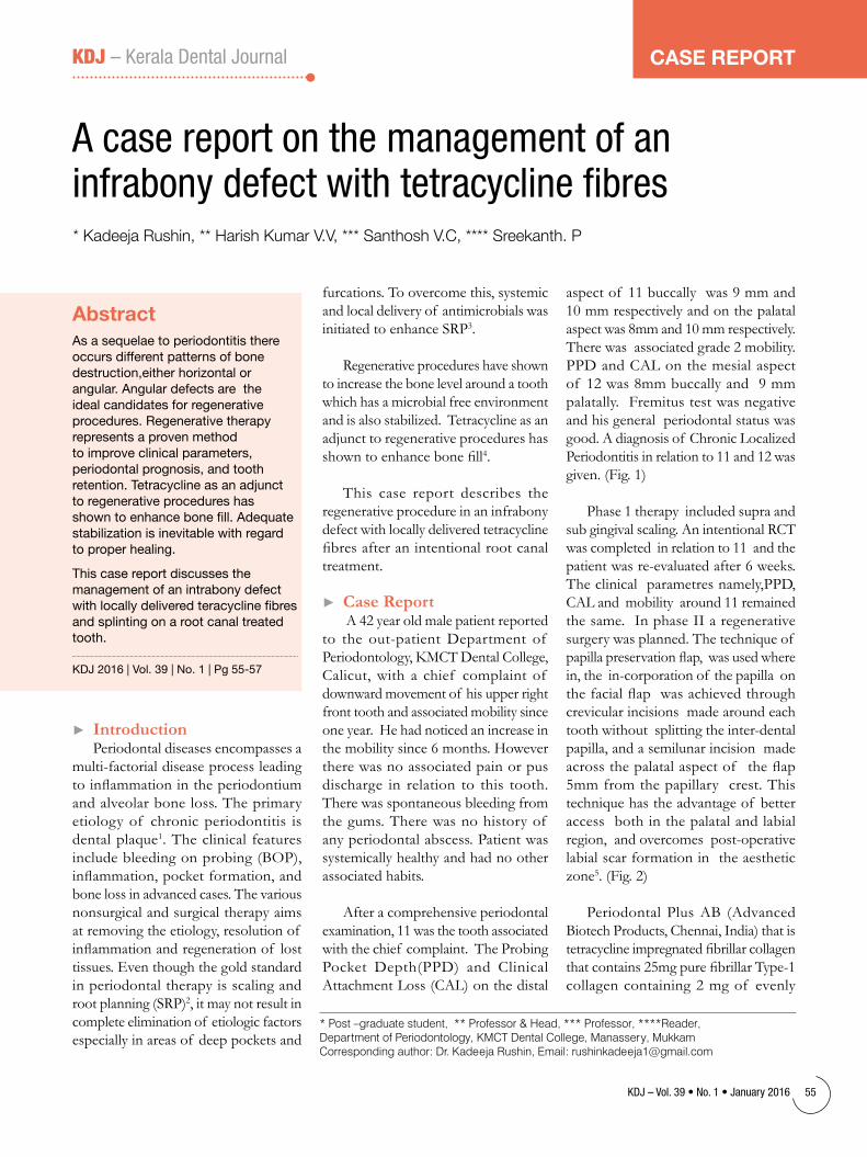
Acasereportonthemanagementofaninfrabonydefectwithtetracyclinefibres
AbstractAs a sequelae to periodontitis there occurs different patterns of bone destruction,either horizontal or angular. Angular defects are the ideal candidates for regenerative procedures. Regenerative therapy represents a proven method to improve clinical parameters, periodontal prognosis, and tooth retention. Tetracycline as an adjunct to regenerative procedures has shown to enhance bone fill. Adequate stabilization is inevitable with regard to proper healing.
This case report discusses the management of an intrabony defect with locally delivered teracycline fibres and splinting on a root canal treated tooth.
KDJ 2016 | Vol. 39 | No. 1 | Pg 55-57
* Post –graduate student, ** Professor & Head, *** Professor, ****Reader, Department of Periodontology, KMCT Dental College, Manassery, Mukkam Corresponding author: Dr. Kadeeja Rushin, Email: [email protected]
furcations. To overcome this, systemic and local delivery of antimicrobials was initiated to enhance SRP3.
Regenerative procedures have shown to increase the bone level around a tooth which has a microbial free environment and is also stabilized. Tetracycline as an adjunct to regenerative procedures has showntoenhancebonefill4.
This case report describes the regenerative procedure in an infrabony defect with locally delivered tetracycline fibresafteranintentionalrootcanaltreatment.
► Case Report A 42 year old male patient reported
to the out-patientDepartment of Periodontology, KMCT Dental College, Calicut, with a chief complaint of downward movement of his upper right front tooth and associated mobility since one year. He had noticed an increase in themobilitysince6months.Howeverthere was no associated pain or pus discharge in relation to this tooth. There was spontaneous bleeding from the gums. There was no history of any periodontal abscess. Patient was systemically healthy and had no other associated habits.
After a comprehensive periodontal examination, 11 was the tooth associated with the chief complaint. The Probing Pocket Depth(PPD) and Clinical Attachment Loss (CAL) on the distal
aspect of 11 buccally was 9 mm and 10 mm respectively and on the palatal aspectwas8mmand10mmrespectively.There was associated grade 2 mobility. PPD and CAL on the mesial aspect of 12was8mmbuccallyand9mmpalatally. Fremitus test was negative and his general periodontal status was good. A diagnosis of Chronic Localized Periodontitis in relation to 11 and 12 was given. (Fig. 1)
Phase 1 therapy included supra and sub gingival scaling. An intentional RCT was completed in relation to 11 and the patientwasre-evaluatedafter6weeks.The clinical parametres namely,PPD, CAL and mobility around 11 remained the same. In phase II a regenerative surgery was planned. The technique of papillapreservationflap,wasusedwherein,thein-corporationof thepapillaonthefacialflapwasachievedthroughcrevicular incisions made around each toothwithoutsplittingtheinter-dentalpapilla, and a semilunar incision made acrossthepalatalaspectof theflap5mm from the papillary crest. This technique has the advantage of better access both in the palatal and labial region,andovercomespost-operativelabial scar formation in the aesthetic zone5. (Fig. 2)
Periodontal Plus AB (Advanced Biotech Products, Chennai, India) that is tetracyclineimpregnatedfibrillarcollagenthatcontains25mgpurefibrillarType-1collagen containing 2 mg of evenly
* Kadeeja Rushin, ** Harish Kumar V.V, *** Santhosh V.C, **** Sreekanth. P
► Introduction Periodontal diseases encompasses a
multi-factorialdiseaseprocessleadingtoinflammationintheperiodontiumand alveolar bone loss. The primary etiology of chronic periodontitis is dental plaque1. The clinical features include bleeding on probing (BOP), inflammation,pocketformation,andbone loss in advanced cases. The various nonsurgical and surgical therapy aims at removing the etiology, resolution of inflammationandregenerationof losttissues. Even though the gold standard in periodontal therapy is scaling and root planning (SRP)2, it may not result in complete elimination of etiologic factors especially in areas of deep pockets and
KDJ–Vol.39•No.1•January2016 55
KDJ – Kerala Dental Journal Case report

impregnated tetracycline hydrochloride were used. It is available as a strip containing four individually packed sterile packs. Fiberswereplacedinthedefectsuchthatthematerialfillsthe depths and curves.
Palatalflapwassuturedusinganinternalhorizontalmattresssuture,whichhastheadvantageof providingatension-freeflapandastablesupportforthearea.Anon-eugenolperiodontaldressing(COE-PAK)wasplaced.
The patient was put on antibiotics for a period of 5 days andanti-inflammatorydrugsforaperiodof 3days.Strictoralhygeine instructions were given. The sutures were removed after 10 days. Provisional splinting was done on the palatal aspect of 12, 11 and 21 using ligature wire and light cure composite resin. (Fig. 3)
Tooth mobility has been described as an important clinical parameter for predicting the prognosis of periodontally compromised teeth. The main objective of splinting is to promote healing and increase the patients comfort and function. The main attraction of splinting is its low cost,the possibility of extending life and function of failing teeth. In cases with mobility that cannot be eliminated by selective coronoplasty alone, splinting should be considered as an adjunct to provide additional tooth stability during the surgical and healing phases of regenerative procedures.6
Oral hygiene instructions were reinforced and patient wasrecalledafter3months6monthsand9months.Therewas a reduction in PPD from 9mm to 4mm along the distal aspectof 11.IOPAshowsanimprovementinbonefillby6months and 9 months. (Fig. 4)
► DiscussionThe ultimate goal of any periodontal therapy is the control
of activeinflammation,thearrestof diseaseprogressionandthe reconstitution of the lost periodontal structures. It has been proven that conventional periodontal therapy did not succeed in predictable tissue regeneration7.
As an adjunct to scaling and root planning in management of periodontitis, systemic administration of tetracycline is routinely practiced. Tetracycline is bacteriostatic, it concentrates inperiodontalpockets,anditsantibacterialefficacymaybeprolonged through its ability to bind to both enamel and dentin surfaces. In addition, tetracyclines have recently been showntosuppresscollagenaseactivityincrevicularfluidandinflammatorycellsandtoinhibitosteoclastmediatedbonerésorption in vitro8.
Systemic administration of tertracycline has adverse effects such as acquired bacterial resistance, drug interaction and drug toxicity which limits its use of systemically. To overcome these shortcomings the concept of controlled release local drug delivery system was developed.
fig. 1 Pre-op fig. 2 Papillapreservationflap fig. 3 suturing
fig. 4 Palatalcompositesplint fig. 5 IOPAata)baselineb)6monthsc)9months
56 KDJ–Vol.39•No.1•January2016
Kadeeja Rushin
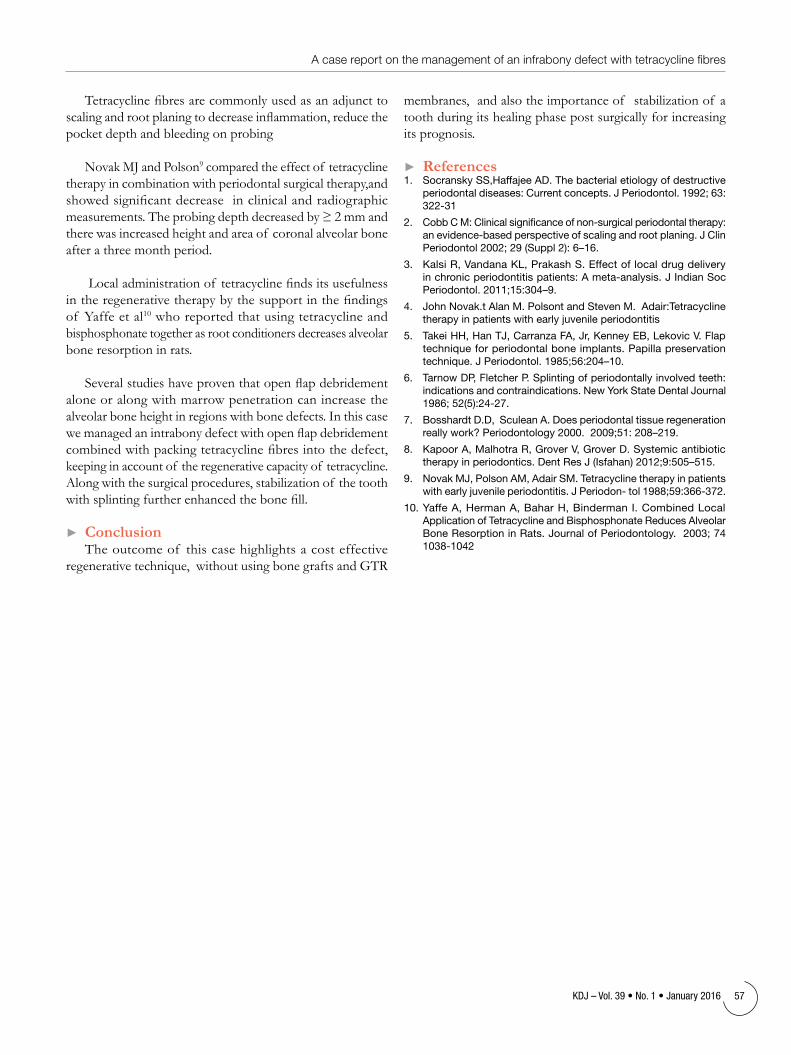
Tetracyclinefibresarecommonlyusedasanadjuncttoscalingandrootplaningtodecreaseinflammation,reducethepocket depth and bleeding on probing
Novak MJ and Polson9 compared the effect of tetracycline therapy in combination with periodontal surgical therapy,and showedsignificantdecreaseinclinicalandradiographicmeasurements.Theprobingdepthdecreasedby≥2mmandthere was increased height and area of coronal alveolar bone after a three month period.
Localadministrationof tetracyclinefindsitsusefulnessintheregenerativetherapybythesupportinthefindingsof Yaffe et al10 who reported that using tetracycline and bisphosphonate together as root conditioners decreases alveolar bone resorption in rats.
Severalstudieshaveproventhatopenflapdebridementalone or along with marrow penetration can increase the alveolar bone height in regions with bone defects. In this case wemanagedanintrabonydefectwithopenflapdebridementcombinedwithpackingtetracyclinefibresintothedefect,keeping in account of the regenerative capacity of tetracycline. Along with the surgical procedures, stabilization of the tooth withsplintingfurtherenhancedthebonefill.
► ConclusionThe outcome of this case highlights a cost effective
regenerative technique, without using bone grafts and GTR
membranes, and also the importance of stabilization of a tooth during its healing phase post surgically for increasing its prognosis.
► References 1. Socransky SS,Haffajee AD. The bacterial etiology of destructive
periodontal diseases: Current concepts. J Periodontol. 1992; 63: 322-31
2. Cobb C M: Clinical significance of non-surgical periodontal therapy: an evidence-based perspective of scaling and root planing. J Clin Periodontol 2002; 29 (Suppl 2): 6–16.
3. Kalsi R, Vandana KL, Prakash S. Effect of local drug delivery in chronic periodontitis patients: A meta-analysis. J Indian Soc Periodontol. 2011;15:304–9.
4. John Novak.t Alan M. Polsont and Steven M. Adair:Tetracycline therapy in patients with early juvenile periodontitis
5. Takei HH, Han TJ, Carranza FA, Jr, Kenney EB, Lekovic V. Flap technique for periodontal bone implants. Papilla preservation technique. J Periodontol. 1985;56:204–10.
6. Tarnow DP, Fletcher P. Splinting of periodontally involved teeth: indications and contraindications. New York State Dental Journal 1986; 52(5):24-27.
7. Bosshardt D.D, Sculean A. Does periodontal tissue regeneration really work? Periodontology 2000. 2009;51: 208–219.
8. Kapoor A, Malhotra R, Grover V, Grover D. Systemic antibiotic therapy in periodontics. Dent Res J (Isfahan) 2012;9:505–515.
9. Novak MJ, Polson AM, Adair SM. Tetracycline therapy in patients with early juvenile periodontitis. J Periodon- tol 1988;59:366-372.
10. Yaffe A, Herman A, Bahar H, Binderman I. Combined Local Application of Tetracycline and Bisphosphonate Reduces Alveolar Bone Resorption in Rats. Journal of Periodontology. 2003; 74 1038-1042
KDJ–Vol.39•No.1•January2016 57
A case report on the management of an infrabony defect with tetracycline fibres

Sweeteningtheburdenofdiabetes* Navia George, **Harish Kumar V.V, *** Santhosh V.C. **** Sreekanth P, **** Sameera G Nath, ***** Shabeer Muhammed
AbstractIntroduction: Diabetes Mellitus is one of the most frequent metabolic disorders. Periodontitis is considered as the sixth complication of diabetes. There is a two way relationship between periodontal disease and diabetes mellitus. Hence, it is necessary to estimate the blood glucose level of periodontitis patients prior to treatment.objective: To evaluate the correlation between capillary blood glucose levels (CBGLs) and sulcular blood glucose levels (SBGLs) in diabetic patients with chronic generalized periodontitis.Methodology: Diabetic patients with moderate to severe periodontitis were included in the study and subjected to routine clinical periodontal examinations. For estimating random blood sugar level (RBS), blood was collected from gingival sulcus following periodontal pocket probing to a test strip of a glucose self-monitoring device. As a control, capillary blood was taken with a finger-stick. Statistical analysis was performed using Pearson’s correlation coefficient and a t-test.Result: It was found that, random blood glucose level were the same in both sulcular blood as well as capillary blood.Conclusion: The result of this study suggests sulcular blood from a routine periodontal examination may be used for diabetes mellitus screening.Key-words: Field cancerization, Squamous cell carcinoma, Second primary tumors
KDJ 2016 | Vol. 39 | No. 1 | Pg 58-59*Post-graduatestudent,**HODofPeriodontology,***Professor,****Reader,*****SeniorLecturer,DepartmentofPeriodontology,KMCTDentalCollege,Manassery,Mukkam;CorrespondingAuthor:Dr.NaviaGeorge, Email:[email protected]
► Introduction:Diabetes mellitus is one of the most
frequent metabolic disorders with an estimated global prevalence of 9% among adultagedmorethan18years[WHO2014], of which nearly half the cases do undiagnosed. Patients with undiagnosed diabetes mellitus are at increased risk for coronary heart disease, stroke and peripheral vascular disease. In addition, recent data indicate that the incidence of the most common type of diabetes i.e., type2maybeincreasingupto6%peryears.1
Diabetes mellitus is associated with a wide range of complications such as retinopathy, nephropathy, neuropathy, micro-macrovasculardisease,alteredwoundhealing&periodontitis.2
Moreover, diabetes and periodontitis seems to interact in a bidirectional manner.3
Currently there are strong evidences to suggest that incidence and severity of periodontitisisinfluencedinpartbydiabetes mellitus and the level of blood glucosecontrol(Nishimaraetal1998).Hence, periodontal therapy might exert beneficialeffectsondiabetescontrol.4
► Aims & Objectives:To evaluate the correlation between
capillary blood glucose levels (CBGLs) and sulcular blood glucose levels (SBGLs) in diabetic patients with chronic generalized periodontitis.
► Materials and methods:Inclusion criteria:• Patientswithchronicgeneralized
periodontitis
• Agebetween35-45years• Nonsmokers• Thoseconsentingtoparticipatein
the studyExclusion criteria:• Patientswithsystemicillnessother
than diabetes mellitus• Pregnancyandlactation
A total of twenty diabetic patients (type2)havinggingivalinflammationwere taken for the study. Upper anterior teeth irrespective of their probing depths were chosen for glucose measurement as they are ideal for collecting gingival crevicular blood. For each measurement only one site with bleeding on probing was selected. Sites with suppuration were excluded from the study while contamination with saliva was prevented by using gauze and air drying. Blood oozing from the gingival crevicular fluidwasusedforglucoseassessment.Inaddition,regularfingerprickcapillaryblood was collected and both samples, 3μleach,wereanalysedusingaglucoseself-monitoringdevice.
The paired t test was used to compare mean scores of two groups andnosignificantdifferenceinthemeandiabetic blood sugar in gingival crevicular and capillary blood was found.
► Results:In this study, it was found that in all
the 20 diabetic patients tested, random blood glucose levels were the same in both gingival crevicular blood collected through bleeding on probing as well as capillary blood collected through fingerprick.Hencethisstudyindicates
58 KDJ–Vol.39•No.1•January2016
KDJ – Kerala Dental JournalINfoRMAtIoN

that gingival crevicular blood collected during diagnostic periodontal examination may be an excellent source of blood for glucometric analysis.
► Discussion:American diabetes association recommends that screening
for diabetes should start at the age of 45 years and be repeated every three years in persons without risk factors, and earlier and more often in those with risk factors for diabetes. Moreover, testing at young age or more frequently should be carried out in individuals who are,• Obese• Haveafirstdegreerelativewithdiabetes• Membersof ahighriskethnicgroup• Hypertensivepatients• Deliveredababyweighingmorethan4.05kgorhavebeen
diagnosed with gestational diabetes mellitus• Hadonprevioustestinganimpairedglucosetoleranceor
an impaired fasting glucose.Epidemiological data regarding diabetes mellitus coupled
with the possible interrelationship between diabetes mellitus and periodontitis suggests that all dentists will encounter patients with diabetes mellitus and clinicians who perform periodontal therapy must be aware and knowledgeable regarding thisdisease.Sinceperiodontalinflammationwith/withoutthecomplicating factor of diabetes mellitus is known to produce sample extravasate of blood during diagnostic periodontal
examination,noextraprocedure,e.g.:fingerpuncturewithalancet is necessary to obtain blood for glucometric analysis.5
Even in the case of very low gingival crevicular bleeding, a glucosemeasurementispossiblewiththeusedself-monitoringdevice,duetothelowamountof blood(3μl)necessarytoperform the analysis. Moreover, technique described is more familiarandlesstraumatictothepatientthanafingerpuncture.
► Summary:The results of the present study indicate that gingival
crevicular blood collected during diagnostic periodontal examination may be an excellent source of blood for glucometric analysis. In addition, the technique described is safe, easy to perform, and comfortable for the patient and might therefore help to increase the frequency of diabetes screeningindentaloffices.
► References:1) Rees TD. Periodontal management of the patient with diabetes
mellitus. Periodontol 2000 2000; 23: 63-72.2) Kuczek BT, Flemming TF. In-dental-office screening for diabetes
mellitus using gingival crevicular blood. J Clin Periodontol 2002; 29: 216-18.
3) Grossi SG, Jenco RJ. Periodontal disease as a complication of diabetes melliyus. Ann Periodontol 1998; 3: 20-9.
4) AAP Parameter on systemic conditions affected by periodontal disease. J Periodontol 2000; 71: 880-83.
5) Parker RC, Isley W. Gingival crevicular blood for assessment of blood glucose in diabetic patients. J Periodontol 1993; 64: 666-72.
Paired Samples Statistics
Mean N Std. Deviation Std. Error Mean t p Remark
SBGL 214.70 20 68.803 15.385 1.35 0.09 Notsignificant
CBGL 217.00 20 69.526 15.547
fig. 1 ChronicperiodontitisPatient fig. 2 Bleedinginducedonprobing fig. 3 GlucometricanalysisofSBGLs
fig. 4 GlucometricanalysisofCBGLs fig. 5 DigitalreadoutonGlucometerGraph
KDJ–Vol.39•No.1•January2016 59
Sweetening the burden of diabetes

Evaluationoftheeffectivenessofphotodynamictherapyusingtoluidineblueformicrobialreductioninperiodontalpockets
AbstractBackground: Recent preclinical and clinical data have suggested a potential benefit of photodynamic therapy (PDT) in the treatment of periodontitis. However, there are very limited data from controlled clinical trials evaluating the effect of PDT in the treatment of periodontitis. The aim of this study was to evaluate microbiologic effects of the adjunctive use of PDT to non-surgical periodontal treatment. Methods: Twenty patients with chronic periodontitis were randomly treated with scaling and root planing followed by a single episode of PDT (test) or scaling and root planing alone (control). Full-mouth plaque score (FMPS), full-mouth bleeding score (FMBS), probing depth (PD), gingival recession, and clinical attachment level (CAL) were measured at baseline. Microbiological analysis was based on semiquantitative assessment of the colony forming units on blood agar.Results: At baseline the intergroup comparison of the demographic parameters were statistically insignificant. Intergroup comparison of the bacterial growth before and after intervention was statistically significant with ap value < 0.05. Conclusion: With a significant reduction in the subgingival microbiota, this study suggests that aPDT can be successfully used as an adjunct to scaling an root planing.
KDJ 2016 | Vol. 39 | No. 1 | Pg 60-64
* Post –graduate student, ** HOD of Periodontology, *** Professor, **** Reader, ***** Senior Lecturer, Department of Periodontology, KMCT Dental College, KMCT Dental College, Manassery, Mukkam, Corresponding author: Dr. Anjhana Narayanan Email: [email protected]
► Introduction: The etiology of periodontitis
is multifactorial, which results in therapeutic difficulties. Bacteria (periodontopathogens), considered to be one of the main etiological agent grow in biofilms, and are beyond the reach of antimicrobial chemical agents. The anatomical complexity of tooth roots provides niches for bacterial deposits, making eradication of periodontopathogensmoredifficultboth mechanically and chemically. Also some periodontopathogens (e.g. Aggregatibacter actinomycetemcomitans) can penetrate into and persist in epithelial cells of the periodontal pockets and outer gingiva thus avoiding host immunity and conventional antimicrobial drugs1. Moreover, there is also a problem of increasing bacterial resistance developing to systemic antibiotics. Conventional treatment such as scaling and root planing (SRP) does not completely eliminate periodontal pathogens, especially in deep periodontal pockets; moreover, it does not prevent this microorganism from penetrating into periodontal tissue. Finally, this predisposes the periodontal pockets to re-colonization, diseaserelapses and maintains the chronic nature of the disease2.
Theseabove-mentionedissuesjustifythe need for alternative antibacterial therapeutic strategies. One of them is photodynamic therapy against microorganisms called antimicrobial photodynamic therapy.
Photodynamic therapy (PDT), also called photoradiation therapy, phototherapy, or photochemotherapy, was introduced in medical therapy in 1904asthelight-inducedinactivationof cells, microorganisms, or molecules3.PDT involves the combination of visible light, usually with a diode laser and a photosensitizer. The photosensitizer is a compound that is capable of absorbing light of a specific wavelength and transforming it into useful energy4.
The action mechanism of PDT can be briefly described as: upon illumination, the photosensitizer is excited and converted from the ground state to the triplet state. The triplet state enables the interaction of the excited photosensitizer with the surrounding molecules. The cytotoxic product, usually singlet oxygen, has a short lifetime in biologicalsystems(<0.04μs)and,alsohaveashortradiusof action(0.02μm).Because of the limited migration of the
* Anjhana Narayanan, ** Harish Kumar V.V., *** Santhosh V.C., **** Sreekanth P., **** Sameera G Nath, *****Shabeer Muhammed
60 KDJ–Vol.39•No.1•January2016
KDJ – Kerala Dental JournalINfoRMAtIoN
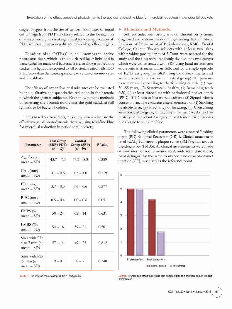
singlet oxygen from the site of its formation, sites of initial cell damage from PDT are closely related to the localization of the sensitizer, thus making it ideal for local application of PDT, without endangering distant molecules, cells or organs.
Toludine blue O(TBO) is cell membrane active photosensitizer, which can absorb red laser light and is bactericidal for many oral bacteria. It is also shown in previous studies that light dose required to kill bacteria treated with TBO is far lower than that causing toxicity to cultured keratinocytes andfibroblasts.
Theefficacyof anyantibacterialsubstancecanbeevaluatedby the qualitative and quantitative reduction in the bacteria to which the agent is subjected. Even though many methods of assessingthebacteriafloraexiststhegoldstandardstillremains to be bacterial culture.
Thus based on these facts, this study aims to evaluate the effectiveness of photodynamic therapy using toluidine blue for microbial reduction in periodontal pockets
► Materials and Methods:Subject Selection: Study was conducted on patients
diagnosed with chronic periodontitis,attending the Out Patient Division of Department of Periodontology, KMCT Dental College, Calicut. Twenty subjects with at least two sites withprobingpocketdepthof 5-7mmwereselectedforthestudy and the sites were randomly divided into two groups which were either treated with SRP using hand instruments and sonic instrumentation followed by a single episode of PDT(test group) or SRP using hand instruments and sonic instrumentation alone(control group). All patients were recruited according to the following criteria: (1) Age 30-55years,(2)Systemicallyhealthy,(3)Remainingteeth≥20,(4)atleastthreesiteswithperiodontalpocketdepth(PPD)of 4-7mmin3ormorequadrants(5)Signedinformconsent form. The exclusion criteria consisted of: (1) Smoking or alcoholism, (2) Pregnancy or lactating, (3) Consuming antimicrobial drugs (ie, antibiotics) in the last 3 weeks, and (4) Historyof periodontalsurgeryinpast6months(5)patientsnot allergic to toluidine blue.
The following clinical parameters were assessed Probing depth(PD),GingivalRecession(GR)&Clinicalattachmentlevel(CAL)full-mouthplaquescore(FMPS),full-mouthbleeding score (FMBS). All clinical measurements were made atfoursitespertooth:mesio-facial,mid-facial,disto-facial,palatal/lingualbythesameexaminer.Thecement-enameljunction (CEJ) was used as the reference point.
ParameterTest Group
(SRP+PDT) (n = 10)
Control Group (SRP)
(n = 10)P Value
Age (years; mean–SD) 43.7–7.3 47.3–8.8 0.289
CAL (mm; mean–SD) 4.1–0.5 4.5–1.0 0.219
PD (mm; mean–SD) 3.7–0.5 3.6–0.6 0.577
REC (mm; mean–SD) 0.5–0.4 1.0–0.8 0.051
FMPS (%; mean–SD) 58–24 62–14 0.631
FMBS (%; mean–SD) 54–16 59–21 0.501
Sites with PD 4 to 7 mm (n; mean–SD)
47–14 49–25 0.812
Sites with PD ‡7 mm (n; mean–SD)
9–9 8–7 0.740
table 1: Thebaselinecharacteristicsofthe20participants Graph 1: Graphcomparingthepreandposttreatmentresultsinmicrobialfloraoftestandcontrolgroup.
KDJ–Vol.39•No.1•January2016 61
Evaluation of the effectiveness of photodynamic therapy using toluidine blue for microbial reduction in periodontal pockets

Study Design: The study performed was a split mouth study. Treatment allocation was performed by a lottery method. Culture samples were collected from both test and control site using paper points and transferred to anaerobic transport medium. Following this supragingival calculus was removed with sonic instrumentation, and subgingival instrumentation for test and control groups was performed until the operator believed that the root surfaces were adequately debrided and planed. Randomization was performed immediately following the completion of instrumentation
Laser Therapy/Photodynamic Therapy: In the test group, the photosensitizer liquid that is 1% toluidine blue dye was applied with a blunt needle to the instrumented sites, starting from the apical end of the pocket and moving coronally to avoid entrapment of air bubbles (Fig. 1). One minute later, all pockets were thoroughly rinsed with sterile saline to remove the excess photosensitizer. Immediatelyafterrinsing,thediodelaser,with810nmwavelengthand0.8Wof poweroutput,equippedwitha probe tip, was placed at the depth of the pocket and moved circumferentially around the tooth for 30 seconds, according to the manufacturer’s instructions (Fig. 2). Post operatively oral hygiene instructions were reinforced and patients were not administered antibiotics.
Samples for Microbiological analysis: The procedure was performed with a sterile paper point which was inserted in the bottom of the pocket for 30 seconds (Fig. 3), after isolating the selected sites using cotton rolls. The paper points from the test and control site was streaked on a sheep blood agar plate such that one half of the plate received samples from the test site and the other half from the control site (Fig. 4). Pre and post treatment samples were collected from both sites on the same day.
Microbiological investigation was done in the Dept. of Microbiology, Aster Malabar Institute of Medical Sciences, Calicut.Thesampleswereincubatedfor48hoursandfurthersubcultured to semi quantitatively assess the microbial colonies. Assessment was based by counting the number of colony forming units of microbial colonies(cfu)5.
Bacterial levels were expressed semi quantitatively as (no growth = 0; scanty = 1; moderately scanty = 2; moderate = 3; moderately heavy 4; heavy =5).
Statistical Analysis The statistical analysis was performed usingSPSSsoftware.Asubject-levelanalysiswasperformedforeachof theparameterswereanalyzedwiththeMann-Whitney U test. Differences were considered statistically significantwhenthePvaluewas<0.05.
fig. 1: 1%toluidinebluedyewasappliedwithabluntneedletotheinstrumentedsites,
fig. 2: Diodelaser,with810nmwavelengthand0.8Wofpoweroutputplacedatthedepthofthepocket
fig. 3: Sterilepaperpointwhichwasinsertedinthebottomofthepockettoobtainmicrobialsamples
fig 4: Samplesstreakedonbloodagarplates fig. 5 Comparisonbetweenthebloodagarplatesofpreandposttreatmentsamplesfromthetestandcontrolsite.
62 KDJ–Vol.39•No.1•January2016
Anjhana Narayanan

► Results : All patients enrolled completed the study. No adverse
effects, such as discomfort, burning sensation, or pain related to the laser irradiation, were reported by any of the subjects. The baseline characteristics of the 20 participants aredisplayedinTable1.Themeanagewas43.7–7.3yearsforthetestgroupand47.3–8.8yearsforthecontrolgroup.None of these demographic parameters showed a statistically significantdifferencebetweenthegroups.Afterincubationof thesubculturesfor48hoursinanaerobicconditions,values of the CFUs on the plates were determined by the semiquantitative method used for determining bacteriuria6. The CFUs from the test and control sites of both pre and post treatmentsampleswerecompared(Graph1),(Fig.5&6)andfrom the results it was inferred that, in all the cases
a) there was slight reduction in the bacterial count at the control site ie CFUs were recorded heavy in pretreatment samplesandmoderate-heavyinposttreatmentsamples.
b) therewassignificantreductioninthebacterialcountat the experimental site, ie CFUs were recorded heavy inpretreatmentsamplesandmoderate-scantyinposttreatment samples. Results of the study was statistically significantwithapvalue<0.05.
Thus from the results of the study we can conclude that photodynamic therapy can be used an effective adjunct to scaling and root planing in reducing the microbial load in periodontal pockets.
► Discussion :Photodynamic therapy is a novel therapeutic approach
for eradicating pathogenic bacteria in periodontal disease. Inactivation of microorganisms using photodynamic therapy hasbeendefinedaseitherantimicrobialphotodynamictherapy(aPDT), photodynamic antimicrobial chemotherapy (PACT) or photodynamic disinfection A number of studies have shown thatTBOisphototoxictoseveralplanktonicgram-positiveandgram-negativebacteria7.Thephotodynamicefficacyof TBO
hasalsobeendemonstratedinseveraloralbiofilmmodels8. Photodynamictherapyeliminatedapproximately63%of bacteria in the planktonic phase, whereas only 32% of bacteria inbiofilms,whichderivedfromthesameplaquesamples9.However,bacterialeradicationfromdentalplaque-derivedbiofilmsisstillatalowerlevelcomparedtotheplanktoniccondition. The study by Fontana et al10confirmedthisfact.Inthisstudywehaveinvestigatedtheantimicrobialefficacyof photodynamic therapy in reducing microbial load as an adjunct to scaling and root planing in patients with chronic periodontitis.
The results of this study has shown that it is possible to kill subgingivalmicroflorainthegingivalcreviceusingtopicallyapplied toluidine blue and red light from a diode laser without causing any detectable damage to the adjacent tissues. There was considerable reduction in density of the black pigmented colonies in the culture plates of sites treated with aPDT.
Wakayama, Y et al showed TB to be an effective photosensitizer which is capable of damaging cell membranes during aPDT, thus leading to the eradication of bacterial cells11. Zanin et al12usedTBandirradiationof 85.7Jcm22tokillinvitro-generatedsingle-speciesbiofilmof S.mutansandotheroral streptococci. The elimination rates of the bacteria were found to vary between 95 and 99.9 %. Bhatti et al. found that 100% killing of P. gingivalis organisms could be achieved with 25μgof toluidinebluepermland4.4Jof redlight.
Recently, Novaes et al. investigated changes occurring in the subgingival microbiological composition of subjects with aggressive periodontitis treated with antimicrobial photodynamic therapy in a single episode or SRP. This trial indicatedthataPDTismoreefficientinreducingthepresenceof Aggregatibacter actinomycetemcomitans than SRP13. On the other hand, SRP limited the number of periodontal pathogens of the Red Complex more effectively than aPDT. Because of the fact that aPDT and SRP affect different species, it is suggested that both methods be combined to gain better resultsinnon-surgicaltreatmentof aggressiveperiodontitis9.
Regarding the mechanism of aPDT, a recent proteomic approach by Dosselli et al.14 revealed that aPDT delays the growth of bacteria, and reduces the capacity of bacteria for glucose consumption. Combined, the bacterial killing effect and the retardation of bacterial growth with aPDT could reduce plaque deposition on teeth. Nonetheless, the negative aspects of the clinical use of aPDT should be considered. Some reports have noted tooth staining with TBO15 however, residual staining of teeth and gingival tissue with TBO after the aPDT procedure was not visible, and did not present a problem in this clinical trial.
fig. 6: Comparisonbetweenthebloodagarplatesofpreandposttreatmentsamplesfromthetestandcontrolsite.
KDJ–Vol.39•No.1•January2016 63
Evaluation of the effectiveness of photodynamic therapy using toluidine blue for microbial reduction in periodontal pockets

► Conclusion From this study we can conclude that using PDT is
lesstraumatic&quickerinthetreatmentof inflammatoryperiodontal diseases. PDT offers numerous advantages, particularly in avoiding emergence of antibiotic resistance species,requiringlesstechnicalskills&reducingoperatingtime in comparison to manual scaling and root planing.
► References :1. Lamont R.J., Yilmaz O. In or out: the invasiveness of oral bacteria.
Periodontol. 2000, 2002; 30: 61-692. Ardila C.M., Granada M.I., Guzmán I.C.: Antibiotic resistance of
subgingival species in chronic periodontitis patients. J. Periodontal. Res., 2010; 45: 557-563
3. Von Tappeiner H, Jodlbauer A. On the effect of photodynamic (fluorescent) substances on protozoa and enzymes (in German).Deutsch Arch Klin Medizin 1904;39:427-87
4. Sharman WM, Allen CM, van Lier JE. Photodynamic therapeutics: Basic principles and clinical applications.Drug Discov Today 1999;4:507-517.
5. Bowler P, Duerden B, Armstrong D. Wound microbiology and associated approaches to wound management. Clin Microbiol Rev. 2001;14(2):244–69.
6. Ruth M Myers and Grace Koshi, Diagnostic Procedures in Medical Microbiology and Immunology/Serology, pg 61, 62; 1982, CMC Vellore.
7. Tang, H. M., M. R. Hamblin, and C. M. Yow. 2007. A comparative in vitro photoinactivation study of clinical isolates of multidrug-resistant pathogens. J. Infect. Chemother. 13:87-91.
8. Zanin, I. C.M. M. Lobo, L. K. Rodrigues, L. A. Pimenta, J. F. Höfling, and R. B. Gonçalves. 2006. Photosensitization of in vitro biofilms by toluidine blue O combined with light-emitting diode. Eur. J. Oral Sci. 114:64-69.
9. Soukos N.S., Ximenez-Fyvie L.A., Hamblin M.R., Socransky S.S., Hasan T.Targeted antimicrobial photochemotherapy. Antimicrob. Agents Chemother., 1998; 42: 2595-2601
10. Fontana C.R., Abernethy A.D., Som S., Ruggiero K., Doucette S., Marcantonio R.C., Boussios C.I., Kent R., Goodson J.M., Tanner A.C., Soukos N.S.The antibacterial effect of photodynamic therapy in dental plaque-derived biofilms. J. Periodont. Res., 2009; 44: 751-759
11. Wakayama, Y., Takagi, M. & Yano, K. (). Photosensitized inactivation of E. coli cells in toluidine blue-light system. Photochem Photobiol 1980;32: 601–605
12. Zanin, I. C., Lobo, M. M., Rodrigues, L. K., Pimenta, L. A., Ho¨ fling, J. F. & Gonc¸ alves, R. B. (). Photosensitization of in vitro biofilms by toluidine blue O combined with a light-emitting diode. Eur J Oral Sci 2006;114: 64–69
13. Novaes A.B.Jr., Schwartz-Filho H.O., de Oliveira R.R., Feres M., Sato S., Figueiredo L.C.: Antimicrobial photodynamic therapy in the non-surgical treatment of aggressive periodontitis: microbiological profile. Lasers Med. Sci., 2012; 27: 389-395
14. Dosselli R, Millioni R, Puricelli L, Tessari P, Arrigoni G, Franchin C, Segalla A, Teardo E, Reddi E: Molecular targets of antimicrobial photodynamic therapy identified by a proteomic approach. J Proteomics 2012, 77:329–343.
15. Williams JA, Pearson GJ, Colles MJ, Wilson M: The photo-activated antibacterial action of toluidine blue O in a collagen matrix and in carious dentine. Caries Res 2004,38(6):530–536.
Thrissur IDA mourns the sad demise of Dr. Manoj DavisAkkaraaged46years(D.O.B.:19.12.1969)followingbrief period of hospitalization at Aster Medicity Ernakulam. He is survived by his wife Mrs. Melissa Manoj and daughter Merlin Manoj. Dr. Manoj graduated from Rajas Dental College, Vadakankulam, Thirunelveli Dist., Tamil Nadu in the year 1994 December. He set up practice at
Olarikkara, Thrissur. He was a regular and active member of IDA and had held various executive posts in committee in the past. A very well loved by all, jovial and good natured, he will be missed sorely. May his soul rest in peace. All of the Thrissur IDA members and nearby branches turned up at the funeral to express the deep grief and pay homage.
OBITUARY
64 KDJ–Vol.39•No.1•January2016
Anjhana Narayanan
*Jyotsna J., **Devisree Naveen, **Teenu Abraham, ***Padmakumar T P, ****Nandakumar K.
AbstractIntroduction: Gummy smile due to excessive gingival display always makes a normal person conscious while smiling. The treatment of choice in this type of cases with excess can be corrected by orthognathic surgery along with orthodontic treatment. It can otherwise be treated by a lip – repositioning procedure; a less invasive approach.
Case Presentation: This is a case report of a female patient who was unsatisfied with her smile and states that it is compromising her self – esteem due to excessive gingival display and not willing for orthognathic surgery; which is corrected by lip repositioning.
Conclusion: In patients with a gummy smile who are apprehensive of undergoing orthognathic surgery, lip repositioning can be a reassuring treatment option.
Key words: Smile, Gummy smile, smile line, lip repositioning, orthognathic surgery, gingivectomy.
KDJ 2016 | Vol. 39 | No. 1 | Pg 65-67
ManagementofExcessiveGingivalDisplay
*PGStudent,**SeniorLecturer,***Professor,****ProfessorandHOD,DepartmentofPeriodontics,AzeeziaCollegeofDentalSciencesandResearch,Meeyannoor,Kollam.CorrespondingAuthor:Dr.Jyotsna.J. E-mail:[email protected]
► Introduction“Beauty lies in the eyes of the
beholder” keeping this old saying in mind, smile corrections can be done in patients with esthetics concerns. Excessive gingival display, commonly referred to as ‘gummy smile’ is a major hurdle in overall personality of an individual. What constitutes a pleasant smile varies from person to person, but usually depends on the
extent of gingival exposure. Recently the dental literature has focused into various treatment modalities in the correction of gummy smile. When a person smiles, the entire crowns of maxillary central incisors and 1 mm of pink attached is visible.
Gummy smile due to excessive gingival display always makes a normal person conscious while smiling, especially when the problem is related with female patients who are more esthetically consciousthanmales(Geron&Atalia2005). “Excessive gingival display”, also called gummy smile is one of the several developmental or acquired deformity that manifest in the periodontium (Armitage 1999). Gummy smile is seen due to improper relation between gingival tissue and the tooth, with gingival tissue in excess and tooth portion in a small amount, with a prevalence of 7%inyoungadultsmales&14%of youngadultfemales(Tjanetal1984;DiamondO,Journ.esthetdent1996).It can be a source of embarrassment for some patients. Gummy smile is governed by various etiological factors, for example jaw deformities, which cause excessive gingival display and require a orthognathic surgery. This occurs due to excessive increased vertical height of maxillary arch. Orthongathic surgery is usually required to treat vertical maxillary excess. A wedge of maxilla is removed and maxilla is positioned in a
pre determined level. In some cases, only mandibular osteotomy will be enough to obtain a stable occlusal relationship. The orthognathic surgery is a complicated procedure and requires team work with hospitalization and general anesthesia and entails significant discomfort, while lip repositioning is innovative and effective, less time consuming and is performed under local anesthesia. Short or hyper active upper lip, altered passive eruption, vertical maxillary excess, dento alveolar extrusion etc affects the gummy smile. Causes like delayed eruption and tooth malpositioning can be predictably treated with resective surgery and orthodontics1. When the gingiva demonstrates a healthy appearance and a reduced crown length of the anteriors; which cannot be completely explained by incisal edge wear, a differential diagnosis of altered passive eruption is made and one of the treatment modality is the crown lengthening2.
In dento alveolar extrusion, mainly due to the over eruption of maxillary anterior teeth and can be corrected bymoving the over-erupted teethinto the original position. This can be accomplished either by orthodontics or with a segmental osteotomy followed by long term retention.
Esthetic Periodontal SurgeryIf the osseous level is appropriate,
if there is more than 3 mm of tissue
KDJ–Vol.39•No.1•January2016 65
KDJ – Kerala Dental Journal Case report

from bone to gingival crest and if it is determined that an adequate zone of attached gingiva will remain after surgery, a gingivectomy is indicated. If the diagnostic procedures revealosseouslevelsapproximatingtheCEJ,agingivalflapwith ostectomy is indicated. If the gingival heights of the anterior teeth are asymmetric, the initial incision should be agingivectomy-typeincisionsothatthefinaltissuecontourwill be symmetric. If the preoperative tissue contours are symmetric,asulcularincisioncanbeusedandtheflapapicallyrepositioned. The inferential incision should always be a sulcular incision, leaving the papilla totally intact interproximally3.
► Case reportA23-year-oldfemalepatientwithacomplaintof unaesthetic
smile with localized gingivititis, and maxillary gingival excess resulting in a gummy smile reported to our department of periodontics(Figs.1&2).Thepatienthasbeenundergoingorthodontic treatment since 3 years for bimaxillary protrusion but she felt that smile was not acceptable aesthetically. On clinical examination, extraorally face was found to be bilaterally symmetrical with incompetent lips (Fig. 3). Intraorally, a
moderate gingival display was seen during smiling, which extends from maxillary right second premolar to maxillary left second premolar. She approached in an outside clinic and was advised for orthognathic surgery.
On clinical examination, she was found to have a vertical maxillaryexcess&adiagnosisof moderateverticalmaxillaryexcess with hypermobility of upper lip was made. A treatment plan involving surgical crown lengthening and lip repositioning was drawn up and presented to the patient. After obtaining approval of the treatment plan and informed consent, phase 1 periodontal therapy was performed to prepare the tissues for surgery. One week later, the patient was recalled for surgery. Bone sounding was done under anesthesia, before starting the excision, to clearly establish the position of the bone. Extra oral and intra oral antisepsis was performed with 2% chlorhexidine solution.
Afteracquiringsufficientanaesthesiaandasepsis,Inversebevelgingivectomy(Fig.4)wasdone,andtheflaprosefordebridement and curettage. Hemostatis was performed using
fig 1: PreOP fig 2: Distancefromthelowerliplinetothegingivalmargin
fig 3: Lateralview fig 4: Gingivectomy
fig 5: Hemostasisusinglasertherapy fig 6: Aftergingivectomy fig 7: Liprepositioning fig 8: Continuousinterlockingsuture
fig 9a: PostOPonemonth fig 9b: PostOPthirdmonth fig 9c: PostOPsixthmonth
66 KDJ–Vol.39•No.1•January2016
Jyotsna J.
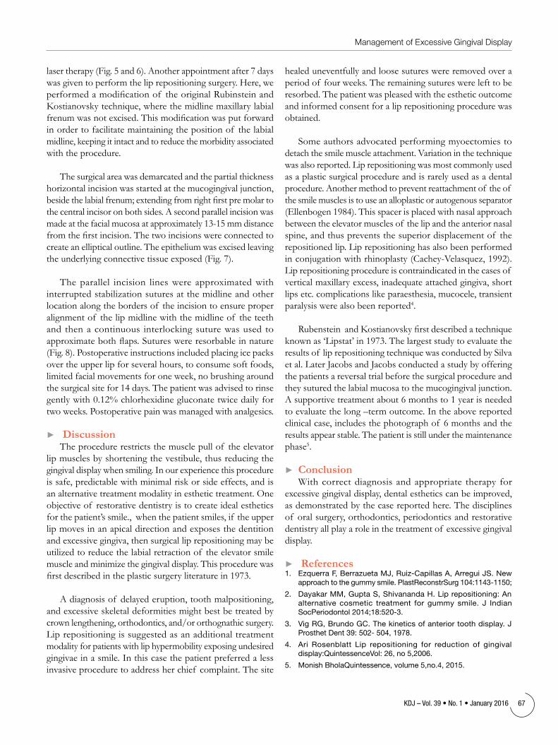
lasertherapy(Fig.5and6).Anotherappointmentafter7dayswas given to perform the lip repositioning surgery. Here, we performedamodificationof theoriginalRubinsteinandKostianovsky technique, where the midline maxillary labial frenumwasnotexcised.Thismodificationwasputforwardin order to facilitate maintaining the position of the labial midline, keeping it intact and to reduce the morbidity associated with the procedure.
The surgical area was demarcated and the partial thickness horizontal incision was started at the mucogingival junction, besidethelabialfrenum;extendingfromrightfirstpremolartothe central incisor on both sides. A second parallel incision was madeatthefacialmucosaatapproximately13-15mmdistancefromthefirstincision.Thetwoincisionswereconnectedtocreate an elliptical outline. The epithelium was excised leaving the underlying connective tissue exposed (Fig. 7).
The parallel incision lines were approximated with interrupted stabilization sutures at the midline and other location along the borders of the incision to ensure proper alignment of the lip midline with the midline of the teeth and then a continuous interlocking suture was used to approximatebothflaps.Sutureswereresorbableinnature(Fig.8).Postoperativeinstructionsincludedplacingicepacksover the upper lip for several hours, to consume soft foods, limited facial movements for one week, no brushing around the surgical site for 14 days. The patient was advised to rinse gently with 0.12% chlorhexidine gluconate twice daily for two weeks. Postoperative pain was managed with analgesics.
► DiscussionThe procedure restricts the muscle pull of the elevator
lip muscles by shortening the vestibule, thus reducing the gingival display when smiling. In our experience this procedure is safe, predictable with minimal risk or side effects, and is an alternative treatment modality in esthetic treatment. One objective of restorative dentistry is to create ideal esthetics for the patient’s smile., when the patient smiles, if the upper lip moves in an apical direction and exposes the dentition and excessive gingiva, then surgical lip repositioning may be utilized to reduce the labial retraction of the elevator smile muscle and minimize the gingival display. This procedure was firstdescribedintheplasticsurgeryliteraturein1973.
A diagnosis of delayed eruption, tooth malpositioning, and excessive skeletal deformities might best be treated by crown lengthening, orthodontics, and/or orthognathic surgery. Lip repositioning is suggested as an additional treatment modality for patients with lip hypermobility exposing undesired gingivae in a smile. In this case the patient preferred a less invasive procedure to address her chief complaint. The site
healed uneventfully and loose sutures were removed over a period of four weeks. The remaining sutures were left to be resorbed. The patient was pleased with the esthetic outcome and informed consent for a lip repositioning procedure was obtained.
Some authors advocated performing myoectomies to detach the smile muscle attachment. Variation in the technique was also reported. Lip repositioning was most commonly used as a plastic surgical procedure and is rarely used as a dental procedure. Another method to prevent reattachment of the of the smile muscles is to use an alloplastic or autogenous separator (Ellenbogen1984).Thisspacerisplacedwithnasalapproachbetween the elevator muscles of the lip and the anterior nasal spine, and thus prevents the superior displacement of the repositioned lip. Lip repositioning has also been performed inconjugationwithrhinoplasty(Cachey-Velasquez,1992).Lip repositioning procedure is contraindicated in the cases of vertical maxillary excess, inadequate attached gingiva, short lips etc. complications like paraesthesia, mucocele, transient paralysis were also been reported4.
RubensteinandKostianovskyfirstdescribedatechniqueknown as ‘Lipstat’ in 1973. The largest study to evaluate the results of lip repositioning technique was conducted by Silva et al. Later Jacobs and Jacobs conducted a study by offering the patients a reversal trial before the surgical procedure and they sutured the labial mucosa to the mucogingival junction. Asupportivetreatmentabout6monthsto1yearisneededtoevaluatethelong–termoutcome.Intheabovereportedclinicalcase,includesthephotographof 6monthsandtheresults appear stable. The patient is still under the maintenance phase5.
► ConclusionWith correct diagnosis and appropriate therapy for
excessive gingival display, dental esthetics can be improved, as demonstrated by the case reported here. The disciplines of oral surgery, orthodontics, periodontics and restorative dentistry all play a role in the treatment of excessive gingival display.
► References1. Ezquerra F, Berrazueta MJ, Ruiz-Capillas A, Arregui JS. New
approach to the gummy smile. PlastReconstrSurg 104:1143-1150; 2. Dayakar MM, Gupta S, Shivananda H. Lip repositioning: An
alternative cosmetic treatment for gummy smile. J Indian SocPeriodontol 2014;18:520-3.
3. Vig RG, Brundo GC. The kinetics of anterior tooth display. J Prosthet Dent 39: 502- 504, 1978.
4. Ari Rosenblatt Lip repositioning for reduction of gingival display:QuintessenceVol: 26, no 5,2006.
5. Monish BholaQuintessence, volume 5,no.4, 2015.
KDJ–Vol.39•No.1•January2016 67
Management of Excessive Gingival Display

68 KDJ–Vol.39•No.1•January2016
KDJ – Kerala Dental JournalSuRvEy
AcrosssectionalsurveyontheutilizationofscientificjournalsamongstaffsandstudentsofDentalCollegesinKerala
* Civy V. Pulayath, ** Rasmi George
*Reader,**Intern,DeptofPublicHealthDentistry,AlAzharDentalCollege,Thodupuzha. CorrespondingAuthor:DrCivyV.Pulayath,Email:[email protected]
► MethodologyThe investigation was conducted
using a cross sectional questionnaire survey among staffs and students of dental colleges in Kerala. A quota sampling methodology was used to collect data from 500 subjects (250 Interns and 250 Staffs). Separate questionnaires with 20 closed ended questions were used for the two groups.
► Results 75.2% (188) of the students
confessed that they were not interested inreadingjournals,while83.6%of theteaching staff remarked that they don’t use dental journals as intense as they did it during their study period. Majority of them agreed that reading journals
necessary for dental professionals mainly to get updated (493 out of 500).
Lack of adequate time prevents 44% of studentsand60%of professionalsfrom reading journals. Majority of students prefer to read Indian journals while staffs make use of international journals. In case of students, library is the main source for reading journals but lack of adequate journals in library is the main problem. At the same time staffs also access subscriptions and internet.
Journal presentation exercise for interns was found to be less prevalent in very few institutions. 92% (230) of teachers substantiated that many ideas fromjournalsinfluencedtheirstudies
► IntroductionDental journals bring scientific
research, interesting case reports, newer techniques and opinions to a wide audience of dental professionals around the globe in order to improve the clinical outcome of dental treatment. A query was done using a cross sectional questionnaire survey to assess the utilisationof thisscientificliteratureindental educational institutions.
► Aim1. To evaluate the level of utilization of
dental journals among students and staffs of dental colleges in Kerala.
► Objectives1. To know whether avenues for
utilization of journals were created in dental institutions.
2. To know whether language act as a barrier in understanding the concepts explained in the journals.
AbstractAim: To describe a hybrid technique Dental journals are the most efficient tool to bring about scientific research data, findings of clinical studies and interesting case reports to both the professionals and dental students. This study aims to measure the awareness and utilization of such journals by the students and professionals.
KDJ 2016 | Vol. 39 | No. 1 | Pg 68-69
fig 4: Academicpracticeofdentalstaffs
fig.1:Difficultyinunderstandingstatisticalconceptsinarticles
fig 2 :Frequencyofreadingjournals
fig 3: Purposeofreadingjournals
KDJ–Vol.39•No.1•January2016 69
A cross sectional survey on the utilization of scientific journals among staffs and students of Dental Colleges in Kerala
aswellascareer,while60%of studentswereopponenttoit.Most(85.2%)of themmentionedthattheinnovativeconceptsexplainedinthejournalsseemtobedifficulttounderstand. 12% of them responded that plagiarism is the majorissueaffectingqualityof scientificpublications.28%of dental professionals made an opinion that sometimes statistics is a barrier in understanding the concepts explained inthejournals.Itwasshockingtofindthatonly12%of dental professionals had published their PG dissertation in journals.Mostof them(88%)commendedthattheycameacrossnewdentalproductsthroughJournals.64%dentalprofessionalsusedtoquotefindingof studiesfromjournalsfor their colleagues and patients. Very few (9 among 250) of them had their own international publications. When asked aboutimpactfactor,ISSNNo:,Pvalue,power73.56%of them were unaware about these technical terms in journals.
► Recommendations1. Article publication credit points must be made mandatory
for staff promotion.2. Journal presentation should be made a compulsory activity
for interns.
3. In order to draw more students into reading journals the collegelibrariesshouldbewellfledgedwithavarietyof journals.
4. Concepts and statistical analysis explained in the journals shouldbesimplifiedtomakeiteasyforeveryone.
► ConclusionThe effective utilization of dental journals among students
and staffs of dental colleges in Kerala were found to be minimal.Lackof time,inadequateaccesstojournals,difficultyin understanding the concepts explained in the journals were thecommonreasonsfound.Roleof scientificjournalsinacademiccurriculamshouldbemademoresignificant.Itisthe responsibility of the teaching staffs to make the students aware of the necessity of reading journals.
► References1. Leyla Tercanlioglu (2004), Post graduate students use of Reading
strategies in L1 and ESL contexts. 2. Pei –Hsuan Hsieh. The instructional effect of online reading
strategies and learning styles on student academic achievement.3. Reisine S.Litt.M. Scholary articles for use of dental journal, social
and psychological theories and their use for dental practice.
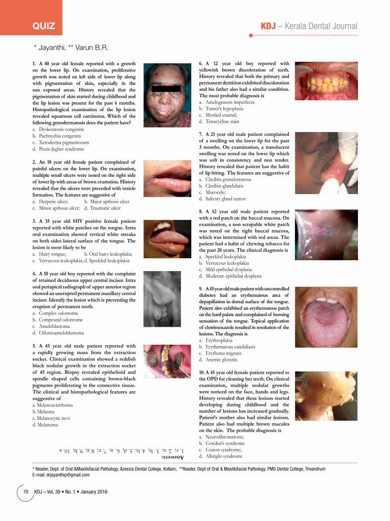
70 KDJ–Vol.39•No.1•January2016
KDJ – Kerala Dental JournalQuIz
1. A 40 year old female reported with a growth on the lower lip. On examination, proliferative growth was noted on left side of lower lip along with pigmentation of skin, especially in the sun exposed areas. History revealed that the pigmentation of skin started during childhood and the lip lesion was present for the past 6 months. Histopathological examination of the lip lesion revealed squamous cell carcinoma. Which of the following genodermatosis does the patient have?a. Dyskeratosis congenita b. Pachnychia congenitac. Xeroderma pigmentosum d. Peutz-Jeghersyndrome
2. An 18 year old female patient complained of painful ulcers on the lower lip. On examination, multiple small ulcers were noted on the right side of lower lip with areas of brown crustation. History revealed that the ulcers were preceded with vesicle formation. The features are suggestive ofa. Herpetic ulcer; b. Major apthous ulcerc. Minor apthous ulcer; d. Traumatic ulcer
3. A 35 year old HIV positive female patient reported with white patches on the tongue. Intra oral examination showed vertical white streaks on both sides lateral surface of the tongue. The lesion is most likely to bea. Hairy tongue; b. Oral hairy leukoplakiac. Verrucous leukoplakia; d. Speckled leukoplakia
4. A 10 year old boy reported with the complaint of retained deciduous upper central incisor. Intra oral periapical radiograph of upper anterior region showed an unerupted permanent maxillary central incisor. Identify the lesion which is preventing the eruption of permanent tooth.a. Complex odontomeb. Compound odontomec. Ameloblastomad. Odontoameloblastoma
5. A 45 year old male patient reported with a rapidly growing mass from the extraction socket. Clinical examination showed a reddish black nodular growth in the extraction socket of 45 region. Biopsy revealed epithelioid and spindle shaped cells containing brown-black pigments proliferating in the connective tissue. The clinical and histopathological features are suggestive ofa. Melanoacanthomab. Melasmac. Melanocytic nevid. Melanoma
6. A 12 year old boy reported with yellowish brown discoloration of teeth. History revealed that both the primary and permanent dentition exhibited discoloration and his father also had a similar condition. The most probable diagnosis isa. Amelogenesis imperfectab. Turner’s hypoplasiac. Mottled enamel;d. Tetracycline stain
7. A 21 year old male patient complained of a swelling on the lower lip for the past 3 months. On examination, a translucent swelling was noted on the lower lip which was soft in consistency and non tender. History revealed that patient has the habit of lip biting. The features are suggestive ofa. Cheilitis granulomatosab. Cheilitis glandularisc. Mucocele; d. Salivary gland tumor
8. A 52 year old male patient reported with a red patch on the buccal mucosa. On examination, a non scrapable white patch was noted on the right buccal mucosa, which was intermixed with red areas. The patient had a habit of chewing tobacco for the past 20 years. The clinical diagnosis isa. Speckled leukoplakiab. Verrucous leukoplakiac. Mild epithelial dysplasiad. Moderate epithelial dysplasia
9. A 45 year old male patient with uncontrolled diabetes had an erythematous area of depapillation in dorsal surface of the tongue. Patient also exhibited an erythematous patch on the hard palate and complained of burning sensation of the tongue. Topical application of clotrimoxazole resulted in resolution of the lesions. The diagnosis isa. Erythroplakiab. Erythematous candidiasisc. Erythema migransd. Anemic glossitis
10. A 45 year old female patient reported to the OPD for cleaning her teeth. On clinical examination, multiple nodular growths were noticed on the face, hands and legs. History revealed that these lesions started developing during childhood and the number of lesions has increased gradually. Patient’s mother also had similar lesions. Patient also had multiple brown macules on the skin. The probable diagnosis isa. Neurofibromatosis;b. Cowden’s syndromec. Coston syndrome;d. Albright syndromeAnswers:1.c;2.a;3.b;4.b;5.d;6.a;7.c;8.a;9.b;10.a
*Reader,Dept.ofOral&MaxillofacialPathology,AzeeziaDentalCollege,Kollam;**Reader,DeptofOral&MaxillofacialPathology,PMSDentalCollege,Trivandrum E-mail:[email protected]
* Jayanthi, ** Varun B.R.
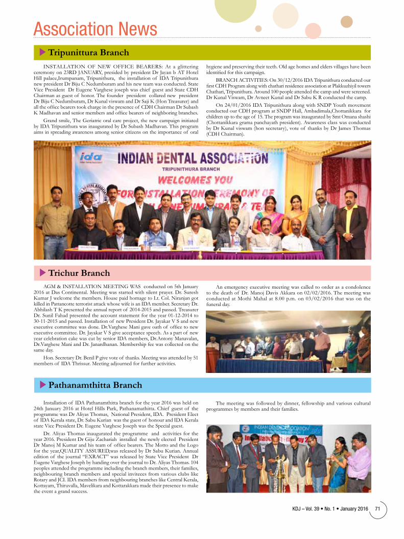
Tripunittura Branch
Installationof IDAPathanamthittabranchfortheyear2016washeldon24thJanuary2016atHotelHillsPark,Pathanamathitta.Chief guestof theprogramme was Dr Aliyas Thomas, National President, IDA. President Elect of IDA Kerala state, Dr. Sabu Kurian was the guest of honour and IDA Kerala state Vice President Dr. Eugene Varghese Joseph was the Special guest.
Dr. Aliyas Thomas inaugurated the programme and activities for the year2016.PresidentDrGijuZachariahinstalledthenewlyelectedPresidentDrManojMKumarandhisteamof officebearers.TheMottoandtheLogofor the year,QUALITY ASSURED,was released by Dr Sabu Kurian. Annual edition of the journal “EXRACT” was released by State Vice President Dr Eugene Varghese Joseph by handing over the journal to Dr. Aliyas Thomas. 104 peoples attended the programme including the branch members, their families, neighbouring branch members and special inviteees from various clubs like Rotary and JCI. IDA members from neighbouring branches like Central Kerala, Kottayam, Thiruvalla, Mavelikara and Kottarakkara made their presence to make the event a grand success.
Pathanamthitta Branch
The meeting was followed by dinner, fellowship and various cultural programmes by members and their families.
Trichur BranchAGM&INSTALLATIONMEETINGWASconductedon5thJanuary
2016atDasContinental.Meetingwasstartedwithsilentprayer.Dr.SureshKumar J welcome the members. House paid homage to Lt. Col. Niranjan got killed in Pattancotte terrorist attack whose wife is an IDA member. Secretary Dr. AbhilashTKpresentedtheannualreportof 2014-2015andpassed.TreasurerDr.SunilFahadpresentedtheaccountstatementfortheyear01-12-2014to30-11-2015andpassed.Installationof newPresidentDr.JayakarVSandnewexecutivecommitteewasdone.Dr.VargheseManigaveoathof officetonewexecutive committee. Dr. Jayakar V S give acceptance speech. As a part of new year celebration cake was cut by senior IDA members, Dr.Antony Manavalan, Dr.Varghese Mani and Dr. Janardhanan. Membership fee was collected on the same day.
Hon. Secretary Dr. Benil P give vote of thanks. Meeting was attended by 51 members of IDA Thrissur. Meeting adjourned for further activities.
An emergency executive meeting was called to order as a condolence tothedeathof Dr.ManojDavisAkkaraon02/02/2016.ThemeetingwasconductedatMothiMahalat8.00p.m.on03/02/2016thatwasonthefuneral day.
INSTALLATION OF NEW OFFICE BEARERS: At a glittering ceremony on 23RD JANUARY, presided by president Dr Jayan b AT Hotel Hill palace,Irumpanam, Tripunithura, the installation of IDA Tripunithura new president Dr Biju C Nedumburam and his new team was conducted. State Vice President Dr Eugene Varghese joseph was chief guest and State CDH Chairman as guest of honor. The founder president collared new president Dr Biju C Nedumburam, Dr Kunal viswam and Dr Saji K (Hon Treasurer) and alltheofficebearerstookchargeinthepresenceof CDHChairmanDrSubashKMadhavanandseniormembersandofficebearersof neighboringbranches.
Grand smile, The Geriatric oral care project, the new campaign initiated by IDA Tripunithura was inaugurated by Dr Subash Madhavan. This program aims in spreading awareness among senior citizens on the importance of oral
hygiene and preserving their teeth. Old age homes and elders villages have been identifiedforthiscampaign.
BRANCHACTIVITIES:On30/12/2016IDATripunithuraconductedourfirstCDHProgramalongwithchathariresidenceassociationatPlakkuzhiyiltowersChathari, Tripunithura. Around 100 people attended the camp and were screened.Dr Kunal Viswam, Dr Avneet Kunal and Dr Sabu K R conducted the camp.
On24/01/2016IDATripunithuraalongwithSNDPYouthmovementconducted our CDH program at SNDP Hall, Ambadimala,Chottanikkara for children up to the age of 15. The program was inaugurated by Smt Omana shashi (Chottanikkara grama panchayath president). Awareness class was conducted by Dr Kunal viswam (hon secretary), vote of thanks by Dr James Thomas (CDH Chairman).
KDJ–Vol.39•No.1•January2016 71
AssociationNews

Trivandrum Branch
Chalakkudy Branch
Eranad Branch
Installationceremonyof thenewofficebearersfortheyear2016wason17thJanuary2016atHotelAthirappillyResidency.OurseniormemberandcharterpresidentDrGeorgeSebastianPullanwastheinstallationofficer.Dr
Seby K Bastin took charge as the president Dr Sijo Manavalan as Secretary and Dr Sanil Paul as treasurer
WeconductedourfirstCDHprogrammeatSNVidyabhavanAloor.
The3rdinstallationof IDAErnadunderpresident-shipof DrRamMohanReddywasheldathotelHITON,Perinthelmennaon17/1/2016at7:00pm.Theinstallation ceremony was held in presence of Dr Sijo Paulose 1st Vice President of IDA Kerala state, Dr Subhash Madhavan CDH convener IDA Kerala state, Dr Jayakrishnan IMA state president,Mr Tini Tom (cine fame), Mr Kalabhavan Sinaj (cinefame).Thenewofficeof IDA Ernad comprised of Dr Ram Mohan Reddy (president), Dr Sabhish Sivadas (hon secretary), Dr Muhsin (hon treasurer), Dr Mithun Balakrishnan (CDE Convener ), Sanal Babu (CDH convener ). Variety of entertainment programs was held under Mr Tini tom and Kalabhavan Sinaj and was followed by appetising feast. The inauguration was well covered and supported by the media and social forums.
IDA Eranad branch celebrates republic day, dental screening camp, and distributionof dentalkiton6centresinMalappuram
1. MMMLP School, Kalkulam, Karapuram P.O2. NSSUP School, Uppada 3. UMALP School, Chathangotupuram4. GMLP School, Mukkatta,Nilamboor5. GLP School, Kizhakethala, Karuvarakkund6.GWUP School, Thrikkulam, Chemmad
Activitiesof IDATrivandrumfortheyear2016-17kickstartedwithinstallation ceremony of Dr Mathew Jose as our new president for this year. Installationwascelebratedinagrandmanneron7thFebruary2016atSreemoolamclub Trivandrum. On his acceptance speech Dr Mathew Jose shared his ideas for thecurrentyearandrevealedhismottoforthisyearSMILELEARN&CARE.Chief Guest of the day was Dr MKC Nair Vice Chancellor Kerala University of Health Sciences. The Chief Guest steeled audience’s attention with a mesmerizing speech. Guest of Honor IDA state president Dr Sameer PT promised the crowd
that he will take IDA more popular to the public with more public awareness programs. State secretary Dr Suresh poured his gratitude and love to his home branch for giving a convincing win during the state elections held at Allepey. Dr Achuthantheoutgoingpresidentthankedhisofficebearersandhismembersforthe smooth running of the activities during his term. Dr Arun Ramachandran secretary Trivandrum thanked the gathering. The installation ceremony was followedbymusicalnightandlip-smackingfood.
72 KDJ–Vol.39•No.1•January2016
Association News

Quilon Branch
Kodungallur Branch
Valluvanad Branch
Theinstallationof newofficebearersof IDAKodungallurfortheyear2016wasconductedinaglitteringceremonyon2/01/2016atIMAHall,Kodungallur.
Dr.Nazeer.P.M. took over the reigns from Dr.Mahesh Narayanan as President of the branch while Dr.Shaji.P.H. continued as the Secretary.
The chief guest for the function was Dr.K.C. Thomas, President, IDA Kerala statewhoinstalledthenewpresidentandotherofficebearers.PaststatepresidentDr.G.Premkumar felicitated the gathering and the function was adjourned for fellowship and dinner at 9.30 P.M.
Installationceremony&Familymeet:The24thinstallationceremonyandfamily meet of IDA Quilon branch was held on 20th December 2015 at The Quilon Beach Hotel Kollam. Dr B S Sundaresan as the president and other officebearerswereinstalled.Thechief guest,worshipfulmayorAdvVRajendraBabu inaugurated the meeting and The past national president Dr K G Nair was honoured for his outstanding contributions to IDA. 12 new members were inducted.IstExecutivecommitteemeeting:Thefirstexecutivecommitteemeetingheldon6thJanuary2016atHotelShahInternational.
Ourmembersattendedthe48thstateconferenceALOHAatAlappuzhafrom9-10thJanuary
CDH program.: Conducted a dental and medical camp at Believers ChurchMathmacentralSchoolEravipuramon24thJanuary2016.
FirstMonthlymeeting.:Thefirstmonthlymeeting&CDEprogramwasheldon30thJanuary2016atLionsHallKollam.56membersattended.
First CDE Programme:: Conducted a lecture on“Management of cardiac patients in dental clinic” by Dr N Pratap Kumar DM, Intervention cardiologist. President Dr Sundaresan chaired the meeting, CDE representative Dr Sudheesh Manoharanintroducedthespeaker.56membersof branchattended.
IDAVALLUVANADBRANCHinstallationof officebearerswasconductedon16thJanuaryatNilaresidency,Shornur.Dr.CijuPaulose,the1stvicepresidentof keralastateconsentedtobethechief guestandinstallationofficer.The program commenced at 7 pm with collaring of the president Dr. Sreekanth .S and followed by prayer song by Asha Lakshmi, daughter of IDA state CDH chairman Dr. Subash Madhavan. Chief guest introduction was given by Dr Haris and the incoming president introduction by Dr. Vishak. The event was honoured with the presence of two guest of honor, Dr. Subash Madhavan, IDA state CDH chairman and Dr. Mercy Joji, the women’s dental council president. A detailed secretary report was read by Dr. Rajarajan. Then the new president, Dr. Vishal Korah was installed by taking oath administered by the installation
officerDr.CijuPaulose.AfterthatofficebearersteamisbeeninstalledbyDr.Vishal Korah, newly elected president of IDA Valluvanad.
The new president was felicitated by Dr. Joji George, the president of IDA Kunnamkulam branch and Mrs. Geetha, the president of rotary club of Shornur. Vote of thanks was given by Dr. Shoukath, the elected secretary and meeting adjourned for dinner and entertainment after national anthem.
About 120 members including family members attended the function. Everyone enjoyed the evening under the master of ceremony Dr.Balasubramaniam.
KDJ–Vol.39•No.1•January2016 73
Association News

Malabar Branch
Central Kerala - Kottayam BranchDECEMBER 2015AGM was held on 12th Dec 2015, at Kottayam club. The annual report
of branch was presented by Hon. Secretary Dr. Renju Titus. Treasurer Dr. Lijo Thomas presented the audited accounts. New President Dr. Bobby John Varghese andnewteamof officebearerstookcharge.
CDH ACTIVITY: A dental checkup camp WAS conducted by Dr. Bobby JohnVargheseon6thJan2016atGovt.LPSchoolPanayara.About300studentsattended the camp.
Dental awareness camps were conducted by Dr. Robin T and Dr. Ittyvirah Babuon16thJan2016atMariaSadanamorphanageandTownBoysOrphanage.About600inmatesfrombothorphanagesreceivedthebenefits.OralHealthPacks were distributed to inmates.
Dr.NidhishandDr.Anukeshconductedcheckupcampon22ndJan2016atExcelsiorschool,Illickal.100studentsbenefittedandOralHealthPackswere distributed.
AdentalcheckupcampwasconductedbyDr.Anukeshon28thJan2016atACMnurseryschoolThiruvathilkal,25kidsbenefitted.
EXECUTIVECOMMITTEEMEETING:ThefirstExecutivemeetingof IDACKKwasheldon14thJan2016,atKottayamclub.
INSTALLATION CEREMONY: Installation ceremony of Dr. Bobby John Vargheseandhisteamwasheldon30thJan2016atCitizensclub,Kottayam.IDA Kerala state President Dr. Mohammed Sameer was the chief guest of the day and he installed the new president Dr. Bobby John. Dr. Divya Iyer IAS (Asst. Collector, Kottayam) inaugurated the CDE programme and Dr. B Ekbal (former VC, University of Kerala) inaugurated the CDH programme.IDA NationalPresident,Dr.AliasThomasinauguratedtheco-operativesocietyof CKK. Dr. Suresh Kumar (Hon. Sec, IDA Kerala State) and Dr. Sabu Kurien (President Elect, IDA Kerala State) addressed the gathering. 220 members attended the ceremony.
FAMILYGETTOGETHER:Thefirstfamilygettogetherwasconductedon30thJan2016,atcitizensclub.220membersparticipated.
INSTALLATION OF NEW OFFICE BEARERS & FAMILY MEETInstallationof theofficebearersof IDAMalabarBranch2015-16washeld
on24thJanuary2016atHotelMalabarPalaceKozhikodeat6.30pm.Dr.DineshKR was installed as the new president of IDA Malabar Branch by Dr.Mohammed Sameer (Hon President IDA Kerala State) was the Guest of honour and installation officer.OutgoingPresidentDr.SajuNShandedoverthePresident’sCollartonewlyinstalledPresident.Followedbytheinstallationof otherofficebearers.Shri.V.K.C Mammad Koya (Hon Mayor Kozhikode) was the Chief Guest of the occasion. Felicitations were given by Dr.Nizaro Siyo Past President IDA Kerala state and Dr.Antony Thomas Past President IDA Kerala State. Many important Dignitaries witnessed the occasion including members from other neighbouring branches like Malappuram and Vadakara. Vote of Thanks were delivered by Dr.Sudheer K.T Secretary IDA Malabar branch followed by many colourfull cultural programmes by members and their families.
CDH ACTIVITY No.1Dental Screening and awareness camp was conducted at Kunjali Marakar
Higher secondary School Kottakal Vadakara, Kozhikode on 20/12/2015 in association with Dept. Of Education Govt. of Kerala. Around 100 Participants were present in the camp. On behalf of IDA Dr.Rubeena Thahir and Dr.Muhsina Participated the camp.
CDH No.2Dental check up and awareness camp was conducted at Devaseva Sangham
KaithapadamPottamalKozhikodeon31/01/2016inassociationwithLionsClub Daffodils. Around 100 Participants were present in the camp. On behalf of IDA Dr.Dinesh KR, Dr.Prathap, Dr.Sudheer KT,Dr.Haris,Dr.Susha,Dr.Sandeep Rajagopal, Dr.Pravish and Dr.Hussain Participated the camp.
74 KDJ–Vol.39•No.1•January2016
Association News

NedumbasseryBranch
Palakkad Branch
Mavelikkara Branch
Annual general body and annual elections of our branch were held at hotel Travancore regency on 22nd December 2015. AGM elected Dr. Seethibegam as president, Dr.Baiju Hariharan as secretary and Dr.Samith as treasurer for theyear2016.
Combinedexecutiveof IDAMavelikkarabranchwasheldon18thJanuary2016athotelTravancoreregency.Meetingdiscussedabouttheinstallationof thenewofficebearersof thebranchanddecidedthedateof installation.
The installation ceremony of Dr Seethi Begam and her team was held on 24thJanuary2016atWytePorticoAdoorat5.pm.Thechief guestof thedaywas Dr Suresh Kumar Hon. Sec IDA Kerala state and the guest of honour was Prof M S Sunil (social activist).Around 150 members of our branch attended the function. There was a variety of entertainment programs by our family members and a music entertainment by MOKSHA THE MUSIC PEOPLE....
IDANedumbasseryBranchhasnewleadershipunderthefirstladypresidentof our branch. President Dr (Mrs) Rajeena Sajee Vinod, Secretary Dr Seby Varghese, Treasurer Dr Teny Mathew.
DrKCThomas-thepastpresidentof IDAKerala,installedtheNewOfficeBearerson2ndJanuary2016.
Our Theme for this year is
1-Welfareof ourmembers.2-SocialresponsibilityandSocialImagingforprofessionandIDA3- Promoting collaborative approach among the health care
professionals.OutfirstExecutivecommitteemeetingheldonJanuary15th2016.
Theinstallationceremonyof theofficebearersof theyear2016washeldon03-01-2016athotelGazala,Palakkad.Chief guestof the function was municipal chairperson Prameela Sasidharan and Dr. Joseph C C (State Sectry IDA hope) was the installation officer.OtherguestslikeDr.SureshandDrAnil felicitated the gathering. A few other members of nearby branches also attended the function.
Dr T. Raghunath Nambiar took charge as President of IDA and Dr. Sivarama Krishnan as Secretary, and the new team of
IDA executive members also took their respective positions. After installation ceremony we had entertainment programmes and dinner. All together the installation ceremony was a great treat to everyone including the participated family members.
Our members Dr Saji Thomas, Dr. Suresh and Dr.Veena Suresh conducted a dental camp on20-01-2016atM.A.L.PSchoolMundekarad,Mannarkkad with the support of our CDH co-ordinatorDr.AnoopRajan.Bythiscampthe activities of this year is kick started with a great bang.
KDJ–Vol.39•No.1•January2016 75
Association News
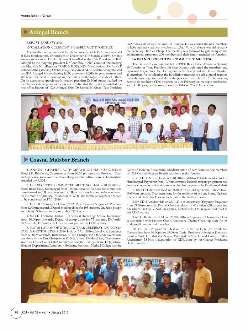
Attingal Branch
Coastal Malabar Branch
REPORT-JANUARY2016INSTALLATIONCEREMONY&FAMILYGETTOGETHERThe installation ceremony and Family Get together of IDA Attingal was held
at IMA Headquarters, Trivandrum on December 27th Sunday at 5PM. On this auspiciousoccasionDrHariKumarRinstalledasthe16thPresidentof IDAAttingal by the outgoing president Dr Arun Roy. Chief Guest of the meeting wasMrs.KalaNL(RegistrarTCMC&KDC).KDCVicepresidentDrAnish.Pwelcomed the gathering. On her inaugural address KDC Registrar congratulated the IDA Attingal for conducting KDC accredited CDEs in good manner and she urged the need of conducting the CDEs on the topic on code of ethics. On his acceptance speech newly installed president Dr Hari kumar thanked the members for electing him as the president. After that the president installed the newofficebearersof IDAAttingal2016.DrSamuelKNinan(PastPresident
IDA Kerala state) was the guest of honour. He welcomed the new members to IDA and inducted new members to IDA. Vote of thanks was delivered by the Secretary Dr Alex Philip. The meeting was followed by gala banquet and entertainment programs. 200 members and their family attended the function.
1st BRANCH EXECUTIVE COMMITTEE MEETINGThe 1st branch executive was held at PWD Rest House; Attingal on January
19 Tuesday at 7pm. President Dr Hari Kumar welcomed the members and expressed his gratitude for electing him as the new president. He also thanked all members for conducting the installation meeting in such a grand manner. Laterthemeetingdiscussedabouttheproposedyearplan2016.Themeetingdecided to conduct a CDE program on 21st February on the topic sterilization and a CDH program in association with IMA on World Cancer day.
1. ANNUAL GENERAL BODY MEETING: Held on 20.12.2015 at HotelJ.K.Residency,Cheruvathurfrom06.30pmonwards.PresidentElectDr.SujaVinodtookovertheofficealongwithherofficebearers.42membersattended the AGM
2.1stEXECUTIVECOMMITTEEMEETING:Heldon01.01.2016atHotel Bekal Club, Kanhangad from 7.00pm onwards. Various subcommittees wereformed.6CDHactivitiesand1CDEactivitywasfinalisedtobeconductedonthemonthof January.Installationof WDCandfamilygettogetherfinalisedtobeconductedon17.01.2016.
3.1stCDHActivity:Heldon5/1/2016atPalavayalSt.JonesL.PSchoolfrom 12.00pm onwards. Dental check up done for 105 students. Dr. Sajan Joseph and Dr.Sijo Sebastian took part in this CDH activity.
4.2ndCDHActivity:Heldon10/1/2016atDurgaHighSchool,Kanhangadfrom 10.00pm onwards. Dental checkup done for 75 patients. Dr.Jyothi, Dr.Sharmila, Dr.Vinaya,Dr.Dishana took part in this CDH activity
5. INSTALLATION OF WDC,NEW YEAR CELEBRATION AND 1st FAMILYGETTOGETHER-2016:Heldon17.01.2016athotelK.K.Residencyfrom6.00pmonwards.Installationof theChairpersonDr.SapnaSreekumarwas done by the Past Chairperson Dr.Suja Vinod. Dr.Merci Joji, Chairperson, Womens’ Dental Council,IDA Kerala State was the Chief guest and Dr.Jayashree, Head of Department,Community Medicine, Pariyaram Medical College was the
Guestof Honour.Recognizinganddistributionof certificatestonewmembersof IDA Coastal Malabar Branch was done at this function.
6.3rdCDHActivity:Heldon23.01.2016atMalabarRehabilitationCentreForHandicapped, Payyannur from 10.00am onwards. Parents’s training programme was donebyconductingadentalawarenessclassfortheparentsbyDr.AhamedShafi.
7.4thCDHActivity:Heldon26.01.2016atOldagehome,Taborefrom09.000am onwards. Treatment done for the residents of old age home. Dr.Sajan Joseph and Dr.Merin Thomas took part in the treatment camp.
8.5thCDHActivity:Heldon26.01.2016atAnganvady,Thayinery,Payyannurfrom 09.30am onwards. Dental Check up done for 45 students,30 parents and 5 teachers. Dr.Suja Vinod, Dr.Cynjila, Dr.Sreedevi, Dr.Deepika took part in this CDH activity
9.6thCDHActivity:Heldon28/01/2016atAnganvady,Cherupzha.Donein association with Lioness Club Cherupuzha. Dental Check up done for 35 students,20 parents and 5 teachers.
10.1stCDEProgramme:Heldon31.01.2016atHotelJ.K.Residency,Cheruvathurfrom04.00pmto09.00pmTopic-ProblemssolvinginDentistry.Faculty-Prof.Dr.Moksha.Nayak,Principal,K.V.G.DentalCollege,Sullia.Attendance-55Nos.Inaugurationof CDEdonebyourCharterPresident,Dr.K.T.Suresh.
76 KDJ–Vol.39•No.1•January2016
Association News

Malappuram Branch
25thinstallationof IDAMalanadubranchwasheldon23rdJanuary2016,Saturday at Hotel Kabani International Muvattupuzha.
Meetingwasstartedat8:00p.m.presidedbyDr.JayanJacobMathew.Meeting was started with the collaring of the President by Secretary Dr. Joby J. Parappuram. Dr. Byju Paul Kurian welcomed the gathering.
Dr. Sankar Vinod past president introduced the new President of Malanadu branch Dr. Arun George. The Immediate past president Dr. Jayan Jacob Mathew installed the new President, and oath was spelled by Dr. Suresh Kumar, secretaryIDAkeralastate,followedbytheInstallationof newofficebearersby Dr. Arun George.
Chief guest for the day Mrs. Asha Sanil, President, Ernakulam District Panchayath who got introduced by Dr. Siju V Jose,inaugurated the IDA activities of ourbranchfortheyear2016.
Guestof honourDr.AliasThomaspresidentIDAHeadoffice,distributedDrPaulGVadathuMemorialMeritAwardstothestudentswhocamefirstinBDSExamsfromeachof thefivedentalcollegeswhichcomeunderIDAMalanadu branch.
Malanadu Branch
Guest of honour Dr. Sabu Kurian, President elect Kerala state Inaugurated –continuingDentalHealthWelfareActivitiesfortheNeglectedIndividualsbyunveiling its logo “Sathkrithi”
Guest of honour Dr.Ciju A Paulose, First vice President, IDA kerala State inaugurated the Continuing Dental education programmes by unveiling its logo ‘Sparsh’fortheyear2016.
Guest of honour Dr. Suresh Kumar, secretary IDA kerala state, released The Malanadu Dental journal
Charter Day was celebrated with a “Cake Cutting Ceremony ‘ by Our Charter President Dr. Abraham Varghese.
Pleasantries were given to the guests by Dr.Arun George. Dr. Litto Manuel, Hon.Secretary gave vote of thanks. Meeting was adjourned for dinner and entertainment programme by 10:15pm.
Report prepared by Dr. Litto Manuel, Hon.Secretary, IDA Malanadu on 24thJanuary,2016.
AGM : The Annual general body meeting of MIDA held on 30/12/15 at Rydgess Inn Kottakkal. More than 24 members participated in the meeting.
EXECUTIVE COMMITTEE MEETINGS: First Executive committee meetingheldon05/01/16from8pmonwardsatCosmopolitanclubManjery.15 members were attended.
INSTALLATION-16:MIDAconductedtheInstallationceremonyonSunday17/1/16atHotelWoodbineManjery.OurChief GuestwasDrMuhamed Sameer PT, President IDA Kerala, Our guest of honor was Dr Suresh kumar G, Honourable Secretary, IDA Kerala. Many important personalities from various organizations were graced the occasion. The entertainments programmes were performed by renowned artists from music world. Followed by Gala dinner culminated with mouth watering cuisines.
“PUTHUPUNJIRI”: The inauguration of our Mega project” PUTHUPUNJIRI”, The 100 Free dentures for poor and needy peoples done in the installation venue in the presence of many leaders and invited guests.
MIDA LOTUS NEW YEAR CELEBRATION: The new year celebration andinaugurationof MIDALOTUS”16(WomensWing)donebycuttingthecakeattheinstallationvenueon17/1/16bytheMIDALOTUSmembersandthe leaders.
PAIN AND PALLIATIVE DAY OBSERVATION: The CDH team visited the Pain and Palliative care centre at Puthanathani on pain and palliative care day.Wehaddiscussionwiththeofficebearersof PainandPalliativeunitof Puthanathani regarding conducting an Oral awareness camp for palliative care unit volunteers in district level by their request.
CDH REPORT: First CDH camp of MIDA held at BHSS Mavandiyoor on 28/1/16.Morethan70studentswerebenefitted.Thisdentalscreeningcamplead by Dr Deebu J Mathew, Dr Hisham, Dr Mahesh, and Dr Muhamed Shereef.
LEGAL CELL ACTIVITIES: The executive committee appointed the Legal cell committee including Dr Sadiq Ali as Legal cell chairman, President, Immediate past president, President Elect, Hon Secretary, Treasurer are the members. The executive decided to start the legal cell bank account separately for the accountability of the collecting amount towards the legal cell. The executive committee appointed the Legal advisor and cases have been reached in the High court already.
CDEREPORT:OurfirstCDEonBasicLifeSupport(BLS)willbeheldatAsterMIMS,Kottakkalon14/2/16on9amonwards.ThisFulldayprogrammewill be leading by Department of Emergency Medicine,MIMS Hospital.
KDJ–Vol.39•No.1•January2016 77
Association News

Vatakara BranchInauguration of IDA VATAKARA was done by Sri.C.K.Nanu,MLA(Vatakara)
by lighting the lamp at an auspicious function held at Dazzle Conference Hall,inthepresenceof variousprominentfiguresandstateleadersof IDA.Dr.K.C.Thomas(pres.IDA Kerala State) presided. State wide Inauguration of HOPE-MEDI,thegroupinsurancescheme&ClinicalStandardisationProgrammealso got inaugurated at the same venue.
Installation of branch president Dr.Abdul Gafoor was done by Dr.K.C.Thomas. Branchpresident installedhis teamof officebearers.Dr.Sanal.O.V (Secretary IDA Kerala State ) handed over the constitution to new selected secretary. Felicitations followed.
Presentlythebranchhas87membersinwhich55arenewtoIDA.Themembership campaign is in full swing to add all dentists in this region to IDA.
Within the short period branch conducted two full day CDE programmes. 1:
First Response Provider Course by Angel’s International Foundation, 51 members attended. 2:Problem Solving in Restorative Dentistry by Dr.Joy Kurian.MDS which had 37 participants.
Branch could conduct two free dental camps also.(1)at Kannookara,Vatakara on 25/10/2015. It was attended by Dr.Abdul Gafoor,Dr.Thulasi Das, Dr.Salil Chakravarthi, Dr.Susanth Krishna, Dr.Sanal.P, Dr. Athul.G.S and Dr. Sajitha. (2) atTownHall,Vatakaraon03/01/2016.Dr.SalilChakravarthiandDr.SusanthKrishna attended.
Immediate future programmes are(1).Free Dental Check Up Camp On 07/02/2016@AbhayagiritribalcolonyinassociationwithJANAMAITRIPOLICE.(2)a CDE programme on management of medically compromised patients(3)Cluster meeting of practicing dentists at different regions of Vatakara tocreategoodwill.(4)afamilygettogetheron28/02/2016.
Wayanad BranchInstallation Ceremony: The activities of IDA Wayanad branch started with
theinstallationceremonyheldatHotelPepperGroove,SulthanBatheryon16thJanuary2016.Thechief guestof theeventwasAdvt.T.MRashidconvenorWayanad-Nilgiri’s(NH)Railwayactioncommittee.
Dr.Mohammed Sameer president IDA kerala state was the guest of honour and keynote speaker for the function.
Dr. Suresh Kumar G. secretary IDA kerala state installed Dr. Thomas Mathew as the new president of IDA Wayanad and Dr. Sanoj P.B as the secretary. The function was well attended by the IDA members with their family and other distinguished guests.
ExecutiveMeeting:Thefirstexecutivemeetingof IDAWayanadbranchwasheldatHotelResort,SulthanBatheryonJanuary21st2016.Itwasdecidedto provide Water Beds and Air Beds for the needy patients of pain and palliative care units in the district.
President Dr. Thomas Mathew brought an innovative idea ie to conduct smiling competition at schools and college levels to promote oral hygiene and to create awareness about the new trends in dentistry among the public.
CDH Programme: Balamughulam: IDA Wayanad branch in association with Bharathiya Chikhilsa Vakuppu with their Balamughulam project conducted Dental Health Check Up Camps and Dental Awareness Programme at 5 Schools in different parts of Wayanad.
Kochi BranchIDAKochiBranchhaditsinstallationFortheyear2016on20thDecember
2015 at Hotel Mercy Estate,Kochi. Kochi Mayor Saumini Jain was the chief guest for the Ceremony,IDA National President Dr.Alias Thomas was the Guest of Honour,IDA Kerala State President Elect Dr. Sameer P.T Guest.
The CDE program organized by Indian Dental Association, Kochi branch, andColgatewasheldon30thJanuary,2016atIMAHouseKochi.ThiswasthefirstCDEprogramof thisyear.TheCDEstartedat7.30pmwithasilentprayerfollowed by welcome address by Dr.Jayakumar.S, President of IDA Kochi. The
faculty was Dr.Vinod Thomas, DM. He is the chief cardiologist of Renai Medicity Kochi, an accomplished speaker and has presented several papers on national and international conferences and has won many accolades. The Program was followed by lecture. The lecture was assisted by audiovisual aids. He delivered a very informative lecture on Cardiology in relevance to dental practice. There were 55 participants for this program. Dr.Balu Soman Secretary IDA Kochi thanked the speaker and Colgate for supporting IDA Kochi with this program. As a token and affection memento was given to the speaker. As soon as the lecture came to close the participants were served dinner.
78 KDJ–Vol.39•No.1•January2016
Association News

Tellicherry BranchInstallationof officebearersof 2016washeldatlionshallTellicherryon
14thFebruary2016.IDAstatepresidentDrMuhammadSameerwasthechief guest. IDA state secretary Dr Suresh Kumar G was the guest of honour. Dr Suresh kumar installed Dr Firoze K. M. as the president. The president installed histeamof officebearers.Themeetingwasattendedbyaround50memberswithfamily. Members from nearby branches also attended the meeting. Felicitations were given by Dr Prathap Pavithran and Dr Retnakaran. The president in his
acceptance speech thanked all the senior members and State president Dr Muhammad Sameer, State secretary Dr Suresh Kumar, State IPP Dr Thomas K C, State past Secretary Dr O V Sanal, past legal cell chairman Dr Shaji K Joseph for their efforts in resolving the issues between IDA Tellicherry branch and deract. Dr Aneesh Sebastian and Dr Sujatha gave mementos to Chief guest and guest of honour respectively. Installation was followed by entertainment programmes and dinner.
North Malabar Branch
Theinstallationceremonyof officebearersof IDANorthMalabarBranchfortheyear2016-17washeldonFebruary14thatHotelMalabarResidency,Kannur.The chief guest for the day was Dr M.K.Abdul Khader,Vice Chancellor,Kannur University and Dr Sreekumar Vasudevan, Past IMA State President was Guest of Honor. During the day Dr.Jayashree K.T. the incoming President, IDA North Malabarbranchandherteamof officebearerswereswornin.Theceremonywas followed by variety entertainment and dinner.
Kottarakkara Branch
Theannualgeneralbodymetingof thebranchwasheldon16thJanuary2016.Theannualreportandtheaccountsof thebranchwaspresentedandpassed.
The installation of Dr. Paul K and team was conducted on the 31st of January2016Smt.AyshaPottiMLAwastheChief GuestandDr.SueshKumar,Hon.Sec. IDA Kerala State was the Chief Guest. The meeting was well attended by members and guests.
First General b o d y m e e t i n g washeldon18thJanuary, Monday, at hotel Highway castle, Kasargod, at 7.30 pm. 30 members attended the meeting. Dr.
Rekha Maiya was elected as President, Dr. Ashwin Devasya as secretary, Dr. UmaMaheshwariastreasurer,fortheyear2016.
A continuing dental education programme on “Phytopharmaceuticals in dentistry” was presented by Dr. Waheed Pasha M.D in Ayurveda from Bangalore.
Executivecommitteemeetingwasheldon26thJanuary,Tuesday,atIMAhall Kasargod. Issues and programmes to be conducted in the current year were discussed.
Kasargod Branch
Karunagappally Branch
The Installation ceremony of IDA K ar unag appa l l y b r a n c h 2 0 1 6was conduc ted on 30/01/2016a t G r a n d e M u s c a t h o t e l , Karunagappally. Dr Eugene Varghese Joseph, vice president IDA Kerala state was the chief guest and installed Dr Bijoy K Sudhakar as the president of IDA Karunagappally branch. Lighting of lamp and keynote address was given by the chief guest. Dr Jayakrishnan welcomed the gathering and Dr A Q Azad proposed vote of thanks. Felicitations were given by Dr Geetha Joy, Dr Jinu Mathew Vidyan, Dr Varghese, Dr Amritha, Dr Nazeer and Dr Laiji Thomas Tharakan. The meeting was well attended followed by Cultural programmes conducted by the family members and dinner.
KDJ–Vol.39•No.1•January2016 79
Association News

The INSTALLATION of IDA THIRUVALLA was held on 29th November 2015atGREENVALLEY,waterthemepark-Adoor.Dr.SamuelKNinanPastPresident-IDAKeralawastheinstallingofficerandcalledforfacingchallengesunitedly. Dr.Rajeev Simon K took oath as the president, Dr.Sunil Roy Koshy as the Hon.Secretary and Dr.Simon George as the treasurer. The President Elect is Dr.Saji Kurien, Dr Mathew P.C. is CDE representative and Dr.Reji Thomas is CDH representative. Past President Dr.Sumod P Mathew congratulated his team
for the fantastic performance and thanked the members for the wholehearted co-operation.Dr.RajeevSimonKoutlinedhisvisionfor2016andsoughtthesupport of members in this endevour. He emphasised membership development andlaudedtheofficebearersthatthenumberstandsat89asof today.Themeeting was proceeded by Members and their family enjoying the swimming pool as well as roller coaster rides and ended with Dinner.
Thiruvalla Branch
Kottayam Branch
InstallationCeremonyof IDAKottayamBranch–31stJan2016atHotel AIDA.
WehadourInstallationceremonyheldon31stJan2016withDr.JojoeJosephSurgical Oncologist, Caritas Hospital as our Chief guest and Dr. Alias Thomas,
IDA National President, and Dr.Sabu Kurian IDA Kerala State President Elect as Guests of Honour.
Dr.Jijoe Jacob welcomed the gathering.The Secretary, Dr.Sunil T.Verghese gave a detailed annual report of all
the activities.Dr.Thomas C.Kappen, narrated his experience being the President of our
Branch and appreciated and acknowledged the good support he received from our members. Later, he installed Dr.Binumon Mani as our new President for the year2016-17.Dr.BinumonManioutlinedhisplanof activitiesforthecomingyear.
Dr.Sabu Kurian was honoured with a Ponnada by our National IDA President Dr.AliasThomasfollowedbyfelicitationsbybothof them.Aroundfiftyeightheads were present at this function. Dr.Sebastian, proposed the vote of thanks followed by the National anthem.
President Secretaries Seminar
Executive Committee Meeting
80 KDJ–Vol.39•No.1•January2016
Association News

KDJ–Vol.39•No.1•January2016 81
Association News

REPORT IDA-HOPE 2015 -16 IDA HOPE is a members welfare scheme of IDA Kerala state. It was formed by merging the PPS& SSS of IDA Kerala state in 2007-08 It has 2434 active members.
Objectives of the Scheme Indemnity insurance and legal aid to the members. Financial compensation to the families of the Deceased member or Total Permanent disability. Medico-legal awareness/education. Encourage promote and popularize ethical treatment amongst Dentists.
Membership Members of IDA Kerala State with a valid DCI registration Membership is co terminus with the membership of IDA Kerala State. Membership has to be renewed annually (along after renewing IDA membership).
Admission Formalities- New members Copies of Birth Certificate or passport or SSLC for proof of age and residence. Copy of professional degree certificate and post graduate certificate if any. Copy of dental council registration certificate and latest renewal receipt Two passport size photographs (1.In Application form, 2 For the Certificate) Application Form (To be endorsed by the branch representative or the secretary for proof of branch membership) DD for admission fees drawn in favor of IDA Hope Payable at Thamarassery.
Membership Contributions - Fraternity contribution Payment of fraternity contribution @ of Rs. 500/- is mandatory along with the next year’s renewal in the event of death of
any member.
Membership Renewal The membership renewal period is between 1st –April to 31 May every year for availing all the benefits of the scheme. A
grace period with fine of Rs. 500 is permitted for renewal from June 1 st to 30 th September. But during this period onlyprofessional protection is covered and Death Claim (social Security) is not covered.
The membership will be terminated if not renewed before 30 th September. Receipts will be issued by local branch Representative
Membership Rights and Obligations In the event of death of a member of at least 12 months membership the nominee/legal heirs will be paid an amount
1000000 (10 lakhs) In case of death due to accidents a benevolent contribution is paid as soon as member ship is realized from the scheme office. The Professional indemnity coverage of the member will commence one month after the acceptance of membership. The maximum liability that will be borne by the scheme shall be Rs. 200000/— (2lakhs only) Continuous membership is obtained on timely renewal. The members shall maintain proper records and adopt standard protocols and safe dental practices as recommended by
the scheme from time to time.
HOPE MEDI“The purpose of human life is to serve and to show compassion and the will to help others.”
HOPE is moving one step ahead with our new health insurance scheme HOPE MEDI. Conflating our members and the insurancecorporation, we successfully implemented this new health insurance plan. We are glad to inform that, 1018 members joined thescheme. This includes spouse, kids and parents. Total of 4500 family members are the covered under HOPE MEDI umbrella.Our TAILOR MADE GROUP POLICY NO.: 1009042815P108336608, PERIOD OF INSURANCE FROM 01/10/2015 To Midnighton 30/09/2016
We have paid 93 lakhs as premium. Till last month we have had 31 claims and around 25 lakhs we conferred as claims.,IDA hope expresses sincere thanks to The United India Insurance Co, Mr. P.V. Alex and team of Cosmos insurance
brokers for their timely efforts for HOPE MEDI.Our Hope Medi Insurance which is a tailor made policy covering Ida HOPE members and their immediate family members,
This unique policy covers pre existing diseases and also offers insurance sharing , at a very nominal premium. This policy canbe claimed from any accredited hospital all over India. An additional feature of this policy is it gives cashless or reimbursementsettlement.
IDA HOPE members who have availed this unique very beneficial insurance policy covering all immediate family membersincluding those having pre existing diseases would have received IDA HOPE MEDI Health card by separate post from VIDALHealth TPA Pvt Ltd.
IDAHopeReportDr. Joseph C.C.
Hon. Secretary
82 KDJ–Vol.39•No.1•January2016

We the office bearers of HOPE have put in best possible effort for getting in maximum possible benefit for our members, it is up to all the members of HOPE and subscribers of HOPE MEDI to keep the momentum going and make only validclaims. Members are requested to inform all the claims requirements to your Hope rep. of your branch immediatelyHOPE MEDI ClaimsClaim Management Guidelines- Reimbursement
For reimbursement of claims, claim form, discharge summary, discharge bill (summary and detailed) from the hospital,medical certificate, investigation reports etc., should be submitted to TPA through M/s Cosmos Insurance Brokers Pvt. Ltd.within 15 days of discharge from hospital.
Claim Management Guidelines- CashlessVIDAL HEALTH is the Third Party Administrator (TPA) for United India Insurance. Claim has to be intimated immediately to
the TPA, whether it is cashless or reimbursement basis. Insurance desk of hospital should be contacted for cashless facilityshowing identity cards and see that relevant treatment details as provided to TPA. It is advisable to report the claim to M/sCosmos Insurance Brokers also when the same is initially intimated to the TPA.
Contact Details:TPA (M/s Vidal Health TPA) : Ph:0484-2358683Assigned Contact Person : Ms. Priya
: Ph :0484-2359191 email: [email protected]/s Cosmos Insurance Brokers : Ph:0484-2351432Assigned contact person : Mr. Darwin Gomez.
: Mob: 9746048805: Mr. P.V Alex.: Mob: 9447608146 email: [email protected]
MembershipThe active membership strength of IDA HOPE as on date is 2434. In the last financial year, 161new members joined IDA
HOPE. Thanks to all the members, representatives and branches for this great achievement. In the top list Malabar branch with25 new members & IDA Central kerala, Kochi and Tripputhura contributed more members to IDA HOPE in the last financial year.
Additional Riders. The premium was reduced to Rs.347347/- following negotiations with company and payment was made on 03/11/2015 toUnited India Insurance Co.This covers IDA HOPE any additional burden in case of multiple deaths / permanent disability in theunfortunate event of mass causality / deaths Rs. Five lakhs per deaths can be claimed by IDA HOPE in such instances to avoiddepletion of the corpus fund.
MeetingsIn the last financial year one Extra ordinary General Body Meeting and three Managing committee meetings were conducted.
Meetings were also held with officers of United India Insurance, Chartered Accountants, Legal Advisors etc.
Local Branch RepresentativesWe have 31 local branch representatives. They are the pillars of IDA HOPE and the real contributors. Last year we could
increase their allowances according to the membership strength of their respective branches. Thanks to all of them for theirsupport and for keeping good relations with IDA HOPE office.
Accounting & AuditingOur Internal Auditor Dr.Nizaro Siyo is giving all guidance for internal internal auditing. Thanks to Dr. Nizaro Siyo. Our
external auditing team is Gopakumar & Co, Chartered Accountants, Calicut. The whole team has been very meticulous inverifying all our details, membership subscriptions, Bank entries, Fixed deposits and all the transactions. This year they tookover two weeks to evaluate our details and we could give sufficient explanations to their queries. Thanks to Chartered accountantGopakumar and Team.
IDA HOPE officeIDA HOPE office is maintaining all the records of our members. For transparency IDA HOPE intends to encourage fund
transfer for Memebership fees, renewal fees through net banking only and minimize cash transactions as far as possible.Irregularities in Applications, DDs and payments are notified to the concerned representatives and letters were forwarded to themembers. We have updated the communication address of most of the members, the applications without nominees wereintimated and updated as far as possible, mobile number updating under process. We could prepare a date of birth list andgender list for the purpose of United Insurance company. We also prepared a digital copy of all our applications and otherrecords as a part of digitalizing IDA HOPE office.
Fixed Deposits & SB AccountsAll our fixed deposits are maintained in the same branches were it was started. They are at various banks at Kozhikodu,
Kunnamangalam, Puthiyara, Thamarassery and Pathanamthitta. The certificates to be updated and Interest & TDS certificatesto be obtained by 31st March 2015.. In this financial year four fixed deposits of Rs. 10 lakhs and two fixed deposits of 5 lakhseach were started .One FD closed for Dr Tennison Thazhath.Our Savings bank accounts are maintained at South Indian Bank Tamarassery and State Bank of Travancore, Tamarassery. .
2
KDJ–Vol.39•No.1•January2016 83
Association News
SSSLast year four of our members expired and 40lakhs handed over to bereaved families .Out of this, the payment to the
beneficiary of Dr.Tennison Thazhath (Hope No.1799) of Central Kerala Br. was withheld under court directives due to familyissues. Now as per the judgment of Family court of Muvatupuzha, the money was handed over in a proportion to his wife andson. Our National President Dr.Eilias Thomas handed over the cheque to Steve Tennison and Bilsy Tennison. Thanks to Dr. BijuKumar S.D, SSS Chairman for the timely help to hope
LEGAL ISSUESIn recent past, there IDA HOPE has seen a spurt in legal claims. This may be due to increased awareness due to easy
availability of information from various sources like internet . However a point we should never forget is that, it is our duty toprovide best possible dental care with proper procedures being followed to avoid and minimize the claims.
A few new cases have been reported this year. Adv. Shyam Padman & Adv. Sudheer Bose are managing our cases. Andall the existing cases are running smoothly. We are getting favorable judgments in most of our cases. Thanks to Legal CellChairman Dr. Samuel K. Ninan Dr. V.Viswanath Dr. Manoj Joseph Michel, Dr.Saju N S and Dr Shameem for their help andsupport to IDA HOPE.
All the IDA HOPE members are adviced to renew their membership on time and also strictly ensure their specialistconsultants and junior dental surgeons working with them are member of IDA HOPE this is to ensure better defence of indeminitycases.
At present there are 21 cases pending in the different consumer redressal forums.
Indemenity Notices have been issued to our members for the following reasons. Post Extraction Pain / Dry Socket. Post Extraction Bleeding Post Extraction Infection / Trismus Removal of Lower third Molar Causing Fracture of Mandible Wrong tooth extraction Post filling pain‘ RCT Failure. Full Denture…not Satisfactory
Wrong orthodontic Extraction Orthodontic result ...not Satisfactory Orthodontic Brackets Breakage / Multiple Non Attending Appointments. Complication in Orthognathic surgery / Death of the Patient.
Death of Patient during Management of Fracture under GAMobility and bone loss after Orthodontic TreatmentBroken instrument during Endodontic TreatmentRoots in Maxillary Antrum
Bridge not satisfactoryCounter replies are being filed and produced in court of law by our legal consultant for the indemnity notices recived .
Before starting any procedure on a patient, the member is advised to :1. Follow proper procedures to avoid and minimize the claims.2. Maintain proper case history, Write details of clinical findings, Diagnosis,3. Advice pre and post operative instructions properly.4. Explain the treatment options, advantages, disadvantages and complications of each.5. Medical / Dental / Allergy History (even if no relevant history – note that) in the case sheet6. Avoid unnecessary assurances and words like Guarantee, life long, permanent etc7. Inform patient and relative or bystander about treatment complications.8. Pre operative IOPA is essential for RCT especially in case in history of anterior trauma9. Give a short brief about medicines prescribed10. Renew your clinic registration, dental council registration, IDA & HOPE Membership on time. Ensure that your consultants
and assistants maintain the same. - Keep copies of their renewed certificates in the clinic11. Advice proper Investigations prior to surgery. Do not undertake any surgical procedure without proper pre operative
investigations .12. Get a singed consent from patient or bystander, Also note bystanders name and relation with the patinet.13. Get radiographs, other investigations, consents from relevant medical specialist in writing whenever necessary.14. Be attentive to the treatment while doing procedures - Avoid phone calls during procedure15. Do not hesitate to refer cases for a second a openion or a specialist advice.16. Do ‘not render wrong or inappropriate treatment .Our philophy should beDO NO HARM ,17. Behave calmly with patient and relative and never provoke the patient or tell them to go for case if they are aggrieved.18. Listen to the patients complaints carefully and try to redress the complaint and convince the patient and bystander.
Proper records and case history will help us to defend effectely in majority of the indemenity cases..All dentists are advised to maintain IDA HOPE prescribed format for maintaining case records or computerize the clinicalrecords and give printed prescriptions to the patients.
3
84 KDJ–Vol.39•No.1•January2016
Association News

In an event of medico legal case : Never panic or succumb to any pressure. Explain to the patient / relatives in polite manner that the procedure done is for the best interest of them. Complications
rarely occur and are part of the life, which we never wish to happen. Be firm, be calm but don’t agitate them. False justification will lead to trouble. Now day’s patients are well aware of the treatment procedures. Inform the issue immediately to Legal Cell Chairman, IDA HOPE representative of your branch & IDA HOPE secretary. Prepare three set of copies of the following and send one set to the Legal Cell Chairman and two sets to the secretary
immediately on receipt of a legal notice. Your Name, IDA Hope No: Copy of Legal notice received Copy of the case sheet and any other document which you feel important like consent letter, referral letter, X rays, Lab
report etc A complete profile of the Doctor / Doctors served with the notice Copy of Degree Certificate and updated dental council registration Copy of Clinic Registration Detailed version of the doctor about the incident with your explanation in your letter head and signed. Letter from branch Secretary stating membership status
Do not send any reply on your own or through any advocate with out the permission of the scheme office. All cases are wellmanaged by our team with the help of our advocates especially Adv. Shyam Padman.REMEMBER IDA HOPE IS ALWAYS THERE TO RENDER ALL POSSIBLE HELP TO ITS MEMBERS,
Administration and office bearers
Office bearers are1. Chairman Dr. Thomas K.C. Kannur, (President, IDA Kerala State)2. Chairman, Legal Cell & First Vice Chairman Dr. Samuel K. Ninan, Pathanamthitta, Mob: 94474400043. Chairman SSS & Second Vice Chairman Dr. Biju Kumar S.D., Kollam, Mob: 94470771474. Hon. Secretary – Dr. Joseph C.C., Thamarasserry Mob: 94472528735. Hon. Joint Secretary – Dr. Manoj Joseph Michel6. Treasurer – Dr. B. Madhavankutty7. Internal Auditor – Dr. Nizaro Siyo
President, President Elect, IPP, Hon. Secretary & Treasurer of IDA Kerala State are ex officio members.Two membersfrom each local Branch – One representative & President or Secretary of the local branch whoever is representing the stateexecutive committee are also members of IDA HOPE Managing Committee.
All the office bearers and managing committee members are working sincerely for the benefit of the members. Thanks fortheir support.
We are trying our level best to be sincere and compassionate to each and every member and to the profession in total. Ourteam had to face a lot of challenges like implementing our HOPE MEDI last year, still we could maintain good membershipgrowth and attain the confidence our members.
Let me thank all our team of office bearers for being responsible to their duties. Thanks to all the office bearers of IDAHOPE and IDA Kerala State, President Elect Dr. Mohammed Sameer, IPP Dr. Nizaro Siyo, Hon. Secretary Dr. Sanal O.V., all theexecutive committee, members, representatives of IDA HOPE, Managing committee members, office bearers of Local Branches,Dr. Manoj Joseph Micheal, Dr.Madhavankutty, Dr. Dinesh K.R. and Dr. Antony Thomas, Dr.V.Viswanath and Dr. Sureshkumar.Gand also IDA HOPE office staff for their continuous support and guidance.
The Office and Address of the Scheme shall be that of the Secretary of the scheme.You are always welcome to contact me at the address below for any query / details etc.
Dr. JOSEPH CC,Hon. Secretary – IDA HOPE,Dental Specialty Clinic,Thamarassery, Calicut (Dt)KERALA Pin - 673573.Phone: Mobile 9447252873 s0495 2222904 (clinic), 0495 2372971 (Resi)E mail: [email protected] or [email protected]
4
Dr. K.C.THOMASChairman – IDA HOPE,Mob : 9447703224
KDJ–Vol.39•No.1•January2016 85
Association News

The annual dental conference is the premier event of all state dental associations. Kerala being the largest association in India with maximum number of branches, the event has
greater importance and have participants and followers from all over the country. Dental Science has been assuming increasing importance in the modern world of changing lifestyles and poses a challenge to the dental professionals. The annual dental conferences are supposed to provide every dental surgeon a good platform for exchange of experience and sharing of skills and thereby updating and upgrading his professional skills.
When such an important event was allotted to such a small branchof 76members,with a handful of active members, a lot of people raised their eyebrows. But the State President Dr. K.C. Thomas
and Hon. Secretary Dr. O.V. Sanal had full faith in the
potential of IDA Alappuzha branch and a COC was formed under the guidance of Dr. Antony Thomas choosing Dr. Chandy Joseph as Organising Chairman and Dr. Joe Bijoy as Organising Secretary and the rest is History.
Everything about the conference was meticulously planned giving full priority to our guests. Alleppey being the top most tourist destination, a special travel desk was formed for advance booking the accommodation at a bargain rate and travel a r rangements were made in such a way that Alappuzha is enjoyed with Family. All Key note Speakers and subjects were carefully selected and Pre-conferencecourseswerechosenforthefirsttime after getting inputs from a large number of dental surgeons around the state. It is also the very firsttimeadynamicwebsitewaslaunched with a payment gateway and people could register for the
conference,submitscientificpapersandorjoinanypreconferencecourses of their choice from the comfort of their homes. It was alsothefirsttimeanexclusiveandroidmobileapplicationwaslaunched and people could do all conference related activities like registrations, know timings of events, navigation to venues and it even created a virtual badge for entry and we are happy to know that a number of people could use only their mobile phone for entry in to multiple conference venues. The use of electronic media has made the conference activities a lot easier for the participants.
There were exclusive arrangements were made for free transportation from railway stations and
from the place of accommodation to conference venue and to banquet venue and vice versa. The fullyair-conditioneda n d s p a c i o u s traders pavilion was attracted and enjoyed by a large number of visitors and was well appreciated by
traders.Wegotallotted6creditpointsforthepreconferencecourseswhichisanotherfirsttimeachievement.
The exclusive mega banquet for the registered delegates was one of the main attractions of the conference with entertainments provided by Indian and international artists followed by large variety of mouthwatering delicacies. No wonder
the then IDA President Dr. K. C. Thomas has rated it as one of the best oraganised conference he has ever seen.
More than everything it is the large participation of each and every one of you made it a large success and it is a big salute from all the members IDA Alappuzha branch for taking the 48thKeralaDentalConferenceby their heart.
Dr. Binu MathewEditor.Aloha‘16
86 KDJ–Vol.39•No.1•January2016

DrNirmalGeorgeSaibuConvenorCDE
CHILDRENS DAY - NOV 14 2015IDA Kerala state observed Children’s Day on Nov 14th
2015 atMar Dionysius Seminary Higher secondary School at
Kottayam. CDH State Chairman Dr.Subash Madhavan presided the function.
Chief guestwasMr.GAshokKumar,AC-ArmedReserved Police. And felicitation done by Sree.Venugopal ADNO Student Police Cadet Kottayam.
256PolicestudentcadetsselectedfromalloverKeralaattended the function and the awareness class. Dental awareness class taken by Dr. Anukesh Prasannan.
WORLD AIDS DAY - DECEMBER 1 A Seminar was conducted on World Aids Day by IDA
Kerala State and hosted by IDA Malabar Branch on 01/12/15 at 7.00pm. The programme was inaugurated by Dr.Subash Madhavan, CDH Chairman, IDA Kerala State in the Presence of Dr.N.S.Saju, President IDA Malabar branch and Dr.Shyam Kishore CDH Chairman, IDA Malabar branch. Dr.Neeraj MD Gen Medicine Asst. Professor Government Medical College explained about the Clinical Precautions and emergency measures in case of accidental exposure (needle prick) in the clinics. Mr.Saadik Coordinator AIDS’S Cell Kozhikode explained about the present scenario. A HIV patient for 13 years, was also present and he explained about his dark days of life for the last 13 years. Dr.Shyam Kishore dellivered the vote of thanks followed by the dinner.
CDHReport
Dear Colleagues,I am privileged to be in this esteemed circle as the Kerala
State CDE Chairman. Thanks for extending your wholehearted co-operationduringallmypasttenuresinvariouspositionsof IDA. I hope the same would continue now as well. Let the history repeat itself, but, of course with more programs.
As always, we would endeavor for more programmes, rich and varied in content and innovative in structure. we would like to bring in the best of international, national and regional faculties for imparting education to the members of IDA Kerala State. This year we are planning to implement four programs 1) Platform for regional faculties2) State level CDE programs for 3 zones 3) CertifiedcoursesforDentists4)Webinar–Fridayclub.
Forallthis,wedefinitelyneedyourcompletesupportandco-operation.Withthehopethatwewouldreceiveit,weinviteyouall to look forward to an exciting and knowledge packed CDE term.
Thank youWith warm regardsDr Nirmal George Saibu
Kerala Dental Council meeting he ld on 20 th Febr uar y 2016elected Dr. Anish P. as President, Dr. Johnykutty Jacob as Vice President. Dr. Shaji K. Joseph, Dr. Prakash P., Dr. Prasanthila Janam, Director of health services were elected as Executive Committee Members. Dr. Shaji K. Joseph is the ethics committee chairman and Dr. Prakash P. is the continuing education committee chairman.
Dr. Anish PPresident
Dr. Johnykutty JacobVice-President
Dr.SubhashMadhavanChairmanCDH
CDEReport
Details of the program…..1) Platform for regional faculties
Criteria for become a regional facultya) minimum 5 yrs in IDAb) must be BDS/ MDSc) good standing member of IDA Kerala Stated) submission of application only through their local
branch.e) submit any two topics as per the wish by the faculty
and also the outline.f) photo of facultyg) submit before March 15th2) State level CDE programs for 3 zones
Four international faculties four different topics3) CertifiedcoursesforDentists Dental Implants and Dental Laser4 Webinar–Fridayclub Monthlyonce–preferablyFriday Eminent faculty 45 Min to 1 hour time
New Office Bearers for Kerala Dental CouncilContinuing Education CommitteeChairman: Dr. Prakash Prabhakaran; Members: Dr. Prasanthila Janam, Dr. Biju A. Nair, Dr. Prasanth S. Scruitiny Committee: Dr. Biju A Nair; Dr. Prasanth S.Ethics Committee: Chariman: Dr. Shaji K. Joseph; Members: Dr. George Varghese, Dr. K. Mohanan, Dr. Biju A Nair, Dr. Prakash Prabhakaran
KDJ–Vol.39•No.1•January2016 87
Association News